protective effect of hesperetin and naringenin against apoptosis in ischemia/reperfusion-induced...
TRANSCRIPT
Research Article
Protective Effect of Hesperetin and Naringeninagainst Apoptosis in Ischemia/Reperfusion-InducedRetinal Injury in Rats
Selcuk Kara,1 Baran Gencer,1 Turan Karaca,2 Hasan Ali Tufan,1 Sedat Arikan,1
Ismail Ersan,1 Ihsan Karaboga,2 and Volkan Hanci3
1 Department of Ophthalmology, Canakkale Onsekiz Mart University, Faculty of Medicine, 17060 Canakkale, Turkey2Department of Histology and Embryology, Trakya University, Faculty of Medicine, 22030 Edirne, Turkey3Deparment of Anesthesiology, Dokuz Eylul University, Faculty of Medicine, 35100 Izmir, Turkey
Correspondence should be addressed to Selcuk Kara; [email protected]
Received 2 December 2013; Accepted 22 December 2013; Published 30 January 2014
Academic Editors: S. Sivaprasad and Y. Zhong
Copyright © 2014 Selcuk Kara et al. his is an open access article distributed under the Creative Commons Attribution License,which permits unrestricted use, distribution, and reproduction in any medium, provided the original work is properly cited.
Purpose. Hesperetin and naringenin are naturally common lavonoids reported to have antioxidative efects. his studywas performed to investigate whether either hesperetin or naringenin has a protective efect against apoptosis on retinalischemia/reperfusion (I/R) injury.Methods. Retinal I/Rwas induced by increasing the intraocular pressure to 150mmHg for 60min-utes.hirty-three maleWistar albino rats were randomised into 5 groups named control, I/R + sham, I/R + solvent (DMSO), I/R +hesperetin, and I/R + naringenin. Animals were given either hesperetin, naringenin, or the solvent intraperitoneally immediatelyfollowing reperfusion. hickness of retinal layers and retinal cell apoptosis were detected by histological analysis, tunel assay, andimmunohistochemistry assay. Results. Hesperetin and naringenin attenuated the I/R-induced apoptosis of retinal cells in the innerand outer nuclear cells of the rat retina. Retinal layer thickness of the naringenin treatment group was signiicantly thicker thanthat of the hesperetin, sham, and solvent groups (� < 0.05). Conclusions. Hesperetin and naringenin can prevent harmful efectsinduced by I/R injury in the rat retina by inhibiting apoptosis of retinal cells, which suggests that those lavanones have a therapeuticpotential for the protection of ocular ischemic diseases.
1. Introduction
Disruption of the retinal blood supply, called “retinal ische-mia,” decreases the delivery of oxygen and essential nutri-ents to the retina and results in cell death. Restoration ofthis disrupted retinal circulation is called “reperfusion” andaggravates ischemic injury by several biochemical processesthat include oxidative stress, calcium overload caused byglutamate excitotoxicity, inlammation, cellular necrosis, andapoptosis [1].Many ocular diseases, such as diabetic retinopa-thy, ischemic optic neuropathy, acute glaucoma, retinal vas-cular occlusions, and retinopathy of prematurity, are asso-ciated with retinal ischemia/reperfusion (I/R) injury [2]. Todeine and better understand the pathophysiological mecha-nisms associated with retinal I/R injury, various experimentalapproaches have been designed using animal models [3, 4].
Simulating transient retinal ischemia by the high intraocularpressure (IOP) model in rats, which is created by transientlyelevating the IOP over ocular perfusion pressure, is well rec-ognized and commonly used in experimental studies [2, 5, 6].
Flavonoids are polyphenolic compounds with variouspharmacological properties and act as scavengers of freeradicals by OH groups in their molecular structure [7, 8].According to their structural diferences, six major classes oflavonoids are present: lavonols, lavones, lavanones, cate-chins, anthocyanidins, and isolavones [9]. Hesperetin (3,5,7-trihydroxy-4�-methoxylavanone) and naringenin (4,5,7-tri-hydroxylavanone) are lavanones abundant in citrus fruits,including oranges and grapefruit, as well as tomatoes andcherries [10]. In nature, these lavanones take glycosideform, which enhances intestinal absorption [11]. Severalstudies have reported that hesperetin and naringenin
Hindawi Publishing Corporatione Scientific World JournalVolume 2014, Article ID 797824, 8 pageshttp://dx.doi.org/10.1155/2014/797824
2 he Scientiic World Journal
have anti-inlammatory, antioxidant, anticarcinogenic, andneuroprotective efects [12–15]. Both hesperetin and nar-ingenin are compounds with 3 hydroxyl groups that main-tain a greater antioxidant potency and ability to activatecellular antioxidant preventing enzymes than other lavano-nes [16, 17].
For this study’s purposes, it is important to note that thebeneicial efect of hesperetin and naringenin on I/R-inducedmyocardial, cerebral, renal, and pancreatic injury has beendemonstrated [18–21]. However, to the best of our knowledge,no study has yet evaluated apoptosis and structural damageon retinal I/R injury. hus, we aimed to evaluate the possibleprotective efects of naringenin and hesperetin on retinal I/Rinjury model in rats.
2. Material and Method
2.1. Animals. hirty-three healthy male Wistar albino ratsweighing 250–300 g were obtained from Saki Yenilli Ani-mal Laboratory, Ankara, Turkey. he animals were housedfor a 12-h light/12-h dark cycle. Animal experiments wereconducted in accordance with the Statement on the Use ofAnimals of the Association for Research in Vision and Oph-thalmology. he institutional review board of the Universityof Canakkale Onsekiz Mart, Turkey, approved the research.
2.2. Ischemia and Reperfusion. Rats were anesthetisedwith intramuscular ketamine chloride (80mg/kg: Ketalar,Eczacıbası, Istanbul, Turkey) and xylazine (4mg/kg: Rom-pun, Bayer, Istanbul, Turkey). Corneal analgesia was achievedby using a topical application of 0.5% proparacaine hydro-chloride (0.5% Alcaine; Alcon, USA). To preserve bodytemperature of the rats, they were placed in containers tocover their body. he anterior chamber of the right eye wascannulated with a 30-gauge infusion needle connected to asaline bottle. Retinal acute ischemia was induced by elevatingthe bottle to a height of 2m to maintain an intraocularpressure of 150mmHg for 60 minutes. Retinal ischemiawas conirmed by the loss of red relex and its return justater reperfusion. Ater a 60-minute period of ischemia,the IOP was returned to its normal values by removing theinfusion cannula. Oloxacin (0.3%) was applied topically tothe eye to prevent infection. he rats in the control groupwere administered by cannulating a 30-gauge needle into theanterior chamber without elevating IOP.
2.3. Drug Administration. Hesperetin (20mg/kg, Sigma-Aldrich Chemical Co., United Kingdom) and naringenin(20mg/kg, Sigma-Aldrich Chemical Co., United Kingdom)were freshly prepared by dissolving the powder in dimethylsulfoxide (DMSO) for intraperitoneal administration. herats were randomly assigned to ive groups: control (� = 6),I/R + sham (� = 6), I/R + solvent (DMSO) (� = 7), I/R +hesperetin (� = 7), and I/R + naringenin (� = 7). Animalswere given either drug (hesperetin or naringenin) or DMSOintraperitoneally immediately following reperfusion.
2.4. Tissue Collection and Histological Analysis. he rats wereanesthetised by intraperitoneal administration of 80mg/kg
ketamine and 4mg/kg xylazine, 48 hours ater retinal I/Rinjury. Eyes tissue samples were obtained for histopathologi-cal and immunochemistry investigation. Eyes samples ixedwith 10% formaldehyde solution for 48 hours, embeddedin parain. Sections 5-�m thick were obtained using amicrotome (Leica, RM2245) and stained with haematoxylinand eosin (H&E). Retinal thickness was quantiied in threeseparate positions: central (100–150�mfrom the optic nerve),peripheral (100–150�m from the ora serrata), and midpe-ripheral (halfway between the central and peripheral). Tworepresentative sections were selected from the same threepositions randomly for each eye, from which measurementswere taken and their values averaged [22].
2.5. Immunohistochemistry in the Retina. he eye sampleswere ixed in 10% formaldehyde solution and embedded in
parain. Immunohistochemical reactions were performedaccording to the avidin biotin-peroxidase complex techniquedescribed by Hsu et al. [23]. Five �m-thick sections wereobtained, and the slides were air-dried and the tissue deparaf-inised. Slides were washed in 0.01mol/L phosphate-buferedsaline (PBS). Ater washes with PBS, an antigen retrievalsolution (0.01M citrate bufer, pH 6.0) was applied for 10minutes at 100∘C in a microwave oven, and endogenousperoxidase was eliminated by incubation in 3% H2O2 in pH7.4 in PBS (0.01M) for 10 minutes. Ater washing, specimenswere treated with a blocking serum (Labvision, TR-060-UB)at room temperature for 10 minutes. he sections were incu-bated with rabbit polyclonal anti-Caspase 3 (Abcam, ab4051;dilution 1 : 100) and reacted with tissue specimens at roomtemperature for 90 minutes. Sections were then washed threetimes with PBS and incubated with biotinylated secondaryantibody (Ultra Vision Detection System-HRP kit, hermo,Fremont, California, USA). Streptavidin peroxidase (UltraVision Detection System-HRP kit, Lab Vision, Fremont, Cal-ifornia, USA) was given at room temperature for 20 minutes.3,3�-Diaminobenzidine (DAB) was used as a chromogen, andsections were counterstained with haematoxylin.
2.6. Tunel Assay. Apoptotic cells were visualized by using a
terminal deoxynucleotidyl transferase dUTP nick-end label-ing (tunel) assay kit (TdT-Fragel TM DNA FragmentationDetection Kit, Cat. no. QIA33, Calbiochem, USA). In brief,parain sections were deparainised in the xylene for 3 ×5 minutes, washing with absolute ethanol (2 × 5 minutes),washing once with 95% ethanol and once with 70% ethanol,and ater that washing by applying 20mg/mL proteinase K(20min.). Endogenous peroxidase activity was inhibited byincubation with 3% hydrogen peroxide. Sections were incu-bated with an equilibration bufer for 10 to 30 minutes andthen with TdT-enzyme, in a humidiied atmosphere at 37∘C,for 60 minutes. hey were subsequently put into prewarmedworking strength stop/wash bufer at room temperature for 10minutes and incubated with a blocking bufer for 30 minutes.Each step was separated by thorough washes in tris-buferedsaline (TBS). Labeling was revealed using DAB, counter
he Scientiic World Journal 3
Table 1: hickness of the rat’s retina (�m).
GCL IPL INL OPL ONL
Control 21.8 ± 5.2 50.4 ± 6.4 25.2 ± 5.1 12.1 ± 1.8 50 ± 9.8Sham 13.4 ± 2.6∗ 20.2 ± 3.9∗ 15.6 ± 2.5∗ 8.6 ± 2.0∗ 40 ± 6.4∗Solvent 11.3 ± 4.1∗ 21.3 ± 3.5∗ 16.2 ± 4.2∗ 8.1 ± 2.1∗ 42 ± 8.7∗Hesperetin treatment 9.5 ± 3.1∗∗ 20.7 ± 5.4∗ 17.4 ± 3.7∗ 8.5 ± 1.7∗ 32 ± 6.3∗∗Naringenin treatment 12.2 ± 3.1∗ 25.4 ± 2.7∗∗∗ 24.2 ± 1.8# 9.5 ± 1.4∗∗∗ 47.6 ± 3.4#GCL: ganglion cell layer; IPL: inner plexiform layer; INL: inner nuclear layer; OPL: outer plexiform layer; ONL: outer nuclear layer.∗� < 0.05: comparison to control; ∗∗P < 0.05: comparison to control, sham, solvent, and naringenin groups; ∗∗∗P < 0.05: comparison to control, sham, solvent,and hesperetin treatment groups; #P < 0.05: comparison to sham, solvent groups, and hesperetin treatment groups.
(a) (b)
(c) (d)
GCL
IPL
INL
OPL
ONL
(e)
Figure 1: (a) In controls, normal retina architecture was seen; (b) ater ischemic injury, severe retinal damage was noted; (c) ischemic injuryplus DMSO group, observed reduction in retinal thickness and injuries; (d and e) there was an improvement in the retinal structure inhesperetin-treated and naringenin-treated ischemic rats, respectively. Retina thickness had increased signiicantly ater in the hesperetin-and naringenin-treated compared to ischemic rats. Haematoxylin and eosin staining (×400).
staining was performed using haematoxylin, and sectionswere dehydrated, cleared, and mounted [24].
2.7. Statistical Analysis. he data were expressed as mean ±standard deviation (SE). Diferences among the groups wereevaluated using one-way analysis of variance (ANOVA). heBartlett test was used to determinewhether the data were het-erogeneous or homogeneous. he Bonferroni multiple com-parison procedure was then applied to identify diferencesbetween means. Diferences were considered signiicant at� < 0.05.
3. Results
Excepting the naringenin treatment group, 48 hours aterretinal I/R injury the overall retinal thickness and other layersof the retina were signiicantly decreased compared to thecontrol group (� < 0.05, Table 1, Figures 1(a) and 1(b)). Itis shown that hesperetin and naringenin treatment groupshave thicker inner retinal layers when compared to shamand solvent groups (� < 0.05, Table 1, Figures 1(b), 1(c),1(d), and 1(e)). It is also shown that the retinal thicknessof the naringenin treatment group was signiicantly more

4 he Scientiic World Journal
Table 2: Number of tunel positive cells in the retina.
Control Sham Solvent group Hesperetin group Naringenin group
INL 3.2 ± 0.4 465.28 ± 85.7a 392 ± 62.1b 170.24 ± 42.4c 149.12 ± 25.8c,d
ONL 0 ± 0 188.16 ± 36.8a 153 ± 18.8b 77.44 ± 16.7c 69.76 ± 12.7c,d
INL: inner nuclear layer; ONL: outer nuclear layer.aP < 0.0001: comparison to control; bP < 0.001: comparison to control and sham groups; cP < 0.005: comparison to control, sham, and solvent groups; dP <0.05: comparison to hesperetin treatment group.
Table 3: Number of caspase-3 positive cells in the retina.
Control Sham Solvent group Hesperetin group Naringenin group
INL 13.44 ± 4.2 520.32 ± 65.7a 491 ± 74.9a 385.92 ± 72.6b 338.56 ± 32.8bONL 2.56 ± 0.4 68.48 ± 12.7a 46.2 ± 14.2c 38.4 ± 8.1b 39.56 ± 9.5b
INL: inner nuclear layer; ONL: outer nuclear layer.aP < 0.0001: comparison to control; bP < 0.001: comparison to control, sham, and solvent groups; cP < 0.001: comparison to control and sham.
improved than the hesperetin group (� < 0.05, Table 1). Inthe naringenin treatment group, there was not statisticallysigniicant diference in the retinal thickness when comparedwith the control group.
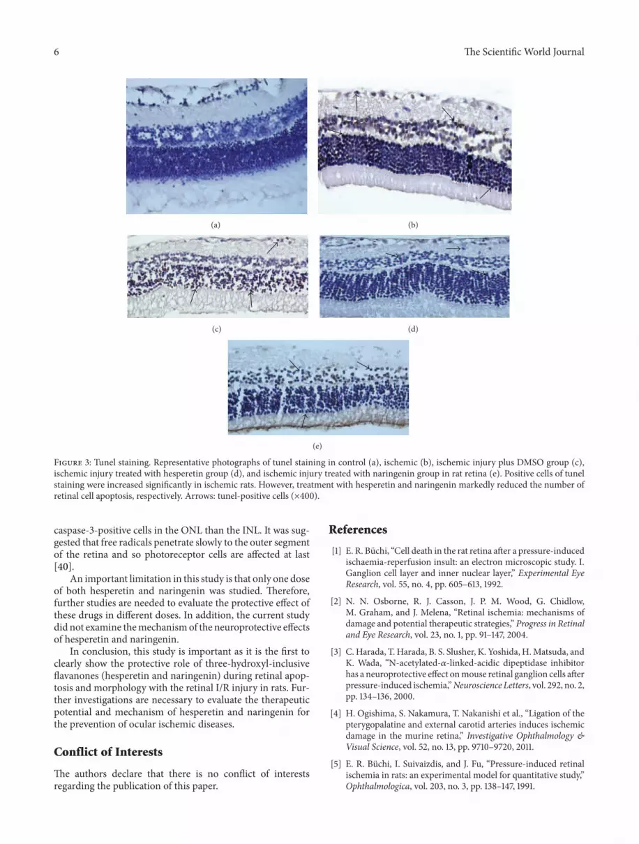
he efects of I/R on retinal cell death were examinedby measuring caspase-3 level and DNA fragmentation innucleus. Tunel staining was used to detect DNA fragmen-tation of cells undergoing apoptosis. In the control group,the retina cell nuclei were almost negative for tunel staining(Table 2, Figure 2(a)). Signiicantly more tunel-positive cellswere found within the inner nuclear cells in the sham andsolvent groups than in the control group (� < 0.001, Table 2,Figures 2(b) and 2(c)). By contrast, in rats subjected to I/Rwith hesperetin or naringenin treatment, signiicantly lesstunel-positive cells were found compared to the other groups(� < 0.005, Table 2, Figures 2(d) and 2(e)). It was alsodemonstrated that the naringenin treatment group has fewertunel-positive cells than the hesperetin group (� < 0.05).We performed immunohistochemistry to detect activatedcaspase-3. he number of caspase-3 positive cells was sig-niicantly less in the retina of rats treated with hesperetin ornaringenin than in the retina of the sham and solvent groups,which had retinal damage induced by I/R (� < 0.001, Table 3,Figure 3).
4. Discussion
his study evaluated the probable protective efects of thetwo lavanones (hesperetin and naringenin) on the retinaldamage caused by I/R in a rat retinal model by studyingretinal morphology and immunohistochemistry. he retinalthickness and the quantity of apoptotic cells were comparedbetween I/R groups and the control group. It was foundthat naringenin and hesperetin markedly reduced retinal cellinjury following I/R in comparison to the sham and solventgroups. In addition, we also found that the treatment ofnaringenin inhibited the apoptosis of the retinal cells andreduced the thinning of the retinal thickness more efectivelywhen compared to hesperetin.
Flavonoids and diets rich with lavonoids have beenreported to maintain treatment for several diseases with
their powerful antioxidant activity, which also modulatesthe enzymatic activity [25, 26]. Antioxidant activity of thelavonoids favors by the presence of a 3-hydroxyl group in theheterocyclic ring and a catechol group in ring B, which arealso the structural features of hesperetin and naringenin [16,27]. Although this study implemented a treatment methodof intraperitoneal injection of lavonoids, their intestinalabsorbable forms include a 2-hydroxyl group (hesperidinand naringin), which appears in citrus fruits naturally andare converted to biologically active forms (hesperetin andnaringenin) in vivo [16, 28]. As those have 3-hydroxyl group,hesperetin and naringenin have been suggested to be moreefective antioxidants than ascorbic acid and �-tocopherol,particularly for urgent antioxidative treatment needs [29].Flavonoids also have potency to increase ocular blood low inrelation to the quantity of OH groups. Xu et al. [30] showedthat intraperitoneal injection of hesperetin and naringeninincreased ocular blood low; thus, the retinal function recov-ery was facilitated during retinal I/R insult. It was suggestedthat lavonoids improve endothelium-dependent (enhanceNO bioavailability) and endothelium-independent (inhibit
the responses to Ca2+) vasodilatation ater I/R, as well asprotect vascular function [29]. Our indings are demonstrat-ing the prevention of structural damage and apoptosis in aretinal I/R injury animal model with the use of naringeninand hesperetin treatments that are compatible with the priorstudies.
he neuroprotective efect of lavanones on cerebral I/Rinjury was shown in recent studies [12, 31–33]. Shi et al. [31]demonstrated that pinocembrin (5,7-dihydroxy lavanone)signiicantly reduced neuronal loss and brain edema at24 h ater global cerebral I/R with a broad therapeutictime window (intravenously 30min before ischemia and30min, 2 h, and 6 h ater reperfusion). Flavonoids seem tosuiciently afect the ischemic site of the neuronal cellswhen the drug is applied just ater I/R. We also aimedto investigate the therapeutic potential of hesperetin andnaringenin ater I/R because occlusive retinal diseases arenonpredictable. Hesperetin and naringenin were reportedto exhibit protective efects in oxidative stress associatedneurodegeneration [12, 32, 33]. In addition, Hwang et al. [32]
he Scientiic World Journal 5
(a) (b)
(c) (d)
(e)
Figure 2: Caspase 3. (a) Control group, a few caspase 3-positive cells; (b) ischemic group; (c) ischemic injury plus DMSO group; (d) ischemiaplus hesperetin-treated group, and (e) ischemia plus naringenin-treated group. At the end of the experiment, fewer caspase 3-positive cellswere noted in the hesperetin- and naringenin-treated groups than those in the ischemic retinal cells. Arrows: Caspase 3-positive cells.Immunoperoxidase, hematoxylin counterstain (×400).
revealed a novel mechanism that hesperetin triggers estrogenreceptor (ER)- and TrkA-mediated parallel pathways whichinduces neuroprotective efect.
his study shows that retinal cell injury seems to occur2 days ater I/R, as demonstrated with the sham group [34].It is commonly accepted that I/R is associated with increasedoxygen-radical production; other factors including excitatoryamino acids, and reduced vasodilator reserve leading toretinal cell damage [35–37]. Earlier studies have shownthat apoptosis and necrosis are parts of I/R-induced retinalcell damage [1, 38]. here are various apoptotic proteinsin apoptotic cell death, including caspase family members.
Caspase-3 particularly is a key enzyme required for executionof apoptosis [39]. In the present study, we have seen anincreased amount of cells with caspase-3 compared to normalretina. Treatment with hesperetin or naringenin similarlyreduced caspase-3 positive cells as compared to the shamand solvent groups. In our tunel study, a signiicant numberof tunel-positive cells were found ater I/R. However, theafected cell amount markedly decreased with hesperetin andnaringenin treatment immediately ater the ischemic period.Consistent with Chiou and Xu [16], this study has shownthat naringenin is more protective for retinal cells duringthe reperfusion period. here were less tunel-positive or
6 he Scientiic World Journal
(a) (b)
(c) (d)
(e)
Figure 3: Tunel staining. Representative photographs of tunel staining in control (a), ischemic (b), ischemic injury plus DMSO group (c),ischemic injury treated with hesperetin group (d), and ischemic injury treated with naringenin group in rat retina (e). Positive cells of tunelstaining were increased signiicantly in ischemic rats. However, treatment with hesperetin and naringenin markedly reduced the number ofretinal cell apoptosis, respectively. Arrows: tunel-positive cells (×400).
caspase-3-positive cells in the ONL than the INL. It was sug-gested that free radicals penetrate slowly to the outer segmentof the retina and so photoreceptor cells are afected at last[40].
An important limitation in this study is that only one doseof both hesperetin and naringenin was studied. herefore,further studies are needed to evaluate the protective efect ofthese drugs in diferent doses. In addition, the current studydid not examine themechanismof the neuroprotective efectsof hesperetin and naringenin.
In conclusion, this study is important as it is the irst toclearly show the protective role of three-hydroxyl-inclusivelavanones (hesperetin and naringenin) during retinal apop-tosis and morphology with the retinal I/R injury in rats. Fur-ther investigations are necessary to evaluate the therapeuticpotential and mechanism of hesperetin and naringenin forthe prevention of ocular ischemic diseases.
Conflict of Interests
he authors declare that there is no conlict of interestsregarding the publication of this paper.
References
[1] E. R. Buchi, “Cell death in the rat retina ater a pressure-inducedischaemia-reperfusion insult: an electron microscopic study. I.Ganglion cell layer and inner nuclear layer,” Experimental EyeResearch, vol. 55, no. 4, pp. 605–613, 1992.
[2] N. N. Osborne, R. J. Casson, J. P. M. Wood, G. Chidlow,M. Graham, and J. Melena, “Retinal ischemia: mechanisms ofdamage and potential therapeutic strategies,” Progress in Retinaland Eye Research, vol. 23, no. 1, pp. 91–147, 2004.
[3] C. Harada, T. Harada, B. S. Slusher, K. Yoshida, H.Matsuda, andK. Wada, “N-acetylated-�-linked-acidic dipeptidase inhibitorhas a neuroprotective efect onmouse retinal ganglion cells aterpressure-induced ischemia,”Neuroscience Letters, vol. 292, no. 2,pp. 134–136, 2000.
[4] H. Ogishima, S. Nakamura, T. Nakanishi et al., “Ligation of thepterygopalatine and external carotid arteries induces ischemicdamage in the murine retina,” Investigative Ophthalmology &Visual Science, vol. 52, no. 13, pp. 9710–9720, 2011.
[5] E. R. Buchi, I. Suivaizdis, and J. Fu, “Pressure-induced retinalischemia in rats: an experimental model for quantitative study,”Ophthalmologica, vol. 203, no. 3, pp. 138–147, 1991.
he Scientiic World Journal 7
[6] U. Yigit, S. Erdenoz, U. Uslu et al., “An immunohistochemicalanalysis of the neuroprotective efects ofmemantine, hyperbaricoxygen therapy, and brimonidine ater acute ischemia reperfu-sion injury,”Molecular Vision, vol. 17, pp. 1024–1033, 2011.
[7] D. Arul and P. Subramanian, “Inhibitory efect of naringenin(citrus lavonone) on N-nitrosodiethylamine induced hepato-carcinogenesis in rats,” Biochemical and Biophysical ResearchCommunications, vol. 434, pp. 203–209, 2013.
[8] S. Keser, S. Celik, and S. Turkoglu, “Total phenolic contentsand free-radical scavenging activities of grape (Vitis vinifera L.)and grape products,” International Journal of Food Sciences andNutrition, vol. 64, pp. 210–216, 2013.
[9] T. Kinoshita, Z. Lepp, Y. Kawai, J. Terao, and H. Chuman, “Anintegrated database of lavonoids,” BioFactors, vol. 26, no. 3, pp.179–188, 2006.
[10] S. Kawaii, Y. Tomono, E. Katase, K. Ogawa, and M. Yano,“Quantitation of lavonoid constituents in Citrus fruits,” Journalof Agricultural and Food Chemistry, vol. 47, no. 9, pp. 3565–3571,1999.
[11] F. Haidari, S. A. Keshavarz, M. R. Rashidi, and M. M.Shahi, “Orange juice and hesperetin supplementation to hype-ruricemic rats alter oxidative stress markers and xanthineoxidoreductase activity,” Journal of Clinical Biochemistry andNutrition, vol. 45, no. 3, pp. 285–291, 2009.
[12] E. J. Choi and W. S. Ahn, “Neuroprotective efects of chronichesperetin administration in mice,” Archives of PharmacalResearch, vol. 31, no. 11, pp. 1457–1462, 2008.
[13] N. Nalini, S. Aranganathan, and J. Kabalimurthy, “Chemopre-ventive eicacy of hesperetin (citrus lavonone) against 1, 2-dimethylhydrazine-induced rat colon carcinogenesis,” Toxicol-ogy Mechanisms and Methods, vol. 22, pp. 397–408, 2012.
[14] J. Lee and G. Kim, “Evaluation of antioxidant and inhibitoryactivities for diferent subclasses lavonoids on enzymes forrheumatoid arthritis,” Journal of Food Science, vol. 75, no. 7, pp.H212–H217, 2010.
[15] Y. Miyake, K. Minato, S. Fukumoto et al., “New potent antiox-idative hydroxylavanones produced with Aspergillus saitoifrom lavanone glycoside in citrus fruit,” Bioscience, Biotechnol-ogy and Biochemistry, vol. 67, no. 7, pp. 1443–1450, 2003.
[16] G. C. Y. Chiou and X. Xu, “Efects of some natural lavonoids onretinal function recovery ater ischemic insult in the rat,” Journalof Ocular Pharmacology andherapeutics, vol. 20, no. 2, pp. 107–113, 2004.
[17] S. E. Pollard, M. Whiteman, and J. P. E. Spencer, “Modulationof peroxynitrite-induced ibroblast injury by hesperetin: a rolefor intracellular scavenging andmodulation of ERK signalling,”Biochemical and Biophysical Research Communications, vol. 347,no. 4, pp. 916–923, 2006.
[18] L. Testai, A. Martelli, M. Cristofaro, M. C. Breschi, and V.Calderone, “Cardioprotective efects of diferent lavonoidsagainst myocardial ischaemia/reperfusion injury in Langen-dorf-perfused rat hearts,” Journal of Pharmacology and Phar-macotherapeutics, vol. 65, pp. 750–756, 2013.
[19] S. S. Raza, M. M. Khan, A. Ahmad et al., “Neuroprotectiveefect of naringenin is mediated through suppression of NF-�B signaling pathway in experimental stroke,”Neuroscience, vol.230, pp. 157–171, 2013.
[20] T. Ahlenstiel, G. Burkhardt, H. Kohler, and M. K. Kuhlmann,“Improved cold preservation of kidney tubular cells by meansof adding biolavonoids to organ preservation solutions,” Trans-plantation, vol. 81, no. 2, pp. 231–239, 2006.
[21] A. Dembinski, Z. Warzecha, S. J. Konturek et al., “Extractof grapefruit-seed reduces acute pancreatitis induced byischemia/reperfusion in rats; possible implication of tissueantioxidants,” Journal of Physiology and Pharmacology, vol. 55,no. 4, pp. 811–821, 2004.
[22] T. T. Woo, S. Y. Li, W. W. Lai, D. Wong, and A. C. Lo,“Neuroprotective efects of lutein in a rat model of retinaldetachment,” Graefe’s Archive for Clinical and ExperimentalOphthalmology, vol. 251, pp. 41–51, 2013.
[23] S. M. Hsu, L. Raine, and H. Fanger, “Use of Avidin-Biotin-Peroxidase Complex (ABC) in immunoperoxidase techniques:a comparison between ABC and unlabeled antibody (PAP)procedures,” Journal of Histochemistry and Cytochemistry, vol.29, no. 4, pp. 577–580, 1981.
[24] S. Oguz, M. Kanter, M. Erboga, and C. Ibis, “Protective efect ofUrtica dioica on liver damage induced by biliary obstruction inrats,” Toxicology and Industrial Health, vol. 29, no. 9, pp. 838–845, 2012.
[25] S. Aranganathan and N. Nalini, “Eicacy of the potentialchemopreventive agent, hesperetin (citrus lavanone), on 1,2-dimethylhydrazine induced colon carcinogenesis,” Food andChemical Toxicology, vol. 47, no. 10, pp. 2594–2600, 2009.
[26] Y. J. Moon, X. Wang, and M. E. Morris, “Dietary lavonoids:efects on xenobiotic and carcinogen metabolism,” Toxicologyin Vitro, vol. 20, no. 2, pp. 187–210, 2006.
[27] P. Pietta, “Flavonoids as antioxidants,” Journal of Natural Prod-ucts, vol. 63, no. 7, pp. 1035–1042, 2000.
[28] A. F. Furtado, M. A. Nunes, and M. H. Ribeiro, “Hesperid-inase encapsulation towards hesperitin production targetingimproved bioavailability,” Journal of Molecular Recognition, vol.25, pp. 595–603, 2012.
[29] O. L. Woodman and E. C. H. Chan, “Vascular and anti-oxidantactions of lavonols and lavones,” Clinical and ExperimentalPharmacology and Physiology, vol. 31, no. 11, pp. 786–790, 2004.
[30] X. Xu, Y. Park, and G. C. Y. Chiou, “Efects of dihydrogenationof lavones and number of hydroxy groups in the molecules onocular blood low in rabbits and retinal function recovery inrats,” Journal of Ocular Pharmacology andherapeutics, vol. 20,no. 4, pp. 311–320, 2004.
[31] L. Shi, B. Chen, M. Gao et al., “he characteristics oftherapeutic efect of pinocembrin in transient global brainischemia/reperfusion rats,” Life Sciences, vol. 88, no. 11-12, pp.521–528, 2011.
[32] S. L. Hwang, J. A. Lin, P. H. Shih, C. T. Yeh, and G. C. Yen,“Pro-cellular survival and neuroprotection of citrus lavonoid:the actions of hesperetin in PC12 cells,” Food and Function, vol.3, pp. 1082–1090, 2012.
[33] H. J. Heo, D. Kim, S. C. Shin, M. J. Kim, B. G. Kim, and D.Shin, “Efect of antioxidant lavanone, naringenin, from Citrusjunoson neuroprotection,” Journal of Agricultural and FoodChemistry, vol. 52, no. 6, pp. 1520–1525, 2004.
[34] S. F. Abcouwer, C. Lin, E. B. Wolpert et al., “Efects of ischemicpreconditioning and bevacizumab on apoptosis and vascularpermeability following retinal ischemia-reperfusion injury,”Investigative Ophthalmology and Visual Science, vol. 51, no. 11,pp. 5920–5933, 2010.
[35] J. L. Zweier, “Measurement of superoxide-derived free radicalsin the reperfused heart. Evidence for a free radical mechanismof reperfusion injury,” he Journal of Biological Chemistry, vol.263, no. 3, pp. 1353–1357, 1988.
[36] D. J. Hearse, L. Maxwell, C. Saldanha, and J. B. Gavin, “hemyocardial vasculature during ischemia and reperfusion: a
8 he Scientiic World Journal
target for injury and protection,” Journal of Molecular andCellular Cardiology, vol. 25, no. 7, pp. 759–800, 1993.
[37] I. L. Ferreira, C. B. Duarte, and A. P. Carvalho, “Ca2+ inluxthrough glutamate receptor-associated channels in retina cellscorrelates with neuronal cell death,” European Journal of Phar-macology, vol. 302, no. 1–3, pp. 153–162, 1996.
[38] L. A. Levin andA. Louhab, “Apoptosis of retinal ganglion cells inanterior ischemic optic neuropathy,”Archives of Ophthalmology,vol. 114, no. 4, pp. 488–491, 1996.
[39] F. Doonan and T. G. Cotter, “Apoptosis: a potential thera-peutic target for retinal degenerations,” Current NeurovascularResearch, vol. 1, no. 1, pp. 41–53, 2004, Erratum in: CurrentNeurovascular Research, vol. 1, article 191, 2004.
[40] W. Ju, K. Kim, H. Hofmann et al., “Selective neuronal survivaland upregulation of PCNA in the rat inner retina followingtransient ischemia,” Journal of Neuropathology and Experimen-tal Neurology, vol. 59, no. 3, pp. 241–250, 2000.

Submit your manuscripts at
http://www.hindawi.com
Hindawi Publishing Corporation
http://www.hindawi.com Volume 2013
Oxidative Medicine and
Cellular Longevity
Hindawi Publishing Corporation http://www.hindawi.com Volume 2013Hindawi Publishing Corporation http://www.hindawi.com Volume 2013
The Scientiic World Journal
International Journal of
EndocrinologyHindawi Publishing Corporationhttp://www.hindawi.com
Volume 2013
ISRN Anesthesiology
Hindawi Publishing Corporationhttp://www.hindawi.com Volume 2013
OncologyJournal of
Hindawi Publishing Corporationhttp://www.hindawi.com Volume 2013
PPARRe sea rch
Hindawi Publishing Corporation
http://www.hindawi.com Volume 2013
OphthalmologyJournal of
Hindawi Publishing Corporationhttp://www.hindawi.com Volume 2013
ISRN AIDS
Hindawi Publishing Corporationhttp://www.hindawi.com Volume 2013
BioMed Research International
Hindawi Publishing Corporation
http://www.hindawi.com Volume 2013
ObesityJournal of
Hindawi Publishing Corporationhttp://www.hindawi.com Volume 2013
ISRN Addiction
Hindawi Publishing Corporationhttp://www.hindawi.com Volume 2013
Hindawi Publishing Corporationhttp://www.hindawi.com Volume 2013
Computational and Mathematical Methods in Medicine
ISRN Allergy
Hindawi Publishing Corporationhttp://www.hindawi.com Volume 2013
Immunology ResearchHindawi Publishing Corporationhttp://www.hindawi.com Volume 2013
Journal of
Diabetes ResearchJournal of
Hindawi Publishing Corporationhttp://www.hindawi.com Volume 2013
Evidence-Based Complementary and Alternative Medicine
Volume 2013Hindawi Publishing Corporationhttp://www.hindawi.com
Hindawi Publishing Corporationhttp://www.hindawi.com Volume 2013
Gastroenterology Research and Practice
Hindawi Publishing Corporationhttp://www.hindawi.com Volume 2013
ISRN Biomarkers
Hindawi Publishing Corporationhttp://www.hindawi.com Volume 2013
MEDIATORSINFLAMMATION
of