propositions 1. each plant species has its own pectin. this
TRANSCRIPT
Propositions
1. Each plant species has its own pectin.
This thesis
2. The rheological behaviour of okra rhamnogalacturonan (RG) I with acetyl groups and
alpha galactose attached to rhamnosyl residues is greatly determined by hydrophobic
interaction caused by the acetylation pattern.
This thesis
3. The use of renewable plant biomass as raw material for a bio-based economy is
hampered by the lack of tailored pretreatments enabling full saccharification of
carbohydrates present as major components of the biomass.
4. The growing acceptance of so-called traditional medicines based on scientific evidence
strongly enhances the search for new sources of “novel” bioactive plant polysaccharides.
B.S. Paulsen, Current Organic Chemistry, 2001, 5, 939-950
5. When discussing the transesterification products of oligosacharides by lipases in
anhydrous organic solvent, Tsukamoto et al. overlook the possibility that not the enzyme,
but the molecular sieve used to dry the solvent is responsible for the formation of double
and triple substituted oligosaccharides.
J. Tsukamoto, S. Haebel, G.P. Valença, M.G. Peter, T.T. Franco, Journal of Chemical
Technology & Biotechnology 83 (2008) 1486-1492.
6. In view of the often low production costs, wide application range, satisfaction for a
substantial part of users and high profits it is recommended to explore the feasibility to
launch the drug “Placebo”.
Propositions belonging to the doctoral thesis of Nipaporn Sengkhamparn entitled
“Chemical, physical and biological features of Okra pectin”
Wageningen University, The Netherlands, 2 December 2009.
Chemical, physical and biological features
of Okra pectin
Nipaporn Sengkhamparn
Thesis committee
Thesis supervisor
Prof.dr.ir. A.G.J. Voragen Emeritus Professor of Food Chemistry Wageningen University Thesis co-supervisors Dr. H.A. Schols Associate Professor at the Laboratory of Food Chemistry Wageningen University Dr. Tanaboon Sajjaanantakul Head of Department of Agro-Industrial Technology Kasetsart University, Bangkok, Thailand Other members Prof. dr. E. van der Linden, Wageningen University Prof. dr. M-D. Nagel, Université de Technologie de Compiègne, France Prof. dr. S.C. de Vries, Wageningen University Prof. dr. J.T. Zuilhof, Wageningen University This research was conducted under the auspices of the Graduated School VLAG (Voeding, Levensmiddelentechnologie, Agrobiotechnologie en Gezondheid)
Chemical, physical and biological features
of Okra pectin
Nipaporn Sengkhamparn
Thesis
submitted in partial fulfillment of the requirements for the degree of doctor at Wageningen University
by the authority of the Rector Magnificus Prof. dr. M.J. Kropff, in the presence of the
Thesis Committee appointed by the Doctorate Board to be defended in public
on Wednesday 2 December 2009 at 1.30 PM in the Aula
Nipaporn Sengkhamparn Chemical, physical and biological features of Okra pectin, 176 pages. Thesis, Wageningen University, Wageningen, NL (2009) With references, with summaries in Dutch and English ISBN: 978-90-8585-529-3
Abstract Okra pods, Abelmoschus esculentus (L.) Moench, are used in Thailand as
vegetable as well as traditional medicine. Both textural and healthy properties are suggested
to originate from the high polysaccharide content of okra pods, although our knowledge
concerning the precise chemical structure of the different okra polysaccharides is far from
complete. Consequently, the first aim of the research described in this thesis was to
characterize all polysaccharides present in okra cell walls with emphasis on pectic
molecules, and in addition to establish their physical and biological properties.
Okra cell wall polysaccharides were fractionation by sequential extraction and the
pectins were obtained in the three fractions. Different types of rhamnogalacturonan I
structures were recognized to be present in the buffer soluble and the chelating agent
soluble fraction. By using enzymatic degradation of the pectins using pure and well
characterized enzymes, followed by identification of the oligomers released by NMR and
mass spectrometry, it was found that the buffer soluble, okra typical RG I, contained a
rather unique substitution of the rhamnosyl moieties present in the backbone with acetyl
groups and alpha galactosyl residues. The chelating agent soluble RG I was linked to
homogalacturonan structural elements and resembled more commonly found RG I with
substitutions of short chains of beta-linked galactose including some arabinose decoration.
The rheological properties of the okra typical RG I differed from other pectins
since diluted solutions gave rise to very high viscosities and a slimy appearance. It is
suggested that the acetylation of the rhamnosyl residues greatly affect its rheological
properties and plays an important role through hydrophobic associations. Okra pectins also
showed a high anti-complementary activity using the complement-fixing activity assay.
Moreover, cell line studies indicated its possible use as a coating material for medical
devices as demonstrated before for more complex RG I segments from apple pectin.
The hemicellulose populations found in okra cell walls, extracted with
concentrated alkali, were characterized by sugar (linkage) composition and enzymatic
degradation studies to be a XXXG-type xyloglucan and a 4-O-methylglucuronoxylan.
The research described leads to the recognition of new pectic RG I structures
being quite different from the ones described so far.

Contents
Chapter 1 General Introduction 9
Chapter 2 Characterization of Cell Wall Polysaccharides from 31
Okra (Abelmoschus esculentus (L.) Moench)
Chapter 3 Okra pectin contains an unusual substitution of 61
its rhamnosyl residues with acetyl and
alpha –linked galactosyl groups
Chapter 4 Physicochemical properties of pectins from 89
Okra (Abelmoschus esculentus (L.) Moench
Chapter 5 Complement-fixing activity and influence on cell 113
behaviour of okra (Abelmoschus esculentus (L.) Moench) pectins
Chapter 6 General Discussion 137
Summary/ Samenvatting 159
Acknowledgement 167
Curriculum Vitae 171
List of Publications 173
Overview of completed training activities 175

General introduction
9
Chapter 1
General Introduction
Chapter 1
10
The okra plant
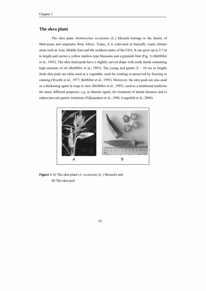
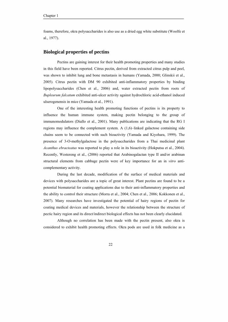
The okra plant Abelmoschus esculentus (L.) Moench belongs to the family of
Malvaceae and originates from Africa. Today, it is cultivated in basically warm climate
areas such as Asia, Middle East and the southern states of the USA. It can grow up to 2.5 m
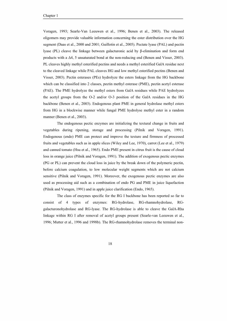
in height and carries a yellow mallow-type blossoms and a greenish fruit (Fig. 1) (BeMiller
et al., 1993). The okra fruits/pods have a slightly curved shape with seeds inside containing
high amounts of oil (BeMiller et al., 1993). The young and gentle (5 – 10 cm in length)
fresh okra pods are often used as a vegetable, used for cooking or preserved by freezing or
canning (Woolfe et al., 1977; BeMiller et al., 1993). Moreover, the okra pods are also used
as a thickening agent in soup or stew (BeMiller et al., 1993), used as a traditional medicine
for many different purposes; e.g. as diuretic agent, for treatment of dental diseases and to
reduce/prevent gastric irritations (Ndjouenkeu et al., 1996; Lengsfeld et al., 2004).
Figure 1 A) The okra plant (A. esculentus (L.) Moench) and
B) The okra pod
A B
General introduction
11
The plant cell wall
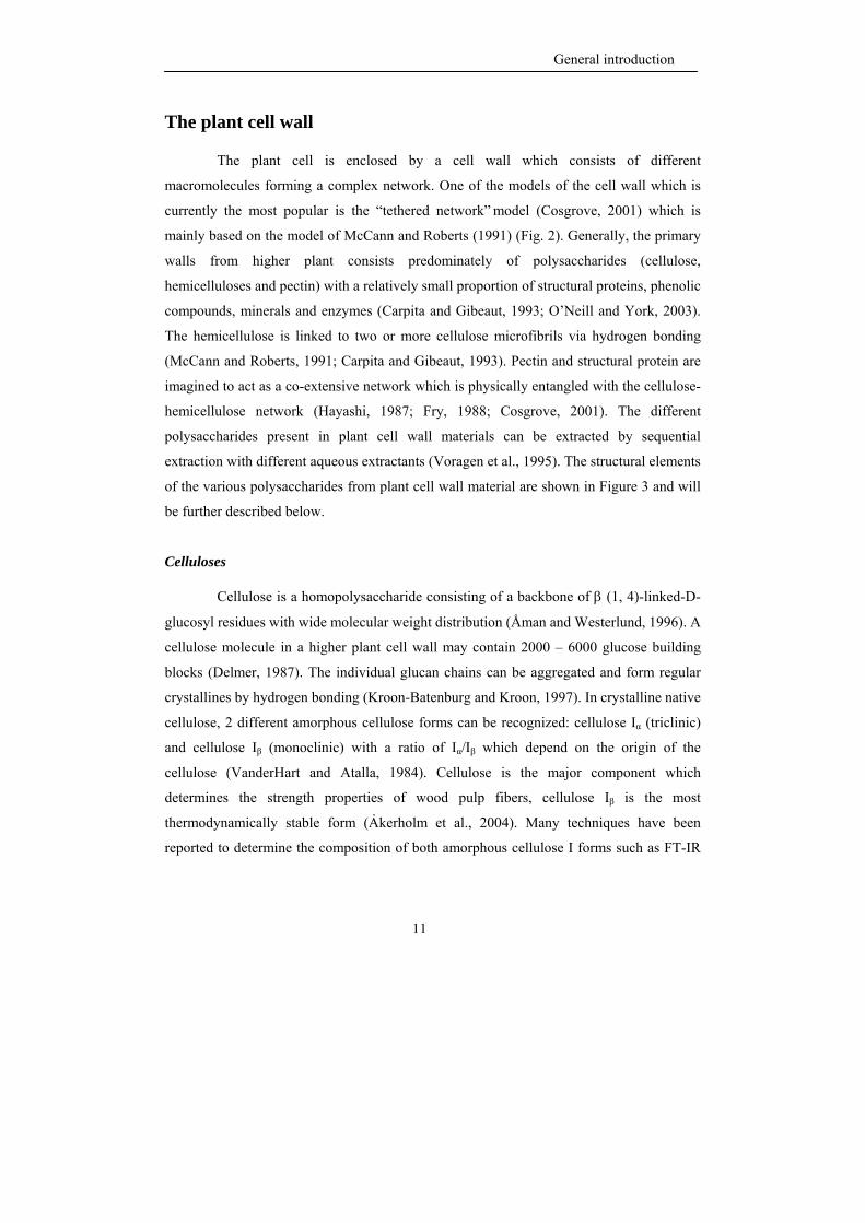
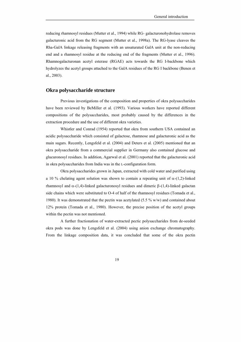
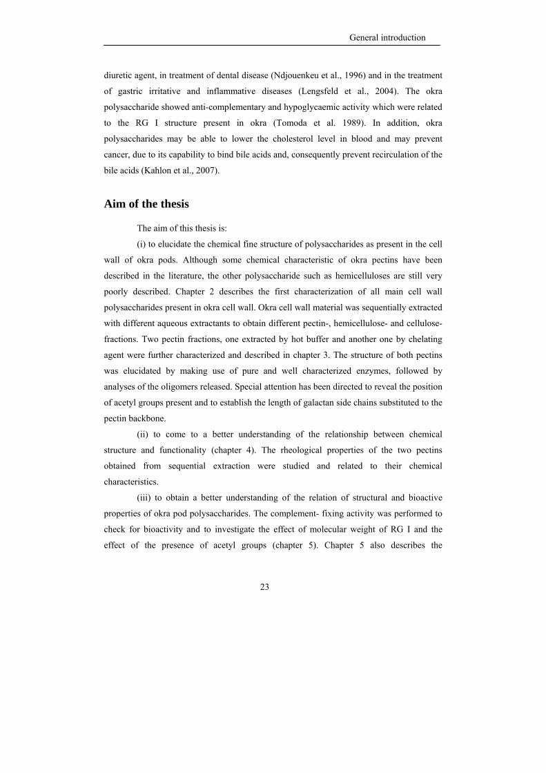
The plant cell is enclosed by a cell wall which consists of different
macromolecules forming a complex network. One of the models of the cell wall which is
currently the most popular is the “tethered network” model (Cosgrove, 2001) which is
mainly based on the model of McCann and Roberts (1991) (Fig. 2). Generally, the primary
walls from higher plant consists predominately of polysaccharides (cellulose,
hemicelluloses and pectin) with a relatively small proportion of structural proteins, phenolic
compounds, minerals and enzymes (Carpita and Gibeaut, 1993; O’Neill and York, 2003).
The hemicellulose is linked to two or more cellulose microfibrils via hydrogen bonding
(McCann and Roberts, 1991; Carpita and Gibeaut, 1993). Pectin and structural protein are
imagined to act as a co-extensive network which is physically entangled with the cellulose-
hemicellulose network (Hayashi, 1987; Fry, 1988; Cosgrove, 2001). The different
polysaccharides present in plant cell wall materials can be extracted by sequential
extraction with different aqueous extractants (Voragen et al., 1995). The structural elements
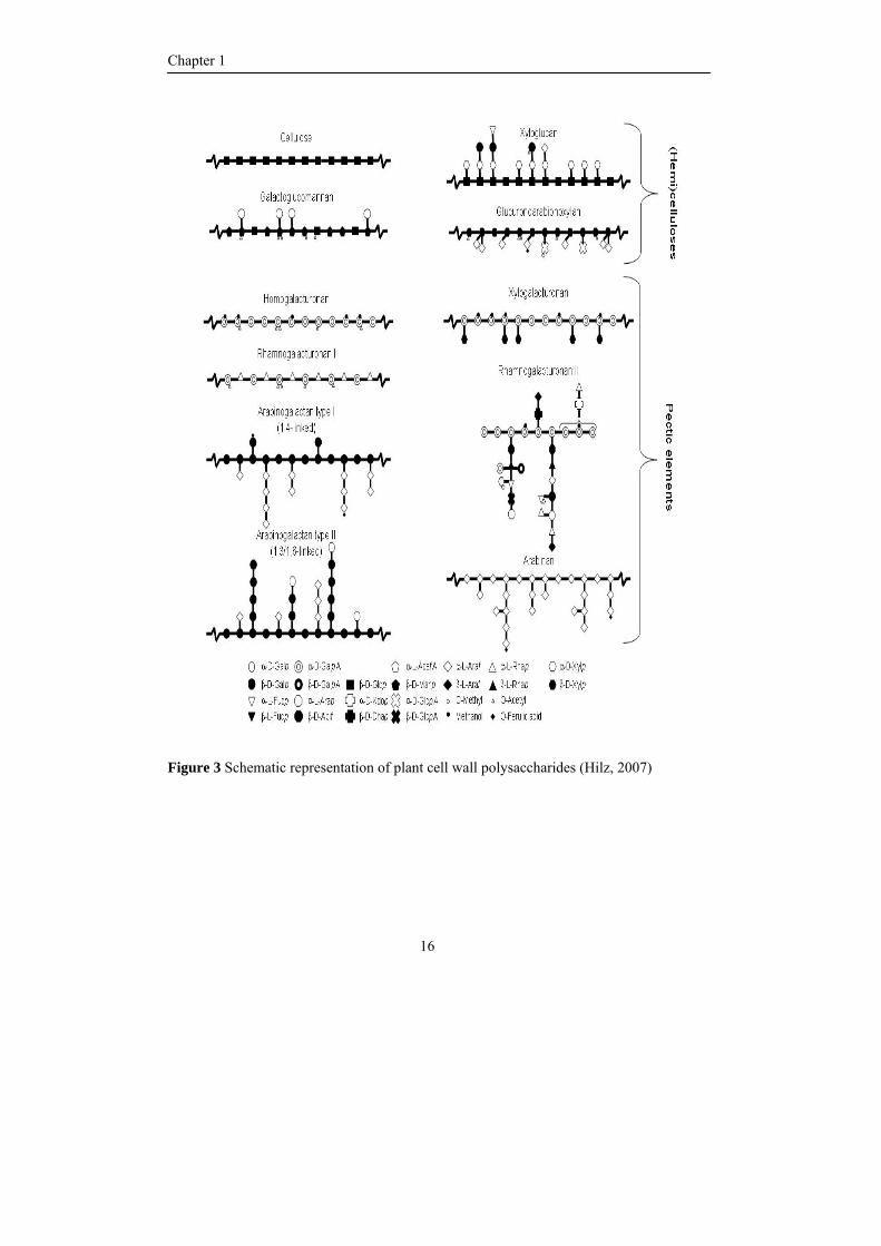
of the various polysaccharides from plant cell wall material are shown in Figure 3 and will
be further described below.
Celluloses
Cellulose is a homopolysaccharide consisting of a backbone of β (1, 4)-linked-D-
glucosyl residues with wide molecular weight distribution (Åman and Westerlund, 1996). A
cellulose molecule in a higher plant cell wall may contain 2000 – 6000 glucose building
blocks (Delmer, 1987). The individual glucan chains can be aggregated and form regular
crystallines by hydrogen bonding (Kroon-Batenburg and Kroon, 1997). In crystalline native
cellulose, 2 different amorphous cellulose forms can be recognized: cellulose Iα (triclinic)
and cellulose Iβ (monoclinic) with a ratio of Iα/Iβ which depend on the origin of the
cellulose (VanderHart and Atalla, 1984). Cellulose is the major component which
determines the strength properties of wood pulp fibers, cellulose Iβ is the most
thermodynamically stable form (Ảkerholm et al., 2004). Many techniques have been
reported to determine the composition of both amorphous cellulose I forms such as FT-IR
Chapter 1
12
spectroscopy (Sassi et al., 2004), dynamic FT-IR spectroscopy (Ảkerholm et al., 2004),
NMR (VanderHart and Atalla, 1984; Yamamoto et al., 1996).
Figure 2A Simplified schematic view of the primary plant cell wall
(McCann and Roberts, 1991)
2B The “tethered network” (Cosgrove, 2001)
A
Xyloglucan chains Xyloglucan chains buried within microfibril
B
General introduction
13
Hemicelluloses
Hemicelluloses are defined as polysaccharides in plant cell walls which are
solubilized by aqueous alkali. Xyloglucans, xylans, mannans and arabinogalactans are the
most abundant representatives (O’Neill and York, 2003).
Xyloglucans are the most abundant hemicellulose in dicotyledons. Their precise
chemical structure strongly depends on their origin. Xyloglucan is generally composed of a
backbone of β-(1,4)-linked-D-glucosyl residues with two or three out of four D-glucosyl
residues being substituted at position O-6 with α-D-xylosyl residues (Vierhuis et al., 2001).
Moreover, some of the D-xylosyl residues can be substituted with either monosaccharides
(D-galactose or L-arabinose) or disaccharides (e.g. D-galactose-L-fucose) (Vierhuis et al.,
2001). The xyloglucans are classified into 2 types, poly-XXXG type and poly-XXGG type
depending on the degree of glucan backbone substitution with D-xylosyl residues (Fry et
al., 1993). Recently, Hilz et al. (2007) found the α-D-xylosyl-β-(1,4)-D-xylosyl side chain
of xyloglucan in bilberries.
Xylans represent an other types of hemicellulose present in the cell wall and have
a backbone consisting of β-(1,4)-linked-D-xylosyl residues. The D-xylosyl residues in the
backbone as present in monocotyles (cereals and grasses) can be substituted with L-
arabinosyl residues, D-glucuronosyl residues (or its 4-O-methyl derivative) and acetyl
groups (McNeil et al., 1984; Brett and Waldom, 1990; Åman and Westerlund, 1996). In
dicotyles (hard woods and herbs) regularly highly acetylated acidic (O-methyl)
glucuronoxylans almost without any arabinose substitution can be found (McNeil et al.,
1984; Brett and Waldom, 1990).
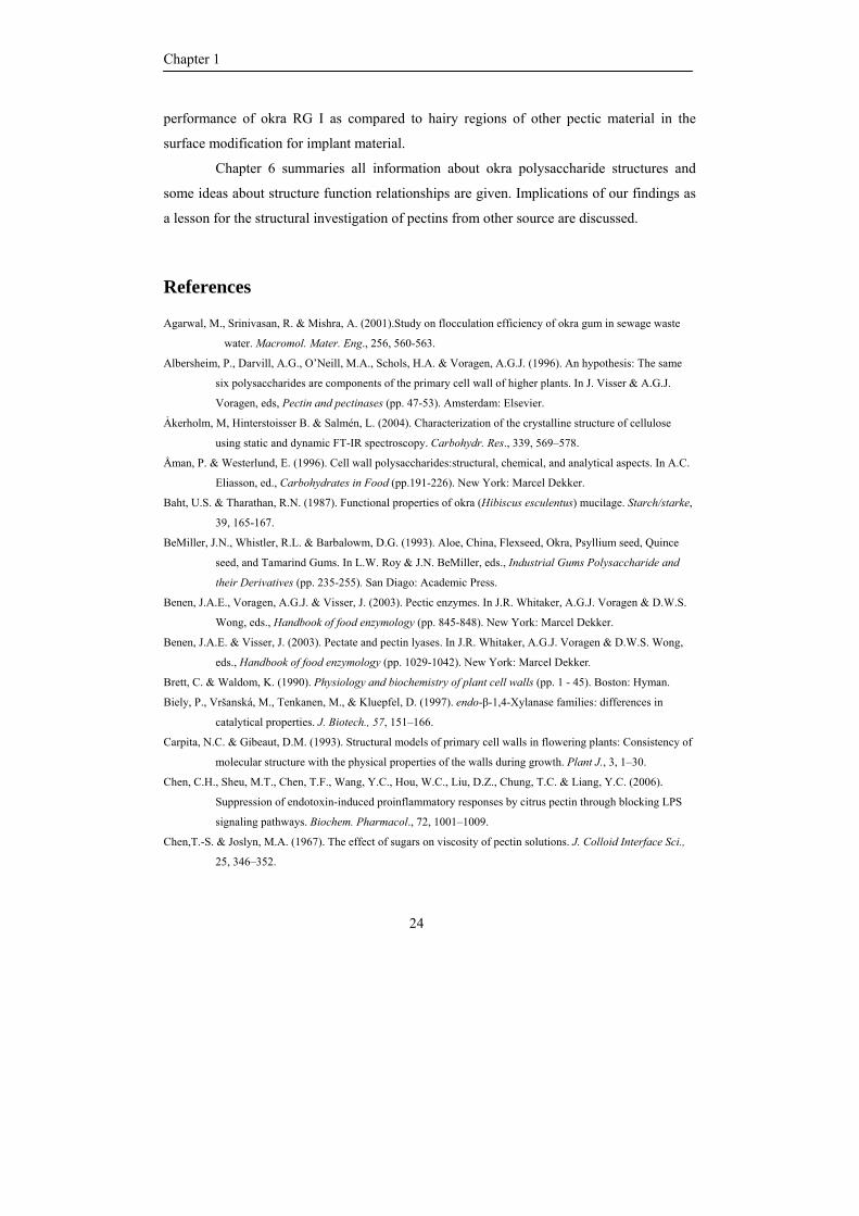
Pectins
Pectin is an important cell wall component of plants and probably the most
complex macromolecule in nature (Vincken et al., 2003). Pectin is composed of 17
different monosaccharides which are arranged in a number of structural elements forming
the building blocks of the pectin network which is shown in Figure 3 (Mohnen, 1999;
Vincken et al., 2003; O’Neill et al., 2004). The pectin backbone can be classified into three
Chapter 1
14
classes based on the elements present; homogalacturonan, substituted galacturonan and
rhamnogalacturonan.
Homogalacturonan (HG) is composed of a backbone of α-(1,4)-linked-D-
galacturonosyl residues in which a variable part of the galacturonic acid is methyl esterified
(Talmadge et al., 1973). The degree of methyl esterification (DM) in HG can classify pectin
into 2 main types, high DM pectin (more than 50%) and low DM pectin and it strongly
influences their functionality (Voragen et al., 1995). Parts of HG may be cross-linked and
be involved in forming a three dimensional network, a pectin gel, which is important in
controlling the porosity and mechanical properties of the cell wall and contributing to the
maintenance of intercellular adhesion (Carpita and Gibeaut, 1993; Willats et al., 2006). The
gelling property of pectins may be influenced by many different factors such as type and
origin of the pectin and the level and distribution of methyl esters and acetyl groups. The
methyl esters can be distributed either randomly or block-wise over the HG segment which
strongly effect the calcium binding of pectin as will be discussed later. In addition, HG may
be partially O-acetylated at position O-2 and/or O-3 of the D-galacturonosyl residues such
as pectin from sugar beet where the presence of acetyl groups has a negative effect on the
gelling behaviour (Rombouts and Thibault, 1986; Oosterveld et al., 2000).
HG can also be branched with a single unit of β-D-xylose residue or longer 1,2-
linked or 1,4 linked β-D-xylose chains attached to O-2 and O-3 of the D-galacturonosyl
residues which is called xylogalacturonan (XGA) (Schols et al., 1995; Albersheim et al.,
1996; Le Goff et al., 2001; Nakamura et al., 2002; Zandleven et al., 2007). Like HG, part of
D-galacturonosyl residues in XGA may be methyl esterified (Schols et al., 1995; Yu and
Mort, 1996).
Rhamnogalacturonan (RG) I contains a backbone of alternating α-(1, 2)-linked-L-
rhamnosyl and α-(1,4)-linked-D-galacturonosyl residues with the ratio of rhamnose to
galacturonic acid of 1:1 (McNeil et al., 1980; Lau et al., 1985; Schols et al., 1990). Many
RG I are partially O-acetylated at position O-2 and/or O-3 of the D-galacturonosyl residues
but so far, no evidence has been reported about the presence of methyl esterified D-
galacturonosyl residues within the RG I backbone (Ishii, 1997; Perrone et al., 2002).
Approximately 20-80 % of all L-rhamnosyl residues are branched at position O-4 with β-
General introduction
15
(1, 4)-linked galactan (Gur’janov et al., 2007), α-(1,5)-linked arabinan or branched
arabinans (Ridley et al., 2001; Vincken et al., 2003) depending on origin and plant tissue
(McNeil et al., 1982; Lau et al., 1987; Ishii et al., 1989). Besides, arabinogalactan type I
(AG I) and type II (AG II) may be presented as side chains of RG I. AG I consists of a β-(1,
4)-linked galactan backbone with α-L-arabinofuranosyl residues attached to the O-3
position of D-galactosyl residues (Vincken et al., 2003). AG II is a branched polymer
composed of a backbone of β-(1,3)-linked-D-galactosyl residues containing side chains of
α-L-Araf-(1→6)-[β-D-Galp-(1→6)]n (n=1,2 or 3) (Mohnen, 1999; Ridley et al., 2001;
Vincken et al., 2003).
Rhamnogalacturonan II is a special structural element of pectin, which has a highly
conserved structure and is found in many different plants (O’Neill et al., 2004). It was
found to be covalently linked to HG (O’Neill et al., 2001) and consists of 12 different
sugars with some very peculiar sugar building blocks such as Apiose, Aceric Acid, 3-
deoxy-D-lyxo-2-heptulosaric acid (DHA) and 2-keto-3-deoxy-D-manno-octulosonic acid
(DKO) (O’Neill et al., 2004). RG II is composed of a backbone of α-(1,4)-linked-D-
galacturonosyl residues to which four well conserved but different side chains are
connected (O’Neill et al., 2004).
Enzymatic degradation
According to the specificity and selectivity of enzymes, analysis of digests
obtained from incubation of complex polysaccharides with well characterized enzymes can
provide valuable information of the structures of the polysaccharides (De Vries, 1982;
Schols et al., 1990; Daas et al., 1999). The potential use of enzymatic degradation in the
elucidation of the structure of polysaccharides depends on the purity of the enzyme used,
and on the knowledge of their substrate specificity and pattern of their action (Voragen et
al., 1993).
Chapter 1
16
Figure 3 Schematic representation of plant cell wall polysaccharides (Hilz, 2007)
General introduction
17
Hemicellulosic enzymes
Hemicelluloses, present in plant cell wall are xyloglucans, xylans, mannans and
arabinogalactans (O’Neill and York, 2003). Xyloglucans can be degraded by xyloglucan
specific endo-glucanase. This enzyme cleaves linkages between unsubstituted glucose and
xylose-substituted glucosyl residues (Pauly et al., 1999). The released oligomers always
contain an unbranched glucose moiety at the reducing end (Hilz et al., 2007) and analysis of
all fragments may provide valuable information about the different building blocks present
within the xyloglucan under investigation (Fry et al., 1993; Hilz et al., 2007).
More information about structure of xylans can be revealed by using endo-
xylanases from different Glycosyl Hydrolase families (http://www.cazy.org/) having
different modes of actions towards substituted xylans. In general family GH 10 xylanases
may be able to cleave rather close to a substitution side releasing shorter (substituted) xylo-
oligosaccharides, while family GH 11 xylanases are more hindered by substitution with
different side groups (Biely et al., 1997; Rantanen et al., 2007).
Pectic enzymes
Pectin modifying enzymes can be devided into hydrolases and lyases both able to
split within the pectin backbone, and esterases able to specifically remove either the methyl
ester or the acetyl group from pectins. Specific groups of such enzymes have been
recognized to act either on galacturonans or rhamnogalacturonans (Benen et al., 2003;
Benen and Visser, 2003; Vincken et al, 2003).
Polygalacturonase (PG) cleaves the linkage between galacturonic acid by
hydrolysis (Benen and Visser, 2003). The PGs can be divided into endo PGs and exo PGs.
Endo PGs cleaves the HG backbone randomly and needs a number of adjacent non-methyl-
esterified galacturonosyl residues (Benen and Visser, 2003; Vincken et al., 2003; Daas et
al., 1999), while the exoPGs cleave mono- or di-mers from the non reducing end of a HG
backbone and need a non esterified GalA residue at subsite -2, -1 and +1 of the enzyme
(Benen and Visser, 2003). The PG action is hindered by the presence of methyl esters and
acetyl groups (Vincken et al., 2003) and consequently, the activity of PG can be improved
by simultaneously using pectin methyl esterase and/or pectin acetylesterase (Pilnik and
Chapter 1
18
Voragen, 1993; Searle-Van Leeuwen et al., 1996; Benen et al., 2003). The released
oligomers may provide valuable information concerning the ester distribution over the HG
segment (Daas et al., 2000 and 2001; Guillotin et al., 2005). Pectate lyase (PAL) and pectin
lyase (PL) cleave the linkage between galacturonic acid by β-elimination and form end
products with a ∆4, 5 unsaturated bond at the non-reducing end (Benen and Visser, 2003).
PL cleaves highly methyl esterified pectins and needs a methyl esterified GalA residue next
to the cleaved linkage while PAL cleaves HG and low methyl esterified pectins (Benen and
Visser, 2003). Pectin esterases (PEs) hydrolyze the esters linkage from the HG backbone
which can be classified into 2 classes, pectin methyl esterase (PME), pectin acetyl esterase
(PAE). The PME hydrolyze the methyl esters from GalA residues while PAE hydrolyzes
the acetyl groups from the O-2 and/or O-3 position of the GalA residues in the HG
backbone (Benen et al., 2003). Endogenous plant PME in general hydrolase methyl esters
from HG in a blockwise manner while fungal PME hydrolyse methyl ester in a random
manner (Benen et al., 2003).
The endogenous pectic enzymes are initializing the textural change in fruits and
vegetables during ripening, storage and processing (Pilnik and Voragen, 1991).
Endogenous (endo) PME can protect and improve the texture and firmness of processed
fruits and vegetables such as in apple slices (Wiley and Lee, 1970), carrot (Lee et al., 1979)
and canned tomato (Hsu et al., 1965). Endo PME present in citrus fruit is the cause of cloud
loss in orange juice (Pilnik and Voragen, 1991). The addition of exogenous pectic enzymes
(PG or PL) can prevent the cloud loss in juice by the break down of the polymeric pectin,
before calcium coagulation, to low molecular weight segments which are not calcium
sensitive (Pilnik and Voragen, 1991). Moreover, the exogenous pectic enzymes are also
used as processing aid such as a combination of endo PG and PME in juice liquefaction
(Pilnik and Voragen, 1991) and in apple juice clarification (Endo, 1965).
The class of enzymes specific for the RG I backbone has been reported so far to
consist of 4 types of enzymes: RG-hydrolase, RG-rhamnohydrolase, RG-
galacturonohydrolase and RG-lyase. The RG-hydrolase is able to cleave the GalA-Rha
linkage within RG I after removal of acetyl groups present (Searle-van Leeuwen et al.,
1996; Mutter et al., 1996 and 1998b). The RG-rhamnohydrolase removes the terminal non-
General introduction
19
reducing rhamnosyl residues (Mutter et al., 1994) while RG- galacturonohydrolase removes
galacturonic acid from the RG segment (Mutter et al., 1998a). The RG-lyase cleaves the
Rha-GalA linkage releasing fragments with an unsaturated GalA unit at the non-reducing
end and a rhamnosyl residue at the reducing end of the fragments (Mutter et al., 1996).
Rhamnogalacturonan acetyl esterase (RGAE) acts towards the RG I-backbone which
hydrolyzes the acetyl groups attached to the GalA residues of the RG I backbone (Benen et
al., 2003).
Okra polysaccharide structure
Previous investigations of the composition and properties of okra polysaccharides
have been reviewed by BeMiller et al. (1993). Various workers have reported different
compositions of the polysaccharides, most probably caused by the differences in the
extraction procedure and the use of different okra varieties.
Whistler and Conrad (1954) reported that okra from southern USA contained an
acidic polysaccharide which consisted of galactose, rhamnose and galacturonic acid as the
main sugars. Recently, Lengsfeld et al. (2004) and Deters et al. (2005) mentioned that an
okra polysaccharide from a commercial supplier in Germany also contained glucose and
glucuronosyl residues. In addition, Agarwal et al. (2001) reported that the galacturonic acid
in okra polysaccharides from India was in the L-configuration form.
Okra polysaccharides grown in Japan, extracted with cold water and purified using
a 10 % chelating agent solution was shown to contain a repeating unit of α-(1,2)-linked
rhamnosyl and α-(1,4)-linked galacturonosyl residues and dimeric β-(1,4)-linked galactan
side chains which were substituted to O-4 of half of the rhamnosyl residues (Tomada et al.,
1980). It was demonstrated that the pectin was acetylated (5.5 % w/w) and contained about
12% protein (Tomada et al., 1980). However, the precise position of the acetyl groups
within the pectin was not mentioned.
A further fractionation of water-extracted pectic polysaccharides from de-seeded
okra pods was done by Lengsfeld et al. (2004) using anion exchange chromatography.
From the linkage composition data, it was concluded that some of the okra pectin
Chapter 1
20
subpopulations consisted of a rather pure galacturonan structural element. Information
concerning other polysaccharides like hemicelluloses is still missing.
Functional properties of pectins
Pectin are widely used as gelling, stabilizing or thickening agent in many food
products such as jam, yoghurt drink, fruity milk drinks and ice cream (Laurent and
Boulenguer, 2003).
The viscosity of a polysaccharide solution depends on many factors such as
molecular mass, hydrodynamic volume, stiffness and charge of the molecule (Williams and
Phillips, 2000). The charged polymers generally have a higher viscosity than non-ionic
polymers at similar mass and chemical structure due to the intermolecular charge repulsion
(Williams and Phillips, 2000). Although pectin carries free carboxyl groups on the
backbone and it behaves as a polyelectrolyte (Voragen et al., 1995), pectin has not been
used very frequently as a thickener because pectin solutions have relatively low viscosities
when compared to other biopolymers at similar concentrations (Voragen et al., 1995). The
viscosity of pectin solutions depend on chemical and physical characteristic of the pectins,
on the ionic strength of the solution (Pals and Hermans, 1952) and on the presence of sugar
(Chen and Joslyn, 1967; Michel et al., 1985).
The junction zones of hydrocolloid gels are normally formed via physical
interaction such as hydrogen bonding, hydrophobic association and cation-mediated cross
linking (Williams and Phillips, 2000). Previous studies have shown that the gel properties
of pectin gels strongly depend on molecular properties of the polymer. The gelation
properties of pectins are influenced by the molecular weight of the pectin, the length of the
pectin side chains, the level and distribution of methyl esterification and the level of
acetylation. For example, Schmelter et al. (2002) suggested that pectin with shorter side
chains gave better gelation properties than pectin with longer side chains. Moreover, the
pattern of esterification, block-wise or random, has a great impact on gel characteristics
(Willats et al., 2001) the enzymatic removal of acetyl groups as present in sugar beet pectin
led to an improved gelation and a much stiffer gel (Oosterveld et al., 2000).
General introduction
21
Okra polysaccharide functional properties
Solutions of okra polysaccharides were found to exhibit pseudoplastic and
viscoelastic behaviour (BeMiller et al., 1993). Baht and Tharathan (1987) found that the
viscosity of the okra polysaccharides extracted with water showed a maximum viscosity in
the pH range of 4-6. The viscosity of okra polysaccharides decreased by addition of glucose
and sucrose (5-40%) and divalent salts (0.1-10% of CaCl2 and MgSO4) in contrast to a
rather small increase found after addition of maltodextrin and the presence of 0.1-10% of
monovalent salts (NaCl and KCl). Furthermore, the viscosity of an okra polysaccharide
solution sharply increased with increasing concentration. Woolfe et al (1997) reported that
heating okra polysaccharide solutions to 90 oC resulted in a decrease in viscosity while
cooling the solution back to room temperature caused in increase in the viscosity.
Okra pectin was found to form a gel at a relatively low concentration (6 g/L)
(Woolfe et al., 1977), while the formation of a stable gel also has been reported after
heating okra polysaccharides at 60 oC for 30 min followed by a cooling step at 4 oC for 24 h
(Baht and Tharathan, 1987). In addition, a synergistic effect was found when okra
polysaccharide was mixed with xanthan gum; mixing with locust bean gum did not give
any synergistic effect (Baht and Tharathan, 1987).
Okra polysaccharide was also shown to have unusual lubricity properties and to be
able to form a tenacious coating on the skin which is difficult to remove by washing
(BeMiller et al., 1993). The lubricity property is also an important property for a good food
fat mimetic (Glicksman, 1991) and okra polysaccharides can be used as fat substitute in
many products like chocolate bars and cookies. Many quality characteristics of such fat free
cookies were comparable with those of full fat cookies (Romanchik-Cerpovicz et al., 2002).
Likewise, okra polysaccharide has also been used as a milk-fat substitute in chocolate
frozen dairy desserts where it could replace the milk-fat up to 70 % while the melting
points of the products did not change, although the melting rate decreased slightly
(Constantino & Romanchik-Cerpovicz, 2004). Okra polysaccharides also exhibit foam
(Baht and Tharathan, 1987) and emulsion stabilizing properties (BeMiller et al., 1993). It
behaves like egg white at higher concentrations which can form threads and stabilize
Chapter 1
22
foams, therefore, okra polysaccharides is also use as a dried egg white substitute (Woolfe et
al., 1977).
Biological properties of pectins
Pectins are gaining interest for their health promoting properties and many studies
in this field have been reported. Citrus pectin, derived from extracted citrus pulp and peel,
was shown to inhibit lung and bone metastasis in humans (Yamada, 2000; Glinskii et al.,
2005). Citrus pectin with DM 90 exhibited anti-inflammatory properties by binding
lipopolysaccharides (Chen et al., 2006) and, water extracted pectin from roots of
Bupleurum falcatum exhibited anti-ulcer activity against hydrochloric acid-ethanol induced
ulserogenensis in mice (Yamada et al., 1991).
One of the interesting health promoting functions of pectins is its property to
influence the human immune system, making pectin belonging to the group of
immunomodulators (Diallo et al., 2001). Many publications are indicating that the RG I
regions may influence the complement system. A (1,6)–linked galactose containing side
chains seem to be connected with such bioactivity (Yamada and Kiyohara, 1999). The
presence of 3-O-methylgalactose in the polysaccharides from a Thai medicinal plant
Acanthus ebracteatus was reported to play a role in its bioactivity (Hokputsa et al., 2004).
Recently, Westereng et al., (2006) reported that Arabinogalactan type II and/or arabinan
structural elements from cabbage pectin were of key importance for an in vitro anti-
complementary activity.
During the last decade, modification of the surface of medical materials and
devices with polysaccharides are a topic of great interest. Plant pectins are found to be a
potential biomaterial for coating applications due to their anti-inflammatory properties and
the ability to control their structure (Morra et al., 2004; Chen et al., 2006; Kokkonen et al.,
2007). Many researches have investigated the potential of hairy regions of pectin for
coating medical devices and materials, however the relationship between the structure of
pectic hairy region and its direct/indirect biological effects has not been clearly elucidated.
Although no correlation has been made with the pectin present, also okra is
considered to exhibit health promoting effects. Okra pods are used in folk medicine as a
General introduction
23
diuretic agent, in treatment of dental disease (Ndjouenkeu et al., 1996) and in the treatment
of gastric irritative and inflammative diseases (Lengsfeld et al., 2004). The okra
polysaccharide showed anti-complementary and hypoglycaemic activity which were related
to the RG I structure present in okra (Tomoda et al. 1989). In addition, okra
polysaccharides may be able to lower the cholesterol level in blood and may prevent
cancer, due to its capability to bind bile acids and, consequently prevent recirculation of the
bile acids (Kahlon et al., 2007).
Aim of the thesis
The aim of this thesis is:
(i) to elucidate the chemical fine structure of polysaccharides as present in the cell
wall of okra pods. Although some chemical characteristic of okra pectins have been
described in the literature, the other polysaccharide such as hemicelluloses are still very
poorly described. Chapter 2 describes the first characterization of all main cell wall
polysaccharides present in okra cell wall. Okra cell wall material was sequentially extracted
with different aqueous extractants to obtain different pectin-, hemicellulose- and cellulose-
fractions. Two pectin fractions, one extracted by hot buffer and another one by chelating
agent were further characterized and described in chapter 3. The structure of both pectins
was elucidated by making use of pure and well characterized enzymes, followed by
analyses of the oligomers released. Special attention has been directed to reveal the position
of acetyl groups present and to establish the length of galactan side chains substituted to the
pectin backbone.
(ii) to come to a better understanding of the relationship between chemical
structure and functionality (chapter 4). The rheological properties of the two pectins
obtained from sequential extraction were studied and related to their chemical
characteristics.
(iii) to obtain a better understanding of the relation of structural and bioactive
properties of okra pod polysaccharides. The complement- fixing activity was performed to
check for bioactivity and to investigate the effect of molecular weight of RG I and the
effect of the presence of acetyl groups (chapter 5). Chapter 5 also describes the
Chapter 1
24
performance of okra RG I as compared to hairy regions of other pectic material in the
surface modification for implant material.
Chapter 6 summaries all information about okra polysaccharide structures and
some ideas about structure function relationships are given. Implications of our findings as
a lesson for the structural investigation of pectins from other source are discussed.
References
Agarwal, M., Srinivasan, R. & Mishra, A. (2001).Study on flocculation efficiency of okra gum in sewage waste
water. Macromol. Mater. Eng., 256, 560-563.
Albersheim, P., Darvill, A.G., O’Neill, M.A., Schols, H.A. & Voragen, A.G.J. (1996). An hypothesis: The same
six polysaccharides are components of the primary cell wall of higher plants. In J. Visser & A.G.J.
Voragen, eds, Pectin and pectinases (pp. 47-53). Amsterdam: Elsevier.
Ảkerholm, M, Hinterstoisser B. & Salmén, L. (2004). Characterization of the crystalline structure of cellulose
using static and dynamic FT-IR spectroscopy. Carbohydr. Res., 339, 569–578.
Åman, P. & Westerlund, E. (1996). Cell wall polysaccharides:structural, chemical, and analytical aspects. In A.C.
Eliasson, ed., Carbohydrates in Food (pp.191-226). New York: Marcel Dekker.
Baht, U.S. & Tharathan, R.N. (1987). Functional properties of okra (Hibiscus esculentus) mucilage. Starch/starke,
39, 165-167.
BeMiller, J.N., Whistler, R.L. & Barbalowm, D.G. (1993). Aloe, China, Flexseed, Okra, Psyllium seed, Quince
seed, and Tamarind Gums. In L.W. Roy & J.N. BeMiller, eds., Industrial Gums Polysaccharide and
their Derivatives (pp. 235-255). San Diago: Academic Press.
Benen, J.A.E., Voragen, A.G.J. & Visser, J. (2003). Pectic enzymes. In J.R. Whitaker, A.G.J. Voragen & D.W.S.
Wong, eds., Handbook of food enzymology (pp. 845-848). New York: Marcel Dekker.
Benen, J.A.E. & Visser, J. (2003). Pectate and pectin lyases. In J.R. Whitaker, A.G.J. Voragen & D.W.S. Wong,
eds., Handbook of food enzymology (pp. 1029-1042). New York: Marcel Dekker.
Brett, C. & Waldom, K. (1990). Physiology and biochemistry of plant cell walls (pp. 1 - 45). Boston: Hyman.
Biely, P., Vršanská, M., Tenkanen, M., & Kluepfel, D. (1997). endo-β-1,4-Xylanase families: differences in
catalytical properties. J. Biotech., 57, 151–166.
Carpita, N.C. & Gibeaut, D.M. (1993). Structural models of primary cell walls in flowering plants: Consistency of
molecular structure with the physical properties of the walls during growth. Plant J., 3, 1–30.
Chen, C.H., Sheu, M.T., Chen, T.F., Wang, Y.C., Hou, W.C., Liu, D.Z., Chung, T.C. & Liang, Y.C. (2006).
Suppression of endotoxin-induced proinflammatory responses by citrus pectin through blocking LPS
signaling pathways. Biochem. Pharmacol., 72, 1001–1009.
Chen,T.-S. & Joslyn, M.A. (1967). The effect of sugars on viscosity of pectin solutions. J. Colloid Interface Sci.,
25, 346–352.
General introduction
25
Cosgrove, D.J. (2001). Wall structure and wall loosening: A look backwards and forwards. Plant Physio., 125,
131-134.
Costantino, A.J. & Romanchick-Cerpoviez, J.E. (2004). Physical and sensory measures indicate moderate fat
replacement in frozen dairy dessert is feasible using okra gum as a milk-fat ingredient substitute. J. Am.
Diet. Assoc. 104, 44.
Daas, P.J.H., Meyer-Hansen, K., Schols, H.A., De Ruiter, G.A. & Voragen, A.G.J. (1999). Investigation of the
non-esterified galacturonic acid distribution in pectin with edopolygalacturonase. Carbohydr. Res.,
318, 135–145.
Daas, P.J.H., Voragen, A.G.J. & Schols, H.A. (2000). Characterisation of non-esterified galacturonic acid
sequences in pectin with endopolygalacturonase. Carbohydr. Res., 326, 120-129.
Daas, P.J.H., Voragen A.G.J. & Schols, H. A. (2001). Study of mehtyl ester distribution in pectin with endo-
polygalacturonase and high performance size exclusion chromatography. Biopolym., 58, 195-203.
Delmer, D.P. (1987). Biochemistry of cellulose biosynthesis. Tappi J., 70, 141-143.
Deters, A.M., Lengsfeld, C. & Andreas, H. (2005). Oligo- and polysaccharides exhibited a structure-dependent
bioactivity on human keratinocytes in vitro. J. Ethnopharm., 102, 391 –399.
De Vries, J.A., Rombouts, F.M., Voragen, A.G.J. & Pilnik, W. (1982). Enzymic degradation of apple pectins.
Carbohydr. Polym., 2, 25-33.
Diallo, D., Paulsen, B.S., Liljeback, T.H.A. & Michaelsen, T.E. (2001). Polysaccharides from the roots of Entada
africana Guill. et Perr., Mimosaceae, with complement fixing activity. J. Ethnopharm., 74, 159–171.
Endo, A. (1965). Studies on Pectolytic Enzymes of Moldes. Agric. Biol. Chem., 29, 29-136.
Fry, S.C. (1988) Wall polymer: chemical characterization. The growing plant cell wall (pp. 103-185). Harlow:
Longman Scientific & technical.
Fry, S.C., York, W.S., Albersheim, P., Darvill, A., Hayashi, T., Joseleau, J.P., Kato, Y., Lorences, E.P.,
Maclachlan, G.A., McNeil, M., Mort, A.J., Reid, J.S.G., Seitz, H.U., Selvendran, R.R., Voragen, A.G.J.
& White, A.R. (1993). An unambiguous nomenclature for xyloglucan derived oligosaccharides.
Physiol. Plantarum., 89, 1-3.
Glicksman, M. (1991). Hydrocolloids and the search for the oil grail. Food technol., 45, 94-103.
Glinskii, O.V, Huxley, V.H, Glinsky, G.V., Pienta, K.J., Raz, A. & Glinsky, V.V. (2005) Mechanical entrapment
is insufficient and intercellular adhesion is essential for metastatic cell arrest in distant organs.
Neoplasia, 7, 522–527.
Guillotin S.E., Bakx, E.J., Boulenguer, P., Mazoyer, J., Schols, H.A. & Voragen, A.G.J. (2005) Populations
having different GalA blocks characteristics are present in commercial similar but have different
functionalilties. Carbohydr. Polym., 30, 391-398.
Gur’janov, O.P., Gorshkova, T.A., Kabel, M., Schols, H.A. & Van Dam, J.E.G. (2007). MALDI-TOF MS
evidence for linking of flax bast fibre galactan to rhamnogalacturonan backbone. Carbohydr. Polym.,
67, 86-96.
Hayashi, T., Marsden, M.P.F. and Delmer, PP. (1987) Peac xyloglucan and cellulose: VI. Xyloglucan cellulose
interactions in vitro and in vivo. Plant Physiol. 83, 384-389
Chapter 1
26
Hilz, H. (2007). Characterization of cell wall polysaccharides in bilberries and black currants. PhD thesis,
Wageningen University, Wageningen, The Netherlands.
Hilz, H., de Jong, L.E., Kabel, M.A., Schols, H.A. & Voragen, A.G.J. (2007). Biberry xyloglucan-nove; building
blocks containing beta-xylose within a complex structure. Carbohydr. Res., 342, 170-181.
Hokputsa, S., Harding, S.E., Inngjerdingen, K., Jumel, K., Michaelsen, T.E., Heinze, T., Koschella, A. & Paulsen,
B.S. (2004). Bioactive polysaccharides from the stems of the Thai medicine plant Acanthus
ebracteatus: their chemical and physical features. Carbohydr. Res., 339, 753-762.
Hsu, C.P., Deshpande, S.N. & Desrosier, N.W. (1965). Role of pectin. methylesterase in firmness of canned
tomatoes. J. Food Sci., 30, 583-588.
Ishii, T. (1997). O-acetylated oligosaccharides from pectins of potato tuber cell walls. Plant Physiol., 113, 1265-
1272.
Ishii, T., Thomas, J.R., Darvill, A.G. & Albersheim, P. (1989). Structure of plant cell walls. XXVI. The walls of
suspension-cultured sycamore cells contain a family of rhamnogalacturonan I-like pectic
polysaccharides. Plant Physiol., 89, 421-428.
Kahlon, T.S., Chapman, M.H. & Smith, G.E. (2007). In vitro binding of bile acids by okra, beets, asparagus, egg
plant, turnips, green beans, carrots, and cauliflower. Food Chem., 103, 676-680.
Kokkonen, H.E., Ilvesaro, J.M., Morra, M., Schols, H.A. & Tuukkanen, J. (2007). Effect of modified pectin
molecules on the growth of bone cells. Biomacromol., 8, 509-515.
Kroon-Batenburg, L.M.J. & Kroon, J. (1997). The crystal and molecular structures of cellulose I and II.
Glycoconjugate J., 14, 677-690.
Lau, J.M., McNeil, M., Darvill, A.G. & Albersheim, P. (1985). Structure of the backbone of rhamnogalacturonan
I, a pectic polysaccharide in the primary cell walls of plants. Carbohydr. Res., 137, 111-125.
Lau, J.M., McNeil, M., Darvill, A.G. & Albersheim, P. (1987). Treatment of rhamnogalacturonan I with lithium in
ethylenediamine. Carbohydr. Res., 168, 245-274.
Laurent, M.A. & Boulenguer, P. (2003). Stabilization mechanism of acid dairy drinks (ADD) induced by pectin.
Food Hydrocolloids., 17, 445-454.
Le Goff, A., Renard, C.M.G.C., Bonin, E. & Thibault, J.F. (2001). Extraction, purification and chemical
characterization of xylogalacturonans from pea hulls. Carbohydr. Res., 45, 325-334.
Lee, C.Y., Bourne, M.C. & vanBuren, J.P. (1979). Effect of. blanching treatments on the firmness of carrots J.
Food Sci., 44, 615-616.
Lengsfeld, C., Titgemeyer, F., Faller, G. & Hensel, A. (2004). Glycosylated compounds from okra inhibit
adhesion of helicobacter pylori to human gastric mucosa. J. Agric. Food Chem., 52, 1495-1503.
McCann, M.C. & Roberts, K. (1991). Architecture of the primary cell wall. In C.W. Lloyrd, ed, The cytoskeletal
basis of plant growth and form (pp.109-129). London: Acadamic press.
McNeil, M., Darvill, A.G., Fry, S.C. & Albersheim, P. (1984). Structure and function of the primary cell walls of
plants. Annu. Rev. Biochem., 53, 625-663.
General introduction
27
McNeil, M., Darvill, A.G. & Albersheim, P. (1980). Structure of plant cell walls X: Rhamnogalacturonan I, a
structurally complex polysaccharide in the walls of suspension-cultured sycamore cells. Plant Physiol.,
66, 1128-1134.
McNeil, M. Darvill, A.G. & Albersheim, P. (1982). Structure of plant cell walls. XII. Identification of seven
differently linked glycosyl residues attached to O-4 of the 2,4 – linked L – rhamnosyl residues of
rhamnogalacturonan I. Plant Physiol., 70, 1586-1591.
Michel, F., Doublier, J.L. & Thibault, J.F. (1985). Etude viscométrique de la première phase de la gélification des
pectines hautement méthyles. Sci. Alim., 5, 305.
Mohnen, D. (1999). Biosynthesis of pectins and galactomannans. In D. Barton, K. Nakanishi & O.Meth-Cohn,
eds., Comprehensive natural products chemistry (pp. 497-527). Elsevier.
Morra, M., Cassinelli, C., Cascardo, G., Nagel, M.D., Della Volpe, C., Siboni, S., Maniglio, D., Brugnara, M.,
Ceccone, G., Schols, H A. & Ulvskov, P. (2004). Effects on interfacial properties and cell adhesion of
surface modification by pectic hairy regions. Biomacromol., 5, 2094-2104.
Mutter, M, Beldman, G., Schols, H.A. & Voragen, A.G.J. (1994). Rhamnogalacturonan α-L-
rhamnopyranosylhydrolase. A novel enzyme specific for the terminal nonreducing rhamnosyl unit in
rhamnogalacturonan regions of pectin. Plant Physiol., 106, 241-250.
Mutter, M., Colquhoun, I.J., Schols, H.A. & Voragen, A.G.J. (1996). Rhamnogalacturonase B from Aspergillus
aculeatus is a rhamnogalacturonan α-L-rhamnopyranosyl-(1,4)-α-D-galactopyranosyluronide lyase.
Plant Physiol. 110, 73 -77.
Mutter, M., Beldman, G., Pitson, S.M., Schols, H.A. & Voragen, A.G.J. (1998a). Rhamnogalacturonan α-D-
galactopyranosyluronohydrolase. An enzyme that specifically removes the terminal nonreducing
galacturonosyl residue in rhamnogalacturonan regions of pectin. Plant Physiol., 117, 153-163.
Mutter, M., Renard, C.M.G.C., Beldman, G., Schols, H.A. & Voragen, A.G.J. (1998b). Mode of action of RG-
hydrolase and RG-lyase toward rhamnogalacturonan oligomers. Characterization of
degradationproducts using RG-rhamnohydrolase and RG- galacturonohydrolase. Carbohydr. Res., 311,
155-164.
Nakamura, A., Furuta, H., Maeda, H., Takao, T. & Nagamatsu, Y. (2002). Analysis of the molecular construction
of xylogalacturonan isolated from soluble soybean polysaccharides. Biosci. Biotech, Biochem., 66,
1155-1158.
Ndjouenkeu, R., Goycoolea, F.M., Morris, E.R. & Akingbala, J.O. (1996). Rheology of Okra (Hibiscus esculentus
L.) and Dika Nut (Irvingia gabonensis) polysaccharides. Carbohydr. Polym., 29, 263-269.
O’Neill, M.A & York, W.S. (2003). The composition and structure of plant primary cell walls. In J.K.C. Rose, ed.,
The plant cell wall (pp. 1-54). USA: CRC Press.
O’Neill, M.A., Ishii, T, Albersheim, P. & Darvill, A.G. (2004). Rhamnogalacturonan II: structure and function of a
borate cross-linked cell wall pectic polysaccharide. Annu. Rev. Plant Bio., 55, 109-139.
O’Neill, M.A., Eberhard, S., Albersheim, P. & Darvill, A.G. (2001). Requirement of borate cross-linking of cell
wall rhamnogalacturonan II for Arabidopsis growth. Science., 294, 846-849
Chapter 1
28
Oosterveld, A., Beldman, G., Searle-van Leeuwen, M.J.F. & Voragen, A.G.J. (2000). Effect of enzymatic
deacetylation on gelation of sugar beet pectin in the presence of calcium. Carbohydr. Polym., 43, 249-
256.
Pals, D.T.F. & Hermans, J.J. (1952). Sodium salts of pectin and of carboxymethylcellulose in aqueous sodium
chloride. Rec.Trav. Chem. Pays Bas., 71, 433.
Pauly ,M., Albersheim, P., Darvill, A. & York, W.S. (1999). Molecular domains of the cellulose/xyloglucan
network in the cell walls of higher plants. Plant J., 20, 629-639.
Perrone ,P., Hewage, C.M., Thomson, A.R., Bailey, K., Sadler, I.H. & Fry, S.C. (2002). Patterns of methyl and O-
acetyl esterification in spinach pectins: new complexity. Phytochem., 60, 67-77.
Pilnik, W. & Voragen, A.G.J. (1991). The significance of endogenous and exogenous pectic enzymes in fruit and
vegetable processing. In Fox, P.F., ed., Food enzymology, Volume I (pp.330-336). London: Elsevier
Applied Science.
Pilnik, W & Voragen, A.G.J. (1993). Pectic enzymes in fruit and vegetable juice manufacture. In T.
Nagodawithana & G. Reed, eds., Enzymes in food processing (pp. 363-399). London: Academic Press.
Rantanen, H., Virkki, L., Tuomainen, P., Kabel, M., Schols, H. & Tenkanen, M. (2007). Preparation of
arabinoxylobiose from rye xylan using family 10 Aspergillus aculeatus endo-1,4-β-D-xylanase
Carbohydr. Polym., 68, 350-359.
Ridley, B.L., O’Neill, M.A. & Mohnen, D. (2001). Pectins: structure, biosynthesis and oligogalacturonide-related
signalling. Phytochem., 57, 929-967.
Romanchik-Cerpovicz, J.E., Tilmon, R.W. & Baldree, K.A. (2002). Moisture retention and consumer acceptability
of chocolate bar cookies prepared with okra gum as a fat ingredient substitute. J. Am. Diet. Assoc., 102,
1301-1303.
Rombouts, F.M. & Thibault, J.F. (1986). Sugar beet pectins: chemical structure and gelation through oxidative
coupling. In M.L. Fishman & J.J. Jen, eds., Chemistry and function of pectins, ACS Symposium series
310 (pp. 49). Washington DC: American Chemistry Society.
Sassi, J-F., Tekely, P. & Chanzy, H. (2000). Relative susceptibility of the Iα and Iβ phases of cellulose towards
acetylation. Cellulose, 7, 119-132.
Schmelter, T., Wientjes, R., Vreeker, R. & Klaffke, W. (2002). Enzymatic modifications of pectins and the impact
on their rheological properties. Carbohydr. Polym., 47, 99–108.
Schols, H.A., Geraeds, C.J.M., Searle-Van Leeuwen, M.F., Kormelink, F.J.M. & Voragen, A.G.J. (1990).
Rhamnogalacturonase: a novel enzyme that degrades the hairy regions of pectins. Carbohydr. Res.,
206, 105-115.
Schols, H.A., Vierhuis, E., Bakx, E.J. & Voragen, A.G.J. (1995). Different populations of pectic hairy regions
occur in apple cell walls. Carbohydr. Res., 275, 343-360.
Searle-Van Leeuwen, M.F., Vincken, J.P., Schipper, D. &. Voragen, A.G.J. (1996). Acetylesterase of Aspergillus
niger purification and mode of action on pectin. In J. Visser, and A.G.J. Voragen, eds., Progress in
biotechnology 14: Pectins and pectinases (pp. 793-798). Amsterdam: Elsevier Science.
General introduction
29
Talmadge, K.W., Keegstra, K., Bauer, W.D. & Albersheim, P. (1973). The structure of plant cell walls: I. The
macromolecular components of the walls of suspension-cultured sycamore cells with a detailed
analysis of the pectic polysaccharides. Plant physiol., 51, 158-173
Tomada, M., Shimada, K., Saito, Y. & Sugi, M. (1980). Plant mucilages. XXVI. Isolation and structural features
of a mucilage, “Okra mucilage”, from the immature fruit of Abelmoschus esculentus. Chem. Pharm.
Bull., 28, 2933-2940.
Tomoda, M., Shimizu, N., Gonda, R., Kanari, M., Yamada, H. & Hikino, H. (1989). Anticomplementary and
hypoglycaemic activity of okra and Hibiscus mucilages. Carbohydr. Res., 190, 323-328.
VanderHart, D. L.& Atalla, R. H. (1984). Studies of microstructure in nativ cellulose using solid state 13CNMR.
Macromol., 17, 1465–1472.
Vierhuis, E., Schols, H.A., Beldman, G. & Voragen, A.G.J. (2001). Structural characterisation of xyloglucan &
xylans present in olive fruit (Olea europaea cv koroneiki). Carbohydr. Polym., 44, 51–62.
Vincken, J.P., York, W.S., Beldman, G. & Voragen, A.G.J. (1997). Two general branching patterns of xyloglucan,
XXXG and XXGG. Plant Physiol., 114, 9-13.
Vincken, J-P., Voragen, A.G.J. & Beldman, G. (2003). Enzymes degrading rhamnogalacturonan and
xylogalacturonan. In J.R. Whitaker, A.G.J. Voragen & D.W.S. Wong, eds., Handbook of food
enzymology (pp. 930-941). New York: Marcel Dekker.
Voragen, A.G.J., Pilnik, W., Thibault, J.F, Axelos, M.A.V. & Renard, C.M.G.C. (1995). Pectins. In A.M.,
Stephen. Food polysaccharide and their application (pp. 287–339). New York: Marcel Dekker.
Voragen, A.G.J., Schols, H.A., & Gruppen, H. (1993). Structural studies of plant cell-wall polysaccharides using
enzymes. In F. Meuser, D.J. Manners & W. Seibel, eds., Proceedings of an international symposium on
plant polymeric carbohydrates (pp. 3–15). Cambridge: Royal Society of Chemistry.
Westereng, B., Yousif, O., Michaelsen, T.E., Knutsen, S.H. & Samuelsen, A.B. (2006). Pectin isolated from white
cabbage- structure and complement-fixing activity. Mol. Nutr. Food Res., 50, 746-755.
Whistler, R.L. & Conrad, H.E. (1954). A Crystalline Galactobiose from acid hydrolysis of okra mucilage. J. Am.
Chem. Soc., 76, 1673-1974.
Wiley, R.C. & Lee, Y.S. (1970). Modifying texture of processed apple slices. Food Technol., 24, 1168-1170.
Williams, P.A. & Phillips, G.O. (2000). Introduction of food hydrocolloids. In G.O. Phillips & P.A. Williams,
eds., Handbook of hydrocolloids (pp. 1-19). USA: CRC Press.
Willast, W.G.T., Orfila, C., Limberg, G., Buchholt, H.C., van Alebeek, G.J.W.M., Voragen, A.G.J., Marcust, S.E.,
Christensen, T.M.I.E., Mikkelsen, J.D., Murray, B.S. & Knox, J.P. (2001). Modulation of degree and
pattern of methyl-esterification of pectic Homogalacturonan in plant cell wall. J. Biol.Chem., 276,
19404-19413.
Willats, W.G.T., Knox, J.P. & Mikkelsen J.D. (2006). Pectin: new insights into an old polymer are starting to gel.
Trend Food Sci. Tech., 17, 97-104.
Woolfe, M.L., Chaplin, M.F. & Otchere, G. (1977). Studies on the mucilages extracted from Okra fruits (Hibiscus
esculentus L.) and Baobab leaves (Adansonia digitata L.). J. Sci. Food Agric., 28, 519-529.
Chapter 1
30
Yamada, H., Sun, X.B., Matsumoto, T, Ra, K.S., Hirano, M. & Kiyohara, H. (1991). Purification of anti- ulcer
polysaccharides form the roots of Bupleurum falcatum. Planta Med., 57, 555-559.
Yamada, H. & Kiyohara, H. 1999. Complement-activating polysaccharides from Medicinal Herbs. In H. Wagner,
ed., Immunomodulatory Agent from plants (pp. 161-202). Basel: Birkhäuser
Yamada, H. (2000). Bioactive plant polysaccharides from Japanese and Chinese traditional herbal medicines. In
B.S. Paulsen, ed., Bioactive carbohydrate polymers (pp. 15-24). London: Kluwer Academic Publishers.
Yamamoto, H., Horii, F. & Hirai, A. (1996). In situ crystallization of bacterial cellulose II. Influences of different
polymeric additives on the formation of celluloses Iα and Iβ at the early stage of incubation. Cellulose,
3, 229–242.
Yu, L. & Mort, A.J. (1996). Partial characterization of xylogalacturonans from cell walls of ripe watermelon fruit:
inhibition of endopolygalacturonase by xylosylation. In J. Visser & A.G.J. Voragen, eds., Pectins and
Pectinases (pp. 79-88). Amsterdam: Elsevier.
Zandleven, J., Sorensen, S.O., Harholt, T., Beldman, G., Schols, H.A., Scheller, H.V. & Voragen, A.G.J. (2007).
Xylogalacturonan exists in cell walls from various tissues of Arabidopsis thaliana. Phytochem., 68,
1219-1226.
Cell wall polysaccharides from Okra
31
Chapter 2
Characterization of Cell Wall Polysaccharides from
Okra (Abelmoschus esculentus (L.) Moench)
Sengkhamparn, N., Verhoef, R., Schols, H.A., Sajjaanantakul, T., & Voragen, A.G.J.
Characterization of Cell Wall Polysaccharides from Okra (Abelmoschus esculentus (L.)
Moench). Carbohydr. Res., 2009, 344, 1824-1832.
Chapter 2
32
Abstract
Okra pods are commonly used in Asia as a vegetable, food ingredient, as well as a
traditional medicine for many different purposes; for example, as diuretic agent, for
treatment of dental diseases and to reduce/prevent gastric irritations. The healthy properties
are suggested to originate from the high polysaccharide content of okra pods, resulting in a
highly viscous solution with a slimy appearance when okra is extracted with water. In this
study, we present a structural characterization of all major cell wall polysaccharides
originating from okra pods. The sequential extraction of okra cell wall material yielded
fractions of soluble solids extractable using hot buffer (HBSS), chelating agent (CHSS),
dilute alkaline (DASS) and concentrated alkaline (CASS). The HBSS fraction was shown
to be rich in galactose, rhamnose and galacturonic acid in the ratio 1.3:1:1.3. The degree of
acetylation is relatively high (DA = 58) while the degree of methyl esterification is
relatively low (DM = 24). The CHSS fraction contained much higher levels of methyl
esterified galacturonosyl residues (63% galacturonic acid; DM = 48) in addition to minor
amounts of rhamnose and galactose. The ratio of galactose to rhamnose to galacturonic acid
was 1.3:1.0:1.3 and 4.5:1.0:1.2 for HBSS and CHSS, respectively. These results indicated
that the HBSS and CHSS fractions contain rhamnogalacturonan type I next to
homogalacturonan, while the latter is more prevailing in CHSS. Also the DASS fraction is
characterised by high amounts of rhamnose, galactose, galacturonic acid and some
arabinose, indicating that rhamnogalacturonan I elements with longer arabinose- and
galactose-rich side chains were part of this fraction. Partial digestion of HBSS and CHSS
by pectin methyl esterase and polygalacturonase resulted in a fraction with a lower Mw and
lower viscosity in solution. These samples were subjected to NMR analysis, which
indicated that, in contrast to known RG I structure, the acetyl groups in HBSS are not
located on the galacturonosyl residues, while for CHSS only part of the acetyl groups are
located on the RG I galacturonosyl residues. The CASS fraction consisted of XXXG-type
xyloglucan and 4-methylglucuronoxylan as shown by their sugar (linkage) composition and
enzymatic digestion.
KEY WORDS: Okra, Polysaccharides, Pectin, Xyloglucan, Xylan
Cell wall polysaccharides from Okra
33
Introduction
The okra plant, Abelmoschus esculentus (L.) Moench, a native plant from Africa,
is now grown in many other areas such as Thailand, the Middle East and the southern states
of the USA. The okra pod is often used as a vegetable. Its water extracts contain thick slimy
polysaccharides and are used to thicken soups and stews (Woolfe et al., 1977; BeMiller et
al., 1993). The immature fruit is also used in folk medicine as a diuretic agent and for
treatment of dental disease (Ndjouenkeu et al., 1996). Okra polysaccharides are also used as
egg white substitute (Costantino and Romanchick-Cerpoviez, 2004), fat substitute in
chocolate bar cookies (Romanchik-Cerpovicz et al., 2002) and in chocolate frozen dairy
dessert (Romanchik-Cerpovicz et al., 2006).
The okra polysaccharide was found firstly as an acidic polysaccharide consisting
of galactose, rhamnose and galacturonic acid (Whistler and Conrad, 1954). Deters et al.
(2005) confirmed the findings as mentioned by Lengsfeld et al. (2004) that okra
polysaccharide consisted of the sugars rhamnose, galacturonic acid, galactose, glucose and
glucuronic acid. Agarwal et al. (2001) suggested that galacturonic acid in the okra
polysaccharide could be in the L-configuration. The main structural elements of okra
polysaccharide was described by Tomada et al. (1980) who concluded that it contained a
repeating unit of alternating α-(1→2)- linked rhamnosyl and α-(1→4)-linked
galacturonosyl residues with a disaccharide side chain of β-(1→4)-linked galactosyl
moieties attached to O-4 of about half the L-rhamnosyl residues. The acetyl content of the
okra polysaccharide was determined to be 5.5% w/w while the precise position of the acetyl
groups within the polysaccharides was not mentioned. Lengsfeld et al. (2004) suggested
from linkage analysis data that okra polysaccharide sub-fractions, which were extracted by
water and fractionated by anion-exchange chromatography, contained more galacturonan
than rhamnogalacturonan as the main structural elements.
In contrast to the chemical characteristics of okra pectin, the information about
other polysaccharides like hemicelluloses is still lacking. In this study, we present the
characterization of all main cell wall polysaccharide in okra, which were extracted
sequentially with different aqueous extractants of increasing strength, with emphasis on the
detailed structures of the various pectin fractions.
Chapter 2
34
Results and discussion
Specific parts of the okra pod
In order to have an impression on the proportion of individual parts of the fresh
okra pods, the whole okra pod was separated into three parts; calyx, pulp and seed. The
relative amounts of the different parts of fresh okra expressed as fresh weight, dry weight
and alcohol-insoluble solids (AIS) are shown in Table 1. The pulp was the major part (~ca.
72 g/100 g) of fresh okra, whereas the calyx represented ~ca.15 g/100 g of fresh okra and
the seed represented ~ca. 9 g/100 g of fresh okra. As can been seen from the figures, about
4% of the material was not recovered and this is probably due to some losses of the seed
fraction. Since the okra pulp formed the major part of the okra pod, this part was subjected
to further studies. The okra pulp yielded about 5.8 g/100 g fresh okra of alcohol-insoluble
solid (AIS) representing cell wall materials.
Table 1 Relative amount of the different parts of fresh okra pods
Parts of okra pods Fresh weighta) Dry weighta)
calyx
pulp
seed
14.6
71.9
9.1
1.4
7.4
1.3
a) gram qualities per 100 g of fresh okra pods
Sugar composition and absolute configuration of okra AIS and okra AIS extracts
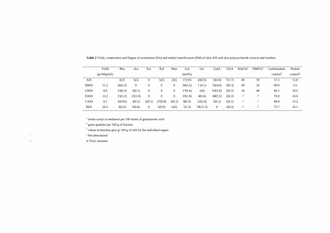
The sugar composition of okra AIS is shown in Table 2. Okra AIS consisted of
mainly glucose (44 mol %), galactose (17 mol %) and galacturonic acid (16 mol %). In
addition to polysaccharides the AIS also contained 15.8% of protein. The sugar
composition suggests that okra AIS consisted of different types of polysaccharides
including pectin, hemicelluloses such as xylan and xyloglucan, and cellulose. The
sequential extraction of the cell wall material (AIS) provides information about the
extractability of the different polysaccharides, namely pectins, hemicellulose and cellulose
Cell wall polysaccharides from Okra
35
(Voragen et al., 1995). The okra AIS was therefore sequentially extracted with different
aqueous extractants. Table 2 shows the sugar composition of the fractions obtained from
sequential extraction of okra AIS of which the HBSS and DASS fractions were the main
fractions.
The HBSS fraction consisted of 35 mol % of galacturonic acids, in addition to
high amounts of rhamnose (26 mol %) and galactose (34 mol %). Galactose was found as
the main neutral sugar in the HBSS fraction, and about 30% of all galactose present in the
AIS was recovered in this fraction. The ratio of the main sugars presented in the HBSS
fraction was 1.3:1.0:1.3 for galactose:rhamnose: galacturonic acid, respectively, which was
rather similar to that reported by Tomada et al. (1980). Lengsfeld et al. (2004) reported that
this ratio was 0.9:1.0:0.6 for the okra polysaccharide water extract. Compared to this result,
our HBSS fraction contained slightly more galactose. This was also true when compared
with the data for water-extracted okra polysaccharides as described by Deters et al. (2005).
The CHSS fraction contained higher amounts of galacturonic acid content (63 mol
%) and less galactose (17 mol %) and rhamnose (14 mol %) when compared to the HBSS
fraction. The CHSS fraction contained also 3 mol % arabinose while this sugar was not
found in the HBSS fraction. No xylose was found in the HBSS, neither in the CHSS
fractions providing evidence that no xylose containing pectic polymer like
xylogalacturonan was present in both fractions. In addition, the HBSS and CHSS fractions
contained low levels of glucuronic acid (3 and 2 mol %, respectively) which was lower than
the levels of glucuronic acid found in water-extracted okra polysaccharides (8.8 mol %) as
described by Lengsfeld et al. (2004).
The HBSS fraction had a higher ratio of rhamnose to galacturonic acid (0.7) than
the CHSS fraction (0.2). Generally, rhamnogalacturonan I (RG I) consists of alternating
rhamnose and galacturonic acid residues as a backbone (Schols and Voragen, 2002). The
ratio of rhamnose: galacturonic acid within a RG I backbone is 1:1. Consequently, the
HBSS fraction was found to contain mainly RG I segments (85%) and much less
homogalacturonan (HG) segments. The CHSS contained mainly HG segments (74%) and
less RG I segments. The high amounts of RG I segments in the HBSS fraction is a bit
uncommon since no water extract from other sources showed such a high amount of RG I
Chapter 2
36
segments. For example, water soluble soybean polysaccharide contained 43% of RG I
segments within the polysaccharides (Wang et al., 2005), water extracts from sugar beet
pulp contained 22% of RG I segments within the polysaccharides (Oosterveld et al., 1996).
The ratio of neutral sugars to rhamnose roughly indicates the length of the side chains. The
ratio of (galactose and arabinose) to rhamnose was 1.3 and 1.4 for the HBSS and CHSS
fractions, respectively. This suggests that the CHSS fraction contained slightly longer side
chains than the HBSS fraction.
The absolute configuration of sugar moieties in the HBSS and the CHSS fractions
were determined by using GC-FID after methanolysis and conversion to their
corresponding butylglycosides. The results showed that all sugars as present in the HBSS
and CHSS fractions were in D-configuration except for the rhamnosyl residues which were
in the L-configuration. These results are in contrast with those reported by Agarwal et al.
(2001) who stated that the okra gum contain L-galacturonic acid. The HBSS, okra
rhamnogalacturonan I is also different from rhamnogalacturonan I of flax seed mucilage
which contains L galactose as a neutral sugar (Naran et al., 2007).
The DASS fraction contained high amounts of galactose and galacturonic acid.
The ratio of rhamnose to galacturonic acid (0.3) was higher than that found for the CHSS
fraction (0.2). It can be calculated that the DASS fraction contained 43% of RG I segments
and 57% of HG segments. Furthermore, the DASS fraction was relatively enriched in
arabinose and galactose. The ratio of arabinose to rhamnose and the ratio of (arabinose and
galactose) to rhamnose of the DASS fraction were 1.0 and 2.5, respectively, which was
higher than that found for the CHSS fraction (0.2 and 1.4, respectively).
The CASS fraction contained glucose (52 mol %) and xylose (27 mol %) as the
main neutral sugars, and only low amounts of glucuronic acid were present which were
quite similar to other fractions. The presence of glucose xylose and glucuronic acid residues
may indicate that next to xyloglucans, acidic xylans were also part of this fraction.
Cell wall polysaccharides from Okra
37
Table 2 Yield, composition and Degree of acetylation (DA) and methyl esterification (DM) of okra AIS and okra polysaccharide extracts and residues
Yield
(g/100gAIS)
Rha
Ara
Fuc
Xyl
Man
Gal
(mol%)
Glc
GalA
GlcA
DA(%)a DM(%)a Carbohydrate
contentb
Protein
contentb
AIS
HBSS
CHSS
DASS
CASS
RES
11.2
4.8
13.2
4.1
26.5
3(2)c
26(2.5)
14(0.5)
13(1.2)
1(0.05)
3(0.5)
5(2)
0
3(0.1)
13(1.0)
3(0.1)
5(0.8)
0
0
0
0
2(0.1)
0
5(2)
0
0
0
27(0.8)
5(0.9)
3(2)
0
0
0
3(0.1)
tr(0)
17(10)
34(3.2)
17(0.6)
19(1.8)
8(0.3)
7(1.3)
44(25)
1 (0.1)
1(0)
4(0.4)
52(2.0)
78(15.3)
16(14)
35(4.0)
63(3.0)
48(5.5)
3(0.1)
0
7(1.7)
3(0.3)
2(0.1)
2(0.2)
1(0.1)
1(0.2)
40
58
18
-e
-e
-e
59
24
48
-e
-e
-e
57.5
90.0
86.2
76.9
86.8
73.7
15.8
3.5
10.5
16.6
13.2
16.1
a moles acetyl or methanol per 100 moles of galacturonic acid b gram qualities per 100 g of fraction c values in brackets give g/ 100 g of AIS for the individual sugars
\ e Not determined
\ tr Trace amounts
Chapter 2
38
In the extraction residue, the main sugar was glucose (78 mol %) representing
approximately 50% of all glucose present in AIS. This glucose originates from cellulose
and hemicellulose. The presence of xylosyl (5 mol %) and galactosyl (7 mol %) residues
indicated that xyloglucan partly remained in the residues. This xyloglucan is either strongly
embedded in the structure of the cellulose fibrils or so extensively hydrogen bonded to the
cellulose fibrils that it resisted extraction by 6 M NaOH.
The residue fraction showed that the solvents used to extract the okra AIS were
able to solubilize pectic material. However, only one-third of polysaccharides in the AIS
could be extracted while 26% was recovered in the residue. This results in a recovery of
60%. In addition, respectively, 26%, 32% and 15% of all glucose, galactose and xylose
residues in AIS were not covered by the analysis of all fractions. They most probably
belong to the hemicellulosic material and were lost during the extraction step with
concentrated alkali.
Degree of methyl esterification and acetylation
The degree of methyl esterification (DM) of the HBSS pectins (24%) was
surprisingly low and much lower than the DM of the CHSS. The DASS fraction was not
included in the analysis due to the removal of methyl esters and acetyl groups during the
dilute alkaline extraction. According to the sugar composition (Table 2), 75% of all
galacturonic acid present in the HBSS pectins originates from RG I for which there is no
evidence that galacturonosyl residues in RG I segments are methyl esterified (O´Neill and
York, 2003). Assuming that the methyl ester is only present within HG segments of HBSS
pectin, the DM of this HG could be as high as 96%. The DM of the CHSS pectins (48%)
was quite low since chelating agents are expected to extract calcium-sensitive pectins with
low DM (Ralet et al., 2003) being present in the form of calcium pectate gels (Voragen et
al., 1995).
The degree of acetylation (DA) was much higher in the HBSS fraction (58%)
compared to the CHSS fraction (18%). So far there is only evidence for the presence of O-
acetyl groups on O-2 and/or O-3 of galacturonosyl residues in HG segments and RG I
segments (O´Neill and York, 2003; Rombouts and Thibault, 1986). In general, the DA is
Cell wall polysaccharides from Okra
39
high in the RG I segments (hairy regions) of pectin as illustrated by the DA of 60% found
for modified hairy regions from apple (Schols et al., 1990). Therefore the HBSS fraction
represents pectins having a unique structure which differs from other pectins for instance
apple, sugar beet and soya pectin. Moreover, a pure RG I with high DA in water extraction
has not frequently been reported for other plants, although recently an Arabidopsis seed
mucilage was described by Deng et al., (2006) showing a water extractable linear
rhamnogalacturonan I.
Glycosidic linkage composition
To obtain more information about the different cell wall polysaccharides present in
the different extracts, the samples were subjected to linkage analysis by permethylation. In
general the data obtained were more qualitative than quantitative. First of all the uronic
acids were not reduced completely to their neutral sugar analogues and therefore they were
not measured. Secondly an underestimation of the terminal pentose and 1,4-linked
galactosyl residues could occur due to the evaporation of terminal pentose and complex
formation of 1,4-linked galactosyl residues with borate during the acetylation procedure
(Harris et al., 1984), respectively.
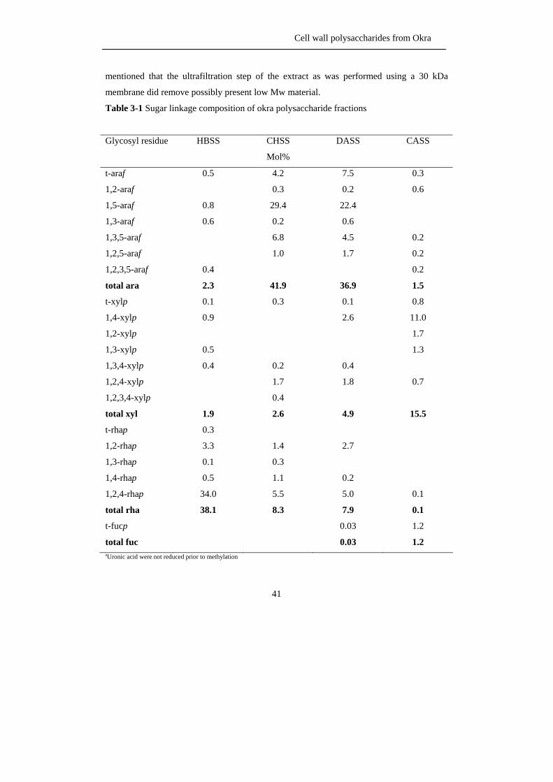
For the HBSS fraction, the sugar linkage composition results (Table 3-1 and 3-2)
indicate the presence of highly branched RG I structures, since the majority (89%) of all
1,2-linked rhamnosyl residues were O-4 substituted. These rhamnosyl residues were
substituted with short galactan side chains containing 1 or 2 galactosyl residues since 65%
of all galactose was present as terminal residues and 23% as 1,4-linked units.
The CHSS fraction was found to represent a slightly less branched RG I structure
as shown by lower levels of O-4 substituted rhamnosyl residues (66% of all rhamnose).
Only 22% of all galactosyl residues were present terminally linked revealing the fact that
slightly longer galactan side chains were present in the CHSS compared to the HBSS.
Summarizing, it can be stated that the HBSS fraction contained RG I backbones with
monomeric or dimeric galactan side chain, while the CHSS fraction contained RG I with
slightly longer galactan side chains. This observation was also reported by Tomada et al.
(1980) for water-extracted okra polysaccharides. Apart from the 1,4-linked galactosyl
Chapter 2
40
residues found in rather high amounts, some 1,6-linked and 1,3,6-linked galactosyl residues
were found to be present indicating the presence of arabinogalactan type II as side chain.
The amounts of arabinose estimated in the linkage analysis procedure for the CHSS fraction
were higher than found in the sugar composition analysis as reported in Table 1. About
42% of all arabinose were present as 1,5-linked which indicated the presence of linear
arabinan side chains.
Compared to HBSS and CHSS, the DASS fraction contained less branched RG I
as shown by the low levels of 1,4- and 1,2,4-linked rhamnosyl moieties. The majority of all
arabinose present in the CHSS and DASS were present as 1,5-; 1,3,5- and 1,2,5-linked
arabinosyl residues in the furanose form, which indicate the presence of 1,2 and 1,3
branched α-(1,5)-arabinans. The DASS fraction is relatively rich in branched arabinans in
which the number of terminally linked arabinosyl residues fitted rather well with the
number of branching points. Besides arabinan side chains, the DASS fraction contains a
mixture of AG I and AG II structures as shown by the presence of both 1,4-linked and
1,3,6-linked galactosyl residues.
The CASS fraction is obviously rich in hemicelluloses such as xylan, mannan and
glucan present as long linear 1,4-linked chains. Besides, xylose, galactose and fucose were
also present as terminal residues and in combination with the presence of 1,4,6-linked
glucosyl residues, this points to the presence of a xyloglucan (Vincken et al., 1994).
Molecular weight distribution
The Mw distribution of the polysaccharides in the different fractions obtained
from okra AIS is shown in Figure 1. The Mw distribution pattern of HBSS showed only
one population having a rather high Mw, while CHSS shows a much broader Mw
distribution representing populations with molecular weights higher and lower than HBSS.
Moreover, the Mw distribution pattern of the DASS is similar to the Mw distribution
pattern of the CHSS. The CASS fraction that represented predominantly containing
hemicellulosic polysaccharides showed one broad Mw distribution representing populations
with lower Mw values than the other fractions. These trends are also reported for olives
(Vierhuis et al., 2000), blue berries and black currents (Hilz et al., 2005). It should be
Cell wall polysaccharides from Okra
41
mentioned that the ultrafiltration step of the extract as was performed using a 30 kDa
membrane did remove possibly present low Mw material.
Table 3-1 Sugar linkage composition of okra polysaccharide fractions
Glycosyl residue HBSS CHSS
Mol%
DASS CASS
t-araf
1,2-araf
1,5-araf
1,3-araf
1,3,5-araf
1,2,5-araf
1,2,3,5-araf
total ara
t-xylp
1,4-xylp
1,2-xylp
1,3-xylp
1,3,4-xylp
1,2,4-xylp
1,2,3,4-xylp
total xyl
t-rhap
1,2-rhap
1,3-rhap
1,4-rhap
1,2,4-rhap
total rha
t-fucp
total fuc
0.5
0.8
0.6
0.4
2.3
0.1
0.9
0.5
0.4
1.9
0.3
3.3
0.1
0.5
34.0
38.1
4.2
0.3
29.4
0.2
6.8
1.0
41.9
0.3
0.2
1.7
0.4
2.6
1.4
0.3
1.1
5.5
8.3
7.5
0.2
22.4
0.6
4.5
1.7
36.9
0.1
2.6
0.4
1.8
4.9
2.7
0.2
5.0
7.9
0.03
0.03
0.3
0.6
0.2
0.2
0.2
1.5
0.8
11.0
1.7
1.3
0.7
15.5
0.1
0.1
1.2
1.2 aUronic acid were not reduced prior to methylation
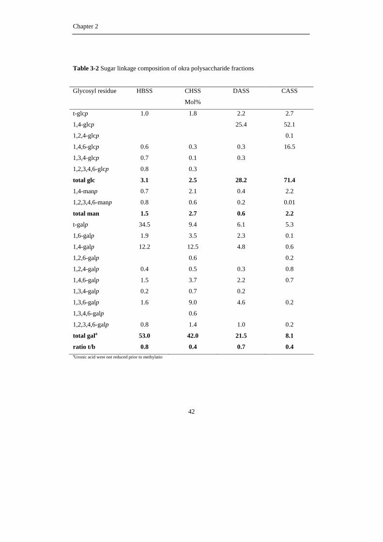
Chapter 2
42
Table 3-2 Sugar linkage composition of okra polysaccharide fractions
Glycosyl residue HBSS CHSS
Mol%
DASS CASS
t-glcp
1,4-glcp
1,2,4-glcp
1,4,6-glcp
1,3,4-glcp
1,2,3,4,6-glcp
total glc
1,4-manp
1,2,3,4,6-manp
total man
t-galp
1,6-galp
1,4-galp
1,2,6-galp
1,2,4-galp
1,4,6-galp
1,3,4-galp
1,3,6-galp
1,3,4,6-galp
1,2,3,4,6-galp
total gala
ratio t/b
1.0
0.6
0.7
0.8
3.1
0.7
0.8
1.5
34.5
1.9
12.2
0.4
1.5
0.2
1.6
0.8
53.0
0.8
1.8
0.3
0.1
0.3
2.5
2.1
0.6
2.7
9.4
3.5
12.5
0.6
0.5
3.7
0.7
9.0
0.6
1.4
42.0
0.4
2.2
25.4
0.3
0.3
28.2
0.4
0.2
0.6
6.1
2.3
4.8
0.3
2.2
0.2
4.6
1.0
21.5
0.7
2.7
52.1
0.1
16.5
71.4
2.2
0.01
2.2
5.3
0.1
0.6
0.2
0.8
0.7
0.2
0.2
8.1
0.4 aUronic acid were not reduced prior to methylatio
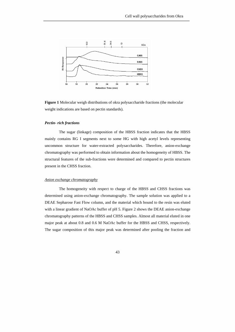
Cell wall polysaccharides from Okra
43
Figure 1 Molecular weigh distributions of okra polysaccharide fractions (the molecular
weight indications are based on pectin standards).
Pectin- rich fractions
The sugar (linkage) composition of the HBSS fraction indicates that the HBSS
mainly contains RG I segments next to some HG with high acetyl levels representing
uncommon structure for water-extracted polysaccharides. Therefore, anion-exchange
chromatography was performed to obtain information about the homogeneity of HBSS. The
structural features of the sub-fractions were determined and compared to pectin structures
present in the CHSS fraction.
Anion exchange chromatography
The homogeneity with respect to charge of the HBSS and CHSS fractions was
determined using anion-exchange chromatography. The sample solution was applied to a
DEAE Sepharose Fast Flow column, and the material which bound to the resin was eluted
with a linear gradient of NaOAc buffer of pH 5. Figure 2 shows the DEAE anion-exchange
chromatography patterns of the HBSS and CHSS samples. Almost all material eluted in one
major peak at about 0.8 and 0.6 M NaOAc buffer for the HBSS and CHSS, respectively.
The sugar composition of this major peak was determined after pooling the fraction and
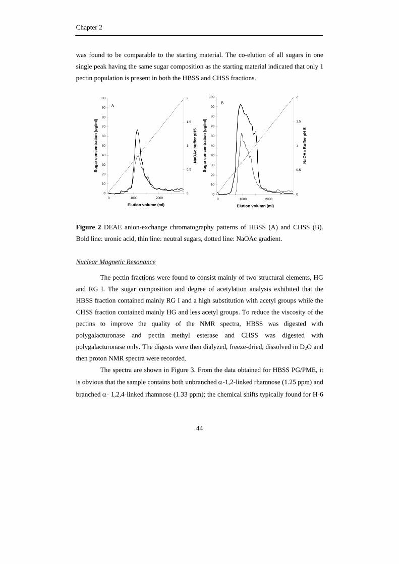
Chapter 2
44
was found to be comparable to the starting material. The co-elution of all sugars in one
single peak having the same sugar composition as the starting material indicated that only 1
pectin population is present in both the HBSS and CHSS fractions.
0
10
20
30
40
50
60
70
80
90
100
0 1000 2000
Elution volume (ml)
Suga
r con
cent
ratio
n (u
g/m
l)
0
0.5
1
1.5
2
NaO
Ac
buffe
r pH
5
A
0
10
20
30
40
50
60
70
80
90
100
0 1000 2000
Elution volumn (ml)Su
gar c
once
ntra
tion
(ug/
ml)
0
0.5
1
1.5
2
NaO
Ac
Buf
fer p
H 5
B
0
10
20
30
40
50
60
70
80
90
100
0 1000 2000
Elution volume (ml)
Suga
r con
cent
ratio
n (u
g/m
l)
0
0.5
1
1.5
2
NaO
Ac
buffe
r pH
5
A
0
10
20
30
40
50
60
70
80
90
100
0 1000 2000
Elution volumn (ml)Su
gar c
once
ntra
tion
(ug/
ml)
0
0.5
1
1.5
2
NaO
Ac
Buf
fer p
H 5
B
Figure 2 DEAE anion-exchange chromatography patterns of HBSS (A) and CHSS (B).
Bold line: uronic acid, thin line: neutral sugars, dotted line: NaOAc gradient.
Nuclear Magnetic Resonance
The pectin fractions were found to consist mainly of two structural elements, HG
and RG I. The sugar composition and degree of acetylation analysis exhibited that the
HBSS fraction contained mainly RG I and a high substitution with acetyl groups while the
CHSS fraction contained mainly HG and less acetyl groups. To reduce the viscosity of the
pectins to improve the quality of the NMR spectra, HBSS was digested with
polygalacturonase and pectin methyl esterase and CHSS was digested with
polygalacturonase only. The digests were then dialyzed, freeze-dried, dissolved in D2O and
then proton NMR spectra were recorded.
The spectra are shown in Figure 3. From the data obtained for HBSS PG/PME, it
is obvious that the sample contains both unbranched α-1,2-linked rhamnose (1.25 ppm) and
branched α- 1,2,4-linked rhamnose (1.33 ppm); the chemical shifts typically found for H-6
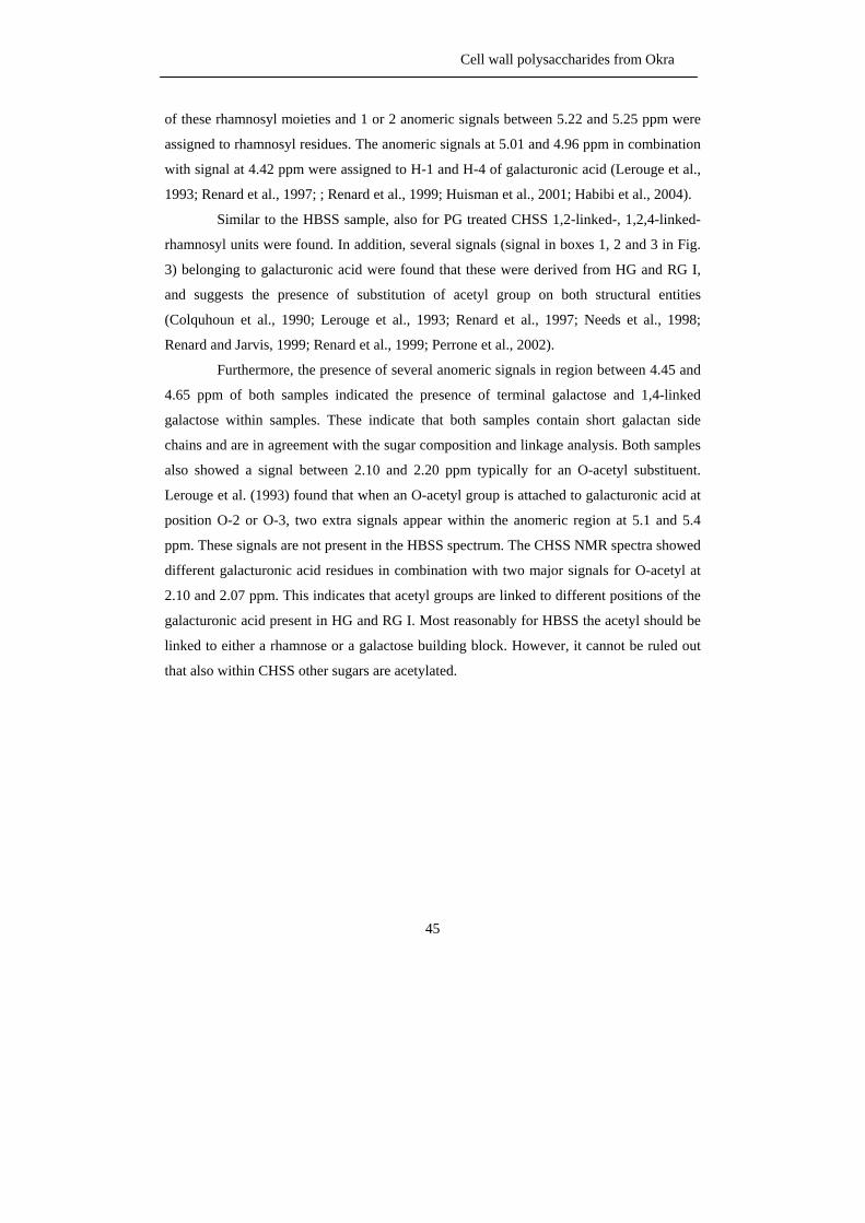
Cell wall polysaccharides from Okra
45
of these rhamnosyl moieties and 1 or 2 anomeric signals between 5.22 and 5.25 ppm were
assigned to rhamnosyl residues. The anomeric signals at 5.01 and 4.96 ppm in combination
with signal at 4.42 ppm were assigned to H-1 and H-4 of galacturonic acid (Lerouge et al.,
1993; Renard et al., 1997; ; Renard et al., 1999; Huisman et al., 2001; Habibi et al., 2004).
Similar to the HBSS sample, also for PG treated CHSS 1,2-linked-, 1,2,4-linked-
rhamnosyl units were found. In addition, several signals (signal in boxes 1, 2 and 3 in Fig.
3) belonging to galacturonic acid were found that these were derived from HG and RG I,
and suggests the presence of substitution of acetyl group on both structural entities
(Colquhoun et al., 1990; Lerouge et al., 1993; Renard et al., 1997; Needs et al., 1998;
Renard and Jarvis, 1999; Renard et al., 1999; Perrone et al., 2002).
Furthermore, the presence of several anomeric signals in region between 4.45 and
4.65 ppm of both samples indicated the presence of terminal galactose and 1,4-linked
galactose within samples. These indicate that both samples contain short galactan side
chains and are in agreement with the sugar composition and linkage analysis. Both samples
also showed a signal between 2.10 and 2.20 ppm typically for an O-acetyl substituent.
Lerouge et al. (1993) found that when an O-acetyl group is attached to galacturonic acid at
position O-2 or O-3, two extra signals appear within the anomeric region at 5.1 and 5.4
ppm. These signals are not present in the HBSS spectrum. The CHSS NMR spectra showed
different galacturonic acid residues in combination with two major signals for O-acetyl at
2.10 and 2.07 ppm. This indicates that acetyl groups are linked to different positions of the
galacturonic acid present in HG and RG I. Most reasonably for HBSS the acetyl should be
linked to either a rhamnose or a galactose building block. However, it cannot be ruled out
that also within CHSS other sugars are acetylated.
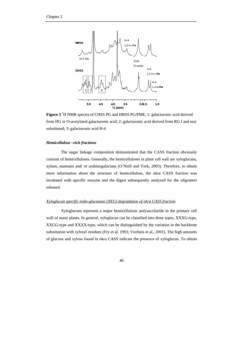
Chapter 2
46
1.01.52.03.54.04.55.0f1 (ppm)
CHSS
HBSS
1 2 3
H-6
1,2-α-rha
H-6
1,2,4-α-rha
CH3
O-acetyl
H-4
1,2-α-rha
H-1 rha
1.01.52.03.54.04.55.0f1 (ppm)
1.01.52.03.54.04.55.0f1 (ppm)
CHSS
HBSS
1 2 3
H-6
1,2-α-rha
H-6
1,2,4-α-rha
CH3
O-acetyl
H-4
1,2-α-rha
H-1 rha
Figure 3 1H NMR spectra of CHSS PG and HBSS PG/PME; 1: galacturonic acid derived
from HG or O-acetylated galacturonic acid; 2: galacturonic acid derived from RG I and non
substituted; 3: galacturonic acid H-4
Hemicellulose –rich fractions
The sugar linkage composition demonstrated that the CASS fraction obviously
consists of hemicelluloses. Generally, the hemicelluloses in plant cell wall are xyloglucans,
xylans, mannans and/ or arabinogalactans (O´Neill and York, 2003). Therefore, to obtain
more information about the structure of hemicellulose, the okra CASS fraction was
incubated with specific enzyme and the digest subsequently analyzed for the oligomers
released.
Xyloglucan specific endo-glucanase (XEG) degradation of okra CASS fraction
Xyloglucans represent a major hemicellulosic polysaccharide in the primary cell
wall of many plants. In general, xyloglucan can be classified into three types, XXXG-type,
XXGG-type and XXXX-type, which can be distinguished by the variation in the backbone
substitution with xylosyl residues (Fry et al. 1993; Vierhuis et al., 2001). The high amounts
of glucose and xylose found in okra CASS indicate the presence of xyloglucan. To obtain
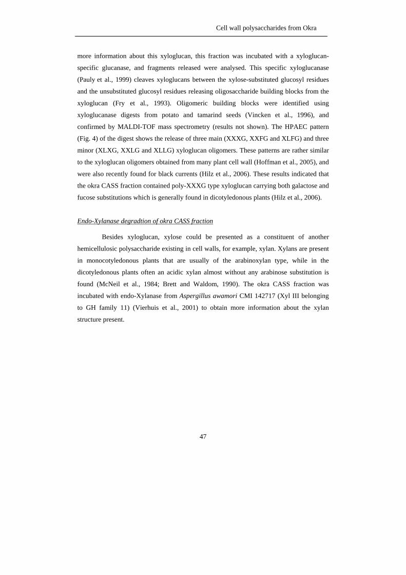
Cell wall polysaccharides from Okra
47
more information about this xyloglucan, this fraction was incubated with a xyloglucan-
specific glucanase, and fragments released were analysed. This specific xyloglucanase
(Pauly et al., 1999) cleaves xyloglucans between the xylose-substituted glucosyl residues
and the unsubstituted glucosyl residues releasing oligosaccharide building blocks from the
xyloglucan (Fry et al., 1993). Oligomeric building blocks were identified using
xyloglucanase digests from potato and tamarind seeds (Vincken et al., 1996), and
confirmed by MALDI-TOF mass spectrometry (results not shown). The HPAEC pattern
(Fig. 4) of the digest shows the release of three main (XXXG, XXFG and XLFG) and three
minor (XLXG, XXLG and XLLG) xyloglucan oligomers. These patterns are rather similar
to the xyloglucan oligomers obtained from many plant cell wall (Hoffman et al., 2005), and
were also recently found for black currents (Hilz et al., 2006). These results indicated that
the okra CASS fraction contained poly-XXXG type xyloglucan carrying both galactose and
fucose substitutions which is generally found in dicotyledonous plants (Hilz et al., 2006).
Endo-Xylanase degradtion of okra CASS fraction
Besides xyloglucan, xylose could be presented as a constituent of another
hemicellulosic polysaccharide existing in cell walls, for example, xylan. Xylans are present
in monocotyledonous plants that are usually of the arabinoxylan type, while in the
dicotyledonous plants often an acidic xylan almost without any arabinose substitution is
found (McNeil et al., 1984; Brett and Waldom, 1990). The okra CASS fraction was
incubated with endo-Xylanase from Aspergillus awamori CMI 142717 (Xyl III belonging
to GH family 11) (Vierhuis et al., 2001) to obtain more information about the xylan
structure present.
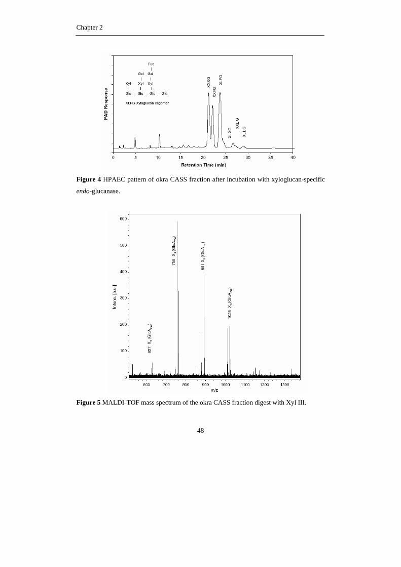
Chapter 2
48
Figure 4 HPAEC pattern of okra CASS fraction after incubation with xyloglucan-specific
endo-glucanase.
Figure 5 MALDI-TOF mass spectrum of the okra CASS fraction digest with Xyl III.
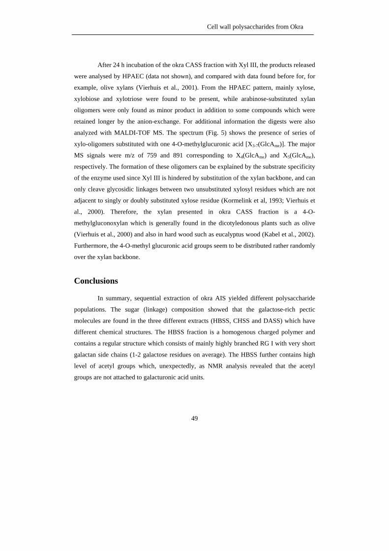
Cell wall polysaccharides from Okra
49
After 24 h incubation of the okra CASS fraction with Xyl III, the products released
were analysed by HPAEC (data not shown), and compared with data found before for, for
example, olive xylans (Vierhuis et al., 2001). From the HPAEC pattern, mainly xylose,
xylobiose and xylotriose were found to be present, while arabinose-substituted xylan
oligomers were only found as minor product in addition to some compounds which were
retained longer by the anion-exchange. For additional information the digests were also
analyzed with MALDI-TOF MS. The spectrum (Fig. 5) shows the presence of series of
xylo-oligomers substituted with one 4-O-methylglucuronic acid [X3-7(GlcAme)]. The major
MS signals were m/z of 759 and 891 corresponding to X4(GlcAme) and X5(GlcAme),
respectively. The formation of these oligomers can be explained by the substrate specificity
of the enzyme used since Xyl III is hindered by substitution of the xylan backbone, and can
only cleave glycosidic linkages between two unsubstituted xylosyl residues which are not
adjacent to singly or doubly substituted xylose residue (Kormelink et al, 1993; Vierhuis et
al., 2000). Therefore, the xylan presented in okra CASS fraction is a 4-O-
methylgluconoxylan which is generally found in the dicotyledonous plants such as olive
(Vierhuis et al., 2000) and also in hard wood such as eucalyptus wood (Kabel et al., 2002).
Furthermore, the 4-O-methyl glucuronic acid groups seem to be distributed rather randomly
over the xylan backbone.
Conclusions
In summary, sequential extraction of okra AIS yielded different polysaccharide
populations. The sugar (linkage) composition showed that the galactose-rich pectic
molecules are found in the three different extracts (HBSS, CHSS and DASS) which have
different chemical structures. The HBSS fraction is a homogenous charged polymer and
contains a regular structure which consists of mainly highly branched RG I with very short
galactan side chains (1-2 galactose residues on average). The HBSS further contains high
level of acetyl groups which, unexpectedly, as NMR analysis revealed that the acetyl
groups are not attached to galacturonic acid units.

Chapter 2
50
The CHSS fraction is also a homogeneously charged polymer and contains mainly
HG and some RG I with galactan side chains consisting of on average 2 to 3 galactosyl
residues. Also more heterogeneous side chains of arabinan and AG II are present. Both
HBSS and CHSS showed one population on anion-exchange chromatography indicating
different ratios of homogalacturonan and rhamnogalacturonan in HBSS and CHSS. The
NMR studies of PG treated HBSS and CHSS showed the presence of acetyl groups that are
linked to other sugar residues present like rhamnose and/or galactose than galacturonic
acid. Moreover, the DASS fraction contains even more complex pectins with arabinans, AG
I and AG II, as neutral side chains.
Besides pectic material, the okra AIS also contains hemicellulosic polysaccharides
which were mainly present in the CASS fraction. The hemicellulosic material in this
fraction was found to consist of mainly XXXG-type xyloglucan and 4-
methylglucuronoxylan.
To obtain more information about the position of the acetyl groups and the length
of galactan side chains, more enzyme degradation studies of the polymer and NMR and MS
characterizations of oligomers will be performed.
Material and methods
Material
The soft and mature of okra pods, A. esculentus (L.) Moench, (5 – 10 cm in
length) were grown and collected at local market in June 2005, Thailand.
Isolation of alcohol-insoluble solid
After removal of the seeds, the okra pods were sliced and homogenized 2 times
with 70 % (v/v) aqueous ethanol at room temperature. After filtration, the insoluble
residues were combined and washed with two volumes of chloroform/methanol (1/1, v/v)
with gentle stirring for 30 min to remove low molecular weight (colored) compounds
(Monpien, 2005). After filtration, the filtrates were washed with acetone and air dried
(Alcohol Insoluble Solid, AIS).
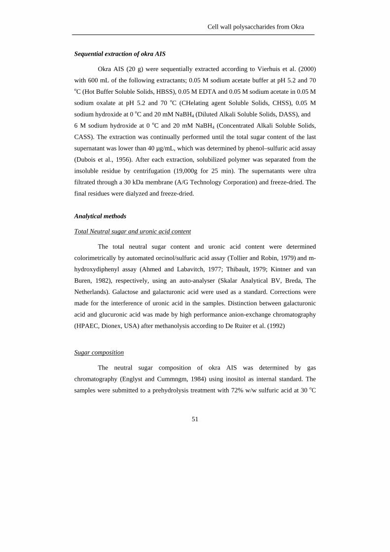
Cell wall polysaccharides from Okra
51
Sequential extraction of okra AIS
Okra AIS (20 g) were sequentially extracted according to Vierhuis et al. (2000)
with 600 mL of the following extractants; 0.05 M sodium acetate buffer at pH 5.2 and 70 oC (Hot Buffer Soluble Solids, HBSS), 0.05 M EDTA and 0.05 M sodium acetate in 0.05 M
sodium oxalate at pH 5.2 and 70 oC (CHelating agent Soluble Solids, CHSS), 0.05 M
sodium hydroxide at 0 oC and 20 mM NaBH4 (Diluted Alkali Soluble Solids, DASS), and
6 M sodium hydroxide at 0 oC and 20 mM NaBH4 (Concentrated Alkali Soluble Solids,
CASS). The extraction was continually performed until the total sugar content of the last
supernatant was lower than 40 µg/mL, which was determined by phenol–sulfuric acid assay
(Dubois et al., 1956). After each extraction, solubilized polymer was separated from the
insoluble residue by centrifugation (19,000g for 25 min). The supernatants were ultra
filtrated through a 30 kDa membrane (A/G Technology Corporation) and freeze-dried. The
final residues were dialyzed and freeze-dried.
Analytical methods
Total Neutral sugar and uronic acid content
The total neutral sugar content and uronic acid content were determined
colorimetrically by automated orcinol/sulfuric acid assay (Tollier and Robin, 1979) and m-
hydroxydiphenyl assay (Ahmed and Labavitch, 1977; Thibault, 1979; Kintner and van
Buren, 1982), respectively, using an auto-analyser (Skalar Analytical BV, Breda, The
Netherlands). Galactose and galacturonic acid were used as a standard. Corrections were
made for the interference of uronic acid in the samples. Distinction between galacturonic
acid and glucuronic acid was made by high performance anion-exchange chromatography
(HPAEC, Dionex, USA) after methanolysis according to De Ruiter et al. (1992)
Sugar composition
The neutral sugar composition of okra AIS was determined by gas
chromatography (Englyst and Cummngm, 1984) using inositol as internal standard. The
samples were submitted to a prehydrolysis treatment with 72% w/w sulfuric acid at 30 oC
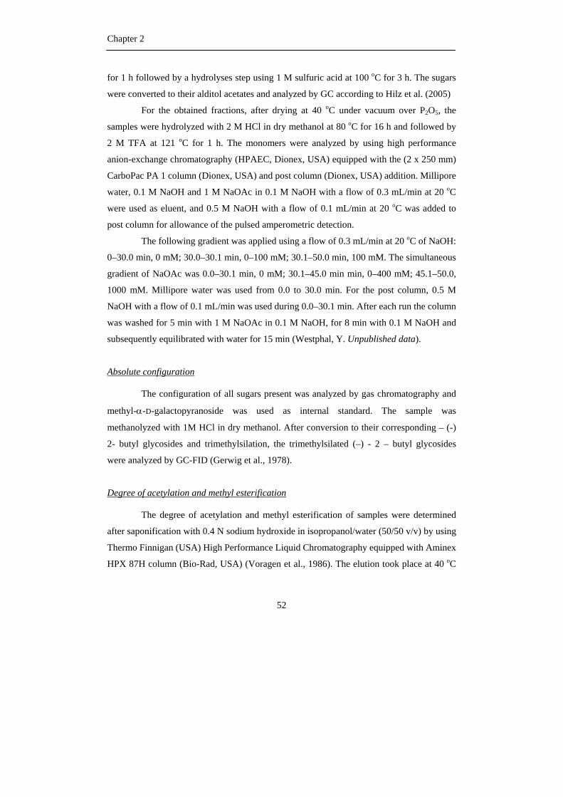
Chapter 2
52
for 1 h followed by a hydrolyses step using 1 M sulfuric acid at 100 oC for 3 h. The sugars
were converted to their alditol acetates and analyzed by GC according to Hilz et al. (2005)
For the obtained fractions, after drying at 40 oC under vacuum over P2O5, the
samples were hydrolyzed with 2 M HCl in dry methanol at 80 oC for 16 h and followed by
2 M TFA at 121 oC for 1 h. The monomers were analyzed by using high performance
anion-exchange chromatography (HPAEC, Dionex, USA) equipped with the (2 x 250 mm)
CarboPac PA 1 column (Dionex, USA) and post column (Dionex, USA) addition. Millipore
water, 0.1 M NaOH and 1 M NaOAc in 0.1 M NaOH with a flow of 0.3 mL/min at 20 oC
were used as eluent, and 0.5 M NaOH with a flow of 0.1 mL/min at 20 oC was added to
post column for allowance of the pulsed amperometric detection.
The following gradient was applied using a flow of 0.3 mL/min at 20 oC of NaOH:
0–30.0 min, 0 mM; 30.0–30.1 min, 0–100 mM; 30.1–50.0 min, 100 mM. The simultaneous
gradient of NaOAc was 0.0–30.1 min, 0 mM; 30.1–45.0 min min, 0–400 mM; 45.1–50.0,
1000 mM. Millipore water was used from 0.0 to 30.0 min. For the post column, 0.5 M
NaOH with a flow of 0.1 mL/min was used during 0.0–30.1 min. After each run the column
was washed for 5 min with 1 M NaOAc in 0.1 M NaOH, for 8 min with 0.1 M NaOH and
subsequently equilibrated with water for 15 min (Westphal, Y. Unpublished data).
Absolute configuration
The configuration of all sugars present was analyzed by gas chromatography and
methyl-α-D-galactopyranoside was used as internal standard. The sample was
methanolyzed with 1M HCl in dry methanol. After conversion to their corresponding – (-)
2- butyl glycosides and trimethylsilation, the trimethylsilated (–) - 2 – butyl glycosides
were analyzed by GC-FID (Gerwig et al., 1978).
Degree of acetylation and methyl esterification
The degree of acetylation and methyl esterification of samples were determined
after saponification with 0.4 N sodium hydroxide in isopropanol/water (50/50 v/v) by using
Thermo Finnigan (USA) High Performance Liquid Chromatography equipped with Aminex
HPX 87H column (Bio-Rad, USA) (Voragen et al., 1986). The elution took place at 40 oC
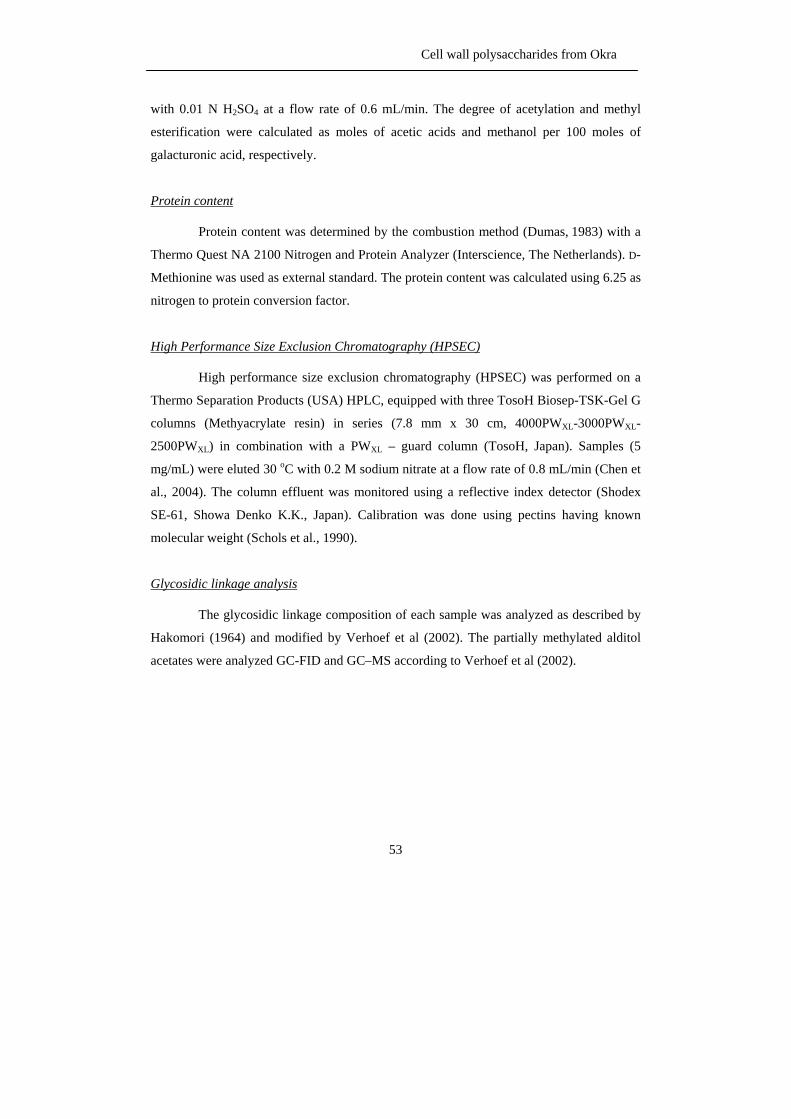
Cell wall polysaccharides from Okra
53
with 0.01 N H2SO4 at a flow rate of 0.6 mL/min. The degree of acetylation and methyl
esterification were calculated as moles of acetic acids and methanol per 100 moles of
galacturonic acid, respectively.
Protein content
Protein content was determined by the combustion method (Dumas, 1983) with a
Thermo Quest NA 2100 Nitrogen and Protein Analyzer (Interscience, The Netherlands). D-
Methionine was used as external standard. The protein content was calculated using 6.25 as
nitrogen to protein conversion factor.
High Performance Size Exclusion Chromatography (HPSEC)
High performance size exclusion chromatography (HPSEC) was performed on a
Thermo Separation Products (USA) HPLC, equipped with three TosoH Biosep-TSK-Gel G
columns (Methyacrylate resin) in series (7.8 mm x 30 cm, 4000PWXL-3000PWXL-
2500PWXL) in combination with a PWXL – guard column (TosoH, Japan). Samples (5
mg/mL) were eluted 30 oC with 0.2 M sodium nitrate at a flow rate of 0.8 mL/min (Chen et
al., 2004). The column effluent was monitored using a reflective index detector (Shodex
SE-61, Showa Denko K.K., Japan). Calibration was done using pectins having known
molecular weight (Schols et al., 1990).
Glycosidic linkage analysis
The glycosidic linkage composition of each sample was analyzed as described by
Hakomori (1964) and modified by Verhoef et al (2002). The partially methylated alditol
acetates were analyzed GC-FID and GC–MS according to Verhoef et al (2002).
Chapter 2
54
Enzymatic degradation
Pectin degradation
The HBSS fraction was dissolved (3 mg/mL) in 50 mM NaOAc buffer, at pH 5,
and incubated with 0.016 units of polygalacturonase from A. aculeatus and 1.08 units of
pectin methyl esterase from A. niger (Schols et al., 1990). The CHSS fraction (4 mg/mL)
was incubated with 0.024 units of polygalacturonase. The incubations were performed at 40 oC for 24 h and the digests were subsequently heated for 5 min at 100 oC to inactivate the
enzyme. The digests were dialyzed by Centricon centrifugal filter devises with 3 kDa cut
off at 1500g for 30 min and then, freeze dried.
Xyloglucan specific endo-glucanase (XEG) degradation
The CASS fraction was dissolved (5 mg/mL) in 50 mM NaOAc buffer, at pH 5,
and incubated with 2.3 units of xyloglucan specific endo-glucanase from A. aculeatus
(Pauly et al., 1999) at 40 oC for 16 h. The digest was heated at 100 oC for 5 min to
inactivate the enzyme.
endo- Xylanase III degradation
The CASS fraction was dissolve (10 mg/mL) in 150 mM NaOAc buffer, at pH 5,
and incubated at 40 oC for 24 h with 0.09 units of endo-xylanase III from A. awamori
(Vierhuis et al., 2001). The enzyme was inactivated by heating the digest for 5 min at 100 oC.
HPAEC of oligosaccharides
For the determination of xyloglucan oligomer were analysed by using a 4 x
250mm CarboPac PA 100 column (Dionex, USA) with pulsed amperometric detection.
Gradients of NaOH and NaOAc with a flow of 1 mL/min were use to elute the oligomer
according to Vincken et al (1996).
Cell wall polysaccharides from Okra
55
For the determination of digest from endo-xylanase III, the (2 x 250 mm)
CarboPac PA 1 column (Dionex, USA) with pulse amperometric detection was equilibrate
with 100 mM NaOH. Gradients of NaOH and NaOAc were used simultaneously to elute
the oligomers according to Vierhuis et al (2001).
Matrix –Assisted Laser Desorption Ionization-Time Of Flight Mass Spectrometry (MALDI-
TOF MS) analysis
MALDI-TOF MS analysis was performed on Ultra flex instrument (Bruker
Daltonics, Germany) with a Nitrogen 337 nm laser beam. The data were collected from
average of 200 shots with the lowest laser energy (35%). The digest sample was desalted
with resin (AG 50W-X8 Resin, H+ form; Bio – Rad, USA). A small spatula of Dowex resin
was added to 10 µL of sample solution, mixed and centrifuge for 5 min at 13,000g. One µL
of matrix solution ,10 mg/mL of 2,5-dihydroxybenzoic acid in millipore water (Milli-Q
Gradient A-10, USA), was placed on a MALDI-TOF plate together with 1 µL of sample
solution and dried under a constant of warm air.
Anion exchange chromatography
Anion-exchange chromatography was performed on a DEAE Sepharose Fast Flow
column (50 x 2.6 cm, Amersham Bioscience, Sweden) using an Akta explorer system
(Amersham Biosciences, Sweden). The sample (280 mL, 0.5 mg/mL) was loaded to the
column (10 mL/min). After loading with sample the column was washed with water for 1
column volume (250 mL, 50 mL/min) and eluted successively with a linear gradient of 0-2
M of NaOAc buffer, pH 5, within 10 column volumes. Finally the column was washed with
2 column volumes of 1 N NaOH. During elution with NaOAc buffer, fractions of 50 mL
were collected as well as the column washed and analyzed for neutral sugar and uronic acid
content as described. The alkaline fractions were neutralized directly by adding acetic acid.
After pooling appropriates fractions, pools were dialyzed and freeze dried.
Chapter 2
56
13C and 1H Nuclear Magnetic Resonance (NMR)
Prior to NMR analyses, the sample was dissolved in 5 mM NaOAc, pH 5, to set
the pD = pH and freeze dried. Then the sample were dissolved in 99.96% D2O (Cambridge
Isotope Laboratories, USA) and after freeze-drying dissolved again in 99.996% D2O
(Cambridge Isotope Laboratories, USA). NMR spectra were recorded at a probe
temperature of 25°C on a Bruker AV-600 spectrometer equipped with a cryoprobe located
at the Biqualys, Wageningen. Chemical shifts were expressed in ppm relative to internal
acetone: δ=2.225 ppm for 1H. The 1D 1H proton spectra were recorded at 600.13 MHz
using 64 scans of 8192 data points and a sweep width of 3000 Hz.
Acknowledgment
This study has been carried out with financial support from the Commission on
Higher Education Ministry of Education, THAILAND, under Staff Development project
for the Joint Ph.D. Program in Food Science at Kasetsart University, Thailand. Partial
support also came from the Graduate School of Kasetsart University, Thailand. Moreover,
we would like to thank Dr. Pieter de Waard from the Wageningen NMR centre for helping
with the NMR experiments. Furthermore, we would like to thank Biqualys, Wageningen for
providing measuring time on their 600 MHz cryoprobe NMR spectrometer.
References
Agarwal, M., Srinivasan, R. & Mishra, A. (2001). Study on flocculation efficiency of okra gum in sewage waste
water. Macromol. Mater. Eng., 256, 560-563.
Ahmed, A.E.R. & Labavitch, J.M. (1977) A simplified method for accurate determination of cell wall uronide
content. J. Food Biochem., 1, 361-365.
BeMiller, J.N., Whistler, R.L. & Barbalowm, D.G. (1993). Aloe, China, Flexseed, Okra, Psyllium seed, Quince
seed, and Tamarind Gums. In L.W. Roy & J.N. BeMiller, eds., Industrial Gums Polysaccharide and
their Derivatives (pp. 235-255). San Diago: Academic Press.
Brett, C. & Waldom, K. (1990). Physiology and biochemistry of plant cell walls (pp. 1-45). Boston: Hyman.
Cell wall polysaccharides from Okra
57
Chen, C.H., Sheu, M.T., Chen, T.F., Wang, Y.C., Hou, W.C., Liu, D.Z., Chung, T.C. & Liang, Y.C. (2006).
Suppression of endotoxin-induced proinflammatory responses by citrus pectin through blocking LPS
signaling pathways. Biochem. Pharmacol., 72, 1001-1009.
Colquhoun, I.J., De Ruiter, G.A., Schols, H.A. & Voragen, A.G.J. (1990). Hairy (ramified) regions of pectins 3.
Identification by nmr-spectroscopy of oligosaccharides obtained by treatment of the hairy regions of
apple pectin with rhamnogalacturonase. Carbohydr. Res., 206, 131-144.
Costantino, A.J. & Romanchick-Cerpoviez, J.E. (2004). Physical and sensory measures indicate moderate fat
replacement in frozen dairy dessert is feasible using okra gum as a milk-fat ingredient substitute. J. Am.
Diet. Assoc., 104, 44.
De Ruiter, G.A., Schols, H.A., Voragen, A.G.J. & Rombouts, F.M. (1992). Carbohydrate analysis of water soluble
uronic acid containing polysaccharides with High – Performance -Anion- Exchange Chromatography
using methanolysis combined with TFA hydrolysis is superior to four other methods. Anal. Biochem.,
207, 176-185.
Deters, A.M., Lengsfeld, C. & Andreas, H. (2005). Oligo- and polysaccharides exhibited a structure-dependent
bioactivity on human keratinocytes in vitro. J. Ethnopharm., 102, 391-399.
Deng, C., O'Neill, M.A. & York, W.S. (2006). Selective chemical depolymerization of rhamnogalacturonans.
Carbohydr. Res., 341, 474-484.
Dubois, M., Gilles, K.A., Hamilton, J.K., Rebers, D.A. & Smith, F. (1956). Colorimetric method for determination
of sugers and related substances. Anal. Chem. 28, 1956, 350-356
Dumas, J.B.A. (1831). Procedes de L’analyse Organique. Annales de Chimie et de. Physique, 47, 198-205.
Englyst, H.N. & Cummngm, J.H. (1984). Simplified method for the measurement of total non-starch
polysaccharides by gas-liquid chromatography of constituent sugars as alditol acetates. Analyst., 109,
93-942.
Fry, S.C., York, W.S., Albersheim, P., Darvill, A., Hayashi, T., Joseleau, J.P., Kato, Y., Lorences, E.P.,
Maclachlan, G.A., McNeil, M., Mort, A.J., Reid, J.S.G., Seitz, H.U., Selvendran, R.R., Voragen, A.G.J.
& White, A.R. (1993). An unambiguous nomenclature for xyloglucan derived oligosaccharides.
Physiol. Plantarum., 89, 1-3.
Gerwig, G.J., Kamerling, J.P. & Vliegenthart, J.F.G. (1978). Determination of the D and Lconfiguration of neutral
monosaccharides by high-resolution capillary G.L.C. Carbohydr. Res., 62, 349-357.
Habibi, Y., Heyraud, A., Mahrouz, M., & Vignon, M. R. (2004). Structural features of pectic polysaccharides from
the skin of Opuntia ficus-indica prickly pear fruits. Carbohydr. Res., 339, 1119-1127.
Hakomori S. (1964). A rapid permethylation of glycolipid and polysaccharide catalysed by methylsulfinyl
carbanion in dimethyl sulfoxide. J. Biochem., 55, 205-208.
Harris, P.J., Henry, R.J., Blakeney, A.B. & Stone, B.A. (1984). An improved procedure for the methylation
analysis of oligosaccharides and polysaccharides. Carbohydr. Res., 127, 59-73.
Hilz, H., de Jong, L.E., Kabel, M.A., Schols, H.A. & Voragen, A.G.J. (2006). A comparison of liquid
chromatography, capillary electrophoresis, and mass spectrometry methods to determine xyloglucan
structure in black current. J. Chromatogr. A. 1113, 275 – 286.
Chapter 2
58
Hilz, H., Bakx, E.J., Schols, H.A. & Voragen, A.G.J. (2005). Cell wall polysaccharides in black currants and
bilberries—characterisation in berries, juice, and press cake. Carbohydr. Polym., 59, 477-488.
Hoffman, M., Jia, Z., Pena, M.J., Cash, M., Harper, A., Blackburn, A.R., Darvill, A. & York, W.S. (2005).
Structure analysis of xyloglucans in primary cell walls of plants in the subclass Asteridae. Carbohydr.
Res., 340, 1826-1840.
Huisman, M.M.H., Fransen, C.T.M., Kamerling, J.P., Vliegenthart, J.F.G., Schols, H.A., & Voragen, A.G.J.
(2001). The CDTA-soluble pectic substances from soybean meal are composed of rhamnogalacturonan
and xylogalacturonan but not homogalacturonan. Biopolymers, 58, 279-294.
Kabel, M.A., Schols, H.A. & Voragen, A.G.J. (2002). Complex xylo-oligosaccharides identified from hydro-
thermally treated Eucalyptus wood and brewery’s spent grain. Carbohydr. Polym. 50: 191-200.
Kintner P.K. & van Buren, J.P. (1982) Carbohydrate interference and its correction in pectin analysis using the m-
hydroxy diphenyl method. J. Food Sci., 47, 756-765
Kormelink, F.J.M., Gruppen, H., Viëtor, R.J. & Voragen, A.G.J. (1993). Mode of action of the xylan-degrading
enzymes from Aspergillus awamori on alkali-extractable cereal arabinoxylan. Carbohydr. Res., 249,
355-367.
Lengsfeld, C., Titgemeyer, F., Faller, G. & Hensel, A. (2004). Glycosylated compounds from okra inhibit
adhesion of helicobacter pylori to human gastric mucosa. J. Agric. Food Chem., 52, 1495-1503.
Lerouge, P., O’Neill, M.A., Darvill, A.G. & Albersheim, P. (1993). Structural characterization of endo-glycanase-
generated oligoglycosyl side-chains of rhamnogalacturonan-I. Carbohydr. Res., 243, 359-371.
McNeil, M., Darvill, A.G., Fry, S.C. & Albersheim, P. (1984). Structure and function of the primary cell walls of
plants. Annu. Rev. Biochem., 53, 625-663.
Monpien, W & Sajjaanantakul, T. (2005). Modification of okra polysaccharides using ammonium hydroxide and
methylamine, MSc. Thesis, Department of Food Science and Technology, Kasetsart University,
Thailand.
Naran, R.; Guibing, C. & Carpita, N.C. (2007). A novel rhamnogalacturonan I from the seed mucilage of flax
functions in enhancing viscosity, Abstacts of papers , XI th Cell Wall Meeting, Copenhagen, Denmark,
12-17 August, 2007, Physiol. Plantarum., 130.
Needs, P.W., Rigby, N.M., Colquhoun, I.J., & Ring, S.G. (1998). Conflicting evidence for non-methyl
galacturonoyl esters in Daucus carota. Phytochemistry, 48, 71-77.
Ndjouenkeu, R., Goycoolea, F.M., Morris, E.R. & Akingbala, J.O. (1996). Rheology of Okra (Hibiscus esculentus
L.) and Dika nut (Irvingia gabonensis) polysaccharides. Carbohydr. Polym., 29, 263-269.
O’Neill, M.A & York, W.S. (2003). The composition and structure of plant primary cell walls. In J.K.C. Rose, ed.,
The plant cell wall (pp. 1-54). USA: CRC Press.
Oosterveld, A., Beldman, G., Schols, H.A., & Voragen, A.G.J. (1996). Arabinose and ferulic acid rich pectic
polysaccharides extracted from sugar beet pulp. Carbohydr. Res., 288, 143-153.
Pauly ,M., Albersheim, P., Darvill, A. & York, W.S. (1999). Molecular domains of the cellulose/xyloglucan
network in the cell walls of higher plants. Plant J., 20, 629-639.
Cell wall polysaccharides from Okra
59
Perrone ,P., Hewage, C.M., Thomson, A.R., Bailey, K., Sadler, I.H. & Fry, S.C. (2002). Patterns of methyl and O-
acetyl esterification in spinach pectins: new complexity. Phytochem., 60, 67-77.
Ralet, M.C., Crepeau, M.J., Buchholt, H.C., & Thibault, J.F. (2003). Polyelectrolyte behaviour and calcium
binding properties of sugar beet pectins differing in their degrees of methylation and acetylation.
Biochem. Eng. J., 16, 191-201.
Renard, C., Crepeau, M. J., & Thibault, J. F. (1999). Glucuronic acid directly linked to galacturonic acid in the
rhamnogalacturonan backbone of beet pectins. Euro. J. Biochem., 266, 566-574.
Renard, C., Lahaye, M., Mutter, M., Voragen, A.G.J., & Thibault, J.F. (1997). Isolation and structural
characterisation of rhamnogalacturonan oligomers generated by controlled acid hydrolysis of sugar-
beet pulp. Carbohydr. Res., 305, 271-280.
Renard, C.M.G.C., & Jarvis, M.C. (1999). Acetylation and methylation of homogalacturonans 1: optimisation of
the reaction and characterisation of the products. Carbohydr. Polym., 39, 201-207.
Rombouts, F.M. & Thibault, J.F. (1986). Sugar beet pectins: chemical structure and gelation through oxidative
coupling. In M.L. Fishman & J.J. Jen, eds., Chemistry and function of pectins, ACS Symposium series
310 (pp. 49). Washington DC: American Chemistry Society.
Romanchik-Cerpovicz, J.E., Tilmon, R.W. & Baldree, K.A. (2002). Moisture retention and consumer acceptability
of chocolate bar cookies prepared with okra gum as a fat ingredient substitute. J. Am. Diet. Assoc., 102,
1301-1303.
Romanchik-Cerpovicz, J.E., Costantino, A.C. & Laura, H.G. (2006) Sensory evaluation ratings and melting
characteristics show that okra gum is an acceptable milk-fat ingredient substitute in chocolate frozen
dairy dessert. J. Am. Diet. Assoc.,106, 594-597
Schols, H.A., Geraeds, C.J.M., Searle-Van Leeuwen, M.F., Kormelink, F.J.M. & Voragen, A.G.J. (1990).
Rhamnogalacturonase: a novel enzyme that degrades the hairy regions of pectins. Carbohydr. Res.,
206, 105-115.
Schols, H. A. &Voragen, A. G. J. (2002). The chemical structure of pectins. In G. B. Seymour, & J. P. Knox
(Eds.), Pectins and their manipulation Sheffield biological sciences (pp. 1-29). Oxford: Blackwell
Publishing.
Thibault, J.-F. (1979). Automatisation du dosage des substances pectiques par la me´thode au me´ta
hydroxydiphenyl. Lebensmittel Wissenschaft und Technologie, 21, 247-251.
Tollier, M.T. & Robin, J.P. (1979). Adaption de la methode a l’orcinol-sulfurique au dosage automatique des
glucide neuters totaux: conditions d’application aux extraits d’orgine vegetable. Ann. Technol. Agric.,
28, 1-15.
Tomada, M., Shimada, K., Saito, Y. & Sugi, M. (1980). Plant mucilages. XXVI. Isolation and structural features
of a mucilage, “Okra mucilage”, from the immature fruit of Abelmoschus esculentus. Chem. Pharm.
Bull., 28, 2933-2940.
Verhoef, R., de Waard, P., Schols, H.A., Rättö, M., Siika-aho, M. & Voragen, A.G.J. (2002). Structural
elucidation of the EPS of slime producing Brevundimonas Vesicularis sp. isolated from a paper
machine. Carbohydr. Res., 337, 1821-1831
Chapter 2
60
Vierhuis, E., Schols, H.A., Beldman, G. & Voragen, A.G.J. (2000). Isolation and characterisation of cell wall
material from olive fruit (Olea europaea cv koroneiki) at different ripening stages.Carbohydr. Polym.,
43, 11-21.
Vierhuis, E., Schols, H.A., Beldman, G. & Voragen, A.G.J. (2001). Structural characterisation of xyloglucan &
xylans present in olive fruit (Olea europaea cv koroneiki). Carbohydr. Polym., 44, 51-62.
Vincken, J.P., Beldman, G., Niessen, W.M.A. & Voragen, A. G. J. (1996). Degradation of apple fruit xyloglucan
by endoglucanase. Carbohydr. Polm., 29, 75-85.
Vincken, J. P., Beldman, G., &Voragen, A. G. J. (1994). The effect of xyloglucans on the degradation of cell-wall-
embedded cellulose by the combined action of cellobiohydrolase and endoglucanases from
Trichoderma viride. Plant Physiol., 104, 99-107.
Voragen, A.G.J., Pilnik, W., Thibault, J.F, Axelos, M.A.V. & Renard, C.M.G.C. (1995). Pectins. In A.M.,
Stephen. Food polysaccharide and their application (pp. 287-339). New York: Marcel Dekker
Voragen, A.G.J., Schols, H.A. & Pilnik, W. (1986). Determination of the degree of methylation and acetylation of
pectins by hplc. Food Hydrocolloid., 1, 65-70.
Wang, Qi, Xiaoqing, H., Akihiro, N, Walther, B. & Hallett, F.R. (2005). Molecular characterisation of soybean
polysaccharides: an approach by size exclusion chromatography, dynamic and static light scattering
methods. Carbohydr. Res., 340, 2637-2644.
Westphal, Y. Unpublished data.
Whistler, R.L. & Conrad, H.E. (1954). A Crystalline Galactobiose from acid hydrolysis of okra mucilage. J. Am.
Chem. Soc., 76, 1673-1974.
Woolfe, M.L., Chaplin, M.F. & Otchere, G. (1977). Studies on the mucilages extracted from Okra fruits (Hibiscus
esculentus L.) and Baobab leaves (Adansonia digitata L.). J. Sci. Fd. Agric., 28, 519-529.
Unusual substitution of rhamnosyl residues within okra pectin
61
Chapter 3
Okra pectin contains an unusual substitution of its
rhamnosyl residues with acetyl and
alpha –linked galactosyl groups
Sengkhamparn, N., Verhoef, R., Bakx, E.J., Schols, H.A., Sajjaanantakul, T. & Voragen,
A.G.J. Okra pectin contains an unusual substitution of its rhamnosyl residues with acetyl
and alpha –linked galactosyl groups. Carbohydr. Res., 2009, 334, 1842-1851.
Chapter 3
62
Abstract
The okra plant, Abelmoschus esculentus (L.) Moench, a native plant from Africa, is now
cultivated in many other areas such as Asia, Africa, Middle East, and the southern states of
the USA. Okra pods are used as vegetables and as traditional medicines. Sequential
extraction showed that the Hot Buffer Soluble Solids (HBSS) extract of okra consists of
highly branched rhamnogalacturonan (RG) I containing high levels of acetyl groups and
short galactose side chains. In contrast, the CHelating agent Soluble Solids (CHSS) extract
contained pectin with less RG I regions and slightly longer galactose side chains. Both
pectic populations were incubated with homogeneous and well characterized
rhamnogalacturonan hydrolase (RGH), endo-polygalacturonase (PG), and endo-galactanase
(endo-Gal), monitoring both high and low molecular weight fragments. RGH is able to
degrade saponified HBSS and, to some extent, also nonsaponified HBSS, while PG and
endo-Gal are hardly able to degrade either HBSS or saponified HBSS. In contrast, PG is
successful in degrading CHSS, while RGH and endo-Gal are hardly able to degrade the
CHSS structure. These results point to a much higher homogalacturonan (HG) ratio for
CHSS when compared to HBSS. In addition, the CHSS contained slightly longer galactan
side chains within its RG I region than HBSS. Matrix-assisted laser desorption ionization-
time of flight mass spectrometry indicated the presence of acetylated RG oligomers in the
HBSS and CHSS enzyme digests and electron spray ionizationion trap-mass spectrum
showed that not only galacturonosyl residues but also rhamnosyl residues in RG I
oligomers were O-acetylated. NMR spectroscopy showed that all rhamnosyl residues in a
20 kDa HBSS population were O-acetylated at position O-3. Surprisingly, the NMR data
also showed that terminal α-linked galactosyl groups were present as neutral side chain
substituents. Taken together, these results demonstrate that okra contained RG I structures
which have not been reported before for pectic RG I.
KEY WORDS: Rhamnogalacturonan, Acetylated rhamnose, Enzymatic degradation,
Alpha-Galactose, NMR, ESI-IT-MSn
Unusual substitution of rhamnosyl residues within okra pectin
63
Introduction
The okra plant, Abelmoschus esculentus (L.) Moench, family Malvaceae, is
cultivated for its immature pods. The immature pod contains a thick and slimy mucilage.
The okra pod is used as a vegetable and as a thickening agent for soups and stews (BeMiller
and Whistler, 1993). In addition, it is used in traditional medicine as a dietary meal in the
treatment of gastric irritations (Lengsfeld et al., 2004) and dental diseases (Ndjouenkeu et
al., 1996) due to its high content of polysaccharides. Physiological studies showed that the
okra polysaccharides (OKP) had hypoglycaemic properties and lower plasma cholesterol
levels in rats (Lengsfeld et al., 2004). In food applications, the OKP was a suitable egg-
white substitute (Costantino and Romanchick-Cerpoviez, 2004) and a fat substitute in
cookies and in chocolate frozen dairy dessert (Romanchik-Cerpovicz et al., 2002;
Romanchik-Cerpovicz et al., 2006).
The OKP was an acidic polysaccharide which consists of galactose, rhamnose, and
galacturonic acid (Whistler and Conrad, 1954). The OKP has been reported to have a
backbone repeating units of -4-α-GalpA-(1,2)-α-L–Rhap-1- dimers and, on average,
dimeric side chains of β–Galp–(1,4)–β–Galp-1- (Tomada et al., 1980). The acetyl content
was about 5.5% w/w (Tomada et al., 1980). Sequential extraction of okra cell wall material
showed that okra contained different types of polysaccharides, that is, pectins, xyloglucans,
xylans, and celluloses (Sengkhamparn et al., 2009). The Hot Buffer Soluble Solids (HBSS)
fraction was the main fraction which contained mainly rhamnogalacturonan (RG) I with
short galactose-containing side chains. The CHelating agent Soluble Solids (CHSS) fraction
mainly contained homogalacturonan (HG) and slightly longer galactose-containing side
chains connected to the RG I segments. In addition, the degree of acetylation of the
galacturonic acid moieties for HBSS was relatively high. About 58 moles of acetyl groups
were present for every 100 galacturonic acid moieties. NMR studies of HBSS polymer after
incubation with polygalacturonase (PG) and pectin methyl esterase(PME) showed that the
majority of the acetyl groups were not linked to galacturonosyl residues and substitution to
other sugars such as rhamnosyl residues had to be considered (Sengkhamparn et al., 2009).
In this study, we provide further structural information of the HBSS and CHSS fractions by
degradation studies using homogeneous and well characterized enzymes. Furthermore,
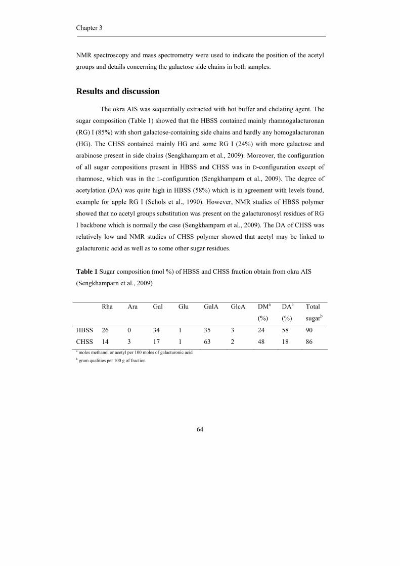
Chapter 3
64
NMR spectroscopy and mass spectrometry were used to indicate the position of the acetyl
groups and details concerning the galactose side chains in both samples.
Results and discussion
The okra AIS was sequentially extracted with hot buffer and chelating agent. The
sugar composition (Table 1) showed that the HBSS contained mainly rhamnogalacturonan
(RG) I (85%) with short galactose-containing side chains and hardly any homogalacturonan
(HG). The CHSS contained mainly HG and some RG I (24%) with more galactose and
arabinose present in side chains (Sengkhamparn et al., 2009). Moreover, the configuration
of all sugar compositions present in HBSS and CHSS was in D-configuration except of
rhamnose, which was in the L-configuration (Sengkhamparn et al., 2009). The degree of
acetylation (DA) was quite high in HBSS (58%) which is in agreement with levels found,
example for apple RG I (Schols et al., 1990). However, NMR studies of HBSS polymer
showed that no acetyl groups substitution was present on the galacturonosyl residues of RG
I backbone which is normally the case (Sengkhamparn et al., 2009). The DA of CHSS was
relatively low and NMR studies of CHSS polymer showed that acetyl may be linked to
galacturonic acid as well as to some other sugar residues.
Table 1 Sugar composition (mol %) of HBSS and CHSS fraction obtain from okra AIS
(Sengkhamparn et al., 2009)
Rha Ara Gal Glu GalA GlcA DMa
(%)
DAa
(%)
Total
sugarb
HBSS 26 0 34 1 35 3 24 58 90
CHSS 14 3 17 1 63 2 48 18 86 a moles methanol or acetyl per 100 moles of galacturonic acid b gram qualities per 100 g of fraction
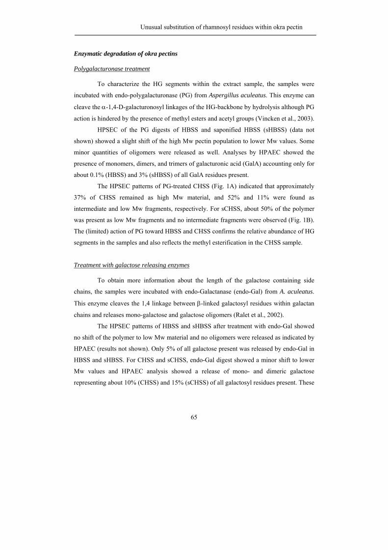
Unusual substitution of rhamnosyl residues within okra pectin
65
Enzymatic degradation of okra pectins
Polygalacturonase treatment
To characterize the HG segments within the extract sample, the samples were
incubated with endo-polygalacturonase (PG) from Aspergillus aculeatus. This enzyme can
cleave the α-1,4-D-galacturonosyl linkages of the HG-backbone by hydrolysis although PG
action is hindered by the presence of methyl esters and acetyl groups (Vincken et al., 2003).
HPSEC of the PG digests of HBSS and saponified HBSS (sHBSS) (data not
shown) showed a slight shift of the high Mw pectin population to lower Mw values. Some
minor quantities of oligomers were released as well. Analyses by HPAEC showed the
presence of monomers, dimers, and trimers of galacturonic acid (GalA) accounting only for
about 0.1% (HBSS) and 3% (sHBSS) of all GalA residues present.
The HPSEC patterns of PG-treated CHSS (Fig. 1A) indicated that approximately
37% of CHSS remained as high Mw material, and 52% and 11% were found as
intermediate and low Mw fragments, respectively. For sCHSS, about 50% of the polymer
was present as low Mw fragments and no intermediate fragments were observed (Fig. 1B).
The (limited) action of PG toward HBSS and CHSS confirms the relative abundance of HG
segments in the samples and also reflects the methyl esterification in the CHSS sample.
Treatment with galactose releasing enzymes
To obtain more information about the length of the galactose containing side
chains, the samples were incubated with endo-Galactanase (endo-Gal) from A. aculeatus.
This enzyme cleaves the 1,4 linkage between β-linked galactosyl residues within galactan
chains and releases mono-galactose and galactose oligomers (Ralet et al., 2002).
The HPSEC patterns of HBSS and sHBSS after treatment with endo-Gal showed
no shift of the polymer to low Mw material and no oligomers were released as indicated by
HPAEC (results not shown). Only 5% of all galactose present was released by endo-Gal in
HBSS and sHBSS. For CHSS and sCHSS, endo-Gal digest showed a minor shift to lower
Mw values and HPAEC analysis showed a release of mono- and dimeric galactose
representing about 10% (CHSS) and 15% (sCHSS) of all galactosyl residues present. These
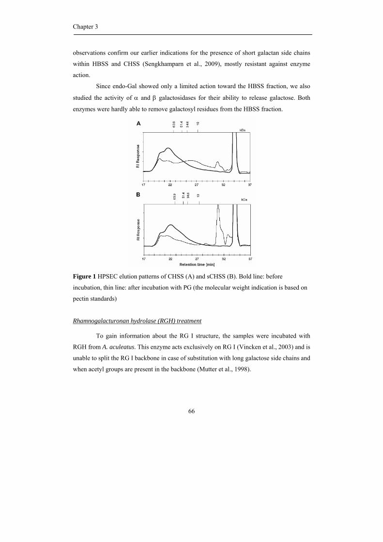
Chapter 3
66
observations confirm our earlier indications for the presence of short galactan side chains
within HBSS and CHSS (Sengkhamparn et al., 2009), mostly resistant against enzyme
action.
Since endo-Gal showed only a limited action toward the HBSS fraction, we also
studied the activity of α and β galactosidases for their ability to release galactose. Both
enzymes were hardly able to remove galactosyl residues from the HBSS fraction.
Figure 1 HPSEC elution patterns of CHSS (A) and sCHSS (B). Bold line: before
incubation, thin line: after incubation with PG (the molecular weight indication is based on
pectin standards)
Rhamnogalacturonan hydrolase (RGH) treatment
To gain information about the RG I structure, the samples were incubated with
RGH from A. aculeatus. This enzyme acts exclusively on RG I (Vincken et al., 2003) and is
unable to split the RG I backbone in case of substitution with long galactose side chains and
when acetyl groups are present in the backbone (Mutter et al., 1998).
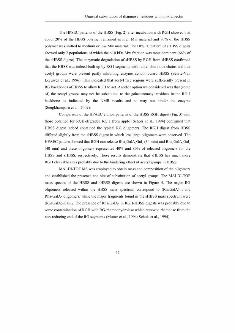
Unusual substitution of rhamnosyl residues within okra pectin
67
The HPSEC patterns of the HBSS (Fig. 2) after incubation with RGH showed that
about 20% of the HBSS polymer remained as high Mw material and 80% of the HBSS
polymer was shifted to medium or low Mw material. The HPSEC pattern of sHBSS digests
showed only 2 populations of which the <10 kDa Mw fraction was most dominant (66% of
the sHBSS digest). The enzymatic degradation of sHBSS by RGH from sHBSS confirmed
that the HBSS was indeed built up by RG I segments with rather short side chains and that
acetyl groups were present partly inhibiting enzyme action toward HBSS (Searle-Van
Leeuwen et al., 1996). This indicated that acetyl free regions were sufficiently present in
RG backbones of HBSS to allow RGH to act. Another option we considered was that (some
of) the acetyl groups may not be substituted to the galacturonosyl residues in the RG I
backbone as indicated by the NMR results and so may not hinder the enzyme
(Sengkhamparn et al., 2009).
Comparison of the HPAEC elution patterns of the HBSS RGH digest (Fig. 3) with
those obtained for RGH-degraded RG I from apple (Schols et al., 1994) confirmed that
HBSS digest indeed contained the typical RG oligomers. The RGH digest from HBSS
differed slightly from the sHBSS digest in which less large oligomers were observed. The
HPAEC pattern showed that RGH can release Rha2GalA2Gal2 (34 min) and Rha3GalA3Gal2
(40 min) and these oligomers represented 40% and 80% of released oligomers for the
HBSS and sHBSS, respectively. These results demonstrate that sHBSS has much more
RGH cleavable sites probably due to the hindering effect of acetyl groups in HBSS.
MALDI-TOF MS was employed to obtain mass and composition of the oligomers
and established the presence and site of substitution of acetyl groups. The MALDI-TOF
mass spectra of the HBSS and sHBSS digests are shown in Figure 4. The major RG
oligomers released within the HBSS mass spectrum correspond to (RhaGalA)2-3 and
Rha2GalA3 oligomers, while the major fragments found in the sHBSS mass spectrum were
(RhaGalA)2Gal2-3. The presence of Rha2GalA3 in RGH-HBSS digests was probably due to
some contamination of RGH with RG-rhamnohydrolase which removed rhamnose from the
non-reducing end of the RG segments (Mutter et al., 1994; Schols et al., 1994).
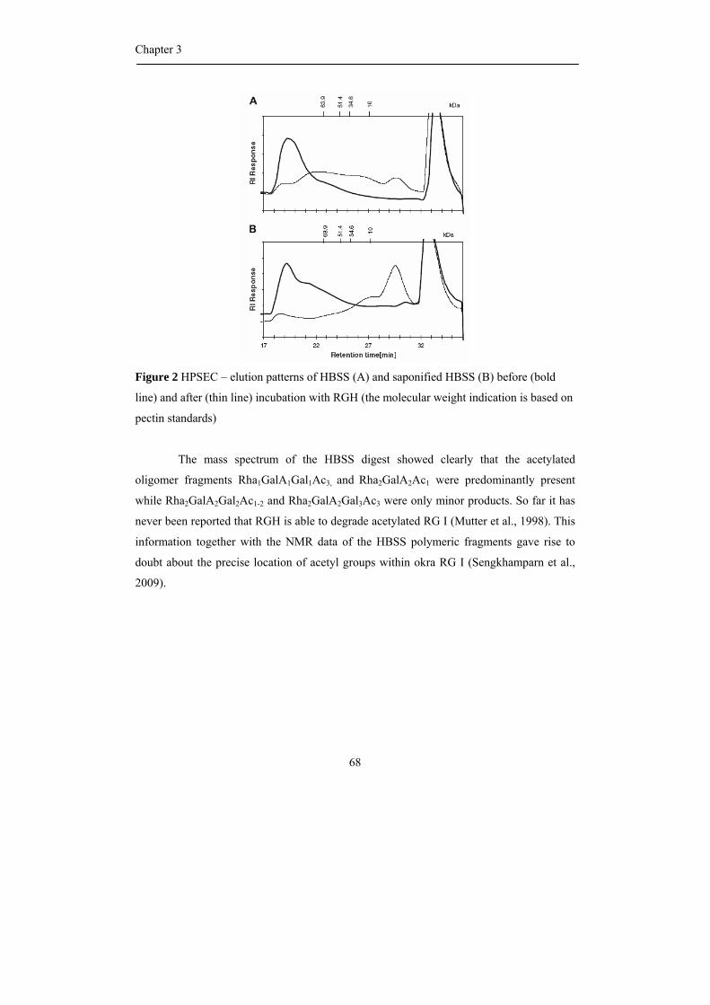
Chapter 3
68
Figure 2 HPSEC – elution patterns of HBSS (A) and saponified HBSS (B) before (bold
line) and after (thin line) incubation with RGH (the molecular weight indication is based on
pectin standards)
The mass spectrum of the HBSS digest showed clearly that the acetylated
oligomer fragments Rha1GalA1Gal1Ac3, and Rha2GalA2Ac1 were predominantly present
while Rha2GalA2Gal2Ac1-2 and Rha2GalA2Gal3Ac3 were only minor products. So far it has
never been reported that RGH is able to degrade acetylated RG I (Mutter et al., 1998). This
information together with the NMR data of the HBSS polymeric fragments gave rise to
doubt about the precise location of acetyl groups within okra RG I (Sengkhamparn et al.,
2009).
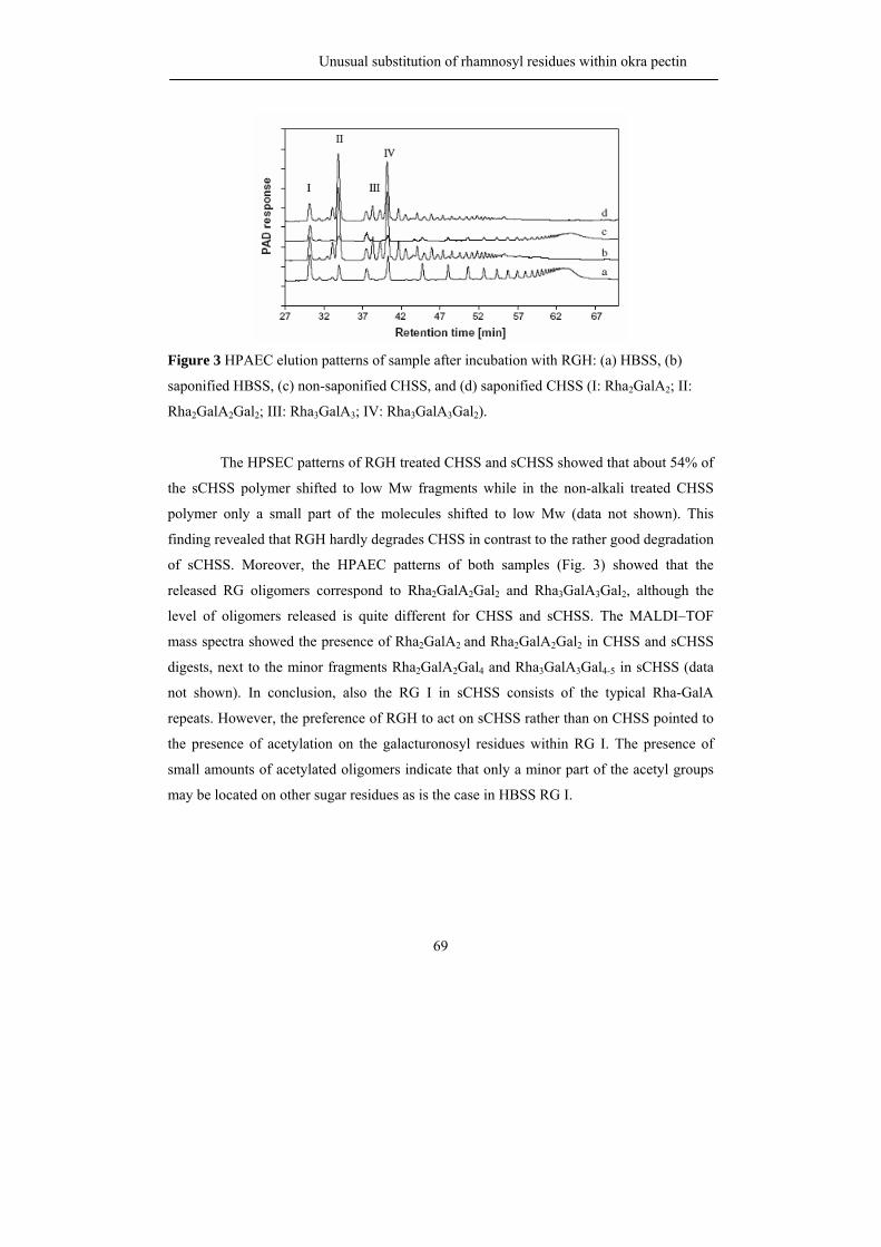
Unusual substitution of rhamnosyl residues within okra pectin
69
Figure 3 HPAEC elution patterns of sample after incubation with RGH: (a) HBSS, (b)
saponified HBSS, (c) non-saponified CHSS, and (d) saponified CHSS (I: Rha2GalA2; II:
Rha2GalA2Gal2; III: Rha3GalA3; IV: Rha3GalA3Gal2).
The HPSEC patterns of RGH treated CHSS and sCHSS showed that about 54% of
the sCHSS polymer shifted to low Mw fragments while in the non-alkali treated CHSS
polymer only a small part of the molecules shifted to low Mw (data not shown). This
finding revealed that RGH hardly degrades CHSS in contrast to the rather good degradation
of sCHSS. Moreover, the HPAEC patterns of both samples (Fig. 3) showed that the
released RG oligomers correspond to Rha2GalA2Gal2 and Rha3GalA3Gal2, although the
level of oligomers released is quite different for CHSS and sCHSS. The MALDI–TOF
mass spectra showed the presence of Rha2GalA2 and Rha2GalA2Gal2 in CHSS and sCHSS
digests, next to the minor fragments Rha2GalA2Gal4 and Rha3GalA3Gal4-5 in sCHSS (data
not shown). In conclusion, also the RG I in sCHSS consists of the typical Rha-GalA
repeats. However, the preference of RGH to act on sCHSS rather than on CHSS pointed to
the presence of acetylation on the galacturonosyl residues within RG I. The presence of
small amounts of acetylated oligomers indicate that only a minor part of the acetyl groups
may be located on other sugar residues as is the case in HBSS RG I.
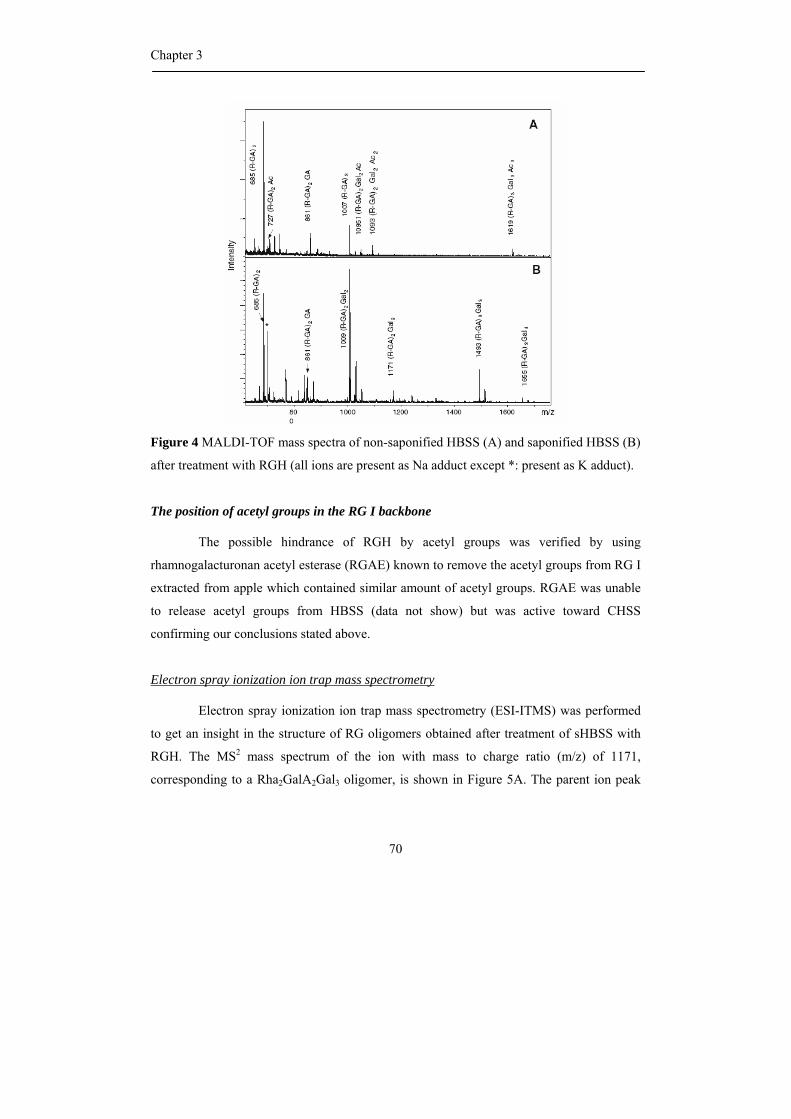
Chapter 3
70
Figure 4 MALDI-TOF mass spectra of non-saponified HBSS (A) and saponified HBSS (B)
after treatment with RGH (all ions are present as Na adduct except *: present as K adduct).
The position of acetyl groups in the RG I backbone
The possible hindrance of RGH by acetyl groups was verified by using
rhamnogalacturonan acetyl esterase (RGAE) known to remove the acetyl groups from RG I
extracted from apple which contained similar amount of acetyl groups. RGAE was unable
to release acetyl groups from HBSS (data not show) but was active toward CHSS
confirming our conclusions stated above.
Electron spray ionization ion trap mass spectrometry
Electron spray ionization ion trap mass spectrometry (ESI-ITMS) was performed
to get an insight in the structure of RG oligomers obtained after treatment of sHBSS with
RGH. The MS2 mass spectrum of the ion with mass to charge ratio (m/z) of 1171,
corresponding to a Rha2GalA2Gal3 oligomer, is shown in Figure 5A. The parent ion peak
Unusual substitution of rhamnosyl residues within okra pectin
71
m/z = 1171 was fragmented to m/z = 863 (releasing of Rha-Gal fragment) and 699
(releasing of Gal-Rha-GalA fragment), indicating that one galactosyl residue was attached
to one rhamnosyl residue. In addition, the removal of Gal2Rha1 fragment (m/z = 701) and
Gal2Rha1GalA1 fragment (m/z = 507) in the MS2 spectrum of m/z = 1171 pointed to two
galactosyl residues linked to one rhamnosyl residue. The possible structure of oligomers
with m/z = 1171 is shown in the right top corner of Figure 5A. However, it was not possible
to make a difference between a side chain of two galactoses or two galactoses, both
attached to the rhamnose at positions O-3 and O-4.
Also, the presence of fragment of m/z = 863 (leaving of a Gal2Rha1fragment) and
699 (leaving of a Gal2Rha1GalA1 fragment) in the MS2 spectrum of m/z = 1333
representing Rha2GalA2Gal4 (Fig. 5B) confirmed that two galactosyl residues are
substituted to one single rhamnosyl residue. Fragments representing the removal of
Rha1Gal1 (m/z 1025) and Rha1Gal3 (m/z 863) were only found as minor fragments in the
MS2 spectrum of m/z = 1333. The possible structure of main oligomers with m/z = 1133 is
shown in the right top corner of Figure 5B. Therefore, the main RG oligomers released
from sHBSS by RGH consist of a Rha-GalA repeat with galactose dimers linked to the
rhamnose moiety, although rhamnoses without, with one or with three galactose units are
also present in minor amounts. Linkage analysis already pointed out that galactoses may be
linked to each other (Sengkhamparn et al., 2009).
To locate the acetyl groups within the HBSS RGH oligomers, ESI-IT-MSn was
performed. The ESI-IT-MS2 spectrum of ions peak 729 (18O labeled Rha2GalA2Ac1) is
shown in Figure 6A and the most apparent fragments were m/z = 583 and 533 which were
formed after removal of rhamnose and 18O-labeled GalA, respectively. The MS3 spectrum
of m/z = 583 showed the leaving of GalA1Ac1 (m/z = 327 and 345) pointing out that an
acetyl group was located on the galacturonosyl residue. Unexpectedly, the MS3 spectrum of
m/z = 533 (Rha2GalA1Ac1) showed the release of Rha1Ac1 (m/z = 327 and 345) suggesting
that an acetyl group was linked to a rhamnosyl residue. From this data, it is concluded that
the oligomers with a m/z of 727/729 represent a mixture of different isomers with an acetyl
group either on the GalA or Rha moieties. This finding was confirmed for ion peak m/z
1093 (18O labeled Rha2GalA2Gal2Ac2) also indicating that acetyl could be linked to either
Chapter 3
72
the GalA or Rha residues. Fragments with an additional loss of one water (-18) refer to
different O-glycosyl fragmentation (Doman and Costello, 1988) while the loss of a second
water molecule refers to a rearrangement of the galacturonosyl residues.
Figure 5 The MS2 spectrum of Rha2GalA2Gal3 (P: m/z=1171) and Rha2GalA2Gal4 (P:
m/z=1333) for sHBSS digest with RGH. All ions are present as Na adducts (possible
structures of the oligomers, m/z= 1171 and 1333, are shown in the right top corner).
Unusual substitution of rhamnosyl residues within okra pectin
73
From the MS2 and MS3 spectra of HBSS RGH oligomers, it can be derived that
rhamnosyl residues were O-acetylated. These findings might explain the activity of RGH
and RGAE toward HBSS as well as the fact that the acetyl groups attached to the
rhamnosyl residues cannot be removed by RGAE but do not hinder RGH either. Some
acetyl groups were, however, located on galacturonosyl residues hindering RGH,
explaining the different digestion of HBSS and sHBSS. However, MS is not a very reliable
technique to quantify the acetylated oligomer since esters are known to have a positive
effect on fragmentation efficiency.
250 300 350 400 450 500 550 600 650 700 750m/z
0
20
40
60
80
1000
20
40
60
80
100
Rel
ativ
eAb
unda
nce
20
40
60
80
100583.2
563.3
683.5327.1 387.2 533.2473.2 655.1407.2 457.2
729.3
313.2261.1327.08
387.2
545.2407.2283.1 345.3 521.3241.1
474.3428.8387.0
345.2
327.1 431.3264.8 497.2
HBSS-RGh-18water_070626124831#247
583.17
533.3
m s 2 72 9 .00
72 9 .00 @ c id 32 .0 0 58 3 .00
F : IT M S + p E S I w F u ll m s 3
-Gal
A - 2
H20
-Ac
(R-G
A) N
a+
(GA-
Ac) N
a+
(R-G
A) N
a+ (-
H2O
)
(R-G
A-Ac
) Na+
405.
2 (R
-GA-
Ac) N
a+ (+
H2O
)
-Gal
A - H
20
-Rha
-Rha
-Ac
- Gal
A
- Gal
A- G
alA
- H2O
- Gal
A - A
c - H
2O
- Gal
A - A
c - 2
H2O
- Gal
A - R
ha -
H2O
-Rha
-H20
-Rha
-H20
-Rha
-H20
- Ac
-Rha
-Ac
-Rha
-Ac
-Rha
-Ac
- H2O
-Gal
A - H
20 -A
c
GalA-Rha- GalA + (Ac) + Na+ 18O labeled
Rha - GalA - Rha + (Ac) + Na+
(R-G
A-A
c) N
a+ (
+H2O
)
241.1
A
B
C
P-(R
ha,H
20)
P-R
ha-A
cP
-Rha
-Ac-
H20
P-(
Gal
A-A
c, 2
H20
)
(Rha
-Gal
A-A
c)-2
H20
(Rha
-Gal
A-A
c)
(Rha
-Gal
A)
(Rha
-Gal
A)-2
H20
)
Par
ent I
on
P-R
ha
D1-G
alA
Dau
ghte
r 1
D1-
(Gal
A, H
2O)
D1-
Gal
A
D1-(
Gal
A-A
c, H
2O)
D1-
(Gal
A-Ac
,2 H
2O)
D1-
(Gal
A-R
ha, H
2O)
Dau
ghte
r 2D
1-Ac
D2-
Rha
D2-
(Rha
-Ac)
345.2D2-(
Rha
-Ac,
H2O
)
D2-
(Rha
, H2O
(Rha
-Gal
A-Ac
)
GalA Rha O18GalA
Ac 407545
327
GalA Rha O18GalA
Ac 407545
327
Rha GalA Rha
Ac
345
387
Rha GalA Rha
Ac
345
387
250 300 350 400 450 500 550 600 650 700 750m/z
0
20
40
60
80
1000
20
40
60
80
100
Rel
ativ
eAb
unda
nce
20
40
60
80
100583.2
563.3
683.5327.1 387.2 533.2473.2 655.1407.2 457.2
729.3
313.2261.1327.08
387.2
545.2407.2283.1 345.3 521.3241.1
474.3428.8387.0
345.2
327.1 431.3264.8 497.2
HBSS-RGh-18water_070626124831#247
583.17
533.3
m s 2 72 9 .00
72 9 .00 @ c id 32 .0 0 58 3 .00
F : IT M S + p E S I w F u ll m s 3
-Gal
A - 2
H20
-Ac
(R-G
A) N
a+
(GA-
Ac) N
a+
(R-G
A) N
a+ (-
H2O
)
(R-G
A-Ac
) Na+
405.
2 (R
-GA-
Ac) N
a+ (+
H2O
)
-Gal
A - H
20
-Rha
-Rha
-Ac
- Gal
A
- Gal
A- G
alA
- H2O
- Gal
A - A
c - H
2O
- Gal
A - A
c - 2
H2O
- Gal
A - R
ha -
H2O
-Rha
-H20
-Rha
-H20
-Rha
-H20
- Ac
-Rha
-Ac
-Rha
-Ac
-Rha
-Ac
- H2O
-Gal
A - H
20 -A
c
GalA-Rha- GalA + (Ac) + Na+ 18O labeled
Rha - GalA - Rha + (Ac) + Na+
(R-G
A-A
c) N
a+ (
+H2O
)
241.1
A
B
C
P-(R
ha,H
20)
P-R
ha-A
cP
-Rha
-Ac-
H20
P-(
Gal
A-A
c, 2
H20
)
(Rha
-Gal
A-A
c)-2
H20
(Rha
-Gal
A-A
c)
(Rha
-Gal
A)
(Rha
-Gal
A)-2
H20
)
Par
ent I
on
P-R
ha
D1-G
alA
Dau
ghte
r 1
D1-
(Gal
A, H
2O)
D1-
Gal
A
D1-(
Gal
A-A
c, H
2O)
D1-
(Gal
A-Ac
,2 H
2O)
D1-
(Gal
A-R
ha, H
2O)
Dau
ghte
r 2D
1-Ac
D2-
Rha
D2-
(Rha
-Ac)
345.2D2-(
Rha
-Ac,
H2O
)
D2-
(Rha
, H2O
(Rha
-Gal
A-Ac
)
GalA Rha O18GalA
Ac 407545
327
GalA Rha O18GalA
Ac 407545
327
Rha GalA Rha
Ac
345
387
Rha GalA Rha
Ac
345
387
Figure 6 ESI MS2 (A) of 18O labeled Rha2GalA2Ac (Parent, P: m/z = 727) and MS3 of ion
peak Daughter D1: m/z =583 (B) and D2: m/z = 533 (C) obtained from the HBSS digest
with RGH (the fragments having a star are 18O-labeled). All ions are present as Na adducts
(possible structures of the fragments, m/z= 583 and 533, are shown in the right top corner)
Chapter 3
74
Nuclear magnetic resonance of okra HBSS
To confirm the position of the acetyl groups within the HBSS on both the
rhamnose and GalA moieties and to enable some quantitation, NMR was performed on a 20
kDa RGH HBSS fragment obtained by preparative SEC over Sephacryl 500 and Sepharcryl
300 (Fig. 7) after RGH digestion in order to overcome the high viscosity of the intact
polymer.
17 22 27 32
Retention Time (min)
RI r
espo
nse
kDa1034.6
63.9
51.4
Figure 7 Molecular weigh distributions of HBSS RGH digest (bold line) and a 20 kDa
fragment obtained after fractionation of Sepharcryl S500 pool 3 over Sepharcryl S300 (thin
line). (The molecular weight indications are based on pectin standards)
Within the anomeric region of the 1H NMR spectrum of recorded five major sugar
moieties of a 20 kDa RGH HBSS fragment, 1,2,4-linked α-Rhap (A), 1,4-linked α-GalpA
(RG I) (B) (Colquhoun et al., 1990; Mutter et al., 1994; Renard et al., 1999; Deng et al.,
2006), t-α-Galp (C) (Gronberg et al., 1994; Strecker et al., 1995) , 1,4-linked Galp (D) and
t-β-Galp (E) (Colquhoun et al., 1990; Huisman et al., 2001) could be observed (Table 2). In
general, the spectra underline the regular structure of okra RG I being a highly branched
rhamnogalacturonan with only one or two galactose units as side chains. A signal at 2.106
ppm typically belonging to an O-acetyl substituent was observed. In general, the O-acetyl
units present on the RG I backbone are expected to be linked to O-3 and/or O-2 of the GalA
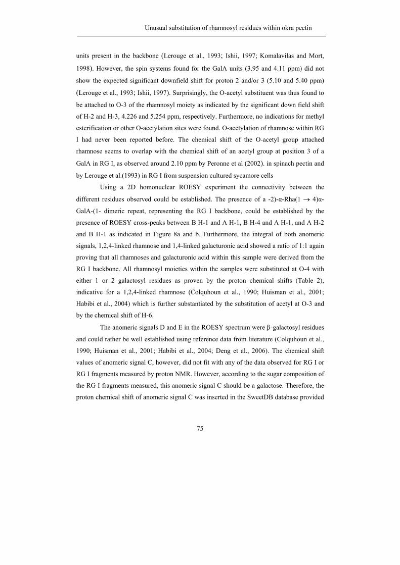
Unusual substitution of rhamnosyl residues within okra pectin
75
units present in the backbone (Lerouge et al., 1993; Ishii, 1997; Komalavilas and Mort,
1998). However, the spin systems found for the GalA units (3.95 and 4.11 ppm) did not
show the expected significant downfield shift for proton 2 and/or 3 (5.10 and 5.40 ppm)
(Lerouge et al., 1993; Ishii, 1997). Surprisingly, the O-acetyl substituent was thus found to
be attached to O-3 of the rhamnosyl moiety as indicated by the significant down field shift
of H-2 and H-3, 4.226 and 5.254 ppm, respectively. Furthermore, no indications for methyl
esterification or other O-acetylation sites were found. O-acetylation of rhamnose within RG
I had never been reported before. The chemical shift of the O-acetyl group attached
rhamnose seems to overlap with the chemical shift of an acetyl group at position 3 of a
GalA in RG I, as observed around 2.10 ppm by Peronne et al (2002). in spinach pectin and
by Lerouge et al.(1993) in RG I from suspension cultured sycamore cells
Using a 2D homonuclear ROESY experiment the connectivity between the
different residues observed could be established. The presence of a -2)-α-Rha(1 → 4)α-
GalA-(1- dimeric repeat, representing the RG I backbone, could be established by the
presence of ROESY cross-peaks between B H-1 and A H-1, B H-4 and A H-1, and A H-2
and B H-1 as indicated in Figure 8a and b. Furthermore, the integral of both anomeric
signals, 1,2,4-linked rhamnose and 1,4-linked galacturonic acid showed a ratio of 1:1 again
proving that all rhamnoses and galacturonic acid within this sample were derived from the
RG I backbone. All rhamnosyl moieties within the samples were substituted at O-4 with
either 1 or 2 galactosyl residues as proven by the proton chemical shifts (Table 2),
indicative for a 1,2,4-linked rhamnose (Colquhoun et al., 1990; Huisman et al., 2001;
Habibi et al., 2004) which is further substantiated by the substitution of acetyl at O-3 and
by the chemical shift of H-6.
The anomeric signals D and E in the ROESY spectrum were β-galactosyl residues
and could rather be well established using reference data from literature (Colquhoun et al.,
1990; Huisman et al., 2001; Habibi et al., 2004; Deng et al., 2006). The chemical shift
values of anomeric signal C, however, did not fit with any of the data observed for RG I or
RG I fragments measured by proton NMR. However, according to the sugar composition of
the RG I fragments measured, this anomeric signal C should be a galactose. Therefore, the
proton chemical shift of anomeric signal C was inserted in the SweetDB database provided
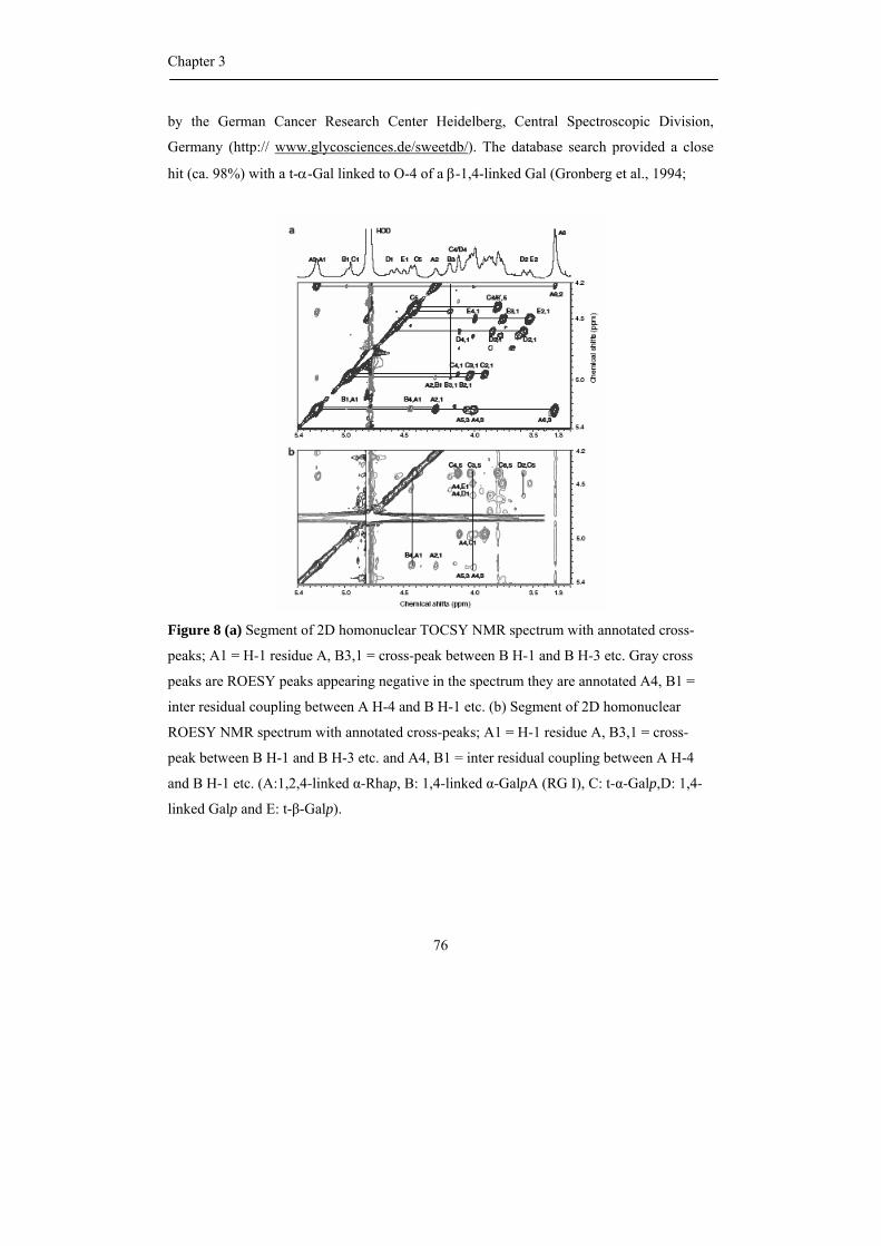
Chapter 3
76
by the German Cancer Research Center Heidelberg, Central Spectroscopic Division,
Germany (http:// www.glycosciences.de/sweetdb/). The database search provided a close
hit (ca. 98%) with a t-α-Gal linked to O-4 of a β-1,4-linked Gal (Gronberg et al., 1994;
Figure 8 (a) Segment of 2D homonuclear TOCSY NMR spectrum with annotated cross-
peaks; A1 = H-1 residue A, B3,1 = cross-peak between B H-1 and B H-3 etc. Gray cross
peaks are ROESY peaks appearing negative in the spectrum they are annotated A4, B1 =
inter residual coupling between A H-4 and B H-1 etc. (b) Segment of 2D homonuclear
ROESY NMR spectrum with annotated cross-peaks; A1 = H-1 residue A, B3,1 = cross-
peak between B H-1 and B H-3 etc. and A4, B1 = inter residual coupling between A H-4
and B H-1 etc. (A:1,2,4-linked α-Rhap, B: 1,4-linked α-GalpA (RG I), C: t-α-Galp,D: 1,4-
linked Galp and E: t-β-Galp).
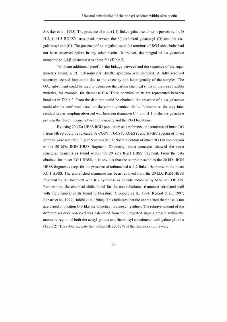
Unusual substitution of rhamnosyl residues within okra pectin
77
Strecker et al., 1995). The presence of an α-(1,4)-linked galactose dimer is proven by the D
H-2, C H-5 ROESY cross-peak between the β(1,4)-linked galactosyl (D) and the t-α-
galactosyl unit (C). The presence of a t-α-galactose at the terminus of RG I side chains had
not been observed before in any other pectins. Moreover, the integral of t-α-galactose
compared to 1,4-β-galactose was about 2:1 (Table 2).
To obtain additional proof for the linkage between and the sequence of the sugar
moieties found, a 2D heteronuclear HMBC spectrum was obtained. A fully resolved
spectrum seemed impossible due to the viscosity and heterogeneity of the samples. The
OAc substituent could be used to determine the carbon chemical shifts of the more flexible
moieties, for example, for rhamnose C-6. These chemical shifts are represented between
brackets in Table 2. From the data that could be obtained, the presence of a t-α-galactose
could also be confirmed based on the carbon chemical shifts. Furthermore, the only inter
residual scalar coupling observed was between rhamnose C-4 and H-1 of the t-α-galactose
proving the direct linkage between this moiety and the RG I backbone.
By using 20 kDa HBSS RGH population as a reference, the structure of intact RG
I from HBSS could be revealed. A COSY, TOCSY, ROESY, and HMBC spectra of intact
samples were recorded. Figure 9 shows the 1H NMR spectrum of intact RG I in comparison
to the 20 kDa RGH HBSS fragment. Obviously, intact structures showed the same
structural elements as found within the 20 kDa RGH HBSS fragment. From the data
obtained for intact RG I HBSS, it is obvious that the sample resembles the 20 kDa RGH
HBSS fragment except for the presence of unbranched α-1,2-linked rhamnose in the intact
RG I HBSS. The unbranched rhamnose has been removed from the 20 kDa RGH HBSS
fragment by the treatment with RG hydrolase as already indicated by MALDI-TOF MS.
Furthermore, the chemical shifts found for the non-substituted rhamnose correlated well
with the chemical shifts found in literature (Gronberg et al., 1994; Renard et al., 1997;
Renard et al., 1999; Habibi et al., 2004). This indicates that the unbranched rhamnose is not
acetylated at position O-3 like the branched rhamnosyl residues. The relative amount of the
different residues observed was calculated from the integrated signals present within the
anomeric region of both the acetyl groups and rhamnosyl substituents with galatosyl units
(Table 2). The ratios indicate that within HBSS, 62% of the rhamnosyl units were
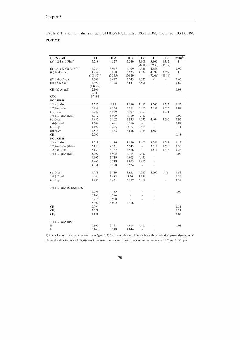
Chapter 3
78
Table 2 1H chemical shifts in ppm of HBSS RGH, intact RG I HBSS and intact RG I CHSS
PG/PME
HBSS RGH H-1 H-2 H-3 H-4 H-5 H-6 RATIO2)
(A) 1,2,4-α-L-Rha1)
5.238
4.227
5.249
3.903
(78.11) 3.965
(69.33) 1.332
(18.19) 1
(B) 1,4-α-D-GalA (RGI) 4.984 3.947 4.109 4.441 4.555 0.92 (C) t-α-D-Gal
4.952 (101.57)3)
3.808 (70.55)
3.923 (70.29)
4.039
4.399 (72.06)
3.697 (61.84)
1
(D) 1,4-β-D-Gal 4.603 3.477 3.743 4.025 -4) - 0.66 (E) t-β-D-Gal
4.492 (104.98)
3.428
3.647
3.891
-
-
0.69
CH3 (O-Acetyl) COO
2.106 (22.09) 174.91
0.98
RG I HBSS 1,2-α-L-rha 5.257 4.12 3.889 3.415 3.765 1.252 0.55 1,2,4-α-L-rha 5.234 4.224 5.251 3.905 3.951 1.333 0.87 t-α-L-rha 5.229 4.059 3.797 3.353 - 1.231 1,4-α-D-galA (RGI) 5.012 3.909 4.119 4.417 - 1.00 t-α-D-gal 4.955 3.802 3.935 4.035 4.404 3.696 0.97 1,4-β-D-gal 4.602 3.491 3.756 - - 0.84 t-β-D-gal 4.492 3.425 3.65 3.888 - 1.11 unknown 4.556 3.563 3.836 4.334 4.563 CH3 2.099 1.18 RG I CHSS 1,2-α-L-rha 5.243 4.116 3.879 3.409 3.745 1.245 0.15 1,2,4-α-L-rha (OAc) 5.199 4.221 5.243 - 3.911 1.328 0.38 1,2,4-α-L-rha 5.163 4.137 3.966 - 3.811 1.313 0.26 1,4-α-D-galA (RGI) 5.007 3.905 4.114 4.427 - 1.00 4.907 3.719 4.003 4.456 - 4.965 3.719 4.003 4.456 - 4.951 3.798 3.924 - - t-α-D-gal 4.951 3.789 3.923 4.027 4.392 3.96 0.53 1,4-β-D-gal 4.6 3.482 3.76 3.956 - - 0.36 t-β-D-gal 4.483 3.421 3.557 3.882 - - 0.34 1,4-α-D-galA (O-acetylated) 5.093 4.135 - - - 1.66 5.165 3.976 - - - 5.216 3.988 - - - 5.309 4.002 4.416 - - CH3 2.094 0.31 CH3 2.071 0.21 CH3 2.181 0.03 1,4-α-D-galA (HG) E 5.105 3.731 4.014 4.466 - 1.01 F 5.143 3.748 4.044 - -
1) Arabic letters correspond to annotation in figure 8; 2) Ratio was calculated from the integrals of individual proton signals; 3) 13C
chemical shift between brackets; 4) - = not determined; values are expressed against internal acetone at 2.225 and 31.55 ppm
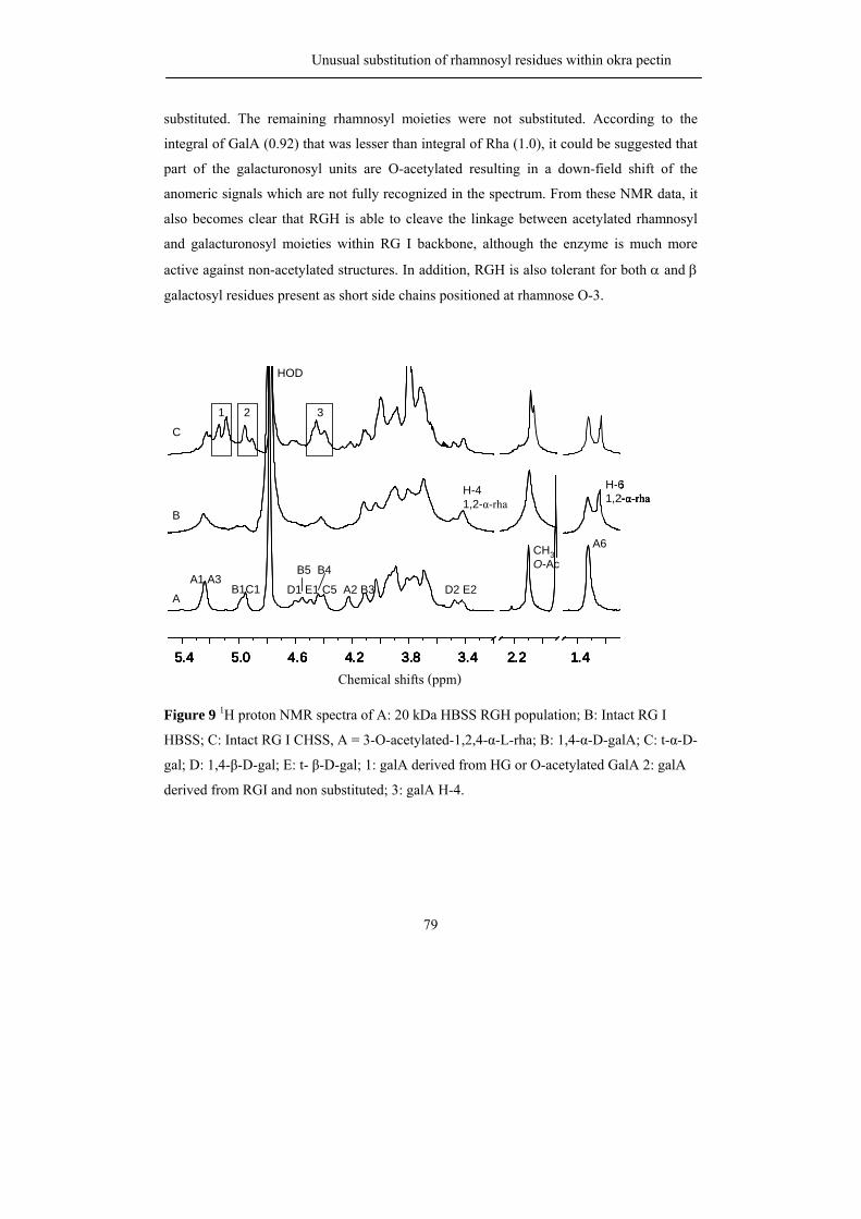
Unusual substitution of rhamnosyl residues within okra pectin
79
substituted. The remaining rhamnosyl moieties were not substituted. According to the
integral of GalA (0.92) that was lesser than integral of Rha (1.0), it could be suggested that
part of the galacturonosyl units are O-acetylated resulting in a down-field shift of the
anomeric signals which are not fully recognized in the spectrum. From these NMR data, it
also becomes clear that RGH is able to cleave the linkage between acetylated rhamnosyl
and galacturonosyl moieties within RG I backbone, although the enzyme is much more
active against non-acetylated structures. In addition, RGH is also tolerant for both α and β
galactosyl residues present as short side chains positioned at rhamnose O-3.
1.42.23.43.84.24.65.05.4f1 (ppm)
A1 A3B1C1 D1 E1 C5 A2 B3 D2 E2
A6
HOD
CH3O-Ac
H-61,2-α-rha
H-41,2-α-rha
C
B
A
B5 B4
1 2 3
Chemical shifts (ppm)1.42.23.43.84.24.65.05.4
f1 (ppm)
A1 A3B1C1 D1 E1 C5 A2 B3 D2 E2
A6
HOD
CH3O-Ac
H-61,2-α-rha
H-41,2-α-rha
C
B
A
B5 B4
1 2 3
1.42.23.43.84.24.65.05.4f1 (ppm)
1.42.23.43.84.24.65.05.4f1 (ppm)
1.42.23.43.84.24.65.05.4f1 (ppm)
A1 A3B1C1 D1 E1 C5 A2 B3 D2 E2
A6
HOD
CH3O-Ac
H-61,2-α-rha
H-41,2-α-rha
C
B
A
B5 B4
1 2 3
Chemical shifts (ppm)
Figure 9 1H proton NMR spectra of A: 20 kDa HBSS RGH population; B: Intact RG I
HBSS; C: Intact RG I CHSS, A = 3-O-acetylated-1,2,4-α-L-rha; B: 1,4-α-D-galA; C: t-α-D-
gal; D: 1,4-β-D-gal; E: t- β-D-gal; 1: galA derived from HG or O-acetylated GalA 2: galA
derived from RGI and non substituted; 3: galA H-4.
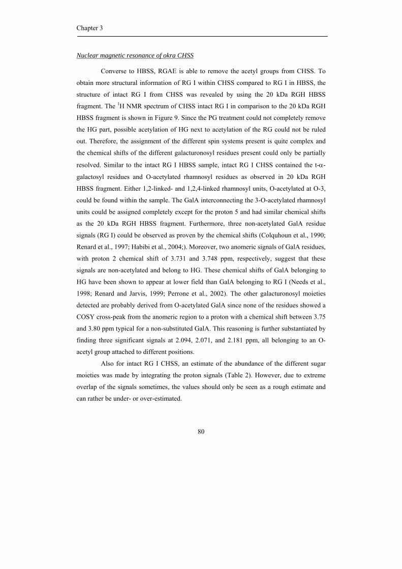
Chapter 3
80
Nuclear magnetic resonance of okra CHSS
Converse to HBSS, RGAE is able to remove the acetyl groups from CHSS. To
obtain more structural information of RG I within CHSS compared to RG I in HBSS, the
structure of intact RG I from CHSS was revealed by using the 20 kDa RGH HBSS
fragment. The 1H NMR spectrum of CHSS intact RG I in comparison to the 20 kDa RGH
HBSS fragment is shown in Figure 9. Since the PG treatment could not completely remove
the HG part, possible acetylation of HG next to acetylation of the RG could not be ruled
out. Therefore, the assignment of the different spin systems present is quite complex and
the chemical shifts of the different galacturonosyl residues present could only be partially
resolved. Similar to the intact RG I HBSS sample, intact RG I CHSS contained the t-α-
galactosyl residues and O-acetylated rhamnosyl residues as observed in 20 kDa RGH
HBSS fragment. Either 1,2-linked- and 1,2,4-linked rhamnosyl units, O-acetylated at O-3,
could be found within the sample. The GalA interconnecting the 3-O-acetylated rhamnosyl
units could be assigned completely except for the proton 5 and had similar chemical shifts
as the 20 kDa RGH HBSS fragment. Furthermore, three non-acetylated GalA residue
signals (RG I) could be observed as proven by the chemical shifts (Colquhoun et al., 1990;
Renard et al., 1997; Habibi et al., 2004;). Moreover, two anomeric signals of GalA residues,
with proton 2 chemical shift of 3.731 and 3.748 ppm, respectively, suggest that these
signals are non-acetylated and belong to HG. These chemical shifts of GalA belonging to
HG have been shown to appear at lower field than GalA belonging to RG I (Needs et al.,
1998; Renard and Jarvis, 1999; Perrone et al., 2002). The other galacturonosyl moieties
detected are probably derived from O-acetylated GalA since none of the residues showed a
COSY cross-peak from the anomeric region to a proton with a chemical shift between 3.75
and 3.80 ppm typical for a non-substituted GalA. This reasoning is further substantiated by
finding three significant signals at 2.094, 2.071, and 2.181 ppm, all belonging to an O-
acetyl group attached to different positions.
Also for intact RG I CHSS, an estimate of the abundance of the different sugar
moieties was made by integrating the proton signals (Table 2). However, due to extreme
overlap of the signals sometimes, the values should only be seen as a rough estimate and
can rather be under- or over-estimated.
Unusual substitution of rhamnosyl residues within okra pectin
81
Conclusions
In summary, HBSS contains predominantly RG I structures with monomeric and
dimeric galactan side chains, although HG is part of the same molecule to a small extent.
The methyl esters present in HBSS seem to be present on the HG part of the molecule. The
acetyl groups are predominantly located at position O-3 of the rhamnosyl moiety. Another
novelty of Okra RG-I as found in the HBSS fraction is the presence of α-galactose
substitution at O-4 of the backbone rhamnosyl residue. In contrast to HBSS RG-I, the
pectic material extracted by chelating agent and being homogeneous with respect to size
and charge contains much more HG which is relatively highly methyl esterified. Minor
amounts of the rhamnose-acetylated RG-I containing α-galactose substitutions are found.
In addition, also more common RG-I structural elements are present consisting of a
galacturonic acid-acetylated backbone, without α-linked galactosyl residues and with
longer arabinose and galactose-containing side chains.
Material and methods
Sequential extraction of okra AIS
Soft and mature okra pods (5–10 cm in length) were collected at local market in
June 2005, Thailand. Okra AIS was prepared and then extracted with 0.05 M sodium
acetate buffer (Hot Buffer Soluble Solids, HBSS), followed by 0.05 M EDTA and 0.05 M
sodium oxalate in 0.05 M sodium acetate (CHelating agent Soluble Solids, CHSS)
according to Sengkhamparn et al (2009).
Intact RG I from HBSS and CHSS were prepared by incubation with endo-
polygalacturonase (PG) and pectin methyl esterase (PME) according to Sengkhamparn et al
(2009).
Chapter 3
82
Analytical methods
Total neutral sugar content and uronic acid content
The total neutral sugar and uronic acid contents were determined by the automated
colorimetric orcinol/sulfuric acid method (Tollier and Robin, 1979) and by the automated
colorimetric m-hydroxydiphenyl method (Ahmed and Labavitch, 1977; Kintner and van
Buren, 1982; Thibault, 1979), respectively. Galactose and galacturonic acid were used as a
standard.
Enzyme degradation
A saponification step was performed for removing methyl ester and acetyl groups
before incubation with enzymes. The HBSS and CHSS (3–4 mg) were dissolved in 300 µL
millipore water and were saponified by adding 300 µL of 0.1 N NaOH. After storage
overnight at 4 oC, the solutions were neutralized with 300 µL of 0.1 M acetic acid and the
volume was adjusted to 1 mL with 0.2 M NaOAc buffer of pH 5.
The non-saponified (final concentration of 3–4 mg/mL in 50 mM NaOAc buffer of
pH 5) and saponified solutions of HBSS and CHSS were incubated with the following
enzymes individually: 0.547 µg protein/mg substrate of rhamnogalacturonan hydrolase
(RGH) from A. aculeatus, 0.016 units of endo-polygalacturonase (PG) from A. niger
(Schols et al., 1990), 2.5 units of endo-galactanase(endo-Gal) from Aspergillus niger
(Guŕjanov et al., 2007), 0.04 units of β-galactosidase from Kluyveromyces lactis, 0.04 units
of α-galactosidase from Guar seed (Megazyme) and A.niger. The incubations were
performed at 40 oC for 24 h and were stopped by heating at 100 oC for 5 min for
inactivating enzymes. The decrease in molecular weight (Mw) and the release of oligomeric
products were analyzed by high performance size exclusion chromatography (HPSEC) and
high performance anion-exchange chromatography (HPAEC), respectively.
High performance size exclusion chromatography (HPSEC)
The high performance size-exclusion chromatography (Thermo Separation
Products, USA) equipped with three TosoH Biosep-TSKGel G columns in series
Unusual substitution of rhamnosyl residues within okra pectin
83
(4000PWXL-3000PWXL-2500PWXL) in combination with a guard PWXL column (TosoH,
Japan) was used to observe the change in Mw distribution of samples. The samples were
eluted with 0.2 M sodium nitrate at 30 oC and with a flow rate of 0.8 mL/min (Chen et al.,
2004). The eluent was monitored using RI detector (Shodex SE-61, Showa Denko K.K.,
Japan).
High performace anion-exchange chromatography (HPAEC)
High performance anion-exchange chromatography was performed on a Dionex
ISO 3000 system with PAD detector system (USA). For the analysis of enzyme digests, a
(2 x 250 mm) CarboPac PA 1 column (Dionex, USA) was equilibrated with 100 mM
NaOH. Gradients of NaOH and NaOAc were used simultaneously to elute the oligomers
with a flow of 0.3 mL/min. The results are in the following gradients of NaOH: 0.0–15.0
min, 0–16 mM; 15.0–15.1 min, 16–100 mM NaOH; 15.1–85.0 min, 100 mM. The
simultaneous gradient of NaOAc was 0.0–15.0 min, 0 mM NaOAc; 15.0–15.1, 0–100 mM
NaOAc; 15.1–55.0 min, 100–400 mM NaOAc; 55.0–80.0 min, 400–700 mM NaOAc;
80.0–85.0 min, 700–1000 mM NaOAc. The column was washed for 5 min with 1 M
NaOAc in 0.1 M NaOH and equilibrated for 15 min with 16 mM NaOH.
Matrix –assisted laser desorption ionization-time of flight mass spectrometry (MALDI-TOF
MS) analysis
A small spatula of AG 50W-X8 Resin (H+ form; Bio-Rad, USA) was added to 10
µl of digest sample for desalting, followed by mixing and centrifugation for 5 min at
13,000g. One microliter of sample solution was placed on a MALDI-TOF plate together
with 1 µL of matrix solution, 10 mg/mL of 2,5-dihydroxybenzoic acid (Bruker Daltonic’s,
Germany) in water, and dried under a constant flow of warm air. The oligomers were
determined by using MALDI-TOF MS (Ultra flex instrument, Bruker Daltonics, Germany)
with a lowest laser energy (35%) of Nitrogen 337 nm laser beam to correcting data from
average of 200 shots. A mixture of maltodextrins, mass range of 300–3000 Da, was used
for calibration.
Chapter 3
84
Electron spray ionization ion trap mass spectrometry (ESI-IT-MS)
For ESI-IT-MSn analysis, HBSS was dissolved in water and incubated with 0.547
µg/mg substrate of RGH at 40 oC for 24 hr. The digest sample obtained after freeze drying
were dissolved in 50% acetonitril/ 49.9% water/ 0.1 % formic acid in H2O (0.5 mg/mL).
For labeling the reducing end with 18O, the sample were dissolved in 50% acetonitril/
49.9% water/ 0.1 % formic acid in H218O (0.5 mg/mL) and incubated for 72 hours at 40 oC.
After centrifugation (15 minutes, 24.000g at 20°C) to remove solid impurities, the samples
were applied on a LTQ Ion Trap Mass Spectrometer (Thermo Electron, USA) using direct
infusion (3 µL/min) with the use of electron spray ionization and detection in positive
mode. The capillary voltage was between 4 and 4.9 kV, and the capillary temperature was
225°C. Before use the instrument was tuned with the peak at m/z 1009 that is present in
HBSS-Sap-RGH sample. The instrument was controlled by XCalibur 2.2 software (Thermo
Electron, USA). The scan range was set on m/z 300-2000. MS2 – MSn was performed with
a mass window of m/z 2 and the collision energy was optimized for every compound. Mass
Frontier 5.0 (Thermo Electron, USA) was used for identifying unknown fragments.
Size exclusion chromatography (SEC)
In order to perform NMR analysis of the rather viscous HBSS sample, the
viscosity was lowered by treating with RGH. HBSS (600 mg in 100 mL water) was
incubated with RGH from A.aculeatus (0.547 µg protein/mg substrate) for 24 h at 40 oC
and the enzyme was then inactivated at 100 oC for 5 min. An Akta explorer system
(Amersham Biosciences, Sweden) was used for fractionation of the digest on preparative
scale. The preparative SEC was performed on Sephacryl S500 column (560 x 160 mm ID.;
Amersham Biosciences, Sweden) and Sephacryl S300 column (900 x 50 mm ID.,
Amersham Biosciences, Sweden). The sample was loaded and eluted with 20 mL/min of
0.1 M ammonium acetate buffer of pH 5 on the Sephacryl S500 column. Fractions of 250
mL were collected (28 fractions) and determined for the uronic acid and neutral sugars
content as described above. After pooling appropriate fractions, the pool with the smallest
Mw fragments from the Sephacryl S500 was fractionated using a Sephacryl S300 column.
Unusual substitution of rhamnosyl residues within okra pectin
85
The sample was eluted with 4 mL/min 0.1 M of ammonium acetate buffer of pH 5.
Fractions of 12.5 mL were collected (54 fractions) and analyzed for uronic acid and neutral
sugar content as described above. After pooling, all pools were desalted by dialysis through
0.1 kDa membrane and freeze dried.
13C and 1H Nuclear magnetic resonance (NMR)
Prior to NMR analyses, the sample was dissolved in 0.5 mM NaOAc buffer of pH
5, freeze dried, and then exchanged twice with 99.96% D2O (Cambridge Isotope
Laboratories). A Bruker AV-600 cryoprobe NMR spectrometer located at Biqualys,
Wageningen, was used to record NMR spectra at a probe temperature of 25°C. The 1 D 1H
proton was recorded at 600.13 MHz as described by Sengkhamparn et al (2009). The 2D
COSY spectrum was acquired using the double quantum filtered (DQF) method with a
standard pulse sequence delivered by Bruker. 2D TOCSY spectra were acquired using
standard Bruker pulse sequences with 110 ms mixing time. For all homonuclear 2D spectra,
512 experiments of 2048 data points were recorded using 16-64 scans per increment.
Chemical shifts were expressed in part per million relative to internal acetone: δ=2.225
ppm for 1H and δ=31.55ppm for 13C.
Acknowledgments
This study has been carried out with financial support from the Commission on
Higher Education Ministry of Education, Thailand, under The Commission on Higher
Education Staff Development project for the Joint Ph.D. Program in Food Science at
Kasetsart University, Thailand. Moreover, we would like to thank Dr. Pieter de Waard from
the Wageningen NMR centre for helping with the NMR experiments.
Chapter 3
86
References
Ahmed, A.E.R. & Labavitch, J.M. (1977). A simplified method for accurate determination of cell wall uronide
content. J. Food Biochem., 1, 361-365.
BeMiller, J.N., Whistler, R.L. & Barbalowm, D.G. (1993). Aloe, China, Flexseed, Okra, Psyllium seed, Quince
seed, and Tamarind Gums. In L.W. Roy & J.N. BeMiller, eds., Industrial Gums Polysaccharide and
their Derivatives (pp. 235-255). San Diago: Academic Press.
Chen, C.H., Sheu, M.T., Chen, T.F., Wang, Y.C., Hou, W.C., Liu, D.Z., Chung, T.C. & Liang, Y.C. (2006).
Suppression of endotoxin-induced proinflammatory responses by citrus pectin through blocking LPS
signaling pathways. Biochem. Pharmacol., 72, 1001-1009.
Colquhoun, I.J., De Ruiter, G.A., Schols, H.A. & Voragen, A.G.J. (1990). Hairy (ramified) regions of pectins 3.
Identification by nmr-spectroscopy of oligosaccharides obtained by treatment of the hairy regions of
apple pectin with rhamnogalacturonase. Carbohydr. Res., 206,131-144.
Costantino, A.J. & Romanchick-Cerpoviez, J.E. (2004). Physical and sensory measures indicate moderate fat
replacement in frozen dairy dessert is feasible using okra gum as a milk-fat ingredient substitute. J. Am.
Diet. Assoc., 104, 44.
Deng, C., O'Neill, M.A. & York, W.S. (2006). Selective chemical depolymerization of rhamnogalacturonans.
Carbohydr. Res., 341, 474-484.
Doman, B.& Costello, C.E. (1988). A systematic nomenclature for carbohydrate fragmentations in FAB-MS/MS
spectra of glycoconjugates. Glycoconjugate J., 5, 397-409.
Gronberg, G., Nilsson, U., Bock, K., & Magnusson, G. (1994). Nuclear-magnetic-resonance and conformational
investigations of the pentasaccharide of the forssman antigen and overlapping disaccharide,
trisaccharide, and tetrasaccharides sequences. Carbohydr. Res., 257, 35-54.
Gur’janov, O.P., Gorshkova, T.A., Kabel, M., Schols, H.A. & Van Dam, J.E.G. (2007). MALDI-TOF MS
evidence for linking of flax bast fibre galactan to rhamnogalacturonan backbone. Carbohydr. Polym.,
67, 86-96.
Habibi, Y., Heyraud, A., Mahrouz, M., & Vignon, M.R. (2004). Structural features of pectic polysaccharides from
the skin of Opuntia ficus-indica prickly pear fruits. Carbohydr. Res., 339, 1119-1127.
Huisman, M.M.H., Fransen, C.T.M., Kamerling, J.P., Vliegenthart, J.F.G., Schols, H.A., & Voragen, A.G.J.
(2001). The CDTA-soluble pectic substances from soybean meal are composed of rhamnogalacturonan
and xylogalacturonan but not homogalacturonan. Biopolymers, 58, 279-294.
Ishii, T. (1997). O-acetylated oligosaccharides from pectins of potato tuber cell walls. Plant Physiol., 113, 1265-
1272.
Kintner P.K. & van Buren, J.P. (1982) Carbohydrate interference and its correction in pectin analysis using the m-
hydroxy diphenyl method. J. Food Sci., 47, 756-765
Komalavilas, P. & Mort, A.J. (1989). The acetylation at O-3 of galacturonic acid in the rhamnose-rich portion of
pectins. Carbohydr. Res., 189, 261-272.
Unusual substitution of rhamnosyl residues within okra pectin
87
Lengsfeld, C., Titgemeyer, F., Faller, G. & Hensel, A. (2004). Glycosylated compounds from okra inhibit
adhesion of helicobacter pylori to human gastric mucosa. J. Agric. Food Chem., 52, 1495-1503.
Lerouge, P., O’Neill, M.A., Darvill, A.G. & Albersheim, P. (1993). Structural characterization of endo-glycanase-
generated oligoglycosyl side-chains of rhamnogalacturonan-I. Carbohydr. Res., 243, 359-371.
Mutter, M., Renard, C.M.G.C., Beldman, G., Schols, H.A. & Voragen, A.G.J. (1998). Mode of action of RG-
hydrolase and RG-lyase toward rhamnogalacturonan oligomers. Characterization of
degradationproducts using RG-rhamnohydrolase and RG- galacturonohydrolase. Carbohydr. Res., 311,
155-164.
Mutter, M, Beldman, G., Schols, H.A. & Voragen, A.G.J. (1994). Rhamnogalacturonan α-L-
rhamnopyranosylhydrolase. A novel enzyme specific for the terminal nonreducing rhamnosyl unit in
rhamnogalacturonan regions of pectin. Plant Physiol., 106, 241-250.
Needs, P.W., Rigby, N.M., Colquhoun, I.J., & Ring, S.G. (1998). Conflicting evidence for non-methyl
galacturonoyl esters in Daucus carota. Phytochemistry, 48, 71-77.
Ndjouenkeu, R., Goycoolea, F.M., Morris, E.R. & Akingbala, J.O. (1996). Rheology of Okra (Hibiscus esculentus
L.) and Dika nut (Irvingia gabonensis) polysaccharides. Carbohydr. Polym., 29, 263-269.
Perrone ,P., Hewage, C.M., Thomson, A.R., Bailey, K., Sadler, I.H. & Fry, S.C. (2002). Patterns of methyl and O-
acetyl esterification in spinach pectins: new complexity. Phytochem., 60, 67-77.
Ralet M.C.; Bonnin E.& Thibault J.F. Pectin In S. De Baets, E.J. Vandamme & A. Steinbüchel, eds., Biopolymers
Vol 6:Polysaccharides II-Polysaccharides from Eukaryotes (pp. 345-380). Germany :Wiley-VCH.
Renard, C., Crepeau, M.J., & Thibault, J.F. (1999). Glucuronic acid directly linked to galacturonic acid in the
rhamnogalacturonan backbone of beet pectins. Euro. J. Biochem., 266, 566-574.
Renard, C., Lahaye, M., Mutter, M., Voragen, A.G.J., & Thibault, J.F. (1997). Isolation and structural
characterisation of rhamnogalacturonan oligomers generated by controlled acid hydrolysis of sugar-
beet pulp. Carbohydr. Res., 305, 271-280.
Renard, C.M.G.C., & Jarvis, M.C. (1999). Acetylation and methylation of homogalacturonans 1: optimisation of
the reaction and characterisation of the products. Carbohydr. Polym., 39, 201-207.
Romanchik-Cerpovicz, J.E., Tilmon, R.W. & Baldree, K.A. (2002). Moisture retention and consumer acceptability
of chocolate bar cookies prepared with okra gum as a fat ingredient substitute. J. Am. Diet. Assoc., 102,
1301–1303.
Romanchik-Cerpovicz, J.E., Costantino, A.C. & Laura, H.G. (2006) Sensory evaluation ratings and melting
characteristics show that okra gum is an acceptable milk-fat ingredient substitute in chocolate frozen
dairy dessert. J. Am. Diet. Assoc., 106, 594-597
Schols, H.A., Geraeds, C.J.M., Searle-Van Leeuwen, M.F., Kormelink, F.J.M. & Voragen, A.G.J. (1990).
Rhamnogalacturonase: a novel enzyme that degrades the hairy regions of pectins. Carbohydr. Res.,
206, 105-115.
Schols, H.A., Voragen, A.J.G.& Colquhoun, I.J. (1994) Isolation and characterization of rhamnogalcturonan
oligomers, liberated during degradation of pectic hairy regions by rhamnogalacturonase. Carbohydr.
Res., 256, 97-111.
Chapter 3
88
Searle-Van Leeuwen, M.F., Vincken, J.P., Schipper, D. &. Voragen, A.G.J. (1996). Acetylesterase of Aspergillus
niger purification and mode of action on pectin. In J. Visser, and A.G.J. Voragen, eds., Progress in
biotechnology 14: Pectins and Pectinases (pp. 793-798). Amsterdam: Elsevier Science.
Sengkhamparn, N., Verhoef, R., Schols, H. A., Sajjaanantakul, T. & Voragen, A.G.J. (2009) Characterization of
Cell Wall Polysaccharide from Okra (Abelmoschus esculentus (L.) Moench). Carbohydr. Res., 344,
1824-1832.
Strecker, G., Wieruszeski, J. M., Plancke, Y. & Boilly, B. (1995). Primary structure of 12 neutral oligosaccharide-
alditols released from the jelly coats of the anuran xenopus-laevis by reductive beta-elimination.
Glycobiol., 5, 137-146.
Thibault, J.-F. (1979). Automatisation du dosage des substances pectiques par la me´thode au me´ta
hydroxydiphenyl. Lebensmittel Wissenschaft und Technologie, 21, 247-251.
Tollier, M.T. & Robin, J.P. (1979). Adaption de la methode a l’orcinol-sulfurique au dosage automatique des
glucide neuters totaux: conditions d’application aux extraits d’orgine vegetable. Ann. Technol. Agric.,
28, 1-15.
Tomoda, M., Shimizu, N., Gonda, R., Kanari, M., Yamada, H. & Hikino, H. (1989). Anticomplementary and
hypoglycaemic activity of okra and Hibiscus mucilages. Carbohydr. Res., 190, 323-328.
Vincken, J-P., Voragen, A.G.J. & Beldman, G. (2003). Enzymes degrading rhamnogalacturonan and
xylogalacturonan. In J.R. Whitaker, A.G.J. Voragen & D.W.S. Wong, eds., Handbook of food
enzymology (pp. 930-941). New York: Marcel Dekker.
Whistler, R.L. & Conrad, H.E. (1954). A Crystalline Galactobiose from acid hydrolysis of okra mucilage. J. Am.
Chem. Soc., 76, 1673-1974.
Physicochemical properties of okra pectin
89
Chapter 4
Physicochemical properties of pectins from
Okra (Abelmoschus esculentus (L.) Moench
Sengkhamparn, N., Sagis, L.M.C., de Vries, R., Schols, H.A., Sajjaanantakul, T. &
Voragen, A.G.J. Physicochemical properties of pectins from Okra (Abelmoschus esculentus
(L.) Moench. Food Hydrocolloid., 2010, 24, 35-41.
Chapter 4
90
Abstract
Okra pectin obtained by hot buffer extraction (HBSS) consists of an unusual pectic
rhamnogalacturonan I structure in which acetyl groups and alpha galactosyl residues are
substituted on rhamnosyl residues within the backbone. The okra Chelating agent Soluble
Solids (CHSS) pectin consists of slightly different structures since relatively more
homogalacturonan is present within the macromolecule and the rhamnogalacturonan I
segments carry slightly longer side chains. The rheological properties of both okra pectins
were examined under various conditions in order to understand the unusual slimy behaviour
of okra pectins. The viscosity of the okra HBSS pectin was 5–8 times higher than the
viscosity of the okra CHSS pectin. The okra HBSS pectin showed an elastic behaviour (G´
> G") over a wide range of frequencies (10-1–10 Hz), at a strain of 10%, while okra CHSS
and saponified okra HBSS/CHSS pectin showed predominantly viscous responses (G´ <
G") over the same frequency range. The results suggest that the structural variation within
the okra pectins greatly affect their rheological behaviour and it is suggested that
acetylation of the pectin plays an important role through hydrophobic associations.
Dynamic light scattering was used to study the association behaviour of both okra pectins at
low concentration (0.001–0.1% w/w). Results showed that the saponified okra pectins did
not exhibit a tendency to aggregate in the concentration range studied, whereas both non
saponified samples showed a substantial degree of association. These results suggest that
the unusual slimy behaviour of the non saponified samples may be related to the tendency
of these pectins to associate, driven by hydrophobic interactions.
KEY WORDS: Okra, Pectin, Rhamnogalacturonan I, Rheological properties, Dynamic
light scattering, Hydrophobic association
Physicochemical properties of okra pectin
91
Introduction
The okra plant, Abelmoschus esculentus (L.) Moench, is a native plant from Africa
and now grown in many areas such as Asia, Middle East and the southern states of the
USA. The Okra pod is used for many purposes; for example as a vegetable, as a thickening
ingredient for soup and stews (BeMiller et al., 1993; Woolfe et al., 1977), and okra is also
used in folk medicine as a diuretic agent and for treatment of dental diseases (Ndjouenkeu
et al., 1996). Recently, it is also claimed that water extracted polysaccharides from okra
could be used as egg- white substitute (Costantino and Romanchick-Cerpoviez, 2004) and
as fat substitute in chocolate bar cookies (Romanchik-Cerpovicz et al., 2002) and in
chocolate frozen dairy dessert (Romanchik-Cerpovicz et al., 2006).
Okra polysaccharides extracted with water can be characterized as an acidic
polysaccharide consisting of galactose, rhamnose and galacturonic acid (Whistler and
Conrad, 1954). The major structural element of okra pectic polysaccharide contains a
repeating unit of alternating rhamnosyl and galacturonosyl residues and carries disaccharide
side chains composed of galactose attached to O-4 of half the rhamnosyl residues (Tomada
et al., 1980). The acetyl content of the okra polysaccharide was determined to be 5.5%
(w/w) (Tomada et al., 1980). A detailed study by Sengkhamparn, et al. (2009a) recently
described the different pectic and hemicellulose (xylan, xyloglucan) structures present in
okra. The pectins present in the Hot Buffer Soluble Solids (HBSS) and Chelating agent
Soluble Solids (CHSS) were studied after enzymatic degradation and subsequent
elucidation of the structure of the fragments by mass spectrometry and NMR spectroscopy.
It was indicated that the HBSS fraction consisted almost exclusively of an acetylated
rhamnogalacturonan (RG) I with very short side chains (1–2 galactosyl residues on
average) while CHSS consisted mainly of homogalacturonan (HG) (70%) next to 30% RG I
segments which have somewhat longer galactosyl containing side chains (2–3 residues on
average) when compared to HBSS RG I while also arabinan and arabinogalactan type II
neutral sugar side chains are present (Sengkhamparn et al., 2009a). It was shown that acetyl
groups present in HBSS were linked to the rhamnosyl residues, in contrast to RG I from
other plant origin where the galacturonosyl moieties carries the acetyl substitution. The
Degree of Methyl esterification of HBSS pectin (moles methanol per 100 mol of
Chapter 4
92
galacturonic acid) was found to be 24%. However, following the literature stating that no
evidence exists for methyl esterification of RG I, a DM of the HG segment of okra HBSS
pectin was calculated to be about 80%. It was also found that 78% of all rhamnosyl residues
present in RG I caries an acetyl group. In addition, an a linked galactosyl residue was found
as a terminal sugar residue of the side chains of HBSS (Sengkhamparn et al., 2009b). The
DM of the CHSS pectin was 48% indicating that the CHSS HG part carry quite some
negative charges. Acetyl groups present in CHSS pectin were mainly located on the
galacturonosyl residues, although there was some acetylation of the rhamnose moieties, as
mentioned for the HBSS pectin. The HBSS pectin and CHSS pectins both showed only one
population of molecules, each having a molecular weight of more than 100 kDa
(Sengkhamparn et al., 2009a and 2009b).
Okra polysaccharide solutions were found to be pseudo-plastic and viscoelastic
(BeMiller et al., 1993). Okra polysaccharides in solution with a fixed ionic concentration
(0.1 M NaCl) gave intrinsic viscosity of 7.6 dl g -1 and showed shear thinning behaviour
and strong tendency to self-association (Ndjouenkeu et al., 1996). Baht and Tharathan
(1987) found that the viscosity of aqueous extracts of okra polysaccharide (BSP)was
highest in the pH range of 4–6 and decreased on addition of glucose and/or sucrose (5–
40%). The viscosity of BSP dispersions were only slightly affected by the presence of 0.1–
10% of monovalent salts (NaCl and KCl), but were strongly affected by adding 0.1–10% of
divalent salts (CaCl2 and MgSO4). BSP also exhibited foam stabilizing and gelling
properties. An aqueous solution of BSP could form a gel by either keeping a 1% solution at
60 oC for 30 min and then cooling it at 4 oC for 24 h or by blending the BSP solution with
xanthan gum (Baht and Tharathan, 1987).
It was observed that during the isolation of okra pectin from okra pods unusual
viscous solutions were obtained which however, could readily be ultrafiltrated due to a
shear thinning behaviour. In this study, we try to explain the typical behaviour of okra
pectins describing some rheological properties of different okra pectins measured under
various conditions and trying to related these properties to the chemical structure of these
pectins.

Physicochemical properties of okra pectin
93
Results and discussion
Flow behaviour measurements
Based on the chemical structure information, it can be concluded that the okra
HBSS extract contains large molecules consisting exclusively of linearly branched RG I,
and is a negatively charged polymer that has both slightly hydrophilic (neutral sugar side
chains) as well as slightly hydrophobic sites (acetyl groups). Okra CHSS pectins are large
polymers containing mainly linear HG chains and are negatively charged polymers with
only minor amounts of branched RG I polymeric segments. The branched polymers within
okra CHSS pectin carry slightly longer side chains than okra HBSS pectin. In contrast to
HBSS where acetyl substitution is present on the rhamnose, most of the acetyl groups
present in CHSS is present on the galacturonic acid moiety.
Table 1 Sugar composition (mol%) of HBSS and CHSS pectins obtain from Okra AIS.
(Sengkhamparn et al., 2009a and 2009b)
Rha Ara Gal Glu GalA GlcA DMa
(%)
DAa
(%)
Total
sugarb
HBSS 26 0 34 1 35 3 24 58 90
CHSS 14 3 17 1 63 2 48 18 86 a moles acetic acid or methanol per 100 mole of galacturonic acid b gram qualities per 100 g of fraction
An unusual slimy appearance of the okra HBSS pectin during extraction was
observed, although the viscous solution was rather easily ultrafiltered. Therefore, a
rheological characterization of okra HBSS and CHSS pectin in water was performed and
the results were compared to de-esterified okra HBSS and CHSS pectins obtained after
saponification (sHBSS and sCHSS) in order to gain insight in the relation between the
chemical structure and the rheological behaviour in the native form and de-esterified form.
Chapter 4
94
0.001
0.01
0.1
1
10
0.001 0.01 0.1 1 10 100 1000
Shear rate (1/s)
Visc
osity
(Pa
s)
Figure 1 Flow curves (20oC) of okra pectins: HBSS (♦), sHBSS (■), CHSS (▲) and
sCHSS (•) solutions at 0.1 % w/w concentration.
The shear rate sweeps (0.01–100 s-1) of the viscosity for all samples at 0.1% w/w
concentration in water (Fig. 1) show that the viscosity of a 0.1% w/w solution of okra
HBSS pectin was 5–8 times higher than the viscosity of okra CHSS pectin. The viscosity of
a polysaccharide solution depends on many factors such as molecular mass, stiffness and
charge of the molecule (Williams and Phillips, 2000). Therefore, our results might be the
result of differences in the molecular structure of the two pectins where okra HBSS RG I
contains mainly acetylated rhamnosyl residues while the okra CHSS pectin contains
relatively much more homogalacturonan. Our results suggest that the hydrophobic
acetylated RG I molecules in HBSS, which may be clustered by hydrophobic associations
through acetylated rhamnosyl residues, increased the viscosity to a much higher level than
acetylated galA residues present in CHSS. Despite the fact that an increased level of total
negative charge and/or higher charge densities (as in CHSS) in general will lead to an
increased viscosity (Williams and Phillips, 2000), it seems that this effect has been
exceeded by acetylated rhamnosyl moieties within the RG I backbone.
Physicochemical properties of okra pectin
95
The effect of methyl esters and acetyl groups on physical properties of the okra
pectins can be determined after chemical removal of these substitutions and comparison of
the physical properties of the resulting pectins with those of the original sample is made.
Chemical saponification at low temperature was used to remove the acetyl groups and
methyl esters from the pectin, with only a slight decrease in the molecular weight
distribution as was established by size exclusion chromatography (Fig. 2). Due to the
removal of the methyl esters, the saponified sample contained more charged carboxyl
groups than the original sample. Since the pH of the sample solutions was above the pKa of
a galacturonic acid carboxyl group, it could be expected that the saponified sample would
show a higher viscosity than the non saponified sample, since the increased charge will
results in an increase in coil dimensions. However, our results show that at low shear rates
okra sHBSS pectin had a (much) lower viscosity than the original okra HBSS pectin,
confirming the importance of the acetylated backbone for the viscosity. Surprisingly, like
okra sHBSS pectin, okra sCHSS pectin also showed a lower viscosity than the original
CHSS although the net charge of the saponified molecules is quite high (63% Galacturonic
acid, no esters present). However, the difference in viscosity between okra CHSS and
sCHSS pectins was relatively small (about a factor of 2). From these results, it can be
concluded that the acetylation of RG I plays an important role in the viscosity properties of
okra pectins, probably by promoting hydrophobic associations. In addition, the precise
location of the acetylation seems to play a major role where acetylated rhamnosyl moieties
seems to lead to higher viscosities.
The specific viscosity is defined as ηsp= (η-ηs)/ηs, where ηs is the viscosity of the
solvent (1 mPa s for water at 20 oC) (Morris et al., 1981). At a 0.1% w/w concentration the
ηsp of okra HBSS pectinwas 1095, which was much higher than the ηsp (estimated from [η]
C, where [η] is the intrinsic viscosity and C is the concentration = 0.1% w/w) of lime
pectins (0.41, Koubala et al.,2008), amberella pectins (0.48, Koubala et al., 2008), apple
pectins (0.31, Hwang and Kokini, 1992) and even higher than found for okra gum (0.76) as
reported by Ndjouenkeu et al. (1996). The specific viscosities of these samples were
determined in the presence of 50–100 mM salt, which affects the coil dimensions by
screening the electrostatic repulsion between the segments. Our samples were measured in
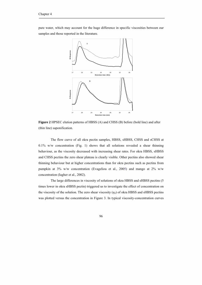
Chapter 4
96
pure water, which may account for the huge difference in specific viscosities between our
samples and those reported in the literature.
Figure 2 HPSEC elution patterns of HBSS (A) and CHSS (B) before (bold line) and after
(thin line) saponification.
The flow curve of all okra pectin samples, HBSS, sHBSS, CHSS and sCHSS at
0.1% w/w concentration (Fig. 1) shows that all solutions revealed a shear thinning
behaviour, as the viscosity decreased with increasing shear rates. For okra HBSS, sHBSS
and CHSS pectins the zero shear plateau is clearly visible. Other pectins also showed shear
thinning behaviour but at higher concentrations than for okra pectins such as pectins from
pumpkin at 3% w/w concentration (Evageliou et al., 2005) and mango at 2% w/w
concentration (Iagher et al., 2002).
The large differences in viscosity of solutions of okra HBSS and sHBSS pectins (5
times lower in okra sHBSS pectin) triggered us to investigate the effect of concentration on
the viscosity of the solution. The zero shear viscosity (η0) of okra HBSS and sHBSS pectins
was plotted versus the concentration in Figure 3. In typical viscosity-concentration curves
17 20 23 26 29 32 35
Retention time ( Min)
RI R
espo
nse
A
17 20 23 26 29 32 35
Retention time (min)
RI R
espo
nse
B
Physicochemical properties of okra pectin
97
of entangled polymer solutions a change of slope is observed at the entanglement
concentration c*. At concentrations below c* the zero shear viscosity scales as η0 α c1.3, and
at concentrations above c* it scales as η0 α c4 (Rubinstein and Semenov, 2001). The zero
shear viscosities of our okra HBSS and sHBSS pectin solutions plotted in Figure 3, scale as
η0 α c2.0 and η0 α c1.7, respectively, and no change in the slope of the curves was found in
the range of concentrations of 0.1–0.5% w/w for either polymer. Ndjouenkeu et al. (1996)
reported a value for c* for okra gum of about 0.15% w/w. For our samples c* is apparently
lower than 0.1% w/w. This difference may be due to a difference in okra variety and
extraction procedure (BeMiller et al., 1993). In addition, Ndjouenkeu et al. (1996) added
0.1 M NaCl to their samples, which would reduce the coil dimensions of the okra polymers,
by screening the electrostatic repulsion between the polymer segments. The results plotted
in Figure 3 suggest that the okra HBSS pectin molecules are able to entangle at a low
concentration (<0.1% w/w), which is much lower than the entanglement concentrations
mentioned for citrus pectins (0.6% w/w) (Kjoniksen et al., 2005). The scaling of the zero
shear viscosity with concentration we have observed is close to that found by Ndjouenkeu
et al. (1996), at concentrations above c*, and below c** (the start of the concentrated
regime). In this regime they observed a scaling exponent of 2.1.
.
0.01
0.1
1
10
100
0.1 1
Concentration (%w/w)
Zero
she
ar v
isco
city
(Pa
s)
Figure 3 Dependence of zero shear viscosity (η0) on the concentration of okra HBSS (♦)
and sHBSS pectins (■).
Chapter 4
98
The shear thinning region of the samples in each concentration was fitted with the
Ostward-DeWaele equation, η = m γ n-1, where m is the consistency index and n is the flow
behaviour index. The m and n values of okra HBSS and sHBSS pectins are shown in Table
2 and it can be seen that the n values are lower than the n values found for apple pectins
(0.83, Hwang and Kokini, 1992) which suggests that the okra HBSS pectin is more pseudo-
plastic than the apple pectins due to entanglement of the okra pectins molecules. This can
be explained by the nature of the substituents of the pectin backbone: it has already been
mentioned by Hwang and Kokini (1992) that more side chains and more branched side
chains may result in lower n values. Moreover, the okra HBSS pectins contained mostly
RG I segments when compared to most other pectins such as citrus, apple pectins which
contain mainly HG segments. The trend of a decrease of the n-values with increasing
concentration of okra HBSS and sHBSS pectins does suggest that the polymers aggregate
(Yanes et al., 2002). Moreover, all samples show Cox–Merz superposition (Cox & Merz,
1958) of the complex viscosity ωωωωη /)()()(* 22 GG ′′+′= and the steady
shear viscosity )(γη & evaluated at ωγ =& , which implies that the aggregation does not
lead to the formation of a weak associative network, as observed in for example xanthan
solutions.
Oscillatory shear measurements
A strain sweep was carried out with deformation values of 10 –103% (Fig. 4) to
determine the linear viscoelastic region of the pectin. A deformation value of 10 % for
every concentration of the samples was in the linear viscoelastic region, and was used to
determine the modulus G′ and G″. At a frequency of 0.1 s-1 the okra HBSS pectin has a
significantly higher storage modulus G′ than the other samples. For the latter samples G″ is
larger than G′ for the entire range of strain values. For the okra HBSS pectin the G′ and G″
values were roughly equal for all strains (i.e. tan δ ~1).
Physicochemical properties of okra pectin
99
Table 2 The consistency index (m) and the flow behaviour index (n) of okra HBSS and
sHBSS pectins in the power region of η versus γ (η = m γ n-1) as a function of concentration.
HBSS sHBSS Concentration
(%w/w) consistency
index (m)
flow
behaviour
index (n)
consistency
index (m)
flow
behaviour
index (n)
0.100
0.125
0.150
0.175
0.200
0.300
0.400
0.500
0.22
0.31
0.44
0.52
0.64
1.07
1.71
3.00
0.47
0.45
0.42
0.41
0.40
0.37
0.33
0.30
0.11
0.43
1.59
0.53
0.48
0.37
To determine the dependence of G' and G" on the frequency, frequency sweeps
were performed over a range of 10-1 to 10 Hz at strain of 10% as shown in Figure 5. The
modulus G' was lower than the modulus G" only at low frequency values. The crossover
between viscous and elastic behaviour of the okra HBSS pectin was at a frequency of about
0.1 s-1 (Fig. 5). For the other okra pectin samples (sHBSS, CHSS and sCHSS) the crossover
occurred at much higher frequencies (ƒ > 1 s-1). The crossover of G' and G" values provides
a good indication of viscoelastic behaviour of the material (Iagher et al., 2002) and defines
the beginning of the elastic behaviour or approaching gel state (Norziah et al., 2001). The
okra HBSS pectin solution presented a crossover point at frequency values lower than the
okra sHBSS and CHSS pectins, probably due to the fact that the okra HBSS pectin
structure can more easily form elastically active zones than okra sHBSS pectin. Although
the presence of acetyl groups has a negative effect on the gel formation of sugar beet
pectins (Oosterveld et al., 2000), our results show that the removal of acetyl group in okra
HBSS pectin weakens the tendency to network formation. These results may be due to the
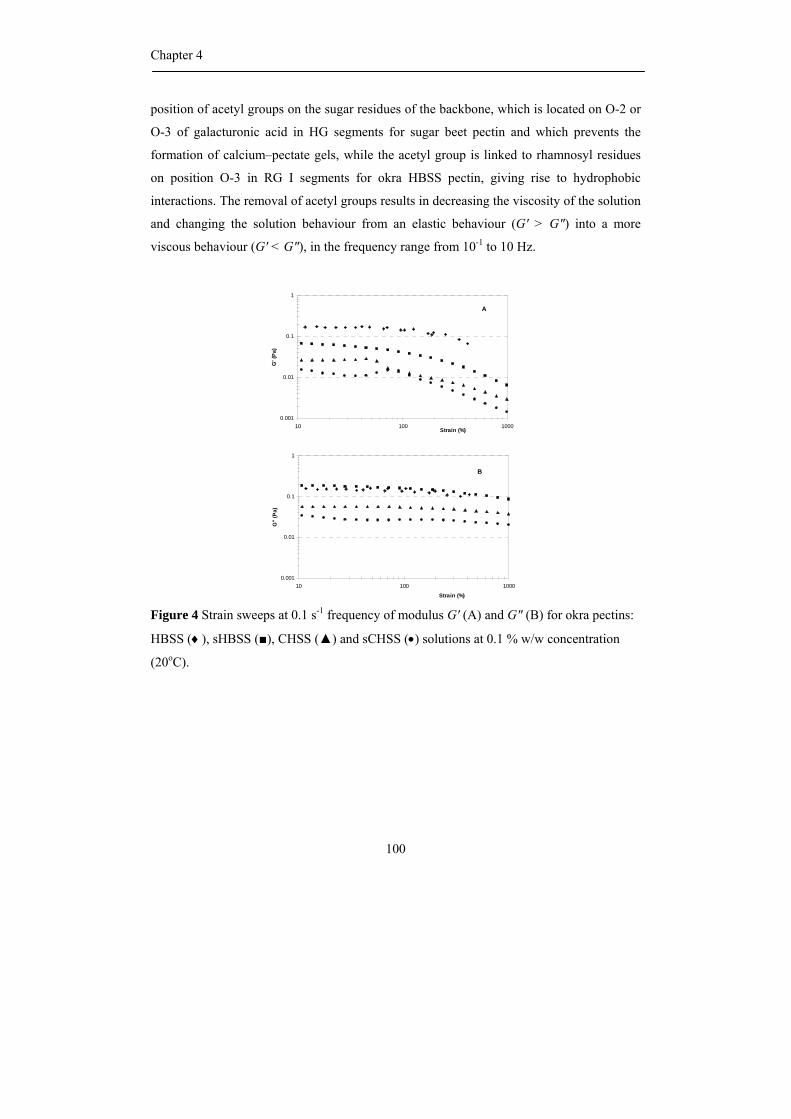
Chapter 4
100
position of acetyl groups on the sugar residues of the backbone, which is located on O-2 or
O-3 of galacturonic acid in HG segments for sugar beet pectin and which prevents the
formation of calcium–pectate gels, while the acetyl group is linked to rhamnosyl residues
on position O-3 in RG I segments for okra HBSS pectin, giving rise to hydrophobic
interactions. The removal of acetyl groups results in decreasing the viscosity of the solution
and changing the solution behaviour from an elastic behaviour (G' > G") into a more
viscous behaviour (G' < G"), in the frequency range from 10-1 to 10 Hz.
Figure 4 Strain sweeps at 0.1 s-1 frequency of modulus G′ (A) and G″ (B) for okra pectins:
HBSS (♦), sHBSS (■), CHSS (▲) and sCHSS (•) solutions at 0.1 % w/w concentration
(20oC).
0.001
0.01
0.1
1
10 100 1000Strain (%)
G' (
Pa)
A
0.001
0.01
0.1
1
10 100 1000
Strain (%)
G"
(Pa)
B
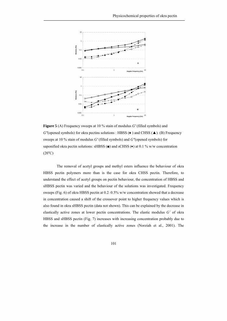
Physicochemical properties of okra pectin
101
Figure 5 (A) Frequency sweeps at 10 % stain of modulus G′ (filled symbols) and
G″(opened symbols) for okra pectins solutions : HBSS (♦) and CHSS (▲). (B) Frequency
sweeps at 10 % stain of modulus G′ (filled symbols) and G″(opened symbols) for
saponified okra pectin solutions: sHBSS (■) and sCHSS (•) at 0.1 % w/w concentration
(20oC)
The removal of acetyl groups and methyl esters influence the behaviour of okra
HBSS pectin polymers more than is the case for okra CHSS pectin. Therefore, to
understand the effect of acetyl groups on pectin behaviour, the concentration of HBSS and
sHBSS pectin was varied and the behaviour of the solutions was investigated. Frequency
sweeps (Fig. 6) of okra HBSS pectin at 0.2–0.5% w/w concentration showed that a decrease
in concentration caused a shift of the crossover point to higher frequency values which is
also found in okra sHBSS pectin (data not shown). This can be explained by the decrease in
elastically active zones at lower pectin concentrations. The elastic modulus G´ of okra
HBSS and sHBSS pectin (Fig. 7) increases with increasing concentration probably due to
the increase in the number of elastically active zones (Norziah et al., 2001). The
0.001
0.01
0.1
1
10
0.1 1 10Angular frequency (1/s)
Mod
ulus
(Pa)
A
0.001
0.01
0.1
1
10
0.1 1 10Angular frequency (1/s)
Mod
ulus
(Pa)
B

Chapter 4
102
concentration dependency of G´ can be expressed by the power law of G' α c β, with β =
1.7 and 1.5 for okra HBSS and sHBSS pectin. A dependence of G' α c 2 is commonly found
well above c* for normal polysaccharide gels (Clark and Ross-Murphy, 1987).
0.01
0.1
1
10
0.001 0.010 0.100 1.000 10.000
Angular frequency (1/s)
Mod
ulus
(Pa)
Figure 6 Frequency sweeps at 10 % stain of modulus G′ (filled symbols) and G″(opened
symbols) for okra HBSS solutions at 0.5 % w/w (♦), 0.4% w/w (■), 0.3% w/w(▲) and
0.2% w/w (•) concentration (20oC).
0.01
0.1
1
10
0.1 1
concentration (%w/w)
Ela
stic
mod
ulus
, G' (
Pa)
Figure 7 Dependence of elastic modulus (G′) on concentration for okra HBSS (♦) and
sHBSS (■) pectins at 11% strain and 0.1 s-1 frequency.
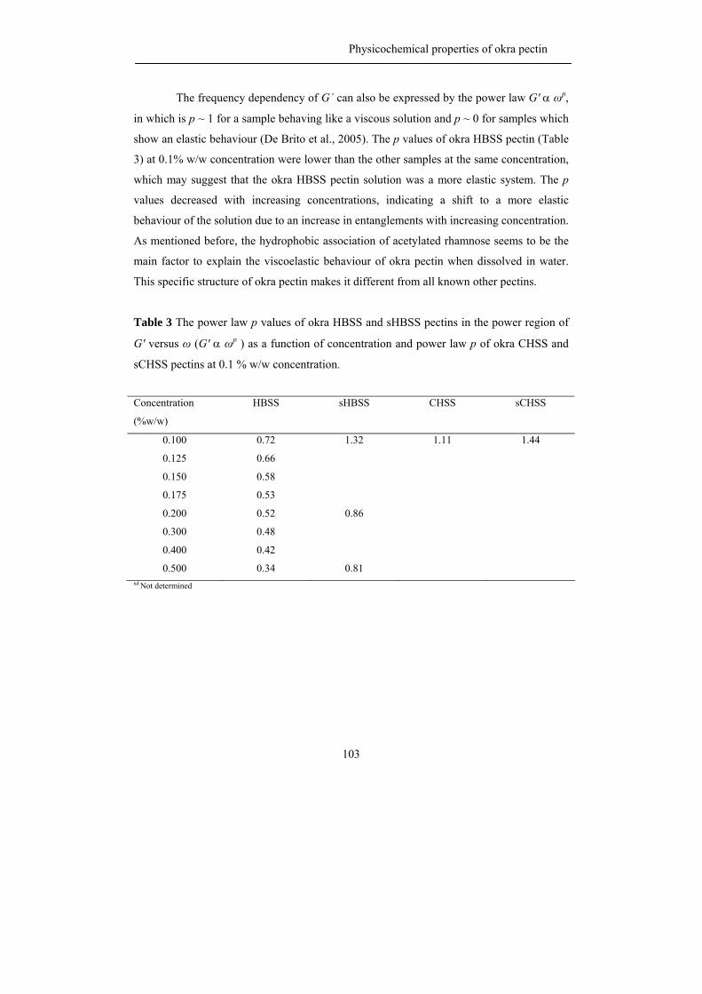
Physicochemical properties of okra pectin
103
The frequency dependency of G´ can also be expressed by the power law G' α ωp,
in which is p ~ 1 for a sample behaving like a viscous solution and p ~ 0 for samples which
show an elastic behaviour (De Brito et al., 2005). The p values of okra HBSS pectin (Table
3) at 0.1% w/w concentration were lower than the other samples at the same concentration,
which may suggest that the okra HBSS pectin solution was a more elastic system. The p
values decreased with increasing concentrations, indicating a shift to a more elastic
behaviour of the solution due to an increase in entanglements with increasing concentration.
As mentioned before, the hydrophobic association of acetylated rhamnose seems to be the
main factor to explain the viscoelastic behaviour of okra pectin when dissolved in water.
This specific structure of okra pectin makes it different from all known other pectins.
Table 3 The power law p values of okra HBSS and sHBSS pectins in the power region of
G' versus ω (G' α ωp ) as a function of concentration and power law p of okra CHSS and
sCHSS pectins at 0.1 % w/w concentration.
Concentration
(%w/w)
HBSS sHBSS CHSS sCHSS
0.100
0.125
0.150
0.175
0.200
0.300
0.400
0.500
0.72
0.66
0.58
0.53
0.52
0.48
0.42
0.34
1.32
0.86
0.81
1.11
1.44
nd Not determined
Chapter 4
104
Dynamic light scattering
To confirm that our non saponified samples are indeed associating in solution, as a
result of hydrophobic interactions, dynamic light scattering was performed at low
concentrations (0.001–0.1% w/w). To increase reproducibility of the measurements all okra
pectin samples were dissolved in a low ionic strength (10 mM) filtered sodium acetate
buffer with a pH of 5.
The intensity of scattered light for sufficiently small objects, at sufficiently low
concentrations is proportional to both the molecular weight M and the concentration C:
MCI ∝
At higher concentrations and for larger object sizes (roughly above 100 nm), this
proportionality is no longer strictly valid (Finsy, 1994). The ratio I/C is therefore only
roughly proportional to the molecular weight of the scattering objects. Figure 8 shows the
dependency of I/C on the concentration for the different pectin samples. Evidently, the
objects (aggregates/polymer coils) formed by the saponified okra pectins (sHBSS and
sCHSS) have lower scattering intensity than non saponified okra pectins and the size of the
aggregates/polymer molecules was not dependent on the concentration. This is in contrast
to the concentration dependent sizes observed for the non saponified okra pectins (HBSS
and CHSS). The higher scattering intensity of the CHSS and HBSS indicates a stronger
tendency to aggregate, and aggregation is strongly concentration dependent. In conclusion,
the saponified okra pectins have lower tendency to aggregate compared to non saponified
okra pectins. Comparing okra HBSS to okra CHSS pectin, the scattering intensity of okra
HBSS pectin was lower and less concentration dependent than for okra CHSS pectin
indicating that at low concentrations and in acetate buffer okra HBSS pectin has less
tendency to aggregate than okra CHSS pectin.
Sizes of the scattering objects can be deduced from a dynamic light scattering
experiment in which the fluctuations of the scattered light are analyzed that are a
consequence of the Brownian motion of the scattering objects. Analysis of the correlation
function of the intensity of scattered light as a function of time, using the Malvern DTS
software, version 5.0 showed that in general two types of particles were present, one slowly
moving (pectin aggregates) and one fast moving (pectin coils). Estimated hydrodynamic
Physicochemical properties of okra pectin
105
diameters of the pectin aggregates and pectin coils are given in Table 4. The size of the fast
moving objects, presumably the pectin coils, was around 20–30 nm for all pectin samples
and at all concentrations; however the sizes of the slow moving objects, pectin aggregates,
were highly variable. The aggregate sizes of saponified pectin molecules were
concentration independent, which is consistent with the scattering intensities, and aggregate
sizes were found to be around 210 and 175 nm for sHBSS and sCHSS, respectively. The
aggregate sizes of non saponified okra pectin were highly concentration dependent,
especially for okra CHSS pectin in accordance with the static scattering intensity. The
concentration dependencies of the aggregate diameters as found by analysis of the dynamic
light scattering are shown in Figure 9.
0
10000
20000
30000
0.00 0.05 0.10C (%w/w)
(I-I 0)
/C (a
.u.)
Figure 8 Intensity of light scattered from okra pectin solutions, as a function of pectin
concentration. Solution conditions: 10 mM sodium acetate buffer pH at 5. Plotted is the
scattered intensity (I), corrected for the scattering by the solvent (I0), and scaled by the
pectin concentration C (in weight %): HBSS (▲), sHBSS (∆), CHSS (■) and sCHSS (□).
The observed lower tendency to aggregate for okra HBSS pectin compared to okra
CHSS pectin seems to contradict our findings from the rheological measurements, where
the higher viscosities for the HBSS samples suggested that this material was more
Chapter 4
106
associated than CHSS. But we must keep in mind that the rheological measurements were
performed at higher concentrations than the light scattering experiments. Unfortunately,
due to the occurrence of multiple scattering, light scattering experiments could not be
performed in the same concentration range used for the rheological experiments. The use of
an acetate buffer in the light scattering experiments may also have affected the association,
since it partially screens the electrostatic repulsion between the molecules. HBSS has a
lower charge density than CHSS and its association will be less affected by the addition of
salt than for CHSS. These two factors make it difficult to give a one-to-one comparison of
the two experiments. But the light scattering experiments do show a big difference in
association between the saponified (and therefore less hydrophobic) samples, and the non
saponified samples, indicating that association driven by hydrophobic interactions is an
important factor in the rheological behaviour of these pectins. For both saponified pectins
(sHBSS and sCHSS), the additional charge introduced by the removal of methyl esters
resulted in more electrostatic repulsion between the polymer, molecules yielding a lower
tendency to aggregate and a non-concentration dependency of aggregate size.
Table 4 Estimated hydrodynamic diameters of okra pectin aggregates and pectin coil in 10
mM NaOAc buffer pH at 5 as determined using dynamic light scattering.
Samples Pectin aggregates
diameter (nm)
pectin coil diameter
(nm)
Scattering by
aggregates (%)
HBSS
sHBSS
CHSS
sCHSS
-a
210 + 20
-a
175 + 30
30 + 5
25 + 5
30 + 5
20 + 5
85
60
70
60 a Concentration dependence
Physicochemical properties of okra pectin
107
0
500
1000
1500
0.00 0.05 0.10C (%w/w)
Agg
rega
te d
iam
eter
(nm
)
Figure 9 Concentration-dependent sizes of HBSS (▲) and CHSS (■) aggregates in 10 mM
NaOAc buffer pH at 5, as determined using dynamic light scattering.
Conclusions
In this research we showed that some physical properties of okra HBSS and CHSS
pectins can be related to their structures. In the absence of salts and at a pH above the pKa
of the COOH groups of galacturonic acid, okra HBSS pectin solutions have a high viscosity
and show shear thinning behaviour. The removal of acetyl groups and methyl esters of okra
HBSS pectin decreases the viscosity of the solution and changes the behaviour of the
solution from a predominantly elastic response (G´ > G") for okra HBSS pectin to a
predominantly viscous response (G´ < G") for okra sHBSS pectin (in the frequency range
from 10-1 to 10 Hz). Light scattering experiments show a decrease in association of the
pectin molecules when the HBSS is saponified, indicating that association of pectin
molecules driven by hydrophobic interactions (resulting from the acetylated rhamnosyl
residues in RG I) play an important role in the rheology of these samples. The okra CHSS
pectin solutions in water have a lower viscosity than HBSS solutions at the same
concentration, and also show shear thinning behaviour. Saponified CHSS solutions had a
lower viscosity than non saponified CHSS, and light scattering experiments again suggest
Chapter 4
108
this decrease in viscosity is a result of a decrease in the degree of association of the
molecules. At low concentrations and in the presence of salt the okra CHSS pectin shows a
higher tendency to aggregate than the HBSS pectin. This may be a result of a difference in
charge density of the molecules. The unique chemical fine structure of okra HBSS pectin
really makes this pectin quite different in rheological and physical behaviour when
compared to the more common homogalacturonan-rich pectins from apple and citrus
Material and methods
Sequential extraction of okra AIS.
The soft and mature okra pods, A. esculentus (L.) Moench, (5–10 cm in length)
were grown in Thailand and were collected at a local market in June 2005. The alcohol
insoluble solids from okra pods (Okra AIS) were prepared and were sequentially extracted
with 0.05 M sodium acetate buffer at pH 5.2 and 70 oC (“Hot Buffer Soluble Solids”,
HBSS), 0.05 M EDTA in 0.05 M sodium acetate and 0.05 M sodium oxalate at pH 5.2 and
70 oC (“CHelating agent Soluble Solids”, CHSS) (Sengkhamparn et al., 2009a).
Part of the HBSS and CHSS extract were saponified for 16 h at 4 oC by adding
50% NaOH to a final concentration of 0.05 M NaOH to completely remove acetyl groups
and methyl esters. The solutions were then neutralized and ultrafiltrated through a
membrane with molecular weight cut-off of 10 kDa and freeze-dried. The molecular weight
distribution of HBSS, CHSS and saponified sample were determined by high performance
size exclusion chromatography (HPSEC) (Sengkhamparn et al., 2009a).
The pectin samples obtained have been characterized by their sugar composition
(Table 1; Sengkhamparn et al., 2009a and 2009b).
Rheological Measurements
HBSS, CHSS, saponified HBSS (sHBSS) and saponified CHSS (sCHSS) samples
were dissolved in millipore water. The viscosity of the samples was determined using a
Physica MCR 301 (Anton Paar, Austria) stress controlled rheometer with concentric double
Physicochemical properties of okra pectin
109
gap cylinder geometry (DG 26.8). Flow curves with increasing shear rate (0.01–100 s-1)
were measured at 20 oC.
Oscillatory measurements were used to determine the storage modulus (G´) and
loss modulus (G") of the samples. The linear viscoelastic region of the samples was
determined by a strain sweep (10–103% at 1 Hz). Frequency sweeps (10-1–10 Hz at 10%
stain) were applied to study the frequency dependence of G´ and G".
Light Scattering
HBSS, CHSS, saponified HBSS (sHBSS) and saponified CHSS (sCHSS) samples
were dissolved in 10 mM NaOAc Buffer pH at 5. The sample solutions were centrifuged at
18,500 x g, 10 oC for 30 min and 20 µL of samples were analyzed by light scattering using
Helma quartz cuvettes with light path 3 mm and centre 9.65 mm (Type No. 105.251.005-
QS). Light scattering was measured at 25 oC using a Malvern NanoS (UK), operating at a
wavelength of 633 nm and a scattering angle of 173o. The effective hydrodynamic radii
reported are the peak positions of a distribution fit of the intensity autocorrelation function,
as reported by the Malvern DTS software, version 5.0.
Acknowledgments
This study has been carried out with financial support from the Commission on
Higher Education Ministry of Education, Thailand, under The Commission on Higher
Education Staff Development project for the Joint Ph.D. Program in Food Science at
Kasetsart University, Thailand.
Chapter 4
110
References
Baht, U.S. & Tharathan, R.N. (1987). Functional properties of okra (Hibiscus esculentus) mucilage. Starch/starke,
39, 165-167.
BeMiller, J.N., Whistler, R.L. & Barbalow, D.G. (1993). Aloe, China, Flexseed, Okra, Psyllium seed, Quince
seed, and Tamarind Gums. In L.W. Roy & J.N. BeMiller, Industrial Gums Polysaccharide and their
Derivatives (pp. 235-255). San Diago: Academic Press.
Clark, A.H. & Ross-Murphy, S.B. (1987) Structural and mechanical properties of biopolymer gels. Advan. Polym.
Sci., 83, 57-192.
Costantino, A.J. & Romanchick-Cerpoviez, J.E. (2004) Physical and sensory measures indicate moderate fat
replacement in frozen dairy dessert is feasible using okra gum as a milk-fat ingredient substitute. J. Am.
Diet. Assoc., 104 (supplement 2), 44.
Cox, W.P.& Merz, E.H. (1958). Correlation of dynamic and steady-flow viscosities. J. Polym. Sci., 28, 619-622.
De Brito, A.C.F., Sierakowski, M.R., Reicher, F., Feitosa, J.P.A. & de Paula, R.C.M. (2005) Dynamic rheological
study of Sterculia striata and karaya polysaccharides in aqueous solution. Food Hydrocolloid., 19, 861-
867.
Evageliou, V., Ptitchkina, N. M. & Morris, E.R. (2005) Solution viscosity and structural modification of pumpkin
biopectin. Food Hydrocolloid., 19, 1032-1036.
Finsy, R. (1994). Particle sizing by quasi-elastic light scattering. Adv. Colloid. interface., 52, 79-143.
Hwang, J. & Kokini, J.L. (1992). Contribution of side branches to rheological properties of pectins. Carbohydr.
Polym., 19, 41-50.
Iagher, F., Reicher, F. & Ganter, J.L.M.S. (2002). Structural and rheological properties of polysaccharides from
mango (Mangifera indica L.) pulp. Int. J. Biol. Macromol., 31, 9-17.
Kjoniksen, A.-L., Hiorth, M. & Nystrom, B. (2005). Association under shear flow in aqueous solutions of pectin.
Eur. Polm. J., 41, 761-770.
Koubala, B.B., Mbome, L.I., Kansci, G., Mbiapo, T.F., Crepeau, M.-J., Thibault,J.-F. & Ralet, M.-C. (2008).
Physicochemical properties of pectins from ambarella Peels (Spondias cytherea) obtained using
different extraction conditions. Food Chem., 106, 1202-1207.
Morris, E.R., Cutler, A.N., Ross-Murphy, S.B., Rees, D.A. & Price ,J. (1981). Concentration and shear rate
dependence of viscosity in random coil polysaccharide solutions. Carbohydr. Polym., 1, 5-21.
Ndjouenkeu, R., Goycoolea, F.M., Morris, E.R. & Akingbala, J.O. (1996). Rheology of Okra(Hibiscus esculentus
L.) and Dika nut (Irvingia gabonensis) polysaccharides. Carbohydr. Polym., 29, 263-269.
Norziah, M.H., Kong, S.S., Karim, A.A. & Seow, C.C. (2001) Pectin-sucrose-Ca2+ interations: effects on
rheological properties. Food Hydrocolliod., 15, 491-498.
Oosterveld, A., Beldman, G., Searle-van Leeuwen, M.J.F. & Voragen, A.G.J. (2000). Effect of enzymatic
deacetylationon gelation of sugar beet pectin in the presence of calcium. Carbohydr. Polym., 43, 249-
256.
Physicochemical properties of okra pectin
111
Romanchik-Cerpovicz, J.E., Costantino, A.C. & Laura, H. G. (2006). Sensory evaluation ratings and melting
characteristics show that okra gum is an acceptable milk-fat ingredient substitute in chocolate frozen
dairy dessert. J. Am. Diet. Assoc.,106, 594-597.
Romanchik-Cerpovicz, J.E., Tilmon, R.W. & Baldree, K.A. (2002). Moisture retention and consumer acceptability
of chocolate bar cookies prepared with okra gum as a fat ingredient substitute. J. Am. Diet. Assoc., 102,
1301-1303.
Rubinstein, M. & Semenov, A.N. (2001). Dynamics of entangled solutions of associating polymers.
Macromolecules., 34, 1058-1068.
Sengkhamparn, N., Verhoef, R., Schols, H. A., Sajjaanantakul, T. & Voragen, A.G.J. (2009a). Characterization of
Cell Wall Polysaccharide from Okra (Abelmoschus esculentus (L.) Moench). Carbohydr. Res., 344,
1824-1832.
Sengkhamparn, N., Verhoef, R., Bakx, E.J., Schols, H.A., Sajjaanantakul, T. & Voragen, A.G.J. (2009b). Okra
pectin contains an unusual substitution of its rhamnosyl residues with acetyl and alpha–linked
galactosyl groups. Carbohydr. Res., 344, 1842-1851.
Tomada, M., Shimada, K., Saito ,Y. & Sugi, M. (1980). Plant mucilages. XXVI. Isolation and structural features
of a mucilage, “Okra mucilage”, from the immature fruit of Abelmoschus esculentus. Chem. Pharm.
Bull., 28, 2933-2940.
Whistler, R.L. & Conrad, H.E. (1954). A crystalline galactobiose from acid hydrolysis of okra mucilage. J. Am.
Chem. Soc., 76, 1673-1974.
Williams, P.A. & Phillips, G.O. (2000). Introduction of food hydrocolloids. In Phillips G.O. & Williams P.A.,
Handbook of hydrocolloids (pp. 1-19). USA: CRC Press.
Woolfe, M.L., Chaplin, M.F. & Otchere, G. (1977). Studies on the mucilages extracted from Okra fruits (Hibiscus
esculentus L.) and Baobab leaves (Adansonia digitata L.). J. Sci. Fd. Agric., 28, 519-529.
Yanes, M., Durán, L. & Costell, E. (2002). Effect of hydrocolloid type and concentration on flow behaviour and
sensory properties of milk beverages model systems. Food Hydrocolloid., 16, 605-611.

Chapter 4
112
Complement-fixing activity and cell behaviour of okra pectins
113
Chapter 5
Complement-fixing activity and influence on cell behaviour
of okra (Abelmoschus esculentus (L.) Moench) pectins
Sengkhamparn, N., Samuelsen, A.B., Michaelsen, T.E., Nagel, M-D, Schols, H.A. &
Voragen, A.G.J.
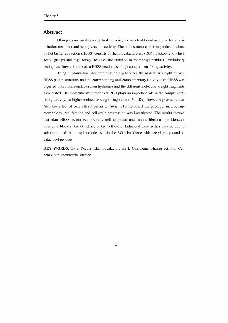
Chapter 5
114
Abstract Okra pods are used as a vegetable in Asia, and as a traditional medicine for gastric
irritation treatment and hypoglycaemic activity. The main structure of okra pectins obtained
by hot buffer extraction (HBSS) consists of rhamnogalacturonan (RG) I backbone to which
acetyl groups and α-galactosyl residues are attached to rhamnosyl residues. Preliminary
testing has shown that the okra HBSS pectin has a high complement-fixing activity.
To gain information about the relationship between the molecular weight of okra
HBSS pectin structures and the corresponding anti-complementary activity, okra HBSS was
digested with rhamnogalacturonan hydrolase and the different molecular weight fragments
were tested. The molecular weight of okra RG I plays an important role in the complement-
fixing activity, as higher molecular weight fragments (>50 kDa) showed higher activities.
Also the effect of okra HBSS pectin on Swiss 3T3 fibroblast morphology, macrophage
morphology, proliferation and cell cycle progression was investigated. The results showed
that okra HBSS pectin can promote cell apoptosis and inhibit fibroblast proliferation
through a block at the G1 phase of the cell cycle. Enhanced bioactivities may be due to
substitution of rhamnosyl moieties within the RG I backbone with acetyl groups and α-
galactosyl residues.
KEY WORDS: Okra, Pectin, Rhamnogalacturonan I, Complement-fixing activity, Cell
behaviour, Biomaterial surface
Complement-fixing activity and cell behaviour of okra pectins
115
Introduction
Pectin is an important cell wall component of plants and probably the most
complex macromolecule in the nature. The pectin backbone has been found to be composed
of two main structural elements, homogalacturonans or smooth regions and
rhamnogalacturonans (RGs) or ramified regions. RG I contains a backbone of alternating
rhamnosyl and galacturonosyl residues (Schols et al., 1990) in which galacturonosyl
residues may be O-acetylated at position O-2 and/or position O-3 (Ishii, 1997; Perrone et
al., 2002). Moreover, rhamnosyl residues may be branched at position O-4 with β-linked
galactan (Gur’janov et al., 2007), α-(1, 5)-linked arabinan or branched arabinogalactan I
(Vincken et al., 2003; Ridley et al., 2001) depending on source and method of extraction
(McNeil et al., 1982; Lau et al., 1987; Ishii et al., 1989). Studies have shown that pectin
may exhibit biological activity such as anti-complementary-, anti-inflammatory-, anti–
ulcer-, IL-6 enhancing- and mutagenic activity and may inhibit cancer metastasis (Wagner
and Kraus, 2000; Chen et al., 2006; Yamada, 2000).
Pectin has the ability to activate the complement system which plays an important
role in the host defense mechanism (Michaelsen et al., 2000; Yamada et al., 1985). The
ramified regions of pectin contain high complement-activating activity (Yamada et al.,
1985) which were related to highly branched neutral sugar side chains with high levels of
β-(1,6)- and β-(1,3,6)-linked Gal and low levels of β-(1,4) – linked Gal (Samuelsen et al.,
2007). Moreover, Yamada and Kiyohara (1999) reported that the minimum requirement for
the activity was the presence of β-(1,6) – linked galactose side chain attached to the RG
backbone. In addition, arabinogalactan type II and/or arabinan structural elements from
cabbage pectin were found to be of key importance for complement-fixing activity while
removal of arabinosyl residues from arabinogalactan type II was found to decrease this
activity (Westereng et al., 2006).
Recently, it was shown that pectin can serve as a biomaterial for surface
modification of medical devices and implants. The biocompatibility of medical devices and
implants are generally established by investigating the interactions of coating material with
cells and adsorbed proteins at the implant/tissue interface (Roach et al., 2005). The
potential of pectin for coating medical devices and implants has been studied by Morra et
Chapter 5
116
al., 2004; Kokkonen et al., 2006; Morra, 2005; Nagel et al, 2008; Bussy et al., 2008, the
relationship between the structure of pectin and direct/indirect biological effect has
however hardly been investigated.
Okra pods are cultivated in many areas of the world and are used as a vegetable
and health promoting ingredient in Asia. Okra pods are also used as a traditional medicine
for treatment of gastric irritation, for treatment of dental diseases (Lengsfeld et al., 2004),
lowering the cholesterol level and cancer prevention (Kahlon et al., 2007). Water extracted
okra pectin showed anti-complementary and hypoglycaemic activities which were related
to the presence of polysaccharides consisting of repeating Rha-1,4-GalA dimers linked by
α-(1,2) linkages in okra (Tomoda et al. 1989).
Okra pods contain thick slimy, acidic polysaccharides consisting of galactose,
rhamnose and galacturonic acid (Whistler and Conrad, 1954). The Okra Hot Buffer Soluble
Solids (HBSS) pectin fraction was found to be composed of RG I structures including an
uncommon substitution of the rhamnosyl residue with acetyl groups and terminally α-
linked galactosyl residues present in rather short side chains. The Okra Chelating agent
Soluble Solid (CHSS) pectin fraction was found to consist mainly of homogalacturonan
segments (70%) in addition to much smaller levels of RG I (30%) having slightly longer β-
(1,4)-linked galactan side chains (2 – 3 residues on average) when compared to CHSS RG
I. In addition, some arabinan and arabinogalactan type II neutral sugar side chains might be
present within the CHSS RG I (Sengkhamparn et al., 2009a, b).
In this chapter, we present the effect of the chemical fine structure and molecular
weight of okra RG I on the complement-fixing activity. We also tested okra polysaccharide
fractions for their effectiveness for surface modification of implant material in order to
understand the effect of okra pectin structure on cell behaviour.
Results and discussion
The sugar composition of okra pectin fractions obtained by hot buffer (HBSS) and
chelating agent (CHSS) extraction is shown in Table 1. Okra HBSS pectin consists of
acetylated rhamnogalacturonan (RG) I containing rhamnosyl residues with acetyl groups
and α-linked galactosyl residues attached. The neutral side chains in HBSS RG I are short,
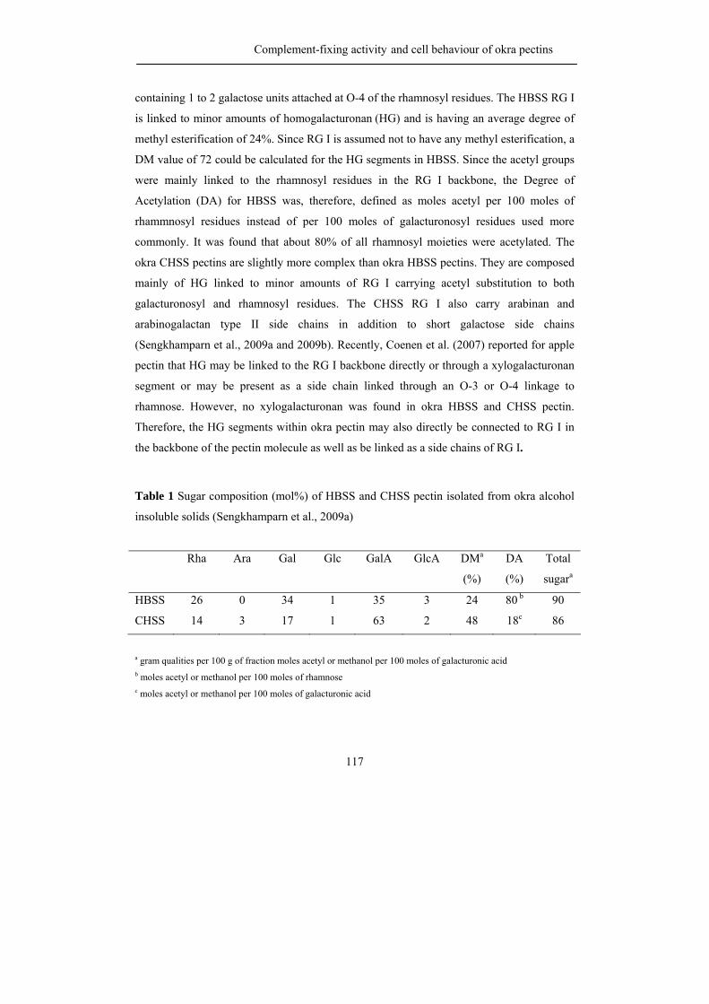
Complement-fixing activity and cell behaviour of okra pectins
117
containing 1 to 2 galactose units attached at O-4 of the rhamnosyl residues. The HBSS RG I
is linked to minor amounts of homogalacturonan (HG) and is having an average degree of
methyl esterification of 24%. Since RG I is assumed not to have any methyl esterification, a
DM value of 72 could be calculated for the HG segments in HBSS. Since the acetyl groups
were mainly linked to the rhamnosyl residues in the RG I backbone, the Degree of
Acetylation (DA) for HBSS was, therefore, defined as moles acetyl per 100 moles of
rhammnosyl residues instead of per 100 moles of galacturonosyl residues used more
commonly. It was found that about 80% of all rhamnosyl moieties were acetylated. The
okra CHSS pectins are slightly more complex than okra HBSS pectins. They are composed
mainly of HG linked to minor amounts of RG I carrying acetyl substitution to both
galacturonosyl and rhamnosyl residues. The CHSS RG I also carry arabinan and
arabinogalactan type II side chains in addition to short galactose side chains
(Sengkhamparn et al., 2009a and 2009b). Recently, Coenen et al. (2007) reported for apple
pectin that HG may be linked to the RG I backbone directly or through a xylogalacturonan
segment or may be present as a side chain linked through an O-3 or O-4 linkage to
rhamnose. However, no xylogalacturonan was found in okra HBSS and CHSS pectin.
Therefore, the HG segments within okra pectin may also directly be connected to RG I in
the backbone of the pectin molecule as well as be linked as a side chains of RG I.
Table 1 Sugar composition (mol%) of HBSS and CHSS pectin isolated from okra alcohol
insoluble solids (Sengkhamparn et al., 2009a)
Rha Ara Gal Glc GalA GlcA DMa
(%)
DA
(%)
Total
sugara
HBSS 26 0 34 1 35 3 24 80 b 90
CHSS 14 3 17 1 63 2 48 18c 86
a gram qualities per 100 g of fraction moles acetyl or methanol per 100 moles of galacturonic acid b moles acetyl or methanol per 100 moles of rhamnose c moles acetyl or methanol per 100 moles of galacturonic acid
Chapter 5
118
The complement-fixing activity
Effect of structural features
The hairy region of the pectin molecules seems to play a major role in its
complement- fixing activity (Samuelsen et al., 2007; Samuelsen et al., 1996; Yamada and
Kiyohara, 1999). Tomada et al. (1989) reported that water extracted okra pectins showed a
high complement-fixing activity. Since okra HBSS pectin was found to have a rather
unique structure differing from RG I structures of other plant origin (Sengkhamparn et al.,
2009b), the complement-fixing activity of okra HBSS and CHSS pectins were measured in
order to gain information of a possible effect of HBSS on the complement system.
Additionally, the effect of methyl esters and acetyl groups on the activity was determined
by measuring the activity of saponified pectins as well.
Figure 1 shows complement-fixing activity of HBSS, saponified HBSS (sHBSS),
CHSS and saponified CHSS (sCHSS). The percentage of haemolysis inhibition which is
the read out of the complement-fixing activity was slightly lower for all okra pectin
samples when compared to the level found for PM II which was a positive control obtained
from Plantago major L (Samuelsen et al, 1996). Comparing HBSS and saponified HBSS
shows that the complement-fixing activity of both samples was similar, indicating that
methyl esters and acetyl groups have no effect on the complement-fixing activity of HBSS.
This is in contrast with the results found for CHSS and saponified CHSS where the
saponified sample showed a slightly lower activity.
In contrast to the literature, stating that arabinosyl structures within pectic hairy
regions such as (1,2,5)- and (1,3,5)– linked α-arabinosyl residues from Acanthus
ebracteatus (Hokputsa et al., 2004) and α-(1,2,5)-arabinan structures isolated from Zizyphus
jujuva (Yamada et al., 1985) are important for complement-fixing activity, okra HBSS
pectin without any arabinose present showed a higher activity than okra CHSS pectin
having slightly higher arabinose levels. Moreover, by using ICH50 values which is the
concentration showing 50 % inhibition of haemolysis in the test system used, it was found
that okra HBSS pectins (ICH50 about 1.00 mg/mL) express rather similar activities compare
to white cabbage pectins and hot water extracted red Kale pectins, even though these
pectins contained higher levels of arabinosyl residues (11.7 and 16.9 w/w% of total
Complement-fixing activity and cell behaviour of okra pectins
119
carbohydrate content) (Samuelsen et al., 2007). Our results indicate that the level of
arabinose plays no major role in okra pectin’s complement-fixing activity.
-100
10203040
506070
8090
1 1.5 2 2.5 3 3.5Concentration (log ug/ml)
% in
hibi
tion
Figure 1 Haemolysis inhibition of okra pectins: HBSS (■), saponified HBSS (□), CHSS
(▲), and saponified CHSS (∆), PM II (♦) is positive control derived from Plantago major
L (Samuelsen et al, 1996)
Another point which may have effect on the bioactivity of pectins is the amount of
neutral sugar side chains other than arabinans. Samuelsen et al. (2007) mentioned that the
bioactivity of pectins from Brassica oleracea (white cabbage, Kale and red Kale) was
related to the amount of β-(1,6)- and β-(1,3,6)- linked galactose and the low amount of α-
(1,4)-linked galacturonic acid. Therefore, galactan structures present may explain the high
activity of the okra HBSS pectins although almost all galactosyl linkages were terminal or
1-4 linked. In addition, the α–galactosyl residues present may determine the complement-
fixing activity as well. Unfortunately, it was not possible to remove the terminal α –
galactose by α-galactosidases from various origin (Sengkhamparn et al., 2009b) preventing
us to confirm the influence of this specific unit on the complement-fixing activity. The
complement-fixing activity was expressed in percentage of PM II (positive control) activity
at a concentration of 500 µg/mL in order to avoid day to day variations in the assay. For
okra HBSS and CHSS pectin values of 63 and 59% were calculated, and these values are
about similar to the values found for white cabbage pectin, 52 % (Samuelsen et al., 2007)
Chapter 5
120
and red cabbage pectin, 60 % (Samuelsen et al., 2007) extracted by hot water (100 oC).
Saponified HBSS pectin (50 %) also showed a similar activity as red cabbage pectin. All
okra pectin expressed lower activities than the 90% inhibition mentioned for pectin from
Glinus oppositifolius (L.) Aug. DC. a Malian medical plant (Inngjerdingen et al., 2005) and
from Acanthus ebracteatus, a Thai medical plant (Hokputsa et al., 2004).
This different behaviour is probably due to the fact that the complement-fixing
activity is affected by many factors such as size, three dimensional structure of the polymer,
pectin source (Kweon et al., 2003; Suzuki et al., 2003) and is also influenced by differences
in the conditions of the bioactivity assay itself.
Figure 2 Haemolysis inhibition of A: HBSS (■), HBSS RGH digest (□), saponified HBSS
(▲), and saponified RGH HBSS (∆) and B: saponified CHSS (■),saponified RGH CHSS
digest (▲)saponified CHSS PG digest (•), PM II (♦) is positive control derived from
Plantago major L (Samuelsen et al, 1996)
To gain more information about the effect of the structure of okra pectins on its
complement-fixing activity, the okra pectins were incubated with polygalacturonase (PG)
and rhamnogalacturonan hydrolase (RGH). PG cleaves the glycosidic linkages in HG
sequences by hydrolysis (Benen & Visser, 2003) while the RGH is able to cleave the GalA-
Rha linkages in RG sequences having the requirement that the GalA residue do not carry
acetyl substitution (Searle-van Leeuwen et al., 1996). Sengkhamparn et al. (2009b) showed
that RGH can also split glycosidic linkages within the RG backbone of okra HBSS still
having an acetyl group on the rhamnose moiety. PG was shown not to be active towards
okra HBSS. Therefore, the okra HBSS pectins were incubated with RGH only. The
-15
0
15
30
45
60
75
90
1 1.5 2 2.5 3 3.5
Concentration (log ug/ml)
% In
hibi
tion
A
-30
-15
0
15
30
45
60
75
1 1.5 2 2.5 3 3.5
Concentration (log ug/ml)
% in
hibi
tion
B
Complement-fixing activity and cell behaviour of okra pectins
121
complement-fixing activity of okra HBSS and sHBSS after partial hydrolysis by RGH
(incubation for 16 hrs.) is shown in Figure 2A. The digests of okra HBSS as well as sHBSS
still retained a high activity which was only slightly lower than the activity of the original
HBSS.
In contrast to the okra HBSS pectins, okra CHSS pectins are composed of mainly
HG (70 %) next to RG I (30%) and both RGH and PG can act toward the okra CHSS
pectins (Sengkhamparn et al., 2009b). Therefore, okra CHSS pectin was incubated with PG
and RGH. The complement-fixing activity of the CHSS PG and CHSS RGH digests
showed negligible activity (Fig. 2B). This might be explained by the structure and size of
the remaining fragments after enzyme treatment. PG from A. aculeatus cleaves the α-1,4-D-
galacturonosyl linkages of the HG-backbone by hydrolysis (Vincken et al., 2003) forming
galacturonic acid oligomers. These were removed from the digest by dialysis. Therefore,
the remaining polymers in the PG CHSS digest were supposed to be mainly RG I
fragments. RG I obtained from many sources have been found to have complement-fixing
activity. The PG treatment of CHSS showed a negligible degradation of the pectin. In
general, pectin has been reported to mainly activate the complement system through the
classical pathway (as examined through our test system) being activated through the
complement protein 1 subunit q (C1q) (Michaelsen et al., 2000, Gaboriaud et al., 2003;
Matsushita et al., 2004). It has been speculated that the activity might depend on
carbohydrate backbone carrying the oligosaccharides with specific spatial organization and
the unsuitable distances leading to inefficient binding (Westereng et al., 2006). Therefore,
CHSS RG I fragment obtained from PG treated might provide insufficient affinity for a
putative binding to the globular head of C1q.
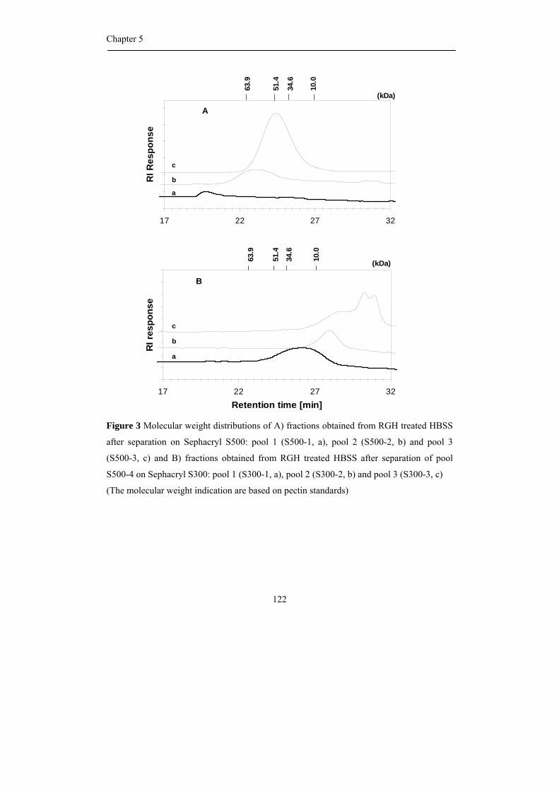
Chapter 5
122
Figure 3 Molecular weight distributions of A) fractions obtained from RGH treated HBSS
after separation on Sephacryl S500: pool 1 (S500-1, a), pool 2 (S500-2, b) and pool 3
(S500-3, c) and B) fractions obtained from RGH treated HBSS after separation of pool
S500-4 on Sephacryl S300: pool 1 (S300-1, a), pool 2 (S300-2, b) and pool 3 (S300-3, c)
(The molecular weight indication are based on pectin standards)
17 22 27 32Retention time [min]
RI r
espo
nse
B
10.0
(kDa)34.6
51.4
63.9
a
b
c
17 22 27 32
Retention time [min]
RI R
espo
nse
(kDa)
63.9
51.4
34.6
10.0
A
a
c
b
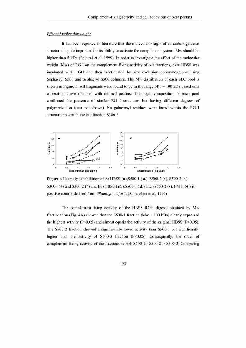
Complement-fixing activity and cell behaviour of okra pectins
123
Effect of molecular weight
It has been reported in literature that the molecular weight of an arabinogalactan
structure is quite important for its ability to activate the complement system: Mw should be
higher than 5 kDa (Sakurai et al. 1999). In order to investigate the effect of the molecular
weight (Mw) of RG I on the complement-fixing activity of our fractions, okra HBSS was
incubated with RGH and then fractionated by size exclusion chromatography using
Sephacryl S500 and Sephacryl S300 columns. The Mw distribution of each SEC pool is
shown in Figure 3. All fragments were found to be in the range of 6 – 100 kDa based on a
calibration curve obtained with defined pectins. The sugar composition of each pool
confirmed the presence of similar RG I structures but having different degrees of
polymerization (data not shown). No galactosyl residues were found within the RG I
structure present in the last fraction S300-3.
Figure 4 Haemolysis inhibition of A: HBSS (■),S500-1 (▲), S500-2 (•), S500-3 (×),
S300-1(+) and S300-2 (*) and B: sHBSS (■), sS500-1 (▲) and sS500-2 (•), PM II (♦) is
positive control derived from Plantago major L (Samuelsen et al, 1996)
The complement-fixing activity of the HBSS RGH digests obtained by Mw
fractionation (Fig. 4A) showed that the S500-1 fraction (Mw > 100 kDa) clearly expressed
the highest activity (P<0.05) and almost equals the activity of the original HBSS (P<0.05).
The S500-2 fraction showed a significantly lower activity than S500-1 but significantly
higher than the activity of S500-3 fraction (P<0.05). Consequently, the order of
complement-fixing activity of the fractions is HB~S500-1> S500-2 > S500-3. Comparing
0
15
30
45
60
75
1 1.5 2 2.5 3 3.5
concentration (log ug/ml)
% in
hibi
tion
A
-30
-15
0
15
30
45
60
75
90
1 1.5 2 2.5 3 3.5
concentration (log ug/ml)
% in
hibi
tion
B
Chapter 5
124
S500-1 with S500-2 shows that a reduction in the molecular weight with about 50%
resulted in a decrease of complement-fixing activity of 40 %. Both the S300 fractions
showed a decreased activity (-70%) when compared to the original okra HBSS pectin.
From these results it can be concluded that the activity of okra HBSS pectins decreases with
decreasing molecular weight. This trend can also be observed for the saponified okra HBSS
pectins. The activity of saponifed S500-1 and sS500-2 fraction expressed both similar, very
low activity (Fig. 4B). Therefore, the minimal size of okra RG I that possess significant
complement-fixing activity according to these experiments is about 50 kDa.
Effect of presence of acetyl groups
To gain insight in the effect of acetyl groups on the activity, the activity of
saponified fractions at a concentration of 1 mg/mL (maximum concentration) were
compared to the activity of the original fractions (Table 2). Only the results for the sS500-1,
sS500-2 fraction are reported since the other saponified fractions showed negligible
activity. The original parental HBSS and S500-1 were significantly more active than
saponified HBSS and sS500-1(P<0.05). These results may suggest that the presence of
acetyl groups has an effect on the activity since the removal of acetyl groups results in a
decrease of the activity of particularly the RG I fragments in S500-1. However, S500-2 was
significantly less active than sS500-2.
The substitution of okra HBSS RG-I by α-linked galactosyl moieties might be
quite essential for the anti complementary activity. So far such effects on the activity have
been mentioned only in literature for arabinose substitution. These results points once again
that the bioactivity of pectins is very difficult to predict and strongly depend on structural
features and size and 3D-stucture of the pectin (Yamada and Kiyohara, 2007; Kweon et al.,
2003; Suzuki et al., 2003).
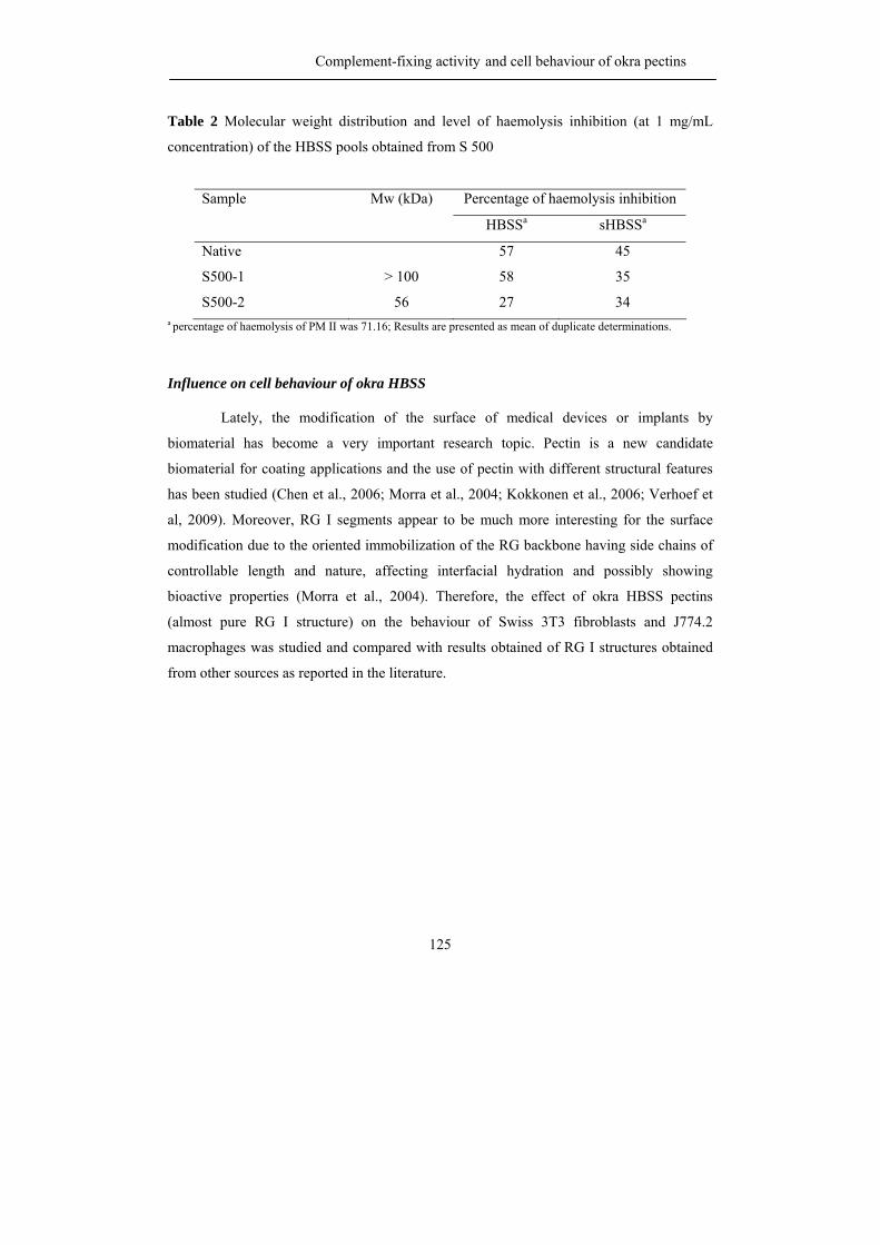
Complement-fixing activity and cell behaviour of okra pectins
125
Table 2 Molecular weight distribution and level of haemolysis inhibition (at 1 mg/mL
concentration) of the HBSS pools obtained from S 500
Percentage of haemolysis inhibition Sample Mw (kDa)
HBSSa sHBSSa
Native
S500-1
S500-2
> 100
56
57
58
27
45
35
34 a percentage of haemolysis of PM II was 71.16; Results are presented as mean of duplicate determinations.
Influence on cell behaviour of okra HBSS
Lately, the modification of the surface of medical devices or implants by
biomaterial has become a very important research topic. Pectin is a new candidate
biomaterial for coating applications and the use of pectin with different structural features
has been studied (Chen et al., 2006; Morra et al., 2004; Kokkonen et al., 2006; Verhoef et
al, 2009). Moreover, RG I segments appear to be much more interesting for the surface
modification due to the oriented immobilization of the RG backbone having side chains of
controllable length and nature, affecting interfacial hydration and possibly showing
bioactive properties (Morra et al., 2004). Therefore, the effect of okra HBSS pectins
(almost pure RG I structure) on the behaviour of Swiss 3T3 fibroblasts and J774.2
macrophages was studied and compared with results obtained of RG I structures obtained
from other sources as reported in the literature.
Chapter 5
126
Table 3 Behaviour of Swiss 3T3 fibroblasts in the presence of okra HBSS – coated Petri
dishes, 48 hours post –seeding
TCPS Okra HBSS
Morphology spread Round and aggregated
Proliferation index (N/N0) (n=6) 6.3±0.3 0.7±0.06
Cell cycle % cells in G1 phase 61.05 86.3
Cell cycle % cells in S phase 23.75 3.6
The Swiss 3T3 fibroblasts were seeded on tissue culture polystyrene (TCPS) and
okra HBSS coated polystyrene (okra HBSS-coated PS). The cell morphology of both
samples after 48 hr seeding was observed. The cells in okra HBSS-coated PS Petri-dishes
displayed rounded and aggregated morphology compared to spread morphology of cells in
TCPS coated Petri-dishes. The proliferation index (Table 3) shows that cell growth was
dramatically decreased on okra-HBSS coated PS, compared to control TCPS. The round
cell morphology often indicates differentiation or apoptosis of adherent cells (Boudreau and
Jones, 1999). Compared to other RG I structures (MHR – modified hairy regions), the
Swiss 3T3 fibroblast morphology in contact with okra HBSS-coated PS was similar to the
morphology found on apple MHR-α/A (recovered after liquefaction of apples using the
enzyme Rapidase C600) as present on coated PS surfaces (Nagel et al., 2008), but differ
from the cell morphology observed on apple MHR B (recovered after treating apples with
the enzyme Rapidase Liq+)-coated PS (Nagel et al., 2008; Kokkonen et al., 2007; Verhoef
et al., 2009).
A cell cycle analysis was performed. In general, in the cell cycle 2 main phases
(M- phase and S- phase) and 2 gap phases can be recognized: the G1 phase which occurs
before the S phase and the G2 phase which occurs before the M phase (Morgan, 2007).
Early in the cell cycle (the S phase), the replication of DNA and duplication of
chromosomes occur, in the M phase typically nuclear division (karyokinesis) and
cytoplasmic division (cytokinesis) take place (Morgan, 2007). In our study the cell cycle
progression analysis (Table 3) showed that the level of cells in the G1 phase were increased
Complement-fixing activity and cell behaviour of okra pectins
127
but the level of cells in S phase were decreased on okra HBSS-coated PS, when compared
to TCPS. It can be concluded that the cells were blocked in the G1 phase and consequently
the percentage of cells synthesizing DNA in S phase was decreased. It seems therefore that
okra-HBSS acts in the same way as MHR-α/A which was demonstrated to inhibit fibroblast
proliferation through a block at G1 phase of the cell cycle, and induce apoptosis (Nagel et
al., 2008).
The interactions of cells at biomaterial surfaces may depend on many factors such
as the protein adsorption capability, the wet ability of surface coating, type of cells (Vogler,
1999; Andrade and Hlady, 1987; Kokkonen et al., 2007). Kokkonen et al.(2007) suggested
that the length of arabinan-rich side chain has an effect on the protein adsorption potential
on the pectin-coated biomaterial surface by steric hindrance in which the adsorption of
protein is necessary for cell attachment and growth on the biomaterial surface (Kokkonen et
al., 2007; Cassinelli et al., 2002). Indeed, it was shown that the capability of fibronectin, a
major serum adhesive protein responsible for cell behaviour, to adsorb onto surface and
make its specific recognizing cell sites accessible, was completely different on MHR- α and
MHR-B coatings. This situation could explain the opposite behaviour of fibroblasts when
cultured on these two MHR coatings (Nagel et al., 2008). Okra HBSS contains very short
galactosyl side chains consisting of sequences of approximately 1-3 galactosyl residues,
attached to the rhamnosyl residues within the rhamnogalacturonan backbone. A possible
explanation for the fewer cells spreading by okra HBSS-coated PS might be the different
location of acetyl groups.
Macrophages play an important role in the host response to biomaterials and this
interaction generally affects device potency and osseous integration of implants. Therefore,
to evaluate the possible interaction of the pectin coating with macrophage growth and
functions in vitro, the behaviour of J 774.2 macrophages towards okra HBSS coated-PS
was studied by observing the macrophage morphology as shown in Figure 4. It was
previously observed that the spreading of J774.2 macrophages is an indicator of cell
activation (Bussy et al., 2008). It can be seen that macrophages adopt a round and
aggregated cell morphology in contact with okra HBSS-coated PS, which probably means
that they are not activated in accordance with the observation found for MHRα coating.
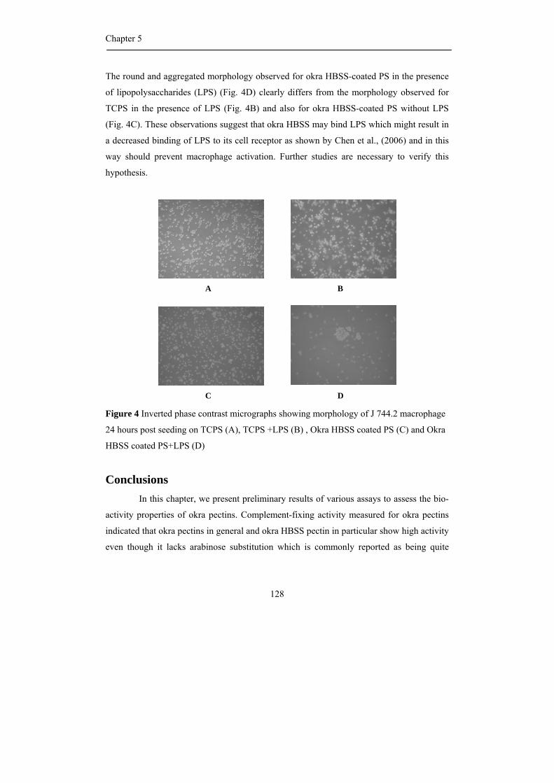
Chapter 5
128
The round and aggregated morphology observed for okra HBSS-coated PS in the presence
of lipopolysaccharides (LPS) (Fig. 4D) clearly differs from the morphology observed for
TCPS in the presence of LPS (Fig. 4B) and also for okra HBSS-coated PS without LPS
(Fig. 4C). These observations suggest that okra HBSS may bind LPS which might result in
a decreased binding of LPS to its cell receptor as shown by Chen et al., (2006) and in this
way should prevent macrophage activation. Further studies are necessary to verify this
hypothesis.
Figure 4 Inverted phase contrast micrographs showing morphology of J 744.2 macrophage
24 hours post seeding on TCPS (A), TCPS +LPS (B) , Okra HBSS coated PS (C) and Okra
HBSS coated PS+LPS (D)
Conclusions In this chapter, we present preliminary results of various assays to assess the bio-
activity properties of okra pectins. Complement-fixing activity measured for okra pectins
indicated that okra pectins in general and okra HBSS pectin in particular show high activity
even though it lacks arabinose substitution which is commonly reported as being quite
A B
C D
Complement-fixing activity and cell behaviour of okra pectins
129
beneficial for such activity. In contrast, HBSS is rich in galactose, present as short side
chains and is having a rather unique alpha-galactosyl residue present in the side-chains.
Furthermore, the activity is linked to the length of the rhamnogalacturonan (RG) I
backbone but it seems to be independent from the acetyl groups and methyl esters. These
results indicated that the factor for expressing complement-fixing activity of pectin is
somewhat complicated to predict and strongly depend on structural features and size and
3D-structure of the pectin.
The use of okra pectins as a biomaterial for coating medical devices and implants
is further enhanced by its ability to inhibit the colonization of the surface by adherent cells
in promoting cell cycle arrest in G1 phase and apoptosis. Okra HBSS pectins also show an
inability to activate macrophages and a potential beneficial effect in preventing macrophage
activation by binding of LPS. These properties are promising for biomaterial anti-adhesive
applications.
Material and methods
Material
The soft part of immature pods of okra (Abelmoschus esculentus (L.) Moench) was
collected from okra grown in Thailand. The okra alcohol insoluble solids were prepared
and were sequential extracted by aqueous hot buffer and chelating agent solutions
according to Sengkhamparn et al. (2009a). Saponification of polysaccharide fractions were
performed by adding 0.05 M NaOH at low temperature (Sengkhamparn et al., 2009b).
Analytical methods
Enzymatic degradation
Pectin solutions (final concentration of 3 mg/mL in 50 mM NaOAc buffer of pH
5) of Okra HBSS and saponified HBSS were incubated with 0.547 µg of
rhamnogalacturonan hydrolase (RGH) from Aspergillus aculeatus per mg substrate
(Sengkhamparn et al., 2009b). Solutions (final concentration of 4 mg/mL in 50 mM NaOAc
buffer of pH 5) of Okra CHSS and saponified CHSS pectin were incubated with 0.547 µg
Chapter 5
130
of RGH per mg substrate and 0.016 units of endo-polygalacturonase (PG) from A.
aculeatus (Sengkhamparn et al., 2009b). The incubations were performed at 40 oC for 24 h
and were stopped by heating at 100 oC for 5 min. The digests were then desalted and freeze
dried.
Size exclusion chromatography (SEC)
HBSS solutions (6 mg/mL in water, 100 mL) were incubated with
Rhamnogalacturonan hydrolase (RGH) for 24 h at 40 oC, the enzyme was then inactivated
at 100 oC for 5 min. The digest was fractionated using SEC, Sephacryl S500 (S500-1, 2, 3)
and Sephacryl S300 (S300-1, 2, 3) according to Sengkhamparn et al. (2009b). After pooling
and desalting, each fraction was saponified.
Molecular weight distribution
The molecular weight distribution was established by High-Performance Size-
Exclusion Chromatography (HPSEC) system (Thermo Separation Products, USA)
according to Chen et al. (2004). The HPSEC was equipped with three Tosoh Biosep TSK-
Gel G columns in series (4000PWXL-3000PWXL-2500PWXL) in combination with a PWXL –
guard column (Tosoh, Japan). The sample was eluted with 0.8 mL/min of 0.2 M sodium
nitrate Detection was performed by refractive index detector (Shodex SE-61, Showa Denko
K.K., Japan).
Complement-fixing activity assay
To measure complement-fixing activity samples were dissolved in a Veronal
Buffer in a concentration of 1 mg/mL and further diluted with VB:BSA buffer solution to
solutions containing 0.25, 0.0625 0.0313 and 0.015 mg pectin/mL. The human serum used
as a complement source was diluted with VB:BSA to a concentration giving about 50%
haemolysis of sheep erythrocytes. 50 µL of diluted human serum was added to 50 µL of the
sample solution in a 96- well microtiterplate. The sample solution was then incubated on a
shaker at 37°C for 30 min. After adding antibody-sensitized sheep erythrocytes (50 µL) and
Complement-fixing activity and cell behaviour of okra pectins
131
incubation at 37°C for 30 min, the microtiter plate was centrifuged at 1600 x g, for 5 min
and 100 µL of the supernatants were transferred to a flat bottomed microtiter plate for
measurement of the absorbance (A) at 405 nm. Results are presented as mean of duplicate
determinations. The percent inhibition of lysis is calculated by [(Acontrol - Asample) / Acontrol] x
100 % in which the absorbance of the mixture of VB:BSA, diluted serum and sensitized
sheep erythrocytes is Acontrol. The percent inhibition of haemolysis results were plotted
versus concentrations (Michaelsen et al., 2000; Mollnes et al., 1995)
Fibroblast and macrophage culture assay
Surface modification of polystyrene (PS) Petri dishes was performed according to
Morra et al. (2004). Swiss 3T3 albino mouse embryo fibroblasts (ATCC ref. CCL-92) and
the murine macrophage cell line J774.2 (European collection of cell cultures) were
maintained in Dulbecco’s modified Eagle’s medium (DMEM) supplemented with 10 %
foetal bovine serum (FBS), 4 mM L-glutamine and penicillin/streptomycin (Gibco
Invitrogen, Cergy-Pontoise France). Both cells lines were grown at 37oC under humidified
atmosphere (10% CO2) and were seeded at 104 cell/cm2 on tissue culture polystyrene
(TCPS) and Petri dishes coated with okra polysaccharide fractions for cell behaviour
investigation.
Proliferation study
To assess Swiss 3T3 fibroblast proliferation, cells were dissociated in 0.25%
trypsin + 1 mM EDTA solution. Trypsin reaction was stopped by adding half a volume of
FBS, then cells were counted in a Malassez haemocytometer. The proliferation index was
the ratio between total cell count (N) and cell number seeded (N0) (Nagel at al, 2008).
Cell cycle analysis
Swiss 3T3 fibroblasts were dissociated in 0.25% trypsin + 1 mM EDTA
solution,washed with 1 mL PBS containing 5 mM EDTA, then fixed for 45 min at 4°C in 1
mL of 75% ethanol in PBS with 5mM EDTA. Cells were washed and suspended in PBS,
Chapter 5
132
5mM EDTA, containing 0.1% Triton X-100, mixed with 40 µg RNase A and 25 µg
propidium iodide (Sigma Aldricht, Saint-Quentin Fallavier, France), and incubated for 15
min protected from light . The stained samples were analysed by Epics XL-MCL flow
cytometer according to Nagel et al. (2008).
Statistical Analysis
The One-way analysis of variance (ANOVA) with Duncan and Least Significant
Difference (LSD) were used for testing significant differences (P<0.05) between
complement-fixing activity of samples as performed with SPSS for Windows (Version
13.0).
Acknowledgments
This study has been carried out with financial support from the Commission on
Higher Education Ministry of Education, Thailand, under The Commission on Higher
Education Staff Development project for the Joint Ph.D. Program in Food Science at
Kasetsart University, Thailand.
References
Andrade, J.D. & Hlady, V. (1987). Plasma-protein adsorption – the big12. Ann. N. Y. Acad. Sci., 516, 158–172.
Benen, J.A.E. & Visser, J. (2003). Pectate and pectin lyases. In J.R. Whitaker, A.G.J. Voragen & D.W.S. Wong,
eds., Handbook of food enzymology (pp. 1029-1042). New York: Marcel Dekker.
Boudreau, N.J. & Jones, P.L. (1999). Extracellular matrix and integrin signalling: the shape of things to come
Biochem. J., 339, 481-488.
Bussy, C., Verhoef, R., Haeger, A., Morra, M., Duval, J.L., Vigneron, P., Bensoussan, A., Velzenberger, E.,
Cascardo., G., Cassinelli, C., Schols, H.A., Knox, P. & Nagel, M-D. (2008). Modulating in vitro bone
cell and macrophage behaviour by immobilized enzymatically tailored pectins. J Biomed. Mater. Res.
A., In press.
Cassinelli,C., Morra, M., Carpi, A., Giardino, R. & Fini, M. (2002). Controlling the lateral distribution and
alignment of human gingival fibroblasts by micropatterned polysaccharide surfaces. Biomed.
Pharmacother., 56, 325-331.
Complement-fixing activity and cell behaviour of okra pectins
133
Chen, C.H., Sheu, M.T., Chen, T.F., Wang, Y.C., Hou, W.C., Liu, D.Z., Chung, T.C. & Liang, Y.C. (2006).
Suppression of endotoxin-induced proinflammatory responses by citrus pectin through blocking LPS
signaling pathways. Biochem. Pharmacol., 72, 1001-1009.
Gaboriaud, C., Juanhuix, J., Gruez, A., Lacroix, M., Darnault, C., Pignol, D., Verger, D., Fontecilla-Camps, J.C. &
Arlaud, G.J. (2003). The crystal structure of the globular head of complement protein C1q provides a
basis for its versatile recognition properties. J. Biol. Chem., 278, 46974-46982.
Gur’janov, O.P., Gorshkova, T.A., Kabel, M., Schols, H.A. & van Dam, J.E.G. (2007). MALDI-TOF MS evidence
for the linking of flax bast fibre galactan to rhamnogalacturonan backbone. Carbohydr. Polym., 67, 86–
96.
Hokputsa, S., Harding, S.E., Inngjerdingen, K., Jumel, K., Michaelsen, T.E., Heinze, T., Koschella, A. & Paulsen,
B.S. (2004). Bioactive polysaccharides from the stems of the Thai medicinal plant Acanthus
ebracteatus: their chemical and physical features. Carbohydr. Res., 339, 753–762.
Inngjerdingen, K.T., Debes, S.C., Inngjerdingen, M, Hokputsa, S., Harding, S.E., Rolstad, B., Michaelsen, T.E.,
Diallo, D. & Paulsen, B.S. (2005). Bioactive pectic polysaccharides from Glinus oppositifolius (L.)
Aug. DC., a Malian medicinal plant, isolation and partial characterization. J. Ethnopharma., 101, 204–
214.
Ishii, T. (1997). O-acetylated oligosaccharides from pectins of potato tuber cell walls. Plant Physiol., 113, 1265-
1272.
Ishii, T., Thomas, J.R., Darvill, A.G. & Albersheim, P. (1989). Structure of plant cell walls. XXVI. The walls of
suspension-cultured sycamore cells contain a family of rhamnogalacturonan I-like pectic
polysaccharides. Plant Physiol., 89, 421–428.
Kahlon, T.S., Chapman, M.H. & Smith, G.E. (2007). In vitro binding of bile acids by okra, beets, asparagus, egg
plant, turnips, green beans, carrots, and cauliflower. Food Chem., 103, 676-680.
Kokkonen, H.E., Ilvesaro, J.M., Morra, M., Schols, H.A. & Tuukkanen, J. (2007). Effect of modified pectin
molecules on the growth of bone cells. Biomacromol., 8, 509-515.
Kweon, M.H., Hwang, H.J & Sung, H.C. (2003). Isolation and characterization of anticomplementary beta-glucan
from the shoots of bamboo Phyllostachys edulis. Planta Med. 69: 56-62.
Lau, J.M., McNeil, M., Darvill, A.G. & Albersheim, P. (1987). Treatment of rhamnogalacturonan I with lithium in
ethylenediamine. Carbohydr. Res., 168, 245-274.
Lengsfeld, C., Titgemeyer, F., Faller, G. & Hensel, A. (2004). Glycosylated compounds from okra inhibit
adhesion of helicobacter pylori to human gastric mucosa. J. Agric. Food Chem., 52, 1495-1503.
Matsushita, M., Matsushita, A., Endo, Y., Nakata, M., Kojima, N., Misuoshi, T. & Fujita, T. (2004). Origin of the
classical complement pathway: Lemprey orthologue of mammalian C1q acts as a lectin. PNAS., 101,
10127-10131.
McNeil, M., Darvill, A.G. & Albersheim, P. (1982). Structure of plant cell walls. XII. Identification of seven
differently linked glycosyl residues attached to O-4 of the 2,4–linked L–rhamnosyl residues of
rhamnogalacturonan I. Plant Physiol., 70, 1586-1591.
Chapter 5
134
Michaelsen, T.E., Gilje, A., Samuelsen, A.B., Hogasen, K. & Paulsen, B.S. (2000). Interaction Between Human
Complement and a Pectin Type Polysaccharide Fraction, PMII, from the leaves of Plantago major L.
Scand. J. Immunol., 52, 483-490.
Mollnes, E.T., Høgåsen, K., Hoaas, F.B., Michaelsen, E.T., Garred, P. & Harbo, M. (1995). Inhibition of the
complement mediated red cell lysis by immunoglobulins is depenent on the IG isotope And its C1
binding properties. Scand. J. Immunol., 41, 449-456.
Morgan, DO. 2007. The cell cycle: Principles of control (pp. 4 – 9). London: New Science Press.
Morra, M. (2005). Engineering of Biomaterials Surfaces by Hyaluronan. Biomacromol., 6, 1205-1223.
Morra, M., Cassinelli, C., Cascardo, G., Nagel, M.D., Volpe, D.C., Siboni, S., Maniglio, D., Brugnara, M.,
Ceccone, G., Schols, H.A. & Ulvskov, P. (2004). Effects on interfacial properties and cell adhesion of
surface modification by pectic hairy regions. Biomacromol., 5, 2094-2104.
Nagel., M.D., Verhoef, R., Schols, H.A., Morra, M., Knox, J.P., Ceccone, G., Volpe, C.D., Vigneron, P., Bussy,
C., Gallet, M., Velzenberger, E., Vayssade, M., Cascardo, G., Cassinelli, C., Haeger, A., Gilliland, D.,
Liakos, I., Rodriguez-Valverde, M. & Siboni, S. (2008). Enzymatically-engineered pectins
differentially influence the morphology, adhesion, cell cycle progression and survival of fibroblasts.
Biochimica et biophysica acta. Biomembranes, 1780, 995-1003.
Perrone, P., Hewage, C.M., Thomson, A.R., Bailey, K., Sadler, I.H. & Fry, S.C. (2002). Patterns of methyl and O-
acetyl esterification in spinach pectins: new complexity. Phytochem., 60, 67-77.
Ridley, B.L., O’Neill, M.A. & Mohnen, D. (2001). Pectins: structure, biosynthesis and oligogalacturonide-related
signalling. Phytochem., 57, 929-967.
Roach, P., Farrar, D & Perry, C.C. (2005). Interpretation of protein adsorption: Surface-induced conformational
changes. J. Am. Chem. Soc., 127, 8168-8173.
Sakurai, M.H., Matsumoto, T., Kiyohara, H., & Yamada, H. (1999). B-cell proliferation activity of pectic
polysaccharides from a medical herb, the roots of Bupleurum falcatum L. and its structural
requirement. Immunol. 97, 540.
Samuelsen, A.B., Paulsen, B.S., Wold, J.K., Otsuka, H., Kiyohara, H., Yamada, H. & Knutsen, S.H. (1996).
Characterization of a biologically active pectin from Plantago Major L. Carbohydr. Polym., 30, 37-44.
Samuelsen, A.B., Westereng, B., Yousif, O., Holtekjolen, A.K., Michaelsen, T.E. & Knutsen, S.H. (2007).
Structural features and complement fixing activity of pectin from three Brassica oleracea Varities:
White cabbage, kale, and red kale. Biomacromol., 8, 644-649.
Searle-Van Leeuwen, M.F., Vincken, J.P., Schipper, D. &. Voragen, A.G.J. (1996). Acetylesterase of Aspergillus
niger purification and mode of action on pectin. In J. Visser, and A.G.J. Voragen, eds., Progress in
biotechnology 14: Pectins and Pectinases (pp. 793-798). Amsterdam: Elsevier Science.
Schols, H.A., Posthumus, M.A. &Voragen, A.G.J. (1990). Structural features of hairy regions of pectins isolated
from Apple juice produced by the liquefraction process. Carbohydr. Res., 206, 117-129.
Sengkhamparn, N., Verhoef, R., Schols, H.A., Sajjaanantakul, T. and Voragen, A.G.J. (2009a). Characterization of
Cell Wall Polysaccharide from Okra (Abelmoschus esculentus (L.) Moench). Carbohydr. Res., 344,
1824-1832.
Complement-fixing activity and cell behaviour of okra pectins
135
Sengkhamparn, N., Verhoef, R., Bakx, E.J., Schols, H.A., Sajjaanantakul, T. and Voragen, A.G.J. (2009b). Okra
pectin contains an unusual substitution of its rhamnosyl residues with acetyl and alpha–linked
galactosyl groups. Carbohydr. Res., 344, 1842-1851.
Suzuki, Y., Miyatake, K., Okamoto, Y., Muraki, E. & Minami, S. (2003). Influence of the chain lengh of chitosan
on complement activation. Carbohydr. Polym. 54: 465-469.
Tomoda, M., Shimizu, N., Gonda, R., Kanari, M., Yamada, H. & Hikino, H. (1989). Anticomplementary and
hypoglycaemic activity of okra and Hibiscus mucilages. Carbohydr. Res., 190, 323-328.
Verhoef, R., Lu, Y., Knox, J.P. , Voragen, A.G.J. & Schols, H.A.(2009). Fingerprinting complex pectins by
chromatographic separation combined with ELISA detection. Carbohydr. Res.
doi:10.1016/j.carres.2008.09.030
Vincken, J.P., Schols, H.A., Oomen, R.J.F.J., McCann, M.C., Ulvskov, P., Voragen, A.G.J. & Visser, R.G.F.
(2003). If homogalactaronan were a side chain of rhamnogalacturonan I. Implications for cell wall
architecture. Plant physiol., 132, 1781-1789.
Vogler, E.A. (1999). Water and the acute biological response to surfaces. J. Biomater. Sci. Polym. Ed., 10, 1015–
1045.
Wagner, H & Kraus, S. (2000). News on immunologically active plant polysaccharides. In B.S. Paulsen (ed.),
Bioactive carbohydrate polymers (pp.1-14). London: Kluwer academic publishers.
Westereng, B., Yousif, O., Michaelsen, T.E., Knutsen, S.H. & Samuelsen, A.B. (2006). Pectin isolated from white
cabbage- structure and complement-fixing activity. Mol. Nutr. Food Res., 50, 746-755.
Whistler, R.L. & Conrad, H.E. (1954). A Crystalline Galactobiose from acid hydrolysis of okra mucilage. J. Am.
Chem. Soc., 76, 1673-1974.
Yamada, H., Nagai, T., Cyong, J.-C. and Otsuka, Y. (1985). Relationship between chemical structure and anti-
complementary activity of plant polysaccharides. Carbohydr. Res., 144, 101-111.
Yamada, H. (2000). Bioactive plants polysaccharides from Japanese and Chinese traditional herbal medicines. In
B.S. Paulsen (ed.), Bioactive carbohydrate polymers (pp.15 - 24). London: Kluwer academic
publishers.
Yamada, H. & Kiyohara, H. (2007). Immunomodulating activity of plant polysaccharide structures. In J.P.
Kamerling, G.J Boons, Y.C. Lee, A. Suzuki, N. Taniguchi & A.G.J. Voragen, eds., Comprehensive
Glycoscience: From chemistry to systems biology. Volume 4 Cell glycobiology and development health
and disease in glycomedicine, (pp. 663-693). Oxford: Elsevier Ltd.
Yamada, H & Kiyohara, H. (1999). Complement-activating polysaccharides from Medicinal Herbs. In Wagner, H
(ed), Immunomodulatory Agent from plants (pp 161-202). Basel: Birkhäuser.
Chapter 5
136
General discussion
137
Chapter 6
General discussion
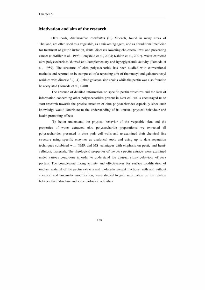
Chapter 6
138
Motivation and aim of the research
Okra pods, Abelmoschus esculentus (L.) Moench, found in many areas of
Thailand, are often used as a vegetable, as a thickening agent, and as a traditional medicine
for treatment of gastric irritation, dental diseases, lowering cholesterol level and preventing
cancer (BeMiller et al., 1993; Lengsfeld et al., 2004; Kahlon et al., 2007). Water extracted
okra polysaccharides showed anti-complementary and hypoglycaemic activity (Tomoda et
al., 1989). The structure of okra polysaccharide has been studied with conventional
methods and reported to be composed of a repeating unit of rhamnosyl and galacturonosyl
residues with dimeric β-(1,4)-linked galactan side chains while the pectin was also found to
be acetylated (Tomada et al., 1980).
The absence of detailed information on specific pectin structures and the lack of
information concerning other polysaccharides present in okra cell walls encouraged us to
start research towards the precise structure of okra polysaccharides especially since such
knowledge would contribute to the understanding of its unusual physical behaviour and
health promoting effects.
To better understand the physical behavior of the vegetable okra and the
properties of water extracted okra polysaccharide preparations, we extracted all
polysaccharides presented in okra pods cell walls and re-examined their chemical fine
structure using specific enzymes as analytical tools and using up to date separation
techniques combined with NMR and MS techniques with emphasis on pectic and hemi-
cellulosic materials. The rheological properties of the okra pectin extracts were examined
under various conditions in order to understand the unusual slimy behaviour of okra
pectins. The complement fixing activity and effectiveness for surface modification of
implant material of the pectin extracts and molecular weight fractions, with and without
chemical and enzymatic modification, were studied to gain information on the relation
between their structure and some biological activities.
General discussion
139
Hemicellulosic polysaccharides in okra
The polysaccharides in okra cell wall material were extracted from okra pods
alcohol insoluble solids by sequential extraction including hot buffer, chelating agent and
both diluted and concentrated alkali. The extracted material, okra pulps, represented the
major part of the okra pods. All fractions obtained were analyzed for their sugar and
glycosidic linkage composition and these data indicated that the okra hemicelluloses mainly
ended up in the concentrated alkali extracted fraction (chapter 2).
The presence xylose and terminally linked fucose, together with the presence of
1,4,6–linked glucosyl residues pointed to the presence of a xyloglucan. This was confirmed
by incubating this fraction with a specific xyloglucanase, and analysis of the released
oligomers by HPAEC and MS. The results showed that XXXG, XXFG and XLFG were the
main building blocks; others were XLXG, XXLG and XLLG. This indicated that poly-
XXXG type xyloglucan carrying both galactose and fucose substitutions were a part of this
fraction. Mass spectrometry of xyloglucanase digests of okra cell wall material showed the
presence of low levels of acetylated oligomers in the digest. However, since only a relative
small part of the xyloglucan was solubilized in this way, the level of xyloglucan acetylation
may still be underestimated.
Furthermore, glucuronic acid was found in the concentrated alkali extracted
fraction indicating that acidic xylans were also part of this fraction. The presence of
glucuronoxylans was demonstrated by treating the extract with a Glycosyl Hydrolase
family 11 xylanase and analyzing the digests by mass spectrometry. The series of xylo-
oligomers substituted with one 4-O-methylglucuronic acid identified showed that the acidic
substituent is rather randomly distributed over the xylan backbone.
The structural features of the xylan and xyloglucan present in okra cell wall were
similar to those found in other dicotyledonous plants (olives: Vierhuis et al., 2001;
Asteridae: Hoffman et al., 2005; berries and currants: Hilz et al., 2006a).
Chapter 6
140
Pectic polysaccharides in okra
Pectins were found to be the main polysaccharides in okra cell wall and were
obtained in the first 3 extracts (hot buffer, chelating agent and diluted alkali). The extracted
pectic polysaccharides were characterized by conventional and new analytical techniques
including enzymatic fingerprinting by using specific enzymes as analytical tools, up to date
separation techniques combined with NMR and MS techniques.
Pectin structural elements in okra pectin
Molecular weight determination of all pectic polysaccharide extracts from okra
showed only one population to be present in the high molecular weight range, however the
molecular weight appeared to be lower for pectic polysaccharides extracted under more
severe extraction conditions (chapter 2). Since also only one molecular weight population
was shown to be present by anion exchange chromatography, it is clear that the different
pectic structural elements in both okra pectin fractions studied are interlinked.
The pectins present in the 3 extracts obtained showed different characteristics.
Based on the proportions of rhamnose and galacturonic acid and on the fact that the
backbone of rhamnogalacturonan (RG) I is composed of alternating rhamnosyl and
galacturonosyl residues, the percentage of homogalacturonan (HG) within the pectin
fraction can roughly be estimated. The hot buffer extract was found to contain 15% HG; the
chelating extract 75% HG and the diluted alkali extract 57% HG. The small amounts of
mono-galacturonic acid and galacturono-oligomers in saponified hot buffer extracted
pectins are indicative for the relative low amounts of HG segment present. The high
proportion of HG in the chelating agent extract compared to the hot buffer extract was
confirmed by the difference in degradability of these extracts by endo-polygalacturonase
(PG) from Aspergillus aculeatus and the pattern of galacturono-oligomers released (chapter
3).
General discussion
141
HG segments in okra pectin
Assuming that no methyl esters were present in the RG I structural elements
(Kravtochenko et al., 1992; Ishii, 1997; Perrone et al., 2002), (almost) all galacturonosyl
residues present in the HG segments of hot buffer extracted okra pectin are methyl
esterified. In the chelating agent extracted okra pectin the DM of the HG segment present
can be calculated to be 64%. Depending from the type of distribution of these methyl ester
groups these HG segments might be cross linked in the cell walls via calcium ions and be
solubilized by extracting with chelating agents. Where acetyl substitution of
homogalacturonan is well established for potato and sugar beet pectin (Albersheim et al.,
1996), NMR analysis of the pectins did not reveal any acetylation of galacturonosyl
residues within HG segments.
Rhamnogalacturonan I segments in Okra pectin
In addition to the typical 1:1 ratio of rhamnose:galacturonic acid, RG I is also
characterized by the presence of acetyl groups substituted to the galacturonosyl residues
(Schols and Voragen, 1994; Albersheim et al., 2006). Variation on the precise location and
distribution may vary depending on plant source. O-3 acetylated galacturonosyl residues in
the RG I backbone have been mentioned for pectins from carrot, cotton, tobacco and
tomato (Komalavilas and Mort, 1989) and spinach (Perrone et al., 2002). Ishii (1995 and
1997) found acetyl groups to be mainly located at O-2 of galacturonic acid in the RG I from
bamboo. Double substitution of a single galacturonosyl residue at O-2 and O-3 has been
reported for RG I from suspension-cultured sycamore cells (Lerouge et al., 1993).
The degree of acetylation (DA) of the pectin in the hot buffer extract of okra pods
was much higher (58%) than in the chelating agent extracted pectin (18%). Since
rhamnogalacturonan hydrolase (RGH) is inhibited by the presence of acetyl groups, it was
expected that the hot buffer extracted okra pectin would not be degradable by this enzyme.
Surprisingly, RGH was quite active towards hot buffer extracted okra pectin and this
activity even increased upon removal of the acetyl groups (chapter 3). It was hypothesized
that the acetyl groups would be located at other sugar moieties in the RG I backbone which
would be in accordance with NMR data obtained where we could not identify signals
Chapter 6
142
belonging to O-acetylated galacturonosyl residues (Lerouge et al., 1993). These
observations were pointing to the existence of a new type of RG I having new structural
features.
Using mass spectrometry technique and NMR techniques, it was demonstrated that
for the HBSS RG I, the major part of the acetyl groups was substituted to O-3 of the
rhamnosyl moiety, where only a minor part was substituted to O-2 and/or O-3 of the
galacturonosyl residues as has been commonly reported so far. The presence of acetyl
groups attached to a rhamnosyl moiety in hot buffer extracted RG I has never been
reported. The different sites for acetylation (okra typical RG I versus common RG I) could
be confirmed by use of different enzymes. RG hydrolase showed to be able to degrade the
rhamnosyl-acetylated RG I (chapter 3), while the enzyme was not able to degrade
galacturonosyl-acetylated RG I (Schols et al., 1990). HG segments are dominating in the
CHSS fraction. However, within the rhamnogalacturonans, the RG I part consisting of
acetylated galacturonosyl residues was prevailing next to only minor amounts of RG I
consisting of acetylated rhamnosyl residues. It is hypothesized that okra pectin extracted by
diluted alkali also consisted of two types of RG I segments with minor amounts of the okra-
specific RG I.
Neutral sugar side chains and other pectin structural elements in okra pectin
So far, it has been reported that the level of branching in RG I structures may vary
significantly ranging from 20-80% of all rhamnosyl residues. The composition and length
of RG I side chains at O-4 or the rhamnosyl residues depends on plant species and origin of
plant tissue (McNeil et al., 1982; Lau et al., 1987; Ishii et al., 1989; Albersheim et al.,
1996). These side chains are mainly reported to consist of galactosyl and /or arabinosyl
moieties such as arabinan, galactan and arabinogalactan type I and II. RG I structures as
present in pectin from apple and sugar beet are rich in arabinan side chains, while e.g.
potato-, soy-, and flax pectins are known to be rich in RG I segments containing mainly
galactan side chains (Voragen et al., 1995). The length of arabinan and galactan side chains
range from one or two residues to around 26 residues (Voragen et al., 1995; Gur’janov et
al., 2007).
General discussion
143
The sugar linkage composition of okra HBSS pectin indicated the presence of
highly branched RG I structures carrying short galactan side chains composed of, on the
average, 1 or 2 galactosyl residues, although these side chains appeared a little bit longer in
chelating agent extracted pectin. Arabinose was absent in the new type of okra RG I from
HBSS pectin and is only a minor constituent in okra RG I from CHSS pectin containing
some higher levels of commonly present RG I. Diluted alkali extracted okra pectin was less
branched and contained arabinan and a mixture of type I and II arabinogalactan structures
as a side chain.
Where galactosyl residues have been reported to be present within beta-linked
galactans, NMR showed the presence of two types of galactosyl residues in RG I. Next to
(1,4- and terminally) beta-linked galactoses, the signals for the other type did not fit with
any data reported before for RG I structures. These signals could be ascribed to terminally
linked alpha-galactosyl residues. These α-linked galactose were linked to O-4 of a β-1,4-
linked galactosyl residue or O-4 of a rhamnosyl residue in about equal amounts (chapter 3).
This t-α-galactosyl residue was also found to be present in chelating agent extracted pectin
but only in minor amounts, confirming the relatively low level of okra HBSS-typical RG I
in the CHSS fraction.
Although the majority of RG I structures described in literature are mentioned to
contain relative long side chains (either rich in arabinose, galactose or both), short
galactose-side are reported to be present in flax RG I (Naran et al., 2007) although these
galactoses were in the L-configuration rather than in the D-configuration as found for okra
RG I and all other RG I structures described in literature. A water extractable RG I from
Arabidopsis seed mucilage was found to be a linear rhamnogalacturonan, devoid of any
side chains (Deng et al., 2006).
Xylo-substitution as present in pectic xylogalacturonan structural elements (Schols
et al., 1995; Coenen et al., 2007) was not found for okra pectins. RG II, another structural
element of pectin having 4 well conserved side chains of many different sugar residues
linked to a sequence of 8 galacturonosyl residues, and usually associated to HG
(Albersheim et al., 1996; Hilz et al., 2006b), was only present in minor amounts (1-2 %).
Chapter 6
144
A new type of RG I as structural element of pectin?
The most striking result of the elucidation of the structural features of the
polysaccharides in the okra pods extracts is the presence of a new type of RG I
characterized by the occurrence of acetylated rhamnosyl residues, the absence of methyl
ester groups and carrying a terminal α-galactosyl residue either directly linked at O-4 or
linked to O-4 of a β-1,4-galactosyl residue. This structural element of pectin is almost
completely extracted in the first extraction step with hot buffer and may explain the typical
physical behavior of okra pectins. This new RG I structure seems to coexist next to the
other, more common RGI structure and some HG in one single population in okra HBSS.
The same is true for okra CHSS pectin, although the ratio between the three pectic
structural elements differ completely from the ratio established for okra HBSS pectin.
Based on the new structural insights gained in this thesis, we hypothesized that the
overall structure of okra hot buffer and chelating agent extracted pectins consists of HG
segments directly connected to RG I forming the backbone of the pectin molecule with side
chains attached to RG I (Coenen et al., 2007). The proposed overall structure of okra pectin
is shown in Figure 1. It should be stated that HG segments may be present in both the
backbone of the pectin molecule as well as side chains linked to rhamnosyl residues present
in the backbone (Coenen et al., 2007; Ralet and Thibault, 2009). Whether the new RG I
structure as present in the okra hot buffer extracts should be considered as a new structural
element fully depend on the frequency of occurrence in pectins from other material.
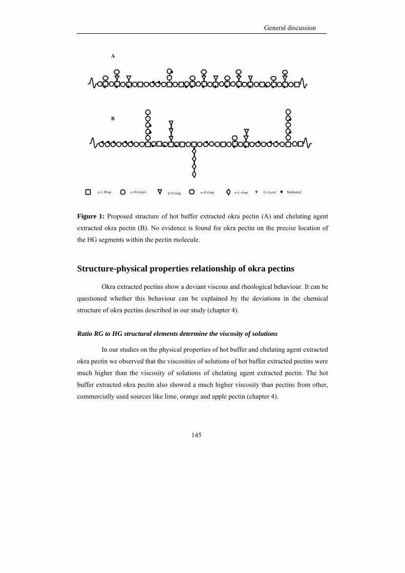
General discussion
145
Figure 1: Proposed structure of hot buffer extracted okra pectin (A) and chelating agent
extracted okra pectin (B). No evidence is found for okra pectin on the precise location of
the HG segments within the pectin molecule.
Structure-physical properties relationship of okra pectins
Okra extracted pectins show a deviant viscous and rheological behaviour. It can be
questioned whether this behaviour can be explained by the deviations in the chemical
structure of okra pectins described in our study (chapter 4).
Ratio RG to HG structural elements determine the viscosity of solutions
In our studies on the physical properties of hot buffer and chelating agent extracted
okra pectin we observed that the viscosities of solutions of hot buffer extracted pectins were
much higher than the viscosity of solutions of chelating agent extracted pectin. The hot
buffer extracted okra pectin also showed a much higher viscosity than pectins from other,
commercially used sources like lime, orange and apple pectin (chapter 4).
α-D-GalpA α-L-Rhap β-D-Galp α-D-Galp O-Acetyl α-L-Arap Methanol
A
B
Chapter 6
146
Okra pectin exhibited a much larger shear thinning behaviour at low concentration
(0.1%) when compared to pectins of pumpkin (Evageliou et al., 2005) and mango (Iagher et
al., 2002). The flow behaviour index of okra hot buffer extracted pectin, derived from the
Ostward- De Waele equation, was lower than the index established for apple pectin
pointing to a more pseudo-plastic nature for okra pectins. Hwang and Kokini (1992)
reported that a highly branched RG backbone with long side chains contributed to a higher
viscosity and pseudo-plastic behaviour. However, a 0.5 % (w/v) solution of okra hot buffer
extracted pectin showed a higher viscosity than highly branched soy RG I with long side
chains and modified hairy regions from apple at the same concentration. The viscosity of
hot buffer extracted pectin solution was higher than found for water soluble soybean
polysaccharides at 10 % w/w concentration (64 mPa s at 129 s-1 and at 20 oC) (Furuta et al.,
1999; Furuta and Maeda, 1999).
From this it appears that not only the ratio of RG to HG, the degree and nature of
branching but also other factors play a role like the sites of acetylation and the charge of the
backbone and the exposure of the charge. For the time being the acetylation of rhamnosyl
residues within the RG structural element seems to be unique for okra and very well may
explain the unique features of okra pectins in solution. The role of alpha-linked galactosyl
moieties as present in okra HBSS pectin in influencing physical properties is less clear and
should be addressed in future research.
Effect of location and level of acetyl groups on physical properties
The viscosity of okra pectin extracted by hot buffer or by hot buffer with chelating
agent was reduced upon saponification 5 and 2 times respectively, even though
saponification results in an increased level of total negative charge and/or higher charge
densities which, in general, will lead to an increased viscosity (Williams and Phillips,
2000). The observed loss in viscosity indicates that the removal of the acetyl groups located
at O-3 of rhamnosyl residues of RG I has much more influence on the viscosity than the
removal of methyl esters which introduce charge to the polymer. It was also observed that
solutions of okra hot buffer extracted pectin at low concentration showed a crossover point
at lower frequency values than corresponding solutions of chelating agent extracted pectin
General discussion
147
indicating that the former can more easily form elastically active zones. The concentration
at which this entanglement occurs with hot buffer extracted pectin is much lower than the
concentration established for other pectins (0.6 %w/w, Kjoniksen et al., 2005), but is close
to the value reported for xanthan gum (0.13 % w/w, Rodd et al., 2000). However, the Cox-
Merz superposition indicated that the aggregation does not lead to the formation of a weak
associative network, as observed in for example xanthan solutions (chapter 4). The
presence of acetyl groups in HG has a negative effect on the gel formation as established
for sugar beet pectins (Oosterveld et al., 2000). The tendency of hot buffer extracted okra
pectin to easily form elastically active zones may be due to the presence and location of
acetyl groups on O-3 of the rhamnosyl residues in the RG I backbone which might enhance
formation of such elastically active zones via hydrophobic interaction.
The aggregation behaviour of pectins as monitored by dynamic light scattering of
their solutions in buffer showed a lower tendency to aggregate for okra hot buffer extracted
pectin compared to okra chelating agent extracted pectin. This indicted a partially screening
effect by salts on the electrostatic repulsion between the molecules which decreases the
possibility of hydrophobic association via acetylated rhamnosyl residues. On the other hand
divalent cations may cause formation of cross-links between non-esterified galacturonosyl
residues which are more abundant in chelating agent extracted pectin and thus enhance their
aggregation. Since the light scattering experiments could not be performed at the same
concentration as the rheological measurements due to limitation of multiple scattering at
higher concentrations, results could not be compared. However, from the big difference in
aggregation behaviour between saponified buffer extracted okra pectin and original buffer
extracted okra pectin, it can be concluded that chain association driven by hydrophobic
interactions is an important factor for okra pectins.
Increasing the amounts of acetyl groups by increasing the concentration of hot
buffer extracted okra pectin in solutions was found to cause a shift of the crossover point to
lower frequency values indicating that the polymer behaves more elastic. This behaviour
can be ascribed to an increase in elastically active zones with increasing concentration. The
hydrophobic association of acetylated rhamnosyl residues seems to be the main factor to
Chapter 6
148
explain the viscoelastic behaviour of okra pectin when dissolved in water. This specific
structure of okra pectin makes it different from all other pectins known at this moment.
Modeling Okra pectins in solutions
Proposed models for okra pectin interaction in aqueous solutions are shown in
Figure 2. At a pH above pKa of the carboxylic group, the okra pectin can associate through
hydrophobic interaction and hydrogen bonding. The numerous hydroxyl groups within the
RG I regions of okra pectin can lead to hydrogen bonding (Ralet et al., 2002). The acetyl
groups of rhamnosyl residues in okra RG I will lead to hydrophobic interaction. Moreover,
calcium bridges might be present within HG segments as well.
Structure-Bioactivity relationship of okra pectins
Pectins are gaining more and more interest for their health promoting properties
and these activities could be attributed to various pectic polysaccharides from various
sources, particularly medical herbs (Yamada et al., 2009). Through their ability to activate
the complement system they also belong to the group of immunomodulators (Yamada et al.,
1985; Michaelsen et al., 2000; Diallo et al., 2001).
Okra pectin and its immunomodulating activity
The complement-fixing activity of okra pectin fractions in relation to its chemical
fine structure has been one of the subjects of this thesis. Hot buffer extracted okra pectin
was found to show a higher complement-fixing activity than the chelating agent extracted
okra pectin. The calculated activity, which are expressed in percentage of PM II (positive
control) activity, of hot buffer extracted okra pectin is about similar to the values found for
hairy regions of pectins extracted from white and red cabbage (Westereng et al., 2006). For
these two cabbage pectins, it has been reported that RG I structures with short β-(1,3,6)
galactan chains or with galactosyl and arabinosyl residues attached were important for
expressing this activity. This results are pointing out that arabinose plays no major role in
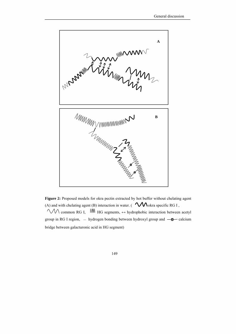
General discussion
149
Figure 2: Proposed models for okra pectin extracted by hot buffer without chelating agent
(A) and with chelating agent (B) interaction in water. ( okra specific RG I ,
common RG I, HG segments, ↔ hydrophobic interaction between acetyl
group in RG I region, — hydrogen bonding between hydroxyl group and calcium
bridge between galacturonic acid in HG segment)
A
B
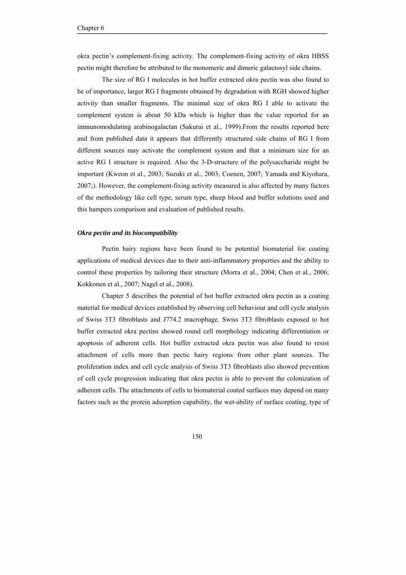
Chapter 6
150
okra pectin’s complement-fixing activity. The complement-fixing activity of okra HBSS
pectin might therefore be attributed to the monomeric and dimeric galactosyl side chains.
The size of RG I molecules in hot buffer extracted okra pectin was also found to
be of importance, larger RG I fragments obtained by degradation with RGH showed higher
activity than smaller fragments. The minimal size of okra RG I able to activate the
complement system is about 50 kDa which is higher than the value reported for an
immunomodulating arabinogalactan (Sakurai et al., 1999).From the results reported here
and from published data it appears that differently structured side chains of RG I from
different sources may activate the complement system and that a minimum size for an
active RG I structure is required. Also the 3-D-structure of the polysaccharide might be
important (Kweon et al., 2003; Suzuki et al., 2003; Coenen, 2007; Yamada and Kiyohara,
2007;). However, the complement-fixing activity measured is also affected by many factors
of the methodology like cell type, serum type, sheep blood and buffer solutions used and
this hampers comparison and evaluation of published results.
Okra pectin and its biocompatibility
Pectin hairy regions have been found to be potential biomaterial for coating
applications of medical devices due to their anti-inflammatory properties and the ability to
control these properties by tailoring their structure (Morra et al., 2004; Chen et al., 2006;
Kokkonen et al., 2007; Nagel et al., 2008).
Chapter 5 describes the potential of hot buffer extracted okra pectin as a coating
material for medical devices established by observing cell behaviour and cell cycle analysis
of Swiss 3T3 fibroblasts and J774.2 macrophage. Swiss 3T3 fibroblasts exposed to hot
buffer extracted okra pectins showed round cell morphology indicating differentiation or
apoptosis of adherent cells. Hot buffer extracted okra pectin was also found to resist
attachment of cells more than pectic hairy regions from other plant sources. The
proliferation index and cell cycle analysis of Swiss 3T3 fibroblasts also showed prevention
of cell cycle progression indicating that okra pectin is able to prevent the colonization of
adherent cells. The attachments of cells to biomaterial coated surfaces may depend on many
factors such as the protein adsorption capability, the wet-ability of surface coating, type of
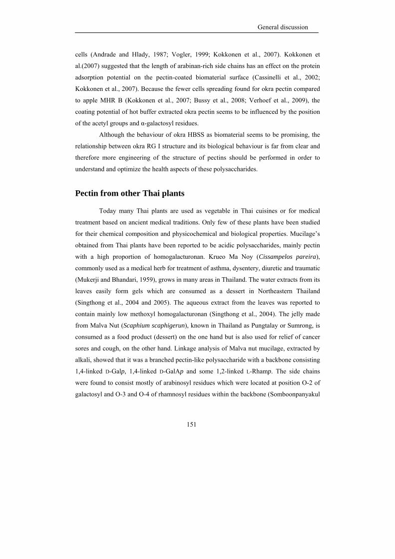
General discussion
151
cells (Andrade and Hlady, 1987; Vogler, 1999; Kokkonen et al., 2007). Kokkonen et
al.(2007) suggested that the length of arabinan-rich side chains has an effect on the protein
adsorption potential on the pectin-coated biomaterial surface (Cassinelli et al., 2002;
Kokkonen et al., 2007). Because the fewer cells spreading found for okra pectin compared
to apple MHR B (Kokkonen et al., 2007; Bussy et al., 2008; Verhoef et al., 2009), the
coating potential of hot buffer extracted okra pectin seems to be influenced by the position
of the acetyl groups and α-galactosyl residues.
Although the behaviour of okra HBSS as biomaterial seems to be promising, the
relationship between okra RG I structure and its biological behaviour is far from clear and
therefore more engineering of the structure of pectins should be performed in order to
understand and optimize the health aspects of these polysaccharides.
Pectin from other Thai plants
Today many Thai plants are used as vegetable in Thai cuisines or for medical
treatment based on ancient medical traditions. Only few of these plants have been studied
for their chemical composition and physicochemical and biological properties. Mucilage’s
obtained from Thai plants have been reported to be acidic polysaccharides, mainly pectin
with a high proportion of homogalacturonan. Krueo Ma Noy (Cissampelos pareira),
commonly used as a medical herb for treatment of asthma, dysentery, diuretic and traumatic
(Mukerji and Bhandari, 1959), grows in many areas in Thailand. The water extracts from its
leaves easily form gels which are consumed as a dessert in Northeastern Thailand
(Singthong et al., 2004 and 2005). The aqueous extract from the leaves was reported to
contain mainly low methoxyl homogalacturonan (Singthong et al., 2004). The jelly made
from Malva Nut (Scaphium scaphigerun), known in Thailand as Pungtalay or Sumrong, is
consumed as a food product (dessert) on the one hand but is also used for relief of cancer
sores and cough, on the other hand. Linkage analysis of Malva nut mucilage, extracted by
alkali, showed that it was a branched pectin-like polysaccharide with a backbone consisting
1,4-linked D-Galp, 1,4-linked D-GalAp and some 1,2-linked L-Rhamp. The side chains
were found to consist mostly of arabinosyl residues which were located at position O-2 of
galactosyl and O-3 and O-4 of rhamnosyl residues within the backbone (Somboonpanyakul
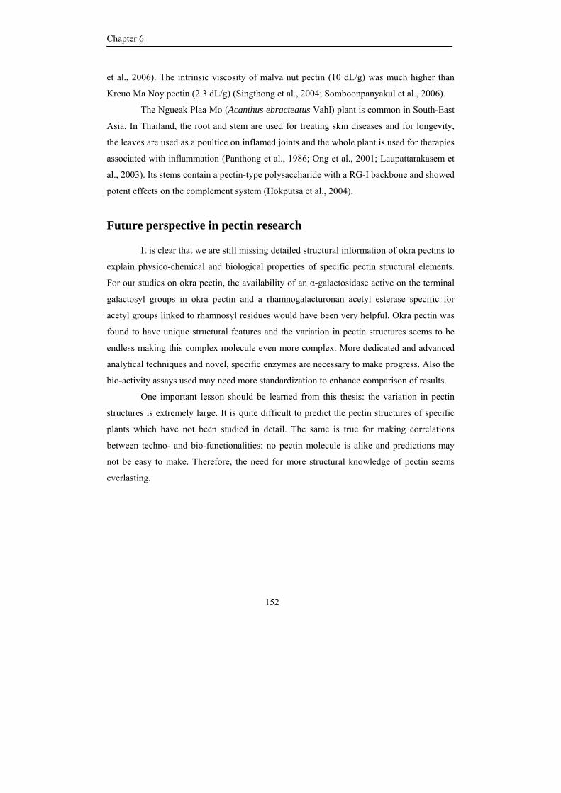
Chapter 6
152
et al., 2006). The intrinsic viscosity of malva nut pectin (10 dL/g) was much higher than
Kreuo Ma Noy pectin (2.3 dL/g) (Singthong et al., 2004; Somboonpanyakul et al., 2006).
The Ngueak Plaa Mo (Acanthus ebracteatus Vahl) plant is common in South-East
Asia. In Thailand, the root and stem are used for treating skin diseases and for longevity,
the leaves are used as a poultice on inflamed joints and the whole plant is used for therapies
associated with inflammation (Panthong et al., 1986; Ong et al., 2001; Laupattarakasem et
al., 2003). Its stems contain a pectin-type polysaccharide with a RG-I backbone and showed
potent effects on the complement system (Hokputsa et al., 2004).
Future perspective in pectin research
It is clear that we are still missing detailed structural information of okra pectins to
explain physico-chemical and biological properties of specific pectin structural elements.
For our studies on okra pectin, the availability of an α-galactosidase active on the terminal
galactosyl groups in okra pectin and a rhamnogalacturonan acetyl esterase specific for
acetyl groups linked to rhamnosyl residues would have been very helpful. Okra pectin was
found to have unique structural features and the variation in pectin structures seems to be
endless making this complex molecule even more complex. More dedicated and advanced
analytical techniques and novel, specific enzymes are necessary to make progress. Also the
bio-activity assays used may need more standardization to enhance comparison of results.
One important lesson should be learned from this thesis: the variation in pectin
structures is extremely large. It is quite difficult to predict the pectin structures of specific
plants which have not been studied in detail. The same is true for making correlations
between techno- and bio-functionalities: no pectin molecule is alike and predictions may
not be easy to make. Therefore, the need for more structural knowledge of pectin seems
everlasting.
General discussion
153
References
Albersheim, P., Darvill, A.G., O’Neill, M.A., Schols, H.A. & Voragen, A.G.J. (1996). An hypothesis: The same
six polysaccharides are components of the primary cell wall of higher plants. In J. Visser & A.G.J.
Voragen, eds, Pectin and Pectinases (pp. 47-53). Amsterdam: Elsevier.
Andrade, J.D. & Hlady, V. (1987). Plasma-protein adsorption – the big12. Ann. N. Y. Acad. Sci., 516, 158–172.
BeMiller, J.N., Whistler, R.L. & Barbalowm, D.G. (1993). Aloe, China, Flexseed, Okra, Psyllium seed, Quince
seed, and Tamarind Gums. In L.W. Roy & J.N. BeMiller, eds., Industrial Gums Polysaccharide and
their Derivatives (pp. 235-255). San Diago: Academic Press.
Bussy, C., Verhoef, R., Haeger, A., Morra, M., Duval, J.L., Vigneron, P., Bensoussan, A., Velzenberger, E.,
Cascardo., G., Cassinelli, C., Schols, H.A., Knox, P. & Nagel, M-D. (2008). Modulating in vitro bone
cell and macrophage behaviour by immobilized enzymatically tailored pectins. J Biomed. Mater. Res.
A., In press.
Cassinelli,C., Morra. M., Carpi, A., Giardino, R. & Fini, M. (2002). Controlling the lateral distribution and
alignment of human gingival fibroblasts by micropatterned polysaccharide surfaces. Biomed.
Pharmacother., 56, 325-331.
Chen, C.H., Sheu, M.T., Chen, T.F., Wang, Y.C., Hou, W.C., Liu, D.Z., Chung, T.C. & Liang, Y.C. (2006).
Suppression of endotoxin-induced proinflammatory responses by citrus pectin through blocking LPS
signaling pathways. Biochem. Pharmacol., 72, 1001-1009.
Coenen, G.J., Bakx, E.J., Verhoef, R.P., Schols, H.A. & Voragen, A.G.J. (2007). Identification of the connecting
linkage between homo- or xylogalacturonan and rhamnogalacturonan I. Carbohydr. Polym., 70, 224-
235.
Coenen, G.J. (2007). Structural characterization of native pectins. PhD thesis, Wageningen University,
Wageningen, The Netherlands.
Deng, C., O'Neill, M.A. & York, W.S. (2006). Selective chemical depolymerization of rhamnogalacturonans.
Carbohydr. Res., 341, 474-484.
Diallo, D., Paulsen, B.S., Liljeback, T.H.A. & Michaelsen, T.E. (2001). Polysaccharides from the roots of Entada
africana Guill. et Perr., Mimosaceae, with complement fixing activity. J. Ethnopharm., 74, 159-171.
Evageliou, V., Ptitchkina, N. M. & Morris, E.R. (2005). Solution viscosity and structural modification of pumpkin
biopectin. Food Hydrocolloid., 19, 1032-1036.
Furuta, H. & Maeda, H. (1999). Rheological properties of water-soluble soybean polysaccharides extracted under
weak acidic condition. Food Hydrocolloid., 13, 267-274.
Furuta, H., Tobe, J., Kiwata, R. & Maeda, H. (1999). Rheological properties of a heat-reversible gel of water-
soluble soybean polysaccharide extracted under acidic conditions. Biosci. Biotechnol. Biochem., 63,
2075-2081.
Gur’janov, O.P., Gorshkova, T.A., Kabel, M., Schols, H.A. & Van Dam, J.E.G. (2007). MALDI-TOF MS
evidence for linking of flax bast fibre galactan to rhamnogalacturonan backbone. Carbohydr. Polym.,
67, 86-96.
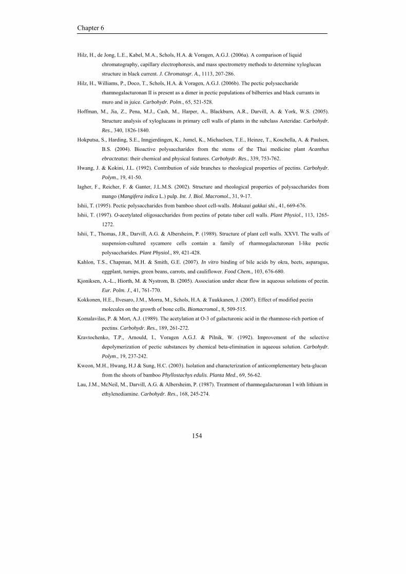
Chapter 6
154
Hilz, H., de Jong, L.E., Kabel, M.A., Schols, H.A. & Voragen, A.G.J. (2006a). A comparison of liquid
chromatography, capillary electrophoresis, and mass spectrometry methods to determine xyloglucan
structure in black current. J. Chromatogr. A., 1113, 207-286.
Hilz, H., Williams, P., Doco, T., Schols, H.A. & Voragen, A.G.J. (2006b). The pectic polysaccharide
rhamnogalacturonan II is present as a dimer in pectic populations of bilberries and black currants in
muro and in juice. Carbohydr. Polm., 65, 521-528.
Hoffman, M., Jia, Z., Pena, M.J., Cash, M., Harper, A., Blackburn, A.R., Darvill, A. & York, W.S. (2005).
Structure analysis of xyloglucans in primary cell walls of plants in the subclass Asteridae. Carbohydr.
Res., 340, 1826-1840.
Hokputsa, S., Harding, S.E., Inngjerdingen, K., Jumel, K., Michaelsen, T.E., Heinze, T., Koschella, A. & Paulsen,
B.S. (2004). Bioactive polysaccharides from the stems of the Thai medicine plant Acanthus
ebracteatus: their chemical and physical features. Carbohydr. Res., 339, 753-762.
Hwang, J. & Kokini, J.L. (1992). Contribution of side branches to rheological properties of pectins. Carbohydr.
Polym., 19, 41-50.
Iagher, F., Reicher, F. & Ganter, J.L.M.S. (2002). Structure and rheological properties of polysaccharides from
mango (Mangifera indica L.) pulp. Int. J. Biol. Macromol., 31, 9-17.
Ishii, T. (1995). Pectic polysaccharides from bamboo shoot cell-walls. Mokuzai gakkai shi., 41, 669-676.
Ishii, T. (1997). O-acetylated oligosaccharides from pectins of potato tuber cell walls. Plant Physiol., 113, 1265-
1272.
Ishii, T., Thomas, J.R., Darvill, A.G. & Albersheim, P. (1989). Structure of plant cell walls. XXVI. The walls of
suspension-cultured sycamore cells contain a family of rhamnogalacturonan I-like pectic
polysaccharides. Plant Physiol., 89, 421-428.
Kahlon, T.S., Chapman, M.H. & Smith, G.E. (2007). In vitro binding of bile acids by okra, beets, asparagus,
eggplant, turnips, green beans, carrots, and cauliflower. Food Chem., 103, 676-680.
Kjoniksen, A.-L., Hiorth, M. & Nystrom, B. (2005). Association under shear flow in aqueous solutions of pectin.
Eur. Polm. J., 41, 761-770.
Kokkonen, H.E., Ilvesaro, J.M., Morra, M., Schols, H.A. & Tuukkanen, J. (2007). Effect of modified pectin
molecules on the growth of bone cells. Biomacromol., 8, 509-515.
Komalavilas, P. & Mort, A.J. (1989). The acetylation at O-3 of galacturonic acid in the rhamnose-rich portion of
pectins. Carbohydr. Res., 189, 261-272.
Kravtochenko, T.P., Arnould, I., Voragen A.G.J. & Pilnik, W. (1992). Improvement of the selective
depolymerization of pectic substances by chemical beta-elimination in aqueous solution. Carbohydr.
Polym., 19, 237-242.
Kweon, M.H., Hwang, H.J & Sung, H.C. (2003). Isolation and characterization of anticomplementary beta-glucan
from the shoots of bamboo Phyllostachys edulis. Planta Med., 69, 56-62.
Lau, J.M., McNeil, M., Darvill, A.G. & Albersheim, P. (1987). Treatment of rhamnogalacturonan I with lithium in
ethylenediamine. Carbohydr. Res., 168, 245-274.
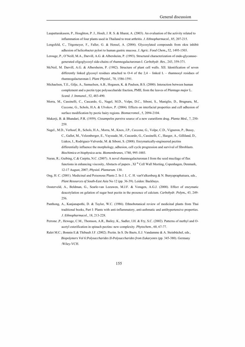
General discussion
155
Laupattarakasem, P., Houghton, P. J., Hoult, J. R. S. & Itharat, A. (2003). An evaluation of the activity related to
inflammation of four plants used in Thailand to treat arthritis. J. Ethnopharmacol., 85, 207-215.
Lengsfeld, C., Titgemeyer, F., Faller, G. & Hensel, A. (2004). Glycosylated compounds from okra inhibit
adhesion of helicobacter pylori to human gastric mucosa. J. Agric. Food Chem., 52, 1495-1503.
Lerouge, P., O’Neill, M.A., Darvill, A.G. & Albersheim, P. (1993). Structural characterization of endo-glycanase-
generated oligoglycosyl side-chains of rhamnogalacturonan-I. Carbohydr. Res., 243, 359-371.
McNeil, M. Darvill, A.G. & Albersheim, P. (1982). Structure of plant cell walls. XII. Identification of seven
differently linked glycosyl residues attached to O-4 of the 2,4 – linked L – rhamnosyl residues of
rhamnogalacturonan I. Plant Physiol., 70, 1586-1591.
Michaelsen, T.E., Gilje, A., Samuelsen, A.B., Hogasen, K. & Paulsen, B.S. (2000). Interaction between human
complement and a pectin type polysaccharide fraction, PMII, from the leaves of Plantago major L.
Scand. J. Immunol., 52, 483-490.
Morra, M., Cassinelli, C., Cascardo, G., Nagel, M.D., Volpe, D.C., Siboni, S., Maniglio, D., Brugnara, M.,
Ceccone, G., Schols, H.A. & Ulvskov, P. (2004). Effects on interfacial properties and cell adhesion of
surface modification by pectic hairy regions. Biomacromol., 5, 2094-2104.
Mukerji, B. & Bhandari, P.R. (1959). Cissampelos pareira source of a new curariform drug. Planta Med., 7, 250-
259.
Nagel., M.D., Verhoef, R., Schols, H.A., Morra, M., Knox, J.P., Ceccone, G., Volpe, C.D., Vigneron, P., Bussy,
C., Gallet, M., Velzenberger, E., Vayssade, M., Cascardo, G., Cassinelli, C., Haeger, A., Gilliland, D.,
Liakos, I., Rodriguez-Valverde, M. & Siboni, S. (2008). Enzymatically-engineered pectins
differentially influence the morphology, adhesion, cell cycle progression and survival of fibroblasts.
Biochimica et biophysica acta. Biomembranes, 1780, 995-1003.
Naran, R.; Guibing, C.& Carpita, N.C. (2007). A novel rhamnogalacturonan I from the seed mucilage of flax
functions in enhancing viscosity, Abstacts of papers , XI th Cell Wall Meeting, Copenhagen, Denmark,
12-17 August, 2007; Physiol. Plantarum. 130.
Ong, H. C. (2001). Medicinal and Poisonous Plants 2. In J. L. C. H. vanValkenburg & N. Bunyapraphatsara, eds.,
Plant Resources of South-East Asia No 12 (pp. 36-39). Leiden: Backhuys.
Oosterveld, A., Beldman, G., Searle-van Leeuwen, M.J.F. & Voragen, A.G.J. (2000). Effect of enzymatic
deacetylation on gelation of sugar beet pectin in the presence of calcium. Carbohydr. Polym., 43, 249-
256.
Panthong, A., Kanjanapothi, D. & Taylor, W.C. (1986). Ethnobotanical review of medicinal plants from Thai
traditional books, Part I: Plants with anti-inflammatory, anti-asthmatic and antihypertensive properties.
J. Ethnopharmacol., 18, 213-228.
Perrone ,P., Hewage, C.M., Thomson, A.R., Bailey, K., Sadler, I.H. & Fry, S.C. (2002). Patterns of methyl and O-
acetyl esterification in spinach pectins: new complexity. Phytochem., 60, 67-77.
Ralet M.C.; Bonnin E.& Thibault J.F. (2002). Pectin. In S. De Baets, E.J. Vandamme & A. Steinbüchel, eds.,
Biopolymers Vol 6:Polysaccharides II-Polysaccharides from Eukaryotes (pp. 345-380). Germany
:Wiley-VCH.
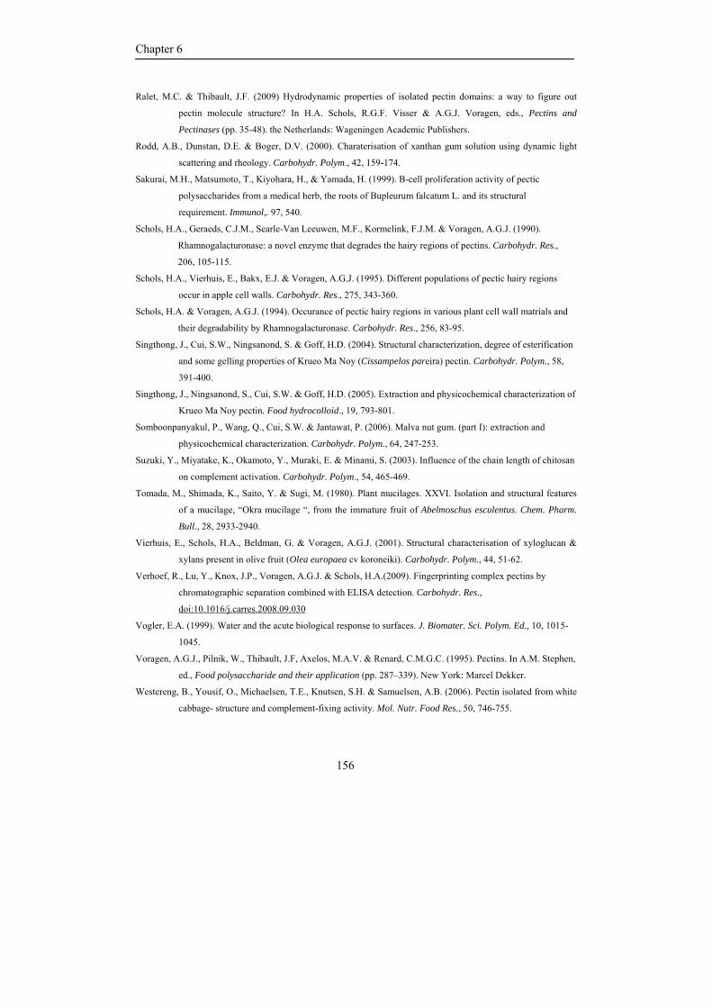
Chapter 6
156
Ralet, M.C. & Thibault, J.F. (2009) Hydrodynamic properties of isolated pectin domains: a way to figure out
pectin molecule structure? In H.A. Schols, R.G.F. Visser & A.G.J. Voragen, eds., Pectins and
Pectinases (pp. 35-48). the Netherlands: Wageningen Academic Publishers.
Rodd, A.B., Dunstan, D.E. & Boger, D.V. (2000). Charaterisation of xanthan gum solution using dynamic light
scattering and rheology. Carbohydr. Polym., 42, 159-174.
Sakurai, M.H., Matsumoto, T., Kiyohara, H., & Yamada, H. (1999). B-cell proliferation activity of pectic
polysaccharides from a medical herb, the roots of Bupleurum falcatum L. and its structural
requirement. Immunol,. 97, 540.
Schols, H.A., Geraeds, C.J.M., Searle-Van Leeuwen, M.F., Kormelink, F.J.M. & Voragen, A.G.J. (1990).
Rhamnogalacturonase: a novel enzyme that degrades the hairy regions of pectins. Carbohydr. Res.,
206, 105-115.
Schols, H.A., Vierhuis, E., Bakx, E.J. & Voragen, A.G.J. (1995). Different populations of pectic hairy regions
occur in apple cell walls. Carbohydr. Res., 275, 343-360.
Schols, H.A. & Voragen, A.G.J. (1994). Occurance of pectic hairy regions in various plant cell wall matrials and
their degradability by Rhamnogalacturonase. Carbohydr. Res., 256, 83-95.
Singthong, J., Cui, S.W., Ningsanond, S. & Goff, H.D. (2004). Structural characterization, degree of esterification
and some gelling properties of Krueo Ma Noy (Cissampelos pareira) pectin. Carbohydr. Polym., 58,
391-400.
Singthong, J., Ningsanond, S., Cui, S.W. & Goff, H.D. (2005). Extraction and physicochemical characterization of
Krueo Ma Noy pectin. Food hydrocolloid., 19, 793-801.
Somboonpanyakul, P., Wang, Q., Cui, S.W. & Jantawat, P. (2006). Malva nut gum. (part I): extraction and
physicochemical characterization. Carbohydr. Polym., 64, 247-253.
Suzuki, Y., Miyatake, K., Okamoto, Y., Muraki, E. & Minami, S. (2003). Influence of the chain length of chitosan
on complement activation. Carbohydr. Polym., 54, 465-469.
Tomada, M., Shimada, K., Saito, Y. & Sugi, M. (1980). Plant mucilages. XXVI. Isolation and structural features
of a mucilage, “Okra mucilage “, from the immature fruit of Abelmoschus esculentus. Chem. Pharm.
Bull., 28, 2933-2940.
Vierhuis, E., Schols, H.A., Beldman, G. & Voragen, A.G.J. (2001). Structural characterisation of xyloglucan &
xylans present in olive fruit (Olea europaea cv koroneiki). Carbohydr. Polym., 44, 51-62.
Verhoef, R., Lu, Y., Knox, J.P., Voragen, A.G.J. & Schols, H.A.(2009). Fingerprinting complex pectins by
chromatographic separation combined with ELISA detection. Carbohydr. Res.,
doi:10.1016/j.carres.2008.09.030
Vogler, E.A. (1999). Water and the acute biological response to surfaces. J. Biomater. Sci. Polym. Ed., 10, 1015-
1045.
Voragen, A.G.J., Pilnik, W., Thibault, J.F, Axelos, M.A.V. & Renard, C.M.G.C. (1995). Pectins. In A.M. Stephen,
ed., Food polysaccharide and their application (pp. 287–339). New York: Marcel Dekker.
Westereng, B., Yousif, O., Michaelsen, T.E., Knutsen, S.H. & Samuelsen, A.B. (2006). Pectin isolated from white
cabbage- structure and complement-fixing activity. Mol. Nutr. Food Res., 50, 746-755.
General discussion
157
Williams, P.A. & Phillips, G.O. (2000). Introduction of food hydrocolloids. In Phillips G.O. & Williams P.A.,
eds., Handbook of hydrocolloids (pp. 1-19). USA: CRC Press.
Yamada, H., Nagai, T., Cyong, J.-C. & Otsuka, Y. (1985). Relationship between chemical structure and anti-
complementary activity of plant polysaccharides. Carbohydr. Res, 144, 101-111.
Yamada, H. & Kiyohara, H. (2007). Immunomodulating activity of plant polysaccharide structures. In J.P.
Kamerling, G.J Boons, Y.C. Lee, A. Suzuki, N. Taniguchi & A.G.J. Voragen, eds., Comprehensive
Glycoscience: From chemistry to systems biology. Volume 4 Cell glycobiology and development health
and disease in glycomedicine, (pp. 663-693). Oxford: Elsevier Ltd.
Yamada, H., Kiyohara, H & Matsumoto, T. (2009). Recents studies on structures and intestinal immunity
modulating activities of pectins and pectic polysaccharides from medicinal herbs. In H.A. Schols,
R.G.F. Visser & A.G.J. Voragen, eds., Pectins and Pectinases (pp. 293-304). the Netherlands:
Wageningen Academic Publishers.
Chapter 6
158
Summary
159
Summary
Samenvatting
Samenvatting
160
Summary
In Thailand, many plants have been used as vegetables as well as for traditional
medicine. Okra, Abelmoschus esculentus (L.) Moench, is an example of such a plant.
Examples for the medical use are treatment of gastric irritation, treatment of dental
diseases, lowering cholesterol level and preventing cancer. These biological activities are
ascribed to polysaccharide structures of okra in particular pectin structures. However, the
precise structure of okra pectins and also of other polysaccharides in okra pods have been
lacking so far.
In order to obtain detailed information of the different polysaccharides present in
okra, okra cell wall material was prepared from the pulp of okra pods and was then
sequentially extracted with hot buffer, chelating agent, diluted alkali and concentrated
alkali. The sugar (linkage) composition indicated that okra cell wall contained, next to
cellulose, different populations of pectins and hemicelluloses.
The pectic polysaccharides were mainly obtained in the first three extracts having
slightly different chemical structures. The okra pectin extracted by hot buffer was almost a
pure rhamnogalacturonan (RG) I with a high degree of acetylation (DA), covalently linked
to a minor amount of homogalacturonan (HG) having a high degree of methyl esterification
(DM). The chelating agent extractable pectin and the diluted alkali extractable pectin
predominantly contained HG with only minor amounts of RG I. Okra pectins extracted by
hot buffer and with chelating agent had in common that both contained highly branched RG
I with very short side chains containing not more than 3 galactosyl units attached to the
rhamnosyl residues in RG I backbone. Chelating agent extracted okra pectins also carried
arabinan and arabinogalactan type II as neutral side chains and these side chains were even
more abundantly present in the diluted alkali extracted okra pectin.
The hemicellulosic polysaccharides ended up in concentrated alkali extract. From
the sugar (linkage) composition and enzymatic degradation studies using pure and well
defined enzymes, it was concluded that this fraction contained a XXXG–type xyloglucan
and 4-methylglucuronoxylan. The cellulosic polysaccharides were retained in the residue.
The okra hot buffer extractable RG I having a high level of acetyl substitution
appeared to be very well degradable by rhamnogalacturonan hydrolase which was known to
Summary
161
be hindered completely by acetylated substrates. In contrast, an acetylated galacturonic
acid-specific rhamnogalacturonan acetyl esterase was unable to remove acetyl groups from
the RG I molecule of hot buffer extracted okra pectin. For these reasons, the precise
position of the acetyl groups present on enzymatically released oligomers were determined
by Electron Spray Ionization Ion Trap Mass Spectrometry (ESI-IT-MS) and Nuclear
Magnetic Resonance (NMR) spectroscopy. The acetyl groups were found to be
predominantly located at position O-3 of the rhamnosyl moiety, while the methyl esters
seemed to be present only on the HG part of the hot buffer extracted okra pectin. Another
novelty of okra RG-I was the presence of terminal alpha-linked galactosyl substitution at
position O-4 of the rhamnosyl residues within the RG I backbone. These specific features
(acetylated rhamnosyl- and alpha-galactosyl-substitutions) were almost absent in the
chelating agent extracted okra pectin where more commonly known substitutions were
present, including acetylated galacturonosyl residues in the RG I backbone. The unique
structure features of hot buffer extracted okra pectin led to the assumption that these
features may contribute to the rather typical physical properties as well as to the biological
properties found for okra pectin.
In order to understand the effect of the specific structural features of RG I on its
physical properties, the rheological properties of hot buffer extracted okra pectin were
determined and compared to those found for chelating agent extracted okra pectin and for
pectins from other plant materials as reported in the literature. The solutions of hot buffer
extracted okra pectin showed a high viscosity and predominant elastic behaviour which
most probably is caused by strong hydrophobic associations through its acetylated
rhamnosyl residues rather than by methyl esterified galacturonosyl residues as is commonly
the case for pectins. The removal of acetyl groups and methyl esters decreased the
association of the pectin molecules as observed by the light scattering experiment, meaning
that not only viscosity and rheological properties but also association of pectin molecules
were as result of both hydrophobic interactions and charge effects.
The effect of the position of acetyl groups on the bioactivity of okra pectin was
also determined. The complement-fixing activity of okra pectins was found to be affected
by many factors like e.g. the presence of acetyl groups, the size of RG segments and the
Samenvatting
162
presence of terminal alpha galactosyl groups and even the three dimensional conformation
of the molecules. The hot buffer extracted okra pectin was also examined for its potential to
modify surfaces of medical devices and implants. The results showed that okra pectin can
be used in coating medical device since it promotes cell apoptosis and shows no
macrophage activation.
The knowledge described in this thesis provided us with novel information on the
unique structures of okra pectins and may lead to a better understanding of the functional
properties of okra polysaccharides in general and okra pectin in particular and to optimize
the use of okra pectins within the food industry and in medical applications. However,
despite our efforts, at the moment the dependency of the (bio) functionality of okra pectins
on the precise chemical structure are not yet completely understood.
Summary
163
Samenvatting In Thailand worden veel planten zowel gebruikt als groenten als ook als
traditionele medicijnen. Okra, Abelmoschus esculentus (L) Moench, is een voorbeeld van
zo’n plant. Voorbeelden van zijn medicinale toepassing zijn behandeling van
maagaandoeningen, behandeling van tandpijn, verlaging van het cholesterol gehalte in het
bloed en het voorkomen van kanker. Deze biologische activiteiten worden toegeschreven
aan de polysacchariden die in okra aanwezig zijn met name aan de complexe pektine
strukturen. De chemische fijn structuur van okra pektine en ook van de andere aanwezig
polysacchariden zijn nog altijd onvoldoende bekend.
Om gedetailleerde informatie te verkrijgen over de verschillende typen
polysacchariden aanwezig in okra werd okra celwand geisoleerd uit een pulp gemaakt van
okra peulen. Hieruit werden door opeen volgende extracties met een warme buffer
oplossing, met een oplossing van een chelaterend middel, met een verdunde loog oplossing
en tenslotte met een geconcentreerde loog oplossing verschillende polysacchariden fracties
verkregen. De suikersamenstelling en de geanalyseerde bindingstypen gaven aan dat de
okra celwanden naast cellulose diverse populaties pektine en hemi-cellulose bevatten.
Pektine-achtige polysacchariden werden voornamelijk verkregen in de eerste drie
extracten en deze vertoonden enkele verschillen in chemische structuur. Okra pektine ge-
extraheerd met warme buffer oplossing was een bijna zuiver rhamnogalacturonaan (RG) I
met een hoge acetyleringsgraad dat covalent gekoppeld was aan kleine hoeveelheden
homogalacturonaan (HG) dat een hoog gehalte aan methylesters (DM) bevatte. De pektines
ge-extraheerd met het chelaterend middel en met verdunde loog waren hoofdzakelijk
homogalacturonanen (HG) met slechts kleine hoeveelheden RG I. Okra pektines ge-
extraheerd met warme buffer en met het chelaterend middel hadden met elkaar gemeen dat
ze beide sterk vertakt RG I bevatten met korte zijketens die uit niet meer dan 3 galactose
eenheden bestonden, gekoppeld aan rhamnose bouwstenen in de RG I hoofdketen. Pektine
ge-extraheerd met het chelaterend middel bevatte ook arabinaan en arabinogalactaan type II
als zijketens. Deze zijketens waren nog meer aanwezig in het pektine ge-extraheerd met
verdunde loog.
Samenvatting
164
Polysacchariden van het hemi-cellulose type werden teruggevonden in het extract
verkregen met geconcentreerde loog. Uit de analyse van de bindingstypen tussen de
suikerbouwstenen en analyse van de afbraakprodukten verkregen met zuivere en goed
gekarakteriseerde enzymen bleek dat deze fractie XXXG-type xyloglucan en 4-
methylglucuronxylan bevatte. Het cellulose bleef achter in het residue van de extracties.
RG I met een hoog acetylgehalte, aanwezig in het warm-buffer extract van okra
celwanden, bleek onverwacht zeer goed afbreekbaar met rhamnogalacturonaanhydrolase.
Tot nu toe was steeds gevonden dat dit enzym niet in staat was geacetyleerd RG I af te
breken. Rhamnogalacturonaan acetyl esterase, specifiek voor RG I, bleek daarentegen niet
in staat acetylgroepen af te splitsen van het RG I ge-extraheerd met warme buffer. Om deze
waarnemingen te kunnen verklaren werd met behulp van Electron Spray Ionization Ion
Trap Mass Spectroscopy (ESI-IT-MS) en Nuclear Magnetic Resonance (NMR)
Spectroscopy de preciese positie van de acetylgroepen in RG I oligomeren, verkregen door
enzymatische afbraak, bepaald. De acetylgroepen bleken hoofdzakelijk voor te komen op
de O-3 positie van rhamnosyl bouwstenen, terwijl de methyl esters hoofdzakelijk leken
voor te komen in de HG segmenten. Een andere nieuwe vinding was de aanwezigheid van
terminale galactosyl eenheden alpha-gebonden aan positie O-4 van de rhamnosyl
bouwstenen in de hoofdketen van okra RG I in het warm-buffer extract. Deze kenmerkende
karakteristieken (geacetyleerd rhamnosyl bouwstenen en alpha gebonden galactosyl
eenheden) bleken nagenoeg afwezig in de pektine fractie ge-extraheerd met het chelaterend
middel. Deze fractie bleek de algemeen bekende substitutuenten te bevatten waaronder
geacetyleerde galacturonosyl bouwstenen id de hoofdketen van RG I. De unieke, structurele
kenmerken van de pektine fractie in het warme buffer extract waren aanleiding voor de
veronderstelling dat deze afwijkende structuren bij zouden kunnen dragen aan zowel de
nogal typische fysisch-chemisch eigenschappen als ook aan de biologische eigenschappen
vastgesteld voor okra pektine.
Om de effecten van de specifieke, structurele kenmerken van RG I op zijn fysische
eigenschappen te kunnen begrijpen werden rheologische parameters van de met warme
buffer ge-extraheerde okra pektine vergeleken met de parameters vastgesteld voor de met
chelateringsmiddel ge-extraheerde okra pektine en met pektines van andere plantaardige
Summary
165
bronnen zoals vermeld in de literatuur. Oplossingen van met warme buffer ge-extraheerde
okra pektine vertoonden een hoge viscositeit en een overheersend elastisch gedrag
waarschijnlijk veroorzaakt door sterke hydrofobe associaties van de geacetyleerde
rhamnosyl bouwstenen. Dit in tegenstelling tot de tot nu toe bekende pectines in oplossing
waar methyl veresterde galacturonosyl bouwstenen de eigenschappen vooral bepalen. Het
verwijderen van de acetyl groepen en methyl esters verlaagde de associatie van de pektine
moleculen zoals waargenomen kon worden uit lichtverstrooiings metingen. Dit duidt erop
dat niet alleen de visceuze en rheologische eigenschappen maar ook de associatie van de
pektine moleculen het gevolg zijn van zowel hydrofobe interacties als van ladingseffecten.
Het effect van de positie van de acetylgroepen op de bioactiviteit van okra pektine
werd ook bepaald. De anti-complement activiteit gemeten voor okra pektines bleek
beinvloed te worden door vele factoren zoals bijvoorbeeld de aanwezigheid van
acetylgroepen, de grootte van de RG I segmenten, de aanwezigheid van terminale alpha-
galactosyl bouwstenen en zelfs van de 3-dimensionale structuur van de pektine moleculen.
De met warme buffer ge-extraheerde pektine fractie werd ook onderzocht op zijn vermogen
het oppervlak van medische apparaten en implantaten te veranderen. Er bleek dat okra
pektine hiervoor gebruikt kan worden omdat hierdoor cell apoptosis bevorderd werd en er
geen macrophage activering optrad.
De nieuwe kennis beschreven in dit proefschrift verschaft ons aanvullende,
waardevolle informatie over de unieke structuur van pektine verkregen uit okra celwanden
en helpt de functionele eigenschappen van okra polyscchariden in het algemeen en van okra
pektines in het bijzonder beter te begrijpen en kan bijdragen de toepassing van deze
pektines in de voedselindustrie en in medische toepassingen te optimaliseren. Echter,
ondanks onze inspanningen is vooralsnog het verband tussen de (bio) functionaliteit van
okra pektines en de preciese chemische structuur nog onvoldoende duidelijk.
Samenvatting
166
Acknowledgement
167
Acknowledgement
“The need for more structural knowledge on pectin seems to be everlasting”.
During my stay in Wageningen I have learned a lot about pectin, particular of
structural, analytical and biological aspects. It was sometimes difficult for me and I think I
still need to learn more. Also writing this thesis in English was hard for me, as is writing
this acknowledgement since I do not want to miss anybody. My thesis would not be
possible without lending a hand from many people.
“Success comes from chance and endeavour”.
After graduation for my Master Degree, it seemed very difficult for me to enrol
in a PhD program. Fortunately, I had a chance to meet Dr. Tanaboon Sajja-anatakool who
made it possible for me to get started with a PhD study. A Pom: thank you for encouraging
me and giving me an opportunity to work in the Netherlands. This was made possible by
Prof. Fons Voragen who, during a visit to Kasetsart University in Bangkok, invited me to
come to Wageningen University and do research in the Laboratory of Food Chemistry.
Therefore, I would like to first express my gratitude to my promoter, Prof. Fons Voragen
and my co-supervisor Dr. Tanaboon Sajja-anatakool. Fons, thank you for giving me this
opportunity. Your encouragement kept me going through the work for the PhD degree. I
received valuable help from you not only in writing the manuscript but also personal advice
while living in Wageningen.
“Yet different cultures but not difficult to live”.
I have met a lot of nice person here in the Netherlands. I would like to express
my appreciation to my co-promotor, Dr. Henk Schols for his nice guidance and support.
Henk, I have learned a lot from you about pectins and plant cell walls. You were always
there for helpful discussions and give insightful advice. You motivated me to ask good
questions and express creative ideas. You showed me how to approach a research problem
and how to evaluate, present and discuss scientific results critically. You amazed me by the
way you organized your time for research, teaching, meetings and supervising.
Acknowlegement
168
Moreover, I would like to honestly say that without you, Henk and Fons, it would
not have been possible for me to finish my dissertation. Thank you for spending long
evenings and/or weekends reviewing my thesis. Moreover, I would like to sincerely thank
Jolanda for her help and kindness. I know it was quite a lot of work for you to meet all the
requirements for getting my Visa, especially dealing with IND. I also owe a great deal to
Harry and Jan-Paul for asking critical questions and giving me enlightened comments.
I am also grateful to Anne Berit Samuelsen of the Norwegian Food Research
Institute-Matforsk with whom we collaborated on the biological aspects of Okra pectin for
her nice guidance. Even though my stay in Norway was short I will never forget the long
days and eating shrimp in the middle of the summer.
“Learning in a new environment with new colleagues gives a lot of fun and
happiness”.
Many, many thanks go to my great colleagues. Even though my English was not
that good, particularly in the beginning, all of you kindly tried to understand me. Thank you
for the very pleasant working atmosphere: Karin, Yun, Lieke, Koen, Gerrit, the safety-team
(Laurice, Koos, Stefan), Martine, Ruud, Johan, Natalie, Hans, Peter, Jean-Paul, Anne,
Mirjam, Peter, Joris, Laura, Hauke, Gerd-Jan, René V., Jeroen, Raymond, Smaaklessen
Team (Hante, Marlies, Coco), Stephanie, Junrong, Takao, and Caros.
Furthermore, without Marijke, Jolan, René K., Margaret, Toos ,Jan, Mark and
Edwin it would have been very difficult to learn the many techniques, thanks a lot. Without
my office-mates Yvonne,V., Walter, Simone, Alice, and Anita, there would not have been
so many laughs and fun. I also want to thank the student who contributed to this thesis, Roy
Delahaije. My special thanks go to Rudy and Kanjana for all the kind arrangements, their
hospitality and for being my paranymph.
Doing a PhD at Wageningen University was quite new and valuable for me.
“Food is all around”.
Besides, having big lunches and dinner with the Thai community in Wageningen,
I also very much enjoyed having lunch and dinner with my colleagues. I would like to
thank Fons and his wife, Lea, Jean-Paul and his wife, Marian, for inviting me to have nice
Acknowledgement
169
dinners at their homes; my mates in office 508: Walter, Yvonne, V. Yvonne, W. and
Simone for the nice office dinners, Rudy for nice dinners and chats in the lab. I hope you
will remember the Thai food and snacks I cooked for you even though you considered them
sometimes too spicy for you.
“Life is not only in the laboratory”.
We had a lot of activities outside our laboratory. I want to thank the activity
committee for all the events they organized. I also had a chance to join the bi-annual PhD
trip to France, Belgium, and England in 2006 and to China, 2008. I want to thank for both
PhD trip committee. These trips were a great experience and a lot of fun like walking
around in London, a nice birthday celebration in Nottingham, having noodles in front of the
Hotel in Beijing, walking to the top of the Great Wall in China, having a nice massage in
Beijing. Of course, I also learned a lot from these PhD trip. For sure, it was not holidays.
I gratefully thank the Commission on Higher Education Ministry of Education,
Thailand, under the Commission on Higher Education Staff Development project for the
Joint PhD program in Food Science at Kasetsart University, Thailand for the financial
support in the first year in the Netherlands.
“To be far away from home is not always easy ”.
It would have been very difficult without all of my Thai friends here in
Wageningen, who were like a big, warm Thai family: Busaba & Kees, Naiyana, Sayam,
Thanawit, Paweena, Supawoot, U-sa, Budsara, Warit, Kanjana, Sueppong, Rattanaporn &
Marcel, Krissana, & Nicolas. Furthermore, I want to thank my younger sisters and brothers
in The Netherlands: Thotsawan, Warangkana, Kanang, Chantita, Panya, and Wasaporn, and
including my younger sister and brother in the ODOS project. Thank you for your advice,
cheerfulness, laughter, sharing happiness and forgiving me.
“The big support comes from my warm family”.
I would like to express my big gratitude to my lovely family, mom, adopted
mom, dad, adopted dad and my younger sister. I know, it was not easy for you since I was
continuously away for my study from my Bachelor till my Ph D. It was quite a long period
Acknowlegement
170
and I would never have come this far without the secure feeling of my family’s love,
support and guidance.
Nipaporn, Tai
Curriculum vitae
171
Curriculum vitae
Nipaporn Sengkhamparn (Tai) was born on 1 November 1977 in Nongkhai
Province, Thailand. In 1995, she started her bachelor degree in Food Technology at
Khonkaen University and graduated in 4 years later with diploma project of ‘Production of
reduced-fat chicken frankfurters with konjac and carrageenan’. Afterwards she started her
master degree in Food Technology at Chulalongkorn University, 2000 – 2003. Her diploma
thesis was entitled ‘Production of concentrated santol Sandoricum indicum Cav. Juice and
santol juice supplemented with calcium and vitamin D’. Tai had continually studied her Ph
D in Food Science at Kasertsart University since June 2003 under financial support from
the Commission on Higher Education Ministry of Education, Thailand, under the
Commission on Higher Education Staff Development project for the Joint Ph.D program in
Food Science at Kasetsart University, Thailand. From November 2005, she went aboard to
Laboratory of Food Chemistry, Wageningen University and Research Centre, the
Netherlands and worked on the research in topic of ‘Chemical, physical and biological
features of Okra pectin’. The research on structural characterization of cell wall
polysaccharides from okra including phyochemical properties of okra pectin within this
thesis was carried out at Food Chemistry Group and Food Physic Group, Wageningen
University and Research Centre, from November 2005 until September 2009. Some part of
this thesis, biological properties of okra pectin was carried out at Norwegian Food Research
Institute-Matforsk, Norway for 3 weeks.
Curriculum vitae
172
List of publications
173
List of publications
Full papers
Sengkhamparn, N., Verhoef, R., Schols, H. A., Sajjaanantakul, T. & Voragen, A.G.J.
(2009a) Characterization of Cell Wall Polysaccharide from Okra (Abelmoschus
esculentus (L.) Moench). Carbohydr. Res., 344, 1824-1832.
Sengkhamparn, N., Verhoef, R., Bakx, E.J., Schols, H.A., Sajjaanantakul, T. & Voragen,
A.G.J. (2009b) Okra pectin contains an unusual substitution of its rhamnosyl
residues with acetyl and alpha –linked galactosyl groups. Carbohydr. Res., 344,
1842-1851.
Sengkhamparn, N., Sagis, L.M.C., de Vries, R., Schols, H.A., Sajjaanantakul, T. &
Voragen, A.G.J.Physicochemical properties of pectins from Okra (Abelmoschus
esculentus (L.) Moench). Food Hydrocolloid., 2010, 24, 35-41.
Book chapter
Sengkhamparn, N., Verhoef, R., Bakx, E.J., Schols, H.A., Sajjaanantakul, T. & Voragen,
A.G.J. (2009). Structural elucidation of cell wall polysaccharides from Okra
(Abelmoschus esculentus (L.) Moench). Pectins and pectinases (49-58).the
Netherlands: Wageningen Academic Publishers.
Abstracts
Sengkhamparn, N., Schols, H.A., Sajjaanantakul, T. & Voragen, A.G.J. (2007) Structural
elucidation of cell wall polysaccharides from Okra (Abelmoschus esculentus (L.)
Moench). Abstacts of papers , XI th Cell Wall Meeting, Copenhagen, Denmark,
12-17 August, 2007; Physiol. Plantarum., 130.

174
Training activities
175
Overview of completed training activities
Discipline specific activities
Courses
Glycosciences Summer School (Wageningen, The Netherlands, June 2006)
Food and Health (Cluj Nacopa, Romania, February 2007)
Polysaccharides as Food Colloids and Biomaterials: Fundamentals and Applications
(Wageningen, The Netherlands, October 2007)
Fundamentals of Hydrocolloid Technology Course (Nottingham, UK, March 2007)
Meetings
XI Cell Wall Meeting (Copenhagen, Denmark, August 2007)
Pectins and Pectinases (Wageningen, The Netherlands, April 2008)
General Courses
Techniques for Writing and Presenting a Scientific Paper (July 2007)
Scientific Publishing (November 2007)
Food Chemistry Colloqia (2005-2008)
Food Chemistry Seminar (2005-2008)
Additional activities
Norwegian Food Research Institute-Matforsk, Bio-activity Testing (June 2007)
Food Chemistry PhD Excursion to Belgium, France and United Kingdom (2006)
Food Chemistry PhD Excursion to China (2008)

176