proliferation and enrichment of cd133(+) glioblastoma cancer stem cells on 3d chitosan-alginate...
TRANSCRIPT

lable at ScienceDirect
Biomaterials xxx (2014) 1e7
Contents lists avai
Biomaterials
journal homepage: www.elsevier .com/locate/biomater ia ls
Proliferation and enrichment of CD133þ glioblastoma cancer stemcells on 3D chitosan-alginate scaffolds
Forrest M. Kievit a, 2, Stephen J. Florczyk b, 1, 2, Matthew C. Leung b, Kui Wang b,Jennifer D. Wu c, d, John R. Silber a, Richard G. Ellenbogen a, e, Jerry S.H. Lee f, g,Miqin Zhang a, b, *
a Department of Neurological Surgery, University of Washington, Seattle, WA 98195, USAb Department of Materials Science & Engineering, University of Washington, Seattle, WA 98195, USAc Department of Medicine, School of Medicine, University of Washington, Seattle, WA 98195, USAd Department of Microbiology and Immunology, Hollings Cancer Center, Medical University of South Carolina, Charleston, SC 29425, USAe Department of Radiology, University of Washington, Seattle, WA 98195, USAf Department of Chemical and Biomolecular Engineering, Johns Hopkins University, Baltimore, MD 21218, USAg Center for Strategic Scientific Initiatives, National Cancer Institute, National Institutes of Health, Bethesda, MD 20892, USA
a r t i c l e i n f o
Article history:Received 8 July 2014Accepted 20 July 2014Available online xxx
Keywords:Brain tumor initiating cellsEpithelial-to-mesenchymal transitionHyaluronic acidMicroenvironment
* Corresponding author. Department of Materials Sversity of Washington, 302L Roberts Hall, Box 3521Tel.: þ1 206 616 9356; fax: þ1 206 543 3100.
E-mail address: [email protected] (M. Zh1 Current address: Biosystems and Biomaterials D
Standards and Technology, 100 Bureau Dr., MS 8543, GUSA.
2 These authors contributed equally to this work.
http://dx.doi.org/10.1016/j.biomaterials.2014.07.0370142-9612/© 2014 Elsevier Ltd. All rights reserved.
Please cite this article in press as: Kievit FMalginate scaffolds, Biomaterials (2014), http:
a b s t r a c t
Emerging evidence implicates cancer stem cells (CSCs) as primary determinants of the clinical behaviorof human cancers, representing an ideal target for next-generation anti-cancer therapies. However CSCsare difficult to propagate in vitro, severely limiting the study of CSC biology and drug development. Herewe report that growing cells from glioblastoma (GBM) cell lines on three dimensional (3D) porouschitosan-alginate (CA) scaffolds dramatically promotes the proliferation and enrichment of cells pos-sessing the hallmarks of CSCs. CA scaffold-grown cells were found more tumorigenic in nude mousexenografts than cells grown from monolayers. Growing in CA scaffolds rapidly promoted expression ofgenes involved in the epithelial-to-mesenchymal transition that has been implicated in the genesis ofCSCs. Our results indicate that CA scaffolds have utility as a simple and inexpensive means to cultivateCSCs in vitro in support of studies to understand CSC biology and develop more effective anti-cancertherapies.
© 2014 Elsevier Ltd. All rights reserved.
1. Introduction
Cancer stem cells (CSCs) constitute a minority subpopulation oftumor cells and are characterized by the capacity of self-renewal,unlimited proliferation, and giving rise to tumor cells with amore differentiated phenotype [1e4]. Compared to the majority ofcells in a tumor, CSCs are more malignant as evidenced by greaterinvasiveness, metastatic potential, and resistance to standardtherapeutic interventions. These findings indicate that CSCs areprimary determinants of tumor clinical behavior, making them
cience and Engineering, Uni-20, Seattle, WA 98195, USA.
ang).ivision, National Institute ofaithersburg, MD 20899-8543,
, et al., Proliferation and enr//dx.doi.org/10.1016/j.biomat
attractive targets for more efficacious treatments. According to thecurrent CSC or hierarchical model of cancer biology, drugs that caneradicate the CSC population in a tumor would likely be a highlyeffective therapy. It has been suggested that tumors with all CSCsremoved would become a benign mass of cells that respond well toconventional chemotherapy [5]. However, development of drugsspecific for CSCs is hindered by the difficulty in isolating andpropagating these cells in vitro since they represent such a smallproportion of the total cells in tumor tissue and in monolayercultures of tumor cell lines, often less than 1% [6].
The most common method for isolating CSCs utilizesfluorescence-activated (FACS) or magnetic-activated (MACS) cellsorting of cells bound with antibodies specific for CSC surfacemarkers (e.g., CD133, prominin-1) [6]. This approach requires costlyantibody and dedicated equipment, and yet yields low numbers ofviable cells. CSCs can also be isolated and propagated in vitro usingserum-free, defined media as suspension cultures of tumorspheres[4]. However, tumorsphere growth is slow, requires large volumesof expensive specialized media and is frequently not successful due
ichment of CD133þ glioblastoma cancer stem cells on 3D chitosan-erials.2014.07.037

F.M. Kievit et al. / Biomaterials xxx (2014) 1e72
to the small percentage of CSCs in the tumor of origin [4]. A majorlimitation of suspension cultures is the absence of a three dimen-sional (3D) environment required for cell-extracellular matrix in-teractions that facilitate proliferation and promote malignancy[7,8]. Assays employing soft agar or agar microbeads have beenused to grow isolated CSCs, but collection and subsequent analysisof cells is impaired by the high density and small pore sizes typicalof polymerized agar [9].
We previously demonstrated that human glioblastoma (GBM)and hepatocellular carcinoma cell lines cultured on 3D chitosan-alginate (CA) scaffolds develop a more malignant phenotype, evi-denced, in part, by increased tumorigenicity in nude mice, thanthose cultured as monolayers. We attributed the greater malignantpotential of scaffold-grown cells to the presence of local structuresin the CA matrix that mimic in vivo tumor niches [10,11]. Thisconclusion is in accord with observations that the tumor micro-environment has a significant effect on the maintenance and self-renewal of CSCs [12], and with our previous report that CA scaf-folds support the proliferation of human embryonic stem cells [13].Here we investigated the ability of CA scaffolds to promote theproliferation and enrichment of the CSCs from GBM cell lines. Weassessed CSC enrichment through CD133 flow cytometry andimmunofluorescence, and further examined CSC growth kineticsthrough PCR analysis of GBM stem cell related genes. CSCs wereconfirmed through implantation into nude mice.
2. Materials and methods
2.1. Cell lines and tissue culture
The human glioblastoma cell lines U-87MG and U-118MGwere purchased fromAmerican Type Culture Collection (Manassas, VA) as was Minimum Essential Media(MEM). Cells were maintained according to manufacturer's instructions in MEMcontaining 10% FBS (Atlanta Biologicals, Lawrenceville, GA) and 1% anti-bioticeantimycotic (Invitrogen, Carlsbad, CA) at 37 �C and 5% CO2 in a fully hu-midified incubator. Anti-human/mouse/rat CD133 primary antibodies (rabbitpolyclonal to CD133) and FITC conjugated goat polyclonal secondary antibodies torabbit IgG were purchased from Abcam (Cambridge, MA).
2.2. CA scaffold synthesis
Chitosan (practical grade, >75% deacetylated, MW ¼ 190,000e375,000) andsodium alginate (alginic acid from brown seaweed) powders were purchased fromSigmaeAldrich (St. Louis, MO) and usedwithout additional purification. CA scaffoldswere prepared as previously reported [10,11,14]. Briefly, a solution of 4 wt% chitosanand 2 wt% acetic acid was mixed under constant stirring for 7 min to obtain a ho-mogeneous solution. A 4 wt% alginate solution in deionized water was then addedand mixed for 10 min, followed by constant mixing in a blender for 5 min to obtain ahomogeneous CA solution. Approximately 3e4 ml of the CA solution was cast ineach 24-well cell culture plate and frozen at �20 �C overnight. The samples werethen lyophilized, sectioned into 2 mm thick, 13 mm diameter disks, then crosslinkedwith 0.2 M CaCl2 for 10 min under vacuum, washed with deionized water severaltimes to remove any excess salt, and sterilized in 70% (vol/vol) ethanol for 2 h undervacuum. The scaffolds were then washed three times with sterile PBS and placed onan orbital shaker for at least 12 h to remove any excess ethanol. To coat CA scaffoldswith polycaprolactone (PCL), CA scaffolds were first dehydrated through twowashesin excess tetrahydrofuran (THF). PCL was dissolved in THF at 10 mg/mL and added todehydrated CA scaffolds for 2 h to allow PCL to adsorb. CA scaffolds were thenremoved from PCL in THF and immediately dried with an air gun and washed inexcess PBS overnight before culturing cells. A uniform PCL coating on CA scaffoldswas confirmed using FTIR and SEM.
2.3. Cell seeding on scaffolds
Cells were seeded onto PBS damp CA scaffolds in 12-well plates at 50,000 cellsper scaffold in 50 mL of supplemented medium. Cells were allowed to attach to thescaffold for 1 h before adding 1 mL of medium to each well. For PCL and polystyrene(PS) scaffolds, 50,000 cells were seeded per scaffold following the manufacturer'sprotocol. For 2D cultures, 12-well plates were inoculated with 1 mL medium con-taining 50,000 cells. Medium were replaced every 2 days or as required.
2.4. Cell proliferation analysis
Cell proliferation was determined using the Alamar Blue assay following themanufacturer's protocol. Briefly, cells were washed with PBS before adding 1 mL ofAlamar Blue solution (110 mg Resazurin per 1 mL medium) to each well. After
Please cite this article in press as: Kievit FM, et al., Proliferation and enalginate scaffolds, Biomaterials (2014), http://dx.doi.org/10.1016/j.biomat
continuing incubation for 1.5 h, the solutionwas transferred to a black-bottom96-wellplate to measure fluorescence. The cell number was calculated based on standardcurves created for each cell line grown asmonolayers. For time course determinations,cells were washed with D-PBS to remove Alamar Blue and returned to fresh medium.
2.5. Scanning electron microscopy
Samples in medium were fixed with 2.5% glutaraldehyde for 30 min at 37 �C,followed by incubation in 2.5% glutaraldehyde in 0.1 M sodium cacodylate bufferovernight at 4 �C. After dehydration by serial washing in increasing ethanol con-centrations (0%, 30%, 50%, 70%, 85%, 95%, 100%) with each wash performed twice,samples were critical point dried, sectioned, mounted, and sputter coated withplatinum before imaging with a JSM-7000 SEM (JEOL, Tokyo, Japan).
2.6. Flow cytometry analysis
Cells were detached from scaffolds with Versene and washed into FACS buffer(2% FBS in D-PBS) at 1 million cells per mL. Cells were incubated with primaryantibody (1:50) for 1 h on ice, washed thrice with FACS buffer, and incubated withFITC conjugated secondary antibody (1:1000) for 30 min on ice and washed thricewith FACS buffer. Secondary only stained cells were used as a background control.Cells were analyzed on a FACSCanto flow cytometer (Beckton Dickinson, FranklinLakes, NJ), and data processed using the FlowJo package (Tree Star, Ashland, OR).
2.7. Immunostaining
Cell cultured scaffolds were fixed overnight in 4% formaldehyde, embedded inparaffin, sectioned into 15 mm sections, and affixed to slides. Slides were depar-affinized with xylenes and rehydrated followed by antigen retrieval using a doubleboiler and Tris-based antigen retrieval buffer. Slides were blocked with 10% BSA for2 h, incubated with primary antibody (1:100) overnight at 4 �C. For immunofluo-rescence imaging slides were washed thrice with 10% BSA before incubation withFITC-conjugated secondary antibody for 2 h at 4 �C (1:1000 dilution in 10% BSA).Slides were mounted using Prolong Gold anti-fade reagent containing DAPI as acounter stain to visualize cell nuclei. Images were obtained on an inverted fluo-rescent microscope (Nikon Instruments, Melville, NY) with the appropriate filtersusing a Nikon Ri1 Color Cooled Camera System and 60 � Oil Objective Lens (NikonInstruments, Melville, NY). For immunohistochemistry, slides were sequentiallyincubated with biotinylated secondary antibody, peroxidase-labeled avidin, anddiaminobenzidine/hydrogen peroxide chromogen substrate and counterstainedwith hematoxylin before mounting.
2.8. PCR
Cells were detached from samples with versene and cell pellets stored at�80 �Cbefore RNA extraction using the Qiagen RNeasy kit (Qiagen, Valencia, CA) followingthemanufacturer's protocol. Following reverse transcription (iScript cDNA synthesiskit, Bio-Rad, Hercules, CA), DNA transcripts were probed using BioRad iQ SYBRGreenSupermix. A BioRad CFX96 Real-Time Detection System was used for PCR analysisand expression levels were normalized to GAPDH.
2.9. Tumorigenesis assay
Cells cultured on CA scaffolds for 15 days were detached using versene, andimmunostained for CD133 as described above. CD133þ and CD133� cells were sortedinto PBS containing 2% FBS using anAria flowcytometer (BecktonDickinson, FranklinLakes,NJ). Cells (500or2000)were injected subcutaneously intotheflanksof athymicnude mice (Charles River Labs, Wilmington, MA) of 6e8 weeks of age. All animalexperiments were conducted in accordance with UW Internal Animal Care and UseCommittee approved protocols. For unsorted cell tumor growth studies, scaffoldscontaining cells cultured for 10 days were implanted into the flanks of athymic nudemice. 2D cultured cells were implanted with growth factor reduced matrigel at thesame cell number as cells on CA scaffolds. Tumors were measured using calipers andthe volume was calculated using previously established methods [10,11].
3. Results
3.1. CD133 expression in CA scaffold cultured cells
Cells were seeded directly in 6-well plates containing cover slipsor 12-well plates containing CA scaffolds at 50,000 cells per samplein fully supplemented culture media. After 5, 10, and 15 days ofculture, cells were fixed, dehydrated, and super-critically dried forSEM imaging. Human U-118 MG GBM cells grown on CA scaffoldsdisplay pronounced differences in morphology and expression ofCD133, a marker of GBM CSCs [15e17], than cells grown asmonolayers. In accord with our earlier report [10], scanning elec-tron microscopy (SEM) revealed that growth on CA scaffolds
richment of CD133þ glioblastoma cancer stem cells on 3D chitosan-erials.2014.07.037

F.M. Kievit et al. / Biomaterials xxx (2014) 1e7 3
produced aggregations of spherical- or ovoid-shaped cells (tumorspheroids) while growth in monolayer yielded sheets of flat,epithelioid cells with numerous, extended processes (Fig. 1a). Asillustrated in Fig. 1b, growth on CA scaffolds for 15 days wasaccompanied by a 1226-fold increase in the population of CD133immunopositive U-118 MG cells and an increase in fraction ofCD133þ cells in GBM cell population from 1.5% to 62.7%. In contrast,the fraction of CD133þ cells in monolayer cultures remained un-changed (~1%) in the same time period. Notably, CD133 mRNAabundance in U-87 MG grown in CA scaffolds was 12-fold higherthan grown in monolayers (Fig. 1c), suggesting that scaffoldsstimulate de novo CD133 gene expression rather than the acquisi-tion of a spherical morphology exposing occult CD133 protein onthe cell surface. Finally, examination of U-118 MG CD133 immu-nopositivity in fixed, paraffin-embedded sections of scaffoldsrevealed that solitary cells expressed no detectable CD133 protein(Fig. 1d), while cells that formed clusters showed apparent CD133immunostaining, and the intensity of the immunostainingincreased with the size of the tumor cell clusters (Fig. 1eeg). Theseresults suggest that CD133� cells were unable to grow or grow veryslowly whereas CD133þ cells preferentially grew into large clustersin the CA scaffolds, resulting in CSC enrichment.
3.2. Characterization of CD133þ CA scaffold cultured cells
Considerable evidence indicates that transformation of normalneural stem cells underlies the genesis of GBM and is accompanied
Fig. 1. Growth of CD133þ GBM cells on CA scaffolds. a) SEM images comparing the morpholomonolayers over 15 days. Scale bar corresponds to 10 mm. b) Comparison of the change in fraas monolayers. Immunopositivity for CD133 was determined by flow cytometry. c) CD133 mCA scaffolds compared to that of monolayer cultures. CD133 mRNA content was normalizedof CA-scaffold cultured U-118 MG GBM cells at day 10. The boxed regions in the top-row imstaining of nuclei. d) Solitary U-118 MG cells generally showed no CD133 staining. e) Smalstaining increases as clusters of U-118 MG cells grow larger. Scale bars for panels deg corr
Please cite this article in press as: Kievit FM, et al., Proliferation and enralginate scaffolds, Biomaterials (2014), http://dx.doi.org/10.1016/j.biomat
by the aberrant expression of genes that promote the normaldevelopment of neural cells [18]. We therefore compared theexpression of mRNA for a panel of genes found in normal neuralprogenitor cells in scaffold and monolayer cultures of U-118 MG.Ten days after cell seeding, cells harvested from scaffolds showed a3.5-fold higher abundance of mRNA for nestin, a cytoskeletal pro-tein specific to neural progenitor cells [18], compared to monolayercells (Fig. 2a). In contrast, scaffold-grown cells showed no increasein mRNA expression for GFAP, a cytoskeletal protein that replacesnestin as neural progenitors differentiate into mature glial cells[18]. As shown in Fig. 2b, growth on scaffolds was accompanied byelevated expression of mRNA for other neural development genesthat have been implicated in the genesis of GBM [19e22], includingFrizzled 4 of the WNT signaling pathway, GLI and Snail of thehedgehog pathway, and HES of the Notch pathway. These datastrongly indicate that growth of U-118 MG on scaffolds is accom-panied by elevated expression of a host of genes characteristic ofundifferentiated GBM CSCs.
CSCs are characterized by their ability to readily form tumors innude mice [1e3], where CSCs and not non-stem cancer cells drivetumor formation [23]. CD133þ and CD133� cells from U-118 MGcells cultured in CA scaffolds for 10 days were separated usingFACS, and implanted subcutaneously into the flanks of nude miceat either 200 or 2000 cells per mouse. As shown in Fig. 3a, sub-cutaneous flank tumors were detectable earlier and grew to alarger size in animals inoculated with U-118 MG cells grown onscaffolds compared to those grown as monolayers. The tumors
gy and proliferation of human U-118 MG GBM cells cultured on 3D CA scaffolds and 2Dction of CD133þ cells in U-118 MG cell population grown for 15 days on CA scaffolds orRNA content determined by real time PCR in U-87 MG GBM cells grown for 10 days onto the monolayer condition. deg) Immunostaining for CD133 (green) and SEM imagingages correspond to the areas of the bottom images. Blue color reflects DAPI counter-
l clusters of U-118 MG cells showed faint CD133 staining. f and g) Intensity of CD133espond to 50 mm for the upper row and 25 mm for the lower row.
ichment of CD133þ glioblastoma cancer stem cells on 3D chitosan-erials.2014.07.037

Fig. 2. CA scaffold-grown U-118 MG GBM cells express characteristic neural stem cell markers, and exhibit phenotypic characteristics of CSCs. a) mRNA content determined by realtime PCR revealed that relative to 2D monolayer cultures, cells grown on CA scaffolds show elevated expression of the neural progenitor intermediate filament nestin while the levelof mRNA for GFAP, the intermediate filament of mature glia is unchanged. b) Comparison of expression of mRNA of genes associated with normal neural cell development and thegenesis of GBM in cells grown on CA scaffolds relative to cells grown as monolayers. Immunofluorescence images in inset shows that enhanced protein expression (green)accompanied elevation of mRNA level for frizzled-4. Scale bars are 10 mm. (For interpretation of the references to color in this figure legend, the reader is referred to the web versionof this article.)
F.M. Kievit et al. / Biomaterials xxx (2014) 1e74
from scaffold-grown cells also expressed a higher level of CD133(Fig. 3b), indicating that the enhanced tumorigenicity of cells fromscaffolds reflected expression and maintenance of the CSCphenotype. Notably, tumors were readily formed in all animals 9weeks after receiving injection of 500 or 2000 CD133þ cells
Fig. 3. CA scaffold-grown U-118 MG GBM cells exhibit phenotypic characteristics of CSCs. a)from CA scaffolds or 2D monolayers. b) Tumors grown from scaffold-grown cells display grec) Flank tumors in nude mice injected with 500 CD133þ or CD133� U-118 MG GBM cells sortmice injected with CD133þ cells. d) Tumorigenicity in nude mice injected subcutaneously w10 days. Tumors grew only in all mice implanted with CD133þ cells. (For interpretation of thearticle.)
Please cite this article in press as: Kievit FM, et al., Proliferation and enalginate scaffolds, Biomaterials (2014), http://dx.doi.org/10.1016/j.biomat
harvested from scaffolds while no tumors formed in animalsinjected with CD133� cells harvested from scaffolds (Fig. 3c and d).This more stringent test of tumorigenicity provides additionalevidence that scaffold-grown CD133þ cells possess the hallmarkproperties of CSCs.
Size of flank tumors in nude mice injected subcutaneously with 50,000 cells harvestedater immunopositivity for CD133 (brown) than tumors grown from 2D monolayer cells.ed by FACS after culture in CA scaffolds. The tumors (green circles) were evident only inith either 500 or 2000 FACS-isolated CD133þ or CD133� cells grown on CA scaffolds forreferences to color in this figure legend, the reader is referred to the web version of this
richment of CD133þ glioblastoma cancer stem cells on 3D chitosan-erials.2014.07.037

F.M. Kievit et al. / Biomaterials xxx (2014) 1e7 5
3.3. Effect of CA scaffold microenvironment on CSC enrichment
To assess if the enrichment of GBM CSCs is simply a consequenceof growth in any 3D structure, we compared morphology andCD133 immunopositivity of the human glioma line U-87 MGcultured for 10 days on commercially available polycaprolactone(PCL) and polystyrene (PS) scaffolds with cells grown on CA scaf-folds and as monolayers. As shown in Fig. 4a, the morphology of U-87 MG cells grown on the commercial scaffolds differed little fromthat of monolayer cells, in contrast to the clusters of spherical orovoid cells on the CA scaffolds. Additionally, cells cultured on CAscaffolds coatedwith a thin layer of PCL did not develop the clustersof spherical cells that were inherent to CA scaffold culture. Thesedata suggest that it was not simply the 3D structure of the scaffoldsthat promoted formation of the tumor spheroids; rather, it waslikely a combination of the 3D and chemical structure of the scaf-folds. Moreover, near 20% of cells on the CA scaffold were immu-nopositive for CD133 after 10 days of culture compared to only1e2% of CD133þ cells grown on the other substrates (Fig. 4b).
The greater proliferation of U-87 MG CSCs on CA scaffoldscompared to monolayer culture is illustrated in Fig. 5a. After a delayof 5 days, U-87 MG CSCs grew rapidly on CA scaffolds through day15. By 15 days after cell seeding the CSC fraction increased from0.3% to 42%; the total number of U-87 MG CD133þ CSCs on CAscaffolds increased 2188-fold while those grown as monolayersshowed little increase. These results indicate that the proliferationand enrichment of CD133þ cells on the CA scaffold reflects thechemistry of the substrate as well as its geometry and that culture
Fig. 4. Proliferation of CD133þ U-118 MG CSC GBM cells is promoted by the physicochemcommercially available polycaprolactone (PCL) and polystyrene (PS) scaffolds, CA scaffolds, aof CD133þ cells determined by flow cytometry after growth for 10 days on 2D plates, PCL,
Please cite this article in press as: Kievit FM, et al., Proliferation and enralginate scaffolds, Biomaterials (2014), http://dx.doi.org/10.1016/j.biomat
on CA scaffolds is a facile method of producing large numbers ofCSC for subsequent study.
3.4. Analysis of CA scaffold-induced CSC enrichment
The delay that precedes the rapid proliferation of CD133þ CSCsobserved above suggests that changes in gene expression arenecessary for cancer cells to proliferate on CA scaffolds. Emergingevidence indicates that the pathway that mediates the epithelial-to-mesenchymal transition (EMT) in cancer can also promote reversionof non-CSC tumor cells to CSCs [24,25]. EMT ismediated by signalingcascades induced by the interaction of the transmembrane glyco-protein CD44 with extracellular matrix [26,27]. Activation of CD44signaling is also associated with enhanced proliferation, invasion,and chemoresistance in cancer cells [27]. We examined theexpression of CD44 and other genes that participate in EMT inscaffold-grown U-87 MG cells. As shown in Fig. 5b, CD44 mRNAcontent was elevated within a day of culture on scaffolds. Elevationof mRNA for Twist2, Snai1 and Snai2, genes that participate in EMT[16,24], was subsequently detected beginning at day 2. Over-expression of CD44 induced the overexpression of Snai1, Snai2, andTwist2 through downstream signaling which resulted in over-expression of CD133 between days 4e6. Another hallmark of EMT,enhanced expression of the cell adhesion molecule N-cadherinaccompanied by suppression of E-cadherin, occurred between days5 and 10 (Fig. 5c). In toto, these findings strongly suggest that acti-vation of at least some elements that participate in EMT accompanythe increased proliferation of CD133þ CSCs on CA scaffolds.
ical environment of the CA scaffolds. a) SEM images of cells cultured in 2D plates,nd PCL coated CA scaffolds. Scale bars: 25 mm at 500 � and 5 mm at 2000 � . b) FractionPS or CA scaffolds.
ichment of CD133þ glioblastoma cancer stem cells on 3D chitosan-erials.2014.07.037
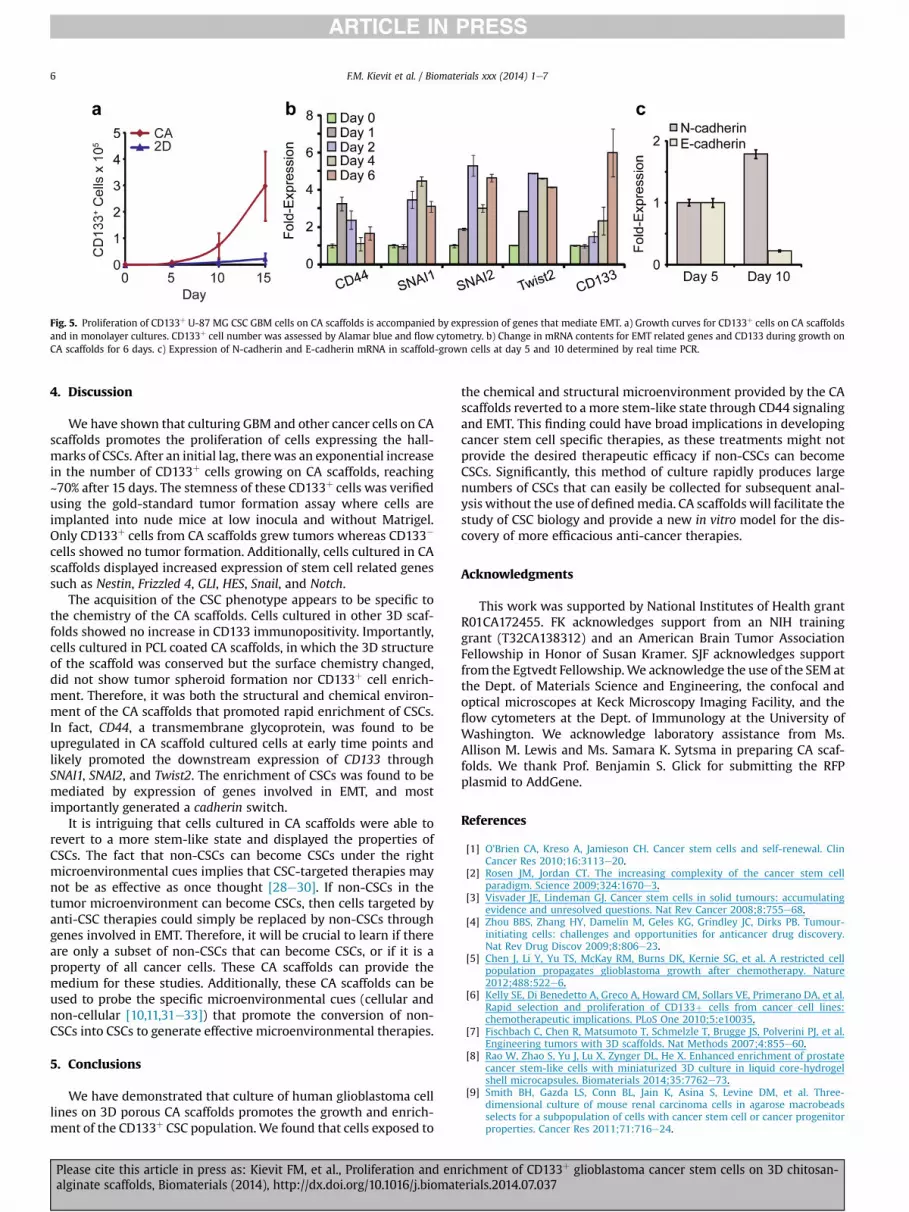
Fig. 5. Proliferation of CD133þ U-87 MG CSC GBM cells on CA scaffolds is accompanied by expression of genes that mediate EMT. a) Growth curves for CD133þ cells on CA scaffoldsand in monolayer cultures. CD133þ cell number was assessed by Alamar blue and flow cytometry. b) Change in mRNA contents for EMT related genes and CD133 during growth onCA scaffolds for 6 days. c) Expression of N-cadherin and E-cadherin mRNA in scaffold-grown cells at day 5 and 10 determined by real time PCR.
F.M. Kievit et al. / Biomaterials xxx (2014) 1e76
4. Discussion
We have shown that culturing GBM and other cancer cells on CAscaffolds promotes the proliferation of cells expressing the hall-marks of CSCs. After an initial lag, therewas an exponential increasein the number of CD133þ cells growing on CA scaffolds, reaching~70% after 15 days. The stemness of these CD133þ cells was verifiedusing the gold-standard tumor formation assay where cells areimplanted into nude mice at low inocula and without Matrigel.Only CD133þ cells from CA scaffolds grew tumors whereas CD133�
cells showed no tumor formation. Additionally, cells cultured in CAscaffolds displayed increased expression of stem cell related genessuch as Nestin, Frizzled 4, GLI, HES, Snail, and Notch.
The acquisition of the CSC phenotype appears to be specific tothe chemistry of the CA scaffolds. Cells cultured in other 3D scaf-folds showed no increase in CD133 immunopositivity. Importantly,cells cultured in PCL coated CA scaffolds, in which the 3D structureof the scaffold was conserved but the surface chemistry changed,did not show tumor spheroid formation nor CD133þ cell enrich-ment. Therefore, it was both the structural and chemical environ-ment of the CA scaffolds that promoted rapid enrichment of CSCs.In fact, CD44, a transmembrane glycoprotein, was found to beupregulated in CA scaffold cultured cells at early time points andlikely promoted the downstream expression of CD133 throughSNAI1, SNAI2, and Twist2. The enrichment of CSCs was found to bemediated by expression of genes involved in EMT, and mostimportantly generated a cadherin switch.
It is intriguing that cells cultured in CA scaffolds were able torevert to a more stem-like state and displayed the properties ofCSCs. The fact that non-CSCs can become CSCs under the rightmicroenvironmental cues implies that CSC-targeted therapies maynot be as effective as once thought [28e30]. If non-CSCs in thetumor microenvironment can become CSCs, then cells targeted byanti-CSC therapies could simply be replaced by non-CSCs throughgenes involved in EMT. Therefore, it will be crucial to learn if thereare only a subset of non-CSCs that can become CSCs, or if it is aproperty of all cancer cells. These CA scaffolds can provide themedium for these studies. Additionally, these CA scaffolds can beused to probe the specific microenvironmental cues (cellular andnon-cellular [10,11,31e33]) that promote the conversion of non-CSCs into CSCs to generate effective microenvironmental therapies.
5. Conclusions
We have demonstrated that culture of human glioblastoma celllines on 3D porous CA scaffolds promotes the growth and enrich-ment of the CD133þ CSC population.We found that cells exposed to
Please cite this article in press as: Kievit FM, et al., Proliferation and enalginate scaffolds, Biomaterials (2014), http://dx.doi.org/10.1016/j.biomat
the chemical and structural microenvironment provided by the CAscaffolds reverted to a more stem-like state through CD44 signalingand EMT. This finding could have broad implications in developingcancer stem cell specific therapies, as these treatments might notprovide the desired therapeutic efficacy if non-CSCs can becomeCSCs. Significantly, this method of culture rapidly produces largenumbers of CSCs that can easily be collected for subsequent anal-ysis without the use of definedmedia. CA scaffolds will facilitate thestudy of CSC biology and provide a new in vitro model for the dis-covery of more efficacious anti-cancer therapies.
Acknowledgments
This work was supported by National Institutes of Health grantR01CA172455. FK acknowledges support from an NIH traininggrant (T32CA138312) and an American Brain Tumor AssociationFellowship in Honor of Susan Kramer. SJF acknowledges supportfrom the Egtvedt Fellowship.We acknowledge the use of the SEM atthe Dept. of Materials Science and Engineering, the confocal andoptical microscopes at Keck Microscopy Imaging Facility, and theflow cytometers at the Dept. of Immunology at the University ofWashington. We acknowledge laboratory assistance from Ms.Allison M. Lewis and Ms. Samara K. Sytsma in preparing CA scaf-folds. We thank Prof. Benjamin S. Glick for submitting the RFPplasmid to AddGene.
References
[1] O'Brien CA, Kreso A, Jamieson CH. Cancer stem cells and self-renewal. ClinCancer Res 2010;16:3113e20.
[2] Rosen JM, Jordan CT. The increasing complexity of the cancer stem cellparadigm. Science 2009;324:1670e3.
[3] Visvader JE, Lindeman GJ. Cancer stem cells in solid tumours: accumulatingevidence and unresolved questions. Nat Rev Cancer 2008;8:755e68.
[4] Zhou BBS, Zhang HY, Damelin M, Geles KG, Grindley JC, Dirks PB. Tumour-initiating cells: challenges and opportunities for anticancer drug discovery.Nat Rev Drug Discov 2009;8:806e23.
[5] Chen J, Li Y, Yu TS, McKay RM, Burns DK, Kernie SG, et al. A restricted cellpopulation propagates glioblastoma growth after chemotherapy. Nature2012;488:522e6.
[6] Kelly SE, Di Benedetto A, Greco A, Howard CM, Sollars VE, Primerano DA, et al.Rapid selection and proliferation of CD133þ cells from cancer cell lines:chemotherapeutic implications. PLoS One 2010;5:e10035.
[7] Fischbach C, Chen R, Matsumoto T, Schmelzle T, Brugge JS, Polverini PJ, et al.Engineering tumors with 3D scaffolds. Nat Methods 2007;4:855e60.
[8] Rao W, Zhao S, Yu J, Lu X, Zynger DL, He X. Enhanced enrichment of prostatecancer stem-like cells with miniaturized 3D culture in liquid core-hydrogelshell microcapsules. Biomaterials 2014;35:7762e73.
[9] Smith BH, Gazda LS, Conn BL, Jain K, Asina S, Levine DM, et al. Three-dimensional culture of mouse renal carcinoma cells in agarose macrobeadsselects for a subpopulation of cells with cancer stem cell or cancer progenitorproperties. Cancer Res 2011;71:716e24.
richment of CD133þ glioblastoma cancer stem cells on 3D chitosan-erials.2014.07.037

F.M. Kievit et al. / Biomaterials xxx (2014) 1e7 7
[10] Kievit FM, Florczyk SJ, Leung MC, Veiseh O, Park JO, Disis ML, et al. Chitosan-alginate 3D scaffolds as a mimic of the glioma tumor microenvironment.Biomaterials 2010;31:5903e10.
[11] Leung M, Kievit FM, Florczyk SJ, Veiseh O, Wu J, Park JO, et al. Chitosan-alginate scaffold culture system for hepatocellular carcinoma increases ma-lignancy and drug resistance. Pharm Res 2010;27:1939e48.
[12] Borovski T, De Sousa EMF, Vermeulen L, Medema JP. Cancer stem cell niche:the place to be. Cancer Res 2011;71:634e9.
[13] Li Z, Leung M, Hopper R, Ellenbogen R, Zhang M. Feeder-free self-renewal ofhuman embryonic stem cells in 3D porous natural polymer scaffolds. Bio-materials 2010;31:404e12.
[14] Florczyk SJ, Kim DJ, Wood DL, Zhang M. Influence of processing parameters onpore structure of 3D porous chitosan-alginate polyelectrolyte complex scaf-folds. J Biomed Mater Res A 2011;98:614e20.
[15] Ricci-Vitiani L, Pallini R, Biffoni M, Todaro M, Invernici G, Cenci T, et al.Tumour vascularization via endothelial differentiation of glioblastoma stem-like cells. Nature 2010;468:824e8.
[16] Fan X, Khaki L, Zhu TS, Soules ME, Talsma CE, Gul N, et al. NOTCH pathwayblockade depletes CD133-positive glioblastoma cells and inhibits growth oftumor neurospheres and xenografts. Stem Cells 2010;28:5e16.
[17] Singh SK, Hawkins C, Clarke ID, Squire JA, Bayani J, Hide T, et al. Identificationof human brain tumour initiating cells. Nature 2004;432:396e401.
[18] Westphal M, Lamszus K. The neurobiology of gliomas: from cell biology tothe development of therapeutic approaches. Nat Rev Neurosci 2011;12:495e508.
[19] Jin X, Jeon HY, Joo KM, Kim JK, Jin J, Kim SH, et al. Frizzled 4 regulatesstemness and invasiveness of migrating glioma cells established by serialintracranial transplantation. Cancer Res 2011;71:3066e75.
[20] Clement V, Sanchez P, de Tribolet N, Radovanovic I, Ruiz i Altaba A. HEDGE-HOG-GLI1 signaling regulates human glioma growth, cancer stem cell self-renewal, and tumorigenicity. Curr Biol 2007;17:165e72.
[21] Rizzo P, Miao H, D'Souza G, Osipo C, Song LL, Yun J, et al. Cross-talk betweennotch and the estrogen receptor in breast cancer suggests novel therapeuticapproaches. Cancer Res 2008;68:5226e35.
Please cite this article in press as: Kievit FM, et al., Proliferation and enralginate scaffolds, Biomaterials (2014), http://dx.doi.org/10.1016/j.biomat
[22] Ikushima H, Todo T, Ino Y, Takahashi M, Miyazawa K, Miyazono K. AutocrineTGF-beta signaling maintains tumorigenicity of glioma-initiating cells throughSry-related HMG-box factors. Cell Stem Cell 2009;5:504e14.
[23] Lathia JD, Gallagher J, Myers JT, Li M, Vasanji A, McLendon RE, et al. Directin vivo evidence for tumor propagation by glioblastoma cancer stem cells.PLoS One 2011;6:e24807.
[24] Mani SA, Guo W, Liao MJ, Eaton EN, Ayyanan A, Zhou AY, et al. The epithelial-mesenchymal transition generates cells with properties of stem cells. Cell2008;133:704e15.
[25] Ouyang G, Wang Z, Fang X, Liu J, Yang CJ. Molecular signaling of the epithelialto mesenchymal transition in generating and maintaining cancer stem cells.Cell Mol Life Sci 2010;67:2605e18.
[26] Misra S, Heldin P, Hascall VC, Karamanos NK, Skandalis SS, Markwald RR, et al.Hyaluronan-CD44 interactions as potential targets for cancer therapy. Febs J2011;278:1429e43.
[27] Toole BP. Hyaluronan-CD44 interactions in cancer: paradoxes and possibil-ities. Clin Cancer Res 2009;15:7462e8.
[28] Pattabiraman DR, Weinberg RA. Tackling the cancer stem cells - what chal-lenges do they pose? Nat Rev Drug Discov 2014;13:497e512.
[29] Chaffer CL, Brueckmann I, Scheel C, Kaestli AJ, Wiggins PA, Rodrigues LO, et al.Normal and neoplastic nonstem cells can spontaneously convert to a stem-like state. Proc Natl Acad Sci U S A 2011;108:7950e5.
[30] Gupta PB, Fillmore CM, Jiang G, Shapira SD, Tao K, Kuperwasser C, et al.Stochastic state transitions give rise to phenotypic equilibrium in populationsof cancer cells. Cell 2011;146:633e44.
[31] Florczyk SJ, Liu G, Kievit FM, Lewis AM, Wu JD, Zhang M. 3D porous chitosan-alginate scaffolds: a new matrix for studying prostate cancer cell-lymphocyteinteractions in vitro. Adv Healthc Mater 2012;1:590e9.
[32] Phan-Lai V, Kievit FM, Florczyk SJ, Wang K, Disis ML, Zhang M. CCL21 andIFNgamma recruit and activate tumor specific T cells in 3D scaffold model ofbreast cancer. Anticancer Agents Med Chem 2014;14:204e10.
[33] Phan-Lai V, Florczyk SJ, Kievit FM, Wang K, Gad E, Disis ML, et al. Three-dimensional scaffolds to evaluate tumor associated fibroblast-mediated sup-pression of breast tumor specific T cells. Biomacromolecules 2013;14:1330e7.
ichment of CD133þ glioblastoma cancer stem cells on 3D chitosan-erials.2014.07.037