primary synovial sarcoma of the parotid gland in 15-year-old boy
TRANSCRIPT
Središnja medicinska knjižnica
Lukšić I., Virag M., Manojlović S., Obradović B., Macan D., Stepan J.
(2010) Primary synovial sarcoma of the parotid gland in 15-year-old
boy. Journal of Cranio-Maxillofacial Surgery, [Epub ahead of print].
ISSN 1010-5182
http://www.elsevier.com/locate/issn/10105182 http://www.sciencedirect.com/science/journal/10105182 http://dx.doi.org/10.1016/j.jcms.2010.10.010 http://medlib.mef.hr/921
University of Zagreb Medical School Repository
http://medlib.mef.hr/
Primary synovial sarcoma of the parotid gland in 15-year-old boy
Ivica Lukšić, MD, PhD1, Mišo Virag, MD, PhD, FRCS1, Spomenka Manojlović, MD,
PhD2, Bojan Obradović, MD, DMD, MSc3, Darko Macan, DMD, PhD1, Jasminka Stepan,
MD, PhD4
1Department of Maxillofacial Surgery (Head: Mišo Virag, MD, PhD, FRCS), University of
Zagreb School of Medicine and School of Dental Medicine, University Hospital Dubrava,
Zagreb, Croatia
2Department of Pathology, University of Zagreb School of Medicine, University Hospital
Dubrava, Zagreb, Croatia
3Department Chair of Maxillofacial Surgery, Medical School University of Banja Luka,
Banja Luka, Bosnia and Herzegovina
4Department of Haematology and Oncology, Paediatric Clinic, Children’s Hospital Zagreb,
Zagreb, Croatia
Correspondence:
Ivica Lukšić, MD, PhD
Department of Maxillofacial Surgery, University of Zagreb School of Medicine, University
Hospital Dubrava

Ave. Gojko Susak 6
10 000 Zagreb, Croatia
Phone: +38512903067
Fax: +385 1 286 4250
E-mail: [email protected]
1
INTRODUCTION
Synovial sarcoma is an unusual malignant tumour, derived from soft tissue mesenchymal
cells. It arises most commonly in the deep soft tissues of lower extremities, especially in the
region of the lower thigh, with a predilection for sites in proximity to large joints, such as
the knee and ankle (Shmookler et al., 1982; Miloro et al., 1994). The peak incidence is in
adolescents and young adults between 16 and 49 years of age (median 34 years), and the
tumour affects males more often than females (Shmookler et al., 1982). Synovial sarcoma
represents approximately 5-10% of all soft tissue sarcomas, only 3% of which occur in the
head and neck region (Kartha and Bumpous, 2002). The most common site is the
hypopharynx, with the larynx being the least common site (Dei Tos et al., 1998).
Primary synovial sarcoma of the parotid gland is exceptionally uncommon, with few cases
reported in the literature (Amble et al., 1992). In cases with adequate clinical
documentation, tumours present as painless masses and mostly in the parotid gland of
young and middle-aged men (Amble et al., 1992). Diagnostic methods are clinical
examination, CT or MR imaging, and histological examination. Therapy included primary
surgical wide excision with elective neck dissection. The tumour is moderately sensitive to
chemotherapy and radiotherapy, and these treatment modalities may prove useful as
adjunctive therapy (Shmookler et al., 1982). The prognosis depends on the size and grade of
the tumor (Thompson et al., 2000).
2
CASE REPORT
A 15-year-old boy with a painless swelling in the right parotid gland was previously
examined by the paediatrician and referred for further treatment. The patient's medical
history was unremarkable and his general condition was good. Clinical examination
revealed a nonmobile mass on the right side of his face, in the front of ear, about 4 cm in
diameter, of solid consistency, and with clinically negative neck. The patient did not
complain of any pain or neurological deficit in the facial region. Opening of the mouth was
not restricted and functions of the facial nerve were completely normal. Results of routine
laboratory tests were within normal limits. Cytological analyses (FNA) of the mass pointed
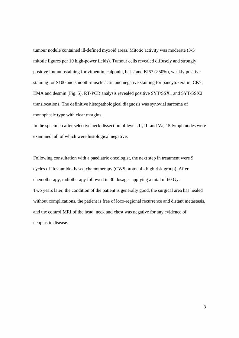
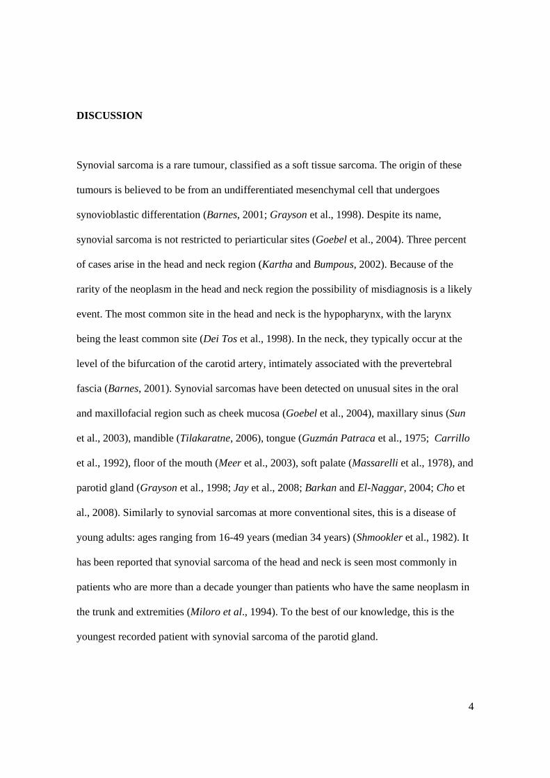
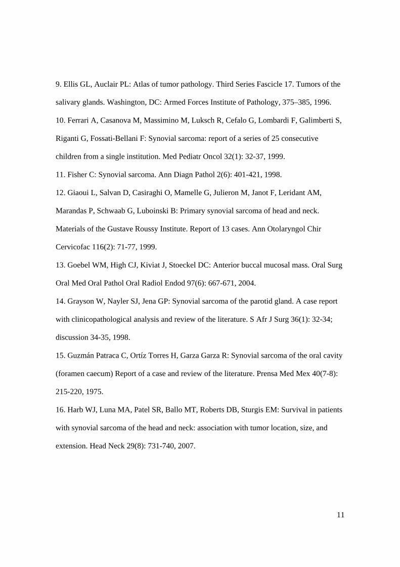
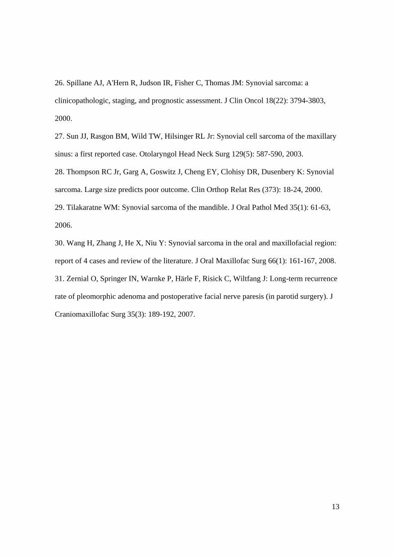
to a tumour of mesenchymal origin. MR imaging with contrast showed an expansile mass
in the deep lobe of the right parotid gland with infiltration of the superficial lobe without
evidence of metastatic lymphadenopathy (Fig. 1 and 2). The first step of therapy was a
surgical procedure, including wide excision and total parotidectomy with selective neck
dissection (levels II, III and Va).
Histopathological findings:
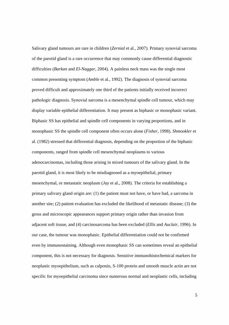
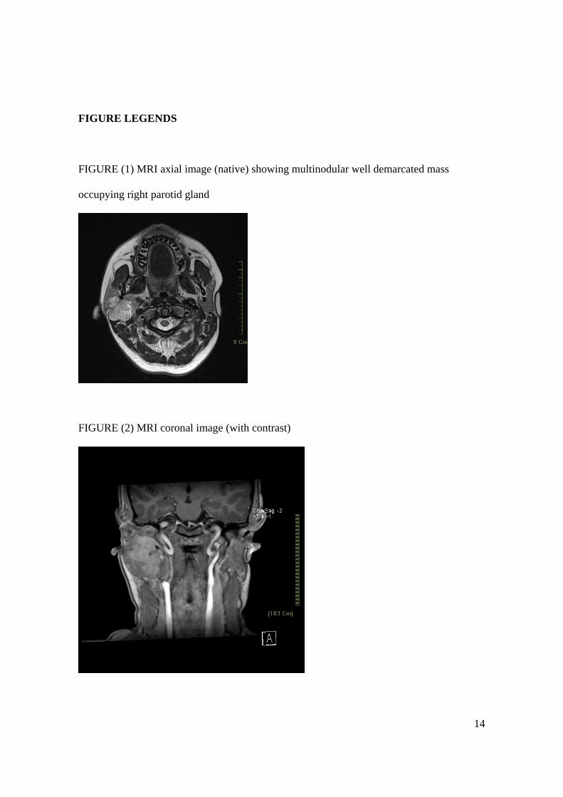
The surgical specimen consisted of the parotid gland and lymph nodes from the selective
neck dissection (Fig. 3). A multinodular, well-demarcated, rubbery, gray-white tumour,
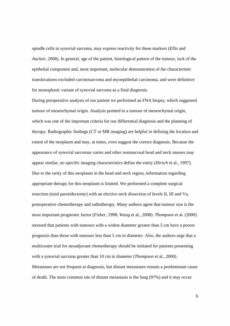
measuring 4 cm in diameter at its widest, presented within the parotid gland. Microscopic
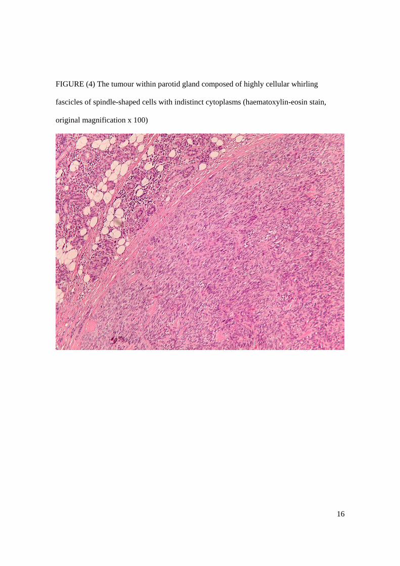
analysis revealed highly cellular whirling fascicles of spindle-shaped cells with uniform,
oval, vesicular, dark-staining nuclei and indistinct cytoplasm (Fig. 4). The cellular portions
alternated with acellular hyalinized collagen bands. The central portion of the largest
3
tumour nodule contained ill-defined myxoid areas. Mitotic activity was moderate (3-5
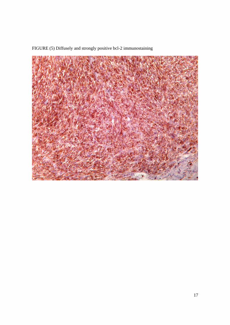
mitotic figures per 10 high-power fields). Tumour cells revealed diffusely and strongly
positive immunostaining for vimentin, calponin, bcl-2 and Ki67 (>50%), weakly positive
staining for S100 and smooth-muscle actin and negative staining for pancytokeratin, CK7,
EMA and desmin (Fig. 5). RT-PCR analysis revealed positive SYT/SSX1 and SYT/SSX2
translocations. The definitive histopathological diagnosis was synovial sarcoma of
monophasic type with clear margins.
In the specimen after selective neck dissection of levels II, III and Va, 15 lymph nodes were
examined, all of which were histological negative.
Following consultation with a paediatric oncologist, the next step in treatment were 9
cycles of ifosfamide- based chemotherapy (CWS protocol - high risk group). After
chemotherapy, radiotherapy followed in 30 dosages applying a total of 60 Gy.
Two years later, the condition of the patient is generally good, the surgical area has healed
without complications, the patient is free of loco-regional recurrence and distant metastasis,
and the control MRI of the head, neck and chest was negative for any evidence of
neoplastic disease.
4
DISCUSSION
Synovial sarcoma is a rare tumour, classified as a soft tissue sarcoma. The origin of these
tumours is believed to be from an undifferentiated mesenchymal cell that undergoes
synovioblastic differentation (Barnes, 2001; Grayson et al., 1998). Despite its name,
synovial sarcoma is not restricted to periarticular sites (Goebel et al., 2004). Three percent
of cases arise in the head and neck region (Kartha and Bumpous, 2002). Because of the
rarity of the neoplasm in the head and neck region the possibility of misdiagnosis is a likely
event. The most common site in the head and neck is the hypopharynx, with the larynx
being the least common site (Dei Tos et al., 1998). In the neck, they typically occur at the
level of the bifurcation of the carotid artery, intimately associated with the prevertebral
fascia (Barnes, 2001). Synovial sarcomas have been detected on unusual sites in the oral
and maxillofacial region such as cheek mucosa (Goebel et al., 2004), maxillary sinus (Sun
et al., 2003), mandible (Tilakaratne, 2006), tongue (Guzmán Patraca et al., 1975; Carrillo
et al., 1992), floor of the mouth (Meer et al., 2003), soft palate (Massarelli et al., 1978), and
parotid gland (Grayson et al., 1998; Jay et al., 2008; Barkan and El-Naggar, 2004; Cho et
al., 2008). Similarly to synovial sarcomas at more conventional sites, this is a disease of
young adults: ages ranging from 16-49 years (median 34 years) (Shmookler et al., 1982). It
has been reported that synovial sarcoma of the head and neck is seen most commonly in
patients who are more than a decade younger than patients who have the same neoplasm in
the trunk and extremities (Miloro et al., 1994). To the best of our knowledge, this is the
youngest recorded patient with synovial sarcoma of the parotid gland.
5
Salivary gland tumours are rare in children (Zernial et al., 2007). Primary synovial sarcoma
of the parotid gland is a rare occurrence that may commonly cause differential diagnostic
difficulties (Barkan and El-Naggar, 2004). A painless neck mass was the single most
common presenting symptom (Amble et al., 1992). The diagnosis of synovial sarcoma
proved difficult and approximately one third of the patients initially received incorrect
pathologic diagnosis. Synovial sarcoma is a mesenchymal spindle cell tumour, which may
display variable epithelial differentiation. It may present as biphasic or monophasic variant.
Biphasic SS has epithelial and spindle cell components in varying proportions, and in
monophasic SS the spindle cell component often occurs alone (Fisher, 1998). Shmookler et
al. (1982) stressed that differential diagnosis, depending on the proportion of the biphasic
components, ranged from spindle cell mesenchymal neoplasms to various
adenocarcinomas, including those arising in mixed tumours of the salivary gland. In the
parotid gland, it is most likely to be misdiagnosed as a myoepithelial, primary
mesenchymal, or metastatic neoplasm (Jay et al., 2008). The criteria for establishing a
primary salivary gland origin are: (1) the patient must not have, or have had, a sarcoma in
another site; (2) patient evaluation has excluded the likelihood of metastatic disease; (3) the
gross and microscopic appearances support primary origin rather than invasion from
adjacent soft tissue, and (4) carcinosarcoma has been excluded (Ellis and Auclair, 1996). In
our case, the tumour was monophasic. Epithelial differentiation could not be confirmed
even by immunostaining. Although even monophasic SS can sometimes reveal an epithelial
component, this is not necessary for diagnosis. Sensitive immunohistochemical markers for
neoplastic myoepithelium, such as calponin, S-100 protein and smooth muscle actin are not
specific for myoepithelial carcinoma since numerous normal and neoplastic cells, including
6
spindle cells in synovial sarcoma, may express reactivity for these markers (Ellis and
Auclair, 2008). In general, age of the patient, histological pattern of the tumour, lack of the
epithelial component and, most important, molecular demonstration of the characteristic
translocations excluded carcinosarcoma and myoepithelial carcinoma, and were definitive
for monophasic variant of synovial sarcoma as a final diagnosis.
During preoperative analysis of our patient we performed an FNA biopsy, which suggested
tumour of mesenchymal origin. Analysis pointed to a tumour of mesenchymal origin,
which was one of the important criteria for our differential diagnosis and the planning of
therapy. Radiographic findings (CT or MR imaging) are helpful in defining the location and
extent of the neoplasm and may, at times, even suggest the correct diagnosis. Because the
appearance of synovial sarcomas varies and other nonmucosal head and neck masses may
appear similar, no specific imaging characteristics define the entity (Hirsch et al., 1997).
Due to the rarity of this neoplasm in the head and neck region, information regarding
appropriate therapy for this neoplasm is limited. We performed a complete surgical
resection (total parotidectomy) with an elective neck dissection of levels II, III and Va,
postoperative chemotherapy and radiotherapy. Many authors agree that tumour size is the
most important prognostic factor (Fisher, 1998; Wang et al., 2008). Thompson et al. (2000)
stressed that patients with tumours with a widest diameter greater than 5 cm have a poorer
prognosis than those with tumours less than 5 cm in diameter. Also, the authors urge that a
multicenter trial for neoadjuvant chemotherapy should be initiated for patients presenting
with a synovial sarcoma greater than 10 cm in diameter (Thompson et al., 2000).
Metastases are not frequent at diagnosis, but distant metastases remain a predominant cause
of death. The most common site of distant metastasis is the lung (97%) and it may occur
7
years after initial therapy, with an average duration from initial treatment to detection of
30.8 months (Amble et al., 1992; Goebel et al., 2004). The article by Lee and Shin (2008)
from Memorial Sloan-Kettering Cancer Center highlights the importance of a
multidisciplinary approach in the treatment of this disease. Harb et al. (2007) stressed that
survival rates were associated with tumour location, size, and extension. Giaoui et al.
(1999) say that favourable prognostic findings include age less than 20 years and complete
initial resection. Post treatment recurrence rate for synovial sarcoma arising from all body
sites is 50%, most cases recur in the first two years after treatment (Fisher, 1998). Follow-
up of patients with synovial sarcoma of the head and neck indicate that 21-44% will
develop local recurrence and 24-48% distant metastases (especially to the lung), with an
overall 5-year survival of 47-58% (Barnes, 2001). However, some authors have shown 5
year survival rates of up to 65-70% (Ladenstein et al., 1993) while other authors emphasize
that despite a five year survival rate of 25-50%, the 10 year survival rate is only 10-15%
(Sun et al., 2003). One other report describes that prognosis for patients with synovial
sarcoma in the head and neck is better than for patients with synovial sarcoma in the trunk
and extremities because this tumour in the head and neck presents in younger patients and
is treated earlier after the onset of symptoms (Hirsch et al., 1997).
Besauese of low reported incidence there is a global lack of experience in dealing with
synovial sarcoma of the head and neck. Kartha and Bumpous (2002) stressed a more
aggressive behaviour of the synovial sarcoma of the head and neck than of the same
neoplasm in the extremities. Because of the aggressive nature of synovial sarcoma, some
modification of accepted treatment modalities may be required. Most authors agree that
adequate primary surgery is essential to both local control and overall outcome for synovial
8
sarcoma patients (Bergh et al., 1999). Some of them propose that a neck dissection is not
warranted, because only 3-4% of synovial sarcoma of head and neck are associated with
cervical lymph node metastasis (Shmookler et al., 1982; Amble et al., 1992). One other
report has shown that twenty-five percent of the cases involve regional lymph nodes
(Tilakaratne, 2006). Moreover, lymph node metastases develop more frequently than in
other nonrhabdomyosarcoma soft tissue sarcomas (Ferrari et al., 1999). In our opinion the
surgical procedure would be radical excision with some type of elective neck dissection
because at the time of operation we didn't know true histological diagnosis. Frozen section
analysis hasn't been done, because it wouldn't change the extent of surgical treatment.
However, in most instances at the time of surgery histopathological diagnosis is unknown.
The only guidance is clinical appearance and the fine needle biopsy.
A number of authors report diverse opinions concerning the value of radiation therapy and
chemotherapy in treatment of synovial sarcoma in the head and neck region (Goebel et al.,
2004; Ferrari et al., 1999). Amble et al. (1992) proposed wide local excision with
postoperative radiotherapy of 65 Gy and considered prophylactic neck dissection and
chemotherapy to be less effective. Synovial sarcoma is often chemosensitive, and given its
poor prognosis, multicenter trials of adjuvant therapy are warranted (Spillane et al., 2000).
9
CONCLUSION
The primary synovial sarcoma of the parotid gland is extremely rare, especially in children.
Two years after therapy our young patient is well with no evidence of disease. That is a
short follow-up period, but we can say that multimodal therapy with aggressive surgical
excision followed by early postoperative chemotherapy and radiotherapy can be a
promising factor.
10
REFERENCES
1. Amble FR, Olsen KD, Nascimento AG, Foote RL: Head and neck synovial cell sarcoma.
Otolaryngol Head Neck Surg 107(5): 631-637, 1992.
2. Barkan GA, El-Naggar AK: Primary synovial sarcoma of the parotid gland. Ann Diagn
Pathol 8(4): 233-236, 2004.
3. Barnes L: Tumors and tumor-like lesions of the soft tissues. In: Barnes L (ed.), Surgical
Pathology of the Head and Neck, Second Edition, Revised and Expanded (in three
volumes), Volume 2. New York, Basel: Marcel Dekker, Inc., 889-1048, 2001.
4. Bergh P, Meis-Kindblom JM, Gherlinzoni F, Berlin O, Bacchini P, Bertoni F,
Gunterberg B, Kindblom LG: Synovial sarcoma: identification of low and high risk groups.
Cancer 85(12): 2596-2607, 1999.
5. Carrillo R, el-Naggar AK, Rodriguez-Peralto JL, Batsakis JG: Synovial sarcoma of the
tongue: case report and review of the literature. J Oral Maxillofac Surg 50(8): 904-906,
1992.
6. Cho KJ, Ro JY, Choi J, Choi SH, Nam SY, Kim SY: Mesenchymal neoplasms of the
major salivary glands: clinicopathological features of 18 cases. Eur Arch Otorhinolaryngol
265 Suppl 1: S47-56, 2008.
7. Dei Tos AP, Dal Cin P, Sciot R, Furlanetto A, Da Mosto MC, Giannini C, Rinaldo A,
Ferlito A: Synovial sarcoma of the larynx and hypopharynx. Ann Otol Rhinol Laryngol
107(12): 1080-1085, 1998.
8. Ellis GL, Auclair PL: Atlas of tumor pathology. Fourth Series Fascicle 9. Tumors of the
salivary glands. Washington, DC: Armed Forces Institute of Pathology, 347–368, 2008.
11
9. Ellis GL, Auclair PL: Atlas of tumor pathology. Third Series Fascicle 17. Tumors of the
salivary glands. Washington, DC: Armed Forces Institute of Pathology, 375–385, 1996.
10. Ferrari A, Casanova M, Massimino M, Luksch R, Cefalo G, Lombardi F, Galimberti S,
Riganti G, Fossati-Bellani F: Synovial sarcoma: report of a series of 25 consecutive
children from a single institution. Med Pediatr Oncol 32(1): 32-37, 1999.
11. Fisher C: Synovial sarcoma. Ann Diagn Pathol 2(6): 401-421, 1998.
12. Giaoui L, Salvan D, Casiraghi O, Mamelle G, Julieron M, Janot F, Leridant AM,
Marandas P, Schwaab G, Luboinski B: Primary synovial sarcoma of head and neck.
Materials of the Gustave Roussy Institute. Report of 13 cases. Ann Otolaryngol Chir
Cervicofac 116(2): 71-77, 1999.
13. Goebel WM, High CJ, Kiviat J, Stoeckel DC: Anterior buccal mucosal mass. Oral Surg
Oral Med Oral Pathol Oral Radiol Endod 97(6): 667-671, 2004.
14. Grayson W, Nayler SJ, Jena GP: Synovial sarcoma of the parotid gland. A case report
with clinicopathological analysis and review of the literature. S Afr J Surg 36(1): 32-34;
discussion 34-35, 1998.
15. Guzmán Patraca C, Ortíz Torres H, Garza Garza R: Synovial sarcoma of the oral cavity
(foramen caecum) Report of a case and review of the literature. Prensa Med Mex 40(7-8):
215-220, 1975.
16. Harb WJ, Luna MA, Patel SR, Ballo MT, Roberts DB, Sturgis EM: Survival in patients
with synovial sarcoma of the head and neck: association with tumor location, size, and
extension. Head Neck 29(8): 731-740, 2007.

12
17. Hirsch RJ, Yousem DM, Loevner LA, Montone KT, Chalian AA, Hayden RE,
Weinstein GS: Synovial sarcomas of the head and neck: MR findings. AJR Am J
Roentgenol 169(4): 1185-1188, 1997.
18. Jay A, Hutchison I, Piper K, Farthing PM, Richards PS: Synovial sarcoma presenting as
a parotid mass: case report and review of literature. Head Neck 30(12): 1654-1659, 2008.
19. Kartha SS, Bumpous JM: Synovial cell sarcoma: diagnosis, treatment, and outcomes.
Laryngoscope 112(11): 1979-1982, 2002.
20. Ladenstein R, Treuner J, Koscielniak E, d'Oleire F, Keim M, Gadner H, Jürgens H,
Niethammer D, Ritter J, Schmidt D: Synovial sarcoma of childhood and adolescence.
Report of the German CWS-81 study. Cancer 71(11): 3647-3655, 1993.
21. Lee N, Shin E: Treatment outcomes for patients with synovial sarcoma of the head and
neck. Expert Rev Anticancer Ther 8(3): 371-373, 2008.
22. Massarelli G, Tanda F, Salis B: Synovial sarcoma of the soft palate: report of a case.
Hum Pathol 9(3): 341-345, 1978.
23. Meer S, Coleman H, Altini M: Oral synovial sarcoma: a report of 2 cases and a review
of the literature. Oral Surg Oral Med Oral Pathol Oral Radiol Endod 96(3): 306-315, 2003.
24. Miloro M, Quinn PD, Stewart JC: Monophasic spindle cell synovial sarcoma of the
head and neck: report of two cases a review of the literature. J Oral Maxillofac Surg 52(3):
309-313, 1994.
25. Shmookler BM, Enzinger FM, Brannon RB: Orofacial synovial sarcoma: a
clinicopathologic study of 11 new cases and review of the literature. Cancer 50(2): 269-
276, 1982.
13
26. Spillane AJ, A'Hern R, Judson IR, Fisher C, Thomas JM: Synovial sarcoma: a
clinicopathologic, staging, and prognostic assessment. J Clin Oncol 18(22): 3794-3803,
2000.
27. Sun JJ, Rasgon BM, Wild TW, Hilsinger RL Jr: Synovial cell sarcoma of the maxillary
sinus: a first reported case. Otolaryngol Head Neck Surg 129(5): 587-590, 2003.
28. Thompson RC Jr, Garg A, Goswitz J, Cheng EY, Clohisy DR, Dusenbery K: Synovial
sarcoma. Large size predicts poor outcome. Clin Orthop Relat Res (373): 18-24, 2000.
29. Tilakaratne WM: Synovial sarcoma of the mandible. J Oral Pathol Med 35(1): 61-63,
2006.
30. Wang H, Zhang J, He X, Niu Y: Synovial sarcoma in the oral and maxillofacial region:
report of 4 cases and review of the literature. J Oral Maxillofac Surg 66(1): 161-167, 2008.
31. Zernial O, Springer IN, Warnke P, Härle F, Risick C, Wiltfang J: Long-term recurrence
rate of pleomorphic adenoma and postoperative facial nerve paresis (in parotid surgery). J
Craniomaxillofac Surg 35(3): 189-192, 2007.
14
FIGURE LEGENDS
FIGURE (1) MRI axial image (native) showing multinodular well demarcated mass
occupying right parotid gland
FIGURE (2) MRI coronal image (with contrast)
15
FIGURE (3) Surgical specimen including wide excision and total parotidectomy with
selective neck dissection (levels II, III and Va)
16
FIGURE (4) The tumour within parotid gland composed of highly cellular whirling
fascicles of spindle-shaped cells with indistinct cytoplasms (haematoxylin-eosin stain,
original magnification x 100)
17
FIGURE (5) Diffusely and strongly positive bcl-2 immunostaining