predictive and prognostic analysis of pik3ca mutation in stage iii colon cancer intergroup trial
TRANSCRIPT

DOI:10.1093/jnci/djt298Advance Access publication November 14, 2013
JNCI | Articles 1789
© The Author 2013. Published by Oxford University Press. All rights reserved. For Permissions, please e-mail: [email protected].
jnci.oxfordjournals.org
Article
Predictive and Prognostic Analysis of PIK3CA Mutation in Stage iii colon cancer intergroup trialShuji Ogino, Xiaoyun Liao, Yu Imamura, Mai Yamauchi, Nadine J. McCleary, Kimmie Ng, Donna Niedzwiecki, Leonard B. Saltz, Robert J. Mayer, Renaud Whittom, Alexander Hantel, Al B. Benson III, Rex B. Mowat, Donna Spiegelman, Richard M. Goldberg, Monica M. Bertagnolli, Jeffrey A. Meyerhardt, Charles S. Fuchs; for the Alliance for Clinical Trials in Oncology
Manuscript received May 6, 2013; revised August 14, 2013; accepted August 15, 2013.
Correspondence to: Shuji Ogino, MD, PhD, MS, Department of Medical Oncology, Dana-Farber Cancer Institute, Harvard Medical School, 450 Brookline Ave, Rm M422, Boston, MA 02215 (e-mail: [email protected]).
Background Somatic mutations in PIK3CA (phosphatidylinositol-4,5-bisphosphonate 3-kinase [PI3K], catalytic subunit alpha gene) activate the PI3K-AKT signaling pathway and contribute to pathogenesis of various malignancies, including colorectal cancer.
Methods We examined associations of PIK3CA oncogene mutation with relapse, survival, and treatment efficacy in 627 stage III colon carcinoma case subjects within a randomized adjuvant chemotherapy trial (5-fluorouracil and leucovorin [FU/LV] vs irinotecan [CPT11], fluorouracil and leucovorin [IFL]; Cancer and Leukemia Group B 89803 [Alliance]). We detected PIK3CA mutation in exons 9 and 20 by polymerase chain reaction and pyrosequencing. Cox proportional hazards model was used to assess prognostic and predictive role of PIK3CA mutation, adjust-ing for clinical features and status of routine standard molecular pathology features, including KRAS and BRAF mutations and microsatellite instability (mismatch repair deficiency). All statistical tests were two-sided.
Results Compared with PIK3CA wild-type cases, overall status of PIK3CA mutation positivity or the presence of PIK3CA mutation in either exon 9 or 20 alone was not statistically significantly associated with recurrence-free, disease-free, or overall survival (log-rank P > .70; P > .40 in multivariable regression models). There was no statistically significant interaction between PIK3CA and KRAS (or BRAF) mutation status in survival analysis (Pinteraction > .18). PIK3CA mutation status did not appear to predict better or worse response to IFL therapy compared with FU/LV therapy (Pinteraction > .16).
Conclusions Overall tumor PIK3CA mutation status is not associated with stage III colon cancer prognosis. PIK3CA mutation does not appear to serve as a predictive tumor molecular biomarker for response to irinotecan-based adjuvant chemotherapy.
J Natl Cancer Inst;2013;105:1789–1798
Phosphatidylinositol-4,5-bisphosphonate 3-kinase (PI3K) can activate the AKT signaling pathway and facilitate cellular growth, proliferation, and survival (1). Activating mutations in PIK3CA (the phosphatidylinositol-4,5-bisphosphonate 3-kinase, catalytic subunit alpha gene; HGNC ID; HGNC:8975) have been found in various human malignancies, including colon cancers (2). A sub-set (10%–30%) of colorectal cancers harbor PIK3CA mutations, which have been associated with various clinical and molecular features, including proximal tumor location and KRAS mutation (3–16).
Despite many previous studies (10–24), a prognostic role of overall PIK3CA mutation status in colorectal cancer remains uncer-tain, although coexistence of PIK3CA mutations in both exons 9 and 20 may be associated with shorter survival (13). Most of these previous studies were underpowered for robust statistical analy-sis (mostly with total sample size of less than 500 and 10%–20%
frequency of PIK3CA mutation). Therefore, additional studies with a large sample size are needed.
A recent study suggests that PIK3CA mutation in colorectal cancer may serve as a molecular biomarker to predict response to aspirin therapy (25). Nonetheless, clinical utility of PIK3CA muta-tion test on colorectal cancer as a predictive tumor biomarker remains to be fully characterized (22,26–29).
We therefore conducted this study to examine prognostic and predictive roles of PIK3CA mutation in stage III colon cancer patients who enrolled in the National Cancer Institute–sponsored randomized clinical trial comparing postoperative adjuvant 5-fluoro-uracil (FU)/leucovorin (LV) with irinotecan/FU/LV (IFL) (CALGB 89803 [Alliance]) (30). Because data on postoperative treatment, per-formance status, and disease stage were carefully recorded in the trial, we could assess prognostic and predictive roles of PIK3CA muta-tions in colon cancer while controlling for potential confounding by
at Harvard L
ibrary on September 8, 2014
http://jnci.oxfordjournals.org/D
ownloaded from

Vol. 105, Issue 23 | December 4, 20131790 Articles | JNCI
those covariables and key molecular characteristics, including KRAS, BRAF, and microsatellite instability (MSI) status. It is important to consider KRAS, BRAF, and MSI status because these are now rou-tinely assessed in colorectal cancer for patient management.
MethodsStudy PopulationPatients in this study were participants in the National Cancer Institute–sponsored Cancer and Leukemia Group B (CALGB) phase III adjuvant therapy trial for stage III colon cancer compar-ing therapy with the weekly Roswell Park regimen of FU/LV with weekly bolus regimen of IFL (CALGB 89803; ClinicalTrials.gov Identifier: NCT00003835) (30). CALGB is now part of the Alliance for Clinical Trials in Oncology. Between April 1999 and May 2001, 1264 patients were enrolled. Patients in the treatment trial (and thus this companion study) were eligible if they underwent a complete surgical resection of the primary tumor within 56 days before study entry and had regional lymph node metastases but no evidence of distant metastases (ie, stage III). Considering the colorectal con-tinuum model, we included cases from cecal cancers to sigmoid cancers (31). Moreover, patients were required to have a baseline Eastern Cooperative Oncology Group performance status of 0 to 2 (ambulatory) and have adequate bone marrow, renal, and hepatic function. Cancer staging was based on Tumor Node Metastatis (TNM) classification (http://www.cancer.org). This analysis repre-sents correlative research based on a subset of patients within the trial and was limited to 627 patients for whom archived formalin-fixed paraffin-embedded tumor tissue and PIK3CA sequencing data were available. All patients signed informed consent, approved by each site’s institutional review board.
We compared baseline characteristics of the patients who were included in this study (with available PIK3CA data: n = 627) with those who were excluded from this study because of unavailability of tissue data (n = 637). We did not detect any statistically signifi-cant or substantial difference between these two groups in terms of age, sex, body mass index, family history, tumor location, extent of invasion through bowel wall (pT stage), lymph node involvement (pN stage), performance status, clinical bowel perforation, clinical bowel obstruction, or treatment arm (all P > .05). In addition, recur-rence-free (RFS), disease-free (DFS), or overall survival (OS) did not statistically significantly differ in subjects with available PIK3CA data as compared with those without PIK3CA data (multivariable hazard ratio [HR] = 1.05, 95% confidence interval [CI] = 0.87 to 1.27; multivariable HR = 1.10, 95% CI = 0.93 to 1.31; multivariable HR = 1.12, 95% CI = 0.93 to 1.36, respectively).
As part of the quality assurance program of the Alliance, mem-bers of the Audit Committee visit all participating institutions at least once every 3 years to review source documents. The auditors verify compliance with federal regulations and protocol require-ments, including those pertaining to eligibility, treatment, adverse events, tumor response, and outcome in a sample of protocols at each institution. Such on-site review of medical records was per-formed for a subgroup of 328 (26%) of the 1264 patients included in the treatment trial. Data quality was also ensured by review of data by the Alliance Statistics and Data Center and by the study chairperson following Alliance policies.
Definitions of Study EndpointsThe study endpoints were 1) RFS, defined as the time from the study enrollment to tumor recurrence or occurrence of a new pri-mary colon cancer; 2) DFS, defined as time from the study enroll-ment to cancer recurrence, occurrence of a new primary colon cancer, or death from any cause; and 3) OS, defined as the time from the study enrollment to death from any cause. For RFS, patients who died without known cancer recurrence were censored at last documented evaluation by a treating provider.
DNA Extraction From Tumor, PIK3CA, BRAF, and KRAS sequencing and MSI AnalysisTumor molecular analyses were performed blinded to patient and outcome data. DNA was extracted from paraffin-embedded colon cancer tissue (32). We marked tumor areas on hematoxylin and eosin–stained slides and dissected tumor tissue by a sterile needle. Polymerase chain reaction (PCR) and pyrosequencing targeted for mutation hotspots in PIK3CA exons 9 and 20 (13), BRAF codon 600 (33), and KRAS codons 12 and 13 were performed, as previ-ously described (32), in the laboratory at the Dana-Farber Cancer Institute. Pyrosequencing assay has been found to be more sen-sitive than Sanger sequencing and can detect approximately 5% to 10% of mutant alleles among a mixture of mutant and normal alleles (32). MSI was assessed by PCR for 10 microsatellite mark-ers (BAT25, BAT26, D17S250, D5S346, ACTC, D18S55, BAT40, D10S197, BAT34c4, and MycL) (34). Tumors with instability in 50% or more of the loci were classified as MSI-high, and those with instability in 0% to 49% of the loci were classified as microsatellite stable; and the concordance between MSI testing and immunohis-tochemistry for MLH1 or MSH2 loss was 97% (34). For 28 cases without PCR MSI results, those with loss of MLH1 or MSH2 were classified as MSI-high, and those with intact expression of MLH1 and MSH2 were classified as microsatellite stable.
Statistical AnalysesDetailed statistical methods are described in the Supplementary Methods (available online). All analyses were based on the clinical study database frozen on November 9, 2009. SAS version 9.2 (SAS Institute, Cary, NC) was used for all statistical analyses, and all P values were two-sided. Statistical significance was set at P equal to .05 for main hypothesis testing on PIK3CA status and outcome. For exploratory analyses of interactions (between PIK3CA mutation and each of the variables, including age, sex, etc.) and clinical, patho-logical, and molecular associations, we adjusted statistical signifi-cance level by Bonferroni correction to P equal to .004 (.05 divided by 13) to account for multiple hypothesis testing. Because a vast majority of participants were non-Hispanic whites, we did not per-form analyses stratified by ethnic group.
The Kaplan–Meier method was used to estimate the distribu-tion of survival time according to PIK3CA status, and the log-rank test was used to compare survival between subgroups. We used the multivariable Cox proportional hazards model to estimate survival hazard ratio according to tumor PIK3CA status. We conducted power calculations for survival analyses (Supplementary Table 1, available online). The proportionality of hazards assumption was assessed using standard survival plots and by evaluating a time-dependent variable, which was the cross-product of PIK3CA and
at Harvard L
ibrary on September 8, 2014
http://jnci.oxfordjournals.org/D
ownloaded from
JNCI | Articles 1791jnci.oxfordjournals.org
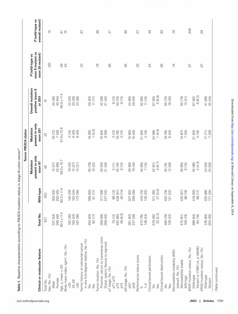
Tab
le 1
. B
asel
ine
char
acte
rist
ics
acco
rdin
g t
o PIK3CA
mu
tati
on
sta
tus
in s
tag
e III
co
lon
can
cer*
Clin
ical
or
mo
lecu
lar
feat
ure
Tota
l No.
Tum
or PIK3CA
sta
tus
P (
wild
-typ
e vs
ex
on
9 m
uta
nt
vs
exo
n 2
0 m
uta
nt)
P (
wild
-typ
e vs
ov
eral
l mu
tan
t)W
ild-t
ype
Mu
tati
on
p
rese
nt
in o
nly
ex
on
9†
Mu
tati
on
p
rese
nt
in o
nly
ex
on
20†
Ove
rall
mu
tati
on
p
osi
tive
(ex
on
9
or
20)‡
Tota
l No.
627
553
4825
74S
ex, N
o. (%
).0
01.1
5
Mal
e33
7 (5
4)30
3 (5
5)15
(31)
18 (7
2)34
(46)
Fe
mal
e29
0 (4
6)25
0 (4
5)33
(69)
7 (2
8)40
(54)
Age
, y, m
ean
± S
D60
.2 ±
11.4
60.2
± 11
.459
.0 ±
10.
761
.9 ±
12.
660
.0 ±
11.4
.59
.91
Bod
y m
ass
inde
x, k
g/m
2 , N
o. (%
).2
4.7
4
<25
162
(30)
140
(29)
19 (4
2)3
(15)
22 (3
3)
25–2
918
3 (3
4)16
3 (3
4)12
(27)
8 (4
0)20
(30)
≥3
019
7 (3
6)17
3 (3
6)14
(31)
9 (4
5)24
(36)
Fam
ily h
isto
ry o
f co
lore
ctal
can
cer
in
any
firs
t-de
gree
rel
ativ
e, N
o. (%
).2
3.9
7
N
o45
4 (8
3)39
9 (8
3)35
(78)
19 (9
5)55
(83)
Ye
s92
(17)
81 (1
7)10
(22)
1 (5
.0)
11 (1
7)Tu
mor
loca
tion,
No.
(%)
.78
.90
Pr
oxim
al, c
ecum
to
tran
sver
se c
olon
359
(58)
317
(58)
26 (5
5)16
(64)
42 (5
8)
Dis
tal,
sple
nic
flexu
re t
o si
gmoi
d25
8 (4
2)22
7 (4
2)21
(44)
9 (3
6)31
(42)
pT s
tage
, No.
(%)
.65
.31
pT
1–pT
275
(12)
66 (1
2)5
(11)
3 (1
2)9
(13)
pT
349
2 (8
0)43
8 (8
1)35
(76)
19 (7
6)54
(75)
pT
449
(8.0
)40
(7.4
)6
(13)
3 (1
2)9
(13)
pN s
tage
, No.
(%)
.94
.80
pN
138
1 (6
2)33
7 (6
2)28
(60)
15 (6
0)44
(60)
pN
223
7 (3
8)20
8 (3
8)19
(40)
10 (4
0)29
(40)
Perf
orm
ance
sta
tus
scor
e.2
0.0
7
047
2 (7
6)41
0 (7
5)40
(85)
21 (8
4)62
(85)
1–
214
6 (2
4)13
5 (2
5)7
(15)
4 (1
6)11
(15)
Clin
ical
bow
el p
erfo
ratio
n.5
4.4
9
No
578
(95)
511
(95)
42 (9
1)24
(96)
67 (9
3)
Yes
32 (5
.3)
27 (5
.0)
4 (8
.7)
1 (4
.0)
5 (6
.9)
Clin
ical
bow
el o
bstr
uctio
n.8
5.6
3
No
480
(78)
425
(78)
35 (7
4)19
(76)
55 (7
5)
Yes
139
(22)
121
(22)
12 (2
6)6
(24)
18 (2
5)M
icro
sate
llite
inst
abili
ty (M
SI)
st
atus
§, N
o. (%
).1
9.7
0
M
icro
sate
llite
sta
ble
470
(81)
420
(81)
35 (8
5)14
(67)
50 (7
9)
MS
I-hig
h10
9 (1
9)96
(19)
6 (1
5)7
(33)
13 (2
1)B
RA
F m
utat
ion
stat
us, N
o. (%
).0
7.0
48
Wild
-typ
e48
6 (8
4)41
9 (8
3)45
(96)
21 (8
4)67
(92)
M
utan
t (c
.179
9T>
A, p
.V60
0E)
94 (1
6)88
(17)
2 (4
.3)
4 (1
6)6
(8.2
)KR
AS
mut
atio
n st
atus
, No.
(%)
.07
.09
W
ild-t
ype
376
(65)
335
(66)
24 (5
0)17
(71)
41 (5
6)
Mut
ant
203
(35)
171
(34)
24 (5
0)7
(29)
32 (4
4)
(Tab
le c
ontin
ues)
at Harvard L
ibrary on September 8, 2014
http://jnci.oxfordjournals.org/D
ownloaded from

Vol. 105, Issue 23 | December 4, 20131792 Articles | JNCI
survival time (P = .46 for RFS; P = .76 for DFS; P = .35 for OS). To assess the potential differential effect of treatment arm according to PIK3CA status, we performed a single multivariable Cox regres-sion analysis, in which we could estimate the effect of treatment arm simultaneously in two strata of PIK3CA status, using a repa-rameterization of the interaction term(s) (35). Interaction was also assessed by including the cross-product of PIK3CA and another variable of interest (without data-missing cases) in a multivariable model using the Wald test.
resultsPIK3CA Mutation and Patient Survival in Stage III Colon CancerStudy participants were drawn from a multicenter study of post-operative adjuvant chemotherapy in stage III colon cancer patients who underwent a curative-intent surgical resection (CALGB 89803) (30). We included 627 case subjects in this study based on availability of tumor tissue for sequencing of PIK3CA exons 9 and 20, which detected mutation in 74 (12%) patients. One case sub-ject showed mutations in both exons 9 and 20. Table 1 summa-rizes baseline characteristics according to PIK3CA mutation status. Overall prevalence of postdiagnosis regular aspirin use (defined as two or more tablets per week) was 7.2% (n = 45 of 627 patients), and there were only four patients who had PIK3CA-mutated tumor and regularly used aspirin after colon cancer diagnosis.
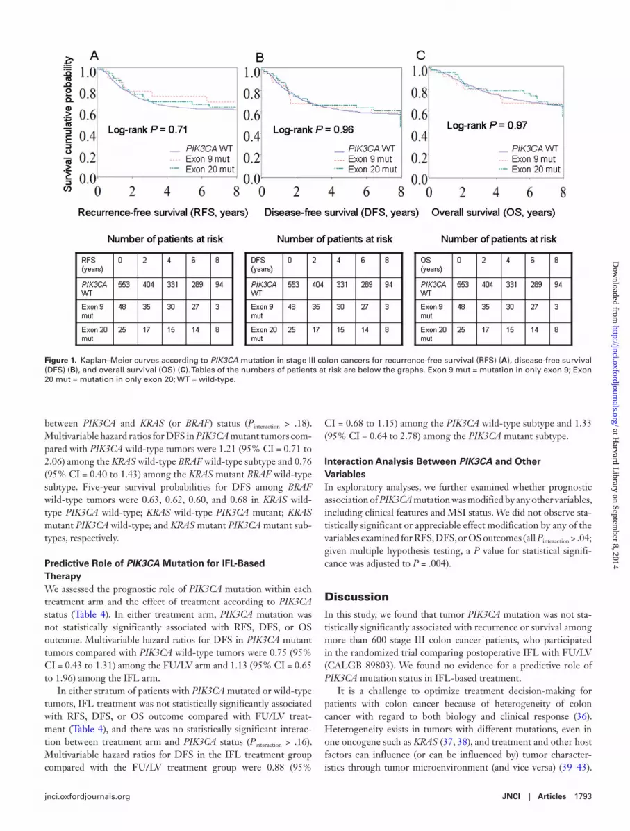
With median follow-up of 7.6 (interquartile range = 7.1–8.1) years among those who were censored for overall survival outcome, there were 225 events for RFS analysis, 258 events for DFS analy-sis, and 210 events for OS analysis.
In a Kaplan–Meier analysis (Figure 1), compared with PIK3CA wild-type patients, PIK3CA mutation in either exon 9 or 20 was not statistically significantly associated with RFS, DFS, or OS outcome (log-rank P > .70).
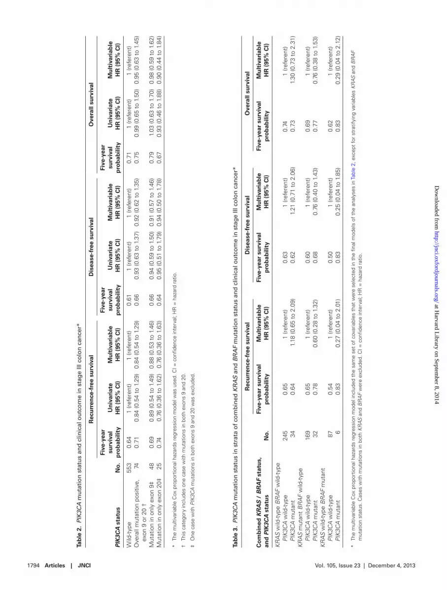
In multivariable Cox regression analysis, we examined the prog-nostic association of PIK3CA mutation adjusting for other predic-tors of patient survival (Table 2; Supplementary Table 2, available online, shows data on all variables in the final multivariable mod-els). Compared with PIK3CA wild-type cases, PIK3CA mutation in only exon 9, or only exon 20, or overall PIK3CA mutation sta-tus was not statistically significantly associated with RFS, DFS, or OS outcome in univariate or multivariable analysis (P > .40 in multivariable regression models). Multivariable hazard ratios for RFS, DFS, and OS in overall PIK3CA mutated tumors compared with PIK3CA wild-type tumors were 0.84 (95% CI = 0.54 to 1.29), 0.92 (95% CI = 0.62 to 1.35), and 0.95 (95% CI = 0.63 to 1.45), respectively.
PIK3CA Mutation and Prognosis in Strata of Combined KRAS and BRAF StatusWe assessed a prognostic role of PIK3CA mutation in strata of com-bined KRAS and BRAF status to examine possible effect modifica-tion by KRAS or BRAF status on PIK3CA mutation (Table 3). We did not observe statistically significant effect modification by KRAS status. The number (n = 6) of cases with both PIK3CA and BRAF mutations precluded robust assessment of effect modification by BRAF status. There was no statistically significant interaction C
linic
al o
r m
ole
cula
r fe
atu
reTo
tal N
o.
Tum
or PIK3CA
sta
tus
P (
wild
-typ
e vs
ex
on
9 m
uta
nt
vs
exo
n 2
0 m
uta
nt)
P (
wild
-typ
e vs
ov
eral
l mu
tan
t)W
ild-t
ype
Mu
tati
on
p
rese
nt
in o
nly
ex
on
9†
Mu
tati
on
p
rese
nt
in o
nly
ex
on
20†
Ove
rall
mu
tati
on
p
osi
tive
(ex
on
9
or
20)‡
Trea
tmen
t ar
m, N
o. (%
).7
1.8
7
FU/L
V31
9 (5
1)28
2 (5
1)26
(54)
11 (4
4)37
(50)
IF
L30
8 (4
9)27
1 (4
9)22
(46)
14 (5
6)37
(50)
* Th
e pe
rcen
tage
s in
dica
te t
he p
ropo
rtio
n of
tum
ors
with
a s
peci
fic c
linic
al o
r m
olec
ular
feat
ure
amon
g tu
mor
s w
ith s
peci
fic P
IK3C
A s
tatu
s. T
here
wer
e ca
ses
with
mis
sing
val
ue/s
tatu
s fo
r so
me
of t
he v
aria
bles
. A χ
2 te
st w
as u
sed
to a
sses
s as
soci
atio
ns, a
nd a
ll P
valu
es w
ere
two-
side
d. F
U/L
V =
5-f
luor
oura
cil a
nd le
ucov
orin
; IFL
= ir
inot
ecan
, 5-f
luor
oura
cil a
nd le
ucov
orin
; SD
= s
tand
ard
devi
atio
n.
† O
ne c
ase
subj
ect
with
PIK
3CA
mut
atio
ns in
bot
h ex
ons
9 an
d 20
was
exc
lude
d.
‡ Th
is c
ateg
ory
incl
udes
one
cas
e su
bjec
t w
ith m
utat
ions
in b
oth
exon
s 9
and
20.
§ Fo
r 29
cas
es w
ithou
t M
SI r
esul
ts b
y po
lym
eras
e ch
ain
reac
tion,
tho
se w
ith lo
ss o
f M
LH1
or M
SH
2 w
ere
clas
sifie
d as
MS
I-hig
h, a
nd t
hose
with
inta
ct e
xpre
ssio
n of
MLH
1 an
d M
SH
2 as
mic
rosa
telli
te s
tabl
e be
caus
e co
ncor
danc
e be
twee
n M
SI p
olym
eras
e ch
ain
reac
tion
and
imm
unoh
isto
chem
istr
y fo
r M
LH1
and
MS
H2
was
ver
y hi
gh (9
7%) a
mon
g ca
se s
ubje
cts
with
bot
h re
sults
ava
ilabl
e (3
4).
Tab
le 1
(C
on
tin
ued
).
at Harvard L
ibrary on September 8, 2014
http://jnci.oxfordjournals.org/D
ownloaded from
JNCI | Articles 1793jnci.oxfordjournals.org
between PIK3CA and KRAS (or BRAF) status (Pinteraction > .18). Multivariable hazard ratios for DFS in PIK3CA mutant tumors com-pared with PIK3CA wild-type tumors were 1.21 (95% CI = 0.71 to 2.06) among the KRAS wild-type BRAF wild-type subtype and 0.76 (95% CI = 0.40 to 1.43) among the KRAS mutant BRAF wild-type subtype. Five-year survival probabilities for DFS among BRAF wild-type tumors were 0.63, 0.62, 0.60, and 0.68 in KRAS wild-type PIK3CA wild-type; KRAS wild-type PIK3CA mutant; KRAS mutant PIK3CA wild-type; and KRAS mutant PIK3CA mutant sub-types, respectively.
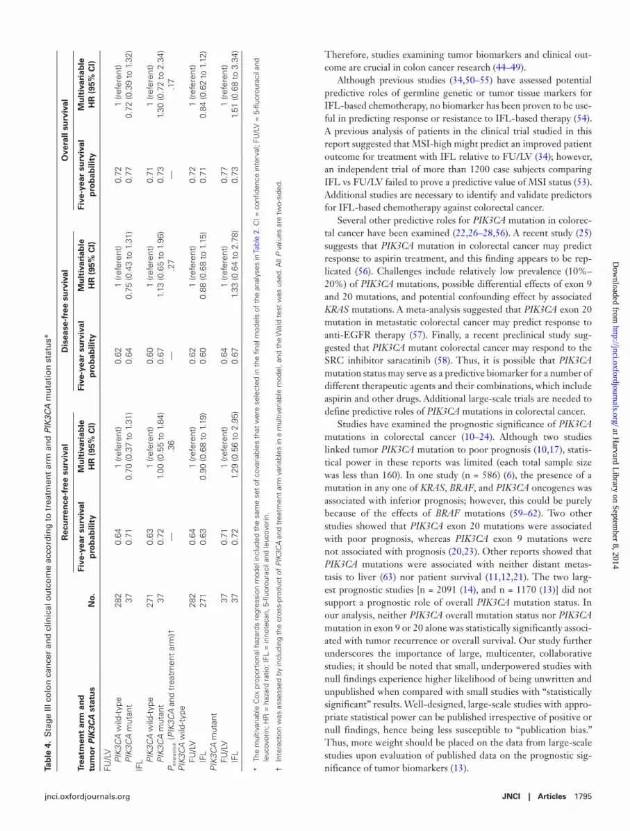
Predictive Role of PIK3CA Mutation for IFL-Based TherapyWe assessed the prognostic role of PIK3CA mutation within each treatment arm and the effect of treatment according to PIK3CA status (Table 4). In either treatment arm, PIK3CA mutation was not statistically significantly associated with RFS, DFS, or OS outcome. Multivariable hazard ratios for DFS in PIK3CA mutant tumors compared with PIK3CA wild-type tumors were 0.75 (95% CI = 0.43 to 1.31) among the FU/LV arm and 1.13 (95% CI = 0.65 to 1.96) among the IFL arm.
In either stratum of patients with PIK3CA mutated or wild-type tumors, IFL treatment was not statistically significantly associated with RFS, DFS, or OS outcome compared with FU/LV treat-ment (Table 4), and there was no statistically significant interac-tion between treatment arm and PIK3CA status (Pinteraction > .16). Multivariable hazard ratios for DFS in the IFL treatment group compared with the FU/LV treatment group were 0.88 (95%
CI = 0.68 to 1.15) among the PIK3CA wild-type subtype and 1.33 (95% CI = 0.64 to 2.78) among the PIK3CA mutant subtype.
Interaction Analysis Between PIK3CA and Other VariablesIn exploratory analyses, we further examined whether prognostic association of PIK3CA mutation was modified by any other variables, including clinical features and MSI status. We did not observe sta-tistically significant or appreciable effect modification by any of the variables examined for RFS, DFS, or OS outcomes (all Pinteraction > .04; given multiple hypothesis testing, a P value for statistical signifi-cance was adjusted to P = .004).
DiscussionIn this study, we found that tumor PIK3CA mutation was not sta-tistically significantly associated with recurrence or survival among more than 600 stage III colon cancer patients, who participated in the randomized trial comparing postoperative IFL with FU/LV (CALGB 89803). We found no evidence for a predictive role of PIK3CA mutation status in IFL-based treatment.
It is a challenge to optimize treatment decision-making for patients with colon cancer because of heterogeneity of colon cancer with regard to both biology and clinical response (36). Heterogeneity exists in tumors with different mutations, even in one oncogene such as KRAS (37, 38), and treatment and other host factors can influence (or can be influenced by) tumor character-istics through tumor microenvironment (and vice versa) (39–43).
Figure 1. Kaplan–Meier curves according to PIK3CA mutation in stage III colon cancers for recurrence-free survival (RFS) (A), disease-free survival (DFS) (B), and overall survival (OS) (C). Tables of the numbers of patients at risk are below the graphs. Exon 9 mut = mutation in only exon 9; Exon 20 mut = mutation in only exon 20; WT = wild-type.
at Harvard L
ibrary on September 8, 2014
http://jnci.oxfordjournals.org/D
ownloaded from
Vol. 105, Issue 23 | December 4, 20131794 Articles | JNCI
Tab
le 3
. PIK3CA
mu
tati
on
sta
tus
in s
trat
a o
f co
mb
ined
KRAS
an
d BRAF
mu
tati
on
sta
tus
and
clin
ical
ou
tco
me
in s
tag
e III
co
lon
can
cer*
Co
mb
ined
KRAS
/ BRAF
stat
us,
an
d PIK3CA
sta
tus
No.
Rec
urr
ence
-fre
e su
rviv
alD
isea
se-f
ree
surv
ival
Ove
rall
surv
ival
Five
-yea
r su
rviv
al
pro
bab
ility
Mu
ltiv
aria
ble
H
R (
95%
CI)
Five
-yea
r su
rviv
al
pro
bab
ility
Mu
ltiv
aria
ble
H
R (
95%
CI)
Five
-yea
r su
rviv
al
pro
bab
ility
Mu
ltiv
aria
ble
H
R (
95%
CI)
KRA
S w
ild-t
ype
BR
AF
wild
-typ
e
PIK3
CA
wild
-typ
e24
50.
651
(ref
eren
t)0.
631
(ref
eren
t)0.
741
(ref
eren
t)
PIK3
CA
mut
ant
340.
641.
18 (0
.65
to 2
.09)
0.62
1.21
(0.7
1 to
2.0
6)0.
731.
30 (0
.73
to 2
.31)
KRA
S m
utan
t B
RA
F w
ild-t
ype
PI
K3C
A w
ild-t
ype
169
0.65
1 (r
efer
ent)
0.60
1 (r
efer
ent)
0.69
1 (r
efer
ent)
PI
K3C
A m
utan
t32
0.78
0.60
(0.2
8 to
1.3
2)0.
680.
76 (0
.40
to 1
.43)
0.77
0.76
(0.3
8 to
1.5
3)KR
AS
wild
-typ
e B
RA
F m
utan
t
PIK3
CA
wild
-typ
e87
0.54
1 (r
efer
ent)
0.50
1 (r
efer
ent)
0.62
1 (r
efer
ent)
PI
K3C
A m
utan
t6
0.83
0.27
(0.0
4 to
2.0
1)0.
830.
25 (0
.04
to 1
.85)
0.83
0.29
(0.0
4 to
2.1
2)
* Th
e m
ultiv
aria
ble
Cox
pro
port
iona
l haz
ards
reg
ress
ion
mod
el in
clud
ed t
he s
ame
set
of c
ovar
iabl
es t
hat
wer
e se
lect
ed in
the
fin
al m
odel
s of
the
ana
lyse
s in
Tab
le 2
, exc
ept
for
stra
tifyi
ng v
aria
bles
KR
AS
and
BR
AF
mut
atio
n st
atus
. Cas
es w
ith m
utat
ions
in b
oth
KRA
S an
d B
RA
F w
ere
excl
uded
. CI =
con
fiden
ce in
terv
al; H
R =
haz
ard
ratio
.
Tab
le 2
. PIK3CA
mu
tati
on
sta
tus
and
clin
ical
ou
tco
me
in s
tag
e III
co
lon
can
cer*
PIK3CA
sta
tus
No.
Rec
urr
ence
-fre
e su
rviv
alD
isea
se-f
ree
surv
ival
Ove
rall
surv
ival
Five
-yea
r su
rviv
al
pro
bab
ility
Un
ivar
iate
H
R (
95%
CI)
Mu
ltiv
aria
ble
H
R (
95%
CI)
Five
-yea
r su
rviv
al
pro
bab
ility
Un
ivar
iate
H
R (
95%
CI)
Mu
ltiv
aria
ble
H
R (
95%
CI)
Five
-yea
r su
rviv
al
pro
bab
ility
Un
ivar
iate
H
R (
95%
CI)
Mu
ltiv
aria
ble
H
R (
95%
CI)
Wild
-typ
e55
30.
641
(ref
eren
t)1
(ref
eren
t)0.
611
(ref
eren
t)1
(ref
eren
t)0.
711
(ref
eren
t)1
(ref
eren
t)O
vera
ll m
utat
ion
posi
tive,
ex
on 9
or
20 †
740.
710.
84 (0
.54
to 1
.29)
0.84
(0.5
4 to
1.2
9)0.
660.
93 (0
.63
to 1
.37)
0.92
(0.6
2 to
1.3
5)0.
750.
99 (0
.65
to 1
.50)
0.95
(0.6
3 to
1.4
5)
Mut
atio
n in
onl
y ex
on 9
‡48
0.69
0.89
(0.5
4 to
1.4
9)0.
88 (0
.53
to 1
.46)
0.66
0.94
(0.5
9 to
1.5
0)0.
91 (0
.57
to 1
.46)
0.79
1.03
(0.6
3 to
1.7
0)0.
98 (0
.59
to 1
.62)
Mut
atio
n in
onl
y ex
on 2
0‡25
0.74
0.76
(0.3
6 to
1.6
2)0.
76 (0
.36
to 1
.63)
0.64
0.95
(0.5
1 to
1.7
9)0.
94 (0
.50
to 1
.78)
0.67
0.93
(0.4
6 to
1.8
8)0.
90 (0
.44
to 1
.84)
* Th
e m
ultiv
aria
ble
Cox
pro
port
iona
l haz
ards
reg
ress
ion
mod
el w
as u
sed.
CI =
con
fiden
ce in
terv
al; H
R =
haz
ard
ratio
.
† Th
is c
ateg
ory
incl
udes
one
cas
e w
ith m
utat
ions
in b
oth
exon
s 9
and
20.
‡ O
ne c
ase
with
PIK
3CA
mut
atio
ns in
bot
h ex
ons
9 an
d 20
was
exc
lude
d.
at Harvard L
ibrary on September 8, 2014
http://jnci.oxfordjournals.org/D
ownloaded from
JNCI | Articles 1795jnci.oxfordjournals.org
Therefore, studies examining tumor biomarkers and clinical out-come are crucial in colon cancer research (44–49).
Although previous studies (34,50–55) have assessed potential predictive roles of germline genetic or tumor tissue markers for IFL-based chemotherapy, no biomarker has been proven to be use-ful in predicting response or resistance to IFL-based therapy (54). A previous analysis of patients in the clinical trial studied in this report suggested that MSI-high might predict an improved patient outcome for treatment with IFL relative to FU/LV (34); however, an independent trial of more than 1200 case subjects comparing IFL vs FU/LV failed to prove a predictive value of MSI status (53). Additional studies are necessary to identify and validate predictors for IFL-based chemotherapy against colorectal cancer.
Several other predictive roles for PIK3CA mutation in colorec-tal cancer have been examined (22,26–28,56). A recent study (25) suggests that PIK3CA mutation in colorectal cancer may predict response to aspirin treatment, and this finding appears to be rep-licated (56). Challenges include relatively low prevalence (10%–20%) of PIK3CA mutations, possible differential effects of exon 9 and 20 mutations, and potential confounding effect by associated KRAS mutations. A meta-analysis suggested that PIK3CA exon 20 mutation in metastatic colorectal cancer may predict response to anti-EGFR therapy (57). Finally, a recent preclinical study sug-gested that PIK3CA mutant colorectal cancer may respond to the SRC inhibitor saracatinib (58). Thus, it is possible that PIK3CA mutation status may serve as a predictive biomarker for a number of different therapeutic agents and their combinations, which include aspirin and other drugs. Additional large-scale trials are needed to define predictive roles of PIK3CA mutations in colorectal cancer.
Studies have examined the prognostic significance of PIK3CA mutations in colorectal cancer (10–24). Although two studies linked tumor PIK3CA mutation to poor prognosis (10,17), statis-tical power in these reports was limited (each total sample size was less than 160). In one study (n = 586) (6), the presence of a mutation in any one of KRAS, BRAF, and PIK3CA oncogenes was associated with inferior prognosis; however, this could be purely because of the effects of BRAF mutations (59–62). Two other studies showed that PIK3CA exon 20 mutations were associated with poor prognosis, whereas PIK3CA exon 9 mutations were not associated with prognosis (20,23). Other reports showed that PIK3CA mutations were associated with neither distant metas-tasis to liver (63) nor patient survival (11,12,21). The two larg-est prognostic studies [n = 2091 (14), and n = 1170 (13)] did not support a prognostic role of overall PIK3CA mutation status. In our analysis, neither PIK3CA overall mutation status nor PIK3CA mutation in exon 9 or 20 alone was statistically significantly associ-ated with tumor recurrence or overall survival. Our study further underscores the importance of large, multicenter, collaborative studies; it should be noted that small, underpowered studies with null findings experience higher likelihood of being unwritten and unpublished when compared with small studies with “statistically significant” results. Well-designed, large-scale studies with appro-priate statistical power can be published irrespective of positive or null findings, hence being less susceptible to “publication bias.” Thus, more weight should be placed on the data from large-scale studies upon evaluation of published data on the prognostic sig-nificance of tumor biomarkers (13).Ta
ble
4.
Sta
ge
III c
olo
n c
ance
r an
d c
linic
al o
utc
om
e ac
cord
ing
to
tre
atm
ent
arm
an
d PIK3CA
mu
tati
on
sta
tus*
Trea
tmen
t ar
m a
nd
tu
mo
r PIK3CA
sta
tus
No.
Rec
urr
ence
-fre
e su
rviv
alD
isea
se-f
ree
surv
ival
Ove
rall
surv
ival
Five
-yea
r su
rviv
al
pro
bab
ility
Mu
ltiv
aria
ble
H
R (
95%
CI)
Five
-yea
r su
rviv
al
pro
bab
ility
Mu
ltiv
aria
ble
H
R (
95%
CI)
Five
-yea
r su
rviv
al
pro
bab
ility
Mu
ltiv
aria
ble
H
R (
95%
CI)
FU/L
V
PIK3
CA
wild
-typ
e28
20.
641
(ref
eren
t)0.
621
(ref
eren
t)0.
721
(ref
eren
t)
PIK3
CA
mut
ant
370.
710.
70 (0
.37
to 1
.31)
0.64
0.75
(0.4
3 to
1.3
1)0.
770.
72 (0
.39
to 1
.32)
IFL
PI
K3C
A w
ild-t
ype
271
0.63
1 (r
efer
ent)
0.60
1 (r
efer
ent)
0.71
1 (r
efer
ent)
PI
K3C
A m
utan
t37
0.72
1.00
(0.5
5 to
1.8
4)0.
671.
13 (0
.65
to 1
.96)
0.73
1.30
(0.7
2 to
2.3
4)P i
nter
actio
n (P
IK3C
A a
nd t
reat
men
t ar
m)†
—.3
6—
.27
—.1
7PI
K3C
A w
ild-t
ype
FU
/LV
282
0.64
1 (r
efer
ent)
0.62
1 (r
efer
ent)
0.72
1 (r
efer
ent)
IF
L27
10.
630.
90 (0
.68
to 1
.19)
0.60
0.88
(0.6
8 to
1.1
5)0.
710.
84 (0
.62
to 1
.12)
PIK3
CA
mut
ant
FU
/LV
370.
711
(ref
eren
t)0.
641
(ref
eren
t)0.
771
(ref
eren
t)
IFL
370.
721.
29 (0
.56
to 2
.95)
0.67
1.33
(0.6
4 to
2.7
8)0.
731.
51 (0
.68
to 3
.34)
* Th
e m
ultiv
aria
ble
Cox
pro
port
iona
l haz
ards
reg
ress
ion
mod
el in
clud
ed t
he s
ame
set
of c
ovar
iabl
es t
hat
wer
e se
lect
ed in
the
fin
al m
odel
s of
the
ana
lyse
s in
Tab
le 2
. CI =
con
fiden
ce in
terv
al; F
U/L
V =
5-f
luor
oura
cil a
nd
leuc
ovor
in; H
R =
haz
ard
ratio
; IFL
= ir
inot
ecan
, 5-f
luor
oura
cil a
nd le
ucov
orin
.
† In
tera
ctio
n w
as a
sses
sed
by in
clud
ing
the
cros
s-pr
oduc
t of
PIK
3CA
and
tre
atm
ent
arm
var
iabl
es in
a m
ultiv
aria
ble
mod
el, a
nd t
he W
ald
test
was
use
d. A
ll P
valu
es a
re t
wo-
side
d.
at Harvard L
ibrary on September 8, 2014
http://jnci.oxfordjournals.org/D
ownloaded from

Vol. 105, Issue 23 | December 4, 20131796 Articles | JNCI
Interestingly, Liao et al. (13) demonstrated that the coexist-ence of PIK3CA mutations in both exons 9 and 20 was associated with shorter survival. Experimental evidence suggests that PIK3CA helical and kinase domain mutations differentially activate protein function and that the presence of PIK3CA mutations in both exon 9 and exon 20 results in a synergistic gain of enzymatic function (64). However, in our analysis, there was only one case subject with PIK3CA mutations in both exons 9 and 20, which precluded robust outcome assessment.
A ligand–receptor interaction of EGFR (HGNC ID: HGNC:3236) leads to activation of two main signal transduction pathways, RAS-RAF-MAPK and PI3K-AKT. Activating mutations in KRAS, BRAF, and/or PIK3CA are well-known carcinogenic mechanisms, and there may be interactive effects of KRAS and PIK3CA mutations (65). In our study on colon cancer, we did not observe statistically significant interactive effects of PIK3CA and KRAS (or BRAF) mutations.
This study used the multi-institutional clinical trial of adjuvant chemotherapy and had several strengths. All study subjects were stage III cancer patients, which decreased potential residual con-founding by disease stage. Methods of follow-up and treatment were standardized, and the date and nature of recurrence were recorded. Furthermore, integrative database of treatment, behav-ioral and lifestyle factors, tumor molecular characteristics (includ-ing PIK3CA, KRAS, BRAF, and MSI status), and clinical outcomes enable molecular pathological epidemiology research (66,67) and controlling for confounding by lifestyle factors. The paradigm of molecular pathological epidemiology has been widely used (68–76). In colorectal cancer, KRAS, BRAF, and MSI tests are a part of routine clinical practice (77), and PIK3CA test is an emerging clinical test (25,29,56).
We recognize limitations of our study. Patients who enrolled in the clinical trials constituted a selected group of individuals and might differ from the general population. Patients needed to meet enrollment criteria and be motivated to participate and were further selected based on availability of tissue specimens. Nonetheless, demographic, clinical, or prognostic data of the patients selected in this study did not substantially differ from those without available tumor tissue. Because this trial included patients from both academic and community hospitals across the United States and Canada, our results might reflect stage III colon cancers in the general North American population. Finally, because PIK3CA status was not available on all patients, statisti-cal power was attenuated, especially for predictive assessment for response to IFL use.
In conclusion, our current study of stage III colon cancer patients has shown that tumor PIK3CA mutation is not statisti-cally significantly associated with recurrence or survival and that PIK3CA mutation status is not a predictive marker for response to IFL-based chemotherapy. Additional large-scale studies are needed to define predictive roles of PIK3CA mutations in colon cancer.
references 1. Samuels Y, Diaz LA Jr, Schmidt-Kittler O, et al. Mutant PIK3CA pro-
motes cell growth and invasion of human cancer cells. Cancer Cell. 2005;7(6):561–573.
2. Samuels Y, Wang Z, Bardelli A, et al. High frequency of mutations of the PIK3CA gene in human cancers. Science. 2004;304(5670):554.
3. Velho S, Oliveira C, Ferreira A, et al. The prevalence of PIK3CA mutations in gastric and colon cancer. Eur J Cancer. 2005;41(11):1649–1654.
4. Baldus SE, Schaefer KL, Engers R, Hartleb D, Stoecklein NH, Gabbert HE. Prevalence and heterogeneity of KRAS, BRAF, and PIK3CA mutations in primary colorectal adenocarcinomas and their corresponding metasta-ses. Clin Cancer Res. 2010;16(3):790–799.
5. Benvenuti S, Frattini M, Arena S, et al. PIK3CA cancer mutations display gender and tissue specificity patterns. Hum Mutat. 2008;29(2):284–288.
6. Barault L, Veyries N, Jooste V, et al. Mutations in the RAS-MAPK, PI(3)K (phosphatidylinositol-3-OH kinase) signaling network correlate with poor survival in a population-based series of colon cancers. Int J Cancer. 2008;122(10):2255–2259.
7. Yamauchi M, Morikawa T, Kuchiba A, et al. Assessment of colorectal cancer molecular features along bowel subsites challenges the conception of distinct dichotomy of proximal versus distal colorectum. Gut. 2012;61(6):847–854.
8. Whitehall VL, Rickman C, Bond CE, et al. Oncogenic PIK3CA mutations in colorectal cancers and polyps. Int J Cancer. 2012;131(4):813–820.
9. Voutsina A, Tzardi M, Kalikaki A, et al. Combined analysis of KRAS and PIK3CA mutations, MET and PTEN expression in primary tumors and cor-responding metastases in colorectal cancer. Mod Pathol. 2013;26(2):302–313.
10. Kato S, Iida S, Higuchi T, et al. PIK3CA mutation is predictive of poor sur-vival in patients with colorectal cancer. Int J Cancer. 2007;121(8):1771–1778.
11. Abubaker J, Bavi P, Al-Harbi S, et al. Clinicopathological analysis of colo-rectal cancers with PIK3CA mutations in Middle Eastern population. Oncogene. 2008;27(25):3539–3545.
12. Souglakos J, Philips J, Wang R, et al. Prognostic and predictive value of common mutations for treatment response and survival in patients with metastatic colorectal cancer. Br J Cancer. 2009;101(3):465–472.
13. Liao X, Morikawa T, Lochhead P, et al. Prognostic role of PIK3CA muta-tion in colorectal cancer: cohort study and literature review. Clin Cancer Res. 2012;18(8):2257–2268.
14. Gavin P, Colangelo LH, Fumagalli D, et al. Mutation profiling and microsatellite instability in stage II and III colon cancer: an assessment of their prognostic and oxaliplatin predictive value. Clin Cancer Res. 2012;18(23):6531–6541.
15. Rosty C, Young JP, Walsh MD, et al. PIK3CA activating mutation in colo-rectal carcinoma: associations with molecular features and survival. PLoS One. 2013;8(6):e65479.
16. Day FL, Jorissen RN, Lipton L, et al. PIK3CA and PTEN gene and exon mutation-specific clinicopathological and molecular associations in colo-rectal cancer. Clin Cancer Res. 2013;19(12):3285–3296.
17. Sartore-Bianchi A, Martini M, Molinari F, et al. PIK3CA mutations in colorectal cancer are associated with clinical resistance to EGFR-targeted monoclonal antibodies. Cancer Res. 2009;69(5):1851–1857.
18. Ogino S, Nosho K, Kirkner GJ, et al. PIK3CA mutation is associated with poor prognosis among patients with curatively resected colon cancer. J Clin Oncol. 2009;27(9):1477–1484.
19. He Y, Van’t Veer LJ, Mikolajewska-Hanclich I, et al. PIK3CA muta-tions predict local recurrences in rectal cancer patients. Clin Cancer Res. 2009;15(22):6956–6962.
20. De Roock W, Claes B, Bernasconi D, et al. Effects of KRAS, BRAF, NRAS, and PIK3CA mutations on the efficacy of cetuximab plus chemotherapy in chemotherapy-refractory metastatic colorectal cancer: a retrospective consortium analysis. Lancet Oncol. 2010;11(8):753–762.
21. Tol J, Dijkstra JR, Klomp M, et al. Markers for EGFR pathway activation as predictor of outcome in metastatic colorectal cancer patients treated with or without cetuximab. Eur J Cancer. 2010;46(11):1997–2009.
22. Prenen H, De Schutter J, Jacobs B, et al. PIK3CA mutations are not a major determinant of resistance to the epidermal growth factor recep-tor inhibitor cetuximab in metastatic colorectal cancer. Clin Cancer Res. 2009;15(9):3184–3188.
23. Farina Sarasqueta A, Zeestraten EC, van Wezel T, et al. PIK3CA kinase domain mutation identifies a subgroup of stage III colon cancer patients with poor prognosis. Cell Oncol (Dordr). 2011;34(6):523–531.
24. Phipps AI, Makar KW, Newcomb PA. Descriptive profile of PIK3CA-mutated colorectal cancer in postmenopausal women [published online
at Harvard L
ibrary on September 8, 2014
http://jnci.oxfordjournals.org/D
ownloaded from
JNCI | Articles 1797jnci.oxfordjournals.org
ahead of print June 1, 2013]. Int J Colorectal Dis. 2013: doi:10.1007/s00384-00013-01715-00388.
25. Liao X, Lochhead P, Nishihara R, et al. Aspirin use, tumor PIK3CA mutation status, and colorectal cancer survival. N Engl J Med. 2012;367(17):1596–1606.
26. De Roock W, Vriendt VD, Normanno N, Ciardiello F, Tejpar S. KRAS, BRAF, PIK3CA, and PTEN mutations: implications for targeted therapies in metastatic colorectal cancer. Lancet Oncol. 2011;12(6):594–603.
27. Lievre A, Blons H, Laurent-Puig P. Oncogenic mutations as predictive fac-tors in colorectal cancer. Oncogene. 2010;29(21):3033–3043.
28. Febbo PG, Ladanyi M, Aldape KD, et al. NCCN Task Force report: evalu-ating the clinical utility of tumor markers in oncology. J Natl Compr Canc Netw. 2011;9(Suppl 5):S1–32; quiz S33.
29. Ogino S, Lochhead P, Giovannucci E, Meyerhardt JA, Fuchs CS, Chan AT. Discovery of colorectal cancer PIK3CA mutation as potential pre-dictive biomarker: power and promise of molecular pathological epide-miology [published online ahead of print June 24, 2013]. Oncogene. 2013; doi:10.1038/onc.2013.244.
30. Saltz LB, Niedzwiecki D, Hollis D, et al. Irinotecan fluorouracil plus leu-covorin is not superior to fluorouracil plus leucovorin alone as adjuvant treatment for stage III colon cancer: results of CALGB 89803. J Clin Oncol. 2007;25(23):3456–3461.
31. Yamauchi M, Lochhead P, Morikawa T, et al. Colorectal cancer: a tale of two sides or a continuum? Gut. 2012;61(6):794–797.
32. Ogino S, Kawasaki T, Brahmandam M, et al. Sensitive sequencing method for KRAS mutation detection by pyrosequencing. J Mol Diagn. 2005;7(3):413–421.
33. Ogino S, Kawasaki T, Kirkner GJ, Loda M, Fuchs CS. CpG island methylator phenotype-low (CIMP-low) in colorectal cancer: pos-sible associations with male sex and KRAS mutations. J Mol Diagn. 2006;8(5):582–588.
34. Bertagnolli MM, Niedzwiecki D, Compton CC, et al. Microsatellite insta-bility predicts improves response to adjuvant therapy with irinotecan, fluo-rouracil, and leucovorin in stage III colon cancer: Cancer and Leukemia Group B protocol 89803. J Clin Oncol. 2009;27(11):1814–1821.
35. Nosho K, Irahara N, Shima K, et al. Comprehensive biostatistical analysis of CpG island methylator phenotype in colorectal cancer using a large population-based sample. PLoS ONE. 2008;3(11):e3698.
36. Ogino S, Fuchs CS, Giovannucci E. How many molecular subtypes? Implications of the unique tumor principle in personalized medicine. Expert Rev Mol Diagn. 2012;12(6):621–628.
37. Imamura Y, Morikawa T, Liao X, et al. Specific mutations in KRAS codons 12 and 13, and patient prognosis in 1075 BRAF-wild-type colorectal can-cers. Clin Cancer Res. 2012;18(17):4753–4763.
38. Chen CC, Er TK, Liu YY, et al. Computational analysis of KRAS muta-tions: implications for different effects on the KRAS p.G12D and p.G13D mutations. PLoS One. 2013;8(2):e55793.
39. Straussman R, Morikawa T, Shee K, et al. Tumor microenvironment contributes to innate RAF-inhibitor resistance through HGF secretion. Nature. 2012;487(7408):500–504.
40. Ogino S, Galon J, Fuchs CS, Dranoff G. Cancer immunology—analysis of host and tumor factors for personalized medicine. Nat Rev Clin Oncol. 2011;8(12):711–719.
41. Dahlin AM, Henriksson ML, Van Guelpen B, et al. Colorectal cancer prognosis depends on T-cell infiltration and molecular characteristics of the tumor. Mod Pathol. 2011;24(5):671–682.
42. Nosho K, Baba Y, Tanaka N, et al. Tumour-infiltrating T-cell subsets, molecular changes in colorectal cancer and prognosis: cohort study and literature review. J Pathol. 2010;222(4):350–366.
43. Galon J, Franck P, Marincola FM, et al. Cancer classification using the Immunoscore: a worldwide task force. J Transl Med. 2012;10(1):205.
44. Lao VV, Grady WM. Epigenetics and colorectal cancer. Nat Rev Gastroenterol Hepatol. 2011;8(12):686–700.
45. Goel A, Boland CR. Epigenetics of colorectal cancer. Gastroenterology. 2012;143(6):1442–1460.
46. Colussi D, Brandi G, Bazzoli F, Ricciardiello L. Molecular pathways involved in colorectal cancer: implications for disease behavior and pre-vention. Int J Mol Sci. 2013;14:16365–16385.
47. Bardhan K, Liu K. Epigenetics and colorectal cancer pathogenesis. Cancers. 2013;5:676–713.
48. Kunzmann AT, Murray LL, Cardwell CR, McShane CM, McMenamin UC, Cantwell MM. PTGS2 (Cyclooxygenase-2) expression and survival amongst colorectal cancer patients: a systematic review. Cancer Epidemiol Biomarkers Prev. 2013;22(9):1490–1497.
49. Ogino S, Nosho K, Kirkner GJ, et al. CpG island methylator phenotype, microsatellite instability, BRAF mutation and clinical outcome in colon cancer. Gut. 2009;58(1):90–96.
50. Dopeso H, Mateo-Lozano S, Elez E, et al. Aprataxin tumor levels predict response of colorectal cancer patients to irinotecan-based treatment. Clin Cancer Res. 2010;16(8):2375–2382.
51. Glimelius B, Garmo H, Berglund A, et al. Prediction of irinotecan and 5-fluorouracil toxicity and response in patients with advanced colorectal cancer. Pharmacogenomics J. 2011;11(1):61–71.
52. Vallbohmer D, Iqbal S, Yang DY, et al. Molecular determinants of irinote-can efficacy. Int J Cancer. 2006;119(10):2435–2442.
53. Tejpar S, Bosman F, Delorenzi M, et al. Microsatellite instability (MSI) in stage II and III colon cancer treated with 5FU-LV or 5FU-LV and irinotecan (PETACC 3-EORTC 40993-SAKK 60/00 trial). J Clin Oncol. 2009;27(15s):abstract 4001.
54. Ogino S, Shima K, Meyerhardt J, et al. Predictive and prognostic roles of BRAF mutation in stage III colon cancer: results from intergroup trial CALGB 89803. Clin Cancer Res. 2012;18(3):890–900.
55. Ogino S, Meyerhardt JA, Irahara N, et al. KRAS mutation in stage III colon cancer and clinical outcome following intergroup trial CALGB 89803. Clin Cancer Res. 2009;15(23):7322–7329.
56. Domingo E, Church DN, Sieber O, et al. Evaluation of PIK3CA mutation as a predictor of benefit from NSAID therapy in colorectal cancer. J Clin Oncol. 2013.
57. Mao C, Yang ZY, Hu XF, Chen Q, Tang JL. PIK3CA exon 20 mutations as a potential biomarker for resistance to anti-EGFR monoclonal antibodies in KRAS wild-type metastatic colorectal cancer: a systematic review and meta-analysis. Ann Oncol. 2012;23(6):1518–1525.
58. Arcaroli JJ, Quackenbush KS, Powell RW, et al. Common PIK3CA mutants and a novel 3’ UTR mutation are associated with increased sensitivity to saracatinib. Clin Cancer Res. 2012;18(9):2704–2714.
59. Phipps AI, Buchanan DD, Makar KW, et al. BRAF mutation sta-tus and survival after colorectal cancer diagnosis according to patient and tumor characteristics. Cancer Epidemiol Biomarkers Prev. 2012;21(10):1792–1798.
60. Zlobec I, Bihl MP, Schwarb H, Terracciano L, Lugli A. Clinicopathological and protein characterization of BRAF- and K-RAS-mutated colorectal cancer and implications for prognosis. Int J Cancer. 2010;127(2):367–380.
61. Popovici V, Budinska E, Tejpar S, et al. Identification of a poor-prognosis BRAF-mutant-like population of patients with colon cancer. J Clin Oncol. 2012;30(12):1288–1295.
62. Lochhead P, Kuchiba A, Imamura Y, et al. Microsatellite instability and BRAF mutation testing in colorectal cancer prognostication. J Natl Cancer Inst. 2013;105(15):1151–1156.
63. Bruin SC, He Y, Mikolajewska-Hanclich I, et al. Molecular alterations associated with liver metastases development in colorectal cancer patients. Br J Cancer. 2011;105(2):281–287.
64. Zhao L, Vogt PK. Helical domain and kinase domain mutations in p110al-pha of phosphatidylinositol 3-kinase induce gain of function by different mechanisms. Proc Natl Acad Sci U S A. 2008;105(7):2652–2657.
65. Kim A, Lee JE, Lee SS, et al. Coexistent mutations of KRAS and PIK3CA affect the efficacy of NVP-BEZ235, a dual PI3K/MTOR inhibitor, in regulating the PI3K/MTOR pathway in colorectal cancer. Int J Cancer. 2013;133(4):984–996.
66. Ogino S, Stampfer M. Lifestyle factors and microsatellite instability in colorectal cancer: the evolving field of molecular pathological epidemiol-ogy. J Natl Cancer Inst. 2010;102(6):365–367.
67. Ogino S, Chan AT, Fuchs CS, Giovannucci E. Molecular pathological epidemiology of colorectal neoplasia: an emerging transdisciplinary and interdisciplinary field. Gut. 2011;60(3):397–411.
68. Curtin K, Slattery ML, Samowitz WS. CpG island methylation in colorec-tal cancer: past, present and future. Pathol Res Int. 2011;2011:902674.
at Harvard L
ibrary on September 8, 2014
http://jnci.oxfordjournals.org/D
ownloaded from

Vol. 105, Issue 23 | December 4, 20131798 Articles | JNCI
69. Hughes LA, Khalid-de Bakker CA, Smits KM, et al. The CpG island methylator phenotype in colorectal cancer: progress and problems. Biochim Biophys Acta. 2012;1825(1):77–85.
70. Ku CS, Cooper DN, Wu M, et al. Gene discovery in familial cancer syn-dromes by exome sequencing: prospects for the elucidation of familial colorectal cancer type X. Mod Pathol. 2012;25(8):1055–1068.
71. Gay LJ, Mitrou PN, Keen J, et al. Dietary, lifestyle and clinico-path-ological factors associated with APC mutations and promoter meth-ylation in colorectal cancers from the EPIC-Norfolk Study. J Pathol. 2012;228(3):405–415.
72. Chia WK, Ali R, Toh HC. Aspirin as adjuvant therapy for colorectal can-cer-reinterpreting paradigms. Nat Rev Clin Oncol. 2012;9(10):561–570.
73. Spitz MR, Caporaso NE, Sellers TA. Integrative cancer epidemiology—the next generation. Cancer Disc. 2012;2(12):1087–1090.
74. Rosty C, Young JP, Walsh MD, et al. Colorectal carcinomas with KRAS mutation are associated with distinctive morphological and molecular fea-tures. Mod Pathol. 2013;26(6):825–834.
75. Buchanan DD, Win AK, Walsh MD, et al. Family history of colorectal cancer in BRAF p.V600E mutated colorectal cancer cases. Cancer Epidemiol Biomarkers Prev. 2013;22(5):917–926.
76. Burnett-Hartman AN, Newcomb PA, Potter JD, et al. Genomic aberra-tions occuring in subsets of serrated colorectal lesions but not conventional adenomas. Cancer Res. 2013;73(9):2863–2872.
77. Funkhouser WK, Lubin IM, Monzon FA, et al. Relevance, pathogenesis, and testing algorithm for mismatch repair-defective colorectal carcino-mas: a report of the Association for Molecular Pathology. J Mol Diagn. 2012;14(2):91–103.
FundingThe research for CALGB 89803 (Alliance) (ClinicalTrials.gov Identifier: NCT00003835) was supported, in part, by grants from the National Institute of Health (CA031946) to Alliance for Clinical Trials in Oncology (Monica M. Bertagnolli, MD, Chairperson) and to the Alliance Statistics and Data Center (Daniel J. Sargent, PhD, CA33601), as well as support from Pharmacia & Upjohn Company, now Pfizer Oncology. S.O., K.N., D.S., J.A.M. and C.S.F. were sup-ported in part by grants from the National Institute of Health (grant numbers R01 CA151993 [to SO], K07 CA148894 [to KN], R01 CA149222 [to JAM], R01 CA118553 [to CSF], P50 CA127003 [to CSF]). Other authors were supported in part by the following grants: CA33601 (DN); CA77651 (LBS); CO15 (RW); CA32102 (AH); CA46282 (AH); CA17145 (ABB); CA21115 (ABB); CA35415 (RBM); CA077658 (RMG); and CA031946 (MMB).
NotesThe sponsors did not participate in the design and conduct of the study; col-lection, management, analysis, and interpretation of the data; and preparation, review, or approval of the manuscript. Its contents are solely the responsibility of the authors and do not necessarily represent the official views of the National Institute of Health.
R.Whittom has received honorarium from the speakers bureau of Hoffmann-La Roche and has served as a consultant to Eli-Lilly, Amgen, Novartis, Pfizer, and Boehringer Ingelheim. A. Hantel is a member of Foundation Medicine Advisory Board. A. B. Benson III has received research funding from Pfizer, Imclone, Bristol Myer Squibb, Amgen, and Sanofi Aventis and served as a scientific advi-sor for Pfizer, Imclone, Bristol Myer Squibb, Amgen, and Sanofi Aventis. R. M. Goldberg has received research funding from Bayer and Sanofi-Aventis and served as a consultant to Bayer, Jennerex, and Sanofi-Aventis. J. A. Meyerhardt has served as a consultant to Bayer. C. S. Fuchs has served as a consultant to Sanofi-Aventis, Pfizer, Genentech, Roche, Bristol Myers Squibb, and Amgen. All other authors declare no conflicts of interest.
We would like to thank the CALGB Pathology Coordinating Office at the Ohio State University for banking and preparing the materials for the study. The following institutions participated in this study: Baptist Cancer Institute CCOP, Memphis, TN (Lee S. Schwartzberg, MD, supported by CA71323); Christiana Care Health Services, Inc. CCOP, Wilmington, DE (Stephen Grubbs, MD, supported by CA45418); Dana-Farber Cancer Institute, Boston,
MA (Eric P. Winer, MD, supported by CA32291); Dartmouth Medical School–Norris Cotton Cancer Center, Lebanon, NH (Marc S. Ernstoff, MD, sup-ported by CA04326); Duke University Medical Center, Durham, NC (Jeffrey Crawford, MD, supported by CA47577); Georgetown University Medical Center, Washington, DC (Minetta C. Liu, MD, supported by CA77597); Cancer Centers of the Carolinas, Greenville, SC (Jeffrey K. Giguere, MD, sup-ported by CA29165); Hematology-Oncology Associates of Central New York CCOP, Syracuse, NY (Jeffrey Kirshner, MD, supported by CA45389); Long Island Jewish Medical Center, Lake Success, NY (Kanti R. Rai, MD, supported by CA11028); Massachusetts General Hospital, Boston, MA (Jeffrey W. Clark, MD, supported by CA12449); Memorial Sloan-Kettering Cancer Center, New York, NY (Clifford A. Hudis, MD, supported by CA77651); Missouri Baptist Medical Center, St. Louis, MO (Alan P. Lyss, MD, supported by CA114558-02); Mount Sinai Medical Center, Miami, FL (Rogerio C. Lilenbaum, MD, supported by CA45564); Mount Sinai School of Medicine, New York, NY (Lewis R. Silverman, MD, supported by CA04457); Nevada Cancer Research Foundation CCOP, Las Vegas, NV (John A. Ellerton, MD, supported by CA35421); North Shore-Long Island Jewish Health System, New Hyde Park, NY (Daniel Budman, MD, supported by CA35279); Rhode Island Hospital, Providence, RI (William Sikov, MD, supported by CA08025); Roswell Park Cancer Institute, Buffalo, NY (Ellis Levine, MD, supported by CA02599); Southeast Cancer Control Consortium Inc. CCOP, Goldsboro, NC (James N. Atkins, MD, supported by CA45808); State University of New York Upstate Medical University, Syracuse, NY (Stephen L. Graziano, MD, supported by CA21060); The Ohio State University Medical Center, Columbus, OH (Clara D Bloomfield, MD, supported by CA77658); University of California at San Diego, San Diego, CA (Barbara A. Parker, MD, supported by CA11789); University of California at San Francisco, San Francisco, CA (Alan P. Venook, MD, supported by CA60138); University of Chicago, Chicago, IL (Gini Fleming, MD, supported by CA41287); University of Illinois MBCCOP, Chicago, IL (Lawrence E. Feldman, MD, supported by CA74811); University of Iowa, Iowa City, IA (Daniel A. Vaena, MD, supported by CA47642); University of Maryland Greenebaum Cancer Center, Baltimore, MD (Martin Edelman, MD, supported by CA31983); University of Massachusetts Medical School, Worcester, MA (William V. Walsh, MD, supported by CA37135); University of Minnesota, Minneapolis, MN (Bruce A Peterson, MD, supported by CA16450); University of Missouri/Ellis Fischel Cancer Center, Columbia, MO (Michael C Perry, MD, supported by CA12046); University of Nebraska Medical Center, Omaha, NE (Anne Kessinger, MD, supported by CA77298); University of North Carolina at Chapel Hill, Chapel Hill, NC (Thomas C. Shea, MD, supported by CA47559); University of Tennessee Memphis, Memphis, TN (Harvey B. Niell, MD, supported by CA47555); University of Vermont, Burlington, VT (Hyman B. Muss, MD, supported by CA77406); Wake Forest University School of Medicine, Winston-Salem, NC (David D Hurd, MD, supported by CA03927); Walter Reed Army Medical Center, Washington, DC (Thomas Reid, MD, supported by CA26806); Washington University School of Medicine, St. Louis, MO (Nancy Bartlett, MD, supported by CA77440); Weill Medical College of Cornell University, New York, NY (John Leonard, MD, supported by CA07968).
Affiliations of authors: Department of Medical Oncology, Dana-Farber Cancer Institute and Harvard Medical School, Boston, MA (SO, XL, YI, MY, NJM, KN, RJM, JAM, CSF); Department of Pathology (SO), Channing Division of Network Medicine, Department of Medicine (DS, CSF), and Department of Surgery (MMB), Brigham and Women’s Hospital and Harvard Medical School, Boston, MA (SO); Department of Epidemiology (SO, DS) and Department of Biostatistics (DS), Harvard School of Public Health, Boston, MA; Alliance Statistics and Data Center, Duke University Medical Center, Durham, NC (DN); Memorial Sloan-Kettering Cancer Center, New York, NY (LBS); Hôpital du Sacré-Coeur de Montréal, Montreal, Canada (RW); Loyola University Stritch School of Medicine, Maywood, IL (AH); current: Edward Cancer Center, Naperville, IL (AH); Northwestern University, Chicago, IL (ABB); Toledo Community Hospital Oncology Program, Toledo, OH (RBM); Division of Medical Oncology, The Ohio State University Comprehensive Cancer Center, Columbus, OH (RMG).
at Harvard L
ibrary on September 8, 2014
http://jnci.oxfordjournals.org/D
ownloaded from