postnatal passive immunization of neonatal macaques with a triple combination of human monoclonal...
TRANSCRIPT
JOURNAL OF VIROLOGY,0022-538X/01/$04.0010 DOI: 10.1128/JVI.75.16.7470–7480.2001
Aug. 2001, p. 7470–7480 Vol. 75, No. 16
Copyright © 2001, American Society for Microbiology. All Rights Reserved.
Postnatal Passive Immunization of Neonatal Macaques with a TripleCombination of Human Monoclonal Antibodies against Oral
Simian-Human Immunodeficiency Virus ChallengeREGINA HOFMANN-LEHMANN,1,2 JOSEF VLASAK,1 ROBERT A. RASMUSSEN,1,2 BEVERLY A. SMITH,1,2
TIMOTHY W. BABA,1,2,3 VLADIMIR LISKA,1,2 FLAVIA FERRANTELLI,1,2 DAVID C. MONTEFIORI,4
HAROLD M. MCCLURE,5 DANIEL C. ANDERSON,5 BRUCE J. BERNACKY,6 TAHIR A. RIZVI,6
RUSSELL SCHMIDT,6 LORI R. HILL,6 MICHALE E. KEELING,6 HERMANN KATINGER,7
GABRIELA STIEGLER,7 LISA A. CAVACINI,2,8 MARSHALL R. POSNER,2,8
TING-CHAO CHOU,9 JANET ANDERSEN,10 AND RUTH M. RUPRECHT1,2*
Department of Cancer Immunology and AIDS, Dana-Farber Cancer Institute,1 Department of Medicine, Harvard Medical School,2
Beth Israel-Deaconess Medical Center,8 and Harvard School of Public Health,10 Boston, Massachusetts 02115; Division ofNewborn Medicine, Tufts University School of Medicine, Boston, Massachusetts 021113; Center for AIDS Research,
Department of Surgery, Duke University Medical Center, Durham, North Carolina 277104; Yerkes RegionalPrimate Research Center, Emory University, Atlanta, Georgia 303225; University of Texas, M. D. Anderson
Cancer Center, Bastrop, Texas 786026; Institute of Applied Microbiology, University of Agriculture,A-1190 Vienna, Austria7; and Molecular Pharmacology and Therapeutics Program,
Memorial Sloan-Kettering Cancer Center, New York, New York 100219
Received 20 February 2001/Accepted 24 May 2001
To develop prophylaxis against mother-to-child human immunodeficiency virus (HIV) transmission, weestablished a simian-human immunodeficiency virus (SHIV) infection model in neonatal macaques thatmimics intrapartum mucosal virus exposure (T. W. Baba et al., AIDS Res. Hum. Retroviruses 10:351–357,1994). Using this model, neonates were protected from mucosal SHIV-vpu1 challenge by pre- and postnataltreatment with a combination of three human neutralizing monoclonal antibodies (MAbs), F105, 2G12, and2F5 (Baba et al., Nat. Med. 6:200–206, 2000). In the present study, we used this MAb combination onlypostnatally, thereby significantly reducing the quantity of antibodies necessary and rendering their potentialuse in humans more practical. We protected two neonates with this regimen against oral SHIV-vpu1 challenge,while four untreated control animals became persistently infected. Thus, synergistic MAbs protect when usedas immunoprophylaxis without the prenatal dose. We then determined in vitro the optimal MAb combinationagainst the more pathogenic SHIV89.6P, a chimeric virus encoding env of the primary HIV89.6. Remarkably,the most potent combination included IgG1b12, which alone does not neutralize SHIV89.6P. We administeredthe combination of MAbs IgG1b12, 2F5, and 2G12 postnatally to four neonates. One of the four infantsremained uninfected after oral challenge with SHIV89.6P, and two infants had no or a delayed CD41 T-celldecline. In contrast, all control animals had dramatic drops in their CD41 T cells by 2 weeks postexposure. Weconclude that our triple MAb combination partially protected against mucosal challenge with the highlypathogenic SHIV89.6P. Thus, combination immunoprophylaxis with passively administered synergistic humanMAbs may play a role in the clinical prevention of mother-to-infant transmission of HIV type 1.
The role of neutralizing antibodies (NAbs) in protectingagainst the human immunodeficiency virus (HIV) has recentlybeen investigated in macaques (3, 37, 38, 56) by using chimericsimian/human immunodeficiency viruses (SHIV) (51, 52, 57).These viruses contain a simian immunodeficiency virus (SIV)isolate mac239 backbone and encode envelope glycoproteinsderived from HIV type 1 (HIV-1), which makes it possible totest antibodies directed against HIV-1 envelope in rhesus ma-caques.
Recently, passively infused antibodies were found to protectagainst an intravenous (i.v.) SHIV challenge in macaques (3,37, 56). We found complete protection in four adult rhesusmacaques challenged with SHIV-vpu1 after an infusion of
F105, 2G12, and 2F5 (3). These human monoclonal antibodies(MAbs) are directed against conserved epitopes on HIV-1.F105 recognizes the CD4 binding site (CD4BS) on gp120 (49).2G12 binds to a conformation-sensitive, glycosylation-depen-dent, discontinuous epitope centered around the C3/V4 do-main on gp120 of HIV (62), and 2F5 is directed against aspecific sequence, ELDKWA, within the external domain ofthe gp41 (17, 45). We and others have also shown that infusionof anti-HIV antibodies protected against mucosal transmissionof SHIV (3, 38). Mascola et al. (38) infused MAbs 2F5 and2G12 with or without high-titer anti-HIV immunoglobulin intoadult rhesus monkeys 24 h prior to vaginal SHIV89.6PD chal-lenge. The best protection was found in the cohort that re-ceived the triple combination. Four of five animals were pro-tected against infection, and the fifth monkey did not developCD41 T-cell depletion. In contrast, all control monkeys givenirrelevant control immunoglobulins developed high plasmaviremia and rapid CD41 T-cell decline.
* Corresponding author. Mailing address: Dana-Farber Cancer In-stitute, 44 Binney St. JFB809, Boston, MA 02115-6084. Phone: (617)632-3719. Fax: (617) 632-3112. E-mail: [email protected].
7470
Our goal is to develop immune prophylaxis against mother-to-child HIV-1 transmission. Previously, we established anSIV/macaque model that mimics mucosal HIV-1 exposure ofneonates that can occur during delivery (2). Using this model,we achieved complete protection of four neonatal rhesus ma-caques with the human MAbs 2F5, F105, and 2G12 againstoral challenge with SHIV-vpu1, a chimeric virus that encodesthe env gene of the laboratory-adapted, T-cell-tropic HIV-1IIIB (3). The neonates received transplacental MAbs beforebirth, by passive therapy of the pregnant dams, as well as bydirect i.v. infusion after birth. Prenatal passive antibody ther-apy of pregnant women requires large amounts of MAbs com-pared to those needed to infuse only neonates. Thus, prenatalimmunoprophylaxis might be too costly and could renderlarge-scale use in humans impractical. In the present study, weassessed the efficacy of immunoprophylaxis using only postna-tal MAb treatment of infants. First, we conducted a pilot studywith two neonatal rhesus macaques that both received 2F5,F105, and 2G12 prior to oral challenge with SHIV-vpu1.
In a second experiment, we determined whether humanMAbs administered postnatally would also inhibit mucosal in-fection by a chimeric virus carrying the env gene of a primary,and hence less neutralization-sensitive, HIV-1 isolate. Thus,neonates were treated with MAbs after birth and challengedorally with SHIV89.6P, an in vivo-passaged, pathogenic viruswhich expresses envelope glycoproteins of a primary HIV-1isolate (52, 53). The optimal combination of human neutraliz-ing MAbs against SHIV89.6P was determined in vitro. Al-though primary HIV-1 isolates are more resistant to neutral-ization than laboratory-adapted strains (19, 44), MAbs 2G12,2F5, and IgG1b12, a MAb which binds to a confirmation-sensitive epitope that overlaps but is not precisely continuouswith the CD4BS (10), have broad neutralizing activity againstprimary virus isolates (8, 10, 11, 17, 20, 61). In addition, wedetected strong synergism and complete neutralization of theprimary isolate, HIV89.6, in human peripheral blood mono-nuclear cells (PBMC) with the triple combination F105, 2F5,and 2G12 (3). Remarkably, although IgG1b12 alone did notneutralize SHIV89.6P or SHIV-KB9, a fourth-passage molec-ular clone of SHIV89.6P (18, 21, 27), the triple combination2G12, IgG1b12, and 2F5 showed the strongest synergism.Thus, we used this combination for the in vivo immunopro-phylaxis studies with oral SHIV89.6P challenge.
MATERIALS AND METHODS
Human IgG1 MAbs. The human immunoglobulin G1 (IgG1) anti-HIV-1MAbs used were 2F5 (17, 45), 2G12 (8, 62), IgG1b12 (10), and F105 (49). TheMAb preparations were of clinical-grade purity and endotoxin-free. For the invivo immunoprophylaxis studies, each of these MAbs was given at a dose of 10mg/kg.
MAb IgG1b12-secreting CHO cells (kindly provided by Dennis Burton,Scripps Research Institute, La Jolla, Calif.) were expanded in endotoxin-freeGlasgow minimum essential medium (Glasgow MEM; Sigma, St. Louis, Mo.).The medium was supplemented with 100 mM MEM nonessential amino acids(Gibco-BRL, Life Technologies, Grand Island, N.Y.), glutamic acid (60 mg/ml),asparagine (60 mg/ml), 1 mM sodium pyruvate (Sigma), heat-inactivated fetalcalf serum (FCS; Gemini Bio-Products, Woodland, Calif.), penicillin (100 U/ml),streptomycin (100 mg/ml), 50 mM methionine sulfoximine (Sigma), and thenucleosides adenosine, guanosine, cytidine, uridine, and thymidine (Sigma), eachat 7 mg/ml. The FCS concentration was stepwise reduced from initially 10% to2%. Supernatants containing the MAb were collected on day 7 and clarified bycentrifugation. The antibody was purified by protein G chromatography (Amer-sham Pharmacia Biotech Inc., Piscataway, N.J.), dialyzed against phosphate-
buffered saline under endotoxin-free conditions, tested for endotoxin, and storedat 4°C until use. If necessary, endotoxin was removed by Detoxi-Gel (Pierce,Rockford, Il.) according to the manufacturer’s instruction.
Virus stocks. We used two different chimeric SHIVs as challenge viruses.SHIV-vpu1 contains env from the laboratory-adapted, T-cell-tropic HIV-1 IIIBon an SIVmac239 backbone (31, 36). SHIV89.6P encodes env of the primary,dualtropic HIV-1 strain 89.6 originally isolated from PBMC of a 47-year-old manwith AIDS (16). The biological isolate was derived from SHIV89.6 after fourserial passages in rhesus macaques (51, 52) and is acutely pathogenic, causingprofound CD41 T-cell depletion within 2 weeks of virus exposure (52). Bothcell-free challenge virus stocks were propagated in mitogen-stimulated rhesusmacaque PBMC in the presence of human interleukin-2 (IL-2). Supernatantswere clarified by centrifugation, filtered, and stored in vapor-phase liquid nitro-gen. Stocks were titrated by endpoint titration on CEMx174 cells as describedelsewhere (3). The SHIV-vpu1 stock and the SHIV89.6P stock contained 2 3105 and 2.6 3 104 50% tissue culture infectious doses per ml, respectively. Todetermine the oral 50% animal infectious doses (AID50), seven neonatal rhesusmonkeys were exposed orally to serial dilutions of SHIV-vpu1; eight neonateswere used to titrate SHIV89.6P in a similar manner. The SHIV-vpu1 stockcontained 3.79 oral AID50/ml (3); the SHIV89.6P stock yielded 30 oral AID50/ml.
In vitro SHIV89.6P neutralization assay. We adapted an MT-2 cell viabilityassay (43) to measure neutralization of SHIV89.6P. MAbs were diluted seriallyin RPMI 1640 (Gibco-BRL) supplemented with 15% heat-inactivated FCS,penicillin (100 U/ml), streptomycin (100 mg/ml), and 2 mM L-glutamine (Sigma).Antibodies (50 ml) were preincubated in triplicate with 50 ml of SHIV89.6P(diluted 1:5 in ice-cold medium; 50 ml containing 4.2 pg of p27) in flat-bottom96-well plates (Corning Costar Corporation, Boston, Mass.) at 37°C for 1 h.Log-phase MT-2 cells (50 ml of a 106-cell/ml suspension) were added to thevirus-antibody mixture and incubated at 37°C with 5% CO2. Virus-only controls(containing SHIV89.6P and MT-2 cells only, without MAbs) and cells-onlycontrols were cultured in parallel. After 2 h, each well was supplemented with100 ml of medium and incubation was continued. Cell densities were reducedafter 3 days of incubation by replacing 100 ml of each cell suspension withmedium. On day 5, cells were fed by exchanging 100 ml of medium. Infection ledto extensive (.80%) syncytium formation after approximately 7 days in theabsence of MAb (virus-only wells). Cells were harvested, and viability was testedby neutral red uptake (43). Cell suspensions (100 ml) were transferred to 96-wellplates precoated with poly-L-lysine (Becton Dickinson, Franklin Lakes, N.J.). Avolume of 100 ml of neutral red (1:300 solution; ICN Pharmaceuticals, CostaMesa, Calif.), diluted 1:10 in medium, was added to each well. The plates wereincubated for 1 h at 37°C with 5% CO2, followed by aspiration of the mediumand 2 gentle washes with phosphate-buffered saline. The neutral red absorbed byviable cells was extracted with 1% acetic acid in 50% ethanol for 1 h at roomtemperature under constant agitation. Optical density was read with a multiscanMicroplate Reader (Du Pont Instruments, Wilmington, Del.) at 540 nm. Thepercentage of protection was defined as the difference in mean absorptionbetween test wells (cells plus virus plus antibody) and virus-only wells, divided bythe difference in mean absorption between cells-only wells and virus-only wells.
Determination of synergism (CI) and DRI. The complex interactions of MAbcombinations were assessed as outlined earlier (29, 30) by the Chou-Talalaymethod (14, 15). Basically, this method yields two indices describing the inter-actions among MAbs in a given combination: the combination index (CI) and thedose reduction index (DRI). A CI of ,1 indicates synergism, a CI of 1 indicatesadditive effects, and a CI of .1 indicates antagonism. The DRI reflects the factorby which the dose of each MAb in a combination may be reduced at a givenpercent neutralization compared with the dose when each MAb is used alone(12, 13). A high DRI correlates with a strong synergistic effect, and the amountof MAb may be decreased accordingly in the combination.
Animals, animal care, and experimental design of the in vivo immunoprophy-laxis study. We used 14 neonatal rhesus monkeys (Macaca mulatta) from aretrovirus-free colony. The animals were kept according to National Institutes ofHealth guidelines on the care and use of laboratory animals at the University ofTexas M. D. Anderson Cancer Center and at the Yerkes Regional PrimateCenter, Emory University, Atlanta, Ga. These facilities are fully accredited bythe Association for Assessment and Accreditation of Laboratory Animal CareInternational. Animal experiments were approved by the animal care and usecommittees of these institutions and the Dana-Farber Cancer Institute. Monkeyswere anaesthetized intramuscularly with ketamine (5 mg/kg) before all proce-dures that required removal from their cages. All animals described were foundto be negative for simian T-lymphotropic virus type 1 (32) and simian retrovirustype D by PCR (34).
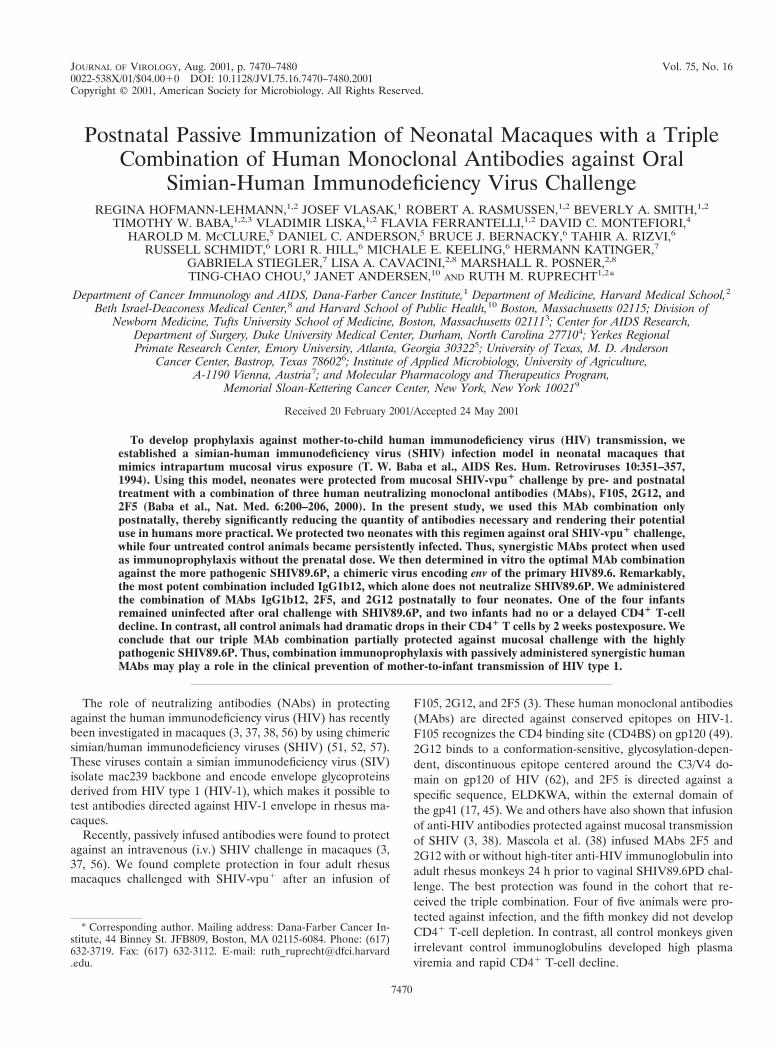
Two neonates, P1 and P2, were treated i.v. with the triple MAb combinationof F105, 2G12, and 2F5 on the day of birth (Fig. 1A). One hour after completion
VOL. 75, 2001 POSTNATAL PASSIVE IMMUNIZATION AGAINST SHIV CHALLENGE 7471
of the infusion, P1 and P2 were challenged orally in a nontraumatic manner (2)with 10 oral AID50 of SHIV-vpu1. A second MAb dose was given on day 8. Fouruntreated control neonates (R9C, R10C, R11C, and R16C) were challengedidentically (Fig. 1B).
Four neonates (RCh-7, RFh-7, RIh-7, RWg-7) were treated i.v. with a com-bination of three MAbs (IgG1b12, 2G12, and 2F5) within the first 3 days ofvaginal delivery (Fig. 1C). The treated animals were challenged orally with 15oral AID50 of SHIV89.6P 1 hour after completion of the MAb infusion. Thisdose yielded a 99% probability of infection (58). A second MAb dose was givenon day 8. Four control group neonates (REh-7, RHh-7, RJh-7, and RUg-7) wereuntreated prior to oral virus challenge (Fig. 1D).
Blood samples were collected from all animals on day 0 prior to MAb infusionand virus inoculation (baseline collection) and on day 8, prior to the second MAbinfusion, to determine infection/protection of animals. Although desirable, noadditional samples could be collected after the MAb infusions to determine theplasma titer of NAbs or to assess pharmacokinetics. The maximum blood volumethat can be sampled from approximately 500-g neonates (6 ml/kg of body weight/month) did not allow it. MAb-treated animals were observed for 31 months afterSHIV-vpu1 challenge and for 12 months after SHIV89.6P challenge, duringwhich time samples were collected regularly. When necessary, animals withprogressive disease were sacrificed via i.v. sodium pentobarbital injection.
Virus isolation; determination of proviral DNA and viral RNA loads. Methodsfor PBMC coculture and DNA PCR have been described previously (2, 33). Thelower limit of detection of the DNA PCR was one copy per 150,000 cells (33).Viral RNA loads were quantified by a one-tube fluorogenic probe-based real-time reverse transcription (RT)-PCR (24). RNA was extracted from 140 ml ofcell-free plasma with a QIAamp Viral RNA Mini kit (Qiagen, Valencia, Calif.).The sensitivity of the RT-PCR is 50 copies per ml of undiluted plasma. Sodiumcitrate-anticoagulated samples were not available from animal R16C for RT-PCR or neutralization assays.
Serological assays. Plasma samples were analyzed as described elsewhere (2,33) for the presence of specific antibodies, using commercially available Westernblot strips prepared from HIV-2 antigens which had been shown to cross-reactwith SIV antisera. HIV-1 Western blot analyses were conducted as instructed bythe manufacturer (Epitope, Beaverton, Oreg.).
NAbs in saliva and plasma. Saliva was collected from the oral cavities of theneonatal macaques using Weck-cel sponges (Xomed Surgical Products, Jackson-ville, Fla.). Saliva samples were collected 1 h after the first MAb infusion and/orprior to virus inoculation. After collection, sponges were saturated with 400 ml ofmedium (see above) for 15 min. Gentle squeezing with a sterile pipette elutedthe saliva, and the liquid (250 ml) was sterile filtered. To measure neutralization,serial dilutions of salivary filtrates were assayed in duplicate in an adapted MT-2cell infection assay with SHIV89.6P as described above or with SHIV-vpu1 asdescribed earlier (30). The salivary NAb titer is the reciprocal dilution whichprotected 50% of MT-2 cells from virus-induced cytotoxicity. This corresponds to90% reduction of the viral Gag synthesis (9). The assay’s lower limit of detectiontiter was 10 for undiluted samples. The same assays were applied to quantifyNAbs in serially diluted, heat-inactivated (56°C, 1 h) plasma samples. Neutral-izing activities against other SHIV variants, SHIV89.6 (51) and SHIV-KU2 (26),and the heterologous T-cell-line-adapted HIV-1 strain MN (23) were measuredin an identical manner.
Quantification of CTL activity in the completely protected animal RCh-7. Anautologous B-lymphoblastic cell line (BLCL) was prepared by transformation ofPBMC from RCh-7 with herpesvirus papio (50). PBMC collected 46 weeks afterSHIV89.6P challenge were stimulated with paraformaldehyde-fixed, autologousBLCL infected with SHIV antigen-encoding vaccinia virus constructs at 2 to 3PFU per cell (Therion, Cambridge, Mass.) Recombinant human IL-2 (20 U/ml)was added every 3 days. Cultures were tested on day 7 in a standard 5-h cytotoxicT-lymphocyte (CTL) assay using autologous, 51Cr-labeled BLCL target cellsinfected with vaccinia virus constructs encoding SIV gag-pol, SIV nef, or HIV-1IIIB env. Background cytotoxicity was measured using wild-type vaccinia virus-infected control target cells. A PBMC culture from an animal with known CTLactivity was included as a positive control for the autologous stimulation andcytotoxicity arms of the assay.
In vitro challenge of PBMC from the completely protected animal RCh-7.PBMC were purified by Ficoll-Hypaque sedimentation from a sodium citrate-anticoagulated blood sample collected from uninfected animal RCh-7 36 weeksafter SHIV89.6P challenge. Cells were washed and stimulated for 3 days withconcanavalin A (5 mg/ml) in RPMI 1640 supplemented with 10% human type ABserum, L-glutamine, and penicillin-streptomycin. Cells were washed, and CD81
T cells were removed by negative isolation using anti-CD8 magnetic beads asinstructed by the manufacturer (Dynal, Lake Success, N.Y.). CD8-depleted cellswere exposed to SHIV89.6P (100 ng of p27) for 12 h at 37°C. The cells werewashed and then incubated for 17 days in the presence of recombinant humanIL-2 (40 U/ml). Supernatants were collected every 2 to 4 days and analyzed forthe presence of SIV p27 antigen, using a commercially available enzyme-linkedimmunoassay kit specific for SIV (Beckman/Coulter, Hialeah, Fla.). PBMC from
FIG. 1. Experimental design of the in vivo oral challenge studieswith SHIV-vpu1 (A and B) and SHIV89.6P (C and D). (A) Twoneonatal rhesus monkeys (P1 and P2) were infused twice i.v. withhuman MAbs F105, 2G12, and 2F5 (10 mg/kg; solid black arrows). d0and d8, days 0 and 8. (B) Four neonates (R9C, R10C, R11C, andR16C) served as untreated control animals. All six animals were chal-lenged orally with 10 oral AID50 of SHIV-vpu1 1 h after the first MAbtreatment on the day of birth (open arrows). Treated animals wereobserved for 31 months, and serial blood samples were collected.Control animals were sacrificed 6 to 12 months after virus challenge.(C) Four neonatal rhesus monkeys (RCh-7, RFh-7, RIh-7, andRWg-7) were infused i.v. twice with human MAbs IgG1b12, 2G12, and2F5 (10 mg/kg; solid black arrows). (D) Four neonates (REh-7,RHh-7, RJh-7, and RUg-7) served as untreated control animals. Alleight animals were challenged orally with SHIV89.6P (15 oral AID50)1 h after the first MAb treatment or at the corresponding time point (3days after birth) (open arrows). Animals were observed for 12 months,and serial blood samples were collected. †, animals RWg-7, RHh-7,RJh-7 were sacrificed 10, 7, and 9 weeks postexposure, respectively.
7472 HOFMANN-LEHMANN ET AL. J. VIROL.
an uninfected normal control animal were stimulated, CD81 T-cell depleted, andinfected in the same manner as a positive control.
Statistical analysis. For analysis, the viral loads were expressed as a log10
transformation based on the distribution of HIV RNA in human studies. Viralloads below the limit of detection were set at half the limit of detection forcalculation. No imputation for missing or truncated data was employed. It isnoted that with four animals per group, there is low statistical power.
RESULTS
Synergistic neutralization of SHIV89.6P by a triple combi-nation of human anti-HIV-1 MAbs in vitro. We previouslytested a large panel of human anti-HIV-1 MAbs for theirability to neutralize SHIV-vpu1 in vitro. MAbs IgG1b12, 2F5,2G12, and F105 were synergistic when used in combinations (3,29, 30). In addition, the triple combination of F105, 2G12, and2F5 was used successfully to protect macaques from i.v. andoral SHIV-vpu1 challenge (3). In the present study, we ex-plored the capacity of these four MAbs, used either alone or incombination, to neutralize pathogenic SHIV89.6P. We alsocompared the synergistic potency of F105 to that of IgG1b12 todetermine whether different MAbs directed against theCD4BS were equally effective.
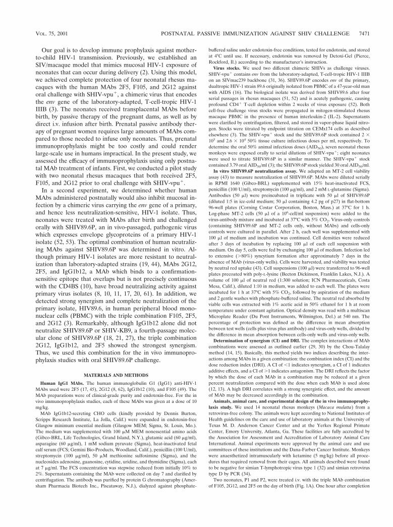
MAb IgG1b12 was inactive against SHIV89.6P; even highIgG1b12 concentrations up to 100 mg/ml did not significantlyneutralize SHIV89.6P (Table 1; Fig. 2). Nevertheless, IgG1b12(included at a constant concentration of 50 mg/ml) increasedthe neutralization potency of other MAbs (2G12 and 2F5)when used in combinations, as indicated by a leftward shift ofthe curves (Fig. 2; Table 1). This allowed us to use a reducedconcentration of antibodies to achieve an equivalent level ofneutralization. For example, when used alone, 1.243 mg of 2F5per ml was required to obtain 50% SHIV89.6P neutralization(Table 1); by adding IgG1b12, the dose of 2F5 necessary to
achieve the same result could be reduced 18.8 times (DRI) to0.066 mg/ml. The addition of IgG1b12 to the combination of2F5 and 2G12 led to very strong synergism and to even greaterDRIs of 110 for 2F5 and 25.7 for 2G12 (Table 1). This effectwas stronger than that observed using F105. The addition ofF105 (also at a constant concentration of 50 mg/ml) to a serialdilution of 2F5 and 2G12 led to synergism at low concentra-tions, whereas antagonism was seen at the desirable, high de-
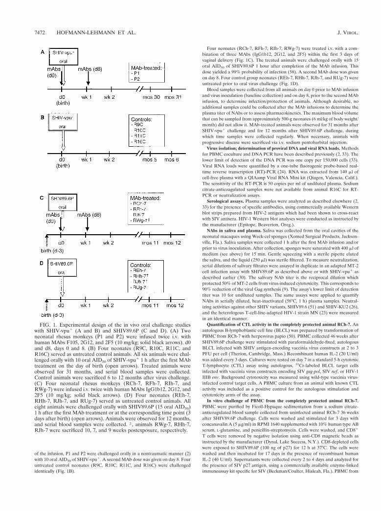
TABLE 1. Synergistic neutralization of SHIV89.6P by human IgG1 MAbs in MT-2 cells
Single antibody (specificity) or antibodycombination (ratio)
Concn (mg/ml) for50% neutralizationa
CIb at: DRIc
InteractionED50 ED90 2F5 2G12
Single agentsIgG1b12 (anti-CD4BS) .1002G12 (anti-gp120) 0.2912F5 (anti-gp41) 1.243
Combinations of 2 MAbs2G12 1 IgG1b12e 0.076 3.8 Potentiation of 2G12 by IgG1b12d
2F5 1 IgG1b12e 0.066 18.8 Potentiation of 2F5 by IgG1b12d
2F5 1 2G12 (1:1) 0.191 0.41 1.25 13.1 3.06 Synergism
Combinations of 3 MAbs2F5 1 2G12 (1:1) 1 IgG1b12e 0.0226 0.048 0.067 110 25.7 Very strong synergism2F5 1 2G12 (1:1) 1 F105e 1.56 3.20 126 1.92 0.37 Antagonism at high degrees of
neutralization; synergism at lowdegrees of neutralization
Combination of 4 MAbs2F5 1 2G12 (1:1) 1 IgG1b12 1 F105e 0.265 0.54 0.80 11.3 2.2 Synergism
a The neutralization dose for combinations of two and more MAbs was the sum of the dose of each MAb used in the combination regimen with the exception ofIgG1b12 and F105. See footnote e.
b Calculated by the Chou-Talalay method as described in Materials and Methods. CI , 1, CI 5 1, and CI . 1, indicate synergism, additive effects, and antagonism,respectively. ED50 and ED90, 50 and 90% effective doses.
c Measured by comparing the doses required to reach 50% virus neutralization when the antibody was used alone and in combination with other antibodies.d When one component in combination has no or negligible effect by itself (e.g., IgG1b12), no CI value can be calculated due to lack of dose-effect parameters. In
this case, the term “potentiation” is used instead of “synergism.”e Always used at a concentration of 50 mg/ml.
FIG. 2. Human MAb neutralization of SHIV89.6P in MT-2 cells.Neutralization was measured colorimetrically by the percentage ofviable cells after incubation with the virus-antibody mixture (y axis).The antibody concentration (x axis) in combinations of two or moreMAbs is the sum of the concentrations of each MAb with the exceptionof IgG1b12 and F105. When used in combinations, MAbs IgG1b12and F105 were at 50 mg/ml; as potentiators, these amounts are notincluded in the total antibody concentration indicated on the x axis. Inaddition, when MAbs 2G12 and 2F5 were tested alone or in combi-nation with other antibodies, the concentration of each was identical.Results are representative of two or three separate experiments foreach antibody.
VOL. 75, 2001 POSTNATAL PASSIVE IMMUNIZATION AGAINST SHIV CHALLENGE 7473
7474 HOFMANN-LEHMANN ET AL. J. VIROL.
grees of neutralization. Furthermore, the quadruple MAbcombination was less synergistic than the triple 2F5-2G12-IgG1b12 combination, presumably because of competition be-tween F105 and IgG1b12 for the CD4BS. Therefore, we se-lected the triple combination of 2G12, IgG1b12, and 2F5 forthe in vivo SHIV89.6P studies.
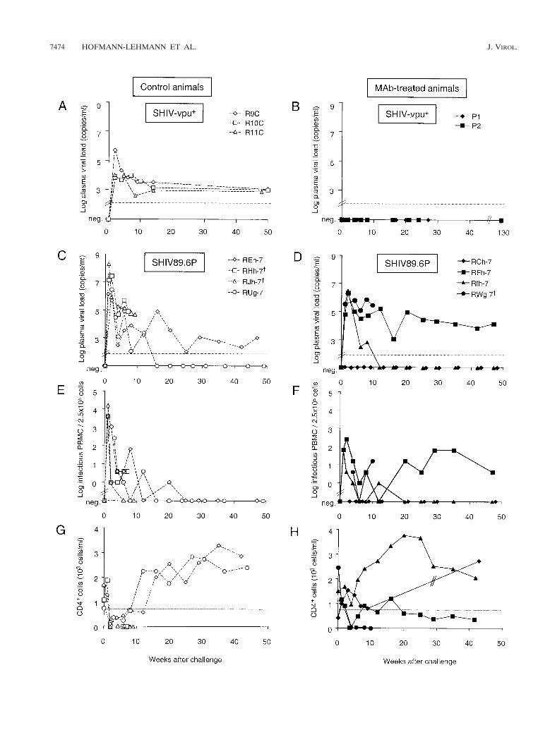
Outcome of oral SHIV-vpu1 challenge. Two neonatal rhesusmacaques were treated i.v. with a combination of 2G12, F105,and 2F5 on the day of birth (day 0 [Fig. 1]) prior to oralSHIV-vpu1 challenge. Both animals received a second infu-sion of this triple MAb combination 8 days later. Four un-treated control animals were also challenged orally. These fourmonkeys served as controls also for another study arm, re-ported previously (3). All four untreated neonates, R9C,R10C, R11C, and R16C, became infected. They were positivefor virus isolation and proviral DNA PCR, and all four animalsseroconverted (3). Plasma samples from R9C, R10C, andR11C were tested by RT-PCR and yielded positive resultsthroughout the observation period (Fig. 3A).
In contrast, both MAb-treated animals were protected fromoral SHIV-vpu1 challenge. No plasma viral RNA was detect-able (Fig. 3B), virus isolation was persistently negative, and noproviral DNA was detectable in PBMC samples obtained fromthese animals between weeks 1 and 6 postexposure (data notshown). Furthermore, no virus-specific antibodies were de-tected by p27 enzyme-linked immunosorbent assay or by West-ern blotting using HIV-2 strips (data not shown). Taken to-gether, our results confirm that the triple combination ofneutralizing MAbs protects from oral SHIV-vpu1 challengeand demonstrate that protection is achieved with only postna-tal treatment.
Virological and clinical outcome of SHIV89.6P challenge.Four neonatal rhesus macaques were treated i.v. with a com-bination of 2G12, IgG1b12, and 2F5 within the first 3 days ofvaginal delivery (day 0 [Fig. 1]) and were challenged orally with15 oral AID50 of SHIV89.6P 1 h after completing the antibodyinfusion. They received a second infusion of this triple MAbcombination 8 days later. Four untreated control animals werealso challenged.
All four untreated control animals (REh-7, RHh-7, RJh-7,and RUg-7) became highly viremic (Fig. 3C and E). The peakviral RNA levels in the three MAb-treated infants that devel-oped viremia were 10 to 100 times lower than those in the twocontrol infants RHh-7 and RJh-7 (Fig. 3C and D), but becauseof the low number of study animals, statistical significancecould not be reached. Without exception, the untreated ani-mals developed rapid and profound CD41 T-cell depletion,leading to nearly complete loss of peripheral CD41 T cells by2 weeks after exposure (Fig. 3G), similar to SHIV89.6P infec-tion of adult macaques (52, 53). The two untreated controlanimals that had very high initial plasma viral RNA loads
(RJh-7 and RHh-7) had complete and persistent CD41 T-celldepletion (Fig. 3C, E, and G). They subsequently developedprogressive disease (diarrhea, weakness, opportunistic infec-tions, pneumonia, and lymphadenopathy) and succumbed toAIDS 7 and 9 weeks postchallenge, respectively. Remarkably,the other two untreated control animals (REh-7 and RUg-7)recovered gradually from declines in CD41 T cells by week 8postexposure, even though both had peak viral RNA loadsexceeding 106 copies/ml.
One of four MAb-treated neonates (RCh-7) was completelyprotected from the oral virus challenge. No evidence of viralreplication was observed, as viral RNA remained undetectablein the plasma by real-time RT-PCR (Fig. 3D) and PBMCcoculture was negative (Fig. 3F). Proviral DNA could not beamplified from PBMC from this animal by DNA PCR (datanot shown). Furthermore, no evidence of virus-induced diseasewas observed. The peripheral CD41 T-cell count from RCh-7did not decrease after challenge (Fig. 3H). The remainingthree MAb-treated animals (RFh-7, RIh-7, and RWg-7) be-came infected, as determined by virus isolation and RT-PCR(Fig. 3D and F). Infant RIh-7 maintained its CD41 T-cellcounts above 955 cells/ml (Fig. 3H), in contrast to all fourcontrols, which suffered severe, acute drops of their CD41 Tcells to 0 to 170 cells/ml at week 2. The MAb-treated infantRFh-7 showed a delay in an eventual decrease in CD41 T cells(Fig. 3H). RFh-7 partially recovered and thereafter had slowlydeclining CD41 T-cell counts. In contrast, RWg-7 had rapidCD41 T-cell depletion and never recovered. This animal wassacrificed 10 weeks after the SHIV89.6P challenge because ofdisease that progressed to AIDS (diarrhea, weakness, oppor-tunistic infections, severe thymic atrophy, and no detectableCD41 T cells).
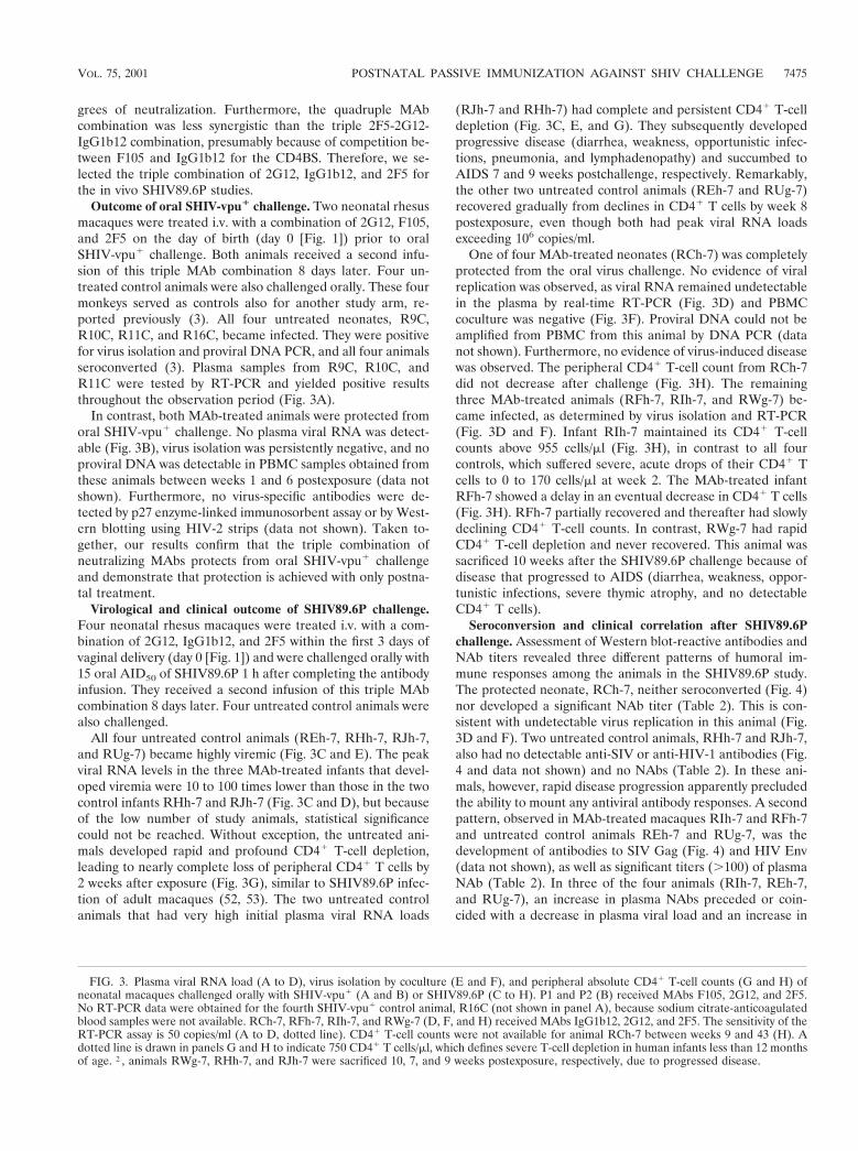
Seroconversion and clinical correlation after SHIV89.6Pchallenge. Assessment of Western blot-reactive antibodies andNAb titers revealed three different patterns of humoral im-mune responses among the animals in the SHIV89.6P study.The protected neonate, RCh-7, neither seroconverted (Fig. 4)nor developed a significant NAb titer (Table 2). This is con-sistent with undetectable virus replication in this animal (Fig.3D and F). Two untreated control animals, RHh-7 and RJh-7,also had no detectable anti-SIV or anti-HIV-1 antibodies (Fig.4 and data not shown) and no NAbs (Table 2). In these ani-mals, however, rapid disease progression apparently precludedthe ability to mount any antiviral antibody responses. A secondpattern, observed in MAb-treated macaques RIh-7 and RFh-7and untreated control animals REh-7 and RUg-7, was thedevelopment of antibodies to SIV Gag (Fig. 4) and HIV Env(data not shown), as well as significant titers (.100) of plasmaNAb (Table 2). In three of the four animals (RIh-7, REh-7,and RUg-7), an increase in plasma NAbs preceded or coin-cided with a decrease in plasma viral load and an increase in
FIG. 3. Plasma viral RNA load (A to D), virus isolation by coculture (E and F), and peripheral absolute CD41 T-cell counts (G and H) ofneonatal macaques challenged orally with SHIV-vpu1 (A and B) or SHIV89.6P (C to H). P1 and P2 (B) received MAbs F105, 2G12, and 2F5.No RT-PCR data were obtained for the fourth SHIV-vpu1 control animal, R16C (not shown in panel A), because sodium citrate-anticoagulatedblood samples were not available. RCh-7, RFh-7, RIh-7, and RWg-7 (D, F, and H) received MAbs IgG1b12, 2G12, and 2F5. The sensitivity of theRT-PCR assay is 50 copies/ml (A to D, dotted line). CD41 T-cell counts were not available for animal RCh-7 between weeks 9 and 43 (H). Adotted line is drawn in panels G and H to indicate 750 CD41 T cells/ml, which defines severe T-cell depletion in human infants less than 12 monthsof age. †, animals RWg-7, RHh-7, and RJh-7 were sacrificed 10, 7, and 9 weeks postexposure, respectively, due to progressed disease.
VOL. 75, 2001 POSTNATAL PASSIVE IMMUNIZATION AGAINST SHIV CHALLENGE 7475
CD41 T-cell counts (data not shown). A third pattern wasobserved in the MAb-treated animal RWg-7, which transientlydeveloped low level anti-Gag antibodies (Fig. 4). AnimalRWg-7 progressed to disease and AIDS. In HIV-infected hu-mans (7) as well as in SIV-infected macaques (2), selective lossof anti-Gag antibodies is a grave prognostic sign heralding thedevelopment of AIDS. The humoral response of RWg-7 fol-lowed just such a pattern.
Potential mechanisms of the protection observed in threeMAb-treated animals. Neonates P1, P2, and RCh-7 were notinfected by all criteria tested and neither seroconverted nordeveloped NAb responses (Fig. 4, Table 2, and data notshown). P1, P2, and RCh-7 appeared to be protected frommucosal virus challenge by well-characterized pure human IgGMAbs. No mucosal NAbs of the IgA subtype were present inour MAb combination. How could systemically administeredIgG MAbs protect against mucosal virus challenge? We testedsaliva samples collected from P1, P2, and RCh-7 1 h after thefirst MAb infusion just prior to virus challenge in neutraliza-tion assays. They had no more neutralizing activity than theprechallenge samples collected from untreated control animals(all titers , 20). This observation was true for all MAb-treated
animals as well (all titers , 20). Neutralizing titers, if presentat all in saliva at the time of virus challenge, were below thelower detection limit of our assay. Thus, direct inactivation ofSHIV-vpu1 and SHIV89.6P in saliva seems unlikely.
Given these results, it is possible that virus crossed the mu-cosal barrier and went through an initial round of infection intarget cells of the local mucosal gastrointestinal tissue. Thepotent neutralizing MAbs would have stopped further waves ofviral spread from these primary target cells. Nevertheless, lim-ited local infection could have permitted the development ofvirus-specific, cellular immune responses, which in turn couldhave subsequently eliminated the small number of infectedcells. To test this possibility, we evaluated CTL activity ofPBMC obtained from RCh-7 46 weeks postchallenge; no sig-nificant specific lysis was detected in 51Cr release assays (datanot shown). However, measuring systemic CTL activity in pe-ripheral blood may not reflect specific antiviral cellular immu-nity at the level of the upper gastrointestinal mucosa itself.
To test for evidence of persistent mucosal immune protec-tion, we rechallenged animal RCh-7 with 15 oral AID50
SHIV89.6P 54 weeks after the first challenge. We detectedviral RNA in plasma obtained 1 week after the rechallenge(71,565 RNA copies/ml of plasma), and the CD41 T-cellcounts decreased from 1,516 to 684 cells/ml. Clearly, no pro-tection was seen.
Potential mechanism that contributed to the recovery fromthe acute pathogenic effects of SHIV89.6P. To gain more in-sight into what immune mechanisms might have contributed tothe recovery of two of our four untreated controls (REh-7 andRUg-7) from the acute pathogenic effects of SHIV89.6P, weanalyzed the humoral and cellular immune responses of theseanimals. As mentioned above, both animals developed hightiters of NAbs against SHIV89.6P (Table 2). We evaluated thebreadth of NAbs by assessing their ability to neutralize otherSHIV variants and an HIV-1 strain as described earlier (18, 42,43). We included plasma samples from REh-7 and RUg-7collected 79 and 81 weeks postexposure, respectively. In addi-tion, we assayed samples collected 35 and 135 weeks postex-posure, respectively, from two other macaques, RDt-7 andRGt-6, that had been inoculated orally with SHIV89.6P asneonates and had recovered from the pathogenic manifesta-tions of the infection. The plasma samples contained—in
FIG. 4. Western blot analysis of plasma collected from SHIV89.6P-exposed animals. Serial samples were analyzed using HIV-2 strips. Thenumber of weeks after challenge is indicated. Migration of HIV-2 proteins is shown on the left. The arrow on the right indicates migration of Gagantigen. Plasma from SIV-free macaques or normal human serum and plasma from SIV-infected macaques or human anti-HIV-2 serum werenegative and positive controls, respectively (leftmost 4 lanes). †, sacrificed animal.
TABLE 2. SHIV89.6P plasma neutralization titers after passiveantibody infusion
GroupPlasma neutralization titera
27–30b 47–49
MAb-treated animalsRCh-7 ,10 ,10RFh-7 1,564 1,900RIh-7 279 155RWg-7 ,10
Untreated control animalsREh-7 1,182 933RHh-7 ,10RJh-7 12RUg-7 979 520
a Reciprocal dilution which protected 50% of MT-2 cells from virus-inducedcytotoxicity. This corresponds to 90% reduction of the viral Gag synthesis (9).
b Weeks after virus exposure. Animals RWg-7, RHh-7, and RJh-7 were sac-rificed in weeks 10, 7, and 9, respectively. Therefore, samples obtained 7 weekspostchallenge were analyzed.
7476 HOFMANN-LEHMANN ET AL. J. VIROL.
addition to anti-SHIV89.6P NAbs—variable and sometimespotent neutralizing activity against the heterologous T-cell-line-adapted HIV-1 strain MN (titers as high as 500) but noactivity against SHIV89.6 and SHIV-KU2 (all titers , 20). Wealso tested specific cellular immune responses in five of ourtotal six recovered macaques by using enzyme-linked immuno-spot assays (28) and found a significant number of SIVmac239Gag- and HIV89.6 Env-specific T lymphocytes in all five ani-mals (data not shown).
Safety of human MAb administration to neonatal ma-caques. Infusions of MAb combinations were tolerated well inmore than 15 neonatal macaques treated so far; no acutereactions were reported after any of the more than 30 i.v.infusions. One MAb-treated neonate (RCh-7) suffered a sei-zure approximately 2 days after the first MAb infusion. RCh-7had a low birth weight (420 g) but subsequently gained bodyweight normally (1.68 kg at necropsy). No further seizureswere reported. As in all of the other animals, the MAb infusionand the virus inoculation had been uneventful, suggesting thata connection between these manipulations and the observedseizure is unlikely. It is also improbable that it interfered withthe challenge protocol, since 2 days had elapsed between viruschallenge and the neurological events. Twenty-four days aftervirus challenge, physical examination of RCh-7 revealed par-tial paralysis of both lower legs (loss of reflexes); 7 days later,contracture and atrophy of the affected musculature were re-ported. Necropsy performed 55 weeks postchallenge revealedthat RCh-7 was in reasonably good condition with the excep-tion of the previously observed pelvic and leg muscle atrophy.In addition, focal cortical atrophy with sparing of the underly-ing white matter and cavitation of the underlying tissue werepresent in the left occipital-parietal junction, extending fromthe parietal lobe into the premotor cortex. Hemosiderin-ladenmacrophages were present at the borders of cavitated areasconsistent with previous cortical hemorrhage. No evidence ofvasculitis was detected. The unilateral lesion in the brain is notthe likely cause of motor dysfunction, given the sparing of themotor areas and intactness of the cervical spinal cord. Nospecific diagnosis for the neurologic deficits could be obtained;however, the time course of events renders an etiologic asso-ciation with the MAb treatment unlikely.
DISCUSSION
We infused triple combinations of human MAbs postnatallyinto six neonatal rhesus macaques to evaluate the protectivepotential against mucosal challenge with two different SHIVstrains. We achieved complete protection in two of two MAb-treated neonates challenged with SHIV-vpu1 and one of fourMAb-treated neonates exposed to SHIV89.6P, while all con-trol animals exposed to either virus became viremic. An addi-tional MAb-treated animal was protected from the rapid CD41
T-cell depletion observed in all control animals after SHIV89.6Pchallenge. Thus, the MAb combination was partially successful inprotecting animals from the pathogenic effects of the SHIV89.6Pinfection. Furthermore, protection occurred without administra-tion of large prenatal maternal MAb doses.
The triple MAb combination of F105, 2G12, and 2F5 pro-tected neonates against oral SHIV-vpu1 challenge after pre-and postnatal immunoprophylaxis (3); it was therefore used in
the present study with SHIV-vpu1 as well. For the SHIV89.6Poral challenge study, F105 was replaced by IgG1b12 in thetriple combination because this regimen produced the stron-gest in vitro neutralization against this virus. Interestingly, neu-tralization of SHIV89.6P by 2G12 and 2F5 was potentiated bythe addition of IgG1b12. This was unexpected, since IgG1b12alone did not inhibit SHIV89.6P replication in vitro. This resultsupports earlier studies that used either SHIV89.6P or SHIV-KB9, a molecular clone derived after the fourth passage ofSHIV89.6P (18, 21, 27). Neutralization resistance of virusesmight be due to a number of factors, including point mutationsleading to loss of the relevant epitope (41) or to global con-formational changes that make the epitope inaccessible to theMAb (47, 60, 64). The IgG1b12 epitope is present on theenvelope protein of SHIV-KB9; IgG1b12 binds soluble KB9gp120 monomers with undiminished capacity (21). However,the epitope seems less accessible for antibody binding in thecontext of the naturally folded oligomeric envelope glycopro-tein complex on the SHIV-KB9 virion surface (21). IgG1b12binds other viral strains with equal or better avidity to theoligomeric form of the envelope glycoprotein; this probablyaccounts for its exceptional potency (22, 54). Resistance toIgG1b12 has been mapped to the V1, V2, and V3 loops of theHIV-1 envelope (21, 41, 54). Antibodies 2G12 and 2F5 recog-nize epitopes unrelated to the V1, V2, or V3 loop or to theCD4BS (8, 17, 45, 62). We postulate that binding of either2G12 or 2F5 induces a conformational change in the oligo-meric envelope glycoprotein complex of SHIV89.6P that al-lows contact of IgG1b12 with its previously hidden cognateepitope.
The addition of a fourth antibody, F105, to the triple com-bination of IgG1b12, 2G12, and 2F5 led to a decrease inneutralization synergism against SHIV89.6P. F105 andIgG1b12 seem to inhibit each other, although they use differ-ent mechanisms to neutralize HIV. F105 inhibits the attach-ment of the neutralized virus to the target cell (49), whileIgG1b12 inhibits the fusion entry process (39). Since they bothrecognize epitopes that at least partially overlap with theCD4BS, the diminished combined neutralization activity maybe due to steric hindrance and competition for the binding site.
We achieved complete protection against oral SHIV-vpu1
challenge in two neonatal rhesus macaques that received post-natal treatment with a triple combination of human neutraliz-ing MAbs. This chimeric virus encodes env of the laboratory-adapted HIV-1 IIIB. To mimic HIV infection of humannewborns more closely, we then used a highly pathogenic chi-meric virus that encodes env derived from the primary, dual-tropic HIV89.6 and achieved partial protection. It is wellknown that primary strains of HIV are more difficult to neu-tralize than laboratory-adapted strains (19, 44). However, wehad optimized the MAb combination previously for high invitro neutralization of SHIV89.6P. Another factor that couldhave contributed to the different outcome of the two immu-noprophylaxis studies could also be the higher challenge doseused of SHIV89.6P (15 AID50) than of SHIV-vpu1 (10AID50).
Protection in the orally challenged neonates was achievedwith only postnatal treatment. We chose this approach becausethe large amounts of each MAb required for treatment of themother during pregnancy may render passive immunoprophy-
VOL. 75, 2001 POSTNATAL PASSIVE IMMUNIZATION AGAINST SHIV CHALLENGE 7477
laxis approaches to prevent maternal HIV transmission in clin-ical trials too costly. Furthermore, we modeled our primatestudy on the successful prevention of maternal transmission ofthe hepatitis B virus, an enveloped virus that is transmittedthrough the same routes as HIV. Anti-hepatitis B immuno-globulin therapy is administered to newborn infants within 12 hof birth; no prenatal treatment is given to infected mothers.Passive immunization alone is 71% effective in preventing pas-sage of hepatitis B virus to infants (5).
To our knowledge, the present study gives the first descrip-tion of the course and outcome of SHIV89.6P infection ofneonatal rhesus macaques. In adult rhesus monkeys, i.v. inoc-ulation of SHIV89.6P led to high initial viral peak RNA loadsand to profound, persistent depletion of CD41 T cells within 2weeks after virus challenge (4, 25, 53, 59, 63). According tomost reports (42, 53, 63), SHIV89.6P infection leads to rapiddisease progression and to AIDS in adult macaques, leavingthe animals unable to seroconvert or mount any virus-specificCTL responses. This course of infection was seen in only twoof our four naive neonates (RHh-7 and RJh-7). Interestingly,the other two untreated control animals (REh-7 and RUg-7)showed a rebound of CD41 T-cell counts 8 weeks after virusexposure. These neonates seroconverted readily, developedhigh titers of NAbs, and remained clinically well throughoutthe 1-year observation period. A similar phenomenon was de-scribed recently by Barouch and coworkers (4). Two of eightrhesus monkeys that were challenged i.v. with 100 AID50 ofSHIV89.6P did not show complete depletion of CD41 T cells;one animal developed no significant disease, and only four ofthe eight macaques died by day 140 postexposure (4). Theanimals developed low virus-specific CD81 CTL responses.However, in accordance with our observation in infant ma-caques, two of the adult animals developed high NAb titers byday 28 after virus exposure (4).
To further analyze the immune mechanisms that might havecontributed to the recovery from the acute SHIV89.6P patho-genicity in our two untreated neonates, we assessed thebreadth of their NAbs using in vitro neutralization assays. Weincluded samples from REh-7, RUg-7, and two additional ma-caques that had been inoculated orally with SHIV89.6P asneonates and had recovered from the acute CD41 T-cell de-pletion. Plasma samples from all four animals had NAb activityagainst SHIV89.6P and the HIV-1 strain MN but not againstSHIV89.6 and SHIV-KU2. These results are similar to thosereported for plasma samples from SHIV89.6PD-infected ma-caques (18, 42) and demonstrate the restricted nature of theinduced NAbs also in SHIV89.6P-infected animals. In addi-tion, all four untreated, recovered macaques had strong virus-specific cellular immune responses. In conclusion, theseSHIV89.6P-infected macaques that recovered from the severeCD41 T-cell depletion and survived for a prolonged periodwithout significant disease demonstrated both strong NAb ac-tivity and cellular immune responses. It remains to be deter-mined what additional factors are responsible for the variableoutcomes observed among individual SHIV89.6P-infectedadult and neonatal macaques.
Virus replication was undetectable in the peripheral bloodof MAb-treated animals P1, P2, and RCh-7. The animals nei-ther seroconverted nor developed NAb responses. Successfulin vitro infection of CD81-depleted PBMC collected 36 weeks
after virus exposure from animal RCh-7 (data not shown)excluded the possibility of natural resistance to SHIV89.6P dueto genetic predisposition, such as lack of coreceptors. There-fore, the triple combination of human neutralizing MAbs musthave protected RCh-7 from systemic infection. It is not clear atwhat level the infused IgG MAbs stopped the virus. As in ourprevious study, in which neonates were completely protectedagainst an oral SHIV-vpu1 challenge (3), we could not detectany significant neutralizing activity in the saliva of P1, P2, andRCh-7 at the time of challenge. While this does not rule out thepossibility that low amounts of MAbs interacted with the virus insaliva, it is more likely that virus crossed the mucosal membraneand underwent a single round of replication in target cells of thelocal gastrointestinal tissues. Secondary waves of virus spreadmight then have been prevented either through direct virus neu-tralization by the parenterally infused IgG MAbs or via elimina-tion of infected MAb-coated cells by antibody-dependent cell-mediated cytotoxicity (ADCC) or complement activation. Indeed,human anti-HIV MAb such as F105 were found to mediateADCC (48), and 2G12 has both ADCC- and complement-medi-ated activity (62). Recently, ADCC or other cell-killing mecha-nisms were also suggested to cause transient reductions of viralload in SIVmac251-infected macaques infused with immuno-globulins (6). Alternatively, the initial infection of a set of suscep-tible target cells might have induced virus-specific cellular im-mune responses. However, no direct evidence for cellularimmunity was seen—virus-specific CTL activity was not detectedin the peripheral blood, and there was no protection from oralrechallenge 54 weeks after the first virus exposure.
The protection that we achieved in the four MAb-treatedneonatal rhesus macaques after oral challenge with pathogenicSHIV89.6P may be compared to the level of protection re-ported for antibody-treated, adult macaques challenged vagi-nally with plasma-derived SHIV89.6PD (38). Similar doses (15versus 10 to 50 AID50) and strains of virus (SHIV89.6P versusSHIV89.6PD [35, 52]) were used in the two studies. However,to achieve reproducible infection, the adult macaques weretreated with progesterone prior to vaginal challenge, resultingin a thinning of the vaginal epithelium. This might have influ-enced the MAb efficacy by allowing more antibodies to seepacross the mucosal barrier. In addition, we used a lower doseof antibody (10 mg/kg) than used in the adult study (15 mg/kg),and different combinations of antibodies were employed.
In conclusion, postnatal triple MAb combination overallprevented infection in three of six treated infants. AmongSHIV89.6P-challenged animals, the MAb combination waspartially successful in preventing infection; half of the treatedinfants were protected from the acute, severe T-cell depletion.The failure to protect all MAb-treated animals fromSHIV89.6P infection did not seem to be related to neutraliza-tion resistance. Viruses from MAb-treated animal RFh-7 anduntreated control animal REh-7, recovered 37 weeks postex-posure, were still as sensitive to neutralization with the tripleMAb combination as the original SHIV89.6P inoculum (datanot shown). Even the partial protection that we report here isencouraging since the macaque-adapted SHIV89.6P (52) ismore pathogenic in monkeys than HIV is in humans. To in-crease the degree of protection in future primate studies, weplan to include other synergistically acting MAbs in the com-bination regimen and/or increase the neonatal MAb doses.
7478 HOFMANN-LEHMANN ET AL. J. VIROL.
Our approach is directly relevant for the development of anew strategy against maternal-fetal HIV-1 transmission in hu-mans, since the MAbs used are human antibodies directedagainst HIV-1 glycoproteins. It may represent an importantaddition to the antiretroviral therapy protocols established toreduce mother-to-child HIV transmission. Being natural hu-man proteins, human MAbs can be expected to have low tox-icity. In symptomatic HIV-infected children, the prophylacticuse of i.v. immunoglobulin for the prevention of bacterial in-fections was safe and was suggested as standard therapy (1, 55).In addition, MAbs 2G12 and 2F5 have been infused safely intoHIV-infected adults in a phase I clinical trial (H. Katinger andG. Stiegler, unpublished observations). The stability and longhalf-lives of neutralizing human MAbs that we had noted inour earlier study in neonates (3) may yield another clinicalbenefit: the protection of infants against oral HIV transmissionthrough infected breast milk in the neonatal period. Protectionagainst this mode of maternal HIV transmission may be easierto achieve than against our SHIV89.6P oral challenge, sincethe infectivity of human breast milk is lower and does not yielda 99% probability of infection (40, 46) as our SHIV89.6Pchallenge dose did.
ACKNOWLEDGMENTS
We thank Matthew Frosch (Department of Pathology, Brigham andWomen’s Hospital, Boston, Mass.) for review of the neuropathologicalslides and helpful discussion. CHO cells producing MAb IgG1b12were kindly provided by Dennis Burton (Department of Immunologyand Molecular Biology, Scripps Research Institute, La Jolla, Calif.).We also thank Yulan Wang for technical assistance and C. Gallegosand S. Sharp for preparation of the manuscript.
This work was supported in part by National Institutes of Healthgrants RO1 AI34266, R21 AI46177, and PO1 AI48240 awarded toR.M.R., RR00165 to H.M.M., AI26926 to M.R.P. and AI45320 toL.A.C. It was also supported by the Pediatric AIDS Foundation grant50864PG23 to R.M.R. and by Center for AIDS Research (CFAR) coregrant IP30 28691 awarded to the Dana-Farber Cancer Institute.D.C.M. was supported by National Institutes of Health contract NO1AI85343. R.H.-L. was supported by a grant from the Swiss NationalScience Foundation (fellowship 823A-50315) and is the recipient of ascholar award from the Friends of Switzerland, Inc. T.W.B. was arecipient of National Institutes of Health Clinical Investigator Devel-opment Award 30/35 KO8-AI01327.
REFERENCES
1. Anonymous. 1991. Intravenous immune globulin for the prevention of bac-terial infections in children with symptomatic human immunodeficiency virusinfection. The National Institute of Child Health and Human DevelopmentsIntravenous Immunoglobulin Study Group. N. Engl. J. Med. 325:73–80.
2. Baba, T. W., J. Koch, E. S. Mittler, M. Greene, M. Wyand, D. Pennick, andR. M. Ruprecht. 1994. Mucosal infection of neonatal rhesus monkeys withcell-free SIV. AIDS Res. Hum. Retroviruses 10:351–357.
3. Baba, T. W., V. Liska, R. Hofmann-Lehmann, J. Vlasak, W. Xu, S. Ayehunie,L. A. Cavacini, M. R. Posner, H. Katinger, G. Stiegler, B. J. Bernacky, T. A.Rizvi, R. Schmidt, L. R. Hill, M. E. Keeling, Y. Lu, J. E. Wright, T. C. Chou,and R. M. Ruprecht. 2000. Human neutralizing monoclonal antibodies of theIgG1 subtype protect against mucosal simian-human immunodeficiency virusinfection. Nat. Med. 6:200–206.
4. Barouch, D. H., S. Santra, J. E. Schmitz, M. J. Kuroda, T. M. Fu, W.Wagner, M. Bilska, A. Craiu, X. X. Zheng, G. R. Krivulka, K. Beaudry, M. A.Lifton, C. E. Nickerson, W. L. Trigona, K. Punt, D. C. Freed, L. Guan, S.Dubey, D. Casimiro, A. Simon, M. E. Davies, M. Chastain, T. B. Strom, R. S.Gelman, D. C. Montefiori, and M. G. Lewis. 2000. Control of viremia andprevention of clinical AIDS in rhesus monkeys by cytokine-augmented DNAvaccination. Science 290:486–492.
5. Beasley, R. P., L. Y. Hwang, G. C. Lee, C. C. Lan, C. H. Roan, F. Y. Huang,and C. L. Chen. 1983. Prevention of perinatally transmitted hepatitis B virusinfections with hepatitis B immune globulin and hepatitis B vaccine. Lancet2:1099–1102.
6. Binley, J. M., B. Clas, A. Gettie, M. Vesanen, D. C. Montefiori, L. Sawyer, J.Booth, M. Lewis, P. A. Marx, S. Bonhoeffer, and J. P. Moore. 2000. Passive
infusion of immune serum into simian immunodeficiency virus-infected rhe-sus macaques undergoing a rapid disease course has minimal effect onplasma viremia. Virology 270:237–249.
7. Binley, J. M., P. J. Klasse, Y. Cao, I. Jones, M. Markowitz, D. D. Ho, andJ. P. Moore. 1997. Differential regulation of the antibody responses to Gag andEnv proteins of human immunodeficiency virus type 1. J. Virol. 71:2799–2809.
8. Buchacher, A., R. Predl, K. Strutzenberger, W. Steinfellner, A. Trkola, M.Purtscher, G. Gruber, C. Tauer, F. Steindl, A. Jungbauer, and H. Katinger.1994. Generation of human monoclonal antibodies against HIV-1 proteins;electrofusion and Epstein-Barr virus transformation for peripheral bloodlymphocyte immortalization. AIDS Res. Hum. Retroviruses 10:359–369.
9. Bures, R., A. Gaitan, T. Zhu, C. Graziosi, K. McGrath, J. Tartaglia, P.Caudrelier, R. El Habib, M. Klein, A. Lazzarin, D. M. Stablein, L. Corey,M. L. Greenberg, D. H. Schwartz, and D. C. Montefiori. 2000. Immunizationwith recombinant canarypox vectors expressing membrane-anchored gp120followed by soluble gp160 boosting fails to generate antibodies that neutral-ize R5 primary isolates of human immunodeficiency virus type 1. AIDS Res.Hum. Retroviruses 16:2019–2035.
10. Burton, D. R., J. Pyati, R. Koduri, S. J. Sharp, G. B. Thornton, P. W. Parren,L. S. Sawyer, R. M. Hendry, N. Dunlop, P. L. Nata, M. Lamacchia, E.Garratty, E. R. Stiehm, Y. J. Bryson, Y. Cao, J. P. Moore, D. D. Ho, and C. F.Barbas. 1994. Efficient neutralization of primary isolates of HIV-1 by arecombinant human monoclonal antibody. Science 266:1024–1027.
11. Capon, D. J., S. M. Chamow, J. Mordenti, S. A. Marsters, T. Gregory, H.Mitsuya, R. A. Byrn, C. Lucas, F. M. Wurm, J. E. Groopman, et al. 1989.Designing CD4 immunoadhesins for AIDS therapy. Nature 337:525–531.
12. Chou, T. C. 1991. The median-effect principle and the combination index forquantitation of synergism and antagonism, p. 61–102. In T. C. Chou andD. C. Rideout (ed.), Synergism and antagonism in chemotherapy. AcademicPress, San Diego, Calif.
13. Chou, T. C., and M. Hayball. 1996. CalcuSyn for Windows. Multi-drugdose-effect analyzer and manual. Biosoft, Cambridge, United Kingdom.
14. Chou, T. C., and P. Talalay. 1981. Generalized equations for the analysis ofinhibitions of Michaelis-Menten and higher-order kinetic systems with twoor more mutually exclusive and nonexclusive inhibitors. Eur. J. Biochem.115:207–216.
15. Chou, T. C., and P. Talalay. 1984. Quantitative analysis of dose-effect rela-tionships: the combined effects of multiple drugs or enzyme inhibitors. Adv.Enzyme Regul. 22:27–55.
16. Collman, R., J. W. Balliet, S. A. Gregory, H. Friedman, D. L. Kolson, N.Nathanson, and A. Srinivasan. 1992. An infectious molecular clone of anunusual macrophage-tropic and highly cytopathic strain of human immuno-deficiency virus type 1. J. Virol. 66:7517–7521.
17. Conley, A. J., J. A. Kessler, II, L. J. Boots, J. S. Tung, B. A. Arnold, P. M.Keller, A. R. Shaw, and E. A. Emini. 1994. Neutralization of divergenthuman immunodeficiency virus type 1 variants and primary isolates by IAM-41-2F5, an anti-gp41 human monoclonal antibody. Proc. Natl. Acad. Sci.USA 91:3348–3352.
18. Crawford, J. M., P. L. Earl, B. Moss, K. A. Reimann, M. S. Wyand, K. H.Manson, M. Bilska, J. T. Zhou, C. D. Pauza, P. W. Parren, D. R. Burton,J. G. Sodroski, N. L. Letvin, and D. C. Montefiori. 1999. Characterization ofprimary isolate-like variants of simian-human immunodeficiency virus. J. Vi-rol. 73:10199–10207.
19. Daar, E. S., X. L. Li, T. Moudgil, and D. D. Ho. 1990. High concentrationsof recombinant soluble CD4 are required to neutralize primary humanimmunodeficiency virus type 1 isolates. Proc. Natl. Acad. Sci. USA 87:6574–6578.
20. D’Souza, M. P., D. Livnat, J. A. Bradac, and S. H. Bridges. 1997. Evaluationof monoclonal antibodies to human immunodeficiency virus type 1 primaryisolates by neutralization assays: performance criteria for selecting candidateantibodies for clinical trials. AIDS Clinical Trials Group Antibody SelectionWorking Group. J. Infect. Dis. 175:1056–1062.
21. Etemad-Moghadam, B., Y. Sun, E. K. Nicholson, G. B. Karlsson, D.Schenten, and J. Sodroski. 1999. Determinants of neutralization resistancein the envelope glycoproteins of a simian-human immunodeficiency viruspassaged in vivo. J. Virol. 73:8873–8879.
22. Fouts, T. R., J. M. Binley, A. Trkola, J. E. Robinson, and J. P. Moore. 1997.Neutralization of the human immunodeficiency virus type 1 primary isolateJR-FL by human monoclonal antibodies correlates with antibody binding tothe oligomeric form of the envelope glycoprotein complex. J. Virol. 71:2779–2785.
23. Gallo, R. C., S. Z. Salahuddin, M. Popovic, G. M. Shearer, M. Kaplan, B. F.Haynes, T. J. Palker, R. Redfield, J. Oleske, B. Safai, et al. 1984. Frequentdetection and isolation of cytopathic retroviruses (HTLV-III) from patientswith AIDS and at risk for AIDS. Science 224:500–503.
24. Hofmann-Lehmann, R., R. K. Swenderten, V. Liska, C. M. Leutenegger, H.Lutz, H. M. McClure, and R. M. Ruprecht. 2000. Sensitive and robustone-tube real-time reverse transcriptase-polymerase chain reaction to quan-tify SIV RNA load: Comparison of one- vs. two-enzyme systems. AIDS Res.Hum. Retroviruses 16:1247–1257.
25. Iida, T., H. Ichimura, T. Shimada, K. Ibuki, M. Ui, K. Tamaru, T. Kuwata,S. Yonehara, J. Imanishi, and M. Hayami. 2000. Role of apoptosis induction
VOL. 75, 2001 POSTNATAL PASSIVE IMMUNIZATION AGAINST SHIV CHALLENGE 7479
in both peripheral lymph nodes and thymus in progressive loss of CD41 cellsin SHIV-infected macaques. AIDS Res. Hum. Retroviruses 16:9–18.
26. Joag, S. V., Z. Li, L. Foresman, D. M. Pinson, R. Raghavan, W. Zhuge, I.Adany, C. Wang, F. Jia, D. Sheffer, J. Ranchalis, A. Watson, and O. Narayan.1997. Characterization of the pathogenic KU-SHIV model of acquired im-munodeficiency syndrome in macaques. AIDS Res. Hum. Retroviruses 13:635–645.
27. Karlsson, G. B., M. Halloran, J. Li, I. W. Park, R. Gomila, K. A. Reimann,M. K. Axthelm, S. A. Iliff, N. L. Letvin, and J. Sodroski. 1997. Character-ization of molecularly cloned simian-human immunodeficiency viruses causingrapid CD41 lymphocyte depletion in rhesus monkeys. J. Virol. 71:4218–4225.
28. Larsson, M., X. Jin, B. Ramratnam, G. S. Ogg, J. Engelmayer, M. A.Demoitie, A. J. McMichael, W. I. Cox, R. M. Steinman, D. Nixon, and N.Bhardwaj. 1999. A recombinant vaccinia virus based ELISPOT assay detectshigh frequencies of Pol-specific CD8 T cells in HIV-1-positive individuals.AIDS 13:767–777.
29. Li, A., T. W. Baba, J. Sodroski, S. Zolla-Pazner, M. K. Gorny, J. Robinson,M. R. Posner, H. Katinger, C. F. Barbas, III, D. R. Burton, T. C. Chou, andR. M. Ruprecht. 1997. Synergistic neutralization of a chimeric SIV/HIV type1 virus with combinations of human anti-HIV type 1 envelope monoclonalantibodies or hyperimmune globulins. AIDS Res. Hum. Retroviruses 13:647–656.
30. Li, A., H. Katinger, M. R. Posner, L. Cavacini, S. Zolla-Pazner, M. K. Gorny,J. Sodroski, T. C. Chou, T. W. Baba, and R. M. Ruprecht. 1998. Synergisticneutralization of simian-human immunodeficiency virus SHIV-vpu1 by tri-ple and quadruple combinations of human monoclonal antibodies and high-titer anti-human immunodeficiency virus type 1 immunoglobulins. J. Virol.72:3235–3240.
31. Li, J. T., M. Halloran, C. I. Lord, A. Watson, J. Ranchalis, M. Fung, N. L.Letvin, and J. G. Sodroski. 1995. Persistent infection of macaques withsimian-human immunodeficiency viruses. J. Virol. 69:7061–7067.
32. Liska, V., P. N. Fultz, L. Su, and R. M. Ruprecht. 1997. Detection of simianT cell leukemia virus type I infection in seronegative macaques. AIDS Res.Hum. Retroviruses 13:1147–1153.
33. Liska, V., A. H. Khimani, R. Hofmann-Lehmann, A. N. Fink, J. Vlasak, andR. M. Ruprecht. 1999. Viremia and AIDS in rhesus macaques after intra-muscular inoculation of plasmid DNA encoding full-length SIVmac239.AIDS Res. Hum. Retroviruses 15:445–450.
34. Liska, V., N. W. Lerche, and R. M. Ruprecht. 1997. Simultaneous detectionof simian retrovirus type D serotypes 1, 2, and 3 by polymerase chainreaction. AIDS Res. Hum. Retroviruses 13:433–437.
35. Lu, Y., C. D. Pauza, X. Lu, D. C. Montefiori, and C. J. Miller. 1998. Rhesusmacaques that become systemically infected with pathogenic SHIV 89.6-PDafter intravenous, rectal, or vaginal inoculation and fail to make an antiviralantibody response rapidly develop AIDS, J. Acquir. Immune Defic. Syndr.19:6–18.
36. Lu, Y., M. S. Salvato, C. D. Pauza, J. Li, J. Sodroski, K. Manson, M. Wyand,N. Letvin, S. Jenkins, N. Touzjian, C. Chutkowski, N. Kushner, M. LeFaile,L. G. Payne, and B. Roberts. 1996. Utility of SHIV for testing HIV-1 vaccinecandidates in macaques. J. Acquir. Immune Defic. Syndr. 12:99–106.
37. Mascola, J. R., M. G. Lewis, G. Stiegler, D. Harris, T. C. VanCott, D. Hayes,M. K. Louder, C. R. Brown, C. V. Sapan, S. S. Frankel, Y. Lu, M. L. Robb,H. Katinger, and D. L. Birx. 1999. Protection of macaques against patho-genic simian/human immunodeficiency virus 89.6PD by passive transfer ofneutralizing antibodies. J. Virol. 73:4009–4018.
38. Mascola, J. R., G. Stiegler, T. C. VanCott, H. Katinger, C. B. Carpenter,C. E. Hanson, H. Beary, D. Hayes, S. S. Frankel, D. L. Birx, and M. G. Lewis.2000. Protection of macaques against vaginal transmission of a pathogenicHIV-1/SIV chimeric virus by passive infusion of neutralizing antibodies. Nat.Med. 6:207–210.
39. McInerney, T. L., L. McLain, S. J. Armstrong, and N. J. Dimmock. 1997. Ahuman IgG1 (b12) specific for the CD4 binding site of HIV-1 neutralizes byinhibiting the virus fusion entry process, but b12 Fab neutralizes by inhibitinga postfusion event. Virology 233:313–326.
40. Miotti, P. G., T. E. Taha, N. I. Kumwenda, R. Broadhead, L. A. Mtimavalye,L. Van der Hoeven, J. D. Chiphangwi, G. Liomba, and R. J. Biggar. 1999. HIVtransmission through breastfeeding: a study in Malawi. JAMA 282:744–749.
41. Mo, H., L. Stamatatos, J. E. Ip, C. F. Barbas, P. W. Parren, D. R. Burton,J. P. Moore, and D. D. Ho. 1997. Human immunodeficiency virus type 1mutants that escape neutralization by human monoclonal antibody IgG1b12.J. Virol. 71:6869–6874.
42. Montefiori, D. C., K. A. Reimann, M. S. Wyand, K. Manson, M. G. Lewis,R. G. Collman, J. G. Sodroski, D. P. Bolognesi, and N. L. Letvin. 1998.Neutralizing antibodies in sera from macaques infected with chimeric simi-an-human immunodeficiency virus containing the envelope glycoproteins ofeither a laboratory-adapted variant or a primary isolate of human immuno-deficiency virus type 1. J. Virol. 72:3427–3431.
43. Montefiori, D. C., W. B. Robinson, and S. S. Schuffman. 1988. Evaluation ofantiviral drugs and neutralizing antibodies to human immunodeficiency virusby a rapid and sensitive microtiter infection assay. J. Clin. Microbiol. 26:231–235.
44. Moore, J. P., Y. Cao, L. Qing, Q. J. Sattentau, J. Pyati, R. Koduri, J.
Robinson, C. F. Barbas III, D. R. Burton, and D. D. Ho. 1995. Primaryisolates of human immunodeficiency virus type 1 are relatively resistant toneutralization by monoclonal antibodies to gp120, and their neutralization isnot predicted by studies with monomeric gp120. J. Virol. 69:101–109.
45. Muster, T., F. Steindl, M. Purtscher, A. Trkola, A. Klima, G. Himmler, F.Ruker, and H. Katinger. 1993. A conserved neutralizing epitope on gp41 ofhuman immunodeficiency virus type 1. J. Virol. 67:6642–6647.
46. Nduati, R., G. John, D. Mbori-Ngacha, B. Richardson, J. Overbaugh, A.Mwatha, J. Ndinya-Achola, J. Bwayo, F. E. Onyango, J. Hughes, and J.Kreiss. 2000. Effect of breastfeeding and formula feeding on transmission ofHIV-1: a randomized clinical trial. JAMA 283:1167–1174.
47. Parren, P. W., M. Wang, A. Trkola, J. M. Binley, M. Purtscher, H. Katinger,J. P. Moore, and D. R. Burton. 1998. Antibody neutralization-resistant pri-mary isolates of human immunodeficiency virus type 1. J. Virol. 72:10270–10274.
48. Posner, M. R., H. S. Elboim, T. Cannon, L. Cavacini, and T. Hideshima.1992. Functional activity of an HIV-1 neutralizing IgG human monoclonalantibody: ADCC and complement-mediated lysis. AIDS Res. Hum. Retro-viruses 8:553–558.
49. Posner, M. R., T. Hideshima, T. Cannon, M. Mukherjee, K. H. Mayer, andR. A. Byrn. 1991. An IgG human monoclonal antibody that reacts withHIV-1/GP120, inhibits virus binding to cells, and neutralizes infection. J. Im-munol. 146:4325–4332.
50. Rabin, H., R. H. Neubauer, R. F. d. Hopkins, E. K. Dzhikidze, Z. V.Shevtsova, and B. A. Lapin. 1977. Transforming activity and antigenicity ofan Epstein-Barr-like virus from lymphoblastoid cell lines of baboons withlymphoid disease. Intervirology 8:240–249.
51. Reimann, K. A., J. T. Li, C. Lekutis, K. Tenner-Racz, P. Racz, W. Lin, D. C.Montefiori, D. E. Lee-Paritz, Y. Lu, R. G. Collman, J. Sodroski, and N. L.Letvin. 1996. An env gene derived from a primary human immunodeficiencyvirus type 1 isolate confers high in vivo replicative capacity to a chimeric simian/human immunodeficiency virus in rhesus monkeys. J. Virol. 70:3198–3206.
52. Reimann, K. A., J. T. Li, R. Veazey, M. Halloran, I. W. Park, G. B. Karlsson,J. Sodroski, and N. L. Letvin. 1996. A chimeric simian/human immunode-ficiency virus expressing a primary patient human immunodeficiency virustype 1 isolate env causes an AIDS-like disease after in vivo passage in rhesusmonkeys. J. Virol. 70:6922–6928.
53. Reimann, K. A., A. Watson, P. J. Dailey, W. Lin, C. I. Lord, T. D. Steenbeke,R. A. Parker, M. K. Axthelm, and G. B. Karlsson. 1999. Viral burden anddisease progression in rhesus monkeys infected with chimeric simian-humanimmunodeficiency viruses. Virology 256:15–21.
54. Roben, P., J. P. Moore, M. Thali, J. Sodroski, C. F. Barbas III, and D. R.Burton. 1994. Recognition properties of a panel of human recombinant Fabfragments to the CD4 binding site of gp120 that show differing abilities toneutralize human immunodeficiency virus type 1. J. Virol. 68:4821–4828.
55. Schaad, U. B., A. Gianella-Borradori, B. Perret, P. Imbach, and A. Morell.1988. Intravenous immune globulin in symptomatic paediatric human im-munodeficiency virus infection. Eur. J. Pediatr. 147:300–303.
56. Shibata, R., T. Igarashi, N. Haigwood, A. Buckler-White, R. Ogert, W. Ross,R. Willey, M. W. Cho, and M. A. Martin. 1999. Neutralizing antibody di-rected against the HIV-1 envelope glycoprotein can completely block HIV-1/SIV chimeric virus infections of macaque monkeys. Nat. Med. 5:204–210.
57. Shibata, R., F. Maldarelli, C. Siemon, T. Matano, M. Parta, G. Miller, T.Fredrickson, and M. A. Martin. 1997. Infection and pathogenicity of chi-meric simian-human immunodeficiency viruses in macaques: determinantsof high virus loads and CD4 cell killing. J. Infect. Dis. 176:362–373.
58. Spouge, J. L. 1992. Statistical analysis of sparse infection data and its impli-cations for retroviral treatment trials in primates. Proc. Natl. Acad. Sci. USA89:7581–7585.
59. Ten Haaft, P., B. Verstrepen, K. Uberla, B. Rosenwirth, and J. Heeney. 1998.A pathogenic threshold of virus load defined in simian immunodeficiencyvirus- or simian-human immunodeficiency virus-infected macaques. J. Virol.72:10281–10285.
60. Thali, M., M. Charles, C. Furman, L. Cavacini, M. Posner, J. Robinson, andJ. Sodroski. 1994. Resistance to neutralization by broadly reactive antibodiesto the human immunodeficiency virus type 1 gp120 glycoprotein conferred bya gp41 amino acid change. J. Virol. 68:674–680.
61. Trkola, A., A. B. Pomales, H. Yuan, B. Korber, P. J. Maddon, G. P. Allaway,H. Katinger, C. F. Barbas, D. R. Burton, D. D. Ho, and J. P. Moore. 1995.Cross-clade neutralization of primary isolates of human immunodeficiencyvirus type 1 by human monoclonal antibodies and tetrameric CD4-IgG. J.Virol. 69:6609–6617.
62. Trkola, A., M. Purtscher, T. Muster, C. Ballaun, A. Buchacher, N. Sullivan,K. Srinivasan, J. Sodroski, J. P. Moore, and H. Katinger. 1996. Humanmonoclonal antibody 2G12 defines a distinctive neutralization epitope on thegp120 glycoprotein of human immunodeficiency virus type 1. J. Virol. 70:1100–1108.
63. Wyand, M. S., K. Manson, D. C. Montefiori, J. D. Lifson, R. P. Johnson, andR. C. Desrosiers. 1999. Protection by live, attenuated simian immunodefi-ciency virus against heterologous challenge. J. Virol. 73:8356–8363.
64. Wyatt, R., and J. Sodroski. 1998. The HIV-1 envelope glycoproteins: fuso-gens, antigens, and immunogens. Science 280:1884–1888.
7480 HOFMANN-LEHMANN ET AL. J. VIROL.