trim5 alpha drives sivsmm evolution in rhesus macaques
TRANSCRIPT

TRIM5 alpha Drives SIVsmm Evolution in RhesusMacaquesFan Wu1, Andrea Kirmaier2, Robert Goeken1, Ilnour Ourmanov1, Laura Hall2, Jennifer S. Morgan2,
Kenta Matsuda1, Alicia Buckler-White1, Keiko Tomioka1, Ronald Plishka1, Sonya Whitted1,
Welkin Johnson2, Vanessa M. Hirsch1*
1 Laboratory of Molecular Microbiology, National Institute of Allergy and Infectious Diseases, National Institutes of Health, Bethesda, Maryland, United States of America,
2 Biology Department, Boston College, Chestnut Hill, Massachusetts, United States of America
Abstract
The antagonistic interaction with host restriction proteins is a major driver of evolutionary change for viruses. We previouslyreported that polymorphisms of the TRIM5a B30.2/SPRY domain impacted the level of SIVsmm viremia in rhesus macaques.Viremia in macaques homozygous for the non-restrictive TRIM5a allele TRIM5Q was significantly higher than in macaquesexpressing two restrictive TRIM5alpha alleles TRIM5TFP/TFP or TRIM5Cyp/TFP. Using this model, we observed that despite anearly impact on viremia, SIVsmm overcame TRIM5a restriction at later stages of infection and that increasing viremia wasassociated with specific amino acid substitutions in capsid. Two amino acid substitutions (P37S and R98S) in the capsidregion were associated with escape from TRIM5TFP restriction and substitutions in the CypA binding-loop (GPLPA87-91) incapsid were associated with escape from TRIM5Cyp. Introduction of these mutations into the original SIVsmE543 clone notonly resulted in escape from TRIM5a restriction in vitro but the P37S and R98S substitutions improved virus fitness inmacaques with homozygous restrictive TRIMTFP alleles in vivo. Similar substitutions were observed in other SIVsmm strainsfollowing transmission and passage in macaques, collectively providing direct evidence that TRIM5a exerts selectivepressure on the cross-species transmission of SIV in primates.
Citation: Wu F, Kirmaier A, Goeken R, Ourmanov I, Hall L, et al. (2013) TRIM5 alpha Drives SIVsmm Evolution in Rhesus Macaques. PLoS Pathog 9(8): e1003577.doi:10.1371/journal.ppat.1003577
Editor: Jeffrey Lifson, SAIC-Frederick, United States of America
Received April 19, 2013; Accepted July 8, 2013; Published August 22, 2013
This is an open-access article, free of all copyright, and may be freely reproduced, distributed, transmitted, modified, built upon, or otherwise used by anyone forany lawful purpose. The work is made available under the Creative Commons CC0 public domain dedication.
Funding: This work was supported by the intramural program of NIAID, and NIH awards AI083118 and AI095092 (WJ). The funders had no role in study design,data collection and analysis, decision to publish, or preparation of the manuscript.
Competing Interests: The authors have declared that no competing interests exist.
* E-mail: [email protected]
Introduction
The epidemic of human immunodeficiency virus (HIV),
including both HIV-1 and HIV-2, is a consequence of cross-
species transmission of lentiviruses from non-human primates
(NHP) to humans [1,2]. HIV-1 is derived from cross-species
infection of simian immunodeficiency virus in chimpanzees
(SIVcpz) and HIV-2 from SIV in sooty mangabeys (SIVsmm)
[3,4,5,6]. The cross-species transmissions of SIV were also
observed between primates of different species in the wild
[7,8,9]. However, not all cross transmissions will result in epidemic
infection in the new species. For HIV-1, several cross-transmission
events, which occurred independently, generated the different
distinct lineages, termed groups M, N, O and P, but only group M
resulted in the worldwide pandemic of acquired immune
deficiency syndrome (AIDS) in humans. For HIV-2, at least eight
distinct lineages, termed groups A–H, were generated by
independent cross-transmission, and only groups A and B have
spread in the human population [2]. The divergence of several
host proteins, including apolipoprotein B mRNA-editing, enzyme-
catalytic, polypeptide-like 3G (APOBEC3G), Tetherin/BST-2,
tripartite motif-containing protein 5a (TRIM5a) and SAM
domain and HD domain-containing protein 1 (SAMHD1),
constitute the specific restrictions preventing lentivirus cross-
transmission among primates of different species [2,10]. Only
the virus strains which escape these restrictions are able to
establish epidemic infection in a new host. The evolution and
selection by interaction between viruses and host restriction factors
resulted in the appearance of species-specific lentiviral lineages
infecting different primates. Studies on how HIV/SIV interacts
with restriction factors and overcomes the species specific barrier
will not only help us to trace the origin of HIV/SIV, but also help
us to understand the pathogenesis of HIV-1 infection. Such
knowledge provides useful information for the development of
anti-HIV drugs and vaccines. In our study, we used the SIVsmm-
infected rhesus macaque model to study the relation between
TRIM5a and SIV infection.
TRIM5a was first identified as a protein responsible for
restriction of HIV-1 replication in macaque cell lines [11]. It is
widely found and described as a retrovirus inhibitory protein in
primates and several other mammals [11,12,13,14,15,16,17,18,19].
TRIM5a is a member of the tripartite motif or TRIM family of
proteins which have RING finger, B-box, and coiled-coil structure
domains. In addition to these three common domains shared by all
TRIM family proteins, TRIM5a also has a B30.2/SPRY domain at
its C terminus [20]. In some primate species, the TRIM5a B30.2/
SPRY domains are replaced by cyclophilin-A (CypA) due to
alternative mRNA splicing and these TRIM-Cyp variants also have
restrictive activity against some retroviruses [14,16,21,22,23,24,25].
TRIM5a blocks lentivirus replication at a post-entry stage before
PLOS Pathogens | www.plospathogens.org 1 August 2013 | Volume 9 | Issue 8 | e1003577

reverse transcription [11,26]. The detailed mechanism of TRIM5arestriction of lentivirus replication has not been well elucidated due
to a lack of good methods for studying virus replication at this stage.
However, binding to viral capsid protein is required for TRIM5amediated restriction [26,27,28]. Several studies revealed that
interaction between TRIM5a and lentiviral capsid protein resulted
in dissociation of the viral core particle before reverse transcription
[29,30,31,32,33], and triggering of the innate immune responses to
restrict virus replication [34]. All the domains of TRIM5a are
required for retroviral restriction activity and the B30.2/SPRY
domain determines the specificity of capsid recognition and
retroviral restriction [35,36,37,38,39]. Phylogenetic analysis of
TRIM5a sequences from human and other species of NHPs
indicated considerable interspecies variability and positive selection
in the B30.2/SPRY domain, which revealed the important role of
TRIM5a in fighting against virus infection during the evolutionary
history of human and NHPs [18,40,41,42].
In addition to interspecies variability, intra-species polymor-
phisms of TRIM5a were also reported in humans [43,44,45,46],
rhesus macaques and sooty mangabeys [47,48]. In rhesus
macaques, an insertion/deletion polymorphism at amino acid
339–341 of the B30.2/SPRY domain resulting in TFP/Q
polymorphisms confers differential restriction against multiple
lentiviruses when tested in a single-cycle infectivity assay [47,48].
Five distinct alleles of TRIM5 have been described in macaques
(Mamu-1 through Mamu-5). Mamu-1, 2 and 3 encode a TFP
polymorphism in the SPRY domain and are restrictive for
SIVsmm-infection, whereas, Mamu-4 and 5 encode a Q at this
position and are permissive for SIVsmm infection [48]. A TRIM-
CypA splice variant is also observed at a fairly low frequency in the
rhesus populations and is also restrictive for SIVsmm-infection.
The restrictive genotypes, TRIM5TFP/TFP, and TRIM5 TFP/Cyp
and TRIM5Cyp/Cyp represent 31, 10 and 2% respectively of a
large cohort of rhesus macaques [unpublished data, WEJ, JSM]
that have been genotyped for TRIM 5 (n = 1742). In our previous
study, we found that these TFP/Q polymorphisms also affected
SIVsmm replication in vivo. Viral loads in macaques with
restrictive TRIM5TFP/TFP and TRIM5 TFP/Cyp genotypes are
approximately 100- to 1,000-fold lower than in macaques with
permissive TRIM5a Q alleles [49]. Modest association of
TRIM5a genotypes and infection was also reported in SIV-
mac251-infected rhesus cohorts, though the magnitude was
smaller than what we observed in SIVsmE543-3 infected cohorts
(,1.3 log) [50]. SIVsmE543-3 has a history of only two passages in
rhesus macaques after its initial isolation from sooty mangabeys
[51], and thus it may not be completely adapted for replication in
rhesus macaques. This contrasts with the repeated passage of
SIVmac in rhesus macaques that has resulted in significant
adaptation. Hence SIVsmE543-3 infected rhesus cohorts with
different TRIM5a genotypes provide us a good model to study
how SIV can overcome TRIM5a restriction.
Results
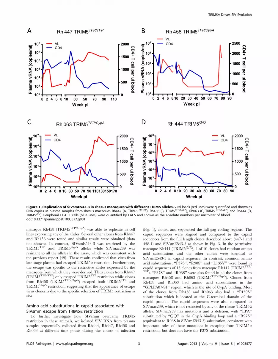
SIVsmm overcame TRIM5a restriction at late stages ofinfection of macaques
To investigate whether SIV could overcome TRIM5a restric-
tion, we monitored the plasma viremia and peripheral blood
CD4+ T cell counts of four SIVsmE543-3-infected rhesus
macaques that had different TRIM5a genotypes. All of these
four macaques were intravenously inoculated with a high dose of
SIVsmE543-3 as previously described [52]. Of these four
macaques, one (Rh447) was homozygous for the restrictive
TRIM5TFP allele, two (Rh458 and Rh063) were heterozygous
for the restrictive TRIM5TFP and TRIM5CypA alleles, and one
(Rh444) was homozygous for the permissive TRIM5Q allele. The
macaques with the restrictive TRIM5TFP/TFP (Rh447) and
TRIM5TFP/CypA (Rh458 and Rh063) genotypes had much lower
plasma viral loads than the macaque with the permissive
TRIM5Q/Q genotype (Rh444) during the acute stage of infection
(Fig. 1). The peak plasma viremia at the acute stage of infection
varied from 103 to 105 copies per ml in these three macaques
(Rh447, Rh458 and Rh063), at least two logs lower than in the
permissive macaque Rh444. All three macaques with restrictive
alleles maintained stable peripheral blood CD4+ T cell counts
during acute infection (above 1000 CD4+ T cells per ml blood),
while a significant loss of CD4+ T cells was observed in macaque
Rh444. These results were consistent with a previous report, which
revealed that TRIM5TFP and TRIM5CypA alleles restrict
SIVsmE543-3 replication in rhesus macaques [49]. However, we
also observed that at a later stage of infection, plasma viral loads in
macaques Rh447, Rh458 and Rh063 increased to 106 to 107
copies per ml, which was comparable to that observed in the
permissive macaque Rh444. Increasing viremia was accompanied
by a significant loss of CD4+ T cells (less than 300 CD4+ T cells
per ml blood). All the macaques progressed to AIDS with
opportunistic infections within two to three years post infection
despite the differences in their TRIM5a genotypes. These results
suggested to us that SIV may have escaped TRIM5TFP and
TRIM5CypA restriction at late stages of infection.
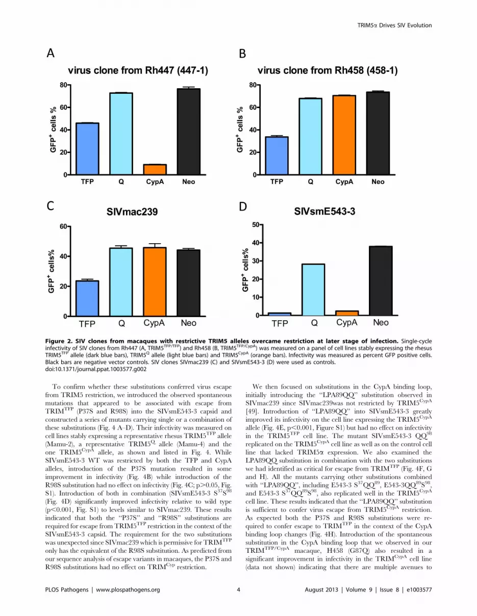
To confirm this hypothesis, we isolated SIV RNA from plasma
samples of macaques Rh447 (at 78 w.p.i., weeks post infection) and
Rh458 (at 110 w.p.i.), and obtained 8.5 kb RT-PCR products
including the full gag-pol-env coding region. The PCR products
were subcloned into the SIVsmE543-3 LTR backbone to
construct full-length virus clones. Infectivity of these virus clones
was measured on cell lines stably expressing different rhesus
TRIM5a alleles by single-cycle infectivity assay as previously
described [49]. As shown in Fig. 2, a representative virus clone,
447-1 from macaque Rh447 (TRIM5TFP/TFP) replicated in cell
lines expressing TRIM5TFP or TRIM5Q alleles but was still
restricted by the TRIM5CypA allele. A virus clone, 458-1 from
Author Summary
Human immunodeficiency virus (HIV) resulted from thetransmission of simian immunodeficiency viruses (SIV)from nonhuman primates followed by adaptation andexpansion as a pandemic in humans. This required thevirus to overcome a variety of intrinsic host restrictionfactors in humans in order to replicate efficiently. Similarly,SIV encounters restriction factors upon cross-speciestransmission between nonhuman primates, specificallyfrom a natural host species such as sooty mangabeymonkeys to rhesus macaques. Previously we observedsignificant differences in the levels of virus replication ofSIV among rhesus macaques due to subtle differences inone of these restriction factors, TRIM5 among individualmacaques. Although a restrictive version of TRIM5 resultedin lower viremia, we also observed that the virusspontaneously mutated in the viral capsid gene and thatthese mutations were associated with escape from TRIM5restriction. In the present study, we found that introduc-tion of these escape mutations into the parental virusconfers resistance to TRIM5 both in tissue culture and inmacaques. These studies provide direct evidence thatTRIM5 is a critical factor influencing the cross-speciestransmission of SIV in primates.
TRIM5a Drives SIV Evolution
PLOS Pathogens | www.plospathogens.org 2 August 2013 | Volume 9 | Issue 8 | e1003577
macaque Rh458 (TRIM5TFP/CypA) was able to replicate in cell
lines expressing any of the alleles. Several other clones from Rh447
and Rh458 were tested and similar results were obtained (data
not shown). In contrast, SIVsmE543-3 was restricted by the
TRIM5TFP and TRIM5CypA alleles while SIVmac239 was
resistant to all the alleles in the assay, which was consistent with
the previous report [49]. These results confirmed that virus from
late stage plasma had escaped TRIM5a restriction. Furthermore,
the escape was specific to the restrictive alleles expressed by the
macaques from which they were derived. Thus clones from Rh447
(TRIM5TFP/TFP) only escaped TRIM5TFP restriction while clones
from Rh458 (TRIM5TFP/CypA) escaped both TRIM5TFP and
TRIM5CypA restriction, suggesting that the appearance of escape
virus clones is due to the specific selection of TRIM5 restriction in
vivo.
Amino acid substitutions in capsid associated withSIVsmm escape from TRIM5a restriction
To further investigate how SIVsmm overcame TRIM5
restriction in these animals, we isolated SIV RNA from plasma
samples sequentially collected from Rh444, Rh447, Rh458 and
Rh063 at different time points during the course of infection
(Fig. 1), cloned and sequenced the full gag coding regions. The
capsid sequences were aligned and compared to the capsid
sequences from the full length clones described above (447-1 and
458-1) and SIVsmE543-3 as shown in Fig. 3. In the permissive
macaque Rh444 (TRIM5Q/Q), 4 of 10 clones had random amino
acid substitutions and the other clones were identical to
SIVsmE543-3 in capsid sequence. In contrast, common amino
acid substitutions, ‘‘P37S’’, ‘‘R98S’’ and ‘‘L135V’’ were found in
capsid sequences of 13 clones from macaque Rh447 (TRIM5TFP/
TFP). ‘‘P37S’’ and ‘‘R98S’’ were also found in all the clones from
macaques Rh458 and Rh063 (TRIM5TFP/CypA). Clones from
Rh458 and Rh063 had amino acid substitutions in the
‘‘GPLPA87-91’’ region, which is the site of CypA binding. Most
of the clones from Rh458 and Rh063 also had a ‘‘P159S’’
substitution which is located at the C-terminal domain of the
capsid protein. The capsid sequences were also compared to
SIVmac239, which is not restricted by any of the rhesus TRIM5aalleles. SIVmac239 has mutations and a deletion, with ‘‘LPA’’
substituted by ‘‘QQ’’ in the CypA binding loop and a ‘‘R97S’’
(equivalent to R98S in SIVsmE543-3) substitution, consistent with
important roles of these mutations in escaping from TRIM5arestriction, but does not have the P37S substitution.
Figure 1. Replication of SIVsmE543-3 in rhesus macaques with different TRIM5 alleles. Viral loads (red lines) were quantified and shown asRNA copies in plasma samples from rhesus macaques Rh447 (A, TRIM5TFP/TFP), Rh458 (B, TRIM5TFP/CypA), Rh063 (C, TRIM5 TFP/CypA) and Rh444 (D,TRIM5Q/Q). Peripheral CD4+ T cells (blue lines) were quantified by FACS and shown as the absolute numbers per microliter of blood.doi:10.1371/journal.ppat.1003577.g001
TRIM5a Drives SIV Evolution
PLOS Pathogens | www.plospathogens.org 3 August 2013 | Volume 9 | Issue 8 | e1003577
To confirm whether these substitutions conferred virus escape
from TRIM5 restriction, we introduced the observed spontaneous
mutations that appeared to be associated with escape from
TRIMTFP (P37S and R98S) into the SIVsmE543-3 capsid and
constructed a series of mutants carrying single or a combination of
these substitutions (Fig. 4 A–D). Their infectivity was measured on
cell lines stably expressing a representative rhesus TRIM5TFP allele
(Mamu-2), a representative TRIM5Q allele (Mamu-4) and the
one TRIM5CypA allele, as shown and listed in Fig. 4. While
SIVsmE543-3 WT was restricted by both the TFP and CypA
alleles, introduction of the P37S mutation resulted in some
improvement in infectivity (Fig. 4B) while introduction of the
R98S substitution had no effect on infectivity (Fig. 4C; p.0.05, Fig.
S1). Introduction of both in combination (SIVsmE543-3 S37S98
(Fig. 4D) significantly improved infectivity relative to wild type
(p,0.001, Fig. S1) to levels similar to SIVmac239. These results
indicated that both the ‘‘P37S’’ and ‘‘R98S’’ substitutions are
required for escape from TRIM5TFP restriction in the context of the
SIVsmE543-3 capsid. The requirement for the two substitutions
was unexpected since SIVmac239 which is permissive for TRIMTFP
only has the equivalent of the R98S substitution. As predicted from
our sequence analysis of escape variants in macaques, the P37S and
R98S substitutions had no effect on TRIMCyp restriction.
We then focused on substitutions in the CypA binding loop,
initially introducing the ‘‘LPA89QQ’’ substitution observed in
SIVmac239 since SIVmac239was not restricted by TRIM5CypA
[49]. Introduction of ‘‘LPA89QQ’’ into SIVsmE543-3 greatly
improved its infectivity on the cell line expressing the TRIM5CypA
allele (Fig. 4E, p,0.001, Figure S1) but had no effect on infectivity
in the TRIM5TFP cell line. The mutant SIVsmE543-3 QQ98
replicated on the TRIM5CypA cell line as well as on the control cell
line that lacked TRIM5a expression. We also examined the
LPA89QQ substitution in combination with the two substitutions
we had identified as critical for escape from TRIMTFP (Fig. 4F, G
and H). All the mutants carrying other substitutions combined
with ‘‘LPA89QQ’’, including E543-3 S37QQ89, E543-3QQ89S98,
and E543-3 S37QQ89S98, also replicated well in the TRIM5CypA
cell line. These results indicated that the ‘‘LPA89QQ’’ substitution
is sufficient to confer virus escape from TRIM5CypA restriction.
As expected both the P37S and R98S substitutions were re-
quired to confer escape to TRIMTFP in the context of the CypA
binding loop changes (Fig. 4H). Introduction of the spontaneous
substitution in the CypA binding loop that we observed in our
TRIMTFP/CypA macaque, H458 (G87Q) also resulted in a
significant improvement in infectivity in the TRIMCypA cell line
(data not shown) indicating that there are multiple avenues to
Figure 2. SIV clones from macaques with restrictive TRIM5 alleles overcame restriction at later stage of infection. Single-cycleinfectivity of SIV clones from Rh447 (A, TRIM5TFP/TFP) and Rh458 (B, TRIM5TFP/CypA) was measured on a panel of cell lines stably expressing the rhesusTRIM5TFP allele (dark blue bars), TRIM5Q allele (light blue bars) and TRIM5CypA (orange bars). Infectivity was measured as percent GFP positive cells.Black bars are negative vector controls. SIV clones SIVmac239 (C) and SIVsmE543-3 (D) were used as controls.doi:10.1371/journal.ppat.1003577.g002
TRIM5a Drives SIV Evolution
PLOS Pathogens | www.plospathogens.org 4 August 2013 | Volume 9 | Issue 8 | e1003577

achieve resistance to TRIMCypA. As expected, the mutant E543-3
S37QQ89S98, which carried the combination of substitutions
‘‘P37S’’, ‘‘R98S’’ and ‘‘LPA89QQ’’, was able to replicate on cell
lines expressing either TRIM5TFP or TRIM5CypA alleles. All of
these mutants replicated well on cell lines expressing the
permissive TRIM5Q alleles. These results are summarized in
Table 1 and confirmed that the amino acid substitutions carried by
late-stage clones from macaques with TRIM5TFP or TRIM5CypA
alleles helped the virus overcome TRIM5a restriction.
Escape from TRIM5a restriction improved virus fitness inrhesus macaques with restrictive TRIM5a alleles
We further investigated whether the escape from TRIM5
restriction improved virus fitness in rhesus macaques that expressed
restrictive TRIM5 alleles. Due to the low frequency of the
TRIM5CypA allele in rhesus macaque populations, we did not have
sufficient macaques that were homozygous for the TRIM5CypA
allele for in vivo studies. Hence we only compared the replication of
SIVsmE543-3 and SIVsmE543-3 S37S98, the mutant which carries
both the ‘‘P37S’’ and ‘‘R98S’’ substitutions, in TRIM5TFP-
homozygous macaques. Before inoculation into macaques, we
investigated virus replication in cultured PBMC. After activation
with phytohemagglutinin (PHA), PBMC from 18 macaques with a
TRIM5TFP/TFP genotype were infected with SIVsmE543-3 and
SIVsmE543-3 S37S98 at a multiplicity of infection (M.O.I.) of 0.001.
PBMC from five macaques with a TRIM5Q/Q genotype served as
positive controls. Virus production was measured by monitoring
reverse transcriptase (RT) activity of supernatant collected at 3-day
intervals and quantified with the phosphor imaging. Fig. 5 A and B
show the replication kinetics of wild type and the variant SIV in
PBMC of a representative TRIM5TFP/TFP and TRIM5Q/Q
macaque, respectively. In PBMC from macaque RhDCWW
(TRIM5Q/Q), SIVsmE543-3 replicated as well as mutant
SIVsmE543-3 S37S98. Meanwhile in PBMC from macaque
RhDBF7 (TRIM5TFP/TFP), the mutant replicated better than wild
type SIVsmE543-3. To make a quantitative comparison, the area
under the replication curve (AUC), which is indicative of total
amount of virus production during the culture, was calculated and
compared pairwise between these two viruses. As shown in Fig. 5 C
and D, the mutant SIVsmE543-3 S37S98 replicated better than wild
type in 18 TRIM5TFP/TFP macaques and the difference was
significant (Paired T test, P,0.01). In contrast, the replication of the
two viruses in 5 TRIM5Q/Q macaque PBMC was not significantly
different (Paired T test, P.0.05).
Then we compared the replication of these wild type
SIVsmE543-3 and the putative TRIMTFP-resistant variant
(SIVsmE543-3S37S98) in vivo. Twelve rhesus macaques with
TRIM5TFP/TFP genotypes were divided into two groups; relative
Figure 3. Identification of amino acid substitutions associated with escape from TRIM5 restriction. The capsid amino acids of SIV clonesfrom Rh444 (TRIM5Q/Q), Rh447 (TRIM5TFP/TFP), Rh458 (TRIM5TFP/CypA) and Rh063 (TRIM5TFP/CypA) were aligned to parental SIVsmE543-3. Identical aminoacids were shown as dot (.), deletions are shown as dash (-). Amino acid substitutions shared among SIV clones from different macaques werehighlighted with yellow. Amino acid substitutions in the CypA binding loop are highlighted in red. The critical amino acid residues identified asresponsible for escape from TRIM restriction are indicated by numbers above the sequence.doi:10.1371/journal.ppat.1003577.g003
TRIM5a Drives SIV Evolution
PLOS Pathogens | www.plospathogens.org 5 August 2013 | Volume 9 | Issue 8 | e1003577
in vitro infectivity of the variant in PBMC of the animals was not
considered in selection. To minimize the effect of MHC restriction
on virus load, the distribution of MHC I genotypes known to have
an effect on SIVmac239 viremia was balanced between the two
groups as shown in Table 2. Before inoculation, viruses were
expanded on PBMC from the permissive TRIM5Q/Q macaque,
RhDCCW. Gag sequences of the two virus stocks were evaluated
after expansion on PBMC and no additional mutations were
found in either of the two virus stocks (data not shown). Each
macaque was inoculated intrarectally (I.R.) with 1000 TCID50
(56105 RNA copies of virus) and the infection was evaluated by
monitoring plasma viral RNA load. Four weeks later, any of the
macaques that remained uninfected were inoculated intrarectally
on a weekly schedule with the same amount of virus until they
became infected. There was no correlation between expression of
a known restrictive MHC-I genotype and either acquisition or
viral load in acute phase of infection. We compared the acquisition
of infection of the group receiving SIVsmE543-3 and SIVsmE543-
3S37S98 by Kaplan-Meier curves as shown in Fig. 6A. In the group
inoculated with the variant, SIVsmE543-3 S37S98, four of six
macaques were infected after the first inoculation, while in the
group inoculated with the wild type SIVsmE543-3 only two
macaques were infected after the first inoculation. Macaques
inoculated with SIVsmE543-3 S37S98 required significantly less
exposure to get infected than macaques inoculated with
SIVsmE543-3 (log-rank test, P = 0.0452). It required 3.5 inocula-
tions to infect half of the macaques with SIVsmE543-3, while it
only required one inoculation by SIVsmE543-3 S37S98. We also
compared the plasma viral loads of these two groups after
infection. The plasma viral loads for each macaque and the
median plasma viral load for each group are shown in Fig. 6 B and
C. Macaques infected with SIVsmE543-3 S37S98 had significantly
higher plasma viremia compared with macaques infected with
SIVsmE543-3, with mean differences of 105-fold at peak, and 25-
fold at 8 w.p.i. As a measure of cumulative virus replication, we
also compared the area under the curve (AUC) during the acute
phase of infection (1–8 weeks) and observed a 70-fold higher level
Figure 4. Introduction of amino acid substitutions into SIVsmE543-3 capsid conferred virus resistance to TRIM5 restriction. Single orcombinations of amino acid substitutions ‘‘P37S’’, ‘‘LPA89QQ’’ and ‘‘R98S’’ were introduced into SIVsmE543-3 capsid. Single-cycle infectivity of thesemutants was measured on a panel of cell lines stably expressing a TRIM5TFP allele (Mamu-2, dark blue bars), TRIM5Q alleles (Mamu-4, light blue bars)and TRIM5CypA (orange bars). Infectivity was measured as percent GFP positive cells. Black bars are negative vector controls. Infectivity on this panelare shown for SIVsmE543-3 (A), SIVsmE543-3 S37 (B), SIVsmE543-3 S98 (C), SIVsmE5433-3 S37 S98 (D), SIVsmE543-3 QQ89 (E), SIVsmE543-3 S37 QQ89 (F),SIVsmE543-3 QQ89 S98 (G) and SIVsmE543-3 S37 QQ89 S98 (H).doi:10.1371/journal.ppat.1003577.g004
Table 1. Summary of virus mutants and TRIM5 resistance.
Virus Gag Capsid MutationTRIM5resistance
37 87–91 98
E543 P GPLPA R -
E543 S37 S GPLPA R -
E543 S98 P GPLPA S -
E543 S37S98 S GPLPA S TFP
E543 QQ89 P GPQQ R CypA
E543 S37QQ89 S GPQQ R CypA
E543QQ89S98 P GPQQ S CypA
E543 S37QQ89S98 S GPQQ S TFP/CypA
doi:10.1371/journal.ppat.1003577.t001
TRIM5a Drives SIV Evolution
PLOS Pathogens | www.plospathogens.org 6 August 2013 | Volume 9 | Issue 8 | e1003577
in macaques inoculated with the variant virus (Fig. 6 D,E and F).
The in vivo infection results, combined with in vitro PBMC infection
results, indicated that the ‘‘P37S’’ and ‘‘R98S’’ substitutions
improved virus fitness in macaques with TRIM5TFP/TFP geno-
types, which also suggested that the appearance of variants
carrying these mutations was due to TRIM5a selection.
TRIM5a exerted selective pressure on SIVsmmtransmission into macaques
We were interested in determining whether the changes we
observed in SIVsmE543 could be generalized to other SIVsmm/
mac strains so we investigated the capsid sequences of SIV isolates
commonly used in NHP models for evidence of TRIM5 selection.
Four lineages of SIV strains, including SIVsmm, SIVmac, SIVstm
and SIVmne, were isolated from infected macaques during several
independent SIV transmission events in the 1970s to 1980s
[4,51,53,54,55,56,57,58,59,60,61,62,63,64]. The macaque pas-
sage history of commonly used SIV isolates is briefly summarized
in Table 3 and the sequences of their capsid N-terminal domains
are aligned and showed in Fig. 7. Phylogenetic analysis indicated
that all of these SIV isolates originate from the cross-transmission
of SIVsmm from sooty mangabeys, followed by unknown or
intended experimental passages in different species including
rhesus, pigtail and stump-tailed macaques [65,66,67]. Capsid
sequences of primary clones of SIV directly isolated from sooty
mangabey monkeys [66] were highly conserved. All SIVsmm
Figure 5. Comparison of PBMC infection with SIVsmE543-3 and SIVsmE543-3 S37S98. PBMCs were collected from 18 macaques with aTRIM5TFP/TFP genotype and 5 macaques with a TRIM5Q/Q genotype and activated with PHA for 3 days. Activated PBMCs were infected withSIVsmE543-3 and SIVsmE543-3 S37S98 at a M.O.I. of 0.001. Virus production was quantified and shown as RT values in supernatants collected at 3-dayintervals. A and B show the replication kinetics of SIVsmE543-3 and SIVsmE543-3 S37S98 in PBMCs of representative macaques RhDBF7 (TRIM5TFP/TFP)and RhDCCW (TRIM5Q/Q). Area under the replication curve (AUC) of SIVsmE543-3 and the variant were calculated for each macaque separately. TheAUC of SIVsmE543-3 and its variant in the TRIM5TFP/TFP group (C) and the TRIM5Q/Q group (D) are shown and compared by paired t test.doi:10.1371/journal.ppat.1003577.g005
TRIM5a Drives SIV Evolution
PLOS Pathogens | www.plospathogens.org 7 August 2013 | Volume 9 | Issue 8 | e1003577

clones from sooty mangabeys encoded a ‘‘P37’’ and ‘‘R98’’ in their
capsid, similar to SIVsmE543-3. Some SIVsmm clones encoded
an ‘‘I91L’’ variation in the CypA binding loop which does not
affect sensitivity to TRIMCypA restriction [49]. The lack of
variability in the sequence alignment of primary SIVsmm capsid
proteins suggested that there was no TRIM5a restriction and
selection during the passage of SIVsmm among sooty mangabeys.
The limited passage of SIVsmm from the Tulane Primate Center
in rhesus macaques resulted in the isolation of several SIVsmm
clones including SIVsmH4, SIVsmE543-3, and SIVsmE660
clones FL6 and FL14 [4,54,68]. All of these clones, except for
SIVsmE660-FL14, carried the‘‘P37’’, ‘‘R98’’ and ‘‘LPA89’’ ob-
served in the capsids of primary SIVsmm clones. SIVsmE660-FL14
had a ‘‘A91P’’ substitution, which allowed escape from TRIM5CypA
restriction (data not shown), suggesting that the passage history of
SIVsmE660 may have involved exposure to TRIM5CypA selection.
The long-term passage of SIVsmm in rhesus macaques resulted in
the isolation of SIVmac [55,69]. SIVmac251 clones and SIV-
mac239 had ‘‘LPA89QQ’’ and ‘‘R97S’’ (equivalent to R98S in
SIVsmm) substitutions in their capsids. SIVmac142, which is
independent of the SIVmac251/239 passage chain, had the ‘‘P37S’’
substitution in addition to the ‘‘LPA89QQ’’ and ‘‘R97S’’substitu-
tions. SIVstm underwent an unknown history of passage in stump-
tailed macaques (Macaca arctoides) after the original SIVsmm cross-
species transmission. Only two SIVstm clones are available.
SIVstm37.16, which was directly cloned from an infected stump-
tailed macaque [56], only has the ‘‘R98S’’ substitution in capsid.
The other, SIVstm22579, isolated after two passages in rhesus
macaques [57] has both the ‘‘P37S’’ and ‘‘R98S’’ in capsid
identified as critical for escape from TRIMTFP restriction but no
changes in the GPLPA motif. Finally, SIVsmPBj which has a history
of passage in pigtail macaques has a substitution in the CypA
binding loop of capsid, ‘‘A91P’’ conferring virus escape from
TRIM5CypA restriction, which is consistent with pigtail macaques
exclusively carrying the TIRM5CypA allele [22]. The variance of
these mutations in different SIV clones was associated with their
different passage history in macaques, suggestive of TRIM5
selection during their cross-transmission and adaptation. However
a common pathway of escape mutations similar to what we
observed spontaneously in SIVsmE543 was observed in all of these
adapted viruses.
Discussion
The antagonistic interaction with host restriction proteins is
considered a major driver of evolutionary change for viruses. We
previously reported that the polymorphism of TRIM5a affected
SIVsmm replication in rhesus macaques, which suggested that
TRIM5a restriction also plays an important role in HIV/SIV
evolution [49]. Here we showed that selection of TRIM5 resistant
SIVsmm capsid mutants at late stages of infection in rhesus
macaques expressing restrictive TRIM5 alleles. The resistance to
TRIM5 restriction was associated with three common amino acid
substitutions in the N-terminal domain of the capsid protein. By
site-direct mutagenesis, we confirmed that these substitutions
conferred TRIM5 restriction and improved the virus fitness in
rhesus macaques with restrictive TRIM5 alleles. These results
provide both in vitro and in vivo evidence to support the hypothesis
that TRIM5 exerts selective pressure on SIV evolution.
Our results also demonstrate the value of using SIV infected
rhesus macaques to study the influence of restriction proteins
during HIV/SIV cross-transmission. Several reports have revealed
that interactions between the host restriction proteins APO-
BEC3G and Tetherin/BST-2 with HIV/SIV resulted in the
acquisition and/or evolution of the Vif, Vpu and Nef proteins
[70,71,72,73]. Since it is impossible to trace back the cross
transmission events which occurred hundreds or thousands of
years ago, most of these studies were based on phylogenetic
analysis of host restriction genes and SIV sequences in different
primate species followed by in vitro infection inhibition assays.
The question remaining however is in what way the selection and
adaptation occur. In this paper, macaques expressing restrictive
TRIM5 alleles were infected by two different routes. Three
macaques (Rh447, Rh458 and Rh063) were intravenously
inoculated with a high dose of wild type SIVsmE543-3. For these
macaques, the restrictive alleles did not prevent infection but the
virus maintained a low level of replication until the appearance of
mutations associated with TRIM5 resistance. The other twelve
macaques were inoculated by a more biologically relevant mucosal
method using repetitive lower dose intrarectal inoculation with the
wild type SIVsmE543-3 or the TRIM5-resistant SIVsmE543-
3S37S98 mutant. This allowed us to observe that the TRIM5-
resistant mutant had enhanced acquisition of infection in addition
to improved viral replication when compared to wild type virus.
To determine whether this was a phenomenon that could be
generalized to all SIVsmm strains, we attempted to trace the
influence of TRIM5 selection on the cross-species transmission of
SIVsmm into captive macaques in the 1970s. The primary
SIVsmm clones derived directly from sooty mangabeys showed a
high degree of conservation in the capsid protein despite being
isolated from separate mangabeys in different U.S. national
primate centers or from wild caught animals in West Africa [66].
None of the SIVsmm clones had any of the amino acid
substitutions that have been associated with escape from TRIM5arestriction. Since all evidence suggests that SIVsmm has circulated
among sooty mangabeys for quite a long time, it may be
completely adapted to this host and not restricted by sooty
TRIM5a. Polymorphism of TRIM5a has also reported in sooty
mangabeys and four different alleles have been identified [47].
However, none of these sooty TRIM5a alleles restricted
SIVsmE543-3 replication in single-cycle infectivity assays (data
not shown) consistent with the long-term co-evolution of this virus
in this species. It also suggests that TRIM5 restriction and
Table 2. MHC genotype of macaques.
SIV virus Macaque ID MHC I
SIVsmE543-3 Rh824 A08, B01
Rh825 A02, B01
Rh828 A01, A08, B17
Rh829 Negative
Rh830 A08
Rh831 B17
SIVsmE543-3 S37S98 Rh826 B01
Rh827 A02, A08
Rh832 A01, B01
Rh833 A08, B01, B08
Rh834 B17
Rh835 Negative
Nine rhesus MHC class I alleles, including Mamu-A*001, A*002, A*008, A*011,B*001, B* 003, B*004, B*008, B*017, were tested and listed for each macaque.Negative: none of the 9 alleles observed. MHC I genotypes known to have aneffect on SIVmac239 viremia are indicated in bold.doi:10.1371/journal.ppat.1003577.t002
TRIM5a Drives SIV Evolution
PLOS Pathogens | www.plospathogens.org 8 August 2013 | Volume 9 | Issue 8 | e1003577

selection occurred mainly at the time when SIV was being
introduced into a new primate species. When compared with
primary SIVsmm clones from sooty mangabeys, many SIV clones
that evolved in macaques following experimental passage,
including SIVmac, SIVsmm, SIVstm and SIVmne, had amino
acid substitutions/deletions at the sites under TRIM5 selection in
Figure 6. Acquisition and replication of SIVsmE543-3 and SIVsmE543-3 S37S98 in macaques with TRIM5TFP/TFP genotype. Eachmacaque was inoculated intrarectally (I.R.) with 1000 TCID50 (56105 RNA copies of virus) and the infection was monitored by measuring plasma viralRNA load. Four weeks later any of the macaques that remained uninfected were inoculated intrarectally on a weekly schedule with same amount ofvirus until they became infected. The acquisition of infection in each group was shown as uninfected percentage after each inoculation andcompared by log-rank test. Median inoculation time was 3.5 for SIVsmE543-3 challenge group and 1 for SIVsmE543-3 S37S98 challenge group (A).Plasma viral RNA copies in each macaque (B) and median plasma viral RNA copies in each group (C) are shown. Peak plasma viral loads (D, P = 0.0152),plasma viral loads at 8 w.p.i. (E, P = 0.0411) and viral load AUC before 8 w.p.i. (F, P = 0.0260) were compared by non-parametric Mann-Whitney-test.doi:10.1371/journal.ppat.1003577.g006
TRIM5a Drives SIV Evolution
PLOS Pathogens | www.plospathogens.org 9 August 2013 | Volume 9 | Issue 8 | e1003577

capsid. The mutations varied depending on their passage history
in macaques. Although there is no record to trace exactly when
and how SIVsmm was introduced into captive macaques, the
sequence alignment of SIV capsid of these clones revealed that the
TRIM5 genotypes of the macaques during virus passage probably
also exerted selective pressure on SIV evolution in macaques.
SIVsmE543 had only been passaged through two rhesus macaques
and at least one of these rhesus was homozygous permissive
for TRIM5 [49], which is consistent with the lack of escape
substitutions in the capsid of this virus. Other viruses exhibited a
range of escape mutations, with viruses passaged the most
extensively in rhesus macaques, such as SIVmac239, exhibiting
escape from both restrictive alleles and others such as those
passaged in pigtail macaques (and thus only subjected to
TRIMCypA selection), only having changes in the Cyclophylin A
binding loop of capsid. Such a process of restriction and selection
may also affect SIV infection in primates of other species, as is
indicated by the observed variation of SIV capsid sequences from
Table 3. Passage history of SIV clones.
SIV clones In vivo macaque passage Reference
Clone Name Accession Number Sooty Mangabey Stump-tail Rhesus Pig tail
SIVsm SIVsmH4 X14307 SM E038 NO RhF236 NO [4]
SIVsmE543 U72748 SM E038 NO RhF236 RhE5432 NO [51]
SIVsmE660-FL6 JQ864085 SM E038 NO RhF236 RhE543 RhE660 NO [54]
SIVsmE660-FL14 JQ864087 SM E038 NO RhF236 RhE543 RhE660 NO [54]
SIVmac1 SIVmac251 1A11 M76764 unknown NO Rh78 Rh251 NO [55,58]
SIVmac251 BK28 M19499 unknown NO Rh78 Rh251 NO [55]
SIVmac251 32H D01065 unknown NO Rh78 Rh251 Rh32H NO [53,55]
SIVmac239 M33262 unknown NO Rh78 Rh251 Rh61 Rh239 NO [55,59]
SIVmac142 Y00277 unknown NO Rh142 NO [55,60]
SIVstm SIVstm37.16 M83293 unknown N.A. NO NO [56]
SIVstm22579 X60667 unknown N.A. Rh21685 Rh22579 NO [57]
SIVPBj SIVPBj6.6 L09212 unknown NO NO PT PBj [61]
SIVmne SIVmne08 M32741 unknown NO NO PT T76321 [62]
SIVmne27 U79412 unknown NO NO PT T76321PT T78027
[62,63]
SIVmne170 U95965 unknown NO NO PT T76321PT T78027
[62,64]
1: For SIVmac, unrecorded passages in rhesus macaques may be involved before passage in the listed rhesus macaques.2. RhE543 is the only macaque with known TRIM5 genotype (TRIM5Q/Q).doi:10.1371/journal.ppat.1003577.t003
Figure 7. Variance of SIV capsid sequences is associated with their passage history in macaques. The capsid N-terminal domain of SIVclones, with or without passage in rhesus, stump-tailed and pigtail macaques, were aligned to a primary SIVsm clone from sooty mangabey (top).Identical amino acids were shown as a dot (.), deletions are shown as a dash (-). The sites under TRIM5 selection are highlighted in yellow and theCyclophilin-A binding site is highlighted in light blue and the critical amino acid residues identified as responsible for escape from TRIM restriction areindicated by numbers above the sequence.doi:10.1371/journal.ppat.1003577.g007
TRIM5a Drives SIV Evolution
PLOS Pathogens | www.plospathogens.org 10 August 2013 | Volume 9 | Issue 8 | e1003577
primates of different species [74]. However the amino acid sites
associated with resistance to TRIM5 may be not the same as what
we observed in SIVsmm-infected macaques and will require
further study for identification.
Regarding the significance of TRIM5 polymorphisms in
modulating HIV-infection of humans, TRIM5a polymorphism
has also been reported in human populations. Several nonsynon-
ymous SNPs were found in screening of more than 1000 samples
from human populations. Two nonsynonymous SNPs, resulting in
amino acid polymorphisms H43Y in the RING domain and
R136Q in the coiled-coiled domain, were reported to affect the in
vitro TRIM5a restriction of HIV-1 and were differently distributed
in HIV-1 positive and negative cohorts [43,44,45,46,75]. Howev-
er, whether these SNPs actually affect virus acquisition result is
controversial since some groups suggested that the effect of
TRIM5a polymorphisms on HIV-1 disease progression was
minimal [46]. Since HIV-1 had already circulated in a small
population in Africa for several decades before causing a
worldwide pandemic in the 1980s [76], the virus may have
already adapted and escaped from human TRIM5a restriction
during early passage in humans. Furthermore, the TRIM5asequences within human beings are much less divergent than
within rhesus macaques and only few polymorphisms in human
TRIM5a were located in the SPRY domain, which may be due to
the lack of positive selection by other retroviruses during the
evolution of humans [43]. The low variability of human TRIM5amay also explain the lack of restriction of human TRIM5a on
HIV-1 replication.
Our studies also provide a model to study the mechanism of
TRIM5a restriction. The conserved capsid protein is a potential
target for anti-HIV drug development because of its important
role in virus replication. Studies on interaction between TRIM5aand the HIV/SIV capsid will provide useful information for anti-
HIV drug development. However, the mechanism of TRIM5arestriction on HIV/SIV replication remains unclear. A recent
report by Pertel et al. indicated that interaction of TRIM5a with
the retrovirus capsid lattice promotes innate immune signaling
[34]. Another report by Battivelli et al. showed that the CTL
escape mutants isolated from HIV-infected patients are more
sensitive to human TRIM5a restriction than laboratory-adapted
HIV-1 strains [77]. These reports revealed that TRIM5arestriction of retroviruses is not only mediated through capsid
lattice dissociation and degradation brought on by interaction
between TRIM5a and the capsid protein, but are also linked to
innate and adaptive immune responses. Macaques infected with
SIVsmE543-3 and its TRIM5-resistant variants will be a valuable
animal model to study the mechanism of TRIM5 restriction in
vivo.
Our study also has quite practical applications to improve the
models of SIV-infected macaques for HIV-1 research. The
present study focused on escape mutations for one of the most
common restrictive TRIM5 alleles to determine whether the
spontaneous substitutions were associated with biological escape
from TRIM5 restriction in vivo. Obviously a virus that has
escaped both restrictive alleles is still required as a challenge virus
for vaccine trials. SIVsmE543-3, although being pathogenic, has
not been widely used as a challenge strain and thus may not be
the ideal target for these studies. SIVsmE660, which was isolated
from a SIVsmE543 infected macaque, is widely used as a
challenge stock in the rhesus macaque model for vaccine
evaluation. Several groups reported that the replication of
SIVsmE660 was also affected by TRIM5 alleles: these studies
suggest that TRIM restriction of this virus can affect acquisition
of infection in repetitive ‘‘low’’ dose intra-rectal challenge models
[78,79]. This effect can confound vaccine evaluation unless study
groups are balanced for TRIM5 alleles or animals with
restrictive alleles are excluded from the study. In our previous
report, we described several infectious molecular clones from a
SIVsmE660 virus stock and found that all of the clones were
restricted by TRIM5TFP, and one SIVsmE660 clone had
TRIM5CypA escape mutations due to substitutions in the CypA
binding loop of capsid [54]. The results in this paper suggest that
the introduction of the ‘‘P37S’’ ‘‘R98S’’ substitutions into
SIVsmE660 clones may confer escape from TRIM5TFP restric-
tion, and provide virus clones not restricted by TRIM5 for use as
challenge viruses in vaccine trials. The replication and patho-
genesis of SIVsmE660 clones with these mutations is under
evaluation in rhesus macaques.
Materials and Methods
EthicsThis study was carried out in strict accordance with the
recommendations described in the Guide for the Care and Use
of Laboratory Animals of the National Institute of Health, the
Office of Animal Welfare and the United States Department of
Agriculture. All animal work was approved by the NIAID
Division of Intramural Research Animal Care and Use
Committees (IACUC), in Bethesda, MD (protocol # LMM-6.
The animal facility is accredited by the American Association for
Accreditation of Laboratory Animal Care. All procedures were
carried out under Ketamine anesthesia by trained personnel
under the supervision of veterinary staff and all efforts were made
to ameliorate the welfare and to minimize animal suffering in
accordance with the ‘‘Weatherall report for the use of non-
human primates’’ recommendations. Animals were housed in
adjoining individual primate cages allowing social interactions,
under controlled conditions of humidity, temperature and light
(12-hour light/12-hour dark cycles). Food and water were
available ad libitum. Animals were monitored twice daily (pre-
and post-challenge) and fed commercial monkey chow, treats
and fruit twice daily by trained personnel. Early endpoint
criteria, as specified by the IACUC approved score parameters,
were used to determine when animals should be humanely
euthanized.
AnimalsColony-bred rhesus macaques of Indian origin (Macaca mulatta)
were housed in a BSL2 facility using BSL3 practices. The TRIM5
genotype of rhesus macaques were determined as previously
described [49], and MHC I genotypes were determined by the
Rhesus Macaque MHC Typing Core facility at the University of
Wisconsin. Four rhesus macaques (Rh444, Rh447, Rh458 and
Rh063) were intravenously infected with the SIVsmE543-3
molecular clone as previously described [52]. Blood and plasma
were collected sequentially. The viral RNA levels in plasma were
determined by quantitative reverse transcriptase PCR (RT-PCR)
and blood CD4+ T cell subsets were quantified by flow cytometric
analysis as previously described [54].
RT-PCRVirus RNA was isolated from plasma of macaque Rh447 and
Rh458 with the QiaAmp Viral RNA kit (QIAGEN, Germany).
Reverse transcription of viral RNA to single-stranded cDNA was
performed using the SuperScript III first-strand synthesis system
(Invitrogen, Carlsbad, CA) with primer R-R (59-TGC TTA CTT
CTA AAT GGC AGC TTT-3) according to the manufacturer’s
instructions. Full-length gag-pol-env cassettes (including the entire
TRIM5a Drives SIV Evolution
PLOS Pathogens | www.plospathogens.org 11 August 2013 | Volume 9 | Issue 8 | e1003577

gag, pol, vif, vpr, tat, rev, env genes and parts of the nef gene) were
amplified by nested PCR using Platinum Taq Hi Fidelity
(Invitrogen) polymerase. First-round PCR was performed by using
1 ml of bulk cDNA with primers Nar-F (59-GGTTGGCGC CCG
AAC AGG GAC TT-39) and R-R under the following cycling
conditions: 94uC for 2 min, followed by 35 cycles of 94uC for 30 s,
60uC for 30 s, and 68uC for 9 min, with a final extension of 68uCfor 10 min. Second-round PCR was performed by using 1 ml of
the first-round PCR product with primers Nar-F and Bgl-R (59-
GGC CAA GAT CTG CTG CCA CCT CTG TC-39) under the
same conditions used for the first-round PCR. PCR products were
subcloned into SIVsmE543-3 by cutting with restriction enzymes
NarI and BglII to construct chimeric virus clones.
To sequence the gag gene, virus RNA was isolated from plasma
of macaque Rh444, Rh447, Rh458 and Rh063. Reverse
transcription was performed as described above. 1.8 kb PCR
products covering the full gag region were amplified from 1 ml of
bulk cDNA with primers Nar-F (59-GGTTGGCGC CCG AAC
AGG GAC TT-39) and Gag-R (59-GCT GAT GAT TCA ATT
GTA ACA GG-39) under following cycling conditions: 94uC for
2 min followed by 35 cycles of 94uC for 30 s, 60uC for 30 s, and
68uC for 2 min, with a final extension of 68uC for 5 min. PCR
products were cloned into PCR4-Topo vector with TOPO-TA
cloning kit (Invitrogen) and sequenced. All sequences were
deposited in GenBank under the following accession numbers
(KC904980-KC905000). Gag sequences were aligned using
ClustalX2 and compared with the parental SIVsmE543-3 clone
(GenBank accession U72748) and SIVmac239 (GenBank acces-
sion M33262).
Site-directed mutagenesisAmino acid substitutions ‘‘P37S’’, ‘‘LPA89QQ’’ and ‘‘R98S’’
were introduced into SIVsmE543-3 capsid by using QuikChange
II Site-Directed Mutagenesis Kits (Agilent) according to the
manufacturer’s instructions. The following primer sets were used
to introduce mutations: P37S: forward: 59- GGC AGA GGT AGT
GTC AGG ATT TCA AGC G-39; reverse: 59- CGC TTG AAA
TCC TGA CAC TAC CTC TGC C-39; LPA89QQ: forward: 59-
CAG CCA GGT CCA CAA CAA GGG CAA C-39; reverse: 59-
GTT GCC CTT GTT GTG GAC CTG GCT G-39; R98S:
forward: 59-GCA ACT TAG AGA GCC ATC AGG ATC AGA
CAT TGC AG-39; reverse: 59-CTG CAA TGT CTG ATC CTG
ATG GCT CTC TAA GTT GC-39;
Single-cycle infectivity assayRetroviral vector V1EGFP-SIV and Crandell-Rees Feline
Kidney (CRFK) cell lines stably expressing six common rhesus
TRIM5 alleles, including three TRIM5TFP alleles, two TRIM5Q
alleles, and the TRIM5CypA allele were described in a previous
report [49]. The entire gag-pol-vif segment of SIVsmE543-3
mutants or virus clones from macaque Rh447 and Rh458 were
subcloned into V1EGFP-SIV by cutting with restriction enzymes
NarI and BstBI. Single-cycle SIV viruses were produced in 293T
cells by co-transfection of V1EGFP-SIV and pVSV-G, titered and
infectivity measured on CRFK cell lines stably expressing TRIM5
alleles as previous described [49].
Preparation of virus stocks293T cells were maintained in Gibco GlutaMAx DMEM media
plus 10% FCS, 100 U/ml penicillin, and 100 mg/ml streptomycin
and transfected with 10 mg SIVsmE543-3 mutants plasmid using
FuGENE 6 transfection reagent (Roche Diagnostics, Indianapolis,
IN). Virus stocks were collected from the supernatant of
transfected cells after 48 hours and filtered through a 0.22 um
filter. The tissue culture infectivity dose (TCID50) of virus stocks
was tested on TZM-bl cells and calculated by the Reed & Muench
method [80].
Viruses were expanded on rhesus PBMCs before inoculation
into rhesus macaques. PBMCs were collected from a permissive
TRIM5Q/Q macaque, RhDCCW and cultured in complete RPMI
1640 media containing 10% interleukin-2 (IL-2) After stimulation
with 2 mg/ml phytohemagglutinin (PHA) for 72 hours, activated
PBMC were infected with SIVsmE543-3 and SIVsmE543-3
S37S98, respectively, at an M.O.I. of 0.001. Infected PBMCs were
washed and cultured in complete RPMI 1640 media containing
10% IL-2. Virus stocks were collected at day six and filtered,
followed by titration on TZM-bl cells and quantitation by real-
time RT-PCR. The gag regions were amplified from virus PBMC
stocks by RT-PCR and PCR products were sequenced as
described above.
PBMC infectionPBMCs were collected from 18 macaques with TRIM5TFP/TFP
genotype and five with TRIM5Q/Q genotype and activated with
PHA as described above. 106 activated PBMCs were infected with
103 TCID50 293T transfection virus stocks at 37uC for 60 min-
utes. The infected PBMCs were washed and cultured in complete
RPMI 1640 media containing 10% IL-2. Virus production was
monitored by reverse transcriptase (RT) activity of supernatant
collected at 3-day intervals. RT values were quantified with
Phosphor Imaging (FujiFilm, Japan). Replication curves were
plotted based on RT values.
Animal inoculationsTwelve STLV, SRV and SIV seronegative rhesus macaques
with TRIM5TFP/TFP genotypes were divided into two groups.
The distribution of major histocompatibility complex (MHC)
class I alleles Mamu-A*01, Mamu-B*08, and Mamu-B*17 were
balanced between the two groups. Each macaque was inoculated
intrarectally (I.R.) with a 1:50 dilution of SIVsmE543-3 or
SIVsmE543-3 S37S98 PBMC virus stocks (1000 TCID50, 56105
RNA copies). After inoculation, viral RNA levels in plasma were
determined by quantitative RT-PCR. Four weeks later, any of
the macaques that remained uninfected were inoculated intrar-
ectally on a weekly schedule with same amount of virus until viral
RNAs became detectable in plasma. After infection, blood and
plasma were collected and plasma viral RNA levels were
determined.
Statistical analysesAll statistical analyses and graphic analyses were performed
using GraphPad Prism5 (GraphPad Prism Software, La Jolla,
CA). In vitro PBMC replication was assessed by Area under
replication curves (AUC) calculation and pairwise compared by
paired t test. Infectivity of SIV variants in the single cycle assay
was calculated as a percentage of infectivity of the vector control
in each cell line and compared statistically to the wild type using
One Way ANOVA with Dunnett’s Multiple Comparison Test.
Kaplan Meier curves were plotted based on the inoculation
number before infection and log-rank test was used to compare
the acquisition of infection. Non-parametric Mann-Whitney-test
was used for the comparison of viral loads between the two
groups.
Supporting Information
Figure S1 Statistical comparison of SIVsmE543-3 andmutant replication on cell lines expressing different
TRIM5a Drives SIV Evolution
PLOS Pathogens | www.plospathogens.org 12 August 2013 | Volume 9 | Issue 8 | e1003577

TRIM5 alleles (raw data shown in Fig. 4). The replica-tion of SIVsmE543-3 and its mutants on cell lines expressingTRIM5TFP (A), TRIM5Q (B) and TRIM5TFP (C) alleles areshowed as the percentage of the replication on cell line express-ing vector control. Differences of replication between mutantsand wild type SIVsmE543-3 were compared by one-way analysisof variance (ANOVA) with Dunnett’s post-test. Pairs ofgroups that differed significantly are indicated (**,p,0.01,***,p,0.001).
(TIF)
Acknowledgments
We thank Heather Cronise-Santis, Joanne Swerczek and Richard Herbert
in NIHAC for excellent care of the study animals.
Author Contributions
Conceived and designed the experiments: FW VMH WJ. Performed the
experiments: FW RG SW AK JSM ABW RP KT IO KM LH. Analyzed
the data: FW AK VMH WJ. Contributed reagents/materials/analysis
tools: RG SW IO KM. Wrote the paper: FW VMH.
References
1. Hahn BH, Shaw GM, De Cock KM, Sharp PM (2000) AIDS as a zoonosis:
scientific and public health implications. Science 287: 607–614.
2. Sharp PM, Hahn BH (2011) Origins of HIV and the AIDS pandemic. Cold
Spring Harb Perspect Med 1: a006841.
3. Gao F, Bailes E, Robertson DL, Chen Y, Rodenburg CM, et al. (1999) Origin of
HIV-1 in the chimpanzee Pan troglodytes troglodytes. Nature 397: 436–441.
4. Hirsch VM, Olmsted RA, Murphey-Corb M, Purcell RH, Johnson PR (1989)
An African primate lentivirus (SIVsm) closely related to HIV-2. Nature 339:
389–392.
5. Gao F, Yue L, White AT, Pappas PG, Barchue J, et al. (1992) Human infection
by genetically diverse SIVSM-related HIV-2 in west Africa. Nature 358: 495–
499.
6. Chen Z, Telfier P, Gettie A, Reed P, Zhang L, et al. (1996) Genetic
characterization of new West African simian immunodeficiency virus SIVsm:
geographic clustering of household-derived SIV strains with human immuno-
deficiency virus type 2 subtypes and genetically diverse viruses from a single feral
sooty mangabey troop. J Virol 70: 3617–3627.
7. Jin MJ, Rogers J, Phillips-Conroy JE, Allan JS, Desrosiers RC, et al. (1994)
Infection of a yellow baboon with simian immunodeficiency virus from African
green monkeys: evidence for cross-species transmission in the wild. J Virol 68:
8454–8460.
8. van Rensburg EJ, Engelbrecht S, Mwenda J, Laten JD, Robson BA, et al. (1998)
Simian immunodeficiency viruses (SIVs) from eastern and southern Africa:
detection of a SIVagm variant from a chacma baboon. J Gen Virol 79 (Pt 7):
1809–1814.
9. Bailes E, Gao F, Bibollet-Ruche F, Courgnaud V, Peeters M, et al. (2003)
Hybrid origin of SIV in chimpanzees. Science 300: 1713.
10. Malim MH, Bieniasz PD (2012) HIV Restriction Factors and Mechanisms of
Evasion. Cold Spring Harb Perspect Med 2: a006940.
11. Stremlau M, Owens CM, Perron MJ, Kiessling M, Autissier P, et al. (2004) The
cytoplasmic body component TRIM5alpha restricts HIV-1 infection in Old
World monkeys. Nature 427: 848–853.
12. Ylinen LM, Keckesova Z, Webb BL, Gifford RJ, Smith TP, et al. (2006)
Isolation of an active Lv1 gene from cattle indicates that tripartite motif protein-
mediated innate immunity to retroviral infection is widespread among
mammals. J Virol 80: 7332–7338.
13. Diehl WE, Stansell E, Kaiser SM, Emerman M, Hunter E (2008) Identification
of postentry restrictions to Mason-Pfizer monkey virus infection in New World
monkey cells. J Virol 82: 11140–11151.
14. Newman RM, Hall L, Kirmaier A, Pozzi LA, Pery E, et al. (2008) Evolution of a
TRIM5-CypA splice isoform in old world monkeys. PLoS Pathog 4: e1000003.
15. Schaller T, Hue S, Towers GJ (2007) An active TRIM5 protein in rabbits
indicates a common antiviral ancestor for mammalian TRIM5 proteins. J Virol
81: 11713–11721.
16. Sayah DM, Sokolskaja E, Berthoux L, Luban J (2004) Cyclophilin A
retrotransposition into TRIM5 explains owl monkey resistance to HIV-1.
Nature 430: 569–573.
17. Keckesova Z, Ylinen LM, Towers GJ (2004) The human and African green
monkey TRIM5alpha genes encode Ref1 and Lv1 retroviral restriction factor
activities. Proc Natl Acad Sci U S A 101: 10780–10785.
18. Sawyer SL, Wu LI, Emerman M, Malik HS (2005) Positive selection of primate
TRIM5alpha identifies a critical species-specific retroviral restriction domain.
Proc Natl Acad Sci U S A 102: 2832–2837.
19. Hatziioannou T, Perez-Caballero D, Yang A, Cowan S, Bieniasz PD (2004)
Retrovirus resistance factors Ref1 and Lv1 are species-specific variants of
TRIM5alpha. Proc Natl Acad Sci U S A 101: 10774–10779.
20. Nisole S, Stoye JP, Saib A (2005) TRIM family proteins: retroviral restriction
and antiviral defence. Nat Rev Microbiol 3: 799–808.
21. Liao CH, Kuang YQ, Liu HL, Zheng YT, Su B (2007) A novel fusion gene,
TRIM5-Cyclophilin A in the pig–tailed macaque determines its susceptibility to
HIV-1 infection. Aids 21 Suppl 8: S19–26.
22. Brennan G, Kozyrev Y, Hu SL (2008) TRIMCyp expression in Old World
primates Macaca nemestrina and Macaca fascicularis. Proc Natl Acad Sci U S A
105: 3569–3574.
23. Nisole S, Lynch C, Stoye JP, Yap MW (2004) A Trim5-cyclophilin A fusion
protein found in owl monkey kidney cells can restrict HIV-1. Proc Natl Acad
Sci U S A 101: 13324–13328.
24. Virgen CA, Kratovac Z, Bieniasz PD, Hatziioannou T (2008) Independent
genesis of chimeric TRIM5-cyclophilin proteins in two primate species. ProcNatl Acad Sci U S A 105: 3563–3568.
25. Wilson SJ, Webb BL, Ylinen LM, Verschoor E, Heeney JL, et al. (2008)Independent evolution of an antiviral TRIMCyp in rhesus macaques. Proc Natl
Acad Sci U S A 105: 3557–3562.
26. Perron MJ, Stremlau M, Song B, Ulm W, Mulligan RC, et al. (2004)
TRIM5alpha mediates the postentry block to N-tropic murine leukemia virusesin human cells. Proc Natl Acad Sci U S A 101: 11827–11832.
27. Hatziioannou T, Cowan S, Von Schwedler UK, Sundquist WI, Bieniasz PD(2004) Species-specific tropism determinants in the human immunodeficiency
virus type 1 capsid. J Virol 78: 6005–6012.
28. Sebastian S, Luban J (2005) TRIM5alpha selectively binds a restriction-sensitive
retroviral capsid. Retrovirology 2: 40.
29. Stremlau M, Perron M, Lee M, Li Y, Song B, et al. (2006) Specific recognition
and accelerated uncoating of retroviral capsids by the TRIM5alpha restrictionfactor. Proc Natl Acad Sci U S A 103: 5514–5519.
30. Perron MJ, Stremlau M, Lee M, Javanbakht H, Song B, et al. (2007) The humanTRIM5alpha restriction factor mediates accelerated uncoating of the N-tropic
murine leukemia virus capsid. J Virol 81: 2138–2148.
31. Shi J, Aiken C (2006) Saturation of TRIM5 alpha-mediated restriction of HIV-1
infection depends on the stability of the incoming viral capsid. Virology 350:493–500.
32. Anderson JL, Campbell EM, Wu X, Vandegraaff N, Engelman A, et al. (2006)Proteasome inhibition reveals that a functional preintegration complex
intermediate can be generated during restriction by diverse TRIM5 proteins.J Virol 80: 9754–9760.
33. Zhao G, Ke D, Vu T, Ahn J, Shah VB, et al. (2011) Rhesus TRIM5alphadisrupts the HIV-1 capsid at the inter-hexamer interfaces. PLoS Pathog 7:
e1002009.
34. Pertel T, Hausmann S, Morger D, Zuger S, Guerra J, et al. (2011) TRIM5 is an
innate immune sensor for the retrovirus capsid lattice. Nature 472: 361–365.
35. Stremlau M, Perron M, Welikala S, Sodroski J (2005) Species-specific variation
in the B30.2(SPRY) domain of TRIM5alpha determines the potency of humanimmunodeficiency virus restriction. J Virol 79: 3139–3145.
36. Javanbakht H, Diaz-Griffero F, Stremlau M, Si Z, Sodroski J (2005) Thecontribution of RING and B-box 2 domains to retroviral restriction mediated by
monkey TRIM5alpha. J Biol Chem 280: 26933–26940.
37. Nakayama EE, Miyoshi H, Nagai Y, Shioda T (2005) A specific region of 37
amino acid residues in the SPRY (B30.2) domain of African green monkeyTRIM5alpha determines species-specific restriction of simian immunodeficiency
virus SIVmac infection. J Virol 79: 8870–8877.
38. Perez-Caballero D, Hatziioannou T, Yang A, Cowan S, Bieniasz PD (2005)
Human tripartite motif 5alpha domains responsible for retrovirus restrictionactivity and specificity. J Virol 79: 8969–8978.
39. Yap MW, Nisole S, Stoye JP (2005) A single amino acid change in the SPRYdomain of human Trim5alpha leads to HIV-1 restriction. Curr Biol 15: 73–78.
40. Song B, Gold B, O’Huigin C, Javanbakht H, Li X, et al. (2005) The
B30.2(SPRY) domain of the retroviral restriction factor TRIM5alpha exhibits
lineage-specific length and sequence variation in primates. J Virol 79: 6111–6121.
41. Liu HL, Wang YQ, Liao CH, Kuang YQ, Zheng YT, et al. (2005) Adaptiveevolution of primate TRIM5alpha, a gene restricting HIV-1 infection. Gene
362: 109–116.
42. Johnson WE, Sawyer SL (2009) Molecular evolution of the antiretroviral
TRIM5 gene. Immunogenetics 61: 163–176.
43. Sawyer SL, Wu LI, Akey JM, Emerman M, Malik HS (2006) High-frequency
persistence of an impaired allele of the retroviral defense gene TRIM5alpha inhumans. Curr Biol 16: 95–100.
44. Javanbakht H, An P, Gold B, Petersen DC, O’Huigin C, et al. (2006) Effects
of human TRIM5alpha polymorphisms on antiretroviral function and
susceptibility to human immunodeficiency virus infection. Virology 354: 15–27.
45. Speelmon EC, Livingston-Rosanoff D, Li SS, Vu Q, Bui J, et al. (2006) Geneticassociation of the antiviral restriction factor TRIM5alpha with human
immunodeficiency virus type 1 infection. J Virol 80: 2463–2471.
46. Goldschmidt V, Bleiber G, May M, Martinez R, Ortiz M, et al. (2006) Role of
common human TRIM5alpha variants in HIV-1 disease progression.
Retrovirology 3: 54.
TRIM5a Drives SIV Evolution
PLOS Pathogens | www.plospathogens.org 13 August 2013 | Volume 9 | Issue 8 | e1003577

47. Newman RM, Hall L, Connole M, Chen GL, Sato S, et al. (2006) Balancing
selection and the evolution of functional polymorphism in Old World monkey
TRIM5alpha. Proc Natl Acad Sci U S A 103: 19134–19139.
48. Wilson SJ, Webb BL, Maplanka C, Newman RM, Verschoor EJ, et al. (2008)
Rhesus macaque TRIM5 alleles have divergent antiretroviral specificities. J Virol
82: 7243–7247.
49. Kirmaier A, Wu F, Newman RM, Hall LR, Morgan JS, et al. (2010) TRIM5
suppresses cross-species transmission of a primate immunodeficiency virus and
selects for emergence of resistant variants in the new species. PLoS Biol 8:
e1000462.
50. Lim SY, Rogers T, Chan T, Whitney JB, Kim J, et al. (2010) TRIM5alpha
Modulates Immunodeficiency Virus Control in Rhesus Monkeys. PLoS Pathog
6: e1000738.
51. Hirsch V, Adger-Johnson D, Campbell B, Goldstein S, Brown C, et al. (1997) A
molecularly cloned, pathogenic, neutralization-resistant simian immunodeficien-
cy virus, SIVsmE543-3. J Virol 71: 1608–1620.
52. Goldstein S, Brown CR, Dehghani H, Lifson JD, Hirsch VM (2000) Intrinsic
susceptibility of rhesus macaque peripheral CD4(+) T cells to simian
immunodeficiency virus in vitro is predictive of in vivo viral replication. J Virol
74: 9388–9395.
53. Rud EW, Cranage M, Yon J, Quirk J, Ogilvie L, et al. (1994) Molecular
and biological characterization of simian immunodeficiency virus macaque
strain 32H proviral clones containing nef size variants. J Gen Virol 75 (Pt 3):
529–543.
54. Wu F, Ourmanov I, Kuwata T, Goeken R, Brown CR, et al. (2012) Sequential
evolution and escape from neutralization of simian immunodeficiency virus
SIVsmE660 clones in rhesus macaques. J Virol 86: 8835–8847.
55. Hunt RD, Blake BJ, Chalifoux LV, Sehgal PK, King NW, et al. (1983)
Transmission of naturally occurring lymphoma in macaque monkeys. Proc Natl
Acad Sci U S A 80: 5085–5089.
56. Novembre FJ, Hirsch VM, McClure HM, Fultz PN, Johnson PR (1992) SIV
from stump-tailed macaques: molecular characterization of a highly transmis-
sible primate lentivirus. Virology 186: 783–787.
57. Khan AS, Galvin TA, Lowenstine LJ, Jennings MB, Gardner MB, et al. (1991)
A highly divergent simian immunodeficiency virus (SIVstm) recovered from
stored stump-tailed macaque tissues. J Virol 65: 7061–7065.
58. Marthas ML, Banapour B, Sutjipto S, Siegel ME, Marx PA, et al. (1989) Rhesus
macaques inoculated with molecularly cloned simian immunodeficiency virus.
J Med Primatol 18: 311–319.
59. Kestler H, Kodama T, Ringler D, Marthas M, Pedersen N, et al. (1990)
Induction of AIDS in rhesus monkeys by molecularly cloned simian
immunodeficiency virus. Science 248: 1109–1112.
60. Chakrabarti L, Guyader M, Alizon M, Daniel MD, Desrosiers RC, et al. (1987)
Sequence of simian immunodeficiency virus from macaque and its relationship
to other human and simian retroviruses. Nature 328: 543–547.
61. Dewhurst S, Embretson JE, Anderson DC, Mullins JI, Fultz PN (1990) Sequence
analysis and acute pathogenicity of molecularly cloned SIVSMM-PBj14. Nature
345: 636–640.
62. Benveniste RE, Arthur LO, Tsai CC, Sowder R, Copeland TD, et al. (1986)
Isolation of a lentivirus from a macaque with lymphoma: comparison with
HTLV-III/LAV and other lentiviruses. J Virol 60: 483–490.
63. Kimata JT, Mozaffarian A, Overbaugh J (1998) A lymph node-derived
cytopathic simian immunodeficiency virus Mne variant replicates in nonstimu-lated peripheral blood mononuclear cells. J Virol 72: 245–256.
64. Kimata JT, Overbaugh J (1997) The cytopathicity of a simian immunodeficiency
virus Mne variant is determined by mutations in Gag and Env. J Virol 71: 7629–7639.
65. Hirsch VM, Johnson PR (1994) Pathogenic diversity of simian immunodefi-ciency viruses. Virus Res 32: 183–203.
66. Apetrei C, Kaur A, Lerche NW, Metzger M, Pandrea I, et al. (2005) Molecular
epidemiology of simian immunodeficiency virus SIVsm in U.S. primate centersunravels the origin of SIVmac and SIVstm. J Virol 79: 8991–9005.
67. Hirsch VM, Lifson JD (2000) Simian immunodeficiency virus infection ofmonkeys as a model system for the study of AIDS pathogenesis, treatment, and
prevention. Adv Pharmacol 49: 437–477.68. Hirsch V, Adger-Johnson D, Campbell B, Goldstein S, Brown CR, et al. (1997)
A molecularly cloned, pathogenic, neutralization-resistant simian immunodefi-
ciency virus, SIVsmE543-3. J Virol 71: 1608–1620.69. Daniel MD, Letvin NL, King NW, Kannagi M, Sehgal PK, et al. (1985)
Isolation of T-cell tropic HTLV-III-like retrovirus from macaques. Science 228:1201–1204.
70. Compton AA, Hirsch VM, Emerman M (2012) The host restriction factor
APOBEC3G and retroviral Vif protein coevolve due to ongoing genetic conflict.Cell Host Microbe 11: 91–98.
71. Compton AA, Emerman M (2013) Convergence and divergence in the evolutionof the APOBEC3G-Vif interaction reveal ancient origins of simian immuno-
deficiency viruses. PLoS Pathog 9: e1003135.72. Kirchhoff F (2010) Immune evasion and counteraction of restriction factors by
HIV-1 and other primate lentiviruses. Cell Host Microbe 8: 55–67.
73. Gotz N, Sauter D, Usmani SM, Fritz JV, Goffinet C, et al. (2012) Reacquisitionof Nef-mediated tetherin antagonism in a single in vivo passage of HIV-1
through its original chimpanzee host. Cell Host Microbe 12: 373–380.74. Kuiken C, Foley B, Leitner T, Apetrei C, Hahn B, et al. (2012) HIV Sequence
Compendium 2012. Los Alamos National Laboratory, Theoretical Biology and
Biophysics, Los Alamos, New Mexico.75. van Manen D, Rits MA, Beugeling C, van Dort K, Schuitemaker H, et al. (2008)
The effect of Trim5 polymorphisms on the clinical course of HIV-1 infection.PLoS Pathog 4: e18.
76. Korber B, Muldoon M, Theiler J, Gao F, Gupta R, et al. (2000) Timing theancestor of the HIV-1 pandemic strains. Science 288: 1789–1796.
77. Battivelli E, Migraine J, Lecossier D, Yeni P, Clavel F, et al. (2011) Gag cytotoxic
T lymphocyte escape mutations can increase sensitivity of HIV-1 to humanTRIM5alpha, linking intrinsic and acquired immunity. J Virol 85: 11846–
11854.78. Reynolds MR, Sacha JB, Weiler AM, Borchardt GJ, Glidden CE, et al. (2011)
The TRIM5{alpha} genotype of rhesus macaques affects acquisition of simian
immunodeficiency virus SIVsmE660 infection after repeated limiting-doseintrarectal challenge. J Virol 85: 9637–9640.
79. Yeh WW, Rao SS, Lim SY, Zhang J, Hraber PT, et al. (2011) The TRIM5 genemodulates penile mucosal acquisition of simian immunodeficiency virus in
rhesus monkeys. J Virol 85: 10389–10398.80. Reed LJ, Muench H (1938) A simple method of estimating fifty percent
endpoints. The American Journal of Hygiene 27: 493–497.
TRIM5a Drives SIV Evolution
PLOS Pathogens | www.plospathogens.org 14 August 2013 | Volume 9 | Issue 8 | e1003577