pineal function in burns: melatonin is not a marker for general sympathetic activity
TRANSCRIPT
Journal of Pineal Research 2: 1-12 (1985)
Pineal Function in Burns: Melatonin Is Not a Marker for General
Sympathetic Activity
George M. Vaughan, Thomas J. Taylor, Basil A. Pruitt, Jr., and Arthur D. Mason, Jr.
U.S. Army Institute of Surgical Research (G.M.V., B.A.P., A.D.M.) and Endocrinology Service (T.J.T.), Brooke Army Medical Center, Fort Sam Houston, Texas
Burn injury in humans or rats is a model o f marked elevation o f general sympathetic activity for weeks, manifested in part by increased heart rate, metabolic rate, core temperature, and plasma and urinary catecholamines. Plasma melatonin was sampled at 2-h intervals for 2 4 h in 9 control subjects and 11 patients with severe burn injury. Daytime melatonin was not different between the groups, but nighttime values were significantly lower in the burn patients. A nocturnal surge was still significant in the patients. Resting heart rate and rectal temperature were elevated in the burn patients. In male Spraguc-Dawley rats, pineal melatonin content did not differ between controls and those with an experimental burn at 4 h into the light phase nor during the nocturnal surge. Male Syrian hamsters with burns had lower daytime pineal melatonin content than did controls, but the nocturnal surge in pineal melatonin was not significantly different between groups, nor was daytime morning serum melatonin. Sympathetic activity appears partitioned, with that controlling melatonin (nocturnal surge) regulated independently. In agreement with our previous findings in other models, melatonin is not a marker for general sympathetic activity, even following severe burn injury.
Key words: melatonin, pineal, burns, human, sympathetic nervous system
INTRODUCTION
Extensive information in the literature indicates that the nocturnal rise of pineal melatonin synthesis in rats, hamsters, and humans is determined by the pineal’s sympathetic innervation [Reiter et al., 1975; Reiter, 1980;
Received September 27, 1984; accepted November 9, 1984.
Address reprint requests to Dr. George M. Vaughan, Internal Medicine Branch, U . S . Army Institute of Surgical Research, Brooke Army Medical Center, Fort Sam Houston, TX 7 8 2 3 4 - 6200.
Published 1985 Alan R. Liss, Inc.
2 Vaughan et al.
Vaughan, 19841. Furthermore, in the rat, this Sympathetic control of mela- tonin includes norepinephrine as the post-ganglionic neurotransmitter in the pineal [Klein, 19821. Burn injuiy in humans [Vaughan and Becker, 1984; Wilmore et al., 19741 and rats [Aprille et al., 1979; Herndon et al., 19771 is a model of marked elevation of general sympathetic activity for weeks, mani- fested in part by marked elevations of plasma and urinary catecholamine content, heart rate, and resting heat production. We have investigated endog- enous melatonin in this setting of sympathetic hyperactivity in order to determine if melatonin is a marker for general sympathetic activity.
MATERIALS AND METHODS
We sampled blood through an indwelling venous catheter into heparin- ized tubes at 2-h intervals over 24 h in 11 accidentally burned patients (aged 36 + 4 years, weight 88 +_ 8 kg) on postburn day (mean + SE) 15 + 3 with total burn size 41% +_ 6% of body surface area. Nine uninjured control subjects (normal, except that four had alopecia areata) were similarly sam- pled on a hospital ward, at age 36 f 4 years and weight 89 4 kg. All individuals were sampled in the supine position. The burn patients slept between 2300 h and 0700 h, when the ward lights were dimmed. The controls slept between 2300 h and 0700 h, when the lights were turned out. Although light intensity measurements were not taken and the ambient lighting at night may have been a little greater for the burn patients than for the controls, nocturnal samples were taken usually while the burns and controls remained asleep with their eyes closed. Morning (0800- 1100 h) resting recumbent pulse rate and rectal temperature were recorded.
One non-burned man with residual hypothalamic destruction from a suprasellar extension of a pituitary tumor was sampled hourly around the clock, sleeping in darkness 2300-0700 h. Melatonin measurements on other samples from this patient taken on a previous occasion have been reported [Vaughan et al., 19791.
Adult male Syrian hamsters housed in light/dark (L/D) 14/10 h received a standard full-thickness scald burn of 23% of body surface under general anesthesia (burn), or others (sham) received only fur clipping [Walker and Mason, 1968; Vaughan, 19821. Hamsters were sacrificed 4 h into the light phase as separate groups (6-l0/group) on various postburn days or during the dark phase on postburn day 7 and 14. Pineals were immediately frozen (-70"C), and in one case serum was saved for later analysis.
Adult male Sprague-Dawley rats (L/D 14/10 h) received a standard full- thickness burn of 60% of body surface (burn), or only fur clipping (sham) under anesthesia. Others were left untreated (control). Rats were sacrificed 4 h into the light phase on postburn day 8 or 14 (9-14/treatment group at each postburn day) or members of a group were sacrificed at times during the dark phase on postburn day 22. Pineals were saved for analysis.
Melatonin was radioimmunoassayed with the Rollag antibody and '*'I- melatonin analogue tracer as previously described [Vaughan et al., 19781, with further modification. Plasma, or pineal sonicated and diluted in pH 7 phosphate-buffered saline containing 0.1 % gelatin (Sigma, St. Louis, MO, No.
Melatonin in Burns 3
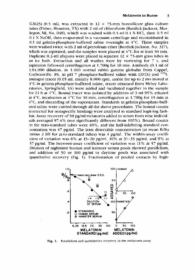
G2625) (0.5 ml), was extracted in 12 x 75-mm borosilicate glass culture tubes (Fisher, Houston, TX) with 2 mI of chloroform (Burdick-Jackson, Mus- kegon, MI, No. 048), which was washed with 0.5 ml0.1 N HCl, then 0.5 ml 0.1 I\; NaOH, then evaporated in a vaccuum centrifuge and reconstituted in 0.5 rnl gelatin-phosphate-buffered saline overnight at 4 "C. These samples were washed twice with 2 ml of petroleum ether (Burdick-Jackson, No. 317), which was aspirated; and the samples were placed at 4°C for at least 30 min. Duplicate 0.2-ml aliquots were placed in separate 12 x 75-mm glass tubes in an ice bath. Extraction and all washes were by vortexing for 7 s, and aspiration followed centrifugation at 1,700g for 10 min. Antibody (0.1 ml of 1:64,000 dilution, in 1:400 normal rabbit gamma gIobulin from Cappel, Cochranville, PA, in pH 7 phosphate-buffered saline with EDTA) and 1251- analogue tracer (0.05 ml, initially 8,000 cpm, usable for up to 2 mo stored at 4 "C in gelatin-phosphate-buffered saline, tracer obtained from Meloy Labo- ratories, Springfield, VA) were added and incubated together in the sample for 24 h at 4°C. Bound tracer was isolated by addition of 3 ml 95% ethanol at 4"C, incubation at 4°C for 10 min, centrifugation at 1,700g for 45 min at 4"C, and discarding of the supernatant. Standards in gelatin-phosphate-buff- ered saline were carried through all the above procedures. The bound counts (corrected for nonspecific binding) were analyzed in standard logit-log fash- ion. Assay recovery of 50 pg/ml melatonin added to serum from nine individ- uals averaged 97.4 % (not significantly different from 100%). Bound counts in the zero-standard tubes were 40%, and the half-inhibiting standard con- centration was 47 pg/ml. The least detectable concentration (at mean B/Bo minus 2 SD for zero-standard tubes) was 4 pg/ml. The within-assay coeffi- cient of variation was 8% at 15-20 pg/ml, 10% at 31-35 pg/ml, and 9% at 57 pg/ml. The between-assay coefficient of variation was 11% at 57 pg/ml. Dilution of nighttime human and hamster serum pools showed parallelism, and addition of 50 or 100 pg/ml to daytime pools was associated with quantitative recovery (Fig. 1). Fractionation of pooled extracts by high-
100 HUMAN 1.01 1
c3 W
mean zero dose-2 S.D. 0 Q
6 .
50
.4. DILUTION OF SERUM
.2. 0 STANDARDS 0 HUMAN SERUM @ HAMSTER SERUM
6.25 12.5 25 50 100 0 50 100
MELATONIN ME LATO N I N STANDARD (pQ/ml) ADDED (pg/ml)
Fig. 1. Parallelism and quantitative recovery in the melatonin assay.
4 Vaughan et al.
pressure liquid chromatography [Tamarkin et al., 19821 yielded a single peak of immunoactivity for human and hamster serum at the retention time for authentic melatonin. Assay of serum and heparinized plasma gave the same results.
Melatonin values for each human subject were fitted to a periodic function [Batschelet, 19741 with clock time converted to angular units both on a 24-h scale (0-360 degrees for 0-24 h) and on a 12-h scale (0-360 degrees for 0-12 h and for 12-24 h) and expressed as the cos and the sin of the angle with respect to each scale. This allowed the time of a sample to be expressed as four separate terms, two for the 24-h scale and two for the 12- h scale, which were the independent variables in a regression in which the dependent variable was melatonin concentration. Nighttime melatonin was evaluated by reading the peak value and the time of its occurrence from the best-fit curve. This analysis was done for each subject and on data pooled within the burn or control group. Subject-specific results were analyzed with a t-test for independent means. For the rat daytime data, a t-test for several means was used, employing the Bonferroni correction to protect against bias toward significance resulting from multiplicity of comparisons [Brown et al., 19831. The rat nighttime data were subjected to stepwise multiple regression analysis with group (control and sham together versus burn) and a linear and quadratic function of time as independent variables. This allowed testing of whether the nocturnal pineal melatonin surge was different between the burn group and the other two groups combined. All analyses were done on a DEC VAX 11/70 computer with the BMDP statistical package [Brown et al., 19831.
RESULTS
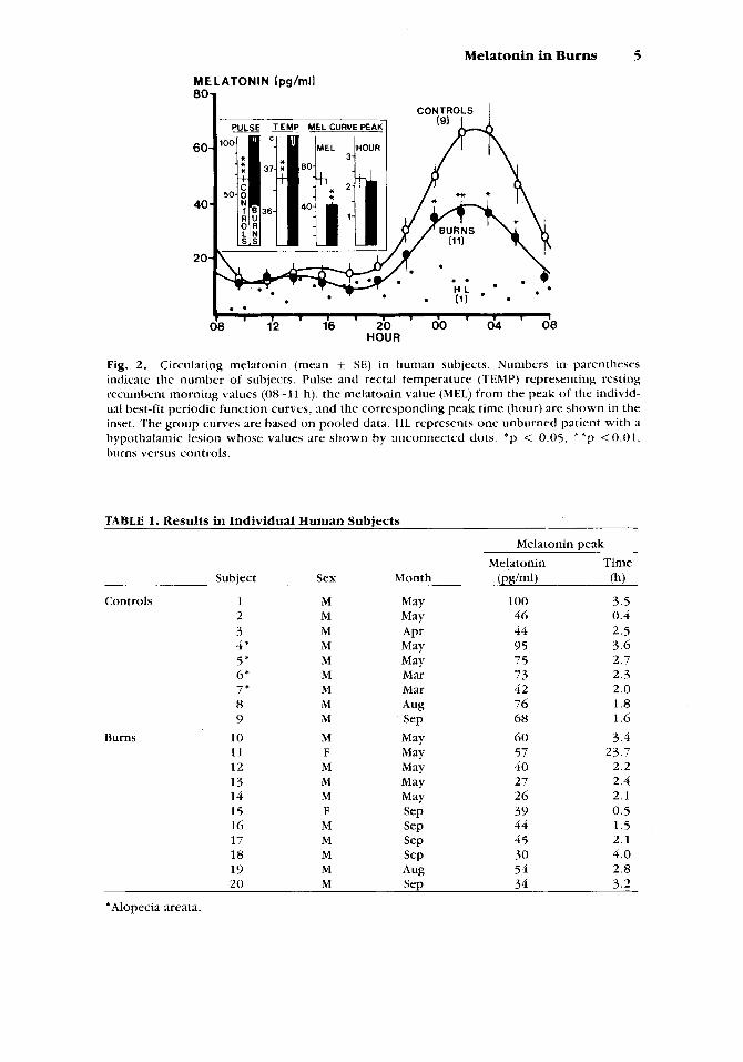
Figure 2 indicates that resting pulse and temperature were elevated in burns compared to controls, but plasma melatonin was lower in burns, which difference became statistically significant at night. Table 1 gives the sex, month of sampling, and the values for melatonin and the time at the best-fit curve peak for each subject. Not indicated in the table is that the rhythm was statistically significant in each subject of both groups. Table 2 gives reanalyses of the curve peak melatonin values by subdividing the groups in several ways. The results indicate that the lower values in burns were not influenced by inclusion of patients with alopecia areata in the control group, nor by month of sampling.
The one patient with hypothalamic destruction (Fig. 2) did not have a statistically significant melatonin rhythm, as reported earlier in this patient [Vaughan et al., 19791 and indicates the necessity for an intact neural path- way (which courses through the hypothalamus and finally through the sym- pathetics) for occurrence of the nocturnal melatonin surge [Vaughan, 19841.
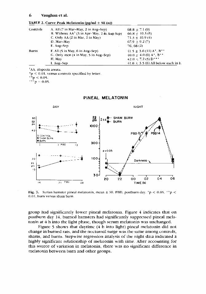
Figure 3 shows that daytime (4 h into light) pineal melatonin in burned hamsters was significantly less than that in shams at the end of the second week postburn. Values at various times during the night on postburn day 7 and at 04 h on postburn day 14 were not significantly different between burn and sham, but at 20 h on postburn day 7 (end of the light phase), the burn
Melatonh in Burns 5
MELATONIN Ipg/mll
HOUR
Fig. 2. Circulating melatonin (mean SE) in human subjects. Numbers in parentheses indicate the number of subjects. Pulse and rectal temperature (TEMP) representing resting recumbent morning values (08-11 h), the melatonin value (MEI.) from the peak of the individ- ual best-fit periodic function curves, and the corresponding peak time (hour) are shown in the inset. 'The group curves are based on pooled data. HL represents one unburned patient with a hypothalamic lesion whose values are shown by unconnected dots. *p < 0.05, * * p < 0 . 0 1 , burns versus controls.
TABLE 1. Results in Individual Human Subjects
Melatonin peak Melatonin Time
Subject Sex Month @ g W (h)
Controls 1 M May 100 3.5 2 M May 46 0.4 3 M APr 44 2.5 4' M May 95 3.6 5 * M May 7 5 2.7 6* M Mar 7 3 2.3 7 * M Mar 42 2.0 8 M Aug 76 1.8 9 M Sep 68 1.6
1 1 F May 57 23.7
13 M May 27 2.4
15 F WJ 39 0.5 16 M SeP 44 1.5
18 M SeP 30 4.0 19 M Aug 54 2.8 20 M Sep 34 3.2
10 M May 6 0 3.4
12 M May 40 2.2
14 M May 26 2. I
17 M SeP 45 2.1
'Alopecia areata.
Burns
6 Vaughan et al.
TABLE 2. Curve Peak Melatonin (pglml f SE (n))
Controls A. All (7 in Mar-May, 2 in Aug-Sep) €3. Without AA' (3 in Apr-May, 2 i'n Aug-Sep) C. Only AA (2 in Mar, 2 in May) D. Mar-May E. Aug-Sep 76, 68 (2)
Burns F. All (5 in May, 6 in Aug-Sep) 41.5 3.6 (11) A * , B* * G. Only men (4 in May, 5 in Aug-Sep) H. May I. Aug-Sep
68.8 k 7.1 (9) 66.8 4 10.3 (5) 71.3 k 10.9(4) 67.9 k 9.2 (7)
40.0 f 4.0 (9)A*, B** 42.0 4 7.2 (5) D* * * 41.0 f_ 3.5 (6) All below each in E.
'AA, alopecia areata. * p < 0.01, versus controls specified by letter. * * p < 0.05. * * * p = 0.05.
1 -
300 -
DAY
+ -
0 CONTROL @ S H A M BURN
7 P B D I 3
PINEAL MELATONIN
NIGHT
I
14 21 P B D 2 8
3 1000
1
$-- E.t
p-0.05
I 1004 +,
SHAM BURN
""I n l l l D J 19'' 30
20 22 00 02 0 4 06 TIME (h)
Fig. 3 . 0.01, burn versus sham burn.
Syrian hamster pineal melatonin, mean f SE. PBD, postburn day. * p < 0.05, * 'p <
group had significantly lower pineal melatonin. Figure 4 indicates that on postburn day 14, burned hamsters had significantly suppressed pineal mela- tonin at 4 h into the light phase, though serum melatonin was unchanged.
Figure 5 shows that daytime (4 h into light) pineal melatonin did not change in burned rats, and the nocturnal surge was the same among controls, shams, and burns. Stepwise regression analysis of the night data indicated a highly significant relationship of melatonin with time. After accounting for this source of variation in melatonin, there was no significant difference in melatonin between burn and other groups.

Melatonin in Burns 7
PINEAL SERUM MEL MEL
(pg/pii) (pg/ml)
4 0 20
20
Fig. 4. Hamster pineal and serum melatonin (mean 14. Shaded bars, sham burns. Solid bars, burns. * * *p <0.001.
SE) during the day on postburn day
PINEAL MELATON IN
(DAY)
100
.....
.....
......... ..... ..... ..... .... ..... ..... ..... ..... PBD
8 .........
C S B
100 - P 9 pin
PBD 50 14
....... ..... ..... ..... ..... ..... ..... ..... fl ..... ......... ..... ..... .....
..... ..... .... ..... ......... ..... C S B
P9 P’”
1000
-
300
I00
3 0
PINEAL MELATONIN (NIGHT)
d 0 CONTROL -29 0
,,.&,*- 4 ‘‘‘‘\$jp 0 SHAMBURN 0 BURN
0 PBD 22 0 ‘\\
,’ ?“ l o g M E L = 2 . 1 t 0 . 4 L - 0 . 0 3 6 t 2 ‘\,
, CONTROL +SHAM BURN ‘\ 0 t = TIME (AS 0- IOh) \
I’ 0 BASED ON MEAN MEL AT EACH t I r z0 .99 0
0,‘ p<o 001 0
0 0
DARKNESS f
20 22 00 0 2 04 06 TIMELh)
Fig. 5. Rat pineal melatonin (mean t SE). C , control; S, sham burn; B, burn; PBD, postburn day. The dashed line represents the best-fit quadratic regression for combined control and sham burn groups.
DISCUSSION
The principal results of this study are that in three species, burn injury was not associated with elevated melatonin values during the day or night, and a significant nocturnal surge of melatonin occurs whether or not burn injury is present. These results are interesting in light of two other observa- tions in the literature. First, the sympathetic innervation of the pineal in all three species controls melatonin production, in that bilateral lesions that interrupt the central or peripheral sympathetic pathway to the pineal prevent the nocturnal rise of pineal metatonin synthesis or of circulating or excreted
8 Vaughanet al.
melatonin [Eichler and Moore, 1971; Klein et al., 1971; Kneisley et al., 1978; Moore and Klein, 1974; Panke et al., 1979; Reiter, 1980; Reiter et al., 1975; Tetsuo et al., 1981; Vaughan, 1984; Vaughan et al., 19791.
Second, burn injury is a model of increased tonic sympathetic activity associated with tachycardia, increased blood flow to the wound and to splanchnic organs with relative vasoconstriction (normal blood flow) in uninjured skin, increased glucose production and lypolysis with maintenance of normal or elevated insulin secretion, increased muscle protein breakdown and synthesis with net weight loss, increased metabolic rate (heat produc- tion) in proportion to elevated circulating and excreted catecholamines, and increased core temperature in proportion to elevated metabolic rate [Aulick and Wilmore, 1983; Danielsson et al., 1976; Harrison et al., 1967; Herndon et al., 1977; Vaughan et al., 1982; Vaughan and Becker, 1984; Wilmore, 1976; Wilmore et al., 1974; Wilmore et al., 1976; Wilmore and Aulick, 1978; Wolfe and Durkot, 19821. This pattern includes a strong component of beta- adrenergic response, further evidenced by blunting of the hypermetabolism after propranolol administration [Wilmore et al., 19741 and of weight loss with other beta-blocking drugs [Szabo, 19791. Patients with a burn size of 41% studied at 2 wk after injury (corresponding to the mean values in the present study) have resting morning plasma values of norepinephrine and epinephrine elevated about threefold above the normal mean [Vaughan and Becker, 19841. Undoubtedly, even greater elevations occur during the day when the patients are disturbed and receive wound care, etc. It is presumed that sympathetic tone is continuously elevated (tachycardia does not resolve at night) in burn injury to provide increased substrate flow for the benefit of the wound until it is healed, when the hypermetabolism resolves. Rats with a burn size of 60% have total 24-h catecholamine excretion elevated t3 sixfold normal on postburn day 9 [McManus, 19831, and their hypermetabo- lism lasts more than 45 days [Herndon et al., 19781. Metabolic measurements are not available for burned hamsters, though in rats with a burn of 20% of body surface, epinephrine excretion and metabolic rate were elevated two- fold above normal, at least from postburn day 3-11 [Aprille et al., 19791.
Although the post-ganglionic sympathetic neurotransmitter for stimu- lation of melatonin synthesis in the pineal is not yet known for humans and Syrian hamsters, it is norepinephrine acting through a beta-receptor and adenyl cyclase stimulation in rats [Klein, 19821. Injection of beta-agonists apparently does not stimulate melatonin synthesis in humans or Syrian ham- sters [see Vaughan, 1984; Lipton et al., 1982; Steinlechner et al., 19841, though it does in rats [Deguchi and Axelrod, 1973; Romero, 19761. Acute adverse stimuli do not alter human plasma melatonin [Vaughan, 19841, though they may elevate rat pineal melatonin synthesis [Allen et al., 1981; Lynch et al., 1973; Lynch et al., 19771. Thus, differences among species for acute beta- adrenergic responsiveness of pineal melatonin further complicate interpre- tation of lack of melatonin responsiveness to the more chronic elevation of sympathetic activity of burn injury. For this, there are perhaps three potential explanations that involve adrenergic activity at the level of the pineal, and they focus on the rat, in which species norepinephrine has become accepted as an intrapineal neurotransmitter.
Melatonin in Burns 9
In the normal rats, injection of beta-agonists down-regulates the re- sponse in pineal melatonin synthesis [Deguchi and Axelrod, 1973; Romero, 19761 and activation of fat cell kdenyl cyclase [Aprille et al., 19791 upon subsequent application of the agent. Against this explanation is the reported resistance to desensitization of the fat cell adenyl cyclase response (normally induced by short-term injections of isoproterenol) in burned rats and in other rat models of chronic elevation of catecholamines, in which adenyl cyclase remained responsive to isoproterenol [Aprille et al., 19791. This and the continued long-term hypermetabolism (02 consumption) in the burned rats of that study suggest that in burn injury, tissue beta-responsiveness is not eliminated by down regulation. The pineals of our burned rats also retained their responsiveness, because their nocturnal surge of melatonin was normal. Secondly, normal rat sympathetic endings take up circulating endogenous catecholamines (re-uptake phenomenon) and thus may protect pinealocytes against activation of melatonin synthesis by adverse stimuli [Parfitt and Klein, 19761. This explanation assumes that the increased sym- pathetic activity in the burned rats did not include elevated neuronal trans- mission in the pineal, but rather that pinealocytes did not respond to burn injury because they were protected from the humoral component of the sympathetic hyperactivity. This explanation, like the first, is unlikely because it would mean that the pineal is different from other tissues since the hyperdynamic metabolic status continues in burns until healing. But the normally innervated rat pineal reportedly does respond to humoral sympa- thetic activation apparently from the adrenal medullae during acute physical immobilization [Lynch et al., 19771. A third explanation, presynaptic inhibi- tion of pineal nerve endings [Pelayo et al., 19771 by the alpha component of activity of the elevated norepinephrine exposure in burns, does not explain lack of response to humoral catecholamines in burns and is further unlikely in that the nocturnal melatonin surge was normal in the burned rats.
The reduction only of nocturnal plasma melatonin in burned humans and the reduced daytime pineal melatonin in burned hamsters remains unex- plained. The unavoidable administration of small doses of narcotic analgesic (usually 5 mg oxycodone orally once or twice) during study of the burn patients appears not to explain our results, because it was administered only during the day in the patients and was not given to the hamsters or rats. Morphine adminsitration to rats was associated with a slight reduction of pineal N-acetyltransferase [Zatz and Brownstein, 19791 but a reported eleva- tion of plasma melatonin [Esposti et al., 19841, and in vitro incubation of rat or hamster pineals with morphine or naloxone produced no effect on the secretion of melatonin [Vaughan, 1984, unpublished results]. Whether al- tered clearance of melatonin in burns could have contributed to the results of measurements of circulating melatonin is not known, but such an effect appears not to have influenced the overall results, because pineal melatonin content was not elevated in burned animals. The possibility of greater dim light exposure at night may have had some role in the lower nocturnal plasma melatonin in the burn patients. We have presented the melatonin rhythm as it occurs in the actual circumstance of their care, and neither day nor night values were above normal despite their hyperadrenergic state. We
10 Vaughan et al.
think that possible differences in nocturnal light exposure in these patients was not a factor because suppression of nocturnal plasma melatonin in normal humans requires light intensi,ty even much greater than the usual indoor daytime intensity [Lewy et al., 19801.
The extent to which local neuronal transmission in peripheral tissues contributes to the overall sympathetic hyperactivity of burn injury, which at least includes the humoral element of markedly elevated circulating and excreted catecholamines, remains unknown. Whether or not neuronal trans- mission provides a major contribution, the present results are best explained by the concept that measurements of circulating or pineal melatonin do not provide a positive index of general sympathetic activity. This agrees with the lack of a melatonin response in humans also seen after acute sympathetic activation [Vaughan, 19841. Control of sympathetic activity appears parti- tioned, with pineal melatonin synthesis regulated independently of cardio- vascular and metabolic functions. Whether other hormones (such as adrenocorticoids and glucagon) that usually surge in conjunction with gen- eral sympathetic activation or the fall in thyroid hormones that accompanies burn injury [Vaughan and Becker, 19841, can have a role in preventing pineal activation at central control sites or at the pineal is not known. Whether chronic changes in these additional hormones might prevent a pineal re- sponse to humoral but not locally released catecholamine in burned rats, should, perhaps, be investigated.
ACKNOWLEDGMENTS
We thank James Lasko, Jan Bullard, Sandy Coggins, and Avery Johnson for technical assistance, and A.C. Brown for referring us four of his patients.
LITERATURE CITED
Allen, J.P., J.W. Sackman, W. Tullis, M.K. Vaughan, R.A. Becker, G.M. Vaughan (1981) Nycto- hemeral rhythms in rat pineal: Epinephrine uptake and N-acetyltransferase response to ether stress. In: Pineal Function. C.D. Matthews and R.F. Seamark, eds., Elsevier/North- Holland Biomedical Press, Amsterdam, pp. 2 11-2 16.
Aprille, J.R., N. Aikawa, T.C. Bell, H.H. Bode, D.F. Malamud (1979) Adenylate cyclase after burn injury: Resistance to desensitization by catecholamines. J. Trauma 19:812-818.
Aulick, L.H., D.W. Wilmore (1983) Hypermetabolism in trauma. In: Mammalian Thermogene- sis. L. Girardier and M.J. Stock, eds., Chapman and Hall, New York, Chap. 9, pp. 259-
Ihtschelet, E. (1974) Statistical rhythm evaluation. In: Biorhythms and Human Reproduction. M. Ferin, F. Halherg, R.M. Richart, and R.L. Vande Wiele, eds., John Wiley & Sons, New York, Chap. 2, pp. 25-35.
Brown, M.B., L. Engleman, J.W. Frane, M.A. Hill, R.I. Jennrich, J.D. loporek (1983) BMDP Statistical Software. W.J. Dixon, ed., University of California Press, Berkeley.
Danielsson, U. , G. Arturson, L. Wennberg (1976) The elimination of hypermetabolism in burned patients: A method suitable for clinical use. Burns 2:llO-114.
Deguchi, T., J. Axelrod (1973) Supersensitivitiy and subsensitivity of the 0-adrenergic receptor in pineal gland regulated by catecholamine transmitter. Proc. Natl. Acad. Sci. U.S.A.
Eichler, V.B., R.Y. Moore (I97 1) Pineal hydroxyindole-0-methyltransferase and gonadal re- sponses to blinding or continuous darkness blocked by pineal denervation in the male hamster. Neuroendocrinology 8:s 1-85.
304.
70:2411-2414.
Melatonin in Burns 11
Esposti, D., G. Esposti, F. Fraschini (1984) Effects of morphine on plasma melatonin in the rat. Third Colloquium of the European Pineal Study Group, EPSG Newsletter, Suppl. 5, Aug., 1984, p. 57 (abstract).
Harrison, T.S., J.F. Seaton, I. Feller (‘1967) Rekdtionship of increased oxygen consumption to catecholamine excretion in thermal burns. Ann. Surg. 165: 169-172.
Herndon, D.N., D.W. Wilmore, A.D. Mason, Jr., B.A. Pruitt, Jr. (1977) HUmordl mediators of nontemperature-dependent hypermetabolism in 50% burned adult rats. Surg. Forum
Herndon, D.N., D.W. Wilmore, A.D. Mason, Jr. (1978) Development and analysis of a small animal model simulating the human postburn hypermetabolic response. J. Surg. Res.
28:57-39.
25:394-403. Klein, D.C. , ed. (1982) Melatonin Rhythm Generating System. Karger, Basel. Klein, D.C., J.L. Weller, R.Y. Moore (1971) Melatonin metabolism: Neural regulation of pineal
serotonin:acetyl coenzyme A N-acetyltransferase activity. Proc. Natl. Acad. Sci. LJ.S.A.
Kneisley, L.W., M.A. Moskowitz, H.J. Lynch (1978) Cervical spinal cord lesions disrupt the rhythm in human melatonin excretion. J. Neural. Transm. [Suppl.] (13):311-323.
Lewy, A.J., T.A. Wehr, F.K. Goodwin, D.A. Newsome, S.P. Markey (1980) Light suppresses melatonin secretion in humans. Science 210: 1267-1269.
Lipton, J.S., LJ. Petterborg, S. Steinlechner, R.J. Reiter (1982) Zn vivo responses of the pineal gland o f the syrian hamster to isoproterenol or norepinephrine. In: The Pineal and its Hormones. Alan R . Liss, Inc., New York, pp. 107-115.
Lynch, H.J., J.P. Eng, and R.J. Wurtman (1973) Control o f pineal indole biosynthesis by changes in sympathetic tone caused by factors other than environmental lighting. Proc. Natl. Acad. Sci. U.S.A. 70: 1704-1707.
Lynch, H.J., M. Ho, R.J. Wurtman (1977) The adrenal medulla may mediate the increase in pineal melatonin synthesis induced by stress but not that caused by exposure to dark- ness. J. Neural Transm. 40:87-97.
Mcblanus, A.T. (1983) Examination of neutrophil function in a rat model o f decreased host resistance following burn trauma. Rev. Infect. Dis. 5(Suppl. 5):S898-S907.
Moore, R.Y., D.C. Klein (1974) Visual pathways and the central neural control of a circadian rhythm in pineal serotonin N-acetyhansferase activity. Brain Res. 71 : 17-33.
Panke, E.S., M.D. Rollag, R.J. Reiter (1979) Pineal melatonin concentrations in the Syrian hamster. Endocrinology 104: 194-197.
Parfitt, A . G . , D.C. Klein (1976) Sympathetic nerve endings in the pineal gland protect against acute stress-induced increase in N-acetyltransferase (E.C.2.3.1.5.) activity. Endocrinol-
Pelayo, F., M.L. Duhocovich, S.Z. Langer (1977) Regulation o f noradrenaline release in the rat pineal through a negative feedback mechanism mediated by presynaptic alpha-adreno- ceptors. Eur. J. Pharmacol. 45:317-318.
Reiter, R.J. (1980) The pineal and its hormones in the control o f reproduction in mammals. Endocr. Rev. 1:109-131.
Reiter, R.J., M.K. Vaughan, G.M. Vdughan, S. Sorrentino, Jr,, R.J. Donofrio (1975) The pincal as an organ of internal secretion. In: Frontiers o f Pineal Physiology. M.D. Altschule, ed. , MIT Press, Cambridge, pp. 54-174.
Romero, J.A. (1976) Influence of diurnal cycles on biochemical parameters o f drug sensitivity: The pineal gland as a model. Fed. Proc. 35: 1157-1161.
Steinlechner, S., T.S. King, T.H. Champney, K. Spanel-Borowski, R.J. Reiter (1984) Comparison of the effects of @-adrenergic agents on pineal serotonin N-acetyltransfeerase activity and melatonin content in two species of hamsters. J. Pineal Res. 1:23-30.
Szabo, K . (1979) Clinical experiences with beta adrenergic blocking therapy on burned pa- tients. Scand. J. Plast. Reconstr. Surg. 13:211-215.
Tamarkin, L., P. Abastillas, H.-A. (:hen, A. McNemar, J.B. Sidbury (1982) The daily profile of plasma melatonin in obese and Prader-Willi syndrome in children. J. Clin. Endocrinol. Metab. 55:491-501.
Tetsuo, M., R.J. Polinsky, S.P. Markey, I.J. Kopin (1981) Urinary 6-hydroxymelatonin excretion in patients with orthostatic hypotension. J. Clin Endocrinol. Metab. 53:607-610.
68:3107-3110.
ogy 99:840-85 1 .
12 Vaughan et al.
Vaughan, G.M. ( 1982) Studies of neuroendocrine abnormalities in burn injury: 11. Thyroidal, reproductive and pineal function in a hamster burn model. In: U.S. Army Institute o f Surgical Research Annual Research Progress Report, FY 82. U.S. Army Medical Research and Development Comm'and, Frederick, Maryland, pp. 81-105.
Vaughan, G.M. (1984) Melatonin in humans. In: Pineal Research Reviews. R.J. Reiter, ed., Alan R. Liss, Inc., New York, Vol. 2 , pp. 141-201.
Vaughan, G.M., J.P. Allen, W. Tullis, T.M. Siler-Khodr, A. de la Peiia, J.W. Sackman (1978) Overnight plasma profiles of melatonin and certain adenohypophyseal hormones in men. J. Clin. Endocrinol. Metab. 47:566-571.
Vaughan, G.M., S.D. McDonald, R.M. Jordan, J.P. Allen, R. Bell, E.A. Stevens (1979) Melatonin, pituitary function and stress in humans. Psychoneuroendocrinology 4:351-362.
Vaughan, G.M., R.A. Becker, J.P. Allen, C.W. Goodwin, Jr., B.A. Pruitt, Jr . , A.D. Mason. Jr. (1982) Cortisol and corticotrophin in burned patients. J. Trauma 22:263-273.
Vaughan, G.M., K.A. Becker (1984) Thyroid hormones and catecholamines in burn patients: A hypermetabolic low T j syndrome. In: Norepinephrine. M.G. Ziegler and C.R. Lake, eds., Williams and Wilkins Company, Baltimore, Chap. 30, pp. 450-470.
Walker, H.L., Mason, A.D., Jr. (1968) A standard animal burn. J. Trauma 8: 1049-1051. Wilmore, D.W. (1976) Hormonal responses and their effect on metabolism. Surg. Clin. North
Am. 56999- 1018. Wilmore, D.W., J.M. Long, A.D. Mason, Jr., R.W. Skreen, B.A. Pruitt, Jr. (1974) Catechol-
amines: Mediator o f the hypermetabolic response to thermal injury. Ann. Surg. 180:653- 669.
Wilmore, D.W., J.M. Long, A.D. Mason, Jr., B.A. Pruitt, Jr. (1976) Stress in surgical patients as a neurophysiologic reflex response. Surg. Gynecol. Obstet. 142:257-269.
Wilmore, D.W., L . H . Aulick (1978) Metabolic changes in burned patients. Surg. Clin. North Am. 58:1173-1187.
Wolfe, K .R . , M.J. Durkot (1982) Evaluation o f the role of the sympathetic nervous system in the response of suhstrate kinetics and oxidation t o burn injury. Circ. Shock 9:395-406.
Zatz, M . , M.J. Brownstein (1979) Central depressants rapidly reduce nocturnal serotonin N- acetyltransferase activity in the rat pineal gland. Brain Res. 160:381-385.