1 name of department: department of plastic surgery and burns
TRANSCRIPT

1
Name of department: Department of Plastic Surgery and Burns Treatments, Rigshospitalet
PhD program: Surgical Sciences
Author: Mikkel Herly, MD
Title and subtitle: Fat grafting in humans: An assessment of the resorption dynamics of fat
grafts and evaluation of methods to measure fat graft retention in the
breast
Topic description: Studies concerning the resorption dynamics of fat grafts and an
investigation into the correct method for measuring fat graft retention
in the breast. The studies support a randomized clinical trial protocol
which is also included in the thesis
Supervisor: Professor Christian von Buchwald, MD, DMSc
Submitted on: July 1st, 2020
Number of study units: Five
2
Supervisors
Professor Christian von Buchwald, MD, DMSc
Principal supervisor.
Professor, University of Copenhagen.
Department of ORL, Head and Neck Surgery and Audiology, Rigshospitalet, Denmark
Peter Viktor Vester-Glowinski, MD, PhD
Primary co-supervisor.
Department of Plastic Surgery and Burns Treatments, Rigshospitalet, Denmark.
Professor Emeritus Krzysztof T. Drzewiecki, MD., DMSc
Co-supervisor.
Professor Emeritus, University of Copenhagen.
Department of Plastic Surgery and Burns Treatments, Rigshospitalet, Denmark.
Members of the assessment committee
(Chairperson)
Professor Mikael Boesen MD., PhD
Professor, University of Copenhagen
Department of Radiology, Bispebjerg and Frederiksberg Hospital, Denmark
Professor Jens Ahm Sørensen MD, DMSc.
Professor, University of Southern Denmark
Department of Plastic Surgery, Odense University Hospital, Denmark
Professor Dirk Schaefer MD., DMSc
Professor, University of Basel
Department of Plastic, Reconstructive, Aesthetic and Hand Surgery, Universitätsspital Basel,
Switzerland

3
Preface
This PhD thesis is comprised of research that I have conducted at the Department of Plastic
Surgery and Burns Treatments, Rigshospitalet from 2014 to 2020. In this period, I have
developed a protocol for a phase III Randomized Clinical Trial of using adipose derived-stem
cells for breast reconstruction with fat grafting. The trial has been approved by the Danish
Medicines Agency and the National Committee on Health Research Ethics and it is included as
study 5 in this thesis. The studies presented in this thesis were made in part as a preparation for
this RCT.
Furthermore I have used a large portion of my time to help my supervisor and colleague, Peter
Vester-Glowinski, MD, PhD to conduct a randomised controlled trial of fat grafting enriched
with ex-vivo expanded adipose-derived stem cells for breast augmentation that we wanted to
finish before I initiated the trial concerning breast reconstructions. Peter Vester-Glowinski was
the trial PI on this study and therefore, this work is not directly included in this thesis. However,
it has had important implications for the future perspectives of my research and therefore, I
discuss the results of this trial in the perspectives section.
My long-term goal is to find a way to use adipose-derived stem cells to improve fat grafting for
breast reconstruction after mastectomy. The studies included in this thesis are all necessary
investigations for the design of a new randomised controlled trial of breast reconstruction with
fat grafting and adipose-derived stem cells.
The studies described in this thesis were funded by the Danish Cancer Society, the Department
of Plastic Surgery and Burns Treatments, Rigshospitalet and the Research Fund of
Rigshospitalet.
Mikkel Herly, July, 2020

4
Acknowledgments
I would like to thank Christian von Buchwald for offering supervision of my efforts to achieve a
PhD. Furthermore, I greatly appreciate Professor Buchwald personally and look forward to
hopefully collaborate with him on many future research projects.
Peter Vester-Glowinski is my closest colleague and a great friend. I worked with him on his PhD
projects as a medical student and this experience has shaped my research career in a very
positive way. Our talents and shortcomings complement each other in a great way. I hope we
will be able to continue our work together and that the research group we are building will
make many exciting discoveries in the future.
Dr. Mathias Ørholt has worked with me on all my past and current research projects as a
medical student alongside his studies. Now he has finished medical school and he is working to
start his PhD. He is very intelligent, and he has a lot more aptitude for many things including
statistical analysis than I have. I have the greatest respect for him, and I hope I will have the
opportunity to work with him going forward.
Dr. Mathilde Hemmingsen has helped me on the studies of methods to measure fat graft
volume retention in the breast. She started as a medical student and has now graduated as a
medical doctor and started her PhD. Mathilde is a very talented researcher and I look forward
to work with her on her many new research projects in the future.
Andreas Larsen has been helping me with a lot of my research including the meta-analysis that
is included in this thesis. Andreas is a very intelligent and talented medical student with
incredible work ethics. He also has a talent for organizing a research group and early on he took
charge of the research projects. I am sure he has a very bright future ahead of him and I am
grateful for the opportunity to work with him.
Krzysztof Drzewiecki has been my supervisor since I started my research in plastic surgery in
2014. Prof. Drzewiecki is a great mentor and sparring partner for research questions, and he
has also helped me with career advice.
Jens Jørgen Elberg has helped to recruit volunteers from his private clinic, Amalieklinikken, for
the MRI studies. Dr. Elberg is a great mentor for me in research and even more so in plastic
surgery. His approach of thinking about the root cause of surgical problems when choosing the
surgical treatment, is something that I will use for the rest of my career. I am very grateful for all I
have learned from him.
Finally, I wish to thank my wonderful wife Cecilie for her support of my research. She is my
closest ally and I always consult her with problems, and she has been listening patiently. Cecilie

5
is great at putting things into perspective and to call me out when I obsess over small obstacles.
I am very lucky.
Mikkel Herly
July, 2020
6
Table of contents
LIST OF STUDIES .............................................................................................................. 7
ABBREVIATIONS ............................................................................................................... 8
DANISH SUMMARY OF THE PHD THESIS/DANSK RESUME AF PHD-AFHANDLINGEN ................................................................................................................ 9
SUMMARY OF THE PHD THESIS .................................................................................... 13
BACKGROUND ................................................................................................................ 17
STUDY 1 - QUANTIFYING LONG-TERM RETENTION OF EXCISED FAT GRAFTS: A LONGITUDINAL, RETROSPECTIVE COHORT STUDY OF 108 PATIENTS FOLLOWED FOR UP TO 8.4 YEARS............................................................................... 23
STUDY 2 - EFFICACY OF BREAST RECONSTRUCTION WITH FAT GRAFTING: A SYSTEMATIC REVIEW AND META-ANALYSIS ............................................................. 27
STUDY 3 - THE CURRENT GOLD STANDARD BREAST VOLUMETRY TECHNIQUE SEEMS TO OVERESTIMATE FAT GRAFT VOLUME RETENTION IN THE BREAST: A VALIDATION STUDY .................................................................................................... 32
STUDY 4 - LIPOVOL: VALIDATED SOFTWARE FOR MEASURING FAT GRAFT VOLUME RETENTION IN THE BREAST WITH MRI ........................................................ 36
STUDY 5 - PROJECT PROTOCOL: FAT GRAFTING WITH EX-VIVO EXPANDED ADIPOSE-DERIVED STEM CELLS FOR BREAST RECONSTRUCTION FOLLOWING MASTECTOMY, EUDRACT: 2016-005186-31 ................................................................. 40
DISCUSSION OF THE PROTOCOL IN THE CONTEXT OF THE CURRENT EVIDENCE ........................................................................................................................ 53
PRELIMINARY RESULTS FROM A RANDOMIZED CONTROLLED TRIAL OF BREAST AUGMENTATION WITH ASC-ENRICHED FAT GRAFTING ............................ 54
CONCLUSION ................................................................................................................... 56
REFERENCE LIST ............................................................................................................ 58
APPENDIX 1 ..................................................................................................................... 66
7
List of studies
Study 1 – Published paper in Appendix 1
Quantifying Long-Term Retention of Excised Fat Grafts: A Longitudinal, Retrospective Cohort
Study of 108 Patients Followed for up to 8.4 Years1
Authors: Mikkel Herly, Mathias Ørholt, Peter V Glovinski, Christian B Pipper, Helle Broholm, Lars
Poulsgaard, Kåre Fugleholm, Carsten Thomsen, Krzysztof T Drzewiecki
Study 2 – Published paper in Appendix 2
Efficacy of breast reconstruction with fat grafting: A systematic review and meta-analysis2
Authors: Mikkel Herly, Mathias Ørholt, Andreas Larsen, Christian B Pipper, Rikke Bredgaard,
Christina S Gramkow, Adam J Katz, Krzysztof T Drzewiecki, Peter V Vester-Glowinski
Study 3 – Published paper in Appendix 3
The current gold standard breast volumetry technique seems to overestimate fat graft volume
retention in the breast: A validation study3
Authors: Mikkel Herly, Felix Christoph Müller, Mathias Ørholt, Joachim Hansen, Sophie Sværke,
Mathilde N Hemmingsen, Bo S Rasmussen, Jens J Elberg, Krzysztof T Drzewiecki, Peter V Vester-
Glowinski
Study 4 – Manuscript in Appendix 4
Lipovol: Validated Software for Measuring Fat Graft Volume Retention in the Breast with MRI
Authors: Mikkel Herly, Mathias Ørholt, Felix Christoph Müller, Mathilde Hemmingsen, Joachim
Hansen, Andreas Larsen, Bo Sonnich Rasmussen, Jens Jørgen Elberg, Christian von Buchwald,
Krzysztof T. Drzewiecki, Peter V. Vester-Glowinski
Study 5
PROJECT PROTOCOL: Fat Grafting with Ex-Vivo Expanded Adipose-Derived Stem Cells for Breast
Reconstruction Following Mastectomy, EudraCT: 2016-005186-31
Authors: Mikkel Herly, Krzysztof T Drzewiecki, Anne Fischer-Nielsen, Niels Kromann, Lea
Munthe Fog, Christina Stunk Gramkow, Rikke Bredgaard, Tove Tvedskov, Peter V Vester-
Glowinski
8
Abbreviations
ASC: Adipose-Derived Stromal Cell (and Adipose-Derived Stem Cell)
BCS: Breast Conserving Surgery
CD…: Cluster of Designation (Surface marker system)
CT: Computed tomography
EudraCT: European Union Drug Regulating Authorities Clinical Trials Database
GCP: Good Clinical Practice unit (international unit for monitoring drug trials)
GMP: Good Manufacturing Practice
HBSS: Hank’s Buffered Saline Solution
HCG: Human Chorionic Gonadotropin
HMT-3522 T4-2: Breast cancer cell line for in vitro studies
MCF-7: Breast cancer cell line for in vitro studies
MSC: Mesenchymal Stem Cell
MRI: Magnetic resonance imaging
P0, P1, etc: Cell passage denomination, P0 being unpassaged cells
POCR: Point of complete reconstruction
PC: Platelet Concentrates
pHPL: pooled Human Platelet Lysate
RCT: randomised controlled trial
SVF: Stromal Vascular Fraction

9
Danish summary of the PhD thesis/Dansk resume af PhD-afhandlingen
Fedttransplantation i sin nuværende form er en relativt ny teknik til at tilføre blødt væv til dele
af kroppen, hvor man ønsker mere volumen, f.eks. i forbindelse med en rekonstruktion af
brystet. Den består i at man suger fedt fra andre steder på kroppen end der hvor man mangler
væv. Derefter sprøjtes fedtet ind i små kanaler og i så mange planer som muligt på
recipientstedet. Dette gøres, for at fedtcellerne fra transplantatet er så tæt på en ny
blodforsyning som muligt på det nye sted på kroppen, hvor man gerne vil have fedtet til at
overleve. Denne teknik er især lovende til brystrekonstruktion, hvor de nuværende teknikker,
der består af frie lapper og brystimplantater, har nogle negative sideeffekter for patienten i
form af donorstedsmorbiditet efter høst af den frie lap og kapseldannelse omkring
brystimplantater.
Fedttransplantation har dog den ulempe, at en stor del af transplantatet forsvinder efter
operationen. Dette er formentlig fordi ikke alle de tilførte celler får en ny blodforsyning i tide,
hvorefter de dør og bliver resorberet af kroppen.4–6 Man forventer at omkring halvdelen af
transplantatet forsvinder efter en fedttransplantationsoperation.7 Af denne grund er mange
forskere ivrige efter at udvikle nye teknikker til at forbedre fedttransplantaters overlevelse. En
af de mest lovende teknikker er at tilsætte patientens egne fedtderiverede stamceller (ASC’er)
til transplantatet, som menes at kunne øge bevarelsen af fedttransplantatets volumen. Et
studie fra 2013 af Kølle et al. fra min forskningsgruppe har vist at fedttransplantater tilført
patientens egne stamceller kunne øge overlevelsen af transplantatet signifikant.8 Disse
resultater satte vi os for at undersøge om vi kunne videreføre til brug i brystet. Studiet gav
anledning til at to yderligere studier blev planlagt: Første studie skulle undersøge ASC-beriget
fedttransplantation til en brystforstørrelse (EudraCT 2014-000510-59) og et andet som skulle
undersøge ASC-beriget fedttransplantation til at rekonstruere et bryst efter en mastektomi
(EudraCT 2016-005186-31). Førstnævnte studie var planlagt at Peter Glowinski skulle stå i
spidsen for og jeg fik muligheden for at være med i dette studie, som medicinstuderende fra
begyndelsen af studiet. Jeg fik senere muligheden for at stå i spidsen for det sidste studie, som
skal undersøge ASC-beriget fedttransplantation til brystrekonstruktion hos kvinder der har
undergået mastektomi.
Der var dog flere ting, som fortsat var ukendt om dynamikken i fedttransplantaters resorption,
der gjorde det svært at undersøge, hvordan man kan forbedre den. Der manglede viden om
hvor lang tid resorptionsprocessen varer, og hvor meget af fedtet der resterer, efter at
resorptionen er tilendebragt. Derudover var der brug for en valideret metode til at måle
retentionen af fedttransplantater, så man kunne sammenligne resultater på tværs af
forskningsgrupper.
I denne PhD afhandling vil jeg præsentere fire studier, der undersøger fedttransplantaters
resorption og teknikker til at måle det. De fire studier er bundet sammen af, at de alle
10
beskæftiger sig med fedtretention på forskellige niveauer, gående fra undersøgelse af
målemetoder til at beregne fedtretention, over en basal undersøgelse af fedttransplantaters
udvikling over år og til en mere klinisk relevant undersøgelse af, hvor mange
fedttransplantationsseancer, der skal til for at rekonstruere et bryst efter en mastektomi.
Hvert af de første fire studier har selvstændige forskningsspørgsmål, som adresseres i
artiklerne. Derudover er de fire studier vigtige forarbejder til det randomiserede kliniske studie,
som skal undersøge om ASC-berigelse kan forbedre volumenbevarelsen af fedttransplantater til
at rekonstruere et bryst efter en mastektomi (studie 5).
Spørgsmålene og hvordan de individuelle studier understøtter det randomiserede kliniske
studie (studie 5) kan beskrives ved de forskningsspørgsmål som skulle besvares:
Spørgsmål 1: Hvor lang tid går der fra en fedttransplantation til fedtresorptionsprocessen er
tilendebragt? Dette spørgsmål skulle besvares for at vælge den korrekte opfølgning af
patienterne i det randomiserede kliniske studie (studie 5).
I studie 1 undersøges fedttransplantater i kraniet hos patienter, som har fået fjernet en tumor
udgået fra de schwannske celler omkring n. vestibularis. Disse patienter var fulgt med CT- og
MR-skanninger for at undersøge for recidiv med en gennemsnitlig opfølgning på 2,7 år og op til
8,4 år. Skanningerne blev brugt til at undersøge fedttransplantatets retention over tid og til at
finde ud af, hvornår fedttransplantaternes volumen var stabilt. Dette kunne bruges til at
beslutte opfølgningsperioden i fremtidige prospektiver studier af fedttransplantater, herunder
det randomiserede kliniske studie (studie 5).
Spørgsmål 2: Hvor mange fedttransplantationsbehandlinger skal der normalt til for at
rekonstruere et bryst efter en mastektomi? Dette spørgsmål var vigtigt for designet af det
randomiserede kliniske studie (studie 5), da vi gerne ville undersøge om stamcelleberigelsen
kunne mindske antallet af behandlinger til at rekonstruere et bryst.
I studie 2 er litteraturen gennemgået systematisk for tidligere studier af brystrekonstruktion
med fedttransplantation. Dernæst har vi brugt meta-analytiske metoder til at estimere, hvor
mange fedttransplantationsbehandlinger der normalt skal bruges til at rekonstruere et bryst
efter hel eller delvis kirurgisk fjernelse af brystet pga. kræft eller en familiær disposition til
kræft. Denne viden kunne bruges til at lave en power-beregning til det kliniske studie om
brystrekonstruktion med stamcelleberiget fedt.
Spørgsmål 3: Hvordan måles volumenoverlevelsen af et fedttransplantat i brystet?
Volumenoverlevelsen af fedttransplanter i brystet er det primære endemål i det randomiserede
kliniske studie (studie 5), som denne PhD afhandlings arbejder lægger sig op ad. Derfor var der
brug for at sikre, at vi havde en valideret målemetode til at måle det korrekt.
I studie 3 undersøgte vi målenøjagtigheden og præcisionen af den mest brugte MRI-baserede
metode til at måle brystvolumen og volumenretention af fedttransplantater i brystet. Metoden
11
blev valideret for dens evne til at måle ændringer i brystvolumen hos kvinder, der undergik en
brystforstørrende operation med implantater eller fedttransplantation. I studie 4 præsenterer
jeg vores eget bud på en metode til at måle fedtretention i brystet med MR-skanninger.
Metoden valideres ud fra samme principper som i studie 3.
I studie 5 præsenteres et resume af protokollen for det randomiserede kliniske studie af
stamcelleberiget fedttransplantation til brystrekonstruktion efter mastektomi, som blev
understøttet af de fire første studier.
Resultater
Studie 1 viste overordnet set, at opfølgningstiden efter fedttransplantation bør være længere end
først antaget. Vores model over fedttransplantaternes volumenretention over tid viste at
fedttransplantaternes volumen var først stabilt efter 2,2 år i kraniet. Dette kan selvfølgeligt være
anderledes for fedttransplantater i brystet, som injiceres med Coleman-teknik6, men resultatet
gjorde alligevel, at vi tilføjede en opfølgning efter to år til protokollen for det randomiserede
kliniske studie, som er beskrevet i studie 5.
I studie 2 kunne vi bruge publicerede studier til at estimere hvor mange behandlinger der typisk
bruges for at rekonstruere et bryst efter en mastektomi. Vores undersøgelse viste, at der bruges
ca. tre behandlinger for at rekonstruere et bryst efter en hudbevarende mastektomi. Det mest
overraskende ved dette studie var, at der ikke ser ud til at skulle bruges flere behandlinger til at
rekonstruere et bryst efter en radikal mastektomi end efter en hudbevarende mastektomi. Til
gengæld viste studiet at der skal bruges signifikant flere behandlinger på at rekonstruere et
bestrålet bryst end et ikke-bestrålet bryst.
Studie 3 og studie 4 viste vores arbejde med at finde en metode til at måle fedtretention i det
kliniske studie med udgangspunkt i MR-skanninger. Resultatet af de to studier er vores nye
metode, som giver ret konsistente resultater på tværs af observatører og forskellige patienter.
Den nye metode (Lipovol) er blevet brugt til at måle fedtretention i et netop færdiggjort klinisk
studie og vi har planlagt at den også skal bruges i det randomiserede kliniske studie, som beskrives
i studie 5.
Foreløbige resultater fra det randomiserede studie af ASC-beriget fedttransplantation til
brystforstørrelse
Det andet studie der skulle undersøge ASC-beriget fedttransplantation til en brystforstørrelse er
tilendebragt. Resultaterne er præsenteret ved kongres9 og manuskriptet afventer indsendelse til
et fagfællebedømt tidsskrift. Desværre viste studiet ingen effekt af ASC-berigelsen. Brysterne som
blev behandlet med ASC-beriget fedt havde ikke en højere bevarelse af transplantaternes volumen
end de normale fedttransplantater (54,0% vs. 55,9%). Resultaterne fra dette studie har haft stor
betydning for mine planer om at igangsætte det næste kliniske studie om ASC-beriget
12
fedttransplantation til brystrekonstruktion. For at undersøge diskrepansen mellem det første
studie som blev udført af Kølle et al. (Lancet 2013)8 og det efterfølgende studie af Glowinski et al.9
har jeg lavet en forskningsplan for studier som skal undersøge forskellene mellem det første
studie, der blev publiceret i 2013 og det ovenfor beskrevne studie. Disse studier skal undersøge
isoleringen af SVF, ekspansion af ASC’er og cellernes egenskaber, for at finde frem til om der er
vigtige forskelle mellem cellerne i de to studier, som er afgørende for at de kan understøtte
fedttransplantaters overlevelse. Identifikation af sådanne forskelle ville muliggøre opstart af det
sidste kliniske studie om ASC-beriget fedttransplantation til brystrekonstruktion, som er beskrevet
i studie 5.
Konklusion
Denne PhD afhandling består af fem arbejder som undersøger dynamikken i fedttransplantaters
resorption og undersøger metoder til at måle fedttransplantaters retentionsrate i brystet. Det
sidste arbejde i PhD’en er en videnskabelig protokol til et randomiseret klinisk studie der skal
undersøge om ASC’er kan forbedre volumenbevarelsen af fedttransplantater til rekonstruktion af
brystet. De første fire studier har alle haft en rolle i designet af protokollen.
De foreløbige resultater fra det andet randomiserede kliniske studie af ASC-beriget
fedttransplantation til brystforstørrelse fra vores gruppe9 viste ingen effekt af ASC-berigelsen.
Dette resultat giver anledning til en række nye studier, som skal undersøge årsagen til den
manglende effekt af ASC-beriget fedt til brystet i forhold til den signifikante effekt, som blev
observeret i studiet på overarmene af raske forsøgspersoner i 20138. Jeg har lavet en foreløbig
forskningsplan for studier, som skal muliggøre at vi kan starte det sidste randomiserede studie af
ASC-beriget fedttransplantation til brystrekonstruktion, så vi forhåbentligt kan tilbyde vores
patienter en bedre behandling i fremtiden.
13
Summary of the PhD thesis
Fat grafting is an increasingly popular technique for the correction of soft tissue defects and now
has an important role in plastic surgery. The procedure consists of harvesting fat from one part of
the body where fat is abundant and transplanting that fat to another part of the body where more
soft tissue volume is needed. The fat is harvested as a block of tissue and placed via an incision or
it is harvested as lipoaspirate and injected in a fan like pattern in multiple channels and planes
with a blunt cannula. Fat grafting is used for both reconstructive and cosmetic purposes. The
advantages of fat grafting are a low risk of donor site morbidity, the absence of foreign material,
and a natural feel. The major drawback of the fat grafting technique is that some of the fat cells
are lost to resorption, which result in a loss of volume. For this reason, multiple sessions of fat
grafting are often necessary to achieve the desired result, which poses a challenge when planning
the surgery.
The resorption of the fat graft is thought to be due to a lack of oxygenation and nutritional support
of the transplanted fat cells.4–6 It is expected that approximately half of the transplanted fat is
resorbed after surgery.7 Therefore, many researchers are eager to design interventions that may
improve the volume retention of fat grafts. One of the most promising techniques is to add the
patient’s own adipose-derived stem/stromal cells (ASCs) to the fat graft. A study performed by
Kølle et al.8 from our group, which was published in 2013, showed that fat grafts enriched with
expanded adipose-derived stem cells can significantly increase the volume retention of bolus
injected fat grafts. This discovery led to the planning of two additional RCT’s that would be used to
translate the first proof-of-concept study to clinical practice. The first study would investigate ASC-
enriched fat grafting for breast augmentation (EudraCT 2014-000510-59) and the other ASC-
enriched fat grafting for breast reconstruction (EudraCT 2016-005186-31). The first of these
follow-up-studies has been conducted with Peter Glowinski as the principal investigator and I had
the opportunity to be a part of the trial group from the beginning as a medical student.
subsequently, I got the opportunity to be the principal investigator of the final trial that would
investigate ASC-enriched fat grafting for breast reconstruction in women who had undergone a
mastectomy.
However, there were still many aspects of non-enriched fat grafting that had yet to be studied, to
ensure the highest possible quality of the RCTs. We learned that there was a lack of studies
investigating the length and extent of the normal resorption process. Furthermore, there was a
need for validated and reproducible techniques for measuring the fat graft retention rate in the
breast, which could be used to compare results between research groups.
In this PhD thesis, I present four studies that investigate the resorption dynamics of fat grafts and
methods for measuring the volume retention of fat grafts. The four studies share a common
foundation; they all investigate fat graft retention. The four studies investigate fat graft retention
at different levels: two studies are concerned with methods for measuring fat graft volume
14
retention in the breast, one study is concerned with the basic resorption dynamics of excised fat
grafts over time and finally, one study is concerned with the very clinically relevant question of
how many fat grafting treatment sessions are needed to complete a breast reconstruction.
Furthermore, all four studies are important supporting studies for a randomized clinical trial (RCT)
that I plan to conduct as the principal investigator. The RCT is summarized in study 5.
The first four studies presented in this thesis build upon individual research questions that are
addressed in manuscripts and summaries of this thesis. However, the studies also have roles to
play in relation to the planning of the randomized clinical trial (study 5) that will be addressed
exclusively in this thesis. The research questions and their relation to the trial protocol are
described below:
Question 1: What is the time to volumetric steady state of an excised fat graft, and what is the
retention rate at steady state? The answer to these questions would help us choose an
appropriate follow-up time to measure fat graft volume retention in the randomized clinical
trial (study 5).
In study 1, I present a retrospective analysis of patients who have undergone surgery to remove
a vestibular schwannoma. The patients were reconstructed with an excised fat graft and
followed by CT and MRI for many years after the surgery to identify relapse of the
schwannoma. These images were used to measure fat graft volume retention over time and to
identify the time to steady-state of the fat graft’s volume retention in a statistical model. We
used the analysis as a guide for the length of the clinical follow-up in the RCT of ASC-enriched
fat grafting for breast reconstruction after mastectomy.
Question 2: How many treatment sessions are needed to complete a breast reconstruction
after a skin-sparing mastectomy? This question is important for the RCT (study 5) in which we
sought to investigate whether a potentially increased fat graft volume retention from the ASC-
enrichment could decrease the number of fat grafting treatment sessions needed to complete a
breast reconstruction.
In study 2 we conducted a systematic review of the literature on breast reconstructions
performed with fat grafting as the only treatment modality. The studies of this review were in
turn used to estimate the number of fat grafting sessions needed to complete a breast
reconstruction following a skin-sparing mastectomy, a modified radical mastectomy and breast
conserving surgery. These estimates were used in the design of the RCT of ASC-enriched fat
grafting for breast reconstruction, which had “number of treatment sessions needed to
complete the reconstruction” as a secondary endpoint.
Question 3: What is the most accurate and objective method for measuring fat graft volume
retention in the breast? The RCT (study 5) relies on a measurement of fat graft volume

15
retention in the breast as the primary endpoint, and therefore it is necessary to ensure that the
method for measuring this endpoint is accurate and precise.
Study 3 is a validation study of the technique that we believe to be the most commonly used
method for measuring fat graft volume retention in the breast based on MRI scans. To the best
of our knowledge, this technique has not been previously validated for its ability to measure
changes in breast volume, although this is prerequisite for measuring fat graft volume
retention. Study 4 is a presentation of a new method for measuring fat graft volume retention
in the breast for use in future studies. The method was validated according to the same
principles used in study 3 and it will be used to measure fat graft volume retention in the RCT,
which is described in study 5.
In study 5, the trial protocol for the RCT (EudraCT 2016-005186-31) is summarized. The
knowledge acquired from the first four PhD studies were used to design the trial.
Results
Study 1 was used to model the resorption process of excised fat grafts placed in the mastoid
cavity. The model estimated that it takes approximately 2.2 years for the fat grafts to reach
volumetric steady state. This tells us that the resorption process has the potential to go on for a
very long time, although we do not yet know whether the resorption process of injected fat
grafts in the breast is equally long. However, the results of this study led us to change the
follow-up period of the primary endpoint in the randomized clinical trial (study 5) to two years,
and we generally plan for a longer follow-up periods in future studies of fat grafting.
Study 2 provided an estimate of the number of treatments typically needed to reconstruct a
breast after a mastectomy. Our analysis relied on meta-analytical methods of the literature and
estimated that approximately three treatment sessions are necessary to completely reconstruct
a breast that has not been treated with radiotherapy after a skin-sparing mastectomy. This
estimate was used in the planning of the randomized clinical trial (study 5) by giving us an
estimate of the background population. The study found that significantly more fat grafting
treatment sessions were necessary to reconstruct an irradiated breast than a non-irradiated
breast. Surprisingly, we also found that the number of treatment sessions to completely
reconstruct the breast was not dependent on whether the patient had been treated with a
modified radical mastectomy or a skin-sparing mastectomy.
Study 3 and 4 comprise our efforts to find an accurate and precise method for measuring the
volume retention of fat grafts in the breast based on MRI scans. We found that the most
commonly used method for measuring fat graft retention in the breast overestimated increases
in breast volume. We also presented our new method for measuring fat graft volume retention
16
in the breast and showed that the method was able to measure changes in breast volume with
high accuracy and reproducibility across observers. The new method which we call Lipovol will
be used in future studies in our group and we plan to use it in the randomized controlled trial of
breast reconstruction with ASC-enriched fat grafting that I describe in study 5. Furthermore, we
hope that other researchers will find the method useful in their studies. The method will be
freely available to other researchers upon request.
Preliminary results from the randomized controlled trial of ASC-enriched fat grafting for breast
augmentation (EudraCT 2014-000510-59)
The second RCT of our group to investigate ASC-enriched fat grafting in women undergoing a
breast augmentation has been finalized. The results have been presented9, and the scientific
manuscript is in the final stages before being submitted to a peer-reviewed journal. Unfortunately,
the trial did not show any effect of the ASC-enrichment. The breasts treated with ASC-enriched fat
grafting did not show a significantly higher fat graft volume retention than the breasts treated
with normal fat grafting (54.0% vs. 55.9%). These results have important implications for my plans
to initiate the next randomized controlled trial of ASC-enriched fat grafting for breast
reconstruction. This recent development has prompted me to outline a research plan to
investigate the differences between the first trial that was published in 20138, which showed a
significant effect; and the second trial that did not. These studies will investigate in detail how to
isolate and expand ASCs and ensure that the cells maintain the properties that make them able to
improve fat graft volume retention.
Conclusion
This PhD thesis consists of five studies. The thesis is generally concerned with the resorption
dynamics of fat grafts and methods to measure fat graft volume retention in the breast. The final
study of the PhD thesis is a protocol for a randomized controlled trial that will investigate ASC-
enriched fat grafting for breast reconstruction. The first four studies all played a role in the design
of the trial protocol.
The preliminary results of the second RCT investigating ASC-enriched fat grafting for breast
augmentations recently showed no effect of the ASC-enrichment. This outcome has prompted us
to conduct a series of studies that will investigate the discrepancy between the first study that
found a significant effect of the ASC-enrichment in the upper arms of healthy individuals and the
second RCT that failed to reproduce this effect in the breast. The research plan will be used to
optimize the isolation and expansion of ASCs which hopefully, will enable us to improve breast
reconstructions with fat grafting so that we can offer a better treatment to our patients in the
future.

17
Background
Fat transplantation was first performed by Neuber in 1893 for correcting a scar-induced
depression in the face with fat harvested from the arm.7 The breast was first used as a recipient
site for fat transplantation in 1895 by Czerny, who transferred a lipoma into the breast tissue to
fill out a volume defect after breast surgery.10 The fat grafting technique was refined by the
contributions of several surgeons, with some of the most prominent being Holländer11 and
Lexer.12 Höllander revolutionized the fat grafting technique by heating the fat graft until it was
fluid and was thereby able to inject the graft into tissue defects.11 Lexer took on the procedure
of fat injection and described a wide range of situations in which this technique could be
utilized.12 These situations included breast asymmetry, scar corrections, Dupytren contractures,
lipodystrophies, traumas and other minor contour deformities.12 Lexer was also one of the first
surgeons to describe the main drawback of fat grafting as a soft tissue filler. He assessed that
approximately 60% percent of the fat graft was resorbed in the period after transplantation.
Fat grafting in its current form is relatively new and was made popular among plastic surgeons
by Dr. Sydney Coleman, who introduced the structural fat grafting technique and its application
to the breast and face.6,13,14 The process is described by Coleman to include three steps:
harvesting of the fat, processing of the fat and injection of the fat. The harvesting is commonly
performed via liposuction from the abdomen, hips, thighs or buttocks with a blunt cannula
after infiltration of either Ringer’s lactate or lidocaine with or without epinephrine. Afterwards,
the fat is processed by centrifugation or sedimentation to eliminate excessive water, blood,
irrigation fluid and oil from the ruptured fat cells. The processing ensures that components that
will be rapidly resorbed are removed from the fat graft and is thought to increase the final fat
graft retention rate. Lastly, the fat is transferred to small syringes and injected through small
incisions in the skin with a blunt cannula. The fat is injected in a fan-like pattern in multiple
planes by releasing small droplets of fat while retracting the syringe. The multiplane pattern
and “pearls-on-a-string” approach secures a maximal surface area of the fat and thereby
facilitates oxygenation of the fat cells.4
After the introduction of structural fat grafting by Coleman, the popularity of fat grafting
increased because it was regarded as the ideal soft tissue filler. Many surgeons such as Illouz15,
Delay16, Bircoll17 and Rigotti18 have contributed to the popularization of fat grafting and have
highlighted the advantages of low donor site morbidity, the absence of foreign material and a
natural feel. Although the technique of fat grafting has evolved throughout many decades, the
main drawback of fat grafting persists, specifically, that a large portion of the graft is lost to
resorption after surgery.15,16 Fat graft resorption complicates planning of the reconstruction
and necessitates multiple procedures if a large volume reconstruction is required. Therefore,
strategies to improve the procedure focus on increasing the retention rate of the fat graft.
18
Strategies to improve fat graft volume retention
Several methods have been proposed to improve the volume retention of fat grafts.19–24 They
can generally be divided into techniques for improving the transplant and techniques for
improving the recipient site. In 2010, Dr. Ueberreiter described a novel method for harvesting
fat cells, called BEAULI.19 This method differed from the traditional manual harvesting methods
because it use a water-jet to free fat cells from the fat tissue (Water-jet assisted lipotransfer,
WAL) simultaneously with the liposuction. The method is believed to increase the survival of fat
cells due to the reduced mechanical stress during harvesting. After harvesting, the fat is
separated from the irrigation fluid via centrifugation, and then, the fat graft is ready for
injection. Another approach for improving fat graft retention that focus on the recipient site is
to pre-expand the breast with an external bra-like device called BRAVA. The BRAVA device
creates a vacuum over the breasts.20–23 Preclinical studies have shown that the use of external
expansion provides less tension in the breast after the fat grafting, and induce a neoangiogenic
response in the recipient bed.25 This in turn would support cell survival due to increased
nutritional support for the transplanted fat cells. However, subsequent reports that many
patients do not tolerate the externally applied BRAVA device, and as an alternative Dr. Stilleart
proposed the use of an internal tissue expander for breast reconstruction.24 The method, which
is called “Intratissular expander-mediated fat grafting” is similar to the BRAVA treatment in
many ways. It is performed by placing a tissue expander in the breast and successively deflating
it while replacing the volume by injecting fat into the surrounding tissue. The procedure is
thought to help prepare the breast for the increase in volume so that the fat graft can be placed
with less pressure. Furthermore, the capsule that forms around the expander implant is
believed to increase blood supply and prevent the fat from pooling in the cavity formed by the
mastectomy. However, both BRAVA and intra-tissular expansion are time consuming and not
always tolerable by the patient. Moreover, intratissular expansion involves foreign bodies and
thus may lead to a whole range of other complications. In addition, both procedures are costly.
Another promising idea to improve fat graft volume retention is to add adipose-derived stromal
cells, which are also called adipose-derived stromal cells (ASCs), to the fat graft.26,27 ASCs are a
type of mesenchymal stem cell that resides in adipose tissue and can differentiate into
adipocytes, chondrocytes and osteocytes, among other cell types, predominately from the
mesodermal germ layer. The addition of these cells to a fat graft has been hypothesized to
increase volume retention of fat grafts either by the ASCs differentiating into new adipocytes or
by the secretion of paracrine factors that stimulate adipocyte survival and proliferation in the
recipient site.28 Multiple studies have investigated the enrichment of fat grafts with ASCs or
stromal vascular fraction (SVF).29–33 SVF is a mixed cell population that include ASCs that can be
obtained from fat tissue. Studies by Gentile, Tissiani and Yoshimura have demonstrated a
significant increase in fat graft volume retention in the breast through addition of non-
expanded SVF to fat grafts.29–31 Tissiani reported fat graft retention up to 79% after SVF

19
enrichment.30 However, studies by Peltoniemi32 and Chiu33 have not been able to reproduce
the beneficial effect of SVF on fat graft volume retention. These conflicting results should be
viewed considering the potential risk of bias in the conducted studies that are non-randomized,
not blinded, and in some cases without control groups. This highlights the need for future
studies with randomization, blinding and objective volumetric measurements.
Studies of ASC-enriched fat grafting in our research group
Randomized controlled trials of ASC-enriched fat grafting require a multidisciplinary team effort
to enrol patients, perform surgeries, isolate and expand ASCs in a GMP-grade facility and
evaluate the graft retention rate with a validated measurement technique in the follow-up
period. Therefore, to put the work of this PhD thesis into the context of the activities of my
current research group and its history, I think it is relevant to briefly describe the work of my
research group. The group planned to conduct three randomised controlled trials of ASC-
enriched fat grafting which are illustrated in the figure below.
Long-term research plan to investigate ASC-enriched fat grafting in humans
The first RCT investigated the efficacy of ex-vivo expanded ASC-enriched fat grafting (EudraCT
2010-023006-12).8 The trial was finalized before I joined the research group. The trial which
was conducted by Stig-Frederik Kølle as principal investigator and Krzysztof Drzewiecki as
Sponsor was a proof-of-concept trial that investigated ASC-enrichment of fat grafts to enhance
fat graft volume retention. Ten voluntary participants were included to undergo liposuction to
obtain ASCs, which were then culture-expanded ex vivo for 14 days. After the culturing period,
20
the expanded ASCs were added to an autologous fat graft. The ASC-enriched fat graft was
injected as a bolus in the upper arm of the participants, and in the same procedure, the
participants received a bolus of non-enriched fat grafting to the contralateral upper arm. The
bolus technique was chosen to optimize visualization of the graft on MRI examinations for
measurement of the fat graft retention rate. The study showed a highly significant effect of the
ASC-enrichment with a fat graft retention of 80.9% in the enriched grafts and 16.3% in the
contralateral control grafts that had not been enriched with ASCs.8 However, the findings
needed to be translated into a clinically relevant setting. Therefore, two additional trials were
planned. A second RCT (EudraCT 2014-000510-59) was planned to investigate ASC-enriched fat
grafting for cosmetic breast augmentations in 10 participants using the structural fat grafting
technique with Peter Vester-Glowinski as the principal investigator. The purpose of this study
was to validate the effect of ex-vivo expanded ASCs in a more relevant recipient site with a
homogenous patient population and with the use of the structural fat grafting technique.
However, one session of fat grafting is often sufficient for a cosmetic breast augmentation and
the impact of the ASC-enrichment of fat grafts is therefore limited in this patient population.34
Thus, we also designed a third RCT, which is described in a detailed summary in study 5 of this
thesis. The purpose of the third RCT (EudraCT 2016-005186-31) is to investigate ASC-enriched
fat grafting in patients undergoing breast reconstruction after a mastectomy. This patient
population is more challenging because the recipient bed is smaller, which reduces the blood
supply to the transplanted adipocytes and thereby graft survival. Therefore, many sessions are
often needed, ranging from 2-7.2 This makes reconstruction with fat grafting a long surgical
treatment course for patients who often have already undergone extensive treatment before
initiating the breast reconstruction treatment. The hypothesis is that ASC-enrichment may
increase graft survival and as a result minimize the number of sessions needed for breast
reconstruction, which would reduce the surgical morbidity and discomfort in this patient group.
If successful, ASC-enriched fat grafting may become a minimally invasive alternative to free
flaps and breast implants for patients who chose to undergo breast reconstruction after
surgical treatment of breast cancer.
Necessary studies for designing an RCT of ASC-enriched fat grafting for breast reconstruction
following a skin-sparing mastectomy (study 5)
During the planning of the RCT (study 5) we encountered several problems that had not been
sufficiently addressed in the literature which interrupted the design process of the trial.
Time to steady state of the fat graft
No consensus exists on the exact amount of fat that is resorbed or on when the fat graft
reaches a steady state in which no further resorption occurs. It is commonly stated in the
literature that the fat graft resorption process ends 6 to 12 months after surgery.16,19,35

21
However, this is based on unproven assumptions because the studies rarely follow the patients
for more than one year. To the best of our knowledge, only two studies have followed the fat
graft resorption process for more than one year, and these studies both indicated that the
resorption process continued for more than a year.36,37 However, the fat graft volume retention
was estimated clinically (i.e., with no objective volumetric method), and the point where the fat
grafts reached a steady state was not determined. Because no studies were able to point
towards an exact time to steady state, it was not possible to determine the best follow-up
period for the RCT (study 5). Therefore, we conducted study 1, which provided a unique
opportunity to map the exact resorption process of a fat graft. We analyzed a sequence of MRI
scans from patients with vestibular schwannoma who underwent a mastoidectomy with
transfer of an en-bloc excised fat graft for closure of the dura mater. Furthermore, this patient
population underwent an extensive radiological control program. This provided us with a mean
follow-up time of up to 8.4 years and thus enough time to evaluate the time to a steady state of
the fat grafts. Such analysis had not been described in the literature previously.
Number of sessions needed for complete breast reconstruction
As background for the RCT (study 5), we also needed to know how many fat grafting sessions
are typically needed to undergo a breast reconstruction after a skin-sparing mastectomy. We
were also interested in investigating the effect of radiotherapy on the number of sessions
needed for complete reconstruction. No consensus exists in the literature on the mean number
of sessions for complete reconstruction, with a wide range of 2 to 7 sessions for complete
breast reconstruction.38–41 Therefore, we conducted a systematic review and meta-analysis to
investigate the number of sessions needed to complete a breast reconstruction. We stratified
patients into 5 groups based on the type of breast surgery the patient had undergone prior to
the breast reconstruction. The stratifications were made based on whether the patient had
undergone a skin-sparing mastectomy, non-skin-sparing mastectomy or breast conserving
surgery. These three groups were further subdivided by whether the patients received
radiotherapy.
Measurement of fat graft volume retention
The primary endpoint of the RCT (study 5) was the fat graft volume retention in each breast
after one year. Many techniques have been suggested to measure fat graft volume retention,
with the most commonly used methods being MRI42–44 and 3D-photography.45–47 The current
gold standard for measuring changes in breast volume is arguably MRI, as proposed by Dr.
Herold et al.43 In this method, an MRI scan is performed before the fat grafting procedure and a
second scan is performed post-operatively. Next, the borders of the entire breast are
delineated manually, and the volume difference is compared to the injected fat graft volume.
However, we hypothesized that the method was associated with a systematic measurement
error and that using the method risked overestimation of changes in breast volume.48 We
22
believed that this was mainly due to lax tissue surrounding the breast being drawn into the
augmented breast. Because the lax tissue is included in the post-operative scan and not in the
preoperative scan, there is a risk of systematic overestimation. In study 3 we validated the
above-mentioned MRI technique for measuring changes in breast volume in 28 patients. Our
results would have implications for studies that have used the method for both estimating fat
graft volume retention22,23,49 and studies that investigated interventions to improve the fat
graft volume retention.32,50 These investigation led us to develop Lipovol, a new method for
measuring fat graft volume retention in the breast (study 4). The method differed from the gold
standard by using fixed osseous markers to ensure that the same block of tissue was analysed
on both the pre-operative and post-operative scan. Thereby, we considered the lax tissue
surrounding the breast, and systematic overestimation was avoided. The method was validated
by measuring the breast volume in 14 patients before fat grafting and 3 hours after the
procedure. The change in breast volume measured with MRI was then compared with the
injected volume. All measurements were performed by four independent and blinded
observers.
23
Study 1 - Quantifying Long-Term Retention of Excised Fat Grafts: A Longitudinal,
Retrospective Cohort Study of 108 Patients Followed for up to 8.4 Years
Authors: Mikkel Herly, Mathias Ørholt, Peter V Glovinski, Christian B Pipper, Helle Broholm, Lars
Poulsgaard, Kåre Fugleholm, Carsten Thomsen, Krzysztof T Drzewiecki
Study objectives
To investigate the long-term retention of excised fat grafts in the craniofacial area and to
calculate the time-to-steady-state volume retention for the grafts.
Study objectives in relation to the PhD thesis
When studying the volume retention of fat grafts, it is important to know whether the resorption
process has ended at the time of the measurement (i.e., that the graft volume is in steady state).
To compare retention rates of fat grafts between studies, assessments must be performed at the
same time in the resorption process, and preferably after the resorption process is finalized. In this
study, I calculated the time to volumetric steady state of excised fat grafts in the mastoid cavity.
To the best of my knowledge, this is the only study that has demonstrated the steady state of fat
grafts with a volumetric analysis. Although the type of fat graft that was examined in this study is
very different from a structural fat graft in the breast, the study population presented a unique
opportunity to track fat grafts over an extended period of time, which is necessary to find the time
to steady state. The results of this study were considered when we decided on the follow-up
period of the primary endpoint in the RCT that is described in study 5 because because no
previous studies have investigated the time to steady state of structural fat grafts in the breast.
Project summary
Background
The main drawback of fat grafting is that a part of the graft is lost to post-operative resorption. It is
essential for the plastic surgeon to have an idea of how much of the graft will stay in place after
the resorption period. Furthermore, it is very important to know the length of the resorption
period to plan any additional fat grafting procedures. The latter is poorly understood and to the
best knowledge of this author, prior to the publication of this study in 2017, no studies had shown
the plateau phase of fat graft resorption (i.e., steady state).
Methods
In this study, we investigated existing radiological images of patients who had undergone ablative
surgery for vestibular schwannoma and volume restoration with an excised fat graft from the
abdomen. These patients were followed up with magnetic resonance imaging (MRI) or computed
24
tomography (CT) scans on a regular basis to detect any recurrence of the schwannoma. The fat
graft was delineated on all image slices that contained the fat graft. These areas were then
multiplied by the slice thickness and summed to calculate the total volume of the graft. Figure 2 in
Appendix 1 illustrates the delineation of a fat graft. We only included patients who had undergone
a post-operative scan within 14 days after surgery. The post-operative scan was used to calculate
the fat graft volume at baseline and was set as 100% graft volume. The fat graft volume retention
was calculated by dividing the fat graft volume calculated on the follow-up scans with the baseline
volume to estimate a retention rate. All retention rates that were calculated based on follow-up
scans were plotted against the number of days after surgery. The fat graft retention over time was
modeled with a four-parameter log logistic dose-response curve. Additionally, we planned a priori
to analyze the effect of sex and baseline fat graft volume with a cut-off volume above or below 16
mL on fat graft volume retention. Steady state fat graft volume retention was defined as a
resorption rate of less than 5% of the baseline fat graft volume per year. For histological
evaluation, biopsies were taken of the fat grafts in patients who underwent revision surgery
during the study period.
Results
Between 2006 and 2015, 208 patients underwent ablative surgery for vestibular schwannoma; 108
of these patients were included in the study. The average observational period was 2.7 years,
(range 17 days – 8.4 years). The baseline fat graft volume was 18.1 mL on average (95% CI, 17.2 mL
– 18 mL). Volumetric steady state was estimated to be 806 days after surgery, at which time the
average fat graft retention rate was 50.5% (95% CI, 46.4% - 54.7%). Patient characteristics and
study variables can be seen in Table 1 in Appendix 1, and a model of the fat graft retention
trajectory can be seen in Figure 4 in Appendix 1.
The fat graft retention rate was significantly higher in male patients than in female patients (p <
0.001). The fat grafts in male patients reached volumetric steady state after 714 days, at which
time the average volume retention was 57.7% (95% CI, 51.4% - 63.2%), whereas the fat grafts in
female patients achieved steady state after 874 days, at which time the average fat graft volume
retention was estimated to be 44.5% (95% CI, 39.5% - 50.1%). A model of the graft retention rate
over time in female patients vs. male patients can be seen in Figure 4 of Appendix 1. We did not
find any statistically significant difference in fat graft volume retention between the low-baseline
volume group (average baseline volume = 13.6 mL) and the high-baseline volume group (average
baseline volume = 20.1 mL).
Five patients who had revision surgery in 2016 consented to have biopsies taken of their fat grafts.
The specimens were obtained 22 days, 1.3 years, 3.3 years, 4.4 years and 4.6 years after the
primary surgery. In the four grafts obtained more than 1 year after surgery, we found mature fat
tissue with limited amounts of fibrosis. In the graft obtained after 22 days, we found focal necrosis
with surrounding macrophages. Histological images of two selected grafts can be seen in Figure 6
of Appendix 1.

25
Discussion of the results and their relation to previous research
To the best of our knowledge, this study was the first to investigate the resorption dynamics of fat
grafts via imaging analysis with a follow-up of more than one year. Furthermore, to the best of our
knowledge, this study was also the first to estimate time to volumetric steady state of fat grafts.
The time to steady state of 2.2 years after surgery was longer than what we had anticipated.
Several previous studies state that the time to steady state fat graft volume retention is
approximately 6 months31,51 because that is how long it takes for the adipocytes to succumb to the
hypoxia, and for the body to clear the dead cells from the recipient site. However, to the best of
my knowledge of the literature, this has not been confirmed by a volumetric analysis. Our study
show that it can take significantly longer than 6 months for fat grafts to reach steady state volume
retention, which demonstrate the need for investigations into this area with other types of fat
grafting and in other recipient sites.
Another interesting finding in our study was that we did not find statistically significantly different
fat graft retention rates between the high baseline fat grafts and the low baseline fat grafts. We
had hypothesized that excised fat grafts with large volumes would have a higher necrosis rate
because a larger part of the graft would be too distant from the nearest blood supply to survive on
diffusion alone. In fact, a previous study by Capaneda et al. suggests that fat more than 1.5 mm
from the nearest blood supply will not survive.52 This study has been cited multiple times as a
reference to the scientific foundation for the structural fat grafting technique. In this study, we
investigated fat grafts that had an average distance from the graft center to the nearest blood
supply of 9.6 mm. If all fat tissue more than 1.5 mm from nearest blood supply necrotized, we
would have expected lower retention rates or more scar tissue formation in our study. On the
contrary, we found a retention rate of more than 50%, which is similar to what Neuber found by a
clinical estimate in 18937. Furthermore, the histological and radiological analyses showed that the
majority of the fat grafts consisted of mature fat tissue, even years after the transplantation of the
excised fat graft.
Interestingly, Bourne et al.5 showed that fat grafting injected with the structural fat grafting
technique, as described by Coleman6, leads to pooling of fat deposits along tissue planes in the
recipient site. Consequently, for a large portion of fat grafts, the distance to the nearest blood
supply may be substantially greater than what is theoretically expected of the structural fat
grafting technique.
Limitations
Our study has limitations that should be addressed. Being a retrospective study, we were limited
by the clinical information that could be obtained from the included patients. For instance, we
were very interested to investigate the effect of weight changes on the fat graft volume retention
over time. Unfortunately, weight was not available from the time of the MRI examinations and
thus, we were prevented from performing that analysis. Furthermore, the exclusion of patients
and scans was a potential source of selection bias. However, the reasons for exclusion were all
related to technical problems such as individual scans not including the entire fat graft. We did not
26
identify any scans or patients who were excluded based on the volume retention of the fat graft
and therefore, I do not expect that selection bias has played a role in the results of the study.
In conclusion, our study highlights the need for further research into the most fundamental parts
of fat grafting. The findings illustrate the need for broader investigations into the time to steady-
state fat graft volume in other recipient sites and with the structural fat grafting technique.
Perspectives for additional research
The calculation of time to steady-state fat graft volume retention and the model of the resorption
trajectory were estimated for excised fat grafts in the craniofacial area. Today, fat grafting is
predominantly performed using the structural fat grafting technique13 in which liposuctioned fat is
injected in multiple channels and planes. We are currently investigating the resorption dynamics
of fat grafts injected with the Coleman technique6 in the breast in a prospective study. In this
study, we include women who undergo a breast augmentation with fat grafting. We use the
Lipovol software (which is described in study 4) to measure the fat graft volume retention in the
breasts. The patients are followed annually for three years with MRI scans. The prospective study
is headed by Dr. Mathias Ørholt with me as a supervisor. The study will be used to model fat graft
volume retention over time and to estimate the time to steady state of structural fat grafts in the
breast. We believe that the findings can be used to decide the follow-up time required in future
studies but until then we will use the findings from Study 1 as a guide for follow-up of fat graft
volume retention.

27
Study 2 - Efficacy of breast reconstruction with fat grafting: A systematic review and meta-
analysis
Study objectives
To investigate the efficacy of breast reconstruction using only fat grafting in a systematic review
of the literature. Specifically, we sought to use meta-analytical methods to find the number of
fat grafting sessions needed to complete a breast reconstruction.
Study objectives in relation to the PhD thesis
This study contributes to the PhD objectives by providing background information on the number
of fat grafting treatments needed to complete a breast reconstruction based on studies reported
in the literature. This study supports the design of the RCT described in study 5 by investigating
the number of fat grafting treatments that is typically needed to complete a breast reconstruction
after a skin-sparing mastectomy. The reason we want to improve the fat graft volume retention in
the RCT is to improve the treatment protocol for women in need of breast reconstruction after
breast cancer. However, an improvement in the fat graft retention rate is less significant for the
patient if the fat graft volume retention does not translate into fewer treatment sessions needed
to complete the reconstruction. Therefore, we included ‘the number of treatments needed to
complete a breast reconstruction’ as an endpoint in the RCT which is described in study 5. If cell-
enrichment results in fewer treatment sessions needed to complete a breast reconstruction, it
would provide a great benefit for patients.
Project summary
Background
Multiple procedures are required to perform a breast reconstruction after mastectomy with fat
grafting alone. Previous reviews concerned with breast reconstruction with fat grafting have
reported on patient satisfaction53 and the oncological safety of the procedure54,55, but to the best
of our knowledge, no reviews have focused on the number of treatments needed to complete a
breast reconstruction. On this background, we decided to conduct a systematic review of the
literature on breast reconstruction with fat grafting and to use meta-analytical methods to
estimate the number of treatments needed to complete a breast reconstruction. Additionally, we
calculated the average total fat graft volume needed to complete the reconstruction, which can be
used as a guide to assess whether a given patient has sufficient fat available for liposuction that
can be used for fat grafting.
Methods
The systematic meta-analysis and review was performed on observational studies. Therefore, it
was conducted in accordance with MOOSE guidelines.56 The selection of studies was conducted by
28
three observers in parallel, and all discrepancies were discussed until a consensus was reached. A
search of the literature was performed July 2017 using the PubMed, EBSCO, EMBASE and
Cochrane databases with the following search terms: (breast reconstruction OR mastectomy OR
lumpectomy OR quadrantectomy OR BCS) AND (fat graft∗ OR lipofilling OR fat transfer OR
lipotransfer OR lipografts OR fat transplantation OR lipomodeling).
We included studies based on consecutive, unselected patients who had undergone breast
reconstruction with fat grafting as the only treatment modality after breast cancer or a genetic
predisposition to breast cancer. The included studies were required to specify that the patients
had completed their breast reconstruction and to include a minimum follow-up time of six
months. We excluded studies in which the patients received a fixed amount of fat grafting
sessions. When we encountered multiple manuscripts from the same authors with overlapping
patient populations, we only included the manuscript with the highest number of patients or the
most recently published manuscript. We contacted the corresponding authors of manuscripts
when we were unsure about whether they fulfilled the inclusion and exclusion criteria of the
review.
A meta-analysis of the patients in the included manuscripts was performed to estimate the
number of treatment sessions needed to complete breast reconstruction based on the following
five treatment categories: irradiated modified radical mastectomy, non-irradiated modified radical
mastectomy, irradiated skin-sparing mastectomy, non-irradiated skin-sparing mastectomy and
breast-conserving surgery.
The included manuscripts were assessed according to the Methodological Index for Non-
Randomized Studies (MINORS) for potential bias. We modeled the number of treatments needed
to complete a breast reconstruction based on treatment category using a shifted hypergeometric
distribution of the study-specific probability of finishing the reconstruction in the next treatment.
This was performed individually for the five treatment categories (irradiated modified radical
mastectomy, non-irradiated modified radical mastectomy irradiated skin-sparing mastectomy,
non-irradiated skin-sparing mastectomy and breast-conserving surgery). All other outcomes were
reported with simple descriptive statistics. A Bonferroni correction was applied to adjust for
multiple testing. The complication rates were compared with chi-squared tests.2
Results
The search identified 1556 studies. The screening for title and abstract provided 202 studies
eligible for full text screening. Thirteen studies were included after full text screening. We then
contacted the authors of 57 studies that may fulfil the inclusion criteria after some clarifications
from the authors. This added an extra 8 studies to the review and meta-analysis, resulting in a
total of 21 studies. The main reason for exclusion was a lack of stratification of the patients into
the treatment categories (e.g., the number of treatment sessions were provided as an average
from a mixed patient population who had undergone cancer treatment ranging from breast-
29
conserving surgery to irradiated radical mastectomy). See Figure 1 in Appendix 2 for a flowchart of
the screening process.
The 21 studies originated from 11 countries and comprised a total of 1011 breast reconstructions
in 834 patients (78.8% unilateral and 21.2% bilateral breast reconstruction). The mean follow-up
time was 43.8 months. All the included studies were observational, two of them were prospective,
10 were retrospective, and nine were case reports/case series. The average MINORS score of the
studies was 9.85 out of 16. Table 1 in Appendix 2 summarizes the characteristics of the included
studies.
The number of fat grafting sessions needed to complete the breast reconstruction was modeled
based on a total of 1011 breast reconstructions. The estimated number of treatments needed to
complete the breast reconstruction was 2.84 (95% CI 2.69–2.98) for the non-irradiated modified
radical mastectomy group, 4.27 (95% CI 3.27–5.28) for the irradiated modified radical mastectomy
group, 2.93 (95% CI 2.25–3.61) for the non-irradiated skin-sparing mastectomy group, 4.66 (95% CI
3.12–6.19) for the irradiated skin-sparing mastectomy group, and 1.72 (95% CI 1.32–2.12) for the
breast-conserving surgery group. The number of treatments needed to complete a breast
reconstruction was significantly higher in the irradiated skin-sparing mastectomy group and the
modified-radical mastectomy group than in the non-irradiated groups. Conversely, the skin-
sparing mastectomy groups were not significantly different from the modified radical mastectomy
groups in terms of number of fat grafting sessions needed to complete a breast reconstruction
when stratifying for irradiation status.
The total injected fat graft volume, which was the sum of the injected fat graft volume of all fat
grafting sessions used to reconstruct the breast, was 744.8 mL in the non-irradiated modified
radical mastectomy group, 740.8 mL in the irradiated modified radical mastectomy group, 657.7
mL in the non-irradiated skin-sparing mastectomy group, 626.2 mL in the irradiated skin-sparing
mastectomy group, and 230.5 mL in the breast-conserving surgery group. The total complication
rates in the five groups were compared, and the irradiated mastectomy groups presented
significantly more complications overall than the non-irradiated groups, with p < 0.001 for both
the skin-sparing mastectomy groups and the modified radical mastectomy groups. See Table 3,
Appendix 2 for specification of the complications in the five groups.
Discussion of the results and their relation to previous research
In this systematic review and meta-analysis, we estimated the number of fat grafting treatment
sessions needed to complete a breast reconstruction based on the resection type and the use or
non-use of radiotherapy. Unexpectedly, we did not find a significantly higher number of treatment
sessions needed to reconstruct the breast after a modified radical mastectomy than after a skin-
sparing mastectomy. We had hypothesized that the more constricted skin envelope would make
more fat grafting sessions necessary to complete the reconstruction. However, this analysis relied
only on the number of treatment sessions, and thus, it is possible that other relevant outcomes
differ between the two groups. A previous retrospective study by Bailey et al. found that patients
30
undergoing breast reconstruction after a skin-sparing mastectomy were significantly more
satisfied with their reconstructed breast than patients who had undergone reconstruction after a
non-nipple-sparing mastectomy.
Our analysis found that a significantly higher number of fat grafting sessions was needed to
complete a breast reconstruction with fat grafting in irradiated breasts than in non-irradiated
breasts after both modified radical mastectomy and skin-sparing mastectomy. This association is
supported by previous observations57,58 and seems plausible from a clinical perspective. This meta-
analysis suggests that 1-2 additional fat grafting treatment sessions are needed to reconstruct an
irradiated breast after a mastectomy than are needed for non-irradiated breasts.
The strength of the meta-analysis lies in the unambiguous nature of the primary endpoint
(number of treatment sessions needed to complete the reconstruction) and the large sample size
distributed across multiple surgeons, surgical centers and countries. However, there are several
limitations that should be considered. The analysis was conducted with ‘breast’ as the
independent sampling unit, which has the consequence that studies with large patient populations
dominate the analysis. This could have the unintended consequence that surgeons with the most
experience would skew the number of treatment sessions needed to complete a breast
reconstruction lower because they are more experienced. However, the funnel plot shown in
Figure 4, Appendix 2 shows that the average results were evenly distributed between small studies
and large studies, apart from two case studies that had especially high numbers of treatment
sessions. These two case studies39,59 were descriptions of especially challenging cases, but the two
studies are not expected to have had a significant impact on the results of our analysis because of
the overall sample size of more than 1000 breast reconstructions. Another limitation is that we
chose to include studies with a minimum follow-up time of six months. We know from a previous
study that the fat graft resorption process can continue for up to 2.2 years1, but if we had decided
to only include studies with a follow-up period of more than 2 years we would have to exclude
many of the studies in this analysis. However, many of the very large studies that we included in
the meta-analysis had a follow-up time of more than 24 months.58,60,61 We decided on a six month
cut-off limit as the best compromise to ensure a wide range of studies in the analysis while
minimizing the risk of included patients undergoing subsequent fat grafting sessions after the
follow-up period.
Perspectives for additional research
A comparison of the fat graft volume retention in the five treatment categories was not attempted
in this meta-analysis because of heterogeneity in the measurement techniques used to calculate
fat graft volume retention. Presently, there is no consensus regarding how fat graft volume
retention is measured, and the techniques being used are generally not subjected to validation
studies that compare measurements of fat graft volume retention between techniques and
against known volume changes in the breast. This is the main reason for inclusion of the next two
studies in this thesis. These studies investigated the accuracy of the current gold standard

31
technique for measuring fat graft volume retention in the breast and presented a new
measurement technique of our own design.
32
Study 3 - The current gold standard breast volumetry technique seems to overestimate fat
graft volume retention in the breast: A validation study
Study objectives
To investigate the most commonly used breast volumetry techniques based on MRI scans for
their ability to measure changes in breast volume. The ability of each technique to measure
changes in breast volume is crucial for measuring fat graft volume retention in the breast.
Study objectives in relation to the PhD thesis
The primary endpoint of the randomized controlled trial of fat grafting with adipose-derived
stromal cells (ASCs) for breast reconstruction is fat graft volume retention, which would be the
third clinical trial investigating ASC-enriched fat grafting conducted by our group. Fat graft volume
retention was the primary endpoint in the first study of these cells conducted by our group8 and
was the primary endpoint of our second trial of fat grafting with ASCs for breast augmentation in
healthy women.9 When we conducted our second trial, which has been presented in part9, we
searched the literature for a validated technique for measuring fat graft volume retention in the
breast. The most commonly used reference point for new techniques was a breast volumetric
technique designed by Herold et al.42,43,62 However, while this technique has been validated for its
reproducibility when measuring breast volume and the volume of an implant by direct delineation
of the implant on an MRI scan, this technique has not previously been validated for its ability to
measure a change in breast volume between two scans of the same patient. Therefore, we
decided to investigate this ability of this technique to measure changes in breast volume to
validate the technique for measuring fat graft volume retention as the primary endpoint in our
randomized clinical trials.
Project summary
Background
Several techniques for measuring breast volume have been proposed and validated.43,47,63–68 Many
of these techniques have subsequently been used to measure fat graft volume retention in the
breast. We propose that techniques for measuring fat graft volume retention should be validated
by assessing the ability of the technique to measure changes in breast volume and preferably an
increase in breast volume. The reason for this suggestion is that accurate measurement of a
change in breast volume is necessary for measuring fat graft volume retention. The volume of a fat
graft in the breast cannot be delineated directly on an MRI scan because the fat graft is dispersed
in multiple tunnels and planes in the breast to maximize blood supply to the graft. This is also the
case for 3D photography that is based on the breast topography and is not able to visualize the
inside of the breast. Therefore, the measurement of the volume retention of a fat graft is typically

33
calculated by measuring the volume of the breast before surgery and after surgery when the
resorption process has ended. The increase in breast volume from the first scan to the second scan
is interpreted as the residual volume of the fat graft, which can then be compared with the initially
injected volume to calculate the retention rate. To summarize, the way fat graft volume retention
in the breast is calculated with current methods hinges on the assumption that the difference in
breast volume from the preoperative scan to the follow-up scan is equal to the residual fat graft
volume. However, to the best of our knowledge, this is not how previous MRI techniques have
been validated. Therefore, we conducted a validation study of one of the most commonly used
techniques43,44,62 for calculating fat graft volume retention in the breast by measuring changes in
breast volume after breast augmentations and comparing the measured change in breast volume
with the volume of the augmentation.
Methods
We included women undergoing breast augmentation with breast implants or with fat grafting in a
private clinic, Amalieklinikken, Copenhagen and the Dept. of Plastic Surgery and Burns treatments,
Rigshospitalet. One surgeon (JJE) performed all the breast augmentations in the study. We
excluded women with known breast diseases, pregnancy or contraindications to MRI examination,
such as implanted metal or claustrophobia. The patients who underwent implant-based breast
augmentation were scanned within 10 days before the surgery and 4-7 months after the surgery.
The two MRI examinations were performed at the same time in the woman’s menstrual cycle. The
patients undergoing breast augmentation with fat grafting were scanned the day before surgery
and again within three hours after the surgery, at which time the edema would not yet have fully
developed and fat graft resorption would not have occurred.
The MRI examinations were performed on a 3-tesla MRI unit, except for two women who were
examined on a 1.5-tesla MRI unit. The volume assessments were performed independently by
three readers (JH, SS, MNH) who did not know the true volumes of the implants/fat grafts. The
breast delineations were performed according to Herold et al.43,44,62: the skin was used as the
superficial border, the internal costal surface was used as the deep border, the center of the
sternum was used as the medial border, and the point where the thickness of the breast becomes
equal to the thickness of the skin was used as the lateral, superior and inferior borders. We
contacted the corresponding author43 of the papers to verify the steps, and he suggested that an
equal number of slices should be used in the preoperative MRI examinations and the post-
operative MRI examinations. To the best of our knowledge, this additional step has not been
described previously in the literature. The accuracy of the technique was assessed by
measurement of the error between the measured increase in breast volume and the true volume
of the breast augmentation (i.e., volume of the implant or volume of the injected fat). The
precision of the technique was assessed in milliliters by the inter- and intra-reader variation and as
a percentage of the true volume of the breast augmentation. Previous studies have validated their
MRI-based measurement technique by its ability to measure the size of a breast implant by direct
delineation of the implant. We do not think this test is a genuine assessment of the ability of a
34
technique to measure a change in breast volume because the fat graft cannot be delineated.
However, to assess the accuracy of the MRI scans and the observers’ ability to delineate correctly,
we also measured the implant volumes by direct delineation of the implant. This was performed
after the measurements of changes in breast volume as to not unblind the observers.
The sample size power calculation was estimated with the Clopper-Pearson exact method which
required 53 breast augmentations for the validation study to achieve a 90% confidence interval
width of less than 10%. Based on this, we decided to include 28 patients for a total of 56 breasts
with, 14 of the patients undergoing breast augmentations with implants and 14 of the patients
undergoing breast augmentation with fat grafting.
Results
Twenty-eight women undergoing bilateral breast augmentation were included. Fourteen of these
women underwent breast augmentation with implants, and the other 14 women underwent
breast augmentation with fat grafting. The patient characteristics are shown in Table 1, Appendix
3. The mean measurement error across the three readers was 178.4 mL (95% CI 156–201 mL)
when measuring the women who received breast implants and 153.4 mL (95% CI 138.4–168.5 mL)
for the women who received fat grafting. These measurement errors correspond to percentage
errors of 50.8% (95% CI 45.3% −56.3%) for breast augmentations with implants and 50.9% (95% CI
45.7% −56.0%) for breast augmentations with fat grafting. Figure 2 in Appendix 3 shows a linear
regression of the relationship between the measured increase in breast volume and the volume of
the breast augmentation (implant or injected fat graft volume). The measurement error in
milliliters increased with the volume of the breast augmentation volume, but the percentage
measurement error remained relatively unchanged from the smallest augmentation to the largest
augmentations. The measurement error did not depend on the type of breast augmentation (i.e.,
whether the augmentation was performed with implants or with fat grafting).
The mean inter-observer variation was 55.25 mL (95% CI 47.13–63.37 mL, range 0.28–268.23 mL)
(n = 168). The mean intra-observer variation was 19.77 mL (95% CI 15.87–23.67 mL, range 0.05–
75.80 mL) (n = 72).
Measurement of the implant volumes by direct delineation had an average measurement error of
15.5 mL and a systematic error of -1.8%, which was not statistically significant.
Discussion of the results and their relation to previous research
MRI is arguably the most accurate non-invasive technique for measuring tissue volume. This is
exemplified by the high accuracy and precision of the measurements of implant volume when
directly delineating the implants in this study and many previous studies.63,66,67,69,70 However, our
study showed that the way MRI scans are used to measure a change in breast volume between
scans may be subject to a large systematic error. The current approach of measuring changes in
breast volume is to first to measure the total breast volume before and after a change in breast
volume. The difference between the two measurements is then considered to be equal to the
change in breast volume. This may seem logical in theory but may introduce systematic error if the
35
measured change in breast volume is interpreted as the residual volume of a fat graft for the
following reasons: when the breast is augmented with fat, the surgeon will distribute the graft in
the breast, but the augmentation will also expand the breast borders laterally, inferiorly, medially
and superiorly.48 Therefore, subcutaneous tissue that was outside the breast before the surgery
may be included as part of the augmented breast after the surgery. In theory, this will be counted
as part of the increase in breast volume, which will overestimate the volume of the fat graft. If this
error is not accounted for by the measurement technique, then the fat graft volume retention
might be systematically overestimated.
Perspectives for additional research
The results of this study prompted us to design a new technique developed specifically to measure
changes in breast volume. The technique was designed to account for the theoretical
overestimation described above. This work is described in study 4.
36
Study 4 - Lipovol: Validated Software for Measuring Fat Graft Volume Retention in the Breast
with MRI
Study objectives
To design an MRI-based method to measure fat graft volume retention in the breast and to
validate the ability of the method to measure changes in breast volume.
Study objectives in relation to the PhD thesis
To validate a new method designed to measure fat graft volume retention in the breast that can
be used to measure fat graft volume retention in the RCT, which is intended to be the primary
outcome.
Project summary
Background
We have previously shown that one of the most widely used MRI-based methods for measuring
fat graft volume retention had a large systematic error of approximately 50% when used to
measure changes in breast volume.3 We hypothesized that one of the potential reasons for this
overestimation is that the approach of measuring total breast volume before and after a change in
breast volume to deduct the difference is incorrect if the objective is to measure fat graft volume
retention. Although the difference in total breast volume must indeed be calculated as described
above, we theorized that the change in total breast volume is the sum of two components. The
first component is the added volume of the fat graft, but there may be another component of the
change in total breast volume that stems from expansion of the breast borders. The breast is a
structure most often defined by its contours. Breast augmentation entails expansion of the breast
projection but also lateral, superior and inferior expansion. The expansion of the breast borders
will make tissue that was previously outside of the breast, by definition, now a part of the breast.
If a method does not account for this phenomenon, there is a risk that this added volume will be
interpreted as part of the residual fat graft.
To account for this risk of overestimation, we designed a new method based on MRI scans that
can be used to measure changes in breast volume but only changes that arise from the added
volume and not from tissue that may be included in the augmented breast due to an expansion of
breast borders. An algorithm based on this method was constructed and translated into software
that would make most steps automatic. The software was validated by measuring changes in
breast volume in 28 women who underwent breast augmentation with a known volume.

37
Methods
The new software method is called “Lipovol” and will be made freely available for use on the
MeVisLab platform.71 None of the authors have any financial disclosures regarding the Lipovol
software or MeVisLab. The validation study was approved by the Regional Ethics Committee of
Copenhagen (nr. H-1501379) and the Regional Data Protection Agency. All participants provided
written informed consent before inclusion in the study.
Two MRI scans are needed to use the Lipovol software: one recorded before the volume change of
interest and another recorded after the volume change has taken place. We used a Dixon
sequence for our validation study, but all scans that can be used for volumetric measurements
should be useable, including CT scans. The two scans are first aligned based on osseous markers in
the thorax in the registration step. Then, a rectangular region-of-interest that encompasses the
highest expected volume is drawn around the breast, thus, expanding the breast borders. This
rectangular region-of-interest is then mirrored by the software on the other scan which is possible
because of the registration. Then, the skin of the patient is delineated automatically with a region-
growing algorithm, and the tissue volume inside of the region-of-interest is calculated by the
software. The volume within the region-of-interest is then shown in a 3D reconstruction, and the
change in breast volume that comes from the augmentation is calculated. See Figure 1, Appendix
4 for an illustration of the 3D reconstruction in a patient before and after breast augmentation.
The validation study was performed with 28 women who had undergone breast augmentation
with either breast implants (14 women) or fat grafting (14 women). The women who underwent
breast augmentation with implants were scanned before the surgery and 4-7 months after surgery
at the same time in their menstrual cycle. The women who underwent breast augmentation with
fat grafting were scanned the day before surgery and within 3 hours after the surgery. The MRI
scans were performed on a 3-Tesla MRI unit (Siemens Magnetom Verio, Germany) using a 4-
channel breast coil. The women were examined head-first and in a prone position.
The ability of the method to accurately measure changes in breast volume was assessed by
measurement error against the true volume of the augmentation (i.e., the volume of the breast
implant or the volume of the injected fat graft). The precision of the method was assessed by
assessing the inter-observer and intra-observer variation.
Measurements were performed by four observers (MH, MØ, JH and MNH) who were all blind to
the true volume of the augmentation and to the measurements of the other observers.
Comparisons of continuous outcomes were performed with t-tests and evaluated at a 5%
significance level. Measurement errors were calculated with the breast as the independent
sampling unit, and an average of the four observers was used to assess the measurement. Inter-
and intra-observer variation were calculated with individual measurement as the independent
sampling unit. The analyses were performed in R, version 3.6.1 (www.r-project.org, R Foundation
for Statistical Computing, Vienna, Austria).
38
Results
Twenty-eight women who underwent bilateral breast augmentation with either implants (14
women) or fat grafting (14 women) between 2014-2018 were included in the validation study. The
four observers made a total of 280 individual measurements of changes in breast volume in the 28
women (56 breasts), with 96 repeated measurements. The women who underwent breast
augmentation with fat grafting received a median fat graft volume of 300 mL per breast (range
250-350). The women undergoing breast augmentation with implants chose implants with a mean
volume of 348 mL. The average measurement error for the women who received fat grafting was
29.2 mL, with a systematic average measurement error of 6.3% overestimation. The average
measurement error for the women who underwent breast augmentation with implants was 92.2
mL, with a systematic measurement error of 23.9% overestimation. The measurement error was
significantly higher when measuring the women who received implants than when measuring the
women who received fat grafting. The measurement errors in both groups were significantly lower
than that achieved with a similar validation of the technique by Herold et al.43 (study 3 of this
thesis)3. The median inter-observer variation was 4.8% (IQR 2.7%-9.8%) for the women undergoing
fat grafting and 4.9% (IQR 2.4%-8.2%) for the women who underwent breast augmentation with
implants. The median intra-observer variation was 4.2% (IQR 2.0%-8.7%) for the women who
received fat grafting and 6.2% (IQR 2.4%-10.1%) for the women who received implants. The results
of the validation study are summarized in Table 1, Appendix 4. Linear regression showed a high
correlation between the measured changes in breast volume and the true volume of the
augmentation (i.e., injected fat graft volume or implant volume), with r2 = 0.97 for the women
who received fat grafting, and r2 = 0.94 for the women who received implants. Figure 2, Appendix
4 shows linear regression with a 95% prediction interval. A Bland-Altman plot is shown in Figure 3,
Appendix 4, indicating that the total measurement error increased with the volume of the
augmentation.
The study was presented in part at Plastic Surgery “The Meeting” in San Diego 2019.72 On the 22nd
of June, 2020, the first half of the study was accepted by PRS Global Open after peer-review.
Based on the review process, we were encouraged to divide the study into two parts where the
first would present the new method and a validation based on the patients undergoing fat
grafting. The second study will include the patients undergoing breast augmentation with implants
along with supplementary investigations that will validate our new method for wider use in clinical
research. The supplementary investigations have yet to be conducted and therefore, I have
maintained the original structure of the study in the PhD thesis.
Discussion of the results and their relation to previous research
To the best of our knowledge, this study is the first to validate a technique for measuring fat graft
volume retention in the breast by validating the ability of a technique to measure changes in
breast volume after breast augmentations. Herold et al.43 validated their technique with
measurements of the volume of breast implants in women who had undergone breast
39
augmentation. However, only post-operative scans were available for this validation, and the
validation was performed through simple delineation of the implant and not by measuring the
change in breast volume. With excellent accuracy, other studies have validated the accuracy of
breast volume measurement techniques based on 3D photography by comparing the
measurement of total breast volume before radical mastectomy with the volume of the
mastectomy specimen.47,63,66,67 However, these assessments compared total breast volume
measurements with mastectomy specimen measurements rather than comparing the change in
total breast volume and the original mastectomy specimen. In our view, the latter is closer to how
fat graft volume retention is measured in the breast using current techniques.
Perspectives for additional research
The validation of the new software “Lipovol” in this study has some limitations that should be
explored further. First, the technique should be tested on more patients and by other researchers
to ensure its reproducibility between research groups. We also plan additional studies to
investigate the effect of weight changes on total breast volume which could be incorporated into
the software for longitudinal studies where the patients’ weight cannot be assumed to remain
constant. This would enable us to isolate the effect of weight changes on the fat graft itself.
Furthermore, the Lipovol should be compared head-to-head to the more commonly used 3D
imaging techniques68, to explore the strengths and weaknesses of the two imaging modalities.
Finally, we hope that the technique will be used for future studies of fat graft volume retention in
the breast by other researchers. The research field would benefit if the research community could
agree on a few standardized and validated measuring techniques, which would allow for a more
direct comparison between studies across research groups.
40
Study 5 - PROJECT PROTOCOL: Fat Grafting with Ex-Vivo Expanded Adipose-Derived Stem
Cells for Breast Reconstruction Following Mastectomy, EudraCT: 2016-005186-31
Study objectives
To investigate if the addition of adipose-derived stromal cells (ASCs) to fat grafting for breast
reconstruction can increase the volume retention of the graft and secondly, decrease the
number of procedures needed to complete a breast reconstruction with fat grafting as the only
treatment modality.
Study objectives in relation to the PhD thesis
This randomised controlled trial is the centre of the work that I have presented in this PhD thesis.
The previous four studies were designed to investigate aspects necessary for designing the
randomised controlled trial.
Summary of the study protocol
Background
Breast cancer is estimated to be the most frequent cancer, and the most frequent cause- of
cancer-related death amongst women world-wide73 Breast cancer incidence in Denmark is
approximately 4700 per year and it constitutes the largest group of patients who needs
reconstruction as a part of their cancer treatment. The main surgical treatments of breast cancer
patients are mastectomy or breast-conserving surgery (BCS). Especially the mastectomy is a highly
invasive procedure and it has a very negative influence on the quality of life for the patient.
The most common negative psychological effects after breast cancer surgery are on body-image
and the social and sexual function of the patient. Breast reconstruction can improve the quality of
life and, to a large degree, restore a positive body image for the patient.74
Reconstructing the breast after mastectomy
For most women who undergo a mastectomy due to breast cancer or a genetic predisposition to
breast cancer, the reconstructive procedure is based on tissue flaps and/or the use of prosthetic
implants. Flap procedures are often effective in restoring breast contour, but they have the
disadvantages of donor site morbidity, and a long recovery period, including several days as an
inpatient. Many women find the use of breast implants unacceptable. In a study of women
reconstructed with breast implants after prophylactic mastectomy, 51% had a feeling of the
reconstructed breast not being their own.75 Also, when implants are used, they are prone to
complications such as implant rupture, displacement, infection, and capsular contracture at high
rates, up to 10-13%.76–79
41
A less invasive procedure for breast reconstruction after a mastectomy is to use autologous fat
grafting. Fat is transplanted as micro ribbons between the muscle fibres of the pectoralis major
muscle via a syringe.58
Transplantation of autologous fat has shown promising results in soft tissue reconstruction,
however, conventional fat grafting has an inherent disadvantage of an unpredictable fat
resorption rate of 25-80%16,80,81, which makes it difficult to achieve the desired volume and
aesthetic result. Reconstructing the breast after mastectomy is possible with fat grafting, however,
the patient must endure multiple procedures over a long time span to build up volume. Previous
studies report that an estimated 3 treatment sessions are necessary for completing a breast
reconstruction after a skin-sparring mastectomy.2 The reason multiple sessions are necessary is
due to resorption of the fat graft between sessions and because only a limited amount of fat can
be transplanted in one session as to avoid fat necrosis.
Fat transplantation with adipose-derived-stem/stromal cells
Our research group is working on improving the field of breast reconstruction with a minimally
invasive technique based on fat transplantation enriched with adipose-derived stromal cells. In a
previous randomised clinical trial, the trial group showed that enrichment of fat grafts with ex-vivo
expanded adipose-derived stem cells (also called adipose-derived stromal cells) significantly
enhanced the fat graft retention rate, compared to non-enriched grafts.8 The study was performed
with a small quantity of fat (30 mL) injected as a bolus into the subcutaneous tissue of the upper
arms of healthy volunteer participants. Each patient had one arm randomly allocated to ASC-
enriched fat grafting and the contralateral to non-enriched fat grafting. This study is a proof of
concept – that ASC may improve fat graft retention in humans.
Adipose-derived stem/stromal cells
ASCs belong to the group of fibroblast-like mesenchymal stem cells (MSCs).26 They have a high
proliferation potential and the ability to differentiate into adipocytes, osteocytes, chondrocytes,
and myocytes.82–84
There are no specific markers for MSCs but the International Society of Cellular Therapy (ISCT) has
recommended a common set of minimal criteria for the definition of MSCs: adherence to a plastic
culture surface; expression of the surface markers CD73, CD90, CD105 and absence of the surface
markers CD14, CD34, CD45; and the ability to differentiate into adipocytes, chondroblasts and
osteoblasts in-vitro, when subjected to the proper inductive factors and culturing media.85,86
Trial design
Primary endpoint:
1. Graft retention of the total volume of injected grafts one year after the final reconstructive
procedure, determined by MRI of the breast.
42
Secondary endpoints:
1. The number of treatments needed for complete breast reconstruction.
2. Radiological changes in the breast after conventional and stem cell-enriched fat injection
based on MRI one year after ended treatment with fat grafting to the breast.
3. The graft retention rate of the first, second and any subsequent grafting sessions.
The clinical study is designed as a double-blind, randomised, paired, controlled trial of fat
grafting with Adipose Derived-Stromal Cells (ASCs) for breast reconstruction following
prophylactic bilateral skin/nipple-sparing mastectomy due to a genetical predisposition to
breast cancer.
Each included participant will undergo a minor liposuction in local anaesthesia 15-20 days
before undergoing bilateral, prophylactic, skin/nipple-sparing mastectomy and immediate
breast reconstruction with fat grafting with/without ASC enrichment. The patients will be
randomized to fat grafting with Adipose Derived-Stem Cells to either the right breast or the left
breast and fat grafting without any supplement to the contralateral breast.
Inclusion criteria
1. Age 25 - 45 years (25 and 45 included)
2. Females who are offered prophylactic, bilateral nipple-sparing mastectomy due to an
estimated familial predisposition to develop breast cancer.
3. Patients who are deemed fit for breast reconstruction with fat grafting by a plastic
surgeon in terms of available fat supply, breast symmetry, and who have a small, non-
ptotic breast mound.
4. Signed informed consent by the patient
5. Mutual agreement on acceptable breast volume for point of complete reconstruction
between patient and surgeon. (defined in METHODS)
Exclusion criteria
1. Smoking.
2. Diabetes or other comorbidities that may increase the risk of surgical complications
assessed by the surgeon.
3. Pregnancy, planned pregnancy or breastfeeding during the study period
4. Genetic syndromes associated with a high risk of cancers other than breast- and
ovarian cancer.
5. Previous- or planned radiotherapy.
6. Asymmetry of the breasts or thorax.
7. Contraindications against MRI examination (i.e. implanted objects containing metal or
claustrophobia)
43
8. Allergy towards necessary anaesthesia.
9. Allergy towards penicillin or streptomycin
10. Current or previous participation in other clinical trials of medicines.
Criteria for excluding subjects during the study:
- Pregnancy.
- Withdrawal of consent by subjects.
- Microbiological contamination of the cell culture or in the transplanted tissue.
The participants must agree to use contraceptives to pregnancy during the trial period and until
one year after the breast reconstructive procedure to be included in the trial. Contraceptives
will be defined as hormonal oral pills or hormonal intrauterine devices. Contraceptives are not
required if the participant is not fertile. All included patients will be tested for pregnancy by a
HCG blood sample.
For this trial, a woman is considered of childbearing potential, i.e. fertile, following menarche
and until becoming post-menopausal unless permanently sterile.
Power calculation
Based on previous studies, we expect an absolute difference in volume retention of the ASC
enriched fat graft compared to the volume retention of a conventional fat graft of at least 20
percentage points within women with a standard deviation of 15 percent. The sample size was
estimated with α = 0.05 and power (1-β) = 0.95.
This gave an estimated sample size of 7 participants for a paired t-test.
Three additional participants will be included to compensate for drop-out during the study
period. Drop out can occur by the withdrawal of consent, by the initiative of the primary
investigator due to serious side effects (described later in the protocol), or by death.
Choice of design
The variation in fat graft retention is high between individuals but low within the individual.48
Therefore, we will conduct this study with a paired design where each patient will receive fat
grafting with ASC to one side and fat grafting without ASC to the contralateral side. This design
will decrease the risk of making a Type I error (the study shows a positive effect even though
this effect is random and not caused by the intervention) or Type II error - (the study shows no
difference, though there is a positive effect associated with the intervention) and reduce the
number of participants needed to investigate the efficacy of the intervention. The control side
will receive fat grafting without ASC with structural fat grafting technique13 which is considered
the gold standard for fat grafting.
Randomisation/Unblinding of the study
The allocation sequence is generated using www.randomization.com and kept by a designated
person who will not be involved with the data analysis or treatment of the patients. The

44
sequence will be printed and kept on a data storage device which will be locked away during
the study period.
Participants, study personnel and outcome data assessors will be blind to treatment allocation,
and the blinding is kept until all the data is analysed. Unblinding of the study can be performed
in the case of an emergency at any time if it is considered necessary by the investigator or the
sponsor.
If no adverse events or unexpected reactions occur during the study period, unblinding will be
performed after the data analysis of all participants after the last visit of the last patient.
Extracting SVF and expanding ASCs
The patients will undergo a minor liposuction under local anesthesia as an outpatient.
Through small incisions, a wetting solution is infiltrated into the subcutaneous fat. The lipoaspirate
is procured with a standard liposuction device and sealed in a sterile container/canister and
brought to the Cell Therapy facility at the Department of Clinical Immunology.
Isolation of ASCs
ASC isolation and ex vivo expansion will be performed in a laboratory approved by the Danish
Patient Safety Authority and the Danish Medicines Agency for Good Manufacturing Practice (GMP)
and clinical stem cell expansion at the Cell Therapy Facility, the Department of Clinical
Immunology, Rigshospitalet.
The lipoaspirate from the first liposuction will be washed with Hanks Buffered Saline Solution
(HBSS) (Gibco, Life Technologies, USA) and centrifuged for five minutes at 100 g. The washed fat
will be digested with collagenase NB 6 GMP Grade (SERVA electrophoresis GmbH, Heidelberg,
Germany) for 60 min at 37°C to extract the Stromal Vascular Fraction (SVF). The collagenase will be
neutralised by adding cell culture medium with 10% pooled Human Platelet Lysate (pHPL)
(culturing media). Then, the suspension will be filtered using 100-μm Steriflip filters (Millipore) and
centrifuged for 10 minutes at 1200 g. The cell pellet will be resuspended in culture medium, and
then the SVF cells will be counted with a NC3000 Nucleocounter) (Chemometec). We expect to
isolate approximately 1 x 10⁶ SVF cells/ml lipoaspirate.
P0 Culture-expansion of ASCs
The isolated SVF cells will be seeded in cellstack culture flasks, with a seeding density of 10,000-
40,000 SVF cells/cm2.
The culture medium will consist of Dulbecco’s modified Eagle’s medium (DMEM) (PAA
Laboratories, Pasching, Austria), 1% penicillin-streptomycin (GIBCO-Invitrogen, Taastrup,
Denmark), 2 IU/mL preservative-free heparin (LEO Pharma, Ballerup, Denmark) and 10% pHPL
(produced by the Blood Bank, Rigshospitalet). The primary cultures (P0) will be incubated in a
humidified atmosphere containing 95% air and 5% CO2 at 37°C as described previously.87 The P0
ASCs will be harvested with trypsin (e.g. TrypLE Select (Gibco, Life Technologies) to detach the cells

45
from the plastic surface. The harvested cells will be divided into three equal parts. One of these
parts will be used immediately for further expansion in P1 in sterile cell culturing flasks. These cells
will be harvested at confluency and used for the first fat grafting session. The two remaining parts
of the P0 ASCs will be cryopreserved with standard technique of the Cell Therapy Facility. Then, at
the time of the second, and possibly third, fat grafting sessions the frozen P0 ASCs will be thawed,
seeded in cell culturing flasks and expanded in P1 as described below. (see figure 1).
Figure 1: Flowchart illustrating the protocol for ASC expansion
P1 Culture of ASC
P0 ASCs will be seeded in sterile culture flasks to obtain enough P1 ASC. In each ASC enriched fat
grafting session, a dosage of 10-20 x 106 P1 ASC will be used per mL fat transplant. Based on
results from previous studies we expect to harvest approximately 8 x 10⁴ ASC/cm2. Combined, this
gives a minimum need of 125 cm2 culturing surface area / mL of estimated fat graft. The culturing
surface area is depending on the estimated fat graft volume for each fat grafting session. E.g. if the
surgeon estimates that 150 mL fat graft is needed for each breast, we would need a surface area
for cell expansion of:
150 mL fat graft x 125 cm2 culturing surface area / mL fat graft = 18.750 cm2
46
To ensure having enough cells we will add a margin of 30% to the total cell culturing surface area:
18.750 cm2 x 1.3 = 24.375 cm2
When the cells reach confluency (expected 5-12 days after passage depending on seeding density
and required cell amount) the ASCs will be harvested for transplantation.
pHPL production
Platelet concentrates (PCs) will be produced by a standard buffy coat method and stored in the
platelet additive solution InterSol (Fenwal, Inc.). After 7 days of storage, the PCs will be
centrifuged at 1677g for 15 min and the platelet pellet will be resuspended in AB plasma.
The pHPL are subsequently manufactured by the freeze-thaw method as described by Schallmoser
et al.88, with minor modifications89: after the Intersol is replaced with plasma, the units of platelet
rich plasma (PRP) will be frozen at -40°C. Ten units of the PRP will be thawed in a 37°C water bath
(Plasma Thawing System, Helmer, Noblesville, Indiana, USA), pooled into a single PRP batch (3-4 L),
aliquoted into 300 mL portions and frozen at -40°C. Then, each of the 300 mL bags will be thawed
in a 37°C water bath, centrifuged at 4000 g for 15 min to sediment the platelet fragments and
transferred into new 300mL bags and stored at -40°C for later use in the preparation of the cell
culture medium.
The blood donors will be traceable to the recipients by the electronic Blood Bank IT system,
Blodflödet, used at the Blood Bank, Rigshospitalet.
ASC harvesting procedure
The ASCs will be harvested on the day of surgery. The cell culture flasks will be incubated with an
enzymatic agent (i.e.TrypLE Select (Invitrogen, Taastrup, Denmark) or Accutase) that will detach
the cells from the plastic surface. Then, the enzyme will be neutralised by adding culture medium
in a 1:1 ratio. The enzyme will be removed by centrifugation of the cell pellet and discard of the
supernatant. The cell pellet will be resuspended in saline with 4 % albumin (CSL Behring ref.
110124 50 g/L, Clinical grade). And brought to the operating theatre for enrichment of the fat
transplant.
The ASCs will be controlled for the absence of pathogen contamination in a sample taken 7 days
before the surgery. The viability of the ASCs must be greater than 85% and the morphology
assessed by microscope must show cells with characteristics of ASCs before the cells are released
for the transplantation.
The ASC suspension will be mixed with the fat graft in 100 cc syringes and used for the breast
allocated to ASC enriched treatment by the randomization sequence (see description of the
surgery on the next page). Documentation of randomization is kept in each patient case report file
(CRF) by signature to comply with GCP standards.
Reference samples, from the ASC cell suspension harvested for each treatment, will be collected,
and kept frozen at – 150 ˚C in a suspension of 90% culture medium and 10% dimethyl sulfoxid
(DMSO). These reference samples will be kept for storage up to 10 years after the follow-up of the
47
study has ended. The samples are stored in the Cell Therapy Facility, the Blood Bank, Department
of Clinical Immunology.
Surgery and follow-up with MRI
MRI to measure the primary endpoint
Magnetic Resonance Imaging (MRI) of the fat grafted breasts will be performed with the use of
standard technique at the Department of Radiology. The Lipovol software will be used to measure
the fat graft volume retention over time in the reconstructed breasts. The scanning will include
the anterior chest wall to ensure the inclusion of the entire breast in the image. MRI will be used
for volume assessment of the fat transplants and the assessment of the fat graft volume will be
based on the change in the total volume of the breast over time.
Step-by-step description of the study
• Minor liposuction under local anesthesia as an outpatient (15-22 days before the 1st fat
grafting procedure) up to 200 CC of centrifuged fat. All obtained fat will be used for
isolating SVF.
• Preoperative MRI of the breasts (0-14 days before the 1st fat grafting procedure) and
weighing of the patient.
• Mastectomy and 1st fat grafting procedure in one session (day 0)
Liposuction, graft preparation, ASC enrichment and fat grafting procedure
The areas to undergo liposuction on the abdomen/thighs/hips will be marked while the participant
is in an upright position. Markings on the breasts for grafting will be done at the same time.
Breast surgeons will perform bilateral skin- and nipple-sparing mastectomy by standard technique.
The excised breast tissue will be volume measured via water displacement technique for each
breast.
Alongside the mastectomy procedure, a wetting Klein’s solution will be infiltrated at the donor
site. The liposuction will be performed to obtain lipoaspirate for the transplantation and will also
contour the body shape of the donor sites. Liposuction will be performed with the use of standard
equipment for fat transplantation. The lipoaspirate will be harvested by standard technique and
standard equipment for liposuction and fat grafting.
The fat tissue will be mixed with the harvested ASCs corresponding to 10-20 x 106 ASC/mL of fat
transplant, this step will be simulated to the control side by adding additional lipoaspirate from a
mock ASC syringe corresponding to the volume of ASCs.

48
The ASC-enriched fat tissue will be transferred from the Signet syringes to 10 cc luer-lock syringes
for transplantation. The fat enriched with ASC and the fat that is mock-enriched will be kept on
two separate tables in the operating room. After the grafts are prepared, a surgical drape will be
put up by two nurses to block the view for the surgeons and research personal. Thereafter, a
sealed envelope will be opened by the appointed nurses, which will contain the allocation of the
ASC side to either the “right” or the “left” breast. The nurses will write “right” and “left” on two
separate tables on surgical drape and allocate the ASC enriched syringes and the mock-enriched
syringes to the two sides according to the instructions provided by the envelope from the
randomized allocation sequence.
Blunt 14-G cannulas will be used for injection, one for each side to prevent residue transplant
being mixed from one breast to the other. The fat grafts will be distributed in the subcutaneous-,
intra-muscular- and submuscular parts of the breasts in as many channels and planes as possible.
• MRI and clinical evaluation (0-14 days before 2nd procedure)
• 2nd fat grafting procedure (Minimum 8 weeks after the 1st fat grafting session and
mastectomy)
After a minimum of 8 weeks after the 1st fat grafting session and mastectomy, the patients will be
offered a second fat grafting session. The fat grafting procedure will be conducted via the same
technique as described in session 1. Another envelope containing the same allocation as in the
first procedure will be used in the same way as described for fat graft session no. 1. ASC enriched
fat grafting will be allocated to the same breast in each session, while keeping the surgeons and
researchers blinded, by using the same allocation for each patient in all fat grafting sessions.
• MRI and clinical evaluation (0-14 days before 3rd procedure)
• 3rd fat grafting procedure
After a minimum of 8 weeks after the 2nd fat grafting session, the patients will be offered a final fat
grafting session if deemed necessary for one or both breasts. A copy of the allocation will be kept
in a sealed envelope as used in session 1&2 and it will be opened as described in session 1.
Hereby, it is ensured that the breast that received ASC enrichment in session 1&2 will be the one
receiving a third ASC enriched fat graft while keeping the surgeons and researchers blinded.
• Subsequent fat grafting procedures will be performed with 8-16 weeks intervals as
described above. This will continue per protocol until the point of complete reconstruction
(POCR) is achieved. POCR is defined below.
• Follow-up from point of complete reconstruction
o MRI 6 months from POCR
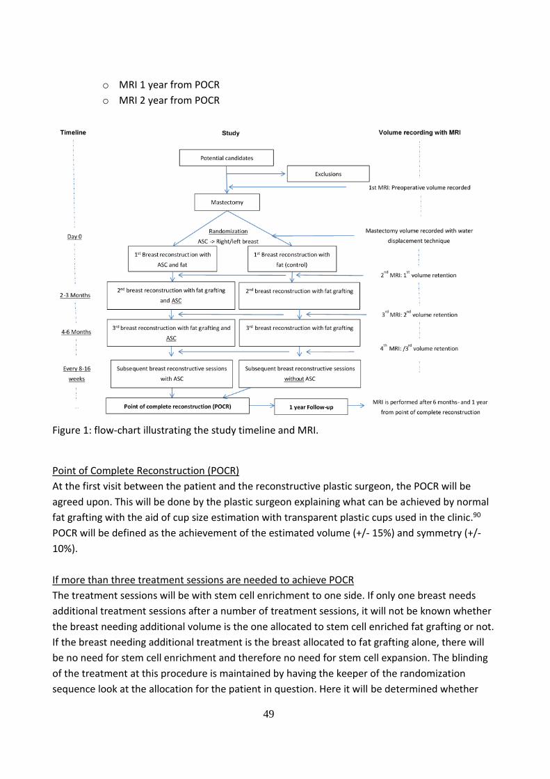
49
o MRI 1 year from POCR
o MRI 2 year from POCR
Figure 1: flow-chart illustrating the study timeline and MRI.
Point of Complete Reconstruction (POCR)
At the first visit between the patient and the reconstructive plastic surgeon, the POCR will be
agreed upon. This will be done by the plastic surgeon explaining what can be achieved by normal
fat grafting with the aid of cup size estimation with transparent plastic cups used in the clinic.90
POCR will be defined as the achievement of the estimated volume (+/- 15%) and symmetry (+/-
10%).
If more than three treatment sessions are needed to achieve POCR
The treatment sessions will be with stem cell enrichment to one side. If only one breast needs
additional treatment sessions after a number of treatment sessions, it will not be known whether
the breast needing additional volume is the one allocated to stem cell enriched fat grafting or not.
If the breast needing additional treatment is the breast allocated to fat grafting alone, there will
be no need for stem cell enrichment and therefore no need for stem cell expansion. The blinding
of the treatment at this procedure is maintained by having the keeper of the randomization
sequence look at the allocation for the patient in question. Here it will be determined whether
50
stem cell expansion is needed. If the breast needing additional surgery is the one allocated to fat
grafting without ASC enrichment, there will not be a cell expansion procedure.
At the point of surgery, the responsible person for the stem cell expansion will deliver an opaque
syringe containing either ASCs or another liquid, which will not be used for transplantation (pHPL
will be used, as it has similar appearance to ASC when contained in an opaque syringe). The mixing
of fat and stem cells will be performed by trained nurses, and the surgeons and trial group
members will be blinded by a surgical drape between them and the mixing table. The nurses will
receive an envelope with the allocation sequence as described previously. If the allocation dictates
ASCs for the breast that is about to receive fat grafting the nurses will use the ASC contained in the
syringe for enrichment. If the allocation dictates ASCs for the breast that is not about to undergo
surgery, the syringe will be discarded and a different syringe containing regular fat graft will be
used for “mock” mixing.
Revision surgery
Other (surgical) procedures than volume replacement performed on reconstructed breasts such as
nipple reconstruction, breast lift or use of implant will not be performed before the end of the
primary study period.
Statistical analysis
Differences in the primary endpoint will be assessed by a paired t-test.
The number of treatments needed for complete reconstruction and graft retention rate within
sessions will be analysed simultaneously by a joint longitudinal model. Specifically graft retention
rates will be modelled by a mixed linear model stratifying on session number and “breast” and
“person” as random effects. Moreover, the number of sessions needed to complete the
reconstruction will be modelled using a shared frailty grouped-data event time models including
“person” as a shared frailty. Changes in radiological markers within the patient (i.e. between
breasts) will be assessed by paired t-tests.
The impact of potential dropout will be assessed by sensitivity analyses based on various inverse
probability weighting schemes.
All statistical analyses will be made in R (www.r-project.org) using the add on packages survival
and multcomp. Adjustment for multiple testing will be done by means of the single step procedure
available in the R-package multcomp. P-values will be evaluated at a 5% significance level.
Ethical considerations
The protocol is approved by the Danish National Committee on Health Research Ethics, the Danish
Data Protection Agency and the Danish Medicines Agency.
Each test subject must provide written informed consent before they can be included in the study.
Information obtained on the subject’s health, other purely private matters and confidential
information is covered by professional secrecy. Project data will be kept securely locked during the
study period.
51
Risk assessment
Fat transplantation is an established treatment in the field of plastic surgery and is increasingly
used for reconstruction after breast cancer surgery15,16,58. Fat transplantation to the breast, which
includes liposuction of donor sites and injection of the fat to a recipient site, has a low risk of
complications (less than 4%) such as infection, bleeding, minor pain, irregularities and loose
skin.15,34
Fat transplantation can cause non-malignant radiological changes in the breast, which have been
reported in 17 studies with an overall occurrence of 13%.91 The radiological changes are mainly
small cysts or calcifications that occur as a result of fat necrosis after conventional fat
transplantation.34 These changes can be differentiated from malignant changes by experienced
radiologists and previous studies of fat grafting for breast reconstruction has not shown any
increase in the risk of cancer50,92–97.
Ex vivo expanded ASCs have not been shown to undergo malignant transformation when they are
utilized at early passages. The tumorigenesis of ASCs and MSCs has been investigated in several
studies, which have not shown any increased tumor formation.98–100 However one in vitro study
has shown that certain breast cancer cell lines increase their migratory properties in coculture
with ASCs although the ASCs had no effect on tumour cell proliferation or invasive properties.101
Another study has shown that ASCs in co-culture with squamous cell carcinoma cell lines increased
the proliferation rate of the cancer cells.102
Several experimental studies with immunodeficient mice and in vitro studies have been conducted
to investigate a wide range of cancer cells that were co-transplanted or stimulated with human
MSCs. One study showed that MSCs inhibit the growth of some breast cancer cell lines (MCF-7).103
Another study showed that MSCs are stimulatory to the HMT-3522 T4-2 breast cancer cell line.104
Human trials of ex vivo expanded MSCs/ASCs have been performed with a combined population of
more than 1000 patients, and none of these trials have reported serious adverse events in
response to the cell treatment.33,105–108 Studies on the effect of ASCs on locoregional recurrence or
distant metastasis of breast cancer after breast reconstruction have yet to be conducted.
However, two large cohort studies with matched controls did not show an increase in recurrence
or a decrease in survival in patients treated with fat grafting.109,110
The patients in this trial will be monitored by the screening program at the University Hospital of
Copenhagen for patients who are genetically predisposed to breast cancer and who have received
bilateral prophylactic nipple-sparing mastectomy.
The patients will receive ASC-enriched fat grafting in only one breast and conventional fat grafting
in the contralateral breast. Therefore, the potential beneficial effect of ASCs in volume retention
will lead to an asymmetric breast volume in the patients.
Any participant who shows breast asymmetry after follow-up will be offered additional
conventional fat transplantation procedures to the breast with a lower volume to achieve
symmetry. Patients with an asymmetry of less than 10% between the breasts may not be offered
52
correctional surgery unless the surgeon assess that an attempt to correct the asymmetry has a
good chance of benefitting the aesthetic outcome.
We have no reason to believe that the patients treated in this study will have to endure more
procedures or more side effects than patients who are treated with standard fat grafting for
breast reconstruction. The participants will benefit from any positive effect in the fat resorption
rate in one breast (either the right or the left) caused by the addition of ASCs, resulting in a
decreased need for fat harvesting from donor sites, which are often in limited supply in these
types of reconstructions. It is expected that both breasts (the breast treated with conventional fat
grafting and the breast treated with fat and ASCs) will require two or more treatments before the
reconstruction is completed.58
53
Discussion of the protocol in the context of the current evidence
To the best of my knowledge, this study will be the first to evaluate the efficacy of ASC-enriched
fat grafting for breast reconstruction after mastectomy. The main strength of the study is the
paired study design in which the included patients who undergo a bilateral prophylactic nipple-
sparing mastectomy are reconstructed with ASC-enriched fat grafting on one side and placebo-
enriched fat grafting on the contralateral side. The paired design balances most factors that
influence fat graft retention to isolate the effect of ASC-enrichment. Both breasts are affected
equally by physiological factors within the patient (i.e., weight change, smoking or
comorbidities), and therefore, the differences in fat graft resorption between the individual
patient’s breasts can most likely be attributed to ASC-enrichment. Furthermore, the study
provides the highest quality evidence because it is randomized, blinded and uses a validated
and objective measurement technique.
The effect of ASC/SVF-enrichment on fat graft volume retention has been investigated in
several reviews of both preclinical and clinical evidence, and generally they conclude that while
cell-enrichment has a great potential, evidence is still lacking.111–113 Most previous studies of
cell enrichment have focused on patients undergoing breast augmentation32 or for facial
contouring111. The first study to investigate the retention rate of ASC-enriched fat grafts in a
mouse model was Matsumoto et al.114 Next, Yoshimura et al. were the first to investigate “cell-
assisted lipotransfer (CAL)” in a clinical cohort study where they included patients to undergo
SVF-enriched fat grafting to the breast.115 However, this study did not included a control group.
Controlled clinical studies of CAL were later conducted by Peltoniemi32, Gentile29, Tissiani30 and
Chiu33. Of these four studies, two29,30 found a significant effect of the SVF enrichment but the
other two studies32,33 did not. However, none of the four studies were randomized or blinded,
making them susceptible to bias. Furthermore, none of the four studies used a validated
measurement technique to measure the fat graft volume retention. The problem with using a
method that is not validated and standardized is especially important in studies with no control
group.2 The studies without control groups are very difficult to compare with other studies if
they do not use identical methods for measuring the fat graft volume retention and even if they
use the same method, there is a risk of interobserver variation that may bias the comparison.
These limitations in the study designs of the previous studies could explain the heterogeneity of
some previous results.
Few studies have investigated the effect of ex-vivo expanded ASCs for enrichment of fat grafts
which can be used to achieve higher concentrations of ASCs per mL fat. The first published
study of ex-vivo expanded ASCs for fat grafting was conducted by Koh for Parry-Romberg
disease in which ASC-enriched fat grafting was used to correct contour deformities in the face.
The second study to investigate ASC-enriched fat grafting was conducted by Kølle et al., which
is the first trial investigating ASC-enriched fat grafting by our group. Both aforementioned
studies8 showed a significant higher retention rate in the ASCs-enriched fat grafts than in the
control fat grafts.
54
The main difference between expanded ASC-enrichment and SVF enrichment is that the SVF is
a heterogenous cell population that contain ASCs, whereas expanded ASCs comprise a
homogenous cell population that has the potential to provide substantially higher ASC dosages.
In a study of bolus fat grafts in a porcine model led by Bo Sonnich Rasmussen from our research
group, we investigated the dose-response of ASC-enriched fat grafting. The study indicated that
the optimal concentration of ASCs was 10 x 106 ASCs per mL of fat116, which is lower than the
concentration used in the first RCT conducted by Kølle el al., in which a dose of 20 x 106 ASCs
per mL fat graft was used. The study found a significantly higher retention rate in the cell-
enriched groups compared with the control group (13.0% vs. 9.2%), but the magnitude of the
difference was substantially lower than what was found in the study by Kølle et al.8 (80.9% vs.
16.3%). Interestingly, the study by Rasmussen et al.116 did not show a significantly difference
between SVF-enriched fat grafts and ASC-enriched fat grafts in terms of fat graft retention rate.
In the study by Glowinski et al.9 (EudraCT 2014-000510-59) we used the 10 x 106 ASCs per mL of
fat concentration for the breast augmentations of healthy female patients with breast
hypotrophy. The latter trial was to be concluded before we initiated the third RCT (EudraCT
2016-005186-31) of ASC-enriched fat grafting for breast reconstruction following skin-sparing
mastectomy, which is described in study 5.
Preliminary results from a randomized controlled trial of breast augmentation with ASC-
enriched fat grafting
The randomized controlled trial (RCT) of ASC-enriched fat grafting for breast augmentation in
women with breast hypotrophy (EudraCT 2014-000510-59) which was conducted by our group
and led by Peter Vester-Glowinski as the Principal Investigator was recently finalized. The
results were presented at the American society of plastic surgery (ASPS) conference Plastic
Surgery “The Meeting”9 and the main paper is in the final stages before submission to a peer-
reviewed journal. In this section, I will briefly summarize the trial and its results because it is
important in the context of this PhD thesis and in the discussion that follows in the next section.
Ten healthy women with breast hypotrophy were included in the trial. First, the women
underwent a small liposuction of approximately 200 cc of fat after local analgesia. This fat was
used to isolate the stromal vascular fraction (SVF), which was used to culture-expand ASCs. The
ASCs were harvested after 17 days of culture expansion with a minimum yield of 3 x 10⁹ ASCs.
These cells were used to perform breast augmentation with ex vivo expanded, autologous ASCs
to one of the breasts of each patient; the contralateral breast was augmented with normal fat
grafting. The ASC-enrichment dose was 10 x 106 ASCs per mL of fat graft. The placebo consisted
of a non-enriched fat graft that was mixed with the main graft using a mixing procedure
identical to that used for the ASC-enriched graft. All the syringes with fat w/o ASCs were
prepared prior to draping to cover the view of the operating table with the graft syringes. Then,
the envelope containing the allocation was opened behind a drape, and two nurses handed the
55
syringes to the surgeon according to the allocation. The surgeons were blinded to which breast
would be treated with ASC-enriched fat grafts.
The women were examined via MRI performed the day before surgery and 4 months and 1 year
after the surgery. These scans were used to measure fat graft volume retention over time using
the Lipovol software72 (described in study 4). Three data assessors blinded to the treatment
allocation performed the fat graft volume retention measurements in parallel.
The 10 women underwent breast augmentation with an average fat graft volume of 310 mL per
breast (range 300-350 mL). All women received the same stem cell or placebo volume in both
breasts. After 4 months, the average fat graft volume retention rate was 54.3% in the breasts
treated with ASC-enriched fat and 56.2% in the breasts treated with normal fat grafting. After 1
year, the average fat graft volume retention was 54.0% in the ASC-enriched breasts compared
with 55.9% in the breasts treated with normal fat grafting. The difference in fat graft volume
retention at 1 year was 1.9% lower in breasts treated with ASC-enriched fat grafting compared
with those treated with non-enriched fat grafts, and this difference was not statistically
significant (95% CI, -9.11% to 5.31%).9
Differences between the first two RCTs and implications for the protocol described in study 5
The preliminary results from the RCT of ASC-enriched fat grafting for breast augmentation
(EudraCT 2014-000510-59) led us to postpone initiating the RCT that would investigate ASC-
enriched fat grafting for breast reconstruction (EudraCT 2016-005186-31). These two RCTs use
the same protocol for isolation and expansion of autologous ASCs and a very similar method of
delivering ASCs to the patient. The most important difference between the two trials is the
recipient site; the former trial used ASC-enriched fat grafting for breast augmentation, and the
next trial will use ASC-enriched fat grafting for breast reconstruction following a prophylactic
mastectomy.
Before initiating a trial of ASC-enriched fat grafting for breast reconstruction (EudraCT 2016-
005186-31), we need to explore the differences between the RCT of bolus-injected fat grafts to
the upper arm (EudraCT 2010-023006-12) and the RCT of ASC-enriched fat grafting for breast
augmentation (EudraCT 2014-000510-59) (See the illustration of the trials of ASC-enriched fat
grafting in our group in the background section). In the first trial (EudraCT 2010-023006-12),
which was published in 20138, the authors found a significantly higher retention rate in the
ASC-enriched fat graft group compared with the non-enriched graft group (80.9% vs 16.3%). In
the second trial (EudraCT 2014-000510-59) which was presented in 20199, we did not find any
effect of ASC-enrichment (54.0 % retention rate in the breasts treated with cell-enriched fat
grafting (n = 10) and 55.9 % in the contralateral breasts treated with normal fat grafting and
placebo (n = 10)).
The two first trials (EudraCT 2010-023006-12 and EudraCT 2014-000510-59) relied on similar
principles for the ASC-enrichment, but there were differences between the two trials that need
to be explored. In the trial of ASC-enrichment for breast augmentation, we used 8 pcs. of
CellStack-10 (Corning Life Science, New York, USA) with a combined surface area of 50,880 cm2
56
per breast, whereas in the first trial of ASC-enriched fat grafting to the upper arm 10 pcs. of
T1000 cell flasks (Nunc A/S, Roskilde, Denmark) with a combined surface area of 10,000 cm2
were used. Second, the dose varied between the two trials, with the first trial8 using 20 x 106
ASCs/mL fat graft for a total dose of 0.6 x 10⁹ ASCs per upper arm and the second trial9 using 10
x 106 ASCs/mL fat graft for a total dose of 3.0 X 10⁹ ASCs per breast. Third, the first trial used a
bolus injection of the fat graft into the upper arm as the recipient site and the second trial used
structural fat grafting to the breast as the recipient site.
Research plan for further studies before initiation of the final trial
The methodological differences between the first two trials conducted in our group have
guided the research plan for how to explore the discrepancy in results.
We need a thorough analysis of the cellular characteristics of the ASCs expanded with the
protocol that was used in the first trial (EudraCT 2010-023006-12) compared with an analysis of
the cell characteristics of the ASCs from the second trial (EudraCT 2014-000510-59). The cells in
the two first trials fulfilled the criteria for mesenchymal stem cells according to Bourin et al.86,
confirmed by surface marker analysis. In addition, the protocols for ASC-expansion used in the
two trials have both been shown to produce cells that can differentiate into adipocytes,
chondrocytes and osteocytes.8,9,87,117 However, further analyses may show differences in the
cell product that might explain the difference in results between the two first trials. One could
hypothesize that ASCs, although viable, can be inactivated so that they do not support fat graft
retention. In the second trial of ASC-enriched fat grafting for breast augmentation (EudraCT
2014-000510-59), we obtained reference samples from all included patients, which are stored
in a biobank. These samples can be compared with cells expanded according to the protocol
used in the first trial (that unfortunately does not have reference samples available) to
investigate differences in cell characteristics (e.g., transcriptome analysis, Cell differentiation
assays and in vitro proliferation potential). Second, further animal studies could be used to
investigate the effect of ASCs in bolus grafts versus structural fat grafts. We have developed a
porcine model in our group that could be used for this purpose.118
Conclusion
This thesis consists of four studies that share a common foundation in that they are all concerned
with the evaluation of fat grafts in humans. The first two studies investigated the resorption
dynamics of fat grafts; and study 3 and 4 evaluate methods to measure fat graft volume retention
in the breast. Furthermore, the four studies are all necessary supporting studies in the design of a
protocol for a randomized controlled trial of ASC-enriched fat grafting for breast reconstruction,
which is described in study 5.
In study 1 of this thesis1, the co-authors and I demonstrated that the time to steady state-graft
retention of excised fat grafts in the mastoid cavity is 2.2 years on average according to our model.
This highlights the need for a long follow-up period when studying fat graft volume retention. In

57
study 2, we modelled the number of fat grafting sessions needed to complete a breast
reconstruction in five groups of patients that have undergone surgery for breast cancer. Our
model estimated that approximately three sessions are needed to complete a breast
reconstruction following a non-irradiated skin-sparring mastectomy. We also found that
significantly more fat grafting sessions are needed to reconstruct an irradiated breast than a non-
irradiated breast. In study 3, we evaluated the most commonly used MRI-based method for
measuring fat graft volume retention in the breast and found it to overestimate changes in breast
volume by approximately 50%. This has important implications for the ability of the method to
measure fat graft volume retention and for previous studies that have used this method. In study
4, the co-authors and I presented, and validated, an automated software tool for measuring fat
graft volume retention in the breast with high accuracy and precision. The new method can be
used in future studies of fat grafting to the breast. The results and the scientific experience gained
from the first four studies of this thesis were used to design a randomized controlled trial of ASC-
enriched fat grafting to the breast which has been approved by the Danish Medicines Agency and
the National Committee on Health Research Ethics.
Recently, our group published the preliminary results of another randomized controlled trial of
ASC-enriched fat grafting for breast augmentation (EudraCT 2014-000510-59) which did not find a
significantly higher fat graft volume retention in breasts treated with ASC-enriched fat grafting
than in breasts treated with normal fat grafting. Based on these results, the study group behind
the next RCT of ASC-enriched fat grafting for breast reconstruction following a skin-sparing
mastectomy (EudraCT 2016-005186-31) has postponed the trial initiation. Further studies are
needed to investigate the optimal procedure for isolation of SVF, expansion of ASCs, ensuring that
the ASCs have the appropriate characteristics to support fat graft volume retention before delivery
to patients. The research plan for these studies are outlined in this thesis although the plan is not
yet final. The heterogeneous results from studies of ASC-enriched fat grafting highlights the need
for more exploration into the exact mechanism by which ASCs may support fat graft volume
retention in vivo. Nevertheless, methods to increase the volume retention of fat grafts are still
warranted. The studies presented in this thesis has contributed to our understanding of the
resorption dynamics of fat grafts. Furthermore, the studies of methods to measure fat graft
volume retention in the breast, including the one that I presented in study 4, can be used to
ensure reproducible results, that can be compared between research groups in future studies. We
expect that the studies described in the research plan will provide us with the necessary
foundation to initiate the third RCT of ASC-enriched fat grafting for breast reconstruction. We
hope that this final study will lead us to our goal, which is the ability to offer our patients in need
of breast reconstruction a better treatment in the future.
58
Reference list
1. Herly M, Ørholt M, Glovinski PVPV, et al. Quantifying Long-Term Retention of Excised Fat Grafts: A Longitudinal, Retrospective Cohort Study of 108 Patients Followed for Up to 8.4 Years. Plast Reconstr Surg. 2017;139(5):1223-1232. doi:10.1097/PRS.0000000000003237
2. Herly M, Ørholt M, Larsen A, et al. Efficacy of breast reconstruction with fat grafting: A systematic review and meta-analysis. Journal of Plastic, Reconstructive and Aesthetic Surgery. 2018.
3. Herly M, Müller FC, Ørholt M, et al. The current gold standard breast volumetry technique seems to overestimate fat graft volume retention in the breast: A validation study. J Plast Reconstr Aesthetic Surg. 2019;72(8):1278-1284. doi:10.1016/j.bjps.2019.03.029
4. James IB, Bourne DA, DiBernardo G, et al. The architecture of fat grafting II: Impact of cannula diameter. Plast Reconstr Surg. 2018;142(5):1219-1225. doi:10.1097/PRS.0000000000004837
5. Bourne DA, James IB, Wang SS, Marra KG, Rubin JP. The Architecture of Fat Grafting: What Lies beneath the Surface. Plast Reconstr Surg. 2016. doi:10.1097/01.prs.0000479992.10986.ad
6. Coleman SR. Structural fat grafting: more than a permanent filler. Plast Reconstr Surg. 2006;118(3S):108S-120S.
7. Neuber G. Fettimplantationen. Verhandlungen der Dtsch Gesellschaftfür Chir. 1893;22(66).
8. Kølle SFT, Fischer-Nielsen A, Mathiasen AB, et al. Enrichment of autologous fat grafts with ex-vivo expanded adipose tissue-derived stem cells for graft survival: A randomised placebo-controlled trial. Lancet. 2013. doi:10.1016/S0140-6736(13)61410-5
9. Vester-Glowinski PV, Herly M, Svalgaard JD, et al. Enrichment of Fat Grafts With Adipose-derived Stromal Cells for Breast Augmentation: A Randomized, Double-blind, Placebo-controlled Trial of Fat Graft Survival. Plast Reconstr Surg Glob Open. 2019;7(8 Suppl):27-28. doi:10.1097/01.GOX.0000584348.15898.97
10. Czerny V. Plastischer ersatz der brustdruse durch ein lipom. Zentralbl Chir. 1895;27:72. 11. Holländer E. Die Kosmetische Chirurgie. Veit; 1912. 12. Lexer E. Die freien Transplantationen. Stuttgart: Enke, 1919–1924: 264–547. 13. Coleman SR, Saboeiro AP. Fat grafting to the breast revisited: Safety and efficacy. Plast
Reconstr Surg. 2007;119(3):775-785. doi:10.1097/01.prs.0000252001.59162.c9 14. Coleman SR. Structural fat grafting. Aesthetic Surg J. 1998;18(5):386-388. 15. Illouz Y, Sterodimas A. Autologous Fat Transplantation to the Breast: A Personal
Technique with 25 Years of Experience. Aesthetic Plast Surg. 2009;33(5):706-715. 16. Delay E, Garson S, Tousson G, Sinna R. Fat Injection to the Breast: Technique, Results,
and Indications Based on 880 Procedures Over 10 Years. Aesthetic Surg J. 2009;29(5):360-376. doi:10.1016/j.asj.2009.08.010
17. Bircoll M. Autologous Fat Transplantation: An Evaluation of Microcalcification and Fat Cell Survivability following (AFT) Cosmetic Breast Augmentation. Am J Cosmet Surg. 1988;5(4):283-288.
18. Rigotti G, Marchi A, Micciolo R, Baroni G. Autologous fat grafting in breast cancer patients. The Breast. 2012;21(5):690.

59
19. Ueberreiter K, von Finckenstein JG, Cromme F, Herold C, Tanzella U, Vogt PM. [BEAULI--a new and easy method for large-volume fat grafts]. Handchir Mikrochir Plast Chir. 2010;42(6):379-385. doi:10.1055/s-0030-1267913
20. Khouri RK, Khouri Jr RK, Rigotti G, et al. Aesthetic applications of Brava-assisted megavolume fat grafting to the breasts: a 9-year, 476-patient, multicenter experience. Plast Reconstr Surg. 2014;133(4):796-809.
21. Ho Quoc C, Piat JM, Carrabin N, Meruta A, Faure C, Delay E. Breast reconstruction with fat grafting and BRAVA((R)) pre-expansion: Efficacy evaluation in 45 cases. Ann Chir Plast Esthet. 2016;61(3):183-189. doi:10.1016/j.anplas.2015.06.010
22. Khouri RK, Eisenmann-Klein M, Cardoso E, et al. Brava and autologous fat transfer is a safe and effective breast augmentation alternative: results of a 6-year, 81-patient, prospective multicenter study. Plast Reconstr Surg. 2012;129(5):1173-1187.
23. Del Vecchio DA, Bucky LP. Breast augmentation using preexpansion and autologous fat transplantation: a clinical radiographic study. Plast Reconstr Surg. 2011;127(6):2441-2450.
24. Stillaert FBJL, Sommeling C, D’Arpa S, et al. Intratissular expansion–mediated, serial fat grafting: A step-by-step working algorithm to achieve 3D biological harmony in autologous breast reconstruction. J Plast Reconstr Aesthetic Surg. 2017;(Preprints).
25. Oranges CM, Striebel J, Tremp M, et al. The preparation of the recipient site in fat grafting: A comprehensive review of the preclinical evidence. Plast Reconstr Surg. 2019;143(4):1099-1107. doi:10.1097/PRS.0000000000005403
26. Zuk PA, Zhu M, Mizuno H, et al. Multilineage cells from human adipose tissue: implications for cell-based therapies. Tissue Eng. 2001;7(2):211-228. doi:10.1089/107632701300062859
27. Zuk PA, Zhu M, Ashjian P, et al. Human adipose tissue is a source of multipotent stem cells. Mol Biol Cell. 2002;13(12):4279-4295.
28. Zhu M, Zhou Z, Chen Y, et al. Supplementation of fat grafts with adipose-derived regenerative cells improves long-term graft retention. Ann Plast Surg. 2010;64(2):222-228.
29. Gentile P, Orlandi A, Scioli MG, et al. A Comparative Translational Study: The Combined Use of Enhanced Stromal Vascular Fraction and Platelet-Rich Plasma Improves Fat Grafting Maintenance in Breast Reconstruction. Stem Cells Transl Med. 2012;1(4):341-351. doi:10.5966/sctm.2011-0065
30. Tissiani LAL, Alonso N. A Prospective and Controlled Clinical Trial on Stromal Vascular Fraction Enriched Fat Grafts in Secondary Breast Reconstruction. Stem Cells Int. 2015:1-12. doi:10.1155/2016/2636454
31. Yoshimura K, Sato K, Aoi N, Kurita M, Hirohi T, Harii K. Cell-assisted lipotransfer for cosmetic breast augmentation: Supportive use of adipose-derived stem/stromal cells. Aesthetic Plast Surg. 2008;32(1):48-55. doi:10.1007/s00266-007-9019-4
32. Peltoniemi HH, Salmi A, Miettinen S, et al. Stem cell enrichment does not warrant a higher graft survival in lipofilling of the breast: A prospective comparative study. J Plast Reconstr Aesthetic Surg. 2013;66(11):1494-1503. doi:10.1016/j.bjps.2013.06.002
33. Chiu CH. Does Stromal Vascular Fraction Ensure a Higher Survival in Autologous Fat Grafting for Breast Augmentation? A Volumetric Study Using 3-Dimensional Laser Scanning. Aesthetic Surg J. 2019;39(1):41-52. doi:10.1093/asj/sjy030
60
34. Ørholt M, Larsen A, Hemmingsen MN, et al. Complications after Breast Augmentation with Fat Grafting. Plast Reconstr Surg. 2020;145(3):530e-537e. doi:10.1097/prs.0000000000006569
35. Zocchi M, Zocchi L. Large-volume breast fat transfer: technical evolutions and safety aspects based on over 800 cases and 26 years of follow-up. Eur J Plast Surg. 2017;40(5):367-382. doi:10.1007/s00238-017-1339-2
36. Ersek RA. Transplantation of purified autologous fat: a 3-year follow-up is disappointing. Plast Reconstr Surg. 1991;87(2):219-227.
37. Niechajev I, Sevćuk O. Long-term results of fat transplantation: clinical and histologic studies. Plast Reconstr Surg. 1994;94(3):496-506.
38. Gentile P, Di Pasquali C, Bocchini I, et al. Breast Reconstruction With Autologous Fat Graft Mixed With Platelet-Rich Plasma. Surg Innov. 2013;20(4):370-376.
39. Hammer-Hansen N, Jensen TBB, Damsgaard TEE. Delayed Total Breast Reconstruction with Brava. Case Rep Surg. 2015;2015:1-3. doi:10.1155/2015/601904
40. Longo B, Laporta R, Sorotos M, Pagnoni M, Gentilucci M, Santanelli di Pompeo F. Total breast reconstruction using autologous fat grafting following nipple-sparing mastectomy in irradiated and non-irradiated patients. Aesthetic Plast Surg. 2014;38(6):1101-1108. doi:10.1007/s00266-014-0406-3
41. Silva-Vergara C, Fontdevila J, Descarrega J, Burdio F, Yoon TS, Grande L. Oncological outcomes of lipofilling breast reconstruction: 195 consecutive cases and literature review. J Plast Reconstr Aesthet Surg. 2016;69(4):475-481. doi:10.1016/j.bjps.2015.12.029
42. Herold C, Ueberreiter K, Busche MN, Vogt PM. Autologous fat transplantation: volumetric tools for estimation of volume survival. A systematic review. Aesthetic Plast Surg. 2013;37(2):380-387. doi:10.1007/s00266-012-0046-4
43. Herold C, Ueberreiter K, Cromme F, Busche MN, Vogt PM. The use of mamma MRI volumetry to evaluate the rate of fat survival after autologous lipotransfer. Handchirurgie, Mikrochirurgie, Plast Chir Organ der Deutschsprachigen Arbeitsgemeinschaft für Handchirurgie Organ der Deutschsprachigen Arbeitsgemeinschaft für Mikrochirurgie der Peripher Nerven und Gefässe Organ der Vereinigung der Deut. 2010;42(2):129-134. doi:10.1055/s-0029-1243204
44. Herold C, Grimme M, Vogt PM, Ueberreiter K, Cromme F. Is there a need for intrapectoral injection in autologous fat transplantation to the breast? - An MRI volumetric study. Handchirurgie Mikrochirurgie Plast Chir. 2011;43(2):119-124. doi:10.1055/s-0030-1269931
45. Koch MC, Adamietz B, Jud SM, et al. Breast volumetry using a three-dimensional surface assessment technique. Aesthetic Plast Surg. 2011;35(5):847-855. doi:10.1007/s00266-011-9708-x
46. Liu C, Luan J, Mu L, Ji K. The role of three-dimensional scanning technique in evaluation of breast asymmetry in breast augmentation: a 100-case study. Plast Reconstr Surg. 2010;126(6):2125-2132.
47. Yip JM, Mouratova N, Jeffery RM, Veitch DE, Woodman RJ, Dean NR. Accurate assessment of breast volume: A study comparing the volumetric gold standard (direct water displacement measurement of mastectomy specimen) with a 3D laser scanning technique. Ann Plast Surg. 2012;68(2):135-141. doi:10.1097/SAP.0b013e31820ebdd0

61
48. Glovinski P V., Herly M, Muller FC, et al. Avoiding a Systematic Error in Assessing Fat Graft Survival in the Breast with Repeated Magnetic Resonance Imaging. Plast Reconstr Surg Glob Open. 2016;4(9):e1023. doi:10.1097/GOX.0000000000001023
49. Jung HK, Kim CH, Song SY. Prospective 1-Year Follow-Up Study of Breast Augmentation by Cell-Assisted Lipotransfer. Aesthet Surg J. 2016;36(2):179-190. doi:10.1093/asj/sjv164
50. Fiaschetti V, Pistolese CA, Fornari M, et al. Magnetic resonance imaging and ultrasound evaluation after breast autologous fat grafting combined with platelet-rich plasma. Plast Reconstr Surg. 2013;132(4):498e-509e.
51. Yoshimura K, Sato K, Aoi N, et al. Cell-assisted lipotransfer for facial lipoatrophy: Efficacy of clinical use of adipose-derived stem cells. Dermatologic Surg. 2008;34(9):1178-1185. doi:10.1111/j.1524-4725.2008.34256.x
52. Carpaneda CA, Ribeiro MT. Study of the histologic alterations and viability of the adipose graft in humans. Aesthetic Plast Surg. 1993. doi:10.1007/BF00455048
53. Groen JW, Negenborn VL, Twisk D, et al. Autologous fat grafting in onco-plastic breast reconstruction: A systematic review on oncological and radiological safety, complications, volume retention and patient/surgeon satisfaction. J Plast Reconstr Aesthet Surg. 2016;69(6):742-764. doi:10.1016/j.bjps.2016.03.019
54. Agha RA, Fowler AJ, Herlin C, Goodacre TEE, Orgill DP. Use of autologous fat grafting for breast reconstruction: a systematic review with meta-analysis of oncological outcomes. J Plast Reconstr Aesthet Surg. 2015;68(2):143-161. doi:10.1016/j.bjps.2014.10.038
55. Waked K, Colle J, Doornaert M, Cocquyt V, Blondeel P. Systematic review: The oncological safety of adipose fat transfer after breast cancer surgery. Breast. 2017;31:128-136. doi:10.1016/j.breast.2016.11.001
56. Stroup DF, Berlin JA, Morton SC, et al. Meta-analysis of observational studies in epidemiology: a proposal for reporting. Jama. 2000;283(15):2008-2012.
57. Katzel EB, Bucky LP. Fat Grafting to the Breast: Clinical Applications and Outcomes for Reconstructive Surgery. Plast Reconstr Surg. 2017;140(5S):69S-76S. doi:10.1097/PRS.0000000000003945
58. Khouri RK, Rigotti G, Khouri RK, Biggs TM. Tissue-Engineered Breast Reconstruction with Brava-Assisted Fat Grafting: A 7-Year, 488-Patient, Multicenter Experience. Plast Reconstr Surg. 2015;135(3):643-658. doi:10.1097/PRS.0000000000001039
59. Babovic S. Complete breast reconstruction with autologous fat graft - a case report. J Plast Reconstr Aesthetic Surg. 2010;63(7). doi:10.1016/j.bjps.2009.11.027
60. Fabiocchi L, Semprini G, Cattin F, et al. “Reverse expansion”: A new technique of breast reconstruction with autologous tissue. J Plast Reconstr Aesthetic Surg. 2017;70(11):1537-1542. doi:10.1016/j.bjps.2017.07.001
61. Semprini G, Cattin F, Zanin C, et al. About locoregional recurrence risk after lipofilling in breast cancer patients: Our experience. Minerva Chir. 2014;69(2):91-96.
62. Herold C, Reichelt A, Stieglitz LH, et al. MRI-based breast volumetry-evaluation of three different software solutions. J Digit Imaging. 2010;23(5):603-610. doi:10.1007/s10278-009-9264-y
63. Losken A, Seify H, Denson DD, Paredes AA, Carlson GW. Validating three-dimensional imaging of the breast. Ann Plast Surg. 2005;54(5):471-476. doi:10.1097/01.sap.0000155278.87790.a1
64. Seoud L, Ramsay J, Parent S, Cheriet F. A novel fully automatic measurement of apparent

62
breast volume from trunk surface mesh. Med Eng Phys. 2017;41:46-54. doi:10.1016/j.medengphy.2017.01.004
65. Henseler H, Smith J, Bowman A, et al. Investigation into variation and errors of a three-dimensional breast imaging system using multiple stereo cameras. J Plast Reconstr Aesthetic Surg. 2012;65(12). doi:10.1016/j.bjps.2012.05.028
66. Lee WY, Kim MJ, Lew DH, Song SY, Lee DW. Three-dimensional surface imaging is an effective tool for measuring breast volume: A validation study. Arch Plast Surg. 2016;43(5):430-437. doi:10.5999/aps.2016.43.5.430
67. Rha EY, Choi IK, Yoo G. Accuracy of the method for estimating breast volume on three-dimensional simulated magnetic resonance imaging scans in breast reconstruction. Plast Reconstr Surg. 2014;133(1):14-20. doi:10.1097/01.prs.0000436813.03838.09
68. Oranges CM, Madduri S, Brantner P, et al. Three-dimensional Assessment of the Breast: Validation of a novel, simple and inexpensive scanning process. In Vivo (Brooklyn). 2019;33(3):839-842. doi:10.21873/invivo.11548
69. Erić M, Anderla A, Stefanović D, Drapšin M. Breast volume estimation from systematic series of CT scans using the Cavalieri principle and 3D reconstruction. Int J Surg. 2014;12(9):912-917. doi:10.1016/j.ijsu.2014.07.018
70. Wesselius TS, Verhulst AC, Vreeken RD, Xi T, Maal TJJ, Ulrich DJO. Accuracy of three software applications for breast volume calculations from three-dimensional surface images. Plast Reconstr Surg. 2018;142(4):858-865. doi:10.1097/PRS.0000000000004728
71. MeVisLab. 72. Hemmingsen MN, Orholt M, Muller F, et al. Lipovol: A Free and Validated Software for
Measuring Fat Graft Volume Retention in the Breast With Magnetic Resonance Imaging. Plast Reconstr Surg - Glob Open. 2019;7:134. doi:10.1097/01.GOX.0000585004.36502.bf
73. Torre LA, Bray F, Siegel RL, et al. Global cancer statistics, 2012. CA Cancer J Clin. 2015;65(2):87-108. doi:10.3322/caac.21262
74. Elder EE, Brandberg Y, Björklund T, et al. Quality of life and patient satisfaction in breast cancer patients after immediate breast reconstruction: A prospective study. Breast. 2005;14(3):201-208. doi:10.1016/j.breast.2004.10.008
75. Bresser PJC, Seynaeve C, Van Gool AR, et al. Satisfaction with prophylactic mastectomy and breast reconstruction in genetically predisposed women. Plast Reconstr Surg. 2006;117(6):1675-1682. doi:10.1097/01.prs.0000217383.99038.f5
76. Cunningham B, McCue J. Erratum: Safety and effectiveness of Mentor’s MemoryGel implants at 6 years (Aesthetic Plastic Surgery DOI: 10.1007/s00266-009-9364-6). Aesthetic Plast Surg. 2009;33(3):439. doi:10.1007/s00266-009-9379-z
77. Handel N, Garcia ME, Wixtrom R. Breast implant rupture: causes, incidence, clinical impact, and management. Plast Reconstr Surg. 2013;132(5):1128-1137. doi:10.1097/PRS.0b013e3182a4c243
78. Sailon AM, Schachar JS, Levine JP. Free transverse rectus abdominis myocutaneous and deep inferior epigastric perforator flaps for breast reconstruction: a systematic review of flap complication rates and donor-site morbidity. Ann Plast Surg. 2009;62(5):560-563. doi:10.1097/SAP.0b013e31819faf0d
79. Antony AK, McCarthy C, Disa JJ, Mehrara BJ. Bilateral implant breast reconstruction outcomes, predictors, and matched cohort analysis in 730 2-stage breast reconstructions over 10 years. In: Annals of Plastic Surgery. Vol 72. ; 2014:625-630.

63
doi:10.1097/SAP.0b013e31826af03e 80. Gir P, Brown SA, Oni G, Kashefi N, Mojallal A, Rohrich RJ. Fat grafting: Evidence-based
review on autologous fat harvesting, processing, reinjection, and storage. Plast Reconstr Surg. 2012;130(1):249-258. doi:10.1097/PRS.0b013e318254b4d3
81. Sinna R, Delay E, Garson S, Delaporte T, Toussoun G. Breast fat grafting (lipomodelling) after extended latissimus dorsi flap breast reconstruction: A preliminary report of 200 consecutive cases. J Plast Reconstr Aesthetic Surg. 2010;63(11):1769-1777. doi:10.1016/j.bjps.2009.12.002
82. Friedenstein AJ, Deriglasova UF, Kulagina NN, et al. Precursors for fibroblasts in different populations of hematopoietic cells as detected by the in vitro colony assay method. Exp Hematol. 1974;2(2):83-92.
83. Friedenstein AJ, Petrakova K V, Kurolesova AI, Frolova GP. Heterotopic of bone marrow. Analysis of precursor cells for osteogenic and hematopoietic tissues. Transplantation. 1968;6(2):230-247. http://www.ncbi.nlm.nih.gov/pubmed/5654088.
84. Locke M, Feisst V, Dunbar PR. Concise review: Human adipose-derived stem cells: Separating promise from clinical need. Stem Cells. 2011;29(3):404-411. doi:10.1002/stem.593
85. Dominici M, Le Blanc K, Mueller I, et al. Minimal criteria for defining multipotent mesenchymal stromal cells. The International Society for Cellular Therapy position statement. Cytotherapy. 2006;8(4):315-317. doi:10.1080/14653240600855905
86. Bourin P, Bunnell BA, Casteilla L, et al. Stromal cells from the adipose tissue-derived stromal vascular fraction and culture expanded adipose tissue-derived stromal/stem cells: A joint statement of the International Federation for Adipose Therapeutics and Science (IFATS) and the International So. Cytotherapy. 2013;15(6):641-648. doi:10.1016/j.jcyt.2013.02.006
87. Glovinski P V, Herly M, Mathiasen AB, et al. Overcoming the bottleneck of platelet lysate supply in large-scale clinical expansion of adipose-derived stem cells: a comparison of fresh versus three types of platelet lysates from outdated buffy coat–derived platelet concentrates. Cytotherapy. 2017;19(2):222-234.
88. Schallmoser K, Bartmann C, Rohde E, et al. Human platelet lysate can replace fetal bovine serum for clinical-scale expansion of functional mesenchymal stromal cells. Transfusion. 2007;47(8):1436-1446. doi:10.1111/j.1537-2995.2007.01220.x
89. Svalgaard JD, Haastrup EK, Reckzeh K, et al. Low-molecular-weight carbohydrate Pentaisomaltose may replace dimethyl sulfoxide as a safer cryoprotectant for cryopreservation of peripheral blood stem cells. Transfusion. 2016;56(5):1088-1095. doi:10.1111/trf.13543
90. Ikander P, Drejøe JB, Lumholt P, et al. Measurement of breast volume is a useful supplement to select candidates for surgical breast reduction. Dan Med J. 2014;61(1).
91. Claro F, Figueiredo JCA, Zampar AG, Pinto-Neto AM. Applicability and safety of autologous fat for reconstruction of the breast. Br J Surg. 2012;99(6):768-780. doi:10.1002/bjs.8722
92. Myckatyn TM, Wagner IJ, Mehrara BJ, et al. Cancer Risk after Fat Transfer: A Multicenter Case-Cohort Study. Plast Reconstr Surg. 2017;139(1):11-18. doi:10.1097/PRS.0000000000002838
93. Gale KL, Rakha EA, Ball G, Tan VK, McCulley SJ, Macmillan RD. A Case-Controlled Study of
64
the Oncologic Safety of Fat Grafting. Plast Reconstr Surg. 2015;135(5):1263-1275. doi:10.1097/PRS.0000000000001151
94. Ihrai T, Georgiou C, Machiavello JC, et al. Autologous fat grafting and breast cancer recurrences: Retrospective analysis of a series of 100 procedures in 64 patients. J Plast Surg Hand Surg. 2013;47(4):273-275. doi:10.3109/2000656X.2012.759583
95. Rietjens M, De Lorenzi F, Rossetto F, et al. Safety of fat grafting in secondary breast reconstruction after cancer. J Plast Reconstr Aesthetic Surg. 2011;64(4):477-483. doi:10.1016/j.bjps.2010.06.024
96. Parikh RP, Doren EL, Mooney B, Sun W V., Laronga C, Smith PD. Differentiating Fat Necrosis from Recurrent Malignancy in Fat-Grafted Breasts. Plast Reconstr Surg. 2012;130(4):761-772. doi:10.1097/prs.0b013e318262f03b
97. Veber M, Tourasse C, Toussoun G, Moutran M, Mojallal A, Delay E. Radiographic findings after breast augmentation by autologous fat transfer. Plast Reconstr Surg. 2011;127(3):1289-1299. doi:10.1097/PRS.0b013e318205f38f
98. Freese KE, Kokai L, Edwards RP, et al. Adipose-derived stems cells and their role in human cancer development, growth, progression, and metastasis: A systematic review. Cancer Res. 2015;75(7):1161-1168. doi:10.1158/0008-5472.CAN-14-2744
99. Barkholt L, Flory E, Jekerle V, et al. Risk of tumorigenicity in mesenchymal stromal cell-based therapies - Bridging scientific observations and regulatory viewpoints. Cytotherapy. 2013;15(7):753-759. doi:10.1016/j.jcyt.2013.03.005
100. Eterno V, Zambelli A, Pavesi L, et al. Adipose-derived mesenchymal stem cells (ASCs) may favour breast cancer recurrence via HGF/c-Met signaling. Oncotarget. 2014;5(3):613-633. doi:10.18632/oncotarget.1359
101. Koellensperger E, Bonnert LC, Zoernig I, et al. The impact of human adipose tissue-derived stem cells on breast cancer cells: Implications for cell-assisted lipotransfers in breast reconstruction. Stem Cell Res Ther. 2017;8(1). doi:10.1186/s13287-017-0579-1
102. Koellensperger E, Gramley F, Preisner F, Leimer U, Germann G, Dexheimer V. Alterations of gene expression and protein synthesis in co-cultured adipose tissue-derived stem cells and squamous cell-carcinoma cells: Consequences for clinical applications. Stem Cell Res Ther. 2014;5(3). doi:10.1186/scrt454
103. Qiao L, Xu Z li, Zhao T jun, Ye L hong, Zhang X dong. Dkk-1 secreted by mesenchymal stem cells inhibits growth of breast cancer cells via depression of Wnt signalling. Cancer Lett. 2008;269(1):67-77. doi:10.1016/j.canlet.2008.04.032
104. Zhao M, Sachs PC, Wang X, et al. Mesenchymal stem cells in mammary adipose tissue stimulate progression of breast cancer resembling the basal-type. Cancer Biol Ther. 2012;13(9):782-792. doi:10.4161/cbt.20561
105. Prockop DJ, Brenner M, Fibbe WE, et al. Defining the risks of mesenchymal stromal cell therapy. Cytotherapy. 2010;12(5):576-578. doi:10.3109/14653249.2010.507330
106. Casiraghi F, Remuzzi G, Abbate M, Perico N. Multipotent Mesenchymal Stromal Cell Therapy and Risk of Malignancies. Stem Cell Rev Reports. 2013;9(1):65-79. doi:10.1007/s12015-011-9345-4
107. Qayyum AA, Haack-Sørensen M, Mathiasen AB, Jørgensen E, Ekblond A, Kastrup J. Adipose-derived mesenchymal stromal cells for chronic myocardial ischemia (MyStromalCell Trial): Study design. Regen Med. 2012;7(3):421-428. doi:10.2217/rme.12.17

65
108. Herreros MD, Garcia-Arranz M, Guadalajara H, De-La-Quintana P, Garcia-Olmo D. Autologous Expanded Adipose-Derived Stem Cells for the Treatment of Complex Cryptoglandular Perianal Fistulas. Dis Colon Rectum. 2012;55(7):762-772. doi:10.1097/dcr.0b013e318255364a
109. Vyas KS, DeCoster RC, Burns JC, et al. Autologous Fat Grafting Does Not Increase Risk of Oncologic Recurrence in the Reconstructed Breast. Ann Plast Surg. 2020;84(6S Suppl 5):S405-S410. doi:10.1097/SAP.0000000000002285
110. Sorrentino L, Regolo L, Scoccia E, et al. Autologous fat transfer after breast cancer surgery: An exact-matching study on the long-term oncological safety. Eur J Surg Oncol. 2019;45(10):1827-1834. doi:10.1016/j.ejso.2019.05.013
111. Zhou Y, Wang J, Li H, et al. Efficacy and Safety of Cell-Assisted Lipotransfer: A Systematic Review and Meta-Analysis. Plast Reconstr Surg. 2016;137(1):44e-57e. doi:10.1097/prs.0000000000001981
112. Rasmussen BS, Lykke Sørensen C, Vester-Glowinski PV, et al. Effect, Feasibility, and Clinical Relevance of Cell Enrichment in Large Volume Fat Grafting: A Systematic Review. Aesthetic Surg J. 2017;37(3):S46-S58. doi:10.1093/asj/sjx008
113. Toyserkani NM, Quaade ML, Sørensen JA. Cell-Assisted Lipotransfer: A Systematic Review of Its Efficacy. Aesthetic Plast Surg. 2016;40(2):309-318. doi:10.1007/s00266-016-0613-1
114. Matsumoto D, Sato K, Gonda K, et al. Cell-assisted lipotransfer: Supportive use of human adipose-derived cells for soft tissue augmentation with lipoinjection. Tissue Eng. 2006;12(12):3375-3382. doi:10.1089/ten.2006.12.3375
115. Yoshimura K, Matsumoto D, Sato K. Cell-assisted lipotransfer for breast augmentation. Japanese J Plast Reconstr Surg. 2007;50(12):1425-1437.
116. Rasmussen BS, Sørensen CL, Kurbegovic S, et al. Cell-Enriched Fat Grafting Improves Graft Retention in a Porcine Model: A Dose-Response Study of Adipose-Derived Stem Cells versus Stromal Vascular Fraction. Plast Reconstr Surg. 2019;144(3).
117. Trojahn Kølle SF, Oliveri RS, Glovinski P V., et al. Pooled human platelet lysate versus fetal bovine serum-investigating the proliferation rate, chromosome stability and angiogenic potential of human adipose tissue-derived stem cells intended for clinical use. Cytotherapy. 2013;15(9):1086-1097. doi:10.1016/j.jcyt.2013.01.217
118. Rasmussen BS, Sørensen CL, Vester-Glowinski P V., et al. A novel porcine model for future studies of cell-enriched fat grafting. Plast Reconstr Surg - Glob Open. 2018;6(4). doi:10.1097/GOX.0000000000001735
66
Appendix 1
Quantifying Long-Term Retention of Excised Fat Grafts: A Longitudinal, Retrospective Cohort Study
of 108 Patients Followed for up to 8.4 Years
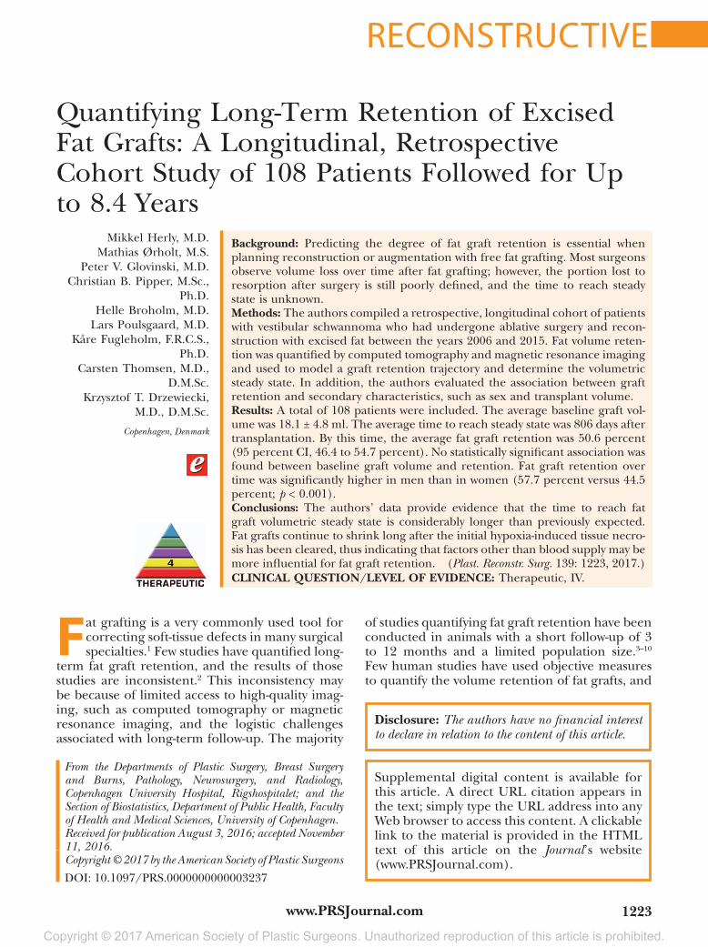
Copyright © 2017 American Society of Plastic Surgeons. Unauthorized reproduction of this article is prohibited.
www.PRSJournal.com 1223
Fat grafting is a very commonly used tool for correcting soft-tissue defects in many surgical specialties.1 Few studies have quantified long-
term fat graft retention, and the results of those studies are inconsistent.2 This inconsistency may be because of limited access to high-quality imag-ing, such as computed tomography or magnetic resonance imaging, and the logistic challenges associated with long-term follow-up. The majority
of studies quantifying fat graft retention have been conducted in animals with a short follow-up of 3 to 12 months and a limited population size.3–10 Few human studies have used objective measures to quantify the volume retention of fat grafts, and
Disclosure: The authors have no financial interest to declare in relation to the content of this article.
Copyright © 2017 by the American Society of Plastic Surgeons
DOI: 10.1097/PRS.0000000000003237
Mikkel Herly, M.D.Mathias Ørholt, M.S.
Peter V. Glovinski, M.D.Christian B. Pipper, M.Sc.,
Ph.D.Helle Broholm, M.D.
Lars Poulsgaard, M.D.Kåre Fugleholm, F.R.C.S.,
Ph.D.Carsten Thomsen, M.D.,
D.M.Sc.Krzysztof T. Drzewiecki,
M.D., D.M.Sc.
Copenhagen, Denmark
Background: Predicting the degree of fat graft retention is essential when planning reconstruction or augmentation with free fat grafting. Most surgeons observe volume loss over time after fat grafting; however, the portion lost to resorption after surgery is still poorly defined, and the time to reach steady state is unknown.Methods: The authors compiled a retrospective, longitudinal cohort of patients with vestibular schwannoma who had undergone ablative surgery and recon-struction with excised fat between the years 2006 and 2015. Fat volume reten-tion was quantified by computed tomography and magnetic resonance imaging and used to model a graft retention trajectory and determine the volumetric steady state. In addition, the authors evaluated the association between graft retention and secondary characteristics, such as sex and transplant volume.Results: A total of 108 patients were included. The average baseline graft vol-ume was 18.1 ± 4.8 ml. The average time to reach steady state was 806 days after transplantation. By this time, the average fat graft retention was 50.6 percent (95 percent CI, 46.4 to 54.7 percent). No statistically significant association was found between baseline graft volume and retention. Fat graft retention over time was significantly higher in men than in women (57.7 percent versus 44.5 percent; p < 0.001).Conclusions: The authors’ data provide evidence that the time to reach fat graft volumetric steady state is considerably longer than previously expected. Fat grafts continue to shrink long after the initial hypoxia-induced tissue necro-sis has been cleared, thus indicating that factors other than blood supply may be more influential for fat graft retention. (Plast. Reconstr. Surg. 139: 1223, 2017.)CLINICAL QUESTION/LEVEL OF EVIDENCE: Therapeutic, IV.
From the Departments of Plastic Surgery, Breast Surgery and Burns, Pathology, Neurosurgery, and Radiology, Copenhagen University Hospital, Rigshospitalet; and the Section of Biostatistics, Department of Public Health, Faculty of Health and Medical Sciences, University of Copenhagen.Received for publication August 3, 2016; accepted November 11, 2016.
Quantifying Long-Term Retention of Excised Fat Grafts: A Longitudinal, Retrospective Cohort Study of 108 Patients Followed for Up to 8.4 Years
Supplemental digital content is available for this article. A direct URL citation appears in the text; simply type the URL address into any Web browser to access this content. A clickable link to the material is provided in the HTML text of this article on the Journal’s website (www.PRSJournal.com).
SUPPLEMENTAL DIGITAL CONTENT IS AVAIL-ABLE IN THE TEXT.
2017
RECONSTRUCTIVE

Copyright © 2017 American Society of Plastic Surgeons. Unauthorized reproduction of this article is prohibited.
1224
Plastic and Reconstructive Surgery • May 2017
to the best of our knowledge, none of these have had a follow-up period longer than 1 year.11–14 Pub-lished studies with a follow-up period longer than 1 year have relied on a surgeon’s assessment/expe-rience and did not quantify graft volume.15–17
The ability to predict the long-term outcome of reconstructive procedures, such as fat grafting, is important to ensure the desired aesthetic outcome. To improve knowledge regarding the development of free fat grafts over time, we quantified long-term graft volume retention in a retrospective longitudi-nal cohort study of patients treated with excised fat to fill dead space in the mastoid cavity after ablative surgery for vestibular schwannoma.
Approximately 50 percent of vestibular schwannomas are removed by means of the trans-labyrinthine approach18 at our institution. This approach starts with a retroauricular incision fol-lowed by a mastoidectomy. Thereafter, the dura is exposed and can be opened. The tumor is found on the inner side of the dura and is removed by dissection. After removal of the tumor, an excised fat graft from the lower abdominal area is used to fill the dead space left by the mastoidectomy (Fig. 1).19 The role of the fat graft is to prevent cerebrospinal fluid leakage and to avert a large hollowing behind the patient’s ear.20
After surgery, the patients are offered postop-erative computed tomography and two follow-up magnetic resonance imaging scans during the
first year, followed by one yearly magnetic reso-nance imaging scan to detect recurrence. This large collection of imaging material had captured the destiny of fat grafts over time, and we saw this as a unique opportunity to document long-term outcomes of excised fat grafts.
PATIENTS AND METHODS
Study DesignIn a retrospective longitudinal cohort study,
fat graft retention was quantified over time by analysis of computed tomographic and magnetic resonance imaging scans from 108 patients. The data were used to model long-term fat graft reten-tion trajectory and to estimate the time to steady state in graft volume.
The data were obtained from patients who had undergone reconstruction with excised fat to fill dead space in the mastoid cavity after ablative sur-gery for vestibular schwannoma. The collection of data without consent was approved by the Danish Health and Medicines Authority. The protocol for handling patient data was approved by the Regional Danish Data Protection Agency of Copenhagen.
Study PopulationThe cohort consisted of patients treated surgi-
cally for vestibular schwannoma with a translabyrin-thine approach and who underwent reconstruction
Fig. 1. A 74-year-old woman. (Left) After removal of vestibular schwannoma and placement of fat graft. The fat graft is marked with an asterisk; the pericranial flap used to cover the fat graft is marked with double asterisks. (Right) Same angle as seen on the left after closure. The location of the incision behind the ear is shown without the draping.
Copyright © 2017 American Society of Plastic Surgeons. Unauthorized reproduction of this article is prohibited.
Volume 139, Number 5 • Long-Term Fat Graft Retention
1225
with excised fat grafting between October of 2006 and September of 2015 at the Copenhagen Uni-versity Hospital. Patients were excluded if they had undergone a secondary operation or radiotherapy in the area of fat grafting or if the first follow-up scan was obtained more than 14 days after surgery. Furthermore, patients were excluded if visualiza-tion of the entire fat graft was prevented by techni-cal issues, such as scans that showed only part of the transplant. The observation started with the first scan and ended with the last follow-up scan.
Neurosurgical treatment of vestibular schwan-noma in Denmark is performed solely at Copen-hagen University Hospital, Rigshospitalet. Thus, the included patient population originates from the entire Danish population (5.6 million inhab-itants).21 Vestibular schwannoma is a benign, slow-growing, and generally nonhereditary tumor originating from the Schwann cells of the vestibu-lar nerve or, more rarely, the acoustic nerve.22–24 Previous reports on Western patients with ves-tibular schwannoma state that the demographics of these patients are similar to those of the back-ground population in age and distribution of the sexes.18,22 Vestibular schwannoma is not related to any significant risk factors or comorbidities, and microscopic free-margin surgery (single treat-ment) is typically curative, with a 0.05 percent recurrence rate.25,26
Primary OutcomeThe primary variables of interest were volume
retention of fat grafts over time and the time to reach volumetric steady state. Volume retention was measured by analysis of computed tomo-graphic and magnetic resonance imaging scans obtained after transplantation. The baseline fat graft volume was measured from the postoperative computed tomographic scan obtained within the first few days after ablative surgery to exclude post-operative bleeding. The baseline volume was set to 100 percent and served as the comparison for all subsequent examinations. The volumes recorded from subsequent follow-up magnetic resonance imaging scans were calculated as a percentage of the baseline volume and plotted against the num-ber of days after surgery. These data were used to model a fat graft retention trajectory. Steady state was defined as a graft resorption rate of less than 5 percent of the original volume per year.
Covariates and Secondary OutcomesTo investigate the association between covari-
ates and fat graft retention, the patients were divided into high- and low-baseline-volume groups,
with a cutoff volume of 16 ml. This value was cho-sen by the authors before reviewing the follow-up data, to avoid post hoc analysis bias. The patients were also stratified by sex.
To give an impression of the available blood supply to the investigated grafts, we measured the distance from the nearest edge of the recipient bed to the graft center in all patients at time 0. Also, the recipient bed was evaluated by calcula-tion of the distribution of tissues comprising the graft boundary in 10 randomly selected scans at time 0. Finally, the histologic composition of the investigated fat grafts was assessed by collecting biopsy specimens from patients undergoing revi-sion surgery because of bleeding (early cases) or recurrence (late cases) from July of 2015 to May of 2016 and by radiologic analysis of magnetic resonance imaging sequences that enabled char-acterization of the tissue composition.27,28
Measuring Fat Graft Volume by Computed Tomography and Magnetic Resonance Imaging
The fat transplant volumes were calculated by plotting a region of interest around the fat graft on all slices containing the fat graft as shown in Figure 2. The area was multiplied by slice thick-ness, and all slices containing the fat transplant were summed to determine the graft volume. The described technique has been demonstrated to be highly accurate and precise.29–32
Pathologic Analysis of Graft CompositionBiopsy specimens were collected from patients
undergoing revision surgery between October of 2015 and October of 2016 to evaluate the histo-logic composition of fat grafts after transplanta-tion. The biopsy specimens were formalin-fixed and paraffin-embedded. They were then cut into 4-µm-thick sections and stained with hematoxy-lin and eosin. To validate the conclusions of the pathologic analysis, a subset of slices were stained immunohistochemically for CD68 PGM1 (mac-rophages), with van Gieson for connective tissue and S-100 for fat tissue.
Statistical AnalysisFat graft retention over time was modeled
with a four-parameter log logistic dose-response curve.33 Evidence of significant resorption, total resorption over time, and the time to steady state (defined as a yearly resorption less than 5 percent of original volume) were evaluated with maximum-likelihood methods. Confidence inter-vals were obtained with a parametric bootstrap

Copyright © 2017 American Society of Plastic Surgeons. Unauthorized reproduction of this article is prohibited.
1226
Plastic and Reconstructive Surgery • May 2017
analysis based on 5000 bootstrap samples; p values were evaluated at the 5 percent significance level.
The fit of the model was assessed by an accu-mulated residual-type goodness-of-fit test. The normality of the residuals was assessed graphically with a quantile-quantile plot and numerically with the Kolmogorov-Smirnov test for normality.
All analyses were performed in R (www.r-project.org) using the add-on package drc. The original technical biostatistics report can be found in the Sup-plemental Digital Content. (See Document, Supple-mental Digital Content 1, which shows the technical biostatistics report describing the statistical analysis and the results, http://links.lww.com/PRS/C143.)
RESULTS
Study CohortA total of 208 patients had undergone fat graft
implantation after vestibular schwannoma ablative surgery between October of 2006 and September of 2015 at our hospital. After exclusion, 108 patients (51.9 percent) were included in the longitudinal cohort as shown in the flow diagram in Figure 3.
Overall, 324 imaging sequences distributed across 108 patients were examined (average number
of imaging sequences per patient, 3.0; 95 percent CI, 2.9 to 3.1 scans; range, two to six scans). The average age on the day of surgery was 47 years (95 percent CI, 44.7 to 49.7 years; range, 26 to 85 years). The average observational period was 2.7 years (95 per-cent CI, 2.3 to 3.1 years; range, 17 days to 8.4 years). The graft volume did not reach total resorption dur-ing follow-up for any patients. Patient characteristics and study variables are listed in Table 1.
Primary OutcomeThe average original fat graft volume was
18.1 ml (95 percent CI, 17.2 to 19 ml). Evidence of resorption over time was significant (p < 0.0001). The fat graft retention trajectory was modeled using 324 examinations (Fig. 4). Steady state in volume was estimated to be after 806 days (95 per-cent CI, 645 to 956 days). At that point in time, fat graft retention was estimated to be 50.6 percent (95 percent CI, 46.4 to 54.7 percent).
Association between Baseline Graft Volume and Graft Retention Rate
The patients were stratified according to base-line graft volume. The mean volume in the low-baseline-volume group (n = 40) was 13.6 ml (95 percent CI, 13 to 14.3 ml), and the mean volume
Fig. 2. A 48-year-old woman. (Above) Second scan, magnetic resonance imaging, 128 days after surgery. A region of interest is drawn around the fat transplant, and the volume is calculated to be 85.9 percent of the original vol-ume. (Below) Third scan; a region of interest is drawn around the same fat transplant at day 863, at which point graft retention was 44.8 percent of the original volume.

Copyright © 2017 American Society of Plastic Surgeons. Unauthorized reproduction of this article is prohibited.
Volume 139, Number 5 • Long-Term Fat Graft Retention
1227
in the high-baseline-volume group (n = 68) was 20.1 ml (95 percent CI, 19.7 to 21.7 ml). The fat graft retention trajectory over time was not signifi-cantly different between the two groups (p = 0.18).
Association between Sex and Graft Retention Rate
Fat graft retention over time was significantly higher in male patients than in female patients (p < 0.001). For men, steady state was estimated to be 714 days after surgery (95 percent CI, 475 to 963 days), and at that point in time, fat graft reten-tion was estimated to be 57.7 percent (95 percent CI, 51.4 to 63.2 percent). For women, steady state was 874 days after surgery (95 percent CI, 684 to 1038 days), and at that point in time, retention was estimated to be 44.5 percent (95 percent CI, 39.5 to 50.1 percent) (Fig. 5).
Histologic AnalysisTo evaluate graft composition, five speci-
mens obtained 22 days, 1.3 years, 3.3 years, 4.4 years, and 4.6 years after the primary operation were analyzed histologically. In the four late grafts, mature fat tissue with limited amounts of fibrosis was found. Macrophages were present with fine vacuolated cytoplasm indicative of lipid digestion. Some samples had hemosiderin con-tent as a result of prior bleeding. In the newly transplanted “young” biopsy specimen (22 days postoperatively), focal necrosis with surround-ing macrophages was found. Figure 6 shows a specimen collected 22 days after transplanta-tion versus a specimen collected 4.4 years after transplantation.
Recipient-Site AnalysisThe recipient sites were evaluated by calcu-
lating the distribution of tissues comprising the boundaries by means of imaging analysis. The mean recipient site was found to consist of bone (39 percent), skin (36 percent), and dura mater (25 percent).
Fig. 3. Flow diagram of patient inclusion.
Table 1. Patient Characteristics and Study Variables (108 Patients)
Characteristic Value (%)
Patients 108 (100) Male 49 (45.4) Female 59 (54.6)Age at surgery, yr Mean 47 95% CI 44.7–49.7Follow-up, yr Mean 2.7 Range 17 days–8.4 yr 95% CI 2.2–3.1Image series, total 324Image series per patient Mean 3.0 Range 2–6 95% CI 2.9–3.1 Patients with 2- to 3-image series 74 (68.5) Patients with 4- to 5-image series 33 (30.5) Patients with 6-image series 1 (1)Baseline graft volume, ml Mean 18.1 95% CI 17.2–19.0Low-volume group No. 40 (37) Mean volume, ml 20.1 95% CI 19.7–21.7High-volume group No. 68 (63) Mean volume, ml 13.6 95% CI 13.0–14.3Recipient-site composition, % Bone Mean 39 95% CI 34.8–43.2 Skin Mean 36 95% CI 30.9–41.8 Dura Mean 25 95% CI 19.2–30.1

Copyright © 2017 American Society of Plastic Surgeons. Unauthorized reproduction of this article is prohibited.
1228
Plastic and Reconstructive Surgery • May 2017
Graft Center–to–Nearest Edge of Recipient Bed Distance
To give an estimate of the theoretical distance from the central adipocytes to the nearest blood supply, we measured the distance from the graft center to the nearest boundary of the recipient bed. The mean distance from recipient tissue to
the graft center was 9.59 mm (95 percent CI, 9.31 to 9.87 mm) (n = 108).
DISCUSSIONIn this longitudinal retrospective cohort study
of 108 patients who were representative of the general Danish population, we determined the
Fig. 4. Estimated and observed percentage volume as a function of time on a logarithmic scale. Dashed lines indicate the upper and lower plateaus of the model; solid vertical line shows the point of steady state (<5 percent resorption of the original volume per year).
Fig. 5. Estimated and observed percentage volume retention as a function of time on a logarithmic scale stratified for sex. Dashed lines represent the upper and lower plateaus of the models; vertical lines show the point of steady state for both sexes.

Copyright © 2017 American Society of Plastic Surgeons. Unauthorized reproduction of this article is prohibited.
Volume 139, Number 5 • Long-Term Fat Graft Retention
1229
first reported long-term fat-graft retention tra-jectory of excised fat grafts. We found that graft resorption continued 2.2 years before steady state was reached. When steady state was achieved, the fat graft volume was reduced to approximately 50 percent of the original volume. The time to steady state was much longer than expected. Most pre-vious studies have concluded that resorption is complete within 6 months after surgery, by which time the body is thought to have removed all the dead cells, leaving a stable cell population.34,35 It is crucial for surgeons to know the amount of time required to achieve steady state in fat graft resorption and to have a qualified prediction of total graft retention to plan appropriate overcor-rection or revision.
This study features important differences in study design and analyses compared with prior studies. Fat graft resorption is a continuous pro-cess; therefore, it should be evaluated through continuous outcomes.36 Other studies have dichotomized the resorption process by assessing graft retention at only a single point in time after transplantation. With the single-point approach, it cannot be known whether graft retention has reached the steady state. This study provides a baseline for the fat graft resorption process and may be used as a guideline for future studies on fat grafting.
Despite the high statistical power of the pres-ent study, there are potential limitations concern-ing the conclusions that can be drawn from the results, because of the study design. We present
these possible limitations in three categories dis-cussed below: (1) study type, (2) recipient site, and (3) fat graft type.
Limitations in Study DesignRetrospective cohort studies have an inher-
ent risk of post hoc analysis bias, such as selection bias, whereby follow-up or inclusion/exclusion criteria are influenced by the variable of interest (e.g., fat graft retention).37 As the present study was retrospective, we were also prevented from gathering useful information on secondary char-acteristics such as smoking status and body mass index, which could have had an influence on graft retention.38
To avoid bias in the present study, the inclu-sion and exclusion criteria were planned before the data were analyzed. The reasons for exclu-sion were related primarily to technical problems associated with evaluating fat graft volume from the follow-up imaging (n = 91). Technical issues were expected because the primary reason for fol-low-up was to screen for recurrence of vestibular schwannoma or postoperative bleeding and not to evaluate the fat graft retention. The technical problems associated with evaluating graft volume were random and not related to the resorption rate of the fat graft. Therefore, we expect that the study design was not a source of bias.
Influence of the Recipient SiteThe graft retention rate is presumably influ-
enced by the characteristics of the recipient area.
Fig. 6. Hematoxylin and eosin staining of biopsy specimens from the middle portion of the fat grafts (4-µm sec-tions). (Left) Biopsy specimen of fat graft collected 22 days after transplantation. The biopsy specimen shows a focal necrosis and macrophages with vacuoles containing lipid as a sign of lipid digestion. (Right) Biopsy speci-men of fat graft collected 4.4 years after transplantation. The biopsy specimen shows mature fat tissue with streaks of fibrosis and few mature macrophages.
Copyright © 2017 American Society of Plastic Surgeons. Unauthorized reproduction of this article is prohibited.
1230
Plastic and Reconstructive Surgery • May 2017
To assess the recipient site in the present study and how it might relate to other known recipi-ent sites for fat grafting, we evaluated 10 random image sequences. We found that the investigated recipient site consisted of bone (39 percent), skin (36 percent), and dura (25 percent). The dura is known to be highly vascularized.39,40 We suggest that the well-vascularized dura combined with bone and skin represents the vascularization and tissue composition of the craniofacial region in general. Fat tissue grafted to other regions, such as the breast, would have different vascular-ization and therefore would possibly have differ-ent graft retention.
Limitations in Fat Graft TypeFat can be grafted in solid form (i.e., excised
fat) or as lipoaspirate (lipofilling); the latter is gaining popularity because of easier extraction and transplantation by means of syringe. The present study investigated excised fat grafts; thus, whether our results apply to lipofilling demands further evaluation. Only three stud-ies, which were conducted in animals, have quantified and compared fat graft retention in excised fat and lipofilling. Two of these stud-ies have reported significantly higher retention rates when using excised fat compared with lipofilling,3,5 and the third study has found no significant differences in retention rate when comparing excised fat with lipoaspirate.4 The studies used graft volumes of 1 to 2 ml and had 6 to 12 months of follow-up.3–5 The higher retention rate achieved when using excised fat appears to be attributable to a higher cellular viability in newly excised fat compared with lipoaspirate.41 Recently, a study by Bourne et al.42 has shown that fat grafting with the Cole-man technique43 leads to pooling of fat deposits along native tissue planes, as evaluated histolog-ically in an excised pannus. Therefore, the theo-retical distance to the nearest blood supply may be longer than anticipated.42 The studies inves-tigating difference in graft retention between the two techniques indicate that excised fat is similar or superior to lipofilling; however, there is a need for cohort studies of lipofilling with sufficient follow-up to show that steady state has occurred.
Previously, it has been proposed that excised fat tissue gradually necrotizes and is replaced by fibrosis over a 9-month period.44 In theory, this outcome could explain the relatively high graft retention and the very lengthy resorption process observed in the present study, but it was
not supported by our analyses. The histologic analysis revealed that normal mature fat tissue was the predominant component of the grafts, with only sporadically located fibrosis, many years after transplantation. Also, we were able to evaluate the graft tissue composition by means of T1-weighted and fat-saturated magnetic res-onance imaging sequences.27,28 This analysis revealed that the grafts consisted of viable fat tis-sue (distinguishable from oil cysts) many years after transplantation (Fig. 7). No oil cysts were found in the images.
Scientific ImplicationsWe have studied fat graft retention of rel-
atively low-volume grafts (mean, 18.1 ml) in humans. However, because the investigated grafts were excised, solid fat grafts, grafted into a dead space in the skull base, there was a long distance to the nearest blood supply for a large part of a given graft. The average distance from the recipient tissue to the graft center was almost 1 cm at the time of transplan-tation. This distance would theoretically cause a very large part of the graft to necrotize and subsequently be replaced by scar tissue accord-ing to the hypothesis of Carpaneda and Ribeiro, which suggests that fat tissue located more than 0.15 cm from a blood supply is bound to necro-tize.45 This hypothesis further suggests that an increase in baseline fat volume would markedly increase the amount of fat outside the survival zone and thereby decrease fat retention. Despite high statistical power, the present study did not support this hypothesis. We were unable to find a significant difference in fat graft retention between the low-baseline-volume group (aver-age, 13.6 ml) and the high-baseline-volume group (average, 20.1 ml) (67.7 percent higher baseline volume). This lack of an association indicates that factors besides blood supply may play a more significant role in determining the retention rate of a fat graft.
An additional finding of this study was the significant difference in fat graft retention between men and women (p < 0.0005). Men had significantly higher fat graft retention (57.7 per-cent) than women (44.5 percent). To the best of our knowledge, this study is the first demon-stration of a statistically significant association between sex and fat graft retention. We found no plausible confounder explaining this differ-ence; rather, we found that male patients receive slightly larger transplants, which we would expect to work in favor of higher retention rates

Copyright © 2017 American Society of Plastic Surgeons. Unauthorized reproduction of this article is prohibited.
Volume 139, Number 5 • Long-Term Fat Graft Retention
1231
in the female patients. The current study is not designed to uncover the cause behind this differ-ence in retention rate and therefore we refrain from speculations regarding this matter. Future studies may shed light on the mechanisms under-lying this difference in graft retention between the sexes.
CONCLUSIONSOur data provide evidence that the time to
reach steady state for fat graft volume is much longer than previously anticipated. The fat graft retention trajectory showed that steady state was reached 2.2 years after transplantation. The abso-lute fat graft retention at that time was 50.6 per-cent. Male patients had a significantly higher fat graft retention rate at steady state (57.7 percent) compared with female patients (44.5 percent). No statistically significant association was found between transplant volume and graft retention rate. The lack of a correlation between trans-plant size and retention rate and the long time required to reach steady state suggest that factors other than blood supply may play a more influ-ential role in fat graft retention than previously thought.
Mikkel Herly, M.D.Department of Plastic Surgery, Breast Surgery and Burns
Copenhagen University HospitalRigshospitaletBlegdamsvej 9
DK-2100 Copenhagen, [email protected]
ACkNOwLEDGMENTSThis work was funded by the Danish Cancer Soci-
ety. The authors wish to thank Christian Nobel, graphic designer, for assistance with Figures 1, 2, 6, and 7; and Tina Rasmussen, clinical photographer, Department of Plastic Surgery, Rigshospitalet, for assistance with Figure 1.
REFERENCES 1. Billings E Jr, May JW Jr. Historical review and present status
of free fat graft autotransplantation in plastic and recon-structive surgery. Plast Reconstr Surg. 1989;83:368–381.
2. Kaufman MR, Miller TA, Huang C, et al. Autologous fat transfer for facial recontouring: Is there science behind the art? Plast Reconstr Surg. 2007;119:2287–2296.
3. Kononas TC, Bucky LP, Hurley C, May JW Jr. The fate of suc-tioned and surgically removed fat after reimplantation for soft-tissue augmentation: A volumetric and histologic study in the rabbit. Plast Reconstr Surg. 1993;91:763–768.
4. Marques A, Brenda E, Saldiva PH, Amarante MT, Ferreira MC. Autologous fat grafts: A quantitative and morphometric study in rabbits. Scand J Plast Reconstr Surg Hand Surg. 1994;28:241–247.
Fig. 7. T1-weighted magnetic resonance imaging scan of a 53-year-old man on the day of surgery. (Above) A region of interest is drawn around a fat graft at 2579 days after surgery (6.8 years) on a T1-weighted sequence. (Below) The same region of interest is shown on a fat-saturated sequence to verify that the original region of inter-est contained mostly fat tissue with a few bright streaks of scar tissue.
Copyright © 2017 American Society of Plastic Surgeons. Unauthorized reproduction of this article is prohibited.
1232
Plastic and Reconstructive Surgery • May 2017
5. Fagrell D, Eneström S, Berggren A, Kniola B. Fat cylinder trans-plantation: An experimental comparative study of three differ-ent kinds of fat transplants. Plast Reconstr Surg. 1996;98:90–96; discussion 97.
6. Karacaoglu E, Kizilkaya E, Cermik H, Zienowicz R. The role of recipient sites in fat-graft survival: Experimental study. Ann Plast Surg. 2005;55:63–68; discussion 68.
7. Kato H, Araki J, Doi K, et al. Normobaric hyperoxygenation enhances initial survival, regeneration, and final retention in fat grafting. Plast Reconstr Surg. 2014;134:951–959.
8. Soares MA, Ezeamuzie OC, Ham MJ, et al. Targeted protec-tion of donor graft vasculature using a phosphodiesterase inhibitor increases survival and predictability of autologous fat grafts. Plast Reconstr Surg. 2015;135:488–499.
9. Zhang MY, Ding SL, Tang SJ, et al. Effect of chitosan nano-spheres loaded with VEGF on adipose tissue transplan-tation: A preliminary report. Tissue Eng Part A 2014;20: 2273–2282.
10. Luo S, Hao L, Li X, et al. Adipose tissue-derived stem cells treated with estradiol enhance survival of autologous fat transplants. Tohoku J Exp Med. 2013;231:101–110.
11. Khouri RK, Eisenmann-Klein M, Cardoso E, et al. Brava and autologous fat transfer is a safe and effective breast augmen-tation alternative: Results of a 6-year, 81-patient, prospective multicenter study. Plast Reconstr Surg. 2012;129:1173–1187.
12. Kølle SF, Fischer-Nielsen A, Mathiasen AB, et al. Enrichment of autologous fat grafts with ex-vivo expanded adipose tissue-derived stem cells for graft survival: A randomised placebo-controlled trial. Lancet 2013;382:1113–1120.
13. Fontdevila J, Guisantes E, Martínez E, Prades E, Berenguer J. Double-blind clinical trial to compare autologous fat grafts versus autologous fat grafts with PDGF: No effect of PDGF. Plast Reconstr Surg. 2014;134:219e–230e.
14. Swanson E. Malar augmentation assessed by magnetic reso-nance imaging in patients after face lift and fat injection. Plast Reconstr Surg. 2011;127:2057–2065.
15. Ersek RA. Transplantation of purified autologous fat: A 3-year follow-up is disappointing. Plast Reconstr Surg. 1991;87:219–227; discussion 228.
16. Niechajev I, Sevćuk O. Long-term results of fat transplan-tation: Clinical and histologic studies. Plast Reconstr Surg. 1994;94:496–506.
17. Coleman SR. Long-term survival of fat transplants: Controlled demonstrations. Aesthetic Plast Surg. 1995;19:421–425.
18. Barker FG II, Carter BS, Ojemann RG, Jyung RW, Poe DS, McKenna MJ. Surgical excision of acoustic neuroma: Patient outcome and provider caseload. Laryngoscope 2003;113:1332–1343.
19. Papadopoulos H, Edwards RS. En bloc autogenous fat graft harvest from the anterior iliac crest. J Oral Maxillofac Surg. 2009;67:704–705.
20. Nickele CM, Akture E, Gubbels SP, Başkaya MK. A stepwise illustration of the translabyrinthine approach to a large cys-tic vestibular schwannoma. Neurosurg Focus 2012;33:E11.
21. Springborg JB, Fugleholm K, Poulsgaard L, Cayé-Thomasen P, Thomsen J, Stangerup SE. Outcome after translabyrin-thine surgery for vestibular schwannomas: Report on 1244 patients. J Neurol Surg B Skull Base 2012;73:168–174.
22. Stangerup SE, Caye-Thomasen P. Epidemiology and natural history of vestibular schwannomas. Otolaryngol Clin North Am. 2012;45:257–268, vii.
23. Propp JM, McCarthy BJ, Davis FG, Preston-Martin S. Descriptive epidemiology of vestibular schwannomas. Neuro Oncol. 2006;8:1–11.
24. Pool JL. The early diagnosis and treatment of acoustic nerve tumors. Am J Med Sci. 1958;235:119.
25. Berkowitz O, Iyer AK, Kano H, Talbott EO, Lunsford LD. Epidemiology and environmental risk factors associated with vestibular schwannoma. World Neurosurg. 2015;84:1674–1680.
26. Ahmad RA, Sivalingam S, Topsakal V, Russo A, Taibah A, Sanna M. Rate of recurrent vestibular schwannoma after total removal via different surgical approaches. Ann Otol Rhinol Laryngol. 2012;121:156–161.
27. Daly CP, Jaeger B, Sill DS. Variable appearances of fat necro-sis on breast MRI. AJR Am J Roentgenol. 2008;191:1374–1380.
28. Delfaut EM, Beltran J, Johnson G, Rousseau J, Marchandise X, Cotten A. Fat suppression in MR imaging: Techniques and pitfalls. Radiographics 1999;19:373–382.
29. Lilleby W, Fosså SD, Knutsen BH, Abildgaard A, Skovlund E, Lien HH. Computed tomography/magnetic resonance based volume changes of the primary tumour in patients with prostate cancer with or without androgen deprivation. Radiother Oncol. 2000;57:195–200.
30. Lund H, Christensen L, Savnik A, Boesen J, Danneskiold-Samsøe B, Bliddal H. Volume estimation of extensor mus-cles of the lower leg based on MR imaging. Eur Radiol. 2002;12:2982–2987.
31. Böni RA, Sulser T, Jochum W, Romanowski B, Debatin JF, Krestin GP. Laser ablation-induced changes in the prostate: Findings at endorectal MR imaging with histologic correla-tion. Radiology 1997;202:232–236.
32. Glovinski PV, Herly M, Müller FC, et al. Avoiding a systematic error in assessing fat graft survival in the breast with repeated magnetic resonance imaging. Plast Reconstr Surg Glob Open 2016;4:e1023.
33. Ritz C, Streibig JC. Nonlinear Regression with R. New York: Springer Science & Business Media; 2008.
34. Yoshimura K, Sato K, Aoi N, Kurita M, Hirohi T, Harii K. Cell-assisted lipotransfer for cosmetic breast augmenta-tion: Supportive use of adipose-derived stem/stromal cells. Aesthetic Plast Surg. 2008;32:48–55; discussion 56.
35. Yoshimura K, Sato K, Aoi N, et al. Cell-assisted lipotransfer for facial lipoatrophy: Efficacy of clinical use of adipose-derived stem cells. Dermatol Surg. 2008;34:1178–1185.
36. Senn S, Julious S. Measurement in clinical trials: A neglected issue for statisticians? Stat Med. 2009;28:3189–3209.
37. Hernán MA, Hernández-Díaz S, Robins JM. A structural approach to selection bias. Epidemiology 2004;15:615–625.
38. Rohrich RJ, Sorokin ES, Brown SA. In search of improved fat transfer viability: A quantitative analysis of the role of centrif-ugation and harvest site. Plast Reconstr Surg. 2004;113:391–395; discussion 396.
39. Roland J, Bernard C, Bracard S, et al. Microvascularization of the intracranial dura mater. Surg Radiol Anat. 1987;9:43–49.
40. Kerber CW, Newton TH. The macro and microvasculature of the dura mater. Neuroradiology 1973;6:175–179.
41. Pu LL, Cui X, Fink BF, Cibull ML, Gao D. The viability of fatty tissues within adipose aspirates after conventional lipo-suction: A comprehensive study. Ann Plast Surg. 2005;54:288–292; discussion 292.
42. Bourne DA, James IB, Wang SS, Marra KG, Rubin JP. The architecture of fat grafting: What lies beneath the surface. Plast Reconstr Surg. 2016;137:1072–1079.
43. Coleman SR. Structural fat grafting: More than a permanent filler. Plast Reconstr Surg. 2006;118(Suppl):108S–120S.
44. Nguyen A, Pasyk KA, Bouvier TN, Hassett CA, Argenta LC. Comparative study of survival of autologous adipose tissue taken and transplanted by different techniques. Plast Reconstr Surg. 1990;85:378–386; discussion 387.
45. Carpaneda CA, Ribeiro MT. Study of the histologic altera-tions and viability of the adipose graft in humans. Aesthetic Plast Surg. 1993;17:43–47.

Appendix 2
Efficacy of breast reconstruction with fat grafting: A systematic review and meta-analysis
Journal of Plastic, Reconstructive & Aesthetic Surgery (2018) 71, 1740–1750
Review
Efficacy of breast reconstruction with fat
grafting: A systematic review and
meta-analysis
Mikkel Herly
a , ∗, Mathias Ørholt
a , Andreas Larsen
a , Christian B. Pipper
b , Rikke Bredgaard
a , Christina S. Gramkow
a , Adam J. Katz
c , Krzysztof T. Drzewiecki a , Peter V. Vester-Glowinski a
a Department of Plastic Surgery, Breast Surgery and Burns, Copenhagen University Hospital, Rigshospitalet, Copenhagen, Denmark b Section of Biostatistics, Department of Public Health, Faculty of Health and Medical Sciences, University of Copenhagen, Copenhagen, Denmark c Division of Plastic and Reconstructive Surgery, University of Florida, College of Medicine, Gainesville, United States
Received 29 March 2018; accepted 29 August 2018
KEYWORDS
Fat grafting; Breast reconstruction; Breast cancer; Meta-analysis; Mastectomy; Breast-conserving surgery
Summary Background: Breast reconstruction with fat grafting is a new alternative to pros- thetic implants and flaps for women with breast cancer. In this study, we investigate the efficacy of fat grafting for breast reconstruction in a meta-analysis. Methods: The study followed the PRISMA and MOOSE guidelines for systematic reviews and meta-analyses. Studies were included if the patients underwent complete breast reconstruction with fat grafting as the only treatment modality. The number of fat grafting treatments needed to complete a breast reconstruction was modeled in a meta-analysis for five treatment cate- gories: modified radical mastectomy, skin-sparing mastectomy, and breast-conserving surgery; the two mastectomy groups were subdivided into nonirradiated and irradiated. Results: Twenty-one studies were included in the meta-analysis. The studies comprised 1011 breast reconstructions in 834 patients. The estimated numbers of treatments to complete a reconstruction were 2.84–4.66 in the mastectomy groups and 1.72 in the breast-conserving surgery group. The number of fat grafting sessions needed to complete a breast reconstruc- tion was significantly higher for the irradiated patients than for the nonirradiated patients ( p < 0.05). There was no significant difference in the number of fat grafting sessions needed
∗ Corresponding author. E-mail address: [email protected] (M. Herly).
https://doi.org/10.1016/j.bjps.2018.08.024 1748-6815/ © 2018 British Association of Plastic, Reconstructive and Aesthetic Surgeons. Published by Elsevier Ltd. All rights reserved.
Efficacy of breast reconstruction with fat grafting 1741
to complete a breast reconstruction after a modified radical mastectomy versus a skin-sparing mastectomy. Conclusions: This study provides an evidence-based foundation for several practical issues related to breast reconstruction with fat grafting. The analysis showed that radiotherapy is the most important factor associated with the number of treatment sessions needed to complete a breast reconstruction and with the rate of complications. © 2018 British Association of Plastic, Reconstructive and Aesthetic Surgeons. Published by El- sevier Ltd. All rights reserved.
Contents
Methods . . . . . . . . . . . . . . . . . . . . . . . . . . . . . . . . . . . . . . . . . . . . . . . . . . . . . . . . . . . . . . . . . . . . . . . . . . . . . . . . . . . . . . . . . . . . . . . . . . . . . . . . . . . . . 1741 Search strategy and inclusion criteria . . . . . . . . . . . . . . . . . . . . . . . . . . . . . . . . . . . . . . . . . . . . . . . . . . . . . . . . . . . . . . . . . . . . . . . . 1742 Treatment categories . . . . . . . . . . . . . . . . . . . . . . . . . . . . . . . . . . . . . . . . . . . . . . . . . . . . . . . . . . . . . . . . . . . . . . . . . . . . . . . . . . . . . . . . . 1742 Data extraction and quality assessment . . . . . . . . . . . . . . . . . . . . . . . . . . . . . . . . . . . . . . . . . . . . . . . . . . . . . . . . . . . . . . . . . . . . . . 1742 Data synthesis and statistical analysis . . . . . . . . . . . . . . . . . . . . . . . . . . . . . . . . . . . . . . . . . . . . . . . . . . . . . . . . . . . . . . . . . . . . . . . . 1742
Results . . . . . . . . . . . . . . . . . . . . . . . . . . . . . . . . . . . . . . . . . . . . . . . . . . . . . . . . . . . . . . . . . . . . . . . . . . . . . . . . . . . . . . . . . . . . . . . . . . . . . . . . . . . . . . . 1743 Search results . . . . . . . . . . . . . . . . . . . . . . . . . . . . . . . . . . . . . . . . . . . . . . . . . . . . . . . . . . . . . . . . . . . . . . . . . . . . . . . . . . . . . . . . . . . . . . . . . 1743 Characteristics and quality of included studies . . . . . . . . . . . . . . . . . . . . . . . . . . . . . . . . . . . . . . . . . . . . . . . . . . . . . . . . . . . . . . . 1743 Injected volumes and complications . . . . . . . . . . . . . . . . . . . . . . . . . . . . . . . . . . . . . . . . . . . . . . . . . . . . . . . . . . . . . . . . . . . . . . . . . . 1743 Number of treatments needed for complete reconstruction . . . . . . . . . . . . . . . . . . . . . . . . . . . . . . . . . . . . . . . . . . . . . . . . . 1745 Fat grafting technique and expansion. . . . . . . . . . . . . . . . . . . . . . . . . . . . . . . . . . . . . . . . . . . . . . . . . . . . . . . . . . . . . . . . . . . . . . . . . 1745 Publication bias . . . . . . . . . . . . . . . . . . . . . . . . . . . . . . . . . . . . . . . . . . . . . . . . . . . . . . . . . . . . . . . . . . . . . . . . . . . . . . . . . . . . . . . . . . . . . . . 1746
Discussion . . . . . . . . . . . . . . . . . . . . . . . . . . . . . . . . . . . . . . . . . . . . . . . . . . . . . . . . . . . . . . . . . . . . . . . . . . . . . . . . . . . . . . . . . . . . . . . . . . . . . . . . . . . 1746 Limitations . . . . . . . . . . . . . . . . . . . . . . . . . . . . . . . . . . . . . . . . . . . . . . . . . . . . . . . . . . . . . . . . . . . . . . . . . . . . . . . . . . . . . . . . . . . . . . . . . . . . 1747
Conclusion . . . . . . . . . . . . . . . . . . . . . . . . . . . . . . . . . . . . . . . . . . . . . . . . . . . . . . . . . . . . . . . . . . . . . . . . . . . . . . . . . . . . . . . . . . . . . . . . . . . . . . . . . . . 1748 Conflict of interest . . . . . . . . . . . . . . . . . . . . . . . . . . . . . . . . . . . . . . . . . . . . . . . . . . . . . . . . . . . . . . . . . . . . . . . . . . . . . . . . . . . . . . . . . . . . . . . . . . 1748 Funding . . . . . . . . . . . . . . . . . . . . . . . . . . . . . . . . . . . . . . . . . . . . . . . . . . . . . . . . . . . . . . . . . . . . . . . . . . . . . . . . . . . . . . . . . . . . . . . . . . . . . . . . . . . . . . 1748 Acknowledgments . . . . . . . . . . . . . . . . . . . . . . . . . . . . . . . . . . . . . . . . . . . . . . . . . . . . . . . . . . . . . . . . . . . . . . . . . . . . . . . . . . . . . . . . . . . . . . . . . . . 1748 Supplementary materials. . . . . . . . . . . . . . . . . . . . . . . . . . . . . . . . . . . . . . . . . . . . . . . . . . . . . . . . . . . . . . . . . . . . . . . . . . . . . . . . . . . . . . . . . . . . 1749 References. . . . . . . . . . . . . . . . . . . . . . . . . . . . . . . . . . . . . . . . . . . . . . . . . . . . . . . . . . . . . . . . . . . . . . . . . . . . . . . . . . . . . . . . . . . . . . . . . . . . . . . . . . . 1749
wb2
b4
c
i
rvbgcde
o
Aso
g
o
f
vpbi
rc
ob
gg
t
t
bm
p
ta
a
M
TMat
f
aT
y
Breast cancer is the most common malignancy in women orldwide, with approximately 1671,149 new cases of reast cancer and 521,907 breast cancer deaths predicted in 012. 1 The percentage of US women who opted to undergoreast reconstruction after breast cancer was estimated at 3.3% based on data from NSQIP 2014. 2 Fat grafting in itsurrent form has been used for more than 30 years, 3 butt remains a relatively new procedure for a complete breasteconstruction. 4 Breast reconstruction with fat grafting pro- ides a new surgical alternative to implants or flap-based reast reconstructive procedures, though most often, fat rafting is used in combination with the two. 5 The oncologi- al safety of fat grafting for breast reconstruction has been iscussed and reviewed extensively, and no study has shown vidence for an increased incidence of recurrence. 4,6 , 7
When fat grafting is performed, one can expect a portionf the injected fat to be lost to resorption after surgery.lthough many studies suggested that the time to steady- tate volume maintenance is 2–4 months after grafting, 8–13
ne study indicated that the time to steady state in fatraft retention can be as long as 2.2 years. 14 The amountf resorption, combined with the fact that the amount ofat that can be injected in a given session is limited by theolume of the recipient site, makes multiple fat grafting rocedures necessary before one breast reconstruction can e completed with fat grafting alone. 15,16 Multiple studies nvestigated fat grafting for breast reconstruction, but the
eported outcomes are quite heterogeneous, and the effi- acy of the procedure has not been systematically reviewedr subjected to meta-analysis. A structured and evidence- ased overview of the key practical issues with using fatrafting for breast reconstruction would help plastic sur- eons and their patients decide whether to proceed withhis approach in a given situation. We conducted a meta-analysis of observational studies
hat focused on the efficacy of using only fat grafting forreast reconstruction. We compared the number of treat- ent sessions and the total amount of fat needed to com-lete a breast reconstruction after a specific type of resec-ion (i.e. radical mastectomy vs. skin-sparing mastectomy) nd stratified the data by the use (or nonuse) of radiother-py.
ethods
his meta-analysis was conducted in accordance with the OOSE guidelines 17 and the Cochrane Handbook for System- tic Reviews of Interventions 18 when applicable. The selec- ion process, data extraction, and analysis of data were per-ormed by three trained investigators in parallel (MH, MØ,nd AL). All discrepancies were discussed until consensus. he protocol for study selection, data extraction, and anal-sis was designed before initiating the search.
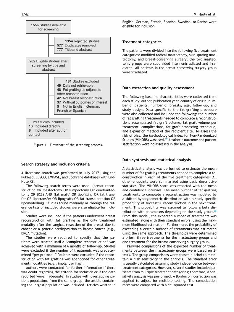
1742 M. Herly et al.
1556 Studies available for screening
1354 Rejected studies577 Duplicates removed777 Title and abstract
202 Eligible studies after screening by title and
abstract
181 Studies excluded49 Data not retrievable48 Fat grafting as adjunct to other reconstruction42 Not breast reconstruction37 Without outcomes of interest 5 Not in English, German, French or Spanish
21 Studies included13 Included directly8 Included after author contact
Figure 1 Flowchart of the screening process.
Search strategy and inclusion criteria
A literature search was performed in July 2017 using thePubMed, EBSCO, EMBASE, and Cochrane databases with End-Note X8.
The following search terms were used: (breast recon-struction OR mastectomy OR lumpectomy OR quadrantec-tomy OR BCS) AND (fat graft ∗ OR lipofilling OR fat trans-fer OR lipotransfer OR lipografts OR fat transplantation ORlipomodelling). Studies found manually or through the ref-erence lists of included studies were also eligible for inclu-sion.
Studies were included if the patients underwent breastreconstruction with fat grafting as the only treatmentmodality after the surgical resection of the breast due tocancer or a genetic predisposition to breast cancer (e.g.,BRCA mutation).
The studies were required to specify that the pa-tients were treated until a “complete reconstruction” wasachieved with a minimum of 6 months of follow-up. Studieswere excluded if the number of treatments was predeter-mined “per protocol.” Patients were excluded if the recon-struction with fat grafting was abandoned for other treat-ment modalities (e.g., implant or flap).
Authors were contacted for further information if therewas doubt regarding the criteria for inclusion or if the datareported were inadequate. In studies with overlapping pa-tient populations from the same group, the article contain-ing the largest population was included. Articles written in
English, German, French, Spanish, Swedish, or Danish wereeligible for inclusion.
Treatment categories
The patients were divided into the following five treatmentcategories: modified radical mastectomy, skin-sparing mas-tectomy, and breast-conserving surgery; the two mastec-tomy groups were subdivided into nonirradiated and irra-diated. All patients in the breast-conserving surgery groupwere irradiated.
Data extraction and quality assessment
The following baseline characteristics were collected fromeach study: author, publication year, country of origin, num-ber of patients, number of breasts, age, follow-up, andstudy design. Data specific to the fat grafting procedurewere also collected and included the following: the numberof fat grafting treatments needed to complete a reconstruc-tion, accumulated fat graft volume, fat graft volume pertreatment, complications, fat graft processing technique,and expansion method of the recipient site. To assess therisk of bias, the Methodological Index for Non-RandomizedStudies (MINORS) was used. 19 Aesthetic outcome and patientsatisfaction were no assessed in the analysis.
Data synthesis and statistical analysis
A statistical analysis was performed to estimate the meannumber of fat grafting treatments needed to complete a re-construction in each of the five treatment categories. Allother endpoints were summarized using basic descriptivestatistics. The MINORS score was reported with the meanand confidence intervals. The mean number of fat graftingtreatments to complete a reconstruction was modeled bya shifted hypergeometric distribution with a study-specificprobability of successful reconstruction in the next treat-ment. This probability was assumed to follow a beta dis-tribution with parameters depending on the study group. 20
From this model, the expected number of treatments wasestimated, along with their standard errors, using the maxi-mum likelihood estimation. Furthermore, the probability ofexceeding a certain number of treatments was estimatedusing the same approach. The thresholds were determineda priori: three treatments for the mastectomy groups andone treatment for the breast-conserving surgery group.
Pairwise comparisons of the expected number of treat-ments between the mastectomy groups were based on Z -tests. The group comparisons were chosen a priori to main-tain a high sensitivity in the analysis. The standard erroris usually calculated assuming study independence betweentreatment categories. However, several studies included pa-tients from multiple treatment categories; therefore, a sen-sitivity analysis was performed. A Bonferroni correction wasapplied to adjust for multiple testing. The complicationrates were compared with a chi-squared test.
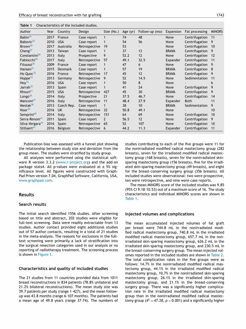
Efficacy of breast reconstruction with fat grafting 1743
Table 1 Characteristics of the included studies.
Author Year Country Design Size (No.) Age (yr) Follow-up (mo) Expansion Fat processing MINORS
Babin 24 2017 France Case report 1 74 48 None Centrifugation 11 Babovic 25 2010 USA Case report 1 54 56 None Centrifugation 9 Brown 26 2017 Australia Retrospective 19 53 – None Centrifugation 10 Cheng 27 2013 Taiwan Case report 1 37 12 BRAVA Centrifugation 9 Constantini 28 2013 Italy Prospective 9 52.2 12 None Centrifugation 12 Fabiocchi 29 2017 Italy Retrospective 57 49.1 32.5 Expander Centrifugation 11 Fitoussi 30 2009 France Case report 1 47 6 None Centrifugation 9 Hansen 31 2015 Denmark Case report 1 57 19 BRAVA Centrifugation 8 Ho Quoc 32 2016 France Retrospective 17 45 12 BRAVA Centrifugation 9 Hoppe 33 2013 Germany Retrospective 9 52 14.5 None Sedimentation 11 Hoy 34 2016 USA Case report 1 50 8 None – 6 Jarrah 35 2013 Spain Case report 1 41 24 None Centrifugation 9 Khouri 21 2015 USA Retrospective 427 45 30 BRAVA Centrifugation 9 Longo 36 2014 Italy Prospective 21 37.5 46.7 None Centrifugation 14 Manconi 37 2016 Italy Retrospective 11 48.4 27.9 Expander Both 11 Mestak 38 2013 Czech Rep. Case report 1 38 10 BRAVA Sedimentation 9 Noor 39 2016 UK Retrospective 32 50.8 49.1 None – 10 Semprini 40 2014 Italy Retrospective 151 64 69 None Centrifugation 10 Serra-Renom
41 2011 Spain Case report 2 56.5 12 None Centrifugation 9 Silva-Vergara 42 2016 Spain Retrospective 65 52 107 None Centrifugation 10 Stillaert 43 2016 Belgium Retrospective 6 44.2 11.3 Expander Centrifugation 11
tg
w
pnPw
R
S
Tb
fso
i
ttri
C
Tb23
ua
s
tbt
s
afit
(
cT
I
Tp
fi
m
iitu
T
ftmmmstgt
Publication bias was assessed with a funnel plot showing he relationship between study size and deviation from the roup mean. The studies were stratified by study group. All analyses were performed using the statistical soft-
are R version 3.3.2 ( www.r-project.org ) and the add-onackage stats4. All p-values were evaluated at a 5% sig- ificance level. All figures were constructed with Graph- ad Prism version 7.04, GraphPad Software, California, USA, ww.graphpad.com .
esults
earch results
he initial search identified 1556 studies. After screening ased on title and abstract, 202 studies were eligible forull-text screening. Data were readily extractable from 13 tudies. Author contact provided eight additional studies ut of 57 author contacts, resulting in a total of 21 studiesn the meta-analysis. The reasons for exclusions in the full-ext screening were primarily a lack of stratification into he surgical resection categories used in our analysis or no eporting of radiotherapy treatment. The screening process s shown in Figure 1 .
haracteristics and quality of included studies
he 21 studies from 11 countries provided data from 1011 reast reconstructions in 834 patients (78.8% unilateral and 1.2% bilateral reconstructions). The mean study size was 9.7 patients per study (range 1–427), and the mean follow-p was 43.8 months (range 6–107 months). The patients had mean age of 49.8 years (range 37–74). The numbers of
tudies contributing to each of the five groups were 11 forhe nonirradiated modified radical mastectomy group (282 reasts), seven for the irradiated modified radical mastec- omy group (168 breasts), seven for the nonirradiated skin-paring mastectomy group (156 breasts), five for the irradi-ted skin-sparing mastectomy group (49 breasts), and eight or the breast-conserving surgery group (356 breasts). All ncluded studies were observational: two were prospective, en were retrospective, and nine were case reports. The mean MINORS score of the included studies was 9.85
95% CI 9.18–10.53) out of a maximum score of 16. The studyharacteristics and individual MINORS scores are shown in able 1 .
njected volumes and complications
he mean accumulated injected volumes of fat graft er breast were 744.8 mL in the nonirradiated modi-ed radical mastectomy group, 740.8 mL in the irradiatedodified radical mastectomy group, 657.7 mL in the non-
rradiated skin-sparing mastectomy group, 626.2 mL in the rradiated skin-sparing mastectomy group, and 230.5 mL in he breast-conserving surgery group. The mean injected vol- mes reported in the included studies are shown in Table 2 .he total complication rates in the five groups were asollows: 14.7% in the nonirradiated modified radical mas- ectomy group, 44.1% in the irradiated modified radical astectomy group, 10.7% in the nonirradiated skin-sparing astectomy group, 26.1% in the irradiated skin-sparing astectomy group, and 21.1% in the breast-conserving urgery group. There was a significantly higher complica- ion rate in the irradiated modified radical mastectomy roup than in the nonirradiated modified radical mastec- omy group ( X
2 = 47.28, p < 0.001) and a significantly higher

1744 M. Herly et al.
Table 2 Mean injected volumes of fat grafting for breast reconstruction.
Modified radical mastectomy nonirradiated ( n = 9)
No. of breasts Accumulated
Mean volume per treatment (mL)
1st 2nd 3rd Subsequent Average
Babovic - 2010 25 1 1615.8 84.2 78.9 84.2 456.1 403.9 Cheng - 2013 27 2 430 230 200 – – 215 Hansen - 2015 31 1 957 187 150 30 147.5 136.71 Hoppe - 2013 33 5 984.4 259 298 261.4 205 259.1 Jarrah - 2013 35 1 720 360 240 120 – 240 Manconi - 2016 37 4 738.5 288 295.5 310 – 305.3 Mestak - 2013 38 2 815 235.0 280 300 – 271.7 Silva-Vergara - 2016 42 9 586.2 218.9 229.3 164.4 210 211 Stillaert - 2016 43 1 510 – – – – 176
Number of breasts 26 26 25 25 23 16 26 Mean – 744.8 239.0 246.4 211.2 218.5 243.3 Range – 430–
1615.8 84.2–360 78.9–298 30–310 147.5–456.1 136.71–
403.9
Modified radical mastectomy irradiated (n = 6)
No. of breasts Accumulated
Mean volume per treatment (mL)
1st 2nd 3rd Subsequent Average
Babin - 2017 24 1 500 200 150 150 – 166 Fitoussi - 2009 30 1 380 150 150 – – 80 Hoppe - 2013 33 4 888.8 236.3 255 240 205 237 Manconi - 2016 37 1 1158 96 167 170 362.5 231.6 Silva-Vergara - 2016 42 18 724.9 196.9 234.6 223.6 195.6 213.9 Stillaert - 2016 43 1 620 – – – – 155
Number of breasts 26 26 25 25 24 23 26 Mean – 740.8 197.4 228.4 221.0 204.5 208.9 Range – 380–1158 96–
236.25 150–255 150–240 195.6–362.5 80–237
Skin-sparing mastectomy nonirradiated ( n = 6)
No. of Breasts Accumulated
Mean volume per treatment (mL)
1st 2nd 3rd Subsequent Average
Constantini - 2013 28 1 272 80 81 111 – 90.66 Fabiocchi - 2017 29 56 706.1 310.4 328.1 280.7 154.3 329.0 Ho Quoc - 2016 32 6 790 – – – – –Longo - 2014 36 17 410.3 114.4 147.9 147.9 – 136.8 Manconi - 2016 37 6 792.3 291 375.3 252 – 324.9 Stillaert - 2016 43 4 728.8 – – – – 168.3
Number of breasts 90 90 80 80 80 56 84 Mean – 657.7 264.4 290.3 248.2 154.3 279.3 Range – 272–
792.3 80–310.4 81–375.3 111–
280.7 – 90.7–329
Skin-sparing mastectomy irradiated ( n = 3)
No. of breasts Accumulated
Mean volume per treatment (mL)
1st 2nd 3rd Subsequent Average
Constantini - 2013 28 1 331 150 116 65 – 110.3 Fabiocchi - 2017 29 20 658.2 286.2 299.8 316.6 177 335.8 Longo - 2014 36 12 597.5 67.5 70 139.2 134.2 110.6
( continued on next page )
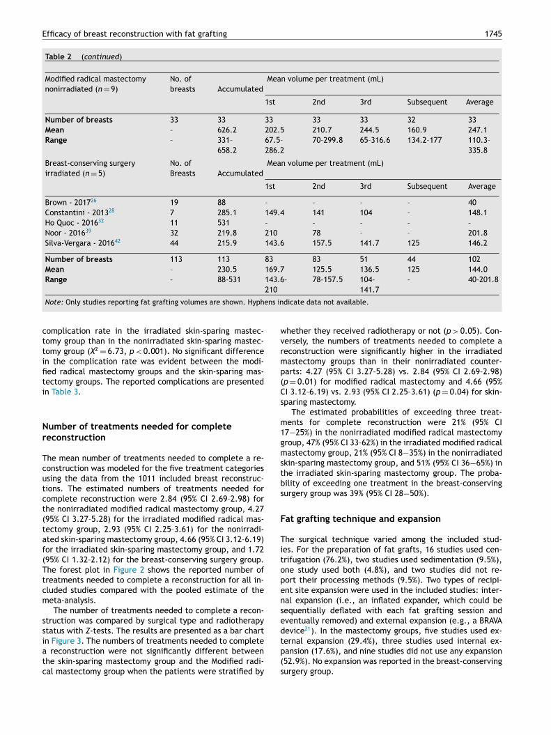
Efficacy of breast reconstruction with fat grafting 1745
Table 2 ( continued )
Modified radical mastectomy nonirradiated ( n = 9)
No. of breasts Accumulated
Mean volume per treatment (mL)
1st 2nd 3rd Subsequent Average
Number of breasts 33 33 33 33 33 32 33 Mean – 626.2 202.5 210.7 244.5 160.9 247.1 Range – 331–
658.2 67.5–286.2
70–299.8 65–316.6 134.2–177 110.3–335.8
Breast-conserving surgery irradiated ( n = 5)
No. of Breasts Accumulated
Mean volume per treatment (mL)
1st 2nd 3rd Subsequent Average
Brown - 2017 26 19 88 – – – – 40 Constantini - 2013 28 7 285.1 149.4 141 104 – 148.1 Ho Quoc - 2016 32 11 531 – – – – –Noor - 2016 39 32 219.8 210 78 – – 201.8 Silva-Vergara - 2016 42 44 215.9 143.6 157.5 141.7 125 146.2
Number of breasts 113 113 83 83 51 44 102 Mean – 230.5 169.7 125.5 136.5 125 144.0 Range – 88–531 143.6–
210 78–157.5 104–
141.7 – 40–201.8
Note: Only studies reporting fat grafting volumes are shown. Hyphens indicate data not available.
ctt
ifiti
Nr
T
cutct(taf(T
tcm
ss
i
atc
w
v
rmp
(
C
s
m1g
m
s
tbs
F
Ti
to
p
e
n
sed
tp
(s
omplication rate in the irradiated skin-sparing mastec- omy group than in the nonirradiated skin-sparing mastec- omy group ( X
2 = 6.73, p < 0.001). No significant differencen the complication rate was evident between the modi- ed radical mastectomy groups and the skin-sparing mas- ectomy groups. The reported complications are presented n Table 3 .
umber of treatments needed for complete
econstruction
he mean number of treatments needed to complete a re-onstruction was modeled for the five treatment categories sing the data from the 1011 included breast reconstruc- ions. The estimated numbers of treatments needed for omplete reconstruction were 2.84 (95% CI 2.69–2.98) for he nonirradiated modified radical mastectomy group, 4.27 95% CI 3.27–5.28) for the irradiated modified radical mas- ectomy group, 2.93 (95% CI 2.25–3.61) for the nonirradi- ted skin-sparing mastectomy group, 4.66 (95% CI 3.12–6.19) or the irradiated skin-sparing mastectomy group, and 1.72 95% CI 1.32–2.12) for the breast-conserving surgery group. he forest plot in Figure 2 shows the reported number ofreatments needed to complete a reconstruction for all in- luded studies compared with the pooled estimate of the eta-analysis. The number of treatments needed to complete a recon-
truction was compared by surgical type and radiotherapy tatus with Z -tests. The results are presented as a bar chartn Figure 3 . The numbers of treatments needed to complete reconstruction were not significantly different between he skin-sparing mastectomy group and the Modified radi- al mastectomy group when the patients were stratified by
hether they received radiotherapy or not ( p > 0.05). Con-ersely, the numbers of treatments needed to complete aeconstruction were significantly higher in the irradiated astectomy groups than in their nonirradiated counter- arts: 4.27 (95% CI 3.27–5.28) vs. 2.84 (95% CI 2.69–2.98) p = 0.01) for modified radical mastectomy and 4.66 (95%I 3.12–6.19) vs. 2.93 (95% CI 2.25–3.61) ( p = 0.04) for skin-paring mastectomy. The estimated probabilities of exceeding three treat-
ents for complete reconstruction were 21% (95% CI 7 −25%) in the nonirradiated modified radical mastectomy roup, 47% (95% CI 33–62%) in the irradiated modified radicalastectomy group, 21% (95% CI 8 −35%) in the nonirradiatedkin-sparing mastectomy group, and 51% (95% CI 36 −65%) inhe irradiated skin-sparing mastectomy group. The proba- ility of exceeding one treatment in the breast-conserving urgery group was 39% (95% CI 28 −50%).
at grafting technique and expansion
he surgical technique varied among the included stud- es. For the preparation of fat grafts, 16 studies used cen-rifugation (76.2%), two studies used sedimentation (9.5%), ne study used both (4.8%), and two studies did not re-ort their processing methods (9.5%). Two types of recipi-nt site expansion were used in the included studies: inter-al expansion (i.e., an inflated expander, which could beequentially deflated with each fat grafting session and ventually removed) and external expansion (e.g., a BRAVA evice 21 ). In the mastectomy groups, five studies used ex-ernal expansion (29.4%), three studies used internal ex- ansion (17.6%), and nine studies did not use any expansion52.9%). No expansion was reported in the breast-conserving urgery group.
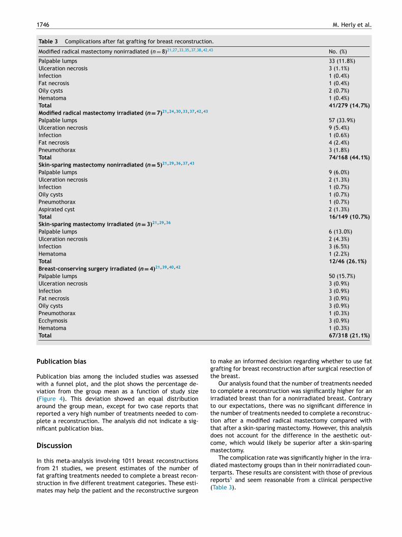
1746 M. Herly et al.
Table 3 Complications after fat grafting for breast reconstruction.
Modified radical mastectomy nonirradiated ( n = 8) 21,27 , 33,35 , 37,38 , 42,43 No. (%)
Palpable lumps 33 (11.8%) Ulceration necrosis 3 (1.1%) Infection 1 (0.4%) Fat necrosis 1 (0.4%) Oily cysts 2 (0.7%) Hematoma 1 (0.4%) Total 41/279 (14.7%) Modified radical mastectomy irradiated ( n = 7) 21 , 24 , 30 , 33 , 37 , 42 , 43
Palpable lumps 57 (33.9%) Ulceration necrosis 9 (5.4%) Infection 1 (0.6%) Fat necrosis 4 (2.4%) Pneumothorax 3 (1.8%) Total 74/168 (44.1%) Skin-sparing mastectomy nonirradiated ( n = 5) 21 , 29 , 36 , 37 , 43
Palpable lumps 9 (6.0%) Ulceration necrosis 2 (1.3%) Infection 1 (0.7%) Oily cysts 1 (0.7%) Pneumothorax 1 (0.7%) Aspirated cyst 2 (1.3%) Total 16/149 (10.7%) Skin-sparing mastectomy irradiated ( n = 3) 21 , 29 , 36
Palpable lumps 6 (13.0%) Ulceration necrosis 2 (4.3%) Infection 3 (6.5%) Hematoma 1 (2.2%) Total 12/46 (26.1%) Breast-conserving surgery irradiated ( n = 4) 21 , 39 , 40 , 42
Palpable lumps 50 (15.7%) Ulceration necrosis 3 (0.9%) Infection 3 (0.9%) Fat necrosis 3 (0.9%) Oily cysts 3 (0.9%) Pneumothorax 1 (0.3%) Ecchymosis 3 (0.9%) Hematoma 1 (0.3%) Total 67/318 (21.1%)
Publication bias
Publication bias among the included studies was assessedwith a funnel plot, and the plot shows the percentage de-viation from the group mean as a function of study size( Figure 4 ). This deviation showed an equal distributionaround the group mean, except for two case reports thatreported a very high number of treatments needed to com-plete a reconstruction. The analysis did not indicate a sig-nificant publication bias.
Discussion
In this meta-analysis involving 1011 breast reconstructionsfrom 21 studies, we present estimates of the number offat grafting treatments needed to complete a breast recon-struction in five different treatment categories. These esti-mates may help the patient and the reconstructive surgeon
to make an informed decision regarding whether to use fatgrafting for breast reconstruction after surgical resection ofthe breast.
Our analysis found that the number of treatments neededto complete a reconstruction was significantly higher for anirradiated breast than for a nonirradiated breast. Contraryto our expectations, there was no significant difference inthe number of treatments needed to complete a reconstruc-tion after a modified radical mastectomy compared withthat after a skin-sparing mastectomy. However, this analysisdoes not account for the difference in the aesthetic out-come, which would likely be superior after a skin-sparingmastectomy.
The complication rate was significantly higher in the irra-diated mastectomy groups than in their nonirradiated coun-terparts. These results are consistent with those of previousreports 5 and seem reasonable from a clinical perspective( Table 3 ).

Efficacy of breast reconstruction with fat grafting 1747
Figure 2 Forest plot showing the mean number of fat grafting treatments needed to complete a reconstruction. The data markers indicate the individual study mean. The size of the mark is proportional to the number of cases. The vertical dashed lines indicate the group means with 95% CIs (shaded interval). Only the studies reporting the average number of fat grafting treatments needed to complete a reconstruction are shown. CI , Confidence Interval.
L
Ttgbboh
t
I
ip
t
o
g
a
n
t
c
t
imitations
he applicability of our analysis is limited by how well he patients included in our analysis represented the back- round population of women treated with fat grafting for reast reconstruction. We evaluated different sources of ias in our meta-analysis to estimate differences between ur sample and the background population and to estimate ow this affected the results of our analysis. The studies with many patients contributed the most to
he results of this meta-analysis, representing a risk of bias.
n the studies with many patients, one can assume that thenvolved surgeons were more experienced than the average lastic surgeon in using fat grafting for breast reconstruc-ions. Thus, the results of this review may represent theutcomes achieved by the best surgeons in the field of fatrafting and may therefore be more optimistic than the trueverage. However, by analyzing the funnel plot, we foundo tendency of the larger studies to present better resultshan average. Rather, the funnel plot was symmetrical, ex-ept for two case studies that deviated considerably fromhe mean, with a high number of treatments needed to com-
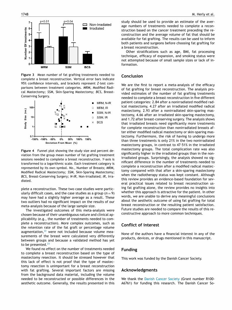
1748 M. Herly et al.
Figure 3 Mean number of fat grafting treatments needed to complete a breast reconstruction. Vertical error bars indicate 95% confidence intervals, and brackets represent Z -test com- parisons between treatment categories. MRM, Modified Radi- cal Mastectomy ; SSM, Skin-Sparing Mastectomy ; BCS, Breast- Conserving Surgery.
Figure 4 Funnel plot showing the study size and percent de- viation from the group mean number of fat grafting treatment sessions needed to complete a breast reconstruction. Y -axis is transformed to a logarithmic scale. Each treatment category is represented by its own symbol. No., Number of Breasts; MRM, Modified Radical Mastectomy ; SSM, Skin-Sparing Mastectomy ; BCS, Breast-Conserving Surgery ; N-IR, Non-Irradiated; IR , Irra- diated.
plete a reconstruction. These two case studies were partic-ularly difficult cases, and the case studies as a group ( n = 9)may have had a slightly higher average as a result. Thesetwo outliers had no significant impact on the results of ourmeta-analysis because of the large sample size.
The investigated outcomes of this meta-analysis werechosen because of their unambiguous nature and clinical ap-plicability (e.g., the number of treatments needed to com-plete a reconstruction). More complex outcomes, such asthe retention rate of the fat graft or percentage volumeaugmentation, 22 were not included because volume mea-surements of the breast were calculated very differentlybetween groups and because a validated method has yetto be presented. 23
We found no effect on the number of treatments neededto complete a breast reconstruction based on the type ofmastectomy resection. It should be stressed however thatthis lack of effect is not proof that the type of mastec-tomy resection is unimportant for a breast reconstructionwith fat grafting. Several important factors are missingfrom the background data material, including the volumeneeded to be reconstructed or possible differences in theaesthetic outcome. Generally, the results presented in this
study should be used to provide an estimate of the aver-age numbers of treatments needed to complete a recon-struction based on the cancer treatment preceding the re-construction and the average volume of fat that should beavailable for fat grafting. The results can be used to informboth patients and surgeons before choosing fat grafting fora breast reconstruction.
Other stratifications such as age, BMI, fat processingtechnique, efficacy of expansion, and smoking status werenot attempted because of small sample sizes or lack of in-formation.
Conclusion
We are the first to report a meta-analysis of the efficacyof fat grafting for breast reconstruction. The analysis pro-vided estimates of the number of fat grafting treatmentsneeded to complete a breast reconstruction in five differentpatient categories: 2.84 after a nonirradiated modified rad-ical mastectomy, 4.27 after an irradiated modified radicalmastectomy, 2.93 after a nonirradiated skin-sparing mas-tectomy, 4.66 after an irradiated skin-sparing mastectomy,and 1.72 after breast-conserving surgery. The analysis showsthat irradiated breasts need significantly more treatmentsfor complete reconstruction than nonirradiated breasts af-ter either modified radical mastectomy or skin-sparing mas-tectomy. Furthermore, the risk of having to undergo morethan three treatments is only 21% in the two nonirradiatedmastectomy groups, in contrast to 47–51% in the irradiatedmastectomy groups. The total complication rate was alsosignificantly higher in the irradiated groups than in the non-irradiated groups. Surprisingly, the analysis showed no sig-nificant difference in the number of treatments needed tocomplete a reconstruction after a modified radical mastec-tomy compared with that after a skin-sparing mastectomywhen the radiotherapy status was kept constant. Althoughthis review provides an evidence-based foundation for sev-eral practical issues related to breast reconstruction us-ing fat grafting alone, the review provides no insights intowhether this approach is attractive for the patient. In otherwords, we are unable to derive any meaningful conclusionsabout the aesthetic outcome of using fat grafting for totalbreast reconstruction or the resulting patient satisfaction.Future studies are needed to compare the results of this re-constructive approach to more common techniques.
Conflict of interest
None of the authors have a financial interest in any of theproducts, devices, or drugs mentioned in this manuscript.
Funding
This work was funded by the Danish Cancer Society.
Acknowledgments
We thank the Danish Cancer Society (Grant number R100-A6761) for funding this research. The Danish Cancer So-

Efficacy of breast reconstruction with fat grafting 1749
c
li
mwi
FD
GY
S
Sf0
R
1
1
1
1
1
1
1
1
1
1
2
2
2
2
2
2
2
2
2
2
3
3
iety had no role in the design of the study; in the col-ection, analysis, or interpretation of data; in the writ- ng of the manuscript; or in the decision to submit theanuscript for publication. We thank the following authors ho contributed with data and clarified questions regard- ng their works: Dr. Guillaume Babin, Dr. Alex Brown, Dr.ederico Cattin, Dr. Antonio Cipriani, Dr. Joan Fontdevila, r. Delia Hoppe, Dr. Ahmed Jarrah, Dr. Andrea Manconi, Dr.loria Semprini, Dr. Claudio Silva-Vergara, and Dr. Kotaro oshimura.
upplementary materials
upplementary material associated with this article can be ound, in the online version, at doi:10.1016/j.bjps.2018.08. 24 .
eferences
1. Ferlay J , Soerjomataram I , Ervik M , et al. GLOBOCAN 2012 v1.0,cancer incidence and mortality worldwide: IARC CancerBase No. 11 [Internet] , Lyon, France: International Agency for Re-search on Cancer; 2013. Available from: Accessed on 28 March2018 .
2. Ilonzo N , Tsang A , Tsantes S , Estabrook A , Thu Ma AM . Breastreconstruction after mastectomy: a ten-year analysis of trends and immediate postoperative outcomes. Breast 2017; 32 :7–12 (Edinburgh, Scotland) .
3. Illouz YG , Sterodimas A . Autologous fat transplantation to thebreast: a personal technique with 25 years of experience. Aes-thet Plastic Surg 2009; 33 (5):706–15 .
4. Agha RA , Fowler AJ , Herlin C , Goodacre TE , Orgill DP . Use ofautologous fat grafting for breast reconstruction: a systematic review with meta-analysis of oncological outcomes. J Plastic, Reconstr Aesthet Surg: JPRAS 2015; 68 (2):143–61 .
5. Katzel EB , Bucky LP . Fat grafting to the breast: clinical applica-tions and outcomes for reconstructive surgery. Plastic Reconstr Surg 2017; 140 (5S):69S–76S .
6. Waked K , Colle J , Doornaert M , Cocquyt V , Blondeel P . System-atic review: the oncological safety of adipose fat transfer afterbreast cancer surgery. Breast 2017; 31 :128–36 (Edinburgh, Scot- land) .
7. Groen JW , Negenborn VL , Twisk D , et al. Autologous fat graft-ing in onco-plastic breast reconstruction: a systematic review
on oncological and radiological safety, complications, volume retention and patient/surgeon satisfaction. J Plastic, Reconstr Aesthet Surg: JPRAS 2016; 69 (6):742–64 .
8. Delay E , Garson S , Tousson G , Sinna R . Fat injection to thebreast: technique, results, and indications based on 880 pro- cedures over 10 years. Aesthet Surg J 2009; 29 (5):360–76 .
9. Khouri RK , Eisenmann-Klein M , Cardoso E , et al. Brava and au-tologous fat transfer is a safe and effective breast augmen-tation alternative: results of a 6-year, 81-patient, prospective multicenter study. Plastic Reconstr Surg 2012; 129 (5):1173–87 .
0. Kolle SF , Fischer-Nielsen A , Mathiasen AB , et al. Enrichmentof autologous fat grafts with ex-vivo expanded adipose tis- sue-derived stem cells for graft survival: a randomised placebo–controlled trial. Lancet 2013; 382 (9898):1113–20 (London, Eng- land) .
1. Ueberreiter K , Von Finckenstein J , Cromme F , Herold C ,Tanzella U , Vogt P . BEAULI TM –eine neue Methode zur ein-fachen und zuverlässigen Fettzell-Transplantation Progeni- tor-enriched adipose tissue transplantation as rescue for breast
implant complications. Handchirurgie · Mikrochirurgie · Plastis- che Chirurgie 2010; 42 (6):379–85 .
2. Yoshimura K , Asano Y , Aoi N , et al. Progenitor-enriched adiposetissue transplantation as rescue for breast implant complica- tions. Breast J 2010; 16 (2):169–75 .
3. Yoshimura K , Sato K , Aoi N , Kurita M , Hirohi T , Harii K . Cell-as-sisted lipotransfer for cosmetic breast augmentation: support- ive use of adipose-derived stem/stromal cells. Aesthet Plast Surg 2008; 32 (1):48–55 discussion 56-47 .
4. Herly M , Orholt M , Glovinski PV , et al. Quantifying long-term re-tention of excised fat grafts: a longitudinal, retrospective co-hort study of 108 patients followed for up to 8.4 years. PlastReconstr Surg 2017; 139 (5):1223–32 .
5. Khouri RK , Rigotti G , Cardoso E , Khouri RKJ , Biggs TM . Megavol-ume autologous fat transfer: part ii. practice and techniques.Plastic Reconstr Surg 2014; 133 (6):1369–77 .
6. Gir P , Brown SA , Oni G , Kashefi N , Mojallal A , Rohrich RJ .Fat grafting: evidence-based review on autologous fat harvest-ing, processing, reinjection, and storage. Plastic Reconstr Surg 2012; 130 (1):249–58 .
7. Stroup DF , Berlin JA , Morton SC , et al. Meta-analysis of observa-tional studies in epidemiology: a proposal for reporting. JAMA2000; 283 (15):2008–12 .
8. Higgins JP , Green S . Cochrane handbook for systematic reviewsof interventions , 4. John Wiley & Sons; 2011 .
9. Slim K , Nini E , Forestier D , Kwiatkowski F , Panis Y , Chipponi J .Methodological index for nonrandomized studies (MINORS): de- velopment and validation of a new instrument. ANZ J Surg2003; 73 (9):712–16 .
0. Weinberg CR , Gladen BC . The beta-geometric distributionapplied to comparative fecundability studies. Biometrics 1986; 42 (3):547–60 .
1. Khouri RK , Rigotti G , Khouri RK Jr , et al. Tissue-engineeredbreast reconstruction with Brava-assisted fat grafting: a 7-year, 488-patient, multicenter experience. Plastic Reconstr Surg 2015; 135 (3):643–58 .
2. Khouri RK . Current clinical applications of fat grafting. PlasticReconstr Surg 2017; 140 (3):e466–86 .
3. Glovinski PV , Herly M , Muller FC , et al. Avoiding a systematicerror in assessing fat graft survival in the breast with repeatedmagnetic resonance imaging. Plast Reconstr Surg Glob Open2016; 4 (9):e1023 .
4. Babin G , Commeny J , Fournier M , et al. [Breast reconstructionin elderly patients: studies of the practices at institut Bergonieduring 2005-2015]. Bull Cancer 2017; 104 (6):508–15 .
6. Babovic S . Complete breast reconstruction with autologous fatgraft - a case report. J Plastic, Reconstr Aesth Surg: JPRAS2010; 63 (7):e561–3 .
6. Brown AWW , Kabir M , Sherman KA , Meybodi F , French JR , El-der EB . Patient reported outcomes of autologous fat graftingafter breast cancer surgery. Breast 2017; 35 :14–20 (Edinburgh,Scotland) .
7. Chen TM , Chen SG , Teng SC . Using an external soft tissue ex-pander before autologous fat grafting for non-irradiated breast reconstruction. J Plastic Surg Hand Surg 2013; 47 (5):419–21 .
8. Costantini M , Cipriani A , Belli P , et al. Radiological findings inmammary autologous fat injections: a multi-technique evalua- tion. Clin Radiol 2013; 68 (1):27–33 .
9. Fabiocchi L , Semprini G , Cattin F , et al. ’Reverse expansion’: anew technique of breast reconstruction with autologous tissue.J Plastic, Reconstr Aesthet Surg JPRAS 2017 .
0. Fitoussi A , Pollet AG , Couturaud B , Salmon RJ . Secondary breastreconstruction using exclusive lipofilling. Annales de Chirurgie Plastique et Esthetique 2009; 54 (4):374–8 .
1. Hammer-Hansen N , Jensen TB , Damsgaard TE . Delayedtotal breast reconstruction with brava. Case Rep Surg 2015; 2015 :601904 .
1750 M. Herly et al.
32. Ho Quoc C , Piat JM , Carrabin N , Meruta A , Faure C , Delay E .Breast reconstruction with fat grafting and BRAVA((R)) pre-ex-pansion: efficacy evaluation in 45 cases. Annales de ChirurgiePlastique et Esthetique 2016; 61 (3):183–9 .
33. Hoppe DL , Ueberreiter K , Surlemont Y , Peltoniemi H , Stabile M ,Kauhanen S . Breast reconstruction de novo by water-jet as-sisted autologous fat grafting–a retrospective study. GermanMed Sci: GMS e-J 2013; 11 :1–11 Doc17 .
34. Hoy E . State of the art: reconstructing partial mastectomy de-fects with autologous fat grafting. Del Med J 2016; 88 (1):20–3 .
35. Jarrah A, Paloma V, Vernetta O. Reconstrucción mamaria medi-ante lipoinfiltrado enriquecido con PRP. Cirugía Plástica Ibero-Latinoamericana. 2013;39(Suppl 1):65–73.
36. Longo B , Laporta R , Sorotos M , Pagnoni M , Gentilucci M , San-tanelli di Pompeo F . Total breast reconstruction using au-tologous fat grafting following nipple-sparing mastectomy inirradiated and non-irradiated patients. Aesthet Plastic Surg2014; 38 (6):1101–8 .
37. Manconi A , De Lorenzi F , Chahuan B , et al. Total breast re-construction with fat grafting after internal expansion and ex-pander removal. Ann Plastic Surg 2016 .
38. Mestak O , Mestak J , Bohac M , Edriss A , Sukop A . Breast recon-struction after a bilateral mastectomy using the BRAVA expan-sion system and fat grafting. Plastic Reconstr Surg Global Open2013; 1 (8):e71 .
39. Noor L , Reeves HR , Kumar D , Alozairi O , Bhaskar P . Imag-ing changes after breast reconstruction with fat grafting -Retrospective study of 90 breast cancer. Pak J Med Sci2016; 32 (1):8–12 .
40. Semprini G , Cattin F , Zanin C , et al. About locoregional recur-rence risk after lipofilling in breast cancer patients: our expe-rience. Minerva Chirurgica 2014; 69 (2):91–6 .
41. Serra-Renom JM , Munoz-Olmo J , Serra-Mestre JM . Breastreconstruction with fat grafting alone. Ann Plastic Surg2011; 66 (6):598–601 .
42. Silva-Vergara C , Fontdevila J , Descarrega J , Burdio F , Yoon TS ,Grande L . Oncological outcomes of lipofilling breast reconstruc-tion: 195 consecutive cases and literature review. J Plastic, Re-constr Aesthet Surg: JPRAS 2016; 69 (4):475–81 .
43. Stillaert FB , Sommeling C , D’Arpa S , et al. Intratissular ex-pansion-mediated, serial fat grafting: a step-by-step work-ing algorithm to achieve 3D biological harmony in autologousbreast reconstruction. J Plastic, Reconstr Aesthet Surg: JPRAS2016; 69 (12):1579–87 .

Appendix 3
The current gold standard breast volumetry technique seems to overestimate fat graft volume
retention in the breast: A validation study

Journal of Plastic, Reconstructive & Aesthetic Surgery (2019) 72, 1278–1284
The current gold standard breast volumetry
technique seems to overestimate fat graft
volume retention in the breast: A validation
study
✰
Mikkel Herly
a , ∗, Felix Christoph Müller
b , Mathias Ørholt
a , Joachim Hansen
a , Sophie Sværke
a , Mathilde N. Hemmingsen
a , Bo S. Rasmussen
a , Jens J. Elberg
a , Krzysztof T. Drzewiecki a , Peter V. Vester-Glowinski a
a Department of Plastic Surgery and Burns Treatment, Copenhagen University Hospital, Rigshospitalet, Rigshospitalet, Blegdamsvej 9, DK-2100, Copenhagen, Denmark b Department of Radiology, Herlev and Gentofte Hospital, University of Copenhagen, Copenhagen, Denmark
Received 12 November 2018; accepted 24 March 2019
KEYWORDS
Fat graft retention; Fat grafting; MRI; Breast volume; Breast volumetry; Volume retention
Summary Background: MRI is generally considered as the gold standard for measuring breast volume because of its high accuracy of the modality. Many techniques used to measure total breast volume have been validated, but none of these techniques have been validated for their ability to measure the volume retention of fat grafts in the breast. In this study, the authors investigated the accuracy of the most common MRI technique used to measure fat graft retention in the breast by measuring the volume changes after breast augmentation. Methods: Patients undergoing breast augmentation with either breast implants or fat grafting underwent MRI scans before and after surgery. Blinded observers measured the change in breast volume from the MRI scans. The difference between the measured change in breast volume and the volume of the breast augmentation was used to determine the accuracy of the MRI technique. Results: Twenty-eight patients with a total of 56 breasts were included. In total, 168 measure- ments of change in breast volume were performed by the observers. The MRI measurements of change in breast volume overestimated the true volumes of the breast augmentations by
✰ Prior presentation of results: A part of this study was presented at the 37th Congress of the Scandinavian Association of Plastic Surgeons in Copenhagen, June 16, 2018.
∗ Corresponding author. E-mail address: [email protected] (M. Herly).
https://doi.org/10.1016/j.bjps.2019.03.029 1748-6815/ © 2019 British Association of Plastic, Reconstructive and Aesthetic Surgeons. Published by Elsevier Ltd. All rights reserved.

A validation study 1279
an average of 50.8%, and only 8 of the 168 individual measurements had measurement errors below 50 mL. Conclusion: The MRI technique, which is considered as the gold standard for the quantification of fat graft volume retention, was associated with a significant measurement error. These find- ings have potential implications for the interpretation of previously published results of studies based on this technique. © 2019 British Association of Plastic, Reconstructive and Aesthetic Surgeons. Published by El- sevier Ltd. All rights reserved.
B
W
tTmp
tm
b
t
t
v
s
oa
v
b
t
g3
ia
i
i
tu
a
r
ilm
ct
e
tmaa
v
m
P
T
i
w
Ef
P
Paw
t
t2
Rw
a
e
tP
s
if
a
ge
M
P
Mt
Mm
w
f
s
h
a
M
Ad
ta
bT
s
ackground
hen fat grafting is performed, it is expected that a por-ion of the fat graft undergoes resorption after surgery. 1 –3
herefore, studies investigating fat grafting often involve easurement of the fat graft volume retention (i.e., what ercentage of the graft volume will remain after the resorp-ion process has ended). We hypothesize that the current ethod used to measure fat graft volume retention in thereast with MRI may be associated with a much larger errorhan that previously estimated. 4
The typical procedure used to assess fat graft volume re-ention in the breast is as follows 5 –10 : (1) the total breastolume is measured by MRI before surgery and again afterurgery when no further resorption is expected; (2) the pre-perative breast volume is subtracted from the postoper- tive breast volume to calculate the change in the breastolume; and (3) the change in the breast volume is dividedy the injected fat volume to calculate the volume reten-ion of the fat graft. For example, if a breast is augmented with a 200 mL fat
raft and we measure the preoperative breast volume as 00 mL, then the breast volume one year after fat graftings 400 mL. The difference between the two measurements is 100 mL increase in breast volume. The change in volumes then compared to the injected volume of 200 mL, whichs interpreted as a fat graft volume retention rate of 50%.
The validity of the approach described above hinges on he presumption that the measured difference in breast vol- me is identical to the residual volume of the fat graft (i.e., 300 mL breast plus a 100 mL residual fat graft volume willesult in a 400 mL breast). The accuracy of simply subtract-ng the measurements of the total breast volume to calcu- ate the change in breast volume (e.g., after breast aug- entation) has never been convincingly validated. In this study, we investigated the accuracy of the most
ommonly used technique to measure fat graft volume re- ention with MRI scans, which has been described by Heroldt al. 5-8 We investigated the efficiency of the MRI techniqueo measure the change in breast volume after breast aug- entation with either breast implants or fat grafts. The greement between the measured change in breast volume nd the true volume of the breast augmentation (i.e., theolume of the breast implant or fat graft) is used to deter-ine the accuracy of the technique.
atients and methods
he collection of data was approved by the regional Dan-sh Data Protection Agency, and the need for study approval
as waived by the Regional Committee on Health Researchthics. All patients provided written informed consent be- ore participating in the study.
atients
atients who were scheduled for bilateral cosmetic breast ugmentation with either breast implants or fat grafting ere eligible for inclusion in the study. The exclusion cri-eria were any known breast disease, pregnancy, or con-raindications for MRI. Patients were recruited from June 014 to April 2017 at the Copenhagen University Hospital,igshospitalet and Amalieklinikken, Copenhagen. Patients ho received breast implants underwent their first MRI ex-mination within 10 days before surgery and the second MRIxamination after a minimum of four months, at which timehe postoperative edema was expected to have subsided. atients were examined at the same time during their men-trual cycle to prevent cyclical breast volume changes fromnfluencing the measurements. The patients who underwent at grafting were examined on the day before surgery andgain within three hours after surgery, at which time no fatraft resorption was expected, and minimal postoperative dema would have formed.
RI examinations
atients were examined with a 3-Tesla MRI unit (Siemensagnetom Verio; Erlangen, Germany), except for two pa- ients who were examined with a 1.5-Tesla unit (Siemensagnetom Avanto; Erlangen, Germany) because of the aintenance of the 3 Tesla unit. A 4-channel breast coilas used in all examinations. Volume assessments were per-ormed on an axial breathhold DIXON sequence, with 72lices and a 3-mm slice thickness. Patients were examinedead-first, with their arms raised and in the prone position,nd with the breasts freely suspended in the breast coil.
RI volume assessment
ll volume assessments were performed by three indepen- ent observers (JH, MNH, and SS) who were blinded to therue volume of the breast augmentation (i.e., implant size nd fat grafting volume). The total breast volume was measured by delineating the
reasts on the axial slices, as described by Herold et al. 5 –8
he borders are shown in Figure 1 . The skin was used as theuperficial border, and the internal costal surface was used

1280 M. Herly, F.C. Müller and M. Ørholt et al.
Figure 1 Sagittal and transverse views of the preoperative (left) and postoperative MRI examinations (right) of a patient with breast implants. Sup. ∗ indicates the adjusted cranial border of the preoperative breast, and the hatched area represents the extra tissue added to obtain an equal number of slices on the two examinations. Sup.: superior; Inf.: inferior; Lat.: lateral; Med. : medial.
as the deep border. The center of the sternum was used asthe medial border, and the lateral border was placed at thepoint where the thickness of the breast was equal to thethickness of the surrounding skin. Similarly, the cranial andcaudal borders of the breast were placed, where the sub-cutaneous thickness of the breast was equal to the thick-ness of the skin identified on sagittal view. The change inbreast volume after breast augmentation was calculated bysubtracting the preoperative breast volume from the post-operative breast volume. All of the above-mentioned stepswere verified by Dr. Herold, who was the corresponding au-thor of the most commonly used MRI technique. 7 Dr. Heroldadded an additional step to the procedure, which was toinclude an equal number of slices in the preoperative exam-ination and the postoperative examination. This procedurewas performed by adding extra slices to the preoperativescan (because the breast was smaller before the augmenta-tion). The delineation along with the procedure for addingextra slices to the preoperative scan is shown in Figure 1 .
To determine the intraobserver variation, the volumemeasurement was repeated by each observer for 12 patients(six patients with implants and six patients with fat graft-ing).
The volumes of the breast implants were also measuredby delineating the implants directly on the postoperativeMRI examination. The measured volume of the implant wascompared to the size of the implant stated by the manufac-turer to provide a control for the MRI delineation accuracy.
Data analysis
The measurement error was calculated by subtracting thetrue volume of the breast augmentation (i.e., the implantsize or fat graft volume) from the measured change in breastvolume between the preoperative and postoperative MRI ex-aminations:
Measur ement er r or = ( postoper ative MRI measur ement
−preoperat ive MRI measurement )
−t rue vol ume
The percentage of measurement error was calculated bydividing the volume of the measurement error by the truevolume of the breast augmentation. The measurement er-rors were then categorized into three sets of clinically rele-vant margins of error chosen a priori: 1) less than 50 mL, 2)between 50 and 100 mL, and 3) more than 100 mL.
Statistical analyses
The sample size was calculated using the Clopper-Pearsonexact method, which resulted in at least 53 breast augmen-tations. We targeted a 1:1 ratio between breast augmenta-tions with implants and those with fat grafting. A scatterplotwas used to visualize the distribution of measurement er-rors against the true volume of the breast augmentations. A
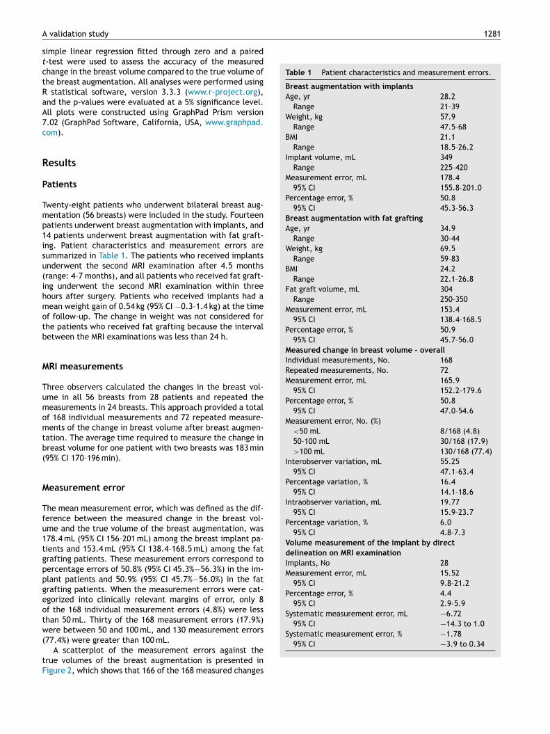
A validation study 1281
st
c
tRa
A7c
R
P
Tmp1is
u(
ihm
o
tb
M
Tu
mom
tb
(
M
T
fu1
t
gp
p
geot
w
(
tF
Table 1 Patient characteristics and measurement errors.
Breast augmentation with implants Age, yr 28.2
Range 21–39 Weight, kg 57.9
Range 47.5–68 BMI 21.1
Range 18.5–26.2 Implant volume, mL 349
Range 225–420 Measurement error, mL 178.4
95% CI 155.8–201.0 Percentage error, % 50.8
95% CI 45.3–56.3 Breast augmentation with fat grafting Age, yr 34.9
Range 30–44 Weight, kg 69.5
Range 59–83 BMI 24.2
Range 22.1–26.8 Fat graft volume, mL 304
Range 250–350 Measurement error, mL 153.4
95% CI 138.4–168.5 Percentage error, % 50.9
95% CI 45.7–56.0 Measured change in breast volume – overall Individual measurements, No. 168 Repeated measurements, No. 72 Measurement error, mL 165.9
95% CI 152.2–179.6 Percentage error, % 50.8
95% CI 47.0–54.6 Measurement error, No. (%)
< 50 mL 8/168 (4.8) 50–100 mL 30/168 (17.9) > 100 mL 130/168 (77.4)
Interobserver variation, mL 55.25 95% CI 47.1–63.4
Percentage variation, % 16.4 95% CI 14.1–18.6
Intraobserver variation, mL 19.77 95% CI 15.9–23.7
Percentage variation, % 6.0 95% CI 4.8–7.3
Volume measurement of the implant by direct delineation on MRI examination
Implants, No 28 Measurement error, mL 15.52
95% CI 9.8–21.2 Percentage error, % 4.4
95% CI 2.9–5.9 Systematic measurement error, mL −6.72
95% CI −14.3 to 1.0 Systematic measurement error, % −1.78
95% CI −3.9 to 0.34
imple linear regression fitted through zero and a paired -test were used to assess the accuracy of the measuredhange in the breast volume compared to the true volume ofhe breast augmentation. All analyses were performed using statistical software, version 3.3.3 ( www.r-project.org ), nd the p-values were evaluated at a 5% significance level.ll plots were constructed using GraphPad Prism version .02 (GraphPad Software, California, USA, www.graphpad. om ).
esults
atients
wenty-eight patients who underwent bilateral breast aug- entation (56 breasts) were included in the study. Fourteen atients underwent breast augmentation with implants, and 4 patients underwent breast augmentation with fat graft- ng. Patient characteristics and measurement errors are ummarized in Table 1 . The patients who received implantsnderwent the second MRI examination after 4.5 months range: 4–7 months), and all patients who received fat graft-ng underwent the second MRI examination within three ours after surgery. Patients who received implants had a ean weight gain of 0.54 kg (95% CI −0.3–1.4 kg) at the timef follow-up. The change in weight was not considered forhe patients who received fat grafting because the interval etween the MRI examinations was less than 24 h.
RI measurements
hree observers calculated the changes in the breast vol- me in all 56 breasts from 28 patients and repeated theeasurements in 24 breasts. This approach provided a total f 168 individual measurements and 72 repeated measure- ents of the change in breast volume after breast augmen-ation. The average time required to measure the change in reast volume for one patient with two breasts was 183 min95% CI 170–196 min).
easurement error
he mean measurement error, which was defined as the dif-erence between the measured change in the breast vol- me and the true volume of the breast augmentation, was 78.4 mL (95% CI 156–201 mL) among the breast implant pa-ients and 153.4 mL (95% CI 138.4–168.5 mL) among the fatrafting patients. These measurement errors correspond to ercentage errors of 50.8% (95% CI 45.3% −56.3%) in the im-lant patients and 50.9% (95% CI 45.7% −56.0%) in the fatrafting patients. When the measurement errors were cat- gorized into clinically relevant margins of error, only 8 f the 168 individual measurement errors (4.8%) were less han 50 mL. Thirty of the 168 measurement errors (17.9%)ere between 50 and 100 mL, and 130 measurement errors77.4%) were greater than 100 mL. A scatterplot of the measurement errors against the
rue volumes of the breast augmentation is presented in igure 2 , which shows that 166 of the 168 measured changes
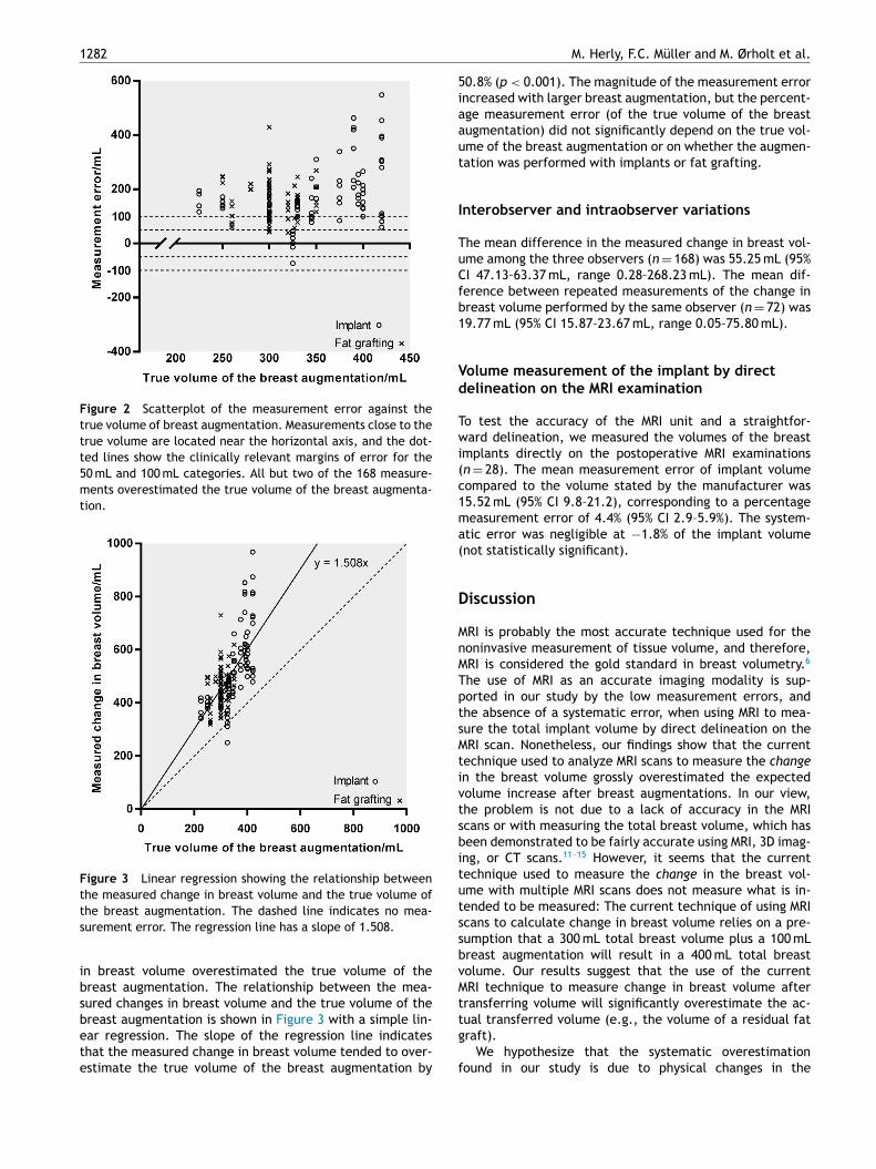
1282 M. Herly, F.C. Müller and M. Ørholt et al.
Figure 2 Scatterplot of the measurement error against the true volume of breast augmentation. Measurements close to the true volume are located near the horizontal axis, and the dot- ted lines show the clinically relevant margins of error for the 50 mL and 100 mL categories. All but two of the 168 measure- ments overestimated the true volume of the breast augmenta- tion.
Figure 3 Linear regression showing the relationship between the measured change in breast volume and the true volume of the breast augmentation. The dashed line indicates no mea- surement error. The regression line has a slope of 1.508.
in breast volume overestimated the true volume of thebreast augmentation. The relationship between the mea-sured changes in breast volume and the true volume of thebreast augmentation is shown in Figure 3 with a simple lin-ear regression. The slope of the regression line indicatesthat the measured change in breast volume tended to over-estimate the true volume of the breast augmentation by
50.8% ( p < 0.001). The magnitude of the measurement errorincreased with larger breast augmentation, but the percent-age measurement error (of the true volume of the breastaugmentation) did not significantly depend on the true vol-ume of the breast augmentation or on whether the augmen-tation was performed with implants or fat grafting.
Interobserver and intraobserver variations
The mean difference in the measured change in breast vol-ume among the three observers ( n = 168) was 55.25 mL (95%CI 47.13–63.37 mL, range 0.28–268.23 mL). The mean dif-ference between repeated measurements of the change inbreast volume performed by the same observer ( n = 72) was19.77 mL (95% CI 15.87–23.67 mL, range 0.05–75.80 mL).
Volume measurement of the implant by direct delineation on the MRI examination
To test the accuracy of the MRI unit and a straightfor-ward delineation, we measured the volumes of the breastimplants directly on the postoperative MRI examinations( n = 28). The mean measurement error of implant volumecompared to the volume stated by the manufacturer was15.52 mL (95% CI 9.8–21.2), corresponding to a percentagemeasurement error of 4.4% (95% CI 2.9–5.9%). The system-atic error was negligible at −1.8% of the implant volume(not statistically significant).
Discussion
MRI is probably the most accurate technique used for thenoninvasive measurement of tissue volume, and therefore,MRI is considered the gold standard in breast volumetry. 6
The use of MRI as an accurate imaging modality is sup-ported in our study by the low measurement errors, andthe absence of a systematic error, when using MRI to mea-sure the total implant volume by direct delineation on theMRI scan. Nonetheless, our findings show that the currenttechnique used to analyze MRI scans to measure the changein the breast volume grossly overestimated the expectedvolume increase after breast augmentations. In our view,the problem is not due to a lack of accuracy in the MRIscans or with measuring the total breast volume, which hasbeen demonstrated to be fairly accurate using MRI, 3D imag-ing, or CT scans. 11 –15 However, it seems that the currenttechnique used to measure the change in the breast vol-ume with multiple MRI scans does not measure what is in-tended to be measured: The current technique of using MRIscans to calculate change in breast volume relies on a pre-sumption that a 300 mL total breast volume plus a 100 mLbreast augmentation will result in a 400 mL total breastvolume. Our results suggest that the use of the currentMRI technique to measure change in breast volume aftertransferring volume will significantly overestimate the ac-tual transferred volume (e.g., the volume of a residual fatgraft).
We hypothesize that the systematic overestimationfound in our study is due to physical changes in the
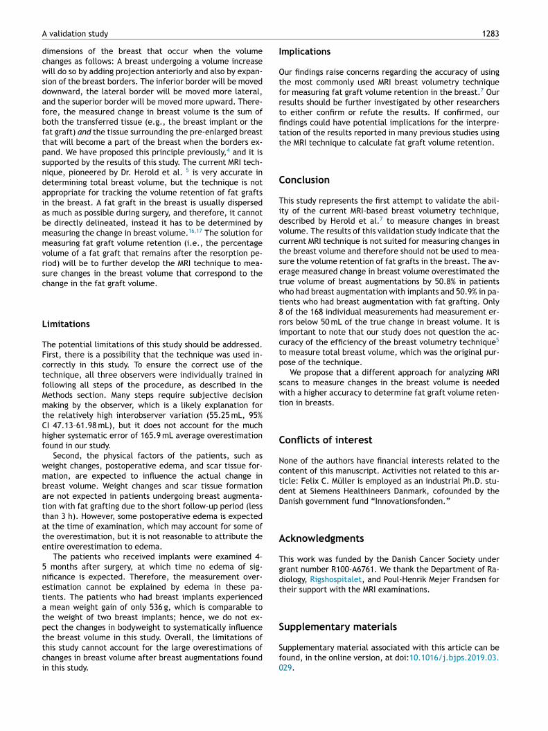
A validation study 1283
dcw
s
da
f
b
ft
ps
n
dai
a
bmmv
r
s
c
L
TF
c
tf
Mm
tC
hf
wmbat
ta
t
e
5
neta
t
pt
tci
I
Otf
rt
fit
t
C
T
id
v
c
t
s
et
w
t8
r
i
ct
p
s
w
t
C
N
c
t
dD
A
T
g
dt
S
Sf029 .
imensions of the breast that occur when the volume hanges as follows: A breast undergoing a volume increase ill do so by adding projection anteriorly and also by expan-ion of the breast borders. The inferior border will be movedownward, the lateral border will be moved more lateral, nd the superior border will be moved more upward. There-ore, the measured change in breast volume is the sum ofoth the transferred tissue (e.g., the breast implant or theat graft) and the tissue surrounding the pre-enlarged breast hat will become a part of the breast when the borders ex-and. We have proposed this principle previously, 4 and it is upported by the results of this study. The current MRI tech-ique, pioneered by Dr. Herold et al. 5 is very accurate inetermining total breast volume, but the technique is not ppropriate for tracking the volume retention of fat grafts n the breast. A fat graft in the breast is usually disperseds much as possible during surgery, and therefore, it cannote directly delineated, instead it has to be determined by easuring the change in breast volume. 16,17 The solution for easuring fat graft volume retention (i.e., the percentage olume of a fat graft that remains after the resorption pe-iod) will be to further develop the MRI technique to mea-ure changes in the breast volume that correspond to thehange in the fat graft volume.
imitations
he potential limitations of this study should be addressed. irst, there is a possibility that the technique was used in-orrectly in this study. To ensure the correct use of theechnique, all three observers were individually trained in ollowing all steps of the procedure, as described in theethods section. Many steps require subjective decision aking by the observer, which is a likely explanation forhe relatively high interobserver variation (55.25 mL, 95%
I 47.13–61.98 mL), but it does not account for the muchigher systematic error of 165.9 mL average overestimation ound in our study. Second, the physical factors of the patients, such as
eight changes, postoperative edema, and scar tissue for- ation, are expected to influence the actual change in reast volume. Weight changes and scar tissue formation re not expected in patients undergoing breast augmenta- ion with fat grafting due to the short follow-up period (lesshan 3 h). However, some postoperative edema is expected t the time of examination, which may account for some ofhe overestimation, but it is not reasonable to attribute thentire overestimation to edema. The patients who received implants were examined 4–
months after surgery, at which time no edema of sig-ificance is expected. Therefore, the measurement over- stimation cannot be explained by edema in these pa- ients. The patients who had breast implants experienced mean weight gain of only 536 g, which is comparable tohe weight of two breast implants; hence, we do not ex-ect the changes in bodyweight to systematically influence he breast volume in this study. Overall, the limitations ofhis study cannot account for the large overestimations of hanges in breast volume after breast augmentations found n this study.
mplications
ur findings raise concerns regarding the accuracy of using he most commonly used MRI breast volumetry technique or measuring fat graft volume retention in the breast. 7 Ouresults should be further investigated by other researchers o either confirm or refute the results. If confirmed, ourndings could have potential implications for the interpre- ation of the results reported in many previous studies usinghe MRI technique to calculate fat graft volume retention.
onclusion
his study represents the first attempt to validate the abil-ty of the current MRI-based breast volumetry technique, escribed by Herold et al. 7 to measure changes in breastolume. The results of this validation study indicate that theurrent MRI technique is not suited for measuring changes inhe breast volume and therefore should not be used to mea-ure the volume retention of fat grafts in the breast. The av-rage measured change in breast volume overestimated the rue volume of breast augmentations by 50.8% in patientsho had breast augmentation with implants and 50.9% in pa-ients who had breast augmentation with fat grafting. Only of the 168 individual measurements had measurement er-ors below 50 mL of the true change in breast volume. It ismportant to note that our study does not question the ac-uracy of the efficiency of the breast volumetry technique 5
o measure total breast volume, which was the original pur-ose of the technique. We propose that a different approach for analyzing MRI
cans to measure changes in the breast volume is neededith a higher accuracy to determine fat graft volume reten-ion in breasts.
onflicts of interest
one of the authors have financial interests related to theontent of this manuscript. Activities not related to this ar-icle: Felix C. Müller is employed as an industrial Ph.D. stu-ent at Siemens Healthineers Danmark, cofounded by the anish government fund “Innovationsfonden.”
cknowledgments
his work was funded by the Danish Cancer Society underrant number R100-A6761. We thank the Department of Ra-iology, Rigshospitalet , and Poul-Henrik Mejer Frandsen for heir support with the MRI examinations.
upplementary materials
upplementary material associated with this article can be ound, in the online version, at doi: 10.1016/j.bjps.2019.03.
1284 M. Herly, F.C. Müller and M. Ørholt et al.
References
1. Gassman AA , Kao KK , Bradley JP , Lee JC . Quantification of adi-pose transfer viability using a novel, bioluminescent murinemodel. J Plastic Reconstr Aesthet Surg 2016; 69 (7):959–65 .
2. Herly M , Orholt M , Glovinski PV , et al. Quantifying long-term re-tention of excised fat grafts: a longitudinal, retrospective co-hort study of 108 patients followed for up to 8.4 years. PlastReconstr Surg 2017; 139 (5):1223–32 .
3. Khouri RK , Eisenmann-Klein M , Cardoso E , et al. Brava and au-tologous fat transfer is a safe and effective breast augmen-tation alternative: results of a 6-year, 81-patient, prospectivemulticenter study. Plastic Reconstr Surg 2012; 129 (5):1173–87 .
4. Glovinski PV , Herly M , Muller FC , et al. Avoiding a systematicerror in assessing fat graft survival in the breast with repeatedmagnetic resonance imaging. Plast Reconstr Surg Glob Open2016; 4 (9):e1023 .
5. Herold C , Reichelt A , Stieglitz L , et al. MRI-based breast vol-umetry—evaluation of three different software solutions. J Dig-ital Imaging 2010; 23 (5):603–10 .
6. Herold C , Ueberreiter K , Busche MN , Vogt PM . Autologousfat transplantation: volumetric tools for estimation of vol-ume survival. A systematic review. Aesthetic Plastic Surg2013; 37 (2):380–7 .
7. Herold C , Ueberreiter K , Cromme F , Busche M , Vogt P . The useof mamma MRI volumetry to evaluate the rate of fat survivalafter autologous lipotransfer. Handchirurgie, Mikrochirurgie,plastische Chirurgie: Organ der Deutschsprachigen Arbeitsge-meinschaft fur Handchirurgie: Organ der DeutschsprachigenArbeitsgemeinschaft fur Mikrochirurgie der Peripheren Nervenund Gefasse: Organ der V 2010; 42 (2):129–34 .
8. Herold C , Ueberreiter K , Cromme F , Grimme M , Vogt PM .Is there a need for intrapectoral injection in autolo-gous fat transplantation to the breast? - An MRI volumet-ric study. Handchirurgie Mikrochirurgie Plastische Chirurgie2011; 43 (2):119–24 .
9. Koch MC , Adamietz B , Jud SM , et al. Breast volumetry usinga three-dimensional surface assessment technique. AestheticPlastic Surg 2011; 35 (5):847 .
10. Seoud L , Ramsay J , Parent S , Cheriet F . A novel fully automaticmeasurement of apparent breast volume from trunk surfacemesh. Med Eng Phys 2017; 41 :46–54 .
11. Eric M , Anderla A , Stefanovic D , Drapsin M . Breast volume es-timation from systematic series of CT scans using the Cava-lieri principle and 3D reconstruction. Int J Surg (Lond, Engl)2014; 12 (9):912–17 .
12. Wesselius TS , Verhulst AC , Vreeken RD , Xi T , Maal TJ , Ulrich DJ .Accuracy of three software applications for breast volume cal-culations from three-dimensional surface images. Plastic Re-constr Surg 2018; 142 (4):858–65 .
13. Losken A , Seify H , Denson DD , Paredes AA Jr , Carlson GW . Vali-dating three-dimensional imaging of the breast. Annals PlasticSurg 2005; 54 (5):471–6 .
14. Lee WY , Kim MJ , Lew DH , Song SY , Lee DW . Three-dimensionalsurface imaging is an effective tool for measuring breast vol-ume: a validation study. Arch Plastic Surg 2016; 43 (5):430 .
15. Rha EY , Choi IK , Yoo G . Accuracy of the method for estimatingbreast volume on three-dimensional simulated magnetic reso-nance imaging scans in breast reconstruction. Plastic ReconstrSurg 2014; 133 (1):14–20 .
16. Bourne DA , James IB , Wang SS , Marra KG , Rubin JP . The archi-tecture of fat grafting: what lies beneath the surface. PlasticReconstr Surg 2016; 137 (3):1072–9 .
17. James IB , Bourne DA , DiBernardo G , et al. The architectureof fat grafting II: impact of cannula diameter. Plastic ReconstrSurg 2018; 142 (5):1219–25 .

Appendix 4
Lipovol: Validated Software for Measuring Fat Graft Volume Retention in the Breast with MRI
Presented in part at the Plastic Surgery “The Meeting” conference in San Diego, September 2019
Manuscript
Title:
Lipovol: A new method for measuring fat graft volume retention in the breast with MRI
Author List:
Mikkel Herly1, Mathias Ørholt1, Felix Christoph Müller2, Mathilde N. Hemmingsen1, Joachim Hansen1,
Andreas Larsen1, Bo S. Rasmussen1, Jens J. Elberg3, Christian von Buchwald4, Krzysztof T. Drzewiecki1 and
Peter V. Vester-Glowinski1
Affiliations:
1Department of Plastic Surgery and Burns, Rigshospitalet; 2100-Copenhagen, Denmark
2Department of Radiology, Herlev and Gentofte Hospital; 2730-Herlev, Denmark
3Amalieklinikkken; 1300-Copenhagen, Denmark
4Department of ORL, Head and Neck Surgery and Audiology, Rigshospitalet; 2100-Copenhagen, Denmark
Corresponding Author:
Mikkel Herly, M.D.
Department of Plastic Surgery and Burns Treatment
Copenhagen University Hospital, Rigshospitalet,
Blegdamsvej 9, DK-2100 Copenhagen, Denmark
E-mail: [email protected]
Summary
Background: We have previously shown that the current gold standard breast volumetry technique fails to
accurately measure changes in fat graft volume retention in the breast and systematically overestimates
volume retention by approximately 50%. In this study, we present Lipovol: a new automated software
method dedicated to measuring fat graft volume retention in the breast based on magnetic resonance
imaging (MRI) scans.
Methods: The software method was validated by four observers using the MRI scans of 28 patients
undergoing breast augmentation with fat grafting or breast implants. The changes in breast volume were
measured with the software and compared with the injected fat graft volumes/implant volumes.
Results: The software method measured the injected fat graft volumes with an average systematic
overestimation of 6.3% (SD = 10.5). Significantly higher measurement errors were recorded for the implant
patients, with a systematic overestimation of 23.9% (SD = 18.1), than for the fat grafting patients. The
median interobserver variation was less than 7% for both groups.
Conclusion: This new software method is significantly more accurate and precise than the previous gold
standard technique. We propose our new method for measuring fat graft volume retention in the breast
for clinical research. The software will be available free of charge and it can be used on the MeVisLab
platform.
Keywords
Fat grafting; breast augmentation; breast; MRI; volume retention

Introduction
One of the greatest challenges in using fat grafting in the breast is that a variable part of the graft volume is
resorbed in the time after surgery.1 An accurate measurement technique to assess the fat graft volume
retention in the breast after breast augmentation2,3 and breast reconstruction4,5 is needed to investigate
the resorption dynamics of fat grafts in the breast, which can be used to develop new strategies for
improving the procedure.6–8 We have previously shown that the measurement of total breast volume9 is a
very different task than measuring the volume retention of fat grafts that are dispersed in the breast and
that the current gold standard technique for measuring fat graft volume retention10,11 was subject to a
systematic error of more than 50%.12,13 The problem with fat grafts in the breast is that they cannot be
delineated on the MRI scan and therefore, we must rely on the measurement of a change in breast volume
between two MRI scans of the breasts recorded before the surgery and after the volume resorption has
taken place. This change in breast volume can then be compared with the injected volume to calculate the
fat graft volume retention rate. E.g. a fat graft of 300 mL is injected and after 1 year the retention rate is
60% corresponding to a 180 mL augmentation. To measure the retention rate, we would have a pre-
operative scan and a 1-year-post-operative scan where the expected increase in breast volume would be
180 mL. The accuracy of the measurement technique should be judged by the technique’s ability to
estimate the exact change in breast volume from one scan to another. However, to our knowledge,
previous techniques have not been validated for their ability to measure a change in breast volume. In this
study, we present a new software method (Lipovol) that is dedicated to the measurement of changes in
breast volume and specifically the volume retention of fat grafts in the breast using magnetic resonance
imaging (MRI) scans. Additionally, we present a validation study of the methods’s ability to measure
changes in breast volume on 28 women who have undergone a breast augmentation with a known
volumes. Lipovol will be freely available and can be used on the MeVisLab14 platform which is an open
source software platform for radiological analysis that is also free to use for noncommercial purposes.
Patients and Methods
Lipovol software
Two MRI scans are required to measure fat graft volume retention with the Lipovol software method: one
acquired before surgery and one acquired after surgery when the fat graft has reached volumetric steady
state. First, the software is used to align the two scans on top of each other using osseous pointers in the
thorax. This is the key step that sets Lipovol apart from previous techniques: the alignment allows the user
to outline a region-of-interest on the second scan (containing the residual fat graft) which is automatically
mirrored on the first scan which ensures that the two regions-of-interest will delineate the same block of
tissue on the two scans. Lipovol will then automatically delineate the skin in the two regions-of-interest and
calculate the volumes. The difference in volume between the two regions-of-interest will be equal to the
residual fat graft volume because the regions-of-interest contain the same tissue block at the two time
points (with and without the residual fat graft). The retention rate is calculated by dividing the residual fat
graft volume with the initially injected fat graft volume. A demonstration of the software method’s
workflow is shown in figure 1 and as a video in the supplementary material.
Validation
The study was approved by the regional ethics committee of Copenhagen (nr. H-1501379) and the Regional
Data Protection Agency. All participants provided written informed consent before inclusion in the study.
The accuracy of the software was validated using three types of patients with known changes in breast
volume: 1) Patients undergoing breast augmentation with fat grafting who were scanned before surgery
and within 3 h after surgery, at which time minimal edema formation is present, and the resorption of the
fat graft has yet to begin. 2) Patients undergoing breast augmentation with breast implants scanned before
surgery and 4-5 months after surgery (i.e., after edema formation). 3) Patients scanned twice on the same
day whose second scan was performed after repositioning (no expected change in breast volume).
All measurements of the changes in breast volume were performed by four observers (MH, MØ, JH, and
MNH) who were blinded to the transplanted volume.
Patients
The patients were included from the University Hospital Copenhagen and Amalieklinikken, Copenhagen.
The MRI scans were acquired on a 3 Tesla MRI unit (Siemens Magnetom Verio, Germany) with a 4-channel
breast coil. The patients were examined in a head-first prone position. Parts of the MRI scans have been
used in a previous study.9 Lipovol works on the inphase and outphase images of a 2-point Dixon sequence.
The patients were scanned at the same point in their menstrual cycle to prevent cyclical breast volume
changes from influencing the measurements.
Statistical analysis
Continuous variables were presented as the mean (SD) when normally distributed and as the median (IQR)
when non-normally distributed. The distributions were visualized with histograms and QQ-plots and tested
for normality with the Shapiro-Wilk test.15 Comparisons between normally distributed data were
performed with t-tests and evaluated at a 5% significance level. Analyses of measurement errors were
calculated with breast as the independent sampling unit and an average of the four reads were used as the
measurement. Inter- and intra-observer variation were calculated with the individual measurement as the
independent sampling unit. All analyses were performed in R, version 3.6.1 (www.r-project.org, R
Foundation for Statistical Computing, Vienna, Austria).
Results
Between 2014-2018, we included and performed MRI on a total of 56 breasts in 28 patients. The patients
underwent bilateral breast augmentation with either fat grafting (n = 14) or implants (n = 14), and all
patients were scanned twice on the first day to calculate the measurement error at baseline. Four
independent observers provided a total of 280 individual measurements of the changes in breast volume
and 96 repeated measurements.
Patients undergoing fat grafting received a median fat graft volume of 300 mL per breast (range 250-350, n
= 28), whereas the mean measured change in breast volume was 322 mL (range 242-403, n = 112). The
systematic measurement error in the fat grafting patients was 6.28% (SD = 10.5), which is significantly more
accurate than the previous gold standard technique10 that had a systematic error of 50.9% (SD = 24.3)9 (p <
0.001). The median interobserver variation was 4.8% (IQR, 2.7-9.8), and the median intraobserver variation
was 4.2% (IQR, 2.0-8.7). Patients undergoing breast augmentation with implants received a median volume
of 348 mL per breast (range 225-420, n = 28), whereas the measured change in breast volume was 432 mL
(range 268-638 mL, n = 28). The systematic measurement error in the implant patients was 23.9% (SD =
18.1). The median interobserver variation was 4.9% (IQR, 2.4-8.2), and the median intraobserver variation
was 6.2 (IQR, 2.4-10.1). The systematic measurement error was significantly higher for the implant patients
than for the fat grafting patients when using Lipovol (p < 0.001), but Lipovol was still significantly more
accurate than the current gold standard technique10, which had a systematic error of 50.8% (SD = 27.3) in
the implant patients13 (p < 0.001). A summary of the validation results is provided in table 1. When
measuring the volume difference between repeated measurements recorded on the same day (expected
volume change = 0 mL) for the 28 patients (56 breasts), we found a systematic measurement error of only -
0.07 mL (SD = 6.2). The linear regression with a 95% prediction interval is shown in figure 2 and indicates a
high correlation between the measured volumes and the true volumes with r2 = 0.97 for the fat grafting
patients and r2 = 0.94 for the implant patients. The measurement errors are analyzed in a Bland-Altman
plot13 in figure 3, which shows a trend of higher measurement errors with higher augmentation volumes.
Discussion

The new software method for measuring fat graft volume retention in the breast was significantly more
accurate than the current gold standard technique.7 Lipovol was particularly accurate and precise for the
analysis of fat graft volume changes (6.28% systematic error, SD = 10.5).
Limitations
The software was validated in patients with known changes in breast volume. Half of the patients had
undergone fat grafting and were examined before and directly after surgery at which time no fat graft
resorption would have occurred (<3 h). The other half had undergone breast augmentation with implants
and were examined before and 4-5 months after surgery. None of the patients experienced any
considerable weight changes during this period (average weight gain of 536 g). The advantage of using fat
grafting patients for validation is that the primary purpose of the software is to measure the residual
volumes of fat grafts. The disadvantage is that we cannot wait for the edema to subside before performing
the second scan because we do not know the true resorption dynamics of the fat grafts; therefore, we must
accept that some edema is present at the time of the second scan (<3 h after surgery). The expected edema
may in part account for the systematic overestimation of 6.28% in these patients. Additionally, the range of
fat graft volumes was relatively narrow in our patient population (range, 250-350 mL). However, this range
is in accordance with the most frequently used fat graft volumes for primary breast augmentation in the
literature.16,17 The implant patients have the advantage of stable implant volumes, which allow us to
perform the second scan after the edema was expected to have subsided. The disadvantage is that the
measured changes in breast volume after breast augmentations with implants might be different from
those of breast augmentations with fat grafts due to the different placement of the implants compared to
that of the dispersed fat grafts. Overall, we do not know how much of the overestimation that stems from
edema in fat grafting patients and physiological changes in the breast after implants affected the error of
the software measurements. Therefore, we do not suggest that the systematic overestimation found in this
validation study should be deducted from future measurements using the software method. We cannot
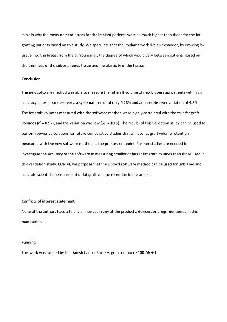
explain why the measurement errors for the implant patients were so much higher than those for the fat
grafting patients based on this study. We speculate that the implants work like an expander, by drawing lax
tissue into the breast from the surroundings, the degree of which would vary between patients based on
the thickness of the subcutaneous tissue and the elasticity of the tissues.
Conclusion
The new software method was able to measure the fat graft volume of newly operated patients with high
accuracy across four observers, a systematic error of only 6.28% and an interobserver variation of 4.8%.
The fat graft volumes measured with the software method were highly correlated with the true fat graft
volumes (r2 = 0.97), and the variation was low (SD = 10.5). The results of this validation study can be used to
perform power calculations for future comparative studies that will use fat graft volume retention
measured with the new software method as the primary endpoint. Further studies are needed to
investigate the accuracy of the software in measuring smaller or larger fat graft volumes than those used in
this validation study. Overall, we propose that the Lipovol software method can be used for unbiased and
accurate scientific measurement of fat graft volume retention in the breast.
Conflicts of interest statement
None of the authors have a financial interest in any of the products, devices, or drugs mentioned in this
manuscript.
Funding
This work was funded by the Danish Cancer Society, grant number R100-A6761.

1. James IB, Bourne DA, DiBernardo G, et al. The architecture of fat grafting II: Impact of cannula
diameter. Plast Reconstr Surg. 2018;142(5):1219-1225. doi:10.1097/PRS.0000000000004837
2. Coleman SR. Structural fat grafting: more than a permanent filler. Plast Reconstr Surg.
2006;118(3S):108S-120S.
3. Khouri RK, Eisenmann-Klein M, Cardoso E, et al. Brava and autologous fat transfer is a safe and
effective breast augmentation alternative: results of a 6-year, 81-patient, prospective multicenter
study. Plast Reconstr Surg. 2012;129(5):1173-1187.
http://www.embase.com/search/results?subaction=viewrecord&from=export&id=L366375370.
4. Herly M, Ørholt M, Larsen A, et al. Efficacy of breast reconstruction with fat grafting: a systematic
review and meta-analysis. J Plast Reconstr Aesthetic Surg. 2018;71(12):1740-1750.
5. Hoppe DL, Ueberreiter K, Surlemont Y, Peltoniemi H, Stabile M, Kauhanen S. Breast reconstruction
de novo by water-jet assisted autologous fat grafting - a retrospective study. GMS Ger Med Sci.
2013;11. doi:10.3205/000185
6. Guimarães PAMP, Sabino Neto M, Lage FC, Guirado FF, de Mello GGN, Ferreira LM. Evaluation of
retropectoral fat grafting in breast reduction by magnetic resonance imaging: a pilot study. Aesthetic
Surg J. 2018;39(5):518-523.
7. Choi M, Small K, Levovitz C, Lee C, Fadl A, Karp NS. The volumetric analysis of fat graft survival in
breast reconstruction. Plast Reconstr Surg. 2013;131(2):185-191.
http://www.embase.com/search/results?subaction=viewrecord&from=export&id=L368615999.
8. Herly M, Ørholt M, Glovinski P V, et al. Quantifying long-term retention of excised fat grafts: a
longitudinal, retrospective cohort study of 108 patients followed for up to 8.4 years. Plast Reconstr
Surg. 2017;139(5):1223-1232.
9. Erić M, Anderla A, Stefanović D, Drapšin M. Breast volume estimation from systematic series of CT

scans using the Cavalieri principle and 3D reconstruction. Int J Surg. 2014;12(9):912-917.
doi:10.1016/j.ijsu.2014.07.018
10. Herold C, Ueberreiter K, Cromme F, Busche MN, Vogt PM. The use of mamma MRI volumetry to
evaluate the rate of fat survival after autologous lipotransfer. Handchirurgie, Mikrochirurgie, Plast
Chir Organ der Deutschsprachigen Arbeitsgemeinschaft für Handchirurgie Organ der
Deutschsprachigen Arbeitsgemeinschaft für Mikrochirurgie der Peripher Nerven und Gefässe Organ
der Vereinigung der Deut. 2010;42(2):129-134.
http://www.embase.com/search/results?subaction=viewrecord&from=export&id=L359015760.
11. Herold C, Reichelt A, Stieglitz LH, et al. MRI-based breast volumetry-evaluation of three different
software solutions. J Digit Imaging. 2010;23(5):603-610. doi:10.1007/s10278-009-9264-y
12. Glovinski P V, Herly M, Muller FC, et al. Avoiding a Systematic Error in Assessing Fat Graft Survival in
the Breast with Repeated Magnetic Resonance Imaging. Plast Reconstr Surg Glob Open.
2016;4(9):e1023. doi:10.1097/gox.0000000000001023
13. Herly M, Müller FC, Ørholt M, et al. The current gold standard breast volumetry technique seems to
overestimate fat graft volume retention in the breast: A validation study. J Plast Reconstr Aesthetic
Surg. 2019.
14. MeVisLab. Available: http://www.mevislab.de/.
15. Shapiro SS, Francia RS. An approximate analysis of variance test for normality. J Am Stat Assoc.
1972;67(337):215-216.
16. Zocchi ML, Zocchi L. Large-volume breast fat transfer: technical evolutions and safety aspects based
on over 800 cases and 26 years of follow-up. Eur J Plast Surg. 2017;40(5):367-382.
doi:10.1007/s00238-017-1339-2
17. Chiu CH. Does Stromal Vascular Fraction Ensure a Higher Survival in Autologous Fat Grafting for

Breast Augmentation? A Volumetric Study Using 3-Dimensional Laser Scanning. Aesthetic Surg J.
2019;39(1):41-52. doi:10.1093/asj/sjy030
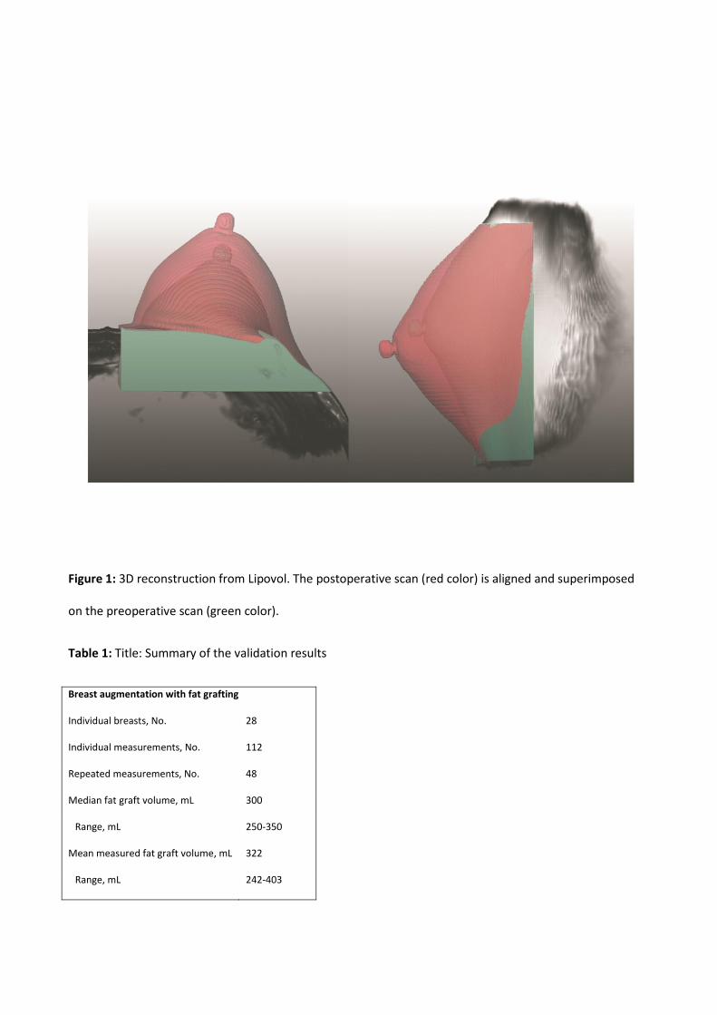
Figure 1: 3D reconstruction from Lipovol. The postoperative scan (red color) is aligned and superimposed
on the preoperative scan (green color).
Table 1: Title: Summary of the validation results
Breast augmentation with fat grafting
Individual breasts, No. 28
Individual measurements, No. 112
Repeated measurements, No. 48
Median fat graft volume, mL 300
Range, mL 250-350
Mean measured fat graft volume, mL 322
Range, mL 242-403
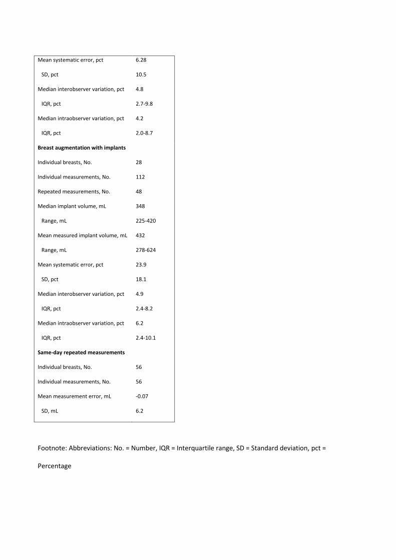
Mean systematic error, pct 6.28
SD, pct 10.5
Median interobserver variation, pct 4.8
IQR, pct 2.7-9.8
Median intraobserver variation, pct 4.2
IQR, pct 2.0-8.7
Breast augmentation with implants
Individual breasts, No. 28
Individual measurements, No. 112
Repeated measurements, No. 48
Median implant volume, mL 348
Range, mL 225-420
Mean measured implant volume, mL 432
Range, mL 278-624
Mean systematic error, pct 23.9
SD, pct 18.1
Median interobserver variation, pct 4.9
IQR, pct 2.4-8.2
Median intraobserver variation, pct 6.2
IQR, pct 2.4-10.1
Same-day repeated measurements
Individual breasts, No. 56
Individual measurements, No. 56
Mean measurement error, mL -0.07
SD, mL 6.2
Footnote: Abbreviations: No. = Number, IQR = Interquartile range, SD = Standard deviation, pct =
Percentage

Figure 2: Linear regression analysis with a 95% prediction interval of the measured changes in breast
volume compared to the true volumes of the breast augmentations.
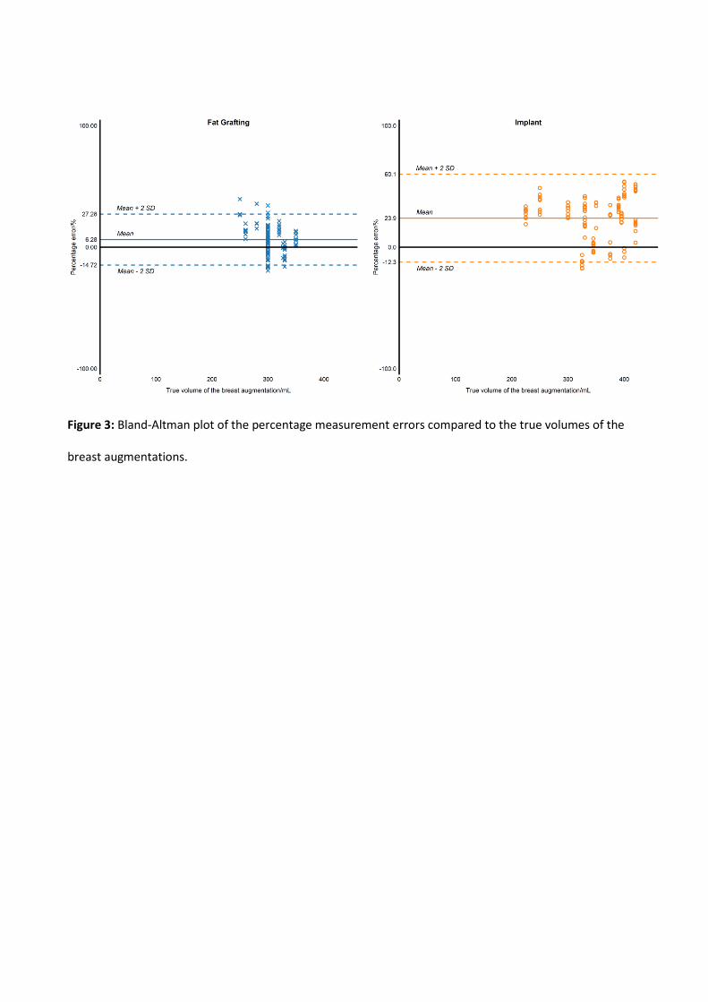
Figure 3: Bland-Altman plot of the percentage measurement errors compared to the true volumes of the
breast augmentations.