phylogeny poorly predicts the utility of a challenging horizontally transferred gene in...
TRANSCRIPT

Phylogeny Poorly Predicts the Utility of a Challenging HorizontallyTransferred Gene in Methylobacterium Strains
Joshua K. Michener,a Stéphane Vuilleumier,b Françoise Bringel,b Christopher J. Marxa,c*
Department of Organismic and Evolutionary Biology, Harvard University, Cambridge, Massachusetts, USAa; Equipe Adaptations et Interactions Microbiennes dansl’Environnement, UMR 7156, Université de Strasbourg—CNRS, Strasbourg, Franceb; Faculty of Arts and Sciences Center for Systems Biology, Harvard University,Cambridge, Massachusetts, USAc
Horizontal gene transfer plays a crucial role in microbial evolution. While much is known about the mechanisms that determinewhether physical DNA can be transferred into a new host, the factors determining the utility of the transferred genes are lessclear. We have explored this issue using dichloromethane consumption in Methylobacterium strains. Methylobacterium ex-torquens DM4 expresses a dichloromethane dehalogenase (DcmA) that has been acquired through horizontal gene transfer andallows the strain to grow on dichloromethane as the sole carbon and energy source. We transferred the dcmA gene into sixMethylobacterium strains that include both close and distant evolutionary relatives. The transconjugants varied in their abilityto grow on dichloromethane, but their fitness on dichloromethane did not correlate with the phylogeny of the parental strains orwith any single tested physiological factor. This work highlights an important limiting factor in horizontal gene transfer,namely, the capacity of the recipient strain to accommodate the stress and metabolic disruption resulting from the acquisition ofa new enzyme or pathway. Understanding these limitations may help to rationalize historical examples of horizontal transferand aid deliberate genetic transfers in biotechnology for metabolic engineering.
The recent accumulation of genome sequences from diversebacterial clades has demonstrated the crucial role of horizontal
gene transfer (HGT) in bacterial evolution (1, 2). It has becomeclear that genes and operons have consistently moved betweendistant bacterial strains, with important implications for bacterialevolution, physiology, and ecology (3). In appreciating the signif-icant impact of HGT, it is important to also consider the factorsthat limit transfer (4). If a gene is beneficial when acquired by onestrain, why do we not observe that gene transferring into other,closely related strains?
One possible explanation for the rarity of successful horizontaltransfers is that recipients that would benefit from the transferhave little opportunity to acquire the corresponding DNA (5), andthat factors such as ecological differentiation and barriers to ge-netic exchange may prevent a strain from encountering a potentialdonor (4, 6). Additionally, transfer events may be rare even in thepresence of a donor (7), since the likelihood of stably integratingand expressing newly acquired DNA is predicted to decrease withincreasing genetic distance and will limit the frequency andbreadth of transfer (8–10). These factors suggest that ecology andphylogeny should largely determine transfer frequencies, based onhow likely a strain is to encounter a donor (ecology) and to ac-quire, stably integrate, and express the transferred DNA (phylog-eny) (11).
Another significant, yet poorly investigated, barrier to HGTdepends on how efficiently a recipient can use its new ability. Anewly acquired gene or pathway may place novel stresses on thehost, either by disrupting existing metabolic and regulatory net-works (12, 13) or by producing new toxic metabolites (13, 14).The fitness cost of such stresses is determined by the host physiol-ogy. A beneficial ability with costly side effects will preferentiallyspread to those recipients best able to accommodate its associatedstresses, leading to a gene distribution shaped by physiology ratherthan by phylogeny.
We used dichloromethane catabolism in strains of Methylobac-
terium to explore factors that limit the functional incorporation ofa horizontally transferred gene. Dichloromethane (DCM) is anindustrial solvent that has reached significant concentrations inthe environment only in the last 50 years. Among several strainsisolated for their ability to grow on DCM as the sole carbon andenergy source (15, 16), a strain of Methylobacterium extorquensknown as DM4 has been investigated in the most detail. ThroughHGT, this strain has acquired a gene, dcmA, encoding a cytoplas-mic glutathione S-transferase that converts DCM to formalde-hyde, with the concomitant release of two molecules of hydro-chloric acid (17, 18). In M. extorquens DM4, the dcmA gene lieswithin a 126-kb genomic island that shows clear evidence of hor-izontal transfer, including a lower GC content (19). Within thisgenomic island, the dcm cluster contains three other genes, in-cluding the transcriptional regulator dcmR and two proteins ofunknown function dcmB and dcmC and is flanked on both sides byIS1354 elements. This four-gene dcm islet is conserved withinmost DCM-degrading strains (18). However, a strain of M. ex-torquens DM4 with a deletion of the genomic island, known asDM4-2cr (20), requires only the dcmA gene to recover growth onDCM (21). While the other genes in the genomic island may in-fluence growth on DCM, they are not essential.
Growth with DCM is very challenging for the cell (Fig. 1). First,
Received 16 January 2014 Accepted 20 March 2014
Published ahead of print 28 March 2014
Address correspondence to Christopher J. Marx, [email protected].
* Present address: Christopher J. Marx, Department of Biological Sciences,University of Idaho, Moscow, Idaho, USA.
Supplemental material for this article may be found at http://dx.doi.org/10.1128/JB.00034-14.
Copyright © 2014, American Society for Microbiology. All Rights Reserved.
doi:10.1128/JB.00034-14
June 2014 Volume 196 Number 11 Journal of Bacteriology p. 2101–2107 jb.asm.org 2101
on Novem
ber 24, 2014 by UN
IVE
RS
ITE
LOU
IS P
AS
TE
UR
http://jb.asm.org/
Dow
nloaded from

the strain must accommodate the protons and chloride producedintracellularly as a by-product of DCM dehalogenation (22–24).Additionally, the S-chloromethylglutathione intermediate formedduring the dehalogenation reaction is highly reactive and muta-genic (25–27). Finally, the cell must quickly channel the formal-dehyde product into its native one-carbon metabolic pathways tominimize any resulting toxicity (28). Given these challenges, it isnot surprising that deliberate transfer of the dcmA gene to twoother strains of M. extorquens, AM1 and CM4, was sufficient toenable growth on DCM only in strain CM4 (21). However, itremains unclear why strain AM1 was unable to grow on DCM, orhow prevalent is the ability among methylotrophic bacteria togrow on DCM using dcmA. We have addressed these questions bytransferring dcmA into a broad range of Methylobacterium strains,quantifying their success at using their new catabolic potential,and investigating the factors that influence this success.
MATERIALS AND METHODSMedia and chemicals. All chemicals were purchased from Sigma-Aldrich(St. Louis, MO) unless otherwise noted. Escherichia coli was grown in LB at37°C with various antibiotic concentrations. Methylobacterium strainswere grown at 30°C in liquid culture in M-PIPES (29) supplemented with3.5 mM succinate or 5 mM DCM as noted. Antibiotics were added to afinal concentration of 12.5 �g/ml for tetracycline, 10 �g/ml for strepto-mycin, or 50 �g/ml for kanamycin. Unless otherwise noted, DCM cul-tures were grown in 10 ml of medium in gas-tight 50-ml screw-top flaskssealed with Teflon tape and Mininert valves (Supelco, Bellefonte, PA).Valves were surface sterilized with ethanol and dried in a laminar flowhood before use. A freshly prepared 100 mM stock of DCM in water wasused to inoculate the flasks.
Plasmid construction. Plasmids and strains used in this study arelisted in Tables 1 and 2. Plasmid pJM10 was constructed by cloning theAatII-SacI fragment containing kanR2 from pCM184 (30) into pME8220(21) digested with AatII-SacI to remove the 5= end of tetA. Plasmid pJM40was constructed by amplifying superecliptic pHluorin (31) and using Gib-son assembly to clone it into pHC08 (32) as a translational fusion tomCherry, yielding pJM25. The entire mCherry-pHluorin expression cas-sette was amplified from pJM25 by PCR to add BamHI and SacI restric-tion sites. The digested PCR fragment was then cloned into pME8220 andpCM62 to yield pJM40 and pJM41, respectively. Plasmid pJM53 was con-structed by amplifying 500-bp regions upstream and downstream of thehypoxanthine phosphoribosyl transferase (hpt) from the genome of M.extorquens DM4. Using Gibson assembly, these fragments were combinedwith a Venus expression cassette in pPS04 (P. Swanson and C. J. Marx,unpublished results), a kanamycin resistance derivative of pCM433 (33).
Plasmid matings. Plasmids were transferred to recipient Methylobac-terium strains using triparental matings as described previously (34). ThedcmA gene was deleted from M. extorquens DM4 using a derivative ofpCM433, which resulted in a mutant unable to grow on DCM (F. Bringeland S. Vuilleumier, unpublished results). M. extorquens DM4 �dcmA-Venus (strain CM4250) was constructed from M. extorquens DM4 �dcmAusing pJM53 and following established protocols (33). Integration of Ve-nus into the hpt locus of M. extorquens DM4 �dcmA was selectively neu-tral during growth on DCM.
Growth rate measurements. Methylobacterium strains were grown tosaturation in 48-well plates under each of the conditions being tested.Cultures were then diluted 64� into 640 �l of fresh medium. Over 48 h,optical densities at 600 nm (OD600) were measured every 30 to 45 minusing an automated system (29). Growth rates were calculated using Cur-veFitter (29).
Competitive fitness assays. Competitive fitness for growth on DCMwas measured by competing each strain against M. extorquens DM4�dcmA-Venus(pJM10) generally following a previous protocol (32). Inbrief, each strain was grown to saturation in M-PIPES–succinate– kana-mycin and then diluted 100� into M-PIPES–DCM. The exception was M.extorquens AM1 �cel-mCherry(pJM10), which cannot grow on DCMalone. This strain was grown in M-PIPES–DCM–tetracycline, where thetetracycline is dissolved in 80% ethanol and therefore provides 14 mMethanol for growth. After 3 days, the cultures were diluted and mixed infresh M-PIPES–DCM. M. extorquens DM4 �dcmA-Venus(pJM10) wasadded to each flask at an OD of 0.001. The test strain was then added at anOD of 0.005. A 450-�l portion of each culture was removed, mixed with50 �l of dimethyl sulfoxide (DMSO), and frozen at �80°C. The mixedcultures were then grown for 3 days.
At the end of the growth phase, the population ratios of the samples,before and after growth on DCM, were determined using flow cytometry.Postgrowth samples were diluted into fresh M-PIPES to a final OD of�0.015. Fluorescence was measured on an LSRII flow cytometer (BD,Franklin Lakes, NJ). Venus was excited at 488 nm and measured at 530 nmwith a 30-nm bandpass filter. When available, mCherry was excited at 561nm and measured at 620/40 nm. The competitive fitness was calculated aslog[(R1*N)/R0]/log[(1 � R1)*N/(1 � R0)], where R0 and R1 represent thepopulation fraction of the test strain before and after mixed growth and Nrepresents the fold increase in the population density. When the test strainwas labeled with mCherry, the population fraction was calculated as theratio of mCherry-positive cells to Venus-positive cells. When the teststrain was unlabeled, the population fraction was the ratio of Venus-negative cells to Venus-positive cells.
Chloride measurements. Chloride released by dehalogenation wasmeasured using the method of Jörg and Bertau (35), comparing the ab-sorbance against that obtained with M-PIPES without added carbon.
Total protein measurements. Total protein was measured by growingthe desired culture in 10 ml of M-PIPES–DCM for 3 days. The cultureswere concentrated by centrifugation and resuspended in 1 ml of PBS plus1 mM EDTA. Cells were lysed using a FastPrep-24 (MP Bio, Santa Ana,CA) and lysing matrix B (MP Bio). Lysates were centrifuged, and totalprotein in the supernatant was quantified using the Bradford quick-startassay (Bio-Rad, Hercules, CA) with bovine serum albumin (BSA) as astandard.
Dehalogenase activity measurements in whole cells. Strains weregrown at 30°C in 50 ml of M-PIPES in 300-ml screw-top Erlenmeyer flasksfitted with Mininert stoppers (Supelco), using 5 mM DCM with or with-out the addition of 3.5 mM succinate as the carbon and energy source.Cultures were harvested in exponential phase after measurement of theirODs and centrifuged for 15 min at 8,000 rpm. The final pH of the cultureswas measured in the spent medium supernatant. The cell pellets wereresuspended in 2 ml of M-PIPES, and the protein concentration in cell
CH2Cl2 GS-CH2Cl CH2OGSH HCl H2O HCl
GSH
FIG 1 Growth on DCM presents several challenges to the host. One moleculeof DCM is converted to one molecule of formaldehyde and two molecules ofHCl. Stress-inducing compounds are indicated in red. In addition to thestresses resulting from HCl and formaldehyde, the glutathione conjugate in-termediate is highly mutagenic.
TABLE 1 Plasmids used in this study
Plasmid Description Reference
pCM433 oriTRP4 cat bla tetA sacB 33pJM10 dcmA kanR2 This workpJM40 PTac-mCherry-pHluorin dcmA tetA This workpJM41 PTac-mCherry-pHluorin tetA This workpJM53 pPS04 hpt::Venus This workpPS04 kanR2 replaces cat, bla, and tetA of
pCM433P. Swanson and C. J. Marx,
unpublished data
Michener et al.
2102 jb.asm.org Journal of Bacteriology
on Novem
ber 24, 2014 by UN
IVE
RS
ITE
LOU
IS P
AS
TE
UR
http://jb.asm.org/
Dow
nloaded from

suspensions was determined by a commercial bicinchoninic acid proteinassay kit (Sigma) adapted to microplate format (0 to 10 �g protein, usingBSA as a reference). Chloride concentration in the final spent medium wasdetermined as described above, adapting the assay to microplate format.DCM dehalogenase activity was determined by the Nash method as de-scribed previously (36), with minor modifications. Briefly, concentratedcell suspensions of the different strains (200 to 500 �l in M-PIPES) wereincubated at 30°C in a total volume of 1 ml M-PIPES containing 7.5 mMpotassium sulfite, 2 mM reduced glutathione, and 20 mM DCM addedlast to the mixture from a 100 mM stock in M-PIPES. Aliquots (60 �l)were taken at different times, added to 540 �l Nash reagent (4 ml 30%[wt/vol] ammonium acetate with 0.4% acetylacetone, 1 ml 1% [wt/vol]iodine in acetone, 10 ml H2O), and incubated for 30 min at 65°C, andabsorbance was measured at 412 nm. Activity was expressed as �molformaldehyde produced per minute per mg protein, using an ε412 value of7,812 M�1 cm�1 for the produced dimethyldihydropyridine derivative.
Internal pH measurements. Strains were grown to saturation in M-PIPES–succinate–tetracycline, diluted 100� in M-PIPES–DCM, andgrown for 3 days. Strain AM1 �cel(pJM40) was the exception, as it cannotgrow on DCM alone. Consequently, this strain was grown in M-PIPES–DCM–tetracycline, where the tetracycline was dissolved in 80% ethanoland provided 14 mM ethanol. A negative control, M. extorquens DM4�dcmA(pJM41), was inoculated into M-PIPES–succinate–tetracyclineand grown to saturation overnight.
Cultures were diluted to 2 ml of fresh M-PIPES at an OD of 0.015.Fluorescence was measured on an LSRII flow cytometer (BD). pHluorinwas excited at 488 nm and measured at 530/30 nm. mCherry was excitedat 561 nm and measured at 620/40 nm. Cultures were gated on forwardand side scatter to isolate cells and then gated to remove events with lowmCherry (pH-independent) fluorescence.
For each sample, a time zero measurement was made by collecting50,000 cells. A 100-�l portion of a 100 mM DCM stock was added to thetube and briefly vortexed, and the tube was put back on the flow cytometerfor continuous sampling. Approximately 20 s elapsed between the addi-tion of DCM and stable measurements of the population fluorescence.
To calibrate the pH biosensor, cultures were diluted into a solutioncontaining 20 mM buffer, 50 mM NaCl, 3 mM KCl, 10 �M valinomycin,and 10 �M nigericin. Buffers used for calibration were MES at pH 5.1, 5.3,5.5, 5.7, and 5.9 and PIPES at pH 6.1, 6.3, 6.5, 6.7, 6.9, 7.1, and 7.3. Threetechnical replicates were performed for each combination of strain andpH. Four calibrations were performed for each experiment, using M. ex-torquens DM4-2cr(pJM40) (20), M. extorquens DM4 �dcmA(pJM41), M.radiotolerans(pJM40), and M. nodulans(pJM40).
Internal pH values were calculated by comparison to the calibrationcurve. The ratio of green (pH-dependent) to red (pH-independent) fluo-rescence was calculated for each cell. For the calibration and time zerosamples, the population geometric mean was calculated for this ratio. Forthe time course samples, the ratio and timestamp of each cell were ex-ported as comma-separated values and imported into Matlab. Furtherprocessing in Matlab using a custom script removed outliers, divided thecells into 5-s bins, calculated the geometric mean of the fluorescence ratiofor each bin, and converted that mean fluorescence into a measured pH bycomparison to the appropriate calibration curve.
Phylogenetic analyses. Phylogenetic trees using 16S rRNA gene se-quences were constructed in the Ribosomal Database Project (37). Phy-logenetic trees based on 400 conserved proteins were constructed withPhyloPhlAn (38).
RESULTS
To determine the prevalence of the capacity to grow on DCM, weconstitutively expressed dcmA from its native promoter on abroad-host-range plasmid (21). The plasmid was transferred byconjugation into seven Methylobacterium strains, including fivethat belong to the same species, M. extorquens (Fig. 2; also, see Fig.S1 in the supplemental material). As recipients, we tested the pre-viously studied strains M. extorquens AM1, CM4, and DM4 (21) aswell as the recently sequenced strains M. extorquens PA1 (39, 40),M. extorquens BJ001 (formerly M. populi) (40, 41), M. nodulans(42), and M. radiotolerans (43). To aid in accurate growth rate andfitness measurements, some recipients were modified by genomicinsertions of fluorescent markers and gene disruption to preventcell clumping, as previously described for strain M. extorquensAM1. The strains AM1 �cel-mCherry (CM3120, referred to hereas AM1) and PA1 �cel-mCherry (CM3839, referred to here asPA1) lack the cellulose biosynthetic cluster (30) and express a redfluorescent protein (32), and DM4 �dcmA-Venus (CM4250, re-ferred to here as DM4 �dcmA) lacks the chromosomal dcmA geneand expresses a yellow fluorescent protein.
Six of the strains, with the notable exception of AM1, were ableto use pJM10 to grow on DCM as the sole carbon and energysource (Fig. 3). Four of the strains grew planktonically and showeda linear relationship between the final optical density and theamount of chloride released, indicating that chloride generation islinked to cell growth to the same extent across these strains. Theremaining three strains, M. extorquens CM4(pJM10), M. ex-torquens BJ001(pJM10), and M. radiotolerans(pJM10), grew asclumpy particulates, rendering measurements of optical densityimpractical. When we grew these strains on DCM, lysed the cells,and measured total protein, we observed a linear relationship be-tween total protein and chloride released (see Fig. S2 in the sup-plemental material).
We next asked whether these differences in yield would trans-late into differences in competitive fitness. Most strains that grewpoorly on DCM in pure culture were also less fit in competition(Fig. 4). However, M. radiotolerans(pJM10) and M. extorquensBJ001(pJM10) were more and less fit, respectively, than their in-dividual yields would predict (see Fig. S3A in the supplementalmaterial). The phylogenetic distance between a test strain and M.extorquens DM4 did not predict the competitive fitness duringgrowth on DCM (see Fig. S3B in the supplemental material).
Given the observed variation in competitive fitness on DCM,
TABLE 2 Strains used in this study
Designation Strain Genotype Reference
AM1 CM3120 M. extorquens AM1 �cel katA::mCherry L. Chubiz and C. J. Marx, unpublished dataPA1 CM3839 M. extorquens PA1 �cel hpt::mCherry D. Nayak and C. J. Marx, unpublished dataDM4 �dcmA CM4250 M. extorquens DM4 �dcmA hpt::Venus This workDM4-2cr DM4-2cr M. extorquens DM4 with a deletion spanning dcmABC 20CM4 CM4 M. extorquens CM4 45BJ001 BJ001 M. extorquens BJ001 41M. nodulans ORS 2060 M. nodulans ORS 2060 42M. radiotolerans JCM 2831 M. radiotolerans JCM 2831 43
Productive Use of a Horizontally Transferred Gene
June 2014 Volume 196 Number 11 jb.asm.org 2103
on Novem
ber 24, 2014 by UN
IVE
RS
ITE
LOU
IS P
AS
TE
UR
http://jb.asm.org/
Dow
nloaded from

we sought to identify physiological traits that differed between thetransconjugants and correlated with fitness. We first testedwhether the strains expressed functional DcmA, as this enzyme isessential for growth on DCM. All transconjugants expressed activeDcmA (see Fig. S4 in the supplemental material), in agreementwith previous results (21). However, AM1(pJM10) showed signif-icantly lower dehalogenase activity than the other transconju-gants. Next we compared the ability of the transconjugants torespond to the predicted stresses of hydrochloric acid and form-aldehyde production. None of the transconjugant strains was ableto grow on 2 mM formaldehyde as the sole carbon and energy
source (data not shown). For comparison, despite being unable togrow on DCM, AM1 can grow on 0.5 mM formaldehyde (data notshown). We next used an automated system to measure growthrates under various medium conditions (29). Somewhat unex-pectedly, DM4 �dcmA(pJM10) was the most sensitive to growthin high external chloride or low external pH (Fig. 5). Altogether,the measured growth rates at high external chloride or low pH didnot correlate to competitive fitness on DCM (see Fig. S5 in thesupplemental material).
Methylobacterium extorquens DM4Methylobacterium extorquens AM1Methylobacterium extorquens CM4
Methylobacterium extorquens PA1Methylobacterium zatmanii
Methylobacterium rhodesianumMethylobacterium thiocyanatum
Methylobacterium extorquens BJ001 (Formerly M. populi)Methylobacterium suomiense
Methylobacterium aminovoransMethylobacterium podarium
Methylobacterium salsuginisMethylobacterium rhodinum
Methylobacterium organophilumMethylobacterium iners
Methylobacterium adhaesivumMethylobacterium jeotgali
Methylobacterium komagatae Methylobacterium persicinumMethylobacterium aerolatum
Methylobacterium gregans Methylobacterium hispanicum
Methylobacterium brachiatum Methylobacterium mesophilicum
Methylobacterium phyllosphaeraeMethylobacterium oryzaeMethylobacterium fujisawaenseMethylobacterium tardumMethylobacterium radiotolerans JCM 2831
Methylobacterium variabileMethylobacterium plataniMethylobacterium aquaticum
Methylobacterium isbiliense Methylobacterium nodulans ORS 2060
Microvirga aerophila
100
58
62 44 4518
9
30 17
4230
65
52 51
95
57 65
97 89
64
100
60
100
7050
38
80
9195 95
100
Scale: 0.01
FIG 2 The DCM degradation pathway can be transferred to diverse Methylobacterium species. Strains used in this work are in bold. The phylogenetic tree wasbuilt using the Ribosomal Database Project (37), based on 16S rRNA genes.
0
2
4
6
8
10
0.00 0.02 0.04 0.06 0.08 0.10 0.12 0.14
Chl
orid
e (m
M)
OD
DM4 ΔdcmA
AM1
M. nodulansBJ001
CM4
PA1
M. radiotolerans
FIG 3 Transconjugant Methylobacterium strains vary in their ability to growon DCM as the sole carbon and energy source. All strains contain the pJM10plasmid expressing DcmA. Cultures were grown in sealed flasks with M-PIPESmedium containing 5 mM DCM. After 3 days’ growth, the optical density andsupernatant chloride concentrations were measured using a spectrophotom-eter. Each point represents a biological replicate. The experiment was repeatedat least three times for each strain, with results similar to those shown.
-0.2
0.0
0.2
0.4
0.6
0.8
1.0
Fitn
ess
rela
tive
to D
M4
Δdcm
A-V
enus
DM4 ΔdcmA CM4 AM1mCherry
PA1mCherry
M.nodulans
M. radiotoleransM. extorquens
BJ001
FIG 4 Competitive fitness of transconjugant Methylobacterium strains con-taining pJM10. Fitness is measured relative to DM4 �dcmA-Venus. Cultureswere grown in pure culture using DCM as the sole carbon and energy source.The strains were then individually competed against DM4 �dcmA-Venus. Er-ror bars show one standard deviation, calculated from three biological repli-cates. Each competition was repeated at least three times, with results similar tothose shown here.
Michener et al.
2104 jb.asm.org Journal of Bacteriology
on Novem
ber 24, 2014 by UN
IVE
RS
ITE
LOU
IS P
AS
TE
UR
http://jb.asm.org/
Dow
nloaded from

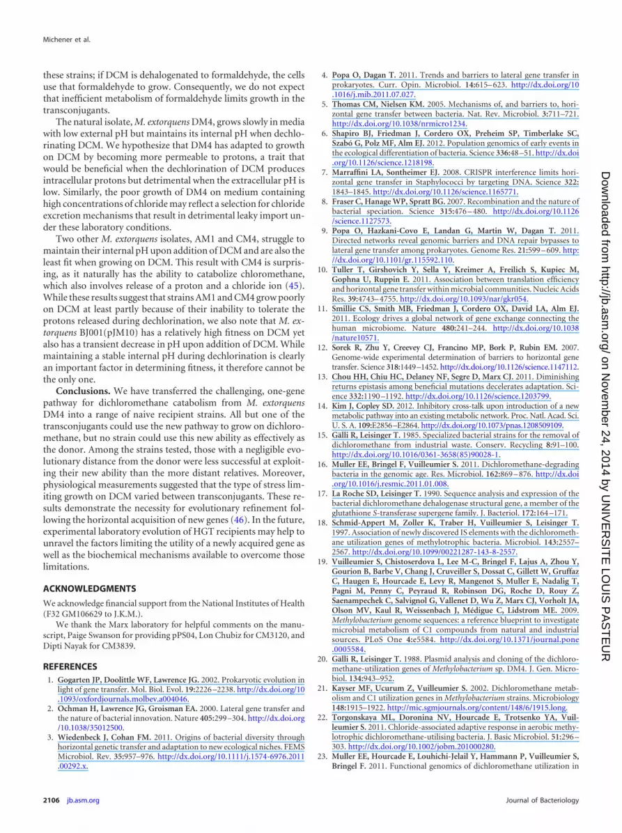
Adding hydrochloric acid to the growth medium does not ex-actly reflect the stress associated with intracellular production ofhydrochloric acid upon DCM dehalogenation. We thereforesought to measure how the transconjugant strains responded tothe dehalogenation of DCM. We used a fluorescent biosensor tomeasure internal pH on a rapid, approximately 5-s time scale (44).When this biosensor was coexpressed with DcmA, we could mea-sure the change in internal pH upon addition of DCM (Fig. 6; also,see Fig. S6 in the supplemental material). We found only a weakcorrelation between smaller transient decreases in intracellularpH and increased fitness of the strain on DCM (Fig. S7 in thesupplemental material).
DISCUSSION
DCM is a very challenging substrate, as its catabolism imposesmultiple types of stress on the host cell: protons and chloride ionsneed to be extruded from the cell interior; the reactive intermedi-ate of DCM dehalogenation, S-chloromethylglutathione, is muta-genic; and formaldehyde, the product of DCM dehalogenation,must be efficiently transformed to minimize its toxic effects. Thecombination of these stresses may explain why transferring dcmAinto naive Methylobacterium strains allowed only poor growth on
DCM. In contrast to previous work, however, we found that theability to use DcmA to grow on DCM was widespread and thatonly M. extorquens AM1 remained unable to grow on DCM afterdcmA transfer. Most evidently, phylogeny was a poor predictor ofa strain’s ability to exploit the dehalogenase DcmA and grow onDCM under the investigated conditions. M. extorquens strainsAM1 and CM4 have 16S rRNA gene sequences identical to thoseof the natural isolate M. extorquens DM4 but are the least success-ful at growing on DCM. Meanwhile, the organisms most distantlyrelated to M. extorquens DM4, M. nodulans and M. radiotolerans,are among the most successful.
In this study, we were able to set aside many of the factorspredicted to limit productive HGT in nature. We know that thenecessary dcmA gene was introduced into the new host, can stablyreplicate, and is functionally expressed. Despite these facts, all ofthe transconjugants are much less fit that the original donor, M.extorquens DM4, and some strains show little or no growth. Otherfactors clearly limit the growth of these transconjugants on DCM,and we used a series of physiological assays in an attempt to iden-tify these factors.
We expected that transconjugant growth on DCM would belimited by one or more of the following: DcmA expression, toler-ance to intracellular production of HCl, efficient use of formalde-hyde, and the mutagenic effects of the glutathione-conjugant in-termediate. None of the assays that we used precisely replicates theoverall stress involved in growing on DCM. That a single physio-logical parameter was not predictive of fitness on DCM may sim-ply reflect the limitations of these assays. However, we hypothesizethat this inconsistency stems from the multiple stresses imposedby growth on DCM, combined with differing abilities of the recip-ients to cope with these stresses. For example, some strains may bemore sensitive to intracellular chloride production, while othersare limited by the intracellular production of protons. In such ascenario, no single physiological parameter could predict fitnesson DCM.
All of the strains showed a linear relationship between chlorideproduced and the final optical density or total protein of the cul-ture. We conclude that dehalogenation is productive in each of
0.0
0.2
0.4
0.6
0.8
1.0
1.2
0 50 100 150
Rel
ativ
e G
row
th R
ate
External NaCl (mM)
0.0
0.2
0.4
0.6
0.8
1.0
1.2
1.4
5.6 5.8 6.0 6.2 6.4 6.6 6.8
Rel
ativ
e G
row
th R
ate
External pH
DM4 ΔdcmA
AM1
M. nodulansBJ001
CM4
PA1
M. radiotolerans
A
B
FIG 5 Transconjugant Methylobacterium strains containing pJM10 vary intheir sensitivity to high external concentrations of NaCl (A) and to low exter-nal pH (B). Cultures were grown in M-PIPES containing succinate as thecarbon and energy source. Growth rates were measured in 48-well plates usingan automated system. M. nodulans grows as a biofilm at 150 mM NaCl, makinggrowth rates difficult to measure. Error bars show one standard deviation,calculated from three biological replicates. The experiment was repeated threetimes, with similar results.
-0.5
-0.4
-0.3
-0.2
-0.1
0.0
0.1
0.0 0.5 1.0 1.5 2.0 2.5 3.0 3.5 4.0
Cha
nge
in In
tern
al p
H
Time (min)
DM4-2cr+pJM40
AM1+pJM40
M. nodulans+pJM40BJ001+pJM40
CM4+pJM40
PA1+pJM40
M. radiotolerans+pJM40DM4 ΔdcmA+pJM41
FIG 6 Internal pH decreases transiently upon addition of DCM. pJM40 ex-presses both the pH biosensor and DcmA. pJM41 contains only the biosensorand serves as a negative control. Internal pH upon addition of 5 mM DCM wasmeasured in cell suspensions of dcmA-containing transconjugants using a pH-sensitive GFP translationally fused to a pH-insensitive mCherry (see Materialsand Methods; also, see Fig. S5 in the supplemental material) (dead time beforereliable fluorescence measurements, approximately 20 s).
Productive Use of a Horizontally Transferred Gene
June 2014 Volume 196 Number 11 jb.asm.org 2105
on Novem
ber 24, 2014 by UN
IVE
RS
ITE
LOU
IS P
AS
TE
UR
http://jb.asm.org/
Dow
nloaded from
these strains; if DCM is dehalogenated to formaldehyde, the cellsuse that formaldehyde to grow. Consequently, we do not expectthat inefficient metabolism of formaldehyde limits growth in thetransconjugants.
The natural isolate, M. extorquens DM4, grows slowly in mediawith low external pH but maintains its internal pH when dechlo-rinating DCM. We hypothesize that DM4 has adapted to growthon DCM by becoming more permeable to protons, a trait thatwould be beneficial when the dechlorination of DCM producesintracellular protons but detrimental when the extracellular pH islow. Similarly, the poor growth of DM4 on medium containinghigh concentrations of chloride may reflect a selection for chlorideexcretion mechanisms that result in detrimental leaky import un-der these laboratory conditions.
Two other M. extorquens isolates, AM1 and CM4, struggle tomaintain their internal pH upon addition of DCM and are also theleast fit when growing on DCM. This result with CM4 is surpris-ing, as it naturally has the ability to catabolize chloromethane,which also involves release of a proton and a chloride ion (45).While these results suggest that strains AM1 and CM4 grow poorlyon DCM at least partly because of their inability to tolerate theprotons released during dechlorination, we also note that M. ex-torquens BJ001(pJM10) has a relatively high fitness on DCM yetalso has a transient decrease in pH upon addition of DCM. Whilemaintaining a stable internal pH during dechlorination is clearlyan important factor in determining fitness, it therefore cannot bethe only one.
Conclusions. We have transferred the challenging, one-genepathway for dichloromethane catabolism from M. extorquensDM4 into a range of naive recipient strains. All but one of thetransconjugants could use the new pathway to grow on dichloro-methane, but no strain could use this new ability as effectively asthe donor. Among the strains tested, those with a negligible evo-lutionary distance from the donor were less successful at exploit-ing their new ability than the more distant relatives. Moreover,physiological measurements suggested that the type of stress lim-iting growth on DCM varied between transconjugants. These re-sults demonstrate the necessity for evolutionary refinement fol-lowing the horizontal acquisition of new genes (46). In the future,experimental laboratory evolution of HGT recipients may help tounravel the factors limiting the utility of a newly acquired gene aswell as the biochemical mechanisms available to overcome thoselimitations.
ACKNOWLEDGMENTS
We acknowledge financial support from the National Institutes of Health(F32 GM106629 to J.K.M.).
We thank the Marx laboratory for helpful comments on the manu-script, Paige Swanson for providing pPS04, Lon Chubiz for CM3120, andDipti Nayak for CM3839.
REFERENCES1. Gogarten JP, Doolittle WF, Lawrence JG. 2002. Prokaryotic evolution in
light of gene transfer. Mol. Biol. Evol. 19:2226 –2238. http://dx.doi.org/10.1093/oxfordjournals.molbev.a004046.
2. Ochman H, Lawrence JG, Groisman EA. 2000. Lateral gene transfer andthe nature of bacterial innovation. Nature 405:299 –304. http://dx.doi.org/10.1038/35012500.
3. Wiedenbeck J, Cohan FM. 2011. Origins of bacterial diversity throughhorizontal genetic transfer and adaptation to new ecological niches. FEMSMicrobiol. Rev. 35:957–976. http://dx.doi.org/10.1111/j.1574-6976.2011.00292.x.
4. Popa O, Dagan T. 2011. Trends and barriers to lateral gene transfer inprokaryotes. Curr. Opin. Microbiol. 14:615– 623. http://dx.doi.org/10.1016/j.mib.2011.07.027.
5. Thomas CM, Nielsen KM. 2005. Mechanisms of, and barriers to, hori-zontal gene transfer between bacteria. Nat. Rev. Microbiol. 3:711–721.http://dx.doi.org/10.1038/nrmicro1234.
6. Shapiro BJ, Friedman J, Cordero OX, Preheim SP, Timberlake SC,Szabó G, Polz MF, Alm EJ. 2012. Population genomics of early events inthe ecological differentiation of bacteria. Science 336:48 –51. http://dx.doi.org/10.1126/science.1218198.
7. Marraffini LA, Sontheimer EJ. 2008. CRISPR interference limits hori-zontal gene transfer in Staphylococci by targeting DNA. Science 322:1843–1845. http://dx.doi.org/10.1126/science.1165771.
8. Fraser C, Hanage WP, Spratt BG. 2007. Recombination and the nature ofbacterial speciation. Science 315:476 – 480. http://dx.doi.org/10.1126/science.1127573.
9. Popa O, Hazkani-Covo E, Landan G, Martin W, Dagan T. 2011.Directed networks reveal genomic barriers and DNA repair bypasses tolateral gene transfer among prokaryotes. Genome Res. 21:599 – 609. http://dx.doi.org/10.1101/gr.115592.110.
10. Tuller T, Girshovich Y, Sella Y, Kreimer A, Freilich S, Kupiec M,Gophna U, Ruppin E. 2011. Association between translation efficiencyand horizontal gene transfer within microbial communities. Nucleic AcidsRes. 39:4743– 4755. http://dx.doi.org/10.1093/nar/gkr054.
11. Smillie CS, Smith MB, Friedman J, Cordero OX, David LA, Alm EJ.2011. Ecology drives a global network of gene exchange connecting thehuman microbiome. Nature 480:241–244. http://dx.doi.org/10.1038/nature10571.
12. Sorek R, Zhu Y, Creevey CJ, Francino MP, Bork P, Rubin EM. 2007.Genome-wide experimental determination of barriers to horizontal genetransfer. Science 318:1449–1452. http://dx.doi.org/10.1126/science.1147112.
13. Chou HH, Chiu HC, Delaney NF, Segre D, Marx CJ. 2011. Diminishingreturns epistasis among beneficial mutations decelerates adaptation. Sci-ence 332:1190 –1192. http://dx.doi.org/10.1126/science.1203799.
14. Kim J, Copley SD. 2012. Inhibitory cross-talk upon introduction of a newmetabolic pathway into an existing metabolic network. Proc. Natl. Acad. Sci.U. S. A. 109:E2856–E2864. http://dx.doi.org/10.1073/pnas.1208509109.
15. Gälli R, Leisinger T. 1985. Specialized bacterial strains for the removal ofdichloromethane from industrial waste. Conserv. Recycling 8:91–100.http://dx.doi.org/10.1016/0361-3658(85)90028-1.
16. Muller EE, Bringel F, Vuilleumier S. 2011. Dichloromethane-degradingbacteria in the genomic age. Res. Microbiol. 162:869 – 876. http://dx.doi.org/10.1016/j.resmic.2011.01.008.
17. La Roche SD, Leisinger T. 1990. Sequence analysis and expression of thebacterial dichloromethane dehalogenase structural gene, a member of theglutathione S-transferase supergene family. J. Bacteriol. 172:164 –171.
18. Schmid-Appert M, Zoller K, Traber H, Vuilleumier S, Leisinger T.1997. Association of newly discovered IS elements with the dichlorometh-ane utilization genes of methylotrophic bacteria. Microbiol. 143:2557–2567. http://dx.doi.org/10.1099/00221287-143-8-2557.
19. Vuilleumier S, Chistoserdova L, Lee M-C, Bringel F, Lajus A, Zhou Y,Gourion B, Barbe V, Chang J, Cruveiller S, Dossat C, Gillett W, GruffazC, Haugen E, Hourcade E, Levy R, Mangenot S, Muller E, Nadalig T,Pagni M, Penny C, Peyraud R, Robinson DG, Roche D, Rouy Z,Saenampechek C, Salvignol G, Vallenet D, Wu Z, Marx CJ, Vorholt JA,Olson MV, Kaul R, Weissenbach J, Médigue C, Lidstrom ME. 2009.Methylobacterium genome sequences: a reference blueprint to investigatemicrobial metabolism of C1 compounds from natural and industrialsources. PLoS One 4:e5584. http://dx.doi.org/10.1371/journal.pone.0005584.
20. Gälli R, Leisinger T. 1988. Plasmid analysis and cloning of the dichloro-methane-utilization genes of Methylobacterium sp. DM4. J. Gen. Micro-biol. 134:943–952.
21. Kayser MF, Ucurum Z, Vuilleumier S. 2002. Dichloromethane metab-olism and C1 utilization genes in Methylobacterium strains. Microbiology148:1915–1922. http://mic.sgmjournals.org/content/148/6/1915.long.
22. Torgonskaya ML, Doronina NV, Hourcade E, Trotsenko YA, Vuil-leumier S. 2011. Chloride-associated adaptive response in aerobic methy-lotrophic dichloromethane-utilising bacteria. J. Basic Microbiol. 51:296 –303. http://dx.doi.org/10.1002/jobm.201000280.
23. Muller EE, Hourcade E, Louhichi-Jelail Y, Hammann P, Vuilleumier S,Bringel F. 2011. Functional genomics of dichloromethane utilization in
Michener et al.
2106 jb.asm.org Journal of Bacteriology
on Novem
ber 24, 2014 by UN
IVE
RS
ITE
LOU
IS P
AS
TE
UR
http://jb.asm.org/
Dow
nloaded from

Methylobacterium extorquens DM4. Environ. Microbiol. 13:2518 –2535.http://dx.doi.org/10.1111/j.1462-2920.2011.02524.x.
24. Evans GJ, Ferguson GP, Booth IR, Vuilleumier S. 2000. Growth inhi-bition of Escherichia coli by dichloromethane in cells expressing dichloro-methane dehalogenase/glutathione S-transferase. Microbiology 146:2967–2975. http://mic.sgmjournals.org/content/146/11/2967.long.
25. Kayser MF, Vuilleumier S. 2001. Dehalogenation of dichloromethane bydichloromethane dehalogenase/glutathione S-transferase leads to forma-tion of DNA adducts. J. Bacteriol. 183:5209 –5212. http://dx.doi.org/10.1128/JB.183.17.5209-5212.2001.
26. Kayser MF, Stumpp MT, Vuilleumier S. 2000. DNA polymerase I isessential for growth of Methylobacterium dichloromethanicum DM4 withdichloromethane. J. Bacteriol. 182:5433–5439. http://dx.doi.org/10.1128/JB.182.19.5433-5439.2000.
27. Gisi D, Leisinger T, Vuilleumier S. 1999. Enzyme-mediated dichloro-methane toxicity and mutagenicity of bacterial and mammalian dichloro-methane-active glutathione S-transferases. Arch. Toxicol. 73:71–79. http://dx.doi.org/10.1007/s002040050589.
28. Vorholt JA, Marx CJ, Lidstrom ME, Thauer RK. 2000. Novel formal-dehyde-activating enzyme in Methylobacterium extorquens AM1 requiredfor growth on methanol. J. Bacteriol. 182:6645– 6650. http://dx.doi.org/10.1128/JB.182.23.6645-6650.2000.
29. Delaney NF, Kaczmarek ME, Ward LM, Swanson PK, Lee MC, MarxCJ. 2013. Development of an optimized medium, strain and high-throughput culturing methods for Methylobacterium extorquens. PLoSOne 8:e62957. http://dx.doi.org/10.1371/journal.pone.0062957.
30. Marx CJ, Lidstrom ME. 2002. Broad-host-range cre-lox system for anti-biotic marker recycling in gram-negative bacteria. Biotechniques 33:1062–1067. http://www.biotechniques.com/multimedia/archive/00010/02335rr01_10092a.pdf.
31. Sankaranarayanan S, De Angelis D, Rothman JE, Ryan TA. 2000. The useof pHluorins for optical measurements of presynaptic activity. Biophys. J.79:2199–2208. http://dx.doi.org/10.1016/S0006-3495(00)76468-X.
32. Lee MC, Chou HH, Marx CJ. 2009. Asymmetric, bimodal trade-offs duringadaptation of Methylobacterium to distinct growth substrates. Evolution63:2816 –2830. http://dx.doi.org/10.1111/j.1558-5646.2009.00757.x.
33. Marx CJ. 2008. Development of a broad-host-range sacB-based vector forunmarked allelic exchange. BMC Res. Notes 1:1. http://dx.doi.org/10.1186/1756-0500-1-1.
34. Fulton GL, Nunn DN, Lidstrom ME. 1984. Molecular cloning of a malylcoenzyme A lyase gene from Pseudomonas sp. strain AM1, a facultativemethylotroph. J. Bacteriol. 160:718 –723.
35. Jörg G, Bertau M. 2004. Thiol-tolerant assay for quantitative colorimetricdetermination of chloride released from whole-cell biodehalogenations.Anal. Biochem. 328:22–28. http://dx.doi.org/10.1016/j.ab.2004.01.027.
36. Vuilleumier S, Leisinger T. 1996. Protein engineering studies of dichlo-romethane dehalogenase/glutathione S-transferase from Methylophilus
sp. strain DM11. Ser12 but not Tyr6 is required for enzyme activity. Eur. J.Biochem. 239:410 – 417.
37. Cole JR, Chai B, Farris RJ, Wang Q, Kulam SA, McGarrell DM, GarrityGM, Tiedje JM. 2005. The Ribosomal Database Project (RDP-II): se-quences and tools for high-throughput rRNA analysis. Nucleic Acids Res.33:D294 –D296. http://dx.doi.org/10.1093/nar/gki038.
38. Segata N, Bornigen D, Morgan XC, Huttenhower C. 2013. PhyloPhlAn is anew method for improved phylogenetic and taxonomic placement of mi-crobes. Nat. Commun. 4:2304. http://dx.doi.org/10.1038/ncomms3304.
39. Knief C, Frances L, Vorholt JA. 2010. Competitiveness of diverse Methy-lobacterium strains in the phyllosphere of Arabidopsis thaliana and iden-tification of representative models, including M. extorquens PA1. Microb.Ecol. 60:440 – 452. http://dx.doi.org/10.1007/s00248-010-9725-3.
40. Marx CJ, Bringel F, Chistoserdova L, Moulin L, Farhan Ul Haque M,Fleischman DE, Gruffaz C, Jourand P, Knief C, Lee MC, Muller EE,Nadalig T, Peyraud R, Roselli S, Russ L, Goodwin LA, Ivanova N,Kyrpides N, Lajus A, Land ML, Medigue C, Mikhailova N, Nolan M,Woyke T, Stolyar S, Vorholt JA, Vuilleumier S. 2012. Complete genomesequences of six strains of the genus Methylobacterium. J. Bacteriol. 194:4746 – 4748. http://dx.doi.org/10.1128/JB.01009-12.
41. Van Aken B, Peres CM, Doty SL, Yoon JM, Schnoor JL. 2004. Methy-lobacterium populi sp. nov., a novel aerobic, pink-pigmented, facultativelymethylotrophic, methane-utilizing bacterium isolated from poplar trees(Populus deltoides�nigra DN34). Int. J. Syst. Evol. Microbiol. 54:1191–1196. http://dx.doi.org/10.1099/ijs.0.02796-0.
42. Sy A, Giraud E, Jourand P, Garcia N, Willems A, de Lajudie P, Prin Y,Neyra M, Gillis M, Boivin-Masson C, Dreyfus B. 2001. MethylotrophicMethylobacterium bacteria nodulate and fix nitrogen in symbiosis withlegumes. J. Bacteriol. 183:214 –220. http://dx.doi.org/10.1128/JB.183.1.214-220.2001.
43. Sanders SW, Maxcy RB. 1979. Patterns of cell division, DNA base com-positions, and fine structures of some radiation-resistant vegetative bac-teria found in food. Appl. Environ. Microbiol. 37:159 –168.
44. Valkonen M, Mojzita D, Penttilä M, Bencina M. 2013. Noninvasivehigh-throughput single-cell analysis of the intracellular pH of Saccharo-myces cerevisiae by ratiometric flow cytometry. Appl. Environ. Microbiol.79:7179 –7187. http://dx.doi.org/10.1128/AEM.02515-13.
45. McDonald IR, Doronina NV, Trotsenko YA, McAnulla C, Murrell JC.2001. Hyphomicrobium chloromethanicum sp. nov. and Methylobacteriumchloromethanicum sp. nov., chloromethane-utilizing bacteria isolatedfrom a polluted environment. Int. J. Syst. Evol. Microbiol. 51:119 –122.http://dx.doi.org/10.1099/00207713-51-1-119.
46. Quandt EM, Deatherage DE, Ellington AD, Georgiou G, Barrick JE.2014. Recursive genomewide recombination and sequencing reveals a keyrefinement step in the evolution of a metabolic innovation in Escherichiacoli. Proc. Natl. Acad. Sci. U. S. A. 111:2217–2222. http://dx.doi.org/10.1073/pnas.1314561111.
Productive Use of a Horizontally Transferred Gene
June 2014 Volume 196 Number 11 jb.asm.org 2107
on Novem
ber 24, 2014 by UN
IVE
RS
ITE
LOU
IS P
AS
TE
UR
http://jb.asm.org/
Dow
nloaded from

1
SI Figure 1: A phylogenetic tree based on 400 conserved proteins is consistent with the
16S rRNA tree. The phylogenetic tree of the seven Methylobacterium strains was built
using PhyloPhlAn (38). The conserved proteins were selected based on ubiquity among
the entire tree of life. Protein homologs were identified using a threshold of 90%
sequence similarity. The most informative residues were extracted from each protein,
concatenated, and used as the input for the phylogenetic tree.

2
SI Figure 2: Non-planktonic strains grown on DCM show a linear relationship between total protein produced and chloride released. DM4 ΔdcmA +pJM10 (planktonic control), CM4+pJM10, BJ001+pJM10, and M. radiotolerans+pJM10 were grown in sealed flasks containing 10 mL of M-PIPES + 5 mM DCM as the sole carbon and energy source. After three days’ growth, chloride concentrations were measured in the supernatant. Cells were then pelleted by centrifugation, lysed using a bead beater, and the total protein in the supernatant was measured with a Bradford assay.

3
SI Figure 3: (A) Transconjugant yield on DCM in pure culture is generally predictive of competitive fitness. The outliers are BJ001, which shows lower fitness than would be predicted based on yield, and M. radiotolerans, which shows higher fitness than predicted. (B) Phylogenetic distance is not an effective predictor of competitive fitness. Error bars show one standard deviation, calculated from three biological replicates.

4
SI Figure 4: (A) All pJM10 transconjugants show DcmA activity in cell suspensions of cultures grown with 3.5 mM succinate and 5 mM DCM. (B) In vitro DcmA activity does not predict in vivo competitive fitness. The outliers, AM1 and DM4 ΔdcmA, have the lowest and highest specific activities, respectively. However, strains with intermediate fitness cannot be resolved based on DcmA activity.

5
SI Figure 5: Neither growth rate in media containing high external chloride concentrations nor growth rate in media buffered to low pH are predictive of growth with DCM. (A) The ratio of growth rates when grown with 100 mM NaCl compared to unmodified M-PIPES+succinate does not predict the competitive fitness on DCM. (B) The ratio of growth rates when grown in M-PIPES+succinate at pH 5.9 compared to pH 6.7 is similarly ineffective at predicting competitive fitness on DCM.

6
SI Figure 6: Calibration curve for the intracellular pH biosensor. A pH-insensitive red fluorescent protein is translationally fused to a pH-sensitive green fluorescent protein. The ratio of green to red fluorescence is indicative of the intracellular pH. For calibration, cells expressing the biosensor construct were incubated in buffer of the indicated pH containing valinomycin and nigericin prior to measurement by flow cytometry. Error bars show one standard deviation calculated from three replicates.
7
SI Figure 7: The maximum decrease in intracellular pH on addition of 5 mM is not predictive of competitive fitness on DCM. From each of the curves in Figure 4, the lowest intracellular pH measurement was taken as representative of the strain’s ability to maintain its intracellular pH under a DCM challenge.