phox2b-mediated regulation of alk expression: in vitro identification of a functional relationship...
TRANSCRIPT

Seediscussions,stats,andauthorprofilesforthispublicationat:https://www.researchgate.net/publication/47459972
PHOX2B-MediatedRegulationofALKExpression:InVitroIdentificationofaFunctionalRelationship...
ArticleinPLoSONE·October2010
DOI:10.1371/journal.pone.0013108·Source:PubMed
CITATIONS
18
READS
36
16authors,including:
Someoftheauthorsofthispublicationarealsoworkingontheserelatedprojects:
MechanismsofuvealmelanomametastasisViewproject
DanielaDiPaolo
IRCCSIstitutoG.Gaslini
48PUBLICATIONS728CITATIONS
SEEPROFILE
IreneCaffa
UniversitàdegliStudidiGenova
66PUBLICATIONS369CITATIONS
SEEPROFILE
MicheleFiore
ItalianNationalResearchCouncil
10PUBLICATIONS112CITATIONS
SEEPROFILE
MircoPonzoni
IRCCSIstitutoG.Gaslini
205PUBLICATIONS4,343CITATIONS
SEEPROFILE
AllcontentfollowingthispagewasuploadedbyUlrichPfefferon02December2016.
Theuserhasrequestedenhancementofthedownloadedfile.
PHOX2B-Mediated Regulation of ALK Expression: In VitroIdentification of a Functional Relationship between TwoGenes Involved in NeuroblastomaTiziana Bachetti1, Daniela Di Paolo2, Simona Di Lascio3, Valentina Mirisola4, Chiara Brignole2, Marta
Bellotti4, Irene Caffa2, Chiara Ferraris1, Michele Fiore1, Diego Fornasari3, Roberto Chiarle5, Silvia
Borghini1, Ulrich Pfeffer4, Mirco Ponzoni2, Isabella Ceccherini1*, Patrizia Perri2
1 Laboratory of Molecular Genetics, G. Gaslini Children’s Hospital, Genoa, Italy, 2 Experimental Therapy Unit, Laboratory of Oncology, G. Gaslini Children’s Hospital, Genoa,
Italy, 3 Department of Pharmacology, School of Medicine, Universita degli Studi di Milano and CNR-Institute of Neuroscience, Milan, Italy, 4 Advanced Molecular
Diagnostics, National Cancer Research Institute, Genoa, Italy, 5 Dept. Biomedical Sciences and Human Oncology, University of Turin, Turin, Italy
Abstract
Background: Neuroblastoma (NB) is a severe pediatric tumor originating from neural crest derivatives and accounting for15% of childhood cancer mortality. The heterogeneous and complex genetic etiology has been confirmed with theidentification of mutations in two genes, encoding for the receptor tyrosine kinase Anaplastic Lymphoma Kinase (ALK) andthe transcription factor Paired-like Homeobox 2B (PHOX2B), in a limited proportion of NB patients. Interestingly, these twogenes are overexpressed in the great majority of primary NB samples and cell lines. These observations led us to test thehypothesis of a regulatory or functional relationship between ALK and PHOX2B underlying NB pathogenesis.
Methodology/Principal Findings: Following this possibility, we first confirmed a striking correlation between thetranscription levels of ALK, PHOX2B and its direct target PHOX2A in a panel of NB cell lines. Then, we manipulated theirexpression in NB cell lines by siRNA-mediated knock-down and forced over-expression of each gene under analysis.Surprisingly, PHOX2B- and PHOX2A-directed siRNAs efficiently downregulated each other as well as ALK gene and,consistently, the enhanced expression of PHOX2B in NB cells yielded an increment of ALK protein. We finally demonstratedthat PHOX2B drives ALK gene transcription by directly binding its promoter, which therefore represents a novel PHOX2Btarget.
Conclusions/Significance: These findings provide a compelling explanation of the concurrent involvement of these twogenes in NB pathogenesis and are going to foster a better understanding of molecular interactions at the base of thedisease. Moreover, this work opens new perspectives for NBs refractory to conventional therapies that may benefit from thedesign of novel therapeutic RNAi-based approaches for multiple gene targets.
Citation: Bachetti T, Di Paolo D, Di Lascio S, Mirisola V, Brignole C, et al. (2010) PHOX2B-Mediated Regulation of ALK Expression: In Vitro Identification of aFunctional Relationship between Two Genes Involved in Neuroblastoma. PLoS ONE 5(10): e13108. doi:10.1371/journal.pone.0013108
Editor: Mikhail V. Blagosklonny, Roswell Park Cancer Institute, United States of America
Received July 1, 2010; Accepted September 3, 2010; Published October 1, 2010
Copyright: � 2010 Bachetti et al. This is an open-access article distributed under the terms of the Creative Commons Attribution License, which permitsunrestricted use, distribution, and reproduction in any medium, provided the original author and source are credited.
Funding: T. Bachetti has been supported by Fondazione Pierfranco e Luisa Mariani. D. Di Paolo is a recipient of a Fondazione Italiana Ricerca Cancro (FIRC)fellowship. The financial support of Associazione Italiana Ricerca Cancro (AIRC) (to M.P.), Fondazione Mariani (grant# 08/69 to I. Ceccherini) and of Ministry ofHealth (Progetti Finalizzato 2006 and Strategico 2009 to I. Ceccherini and Finalizzato 2007 to M.P.) are gratefully acknowledged. The funders had no role in studydesign, data collection and analysis, decision to publish, or preparation of the manuscript.
Competing Interests: The authors have declared that no competing interests exist.
* E-mail: [email protected]
Introduction
Novel insights into the molecular pathogenesis of neuroblasto-
ma (NB), a severe pediatric tumor originating from neural crest
cells and accounting for 15% of childhood cancer mortality, have
been gained after the identification of germline as well as
somatically acquired mutations in the genes encoding the
paired-like homeobox 2b (PHOX2B) transcription factor [1,2]
and the Anaplastic Lymphoma Kinase (ALK) tyrosine kinase
receptor [3].
The PHOX2B gene is involved in the specification of the
noradrenergic phenotype during the development and differenti-
ation of neural crest derivatives [4–9]. Missense and frameshift
mutations of this gene were identified in only a few pedigrees of
familial NB [1,2,10,11] and in about 4% of sporadic cases [12],
suggesting genetic heterogeneity of NB [13]. PHOX2B mutations
are often found in association with other neurocristopathies such
as Congenital Central Hypoventilation Syndrome (CCHS) and
Hirschsprung disease (HSCR), likely modifying susceptibility to
NB in the corresponding patients [1,2,10,11,13]. Moreover, the
involvement of PHOX2B and its paralogue PHOX2A in NB
pathogenesis seems to be also mediated by a mechanism of gene
up-regulation [14], with abundance of PHOX2B transcript shown
to be highly prognostic of poorer progression-free and overall
survival [15,16]. Little is known about physiological regulation of
the PHOX2 genes transcription, except that PHOX2B expression
depends on an auto-regulatory mechanisms in NB cells [17] and
regulates transcription of PHOX2A [6]. Other known transcrip-
PLoS ONE | www.plosone.org 1 October 2010 | Volume 5 | Issue 10 | e13108
tional targets of PHOX2B are TH (Tyrosine Hydroxylase) and
DBH (Dopamine-Beta-Hydroxylase), two genes encoding enzymes
involved in the cathecolamine biosynthesis [5,18], TLX-2, a
transcription factor controlling development of enteric innervation
[9], RET, the major gene involved in the complex inheritance of
HSCR [19] and MSX-1, a negatively regulated homeobox gene
[20].
More recently, mutations associated with both hereditary and
sporadic neuroblastoma were discovered in the ALK tyrosine
kinase [3,21], a gene mapping to a region previously found in
linkage with NB [3,22]. The ALK gene was already known to have
a physiological role in neuronal development [23] and to be
involved in the pathogenesis of cancer, especially lymphomas but
also solid tumors of ectodermal, myofibroblastic or neuroblastic
origin [23–25]. About 11–12% of the NB tumors were shown to
carry non-synonymous sequence variations in conserved positions
of the tyrosine kinase domain. Particularly, the most frequent
mutant ALK proteins p.F1174L and p.R1275Q demonstrated
gain-of-function kinase activity [3,21,26–27]. Molecular events
able to promote ALK gene transcription may also have a
pathogenetic role but only 3–4% of NB cases were found to bear
extensive ALK amplification while 17–23% presented lower levels
of ALK gene gain (2#gene copies #4) [3,21,26–30], therefore most
of the ALK over-expression in NB still remains unexplained.
Functional assays showed induction of a constitutive kinase
activity in overexpressed and/or hyperphosphorylated ALK
proteins, either mutated or wild type. Accordingly, the knock-
down of ALK expression in cell model systems led to a marked
decrease of cell proliferation clearly indicating ALK as a critical
player in NB development [3,21,26–27]. Notably, ALK mutations
and amplifications as well as gene over-expression were found to
significantly correlate with other unfavorable features of poor
outcome in advanced/metastatic compared with localized tumors
[3,21,26–29]. Details on regulatory molecular mechanisms acting
under physiological conditions or sustaining over-expression of the
ALK gene in NB are currently unknown.
Therefore, i) PHOX2B and ALK mutations are involved either in
the initiation or the progression of NB and ii) wild-type as well as
mutated transcripts of both these genes are reported to be
overexpressed in the vast majority of the NB cell lines and tumor
samples analyzed. This suggests their possible concurrent role in
the development and/or maintenance of the sympathetic nervous
system, thus prompting us to test the hypothesis of a cross-talk
between PHOX2B and ALK.
Here, a number of compelling evidences are reported,
demonstrating extensive co-regulated over-expression of PHOX2A,
PHOX2B and ALK in NB cell lines and a novel PHOX2B-
mediated effect on ALK transcriptional induction, sustained by
PHOX2B binding the ALK promoter region, thus establishing ALK
as a novel PHOX2B target gene.
Results
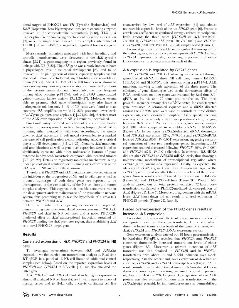
Correlated expression of ALK, PHOX2B and PHOX2A in NBcells
To investigate correlations between ALK and PHOX2B
expression, we first carried out transcription analysis by Real-time
RT-qPCR in a panel of 13 NB cell lines and additional control
samples (see below). Based on the reported expression levels of
PHOX2B and PHOX2A in NB cells [14], we also analyzed the
latter gene.
ALK, PHOX2B and PHOX2A resulted to be highly expressed in
almost all analyzed NB cell lines (Figure 1) with respect to a pool of
normal tissues and to HeLa cells, a cervix carcinoma cell line
characterized by low level of ALK expression [31] and almost
undetectable expression levels of the two PHOX2 genes [6]. Pearson’s
correlation coefficients (r) confirmed strongly related transcriptional
levels among the three genes [PHOX2B vs. ALK (r = 0.941;
P,0.0001), PHOX2A vs. ALK (r = 0.938; P,0.0001) and PHOX2A
vs. PHOX2B (r = 0.983; P,0.0001)] in all samples tested (Figure 1).
To investigate on the possible inter-regulated transcription of
these three genes, we considered to manipulate ALK, PHOX2B and
PHOX2A expression in vitro, performing experiments of either
knock-down or forced-expression for each of them.
ALK expression is regulated by PHOX2 genesALK, PHOX2B and PHOX2A silencing was achieved through
gene-directed siRNA in three NB cell lines, namely IMR-32,
HTLA-230 and SH-SY5Y, this latter carrying a p.F1174L ALK
mutation, showing a high expression of the three genes. The
efficiency of gene silencing as well as the downstream effects of
RNA interference on other genes was evaluated by Real-time RT-
qPCR at 24, 48 and 72 hours post-transfection. The most
powerful sequence among three siRNAs tested for each targeted
gene, was used. A scrambled sequence and a siRNA directed
against the GAPDH gene were used as controls in three distinct
experiments, each performed in duplicate. Gene specific silencing
was very effective already at 48 hours post-transfection, ranging
between 87% and 91% for the three genes under analysis
(P,0.001 when compared to scrambled siRNA) in SH-SY5Y
(Figure 2A). In particular, PHOX2B-directed siRNA downregu-
lated PHOX2A expression (82%, P,0.001) and PHOX2A-siRNA
lowered PHOX2B (80%, P,0.001) expression, suggesting recipro-
cal regulation of these two paralogous genes. Interestingly, ALK
expression resulted decreased following PHOX2B (80%, P,0.001)
and PHOX2A (67%, P,0.01) silencing. In contrast, ALK-siRNA
did not alter PHOX2A or PHOX2B mRNA levels, thus showing a
unidirectional mechanism of transcriptional regulation where
PHOX2 genes control ALK expression. Finally, as expected, the
silencing of TLX2, a gene known as a downstream target of the
PHOX2 genes [9], did not affect the expression level of the studied
genes. Similar results were obtained by transfection in IMR-32
(Figure 2B) and HTLA-230 cell lines (Figure 2C). Western blot
analysis carried out on total proteins extracted 72 hours post-
transfection confirmed a PHOX2-mediated down-regulation of
ALK (Figure 2D, lane 5). Moreover, in agreement with the mRNA
levels, ALK knock-down did not result in altered expression of
PHOX2B protein (Figure 2D, lane 3).
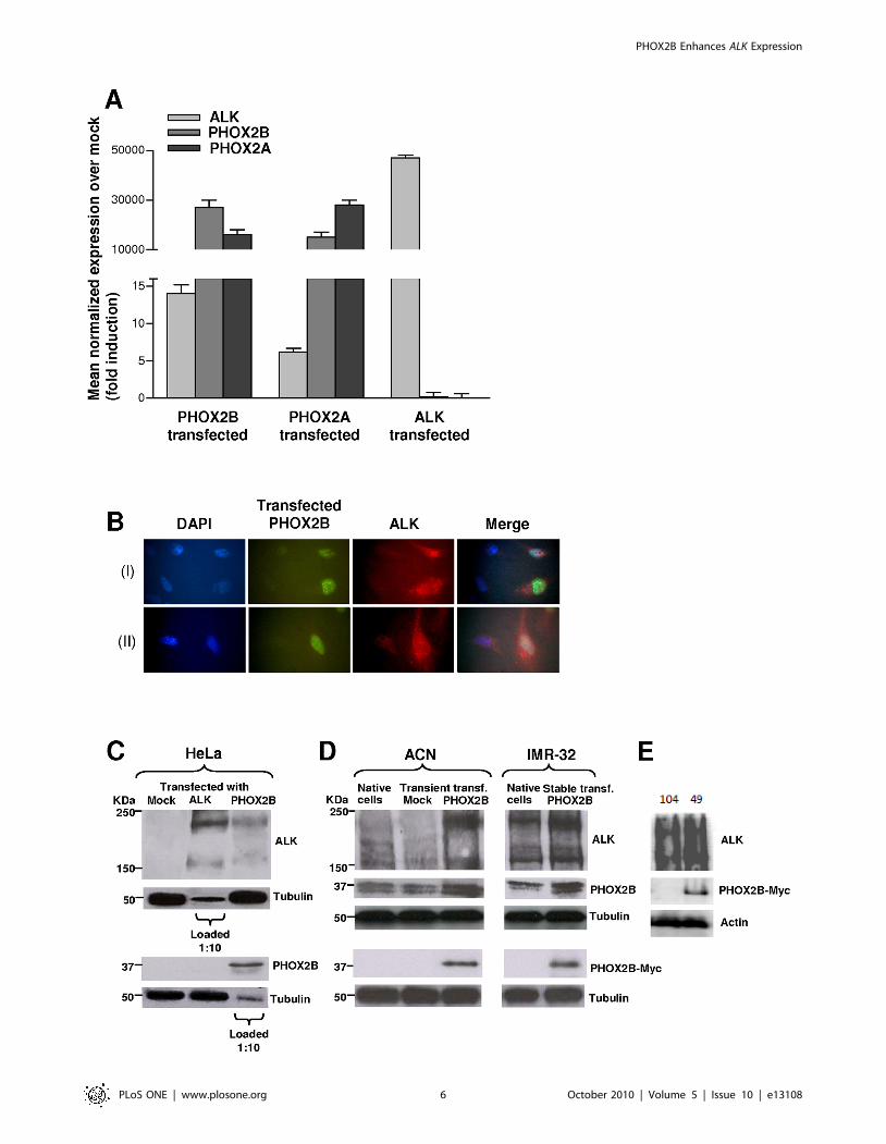
Forced over-expression of the PHOX2 genes results inincreased ALK expression
To evaluate downstream effects of forced over-expression of
each protein over the others, we transfected HeLa cells, which
show the lowest transcription levels of the genes of interest, with
ALK, PHOX2A and PHOX2B cDNAs expressing vectors.
Gene expression analysis carried out 48 hours post-transfection
by Real-time RT-qPCR revealed that PHOX2A and PHOX2B
constructs dramatically increased transcription levels of either
genes (Figure 3A). Moreover, a relevant increment of ALK
transcript was also obtained in PHOX2B and in PHOX2A
transfectants (with about 14 and 6 fold induction over mock,
respectively). On the other hand, over-expression of ALK had no
effect on PHOX2B and PHOX2A transcript levels (Figure 3A), a
result consistent with data obtained using siRNA-mediated knock-
down and once again indicating an unidirectional expression
regulation of ALK by PHOX2 genes. Up-regulation of the ALK
protein was also evaluated 48 hours after transfection with the
PHOX2B-Myc plasmid, by immunofluorescence in permeabilized
PHOX2B Enhances ALK Expression
PLoS ONE | www.plosone.org 2 October 2010 | Volume 5 | Issue 10 | e13108
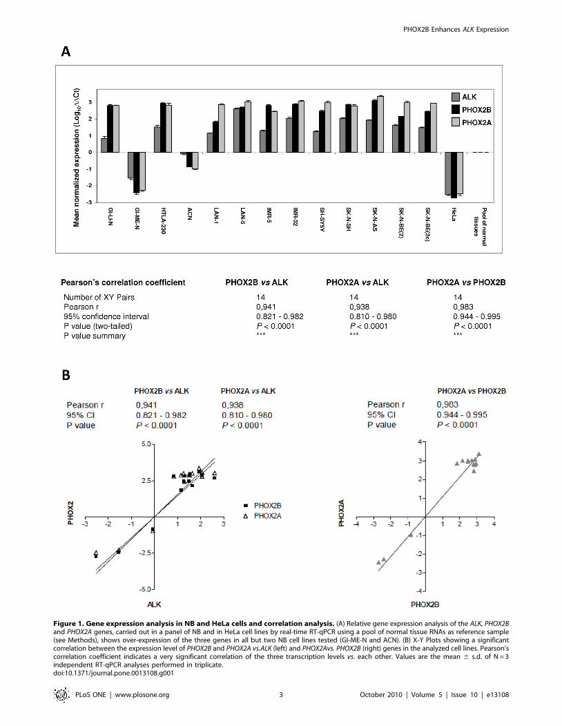
Figure 1. Gene expression analysis in NB and HeLa cells and correlation analysis. (A) Relative gene expression analysis of the ALK, PHOX2Band PHOX2A genes, carried out in a panel of NB and in HeLa cell lines by real-time RT-qPCR using a pool of normal tissue RNAs as reference sample(see Methods), shows over-expression of the three genes in all but two NB cell lines tested (GI-ME-N and ACN). (B) X-Y Plots showing a significantcorrelation between the expression level of PHOX2B and PHOX2A vs.ALK (left) and PHOX2Avs. PHOX2B (right) genes in the analyzed cell lines. Pearson’scorrelation coefficient indicates a very significant correlation of the three transcription levels vs. each other. Values are the mean 6 s.d. of N = 3independent RT-qPCR analyses performed in triplicate.doi:10.1371/journal.pone.0013108.g001
PHOX2B Enhances ALK Expression
PLoS ONE | www.plosone.org 3 October 2010 | Volume 5 | Issue 10 | e13108
Figure 2. siRNA-mediated silencing of ALK, PHOX2B and PHOX2A in NB cells. Effects on the transcription level of the ALK (left side graphs),PHOX2B (middle graphs) and PHOX2A (right side graphs) genes after knock-down of the same genes in SHSY-5Y (A), IMR-32 (B) and HTLA-230 (C) cells.Gene-specific knock-down, evaluated 48 hours post-transfection by real-time RT-qPCR analysis, is very effective but also PHOX2-directed siRNAs are
PHOX2B Enhances ALK Expression
PLoS ONE | www.plosone.org 4 October 2010 | Volume 5 | Issue 10 | e13108
HeLa cells. As shown in Figure 3B, the intensity of ALK protein
staining (red fluorescence) was much higher in cells expressing
PHOX2B (green fluorescence) than in untransfected cells, which
were positive only for the blue nuclear staining. These findings
have been confirmed by Western blot analysis on total protein
extracts at 72 hours post-transfection (Figure 3C) showing
PHOX2B-mediated ALK up regulation not coupled to reciprocal
ALK-mediated PHOX2B increase.
Finally, we transfected the NB cell line ACN, which shows very low
expression levels of the three genes, with the PHOX2B-Myc plasmid.
As already observed in HeLa cells, Western blot analysis revealed an
increment of ALK protein at 72 hours post-transfection (Figure 3D,
left). Similar results were also confirmed by evaluating ALK and
PHOX2B protein amounts in a clone of IMR-32 stably expressing
the same construct (Figure 3D, right), as well as by comparing two
clones deriving from the same IMR-32 culture, expressing (# 49) and
not expressing (# 104) PHOX2B/Myc (Figure 3E).
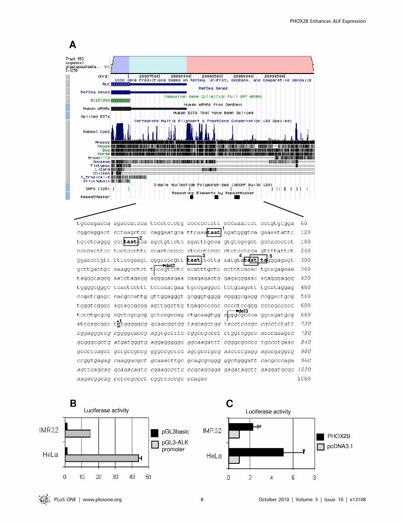
In silico prediction of the ALK regulatory region andputative binding elements
In the light of the straightforward involvement of PHOX2B in
NB development, and opposite to PHOX2A whose mutations
never resulted in association with any neural crest derived tumor
[14], we have focused on the former gene to deepen into
molecular details of its regulative role on the ALK gene
transcription.
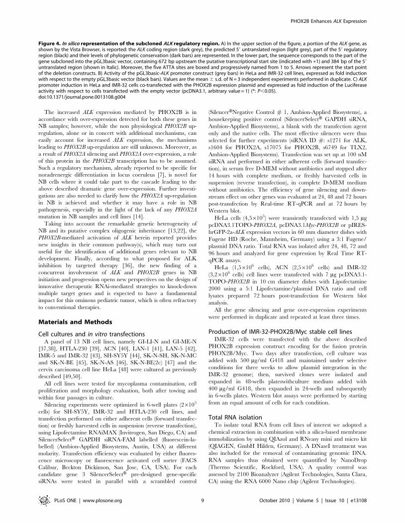
In silico analysis of about 3 kb sequence lying upstream of the
ALK coding region was performed to search for conserved sequences,
which were then identified in the portion encompassing from 21 kb
to the ALK predicted transcriptional start site (GenomeVista,
http://pipeline.lbl.gov/cgi-bin/GenomeVista). The Genomatix
Matinspector software (http://www.genomatix.de), used to identify
putative binding sites for transcription factors in this region, showed
five sequences recognized by homeoproteins (ATTA boxes) we have
named ATTA 1, 2, 3, 4 and 5 (Figure 4A). To investigate whether
PHOX2B might take part in the transcriptional regulation of this
region, a sequence spanning from about 2672 bp up to +384 bp
was amplified and inserted into the pGL3basic vector, upstream the
Luciferase reporter gene. Basic activities of this promoter construct
assessed with respect to the empty vector 24 hours after transfection
in two different cell lines, the HeLa cells and IMR-32 cells, are
reported in Figure 4B.
Once assessed a transcriptional activity driven by this portion of
ALK promoter in both cell lines, the ability of PHOX2B to induce the
ALK gene expression was then investigated. As shown in Figure 4C,
co-transfection of an expression construct encoding for the
PHOX2B-Myc fusion protein with a reporter plasmid containing
the Luciferase gene under the transcriptional control of the ALK
promoter showed, both in Hela and in IMR-32 cell lines, that
PHOX2B is able to activate transcription of the ALK gene by
regulating the portion under analysis of its promoter. Therefore, as in
IMR-32 cells the PHOX2B-mediated trans-activation of the reporter
gene was less relevant than in HeLa cells, likely due to the already
high expression of endogenous PHOX2B (see Figure 3D), the further
co-transfection experiments were carried out only in Hela cells.
Interaction between PHOX2B and the ALK promoterTo deepen into PHOX2B-mediated trans-activation of the ALK
promoter, direct binding between the ATTA 1, 2, 3 and 4/5
sequences and PHOX2B was investigated in vitro through
electrophoretic mobility shift assays (EMSA), by incubating probes
containing each of the ATTA sites with nuclear extracts from
IMR-32 cells, expressing high levels of endogenous PHOX2B, or
alternatively with the in vitro translated PHOX2B protein.
As shown in Figure 5A, by incubating the IMR-32 extracts with
probes including each of the ATTA 1, 2, 3 or 4/5 boxes we
observed in any case formation of specific complexes (lanes 2),
which disappeared in the presence of the corresponding unlabelled
oligonucleotide (lanes 3). However, differently from what observed
by probing the ATTA 1 and ATTA 2 sequences, the presence of
PHOX2B inside the two complexes detected with ATTA 3 and
ATTA 4/5 was confirmed by supershifted bands obtained
following incubation of nuclear extracts with a PHOX2B specific
antibody (lanes 4). Moreover, the fastest of two of the above
specific complexes was obtained also by incubating the ATTA 3
and ATTA 4/5 probes with the in vitro translated PHOX2B-Myc
fusion protein (lanes 5). While its specificity was assessed by the
band disappearance in the presence of an excess of unlabelled
oligonucleotide (lanes 6), presence of PHOX2B was confirmed by
the supershifted band observed following incubation with a c-Myc
antibody (lanes 7).
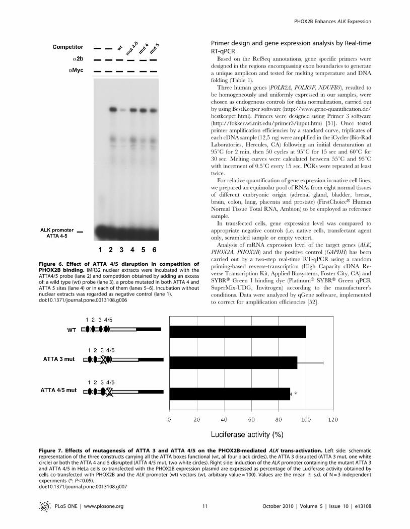
Specificity of the ATTA4/5 sites was confirmed by performing
EMSA following incubation of the probe carrying the ATTA4/5
boxes and competition assays with probes containing one or both
the mutant ATTA sites; in particular, while the unlabelled ATTA
4/5 probe could efficiently compete for the PHOX2B binding,
oligonucleotides carrying mutations in the ATTA box did not
abolish DNA interaction with PHOX2B (Figure 6).
Finally, PHOX2B interaction with the ALK promoter was
further confirmed by performing a ChIP assay. In particular, the
chromatin extract from PHOX2B-expressing IMR-32 cells was
incubated with the PHOX2B specific antibody, immunoprecipi-
tated and the product obtained was amplified with primers
surrounding the two ATTA 3 and ATTA 4/5 sites. Negative and
positive controls of the reaction were obtained by incubating
chromatin extracts with normal IgY and with hyperacetylated
histone H4 specific antibody, respectively.
As shown in Figure 5B, presence of products amplified from
immunoprecipitation obtained by using the anti-PHOX2B and
anti-acetylated histone H4 antibodies suggests that in IMR-32 cells
this is a transcriptionally active promoter region which, among
others, can bind the PHOX2B transcription factor. Moreover, the
absence of amplification of chromatin immunoprecipitated with
pre-immune IgY confirmed the specificity of the assay.
Effect of the ATTA 3 and ATTA 4/5 regions on PHOX2B-induced ALK trans-activation
To verify whether only one or both the ATTA sites here
identified to bind PHOX2B were also functionally active, thus
mediating the PHOX2B trans-activation of this region of the ALK
promoter, we co-transfected HeLa cells with each reporter
construct of the ALK promoter, carrying mutant versions of
ATTA 3, ATTA 4/5 or both elements, and the PHOX2B
expression construct. As shown in Figure 7, mutagenesis of ATTA
3 could not significantly impair the ability of PHOX2B to activate
ALK transcription, while disruption of the ATTA 4/5 induced a
low but significant reduction of PHOX2B-mediated ALK trans-
activation. As disruption of all the three ATTA boxes did not
produce a more pronounced effect (not shown), we gathered that
able to downregulate ALK at a similar extent (**: P,0.01; ***: P,0.001). Values are the mean 6 s.d. of N = 3 independent experiments performed induplicate. (D) Gene silencing was confirmed at 72 hours post-transfection by Western blot.doi:10.1371/journal.pone.0013108.g002
PHOX2B Enhances ALK Expression
PLoS ONE | www.plosone.org 5 October 2010 | Volume 5 | Issue 10 | e13108
PHOX2B Enhances ALK Expression
PLoS ONE | www.plosone.org 6 October 2010 | Volume 5 | Issue 10 | e13108
the only functional homeoprotein binding sequence responsible for
PHOX2B-mediated trans-activation of the ALK promoter was the
ATTA 4/5 box.
Finally, to investigate a possible indirect role of PHOX2B over
different region of the ALK promoter, two deleted reporter
constructs, lacking segments that include the ATTA sites, and
characterized by shorter fragments of the ALK promoter
(Figure 8A), were compared to the full length ALK promoter
construct (wt) for their ability to mediate reporter expression in the
presence of PHOX2B in HeLa cells. As shown in Figure 8B,
removal of all the ATTA boxes markedly reduced the basal
promoter activity, which was completely abolished only in the
most proximal region. In addition, consistent with data obtained
from mutagenesis of the ATTA boxes, co-transfection of
PHOX2B-Myc expression plasmid and the ALK(2351 bp) promot-
er construct showed a lower trans-activation than that obtained
using the entire ALK(2672 bp) promoter construct (Figure 8C).
On the other hand, the activity of the shortest ALK(231) fragment
was not modulated by PHOX2B, suggesting that the residual
effect of PHOX2B, likely indirect, is mediated by the 2351 bp to
231 bp region of the ALK promoter.
Discussion
Germline as well as somatically acquired mutations in the
PHOX2B and ALK genes have been detected in both sporadic and
familial NB cases [1–3,10]. These mutations show an autosomal
dominant inheritance with reduced penetrance in NB families
and, while ALK mutations have clearly been demonstrated to act as
‘‘gain-of-function’’ mutations [3,25–27], the molecular mecha-
nism(s) underlying the effects of NB associated PHOX2B mutations
is still to be determined. In fact, dominant negative effect, haplo-
insufficiency or, at opposite, up-regulation of target genes may be
considered equally possible consequences of PHOX2B-pathway
alterations [2,10,12,32–34]. Furthermore, wild-type as well as
mutated transcripts of ALK and PHOX2B, and also the wild type
transcript of its paralogue PHOX2A, have been found to be
overexpressed in the vast majority of NB cell lines and tumor
samples analyzed so far [3,2,14,21,27,33], thus providing strong
indications that up-regulation of these genes is involved in NB
molecular pathogenesis. Accordingly, a very recent study has
demonstrated that the disease is sustained by MycN-driven
expansion of Phox2b expressing neuronal progenitors, in a
transgenic mouse model of NB [35].
Activation of the ALK proto-oncogene is required for tumor
transformation, through the induction of several downstream
pathways which control key cellular processes such as cell-cycle
progression, survival, cell migration and cell shaping [24]. As for
other proto-oncogenes encoding receptor tyrosine kinases, ALK
activation is physiologically achieved upon ligand and co-receptor
binding but might be also triggered through different mechanisms
such as DNA mutations, gene amplification or chromosomal
translocations, as well as post-translational modifications. Remark-
ably, only a proportion of NB tumor samples with high level of
ALK expression carries gene amplification and/or mutations
[3,21,26–30], therefore, most of the ALK over-expression and its
functional effects are uncharacterized and the underlying
molecular mechanisms still undisclosed.
Herein, we report for the first time evidences of a direct role of
PHOX2B in the transcriptional regulation of ALK, thus pointing
not only at a combined role of these two genes in NB pathogenesis
but also suggesting a possible synergistic and joined effect of
PHOX2B and ALK in the development and/or maintenance of the
sympathetic nervous system.
Furthermore, we have dissected a 1 kb region upstream of the
ALK coding sequence, shown to be conserved and to contain
several TAAT/ATTA boxes, two of which (ATTA 3 and ATTA
4/5) have been demonstrated to bind PHOX2B. Among the two
homeoprotein recognition sequences, the ATTA 4/5 has shown to
bind PHOX2B at a higher extent than ATTA 3, an observation in
accordance with the decreased PHOX2B-mediated ALK trans-
activation detected when this sequence is disrupted. Moreover,
binding of the PHOX2B transcription factor to the ALK promoter
regulatory region was assessed on chromatin immunoprecipitated
from the PHOX2B-expressing IMR-32 cells by using a specific
antibody. In the attempt to explain why mutations of the ATTA 3
and ATTA 4/5 sites did not completely abolish the PHOX2B
ability to upregulate the ALK promoter activity, we have identified
residual PHOX2B activity in the ALK promoter region spanning
from 2351 bp to 231 bp upstream the transcriptional start site.
Though the binding to this region may represent an additional
mechanism through which PHOX2B promotes ALK transcription,
it may be also due to an indirect effect of PHOX2B over-expression
on this proximal portion of the ALK promoter. Consistently, the
most proximal 31 bp showed an unspecific activation, similar to
that induced by the empty reporter vector.
Based on these observations, we can conclude that ALK is a
novel PHOX2B target gene. Therefore, data herein reported add
knowledge about the transcriptional cascade triggered by
PHOX2B, its physiological role in the sympathetic nervous system
and, relevantly for understanding the NB molecular pathogenesis,
they prove that ALK and PHOX2B act in a same pathway which,
once impaired or dysregulated, may affect the risk for NB
development.
Figure 3. Forced over-expression of ALK, PHOX2B and PHOX2A in HeLa and NB cell lines. A) Transcription levels of the ALK, PHOX2B andPHOX2A genes were evaluated in HeLa cells 48 hours post-transfection with the corresponding gene-specific cDNA expressing vectors by real-timeRT-qPCR. Besides the dramatic increase of gene transcripts by each respective transfectants, ALK expression results enhanced by the PHOX2 genesover-expression. Values are the mean 6 s.d. of N = 3 independent experiments performed in duplicate. B) Upper (I) and lower (II) lanes fromimmunofluorescence analysis report examples of HeLa cells transfected with the PHOX2B-Myc expression construct. From left to right images showDAPI stained cell nuclei (blue), staining for the PHOX2B-Myc protein (green), and staining for the ALK protein (red). The most distal image is themerge of the three nearby figures. C) Western blot evaluating protein amounts of ALK and PHOX2B in HeLa cells at 72 hours post-transfection withgene-specific cDNA constructs. A consistent transcript increment of each gene is observed but ALK starts to be expressed also following the forcedexpression of PHOX2B-Myc protein. Gel was loaded with 100 mg of total protein extracts except for evaluation of ALK in ALK-transfected cells andPHOX2B in PHOX2B-transfected cells, for which 1:10 (10 mg) of protein extracts was loaded to avoid over-saturation of autoradiograph films. D)Western blot evaluating protein amounts of ALK in ACN cells (left) at 72 hours post-transfection with the PHOX2B-Myc construct, in a clone of IMR-32cells stably expressing the same PHOX2B-Myc fusion protein (right). A marked increment in ALK expression is detected following PHOX2B-Mycexpression in transient –transfected ACN cells compared to both native and mock-transfected cells and in a clone of IMR-32 cells, stably expressingPHOX2B-Myc, compared to native cells. An anti-cMyc antibody was specifically used to distinguish PHOX2B-Myc fusion protein from endogenousPHOX2B of NB cells (lower blots). E) Two stable IMR-32 clones, one negative (104) and one positive (49) for PHOX2B-Myc expression were analyzed forALK expression (upper panels); expression of the fusion PHOX2B-Myc protein was assessed by Western Blot using anti –cMyc (middle panels) and theanti-actin antibodies (bottom panels).doi:10.1371/journal.pone.0013108.g003
PHOX2B Enhances ALK Expression
PLoS ONE | www.plosone.org 7 October 2010 | Volume 5 | Issue 10 | e13108
PHOX2B Enhances ALK Expression
PLoS ONE | www.plosone.org 8 October 2010 | Volume 5 | Issue 10 | e13108
The increased ALK expression mediated by PHOX2B is in
accordance with over-expression detected for both these genes in
NB samples; however, while the non physiological PHOX2B up-
regulation, alone or in concert with additional mechanisms, can
easily account for increased ALK expression, the mechanisms
leading to PHOX2B up-regulation are still unknown. Moreover, as
a result of PHOX2A silencing and PHOX2A over-expression, a role
of this protein in the PHOX2B transcription has to be assumed.
Such a regulatory mechanism, already reported to be specific for
noradrenergic differentiation in locus coeruleus [7], is novel for
NB cells where it could take part to the cascade leading to the
above described dramatic gene over-expression. Further investi-
gations are also needed to clarify how the PHOX2A up-regulation
in NB is achieved and whether it may have a role in NB
pathogenesis, especially in the light of the lack of any PHOX2A
mutation in NB samples and cell lines [14].
Taking into account the remarkable genetic heterogeneity of
NB and its putative complex oligogenic inheritance [13,22], the
PHOX2B-mediated activation of ALK herein reported provides
new insights in their common pathway(s), which may turn out
useful for the identification of additional genes relevant to NB
development. Finally, according to what proposed for ALK
inhibition by targeted therapy [36], the new finding of a
concurrent involvement of ALK and PHOX2B genes in NB
initiation and progression opens new perspectives on the design of
innovative therapeutic RNAi-mediated strategies to knock-down
multiple target genes and is expected to have a fundamental
impact for this ominous pediatric tumor, which is often refractory
to conventional therapies.
Materials and Methods
Cell cultures and in vitro transfectionsA panel of 13 NB cell lines, namely GI-LI-N and GI-ME-N
[37,38], HTLA-230 [39], ACN [40], LAN-1 [41], LAN-5 [42],
IMR-5 and IMR-32 [43], SH-SY5Y [44], SK-N-SH, SK-N-MC
and SK-N-BE [45], SK-N-AS [46], SK-N-BE(2c) [47] and the
cervix carcinoma cell line HeLa [48] were cultured as previously
described [49,50].
All cell lines were tested for mycoplasma contamination, cell
proliferation and morphology evaluation, both after towing and
within four passages in culture.
Silencing experiments were optimized in 6-well plates (26105
cells) for SH-SY5Y, IMR-32 and HTLA-230 cell lines, and
transfection performed on either adherent cells (forward transfec-
tion) or freshly harvested cells in suspension (reverse transfection),
using Lipofectamine RNAiMAX (Invitrogen, San Diego, CA) and
SilencerSelectH GAPDH siRNA-FAM labelled (fluorescein-la-
belled) (Ambion-Applied Biosystems, Austin, USA) at different
molarity. Transfection efficiency was evaluated by either fluores-
cence microscopy or fluorescence activated cell sorter (FACS
Calibur, Beckton Dickinson, San Jose, CA, USA). For each
candidate gene 3 SilencerSelectH pre-designed gene-specific
siRNAs were tested in parallel with a scrambled control
(SilencerHNegative Control # 1, Ambion-Applied Biosystems), a
housekeeping positive control (SilencerSelectH GAPDH siRNA,
Ambion-Applied Biosystems), a blank with the transfection agent
only and the native cells. The most effective silencers were thus
selected for further experiments (siRNA ID #: s1271 for ALK,
s1604 for PHOX2A, s17075 for PHOX2B, s6749 for TLX2,
Ambion-Applied Biosystems). Transfection was set up at 100 nM
siRNA and performed in either adherent cells (forward transfec-
tion), in serum free D-MEM without antibiotics and stopped after
14 hours with complete medium, or freshly harvested cells in
suspension (reverse transfection), in complete D-MEM medium
without antibiotics. The efficiency of gene silencing and down-
stream effect on other genes was evaluated at 24, 48 and 72 hours
post-transfection by Real-time RT-qPCR and at 72 hours by
Western blot.
HeLa cells (4,56105) were transiently transfected with 1,5 mg
pcDNA3.1TOPO-PHOX2A, pcDNA3.1Myc-PHOX2B or pIRES-
hrGFP-2a-ALK expression vectors in 60 mm diameter dishes with
Fugene HD (Roche, Mannheim, Germany) using a 3:1 Fugene/
plasmid DNA ratio. Total RNA was isolated after 24, 48, 72 and
96 hours and analyzed for gene expression by Real Time RT-
qPCR assays.
HeLa (1,56106 cells), ACN (2,56106 cells) and IMR-32
(3,26106 cells) cell lines were transfected with 7 mg pcDNA3.1-
TOPO-PHOX2B in 10 cm diameter dishes with Lipofectamine
2000 using a 5:1 Lipofectamine/plasmid DNA ratio and cell
lysates prepared 72 hours post-transfection for Western blot
analysis.
All the gene silencing and gene over-expression experiments
were performed in duplicate and repeated at least three times.
Production of IMR-32-PHOX2B/Myc stable cell linesIMR-32 cells were transfected with the above described
PHOX2B expression construct encoding for the fusion protein
PHOX2B/Myc. Two days after transfection, cell culture was
added with 500 mg/ml G418 and maintained under selective
conditions for three weeks to allow plasmid integration in the
IMR-32 genome; then, survived clones were isolated and
expanded in 48-wells plateswithculture medium added with
400 mg/ml G418, then expanded in 24-wells and subsequently
in 6-wells plates. Western blot assays were performed by starting
from an equal amount of cells for each condition.
Total RNA isolationTo isolate total RNA from cell lines of interest we adopted a
chemical extraction in combination with a silica-based membrane
immobilization by using QIAzol and RNeasy mini and micro kit
(QIAGEN, GmbH Hilden, Germany). A DNaseI treatment was
also included for the removal of contaminating genomic DNA.
RNA samples thus obtained were quantified by NanoDrop
(Thermo Scientific, Rockford, USA). A quality control was
assessed by 2100 Bioanalyzer (Agilent Technologies, Santa Clara,
CA) using the RNA 6000 Nano chip (Agilent Technologies).
Figure 4. In silico representation of the subcloned ALK regulatory region. A) In the upper section of the figure, a portion of the ALK gene, asshown by the Vista Browser, is reported: the ALK coding region (dark grey), the predicted 59 untranslated region (light grey), part of the 59 regulatoryregion (black) and their levels of phylogenetic conservation (dark bars) are represented. In the lower part, the sequence corresponds to the part of thegene subcloned into the pGL3basic vector, containing 672 bp upstream the putative transcriptional start site (indicated with +1) and 384 bp of the 59untranslated region (shown in Italic). Moreover, the five ATTA sites are boxed and progressively named from 1 to 5. Arrows represent the start pointof the deletion constructs. B) Activity of the pGL3basic-ALK promoter construct (grey bars) in HeLa and IMR-32 cell lines, expressed as fold inductionwith respect to the empty pGL3basic vector (black bars). Values are the mean 6 s.d. of N = 3 independent experiments performed in duplicate. C) ALKpromoter induction in HeLa and IMR-32 cells co-transfected with the PHOX2B expression plasmid and expressed as fold induction of the Luciferaseactivity with respect to cells transfected with the empty vector (pcDNA3.1, arbitrary value = 1) (*: P,0.05).doi:10.1371/journal.pone.0013108.g004
PHOX2B Enhances ALK Expression
PLoS ONE | www.plosone.org 9 October 2010 | Volume 5 | Issue 10 | e13108
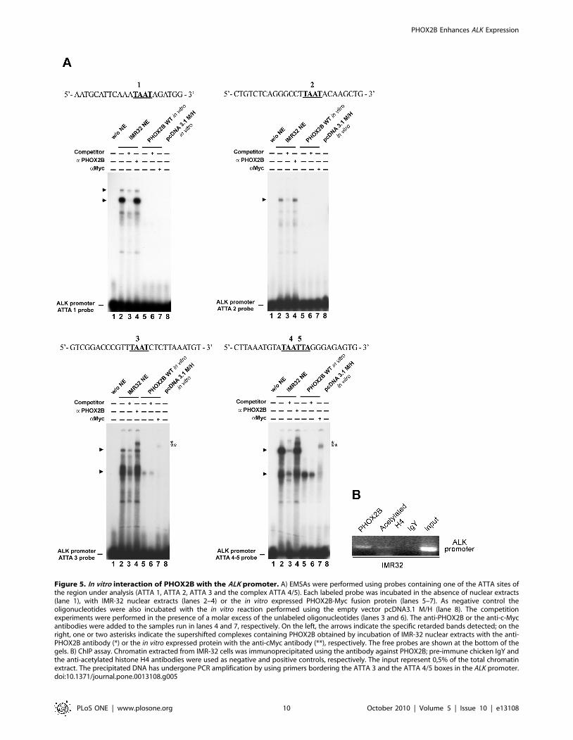
Figure 5. In vitro interaction of PHOX2B with the ALK promoter. A) EMSAs were performed using probes containing one of the ATTA sites ofthe region under analysis (ATTA 1, ATTA 2, ATTA 3 and the complex ATTA 4/5). Each labeled probe was incubated in the absence of nuclear extracts(lane 1), with IMR-32 nuclear extracts (lanes 2–4) or the in vitro expressed PHOX2B-Myc fusion protein (lanes 5–7). As negative control theoligonucleotides were also incubated with the in vitro reaction performed using the empty vector pcDNA3.1 M/H (lane 8). The competitionexperiments were performed in the presence of a molar excess of the unlabeled oligonucleotides (lanes 3 and 6). The anti-PHOX2B or the anti-c-Mycantibodies were added to the samples run in lanes 4 and 7, respectively. On the left, the arrows indicate the specific retarded bands detected; on theright, one or two asterisks indicate the supershifted complexes containing PHOX2B obtained by incubation of IMR-32 nuclear extracts with the anti-PHOX2B antibody (*) or the in vitro expressed protein with the anti-cMyc antibody (**), respectively. The free probes are shown at the bottom of thegels. B) ChIP assay. Chromatin extracted from IMR-32 cells was immunoprecipitated using the antibody against PHOX2B; pre-immune chicken IgY andthe anti-acetylated histone H4 antibodies were used as negative and positive controls, respectively. The input represent 0,5% of the total chromatinextract. The precipitated DNA has undergone PCR amplification by using primers bordering the ATTA 3 and the ATTA 4/5 boxes in the ALK promoter.doi:10.1371/journal.pone.0013108.g005
PHOX2B Enhances ALK Expression
PLoS ONE | www.plosone.org 10 October 2010 | Volume 5 | Issue 10 | e13108
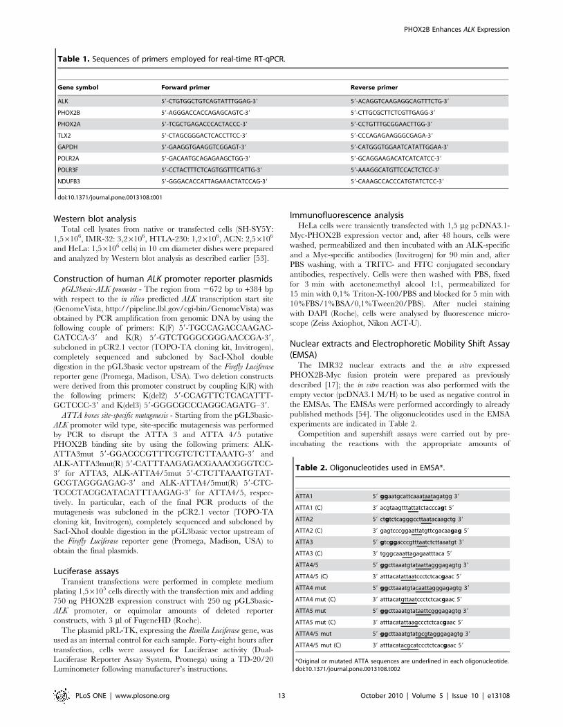
Primer design and gene expression analysis by Real-timeRT-qPCR
Based on the RefSeq annotations, gene specific primers were
designed in the regions encompassing exon boundaries to generate
a unique amplicon and tested for melting temperature and DNA
folding (Table 1).
Three human genes (POLR2A, POLR3F, NDUFB3), resulted to
be homogeneously and uniformly expressed in our samples, were
chosen as endogenous controls for data normalization, carried out
by using BestKeeper software (http://www.gene-quantification.de/
bestkeeper.html). Primers were designed using Primer 3 software
(http://fokker.wi.mit.edu/primer3/input.htm) [51]. Once tested
primer amplification efficiencies by a standard curve, triplicates of
each cDNA sample (12,5 ng) were amplified in the iCycler (Bio-Rad
Laboratories, Hercules, CA) following an initial denaturation at
95uC for 2 min, then 50 cycles at 95uC for 15 sec and 60uC for
30 sec. Melting curves were calculated between 55uC and 95uCwith increment of 0.5uC every 15 sec. PCRs were repeated at least
twice.
For relative quantification of gene expression in native cell lines,
we prepared an equimolar pool of RNAs from eight normal tissues
of different embryonic origin (adrenal gland, bladder, breast,
brain, colon, lung, placenta and prostate) (FirstChoiceH Human
Normal Tissue Total RNA, Ambion) to be employed as reference
sample.
In transfected cells, gene expression level was compared to
appropriate negative controls (i.e. native cells, transfectant agent
only, scrambled sample or empty vector).
Analysis of mRNA expression level of the target genes (ALK,
PHOX2A, PHOX2B) and the positive control (GAPDH) has been
carried out by a two-step real-time RT-qPCR using a random
priming-based reverse-transcription (High Capacity cDNA Re-
verse Transcription Kit, Applied Biosystems, Foster City, CA) and
SYBRH Green I binding dye (PlatinumH SYBRH Green qPCR
SuperMix-UDG, Invitrogen) according to the manufacturer’s
conditions. Data were analyzed by qGene software, implemented
to correct for amplification efficiencies [52].
Figure 6. Effect of ATTA 4/5 disruption in competition ofPHOX2B binding. IMR32 nuclear extracts were incubated with theATTA4/5 probe (lane 2) and competition obtained by adding an excessof: a wild type (wt) probe (lane 3), a probe mutated in both ATTA 4 andATTA 5 sites (lane 4) or in each of them (lanes 5–6). Incubation withoutnuclear extracts was regarded as negative control (lane 1).doi:10.1371/journal.pone.0013108.g006
Figure 7. Effects of mutagenesis of ATTA 3 and ATTA 4/5 on the PHOX2B-mediated ALK trans-activation. Left side: schematicrepresentation of the three constructs carrying all the ATTA boxes functional (wt, all four black circles), the ATTA 3 disrupted (ATTA 3 mut, one whitecircle) or both the ATTA 4 and 5 disrupted (ATTA 4/5 mut, two white circles). Right side: induction of the ALK promoter containing the mutant ATTA 3and ATTA 4/5 in HeLa cells co-transfected with the PHOX2B expression plasmid are expressed as percentage of the Luciferase activity obtained bycells co-transfected with PHOX2B and the ALK promoter (wt) vectors (wt, arbitrary value = 100). Values are the mean 6 s.d. of N = 3 independentexperiments (*: P,0.05).doi:10.1371/journal.pone.0013108.g007
PHOX2B Enhances ALK Expression
PLoS ONE | www.plosone.org 11 October 2010 | Volume 5 | Issue 10 | e13108
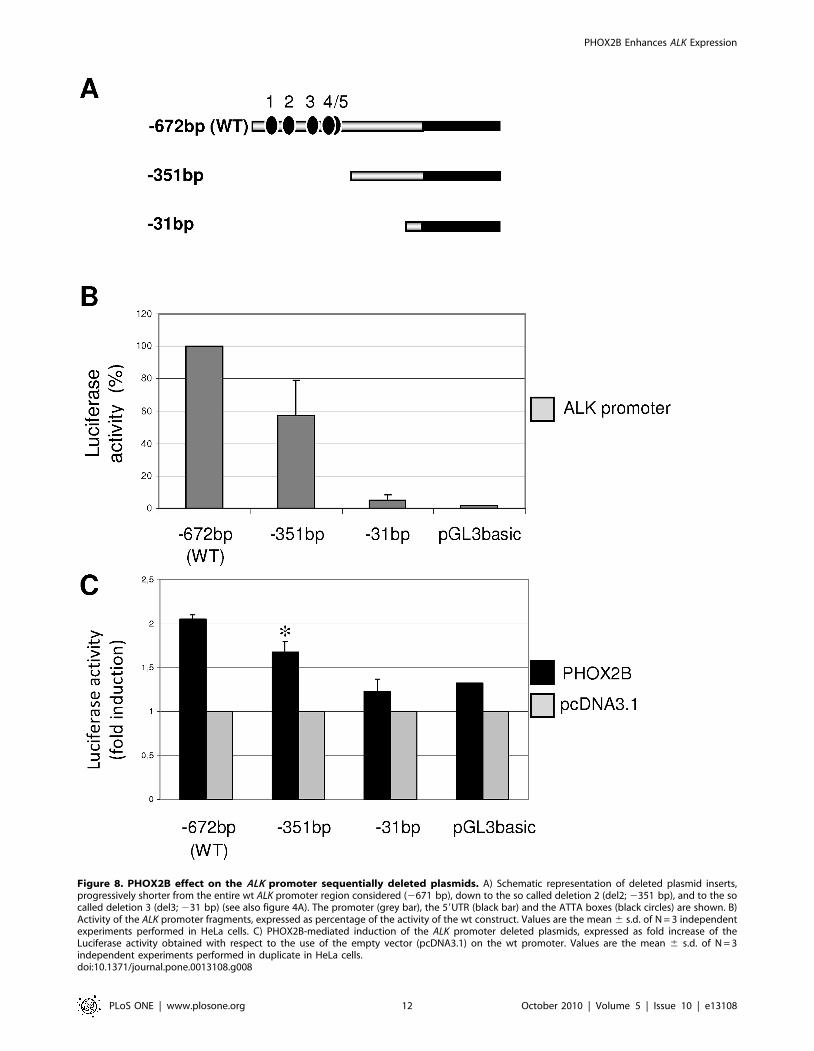
Figure 8. PHOX2B effect on the ALK promoter sequentially deleted plasmids. A) Schematic representation of deleted plasmid inserts,progressively shorter from the entire wt ALK promoter region considered (2671 bp), down to the so called deletion 2 (del2; 2351 bp), and to the socalled deletion 3 (del3; 231 bp) (see also figure 4A). The promoter (grey bar), the 59UTR (black bar) and the ATTA boxes (black circles) are shown. B)Activity of the ALK promoter fragments, expressed as percentage of the activity of the wt construct. Values are the mean 6 s.d. of N = 3 independentexperiments performed in HeLa cells. C) PHOX2B-mediated induction of the ALK promoter deleted plasmids, expressed as fold increase of theLuciferase activity obtained with respect to the use of the empty vector (pcDNA3.1) on the wt promoter. Values are the mean 6 s.d. of N = 3independent experiments performed in duplicate in HeLa cells.doi:10.1371/journal.pone.0013108.g008
PHOX2B Enhances ALK Expression
PLoS ONE | www.plosone.org 12 October 2010 | Volume 5 | Issue 10 | e13108
Western blot analysisTotal cell lysates from native or transfected cells (SH-SY5Y:
1,56106, IMR-32: 3,26106, HTLA-230: 1,26106, ACN: 2,56106
and HeLa: 1,56106 cells) in 10 cm diameter dishes were prepared
and analyzed by Western blot analysis as described earlier [53].
Construction of human ALK promoter reporter plasmidspGL3basic-ALK promoter - The region from 2672 bp to +384 bp
with respect to the in silico predicted ALK transcription start site
(GenomeVista, http://pipeline.lbl.gov/cgi-bin/GenomeVista) was
obtained by PCR amplification from genomic DNA by using the
following couple of primers: K(F) 59-TGCCAGACCAAGAC-
CATCCA-39 and K(R) 59-GTCTGGGCGGGAACCGA-39,
subcloned in pCR2.1 vector (TOPO-TA cloning kit, Invitrogen),
completely sequenced and subcloned by SacI-XhoI double
digestion in the pGL3basic vector upstream of the Firefly Luciferase
reporter gene (Promega, Madison, USA). Two deletion constructs
were derived from this promoter construct by coupling K(R) with
the following primers: K(del2) 59-CCAGTTCTCACATTT-
GCTCCC-39 and K(del3) 59-GGGCGCCCAGGCAGATG–39.
ATTA boxes site-specific mutagenesis - Starting from the pGL3basic-
ALK promoter wild type, site-specific mutagenesis was performed
by PCR to disrupt the ATTA 3 and ATTA 4/5 putative
PHOX2B binding site by using the following primers: ALK-
ATTA3mut 59-GGACCCGTTTCGTCTCTTAAATG-39 and
ALK-ATTA3mut(R) 59-CATTTAAGAGACGAAACGGGTCC-
39 for ATTA3, ALK-ATTA4/5mut 59-CTCTTAAATGTAT-
GCGTAGGGAGAG-39 and ALK-ATTA4/5mut(R) 59-CTC-
TCCCTACGCATACATTTAAGAG-39 for ATTA4/5, respec-
tively. In particular, each of the final PCR products of the
mutagenesis was subcloned in the pCR2.1 vector (TOPO-TA
cloning kit, Invitrogen), completely sequenced and subcloned by
SacI-XhoI double digestion in the pGL3basic vector upstream of
the Firefly Luciferase reporter gene (Promega, Madison, USA) to
obtain the final plasmids.
Luciferase assaysTransient transfections were performed in complete medium
plating 1,56105 cells directly with the transfection mix and adding
750 ng PHOX2B expression construct with 250 ng pGL3basic-
ALK promoter, or equimolar amounts of deleted reporter
constructs, with 3 ml of FugeneHD (Roche).
The plasmid pRL-TK, expressing the Renilla Luciferase gene, was
used as an internal control for each sample. Forty-eight hours after
transfection, cells were assayed for Luciferase activity (Dual-
Luciferase Reporter Assay System, Promega) using a TD-20/20
Luminometer following manufacturer’s instructions.
Immunofluorescence analysisHeLa cells were transiently transfected with 1,5 mg pcDNA3.1-
Myc-PHOX2B expression vector and, after 48 hours, cells were
washed, permeabilized and then incubated with an ALK-specific
and a Myc-specific antibodies (Invitrogen) for 90 min and, after
PBS washing, with a TRITC- and FITC conjugated secondary
antibodies, respectively. Cells were then washed with PBS, fixed
for 3 min with acetone:methyl alcool 1:1, permeabilized for
15 min with 0,1% Triton-X-100/PBS and blocked for 5 min with
10%FBS/1%BSA/0,1%Tween20/PBS). After nuclei staining
with DAPI (Roche), cells were analysed by fluorescence micro-
scope (Zeiss Axiophot, Nikon ACT-U).
Nuclear extracts and Electrophoretic Mobility Shift Assay(EMSA)
The IMR32 nuclear extracts and the in vitro expressed
PHOX2B-Myc fusion protein were prepared as previously
described [17]; the in vitro reaction was also performed with the
empty vector (pcDNA3.1 M/H) to be used as negative control in
the EMSAs. The EMSAs were performed accordingly to already
published methods [54]. The oligonucleotides used in the EMSA
experiments are indicated in Table 2.
Competition and supershift assays were carried out by pre-
incubating the reactions with the appropriate amounts of
Table 1. Sequences of primers employed for real-time RT-qPCR.
Gene symbol Forward primer Reverse primer
ALK 59-CTGTGGCTGTCAGTATTTGGAG-39 59-ACAGGTCAAGAGGCAGTTTCTG-39
PHOX2B 59-AGGGACCACCAGAGCAGTC-39 59-CTTGCGCTTCTCGTTGAGG-39
PHOX2A 59-TCGCTGAGACCCACTACCC-39 59-CCTGTTTGCGGAACTTGG-39
TLX2 59-CTAGCGGGACTCACCTTCC-39 59-CCCAGAGAAGGGCGAGA-39
GAPDH 59-GAAGGTGAAGGTCGGAGT-39 59-CATGGGTGGAATCATATTGGAA-39
POLR2A 59-GACAATGCAGAGAAGCTGG-39 59-GCAGGAAGACATCATCATCC-39
POLR3F 59-CCTACTTTCTCAGTGGTTTCATTG-39 59-AAAGGCATGTTCCACTCTCC-39
NDUFB3 59-GGGACACCATTAGAAACTATCCAG-39 59-CAAAGCCACCCATGTATCTCC-39
doi:10.1371/journal.pone.0013108.t001
Table 2. Oligonucleotides used in EMSA*.
ATTA1 59 ggaatgcattcaaataatagatgg 39
ATTA1 (C) 39 acgtaagtttattatctacccagt 59
ATTA2 59 ctgtctcagggccttaatacaagctg 39
ATTA2 (C) 39 gagtcccggaattatgttcgacaagag 59
ATTA3 59 gtcggacccgtttaatctcttaaatgt 39
ATTA3 (C) 39 tgggcaaattagagaatttaca 59
ATTA4/5 59 ggcttaaatgtataattagggagagtg 39
ATTA4/5 (C) 39 atttacatattaatccctctcacgaac 59
ATTA4 mut 59 ggcttaaatgtacaattagggagagtg 39
ATTA4 mut (C) 39 atttacatgttaatccctctcacgaac 59
ATTA5 mut 59 ggcttaaatgtataattcgggagagtg 39
ATTA5 mut (C) 39 atttacatattaagccctctcacgaac 59
ATTA4/5 mut 59 ggcttaaatgtatgcgtagggagagtg 39
ATTA4/5 mut (C) 39 atttacatacgcatccctctcacgaac 59
*Original or mutated ATTA sequences are underlined in each oligonucleotide.doi:10.1371/journal.pone.0013108.t002
PHOX2B Enhances ALK Expression
PLoS ONE | www.plosone.org 13 October 2010 | Volume 5 | Issue 10 | e13108
unlabelled oligonucleotide and antibody anti-PHOX2B [17] or
anti-c-myc antibody (Sigma).
Chromatin immunoprecipitation assay (ChIP)Chromatin from IMR-32 cells was prepared as manufacturer’s
instruction (ChIP-IT Express, Active Motif); a little portion of the
supernatants was kept as ‘‘input’’ material (0,5% total chromatin).
Cleared chromatin was incubated overnight at 4uC with anti-
PHOX2B antibody [17] or anti-acetylated histone H4. As
negative control chicken pre-immune chicken IgY (Santa Cruz
Biotechnology) were used. Immunocomplexes were collected by
magnetic beads. Chromatin was isolated by reversing crosslinking
at 65uC for 2 hours, followed by proteinase K treatment and DNA
purification (NucleoSpin Extract II; Macherey-Nagel). The
genomic sequence of interest, including both ATTA 3 and ATTA
4/5 boxes, was amplified by PCR using primer K(del1) 59-
CCCCTCTCCTCCAGTTTTATTC -39 with primer K(R)2 59-
TCCCTCTGTTCCCTCTCC -39.
Statistical AnalysesAll in vitro data are from at least three independent experiments.
Results are expressed as mean values 695% Confidence Interval
(CI) for quantitative variables and as numbers and percentages for
qualitative ones. For continuous variables, the statistical signifi-
cance of differential findings between experimental and control
groups was determined by ANOVA with the Tukey’s multiple
comparison test. The correlation between categorical variables
was assessed by the Pearson’s coefficient test. ANOVA and
Pearson’s tests were performed by Graph-Pad Prism 3.0 software
(Graph-Pad Software, Inc, El Camino Real, San Diego, CA). All
tests were two-sided and a P-value,0.05 was considered as
statistically significant.
Acknowledgments
We thank Dr. Vito Pistoia (Genova, Italy) for helpful discussions and
comments on the manuscript.
Author Contributions
Conceived and designed the experiments: TB SB UP MP IC PP.
Performed the experiments: TB DDP SDL VM CB MB IC CF MF PP.
Analyzed the data: VM MP. Contributed reagents/materials/analysis
tools: SDL DF RC. Wrote the paper: TB IC PP. Manuscript editing: SB
UP MP.
References
1. Trochet D, Bourdeaut F, Janoueix-Lerosey I, Deville A, de Pontual L, et al.
(2004) Germline mutations of the paired-like homeobox 2B (PHOX2B) gene in
neuroblastoma. Am J Hum Genet 74: 761–764.
2. Trochet D, O’Brien LM, Gozal D, Trang H, Nordenskjold A, et al. (2005)
PHOX2B genotype allows for prediction of tumor risk in congenital central
hypoventilation syndrome. Am J Hum Genet 76: 421–426.
3. Mosse YP, Laudenslager M, Longo L, Cole KA, Wood A, et al. (2008)
Identification of ALK as a major familial neuroblastoma predisposition gene.
Nature 455: 930–935.
4. Pattyn A, Morin X, Cremer H, Goridis C, Brunet JF (1999) The homeobox gene
Phox2b is essential for the development of autonomic neural crest derivatives.
Nature 399: 366–370.
5. Adachi M, Browne D, Lewis EJ (2000) Paired-like homeodomain proteins
Phox2a/Arix and Phox2b/NBPhox have similar genetic organization and
independently regulate dopamine beta-hydroxylase gene transcription. DNA
Cell Biol 19: 539–554.
6. Flora A, Lucchetti H, Benfante R, Goridis C, Clementi F, et al. (2001) Sp
proteins and Phox2b regulate the expression of the human Phox2a gene.
J Neurosci 21: 7037–7045.
7. Brunet JF, Pattyn A (2002) Phox2 genes - from patterning to connectivity. Curr
Opin Genet Dev 12: 435–440.
8. Bachetti T, Matera I, Borghini S, Di Duca M, Ravazzolo R, et al. (2005) Distinct
pathogenetic mechanisms for PHOX2B associated polyalanine expansions and
frameshift mutations in congenital central hypoventilation syndrome. Hum Mol
Genet 14: 1815–1824.
9. Borghini S, Bachetti T, Fava M, Di Duca M, Cargnin F, et al. (2006) The TLX2
homeobox gene is a transcriptional target of PHOX2B in neural-crest-derived
cells. Biochem J 395: 355–361.
10. Mosse YP, Laudenslager M, Khazi D, Carlisle AJ, Winter CL (2004) Germline
PHOX2B mutation in hereditary neuroblastoma. Am J Hum Genet 75:
727–730.
11. McConville C, Reid S, Baskcomb L, Douglas J, Rahman N (2006) PHOX2B
analysis in non-syndromic neuroblastoma cases shows novel mutations and
genotype-phenotype associations. Am J Med Genet A 140: 1297–1301.
12. van Limpt V, Schramm A, van Lakeman A, Sluis P, Chan A, et al. (2004) The
Phox2B homeobox gene is mutated in sporadic neuroblastomas. Oncogene 23:
9280–9288.
13. Perri P, Bachetti T, Longo L, Matera I, Seri M, et al. (2005) PHOX2B
mutations and genetic predisposition to neuroblastoma. Oncogene 24:
3050–3053.
14. Longo L, Borghini S, Schena F, Parodi S, Albino D, et al. (2008) PHOX2A and
PHOX2B genes are highly co-expressed in human neuroblastoma. Int J Oncol
33: 985–991.
15. Cheung IY, Feng Y, Gerald W, Cheung NK (2008) Exploiting gene expression
profiling to identify novel minimal residual disease markers of neuroblastoma.
Clin Cancer Res 14: 7020–7027.
16. Stutterheim J, Gerritsen A, Zappeij-Kannegieter L, Kleijn I, Dee R, et al. (2008)
PHOX2B is a novel and specific marker for minimal residual disease testing in
neuroblastoma. J Clin Oncol 26: 5443–5449.
17. Cargnin F, Flora A, Di Lascio S, Battaglioli E, Longhi R, et al. (2005) PHOX2B
regulates its own expression by a transcriptional auto-regulatory mechanism.
J Biol Chem 280: 37439–37448.
18. Lo L, Morin X, Brunet JF, Anderson DJ (1999) Specification of neurotransmitter
identity by Phox2 proteins in neural crest stem cells. Neuron 22: 693–705.
19. Bachetti T, Borghini S, Ravazzolo R, Ceccherini I (2005) An in vitro approach
to test the possible role of candidate factors in the transcriptional regulation of
the RET proto-oncogene. Gene Expr 12: 137–149.
20. Revet I, Huizenga G, Chan A, Koster J, Volckmann R, et al. (2008) The MSX1
homeobox transcription factor is a downstream target of PHOX2B and activates
the Delta-Notch pathway in neuroblastoma. Exp Cell Res 314: 707–719.
21. George RE, Sanda T, Hanna M, Frohling S, Luther W, et al. (2008) Activating
mutations in ALK provide a therapeutic target in neuroblastoma. Nature 455:
975–978.
22. Longo L, Panza E, Schena F, Seri M, Devoto M, et al. (2007) Genetic
predisposition to familial neuroblastoma: identification of two novel genomic
regions at 2p and 12p. Hum Hered 63: 205–211.
23. Palmer RH, Vernersson E, Grabbe C, Hallberg B (2009) Anaplastic lymphoma
kinase: signalling in development and disease. Biochem J 420: 345–361.
24. Chiarle R, Voena C, Ambrogio C, Piva R, Inghirami G (2008) The anaplastic
lymphoma kinase in the pathogenesis of cancer. Nat Rev Cancer 8: 11–23.
25. Webb TR, Slavish J, George RE, Look AT, Xue L, et al. (2009) Anaplastic
lymphoma kinase: role in cancer pathogenesis and small-molecule inhibitor
development for therapy. Expert Rev Anticancer Ther 9: 331–356.
26. Chen Y, Takita J, Choi YL, Kato M, Ohira M, et al. (2008) Oncogenic
mutations of ALK kinase in neuroblastoma. Nature 455: 971–974.
27. Janoueix-Lerosey I, Lequin D, Brugieres L, Ribeiro A, de Pontual L, et al. (2008)
Somatic and germline activating mutations of the ALK kinase receptor in
neuroblastoma. Nature 455: 967–970.
28. Caren H, Abel F, Kogner P, Martinsson T (2008) High incidence of DNA
mutations and gene amplifications of the ALK gene in advanced sporadic
neuroblastoma tumours. Biochem J 416: 153–159.
29. Passoni L, Longo L, Collini P, Coluccia AM, Bozzi F, et al. (2009) Mutation-
Independent Anaplastic Lymphoma Kinase Overexpression in Poor Prognosis
Neuroblastoma Patients. Cancer Res 69: 7338–7346.
30. Wang Q, Diskin S, Rappaport E, Attiyeh E, Mosse Y, et al. (2006) Integrative
genomics identifies distinct molecular classes of neuroblastoma and shows that
multiple genes are targeted by regional alterations in DNA copy number.
Cancer Res 66: 6050–6062.
31. Perez-Pinera P, Zhang W, Chang Y, Vega JA, Deuel TF (2007) Anaplastic
lymphoma kinase is activated through the pleiotrophin/receptor protein-
tyrosine phosphatase beta/zeta signaling pathway: an alternative mechanism
of receptor tyrosine kinase activation. J Biol Chem 282: 28683–28690.
32. Bourdeaut F, Trochet D, Janoueix-Lerosey I, Ribeiro A, Deville A, et al. (2005)
Germline mutations of the paired-like homeobox 2B (PHOX2B) gene in
neuroblastoma. Cancer Lett 228: 51–58.
33. Raabe EH, Laudenslager M, Winter C, Wasserman N, Cole K, et al. (2008)
Prevalence and functional consequence of PHOX2B mutations in neuroblas-
toma. Oncogene 27: 469–476.
PHOX2B Enhances ALK Expression
PLoS ONE | www.plosone.org 14 October 2010 | Volume 5 | Issue 10 | e13108
34. Reiff T, Tsarovina K, Majdazari A, Schmidt M, del Pino I, et al. (2010)
Neuroblastoma phox2b variants stimulate proliferation and dedifferentiation of
immature sympathetic neurons. J Neurosci 30: 905–915.
35. Alam G, Cui H, Shi H, Yang L, Ding J, et al. (2009) MYCN promotes the
expansion of Phox2B-positive neuronal progenitors to drive neuroblastoma
development. Am J Pathol 175: 856–866.
36. Mosse YP, Wood A, Maris JM (2009) Inhibition of ALK signaling for cancer
therapy. Clin Cancer Res 15: 5609–5614.
37. Longo L, Christiansen H, Christiansen NM, Cornaglia-Ferraris P, Lampert F
(1988) N-myc amplification at chromosome band 1p32 in neuroblastoma cells as
investigated by in situ hybridization. J Cancer Res Clin Oncol 114: 636–640.
38. Cornaglia-Ferraris P, Sansone R, Mariottini GL, Longo L, Tonini GP (1990)
Evidence of loss of N-myc amplification during the establishment of a human
neuroblastoma cell line. Int J Cancer 45: 578–579.
39. Bogenmann E (1996) A metastatic neuroblastoma model in SCID mice.
Int J Cancer 67: 379–385.
40. Gross N, Favre S, Beck D, Meyer M (1992) Differentiation-related expression of
adhesion molecules and receptors on human neuroblastoma tissues, cell lines
and variants. Int J Cancer 52: 85–91.
41. Seeger RC, Rayner SA, Banerjee A, Chung H, Laug WE, et al. (1977)
Morphology, growth, chromosomal pattern and fibrinolytic activity of two new
human neuroblastoma cell lines. Cancer Res 37: 1364–1371.
42. Seeger RC, Siegel SE, Sidell N (1982) Neuroblastoma: clinical perspectives,
monoclonal antibodies, and retinoic acid. Ann Intern Med 97: 873–884.
43. Tumilowicz JJ, Nichols WW, Cholon JJ, Greene AE (1970) Definition of a
continuous human cell line derived from neuroblastoma. Cancer Res 30:
2110–2118.
44. Biedler JL, Helson L, Spengler BA (1973) Morphology and growth,
tumorigenicity, and cytogenetics of human neuroblastoma cells in continuous
culture. Cancer Res 33: 2643–2652.
45. Barnes EN, Biedler JL, Spengler BA, Lyser KM (1981) The fine structure of
continuous human neuroblastoma lines SK-N-SH, SK-N-BE(2), and SK-N-MC.In Vitro 17: 619–631.
46. White PS, Maris JM, Beltinger C, Sulman E, Marshall HN, et al. (1995) A
region of consistent deletion in neuroblastoma maps within human chromosome1p36.2–36.3. Proc Natl Acad Sci U S A 92: 5520–5524.
47. Biedler JL, Roffler-Tarlov S, Schachner M, Freedman LS (1978) Multipleneurotransmitter synthesis by human neuroblastoma cell lines and clones.
Cancer Res 38: 3751–3757.
48. Scherer WF, Syverton JT, Gey Go (1953) Studies on the propagation in vitro ofpoliomyelitis viruses. IV. Viral multiplication in a stable strain of human
malignant epithelial cells (strain HeLa) derived from an epidermoid carcinomaof the cervix. J Exp Med 97: 695–710.
49. Pagnan G, Stuart D, Pastorino F, Raffaghello L, Montaldo PG, et al. (2000)Delivery of c-myb antisense oligodeoxynucleotides to human neuroblastoma
cells via disialoganglioside GD(2)-targeted immunoliposomes: antitumor effects.
J Natl Cancer Inst 92: 253–261.50. Pastorino F, Di Paolo D, Piccardi F, Nico B, Ribatti D, et al. (2008) Enhanced
antitumor efficacy of clinical-grade vasculature-targeted liposomal doxorubicin.Clin Cancer Res 14: 7320–7329.
51. Rozen S, Skaletsky H (2000) Primer3 on the WWW for general users and for
biologist programmers. Methods Mol Biol 132: 365–386.52. Muller PY, Janovjak H, Miserez AR, Dobbie Z (2002) Processing of gene
expression data generated by quantitative real-time RT-PCR. Biotechniques 32:1372–1379.
53. Pagnan G, Di Paolo D, Carosio R, Pastorino F, Marimpietri D, et al. (2009) Thecombined therapeutic effects of bortezomib and fenretinide on neuroblastoma
cells involve endoplasmic reticulum stress response. Clin Cancer Res 15:
1199–1209.54. Benfante R, Flora A, Di Lascio S, Cargnin F, Longhi R, et al. (2007)
Transcription factor PHOX2A regulates the human alpha3 nicotinic receptorsubunit gene promoter. J Biol Chem 282: 13290–13302.
PHOX2B Enhances ALK Expression
PLoS ONE | www.plosone.org 15 October 2010 | Volume 5 | Issue 10 | e13108