photon-in/photon-out x-ray free-electron laser studies of
TRANSCRIPT
applied sciences
Article
Photon-In/Photon-Out X-ray Free-Electron Laser Studiesof Radiolysis
Linda Young 1,2,* , Emily T. Nienhuis 3 , Dimitris Koulentianos 1 , Gilles Doumy 1 , Anne Marie March 1 ,Stephen H. Southworth 1 , Sue B. Clark 3,4 , Thomas M. Orlando 5 , Jay A. LaVerne 6 andCarolyn I. Pearce 3,7,*
�����������������
Citation: Young, L.; Nienhuis, E.T.;
Koulentianos, D.; Doumy, G.;
March, A.M.; Southworth, S.H.;
Clark, S.B.; Orlando, T.M.;
LaVerne, J.A.; Pearce, C.I.
Photon-In/Photon-Out X-ray
Free-Electron Laser Studies of
Radiolysis. Appl. Sci. 2021, 11, 701.
http://doi.org/10.3390/app11020701
Received: 23 December 2020
Accepted: 11 January 2021
Published: 13 January 2021
Publisher’s Note: MDPI stays neu-
tral with regard to jurisdictional clai-
ms in published maps and institutio-
nal affiliations.
Copyright: © 2021 by the authors. Li-
censee MDPI, Basel, Switzerland.
This article is an open access article
distributed under the terms and con-
ditions of the Creative Commons At-
tribution (CC BY) license (https://
creativecommons.org/licenses/by/
4.0/).
1 Chemical Sciences and Engineering Division, Argonne National Laboratory, Lemont, IL 60439, USA;[email protected] (D.K.); [email protected] (G.D.); [email protected] (A.M.M.);[email protected] (S.H.S.)
2 Department of Physics and James Franck Institute, The University of Chicago, Chicago, IL 60637, USA3 Pacific Northwest National Laboratory, Richland, WA 99354, USA; [email protected] (E.T.N.);
[email protected] (S.B.C.)4 Department of Chemistry, Washington State University, Pullman, WA 99164, USA5 Georgia Institute of Technology, School of Chemistry and Biochemistry, Atlanta, GA 30332, USA;
[email protected] Radiation Laboratory and Department of Physics, University of Notre Dame, Notre Dame, IN 46556, USA;
[email protected] Department of Crop and Soil Sciences, Washington State University, Pullman, WA 99164, USA* Correspondence: [email protected] (L.Y.); [email protected] (C.I.P.)
Abstract: Understanding the origin of reactive species following ionization in aqueous systems is animportant aspect of radiation–matter interactions as the initial reactive species lead to production ofradicals and subsequent long-term radiation damage. Tunable ultrafast X-ray free-electron pulsesprovide a new window to probe events occurring on the sub-picosecond timescale, supplementingother methodologies, such as pulse radiolysis, scavenger studies, and stop flow that capture longertimescale chemical phenomena. We review initial work capturing the fastest chemical processesin liquid water radiolysis using optical pump/X-ray probe spectroscopy in the water window anddiscuss how ultrafast X-ray pump/X-ray probe spectroscopies can examine ionization-inducedprocesses more generally and with better time resolution. Ultimately, these methods will be appliedto understanding radiation effects in complex aqueous solutions present in high-level nuclear waste.
Keywords: X-ray pump/X-ray probe; X-ray free-electron laser; water radiolysis; high-level nu-clear waste
1. Introduction
Understanding the elementary steps following ionization in aqueous systems willprovide a framework for radiation–matter interactions in chemistry and biology. It is ofcentral importance for radiobiological physics and chemistry and impacts cancer therapies,space travel, nuclear reactors, and environmental management [1,2]. Impact of an ionizingparticle (X-ray, gamma, ion) leads to ejection of energetic primary electrons, followed byinelastic and non-adiabatic processes that produce highly damaging slower secondaryelectrons during the so-called physical and physico-chemical stages, that is, within a timescale of 10−12 s [1]. During these stages, reactive radical species are produced, e.g., thehydrated electron [3,4] and the chemically aggressive hydroxyl radical [5,6]. These radicalspecies as well as H atoms and the molecular products H2 and H2O2 will undergo reactionsamongst themselves and with any added solutes, interfaces, etc. in the subsequent chemicalstage to induce radiation damage that may not be manifest until even decades followingthe initial passage of the ionizing radiation. Although these latter reactions are nominallythe observed outcome of radiolysis, it is the very fast processes on the sub-picosecond timescale that initiate this radiation damage.
Appl. Sci. 2021, 11, 701. https://doi.org/10.3390/app11020701 https://www.mdpi.com/journal/applsci
Appl. Sci. 2021, 11, 701 2 of 15
Most studies in the ultrafast physico-chemical time regime of liquid water radiolysishave focused on the hydrated electron as summarized in a recent comprehensive review [4].The primary ionization produces H2O+ and a quasifree electron. Only recently has theelementary proton transfer reaction H2O++H2O→ H3O+ + OH, which occurs in ∼50-fsto produce the hydroxyl radical, been observed [5]. Generally speaking, experimentaldata probing the mechanisms of more complex chemical reactions on ultrafast timescalesare extremely limited, despite being critical input for modeling of radiation chemistries,particularly in condensed phases.
Inner-shell vacancy decay represents a first step in radiation chemistries and in thecondensed phase differs significantly from that of isolated atoms in the gas phase, wherenon-radiative Auger decay and fluorescence are well characterized. Following theoreticalpredictions [7] for inner-valence excitation, studies of gas-phase clusters have revealed anovel, non-local decay mechanism, Intermolecular Coulombic Decay (ICD) first in neonand water dimers [8,9]. In ICD, when the energy of the initially created hole is greater thanthe double ionization potential of the system, energy transfer following Auger-decay caneject an “ICD” electron from the neighbor of the originally excited molecule, leading to aCoulomb explosion and slow electrons that can be captured in coincidence with recoilingions. Studies with larger clusters suggest that the propensity toward non-local decayincreases with size [10].
Extensions to condensed phases have been pioneered using electron spectroscopy inliquid microjets by Winter and co-workers [11,12] and have been initiated by core levelionization. These studies have revealed other non-local decay mechanisms, e.g., electrontransfer mediated decay (ETMD). Static electron spectroscopy measurements identifiedETMD and ICD by lineshape analysis-observing subtle changes in Auger spectra andinferring time resolution using the internal Auger clock, ∼4 fs for the oxygen 1 s hole.For example, comparison of the Auger spectra of normal and heavy water revealed that theinner-shell triggered autoionization processes are affected by proton transfer (OH stretchingfrequency is ∼9 fs) [13]. It was noted that the experimental spectra alone provided limitedinsight into the type of the relaxation processes contributing to the spectrum, and noinformation on their relative efficiencies could be inferred because the kinetic energyreleased in the different relaxation channels is similar—ab initio theoretical calculationsare essential for interpretation. It was shown that ICD occurs at condensed interfacescontaining water by examining the protonated water cluster ion, kinetic energy release,and threshold production energy. The latter was correlated with s-shell ionization of raregas partners. Though these experiments could clearly reveal the occurrence of ICD incomplex condensed phase targets, they could not determine the time scales of the protontransfer step vs. the initiating ICD event [14]. In general, the temporal evolution of thetransient species can not be extracted from static experimental spectra [15].
Going beyond static photoelectron spectroscopy, one may consider time-resolved pho-toelectron spectroscopy (TRPES) to probe atom-specific mechanistic details of ionization-induced reactions in solution. Such studies have focused on the hydrated electron, wherethe modest vertical binding energy (VBE) of ∼3.5 eV allows detection in both the groundand excited states of e−(aq) with readily available UV probes. As done previously for non-time-resolved studies of ICD [8,9], water cluster species introduced into vacuum in concertwith ion/electron detection are often used to approximate bulk behavior [16–20]. TRPESstudies in solution phase are challenging due to difficulties associated with introducing liq-uid jets [21] into the vacuum environment required by photoelectron spectrometers—spacecharge, electron escape depth issues render experimental realization and interpretation,respectively, a challenge [22]. The liquid jet studies have also focused on the dynamics ofthe hydrated electron; the e−(aq) is created via a photoinduced charge transfer to solvent(CTTS) from a solute anion (I−,Br−) [23,24] and its dynamics subsequently studied with IRpump/UV probe spectroscopy with photoelectron detection.
Photon-in photon-out spectroscopies powered by soft X-ray free electron lasers(XFELs) provide a powerful window into aqueous phase chemical reactions in the physico-
Appl. Sci. 2021, 11, 701 3 of 15
chemical time domain. Not only is the few femtosecond time regime and the photon energyto probe inner-shell hole configurations of oxygen, the K-shell absorption edges of carbon(284 eV), nitrogen (410 eV), oxygen (543 eV) and aluminum (1560 eV) readily accessible,but the challenging vacuum requirement for photoelectron spectroscopy is markedly re-duced. A further enabling breakthrough has been the development of stable liquid sheetjets of micron and sub-micron thickness [25,26]. This combination of soft X-ray XFELsand the liquid sheet jets has enabled laser-pump/X-ray probe time-resolved absorptionspectroscopy in the soft X-ray water window (280–530 eV) with milliOD sensitivity [5] andtime-resolved resonant inelastic X-ray scattering (RIXS) [6] to study the fastest chemicalprocesses in water radiolysis.
Here we recap the optical pump/X-ray probe studies of radiolysis in pure liquid waterin Section 2.1. In Section 2.2, we describe the prospects for extending these fundamentalstudies to the less “ideal” situation encountered in true radiolysis environments via X-raypump/X-ray probe studies. In Section 2.3, we briefly describe methods used at longertimescales to probe radiolysis products, i.e., pulse radiolysis, scavenger, and stop-flowmethods. In Section 3, we discuss the application to a practical problem of considerableimportance: remediation of high-level nuclear waste (HLW). Finally in Section 4, wedescribe the outlook for ultrafast studies of radiolysis with XFELs.
2. Methods
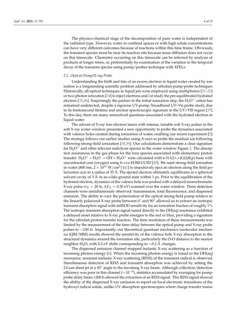
As a prototype of radiation-induced chemical reactions in more complex systems, wefirst consider pure water radiolysis as shown in Figure 1. The physico-chemical time regimeis covered primarily by XFEL pump/probe (and optical pump/probe studies, as describedin the introduction). The tabletop optical pump/probe studies have been limited to thestudy of the hydrated electron with those in the bulk liquid studies often employing soluteanions to generate e−(aq), though extensions to the soft X-ray transient absorption studiesare becoming feasible as high harmonic sources approach the oxygen K-edge [27–29].Chemical phenomena occurring on longer timescales are addressed by pulse radiolysis,scavenger, and stopped flow techniques. The combination of these many methods promisea comprehensive view of ionization-induced phenomena in complex systems.
Figure 1. Top: Various stages that occur in the radiolysis of water by fast ions or electrons orenergetic photons. Energy loss by the incident radiation to produce ionized or excited states of themedium is considered to occur in the physical stage that is best described in terms of cross sections.The relaxation of the medium from a few femtoseconds to about a picosecond is considered thephysico-chemical stage and represents the true beginning of the formation of chemically reactivespecies and that can be examined by XFELs. From top to bottom, these processes following ionizationof the water molecule are the proton transfer to produce the OH radical, hydration of the electron,and dissociative electron attachment. Neutralization of the electron with the water cation or directexcitation will lead to the excited water molecule followed by two most probable reactions shown.Reaction scheme and time scales taken from Ref. [30]. Bottom: Techniques used to probe chemicalphenomena at various timescales.
Appl. Sci. 2021, 11, 701 4 of 15
The physico-chemical stage of the decomposition of pure water is independent ofthe radiation type. However, water in confined spaces or with high solute concentrationscan have very different outcomes because of reactions within this time frame. Obviously,the transient species must be near its reaction site because mass diffusion does not occuron this timescale. Chemistry occurring on this timescale can be inferred by analysis ofproducts at longer times, or, preferentially by examination of the variation in the temporaldecay of the transient species using pump/probes technique with XFELs.
2.1. Optical-Pump/X-ray Probe
Understanding the birth and fate of an excess electron in liquid water created by ion-ization is a longstanding scientific problem addressed by ultrafast pump-probe techniques.Historically, all-optical techniques in liquid jets were employed using multiphoton [31–33]or two-photon ionization [34] to inject electrons and/or study the pre-equilibrated hydratedelectron [35,36]. Surprisingly the partner in the initial ionization step, the H2O+ cation hasremained undetected, despite a rigorous UV-pump/broadband UV–Vis probe study, dueto its femtosecond lifetime and unclear spectroscopic signature in the UV/VIS region [37].To this day, there are many unresolved questions associated with the hydrated electron inliquid water.
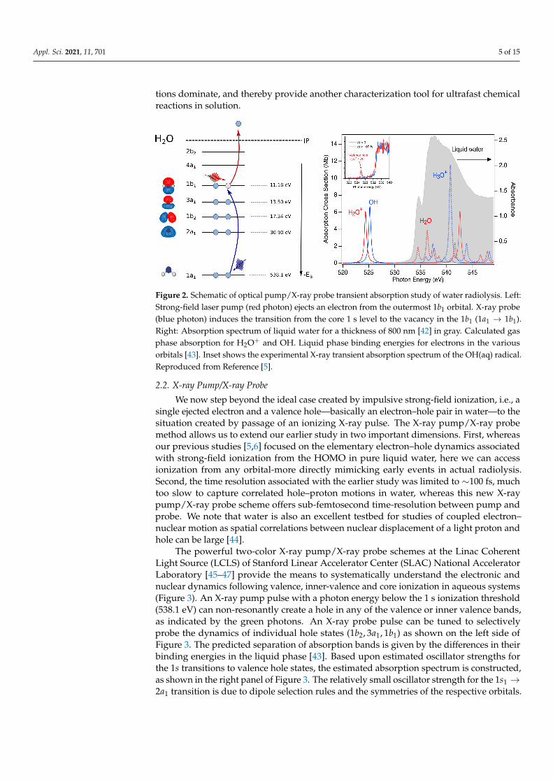
The advent of X-ray free electron lasers with intense, tunable soft X-ray pulses in thesoft X-ray water window presented a new opportunity to probe the dynamics associatedwith valence holes created during ionization of water, enabling our recent experiment [5].The strategy follows our earlier studies using X-rays to probe the residual ion followingfollowing strong-field ionization [38,39]. Our calculations demonstrate a clear signaturefor H2O+ and other relevant radiolysis species in the water window Figure 2. The absorp-tion resonances in the gas phase for the four species associated with elementary protontransfer: H2O+ + H2O→ OH + H3O+ were calculated with 6-311(2+,+)G(2df,p) basis withuncontracted core (oxygen) using fc-cvs-EOM-CCSD [40]. We used strong-field ionizationin water (800 nm, 2× 10¹³ W/cm2) [41] to impulsively eject an electron along the field po-larization axis to a radius of 35 Å. The ejected electron ultimately equilibrates in a sphericalsolvent cavity of 3 Å in an s-like ground state within 1 ps. Prior to the equilibration of thehydrated electron, dynamics of the valence hole was probed with a delayed monochromaticX-ray pulse (τX = 20 fs, ∆EX = 0.20 eV) scanned over the water window. Three detectionchannels were simultaneously observed: transmission, total fluorescence, and dispersedemission. The ability to vary the polarization of the optical strong-field pump relative tothe linearly polarized X-ray probe between 0◦ and 90◦ allowed us to extract an isotropictransient absorption signal with milliOD sensitivity for an ionization fraction of roughly 1%.The isotropic transient absorption signal tuned directly to the OH(aq) resonance exhibiteda delayed onset relative to X-ray probe energies to the red or blue, providing a signaturefor the ultrafast proton transfer reaction. The time resolution of these measurements waslimited by the measurement of the time-delay between the optical pump and X-ray probepulses to ∼100 fs. Importantly, our theoretical quantum mechanics/molecular mechan-ics (QM/MM) results showed the sensitivity of the valence hole X-ray absorption to thestructural dynamics around the ionization site, particularly the O-O distance to the nearestneighbor H2O, with 0.2 eV shifts corresponding to ∼0.2 Å changes.
The dispersed emission channel mapped inelastic X-ray scattering as a function ofincoming photon energy [6]. When the incoming photon energy is tuned to the OH(aq)resonance, resonant inelastic X-ray scattering (RIXS) of the transient radical is observed.Simultaneous detection of RIXS and transient absorption was achieved by setting the2.0µm sheet jet at a 45◦ angle to the incoming X-ray beam. Although collection/detectionefficiency was poor in this channel (∼10−8), statistics accumulated by averaging for pump-probe delay times >200 fs allowed the extraction of an RIXS signal. This RIXS signal showedthe ability of the dispersed X-ray emission to report on local electronic transitions of thehydroxyl radical solute, unlike UV absorption spectroscopies where charge transfer transi-
Appl. Sci. 2021, 11, 701 5 of 15
tions dominate, and thereby provide another characterization tool for ultrafast chemicalreactions in solution.
Figure 2. Schematic of optical pump/X-ray probe transient absorption study of water radiolysis. Left:Strong-field laser pump (red photon) ejects an electron from the outermost 1b1 orbital. X-ray probe(blue photon) induces the transition from the core 1 s level to the vacancy in the 1b1 (1a1 → 1b1).Right: Absorption spectrum of liquid water for a thickness of 800 nm [42] in gray. Calculated gasphase absorption for H2O+ and OH. Liquid phase binding energies for electrons in the variousorbitals [43]. Inset shows the experimental X-ray transient absorption spectrum of the OH(aq) radical.Reproduced from Reference [5].
2.2. X-ray Pump/X-ray Probe
We now step beyond the ideal case created by impulsive strong-field ionization, i.e., asingle ejected electron and a valence hole—basically an electron–hole pair in water—to thesituation created by passage of an ionizing X-ray pulse. The X-ray pump/X-ray probemethod allows us to extend our earlier study in two important dimensions. First, whereasour previous studies [5,6] focused on the elementary electron–hole dynamics associatedwith strong-field ionization from the HOMO in pure liquid water, here we can accessionization from any orbital-more directly mimicking early events in actual radiolysis.Second, the time resolution associated with the earlier study was limited to ∼100 fs, muchtoo slow to capture correlated hole–proton motions in water, whereas this new X-raypump/X-ray probe scheme offers sub-femtosecond time-resolution between pump andprobe. We note that water is also an excellent testbed for studies of coupled electron–nuclear motion as spatial correlations between nuclear displacement of a light proton andhole can be large [44].
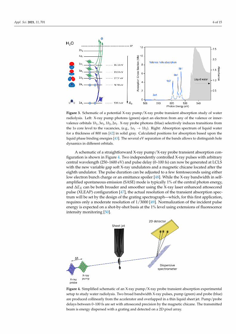
The powerful two-color X-ray pump/X-ray probe schemes at the Linac CoherentLight Source (LCLS) of Stanford Linear Accelerator Center (SLAC) National AcceleratorLaboratory [45–47] provide the means to systematically understand the electronic andnuclear dynamics following valence, inner-valence and core ionization in aqueous systems(Figure 3). An X-ray pump pulse with a photon energy below the 1 s ionization threshold(538.1 eV) can non-resonantly create a hole in any of the valence or inner valence bands,as indicated by the green photons. An X-ray probe pulse can be tuned to selectivelyprobe the dynamics of individual hole states (1b2, 3a1, 1b1) as shown on the left side ofFigure 3. The predicted separation of absorption bands is given by the differences in theirbinding energies in the liquid phase [43]. Based upon estimated oscillator strengths forthe 1s transitions to valence hole states, the estimated absorption spectrum is constructed,as shown in the right panel of Figure 3. The relatively small oscillator strength for the 1s1 →2a1 transition is due to dipole selection rules and the symmetries of the respective orbitals.
Appl. Sci. 2021, 11, 701 6 of 15
Figure 3. Schematic of a potential X-ray pump/X-ray probe transient absorption study of waterradiolysis. Left: X-ray pump photons (green) eject an electron from any of the valence or inner-valence orbitals 1b1, 3a1, 1b2, 2a1. X-ray probe photons (blue) selectively induces transitions fromthe 1s core level to the vacancies, (e.g., 1a1 → 1b2). Right: Absorption spectrum of liquid waterfor a thickness of 800 nm [42] in solid gray. Calculated positions for absorption based upon theliquid phase binding energies [43]. The several eV separation of the bands allows to distinguish holedynamics in different orbitals.
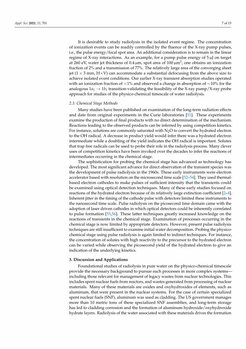
A schematic of a straightforward X-ray pump/X-ray probe transient absorption con-figuration is shown in Figure 4. Two independently controlled X-ray pulses with arbitrarycentral wavelength (250–1600 eV) and pulse delay (0–100 fs) can now be generated at LCLSwith the new variable gap soft X-ray undulators and a magnetic chicane located after theeighth undulator. The pulse duration can be adjusted to a few femtoseconds using eitherlow electron bunch charge or an emittance spoiler [48]. While the X-ray bandwidth in self-amplified spontaneous emission (SASE) mode is typically 1% of the central photon energy,and ∆EX can be both broader and smoother using the X-ray laser enhanced attosecondpulse (XLEAP) configuration [47], the actual resolution of the transient absorption spec-trum will be set by the design of the grating spectrograph—which, for this first application,requires only a moderate resolution of 1/3000 [49]. Normalization of the incident pulseenergy is expected on a shot-by-shot basis at the 1% level using extensions of fluorescenceintensity monitoring [50].
Figure 4. Simplified schematic of an X-ray pump/X-ray probe transient absorption experimentalsetup to study water radiolysis. Two broad bandwidth X-ray pulses, pump (green) and probe (blue)are produced collinearly from the accelerator and overlapped in a thin liquid sheet jet. Pump/probedelays between 0–100 fs are set with attosecond precision by the magnetic chicane. The transmittedbeam is energy dispersed with a grating and detected on a 2D pixel array.
Appl. Sci. 2021, 11, 701 7 of 15
It is desirable to study radiolysis in the isolated event regime. The concentrationof ionization events can be readily controlled by the fluence of the X-ray pump pulses,i.e., the pulse energy/focal spot area. An additional consideration is to remain in the linearregime of X-ray interactions. As an example, for a pump pulse energy of 5µJ on targetat 260 eV, water jet thickness of 0.4µm, spot area of 100µm2, one obtains an ionizationfraction of 2% and a transmission of 77%. The relatively large area of the converging sheetjet (1× 3 mm, H×V) can accommodate a substantial defocusing from the above size toachieve isolated event conditions. Our earlier X-ray transient absorption studies operatedwith an ionization fraction of <1% and observed a change in absorption of ∼10% for theanalogous 1a1 → 1b1 transition-validating the feasibility of the X-ray pump/X-ray probeapproach for studies of the physico-chemical timescale of water radiolysis.
2.3. Chemical Stage Methods
Many studies have been published on examination of the long-term radiation effectsand date from original experiments in the Curie laboratories [51]. These experimentsexamine the production of final products with no direct determination of the mechanism.Reactions leading to the observed products can be inferred by using competition kinetics.For instance, solutions are commonly saturated with N2O to convert the hydrated electronto the OH radical. A decrease in product yield would infer there was a hydrated electronintermediate while a doubling of the yield indicates the OH radical is important. Solutesthat trap free radicals can be used to probe their role in the radiolysis process. Many cleveruses of competition kinetics have been invoked over the decades to infer the reactions ofintermediates occurring in the chemical stage.
The sophistication for probing the chemical stage has advanced as technology hasdeveloped. The most significant advance for direct observation of the transient species wasthe development of pulse radiolysis in the 1960s. These early instruments were electronaccelerator based with resolution on the microsecond time scale [52–54]. They used thermal-based electron cathodes to make pulses of sufficient intensity that the transients couldbe examined using optical detection techniques. Many of these early studies focused onreactions of the hydrated electron because of its relatively large extinction coefficient [2–4].Inherent jitter in the timing of the cathode pulse with detectors limited these instruments tothe nanosecond time scale. Pulse radiolysis on the picosecond time domain came with theadoption of laser driven cathodes in which optical detectors could be inherently correlatedto pulse formation [55,56]. These latter techniques greatly increased knowledge on thereactions of transients in the chemical stage. Examination of processes occurring in thechemical stage is now limited by appropriate detectors. However, present pulse radiolysistechniques are still insufficient to examine initial water decomposition. Probing the physico-chemical stage using pulse radiolysis is again limited to indirect techniques. For instance,the concentration of solutes with high reactivity to the precursor to the hydrated electroncan be varied while observing the picosecond yield of the hydrated electron to give anindication of the underlying kinetics.
3. Discussion and Applications
Foundational studies of radiolysis in pure water on the physico-chemical timescaleprovide the necessary background to pursue such processes in more complex systems—including those relevant for management of legacy wastes from nuclear technologies. Thisincludes spent nuclear fuels from reactors, and wastes generated from processing of nuclearmaterials. Many of these materials are oxides and oxyhydroxides of elements, such asaluminum, that were present in the nuclear systems. For the case of certain specializedspent nuclear fuels (SNF), aluminum was used as cladding. The US government managesmore than 10 metric tons of these specialized SNF assemblies, and long-term storagehas led to cladding corrosion and the formation of aluminum hydroxide/oxyhydroxidehydrate layers. Radiolysis of the water associated with these materials drives the formation
Appl. Sci. 2021, 11, 701 8 of 15
of molecular hydrogen and other byproducts that must be quantified and considered indecisions regarding their long-term storage.
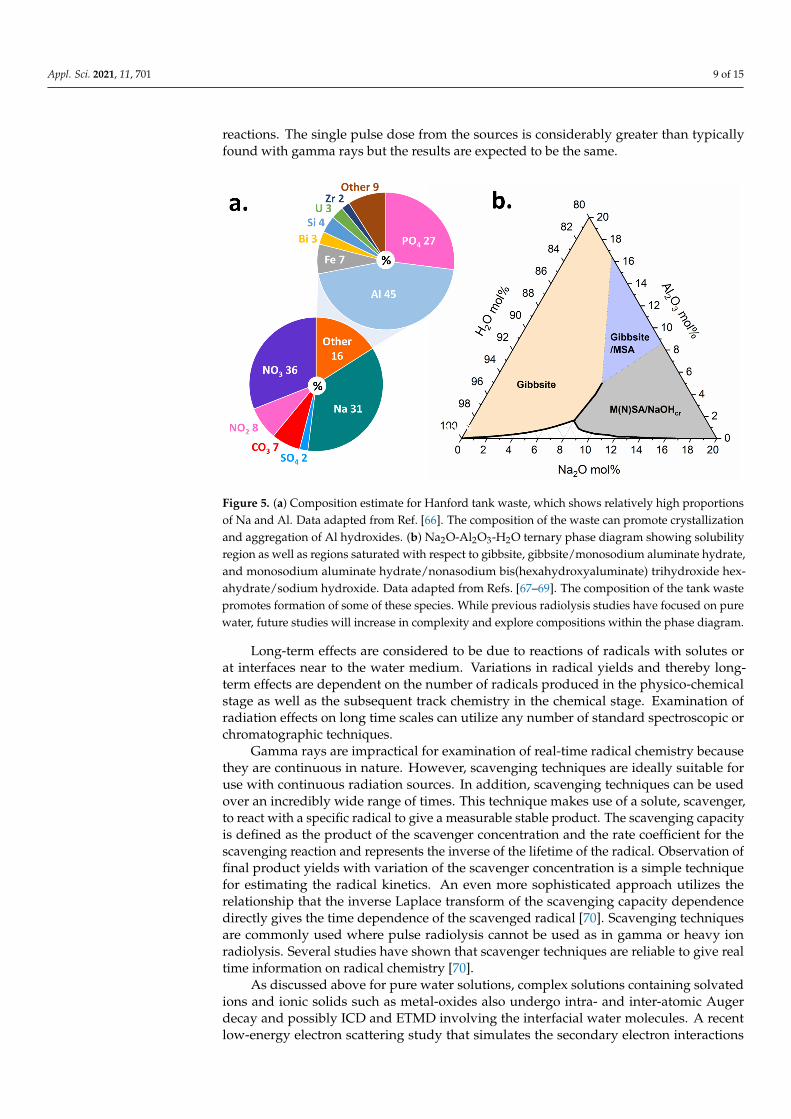
In the situation of wastes from nuclear materials processing, the production of pluto-nium from irradiated targets has resulted in the generation of more than 300 million litersof high-level radioactive wastes stored in underground tanks that must be managed bythe Department of Energy’s Office of Environmental Management (EM) [57]. The Hanfordsite in Washington State, USA houses 54 million gallons of hazardous radioactive waste,categorized as high-level waste (HLW) or low activity waste (LAW), resulting from decadesof plutonium production, and currently stored in 177 underground steel tanks. The tankwaste chemistry is complex (Figure 5a) and it consists of an aqueous supernatant, salt cakesolids, and a sludge layer [58]. The supernatant liquid is a caustic mixture of 3–5 molarsodium hydroxide (NaOH) and large fractions of oxyanions, such as nitrate (NO−3 ) andnitrite (NO−2 ), with the dominant metallic element being aluminum (Al). The salt cakeconsists of water-soluble oxyanion and halide salts. The sludge consists primarily ofinsoluble Al oxides and oxyhydroxides, such as gibbsite (Al(OH)3), or amorphous agglom-erates of varying sizes and morphologies, all of which serve to complicate retrieval andprocessing [58,59].
The tank waste will be retrieved and processed into a chemically durable vitrifiedwaste form for final disposal [60]. This remediation effort hinges on understanding com-ponents present in both the solid and aqueous phase, and controlling dissolution, pre-cipitation, and particle aggregation reactions [61]. Foundational to this is a completeunderstanding of radiation-driven reactions and their implications for the speciation,structure, and dynamics of solutions representative of the waste.
Within the tanks, γ-ray and β emitters are the primary radiation load (137Cs, 90Sr,99Tc,129I) [60,62,63]. Many of these radionuclides are present in the supernatant but can alsobe present in the tank sludge [63], potentially impacting solution speciation and structure,as well as aggregation behavior and solubility of the solid phases. Though γ-radiation isthe primary radiation load for the chemically complex tank wastes, to better understandthe impacts, different radiation sources are being used to evaluate the various timescalesand processes in simplified solutions representative of the major components of the waste,such as is shown in the Na2O-Al2O3-H2O ternary phase diagram in Figure 5b. Futurework will place greater emphasis on increasingly complex electrolyte solutions to be morerepresentative of simulant tank wastes, and the cascade of processes across broad time andlength scales that are set in motion by ionizing radiation.
Gamma rays, the primary radiation load in HLW, interact with water in a Comptonprocess in which the incident photon behaves as a particle to produce an electron of a fewhundred keV. Radiolysis of water by gamma rays is indistinguishable from that by fastelectrons. The initial energy loss by fast charged particles to a medium water moleculecan vary widely according to kinematic limits, but the average energy loss is about 60 eV.This average is nearly constant and independent of the initial energy of electrons andheavy ions [64]. A 60 eV electron will scatter significantly along its path as it produces aseries of lower energy ionization events. The result is a localized cluster or spur of ionizedand excited water molecules that will decay as described above in the physico-chemicalstage. Each initial event is a stochastic process so the number of ionized and excited watermolecules in a spur varies from about one to six [65]. The true chemical stage starts at about100 picoseconds when diffusion begins and lasts until about a microsecond. During thisperiod the reactions of the sibling products within a spur will react and diffuse as thenon-homogeneous spur decays. Heavy ions have a much higher rate of energy loss thanelectron so the spurs overlap to form a track. Track effects generally describe the chemicalstage for fast electrons and heavy ions and the competition between reaction and diffusionis responsible for the observed difference in product yields. A variety of time resolvedtechniques are available for directly probing the temporal variation of radicals in theradiolysis of water during the chemical stage. Pulse radiolysis with electrons in the MeVenergy range can use a variety of optical detection techniques to directly probe radical
Appl. Sci. 2021, 11, 701 9 of 15
reactions. The single pulse dose from the sources is considerably greater than typicallyfound with gamma rays but the results are expected to be the same.
Figure 5. (a) Composition estimate for Hanford tank waste, which shows relatively high proportionsof Na and Al. Data adapted from Ref. [66]. The composition of the waste can promote crystallizationand aggregation of Al hydroxides. (b) Na2O-Al2O3-H2O ternary phase diagram showing solubilityregion as well as regions saturated with respect to gibbsite, gibbsite/monosodium aluminate hydrate,and monosodium aluminate hydrate/nonasodium bis(hexahydroxyaluminate) trihydroxide hex-ahydrate/sodium hydroxide. Data adapted from Refs. [67–69]. The composition of the tank wastepromotes formation of some of these species. While previous radiolysis studies have focused on purewater, future studies will increase in complexity and explore compositions within the phase diagram.
Long-term effects are considered to be due to reactions of radicals with solutes orat interfaces near to the water medium. Variations in radical yields and thereby long-term effects are dependent on the number of radicals produced in the physico-chemicalstage as well as the subsequent track chemistry in the chemical stage. Examination ofradiation effects on long time scales can utilize any number of standard spectroscopic orchromatographic techniques.
Gamma rays are impractical for examination of real-time radical chemistry becausethey are continuous in nature. However, scavenging techniques are ideally suitable foruse with continuous radiation sources. In addition, scavenging techniques can be usedover an incredibly wide range of times. This technique makes use of a solute, scavenger,to react with a specific radical to give a measurable stable product. The scavenging capacityis defined as the product of the scavenger concentration and the rate coefficient for thescavenging reaction and represents the inverse of the lifetime of the radical. Observation offinal product yields with variation of the scavenger concentration is a simple techniquefor estimating the radical kinetics. An even more sophisticated approach utilizes therelationship that the inverse Laplace transform of the scavenging capacity dependencedirectly gives the time dependence of the scavenged radical [70]. Scavenging techniquesare commonly used where pulse radiolysis cannot be used as in gamma or heavy ionradiolysis. Several studies have shown that scavenger techniques are reliable to give realtime information on radical chemistry [70].
As discussed above for pure water solutions, complex solutions containing solvatedions and ionic solids such as metal-oxides also undergo intra- and inter-atomic Augerdecay and possibly ICD and ETMD involving the interfacial water molecules. A recentlow-energy electron scattering study that simulates the secondary electron interactions
Appl. Sci. 2021, 11, 701 10 of 15
from X-rays and high energy gamma rays, demonstrated very efficient ICD and ETMDin micro-solvated boehmite (AlOOH) nanoplatelets [71]. This occurs when the densityof states of the ionized chemisorbed species significantly overlaps the core hole states ofthe solid and is likely a primary process for producing ionized and radical species at theinterfaces of particles in complex waste forms. This experimental and theoretical workdemonstrated that ICD is a preferred decay mechanism upon single and double shallowcore hole excitation of adsorbed interfacial water and the underlying nanoplatelet. ETMDfrom the water also occurs following ionization of the boehmite Al(2s) level. ICD andETMD both lead to the formation and ejection of H2O+ with broad bimodal kinetic energyrelease (KER) distributions. The latter indicates the involvement of a manifold of complexmulti-hole states with lifetimes that compete with proton transfer and hole delocalization.None of these interactions have been probed with time-resolved techniques,
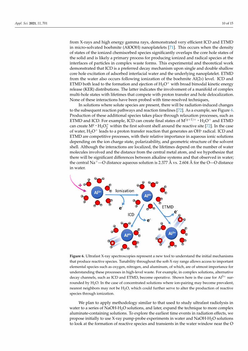
In solutions where solute species are present, there will be radiation-induced changesto the subsequent reaction pathways and reaction timelines [72]. As a example, see Figure 6.Production of these additional species takes place through relaxation processes, such asETMD and ICD. For example, ICD can create final states of M(q+1)+ • H2O+ and ETMDcan create Mq • H2O+
2 within the first solvent shell around the reactive site [72]. In the caseof water, H2O+ leads to a proton transfer reaction that generates an OH• radical. ICD andETMD are competitive processes, with their relative importance in aqueous ionic solutionsdepending on the ion charge state, polarizability, and geometric structure of the solventshell. Although the interactions are localized, the lifetimes depend on the number of watermolecules involved and the distance from the central metal atom, and we hypothesize thatthere will be significant differences between alkaline systems and that observed in water;the central Na+—O distance aqueous solution is 2.377 Å vs. 2.604 Å for the O—O distancein water.
Figure 6. Ultrafast X-ray spectroscopies represent a new tool to understand the initial mechanismsthat produce reactive species. Tunability throughout the soft-X-ray range allows access to importantelemental species such as oxygen, nitrogen, and aluminum, of which, are of utmost importance forunderstanding these processes in high-level waste. For example, in complex solutions, alternativedecay channels, such as ICD and ETMD, become operative. Shown here is the case for Al3+ sur-rounded by H2O. In the case of concentrated solutions where ion-pairing may become prevalent,nearest neighbors may not be H2O, which could further serve to alter the production of reactivespecies through ionization.
We plan to apply methodology similar to that used to study ultrafast radiolysis inwater to a series of NaOH-H2O solutions, and later, expand the technique to more complexaluminate-containing solutions. To explore the earliest time events in radiation effects, wepropose initially to use X-ray pump-probe experiments in water and NaOH-H2O solutionsto look at the formation of reactive species and transients in the water window near the O
Appl. Sci. 2021, 11, 701 11 of 15
K-edge using the techniques developed for water ionization studies [5]. Though the NaOHcomponents will not be observed directly in this manner, the way in which they perturbthe water network will be. Analogously, the oxidation state of the Na can be independentlyprobed on the ultrafast timescale giving a direct view into non-local decay processes at themetal-ion site. The specific questions we seek to answer include the impact of incompletesolvent shells as the nearest neighbors for ICD and ETMD processes are not necessarily awater molecule, the impact of ion networks on these processes when comparing dilute andconcentrated solutions of similar composition, and the overall timescale of the processesresulting from alteration of the solution structure.
Interpretation of ultrafast X-ray spectroscopic measurements is invariably linked withtheory, which, for understanding structural dynamics of core-excited matter, is an ongoingchallenge as described in some recent reviews [73,74]. There are already challenges at avery basic level, e.g., to predict the X-ray absorption spectrum of ground-state species withsub-eV accuracy [75] and to locate the positions of double-hole states [76]. Here, advancesin equation-of-motion coupled-cluster (EOM-CC) approaches [77,78] have allowed a sys-tematic convergence after application of the core-valence separation scheme [79] withinthe EOM-CC framework [80]. This general approach has allowed EOM-CC-CVS to achieveimproved accuracy for single photon transitions [40,81] and expand to two-photon RIXScalculations [82]. Beyond high-level calculations of electronic structure, capturing core-excited state dynamics poses a further challenge that has been largely pursued with simplerelectronic structure methods [72,83,84]. Describing non-local multiple-hole dynamics incomplex systems is likely to remain a frontier and foster synergistic research betweenexperiment and theory.
4. Conclusions and Outlook
XFEL-based photon-in/photon-out spectroscopies represent an emerging and uniqueopportunity to study radiolysis in the sub-picosecond physico-chemical time range. For stud-ies in the liquid phase, the photon-in/photon-out methods provide substantially relaxedvacuum requirements relative to those for the photoelectron-based spectroscopies. The ver-satile two-color, sub-femtosecond pulse generation capabilities over a wide energy range(250–1600 eV) at LCLS allow selection of initial ionization conditions and selective probingof ionization-generated chemical species by transient absorption. Similar capabilities arecurrently being considered at other facilities [85,86]. Advanced beamsplitter techniquesthat simultaneously provide sample and reference probe beams onto a single 2D-detectorpromise to further enhance the sensitivity for transient absorption studies [87,88]. The in-corporation of RIXS further discriminates transient species with overlapping absorptionresonances, and while statistically limited at present 120 Hz repetition rates, will becomemore routine at the MHz repetition rates soon expected. Such experimental studies canprovide missing input into the challenging theoretical problem associated with the descrip-tion of coupled electron-nuclear motion on highly excited electronic states associated withradiolysis in both simple and complex systems.
Author Contributions: Conceptualization, L.Y., E.T.N., J.A.L., and C.I.P.; methodology, L.Y., G.D.,D.K., S.H.S., and A.M.M.; writing—original draft preparation, L.Y., E.T.N., D.K., J.A.L., and C.I.P.writing—review & editing, S.B.C., T.M.O. All authors have read and agreed to the published versionof the manuscript.
Funding: This research was supported by IDREAM (Interfacial Dynamics in Radioactive Envi-ronments and Materials), an Energy Frontier Research Center funded by the U.S. Department ofEnergy (DOE), Office of Science, Basic Energy Sciences (BES). PNNL is a multiprogram nationallaboratory operated for DOE by Battelle Memorial Institute under Contract DE-AC05-76RL0-1830.DK,GD,AMM,SHS were supported by the U.S. Department of Energy, Office of Science, Basic EnergyScience, Chemical Sciences, Geosciences, and Biosciences Division under Contract No. DE-AC02-06CH11357.
Institutional Review Board Statement: Not applicable.
Appl. Sci. 2021, 11, 701 12 of 15
Informed Consent Statement: Not applicable.
Data Availability Statement: The data presented in this study are available on request from thecorresponding author.
Acknowledgments: We are grateful for technical discussions on the performance of LCLS operatingmodes with A. Marinelli, J. Cryan, on potential beamline and liquid jet endstation configurationswith W.S. Schlotter, K. Kunnus, G. Dakovski, D. DePonte, M. Minitti, R.W. Schoenlein. We appreciateongoing scientific discussions with Z.-H. Loh, J.-E. Rubensson and theoretical support from R. Santra,L. Inhester, A. I. Krylov, K. Nanda, L. Cheng, P.J. Ho, A. Fouda, X. Li, and A. Clark.
Conflicts of Interest: The authors declare no conflict of interest.
References1. Alizadeh, E.; Sanche, L. Precursors of Solvated Electrons in Radiobiological Physics and Chemistry. Chem. Rev. 2012, 112, 5578–
5602. [CrossRef] [PubMed]2. Garrett, B.C.; Dixon, D.A.; Camaioni, D.M.; Chipman, D.M.; Johnson, M.A.; Jonah, C.D.; Kimmel, G.A.; Miller, J.H.; Rescigno,
T.N.; Rossky, P.J.; et al. Role of Water in Electron-Initiated Processes and Radical Chemistry: Issues and Scientific Advances.Chem. Rev. 2005, 105, 355–390. [CrossRef] [PubMed]
3. Hart, E.J.; Boag, J.W. Absorption Spectrum of the Hydrated Electron in Water and in Aqueous Solutions. J. Am. Chem. Soc. 1962,84, 4090–4095. [CrossRef]
4. Herbert, J.M.; Coons, M.P. The Hydrated Electron. Annu. Rev. Phys. Chem. 2017, 68, 447–472. [CrossRef]5. Loh, Z.H.; Doumy, G.; Arnold, C.; Kjellsson, L.; Southworth, S.H.; Al Haddad, A.; Kumagai, Y.; Tu, M.F.; Ho, P.J.; March,
A.M.; et al. Observation of the fastest chemical processes in the radiolysis of water. Science 2020, 367, 179–182. [CrossRef]6. Kjellsson, L.; Nanda, K.D.; Rubensson, J.E.; Doumy, G.; Southworth, S.H.; Ho, P.J.; March, A.M.; Al Haddad, A.; Kumagai, Y.; Tu,
M.F.; et al. Resonant Inelastic X-ray Scattering Reveals Hidden Local Transitions of the Aqueous OH Radical. Phys. Rev. Lett.2020, 124, 236001. [CrossRef]
7. Cederbaum, L.S.; Zobeley, J.; Tarantelli, F. Giant Intermolecular Decay and Fragmentation of Clusters. Phys. Rev. Lett. 1997,79, 4778–4781. [CrossRef]
8. Jahnke, T.; Czasch, A.; Schöffler, M.S.; Schössler, S.; Knapp, A.; Käsz, M.; Titze, J.; Wimmer, C.; Kreidi, K.; Grisenti, R.E.;Staudte, A.; et al. Experimental Observation of Interatomic Coulombic Decay in Neon Dimers. Phys. Rev. Lett. 2004, 93, 163401.[CrossRef]
9. Mucke, M.; Braune, M.; Barth, S.; Förstel, M.; Lischke, T.; Ulrich, V.; Arion, T.; Becker, U.; Bradshaw, A.; Hergenhahn, U. A hithertounrecognized source of low-energy electrons in water. Nat. Phys. 2010, 6, 143–146. [CrossRef]
10. Richter, C.; Hollas, D.; Saak, C.M.; Förstel, M.; Miteva, T.; Mucke, M.; Björneholm, O.; Sisourat, N.; Slavícek, P.; Hergenhahn, U.Competition between proton transfer and intermolecular Coulombic decay in water. Nat. Commun. 2018, 9, 4988. [CrossRef]
11. Aziz, E.F.; Ottosson, N.; Faubel, M.; Hertel, I.V.; Winter, B. Interaction between liquid water and hydroxide revealed by core-holede-excitation. Nature 2008, 455, 89–91. [CrossRef] [PubMed]
12. Thürmer, S.; Oncák, M.; Ottosson, N.; Seidel, R.; Hergenhahn, U.; Bradforth, S.E.; Slavícek, P.; Winter, B. On the nature and originof dicationic, charge-separated species formed in liquid water on X-ray irradiation. Nat. Chem. 2013, 5, 590–596. [CrossRef][PubMed]
13. Slavícek, P.; Winter, B.; Cederbaum, L.S.; Kryzhevoi, N.V. Proton-Transfer Mediated Enhancement of Nonlocal ElectronicRelaxation Processes in X-ray Irradiated Liquid Water. J. Am. Chem. Soc. 2014, 136, 18170–18176. [CrossRef]
14. Grieves, G.A.; Orlando, T.M. Intermolecular Coulomb Decay at Weakly Coupled Heterogeneous Interfaces. Phys. Rev. Lett. 2011,107, 016104. [CrossRef]
15. Slavícek, P.; Kryzhevoi, N.V.; Aziz, E.F.; Winter, B. Relaxation Processes in Aqueous Systems upon X-ray Ionization: Entanglementof Electronic and Nuclear Dynamics. J. Phys. Chem. Lett. 2016, 7, 234–243. [CrossRef] [PubMed]
16. Coe, J.V.; Lee, G.H.; Eaton, J.G.; Arnold, S.T.; Sarkas, H.W.; Bowen, K.H.; Ludewigt, C.; Haberland, H.; Worsnop, D.R. Photoelec-tron spectroscopy of hydrated electron cluster anions, (H2O)−n=2−69. J. Chem. Phys. 1990, 92, 3980–3982. [CrossRef]
17. Bragg, A.E.; Verlet, J.R.R.; Kammrath, A.; Cheshnovsky, O.; Neumark, D.M. Hydrated Electron Dynamics: From Clusters to Bulk.Science 2004, 306, 669–671. [CrossRef]
18. Coe, J.V.; Williams, S.M.; Bowen, K.H. Photoelectron spectra of hydrated electron clusters vs. cluster size: connecting to bulk. Int.Rev. Phys. Chem. 2008, 27, 27–51. [CrossRef]
19. Svoboda, V.; Michiels, R.; LaForge, A.C.; Med, J.; Stienkemeier, F.; Slavícek, P.; Wörner, H.J. Real-time observation of waterradiolysis and hydrated electron formation induced by extreme-ultraviolet pulses. Sci. Adv. 2020, 6. [CrossRef]
20. LaForge, A.C.; Michiels, R.; Bohlen, M.; Callegari, C.; Clark, A.; von Conta, A.; Coreno, M.; Di Fraia, M.; Drabbels, M.; Huppert,M.; et al. Real-Time Dynamics of the Formation of Hydrated Electrons upon Irradiation of Water Clusters with Extreme UltravioletLight. Phys. Rev. Lett. 2019, 122, 133001. [CrossRef]
Appl. Sci. 2021, 11, 701 13 of 15
21. Winter, B.; Faubel, M.; Hertel, I.V.; Pettenkofer, C.; Bradforth, S.E.; Jagoda-Cwiklik, B.; Cwiklik, L.; Jungwirth, P. ElectronBinding Energies of Hydrated H3O+ and OH− : Photoelectron Spectroscopy of Aqueous Acid and Base Solutions Combinedwith Electronic Structure Calculations. J. Am. Chem. Soc. 2006, 128, 3864–3865. [CrossRef]
22. Suzuki, T. Time-resolved photoelectron spectroscopy of non-adiabatic electronic dynamics in gas and liquid phases. Int. Rev.Phys. Chem. 2012, 31, 265–318. [CrossRef]
23. Elkins, M.H.; Williams, H.L.; Shreve, A.T.; Neumark, D.M. Relaxation Mechanism of the Hydrated Electron. Science 2013,342, 1496–1499. [CrossRef] [PubMed]
24. Karashima, S.; Yamamoto, Y.i.; Suzuki, T. Resolving Nonadiabatic Dynamics of Hydrated Electrons Using Ultrafast PhotoemissionAnisotropy. Phys. Rev. Lett. 2016, 116, 137601. [CrossRef] [PubMed]
25. Koralek, J.D.; Kim, J.B.; Bruža, P.; Curry, C.B.; Chen, Z.; Bechtel, H.A.; Cordones, A.A.; Sperling, P.; Toleikis, S.; Kern, J.F.; et al.Generation and characterization of ultrathin free-flowing liquid sheets. Nat. Commun. 2018, 9, 1353. [CrossRef] [PubMed]
26. Ha, B.; DePonte, D.P.; Santiago, J.G. Device design and flow scaling for liquid sheet jets. Phys. Rev. Fluids 2018, 3, 114202.[CrossRef]
27. Kraus, P.M.; Zürch, M.; Cushing, S.K.; Neumark, D.M.; Leone, S.R. The ultrafast X-ray spectroscopic revolution in chemicaldynamics. Nat. Rev. Chem. 2018, 2, 82–94. [CrossRef]
28. Pertot, Y.; Schmidt, C.; Matthews, M.; Chauvet, A.; Huppert, M.; Svoboda, V.; von Conta, A.; Tehlar, A.; Baykusheva, D.; Wolf,J.P.; et al. Time-resolved X-ray absorption spectroscopy with a water window high-harmonic source. Science 2017, 355, 264–267.[CrossRef]
29. Teichmann, S.M.; Silva, F.; Cousin, S.L.; Hemmer, M.; Biegert, J. 0.5-keV Soft X-ray attosecond continua. Nat. Commun. 2016,7, 11493. [CrossRef]
30. La Cäer, S. Water Radiolysis: Influence of Oxide Surfaces on H2 Production under Ionizing Radiation. Water 2011, 3, 235–253.[CrossRef]
31. Long, F.H.; Lu, H.; Eisenthal, K.B. Femtosecond studies of the presolvated electron: An excited state of the solvated electron?Phys. Rev. Lett. 1990, 64, 1469–1472. [CrossRef] [PubMed]
32. Migus, A.; Gauduel, Y.; Martin, J.L.; Antonetti, A. Excess electrons in liquid water: First evidence of a prehydrated state withfemtosecond lifetime. Phys. Rev. Lett. 1987, 58, 1559–1562. [CrossRef] [PubMed]
33. Crowell, R.A.; Bartels, D.M. Multiphoton Ionization of Liquid Water with 3.0–5.0 eV Photons. J. Phys. Chem. 1996, 100,17940–17949. [CrossRef]
34. Elles, C.G.; Jailaubekov, A.E.; Crowell, R.A.; Bradforth, S.E. Excitation-energy dependence of the mechanism for two-photonionization of liquid H2O and D2O from 8.3 to 12.4 eV. J. Chem. Phys. 2006, 125, 044515. [CrossRef] [PubMed]
35. Silva, C.; Walhout, P.K.; Yokoyama, K.; Barbara, P.F. Femtosecond Solvation Dynamics of the Hydrated Electron. Phys. Rev. Lett.1998, 80, 1086–1089. [CrossRef]
36. Pshenichnikov, M.S.; Baltuška, A.; Wiersma, D.A. Hydrated-electron population dynamics. Chem. Phys. Lett. 2004, 389, 171– 175.[CrossRef]
37. Marsalek, O.; Elles, C.G.; Pieniazek, P.A.; Pluharová, E.; VandeVondele, J.; Bradforth, S.E.; Jungwirth, P. Chasing chargelocalization and chemical reactivity following photoionization in liquid water. J. Chem. Phys. 2011, 135, 224510. [CrossRef]
38. Young, L.; Arms, D.A.; Dufresne, E.M.; Dunford, R.W.; Ederer, D.L.; Höhr, C.; Kanter, E.P.; Krässig, B.; Landahl, E.C.;Peterson, E.R.; et al. X-ray Microprobe of Orbital Alignment in Strong-Field Ionized Atoms. Phys. Rev. Lett. 2006, 97, 083601.[CrossRef]
39. Goulielmakis, E.; Loh, Z.H.; Wirth, A.; Santra, R.; Rohringer, N.; Yakovlev, V.S.; Zherebtsov, S.; Pfeifer, T.; Azzeer, A.M.;Kling, M.F.; et al. Real-time observation of valence electron motion. Nature 2010, 466, 739–743. [CrossRef]
40. Vidal, M.L.; Feng, X.; Epifanovsky, E.; Krylov, A.I.; Coriani, S. New and Efficient Equation-of-Motion Coupled-Cluster Frameworkfor Core-Excited and Core-Ionized States. J. Chem. Theory Comput. 2019, 15, 3117–3133. [CrossRef]
41. Li, J.; Nie, Z.; Zheng, Y.Y.; Dong, S.; Loh, Z.H. Elementary Electron and Ion Dynamics in Ionized Liquid Water. J. Phys. Chem. Lett.2013, 4, 3698–3703. [CrossRef]
42. Nagasaka, M.; Hatsui, T.; Horigome, T.; Hamamura, Y.; Kosugi, N. Development of a liquid flow cell to measure soft X-rayabsorption in transmission mode: A test for liquid water. J. Electron. Spectrosc. Relat. Phenom. 2010, 177, 130–134. [CrossRef]
43. Winter, B.; Weber, R.; Widdra, W.; Dittmar, M.; Faubel, M.; Hertel, I.V. Full Valence Band Photoemission from Liquid Water UsingEUV Synchrotron Radiation. J. Phys. Chem. A 2004, 108, 2625–2632. [CrossRef]
44. Li, Z.; El-Amine Madjet, M.; Vendrell, O.; Santra, R. Core-level transient absorption spectroscopy as a probe of electron holerelaxation in photoionized H+(H2O)n. Faraday Discuss. 2014, 171, 457–470. [CrossRef]
45. Lutman, A.A.; Coffee, R.; Ding, Y.; Huang, Z.; Krzywinski, J.; Maxwell, T.; Messerschmidt, M.; Nuhn, H.D. ExperimentalDemonstration of Femtosecond Two-Color X-ray Free-Electron Lasers. Phys. Rev. Lett. 2013, 110, 134801. [CrossRef]
46. Lutman, A.A.; Maxwell, T.J.; MacArthur, J.P.; Guetg, M.W.; Berrah, N.; Coffee, R.N.; Ding, Y.; Huang, Z.; Marinelli, A.; Moeller,S.; et al. Fresh-slice multicolour X-ray free-electron lasers. Nat. Photonics 2016, 10, 745–750. [CrossRef]
47. Duris, J.; Li, S.; Driver, T.; Champenois, E.G.; MacArthur, J.P.; Lutman, A.A.; Zhang, Z.; Rosenberger, P.; Aldrich, J.W.;Coffee, R.; et al. Tunable isolated attosecond X-ray pulses with gigawatt peak power from a free-electron laser. Nat. Pho-tonics 2020, 14, 30–36. [CrossRef]
Appl. Sci. 2021, 11, 701 14 of 15
48. Emma, P.; Bane, K.; Cornacchia, M.; Huang, Z.; Schlarb, H.; Stupakov, G.; Walz, D. Femtosecond and Subfemtosecond X-rayPulses from a Self-Amplified Spontaneous-Emission–Based Free-Electron Laser. Phys. Rev. Lett. 2004, 92, 074801. [CrossRef][PubMed]
49. Chuang, Y.D.; Shao, Y.C.; Cruz, A.; Hanzel, K.; Brown, A.; Frano, A.; Qiao, R.; Smith, B.; Domning, E.; Huang, S.W.; et al. Modularsoft X-ray spectrometer for applications in energy sciences and quantum materials. Rev. Sci. Instrum. 2017, 88, 013110. [CrossRef][PubMed]
50. Heimann, P.; Reid, A.; Feng, Y.; Fritz, D. Fluorescence intensity monitors as intensity and beam-position diagnostics for X-rayfree-electron lasers. J. Synchrotron Radiat. 2019, 26, 358–362. [CrossRef]
51. Curie, P.; Debierne, A. Sur la radio-activité induite et les gaz activés par le radium. Compt. Rend 1901, 132, 768–770.52. Keene, J.P. Kinetics of Radiation-induced Chemical Reactions. Nature 1960, 188, 843–844. [CrossRef]53. Matheson, M.S.; Dorfman, L.M. Detection of Short-Lived Transients in Radiation Chemistry. J. Chem. Phys. 1960, 32, 1870–1871.
[CrossRef]54. McCarthy, R.; MacLachlan, A. Transient benzyl radical reactions produced by high-energy radiation. Trans. Faraday Soc. 1960,
56, 1187–1200. [CrossRef]55. Belloni, J.; Monard, H.; Gobert, F.; Larbre, J.P.; Demarque, A.; De Waele, V.; Lampre, I.; Marignier, J.L.; Mostafavi, M.; Bourdon, J.C.;
et al. ELYSE—A picosecond electron accelerator for pulse radiolysis research. Nucl. Instrum. Methods Phys. Res. Sect. A: Accel.Spectrometers Detect. Assoc. Equip. 2005, 539, 527–539. [CrossRef]
56. Wishart, J.F.; Cook, A.R.; Miller, J.R. The LEAF picosecond pulse radiolysis facility at Brookhaven National Laboratory. Rev. Sci.Instrum. 2004, 75, 4359–4366. [CrossRef]
57. Clark, S.B.; Buchanan, M.; Wilmarth, B. Basic Research Needs for Environmental Management; Technical Report; PNNL-25166; DOE:Washington, DC, USA, 2016.
58. Peterson, R.A.; Buck, E.C.; Chun, J.; Daniel, R.C.; Herting, D.L.; Ilton, E.S.; Lumetta, G.J.; Clark, S.B. Review of the scientificunderstanding of radioactive waste at the US DOE Hanford Site. Environ. Sci. Technol. 2018, 52, 381–396. [CrossRef]
59. Colburn, H.A.; Peterson, R.A. A history of Hanford tank waste, implications for waste treatment, and disposal. Environ. Prog.Sustain. Energy 2020, 40, e13567.
60. Kruger, A.A.; Vienna, J.D.; Kim, D.S. Advances in the Glass Formulations for the Hanford Tank Waste Treatment and ImmobilizationPlant; Technical Report; Hanford Site (HNF): Richland, WA, USA, 2015.
61. Page, J.S.; Reynolds, J.G.; Cooke, G.A.; Wells, B.E. Large cemented gibbsite agglomerates in alkaline nuclear waste at the Hanfordsite and the impacts to remediation. J. Hazard. Mater. 2020, 384, 121318. [CrossRef]
62. Gephart, R.E. A short history of waste management at the Hanford Site. Phys. Chem. Earth Parts A/B/C 2010, 35, 298–306.[CrossRef]
63. Gephart, R.E.; Lundgren, R.E. Hanford Tank Clean Up: A Guide to Understanding the Technical Issues; Technical Report; PacificNorthwest Lab.: Richland, WA, USA, 1995.
64. Pimblott, S.M.; LaVerne, J.A.; Mozumder, A.; Green, N.J. Structure of electron tracks in water. 1. Distribution of energy depositionevents. J. Phys. Chem. 1990, 94, 488–495. [CrossRef]
65. Pimblott, S.M.; LaVerne, J.A. Stochastic simulation of the electron radiolysis of water and aqueous solutions. J. Phys. Chem. A1997, 101, 5828–5838. [CrossRef]
66. PNNL: Tank Waste Information Network System; Best Basis Inventory. 2020. Available online: https://twins.labworks.org/twinsdata/forms/about.aspx?subject=BestBasisInventory (accessed on 6 April 2020.)
67. Graham, T.R.; Dembowski, M.; Hu, J.Z.; Jaegers, N.R.; Zhang, X.; Clark, S.B.; Pearce, C.I.; Rosso, K.M. Intermediate Species in theCrystallization of Sodium Aluminate Hydroxy Hydrates. J. Phys. Chem. C 2020, 124, 12337–12345. [CrossRef]
68. Graham, T.R.; Gorniak, R.; Dembowski, M.; Zhang, X.; Clark, S.B.; Pearce, C.I.; Clark, A.E.; Rosso, K.M. Solid-State Recrystalliza-tion Pathways of Sodium Aluminate Hydroxy Hydrates. Inorg. Chem. 2020, 59, 6857–6865. [CrossRef] [PubMed]
69. Zhang, Y.; Zheng, S.; Du, H.; Wang, S.; Zhang, Y. Solubility of Al2O3 in the Na2O-Al2O3-H2O-CH3OH System at (30 and 60) ◦C.J. Chem. Eng. Data 2010, 55, 1237–1240. [CrossRef]
70. Pimblott, S.M.; LaVerne, J.A.; Bartels, D.M.; Jonah, C.D. Reconciliation of transient absorption and chemically scavenged yields ofthe hydrated electron in radiolysis. J. Phys. Chem. 1996, 100, 9412–9415. [CrossRef]
71. Jones, B.M.; Hu, H.; Alexsandrov, A.; Smith, W.; Clark, A.E.; Li, X.; Orlando, T.M. Efficient Intermolecular Energy Exchange andSoft Ionization of Water at Nanoplatelet Interfaces. J. Phys. Chem. Lett. 2020, 11, 10088–10093. [CrossRef]
72. Stumpf, V.; Brunken, C.; Gokhberg, K. Impact of metal ion’s charge on the interatomic Coulombic decay widths in microsolvatedclusters. J. Chem. Phys. 2016, 145, 104306. [CrossRef]
73. Norman, P.; Dreuw, A. Simulating X-ray Spectroscopies and Calculating Core-Excited States of Molecules. Chem. Rev. 2018,118, 7208–7248. [CrossRef]
74. Besley, N.A. Density Functional Theory Based Methods for the Calculation of X-ray Spectroscopy. Accounts Chem. Res. 2020,53, 1306–1315. [CrossRef]
75. Sorensen, S.L.; Zheng, X.; Southworth, S.H.; Patanen, M.; Kokkonen, E.; Oostenrijk, B.; Travnikova, O.; Marchenko, T.; Simon, M.;Bostedt, C.; et al. From synchrotrons for XFELs: The soft X-ray near-edge spectrum of the ESCA molecule. J. Phys. B At. Mol. Opt.Phys. 2020, 53, 244011. [CrossRef]
Appl. Sci. 2021, 11, 701 15 of 15
76. Zheng, X.; Liu, J.; Doumy, G.; Young, L.; Cheng, L. Hetero-site Double Core Ionization Energies with Sub-electronvolt Accuracyfrom Delta-Coupled-Cluster Calculations. J. Phys. Chem. A 2020, 124, 4413–4426. [CrossRef] [PubMed]
77. Stanton, J.F.; Bartlett, R.J. The equation of motion coupled-cluster method. A systematic biorthogonal approach to molecularexcitation energies, transition probabilities, and excited state properties. J. Chem. Phys. 1993, 98, 7029–7039. [CrossRef]
78. Krylov, A.I. Equation-of-Motion Coupled-Cluster Methods for Open-Shell and Electronically Excited Species: The Hitchhiker’sGuide to Fock Space. Annu. Rev. Phys. Chem. 2008, 59, 433–462. [CrossRef]
79. Cederbaum, L.S.; Domcke, W.; Schirmer, J. Many-body theory of core holes. Phys. Rev. A 1980, 22, 206–222. [CrossRef]80. Coriani, S.; Koch, H. Communication: X-ray absorption spectra and core-ionization potentials within a core-valence separated
coupled cluster framework. J. Chem. Phys. 2015, 143, 181103. [CrossRef] [PubMed]81. Zheng, X.; Cheng, L. Performance of Delta-Coupled-Cluster Methods for Calculations of Core-Ionization Energies of First-Row
Elements. J. Chem. Theory Comput. 2019, 15, 4945–4955. [CrossRef] [PubMed]82. Nanda, K.D.; Vidal, M.L.; Faber, R.; Coriani, S.; Krylov, A.I. How to stay out of trouble in RIXS calculations within equation-
of-motion coupled-cluster damped response theory? Safe hitchhiking in the excitation manifold by means of core–valenceseparation. Phys. Chem. Chem. Phys. 2020, 22, 2629–2641. [CrossRef] [PubMed]
83. Hao, Y.; Inhester, L.; Hanasaki, K.; Son, S.K.; Santra, R. Efficient electronic structure calculation for molecular ionization dynamicsat high X-ray intensity. Struct. Dyn. 2015, 2, 041707. [CrossRef] [PubMed]
84. Ho, P.J.; Daurer, B.J.; Hantke, M.F.; Bielecki, J.; Al Haddad, A.; Bucher, M.; Doumy, G.; Ferguson, K.R.; Flückiger, L.; Gorkhover,T.; et al. The role of transient resonances for ultra-fast imaging of single sucrose nanoclusters. Nat. Commun. 2020, 11, 167.[CrossRef]
85. Serkez, S.; Decking, W.; Froehlich, L.; Gerasimova, N.; Grünert, J.; Guetg, M.; Huttula, M.; Karabekyan, S.; Koch, A.;Kocharyan, V.; et al. Opportunities for Two-Color Experiments in the Soft X-ray Regime at the European XFEL. Appl. Sci. 2020,10, 728. [CrossRef]
86. Reiche, S.; Knopp, G.; Pedrini, B.; Prat, E.; Aeppli, G.; Gerber, S. Towards the Perfect X-ray Beam Splitter. arXiv 2020,arXiv:physics.acc-ph/2010.00230.
87. Engel, R.; Miedema, P.; Turenne, D.; Vaskivskyi, I.; Brenner, G.; Dziarzhytski, S.; Kuhlmann, M.; Schunck, J.; Döring, F.;Styervoyedov, A.; et al. Parallel Broadband Femtosecond Reflection Spectroscopy at a Soft X-ray Free-Electron Laser. Appl. Sci.2020, 10, 6947. [CrossRef]
88. Schlotter, W.F.; Beye, M.; Zohar, S.; Coslovich, G.; Dakovski, G.L.; Lin, M.F.; Liu, Y.; Reid, A.; Stubbs, S.; Walter, P.; et al. BalancedDetection in Femtosecond X-ray Absorption Spectroscopy to Reach the Ultimate Sensitivity Limit. arXiv 2020, arXiv:physics.ins-det/2006.13968.