photofunctional layered materials
TRANSCRIPT

Series Editor: D.M.P. MingosStructure and Bonding 166
Dongpeng YanMin Wei Editors
Photofunctional Layered Materials

Structure and Bonding
Series Editor:
D.M.P. Mingos, Oxford, United Kingdom
Editorial Board:
F.A. Armstrong, Oxford, United Kingdom
X. Duan, Beijing, China
L.H. Gade, Heidelberg, Germany
K.R. Poeppelmeier, Evanston, IL, USA
G. Parkin, NewYork, USA
M. Takano, Kyoto, Japan
166

Aims and Scope
The series Structure and Bonding publishes critical reviews on topics of research
concerned with chemical structure and bonding. The scope of the series spans the
entire Periodic Table and addresses structure and bonding issues associated with all
of the elements. It also focuses attention on new and developing areas of modern
structural and theoretical chemistry such as nanostructures, molecular electronics,
designed molecular solids, surfaces, metal clusters and supramolecular structures.
Physical and spectroscopic techniques used to determine, examine and model
structures fallwithin the purview of Structure and Bonding to the extent that the
focus is on the scientific results obtained and not on specialist information
concerning the techniques themselves. Issues associated with the development of
bonding models and generalizations that illuminate the reactivity pathways and
rates of chemical processes are also relevant.
The individual volumes in the series are thematic. The goal of each volume is to
give the reader, whether at a university or in industry, a comprehensive overview of
an area where new insights are emerging that are of interest to a larger scientific
audience. Thus each review within the volume critically surveys one aspect of that
topic and places it within the context of the volume as a whole. The most significant
developments of the last 5 to 10 years should be presented using selected examples
to illustrate the principles discussed. A description of the physical basis of the
experimental techniques that have been used to provide the primary data may also
be appropriate, if it has not been covered in detail elsewhere. The coverage need not
be exhaustive in data, but should rather be conceptual, concentrating on the new
principles being developed that will allow the reader, who is not a specialist in the
area covered, to understand the data presented. Discussion of possible future
research directions in the area is welcomed.
Review articles for the individual volumes are invited by the volume editors.
In references Structure and Bonding is abbreviated Struct Bond and is cited as a
journal.
More information about this series athttp://www.springer.com/series/430

Dongpeng Yan • Min Wei
Editors
Photofunctional LayeredMaterials
With contributions by
R. Tian � M. Wei � D. Yan � J. Liang � R. Ma � T. Sasaki �M. Shao � D.G. Evans � X. Duan � D.-H. Park � G. Choi �J.-H. Choy � T. Okada �M. Sohmiya �M. Ogawa � L. Zhang �Z. Liu

EditorsDongpeng YanBeijing University of ChemicalTechnology
BeijingChina
Min WeiBeijing University of ChemicalTechnology
BeijingChina
ISSN 0081-5993 ISSN 1616-8550 (electronic)Structure and BondingISBN 978-3-319-16990-3 ISBN 978-3-319-16991-0 (eBook)DOI 10.1007/978-3-319-16991-0
Library of Congress Control Number: 2015940114
Springer Cham Heidelberg New York Dordrecht London© Springer International Publishing Switzerland 2015This work is subject to copyright. All rights are reserved by the Publisher, whether the whole or part ofthe material is concerned, specifically the rights of translation, reprinting, reuse of illustrations,recitation, broadcasting, reproduction on microfilms or in any other physical way, and transmissionor information storage and retrieval, electronic adaptation, computer software, or by similar ordissimilar methodology now known or hereafter developed.The use of general descriptive names, registered names, trademarks, service marks, etc. in thispublication does not imply, even in the absence of a specific statement, that such names are exemptfrom the relevant protective laws and regulations and therefore free for general use.The publisher, the authors and the editors are safe to assume that the advice and information in thisbook are believed to be true and accurate at the date of publication. Neither the publisher nor theauthors or the editors give a warranty, express or implied, with respect to the material containedherein or for any errors or omissions that may have been made.
Printed on acid-free paper
Springer International Publishing AG Switzerland is part of Springer Science+Business Media(www.springer.com)

Preface
Photofunctional materials are a large family of photoactive compounds that can
transfer, absorb, store, switch, or utilize light energy. Layered compounds with
two-dimensional (2D) structures have recently emerged as a new platform for
developing novel photofunctional materials and have already been explored and
applied in the fields of light-harvesting, photoluminescence, photocatalysis, bio-
logical imaging, band structure engineering, photochromic sensors, and optoelec-
tronic devices.
Given the rapid development of photofunctional layered materials, we believe
the collection of reviews in this volume is timely. In the first chapter, Yan and
colleagues review the photofunctionality of layered double hydroxide (LDH)
materials, with a particular focus on the incorporation of organic chromophores,
polymers, metal complexes, and quantum dots in LDHs; the fluorescence, optical
sensing, and IR/UV absorption properties of the resulting composite materials are
covered.
Although similar to LDH materials, layered rare earth hydroxides (LREHs)—
which contain rare earth hydroxide cationic host layers—have been recognized as a
new class of multifunctional materials with an intercalation ability that leads to
extensive host–guest chemistry. In the second chapter, Sasaki and colleagues
provide an overview of the development of LREH materials and summarize the
synthetic methods, structure characterization, as well as their photoluminescence
properties.
Photocatalytic materials have attracted great attention in environmental science,
as well as in energy storage and transfer. In the third chapter, Wei et al. summarize
the synthesis and photocatalytic applications of LDH-based materials. In particular,
several important photocatalytic systems—such as pollutant degradation, water
splitting for solar fuel production, and reduction of CO2 to useful carbon mate-
rials—are reviewed and discussed.
In view of their potential benefits to human health, the fabrication of new
bio-functional nanomaterials has become a fast-growing area. The use of LDHs
as fluorescence imaging materials and related biomedical applications has been
v

widely developed. In the fourth chapter, Choy et al. review the structural chemistry
of bio-LDH nanohybrid systems and summarize their applications in gene and drug
delivery (in both therapeutics and diagnostics).
Photoresponsive materials (such as photochromic compounds) are regarded as
one of the most interesting photo-related topics. In the fifth chapter, Ogawa
et al. provide an overview of the development of photochromic intercalation
compounds and describe how the formation of different layered host materials
allows control over the orientation and aggregation of photochromic guests as
well as the resulting photoresponsive properties.
As 2D functional units, graphene and its derivatives have attracted considerable
interest due to their novel optical, electronic, mechanical, and surface properties.
Recently, photogenerated free radicals have been shown to provide a new way for the
covalent functionalization and band structure engineering of graphene. In the final
chapter of this volume, Zhang and Liu discuss the photochemistry of graphene,
including its photohalogenation, photoarylation, photoalkylation, and photocatalytic
oxidation.
There has been an explosive growth in publications dealing with photofunctional
layered materials in recent years. The reviews in this volume have attempted to
cover representative examples of such layered compounds in the literature
published up to 2014. Many interesting additional examples involving layered
structures and novel photofunctionalities may have been excluded, either inadver-
tently or due to space limitations, but we hope that the volume has captured the
more important developments.
We express our thanks to all the authors for helping us capture the essence of
recent research in this field.
Beijing, China Dongpeng Yan
Min Wei
vi Preface

Contents
Layered Double Hydroxide Materials: Assembly
and Photofunctionality . . . . . . . . . . . . . . . . . . . . . . . . . . . . . . . . . . . . . . 1
Rui Tian, Dongpeng Yan, and Min Wei
Layered Rare Earth Hydroxides: Structural Aspects
and Photoluminescence Properties . . . . . . . . . . . . . . . . . . . . . . . . . . . . . 69
Jianbo Liang, Renzhi Ma, and Takayoshi Sasaki
Layered Double Hydroxide Materials in Photocatalysis . . . . . . . . . . . . . 105
Mingfei Shao, Min Wei, David G. Evans, and Xue Duan
Bio-Layered Double Hydroxides Nanohybrids for Theranostics
Applications . . . . . . . . . . . . . . . . . . . . . . . . . . . . . . . . . . . . . . . . . . . . . . 137
Dae-Hwan Park, Goeun Choi, and Jin-Ho Choy
Photochromic Intercalation Compounds . . . . . . . . . . . . . . . . . . . . . . . . 177
Tomohiko Okada, Minoru Sohmiya, and Makoto Ogawa
Photochemistry of Graphene . . . . . . . . . . . . . . . . . . . . . . . . . . . . . . . . . 213
Liming Zhang and Zhongfan Liu
Index . . . . . . . . . . . . . . . . . . . . . . . . . . . . . . . . . . . . . . . . . . . . . . . . . . . 239
vii

Layered Double Hydroxide Materials:
Assembly and Photofunctionality
Rui Tian, Dongpeng Yan, and Min Wei
Contents
1 Introduction . . . . . . . . . . . . . . . . . . . . . . . . . . . . . . . . . . . . . . . . . . . . . . . . . . . . . . . . . . . . . . . . . . . . . . . . . . . . . . . . . . . 3
2 Optical Properties of LDHs . . . . . . . . . . . . . . . . . . . . . . . . . . . . . . . . . . . . . . . . . . . . . . . . . . . . . . . . . . . . . . . . . . 4
3 Luminescent Properties of LDHs Nanoparticles . . . . . . . . . . . . . . . . . . . . . . . . . . . . . . . . . . . . . . . . . . . . 6
3.1 Defect-Induced Luminescence . . . . . . . . . . . . . . . . . . . . . . . . . . . . . . . . . . . . . . . . . . . . . . . . . . . . . . . . . 6
3.2 Rare-Earth-Doped Luminescence . . . . . . . . . . . . . . . . . . . . . . . . . . . . . . . . . . . . . . . . . . . . . . . . . . . . . 7
4 LDH-Based UV-Shielding Materials and the Applications . . . . . . . . . . . . . . . . . . . . . . . . . . . . . . . . 8
4.1 Introduction of UV Light and UV-Shielding Materials . . . . . . . . . . . . . . . . . . . . . . . . . . . . . . 8
4.2 UV-Shielding Properties of LDHs . . . . . . . . . . . . . . . . . . . . . . . . . . . . . . . . . . . . . . . . . . . . . . . . . . . . . 8
4.3 Host–Guest Interaction and UV-Shielding Performances . . . . . . . . . . . . . . . . . . . . . . . . . . . . 10
4.4 Application of LDH-Based UV-Shielding Materials
in Polypropylene and Asphalt . . . . . . . . . . . . . . . . . . . . . . . . . . . . . . . . . . . . . . . . . . . . . . . . . . . . . . . . . 13
5 LDH-Based IR Absorption Materials and the Application . . . . . . . . . . . . . . . . . . . . . . . . . . . . . . . . . 17
5.1 Introduction of IR Absorption Materials . . . . . . . . . . . . . . . . . . . . . . . . . . . . . . . . . . . . . . . . . . . . . . 17
5.2 IR Absorption Properties of LDHs . . . . . . . . . . . . . . . . . . . . . . . . . . . . . . . . . . . . . . . . . . . . . . . . . . . . 17
5.3 Intercalation and Performances of LDH-Based IR Absorption Materials . . . . . . . . . . . 19
5.4 Application of LDH-Based IR Absorption Materials . . . . . . . . . . . . . . . . . . . . . . . . . . . . . . . . . 22
6 Fabrication and Application of LDH-Based Host–Guest Luminescent Materials . . . . . . . . . 24
6.1 The Basic Guest Units of LDH-Based Photofunctional Materials . . . . . . . . . . . . . . . . . . . 25
6.2 Fabrication of LDH-Based Photofunctional Material . . . . . . . . . . . . . . . . . . . . . . . . . . . . . . . . . 25
6.3 Adjusting Photofunctionalities of LDH-Based Materials . . . . . . . . . . . . . . . . . . . . . . . . . . . . . 38
7 Conclusion and Outlook . . . . . . . . . . . . . . . . . . . . . . . . . . . . . . . . . . . . . . . . . . . . . . . . . . . . . . . . . . . . . . . . . . . . . 63
References . . . . . . . . . . . . . . . . . . . . . . . . . . . . . . . . . . . . . . . . . . . . . . . . . . . . . . . . . . . . . . . . . . . . . . . . . . . . . . . . . . . . . . . . 64
R. Tian • M. Wei
State Key Laboratory of Chemical Resource Engineering, Beijing University of Chemical
Technology, Beijing 100029, P. R. China
D. Yan (*)
State Key Laboratory of Chemical Resource Engineering, Beijing University of Chemical
Technology, Beijing 100029, P. R. China
College of Chemistry, Beijing Normal University, Beijing 100875, P. R. China
e-mail: [email protected]; [email protected]
© Springer International Publishing Switzerland 2015
D. Yan, M. Wei (eds.), Photofunctional Layered Materials, Structure and Bonding
166, DOI 10.1007/978-3-319-16991-0_1
1

Abstract As a large type of inorganic layered compounds with diversity of
composition in host layer and interlayer anions, layered double hydroxides
(LDHs) have been intensively studied toward construction of advanced
photofunctional materials. In this chapter, the optical properties of LDH materials
and related potential applications—mainly involving the functionalities of LDH
layers (such as tunable color, infrared radiation (IR) absorption, and ultraviolet
(UV) shielding)—will be described firstly. Then, we will review the development
of intercalated luminescent materials by adjusting both the host layer and guest
molecules, in which the static and dynamic photofunctional modulations have been
focused. Due to the host–guest and guest–guest interaction, fluorescence properties
of composites can be effectively altered, and intelligent materials with stimuli-
responsive performance can be further obtained. Finally, perspectives on the future
development of LDH-based solid-state luminescent materials are addressed.
Keywords Assembly • Layered double hydroxides • Luminescence • Sensor • UV
shielding
Abbreviations
AFM Atomic force microscopy
CL Chemiluminescence
ECL Electrochemiluminescence
EDTA Ethylenediaminetetraacetic acid
EL Electroluminescence
FRET F€orster resonance energy transfer
HMIs Heavy metal ions
IR Infrared radiation
LDHs Layered double hydroxides
LDPE Low-density polyethylene
LED Light-emitting diode
MMO Mixed metal oxide
PL Photoluminescence
RH Relative humidity
SEM Scanning electron microscopy
SNAS Separate nucleation and aging steps
TEM Transmission electron microscopy
UTF Ultrathin film
UV-Vis Ultraviolet-visible
VOCs Volatile organic compounds
XRD X-ray diffraction
2 R. Tian et al.

1 Introduction
Layered double hydroxides (LDHs) are one type of anionic clay materials, also
known as hydrotalcite compound [1, 2]. The basic structure of LDHs is derived
from the brucite with edge-sharing M(OH)6 octahedra, in which a fraction of the
divalent cations are substituted by trivalent cations in the layer and the positive
charges can be balanced by anions within the hydrated interlayer galleries
[3–5]. For the common binary LDHs, the general formula can be expressed as
[M2+1�xM
3+x(OH)2] (A
n�)x/n ·mH2O, with tunable composition and relative pro-
portions for both di- and trivalent cations as well as interlayer ions [6–8]. This
versatility gives rise to a large amount of LDH-based materials with variable
functionalities due to the exchange of interlayer ions and diverse properties from
different elemental compositions and charge densities of host layers. The structures
of LDHs have been comprehensively studied and introduced in several previous
literatures [9–11], and the models for typical packing fashions (3R and 2H) of
LDHs are illustrated in Scheme 1 [12].
During last few decades, LDHs have received much attention in the fields of
catalysis [13], separation processes [14], photochemical cell [15], and drug carrier
[16]. Moreover, based on the tunable host layer and interlayer guest molecules, the
optical/luminescent properties have also been largely investigated, and a number of
photofunctional materials have been developed [17]. Due to the rapid development
in the design and fabrication of LDH-based photo-related materials, it is timely and
Scheme 1 Typical models of LDHs structures with two packing fashions: (a) 2H; (b) 3R [12]
Layered Double Hydroxide Materials: Assembly and Photofunctionality 3

necessary to summarize recent progress in this area and thus hopefully stimulate the
construction of new types of layered photofunctional materials.
2 Optical Properties of LDHs
Due to different photo-absorption characteristics of the metal ions in the LDH
layers, the powdered LDH materials can present different colors under daylight.
Moreover, the nontransparent LDH powders can be further fabricated into contin-
uous self-supporting transparent films through solvent evaporation or spin-coating
method. The resulting films usually exhibit oriented (00l ) reflection due to the
edge–edge interaction between the adjacent LDH particles. For example, alkoxide-
intercalated LDHs have been synthesized in a nonaqueous media (methanol), and
the hydrolysis of these LDH derivatives resulted in a colloidal LDH suspension
which can be used as a precursor for the formation of continuous transparent films
via solvent evaporation [18, 19]. For another example, the fabrication of oriented
LDHs films without organic solvents has been attempted, and separate nucleation
and aging steps (SNAS) were adopted as a main method [20]. Due to the uniform
and small crystal size (about 40 nm) of the LDH platelets, both the face-to-face and
edge-to-edge interactions occur between individual LDH platelets, which facilitate
the formation of densely packed c-oriented LDH film [21]. The obtained films were
sufficiently thick and mechanically robust and maintained good optical transpar-
ency as well as uniform colors for ZnAl-NO3, NiAl-NO3, and ZnAl-Tb-EDTA
(EDTA, ethylenediaminetetraacetic acid) samples (Fig. 1). Moreover, Zhang [22]
has fabricated oriented LDH films by a spin-coating method, and the as-prepared
Fig. 1 Photographs of transparent self-supporting LDH films of (a) ZnAl-NO3, (b) NiAl-NO3, (c)
ZnAl-Tb(EDTA) (the ruler with centimetre scale lies behind the films), and SEM images of ZnAl-
NO3: (d) top view and (e) edge view with a high-resolution image of this structure shown in the
inset image [21]
4 R. Tian et al.

100
98
96
94
92
90
2
8
902 4
N6 8
95
100
1
7
T650 / %
l/nm
T/%
300 400 500 600 700 800
Fig. 2 UV–Vis transmittance spectra of the MMO10 film (1, 3, 5, 7) and the rehydrated LDH film
(2, 4, 6, 8). The inset shows the transmittance at 650 nm T650 as a function of cycle number n;cycling occurs between MMO and LDH films [23]
Fig. 3 Response of the (MMO-TiO2)61DPC toward different VOCs and RH: (A) reflectance
spectra and (B) photographs in air (a) and various VOCs: (b) ether, (c) acetone, (d) methanal, and
(e) toluene; (C) reflectance spectra and (D) photographs of color variation (I–V: 5 %, 30 %, 54 %,
75 %, and 85 %, respectively) [24]
Layered Double Hydroxide Materials: Assembly and Photofunctionality 5

films displayed good uniformity and compact structure composed of LDH platelets
stacked parallel to the substrate surface.
The calcinations of LDH materials can form mixed metal oxide (MMO) with
diverse optical properties compared with the pristine LDHs. Han et al. [23] havestudied the optical antireflection (AR) performance of MMO through calcinations
of LDHs. Figure 2 shows the changes of transmittance by the reconstruction effect
of LDH materials through recycling the calcination–rehydration procedure. These
AR properties were generated by the transformation between porous (MMO film)
and nonporous state (LDH film), and this phenomenon makes the MMO film an
effective candidate for erasable AR coatings. Furthermore, MMO-TiO2
one-dimensional photonic crystals (1DPC) can be fabricated through deposition
of LDHs and TiO2, followed by calcination [24]. The porous structure of MMO
enabled the adsorption of volatile gas or water molecule in the mesopores of the
MMO-TiO2 structure, which results in the change of optical refractive index
(Fig. 3). Thus, this system can serve as a colorimetric sensor for the detection of
volatile organic compounds (VOCs) or relative humidity (RH).
3 Luminescent Properties of LDHs Nanoparticles
3.1 Defect-Induced Luminescence
Several observations have proven that the pristine LDH particles and colloids can
exhibit well-defined luminescence. For example, the excitation and emission spec-
trum of Zn-Al-LDH has been recorded in Fig. 4 [25], and emission in the 350–
550 nm can be observed. The basic mechanisms for the luminescence of the LDH
colloids are attributed to the numerous surface defects of LDH nanocrystals. These
surface defects can act as traps which are beneficial to the recombination of excited
electrons and holes, and thus, obvious luminescence can be observed. In addition,
the effects of the surface charge density (e.g., the ratio of Zn to Al) on the
photoluminescence (PL) have been investigated. Figure 4 shows that higher
Zn/Al ratio resulted in a stronger PL intensity, that is, different surface charge
density may largely influence the PL intensity of LDH colloids. In addition,
fluorescence properties of MgAl-LDHs have also been studied, and influence of
MgAl-LDHs with different platelet size has been considered. The results showed
that LDHs with higher specific surface areas may result in more surface defects and
stronger fluorescence intensity [26].
6 R. Tian et al.

3.2 Rare-Earth-Doped Luminescence
One of the most striking features of LDHs is their versatility of composition, and
thus, numerous metal cations can be accommodated in the host layer. It was
reported that some rare-earth ions, such as lanthanide Tb3+, Eu3+, and Nd3+, can
be doped into the octahedral lattice of the brucite-like layer [27, 28]. Incorporation
of Tb3+ has been achieved by coprecipitation method under ambient conditions
[29]. Figure 5 shows the homogeneous distribution of Tb3+ in the lattice of LDH
layers regardless of the large ionic size of Tb3+, and up to 19 wt% of Tb3+ can be
incorporated. This Tb3+-doped LDH presents strong luminescence in the green
region due to the intra electron transition of Tb3+. Additionally, the photosensitizer
could also be incorporated into the interlayer of rare-earth-doped LDHs to obtain an
advanced performance. For example, F€orster resonance energy transfer (FRET)
process could occur between the interlayer guest and doped ions in the host layer of
LDHs [30–32], which leads to enhanced fluorescence.
Fig. 4 Effect of Zn/Al ratio on (a) excitation and (b) emission spectra for the colloids of the
pristine Zn–Al-LDHs [25]
Fig. 5 TEM images and the corresponding EDS images of Mg, Al, and Tb elements in Tb3+ doped
LDHs [29]
Layered Double Hydroxide Materials: Assembly and Photofunctionality 7

4 LDH-Based UV-Shielding Materials
and the Applications
4.1 Introduction of UV Light and UV-Shielding Materials
UV light possesses the shortest wavelength in the solar spectrum, and it takes up
about 8 % of the total solar energy. UV radiation includes UVC (220–290 nm),
UVB (290–320 nm), and UVA (320–400 nm). Among them, UVC region is always
absorbed by ozonosphere, and UVA and UVB in the range of 290–400 nm could
reach the earth’s surface. The UV light presents high energy which has severe
damages to the environment and human health [33]. Some diseases, sunburn,
acceleration of aging, and even cancer can be triggered due to the damage of skin
fibers, proteins, and nucleic acids. And the commonly used organic materials (such
as polypropylene, asphalt, rubber, and paints) are easy to degrade under the high
energy UV light as a result of the covalent bonds breakage (e.g., C–H, C–C, C–Cl)
[34]. Therefore, the shielding of UV irradiation is of great importance in our
daily life.
UV-shielding materials could be classified into organic, inorganic, and organic/
inorganic composites. The main organic UV absorbents include benzotriazole,
benzophenone, salicylic acid, and esters; the shielding mechanism is the
UV-induced molecular rearrangement, leading to the release of the absorbed energy
[35, 36]. The inorganic UV blocking materials contain TiO2, ZnO, CaCO3, French
chalk, and LDHs, and they are commonly dispersed into organic or inorganic
materials to form organic/inorganic composites with enhanced UV-shielding per-
formances [37, 38].
4.2 UV-Shielding Properties of LDHs
4.2.1 Scattering Effect of LDH Particles
According to the Lambert–Beer’s law, the particle size severely affects the scatter-
ing and reflection effects of UV light. Xing et al. [38, 39] have prepared Zn-Al-
LDHs through separate nucleation and aging steps (SNAS) at different tempera-
tures, and the influences of particle sizes on the UV blocking properties have been
investigated. The particle sizes calculated from XRD patterns via Scherrer expres-
sion are shown in Table 1. The results show that with increase of aging temperature,
the particle size of LDHs increases in both a and c directions.The suspension of ZnAl-CO3-LDHs with different aging temperature and parti-
cle size can be obtained by dispersing the LDHs into water at 0.02 wt%, and their
UV–Vis transmittance curves are shown in Fig. 6. With the increase of aging
temperature, the UV-shielding performances increased obviously. In order to
investigate the exact relation between particle size and UV-shielding performance,
8 R. Tian et al.

the absorbance and transmittance values are analyzed at different wavelengths, and
the threshold values for UV shielding at different wavelengths are shown in Table 2.
It can be known that long wavelengths correspond to high threshold value in a and
c directions. Therefore, different preparation conditions can be chosen to meet the
requirements of transmittance in different wavelengths.
4.2.2 Reflection and Absorption of LDHs
The reflection and absorption of UV light are largely dependent on the layered
structure of LDHs. The rigid LDH laminates could effectively reflect UV light, and
multiple decrements of UV intensity may occur through multilayer reflection of UV
light [34]. Moreover, the difference of metal elemental composition in LDHs layers
may result in different performance of UV shielding. It was known that the
UV-shielding properties of MgAl-LDH are highly related to the particle scattering
and reflection of LDH layers. However, the replacement of Mg by Zn atom in the
layer may further improve the UV-shielding properties [34, 40]. Figure 7a shows
Table 1 Particle sizes of LDHs at different aging temperatures [39]
Aging temperature (�C) 60 80 100
Particle size in a direction Da (nm) 37.227 39.178 41.806
Particle size in c direction Dc (nm) 25.318 34.097 42.662
Fig. 6 UV–Vis transmittance curves of suspension of ZnAl-CO3-LDHs with different aging
temperature: (a) 60 �C, (b) 80 �C, and (c) 100 �C [39]
Table 2 Threshold values of particle size in a and c directions at different wavelengths [39]
Wavelength (nm) Threshold value in a direction Threshold value in c direction
290 32.92 9.73
320 33.77 12.79
400 34.77 16.47
600 35.56 19.35
800 35.79 20.22
Layered Double Hydroxide Materials: Assembly and Photofunctionality 9

the UV–Vis absorption spectra of the ((ZnxMgy)2Al-CO3-LDH/PAA)10 films which
are assembled by polyacrylate (PAA, without any UV absorption) and LDHs with
the same particle size but different Zn2+/Mg2+ ratios [34]. The results show that the
UV absorption capacity enhances along with the increased Zn2+/Mg2+ ratios. The
calculated density of states (DOS) of the (ZnxMgy)2Al-CO3-LDHs are shown in
Fig. 7b, which indicates that the UV absorption of the pure Mg2Al-CO3-LDHs is
due to the interlayer CO32� anions. Compared with the Mg2Al-CO3-LDHs
(4.58 eV), a decrease in the bandgap (3.33–4.16 eV) of the (ZnxMgy)2Al-CO3-
LDHs occurs, which facilitates the increase in photon absorption efficiency. There-
fore, the employment of Zn element is beneficial to tune the transition mode,
electron structure, bandgap, and UV-shielding properties of LDH materials.
4.3 Host–Guest Interaction and UV-Shielding Performances
Due to the versatility and ions exchangeable properties of LDH materials, a number
of anions with strong UV absorption properties have been intercalated into the
interlayer of LDHs. Therefore, the UV-shielding properties of the LDH-based
materials can be effectively improved due to the synthetic effects of interlayer
absorption, particle scattering, and structural reflection in the layer. Moreover, the
host–guest interaction between interlayer guests with LDHs layers may further
influence their shielding properties.
Fig. 7 (a) UV–Vis absorption spectra of the ((ZnxMgy)2Al-CO3-LDH/PAA)10 films (x:y¼ 1:0,
4:1, 3:2, 1:1, 2:3, 1:4 and 0:1; x + y¼ 1.0); (b) the density of states for the (ZnxMgy)2Al-CO3-LDHs
(x:y¼ 0:1, 2:3, 1:1, 4:1 and 1:0; x + y¼ 1.0) [34]
10 R. Tian et al.

4.3.1 Salicylic Acid and Esters
Salicylic acid and esters are commonly used as organic UV absorbents; Xing et al.[41] have prepared sodium salicylate (SS) intercalated LDHs by ion-exchange
method. The obtained supramolecular materials exhibit advanced UV-shielding
properties and promoted stability, which can be due to the electrostatic interaction
and hydrogen bond between LDHs and guest ions. UV transmittance measurements
indicate that the UV-shielding performance is highly improved for the ZnAl-SS-
LDHs, compared with the pristine SS and ZnAl-CO3-LDHs samples. In addition,
some other salicylate compounds, including 5-sulfosalicylic acid (SSA) [42], 5,
50-thiodisalicylic acid (TDSA) [43], and 5,50-methylenedisalicylic acid (MDSA)
[44], have been intercalated into LDHs, and the resulting LDHs composites exhibit
promoted UV absorption performance (Fig. 8).
4.3.2 Benzophenones
He et al. [33] have prepared 2-hydroxy-4-methoxybenzophenone-5-sulfonic acid
(HMBA, an organic UV absorbers) assembled Zn-Al-LDHs through ion-exchange
method, and the obtained composites possess better UV absorption ability and high
stability. Moreover, HMBA/LDH system can also be obtained by the SNAS method
[45]. The intercalated LDHs composites had a strong UV absorption capacity, and a
broad-range characteristic can be attributed to the regular arrangement of HMBA in
LDHs galleries (Fig. 9A). Photostability tests were carried out by irradiating the
samples under UV light for different time. The UV absorption intensity of pure
HMBA has decreased for about 20 % in the 200–400 nm after UV irradiation
accompanied by a significant increase in absorption in the visible region, which
indicates a structural change for the HMBA (Fig. 9B). For the HMBA-LDHs
composites, except for the slight change due to the degradation of absorbed
HMBA on LDHs surface, there is almost no obvious change during the UV
irradiation (Fig. 9C). Thus, significantly improved photostability was obtained
upon the intercalation of UV absorbents.
Fig. 8 UV absorption curves of (A) SSA-LDHs, (B) TDSA-LDHs, and (C) MDSA-LDHs
composites, and the details are (a) ZnAl-NO3-LDHs, (b) UV absorbents, and (c) intercalated
LDHs, and the insets show the structures of UV absorbents [42–44]
Layered Double Hydroxide Materials: Assembly and Photofunctionality 11

4.3.3 Benzotriazole
Tuo et al. [35, 46] have intercalated 5-benzotriazolyl-4-hydroxy-3-sec-butyl-
benzenesulfonic acid (BZO) into interlayer of LDHs by ion-exchange method
using ZnAl-NO3-LDHs as a precursor. Figure 10 shows the UV transmittance
curves of intercalated BZO-LDHs composites as well as the pure LDH and BZO
precursors. UV absorption bands of ZnAl-NO3-LDHs are located around 300 and
230 nm (Fig. 10a), and BZO possesses strong absorption below 400 nm (Fig. 10b).
After intercalation, the BZO/LDH exhibits a broader range of UV shielding com-
pared with the pristine BZO (Fig. 10c) and also maintains high visible light
transmittance.
4.3.4 Others
Some other organic UV absorbents have also been intercalated into the interlayer
galleries of LDHs, and superior UV blocking properties, visible light transmittance,
Fig. 9 (A) UV–Vis absorption curves of (a) ZnAl-CO3-LDHs, (b) pure HMBA, (c) sodium
HMBA, and (d) ZnAl-HMBA-LDHs; UV-Visible absorption curves of (B) pure HMBA and (C)
ZnAl-HMBA-LDHs after different times of UV irradiation, (a)–(g): 0 min, 15 min, 30 min,
45 min, 60 min, 75 min, and 90 min [45]
Fig. 10 UV transmittance curves of (a) ZnAl-NO3-LDHs, (b) BZO, and (c) ZnAl-BZO-LDHs,
and the inset shows the structure of BZO [46]
12 R. Tian et al.

and photo- and thermal stability can also be obtained. The molecules and their
chemical structures are listed in Table 3.
4.4 Application of LDH-Based UV-Shielding Materialsin Polypropylene and Asphalt
4.4.1 Polypropylene
Polypropylene (PP) is one of the most common plastic, but its application has been
hindered due to the weak photostability. The mechanical properties of PP are
usually affected by the UV photo-oxidative degradation under sunlight
[43, 44]. To improve the anti-UV ability of PP, photostabilizers with
Table 3 Molecules and its structure of UV absorbents for intercalation
Interlayer guest molecules Structure Refs.
4-Hydroxy-3-methoxybenzoic acid [33]
4-Hydroxy-3-methoxycinnamic acid [33]
4,40-Diaminostilbene-2,20-disulfonic acid [33]
p-Aminobenzoic acid [33]
Urocanic acid [33]
Cinnamic acid [47]
p-Methoxycinnamic acid [47]
2-Phenylbenzimidazole-5-sulfonic acid [35, 48]
3,6-Dihydroxynaphthalene-2,7-disulfonate [36]
2-Naphthylamine-1,5-disulfonic acid [36, 49]
2,3-Dihydroxynaphthalene-6-sulfonic acid [36, 50]
Aurintricarboxylic acid [51]
Layered Double Hydroxide Materials: Assembly and Photofunctionality 13

UV-shielding properties have been added to the PP composite [35, 42, 49–51]. Fig-
ure 11 shows the compared IR reflectance spectra of PP films with and without the
addition of ZnAl-BZO-LDHs during photodegradation process [35]. Wavenumbers
at 1,456, 1,376, 1,165, 974, 841, and 808 cm�1 are characteristic IR absorption
bands of PP and ZnAl-BZO-LDHs/PP (Fig. 11a, b) samples. The absorption
intensity ratios I(carbonyl)/I(C–H) and I(hydroxyl)/I(C–H) can be calculated
(Fig. 11c, d), and the results show that the rate for formation of C¼O and
OH/OOH groups in ZnAl-BZO-LDHs/PP under UV irradiation is significantly
lower than that in the pristine PP. Therefore, the addition of ZnAl-BZO-LDH has
markedly blocked the UV light and prevented the degradation of PP and thus
enhances the photostability. Figure 12 shows the digital photos of pristine PP and
LDH-modified PP after UV exposure for 15 min. It was found that the pristine PP
film becomes brittle after exposure to UV light, while the LDH-modified sample
maintains high mechanical properties under the same conditions. This result further
confirms that LDHs can effectively shield UV light to prevent degradation.
4000
0.42PPmodified PP
PPmodified PP
0.60
0.55
0.50
0.45
0.40
0.35
0.30
0.25
0 5 10 15 20 25 30 35
0.40
0.38
0.36
0.34
0.32
0.30
0.28
0.26
0 5 10 15
Aging time (mins) Aging time (mins)
hydr
oxyl
pea
k in
tent
sity
(/ 33
46// 84
1)
hydr
oxyl
pea
k in
tent
sity
(/ 17
41// 84
1)
20 25 30 35
3346nCH
3346nCH 1730
1744
nCO
841nCH
1730
1744
nCO
841nCH
3500 3000 2500
Wavenumber [cm–1] Wavenumber [cm–1]
Tran
smitt
ance
/a.u
.
Tran
smitt
ance
/a.u
.
2000 1500 1000 500 4000 3500 3000 2500 2000 1500 1000 500
ba
dc
Fig. 11 FT-IR spectra of (a) pristine PP and (b) PP modified with 1 % ZnAl-BZO-LDHs under
UV irradiation for 35 min, and the relative peak intensities of (c) hydroxyl and (d) carbonyl bands
for pristine PP and LDH-modified PP (1 %) [35]
14 R. Tian et al.

4.4.2 Asphalt Pavement
As a highly viscous mixture of polycyclic aromatic hydrocarbon, asphalt has been
widely used in road constructions. However, UV radiation always leads to the aging
of asphalt, which is caused by the chemical transformation of the containing
organic polymers. The aging of asphalt would subsequently affect the upper layers
of the pavement, resulting in cracking, stripping, or other degradation of the
pavement. Considering the serious damages of pavement, it is important to find
new ways to improve the ability of asphalt to delay UV radiation.
In order to investigate the practical application, the law of solar radiation was
studied, and a series of artificial tests (such as accelerated aging tests, rheological,
and viscoelastic measurements) have been carried out. Important parameters in
evaluating the aging resistance ability of asphalt, such as viscosity aging index
(VAI) and softening point increment (ΔS) after aging, are obtained. The higher
values of VAI and ΔS correspond to samples with weaker antiaging properties
[52]. Wang et al. [40] have performed UV light irradiation tests of asphalt and
composite of asphalt with 3 wt% LDHs in an accelerated weather tester for 4 days.
Table 4 shows that the values of VAI and ΔS for the asphalt doped with LDHs are
lower than those of the pristine asphalt, especially for VAI values. Digital photos in
Table 4 Softening point increment and VAI values for three asphalt samples [41]
Sample
Initial
softening
point (�C)
Softening
point after
4 days (�C)ΔS(�C)
Initial
viscosity
(Pa∙s)
Viscosity
after 4 days
(Pa∙s) VAI
Pristine asphalt 49.3 55.4 6.1 0.926 1.137 0.228
Asphalt with 3 wt
% Mg4Al2-CO3-
LDH
50.2 51.7 1.5 1.102 1.330 0.207
Asphalt with 3 wt
% Zn4Al2-CO3-
LDH
49.2 50.4 1.2 1.064 1.254 0.179
Fig. 12 Photographs of (a) pristine PP and (b) LDH-modified PP after UV exposure for 15 min
[35]
Layered Double Hydroxide Materials: Assembly and Photofunctionality 15

Fig. 13 have further confirmed the enhanced antiaging performances of
LDH-modified asphalt. Thus, it can be concluded that the employment of LDHs
as anti-UV aging agents can result in enhanced photostability and prolonged service
lifetime of asphalt. As well, Shi et al. [34] have investigated the UV resistance
properties of LDHs. The values of VAI and ΔS show that the LDH-incorporated
asphalt possesses superior performances than those modified by commercial UV
absorbents (such as UV 531, SiO2 and TiO2), demonstrating a promising
UV-resistance agent (Fig. 14).
Fig. 13 Asphalt samples aged in UV light for 4 days: (a) pristine asphalt, (b) asphalt with 3 wt%
MgAl-CO3-LDH, and (c) asphalt with 3 wt% ZnAl-CO3-LDH [41]
Fig. 14 (a) Viscosity aging index and (b) softening point increment of base bitumen and the four
modified bitumen after irradiation with a UV lamp (500 W, 15,000 μW cm�2) for 12 days [34]
16 R. Tian et al.

5 LDH-Based IR Absorption Materials
and the Application
5.1 Introduction of IR Absorption Materials
Solar light plays a crucial role in the photosynthesis and the growth of things on
earth. However, the heat supplied by solar during the daytime would rapidly lose as
heat radiation at night, which restricts the growth of plants to some extent. The heat
loss from the earth surface to the surrounding is usually diffused by means of
low-energy mid-infrared radiation, ranging from 7 to 25 μm (1,428–400 cm�1) with
the peak in the range of 9–11 μm (1,111–909 cm�1). Therefore, to keep the
temperature and prevent heat loss, agricultural plastic films (agri-film) [53, 54]
are widely used in “green house.”
Polymers, such as low-density polyethylene (LDPE), are commonly used agri-
films for crop harvest and energy saving [55, 56]. However, LDPE has poor heat
preservation, and the heat is rapidly lost through the film at night [57]. To effec-
tively enhance the ability of heat storage of agri-film, some additives are needed.
The heat-preservation additives should block the loss of long-wavelength IR at
night and maintain transmission of visible light during the daytime [55, 58]. Some
inorganic additives, such as French chalk, china clay, and metal oxides (ZnO, TiO2,
and Sb2O3), were incorporated as IR absorbents [54, 59, 60]. However, the
contained impurities and variable particle sizes have accelerated the degradation
of the film, and their transmittance of visible light and mechanical property are
unsatisfactory [61]. Therefore, there is a need to develop materials with high purity
and controllable particle size as alternative inorganic heat-preservation additives
used in agri-films.
5.2 IR Absorption Properties of LDHs
LDHs have been used as IR absorption materials due to the uniform particle size,
high crystallization degree, good photo-/thermal-stability, and tunable IR absorp-
tion abilities due to the changeable metal-oxide vibrations. By adding the LDHs
particles into agri-film, the IR absorption ability, transparency, heat-preservation
capacity, and the mechanical properties can be improved.
5.2.1 IR Absorption Ability
LDHs can strongly absorb IR due to the vibration of metal-oxide bonds in host layer
and the interlayer anions [62]. For the typical CO32� intercalated LDHs, the broad
peak around 3,441 cm�1 can be ascribed to the stretching vibration of OH groups
attached to the metal atoms in the LDH layers. The band at about 1,625 cm�1 can be
Layered Double Hydroxide Materials: Assembly and Photofunctionality 17

attributed to the deformation vibration of water molecules in the interlayer domain.
In addition to these absorption bands, characteristic peaks at 1,360, 840 and
671 cm�1 can be attributed to the ν3 vibration of CO32� [63]. The vibrations of
M-O units below 1,050 cm�1 (Fig. 15) are the key characteristics for the applica-
tions in IR absorption materials [64]. Moreover, in order to extend the range of IR
absorption, Eu-doped LDH was investigated [58, 65]. In addition to the common IR
absorption, the employment of Eu resulted in broad vibration peaks in the range of
600–900 cm�1 and 1,250–1,350 cm�1, which has further improved the heat-
preservation properties of agri-film.
5.2.2 Antireflection Property
The particle size of LDHs can be controlled due to different preparation methods
[66]. Different from the traditional additives (such as the French chalk and china
clay) with the particle size in the range of several μm, the LDH particles with the
size distribution in the range of 50–500 nm can be obtained [64, 67]. Thus, LDHs
with small size can be adopted as an alternative to promote dispersion degree in the
agri-film. Moreover, the similar refractive index of LDHs and LDPE (illustrated in
Table 5) has effectively reduced refraction phenomenon, leading to the low light
loss and the promoted transparency in the agri-film.
Fig. 15 FT-IR spectra of MgAl-CO3-LDH [64]
Table 5 Typical heat-retention additives and their refractive index [67]
Materials Refractive index Materials Refractive index
LDPE 1.51 MgAl-LDH 1.50
Silica 1.46 China clay 1.56
Light calcium carbonate 1.49 Diatomite 1.45
French chalk 1.55 Muscovite 1.58
18 R. Tian et al.

5.2.3 Heat-Preservation, Photostability, and Antifogging Properties
To achieve high heat-preservation ability, visible light transparency and infrared
irradiation blocking are significant. The LDH materials with high visible light
transmittance enabled visible light get through the agri-film during the daytime,
and the IR absorption properties of LDHs could prevent the infrared irradiation and
heat loss at night.
As an additive, LDHs are highly stable under the photo-irradiation, which
ensures their long service lifetime in practical use. Moreover, LDHs can absorb
or isolate acidic groups and heavy metal ions to protect agri-film from degradation.
Owing to the large specific surface area, porous structure, and oil absorption
property of LDHs, the addition of LDHs enables controlled release of antifogging
agent, which can further enhance the antifogging properties of agri-film.
However, the IR absorption of NO3� or CO3
2� intercalated LDHs usually ranges
from 7 to 8 μm, which cannot perfectly meet the strong IR loss at 9–11 μm (909–
1,111 cm�1). Therefore, the introduction of suitable anions with high IR absorption
at 909–1,111 cm�1 is necessary to reduce the IR loss and further promote the
capability of heat preservation of agri-film [68].
5.3 Intercalation and Performances of LDH-Based IRAbsorption Materials
Several molecule-based systems possess strong IR absorption required for blocking
heat irradiation. However, these molecules sometimes are acidic, and their direct
use as additives may result in fast degradation or aging of the film. Therefore, the
incorporation of these molecules (usually containing sulfate, carboxyl, and phos-
phate groups) into LDHs may be an effective way to achieve high photo- and
thermal stability of the agri-film.
5.3.1 Intercalation with Inorganic Anions
Jiao et al. [63] have prepared Mg2Al-CO3-SO4-LDH via the ion-exchange method,
and the obtained composites have well-defined crystal structure and present higher
selective IR absorption ability than the pristine Mg2Al-CO3-LDHs. The peaks at
1,104 and 1,194 cm�1 can be attributed to the ν3 vibration of SO42�, associated with
peaks at 1,019 and 619 cm�1 for ν1 and ν4 vibrations (Fig. 16). Therefore, the
Mg2Al-CO3-SO4-LDHs possess better selective IR absorption properties.
Phosphate groups (such as the vibrations of P–O and P¼O) present strong IR
absorption in the range of 1,300–900 cm�1 which is exactly the region required for
effective absorption of heat irradiation. Wang et al. [69, 70] have prepared phos-
phate groups intercalated MgAl-LDHs. As shown in Fig. 17, peaks at 1,253 cm�1
Layered Double Hydroxide Materials: Assembly and Photofunctionality 19

and 1,091 cm�1 are attributed to the vibration of PO2, while the peak at 1,020 cm�1
is due to the stretching vibration of P–OH. Further experiment was carried out by
mixing MgAl-CO3-LDH and MgAl-H2PO4-LDH at different ratios (Table 6), and
Fig. 16 IR spectra of samples exchanged bySO42� for different time: (a) 0 h, (b) 4 h, and (c) 16 h [63]
Fig. 17 IR spectra of MgAl-H2PO4-LDH [69]
Table 6 Average transmittances of mixed MgAl-H2PO4-LDHs and MgAl-CO3-LDHs in differ-
ent ranges [69]
Sample 7–14 μm 7–25 μm 9–11 μm
MgAl-CO3-LDHs 34.5 29.7 39.5
MgAl-CO3-LDHs:MgAl-H2PO4-LDHs (3:1) 28.9 26.3 37.2
MgAl-CO3-LDHs:MgAl-H2PO4-LDHs (2:1) 27.1 25.2 24.6
MgAl-CO3-LDHs:MgAl-H2PO4-LDHs (1:1) 23.6 21.5 19.8
MgAl-CO3-LDHs:MgAl-H2PO4-LDHs (1:2) 25.0 22.6 18.5
MgAl-CO3-LDHs:MgAl-H2PO4-LDHs (1:3) 25.6 23.2 18.3
MgAl-H2PO4-LDHs 28.2 24.2 17.8
20 R. Tian et al.

employment of H2PO4 has effectively improved the selective IR properties com-
pared with pristine MgAl-CO3-LDH, especially in the region of 9–11 μm. In order
to fabricate materials with comprehensive IR absorption properties, all of the IR
regions (such as 7–25, 7–14 and 9–11 μm) should be taken into consideration. The
sample with the ratio of MgAl-CO3-LDH and MgAl-H2PO4-LDH at 1:1 possess
optimized absorption ability in these three region. As another example, Badreddine
et al. [71] have prepared a series of phosphate anions (such as PO3�, H2PO4
�,HPO4
2�, PO43�, P2O7
4� and P3O105� ) intercalated LDH composites, and the
obtained composites displayed largely promoted IR absorption properties.
5.3.2 Intercalation with Organic Anions
Some organic anions containing phosphate groups have also been intercalated into
the interlayer of LDHs [70], and the LDH-based composites with different IR
absorption intensity and selectivity were obtained. For example, Wang et al. have
employed N,N-Bis(phosphonomethyl)glycine (GLYP) [53], aminotrimethylene-
phosphonic acid (ATMP) [55], and N-phosphonomethyliminodiacetic acid
(PMIDA) [59, 62] as interlayer guests (Fig. 18) to promote the IR absorption
capacity in the range of 909–1,111 cm�1 in the composites. Thus, the obtained
hybrid materials possess higher infrared absorption than the original MgAl-CO3-
LDH, and the heat loss via the radiation to the atmosphere environment can be
effectively inhibited.
Fig. 18 Molecular structure and IR spectra of (a) GLYP, (b) ATMP, (c) PMIDA, (d) MgAl-NO3-
LDH, (e) MgAl-CO3-LDH, (f) MgAl-GLYP-LDH, (g) MgAl-ATMP-LDH, and (h) MgAl-
PMIDA-LDH [53, 55, 59]
Layered Double Hydroxide Materials: Assembly and Photofunctionality 21

5.4 Application of LDH-Based IR Absorption Materials
The obtained MgAl-X-LDHs, X¼H2PO4� [69], GLYP [53], ATMP [55], PMIDA
[59], and IDA [62]) were mixed with LDPE to form the LDH/LDPE composite
films by the master batch technology [67, 72]. Taking the MgAl-GLYP-LDH as an
example [53, 70], TEM images of LDH/LDPE composites (4 wt% loadings of
GLYP-LDH and CO3-LDH) are shown in Fig. 19. The continuous gray areas stand
for the polymer matrices, while the dark dots in left pictures represent LDHs
composites. It can be clearly seen that both GLYP-LDH and CO3-LDH particles
are uniformly dispersed in the polymer matrices without any aggregation of
clusters.
The TG-DTA curves of LDPE, GLYP-LDH/LDPE, and CO3-LDH/LDPE are
shown in Fig. 20. The DTA curves in three samples show endothermic transition at
about 130 �C (melting point of LDPE). The main weight loss is between 270 and
500 �C with an exothermic peak at 410 �C which can be attributed to the degrada-
tion of the hydrocarbon chains [73–75]. However, for GLYP-LDH/LDPE and CO3-
LDH/LDPE, the weight loss begins at 320 �C and 380 �C, respectively, and the
exothermic peaks shift to 420 �C and 425 �C. Moreover, the T50 values (the
temperature corresponding to 50 % weight loss) for LDPE, GLYP-LDH/LDPE,
and CO3-LDH/LDPE are calculated at 420 �C, 460 �C, and 450 �C, respectively.Therefore, it can be concluded that addition of LDHs composites has effectively
promoted the thermal stability of LDPE film.
Fig. 19 TEM images of LDH/LDPE composites with 4 wt% loadings of (a) GLYP-LDH and (b)
CO3-LDH at different magnification scales (scale bar: 1 μm for left and 100 nm for right pictures)[53]
22 R. Tian et al.

Figure 21 shows the FT-IR spectra of the LDPE film, GLYP-LDH/LDPE, and
CO3-LDH/LDPE film. Compared with the pristine LDPE film, the CO3-LDH-
doped LDPE composite exhibits superior infrared radiation in the range of 1,428–
400 cm�1, while GLYP-LDH/LDPE displays advanced infrared absorbing ability
in the range of 909–1,111 cm�1. In addition, mechanical properties of incorporated
LDPE films were also carried out, and the average values of breaking elongation
and tensile strength are shown in Table 7. Compared with LDPE film, the breaking
elongation property was promoted after the addition of LDHs, and the tensile
strength was just slightly reduced. Therefore, the addition of LDHs would not
influence the mechanical properties of LDPE.
Fig. 21 FT-IR spectra of (a) pure LDPE film and LDH/LDPE films with 4 wt% loadings of (b)
GLYP-LDH and (c) CO3-LDH [73, 74]
Fig. 20 TG-DTA-DTG curves of (a) LDPE, LDH/LDPE composites with 4 wt% loadings of (b)
GLYP-LDH and (c) CO3 LDH [73, 74]
Layered Double Hydroxide Materials: Assembly and Photofunctionality 23

6 Fabrication and Application of LDH-Based Host–Guest
Luminescent Materials
The photoactive guest molecules assembled into the LDHs host layers can achieve
new type of host–guest composite materials with advanced photo-related perfor-
mances (such as luminescence). There are several advantages for the accommoda-
tion of photofunctional species into LDHs inorganic matrices:
1. LDHs layers can provide a confined and isolated microenvironment for the
intercalated guest molecules. In the confined interlayer space, the vibration
and thermal motion of guests could be inhibited, and this may effectively
improve the luminescent efficiency of the photoactive molecules. Furthermore,
the photofunctional guests with different emission can be assembled into the
host layers in a step-by-step manner, in which the rigid LDH layers can avoid the
permeation between different guest molecules.
2. The regular and crystalline LDHs layers allow the ordered arrangement and high
orientation of interlayer guest molecules based on host–guest interactions. By
tuning the chemical composition and charge density of host layers as well as the
amount, arrangement and aggregation state of the guest molecules, the photo-
related performance can be finely adjusted.
3. Due to the introduction of LDHs layers, the thermal- and photostability of the
intercalated composites can be largely improved. Thus, the problem involving
the stability of pristine organic materials in the practical applications can be
resolved effectively.
4. The combination of transparent insulating LDHs layers with photoactive mole-
cules may form an inorganic/organic multi-quantum-well structure. The insu-
lating LDHs can act as energy barrier layers in the quantum well, which can
accelerate the formation of exciton and improve the fluorescence efficiency.
5. Based on the changes in the aggregation states and/or the conformations of the
interlayer photoactive molecules induced by the external environmental stimuli,
the corresponding photo-related properties can be further tuned due to the
alternations of host–guest and guest–guest interactions. Therefore, the
LDH-based intelligent-responsive materials can be further constructed.
According to the design strategies mentioned above, a number of host–guest
photofunctional materials have been continuously reported. In the following sec-
tion, we mainly focus on the host–guest photofunctional materials with tunable
static and dynamic luminescence.
Table 7 Mechanical properties of LDPE and LDH/LDPE [73, 74]
Samples Breaking elongation (%) Tensile strength (MPa)
LDPE 442.3 14.0
MgAl-GLYP-LDH/LDPE 447.2 13.3
MgAl-CO3-LDH/LDPE 447.1 13.6
24 R. Tian et al.

6.1 The Basic Guest Units of LDH-Based PhotofunctionalMaterials
To date, both the organic and inorganic photoactive species (including polymers,
complexes, small molecules, quantum dots, and polyoxometalates) can serve as
good candidates as guest molecules to assemble with LDHs. And the difficulties,
such as the aggregation-induced quenching, lifetime and stability are eager to be
solved. Table 8 gives typical examples of the interlayer photofunctional guests and
their related fabrication methods.
6.2 Fabrication of LDH-Based Photofunctional Material
6.2.1 Layer-by-Layer Assembly
Layer-by-layer (LbL) assembly is a widely used method to achieve multilayer
LDH-based ultrathin films (UTFs), which involves alternative dipping a substrate
in positively charged LDH nanosheets and negative-charged photofunctional mol-
ecules [129]. Based on their electrostatic interactions, the guest molecules can be
accommodated into the interlayer of LDH nanosheets. The employment of LDH
nanosheets has provided an opportunity to finely tune the arrangement and relative
distance of the interlayer molecules, and the homogeneous distribution of
photofunctional molecules can be obtained [130]. Due to the confined and ordered
environment provided by the rigid LDHs nanosheets, the luminescence quenching
commonly caused by the aggregation and/or accumulation of photoactive mole-
cules can be efficiently reduced. Moreover, the existence of inorganic layers may
improve the thermal and optical stability of the photofunctional molecules
[76]. Therefore, LbL assembly has supplied an effective way for the fabrication
of hybrid UTFs with advanced photo-related properties.
Assembly Based on π-Conjugated Polymers
Luminescent organic polymer materials have attracted extensive attention, partic-
ularly due to their applications in optoelectronic devices, such as liquid-crystal
displays [131, 132]. However, the development of these polymers is severely
hindered by their short lifetime and poor photo- or thermal stability [133]. Yan
et al. have presented a potential solution to these problems by assembling the
polymers with rigid LDH nanosheets which can serve as a new inorganic/organic
quantum well structure. Several anionic polymers, such as the derivates of poly
(p-phenylene) [76], poly(phenylenevinylene) [78], and polythiophene [79], have
been studied, and investigation on a sulfonated poly(p-phenylene) anionic derivate
(APPP) will be particularly introduced below.
Layered Double Hydroxide Materials: Assembly and Photofunctionality 25

Table 8 Photofunctional guests used in LDH-based composites and their fabrication methods
Materials Categories Molecules Method Refs.
Organic Polymers Poly(p-phenylene) anionic derivate
(APPP)
LbL [76,
77]
Poly(5-methoxy-2-
(3-sulfopropoxy)-1,4-phenylene-
vinylene anionic derivate (APPV)
LbL [77,
78]
Sulfonated polythiophene (SPT) LbL [79,
80]
Polydiacetylene (PDA) LbL [81]
Poly{1-4[4-(3-carboxy-4-
hydroxyphenylazo)benzenesul-
fonamido]-1,2-ethanediyl sodium
salt} (PAZO)
LbL [82]
Complexes Tris(1,10-phenanthroline-4,7-
diphenyl-sulfonate)ruthenium
(II) ([Ru(dpds)3]4�)
LbL [80,
83]
Bis(8-hydroxyquinolate)zinc, tris
(8-hydroxyquinolate-5-sulfonate)
aluminum
LbL/co-
intercalation
[84–
87]
[Tetrakis(4-carboxyphenyl)-
porphyrinato]zinc(II) (ZnTPPC)
Ion exchange [88]
Pd(II)-5,10,15,20-tetrakis
(4-carboxyphenyl) porphyrin
(PdTPPC)
Coprecipitation [89]
5,10,15,20-Tetrakis
(4-sulfonatophenyl) porphyrin
(TPPS) and its palladium complex
(PdTPPS)
Coprecipitation [89–
91]
Zinc phthalocyanines (ZnPc) Coprecipitation [92]
Tris[2-(4,6-difluorophenyl)
pyridinato-C2,N]iridium(III) (Ir
(F2ppy)3)
LbL [93]
Small molecules Sulforhodamine B (SRB) Co-
intercalation
[94]
Bis(N-methyl-acridinium) (BNMA) LbL [77,
95]
3,4,9,10-Perylene tetracarboxylate
(PTCB)
LbL/co-
intercalation
[96,
97]
Benzocarbazole (BCZC) Coprecipitation [98]
Bis(2-sulfonatostyryl)biphenyl
(BSB)
LbL [99]
Sulfonated derivate of cyanin (Scy) LbL [100]
2,20-(1,2-Ethenediyl)bis[5-[[4-(diethylamino)-6-
[(2,5-disulfophenyl) amino]-1,3,5-
triazin-2-yl]amino] benzene-
sulfonicacid] hexasodium salt
(BTBS)
LbL [101,
102]
(continued)
26 R. Tian et al.

The LbL assembly process of the (APPP/LDH)n UTFs was monitored by UV–
Vis absorption spectroscopy (Fig. 22), in which the absorption intensity at about
207 nm and 344 nm attributed to 1E1u and π–π* transition of phenylene increases
linearly along with the bilayer number n (Fig. 22a, inset), indicating a stepwise anduniform deposition procedure. Figure 22b shows the fluorescence emission spectra
of (APPP/LDH)n UTFs, and the sharp peak at 415 nm shows a monotonic increase,
consistent with the increase of bilayer number of (APPP/LDH) unit. The gradual
Table 8 (continued)
Materials Categories Molecules Method Refs.
4,40-Bis[2-di(b-hydroxyethyl)amino-4-(4-sulfophenylamino)-s-
triazin-6-ylamino] stilbine-2,20-
disulfonate (BBU)
LbL [103]
α-Naphthalene acetate (α-NAA) andβ-naphthalene acetate (β-NAA)
Ions exchange [104,
105]
Fluorescein Co-
intercalation
[106,
107]
2,20-Azino-bis(3-ethylben-zothiazoline-6-sulfonate) (ABTS)
LbL [108]
Calcein LbL [109]
Coumarin-3-carboxylate (C3C) Coprecipitation [110]
(2,20-(1,2-Ethenediyl)bis[5-[[4-(diethylamino)-6-
[(2,5disulfophenyl) amino]-1,3,5-
triazin-2-yl] amino]benzene sulfo-
nate anion (BTZB)
Coprecipitation [111]
4-(4-Anilinophenylazo)
benzenesulfonate (AO5)
Co-
intercalation
[112]
1,3,6,8-Pyrenetetrasulfonat acid
tetrasodium salt (PTS)
LbL [113]
9-Fluorenone-2,7-dicarboxylate
(FDC)
Co-
intercalation
[114]
2-Phenylbenzimidazole-5-sulfonate
(PBS)
Co-
intercalation
[115]
Inorganic Quantum dots CdTe LbL [102,
116–
121]
CdSe/ZnS LbL [118]
InP/ZnS Coprecipitation [122]
CdSe/CdS/ZnS LbL [123]
CdS Ions exchange [124]
ZnS Ions exchange [125]
Polyoxometalates Na9[EuW10O36]·32H2O (EuW10) LbL [126,
127]
[EuW10O36]9–, [Eu(BW11O39)
(H2O)3]6� and [Eu(PW11O39)2]
11–Ions exchange [128]
Layered Double Hydroxide Materials: Assembly and Photofunctionality 27

change in luminescence can also be seen from photographs under UV light
(Fig. 22c). Moreover, the UTFs with different n show no obvious shift or broaden
in the absorption and fluorescence emission spectra, and this phenomenon suggests
that no aggregation of APPP was formed during the assembly process.
The top-view SEM image (Fig. 23a) displays the homogeneity and uniformity of
(APPP/LDH)9 UTFs. Thickness of 20–23 nm for (APPP/LDH)9 UTF was obtained
from the side-view SEM image (Fig. 23b), with the average value about 2.5–2.8 nm
for the basic unit of (APPP/LDH)1. The AFM topographical image (3 μm� 3 μm) is
illustrated in Fig. 23c with the root-mean-square roughness of 5.8 nm, indicating a
smooth surface. Figure 23d shows the homogeneous distribution of the APPP
chromophore throughout the film as dark blue color under the fluorescence
microscope.
The fluorescence lifetime of (APPP/LDH)n UTF (18.63–21.05 ns) is largely
prolonged compared with the pristine APPP solution (0.88 ns). This remarkable
increase is a result of isolation of the adjacent polymer provided by rigid LDH
Fig. 22 Characterizations of (APPP/LDH)n (n¼ 3–30) UTFs: (a) UV–Vis absorption spectra
(inset shows the linear correction of absorbance at 207 and 344 nm vs. n), (b) fluorescence spectra,and (c) photographs under 365 nm UV irradiation at different values of n [76]
28 R. Tian et al.

nanosheets. Moreover, APPP molecules are confined within the interlayer environ-
ment of the LDH, and the inhibited thermal vibration of the APPP backbones may
also contribute to the prolonged lifetime.
Photostability of (APPP/LDH)27 UTF was performed under UV irradiation, and
(APPP/PDDA)27 (PDDA, polydimethyldiallylammonium chloride) was tested as a
reference sample. As seen in Fig. 24, �70 % of initial fluorescence intensity of
(APPP/LDH)27 UTF remained after continuous irradiation for 2 min, in contrast
to� 50 % for (APPP/PDDA)27 with the same measurement. Broadened emission
band was observed for (APPP/PDDA)27 UTF after 15 min, while no obvious band
shift was found for (APPP/LDH)27 in the whole test. It can be concluded that
(APPP/LDH)27 UTF possesses better UV light resistance ability compared with
(APPP/PDDA)27.
Assembly Based on Anionic Metal Complexes and Small Molecules
Transition-metal-based photoactive complexes have been extensively investigated
in optoelectronics due to their high chemical stability and excellent luminescence
properties [134]. Ruthenium-based complexes, tris(1,10-phenanthroline-4,7-
diphenylsulfonate)ruthenium(II) (denoted as Ru(dpds)3), have been studied by
Fig. 23 The morphology of (APPP/LDH)9 UTF: (a) top view of SEM image, (b) side view of
SEM image, (c) tapping-mode AFM image, and (d) fluorescence microscope image [76]
Layered Double Hydroxide Materials: Assembly and Photofunctionality 29

assembly with LDH nanosheet to obtain orderly UTFs [83]. Figure 25 has illus-
trated the assembly process of (Ru(dpds)3/LDH)n UTFs, and UV–Vis absorption
and fluorescence emission spectra have been employed to monitor the process with
the increased intensity when varying the number of bilayers. Except for the periodic
long-range ordered structures, well-defined red photoluminescence for the UTFs,
the ordered Ru(dpds)3 has overcome symmetry due to interaction with LDHs, and
polarized photoluminescence behavior was observed. Other small molecules such
as sulfonated carbocyanine derivate (Scy) [100] can also be directly assembled with
LDH nanosheet, and the fluorescence performance and stability can be largely
improved for the assembled UTFs.
Assembly Based on Positive-Charged and Low-Charged Molecules
Due to the charge-balance rule, it has been known that only anionic species can be
directly assembled into the galleries of the positively charged LDH layers. This
largely restricts the development of LDH-based functional materials, and the
assembly of LDH nanosheets with abundant functional cations remains a challenge.
To meet this requirement, Yan et al. [95] have proposed a new assembly strategy for
cation–LDH UTFs by employing a suitable polyanion as the carrier. Bis(N-methylacridinium) (BNMA, an important dye in the field of chemiluminescence)
Fig. 24 Decay of normalized maximal PL intensity with time, normalized against the initial PL
value; λex¼ 365 nm to probe the UV irradiation resistance ability of (APPP/PDDA)27 (black datapoints) and (APPP/LDH)27 UTF (red) irradiated under 344 nmUV light. Insets: photographs under
UV light of (a) (APPP/PDDA)27 and (b) (APPP/LDH)27 UTFs after the UV resistance experiment
was finished [76]
30 R. Tian et al.

and an optically inert polyanion polyvinylsulfinate (PVS) was taken as examples for
fabrication of (BNMA@PVS/LDH)n UTFs, and the co-assembly process involves
two steps. Firstly, BNMA is mixed with the main chain of the PVS, based on
Coulombic interaction at the molecular level, and a cation–polyanion pair
(BNMA@PVS) with negative charge and functional behavior of BNMA can be
obtained. Subsequently, this negatively charged (BNMA@PVS) pair was assem-
bled with LDH nanosheet via LbL process, whereby the cations and polyanion were
stably coexisted in the gallery environment provided by LDH nanosheets.
Fig. 25 (A) Schematic representation of assembly process of (Ru(dpds)3/LDH)n: (a) structure of
[Ru(dpds)3]4�, (b) representation of a monolayer of MgAl-LDH (dark pink, Al(OH)6 octahedra;
green, Mg(OH)6 octahedra), and (c) assembly process; (B) UV–Visible absorption spectra (inset
shows plots of the absorbance at 196, 287 and 471 nm vs. n), and (C) fluorescence emission spectra
[83]
Layered Double Hydroxide Materials: Assembly and Photofunctionality 31

UV–visible absorption spectra were measured to monitor the assembly process
for (BNMA@PVS/LDH)n UTFs. The linearly increased intensities at 265 and
371 nm which correspond to the characteristic absorption bands of BNMA dem-
onstrated an ordered and regular film growth procedure (Fig. 26a). Fluorescence
emission intensity at 510 nm also presents consistent enhancement with the
increase of bilayer number n (Fig. 26b), which can be confirmed by increased
luminescence brightness under UV light irradiation (the inset of Fig. 26b). These
results demonstrated successful co-assembly of BNMA@PVS pairs into the
interlayer of LDHs. Moreover, the absorption and fluorescence emission spectra
show no obvious red or blue shift compared with the pristine BNMA solution,
suggesting uniform distribution throughout the whole assembly processing.
Top-view SEM (Fig. 27a), AFM (Fig. 27c) and fluorescence microscopy
(Fig. 27d) images show that the assembled UTFs exhibit a homogeneous and
continuous surface, indicating that the BNMA chromophores are distributed uni-
formly throughout the whole film. Side-view SEM image (Fig. 27b) reveals an
approximate 1.5 nm for per (BNMA@PVS/LDH)1 bilayer, in accordance with the
basal spacing obtained by XRD. This result further confirms a uniform and periodic
layered structure of the multilayer film.
The molecular dynamic (MD) simulation was carried out to further understand
the geometric structures of this new type UTF system. An idealized BNMA@PVS/
LDH structural model was constructed, and the computational basal spacing is
ca. 1.47 nm, in agreement with the experimental results. The simulation shows that
the organic ions are found to adopt an ordered arrangement with a preferred orien-
tation (15� between N-N axis to layers) in the as-prepared UTF, and the driving
force for the UTF assembly is mainly dominated by the Coulomb interaction.
Moreover, several small low-charged molecules or molecules with poor assem-
bled force can also be co-assembled into interlayer of LDHs by employing
Fig. 26 (a) UV–Vis absorption spectra, (b) fluorescence spectra of the (BNMA@PVS/LDH)n(n¼ 4–32) UTFs. The insets in (a) and (b) show the plots of absorbance at 265 and 371 nm
vs. n and optical photographs under 365 nm UV irradiation, respectively [95]
32 R. Tian et al.

polyanions. For example, Li et al. [84] have achieved the co-assembly of the tris
(8-hydroxyquinolate-5-sulfonate) aluminum (AQS3�) with three polyanions
[polyanions poly(acrylic acid) (PAA), poly(styrene 4-sulfonate) (PSS), and poly
[5-methoxy-2-(3-sulfopropoxy)-1,4-phenylene vinylene] (PPV)] to obtain the hybrid
UTFs, denoted as (AQS@PAA/LDH)n, (AQS@PSS/LDH)n, and (AQS@PPV/
LDH)n (Scheme 2). And similar work was also carried out by employing PSS and
PVS to co-assemble with PTCB (perylene 3,4,9,10-tetracarboxylate) [97]. Therefore,
the small complex anion–polyanion is anticipated to enhance the cohesion to assem-
ble with LDH for UTFs. The luminous properties of such ordered UTFs can also be
modulated by alternating the polyanion species and proportion.
Assembly Based on Electroneutral Molecules
A micellar assembly route for combination of neutral molecules and LDH
nanosheets has been put forward in the absence of interaction between them. For
example, neutral bis(8-hydroxyquinolate)zinc (Znq2) can be encapsulated into the
block copolymer poly(tert-butyl acrylate-co-ethyl acrylate-co-methacrylic acid)
(PTBEM) micelles, and then the anionic block copolymer micelles Znq2@PTBEM
were assembled with the exfoliated LDH nanosheets to obtain hybrid
(Znq2@PTBEM/LDH)n UTFs (Fig. 28a) [87]. The as-prepared UTFs present a
stepwise growth by varying number of bilayers, which can be observed in fluores-
cence emission spectra and corresponding photos under UV light (Fig. 28b).
Fig. 27 The morphology of (BNMA@PVS/LDH)8 UTF for (a) top-view SEM image, (b) side-
view SEM image, (c) tapping-mode AFM image, and (d) fluorescence microscope image multi-
plied by 50-fold and 1,000-fold (the inset plot) [95]
Layered Double Hydroxide Materials: Assembly and Photofunctionality 33
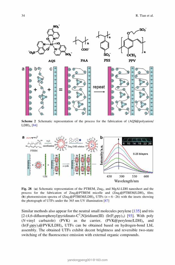
Similar methods also appear for the neutral small molecules perylene [135] and tris
[2-(4,6-difluorophenyl)pyridinato-C2,N]iridium(III) (Ir(F2ppy)3) [93]. With poly
(N-vinyl carbazole) (PVK) as the carrier, (PVK@perylene/LDH)n and
(Ir(F2ppy)3@PVK/LDH)n UTFs can be obtained based on hydrogen-bond LbL
assembly. The obtained UTFs exhibit decent brightness and reversible two-state
switching of the fluorescence emission with external organic compounds.
Fig. 28 (a) Schematic representation of the PTBEM, Znq2, and MgAl-LDH nanosheet and the
process for the fabrication of Znq2@PTBEM micelle and (Znq2@PTBEM/LDH)n film;
(b) photoemission spectra of (Znq2@PTBEM/LDH)n UTFs (n¼ 6�26) with the insets showing
the photograph of UTFs under the 365 nm UV illumination [87]
Scheme 2 Schematic representation of the process for the fabrication of (AQS@polyanion/
LDH)n [84]
34 R. Tian et al.

6.2.2 Intercalation Assembly
Intercalation is one of the most common methods to incorporate the photoactive
molecules into the interlayer of LDHs, and the powdered forms of layered organic–
inorganic composites can be obtained. Various anionic molecules can be interca-
lated due to the diversity of the composition in layers and ion exchangeable
properties of LDHs. Several intercalation methods have been applied and devel-
oped, such as direct intercalation (typical coprecipitation), ions exchange, and
co-intercalation. The intercalation of chromophore molecules into LDHs may
efficiently inhibit the quenching problems induced by molecular aggregation, and
the corresponding fluorescence properties can be largely improved.
Direct Intercalation
By the use of coprecipitation method, the single-component chromophore mole-
cules can be intercalated into the LDH galleries. As an example, 3,4,9,10-perylene
tetracarboxylate (PTCB) anion has been intercalated into LDH layers by
dropwisely adding the mixed solution of sodium hydroxide and PTCB to the
solution containing magnesium nitrate and aluminium nitrate [96]. Figure 29
shows the XRD pattern of as-prepared PTCB/LDH composites, the main charac-
teristic reflections appeared at 4.771� (003), 9.641� (006), and 14.331� (009)
indicated the successful intercalation of PTCB molecules. Good multiple relation-
ships can be observed for d003, d006, and d009 between the basal, second- and third-
order reflections. The stronger reflection of 006 compared with 003 implied that
PTCB molecules are located in the symmetric center of the half-way plane within
the interlayer region. The photophysical properties, thermolysis behavior, and
orientation arrangements of the PTCB/LDH have been studied to compare with
the pure PTCB. It was found that the intercalation into LDH has weakened the
Fig. 29 Powder XRD
pattern of PTCB/LDH [96]
Layered Double Hydroxide Materials: Assembly and Photofunctionality 35

strong π–π interaction between PTCB anions, which has paved a way for solid-state
optoelectronic devices with high performance.
Ion Exchange
The ion-exchange method is often employed as an alternative of direct intercala-
tion, particularly if the guest anions are unstable in aqueous solution with the
divalent or trivalent metal cations. According to the different stability and affinity
of the interlayer guests, nitrate-LDH is always applied as a precursor in the
ion-exchange process. For example, Shi et al. [104] have fabricated
α-naphthalene acetate (α-NAA) and β-naphthalene acetate (β-NAA) intercalatedLDH materials through ion-exchange method. The XRD in Fig. 30 show the
successful intercalation of these two compounds. The basal spacing of Zn2Al-
NO3-LDH (Fig. 30a) powder is 0.87 nm, while 2.80 and 1.94 nm of basal spacing
can be obtained when NO3� is replaced by α-NAA and β-NAA (Fig. 30b, c).
UV–Vis absorption spectra were used to investigate the status and arrangement
of NAA in the interlayer of LDH. The pristine NAA in solution, physical mixture of
NAA and LDH, and NAA intercalated LDH samples were measured. For the
mixture of α-NAA and LDH, a broadened and red-shifted absorption band can be
observed. The broaden of band is due to the existence of a continuous set of
vibrational sublevels in each electronic state; red shift of 14 nm is the result of
ordered and dense packing of molecules in the interlayer, and this may cause
intermolecular interactions between NAA (Fig. 31A, line b). In contrast, the
α-NAA intercalated LDH sample (curve c in Fig. 31A) exhibits a monomer form
in the LDH galleries without any shift caused by intermolecular interactions.
Similar results can be obtained for β-NAA samples (Fig. 31B). Moreover, other
inorganic species (such as quantum dots [124, 125] and polyoxometalates (POMs)
[128] can also be assembled into LDH through ion-exchange process, and the host–
guest interaction can effectively affect the fluorescence properties of the guest
molecules.
Fig. 30 XRD patterns for (a) Zn2Al-NO3-LDH powder, (b) α-NAA-LDH powder, and
(c) β-NAA-LDH powder [104]
36 R. Tian et al.

Co-intercalation
Co-intercalation is an effective way if guest molecules are difficult to accommodate
in the LDH layers. Frequently, surfactant, such as dodecyl sulfonate (DDS�),1-heptanesulfonic acid sodium (HES), and dodecylbenzene sulfonate (DBS) can
be employed as the co-intercalation agents. The introduction of surfactant has
provided an enlarged interlayer spacing, which facilitates the successful incorpo-
ration of guest. Moreover, a nonpolar and homogeneous microenvironment can be
obtained due to surfactant, and thus, the adjustment of the orientation and aggre-
gation status of photofunctional molecules can be achieved. The adjustment of ratio
of fluorophore and surfactant within the LDH layer can effectively influence the
fluorescence properties of guest molecules. Several fluorophores have been
co-intercalated in the LDH layers, such as sulforhodamine B (SRB) [94], zinc
phthalocyanines (ZnPc) [92], fluorescein (FLU) [106, 107, 136], ammonium
1-anilinonaphthalene-8-sulfonate (ANS) [137], and other similar systems
(as shown in Table 8). The quantity of intercalated guest can be modulated through
tuning the ratio of guest and surfactant, and the detailed information will be further
introduced in Sect. 6.3.1.
Fig. 31 UV–Vis absorption spectra of (A) α-NAA for (a) in solution, (b) mixed with LDH, and
(c) intercalated in LDH; (B) β-NAA for (a) in solution, (b) mixed with LDH, and (c) intercalated in
LDH [104]
Layered Double Hydroxide Materials: Assembly and Photofunctionality 37

6.3 Adjusting Photofunctionalities of LDH-Based Materials
6.3.1 Static Adjustment
Modulation Based on the Charge Density and Elemental Composition of Host
Layer
By controlling the synthesis condition and mole ratio of raw materials, the charge
density of the LDH layer can be varied in a wide range, which enabled the tunable
photo-related properties of intercalated guest molecules. Luminescent
benzocarbazole anions (BCZC), which possesses high quantum yield and good
color purity, has been incorporated into LDH via coprecipitation with different
layered charge densities of LDHs [98]. MgAl-LDHs with the Mg/Al ratio at 2 and
3.5 were employed to adjust the arrangement of the BCZC. Figure 32A shows the
structural model for LDHs with different charge density. LDHs with host layer
containing 12 Mg atoms and 6 Al atoms (Mg/Al ratio of 2, Fig. 32A-a) and 14 Mg
atoms and 4 Al atoms (Mg/Al ratio of 3.5, Fig. 32A-b) have been adopted. The main
characteristic peaks for BCZC/LDH samples present a good multiple relationship
for the basal, second- and third-order reflections, with the basal spacing of 2.31 nm
for Mg/Al ratio of 2 and 2.39 nm for Mg/Al ratio of 3.5 (Fig. 32B).
UV–Vis spectra of the pristine BCZC and intercalated BCZC have been studied.
The absorption bands at 265, 289, and 320 nm can be attributed to the characteristic
absorptions of carbazole-type dyes for BCZC in solution, while the solid sample of
pristine BCZC and BCZC intercalated LDH composites possess a broadband at
295 and 410 nm due to the molecule stacking (Fig. 33A). Figure 33B shows the
fluorescence emission spectra for these samples, and emissions in the range of 490–
520 nm are displayed. It can be observed that the spectra of pristine BCZC in
solution is similar with BCZC intercalated LDH with Mg/Al ratio at 2, while
photoemission of the interlayer BCZC (Mg/Al ratio at 3.5) is close to that of its
solid state. Therefore, it can be concluded that variation on the layered charge
Fig. 32 (A) Structure model for the superlattice layer model for MgAl-LDH (dark pink: Al(OH)6octahedra; green: Mg(OH)6 octahedra) for Mg/Al ratio of (a) 2 and (b) 3.5, and (c) structural
model of BCZC; (B) XRD patterns of BCZC/MgAl-LDH (d, e stand for samples with Mg/Al ratio
of 2 and 3.5, respectively; Asterisk: the Al substrate) [98]
38 R. Tian et al.

density could effectively modify the luminescence performance of the guest dye
molecules.
The chemical composition in the LDHs layer also serves as a parameter in
modulating the luminescent behavior. For a sulfonated derivate of stilbene interca-
lated LDHs system [138], compared with the pure sample with the maximum
emission wavelength (λemmax) located at 463 nm, the intercalation products exhibit
tunable λemmax in the range from 456 nm (Co2Al-LDH as the layer) to 484 nm
(Mg2Al-LDH as the layer), demonstrating that the fluorescent wavelength can be
continuously tuned by changing the chemical composition of LDHs, as a result of
different host–guest interactions between the host layer and the interlayer anions.
Modulation Based on the Quantity of Interlayer Molecules
The orientation and aggregation state of interlayer guest can be tuned by controlling
the contents of the interlayer chromophores, and thus, the fluorescence performance
can also be varied. For example, rhodamine is a widely used fluorescent dye due to
its high quantum yields, but its fluorescence properties are severely affected by its
concentration, and a high concentration usually leads to aggregation and subsequent
quenching [139]. Yan et al. have achieved the co-intercalation of sulforhodamine
B (SRB) and dodecylbenzene sulfonate (DBS) with different ratios into LDHs
[94]. Figure 34A shows the XRD pattern of SRB-DBS/LDH with different interlayer
concentration of SRB, and it was observed that when the ration of intercalated SRB
increased from 0 to 16.67 %, the spacing increased from 28.87 Å to the maximum
30.96Å (Fig. 34A, curve a–g). And the spacing value has a decreasing trend when the
quantity of SRB increased from 16.67 % to the maximum 100 % (Fig. 34A, curve h–
k). This variation of the interlayer spacing may be a result of changeable distribution
and arrangement of interlayered SRB with different ratios.
Fig. 33 (A) The UV–Vis absorption spectra and (B) normalized photoemission spectra for
(a) pristine BCZC aqueous solution (10 mM), (b) solid BCZC, and BCZC/MgAl-LDH with the
Mg/Al ratio of (c) 2 and (d) 3.5 [98]
Layered Double Hydroxide Materials: Assembly and Photofunctionality 39

The fluorescence emission behavior of SRB-DBS/LDHs with different SRB
content has been studied, and the emission spectra are displayed in Fig. 35A.
With the increase of SRB in SRB-DBS/LDHs, the intensity of fluorescence emis-
sion exhibits an increase to maximum (content of SRB is 4.76 %), followed by a
continuous decrease. The emission peak varies from 587 to 619 nm as the SRB
content increased to 75 %. This can be attributed to the variation of distribution
status and arrangement of interlayer SRB, and high concentration may facilitate the
formation of dye aggregation, accounting for the red shift and quenching in
fluorescence emission. UV–Vis spectra (Fig. 35B) show that absorption peaks at
Fig. 35 Study of SRB(x%)-DBS/LDH with different SRB content: (a) 0.99 %, (b) 1.96 %,
(c) 4.76 %, (d) 7.41 %, (e) 9.09 %, (f) 16.67 %, (g) 25.0 %, (h) 50.0 %, and (i) 75.0 % for (A)
photoemission spectra (with the excitation wavelength of 360 nm and the inset shows the
fluorescence intensity and the variation of maximum emission wavelength) and (B) solid UV–
Vis absorption spectra [94]
Fig. 34 (A) Powder XRD patterns of SRB(x%)-DBS/LDH (a, 0 %; b, 0.99 %; c, 1.96 %; d,
4.76 %; e, 7.41 %; f, 9.09 %; g, 16.67 %; h, 25.0 %; i, 50.0 %; j, 75.0 %; k, 100 %). (B) The plot of
interlayer spacing vs. SRB concentration [94]
40 R. Tian et al.

360 and 570 nm increase as more SRB molecules were intercalated. An absorption
shoulder at ca. 530 nm appears (SRB content of 16.67 %), indicating a formation of
H-type aggregates.
For the fluorescence lifetime, compared with pristine SRB solution (2.38 ns), the
fluorescence lifetime of SRB-DBS/LDH firstly increases with SRB content
changed from 0.99 to 16.7 %, and the lifetime decreases when the content increased
to 25 % (Table 9). The change in lifetime further indicates the aggregates of SRB,
which is in accordance with emission and absorption spectra. Therefore, the
absorption/emission intensity, wavelength, and fluorescence lifetime could be
changed due to the variation of interlayer content.
As another example, Li et al. have prepared co-intercalated systems based on bis
(8-hydroxyquinolate-5-sulfonate)zinc anion (ZQS2�) and DDS, and the fluores-
cence properties can also be changed obviously through adjustment of intercalation
content [86]. Table 9 shows the fluorescence emission information of ZQS-DDS/
LDHs. For different ratio of ZQS in the interlayer and different host layer compo-
sition, the maximum emission wavelength and quantum yields can be finely
controlled. Furthermore, the brightest luminous intensity, together with the longest
fluorescence lifetime, was observed for DDS-ZQS (5 %)/Mg2Al-LDH.
Liang et al. have investigated the dispersion and corresponding singlet oxygen
production efficiency of zinc phthalocyanines (ZnPc) after intercalation [92]. XRD
patterns showed the basal spacing varied in the range of 23.6–24.6 Å with the
content of ZnPc changed from 1 to 10 %, and the co-intercalation of ZnPc and SDS
can be further demonstrated by FT-IR spectra. Compared with the pristine form of
ZnPc in water and ethanol, the monomer dispersed state with maximum absorption
at 678 nm can be obtained via intercalation into the LDH galleries (Fig. 36A, curve
c), indicating a low polarity microenvironment provided by LDH gallery for the
formation of monomeric ZnPc. With the ZnPc content increased from 1 to 10 %, the
absorption intensity exhibit a gradual increase at 678 nm, while a broad absorption
Table 9 The maximum wavelength of absorption and emission of the ZQS(x%)-DDS/LDH [86]
Sample λabs (nm) λem (nm) PLQY (%)
DDS-ZQS(2 %)/Mg2Al-LDH 258, 379 464 5.0
DDS-ZQS(5 %)/Mg2Al-LDH 258, 381 479 24.4
DDS-ZQS(10 %)/Mg2Al-LDH 256, 390 486 18.1
DDS-ZQS(20 %)/Mg2Al-LDH 256, 395 487 17.3
DDS-ZQS(40 %)/Mg2Al-LDH 256, 399 488 16.4
DDS-ZQS(60 %)/Mg2Al-LDH 256, 396 491 9.6
DDS-ZQS(80 %)/Mg2Al-LDH 256, 389 495 5.8
ZQS(100 %)/Mg2Al-LDH 256, 385 497 2.3
DDS-ZQS(5 %)/Mg3Al-LDH 258, 382 490 9.7
DDS-ZQS(5 %)/Zn2Al-LDH 258, 377 491 27.4
DDS-ZQS(5 %)/Zn3Al-LDH 258, 373 491 25.9
H2ZQS crystal 258, 396 466 20.4
ZQS solution (5� 10�5 mol L�1) 254, 368 516 –
Layered Double Hydroxide Materials: Assembly and Photofunctionality 41

band appeared in 580�610 nm when ZnPc content increased to 10 % (Fig. 36B).
The fluorescence intensity displayed a first increase and then decrease with the
optimal luminous intensity presents in 1.5 % ZnPc/LDH (Fig. 36C). This result
indicates an aggregate-induced-quenching process. Moreover, better stability
against UV irradiation can be obtained for ZnPc/LDH composites compared with
the pristine ZnPc. In addition, the high dispersed ZnPc as monomeric state in the
interlayer region of LDH induced by the host–guest and guest–guest interactions
can make a big contribution to large singlet oxygen production efficiency in vitro
and in vivo photodynamic therapy.
Modulation of the Orientation and Polarized Luminescence
As described above, due to the confined environment of LDHs, interlayer guest
molecules possess ordered arrangement and preferred orientation, and thus, the
Fig. 36 (A) The UV–Vis absorption spectra: (a) pristine ZnPc (2� 10�5 M) in aqueous solution,
(b) pristine ZnPc (2� 10�5 M) in 60 % ethanol solution, (c) ZnPc (1.5 %)/LDH (3� 10�4 M)
aqueous suspension, (d) pristine LDH (3� 10�4 M) aqueous suspension. (B) The UV–Vis
absorption spectra and (C) photoluminescence spectra of various ZnPc (x%)/LDH suspension
(3� 10�4 M) with x ranging in 1–10 %. (D) Decay curves of absorption at 678 nm for ZnPc
(1.5 %)/LDH, ZnPc (2.5 %)/LDH, and 635 nm for pristine ZnPc, respectively, as a function of
irradiation time (650 nm) [92]
42 R. Tian et al.

obtained LDHs can present polarized luminescence properties. For example, cou-
marin-3-carboxylate (C3C) and DDS has been co-intercalated into LDH layer, and
a C3C-DDS/LDH thin film can be subsequently obtained through solvent evapora-
tion method [110]. Figure 37a shows the sole strong basal reflections (00l ) for thethin films of C3C-DDS/LDH, indicating a well c-oriented assembly of LDH
platelets. The uniform and smooth morphology of this film can also be confirmed
by top SEM and AFM (Fig. 37b, d). The obtained thin film possesses continuous
structure with the average thickness of 1.2 μm (Fig. 37c).
The polarized luminescence of the C3C–DDS/LDH thin films was investigated.
Generally, polarized luminescence can be evaluated by determining the anisotropy
value r, which can be expressed by the formula:
r ¼ Ik � I⊥
Ik þ 2I⊥or r ¼ IVV � GIVH
IVV þ 2GIVH;
where IVH stands for the PL intensity obtained with vertical polarized light excita-
tion and horizontal polarization detection and IVV, IHH, IHV are defined in a similar
way; G¼ IHV/IHH is determined from an aqueous solution of the C3C. Theoreti-
cally, the value of r is in the range from �0.2 (absorption and emission transition
dipoles are perpendicular to one another) to 0.4 (two transition dipoles are parallel
Fig. 37 (a) XRD pattern, (b) top-view SEM, (c) side-view SEM, and (d) tapping-mode AFM
images for C3C-DDS/LDH (1.96 %) thin film [110]
Layered Double Hydroxide Materials: Assembly and Photofunctionality 43

to each other). The polarized photoemission spectra of the C3C-DDS/LDH (optimal
luminous properties was obtained when the content of C3C was 1.96 %) thin film
are displayed in Fig. 38. Two typical measurement setups of polarized fluorescence
were employed to determine the fluorescence anisotropic value r. It can be observedthat the value r is about 0.15 for horizontal excitation (Fig. 38a) and 0.10 for
vertical excitation (Fig. 38b), demonstrating highly dispersed and well-oriented
arrangement of C3C in the gallery of LDH.
To further investigate variation of the basal spacing influenced by the ratio of
C3C/DDS, molecular dynamic simulation was employed to study arrangement and
orientation of the C3C anions in the LDH galleries with C3C content ranged from
0, 25, 50, 75, to 100 % (Fig. 39). The results show that the DDS anions under low
and high concentration exhibit twisting and stretching state, which severely influ-
ence the basal spacing of the C3C-DDs/LDH composites.
Angle θ was defined as the orientational angle of the C3C plane with respect to
the LDH layer. It was observed that θ varies between 29 and 55 � with the most
VHVV
VHVV
450 500
Wavelength/nm Wavelength/nm
Inte
nsity
/a.u
.
Inte
nsity
/a.u
.
Ani
sotr
opy
(r)
Ani
sotr
opy
(r)
550 6000.00 460 480 500 520 540 560 580
0.00
0.05
0.10
0.15
0.20
0.05
0.10
0.15
0.20
0.25a b
Fig. 38 Polarized fluorescence profiles in the VV, VH modes and anisotropic value (r) for theC3C-DDS/LDH (1.96 %) thin film sample. (a) and (b) correspond to excitation light with glancing
and normal incidence geometry, respectively [110]
Fig. 39 Snapshots of the simulated equilibrium structures for C3C-DDS/LDHs (x%): (a) 0 %, (b)
25 %, (c) 75 %, (d) 100 % [110]
44 R. Tian et al.

probable angle of ca. 32� for the intercalated C3C ratio at 100 %. Stimulation from
Fig. 40 shows that all the C3C molecules are inclining to the layers, attributed to the
strong electrostatic interaction between the positive-charged host layers and the
C3C anions. The optimal angles of 32�, 42�, and 54� can be obtained when the ratioof DDS and C3C varies from 0 to 1, with a tendency to change from a tilted to a
vertical arrangement with respect to the layers. Moreover, the distribution of θangle for the C3C-DDS/LDH (25 %) with high DDS content corresponds to the
relative rigid environment imposed by the DDS which restricts the flexible space
for the C3C guest anions. These results show that the changeable interlayer ratio of
two components can highly influence the preferred orientation of the photoactive
guests.
Several LDH-based LbL thin films also exhibit polarized luminescence proper-
ties. Due to the rigid-rod chain configuration or intrinsic one-dimensional structural
anisotropy for these luminescent molecules, well-oriented thin films with a macro-
scopic polarized optical effect can be prepared. For example, sulfonated
carbocyanine derivate (Scy) has been assembled with LDH nanosheets, which
presents polarized near-infrared (NIR) emission luminescence [100]. The average
anisotropy of pristine Scy solution is about 0.2 in the range from 770 to 820 nm,
which is related to the random distribution of the dye molecules in the nonviscous
liquid. NIR polarized fluorescence with the anisotropic value of ~0.8 can be
0.035
0.0300.025
0.0200.0150.0100.005
0.040
0.000
0.0450.0400.0350.030
Probab
lity
Probab
ility
q /degree q /degree
0.0250.0200.0150.0100.005
0.050
0.000
20 30 40 50 6010 70
0.035
0.030
Probab
lity
q /degree
0.0250.0200.0150.010
0.005
0.040
0.000
10 20 30 40 50 60 70 80
0.040
0.035
0.030
Probab
lity
q /degree
0.0250.0200.0150.0100.0050.000
10 20 30 40 50 60 70 80
20 30 40 50 60 7010 80
a
c d
b
Fig. 40 The distributions of orientational angle θ (the plane of C3C with respect to the LDH layer)
with different ratios of C3C to DDS for C3CDDS/LDH (x%): (a) 100 %, (b) 75 %, (c) 50 %, and
(d) 25 % [110]
Layered Double Hydroxide Materials: Assembly and Photofunctionality 45

observed for the Scy/LDH UTF (Fig. 41a), confirming the ordered arrangement of
Scy between the LDH nanosheets. There were no obvious changes for UTFs with
different number of bilayers (Fig. 41b), indicating the independent macroscopic
polarized luminescence on assembly process.
Moreover, due to the regular assembly within the LDH nanosheets, some
molecules with no luminescence polarization can also exhibit well-defined anisot-
ropy: the as-mentioned Ru(dpds)3/LDH UTF is such an example [83]. The pristine
Ru(dpds)3 in aqueous solution shows r at 0.02, while the r value for typical (Ru
(dpds)3/LDH)32 achieved about 0.2–0.3 when comparing the parallel and perpen-
dicular directions to the excitation polarization (IVV vs. IVH) for the in-plane
polarized excitation light. Upon increasing the number of bilayers, the r value
increases systematically, indicating enhancement of the orderly degree for Ru
(dpds)3 between LDH nanosheets.
Construction of Tunable Multicolor Emissive Film Materials
Materials with multicolor luminescence (especially white-light emission) have
been considered as excellent candidates in full-color displays [140], light-emitting
diodes (LEDs) [141], and optoelectronic devices [142]. Conventional strategies
usually rely on combination of three primary color luminescence species, such as
polymers [143], rare-earth compounds [144], metal complexes [145], and semicon-
ductor nanocrystals [146]. However, the different luminous efficiency and inten-
sity, complex fabrication process, as well as complicated interaction between these
chromophores have severely limited the development of multicolor materials. Yan
et al. [77] have introduced LDH nanosheet as building block to combine different
chromophores; in this case, materials with a rigid and ordered microenvironment,
finely tuning emission color, and polarized multicolor luminescence can be achieved.
Fig. 41 (a) Polarized fluorescence profiles in the VV, VH modes and anisotropic value r for the(Scy/LDH)8 UTF. (b) The correlation between fluorescence anisotropic values r of the Scy/LDHUTFs (averaged in the range 770–820 nm) and the bilayer number in the measurement mode with
glancing incidence geometry [100]
46 R. Tian et al.

Figure 42 shows the assembly process of multicolor emission materials employing
APPP (blue) [76], APPV (orange) [78], SPT (red) [79], and BNMA (green) [95] as
primary chromophores. The combination of these individual chromophores can
obtain organic/inorganic UTFs with emission color throughout the whole visible
region, even the white color emission.
Taking (APPP/LDH)12/(BNMA@PVS/LDH)n (n¼ 0–6) UTFs (B/G lumines-
cence) as the example, the assembly process was carried out by assembling
(BNMA@PVS/LDH)n (n¼ 1–6) bilayers onto the as-prepared (APPP/LDH)12UTF. Figure 43a1 shows the fluorescence monotonic increase at 481 and 504 nm
which can be attributed to the emission of BNMA@PVS, while the emission peak
at 410 nm is assigned to the pristine (APPP/LDH)12 UTF. The similar spectra
between assembled UTFs and solutions indicate that the aggregation of BNMA can
be avoided throughout the assembly process. The insets show the photographs
taken under UV illumination, and high luminescence can be observed for these
UTFs. The color coordinates in Fig. 43a2 demonstrated that the color of the UTFs
can be tuned from blue (CIE 1931: (0.172, 0.149); n¼ 0) to bluish green (CIE 1931:
(0.208, 0.343); n¼ 6) effectively. The multicolor emission of blue/orange, red/blue,
and red/green can also be tuned by this method throughout the whole visible region
(Fig. 43b–d). In addition, based on the periodic ordered structure of these UTFs,
they exhibit well-defined multicolor polarized luminescence with high anisotropy,
which may effectively extend its application in multicolor or white polarized
photoemission devices.
For the inorganic building blocks, quantum dots (QDs) [147] have attracted great
attention due to high quantum yields, good stability, and high color purity [148],
Fig. 42 (a) Representation of one monolayer of MgAl-LDH nanosheet; the chemical formulae of
(b) APPP (blue luminescence), (c) BNMA@PVS (green luminescence), (d) APPV (orangeluminescence), and (e) APT (red luminescence); (f) procedure for assembling two-color emitting
UTFs with blue/green (B/G), blue/orange (B/O), red/blue(R/B), and red/green (R/G) lumines-
cence [77]
Layered Double Hydroxide Materials: Assembly and Photofunctionality 47

and application of QDs in the multicolor emission materials has also been carefully
investigated. Liang et al. [117] have assembled QDs with LDH nanosheets to obtain
highly ordered structure and finely tunable fluorescence, and the tunable color in the
red–green region could be achieved by adjusting the assembly cycle number and
sequence of red- and green-emission QDs. In addition, Tian et al. [102] have
introduced fabrication of precisely tuned white color emission UTFs employing
red- and green-emission QDs and blue 2,20-(1,2-ethenediyl) bis
[5-[[4-(diethylamino)-6-[(2,5-disulfophenyl) amino]-1,3,5-triazin-2-yl] amino]
benzene-5 sulfonic acid] hexasodium salt (BTBS). By adjusting the deposition
sequence and relative ratio of the blue-, green- and red-emission units, luminescent
UTFs with finely tunable photoemission in the white spectral region were obtained
(Fig. 44B). Moreover, the color coordinates of the best white-light emission
(denoted as f in Fig. 44C) could achieve (0.322, 0.324) which is very close to the
standard coordinates of white light (0.333, 0.333).
The emission color of hybrid composites can be tuned in response to different
excitation light, which has been recognized as an intelligent photoluminescence
material. This composite UTF was illustrated as (BTBS/LDH)12(QD-530/
LDH)20(QD-620/LDH)7, composed of three chromophores: red-emission and
green-emission QDs and blue-emission organic compound BTBS. Due to the
different excitation and emission spectra of these three chromophores, it can be
concluded that BTBS can be excited at 360 nm while photon energy above 500 and
600 nm can efficiently excite QD-530 and QD-620. Therefore, excited at 360, 460,
and 560 nm, the UTFs display three different emission spectra as well as
corresponding white, orange, and red color, respectively (Fig. 45). These hybrid
materials responding to external excitation light via the alteration of emission color
can be widely used in anti-forgery, colorimetric sensors, and display devices.
Fig. 43 Fluorescence spectra (the inset shows the ratio of two chromophores), photographs under
UV light (365 nm), and the change in color coordinates with n for (a) (APPP/LDH)12/
(BNMA@PVS/LDH)n (n¼ 0–6) UTFs (for B/G luminescence), (b) (APPP/LDH)12/(APPV/
LDH)n (n¼ 0–18) UTFs (for B/O luminescence), (c) (APPP/LDH)12/(APT/LDH)n (n¼ 0–18)
UTFs (for R/B luminescence), and (d) (APT/LDH)12/(BNMA@PVS/LDH)n (n¼ 0–6) UTFs
(for R/G luminescence) [77]
48 R. Tian et al.

Fig. 45 (A) The emission spectra, (B) photographs of the (BTBS/LDH)12(QD-530/
LDH)20(QD-620/LDH)7 UTF with different excitation wavelength: (a) excited at 360 nm,
(b) excited at 460 nm, (c) excited at 560 nm, respectively [102]
Fig. 44 Optical spectroscopy characterization of the (BTBS/LDH)m(QD-530/LDH)n(QD-620/
LDH)p UTFs: (A) the change in color coordinates in the white-light region, (B) their photographsunder UV light (365 nm), (C) the color coordinates and detailed compositions of these UTFs [102]
Layered Double Hydroxide Materials: Assembly and Photofunctionality 49

Moreover, sulfonated Eu- and Tb-based lanthanide complexes can also be
employed in fabrication of multicolor emission materials [149]. After the synthesis
of red-emissive Eu(DBM)3bath and green-emissive Tb(acac)3Tiron (DBM,
dibenzoylmethane; bath, bathophenanthroline disulfonate disodium; acac,
2,4-pentanedione), these lanthanide complexes can be assembled with LDH to
obtain highly emissive luminescence with tunable color on tuning of their relative
ratio (Fig. 46). In addition, these UTFs exhibit well-defined one and two-color
polarized fluorescence and obvious red and green up-conversion emission upon
excitation by near-IR laser light.
6.3.2 Dynamic Tuning of the Emission
The photofunctional materials with smart-responsive properties can present
dynamic changes by tuning the external stimuli. Such dynamic-responsive
photofunctional materials should have rapid response time, high sensitivity, obvi-
ous signal change, and high stability for long-term use [150]. In order to achieve
these requirements, LDHs have been employed as an effective inorganic matrix and
building block for the ordered arrangement and improved stability of active chro-
mophores in the solid state [151]. Recently, the smart-responsive LDH
photofunctional materials responding to environmental stimuli (such as chemical
substances, pH value, temperature, pressure, light irradiation, and electricity) have
been studied, which present dynamic tuning of their photofunctionalities.
Detection of Chemical Substances
Chemo-sensor plays an important role in the detection of heavy metal ions (HMIs),
biomolecules, environmental pollutants, and chemical warfare agents [152–156].
The basic mechanisms of fluorescence detection for most of hazardous substances
Fig. 46 Digital photos of flexible (a) (Eu(DBM)3bath/LDH)8 and (b) (Tb(acac)3Tiron/LDH)8UTFs under UV irradiation; (c) fluorescence spectra of (Eu(DBM)3bath/LDH)10/(Tb(acac)3Tiron/
LDH)n (n¼ 0–14) UTFs, and insets show the photographs under UV light [149]
50 R. Tian et al.

rely on the quenching induced by the binding between analytes and fluorescence
probe molecules. However, the commonly studied fluorescence probe molecules
are in aqueous or powder states, and the complicated operation process and
difficulty in recyclability have severely limited the application of such materials.
Therefore, the development of suitable species and/or new approaches toward the
facile fabrication of chemo-sensor with high sensitivity, fast response, and decent
recyclability remains a stimulating challenge.
Fabrication of photofunctional UTFs based on the LbL assembly with LDH
nanosheets can serve as an effective method to solve the above difficulties. A
certain amount of fluorescence molecules have been applied (such as BTBS
[101], calcein [109], and 1-amino-8-hydroxy-3,6-disulfonaphthalene (H-acid)
[157]) as probes for the detection of HMIs (such as mercury ion). Taking BTBS/
LDH system as an example, fluorescence emission spectrum shows a monotonic
increase with n at ca. 445 nm, and the UTFs show well-defined blue luminescence
under UV light (Fig. 47a and inset). The systematical and significant photoemission
decrease of the (BTBS/LDH)32 UTF can be observed in responding to the addition
of Hg2+ (Fig. 47b), while different degrees of quenching for metal ions were
presented in Fig. 47d. The strongest quenching by Hg2+ is related to the high
thermodynamic affinity with typical O-chelate ligands and metal-to-ligand binding
kinetics. Reproducibility was carried out for alternative dipping the UTFs into Hg2+
and EDTA, and the UTFs can recover to the pristine fluorescence intensity at least
five cycles (Fig. 47c). The immobilization by LDH has guaranteed the repeatability
of HMI detection, while the drop-casted sample is easy to fall off in solution.
Moreover, Jin et al. have proposed the detection of biomolecules dextran-40 by
employing 8-amino-1,3,6-naphthalenetrisulfonates (ANTS) and LDH as building
block [158]. The obtained (ANTS/LDH)n UTF can be adapted in the detection of
Mg2+ which is important in stabilizing the structure of DNA and transferring of
biological signals [159]. The detection limit of 2.37� 10�7 M can be obtained.
Trinitrotoluene (TNT), which is an important explosives commonly applied in both
military and forensic science, can be specifically detected by (BBU/LDH)n UTFs,
which are assembled by 4,40-bis[2-di(b-hydroxyethyl)amino-4-(4-sulfophe-nylamino)-
s-triazin-6-ylamino] stilbine-2,20-disulfonate (BBU) with LDH nanosheets [103].
Li, Qin, and coworkers have efficiently detected the volatile organic compounds
(VOCs) based on modulating the FRET process. The basal spacing and the
interlayer distance between donor and acceptor can be varied with external stimuli
(VOC vapor), and thus, the color change from blue to violet can be observed for the
detection [93, 135].
pH Sensor
Optical pH sensor with stable lifetime and signal is of great importance in environ-
mental research, toxicological assay, blood measurement, and biotechnology [160–
163]. The red-emissive (SPT/LDH)32 UTF described above exhibited reversible
responses toward pH in alternative dipping in aqueous at pH¼ 4 and pH¼ 13 [79].
Layered Double Hydroxide Materials: Assembly and Photofunctionality 51

It has been found that dipping the (SPT/LDH)32 UTFs in aqueous solutions with
different pH values ranging from 4 to 14 for 10 s is sufficient for the proton to
transfer into the UTFs, and the UTFs can display a variation of maximum emission
wavelength (Fig. 48a). This result can be attributed to the occurrence of protonation
and deprotonation of SPT between the LDH nanosheets. Reproducibility was also
carried out in alternation between pH¼ 4 and pH¼ 13 for five cycles (Fig. 48b),
and rapid change from bright red luminescence at pH¼ 13 to dark orange at pH¼ 4
can be observed under UV light (365 nm).
Shi et al. have fabricated another pH sensor based on the co-intercalation of the
fluorescein (FLU) and 1-heptanesulfonic acid sodium (HES), and the obtained
samples exhibit good photo- and storage stability as well as high repeatability
Fig. 47 Fluorescence spectra of the (BTBS/LDH)32 UTFs for (a) variation in bilayer number
(inset shows the photograph under UV light), (b) treated with Hg2+ at different concentration, and
(c) detecting the Hg2+ after alternate treatment by metal ions and EDTA aqueous solutions over
continuous reversible cycles (the inset shows the fluorescence intensities over five cycles); (d)
comparison of the normalized luminescence intensity of the UTF responding to aqueous solutions
of different metal ions (10�2 M) [101]
52 R. Tian et al.

toward responses of pH [106]. Employing electrophoretic deposition method, a
sensor with broad linear dynamic range in pH (5.02–8.54) can be obtained
(Fig. 49a). Remarkable repeatability in 20 cycles can be achieved with the relative
standard deviation (RSD) less than 1.5 % (Fig. 49b). The sensitive and rapid
responses of SPT/LDH and FLU/LDH toward pH materials indicated their potential
application as pH luminescence sensor.
Fig. 49 (a) Fluorescence intensity of the pH sensor recorded after 1, 5, 10, 15, and 20 cycles at
pH 5.02, 6.51, and 8.54 and (b) the reversibility recorded by alternate measurement in two
solutions with pH 5.02 and 8.54 [106]
Fig. 48 (a) Variation of the maximum photoemission wavelength of the (SPT/LDH)32 UTF with
different pH values and (b) the reversible photoemission response upon alternation between
pH¼ 4 and pH¼ 13 (the inset show the luminescence photographs of the UTFs at the two pH
conditions) [79]
Layered Double Hydroxide Materials: Assembly and Photofunctionality 53

Temperature Sensor
Temperature is one of the most common and natural environmental factor, and
thermochromic materials can be potentially used in temperature sensor and devices
[164–166]. Therefore, much attention has been paid on fabrication of
thermochromic materials with good sensitivity and reversibility. Bis
(2-sulfonatostyryl)biphenyl (BSB) was employed as thermochromic luminescence
(TCL) molecule to assemble with LDH nanosheet to achieve an ordered supramo-
lecular UTFs [99]. Figure 50 shows the assembly process of (BSB/LDH)n with the
monotonic increase in absorption and fluorescence emission spectra. The maximum
emission peak is located at 444 nm, and no broadening or shift can be observed in
the whole assembly process, indicating no interaction between molecules occurred.
The XRD data show the ordered and monolayer arrangement of BSB in the
interlayer of LDHs.
The TCL behavior of the UTFs was studied on heating from 20 to 100 �C, and agradual decrease in fluorescence intensity with a red shift from 444 to 473 nm can
be observed (Fig. 51a). A visible change can be observed under UV light with the
emission color changes from blue to bluish green upon heating. The recover in color
and spectrum can be accomplished when the UTF cooled to 20 �C, and this heating–cooling process can be readily repeated (Fig. 51a, inset). Fluorescence lifetimes of
these (BSB/LDH)n UTFs were also studied to obtain the excited-state information.
The results show that lifetime of the UTF is longer at 100 �C (1.9 ns) than that at
20 �C (0.9 ns), and the recycle can also be achieved (Fig. 51b). Figure 51c shows the
polarized fluorescence measurements of the UTFs, and the degree of structural
order can be obtained by detecting the fluorescence anisotropy r. The r value
decreases from 0.15 at 20 �C to 0.02 for UTFs at 100 �C, which exhibit a less
ordered arrangement of molecules at high temperature.
Fig. 50 Characterization of (BSB/LDH)n (n¼ 4–32) UTFs. (a) UV–Vis absorption spectra (inset:
plots of the absorbance at 204 and 349 nm vs. the number of bilayers n) and (b) fluorescence
spectra. The insets in (a) and (b) are photographs of UTFs with different n under daylight and UVlight (365 nm), respectively [99]
54 R. Tian et al.

Similar observations can also be obtained for (CdTe QDs/LDH)30 UTF [116];
Fig. 52a shows the responses toward temperature upon heating, and an intensity
decrease along with red shift can be observed. These inorganic–inorganic hybrid
UTFs possess good sensitivity, and�1 % per �C decrease and 0.1 nm �C�1 red shift
can be obtained. The UTFs exhibit excellent repeatability for eight cycles in the
heating–cooling process (Fig. 52b).
Aromatic azo dyes, such as 4-(4-anilinophenylazo)benzenesulfonate (AO5),
have been widely used due to the easy transformation between the azo A-form
and hydrazone H-form (Scheme 3). It was well known that this tautomerism is very
sensitive to the surrounding environment; therefore, it has been widely used as
thermal sensors and in molecular memory storage. In order to further modulate the
environment in an inorganic matrix, AO5 was co-intercalated with sodium
dodecylbenzene sulfonate (SDS) into the interlayer of LDH [112]. The absorption
500
400
400 450 500
300
200
100
0
500
600
400
300
200
100
00550 600 650 700
Wavelength/nm
23°C
30°C
40°C
50°C
60°C
70°C
80°C
Inte
nsity
Intensity
Inte
nsity
Peak/nm
558560
564566568
562
R=0.994 R=0.998
20 30 40 50 60 70 80Temperature/°C
20 30 40 50 60 70 80Temperature/°C
400
320
240
160
80
1 2 3 4 5 6 7 8Number of cycle
Heating
Cooling
a b
Fig. 52 (a) The luminescence spectra of the (CdTe QDs/LDH)30 UTF in the temperature range
23–80 �C (inset: the emission position or intensity as a function of temperature, respectively); (b)
the reversible fluorescence response of eight consecutive cycles (the inset shows the UTF
photographs at 23 and 80 �C, respectively) [116]
Fig. 51 TCL of the (BSB/LDH)8 UTF at 20 and 100 �C. (a) Fluorescence emission spectra (inset:
the reversible fluorescence response over five consecutive cycles); (b) typical fluorescence decay
curve (inset: fluorescence lifetimes over five consecutive cycles, black: 20 �C, red: 100 �C; (c)fluorescence anisotropy over five consecutive cycles [99]
Layered Double Hydroxide Materials: Assembly and Photofunctionality 55

band at 363 nm at room temperature is attributed to the absorption of A-form, and
the intensity increases with elevated temperature (Fig. 53a). The color of the
composite film exhibits reversible change from light yellow to orange in the
range of 35–65 �C, which is in accordance with absorption at 445 nm. Since the
pristine AO5 shows no thermal-responsive behavior, this thermochromic phenom-
enon for the UTF can be a result of tautomerism of interlayer AO5 and changes in
host–guest and guest–guest interactions.
In addition, thermal colorimetric and fluorescent response behaviors of
polydiacetylene (PDA) in the LDH layers were also studied [81]. Diacetylene
(DA) was firstly intercalated into the interlayer of LDHs, and subsequent
PDA/LDH films were fabricated through UV-induced polymerization of interca-
lated DA. And the reversible changes under both visible light and UV light can be
observed in the temperature range of 20–130 �C. This phenomenon can be attrib-
uted to the influence of hydrogen bond formed between LDH layers and PDA
guests.
Pressure Sensor
Pressure-induced chromic, known as piezochromic, is an interesting phenomenon
for luminescence materials responding to natural external force [167]. However, the
research on piezochromic luminescent (PCL) materials remains relative seldom
Fig. 53 (a) In situ absorption spectra of the 10 % AO5-LDH film during heating in the range 35–
65 �C. (b) The correlation between the absorbance of the band at 445 nm and temperature over a
complete heating–cooling cycle (inset shows the corresponding digital photographs) [112]
Scheme 3 Tautomeric equilibrium between the A-Form and H-Form of AO5 [112]
56 R. Tian et al.

compared with other environment-responsive materials (such as temperature-, pH-
and HMI-sensitive materials). Therefore, the investigation on PCL materials with
excellent reversibility, repeatability, and rapid responsiveness is still a challenge
[168, 169].
Yan et al. have studied the fluorescence properties as well as molecular packing
mode of (2,20-(1,2-ethenediyl)bis[5-[[4-(diethylamino)-6-[(2,5disulfophenyl)
amino]-1,3,5-triazin-2-yl] amino]benzene sulfonate anion (BTZB) after intercala-
tion into LDHs matrix [111]. The fluorescence of the pristine BTZB and BTZB/
LDH composites is shown at approximately 452 nm, and significant luminescent
changes can be observed for the BTZB/LDHs treated with pressures while pristine
BTZB remains unchanged. Figure 54a shows the changes of fluorescence spectra
upon increasing the pressure from 0.1 MPa to 18.8 GPa; the emission wavelength
shifts from 452 to 515 nm, which can be assigned to the formation of J-type
Fig. 54 (a) Fluorescence emission and (b) UV–Vis spectra of the BTZB/Mg2Al-LDH at different
pressures (inset shows the photographs of pressurized samples under daylight and UV light,
respectively); (c) XRD pattern with the varying interlayer distance and (d) fluorescence lifetime
of the BTZB/Mg2Al-LDH at different pressures [111]
Layered Double Hydroxide Materials: Assembly and Photofunctionality 57

aggregates in the LDH galleries. The change of this phenomenon can be obviously
seen from photographs of the BTZB/LDH pellet under UV light (Fig. 54a, inset)
with the color varies from blue to green with increasing pressure. UV–Vis spectra
also exhibit an obvious change on varying the pressure (Fig. 54b). Except for the
absorption band at 290 nm for the pristine BTZB/LDH, a new band, ranging from
350 to 450 nm, appears when the pressure is sustainable increased. A broad
shoulder band at 450–550 nm can be observed when the pressure was increased
to 15.6 GPa. The appearance of this new band is due to the formation of J-typeaggregates of BTZB in the interlayer of LDH, which is in accordance with the
results from fluorescence tests above. In order to take an insight into the influences
of pressure on the supramolecular organization in this BTZB/LDH host–guest
system, XRD was carried out. Figure 54c shows that upon increasing the pressure
from 0.1 MPa to 18.8 GPa, the basal d003 spacing changes from 1.57 to 1.45 nm,
maintaining the structure of LDHs. This may due to the contracted gallery height
and subsequent rearrangement of intercalated guest molecules. And the fluores-
cence lifetime of BTZB/LDH is much shorter at high pressure (2.14 ns at 18.8 GPa
and 4.63 ns at 0.1 MPa), further confirming the aggregation of the BTZB (Fig. 54d).
Reversibility and repeatability tests are also carried out by grinding and subse-
quent heating for the pellet sample, and a reversible performance for practical
application of PCL materials is satisfactorily obtained. Moreover, the different
composition of host layer has been investigated, and the results show that Mg2Al-
LDH possesses better PCL performances compared to Zn-Al-LDH or Mg3Al-LDH.
Therefore, these BTZB/LDH materials exhibit obvious changes in UV–Vis absorp-
tion, fluorescence emission, and lifetime upon the increase of pressure, and the
color change from blue to green under UV light can be visually seen. Theoretical
results show that the variations of the arrangement and aggregation status of BTZB
in the interlayer of LDH cause the subsequent PCL phenomenon.
Mechanofluorochromism can be observed in the NFC-HPS (NFC, niflumic acid,
a typical aggregation-induced emission molecule; HPS, heptanesulfonate)
co-intercalated LDHs [170]. The results show that NFC-HPS/LDH (HFC:
HPS¼ 5 %) possesses obvious change in fluorescence on mechanical force, while
the pristine solid-state NFC sample remains nearly unchanged under pressure. This
phenomenon can be attributed to the slipping of LDH sheets and reorientation of the
interlayer surfactant; and the changed host–guest interactions can tune the molec-
ular conformation and intermolecular interaction of NFC anions.
Light-Induced Sensor
Photochromism is a phenomenon that substance exhibits color change upon light
irradiation, and it has been widely used in light switching, liquid-crystal alignment,
information storage, and nonlinear devices [171–173]. It is well known that the
photoisomerization process is very sensitive to the surrounding microenvironment,
and the low isomerization efficiency and stability has limited the development
of photochromic materials. Embedding the photo-responsive molecules into the
58 R. Tian et al.

inorganic matrix, such as LDH nanosheets, may effectively inhibit the aggregation of
the molecules and thus enhance the photoisomerization free space for photochromism.
Wang et al. have studied the photoisomerization process of a kind of Schiff base
(azomethine-H anions, AMH) assembled LDH system [174]. Generally, after the
excitation of the initial enol (anti-enol) tautomer, the excited-state intramolecular
proton transfer (ESIPT) occurs for AMH, leading to the excited keto tautomer
(cis-keto or its zwitterionic form), exhibiting a characteristic Stokes fluorescence
band. After the structural changes (rotation around C¼C and/or C–N bond)
involving the cleavage of the intramolecular hydrogen bond, the red long-lived
photochromic tautomer (trans-keto or its zwitterionic form) in the ground state is
generated either by thermal treatment or by irradiation of visible light.
In order to obtain advanced performance along with good stability, AMH was
co-intercalated with 1-pentanesulfonate (PS) into the interlayer of LDH, and the
corresponding thin films of AMH/PS-LDH were obtained by the solvent evapora-
tion. Firstly, the photoisomerization process of the pristine AMH solution has been
studied, and the transient absorption spectra were illustrated in Fig. 55a. The bands
at 470 nm are attributed to the absorption from the S0 state of the trans-keto
tautomer, and the band with a maximum at ~370 nm is possibly related to the
long-lived syn-enol form. Upon irradiation of laser, a dramatic decrease can be
observed. For AMH/PS-LDH films, an excited-state intramolecular proton transfer
to the trans-keto tautomer was occurred when the initial AMH (as an enol tautomer)
receives UV excitation, and the relaxed back isomerization to the ground state was
observed through transient UV–Vis spectroscopy (Fig. 55b). The band located at
465 nm is due to the S0 state of the trans-keto tautomer, and a 5 nm blue shift
due to the changed weak polar microenvironment can be observed. The absence of
syn-enol form indicates the inhibition of the syn-enol/anti-enol isomerization in the
Fig. 55 Transient absorption spectra obtained by laser photolysis for (a) nitrogen-saturated
solution of AMH (5.0� 10�5 M) in water (inset shows the magnified absorption from 350 to
400 nm), and (b) the 2 % AMH-LDH film [174]
Layered Double Hydroxide Materials: Assembly and Photofunctionality 59

2D confined environment. As a result, the trans-keto tautomer of AMH is less stable
in the 2D confined environment, and relaxed back isomerization is more easily to
take place.
Moreover, photoactive azobenzene polymer PAZO was also applied in the
assembly with LDH to obtain a photochromism material [82]. Upon UV irradiation,
the absorption at 376 nm decreases as a function of time (Fig. 56A) due to the
transformation of trans to cis of PAZO. A complete isomerization can be achieved
(Fig. 56B, line a–b) after sufficient irradiation time. The π–π* absorption band
increases again (Fig. 56B, line c) when irradiated by visible light (λ> 450 nm), and
the back isomerization for the cis–trans can be achieved. The extent of 51 % is
obtained for the cis to trans back isomerization, and repeatability for several cycles
can be accomplished by alternative irradiation of UV and visible light. The results
show that the intertwisting of polymer chains and π–π stacking of the azobenzene
chromophores can be efficiently reduced, and sufficient free space is available for
the isomerization of the azobenzene moiety due to the employment of LDH
nanosheets.
Light-triggered materials containing porphyrin and phthalocyanine molecules
for biological and medical application were also largely investigated. Through the
intercalation, the inherent properties of these porphyrin and phthalocyanine mole-
cules may be altered [175]. [Tetrakis(4-carboxyphenyl)-porphyrinato]zinc
(II) (ZnTPPC) [88], Pd(II)-5,10,15,20-tetrakis(4-carboxyphenyl) porphyrin
(PdTPPC) [89], 5,10,15,20-tetrakis(4-sulfonatophenyl) porphyrin (TPPS), and its
palladium complex (PdTPPS) [89–91] intercalated LDH-based hybrid materials
have been studied. Figure 57 shows the relaxation of PdTPPS phosphorescence
(triplet states) and luminescence signals of O2(1Δg) in the assembled LDH-DS/
PdTPPS films. The proximity of the porphyrin triplet states and their interaction
with O2(1Δg) contribute to the repopulation of the fluorescent excited singlet states
of TPPS. Moreover, Liang et al. have also studied the singlet oxygen production
2.0
1.5
1.0
0.5
0.0
a
c
b
2.0
1.5
1.0
0.5
0.0200 300 400 500 600 700 800
1.8
1.6
1.4
1.2
1.0
0.8
0.6
1.81.61.41.21.00.80.60.40.2
0 1 2 3 4 5 6Number of irradiation cycles
after UV irradiation
recovery by visible light0 min
215 min0 50 100 150 200 250 300
Irradiation time of UV light/min
Abs
orba
nce
Abs
orba
nce
Abs
orba
nce
Abs
orba
nce
Wavelength/nm200 300 400 500 600 700 800
Wavelength/nm
a b
Fig. 56 UV–Vis absorption spectra of photoisomerization for the (LDH/PAZO)50 film: (A) the
absorption intensity changes with time; (B) repeatability tests for (a) the trans isomer, (b) the same
sample after irradiation with UV light, and (c) the recovered trans isomer after irradiation with
visible light. The inset shows the change in the absorbance at the maximum of 376 nm [82]
60 R. Tian et al.

efficiency of zinc phthalocyanines intercalated LDHs, and the composite materials
exhibit excellent anticancer behavior when used for photodynamic therapy [92].
Electroluminescence, Chemiluminescence, and Electrochemiluminescence
Electroluminescence (EL) is the luminescent behavior induced by external electric
field, which requires saturated emission colors, high thermal stability, and long
operational lifetimes of chromophores [176]. However, traditional EL devices often
suffered from low efficiency, short lifetimes, and complicated fabrication technique
under specially controlled environment. A method for fabricating high efficiency
and thermally stable light-emitting device (LED) involving simple technique is
quite needed. Bendall et al. [119] have designed an all-inorganic EL device
constructed by assembly highly luminescent CdTe QDs with LDH nanosheet
(Fig. 58a). EL emission peak, which can be observed at 637 nm, is almost the
Fig. 58 (a) Schematic of the architecture of the device, which is based on a “sandwich” structure,
and (b) photoluminescence peak of aqueous CdTe QDs (gray curve) compared to the electrolu-
minescence peak of the device (black curve) [119]
Fig. 57 (a) Quenching of the PdTPPS triplet states in the Mg2Al-LDH-DS/PdTPPS film at
different oxygen pressures (excitation at 425 nm, phosphorescence recorded at 690 nm); the
arrow shows increasing O2 pressure (vacuum, 100, 210, 300, 400, 760 Torr). (b) Time dependen-
cies of the O2(1Δg) luminescence signals in air (black line) and oxygen (green line) after the
excitation of TPPS in the Mg2Al-LDH-DS/TPPS film. The red lines are least-squares
monoexponential fits (inset shows emission band of O2 (1Δg) in air) [91]
Layered Double Hydroxide Materials: Assembly and Photofunctionality 61

same as the photoluminescence (PL) emission peak of the bare QD without any
shift or broadening occurs. Such device maintains the requirements for the common
organic LEDs, and possesses better thermal stability (can be operated over 80 �C)due to the employment of LDHs. Thus, a bright, monochrome, and stable EL device
with excellent thermal stability, brightness, and simple fabrication process is
achieved.
Chemiluminescence (CL) stands for the luminescence derived from a chemical
reaction. Much attention has been paid on enhanced CL for detection in biology and
sensor fields [177]. Lu et al. have investigated the CL phenomenon based on
nanocomposite material constructed by the assembly of CdTe QDs upon the surface
of DBS-LDHs in an ordered manner. The obtained material exhibited a remarkable
CL amplification of the luminol–H2O2 system due to ordered arrangement of QDs
and host–guest interaction between LDH and CdTe QDs [121].
Electrochemiluminescence (ECL) is a combined process of EL and CL, which is
caused by energetic electron transfer (redox) reactions of electrogenerated species
[178]. Recently, ECL based on a solid-state sensor has also been investigated
employing ruthenium(II) tris(bipyridine) and poly(sodium 4-styrene sulfonate) to
assemble with LDH nanosheets [179]. Figure 59a shows the assembly process for
Fig. 59 Assembly and properties of (LDH/PSS/Ru(bpy)32+/PSS)n multilayer films: (a) schematic
representation, (b) UV–Vis absorption spectra along with different tetra-layer number n (the insetshows the absorbance at 291 and 457 nm vs. n), and (c) ECL spectra in the presence of NADH with
various concentrations (from a to h: 3.16� 10�6 to 3.23� 10�4 M, scan rate ¼ 100 mV s�1, and
the inset shows plot of ECL intensity vs. NADH concentration) [179]
62 R. Tian et al.

this (LDH/PSS/Ru(bpy)32+/PSS)10 multilayer films, and UV–Vis spectra has been
used to monitor the stepwise growth of the UTFs (Fig. 59b). A linear response to
dihydronicotinamide adenine dinucleotide (NADH) in the range 3.16� 10�6 to
3.23� 10�4 M can be obtained, and the detection limit can reach as low as
0.023 μM (Fig. 59c).
Therefore, the application of LDH-based materials can be extended to EL, CL,
and ECL devices due to the uniform dispersion of photoactive molecules in 2D
confined microenvironment supported by the superlattice structure of the multilayer
films.
7 Conclusion and Outlook
In conclusion, LDH-based compounds have become promising photofunctional
materials, which can be used in infrared radiation absorption, ultraviolet shielding,
and tunable color/luminescence with both static and dynamic manners. Due to their
inherent high stability, ion-exchangeable property, and tailorable photo-related
performance, LDHs can serve as an effective building block to incorporate
photoactive molecules for the fabrication of new hybrid materials with diverse
potential applications. Through the adjustment of the elemental composition,
charge density, and particle size of LDH precursors, the quantity and distribution
of confined guests can be tuned, which enable the fabrication of photofunctional
materials with preferred orientations. Host–guest interaction and interlayer con-
finement effect can further influence the arrangement and orientation of intercalated
guest molecules, and hybrid materials with superior optical properties compared
with the pristine form can be achieved. For example, the LDH-based luminescent
materials with precisely tuned emission in the whole visible range are obtained.
And the hybrid materials also present repeatable and reversible responses toward
external stimuli (such as light, pH, pressure, temperature, electricity, and
chemicals), which can be utilized in the fields of photochromic, piezochromic,
thermochromic, and polarized luminescence, chemical sensors, and switches.
Moreover, some of LDHs systems (IR absorption and UV-shielding materials)
have already been used as functional addictives due to the significant improvement
of IR/UV properties for polymers and asphalt.
From both academic and engineering perspectives, two key scientific problems
still need to be further understood for these LDH-based photofunctional materials:
(1) How the fine structures in both the inner layer and interlayer of LDHs (such as
interface, surface, and defect) as well as their size effect can influence the
macroscopical photo-related properties; (2) how the host–guest interactions
between the LDHs layers and photoactive guests can influence the arrangement,
orientation, and aggregation of the interlayer species. In addition, to meet the need
of practical application, further explorations and interests can be concentrated on:
(1) Intelligent materials: the development of new types of stimuli-responsive
luminescent materials with fast response, high on/off ratio, and good repeatability
Layered Double Hydroxide Materials: Assembly and Photofunctionality 63

are highly desirable. As well, the multi-stimuli-responsive materials remain in an
early stage and have been very seldom studied. (2) Large-scale preparation of
LDHs photofunctional materials: up to now, most of these materials are still at
the laboratory level, and how to scale up the LDHs materials for industrial appli-
cations needs further exploration from both process engineering and product engi-
neering perspectives. (3) Combination of the photo-related performance with other
functionalities, such as medical, pharmaceutical, environment, and energy fields,
would be an important strategy and direction to broaden LDH-based
multifunctional materials. Researches in these directions are continuing by us and
many others worldwide.
Acknowledgment We would like to thank all the coworkers cited in the references for their
invaluable contributions to the work described here. This work was supported by the 973 Program
(Grant No. 2014CB932103), the National Natural Science Foundation of China (NSFC), Beijing
Municipal Natural Science Foundation (Grant No. 2152016), the 111 Project (Grant B07004), and
Program for Changjiang Scholars and the Innovative Research Team in University (PCSIRT:
IRT1205).
References
1. Newman SP, Jones W (1998) New J Chem 22:105
2. Funnell NP, Wang Q, Connor L, Tucker MG, O’Hare D, Goodwin AL (2014) Nanoscale
6:8032
3. Jellicoe TC, Fogg AM (2012) J Phys Chem Solids 73:1496
4. Fogg AM, Dunn JS, Shyu SG, Cary DR, O’Hare D (1998) Chem Mater 10:351
5. Wang Q, O’Hare D (2012) Chem Rev 112:4124
6. Ma R, Liu Z, Li L, Iyi N, Sasaki T (2006) J Mater Chem 16:3809
7. He J, Wei M, Li B, Kang Y, Evans DG, Duan X (2006) Struct Bond 119:89
8. Kim JA, Hwang SJ, Choy JH (2008) J Nanosci Nanotechnol 8:5172
9. Guo X, Zhang F, Evans DG, Duan X (2010) Chem Commun 46:5197
10. Liu Z, Ma R, Osada M, Iyi N, Ebina Y, Takada K, Sasaki T (2006) J Am Chem Soc 128:4872
11. Evans DG, Slade RCT (2006) Struct Bond 119:1
12. Yan D, Lu J, Duan X (2013) Scientia Sinica Chimica 43:1
13. Li C, Dou Y, Liu J, Chen Y, He S, Wei M, Evans DG, Duan X (2013) Chem Commun
49:9992
14. Shao M, Ning F, Zhao J, Wei M, Evans DG, Duan X (2012) J Am Chem Soc 134:1071
15. Vialat P, Leroux F, Taviot-Gueho C, Villemure G, Mousty C (2013) Electrochim Acta
107:599
16. Choi SJ, Choy JH (2011) Nanomedicine 6:803
17. Yan D, Lu J, Wei M, Evans DG, Duan X (2011) J Mater Chem 21:13128
18. Gursky JA, Blough SD, Luna C, Gomez C, Luevano AN, Gardner EA (2006) J Am Chem Soc
128:8376
19. Gardner E, Huntoon KM, Pinnavaia TJ (2001) Adv Mater 13:1263
20. Li C, Wang L, Wei M, Evans DG, Duan X (2008) J Mater Chem 18:2666
21. Wang L, Li C, Liu M, Evans DG, Duan X (2007) Chem Commun 2:123
22. Zhang F, Sun M, Xu S, Zhao L, Zhang B (2008) Chem Eng J 141:362
23. Han J, Dou Y, Wei M, Evans DG, Duan X (2010) Angew Chem Int Ed 49:2171
24. Dou Y, Han J, Wang T, Wei M, Evans DG, Duan X (2012) J Mater Chem 22:14001
64 R. Tian et al.

25. Zhang Z, Chen G, Xu K (2013) Ind Eng Chem Res 52:11045
26. Xu K, Zhang Z, Chen G, Shen J (2014) RSC Adv 4:19218
27. Posati T, Costantino F, Latterini L, Nocchetti M, Paolantoni M, Tarpani L (2012) Inorg Chem
51:13229
28. Zhao Y, Li JG, Fang F, Chu N, Ma H, Yang X (2012) Dalton Trans 41:12175
29. Gunawan P, Xu R (2009) J Phys Chem C 113:17206
30. Gao X, Hu M, Lei L, O’Hare D, Markland C, Sun Y, Faulkner S (2011) Chem Commun
47:2104
31. Gao X, Lei L, Kang L, Wang Y, Lian Y, Jiang K (2014) J Alloys Compd 585:703
32. Zhang Z, Chen G, Liu J (2014) RSC Adv 4:7991
33. He Q, Yin S, Sato T (2004) J Phys Chem Solids 65:395
34. Shi W, Lin Y, Zhang S, Tian R, Liang R, Wei M, Evans DG, Duan X (2013) Phys Chem
Chem Phys 15:18217
35. Tuo Z (2006) Beijing University of Chemical Technology, China
36. Chai H (2008) Beijing University of Chemical Technology, China
37. Yu S, Zheng Y, Du J, Liu J, Shang H, Liu L (2005) China Surf Det Cosm 352:48
38. Xing Y (2003) Beijing University of Chemical Technology, China
39. Xing Y, Li D, Guo C, Evans DG, Duan X (2003) Fine Chem 20:1
40. Wang G, Rao D, Li K, Lin Y (2014) Ind Eng Chem Res 53:4165
41. Xing Y, Li D, Reng L, Evans DG, Duan X (2003) Acta Chim Sin 61:267
42. Zhang L, Lin Y, Tuo Z, Evans DG, Li D (2007) J Solid State Chem 180:1230
43. Cui G, Evans DG, Li D (2010) Polym Degrad Stab 95:2082
44. Cui GJ, Xu XY, Lin YJ, Evans DG, Li DQ (2010) Ind Eng Chem Res 49:448
45. Feng Y, Li D, Wang Y, Evans DG, Duan X (2006) Polym Degrad Stab 91:789
46. Li D, Tuo Z, Evans DG, Duan X (2006) J Solid State Chem 179:3114
47. Sun W, He Q, Luo Y (2007) Mater Lett 61:1881
48. Lin Y, Tuo Z, Chai H, Evans DG, Li D (2006) Chin J Inorg Chem 22:1431
49. Chai H, Lin Y, Evans DG, Li D (2008) Ind Eng Chem Res 47:2855
50. Chai H, Xu X, Lin Y, Evans DG, Li D (2009) Polym Degrad Stab 94:744
51. Zhu H, Feng Y, Tang P, Cui G, Evans DG, Li D, Duan X (2011) Ind Eng Chem Res 50:13299
52. Wang J (2012) Beijing University of Chemical Technology, China
53. Wang L, Xu X, Evans DG, Li D (2010) Ind Eng Chem Res 49:5339
54. Liu Y (2002) Plastics Sci Technol 2:22
55. Wang L, Wang L, Feng Y, Feng J, Li D (2011) Appl Clay Sci 53:592
56. Gao J (1999) China Plast 13:66
57. Ding S (1998) Chin Synth Res Plast 15:32
58. Tian Y (2004) China Plast 18:1
59. Zhu H, Tang P, Feng Y, Wang L, Li D (2012) Mater Res Bull 47:532
60. Wang S, Zhao X, Li SL (2003) Plast Sci Technol 3:29
61. Kunihiko K (2003) China Plast 17:1
62. Wang L, Xu X, Evans DG, Duan X, Li D (2010) J Solid State Chem 183:1114
63. Jiao Q, Zhao Y, Xie H, Evans DG, Duan X (2002) Chin J Appl Chem 19:1011
64. Xie H (2000) Beijing University of Chemical Technology, China
65. Liu J, Lian S, Zhu A, Li Q, Liu L, Zeng L (2007) Chin J Lumin 28:67
66. Duan X, Lu J (2013) Nano science and technology. The two-dimensional nano composite
hydroxide: structure, assembly and function. Science Press, China
67. Chen Z, Kang J (2006) China Plast 20:13
68. Wang L, Cui H, Jiao H (2007) Spectrosc Spect Anal 27:259
69. Wang L, Xu X, Evans DG, Li D (2010) Chin J Inorg Chem 26:970
70. Wang L (2011) Beijing University of Chemical Technology, China
71. Badreddinea M, Legrouri A, Barroug A, DeRoy A, Besse JP (1999) Mater Lett 38:391
72. Xu G, Guo C, Duan X (1999) Chin J Appl Chem 16:45
Layered Double Hydroxide Materials: Assembly and Photofunctionality 65

73. Haurie L, Fernandez AI, Velasco JI, Chimenos JM, Lopez Cuesta JM, Espiell F (2006) Polym
Degrad Stab 91:989
74. Roy PK, Surekha P, Rajagopal C, Chatterjee SN, Choudhary V (2007) Polym Degrad Stab
92:1151
75. Singh R, Samra KS, Kumar R, Singh L (2008) Radiat Phys Chem 77:53
76. Yan D, Lu J, Wei M, Han J, Ma J, Li F, Evans DG, Duan X (2009) Angew Chem Int Ed
48:3073
77. Yan D, Lu J, Wei M, Qin S, Chen L, Zhang S, Evans DG, Duan X (2011) Adv Funct Mater
21:2497
78. Yan D, Lu J, Ma J, Wei M, Wang X, Evans DG, Duan X (2010) Langmuir 26:7007
79. Yan D, Lu J, Ma J, Wei M, Evans DG, Duan X (2011) AIChE J 57:1926
80. Gao R, Lei X, Chen M, Yan D, Wei M (2013) New J Chem 37:4110
81. Shi W, Lin Y, He S, Zhao Y, Li C, Wei M, Evans DG, Duan X (2011) J Mater Chem
21:11116
82. Han J, Yan D, Shi W, Ma J, Yan H, Wei M, Evans DG, Duan X (2010) J Phys Chem B
114:5678
83. Yan D, Lu J, Wei M, Ma J, Evans DG, Duan X (2009) Chem Commun 42:6358
84. Li S, Lu J, Ma H, Xu J, Yan D, Wei M, Evans DG, Duan X (2011) Langmuir 27:11501
85. Li S, Lu J, Wei M, Evans DG, Duan X (2010) Adv Funct Mater 20:2848
86. Li S, Lu J, Xu J, Dang S, Evans DG, Duan X (2010) J Mater Chem 20:9718
87. Li S, Lu J, Ma H, Yan D, Li Z, Qin S, Evans DG, Duan X (2012) J Phys Chem C 116:12836
88. Daniel SR, Prabir KD (1996) Langmuir 12:402
89. Eva K, Kamil L, Pavel K, Mariana K, Jirı M, Miroslav S, Anne-Lise TT, Fabrice L,
Vincent V, Christine TG (2010) J Mater Chem 20:9423
90. Eva K, Christine TG, Petr B, Mariana K, Petr K, Pavel K, Jirı M, Miroslav P, Kamil L (2010)
Chem Mater 22:2481
91. Marie J, Jan D, Pavel K, Jirı H, Frantisek K, Kamil L (2011) J Phys Chem C 115:21700
92. Liang R, Tian R, Ma L, Zhang L, Hu Y, Wang J, Wei M, Yan D, Evans DG, Duan X (2014)
Adv Funct Mater 24:3144
93. Qin Y, Lu J, Li S, Li Z, Zheng S (2014) J Phys Chem C 118:20538
94. Yan D, Lu J, Wei M, Evans DG, Duan X (2009) J Phys Chem B 113:1381
95. Yan D, Lu J, Chen L, Qin S, Ma J, Wei M, Evans DG, Duan X (2010) Chem Commun
46:5912
96. Yan D, Lu J, Wei M, Ma J, Evans DG, Duan X (2009) Phys Chem Chem Phys 11:9200
97. Zhang Y, Song M, Yun R, Meng Q, Yan D (2014) Chin J Chem 32:859
98. Yan D, Lu J, Ma J, Wei M, Evans DG, Duan X (2010) Phys Chem Chem Phys 12:15085
99. Yan D, Lu J, Ma J, Wei M, Evans DG, Duan X (2011) Angew Chem Int Ed 50:720
100. Yan D, Lu J, Ma J, Wei M, Li S, Evans DG, Duan X (2011) J Phys Chem C 115:7939
101. Yan D, Lu J, Wei M, Li S, Evans DG, Duan X (2012) Phys Chem Chem Phys 14:8591
102. Tian R, Liang R, Yan D, Shi W, Yu X, Wei M, Li LS, Evans DG, Duan X (2013) J Mater
Chem C 1:5654
103. Ma H, Gao R, Yan D, Zhao J, Wei M (2013) J Mater Chem C 1:4128
104. Shi W, Wei M, Lu J, Li F, He J, Evans DG, Duan X (2008) J Phys Chem C 112:19886
105. Shi W, Wei M, Lu J, Evans DG, Duan X (2009) J Phys Chem C 113:12888
106. Shi W, He S, Wei M, Evans DG, Duan X (2010) Adv Funct Mater 20:3856
107. Shi W, Sun Z, Wei M, Evans DG, Duan X (2010) J Phys Chem C 114:21070
108. Shi W, Ji X, Zhang S, Wei M, Evans DG, Duan X (2011) J Phys Chem C 115:20433
109. Shi W, Ji X, Wei M, Evans DG, Duan X (2012) Langmuir 28:7119
110. Yan D, Lu J, Ma J, Wei M, Qin S, Chen L, Evans DG, Duan X (2010) J Mater Chem 20:5016
111. Yan D, Lu J, Ma J, Qin S, Wei M, Evans DG, Duan X (2011) Angew Chem Int Ed 50:7037
112. Wang X, Lu J, Shi W, Li F, Wei M, Evans DG, Duan X (2010) Langmuir 26:1247
113. Shi W, Lin Y, Kong X, Zhang S, Jia Y, Wei M, Evans DG, Duan X (2011) J Mater Chem
21:6088
66 R. Tian et al.

114. Yan D, Zhao Y, Wei M, Liang R, Lu J, Evans DG, Duan X (2013) RSC Adv 3:4303
115. Zheng S, Lu J, Li W, Qin Y, Yan D, Evans DG, Duan X (2014) J Mater Chem C 2:5161
116. Liang R, Tian R, Shi W, Liu Z, Yan D, Wei M, Evans DG, Duan X (2013) Chem Commun
49:969
117. Liang R, Xu S, Yan D, Shi W, Tian R, Yan H, Wei M, Evans DG, Duan X (2012) Adv Funct
Mater 22:4940
118. Liang R, Yan D, Tian R, Yu X, Shi W, Li C, Wei M, Evans DG, Duan X (2014) Chem Mater
26:2595
119. Bendall JS, Paderi M, Ghigliotti F, Li Pira N, Lambertini V, Lesnyak V, Gaponik N,
Visimberga G, Eychmuller A, Torres CMS, Welland ME, Gieck C, Marchese L (2010)
Adv Funct Mater 20:3298
120. Dong S, Guan W, Lu C (2013) Sens Actuators B Chem 188:597
121. Dong S, Liu F, Lu C (2013) Anal Chem 85:3363
122. Cho S, Jung S, Jeong S, Bang J, Park J, Park Y, Kim S (2013) Langmuir 29:441
123. Cho S, Kwag J, Jeong S, Baek Y, Kim S (2013) Chem Mater 25:1071
124. Xu X, Zhang F, Xu S, He J, Wang L, Evans DG, Duan X (2009) Chem Commun 48:7533
125. Wu G, Wang L, Evans DG, Duan X (2006) Eur J Inorg Chem 2006:3185
126. Han Z, Guo Y, Tsunashima R, Song YF (2013) Eur J Inorg Chem 2013:1475
127. Omwomaa S, Chena W, Tsunashimab R, Song Y (2014) Coord Chem Rev 258–259:58–71
128. Sousa FL, Pillinger M, Sa Ferreira RA, Granadeiro CM, Cavaleiro AMV, Rocha J, Carlos
LD, Trindade T, Nogueira HIS (2006) Eur J Inorg Chem 2006:726
129. Li L, Ma R, Ebina Y, Iyi N, Sasaki T (2005) Chem Mater 17:4386
130. Ma RZ, Sasaki T (2012) Recent Pat Nanotechnol 6:159
131. Takakazu Y, Kiyoshi S, Takashi K, Tetsuji I, Takaki K (1996) J Am Chem Soc 118:3930
132. Sirringhaus H (2005) Adv Mater 17:2411
133. Igor AL, Jinsang K, Timothy MS (1999) J Am Chem Soc 121:1466
134. Gao ZQ, Mi BX, Tam HL, Cheah KW, Chen CH, Wong MS, Lee ST, Lee CS (2008) Adv
Mater 20:774
135. Li Z, Lu J, Li S, Qin S, Qin Y (2012) Adv Mater 24:6053
136. Shi W, Wei M, Evans DG, Duan X (2010) J Mater Chem 20:3901
137. Sun Z, Jin L, Shi W, Wei M, Evans DG, Duan X (2011) Langmuir 27:7113
138. Yan D, Lu J, Wei M, Evans DG, Duan X (2013) Chem Eng J 225:216
139. Ray K, Nakahara H (2002) J Phys Chem B 106:92
140. Wood V, Panzer MJ, Chen J, Bradley MS, Halpert JE, Bawendi MG, Bulovic V (2009) Adv
Mater 21:1
141. Kim S, Kim T, Kang M, Kwak SK, Yoo TW, Park LS, Yang I, Hwang S, Lee JE, Kim SK,
Kim SW (2012) J Am Chem Soc 134:3804
142. Lee M, Yang R, Li C, Wang ZL (2010) J Phys Chem Lett 1:2929
143. Chiang CL, Tseng SM, Chen CT, Hsu CP, Shu CF (2008) Adv Funct Mater 18:248
144. Shang M, Geng D, Kang X, Yang D, Zhang Y, Lin J (2012) Inorg Chem 51:11106
145. Wang M, Guo S, Li Y, Cai L, Zou J, Xu G, Zhou W, Zheng F, Guo G (2009) J Am Chem Soc
131:13572
146. Ki W, Li J (2008) J Am Chem Soc 130:8114
147. Kwak J, Bae WK, Lee D, Park I, Lim J, Park M, Cho H, Woo H, Yoondo Y, Char K, Lee S,
Lee C (2012) Nano Lett 12:2362
148. Rosson TE, Claiborne SM, McBride JR, Stratton BS, Rosenthal SJ (2012) J Am Chem Soc
134:8006
149. Gao R, Zhao M, Guan Y, Fang X, Li X, Yan D (2014) J Mater Chem C 2:9579
150. Kim HN, Guo Z, Zhu W, Yoon J, Tian H (2011) Chem Soc Rev 40:79
151. Li W, Yan D, Gao R, Lu J, Wei M, Duan X (2013) J Nanomater 2013:437082
152. Tomchenko AA, Harmer GP, Marquis BT (2005) Sens Actuators B 108:41
153. Davis AP, Wareham RS (1999) Angew Chem Int Ed 38:2978
Layered Double Hydroxide Materials: Assembly and Photofunctionality 67

154. Zampolli S, Elmi I, Ahmed F, Passini M, Cardinali GC, Nicoletti S, Dori L (2005) Sens
Actuators B 105:400
155. Yoon S, Miller EW, He Q, Do PH, Chang CJ (2007) Angew Chem Int Ed 46:6658
156. Rose A, Hu Z, Madigan CF, Swager TM, Bulovic V (2005) Nature 434:876
157. Sun Z, Jin L, Zhang S, Shi W, Pu M, Wei M, Evans DG, Duan X (2011) Anal Chim Acta
702:95
158. Jin L, Guo Z, Wang T, Wei M (2013) Sens Actuators B 177:145
159. Jin L, Guo Z, Sun Z, Li A, Jin Q, Wei M (2012) Sens Actuators B 161:714
160. Michelle M, Ward M, Asher SA (2008) Adv Funct Mater 18:1186
161. Schroeder CR, Weidgans BM, Klimant I (2005) Analyst 130:907
162. Mattu J, Johansson T, Holdcroft S, Leach GW (2006) J Phys Chem B 110:15328
163. Roy I, Gupta MN (2003) Chem Biol 10:1161
164. Chandrasekharan N, Kelly LA (2001) J Am Chem Soc 123:9898
165. Allison SW, Gillies GT (1997) Rev Sci Instrum 68:2615
166. Tian L, He F, Zhang H (2007) Angew Chem Int Ed 46:3245
167. Kunzelman J, Kinami M, Crenshaw BR, Protasiewicz JD, Weder C (2008) Adv Mater 20:119
168. Zhang X, Chi Z, Li H (2011) Chemistry 6:808
169. Chi Z, Zhang X, Xu B (2012) Chem Soc Rev 41:3878
170. Zhao Y, Lin H, Chen M, Yan D (2014) Ind Eng Chem Res 53:3140
171. Durr H (1989) Angew Chem Int Ed 28:413
172. Zhang J, Zou Q, Tian H (2012) Adv Mater 25:378
173. Fukaminato T, Irie M (2006) Adv Mater 18:3225
174. Wang X, Lu J, Yan D, Wei M, Evans DG, Duan X (2010) Chem Phys Lett 493:333
175. Jan D, Kamil L (2012) Eur J Inorg Chem 2012:5154
176. Schreuder MA, Xiao K, Ivanov IN, Weiss SM, Rosenthal SJ (2010) Nano Lett 10:573
177. Gill SK, Brice LK (1984) J Chem Educ 61:713
178. Richter MM (2004) Chem Rev 104:3003
179. Zhang B, Shi S, Shi W, Sun Z, Kong X, Wei M, Duan X (2012) Electrochim Acta 67:133
68 R. Tian et al.

Layered Rare Earth Hydroxides: Structural
Aspects and Photoluminescence Properties
Jianbo Liang, Renzhi Ma, and Takayoshi Sasaki
Contents
1 Introduction . . . . . . . . . . . . . . . . . . . . . . . . . . . . . . . . . . . . . . . . . . . . . . . . . . . . . . . . . . . . . . . . . . . . . . . . . . . . . . . . . 70
2 Background Survey . . . . . . . . . . . . . . . . . . . . . . . . . . . . . . . . . . . . . . . . . . . . . . . . . . . . . . . . . . . . . . . . . . . . . . . . . 71
3 Synthetic Strategies . . . . . . . . . . . . . . . . . . . . . . . . . . . . . . . . . . . . . . . . . . . . . . . . . . . . . . . . . . . . . . . . . . . . . . . . . 72
3.1 Transformation from Oxide Solid Precursor . . . . . . . . . . . . . . . . . . . . . . . . . . . . . . . . . . . . . . . . . 72
3.2 Precipitation of Aqueous Rare Earth Ions . . . . . . . . . . . . . . . . . . . . . . . . . . . . . . . . . . . . . . . . . . . 73
3.3 Synthesis via Solvothermal Reaction . . . . . . . . . . . . . . . . . . . . . . . . . . . . . . . . . . . . . . . . . . . . . . . . 74
4 Structural Features . . . . . . . . . . . . . . . . . . . . . . . . . . . . . . . . . . . . . . . . . . . . . . . . . . . . . . . . . . . . . . . . . . . . . . . . . . 75
4.1 LREH-I Category . . . . . . . . . . . . . . . . . . . . . . . . . . . . . . . . . . . . . . . . . . . . . . . . . . . . . . . . . . . . . . . . . . . . . 75
4.2 LREH-II Category . . . . . . . . . . . . . . . . . . . . . . . . . . . . . . . . . . . . . . . . . . . . . . . . . . . . . . . . . . . . . . . . . . . . 81
5 Topological Features . . . . . . . . . . . . . . . . . . . . . . . . . . . . . . . . . . . . . . . . . . . . . . . . . . . . . . . . . . . . . . . . . . . . . . . . 84
6 Structural Evolution Associated with “Lanthanide Contraction” . . . . . . . . . . . . . . . . . . . . . . . . . 88
6.1 A Series of RE2(OH)5Cl·nH2O . . . . . . . . . . . . . . . . . . . . . . . . . . . . . . . . . . . . . . . . . . . . . . . . . . . . . . . 88
6.2 A Series of RE2(OH)5NO3·nH2O . . . . . . . . . . . . . . . . . . . . . . . . . . . . . . . . . . . . . . . . . . . . . . . . . . . . . 90
6.3 A Series of RE2(OH)4SO4·2H2O . . . . . . . . . . . . . . . . . . . . . . . . . . . . . . . . . . . . . . . . . . . . . . . . . . . . . 91
7 Photoluminescence Properties and Potential Applications . . . . . . . . . . . . . . . . . . . . . . . . . . . . . . . . 92
7.1 Photoluminescence Features of LREH Compounds . . . . . . . . . . . . . . . . . . . . . . . . . . . . . . . . . 93
7.2 Oxide Phosphors Derived from LREH Compounds . . . . . . . . . . . . . . . . . . . . . . . . . . . . . . . . . 97
7.3 Phosphors Based on Exfoliated Nanosheet Crystallites . . . . . . . . . . . . . . . . . . . . . . . . . . . . . 100
8 Summary and Outlook . . . . . . . . . . . . . . . . . . . . . . . . . . . . . . . . . . . . . . . . . . . . . . . . . . . . . . . . . . . . . . . . . . . . . . 102
References . . . . . . . . . . . . . . . . . . . . . . . . . . . . . . . . . . . . . . . . . . . . . . . . . . . . . . . . . . . . . . . . . . . . . . . . . . . . . . . . . . . . . . . 102
Abstract Layered rare earth hydroxides (LREHs), a special class of layered solids
featuring cationic host layers of rare earth (RE) hydroxides, have become recog-
nized as novel multifunctional materials in which the intercalation reactivity and
host–guest interaction are coupled with the appealing physicochemical properties
of RE elements. This chapter presents a background survey and an up-to-date
overview on the development of LREH materials in terms of their synthesis,
structural characterization, and photoluminescence properties. We first summarize
the synthetic strategies to produce LREHs in various forms. In the following
J. Liang • R. Ma • T. Sasaki (*)
International Center for Materials Nanoarchitectonics (MANA), National Institute for
Materials Science (NIMS), 1-1 Namiki, Tsukuba, Ibaraki 305-0044, Japan
e-mail: [email protected]
© Springer International Publishing Switzerland 2015
D. Yan, M. Wei (eds.), Photofunctional Layered Materials, Structure and Bonding
166, DOI 10.1007/978-3-319-16991-0_2
69

section, the basic structural features of LREH compounds are illustrated for typical
anionic forms, and we highlight the critical importance of this knowledge in
interpreting their fundamental properties and functionality hunting. Then, the
photoluminescence properties of LREH compounds are discussed. Various phos-
phors with tunable or enhanced performance, including forms of oriented films,
exfoliated nanosheet crystallites, and hybrid nanocomposites, are designed based
on the structural features of LREH compounds. We describe the major contribu-
tions to this topic from studies conducted before 2015.
Keywords Anion exchange • Crystal structure • Intercalation • Layered rare earth
hydroxides • Phosphor • Photoluminescence • Pillared layered framework
Abbreviations
AQS 2,6-Anthraquinonedisulfonate
DS Dodecyl sulfate
HMT Hexamethylenetetramine
LDHs Layered double hydroxides
NDS 2,6-Naphthalenedisulfonate
PL Photoluminescence
RH Relative humidity
SAED Selected area electron diffraction
SEM Scanning electron microscopy
TEM Transmission electron microscopy
XRD X-ray diffraction
1 Introduction
The term of layered rare earth hydroxides (LREHs) is used to designate a special
class of lamellar solids built up from cationic host layers of rare earth hydroxides
and counter anionic species, which are stacked in an alternating sequence [1–
3]. The compounds have recently emerged as new cationic layered hosts besides
the well-known layered double hydroxides (LDHs). Rare earth hydroxide layers,
which can be described as two-dimensional corrugated sheets, are constituted by
RE3+-centered polyhedral units interconnected via hydroxyl groups and water
vertexes. The counter anions, either inorganic or organic species, or even negatively
charged metal clusters, are accommodated in the hydroxide galleries either by
electrostatic interaction or covalent bonding as well as hydrogen bond networks.
LREH compounds can be synthesized with variable compositions. The general
chemical formula is expressed as RE2(OH)6�m(An�)m/n·xH2O (1�m� 2, An�
represents the n-valence anion), in which the ideal phase of m¼ 1 and m¼ 2
represents two distinctive types of host layer topologies. Reports of LREH
70 J. Liang et al.

compounds in the literature can be traced back to the 1950s [4], when a compound
of them¼ 2 phase was isolated. The lamellar structure of this phase was revealed in
the following years. Though compounds of the m¼ 1 phase were reported in the
1970s, it was not until the middle of the 2000s that their structural features were
clearly unveiled. On the solid basis of structural knowledge, the compounds have
been identified as a new family of layered solids, in which the intercalation
chemistry and host–guest interaction are coupled with the attractive
physiochemical properties of RE elements. This chapter provides an overview of
the efforts that have led to the development of LREH compounds as multifunctional
phosphors.
2 Background Survey
Reports on LREH compounds date back to the 1950s, when Aksel’rud et al. [4–6]
observed the formation of anion-containing rare earth hydroxides when they stud-
ied the precipitation of aqueous RE3+ ions. These precipitates were obtained as
amorphous powders, and the compositions were determined as RE(OH)2Cl by
chemical analysis. Crystalline samples of RE(OH)2Cl were later obtained by
other researchers. Klevtsov and coworkers [7] isolated single crystals of Y
(OH)2Cl as a side product in the hydrothermal growth of rare earth ferrite–garnets
and solved the crystal structure. The compounds crystallize in two polymorphs [8,
9], monoclinic and orthorhombic systems, and comprise identical cationic layers of
{[Y2(OH)4]2+}1 stacking in a different sequence. In addition to Y(OH)2Cl,
isostructural compounds containing other RE members such as La, Pr, Sm, Gd,
and Nd [10–13] or nitrate as a counter anion were reported. In some papers, these
compounds are referred as basic rare earth salts [12], and they are the first category
of LREH compounds to be structurally established.
It is known that rare earth oxide solids can be transformed into a hydroxide form
under proper hydrothermal conditions. Haschke and his coworkers [14–21] studied
the hydrothermal equilibria of RE2O3–REX3–H2O (X represents the anions of
halides, nitrate, sulfate, or carbonate) systems and obtained a number of hydroxide
compounds incorporated with inorganic anions. In addition to the phases mentioned
above, they identified some non-stoichiometric compounds such as RE
(OH)3�x(NO3)x [14, 15] and RE(OH)3�xClx [18] with x values close or equal to
0.5. Many details of their studies, including the synthesis conditions, the thermal
stability of the products, and some structural insights were described. The synthetic
method employed by Haschke and his coworkers required high temperature and
pressure. In an alternative route, compounds of similar composition could be
obtained under mild conditions based on precipitation of aqueous RE3+ ions
[20]. For a long period, a full understanding of the structural features of these
non-stoichiometric compounds was not available, and little attention was paid to
their potent intercalation properties. In 2006, Monge’s group [3] demonstrated a
new type of host layer, {[RE2(OH)5(H2O)2]+}1, from compounds of
Layered Rare Earth Hydroxides: Structural Aspects and Photoluminescence. . . 71

Yb4(OH)10[C14H6O2(SO3)2]·4H2O and Y4(OH)10[C10H6(SO3)2]·4H2O. This is
believed to be the first time that the layer topology and intercalation chemistry of
LREH compounds have come to be compared and discussed with those of the LDH
compounds. In separate studies, Fogg’s group and our group successfully synthe-
sized compounds of Ln2(OH)5NO3·1.5H2O [22] and Ln2(OH)5Cl·nH2O [23],
respectively, and reported their anion-exchange properties, opening the avenue to
their rich intercalation chemistry relating to their dehydration/rehydration, swell-
ing, and exfoliation behaviors. Then, a new class of anion-exchangeable lamellar
hosts has been recognized, which inspired a range of studies to explore the novel
multifunctionality based on the unique properties of RE elements and the interest-
ing intercalation reactivity.
The LREH family has become rich and diverse. The composition covers the
whole lanthanide series including their congener Y, and the compounds can accom-
modate inorganic, organic, and polyoxometalate species. The empirical formula of
LREH compounds is generally described as RE2(OH)6�m(An�)m/n·xH2O
(1� x� 2). Here, we denote the phase of m¼ 1 and 2 as LREH-I and LREH-II
category, respectively. Compounds in these two separate categories crystallize in
different structures and exhibit different photoluminescence properties.
3 Synthetic Strategies
In general, LREH compounds are synthesized through solution processes. Two
major routes have been developed relating to starting reactants. One method starts
from solid precursors, transforming rare earth oxides into hydroxide forms via
hydrothermal treatment, and the other method is based on the precipitation of
aqueous RE3+ ions through hydrolysis via titration or homogeneous alkalization.
Details of these routes are described in this section.
3.1 Transformation from Oxide Solid Precursor
Rare earth oxides can undergo hydrolysis to corresponding hydroxide compounds:
RE2O3 þ 3H2O ! 2RE OHð Þ3Because of the low solubility of RE2O3 solids, hydrothermal treatment at high
temperature and pressure near or at the supercritical conditions is required to
promote the reaction. This reaction was modified and employed to synthesize
LREH compounds. The products are often obtained as mixed phases in the form
of a single crystal or a polycrystalline powder. Klevtsov and coworkers [7–10]
isolated crystals of RE(OH)2Cl from a system of FeCl3–NH4Cl solution/Y2O3–
Fe2O3 solids. Haschke and coworkers [14–21] systematically investigated the phase
72 J. Liang et al.

equilibria of RE2O3–REX3–H2O systems and obtained a number of LREH com-
pounds as well as hydroxide compounds in other structures (X: anions of Cl�, Br�,I�, NO3
�, and SO42�). This method can produce LREH compounds containing
robust inorganic anions but is not suitable to prepare compounds containing organic
species due to the low stability even at critical conditions.
3.2 Precipitation of Aqueous Rare Earth Ions
3.2.1 Precipitation by Titration with an Alkali
Aksel’rud et al. [4–6] reported the formation of RE(OH)2Cl as an amorphous
precipitate when adding NaOH into a RECl3 solution at room temperature. Fogg
and coworkers [22, 24–28] reported that crystalline compounds of
RE2(OH)5NO3·H2O were derived from such precipitates by drying at 80 �C. Toacquire samples of an improved quality, hydrothermal treatments were further
employed. In a typical procedure, a solution containing RE3+ ions was titrated by
an alkali solution to a certain pH value. The as-yielded slurry was transferred into
an autoclave, tightly sealed, and heated at an elevated temperature (usually not
higher than 250 �C) to produce a crystalline sample. In addition to inorganic anions
such as the halides and nitrate [22, 24–28], compounds containing specific organic
anions, for example, dodecyl sulfate (DS�, C12H25OSO3�) [29],
2,6-naphthalenedisulfonate (NDS2�), or 2,6-anthraquinonedisulfonate (AQDS2�)[3, 30], could be obtained by adding an alkali into a solution containing RE ions and
target anions. The conditions for precipitating RE3+ ions are restricted compared
with the preparation of LDH compounds. Carefully controlling the pH value in a
narrow range is essential to avoiding the formation of RE(OH)3 as well as other
RE-related compounds. In addition, the alkali employed in the titration has a
significant impact on the products. Thus, careful selection of the alkali is needed.
Samples obtained by this method are mostly in the form of a polycrystalline
powder. But under optimal conditions, single crystals suitable for structure deter-
mination can be obtained [3, 9, 24, 27, 28].
3.2.2 By Homogeneous Alkalization
Homogeneous alkalization, which employs the hydrolysis of a special reagent to
tune the pH value, is a special route to obtain highly crystalline LREH samples.
Louner et al. [31] reported the synthesis of Nd(OH)2NO3·H2O by refluxing a
concentrated solution of Nd(NO3)3 and NH4Cl. The product was composed of
well-developed microplatelets, and the crystal structure could be revealed from
corresponding powder diffraction data. This method is simple, yet has not been
extended to other LREH compounds. In an alternate strategy, our group found that
Layered Rare Earth Hydroxides: Structural Aspects and Photoluminescence. . . 73

hexamethylenetetramine [(CH2)6N4, HMT] was useful in the synthesis of LREH
compounds [23, 32–40]. The hydrolysis of HMT is expressed as follows:
CH2ð Þ6N4 þ 6H2O ! 6HCHO þ 4NH3
NH3 þ H2O ! NH4þ þ OH�
In a typical synthesis, a solution containing RE3+ ions, HMT, and a source of target
anions is prepared and refluxed under N2 gas flow. Upon refluxing, the HMT
reagent undergoes hydrolysis, progressively releasing OH� to precipitate RE3+
ions. Employing a large excess of the anion source can effectively suppress the
formation of RE(OH)3 and promote sample crystallinity. Compared with the
precipitation by adding an alkali solution, homogeneous alkalization can avoid
the complicated steps needed to accurately control the pH value. Compounds of
various RE members including the solid solution phase can be conveniently
synthesized.
Urea is a well-known reagent to promote the precipitation of transition metal
ions, and it has been effectively applied to synthesize a wide range of LDH
compounds.
NH2ð Þ2CO þ H2O ! CO2 þ 2NH4þ þ 2OH�
NH3 þ H2O ! NH4þ þ OH�
CO2 þ 2OH� ! CO32� þ 2H2O
Due to the release of CO2, employing urea often results in the precipitation of
RE2(CO3)3 and other phases other than LREH compounds. The formation of DS�
intercalated LREH compounds coexisting with RE2(CO3)3 impurities by this
method was reported [41].
3.3 Synthesis via Solvothermal Reaction
It is difficult to obtain a compound of La2(OH)5NO3·nH2O using the methods
described above. Byeon and coworkers [42] developed a non-aqueous solvothermal
route to acquire the target sample. In their strategy, ethanol was selected as a
solvent to dissolve the reactants of La(NO3)3·6H2O and AOH (A¼K, Rb, and
Cs). Though AOH was only partially dissolved in ethanol at room temperature,
complete dissolution could be attained under solvothermal conditions. The mixture
was sealed in a Teflon-lined autoclave, heated to 150–160 �C, and aged for several
hours, which produced a layered compound mixed with ANO3 salt. The ANO3
impurity was removed by washing with water. The final product was determined as
La2(OH)5NO3·1.5H2O by chemical analysis.
74 J. Liang et al.

4 Structural Features
Elucidating the crystal structure of LREH compounds is of critical importance to
understanding their physicochemical properties. The structural information has
been derived from either single crystals or polycrystalline powders. Well-
crystallized single crystals were successfully obtained for
Yb4(OH)10[C14H6O2(SO3)2]·4H2O and Y4(OH)10[C10H6(SO3)2]·4H2O [3]. Using
high-flux X-ray synchrotron radiation, the structural information of
Yb2(OH)5Cl·1.5H2O [24] was acquired from a crystal of small size. On the other
hand, for Yb2(OH)5NO3·H2O and Yb2(OH)5NO3·2H2O [27], only the structure of
the host layer was determined. The homogeneous precipitation route has enabled to
yield well-crystallized polycrystalline samples suitable for structural analysis. On
the basis of high-resolution synchrotron or laboratory powder XRD data, the
structures of several anionic series were successfully determined by the direct
method coupled with the Rietveld refinement [23, 32, 34, 40]. The structural
features of the LREH compounds will be described below according to the anionic
form in each category.
4.1 LREH-I Category
4.1.1 Aromatic Disulfonate Form
Yb4(OH)10[C14H6O2(SO3)2]·4H2O (Yb-AQDS) andY4(OH)10[C10H6(SO3)2]·4H2O
(Y-NDS) are the first examples, showing the crystal structure of the LREH-I
category [3, 30]. Single crystals were produced by a hydrothermal reaction using
(CH3CH2)3N as an alkalization reagent. The two compounds crystallize in an
orthorhombic system with the same space group of Ibam [cell parameters:
Y-NDS: a¼ 1.2639(1), b¼ 3.0525(2), c¼ 0.71348(6) nm; Yb-AQDS:
a¼ 1.25401(6), b¼ 3.5652(2), c¼ 0.70347(4) nm], and crystallographic data
show that the compounds comprise identical inorganic layers. Two types of RE
coordination sites are observed: one is surrounded by eight hydroxy groups and one
water molecule, forming a monocapped square antiprism of {RE(OH)8(H2O)},
while the other is enclosed with seven hydroxy groups and one water molecule,
showing a dodecahedron of {RE(OH)7(H2O)}. RE3+ ions of the same type are
packed along the [001] direction to produce a linear row. The two types of rows
alternately combine with each other to form a cationic layer of
{[RE2(OH)5(H2O)2]+}1. The anions of AQDS2� or NDS2� are sandwiched
between the inorganic layers, adopting an alternating configuration along the
b axis. The aromatic rings are parallel to the ab plane. For the AQDS2� ions, the
ordered arrangement gives rise to π–π stacking interactions between the aromatic
rings and O–π stacking interactions among oxygen atoms from the quinine groups
and the lateral aromatic rings. In the case of Y-NDS, only C–π stacking interactions
Layered Rare Earth Hydroxides: Structural Aspects and Photoluminescence. . . 75

among NDS2� anions are possible. The organic anions are bonded to the hydroxide
layers via hydrogen bonds other than a covalent bond to the RE3+ ions. The hydrogen
bonds are generated between oxygen atoms from the sulfonate ions and water
molecules or hydroxy groups. Selected bond lengths and angles are listed in Table 1.
The well-ordered position of the anions in the interlayer space renders the compound
a porous structure with channels running along the c direction.One noticeable characteristic is that the RE3+ ions in the
{[RE2(OH)5(H2O)2]+}1 layer are arranged in a quasi-hexagonal order. The struc-
ture is distorted from an ideal hexagonal lattice to a larger orthorhombic one,
producing two independent RE3+ coordinations and somewhat irregular interatomic
distances. The crystallographic relation between such a LREH-I host lattice and
that of the brucite-like layer of LDHs was addressed by Monge and coworkers
(Fig. 1).
4.1.2 Chloride Form
Highly crystalline samples with a chemical formula of RE2(OH)5Cl·nH2O (RE,
from Nd to Er, n ~ 1.6) were synthesized via the homogeneous alkalization route by
refluxing a solution containing RECl3, HMT, and NaCl. These compounds
isostructurally crystallize in an orthorhombic system. The crystal structures were
solved from synchrotron X-ray powder diffraction data and found to comprise host
layers of {[RE2(OH)5(H2O)2�x]+}1 [23, 32], close to those described by Monge
and coworkers. In a typical example of Eu2(OH)5Cl·1.6H2O [space group P21212,cell parameters: a¼ 1.29152(3), b¼ 0.73761(1), and c¼ 0.87016(3) nm, Z¼ 4],
two types of Eu3+ are found in the unit cell: one is surrounded by seven hydroxy
groups and a fully occupied water molecule, leading to dodecahedron of {Eu
(OH)7(H2O)}, and the other is bonded to eight hydroxy groups and one partially
occupied water molecule, forming a monocapped square antiprism of
Table 1 Bond lengths (Å) and angles (degree) of hydrogen bonds in compounds of Y-NDS and
Yb-AQDS (in italics)
D–H� � �A d(D–H) d(H� � �A) d(D� � �A) Angle (D H A)
OW1–H� � �O (�2) 0.82 2.04 2.81 156
0.85 1.94 2.77 167
OW2–H� � �O (�2)
0.85 2.12 2.85 144
OH–H� � �O (�2) 0.98 2.00 2.939 163
1.12 2.32 3.023 119
OH–H� � �O 0.98 2.14 3.032 151
0.92 2.60 3.097 115
The hydrogen bonds are between oxygen atoms from the ligand sulfonate ions and water
molecules (Ow) or hydroxyl groups (OH) around the rare earth ions in the inorganic layers. D
represents the donor and A represents the acceptor. Adapted with permission from [3]. Copyright
2006 Wiley-VCH Verlag GmbH & Co. KGaA
76 J. Liang et al.

[Eu(OH)8(H2O)]. Polyhedra of the same type are connected in rows along the
b axis, and the rows are alternatingly packed via OH–OH edges to form infinite
layers of {[Eu2(OH)5(H2O)2�x]+}1. The 3D electron density distribution map
indicates that the chloride ions are located in well-defined sites in the interlayer
galleries, a feature differing from the heavily disordered arrangement in LDH
compounds. The interatomic distances between the chloride ions and the closest
Eu3+ ions are in the range of 4.5–4.9 Å, beyond the scale of a covalent bonding. Thecharge density of the {[Eu2(OH)5(H2O)2�x]
+}1 layer is approximately 4.8� 10�2
q/Å2, which is comparable to that of Mg2Al(OH)6(CO32�)0.5·2H2O (Fig. 2).
Fogg’s group synthesized RE2(OH)5Cl·1.5H2O (Ln¼Y, Dy, Er, Yb) via the
titration method followed by hydrothermal treatment [24]. The powder XRD
pattern of the Yb sample showed two sets of basal reflections, 0.84 and 0.80 nm.
Fig. 1 Crystal structure of
Y-NDS and Yb-AQDS.
Reprinted with permission
from [3]. Copyright 2004
Wiley-VCH Verlag GmbH
& Co. KGaA
Layered Rare Earth Hydroxides: Structural Aspects and Photoluminescence. . . 77

Fig. 2 (a) Crystal structure of Eu(OH)2.5Cl0.5·0.8H2O; (b) 3D electron density distribution of the
structure; (c) Rietveld refinement profiles; (d) coordination of the Eu ions in the cationic host
layers. Reprinted with permission from [23]. Copyright 2008 Wiley-VCH Verlag GmbH &
Co. KGaA
78 J. Liang et al.

Single crystals of each phase were isolated to solve the crystal structure. Briefly, the
0.84 nm phase crystallizes in an orthorhombic system (space group Pca21) with{[Yb2(OH)5(H2O)1.5]
+}1 layers similar to those described above. In contrast, the
0.80 nm phase was less hydrated and crystallizes in a monoclinic system (space
group of P21). The {[Yb2(OH)5(H2O)1.5]+}1 layers in the 0.8 nm phase differed
from those of 0.84 nm phase in that a portion of the {Yb(OH)8(H2O)} in the latter
was transformed into {Yb(OH)7(H2O)} by removing the capping water molecule,
therefore lowering the symmetry of the 2D lattice. Both of these phases showed
anion-exchange properties and could be converted into other anion forms (Fig. 3).
4.1.3 Nitrate Form
Fogg and coworkers [22] synthesized samples of RE2(OH)5NO3·1.5H2O (RE: Y,
and Gd to Lu) by titrating a RE(NO3)3 solution with NaOH followed by hydrother-
mal treatment. The samples were polycrystalline, and their diffraction peaks
showed heavy overlaps, preventing the determination of detailed structural infor-
mation. By a combination of XRD and in-plane SAED data, the Er sample was
determined as the monoclinic phase with cell parameters of a ~ 0.71, b ~ 1.27,c ~ 1.88 nm, and β ~ 92.5�. For Yb, two samples in different hydration states,
Yb2(OH)5NO3·H2O and Yb2(OH)5NO3·2H2O [27], were isolated by controlling
the hydrothermal treatment temperature. Both of these two phases crystallize in an
orthorhombic system. Yb2(OH)5NO3·2H2O adopts a space group of Cmcm (No. 63)
Fig. 3 Crystal structure of 0.84 nm phase (left) and 0.80 nm phase (right) Yb2(OH)5Cl·1.5H2O
samples. Adapted with permission from [24]. Copyright 2008 American Chemical Society
Layered Rare Earth Hydroxides: Structural Aspects and Photoluminescence. . . 79

with cell parameters of a¼ 0.60000(12), b¼ 1.8589(4), and c¼ 0.37555(8) nm, and
Yb2(OH)5NO3·H2O is in a space group of Pna21 (No. 33) and the cell parameters
are a¼ 3.3866(5), b¼ 0.37305(6), and c¼ 0.59417(10) nm. It should be mentioned
that the in-plane cell parameters of these two phases differ from those of
Yb2(OH)5NO3·1.5H2O, indicating different layer structures. The crystal structure
was explored from corresponding single-crystal samples. The host layer was
preliminarily determined, but the location of the interlayer species is yet
undetermined. In contrast to RE2(OH)5NO3·1.5H2O, nitrate ions in these two
phases cannot be exchanged at room temperature. The nitrate anions may be
chemically bonded to the Yb ions (Fig. 4).
Our group synthesized samples of RE2(OH)5NO3·nH2O (RE: from Sm to Tm,
and Y, 1.0< n< 2.0) employing the homogeneous alkalization protocol [33]. The
products were composed of microcrystals with well-defined edges, showing high
crystallinity. Based on XRD and SAED data, these compounds were identified as
Fig. 4 Crystal structure of Yb2(OH)5NO3·2H2O (a) and Yb2(OH)5NO3·H2O (b). Adapted with
permission from [27]. Copyright 2010 American Chemical Society
80 J. Liang et al.

the monoclinic phase with in-plane unit cells nearly identical to the corresponding
members of RE2(OH)5Cl·nH2O. It is reasonable to consider that the host layers in
this series are identical to the chloride forms. Parameter c along the layer stacking
direction is double that of the basal spacing, suggesting the gliding of the host layers
with respect to each other. The possible space group was among Pc, P2/c, or P21/c.Due to the severe overlapping of diffraction peaks, determination of the exact
crystal structures was unsuccessful.
4.2 LREH-II Category
4.2.1 Nitrate and Halide Forms
A polycrystalline powder of Nd(OH)2NO3·H2O was prepared by refluxing Nd
(NO3)3 and NH4NO3 [31]. The compound crystallizes in a monoclinic system
(space group of C2/m, Z¼ 4) with unit cell dimensions of a¼ 2.0996(4),
b¼ 0.3875(2), c¼ 0.6282(2) nm, and β¼ 113.73(3)�. The structure solved from
the powder XRD pattern clearly shows layers stacking along the [100] direction.
Each hydroxy group is connected to three Nd3+ ions with distances ranging from
0.240 to 0.256 nm, while a water molecule is bonded to one Nd3+ ion with a
distance of 0.26 nm. The Nd–O–Nd angles vary from 105� to 108�, implying the
H position at the vertex of the tetrahedron. One Nd3+ is coordinated by nine oxygen
atoms: six from hydroxy groups, two from two separated nitrate groups, and one
from a water molecule. The subunits of {NdO9} are interconnected via the hydroxy
groups to form infinite two-dimensional layers. The nitrate ions adopt two orienta-
tions that are statistically distributed in the layers (Fig. 5).
Fig. 5 Lattice description of Nd(OH)2NO3·H2O. Reprinted with permission from [31]. Copyright
1987 Elsevier
Layered Rare Earth Hydroxides: Structural Aspects and Photoluminescence. . . 81

Similar layered structures are found in compounds incorporated by halide ions,
for example, La(OH)2I·H2O [43]. A single crystal of the compound was obtained by
the hydrothermal treatment of a solution prepared by dissolving La2(CO3)3·xH2O
into concentrated HI and adjusting the pH value to 5. The compound is monoclinic
(space group C2/m, Z¼ 4) with lattice parameters of a¼ 1.9691(3), b¼ 0.4136(1),
c¼ 0.6286(1) nm, and β¼ 108.45(1)�. Each La3+ ion is coordinated by six hydroxygroups, one water molecule, and two iodide ions forming two-dimensional host
layers similar to those in Nd(OH)2NO3·H2O.
4.2.2 Sulfate Form
RE2(OH)4SO4·2H2O [34, 39] (Ln¼ Pr, Nd, Sm~Tb) were synthesized by refluxing
a solution containing RE2(SO4)3, HMT, and Na2SO4. These compounds
isostructurally crystallize in the monoclinic phase with cell parameters a ~ 0.63,b ~ 0.37, c ~ 0.84 nm, and 90.07< β< 90.39�. β is approximately equal to 90�, andthe unit cell is subtly distorted from the orthorhombic one. The compounds adopt a
face-centered space group of A2/m (No. 12).
The crystal structure of a representative sample, Tb2(OH)4SO4·2H2O, was
revealed by the Rietveld analysis [34]. In the unit cell, there is one crystallograph-
ically distinct site for Tb3+, two distinct sites for hydroxy groups, two for oxygen
atoms of SO42�, and one unique site for S and oxygen in the water molecule. Each
Tb3+ ion is surrounded by nine oxygen atoms: six from hydroxy groups, two from
water molecules, and one from a SO42� ion. Each {TbO9} polyhedron is connected
with six surrounding neighbors by sharing the μ3-OH groups and μ2-H2O mole-
cules, forming an infinite layer of {[Tb(OH)2H2O]2+}1 parallel to the ab plane.
Viewed along the c axis, the Tb3+ ions arrange themselves into a quasi-hexagonal
network. The {TbO9} polyhedra can be described as a distorted monocapped square
antiprism with one oxygen atom of a sulfate group as the cap. The oxygen caps of
two {TbO9} polyhedra from neighboring layers that point to each other form a
tetrahedron for a sulfate along with two other terminal oxygen atoms in the gallery.
The Tb–O (OH�) distances range from 2.322(4) to 2.446(4)Å, while the bond
lengths of Tb–O (SO42�) and Tb–O (H2O) are 2.472(4)Å and 2.600(3)Å, respec-
tively. The SO42� ion is covalently bonded to the rare earth ions in a trans-bidentate
configuration, as supported by the FT-IR spectra, bridging the {[Tb
(OH)2(H2O)]+}1 layers to form a rigid pillared structure. The distance between
the S and O atoms coordinated to the Tb3+ ions (1.414(6)/1.438(6)Å) is shorter thanthat to the other two terminal oxygen atoms [1.499(6)Å]. The O–S–O angles are
118.8(3)�, 108.6(3)�, 107.0(3)�, and 107.6(3)� with an average of 109.4�, whichsuggests that the SO4
2� ion is distorted into a quasi-C2v symmetry. The strong
chemical bonding is responsible for the distortion of the SO42� ions. In the unit cell,
SO42� ions take two orientations, resulting in an alternative ordered structure
supported by the superspots in the electron diffraction data. This alternate align-
ment of SO42� ions produces a void space between them, although it is not
82 J. Liang et al.

accessible due to the very narrow open windows. This structure can be identified as
a crystalline framework constructed entirely of inorganic components (Fig. 6).
4.2.3 Linear Alkyl Disulfonate Form
Inspired by the structural features of RE2(OH)4SO4·2H2O compounds, if the sulfate
pillars can be replaced by some other bidentate ligands, a new type of frameworks
may be created that provides new opportunities to engineer the gallery spaces.
Screening synthesis found that linear organo-disulfonate salts, Na2[O3S(CH2)nSO3],
were the right choice. A new series of Ln2(OH)4[O3S(CH2)nSO3]∙2H2O (Ln:
La, Ce, Pr, Nd, Sm, n¼ 3, 4) [40] was obtained by the homogeneous alkalization
route. These samples isostructurally crystallize in a monoclinic system with a space
group of P 21/n (No. 14). The in-plane unit cell dimensions are almost identical
to those of the sulfate series, while the basal spacing is largely expanded from
0.84 nm for the RE2(OH)4SO4·2H2O to ~1.31 nm (n¼ 3) and ~1.40 nm (n¼ 4),
Fig. 6 Crystal structure of Tb2(OH)4SO4·2.0H2O. Adapted with permission from [34]. Copyright
2011 American Chemical Society
Layered Rare Earth Hydroxides: Structural Aspects and Photoluminescence. . . 83

reflecting the successful incorporation of organic moieties to yield inorganic-
organic hybrids.
The structural features of these hybrids are illustrated by a typical sample of
La2(OH)4[O3S(CH2)3SO3]∙2.2H2O. This structure can be described as a hybrid
framework consisting of cationic host layers of {[La(OH)2(H2O)]+}1 bridged by
α,ω-alkanedisulfonate pillars. Briefly, lanthanide ions are surrounded by nine
oxygen atoms, among which six are from OH groups and two from H2O molecules
and the last one stems from a SO3 group of the organic ligand. The {[La
(OH)2(H2O)]+}1 layers are almost identical to those in the sulfate series. The
La–O(SO3) bond distance is 0.258 nm, close to that for the OH group and
a H2O molecule (0.252–0.277 nm), indicating a covalent bonding nature. Each
α,ω-alkanedisulfonate ligand is coordinated to two lanthanide ions in the two
neighboring layers, bridging the hydroxide layers to yield the hybrid 3D crystalline
framework. In the gallery, the relevant positions for O and S are split in two, and
thus the linear alkyl disulfonate ions present equivalent configurations. The alkyl
chains are nearly parallel to the ac plane but tilted toward the bc plane at an angle of~38.8�. Slot-like hydrophobic voids are noticed among these alkyl chains (Fig. 7).
5 Topological Features
Monge and coworkers [3, 30] addressed the topology features of
Yb4(OH)10[C14H6O2(SO3)2]·4H2O (Yb-AQDS). From a crystallographic point of
view, a direct relationship is observed between the LDH hexagonal in-plane lattice
and that of the LERH compounds. Given that in the latter there are two independent
Yb atoms in the asymmetric unit, the ideal hexagonal superlattice would have an
“a0” parameter twice that of the LDHs. Because the distances among the Yb atoms
are not uniform, the real structure involves some distortion from the hexagonal
Fig. 7 Crystal structure of
La2(OH)4[O3S(CH2)3SO3]
∙2.2H2O. Adapted with
permission from
[40]. Copyright 2013
American Chemical Society
84 J. Liang et al.

system to the orthorhombic. Vectors of the LREH unit cell can be deduced from
those of the LDH by applying a transformation matrix of 220, 001, 220.
For the LREH-II compounds, the ninefold RE3+ coordination and the μ3-OHconnectivity are reminiscent of the UCl3-type RE(OH)3. In UCl3-type RE(OH)3(hexagonal system, P 63/m), the [RE(OH)9] polyhedron is a regular tricapped
trigonal prism with threefold symmetry. The polyhedra are connected into a linear
column via sharing faces, and each column is laterally joined with three neighbor-
ing ones to form tunnel structure. In the case of La2(OH)4(O3S(CH2)3SO3)·2.2H2O
[40], interatomic distances and bond angles in the polyhedra are very close to those
in La(OH)3. This aspect indicates that the [RE(OH)6(H2O)2A] subunit can be
considered as a derivative of the [RE(OH)9], in which one third of the OH groups
in the latter are replaced by two H2O molecules and one oxygen from the guest
anion. Such an enrolling of the SO3 ligand and the H2O molecules in lanthanide
coordination is the key to yielding structural anisotropy. Accordingly, the columns
are laterally linked to two neighbors, thereby extending in a planar manner to
produce the 2D host layer. In this sense, the LREH-II compounds can be regarded
as a substitutional derivative of UCl3-type RE(OH)3. The substituting transforma-
tion from Ln(OH)3 with the highly symmetrical UCl3-type structure to the LREH-
II-type LREH compounds corresponds to a unique example of 3D to 2D topology
engineering (Fig. 8).
In case of the LREH-I category, the {[RE2(OH)5(H2O)2�x]+}1 layer [23, 40]
represents a different type of lamellar topology, which is built up from two different
polyhedra, {RE(OH)8(H2O)} and {RE(OH)7(H2O)1�x}. The {RE(OH)8(H2O)}
polyhedra are arranged into a column in the same fashion as the LREH-II series,
whereas the {RE(OH)7(H2O)} polyhedra are linked into a linear chain by sharing
edges. As discussed above, the {[RE2(OH)5(H2O)2�x]+}1 layer is generated by
combining the columns and chains in an alternating sequence. The
{RE(OH)7(H2O)} subunits can be considered as “nodes,” which connect the
{RE(OH)8(H2O)1�x} polyhedra via both edge and face sharing. Such an arrange-
ment produces a quasi-hexagonal arrangement of the nearest neighboring lantha-
nide ions, and thereby the in-plane unit cell dimensions are double those in
the LREH-II compounds. This unique topology feature is evidenced by the
“superspots” in the in-plane SAED pattern (Fig. 9).
The quasi-hexagonal polyhedral arrangements of the LREH compounds are
compared with the LDH structure. In the host layer of LDH compounds, the
metal hydroxide octahedra are connected via hydroxide edges yielding a hexagonal
two-dimensional lattice. For the LREH compounds, because of the nonuniform
Layered Rare Earth Hydroxides: Structural Aspects and Photoluminescence. . . 85

distance between the neighboring polyhedra, the rare earth hydroxide layers present
rectangular 2D lattices with different dimensions. The LREH-II type of host layer
shows a larger dimension than that in the LREH-I category due to the presence of
two types of polyhedra (Fig. 10).
Fig. 8 Topology description of La2(OH)4[O3S(CH2)3SO3]∙2.2H2O and UCl3-type La(OH)3.
Reprinted with permission from [40]. Copyright 2013 American Chemical Society
86 J. Liang et al.

Fig. 9 The in-plane SAED
pattern of Eu
(OH)2.5Cl0.5·0.8H2O and an
illustration of the
relationship between the
fundamental cell and the
superlattice cell. Labels
with asterisks are thecorresponding reciprocal
axes. Adapted with
permission from
[23]. Copyright 2008
Wiley-VCH Verlag GmbH
& Co. KGaA
Fig. 10 Illustration of the
quasi-hexagonal polyhedral
arrangement in the LREH-I
compounds (upper right)and LREH-II compounds
(lower right) in comparison
with LDH structure. The
solid lines represent the unitcell and the dotted linesdonate the assumed
hexagonal lattice
Layered Rare Earth Hydroxides: Structural Aspects and Photoluminescence. . . 87

6 Structural Evolution Associated with “Lanthanide
Contraction”
The homogeneous alkalization method successfully isolated a number of LREH
series. The compounds show isostructural features across the RE series, clearly
manifesting the structural trend arising from “lanthanide contraction” in a
two-dimensional hydroxide lattice. This structural knowledge can provide insights
into the nature of the host–guest interactions as well as the intercalation reactivities.
6.1 A Series of RE2(OH)5Cl·nH2O
The series of RE2(OH)5Cl·nH2O [32] show a clear trend associated with “lantha-
nide contraction.” In the orthorhombic unit cell, parameters such as the in-plane
lattice dimensions, average Ln–O interatomic distances, and the charge density are
monotonically or even linearly decreasing with RE3+ radius. The member of Gd
shows a large derivation from the expected value, which may be related to the
shielding effect of the half-filled 4f electron shells (the “Gd break” effect). Unlike
the in-plane cell parameters, the interlayer distances are not regularly evolved but
rather show two distinct sets of value, ~0.87 nm for members with a larger ionic
radius (Nd, Sm, Eu, and Gd) and ~0.845 nm for Tb, Dy, Ho, Er, and Tm, which have
a smaller radius (Fig. 11).
Fig. 11 Lattice parameters of RE2(OH)5Cl·nH2O. Reprinted with permission from [32]. Copyright
2008 American Chemical Society
88 J. Liang et al.

The interlayer distance of the compounds is dependent on the relative humidity.
Based on XRD and gravimetric measurements, the basal spacing difference is
attributed to the differing water content in the layered solids. Accordingly, the
two sets of basal spacing are assigned as the high hydration (HH) state and the low
hydration (LH) state. Rietveld refinements indicate that the interlayer spacing
difference originates from the occupancy of water molecules or, specifically,
water molecules bonded to the ninefold coordinated RE3+ ions. In an ideal simpli-
fied structure where all the sites of H2O molecules are occupied, the total hydration
number per chemical formula is 8 (4 from the eightfold coordination and 4 from the
ninefold coordination). In the Gd sample where both the HH and LH phases are
isolated, the refined hydration numbers are 6.6 and 6.0, respectively. The basal
spacing shrinks by approximately 0.02 nm as the sample transforms from the HH to
the LH state. Across the series, the hydration numbers are 7.4, 6.3, and 7.2 for
samples adopting the HH state (Nd, Sm, and Eu) and 5.8, 5.6, 5.4, and 4.9 for Tb,
Dy, Sm, and Er in the low hydration state. The hydration number tends to decrease
as RE3+ ions shrink in radius. In the crowded RE coordination sphere, the occu-
pancy of water molecules is suppressed due to the repulsion between the hydroxyl
groups. Therefore, compounds of small REs favor LH structure. Note that for Er,
the right end of this series, the occupancy of water molecules bonded to the ninefold
coordinated RE3+ ions is only 0.2. Water molecules play a crucial role in the
stability of the hydroxide layer, and Er might represent the lower limit of the series.
The dehydration/rehydration behaviors of this series are greatly influenced by such
structural features. In situ XRD and gravimetric characterizations show that Nd
adopted a HH state even at a RH of 5 %. For the intermediate members of Sm, Eu,
and Gd, the phase transition between the HH and LH states occurred at RH values
of 10 %, 20 %, and 50 %, respectively. For the members from Tb to Tm, the LH
state was favored in the whole RH range from 5 % to 95 % (Table 2) (Fig. 12).
Table 2 Site occupancy factors and the number of water molecules from the structure refinement
and chemical analysis
Ln
Occupancy, H2O(2)/
H2O(3)
Water number from
refinement
Water number from chemical
analysis
Nd 0.98/0.72 7.4 –
Sm 0.58/0.55 6.3 6.3
Eu 0.91/0.69 7.2 6.2
Gd HH:73/0.58 HH:6.6 7.4
LH:0.61/0.37 LH:6.0 –
Tb 0.36/0.54 5.8 7.0
Dy 0.50/0.30 5.6 6.7
Ho 0.33/0.36 5.4 6.3
Er 0.24/0.21 4.9 6.0
Y 0.47/0.71 6.4 6.3
Adapted with permission from [32]. Copyright 2008 American Chemical Society
Layered Rare Earth Hydroxides: Structural Aspects and Photoluminescence. . . 89

6.2 A Series of RE2(OH)5NO3·nH2O
In addition to the repulsion between ligands in RE3+ coordination, the hydrogen
bonding networks between the host layers and the guest anions play an important
role in the dehydration/rehydration processes. The interaction is more obvious in
the case of anions having nonspherical geometry, for example, nitrate, in which the
central nitrogen atom is surrounded by three identically bonded oxygen atoms in a
triangular plane arrangement. Compounds of RE2(OH)5NO3·nH2O [33] synthe-
sized via the homogeneous alkalization route have almost identical in-plane lattice
parameters to the corresponding chloride member and therefore are believed to
consist of the same type of host layer. However, their basal spacing measured at
normal conditions showed opposite trends to the chloride series, that is, the Sm
sample was in the LH state while the Tm member adopted the HH structure.
Moreover, in situ XRD and gravimetric measurements revealed that their dehydra-
tion/rehydration behaviors are different from the chloride members in three aspects.
First, the basal spacing difference between the HH and the LH phases reached
0.06 nm, much larger than the 0.02 nm in the chloride series. Second, the phase
transition between the HH and the LH states initiated at a critical RH value, and
only partial transformation occurs, as indicated by the coexistence of the two sets of
Fig. 12 Hydration/dehydration profiles of RE2(OH)5Cl·nH2O series. (a) Basal spacing and (b)
gravimetry as a function of the relative humidity. (c) XRD pattern of the Eu sample at RH of 20 %
and 30 %, respectively. The arrows indicate the emergence of new peaks when the sample is
subjected at RH of 20 %. Reprinted with permission from [32]. Copyright 2008 American
Chemical Society
90 J. Liang et al.

basal series. Extending the aging time had little effect in prompting the phase
transition. Third, unlike the promptly reversible transformation in the chloride
series, hysteresis was observed. These differences strongly suggest that additional
factors affect the phase transition in the dehydration/rehydration processes. The
nitrate anions may adopt a perpendicular or tilted orientation in the HH state while
prefer a lying-flat configuration in the LH states. Remarkably, in the nitrate series,
the critical RH value for the phase transition decreased, and the RH hysteresis loop
became narrow with the increasing atomic number of RE3+, suggesting an increased
stability of the HH states over the chloride series (Fig. 13).
6.3 A Series of RE2(OH)4SO4·2H2O
The series of RE2(OH)4SO4·2H2O [34, 39] consists of typical LREH-II-type com-
pounds crystallized in a monoclinic system in which the host layers of {[RE
(OH)2(H2O)]+}1 are pillared by bidentate SO4
2� to form a framework structure.
The in-plane lattice parameters progressively decrease with the increasing RE
atomic number, and the β angle also progressively decreases, varying from
90.387(1)� for Pr to 90.0718(3)� for Tb. The unit cell is only subtly distorted
from the orthorhombic, especially for the right end member of Tb. The gallery
height decreases from Pr to Tb, but in a moderate trend compared with the in-plane
parameters, which is due to the rigid pillaring of the hydroxide layers with SO42�.
Due to the strong host–guest interactions, geometry distortion is observed for the
SO42� pillars, as revealed by the crystallographic data. Spectral features such as
FT-IR data also confirm that the SO42� ion is distorted from the ideal tetrahedral
shape. The degree of distortion becomes less from Pr to Tb, as noted from the S–O
distances and the O–S–O dihedral angles (Table 3) (Fig. 14).
Fig. 13 Hydration/dehydration profile of RE2(OH)5NO3·nH2O. Reprinted with permission from
[33]. Copyright 2009 American Chemical Society
Layered Rare Earth Hydroxides: Structural Aspects and Photoluminescence. . . 91

7 Photoluminescence Properties and Potential
Applications
The photoluminescence properties of rare earth compounds are useful for their
sharp line-like emissions, which generates light of a high color purity. The LREH
compounds can accommodate various RE luminescent centers, for example, Eu3+
or Tb3+, and display corresponding characteristic emissions at room temperature.
The LREH-I compounds are anion exchangeable, which enables incorporation of
anionic sensitizers or quenchers to tune the photoluminescence behavior. In the
LREH-II compounds where anionic species are directly connected to the lumines-
cent centers through robust covalent bonds, the photoluminescence behaviors can
be finely tuned by selecting a proper ligand. On the other hand, it is known that
water molecules and hydroxyl groups have quenching effects on the emission,
showing a disadvantage of hydroxide materials for PL applications. High-
performance phosphors can be acquired by posttreatment of the LREH compounds
to oxides or another form. This section concerns the photoluminescence behaviors
of LREH compounds and their derivatives.
Table 3 The S–O interatomic distances (Å) of sulfate ions in RE2(OH)4SO4·2H2O
Ln S–O1 S–O2 Difference between two S–O1 distances
Pr 1.375(8)/1.467(9) 1.477(7) 0.092
Nd 1.387(9)/1.473(9) 1.481(8) 0.086
Sm 1.402(8)/1.451(9) 1.484(8) 0.049
Eu 1.431(8)/1.408(9) 1.476(8) 0.023
Gd 1.415(9)/1.397(11) 1.468(9) 0.018
Tb 1.414(6)/1.438(6) 1.499(6) 0.024
Adapted with permission from [34]. Copyright 2011 American Chemical Society
Fig. 14 Unit cell
parameters of
RE2(OH)4SO4·2H2O versus
the ion size of RE3+.
Reprinted with permission
from [34]. Copyright 2011
American Chemical Society
92 J. Liang et al.

7.1 Photoluminescence Features of LREH Compounds
Eu- and Tb-based LREH-I compounds show typical red and green emission,
respectively, at room temperature. In a typical example of Eu(OH)2.5Cl0.5·0.8H2O
[23], the excitation spectrum monitored at the 5D0–7F2 lines (612 nm) showed a
series of sharp lines ascribable to intra-4f6 transitions within the 4f6 electronic
configuration of the Eu3+ ions. The emission spectrum displayed typical 5D0–7FJ
(J¼ 0–4) transitions at 578, 595, 612, 649, and 697 nm. The emission from higher
excited levels such as 5D1 was not detected, indicating that the nonradiative
relaxation to the 5D0 level was efficient (Fig. 15).
It is known that the 5D0–7FJ emission lines are sensitive to the local environment
of the Eu3+ ions. The relative intensity of each line and the splitting of the emission
peaks are often used to probe the local symmetry of the crystal field. Theoretically,
if a Eu3+ ion is located in an inversion symmetric site, the magnetic dipole transition
of 5D0–7F1 (approximately at 590 nm) will be dominant. However, if the electric
dipole transition 5D0–7F2 (approximately in 610–620 nm) is prominent, the ion
should be located in a site without inversion symmetry. The intense emission at
612 nm suggests that the Eu ions are accommodated in a site without inversion
symmetry. Moreover, the 5D0–7F1 and
5D0–7F2 emissions show three- and twofold
Fig. 15 Excitation and emission spectra of Eu(OH)2.5Cl0.5·0.8H2O. Adapted with permission
from [23]. Copyright 2008 Wiley-VCH Verlag GmbH & Co. KGaA
Layered Rare Earth Hydroxides: Structural Aspects and Photoluminescence. . . 93

splits, respectively, implying a low symmetric surrounding of the Eu3+ ions. This
discussion is consistent with the crystallographic data, in which Eu3+ ions occupy
sites with local symmetry groups of C1 and C4v.
In another example, green emissions of Tb3+ can be observed from Tb
(OH)2.5Cl0.5·0.8H2O [32]. The excitation spectrum monitored within the intra-4f8
5D4–7F5 lines was composed of two UV broadbands approximately at 230 and
250 nm and the lines straight from the Tb3+ ions. The two UV bands can be assigned
to the spin-allowed (low-spin, LS) and spin-forbidden (high-spin, HS) interconfi-
gurational Tb3+ f–d transitions. When the compound was excited into the UV band
or directly into the intra-4f8 levels, emissions corresponding to 5D4–7FJ (J¼ 3–6)
transitions of the Tb3+ ion, 484, 543, 589, and 626 nm, were observed. The splitting
of these emission lines may be associated with a low-symmetry coordination
environment (Fig. 16).
To optimize the photoluminescence performance, Gd(OH)2.5Cl0.5·0.9H2O was
selected as a host lattice to accommodate the red emitter of Eu3+ in a varied
concentration [35–37]. XRD data indicated the formation of the solid solution of
EuxGd1�x(OH)2.5Cl0.5·0.9H2O in the full composition range. The excitation spec-
trum showed both the intra 4f6 transitions of Eu3+ and the transitions from the
ground level 8S7/2 to the excited levels6DJ,
6IJ, and6PJ of Gd
3+. Upon excitation by
the 8S7/2–6IJ transition of Gd
3+ at 273 nm, all samples exhibited the typical 5D0–7FJ
Fig. 16 The excitation and emission spectra of Tb(OH)2.5Cl0.5·0.8H2O. Adapted with permission
from [32]. Copyright 2008 American Chemical Society
94 J. Liang et al.

(J¼ 0–4) radiative relaxational transitions of Eu3+ at 579, 595, 615, 651, and
701 nm. This feature indicates the effective energy transfer from Gd3+ to Eu3+.
Upon UV excitation, the energy is absorbed by the sample, inducing an electronic
transition of Gd3+ from the ground state to the excited levels. Some of the electrons
in the excited levels of Gd3+ are transferred to the Eu3+ ions followed by the5D0–
7FJ transition and result in the emission. The emission through energy transfer
from Gd3+ to Eu3+ is concentration dependent. The intensity increased sharply as
the Eu3+ content reached the optimal fraction of 0.05 and then steadily decreased
upon further increasing the dopant. This phenomenon may be explained by the
decrease in the effective absorption cross section of Gd3+ with higher Eu3+ content
and also the concentration quenching effect. In contrast, the emission through direct
excitation of Eu3+ at 393 nm gradually increased with the Eu3+ content, preferring
the relaxation process over the nonradiative relaxation process through the concen-
tration quenching effect. The intensity was nearly equal at a Eu3+ content of 0.3 for
the two excitation pathways.
In another study, the photoluminescence properties of (Y1�xLnx)
(OH)2.5(NO3)0.5·nH2O (Ln¼Tb or Eu) samples containing binary or ternary RE
elements were studied in relation to their varied composition [44]. In addition to the
excitation and emission spectra, the lifetime of photoluminescence emission was
evaluated from decay profiles. The lifetimes of the Tb-containing samples were
observed in the range of 0.073–0.0904 ms and for the Eu-containing samples were
between 0.064 and 0.241 ms. For both systems, the lifetimes decreased with
increasing dopant concentration (Table 4) (Fig. 17).
It has been demonstrated that the excitation and emission of Tb
(OH)2.5Cl0.5·0.8H2O and Tb(OH)2.5(NO3)0.5·nH2O show nearly identical features
[33]. The LREH-I-type compounds show anion-exchange properties. These inor-
ganic anions are simply accommodated between the host layers by electrostatic
interactions as well as hydrogen bonds. The absence of a direct bond to the
phosphor centers leads to little effects on the photoluminescence properties.
Eu2(OH)4SO4·2H2O and Tb2(OH)4SO4·2H2O [39] showed characteristic red and
green emissions, respectively. The excitation spectrum of the Eu sample monitored
within the 5D0–7F2 line showed two strong, broad peaks located in the deep UV
band and a series of lines to intra-4f6 transitions from the typical Eu3+ 4f6 electronic
Table 4 Photoluminescence decay data of the (Y1�xLnx)(OH)2.5(NO3)0.5·nH2O samples
(Y1�xTbx)(OH)2.5(NO3)0.5·nH2O (Y1�xEux)(OH)2.5(NO3)0.5·nH2O
x τ (ms) x τ (ms)
0.1 0.904 0.1 0.092
0.3 0.891 0.4 0.086
0.5 0.823 0.5 0.049
0.6 0.265 0.6 0.023
0.8 0.109 0.8 0.018
1.0 0.073 1.0 0.024
Adapted from [44] with the permission of The Royal Society of Chemistry
Layered Rare Earth Hydroxides: Structural Aspects and Photoluminescence. . . 95

configuration. Similar broad UV peaks were observed in a Eu3+-doped YVO3 or
Gd2O3 matrix, which was attributed to the charge transfer from O2� to Eu3+
[39]. Very likely, the sulfate groups may contribute to the charge transfer process.
Upon excitation, typical 5D0–7FJ (J¼ 0–4) transitions at 581, 589, 618, 652, and
698 nm were observed. Compared with Eu(OH)2.5Cl0.5·0.8H2O, the line of5D0–
7F2showed a red shift by approximately 6 nm. The strong interaction between the
sulfate and the Eu3+ ions may be responsible for this line shift. In the excitation
spectrum of the Tb sample, strong spin-allowed (low-spin, LS) and spin-forbidden
(high-spin, HS) interconfigurational Tb3+ 4f–5d transition bands were observed.
The LS transition band was much stronger than the HS band, different from that in
Fig. 17 The excitation and emission spectra of the solid solution of
EuxGd1�x(OH)2.5Cl0.5·0.9H2O. The emission intensity of the 615 line versus the Eu3+ content
and the energy levels of the Gd3+–Eu3+ system are shown. Adapted with permission from
[38]. Copyright 2010 American Chemical Society
96 J. Liang et al.

Tb(OH)2.5Cl0.5·0.8H2O and Tb(OH)2.5(NO3)0.5·0.8H2O. A sharp green emission of
545 nm for Tb3+ was observed under UV excitation at 212 nm (Fig. 18).
La2(OH)4[O3S(CH2)nSO3]∙2H2O was selected as a host lattice to accommodate
the luminescent center of Eu3+ [40]. This sample showed strong O–Eu charge
transfer and red emissions similar to Eu2(OH)4SO4·2H2O. The5D0–
7F2 line was
narrow, in contrast to the splitting lines for Eu2(OH)4SO4·2H2O, suggesting that the
luminescent behaviors can be tuned with anions in this series. In addition, it was found
that Ce3+ ions could be stabilized in the La2(OH)4[O3S(CH2)nSO3]∙2H2O host. The
sample showed intense emission in the near UV region, a typical feature of Ce3+ ions.
7.2 Oxide Phosphors Derived from LREH Compounds
Rectangular microcrystals of (Gd0.95Eu0.05)(OH)2.5Cl0.5·0.9H2O were synthesized
through the homogeneous alkalization reaction. These crystals could be transferred
onto a substrate after trapping them at a hexane/water interface. This unique process
produced a monolayer film composed of densely packed platelet crystals with their
faces parallel to the substrate [36]. Upon heating at 200–1,000 �C, the hydroxide
film underwent a quasi-topotactic transformation into a film of RE2O3. The
resulting oxide film was highly oriented along the (111) axis as a consequence of
Fig. 18 The excitation and emission spectra of Eu2(OH)4SO4·2H2O (a) and Tb2(OH)4SO4·2H2O
(b). Adapted with permission from [39]. Copyright 2010 American Chemical Society
Layered Rare Earth Hydroxides: Structural Aspects and Photoluminescence. . . 97

this unique process. The red emission was greatly enhanced by a factor ~500
compared with that of the precursor (Fig. 19).
(La0.95Eu0.05)2O2SO4 was derived from (La0.95Eu0.05)2(OH)4SO4·2H2O through
low-temperature dehydration and dehydroxylation reactions [39, 45]:
Ln2 OHð Þ4SO4 2H2O ! Ln2 OHð Þ4SO4 þ 2H2O 230� 315 �Cð ÞLn2 OHð Þ4SO4 ! Ln2O2SO4 þ H2O 315� 400 �Cð Þ
Ln2O2SO4 is usually synthesized by heating Ln2(SO4)3·6H2O or oxidizing Ln2S3 at
high temperatures [41]. The high processing temperature (over 800 �C) and releaseof SO2 through these routes are neither economically nor environmentally desir-
able. Apparently, the route derived from the Ln2(OH)4SO4·2H2O precursor is
environment-friendly, proceeding at a much lower temperature to produce the
target phase. FT-IR spectra revealed that both the precursor and the target phase
have strong RE3+–SO42� chemical bonding, which should be the key reason for the
low-temperature transformation. The product showed strong emission of 620 nm
excited by UV light of 280 nm, the typical 5D0–7F2 transition of Eu
3+ ions (Fig. 20).
Thermal treatment of La2(OH)4[O3S(CH2)3SO3]·2.2H2O [40] can lead to hybrid
oxides of Ln2O2[O3S(CH2)nSO3]. The sample crystallizes in an orthorhombic
structure with cell parameters of a¼ 0.8858(1), b¼ 0.4250(1), and c¼ 2.4136
(4) nm. Parameters a and b have values close to those in La2O2SO4, a layered
Fig. 19 The excitation and emission spectra for a film of (Gd0.95Eu0.05)(OH)2.5Cl0.5·0.9H2O
(bottom) and (Gd0.95Eu0.05)2O3 (top). Insets are the corresponding SEM images and emission
photographs under ultraviolet irradiation. Adapted with permission from [36]. Copyright 2009
Wiley-VCH Verlag GmbH & Co. KGaA
98 J. Liang et al.

oxide containing infinite {[La2O2]2+}1 layers [46] [cell parameters: a¼ 1.4342(1),
b¼ 0.42827(3), c¼ 0.83853(7) nm, and β¼ 107.0(1)�]. Based on the unit cell
parameter, similar {[La2O2]2+}1 layers are believed to be present in the
La2O2[O3S(CH2)3SO3] compound. The Eu3+-doped hybrid oxide showed a strong
emission of 619 nm when excited by UV light of 275 nm (Fig. 21).
Fig. 20 The emission spectrum of (La0.95Eu0.05)2O2SO4 derived from corresponding
(La0.95Eu0.05)2(OH)4SO4·2H2O precursor and corresponding CIE chromaticity diagram. Adapted
with permission from [45]
Fig. 21 Crystal structure and room temperature excitation and emission spectra for samples with
doping of 5 % Eu3+ (a) La2(OH)4[O3S(CH2)3SO3]∙2.2H2O and (b) La2O2[O3S
(CH2)3SO3]. Adapted with permission from [40]. Copyright 2013 American Chemical Society
Layered Rare Earth Hydroxides: Structural Aspects and Photoluminescence. . . 99

7.3 Phosphors Based on Exfoliated Nanosheet Crystallites
LREH-I compounds can undergo swelling and exfoliation via intercalation of DS�
ions followed by treating with formamide [37]. Vigorous agitation or ultrasonic
treatment helps drive the exfoliation to a completion. Atomic force microscopy
(AFM) images detected extremely thin 2D crystallites with a very flat terrace after
drying a diluted sample on a substrate. The observed thickness of 1.6 nm provides
clear evidence for the formation of unilamellar nanosheets. In-plane XRD data
showed sharp diffraction peaks compatible with the 2D lattice from the original
layered compounds, confirming the intactness of the 2D layer architecture. The
nanosheet suspension based on Eu3+ ions showed characteristic PL features similar
to the precursor powder (Fig. 22).
The unilamellar nanosheets are positively charged and can be deposited onto a
quartz glass by the layer-by-layer sequential adsorption process [29, 37]. A thin film
of Eu3+-based hydroxide nanosheets prepared by this method showed the typical
red emission similar to that from the suspension or the pristine powder, although
very weak due to its tiny quantity. A hetero-assembled film of titanium oxide and
rare earth hydroxide nanosheets was found to show a drastic red emission under UV
light excitation (260 nm). The titanium oxide nanosheet is believed to act like an
antenna, collecting the UV light and promoting the Eu3+ emission [29] (Fig. 23).
A sample of [Gd2(OH)5(H2O)x]Cl can be dispersed in water, forming a translu-
cent suspension after intensive ultrasonic treatments [47–52]. The colloidal parti-
cles thus obtained can be combined with polyoxometalate anions of
paradodecatungstate or polyoxomolybdate to form hierarchical microparticles.
The anions were found to enhance the red emission of Eu3+ and the green emission
of Tb3+ in the microparticles. Colloidal particles of [Eu2(OH)5(H2O)x]Cl and
[Tb2(OH)5(H2O)x]Cl [47] can be transferred onto substrates by the layer-by-layeradsorption method, forming multilayer or hetero-assembled films. After annealing,
transparent films with tunable PL features were readily obtained (Fig. 24).
Fig. 22 The excitation and
emission spectra of the
suspension of nanosheets
exfoliated from a
Eu2(OH)5DS·nH2O sample.
Adapted with permission
from [37]. Copyright 2010
Wiley-VCH Verlag GmbH
& Co. KGaA
100 J. Liang et al.

Fig. 23 The excitation and emission spectra of hetero-assembled film of titanium oxide and rare
earth hydroxide nanosheets. Adapted from [29] with the permission of The Royal Society of
Chemistry
Fig. 24 The emission feature and photograph of transparent films prepared from colloidal
particles of [Eu2(OH)5(H2O)x]Cl and [Tb2(OH)5(H2O)x]Cl. Adapted with permission from
[47]. Copyright 2010 Wiley-VCH Verlag GmbH & Co. KGaA
Layered Rare Earth Hydroxides: Structural Aspects and Photoluminescence. . . 101

8 Summary and Outlook
This chapter overviews the background and recent advances in the development of
LREH compounds, including the synthetic strategy, structural characterizations,
and photoluminescence properties. LREH compounds have been recognized as a
new type of cationic layered host besides the well-known LDH compounds. In the
new materials, the intercalation chemistry is coupled with the unique physicochem-
ical properties of RE elements, opening a new avenue for the design of novel
multifunctional phosphors by rationally selecting RE emitting centers and anionic
counter species. This is just the beginnings of the topic, and we can expect many of
opportunities in this emerging field.
The structural knowledge of LREH compounds is of critical importance to
understanding their fundamental properties. Though the basic lamellar topological
features of LREH compounds have been demonstrated, some important aspects, for
example, the hydrogen bonding networks, which will have a high impact on the
intercalation activity and host–guest interaction, have not yet been thoroughly
understood. Such knowledge relies on the synthesis of highly crystalline single-
crystal samples for structural characterization. In this regard, improvement and
development of synthetic technologies are desired to produce LREH samples with
high crystallinity, particularly, single-crystal samples to satisfy structural charac-
terizations. On the other hand, it is difficult at present to produce LREH compounds
with a desired composition, for example, the synthesis of compounds across the
whole RE series from La to Lu. New synthetic strategies are needed to yield such
samples.
We anticipate rich opportunities when the properties of RE elements are com-
bined with the intercalation properties of LREH compounds. Novel optical mate-
rials besides the red and green phosphors, for example, yellow, blue, or even white
emission phosphors, up- or down-conversion materials, and optically active chiral
crystals may be expected by incorporating novel anionic species into the rare earth
hydroxide layers. Synergistic effects are expected from the rare earth hydroxide
host lattice and the guest anions, which may offer great potential for the design of
novel optical sensors or devices. We highlight the potential of the exfoliated
nanosheet crystallites, an unusual type of 2D macromolecule-like crystals featuring
positive charges, as versatile building blocks to design novel optical
nanocomposites and artificial hetero-assembled structures.
References
1. Geng F, Ma R, Sasaki T (2010) Acc Chem Res 43:1177
2. Liang J, Ma R, Sasaki T (2014) Dalton Trans 43:10355
3. Gandara F, Perles J, Snejko N, Iglesias M, G�omez-Lor B, Gutierrez-Puebla E, Monge MA
(2006) Angew Chem Int Ed 45:7998
4. Aksel’rud NV, Ermolenko VI (1957) Zh Neorgan Khim 2:2709
102 J. Liang et al.

5. Aksel’rud NV, Spivakovskii VB (1958) Zh Neorgan Khim 4:56
6. Aksel’rud NV (1963) Russ Chem Rev 32:353
7. Klevtsov PV, Klevtsova RF, Sheina LP (1965) Zh Strukt Khim 6:469
8. Klevtsova RF, Klevtsov PV (1966) Zh Strukt Khim 7:556
9. Klevtsova RF, Gllnskaya LA (1969) Zh Strukt Khim 10:494
10. Klevtsov PV (1969) Zh Strukt Khim 10:498
11. Carter FL, Levinson S (1969) Inorg Chem 8:2788
12. Tarkhova TN, Mironov NN, Grishin IA (1970) Zh Strukt Khim 11:556
13. Mullica DF, Sappenfield EL, Grossie DA (1986) J Solid State Chem 63:231
14. Haschke J, Eyring L (1971) Inorg Chem 10:2267
15. Haschke J (1974) Inorg Chem 13:1812
16. Haschke J (1975) J Solid State Chem 12:115
17. Haschke JM (1975) J Solid State Chem 14:238
18. Lance ET, Haschke JM (1976) J Solid State Chem 17:55
19. Lance-G�omez ET, Haschke JM (1978) J Solid State Chem 23:275
20. Lance-Gomez ET, Haschke JM (1980) J Solid State Chem 35:357
21. Haschke JM (1988) J Solid State Chem 73:71
22. McIntyre LJ, Jackson LK, Fogg AM (2008) Chem Mater 20:335
23. Geng F, Xin H, Matsushita Y, Ma R, Tanaka M, Izumi F, Iyi N, Sasaki T (2008) Chem Eur J
14:9255
24. Poudret L, Prior TJ, McIntyre LJ, Fogg AM (2008) Chem Mater 20:7447
25. McIntyre LJ, Jackson LK, Fogg AM (2008) J Phys Chem Solids 69:1070
26. Hindocha SA, McIntyre LJ, Fogg AM (2009) J Solid State Chem 182:1070
27. McIntyre LJ, Prior TJ, Fogg AM (2010) Chem Mater 22:2635
28. Southworth FY, Wilson C, Coles SJ, Fogg AM (2014) Dalton Trans 43:10451
29. Ida S, Sonoda Y, Ikeue K, Matsumoto Y (2010) Chem Commun 46:877
30. Gandara F, Puebla EG, Iglesias M, Proserpio DM, Snejko N, Monge MA (2009) Chem Mater
21:655
31. Louer D, Louer M (1987) J Solid State Chem 68:292
32. Geng F, Matsushita Y, Ma R, Xin H, Tanaka M, Izumi F, Iyi N, Sasaki T (2008) J Am Chem
Soc 130:16344
33. Geng F, Matsushita Y, Ma R, Xin H, Tanaka M, Iyi N, Sasaki T (2009) Inorg Chem 48:6724
34. Geng F, Ma R, Matsushita Y, Liang J, Michiue Y, Sasaki T (2011) Inorg Chem 50:6667
35. Hu L, Ma R, Ozawa TC, Geng F, Iyi N, Sasaki T (2008) Chem Commun 4897
36. Hu L, Ma R, Ozawa TC, Sasaki T (2009) Angew Chem Int Ed 48:3846
37. Hu L, Ma R, Ozawa TC, Sasaki T (2010) Chem Asian J 5:248
38. Hu L, Ma R, Ozawa TC, Sasaki T (2010) Inorg Chem 49:2960
39. Liang J, Ma R, Geng F, Ebina Y, Sasaki T (2010) Chem Mater 22:6001
40. Liang J, Ma R, Ebina Y, Geng F, Sasaki T (2013) Inorg Chem 52:1755
41. Machida M, Kawamura K, Kawano T, Zhang D, Ikeue K (2006) J Mater Chem 16:3084
42. Lee K-H, Byeon S-H (2009) Eur J Inorg Chem 2009:4727
43. Nilges T (2006) Z Naturforsch 61b:117
44. Wang L, Yan D, Qin S, Li S, Lu J, Evans D, Duan X (2011) Dalton Trans 40:11781
45. Wang X, Li J-G, Zhu Q, Li X, Sun X, Sakka Y (2014) J Alloys Compd 603:28
46. Zhukov S, Yatsenko A, Chernyshev V, Trunov V, Tserkovnaya E, Antson O, Holsa J,
Baules P, Schenk H (1997) Mater Res Bull 32:43
47. Yoon Y-S, Byeon S-H, Lee I (2010) Adv Mater 22:3272
48. Lee B-I, Byeon S-H (2011) Chem Commun 47:4093
49. Lee B-I, Lee S-Y, Byeon S-H (2011) J Mater Chem 21:2916
50. Jeong H, Lee B-I, Byeon S-H (2012) Dalton Trans 41:14055
51. Lee B-I, Bae J, Lee E, Byeon S-H (2012) Bull Korean Chem Soc 33:601
52. Lee B-I, Jeong H, Byeon S-H (2014) Eur J Inorg Chem 2014:3298
Layered Rare Earth Hydroxides: Structural Aspects and Photoluminescence. . . 103

Layered Double Hydroxide Materials
in Photocatalysis
Mingfei Shao, Min Wei, David G. Evans, and Xue Duan
Contents
1 Introduction . . . . . . . . . . . . . . . . . . . . . . . . . . . . . . . . . . . . . . . . . . . . . . . . . . . . . . . . . . . . . . . . . . . . . . . . . . . . . . . . . 106
2 The Preparation of LDH-Based Photocatalysts . . . . . . . . . . . . . . . . . . . . . . . . . . . . . . . . . . . . . . . . . . . . 107
2.1 The Construction of Photocatalytic Host Layers . . . . . . . . . . . . . . . . . . . . . . . . . . . . . . . . . . . . 107
2.2 The Sensitization with Interlayer Guest Molecules . . . . . . . . . . . . . . . . . . . . . . . . . . . . . . . . . . 112
2.3 LDH-Based Nanocomposites for Photocatalysis . . . . . . . . . . . . . . . . . . . . . . . . . . . . . . . . . . . . 114
3 The Applications of LDH-Based Photocatalysts . . . . . . . . . . . . . . . . . . . . . . . . . . . . . . . . . . . . . . . . . . . 121
3.1 Photocatalytic Degradation of Pollutants . . . . . . . . . . . . . . . . . . . . . . . . . . . . . . . . . . . . . . . . . . . . 122
3.2 Photocatalytic Water Splitting . . . . . . . . . . . . . . . . . . . . . . . . . . . . . . . . . . . . . . . . . . . . . . . . . . . . . . . . 126
3.3 Photocatalytic CO2 Reduction . . . . . . . . . . . . . . . . . . . . . . . . . . . . . . . . . . . . . . . . . . . . . . . . . . . . . . . . 131
4 Summary and Outlook . . . . . . . . . . . . . . . . . . . . . . . . . . . . . . . . . . . . . . . . . . . . . . . . . . . . . . . . . . . . . . . . . . . . . . 133
References . . . . . . . . . . . . . . . . . . . . . . . . . . . . . . . . . . . . . . . . . . . . . . . . . . . . . . . . . . . . . . . . . . . . . . . . . . . . . . . . . . . . . . . 134
Abstract Layered double hydroxide (LDH)-based photocatalysts have attracted
great attention in the fields of environment and energy (e.g., degradation of pollut-
ants, water splitting for solar fuel production), owing to their unique intercalation
structure with highly dispersed metal cations and exchangeable anions, large
specific surface areas, and remarkable adsorption capacities. This chapter aims to
review and summarize the recent advances in the synthesis and photocatalytic
applications of LDH-based materials. Typically, several important strategies have
been developed for the fabrication of LDH-based photocatalysts by tuning the
composition of the host layers, intercalating guest sensitizers, and constructing
nanocomposites. The obtained photocatalysts exhibit excellent performances in
the fields of pollutant degradation, water splitting, and reduction of CO2 into carbon
sources. The fabrication and application of LDH-based photocatalysts represent a
promising direction in the development of LDH-based multifunctional materials,
which will contribute to the progress of chemistry and material science.
M. Shao • M. Wei (*) • D.G. Evans • X. Duan
State Key Laboratory of Chemical Resource Engineering, Beijing University of Chemical
Technology, Beijing 100029, China
e-mail: [email protected]
© Springer International Publishing Switzerland 2015
D. Yan, M. Wei (eds.), Photofunctional Layered Materials, Structure and Bonding
166, DOI 10.1007/978-3-319-16991-0_3
105

Keywords Energy conversion • Environmental remediation • Layered double
hydroxide • Photocatalysis
1 Introduction
The development of non-pollution and low-energy-consumption technologies for
environmental remediation and new energy resources is an urgent task for the
sustainable development of human society. With wide applications in solar energy
conversion and environmental remediation (water purification and decontamination,
deodorization of air, etc.), photocatalysis using semiconductors such as TiO2 [1],
ZnO [2], SnO2 [3], Bi2WO6 [4], and CdS [5] have been extensively investigated.
However, the easy charge (electron/hole) recombination and limited light absorption
because of the relatively large band gap for most semiconductors greatly decrease
their photocatalytic efficiency. In the past decade, many efforts have been developed
to improve the performance of photocatalysts by nonmetal or metal doping [6, 7],
morphological control [8], as well as constructing semiconductor-based composites
[9–11]. Despite all this progress, the design and development of visible-light-driven
photocatalysts with tunable band gap and largely enhanced photoconversion effi-
ciency still present a significant challenge to scientists and engineers.
Layered double hydroxides (LDHs) are a large class of typical inorganic
layered host materials which can be described by the general formula
[MII1�xM
IIIx(OH)2]
z+(An�)z/n·yH2O (MII and MIII are divalent (e.g., Mg2+, Zn2+,
Fe2+, Co2+, Cu2+, or Ni2+) and trivalent metals (e.g., Al3+, Fe3+, Cr3+, Ga3+, or In3+),
respectively; An� is the interlayer inorganic or organic anion compensating for the
positive charge of the brucite-like layers) [12]. In addition to divalent and trivalent
cations, a wide range of cations in monovalence or higher valence (e.g., Li+, Ti4+,
Sn4+, Zr4+, etc.) may also be accommodated in the octahedral sites of host layers.
The wide tunability of metal ions without altering the structure as well as anion-
exchange properties of LDHmaterials makes them attractive candidates in catalysis
[13], adsorption/separation [14], biology [15], energy storage, and conversion
[16]. Recently, LDHs have been intensively investigated as promising heteroge-
neous photocatalysts because of their excellent intrinsic photo-response character-
istics, low cost, as well as facile preparation and modification [17]. LDHs as
photocatalysts are expected to possess large energy-conversion efficiency as a result
of the high dispersion of active species in a layered matrix, which facilitates the
charge separation. Many LDH-based photocatalysts have been developed via appro-
priate incorporation of photoactive components into LDH host layers, sensitizing by
interlayer functional molecules and construction of nanocomposites. In addition to
the development in the preparation of LDH-based photocatalysts, the applications
have also been enriched from the elimination of organic pollutants and water
splitting to the photocatalytic reduction of CO2 to organic species. In this chapter,
the synthesis, modification, and photocatalytic applications of the LDH-based
photocatalysts have been reviewed in detail. A deep insight into understanding the
role of LDH materials for the photocatalysis has also been discussed.
106 M. Shao et al.

2 The Preparation of LDH-Based Photocatalysts
2.1 The Construction of Photocatalytic Host Layers
An advantage of LDHs is that the type of metal ions and atomic ratio between the
di- and tri- or tetravalent cations in the host layer can be varied in a wide range
without altering the structure of material. In recent years, researchers have found
that the Ti-containing LDHs (Fig. 1) exhibit good performance in the photocatalysis
[18]. Consequently, various di- and trivalent metal ions have been introduced into
the Ti-containing LDHs for maximizing their photocatalytic performances. He
et al. [19] reported the synthesis of NiTi–LDH for the first time by homogeneous
precipitation method utilizing urea hydrolysis. The results indicate that Ti4+ cations
are incorporated in the host layers with cyanate as the interlayer anion (Fig. 2), but
the crystallinity decreases upon increasing Ti4+ dosage in the synthesis mixture.
Ti-containing semiconductor nanocomposite can be further obtained from calcina-
tion of NiTi–LDH precursor, which shows promising photocatalytic properties
[20]. It is reported that the size, shape, and morphology of semiconductors are
key factors in determining their efficiency of charge separation and the
photoconversion capability [4, 10, 21]. The small size of NiTi–LDH nanosheets
with lateral dimensions in the range 30–60 nm has been prepared using a reverse
microemulsion method, which exhibit excellent photocatalytic activity for oxygen
evolution from water using visible light [22]. ZnTi–LDHs with different Zn/Ti ratio
have been synthesized by coprecipitation of zinc and titanium salts from homoge-
neous solution, which were demonstrated as efficient visible-light photocatalysts
[23]. Recently, several other Ti-containing LDH photocatalysts were also prepared,
including MgTi–LDH [24], MgAlTi–LDH [25], and ZnAlTi–LDH [26], which
displays promising photocatalytic activity for the water splitting.
Except for Ti-containing LDHs, Garcia et al. [27] firstly reported the high
photocatalytic activity of ZnCr–LDHs, which further evoked research interest in
LDH materials as photocatalysts. Compared with Ti-based LDHs, Cr-containing
LDHs possess more efficiency in using the visible light. As shown in Fig. 3, ZnTi–
Fig. 1 Scheme of (MII/Ti) LDH structure. Blue octahedron, MII; silver octahedron, Ti4+; redsphere, O; yellow circle, interlayer anion. Reprinted with permission from [18]. Copyright RSC
Layered Double Hydroxide Materials in Photocatalysis 107

LDH shows the most intense band occurring at 304 nm. In contrast, ZnCr–LDH
exhibits two absorption maxima in the visible region at 410 and 570 nm, respec-
tively. Various Zn/Cr atomic ratios (from 4:2 to 4:0.25) were tested for the visible-
light photocatalytic oxygen generation, and the results showed that the efficiency of
photocatalysts enhances asymptotically with the increase of Cr content [28]. Wei
et al. [29] further reported the synthesis of visible-light-responsive MCr–X–LDHs
(M¼Cu, Ni, Zn; X¼NO3�, CO3
2�) by a simple and scale-up coprecipitation
method. It was found that the MCr–NO3–LDHs (M¼Cu, Ni) exhibit 20 times
higher photocatalytic activity than P25, owing to the high dispersion of the CrO6
unit in the LDH matrix. Likewise, Mohapatra et al. [30] reported a series of ZnBi–
LDH photocatalysts with different molar ratios of Zn/Bi. This study provided the
Fig. 2 Powder XRD
patterns for NiTi–LDH
with different molar ratios
of Ni2+/Ti4+: (a) 5:1, (b) 5:2,
(c) 3:2. Reprinted with
permission from
[19]. Copyright Elsevier
Fig. 3 Diffuse reflectance
UV–vis spectra (plotted as
the Kubelka–Munk function
of the reflectance, R) of
(a) ZnTi–LDH, (b) ZnCe–
LDH, and (c) ZnCr–LDH.
Inset: Diffuse reflectance
UV–vis spectra of ZnCr–
LDH before (A) and after
(B) calcination at 450 �C.Reprinted with permission
from [27]. Copyright
American Chemical Society
108 M. Shao et al.

evidence that the Zn(II)–O–Bi(III) units result in the generation of superoxide
radicals which facilitate the photocatalytic degradation of organic pollutants.
LDHs with a cation doping capacity at the octahedral sites of brucite layers
reflect the properties of a doped semiconductor toward photocatalysis. Recently,
photofunctional LDHs with ternary cations were demonstrated to show synergetic
results. For instance, the doping of Co2+ in the framework of CuCr–LDH precursor
gives an enhanced photocatalytic performance. Parida et al. [31] fabricated
CuCoCr–LDH with varied atomic ratio of binary cations (Cu2+/Co2+; the atomic
ratio of (Cu +Co)/Cr is constant) and investigated the influences of Co2+ on
structural, electronic, optical, and photocatalytic properties of CuCr–LDHs
(Fig. 4). The results show that the moderate content of Co2+ doping definitely
enhances the photocatalytic degradation of malachite green (MG). This can be
attributed to the cooperative effect of binary cations and the more electron-transfer
capability of Co. In addition, the amount of hydroxyl radicals can be detected by the
luminescence technique using terephthalic acid (TA) as probe molecules, and the
Co-doped LDH also shows more surface hydroxyl radicals. Up to now, various
metal ion dopants have been introduced into the LDH host layers to tune the
textural properties and photocatalytic activity, such as Fe–MgAl–LDH [32], Ni–
ZnCr–LDH [33], Ga–ZnAl–LDH [34], Ti–ZnAl–LDH [26], and In–MgZn–
LDH [35].
The topotactic transformation from LDHs to mixed metal oxides (MMOs) pro-
vides another effective approach for the fabrication of metal oxide nanoparticles
which are grafted within an amorphous matrix, giving rise to a high dispersion of
immobilized semiconductor nanoparticles since agglomeration is effectively
inhibited. The large specific surface area, possible synergistic effects between the
Fig. 4 A schematic
diagram of CuCoCr–LDH.
Reprinted with permission
from [31]. Copyright
American Chemical Society
Layered Double Hydroxide Materials in Photocatalysis 109

components of MMOs further facilitates the improvement of their photocatalytic
activity. Wei et al. [36] offered a mean to the fabrication of hierarchical MMO
materials on a biological substrate via an atomic layer deposition (ALD) followed
by in situ growth method (Fig. 5). A uniform Al2O3 coating was first deposited on
the surface of a biotemplate with the ALD process, and the film of ZnAl–LDH was
further prepared by an in situ growth technique. Subsequently, ZnAl–MMO frame-
work was obtained by calcination of the LDH precursor, which was demonstrated
as an effective and recyclable photocatalyst for the decomposition of dyes in water,
owing to its rather high specific surface area and hierarchical distribution of pore
size.
Wei et al. [37] further reported the preparation of ZnO nanoplatelets with a high
degree of exposure of (0001) facets embedded in a hierarchical flower-like matrix
using the topotactic transformation of LDH precursor (Fig. 6). It was demonstrated
that ZnO nanoparticles with a high percentage exposure of (0001) facets exhibit
significantly higher visible-light photocatalytic performance than other ZnO
nanomaterials with fewer exposed (0001) facets. This work provides a facile
approach for the fabrication of immobilized metal or metal oxide nanocatalysts
Fig. 5 (a) A schematic representation for the fabrication of biotemplated LDH film and MMO
framework from the legume. SEM images of the in situ growth LDH/legume film at (b) low
magnification and (c) high magnification. Reprinted with permission from [36]. Copyright Amer-
ican Chemical Society
110 M. Shao et al.

with preferential exposure of highly active facets. Fan et al. [38] reported the
synthesis of visible-light-induced heterostructure ZnAlIn–MMO nanocomposite
photocatalyst from ZnAlIn–LDH precursors. Well-dispersed amorphous In2O3
domains intimately attached to ZnO nanocrystallites were observed in the ZnAlIn–
MMO nanocomposites, and thus a heterostructure of MMOs was formed. The
ZnAlIn–MMO nanocomposites with the band gaps in the range 2.50–2.60 eV
display enhanced visible-light-driven photocatalytic activity for the degradation
of MB compared with single-phased In2O3. Seftel et al. [39] also reported a similar
MMO heterostructure system consisting of nano-sized coupled ZnO/SnO2 compos-
ite synthesized by the calcination of LDH precursors. This ZnO/SnO2 MMO
composite exhibits enhanced photocatalytic performance than the physic mixture
of the two semiconductor oxides for the degradation of methyl orange (MO) dye.
The enhanced photocatalytic activity was attributed to the efficient separation and
transportation of photogenerated charge carriers, as a result of the novel
heterostructure as well as the large specific surface area of the nanocomposites.
Fig. 6 HRTEM images of the MMO sample: (a) a MMO nanoflake with ZnO nanoplatelets on its
surface; (b) a single-crystalline ZnO nanoplatelet; (c) HRTEM image of (b) (inset: the
corresponding SAED pattern along the [0001] zone axis); (d) the cross section of a ZnO
nanoplatelet with exposed (0001) facet embedded on the F-MMO substrate. Reprinted with
permission from [37]. Copyright RSC
Layered Double Hydroxide Materials in Photocatalysis 111

In addition, LDH materials demonstrate the potential in the fabrication of anion-
doped metal oxide nanostructures. Lee et al. [40] prepared the carbon and nitrogen
co-doped MMO nanostructures by annealing a terephthalate-intercalated ZnAl–
LDH under ammonia gas flow. Moreover, the interlayer gallery of LDH allows
effective access of dopants to its crystal lattice for uniform doping (Fig. 7). Such
co-doped MMO exhibits a significant redshift in absorption spectrum to visible-
light region relative to pure MMO, resulting in band gap narrowing and improved
incident-photon-to-current-conversion efficiency.
2.2 The Sensitization with Interlayer Guest Molecules
The exchangeable property of interlayer anions in LDHs evokes great interests on
the construction of host–guest supramolecular structures with desirable properties
and functionalities [41–44]. By an ion-exchange process, active species can be
intercalated into the interlayer galleries of LDHs, and thus the improved
photocatalytic performance can be achieved from the host–guest interaction or
Fig. 7 A schematic diagram showing access of a nitriding gas to the precursors in the cases of (a)
bulk ZnO structure and (b) ZnAl–LDH structure. Reprinted with permission from [40]. Copyright
Wiley
112 M. Shao et al.

the synergistic effect between individual components. It has been demonstrated that
a number of organic dyes and inorganic quantum dots can be used as efficient
photosensitizers to broaden the light absorption range or to enhance the
photocatalytic H2/O2 evolving [45, 46]. Another feasible way of creating
photofunctional LDHs is the intercalation of sensitizer molecules into the interlayer
space of the host. Embedding of an active species within the LDH gallery not only
provides an easy-to-apply photonic material but may also minimize eventual
leakage and retard degradation of sensitizer molecules. For instance, LDHs pillared
by polyoxometalate (POM) ions such as [W7O24]6�, [PW12O40]
3�, [SiW12O40]4�,
and [W7O24]6� have attracted considerable attention as a potentially important
class of photocatalysts [47, 48]. Embedding POMs into the LDH gallery effectively
overcomes some intrinsic limitations of POMs themselves as photocatalysts, such
as the weak stability and hard separation from the reaction systems. Furthermore,
decavanadate-intercalated ZnBi–LDHs were synthesized by the ion-exchange
method (Fig. 8), which showed a high photochemical stability and photocatalytic
activity toward the degradation of various organic pollutants for practical applica-
tions under solar light irradiation [49]. The study reveals that the presence of
distorted VO6 octahedron of decavanadate is essential for the photocatalysis due
to the large dipole moment in the materials, which could facilitate the electron–hole
separation.
Metal phthalocyanine sulfonates (MPcS) are a class of macrocyclic complex and
have been widely investigated as photosensitizer for singlet oxygen generation,
which possess high oxidative activity. However, their efficiency is greatly limited
Fig. 8 A schematic
representation for the
nitrate/carbonate exchange
with decavanadate in LDH.
Reprinted with permission
from [49]. Copyright RSC
Layered Double Hydroxide Materials in Photocatalysis 113

by aggregation, resulting in enhanced self-quenching of the excited complex.
Therefore, the incorporation of MPcS species into a restricted space like LDH
would inhibit the dye aggregation and therefore accelerate the photosensitized
reaction. Xu et al. [50] prepared PdPcS-intercalated MgAl–LDH via direct
coprecipitation, and anionic dodecyl sulfonate (SDS) co-intercalation provides a
chance for better dispersion of PdPcS in LDH interlayer. The as-obtained products
were used as photocatalysts for the photosensitized degradation of 2,4,6-
trichlorophenol (TCP) in water under visible-light irradiation. The PdPcS/SDS
co-intercalated LDH displays the highest photocatalytic activity compared with
pure LDH and the sample of PdPcS absorbed on the LDH surface. In addition, this
photocatalyst also shows excellent stability and recyclability, without significant
loss in activity after eight consecutive tests.
Nanoparticles also can be intercalated into LDH gallery for prohibiting the
recombination of charge carriers. Chen et al. [51] synthesized the Pt nanoparticle-
intercalated ZnTi–LDHs via ionic exchange followed by photochemical reduction
in H2PtCl6 solution (Fig. 9). The combination of ZnTi–LDHs and Pt nanoparticles
introduces some properties of Pt into photocatalysis such as excellent conductivity
and controllability, which guarantees the rapid transport of charge carriers and
hence improves the photocatalytic efficiency. Although some progress has been
made in sensitizing LDHs using the intercalation method, the fine-tuning of guest
species and microenvironments for the achievement of host–guest synergetic
photocatalysis performance remains a big challenge.
2.3 LDH-Based Nanocomposites for Photocatalysis
It is recognized that the loading of photocatalysts with a suitable cocatalyst, such as
noble metals (e.g., Pt, Ag) [52–54] and metal oxides (e.g., NiOx, RuO2) [55–57], is
an effective method to obtain a high activity and reasonable reaction rate. Utilizing
the LDH nanoplatelets as the host platform to couple with other nanoparticles for
developing highly active photocatalysts has been proved as a promising approach.
Recently, it is found that LDH materials can be modified by transition metal oxides
under adequate reaction conditions, in which the newly introduced metal oxides are
Fig. 9 A schematic illustration for the synthesis of Pt nanoparticle-intercalated ZnTi–LDH
composite: (a) ZnTi–LDHs; (b) subsequent exchange of CO32� by PtCl6
2�; (c) followed by
photoreduction to obtain the final Pt/ZnTi–LDHs. Reprinted with permission from [51]. Copyright
Elsevier
114 M. Shao et al.

well dispersed and doped within the layered structure of LDHs. The resulting
nanocomposite materials have higher specific surface area, suitable pore size
distribution, as well as dramatically improved photocatalysis ability. Nano-sized
TiO2 with high photocatalytic activity is one of the most studied and used
photocatalysts [58, 59]; however, the easy aggregation and health hazards by the
inhalation toxicity have largely limited its practical applications. Meanwhile, it is
difficult for hydrophobic organic molecules to reach the surface of TiO2 for further
photocatalytic conversion because of its intrinsic hydrophilic properties. To solve
these problems, the immobilization of TiO2 nanoparticles onto appropriate
photocatalyst supports has been regarded as an effective method. LDHs are a
desirable candidate for the immobilization of TiO2 nanoparticles with the intention
to provide adequate porous structure and prevent aggregation of TiO2, thanks to the
tunable composition, morphology, and surface charge density. The reconstruction
process of LDHs in a dilute aqueous solution of TiOSO4 at room temperature
induces the formation of a bicomponent nanocomposite consisting of
LDH-supported TiO2 nanoparticles [60]. The high-magnification TEM image
reveals that the small TiO2 nanoparticles are well dispersed on the surface of
platelike LDH particles (Fig. 10a, b). The corresponding selected area electron
diffraction (SAED) pattern shows two faint rings for pristine LDH while powder
ring diffraction patterns of nanocrystalline anatase for TiO2/LDH sample. Guo
et al. [61] further reported the synthesis of TiO2/ZnAl–LDH composites by a
one-pot in situ approach, giving contribution to the large-scale preparation of
TiO2/LDH photocatalysts.
The loading amount of TiO2 on the photocatalytic properties of TiO2/ZnAl–
LDHs nanocomposites was further studied [62], which showed a great influence on
the interactions between ZnAl–LDH and TiO2. A proper TiO2 loading can ensure
the stability of photocatalytic system, constrain the undesired LDH memory effect,
Fig. 10 Representative TEM images of (a) ZnAl–LDH and (b) TiO2/ZnAl–LDH. Reprinted with
permission from [60]. Copyright American Chemical Society
Layered Double Hydroxide Materials in Photocatalysis 115

and enhance the photocatalytic performances upon solar light. TiO2/ZnAl–LDH
can be used as functional coatings on the surface of porous building mineral
substrates (e.g., historical, handmade, industrial brick, and granite stone), imposing
influences on the surface characteristics of the building materials (for instance,
photoinduced hydrophilicity, photocatalytic activity, and self-cleaning effect).
Zhang et al. [63] fabricated the LDH-supported TiO2 nanoparticles by selective
reconstruction of CuMgAlTi–LDH through calcination and rehydration process.
CuMgAlTi–MMO can be obtained via calcination of the LDH precursor in air at
400 �C. By subsequent rehydration in deionized water at room temperature, LDH
structure with Cu2+, Mg2+, and Al3+ in host layers (CuMgAl–LDH) was selectively
reconstructed, accompanying with the formation of TiO2 nanoparticles supported
on the LDH surface (Fig. 11). Substituting partial magnesium with copper ions in
the framework during the synthesis of LDH makes it possible to improve the
photocatalytic efficiency.
The layered titanate coupling with LDH materials also shows enhanced
photocatalytic performance. The common 2D structures of both LDH and layered
titanate enable an effective physical contact and a strong electronic coupling
between them. Hwang et al. [64] reported the synthesis of mesoporous
heterolayered ZnCr–LDH and layered titanate nanohybrids by self-assembly
between oppositely charged 2D nanosheets of ZnCr–LDH and layered titanium
oxide and demonstrated their excellent photocatalytic activity toward visible-light-
induced O2 generation (Fig. 12). The obtained heterolayered nanohybrids possess a
visible-light-harvesting ability and a highly porous structure, which are attributable
to the electronic coupling between the component nanosheets. The ZnCr–LDH and
layered titanate nanohybrids show highly efficient photocatalytic activity for O2
production under visible illumination, which is superior to the activities of the
Fig. 11 A schematic illustration for the resulting TiO2/CuMgAl–LDH sample and the proposed
photocatalytic mechanism. Reprinted with permission from [63]. Copyright Elsevier
116 M. Shao et al.

pristine ZnCr–LDH and layered titanate. Moreover, the chemical stability of the
ZnCr–LDH is significantly improved because of the protection effect by layered
titanate. In addition to the enhancement of the photocatalytic activity, the separa-
tion of catalysts from aqueous solution is also an urgent problem considering the
industrial applications, especially in the treatment of river, lake, and other natural
water. In this regard, magnetic separation is a very convenient approach for
removing and recycling magnetic particles/composites. Recently, the combination
of magnetic nanoparticles with LDHs has been developed to enhance separation
and re-dispersion performance of LDHs in aqueous solution [65–67]. For instance,
magnetic Fe3O4/ZnCr–LDH composite with a high saturation magnetization shows
improved adsorption and photocatalytic activity [68]. Therefore, the construction of
nanocomposites by dispersing metal oxides on LDH nanoplatelets provides an
effective method to tune the physical and chemical properties of the photocatalysts
in activity, separation, as well as cycling stability.
Up to now, a major limitation of achieving high photocatalytic efficiency in
semiconductor nanostructures is the fast recombination of charge carriers. Metal
nanoparticles exhibit unusual redox activity by readily accepting electrons from a
suitable donor; such electron transfer would greatly enhance the separation of
photogenerated electrons and holes and hence improve the photocatalytic efficiency
[69]. Moreover, the metal cocatalysts provide reaction sites and decrease the
activation energy for gas evolution in photocatalytic water splitting and CO2
reduction [70]. Among all metal cocatalysts, platinum (Pt) has been widely used
owing to its small work function and low overpotential for solar fuel production. Xu
et al. [71] demonstrated the construction of photocatalytic hydrogen evolution
system by using LDH-supported photosensitizer [rose bengal (RB)] and
photocatalyst (Pt). Such a system offers the advantages in photocatalytic water
splitting, including suppressed self-quenching by immobilizing the dye photosen-
sitizer, efficient electron transfer between photosensitizer and catalyst
nanoparticles, and good dispersion of cocatalyst nanoparticles on the LDH surface.
Fig. 12 (a) Cross-sectional HRTEM image of the ZnCr–LDH and layered titanate nanohybrid;
(b) its enlarged view and structural model. Reprinted with permission from [64]. Copyright
American Chemical Society
Layered Double Hydroxide Materials in Photocatalysis 117

Recently, silver/silver halide-based (Ag/AgX, X¼Br, Cl) nanomaterials have been
developed as photocatalysts which display excellent plasmonic photocatalytic
performances in the degradation of pollutants under visible-light irradiation
[72, 73]. Ag/AgX supported on LDH substrates has shown considerable visible-
light activity in the decomposition of organic pollutants with improved
photocatalytic stability. Ai et al. [74] synthesized Ag/AgBr/CoNi–LDH
nanocomposites by adding AgNO3 solution to a suspension of CoNi–LDH
(Fig. 13). CoNi–LDH was used as the nucleating agent of AgBr nanoparticles
with a diameter of 50–150 nm. After partial reduction of AgBr to metallic Ag by
using light illumination, the Ag/AgBr/CoNi–LDH nanocomposite was formed with
negligible aggregation. The LDH surface not only acts as a substrate to anchor and
grow Ag/AgBr nanoparticles but plays an important role in the stabilization of
nanoparticles. Consequently, the photocatalytic activities of the obtained
nanocomposite for the removal of dyes and phenol are higher than those of
CoNi–LDH and Ag/AgBr. Similarly, Ag/AgCl/ZnCr–LDH composite also shows
enhanced visible-light photocatalytic abilities toward degradation of organic pol-
lutants than Ag/AgCl and ZnCr–LDHs [75]. In addition, Ag/AgCl nanoparticles
Fig. 13 (a) Illustration of the concept for formation of Ag/AgBr/CoNi–LDH nanocomposite by
the anion-exchange precipitation method; SEM images of (b) CoNi–LDH sheets and (c) Ag/AgBr/
CoNi–LDH nanocomposite. Reprinted with permission from [74]. Copyright Wiley
118 M. Shao et al.

grown on the ZnAl–LDH surface by using AgNO3 as the silver source were also
synthesized [76], which showed good antimicrobial activity against Gram-positive
and Gram-negative bacteria and fungi.
Superior charge mobility of photocatalysts is beneficial to prevent the recombi-
nation of electron–hole and thus improve the catalytic efficiency. As a result,
several attempts have been explored to enhance the electric conductivity of
LDH-based photocatalysts by incorporation of carbon materials or conductive
polymers. Li et al. [77] firstly reported the assembly of hybrid ZnAl–LDH/carbon
nanotube (CNT) nanocomposites through noncovalent bonds in the presence of L-
cysteine molecules (Fig. 14a). L-cysteine as bridging linker plays a double role:
(1) enhancing both adhesion and dispersion of LDH nanocrystallites onto the
surface of CNT matrix through the interfacial interaction and (2) inhibiting the in situ
growth rate of LDH crystallites so as to obtain a reduced particle size (Fig. 14b, c).
The as-assembled hybrid LDH/CNT nanocomposites exhibit excellent performance
toward photodegradation of methyl orange molecules under UV irradiation.
Graphene with honeycomb-like crystal lattice has many unique properties such
as high electron mobility, a large surface area, and environmental stability, which is
highly desirable as a two-dimensional catalyst support [78–80]. Recently, some
attempts have been made to combine graphene with LDH photocatalysts to enhance
the performance due to the excellent electron-transfer capability of graphene and its
visible-light-responsive activity. Graphene oxide (GO) containing negative charges
can be easily combined with exfoliated LDH nanosheets by electrostatically driven
Fig. 14 (a) A schematic illustration of the synthesis pathway for in situ growth of ZnAl–LDH
onto the modified CNTs in the presence of L-cysteine; TEM images of (b) LDH–CNTs and
(c) LDH–cysteine–CNTs. Reprinted with permission from [77]. Copyright Wiley
Layered Double Hydroxide Materials in Photocatalysis 119

self-assembly, resulting in a layer-by-layer ordered nanohybrid. Wu et al. [81]
synthesized the ordered carboxyl graphene (CG)/ZnAl–LDH nanohybrid based
on the electrostatically induced self-assembly; the porous CG/LDH nanohybrid
was formed by cross-linked nanoflakes with a house-of-cards-type stacking struc-
ture (Fig. 15). The photocatalytic performance of its calcined product was signif-
icantly enhanced with improved chemical stability, which was mainly attributed to
the effective electronic coupling between graphene and calcined ZnAl–LDH. Wei
et al. [82] demonstrated a visible-light-driven NiTi–LDH/RGO photocatalyst fab-
ricated by anchoring NiTi–LDH nanosheets onto the surface of RGO via an in situ
growth method. It is found that NiTi–LDH nanosheets (diameter: 100–200 nm) are
Fig. 15 CG/LDH nanohybrid prepared through layer-by-layer assembly: (a) photograph of
nanohybrid precipitation; (b) SEM image of CG/LDH nanohybrid; (c) HRTEM image of
nanohybrid; (d) AFM image of nanohybrid; (e) height profile of the AFM image; (f) cross-
sectional HRTEM image of nanohybrid and (g) low-angle XRD pattern of nanohybrid powder
(inset: XRD pattern of LDH). Reprinted with permission from [81]. Copyright RSC
120 M. Shao et al.

highly dispersed on the surface of RGO sheets and the absorbance of the NiTi–
LDH/xRGO composites in visible-light region enhances markedly with the increase
of the RGO content (Fig. 16). Moreover, NiTi–LDH/RGO composites present more
efficient separation of electron�hole pairs compared with the pristine NiTi–LDH.
By using electron spin resonance (ESR) and Raman scattering strategies, it is shown
that the electron transfer occurs from NiTi–LDH nanosheets to RGO, resulting in
the largely enhanced carrier mobility and photocatalytic activity. Therefore, the
combination of carbon-based materials with LDHs enhances the carrier mobility
and improves the photogenerated electron�hole separation efficiency of LDHs. In
addition, the large specific surface area of carbon-based materials allows sufficient
exposure of LDH active sites, which would lead to superior photocatalytic
performances.
3 The Applications of LDH-Based Photocatalysts
With the ever-increasing fossil fuel consumption and the consequently serious
environmental pollution, the research on the utilization and conversion of solar
energy has received considerable interests [83–85]. Numerous efforts have been
devoted to the investigation on semiconductor-based photocatalysts with the ability
Fig. 16 UV–vis diffusion reflectance spectra of various NiTi–LDH/xRGO composites: from a to
g: x¼ 0 %, 0.5 %, 1.0 %, 1.5 %, 2.0 %, 2.5 %, and 3.0 %, respectively. Reprinted with permission
from [82]. Copyright American Chemical Society
Layered Double Hydroxide Materials in Photocatalysis 121

of pollutant elimination as well as water splitting by using solar energy, due to their
crucial role in the solar conversion process. Because of the unique properties of
LDHs such as the versatility in the matrix elements and interlayer anions and the
atomic-level homogeneous distribution of metal cations within the host layer, LDH
materials and their derived composites (e.g., MMOs, intercalated LDHs, and
LDH-based nanocomposites) have been widely studied as photocatalyst for the
applications in pollutant degradation, water splitting for H2 and/or O2 generation,
and photocatalytic synthesis.
3.1 Photocatalytic Degradation of Pollutants
Nowadays, increasing attention has been focused on the environmental pollution
caused by the organic pollutants from chemical and pharmaceutical industries. The
commonly used methods for the removal of pollutants (e.g., chemical oxidation,
reduction degradation, biological decomposition) suffer from low reaction rate,
high cost, as well as secondary pollution. The absorption process with high effi-
ciency only simply transfers the pollutant from one medium to another, still leaving
the pollutants in the environment. The photocatalytic degradation of pollutants by
oxidizing organic pollutants into nontoxic inorganic micromolecules using solar
energy is an environmental-friendly and cost-effective approach for the removal of
pollutants [86, 87]. Recently, LDH-based photocatalysts have been widely applied
to this photocatalytic degradation process.
LDHs with matrix elements containing Zn, Cr, Fe, or Ti exhibit excellent
photocatalytic activity toward pollutant degradation. ZnTi–LDH with hierarchical
nanostructure and large specific surface area (91.96�107.9 m2 g�1) shows much
higher photocatalytic activity than TiO2, ZnO, and P25 for the degradation of
methylene blue (MB) under visible-light irradiation [23]. As shown in Fig. 17,
MB decomposes ~40 % under light exposure without catalyst, indicating that
photolysis reaction also contributes to the degradation of dye. However, the
decomposition of MB by the ZnTi–LDH reaches ~100 % under the same condi-
tions, which is even higher than that of commercial P25. The photodegradation of
MB follows the pseudo-first-order rate law; the rate constant was calculated to be
0.0407 min�1 (t1/2¼ 50 min) with ZnTi–LDH as the photocatalyst, much larger
than that of TiO2, ZnO, as well as P25.
Cr-containing LDHs display much remarkable visible-light-induced
photocatalytic activity for the degradation of sulforhodamine-B (SRB), Congo
red, 2,4,6-trichlorophenol (2,4,6-TCP), and salicylic acid sodium. Wei et al. [29]
proposed that the highly efficient photocatalytic performance of Cr-containing
LDHs is attributed to the visible-light response originated from the d–d transition
of the CrO6 octahedron in the LDH layer. Moreover, the abundant OH groups on the
surface of LDHs facilitate the generation of highly reactive hydroxyl radicals by
reacting with photogenerated holes, which provides enough oxidation active sites
for the degradation of pollutants. However, Parida et al. [33] put forward that the
122 M. Shao et al.

metal-to-metal charge transfer (MMCT) excitation of oxo-bridged bimetallic link-
ages of MII–O–CrIII in neighboring MO6 and CrO6 octahedra in LDHs matrix is
responsible for the photocatalytic activity of Cr-containing LDHs under visible-
light irradiation (Fig. 18). The presence of MMCT was confirmed by optical
difference spectra (ODS); electron paramagnetic resonance (EPR) revealed that
the MMCT results in the generation of superoxide anion radical, which is an
effective oxidative species for pollutants. In addition, MMCT was also presented
in Zn/Y and Zn/Bi LDH in the photocatalytic degradation of pollutants such as
malachite green (MG), rhodamine 6G (RhG), and 4-chloro-2-nitrophenol
(CNP) [30].
The photocatalytic efficiency of single-component semiconductor is seriously
impeded by the rapid recombination of photogenerated electrons and holes, while
coupling semiconductors with matched energy levels can allow the migration of
photogenerated charge carriers from one species to the other one and therefore
suppress the rapid recombination of electrons and holes. α-Ag2WO4/ZnCr–LDH
composite with a large scale of α-Ag2WO4 nanoparticles dispersed on the surface of
ZnCr–LDHs exhibits a higher visible-light-driven photocatalytic activity toward
the degradation of rhodamine B (RhB), compared with a-Ag2WO4 crystals, ZnCr–
LDHs, and their physical mixture [88]. In addition to the larger specific surface area
of α-Ag2WO4/ZnCr–LDH composite, the migration of photogenerated charge
carrier between the two components is responsible for the enhanced photocatalytic
performance. As shown in Fig. 19a, the photogenerated electrons and holes migrate
respectively to the CB of α-Ag2WO4 and the VB of ZnCr–LDH due to the energy
Fig. 17 Photodegradation of MB monitored as the normalized concentration vs. irradiation time
under visible-light irradiation (λ> 420 nm) with the presence of different photocatalysts.
Reprinted with permission from [23]. Copyright Elsevier
Layered Double Hydroxide Materials in Photocatalysis 123

level difference, leading to efficient separation of photogenerated carriers. Simi-
larly, Bi2MoO6/ZnAl–LDH with hierarchical heterostructure displays a higher
photocatalytic activity than their single components [89]; the enhanced
photocatalytic performance is ascribed to the charge transfer in the heterostructure
(Fig. 19b). ZnAl–LDH inactive to visible light can accept photogenerated electrons
from Bi2MoO6 because of its more positive CB than Bi2MoO6, which facilitates the
effective separation of photogenerated electrons and holes. ZnAl–LDH coupled
with layered titanate H2Ti3O7 also shows a photogenerated charge carrier migration
between the two components, as shown in Fig. 19c [90]. This nanohybrid exhibits a
superior photocatalytic activity in the degradation of MB. Zhang et al. [91]
designed novel ZnO/ZnAl2O4 nanocomposites derived from ZnAl–LDHs precur-
sors, in which the band structure coupling of ZnO and ZnAl2O4 ensures the efficient
separation of photogenerated charge carriers (Fig. 19d), accounting for the superior
photocatalytic performances toward the degradation of methylene orange (MO).
For the purpose of obtaining immobilized and recyclable photocatalysts toward
aqueous pollutant degradation, Wei et al. [92] fabricated oriented CuCr–LDH film
on copper substrate by electrophoretic deposition method. Due to its large specific
surface area and the rich macro-/mesoporous structure, the LDH film with thickness
of 16.5 μm exhibits excellent visible-light-driven photocatalytic activity for the
degradation of 2,4,6-TCP, SRB, and Congo red, superior to that of corresponding
LDH-powered sample. The photocatalytic degradation kinetics of 2,4,6-TCP fol-
lows the pseudo-first-order kinetics. This film catalyst also displays excellent
photocatalytic stability and recyclability as well as easy manipulation, facilitating
the repeatable and cyclic usage of this film over a long period.
Parida et al. [93] investigated the contribution of reactive oxidative species
(ROS) including hydroxyl radical (OH•), superoxide radical (O2•�), and singlet
oxygen (1O2) in the visible-light-driven photocatalytic degradation of organic
chemicals such as RhB, rhodamine 6G (R6G), and 4-CNP using LDH as
Fig. 18 A schematic
representation of the
proposed mechanism for the
photocatalytic degradation
of MO over MIICr–LDHs
under solar light irradiation.
Reprinted with permission
from [33]. Copyright
American Chemical Society
124 M. Shao et al.

photocatalyst. The contribution of ROS was investigated by using different scav-
engers (isopropanol, benzoquinone (BQ), and NaN3, respectively). As shown in
Fig. 20, the hydroxyl radical is the predominant ROS although peroxyl radical is
responsible for the degradation of all organic pollutants, while singlet oxygen (1O2)
only plays a very minor role in the degradation process.
According to the previously reported results, the possible mechanism for effi-
cient degradation of pollutants over the LDH-based photocatalysts was discussed
[31]. The MO6 (e.g., M¼Ti, Cr) octahedral units in the LDH layer act as semi-
conductors. Photoexcitation of electrons (e�) and holes (h+) occurs when the energyof incident photon matches or exceeds the band gap of the MO6 semiconductor. The
e� and h+ can react with water and dissolved oxygen on the surface of the LDH
photocatalyst to form OH• and superoxide radicals O2•�, respectively, which are
highly oxidizing species. The possible reaction process for photocatalytic degrada-
tion of pollutants by LDH is summarized by the following Eqs. (1)–(8):
Fig. 19 Schematic photocatalytic mechanisms for (a) α-Ag2WO4/ZnCr–LDH composite under
visible-light illustration; (b) RhB degradation over a Bi2MoO6/ZnAl–LDH composite under
visible-light irradiation; (c) photoexcitation in ZnAl–Ti3O7 semiconductor–semiconductor
interstratified heterolayered nanohybrid; (d) the band energy levels of ZnO and ZnAl2O4 as well
as the transfer procedure of photogenerated e� and h+ pairs. Reprinted with permission from
[88–91]. Copyright Elsevier, RSC, Elsevier and Wiley, respectively
Layered Double Hydroxide Materials in Photocatalysis 125

LDH þ hν ! hþ þ e� ð1Þhþ þ H2O ! Hþþ�OH ð2Þ
e� þ O2 ! �O2� ð3Þ
�O2� þ Hþ ! HO2
� ð4ÞHO2
�þ HO2� ! H2O2 þ O2 ð5Þ
H2O2 ! 2�OH ð6Þ�O2
� þ pollutants ! degradation products ð7Þ�OH þ pollutants ! degradation products ð8Þ
3.2 Photocatalytic Water Splitting
It is very difficult for the simultaneous generation of O2 and H2 from a
photocatalytic water splitting process due to the low efficiency caused by the
easy recombination of the photogenerated electrons and holes. Therefore, great
efforts have been devoted to investigating the half-reactions of photocatalytic water
splitting (photocatalytic O2 or H2 generation) with the use of sacrificial agent,
which reacts with photogenerated electrons or holes so as to suppress their recom-
bination. In general, sacrificial agent for electrons such as AgNO3 is required for
photocatalytic O2 generation, while sacrificial agent for holes such as methanol and
lactic acid is necessary for photocatalytic H2 generation. Silva et al. [27] used LDH
materials in photocatalytic oxygen generation under visible light for the first time.
Fig. 20 Photocatalytic degradation of RhB, RhG, and 4-CNP over the ZnCr–LDH sample under
visible irradiation in the presence of different quenchers. Reprinted with permission from
[93]. Copyright Elsevier
126 M. Shao et al.

Among several kinds of Zn-containing LDHs (ZnTi, ZnCe, and ZnCr–LDHs),
ZnCr–LDH displays the highest photocatalytic activity (Fig. 21) with an extremely
high apparent quantum yield of 60.9 % for O2 generation. As Cr content is varied by
introducing photocatalytically inactive Al species, the activity of ZnCrAl–LDH
enhances gradually along with the increase of Cr3+/Al3+ ratio in the LDH layer, and
the one with the maximum possible Cr loading shows the best performance.
Titanium-embedded LDHs such as NiTi–LDH and CuTi–LDH also serve as
water oxidation photocatalysts under visible light [18]. With two absorption bands
in the red and blue region, both NiTi–LDH and CuTi–LDH exhibit high
photocatalytic activity and produce 49 and 31 μmol of O2 in water oxidation by
using 200 mg of photocatalyst and 1 mmol of AgNO3 as sacrificial agent in 5 h. The
surface Ti3+ defects can serve as trapping sites for photogenerated electrons and
suppress the electron–hole recombination. In order to increase the density of
surface Ti3+ defects in Ti-containing LDHs, Wei et al. [22] synthesized NiTi–
LDH nanosheets with lateral dimension in the range 30–60 nm. The NiTi–LDH
with lateral size of ~30 nm exhibits an extraordinarily high photocatalytic activity
toward O2 generation (~2,148 μmol g�1 h�1) under visible light with a quantum
yield of ~65 % at 400 nm. CoFe–LDH has also been reported as efficient
photocatalyst for O2 generation [94]. Among the three CoFe–LDHs with different
Co/Fe molar ratios, the sample containing the most iron displays the highest
photocatalytic activity (~45 μmol of O2 using 50 mg of photocatalyst in 3 h), due
to the abundant Co–O–Fe bridge which suppresses the fast recombination of holes
and electrons.
Recently, LDH-based nanocomposites have been reported as efficient water
oxidation photocatalysts with improved performances than pristine LDH materials.
Layer-by-layer assembled nanosheets of ZnCr–LDH and layered titanium oxide were
employed in photocatalytic oxygen generation from water under visible light, which
showed largely enhanced photocatalytic activity with a rate of ~1.18 mmol h�1 g�1
compared with pristine ZnCr–LDH (~0.67 mmol h�1 g�1) [64]. In addition, the
Fig. 21 Temporal profile
of the volume of oxygen
evolved during the
irradiation of aqueous
suspensions of ZnCr–LDH
(closed circle), calcinedZnCr–LDH (open circle),ZnCe–LDH (closedtriangle), and ZnTi–LDH
(closed square) using10�2 M AgNO3 as the
sacrificial acceptor.
Reprinted with permission
from [27]. Copyright
American Chemical Society
Layered Double Hydroxide Materials in Photocatalysis 127

chemical stability of ZnCr–LDH is significantly improved via hybridization with
the highly stable titanate layer (Fig. 22). Gunjakar et al. [95] reported that by the
self-assembly of ZnCr–LDH nanoplatelets with graphene nanosheets, the
as-obtained ZnCr–LDH/RGO nanohybrids exhibited an extremely high
photocatalytic activity toward visible-light-driven O2 generation with a rate of
~1.20 mmol h�1 g�1, much larger than that of the pristine ZnCr–LDH
(~0.67 mmol h�1 g�1). In addition, the NiTi–LDH/RGO nanocomposite also
displays an excellent activity for photocatalytic O2 generation with a rate of
1.968 mmol h�1 g�1 and a quantum efficiency of 61.2 % at 500 nm.
LDH nanoparticles modified with photosensitizer (rose bengal, RhB) and Pt
were used in photocatalytic H2 generation and exhibited higher photocatalytic
activity than the free system (without LDH). LDHs can also be directly used as
photocatalyst for H2 generation. Parida et al. [28] employed carbonate-intercalated
ZnCr–LDH as photocatalyst for H2 evolution. The ZnCr–LDH (CO3) exhibits
enhanced photocatalytic activity than other anion-intercalated ZnCr–LDHs because
carbonate can be oxidized by holes to form carbonate radicals, thus suppressing the
rapid recombination of photogenerated electrons and holes. Furthermore, Ni was
incorporated into the host layer of ZnCr–LDH to achieve enhanced photocatalytic
hydrogen evolution [33]. With a certain Ni:Zn ratio (3:1), the NiZnCr–CO32�–
LDH exhibits the best visible-light-driven photocatalytic activity, as shown in
Fig. 23. It is proposed that the d–d transition of Cr3+ in CrO6 is forbidden by
Laporte rules due to the centrosymmetric nature of CrO6. The visible-light absorp-
tion is attributed to the MMCT excitation of ZnII/NiII–O�CrIII to ZnI/NiI�O�CrIV;
the oxo-bridged bimetallic linkage acts as photocatalytic center for hydrogen
evolution. The substitution of Ni by Zn increases the surface area and narrows
the band gap energy, which increases the photocatalytic activity.
Fig. 22 (Left) Time-dependent photoproduction of oxygen gas under visible-light illumination
(λ> 420 nm) by (a) ZCT-1, (b) ZCT-2, and (c) ZCT-3, in comparison with the data by (d) pristine
ZnCr–LDH and (e) layered cesium titanate. 10 mmol of AgNO3 is used as an electron scavenger.
(Right) Time-dependent variation of zinc concentration dissolved from the ZCT-1 nanohybrid
(closed square) and the pristine ZnCr–LDH (closed circle). Reprinted with permission from
[64]. Copyright American Chemical Society
128 M. Shao et al.

Ti- or Fe-containing LDHs have been used as photocatalyst for water splitting
into hydrogen. Zhao et al. [24] reported the photocatalytic properties of MTi–LDHs
(M¼Ni, Zn, Mg) for H2 generation with an H2 production rate of 31.4 μmol h�1
(Fig. 24), superior to the photocatalytic activity of K2Ti4O9. In addition, MgAlFe–
LDH (CO3) was used as efficient photocatalyst for H2 generation from water under
visible light. The LDH sample containing the highest amount of iron in the host
layer was found to be the most active photocatalyst with a hydrogen evolution rate
of 301 μmol g�1 h�1. The incorporation of Fe3+ into Mg–Al LDH structure leads to
the visible-light-driven photocatalytic activity and prevents the photogenerated
electron–hole recombination.
Photoelectrochemical (PEC) water splitting which integrates photocatalytic
solar energy conversion with water electrolysis can realize the overall water
splitting into H2 and O2 without the addition of sacrificial agent. LDHs can be
Fig. 23 Temporal changes
of the amount of hydrogen
evolved (μmol) with
different types of LDH
samples during irradiation.
Reprinted with permission
from [33]. Copyright RSC
200
150
100
50
0 1 20
Time (h)
3 4 5
H2
(µm
ol)
ZnTi-LDHNiTi-LDHMgAITi-LDHK2Ti4O9
Fig. 24 H2 evolution from
aqueous solution of lactic
acid as sacrificial electron
donor. Conditions: 100 mg
of catalyst, 300 W Xe-lamp
irradiation, 150 mL of
degassed lactic acid
solution (0.133 vol%).
Reprinted with permission
from [24]. Copyright Wiley
Layered Double Hydroxide Materials in Photocatalysis 129

used as photocatalyst in this PEC water splitting system by the immobilization of
LDH onto conductive substrates. Uniform carbon and nitrogen co-doped
ZnO/ZnAl2O4 MMO was prepared by annealing terephthalate-intercalated ZnAl
LDH in ammonia gas flow, followed by electrophoretic deposition on FTO glass as
photocatalyst toward PEC water splitting. As shown in Fig. 25, the co-doped MMO
with smaller band gap displays significantly enhanced visible-light absorption and
higher photocurrent density than pure MMO. Furthermore, Cho et al. [40] reported
that the ZnCr–MMO photoanode obtained by in situ synthesis method exhibits a
stable and an order-of-magnitude higher PEC water splitting activity compared
with electrophoretically deposited ZnCr–MMO photoanode.
The integration of promising water oxidation catalyst with a photon-absorbing
substrate can provide a substantial reduction in the external power needed to drive
water splitting. Transition metal-containing LDHs demonstrate potential as effec-
tive cocatalyst to increase the reaction kinetics and depress the charge recombina-
tion rate. Wei et al. [96] reported hierarchical nanoarrays containing ZnO core and
CoNi–LDH nanoplatelets shell which exhibited promising behavior in PEC water
splitting, giving rise to a largely enhanced photocurrent density (1.5 mA cm�2 at
+0.5 V) as well as stability, much superior to those of ZnO-based photoelectrodes.
The enhancement is attributed to the effective photogenerated electron–hole sepa-
ration and fast water oxidizing kinetics originating from the intimate contact
between photocatalysis-active ZnO core and electrocatalysis-active LDH shell.
Moreover, the hierarchical structure enables convenient charge transfer and ion
diffusion on the electrode/electrolyte interface where the water oxidation occurs.
Fig. 25 (a) UV–vis absorption spectra of the ZnAl–LDH, MMO, N-doped MMO, C and N
co-doped MMO material; (b) photocurrent–potential curves for ZnAl–LDH, MMO, N-doped
MMO, C and N co-doped MMO, and the amorphous cobalt–phosphate (Co–Pi)-deposited C and
N co-doped MMO material measured under a Xe-arc lamp (300 W) attached with a UV cut-off
filter (λ� 420 nm) in a potassium phosphate buffer solution (0.1 M, pH 7). Reprinted with
permission from [40]. Copyright Wiley
130 M. Shao et al.

3.3 Photocatalytic CO2 Reduction
Conversion of the greenhouse gas CO2 into carbon and useful carbon sources such
as CH4, CO, HCOOH, and CH3OH is a potential strategy of solving both environ-
mental and energy problems [97, 98]. Photocatalytic conversion of CO2 is attractive
from the viewpoint of sustainable energy production and greenhouse gas reduction.
Recently, ZnCuM(III) (M¼Al, Ga)–LDHs have been reported as photocatalysts to
convert gaseous CO2 to CH3OH or CO using hydrogen as reductant under UV–
visible light [99]. The results showed that ZnAl–LDH was the most active toward
CO2 photoreduction (0.16 % conversion) with a high CO selectivity (94 mol%) at a
formation rate of 620 nmol h�1 gcat�1, while ZnCuGa–LDH was the most selective
for CH3OH production (68 mol%) at 0.03 % conversion and a formation rate of
170 nmol h�1 gcat�1. A mechanism for the photoreduction of CO2 on the surface of
LDHs is proposed (Fig. 26): the improved methanol selectivity by the introduction
of Cu was attributed to the binding of CO2 at the Cu�O�M (M¼Zn and Ga) sites
to form hydrogen carbonate species. Furthermore, the photoreduction rate and
methanol selectivity of ZnCuGa–LDH were improved by the intercalation of
[Cu(OH)4]2� (490 nmolMethanol h
�1 gcat�1 and 88 mol%, respectively)
[100]. Morikawa et al. [101] designed a reverse photofuel cell for photocatalytic
reduction of CO2 into methanol accompanied with generation of O2. [Cu(OH)4]2�-
intercalated ZnCuGa LDH and WO3 were used as photoreduction catalyst of CO2
and photooxidation catalyst of water, respectively.
Teramura et al. [102] employed a series of M2+/M3+–LDHs (M2+¼Mg2+, Zn2+,
Ni2+; M3+¼Al3+, Ga3+, In3+) in the photocatalytic conversion of CO2 into CO and
O2 in an aqueous system. As shown in Fig. 27, the LDHs exhibit superior activity:
3.21 and 17.0 μmol of CO and O2 were generated in the presence of MgIn–LDH
after 10 h of photoirradiation, while Mg(OH)2 and In(OH)3 exhibited much lower
photocatalytic activity. The oxidation and reduction processes are listed as follows:
Fig. 26 Proposed photocatalytic catalytic cycle of CO2 reduction to methanol or CO over
ZnCuGa–LDH catalysts. Reprinted with permission from [100]. Copyright ScienceDirect
Layered Double Hydroxide Materials in Photocatalysis 131

CO2 þ 2e� þ 2Hþ ! CO þ H2O ð9Þ2H2O ! O2 þ 4Hþ þ 4e� ð10Þ
Recently, noble metal (Pt, Pd, Au)-loaded ZnCr–LDHs were employed in the
photocatalytic reduction of CO2 into CO under UV irradiation [103]. The 0.1 wt%
Pt-loaded LDH exhibits the highest CO evolution (0.13 μmol m�2). In addition to
the photoreduction of CO2, LDHs can also be used in other photocatalytic trans-
formation processes. Yang et al. [104] employed ZnTi–LDH as a photocatalyst for
the conversion of primary aromatic to their corresponding imines in aerobic
atmosphere. The photocatalytic activity of ZnTi–LDH is much better than TiO2
(P25) and ZnO; the enhancement was ascribed to the high dispersion of active TiO6
unit in LDHs. After 5 h of photocatalytic reaction, a broad range of primary
aromatic amines are converted into their corresponding imines with the conversion
efficiency and selectivity closed to 100 %. The substituents are unaffected in these
Fig. 27 Yields of O2 and CO for the photocatalytic conversion of CO2 in water, over various
LDHs (M2+/M3+¼ 3) after 10 h of photoirradiation (light intensity is adjusted constantly).
Reprinted with permission from [102]. Copyright Wiley
Fig. 28 Possible benzylamine aerobic oxidation reaction pathways. Reprinted with permission
from [104]. Copyright RSC
132 M. Shao et al.

photocatalytic oxidation processes. A possible mechanism for the photocatalytic
aerobic oxidation reaction is proposed (Fig. 28), in which O2•� generated from
oxygen is responsible for the oxidation of aromatic amines to imines.
4 Summary and Outlook
LDHs represent one of the most promising photocatalyst materials due to their low
cost, relative ease preparation/modification, and versatility in host matrix and
interlayer anions. LDH-based photocatalytic materials have been rationally
designed and synthesized by well-developed methodologies: (1) taking advantage
of the flexible tunability of metal cations in LDH host layer, various photoactive
species (e.g., Zn, Cu, Ti, Cr, Fe, In) have been introduced into LDHs, resulting in
improvements in electronic microstructure, light absorption property, and surface
characteristics of LDH-derived photocatalysts; (2) the ion-exchange ability of
interlayer anions allows further sensitization with organic dyes and inorganic
nanoparticles, which expands the light response range and facilitates the
photocatalytic reactions; (3) the layered structure and positively charged surface
of LDHs make them as candidates for supporting other functional materials so as to
achieve a synergetic effect in photocatalysis. In addition, the assembly or integra-
tion of LDHs with hierarchical nanomaterials affords multifunctional
nanocomposites with improved photocatalytic performance. The high adsorption
capabilities of LDH materials enrich the reactant concentration on the surface of
LDH-based photocatalysts, accelerating the photocatalytic reaction rate. The
obtained LDH-based photocatalysts have been widely used for the photocatalytic
degradation of pollutants, water splitting for H2 and O2 generation, as well as
reduction of CO2. Therefore, LDH-based photocatalysts can be potentially used
as effective and efficient photocatalysts for large-scale environmental purification
as well as solar to chemical energy conversion.
Although new and exciting applications of LDH-based photocatalysts are
springing up at an increasing rate, great challenges still remain in the design,
preparation, and application of LDH-based photocatalysts: (1) the intrinsic semi-
conductor properties of LDHs and the synergetic effects among different metal ions
in the host layer are not well understood; (2) the methods to achieve the precise
tailoring of electronic structure and architecture of LDH-based photocatalysts on
multi-scale levels need to be further developed; (3) the detailed insights into the
photocatalytic processes of LDH-based photocatalysts as well as further enhance-
ments in photocatalytic efficiency deserve an intensive study. Therefore, in the
future, it is worthy to prepare high-quality and cost-effective LDH-based
photocatalysts with careful control over composition, structure, and morphology.
Moreover, the understanding on active sites, reaction kinetics, and the synergy
between two or more active components of LDHs for light absorption and carrier
transportation are very significant and challenging. In addition, the application
extension in the field of energy conversion (e.g., water splitting and photocatalytic
Layered Double Hydroxide Materials in Photocatalysis 133

synthesis) would be welcome, in view of the intrinsic catalytic advantages of
LDH-based materials and a great demand for the new energy resources and
synthetic technologies.
Acknowledgments This work was supported by the 973 Program (Grant No. 2011CBA00504),
the National Natural Science Foundation of China (NSFC), the Beijing Natural Science Founda-
tion (2132043), and the Scientific Fund from Beijing Municipal Commission of Education.
M. Wei particularly appreciates the financial aid from the China National Funds for Distinguished
Young Scientists of the NSFC.
References
1. Linsebigler AL, Lu G, Yates JT Jr (1995) Chem Rev 95:735
2. Zheng YH, Zheng LR, Zhan YY, Lin XY, Zheng Q, Wei KM (2007) Inorg Chem 46:6980
3. Niu MT, Huang F, Cui LF, Huang P, Yu YL, Wang YS (2010) ACS Nano 4:681
4. Fu HB, Pan CS, Yao WQ, Zhu YF (2005) J Phys Chem B 109:22432
5. Tada H, Mitsui T, Kiyonaga T, Akita T, Tanaka K (2006) Nat Mater 5:782
6. Asahi R, Morikawa T, Ohwaki T, Aoki K, Taga Y (2001) Science 293:269
7. Wang C, Xie ZG, deKrafft KE, Lin WL (2011) J Am Chem Soc 133:13445
8. Kamat PV (2007) J Phys Chem C 111:2834
9. Williams G, Seger B, Kamat PV (2008) ACS Nano 2:1487
10. Subramanian V, Wolf EE, Kamat PV (2004) J Am Chem Soc 126:4943
11. Yao Y, Li G, Ciston S, Lueptow RM, Gray KA (2008) Environ Sci Technol 42:4952
12. Khan AI, O’Hare D (2002) J Mater Chem 12:3191
13. Sels B, De Vos D, Buntinx M, Pierard F, Mesmaeker KD, Jacobs P (1999) Nature 400:855
14. Shao MF, Ning FY, Zhao JW, Wei M, Evans DG, Duan X (2012) J Am Chem Soc 134:1071
15. Gu Z, Thomas AC, Xu ZP, Campbell JH, Lu GQ (2008) Chem Mater 20:3715
16. Shao MF, Ning FY, Zhao YF, Zhao JW, Wei M, Evans DG, Duan X (2012) Chem Mater
24:1192
17. Liu J, Zhang G (2014) Phys Chem Chem Phys 16:8178
18. Lee Y, Choi JH, Jeon HJ, Choi KM, Lee JW, Kang JK (2011) Energy Environ Sci 4:914
19. Shu X, Zhang W, He J, Gao F, Zhu Y (2006) Solid State Sci 8:634
20. Shu X, He J, Chen D (2008) J Phys Chem C 112:4151
21. Tian J, Zhao Z, Kumar A, Boughton R, Liu H (2014) Chem Soc Rev 43:6920
22. Zhao Y, Li B, Wang Q, Gao W, Wang CJ, Wei M, Evans DG, Duan X, O’Hare D (2014)
Chem Sci 5:951
23. Shao M, Han J, Wei M, Evans DG, Duan X (2011) Chem Eng J 168:519
24. Zhao Y, Chen P, Zhang B, Su DS, Zhang S, Tian L, Lu J, Li Z, Cao X, Wang B, Wei M,
Evans DG, Duan X (2012) Chem Eur J 18:11949
25. Mendoza-Damian G, Tzompantzi F, Mantilla A, Barrera A, Lartundo-Rojas L (2013)
J Hazard Mater 263P:67
26. Wang X, Wu P, Huang Z, Zhu N, Wu J, Li P, Dang Z (2014) Appl Clay Sci 95:95
27. Silva CG, Bouizi Y, Fornes V, Garcıa H (2009) J Am Chem Soc 131:13833
28. Parida K, Mohapatra L (2012) Dalton Trans 41:1173
29. Zhao Y, Zhang S, Li B, Yan H, He S, Tian L, Shi W, Ma J, Wei M, Evans DG, Duan X (2011)
Chem Eur J 17:13175
30. Mohapatra L, Parida KM (2014) Phys Chem Chem Phys 16:16985
31. Parida K, Mohapatra L, Baliarsingh N (2012) J Phys Chem C 116:22417
32. Parida K, Satpathy M, Mohapatra L (2012) J Mater Chem 22:7350
33. Baliarsingh N, Mohapatra L, Parida K (2013) J Mater Chem A 1:4236
134 M. Shao et al.

34. Prince J, Tzompantzi F, Mendoza-Damian G, Hernandez-Beltran F, Valente JS (2015) Appl
Catal B 163:352
35. Huang L, Chua S, Wang J, Kong F, Luo L, Wang Y, Zou Z (2013) Catal Today 212:81
36. Zhao Y, Wei M, Lu J, Wang ZL, Duan X (2009) ACS Nano 3:4009
37. He S, Zhang S, Lu J, Zhao Y, Ma J, Wei M, Evans DG, Duan X (2011) Chem Commun
47:10797
38. Lan M, Fan GL, Sun W, Li F (2013) Appl Surf Sci 282:937
39. Seftel EM, Puscasu MC, Mertens M, Cool P, Carja G (2014) Appl Catal B 150:157
40. Cho S, Jang JW, Kong KJ, Kim ES, Lee KH, Lee JS (2013) Adv Funct Mater 23:2348
41. Yan D, Lu J, Ma J, Wei M, Evans DG, Duan X (2011) Angew Chem Int Ed 50:720
42. Wang Q, O’Hare D (2012) Chem Rev 112:4124
43. He S, An Z, Wei M, Evans DG, Duan X (2013) Chem Commun 49:5912
44. Zhang H, Pan D, Duan X (2009) J Phys Chem C 113:12140
45. Takanabe K, Kamata K, Wang XC, Antonietti M, Kubota J, Domen K (2010) Phys Chem
Chem Phys 12:13020
46. Parayil SK, Baltrusaitis J, Wu CM, Koodali RT (2013) Int J Hydrogen Energy 38:2656
47. Guo Y, Li D, Hu C, Wang Y, Wang E (2001) Int J Inorg Mater 3:347
48. Guo Y, Li D, Hu C, Wang E, Zou Y, Ding H, Feng S (2002) Microporous Mesoporous Mater
56:153
49. Mohapatraab L, Parida KM (2014) Phys Chem Chem Phys 16:16985
50. Xiong Z, Xu Y (2007) Chem Mater 19:1452
51. Chen G, Qian S, Tu X, Wei X, Zou J, Leng L, Luo S (2013) Appl Surf Sci 293:345
52. Wang C, Yin L, Zhang L, Liu N, Lun N, Qi Y (2010) ACS Appl Mater Interfaces 2:3373
53. Height MJ, Pratsinis SE, Mekasuwandumrong O, Praserthdam P (2006) Appl Catal B
Environ 63:305
54. Li H, Bian Z, Zhu J, Huo Y, Li H, Lu Y (2007) J Am Chem Soc 129:4538
55. Corma A, Atienzar P, Garcia H, Chane-Ching JY (2004) Nat Mater 3:394
56. Kato H, Kudo A (2003) Catal Today 78:561
57. Sato J, Kobayashi H, Ikarashi K, Saito N, Nishiyama H, Inoue Y (2004) J Phys Chem B
108:4369
58. Yu JC, Yu JG, Ho WK, Zhang LZ (2001) Chem Commun 19:1942
59. Jing LQ, Sun XJ, Shang J, Cai WM, Xu ZL, Du YG, Fu HG (2003) Sol Energ Mater Sol Cells
79:133
60. Carja G, Nakajima A, Dranca S, Dranca C, Okada K (2010) J Phys Chem C 114:14722
61. Shao L, Yao Y, Quan S, Wei H, Wang R, Guo Z (2014) Mater Lett 114:111
62. Huang Z, Wu P, Lu Y, Wang X, Zhu N, Dang Z (2013) J Hazard Mater 246–247:70
63. Lu R, Xu X, Chang J, Zhu Y, Xu S, Zhang F (2012) Appl Catal B 111–112:389
64. Gunjakar JL, Kim TW, Kim HN, Kim IY, Hwang SJ (2011) J Am Chem Soc 133:14998
65. Zhang H, Pan D, Zou K, He J, Duan X (2009) J Mater Chem 19:3069
66. Ay AN, Zumreoglu-Karan B, Temel A, Rives V (2009) Inorg Chem 48:8871
67. Mi F, Chen X, Ma Y, Yin S, Yuan F, Zhang H (2011) Chem Commun 47:12804
68. Chen D, Li Y, Zhang J, Zhou J, Guo Y, Liu H (2012) Chem Eng J 185–186:120
69. Cushing SK, Li JT, Meng FK, Senty TR, Suri S, Zhi MJ, Li M, Bristow AD, Wu NQ (2012)
J Am Chem Soc 134:15033
70. Feng XJ, Sloppy JD, LaTemp TJ, Paulose M, Komarneni S, Bao NZ, Grimes CA (2011)
J Mater Chem 21:13429
71. Hong J, Wang Y, Pan J, Zhong Z, Xu R (2011) Nanoscale 3:4655
72. Wang P, Huang BB, Qin XY, Zhang XY, Dai Y, Wei JY, Whangbo MH (2008) Angew Chem
Int Ed 120:8049
73. Han L, Wang P, Zhu CZ, Zhai YM, Dong SJ (2011) Nanoscale 3:2931
74. Fan H, Zhu J, Sun J, Zhang S, Ai S (2013) Chem Eur J 19:2523
75. Nocchetti M, Donnadio A, Ambrogi V, Andreani P, Bastianini M, Pietrellac D, Latterini L
(2013) J Mater Chem B 1:2383
Layered Double Hydroxide Materials in Photocatalysis 135

76. Sun J, Zhang Y, Cheng J, Fan H, Zhu J, Wang X, Ai S (2014) J Mol Catal A Chem 382:146
77. Wang H, Xiang X, Li F (2010) AIChE J 56:768
78. Eda G, Chhowalla M (2010) Adv Mater 22:2392
79. Rao CV, Reddy ALM, Ishikawa Y, Ajayan PM (2011) ACS Appl Mater Interfaces 3:2966
80. Mathkar A, Tozier D, Cox P, Ong PJ, Galande C, Balakrishnan K, Reddy ALM, Ajayan PM
(2012) J Phys Chem Lett 3:986
81. Huang Z, Wu P, Gong B, Fang Y, Zhu N (2014) J Mater Chem A 2:5534
82. Li B, Zhao Y, Zhang S, Gao W, Wei M (2013) ACS Appl Mater Interfaces 5:10233
83. Fujishima A, Honda K (1972) Nature 238:37
84. Kato H, Asakura K, Kudo A (2003) J Am Chem Soc 125:3082
85. Armaroli N, Balzani V (2007) Angew Chem Int Ed 46:52
86. Muruganandham M, Swaminathan M (2006) J Hazard Mater 135:78
87. Takeuchi M, Kimura T, Hidaka M, Rakhmawaty D, Anpo M (2007) J Catal 246:235
88. Zhu J, Fan H, Sun J, Ai S (2013) Sep Purif Technol 120:134
89. Li H, Deng Q, Liu J, Hou W, Du N, Zhang R, Tao X (2014) Catal Sci Technol 4:102
90. Lin BZ, Ping S, Zhou YI, Jiang SF, Gao BF, Chen YL (2014) J Hazard Mater 280:156
91. Zhao XF, Wang L, Xin X, Ler XD, Xu SL, Zhang FZ (2012) AIChE J 58:573
92. Tian L, Zhao Y, He S, Wei M, Duan X (2012) Chem Eng J 184:261
93. Mohapatra L, Parida KM (2012) Sep Purif Technol 91:73
94. Kim JS, Lee Y, Lee DK, Lee JW, Kang JK (2014) J Mater Chem A 2:4136
95. Gunjakar JL, Kim IY, Lee JM, Lee NS, Hwang SJ (2013) Energy Environ Sci 6:1008
96. Shao M, Ning F, Wei M, Evans DG, Duan X (2014) Adv Funct Mater 24:580
97. Matsuoka M, Kitano M, Takeuchi M, Tsujimaru K, Anpo M, Thomas JM (2007) Catal Today
122:51
98. Chen XB, Shen SH, Guo LJ, Mao SS (2010) Chem Rev 110:6503
99. Ahemed N, Shibata Y, Taniguchi T, Izumi Y (2011) J Catal 279:123
100. Ahmed N, Morikawa M, Izumi Y (2012) Catal Today 185:263
101. Morikawa M, Ogura Y, Ahmek N, Kawamura S, Mikami G, Okamoto S, Izumi Y (2014)
Catal Sci Technol 4:1644
102. Teramura K, Iguchi S, Mizuno Y, Shishido T, Tanaka T (2012) Angew Chem 124:8132
103. Katsumata KI, Sakai K, Ikeda K, Carja G, Matsushita N (2013) Mater Lett 107:138
104. Yang XJ, Chen B, Li XB, Zheng LQ, Wu LZ, Tung CH (2014) Chem Commun 50:6664
136 M. Shao et al.

Bio-Layered Double Hydroxides
Nanohybrids for Theranostics Applications
Dae-Hwan Park, Goeun Choi, and Jin-Ho Choy
Contents
1 Introduction . . . . . . . . . . . . . . . . . . . . . . . . . . . . . . . . . . . . . . . . . . . . . . . . . . . . . . . . . . . . . . . . . . . . . . . . . . . . . . . . . 138
2 Design of Bio-LDH Nanohybrids . . . . . . . . . . . . . . . . . . . . . . . . . . . . . . . . . . . . . . . . . . . . . . . . . . . . . . . . . . 141
2.1 Intercalation Routes to Bio-LDH Nanohybrids . . . . . . . . . . . . . . . . . . . . . . . . . . . . . . . . . . . . . . 141
2.2 Structural Features and Chemical Bonding Nature . . . . . . . . . . . . . . . . . . . . . . . . . . . . . . . . . . 143
3 Molecular Engineering for Multifunctional Bio-LDH Nanohybrids . . . . . . . . . . . . . . . . . . . . . . 148
3.1 Bio-LDH Nanohybrids with Targeting Functions . . . . . . . . . . . . . . . . . . . . . . . . . . . . . . . . . . . 150
3.2 Bio-LDH Nanohybrids with Imaging Functions . . . . . . . . . . . . . . . . . . . . . . . . . . . . . . . . . . . . . 156
4 Bio-LDH Nanohybrids for Theranostics Applications . . . . . . . . . . . . . . . . . . . . . . . . . . . . . . . . . . . . 162
4.1 Gene-LDH Theranostics In Vitro and In Vivo . . . . . . . . . . . . . . . . . . . . . . . . . . . . . . . . . . . . . . 162
4.2 Drug–LDH Theranostics In Vitro and In Vivo . . . . . . . . . . . . . . . . . . . . . . . . . . . . . . . . . . . . . . 166
5 Summary and Outlook . . . . . . . . . . . . . . . . . . . . . . . . . . . . . . . . . . . . . . . . . . . . . . . . . . . . . . . . . . . . . . . . . . . . . . 170
References . . . . . . . . . . . . . . . . . . . . . . . . . . . . . . . . . . . . . . . . . . . . . . . . . . . . . . . . . . . . . . . . . . . . . . . . . . . . . . . . . . . . . . . 173
Abstract Bio-related science has been one of the most important fields in the
viewpoint of health and welfare of human being. A biofunctionalization of inor-
ganic nanoparticles becomes more and more important in the near future, since it
allows what is called theranostics, a new concept of next-generation medicine that
combines therapeutics and diagnosis. Ever since the notion of intercalative
bioinorganic nanohybrids was created based on the layered double hydroxide
(LDH) material, a variety of bio-LDH nanohybrids have been developed to provide
new opportunities toward theranostic biomedical applications. In this chapter, we
review the structural chemistry of several bio-LDH nanohybrids available for gene
and drug delivery system and focus on the platform strategies for advanced
theranostic system. The cellular trafficking mechanism of LDH and cancer thera-
peutic efficacy along with diagnostic imaging of the bio-LDH nanohybrids are also
discussed in in vitro as well as in vivo studies.
D.-H. Park • G. Choi • J.-H. Choy (*)
Department of Chemistry and Nano Science, Center for Intelligent NanoBio Materials
(CINBM), Ewha Womans University, Seoul 120-750, Republic of Korea
e-mail: [email protected]
© Springer International Publishing Switzerland 2015
D. Yan, M. Wei (eds.), Photofunctional Layered Materials, Structure and Bonding
166, DOI 10.1007/978-3-319-16991-0_4
137

Keywords Bio-LDH • Biomedical • Diagnosis • Drug delivery system • Gene
delivery system • Imaging • Layered double hydroxide (LDH) • Nanohybrid •
Nanomedicine • Targeting • Theranostics • Therapy
1 Introduction
In material chemistry, the “intercalation” denotes a chemical process, where guest
species are reversibly inserted into an interlayer space of two-dimensional
(2D) inorganic hosts to form new heterostructured hybrid materials [1–4]. Atoms,
ions, molecules, and clusters have been successfully intercalated into 2D lattices of
diverse inorganic solids, from natural clay minerals to synthetic materials such as
layered double hydroxides (LDHs), transition metal oxides and chalcogenides, and
carbonaceous layered materials [5–9].
Since the intercalation chemistry for “bioinorganic nanohybrids” was proposed
for the first time in 1999 [10], the pioneering works of Choy’s group have led to notonly a paradigm shift in the research fields of nanoengineering and nanomedicine
but also the tremendous developments for new multidisciplinary convergence
science and technology (NT-BT-IT-CT) and multiplex functional nanohybrid sys-
tems [11–16].
For over a decade, it has been very successful in developing 2D bioinorganic
nanohybrids via intercalation reaction, based on chimie douce, of biofunctional
guest species into LDHs for innovative nano-biomedical breakthroughs in drug
delivery, therapeutics, diagnostics, and even forensics [17–21].
The chemical formula of synthetic LDHs, in general, is described as [M2+1�x
M3+x(OH)2]
x+(Am�)x/m∙nH2O, where M2+ is a divalent metal cation, M3+ is a
trivalent metal cation, and Am� is an interlayer anion [22–25]. LDHs are also
known as anionic clays, since their chemistry and crystal structure are very closely
related to those of natural mineral, hydrotalcite, where its chemical composition is
fixed like Mg6Al2(OH)16CO3∙4H2O with the Mg/Al ratio of 3 and the interlayer
anion of CO32�. To describe soft chemistry of LDHs, it is required to understand,
for a start, the structure of brucite, Mg(OH)2, where the divalent Mg2+ ion is
coordinated with six hydroxo (OH�) ligands to form an octahedron, and thus
formed octahedrons are bound to one another by sharing edges to form infinite
sheets, those which are then stacked on the top of each other to build up lamellar
structure through strong hydrogen bonding. In the LDH structure, however, diva-
lent cations, such as Mg2+, Zn2+, Ca2+, and Fe2+, are partially substituted with
trivalent ones such as Al3+ and Fe3+, so that the positive layer charge can be
generated. In order to compensate the positive layer charge in LDH lattice,
exchangeable anionic species should be simultaneously stabilized in the interlayer
space of LDHs, which can be then further exchanged with other anionic species
from atomic ions to molecular clusters—and even supramolecular anions.
138 D.-H. Park et al.

The earliest example of medical application of LDHs is thought to be a phar-
maceutical active ingredient for mainly antacid or anti-pepsin agent [26–28]. Phar-
macodynamic effect of a synthetic LDH in the form of hydrotalcite is described as
relief for pain and disorders of indigestion, heartburn, and other symptoms related
to excess stomach acid, by raising the pH of stomach contents to above 3.5.
Commercially available drug product contains 500 mg of hydrotalcite in one
chewable tablet, which is indicated equivalent to a neutralization capacity of
13 mval HCl [28].
The recent studies on medical applications of LDHs have aimed to demonstrate
the feasibility of LDHs and their intercalative nanohybrids as multiplex bio-LDH
drug delivery hybrid systems for storage, transport, and subsequent controlled
release of bioactive molecules such as genes, drugs, and nutrients and cosmetic
ingredients with potential therapeutic functions in deceased environments
[29–45]. To explore such hybrid systems, attempts have been made on the basis
of four different synthetic strategies being well known, (1) coprecipitation, (2) ion
exchange, (3) calcination–reconstruction, and (4) exfoliation–reassembling, and to
understand chemical bonding nature between host LDH and guest molecules,
intercalation and de-intercalation mechanisms, and kinetics along with controlled
release behaviors of bioactive molecules out of LDHs in a simulated body fluid.
These studies have taken advantages of LDHs such as anion-exchange capacity,
variable–chemical compositions, biocompatibility, crystallization–dissolution
characters, and host–guest interactions.
For example, the negatively charged bioactive molecules (nucleosides, ATP,
DNA, antisense, vitamers, etc.) or anticancer drugs (methotrexate, 5-fluorouracil,
doxorubicin, etc.) have been electrostatically incorporated into the interlayer space
of positively charged LDHs via various intercalation routes to form charge-
neutralized bio-LDH nanohybrids. The LDHs can play a role as a reservoir to
stably protect chemical and biological integrity of bioactive molecules from severe
physical, chemical, and biological environments arising from the change in param-
eters like temperature, light, moisture, pH, enzyme, etc. It has been also proved that
thus prepared bio-LDHs could be used as delivery–carrier systems, since they can
easily be permeated inside the cells by reducing the electrostatic repulsion between
anionic biomolecules in LDHs and negatively charged cell membrane giving rise to
enhanced cellular transfection efficiency. Since LDHs are fairly insoluble in neutral
and basic pH domains, but become soluble at low pH, one can intentionally recover
drugs or biomolecules intercalated in LDHs by dissolving them in a weakly acidic
medium like a cellular cytoplasm and/or induce sustained release of guest mole-
cules out of LDHs via ion-exchange reaction in electrolyte solution.
A most pioneering work, in this regard, was the delivery of therapeutic DNA
stands into cancerous cells by means of the LDH carrier. Once DNA–LDH
nanohybrids penetrate into the cell, DNA molecules stabilized in LDH would be
then slowly discharged in cytosolic electrolyte not only by ion-exchange reaction
but also by dissolution of LDH nanoparticles in acidic intracellular compartments
such as late endosome and lysosome, where the pH becomes lower than 5. The first
report on antisense oligonucleotide technology, a new gene therapy, has boosted
Bio-Layered Double Hydroxides Nanohybrids for Theranostics Applications 139

popular interest in LDH-based new delivery system. It has been, therefore, highly
expected that LDHs can act as a new type of nonviral vector to effectively reserve
and deliver therapeutic genes or drugs into specific targets in the human body,
especially tumor cells.
According to the US NIH road map for medical research in 2002,
“nanomedicine” is considered as a new pathway to discover highly specific medical
intervention at the molecular scale for curing disease or repairing damaged tissues
[46, 47]. Furthermore, as appeared in the MIT 10 Technology Review in 2006, two
research fields related to nano-bio convergence, such as nanomedicine and
nanobiomechanics, were announced as the emerging ones out of ten technologies
[48]. What is nanomedicine? The nanomedicine is nothing but a compound word,
which is formed from the words “nano” and “medicine” and now becomes an
important field of nano convergence. Nanomedicine comprises of six research fields
such as (1) drug delivery, (2) drug and therapy, (3) in vivo imaging, (4) in vitro
diagnostics, (5) active implants, and (6) biomaterials. Among them, drug delivery
has been considered as the most intensively studied field in terms of the number of
publications and patents in the world [49]. Current research trends in nanomedicine
are moving to the development of personalized medicine for most cancers based on
the theranostic strategies. As coined in 2002, the term “theranostics” is defined as
bio-functionalized materials at nanoscale for the combination modality of therapy
and diagnostic imaging at the same time [50, 51]. Such a convergence system is
considered as an emerging platform technology to realize a significant increase in
therapeutic efficacy with an accurate detection of early-stage cancer through selec-
tive biodistribution and enhanced pharmacokinetics. Such integrated approaches
could not only strengthen a number of advantages but also make up for drawbacks
of traditional drug delivery systems (DDS), and, namely, allow to overcome
biological barriers such as blood–brain barrier (BBB), vascular endothelium, or
augmented osmotic pressure states around cancer diseases by primarily tracking the
position of cancer tissues and eventually cancer cells and subsequently targeted
delivery with controlled release manner to maintain an effective and nontoxic
concentration level of therapeutic agents for a relatively long period of time
[52, 53].
In addition to therapeutic applications, the bio-LDH nanohybrids have been
engineered with contrast reagents to promote imaging and diagnostic functions,
such as fluorescence imaging, optical imaging, magnetic resonance imaging (MRI),
and X-ray computed tomography (CT) imaging. More recently, some challenging
works have been made to modify or functionalize the surface of LDHs in order to
induce the advanced theranostic functions; drug–LDH nanohybrids can (a) target
passively tumor cells along with fluorescence imaging in vivo cancer models based
on the enhanced permeability and retention (EPR) effect and (b) target actively
tumor cells on the basis of receptor-mediated endocytosis through targeting moie-
ties and (c) the metal ions in LDH can be isomorphously substituted in order to have
photoluminescence or magnetic properties and/or LDH nanoparticles can be
hybridized with functional metal ones for multimodal imaging and be intercalated
140 D.-H. Park et al.

with drug or gene molecules to form drug/gene-LDHs for new drug delivery
systems simultaneously with imaging functions.
In this review, we present an overview of multifunctional bio-LDH nanohybrids
along with their synthetic methods, structural features, chemical bonding nature,
and surface modifications. As shown in Fig. 1, the bio-LDH systems with passive
targeting function developed for cancer treatments and emerging challenges in
active theranostics strategies for the design of selective tumor targeting and imag-
ing, anticancer activity, pharmacokinetics, and biodistributions will be highlighted
with dramatic experimental findings in in vitro and in vivo studies.
2 Design of Bio-LDH Nanohybrids
2.1 Intercalation Routes to Bio-LDH Nanohybrids
Recently, great attentions have been made to the intercalative nanohybrids origi-
nated from LDHs because of their unique 2D nature in terms of crystal structure and
internal and external surface properties. Especially their intercalation property
allows new potential in exploring novel bioinorganic nanohybrids. LDHs consist
Fig. 1 Schematic illustration of the multifunctional bio-LDH system for theranostics applications
and their targeted transfer mechanism into tumor and cancer cell
Bio-Layered Double Hydroxides Nanohybrids for Theranostics Applications 141

of positively charged hydrotalcite-like layers of metal hydroxide and negatively
charged molecules in the interlayer space satisfying the charge neutrality condition.
Due to their high anionic-exchange capacity, various biomolecules with negative
charge can readily be immobilized in the interlayer space of LDHs depending on
the synthetic routes such as coprecipitation, ion exchange, calcination–reconstruc-
tion, and exfoliation–reassembling (Fig. 2) [6]. Such intercalative nanohybrids
could be stabilized mainly by electrostatic, partly by coordinative bondings, and
molecular interactions such as hydrogen bonding and van der Waals at the inter-
faces between LDH layers and interlayer biomolecules.
As described, the coprecipitation is a simple and economic way of preparing
bio-LDH nanohybrids. In this process, the formation of 2D framework of LDH is
made upon base titration of metal salt precursors in the presence of anionic bio-
molecules in a solution, and therefore, anions can simultaneously be incorporated
inside the LDH lattice. The pH of a solution is generally adjusted to a
coprecipitation region of corresponding metal hydroxides, whose range can also
be decided after considering the pKa values of functional groups in biomolecules.
The hydrothermal treatment can be subsequently made to improve the crystallinity
and to control the particle size of LDHs. The ion-exchange route to LDH
Fig. 2 Intercalation routes to bio-LDH nanohybrids. (a) coprecipitation, (b) ion-exchange,
(c) calcination-reconstruction, and (d) exfoliation-reassembling (reproduced from [6])
142 D.-H. Park et al.

nanohybrids is a traditional way to replace interlayer anions with other guests like
biomolecules. The anion-exchange capacity of LDHs is a critical factor for the
ion-exchange reaction, and it depends on effective layer charge per unit area
(charge density and charge distribution), particle size, and chemical compositions
of LDHs. Some bio-LDH nanohybrids can be prepared by calcination-
reconstruction method, thanks to the structural memory effect of LDHs. When
calcined at around 400–500 �C, the pristine LDH particles are decomposed into
mixed metal oxides with a formula [M2+1�xM
3+xO1+0.5x] due to the
dehydroxylation reaction. The resulting mixed metal oxides can return
topotactically to the pristine LDH upon rehydration and also turn out to be
bio-LDH in the presence of biomolecules in an aqueous solution. The exfoliation–
reassembling technique is also used for the encapsulation of bulky and large-sized
biomolecules. Since the chemical exfoliation method to single-LDH nanosheets
was recently developed, the reassembling reaction to bio-LDH hybrids would be
actually quite simple and straightforward when the positively charged LDH sheets
and negatively charged biomolecules were coexisting in an aqueous solution. More
recently, several attempts were made to explore new morphologies with core–shell,
hollow, and porous nanostructure and/or multilayered thin films under mild
conditions.
2.2 Structural Features and Chemical Bonding Nature
Generally, structural features of intercalative nanohybrids based on LDHs are
systematically characterized with combination of X-ray diffraction (XRD), X-ray
absorption spectroscopy (XAS), nuclear resonance spectroscopy (NMR), molecular
dynamics simulations, and microscopic tools. In particular, powder XRD is the
main tool to analyze the crystal structure of 2D nanohybrids together with electron
density mapping estimated from the well-developed (00l ) reflections, which are thedominating feature of most intercalative nanohybrids corresponding to the basal
spacing (d ) that is increased to higher values upon intercalation. The d spacings of
LDH nanohybrids can be calculated from Bragg’s Law: d¼ nλ/2sinθ, where d is thedistance between basal planes of LDHs, λ is the wavelength of X-ray, and θ is the
diffraction angle. The d spacing reflects size, orientation, and interlayer arrange-
ment of the charge-balancing guest molecules, whatever they are, inorganic or
organic or biomolecules. Moreover, one-dimensional (1D) electron density map
along the crystallographic c-axis can be obtained via Fourier transform analysis
from the relative large number of (00l ) reflections. From those results, one can
suggest the intracrystalline structures, namely, the molecular orientations and
arrangements of guest molecules in the interlayer space projected along the c-axis.In this section, structural features of DNA–LDH nanohybrids are shown, and
theoretical principles and experimental findings are also described in detail for
better understanding of intercalation mechanism as well as physicochemical prop-
erties of bio-LDH nanohybrids for various medical applications.
Bio-Layered Double Hydroxides Nanohybrids for Theranostics Applications 143

As shown in Figs. 3 and 4, Choy et al. have reported two reaction routes to
incorporate DNA molecules into [Mg2Al(OH)6] LDH (DNA–LDH nanohybrids)
[10, 13]. One is the conventional way of intercalating natural DNA molecules into
[Mg2Al(OH)6][NO3] (DNA–LDH), namely, the ion-exchange reaction (Fig. 3A),
and the other novel method is the encapsulation of designed DNA molecules into
the core of spherical LDH nanoshell reassembled from the exfoliated [Mg2Al
(OH)6]+ LDH nanosheets (DNA@LDH) via exfoliation–reassembling reaction
(Fig. 4A). For better understanding of these two DNA–LDH nanohybrids, the
structural difference between earlier 2D DNA–LDH and recent core–shell
DNA@LDH is visualized with XRD patterns and cross-section TEM images.
In the ion-exchange reaction, the d spacing was increased from 10.8 to 23.9 Åalong the c-axis by replacing nitrate ions with DNA molecules (Fig. 3B-a). It
corresponds to the van der Waals thickness of DNA molecules (�20 Å) with
double helix structure oriented parallel to the basal plane of LDH. The periodic
lattice fringes caused by the (00l ) serial planes were also observed in both pristine
LDH and 2D DNA–LDH, exhibiting well-ordered layer structure with the
d spacings of 0.8 nm for the pristine LDH and 2.4 nm for 2D DNA–LDH,
Fig. 3 (A) Schematic illustration of the ion-exchange route to DNA-LDH nanohybrid with
layered structure (reproduced from [29]) (B) (a) XRD patterns and cross-section TEM images of
the (b) pristine [Mg2Al(OH)6][NO3] LDH and (c) DNA-LDH (reproduced from [13]) (C) Snap-
shots of computer simulations of the (a) intercalation of linear duplex DNA strands into [Mg2Al
(OH)6][Cl] LDH and (b) partially ion-exchanged DNA in Daumas-Herold configuration
(reproduced from [54, 55])
144 D.-H. Park et al.

respectively (Fig. 3B-b, c). It was experimentally found that the helical DNA
molecule is slightly more twisted than the intact DNA to fit the demanded area
per unit charge of DNA (27.9–29.7 Å2/e�) into the equivalent area of the hexagonalLDH (25.0–26.6 Å2/e�) [56]. The spectra of circular dichroism (CD) supported the
intracrystalline structure of the DNA–LDH based on the interlayer orientation of
phosphate groups of DNA adjusted to aluminum site of LDH lattice. The collapse
of CD band of DNA at 275 nm was observed not only for 2D DNA–LDH but also
for the intact DNA in an aqueous solution with high-salt concentration, where
electrostatic attraction is maximized between negative charge of DNA and positive
charge of LDH to satisfy the charge neutrality condition.
Recent computer simulation studies on DNA–LDH nanohybrid have suggested
the possible mechanism of DNA intercalation, which allows getting an insight on
intracrystalline structure and a rationale for thermodynamic stability of DNA in
Fig. 4 (A) Schematic illustration of the exfoliation-reassembling route to DNA@LDH
nanohybrid with core-shell structure (reproduced from [29]) (B) (a) XRD patterns and cross-
section TEM images of the (b) [Mg2Al(OH)6]+ LDH nanosheets and (c) DNA@LDH (reproduced
from [13]) (C) (a) gel electrophoresis of DNA@LDH; lane 1: positive control of DNA; lane 2:
DNA after DNase I treatment, lane 3: positive control of DNA@LDH, lane 4: DNA@LDH after
DNase I treatment, lane 5: DNA released from DNA@LDH after DNase I treatment, DNA-chip
assay images of the (b) DNA after DNase I treatment and (c) DNA released from DNA@LDH
after DNase I treatment (reproduced from [13])
Bio-Layered Double Hydroxides Nanohybrids for Theranostics Applications 145

LDH (Fig. 3C) [54, 55]. As a result of molecular dynamic modeling, it was found
that the LDH layers are flexible enough to deform around bulky DNA molecules.
The calculated XRD patterns for all the simulated systems were in closer agreement
with experimental results of Choy et al., who used ion-exchange method to inter-
calate DNA strands into Mg2Al LDH [10], but were slightly different from those of
Desigaux et al., who intercalated DNA strands into Mg2Ga LDH by coprecipitation
method [57]. It was theoretically demonstrated that the negatively charged phos-
phate groups of DNA are preferentially clustered closer to positively charged
aluminum sites than magnesium sites along the LDH lattice. It was also found
that the structural stability of intercalated DNA in LDH is significantly enhanced as
compared to that of free DNA in bulk water because almost Watson–Crick hydro-
gen bonds can be retained in the Daumas–Herold arrangement.
On the other hand, the XRD pattern of the core–shell DNA@LDH showed broad
feature in the range of low Bragg’s angle (2θ< 15�), indicating the formation of
amorphous metaphase (Fig. 4B-a). Its morphology turned out to be fairly spherical
with uniform structure, and the particle size was determined to be around 120 nm.
By cross-section TEM image, the DNA@LDH was found to be chemically well-
defined core–shell nanostructure, which consisted of rationally designed DNA
molecules in the core with a size of 100 nm and spherical LDH nanoshell with a
wall thickness of 10 nm reassembled with LDH nanosheets (Fig. 4B-b, c). The
spherical nature was induced by turbostratic multilayer reassembling of several tens
of LDH nanosheets around DNAmolecules. Such phenomena are often observed in
the transformation of 2D nanosheets into 1D nanotubes by curling up gradually in a
salt solution.
This DNA@LDH with spherical shape was the first example of core–shell-type
bioinorganic nanohybrid, which is completely different from layer-by-layer type
bioinorganic nanohybrids. It is, therefore, worthy to note that the exfoliation–
reassembling route can provide facile method to encapsulate any size of biopoly-
mers like DNA with genetic information and even supramolecular DNA architec-
tures functionalized with nanoparticles. Structure and morphology of thus prepared
core–shell nanohybrids would be a cytoskeleton and sponge-like heterostructure at
nanoscale or a wire or worm appearance at the submicron scale, which could be
controlled by flexibility of charge density and distribution as well as steric property
of biomolecules.
For both 2D DNA–LDH and spherical DNA@LDH, the fragile and soft DNA
molecules were found to be physically, chemically, and biologically stable inside
LDH containers even after treatment with heat, oxidative radical stress, and endo-
nuclease DNase I enzyme. As well known, the MgAl LDH is crystallized under
basic condition but slowly dissolved in acidic environment, especially below pH 5.
Thus, the intercalated and encapsulated DNA could be safely recovered from the
LDHs without any alterations of their structural integrity by treating them with an
acidic solution pH� 5 in the presence of EDTA, a chelating agent, to trap Mg2+ and
Al3+ ions dissolved from the LDH. The designed DNA released out from LDH
nanoshell exhibited the same electrophoretic mobility as that of the free DNA
(Fig. 4C-a). The unique base sequence of DNA was also protected from the
146 D.-H. Park et al.

enzymatic hydrolytic cleavage via a microarray chip assay, where single-stranded
DNA with complementary sequence was embedded in the each spot to detect the
target DNA (Fig. 4C-b, c). These results could be extended to a wide range of
bio-inspired applications, such as advanced gene delivery system, biomedical
diagnostics, and bioinformatics.
As shown in Fig. 5, Choy et al. came up with an idea that this research could be
further explored to a new convergence system through combination of nanotech-
nology, biotechnology, and information and communication technology (NT-BT-
Fig. 5 (A) Nano-forensics with DNA@LDH. (a) Photographs of the [S] marking by pure ink (left)and by DNA@LDH ink (right), (b) fluorescence microscopy images, (c) schematic procedure for
DNA detection, (d) readout images of the microarray (reproduced from [13]) (B) Schematic
illustration of the Avatar DNA nanohybrid system (reproduced from [15])
Bio-Layered Double Hydroxides Nanohybrids for Theranostics Applications 147

ICT) [13, 15]. The DNA–LDH nanohybrid could be used as a genetic molecular
coding system consisting of four steps: encoding, encrypting, decrypting, and
decoding. This result was conceptually successful, but supplementary strategies
were needed for practical application: (1) design of rational DNA with probing
agents for on-site detection, (2) complete protection of bulky DNA molecules by
LDH, and (3) rapid analyzing tool at extremely low concentration. For those
reasons, Choy et al. suggested the nano-forensics as a newly adopted integrative
concept that combines nanotechnology with forensic science. In this application,
the DNA@LDH nanohybrid showed its potential as a security marking in
DNA-based authentication (Fig. 5A). The integration of DNA@LDH with biosen-
sor assay like a microarray detection was found to give solutions to the rapid and
accurate DNA coding system at concentration as low as 100 pm within a time of 1 s.
More recently, “Avatar DNA@LDH” nanohybrid was developed and then com-
bined with electrical mobile smartphone device for more practical accessibility than
the previous system (Fig. 5B). Information encoded in DNA sequence could be
decoded through camera scan of smartphone for connecting an off-line DNA to an
online web server network to provide secret message, index, or URL from database
library. This NT-BT-ICT platform is continuously exploring for strategic vision
detection and recording at molecular level to coordinate and integrate convergence
channels.
3 Molecular Engineering for Multifunctional Bio-LDH
Nanohybrids
The various bio-LDH nanohybrids have attracted much attention especially in
controlled gene and drug delivery systems to maintain their therapeutic dosages
during cancer treatments. Although they hold great promising aspects in biomedical
applications, there are still limitations and drawbacks of using conventional
bio-LDH nanohybrids in a passive type for advanced theranostics applications.
The development of effective biomedical applications requires clear understanding
of physical and chemical properties of LDHs that affect the interaction, toxicity,
and fate in biological environments. Besides, simple bio-LDH nanohybrids should
be upgraded to multiplex bio-LDH nanohybrids to meet the requirements of
advanced theranostics system. Those active nanostructures involve the
multifunction of an extended circulation time together with imaging tracking
functions and tumor-targeted delivery for on-demand release to minimize harmful
effects on normal cells.
Recently, engineering of bio-LDH nanohybrids have been explored for targeted
delivery and functional imaging, as both internal and external surfaces could be
further chemically functionalized with additional agents [58–60]. Figure 6 shows
the evolution of chemical modification techniques at nanoscale level to improve
bioavailability of bio-LDH and to impart new diagnostics and sensing functions.
148 D.-H. Park et al.

First generation is the manipulation of structural properties of bio-LDH
nanohybrids: (a) precise control of composition, size, and morphology of LDHs
to enhance its passive targeting function and reduce its toxicity and (b) typical
intercalation of photofunctional anions into LDH matrix or substitution of isomor-
phous metal cations into LDH lattice for fluorescent, optical, and MR imaging.
Fig. 6 Evolution of molecular engineering techniques of the bio-LDH nanohybrids for targeting
and imaging functions. First generation: manipulation of bio-LDH nanohybrids. (a) Size control of
LDH by hydrothermal treatment (reproduced from [61]) and (b) Intercalation of photo-functional
agents or substitution of metal cations LDH layers (reproduced from [62, 63]) Second generation:
non-covalent coating and self-assembly: (c) covering with organic supramolecules (reproduced
from [64–66) and (d) assembly with inorganic nanoparticles (reproduced from [67, 68]) 3rd
generation: covalent conjugation. (e) Grafting of active targeting ligands (reproduced from [37])
and (f) grafting of fluorescent probes (reproduced from [36, 69, 70])
Bio-Layered Double Hydroxides Nanohybrids for Theranostics Applications 149

Second generation is the non-covalent coating and self-assembly: (c) covering of
the bio-LDH materials with organic molecules or polymers to improve their
bioavailability and biodistribution and (d) immobilization or incorporation of
third functional components with photoluminescence and magnetic properties
onto external surface of the bio-LDH nanohybrids for imparting novel multifunc-
tionality. Third generation is the covalent conjugation of bio-LDH nanohybrids
with (e) specific moieties for active targeting and (f) fluorescent probes for imaging
and tracing both in vitro and in vivo.
3.1 Bio-LDH Nanohybrids with Targeting Functions
To obtain optimal therapeutic efficacy, therapeutic agents should be delivered to the
intended sites, i.e., specific tissues or intracellular compartments, by two ways of
accumulation process: passive targeting and active targeting [59, 60].
Generally, passive targeting is based on enhanced permeability and retention
(EPR) effect caused by leaky vasculature around solid tumors. Thus, selective
biodistribution is determined by blood circulation and extravasation after adminis-
tration of specific nanohybrids through oral or injection route. Such passive
targeting requires a long circulation half-life for sufficient delivery, and therefore,
it is largely dependent on the particle size, surface charge, and other biophysical
properties.
As investigated in many reports, the LDH nanoparticles with a size of around
80–200 nm and hydrophilic surface property were able to be accumulated in the
tumor sites rather than other organs and tissues [37, 39, 40]. Most nanoparticles are
synthesized with organic capping molecules to control their colloidal stability
(dispersion), chemical functionality, surface modification, or biocompatibility.
One of the solutions to manipulate the size of platelike LDH nanoparticles is the
hydrothermal treatment at different temperature and aging time [61, 71, 72]. Oh
et al. have successfully synthesized four different Mg2Al LDHs in the range of 50–
350 nm (Fig. 6a) [61]. The formation of crystal nuclei or nucleation of LDH can be
generalized on the basis of crystal growth mechanism by the following equation:
L¼ (cν/8N )1/3, where L is the crystal size, c is concentration, ν is molecular
volume, and N is the number of crystal. The hydrothermal conditions for LDHs
with an average particle size of 50 nm, 100 nm, 200 nm, and 350 nm were 80 �C for
6 h, 100 �C for 12 h, 200 �C for 24 h, and 200 �C for 48 h, respectively. In this way,
the LDH nanoparticles with a narrow size distribution can be homogeneously
dispersed in an aqueous solution enough to prepare stable colloidal suspension
for injectable drug delivery system [73].
Indeed, Choy’s group have intensively reported that size-controlled bio-LDH
nanohybrid intercalated with anticancer drug, methotrexate (MTX)-LDH, which
could target to tumor, and then strongly inhibit cancer cell growth compared to the
controls. The MTX-LDH nanohybrid had a well-developed stacking structure with
150 D.-H. Park et al.

a interlayer distance of 21.0 Å between the periodic lattice fringes from the (00l )planes and a platelike morphology with an average particle size of 100 nm (Fig. 7).
According to the in vitro cell culture experiments, the drugs or genes intercalated
in LDHs were effectively transferred into the cell by clathrin-mediated
endocytosis [32].
The anticancer efficiency and cellular uptake of MTX-LDH nanohybrids com-
pared to MTX itself were evaluated by bioassay using N-methyl-N-nitro-N-nitrosoguanidine (MNNG) human osteosarcoma (HOS) cells. The IC50 value
(at which concentration, 50 % of inhibition is measured) of MTX-LDH in
MNNG/HOS cells was about 2.5 lower than that of MTX only, indicating that
MTX-LDH is more effectively suppressing the proliferation of cancer cells than
MTX only [74, 75].
Generally, an efficacy of new chemotherapeutic agents should be validated for
their therapeutic responses in appropriate in vivo tumor models before preclinical
and clinical studies for the FDA approval [76]. The xenograft mice model subcu-
taneously implanted with human tumor is useful to evaluate therapeutic efficacy
and, in other cases, to identify targeting functions by predictive biomarkers. These
mice models are technically straightforward and generate relatively successful
results. On the other hand, the orthotopic models can provide an organ microenvi-
ronment in which the tumor with metastatic properties grows similar to clinical
cases, so that such important information leads to more reliable translation for
successful clinical trials.
Most recently, Choy’s group have successfully demonstrated in vivo anticancer
effect of the MTX-LDH nanohybrid in the orthotopic and xenograft tumor models
[40, 41]. Especially, the 2-dimensional inorganic nanovector was used for inject-
able nanomedicine in real animal orthotopic breast cancer model for the first time.
The pharmacokinetic properties, biodistributions, antitumor activities, and even
survival rates and body weights of mice were systemically evaluated after injection
of MTX-LDH into each tumor mice model (Fig. 8). The MTX-LDH hybrid system
showed targeted tumor delivery and remarkable tumor inhibition effect with low
toxicity in both in vivo cases based on the EPR effect. As shown in Fig. 8A-a, B-a,
Fig. 7 (a) TEM image (side view) and XRD pattern (inset image), and (b) particle size distribu-
tion and HR-TEM image of MTX-LDH nanohybrid (reproduced from [41])
Bio-Layered Double Hydroxides Nanohybrids for Theranostics Applications 151

the tumor-to-liver bioaccumulation ratio for MTX-LDH compared with free MTX
represents a 2.6-fold and a 2.4-fold increase at 1 h in the orthotopic and xenograft
tumor models, respectively. Interestingly, the challenges to breast cancer (MCF-7/
mot)-bearing orthotopic model represented a sixfold increase in the accumulation
of tumor-to-liver ratio for MTX-LDH compared with free MTX at 2 h after
intraperitoneal (IP) injection (Fig. 8A-a). Since the tumor-to-liver ratio is a very
important indicator of both the therapeutic efficacy and the safety profile, the
enhanced tumor-to-liver ratio for MTX-LDH indicates its high potential as an
effective and safe systemic therapy for the treatment of cancer patients. As
expected, the MTX-LDH showed enhanced therapeutic efficacy with 74.3 % reduc-
tions in tumor volume compared with free MTX (Fig. 8A-b). The groups treated
with MTX-LDH or MTX exhibited survival rates of 100 % and 66.6 %, respectively
(Fig. 8A-c). In the study on HOS-bearing xenograft model, tumor growth was
significantly suppressed by treating with MTX-LDH compared to the control
group (Fig. 8B-b). Furthermore, the MTX-LDH did not show acute toxicity up to
Fig. 8 (A) Orthotopic mice model by IP injection and (B) Xenograft mice model by IV injection:
(a) biodistributions, (b) anti-tumor activity, and (c) survival rates of each tumor-bearing mice
model (reproduced from [40, 41])
152 D.-H. Park et al.

dose of 50 mg/kg, and its LD50 value was determined to be higher than 100 mg/kg
(Fig. 8B-c).
In the literature, zeta (ζ) potentials of MgAl LDHs are generally from +20 to
+30 mV when the LDHs are monodispersed in an aqueous solution [13]. The
electrokinetic potentials of the drug–LDH nanohybrids, however, were largely
decreased in the simulated body fluid (SBF); thus the redispersed ones can be
incompletely suspended with serious aggregates of average submicron size
[39]. Such phenomena were resulted from the drastic subtraction of repulsive
force between the nanohybrids. In the physiological condition, the positively
charged drug–LDH is likely to tend to aggregate each other as well as to bind
with nonspecific foreign substances in the bloodstream which are commonly
negatively charged when clinically administrated into the human body. To increase
the water solubility, bioavailability, circulation time, and accumulation in the
targeted sites, those nanohybrids can be additionally coated with hydrophilic
surfactants, lipid, enteric polymers, biopolymers, and biodegradable copolymers,
e.g., tween [77], dextran [64, 78], phospholipid [79], polyethylene glycol (PEG)
[80], PEGylated phospholipid [65], Eudragit® L100 [81], Eudragit® S100 [82], poly
(lactic-co-glycolic acid) (PLGA) [83, 84], and heparin [66] (Fig. 6c).
When the LDH particles were coated with nontoxic hydrophilic polymer,
anionic PEG derivatives, zeta potentials were changed from a positive value to a
negative value in the range�20 to�25 mV depending on the feeding amount of the
PEG [80]. Thus, obtained LDH/PEG nanocomposites showed a highly stable water
stability and enhanced dispersity due to the steric hindrance of PEG and the
electrostatic repulse of LDH.
Most recently, Yan et al. reported the PEGylated phospholipid-coated LDH
(PEG-PLDH) with an average particle size of 130 nm for improvement of pharma-
cokinetic and antitumor activity in drug delivery system (Fig. 9) [65]. The LDH
loaded with anticancer drug, methotrexate (MTX), was used coated with an anionic
phospholipid, dioleoylphosphaidic acid (DOPA). The association of positively
Fig. 9 In vivo improvement of pharmacokinetic and antitumor activity of the LDH by coating
with PEGylated phospholipid. (a) Plasma concentration-time curves after IV administration to
Sprague–Dawley rats. (b) Tumor growth rate in mice model after IV administration (reproduced
from [65])
Bio-Layered Double Hydroxides Nanohybrids for Theranostics Applications 153

charged nanomaterials with negatively charged plasma proteins adversely influ-
ences their pharmacokinetics and pharmacodynamics. The negatively charged
phosphatidic acid group of DOPA in the PEG-lipid conjugates readily formed
liposomes around the LDH-MTX, and it aided materially in the shielding of
positive charge of the LDH-MTX. It resulted in slower blood clearance and more
effective passive target delivery. From in vivo pharmacokinetic study, a larger area
under the concentration–time curve (AUC) and longer half-life were observed in
the PEG-PLDH-MTX (Fig. 9a). The PEG-PLDH-MTX also showed the prolonged
circulation time of 8 h after intravenous (IV) injection into the tail vein of rats. In
vivo tumor model of S-180, the antitumor efficacy of PEG-PLDH-MTX, was
significantly enhanced compared with 5 % dextrose, free MTX, and LDH-MTX
(Fig. 9b). Those results can be facilitated by the longer blood circulation time and
the lesser interactions with non-targeted biological environments such as endothe-
lial cells, plasma proteins, and immune systems.
Some LDH–polymer hybrid films have been reported for blood compatibility
and tissue regeneration. Chakraborti et al. developed LDH/PLGA films co-loaded
with antibacterial and bisphosphonate for guided tissue regeneration membrane in
periodontal therapy [83, 84]. The bio-LDH/PLGA hybrids were able to achieve
significant increase in alkaline phosphatase activity and bone nodule formation
bioavailability by providing better controlled drug-release kinetics, and therefore,
they would be suitable implant matrixes for supporting osteoblast proliferation and
differentiation. Shu et al. developed nacre-like heparin/LDH ultrathin films via
bottom-up layer-by-layer (LBL) deposition technique [66]. It simultaneously
showed high-performance mechanical properties (modulus to 23 GPa) and good
blood compatibility from the hemolysis and dynamic clotting time tests, which may
find new medical applications.
On the other hand, the active targeting can be achieved through ligand receptor-
mediated accumulation and uptake in cancer cells (Fig. 6e) [37]. Such more
efficient delivery could be facilitated by modifying and engineering those
nanohybrids with ligand moieties for specific interaction and recognition of target
cells at molecular level. A wide variety of receptor-specific targeting moieties
include small molecules, aptamers, peptides, and proteins (antibodies), and they
can be covalently grafted to or grafted from the surface of nanohybrids during
synthesis process.
The silylation, also known as silane grafting, is one of the most efficient and
conjugation methods to introduce reactive functional group onto the surface of
LDHs [69, 85–94]. The reaction of abundant hydroxyl (�OH) groups on the LDH
surfaces with nucleophilic groups of organosilanes or aminosilane derivates to form
Si-O-Si networks is a general way not only to modify the physicochemical prop-
erties of the LDH surface but also to covalently bond with further components,
either directly or after activation of the graft via coupling agents.
Park et al. firstly attempted and reported surface modification of ZnCr LDH with
silane, 3-aminopropyltriethoxysilane (APS), by covalent bonding [85]. A major
difficulty of LDHs in surface modification is their high-surface charge density
154 D.-H. Park et al.

(ca. one positive charge per 25 Å2 in the molar ratio of Mg2+/Al3+, x¼ 0.32) [95]
compared with other clays (e.g., from 0.6 to 1.4 per O20 unit for the unit cell charge
of smectite clays) [96]. In order to overcome such a problem, a two-step approach
was taken in their study: pre-intercalation of long alkyl-chain organic molecules,
sodium dodecyl sulfate (SDS), to improve the hydrophobicity of LDH surface and
then addition of cationic surfactant, N-cetyl-N,N,N-trimethylammonium (CTAB),
in organic polar solvent to introduced silane molecules to condense with the –OH
groups of LDH.
More recently, there are some studies on the LDH modification for biology and
medical purposes. Oh et al. reported for the first time both LDH nanoparticles and
MTX-LDH nanohybrid conjugated with active targeting moiety molecules for
enhanced drug delivery system (Fig. 10A) [37]. This process involved previous
step of the APS-based silylation for surface activation followed by grafting reaction
of cancer cell-specific ligand, folic acid (FA). It was proven that the formation of
silylated LDH products occurred mostly on the outer surface of them with a higher
population of bidentate (T2) and tridentate (T3) bonding characters. The FA ligands
were then covalently bounded onto the surface of both LDH (LDH/FA) and
MTX-LDH (MTX-LDH/FA) through thiourea bond, i.e., by the reaction between
the NH2 moieties of APS with the amino-reactive NHS-ester bond of FA coupled
with N-hydroxysuccinimide (NHS). The MTX-LDH/FA showed higher cellular
Fig. 10 (A) (a) LDH nanoparticles conjugated with FA and (b) fluorescence microscopy images
of KB cells treated with either fluorescence-labeled LDH or LDH/FA (reproduced from [37])
(B) TEM images and hemolysis assay results of the LDH and LDH/PS (reproduced from [86])
Bio-Layered Double Hydroxides Nanohybrids for Theranostics Applications 155

uptake ability and anticancer drug efficacy than MTX-LDH in the folate receptor
overexpressing KB cell line, which was resulted from the targeting function of
ligand conjugation and subsequent ligand–receptor-mediated endocytosis. Chen
group suggested a self-assembled type of FA-conjugated LDH nanoparticles. In
their study, APS FA was attached to SDS-LDH nanoparticles under ultrasonication
treatment, where CTAB was used to extract SDS by forming hydrophobic salts
between CTAB and SDS in the interlayer of the LDH. Those nanoparticles loaded
with MTX (27.4 wt%) also exhibited strong cell penetration and improved efficacy
of MTX in KB cells.
As mentioned above, the blood compatibility of LDH surfaces is of crucial issue
for in vivo applications. Well-defined hemocompatible LDH conjugated with poly
(sulfobetaine) was developed by Mao group (Fig. 10B) [86]. Surface-initiated atom
transfer radical polymerization (ATRP) of zwitterionic polymer, 3-dimethyl
(methacryloyloxyethyl) ammonium propane sulfonate (DMPS), has been success-
fully employed to the LDH nanoparticles (LDH/PS) for anticoagulation and effec-
tiveness in resisting thrombosis. From the several investigations for the blood
compatibilities, such as coagulation tests, complement activation, platelet activa-
tion, hemolysis assay, morphological changes of red blood cells (RBCs), and
cytotoxicity assay, the hemocompatibility of LDH/PS was found to be substantially
enhanced without causing any hemolysis.
3.2 Bio-LDH Nanohybrids with Imaging Functions
Molecular imaging combined with employment of nanomaterials can provide new
functionalities such as unique fluorescence, optical, magnetic, or radioactive prop-
erties for enhanced contrast, signal amplification, controlled quantification, and
targeted biodistribution. Such imaging functions can be induced by LDH
nanoparticles for disease diagnostics and tumor visualization in a biological system.
In order to impart such functions to LDH, several ways have been reported so far:
(1) intercalation of organic fluorophores or rare-earth complexes into LDH matrix,
(2) substitution of radioactive elements or rare-earth metals into LDH lattices,
(3) self-assembly with metal or metal oxides nanoparticles, and (4) conjugation
of LDHs with photofunctional components.
Choy’s group firstly investigated not only in vitro cellular uptake behaviors but
also in vivo biodistribution using MgAl LDH nanoparticles with either green
fluorescent dye, fluorescein isothiocyanate (FITC), or red fluorescent dye, rhoda-
mine B isothiocyanate (RITC) (Fig. 6f) [11, 33, 35, 36, 38].
According to the in vitro cell culture experiments, the drugs or genes intercalated
in LDHs were effectively transferred into the cell by clathrin-mediated endocytosis
[33]. What would happen if inorganic nanovehicles, such as LDHs, were in contact
with cellular membranes? A challenging study has been systematically made
in vitro and in vivo, especially involving cellular behaviors such as intercellular
uptake mechanism [33] and intracellular trafficking pathway [35] and
156 D.-H. Park et al.

biocompatibility and safety depending on the physicochemical properties and
particle size of LDH nanovehicles [42–44]. According to the immunofluorescence
microscopic studies in in vitro, it was clearly proven that LDH nanoparticles were
internalized inside the cells through the clathrin-mediated endocytosis (Fig. 11A).
When the same experiments were made with the drug only and the drug–LDH
hybrid, respectively, the drug molecules inside the cell were detected several times
more for the latter than the former, thanks to the clathrin-mediated endocytosis.
Though no targeting ligands or antibodies were grafted on the surface of LDHs, a
passive targeting effect could be observed due to the self-assembling of clathrin-
related membrane proteins to form vesicles and vacuoles effectively wrapping alien
LDH nanoparticles to transfer them inside the cells. However, such a selective
permeation of LDH nanovehicle in in vitro is no more valid if the particle size is
beyond ~300 nm [36].
One more thing to underline is that the LDH nanovehicle could bypass the drug
resistance [34]. According to the cellular permeation mechanism, MTX can be
internalized in the cell through reduced folate carrier (RFC) and folate receptor
sites, since it behaves as a folate antagonist. When MTX is once internalized in the
cell, it conjugates to the dihydrofolate reductase, deactivates its enzymatic reaction
involved in folate cycle (conversion of FH2–FH4), consequently inhibits the DNA
synthesis and cell division, and finally drives the cells to death. If MTX molecules
were encased in LDH by intercalation reaction, namely, dressed in camouflage
mask with LDH, the MTX-LDH hybrid drug could be internalized inside the cell
Fig. 11 (A) Intercellular uptake mechanism of the LDH nanoparticles: (a) schematic illustration
of the proposed clathrin-mediated endocytosis and (b) fluorescence microscopic images of
MNNG/HOS cells treated with FITC-LDH (green) (reproduced from [33]). (B) The mechanism
of action of MTX-LDH for bypassing drug resistance and (b) comparative growth inhibition
profile of LDH, free MTX, and the MTX-LDH in wild-type HOS and HOS/Mtx cell lines
(reproduced from [34])
Bio-Layered Double Hydroxides Nanohybrids for Theranostics Applications 157

not through RFC and folate receptor site but through the clathrin-mediated endo-
cytosis (Fig. 11B). Therefore, more MTX could be delivered for the MTX-LDH
hybrid than for the intact MTX in the MTX-resistant cell culture lines, since MTX
must suffer from drug resistance due to the repeated administrations with a high
dosage. The diffusion rate and pathway of therapeutic gene and drug out of the
LDHs in endosome is also controlled by intercellular/intracellular trafficking path-
way of LDHs.
Choy’s group suggested the intracellular fate and trafficking mechanism of LDH
nanoparticles employing different-sized LDHs (50 nm and 100 nm) conjugated with
FITC (Fig. 12) [35]. Intracellular uptake was investigated in HOS cells by quanti-
tatively analyzing the amount of nanoparticles co-localized in specific intracellular
subcompartments such as early endosomes, late endosomes, lysosomes, Golgi
complex, and endoplasmic reticulum, respectively. These organelles are involved
in transport, storage, release, and degradation. Once LDH nanoparticles are taken
up by cells, they follow endocytosis and exocytosis pathways; one is a typical
endocytic pathway represented as early endosome! late endosome! lysosome,
and the other is an exocytic pathway represented as early endosome!Golgi
apparatus! endoplasmic reticulum!Golgi apparatus. The 100 nm LDH
nanoparticles were massively discovered in the exocytic Golgi apparatus, whereas
the 50 nm ones were almost equally observed in lysosomes and Golgi apparatus.
From the results, it is apparent that most of the 100 nm LDH nanoparticles escaped
typical endolysosomal degradation. Therefore, it is concluded that the size of LDH
nanoparticles plays key roles in intercellular uptake and intracellular trafficking to
maximize therapeutic efficacy of drug or gene intercalated in LDHs. Especially, the
100 nm LDH nanoparticles can be more useful in gene delivery system, since they
can steer clear of the lysosome containing diverse hydrolysis enzymes to digest
nucleic acid.
In vivo tissue distribution of LDH nanoparticles in normal mice model was
studied by Choy’s group to obtain information on distribution in target-specific
organs and permeation of blood–brain barrier (BBB) [38]. To study tissue distri-
bution, they prepared 100 nm LDH nanoparticles conjugated with RITC
(LDH/RITC) via silane-coupling reaction and thiourea bond formation and then
intraperitoneally injected into female Balb/c mice. As shown in Fig. 13a, the
LDH/RITC was largely accumulated in the kidney, liver, lung, and spleen due to
reticuloendothelial system (RES) uptake, but not virtually observed in the brain and
heart. According to histopathological examination, no remarkable abnormality,
morphological damage, and inflammation response were observed in overall tissues
in the presence of effective dose (100 mg/kg), which could be considered of
practical medical use in drug delivery system (Fig. 13b).
On the other hand, radioisotopes have been directly substituted into LDH
lattices. Musumeci et al. have directly doped radioactive 57Co2+ and 67Ga3+ into
LDH for generation of radiolabeled LDHs [97]. Recently, Oh et al. were successful
in preparing Co2+-substituted MgAl LDH with a regular particle size under hydro-
thermal conditions, suggesting possible application in positron emission tomogra-
phy (PET) (Fig. 6b) [62]. In addition, MR contrasting agents such as Gd(III) metal
158 D.-H. Park et al.

cation and its complex, Gd(III)-DTPA, have been incorporated into LDHs by
substitution or intercalation [98, 99]. Yoo et al. demonstrated Gd-doped LDHs as
multifunctional nanoparticles and as a novel delivery platform for microRNA
therapeutics [100]. Byeon et al. have developed another type of LDH containing
only Gd metal (LGdH) for bimodal imaging function (Fig. 6b) [63, 101]. From the
results of cytotoxicity test and T2-weighted MRI experiment in vivo, the stable
colloid of LGdH nanosheets modified with FITC and PEG was found to be
Fig. 12 Intracellular trafficking pathway of the LDH nanoparticles with a size of (A) 50 nm and
(B) 100 nm: (a) schematic diagram of endocytic and exocytic pathways, (b) confocal microscopic
images of the HOS cells treated with LDH-FITC (green) of 50 and 100 nm co-localized (yellow-ish) in specific organelles (red), early endosome, lysosome, and Golgi apparatus at 0.5 and 4 h,
respectively, and (c) quantitative co-localization analysis of the LDH-FITC nanoparticles
(reproduced from [35])
Bio-Layered Double Hydroxides Nanohybrids for Theranostics Applications 159

biocompatible and effective as a multimodal probe for MR and photoluminescence
imaging.
Lanthanide (Ln) substitution and intercalation of lanthanide complexes have
been made in order to develop new LDHs with photoluminescence property.
Several Ln(III) cations, e.g., Eu3+, Tb3+, Yb3+, Tb3+, Nd3+, and Sm3+, have been
substituted into various LDHs lattice. For example, Musumeci et al. reported
Tb-doped MgAl LDH material with strong green fluorescence property
[102]. Chen et al. reported photoluminescence properties of Eu-doped ZnAl
LDH. Typical 5D0! 7FJ (J¼ 0–4) transitions of Eu3+ were clearly observed due
to the structural substitution of the Ln in the LDH layers [103]. Rocha’s group
developed multi-wavelength luminescent LDH materials by co-doping Eu3+ and
Nd3+ into hydrocalumite-type LDH [102]. The complex has been reported to emit a
large vis/NIR spectral region with potential biomedical imaging applications.
Recently, Posati et al. were able to prepare nano-sized LDH doped with Eu3+,
Yb3+, Tb3+, or Nd3+ in confined environment following microemulsion method
[104]. Furthermore, there are also some reports on photoluminescent LDHs along
with intercalation of organic sensitizers to enhance the degree of excitation [105–
107]. Organic sensitizers with strong UV-absorption property have commonly been
intercalated to convert light energy into the excited states of Ln cations. Such
incorporation of the photofunctional anions into the Ln-doped LDH matrix can
provide stable and robust photostability. It is proposed that good photoluminescent
LDH nanoparticles with modified targeting ligands may become one of the novel
optical imaging materials.
The bio-LDH nanohybrids can be extended by self-assembling with third func-
tional inorganic components including metal or metal oxide particles for biological
applications [67, 68, 108–111]. Chen et al. suggested new design of bio-LDH
nanohybrid intercalated with chemotherapeutic drug, 5-fluorouracil (5FU), at the
same time functionalized with luminescent properties, especially, the near-infrared
Fig. 13 (a) Tissue distribution and (b) histopathological examination of the LDH/RITC with a
size of 100 nm in mice after IP injection with a dose of 600 mg/kg (reproduced from [38])
160 D.-H. Park et al.

(NIR) upconversion luminescence (Y2O3:Er3+,Yb3+@SiO2@LDH-5FU) via in situ
growth method [67]. The bio-LDH nanohybrids not only exhibited strong red
fluorescence under the excitation of 980 nm laser for in vitro cell imaging but
also showed superior anticancer efficacy in cancerous cells. Duan’s group devel-
oped core–shell microspheres containing three functional components,
Fe3O4@SiO2@LDH, via in situ growth method for magnetic separation of proteins
[68]. This report provides a facile approach to prepare hierarchical functionalized
nanostructures, which have potential bio-imaging applications.
Posati et al. reported LDH nanoparticles conjugated with organic oligothiophene
fluorescent compound (T4Si) by using direct microwave-assisted silylation for
producing fluorescent and biocompatible nanocomposites [69]. Wei et al. recently
developed innovative chitosan-coated LDH nanoparticles intercalated with a NIR
fluorescent dye indocyanine green (ICG), contrast agent with US Food and Drug
Administration (FDA) approval for clinical use by electrostatic interaction for
in vivo NIR optical imaging [70]. The coating of chitosan (CS) was achieved on
the external surface of LDHs-NH2-ICG by using glutaraldehyde as a cross-linked
agent (Fig. 14A). Typical huge accumulation of LDHs-NH2-ICG-CS in the liver
and lungs was observed in vivo biodistribution through noninvasively optical
imaging (Fig. 14B). From NIR optical image results of dissected organs from a
Fig. 14 (A) Schematic illustration of the LDHs-NH2-ICG-CS and their biodistributions. (B) In
vivo optical images of LDHs-NH2-ICG-CS-2 in nude mice (a) before and (b) after (1.5 h) IV tail
vein injection. (C) Ex vivo optical images after (3 h) IV injection of (a) LDHs-NH2-ICG, (b)
LDHs-NH2-ICG-CS-1, and (c) LDHs-NH2-ICG-CS-2 (reproduced from [70])
Bio-Layered Double Hydroxides Nanohybrids for Theranostics Applications 161

sacrificed mouse (Fig. 14C), it was demonstrated that the degree of coating for the
LDHs-NH2-ICG can offer organ-specific drug delivery systems; uncoated LDHs-
NH2-ICG targeted liver and spleen, mono chitosan-coated one (LDHs-NH2-ICG-
CS-1) targeted lungs, and double chitosan-coated one (LDHs-NH2-ICG-CS-2)
targeted liver and lungs. Those bio-LDH nanohybrids could function as high-
potential theranostics materials for both clinical cancer diagnosis and
chemotherapy.
4 Bio-LDH Nanohybrids for Theranostics Applications
4.1 Gene-LDH Theranostics In Vitro and In Vivo
As mentioned above, LDH nanoparticles are quite useful as delivery vehicle to
target the cell nucleus, in which the information of therapeutic genes can
be transmitted, and subsequently function of the cell can be regulated by expression
of specific genes [112, 113]. Choy et al. suggested for the first time the use of As-
myc-LDH nanohybrid in nonviral gene therapy [11]. They were successful in
intercalating antisense oligonucleotide, As-myc with sequence of
50-(AACGTTGAGGGGCAT)-30, to effectively transport it to HL-60 cancer cells
(Fig. 15A). The HL-60 cells treated with As-myc-LDH exhibited time-dependent
inhibition on cell proliferation, indicating nearly 65 % of inhibition on the growth
compared to the untreated cells. Since it has already been proven in various cell
culture lines that LDH itself is neither harmful to cell growth or division nor critical
to cell membrane, the suppressive effect of leukemia cell growth is purely affected
by delivery ability of LDH. It was also reported that the growth inhibition effect is
time and dose dependent.
Fig. 15 (A) Effect of As-myc-LDH nanohybrid on the growth of HL-60 (reproduced from [11])
(B) (a) SEM image of the 20 nm Mg3Al LDH nanoparticles, (b) cellular uptake curves for the
LDH-FITC with NSC 34 cells, (c) fluorescence image and (d) bright field image of cells with
pEGFP-N1 expressed after transfection (reproduced from [114])
162 D.-H. Park et al.

Giannelis’s group reported intercalation, delivery, and expression of the gene-
encoding green fluorescence protein (GFP) by using LDH in various types of cells
including 9L glioma cells, JEG3 choriocarcinoma placental cells, and cardiac
myocytes [115]. All cells expressed the gene with up to 90 % transfection effi-
ciency. Lu and O’Hare recently developed fluorescein isothiocyanate isomer I
labeled Mg3Al LDH nanoparticles with size as small as ca. 20 nm and, at the
same time, demonstrated their cellular uptake and low toxicity for potential gene
delivery (Fig. 15B) [114]. As concentration of the LDH/FITC nanoparticles was
increased, the amount of intracellular distribution in nucleus and cytoplasm was
linearly increased, indicating an enhanced nucleus translocation of the LDH/FITC
nanoparticles. From TEM and confocal images, it was found that 20 nm LDHs
nanoparticles were internalized into the cytoplasm with subsequent enrichment in
the cellular nucleus, while LDH nanoparticles larger than 20 nm were only located
in the cytoplasm. The 20 nm LDH nanoparticles loaded with plasmid DNA
(pEGFP-N1) were then successfully transfected into NSC 34 cells for the expres-
sion of GFP.
Xu’s group has intensively reported that small and regular LDH nanoparticles
can efficiently deliver therapeutic small interfering RNA (siRNA) molecules into
the cytoplasm of cancer cells for siRNA-based mRNA degradation
[116, 117]. More recently, they suggested co-delivery system of Allstars Cell
Death siRNA (CD-siRNA) and anticancer drug (5-FU) through LDH nanoparticles
for further enhanced efficacy of cancer treatment (Fig. 16A) [118]. Fluorescence
image data clearly demonstrated entry of red siRNA 456-5-FU/LDH nanohybrids
into cancer cells with subsequent delivery of 5-FU drug and simultaneous trans-
fection of siRNA gene into the cytoplasm via clathrin-mediated endocytosis. It was
also clear that the proposed combination treatment of the CD-siRNA/5-FU/LDH
nanohybrid achieved more enhanced efficacy than single treatments of either CD-
Fig. 16 (A) Schematic diagram of the LDH co-delivery system loaded with 5-FU and siRNA. (B)
Confocal microscopy images of MCF-7 cells treated with red siRNA 456-5-FU/LDH nanohybrid
(C) Suppression of Bcl-2 protein expression in MCF-7 cells treated with CD-siRNA/5-FU/LDH
nanohybrid: (a) western blot analysis and (b) quantitative evaluation of the expression levels of
Bcl-2 protein against α-tubulin (reproduced from [118])
Bio-Layered Double Hydroxides Nanohybrids for Theranostics Applications 163

siRNA-LDH or 5-FU/LDH in the suppression of Bcl-2 protein expression in
MCF-7 cells.
Although the combination of chemotherapeutic genes and drugs showed high
potential as a novel chemotherapy approach, it is hypothesized that effective
theranostics would be promising as an ideal delivery system that can incorporate
therapeutic agents with imaging probes. Currently, Choy’s group is exploring
theranostic siRNA-LDH nanohybrids system for targeted cancer therapy and imag-
ing function (Fig. 17) [119]. Two groups of siRNA-LDH nanohybrids were devel-
oped for the in vivo delivery of siRNA with advanced cancer treatment. One is a
passive type of siRNA-LDH nanohybrids for passive delivery based on EPR effect
followed by clathrin-mediated endocytosis. The other is active type of siRNA-
LDH/FA nanohybrids conjugated with folic acid ligand aiming at folate receptor
(FR)-mediated active cancer targeting. All of the nanohybrids were made with
100 nm LDH nanoparticles conjugated with fluorescent probe, FITC (LDH/FITC
and LDH/FITC/FA), via silane coupling. The negatively charged siRNA was self-
assembled with positively charged LDH or LDH/FA by electrostatic interaction at
weight ratio of 1:25 and 1:50, respectively. Each siRNA-HTN and siRNA-LDH/FA
was uniform platelets with a size of 100 nm enough to avoid endolysome degrada-
tion (Fig. 17a). In vitro Survivin inhibition efficacy of siRNA-LDH in KB cell was
observed to be 50 % in Survivin mRNA expression regardless of the existence of
free FA in the media, indicating internalization of siRNA-LDH via clathrin endo-
cytosis pathway (Fig. 17b). In contrast, siRNA-LDH/FA downregulated Survivin
expression by more than 70 % under FA-free condition in KB cell line, in which the
receptor binding endocytosis occurred completely between free FA and FA conju-
gated with siRNA-LDH/FA. Both the LDH/FITC and LDH/FITC/FA showed
selective tumor targeting over other organs including lungs, liver, and spleen,
which are the main sites for the accumulation of typical nanoparticles by RES
uptake (Fig. 17c). Fluorescence intensity of tumor treated with LDH/FITC/FA was
1.5-folds higher than that of the one treated with LDH/FITC. It indicated that the
FA ligand conjugated onto the surface of LDH nanoparticles could offer Survivin
siRNA uptake by tumors in a short period of time while minimizing nonspecific
uptake of siRNA by undesired tissues. Treatment of LDH/FA hybridized with
Survivin siRNA showed not only high transfection in FR-overexpressing KB
cells in vitro followed by effective Survivin mRNA expression but also significant
downregulation of tumor volume in vivo after intraperitoneal injection into tumor
xenograft nude mice bearing KB cells (Fig. 17d).
Yan et al. developed LDH nanoparticles incorporated with antigen and
co-stimulatory ligand for protein-based vaccination purposes to induce both cell-
mediated (Th1) and antibody-mediated (Th2) immune responses [120]. In such a
system, polarized immune response was modulated with a toll-like receptor ligand,
CpG, by immobilizing them onto the LDH nanoparticles, and a model antigen,
ovalbumin (OVA), was used to deliver LDH nanoparticles into antigen-presenting
cells for validation of adjuvant activity (LDH-CpG-OVA) (Fig. 18A). In vivo
adjuvant activity of LDH nanoparticles was investigated by subcutaneously
(SC) injecting each sample into C57BL/6 female mice followed by determining
164 D.-H. Park et al.

the specific anti-OVA antibody titer in serum with ELISA. Co-transportation of
OVA and CpG with LDH nanoparticles prompted much higher levels of both IgG1
and IgG2a antibodies (Fig. 18B). Significant induction of Th1-polarized immune
response by LDH-CpG-OVA was observed; especially, LDH-CpG-OVA produced
almost fourfold higher antibody than commercial adjuvant aluminum hydroxide
phosphate (Alum) conjugated with CpG and OVA (Alum-CpG-OVA) at 35 days.
Interestingly, adjuvant effect of LDH-CpG-OVA on antitumor response to
OVA-expressing B16/F10 tumor cells positively contributed in the reduction of
tumor growth more than Alum-CpG-OVA (Fig. 18C).
Fig. 17 (a) SEM image and size distribution of the siRNA-LDH/FA. (b) In vitro quantitative RT-
PCR analysis of Survivin in KB cell after treatment with siRNA-LDH or siRNA-LDH/FA in the
presence of free FA or in the absence of free FA in the media. (c) In vivo fluorescence images (top)and quantitative analysis of tumors (bottom) after (2 h) IP injection of the LDH/FITC (left) andLDH/FITC/FA (right). (d) Anti-tumour efficacy of xenograft tumor mice bearing KB cells via IP
injection of the siRNA-LDH and siRNA-LDH/FA (Tumor sizes are presented as means� SE.
*p< 0.05 vs. siRNA-LDH treated group; **p< 0.01 vs. PBS-treated group.) (reproduced from
[119])
Bio-Layered Double Hydroxides Nanohybrids for Theranostics Applications 165

4.2 Drug–LDH Theranostics In Vitro and In Vivo
A variety of pharmaceutically active drugs including anticancer drugs, anti-
inflammatory drugs, and cardiovascular drugs have been hybridized with LDH
nanoparticles for sustained drug release, prolonged drug half-life, increased drug
accumulation, and enhanced drug efficacy [121, 122].
Most recently, Zhu et al. reported novel platinum (Pt)-loaded LDH nanoparticles
for efficient and cancer cell-specific delivery of a Pt-based anticancer drug
(Fig. 19A) [123]. The disuccinatocisplatin (DSCP) is a member of the most
effective Pt(IV) cisplatin prodrug for cancer treatments including testicular, ovar-
ian, cervical, and non-small cell lung cancer. However, such Pt(IV) complex can
lead to high toxicity, severe side effects, and drug resistance. The cytotoxicity of
cisplatin prodrug in normal cells was reduced after intercalation. It was found that
cancer cells were able to uptake LDH-Pt(IV) nanohybrid, which subsequently
bonded to genomic DNA, stopped cell cycle, and induced apoptosis (Fig. 19B).
Fig. 18 (A) Schematic illustration of the LDH-based adjuvant-antigen hybrid system with OVA
surface-adsorbed and CpG surface-adsorbed or intercalated. (B) Histograms of the OVA-specific
serum (a) IgG1 and (b) IgG2a production in C57BL/6 female mice at 35 days following immu-
nization with OVA coupled with LDH or Alum, with or without CpG (data are expressed as
mean� S.E.M. ***p< 0.001, **p< 0.01, *p< 0.05.). (C) Comparison of LDH and Alum adju-
vant activity on OVA-specific anti-tumor protective immunity (reproduced from [120])
166 D.-H. Park et al.

Li et al. developed hierarchically structured core–shell nanospheres with
multifunction such as controlled release rate of anticancer drug (doxorubicin
(DOX), fluorescence imaging by FITC, magnetic susceptibility from magnetic
nanoparticles (MSPs), and folate-targeting capability (DOX@MSPs/Ni-LDH-
folate) [124]. Interestingly, they found acidic cytoplasm as a key factor for the
dramatic anticancer efficacy of DOX@MSPs/Ni-LDH-folate in only cancer cells
(Fig. 20). In the acidic cytoplasm, the amide bond of DOX-COOH inside the
interlayer of LDH could be cleaved, and thus protonated DOX drug could be
quickly released out from LDH into cytoplasm. The results were apparent in cancer
cells (Hela cell) but not in normal cells (HEK 293 T cell). This unique release
capability of nanohybrid has a great potential in selective drug delivery system only
after tumorous cell uptake.
Shi’s group has successfully demonstrated diagnosis and simultaneous drug
delivery monitored by noninvasive visualization in vitro and in vivo [45]. They
developed platform material, Gd-doped LDH nanoparticles followed by deposition
of Au nanoparticles (LDH-Gd/Au), serving both as drug vehicle and diagnostic
agent (Fig. 21A). The LDH-Gd/Au was able to transport DOX into the cancer cell
via endocytosis followed by intracellular release of loaded DOX in the acidic
Fig. 19 (A) Schematic diagram of the intracellular pathway of LDH-Pt(IV). (B) Cell cycle arrest
of A2780 cells after treatment with different concentrations of (a) DSCP and (b) LDH-Pt(IV)
(reproduced from [123])
Bio-Layered Double Hydroxides Nanohybrids for Theranostics Applications 167

cytoplasm and inducing death of cancer cells. The pharmaceutical activity of DOX
loaded inside LDH-Gd/Au was much larger than that of free DOX in the HeLa cell
line (Fig. 21B). In addition, LDH-Gd/Au nanohybrid exhibited not only better
in vitro CT and T1-weighted MRI performance than commercial agents, Iobitridol
and Magnevist, but also feasible and effective in vivo dual imaging function. It was
also demonstrated that LDH-Gd/Au nanohybrids were gradually accumulated in the
Fig. 20 Schematic illustration of the plausible DOX release mechanism of DOX@MSPs/Ni-
LDH-folate in cytoplasm of tumor cells. (a) The different fluorescent behaviors of Hela and HEK
293 T cells incubated with the nanohybrid. (b) The mechanism of pumping positive-charged DOX
out of the LDH nanoparticles (reproduced from [124])
168 D.-H. Park et al.

liver and spleen through nonspecific RES uptake mechanism in normal mice. After
surface modification of LDH-Gd/Au nanohybrid with heparin, in vivo CT and MRI
were carried out on 4 T1-murine breast tumor-bearing mice via intravenous injec-
tion. It was clearly found in dual modal CT-MR imaging that LDH-Gd/Au-heparin
nanohybrid selectively targeted the tumor in vivo through EPR effect and was then
uptaken by cancer cells for pH-responsive anticancer drug delivery (Fig. 21C).
Consequently, the theranostics system of bio-LDH nanohybrids is no longer limited
Fig. 21 (A) Schematic illustration of the LDH-Gd/Au as a theranostics platform for dual modal
CT-MR imaging and anti-cancer drug delivery (inset: TEM image of the LDH-Gd/Au). (B) HeLa
cell viabilities when exposed to free DOX and LDH-Gd/Au-DOX at different concentrations
(*p< 0.05, **p< 0.01, ***p< 0.001). (C) (a) CT and (b) T1-weighted MR images of tumor after
IV injection of LDH-Gd/Au-heparin in 4 T1 murine breast tumor-bearing mice for 0 h, 1 h and 4 h
(reproduced from [45])
Bio-Layered Double Hydroxides Nanohybrids for Theranostics Applications 169

to other multifunctional nano-biomaterials with respect to structural features,
controlled release, diagnostics, and targeting functions.
5 Summary and Outlook
The LDHs hold a number of strategic properties, such as unique host–guest
interactions, tunable particle size, biocompatibility, high loading capacity of bio-
molecules, protection of genes and drugs within interlayer galleys during the
delivery process, pH-controlled solubility, and sustained release. Accordingly,
substantial efforts have been made by Choy’s group and many scientists to develop
soft-chemical intercalation methods and to demonstrate uptake of LDH
nanoparticles into the nucleus of cancer cells for efficient drug and gene delivery.
Dynamic computer simulations have provided information on structural features of
bio-LDH nanohybrids with detailed information on molecular arrangement of the
biomolecules inside interlayer space and have supported chemical interactions at
the interface between biomolecules and LDH layers including charge density and
distribution, energy functions, influence of interlayer water on the stability of
structure, etc.
The manipulation and size controlling of LDHs are of crucial importance in the
designing of new biomedical hybrid materials. The most fascinating feature is that
the LDH nanoparticles with a regular size of 100 nm play key roles in clathrin-
mediated intercellular uptake to overcome drug resistance and in the intracellular
trafficking to escape endolysosomal degradation in cancer cells. The surface prop-
erties of LDHs are actively functionalized via molecular engineering techniques,
such as covalent coupling of cancer cell-specific ligands. Therefore, such improve-
ments in bio-LDH nanohybrids extend their biomedical applications to theranostics
applications for cancer therapy and simultaneous functional diagnosis. These inte-
grated and multifunctional approaches can significantly improve efficiency of
targeted delivery and imaging system both in vitro and in vivo. The representative
LDH nanoparticles for various theranostics purposes are briefly summarized in
Table 1.
In this review, we highlighted rapid progress and outstanding figures for both
passive and active bio-LDH nanohybrids for theranostics applications with respect
to two aspects—systematic targeting of therapeutic agents and imaging of tumor
and cancer cells—which thus far have been demonstrated to possess high potential
for clinical trials. However, further new platforms with intelligent communicating
system may still be necessary to increase patient compliance and reduce nonspecific
effects on normal cells in cancer patients. The ideal LDH nanoparticles and next
generation of their bionanohybrids would be challenged in order to compromise
several critical issues shared among different nanoparticles and biosystems for
efficient clinical use following the approval by FDA.
170 D.-H. Park et al.

Table
1Abrief
summaryofrepresentativetheranosticsapplicationsofLDH
nanoparticles
LDH
host
Theranosticsagents
Molecular
engineering
Application
Key
feature
Refs.
MgAl
–Sizecontrol
Biosafety
study
Biocompatibility
[73]
MgAl
FITC
Intercalationand
size
control
Intercellularim
aging
Intercellularuptakemechanism
study:
clathrin-m
ediatedendocytosis
[33]
MgAl
FITC
Sizecontroland
silanecoupling
Intracellularim
aging
Intracellularfate
andtraffickingmecha-
nism
study:endolysosomal
escapefor
only
100nm
[35]
MgAl
MTX
Sizecontrol
Anticancerdrugdeliverywithout
repeatedadministrationswithahigh
dosage
Bypassdrugresistance
[34]
MgAl
MTX,folicacid,and
FITC
Silanegrafting
Cancercell-specificdrugdeliverywith
activetargetingmoiety
Ligandreceptor-mediatedcellularuptake
incancercells
[37]
MgAl
MTX
Sizecontrol
Anticancerdrugdeliveryin
invivo
xenograftmodel
EPRtargetingeffect:2.4-fold
tumor-to-
liver
accumulation
[41]
MgAl
MTX
Sizecontrol
Anticancerdrugdeliveryin
invivo
orthotopic
model
EPRtargetingeffect:2.6-fold
tumor-to-
liver
accumulation
[40]
MgAl
MTX
PEGylatedphospho-
lipid
coating
Anticancerdrugdeliveryandpharmaco-
kinetic
studyin
vivo
Longer
bloodcirculationtimeandEPR
targetingeffect
[65]
MgAl
DMPS
ATRP
Hem
ocompatibilitystudy
Bloodcompatibilityandanti-biofouling
property
[86]
MgAl
RITC
Sizecontroland
silanecoupling
Biodistributionstudy
Largeaccumulationin
kidney,liver,lung,
andspleen
byRESuptake,butnotinbrain
andheart
[38]
MgAl
Indocyaninegreen
and
chitosan
Intercalationand
covalentcoating
InvivoNIR
opticalim
aging
Organ-specificdrugdeliverysystem
with
noninvasiveopticalim
aging
[70]
MgAl
As-myc(antisense)
Intercalation
Gene-based
cancertherapy
Thefirstwork
ongenedeliverysystem
withhightransfectionefficiency
[11]
(continued)
Bio-Layered Double Hydroxides Nanohybrids for Theranostics Applications 171

Table
1(continued)
LDH
host
Theranosticsagents
Molecular
engineering
Application
Key
feature
Refs.
MgAl
pEGFP-N
1(plasm
id
DNA)
Self-assembly
Deliveryandexpressionofgene-
encodinggreen
fluorescence
protein
(GFP)
Internalizationinto
cytoplasm
andsubse-
quentenrichmentin
cellularnucleusof
20nm
LDHnanoparticles
[114]
MgAl
5-FUandsiRNA
Intercalationand
self-assem
bly
Co-deliverysystem
ofdrugandgene
Enhancedtherapeuticefficacy
ofcom-
bined
treatm
ent
[118]
MgAl
siRNA,folicacid,and
FITC
Sizecontrol,silane
coupling,andself-
assembly
Theranostic
nanohybridssystem
for
targeted
gene-based
cancertherapyand
imagingfunction
SelectivetumortargetingandsiRNA-
based
mRNA
degradationin
vitro
and
invivo
[119]
MgAl
Ovalbumin
(antigen)and
CpG(ligand)
Self-assembly
Protein-based
vaccination
Significantinductionofboth
cell-
mediated(Th1)andantibody-m
ediated
(Th2)im
muneresponses
[120]
MgAl
Disuccinatocisplatin(Pt
(IV)cisplatinprodrug)
Intercalation
Cancercell-specificdeliveryofPt-based
anticancerdrugs
Reductionofhightoxicity,severeside
effects,anddrugresistance
ofPt
(IV)complexcisplatinprodrugin
norm
al
cells
[123]
NiAl
Doxorubicin,FITC,folic
acid,andmagnetic
supraparticles
Self-assembly
Selectivedrugdeliverysystem
withfluo-
rescence
cellularim
aging
Controlled
releaseofanticancerdrugin
only
acidic
cytoplasm
ofcancercells
[124]
MgAl
Doxorubicin,Gdmetal,
andAuNP
Substitutionand
self-assem
bly
Diagnosisandsimultaneousdrugdeliv-
eryin
vitro
andin
vivo
Dual
modal
CT-M
Rim
agingwithselec-
tivelytargeted
cancertherapyin
vivo
throughEPReffect
[45]
172 D.-H. Park et al.

Acknowledgments This work was supported by a National Research Foundation of Korea (NRF)
Grant funded by the Korean Government (MSIP) (2005-0049412).
References
1. Whittingham MS, Jacobson AJ (eds) (1982) Intercalation chemistry. Academic Press,
New York, NY
2. Bruce DW, O’Hare D (eds) (1997) Inorganic materials. Wiley, New York, NY
3. Ogawa M, Kuroda K (1995) Chem Rev 95:399
4. Choy JH (2004) J Phys Chem Solids 65:373
5. Paek SM, Oh JM, Choy JH (2011) Chem Asian J 6:324
6. Park DH, Hwang SJ, Oh JM, Yang JH, Choy JH (2013) Prog Polym Sci 38:1442
7. Khan AI, O’Hare D (2002) J Mater Chem 12:3191
8. Auerbach SM, Carrado KA, Dutta PK (eds) (2004) Handbook of layered materials. CRC
Press/Marcel Dekker, Boca Raton, FL/New York, NY
9. Williams GR, O’Hare D (2006) J Mater Chem 16:3065
10. Choy JH, Kwak SY, Park JS, Jeong YJ, Portier J (1999) J Am Chem Soc 121:1399
11. Choy JH, Kwak SY, Jeong JJ, Park JS (2000) Angew Chem Int Ed 39:4041
12. Evans DG, Duan X (2006) Chem Commun 5:485
13. Park DH, Kim JE, Oh JM, Shul YG, Choy JH (2010) J Am Chem Soc 132:16735
14. Oh JM, Park DH, Choy JH (2011) Chem Soc Rev 40:583
15. Park DH, Han CJ, Shul YG, Choy JH (2014) Sci Rep 4:4879
16. Choy JH, Oh JM, Park M, Sohn KM, Kim JW (2004) Adv Mater 16:1181
17. Oh JM, Biswick TT, Choy JH (2009) J Mater Chem 19:2553
18. Ruiz-Hitzky E, Aranda P, Darder M, Rytwo G (2010) J Mater Chem 20:9306
19. Bruce DW, O’Hare D, Walton RI (eds) (2010) Low-dimensional solids. Wiley, Chichester
20. Choy JH, Choi SJ, Oh JM, Park T (2007) Appl Clay Sci 36:122
21. Kim KM, Kang JH, Vinu A, Choy JH, Oh JM (2013) Curr Top Med Chem 13:488
22. Cavani F, Trifiro F, Vaccari A (1991) Catal Today 11:173
23. Duan X, Evans DG (eds) (2006) Layered double hydroxides. Springer, Heidelberg
24. Rives V (ed) (2001) Layered double hydroxides: present and future. Nova Science,
New York, NY
25. Forano C, Hibino T, Leroux F, Taviot-Gueho C (2006) Handbook of Clay Science. Elsevier,
Amsterdam
26. Miyata S (1985) US Patent 5,514,389
27. Jettka W, Gajdos B, Benedikt MD (1996) Eur. Patent EP 715846, to Rhone-Poulenc Rorer
Gmbh, Germany
28. TALCID® (2000) Bayer Healthcare. http://www.meppo.com/pdf/drugs/1676-TALCID-
1328540040.pdf
29. Oh JM, Park DH, Choi SJ, Choy JH (2012) Recent Pat Nanotechnol 6:200
30. Choy JH, Kwak SY, Park JS, Jeong YJ (2001) J Mater Chem 11:1671
31. Hwang SH, Han YS, Choy JH (2001) Bull Korean Chem Soc 22:1019
32. Khan AI, Lei L, Norquist AJ, O’Hare D (2001) Chem Commun 22:2342
33. Oh JM, Choi SJ, Kim ST, Choy JH (2006) Bioconjug Chem 17:1441
34. Choi SJ, Choi G, Oh JM, Oh YJ, Park MC, Choy JH (2010) J Mater Chem 20:9463
35. Chung HE, Park DH, Choy JH, Choi SJ (2012) Appl Clay Sci 65:24
36. Oh JM, Choi SJ, Lee GE, Kim JE, Choy JH (2009) Chem Asian J 4:67
37. Oh JM, Choi SJ, Lee GE, Han SH, Choy JH (2009) Adv Funct Mater 19:1617
38. Choi SJ, Oh JM, Choy JH (2009) J Ceram Soc Jpn 1175:543
39. Choi G, Kim SY, Oh JM, Choy JH (2012) J Am Ceram Soc 95:2758
Bio-Layered Double Hydroxides Nanohybrids for Theranostics Applications 173

40. Choi SJ, Oh JM, Chung HE, Hong SH, Kim IH, Choy JH (2013) Curr Pharm Des 19:7196
41. Choi G, Kwon OJ, Oh Y, Yun CO, Choy JH (2014) Sci Rep 10:1038
42. Choi SJ, Oh JM, Choy JH (2008) J Mater Chem 18:615
43. Choi SJ, Choy JH (2011) J Mater Chem 21:5547
44. Choi SJ, Choy JH (2011) Nanomedicine 6:803
45. Wang X, Li J-G, Zhu Q, Li X, Sun X, Sakka Y (2014) J Alloys Compd 603:28
46. Zerhouni E (2003) Science 302:63
47. Martin CR (2006) Nanomedicine 1:5
48. MIT Technology Review (2006) 10 breakthrough technologies. http://www2.
technologyreview.com/tr10/?year¼2006
49. Wagner V, Dullaart A, Bock AK, Zweck A (2006) Nat Biotechnol 24:1211
50. Kelkar SS, Reineke TM (2011) Bioconjug Chem 22:1879
51. Lee DE, Koo H, Sun IC, Ryu JH, Kim K, Kwon IC (2012) Chem Soc Rev 41:2656
52. Ferrari M (2005) Nat Rev Cancer 5:161
53. Kievit FM, Zhang M (2011) Adv Mater 23:H217
54. Thyveetil MA, Coveney PV, Greenwell HC, Suter JL (2008) J Am Chem Soc 130:4742
55. Thyveetil MA, Coveney PV, Greenwell HC, Suter JL (2008) J Am Chem Soc 130:12485
56. Oh JM, Kwak SY, Choy JH (2006) J Phys Chem Solids 67:1028
57. Desigaux L, Belkacem MB, Richard P, Cellier J, Leone P, Cario L, Leroux F, Taviot-
Gueho C, Pitard B (2006) Nano Lett 6:199
58. Ariga K, Ji O, McShane MJ, Lvov YM, Vinu A, Hill JP (2012) Chem Mater 24:728
59. Zhang L, Lia Y, Yu JC (2014) J Mater Chem B 2:452
60. Yhee JY, Lee S, Kim K (2014) Nanoscale 6:13383
61. Oh JM, Hwang SH, Choy JH (2002) Solid State Ion 151:285
62. Kim TH, Lee WJ, Lee JY, Paek SM, Oh JM (2014) Dalton Trans 43:10430
63. Yoon YS, Lee KS, Im GH, Lee BI, Byeon SH, Lee JH, Lee IS (2009) Adv Funct Mater
19:3375
64. Huang J, Gou G, Xue B, Yan Q, Sun Y, Dong LE (2013) Int J Pharm 450:323
65. Yan M, Zhang Z, Cui S, Lei M, Zeng K, Liao Y, Chu W, Deng Y, Zhao C (2014) Int J
Nanomedicine 9:4867
66. Shu YQ, Yin PG, Liang B, Wang S, Gao L, Wang H, Guo L (2012) J Mater Chem 22:21667
67. Chen C, Yee LK, Gong H, Zhang Y, Xu R (2013) Nanoscale 5:4314
68. Shao M, Ning F, Zhao J, Wei M, Evans DG, Duan X (2012) J Am Chem Soc 134:1071
69. Posati T, Melucci M, Benfenati V, Durso M, Nocchetti M, Cavallini S, Toffanin S,
Sagnella A, Pistone A, Muccini M, Ruani G, Zamboni R (2014) RSC Adv 4:11840
70. Wei PR, Cheng SH, Liao WN, Kao KC, Weng CF, Lee CH (2012) J Mater Chem 22:5503
71. Xu ZP, Stevenson GS, Lu CQ, Lu GQ, Bartlett PF, Gray PP (2006) J Am Chem Soc 128:36
72. Xu ZP, Stevenson G, Lu CQ, Lu GQ (2006) J Phys Chem B 110:16983
73. Choi SJ, Oh JM, Choy JH (2008) J Nanosci Nanotechnol 8:5297
74. Choy JH, Jung JS, Oh JM, Park M, Jeong J, Kang YK, Han OJ (2004) Biomaterials 25:3059
75. Oh JM, Park M, Kim ST, Jung JY, Kang YK, Choy JH (2006) J Phys Chem Solids 67:1024
76. Bibby MC (2004) Eur J Cancer 40:852
77. Kura AU, Hussein-Al-Ali SH, Hussein MZ, Fakurazi S (2014) Sci World J 8:47
78. Dong L, Gou G, Jiao L (2013) Acta Pharm Sin B 3:400
79. Begu S, Pouessel AA, Polexe R, Leitmanova E, Lerner DA, Devoisselle JM, Tichit D (2009)
Chem Mater 21:2679
80. Li D, Xu X, Xu J, Hou W (2011) Colloid Surf A 384:585
81. Li B, He J, Evans DG, Duan X (2004) Int J Pharm 287:89
82. Choi G, Lee JH, Oh YJ, Choy YB, Park MC, Chang HC, Choy JH (2010) Int J Pharm 402:117
83. Chakraborti M, Jackson JK, Plackett D, Brunette DM, Burt HM (2011) Int J Pharm 416:305
84. Chakraborti M, Jackson JK, Plackett D, Gilchrist SE, Burt HM (2012) J Mater Sci Mater Med
7:1705
85. Park AY, Kwon H, Woo AJ, Kim SJ (2005) Adv Mater 17:106
174 D.-H. Park et al.

86. Hu H, Wang XB, Xu SL, Yang WT, Xu FJ, Shen J, Mao C (2012) J Mater Chem 22:15362
87. Yan L, Chen W, Zhu X, Huang L, Wang Z, Zhu G, Roy VAL, Yu KN, Chen X (2013) Chem
Commun 49:10938
88. Yan L, Wang Y, Li J, Kalytchuk S, Susha AS, Kershaw SV, Yan F, Rogach AL, Chen X
(2014) J Mater Chem 2:4490
89. Tao Q, He H, Li T, Frost RL, Zhang D, He Z (2014) J Solid State Chem 213:176
90. Hu H, Xiu KM, Xu SL, Yang WT, Xu FJ (2013) Bioconjug Chem 24:968
91. Tao Q, Yuan J, Frost RL, He H, Yuan P, Zhu J (2009) Appl Clay Sci 45:262
92. Kotal M, Srivastava SK (2011) J Mater Chem 21:18540
93. Tao Q, Zhu J, Wellard RM, Bostrom TE, Frost RL, Yuan P, He P (2011) J Mater Chem
21:10711
94. Singha S, Sahoo M, Parida KM (2011) Dalton Trans 40:7130
95. Li C, Wang G, Evans DG, Duan X (2004) J Solid State Chem 177:4569
96. Kickelbick G (ed) (2007) Hybrid materials: synthesis, characterization, and applications.
Wiley, Weinheim
97. Musumeci AW, Schiller TL, Xu ZP, Minchin RF, Martin DJ, Smith SV (2009) J Phys Chem
C 114:734
98. Xu ZP, Kurniawan ND, Bartlett PF, Lu GQ (2007) Chem Eur J 13:2824
99. Kim SY, Oh JM, Lee JS, Kim TJ, Choy JH (2008) J Nanosci Nanotechnol 10:5181
100. Yoo SS, Razzak R, Bedard E, Guo L, Shaw AR, Moore RB, RoaWH (2014) Nanotechnology
25:425102
101. Lee BI, Lee KS, Lee JH, Lee IS, Byeon SH (2009) Dalton Trans 14:2490
102. Chen H, Zhang WG (2010) J Am Ceram Soc 93:2305
103. Chen YF, Zhou SH, Li F, Chen YW (2010) J Mater Sci 45:6417
104. Dominguez M, Perez-Bernal ME, Ruano-Casero RJ, Barriga C, Rives V, Ferreira RAS,
Carlos LD, Rocha J (2011) Chem Mater 23:1993
105. Yan D, Lu J, Wei M, Evans DG, Duan X (2011) J Mater Chem 21:13128
106. Zhang Z, Chen G, Liu J (2014) RSC Adv 4:7991
107. Butturini E, Dolcet P, Casarin M, Speghini A, Pedroni M, Benetti F, Motta A, Badocco D,
Pastore P, Diodati S, Pandolfo L, Gross S (2014) J Mater Chem B 2:6639
108. Zhang H, Pan D, Zou K, He J, Duan X (2009) J Mater Chem 19:3069
109. Liu J, Harrison R, Zhou JZ, Liu TT, Yu CZ, Lu GZ, Qiao SZ, Xu ZP (2011) J Mater Chem
21:10641
110. Liang R, Tian R, Shi W, Liu Z, Yan D, Wei M, Evans DG, Duan X (2013) Chem Commun
49:969
111. Sun J, Li J, Fan H, Ai S (2013) J Mater Chem B 1:5436
112. Adam D (2000) Nature. doi:10.1038/news001123-1
113. Sokolova V, Epple M (2008) Angew Chem Int Ed 47:1382
114. Li S, Li J, Wang CJ, Wang Q, Cader MZ, Lu J, Evans DG, Duan X, O’Hare D (2013) J Mater
Chem B 1:61
115. Tyner KM, Roberson MS, Berghorn KA, Li L, Gilmour RF, Batt CA, Giannelis EP (2004)
J Control Release 100:399
116. Ladewig K, Niebert M, Xu ZP, Gray PP, Lu GQ (2009) Biomaterials 10:1821
117. Dong H, Chen M, Rahman S, Parekh HS, Cooper HM, Xu ZP (2014) Appl Clay Sci 100:66
118. Li L, Gu W, Chen J, Chen W, Xu ZP (2014) Biomaterials 3:761
119. Park DH, Cho J, Kwon OJ, Yun CO, Choy JH (2014) submitted
120. Yan S, Rolfe BE, Zhang B, Mohammed YH, Gu W, Xu ZP (2014) Biomaterials 35:9508
121. Rives V, Del Arco M, Martın C (2013) J Control Release 169:28
122. Bi X, Zhang H, Dou L (2014) Pharmaceutics 6:298
123. Ma R, Ang Z, Li Y, Chen X, Zhu G (2014) J Mater Chem B 2:4868
124. Li D, Zhang YT, Yu M, Guo J, Chaudhary D, Wang CC (2013) Biomaterials 32:7913
Bio-Layered Double Hydroxides Nanohybrids for Theranostics Applications 175

Photochromic Intercalation Compounds
Tomohiko Okada, Minoru Sohmiya, and Makoto Ogawa
Contents
1 Introduction . . . . . . . . . . . . . . . . . . . . . . . . . . . . . . . . . . . . . . . . . . . . . . . . . . . . . . . . . . . . . . . . . . . . . . . . . . . . . . . . . 178
2 General Background . . . . . . . . . . . . . . . . . . . . . . . . . . . . . . . . . . . . . . . . . . . . . . . . . . . . . . . . . . . . . . . . . . . . . . . . 180
2.1 Photochromic Reactions in Solids . . . . . . . . . . . . . . . . . . . . . . . . . . . . . . . . . . . . . . . . . . . . . . . . . . . . 180
2.2 Host–Guest Systems . . . . . . . . . . . . . . . . . . . . . . . . . . . . . . . . . . . . . . . . . . . . . . . . . . . . . . . . . . . . . . . . . . 181
2.3 Forms of Intercalation Compounds . . . . . . . . . . . . . . . . . . . . . . . . . . . . . . . . . . . . . . . . . . . . . . . . . . 183
2.4 Photochromic Reactions . . . . . . . . . . . . . . . . . . . . . . . . . . . . . . . . . . . . . . . . . . . . . . . . . . . . . . . . . . . . . . 184
3 Photochromic Reaction of Adsorbed Dyes: Fundamental Studies . . . . . . . . . . . . . . . . . . . . . . . . 184
3.1 Spectroscopic and Photochemical Characteristics of the Adsorbed
Photochromic Dyes . . . . . . . . . . . . . . . . . . . . . . . . . . . . . . . . . . . . . . . . . . . . . . . . . . . . . . . . . . . . . . . . . . . 184
3.2 Controlled Reaction Paths by Host–Guest Interaction . . . . . . . . . . . . . . . . . . . . . . . . . . . . . . 190
3.3 Some Relevant Studies on the Optical Application of Intercalation Compounds . . 192
4 Photoresponsive Intercalation Compounds . . . . . . . . . . . . . . . . . . . . . . . . . . . . . . . . . . . . . . . . . . . . . . . . 193
4.1 Photoinduced Structural and Morphological Changes . . . . . . . . . . . . . . . . . . . . . . . . . . . . . . . 193
4.2 Photoinduced Adsorption . . . . . . . . . . . . . . . . . . . . . . . . . . . . . . . . . . . . . . . . . . . . . . . . . . . . . . . . . . . . . 197
4.3 Photoresponse of Magnetic Properties . . . . . . . . . . . . . . . . . . . . . . . . . . . . . . . . . . . . . . . . . . . . . . . 198
5 Photochromism Based on Photoinduced Electron Transfer . . . . . . . . . . . . . . . . . . . . . . . . . . . . . . . 200
6 Conclusions and Future Perspectives . . . . . . . . . . . . . . . . . . . . . . . . . . . . . . . . . . . . . . . . . . . . . . . . . . . . . . . 205
References . . . . . . . . . . . . . . . . . . . . . . . . . . . . . . . . . . . . . . . . . . . . . . . . . . . . . . . . . . . . . . . . . . . . . . . . . . . . . . . . . . . . . . . 206
T. Okada (*)
Faculty of Engineering, Department of Chemistry and Material Engineering, Shinshu
University, Wakasato 4-17-1, Nagano 380-8553, Japan
e-mail: [email protected]
M. Sohmiya
Department of Earth Sciences, Waseda University, Nishiwaseda 1-6-1, Shinjuku-ku, Tokyo
169-8050, Japan
e-mail: [email protected]
M. Ogawa (*)
Department of Earth Sciences, Waseda University, Nishiwaseda 1-6-1, Shinjuku-ku, Tokyo
169-8050, Japan
Graduate School of Creative Science and Engineering, Waseda University, Nishiwaseda 1-6-1,
Shinjuku-ku, Tokyo 169-8050, Japan
Institute of Molecular Science and Engineering, Vidyasirimedhi Institute of Science and
Technology, Rayong 21210, Thailand
e-mail: [email protected]
© Springer International Publishing Switzerland 2015
D. Yan, M. Wei (eds.), Photofunctional Layered Materials, Structure and Bonding
166, DOI 10.1007/978-3-319-16991-0_5
177

Abstract Photochromism of intercalation compounds has been investigated so far.
Starting from fundamental studies on the photochromic reactions of the dyes in the
presence of layered materials, the precise design of the nanostructures of interca-
lation compounds toward controlled photochemical reactions and the creation of
novel photoresponsive supramolecular systems based on layered solids have been a
topic of interests. Various layered materials with different surface chemistries have
been used as hosts for the controlled orientation, and aggregation of the intercalated
dyes and the states of the intercalated guests affected photoresponses. Molecular
design of the photochromic dyes has also been conducted in order to organize them
on layered solids with the desired manner. On the other hand, layered solids with
such functions as semiconducting and magnetic have been examined to host
photochromic dyes for the photoresponsive changes in the materials’ properties.
Keywords Electron transfer • Host–guest hybrid • Intercalation • Isomerization •
Photochromism • Photoregulation
1 Introduction
Photochemical reactions in heterogeneous systems may differ significantly from
analogous reactions in homogeneous solutions or in gas phases [1–6], and the
possible roles of the reaction media to control the reaction rates and product
selectivity have been recognized. In other words, one can tune the attractive
properties, including photochemical reactions, by organizing functional species in
nanospaces with appropriate geometry and chemical nature. Accordingly, photo-
chemical reactions in various restricted geometries and chemical environments
have been topics of interests of photochemistry and materials chemistry. Materials
with nanospace have advantages in that the properties of the immobilized species
can be discussed on the basis of their nanoscopic structures [7]. Their structure–
property relationships will provide indispensable information on designing mate-
rials with controlled properties. Spectroscopic properties, which are very sensitive
to the environment, of the immobilized species have given insights to the
nanoscopic structures of the host–guest systems where conventional instrumental
analysis does not have access [7–11]. By utilizing photoprocesses, one can obtain
such information as distribution, orientation, and mobility of the guest species.
Layered materials offer a two-dimensional expandable interlayer space for
organizing guest species, among available ordered or constrained nano-
environments [7, 12–20]. The study on the intercalation reactions has been moti-
vated by the facts that the optical and electronic properties of both guest and host
can be altered by the reactions [21, 22]. If compared with other host–guest systems,
the interlayer space of layered materials is characterized as the two-dimensional
expandable nanospace, whose geometry and chemical nature can be tailored by
178 T. Okada et al.

selecting and designing both the guests and hosts and also by co-adsorption. From
X-ray diffraction studies, interlayer distances are measured and the orientation of
the intercalated species is estimated based on their size and shape. Moreover, some
materials have been processed into single-crystal or oriented films, in which
microscopic anisotropy can be converted into a macroscopic property [23]. The
hierarchical anisotropy achieved for the single-crystal and oriented films and the
detailed characterization of the orientation of the adsorbed species have been done
[24, 25]. Such structural features make it possible to discuss the structure–property
relationships in detail.
In this article, chemical and material investigations regarding the photochromic
reactions of intercalation compounds are summarized (Fig. 1). Photochromism,
which deals with photochemical reactions that are thermally or photochemically
reversible (Scheme 1), has received considerable attention because of its actual and
potential applications and for its paramount importance in biological phenomena
[26–29]. Studies concerning photochromic reactions in solids have significance for
practical applications such as optical recording. Accordingly, the interactions of
photochromic dyes with layered solids have been investigated so far [30–42]. These
studies have initially been done to characterize the interlayer nanospaces and the
host–guest system and then to contribute to future practical applications of the
intercalation compounds for optical recording and photoresponsive materials. The
layered structures with the ability to accommodate a variety of guest species on the
layer surface are very useful for organizing a variety of photoactive species to
evaluate and to control photochromic reactions. Here, our attention will mainly be
focused on the role of the nanostructures, which directly and indirectly correlate the
photochromic reactions.
Ion Exchange
CovalentAttachment
Photochromic Dyes
Smectite
LDH
Layered silicateOctosilicate K4Nb6O17
Layered titanate
Layered niobate
Intercalation
Zirconium phosphate/phosphonate
N NR R
N OCH3
H3C CH3
NO2
N N
R
R
S S RR
F6
HNR
KxTi2-x/3Lix/3O4
Fig. 1 Materials’ variation in photochromic dyes to be intercalated into layered solids cited in the
present review
Photochromic Intercalation Compounds 179

2 General Background
2.1 Photochromic Reactions in Solids
The photochromic reaction of dyes in solid matrices has been reported so far
[29, 31]. In order to evaluate the photochemical reactions, solids that are photo-
chemically inert and optically transparent in UV and visible light are useful.
N N N N
NO
CH3
H3C CH3
NO2
N
CH3
H3C CH3
O
NO2
N NR R + D
h
O2
S S RR
F6
S S RR
F6
UV
UV
Vis. or
N NR R + DOX
UV
HN
R
HN
R
UV
a
b
c
d
e
D
Vis. or D
Vis. or D
Vis. or D
n
Scheme 1 Photochromism of representative photochromic dyes: (a) azobenzene, (b) spiropyran,
(c) diarylethene, (d) stilbazolium, and (e) viologens (N,N0-bis(R)-4,40-bipyridinium)
180 T. Okada et al.

Therefore, various polymers and silica gels have been used [26, 43–46]. The optical
transparency has been achieved for amorphous systems, so that, in general, the
concentration of the dye is low and the orientation and distribution of the photo-
chromic moiety are random. In addition to the systems based on photochromic
compounds in such transparent solid matrices as polymers and silicas, various
supramolecular systems have been utilized in order to organize photochromic
moiety in a controlled manner. Zeolites and mesoporous materials have been
used as matrices for constructing photofunctional hybrids [1, 2, 4, 5, 47–51]. Chem-
ical interactions between matrices and photochromic dyes have been designed to
construct host–guest photochromic materials [52–55]. As a recent example of a
solid-state photochromic host–guest system, which is relevant to the intercalation
compounds, a metal–organic framework (MOF or a porous coordination polymer
PCP) with an azobenzene group introduced to the organic linker has been devel-
oped for reversible change in CO2 uptake upon external stimuli [56]. The positional
changes of the dangling benzene group in the cubic cavity of an MOF upon
reversible trans-to-cis photoisomerization have been a possible reason to the
increased and decreased CO2 uptake. Photoresponsive adsorption of N2 has also
been realized by photoisomerization of the azobenzene group in the material [57].
Photochromic moieties have been covalently immobilized into organic supra-
molecular systems such as surfactant assemblies and polymers. Surfactant assem-
blies often take membrane-like two-dimensional structures, therefore relevant to
layered compounds and their intercalation compounds [6]. Chemical and structural
stability, mechanical property, optical quality, preparation, and processing of dif-
ferent supramolecular systems should be discussed for practical applications. In
order to achieve photoresponsive materials, the introduction of photochromic
moiety into microheterogeneous systems has also been conducted extensively.
The supramolecular photochemistry concepts have also been motivated to the
biological application of photochemical switches [26]. The nanoparticles of layered
solids can be a possible candidate for such purposes.
2.2 Host–Guest Systems
Smectite is a group of 2:1 clay minerals consisting of negatively charged silicate
layers and readily exchangeable interlayer cations [16, 24, 58, 59]. The isomor-
phous substitution of framework metal cations with similar size and lower valency
generates a net negative charge for layers, and to compensate for the negative
charges and thus metal cations as sodium and calcium occupy the interlayer space.
The amount and the site of the isomorphous substitution influence the surface and
colloidal properties of smectites. Impurities are present both within the structure
and on the particle surface, and their amounts vary depending on the source of the
clay minerals. Synthetic analogs of smectites, i.e., hectorite (Laponite, Rockwood
Ind. Co.) [60], saponite (Sumecton SA, Kunimine Ind. Co.) [61], and swelling mica
(sodium-fluor-tetrasilicic mica, Na-TSM, Topy Ind. Co.) [62, 63], have advantages
Photochromic Intercalation Compounds 181

for the photochemical studies since natural clay minerals contain impurities, which
gave colored clays. Synthetic analogs of smectite have been prepared in the
laboratory and used for the adsorption of dyes [19, 64–67].
The mechanism of the intercalation for smectite can be classified into two
[16, 24]: the cation exchange with interlayer exchangeable cations and the adsorp-
tion of polar molecules by ion–dipole interactions with interlayer cations and/or
hydrogen bonding with the surface oxygen atom of the silicate sheets. One of the
characteristic features of smectites is the possible surface modification. Nanoporous
pillared smectites have been obtained using inorganic particles and small organic
cations as pillars [68, 69]. Organophilic modification has also been conducted by
cation exchange with cationic surfactants of various structures [70–75] (Fig. 2).
Besides smectites, a large variety of layered solids with the ability to accom-
modate guest species in the interlayer space is available [7, 15, 17, 21, 22]. Layered
alkali silicates are capable of incorporating guest species (organoammonium ions
and polar molecules) in the interlayer space to form intercalation compounds [72,
76–79]. Organosilane-grafted derivatives have been obtained previously [80–
91]. Compared with smectites, the series of layered alkali silicates possess such
useful properties for organizing guest species as the high layer charge density and
well-defined particle morphology. Metal phosphates and phosphonates and layered
transition metal oxides have also been used for constructing photochromic interca-
lation compounds [89–93].
Due to the variation of the layer charge density, particle morphology, and
electronic properties, host–guest systems with unique microstructures and proper-
ties have been obtained. On the other hand, the intercalation of guest species is not
Organophilic modification
Pillaring
Smectite
Charge densityLow High
Fig. 2 Surface modification of smectites with organoammonium ions
182 T. Okada et al.

as easy as that is for smectites. In order to introduce bulky organic species in the
interlayer spaces with designed manners, organoammonium-exchanged forms,
which are prepared by conventional ion exchange reactions in aqueous media,
have been used as the intermediates [25, 94–96].
Layered double hydroxides (LDHs) are composed of positively charged brucite-
type layers of mixed-metal hydroxides and exchangeable anions located at the
interlayer spaces, which compensate for the positive charge of the brucite-type
layers [97–101]. Due to the structural and compositional characteristics, the appli-
cation of LDHs in such areas as adsorption, catalyst, medical, and biochemical
application has been proposed so far [100, 101]. The chemical composition of the
LDHs is generally expressed as [M(II)1�xM0(III)x(OH)2][A
n�x/n]
x� where M(II)¼Mg, Co, Ni, etc., M(III)¼Al, Cr, Fe, etc., and A is an interlayer anion such as
CO32� and Cl�. Anionic species have been introduced into the interlayer spaces of
LDHs by three methods. Anion exchange reactions using an aqueous solution of
guest species have been investigated widely. Compared with the cation exchange of
smectites, the ion exchange reaction for LDHs is more difficult because of their
high selectivity to carbonate anions. Therefore, CO2 should be excluded during the
sample preparation. Intercalation compounds have also been prepared via a direct
synthesis in which an LDH phase precipitates in the presence of a guest species
[102]. The reaction of a mixed-metal oxide solid solution, which was obtained by
the thermal decomposition of LDH-carbonate, with an aqueous solution of guest
species results in the formation of the LDH intercalation compound [reconstruction
method] [103].
In addition to the crystalline structures, the particle size and its distributions of
layered solids are key issues in order to achieve optimum performance of layered
solids and their intercalates, and accordingly, attention has been paid for the powder
morphology [104]. Recently, monodispersed particles of LDHs have been obtained
[105–108]. The effects of synthetic temperature on the particle size of layered
transition metal oxides, which affected the photocatalytic ability, have also been
reported recently [109, 110].
Thanks to the variation of available layered solids with designable chemical
nature, one can investigate photochromic reactions of cationic, anionic, and non-
ionic photochromic molecular species. The location, distribution, and orientation of
the photochromic moieties can be designed by selecting host and host–guest
complexation, in order to control the photochromic reactions.
2.3 Forms of Intercalation Compounds
For the evaluation of the photoprocesses, samples have been obtained as powders,
suspensions, and thin films. One of the most unique and attractive properties of
smectites is their spontaneous swelling in water. By dispersing in water, smectites
form a stable thixotropic gel or suspension. The careful preparation of suspension
occasionally led to liquid crystalline phases for smectites, layered transition metal
Photochromic Intercalation Compounds 183

oxides, arsenate, and so on [111–116]. One of the advantages of using exfoliated
layered solids (nanosheet) suspension if compared with microheterogeneous sys-
tems composed of surfactant and polymer is the stable layered structures in the wide
range of temperature, composition, and solvents. In microheterogeneous systems
composed of soluble surfactants and polymers, the nanostructures vary significantly
depending on the experimental conditions.
When the suspension is evaporated on a flat plate, platy particles pile up with
their ab plane parallel to the substrate to form a film [117–119]. The preparation of
thin films by the Langmuir–Blodgett technique from exfoliated platelets of clays
has also been reported [120–122]. Inorganic–organic multilayered films have also
been prepared via alternate adsorption of a cationic species and an anionic sheet of
an exfoliated layered solid (layer-by-layer deposition technique, hereafter abbrevi-
ated as LbL technique) [123–130]. In order to apply LbL for the fabrication of thin
films, the swelling of layered solids into the nanodimension is a basic prerequisite.
Therefore, the exfoliation of various layered solids has been a topic of active
research during these two decades after the successful preparation of the so-called
nanosheet suspension and thin-film formation [126, 131–134]. The exfoliation and
the film preparation will be described in other article of this volume by Sasaki et al.
2.4 Photochromic Reactions
The “E-Z” isomerization of azobenzenes, cyclization of spiropyrans and
spirooxazines, cycloaddition of stilbenes, and reduction of viologen have been
investigated so far. The involved chemical reactions are shown in the scheme
(Scheme 1):
3 Photochromic Reaction of Adsorbed Dyes: Fundamental
Studies
3.1 Spectroscopic and Photochemical Characteristicsof the Adsorbed Photochromic Dyes
Amphiphilic cationic azobenzene derivatives (Scheme 2a, b) have been intercalated
into layered silicates (magadiite and montmorillonite) [135–138]. The dye orien-
tation in the interlayer spaces has been discussed from the spectral shifts and the
gallery heights of the products. The intermolecular interactions of chromophores
give aggregated states and the dye–dye interactions cause both bathochromic and
hypsochromic spectral shifts depending on the nanostructures of aggregates
[139]. The spectral shifts reflect the orientation of the dipoles in the aggregates;
smaller spectral red shifts are expected for the aggregates with larger tilt angles of
184 T. Okada et al.

the dipoles. Depending on the layer charge density (cation exchange capacity,
CEC) of host materials and the molecular structures of the amphiphilic azo dyes,
aggregates (J- and H-aggregates) with different microstructures (tilt angle) formed
in the interlayer spaces of layered silicates.
The intercalation of ionic photochromic dyes into the interlayer space of
ion-exchangeable layered solids has been investigated extensively [30, 140–
142]. Katsuhiko Takagi et al. reported the intercalation of 10,30,30-trimethylspiro
[2H-1-benzopyran-2,20-indoline](H-SP) and its 6-nitro (NO2-SP) and 6-nitro-8-
(pyridinium)-methyl (Py+-SP) derivatives into montmorillonite, and their photo-
chromic behavior has been studied for colloidal suspension [32]. The effects of the
intercalation on the rate of thermal coloration and decoloration have been compared
with those in other systems such as colloidal silica, aqueous micellar solution of
hexadecyltrimethylammonium bromide (C163C1N+ Br), or sodium dodecyl sulfate
(SDS). Besides the results on the photochemical studies, one of the advantages of
NN O C2H4 N
CH3
C2H4
C6H13
CH3
NH
O
C3F7
NN O C2H4 N
CH3
CH3
CH3
C2H5
NN O C2H4 N
CH3
CH3
C2H4OH
NN O C10H20 N
CH3
CH3
CH3
OC8H17
NN O C5H10 N
CH3
CH3
CH3
OC12H25
a
b
c
d
e
Scheme 2 Molecular structures of cationic azobenzene derivatives cited in this review. (a)
π-(ω-Trimethylammoniodecyloxy)-π0-(octyloxy)azobenzene, (b) π-(ω-trimethylammonio
heptyloxy)-π0-(dodecyloxy)azobenzene, (c) p-[2-(2-hydroxyethyldimethylammonio)ethoxy]
azobenzene, (d) [2-(2,2,3,3,4,4,4-heptafluorobutylamino)ethyl]-{2-[4-(4-hexyphenylazo)-
phenoxy]-ethyl}dimethylammonium, and (e) 2-[4-(4-ethylphenylazo)phenoxy]ethyl(trimethyl)
ammonium
Photochromic Intercalation Compounds 185

using layered solids is the robust layered structure existing in the wide range of
temperature, composition, and solvents. In microheterogeneous systems composed
of soluble surfactants and polymers, the nanostructures vary significantly
depending on the experimental conditions.
Py+-SP was intercalated into montmorillonite quantitatively as an equilibrium
mixture with the corresponding merocyanine (MC) with the ratio of Py+-SP:Py+-
MC of 35:65 and exhibited reverse photochromism. It is known that thermal
equilibria between SP and MC are dependent on the polarity of the molecular
surroundings; MC becomes the major product under the increased polarity envi-
ronments. The reverse photochromism observed for the montmorillonite systems
has been explained in terms of the polar nature of the interlayer space of montmo-
rillonite. The thermal isomerization of Py+-SP intercalated in aqueous colloidal
montmorillonite suspension exhibited a linear combination of two components of
first-order kinetics, indicating the presence of two different states of the intercalated
Py+-SP; one is a molecularly separated species and the other is an aggregated
species.
In contrast, a preferential adsorption as Py+–SP was observed for the hydropho-
bic montmorillonite co-adsorbing C163C1N+. Normal photochromism has been
observed in these systems. A single first-order kinetic has been observed for the
Py+-SP-C163C1N+-montmorillonite. The effects of the co-adsorbing C163C1N
+ on
the photochromic behavior showed that the C163C1N+ surrounds Py+-SP to create a
hydrophobic environment for each Py+-SP molecule.
The photochromism of a cationic diarylethene, 1,2-bis(2-methyl-3-thiophenyl)
perfluorocyclopentene bearing two pyridinium substituents at each thiophenyl ring,
intercalated in montmorillonite was reported so far [143]. The product was prepared
as oriented films by casting and the dye orientation was deduced from the basal
spacing and spectroscopic behavior, which was determined by using polarized
light. The photochromic reaction was efficient and smooth, while the efficiency
decreased upon repeated irradiation. The decrease was attributed to the formation
of photo-inactive species (degradation). The degradation was successfully
suppressed by the co-adsorption of dodecylpyridinium cations with the cationic
diarylethene.
The intercalation of another cationic diarylethene, 1,2-bis(20-methyl-50-(1-00-methyl-300-pyridinio)thiophen-30-yl)-3,3,4,4,5,5-hexafluorocyclopentene (1, the
molecular structure is shown in Fig. 3a), into montmorillonite was reported
[33, 144]. As schematically shown in Fig. 3b, from XRD and UV–Vis polarized
spectroscopy, the cationic diarylethene 1 was shown to be anisotropically accom-
modated with DMF in a gelatin film involving the dye-montmorillonite hybrid. The
photochemical interconversion between a colorless 1 and blue-colored 2 was
achieved with good repeatability (Fig. 3a, c). The interconversion was also
observed for diarylethene, which was covalently immobilized to the surface silanol
groups on magadiite [145, 146].
On the other hand, nonionic photochromic dyes have been intercalated to the
long-chain organoammonium-modified silicates [36, 37, 41, 42, 135, 147]. The role
of the surfactant is not only for producing hydrophobic interlayer spaces but also for
186 T. Okada et al.

controlling the states of the adsorbed dyes. Seki and Ichimura have investigated the
thermal isomerization kinetics of photoinduced merocyanine (MC) to spiropyran
(SP) in solid films having multi-bilayer structures, which consist of ion complexes
between the cationic bilayer forming amphiphiles (dioctadecyldimethylammonium
cation, abbreviated as 2C182C1N+) and polyanions (montmorillonite and poly(sty-
rene sulfonate), abbreviated as PSS) [34]. 1,3,3-Trimethyl-60-nitrospiro[indoline-2,20-20H-benzopyran](SP) was incorporated into the thin films of the polyion
complexes, which were prepared by casting the chloroform solution or suspension
of the polyion complexes.
X-ray diffraction studies showed that the cast films were composed of a multi-
bilayer structure whose lamellar plane was oriented parallel to the film surface.
Endothermic reactions were observed in the DSC (differential scanning calorime-
try) curves of the films to show the phase transition at the temperatures of 54.5 and
48.5 �C for the 2C182C1N+-montmorillonite and 2C182C1N
+-PSS films, respec-
tively. Both DSC and X-ray diffraction results indicated that the 2C182C1N+-
montmorillonite film had a more ordered structure than 2C182C1N+-PSS film.
Annealing the film at 60–70 �C at the relative humidity of ca. 100 % for a few
hours resulted in the improved ordering of 2C182C1N+-PSS film, and the photo-
chromic behavior was investigated for the annealed films. The difference in the film
structure influenced the kinetics of the thermal decay of MC embedded in the films.
The incorporated SP exhibited photochromism in both of the immobilized bilayer
complexes with montmorillonite and PSS. The decoloration reaction rate was
dependent on the mobility of the surroundings and, in polymer matrices, was
influenced by the glass transition. It was found that the reaction rates abruptly
ab
c
Fig. 3 (a) Absorption spectra of a clay-1 hybrid film recorded (1) before and (2) after UV
irradiation and (3) after subsequent visible light irradiation. The inserted photograph is the film
after the UV irradiation. The left-half area was not exposed to the light using a black screen paper;
(b) schematic drawing of proposed models of (top) clay-1 hybrid film, (bottom, left) intercalated 1in the powder samples, and (bottom, right) intercalated 1 in clay dispersed in the optically
transparent film; (c) changes in the absorbance at 600 nm with alternative irradiation of UV and
visible light: (squares) 1 in methanol, (circles) powder sample of the clay-1 hybrid, (triangles)gelatin-clay-1 hybridfilm
Photochromic Intercalation Compounds 187

increased near the gel to liquid-crystal phase transition temperature (54 �C) of theimmobilized bilayer due to increased matrix mobility in this system. The film
prepared with montmorillonite gave more homogeneous reaction environments
for the chromophore than those with PSS. This led to the drastic changes in the
reaction rate at the crystal to liquid-crystal phase transition of the bilayer, showing
the effect of the phase transition of bilayers immobilized on layered solids to be
more pronounced than that of the transition of bilayers immobilized on amorphous
polymers.
The formation ofH- (parallel type) and J- (head-to-tail type) aggregates of photo-merocyanines upon adsorption in didodecyldimethylammonium(2C122C1N
+)-
montmorillonite has been suggested for a series of 10-alkyl-30,30-dimethyl-6-nitro-
8-alkanoyloxymethylspiro(2H-1-benzopyran-2,20-indoline) derivatives with differ-ent lengths of alkylchains [35]. A cast film consisting of the SP incorporated in the
2C122C1N+-montmorillonite was prepared on a glass plate by slowly evaporating
the suspension of the SP and 2C122C1+-montmorillonite. When longer alkylchains
were introduced, new very sharp absorption peak appeared at around 500 nm upon
UV light irradiation in addition to the absorption at 570 nm due to the monomeric
photomerocyanine (abbreviated as PMC). New sharp absorption bands appeared at a
longer wavelength region (around 610 nm) when SPs bearing longer alkylchains
were exposed to UV light. These new absorption bands are attributed to aggregates
of PMCswhich are reported to form occasionally in organized molecular assemblies
[35]. The absorption bands at around 500 and 610 nm have been ascribed toH- and J-aggregates of PMC, respectively. A high activation energy and highly positive
activation entropy for J- and H-aggregates of PMCs, which directly correlate with
the thermal stability of these aggregates (which led the slow decoloration), have
been observed. Thus, the kinetics of the thermal decoloration of the MCs were
determined by the aggregation, which was controlled by the host–guest interactions.
From this viewpoint, layered materials are quite useful as matrices of dye and dye
aggregate (aggregation and isolation) because of the expandable interlayer space
(geometrically adaptable size for both isolated molecules and dye aggregates) and
the variable layer charge density, which directly correlate with the intermolecular
distance between adjacent ionic guest species.
Photochemical trans-to-cis isomerization of azobenzenes intercalated in the
hydrophobic interlayer space of alkylammonium-montmorillonite and TSMs has
also been investigated [36, 37, 41]. Intercalation compounds were obtained by a
solid–solid reaction between the organophilic hosts and nonionic azobenzene. The
intercalated azobenzene showed reversible trans-to-cis photoisomerization upon
UV irradiation and cis-to-trans isomerization by subsequent thermal treatment
(or visible light irradiation). The hydrophobic interlayer space of the
alkylammonium-smectites serves as reaction media for the immobilization and
efficient photochemical isomerization of the azo dye. The degree of the isomeriza-
tion at the photostationary states varied, suggesting the possible design toward
optimized photochemical reactions by selecting host–guest systems.
188 T. Okada et al.

For detailed evaluation of the photochemical reactions, dialkyldimethy-
lammonium(2Cn2C1N+)-TSM-azobenzene intercalation compounds were fabri-
cated as films by casting the suspension of the 2Cn2C1N+-TSM and azobenzene
on a flat substrate [37]. The intercalated azobenzene also exhibits reversible pho-
tochromic reactions. The fraction of the photochemically formed cis-isomer in
photostationary states depends on the reaction temperature (Fig. 4a; the experimen-
tal setup is shown in Fig. 4b), suggesting that a change in the states of the interlayer
2Cn2C1N+ occurs. The phase transition temperatures estimated from the photo-
chemistry of azobenzene were in good agreement with the values determined by
other techniques [34, 148, 149].
The d values for the 2Cn2C1N+-smectites varied [34, 37, 148–150] and the
difference in the basal spacings corresponds to the difference in the orientation of
the alkylchains. It has been known that excess guest species can be accommodated
in the interlayer spaces of smectites as a salt, and this phenomenon is referred to as
“intersalation.” The large d values reported in the literature may be due to the
intersalation as well as the difference in the surface layer charge density.
The introduction of retinal, which is the chromophore of rhodopsin, into a
surfactant (dimethyloctadecylammonium)-modified clay was investigated in order
to mimic the properties of rhodopsin [38, 39]. The spectroscopic and photochemical
properties of retinal in vitro are of interest in studying the primary chemical process
of vision and in developing novel photoresponsive materials. The modified clay
interlayer offers environments for retinal similar to rhodopsin in two respects: color
regulation and efficient isomerization at a cryogenic temperature. Protein environ-
ments have the ability to tune the color of retinal Schiff bases; however, the color
a b
Sample chamber
UV-Vis. spectrometer
Cryostat
Measurement of absorbance in UV-Vis.
range
Light Irradiation (Xe lamp)
Sample holder
Sample
Fig. 4 (a) The temperature dependence of the fraction of the photochemically formed cis-isomer
at the photostationary state for the 2C182C1N+–TSM–azobenzene intercalation compound. Arrow
indicates the phase transition temperature. (b) A photograph of experimental setup of recording
absorption spectrum with UV and/or visible light irradiations under varied temperature using a
cryostat
Photochromic Intercalation Compounds 189

regulation in artificial systems was not satisfactory. In rhodopsins, a retinal mole-
cule forms a Schiff base linkage with a lysine residue and the retinal Shiff base is
protonated. It was proposed that a proton was supplied from dimethyloctadecy-
lammonium to retinal Shiff base. The trans-to-cis isomerization of a protonated
retinal Shiff base occurs even at 77 K as revealed by visible and infrared spectros-
copy. On the other hand, azobenzene isomerization in dialkyldimethylammonium-
TSM was reported to be suppressed at lower temperature. The efficient isomeriza-
tion of retinal at 77 K was worth mentioning as a mimic of the primary photo-
chemical reaction in rhodopsin. The difference is worth investigating
systematically using similar materials (clay minerals, surfactants, and dyes). The
adsorption of retinal onto smectites with different origin was examined to find the
color development of retinal that depends on the nature of clay minerals [40]. The
isomerization of the retinal adsorbed on various clay minerals is worth investigating
further [40].
Amphiphilic cationic azobenzene derivatives (Scheme 2a, b) have been interca-
lated into the layered silicates magadiite and montmorillonite to show the con-
trolled orientation by the host–guest interactions [135–139]. Layer charge density
determines the orientation (tilt angle) of the intercalated dye when quantitative ion
exchange was achieved. The azobenzene chromophore photoisomerized effectively
in the interlayer space of silicates, despite the fact that the azobenzene chromophore
is aggregated there. These unique characteristics have been used for the construc-
tion of photoresponsive intercalation compounds, which will be described in the
following section of this article.
3.2 Controlled Reaction Paths by Host–Guest Interaction
The interlayer spaces of clay minerals have been shown to provide a stable and
characteristic reaction field suitable for stereochemically controlled photochemical
reactions. Regioselective photocycloaddition of stilbazolium cations, intercalated
in the interlayer space of saponite, has been reported [147, 151–157]. There are four
possible photochemical reaction paths of the stilbazolium ion Fig. 5a. Upon irradi-
ation of UV light to a stilbazolium-saponite suspension, syn-head-to-tail dimers
(2, in Fig. 5a) were predominantly formed at the expense of cis-to-trans isomeri-
zation (4, in Fig. 5a), which is a predominate path in homogeneous solution. The
selective formation of head-to-tail dimers suggests that the intercalation occurs in
an antiparallel fashion, as shown in Fig. 5b. Since the dimer yields were only
slightly dependent on the loading amount, stilbazolium ions were adsorbed
inhomogeneously and form aggregates with antiparallel alternative orientation
even at very low loading (1 % of C.E.C.). This aggregation was supported by the
fluorescence spectrum of the dye adsorbed on saponite, in which excimer fluores-
cence was observed at 490–515 nm at the expense of the monomer fluorescence at
ca. 385–450 nm. The selective formation of syn-head-to-tail dimers indicates the
190 T. Okada et al.

formation of the aggregates with antiparallel alternative orientation owing to the
hydrophobic interaction between the adsorbate ions.
The aggregation state of γ-stilbazolium [4-(2-phenylvinyl)pyridinium] ion on
saponite was changed by the co-adsorption of alkylammonium ions (CnN+), where
n indicates the carbon number in the alkylchain [153]. Figure 5c shows the effect of
C8N+ on photoreactivity of the pre-intercalated stilbazolium ions on the synthetic
saponite. On co-adsorbing CnN+ with the alkyl group longer than the stilbazolium
ion, the major photochemical reaction was changed from cyclodimerization to
trans-to-cis isomerization, and the excimer emission of the intercalated
stilbazolium ions was dramatically reduced.
Photochemical cycloaddition for several unsaturated carboxylates has been
studied in the presence of a layered double hydroxide, hydrotalcite [152, 154,
155]. In addition to the antiparallel packing of the guest, the intermolecular
distances of two double bonds of adjacent carboxylates were found to affect the
stereoselectivity of the photochemical reactions. While cinnamate yielded head-to-
head dimers exclusively, stilbene carboxylates gave a significant amount of head-
to-tail dimer in addition to a head-to-head dimer. This difference was explained by
the molecular packing of the dye anions in the interlayer space of hydrotalcite.
Being similar to the effects of the organoammonium ions on the photochemistry of
stilbazolium ions in smectite as mentioned before [153], the addition of p-phenethylbenzoate, a photochemically inactive co-adsorbate, affected significantly the prod-
uct distribution of p-(2-phenylethenyl) benzoate upon irradiation. The series of
pioneering investigations on the roles of host–guest interactions on the selectivity
of the reactions paths shows that the organization of organic species into the
interlayer space of layered materials is a way of crystal engineering in which
reaction selectivity can be determined. In other words, the relation between the
selectivity of reactions and the interlayer spacing is a method of probing the
orientation and aggregation of the intercalated species. Thus, the photochemical
reactions of the adsorbed photochromic dyes are useful for the evaluation of the
1
2 3 4
a b c
HN
RNH
R HN
RNH
R HN
RNH
R
Fig. 5 (a) Possible photochemical reaction paths of stilbazolium. (b) A schematic drawing of the
stilbazolium-intercalated saponite. (c) Effect of C8N+ on the photoreactivity of the pre-intercalated
S-type monomer on saponite; syn head-to-tail dimer (circle), syn-head-to-head dimer (square), andZ-type monomer (triangle). Pre-intercalated stilbazolium-saponite was suspended in aqueous
solution of C8N+
Photochromic Intercalation Compounds 191

orientation and packing in the interlayer space as well as for controlling the reaction
paths and efficiency. More recently, Shinsuke Takagi and his coworkers reported
the photochemical behaviors of a dicationic azobenzene on saponite and in aqueous
solution to find an important role of the saponite surface to control the relative
stability of two isomers (trans- and cis-forms). The quantum yield of trans-to-cisphotoisomerization of the azo dye cation on the saponite was much smaller than
that in water, while cis-to-trans isomerization was accelerated. Almost 100 %
trans-isomer was successfully obtained after the visible light irradiation thanks to
the interactions between the dye and saponite surface [142].
3.3 Some Relevant Studies on the Optical Applicationof Intercalation Compounds
Photochemical hole burning (PHB) is another phenomena relevant to the photo-
chromism, which is a quite sensitive tool to probe the host–guest interactions. PHB
is the site-selective and persistent photobleaching of an inhomogeneously broad-
ened absorption band, induced by resonant laser light irradiation at cryogenic
temperatures [158]. PHB has attracted attention due in part to its possible applica-
bility to a high-density frequency domain optical storage, in which more than 103
times more storage density than present optical disk systems would in principle be
available. Since the phenomena occurred at a cryogenic temperature using laser,
naked eye observation of the color change is almost impossible. A search for new
materials has been done because the hole formation processes depend significantly
on the nature of host–guest systems. For the PHB materials, the host–guest system
is essential. Since hole formation depends significantly on the structures of host–
guest systems, it can be used as high-resolution solid-state spectroscopy. PHB
reaction of intercalation compounds using synthetic saponite and
1,4-dihydroxyanthraquinone (abbreviated as DAQ) and cationic porphines, both
of which are typical PHB dyes, has been reported [159, 160].
We have prepared the TMA–saponite–DAQ intercalation compound and inves-
tigated its PHB reaction to show the merits of nanoporous saponite. The PHB
reaction of DAQ is l due to the breakage of internal hydrogen bond(s) and the
subsequent formation of external hydrogen bond(s) to proton acceptor(s) within a
matrix. A molecularly isolated chromophore is another basic prerequisite for
efficient PHB materials, in order to avoid line broadening due to energy transfer.
For this purpose, saponite was modified by tetramethylammonium (abbreviated as
TMA) ions to obtain independent micropores in which a DAQ molecule was
incorporated without aggregation. Taking into account the molecular size and
shape of DAQ and the geometry of the micropore of TMA–saponite, DAQ was
intercalated with the molecular plane nearly perpendicular to the silicate sheet.
A persistent spectral zero-phonon hole was obtained at liquid helium tempera-
tures by Kr+ laser light irradiation (520.8 nm). In spite of the high concentration of
192 T. Okada et al.

DAQ (ca. 1.5 mol kg�1), a narrow hole with an initial width of 0.25 cm�1 (at 4.6 K)
was obtained. The width was narrower than those (e.g., 0.4–0.8 cm�1) of DAQ
doped in ordinary polymers and organic glasses (e.g., PMMA, ethanol/methanol
mixed glass) obtained under similar experimental conditions. A narrow line width
is desirable for the optical recording application, since the number of holes to be
made in an inhomogeneously broadened band increases for narrow hole. The width
is related mainly to the dephasing but contributions from spectral diffusion cannot
be neglected. On the other hand, a broad pseudo-phonon sidehole, whose shift fromthe zero-phonon hole is 25 cm�1, appears only after irradiation stronger than
1,500 mJ cm�2. The burning efficiency was high, being similar to or higher than
the typical one observed in ordinary dye-dispersed in amorphous solids. The
microporous structure of the TMA–saponite intercalation compound therefore
apparently leads to some desirable characteristics regarding hole formation.
The nonlinear optic (NLO) of organic compounds is another relevant phenom-
enon where nanostructures of intercalation compounds are designed by host–guest
interactions. In the NLO application, photochemical reactions should be avoided to
achieve durable performances. Nonlinear optics comprises the interaction of light
with matter to produce a new light field that is different in wavelength or phase
[161, 162]. Examples of nonlinear optical phenomena are the ability to alter the
frequency (or wavelength) of light and to amplify one source of light with another,
switch it, or alter its transmission characteristics throughout the medium, depending
on its intensity. Since nonlinear optical processes provide key functions for pho-
tonics, the activity in many laboratories has been directed toward understanding and
enhancing second- and third-order nonlinear effects. Second harmonic generation
(SHG) is a nonlinear optical process that converts an input optical wave into an
outwave of twice the input frequency. Large molecular hyperpolarizabilities of
certain organic materials lead to anomalously large optical nonlinearities. Research
efforts had been done toward (a) identifying new molecules possessing large
nonlinear polarizability and (b) controlling molecular orientation at nanoscopic
level. Intercalation compounds have potential because the arrangements of the
intercalated species are determined by the host–guest interactions [163, 164].
4 Photoresponsive Intercalation Compounds
4.1 Photoinduced Structural and Morphological Changes
There are some examples on XRD-detectable nanostructural changes induced by
photochemical reactions of the interlayer dyes. The change in the basal spacings
triggered by the photoisomerization of the intercalated azobenzenes has been
observed for organophilic smectites-nonionic azobenzene intercalation compounds
[36, 37, 41, 147]. However, the location of the intercalated azobenzene is difficult to
determine in those systems, since the dyes were solubilized in the hydrophobic
Photochromic Intercalation Compounds 193

interlayer spaces surrounded by alkylchains of the interlayer alkylammonium ions.
Even after the careful control of the orientation of the intercalated amphiphilic
cationic azobenzenes, the photoresponse of the basal spacing has hardly been
observed in clay-amphiphilic cationic azo dye systems [135–138]. Irreversible
change in the basal spacing upon UV and visible light irradiations has been
observed by using Li-fluor-taeniolite (cation exchange capacity (CEC): 1.57 meq/g)
and cationic azo dyes without flexible alkylchains in the structures [165].
When a cationic azo dye, p-[2-(2-hydroxyethyldimethylammonio)ethoxy]
azobenzene bromide (Scheme 2c), was intercalated in magadiite with the adsorbed
amount of 1.90 meq/g silicate), the basal spacing changed after UV irradiation from
2.69 to 2.75 nm and the value came back to 2.69 nm upon visible light irradiation
(Fig. 6a) [42]. The reversible change in the basal spacing has been observed
repeatedly as shown in XRD patterns of Fig. 6a. The spectral properties as well
as XRD results revealed that the intercalated azo dye cations form head-to-head
aggregates (H-aggregate) in the interlayer space, as schematically shown in Fig. 6b.
The fraction of cis-isomer at room temperature was ca. 50 %, which is lower than
that (ca. 70 %) for the azo dye occluded in a mesoporous silica film [166,
167]. Upon UV irradiation, half of the trans-form isomerized to cis-form and
coexists with trans-form in a same interlayer space as suggested by the single-
phase X-ray diffraction patterns before and during the irradiation. The first-order
plot for the thermal cis-to-trans isomerization of the azo dye–magadiite at 360 K
showed that the rate became slow after 20 min [168]. This fact suggests that the
trans-azo dye cations are thought to form a densely packed aggregate in the
interlayer space. However, the cationic azo dye, which intercalated as J-aggregate(Fig. 6c) in the interlayer space of montmorillonite (CEC: 1.19 meq/g), did not give
ab
UV
UV
Vis.
Vis.
c
: p-[2-(2-hydroxyethyldimethylamino)ethoxy] azobenzene
Silicate layerSilicate layer
Fig. 6 (a) The reversible change in the XRD pattern of the magadiite modified with
p-[2-(2-hydroxyethyldimethylammonio)ethoxy]azobenzene. Inset in this figure shows the change
in the basal spacing from the photochemical reactions. Schematic drawing of possible arrange-
ments of the cationic azobenzene in layered silicates as (b) H- and (c) J-aggregates
194 T. Okada et al.

the photoresponses of the basal spacing during the photoisomerization [169]. It is
postulated that a densely packed aggregate is difficult to form in the interlayer space
of magadiite at the photostationary state due to the geometric difference of the two
isomers, and this causes the change in the basal spacings. As mentioned previously,
amphiphilic cationic azobenzene intercalated in layered silicates (smectites and
magadiite) was photoisomerized while the basal spacing was hardly changed
[136–138]. The cationic azo dye does not contain flexible units such as long
alkylchains; therefore, the photoisomerization induced the change in the micro-
structure detectable by XRD. This demonstrated a type of photomechanical effect
and larger volume change is expected.
XRD-detectable microstructural change has also been observed in a cationic
spiropyran–montmorillonite system [170]. As schematically shown in Fig. 7, the
introduction of a cationic spiropyran in montmorillonite has been conduced through
two methods: cation exchange reactions with interlayer Na ions of the host (“ion
exchange method”) and partial exchange with pre-intercalated hexadecyltrimethy-
lammonium (C163C1N+) ions (“guest exchange method”). Reversible conversions
of spiropyran–merocyanine were observed upon UV and visible light irradiation for
both hybrids. The basal spacing changed reversibly by the photoinduced conversion
in the absence of C163C1N+, while the basal spacing did not change in the latter case
due to the interlayer expansion by bulkier co-intercalated C163C1N+.
Inoue and his coworkers have reported the intercalation of a polyfluorinate
cationic azo dye (C3F-azo: Scheme 2d) into [171] a layered niobate [172] and a
layered titanoniobate [173]. Because the intercalation of the azo dye into the
niobate and the titanoniobate is difficult, if compared with the intercalation into
smectite, due to their higher layer charge densities, the pre-intercalation of
hexylamine and 1,10-dimethyl-4,40-bipyridinium and the subsequent guest
exchange with the C3F-azo dye were conducted. The basal spacings of the niobate
[174] and the titanoniobate intercalates [from (020) reflection] decreased by the
irradiation of a light with shorter wavelength than ca. 370 nm and increased to the
value the same before the photoirradiations upon subsequent light irradiation at
ca. 460 nm. Reversible trans-to-cis photoisomerization of the intercalated
azobenzene moiety accompanied the change in the basal spacing and the change
can be repeated. The direction of the interlayer distance upon each isomerization
was opposite to that observed for the magadiite system as described above.
Morphological change in the C3F-azo-layered niobate hybrid film with response
to light irradiation has been reported [175]. Right side of Fig. 8 shows an AFM
image of the C3F-azo-layered niobate film, and the height profiles (along the white
line perpendicular to the dashed line) are shown in the left side of this figure. After
the irradiation of ca. 370 nm light, the bottom edge (point A in Fig. 8) of the
nanosheet stack slid out from the interior of the layered film, while the height of the
top edge (point B) maintained. Subsequent higher wavelength light (ca. 460 nm)
irradiation resulted in the protruded bottom edge (point C) slid back to the original
position before the photoirradiation. The sliding distance of the bottom edge of the
film reached ca. 1,500 nm. The photoinduced nanosheet sliding back and forth on
such a giant scale is interesting from the viewpoint of the driving mechanism in the
Photochromic Intercalation Compounds 195

morphological change. Photoresponsive morphological changes have also been
reported in the hybrid film composed of an azobenzene-containing polymer and
an LDH obtained by using a layer-by-layer self-assembly (LbL) technique
[176]. AFM observations of the resulting film exhibited changes in the surface
roughness with responses to UV and visible light irradiation.
Fig. 7 Schematic illustration of mechanism for the intercalation of the cationic spiropyran
through the ion and guest exchange methods in montmorillonite, followed by the conformational
change in the spiropyran in the interlayer space upon the photoirradiations
196 T. Okada et al.

4.2 Photoinduced Adsorption
We have reported reversible change in the basal spacing by phenol adsorption
triggered by photoirradiations. This was achieved by the complexation of smectite
with the cationic azo dye, p-[2-(2-hydroxyethyldimethylammonio)ethoxy]
azobenzene [177]. Phenol was intercalated into montmorillonite (CEC: 1.19 meq/g
clay) modified with the cationic azo dye to expand the interlayer space bymechanical
mixing. From the change in the XRD pattern (Fig. 9), photoinduced intercalation of
phenol was observed by the UV irradiation, and subsequent visible light irradiation
indicated phenol deintercalation. It was assumed to the intercalation and
deintercalation of phenol induced by reversible trans-to-cis isomerization of the
azobenzene chromophore. On the contrary, both of the intercalation and the pho-
toinduced intercalation were not observed for the azo dye Sumecton SA (the CEC
of 0.71 meq/g clay). The dye orientation (with the molecular long axis inclined to
the silicate layer) plays an important role in the photoinduced intercalation of
phenol; the phenol intercalation before the irradiation is prerequisite to induce the
photoresponsed intercalation [178]. Studies toward the following goals are worth
investigating: no intercalation at ground state, larger amount of intercalation by
irradiation, complete deintercalation by subsequent irradiation, and efficiencies of
all the repeatable phenomena.
We have deduced that photoisomerization to cis-form of azobenzene chromo-
phore leads to the intercalation of atmospheric water [42] as well as phenol [177]. If
azo dye without hydroxyl group is used, the interactions of water with intercalation
compounds are expected to be weaker. In order to confirm the idea, another type of
azo dye, 2-[4-(4-ethylphenylazo)phenoxy]ethyl(trimethyl)ammonium (Scheme 2e),
was synthesized and intercalated into the interlayer space of smectite to compare
the adsorptive properties of the resulting intercalation compounds for phenol
Fig. 8 Change in the height profile of in the C3F-azo-layered niobate hybrid film upon the
repeated photoirradiations
Photochromic Intercalation Compounds 197

[179]. Due to the absence of the hydroxyl group, the hybrids are hydrophobic and
the larger amount of phenol was intercalated before the irradiation. Then the
amount of the intercalated phenol by the UV irradiation was smaller compared to
azo dye with a hydroxyl group.
Recently, molecular dynamics (MD) simulation of the layered silicates interca-
lating a series of cationic azo dyes has been investigated by Heinz and his
coworkers [180]. The simulation results indicate that the reversible change in the
basal spacing upon UV and visible light irradiation may be improved through the
(1) presence of a co-intercalated species, (2) conformational rigidity of azo dye, and
(3) upright orientation of the dye. A moderate-to-high CEC, the absence of flexible
alkyl spacers in the cationic azo dye, and the use of rigid macrocyclic “pedestals”
support this objective.
4.3 Photoresponse of Magnetic Properties
Magnetic properties of transition metal-layered hydroxides and double hydroxides
have been investigated and the photoresponses of magnetic properties have been
Phenol
adsorption
desorption
ca.1
nm
1.8
nm3.
3nm
: trans-AZ+
: cis-AZ+
2.6
nm2q(Cu Ka) / °
d(001) =
1.8 nm
d(001) = 2.6 nm
d(001) = 3.2 nm
1 2 3 4 5 6 7 8
Inte
nsity
(a.u
.)
d(001) = 2.6 nm
2.6
nm
UV (30 min.)
adsorption
UV (30 min.)
Vis. (30 min.)
Fig. 9 Change in the XRD patterns of p-[2-(2-hydroxyethyldimethylammonio)ethoxy]
azobenzene–montmorillonite by the reaction with phenol and by photoirradiations. Photoinduced
phenol intercalation behavior is also schematically summarized in this figure
198 T. Okada et al.

reported. Such magnetism as ferromagnetism, ferrimagnetism, and antiferromag-
netism depend on the interlayer distance [163, 181]. Fujita and Awaga reported the
intercalation of an anionic dye, 8-(( p-(phenylazo)phenyl)oxy-octanoate, into the
interlayer space of [Cu2(OH)3]� and investigated the magnetic properties of the
intercalation compound [182]. While the interlayer distance of the layered hydrox-
ide was 2.07 nm in methanol, that in acetonitrile was increased to 3.87 nm, which is
almost twice as long as the molecular height of the 8-(( p-(phenylazo)phenyl)oxy-octanoate anion, forming a membrane-like bilayer. Reversible mono-to-bilayer
phase transition of the anion layer in the hybrid resulted in the drastic change in
magnetic properties. The monolayer phase was paramagnetic down to 3 K, while
the bilayer phase became a weak ferromagnet with critical temperature of
Tc¼ 10.8 K. The origin of the magnetic variety was thought to be due to the
sensitivity of the magnetic interactions to the Cu–OH–Cu bridging angles in the
[Cu2(OH)3]� network. Although the photoisomerization of azobenzene chromo-
phore was not linked to the magnetic properties [183], this is a successful example
on controllable magnetic properties induced by using the structural change of the
intercalation compounds.
Recently, Abellian et al. reported the photoresponses of magnetization and
critical temperature (Tc) of ferromagnetic CoII–AlIII-layered double hydroxide
(LDH) induced by trans-to-cis photoisomerization of the intercalated azobenzene
dianion. The interlayer spacing changed reversibly by the isomerization. UV
irradiation (trans-to-cis isomerization of the azobenzene chromophore) led to the
increased Tc from 4.5 to 5.2 and the decreased magnetization (external magnetic
field of 2 T at 2 K) from 0.66 to 0.48 emu/mol. The magnetic properties in Tc andmagnetization came back to the values close to that observed before the UV
irradiation. It was explained that the observed reversibility was concerned with
antiferromagnetic dipolar interactions and magnetic correlation length with the aid
of flexible LDH layers [184].
Reversible photoresponsive changes in magnetization have been achieved by
hybridizing amphiphilic cationic azobenzene, montmorillonite, and Prussian blue
(CN–FeIII–NC–FeII–O) [185, 186]. A magnetic film has been fabricated through a
Langmuir–Blodgett technique (azobenzene-clay film), cation exchange reactions
with FeCl2, and subsequent reactions with K3[Fe(CN)6]. Under an external mag-
netic field of 10 G, the hybrid multilayer films exhibited ferromagnetic properties
with a critical temperature (Tc) of 3.2 K. Upon repeated light irradiation of UV and
Vis, the magnetization of the hybrid film at 2 K repeatedly decreased and increased,
respectively, and the photoinduced changes were estimated to be ca. 11 % in total.
The change in the values has been explained by electrostatic field driven by the
photoisomerization of the azobenzene chromophore. Such changes are necessary to
transfer an electron from the [Fe(CN)6]3� to FeIII, and this might affect the
superexchange interaction between the spins in the Prussian blue. In a separated
paper, the photoinduced electron transfer from FeII to CoIII in a Co–Fe Prussian blue
clay oriented film has been reported to vary the magnetization depending on the
direction of the applied magnetic field [187].
Photochromic Intercalation Compounds 199

Reversible change in π-conjugation in the dye assemblies is also useful for
controlled magnetic interactions between layers. Kojima and his coworkers
reported the intercalation of a photochromic diarylethene divalent anion into a
layered cobalt(II) hydroxide, which contains both tetrahedrally and octahedrally
coordinated cobalt(II) ions [188]. In the dark and under UV-irradiated (313 nm)
conditions, open and closed forms of the interlayer diarylethene anion were
obtained, respectively. The photochemical reactions forming a closed form led to
the change in Curie temperature from Tc¼ 9 to 20 K. The enhancement has been
explained by the delocalization of the π-electrons in the closed form, which
correlates the enhancement of the interlayer magnetic interactions.
Benard et al. have investigated the intercalation of a cationic spiropyran into
layered MnPS3 [189] and the variation of the magnetic properties in response to
spiropyran–merocyanine conversion upon UV irradiation. Intercalation via cation
exchange reactions into MnPS3 often results in the appearance of a spontaneous
magnetization derived from generation of intralayer vacancies by losing Mn2+ ions.
Upon repeating UV irradiation and thermal treatment, a reversible change in the
basal spacing by 0.01 nm was observed repeatedly in one of the film samples. The
merocyanine form (J-aggregate) was quite stable over several months in dark.
While the critical temperature (Tc) did not change by the UV irradiation, remanence
and coercivity increased. After the subsequent thermal treatment, the VSM curves
returned to the shape close to that before UV irradiation.
5 Photochromism Based on Photoinduced Electron
Transfer
Viologens (N,N0-bis(R)-4,40-bipyridinium, or di(R)viologen, Scheme 1e) are
photoreduced reversibly in the presence of an electron donor to form blue radical
cations, which is a kind of photochromic reaction between colorless and blue
[190]. The structural variation of viologens using the organic moiety at 4,4-0-positions is a merit for the use as a building block of hybrid materials. The color
development by electrochemical reduction and UV light irradiation is shown in
Scheme 1e. Kakegawa et al. reported that some synthetic smectites (Sumecton SA,
and Laponite XLG) play a role as an electron donor for the photoinduced reduction
of the intercalated dimethylviologen [191]. Proposed electron donating sites are
bridging Si–O–Al (in a silicate layer of Sumecton SA) and crystal edges of
smectites. From the aspect of designing adsorbents, dimethylviologen has been
used as a scaffold to create nanospace in the interlayer spaces of smectites to
accommodate 2,4-dichlorophenol [66, 192]. It was shown that charge-transfer
interactions with dimethylviologen–smectites are a driving force for the adsorption.
Miyata et al. reported the photochromism of dipropyl and di-n-heptylviologensintercalated in the interlayer space of montmorillonite with co-intercalating
200 T. Okada et al.

polyvinylpyrrolidone (PVP) [193]. The viologen dications were intercalated in the
interlayer space of pre-synthesized montmorillonite–PVP intercalation compound
by cation exchange. Photochemical studies were conducted for cast films of
viologen–montmorillonite–PVPs by the UV irradiation with Hg lamp. Upon UV
irradiation, viologen radical cations formed as shown by blue color, characteristic
absorption bands at 610 and 400 nm, and ESR signal. Reversible color development
by UV irradiation and color fading were observed. In this system, co-intercalated
PVP was assumed to act as the electron donor for the reduction of the viologens.
The color fading required a longer period than that in a pure PVP matrix. Since the
color-fading process in the PVP matrix was an oxidation caused by oxygen in air,
the slow color-fading reaction observed for the viologen–montmorillonite–PVP
system was explained by the prevention of contact between the intercalated
viologen radical cations and the atmospheric oxygen.
Thompson and his coworkers have reported stable photoinduced charge separa-
tion in layered zirconium phosphonate containing viologen moiety in both powder
and thin-film samples [194, 195]. In order to overcome the scattering problems
associated with powder, transparent multilayer thin films of zirconium phosphonate
viologen salt [ZrPV(X)] were grown directly onto fused-silica substrates from
aqueous solution. The sequential growth method has been applied in the preparation
of ZrPV(X) films (Fig. 10). In this case, fused-silica slides are treated with
(et)3SiCH2CH2CH2NH2, followed by treatment with POCl3. This procedure leads
to a phosphonate-rich surface suitable for treatment with ZrOCl2. The slides are
then allowed to react with H2O3P-CH2CH2-bipyridinium-CH2CH2-PO3H2X2.
Photolysis of Zr(O3P-CH2CH2(bipyridinium) CH2CH2-PO3)X2 (X¼Cl, Br, I),
ZrPV(X), resulted in the formation of blue radical cations of viologen which were
stable in air. The photoreduction of viologen in these thin-film samples was very
: Zr: O: P
N
N
X
X
N
N
X
X
N
N
X
X
N
N
X
X
Fig. 10 Proposed structure
of ZrPV(X)
Photochromic Intercalation Compounds 201

efficient (quantum yields¼ 0.15), showing simple isosbestic behavior in the elec-
tronic spectra. Contrary to bulk solids, photoreduced thin films are very air sensi-
tive. The mechanism for the formation of charge-separated states in these materials
involves both irreversible and reversible components. An irreversible component is
proposed to involve hydrogen atom abstraction by photochemically formed halide
radicals, followed by structural rearrangements. Optimization of the reversible
process may make it possible to use these materials for the efficient conversion
and storage of photochemical energy.
The photochromic behavior of dimethylviologen intercalated into a series of
layered transition metal oxides has been reported [196]. K2Ti4O9 [197], HTiNbO5
[198], K4Nb6O17 [199, 200], HNb3O8 [200], and HA2Nb3O10 (A¼Ca, Sr) [201]
were used as host materials and dimethylviologen was intercalated by cation
exchange. The photochemical studies were conducted for powdered samples by
irradiation with a Hg lamp, and the reactions were monitored by diffuse reflectance
spectra. Semiconducting host layers acted as electron donors for the reduction of
viologen to form radical cations of the intercalated dimethylviologen in the
interlayer space. The stability of the photochemically formed blue radical cations
has been discussed with respect to their microscopic structures.
The photochemistry of intercalation compounds formed between layered nio-
bates K4Nb6O17 and HNb3O8 with dimethylviologen can be controlled by changing
the interlayer structures [200]. Two types of dimethylviologen-intercalated com-
pounds with different structures have been prepared for each host. In the K4Nb6O17
system, two intercalation compounds were obtained by changing the reaction
conditions. In both of the intercalation compounds, dimethylviologen ions are
located only in the interlayer I. HNb3O8 also gave two different intercalation
compounds; one was prepared by the direct reaction of HNb3O8 with dimethyl-
viologen and the other was obtained by using propylammonium-exchanged
HNb3O8 as an intermediate. In the latter compound, propylammonium ions and
dimethylviologen were located in the same layer. All the intercalation compounds
formed dimethylviologen radical cation in the interlayers by host–guest electron
transfer upon UV irradiation. The presence of co-intercalated K+ and
propylammonium ion in the K4Nb6O17 and HNb3O8 systems, respectively, signif-
icantly affected the decay of the viologen radical cation. This difference was
explained by the guest–guest interactions with the co-intercalated photo-inactive
guests (K+ and propylammonium ion).
Viologens on a layered solids have also been used as electron mediator. The
photoreduction of dipropyl and di-n-heptylviologens from incorporated PVP is an
example [193]. Electron transfer quenching of tris(2,20-bipyridine)ruthenium(II) ([Ru(bpy)3]
2+) by dimethylviologen in aqueous suspension of smectites
(Sumecton SA, Laponite XLG, and ME-100) in the presence of PVP was also
investigated [202]. It is known that dimethylviologen strongly interacts with clay
surfaces [191, 203, 204] and does not quench the excited state of [Ru(bpy)3]2+ on
clay due to segregation, which is a phenomenon occasionally observed for the
intercalation as schematically shown in Fig. 11 [205]. The concept has been
proposed in 1980s [205] and more recently discussed in our recent review article
202 T. Okada et al.

[17]. On the contrary, the adsorption of PVP on clay resulted in the co-adsorption of
[Ru(bpy)3]2+ and dimethylviologen without segregation and the photoluminescence
study on the [Ru(bpy)3]2+/dimethylviologen in PVP–smectite indicated the homo-
geneous distribution of the adsorbed dyes without segregation.
The intraparticle electron transfer on smectite in the absence of PVP was
revealed for the system containing co-intercalation of dihexadecylviologen cation
and tetraphenylporphine through hydrophobic interactions (Fig. 12) [206]. Energy-
transfer among naphthyl-, anthryl-, and pyrenylalkylammonium bound to zirco-
nium phosphate and photoinduced electron transfer from 5,10,15,20-tetrakis
(4-phosphonophenyl)porphyrin to N,N0-bis(3-phosphonopropyl)-4,40-bipyridiniumorganized in Zr phosphonate-based self-assembled multilayers have been reported
[207–209]. The layered structure plays an important role in organizing reactants to
control the reaction. Recently, Shinsuke Takagi and his coworkers estimated the
size of aggregate (island) of a cationic porphine around N,N0-bis(2,4-dinitrophenyl)viologen cations on Sumecton SA by using time-resolved fluorescent measurement
[210]. The segregation structure was found to be different depending on the
molecular structure of porphyrin and the island size is a key factor responsible for
enhancing fluorescence quenching by electron transfer from viologen to porphyrin.
The photoinduced electron transfer through interparticle electron hopping from [Ru
(bpy)3]2+-intercalated clays (Kunipia F, Sumecton SA, Laponite, and
fluorohectorite) to the dimethylviologen counterparts in the presence of
ethylenediamine tetraacetate (sacrificial electron donor) was shown by Nakato
et al. [211]. The photoreduction occurred depending on the lateral size of the clay
Interparticle segregation
Homogeneous distribution
: A
: B
Homogeneous distribution
Interparticle distribution
Internal/External surface
Intraparticle distribution
Intraparticle segregation
Fig. 11 Variation in the spatial distribution of guest species in/on layered materials
Photochromic Intercalation Compounds 203

mineral particles; electron transfer was observed in the case of smaller sized clay
minerals [synthetic saponite (Sumecton SA) and synthetic hectorite (Laponite)]
with appropriate aggregation, while the electron transfer did not proceed in large
clay mineral particles (natural montmorillonite and synthetic swelling mica)
irrespective of the flocculation (Fig. 13).
Spatial separation of acceptor and donor species has been optimized to stabilize
a long-lived charge-separated state as a result of suppressed back electron transfer
[212]. K4Nb6O17 nanosheets are excited upon UV irradiation to generate electron–
hole pairs, and then, dimethylviologen (divalent cation) adsorbed on hectorite
nanosheets accepted photoexcited electrons from the niobate nanosheets. The stable
photoinduced charge separation has been explained by the slower diffusion of the
Fig. 12 Schematic drawing of intraparticle electron transfer reactions from tetraphenylporphine
to dihexadecylviologen in the interlayer space of smectite
Fig. 13 Schematic representation of the interparticle visible light-induced electron transfer from
[Ru(bpy)3]2+- to dimethylviologen clays
204 T. Okada et al.

nanosheets that form the microdomains (Fig. 14) than that of the molecular species.
As a result, the generation of the radical cation was slower, followed by slow
oxidation of the formed radical cation to the dication. Thus, the efficiency of the
reduction and the stability of the formed charge-separated states have been
designed through the nanostructural design and the interparticle level controlled
spatial distribution of donor and acceptors in and on layered solids. Since the roles
of viologens in photocatalytic applications have long been recognized, the organi-
zation (and the characterization of them) of viologens achieved so far will bring
useful information and hint to understand and modify the artificial photosynthesis
based on layered solids [213].
6 Conclusions and Future Perspectives
Examples and progresses of photochromic reactions of intercalation compounds are
summarized. Interactions of various photochromic dyes with layered solids have
been investigated to control the photochromic reactions (selectivity, yield, and
dynamics). The photochromic reaction of the intercalated dyes have also been
used to probe the microenvironments of interlayer space, to which conventional
instrumental analyses are not useful. Not only the variation of layered materials but
also possible chemical modification of the chemical nature of interlayer spaces
makes the materials’ diversity. Photoirradiation triggered responses of such prop-
erties of intercalation compounds as adsorptive and magnetic ones have success-
fully been achieved, which motivates further study on the photoresponsive
functional intercalation compounds.
Developments regarding controlled solid-state forms of intercalation com-
pounds from nano (nanoparticle with designed shape, size, and size distribution)
to macroscopic ones (large single-crystal and oriented film) represent milestones
for the application of photochromic intercalation compounds. Not only for the
solid-state, the structure of suspension (or dispersion) of layered materials have
been characterized and controlled, and the possible role of the suspension for the
controlled photochromic reaction has been proposed. Combining the materials’
Bandgap photoexcitation
Acceptor domain: MV2+ on hectorite nanosheets
Donor domain: Niobate nanosheetsElectron Transfer
UVFig. 14 Schematic
illustration of spatial
separation of acceptor
(dimethylviologen on
hectorite nanosheets) and
donor (niobate nanosheets)
to stabilize a long-lived
charge-separated state
Photochromic Intercalation Compounds 205

diversity and the hierarchical control of the forms of intercalation compounds, the
materials performances will be optimized further to lead future practical applica-
tions. The preparation of layered solids with novel structure and chemical nature,
the modification of the interlayer space, and the complexation of newly developed
photochromic dyes with appropriate layered solids will be conducted to find novel
photoresponsive phenomena as well as to control photochromic reactions precisely.
References
1. Anpo M, Matsuura T (eds) (1989) Photochemistry on solid surfaces. Elsevier, Amsterdam
2. Anpo M (ed) (1996) Surface photochemistry. Wiley, Chichester
3. Klafter J, Drake JM (eds) (1989) Molecular dynamics in restricted geometries. Wiley,
New York, NY
4. Ramamurthy V (ed) (1991) Photochemistry in organized & constrained media. VCH,
New York, NY
5. Ramamurthy V, Schanze KS (eds) (2000) Solid state and surface photochemistry. Marcel
Dekker, New York, NY
6. Fendler JH (ed) (1994) Membrane-mimetic approach to advanced materials. Springer,
Heidelberg
7. Alberti G, Bein T (eds) (1996) Solid-state supramolecular chemistry: two- and three-
dimensional inorganic networks. Pergamon, Oxford
8. Thomas JK (1987) J Phys Chem 91:267–276
9. Thomas JK (1993) Chem Rev 93:301–320
10. Turro NJ, Gratzel M, Braun AM (1980) Angew Chem Int Ed 19:675–696
11. Ramamurthy V (1986) Tetrahedron 42:5753–5839
12. Thomas JK (1988) Acc Chem Res 21:275–280
13. Ogawa M, Kuroda K (1995) Chem Rev 95:399–438
14. Ogawa M (1998) Annu Rep Prog Chem Sect C Phys Chem 94:209–257
15. Auerbach SM, Carrado KA, Dutta PK (eds) (2004) Handbook of layered materials. Taylor &
Francis, Boca Raton, FL
16. Bergaya F, Theng BKG, Lagaly G (eds) (2006) Handbook of clay science. Elsevier,
Amsterdam
17. Ogawa M, Saito K, Sohmiya M (2014) Dalton Trans 43:10340–10354
18. Takagi K, Shichi T (2000) Photophysics and photochemistry in clay materials. In:
Ramamurthy V, Schanze KS (eds) Solid state and surface photochemistry, vol 5. Marcel
Dekker, New York, NY (Chapter 2)
19. Okada T, Ide Y, Ogawa M (2012) Chem Asian J 7:1980–1992
20. Takagi S, Shimada T, Ishida Y, Fujimura T, Masui D, Tachibana H, Eguchi M, Inoue H
(2013) Langmuir 29:2108–2119
21. Whittingham MS, Jacobson AJ (eds) (1982) Intercalation chemistry. Academic Press,
New York, NY
22. Muller-Warmuth W, Sch€ollhorn R (eds) (1994) Progress in intercalation research. Kluwer
Academic, Dordrecht
23. Inadomi T, Ikeda S, Okumura Y, Kikuchi H, Miyamoto N (2014) Macromol Rapid Commun
35:1741–1746
24. Theng BKG (1974) The chemistry of clay-organic reactions. Wiley, London
25. Kaito R, Miyamoto N, Kuroda K, Ogawa M (2002) J Mater Chem 12:3463–3468
26. Morrison H (ed) (1993) Biological applications of photochemical switches. Wiley,
New York, NY
206 T. Okada et al.

27. Crano JC, Guglielmetti RJ (1990) Main photochromic families. Organic photochromic and
thermochromic compounds, vol 1. Plenum, New York, NY
28. Crano JC, Guglielmetti RJ (1999) Physicochemical studies, biological applications, and
thermochromism. Organic photochromic and thermochromic compounds, vol 2. Kluwer
Academic/Plenum, New York, NY
29. Durr H, Bouas-Laurent H (eds) (2003) Photochromism: molecules and systems. Elsevier,
Amsterdam
30. Adams JM, Gabbutt AJ (1990) J Inclus Phenom Mol 9:63–83
31. Irie M, Yokoyama Y, Seki T (eds) (2013) New frontiers in photochromism. Springer, Japan
32. Takagi K, Kurematsu T, Sawaki Y (1991) J Chem Soc Perkin Trans 2:1517–1522
33. Sasai R, Ogiso H, Shindachi I, Shichi T, Takagi K (2000) Tetrahedron 56:6979–6984
34. Seki T, Ichimura K (1990) Macromolecules 23:31–35
35. Tomioka H, Itoh T (1991) J Chem Soc Chem Commun 532–533
36. Ogawa M, Kimura H, Kuroda K, Kato C (1996) Clay Sci 10:57–65
37. Ogawa M, Hama M, Kuroda K (1999) Clay Miner 34:213–220
38. Sasaki M, Fukuhara T (1997) Photochem Photobiol 66:716–718
39. Kandori H, Ichioka T, Sasaki M (2002) Chem Phys Lett 354:251–255
40. Furutani Y, Ido K, Sasaki M, Ogawa M, Kandori H (2007) Angew Chem Int Ed
46:8010–8012
41. Ogawa M, Fujii K, Kuroda K, Kato C (1991) Mater Res Soc Symp Proc 233:89–94
42. Ogawa M, Ishii T, Miyamoto N, Kuroda K (2001) Adv Mater 13:1107
43. Tran-Thi TH, Dagnelie R, Crunaire S, Nicole L (2011) Chem Soc Rev 40:621–639
44. Lebeau B, Innocenzi P (2011) Chem Soc Rev 40:886–906
45. Naito T, Kunishige M, Yamashita T, Horie K, Mita I (1991) React Polym 15:185–192
46. Naito T, Horie K, Mita I (1993) Polymer 34:4140–4145
47. Yoon KB (2003) Photoinduced electron transfer in zeolites. In: Auerbach SM, Carrado KA,
Dutta PK (eds) Handbook of zeolite science and technology. CRC Press, New York, NY
(Chapter 13)
48. Yoon KB (1993) Chem Rev 93:321–339
49. Hashimoto S (2003) J Photochem Photobiol C Photochem Rev 4:19–49
50. Ogawa M (2002) J Photochem Photobiol C Photochem Rev 3:129–146
51. Ogawa M, Saito K, Sohmiya M. (2015) Eur J Inorg Chem: 1126–1136
52. Oshita S, Matsumoto A (2003) Chem Lett 32:712–713
53. Oaki Y, Imai H (2009) Bull Chem Soc Jpn 82:613–617
54. Guo S, Sugawara-Narutaki A, Okubo T, Shimojima A (2013) J Mater Chem C 1:6989
55. Liu N, Yu K, Smarsly B, Dunphy DR, Jiang YB, Brinker CJ (2002) J Am Chem Soc
124:14540–14541
56. Park J, Yuan D, Pham KT, Li JR, Yakovenko A, Zhou HC (2012) J Am Chem Soc
134:99–102
57. Yanai N, Uemura T, Inoue M, Matsuda R, Fukushima T, Tsujimoto M, Isoda S, Kitagawa S
(2012) J Am Chem Soc 134:4501–4504
58. Grim RE (1953) Clay mineralogy. McGraw-Hill, New York, NY
59. Van Olphen H (1977) An introduction to clay colloid chemistry, 2nd edn. Wiley, New York,
NY
60. Thompson DW, Butterworth JT (1992) J Colloid Interface Sci 151:236–243
61. Ogawa M, Nagafusa Y, Kuroda K, Kato C (1992) App Clay Sci 7:291–302
62. Kitajima K, Daimon N (1974) Nippon Kagaku Kaishi 1:685
63. Soma M, Tanaka A, Seyama H, Hayashi S, Hayamizu K (1990) Clay Sci 8:1–8
64. Ogawa M, Matsutomo T, Okada T (2008) J Ceram Soc Jpn 116:1309–1313
65. Ogawa M, Matsutomo T, Okada T (2009) Bull Chem Soc Jpn 82:408–412
66. Okada T, Matsutomo T, Ogawa M (2010) J Phys Chem C 114:539–545
67. Egawa T, Watanabe H, Fujimura T, Ishida Y, Yamato M, Masui D, Shimada T, Tachibana H,
Yoshida H, Inoue H, Takagi S (2011) Langmuir 27:10722–10729
Photochromic Intercalation Compounds 207

68. Barrer RM (1978) Zeolites and clay minerals as sorbents and molecular sieves. Academic
Press, London
69. Mitchell IV (ed) (1990) Pillared layered structures: current trends and applications. Elsevier,
London
70. Lagaly G (1981) Clay Miner 16:1–21
71. Lagaly G (1986) Solid State Ion 22:43–51
72. Lagaly G, Beneke K (1991) Colloid Polym Sci 269:1198–1211
73. Ogawa M, Kuroda K (1997) Bull Chem Soc Jpn 70:2593–2618
74. Okada T, Ogawa M (2011) Clay Sci 15:103–110
75. Okada T, Seki Y, Ogawa M (2014) J Nanosci Nanotechnol 14:2121–2134
76. Lagaly G (1979) Adv Colloid Interface Sci 11:105–148
77. Schwieger W, Lagaly G (2004) Alkali silicates and crystalline silicic acids. In: Auerbach SM,
Carrado KA, Dutta PK (eds) Handbook of layered materials. Taylor & Francis, Boca Raton,
FL (Chapter 11)
78. Ide Y, Ochi N, Ogawa M (2011) Angew Chem Int Ed 50:654–656
79. Takahashi N, Kuroda K (2011) J Mater Chem 21:14336–14353
80. Ruiz-Hitzky E, Rojo JM (1980) Nature 287:28–30
81. Ruiz-Hitzky E, Rojo JM, Lagaly G (1985) Colloid Polym Sci 263:1025–1030
82. Ogawa M, Okutomo S, Kuroda K (1998) J Am Chem Soc 120:7361–7362
83. Ogawa M, Miyoshi M, Kuroda K (1998) Chem Mater 10:3787
84. Isoda K, Kuroda K, Ogawa M (2000) Chem Mater 12:1702–1707
85. Fujita I, Kuroda K, Ogawa M (2005) Chem Mater 17:3717–3722
86. Ide Y, Fukuoka A, Ogawa M (2007) Chem Mater 19:964–966
87. Ide Y, Iwasaki S, Ogawa M (2011) Langmuir 27:2522–2527
88. Nakamura T, Ogawa M (2012) Langmuir 28:7505–7511
89. Clearfield A, Constantino U (1996) Layered metal phosphates and their intercalation chemistry.
In: Alberti G, Bein T (eds) Solid-state supramolecular chemistry: two- and three-dimensional
inorganic networks, vol 7. Pergamon, Oxford (Chapter 4)
90. Raveau B (1987) Rev Inorg Chem 9:37–64
91. Ide Y, Sadakane M, Sano T, Ogawa M (2014) J Nanosci Nanotechnol 14:2135–2147
92. Kumar CV, Bhambhani A, Hnatiuk N (2004) Layered alpha-zirconium phosphates and
phosphonates. In: Auerbach SM, Carrado KA, Dutta PK (eds) Handbook of layered materials.
Taylor & Francis, Boca Raton, FL (Chapter 7)
93. Alberti G (1996) Layerd metal phosphonates and covalently pillared diphosphonates. In:
Alberti G, Bein T (eds) Solid-state supramolecular chemistry: two- and three-dimensional
inorganic networks, vol 7. Pergamon, Oxford (Chapter 5)
94. Ogawa M, Maeda N (1998) Clay Miner 33:643–650
95. Ogawa M, Takizawa Y (1999) J Phys Chem B 103:5005–5009
96. Ogawa M, Takizawa Y (1999) Chem Mater 11:30
97. Evans DG, Slade RCT (2006) Structural aspects of layered double hydroxides. In: Duan X,
Evans DG (eds) Layered double hydroxides. Springer, Heidelberg
98. Reichle WT (1986) Chemtech 16:58–63
99. Trifiro F, Vaccari A (1996) Hydrotalcite-like anionic clays (layered double hydroxides). In:
Alberti G, Bein T (eds) Solid-state supramolecular chemistry: two- and three-dimensional
inorganic networks, vol 7. Pergamon, Oxford (Chapter 8)
100. Rives V, Ulibarri MA (1999) Coord Chem Rev 181:61–120
101. Li F, Duan X (2006) Applications of layered double hydroxides. In: Duan X, Evans DG (eds)
Layered double hydroxides. Springer, Heidelberg
102. Park IY, Kuroda K, Kato C (1990) J Chem Soc Dalton Trans 3071–3074
103. Chibwe M, Pinnavaia TJ (1993) J Chem Soc Chem Commun 278–280
104. Ogawa M, Inomata K (2011) Clay Sci 15:131–137
105. Li L, Ma RZ, Ebina Y, Iyi N, Sasaki T (2005) Chem Mater 17:4386–4391
208 T. Okada et al.

106. Liu Z, Ma R, Osada M, Iyi N, Ebina Y, Takada K, Sasaki T (2006) J Am Chem Soc
128:4872–4880
107. Kayano M, Ogawa M (2006) Bull Chem Soc Jpn 79:1988–1990
108. Arai Y, Ogawa M (2009) Appl Clay Sci 42:601–604
109. Igarashi S, Sato S, Takashima T, Ogawa M (2013) Ind Eng Chem Res 52:3329–3333
110. Ogawa M, Morita M, Igarashi S, Sato S (2013) J Solid State Chem 206:9–13
111. Gabriel JC, Camerel F, Lemaire BJ, Desvaux H, Davidson P, Batail P (2001) Nature
413:504–508
112. Nakato T, Furumi Y, Okuhara T (1998) Chem Lett 27:611–612
113. Nakato T, Miyamoto N (2002) J Mater Chem 12:1245–1246
114. Nakato T, Miyamoto N, Harada A (2004) Chemical Commun 78–79
115. Fossum JO, Gudding E, Fonseca DDM, Meheust Y, DiMasi E, Gog T, Venkataraman C
(2005) Energy 30:873–883
116. Fonseca DM, Meheust Y, Fossum JO, Knudsen KD, Parmar KP (2009) Phys Rev E Stat
Nonlin Soft Matter Phys 79:021402
117. Ogawa M, Takahashi M, Kato C, Kuroda K (1994) J Mater Chem 4:519–523
118. Isayama M, Sakata K, Kunitake T (1993) Chem Lett 22:1283–1286
119. Ogawa M (1998) Langmuir 14:6969–6973
120. Inukai K, Hotta Y, Taniguchi M, Tomura S, Yamagishi A (1994) J Chem Soc Chem Commun
959–959
121. Hotta Y, Taniguchi M, Inukai K, Yamagishi A (1997) Clay Miner 32:79–88
122. Suzuki Y, Tenma Y, Nishioka Y, Kawamata J (2012) Chem Asian J 7:1170–1179
123. Kleinfeld ER, Ferguson GS (1994) Science 265:370–373
124. Kleinfeld ER, Ferguson GS (1996) Chem Mater 8:1575
125. Lvov Y, Ariga K, Ichinose I, Kunitake T (1996) Langmuir 12:3038–3044
126. Keller SW, Kim HN, Mallouk TE (1994) J Am Chem Soc 116:8817–8818
127. Sasaki T, Watanabe M, Hashizume H, Yamada H, Nakazawa H (1996) Chem Commun
229–230
128. Sasaki T, Nakano S, Yamauchi S, Watanabe M (1997) Chem Mater 9:602–608
129. Lotsch BV, Ozin GA (2008) Adv Mater 20:4079
130. Ariga K, Ji Q, McShane MJ, Lvov YM, Vinu A, Hill JP (2012) Chem Mater 24:728–737
131. Guang C, Hong HG, Mallouk TE (1992) Acc Chem Res 25:420–427
132. Sasaki T, Watanabe M, Hashizume H, Yamada H, Nakazawa H (1996) J Am Chem Soc
118:8329–8335
133. Osada M, Sasaki T (2009) J Mater Chem 19:2503–2511
134. Osada M, Sasaki T (2012) Adv Mater 24:210–228
135. Ogawa M (1996) Chem Mater 8:1347
136. Ogawa M, Ishikawa A (1998) J Mater Chem 8:463–467
137. Ogawa M, Yamamoto M, Kuroda K (2001) Clay Miner 36:263–266
138. Ogawa M, Goto R, Kakegawa N (2000) Clay Sci 11:231–241
139. Shimomura M, Aiba S (1995) Langmuir 11:969–976
140. Iyi N, Kurashima K, Fujita T (2002) Chem Mater 14:583–589
141. Sudo H, Hatano B, Kadokawa JI, Tagaya H (2007) J Ceram Soc Jpn 115:901–904
142. Umemoto T, Ohtani Y, Tsukamoto T, Shimada T, Takagi S (2014) Chem Commun
50:314–316
143. Sasai R, Shichi T, Gekko K, Takagi K (2000) Bull Chem Soc Jpn 73:1925–1931
144. Sasai R, Itoh H, Shindachi I, Shichi T, Takagi K (2001) Chem Mater 13:2012–2016
145. Shindachi I, Hanaki H, Sasai R, Shichi T, Yui T, Takagi K (2004) Chem Lett 33:1116–1117
146. Shindachi I, Hanaki H, Sasai R, Shichi T, Yui T, Takagi K (2007) Res Chem Intermed
33:143–153
147. Fujita T, Iyi N, Klapyta Z (1998) Mater Res Bull 33:1693–1701
148. Ahmadi MF, Rusling JF (1995) Langmuir 11:94–100
149. Okahata Y, Shimizu A (1989) Langmuir 5:954–959
Photochromic Intercalation Compounds 209

150. Hu NF, Rusling JF (1991) Anal Chem 63:2163–2168
151. Takagi K, Usami H, Fukaya H, Sawaki Y (1989) J Chem Soc Chem Commun 1174–1175
152. Shichi T, Takagi K, Sawaki Y (1996) Chem Lett 25:781–782
153. Usami H, Takagi K, Sawaki Y (1992) J Chem Soc Faraday Trans 88:77–81
154. Usami H, Takagi K, Sawaki Y (1991) Bull Chem Soc Jpn 64:3395–3401
155. Shichi T, Takagi K, Sawaki Y (1996) Chem Commun 2027–2028
156. Usami H, Takagi K, Sawaki Y (1990) J Chem Soc Perkin Trans 2:1723–1728
157. Takagi K, Shichi T, Usami H, Sawaki Y (1993) J Am Chem Soc 115:4339–4344
158. Moerner WE (ed) (1988) Persistent spectral hole-burning: science and application. Springer,
Berlin
159. Sakoda K, Kominami K (1993) Chem Phys Lett 216:270–274
160. Ogawa M, Handa T, Kuroda K, Kato C, Tani T (1992) J Phys Chem 96:8116–8119
161. Prasad PN, Karna SP (1994) Int J Quant Chem 52:395–410
162. Munn RW, Ironside CN (eds) (1993) Principles and applications of nonlinear optical mate-
rials. Springer, Netherlands
163. Rabu P, Drillon M (2003) Adv Eng Mater 5:189–210
164. Delahaye E, Eyele-Mezui S, Bardeau J-F, Leuvrey C, Mager L, Rabu P, Rogez G (2009)
J Mater Chem 19:6106
165. Iyi N, Fujita T, Yelamaggad CV, Arbeloa FL (2001) Appl Clay Sci 19:47–58
166. Ogawa M, Kuroda K, Mori J (2000) Chem Commun 2441–2442
167. Ogawa M, Kuroda K, Mori J (2002) Langmuir 18:744–749
168. Ogawa M (2002) J Mater Chem 12:3304–3307
169. Ogawa M, Ishii T, Miyamoto N, Kuroda K (2003) Appl Clay Sci 22:179–185
170. Kinashi K, Kita H, Misaki M, Koshiba Y, Ishida K, Ueda Y, Ishihara M (2009) Thin Solid
Films 518:651–655
171. Yui T, Yoshida H, Tachibana H, Tryk DA, Inoue H (2002) Langmuir 18:891–896
172. Tong ZW, Takagi S, Shimada T, Tachibana H, Inoue H (2006) J Am Chem Soc 128:684–685
173. Tong ZW, Sasamoto S, Shimada T, Takagi S, Tachibana H, Zhang XB, Tryk DA, Inoue H
(2008) J Mater Chem 18:4641–4645
174. Nabetani Y, Takamura H, Hayasaka Y, Sasamoto S, Tanamura Y, Shimada T, Masui D,
Takagi S, Tachibana H, Tong Z, Inoue H (2013) Nanoscale 5:3182–3193
175. Nabetani Y, Takamura H, Hayasaka Y, Shimada T, Takagi S, Tachibana H, Masui D, Tong Z,
Inoue H (2011) J Am Chem Soc 133:17130–17133
176. Han J, Yan D, Shi W, Ma J, Yan H, Wei M, Evans DG, Duan X (2010) J Phys Chem B
114:5678–5685
177. Okada T, Watanabe Y, Ogawa M (2004) Chem Commun 320–321
178. Okada T, Watanabe Y, Ogawa M (2005) J Mater Chem 15:987–992
179. Okada T, Sakai H, Ogawa M (2008) Appl Clay Sci 40:187–192
180. Heinz H, Vaia RA, Koerner H, Farmer BL (2008) Chem Mater 20:6444–6456
181. Hornick C, Rabu P, Drillon M (2000) Polyhedron 19:259–266
182. Fujita W, Awaga K (1997) J Am Chem Soc 119:4563–4564
183. Fujita W, Awaga K, Yokoyama T (1999) Appl Clay Sci 15:281–303
184. Abellan G, Coronado E, Marti-Gastaldo C, Ribera A, Jorda JL, Garcia H (2014) Adv Mater
26:4156–4162
185. Yamamoto T, Umemura Y, Sato O, Einaga Y (2004) Chem Mater 16:1195–1201
186. Yamamoto T, Umemura Y, Sato O, Einaga Y (2006) Sci Technol Adv Mater 7:134–138
187. Yamamoto T, Saso N, Umemura Y, Einaga Y (2009) J Am Chem Soc 131:13196
188. Shimizu H, Okubo M, Nakamoto A, Enomoto M, Kojima N (2006) Inorg Chem 45:2006
189. Benard S, Leaustic A, Riviere E, Yu P, Clement R (2001) Chem Mater 13:3709–3716
190. Monk PMS (1998) The viologens: physicochemical properties, synthesis and applications of
the salts of 4,40-bipyridine. Wiley, Chichester
191. Kakegawa N, Kondo T, Ogawa M (2003) Langmuir 19:3578–3582
192. Okada T, Ogawa M (2003) Chem Commun 1378–1379
210 T. Okada et al.

193. Miyata H, Sugahara Y, Kuroda K, Kato C (1987) J Chem Soc Faraday Trans 183:1851–1858
194. Vermeulen LA, Thompson ME (1992) Nature 358:656–658
195. Vermeulen LA, Snover JL, Sapochak LS, Thompson ME (1993) J Am Chem Soc
115:11767–11774
196. Nakato T, Kuroda K (1995) Eur J Sol State Inorg 32:809–818
197. Miyata H, Sugahara Y, Kuroda K, Kato C (1988) J Chem Soc Faraday Trans 184:2677–2682
198. Nakato T, Miyata H, Kuroda K, Kato C (1988) React Solids 6:231–238
199. Nakato T, Kuroda K, Kato C (1989) J Chem Soc Chem Commun 1144–1145
200. Nakato T, Kuroda K, Kato C (1992) Chem Mater 4:128–132
201. Nakato T, Ito K, Kuroda K, Kato C (1993) Microporous Mater 1:283–286
202. Kakegawa N, Ogawa M (2004) Langmuir 20:7004–7009
203. Weber JB, Perry PW, Upchurch RP (1965) Soil Sci Soc Am J 29:678–688
204. Raupach M, Emerson WW, Slade PG (1979) J Colloid Interface Sci 69:398
205. Ghosh PK, Bard AJ (1984) J Phys Chem 88:5519–5526
206. Kakegawa N, Ogawa M (2003) Clay Sci 12:153–158
207. Snover JL, Thompson ME (1994) J Am Chem Soc 116:765–766
208. Ungashe SB, Wilson WL, Katz HE, Scheller GR, Putvinski TM (1992) J Am Chem Soc
114:8717–8719
209. Kumar CV, Chaudhari A (1994) J Am Chem Soc 116:403–404
210. Konno S, Fujimura T, Otani Y, Shimada T, Inoue H, Takagi S (2014) J Phys Chem C
118:20504–20510
211. Nakato T, Watanabe S, Kamijo Y, Nono Y (2012) J Phys Chem C 116:8562–8570
212. Miyamoto N, Yamada Y, Koizumi S, Nakato T (2007) Angew Chem Int Ed 46:4123–4127
213. Abe R, Shinmei K, Koumura N, Hara K, Ohtani B (2013) J Am Chem Soc 135:16872–16884
Photochromic Intercalation Compounds 211

Photochemistry of Graphene
Liming Zhang and Zhongfan Liu
Carbon has this genius of making a chemically stabletwo-dimensional, one-atom-thick membrane in a three-dimensional world. And that, I believe, is going to be veryimportant in the future of chemistry and technology ingeneral [1].
—Richard Smalley (from Nobel Lecture in 1996)
Contents
1 Introduction . . . . . . . . . . . . . . . . . . . . . . . . . . . . . . . . . . . . . . . . . . . . . . . . . . . . . . . . . . . . . . . . . . . . . . . . . . . . . . . . . 214
2 Free-Radical-Induced Photochemistry of Graphene . . . . . . . . . . . . . . . . . . . . . . . . . . . . . . . . . . . . . . . 218
2.1 Generation of Free Radicals . . . . . . . . . . . . . . . . . . . . . . . . . . . . . . . . . . . . . . . . . . . . . . . . . . . . . . . . . . 218
2.2 Photohalogenation . . . . . . . . . . . . . . . . . . . . . . . . . . . . . . . . . . . . . . . . . . . . . . . . . . . . . . . . . . . . . . . . . . . . 219
2.3 Photoarylation and Photoalkylation . . . . . . . . . . . . . . . . . . . . . . . . . . . . . . . . . . . . . . . . . . . . . . . . . . 220
2.4 Photocatalytic Oxidation . . . . . . . . . . . . . . . . . . . . . . . . . . . . . . . . . . . . . . . . . . . . . . . . . . . . . . . . . . . . . . 220
2.5 Addition of Other Functional Groups . . . . . . . . . . . . . . . . . . . . . . . . . . . . . . . . . . . . . . . . . . . . . . . . 222
3 Geometry-Correlated Effects in Graphene Photochemistry . . . . . . . . . . . . . . . . . . . . . . . . . . . . . . . 222
3.1 Single- and Double-Sided Additions . . . . . . . . . . . . . . . . . . . . . . . . . . . . . . . . . . . . . . . . . . . . . . . . . 223
3.2 Janus Graphene from Asymmetric 2D Chemistry . . . . . . . . . . . . . . . . . . . . . . . . . . . . . . . . . . . 224
3.3 Edge Effect . . . . . . . . . . . . . . . . . . . . . . . . . . . . . . . . . . . . . . . . . . . . . . . . . . . . . . . . . . . . . . . . . . . . . . . . . . . . 225
3.4 Interlayer Coupling Effect . . . . . . . . . . . . . . . . . . . . . . . . . . . . . . . . . . . . . . . . . . . . . . . . . . . . . . . . . . . . 226
4 Applications . . . . . . . . . . . . . . . . . . . . . . . . . . . . . . . . . . . . . . . . . . . . . . . . . . . . . . . . . . . . . . . . . . . . . . . . . . . . . . . . . 228
4.1 Band Structure Engineering . . . . . . . . . . . . . . . . . . . . . . . . . . . . . . . . . . . . . . . . . . . . . . . . . . . . . . . . . . 229
4.2 Surface Property Modulation . . . . . . . . . . . . . . . . . . . . . . . . . . . . . . . . . . . . . . . . . . . . . . . . . . . . . . . . . 229
4.3 Graphene Derivatives and Its 2D Hybrids . . . . . . . . . . . . . . . . . . . . . . . . . . . . . . . . . . . . . . . . . . . 230
5 Summary and Outlook . . . . . . . . . . . . . . . . . . . . . . . . . . . . . . . . . . . . . . . . . . . . . . . . . . . . . . . . . . . . . . . . . . . . . . 232
References . . . . . . . . . . . . . . . . . . . . . . . . . . . . . . . . . . . . . . . . . . . . . . . . . . . . . . . . . . . . . . . . . . . . . . . . . . . . . . . . . . . . . . . 233
Abstract As a two-dimensional (2D) giant polycyclic aromatic molecule,
graphene provides a great opportunity for studying the behaviors of chemical
reactions in two dimensions. However, the chemistry of graphene is challenging
because of its extreme inertness due to the highly delocalized π electron system.
L. Zhang • Z. Liu (*)
Center for Nanochemistry (CNC), Beijing Science and Engineering Center for Nanocarbons,
Beijing National Laboratory for Molecular Sciences, College of Chemistry and Molecular
Engineering, Peking University, Beijing 100871, People’s Republic of Chinae-mail: [email protected]
© Springer International Publishing Switzerland 2015
D. Yan, M. Wei (eds.), Photofunctional Layered Materials, Structure and Bonding
166, DOI 10.1007/978-3-319-16991-0_6
213

Recently, photogenerated free radicals have been demonstrated to effectively
activate the chemical reactions of graphene. Such kinds of graphene photochemis-
try provide a new route for the covalent functionalization and band structure
engineering of zero-gap graphene. In this chapter, the graphene photochemistry
based on photogenerated free radicals is reviewed, including photohalogenation,
photoarylation, photoalkylation, and photocatalytic oxidation. Although most pho-
tochemical reactions on graphene can be inspired by its small organic analogues,
graphene photochemistry is of particular attraction due to its infinite 2D geometry,
which offers a platform for studying geometry-correlated covalent chemistry,
including single- and double-sided covalent addition reactions, asymmetric chem-
istry on two faces of monolayer graphene, edge-selective chemistry, and interlayer
coupling-dependent few-layer graphene chemistry. In addition to the modulation of
surface properties and the band structure engineering, new 2D derivatives and
superlattices with fascinating features beyond mother graphene can be built by
graphene photochemistry, which greatly expands the graphene family and its
attraction. This chapter also summarizes the potential applications of graphene
photochemistry with a specific focus being laid on the general consideration and
understanding of graphene photochemistry towards electronic/optoelectronic
devices and materials science. At the end, a brief discussion on the future directions,
challenges, and opportunities in this emerging area is provided.
Keywords Graphene • Photochemistry • Free-radical addition • Janus graphene •
Band structure engineering • Graphene superlattice
1 Introduction
Human beings have been using carbon for thousands of years; however, acquain-
tance with atomic layers of carbon atoms arose only when A. Geim and
K. Novoselov successfully isolated graphene in 2004 [2]. To obtain deep insight
into the chemistry of graphene, it is required to know the conventional hybridiza-
tion of the atomic orbitals of carbon atoms first. Elemental carbon exists dominantly
in three bonding states—sp3, sp2, and sp hybridization, and the corresponding threecarbon allotropes with an integer degree of carbon bond hybridization are diamond,
graphite, and carbyne [3]. Interestingly, there are also intermediate carbon forms
with a non-integer degree of carbon bond hybridization, spn. For example, some
closed-shell carbon structures, such as fullerene, carbon onion, and carbon nano-
tube, have carbon bonding hybridization states with 2< n< 3 [3–5]. The newly
known carbon allotrope, graphyne, has a hybridization state in the range of
1< n< 2 [6–8]. In addition, some carbon allotropes have multiple hybridized
states, such as amorphous carbon, which comprises carbon atoms with sp2 and
214 L. Zhang and Z. Liu

sp3 hybridization simultaneously, each state with short-range ordering. The differ-
ent carbon allotropes can be visualized in a carbon allotrope diagram based on
carbon valence bond hybridization, as shown in Fig. 1. Starting with small organic
molecules (the inner circle in Fig. 1), the hierarchy of carbon materials can be
described as an extension of organic molecular species to the bulk inorganic
all-carbon materials. Comparing with other crystalline carbon allotropes, carbon
atoms in graphene are densely packed in a perfect hexagonal pattern with an infinite
two-dimensional (2D) structure [9]. All of the carbon atoms in graphene are bonded
with three other carbon atoms via sp2 hybridization, namely, there are three σorbitals placed in the graphene plane with angle 120� and one π orbital along
Z-axis in the perpendicular direction. This unique structure gives graphene extraor-dinary mechanical, optical, and electronic properties. It is the thinnest material in
the universe but the strongest ever measured [10]; it has excellent optical transpar-
ency (~97.7 %) [11]; its charge carriers exhibit a giant intrinsic carrier mobility
(~200,000 cm2/Vs), have zero effective mass, and can travel for micrometers
without scattering at room temperature [12–16]. The promising applications of
graphene in the future microelectronics, nanoelectronics, and optoelectronics
have already motivated intense researches in various aspects ranging from theoret-
ical predictions [17–25] and fundamental properties [10–12, 26–31] to large-scale
synthesis [32–50] and practical applications [51–69].
Fig. 1 A carbon allotrope diagram based on carbon valence bond hybridization
Photochemistry of Graphene 215

As early as the 1940s, graphene is endowed with a unique band structure by the
special 2D atomic arrangement with two sp2 hybridized carbon atoms in one unit
cell (Fig. 2a) [71]. Each atom has one 2p orbital oriented perpendicular to the
molecular plane, hybridizing into π and π* to form the valence and conduction
bands, respectively. These two bands are only allowed to touch at six points, named
Dirac points, at the Fermi level with no overlap [70]. This means that graphene can
be considered as a semiconductor with zero bandgap or, alternatively, as a metal
with zero density of states (DOS) at the Fermi level. In the vicinity of a Dirac point,
the valence and conduction bands form a double cone (Dirac cone) and approach
the Dirac point with zero curvature along certain symmetry-determined directions
(Fig. 2b) [70]. The lack of bandgap is one big challenge for the practical application
of graphene, since it leads to a high leakage of currents and power dissipation in the
logic circuits. To open up a bandgap, it is necessary to break the symmetry of
carbon atoms in one unit cell, making the potential on the two atoms different.
Several strategies have been proposed to break the symmetry of graphene. For
instance, formation of nanoribbon [19, 72–74]/nanomesh [23, 75–77] can generate
a large bandgap by confining the electrons and holes to a “quantum box.” Addi-
tionally, a tunable bandgap up to 250 mV can be opened in Bernal-stacked bilayer
graphene by an external electric field [78, 79]. The limited edge controllability
and/or lack of scalability as well as the complicated and time-consuming
Fig. 2 (a) Hexagonal honeycomb lattice of graphene with two carbon atoms (A and B) per unit
cell. The sites of one sub-lattice (A) are at the centers of triangles defined by the other (B). The
length of –C¼C– bond is 1.42 Å. (b) Energy-momentum dispersion in graphene. The π and π*bands (blue curves) are decoupled from the σ and σ* bands (red curves) due to the inversion
symmetry and are closer to the Fermi energy as they participate less in bonding. The bands form
conical valley that touch at two of the high symmetry points, conventionally labeled as K and K0,in the Brillouin zone (inset, right). (c) Band structure change of monolayer graphene near K point
of the Brillouin zone after chemical modification. (a) and (b) are adapted from [70]
216 L. Zhang and Z. Liu

procedures of these approaches call for more efforts and breakthroughs on graphene
band structure engineering for electronic applications.
Single-layer graphene is a giant 2D molecule with all the atoms accessible from
surface. Graphene chemistry provides a powerful pathway to break the symmetry of
graphene through covalent attachment of chemical groups, which leads to the
transition of carbon hybridization from sp2 to sp3, thus breaking the conducting πbands and opening a tunable bandgap in graphene (Fig. 2c) [80]. In addition to the
bandgap modification, graphene chemistry also provides some other attractive
possibilities. For instance, novel crystalline graphene derivatives can be created
by controlling the stoichiometry. A. Geim and his colleagues pioneered such
graphene chemistry and achieved two crystalline graphene derivatives, graphane
(hydrogen atoms are bonded to each carbon atom) [81] and fluorographene
[82]. Both 2D derivatives are insulators with a large bandgap (more than 3 eV)
and are stable at ambient temperature. In addition, surface chemical properties of
graphene can be drastically changed by the attached decorations. Homogeneous
dispersion of graphene in aqueous [44] and organic solvents [83] can be obtained by
selectively attaching different functional groups on its surface, which facilitates
various solution chemistry of graphene and practical applications. Different chem-
ical groups can also be attached onto two faces of 2D graphene asymmetrically,
creating an anisotropic wetting property on a single layer of carbon atoms
[84]. Moreover, the in-plane hybrids of graphene, such as graphene/n-graphene[85, 86], graphene/hexagonal boron nitride (h-BN) [87, 88], graphene/graphane
[89, 90], graphene/chlorinated graphene [91], graphene/graphene oxide [92], etc.,
can be fabricated by periodic chemical functionalization, providing an ideal plat-
form to study the rich physics expected in 2D electronic system. Furthermore,
chemical modification can be employed to modulate the optical, chemical, and
mechanical properties of graphene for the purpose of fabricating sensors and
actuators for biochemical detection and energy storage [57, 60, 63, 66, 68, 93].
Graphene is built with an infinite number of benzene rings two-dimensionally.
Similar to benzene, all the sp2 carbon atoms in graphene contribute a 2pz electron
for π bonding; thus, various reactions occurring on –C¼C– of benzene can be
expected for graphene. However, graphene chemistry is more difficult as a result of
the giant-conjugation system, little structural curvature, and absence of dangling
bonds. Thus, highly reactive chemical species are needed to activate the reaction.
So far, only a few examples, such as graphene hydrogenation [81, 89], fluorination
[82, 94], oxidation [95], diazonium salt reactions [83, 96], and some cycloaddition
reactions [97–99], have been successfully demonstrated though the reaction effi-
ciency is very limited.
Free radicals are known to be highly reactive chemical species, which can be
easily generated by photochemical or thermochemical processes [100]. The high
reactivity of free radicals makes it possible to overcome the large energy barriers in
graphene chemistry and break the inert –C¼C– bonds. In this chapter, we present a
review on the photochemical modification of graphene using a variety of photoin-
duced free radicals [101]. As schematically illustrated in Fig. 3, the free radicals X ·
are photogenerated under illumination from neutral molecules. These radicals can
Photochemistry of Graphene 217

react with the exposed graphene surface, leading to the light-guided covalent
modification of graphene.
In the following section, we summarize –C¼C– addition/oxidation reactions of
graphene with various photogenerated radicals. Emphasis will be laid on the gas–
solid interface reactions rather than liquid phase dispersions, targeting the carbon-
based high-performance electronic devices. In the third section, we show various
geometry-correlated effects in graphene photochemistry, including the single- and
double-sided covalent addition reactions, asymmetric chemistry on two faces of
monolayer graphene, edge-selective chemistry, and interlayer coupling-determined
few-layer graphene chemistry. Finally, we demonstrate the applications of
graphene photochemistry together with a brief discussion on the future directions,
challenges, and opportunities in this fascinating research area.
2 Free-Radical-Induced Photochemistry of Graphene
2.1 Generation of Free Radicals
The electrons in molecules are paired as bonding/lone electron pairs. However, free
radical is a chemical species (atom, molecule, or ion) that has single unpaired
electron. The unpaired electron in free radicals has the highest energy among all
bonding and nonbonding electrons in a molecule, which makes free radicals highly
reactive towards other molecules. The free radicals are typically generated by
photochemical or thermal homolytic bond cleavage, which requires significant
amount of energy [100]. In particular, the photochemical approach is applied
widely since it is clean and easy to operate. By tuning the wavelength of
X-X 2X ·
X · + Graphene Graphene-X
hn
Fig. 3 Schematic
illustration of graphene
photochemistry based on
photogenerated high energy
free radicals. Adapted from
[101]
218 L. Zhang and Z. Liu

illumination, photons can well satisfy the varied energy requirements in the radical
generation.
The required energy for generating free radicals varies with different radicals.
For example, splitting H2 into 2H · has a ΔH� of +435 kJ/mol, and that of Cl2 has a
ΔH� of +243 kJ/mol [102]. This is known as the homolytic bond dissociationenergy (BDE). Radicals requiring more energy to form are less stable than those
requiring less energy. The reactivity of radical is dependent on its stability and
lifetime. As a general role, the more stable the radical is, the less active it
is. Overall, we can get a rough trend of the chemical reactivity of free radicals
[100, 103]:
F� > HO� > F3C� > H � , Cl � , MeO� > Ph� > Me� > Et� > Cl3C � , Br�> PhCH2� > Ph3C� > I�
In the following, we will demonstrate a few examples of graphene photochemistry
based on the free radicals with high chemical reactivity outlined above.
2.2 Photohalogenation
The chemical reactivity of halogen radicals essentially increases with the trend I �< Br � < Cl � < F� [100]. In particular, F · and Cl · are very reactive and can
easily break –C¼C– bonds. For example, F · can functionalize graphene in a very
short time once generated and achieve stoichiometric fluorographene (C/F ratio 1:1)
ultimately [82, 94, 104, 105]. In order to slow down the reaction and get better
controllability, F · has to be generated via fluorination agents, such as XeF2 and
fluoropolymers, from which F · can be mildly released [94]. Recently, a facile
photochemical approach has been developed to obtain fluorographene by laser
irradiating fluoropolymer-covered graphene, in which the fluoropolymer molecules
can release F · under the irradiation and functionalize graphene ultimately [104].
The photochlorination of graphene was inspired by the chlorine addition reac-
tion of benzene in the presence of light [106]. Unlike the rapid destruction of
graphene by fluorine, much slower reaction kinetics between chlorine and graphene
were observed, allowing for controlled chlorination. The photogenerated chlorine
radicals were first adsorbed onto graphene surface when exposing to Cl2 gas under
UV light, leading to a strong p-type doping effect and a significant increase of
electrical conductivity. Addition reaction occurred afterwards (Fig. 4a), forming a
chlorinated graphene with ~8 atom % Cl coverage at most [91]. The
photochlorination reaction was highly efficient as shown in Fig. 4b, in which only
a few minutes were required to reach the saturated coverage. Plasma treatment was
also applied for graphene halogenation. For example, graphene can be successfully
chlorinated under the chlorine plasma, achieving ~8.5 atom % Cl coverage in
1 min [108].
Photochemistry of Graphene 219

2.3 Photoarylation and Photoalkylation
Aryl and alkyl groups can be attached onto graphene surface by radical chemistry.
Such kinds of functional groups will neither introduce much doping effect nor
change the surface wettability of graphene. Typically, the reactivities of these
radicals are not very strong, thus providing a good opportunity to study the reaction
selectivity on graphene 2D surface, such as interlayer coupling and edge-
determined reaction behaviors. Brus et al. pioneered the photoinduced graphene
arylation. Using benzoyl peroxide as the radical source, they introduced aryl groups
onto the graphene scaffold via photo-assisted aryl radical addition [109]. Similar
strategy also works on the photomethylation of graphene, using ditertbutyl peroxide
as the methyl radical source [107]. As shown in Fig. 4c, the successful addition of
methyl groups can be confirmed by infrared (IR) spectrum. Raman spectra showed
that the addition preferably takes place on monolayer graphene surface as compared
with few-layer graphene (Fig. 4d).
2.4 Photocatalytic Oxidation
Complementary to –C¼C– addition, oxidation is another strategy to achieve
covalent chemical modification of graphene. In the early 1850s, graphene oxide,
Cl2 hnn
a
Cl
Cl
b c
d
Fig. 4 (a) Schematic illustration of photochlorination of graphene. (b) Raman spectral evolution
of graphene upon photochlorination, where the rapid increase of D band and decrease of 2D band
intensity indicate the fast reaction kinetics. (c) IR spectra of pristine (top) and methylated graphene
(bottom), confirming the successful addition of methyl groups. (d) Raman mapping of D band on
graphene after photomethylation, showing the higher reactivity of monolayer graphene. (a) and (b)are adapted from [91]; (c) and (d) are adapted from [107]
220 L. Zhang and Z. Liu

another insulating derivative of graphene, has been obtained by the oxidation of
graphite powder in strong oxidative solvent [95]. Graphene oxide can also be
achieved through oxygen plasma [93] or O2 treatment under high temperature
[110]. As mentioned in Sect. 2.1, hydroxyl radical (· OH) possesses a strong
oxidation capability, and it can be used for highly efficient oxidation of graphene.
One successful example of controllable generation of · OH is TiO2-based
photocatalytic strategy (Fig. 5a). TiO2-based photocatalytic oxidation is a typical
photochemical reaction and has been widely used to decompose various organic
pollutants for environmental protection [111, 112]. With the aid of TiO2, highly
reactive · OH can be generated under UV illumination and suitable humidity. These
photogenerated radicals progressively react with the graphenic carbons, leading to
the incorporation of oxy groups (e.g., epoxy, hydroxyl, carbonyl, and carboxyl
Fig. 5 (a) Schematic of TiO2-based photocatalytic oxidation of graphene, where the
photogenerated ·OH is employed as chemical scissors for graphene tailoring. (b) Optical micro-
scope image of the obtained graphene electrode patterns by photocatalytic tailoring. (c) Ids–Vg
curve (at Vds¼ 0.1 V) of an all-carbon FET with a semiconducting carbon nanotube serving as the
conduction channel and the patterned graphene as the contacts. Inset, SEM image of the device.
Adapted from [92]
Photochemistry of Graphene 221

groups) onto the basal plane of graphene [92]. By controlling the UV light intensity
and irradiation time, the photogenerated · OH can work as sharp chemical scissors
to cut graphene into arbitrary patterns, allowing for the versatile design and
fabrication of graphene-based devices and circuits (Fig. 5b) [92]. For example, an
all-carbon field effect transistor (FET) array can be constructed with the patterned
graphene as the electrode array and single-walled carbon nanotube as the
conducting channel (Fig. 5c).
2.5 Addition of Other Functional Groups
A variety of other kinds of functional groups can also be decorated onto graphene
surface via photochemistry, offering diverse surface properties of graphene deriv-
atives. For instance, phenyl and alkyl azides react with the –C¼C– bonds by the
formation of reactive intermediate nitrene [113–116]. By photochemically activat-
ing a series of para-substituted perfluorophenylazides (PFPA), various functional
groups were attached onto graphene via a three-membered aziridine ring
[114]. These reactions result in varying solubility–dispersibility and surface energy
of the modified graphenes.
In addition to the photoactivation, free radicals can also be initiated via thermal
treatment. Upon heating diazonium salt, a highly reactive free radical is produced,
which attacks the –C¼C– of graphene, forming a covalent bond. This reaction has
been used by J. Tour and coworkers to decorate graphene with nitrophenyls
[83, 117]. Trifluoromethylation of graphene can be realized by applying highly
reactive trifluoromethyl (· CF3) free radicals. For example, · CF3 was generated via
copper-catalyzed process and grafted onto the basal plane of graphene, achieving
~4 atom % CF3 coverage [118]. As CF3 group is a very important moiety in
medicines and agrochemicals, implanting CF3 onto graphene may open up a
gateway to graphene’s applications in biotechnology.
3 Geometry-Correlated Effects in GraphenePhotochemistry
Graphene is a 2D giant polycyclic aromatic molecule that provides opportunities
for studying the behaviors of chemical reactions in two dimensions. For example,
monolayer graphene has two faces and all of the atoms are accessible from the
surface. This unique structure offers a platform to study the geometry-correlated
chemical reactions on 2D surface, such as single- and double-sided covalent
addition reactions, asymmetric chemistry on two faces of monolayer graphene,
and edge-selective chemistry. In addition, layer numbers and stacking orientations
provide a diversity to tune the chemical reactivity of few-layer graphene from the
222 L. Zhang and Z. Liu

vertical direction; hence, probing the thickness- and stacking configuration-
dependent graphene chemistry can give rich information to understand the
interlayer coupling effect. All of the characteristics described above originate
from the specific ultrathin 2D feature of graphene. In this section, we will provide
more details on these geometric effects in the free-radical-based graphene
photochemistry.
3.1 Single- and Double-Sided Additions
As a perfect 2D material, single-layer graphene has two faces. A number of
theoretical works have shown that single-sided functionalization of graphene is
not kinetically favorable relative to the double-sided functionalization [80, 119–
121]. For example, calculation has shown that chemisorption of hydrogen by
adjacent carbon atoms from both sides simultaneously is more energetically pref-
erable [80]. This side-dependent reaction can be partially explained by the steric
hindrance effect, especially when the functional groups are in a large size
[120]. When the functional groups come from two sides, steric hindrance can be
largely decreased, leading to a higher coverage of decorations (Fig. 6a). In addition,
transformation of the sp2 hybridization of carbon to the sp3 hybridization will lead
to the change of the bond lengths and angles, from which a surface deformation will
be induced. For sp3 hybridization, the standard value of –C–C– bond length is
1.54 Å, and the angle is 109.5� [80]. Therefore, the carbon atoms attached with
functional groups will be stretched out from the plane, leading to the deformation of
the neighboring carbon atoms to the other direction, which will subsequently lower
the activation energy of the following attachment of functional groups coming from
the other side.
Fig. 6 (a) Schematic illustration of single- (top) and double-sided (bottom) functionalization of
graphene. (b) Fluorine fraction of graphene for single (graphene/Cu)- and double [graphene/
silicon-on insulator (SOI)]-side exposure to XeF2 for different times. SOI can be etched by
XeF2; thus, graphene loading on SOI will be suspended ultimately. (c) Raman spectra of
suspended (double-side) and supported (single-side) monolayer graphene after photochlorination
under the same condition. Inset shows the optical image of a monolayer graphene, part of which
has been suspended via loading on a trench. (b) and (c) are adapted from [94] and [122],
respectively
Photochemistry of Graphene 223

Perfect graphene is an impermeable membrane in spite of one atomic layer
thickness [123–128]; thus, it is possible to experimentally carry out the side-
selective chemistry on monolayer graphene. In fact, free-standing graphene always
exhibits higher chemical reactivity than the solid surface-supported graphene
because of its higher accessibility to the reactants. For example, fluorination of
as-grown graphene on copper is a single-sided reaction with a preferred C4F
stoichiometry, while fluorination of suspended graphene results in a double-sided
reaction with a dominant CF stoichiometry (Fig. 6b) [94]. Free-standing graphene
can be achieved by loading graphene on a substrate with trenches. As shown in
Fig. 6c, the reaction behavior of suspended and supported graphene has been
compared, using photochlorination reaction as a probe [122]. Raman spectra clearly
show that the free-standing graphene exhibits a higher chlorination extent at the
same experimental conditions, which is more than twice compared to the graphene
supported on the substrate. This dramatic increase in reactivity indicates that the
steric hindrance and surface deformation of graphene have a synergic effect on
increasing the reaction kinetics of suspended graphene.
3.2 Janus Graphene from Asymmetric 2D Chemistry
Encouraged by the side-dependent graphene chemistry, asymmetric functiona-
lization on two faces of monolayer graphene is highly desired to pursuit. Janusgraphene can be obtained in such a way, which has a molecular-thick sandwich
structure, X–G–Y, as shown in Fig. 7a, b. In addition to the unique structure, such
kinds of asymmetrically modified graphene may provide additional attractions. For
instance, the half-hydrogenated and half-fluorinated graphene was predicated to
have a direct bandgap, which was larger than that of fluorographene but smaller
than graphane [129]. A series of Janus graphene asymmetrically modified with two
different species, e.g., H, F, Cl, Br, and phenyl, onto the different sides of mono-
layer graphene was theoretically studied [130]. It was found that the asymmetric
functionalization was able to break the symmetric restrictions of graphene, leading
to a robust nonzero bandgap, which was linearly correlated with the binding energy
of two functionalities. Experimentally, the first Janus graphene was achieved by a
two-step functionalization method [84]. A successful Janus graphene example is
shown in Fig. 7c, in which one side of graphene was photochlorinated while the
other side was photophenylated. A particularly interesting phenomenon is that,
compared with pristine graphene, no discernible kinetic change for phenylation was
detected when having phenyl groups on the opposite side, while a rapid increase of
kinetics was found when having the same decoration extent of chlorine groups on
the opposite side (Fig. 7d). This phenomenon was explained by the different cluster
configurations of chlorine and phenyl groups attached on graphene surface
[120, 121]. The chlorine groups can introduce more activation effect to the
phenylation on the opposite side and thus lead to a dramatic increase in the reaction
rate. This phenomenon indicates that the functionalities attached on the opposite
224 L. Zhang and Z. Liu

sides of graphene can “communicate” with each other through the single-layer
carbon scaffold.
3.3 Edge Effect
Graphene confines its π-electrons in the space defined by the σ-bond network; thus,π-electron will respond to both the confinement and the shape of the peripheries.
When one of the two dimensions of graphene is nano-sized, the bandgap enlarges
exponentially with further shrinking [18, 131]. Nanographenes have different
shapes and topologies, such as one-dimensional (1D) nanoribbons and zero-
dimensional (0D) nanodots, in which the edge effect will be more pronounced.
Depending on the cutting directions, two unique types of edges can be obtained:
zigzag and armchair (Fig. 8a) [19]. By constructing an analytical model for the
π-electron wave function [133], Nakada et al. showed that the zigzag edge in a
semi-infinite graphene sheet results in a degenerate flat band near the Fermi level
for the k vector between 2π/3 and π. For k¼ π, the wave function is completely
localized at the edge sites, leading to localized states. Comparing with zigzag edges,
armchair edges are more stable and in most cases can be understood as double
bonds. That is why most of the polycyclic aromatic hydrocarbons (PAHs) synthe-
sized have armchair peripheries, while the synthesis of PAHs with consecutive
zigzag edges remains challenging [133].
For edge effect study, the majority of published research stays in the theoretical
field, which indicates the huge challenges in the controllable synthesis and
Fig. 7 (a, b) Schematic structure of a Janus graphene. The red balls in (b) represent the Cl or Fatoms covalently attached on one side of the as-prepared Janus graphene, and the blue ballsrepresent the oxygen functional groups or the different phenyl-based groups. (c) Raman spectral
evolution of graphene following one- and two-sided asymmetric modifications. (d) Functionalitydependence of phenylation kinetics on the other side, probed by in situ Raman spectroscopy.
Change of ID/IG in response to the phenylation time with varied functionalities on the other side.
Adapted from [84]
Photochemistry of Graphene 225

characterization of the atomically flat edges. With the localized states, the edge sites
of graphene are more chemically reactive than the basal plane atoms [96, 132, 134,
135]. Remarkably, the photomethylation of graphene usually starts from graphene
edges (Fig. 8b) [107], which may offer a unique pathway to synthesize graphene
nanoribbons simply by narrowing the electrically conductive pristine graphene
areas. Similarly, hydrogen plasma has been used to decrease the lateral size of
monolayer graphene nanoribbons with a rate of 0.27� 0.05 nm/min, without
breaking the planar –C¼C– bonds [135]. The higher chemical reactivity of edge
atoms can be useful in the doping of graphene devices. For example, n-typemolecules can be selectively attached onto the edge sites of graphene nanoribbon
without damaging the conjugative carbon network, thus demonstrating a possibility
to construct an n-type graphene-based electronic device with high carrier mobility
(Fig. 8c) [132].
3.4 Interlayer Coupling Effect
Monolayer graphene is a zero-gap semiconductor with a linear Dirac-like spectrum
around the Fermi energy, while graphite shows a semimetallic behavior with a band
a
c
Armchair
Zigzag
5
b
mm
Fig. 8 (a) Cutting through an infinite graphene sheet to obtain zigzag and armchair edges. (b)Time evolution of photomethylation demonstrating that the reaction starts from graphene edges.
Bottom pictures show the Raman mapping of ID/IG on single-layer graphene with the reaction
time, revealing that the photomethylation starts from the edge sites of graphene. (c) Ids–Vg curves
of the as-made graphene nanoribbon-based FET device in vacuum ( p-type, red) and after
e-annealing in NH3 (n-type, blue). Inset shows the schematic of a device annealed under high
current in NH3. (b) and (c) are adapted from [107] and [132], respectively
226 L. Zhang and Z. Liu

overlap of about 41 meV [136]. In between is the few-layer graphene (FLG) whose
electronic structure and physical/chemical properties strongly depend on the layer
thickness and stacking configurations [9, 137, 138]. For example, bilayer graphene
exhibits totally different band structure with monolayer graphene based on tight-
binding theoretical model [136]. No linear dispersion was found at the Fermi
energy of bilayer graphene; instead, a parabolic spectrum with a band overlap of
0.16 meV was observed (Fig. 9a). In addition to the thickness, stacking configura-
tion offers another parameter to tune the properties of FLG. For instance, a tunable
bandgap up to 250 mV can be opened in Bernal-stacked bilayer graphene by an
external electric field, which cannot work successfully on the bilayer with random
stacking configurations (Fig. 9b, c) [78, 79]. Similarly, trilayer graphenes with
different stacking orientations, such as ABA and ABC stacking orders, exhibit
distinct band structures and response differently under the vertical electric
field [139].
The chemical reactivity of FLG can also be influenced by the layer thickness and
stacking configurations. This influence partially comes from the band structure
evolution, which determines the transfer rate of excited carriers at the Fermi level
[140]. Another possible reason arises from the geometric distortion of chemical
Fig. 9 (a) The electronic band structure of bilayer graphene with Bernal stacking orientation. (b,c) Schematic atomic structures of bilayer graphene with Bernal (b) and random (c) stacking
configurations. (d) Statistical analysis of the ID/IG in Raman spectra as a function of layer numbers
in photochlorination, in which an anomalous chemical inertness of bilayer graphene with Bernal
stacking orientation (mechanically exfoliated graphene) was observed. The segregated graphene
with random stacking orders does not show this anomalous phenomenon. (e) Raman mapping of D
band on graphene grown by CVD after photochlorination. The bilayer region shows a uniformly
distributed dark color, indicating a high homogeneity and low radical reactivity of AB stacked
bilayer graphene domains. Adapted from [122]
Photochemistry of Graphene 227

bonds. Inherent ripples and corrugations are observed on suspended graphene,
which is believed to be the reason why monolayer graphene can stably exist
[28, 141]. The associated strain and curvature may cause the distortion of –C¼C–
bonds; thus, the energy barriers of graphene chemistry can be reduced accordingly.
Typically, graphene becomes stiffer as the layer number increases; therefore, the
energy barriers for sp2 to sp3 structural change will increase, leading to a more
chemical resistance to free radicals. It has been experimentally confirmed that the
barrier for most chemical reactions on graphene will enlarge as the thickness
increases, leading to a lower decoration coverage on thicker graphene films
[96, 109, 110, 142]. For example, the reactivity of monolayer graphene is seven
times higher than that of bilayer in photoarylation reaction with benzoyl peroxide
[109]. Interestingly, an anomalous chemical inertness of bilayer graphene was
observed in graphene photochlorination [122]. As shown in Fig. 9d, bilayer
graphene exhibits the lowest ID/IG compared with the monolayer and multilayer
counterparts, which was attributed to the inversion symmetry broken by the dis-
placement field perpendicular to graphene surface when Cl atoms adsorbed onto the
top layer. On the contrary, bilayer graphene with random stacking configuration did
not show the anomalous chemical inertness. The different reactivities can be
applied to rapidly identify the stacking orientations of bilayer graphene grown by
chemical vapor deposition (CVD) (Fig. 9e).
4 Applications
Graphene chemistry provides a powerful pathway to break the symmetry of
graphene through covalent attachment of functional groups, leading to the transi-
tion of carbon hybridization from sp2 to sp3. Band structure of graphene can be
largely modulated, paving the ways for the application of graphene in electronics
and optoelectronics. From the structural point of view, graphene has a very inert
surface without dangling bonds; thus, it is hard to be dispersed in any solvents.
However, surface properties of graphene can be modified by the attached decora-
tions, which facilitate graphene to carry out chemical reactions in various solutions
and practically apply in a large scale. Moreover, new 2D materials and superlattices
with fascinating properties beyond mother graphene can be built from graphene
scaffold, greatly expanding the graphene family and its attraction. In this section,
we will briefly demonstrate the applications of graphene chemistry, and the specific
focus is laid on the general consideration and understanding of graphene photo-
chemistry towards electronic and optoelectronic devices and materials science.
228 L. Zhang and Z. Liu

4.1 Band Structure Engineering
The covalent modification of graphene can provide a chemical path to make the two
carbon atoms in one unit cell different from each other and thus open the bandgap.
The bandgap engineering can be confirmed by the changes of electronic and optical
properties after chemical functionalization. For example, the aryl radicals generated
from diazonium salts can break the –C¼C– double bonds and introduce aryl groups
onto graphene. Thus, decorated graphene supported on a Si/SiO2 substrate behaved
as a granular metal with a mobility gap of 100 meV at low temperature and a
variable-range hopping transport. For the suspended graphene modified in both
sides by aryl groups, it was found to behave as a semiconductor with 80 meV gap at
room temperature [143]. From the device fabrication point of view, photochemical
reaction is advantageous since light is particularly attractive as a clean, solvent-free,
and easily operated tool. Calculation based on density functional theory (DFT) has
shown that chlorination can tune the bandgap of graphene in a range of 0–1.3 eV by
attaching Cl on both sides of monolayer graphene [121]. Experimentally, when
graphene was exposed to Cl2 gas under UV light, the photogenerated Cl · with high
chemical reactivity will break graphenic carbon bonds, leading to the formation of
chlorinated graphene, which has a transport gap of ca. 45 meV [91]. It is notable
that the expected bandgap of chlorinated graphene should be much larger than the
measured value of 45 meV due to the presence of localized states in the bandgap.
Graphene can be made luminescent by reducing the connectivity of π-electronnetwork and introducing a bandgap. For example, near-UV-to-blue photolumi-
nescence (PL) has been observed in solution-processed graphene oxide, which
originates from the recombination of electron–hole (e–h) pairs localized within
small sp2 carbon clusters, which are embedded within an sp3 matrix [131, 144]. In
the solid states, individual graphene flakes can be made brightly luminescent by
photocatalytic oxidation and oxygen plasma treatment [93]. The graphene-based
fluorescent organic compounds are of great importance to the development of
low-cost optoelectronic devices [63, 93, 131, 145].
4.2 Surface Property Modulation
Homogeneous dispersion of graphene in aqueous and organic solvents can be
obtained by selectively attaching different functional groups on the surface
[44, 83, 114, 116]. The development of chemical reactions in various solutions
largely improves the practical applications of graphene. For instance, introducing
aziridine to the graphene surface through photochemical reaction was highlighted
by Yan and coworkers [114]. Analogues of perfluorophenylazide containing long
alkyl, ethylene oxide, or perfluoroalkyl chains were used to graft different moieties
on graphene surface to modify the solubility of the hybrid graphene materials in
aqueous and organic solvents. The hybrid graphene materials with alkyl and
Photochemistry of Graphene 229

perfluoroalkyl chains were highly soluble in o-dichlorobenzene, while the hybrid
graphene material with ethylene oxide chains was soluble in the aqueous solution.
Interestingly, different chemical groups can also be attached onto two faces of
monolayer graphene asymmetrically, creating the anisotropic wetting property on a
single layer of carbon atoms [84]. For example, the half-chlorinated and half-
phenylated Janus graphene shows different wettability on two sides. With only
one atom thickness, the surface wettability can be remarkably altered by chemical
modification on the other side.
4.3 Graphene Derivatives and Its 2D Hybrids
Graphene can serve as an excellent scaffold to build up new 2D materials with
designed structures and properties since all the sp2 hybridized carbon atoms in
graphene are chemically accessible, offering a versatile choice for chemical
designs. Therefore, graphene chemistry can be employed to create novel crystalline
graphene derivatives, which is of great importance from the viewpoint of new 2D
materials synthesis. Graphane, an extended 2D hydrocarbon derived from a single
graphene sheet, is predicted to be stable with a bonding energy comparable to other
hydrocarbons such as benzene, cyclohexane, and polyethylene [119]. Experimen-
tally, graphane was first achieved by functionalizing suspended monolayer
graphene with hydrogen plasma [81]. All of the carbons in graphane are in sp3
hybridization, and the hydrogen atoms are bonded to carbon on both sides of the
plane in an alternative manner (Fig. 10a). Another representative example of 2D
materials derived from mother graphene is fluorographene, a fully fluorinated
graphene via addition reaction of –C¼C– double bonds [82, 147]. Theoretical
simulation showed that both graphane and fluorographene have two stable confor-
mations, in which the chair conformer is more stable than the boat conformer
[80, 119, 148]. Their bandgaps are higher than 3.0 eV, and both are transparent at
most visible frequencies [81, 82].
Although the formation of stoichiometric graphene derivatives can tune the
bandgap of graphene in a large extent, the change of hybridization leads to a
sharp decrease of carrier mobility, which sacrifices the major merits of graphene-
based electronic devices. One way to overcome this issue is to perform site-
selective periodic chemical modification. Graphene superlattice with charge car-
riers confined in the intact regions can be fabricated in such a way. A typical
graphene superlattice structure is the mosaic structure composed of graphene and h-BN, which exhibits ubiquitous opened bandgap governed by the width of the wall
between BN quantum dots (Fig. 10b) [146, 149]. One of the most important features
of such superlattice structure is the extremely high carrier mobility together with
the bandgap opening, which is essential for fabricating graphene-based high-per-
formance electronic devices [150]. The uniform graphene/h-BN patchworks have
been experimentally achieved by CVD technique on Cu [87] and Rh (111) [88]
metal substrates. Another interesting mosaic graphene structure can be realized by
230 L. Zhang and Z. Liu

alternatively growing pristine and n-type graphene using methane and acetonitrile
as the carbon sources, respectively [85, 86]. Remarkably, photochemistry of
graphene is advantageous in fabricating 2D superlattice since light is attractive as
an easily patternable tool (Fig. 10c). For instance, a 2D superlattice structure can be
achieved under masked photocatalytic oxidation, where the UV light-exposed areas
were selectively oxidized via oxidation reaction while the unexposed areas
remained intact [92]. As shown in Fig. 10d, the Raman D band mapping clearly
shows the formation of 2D graphene/graphene oxide superlattice. Similarly, the
photochlorination under photomask could also be employed for fabricating
graphene/chlorinated graphene superlattice [91]. The challenge for the
photomask-derived graphene patterning is the sub-diffraction-limited transverse
feature size, which still needs more technological breakthrough.
One promising way to fabricate the 2D graphene superlattices with the
nanometer-scale feature size is to utilize the substrate-induced geometric/electronic
effects. The chemical reactivity of graphene is strongly affected by the underlying
substrate because of its atomically thin nature. For example, a moire pattern can be
Fig. 10 (a) Schematic structures of graphene (left) and theoretically predicted graphane (right).(b) Schematic illustration of h-BN embedded graphene superlattice: R represents the radius of h-BN quantum dots and W represents the width of the wall. (c) Cartoon illustration of photoinduced
free-radical-based site-selective photochemical modification of graphene. (d) Raman D/G inten-
sity ratio mapping of graphene after photocatalytic oxidation using a circular TiO2 photomask, the
red cycles represent the modified area with larger D/G ratio and the black remain pristine. Inset
shows the Raman spectra of exposed and unexposed regions after photocatalytic oxidation.
(a), (b), and (d) are adapted from [81], [146], and [92], respectively
Photochemistry of Graphene 231

observed on the as-grown monolayer graphene on Ir (111) surface, which is
attributed to the geometric and electronic effects of the substrate. Such moire
superlattice can induce a patterned adsorption of hydrogen atoms on graphene
[89]. Another interesting approach is the use of strain-enhanced reactivity of
graphene. The nanoscale deformation may be introduced by designed surface
nanostructures, e.g., nanosphere arrays, which would enable the nanometer-scale
modifications of graphene by inducing highly localized chemical reactions.
5 Summary and Outlook
Graphene chemistry is a promising research area; however, one challenge remained
along this direction is the enhancement of reaction efficiency. Photogenerated free
radicals can effectively react with the chemically inert graphene, leading to the
versatile covalent modification. Such kinds of free-radical-based photochemical
approaches offer an alternative pathway for bandgap engineering of graphene
towards electronic applications with advantages of high efficiency, easy patterning,
and solvent-free process. Although most reactions can be inspired from those of
small organic analogues, the chemistry of graphene has distinct characteristics due
to the infinite 2D feature and offers a platform to study the geometry-induced
selective chemistry on 2D surface. Remarkably, new 2D materials and superlattices
with fascinating features can be created using the graphene scaffold based on a
well-controlled covalent modification, which provides an attractive route to make
graphene go beyond.
Another challenge in graphene chemistry is the carrier mobility drop accompa-
nied by the covalent modification, which sacrifices the major merits of graphene-
based electronic devices. The formation of graphene superlattices by site-selective
modification may solve this problem. As an easily patternable technique, light-
guided photochemistry of graphene provides more promising possibilities along
this direction. However, the feature sizes of most periodic structures obtained so far
are too large to tune the bandgap. To improve the electrical performance, a
nanoscale feature size is highly desirable, which calls for more technological
breakthroughs. One promising way is to utilize the substrate-induced strain effect,
which is able to localize the reactive graphenic carbon in the nanoscale regions.
Moreover, as a 2D atomic crystal model system, graphene’s chemistry provides rich
information for chemical studies in other 2D systems, such as h-BN, metal
dichalcogenides, and graphene–boron nitride composite (BCN). Overall, the chem-
ical research on graphene definitely needs more chemists to join in.
Acknowledgments The work was supported by the Natural Science Foundation of China (Grants
51432002, 50121091, 51290272) and the Ministry of Science and Technology of China (Grants
2013CB932603, 2012CB933404, 2011CB933003).
232 L. Zhang and Z. Liu

References
1. Smalley RE (1997) Discovering the fullerenes. Rev Mod Phys 69(3):723–730
2. Novoselov KS, Geim AK, Morozov S et al (2004) Electric field effect in atomically thin
carbon films. Science 306(5696):666–669
3. Shenderova O, Zhirnov V, Brenner D (2002) Carbon nanostructures. Crit Rev Solid State
Mater Sci 27(3–4):227–356
4. Kroto HW, Heath JR, Obrien SC, Curl RF, Smalley RE (1985) C60- Buckminster fullerene.
Nature 318(6042):162–163
5. Iijima S (1991) Helical microtubules of graphitic carbon. Nature 354(6348):56–58
6. Narita N, Nagai S, Suzuki S et al (1998) Optimized geometries and electronic structures of
graphyne and its family. Phys Rev B 58(16):11009
7. Narita N, Nagai S, Suzuki S et al (2000) Electronic structure of three-dimensional graphyne.
Phys Rev B 62(16):11146
8. Zhang H, ZhaoM, He X et al (2011) High mobility and high storage capacity of lithium in sp–
sp2 hybridized carbon network: the case of graphyne. J Phys Chem C 115(17):8845–8850
9. Geim AK, Novoselov KS (2007) The rise of graphene. Nat Mater 6(3):183–191
10. Lee C, Wei XD, Kysar JW et al (2008) Measurement of the elastic properties and intrinsic
strength of monolayer graphene. Science 321(5887):385–388
11. Nair RR, Blake P, Grigorenko AN et al (2008) Fine structure constant defines visual
transparency of graphene. Science 320(5881):1308
12. Novoselov KS, Geim AK, Morozov SV et al (2005) Two-dimensional gas of massless Dirac
fermions in graphene. Nature 438(7065):197–200
13. Avouris P, Chen ZH, Perebeinos V (2007) Carbon-based electronics. Nat Nanotechnol 2
(10):605–615
14. Bolotin KI, Sikes K, Jiang Z et al (2008) Ultrahigh electron mobility in suspended graphene.
Solid State Commun 146(9):351–355
15. Du X, Skachko I, Barker A et al (2008) Approaching ballistic transport in suspended
graphene. Nat Nanotechnol 3(8):491–495
16. Schwierz F (2010) Graphene transistors. Nat Nanotechnol 5(7):487–496
17. Gusynin V, Sharapov S (2005) Unconventional integer quantum Hall effect in graphene. Phys
Rev Lett 95(14):146801
18. Barone V, Hod O, Scuseria GE (2006) Electronic structure and stability of semiconducting
graphene nanoribbons. Nano Lett 6(12):2748–2754
19. Son YW, Cohen ML, Louie SG (2006) Energy gaps in graphene nanoribbons. Phys Rev Lett
97(21):216803
20. Hwang E, Adam S, Sarma SD (2007) Carrier transport in two-dimensional graphene layers.
Phys Rev Lett 98(18):186806
21. Pisana S, Lazzeri M, Casiraghi C et al (2007) Breakdown of the adiabatic Born–Oppenheimer
approximation in graphene. Nat Mater 6(3):198–201
22. Nomura K, MacDonald A (2007) Quantum transport of massless Dirac fermions. Phys Rev
Lett 98(7):076602
23. Park CH, Yang L, Son YW et al (2008) Anisotropic behaviours of massless Dirac fermions in
graphene under periodic potentials. Nat Phys 4(3):213–217
24. Neto AC, Guinea F, Peres N et al (2009) The electronic properties of graphene. Rev Mod
Phys 81(1):109–162
25. Abergel D, Apalkov V, Berashevich J et al (2010) Properties of graphene: a theoretical
perspective. Adv Phys 59(4):261–482
26. Zhang YB, Tan YW, Stormer HL et al (2005) Experimental observation of the quantum Hall
effect and Berry’s phase in graphene. Nature 438(7065):201–204
27. Novoselov KS, Jiang Z, Zhang Y et al (2007) Room-temperature quantum hall effect in
graphene. Science 315(5817):1379
Photochemistry of Graphene 233

28. Meyer JC, Geim AK, Katsnelson MI et al (2007) The structure of suspended graphene sheets.
Nature 446(7131):60–63
29. Katsnelson M, Novoselov K, Geim A (2006) Chiral tunnelling and the Klein paradox in
graphene. Nat Phys 2(9):620–625
30. Balandin AA, Ghosh S, Bao WZ et al (2008) Superior thermal conductivity of single-layer
graphene. Nano Lett 8(3):902–907
31. Seol JH, Jo I, Moore AL et al (2010) Two-dimensional phonon transport in supported
graphene. Science 328(5975):213–216
32. Kim KS, Zhao Y, Jang H et al (2009) Large-scale pattern growth of graphene films for
stretchable transparent electrodes. Nature 457(7230):706–710
33. Li X, Cai W, An J et al (2009) Large-area synthesis of high-quality and uniform graphene
films on copper foils. Science 324(5932):1312–1314
34. Reina A, Jia X, Ho J et al (2009) Large area, few-layer graphene films on arbitrary substrates
by chemical vapor deposition. Nano Lett 9(1):30–35
35. Bae S, Kim H, Lee Y et al (2010) Roll-to-roll production of 30-inch graphene films for
transparent electrodes. Nat Nanotechnol 5(8):574–578
36. Sun Z, Yan Z, Yao J et al (2010) Growth of graphene from solid carbon sources. Nature 468
(7323):549–552
37. Liu N, Fu L, Dai B et al (2010) Universal segregation growth approach to wafer-size
graphene from non-noble metals. Nano Lett 11(1):297–303
38. Li XS, Magnuson CW, Venugopal A et al (2011) Large-area graphene single crystals grown
by low-pressure chemical vapor deposition of methane on copper. J Am Chem Soc 133
(9):2816–2819
39. Yan K, Peng HL, Zhou Y et al (2011) Formation of bilayer bernal graphene: layer-by-layer
epitaxy via chemical vapor deposition. Nano Lett 11(3):1106–1110
40. Dai B, Fu L, Zou Z et al (2011) Rational design of a binary metal alloy for chemical vapour
deposition growth of uniform single-layer graphene. Nat Commun 2:522
41. Zou Z, Fu L, Song X et al (2014) Carbide-forming groups IVB-VIB metals: a new territory in
the periodic table for CVD growth of graphene. Nano Lett 14(7):3832–3839
42. Sun J, Gao T, Song X et al (2014) Direct growth of high-quality graphene on high-κ dielectricSrTiO3 substrates. J Am Chem Soc 136(18):6574–6577
43. Eda G, Fanchini G, Chhowalla M (2008) Large-area ultrathin films of reduced graphene
oxide as a transparent and flexible electronic material. Nat Nanotechnol 3(5):270–274
44. Li D, Muller MB, Gilje S et al (2008) Processable aqueous dispersions of graphene
nanosheets. Nat Nanotechnol 3(2):101–105
45. Park S, Ruoff RS (2009) Chemical methods for the production of graphenes. Nat
Nanotechnol 4(4):217–224
46. Cai JM, Ruffieux P, Jaafar R et al (2010) Atomically precise bottom-up fabrication of
graphene nanoribbons. Nature 466(7305):470–473
47. Sutter PW, Flege JI, Sutter EA (2008) Epitaxial graphene on ruthenium. Nat Mater 7
(5):406–411
48. Emtsev KV, Bostwick A, Horn K et al (2009) Towards wafer-size graphene layers by
atmospheric pressure graphitization of silicon carbide. Nat Mater 8(3):203–207
49. Hernandez Y, Nicolosi V, Lotya M et al (2008) High-yield production of graphene by liquid-
phase exfoliation of graphite. Nat Nanotechnol 3(9):563–568
50. Si Y, Samulski ET (2008) Synthesis of water soluble graphene. Nano Lett 8(6):1679–1682
51. Stankovich S, Dikin DA, Dommett GH et al (2006) Graphene-based composite materials.
Nature 442(7100):282–286
52. Stoller MD, Park S, Zhu Y et al (2008) Graphene-based ultracapacitors. Nano Lett 8
(10):3498–3502
53. Wang X, Zhi L, Mullen K (2008) Transparent, conductive graphene electrodes for
dye-sensitized solar cells. Nano Lett 8(1):323–327
234 L. Zhang and Z. Liu

54. Becerril HA, Mao J, Liu Z et al (2008) Evaluation of solution-processed reduced graphene
oxide films as transparent conductors. ACS Nano 2(3):463–470
55. Ramanathan T, Abdala A, Stankovich S et al (2008) Functionalized graphene sheets for
polymer nanocomposites. Nat Nanotechnol 3(6):327–331
56. Yoo E, Kim J, Hosono E et al (2008) Large reversible Li storage of graphene nanosheet
families for use in rechargeable lithium ion batteries. Nano Lett 8(8):2277–2282
57. Sun X, Liu Z, Welsher K et al (2008) Nano-graphene oxide for cellular imaging and drug
delivery. Nano Res 1(3):203–212
58. Chen H, Muller MB, Gilmore KJ et al (2008) Mechanically strong, electrically conductive,
and biocompatible graphene paper. Adv Mater 20(18):3557–3561
59. Blake P, Brimicombe PD, Nair RR et al (2008) Graphene-based liquid crystal device. Nano
Lett 8(6):1704–1708
60. Lu CH, Yang HH, Zhu CL et al (2009) A graphene platform for sensing biomolecules.
Angew Chem Int Ed 121(26):4879–4881
61. Xia F, Mueller T, Lin YM et al (2009) Ultrafast graphene photodetector. Nat Nanotechnol 4
(12):839–843
62. Lin YM, Dimitrakopoulos C, Jenkins KA et al (2010) 100-GHz transistors from wafer-scale
epitaxial graphene. Science 327(5966):662
63. Bonaccorso F, Sun Z, Hasan T et al (2010) Graphene photonics and optoelectronics. Nat
Photonics 4(9):611–622
64. Qu L, Liu Y, Baek J-B et al (2010) Nitrogen-doped graphene as efficient metal-free
electrocatalyst for oxygen reduction in fuel cells. ACS Nano 4(3):1321–1326
65. Wang H, Cui L-F, Yang Y et al (2010) Mn3O4-graphene hybrid as a high-capacity anode
material for lithium ion batteries. J Am Chem Soc 132(40):13978–13980
66. Wang H, Casalongue HS, Liang Y et al (2010) Ni(OH)2 nanoplates grown on graphene as
advanced electrochemical pseudocapacitor materials. J Am Chem Soc 132(21):7472–7477
67. Li Y, Wang H, Xie L et al (2011) MoS2 nanoparticles grown on graphene: an advanced
catalyst for the hydrogen evolution reaction. J Am Chem Soc 133(19):7296–7299
68. Liang Y, Li Y, Wang H et al (2011) Co3O4 nanocrystals on graphene as a synergistic catalyst
for oxygen reduction reaction. Nat Mater 10(10):780–786
69. Wu Y, Lin YM, Bol AA et al (2011) High-frequency, scaled graphene transistors on
diamond-like carbon. Nature 472(7341):74–78
70. Geim AK, MacDonald AH (2007) Graphene: exploring carbon flatland. Phys Today 60
(8):35–41
71. Wallace P (1947) The band theory of graphite. Phys Rev 71(9):622–634
72. Han M, Ozyilmaz B, Zhang Y et al (2007) Energy band-gap engineering of graphene
nanoribbons. Phys Rev Lett 98(20):206805
73. Jiao L, Zhang L, Wang X et al (2009) Narrow graphene nanoribbons from carbon nanotubes.
Nature 458(7240):877–880
74. Kosynkin DV, Higginbotham AL, Sinitskii A et al (2009) Longitudinal unzipping of carbon
nanotubes to form graphene nanoribbons. Nature 458(7240):872–876
75. Rosales L, Pacheco M, Barticevic Z et al (2009) Transport properties of antidot superlattices
of graphene nanoribbons. Phys Rev B 80(7):073402
76. Bai JW, Zhong X, Jiang S et al (2010) Graphene nanomesh. Nat Nanotechnol 5(3):190–194
77. Kim M, Safron NS, Han E et al (2010) Fabrication and characterization of large-area,
semiconducting nanoperforated graphene materials. Nano Lett 10(4):1125–1131
78. Ohta T, Bostwick A, Seyller T et al (2006) Controlling the electronic structure of bilayer
graphene. Science 313(5789):951–954
79. Zhang Y, Tang TT, Girit C et al (2009) Direct observation of a widely tunable bandgap in
bilayer graphene. Nature 459(7248):820–823
80. Boukhvalov D, Katsnelson M, Lichtenstein A (2008) Hydrogen on graphene: electronic
structure, total energy, structural distortions and magnetism from first-principles calculations.
Phys Rev B 77(3):035427
Photochemistry of Graphene 235

81. Elias DC, Nair RR, Mohiuddin TM et al (2009) Control of graphene’s properties by
reversible hydrogenation: evidence for graphane. Science 323(5914):610–613
82. Nair RR, Ren WC, Jalil R et al (2010) Fluorographene: a two-dimensional counterpart of
teflon. Small 6(24):2877–2884
83. Lomeda JR, Doyle CD, Kosynkin DV et al (2008) Diazonium functionalization of surfactant-
wrapped chemically converted graphene sheets. J Am Chem Soc 130(48):16201–16206
84. Zhang L, Yu J, Yang M et al (2013) Janus graphene from asymmetric two-dimensional
chemistry. Nat Commun 4:1443
85. Yan K, Wu D, Peng H et al (2012) Modulation-doped growth of mosaic graphene with single-
crystalline p–n junctions for efficient photocurrent generation. Nat Commun 3:1280
86. Wu D, Yan K, Zhou Y et al (2013) Plasmon-enhanced photothermoelectric conversion in
chemical vapor deposited graphene p–n junctions. J Am Chem Soc 135(30):10926–10929
87. Ci L, Song L, Jin C et al (2010) Atomic layers of hybridized boron nitride and graphene
domains. Nat Mater 9(5):430–435
88. Gao Y, Zhang Y, Chen P et al (2013) Toward single-layer uniform hexagonal boron nitride–
graphene patchworks with zigzag linking edges. Nano Lett 13(7):3439–3443
89. Balog R, Jorgensen B, Nilsson L et al (2010) Bandgap opening in graphene induced by
patterned hydrogen adsorption. Nat Mater 9(4):315–319
90. Sun ZZ, Pint CL, Marcano DC et al (2011) Towards hybrid superlattices in graphene. Nat
Commun 2:559
91. Li B, Zhou L, Wu D et al (2011) Photochemical chlorination of graphene. ACS Nano 5
(7):5957–5961
92. Zhang L, Diao S, Nie Y et al (2011) Photocatalytic patterning and modification of graphene.
J Am Chem Soc 133(8):2706–2713
93. Gokus T, Bonetti A, Lombardo A et al (2009) Making graphene luminescent by oxygen
plasma treatment. ACS Nano 3(12):3963–3968
94. Robinson JT, Burgess JS, Junkermeier CE et al (2010) Properties of fluorinated graphene
films. Nano Lett 10(8):3001–3005
95. Hummers WS, Offeman RE (1958) Preparation of graphitic oxide. J Am Chem Soc 80
(6):1339
96. Sharma R, Baik JH, Perera CJ et al (2010) Anomalously large reactivity of single graphene
layers and edges toward electron transfer chemistries. Nano Lett 10(2):398–405
97. Quintana M, Spyrou K, Grzelczak M et al (2010) Functionalization of graphene via
1,3-dipolar cycloaddition. ACS Nano 4(6):3527–3533
98. Georgakilas V, Bourlinos AB, Zboril R et al (2010) Organic functionalisation of graphenes.
Chem Commun 46(10):1766–1768
99. Devadoss A, Chidsey CED (2007) Azide-modified graphitic surfaces for covalent attachment
of alkyne-terminated molecules by “click” chemistry. J Am Chem Soc 129(17):5370–5371
100. Mu G (1983) The reaction of free radicals, Higher Education Press, Beijing, pp 27–37
101. Zhang L, Zhou L, Yang M et al (2013) Photo‐induced free radical modification of graphene.
Small 9(8):1134–1143
102. Benson SW (1965) Bond energies. J Chem Educ 42:502
103. Bansal RK (1978) Organic reaction mechanisms, Tata McGraw Hill, New Delhi, Chapter 6.
104. Lee WH, Suk JW, Chou H et al (2012) Selective-area fluorination of graphene with
fluoropolymer and laser irradiation. Nano Lett 12(5):2374–2378
105. Jeon KJ, Lee Z, Pollak E et al (2011) Fluorographene: a wide bandgap semiconductor with
ultraviolet luminescence. ACS Nano 5(2):1042–1046
106. Bowen EJ, Hinshelwood CN, Sidgwick NV (1932) Annu Rep Progr Chem 29:13–73
107. Liao L, Song Z, Zhou Y et al (2013) Photoinduced methylation of graphene. Small 9
(8):1348–1352
108. Wu J, Xie L, Li Y et al (2011) Controlled chlorine plasma reaction for noninvasive graphene
doping. J Am Chem Soc 133(49):19668–19671
236 L. Zhang and Z. Liu

109. Liu H, Ryu S, Chen Z et al (2009) Photochemical reactivity of graphene. J Am Chem Soc 131
(47):17099–17101
110. Liu L, Ryu S, Tomasik MR et al (2008) Graphene oxidation: thickness-dependent etching and
strong chemical doping. Nano Lett 8(7):1965–1970
111. Fujishima A, Zhang XT, Tryk DA (2008) TiO2 photocatalysis and related surface phenom-
ena. Surf Sci Rep 63(12):515–582
112. Tatsuma T, Tachibana S, Miwa T et al (1999) Remote bleaching of methylene blue by
UV-irradiated TiO2 in the gas phase. J Phys Chem B 103(38):8033–8035
113. Liu LH, Yan MD (2009) Simple method for the covalent immobilization of graphene. Nano
Lett 9(9):3375–3378
114. Liu LH, Yan MD (2010) Derivatization of pristine graphene with well-defined chemical
functionalities. Nano Lett 10(9):3754–3756
115. Liu LH, Zorn G, Castner DG et al (2010) A simple and scalable route to wafer-size patterned
graphene. J Mater Chem 20(24):5041–5046
116. Liu LH, Yan MD (2011) Functionalization of pristine graphene with perfluorophenyl azides.
J Mater Chem 21(10):3273–3276
117. Sinitskii A, Dimiev A, Corley DA et al (2010) Kinetics of diazonium functionalization of
chemically converted graphene nanoribbons. ACS Nano 4(4):1949–1954
118. Zhou L, Zhou L, Wang X et al (2014) Trifluoromethylation of graphene. Appl Phys Lett 2
(9):092505
119. Sofo J, Chaudhari A, Barber G (2007) Graphane: a two-dimensional hydrocarbon. Phys Rev
B 75(15):153401
120. Zhang YH, Zhou KG, Xie KF et al (2010) Tuning the electronic structure and transport
properties of graphene by noncovalent functionalization: effects of organic donor, acceptor
and metal atoms. Nanotechnology 21(6):065201
121. Yang M, Zhou L, Wang J et al (2012) Evolutionary chlorination of graphene: from charge-
transfer complex to covalent bonding and nonbonding. J Phys Chem C 116(1):844–850
122. Zhou L, Zhou L, Yang M et al (2013) Free radical reactions in two dimensions: a case study
on photochlorination of graphene. Small 9(8):1388–1396
123. Bunch JS, Verbridge SS, Alden JS et al (2008) Impermeable atomic membranes from
graphene sheets. Nano Lett 8(8):2458–2462
124. Leenaerts O, Partoens B, Peeters FM (2008) Graphene: a perfect nanoballoon. Appl Phys Lett
93(19):193107
125. Garaj S, Hubbard W, Reina A et al (2010) Graphene as a subnanometre trans-electrode
membrane. Nature 467(7312):190–193
126. Merchant CA, Healy K, Wanunu M et al (2010) DNA translocation through graphene
nanopores. Nano Lett 10(8):2915–2921
127. Koenig SP, Boddeti NG, DunnML et al (2011) Ultrastrong adhesion of graphene membranes.
Nat Nanotechnol 6(9):543–546
128. Min SK, Kim WY, Cho Y et al (2011) Fast DNA sequencing with a graphene-based
nanochannel device. Nat Nanotechnol 6(3):162–165
129. Zhou J, Wu MM, Zhou X et al (2009) Tuning electronic and magnetic properties of graphene
by surface modification. Appl Phys Lett 95(10):103108
130. Yang M, Zhao R, Wang J et al (2013) Bandgap opening in Janus-type mosaic graphene.
J Appl Phys 113(8):084313
131. Eda G, Mattevi C, Yamaguchi H et al (2010) Blue photoluminescence from chemically
derived graphene oxide. Adv Mater 22(4):505–509
132. Wang X, Li X, Zhang L et al (2009) N-doping of graphene through electrothermal reactions
with ammonia. Science 324(5928):768–771
133. Nakada K, Fujita M, Dresselhaus G et al (1996) Edge state in graphene ribbons: nanometer
size effect and edge shape dependence. Phys Rev B 54(24):17954
134. Wang X, Dai H (2010) Etching and narrowing of graphene from the edges. Nat Chem 2
(8):661–665
Photochemistry of Graphene 237

135. Xie L, Jiao L, Dai H (2010) Selective etching of graphene edges by hydrogen plasma. J Am
Chem Soc 132(42):14751–14753
136. Partoens B, Peeters F (2006) From graphene to graphite: electronic structure around the K
point. Phys Rev B 74(7):075404
137. Mak KF, Shan J, Heinz TF (2010) Electronic structure of few-layer graphene: experimental
demonstration of strong dependence on stacking sequence. Phys Rev Lett 104(17):176404
138. Frank O, Bousa M, Riaz I et al (2011) Phonon and structural changes in deformed Bernal
stacked bilayer graphene. Nano Lett 12(2):687–693
139. Lui CH, Li ZQ, Mak KF et al (2011) Observation of an electrically tunable band gap in
trilayer graphene. Nat Phys 7(12):944–947
140. Goh MS, Pumera M (2010) The electrochemical response of graphene sheets is independent
of the number of layers from a single graphene sheet to multilayer stacked graphene platelets.
Chem Asian J 5(11):2355–2357
141. Fasolino A, Los J, Katsnelson MI (2007) Intrinsic ripples in graphene. Nat Mater 6
(11):858–861
142. Luo ZQ, Yu T, Kim KJ et al (2009) Thickness-dependent reversible hydrogenation of
graphene layers. ACS Nano 3(7):1781–1788
143. Zhang H, Bekyarova E, Huang JW et al (2011) Aryl functionalization as a route to band gap
engineering in single layer graphene devices. Nano Lett 11(10):4047–4051
144. Mei Q, Guan G, Liu B et al (2010) Highly efficient photoluminescent graphene oxide with
tunable surface properties. Chem Commun 46(39):7319–7321
145. Luo Z, Mele EJ, Johnson ATC et al (2009) Photoluminescence and band gap modulation in
graphene oxide. Appl Phys Lett 94(11):111909
146. Zhao R,Wang J, YangM et al (2012) BN-embedded graphene with a ubiquitous gap opening.
J Phys Chem C 116(39):21098–21103
147. Zboril R, Karlicky F, Bourlinos AB et al (2010) Graphene fluoride: a stable stoichiometric
graphene derivative and its chemical conversion to graphene. Small 6(24):2885–2891
148. Sahin H, Topsakal M, Ciraci S (2011) Structures of fluorinated graphene and their signatures.
Phys Rev B 83(11):115432
149. Zhao R, Wang J, Yang M et al (2012) Graphene quantum dots embedded in a hexagonal BN
sheet: identical influences of zigzag/armchair edges. Phys Chem Chem Phys 15(3):803–806
150. Wang J, Zhao R, Liu Z et al (2013) Widely tunable carrier mobility of boron nitride‐embedded graphene. Small 9(8):1373–1378
238 L. Zhang and Z. Liu

Index
AAgri-film, 17
8-Amino-1,3,6-naphthalenetrisulfonates
(ANTS), 51
1-Amino-8-hydroxy-3,6-disulfonaphthalene
(H-acid), 51
Aminotrimethylenephosphonic acid (ATMP), 21
Ammonium 1-anilinonaphthalene-8-sulfonate
(ANS), 37
4-(4-Anilinophenylazo)benzenesulfonate
(AO5), 55
Anion exchange, 69
Antacid/anti-pepsin agent, 139
Anticancer efficiency, 151
Antiferromagnetism, 199
Antifogging, 19
Antireflection (AR), 6, 18
Asphalt, aging, 13, 15
Assembly, 1
Atomic layer deposition (ALD), 110
Azobenzenes, 180, 185
Azomethine-H, 58
BBandgap engineering, 229
Band structure engineering, 213
Benzophenones, 11
Benzoquinone (BQ), 125
Benzotriazole, 12
5-Benzotriazolyl-4-hydroxy-3-sec-
butylbenzenesulfonic acid (BZO), 12
Bio-LDH, 137, 141
Biomedical applications, 137
Bis(R)-4,40-bipyridinium, 180, 200
Bis[2-di(b-hydroxyethyl)amino-4-
(4-sulfophenylamino)-s-triazin-6-
ylamino] stilbine-2,20-disulfonate(BBU), 51
Bis(8-hydroxyquinolate-5-sulfonate)zinc, 41
Bis(8-hydroxyquinolate)zinc, 33
Bis(N-methylacridinium) (BNMA), 30
Bis(2-methyl-3-thiophenyl)
perfluorocyclopentene, 186
Bis(phosphonomethyl)glycine (GLYP), 21
Bis(2-sulfonatostyryl)biphenyl (BSB), 53
Bond dissociation energy (BDE), 219
CCalcein, 51
Carbyne, 214
Chemiluminescence, 61
China clay, 18
4-Chloro-2-nitrophenol (CNP), 123
Cisplatin, 166
Co-intercalation, 37
CO2 reduction, photocatalytic, 131
Congo red, 122
Coumarin-3-carboxylate (C3C), 43
Crystal structure, 69
DDefect-induced luminescence, 6
Diacetylene (DA), 56
Diagnosis, 137
Diarylethene, 180
Dihexadecylviologen, 204
Dihydroxyanthraquinone (DAQ), 192
© Springer International Publishing Switzerland 2015
D. Yan, M. Wei (eds.), Photofunctional Layered Materials, Structure and Bonding
166, DOI 10.1007/978-3-319-16991-0
239

Dimethylviologen, 200
Dioleoylphosphaidic acid (DOPA), 153
Disuccinatocisplatin (DSCP), 166
Di(R)viologen, 200DNA–LDH nanohybrids, 143
Dodecylbenzene sulfonate (DBS), 37
Doxorubicin, 167
Drug delivery, 137
Dynamic tuning, 50
EEdge effect, 225
Electrochemiluminescence, 61
Electroluminescence, 61
Electron transfer, 177
Energy conversion, 105
Environmental remediation, 105
2,20-(1,2-Ethenediyl) bis[5-[[4-(diethylamino)-
6-[(2,5-disulfophenyl) amino]-
1,3,5-triazin-2-yl] amino]benzene-5
sulfonic acid] hexasodium salt (BTBS),
48, 57
2-[4-(4-Ethylphenylazo)phenoxy]ethyl
(trimethyl)ammonium, 197
Excited-state intramolecular proton transfer
(ESIPT), 58
FFerrimagnetism, 199
Ferromagnetism, 199
Few-layer graphene (FLG), 227
Fluorescein (FLU), 37, 52
Fluorescein isothiocyanate (FITC), 156
Fluorographene, 219, 224
F€orster resonance energy transfer (FRET), 7
Free-radical addition, 213, 218
French chalk, 18
GGene delivery, 137
Graphene, 213
photohalogenation, 219
superlattice, 213, 230
Graphene oxide (GO), 119
Graphyne, 214
HHeat-preservation, 19
Hectorite, 181, 204
1-Heptanesulfonic acid sodium (HES), 37, 52
Host–guest, 10, 177, 181
Host layers, 107
Hydrotalcite, 139, 191
p-[2-(2-Hydroxyethyldimethylammonio)
ethoxy]azobenzene bromide, 194, 197
2-Hydroxy-4-methoxybenzophenone-
5-sulfonic acid (HMBA), 11
IImaging
Intercalation, 19, 35, 69, 138, 177
Interlayer coupling effect, 226
Interlayer guest, 112
Ion exchange, 36
IR absorption materials, 17
Isomerization, 177
JJanus graphene, 213, 224
LLanthanides, 7, 160
contraction, 88
Layer-by-layer (LbL) assembly 25
Layered double hydroxides (LDHs),
1, 70, 105
Layered rare earth hydroxides (LREHs), 69
Li-fluor-taeniolite, 194
Light-induced sensors, 57
Low-density polyethylene (LDPE), 17
Luminescence, 1
MMagadiite, 195
Magnetic nanoparticles (MSPs), 167
Magnetism, 199
Malachite green (MG), 123
Mechanofluorochromism, 57
Merocyanine, 186
Metal–organic framework (MOF), 181
Metal phthalocyanine sulfonates (MPcS), 113
Methotrexate (MTX)-LDH, 150
Methylene blue (MB), 122
Methylenedisalicylic acid (MDSA), 11
Mica, 181
Mixed metal oxides (MMO), 6, 109
Molecular dynamics (MD), 32
Montmorillonite, 186
240 Index

NNanographenes, 225
Nanohybrids, 137
Nanomedicine, 137, 140
Nanoribbons, 216, 225, 226
Nanosheet crystallites, exfoliated, 100
Naphthalene acetate, 36
Niobate, 195
NLO applications, 193
OOne-dimensional photonic crystals (1DPC), 6
Optical pH sensors, 51
Oxide phosphors, 97
PPd(II)-tetrakis(4-carboxyphenyl) porphyrin
(PdTPPC), 60
PEGylated phospholipid-coated LDH
(PEG-PLDH), 153
1-Pentanesulfonate (PS), 58
Perfluorophenylazides (PFPA), 222, 229
Peroxyl radical, 125
Perylene tetracarboxylate (PTCB), 35
8-(( p-(Phenylazo)phenyl)oxy-octanoate, 199pH sensor, 51
N-Phosphonomethyliminodiacetic acid
(PMIDA), 21
Phosphor, 69
Photoalkylation, 220
Photoarylation, 220
Photocatalysis, 105, 114
Photocatalytic oxidation, 220
Photochemical hole burning (PHB), 192
Photochemistry, 213
Photochromism, 57, 177, 179
Photohalogenation, 219
Photoisomerization, 57
Photoluminescence, 6, 62, 69, 92
Photoregulation, 177
Photostability, 19
Photostabilizers, 13
Piezochromic luminescent (PCL), 56
Pillared layered framework, 69
Platinum (Pt), 117
Pollutants, photocatalytic degradation, 122
Polycyclic aromatic hydrocarbons (PAHs), 225
Polydiacetylene (PDA), 56
Polyoxometalates (POMs), 36, 113
Polypropylene, photostability, 13
Poly(tert-butyl acrylate-co-ethyl acrylate-co-methacrylic acid) (PTBEM), 33
Polyvinylsulfinate (PVS), 31
Pressure sensors, 56
Prussian blue, 199
QQuantum dots (QDs), 47
RRare-earth-doped luminescence, 7
Rare earth oxides, 71
Reactive oxidative species (ROS), 124
Reduced folate carrier (RFC), 157
Relative humidity (RH), 6
Rhodamine B isothiocyanate (RITC), 156
Rhodamine 6G (RhG), 123
SSaponite, 181, 204
Scattering, 8
Second harmonic generation (SHG), 193
Sensors, 1
Separate nucleation and aging steps
(SNAS), 4
Silver/silver halide, 118
Silylation, 154
Smectite, 181, 200
Sodium salicylate (SS), 11
Solvothermal reaction, 74
Spiropyran, 180, 187
Static adjustment, 38
Stilbazolium, 180, 191
Sulforhodamine B (SRB), 37, 122
Sulfosalicylic acid (SSA), 11
Surfaces, property modulation, 229
Surfactants, 181
TTargeting, 137, 150
Temperature sensors, 53
Terephthalic acid (TA), 109
[Tetrakis(4-carboxyphenyl)-porphyrinato]
zinc(II) (ZnTPPC), 60
Tetrakis(4-sulfonatophenyl) porphyrin
(TPPS), 60
Tetraphenylporphine, 204
Theranostics, 137, 162
Therapy, 137
Thermochromic luminescence (TCL), 53
Thiodisalicylic acid (TDSA), 11
Titanoniobate, 195
Index 241

Trichlorophenol, 122
Trimethylspiro[2H-1-benzopyran-2,20-indoline] (H-SP), 185
Trinitrotoluene (TNT), 51
Tris[2-(4,6-difluorophenyl)pyridinato-C2,N]
iridium(III), 34
Tris(8-hydroxyquinolate-5-sulfonate)
aluminum, 33
Tunable multicolor emissive film, 46
UUltrathin films (UTFs), 25
UV shielding, 1, 8
VViologens, 180, 200
Volatile organic compounds (VOCs), 6, 51
WWater splitting, photocatalytic, 126
ZZeolites, 181
Zinc phthalocyanines (ZnPc), 37, 41
Zirconium phosphonate viologen salt, 201
ZnAlIn–MMO nanocomposite, 111
242 Index