photochemistry of dna fragments via semiclassical nonadiabatic dynamics
TRANSCRIPT
Photochemistry of DNA Fragments via Semiclassical Nonadiabatic Dynamics
Anastassia N. Alexandrova* and John C. TullyDepartment of Chemistry, Yale UniVersity, New HaVen, Connecticut 06520-8107
Giovanni GranucciDipartimento di Chimica e Chimica Industriale, UniVersita di Pisa, V. Risorgimento 35, I-56126, Pisa, Italy
ReceiVed: April 13, 2010; ReVised Manuscript ReceiVed: August 6, 2010
Forming upon absorption of a UV photon, excited states of DNA are subject to nonadiabatic evolution, viaeither internal conversion (IC) back to the ground state or mutagenesis. Nonadiabatic processes following theformation of the first singlet excited states, S1, in 10 different small DNA fragmentss4 single 4′H-nucleosides,2 Watson-Crick base pairs, and 4 nucleotide quartetsshave been investigated. Simulations were done viathe nonadiabatic direct trajectory surface hopping semiclassical dynamics. The electronic wave function wasobtained with configuration interaction, based on the semiempirical AM1 and PM3 Hamiltonians with fractionalorbital occupation numbers. The evolution of the electronic wave function was governed by the time-dependentSchrodinger equation with a locally diabatic representation, intrinsically stable near surface crossings. Thenuclei evolved on adiabatic potential energy surfaces, as prescribed by classical Newtonian dynamics, withsudden hops between potential energy surfaces to account for nonadiabatic transitions. The “fewest switches”surface hopping algorithm coupled the quantum and classical parts of the system. The dynamics simulationsrevealed several routes of nonadiabatic relaxation in these systems, which were not reported previously, andalso recovered known routes of IC.
Introduction
Upon absorption of UV light in the UV-B and UV-C regionsof the solar spectrum, DNA undergoes electronic excitations.The excited states formed can further submit to an ultrafastnonradiative internal conversion (IC) back to the groundelectronic state, S0. Alternatively, excited electronic states canevolve toward new minima on the ground potential energysurface (PES), which constitutes mutagenesis. Damaged DNArequires intracellular repair, while the accumulation of mutagenicproducts in the genome may ultimately lead to cancer.
The dynamics of small DNA fragments following photoex-citation have been subjected to femtosecond pump-probeexperimental and theoretical investigations. Kang et al.1 reportedexperimental results indicating that IC for A, C, T, and Uconsists of a short step (<15 fs) and a long step (750-1020 fs).Canuel et al.2 also found that the decay for all bases occurs intwo steps; however, from their experiments, a short step shouldtake 100-160 fs and the long step should take 1-5 ps,depending on the base. Pecourt et al.3 performed femtosecondpump-probe spectroscopy on nucleosides in water. They foundthat the excited state lifetimes of Ado, Guo, Cyd, and Thd were290, 460, 720, and 540 fs, respectively.3 However, full dynami-cal information about the mechanisms of IC and mutagenesisis hardly ever accessible experimentally, since various darkstates are known to be involved in the dynamics.
A variety of conical intersections were proposed for IC inpyridine bases, most of which proceed via one of the out-of-plane bending modes of the aromatic system.1,2,4-15 Ullrich etal.9 reported femtosecond time-resolved photoelectron spectraof nucleobases and, for adenine, proposed the predominant ICmechanism, in which the initial ππ* excited state first crossed
to the nπ* state, which in turn crossed to S0. This mechanisticproposal came in accord with theoretical studies by Blancafort,10
Perun et al.,11 and Serrano-Andres et al.,12 who also reportedthe major role of the ππ* (1Lb) state in the dynamics: atCASPT2//CASSCF, this state decays either directly through aconical intersection with S0 or indirectly via a relay nπ* state,either with or without a barrier. The semiempirical OM2/MR-CI dynamics simulations by Fabiano and Thiel13 also yieldedthe two paths for ICsdirect and indirectsand the indirect ππ*to nπ* to S0 path was found to be predominant. At the sametime, Coni et al.14 claimed the ππ* (1Lb) state to be solelyresponsible for the dynamics, both at short and at long timescales, whereas nπ*, ππ* (1La), and πσ* do not play a significantrole in IC. Controversially, Sobolewski and Domcke15 putforward a mechanism of IC via the population of the N-Hdissociative πσ* state. The decay path was also found to bedependent upon the initial excitation energies;9 for example, theπσ* IC channel was shown to play a role in 267 nm excitations.Finally, Barbatti and Lischka,5 in their dynamics study per-formed at the MR-CIS level and supported by CASPT2calculations on conical intersections, reported many differentconical intersections for 9H-adenine, including several ringpuckering structures, and one rare structure with an openC8-N9 bond. The authors found no dynamics evidence for thedirect ππ* to S0 IC mechanism and proposed that the shortcomponent of the decay is due to S3-S1 relaxation and thelong component is due to the decay from S1 to S0. They alsoemphasized that dynamics simulations are required to assessphotochemical processes and that their mechanisms are virtuallyimpossible to deduce from static simulations, mainly becauseof coupling between many involved internal coordinates.
Chen and Li,16 Yamazaki et al.,17 and Serrano-Andres et al.18
performed CASPT2//CASSCF studies on 9H-guanine and* To whom correspondence should be addressed. E-mail:
J. Phys. Chem. B 2010, 114, 12116–1212812116
10.1021/jp103322c 2010 American Chemical SocietyPublished on Web 08/26/2010

proposed the IC mechanism consisting of a direct ππ* (1La) toS0 decay. This IC path is barrierless at CASPT2//CASSCF, butat DFT, there is a quasiplanar minimum of the ππ* surface.19a
The nπ* state could also decay via first converging to ππ* andthen to S0.16 The N-H dissociation via the πσ* state constitutesthe second possible mechanism of decay, from CASPT2//CASSCF17 and Car-Parrinello molecular dynamics studies.19b
Serrano-Andres et al.18 also proposed that the nOπ* state shouldbe involved and responsible for the slower decay via the nOπ*/S0 conical intersection. However, Chen and Li16 reported theinvolvement of πσ* and nOπ* states to be energeticallyunfavorable, and thus unlikely. In the dynamics study by Lanet al.,20 9H-guanine was shown to undergo IC via two channels,both involving bending of the base: a faster process via C2 goingout of the molecular plane and a slower process via distortionof the NH2 group. A Car-Parrinello molecular dynamics studyof 9Me-guanine showed that this species also decays via theNH2 group going out of the molecular plane.21 Unlike in 9H-guanine, however, this deformation on S1 is much stronger, andoptical activity of 9Me-guanine in the Franck-Condon regionis much smaller.21 This may be an indication that 9H-piridinenucleobases are not the best models of the biologically relevantsystems.
For pyrimidine bases, cytosine and thymine, generally, twoalternative paths for IC were proposed: from the ππ* state,directly via the conical intersection between ππ* and S0, andindirectly through a minimum on ππ*, to nπ*, to S0.3,22-28 Tis the base that takes the longest to relax from its UV-excitedstate.2,9b Another experimental work showed that in fact thereare three components for IC in T: <50 fs, 490 fs, and 6.4 ps.9b
The long lifetime was attributed by different authors to atrapping ππ* minimum on S2,29,30 a nOπ* minimum on S2,27 aππ* minimum on S1,31 and a nπ* minimum on S1.27,28,32
Interestingly, the dynamics CASSCF simulations paired withCASPT2 calculations26 revealed that CASSCF tends to over-estimate the rate of the nπ* decay for T, thereby questioningall quantitative timing information obtained from on-the-flyCASSCF dynamics. Perun et al. found three conical intersectionsbetween S1 and S0 in T, at CASPT2//CASSCF and CC2 levelsof theory.28 All these structures are characterized by out-of-plane deformation of the base. The most energetically accessibleone involves the CH3 group going strongly out of the molecularplane and corresponds to a crossing between ππ* and S0. Acomprehensive ab initio study by Zechmann and Barbatti32
revealed a total of eight extremes on the crossing seam forthymine, characterized by puckering of the ring in differentways, and bond-length shortening and elongation.
In a few studies of IC in C, the direct IC path from ππ* toS0 was proposed to be preferred, without the involvement ofnOπ* and nNπ* states, in contrast with that for T.22,33-35 Forthe direct ππ* to S0 IC, the deformation of C on S1 involvesbending of the base. In the CAS-MRCI//CASSCF study byKistler and Matsika,35 the ππ* state had two channels for IC:via N3 going out of plane and via twisting of the C5-C6 bond.The C5-C6 twisting mechanism was confirmed at CASPT2//CASSCF.36 The authors36 also showed that both nπ* states mightbe involved in IC, but they have minima that slow the decaydown, and that may explain its biexponential character. Theconical intersection between nNπ* and S0 was claimed to bemuch more energetically accessible than the nOπ*-S0 one.36
However, in the study by Ismail et al.,37 the ππ* state firstconverted to nOπ*, and only then decayed to S0, whereas thenNπ* state was not involved. Finally, Blancafort and Robb38
reported a three-state conical intersection between S0, ππ*, and
nOπ*, and named it dominant in the decay of the first singletexcited state of C. With the three-state conical intersection,different combinations of bond inversion and pyramidalizationmodes may be involved in the dynamics.38 The on-the-fly SA-CASSCF spawning Gaussian dynamics study by Hudock andMartınez39 revealed the complexity of IC in C, and showed thatmultiple IC paths can be simultaneously operational. All ππ*,nNπ*, and nOπ* states appeared to be involved in IC at differenttime scales. The associated reaction coordinates were variousbase bending and bond stretching and shortening modes. Thisstudy dismissed the proposal about a three-state conical intersec-tion, however. Thus, there is a variety of opinions in theliterature about IC in C.
IC processes for a few systems containing more than onenucleobase have been documented. IC from the first excitedstate in the cytosine-guanine base pair in the gas phase, insolution, and in the context of a DNA double helix is known tooccur via proton transfer from N1 of guanine to N3 of cytosineand a skeletal deformation of the bases, a downhill process onthe S1 PES, which leads to the conical intersection with S0.40-44
The corresponding excited state involved is G to C chargetransfer (CT). For the AT pair, the analogous mechanism,involving CT, followed by H-transfer, was proposed, based onCC2 calculations.45 In general, IC in the AT pair is largelyunderstudied. Interestingly and controversially, in ref 46, nearlyall low-energy excitations in CG and AT base pairs wereproposed to be localized on just one base in the base pair, thusraising a concern that CT states and their decay may not be asrelevant to IC.
When two pyrimidine bases (thymine or cytosine) neighboreach other in a DNA strand, upon UV excitation, the systemcan either undergo IC via a local excitation on a single base47
or it can undergo an extensively studied ultrafast dimerization.47-58
The crystal structures of DNA containing a TT dimer lesionalone58 and bound to repair enzymes57 were also recentlyobtained.
Most of the theoretical works so far explored single minimumenergy paths (MEPs) and minimum energy conical intersections(MECIs), which can be insufficient to describe the photochem-istry, as was stated more than once.5,33,39 Additional uncertaintiesarise from the levels of theory used in both static and dynamicstudies. Static simulations usually rely on CASSCF geometryoptimizations, followed by single point energy calculations atCASPT2. CASSCF, along with semiempirical methods, is alsothe most affordable method for the dynamics on-the-fly.However, CASSCF lacks dynamic electron correlation andsuffers from ambiguity due to the choice of CI active space.Also, CASSCF dynamics simulations can be afforded only withsmall basis sets. The number and locations of extremes on PESsand MEPs predicted by CASSCF may thus be inaccurate. Dueto these methodological problems, there are controversies inthe literature, regarding the mechanisms of IC in most of thesystems considered so far, calling for further dynamics and highlevel ab initio studies. Semiempirical methods are less accuratebut considerably cheaper, which at least allows for longerdynamics simulations, on a larger number of trajectories, andpotentially for larger systems. A large number of differenttrajectories also seems to be essential to sample a variety ofstarting conditions for the dynamics, because it was shown thatthe destiny of the excited state depends upon the excess energyin this state.9
Additionally, system-wise, no comprehensive dynamics simu-lations were accomplished for systems in which more than twonucleobases were assessed quantum mechanically. Furthermore,
Photochemistry of DNA Fragments J. Phys. Chem. B, Vol. 114, No. 37, 2010 12117
even for single bases, the role of the sugar was not addressed,whereas, for example, 9H-guanine and 9Me-guanine were shownto have differences in IC,21 suggesting the potential importanceof the substituent at N9. In the present study, semiclassicalnonadiabatic dynamics simulations with semiempirical Hamil-tonians are conducted for 10 small DNA fragments: 4 4H′-nucleosides (base bound to its sugar), 2 base pairs, and 4nucleotide quartets, including the backbone. Most of thesefragments were not considered previously. The study featuresan extensive sampling of initial conditions in the dynamics, andnumerous trajectories for each system, as well as fast semiclas-sical dynamics simulations. The aim is to identify the likely ICand mutagenic paths from the first singlet excited state, in aqualitative fashion, and using a unified methodology. We presenta few new routes of IC and mutagenesis that are likely to beinvolved in the photochemistry of DNA.
Methods
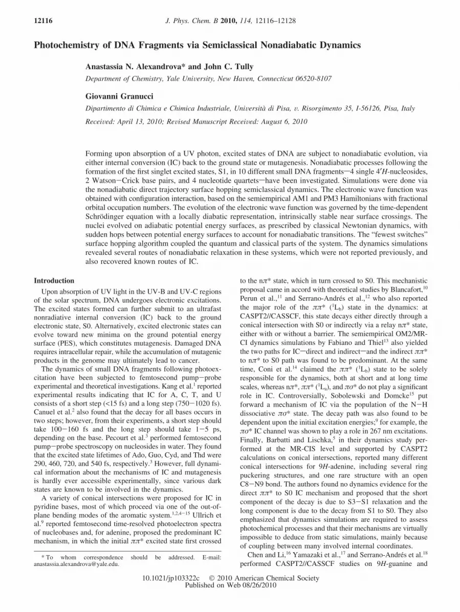
The starting point for the present study is the crystal structureof a DNA double helix fragment (Protein Data Bank code1N4E).58 The 1N4E structure is a dimer, and only a 20-basemonomer without a lesion was retained for simulations (Figure1A). Crystallographic water molecules were removed. Thesystem was relaxed using Monte Carlo (MC) simulation in anNTP ensemble at 25 °C and 1 atm, as implemented in theMCPRO 2.00 package.59 The OPLS-AA force field was used.60
The solvent was represented with an implicit generalized Born/surface area (GB/SA) solvation model,61-63 with an ionicstrength of 0.5 imitating a 1 M Na+ solution, for the compensa-tion of the negative charge on the DNA backbone. For thenucleotide bases, all valence angles and torsions were sampledin MC, whereas the bond lengths and the DNA backbone werekept fixed. The simulation consisted of a total of 30 × 106
configurations. Noticeably, all DNA structures resulting fromthe MC run contained several “imperfect” (twisted and shifted)Watson-Crick pairs (Figure 1A). This is quite expected, sincethe DNA structure is rather flexible, but it is also interestingfor the present investigation, since it was shown previously that
the fate and nature of excited states in DNA is dependent uponthe nucleotide sequence, and the higher-order structure, i.e., thegeometry of the base-stacking.64,65,56,66-70 In view of this,fragments taken from the relaxed double helix should present amore realistic model than the ground state geometries of theisolated fragments in the gas phase. Five representative low-energy configurations from the end of the MC simulation weretaken as starting points for further investigation. DNA fragmentsof interest were cut out of these relaxed structures (Figure 1B).For the nucleotide quartets, two Na+ cations were manuallyadded to compensate the negative charge on the backbone, andtheir positions were optimized with AM1.71
Semiclassical dynamics simulations were conducted using thedirect trajectory surface hopping method by Granucci et al.72
Nuclei moved classically on a single adiabatic potential energysurface (PES), as prescribed by Newton’s equations of motion,and are allowed to hop to other PESs at any time, according totransition probabilities computed with the “fewest switches”algorithm.73 The evolution of the electronic wave function wasgoverned by the time-dependent Schrodinger equation, with alocally diabatic representation.72 The latter makes the solutionstable in situations of near-degeneracy of the adiabatic states,for example, near conical intersections. The electronic wavefunctions were built with a full configuration interaction withina chosen active space (CAS-CI). In the present work, a CAS-CI space of two electrons in two orbitals, CI(2,2), was used,and always two electronic states were involved in the dynamics:S0 and S1. The reference function for the CI was computedwith the semiempirical molecular orbital (MO) methods, mainlyAM1,71 and additionally PM3,74 with floating orbital occupationnumbers (FON),72 using MOPAC 2000.75 Unless otherwiseindicated, the reported results were obtained with AM1 CI(2,2).The Gaussian width was set at 0.1. The choice for the electronicHamiltonian was justified by the smallest number of trajectoriesrunning into the problem of SCF nonconvergence and energynonconservation (the problem mainly concerned adenine 4′H-nucleoside). Other tested Hamiltonians, however, includedCI(4,4), (6,6), and (10,10), with AM1 or PM3 as a reference
Figure 1. (A) Representative structure of the DNA double helix from the end of the MC run: G - yellow, A - purple, C - red, T - green, “imperfect”base pairs may be seen. (B) Fragments cut from the structure in part A and considered in this work.
12118 J. Phys. Chem. B, Vol. 114, No. 37, 2010 Alexandrova et al.

function, and the Gaussian width of 0.3. The dynamics startedfrom a vertical excitation from S0 to S1. For each atom, thenorm of the velocity (V) was assigned on the basis of theequipartition principle, at a temperature of 300 K. The directionof the velocity was assigned at random, and the system wasallowed to evolve. Velocities were different for each trajectory.
Each semiclassical dynamics trajectory entailed 10 000 stepsof 0.1 fs, or a total of 1 ps. In the case of the adenine-thymineWatson-Crick pair, simulations were extended up to 5 ps, tobetter capture the mechanism of IC in this system, whichappeared to be slow. For each system, each of the fiverepresentative configurations resulting from the MC run gaverise to 100 trajectories, i.e., a total of 500 unique trajectories.Occasionally, the simulations still ran into the energy noncon-servation problem, for reasons that we could not deduce. Suchtrajectories were discarded. All results reported herein areaveraged over only good and complete trajectories, whosenumber was unavoidably smaller than 500 in each case. Thesampling of starting structures was our way to account for thesensitivity9 of the dynamics in DNA fragments to the excessenergy in the vertically excited species.
The performance of semiempirical MO methods for excitedstateshasbeenvalidatedforanumberofclassesofcompounds.76-78
They are reliable in providing at least a qualitative descriptionof the topology of excited state PESs. The results of the presentwork, including the general trends in nuclear rearrangementsduring the evolution of the excited states, the relative prevalenceof different routes in the dynamics, the locations of conicalintersections, and the rates on nonadiabatic processes, are,therefore, qualitative in nature. However, semiempirical methodsare also fast, which allows for performing the on-the-flydynamics. Using ab intio methods for the electronic part of theproblem would be nearly computationally prohibitive, especiallyfor the larger systems considered here. Once the routes of thedynamics are qualitatively mapped out, and the relevant reactioncoordinates are revealed, more accurate theoretical treatmentscould be carried out.
In order to unveil the nature of the observed electronicexcitations, and the evolution of the electronic structure of thesystems as they progress toward a conical intersection, theelectronic states were monitored via molecular orbital (MO)analysis. MOs were obtained at the same level of theory as thecorresponding dynamics runs. Some additional DFT calculationswere done using Gaussian 03.79
Results and Discussion
Single 4′H-Nucleosides. Figure 1B shows representativestarting ground state structures of G, A, T, and C bound to theirsugars, as resulting from the MC relaxation of the DNA doublehelix. The total charge of each species is zero.
Guanine. The starting structures taken from the DNA doublehelix differ from those for the optimized nucleoside in the gasphase: the relative positions of the base and sugar are signifi-cantly different for the two contexts, and also there are smallout-off-plane distortions in the base taken from DNA. Theenergy difference between our starting structures and theoptimized one varies from 1.5 to 2.4 eV, again, majorly due tothe position of the sugar. This destabilization of the ground state,however, does not directly correlate with the S0-S1 energydifference in the Franck-Condon region.
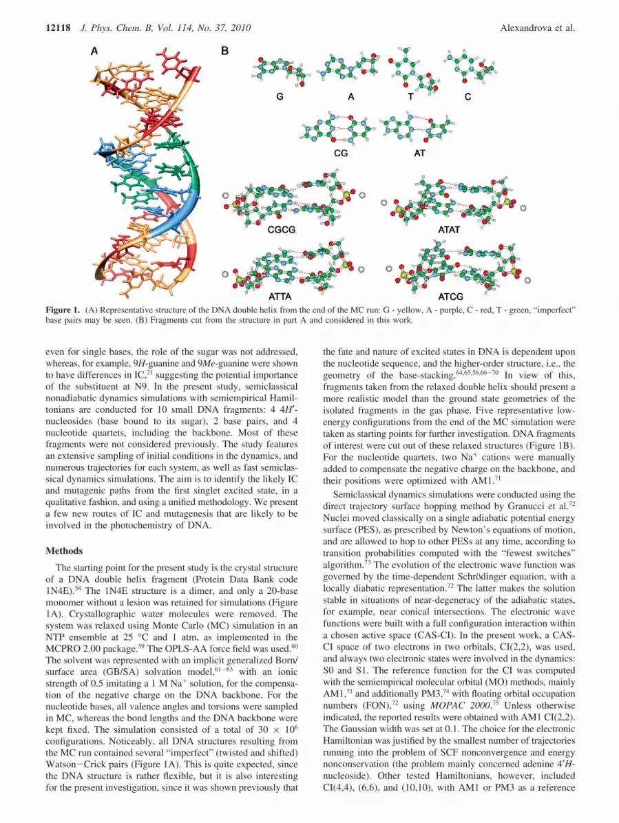
Out of 500 trajectories run for guanine 4′H-nucleoside, 498finished without running into SCF convergence or energynonconservation problems. The results of these dynamicssimulations are shown in Figure 2. A part of a typical trajectory
from the dynamics simulations is shown in Figure 2A. In thebeginning of the simulations, G is vertically excited to the firstsinglet excited state S1 and it has the ground starting geometryGv (vertical red arrow in Figure 2A). The excitation energy inour simulations is 4.11 ( 0.32 eV, depending on the startingstructure (compare 4.92 eV, the experimental value for theisolated 4′H-nucleoside, as measured at the center of theabsorption band).80 The red points in the trajectory (Figure 2A)belong to the S1 PES, and the blue points belong to S0. Thechanges in the intramolecular degrees of freedom in G aremonitored throughout the simulations. It appeared that the out-of-plane skeletal deformation in the base, corresponding tosimultaneous pyramidalization at N1 and C2, is the internalcoordinate most relevant to the dynamics. We chose to plot thetrajectories against the (N1-C2-N3-C4) torsion, as one alwaysrelevant reaction coordinate. Bending of the base leads to aconical intersection between S1 and S0, in accord with refs16-21. A representative hopping point, Ghop, is shown in Figure2B. The presence of the sugar does not qualitatively changethe dynamics in the case of G; IC occurs solely via changes inthe internal coordinates of the base.
From Figure 2A, it is evident that there are two almostequivalent conical intersections relevant to bending of G: thatin the direction of the positive (N1-C2-N3-C4) dihedralangles and that in the direction of negative (N1-C2-N3-C4)dihedral angles. The hops occurred at dih(N1-C2-N3-C4)) (16.9 ( 8.9°. The representative trajectory in Figure 2Aalso illustrates that the S1 PES is relatively flat with respect tothis motion of the molecule. As a result, the molecule easilymoves in both directions along the reaction coordinate whileon S1. Furthermore, sometimes in the same trajectory, hopsoccur both at positive and at negative values of dih(N1-C2-N3-C4). Figure 2D shows the density plots of values of therelevant torsions versus the corresponding difference in energybetween S1 and S0. In this case, these plots just show that hopsfor G may occur at a wide range of values for these torsions, inboth positive and negative directions, and greater values arecharacteristic of dih(N1-C2-N3-C4) rather than dih(C2-N3-C4-N1).
Figure 2C shows the fractions of trajectories evolving on S1as a function of time. It can be seen that the half-life of the S1state, i.e., the time at which ∼50% of the trajectories switchedfrom S1 to S0, is ca. 240 fs. Also, a tiny fraction of trajectoriesdecayed within 120 fs, which might be the fast component ofthe decay known from the literature. 240 fs is a bit faster thanthe experimentally obtained timing of 360 fs.2 Partly, this isprobably due to the uncertainties associated with the semiem-pirical electronic Hamiltonian. It is also likely that the timingdepends on the presence or absence of the solvent, and the sugarbound to the nucleobase. By the end of 1 ps, 74% of trajectoriesexperience IC. Trajectories that did not end up on S0 by 1 pscorresponded to situations where a rehop from S0 to S1happened near 1 ps, and G continued to flop on S1 along thesame reaction coordinate. Rehops were frequent in the dynamics.The mechanism of IC was always the same, and we did notobserve trapping in any minima on S1 such that the systemwould fail to decay.
The results were controlled via running additional dynamicssimulations with AM1 CI(6,6), PM3 CI(2,2), and PM3 CI(4,4)Hamiltonians. The trend in nuclear rearrangements and thetiming for IC were found to be qualitatively the same. Forcomparison, in Figure 2C (blue curve), we show the fractionof trajectories on S1 as a function of time, obtained with PM3CI(2,2).
Photochemistry of DNA Fragments J. Phys. Chem. B, Vol. 114, No. 37, 2010 12119
To support and illustrate the dynamics results, we analyzedthe changes in the electronic structure of the system bymonitoring the molecular orbitals (MOs), obtained with the sameAM1 CI(2,2) method (Figure 2B). The first electronic excitationin G 4′H-nucleoside corresponds to the promotion of an electronfrom the HOMO (45a) to the LUMO (46a), yielding two singlyoccupied molecular orbitals (SOMOs). The S0-S1 excitationis almost completely localized on the base and does not involvethe sugar or the glycosidic bond. The forming state is ππ* (La).The redistribution of the π-electron density in the base manifestsitself in the out-of-plane motion on S1. In the vertically excitedstructure, Gv, both SOMOs are mixes of π-MOs on the aromaticsystem and the lone pair (LP) on the amino group, in theframework of MO-LCAO. The SOMOs of the representative
hopping point, Ghop, obviously resemble those in Gv, exceptnow the SOMO (45a) is mostly a π-MO on the base and theSOMO (46a) contains more electron density on the LP of theamino group. The nature of the S1 state does not change,however; it is still ππ* (La). Hence, our simulations predict astraightforward decay from the ππ* S1 state to S0, in agreementwith earlier reports.16-19a A few trajectories that underwent ICon a shorter time scale corresponded to a smaller deviation fromthe planarity in the base. The SOMO (46a) in the hoppinggeometries in these trajectories more closely resembles theSOMO (46a) in Gv, with a smaller shift of electron density fromthe base to the amino group.
Adenine. Figure 3 presents results for adenine. The startingstructures for A were taken from a particular context in DNA
Figure 2. Nonadiabatic dynamics in the G 4′H-nucleoside: (A) a representative trajectory: electronic energy relative to the starting ground statestructure is shown on the vertical axis; points are registered every fifth configuration, red points belong to the S1 PES, blue points belong to S0;the vertical red arrow indicates the initial S0-S1 excitation to Gv; the system then evolves to a hopping point, Ghop (red arrow pointing to theright), and hops from S1 to S0; multiple rehops from S0 to S1 and back, including those through bending in the direction of negativedih(N1-C2-N3-C4) (e.g., purple dashed arrows), happen in this trajectory; the system ends up near the minimum on S0, where dih(N1-C2-N3-C4)∼ 0°. (B) Structures of representative Gv and Ghop and the comparison between their MOs. (C) The fraction of trajectories evolving on S1 as afunction of time: red - at the AM1 CI(2,2) level; blue - at PM3 CI(2,2). (D) The density plot showing the values of relevant internal coordinatesat hopping points versus the corresponding energy difference between S1 and S0.
12120 J. Phys. Chem. B, Vol. 114, No. 37, 2010 Alexandrova et al.

that happened to be quite far from the optimized ground statestructure (2.2-2.8 eV), due to both the position of the sugarand a slight bend in the base. The vertical excitation energieswere 3.51 ( 0.39 eV. The experimental value for the isolatednucleoside is 4.78 eV, at the absorption band maximum,80
though, again, the direct comparison between these values mightbe misleading due to structural differences. The motion on theexcited state leading to IC is similar to that observed in guanine:it is the bending in the aromatic system of the base, which isconsistent with previous reports.5,9-14 The most appropriatereaction coordinate is the (N1-C6-C5-C4) torsion, as 96.0%of the hops occurred via N1 going out of the molecular planeand the remaining 4.0% of the hops involved C6 going out ofthe molecular plane. A representative hopping point, Ahop, isshown in Figure 3A. As averaged over 422 trajectories, at thehopping point, dih(N1-C6-C5-C4) ) 30.15 ( 17.70° (Figure3C), and the electronic energy is -109.172334 ( 0.047551 au,which is ca. 1.2 eV below Av. There is a qualitative consensusbetween results obtained with AM1 CI(2,2), AM1 CI(6,6), PM3CI(6,6), and PM3 CI(10,10), although we had difficultiesobtaining a significant number of trajectories with these Hamil-tonians due to energy nonconservation and SCF nonconvergenceproblems.
Noticeably, in our simulations, IC in adenine is also slowerthan that in guanine. Figure 3B shows the fraction of trajectoriesevolving on S1 as a function of time. It illustrates how inefficientIC is: on average, switching from S1 to S0 does not begin untilca. 400 fs into the dynamics. Figure 3C also illustrates howscarce S1-S0 transitions were in the dynamics for A (compareto Figure 2D). Unlike for guanine, we obtained no fast decayingtrajectories. By the end of 1 ps, the population of S1 is stillslightly greater than that of S0. It looks like our dynamicssimulations do not capture the fast component of the decay thatwas detected experimentally, and we only see the slow
component, which is known to take ca. 1.1 ps.2 However,Barbatti and Lischka in their dynamics study5 proposed that Acan live on S1 (ππ*) in the neighborhood of the structure similarto our Ahop, which they called 2E. In their simulations, thistrapped structure slowly decayed via the conical intersectionwith S1, whereas the fast component was due to the precedingrelaxation from S3 to S1. If this mechanism is right, we do notsee the fast component of the decay because we start thedynamics from S1. Hence, our results indirectly confirm thisIC mechanism by Barbatti and Lischka.
The S0-S1 excitation in the starting geometry of A is of theππ* (Lb) character (Figure 3A), which is different from that inG. The Lb character of the S1 state in A was also predicted byab initio studies (for example, ref 8). In Figure 3A, the SOMOsin Av are matched against those in the hopping point, Ahop. TheSOMO (43a) in Ahop has the largest component of the electrondensity localized on the amino group of the base, just like inGhop (Figure 2). The electron density on the base is significantlyperturbed due to ring puckering: the nodal structure of thisSOMO changes, and also, the contributing pz-AOs on C6 andN3 orient at large angles to those on the remaining atoms inthe base. Thus, the ππ* (Lb) state adiabatically changes in thedynamics on S1, and it is no longer possible to characterize itas pure Lb at Ahop. The major role of the Lb state and itsevolution in the dynamics is in agreement with ab initio resultsobtained earlier.10-14
There is a significant amount of controversy in the literatureregarding IC in pyrimidine bases. Mainly, the argument focuseson whether the ππ* excited state decays directly to S0 orproceeds through a nπ* state or states along the way. It isevident that the ππ* and nπ* excited states are closely spacedin the Franck-Condon region, at least for the isolated pyrimi-dine bases. Considering this fact, and the innate weaknesses of
Figure 3. Nonadiabatic semiclassical dynamics in A 4′H-nucleoside: (A) A representative starting vertically excited structure, Av, and a hoppingpoint, Ahop, and the comparison between their SOMOs. (B) Averaged over 422 trajectories, the fraction of trajectories evolving on S1, as a functionof time. (C) The density plot showing the values of dih(N1-C6-C5-C4) at hopping points versus the corresponding energy difference betweenS1 and S0.
Photochemistry of DNA Fragments J. Phys. Chem. B, Vol. 114, No. 37, 2010 12121

semiempirical methods, we hereby proceed with caution ininterpreting results for T and C.
Thymine (Figure 4). The starting structures for T were1.3-1.9 eV above the optimized structure, and the verticalexcitation happens to take 4.19 ( 0.17 eV (the experimentalvalue is 4.65 eV for the center of the absorption band).80 Thevertical excitation is ππ*. Interestingly, the electron density ofthe sugar moiety is involved in both SOMOs (Figure 4A), andthus is likely to influence the dynamics, which was neveraddressed previously.
The most prominent paths in the dynamics were reported toinvolve ring puckering of the base.27-32 However, in oursimulations, only 18.98% of the trajectories involve solo bendingof the base via pyramidalization at various sites: N1, C2, N3,and C6, sometimes with a slight N2-C3 stretch (Figure 4B).In these trajectories, the S1 state retained its ππ* character atthe hopping point. Surprisingly, the majority of the trajectories(76.2%) proceeded via stretching the N1-C2 bond to ca. 2.02Å, accompanied by its ring puckering via N3 going out of themolecular plane (Figure 4A). A typical hopping point occurredat 0.28 eV below Tv. In Thop, the SOMO (42a) is a nOπ*-MOthat puts antibonding character on the N1-C2 bond, which leadto the N1-C2 stretch. 31.5% of these trajectories returned backto the ground state structure. 44.7% proceeded to the completecleavage of the N1-C2 bond (Topen, Figure 4A). We confirmedthe existence of this minimum with B3LYP/6-311G*. Themechanism was also found with PM3 CI(2,2) and PM3 CI(4,4).
The lifetime of S1 in our simulations is ca. 30 fs at AM1CI(2,2) and ca. 110 fs at PM3 CI(2,2), whereas the experimentalvalue for the short IC component, for example, by Ullrich etal.9b is less than 50 fs. By 1 ps, 90% of trajectories are on S0.The trajectories that failed to converge to the ground state bythe end of 1 ps did not get trapped on S1, but rehops occurredfrom S0 to S1 toward the end of the run, and mostly that
happened with the structures already containing open N1-C2bonds. We did not capture the long components of the decay(490 fs and 6.4 ps).9b
In order to elaborate on the origin of the discrepancy betweenour results and those obtained with ab initio methods, we ranan additional 500 trajectories of dynamics simulations with AM1CI(2,2) for 1H-thymine, e.g., the base without the sugar. Thefive representative geometries of T were taken from those usedfor the nucleoside. The vertical excitation energy in this casewas 4.50 ( 0.19 eV. We found that the isolated base neverundergoes the ring opening during IC, and only in 4.2% of thetrajectories is the N1-C2 bond slightly stretched to ca. 1.6 Åat hopping points. Instead, the system indeed adopts one of thering puckering paths during IC. The most prevalent route,adopted in 95.1% of trajectories, entails C4 going out of themolecular plane. Hence, the mechanism of IC in the nucleoside,as opposed to the isolated base, is predicted by AM1 CI(2,2) tobe altered by the presence of the sugar. This correlates with theinvolvement of the electron density from the sugar in theSOMOs. There is still a possibility that, for the nucleoside,the unusual IC path via the N1-C2 stretch could be a result ofincorrect order of excited states predicted by semiempiricalmethods. The mechanism requires further confirmation at higherlevels of theory. It is also anticipated that, in the context of theWC pair, this mechanism would be hindered by H-bonding tothe opposing base.
Cytosine (Figure 5). Initial structures of the nucleoside were0.9-2.2 eV above the ground state, and the vertical excitationenergies were 4.17 ( 0.49 eV (experimental value is 4.52 eV).80
The S1 state is ππ* in nature, in agreement with previousreports.22,33-35 However, both SOMOs also include the electrondensity from the sugar moiety, which suggests the possibleimportance of sugar for the dynamics.
Figure 4. Nonadiabatic semiclassical dynamics in T 4′H-nucleoside: (A) The starting vertically excited structure, Tv, and a representative hoppingpoint, Thop, and Topen with cleaved N1-C2 bond, and the comparison between the MOs. (B) Other types of hopping points, with markedpuramidalization sites (I-III), and the C2-N3 stretch (IV). (C) Averaged over 396 trajectories, the fraction of trajectories evolving on S1, as afunction of time, at AM1 CI(2,2) (red curve); the blue curve represents the PM3 CI(2,2) result for comparison.
12122 J. Phys. Chem. B, Vol. 114, No. 37, 2010 Alexandrova et al.

22.8% of trajectories involved IC via bending of the base,which corresponded to the direct ππ*f S0 decay, in agreementwith earlier studies on the isolated C base (for example, ref39). However, again, the prevailing IC route occurred viastretching the N1-C2 bond accompanied by ring puckering(Figure 5A). 27.2% of such trajectories returned back to theequilibrium geometry of S0, and 50.0% proceeded toward thenew minimum on S0, corresponding to the open N1-C2 bond.We confirmed the existence of this minimum at B3LYP/6-311G*. In Chop, R(N1-C2) ) 1.74 ( 0.25 Å, i.e., smaller thanin Thop. The N1-C2 stretch is due to the population of theSOMO (39a), which is a mixed π*- and nOπ*-MO that puts aσ* antibonding character in the region of the N1-C2 bond.The population of nOπ* is in agreement with earlier reports,37-39
though the N1-C2 stretch was not found previously. We suspectthat this path of IC for C and T may be hindered in the CGWC pair.
The half-life of S1 is ca. 60 fs at AM1 CI(2,2) and ca. 120fs at PM3 CI(2,2), which might correspond to the fastcomponent of the decay. By the end of 1 ps, 96% of alltrajectories convert to S0. Trajectories that failed to return tothe ground state by the end of 1 ps corresponded to rehops viavery similar nuclear rearrangements: base puckering and stretch-ing N1-C2 bond. No trajectories remained on S1 for the entireduration of the simulation. We again fail to see the slowcomponent of IC.
Whether the found mechanism originates from the poorperformance of semiempirical methods remains to be explored.However, we performed additional simulations for the isolatedC (500 trajectories at AM1 CI(2,2)) and found that for thissystem we recover the IC routes reported in the literature.Specifically, 80.6% of the trajectories evolved via C4 going outof the molecular plane, and other trajectories involved othermodes of ring puckering. The N1-C2 opening was neverobserved for the isolated base, though its stretch to ca. 1.6 Åwas found in 30.4% of the hopping geometries. The verticalexcitation energy in 1H-cytosine was 4.50 ( 0.32 eV. Hence,in our simulations, the N1-C2 stretch is due to the presence ofthe sugar.
We would like to emphasize again that the ππ* and nπ* statesare close in energy, at least in isolated T and C, and as predictedby higher levels of theory. Hence, there is a risk that semiem-pirical methods predict the wrong order of excited states, andtake the systems along the wrong PESs in IC. We do recoverIC paths known for isolated bases, and that adds certaincredibility to our methodology. However, still, the new prevail-
ing IC mechanism needs to be confirmed at higher levels oftheory. We hope our simulations at least bring up a warningthat the sugar might need to be included when studying IC inpyrimidine nucleotides.
Watson-Crick Base Pairs. For two or more bases in thesystem, several different possibilities arise for the verticalelectronic excitations. There can be local excitations on the samebase, or there can be interbase CT excitations. The cross sectionsfor the latter are known to be smaller. We find that the energyordering of low-energy excited states depends on the geometryof the pairs.
Cytosine-Guanine. The nonadiabatic dynamics of the CGWC pair has been extensively studied, and therefore presents agood benchmark case for our investigation. In our simulations,the system consisted of 9H-guanine and 1H-cytosine (Figure1B). A harmonic constraint was introduced between N1G andN3C, holding the two atoms within the range from 2.5 to 3.3 Åapart (at equilibrium, R(N1G-N3C) ) 2.9 Å), in order to keepthe two bases paired. Without the constraint, the system alwaysfell apart during the dynamics, as the hydrogen bonding withinthe Watson-Crick pair was insufficient to keep the pair bound.
Figure 6A shows the results. The vertical excitation energywas 5.37 ( 0.33 eV. At AM1 (CI2,2), 98.5% of the trajectoriesindeed underwent IC via the N1-H stretch, or completeN1G-N3C proton transfer. The structure with completelytransferred proton is not a minimum on the S0 PES, however.Among those, 67.5% of trajectories also involved a skeletaldeformation, mainly due to pyramidalization at the N3C site.CGhop is a characteristic hopping point (Figure 6A). A typicalCGhop occurred at -131.49973 ( 0.02263 au, which is ca. 0.13eV below the averaged vertically excited structure (averagedover 442 successful trajectories). The remaining 1.5% of thetrajectories involved skeletal deformation alone. IC in CG isvery efficient. S1 decays in the time scale of 30 fs, and by theend of 1 ps, 96% of the trajectories end up on S0.
For all studied geometries, the first excited state in CGv
corresponds to charge transfer from G to C. It is illustrated inFigure 6A: the SOMO (49a) is fully localized on G, whereasthe newly populated SOMO (50a) is localized solely on C. Thisstructure of MOs is retained in CGhop. The negative charge onC attracts the shuttling proton from G, and this motionstraightforwardly leads to the S0-S1 conical intersection.
It is also known from previous reports43 and from our B3LYP/6-311G* calculations that there is a minimum correspondingto a double proton transfer in CG: between N1G and N3C andbetween the amino group in C and the carbonyl in G. However,
Figure 5. Nonadiabatic semiclassical dynamics in C 4′H-nucleoside: (A) The starting vertically excited structure, Cv, and a representative hoppingpoint, Chop, and the comparison between the MOs. (B) Averaged over 361 trajectories, the fraction of trajectories evolving on S1, as a function oftime, at AM1 CI(2,2) (red curve); the blue curve represents the PM3 CI(2,2) result for comparison.
Photochemistry of DNA Fragments J. Phys. Chem. B, Vol. 114, No. 37, 2010 12123

the apparently low-probability event of its formation was neverobserved in our simulations, perhaps due to insufficient statistics.Overall, the mechanism of IC in CG obtained with oursemiclassical dynamics simulations appears to be in agreementwith previously reported, more accurate, ab initio calculations.40-44
Adenine-Thymine. The nature and order of excited statesand dynamics in the (A)n · (T)n systems have been the subjectof some studies,18,36,37,40 though IC in this system is lessunderstood than that in CG. Perun et al.37 predicted theinvolvement of a ππ* CT state in the IC of the AT WC pair,which occurs via H-transfer. The authors also mentioned thepossibility for the dynamics to occur via bending of A or T,when the corresponding π*-MOs get populated. However, theyomitted further exploration of these pathways, and left it to bethe subject of future studies. On the other hand, Crespo-Hernandez et al.36 argued that for A and T base stacking isresponsible for nonradiative decay, rather than WC pairing.
We found IC in the AT pair to be inefficient. In 1 ps, only13.7% of the trajectories converted from S1 to S0. For 20trajectories, the simulations were then extended to 5 ps, duringwhich 55.5% of the S1 population decayed to S0. All 500trajectories for AT successfully completed, without having SCFconvergence problems.
The vertical excitation energy was 4.52 ( 0.23 eV. The mainIC route observed 89.4% of the time (in 1 ps trajectories) wascharacterized only by the out-of-plane motion of the N1A atomon adenine. The averaged value of dih(N1-C6-C6-C2)A inA at AThop was (41.3 ( 15.9°. Figure 6B shows a representa-tive vertically excited structure (ATv) and a hopping point(AThop), which was found ca. 1.15 eV below ATv. 12.5% ofthe trajectories involved both N1A going out of plane and anadditional smaller out-of-plane deformation of T. However, inthose trajectories, the averaged value of dih(N3-C2-C1-C6)T
in T was (20.7 ( 4.1°, suggesting that the deformation of Ahas a greater relevance to the dynamics. When the system wastested with other Hamiltonians and CI spaces, PM3 CI(2,2),PM3 CI(4,4), the trends in geometrical changes during IC werethe same.
At the AM1 level, the S0-S1 excitation in AT is a ππ* (Lb)excitation localized on A (Figure 6B), and this electronicstructure is retained all the way to AThop, just like in the unpairedA (Figure 3). Consistently, it leads to the observed out-of-planedistortion of A. Thus, the S1-S0 IC path in our simulationsinvolves only A. However, its timing is slowed down byH-bonding with T, as compared to the case of isolated A. Wedid not find any H-transfer involvement in the dynamics, whichwas expected, since in our simulations S1 is not a CT state.
Nucleotide Quartets. The larger DNA fragments investigatedherein are nucleotide quartets: two stacked Watson-Crick basepairs with the backbone phosphate groups and counterions,2Na+. The presence of the backbone was essential not only fora more realistic representation of the system but also to keepthe integrity of the quartets during the dynamics. There are atotal of eight possible base arrangements in a quartet; here, weconsider only four: CGCG, ATAT, ATTA, and ATCG (Figure1B). Four geometric constraints were imposed on each system:the distance between the N3 atom of C or T and the N1 atomof G or A, respectively, was kept restrained between 2.5 and4.0 Å; the stacked bases were kept close via constraining thedistance between the N1 atoms of pyrimidine bases and the N9atoms of the purine bases to fall between 4.0 and 5.5 Å. Thesemiclassical dynamics simulations for these systems are quitedemanding, even at the semiempirical level of theory. For eachsystem, only 20 trajectories were run, 10 at AM1 CI(2,2) and10 at PM3 CI(2,2), using 5 starting structures. This might beinsufficient to be statistically significant. Hence, the resultspresented herein are qualitative, and no statistics are reported.For nucleotide quartets, AM1 exhibited more SCF convergenceproblems than did PM3, so we focus on the PM3 CI(2,2) results.We found that, at AM1, the 3s atomic orbitals on Na+ cationscan lay as low in energy as the LUMO of quartets, which isnot physical. These orbitals can get populated in the dynamics,and lead to erroneous results.
CGCG Quartet. At both AM1 and PM3, the S1 PES inCGCG exhibited a repulsive character along the out-of-planebending mode of one of the G bases, labeled “G” in Figure 7.No other noticeable geometric changes were observed at thehopping points. According to MO analysis, at a typical hoppingpoint, CGCGhop, the structural deformation is associated withthe promotion of an electron from the π-HOMO to the LUMOmostly consisting of the lone pair on N of NH2 and some π*-character of the base. Both resulting SOMOs are localized onthe deforming G. However, at the starting geometry shown inFigure 7, the S0 to S1 excitation is different: it is CT from Gto the C π-stacked to it in the strand. The SOMO (285a)switches with the LUMO (286a) in the dynamics. It isremarkable that the MOs near the Fermi level in CGCGv areclosely spaced in energy, their order depends on the startinggeometry, and it is different in the initial structures used herebut never the same as in CGCGhop. This contrasts what wasobserved for the CG WC pair, where the S0-S1 excitation isalways CT within the pair. Hence, our results suggest that theIC process in DNA may be more complicated than that in anisolated CG pair. First of all, the nature of the first excited state
Figure 6. (A) Nonadiabatic semiclassical dynamics in CG: the starting vertically excited structure, CGv, and a hopping point, CGhop, and thecomparison between their MOs. (B) Same information for the AT pair.
12124 J. Phys. Chem. B, Vol. 114, No. 37, 2010 Alexandrova et al.
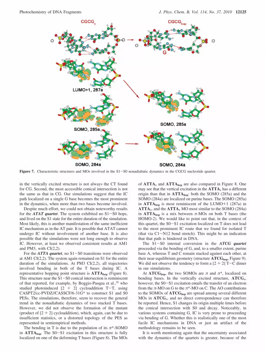
in the vertically excited structure is not always the CT foundfor CG. Second, the most accessible conical intersection is notthe same as that in CG. Our simulations suggest that the ICpath localized on a single G base becomes the most prominentin the dynamics, when more than two bases become involved.
Despite much effort, we could not obtain noteworthy resultsfor the ATAT quartet. The system exhibited no S1-S0 hops,and lived on the S1 state for the entire duration of the simulation.Most likely, this is another manifestation of the same inefficientIC mechanism as in the AT pair. It is possible that ATAT cannotundergo IC without involvement of another base. It is alsopossible that the simulations were not long enough to observeIC. However, at least we observed consistent results at AM1and PM3, with CI(2,2).
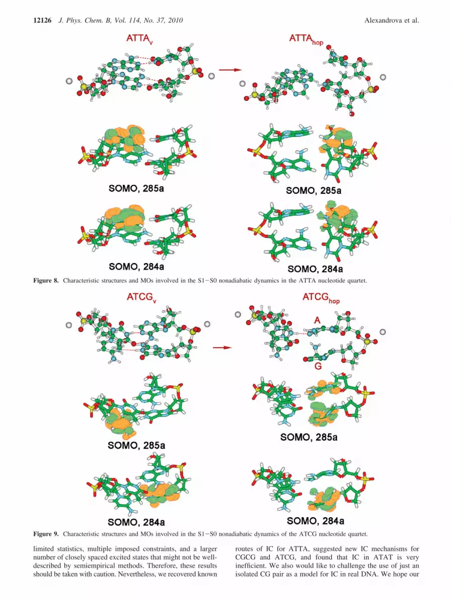
For the ATTA quartet, no S1-S0 transitions were observedat AM1 CI(2,2). The system again remained on S1 for the entireduration of the simulations. At PM3 CI(2,2), all trajectoriesinvolved bending in both of the T bases during IC. Arepresentative hopping point structure is ATTAhop (Figure 8).This structure near the S1-S0 conical intersection is reminiscentof that reported, for example, by Boggio-Pasqua et al.,26 whostudied photoinduced [2 + 2] cycloaddition T-T, usingCASPT2/cc-PVDZ//CASSCF/6-31G* to construct S1 and S0PESs. The simulations, therefore, seem to recover the generaltrend in the nonadiabatic dynamics of two stacked T bases.However, we did not observe the formation of the mutant(product of [2 + 2] cycloaddition), which, again, can be due toinsufficient statistics, or a distorted topology of the PES asrepresented in semiempirical methods.
The bending in T is due to the population of its π*-SOMOin ATTAhop. The S0-S1 excitation in this structure is fullylocalized on one of the deforming T bases (Figure 8). The MOs
of ATTAv and ATTAhop are also compared in Figure 8. Onemay see that the vertical excitation in the ATTAv has a differentorigin than that in ATTAhop: both the SOMO (285a) and theSOMO (284a) are localized on purine bases. The SOMO (285a)in ATTAhop is most reminiscent of the LUMO+1 (287a) inATTAv, and the ATTAv MO most similar to the SOMO (284a)in ATTAhop is a mix between π-MOs on both T bases (theHOMO-2). We would like to point out that, in the context ofthis quartet, the S0-S1 excitation localized on T does not leadto the most prominent IC route that we found for isolated T(that via C1-N12 bond stretch). This might be an indicationthat that path is hindered in DNA.
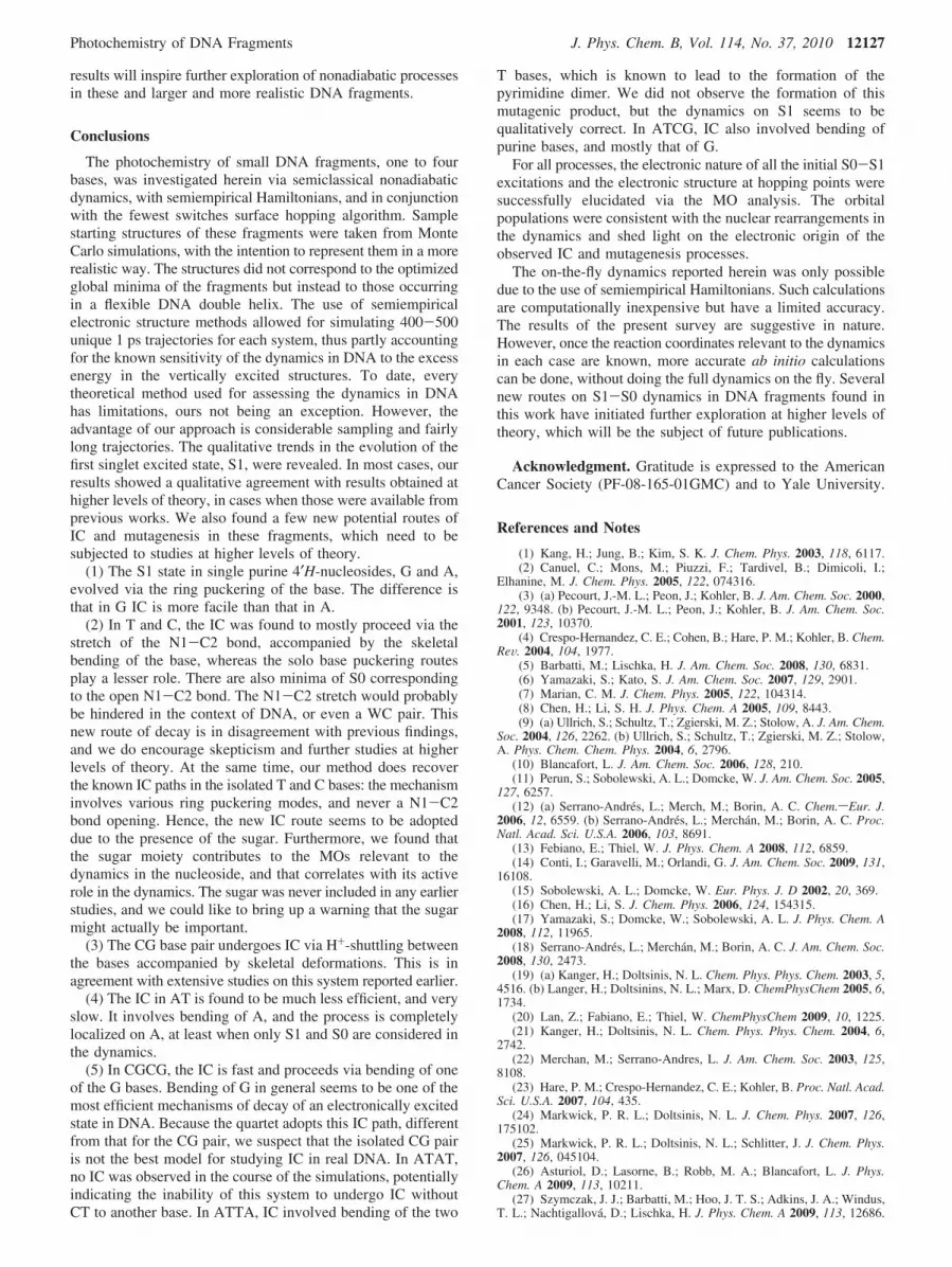
The S1-S0 internal conversion in the ATCG quartetproceeded via the bending of G, and, to a smaller extent, purinebase A, whereas T and C remain stacked against each other, attheir near-equilibrium geometry (structure ATCGhop, Figure 9).We did not observe the tendency to form a [2 + 2] T-C dimerin our simulations.
At ATCGhop, the two SOMOs are π and π*, localized onbending bases. In the vertically excited structure, ATCGv,however, the S0-S1 excitation entails the transfer of an electronfrom the π-MO on G to the π*-MO on C. The AO contributionsto the SOMOs of ATCGhop are spread among several differentMOs in ATCGv, and no direct correspondence can thereforebe reported. Hence, S1 changes its origin multiple times beforethe conical intersection with S0 and decay. Noticeably, invarious systems containing G, IC is very prone to proceedingvia bending of G. Whether this is realistically one of the mostfacile IC mechanisms in DNA or just an artifact of themethodology remains to be seen.
It is worth mentioning again that the uncertainty associatedwith the dynamics of the quartets is greater, because of the
Figure 7. Characteristic structures and MOs involved in the S1-S0 nonadiabatic dynamics in the CGCG nucleotide quartet.
Photochemistry of DNA Fragments J. Phys. Chem. B, Vol. 114, No. 37, 2010 12125
limited statistics, multiple imposed constraints, and a largernumber of closely spaced excited states that might not be well-described by semiempirical methods. Therefore, these resultsshould be taken with caution. Nevertheless, we recovered known
routes of IC for ATTA, suggested new IC mechanisms forCGCG and ATCG, and found that IC in ATAT is veryinefficient. We also would like to challenge the use of just anisolated CG pair as a model for IC in real DNA. We hope our
Figure 8. Characteristic structures and MOs involved in the S1-S0 nonadiabatic dynamics in the ATTA nucleotide quartet.
Figure 9. Characteristic structures and MOs involved in the S1-S0 nonadiabatic dynamics of the ATCG nucleotide quartet.
12126 J. Phys. Chem. B, Vol. 114, No. 37, 2010 Alexandrova et al.
results will inspire further exploration of nonadiabatic processesin these and larger and more realistic DNA fragments.
Conclusions
The photochemistry of small DNA fragments, one to fourbases, was investigated herein via semiclassical nonadiabaticdynamics, with semiempirical Hamiltonians, and in conjunctionwith the fewest switches surface hopping algorithm. Samplestarting structures of these fragments were taken from MonteCarlo simulations, with the intention to represent them in a morerealistic way. The structures did not correspond to the optimizedglobal minima of the fragments but instead to those occurringin a flexible DNA double helix. The use of semiempiricalelectronic structure methods allowed for simulating 400-500unique 1 ps trajectories for each system, thus partly accountingfor the known sensitivity of the dynamics in DNA to the excessenergy in the vertically excited structures. To date, everytheoretical method used for assessing the dynamics in DNAhas limitations, ours not being an exception. However, theadvantage of our approach is considerable sampling and fairlylong trajectories. The qualitative trends in the evolution of thefirst singlet excited state, S1, were revealed. In most cases, ourresults showed a qualitative agreement with results obtained athigher levels of theory, in cases when those were available fromprevious works. We also found a few new potential routes ofIC and mutagenesis in these fragments, which need to besubjected to studies at higher levels of theory.
(1) The S1 state in single purine 4′H-nucleosides, G and A,evolved via the ring puckering of the base. The difference isthat in G IC is more facile than that in A.
(2) In T and C, the IC was found to mostly proceed via thestretch of the N1-C2 bond, accompanied by the skeletalbending of the base, whereas the solo base puckering routesplay a lesser role. There are also minima of S0 correspondingto the open N1-C2 bond. The N1-C2 stretch would probablybe hindered in the context of DNA, or even a WC pair. Thisnew route of decay is in disagreement with previous findings,and we do encourage skepticism and further studies at higherlevels of theory. At the same time, our method does recoverthe known IC paths in the isolated T and C bases: the mechanisminvolves various ring puckering modes, and never a N1-C2bond opening. Hence, the new IC route seems to be adopteddue to the presence of the sugar. Furthermore, we found thatthe sugar moiety contributes to the MOs relevant to thedynamics in the nucleoside, and that correlates with its activerole in the dynamics. The sugar was never included in any earlierstudies, and we could like to bring up a warning that the sugarmight actually be important.
(3) The CG base pair undergoes IC via H+-shuttling betweenthe bases accompanied by skeletal deformations. This is inagreement with extensive studies on this system reported earlier.
(4) The IC in AT is found to be much less efficient, and veryslow. It involves bending of A, and the process is completelylocalized on A, at least when only S1 and S0 are considered inthe dynamics.
(5) In CGCG, the IC is fast and proceeds via bending of oneof the G bases. Bending of G in general seems to be one of themost efficient mechanisms of decay of an electronically excitedstate in DNA. Because the quartet adopts this IC path, differentfrom that for the CG pair, we suspect that the isolated CG pairis not the best model for studying IC in real DNA. In ATAT,no IC was observed in the course of the simulations, potentiallyindicating the inability of this system to undergo IC withoutCT to another base. In ATTA, IC involved bending of the two
T bases, which is known to lead to the formation of thepyrimidine dimer. We did not observe the formation of thismutagenic product, but the dynamics on S1 seems to bequalitatively correct. In ATCG, IC also involved bending ofpurine bases, and mostly that of G.
For all processes, the electronic nature of all the initial S0-S1excitations and the electronic structure at hopping points weresuccessfully elucidated via the MO analysis. The orbitalpopulations were consistent with the nuclear rearrangements inthe dynamics and shed light on the electronic origin of theobserved IC and mutagenesis processes.
The on-the-fly dynamics reported herein was only possibledue to the use of semiempirical Hamiltonians. Such calculationsare computationally inexpensive but have a limited accuracy.The results of the present survey are suggestive in nature.However, once the reaction coordinates relevant to the dynamicsin each case are known, more accurate ab initio calculationscan be done, without doing the full dynamics on the fly. Severalnew routes on S1-S0 dynamics in DNA fragments found inthis work have initiated further exploration at higher levels oftheory, which will be the subject of future publications.
Acknowledgment. Gratitude is expressed to the AmericanCancer Society (PF-08-165-01GMC) and to Yale University.
References and Notes
(1) Kang, H.; Jung, B.; Kim, S. K. J. Chem. Phys. 2003, 118, 6117.(2) Canuel, C.; Mons, M.; Piuzzi, F.; Tardivel, B.; Dimicoli, I.;
Elhanine, M. J. Chem. Phys. 2005, 122, 074316.(3) (a) Pecourt, J.-M. L.; Peon, J.; Kohler, B. J. Am. Chem. Soc. 2000,
122, 9348. (b) Pecourt, J.-M. L.; Peon, J.; Kohler, B. J. Am. Chem. Soc.2001, 123, 10370.
(4) Crespo-Hernandez, C. E.; Cohen, B.; Hare, P. M.; Kohler, B. Chem.ReV. 2004, 104, 1977.
(5) Barbatti, M.; Lischka, H. J. Am. Chem. Soc. 2008, 130, 6831.(6) Yamazaki, S.; Kato, S. J. Am. Chem. Soc. 2007, 129, 2901.(7) Marian, C. M. J. Chem. Phys. 2005, 122, 104314.(8) Chen, H.; Li, S. H. J. Phys. Chem. A 2005, 109, 8443.(9) (a) Ullrich, S.; Schultz, T.; Zgierski, M. Z.; Stolow, A. J. Am. Chem.
Soc. 2004, 126, 2262. (b) Ullrich, S.; Schultz, T.; Zgierski, M. Z.; Stolow,A. Phys. Chem. Chem. Phys. 2004, 6, 2796.
(10) Blancafort, L. J. Am. Chem. Soc. 2006, 128, 210.(11) Perun, S.; Sobolewski, A. L.; Domcke, W. J. Am. Chem. Soc. 2005,
127, 6257.(12) (a) Serrano-Andres, L.; Merch, M.; Borin, A. C. Chem.sEur. J.
2006, 12, 6559. (b) Serrano-Andres, L.; Merchan, M.; Borin, A. C. Proc.Natl. Acad. Sci. U.S.A. 2006, 103, 8691.
(13) Febiano, E.; Thiel, W. J. Phys. Chem. A 2008, 112, 6859.(14) Conti, I.; Garavelli, M.; Orlandi, G. J. Am. Chem. Soc. 2009, 131,
16108.(15) Sobolewski, A. L.; Domcke, W. Eur. Phys. J. D 2002, 20, 369.(16) Chen, H.; Li, S. J. Chem. Phys. 2006, 124, 154315.(17) Yamazaki, S.; Domcke, W.; Sobolewski, A. L. J. Phys. Chem. A
2008, 112, 11965.(18) Serrano-Andres, L.; Merchan, M.; Borin, A. C. J. Am. Chem. Soc.
2008, 130, 2473.(19) (a) Kanger, H.; Doltsinis, N. L. Chem. Phys. Phys. Chem. 2003, 5,
4516. (b) Langer, H.; Doltsinins, N. L.; Marx, D. ChemPhysChem 2005, 6,1734.
(20) Lan, Z.; Fabiano, E.; Thiel, W. ChemPhysChem 2009, 10, 1225.(21) Kanger, H.; Doltsinis, N. L. Chem. Phys. Phys. Chem. 2004, 6,
2742.(22) Merchan, M.; Serrano-Andres, L. J. Am. Chem. Soc. 2003, 125,
8108.(23) Hare, P. M.; Crespo-Hernandez, C. E.; Kohler, B. Proc. Natl. Acad.
Sci. U.S.A. 2007, 104, 435.(24) Markwick, P. R. L.; Doltsinis, N. L. J. Chem. Phys. 2007, 126,
175102.(25) Markwick, P. R. L.; Doltsinis, N. L.; Schlitter, J. J. Chem. Phys.
2007, 126, 045104.(26) Asturiol, D.; Lasorne, B.; Robb, M. A.; Blancafort, L. J. Phys.
Chem. A 2009, 113, 10211.(27) Szymczak, J. J.; Barbatti, M.; Hoo, J. T. S.; Adkins, J. A.; Windus,
T. L.; Nachtigallova, D.; Lischka, H. J. Phys. Chem. A 2009, 113, 12686.
Photochemistry of DNA Fragments J. Phys. Chem. B, Vol. 114, No. 37, 2010 12127

(28) Perun, S.; Sobolewski, A. L.; Domcke, W. J. Phys. Chem. A 2006,110, 13238.
(29) Hudock, H. R.; Levine, B. G.; Thompson, A. L.; Satzger, H.;Townsend, D.; Gador, N.; Ullrich, S.; Stolow, A.; Martynez, T. J. J. Phys.Chem. A 2007, 111, 8500.
(30) Hudock, H. R.; Levine, B. G.; Thompson, A. L.; Satzger, H.;Townsend, D.; Gador, N.; Ullrich, S.; Stolow, A.; Martı´nez, T. J. J. Phys.Chem. A 2007, 111, 8500.
(31) Merchan, M.; Gonzalez-Luque, R.; Climent, T.; Serrano-Andres,L.; Rodriuguez, E.; Reguero, M.; Pelaez, D. J. Phys. Chem. B 2006, 110,26471.
(32) Zechmann, C.; Barbatti, M. J. Phys. Chem. A 2008, 112, 8273.(33) Lan, Z.; Fabiano, E.; Thiel, W. J. Phys. Chem. B 2009, 113, 3548.(34) Zgierski, M. Z.; Patchkovskii, S. J. Chem. Phys. 2005, 123, 081101.(35) Kistler, K. A.; Matsika, S. J. Phys. Chem. A 2007, 111, 2650.(36) Blancafort, L. Photochem. Photobiol. 2007, 83, 603.(37) Ismail, N.; Blancafort, L.; Olivucci, M.; Kohler, B.; Robb, M. A.
J. Am. Chem. Soc. 2002, 124, 6818.(38) Blancafort, L.; Robb, M. A. J. Phys. Chem. A 2004, 108, 10609.(39) Hudock, H. R.; Martnez, T. J. ChemPhysChem 2008, 9, 2486.(40) Sobolewski, A. L.; Domcke, W. Phys. Chem. Chem. Phys. 2004,
6, 2763.(41) Schultz, T.; Samoylova, E.; Radloff, W. I. V. H.; Sobolewski, A. L.;
Domcke, W. Science 2004, 206, 1765.(42) Villani, G. J. Chem. Phys. 2008, 128, 114306.(43) Groenhof, G.; Schafer, L. V.; Boggio-Pasqua, M.; Goette, M.;
Grubmuller, H.; Robb, M. A. J. Am. Chem. Soc. 2007, 129, 6812.(44) Sobolewski, A. L.; Domcke, W.; Hattig, C. Proc. Natl. Acad. Sci.
U.S.A. 2005, 102, 17903.(45) Perun, S.; Sobolewski, A. L.; Domcke, W. J. Phys. Chem. A 2006,
110, 9031.(46) Shukla, M. K.; Leszczynski, J. J. Phys. Chem. A 2002, 106, 4709.(47) Kwok, W.-M.; Ma, C.; Phillips, D. L. J. Am. Chem. Soc. 2008,
130, 5131.(48) Schreier, W. J.; Schrader, T. E.; Koller, F. O.; Gilch, P.; Crespo-
Hernandez, C. E.; Swaminathan, V. N.; Carell, T.; Zinth, W. B.; Kohler,B. Science 2007, 315, 625.
(49) Danilov, V. I.; Slyusarchuk, O. N.; Alderfer, J. L.; Stewart, J. J.;Callis, P. R. Photochem. Photobiol. 1994, 59.
(50) Min, J.-H.; Pavletich, N. P. Nature 2007, 449, 570.(51) Masson, F.; Laino, T.; Tavernelli, I.; Rothlisberger, U.; Hutter, J.
J. Am. Chem. Soc. 2008, 130, 3443.(52) Serrano-Perez, J. J.; Gonzalez-Ramirez, I.; Coto, P. B.; Merchan,
M.; Serrano-Andres, L. J. Phys. Chem. B 2008, 112, 14096.(53) Roca-Sanjuan, D.; Olaso-Gonzales, G.; Gonzales-Ramirez, I.;
Serrano-Andres, L.; Mechan, M. J. Am. Chem. Soc. 2008, 130.(54) Boggio-Pasqua, M.; Groenhof, G.; Schefer, L. V.; Grubmuller, H.;
Robb, M. A. J. Am. Chem. Soc. 2007, 129, 10996.(55) Tachikawa, H.; Kawabata, H. Chem. Phys. Lett. 2008, 462, 321.(56) Hariharan, M.; Lewis, F. D. J. Am. Chem. Soc. 2008, 130, 11870.(57) Mees, A.; Klar, T.; Gnau, P.; Hennecke, U.; Eker, A. P. M.; Carell,
T.; Essen, L.-O. Science 2004, 206, 1789.(58) Park, H.; Zhang, K.; Ren, Y.; Nadji, S.; Sinha, N.; Taylor, J.-S.;
Kang, G. Proc. Natl. Acad. Sci. U.S.A. 2002, 99, 15965.
(59) Jorgensen, W. L.; Tirado-Rives, J. J. Comput. Chem. 2005, 26,1689.
(60) Jorgensen, W. L.; Maxwell, D. S.; Tirado-Rives, J. J. Am. Chem.Soc. 1996, 118, 11225.
(61) Still, W. C.; Tempezyk, A. L.; Hawwley, R. C.; Hendrickson, T.J. Am. Chem. Soc. 1990, 112, 6127.
(62) Qiu, D.; Shenkin, P. S.; Hollinger, F. P.; Still, W. C. J. Phys. Chem.A 1997, 101, 3005.
(63) Jorgensen, W. L.; Ulmschneider, J. P.; Tirado-Rives, J. J. Phys.Chem. B 2004, 108, 16264.
(64) Czader, A.; Bittner, E. R. J. Chem. Phys. 2008, 128, 035101.(65) Emanuele, E.; Zakrzewska, K.; Markovitsi, D.; Lavery, R.; Millie,
P. J. Phys. Chem. B 2005, 109, 16109.(66) Crespo-Hernandez, C. E.; Cohen, B.; Kohler, B. Nature 2005, 436,
1141.(67) Jean, J. M.; Hall, K. B. Biochemistry 2002, 41, 13152.(68) Schwalb, N. K.; Temps, F. Science 2008, 322, 243.(69) Lange, A. W.; Herbert, J. M. J. Am. Chem. Soc. 2009, 131, 3913.(70) Emanuele, E.; Markovitsi, D.; Millie, P.; Zakrzewska, K. ChemP-
hysChem 2005, 6, 1387.(71) Dewar, M. J. S.; Zoebisch, E. F.; Healy, E. F.; Stewart, J. J. P.
J. Am. Chem. Soc. 1985, 107, 3902.(72) Granucci, G.; Persico, M.; Toniolo, A. J. Chem. Phys. 2001, 114,
10608.(73) Tully, J. C. J. Chem. Phys. 1990, 93, 1061.(74) Stewart, J. J. P. J. Comput. Chem. 1989, 10, 221.(75) Stewart, J. J. P. MOPAC 2000; Tokyo, Japan, 1999.(76) Ridly, J. E.; Zerner, M. C. Theor. Chim. Acta 1976, 42, 223.(77) Baraldi, I.; Carnevali, A.; Momicchioli, F.; Ponterini, G. Spectro-
chim. Acta, Part A 1993, 49A, 471.(78) Germain, A.; Millie, P. Chem. Phys. 1997, 219, 265.(79) Frisch, M. J.; Trucks, G. W.; Schlegel, H. B.; Scuseria, G. E.; Robb,
M. A.; Cheeseman, J. R.; Montgomery, J. A., Jr.; Vreven, T.; Kudin, K. N.;Burant, J. C.; Millam, J. M.; Iyengar, S. S.; Tomasi, J.; Barone, V.;Mennucci, B.; Cossi, M.; Scalmani, G.; Rega, N.; Petersson, G. A.;Nakatsuji, H.; Hada, M.; Ehara, M.; Toyota, K.; Fukuda, R.; Hasegawa, J.;Ishida, M.; Nakajima, T.; Honda, Y.; Kitao, O.; Nakai, H.; Klene, M.; Li,X.; Knox, J. E.; Hratchian, H. P.; Cross, J. B.; Bakken, V.; Adamo, C.;Jaramillo, J.; Gomperts, R.; Stratmann, R. E.; Yazyev, O.; Austin, A. J.;Cammi, R.; Pomelli, C.; Ochterski, J. W.; Ayala, P. Y.; Morokuma, K.;Voth, G. A.; Salvador, P.; Dannenberg, J. J.; Zakrzewski, V. G.; Dapprich,S.; Daniels, A. D.; Strain, M. C.; Farkas, O.; Malick, D. K.; Rabuck, A. D.;Raghavachari, K.; Foresman, J. B.; Ortiz, J. V.; Cui, Q.; Baboul, A. G.;Clifford, S.; Cioslowski, J.; Stefanov, B. B.; Liu, G.; Liashenko, A.; Piskorz,P.; Komaromi, I.; Martin, R. L.; Fox, D. J.; Keith, T.; Al-Laham, M. A.;Peng, C. Y.; Nanayakkara, A.; Challacombe, M.; Gill, P. M. W.; Johnson,B.; Chen, W.; Wong, M. W.; Gonzalez, C.; Pople, J. A. Gaussian 03,revision C.02; Gaussian, Inc.: Wallingford, CT, 2004.
(80) (a) Voet, D.; Gratzer, W. B.; Cox, R. A.; Doty, P. Biopolymers1963, 1, 193. (b) Onidas, D.; Markovitsi, D.; Marguet, S.; Sharonov, A.;Gustavsson, T. J. Phys. Chem. B 2002, 106, 11367.
JP103322C
12128 J. Phys. Chem. B, Vol. 114, No. 37, 2010 Alexandrova et al.