ph-dependent function, purification, and intracellular location of a major collagen-binding...
TRANSCRIPT

pH-dependent Function, Purification, and Intracellular Location of a Major Collagen-binding Glycoprotein Shinsuke Saga ,* K a z u h i r o Naga t a , * Wen-Tien Chen ,* a n d K e n n e t h M. Yamada*
* Membrane Biochemistry Section, Laboratory of Molecular Biology, National Cancer Institute, National Institutes of Health, Bethesda, Maryland 20892; and ~ Department of Anatomy and Cell Biology, Georgetown University Medical Center, Washington, DC 20007
Abstract. A major collagen-binding heat shock pro- tein of molecular mass 47,000 D was found to bind to collagen by a pH-dependent interaction; binding was abolished at pH 6.3. Native 47-kD protein could there- fore be purified from chick embryo homogenates in milligram quantities by gelatin-affinity chromatography and gentle acidic elution. Rat monoclonal and rabbit polyclonal antibodies were generated against the puri- fied 47-kD protein. Immunofluorescence microscopy of cultured chick embryo fibroblasts with these antibodies revealed bright, granular perinuclear staining as well as a weaker reticular network structure towards the cell periphery, suggesting that this protein was located in the endoplasmic reticulum. No immunofluorescence staining was detected on the cell surface. Double- staining experiments with these antibodies and fluores- cently labeled wheat-germ agglutinin suggested that
the 47-kD protein was absent from the Golgi appara- tus. Localization of the 47-kD protein in the endoplas- mic reticulum but not in the Golgi complex was confirmed by immunoelectron microscopy. In vivo lo- calization studies using immunohistochemistry of cryostat sections of chick liver revealed that the 47-kD protein was present in fibrocytes, Kupffer cells, and smooth muscle cells. It was absent from hepatocytes and the epithelia of bile ducts or sinusoidal endo- thelium. This major transformation- and heat shock- regulated glycoprotein is thus localized intracellularly, is expressed in only certain cells, and functions in a pH-regulated manner. These findings suggest that this glycoprotein is not likely to be a general cell-surface collagen receptor, but may instead play roles in intra- cellular protein processing or translocation.
ECENT studies have characterized a major collagen- binding protein of Mr 47,000 D that is present in a wide variety of cultured cell types (12, 15, 16, 21).
This membrane-associated glycoprotein binds to gelatin (denatured collagen types I and ~ and to native collagen types I, M, and IV (12, 15, 16, 21). It represents the major protein besides fibronectin that binds to collagen-affinity columns under physiological salt conditions, and it can be the major concanavalin A-binding protein of membrane preparations (6, 12, 15, 16, 21).
Previously, we reported (16) that this 47-kD collagen/ gelatin-binding protein corresponds to a previously described membrane protein (6, 19), and that its synthesis and phos- phorylation are regulated in opposing directions by an on- cogene product. In addition, we found that this protein is a novel heat-shock protein (15). Studies of its specificity of binding indicated that it is relatively specific for the colla- gens, although it can bind to fetuin (16; Nagata, K., S. Saga, and K. M. Yamada, unpublished); whether such interactions with noncollagenous proteins provide a mechanism for regu-
Dr. Saga's present address is The Second Department of Pathology, Nagoya University School of Medicine, Nagoya 466, Japan. Dr. Nagata's present ad- dress is Chest Disease Research Institute, Kyoto University, Kyoto 606, Japan.
lation of collagen binding by competitive inhibition or whether the binding of each has a distinct physiological role is not yet clear.
In the present study, we found unexpectedly that the bind- ing of the 47-kD protein to collagen could be regulated by pH in a pH range that could be considered physiological for certain intracellular vesicular compartments. This discovery permitted the isolation of large quantifies of relatively pure protein under very mild chromatographic conditions. This nondenatured protein was used to prepare monoclonal and polyclonal antibodies, which were used for immunolocaliza- tion studies that established the intracellular localization of this major glycoprotein in the endoplasmic reticulum of fibroblasts in vitro. An in vivo analysis revealed that the pro- tein was present in only certain cell types, indicating yet an- other level of regulation of this novel transformation- and heat shock-regulated protein.
Materials and Methods
Chemicals and Reagents Gelatin-Sepharose 4B, cyanogen bromide-activated Sepharose 4B, and Pro- tein A-Sepharose CL-4B were purchased from Pharmacia Fine Chemicals
© The Rockefeller University Press, 0021-9525/87/07/517/11 $2.00 The Journal of Cell Biology, Volume 105, July 1987 517-527 517

(Piscataway, NJ). Octyl-13-o-glucopyranoside (octylglucoside), N-ethyl- maleimide, aminopterin, and rhodamine-labeled wheat-germ agglutinin (WGA) 1 were purchased from Sigma Chemical Co. (St. Louis, MO). Nonidet P40 (NP-40) was obtained from Gallard-Schlesinger Chemical Mgf. Corp. (Carle Place, NY). Leupeptin and pepstatin A were obtained from Boehringer Mannheim Biochemicals (Indianapolis, IN). Hypoxan- thine and thymidine were purchased from Calbiochem-Behring Corp. (La Jolla, CA). [3~S]methionine (specific activity 1,114 Ci/mmol) was obtained from New England Nuclear (Boston, MA). Other reagents were purchased from Bethesda Research Laboratories (Gaithersburg, MD), Gibco (Grand Island, NY), or Bio-Rad Laboratories (Richmond, CA).
Cell Culture Chick embryo fibroblasts were maintained in Vogt's GM medium (24), pas- saged using 0.05 % trypsin-0.02 % EDTA (Gibco), and used for experiments between passages three and six. For immunofluorescence experiments, 5-10 x 104 cells were plated on ethanol-cleaned glass coverslips in 35-mm plas- tic tissue culture dishes and cultured for 16-24 h. For isotopic labeling, cells were plated at 1 x 10⁷ in a 150-mm dish and cultured for 3 d before adding isotope.
Preparation of Chick Embryo Membrane Extracts After removal of heads and viscera, 60 chick embryos (13 d old) were homogenized in450 mi of 10 mM Tris-HCI, pH 7.5, containing 0.25 M su- crose and a standard set of protease inhibitors (5 mM EDTA, 2 mM phenylmethylsnifonyl fluoride, 2 mM N-ethylmaleimide, 1 p,g/ml leupeptin, and 1 gg/ml pepstatin A) at 4°C in a Waxing Blendor (Waring Products Div., Dynamics Corp. of America, New Hartford, C'I') for 2 rain. The suspension was then further homogenized with a Polytron (Brinkmann Instruments Co., Westbury, NY) at maximum speed for 1 min in ice. The following steps were all performed at 4°C. The homogenate was centrifuged at 4,200 g for 15 min in a GS3 rotor (Sorvall Instruments, Wilmington, DE). To obtain crude membrane fractions, the supernatant solutions were ultracentrifuged at 150,000 g for 1 h in a 50.2 Ti rotor (Beckman Instruments, Inc., Palo Alto, CA). The pellets were resuspended in 125 ml of extraction buffer containing 1% NP-40, 50 mM Tris-HC1, pH 8.0, 0.15 M NaCI, and the standard set of protease inhibitors, homogenized in a Dounce homogenizer with five strokes, then gently stirred for 1 h. The extract was ultracentrifuged at 150,000 g for 1 h; the supernatant solutions were used for subsequent purification of the 47-kD protein.
Partial Purification of 47-kD Protein by Gelatin-A2lfinity Chromatography The 47kD protein was partially purified from extracts by gelatin-affinity chromatography and elution at acidic pH. Membrane extracts were mixed with gelatin-Sepharose 413 as a 30-40% suspension (vol/vol). The mixture was rotated end-over-end at 4°C for 15 h. The gelatin-Sepharose containing bound 47-kD protein was packed into a 2.5-cm diam column and was washed extensively with 10 bed volumes of high ionic strength buffer (50 mM Tris-HCl, pH 8.0, containing 40 mM octylglucoside, 0.4 M NaCI, and the standard set of protease inhibitors; alternatively, 1% NP-40 could be substituted for the octylglucoside). The column was then washed with two bed volumes of isotonic buffer (50 mM Tris-HCl, pH 8.0, containing 40 mM octylglucoside, 0.15 NaC1, and all protease inhibitors except N-ethylmale- imide). The 47-kD protein was eluted from the gelatin-Sepharose 413 column by decreasing the pH. The optimal pH for elution was established using a pH gradient from pH 8.0 (50 raM Tris-HC1) to pH 5.5 (50 mM citric acid). Because the ehition profile showed only a single peak of purified 47-kD pro- tein at pH 6.3, routine preparations thereafter used single-step elution at pH 6.3 using 50 mM Tris-HC1, containing 40 mM octylglueoside, 0.15 M NaCI, and all protease inhibitors except N-ethylmaleimide.
Further Purification of 47-kD Protein by Monoclonal Antibody Affinity Chromatography The peak fractions of 47-kD protein recovered by pH 6.3 elution after gelatin-affinity chromatography were pooled (3 mg of 47-kD protein), and
1. Abbreviations used in thispaper: BSG, bovine serum albumin with 0.05 % saponin, and 0.1 M glycine; CEF, chick embryo fibroblast(s); ER, en- doplasmic reticulum; WGA, wheat-germ agglutinin.
the pH was adjusted to 7.5 with 0.1 N NaOH. This solution was applied to an llD10-Sepharose 4B column (10 mg of IgG/ml of beads) (1.4 x 8 cm) equilibrated with 10 mM Tris-HC1, pH 7.5, containing 1% NP-40, 0.15 M NaCI, and the standard protease inhibitors. After washing with 400 ml of the same buffer, the 47-kD protein was eluted by increasing the pH to 11.0 (50 mM diethylamine, pH 11.0, containing 1% NP-40, 0.15 M NaCI, and protease inhibitors). Each eluted fraction (1.2 ml) was immediately neutral- ized by adding 0.1 ml 1 M Tris-HCl, pH 7.0. The peak fractions were pooled and dialyzed against 1 1 50 mM Tris-HCl, pH 8.0 containing 1% NP-40, 0.15 M NaC1, and protease inhibitors with three changes. 2.1 mg of 47-kD protein was recovered.
Immunizations 1 mg of 47-kD protein isolated by gelatin-affinity chromatography and pH 6.3 elution was pretreated with 2% glutaraldehyde for 30 min in ice, dia- lyzed against PBS, emulsified with an equal volume of Freund's complete adjuvant, and injected subcutaneously into a Sprague-Dawley rat. The rat received booster injections containing 500 ~g of the protein in Freund's in- complete adjuvant 4 and 6 wk later. The spleen was used 3 d after the final injection for hybridoma production (see below).
A New Zealand white rabbit was immunized similarly with 47-kD pro- tein sequentially purified by gelatin-affinity chromatography and liD10 monoclonal antibody-affinity chromatography to obtain polyclonal antibod- ies. Sera were collected 2, 3, and 4 wk after the second injection. Antibodies were affinity-purified using immobilized, electrophoretically homogeneous preparations of 47-kD protein. Purified 47-kD protein was coupled to cyano- gen bromide-activated Sepharose 4B according to the manufacturer's in- structions in the presence of 1% NP-40, and 2 mg of coupled protein was used to absorb 3.2 mg of IgG fraction from the rabbit antiserum. After wash- ing with PBS, the antibodies were eluted with 50 mM diethylamine, pH 11.0, 0.15 M NaC1, and neutralized as above. BSA (5 mg/mi) was added to stabi- lize the antibodies against denaturation, and then the affinity-purified anti- bodies were dialyzed against PBS.
Hybridoma Production Hybridoma procedures followed the methods of Furth et al. (7). Briefly, spleen cells (~1 x 10 a) from an immunized rat were fused to Y3 rat my- eloma cells (2 x 10⁷) (9) using 50% polyethylene glycol 4000 (E. Merck, Darmstadt, Federal Republic of Germany). After washing, cells were resuspended in 500 ml of a 1:1 mixture of Dnlbecco's modified Eagle's medium: Ham's F12 (DMEM/F12) containing 20% fetal bovine serum (Hy- clone Laboratories Inc., Logan, Utah), 10 -4 M hypoxanthine, 1.6 x 10 -5 M thymidine, and 4 × 10 -7 M aminopterin; the cells were plated in 96- multiwell plates (total of 25 plates) at cloning densities. Culture media were screened by ELISA using horseradish peroxidase-conjugated rabbit anti-rat IgG (Miles Scientific, Naperville, IL) as previously described (2) except using nitrocellulose filtration plates (Millipore Corp., Bedford, MA). Posi- tive hybrid cells were cloned by limiting dilution. For purification of mono- clonal antibodies, hybridoma cell lines were cultured in serum-free medium, and antibodies were isolated as described previously (1).
Cryostat Sections Chick liver (8 d after hatching) was cut into 5-8-mm cubes and fixed in ab- solute ethanol for 24 h at 4°C. The fixed tissues were infused with Dul- becco's PBS without Ca 2+ and Mg 2+ containing 0.6 M sucrose and 0.02% sodium azide overnight at 4°C. They were then infused with PBS containing 7.5 % gelatin and 0.6 M sucrose for I h at 37°C and frozen in OCT compound (Miles Scientific) in absolute ethanol-dry ice. Frozen sections (5-10 ttm) were cut on a Histostat cryostat (Reichert Scientific Instruments, Buffalo, NY). Frozen sections on glass slides were dipped into cold acetone for 30 s and air-dried.
I m m u n o f l u o r e s c e n c e
Chick embryo fibroblasts cultured on glass coverslips were fixed with 4% paraformaldehyde and 5% sucrose in PBS containing Ca 2+ and Mg 2+ (PBS +) for 30 min at room temperature. The cells were permeabilized with 0.2% Triton X-100 in PBS + for 4 min. After washing, nonspecific sites were blocked with 3% BSA, 0.1 M glycine, and 10% nonimmune goat serum in PBS + for 30 rain. The cells were then incubated with first-step antibody (20 ~tg/ml affinity-purified rabbit anti-47-kD protein IgG, 20 ~,g/ml preimmune IgG, 1:50 diluted preimmune rabbit serum, or 50 p.g/ml
The Journal of Cell Biology, Volume 105, 1987 518

~ 8.0 .04 E _ 7.0 - -
. 0 3 < 6.0 ~
~ 5 . 0 . 0 2
i L i I i I i I i
! 0 20 30 40 50 60 FRACTION NUMBER
Figure 1. Elution of the 47-kD protein from a gelatin-affinity column by a gradient of decreasing pH. A membrane extract from 36 chick embryos was incubated with 25 ml of gelatin-Sepharose 4B. After the column was washed with a pH 8.0 buffer containing octylglucoside, the 47-kD protein was eluted with a 50-ml pH gra- dient from pH 8.0 (50 mM Tris-HCl) to 5.5 (50 mM citric acid). The volume of each fraction was 3 ml, and absorbance was mea- sured at 280 nm. Background absorbance by the buffer was ,~0.023- 0.025. The peak at fractions 45-50 consisted of 47-kD protein ac- cording to SDS-PAGE (as shown in Fig. 2).
rat monoclonal anti-47-kD protein IgG) in PBS + containing 10% nonim- mune goat serum for 30 rain at room temperature. After riming three times in PBS for 3 min each, the cells were incubated with affinity-purified, rhodamine-conjngated goat antibody against rabbit IgG (1:50 diluted) (Boehringer Mannheim), or affinity-purified fluorescein-conjugated goat antibody against rabbit IgG (1:50 diluted) (Hyclone Laboratories, Inc.) plus rhodamine-conjngated WGA (50 ~tg/ml), or fluorescein-conjugated goat an- tibody against rat IgG (1:50 diluted) (Boetu'inger Mannheim) in PBS + con- taming 10% nonimmune goat serum for 30 rain. The coverslips were rinsed and mounted in 80% glycerol, 40 mM Tris-HCl, pH 8.0, 0.02% azide con- taining 1 mg/ml p-phenylenediamine (18).
Cryostat sections of chick liver were rehydrated in PBS and blocked with 0.1% gelatin in PBS for 15 min at room temperature. The sections were in- cubated with 20 gg/ml affinity-purified rabbit anti-47-kD protein IgG in PBS containing 0.1% gelatin for 30 min at room temperature. After rinsing three times in PBS for 3 min each, the sections were incubated with 1:50 diluted affinity-purified rhodamine-conjugated goat anti-rabbit IgG for 30 rain, rinsed, and mounted as described above.
Immunolabeled cultured cells were photographed with a x63 objective (NA 1.4) on a Zeiss Photomicroscope III (Carl Zeiss Inc., Thornwood, NY) by epifluorescence microscopy and by phase-contrast microscopy using Ko- dak Tri-X pan film and Kodak technical pan film, respectively (Eastman Ko- dak Co., Rochester, NY).
Immunolabeled cryostat sections were photographed with a x20 objec- tive on a Nikon Diaphot microscope (Nikon Inc., Garden City, NY) by epifluorescence and phase-contrast microscopy using Kodak Tri-X pan film. Adjacent serial cryostat sections were stained with hematoxylin and eosin for comparison with immunofluorescence images.
Immunoelectron Microscopy Immunoelectron microscopy was performed according to the methods of Hedman and Saraste (10, 20) with slight modifications. Briefly, confluent chick embryo fibroblasts were cultured on glutaraldehyde-ftxed gelatin film on glass coverslips for 16 h as described (3, 4). After washing with PBS +, the cells were fixed with 4% paraformaldehye, 0.05% glutaraldehyde, 0.05% saponin in 0.1 M cacodylate buffer, pH 7.4, for 30 rain on ice. The cells were treated with 5 mg/ml sodium borohydride in PBS for 30 rain. Af- ter washing with 0.5% BSA, 0.05% saponin, 0.1 M glycine in PBS, pH 7.4 (BSG-PBS), the cells were incubated in 20 gg/ml affinity-purified rabbit anti-47-kD protein IgG or preimmune rabbit IgG in BSG-PBS for 16 h at 4°C. The cells were washed five times with BSG-PBS for 8 h at 4°C, and successively incubated in horseradish peroxodase-labeled Protein A (Boch- ringer-Manheim) diluted to 1:20 in BSG-PBS for 16 h at 4"C. After washing with BGS-PBS for 3 h and rinsing in PBS, the cells were postfixed with 2.5% glutaraldehyde in 0.1 M cacodylate buffer, pH 7.4, for 30 min on ice followed by washing with PBS. The diaminobenzidine reaction was per-
formed in a semisolid environment to inhibit diffusion of dye products as described (20). The cells were then postfixed with 2% OsO4 for 90 min on ice, rinsed in deionized water, and stained with 2 % uranyl acetate for 30 min. After rinsing in deionized water, the cells were dehydrated with ethanol and embedded in Epon. The glass coverslips were removed from the poly- merized Epon by dipping into liquid nitrogen and prying away the glass. The cell layers in Epon were horizontally sectioned. Ultrathin sections were ex- amined in a JEOL 100 S electron microscope (JEOL, Tokyo, Japan) at 60kV.
Gel Electrophoresis, Western Immunoblotting, and Immunoprecipitation SDS polyacrylamide slab gel electrophoresis (PAGE) was performed ac- cording to the methods of Laemmli (13) using 4% stacking and 10% resolv- ing gels. The slab gels were stained with Coomassie Brilliant Blue or by a silver-staining method (17) to analyze the purity of 47-kD protein prepara- tions based on quantitative densitometer scans (Helena Laboratories, Beau- mont, TX). Molecular weight markers (Bethesda Research Laboratories) consisted of myosin (rnol wt = 200,000), phosphorylase b (97,400), BSA (67000), ovalbumin (43,000), Q-chymotrypsinogen (25,700), ~-lactoglobu- lin (18,400), and lysozyme (14,300). Western immunoblotting was per- formed as described previously (5).
For immunoprecipitation, confluent cultures of chick embryo fibroblasts (CEF) were labeled with 150 ltCi/ml [35S]methionine in methionine-free Vogt's GM medium (24) for 16 h. After washing with PBS +, the cells were homogenized with 1 ml of lysis buffer per 30 cm 2 of culture surface area; the lysis buffer consisted of 2% Triton X-100 in PBS + containing 2 mM PMSE After centrifugation at 27,000 g for 15 min and preabsorption with Protein A-Sepharose CL-4B, 20 ~tl of the lysate was mixed with 5 Ixl of anti-47-kD protein rabbit serum or preimmune rabbit serum, both of which were preabsorbed with unlabeled CEF lysate. The mixture was incubated for 30 min at 4°C, then was added to 25 gl of Protein A-Sepharose CL-4B (50 % suspension) that had been preabsorbed with unlabeled cell lysate. The mixture was incubated for 30 rain at 4°C with agitation. The Protein A-Sepharose CL-4B beads were washed six times with 1% Triton X-100 in PBS +, then the immunoprecipitated protein was eluted with 40 gl of Laemmli's SDS sample buffer (13), heated at 100°C for 3 min, and then ana- lyzed by SDS-PAGE.
Results
pH Dependence of 47-kD Protein Binding and Preparative Chromatography Membrane extracts were incubated with gelatin-Sepharose beads, which were then packed into a column, washed with high ionic strength buffer, and finally eluted by a pH gradient as described in Materials and Methods. Using a gradient of decreasing pH ranging from 8.0 to 5.5, the 47-kD protein was found to elute as a single symmetrical peak at pH 6.3 (Fig. 1). No residual bound 47-kD protein could be detected after elution by 6 M urea (not shown).
For routine preparative chromatography, the protein was eluted by a single step gradient ofpH 6.3 buffer. Fig. 2 shows a representative preparative fractionation analyzed by SDS- polyacrylamide slab gel electrophoresis. The purity of the 47-kD protein eluted in the peak fraction (fraction number 97 in Fig. 2) as determined by densitometric analysis of a silver-stained gel was 90 %, with several minor contaminants detectable, especially one at 83 kD. Although fibronectin also binds to gelatin-Sepharose, little if any was eluted at pH 6. 3. Two faint bands of slightly lower molecular size (43 and 45 kD)were often detected in purified 47-kD protein prepa- rations. The latter bands were probably minor degradation products of the 47-kD protein, because independent mono- clonal antibodies that detected the 47-kD protein by Western immunoblotting also stained these two bands (see below;
Saga et al. Collagen-binding Glycoprotein Function and Location 519

Figure 2. Purification of the 47-kD pro- tein by gelatin-affinity chromatography and step elution. A membrane extract from 60 chick embryos was incubated with 40 ml of gelatin-Sepharose 4B. The gelatin-Sepharose was packed into a col- umn, washed with 10 bed volumes of high ionic strength buffer containing 1% NP-40, pH 8.0, and then washed with two bed volumes of isotonic buffer con- raining 40 mM octylglucoside, pH 8.0 (fractions 76-90). The 47-kD protein was eluted with pH 6.3 buffer containing 40 mM octylglucoside (starting at frac- tion 91). The volume of each fraction was 6 ml, and absorbance was measured at 280 nm. Key fractions were analyzed by SDS-PAGE (10% polyacrylamide). The slab gel was stained by a silver-stain- ing method (17). The molecular masses and mobilities of standard marker pro- teins are indicated (k = kilodaltons).
data not shown). A partial loss of some of the 47-kD protein bound to gelatin-Sepharose was unavoidable during the ex- tensive washing prior to the pH shift; this wash was required to elute other contaminating proteins (fractions 81-90 in Fig. 2). This procedure permitted the isolation of ,~600-800 ~tg of 47kD protein from 60 chick embryos under very mild preparative conditions.
Monoclonal Antibodies against the 47-kD Protein
Spleen cells from a Sprague-Dawley rat immunized with gelatin-affinity purified 47-kD protein were fused to Y3 rat myeloma cells to generate rat hybridoma cell lines producing monoclonal antibodies against the 47-kD protein. 15 inde- pendent hybridoma clones were selected and characterized by ELISA using purified 47-kD protein, by Western immuno- blotting of CEF lysates, and by immunofluorescence of CEF (see details below). The 15 independent clones were gener- ally similar in their properties by these assays, although nine out of 15 were found to immunoblot strongly and six did not. The monoclonal antibody with the highest apparent af- finity for the 47-kD protein was llD10, although this antibody also showed weak cross-reactivity with ct-actinin (data not shown).
Further Purification of 47-kD Protein and Preparation of Polyclonal Antibodies Gelatin-affinity purified 47-kD protein was further affinity- purified using a monoclonal antibody column consisting of the high-affinity antibody llD10 coupled to Sepharose 4B, and chromatographed as described in Materials and Methods. The purity of the final 47-kD protein preparaiion was ana- lyzed by SDS-polyacrylamide slab gel electrophoresis. As shown in Fig.3 A, it was >98% pure by densitometric analy- sis; the very minor contaminants consisted of two smaller ap- parent degradation fragments of the 47-kD protein.
Polyclonal antiserum against the 47-kD protein was raised in a rabbit. The molecular specificity of this polyclonal anti- body was established for whole-cell lysates of CEF by West- ern immunoblotting and immunoprecipitation of [35S]me-
thionine-labeled CEE Immunoblots revealed only a single band of Mr = 47,000 kD in Western immunoblots (Fig. 3 B). Immunoprecipitates showed one major band, plus a vari-
Figure 3. Characterization of the immunogen and of the anti--47-kD polyclonal antibody. (A) 30 ~tg of the 47-kD protein purified by gelatin-affinity chromatography and monoclonal antibody (llDI0)- affinity chromatography was analyzed by SDS-PAGE. The slab gel was stained with Coomassie Brilliant Blue. Even though the gel was heavily overloaded by this amount of protein in order to detect con- taminating proteins, little or none were visualized. (B) Western im- munoblotting with polyclonal anti-47-kD protein antibody. A confluent culture of chick embryo fibroblasts in a 75-cm 2 flask was lysed with Laemmli's SDS sample buffer (13) and 40 txl of the lysate was applied to each lane of an SDS-PAGE slab gel. Western immu- noblots were incubated with a 1:200 dilution of preimmune rabbit serum (lane 1) or anti--47-kD protein rabbit serum (lane 2) and then with ~25I-Protein A. (C) Immunoprecipitation with polyclonal anti--47-kD protein antibody. A confluent culture of chick embryo fibroblasts in a 150-mm dish was labeled with 150 t.tCi/ml [3sS]me- thionine in 10 ml of methionine-free medium for 16 h. The cells were lysed with 5 ml 2 % Triton X-100 and immunoprecipitated with anti--47-kD serum or an equal volume of preimmune serum as de- scribed in Materials and Methods. The autoradiogram was exposed for 24 h. Lane 1, anti--47-kD protein rabbit antiserum. Lane 2, preimmune serum from the same rabbit.
The Journal of Cell Biology, Volume 105, 1987 520
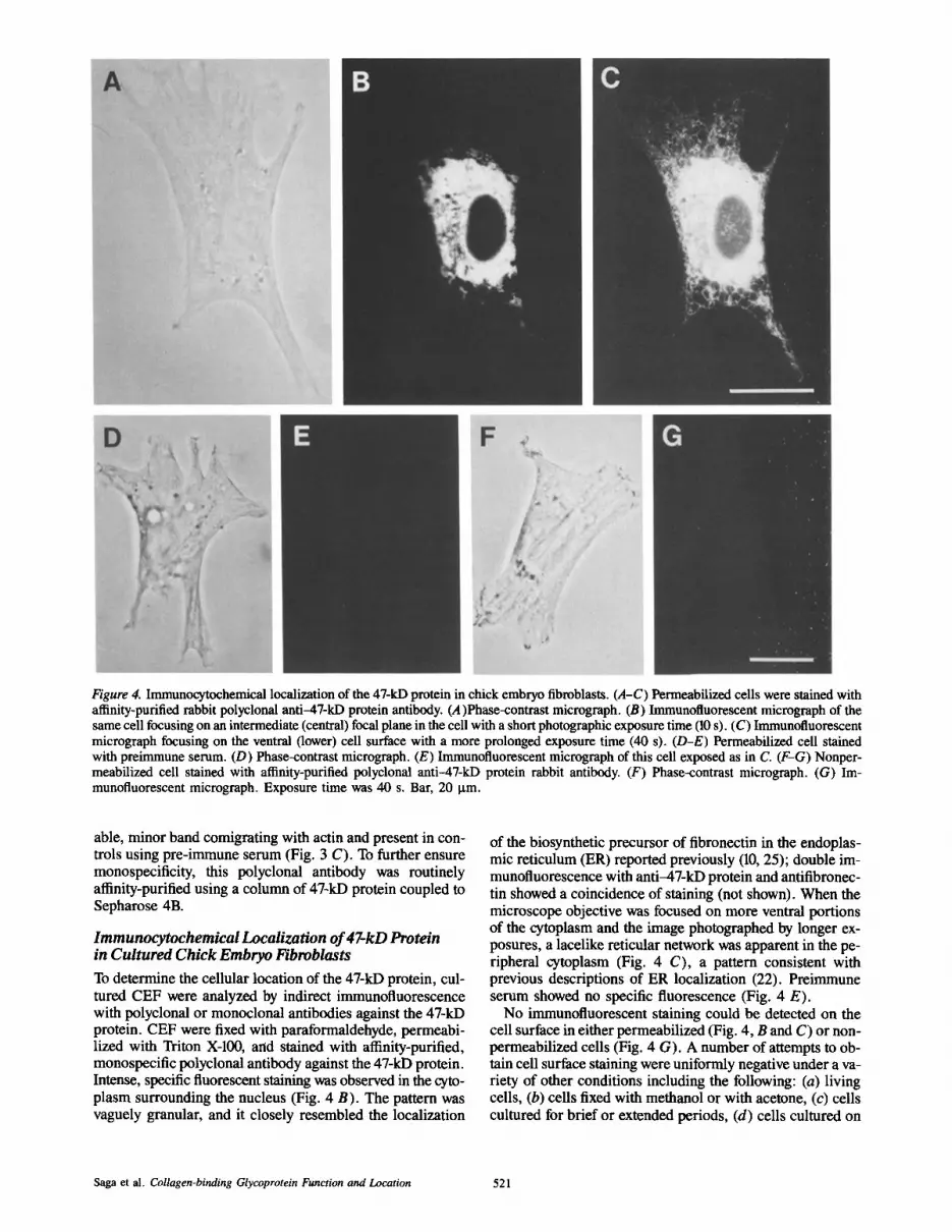
Figure 4. Immunocytochemical localization of the 47-kD protein in chick embryo fibroblasts. (A-C) Permeabilized ceils were stained with afffinity-purified rabbit polyclonal anti-47-kD protein antibody. (A)Phase-contrast micrograph. (B) Immunofluorescent microgmph of the same cell focusing on an intermediate (central) focal plane in the cell with a short photographic exposure time (10 s). (C) Immunofluorescent micrograph focusing on the ventral (lower) cell surface with a more prolonged exposure time (40 s). (D-E) Permeabilized cell stained with preimmune serum. (D) Phase-contrast micrograph. (E) Immunofluorescent micrograph of this cell exposed as in C. (F-G) Nonper- meabilized cell stained with affinity-purified polyclonal anti-47-kD protein rabbit antibody. (F) Phase-contrast micrograph. (G) Im- munofluorescent micrograph. Exposure time was 40 s. Bar, 20 Ixm.
able, minor band comigrating with actin and present in con- trois using pre-immune serum (Fig. 3 C). To further ensure monospecificity, this polyclonal antibody was routinely afffinity-purified using a column of 47-kD protein coupled to Sepharose 4B.
Immunocytochemical Localization of 47-kD Protein in Cultured Chick Embryo Fibroblasts
To determine the cellular location of the 47-kD protein, cul- tured CEF were analyzed by indirect immunofluorescence with polyclonal or monoclonal antibodies against the 47-kD protein. CEF were fixed with paraformaldehyde, permeabi- lized with Triton X-100, arid stained with affinity-purified, monospecific polyclonal antibody against the 47-kD protein. Intense, specific fluorescent staining was observed in the cyto- plasm surrounding the nucleus (Fig. 4 B). The pattern was vaguely granular, and it closely resembled the localization
of the biosynthetic precursor of fibronectin in the endoplas- mic reticulum (ER) reported previously (10, 25); double im- munofluorescence with anti-47-kD protein and antifibronec- tin showed a coincidence of staining (not shown). When the microscope objective was focused on more ventral portions of the cytoplasm and the image photographed by longer ex- posures, a lacelike reticular network was apparent in the pe- ripheral cytoplasm (Fig. 4 C), a pattern consistent with previous descriptions of ER localization (22). Preimmune serum showed no specific fluorescence (Fig. 4 E).
No immunofluorescent staining could be detected on the cell surface in either permeabilized (Fig. 4, B and C) or non- permeabilized cells (Fig. 4 G). A number of attempts to ob- tain cell surface staining were uniformly negative under a va- riety of other conditions including the following: (a) living cells, (b) cells fixed with methanol or with acetone, (c) cells cultured for brief or extended periods, (d) cells cultured on
Saga et al. Collagen-binding Glycoprotein Function and Location 521

Figure 5. (A-C) Double-staining with anti-47-kD protein antibody and fluorescent conjugates of WGA. Chick embryo fibroblasts were fixed, permeabilized, and stained with afffinity-purified rabbit anti-47-kD protein IgG, followed by fluorescein-conjugated goat anti-rabbit IgG plus rhodamine-conjugated WGA. (A) Phase-contrast micrograph. (B) Immunofluorescent micrograph (fluorescein channel) shows localization of the 47-kD protein. (C) Fluorescent micrograph (rhodamine channel) shows localization of the Golgi apparatus by WGA. (D-E) Immunofluorescent staining with monoclonal antibodies. Chick embryo fibroblasts were fixed, permeabilized, and stained with monoclonal antibodies against the 47-kD protein followed by fluorescein-conjugated goat anti-rat IgG. (D) Immunofluorescent micrograph stained with monoclonal antibody 7C8; exposure time was 60 s. (E) Immunofluorescent micrograph stained with monoclonal antibody 8B12; exposure time was 15 s. (F) Immunofluorescent micrograph stained with monoclonal antibody 9F9; exposure time was 15 s. Bar, 20 ~tm.
cross-linked gelatin films, (e) cells cultured on native colla- gen, ( f ) cells treated with collagenase to expose possible buried receptors; (g) cells permeabilized with saP0nin; and (h) cells incubated in antibodies for extensive periods (>16 h) to permit diffusion of antibody into poorly accessible sites.
Immunofluorescence localization with a variety of rat monoclonal antibodies against the 47-kD protein showed staining patterns essentially identical to that observed with the polyclonal antibodies (Fig. 5, D--F). The only exceptions were a few monoclonals that showed no immunofluorescence localization, one with poor specificity and a diffuse localiza-
Figure 6. Immunoelectron micrograph for intracellular localization of 47-kD protein in chick embryo fibroblasts. (A) The cells were stained by affinity-purified rabbit anti--47-kD protein antibody followed by peroxidase-conjugated Protein A in the presence of saponin as described in Materials and Methods. (B) Control staining with an equal concentration of preimmune rabbit IgG. Bar, 1 larn.
The Journal of Cell Biology, Volume 105, 1987 522

Saga et al. Collagen-binding Glycoprotein Function and Location 523

Figure 7. Absence of 47-kD protein from Golgi apparatus and plasma membrane. Chick embryo fibroblasts were stained as described in the legend for Fig. 6. No staining was found in the Golgi apparatus or at the cell surface. Bar, 1 gm.
tion pattern, and one with a weak pattern of a-actinin local- ization superimposed on a strong ER staining pattern, due to weak cross-reactivity with ct-actinin (data not shown). None of the monoclonal antibodies was observed to stain the cell surface of nonpermeabilized or living cells.
To examine whether the 47-kD protein was present in the Golgi apparatus, fixed and permeabilized cells were double- stained with polyclonal antibody against the 47-kD protein and rhodamine-labeled WGA. As shown in Fig. 5, B and C, staining for the 47-kD protein was strikingly excluded from the Golgi region, to which the WGA binds preferentially (23).
To confirm the presence of the 47-kD protein in the ER, cells were examined by immunoelectron microscopy. Fig. 6 A shows that the 47-kD protein was present in large quantities in the endoplasmic reticulum. No staining of the Golgi ap- paratus and the plasma membrane was detected (Fig. 7).
Immunohistochemical Localization of 47okD Protein In Vivo
Cryostat sections from chick liver were examined by im- munofluorescence labeling with anti--47-kD antibodies to de- termine whether the protein was a ubiquitous glycoprotein. Fig. 8 shows localization of the 47-kD protein in the portal region of chick liver (8 d after hatching). Cells likely to be
fibrocytes present throughout the connective tissue sur- rounding the portal vein, hepatic artery, and bile ducts were highly enriched in the 47-kD protein. Smooth muscle cells in the arterial wall, as well as Kupffer cells were also stained brightly. In contrast, the 47-kD protein was not detected in hepatocytes, sinusoidal endothelial cells, or the epithelial cells of bile ducts. These findings indicate that the 47-kD pro- tein is not uniformly distributed, and is instead expressed in only specific cell types.
Discussion
A major collagen-binding glycoprotein of Mr 47,000 D is present in a variety of cell types in vitro, and it has been par- tially characterized by several laboratories (12, 15, 16, 21). In previous papers (15, 16), we reported that (a) the major colla- gen/gelatin-binding protein in detergent extracts of chick em- bryo fibroblasts are the 47-kD glycoprotein and fibronectin, (b) the synthesis of the 47-kD gelatin-binding protein is de- creased after Rous sarcoma virus transformation, (c) the phosphorylation of this protein is increased after transforma- tion, and (d) the synthesis of the 47-kD protein is transiently induced by hyperthermia, identifying a novel heat-shock protein. In addition, we found that this 47-kD protein can bind specifically to fetuin besides collagen and gelatin (Nagata, K., S. Saga, and K. M. Yamada, unpublished data).
The Journal of Cell Biology, Volume 105, 1987 524

Figure 8. Immunohistochemical localization of the 47-kD protein in chick liver. Cryostat section of chick liver was stained with aflinity- purified rabbit anti--47-kD protein IgG followed by rhodamine-conjugated goat anti-rabbit IgG. (A) Cryostat section stained with hematoxy- lin and eosin; this section was an adjacent serial section to the section in B. (B) Immunofluorescent micrograph corresponding to the area shown in A. PV, portal vein; HA, hepatic artery; BD, bile ducts. Bar, 100 I~m.
Saga et al. Collagen-binding Glycoprotein Function and Location 525

In this paper, we describe (a) pH regulation of the ability of this glycoprotein to bind to collagen, (b) the use of this finding to isolate milligram quantities of the protein without using denaturants for the production of monoclonal antibod- ies, (c) localization of the antigen to the endoplasmic reticu- lum by a polyclonal and a variety of monoclonal antibodies, and (d) cell-type specificity in the expression of this major glycoprotein.
We found that all binding of the 47-kD glycoprotein to gelatin-affinity columns was abolished at pH 6.3. It is of in- terest that similar or even lower pH values are characteristic for certain vesicular secretory and endocytic compartments of cells involved in protein processing or translocation (reviewed by Griffiths and Simons [8]). This property of pH dependence could permit the regulated release of bound ligands from membrane-associated 47-kD molecules by a simple local reduction in pH, as is thought to occur in some step(s) of protein and receptor sorting.
The ability to release the 47kD protein from immobilized gelatin by brief pH 6.3 treatment permitted the isolation of large amounts of relatively pure protein from tissue sources without the need for the SDS treatment used in previous studies (12, 15, 16). 15 independent monoclonal antibodies and a polyclonal antibody were generated; it is not known whether our use of such initially native protein or the glutaraldehyde crosslinking step explains the good immuno- logic response compared with previous attempts (21; and un- published results).
Using these antibodies, we examined the localization of the 47-kD protein by immunofluorescence microscopy. When CEF were fixed, permeabilized, and stained with mono- specific polyclonal antibody or monoclonal antibodies against the 47-kD protein, the most intense staining occurred in a perinuclear pattern very similar to that reported previously for fibronectin in the ER of this particular cell type (10, 25). There was also a weaker pattern of staining extending out- ward to the cell periphery that was similar to the reticular network structure visualized with the cationic fluorescent dye 3-3'-dihexyloxacarbocyanine iodide, which is known to stain the ER of living or fixed cells (22). The presence of the 47-kD protein in the ER and its apparent absence from the Golgi apparatus was strikingly confirmed by immunoelec- tron microscopy using polyclonal antibody against the 47-kD protein. Staining was reproducibly detected not only on the ER membrane, but also in the lumen, which could be due either to presence of the 47-kD protein in the lumen or to diffusion of reaction product.
Hogan, Kurkinen, and co-workers have characterized a 47,000-dalton glycoprotein in mouse parietal endoderm cells and other cell types related to the protein described in this study (12, 21). This protein, which they term "colligin," binds to several collagen types and is not secreted into culture medium (12, 21). Colligin was originally postulated to be a cell surface protein because it could be labeled by lacto- peroxidase-mediated iodination in cells detached from sub- strates with EDTA (12). However, Hughes in collaboration with the same group has very recently reported (11) that the glycosylation pattern of this protein suggests that it is actu- ally located in the ER. Our immunocytochemical studies directly demonstrate that the corresponding 47-kD protein in chick embryo fibroblasts is located exclusively in the ER,
and that it is not convincingly detectable by our methods ei- ther in the Golgi complex or on the cell surface.
Because the absence of this collagen-binding glycoprotein from the cell surface was so unexpected, we employed a vari- ety of experimental manipulations in attempts to visualize such staining, including alterations in culture conditions, substrates, staining conditions, and even collagenase treat- ment to expose epitopes. No plasma membrane localization could be detected. We conclude that no 47-kD protein is likely to be present on the cell surface, unless as an undetect- able or cryptic form.
The apparent absence of the 47-kD protein from the cell surface and its location in the ER argues against all but the most transient (or no) interactions of this protein on the cell surface with its ligands, e.g., various types of collagen. If such interactions are indeed physiologically significant, they would consequently occur intracellularly in the ER. It may be relevant that a protein related to hsp70, another heat-shock protein, is present in rat liver, is found in the lumen of the ER but is not secreted, and binds to IgG heavy chains in an ATP-dependent manner. This protein termed BiP or grp78 is speculated to play a role in the assembly and secretion of immunoglobulin (14). It was thus important to determine whether all cells, especially secretory cells such as hepato- cytes, express the 47-kD glycoprotein in vivo.
Although this major glycoprotein was previously shown to be synthesized by a variety of cell types in vitro (12, 15, 21), an examination of its localization in vivo indicates that it is clearly not ubiquitous. In particular, it could not be detected in chicken liver hepatocytes, nor in the epithelia of bile ducts. Instead, it was present in substantial quantities in connective tissue cells presumed to be fibrocytes, as well as in Kupffer cells and in vascular smooth muscle cells. Thus, the protein was not detectable in albumin-secreting cells, and its expres- sion was regulated according to the specific cell type.
The apparent absence of the 47-kD protein from the cell surface and Golgi complex, its presence in ER, and its cell type-specific expression, strongly suggest that this glycopro- tein is not a general cell surface receptor for collagen. Be- cause it is a major cellular glycoprotein, it may instead func- tion within the ER in some as yet unknown role in protein processing or translocation. For example, this abundant gly- coprotein might bind to its ligands in the ER to segregate them from other secreted proteins such as fibronectin. Alter- natively, it might be involved in protein folding or movement in the ER, or in modulating the intraceUular degradation of procollagen and other secreted proteins. Because it exists in cells in substantial quantities (6, 12, 15, 16, 21), and because it can be regulated by a variety of factors including heat shock (15), cyclic nucleotides and retionic acid (12), transfor- mation (16), and cell type (this paper), this 47-kD glycopro- tein appears worthy of more detailed characterizations in the future.
We are grateful to Susan Yamada for developing the immunoprecipitation protocol and to Althea Gaddis for preparing the manuscript.
Received for publication 2 January 1987, and in revised form 20 March 1987.
References
1. Akiyama, S. K., E. Hasegawa, T. Hasegawa, and K. M. Yamada. 1985. The interaction of fibronectin fragments with fibroblastic cells. J. Biol. Chem. 260:13256-13260.
The Journal of Cell Biology, Volume 105, 1987 526

2. Akiyama, S. K., and K. M. Yamada. 1985. Comparisons of evolutionarily distinct fibronectins: evidence for the origin of plasma and fibroblast cel- lular fibronectins from a single gene. J. Cell. Biochem. 27:97-107.
3. Chen, W.-T., and S. J. Singer. 1980. Fibronectin is not present in the focal adhesions formed between normal cultured fibroblasts and their substrata. Proc. Natl. Acad. Sci. USA. 77:7318-7322.
4. Chen, W.-T., and S. J. Singer. 1982. Immunoelectron microscopic studies of the sites of cell-substratum and cell-cell contacts in cultured fibro- blasts. J. Cell Biol. 95:205-222.
5. Chen, W.-T., J. Wang, T. Hasegawa, S. S. Yamada, and K. M. Yamada. 1986. Regulation of fibronectin receptor distribution by transformation, exogenous fibronectin, and synthetic peptides. J. Cell Biol. 103:1649- 1661.
6. Dubbelman, T. M. A. R., and K. M. Yamada. 1982. A survey of differ- ences between membrane polypeptides of transformed and nontrans- formed chick embryo fibroblasts. Biochim. Biophys. Acta. 693:177-187.
7. Furth, M. E., L. J. Davis, B. Fleurdelys, and E. M. Scolnick. 1982. Mono- clonal antibodies to the p21 products of the transforming gene of Harvey murine sarcoma virus and of the cellular ras gene family. J. Viol. 43: 294-304.
8. Griffiths, G., and K. Simons. 1986. The trans Golgi network: Sorting at the exit site of the Golgi complex. Science (Wash. DC). 234:438-443.
9. Galfre, G., C. Milstein, and B. Wright. 1979. Rat x rat hybrid myelomas and a monoclonal anti-Fd portion of mouse IgG. Nature (Lond.). 277:131- 133.
10. Hedrnan, K. 1980. Intracellular localization of fibronectin using im- munoperoxidase cytochemistry in light and electron microscopy. J. His- tochem. Cytochem. 28:1233-1241.
11. Hughes, R. C., A. Taylor, H. Sage, and B. L. M. Hogan. 1987. Distinct patterns of glycosylation of colligin, a collagen-binding glycoprotein, and SPARC (osteonectin), a secreted Ca÷+-binding glycoprotein: evidence for the localization of colligin in the endoplasmic reticulum. Eur. J. Bio- chem. 163:57-63.
12. Kurkinen, M., A. Taylor, J. I. Garrels, and B. L. M. Hogan. 1984. Cell surface-associated proteins which bind native type IV collagen or gelatin. J. Biol. Chem. 259:5915-5922.
13. Laemmli, U. K. 1970. Cleavage of structural proteins during the assembly of the head of bacteriophage T4. Nature (Lond.). 283:249-256.
14. Munro,-S., and H. R. B. Pelham. 1986. An hsp70-1ike portein in the ER: identity with the 78 kb glucose-regulated protein and immunoglobulin heavy chain binding protein. Cell. 46:291-300.
15. Nagata, K., S. Saga, and K. M. Yamada. 1986. A major collagen-binding protein of chick embryo fibroblasts is a novel heat shock protein. J. Cell Biol. 103:223-229.
16. Nagata, K., and K. M. Yamada. 1986. Phosphorylation and transformation sensitivity of a major collagen-binding protein of fibroblasts. J. Biol. Chem. 261:7531-7536.
17. Oakley, B. R., D. R. Kirsch, and N. R. Morris. 1980. A simplified ultrasensitive silver stain for detecting proteins in polyacrylamide gels. Anal. Biochem. 105:361-363.
18. Platt, J. L., and A. F. Michael. 1983. Retardation of fading and enhance- ment of intensity of immunofluorescence by p-phenylenediamine. J. Histochem. Cytochem. 31:840-842.
19. Robbins, P. W., G. G. Wickus, B. J. Gaffney, C. B. Hirschberg, P. Fuchs, and P. M. Blumberg. 1974. The chick fibroblast cell surface after trans- formation by Rous sarcoma virus. Cold Spring Harbor Symp. Quant. Biol. 39:1173-1180.
20. Saraste, J., and Hedman, K. 1983. Intracellular vesicles involved in the transport of Semliki Forest virus membrane proteins to the cell surface. EMBO (Eur. Mol. Biol. Organ.) J. 2:2001-2006.
21. Taylor, A., B. L. M. Hogan, and F. M. Watt. 1985. Biosynthesis of EGF receptor, transferrin receptor and colligin by cultured human keratino- cytes and the effect of retinoic acid. Exp. Cell Res. 159:47-54.
22. Terasaki, M., J. Song, J. R. Wong, M. J. Weiss, and L. B. Chen. 1984. Localization of endoplasmic reticulum in living and glutaraldehyde-fixed cells with fluorescent dyes. Cell. 38:101-108.
23. Virtanen, I., P. Ekblom, and P. Laurila. 1980. Subcellular compartmental- ization of saccharide moieties in cultured normal and malignant cells. J. Cell Biol. 85:429-434.
24. Vogt, P. K. 1969. Focus assay of Rous sarcoma virus. In Fundamental Techniques in Virology. K. Habel and N. P. Salzman, editors. Academic Press, Inc., New York. 66-71.
25. Yamada, S. S., K. M. Yamada, and M. C. Willingham. 1980. Intracellular localization of fibronectin by immunoelectron microscopy. J. Histochem. Cytochem. 28:953-960.
Saga et al. Collagen-binding Glycoprotein Function and Location 527