peripheral neutrophil functions and cell signalling in crohn`s disease
TRANSCRIPT

Peripheral Neutrophil Functions and Cell Signalling inCrohn`s DiseaseRajesh Somasundaram, Veerle J. A. A. Nuij, C. Janneke van der Woude, Ernst J. Kuipers, Maikel P.Peppelenbosch, Gwenny M. Fuhler*
Department of Gastroenterology and Hepatology, Erasmus University Medical Center, Rotterdam, Rotterdam, The Netherlands
Abstract
The role of the innate immunity in the pathogenesis of Crohn’s disease (CD), an inflammatory bowel disease, is asubject of increasing interest. Neutrophils (PMN) are key members of the innate immune system which migrate tosites of bacterial infection and initiate the defence against microbes by producing reactive oxygen species (ROS),before undergoing apoptosis. It is believed that impaired innate immune responses contribute to CD, but it is as yetunclear whether intrinsic defects in PMN signal transduction and corresponding function are present in patients withquiescent disease. We isolated peripheral blood PMN from CD patients in remission and healthy controls (HC), andcharacterised migration, bacterial uptake and killing, ROS production and cell death signalling. Whereas IL8-inducedmigration and signalling were normal in CD, trans-epithelial migration was significantly impaired. Uptake and killing ofE. coli were normal. However, an increased ROS production was observed in CD PMN after stimulation with thebacterial peptide analogue fMLP, which was mirrored by an increased fMLP-triggered ERK and AKT signalactivation. Interestingly, cleavage of caspase-3 and caspase-8 during GMCSF-induced rescue from cell-death wasdecreased in CD neutrophils, but a reduced survival signal emanating from STAT3 and AKT pathways wasconcomitantly observed, resulting in a similar percentage of end stage apoptotic PMN in CD patients and HC. In toto,these data show a disturbed signal transduction activation and functionality in peripheral blood PMN from patientswith quiescent CD, which point toward an intrinsic defect in innate immunity in these patients.
Citation: Somasundaram R, Nuij VJAA, van der Woude CJ, Kuipers EJ, Peppelenbosch MP, et al. (2013) Peripheral Neutrophil Functions and CellSignalling in Crohn`s Disease. PLoS ONE 8(12): e84521. doi:10.1371/journal.pone.0084521
Editor: Juliet Spencer, University of San Francisco, United States of America
Received July 11, 2013; Accepted November 14, 2013; Published December 19, 2013
Copyright: © 2013 Somasundaram et al. This is an open-access article distributed under the terms of the Creative Commons Attribution License, whichpermits unrestricted use, distribution, and reproduction in any medium, provided the original author and source are credited.
Funding: GMF is supported by the Dutch Cancer Society (grant 2010-4737). MPP is supported by a personal ECCO grant. These funders had no role instudy design, data collection and analysis, decision to publish, or preparation of the manuscript.
Competing interests: The authors have declared that no competing interests exist.
* Email: [email protected]
Introduction
Crohn`s disease (CD) is a chronic inflammatory boweldisease with a complex aetiology involving genetic factors,priming by enteric microflora, environmental factors and analteration in the immune-mediated response [1-3]. Increasingevidence points towards a role of the innate immune system inCD pathology, with a role for dendritic cells, macrophages andneutrophils [4,5]. Neutrophils (polymorphonuclear cells; PMN),one of the most abundant and important mediators of innateimmunity, are professional phagocytes which mount the acuteinflammatory response and act as the first line of defenceagainst invading pathogens [6]. The role of PMN in CDpathology remains obscure. Impaired PMN function may resultin limited bacterial clearance and fuel an on-going, chronicinflammatory response. Indeed, patients with congenitaldisorders of PMN function (i.e. migration, production of reactiveoxygen species [ROS]) often develop inflammatory boweldisease (IBD) [7-10]. Furthermore, mice lacking the NADPH
oxidase gene encoding p40phox show enhanced colitis [11]supporting a positive role for ROS in the resolution of disease.On the other hand, epithelial cell damage and ensuing bacterialinvasion and inflammation have been attributed to noxiousROS released by PMN, and PMN ablation has provenbeneficial in a subset of CD patients [12-14]. Relatively fewstudies have investigated PMN cell biology in CD, and thosethat have, show conflicting results. Although an inadequatePMN influx and subsequent clearance of bacteria has beenobserved in CD, this may be caused by defective secretion ofpro-inflammatory cytokines by macrophages, and it is as yetunclear whether PMN intrinsically lack migratory capacity, ROSproduction or bactericidal activity [15-20].
Altogether, varying predictions have been made regardingthe role of PMN in the pathogenesis of CD. Recently, acomprehensive analysis of peripheral blood monocytes inpatients with quiescent CD revealed intrinsic defects in thiscell-type, prior to inflammation and their recruitment to themucosa [21]. Impaired cytokine profiles were observed in CD
PLOS ONE | www.plosone.org 1 December 2013 | Volume 8 | Issue 12 | e84521
monocytes, whereas migration, ROS production andphagocytosis were unaffected. However, an exhaustiveanalysis of multiple PMN effector functions and the signallingevents involved in one study has so far not been conducted butis urgently needed to complement our insight into the innateimmune system functionality in IBD patients. In the currentstudy, we investigated whether PMN from quiescent CDpatients are constitutively defective, by investigating thecapacity of PMN to respond to stimuli inducing migration,phagocytosis, bacterial killing, ROS production and apoptosis,and the correlation thereof to the activity of the signaltransduction pathways involved. We show that transepithelialmigration and fMLP-induced ROS production as well as fMLPand granulocyte-macrophage colony-stimulating factor(GMCSF)-mediated signalling are altered in CD PMN, whereasphagocytosis and bacterial killing are normal.
Materials and Methods
PatientsThis study was approved by the ethical board of the Erasmus
MC, Rotterdam, The Netherlands (protocol MEC-2004-168).Patients and healthy controls were included after writteninformed consent was obtained. In total, 53 patients and 20healthy controls were included (Table 1). Due to the limitednumber of PMN obtained from 20 ml of peripheral blood, theethical limit in our protocol, as well as logistical arrangements,not all the experiments could be performed with the same setof patients. However, the characteristics of the patients usedwere similar between experiments, thus precluding the skewingof results of secondary reasons such as age or medication.Patients were in clinical remission (quiescent disease) at thetime of blood collection, with no evidence of inflammation inendoscopies performed around this time. All experiments onCD PMN were performed simultaneously on PMN from ahealthy volunteer.
Granulocyte isolation from human peripheral bloodHeparin anti-coagulated blood was obtained from CD
patients and HCs in parallel. Neutrophils were isolated asdescribed previously [22]. Briefly, mononuclear cells wereremoved by centrifugation of heparinized blood over Ficoll-Paque (Amersham), followed by erythrocyte lysis with ice-coldNH4Cl solution. PMN were allowed to recover for 30 minutes at37°C in RPMI 1640 supplemented with 0.5% human serumalbumin (HSA; Sanquin, the Netherlands). PMN wereresuspended in incubation buffer (20mM HEPES, 132mM Nacl,6mM KCL, 1mM MgSO4, 1.2mM KH2PO4, 5mM glucose, 1mMCaCl2 and 0.5% HSA) before they were subjected to functionalassays.
Migration assayThe migration assay was performed using a microchamber
transwell system with 3µM pores (Becton Dickinson). PMN (2 x10⁵) were applied to the upper well of the chamber. Migrationwas induced by 20 ng/ml IL8 (Peprotech, Rockyhill, NJ)present in the lower compartment of the chamber for 4 hours at
37°C. Basal to apical migration assay was performed usinginverted monolayers of Caco2 cells, which were grown invertedon collagen-coated transwell inserts for 5 days in DMEM (PAAlaboratories, Pasching, Austria)/10% fetal calf serum (FCS,PAA)/ 10ug/ml Penicillin/Streptomycin (Gibco) (37°C and 5%CO2). Confluence of the epithelial cell monolayer wasconfirmed by testing their permeability to bovine serum albumin(BSA) as described previously [23]. PMN migration wasdetermined by fluorescence-activated cell sorting (FACS)analysis as described, using FACSCantoII (BD Biosciences)[24], and cells migrated towards IL8 were expressed aspercentage of those migrated in control wells without IL8.
Phagocytosis and bactericidal activity of PMNBacterial uptake and killing were performed as previously
described [25]. Briefly, E. coli bacteria, transformed with GFPexpression vector were grown in kanamycin-containing LBmedia until OD of 1, after which cultures were centrifuged andresuspended in 1ml of PBS supplemented with 0.1% Gelatinand 10mM HEPES. Bacterial opsonisation was carried out byincubating bacteria with non-heat inactivated human serum(Gibco) for 15 minutes at 37°C. PMN were challenged with 100µl of opsonised bacteria at 37°C for 15 minutes, using 0°Ccontrol for each experiment. The percentage of phagocytosingPMN, as well as their fluorescence intensity as a measure ofthe amount of phagocytosed bacteria, were determined by flowcytometry. Bacterial killing was tested by washing E. coli-challenged PMN 2 times, and resuspending the cell pellet in1ml of antibiotics-containing buffer in order to kill anycontaminating bacteria attached to the plastic. Bacterial killingwas allowed to take place for 4 hours. PMN were lysed using
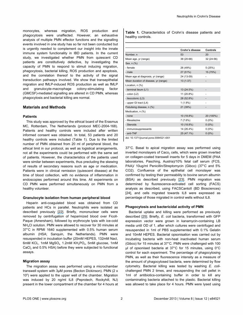
Table 1. Characteristics of Crohn’s disease patients andhealthy controls.
Crohn’s disease ControlsNumber, n 53 20Mean age, yr (range) 38 (20-68) 32 (24-56)Sex, n (%) - female 26 (49%) 5 (25%)- male 27 (51%) 15 (75%)Mean age at diagnosis, yr (range) 24 (13-59) -Mean duration of disease, yr (range) 13 (1-37) -Location, n (%) - terminal ileum (L1) 13 (24.5%) -- colon (L2) 11 (20.8%) -- ileocolonic (L3) 28 (52.8%) -- upper GI tract (L4) 1 (1.9%) -Fistulising disease, n (%) 21 (39%) -Medication, n (%) - none 10 (18.8%) 20 (100%)- Mesalazine 7 (7.6%) 0 (0%)- Steroids 10 (18.8%) 0 (0%)- immunosuppressants 14 (26.4%) 0 (0%)- anti-TNF 25 (47.1%) 0 (0%)
doi: 10.1371/journal.pone.0084521.t001
Neutrophils in Crohn's Disease
PLOS ONE | www.plosone.org 2 December 2013 | Volume 8 | Issue 12 | e84521

sterile water, lysates were plated on LB agar plates and thecolonies grown after 18 hours were counted using a colonycounter. Each experiment was done in duplicate.
ROS production assayROS production was performed as previously described [26].
Briefly, PMN (2x10⁶cells/ml) were incubated with DHR123(Sigma-Aldrich) for 15 minutes and stimulated with 1µM fMLP(Sigma-Aldrich) for 30 minutes. For priming experiments, cellswere pre-treated with 5ng/ml GMCSF (Sargramostim, Bayer,Germany) for 15 minutes prior to fMLP stimulation. Stimulationwas terminated by washing the cells with ice-cold PBScontaining 1% HSA and placing them on ice. Oxidation ofDHR123 to the fluorescent Rhodamine 123 was measured byflow cytometry within 30 minutes of termination of stimulation.
Apoptosis analysisApoptosis was induced by culturing PMN (2x10⁶/ml) with
anti-Fas antibody (Fas-Ab, CH 11, 100ng/ml, Millipore).Alternatively, PMN were treated with GMCSF (10ng/ml). After 6hours of Fas-Ab-induced apoptosis and 15 hours of GMCSF-induced rescue, the percentage of apoptotic PMN wasmeasured by Annexin-V kit according to the manufacturer’sinstructions (BD Biosciences, San Jose, CA). Necrotic PMNwere excluded by 7AAD (BD Biosciences, San Jose, CA)positivity. Late apoptosis was measured by internucleosomalDNA fragmentation using Apo direct in situ DNA fragmentationassay kit (Biovision, Milpitas, California). Briefly, this TUNEL-based detection kit utilizes terminal deoxynucleotidyltransferase (TdT) to catalyse the incorporation of fluorescein-12-dUTP at the free 3`-hydroxyl ends of the fragmented DNA.Stained PMN were analyzed using Flowcytometry and the datawere analyzed using FlowJo software (Ashland, OR).
Quantitative western blot analysisPMN were stimulated with 1µM fMLP, 5ng/ml GMCSF,
5ng/ml GCSF or 100ng/ml Fas-Ab (CH 11) as indicated in thefigures. Pelleted cells were resuspended in Laemmli buffer,boiled, separated by SDS-PAGE and electrophoreticallytransferred to PVDF Immobilon FL membrane (Milipore,Billerica, MA). Membranes were probed with antibodies againstphospho-ERK1/2 (Thr202/Tyr204), phospho-AKT (Ser473),phospho-STAT3, Caspase 3 (cleaved and uncleaved) orcleaved Caspase 8, all from Cell signalling technology(Danvers, MA). Total levels of ERK, AKT and STAT3 areunaffected by short term stimulation of cells with IL8, fMLP orGMCSF [27-31]. In addition, we demonstrated that total levelsof these proteins show excellent correlation with total β-actinlevels (Figure 1). Therefore, equal loading was confirmed byreprobing blots with antibodies against β-actin (Santa CruzBiotechnology, Santa Cruz, CA) according to themanufacturers’ protocols. Proteins were detected by IR dyes(LI-COR, Lincoln, NE). Quantification of phosphorylation andcleavage of Caspases were performed by densitometry of theimages, using Odyssey 3.0 software.
Statistical analysisComparisons between CD and HC samples were tested by
non-parametric test for unpaired samples (Mann-Whitneytesting) in functional experiments. For western blot analysis,where CD and HC samples were paired per gel, comparisonsfor paired samples were tested by Student-T-test usingGraphpad software (La Jolla, CA).
Results
Decreased trans-epithelial migration of neutrophilsfrom CD patients in response to IL8
First, we investigated the migratory capacity of CDneutrophils and two of the major signalling pathways involvedtherein, the ERK1/2 and PI3K-AKT signalling moieties [32].After confirming the partial dependence of IL8-inducedmigration on these pathways by using their respective specificinhibitors (Figure 1A: 100 vs. 65.5 ±21% for U0126 and 100 vs.67 ±22% for LY294002), we examined the phosphorylation ofthese signal transducers in PMN from CD patients and HCs.We observed a rapid and transient activation of ERK1/2 andAKT in response to IL8 stimulation, but found no significantdifferences in the level of activation of these moleculesbetween CD patients and HCs (Figure 1B, C and D, n=10).Total levels of ERK were similar between CD patients (n=18)and HC (n=16, p=0.7, Figure 2A and B). In line with thisunaltered migration-dependent signalling, the percentage ofPMN migrating towards IL8 was not different between CDpatients (n=11) and HCs (n=8) (2267±1859% vs. 3574±2443%,p=0.114, Figure 1E).
In an in vivo setting, IL8-mediated migration of PMN towardsthe lumen of the gut requires basolateral-to-apical migration ofPMN over epithelial cells. As this process is substantiallydifferently regulated as compared to migration of PMN towardcytokines alone [33,34], we also determined the level ofbasolateral-to-apical migration of PMN through an invertedmonolayer of human epithelial Caco2 cells. Interestingly, thepercentage of PMN migrating towards IL8 through epithelialcells was significantly reduced in CD patients compared to HCs(Figure 1F, mean±SEM of 133±55% vs. 190±60%, n=10, p =0.04). Together, these data suggest that IL8 stimulation of CDPMN in itself results in normal activation of the ERK and PI3Kpathways and migration, whereas intrinsic trans-epithelialmigration capacity of PMN from CD patients is impaired.
Bacterial uptake and killing are not affected in CDpatients
Next, we investigated the uptake of GFP-positive E. coli byisolated PMN from CD patients (n=16) and HCs (n=14). Asshown in Figure 2A and B, neither the percentage ofphagocytosing PMN (mean±SEM of 64±24% vs. 62±19%,p=0.7) nor the number of bacteria taken up per granulocyte(1648±1244 vs. 1242±759 MFI, p=0.313) were significantlydifferent between CD patients and HCs. In addition, an equalamount of bacterial colonies were grown from CD and HCPMN, demonstrating that the efficiency of bacterial killing wasnot different between patients (n=10) and controls (n=9)(263±172 vs. 305±199 colonies, Figure 2C). These results
Neutrophils in Crohn's Disease
PLOS ONE | www.plosone.org 3 December 2013 | Volume 8 | Issue 12 | e84521

Figure 1. PMN from CD patients are deficient in trans-epithelial migration towards IL8. (A) The involvement of ERK1/2 andPI3K pathways in IL8-induced migration was confirmed by measuring the percentage of migrated PMN after incubation with orwithout 10 µM of U0126 and LY294002, respectively. Mean±SEM is shown (n=3). (B) PMN were stimulated with 20 ng/ml of IL8 forthe indicated time points. Experiments were performed on healthy controls (HC) and CD PMN simultaneously, and samples wereloaded side-by-side on the same gel. ERK1/2 and AKT activation were detected by their phospho-specific antibodies.Representative example is shown. (C) No differences in levels of activated ERK1/2 were observed between CD patients and HC(n=10, mean±SEM shown) upon quantification of blots by densitometry. (D) No differences in levels of activated AKT were observedbetween CD patients and HC (n=10, mean±SEM shown) upon quantification of blots by densitometry. (E) PMN from HC and CDpatients were applied to the upper compartment of a transwell system. PMN transmigrated in response to 20 ng/ml IL8 present inthe lower compartment were counted by flow cytometry and results are represented as percentage of those migrated in controlwells. No differences were observed between Mean±SEM of CD patients (n=11) and HC (n=8). (F) PMN from healthy and CDpatients were allowed to migrate through a monolayer of epithelial cells towards IL8 for 4 hours at 37°C. Compared to HC PMN, CDPMN showed significantly less migration (Mean±SEM, *p=0.02, n=10).doi: 10.1371/journal.pone.0084521.g001
Neutrophils in Crohn's Disease
PLOS ONE | www.plosone.org 4 December 2013 | Volume 8 | Issue 12 | e84521

indicate that there is no intrinsic defect in either phagocytosisor killing of E. coli bacteria in PMN from CD patients withquiescent disease.
Enhanced fMLP-induced ROS production in CDpatients, corresponding with increased ERK and AKTsignalling
The production of ROS is an important antibacterial defencemechanism of PMN. We therefore studied the amount ofsuperoxide produced, and the signalling events involved, inresponse to the bacterial peptide analogue fMLP. As shown inFigure 3A, fMLP–stimulated ROS production was significantlyhigher in PMN from CD patients as compared to HCs (mean±SEM of 130±31% vs. 106±28%, p=0.03, n=14). Thiscorresponded to a significantly enhanced fMLP-inducedphosphorylation of the ERK and PI3K/AKT pathways (known tobe required for ROS production [35]), in PMN from CD patients(p=0.03 and p=0.02, respectively at t=2 min, n=9, Figure 3B-D).These results suggest that PMN from CD patients may alreadybe partially primed in vivo. Priming is normally established bypro-inflammatory cytokines such as granulocyte-macrophagecolony-stimulating factor (GMCSF), and serves to drasticallyenhance the respiratory burst in response to bacterial peptidesin an inflammatory environment. Indeed, priming of PMN withGMCSF resulted in a significantly higher fMLP-triggered ROSproduction in both CD and HC PMN (p<0.05). However, ROSproduction after priming with GMCSF did not differ between CDpatients and HCs, indicating that maximal achievablerespiratory burst is equal between these groups. As for ROSproduction, priming of PMN with GMCSF resulted in asignificantly enhanced fMLP-triggered phosphorylation of bothERK1/2 and AKT, which again was equal between CD patientsand HCs. Similar results were obtained when ROS productionand signalling were investigated in GCSF-primed PMN (notshown).
Together, these results suggest that PMN from CD patientsrelease ROS more rapidly in response to bacterial stimuli, butthat the maximum achievable level of ROS production isunaltered.
Reduced Caspase cleavage during spontaneousapoptosis in PMN from CD patients
After performing their bactericidal function, PMN undergoapoptosis and are cleared by macrophages. One of the earlysignalling events to take place in cellular apoptosis is thecleaving of Caspase 8 and 3. Although no differences in totalCaspase 3 and Caspase 8 levels were observed in patients(Figure 4A and B, and Figure 2C-E), the amount of Caspase 3and 8 cleaved during spontaneous cell death was reduced inCD compared to HCs after 6 hours (Figure 4C-E, p=0.1 andp=0.04 for Caspase 3 and Caspase 8 respectively, n=6).Treatment of cells with the apoptosis-inducing Fas-antibody CH11 enhanced cleavage of both Caspases to an equal extent inpatients and HCs (Figure 4C-E, p=0.5 and p=0.2 for Caspase 3and Caspase 8 respectively, n=6), indicating that only theintrinsic apoptosis machinery is affected in CD.
Treatment of PMN with GMCSF for 15 hours protects againstcleavage of Caspase 3 (Figure 4F), which corresponds to a
decreased number of apoptotic cells as measured byexternalisation of phosphatidylserine (PS) by Annexin Vstaining (Figure 4G). When comparing CD patients and HCs forCaspase cleavage in the presence of GMCSF, a significantlyenhanced GMSCF-induced survival signal was observed in CDpatients, as evidenced by reduced cleavage of Caspase 3 and8 (Figure 4C, D and E, p=0.04 and p=0.003, respectively, n=6).
Normal end-stage apoptosis in PMN from CD patientsTo test whether the reduced Caspase 3 and 8 signal in CD
patients results in a decreased cell death, we measured thepercentage of annexin-V-positive cells in PMN cultures at t = 0,6 and 15h of culture. As expected, cell viability immediatelyupon isolation (t=0h) was more than 90% (mean±SEM of5.4±3.6% dead cells in CD, n=9, vs. 3.5±2.2% in HC, n=8,p=0.1, Figure 5A). Surprisingly, spontaneous apoptosis,observed within 6 hours, was not reduced in CD patientscompared to healthy controls (mean±SEM of 30±19% vs.27±11%, p=0.9). Engagement of the Fas-receptor drasticallyincreased the amount of annexin V-positive cells, equally in CDpatients and HC (71±12% vs. 77±11%, p=0.2).
In addition, whereas Caspase cleavage in GMCSF culturedPMN was significantly reduced in CD patients, apoptosis asmeasured by PS-expression showed no differences betweenCD and HC PMN in either spontaneous apoptosis after 15h, orthe rescue thereof by GMCSF (mean±SEM of 42±17% vs.48±18%, p=0.7). These findings were confirmed by TUNELassay, showing no significant differences between CD patientsand HCs in the percentage of apoptotic PMN cultured with orwithout GMCSF for 15 h (mean±SEM of 66±5% vs. 57±20%, p= 0.45 and 31±7% vs. 27±11%, p = 0.49, respectively, n=6,Figure 5B).
Decreased GMCSF-induced STAT3 phosphorylation inPMN from CD patients
Whereas GMCSF-induced rescue of Caspase cleavage wasenhanced in CD patients, this was not mirrored by anincreased survival of PMN. These results suggest that otherdeath mechanisms may override the positive survival signal inCD patients. We therefore investigated GMCSF-inducedphosphorylation of STAT3 and AKT, constituting two of themajor survival mechanisms induced by this cytokine [31,36]. Asshown in Figure 5C and D, STAT3 phosphorylation in responseto GMCSF was significantly reduced in PMN from CD patientscompared to their healthy counterparts, whereas total STAT3levels were unchanged (Figure 2A and F). Similarly, a reducedAKT phosphorylation was observed in 4 out of 5 CD patients.These results suggest that an impaired STAT3 and AKT-survival pathway in CD patients may counteract the reducedCaspase cascade activation, thus resulting in equal numbers ofapoptotic PMN in CD and HC.
Discussion
In the current study, we demonstrate that intrinsic propertiesof PMN from patients with quiescent CD are changed. Adecreased trans-epithelial migration, increased ROSproduction in response to bacterial peptides, and impaired
Neutrophils in Crohn's Disease
PLOS ONE | www.plosone.org 5 December 2013 | Volume 8 | Issue 12 | e84521

Figure 2. Normal bacterial uptake and killing by PMN from CD patients. Isolated PMN were challenged with opsonised GFP-expressing E. coli for 15 minutes at 37°C after which GFP fluorescence was determined by FACs analysis. Appropriate 0 °C controlwas taken for each experiment. (A) Mean±SEM of median fluorescence intensity (MFI) of PMN from CD patients (n=16) and HC(n=14) is shown. (B) Percentage of PMN positive for E. coli–GFP (%) of 16 CD patients and 14 HC. (C) PMN were challenged withE. coli for 15 minutes at 37°C and allowed to kill bacteria for 4 hours at 37°C. Colonies grown from lysed PMN after 15 hours werecounted using a colony counter. Mean±SEM of CD patients (n=10) and HC (n=9) is shown.doi: 10.1371/journal.pone.0084521.g002
Neutrophils in Crohn's Disease
PLOS ONE | www.plosone.org 6 December 2013 | Volume 8 | Issue 12 | e84521

Figure 3. Enhanced fMLP-induced ROS production in CD patients corresponds with increased ERK and AKTsignalling. (A) PMN production of superoxide after stimulation was measured by flow cytometry analysis and expressed as apercentage of the fluorescence in unstimulated cells. Mean±SEM of CD patients and HC is shown. Asterisks indicate significantlyhigher ROS production in fMLP stimulated cells in CD patients compared to HCs (*p=0.03, n=14). Preincubation of PMN with 5ng/mlGMCSF enhanced fMLP-induced ROS production, to an equal maximum in CD patients and healthy controls. (B) Isolated PMNfrom CD and HC were simultaneously stimulated with 1 µM fMLP with or without priming with 5ng/ml of GMCSF. PhosphorylatedERK1/2 and AKT (upper panels) was detected by Western blot analysis. Membranes were reprobed with antibodies against β-actin(lower panel) to confirm equal loading. (C) Quantification of blots shows that fMLP-induced phosphorylation of AKT is significantlyincreased in CD patients compared to HC PMN (mean±SEM, *p=0.03, n=9).(D) Quantification of blots shows that fMLP-induced phosphorylation of ERK1/2 is significantly increased in CD patients compared toHC PMN (mean±SEM, *p=0.03, n=9).doi: 10.1371/journal.pone.0084521.g003
Neutrophils in Crohn's Disease
PLOS ONE | www.plosone.org 7 December 2013 | Volume 8 | Issue 12 | e84521
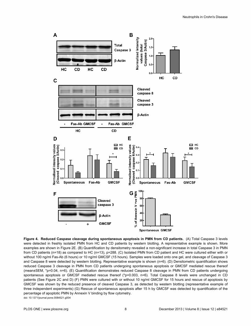
Figure 4. Reduced Caspase cleavage during spontaneous apoptosis in PMN from CD patients. (A) Total Caspase 3 levelswere detected in freshly isolated PMN from HC and CD patients by western blotting. A representative example is shown. Moreexamples are shown in Figure 2E. (B) Quantification by densitometry revealed a non-significant increase in total Caspase 3 in PMNfrom CD patients (n=18) as compared to HC (n=13), p=288. (C) Isolated PMN from CD patient and HC were cultured either with orwithout 100 ng/ml Fas-Ab (6 hours) or 10 ng/ml GMCSF (15 hours). Samples were loaded onto one gel, and cleavage of Caspase 3and Caspase 8 were detected by western blotting. Representative example is shown (n=6). (D) Densitometric quantification showsreduced Caspase 3 cleavage in PMN from CD patients undergoing spontaneous apoptosis or GMCSF mediated rescue thereof(mean±SEM, *p=0.04, n=6). (E) Quantification demonstrates reduced Caspase 8 cleavage in PMN from CD patients undergoingspontaneous apoptosis or GMCSF mediated rescue thereof (*p=0.003, n=6). Total Caspase 8 levels were unchanged in CDpatients (See Figure 2C and D) (F) PMN were cultured with or without 10 ng/ml GMCSF for 15 hours and rescue of apoptosis byGMCSF was shown by the reduced presence of cleaved Caspase 3, as detected by western blotting (representative example ofthree independent experiments) (G) Rescue of spontaneous apoptosis after 15 h by GMCSF was detected by quantification of thepercentage of apoptotic PMN by Annexin V binding by flow cytometry.doi: 10.1371/journal.pone.0084521.g004
Neutrophils in Crohn's Disease
PLOS ONE | www.plosone.org 8 December 2013 | Volume 8 | Issue 12 | e84521

Figure 5. Impaired survival signalling in CD PMN does not affect intermediate and end-stage apoptosis. (A) Isolated PMNwere cultured either with or without 100 ng/ml Fas-Ab (6 hours) or 10 ng/ml GMCSF (15 hours) and the percentage of apoptoticPMN was determined by Annexin V positivity (9 CD patients and 8 HC). (B) No differences in DNA fragmentation as measured byTUNEL assay after 15 hours of PMN culture with or without 10 ng/ml GMCSF were observed between CD and HC (n=6). (C-F)PMN were isolated simultaneously from a CD patient and healthy control, stimulated with 5 ng/ml GMCSF for 15 minutes andsamples were run on one gel. (C) STAT3 activation was detected by western blotting using phospho-STAT3 antibodies.Representative experiment is shown. (D) Significantly decreased levels of activated STAT3 were observed in CD patients comparedto HC (mean±SEM, *p=0.04, n=5). Total STAT3 levels were unchanged (see Figure 2A and F). (E) AKT activation was detected bywestern blotting using phospho-AKT antibodies Representative experiment is shown. (F) Protein levels of activated AKT werequantified by densitometry and corrected for β-actin protein levels. Mean±SEM of CD patients and HC is shown (n=5).doi: 10.1371/journal.pone.0084521.g005
Neutrophils in Crohn's Disease
PLOS ONE | www.plosone.org 9 December 2013 | Volume 8 | Issue 12 | e84521

cellular signalling were observed. Inadequate PMN influx andsubsequent clearance of bacteria in CD may contribute todisease status [15,37]. However, conflicting findings arereported regarding intrinsic migratory capacity of PMN in CDpatients. In vitro migration of PMN from CD patients was foundto be normal or increased in transwell assays [16], whereas invivo PMN migration towards skin blisters, skin windows orinjured intestinal mucosa was decreased in CD PMN [15,38].This was attributed to reduced cytokine production, as blisterfluid in CD patients contained less IL8, and PMN migrationtowards skin windows was restored by adding exogenous IL8[15]. However, increased mucosal IL8 levels have beenreported in CD patients, suggesting that other factors maycontribute to impaired intestinal PMN migration [39,40]. Thereare clear differences between migration through bare transwellfilters versus transepithelial migration [33], with PMNtransepithelial migration requiring ICAM-1 and other adhesion-mediated events [41], and epithelial cells producing andreleasing a range of chemoattractants at their apical side,which may enhance PMN basolateral-to-apical migration [42].We now show that in vitro transepithelial migration, a cleanmeasure of the intrinsic capacity of PMN to migrate throughintestinal epithelial cells, is impaired in CD patients. WhereasIL8-mediated ERK1/2 and AKT signalling is unlikely tocontribute to this impairment, a number of adhesion defectsmay underlie this decreased migration. For instance, IL8 isknown to enhance CD11b expression on PMN [43]. Increasedexpression of the adhesion molecule CD11b on CD PMN hasindeed been described, which may be linked to enhancedadhesion and reduced migration in CD [44,45]. Thus, althoughepithelial cytokine production in CD may be altered, our studyshows that an intrinsic defect in PMN transepithelial migrationexists, which may contribute in decreased neutrophilrecruitment to sites of inflammation.
Defective bacterial clearance has been associated with thedevelopment of CD [46,47] . Loss of NADPH oxidase activityleads to reduced bactericidal activity of PMN, and defectiveROS production in a number of inherited disorders is highlyassociated with intestinal inflammation that is undistinguishablefrom CD [48]. A recent study by Hayee et al. showed impairedfMLP-induced ROS production in CD PMN, but no defect inbacterial killing [20]. Whereas we confirmed the normalbacterial phagocytosis and killing, our study also demonstratedan enhanced fMLP-induced respiratory burst in CD PMN. As ithas long been recognised that oxidative damage plays a majorrole in mucosal injury in CD, it is conceivable that anexaggerated bacterial peptide-induced PMN ROS production,independent on priming by pro-inflammatory cytokines, maycontribute to mucosal damage [12,13]. The discrepancybetween these and other studies may be partially explained bydifferences in study cohorts. Treatment regimens present in CDpatients but not HC may have an impact on cellular function. Inaddition, in our HC cohort, ratio male/female was slightly higherthan in the CD group. Although we cannot formally exclude thepossibility that this affects results, gender in general does notseem to affect PMN ROS production, migration orphagocytosis [49,50]. In addition, it has been speculated thatthe genetic alterations associated with increased risk for IBD
development, affect innate immune cell function [51]. However,the number of genetic alterations and their method of action onPMN signalling and function is unknown, and is thus difficult totake into consideration in this type of study. The enhancedfMLP-mediated ROS production observed in this study wasmirrored by enhanced ERK1/2 and AKT signalling in CDpatients, confirming our results, and suggesting that PMN inCD patients may already be primed to some extent in vivo. AsCD remains incurable, flaring of disease at some point isinevitable, and it is conceivable that circulating levels of pro-inflammatory cytokines are already present even in theabsence of a clear inflammation.
PMN are short-lived cells. In the absence of appropriatestimuli, they rapidly undergo characteristic changes indicativeof apoptosis. These include cleavage of Caspases, followed byPS exposure on the cell membrane, cleaving of the DNA repairenzyme poly (ADP-ribose) polymerase (PARP), and ending inDNA fragmentation [52]. Delayed PMN apoptosis can result inpersisting inflammation and host tissue injury [53]. In this study,we demonstrate a decreased Caspase 3 and 8 cleavage duringspontaneous apoptosis and rescue thereof by GMCSF.Surprisingly, this was not mirrored by an enhanced long termPMN survival, as determined by either Annexin V staining orTUNEL staining. In vivo, rescue of PMN from apoptosis byGMCSF ensures a longer window of opportunity for PMN to killinvading pathogens, and is mediated through activation of thePI3K/AKT and STAT3 pathways [54,55]. We now demonstratethat both AKT and STAT3 activation are severely reduced inPMN from CD patients in response to GMCSF. It isconceivable that a reduced survival signal coming from thesepathways may counteract the enhanced survival mediatedthrough reduced Caspase cleavage. Our data strongly suggestan improper activation of apoptotic signalling pathways in CDPMN, the net result being a normal frequency of apoptoticcells. Whether other functional properties are affected by thisimpaired signalling remains to be elucidated.
In toto, we demonstrate that intrinsic defects in transepithelialmigration, ROS production and chemokine and cytokineinduced signalling are present in PMN from quiescent CDpatients. CD is a heterogeneous disease, where differentunderlying mechanisms may cause patient-to-patientvariability. Genetic variation is likely to contribute to PMNfunction, and it is probable that some roles of innate immunecells are underestimated or even obscured by pooling CDpatients. Nevertheless, our study clearly shows that geneticvariation notwithstanding, several PMN functions are impairedacross patients, strongly implying a role for innate immunity inthe development of this disease. Through these and otherstudies, a role for the innate immune system in thedevelopment of CD is becoming ever more apparent.
Supporting Information
Figure S1. Short term stimulation of PMN does not affecttotal ERK or STAT3 levels. Isolated PMN were stimulatedwith 1 µM fMLP with or without priming with 5ng/ml of GMCSF.Stimulation was confirmed by probing blots with p-AKT or p-STAT3 antibodies (B). Probing blots with total ERK1/2 (A) or
Neutrophils in Crohn's Disease
PLOS ONE | www.plosone.org 10 December 2013 | Volume 8 | Issue 12 | e84521

STAT3 (B) antibodies showed that stimulation does not hugelyinfluence total protein levels. Moreover, total ERK and totalSTAT3 protein levels show excellent correlation with β-Actinlevels in the same lanes (C and D, respectively), showing thatβ-Actin is a good loading control.(TIF)
Figure S2. No differences in total ERK, STAT3, Caspase 3or Caspase 8 levels between CD patients and healthycontrols (HC). Unstimulated, isolated PMN from CD patientsand HC were run on SDS-PAGE, and probed with antibodiesagainst total ERK protein, total STAT3 protein (examples inpanel A), total uncleaved Caspase 8 (panel C) or uncleavedCaspase 3 (examples panel E, more in main manuscript).Quantitation of blots showed no differences in total ERK levelsbetween CD (n=18) and HC (n=16, p=0.6915, panel B). Therewere no differences in total STAT3 levels between CD (n=24)
and HC (n=22, p=0.448, panel F). There were no differences intotal uncleaved Caspase 8 levels between CD (n=16) and HC(n=16, p=0.266, panel D). Quantitation of total uncleavedCaspase 3 levels is shown in manuscript, Figure 4B.(TIF)
Acknowledgements
Authors thank all patients and healthy volunteers whoparticipated in this study.
Author Contributions
Conceived and designed the experiments: RS CJW EJK MPPGMF. Performed the experiments: RS VJJA GMF. Analyzedthe data: RS VJJAN CJW MPP GMF. Wrote the manuscript:RS VJAAN MPP GMF.
References
1. Marks DJ, Rahman FZ, Sewell GW, Segal AW (2010) Crohn's disease:an immune deficiency state. Clin Rev Allergy Immunol 38: 20-31. doi:10.1007/s12016-009-8133-2. PubMed: 19437144.
2. Podolsky DK (2002). Inflammatory Bowel Diseases - N Engl J Med347: 417-429.
3. Barrett JC, Hansoul S, Nicolae DL, Cho JH, Duerr RH et al. (2008)Genome-wide association defines more than 30 distinct susceptibilityloci for Crohn's disease. Nat Genet 40: 955-962. doi:10.1038/ng.175.PubMed: 18587394.
4. Casanova JL, Abel L (2009) Revisiting Crohn's disease as a primaryimmunodeficiency of macrophages. J Exp Med 206: 1839-1843. doi:10.1084/jem.20091683. PubMed: 19687225.
5. Smith AM, Rahman FZ, Hayee B, Graham SJ, Marks DJ et al. (2009)Disordered macrophage cytokine secretion underlies impaired acuteinflammation and bacterial clearance in Crohn's disease. J Exp Med206: 1883-1897. doi:10.1084/jem.20091233. PubMed: 19652016.
6. Segal AW (2005) How neutrophils kill microbes. Annu Rev Immunol 23:197-223. doi:10.1146/annurev.immunol.23.021704.115653. PubMed:15771570.
7. Mulholland MW, Delaney JP, Foker JE, Leonard AS, Simmons RL(1983) Gastrointestinal complications of congenital immunodeficiencystates. The surgeon's role. Ann Surg 198: 673-680. doi:10.1097/00000658-198312000-00001. PubMed: 6605728.
8. Segal AW (1996) The NADPH oxidase and chronic granulomatousdisease. Mol Med Today 2: 129-135. doi:10.1016/1357-4310(96)88723-5. PubMed: 8796870.
9. Annabi B, Hiraiwa H, Mansfield BC, Lei KJ, Ubagai T et al. (1998) Thegene for glycogen-storage disease type 1b maps to chromosome11q23. Am J Hum Genet 62: 400-405. doi:10.1086/301727. PubMed:9463334.
10. Uzel G, Tng E, Rosenzweig SD, Hsu AP, Shaw JM et al. (2008)Reversion mutations in patients with leukocyte adhesion deficiencytype-1 (LAD-1). Blood 111: 209-218. doi:10.1182/blood-2007-04-082552. PubMed: 17875809. Available online at: doi:10.1182/blood-2007-04-082552 Available online at: PubMed: 17875809
11. Conway KL, Goel G, Sokol H, Manocha M, Mizoguchi E et al. (2012)p40phox expression regulates neutrophil recruitment and functionduring the resolution phase of intestinal inflammation. J Immunol 189:3631-3640. doi:10.4049/jimmunol.1103746. PubMed: 22914050.
12. McKenzie SJ, Baker MS, Buffinton GD, Doe WF (1996) Evidence ofoxidant-induced injury to epithelial cells during inflammatory boweldisease. J Clin Invest 98: 136-141. doi:10.1172/JCI118757. PubMed:8690784.
13. Grisham MB, Granger DN (1988) Neutrophil-mediated mucosal injury.Role of reactive oxygen metabolites. Dig Dis Sci 33: 6S-15S. doi:10.1007/BF01538126. PubMed: 2831016.
14. Passalacqua S, Ferraro PM, Bresci G, D'Ovidio V, Astegiano M et al.(2011) The Italian Registry of Therapeutic Apheresis: granulocyte-monocyte apheresis in the treatment of inflammatory bowel disease. Amulticentric study. J Clin Apher 26: 332-337. doi:10.1002/jca.20315.PubMed: 22072543.
15. Marks DJ, Harbord MW, MacAllister R, Rahman FZ, Young J et al.(2006) Defective acute inflammation in Crohn's disease: a clinicalinvestigation. Lancet 367: 668-678. doi:10.1016/S0140-6736(06)68265-2. PubMed: 16503465.
16. Rhodes JM, Jewell DP (1983) Motility of neutrophils and monocytes inCrohn's disease and ulcerative colitis. Gut 24: 73-77. doi:10.1136/gut.24.1.73. PubMed: 6848434.
17. Biagioni C, Favilli F, Catarzi S, Marcucci T, Fazi M et al. (2006) Redoxstate and O2*- production in neutrophils of Crohn's disease patients.Exp Biol Med (Maywood) 231: 186-195. PubMed: 16446495.
18. Gionchetti P, Campieri M, Guarnieri C, Belluzzi A, Brignola C et al.(1994) Respiratory burst of circulating polymorphonuclear leukocytesand plasma elastase levels in patients with inflammatory bowel diseasein remission. Dig Dis Sci 39: 550-554. doi:10.1007/BF02088341.PubMed: 8131691.
19. Verspaget HW, Peña AS, Weterman IT, Lamers CB (1988) Diminishedneutrophil function in Crohn's disease and ulcerative colitis identified bydecreased oxidative metabolism and low superoxide dismutasecontent. Gut 29: 223-228. doi:10.1136/gut.29.2.223. PubMed: 2831119.
20. Hayee B, Rahman FZ, Tempero J, McCartney S, Bloom SL et al.(2011) The neutrophil respiratory burst and bacterial digestion inCrohn's disease. Dig Dis Sci 56: 1482-1488. doi:10.1007/s10620-010-1426-8. PubMed: 20936355.
21. Schwarzmaier D, Foell D, Weinhage T, Varga G, Däbritz J (2013)Peripheral monocyte functions and activation in patients with quiescentCrohn's disease. PLOS ONE 8: e62761. doi:10.1371/journal.pone.0062761. PubMed: 23658649.
22. Fuhler GM, Hooijenga F, Drayer AL, Vellenga E (2003) Reducedexpression of flavocytochrome b558, a component of the NADPHoxidase complex, in neutrophils from patients with myelodysplasia. ExpHematol 31: 752-759. doi:10.1016/S0301-472X(03)00188-7. PubMed:12962720.
23. Fiuza C, Salcedo M, Clemente G, Tellado JM (2002) Granulocytecolony-stimulating factor improves deficient in vitro neutrophiltransendothelial migration in patients with advanced liver disease. ClinDiagn Lab Immunol 9: 433-439. PubMed: 11874890.
24. Fuhler GM, Drayer AL, Olthof SG, Schuringa JJ, Coffer PJ et al. (2008)Reduced activation of protein kinase B, Rac, and F-actin polymerizationcontributes to an impairment of stromal cell derived factor-1 inducedmigration of CD34+ cells from patients with myelodysplasia. Blood 111:359-368. doi:10.1182/blood-2006-11-060632. PubMed: 17898317.
25. Zhou L, Somasundaram R, Nederhof RF, Dijkstra G, Faber KN et al.(2012) Human granulocyte and monocyte isolation procedures: impacton functional studies. Clin Vaccine Immunol.
26. Fuhler GM, Blom NR, Coffer PJ, Drayer AL, Vellenga E (2007) Thereduced GM-CSF priming of ROS production in granulocytes frompatients with myelodysplasia is associated with an impaired lipid raftformation. J Leukoc Biol 81: 449-457. doi:10.1189/jlb.0506311.PubMed: 17079651.
27. Fuhler GM, Knol GJ, Drayer AL, Vellenga E (2005) Impairedinterleukin-8- and GROalpha-induced phosphorylation of extracellularsignal-regulated kinase result in decreased migration of neutrophils
Neutrophils in Crohn's Disease
PLOS ONE | www.plosone.org 11 December 2013 | Volume 8 | Issue 12 | e84521

from patients with myelodysplasia. J Leukoc Biol 77: 257-266. PubMed:15561756.
28. Fuhler GM, Drayer AL, Vellenga E (2003) Decreased phosphorylationof protein kinase B and extracellular signal-regulated kinase inneutrophils from patients with myelodysplasia. Blood 101: 1172-1180.doi:10.1182/blood.V101.3.1172. PubMed: 12529294.
29. Burelout C, Naccache PH, Bourgoin SG (2007) Dissociation betweenthe translocation and the activation of Akt in fMLP-stimulated humanneutrophils--effect of prostaglandin E2. J Leukoc Biol 81: 1523-1534.doi:10.1189/jlb.0406256. PubMed: 17339610.
30. Crepaldi L, Silveri L, Calzetti F, Pinardi C, Cassatella MA (2002)Molecular basis of the synergistic production of IL-1 receptor antagonistby human neutrophils stimulated with IL-4 and IL-10. Int Immunol 14:1145-1153. doi:10.1093/intimm/dxf079. PubMed: 12356680.
31. Epling-Burnette PK, Zhong B, Bai F, Jiang K, Bailey RD et al. (2001)Cooperative regulation of Mcl-1 by Janus kinase/stat andphosphatidylinositol 3-kinase contribute to granulocyte-macrophagecolony-stimulating factor-delayed apoptosis in human neutrophils. JImmunol 166: 7486-7495. PubMed: 11390502.
32. Fuhler GM, Knol GJ, Drayer AL, Vellenga E (2005) Impairedinterleukin-8- and GROalpha-induced phosphorylation of extracellularsignal-regulated kinase result in decreased migration of neutrophilsfrom patients with myelodysplasia. J Leukoc Biol 77: 257-266. PubMed:15561756.
33. Blake KM, Carrigan SO, Issekutz AC, Stadnyk AW (2004) Neutrophilsmigrate across intestinal epithelium using beta2 integrin (CD11b/CD18)-independent mechanisms. Clin Exp Immunol 136: 262-268.PubMed: 15086389.
34. Brazil JC, Lee WY, Kolegraff KN, Nusrat A, Parkos CA et al. (2010)Neutrophil migration across intestinal epithelium: evidence for a role ofCD44 in regulating detachment of migrating cells from the luminalsurface. J Immunol 185: 7026-7036. doi:10.4049/jimmunol.1001293.PubMed: 20974992.
35. Fuhler GM, Drayer AL, Vellenga E (2003) Decreased phosphorylationof protein kinase B and extracellular signal-regulated kinase inneutrophils from patients with myelodysplasia. Blood 101: 1172-1180.doi:10.1182/blood.V101.3.1172. PubMed: 12529294.
36. Sakamoto C, Suzuki K, Hato F, Akahori M, Hasegawa T et al. (2003)Antiapoptotic effect of granulocyte colony-stimulating factor,granulocyte-macrophage colony-stimulating factor, and cyclic AMP onhuman neutrophils: protein synthesis-dependent and protein synthesis-independent mechanisms and the role of the Janus kinase-STATpathway. Int J Hematol 77: 60-70. doi:10.1007/BF02982604. PubMed:12568301.
37. Zhou L, Braat H, Faber KN, Dijkstra G, Peppelenbosch MP (2009)Monocytes and their pathophysiological role in Crohn's disease. CellMol Life Sci 66: 192-202. doi:10.1007/s00018-008-8308-7. PubMed:18791847.
38. Harbord MW, Marks DJ, Forbes A, Bloom SL, Day RM et al. (2006)Impaired neutrophil chemotaxis in Crohn's disease relates to reducedproduction of chemokines and can be augmented by granulocyte-colony stimulating factor. Aliment Pharmacol Ther 24: 651-660. doi:10.1111/j.1365-2036.2006.03016.x. PubMed: 16907898.
39. Mitsuyama K, Toyonaga A, Sasaki E, Watanabe K, Tateishi H et al.(1994) IL-8 as an important chemoattractant for neutrophils inulcerative colitis and Crohn's disease. Clin Exp Immunol 96: 432-436.PubMed: 8004812.
40. Jones SC, Evans SW, Lobo AJ, Ceska M, Axon AT et al. (1993) Seruminterleukin-8 in inflammatory bowel disease. J Gastroenterol Hepatol 8:508-512. doi:10.1111/j.1440-1746.1993.tb01643.x. PubMed: 8280836.
41. Liu L, Mul FP, Kuijpers TW, Lutter R, Roos D et al. (1996) Neutrophiltransmigration across monolayers of endothelial cells and airway
epithelial cells is regulated by different mechanisms. Ann N Y Acad Sci796: 21-29. doi:10.1111/j.1749-6632.1996.tb32563.x. PubMed:8906208.
42. Taguchi M, Sampath D, Koga T, Castro M, Look DC et al. (1998)Patterns for RANTES secretion and intercellular adhesion molecule 1expression mediate transepithelial T cell traffic based on analyses invitro and in vivo. J Exp Med 187: 1927-1940. doi:10.1084/jem.187.12.1927. PubMed: 9625753.
43. Furebring M, Håkansson L, Venge P, Sjölin J (2006) C5a, interleukin-8and tumour necrosis factor-alpha-induced changes in granulocyte andmonocyte expression of complement receptors in whole blood and onisolated leukocytes. Scand J Immunol 63: 208-216. doi:10.1111/j.1365-3083.2006.01724.x. PubMed: 16499574.
44. Maiguel D, Faridi MH, Wei C, Kuwano Y, Balla KM et al. (2011) Smallmolecule-mediated activation of the integrin CD11b/CD18 reducesinflammatory disease. Sci Signal 4: ra57. PubMed: 21900205.
45. Brandt E, Müller-Alouf H, Desreumaux P, Woerly G, Colombel JF et al.(1998) Circulating growth-regulator oncogene alpha contributes toneutrophil priming and interleukin-8-directed mucosal recruitment intochronic lesions of patients with Crohn's disease. Eur Cytokine Netw 9:647-653. PubMed: 9889409.
46. Klionsky DJ (2009) Crohn's disease, autophagy, and the Paneth cell. NEngl J Med 360: 1785-1786. doi:10.1056/NEJMcibr0810347. PubMed:19369659.
47. Glasser AL, Darfeuille-Michaud A (2008) Abnormalities in the handlingof intracellular bacteria in Crohn's disease: a link between infectiousetiology and host genetic susceptibility. Arch Immunol Ther Exp(Warsz) 56: 237-244. doi:10.1007/s00005-008-0026-1. PubMed:18726145.
48. Rahman FZ, Marks DJ, Hayee BH, Smith AM, Bloom SL et al. (2008)Phagocyte dysfunction and inflammatory bowel disease. InflammBowel Dis 14: 1443-1452. doi:10.1002/ibd.20449. PubMed: 18421761.
49. Kopprasch S, Graessler J, Kohl M, Bergmann S, Schröder HE (1996)Comparison of circulating phagocyte oxidative activity measured bychemiluminescence in whole blood and isolated polymorphonuclearleukocytes. Clin Chim Acta 253: 145-157. doi:10.1016/0009-8981(96)06365-6. PubMed: 8879845.
50. Trottier MD, Naaz A, Kacynski K, Yenumula PR, Fraker PJ (2012)Functional capacity of neutrophils from class III obese patients. Obesity(Silver Spring) 20: 1057-1065. doi:10.1038/oby.2011.354. PubMed:22158006.
51. Marks DJ (2011) Defective innate immunity in inflammatory boweldisease: a Crohn's disease exclusivity? Curr Opin Gastroenterol 27:328-334. doi:10.1097/MOG.0b013e3283463b45. PubMed: 21483259.
52. Homburg CH, de Haas M, von dem Borne AE, Verhoeven AJ,Reutelingsperger CP et al. (1995) Human neutrophils lose their surfaceFc gamma RIII and acquire Annexin V binding sites during apoptosis invitro. Blood 85: 532-540. PubMed: 7812008.
53. Smith JA (1994) Neutrophils, host defense, and inflammation: a double-edged sword. J Leukoc Biol 56: 672-686. PubMed: 7996043.
54. Kotone-Miyahara Y, Yamashita K, Lee KK, Yonehara S, Uchiyama T etal. (2004) Short-term delay of Fas-stimulated apoptosis by GM-CSF asa result of temporary suppression of FADD recruitment in neutrophils:evidence implicating phosphatidylinositol 3-kinase and MEK1-ERK1/2pathways downstream of classical protein kinase C. J Leukoc Biol 76:1047-1056. doi:10.1189/jlb.0104048. PubMed: 15328334.
55. Fortin CF, Larbi A, Dupuis G, Lesur O, Fülöp T Jr. (2007) GM-CSFactivates the Jak/STAT pathway to rescue polymorphonuclearneutrophils from spontaneous apoptosis in young but not elderlyindividuals. Biogerontology 8: 173-187. doi:10.1007/s10522-006-9067-1. PubMed: 17086367.
Neutrophils in Crohn's Disease
PLOS ONE | www.plosone.org 12 December 2013 | Volume 8 | Issue 12 | e84521