performance of immunodiagnostic tests for typhoid fever
TRANSCRIPT

pathogens
Systematic Review
Performance of Immunodiagnostic Tests for Typhoid Fever:A Systematic Review and Meta-Analysis
Mohamad Ahmad Najib 1 , Khairul Mohd Fadzli Mustaffa 1 , Eugene Boon Beng Ong 2, Kasturi Selvam 1,Muhammad Fazli Khalid 1 , Mohd Syafiq Awang 3, Nor Syafirah Zambry 1 , Asrulnizam Abd Manaf 3 ,Yazmin Bustami 4, Hairul Hisham Hamzah 5 , Asma Ismail 1 and Ismail Aziah 1,*
�����������������
Citation: Najib, M.A.; Mustaffa,
K.M.F.; Ong, E.B.B.; Selvam, K.;
Khalid, M.F.; Awang, M.S.; Zambry,
N.S.; Manaf, A.A.; Bustami, Y.;
Hamzah, H.H.; et al. Performance of
Immunodiagnostic Tests for Typhoid
Fever: A Systematic Review and
Meta-Analysis. Pathogens 2021, 10,
1184. https://doi.org/10.3390/
pathogens10091184
Academic Editor: Po-Lin Chen
Received: 16 July 2021
Accepted: 7 September 2021
Published: 13 September 2021
Publisher’s Note: MDPI stays neutral
with regard to jurisdictional claims in
published maps and institutional affil-
iations.
Copyright: © 2021 by the authors.
Licensee MDPI, Basel, Switzerland.
This article is an open access article
distributed under the terms and
conditions of the Creative Commons
Attribution (CC BY) license (https://
creativecommons.org/licenses/by/
4.0/).
1 Institute for Research in Molecular Medicine (INFORMM), Universiti Sains Malaysia,Kubang Kerian 16150, Kelantan, Malaysia; [email protected] (M.A.N.); [email protected] (K.M.F.M.);[email protected] (K.S.); [email protected] (M.F.K.); [email protected] (N.S.Z.);[email protected] (A.I.)
2 Institute for Research in Molecular Medicine (INFORMM), Universiti Sains Malaysia,Gelugor 11800, Pulau Pinang, Malaysia; [email protected]
3 Collaborative Microelectronic Design Excellence Centre (CEDEC), Universiti Sains Malaysia,Bayan Lepas 11900, Pulau Pinang, Malaysia; [email protected] (M.S.A.);[email protected] (A.A.M.)
4 School of Biological Sciences, Universiti Sains Malaysia, Gelugor 11800, Pulau Pinang, Malaysia;[email protected]
5 School of Chemical Sciences, Universiti Sains Malaysia, Gelugor 11800, Pulau Pinang, Malaysia;[email protected]
* Correspondence: [email protected]
Abstract: Typhoid fever, also known as typhoid, is a life-threatening bacterial infection that remainsa global health concern. The infection is associated with a significant morbidity and mortality rate,resulting in an urgent need for specific and rapid detection tests to aid prevention and managementof the disease. The present review aims to assess the specificity and sensitivity of the availableliterature on the immunodiagnostics of typhoid fever. A literature search was conducted using threedatabases (PubMed, ProQuest and Scopus) and manual searches through the references of identifiedfull texts to retrieve relevant literature published between 1 January 2011 and 31 December 2020. Ofthe 577 studies identified in our search, 12 were included in further analysis. Lipopolysaccharides(LPS) and hemolysin E (HlyE) were the most frequently studied antigens. The specimens examinedin these studies included serum and saliva. Using blood culture as the gold standard, anti-LPSIgA gave the highest sensitivity of 96% (95% CI: 93–99) and specificity of 96% (95% CI: 93–99) fordistinguishing between typhoid cases and healthy controls, whereas the combination of anti-LPS andanti-flagellin total IgGAM gave the highest sensitivity of 93% (95% CI: 86–99) and specificity of 95%(95% CI: 89–100) for distinguishing typhoid cases and other febrile infections. A comparably highsensitivity of 92% (95% CI: 86–98) and specificity of 89% (95% CI: 78–100) were shown in testing basedon detection of the combination of anti-LPS (IgA and IgM) and anti-HlyE IgG as well as a slightlylower sensitivity of 91% (95% CI: 74–100) in the case of anti-50kDa IgA. Anti-50kDa IgM had thelowest sensitivity of 36% (95% CI: 6–65) against both healthy and febrile controls. The developmentof a rapid diagnostic test targeting antibodies against lipopolysaccharides combined with flagellinappeared to be a suitable approach for the rapid detection test of typhoid fever. Saliva is addedbenefit for rapid typhoid diagnosis since it is less invasive. As a result, further studies could be doneto develop additional approaches for adopting such samples.
Keywords: immunodiagnostic; typhoid; systematic review; sensitivity; specificity
1. Introduction
Typhoid fever is a systemic infection associated with the Gram-negative and rod-shaped bacillus Salmonella enterica serovar Typhi (S. Typhi). The life-threatening disease has
Pathogens 2021, 10, 1184. https://doi.org/10.3390/pathogens10091184 https://www.mdpi.com/journal/pathogens

Pathogens 2021, 10, 1184 2 of 14
been a public health problem in developing and underdeveloped countries for generations.A systematic analysis by typhoid and paratyphoid collaborators estimated that 10.9 millioncases of typhoid fever occurred globally in 2017, resulting in 116.8 thousand deaths [1].There is a declining global trend in typhoid incidence over the last few years with respectto the number of cases per capita, particularly in most high-income countries, such asAustralia, Japan, New Zealand, Singapore, South Korea and Taiwan and middle-incomecountries, such as Malaysia and Thailand [2]. However, in some countries such as Ghana,Malawi, Fiji, China, Indonesia, Cambodia and Iraq, typhoid incidence shows a steadyincrease from 1990 to 2014. These numbers are more than likely an underrepresentationof the true disease burden given that a large proportion of patients are treated on anoutpatient basis or receive no treatment at all. In Malaysia, recent estimates suggest thatapproximately 0.58 to 1.42 cases of S. Typhi infection per 100,000 population were reportedeach year between 2011 and 2015 and most of the cases were due to travelers returning froman endemic region, migrants and food chains being contaminated by food handlers [3].
Salmonella Typhi is an obligate pathogen and humans are the only host for this bac-terium. Humans acquire the infection through ingestion of water or food contaminatedwith S. Typhi. Typhoid fever is a great risk in areas that have low-quality potable water,non-hygienic living conditions and improper sanitation systems. Asymptomatic carriersplay an important role in introducing contamination and disease transmission [2]. Thedisease causes a prolonged fever that can be as high as 103–104 ◦F (39–40 ◦C), fatigue,headache, nausea, constipation or diarrhea and abdominal pain, which develops within6 to 30 days after infection [4]. Severe cases may lead to serious complications or evendeath due to intestinal perforation [5,6].
Improving the quality of drinking water supplies and education in better hygienepractices are likely the most effective measures towards typhoid elimination. However,these interventions cannot be promptly realized in the endemic areas of Africa and SouthAsia. Therefore, short-term control of typhoid is largely dependent on extensive vaccina-tion programs and appropriate treatment, both of which rely on the application of rapiddiagnostic tests (RDTs) that have robust performance. There are several commerciallyavailable RDTs for typhoid fever but finding a typhoid diagnostic assay with a high degreeof sensitivity or specificity is challenging. The lack of such tests limits the disease burdenassessments and may result in patients being misdiagnosed and receiving suboptimaltherapy. Furthermore, with the global increase in multidrug-resistant S. Typhi, the demandfor typhoid diagnostics is growing [7–9].
The laboratory diagnosis of typhoid fever currently relies on blood and stool cultures,which is considered the gold standard for diagnosis. However, this may pose a majorchallenge in resource-limited settings, such as areas with a lack of basic laboratory facilitiesfor culturing purposes [10]. Furthermore, the culture method is time-consuming, requiringseveral days for the isolation and identification of the causative agents [11]. The Widal test,which is the current standard serological method for typhoid diagnosis, relies on O and Hantigens that have a relatively low specificity due to cross-reactivity with other bacterialinfections [12]. Thus, a rapid and sensitive assay for the detection of S. Typhi would helpboth in clinical diagnosis and in preventing the spread of disease [13]. In this context,development of a highly specific and sensitive immunodiagnostic test for the diagnosis oftyphoid fever is an appropriate approach.
Several serology-based rapid diagnostic tests for typhoid fever are commercially avail-able, such as Typhidot (Malaysia), TUBEX (Sweden) and Multi-Test Dip-S-Tics (USA).These commercial RDTs have been globally evaluated for their performance. Despitethat, none of these tests yielded satisfactory results when validated in different endemicsetups [14]. This is due to the consistency in sensitivity level in those studies resultingfrom false negative issue. Numerous antigens have been evaluated for their effectivenessin detecting S. Typhi, such as flagellin, hemolysin E (HlyE), YncE and cytolethal distendingtoxin subunit B (CdtB) [15–17]. However, due to the lack of thorough comparisons ofthe available data, the option of the best antigen for S. Typhi detection is yet to be deter-
Pathogens 2021, 10, 1184 3 of 14
mined. An understanding of the specificity and sensitivity of previously identified S. Typhibiomarkers is critical in providing vital information for the selection of an effective targetand consequently establishing highly accurate diagnostic approaches for typhoid fever.Therefore, the present review focuses on the evaluation of the available evidence regardingthe specificity and sensitivity of S. Typhi antigens. It is intended that our findings willserve as an informative resource for researchers aiming to develop an accurate laboratorydiagnostic test for typhoid fever.
2. Results
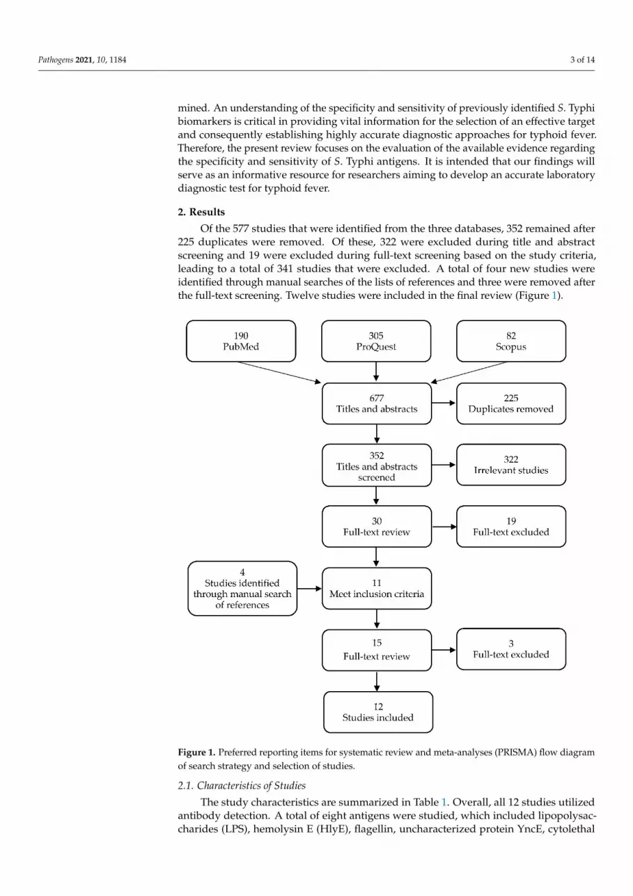
Of the 577 studies that were identified from the three databases, 352 remained after225 duplicates were removed. Of these, 322 were excluded during title and abstractscreening and 19 were excluded during full-text screening based on the study criteria,leading to a total of 341 studies that were excluded. A total of four new studies wereidentified through manual searches of the lists of references and three were removed afterthe full-text screening. Twelve studies were included in the final review (Figure 1).
Pathogens 2021, 10, x FOR PEER REVIEW 3 of 14
demic setups [14]. This is due to the consistency in sensitivity level in those studies result-
ing from false negative issue. Numerous antigens have been evaluated for their effective-
ness in detecting S. Typhi, such as flagellin, hemolysin E (HlyE), YncE and cytolethal dis-
tending toxin subunit B (CdtB) [15–17]. However, due to the lack of thorough comparisons
of the available data, the option of the best antigen for S. Typhi detection is yet to be de-
termined. An understanding of the specificity and sensitivity of previously identified S.
Typhi biomarkers is critical in providing vital information for the selection of an effective
target and consequently establishing highly accurate diagnostic approaches for typhoid
fever. Therefore, the present review focuses on the evaluation of the available evidence
regarding the specificity and sensitivity of S. Typhi antigens. It is intended that our find-
ings will serve as an informative resource for researchers aiming to develop an accurate
laboratory diagnostic test for typhoid fever.
2. Results
Of the 577 studies that were identified from the three databases, 352 remained after
225 duplicates were removed. Of these, 322 were excluded during title and abstract
screening and 19 were excluded during full-text screening based on the study criteria,
leading to a total of 341 studies that were excluded. A total of four new studies were iden-
tified through manual searches of the lists of references and three were removed after the
full-text screening. Twelve studies were included in the final review (Figure 1).
Figure 1. Preferred reporting items for systematic review and meta-analyses (PRISMA) flow dia-
gram of search strategy and selection of studies.
Figure 1. Preferred reporting items for systematic review and meta-analyses (PRISMA) flow diagramof search strategy and selection of studies.
2.1. Characteristics of Studies
The study characteristics are summarized in Table 1. Overall, all 12 studies utilizedantibody detection. A total of eight antigens were studied, which included lipopolysac-charides (LPS), hemolysin E (HlyE), flagellin, uncharacterized protein YncE, cytolethal

Pathogens 2021, 10, 1184 4 of 14
distending toxin subunit B (CdtB), membrane proteins (MP), Vi antigen and 50 kDa outermembrane protein (OMP). LPS were the most frequently studied antigen, with 7 (58.3%)out of the 12 studies reporting on diagnostic sensitivity and specificity, followed by HlyE,which was reported in six (50.0%) studies. A total of eight (66.7%) studies evaluated theperformance of antibody-based detection using ELISA, two (16.7%) studies performeddetection using a lateral flow assay, one (8.3%) study used a dot enzyme immunoassay andone (8.3%) study used both ELISA and a lateral flow assay. Most of the studies tested forthe presence of antibodies in serum sample and only two studies tested saliva.
Table 1. Characteristics of included studies.
Method Biomarker Sample Sensitivity and Specificity No of Samples Ref.
1 ELISA
Anti-LPS (IgG, IgMand IgA)anti-flagellin (IgG,IgM and IgA)
Serum
Total Ig: 93% and 95%IgG: 75% and 55%IgM: 79% and 95%IgA: 57% and 96%
Positive S. Typhi blood culture(n = 67), Widal positive (98),febrile controls (n = 216) andhealthy controls (n = 7).
[18]
2 ELISA Anti-LPS IgA Saliva 89.2% and 100%Positive S. Typhi blood culture(n = 37), febrile controls (n = 30)and healthy controls (n = 30).
[19]
3 IC-LFTAnti-LPS (IgG IgM)and anti-flagellin(IgG IgM)
Serum 68.8% and 71.1%Positive S. Typhi blood culture(n = 80) and negative S. Typhiblood culture (n = 256).
[14]
4 ELISAAnti-YncE (IgG)and anti-Vi (IgGand IgA)
SerumYncE IgG: 70% and 100%Vi IgG: 40% and 100%Vi IgA: 50% and 97%
S. Typhi carriers (n = 10), acutetyphoid fever cases (n = 8),Nepalese controls undergoingelective cholecystectomy withnegative bile cultures (n = 8) andhealthy Bangladeshis (n = 8).
[20]
5 ELISA Anti-HlyE (IgGIgM and IgA) Serum 70% and 100%
Positive S. Typhi blood culture(n = 50), positive S. Paratyphi Ablood culture (n = 6), other febrileinfections (n = 19) and healthyindividuals (n = 25).
[21]
6 LFTAnti-HlyE (IgGIgA) and anti-LPS(IgG IgA)
Serum
HlyE IgG: 91.5% and 80.0%(against healthy)HlyE IgA: 94.4% and 90.0%(against healthy)HlyE IgG: 78.7% and 100%(against other febrile infections)HlyE IgA: 66.7% and 80% (againstother febrile infections)LPS IgG: 89.3% and 100%(against healthy)LPS IgA: 94.4% and 90.0%(against healthy)LPS IgG: 82.1% and 60.0% (againstother febrile infections)LPS IgA: 81.8% and 54.5% (againstother febrile infections)
Positive S. Typhi blood culture(n = 47), other febrile infections(n = 15), febrile with no bacterialgrowth (n = 67), healthy U.S.adults (n = 11) and healthyNigerian children (n = 10).
[22]
7 ELISAAnti-HlyE (IgG IgAIgM) and anti-LPS(IgG IgA IgM)
Serum
HlyE IgG: 84% and 90% (againstNigerian febrile controls)LPS IgA and IgM: 90% and 90%(against Nigerian febrile controls)
Nigerian pediatric typhoid cases(n = 86), Nigerian febrile controls(n = 28) and Nigerian healthycontrols (n = 48).
[23]
8 Dot-EIA Anti-50 kDa (IgGIgA IgM)
Saliva,serum
50 kDa IgGAM: 90.9% sensitivityand 85.4% (saliva)50 kDa IgGAM: 100% and100% (serum)
Positive S. Typhi blood culture(n = 11), non-typhoid fever patients(n = 43) and healthy (n = 53).
[24]
9 ELISA Anti-CdtB IgM Serum 100% and 83.3% Positive S. Typhi blood culture(n = 21) and healthy controls (n = 12). [25]

Pathogens 2021, 10, 1184 5 of 14
Table 1. Cont.
Method Biomarker Sample Sensitivity and Specificity No of Samples Ref.
10 ELISAAnti-HlyE IgA,anti-MP IgA andanti-LPS IgA
Serum
HlyE: 90% and 87%LPS: 90% and 77%MP: 90% and 48%HlyE and anti-LPS: 90% and 92%HlyE, LPS and MP: 90% and 90%
Positive S. Typhi blood culture(n = 105), healthy controls (n = 84)and other febrile disease (n = 64).
[15]
11 ELISAAnti-HlyE (IgG IgAIgM) and anti-YncE(IgG IgA IgM)
Serum HlyE IgGAM: 83% and 98%
Acute typhoid cases (n = 115),healthy controls (n = 95), otherfebrile infections (n = 95) and foodhandlers (n = 117).
[16]
12 ELISA,DDP-LF
Anti-LPS IgA andanti-HlyE IgA Serum
92% and 94% (against all controls)90% and 96% (against febrileendemic controls)
Positive S. Typhi blood culture(n = 30), positive S. Paratyphi Ablood culture (n = 20), healthyendemic controls (n = 25) andfebrile endemic controls (n = 25).
[26]
HlyE = hemolysin E; LPS = lipopolysaccharides; CdtB = cytolethal distending toxin subunit B; MP = membrane protein; Ig = immunoglob-ulin; YncE = uncharacterized protein; kDa = Kilodalton; Vi = virulence antigen; IC-LFT = immunochromatography lateral flow test;DPP-LF = dual-path platform lateral flow; Dot-EIA = dot enzyme immunoassay; ELISA = enzyme-linked immunosorbent assay.
2.2. Methodological Risk of Bias
A summary of the QUADAS-2 assessment is presented in Figure 2. The overall resultsof the quality assessment showed a low risk of bias in all 12 studies. For patient selection,eight studies (67%) demonstrated a low risk of bias and four studies (33%) demonstratedan unclear risk of bias. For index test, all 12 studies (100%) demonstrated a low risk ofbias with clear interpretation of the index test. For reference standard, 11 studies (92%)demonstrated a low risk of bias and 1 study (8%) demonstrated a high risk of bias, as thehealthy controls were not confirmed with blood culture. For flow and timing, nine studies(75%) demonstrated a low risk of bias and three studies (25%) demonstrated an unclearrisk of bias.
Pathogens 2021, 10, x FOR PEER REVIEW 6 of 14
Figure 2. QUADAS-2 criteria among all studies (n = 12).
2.3. Performances of the Immunodiagnostic Assays
Sensitivity measures the ability of a diagnostic test to correctly identify patients who
have a disease with positive test results. Using samples from healthy individuals as the
control group, the sensitivity of antibody detection varied between 36% and 96% (Figure
3). The anti-LPS IgA gave the highest sensitivity of 96% (95% CI: 93–99), followed by anti-
LPS (IgA and IgM), which achieved a sensitivity of 95% (95% CI: 91–100), anti-LPS IgM
with 94% (95% CI: 89–99) and anti-HlyE IgA with 94% (95% CI: 90–98). Specificity
measures the ability of a diagnostic test to correctly identify people who do not have the
disease with a negative test result. Comparably high specificities ranging from 72% to
100% were observed among all biomarkers. Only when the results of anti-LPS and anti-
flagellin were combined did the specificity decrease to 72% (95% CI: 66–77), while all other
results showed a specificity above 80%.
Figure 3. Meta-analysis of the diagnostic sensitivity and specificity of antibody detection tests with healthy individuals as
a control group and blood culture as a reference standard.
Using samples from other febrile infections as the control group, the sensitivity and
specificity of antibody detection tests for distinguishing typhoid and other febrile infec-
0% 20% 40% 60% 80% 100%
Flow and timing risk of bias
Reference standard risk of bias
Index test risk of bias
Patient selection risk of bias
Proportion of studies with low, high or unclear risk of bias
QU
AD
AS-
2 D
om
ain
Low High Unclear
Figure 2. QUADAS-2 criteria among all studies (n = 12).
2.3. Performances of the Immunodiagnostic Assays
Sensitivity measures the ability of a diagnostic test to correctly identify patients whohave a disease with positive test results. Using samples from healthy individuals as thecontrol group, the sensitivity of antibody detection varied between 36% and 96% (Figure 3).The anti-LPS IgA gave the highest sensitivity of 96% (95% CI: 93–99), followed by anti-LPS(IgA and IgM), which achieved a sensitivity of 95% (95% CI: 91–100), anti-LPS IgM with94% (95% CI: 89–99) and anti-HlyE IgA with 94% (95% CI: 90–98). Specificity measuresthe ability of a diagnostic test to correctly identify people who do not have the diseasewith a negative test result. Comparably high specificities ranging from 72% to 100% were

Pathogens 2021, 10, 1184 6 of 14
observed among all biomarkers. Only when the results of anti-LPS and anti-flagellin werecombined did the specificity decrease to 72% (95% CI: 66–77), while all other results showeda specificity above 80%.
Pathogens 2021, 10, x FOR PEER REVIEW 6 of 14
Figure 2. QUADAS-2 criteria among all studies (n = 12).
2.3. Performances of the Immunodiagnostic Assays
Sensitivity measures the ability of a diagnostic test to correctly identify patients who
have a disease with positive test results. Using samples from healthy individuals as the
control group, the sensitivity of antibody detection varied between 36% and 96% (Figure
3). The anti-LPS IgA gave the highest sensitivity of 96% (95% CI: 93–99), followed by anti-
LPS (IgA and IgM), which achieved a sensitivity of 95% (95% CI: 91–100), anti-LPS IgM
with 94% (95% CI: 89–99) and anti-HlyE IgA with 94% (95% CI: 90–98). Specificity
measures the ability of a diagnostic test to correctly identify people who do not have the
disease with a negative test result. Comparably high specificities ranging from 72% to
100% were observed among all biomarkers. Only when the results of anti-LPS and anti-
flagellin were combined did the specificity decrease to 72% (95% CI: 66–77), while all other
results showed a specificity above 80%.
Figure 3. Meta-analysis of the diagnostic sensitivity and specificity of antibody detection tests with healthy individuals as
a control group and blood culture as a reference standard.
Using samples from other febrile infections as the control group, the sensitivity and
specificity of antibody detection tests for distinguishing typhoid and other febrile infec-
0% 20% 40% 60% 80% 100%
Flow and timing risk of bias
Reference standard risk of bias
Index test risk of bias
Patient selection risk of bias
Proportion of studies with low, high or unclear risk of bias
QU
AD
AS-
2 D
om
ain
Low High Unclear
Figure 3. Meta-analysis of the diagnostic sensitivity and specificity of antibody detection tests with healthy individuals as acontrol group and blood culture as a reference standard.
Using samples from other febrile infections as the control group, the sensitivity andspecificity of antibody detection tests for distinguishing typhoid and other febrile infectionswere 36–93% and 88–80%, respectively (Figure 4). The highest sensitivity was observedwhen the results of anti-LPS and anti-flagellin were combined. The three combinations anti-LPS (IgA + IgM) + anti-HlyE IgG, anti-HlyE (IgA) and anti-50 kDa (IgA) had comparablyhigh sensitivities. The specificity of the immunodiagnostic tests ranged between 49% and100%. Only two biomarkers showed a specificity lower than 80%, namely anti-membraneprotein IgA and anti-LPS IgG.
Pathogens 2021, 10, x FOR PEER REVIEW 7 of 14
tions were 36–93% and 88–80%, respectively (Figure 4). The highest sensitivity was ob-
served when the results of anti-LPS and anti-flagellin were combined. The three combina-
tions anti-LPS (IgA + IgM) + anti-HlyE IgG, anti-HlyE (IgA) and anti-50 kDa (IgA) had
comparably high sensitivities. The specificity of the immunodiagnostic tests ranged be-
tween 49% and 100%. Only two biomarkers showed a specificity lower than 80%, namely
anti-membrane protein IgA and anti-LPS IgG.
Figure 4. Meta-analysis of the diagnostic sensitivity and specificity of antibody detection tests with other febrile infections
as a control group and blood culture as a reference standard.
Analysis of the studies based on the immunodiagnostic detection of saliva and serum
samples showed that the tests had heterogeneous performance. A lower sensitivity
(89.2%) was observed for studies detecting anti-LPS IgA in saliva when compared to se-
rum (92%) [19,24]. On the contrary, anti-LPS IgA in saliva gave a higher specificity of 100%
compared to that in serum of 92%. For studies detecting antibodies (IgGAM) against a 50
kDa antigen, the dot enzyme immunoassay showed 100% sensitivity and specificity when
using serum but 90.9% sensitivity and 85.4% specificity when using saliva [22]. When Ig
class-specific screening was performed, the study showed a higher sensitivity for IgA
(90.9%) compared to IgG (72.7%) and IgM (72.7%) in saliva, but for serum, IgG (90.9%)
had a higher degree of sensitivity compared to IgA (36.4%) and IgM (63.6%).
3. Discussion
Typhoid fever remains a public health problem with significant morbidity and mor-
tality worldwide, particularly in underdeveloped and developing countries. Effective
management of the disease depends on the performance and turnaround time of diagnos-
tic tests. Although commercial point-of-care RDTs for enteric fever are available as alter-
natives to the current reference standard test of blood, stool, or bone marrow culture or
the widely used Widal test, their diagnostic accuracy is still unsatisfactory [27]. The per-
formance of currently available rapid diagnostics tests for typhoid fever, namely the
Tubex TF and Typhidot tests, was moderate, with an average sensitivity of 78% and a
specificity of 87% for Tubex TF and an average sensitivity and specificity of 78% and 77%,
respectively, for Typhidot [28]. The present systematic review sought to evaluate the per-
formance of newly developed immunodiagnostic tests for typhoid fever reported in the
past 10 years to identify an alternative option for accurate laboratory testing.
The present study discovered an important gap in the immunodiagnostics for ty-
phoid fever, namely, that, for all 12 studies included in the final review, diagnosis was
Figure 4. Meta-analysis of the diagnostic sensitivity and specificity of antibody detection tests with other febrile infectionsas a control group and blood culture as a reference standard.

Pathogens 2021, 10, 1184 7 of 14
Analysis of the studies based on the immunodiagnostic detection of saliva and serumsamples showed that the tests had heterogeneous performance. A lower sensitivity (89.2%)was observed for studies detecting anti-LPS IgA in saliva when compared to serum(92%) [19,24]. On the contrary, anti-LPS IgA in saliva gave a higher specificity of 100%compared to that in serum of 92%. For studies detecting antibodies (IgGAM) against a50 kDa antigen, the dot enzyme immunoassay showed 100% sensitivity and specificitywhen using serum but 90.9% sensitivity and 85.4% specificity when using saliva [22]. WhenIg class-specific screening was performed, the study showed a higher sensitivity for IgA(90.9%) compared to IgG (72.7%) and IgM (72.7%) in saliva, but for serum, IgG (90.9%) hada higher degree of sensitivity compared to IgA (36.4%) and IgM (63.6%).
3. Discussion
Typhoid fever remains a public health problem with significant morbidity and mor-tality worldwide, particularly in underdeveloped and developing countries. Effectivemanagement of the disease depends on the performance and turnaround time of diagnostictests. Although commercial point-of-care RDTs for enteric fever are available as alternativesto the current reference standard test of blood, stool, or bone marrow culture or the widelyused Widal test, their diagnostic accuracy is still unsatisfactory [27]. The performance ofcurrently available rapid diagnostics tests for typhoid fever, namely the Tubex TF andTyphidot tests, was moderate, with an average sensitivity of 78% and a specificity of 87%for Tubex TF and an average sensitivity and specificity of 78% and 77%, respectively,for Typhidot [28]. The present systematic review sought to evaluate the performance ofnewly developed immunodiagnostic tests for typhoid fever reported in the past 10 years toidentify an alternative option for accurate laboratory testing.
The present study discovered an important gap in the immunodiagnostics for typhoidfever, namely, that, for all 12 studies included in the final review, diagnosis was based on thedetection of antibodies rather than antigens. Tests conducted to detect antibody responsesto S. Typhi infection have revolutionized typhoid diagnostics by significantly increasingdetection capacity, allowing more people to be tested. However, such tests have limitedutility because the tests are unable to produce rapid diagnosis in the case of acute infections,which hinders the process of taking immediate action and starting the needed course oftreatment. This delay is due to the time required for the host immunological response toinfection, for which detection is dependent, which can take several days [29]. Furthermore,the detection of antibodies does not always correlate with the existence of the bacteria, asantibodies can persist in the body for several months to years, making it challenging todistinguish between acute and convalescent cases [30,31]. The decay rates of IgG, IgA andIgM following S. Typhi infection were characterized in a kinetic study of human antibodiesresponse after the onset of the infection [32]. The antibody decay profiles showed IgGantibodies persisted over 12 months after infection while the IgM and IgA antibodiesappeared to decline after 3 to 4 months post infection. Another important limitation ofantibody testing is that false positives may occur due to cross-reactivity resulting fromother infections [33].
Detection of antigens, rather than antibodies, seems to be a suitable approach for thefuture development of RDTs for typhoid fever. A test that is able to detect S. Typhi antigensin clinical specimens could provide rapid and direct evidence of active disease. The lackof studies on antigen detection might reflect the difficulty in developing a reproduciblesystem that can capture the target antigen in biological samples. However, there is analternative to solve the problem, in which an aptamer can be used as the ligand instead ofantibodies. Aptamers have unique advantages over antibodies as they can be producedin vitro, which reduces the potential for batch-to-batch variability and they are more stableand cheaper to produce, making them a suitable alternative to replace antibodies in antigendetection assays [34]. Even though antigen-based detection is superior to antibodies-baseddetection, but it has several limitations. First, antigen detection for typhoid fever cannot beperformed using saliva as S. Typhi does not present in saliva during acute infection. Second,

Pathogens 2021, 10, 1184 8 of 14
antigen-based detection is also challenging for carriers’ detection whereby intermittentshedding of the bacteria resulting in higher number of false negative cases. Therefore,combination of antigen-based tests can be a better alternative for carriers’ detection.
Anti-LPS was the most frequently studied antigen, probably because the use of S. TyphiO antigen for antibody detection in the Widal test has become the basis of antigen-baseddiagnostics. LPS are a component of the outer membrane and protect bacterial cells fromthe actions of the innate immune system during infection [35]. In addition, LPS are alsothe most abundant antigen on the cell surface of most Gram-negative bacteria, includ-ing Salmonella spp., making them a suitable target for typhoid testing. Another antigenthat is frequently studied is HlyE, with all isotypes of antibodies having been evaluated.Since the first report of the presence of HlyE-reactive antibodies in typhoid patients in2006 [36], several studies have shown that antibodies against the S. Typhi HlyE antigenare a promising biomarker for the detection of individuals with acute typhoid infectionsdue to their ability to discriminate between typhoid cases and healthy individuals [37,38].HlyE is a pore-forming cytotoxin of a 34 kDa protein that assembles into a ring-shapeddodeca-oligomer that forms stable pores in host membranes [39].
The detection system plays an important role in the process of developing RDTs. Theadvancement of RDT development, which is moving towards the utilization of label-freedetection systems such as biosensors and paper-based sensors, shows good potential forfuture diagnostics [40]. However, the majority of the 12 studies still relied on ELISA, whichrequires labeling and advanced laboratory equipment such as a microplate reader, makingits application in the field setting infeasible. Only three studies utilized a lateral flow assayas the detection system. This finding indicates the lack of point-of-care testing (POCT)development for typhoid fever. Adoption of POCT can help to reduce the turnaroundtimes and to avoid sample transport problems, as onsite testing can be performed at thelocation of patientcare [41]. Therefore, there is a need to develop POCT for typhoid feveras rapid availability of results enables better clinical decisions, prevention and control,especially in rural areas where access to laboratory-based testing is not available.
Non-invasive testing and sample collection are some of the important characteristicsto be considered in the development of a new testing kit/method. Samples such as salivaand stool are quick and easy to collect and can be taken without the need for individualexpertise. Our findings showed that only two studies used saliva samples. This may bepartially attributed to the lack of studies on the saliva proteome. Most studies used serumas a test sample because blood is the most popular biological specimen used for laboratorydiagnosis and the analysis of antibodies in serum has been widely reported [15,42]. Thecomparative performance of serum and saliva for the diagnosis of typhoid fever remainsunclear. Detection of anti-LPS IgA in saliva gave a higher specificity but a lower sensitivitywhen compared to serum. Similarly, detection of specific Ig classes against a 50 kDa antigenshowed a higher sensitivity for IgA when using saliva as compared to serum. However,detection of IgG showed a higher degree of sensitivity when using serum compared tosaliva, while for IgM, saliva showed a higher sensitivity compared to serum. Combiningall isotypes (IgGAM), serum showed a higher sensitivity and specificity compared to saliva.Therefore, further studies are needed to evaluate the performance of serum and saliva sothat the best sample type can be determined for each biomarker.
Our meta-analysis showed that anti-LPS IgA gave the best diagnostic performance fordistinguishing between typhoid cases and healthy individuals, with a sensitivity of 96%and a specificity of 96%. The IgA responses to LPS were also identified in previous typhoidmicroarray studies, in which the authors suggested including S. Typhi anti-LPS IgA inthe development of new immunodiagnostic assays [43]. However, a lower sensitivity(87%) and specificity (84%) were observed in studies that involved other febrile infectionsas the control group. Since typhoid fever symptoms are similar to those of many otherinfectious diseases such as malaria and dengue [44,45], RDTs for typhoid fever would needto have a high sensitivity for distinguishing typhoid fever from other febrile infections.RDTs that are good at distinguishing between typhoid cases and healthy individuals

Pathogens 2021, 10, 1184 9 of 14
might be valuable for epidemiological purposes, but their clinical utility will be restricted.Combining the results of anti-LPS and anti-flagellin total Ig showed the highest sensitivityfor distinguishing typhoid cases and other febrile infections, making it the best biomarkerfor typhoid testing. RDTs with such performance would be helpful to enable faster clinicaldecision-making by avoiding false positive results due to other infections. However, itis important to note that the two studies on the anti-LPS and anti-flagellin included inthis review did not include non-Typhoidal Salmonella species in the febrile control group.The two studies only included dengue, malaria, paratyphoid, brucellosis, rickettsiosis andleptospirosis [14,18]. Therefore, there is a possibility for cross reactivity of the anti-LPSand anti-flagellin with non-Typhoidal Salmonella species and other Enterobacteriaceaeorganisms. Further prospective evaluations with a larger sample size, more febrile controlsand at different settings are still needed to provide conclusive data.
On the other hand, the sensitivity and specificity of anti-HlyE ELISA as a diagnostictool for the detection of individuals with typhoid fever were comparable to those of anti-LPS, with a sensitivity and specificity above the threshold of 90% in both control groups,making it a suitable alternative option for accurate rapid diagnostic testing. We alsofound that two studies reported higher levels of anti-YncE in chronic carriers. A study in2013 reported that 7 out of 10 chronic carriers had detectable anti-YncE IgG and IgA inthe blood [20]. Another study identified nine individuals seropositive against anti-HlyEwho showed higher levels of antibodies against YncE, suggesting that these individualscould be transient carriers of S. Typhi [16]. These two findings indicate that anti-YncE isa potential biomarker for typhoid carrier detection. Such a biomarker is important forthe development of a diagnostic assay that can detect Salmonella carriers, as it would be apowerful tool to estimate true disease burden and potential of transmission [46]. Overall,the number of studies that have developed and evaluated the immunodiagnostic testsfor typhoid in the past ten years is still limited. There is a need for more field evaluationstudies to be performed in the future in order to provide a comprehensive overview ofthe immunodiagnostic performance, leading to the development of highly sensitive andspecific RDTs for typhoid testing.
The present review has several limitations. First, the present study considered sensitiv-ity and specificity but not predictive values. The prevalence of the disease among the studypopulation has a significantly greater impact on predictive values than sensitivity andspecificity, making it difficult to compare predictive values between studies [47]. Secondly,the studies on anti-YncE, anti-CdtB, anti-Vi and anti-50 kDa has small sample sizes, whichmight have affected the analysis of their diagnostic performance [48]. An insufficientsample size may result in poor diagnostic performance when there are false negativepatients in the healthy control group. Thirdly, publication bias might have resulted inoverestimation of some of the diagnostic performances. For example, studies with poordiagnostic performance are less likely to be published. Although only a small number ofrelevant primary studies are available, our search of multiple literature databases and themanual search of references in the retrieved literature should have helped to minimize therisk of publication bias in our review. Finally, the unknown sensitivity of blood cultureis likely to have affected the analysis of immunodiagnostic performances. Blood cultureis recognized to be an imperfect gold standard [49]. Owing to the poor sensitivity of theblood culture method, which is dependent on the abilities and knowledge of the laboratorystaff, patients with a negative blood culture in the control group bear the risk of includingundetected Salmonella Typhi cases [47]. Polymerase chain reaction (PCR) seems to be amore suitable method for use as the reference standard, as several studies have reportedthat PCR is more sensitive than the culture method [50,51].
4. Methods
The present systematic review utilized the preferred reporting items for systematicreview and meta-analyses (PRISMA) guidelines.

Pathogens 2021, 10, 1184 10 of 14
4.1. Search Strategy
The literature search was conducted in January 2021 according to the modified pre-ferred reporting items for systematic reviews and meta-analyses guidelines [52]. The searchwas conducted through three databases (PubMed, ProQuest and Scopus) using lists ofkeywords with reference to the expanded Medical Subject Headings (MeSH) thesaurus.These keywords were combined using the Boolean operators OR (within key concepts)and AND (between key concepts) as follows: [“Salmonella Typhi” OR “typhoid”] AND[“antigen” OR “antibody”] AND [“diagnosis” OR diagnostic”] AND [“specificity” OR“sensitivity”]. An additional search was conducted by manually screening the references ofthe retrieved literature.
4.2. Selection of Studies
Articles were excluded if (i) the studies were published before 1 January 2011, or after31 December 2020; (ii) the studies were published in languages other than English or Malay;(iii) the studies did not mention the types of antigens used. We limit the studies to post2011 (10 years) as the older literatures are not relevance to represent current diagnosticperformance of the newly developed assays. Only studies that analyzed samples of at leasttwo groups consisting of healthy controls and typhoid patients confirmed by positive bloodculture were included in this systematic review. The retrieved literature was downloadedinto Mendeley reference manager and duplicates were identified and removed. Thereferences were distributed to four authors, who independently reviewed the titles andabstracts. A satisfactory agreement for the screening process was assessed between thereviewers. All four reviewers performed full-text screening and summarized the findings.Data from the selected sources were collated and summarized using a standard chartingtable consisting of six domains: (i) detection assays; (ii) biomarker; (iii) biological specimens;(iv) sample size; (v) specificity and sensitivity; (vi) year of publication.
4.3. Data Analysis
The number of true positives (TF), true negatives (TN), false positives (FP) and falsenegatives (FN) was independently retrieved from each article by three investigators andentered into an Excel datasheet. Discordant findings were assessed through discussionand when in doubt, the authors sought verification. Using blood culture as the referencestandard, we calculated the sensitivity and specificity for each of the studied biomarker.Sensitivity was calculated by dividing the number of true positive outcomes with thesum of true positive and false negative outcomes. Specificity was calculated by dividingthe number of true negative outcomes with the sum of true negative and false positiveoutcomes. Performance comparison of the immunodiagnostic assays was presented usingforest plots based on control groups.
Sensitivity =TP
TP + FN
Speci f icity =TN
TN + FP
4.4. Assessment of the Risk of Bias
The risk of bias for all included studies was assessed using the Quality Assessment ofDiagnostic Accuracy Studies 2 (QUADAS-2) tool [53]. The QUADAS-2 tool comprises fourdomains: patient selection, index test, reference standard and flow and timing (Table 2).Signaling questions were included to help the authors judge the risk of bias. The risk of biaswas categorized as low, high, or unclear for each domain following the recommendationsof the authors of the QUADAS checklist. The assessment was performed by three authorsindependently and disagreements among the authors were resolved by discussion.

Pathogens 2021, 10, 1184 11 of 14
Table 2. QUADAS-2 risk of bias assessment criteria.
Domains Criteria for Low Risk Assessment
Patient selection Patient enrolment strategy is specified and free of bias. A case—controldesign and inappropriate exclusions are avoided.
Index testThe index test results are interpreted without knowledge of the results ofthe reference standard. The conduct or interpretation of the index testdoes not introduce bias.
Reference standard
The reference standard correctly classifies the target condition. Thereference standard results are interpreted without knowledge of theresults of the index test. The reference standard, its conduct, or itsinterpretation do not introduce bias.
Flow and timingThere is an appropriate interval between the index test(s) and referencestandard. All patients receive the same reference standard. All patientsincluded in the analysis and patient flow do not introduce bias.
5. Conclusions
The present systematic review provides an overview of the performance of immun-odiagnostic tests for typhoid fever and found that tests based on anti-LPS in combinationwith anti-flagellin offered the best diagnostic performance for the diagnosis of typhoidfever and that anti-HlyE could be an alternative option with a performance comparable tothat of anti-LPS. Our results highlight the limitations of the ongoing immunodiagnosticdevelopment and provide baseline information for future studies to select appropriatebiomarkers in the development of RDTs for typhoid fever. Based on the good performanceof anti-LPS and anti-HlyE, these two antigens appear to be suitable options for the devel-opment of future POCTs for typhoid fever. The currently low number of studies usingsaliva samples for detection suggests that more studies could explore the developmentof alternative approaches implementing such samples, which have the advantages of lessinvasive sample collection, which would help to boost accessibility and facilitate efficienttesting in a community, especially during a disease outbreak and in areas where access tolaboratory-based testing is not available.
Author Contributions: Conceptualization, M.A.N. and I.A.; methodology, M.A.N., Y.B. and K.M.F.M.;software, M.A.N. and N.S.Z.; validation, E.B.B.O., K.M.F.M., H.H.H. and I.A.; formal analysis, M.A.N.,K.S., M.F.K. and M.S.A.; investigation, Y.B.; resources, I.A. and A.A.M.; data curation, N.S.Z.; writing—original draft preparation, M.A.N., K.S., M.F.K. and M.S.A.; writing—review and editing, M.A.N.,E.B.B.O., H.H.H. and A.A.M.; visualization, M.S.A.; supervision, A.I. and I.A.; project administration,A.I. and I.A.; funding acquisition, I.A. All authors have read and agreed to the published version ofthe manuscript.
Funding: This study was funded by the Collaborative Research in Engineering, Science and Technol-ogy (CREST) Grant (304/CIPPM/6150181/C121) and the Higher Institution Centre of Excellence(HICoE) Grant (311/CIPPM/4401005) from the Ministry of Higher Education, Malaysia.
Institutional Review Board Statement: Not applicable.
Informed Consent Statement: Not applicable.
Data Availability Statement: Not applicable.
Acknowledgments: Authors would like to thank Collaborative Research in Engineering, Scienceand Technology (CREST) for their continuous support in this research. The authors would also liketo thank Norhayati Mohd Noor of School of Medical Sciences, Universiti Sains Malaysia for helpfuldiscussions and technical support during the preparation of this manuscript.
Conflicts of Interest: The authors declare no conflict of interest.
Pathogens 2021, 10, 1184 12 of 14
References1. Stanaway, J.D.; Reiner, R.C.; Blacker, B.F.; Goldberg, E.M.; Khalil, I.A.; Troeger, C.E.; Andrews, J.R.; Bhutta, Z.A.; Crump, J.A.;
Im, J.; et al. The global burden of typhoid and paratyphoid fevers: A systematic analysis for the global burden of disease study2017. Lancet Infect. Dis. 2019, 19, 369–381. [CrossRef]
2. Als, D.; Radhakrishnan, A.; Arora, P.; Gaffey, M.F.; Campisi, S.; Velummailum, R.; Zareef, F.; Bhutta, Z.A. Global trends intyphoidal salmonellosis: A systematic review. Am. J. Trop. Med. Hyg. 2018, 99, 10–19. [CrossRef] [PubMed]
3. Muhammad, E.N.; Abdul Mutalip, M.H.; Hasim, M.H.; Paiwai, F.; Pan, S.; Mahmud, M.A.F.; Yeop, N.; Tee, G.H.; Senin, A.A.;Aris, T. The burden of typhoid fever in Klang Valley, Malaysia, 2011–2015. BMC Infect. Dis. 2020, 20, 843. [CrossRef]
4. Gunn, J.S.; Marshall, J.M.; Baker, S.; Dongol, S.; Charles, R.C.; Ryan, E.T. Salmonella chronic carriage: Epidemiology, diagnosis,and gallbladder persistence. Trends Microbiol. 2014, 22, 648–655. [CrossRef]
5. Contini, S. Typhoid intestinal perforation in developing countries: Still unavoidable deaths? World J. Gastroenterol. 2017, 23,1925–1931. [CrossRef] [PubMed]
6. Dougan, G.; Baker, S. Salmonella enterica serovar Typhi and the pathogenesis of typhoid fever. Annu. Rev. Microbiol. 2014, 68,317–336. [CrossRef]
7. Katiyar, A.; Sharma, P.; Dahiya, S.; Singh, H.; Kapil, A.; Kaur, P. Genomic profiling of antimicrobial resistance genes in clinicalisolates of Salmonella Typhi from patients infected with typhoid fever in India. Sci. Rep. 2020, 10, 8299. [CrossRef]
8. Godbole, G.; McCann, N.; Jones, S.M.; Dallman, T.J.; Brown, M. Ceftriaxone-resistant Salmonella Typhi in a traveller returningfrom a mass gathering in Iraq. Lancet Infect. Dis. 2019, 19, 467. [CrossRef]
9. Qamar, F.N.; Yousafzai, M.T.; Khalid, M.; Kazi, A.M.; Lohana, H.; Karim, S.; Khan, A.; Hotwani, A.; Qureshi, S.; Kabir, F.;et al. Outbreak investigation of ceftriaxone-resistant Salmonella enterica serotype Typhi and its risk factors among the generalpopulation in Hyderabad, Pakistan: A matched case-control study. Lancet Infect. Dis. 2018, 18, 1368–1376. [CrossRef]
10. Castonguay-Vanier, J.; Davong, V.; Bouthasavong, L.; Sengdetka, D.; Simmalavong, M.; Seupsavith, A.; Dance, D.A.B.; Baker, S.;Phuong, T.L.T.; Vongsouvath, M.; et al. Evaluation of a simple blood culture amplification and antigen detection method fordiagnosis of Salmonella enterica serovar Typhi bacteremia. J. Clin. Microbiol. 2013, 51, 142–148. [CrossRef]
11. Baker, S.; Favorov, M.; Dougan, G. Searching for the elusive typhoid diagnostic. BMC Infect. Dis. 2010, 10, 45. [CrossRef]12. Adhikari, A.; Rauniyar, R.; Raut, P.P.; Manandhar, K.D.; Gupta, B.P. Evaluation of sensitivity and specificity of ELISA against
widal test for typhoid diagnosis in endemic population of Kathmandu. BMC Infect. Dis. 2015, 15, 523. [CrossRef] [PubMed]13. Abdullah, J.; Saffie, N.; Sjasri, F.A.R.; Husin, A.; Abdul-Rahman, Z.; Ismail, A.; Aziah, I.; Mohamed, M. Rapid detection of
Salmonella Typhi by loop-mediated isothermal amplification (LAMP) method. Braz. J. Microbiol. 2014, 45, 1385–1391. [CrossRef][PubMed]
14. Das, S.; Rajendran, K.; Dutta, P.; Saha, T.K.; Dutta, S. Validation of a new serology-based dipstick test for rapid diagnosis oftyphoid fever. Diagn. Microbiol. Infect. Dis. 2013, 76, 5–9. [CrossRef]
15. Andrews, J.R.; Khanam, F.; Rahman, N.; Hossain, M.; Bogoch, I.I.; Vaidya, K.; Kelly, M.; Calderwood, S.B.; Bhuiyan, T.R.;Ryan, E.T.; et al. Plasma immunoglobulin A responses against 2 Salmonella Typhi antigens identify patients with typhoid fever.Clin. Infect. Dis. 2019, 68, 949–955. [CrossRef]
16. Franklin, F.; Chong, C.W.; Chua, L.H.; Anthony, A.A.; Liew, M.W.O.; Aziah, I.; Ong, E.B.B. Evaluation of Salmonella Typhi antigenYncE alongside HlyE for the detection of typhoid fever and its carriers. Med. Microbiol. Immunol. 2020, 209, 593–601. [CrossRef]
17. Mitra, R.; Bhan, S.; Nath, G.; Kumar, N.; Ali, Z. Development of a novel rapid immunodiagnostic kit based on flagellar 40 kDaantigen epitope for the detection of typhoid fever in Indian patients. Sci. World J. 2013, 2013, 363652. [CrossRef] [PubMed]
18. Fadeel, M.A.; House, B.L.; Wasfy, M.M.; Klena, J.D.; Habashy, E.E.; Said, M.M.; Maksoud, M.A.; Rahman, B.A.; Pimentel, G.Evaluation of a newly developed ELISA against widal, TUBEX-TF and Typhidot for typhoid fever surveillance. J. Infect. Dev. Ctries2011, 5, 169–175. [CrossRef]
19. Zaka-ur-Rab, Z.; Abqari, S.; Shahab, T.; Islam, N.; Shukla, I. Evaluation of salivary anti-Salmonella typhi lipopolysaccharide IgAELISA for serodiagnosis of typhoid fever in children. Arch. Dis. Child. 2012, 97, 236–238. [CrossRef]
20. Charles, R.C.; Sultana, T.; Alam, M.M.; Yu, Y.; Wu-Freeman, Y.; Bufano, M.K.; Rollins, S.M.; Tsai, L.; Harris, J.B.; LaRocque, R.C.;et al. Identification of immunogenic Salmonella enterica serotype Typhi antigens expressed in chronic biliary carriers of S. Typhi inKathmandu, Nepal. PLoS Negl. Trop. Dis. 2013, 7, e2335. [CrossRef]
21. Ong, E.B.; Ignatius, J.; Anthony, A.A.; Aziah, I.; Ismail, A.; Lim, T.S. Multi-isotype antibody responses against the multimericSalmonella Typhi recombinant hemolysin E antigen. Microbiol. Immunol. 2015, 59, 43–47. [CrossRef] [PubMed]
22. Davies, D.H.; Jain, A.; Nakajima, R.; Liang, L.; Jasinskis, A.; Supnet, M.; Felgner, P.L.; Teng, A.; Pablo, J.; Molina, D.M.; et al.Serodiagnosis of acute typhoid fever in Nigerian pediatric cases by detection of serum IgA and IgG against hemolysin E andLipopolysaccharide. Am. J. Trop. Med. Hyg. 2016, 95, 431–439. [CrossRef]
23. Felgner, J.; Jain, A.; Nakajima, R.; Liang, L.; Jasinskas, A.; Gotuzzo, E.; Vinetz, J.M.; Miyajima, F.; Pirmohamed, M.; Hassan-Hanga, F.;et al. Development of ELISAs for diagnosis of acute typhoid fever in Nigerian children. PLoS Negl. Trop. Dis. 2017, 11, e0005679.[CrossRef]
24. Mohd Redhuan, N.E.; Chin, K.L.; Adnan, A.S.; Ismail, A.; Balaram, P.; Phua, K.K. Salivary anti-50 kDa antibodies as a usefulbiomarker for diagnosis of typhoid fever. J. Clin. Diagnostic. Res. 2017, 11, 10–13.
Pathogens 2021, 10, 1184 13 of 14
25. Sharma, T.; Sharma, C.; Sankhyan, A.; Bedi, S.P.; Bhatnagar, S.; Khanna, N.; Gautam, V.; Sethi, S.; Vrati, S.; Tiwari, A. Serodiagnosticevaluation of recombinant CdtB of S. Typhi as a potential candidate for acute typhoid. Immunol. Res. 2018, 66, 503–512. [CrossRef][PubMed]
26. Kumar, S.; Nodoushani, A.; Khanam, F.; DeCruz, A.T.; Lambotte, P.; Scott, R.; Bogoch, I.I.; Vaidya, K.; Calderwood, S.B.;Bhuiyan, T.R.; et al. Evaluation of a rapid point-of-care multiplex immunochromatographic assay for the diagnosis of entericfever. Clin. Sci. Epidemiol. 2020, 5, 1–8. [CrossRef]
27. Maheshwari, V.; Kaore, N.M.; Ramnani, V.K.; Sarda, S. A comparative evaluation of different diagnostic modalities in thediagnosis of typhoid fever using a composite reference standard: A tertiary hospital-based study in Central India. J. Clin.Diagnostic. Res. 2016, 10, DC01–DC04. [CrossRef]
28. Wijedoru, L.; Mallett, S.; Parry, C.M. Rapid diagnostic tests for typhoid and paratyphoid (enteric) fever. Cochrane Database Syst. Rev.2017, 2017, CD008892. [CrossRef]
29. Herath, H.M.T.U. Early diagnosis of typhoid fever by the detection of salivary IgA. J. Clin. Pathol. 2003, 56, 694–698. [CrossRef]30. Choo, K.E.; Davis, T.M.; Ismail, A.; Ong, K.H. Longevity of antibody responses to a Salmonella typhi-specific outer membrane
protein: Interpretation of a dot enzyme immunosorbent assay in an area of high typhoid fever endemicity. Am. J. Trop. Med. Hyg.1997, 57, 656–659. [CrossRef]
31. Ismail, A. New Advances in the Diagnosis of Typhoid and Detection of Typhoid Carriers. Malays. J. Med. Sci. 2000, 7, 3–8.32. Strid, M.A.; Dalby, T.; Mølbak, K.; Krogfelt, K.A. Kinetics of the Human Antibody Response against Salmonella enterica Serovars
Enteritidis and Typhimurium Determined by Lipopolysaccharide Enzyme-Linked Immunosorbent Assay. Clin. Vaccine Immunol.2007, 14, 741–747. [CrossRef]
33. Bhatti, A.B.; Ali, F.; Satti, S.A. Cross-reactivity of rapid Salmonella Typhi IgM immunoassay in dengue fever without co-existinginfection. Cureus J. Med. Sci. 2015, 7, e396. [CrossRef]
34. Dunn, M.R.; Jimenez, R.M.; Chaput, J.C. Analysis of aptamer discovery and technology. Nat. Rev. Chem. 2017, 1, 0076. [CrossRef]35. Kintz, E.; Heiss, C.; Black, I.; Donohue, N.; Brown, N.; Davies, M.R.; Azadi, P.; Baker, S.; Kaye, P.M.; Woude, M.V.D. Salmonella enterica
Serovar Typhi lipopolysaccharide O-antigen modification impact on serum resistance and antibody recognition. Infect. Immun. 2017,85, e01021-16. [CrossRef]
36. Von Rhein, C.; Hunfeld, K.; Ludwig, A. Serologic evidence for effective production of cytolysin A in Salmonella enterica serovarsTyphi and Paratyphi A during human infection. Infect. Immun. 2006, 74, 6505–6508. [CrossRef]
37. Liang, L.; Juarez, S.; Nga, T.V.T.; Dunstan, S.; Nakajima-Sasaki, R.; Huw Davies, D.; McSorley, S.; Baker, S.; Felgner, P.L. Immuneprofiling with a Salmonella Typhi antigen microarray identifies new diagnostic biomarkers of human typhoid. Sci. Rep. 2013, 3, 1043.[CrossRef]
38. Chin, K.L.; Redhuan, N.E.M.; Balaram, P.; Phua, K.K.; Ong, E.B.B. Detection of salivary IgA antibodies against the HlyE antigenas a diagnosis of typhoid fever. J. Clin. Diagnostic. Res. 2016, 10, DM01–DM03. [CrossRef] [PubMed]
39. Von Rhein, C.; Bauer, S.; López Sanjurjo, E.J.; Benz, R.; Goebel, W.; Ludwig, A. ClyA cytolysin from Salmonella: Distributionwithin the genus, regulation of expression by SlyA, and pore-forming characteristics. Int. J. Med. Microbiol. 2009, 299, 21–35.[CrossRef]
40. Singh, A.; Verma, H.N.; Arora, K. Surface plasmon resonance-based label-free detection of Salmonella using DNA self-assembly.Appl. Biochem. Biotechnol. 2014, 175, 1330–1343. [CrossRef] [PubMed]
41. Shephard, M.; Shephard, A.; Matthews, S.; Andrewartha, K. The benefits and challenges of point-of-care testing in rural andremote primary care settings in Australia. Arch. Pathol. Lab. Med. 2020, 144, 1372–1380. [CrossRef]
42. Tran Vu Thieu, N.; Trinh Van, T.; Tran Tuan, A.; Klemm, E.J.; Nguyen Ngoc Minh, C.; Voong Vinh, P.; Pham Thanh, D.; Ho NgocDan, T.; Pham Duc, T.; Langat, P.; et al. An evaluation of purified Salmonella Typhi protein antigens for the serological diagnosisof acute typhoid fever. J. Infect. 2017, 75, 104–114. [CrossRef]
43. Darton, T.C.; Baker, S.; Randall, A.; Dongol, S.; Karkey, A.; Voysey, M.; Carter, M.J.; Jones, C.; Trappl, K.; Pablo, J.; et al. Identificationof novel serodiagnostic signatures of typhoid fever using a Salmonella proteome array. Front. Microbiol. 2017, 8, 1794. [CrossRef][PubMed]
44. Naveen Kumar, C.; Ponniah, M.; Srikumar, R.; Vijayakumar, R.; Chidambaram, R.; Jayalakshmi, G.; Prabhakar Reddy, E.;Manoharan, A.; Sai Ravi Kiran, B. Incidence of dengue fever in febrile patients and co-infection with typhoid fever in South India.Ann. Med. Health Sci. Res. 2017, 7, 111–113.
45. Onyido, A.E.; Ifeadi, C.P.; Umeanaeto, P.U.; Irikannu, K.C.; Aribodor, D.N.; Ezeanya, L.C.; Ugha, C.N.; Obiechina, I.O. Co-Infection of malaria and typhoid fever in Ekwulumili Community Anambra State, Southeastern Nigeria. N. Y. Sci. J. 2014, 7,18–27.
46. Parry, C.M.; Wijedoru, L.; Arjyal, A.; Baker, S. The utility of diagnostic tests for enteric fever in endemic locations. Expert Rev.Anti-Infect. Ther. 2011, 9, 711–725. [CrossRef] [PubMed]
47. Thriemer, K.; Ley, B.; Menten, J.; Jacobs, J.; Van Den Ende, J. A systematic review and meta-analysis of the performance of twopoint of care typhoid fever tests, tubex TF and typhidot, in endemic countries. PLoS ONE 2013, 8, e81263.
48. Bujang, M.A.; Adnan, T.H. Requirements for minimum sample size for sensitivity and specificity analysis. J. Clin. Diagnostic. Res.2016, 10, YE01–YE06. [CrossRef] [PubMed]
49. Storey, H.L.; Huang, Y.; Crudder, C.; Golden, A.; De Los Santos, T.; Hawkins, K. A meta-Analysis of typhoid diagnostic accuracystudies: A recommendation to adopt a standardized composite reference. PLoS ONE 2015, 10, e0142364.

Pathogens 2021, 10, 1184 14 of 14
50. Amalina, Z.N.; Khalid, M.F.; Rahman, S.F.; Ahmad, M.N.; Najib, M.A.; Ismail, A.; Aziah, I. Nucleic acid-based lateral flow biosensorfor Salmonella Typhi and Salmonella Paratyphi: A detection in stool samples of suspected carriers. Diagnostics 2021, 11, 700. [CrossRef]
51. Maude, R.R.; de Jong, H.K.; Wijedoru, L.; Fukushima, M.; Ghose, A.; Samad, R.; Hossain, M.A.; Karim, M.R.; Faiz, M.A.;Parry, C.M.; et al. The diagnostic accuracy of three rapid diagnostic tests for typhoid fever at Chittagong Medical College Hospital,Chittagong, Bangladesh. Trop. Med. Int. Health 2015, 20, 1376–1384. [CrossRef] [PubMed]
52. Liberati, A.; Altman, D.G.; Tetzlaff, J.; Mulrow, C.; Gotzsche, P.C.; Ioannidis, J.P.A.; Clarke, M.; Devereaux, P.J.; Kleijnen, J.;Moher, D. The PRISMA statement for reporting systematic reviews and meta-analyses of studies that evaluate healthcareinterventions: Explanation and elaboration. BMJ 2009, 339, b2700. [CrossRef] [PubMed]
53. Whiting, P.F.; Rutjes, A.W.S.; Westwood, M.E.; Mallett, S.; Deeks, J.J.; Reitsma, J.B.; Leeflang, M.M.G.; Sterne, J.A.C.;Bossuyt, P.M.M. QUADAS-2: A revised tool for the quality assessment of diagnostic accuracy studies. Ann. Intern. Med. 2011,155, 529–536. [CrossRef] [PubMed]