pdf - allen press
TRANSCRIPT
Dow
nloaded from http://m
eridian.allenpress.com/operative-dentistry/article-pdf/13/3/1/1818880/1559-2863-13-3-1.pdf by guest on 27 January 2022

Dow
nloaded from http://m
eridian.allenpress.com/operative-dentistry/article-pdf/13/3/1/1818880/1559-2863-13-3-1.pdf by guest on 27 January 2022
©OPERATIVE DENTISTRY, 1988, 13, 105-106 105
EDITORIAL
The Declining Applicant Pool: Problems for the Future?
Dentistry is continually confronted with a myriad of problems, a situation which has not changed since it became a recognized profession. In recent years we have seen many problems which have had an impact, some positive and some negative. One of our more recent dilemmas, and perhaps the most serious, has been the rapid decline in applicants for admission to our dental schools. You may question whether this is serious, and if so, then to what degree.
It might be assumed, but incorrectly, that this decline in the number of student candidates for admission to dental schools is not serious, becausewe knowthatthe number of dental schools has decreased, a trend which is expected to continue. In 1975 there were almost 15 734 applicants for 5763 openings for first-year students, but for the entering class of 1987 there were only 5397 applicants for 4176 positions. It is anticipated that the applicants for 1988 entry will dropbelow5000forthefirsttimesince 1961. This phenomenal drop in candidates desiring to enter our profession will have an impact on us for many years to come.
The drop in applicants equates to a reduced applicant pool which forces many schools to either accept inferior candidates to fill their 'spaces' or continue to decrease their class size. It is a real dilemma! Most of us would opt for an immediate response of further cutbacks in the number of students, rather than jeopardize quality, but do you reallythinkthat schools will do
that? As each year passes and the number of candidates with excellent qualifications drops, don't you suppose that it will be a natural phenomenon to just lower the qualifications rather than decrease class size? But this will result in a less-qualified dental student. Will dental schools pass them along or require them to meet previously established standards? What is to safeguard the profession? Should we just close another group of schools to make the number of qualified applicants fit the number of positions available?
While it is easy to demand an immediate reduction in the number of schools or decreases in class size, it may not be the right way to go. If we really want to enforce further cutbacks, we have to face up to the public's future needs for dental care. Recognizing that dental decay for a large segment of the younger population has been significantly reduced, many would have us believe that dentistry is dying, has served its usefulness, and needs to be curtailed. That is absurd. Dentistry is still a growing, viable profession. It becomes more interesting and challenging each year. New technology gives us new means of treatment, much of which is more complicated and technically demanding than that of 30 years ago, or even 5 years ago. We still have an adult population with large numbers of restored teeth, all of which require maintenance and remakes. Teeth are lasting longer and are subjected to more stress over a longer period of time, requiring more comprehensive care than ever. Further
Dow
nloaded from http://m
eridian.allenpress.com/operative-dentistry/article-pdf/13/3/1/1818880/1559-2863-13-3-1.pdf by guest on 27 January 2022
106
reductions at this time will have a serious impact on the dental health care available. Already there are forecasts of a shortage of dentists in the early 1990s as the rapid decline in graduates becomes a reality. Then too we must consider the age of our dentist population and start to think aboutthe replacement of those entering retirement. Where do the new dentists come from?
There can be no doubt that we need to do something about this applicant decline in dentistry. We must recognize that we are not alone in this phenomenon; other allied health fields are also feeling the crunch. Perhaps a starting point is to speculate on the causes of the decline. One of the obvious reasons is the result of the large build-up in dental school enrollments which reached its peak output at a time when the country was experiencing a rather severe economic recession. About the same time we were beginning to feel the impact of the caries reduction in our younger patients. This combination of factors resulted in an outburst from the existing professions that we were producing just too many dentists. The budgetary constraints brought about by the economic recession, coupled with the demand from the practicing community, led to a decrease not only in class size but also in the number of schools remaining in operation. It is speculated that we will continue to see many more schools close before the year 2000. The furor over the excess production of dentists leads to a very negative attitude on the part of many practicing dentists. We still hear many dentists 'bad-mouthing' dentistry and advising their young patients not to consider it as a profession. What a tragedy this is!
Let us not forget the academicians, either. There are many in the ranks of university faculties who are doing the same thing: continually and vocally telling everyone they can that dentistry is not a viable profession and advising candidates not to select it. Such faculty members should resign their positions and leave academics; they are not worthy of the trust bestowed upon them.
OPERATIVE DENTISTRY
Another factor to consider is the amount oftime required for formal training--but then that really hasn't changed, has it? So why the decline? Perhaps one of the biggest problems of recruitment is the high cost of starting up a dental practice and the length of time it takes to become financially solvent. In other words, for many, the economics of dentistry today does not justify the outlay.
The economics associated with a lengthy dental education are a reality of life, and from my perspective are about the same as they were in the days when I attended dental school. i graduated with just under $9000 in debts, which compares favorably to today's graduates who have school debts of $50 000 to $90 000. The time it takes to start up a practice today is much more of a hurdle than when I graduated. Many young graduates find the only viable solution is to buy an existing practice. And they don't come cheap--just try pricing one!
We asa profession can and must do something to reverse this decrease in the number and quality of applicants. It is time we started to be less negative and put forth an image of what we truly are: a dedicated group of professionals in a health-care setting where we can do much for so many with today's technology. Our profession is alive and viable, and we should act accordingly. I do not suggestthatwe should continue with the same number of entering-student positions indefinitely, but certainly the time for more cuts is not now. Only we members of this profession, both practitioners and academicians, can hope to eliminate this disastrous mode, rather than be its cause. Let's all get on with it!
DAVID J BALES University of Washington
School of Dentistry, SM-56 Seattle, WA 98195
Dow
nloaded from http://m
eridian.allenpress.com/operative-dentistry/article-pdf/13/3/1/1818880/1559-2863-13-3-1.pdf by guest on 27 January 2022

OPERATIVE DENTISTRY, 1988, 13, 107-113 107
ORIGINAL ARTICLES
Pulp Responses to a Dentin and Enamel Adhesive Bonding Procedure
HAROLD R STANLEY • RAFAEL L BOWEN EVERETI N COBB
Summary This study evaluated the pulp biocompati
bility of a procedure for bonding composites to dentin and enamel with and without retention form in the teeth. All restorations were retained for seven or 21 days, except one where the preparation was contaminated before the composite was placed.
Pathological changes were minimal, despite small dentin thicknesses which remained. Superficial and deep pulp re-
*Department of Oral Diagnostic Sciences, College of Dentistry, University of Florida, Gainesville, FL 32610
**American Dental Association Health Foundation, Paffenbarger Research Center, National Bureau of Standards, Gaithersburg, MD 20899
***School of Dentistry, Georgetown University, Washington, DC 20007
*Harold R Stanley, DDS, BS, MS, professor emeritus
**Rafael L Bowen, DDS, D Sc (Hon), director
***Everett N Cobb, DDS, BS, MS, associate professor
sponses approached an average of 1.0° ("slight") only at seven days ina categorythat included specimens with deep cavities, two of which were almost exposures.
Introduction
Adhesive bonding of composite materials to both dentin and enamel has been made possible by a technique that has been reported in the past few years (Bowen, Cobb & Rapson, 1982; Bowen, Cobb & Setz, 1982; Bowen & Cobb, 1983; Bowen, Cobb &Misra, 1984). Since it has been established that strong adhesive bonding can be obtained with extracted teeth under clinically feasible circumstances, the question of biocompatibility arises. This report describes the pulp responses resulting from the use of new materials and procedures in experimental animals.
The potential advantages of bonding to both dentin and enamel have long been recognized in the dental profession. Although bonding to enamel with the acid-etch technique is well accepted and forms the basis of many clinical procedures, bonding to dentin has been elusive for over 20 years (Council on Dental Materials, Instruments, and Equipment, 1984).
A material that is adequately adhesive would allow a simpler tooth preparation for cavity and abutment and help save the supportive, non carious parts of the tooth. Complete bonding would prevent secondary caries, since it would pre-
Dow
nloaded from http://m
eridian.allenpress.com/operative-dentistry/article-pdf/13/3/1/1818880/1559-2863-13-3-1.pdf by guest on 27 January 2022
108
vent microleakage at the margins of the restoration. The absence of adhesion at the interface contributes significantly to secondary caries because it allows percolation of micro-organisms, liquids, and other matter into the marginal areas (Nakabayashi, 1985).
To optimize the bonding of restorative materials to dentin, it is necessary to remove the "smear layer" --the disturbed and weakened surface layer which results from cutting or grinding. Various acids and EDTA are capable of removing the smear layer (Gwinnett, 1984; Brannstrom, 1984). One concept is to remove the original smear layer and replace it with an artificial precipitate that obturates the openings of the dentinal tubules (Bowen, Cobb & Rapson, 1982; Bowen & Cobb, 1983). The materials described below accomplish this with dentin and also etch the enamel. The organic components impregnate these surfaces and polymerize into an adhesive resin. Composite materials then bond to this resin layer.
Materials and Methods
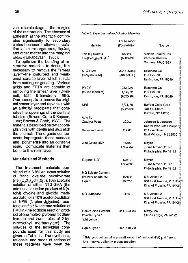
The treatment materials consisted of a 6.8% aqueous solution of ferric oxalate hexahydrate [Fe2(C204h6H20]; a 10% acetone solution of either NTG-GMA [the additional reaction product of N(otolyl) glycine and glycidyl methacrylate] ora 10%acetonesolution of NPG (N-phenylglycine); acetone, and a 5% acetone solution of PMDM (the addition reaction product of one mole of pyromellitic dianhydride and two moles of 2-hydroxyethyl methacrylate). The sources of the individual compounds used for this study are given in Table 1. The syntheses, rationale, and mode of actions of these reagents have been de-
Table 1. Experimental and Control Materials
Material
Iron (Ill) oxalate
Fe2(C204)3·6H20*
NTG-GMA (recrystallized)
PMDM
(mixed isomers)
NPG
Adaptic
Catalyst Paste
Universal Paste
Zinc Oxide USP
EugenolUSP
MQ Silicate Cement
(Powder shade 55)
Liquid
MQ Lubricant
Fleck's Zinc Cement
Powder Type 1
light yellow
Liquid Type 1
Lot Number
(Formulation)
052280 (A600-33)
297-1 (5/83) (A628-38 E)
283-22A
1/'30/82 (A625-99)
8/24/79 (A625-99)
2C003
28003
16300 Lot #182
57612
Lot #208
456508 106710
#55
D11 050384
H47 110383
OPERATIVE DENTISTRY
Source
Morton Thiokol, Inc
Ventron Division
Danvers, MA 01923
Esschem Co
P 0 Box 56 Essington, PA 19029
Esschem Co
P 0 Box 56 Essington, PA 19029
Buffalo Color Corp
340 Elk Street Buffalo, NY 14210
Johnson & Johnson
Dental Products Company
20 Lake Drive
East Windsor, NJ 08520
Moyco
J Bird Moyer Co, Inc
Philadelphia, PA 19132
Moy co
J Bird Moyer Co, Inc
Philadelphia, PA 19132
SS White Co
900 First Avenue, P 0 Box C
King of Prussia, PA 19406
S SWhite Co
900 First Avenue, P 0 Box C
King of Prussia, PA 19406
Mizzy, Inc
Clifton Forge, VA 24122
*This product contains a small amount of residual HN03; different
lots may vary slightly in concentration.
Dow
nloaded from http://m
eridian.allenpress.com/operative-dentistry/article-pdf/13/3/1/1818880/1559-2863-13-3-1.pdf by guest on 27 January 2022

STANLEY/ BOWEN/ COBB: PULP RESPONSES TO DENTIN BONDING AGENTS 109
scribed (Bowen, Cobb & Rapson, 1982; Bowen, Cobb&Setz, 1982; Bowen &Cobb, 1983; Bowen & others, 1984).
Each erupted tooth in all eight quadrants of two primates (Macaca fascicularis) received either the experimental or control materials. Each animal was under general anesthesia during the procedures. A class 5 cavity was prepared in each tooth in a given quadrant before the materials were placed. However, experience in this and other studies suggests that to avoid contamination it would be preferable to prepare the cavities and perform the procedures on only one or a few teeth at a time to simulate clinical treatment. The cavities were prepared with diamond instruments utilizing high speed (about 50 000 rpm free-running speed) provided by a portable, water-driven, turbine contra-angle handpiece {Turbo Jet Dental Unit, Bowen & Co, Inc, Rockville, MD 20852) with copious water spray.
The same materials were applied to matching teeth in both of the two animals to get the best estimate of the time effects of seven and 21 days.
In the upper right quadrants, after retentionform cavity preparations, all cavities were treated simultaneously for 60 seconds with the aqueous ferric oxalate solution (pH 0.84, mOsm 480), rinsed with running water for about 1 O seconds, and subjected to about 1 O seconds of a compressed air stream that was not completely dry. The cavities were then treated with a freshly prepared 10% solution of NTG-GMA in acetone. After about 60 seconds, clean reagent acetone was applied for 1 O seconds to all of the cavities and blown away with air. The PMDM solution was then applied to all of the preparations, and after about 60 seconds the cavities were "dried" with the air stream for 1 o seconds.
At this point a two-paste composite (Adaptic, Johnson & Johnson, East Windsor, NJ 08520) was mixed and placed into the cavity preparations with a hand instrument. Several cavities were filled with each mix, starting with the most distal teeth.
The upper and lower right quadrants received the same treatment with this exception: each cavity preparation in the upper right quadrant had retention form, but the cavity preparations in the lower right quadrant did not.
For the cavities in the upper left quadrant, the same treatment procedure was used as that
described for the cavities in the right quadrant, except that a 10% acetone solution of NPG (Nphenylglycine) was substituted for the 10% acetone solution of NTG-GMA; some of these cavities had retention forms (UL 2, 3, 5, and 6) and some did not (UL 1, 4, and 7).
In both animals, control materials were placed in cavities with retention forms in the lower left quadrants. Care was taken to ensure that the control cavities were not cut deeper than the experimental cavities, which would have resulted in less remaining dentin thickness, or RDT, and would have biased the pulp response in favor of the experimental cavities. The controls consisted of unlined silicate cement, ZOE (zinc oxide-eugenol), Adaptic, and a zinc phosphate cement.
The two animals were sacrificed with T-61 Euthanasia Solution {Taylor Pharmacal Co, Decatur, IL62521), one at seven and the other at 21 days. The jaw segments were dissected with a water-cooled bone saw so that the apical onethird of each tooth was severed. The jaws were placed in a 10% buffered formalin phosphate (Fisher Scientific, Fair Lawn, NJ 07410). After seven days of fixation, the specimens were decalcified in 5% formic acid. The teeth were then removed from the jaw blocks for processing.
An important observation was made at that time. When restored teeth are decalcified, the restorations ordinarily fall out as the enamel disappears and thedentin softens. After 21 days in formic acid, the enamel was gone and the tooth was flexible, but most of the restorations remained attached to the decalcified dentin, a most unusual finding. The Adaptic restorations were teased or shaved off with a razor blade.
The tooth specimens were then processed in a routine fashion for histologic pulpal examination. They were sectioned in serial order through the entire pulp, stained with hematoxylin and eosin, and the section next to the most critical section, which was defined by the remaining dentin thickness and severity of the lesion, was treated with the Brown and Brenn stain to detect bacteria (Brown & Brenn, 1931).
The sections were evaluated and graded, in accord with the criteria outlined in section 4.3.4. 7 of the ADA's "Recommended Standard Practices for Biological Evaluation of Dental Materials" (Council on Dental Materials and Devices,
Dow
nloaded from http://m
eridian.allenpress.com/operative-dentistry/article-pdf/13/3/1/1818880/1559-2863-13-3-1.pdf by guest on 27 January 2022
110
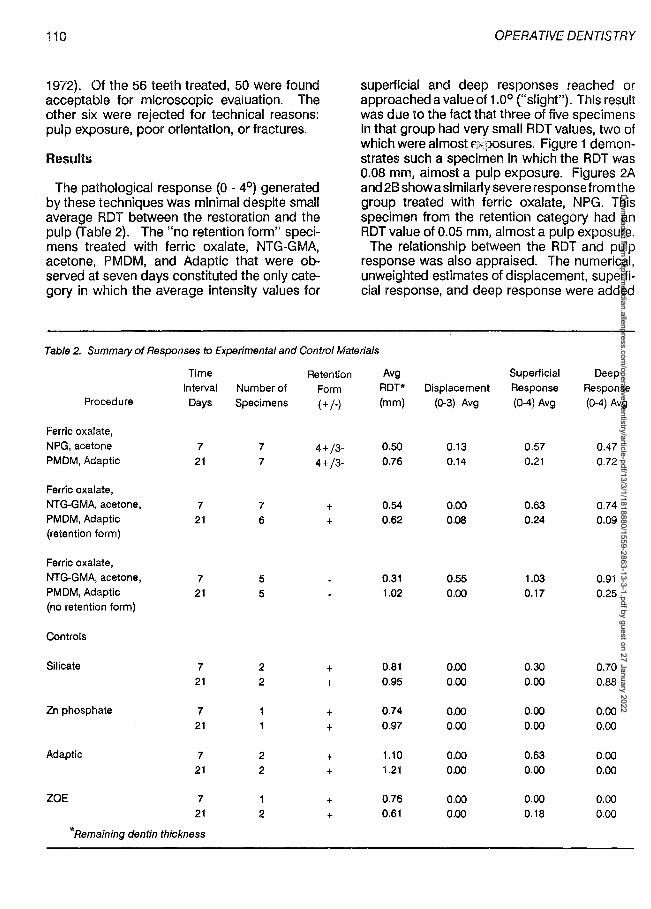
1972). Of the 56 teeth treated, 50 were found acceptable for microscopic evaluation. The other six were rejected for technical reasons: pulp exposure, poor orientation, or fractures.
Results
The pathological response (O - 4°) generated by these techniques was minimal despite small average RDT between the restoration and the pulp (Table 2). The "no retention form" specimens treated with ferric oxalate, NTG-GMA, acetone, PMDM, and Adaptic that were observed at seven days constituted the only category in which the average intensity values for
OPERATIVE DENTISTRY
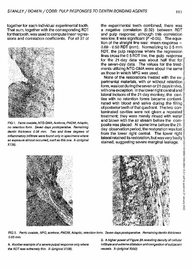
superficial and deep responses reached or approached a value of 1.0° ("slight"). This result was due to the fact that three of five specimens in that group had very small RDT values, two of which were almost 6*POsures. Figure 1 demonstrates such a specimen in which the RDT was 0.08 mm, almost a pulp exposure. Figures 2A and 28 show a similarly severe response from the group treated with ferric oxalate, NPG. This specimen from the retention category had an RDT value of 0.05 mm, almost a pulp exposure.
The relationship between the RDT and pulp response was also appraised. The numerical, unweighted estimates of displacement, superficial response, and deep response were added
Table 2. Summary of Responses to Experimental and Control Materials
Time Retention Avg Superficial Deep Interval Number of Form ROT* Displacement Response Response
Procedure Days Specimens (+ /-) (mm) (0-3) Avg (0-4) Avg (0-4) Avg
Ferric oxalate, NPG, acetone 7 7 4+/3- 0.50 0.13 0.57 0.47 PMDM, Adaptic 21 7 4+/3- 0.76 0.14 0.21 0.72
Ferric oxalate, NTG-GMA, acetone, 7 7 + 0.54 0.00 0.63 0.74 PMDM, Adaptic 21 6 + 0.62 0.08 0.24 0.09 (retention form)
Ferric oxalate, NTG-GMA, acetone, 7 5 0.31 0.55 1.03 0.91 PMDM, Adaptic 21 5 1.02 0.00 0.17 0.25 (no retention form)
Controls
Silicate 7 2 + 0.81 0.00 0.30 0.70 21 2 + 0.95 0.00 0.00 0.88
Zn phosphate 7 + 0.74 0.00 0.00 0.00 21 + 0.97 0.00 0.00 0.00
Adaptic 7 2 + 1.10 0.00 0.63 0.00 21 2 + 1.21 0.00 0.00 0.00
ZOE 7 + 0.76 0.00 o.oo 0.00 21 2 + 0.61 0.00 0.18 0.00
*Remaining dentin thickness
Dow
nloaded from http://m
eridian.allenpress.com/operative-dentistry/article-pdf/13/3/1/1818880/1559-2863-13-3-1.pdf by guest on 27 January 2022
STANLEY/ BOWEN/ COBB: PULP RESPONSES TO DENTIN BONDING AGENTS 111
together for each individual experimental tooth. That sum, together with the corresponding RDT forthattooth, was used to compute linear regressions and correlation coefficients. For all 37 of
FIG 1. Ferric oxalate, NTG-GMA, Acetone, PMDM, Adaptic, no retention fOrrn. Seven days postoperative. Remaining dentin thickness 0.08 mm. Two and three degrees of inflammatory infiltrate were fOund only in specimens where an exposure almost occurred, such as this one. X-(original
X138).
the experimental teeth combined, there was a negative correlation (0.32) between RDT and pulp response; although this correlation was low, it was significant (P "'0.05). The equation of the straight line was: mean response = 0.69 - 0.50 RDT (mm). Normalizing to 0.5 mm RDT, the pulp response where the regression lines cross the 0.5 RDT line, the pulp response for the 21-day data was about half that for the seven-day data. The values for the treatments utilizing NTG-GMA were about the same as those in which MPG was used.
None of the restorations treated with the experimental materials, with or without retention form, was lost during the seven or 21 days in vivo, with one exception. In the lower right central and lateral incisors of the 21-day monkey, the cavities with no retention forms became contaminated with blood and saliva during the filling of posterior teeth of that quadrant. The two contaminated cavities were not given a repeated treatment; they were merely rinsed with water and blown with the air stream before the composite was placed. At some time before the 21-day observation period, the restoration was lost from the lower right central. The lower right lateral retained its restoration, but was very badly stained, suggesting severe marginal leakage.
FIG 2. Ferric oxalate, NPG, acetone, PMDM, Adaptic, retention fOrm. Seven days postoperative. Remaining dentin thickness
0.05mm.
A. Another example of a severe pulpal response only where
the ROT was extremely thin. X-(original X108).
B. A higher power of Figure 2A revealing density of cellular infiltrate and extreme dilatation and congestion of subjacent vessels. X-(origina/ X550).
Dow
nloaded from http://m
eridian.allenpress.com/operative-dentistry/article-pdf/13/3/1/1818880/1559-2863-13-3-1.pdf by guest on 27 January 2022

112
The sections to which the Brown and Brenn stain was applied revealed nothing consistent in terms of bacterial invasion, due to marginal leakage. Only three specimens showed a significant number of bacteria: one of the blood-contaminated preparations previously described and two of the silicate and Adaptic control specimens. Neither of these had pulp lesions.
However, the Brown and Brenn stain revealed a modification of the superficial dentin exposed to the treatment solutions and composite. A purplish-red color change in what appeared to be a smear layer in the cavity preparations of most of the experimental teeth occurred with the Brown and Brenn stain. (This color change cannot be appreciated in the black and white prints; Figures 3A, B & C). This characteristic appears to represent a change in the dentin surface from which the composite had been removed after decalcification.
OPERATIVE DENTISTRY
Discussion
Finding so little pulpal pathology is in accord with the concept that the ferric oxalate and other solutes bring about an obturation of the dentinal tubules without releasing noxious components. Scanning electron microscope (SEM) observations (Bowen, Cobb & Rapson, 1982; Bowen & Cobb, 1983) show that the appearance of waterinsoluble precipitates in the dentinal tubule lumina follows the application of aqueous ferric oxalate, water, and an air stream. Transmission electron microscope (TEM) studies indicate a removal of the smeared layer and the formation of a microporous, rigid structure that probably becomes impregnated by the NTG-GMA or NPG and PMDM. The PMDM spontaneously polymerizes (Bowen & others, 1984) and copolymerizes with the resin of the composite, thereby sealing the tubules from the ingress of oral con-
FIG 3. Brown and Brenn stain. Shown are three examples of the reddish-purple color change of the superficial dentin exposed to the experimental treatment solutions and composite that appears to represent a restructured surface layer that is chemically receptive to the cationic dyes of the B & B staining procedure after formalin fixation, formic acid deca/cification, and the steps of histologic preparation and sectioning.
A. Ferric oxalate, NTG-GMA, acetone, PMDM, Adaptic, retention form. Seven days postoperative. In this specimen, the artificial layer did not appear to involve the dentinal tubules, perhaps due to the angulation of the lumina relative to the plane of the section. X-(original X768).
B. Ferric oxalate, NTG-GMA, acetone, PMDM, Adaptic, retention form. Seven days postoperative. In this specimen, some of the artificial layer has entered a few tubules. X-(original X512).
C. Ferric oxalate, NPG, acetone, PMDM, Adaptic, no retention form. Seven days postoperative. In this specimen, the artificial layer extends for a short distance into many dentina/ tubules. X-(original X768).
Dow
nloaded from http://m
eridian.allenpress.com/operative-dentistry/article-pdf/13/3/1/1818880/1559-2863-13-3-1.pdf by guest on 27 January 2022
STANLEY/ BOWEN/ COBB: PULP RESPONSES TO DENTIN BONDING AGENTS 113
taminants. Decreased permeability of this thin outer layer may be in accord with minimal pulp response even in the deep cavities.
The lack of responses in the controls (zinc phosphate, silicate cements, and unlined Adaptic) is attributed to the larger ROT in some categories and the shortness of the observation periods.
Thefinding of the unique color change caused by the Brown and Brenn stain reagents in the superficial dentin might lead to a new way of studying the surface chemistry involved in this adhesion-promoting procedure. The apparent absence of micro-organisms in the treated (experimental) teeth is in keeping with the expectation that application of these solutions would have a sealing and bactericidal effect on the surfaces being treated.
Conclusions
The finding of very limited pulp response is in accord with the concept that the treatment (1) yields insoluble products that occlude the tubules and (2) does not release noxious components in the process. The low-level pulp response of these treatments permits the judicious initiation of human clinical trials using this adhesion technique on both dentin and enamel. However, contamination of the treated cavity preparation induced clinical discoloration or loss of the restoration, and it is therefore necessary to prevent any impurities from contacting the surfaces of the teeth from the beginning of the treatment until a composite material is placed and has hardened.
• This investigation was supported in part by USPHS Research Grant DEOS 129-07 to the American Dental Association Health Foundation from the National Institutes of Health-National Institute of Dental Research and is part of the dental research program conducted by the National Bureau of Standards in cooperation with the American Dental Association Health Foundation.
• Certain commercial materials and equipment are identified in this paper to specify the experimental procedure. In no instance does such identification imply recommendation or endorsement by the National Bureau of Standards
or the ADA Health Foundation or that the material or equipment identified is necessarily the best available for the purpose.
• Contributions of the National Bureau of Standards. Not subject to copyright.
Acknowledgments The authors thank Dr Dan Dalgard, Fred Synder, and the
staff at Hazleton Laboratories America, Inc, Vienna, Virginia, where the animal care was provided, the anesthetic administered, and the dental work performed.
(Received B April 1987)
References
BOWEN, R L & COBB, E N (1983) A method for bonding to dentin and enamel Journal of the American Dental Association 107 734-736.
BOWEN, R L, COBB, E N & MISRA, D N (1984) Adhesive bonding by surface initiation of polymerization (part XXVlll of series on adhesive bonding of various materials to hard tooth tissues) Industrial Engineering Chemical Products Research Division 23 78-81.
BOWEN, R L, COBB, E N & RAPSON, J E (1982) Adhesive bonding of various materials to hard tooth tissues. XXV: Improvement in bond strength to dentin Journal of Dental Research 61 1070-1076.
BOWEN, R L, COBB, E N & SETZ, L E (1982) Adhesive bonding to dentin and enamel (part XXVll of series on adhesive bonding of various materials to hard tooth tissues) Dentistry '82 2 11-13.
BRANNSTROM, M (1984) Smear layer: pathological and treatment considerations Operative Dentistry Supplement 3 35-42.
BROWN, J H & BRENN, L (1931) Bulletin of the Johns Hopkins Hospital 48 69-73, as modified in LUNA, L G (1968) Manual of Histological Staining Methods of the Armed Forces Institute of Pathology, 3rd edition, pp 222-223. New York: McGraw-Hill.
COUNCIL ON DENTAL MATERIALS AND DEVICES (1972) Recommended standard practices for biological evaluation of dental materials Journal of the American Dental Association 84 382.
COUNCIL ON DENTAL MATERIALS, INSTRUMENTS, AND EQUIPMENT (1984) Resin dentin bonding systems Journal of the American Dental Association 108 240-241.
GWINNETT, AJ (1984) Smear layer: morphological considerations Operative Dentistry Supplement 3 3-12.
NAKABAYASHI, N (1985) Biocompatibilityand promotion of adhesion to tooth substrates Critical Reviews in Biocompatibility 1 2~9.
Dow
nloaded from http://m
eridian.allenpress.com/operative-dentistry/article-pdf/13/3/1/1818880/1559-2863-13-3-1.pdf by guest on 27 January 2022
114 @OPERATIVE DENTISTRY, 1988, 13, 114-118
Composite Resin Repair of Porcelain Using Different Bonding Materials
WILLIAM A GREGORY • CHARLES A HAGEN JOHN M POWERS
Summary Bond strengths of porcelain/composite
resin repair samples, using five different repair liquids and two composites, were evaluated. Samples were allowed to set without disturbance. After storage in 37 °C water for intervals of one day, seven days, and 28 days, the test samples were subjected to tensile force until fracture. There were significant differences in bond strengths of repair agents at all test intervals. All mean bond strengths were significantly less at 28 days than at one day. Repairs made with the repair liquid and composite of a single manufacturer did not always perform better than heterogeneous combinations. Fractures of all specimens were caused by adhesive failures occurring at the interface.
University of Michigan, School of Dentistry, Department of Operative Dentistry, Ann Arbor, Ml 48109
William A Gregory, DDS, MS, assistant professor
Charles A Hagen, BA, third-year dental student
John M Powers, PhD, professor
Introduction
The increasing use of porcelain-fused-to-metal restorations in dentistry has resulted in the need for repair of fractured porcelain. Composite resins have been the material of choice for their esthetic appearance and ease of manipulation (Dent, 1979). In the late 1970s, materials were specifically developed to enhance porcelaincomposite resin adhesion and have been evaluated for bond strengths over time (Eames & Rogers, 1979) and after thermally induced stress (Newburg & Pameijer, 1978). Repair bond strengths did not prove to be acceptable over long periods and were considered to be temporary (Eames & Rogers, 1979). Currently, newer materials are available for the repair of porcelain with composite having improved bond strengths.
The purpose of this investigation was to measure the tensile repair bond strengths between a single dental porcelain used for metal veneering and two composite resins when different commercial repair bonding agents were used. Since some bonding material manufacturers suggest the use of their own composite for repair, two homogeneous systems of repair materials were tested for comparison with the repairs made with combinations from different manufacturers.
Dow
nloaded from http://m
eridian.allenpress.com/operative-dentistry/article-pdf/13/3/1/1818880/1559-2863-13-3-1.pdf by guest on 27 January 2022

GREGORY/ HAGEN/ POWERS: COMPOSITE RESIN REPAIR OF PORCELAIN 115
Materials and Methods
Porcelain buttons, approximately 9 mm in diameter and 7 mm thick, were made using Vita VMIC 68 enamel 559 porcelain and liquid (Unitek Corp, Monrovia, CA 91016, batch #733-220). The materials were condensed and vibrated against a piece of platinum foil matrix, 0.01 inch thick, according to the manufacturer's directions, in a specially designed, tapered plexiglass mold. After removal from the mold, the buttons were fired under vacuum at 1760 °F. Following sintering, the porcelain was examined for voids and imperfect samples were discarded. Forty acceptable specimens were embedded in cylinders of bioplastic (DecraCoat, Resco, Miami, FL 33054) with approximately 2 mm of the porcelain button protruding above the resin. An eyehook was embedded in the opposite side of the resin cylinder. The exposed surface of the porcelain button was abraded and polished, using a No 600 grit silicon carbide paper mounted on a mechanical horizontal rotating grinder to remove the glaze, to eliminate any scoring that would provide mechanical retention, and to
standardize the surfaces prior to bonding. Table 1 lists the materials used in this study. A
light-cured hybrid composite, Ultrafine, was used as the primary repair material in combination with five porcelain bonding agents. A lightcured microfilled composite, Silux, was tested using one bonding agent.
Bonding agents were applied to the prepared porcelain surface according to manufacturer's instructions. A Delrin plastic tube (AIN Plastics, Inc, Mount Vernon, NY 10550) was used to form the composite resin sample. The tube end opposite the porcelain surface was enlarged to mechanically retain the composite. The tube was threaded exteriorly to accept a plumbing pipe cap. A hole in the end of the cap allowed an S-hookto be inserted. The repair composite was condensed through and within the Delrin tube and adapted to the porcelain surface in increments less than 2 mm and cured for 40 seconds each until the tube was filled. The same curing light was used for all samples.
Fifteen randomly selected porcelain/composite resin repair samples were made with each of the five bonding agents and the composite Ultrafine. Five samples of each group were stored
Table 1. Materials Evaluated
Material Manufacturer Batch Number
Composites
Command Ultrafine Kerr /Sybron Romulus, Ml 48174
3604-16855
Silux
Bonding Agent
Porcelain Repair Liquid
Silanit
Scotch prime
Fusion
Ultrabond
3M Dental Products St Paul, MN 55144
Kerr /Sybron Romulus, Ml 48174
Vivadent (USA), Inc Tonawanda, NY 14150
3M Dental Products St Paul, MN 55144
George Taub Products Jersey City, NJ 07307
Den-Mat Corp Santa Maria, CA 93456
70-2005-2306
3604-18446
6163-5160
70200523069
L279
550060
Dow
nloaded from http://m
eridian.allenpress.com/operative-dentistry/article-pdf/13/3/1/1818880/1559-2863-13-3-1.pdf by guest on 27 January 2022
116
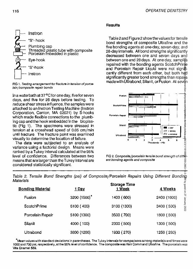
lnstron
"S"- hook
Plumbing cap Threaded plastic tube with composite Porcelain imbedded in plastic
Eye-hook
"S"-hook
lnstron
FIG 1. Testing a"angement for fracture in tension of porcelain/composite repair bonds
in a waterbath at 37 °Ctor one day, five for seven days, and five for 28 days before testing. To reduce shear stress influence, the samples were attached to an lnstron Testing Machine (lnstron Corporation, Canton, MA 02021) by S-hooks which made flexible connections to the plumbing cap and the hook embedded in the bioplastic (Fig 1). The specimens were stressed in tension at a crosshead speed of 0.05 cm/min until fracture. The fracture point was examined visually to determine the location of failure.
The data were subjected to an analysis of variance using a factorial design. Means were ranked by a Tukey interval calculated at the 95% level of confidence. Differences between two means that are larger than the Tukey interval are considered statistically significant.
OPERATIVE DEN/TSTRY
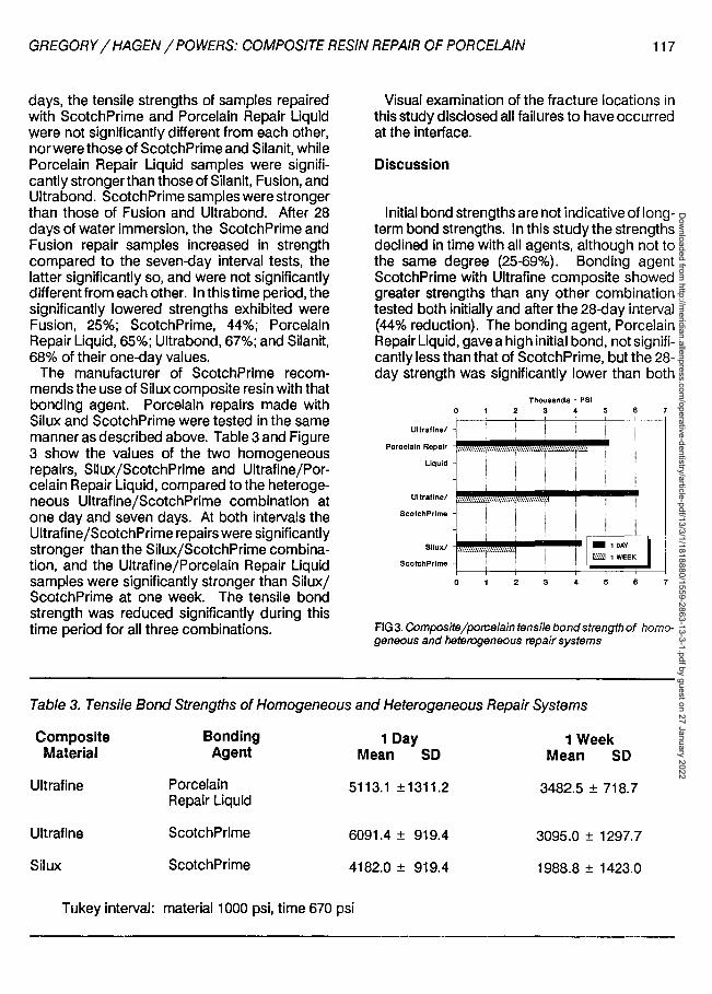
Results
Table2and Figure2 show the values for tensile bond strengths of composite Ultrafine and the five bonding agents at one-day, seven-day, and 28-day intervals. All bond strengths significantly decreased between one and seven days and between one and 28 days. At one day, samples repaired with the bonding agents ScotchPrime and Porcelain Repair Liquid were not significantly different from each other, but both had significantly greater bond strengths than repairs made with Ultrabond, Silanit, or Fusion. At seven
Fualon
Scotch Prime
Porcelain Repair
-~ 1- 1DAY ~ 1WEEK
CD 4 WEEKS
Sllanit
Ultrabond
0 2 3 <4 6 Thousands - PSI
FIG 2. Composite/porcelain tensile bond strength of different bonding agents and composite
Table 2. Tensile Bond Strengths (psi) of Composite/Porcelain Repairs Using Different Bonding Materials
Storage Time Bonding Material 1 Day 1 Week 4 Weeks
Fusion 3200 (1500)* 1400 ( 600) 2400 (1600)
ScotchPrime 6100 ( 400) 3100 (1300) 3400 ( 500)
Porcelain Repair 5100 (1300) 3500 ( 700) 1800 ( 500)
Sil an it 4000 ( 100) 2300 ( 500) 1300 ( 500)
Ultrabond 3800 (1200) 1930 ( 270) 1250 ( 280)
*Mean values with standard deviations in parentheses. The Tu key intervals for comparisons among materials and times were 1000 and 700 psi, respectively, atthe 95% level of confidence. The composite was Kerr Command Ultrafine. The porcelain was Vita Enamel 559.
Dow
nloaded from http://m
eridian.allenpress.com/operative-dentistry/article-pdf/13/3/1/1818880/1559-2863-13-3-1.pdf by guest on 27 January 2022
GREGORY/ HAGEN/ POWERS: COMPOSITE RESIN REPAIR OF PORCELAIN 117
days, the tensile strengths of samples repaired with ScotchPrime and Porcelain Repair Liquid were not significantly different from each other, nor were those of ScotchPrime and Silanit, while Porcelain Repair Liquid samples were significantly stronger than those of Silanit, Fusion, and Ultrabond. ScotchPrime samples were stronger than those of Fusion and Ultrabond. After 28 days of water immersion, the ScotchPrime and Fusion repair samples increased in strength compared to the seven-day interval tests, the latter significantly so, and were not significantly differentfrom each other. In this time period, the significantly lowered strengths exhibited were Fusion, 25%; ScotchPrime, 44%; Porcelain Repair Liquid, 65%; Ultrabond, 67%; and Silanit, 68% of their one-day values.
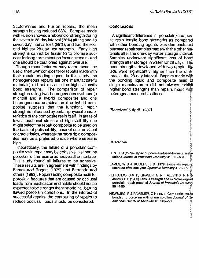
The manufacturer of ScotchPrime recommends the use of Silux composite resin with that bonding agent. Porcelain repairs made with Silux and ScotchPrime were tested in the same manner as described above. Table 3 and Figure 3 show the values of the two homogeneous repairs, Silux/ScotchPrime and Ultrafine/Porcelain Repair Liquid, compared to the heterogeneous Ultrafine/ScotchPrime combination at one day and seven days. At both intervals the Ultrafine /ScotchPrime repairs were significantly stronger than the Silux/ScotchPrime combination, and the Ultrafine/Porcelain Repair Liquid samples were significantly stronger than Silux/ ScotchPrime at one week. The tensile bond strength was reduced significantly during this time period for all three combinations.
Visual examination of the fracture locations in this study disclosed all failures to have occurred at the interface.
Discussion
Initial bond strengths are not indicative of longterm bond strengths. In this study the strengths declined in time with all agents, although not to the same degree (25-69%). Bonding agent ScotchPrime with Ultrafine composite showed greater strengths than any other combination tested both initially and after the 28-day interval (44% reduction). The bonding agent, Porcelain Repair Liquid, gave a high initial bond, not significantly less than that of ScotchPrime, but the 28-day strength was significantly lower than both
Thcuoanda - PSI 0 2 3 4 5 8 7
Ultraflne/
Porcelain Repair
Liquid
Ultraflne/
ScotchPrlme
Sllux/
ScotchPrlme
...... -,_...,., I:' ::;EK I !
0 2 3 4 5 8 7
FIG 3. Composite/porcelain tensile bond strength of homogeneous and heterogeneous repair systems
Table 3. Tensile Bond Strengths of Homogeneous and Heterogeneous Repair Systems
Composite Bonding 1 Day 1Week Material Agent Mean SD Mean SD
Ultrafine Porcelain 5113.1 ±1311.2 3482.5 ± 718. 7 Repair Liquid
Ultrafine ScotchPrime 6091.4 ± 919.4 3095.0 ± 1297.7
Silux ScotchPrime 4182.0 ± 919.4 1988.8 ± 1423.0
Tukey interval: material 1000 psi, time 670 psi
Dow
nloaded from http://m
eridian.allenpress.com/operative-dentistry/article-pdf/13/3/1/1818880/1559-2863-13-3-1.pdf by guest on 27 January 2022
118
ScotchPrime and Fusion repairs, the mean strength having reduced 65%. Samples made with Fusion showed a rebound of strength during the seven to 28-c:lay interval (78%) after a one- to seven-day interval loss (58%), and had the second highest 28-c:lay test strength. Early high strengths cannot be assumed to promise success for long-term retention for such repairs, and one should be cautioned against overuse.
Though manufacturers may recommend the use of their own composite for repairs made with their repair bonding agent, in this study the homogeneous repairs (all one manufacturer's materials) did not result in the highest tensile bond strengths. The comparison of repair strengths using two homogeneous systems (a microfill and a hybrid composite) and one heterogeneous combination (the hybrid composite) suggests that the functional repair strength is influenced by certain physical characteristics of the composite resin itself. In areas of lower functional stress and high visibility one might select the repair composite to be used on the basis of polishability, ease of use, or visual characteristics, whereas the more rigid composites may be a preferred choice where stress is high.
Theoretically, the failure of a porcelain-composite resin repair may be cohesive in either the porcelain or the resin or adhesive at the interface. This study found all failures to be adhesive. These results are in agreement with findings by Eames and Rogers (1979) and Ferrando and others (1983). Repairs using composite resin for porcelain fractures that are caused by occlusal loads from mastication and habits should not be expected to be strongerthan the original, barring flawed porcelain conditions. In the interest of successful repairs, the contouring of repairs to reduce occlusal loads should be considered.
OPERATIVE DENTISTRY
Conclusions
A significant difference in porcelain/composite resin tensile bond strengths as compared with other bonding agents was demonstrated between repair samples made with the other materials after the one-day water storage interval. Samples underwent significant loss of bond strength after storage in water for 28 days. The bond strengths developed with two repair liquids were significantly higher than the other three at the 28-c:lay interval. Repairs made with the bonding liquid and composite resin of single manufacturers did not always exhibit higher bond strengths than repairs made with heterogeneous combinations.
(Received 6 April 1987)
Reference•
DENT, R J (1979) Repair of porcelain-fused-to-metal restorations Journal of Prosthetic Dentistry 41 661-664.
EAMES, W B & ROGERS, L B (1979) Porcelain repairs: retention after one year Operative Dentistry 4 75-77.
FERRANDO, J-M P, GRASER, G N, TALLENTS, R H & JARVIS, RH (1983) Tensile strength and microleakage of porcelain repair material Journal of Prosthetic Dentistry 50 44-50.
NEWBURG, R & PAMEIJER, CH (1978) Composite resins bonded to porcelain with silane solution Journal of the American Dental Association 96 288-291.
Dow
nloaded from http://m
eridian.allenpress.com/operative-dentistry/article-pdf/13/3/1/1818880/1559-2863-13-3-1.pdf by guest on 27 January 2022
©OPERATIVE DENTISTRY, 1988, 13, 119-127 119
Early Observations and Three-year Clinical Evaluation
of Four Amalgam Alloys
JAMES B RICKER • EVAN H GREENER
Summary Four amalgam alloys were evaluated forth is
study: Dispersalloy, Tytin, Sybraloy, and Valiant. Six evaluators viewed slides for each restoration over a three-year period (after two weeks, eight weeks, 16 weeks, one year, two years, and three years). Marginal breakdown was apparent at 16 weeks for all alloys and changed significantly up to one year, then leveled off for years two and three. The results showed that clinical performance as early as six months correlates significantly with that at three years, a viable alternative to laboratory studies in predicting clinical performance.
Northwestern University Dental School, Department of Operative Dentistry, Chicago, IL 60611
JAMES B RICKER, DDS, MS, associate professor and preclinical director
EVAN H GREENER, PhD, professor and chairman, Department of Biological Materials
INTRODUCTION
Dental amalgam has been used in this country for over 150 years as a major tooth restorative material (Greener, 1979). Conventional alloys conform to ADA Specification No 1, as written in 1969, which allows a maximum copper concentration of 6% (Council on Dental Materials and Devices, 1969). After high-copper alloys had demonstrated superior clinical performance, the specification was revised in 1977 (Council on Dental Materials and Devices, 1977) to allow for higher copper concentration, in a range from 9 -30%, with silver and tin the primary components.
Failures of amalgam restorations may be clinically classified as marginal fracture, gross bulk fracture, secondary caries, dimensional change, and excessive discoloration (Baum, Phillips & Lund, 1981 ). Marginal fracture or breakdown is the most common type of deterioration seen in amalgam restorations (Osborne & others, 1976). In a survey to members of the Academy of Operative Dentistry, Charbeneau, Klausner, and Green (1986) found that of a total of 2296 amalgam restorations replaced over a two-week period, 17% were due to poor margins, and 53% due to recurrent caries. Other causes for failure were isthmus fracture, tooth fracture, and other reasons, all at lower percentages. This marginal breakdown has been attributed to retention of excess mercury, improper cavity preparation, failure to carve or finish the amalgam flush with the margins of the cavity, corrosion of the mar-
Dow
nloaded from http://m
eridian.allenpress.com/operative-dentistry/article-pdf/13/3/1/1818880/1559-2863-13-3-1.pdf by guest on 27 January 2022
120
gins and mercuroscopic expansion, and delayed expansion (Rupp, Paffenbarger & Patel, 1980).
Numerous amalgam alloys on the market vary in their clinical performance as well as in their mechanical and handling properties. Many clinical studies have shown differences among amalgam alloys by comparing marginal integrity with respect to time (Osborne & others, 1976; Mahler & others, 1970; Osborne & others, 1980; Osborne & Gale, 1979; Osborne, Binon & Gale, 1980; Letzel & Vrijhoef, 1984; Smales & Gerke, 1984). Three theories have been advanced to explain the marginal breakdown process.
Marginal Breakdown: Three Theories
The Mercuroscopic Expansion Theory as proposed by Jorgensen (1965) identifies a crevice corrosion at the tooth-amalgam interface. The initiating mechanism in this process is corrosion. Free mercury liberated during this process diffuses back into the marginal amalgam, causing an expansion and subsequent deflection of the amalgam margin from the tooth. This exposed, raised margin is then broken off due to masticatory forces. Sarker, Osborne, and Leinfelder (1982) have shown significant correlations between corrosion index data in vitro and marginal fracture in vivo. The most corrosion-prone phase in dental amalgam is the 1-2 phase, although the Cu6Sn5 phase is also corrosive (Sarkar & Greener, 1975). The copper-rich amalgam alloys have virtually eliminated 1-2 from their restorations (Leinfelder, 1983). Copper combines with tin to form Cu6Sn5 and thus prevents mercury from reacting with the tin to form 1-2. The Cu6Sni is also a corrosive phase, although much ess so than 1-2 (Sarkar & Greener, 1975). The addition of palladium has been shown to have a positive effect on corrosion resistance (Greener & Szurgot, 1982). Lin and others (1986) found that adding palladium to blended amalgam produced no 1-2 formation and suppressed the Cu6Sn5 phase.
The Crack Penetration Attack Theory, as proposed by Espevik and Mjor (1978), suggests that the breakdown of margins appears to be associated with cracks penetrating perpendicular to the occlusal surfaces. These cracks, which extend along 1-1-grain boundaries, contain tin hydroxychloride corrosion products. Espervik
OPERATIVE DENTISTRY
and Mjor theorized that when sufficient weakening of the marginal areas by this penetration had occurred, portions would fracture off.
The Rheological Theory was developed from studies conducted by Mahler and co-workers (Mahler & others, 1970; Mahler, Terkla & van Eysden, 1973; Mahler, van Eysden & Terkla, 1975). Their findings showed an association between the laboratory tests of dynamic creep, static creep, and slow compressive strength with the marginal fracture potential of amalgams. They suggested that static creep could be used as a predictor of clinical performance; however, only one high-copper alloy was available for study at that time. The relationship between creep and marginal breakdown does not seem to hold when only 1-2-free amalgams are studied. In separate reports, Osborne and others (1980), Laswell, Berry, and Osborne (1980), and Jordan, Suzuki, and Mills (1978) have shown no significant correlations between creep and marginal breakdown. In looking for other variables to explain marginal deterioration, Mahler, Marantz, and Engle (1980) designed a predictive model using creep, zinc content, and 1-2 content. Applying this model to the three-year clinical results of 12 alloys, Gale, Osborne, and Winchell (1982) obtained results similar to Mahler's when both 1-2- containing and 1-2-free alloys are used in the same equation. However, when only the 1-2-free alloys are used in the equation, the correlation of predicted versus observed fracture at the margins is a negative one and, therefore, inappropriate to use. Nonetheless, the creep test can be used for gross screening of alloys with respect to clinical performance (Leinfelder, 1983). The addition of palladium has markedly decreased the creep values of blended amalgams (Chung, Lin & Greener, 1986). The palladium substitution for copper was about twice as effective in decreasing creep than it was when substituted for silver.
Predicting Clinical Performance
Of all the laboratory tests that have been developed, the Sarkar Corrosion Index seems to have the greatest potential for predicting clinical performance. Clinical studies will determine the success of this test. The new blends and the palladium-containing alloys should be in-
Dow
nloaded from http://m
eridian.allenpress.com/operative-dentistry/article-pdf/13/3/1/1818880/1559-2863-13-3-1.pdf by guest on 27 January 2022
RICKER/ GREENER: THREE-YEAR CLINICAL EVALUATION OF AMALGAMS 121
eluded in these studies. The profession must rely upon carefully conducted clinical studies to test the performance of the materials. Osborne, Schlissel & Gale (1981) have shown that six-month clinical data predicted three-year results better than any of the laboratory tests for the highcopper alloys.
From the clinical standpoint, one of the problems with amalgam restorations has been marginal integrity. Breakdown of the margins is evidenced by chipping, cracking, and breaking away of the amalgam from the periphery of the restoration at the cavosurface margin. The true mechanism for improved marginal integrity of high-copper alloys has not been finalized, but they are known to perform much better as a group than traditional alloys.
Clinical research on amalgam performance can be lengthy, time consuming, and costly. If short-term studies could be shown to predict long-term performance accurately, they would enhance the capability of conducting longevity studies.
The specific objectives of this study were to determine whether:
(1) marginal breakdown is evident within the first four months after placement and polishing of amalgam restorations, and, subsequently, at one, two, and three years;
(2) alloy discrimination is evident during the first four months and, subsequently, at one, two, and three years;
(3) ear1y clinical performance as related to marginal integrity can be regarded as a predictor of long-term performance; and
(4) clinical performance correlates with physical properties.
Table 2. Alloy Assignment by Tooth Type
Maxillary Premolar
Alloy % Dispersalloy 1 4.2 Tytin 2 8.3 Valiant 2 8.3 Sybraloy 1 5.0
Total 6 6.5
Table 1. Trituration Conditions
Alloy * Jua~'&~r Hg/Alloy T~c~ Speed**
Dispersalloy 08817 1: 1 15 high
Tytin 798009 0.74:1 5 high
Valiant 121680 0.71:1 8 high
Sybraloy 1308 0.82:1 15 high
*All precapsulated
**Wig-L-Bug LP-60
MATERIALS AND METHODS
Posterior teeth were selected with either virgin caries or existing restorations needing replacement. All teeth selected needed intracoronal restorations and all were in occlusion. One hundred teeth were selected from among 32 patients. Two patients left the study by the 16-week recall, thus the early data were for 92 teeth. Five more patients left the study over the threeyear period, reducing the number of restorations at the recall periods.
Four amalgam alloys (Table 1) were selected forthis study: Dispersalloy (Johnson &Johnson, EWindsor, NJ08520), Tytin (SS White, Philadelphia, PA 19102), Sybraloy (Kerr Mfg Co, Romulus, Ml 48174), and Valiant (L D Caulk Co, Milford, DE 19963). These alloys are considered (epresentative of the broad spectrum of amalgam alloys used clinically today, encompassing both blended and single-composition types.
Alloys were ranaomly assigned to the teeth to be restored; however, adjustments were made so that different alloys would not contact opposing or adjacent surfaces. A summary of the assignment of alloys to type of teeth is shown in Table 2.
Maxillary Mandibular Mandibular Premolar Molar Molar
% % %
6 25.0 2 8.3 15 62.5 10 41.2 4 16.7 8 33.3 10 41.2 0 0.0 12 50.0 8 40.0 3 15.0 8 40.0
34 37.0 9 9.8 43 46.75
Dow
nloaded from http://m
eridian.allenpress.com/operative-dentistry/article-pdf/13/3/1/1818880/1559-2863-13-3-1.pdf by guest on 27 January 2022
122
One clinician placed the restorations, following standard operative technique. Rubber dam isolation was used in all cases except on four maxillary molars where tooth morphology precluded placement of the rubber dam clamp. In those cases cotton roll and gauze isolation was used and great care taken to avoid moisture contamination during insertion of the amalgam. All amalgams were mixed using a variable speed triturator (Wig-L-Bug, LP-60, Crescent Dental Mfg Co, Lyons, IL 60534). Both speed and time for mixing were set according to the manufacturer's instructions. Appropriate settings are listed in Table 1. The technical procedure of placement followed that of Larson and others (1979). The restorations were surfaced using ball- and egg-shaped burnishers (American Dental Mfg Co, Missoula, MT 59806) with light pressure to smooth the amalgam.
All restorations were finished and polished at subsequent appointments two to seven days after placement. The sequence of materials for the final finishing and polishing was: white stones, pumice with water on a flexible prophylaxis cup, followed by dry tin oxide on a prophy cup, or brownie and greenie points (Shofu Dental Corp, Menlo Park, CA 94025). All finishing and polishing materials were used at low speed.
The technique of alloy evaluation employed the use of 35 mm slides of the occlusal surfaces. All restorations were photographed at polish for a base line and at two, eight, and 16 weeks, and at one, two, and three years after polishing. The photos were made with an Olympus OM-2 camera and Olympus T1 O ring flash (Olympus Camera Corp, Woodbury, NY 11797). Kodak Kodachrome color film (Eastman Kodak Co, Rochester, NY 14650) was used for all photographs. The photographs were taken using a front surface mirror so that the pictures were perpendicular to the occlusal surfaces of the teeth. Thirty-five mm color slides were made with the tooth under study always positioned in the center of the picture and taken at a 1 : 1 ratio. The projected slides were used for category ranking by evaluators and rid it analyis of the raw data. All pictures were compared to the 11-category Mahler scale (Mahler & Marantz, 1979).
Six evaluators viewed randomly arranged slides for each restoration at each time period. Evaluations were done at three separate sessions over the three-year period (after 16 weeks, after one year, and after three years). The slide
OPERATIVE DENTISTRY
of the restoration to be evaluated and the standard slide were projected on side-by-side screens at approximately X35 magnification. The evaluators categorized each restoration independently of one another and without knowing the alloys and time period at which they were looking.
The category ranking was sorted out for each alloy at each time period by each evaluator. For this category distribution, ridit means were calculated for each evaluator using Dispersalloy at polish for evaluator D as the standard. The statistical methodology followed that of Fleiss, Chilton, and Wallenstein (1979). The riditanalysis and pairwise comparison tests utilizing the Bonferroni correction factor were used to determine differences among alloys and/or among alloy observation times. Friedman ANOVA and Spearman rank-order correlations for data taken at polish, two, eight, and 16 weeks and pairwise comparison tests for one-, two-, and three-year data along with the Bonferroni factor were used to eliminate any evaluators who were outliers with respect to the group as a whole.
In addition to the clinical phase of this study, the laboratory tests of static creep, one-hour compressive strength, and 24-hour compressive strength were conducted according to ADA Specification No 1 guidelines (Council on Dental Materials and Devices, 1977). The ridit means from the clinical evaluation were compared to the values obtained from the laboratory tests to determine how well the two correlated. This was done graphicallywith a regression line and the correlation coefficient, r, calculated (at P < 0.05).
The raw data could not be assumed to be part of a normal distribution since there are so many variables associated with clinical studies. Each alloy observation was a unique entity and could not be used to derive an average value. Thus it was appropriate to use nonparametric statistical tests to represent the data.
The evaluators used in this study were all academicians. One was a PhD and five were dentists. No prior calibration of the evaluators was performed. Differences among evaluators were corrected by application of appropriate correlation tests to the experimental data. The evaluators were placed into subgroups displaying significant agreement with one another as determined by the Friedman nonparametric ANOVA test, Spearman correlation tests, and
Dow
nloaded from http://m
eridian.allenpress.com/operative-dentistry/article-pdf/13/3/1/1818880/1559-2863-13-3-1.pdf by guest on 27 January 2022
RICKER/ GREENER: THREE-YEAR CLINICAL EVALUATION OF AMALGAMS 123
Table 3. Marginal Ridits over Three Years Based on Evalu-ator Subgroups
Time Dispersalloy Tytin Valiant Sybraloy
Polish 0.5001 0.4528 0.5537 0.3926
Weeks:
2 0.4703 0.5157 0.6040 0.4086
8 0.6081 0.6424 0.7208 0.5064
16 0.6758 0.7213 0.7265 0.5992
52 0.8127 0.8664 0.9180 0.7920
104 0.8205 0.8555 0.8946 0.8778
156 0.8757 0.8693 0.9055 0.8109
Significant difference = 0.15 for P < 0.05.
pairwise comparison tests. Osborne and others (1976) and Leinfelder (1980) have found high correlations among evaluators using the Spearman correlation tests. The methodology used in the current study follows that of Osborne and Leinfelder.
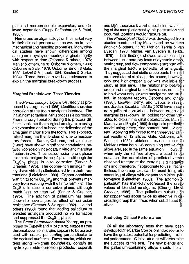
RESULTS Clinical
The rid it means for all alloys atthe various times are shown in Table 3. The alloy used for the reference group was Dispersalloy at polish, which mathematically must have a mean ridit of 0.5 (Fleiss & others, 1979). Dispersalloy was selected as the reference alloy because it is the oldest high-copper alloy and has been used routinely in many clinical studies, including most of those cited in the references. A graph of ridit means versus time is shown in Figure 1. The evaluation sessions were held after 16 weeks, one year, and three years. The first session combined two-, eight-, and 16-week photos and the last session combined the two- and threeyear photos. The statistical tests showed differences among evaluators in each of the evaluation sessions, thus evaluator subgroups were used. All subgroups consisted of at least four of the six evaluators. The ridit means in Table 3 have been calculated based on observations of evaluator subgroups in statistical agreement.
The critical value for significant differences in ridit means, R, was calculated as R > 0.15 for all comparisons where R is the standard error of ridit means times the tabled Z value for the
~ DISPERSALLOY ~ TYTIN o VALIANT • SYBRALOY
60 80 100 120 140 160
TIME (WEEKS)
FIG 1. Ridit means as a function of time
Bonferroni criterion. Pairwise comparison tests using the Bonferroni correction factor showed lack of alloy discrimination (i.e., R < 0.15) at any point in time over the three-year period, except for Valiant and Sybraloy at two and eight weeks. Early observations showed significant differences in marginal integrity between the tirne of polish and 16 weeks later for all alloys. For Dispersalloy and Tytin there were no significant changes betw~en 16 weeks and one year, whereas for Valiant and Sybraloy there were changes. For all alloy systems no significant changes in marginal integrity existed between one year and either two or three years. A leveling off of ridit means versus time is evident in Figure 1. All Bonferroni comparisons were made atP < 0.05.
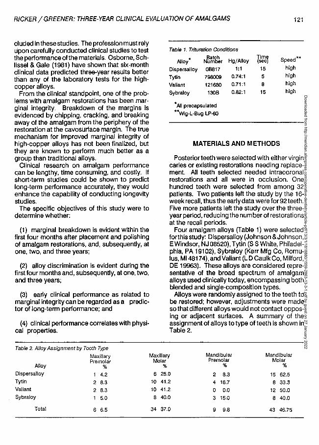
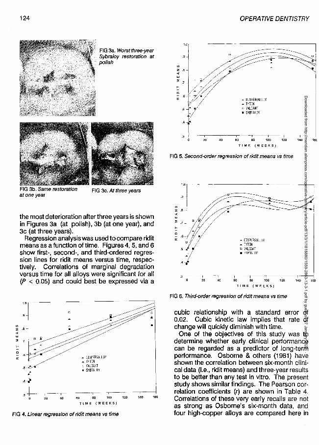
Representative photographs of one of the best Sybraloy restorations at three years is shown in Figures 2a (at polish), 2b (at one year), and 2c (at three years). Representative photographs of one of the Sybraloy restorations that showed
a b c
FIG 2a. Bast three-year Sybraloy restoration at polish 2b. Same restoration at one year 2c. Same restoration at three years
Dow
nloaded from http://m
eridian.allenpress.com/operative-dentistry/article-pdf/13/3/1/1818880/1559-2863-13-3-1.pdf by guest on 27 January 2022
124 OPERATIVE DENTISTRY
FIG 3a. Worstthree-year Sybraloy restoration at .9
<J>
FIG 3b. Same restoration atone year
polish
FIG 3c. At three years
the most deterioration after three years is shown in Figures 3a (at polish), 3b (at one year), and 3c (at three years).
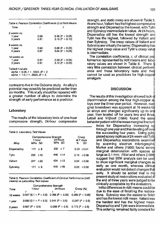
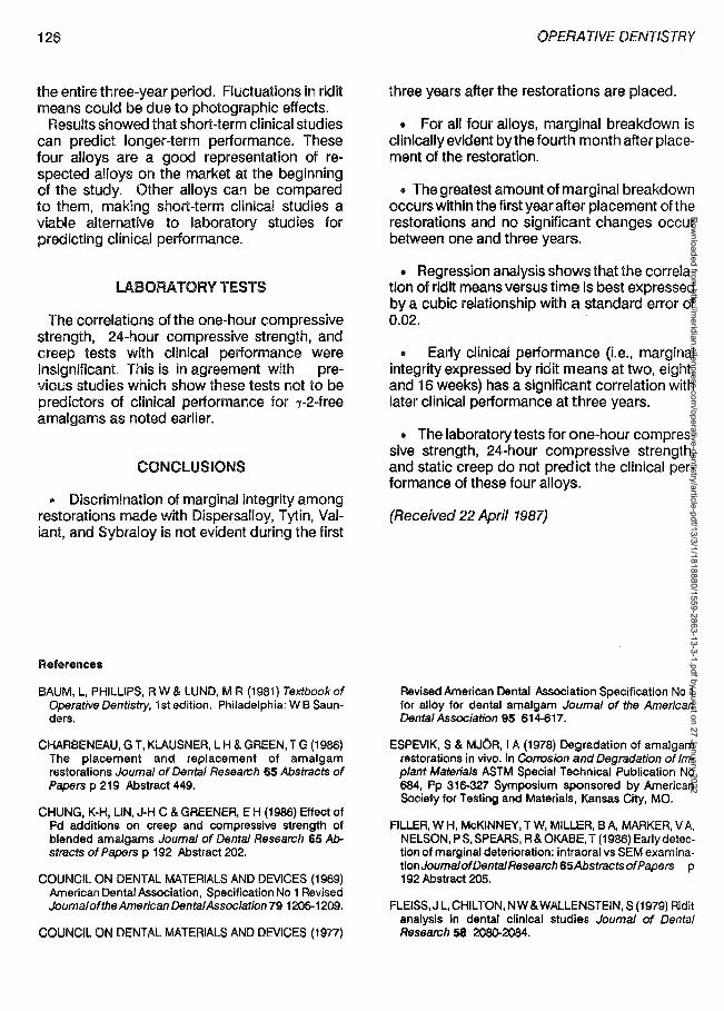
Regression analysis was used to compare ridit means as a function of time. Figures 4, 5, and 6 show first-, second-, and third-ordered regression lines for ridit means versus time, respectively. Correlations of marginal degradation versus time for all alloys were significant for all (P < 0.05) and could best be expressed via a
.9 -
z .8 <(
UJ
:; .7
.... 0 - .6 a:
.5
.4 •
. 3 20 40
A O!SPERSALLDY • TYTIN o VALIANT • SYBRALDY
60 BO 100
TIME (WEEKS)
FIG 4. Linear regression of ridit means vs time
120 140 160
<J>
z .8 <(
UJ
:; .7
.... 0
a:
.4 •
20 40
A 0 ISPERSALLDY • TYTIN o VALIANT • SYBRALOY
60 80 100 120 140 160
TIME (WEEKS)
FIG 5. Second-order regression of ridit means vs time
.9
<J>
z .8 <(
UJ
:; .7
.... 0 - .6 a:
.5
.4
.3 20 40
A DISPERSALLOY • TYTIN D VALIANT • SYBRALDY
60 80 100 120 140 160
TIME (WEEKS)
FIG 6. Third-order regression of ridit means vs time
cubic relationship with a standard error of 0.02. Cubic kinetic law implies that rate of change will quickly diminish with time.
One of the objectives of this study was to determine whether early clinical performance can be regarded as a predictor of long-term performance. Osborne & others (1981) have shown the correlation between six-month clini" cal data (i.e., ridit means) and three-year results to be better than any test in vitro. The present study shows similar findings. The Pearson correlation coefficients (r) are shown in Table 4 . Correlations of these very early recalls are not as strong as Osborne's six-month data, and four high-copper alloys are compared here in
Dow
nloaded from http://m
eridian.allenpress.com/operative-dentistry/article-pdf/13/3/1/1818880/1559-2863-13-3-1.pdf by guest on 27 January 2022
RICKER/ GREENER: THREE-YEAR CLINICAL EVALUATION OF AMALGAMS 125
Table 4. Pearson Correlation Coefficients (r) of Ridit Means
Time r t
2 weeks vs: 1 year 0.98 3 years 0.91
8 weeks vs: 1 year 0.94 3 years 0.97
16weeks vs: 1 year 0.87 3 years 0.91
Tabled tvalues: alpha = 0.05, t = .4303, df = 2 alpha = 1.0, t = .2920, df = 2
6.96 (P < 0.05) 3.10 (P < 0.10)
6.68 (P < 0.05) 5.64 (P < 0.05)
2.50 (P < 0.05) 3.10 (P < 0.05)
contrast to five in the Osborne study. An alloy's potential may possibly be predicted earlier than six months. This study should be repeated with a greater number of alloys to determine the strength of early performance as a predictor.
Laboratory
The results of the laboratory tests of one-hour compressive strength, 24-hour compressive
Table 5. Laboratory Test Values
Comprehensive Strength 1 hour 24 hours
Alloy MPa SD MPa SD
Creep at 7 days % SD
strength, and static creep are shown in Table 5. At one hour, Valiant has the highest compressive strength and Dispersalloy the lowest, with Tytin and Sybraloy intermediate in value. At 24 hours, Dispersalloy still has the lowest strength and Tytin has the highest, followed by Valiant and then Sybraloy. The creep values of Valiant and Sybraloy are virtually the same; Dispersalloy has the highest creep value and Tytin's creep value is intermediate.
The correlation coefficients, r, of clinical performance represented by ridit means and laboratory values are shown in Table 6. There is very little correlation between clinical performance and these laboratory tests and they cannot be used as predictors for high-copper amalgam.
DISCUSSION
The results of this investigation showed lack of discrimination among the four high-copper alloys over the three-year period. However, marginal breakdown was apparent at 16 weeks for all alloys and changed significantly up to one year, then leveled off for years two and three. Letze! and Vrijhoef {1984) found the same behavior pattern with the mean marginal fracture
index for Dispersalloy increasing up through one year and then leveling off over the succeeding four years. Using goldplated epoxy replicas of 24-week-old Tytin and Dispersalloy restorations examined by scanning electron micrographs,
Dispersalloy 117 ±8 330 ±7 0.22 ±0.01 Marker and others {1986) found severe marginal deterioration with spaces as large as 0.1 mm. Filler and others (1986) suggest that SEM analysis can be used to show significant marginal changes as early as one month, whereas clinical evaluation would not detect changes that
Tytin 200 ±10 446 ±14 0.19 ±0.09
Valiant 347 ±20 434 ±13 0.04 ±0.02
Sybraloy 171 ±5 416 ±48 0.05 ±0.02
Table 6. Pearson Correlation Coefficient of Clinical Performance (ridit means) vs Laboratory Test Values
Comprehensive Strength Time 1 hour 24 hours Creep(%)
r r r 16 weeks 0.547 (0.1 < P < 0.5) 0.266 (P > 0.05) 0.266 (P > 0.05)
1 year 0.890 (0.1 < P < 0.5) 0.541 (P > 0.5) -0.247 (P > 0.5)
3 years 0.567 (P > 0.5) -0.008 (P > 0.5) 0.173 (P > 0.5)
early. It should be added that in the present study all restorations evaluated at the end of three years were judged to be clinically acceptable by the evaluators.
Initial differences in ridit means could be due to the ease of finishing the restorations. Sybraloy was the easiest to finish and has the lowest rid it mean. Valiant was the hardest and has the highest mean. Dispersalloy and Tytin were intermediate. This ordering remained fairly constant for
Dow
nloaded from http://m
eridian.allenpress.com/operative-dentistry/article-pdf/13/3/1/1818880/1559-2863-13-3-1.pdf by guest on 27 January 2022
126
the entire three-year period. Fluctuations in ridit means could be due to photographic effects.
Results showed that short-term clinical studies can predict longer-term performance. These four alloys are a good representation of respected alloys on the market at the beginning of the study. Other alloys can be compared to them, making short-term clinical studies a viable alternative to laboratory studies for predicting clinical performance.
LABORATORY TESTS
The correlations of the one-hour compressive strength, 24-hour compressive strength, and creep tests with clinical performance were insignificant. This is in agreement with previous studies which show these tests not to be predictors of clinical performance for 1-2-free amalgams as noted earlier.
CONCLUSIONS
• Discrimination of marginal integrity among restorations made with Dispersalloy, Tytin, Valiant, and Sybraloy is not evident during the first
References
BAUM, L, PHILLIPS, R W & LUND, MR (1981) Textbook of Operative Dentistry, 1st edition. Philadelphia: W B Saunders.
CHARBENEAU, G T, KLAUSNER, L H & GREEN, T G (1986) The placement and replacement of amalgam restorations Journal of Dental Research 65 Abstracts of Papers p 219 Abstract 449.
CHUNG, K-H, LIN, J-H C & GREENER, E H (1986) Effect of Pd additions on creep and compressive strength of blended amalgams Journal of Dental Research 65 Al:r stracts of Papers p 192 Abstract 202.
COUNCIL ON DENTAL MATERIALS AND DEVICES (1969) American Dental Association, Specification No 1 Revised Journal of the American Dental Association 79 1206-1209.
COUNCIL ON DENTAL MATERIALS AND DEVICES (19n)
OPERATIVE DENTISTRY
three years after the restorations are placed.
• For all four alloys, marginal breakdown is clinically evident by the fourth month after placement of the restoration.
• The greatest amount of marginal breakdown occurs within the first year after placement of the restorations and no significant changes occur between one and three years.
• Regression analysis shows that the correlation of ridit means versus time is best expressed by a cubic relationship with a standard error of 0.02.
• Early clinical performance (i.e., marginal integrity expressed by ridit means at two, eight, and 16 weeks) has a significant correlation with later clinical performance at three years.
• The laboratory tests for one-hour compressive strength, 24-hour compressive strength, and static creep do not predict the clinical performance of these four alloys.
(Received 22 April 1987)
Revised American Dental Association Specification No 1 for alloy for dental amalgam Journal of the American Dental Association 95 614-617.
ESPEVIK, S & MJOR, I A (1978) Degradation of amalgam restorations in vivo. In Corrosion and Degradation of Implant Materials ASTM Special Technical Publication No 684, Pp 316-327 Symposium sponsored by American Society for Testing and Materials, Kansas City, MO.
FILLER, W H, McKINNEY, T W, MILLER, BA, MARKER, VA, NELSON, PS, SPEARS, R& OKABE, T (1986) Early detection of marginal deterioration: intraoral vs SEM examination.JournalofDenta/Research 65Abstractsof Papers p 192 Abstract 205.
FLEISS, J L, CHILTON, NW & WALLENSTEIN, S (1979) Ridit analysis in dental clinical studies Journal of Dental Research 58 2080-2084.
Dow
nloaded from http://m
eridian.allenpress.com/operative-dentistry/article-pdf/13/3/1/1818880/1559-2863-13-3-1.pdf by guest on 27 January 2022

RICKER/ GREENER: THREE-YEAR CLINICAL EVALUATION OF AMALGAMS 127
GALE, E N, OSBORNE, J W & WINCHELL, PG {1982) Fracture at the margins of amalgam as predicted by creep, zinc content and gamma-2 content Journal of Dental Research 61 678-680.
GREENER, E H {1979) Amalgam-yesterday, today, and tomorrow Operative Dentistry 4 24-35.
GREENER, E H & SZURGOT, K {1982) Properties of Ag-CuPd dispersed phase amalgam: compressive strength, creep, and corrosion Journal of Dental Research 61 1192-1194.
JORDAN, R E, SUZUKI, M & MILLS, AR {1978) Marginal integrity of amalgam alloys in relation to creep: a preliminary report Journal of Prosthetic Dentistry 40 299-303.
JORGENSEN, K D {1965) The mechanism of marginal fracture of amalgam fillings Acta Odontologica Scandinavica 23 347-389.
LARSON, T H, SABOTT, D, COOLEY, R & GREENER, E H {1979) A clinical study of marginal integrity and tarnish behaviour of three Cu-rich amalgam systems Journal of Oral Rehabilitation 6 61-66.
LASWELL, HR, BERRY, T G & OSBORNE, J W (1980) Twoyear clinical evaluation of low creep dental amalgams New York State Dental Journal 46 406-408.
LEINFELDER, K F {1980) Clinical performance of amalgams with high content of copper Operative Dentistry 5 125-130.
LEINFELDER, K F {1983) Clinical evaluation of high-copper amalgam General Dentistry 31 105-109.
LETZEL, H & VRIJHOEF, MM A {1984) Long-term influences on marginal fracture of amalgam restorations Journal of Oral Rehabilitation 11 95-101.
LIN, J-H C, MARSHALL, SJ, CHUNG, K-H & GREENER, EH {1986) Structural changes in blending amalgam compositions with Pd additions Journal of Dental Research 65 Abstracts of Papers p 192 Abstract 201.
MAHLER, DB & MARANTZ, RL (1979) The effect of time on the marginal fracture behaviour of amalgam Journal of Oral Rehabilitation 6 391-398.
MAHLER, D B, MARANTZ, R L & ENGLE, J H (1980) A predictive model for the clinical marginal fracture of amalgam Journal of Dental Research 59 1420-1427.
MAHLER, D B, TERKLA, L G & VAN EYSDEN, J {1973) Marginal fracture of amalgam restorations Journal of
Dental Research 52 823-827.
MAHLER, DB, TERKLA, LG, VAN EYSDEN, J & REIS BICK, M H {1970) Marginal fracture vs mechanical properties of amalgam Journal of Dental Research 49 1452-1457.
MAHLER, D B, VAN EYSDEN, J & TERKLA, LG (1975) Relationship of creep to marginal fracture of amalgam Journal of Dental Research 54 Program and Abstracts of Papers p 183 Abstract 553.
MARKER, VA, FILLER, W H, McKINNEY, T W, MILLER, BA, MITCHELL, R J & OKABE, T {1986) Early progression of marginal breakdown in high copper amalgam restorations Journal of Dental Research 65Abstracts of Papers p 729 Abstract 31.
OSBORNE, J W, SINON, PP & GALE, EN (1980) Dental amalgam: clinical behavior up to eight years Operative Dentistry 5 24-28.
OSBORNE, J W & GALE, EN (1979) Failure rate of margins of amalgams with a high content of copper Operative Dentistry 4 2-8.
OSBORNE, J W, LEINFELDER, K F, GALE, E N & SLUDER, TB (1980) Two independent evaluations of ten amalgam alloys Journal of Prosthetic Dentistry 43 622-626.
OSBORNE, J W, PHILLIPS, R W, GALE, E N & SINON, P P {1976) Three-year clinical comparison of three amalgam alloy types emphasizing an appraisal of the evaluation methods used Journal of the American Dental Association 93 784-789.
OSBORNE, J W, SCHLISSEL, ER & GALE, E N {1981) Clinical test for the development of new amalgam alloys Journal of Dental Research 60 999.
RUPP, NW, PAFFENBARGER, G C & PATEL, P R {1980) Effect of residual mercury content on creep in dental amalgams Journal of the American Dental Association 100 52-55.
SARKAR, N K & GREENER, EH {1975) Electrochemistry of the saline corrosion of conventional dental amalgams Journal of Oral Rehabilitation 2 49-62.
SARKAR, N K, OSBORNE, J W & LEIN FELDER, K F (1982) In vitro corrosion and in vivo marginal fracture of dental amalgams Journal of Dental Research 61 1262-1268.
SMALES, R J & GERKE, DC (1984) Clinical evaluation of four high-copper amalgam alloys Journal of Dentistry 12 127-134.
Dow
nloaded from http://m
eridian.allenpress.com/operative-dentistry/article-pdf/13/3/1/1818880/1559-2863-13-3-1.pdf by guest on 27 January 2022
128 ©OPERATIVE DENTISTRY, 1988, 13, 128-137
A Three-dimensional Finite Element Model
S C KHERA • V K GOEL R C S CHEN • S A GURUSAMI
Summary The finite element technique was used to
investigate stress distribution in a normal mandibular premolar tooth with and without cavity preparations. A conservative class 2 MOD cavity with an isthmus width of 1.06 mm and pulpal depth of 3.48 mm was compared to another class 2 MOD cavity with an isthmus width of 2.08 mm and pulpal depth of 4.85 mm. Both designs were also compared to the normal tooth with respect to the stress pattern in the tooth structure. The stress value in the
Department of Operative Dentistry, College of Dentistry, *Department of Biomedical Engineering, College of Engineering, University of Iowa, Iowa City, IA 52242
Satish C Khera, BOS, DDS, MS, professor
*Vijay K Goel, PhD, associate professor
Robert CS Chen, DDS, MS, graduate student, Department of Oral Biology, School of Dentistry, University of Michigan, Ann Arbor, Ml 48109
Senthil A Gurusami, BE, MS, senior engineer, Creative Industries Group, Auburn Hills, Ml 48057
enamel of the wider and deeper preparation was much greaterthan in the smaller preparation. More importantly, the stresses on the cavity floor of the shallower and narrower cavity were compressive, whereas tensile stresses were found on the pulpal floor of the larger and deeper preparation. This suggests there is a greater potential of cusp fracture in teeth with wider and deeper cavity preparations than in teeth with narrower and shallower cavity preparations. According to the pattern observed in this study, the tooth with conservative cavity preparation is relatively less susceptible to fracture than the tooth with wider and deeper cavity preparations.
Introduction
With age, human teeth are weakened by caries, abrasion, malocclusion, and fracture (Hiatt, 1973; Talim & Gohil, 1974; Cameron, 1976; Braly & Maxwell, 1981). Cavity preparation procedures and endodontic treatments, due to the reduction of tooth structure and loss of nutrient supply from dental pulp, exaggerate the fracture potential of the remaining tooth structure (Fayle, 1965, 1973; Baraban, 1967; Shillingburg & Fisher, 1970; Weine, 1982; Vale, 1956; Silvestri, 1976; Mondelli & others, 1980; Larson, Douglas & Geistfeld, 1981; Blaser & others, 1983; Landy & Simonsen, 1984; Gher & others, 1987). Inappropriate treatments, such as unnecessarily wide cavity preparations, increase the potential
Dow
nloaded from http://m
eridian.allenpress.com/operative-dentistry/article-pdf/13/3/1/1818880/1559-2863-13-3-1.pdf by guest on 27 January 2022
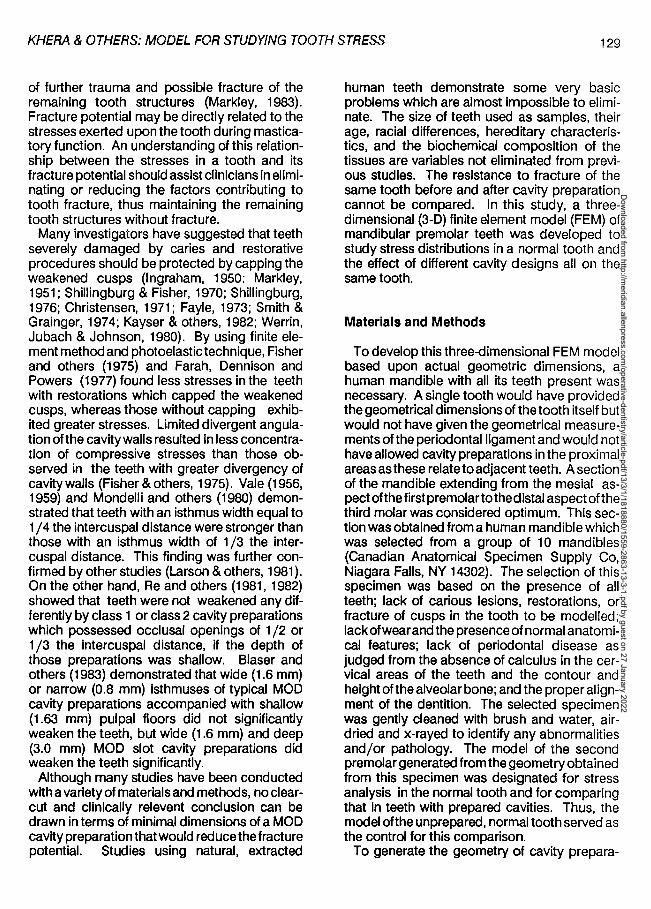
KHERA & OTHERS: MODEL FOR STUDYING TOOTH STRESS 129
of further trauma and possible fracture of the remaining tooth structures (Markley, 1983). Fracture potential may be directly related to the stresses exerted upon the tooth during masticatory function. An understanding of this relationship between the stresses in a tooth and its fracture potential should assist clinicians in eliminating or reducing the factors contributing to tooth fracture, thus maintaining the remaining tooth structures without fracture.
Many investigators have suggested that teeth severely damaged by caries and restorative procedures should be protected by capping the weakened cusps (Ingraham, 1950: Markley, 1951; Shillingburg & Fisher, 1970; Shillingburg, 1976; Christensen, 1971; Fayle, 1973; Smith & Grainger, 1974; Kayser & others, 1982; Werrin, Jubach & Johnson, 1980). By using finite element method and photoelastictechnique, Fisher and others (1975) and Farah, Dennison and Powers (1977) found less stresses in the teeth with restorations which capped the weakened cusps, whereas those without capping exhibited greater stresses. Limited divergent angulation of the cavity walls resulted in less concentration of compressive stresses than those observed in the teeth with greater divergency of cavity walls (Fisher & others, 1975). Vale (1956, 1959) and Mondelli and others (1980) demonstrated that teeth with an isthmus width equal to 1 /4 the intercuspal distance were stronger than those with an isthmus width of 1 /3 the intercuspal distance. This finding was further confirmed by other studies (Larson & others, 1981). On the other hand, Re and others (1981, 1982) showed that teeth were not weakened any differently by class 1 or class 2 cavity preparations which possessed occlusal openings of 1 /2 or 1 /3 the intercuspal distance, if the depth of those preparations was shallow. Blaser and others (1983) demonstrated that wide (1.6 mm) or narrow (0.8 mm) isthmuses of typical MOD cavity preparations accompanied with shallow (1.63 mm) pulpal floors did not significantly weaken the teeth, but wide (1.6 mm) and deep (3.0 mm) MOD slot cavity preparations did weaken the teeth significantly.
Although many studies have been conducted with a variety of materials and methods, no clearcut and clinically relevant conclusion can be drawn in terms of minimal dimensions of a MOD cavity preparation that would reduce the fracture potential. Studies using natural, extracted
human teeth demonstrate some very basic problems which are almost impossible to eliminate. The size of teeth used as samples, their age, racial differences, hereditary characteristics, and the biochemical composition of the tissues are variables not eliminated from previous studies. The resistance to fracture of the same tooth before and after cavity preparation cannot be compared. In this study, a threedimensional (3-D) finite element model (FEM) of mandibular premolar teeth was developed to study stress distributions in a normal tooth and the effect of different cavity designs all on the same tooth.
Materials and Methods
To develop this three-dimensional FEM model based upon actual geometric dimensions, a human mandible with all its teeth present was necessary. A single tooth would have provided the geometrical dimensions of the tooth itself but would not have given the geometrical measurements of the periodontal ligament and would not have allowed cavity preparations in the proximal areas as these relate to adjacent teeth. A section of the mandible extending from the mesial aspect of the first premolar to the distal aspect of the third molar was considered optimum. This section was obtained from a human mandible which was selected from a group of 1 o mandibles (Canadian Anatomical Specimen Supply Co, Niagara Falls, NY 14302). The selection of this specimen was based on the presence of all teeth; lack of carious lesions, restorations, or fracture of cusps in the tooth to be modelled; lack of wear and the presence of normal anatomical features; lack of periodontal disease as judged from the absence of calculus in the cervical areas of the teeth and the contour and height of the alveolar bone; and the proper alignment of the dentition. The selected specimen was gently cleaned with brush and water, airdried and x-rayed to identify any abnormalities and/or pathology. The model of the second premolar generated from the geometry obtained from this specimen was designated for stress analysis in the normal tooth and for comparing that in teeth with prepared cavities. Thus, the model of the unprepared, normal tooth served as the control for this comparison.
To generate the geometry of cavity prepara-
Dow
nloaded from http://m
eridian.allenpress.com/operative-dentistry/article-pdf/13/3/1/1818880/1559-2863-13-3-1.pdf by guest on 27 January 2022
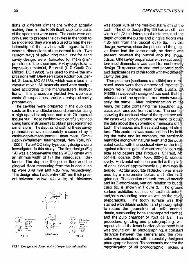
130
tions of different dimensions without actually making them in the tooth itself, duplicate casts of the specimen were used. The casts were not only used to prepare the cavities in the tooth to be modelled, they were also used to map the relationship of the cavities with regard to the external dimensions of the normal tooth. Two custom trays of self-curing resin, one for each cavity design, were fabricated for making impressions of the specimen. A vinyl polysiloxane impression material, Reprosil (L D Caulk Co, Milford, DE 19963), was used to make the impressions with Die-Keen stone (Columbus Dental, St Louis, MO 63188), which was mixed in a vacuum mixer. All materials used were manipulated according to the manufacturers' instructions. This procedure yielded two duplicate casts of the specimen, one for each type of cavity preparation.
The cavities were prepared in the duplicate casts on the mandibular second premolar using a high-speed handpiece and a #170 tapered fissure bur. These cavities were carefully refined using hand instruments to obtain predetermined dimensions. The depth and width of these cavity preparations were accurately measured by a cavity-depth-measurement instrument, Omnidepth (Whaledent International, New York, NY 10001 ). Two MOD inlay-type cavity designs were investigated in this study. The first design (Fig 1 A) was a conservative class 2 MOD cavity with an isthmus width of 1 /4 the intercuspal distance. The depth of the pulpal floor and the gingival floor measuring from the buccal cusp tip were 3.48 mm and 4.85 mm, respectively. This design also had dentin 4.87 mm thick present between the two axial walls; this thickness
P0=3.4Bmm GD=4.85 CW=4 IW=1.06
MD=7.2 IT""4.87
Fig. la
B
'w
GD
'
W#J "
normal tooth
PD=4.85mm GD=A.85 CW=4
Fig. lb
FIG 1. Design and dimensions of experimental cavities
OPERATIVE DENTISTRY
was about 70% of the mesio-distal width of the tooth. The other design (Fig 1 B) had an isthmus width of 1 /2 the intercuspal distance, and the depth of both the pulpal and gingival floors was 4.85 mm from the buccal cusp tip. In this design, however, since the pulpal and the gingival floors had the same depth, no dentin was present between the buccal and the lingual cusps. One cavity preparation with exact predetermined dimensions was used for each cavity design. This procedure produced a normal tooth and duplicate casts of this tooth with two different cavity designs.
The specimen (sectioned mandible) and duplicated casts were then embedded together in epoxy resin (Chemco Resin Craft, Dublin, CA 94568) in a specially designed box such that the orientation of the specimen and casts was exactly the same. After polymerization of the resin, the cube containing the specimen and casts was removed from the box. The surface showing the occlusal view of the specimen and the casts was serially ground by hand to obtain the data about the geometric dimensions of the tooth, the tissues, and the related support structure. This treatment was accomplished by holding the cube and its contents, the sectioned mandible along with natural teeth and the duplicated casts, with the occlusal view of the teeth against different grits of waterproof silicon carbide sand papers (3M Products, St Paul, MN 55144): coarse, 240-, 400-, 600-grit, successively. Horizontal reduction parallel to the plane of occlusion of approximately 0.5 mm was intended. Actual accurate reduction was measured by a micrometer before and after each grinding. The location of each ground section and its Z-coordinate, vertical relation to buccal cusp tip, is shown in Figure 2. The ground surface exhibited outlines of tooth structures and/or surrounding bone as well as the cavity preparations. The tooth surface was then stained with thionin solution and photographed to recor-d the geometries of tooth, enamel, dentin, surrounding bone, the prepared cavities, and the pulp chamber or root canals. This procedure, grinding and photographing, was repeated until the lower border of the mandibule was ground off. In photographing, a constant distance between the camera and the resin cube was maintained with a specially designed photographic bench. To constantly monitor the magnification of all photographic slides, a
Dow
nloaded from http://m
eridian.allenpress.com/operative-dentistry/article-pdf/13/3/1/1818880/1559-2863-13-3-1.pdf by guest on 27 January 2022
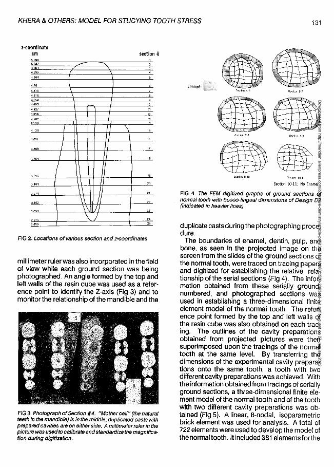
KHERA & OTHERS: MODEL FOR STUDYING TOOTH STRESS 131
z-coordinate cm
5.098 5.047 4.991 4.935
4.884
4.75
4.879 4.818
4.554 4.493
4.427
4.359
4.288 4.239
4.138
4.021
3.888
3.754
3.515
3.404
3.274
3.152
3.033
2.915 2.855
I I I
/
\ \ I
I\ \\ \\ '
\\ J \. J
section#
' 2
' 3 \ 4
5
6
7 8
9 10
I 11
I 12
I 13 14
\ 15
I 16
17
18
19
20
21
22
23
I 24 25
FIG 2. Locations of various section and z-coordinates
millimeter ruler was also incorporated in the field of view while each ground section was being photographed. An angle formed by the top and left walls of the resin cube was used as a reference point to identify the Z-axis (Fig 3) and to monitor the relationship of the mandible and the
FIG 3. Photograph of Section #4. "Mother cell" (the natural teeth in the mandible) is in the middle; duplicated casts with prepared cavities are on either side. A millimeter ruler in the picture was used to calibrate and standardize the magnification during digitization.
Enamel= Section 4-6 Section 6-7
Section 7-8 Section 8-9
Section 9-10 Section 10-11
Section 10-11: No Enamel
FIG 4. The FEM digitized graphs of ground sections of normal tooth with bucco-lingual dimensions of Design 02 (indicated in heavier lines)
duplicate castsduringthe photographing procedure.
The boundaries of enamel, dentin, pulp, and bone, as seen in the projected image on the screen from the slides of the ground sections of the normal tooth, were traced on tracing papers and digitized for establishing the relative relationship of the serial sections (Fig 4). The information obtained from these serially ground, numbered, and photographed sections was used in establishing a three-dimensional finite element model of the normal tooth. The reference point formed by the top and left walls of the resin cube was also obtained on each tracing. The outlines of the cavity preparations obtained from projected pictures were then superimposed upon the tracings of the normal tooth at the same level. By transferring the dimensions of the experimental cavity preparations onto the same tooth, a tooth with two different cavity preparations was achieved. With the information obtained from tracings of serially ground sections, a three-dimensional finite element model of the normal tooth and of the tooth with two different cavity preparations was obtained (Fig 5). A linear, 8-nodal, isoparametric brick element was used for analysis. A total of 722 elements were used to develop the model of thenormaltooth. ltincluded381 elementsforthe
Dow
nloaded from http://m
eridian.allenpress.com/operative-dentistry/article-pdf/13/3/1/1818880/1559-2863-13-3-1.pdf by guest on 27 January 2022
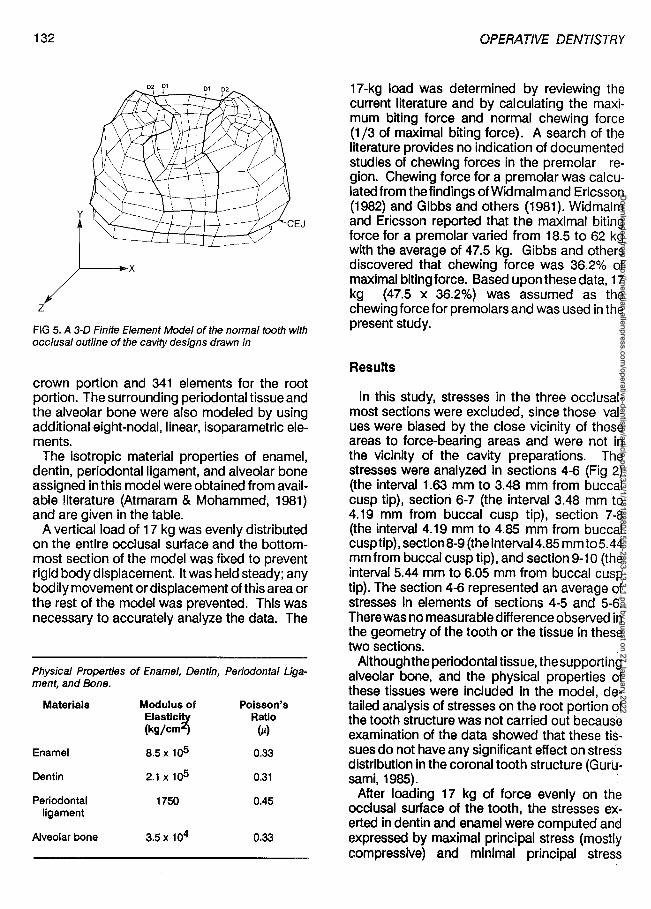
132
z FIG 5. A 3-D Finite Element Model of the normal tooth with occlusal outline of the cavity designs drawn in
crown portion and 341 elements for the root portion. The surrounding periodontal tissue and the alveolar bone were also modeled by using additional eight-nodal, linear, isoparametric elements.
The isotropic material properties of enamel, dentin, periodontal ligament, and alveolar bone assigned in this model were obtained from available literature (Atmaram & Mohammed, 1981) and are given in the table.
A vertical load of 17 kg was evenly distributed on the entire occlusal surface and the bottommost section of the model was fixed to prevent rigid body displacement. It was held steady; any bodily movement or displacement of this area or the rest of the model was prevented. This was necessary to accurately analyze the data. The
Physical Properlies of Enamel, Dentin, Periodontal Liga-ment, and Bone.
Materials Modulus of Poisson's Elastic~ Ratio (kg/cm (µ)
Enamel 8.5 x 1<>5 0.33
Dentin 2.1x1o5 0.31
Periodontal 1750 0.45 ligament
Alveolar bone 3.Sx 104 0.33
OPERATNE DENTISTRY
17-kg load was determined by reviewing the current literature and by calculating the maximum biting force and normal chewing force (1 /3 of maximal biting force). A search of the literature provides no indication of documented studies of chewing forces in the premolar region. Chewing force for a premolar was calculated from the findings of Widmalm and Ericsson (1982) and Gibbs and others (1981). Widmalm and Ericsson reported that the maximal biting force for a premolar varied from 18.5 to 62 kg with the average of 47.5 kg. Gibbs and others discovered that chewing force was 36.2% of maximal biting force. Based upon these data, 17 kg (47.5 x 36.2%) was assumed as the chewing force for premolars and was used in the present study.
Results
In this study, stresses in the three occlusalmost sections were excluded, since those values were biased by the close vicinity of these areas to force-bearing areas and were not in the vicinity of the cavity preparations. The stresses were analyzed in sections 4-6 (Fig 2) (the interval 1.63 mm to 3.48 mm from buccal cusp tip), section 6-7 (the interval 3.48 mm to 4.19 mm from buccal cusp tip), section 7-8 (the interval 4.19 mm to 4.85 mm from buccal cusp tip), section 8-9 (the interval 4.85 mm to 5.44 mm from buccal cusp tip), and section9-10 (the interval 5.44 mm to 6.05 mm from buccal cusp tip). The section 4-6 represented an average of stresses in elements of sections 4-5 and 5-6. There was no measurable difference observed in the geometry of the tooth or the tissue in these two sections.
Although the periodontal tissue, the supporting alveolar bone, and the physical properties of these tissues were included in the model, detailed analysis of stresses on the root portion of the tooth structure was not carried out because examination of the data showed that these tissues do not have any significant effect on stress distribution in the coronal tooth structure (Gurusami, 1985). ,
After loading 17 kg of force evenly on the occlusal surface of the tooth, the stresses exerted in dentin and enamel were computed and expressed by maximal principal stress (mostly compressive) and minimal principal stress
Dow
nloaded from http://m
eridian.allenpress.com/operative-dentistry/article-pdf/13/3/1/1818880/1559-2863-13-3-1.pdf by guest on 27 January 2022
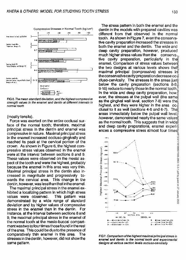
KHERA & OTHERS: MODEL FOR STUDYING TOOTH STRESS 133
Compressive Stresses in Normal Tooth (kg/cm2)
Area closest to load application. J----
Section Level#6-Pulpal wall Design #1.
Section I.Ivel #8-Pulpal/Gingival wall Design #2
4-6
}--6-7
7-6
}-' 8-9
-20
E=Enamel
D=Dentin
-40 -60 -80 -100
FIG 6. The mean standard deviation, and the highest compressive strength values in the enamel and dentin at different inteNals in normal tooth
(mostly tensile). Force was exerted on the entire occlusal sur
face of the normal tooth; therefore, maximal principal stress in the dentin and enamel was compressive in nature. Maximal principal stress in the enamel increased occluso-gingivally and reached its peak at the cervical portion of the crown. As shown in Figure 6, the highest compressive stress values observed in the enamel were at the interval between sections 8 and 9. These values were observed on the mesial aspect of the tooth and were the highest, probably because the enamel in this area was very thin. Maximal principal stress in the dentin also increased in magnitude and progressively towards the cervical area. This change in the dentin, however, was less than that inthe enamel.
The maximal principal stress in the enamel exhibited a localizing pattern in which high stress values were observed. This pattern was demonstrated by a wide range of standard deviation and by higher values of compressive stress in the enamel than in the dentin. For instance, at the interval between sections 8 and 9, the maximal principal stress in the enamel of the normal tooth at the mesio-buccal compartmentwastwo tofourtimes those found in the rest of the area. This could be due to the presence of comparatively thin enamel in this area. The stresses in the dentin, however, did not showthe same pattern.
The stress pattern in both the enamel and the dentin in the models with prepared cavities was different from that observed in the normal tooth. As shown in Figure 7, even the conservative cavity preparation increased the stresses in both the enamel and the dentin. The wide and deep cavity preparation, however, produced much higher stress values than the conservative cavity preparation, particularly in the enamel. Comparison of stress values between the two designs at various levels shows that maximal principal (compressive) stresses in the conservative cavity preparation decrease occluso-cervically. The stresses in the areas just below the cavity preparation (sections 8-9, 9-10) reduce to nearly those in the normal tooth. In the wide and deep cavity preparation, however, the stresses at the pulpal wall (the same as the gingival wall level, section 7-8) were the highest, and they were higher in the area occlusal to it as well (sections 4-6 and 6-7). The areas immediately below the pulpal wall level, however, demonstrated nearly the same values as the normal tooth. This suggests that in wide and deep cavity preparations, enamel experiences a compressive stress almost four times
-340
-320
- -300
~ -280
~ -260
"' ~ -240
(/') -220
Oi .e. -200 0
~ -180
ta -160
·~ -140 .. ~ -120
~ ~ -100
i -80
-60
-40
-20
4-6 6-7 7-8 8-9 9-10 SECTION LEVEL
- OCCLUSAL CERVICAL +-........... Dent!n Normal Tooth-ON G·--.-0DenlinDesign0ne-DD1 ~·-·@>Dentin Design Two-DD2
-------. Enamel Nonna I Tooth -EN G----o Enamel DesilJfl One-ED1 li-·---1!1 Enamel Design Two-ED2
FIG 7. Comparison of the highest maximal principal stress in enamel and dentin in the normal tooth and experimental designs at various section levels occ/uso-ceNically
Dow
nloaded from http://m
eridian.allenpress.com/operative-dentistry/article-pdf/13/3/1/1818880/1559-2863-13-3-1.pdf by guest on 27 January 2022

134
that seen in the normal tooth at the pulpal wall level. In conservative cavity preparations the stresses in the ptJlpal wall area are about two and a half times those observed in the normal tooth at the same level.
The dentin in these two designs also showed a similar pattern. Both cavity preparations produced much higher compressive stresses in the dentin than those observed in the normal tooth at the same level, although the increase in these values for the conservative cavity preparation was far less than that caused by the wide and deep cavity preparation. The dentin in the latter showed abnormally high compressive stress values in the area close to the cave-surface margin (the area of load application, section 4-6) and the area of the pulpal wall/gingival wall (section 7-8). These abnormally high compressive stresses in the area of the pulpal wall in both the enamel and the dentin of the wide and deep cavity preparations may damage these tissues in time and therefore damage the remaining tooth
BE= buccal enamel BO= buccal dentin
OPERATIVE DENTISTRY
structure. This damage may be in the form of incomplete, greenstick fracture-like cracks often observed on the pulpal wall. Enamel in this area, however, is more susceptible to damage than dentin and may show its effects in the form of fractured cusps.
Typical stress distribution patterns were analyzed in a bucco-lingual direction across the pulpal floor of the cavity preparation and were compared to the stress values in the normal tooth at the same level. It was observed that the increase of stress at the pulpo-lingual line angle of the wide and deep cavity preparation was much higher than that in the conservative cavity preparation. The most critical change, however, was found on the pulpal floor of the wide and deep cavity, where a combination of tensile and compressive maximal principal stresses was detected across the cavity floor bucco-lingually. In the wide and deep cavity design, it was observed that the stresses on the buccal aspect of the pulpal wall were tensile (Fig 8). The stresses
DBPL=dentin next to buccopulpat fine angle BY.zP= buccal 112 of pulpal wall
I ._ -120
~ BE
~ -100
i:i e Ci) -80
~ ., -60 c: ., I- 01 ~ -40
~ N1··········· ...!... ~ -20
-~
~ 0
8 +10
+20 ENAMEL .
BE BD
DENTIN
DBPL B\\P L\\P
LY.zP= IJngual ~ of pulpal wall OLPL =dentin next to linguopulpal line angle
LO= IJnguaJ dentin LE= llngual enamel
01
ENAMEL
DLPL LD LE
~
~ -120 ., 02
1l ~ -100 ., ~ 02
Cl) -80
~ -80 ., c: ~ "ti -40 c: ca ......
...!...
.~ -20
i:i e Q.
E 0
0 +10 (.)
BE
··· .. J .... I I i
+20 ENAMEL .
BE BD
L\\P DLPL
DENTIN
DBPL B\\P L\\P DLPL
I I I I
LD I I I i
. ENAMEL
LD LE
FIG 8. Comparative analysis of compressive stength and tensile strength t:>etween normal D 1 and 02 designs in a bucco-lingual direction at the level of the pu/pal wall
02
Dow
nloaded from http://m
eridian.allenpress.com/operative-dentistry/article-pdf/13/3/1/1818880/1559-2863-13-3-1.pdf by guest on 27 January 2022
KHERA & OTHERS: MODEL FOR STUDYING TOOTH STRESS 135
in the area immediately buccal or lingual to the pulpal wall were compressive. A normal tooth in the same area exhibits only compressive stresses, but after the wide and deep cavity preparation, the character of stresses in this area changed to tensile, which is abnormal for this area of the tooth. Another equally critical factor is that although the tensile stresses detected are of relatively low value when examined individually, the combination of heavy compressive stresses on either side of the site where tensile stresses are present changes the stress gradient abnormally. It is this pattern of stresses which with time, due to fatigue, may cause cuspal fractures more readily. This abrupt and dramatic change in stress gradient on the pulpal wall could be the major reason for fractures of cusps at about 45° to the pulpal wall of teeth restored with MOD restorations, as suggested by Bell, Smith and de Pont (1982). The stresses on the pulpal floor of the conservative design, on the other hand, were compressive and closer to the value found in the normal tooth. In this type of cavity preparation, because the quality of stresses is compressive only and the values are closer to those inthe normal tooth, cuspal deflection is not likely to occur as easily as in a wide cavity with a deep pulpal floor.
Discussion
By using the technique presented in a previous report (Goel & others, 1985), a 3-D model of a mandibular premolar was established and the characteristics of a stress pattern in the tooth under occlusal force were observed. Since the model was fixed at the root apex (section 23, Fig 2) and the area of interest in this study was far removed from there, the possible effect of boundary conditions, if any, was eliminated. Yettram, Wright and Pickard (1976) used a 2-D FEM model and found that the enamel near the cemento-enamel (C-E) junction was a highly stressed area. A similar pattern of stress distribution was observed in the present study as well. Moreover, the precise location of these areas was detected in the observation of substantially high stresses at the mesio-buccal segment of the enamel near the C-E junction; this may be due to extremely thin enamel present in this area. The highest stresses in the enamel atthe middle portion of the crown were detected
at the disto-lingual segment. This change in location of the stressed area may be due to the morphological character in this area, which is in the form of a thick and prominent disto-lingual line angle in the enamel. Yettram and others (1976) also reported that because the enamel demonstrated greater stiffness than the dentin, the enamel absorbed most of the occlusal force and so displayed higher stresses than those observed in the dentin. The results of this study consistently showed higher localized stresses in the enamel and lower and more evenly distributed stresses in the dentin.
The stresses at the bucco-pulpal and linguopulpal line angles increased, but were greater at the former than at the latter, as also reported by Fisher and others (1975) and Bell and others (1982). As the pulpal floor became deeper, the values of the stresses in both areas increased. In addition to the changes in stress values at the line angles, a striking change in stress character was observed on the cavity floor. In the conservative cavity preparations only compressive stresses were observed. In the wide and deep cavity preparations, however, the tensile stresses were observed across the cavity floor bucco-lingually. This phenomenon, accompanied by significantly high compressive stresses on both the buccal and lingual walls, could be expected to increase the fracture potential in wide and deep cavities more than in conservative cavities. This is due to the higher possibility of cuspal flexing in wide and deep cavities as it relates to the stress pattern detected. The detection of the stress character alteration was previously reported by Rubin and others (1983), who suggested that further clarification of this phenomenon may be obtained by analyzing stresses in a tooth using a three-dimensional finite element model.
According to this model, the wide and deep cavity showed a much greater potential for tooth fracture than did the conservative cavity. With a three-dimensional finite element model, it is possible to (a) highlight the areas of stress concentration in the tooth, (b) compare the effects of different cavity designs on tooth structures, and (c) detect tensile stress of the pulpal wall of a wide and deep cavity preparation and high compressive stress values in the areas immediately adjacent to that part of the pulpal wall. Now that a technique has been developed to obtain a realistic three-dimen-
Dow
nloaded from http://m
eridian.allenpress.com/operative-dentistry/article-pdf/13/3/1/1818880/1559-2863-13-3-1.pdf by guest on 27 January 2022
136
sional finite element model of a tooth and cavity preparations, further investigation is in progress to evaluate the influence of different isthmus widths and the thickness of residual interaxial dentin on teeth with various cavity designs. The effect of change in the direction of force on the stress magnitude and pattern, i e, the difference in the angulation and magnitude of the force, which would mimic masticatory movement in the mouth, is also being investigated.
Conclusion
To appreciate realistically the stress distribution characteristic in a tooth under certain loading conditions, it is critical to use a three-dimensional finite element model. In this investigation, the thin enamel in the mesio-buccal compartment adjacent to the cementa-enamel junction showed the highest compressive stress values. The tooth with a wide and deep cavity not only demonstrated high compressive stress on both buccal and lingual walls, but also demonstrated tensile instead of compressive stress in the middle of the cavity floor, thus producing a potentially damaging environment for the remaining tooth structure, which would lead ultimately to fracture.
(Received 22 May 1987)
References
ATMARAM, G H & MOHAMMED, H (1981) Estimation of physiological stresses with a natural tooth considering fibrous POL structure Journal of Dental Research 60 873-877.
BARABAN, D J (1967) The restoration of pulpless teeth Dental Clinics of North America 11 633-653.
BELL, JG, SMITH, MC &de PONT, J J (1982) Cuspal failures of MOD restored teeth Australian Dental Journal 27 283-287.
BLASER, P K, LUND, M R, COCHRAN, MA & POTTER, R H (1983) Effects of designs of class 2 preparations on resistance of teeth to fracture Operative Dentistry 8 6-10.
BRALY, B V & MAXWELL, EH (1981) Potential for tooth fracture in restorative dentistry Journal of Prosthetic
OPERATIVE DENTISTRY
Dentistry 45 411-414.
CAMERON, CE (1964) Cracked-tooth syndrome Journal of the American Dental Association 68 405-411.
CAMERON, CE (1976) The cracked tooth syndrome: additional findings Journal of the American Dental Association 93 971-975.
CHRISTENSEN, G J (1971) Clinical and research advancements in cast-gold restorations Journal of Prosthetic Dentistry 25 62-68.
CRAIG, R G & PEYTON, FA (1958) Elastic and mechanical properties of human dentin Journal of Dental Research 37 710-718.
CRAIG, R G, PEYTON, FA & JOHNSON, D W (1961) Compressive properties·of enamel, dental cements, and gold Journal of Dental Research 40 936-940.
FARAH,JW, DENNISON,J B&POWERS,J M (1977) Effects of design on stress distribution of intracoronal gold restorations Journal of the American Dental Association 94 1151-1154.
FAYLE, HE Jr (1965) Class II gold inlay design Texas Dental Journal 83 7-10.
FAYLE, H E Jr (1973) Gold inlay failures and some causes Journal of Prosthetic Dentistry 29 439-441.
FISHER, D W, CAPUTO, A A, SHILLINGBURG, H T Jr & DUNCANSON, M G Jr (1975) Photoelastic analysis of inlay and onlay preparations Journal of Prosthetic Dentistry 33 47-53.
GHER, ME Jr, DUNLAP, RM, ANDERSON, M H & KUHL, L V (1987) Clinical survey of fractured teeth Journal of the American Dental Association 114 17 4-177.
GIBBS, C H, MAHAN, P E, LUNDEEN, H C, BREHNAN, K, WALSH, EK & HOLBROOK, W B (1981) Occlusal forces during chewing and swallowing as measured by sound transmission Journal of Prosthetic Dentistry 46 443-449.
GOEL, V K, KHERA, S C, SENTHIL, G & CHEN, R (1985) Effect of cavity design on stresses in first molar Journal of Dental Research 64 Abstracts of Papers p 350 Abstract 1577.
GURUSAMI, SA (1985) Stresses in human teeth-an application of the finite element technique. Master's thesis, University of Iowa Graduate College.
HELKIMO, E & INGERVALL, B (1978) Bite force and functional state of the masticatory system in young men Swedish Dental Journal 2 167-175.
HIATT, WH (1973) Incomplete crown-rootfracture in pulpalperiodontal disease Journal of Periodontology 44 369-379.
HOWELL,AH&MANLY, RS (1948) Electronic strain gauge
Dow
nloaded from http://m
eridian.allenpress.com/operative-dentistry/article-pdf/13/3/1/1818880/1559-2863-13-3-1.pdf by guest on 27 January 2022
KHERA & OTHERS: MODEL FOR STUDYING TOOTH STRESS 137
for measuring oral forces Journal of Dental Research 27 705-712.
INGRAHAM, R (1950) The application of sound biomechanical principles in the design of inlay, amalgam and gold foil restorations Journal of the American Dental Association 40 402-413.
KAYSER, AF, BATTISTUZZI, PG F CM, SNOEK, PA & SPANAUF, A J (1982) The rationale for the indication and design of the MOD inlay Australian Dental Journal 27 22-26.
LANDY, NA & SIMONSEN, RJ (1984) Cusp fracture strength in class II composite resin restorations Journal of Dental Research 63 Abstracts of Papers p 175 Abstract 40.
LARSON, T D, DOUGLAS, W H & GEISTFELD, RE (1981) Effect of prepared cavities on the strength of teeth Operative Dentistry 6 2-5.
MARKLEY, M R (1951) Restorations of silver amalgam Journal of the American Dental Association 43 133-146.
MARKLEY, M R (1983) Personal communication.
MONDELLI, J, STEAGALL, L, ISHIKIRIAMA, A, de LIMA NAVARRO, MF & SOARES, F B (1980) Fracture strength of human teeth with cavity preparations Journal of Prosthetic Dentistry 43 419-422.
PROFFIT, WR, FIELDS, H W & NIXON, W L (1983) Occlusal forces in normal and long-face adults Journal of Dental Research 62 566-570.
RE, GJ &NORLING, BK (1981) Fracturing molars with axial forces Journal of Dental Research 60 805-808.
RE, G J, DRAHEIM, RN & NORLING, BK (1981) Fracture resistance of mandibular molars with occlusal class I amalgam preparations Journal of the American Dental Association 103 580-583.
RE, G J, NORLING, BK & DRAHEIM, RN (1982) Fracture strength of molars containing three surface amalgam restorations Journal of Prosthetic Dentistry 47 185-187.
REEL, DC & MITCHELL, R J (1984) Fracture resistance of teeth restored with class II composite restorations Journal
of Dental Research 63 Abstracts of Papers p 276 Abstract 950.
RUBIN, C, KRISHNAMURTHY, N, CAPILOUTO, E & YI, H (1983) Stress analysis of the human tooth using a threedimensional finite element model Journal of Dental Research 62 82-86.
SHILLINGBURG, HT Jr & FISHER, D W (1970) The MOD onlay- a rational approach to a restorative problem New Mexico Denta/Joumal 21 12-14, 32-35.
SHILLINGBURG, HT Jr (1976) Conservative preparations for cast restorations Dental Clinics of North America 20 259-271.
SILVESTRI, A R Jr (1976) The undiagnosed split-root syndrome Journal of the American Dental Association 92 930-935.
SMITH, GE &GRAINGER, DA (1974) Biomechanical design of extensive cavity preparations for cast gold Journal of the American Dental Association 89 1152-1157.
TALIM, ST & GOHIL, KS (1974) Management of coronal fractures of permanent posterior teeth Journal of Prosthetic Dentistry 31 172-178.
VALE, WA (1956) Cavity preparation Irish Dental Review 2 33-41.
VALE, WA (1959) Cavity preparation and further thoughts on high speed British Dental Journal 107 333-346.
WEINE, F S (1982) Endodontic Therapy, 3rd edition. St Louis: CV Mosby.
WERRIN, SR, JUBACH, TS &JOHNSON, B W (1980) Inlays and onlays: making the right decision Quintessence International 11 13-18.
WIDMALM, SE & ERICSSON, S G (1982) Maximal bite force with centric and eccentric load Journal of Oral Rehabilitation 9 445-450.
YETTRAM, AL, WRIGHT, KW J & PICKARD, HM (1976) Finite element stress analysis of the crowns of normal and restored teeth Journal of Dental Research 55 1004-1011.
Dow
nloaded from http://m
eridian.allenpress.com/operative-dentistry/article-pdf/13/3/1/1818880/1559-2863-13-3-1.pdf by guest on 27 January 2022
138 ©OPERATIVE DENTISTRY, 188, 13, 138-143
Seating of Cast Gold Inlays and Onlays with and without
Gingival Margin Bevels
MICHAEL P MOLVAR • MICHELLE GORES
Summary Gingival margin openings were determined in
vitro before and after cementation of inlays and onlays with and without gingival bevels. All inlays and onlays were more open at the gingival margin after cementation than prior to cementation. Beveled inlays and onlays had less internal margin opening along the bevel after cementation than did nonbeveled inlays and onlays.
Department of Adult Restorative Dentistry University of Nebraska Medical Center College of Dentistry, Lincoln, NE 68583-0740
Michael P Molvar, DDS, MS, assistant professor
Michelle Gores, DDS, Captain, U S Army Dental Corps, 87th Medical Detachment, Bamberg, Federal Republic of Germany, APO NY 09139
Introduction
Closure of margins between gold and tooth is considered to bea critical factor in the longevity of cast gold inlays and onlays (Allan, 1969; Silness & Hegdahl, 1970; Carpenter, 1981; Molvar & others, 1985). These restorations are typically luted with a dental cement, most commonly zinc phosphate cement. All dental cements, including zinc phosphate cement, are soluble in oral fluids (Craig, 1985). Cement exposed at the margins is considered to be the weakest link in cast gold restorations (S ii ness & Hegdahl, 1970; Charbeneau & others, 1981; Sturdivant & others, 1985). Dissolution of the cement at the margins can lead to open margins which predispose to development of secondary caries at the margins under the restoration (Jorgensen & Wakumoto, 1968). Thus, minimizing the marginal opening where cement is exposed to the oral environment is a major concern when placing an inlay or onlay (Silness & Hegdahl, 1970).
The configuration of margins of cast gold restorations to minimize the marginal opening has been the subject of widespread discussion.
Dow
nloaded from http://m
eridian.allenpress.com/operative-dentistry/article-pdf/13/3/1/1818880/1559-2863-13-3-1.pdf by guest on 27 January 2022

MOL VAR/ GORES: SEA TING OF INLAYS AND ONLA YS 139
The sliding-joint margin has been widely accepted because of greater geometric closure relative to unbeveled margins (Rosner, 1963; Silness & Hegdahl, 1970; Pardo, 1982; Ostlund, 1985). According to the sliding-joint theory, the closure perpendicular to beveled margins is better than perpendicular to nonbeveled margins; butfor similar internal vertical openings, the opening at the margin parallel to the tooth would be much alike for either beveled or unbeveled margins. However, dental cements have measurable minimum film thicknesses which must be taken Into account when castings are seated (about 25 µm for zinc phosphate cement - ADA Specification No 12). Recently, a theory has been proposed (Ostlund, 1985) that cement film thickness will not allow complete seating of castings with beveled (or sliding-joint) margins and that unbeveled or very slightly beveled margins will allow more complete seating when cement is in place. This theory is based on the geometry at the margin when cement with a film thickness of at least 25 µm is present. There is empirical evidence that cast gold inlays and onlays do not always seat as fully upon cementation as they did at the precementation try-in (Ostlund, 1985; personal observations). There is also evidence that full crowns that are fabricated without die spacers do not seat as fully upon cementation as prior to cementation (Charbeneau & Peyton, 1958; Silness & Hegdahl, 1970). However, there is little scientific evidence to help determine if margin configuration is a factor in this phenomenon.
This study was designed to test the hypothesis that unbeveled gingival margins would allow better closure of these margins on inlays and onlays upon cementation than would beveled margins. Specifically, It was designed to evaluate and compare the effects of o0 and 60° gingival bevels on gingival margin gaps of inlays and onlays, before and after cementation.
Materials and Methods
Twenty MOD inlays and 20 MOD onlays were prepared on mandibular, first molar, ivorine teeth (Columbia Dentaform, Long Island City, NY 11101 ). Ten of each offourtypes of preparations were made: inlays with o0 gingival bevels; inlays with 60° gingival bevels leaving a 30° angle of
gold at the margins; onlays with 0° bevels; and onlays with 60° bevels (Fig 1).
The 60° ginglval margins were placed with gingival margin trimmers (American Dental, Missoula, MT 59806) altered to provide a 60° angle when the blade was held parallel to the axial wall. All other aspects of the preparations were as identical as possible, with a standard two-plane reduction on buccal and lingual cusps of the onlay preparations (Sturdivant & others, 1985). Three layers of Tru-Fit die spacer (Geo Taub Co, Jersey City, NJ 07307) were placed on all areas of the preparations to within approximately 1 mm of the margins. The preparations were then waxed directly on the ivorine teeth and invested according to the manufacturer's instructions in Novocast (Whip Mix, Louisville, KY 40217). They were burned out according to the manufacturer's instructionsat high heatand cast in Modulay type II gold (Jelenko, Armonk, NY 10504).
The die spacer was completely removed from the preparations using the die spacer thinner. The castings were then fitted to the ivorine teeth. Thegingival margins were minimallyfinished (off of the tooth) with a large, soft rubber wheel #5009 (Dedico, Long Island, NY 12760) only as
FIG 1. Angle of gingiva/ margins.
Dow
nloaded from http://m
eridian.allenpress.com/operative-dentistry/article-pdf/13/3/1/1818880/1559-2863-13-3-1.pdf by guest on 27 January 2022
140
much as necessary to clearly read the margins under a microscope. No other attempt was made to close the margins or otherwise finish the restorations either before or after cementation.
Once the castings were fit as well as possible, a section of both mesial and distal gingival margins approximately 1 mm long (Fig 2) was marked on gold and tooth with an ultra-thin separating disk (Dedico). The areas and lengths of the openings of the marked sections were measured directly, using a Nikon Alphaphot YS microscope at X40 with side illumination (Nikon, Garden City, NY 11530). Three measurements were made for each area and length by tracings, using a Hipad digitizer and the Bioquant computer program (R and M Biometrics, Nashville, TN 37209) with an Apple lie computer (Apple Computers, Cupertino, CA95014). The means of these measurements were calculated and the mean marginal opening was calculated by the equation:
mean marginal opening
mean area of margin opening mean length of margin opening.
The gap perpendicular to the beveled margins was termed the "internal marginal gap," and was calculated using the equation (adapted from Ostlund, 1985):
FIG 2. Measurement sffes of gingiva/ margin openings
OPERATIVE DENTISTRY
Internal marginal gap = mean margin opening x cosine 60° (where 60° = the angle of the gingival bevel);= mean margin opening x 0.5 (cosine 60° = 0.5).
Since cosine o0 = 1, the margin gap of non beveled inlays would not be different from the measured margin gap. The gingival marginal opening and the internal marginal opening for each casting prior to cementation were taken as the means of the calculated mesial and distal marginal openings.
After precementation readings, the castings were cemented using zinc phosphate cement (Flecks Cement; Mizzy, Clifton Forge, VA 24422) according to the manufacturer's directions. In orderto minimize the possible effect of variability in cement mixes, one inlay (or onlay) with and one without gingival bevels were seated simultaneously with the same mix of cement. Castings were seated with firm finger pressure (15 lbs for 1 O seconds and 10 lbs for 20 seconds as measured on a standard scale). After cementation, the measurements and calculations for marginal openings were repeated.
Differences between pre- and postcementation mean marginal openings for inlays with and without bevels, and for onlays with and without bevels, were analyzed using the two-way analy-
Microscopic View at 40X
Dow
nloaded from http://m
eridian.allenpress.com/operative-dentistry/article-pdf/13/3/1/1818880/1559-2863-13-3-1.pdf by guest on 27 January 2022
MOL VAR/ GORES: SEA TING OF INLAYS AND ONLA YS 141
sis of variance test followed by the Scheffe critical difference test. Statistical significance was accepted at P values < 0.05.
Difference Pre- to Postcementatlon
Mean Difference Sig
!.w:!!l SD p < 0.001
Bevel 23.57 13.91 * Inlay
No Bevel 23.83 10.60 *
Bevel 23.65 13.00 * On lay
No Bevel 36.31 19.56 *
Results
Mean differences between pre- and postcementati.on margin openings for the four types of castings are summarized in the table. All of these differences were considered to be clinically important (all were > 23 µm and statistically significant; P < 0.001). Mean pre- and postcementation margin openings are graphically depicted in Figure 3. Margin openings as measured parallel to the external of the tooth were not significantly differentfor beveled versus non beveled inlays or onlays either before or aftercementation. Internal margin openings perpendicular to beveled and nonbeveled margins, calculated before and after cementation, are graphically depicted in Figure 4. The mean internal margin openings were considerably less for beveled inlays and onlays in all cases both before and after cementation. These differences were statistically significant for postcementation readings (P < 0.01); they were not significant for precementation readings though they approached significance for inlays (P < 0.1).
Discussion
Three layers of die spacer were used to attempt to eliminate the possibility that internal discrepancies would affect seating of the castings and
INLAYS ONLAYS 90
80 en z 70 0 cc u ~ 60
~ (!) 50 z z 40 w "-0 z 30 (3 cc <( 20 :;;
10
0 BEVEL NO BEVEL BEVEL NO BEVEL
~ PRECEMENTATION • POSTCEMENTATION
FIG 3. Internal gingival margin openings
INLAYS ONLAYS 90
80 en z 70 0 a: u ~ 60
~ (!) 50 z z 40 w "-0 z 30 a a: <( 20 :;;
10
0 PRE POST PRE POST
CEMENTATION
~BEVEL • NO BEVEL
FIG 4. Gingival margin openings of inlays and on lays with and without gingival bevels
also to attempt to eliminate cement film thickness as a factor except at the margins (Krug & Markley, 1969; Eames & others, 1978). Thus, differences demonstrated in this study should represent only factors related to the 1 mm of castings at the margins. Use of the digitizer and the computer program allowed rapid direct measurement of the marginal openings. The measurements were generally reproducible within approximately 1 O square µm for areas and
Dow
nloaded from http://m
eridian.allenpress.com/operative-dentistry/article-pdf/13/3/1/1818880/1559-2863-13-3-1.pdf by guest on 27 January 2022
142
within 5 µm for lengths. The calculated mean marginal openings from areas divided by lengths of marginal openings were considered to be more accurate indications of the overall marginal gaps than direct readings of gaps at single discrete positions along the margins. The mean of mesial and distal margin openings was used for statistical computations to attempt to compensate for the possibility that the castings might be slightly tipped to one side or the other upon seating; if one side were more open, the other side should be more closed in such a circumstance.
This study confirms clinical observations that inlays and onlays, when seated with firm finger pressure, do not seat as well upon cementation as they did prior to cementation (Ostlund, 1985). This was clearly established even with three layers of die spacer to within 1 mm of the margins. The differences between pre- and postcementation margin openings {23-36 µm) were close to the ADA specification for the film thickness for zinc phosphate cement. This raises a question about cement film thickness as a factor in seating of inlays and onlays even if intimate contact of tooth and casting occurs only close to the margins. Surely this question deserves further study, especially in relation to the force of seating of the casting.
Prior to cementation for both inlays and onlays, the measured marginal openings were not significantly different for beveled versus nonbevelecj~castings; the internal marginal openings carculated before cementation were smaller for both beveled inlays and onlays, but the differences were not significant. These findings were consistent with the sliding-joint theory of closure for precemented castings as discussed by Ostlund. After cementation for both inlays and onlays, the measured marginal openings were not significantly different for beveled versus nonbeveled castings; the internal margin openings calculated after cementation were significantly less for beveled versus nonbeveled castings. These findings were consistent with the sliding-joint theory of closure, but were contrary to the theory of better closure of nonbeveled margins as presented by Ostlund. Clearly, inlays and onlays did not seat as well after cementation as before cementation, but beveled inlays and onlays, when cemented, seated with smaller internal marginal openings than did nonbeveled Inlays and onlays.
OPERATIVE DENTISTRY
Conclusions
Within the context of this study in vitro:
1. The gingival margin openings of inlays or onlays with or without gingival bevels were greater aftercementation than prior to cementation (P < 0.001).
2. Differences between before and after cementation of external gingival margin openings were not significantly different for beveled versus nonbeveled inlays or onlays.
3. Calculated internal margin openings were significantly less for beveled versus nonbeveled inlays and onlays after cementation (P < 0.01 ).
(Received 22 April 1987)
Acknowledgements
This study was supported by University of Nebraska College of Dentistry Research Grant 86-23. The authors thank Mrs Betty Schellpeper for her assistance in preparation of the manuscript and Mr Kim Theesen for preparation of illustrations.
References
ALLAN, D N (1969) The durability of conservative restorations British Dental Joumal 126 172-177.
CARPENTER, KE (1981) Qualitative assessment of restorations at varying time intervals. Master's thesis, University of Michigan.
CHARBENEAU, GT, CARTWRIGHT, CB, COMSTOCK, FW, KAHLER, F W, SNYDER, D T, DENNISON, J B & MARG-
Dow
nloaded from http://m
eridian.allenpress.com/operative-dentistry/article-pdf/13/3/1/1818880/1559-2863-13-3-1.pdf by guest on 27 January 2022
MOL VAR/ GORES: SEA TING OF INLAYS AND ONLA YS 143
ESON, RD (1981) Principles and Practice of Operative Dentistry, 2nd edition. Philadelphia: Lea & Febiger.
CHARBENEAU, G T & PEYTON, FA (1958) Some effects of cavity instrumentation on the adaptation of gold castings and amalgam Journal of Prosthetic Dentistry 8 514-525.
CRAIG, R G (1985) Restorative Dental Materials, 7th edition. St Louis: CV Mosby.
EAMES, W 8, O'NEAL, S J, MONTEIRO, J, MILLER, C, ROAN,J DJr&COHEN, KS (1978) Techniques to improve seating of castings Journal of the American Dental Association 96 432-437.
JORGENSEN, K D & WAKUMOTO, S (1968) Occlusal amalgam fillings: marginal defects and secondary caries Odontologist Tidskrift 76 43-54.
KRUG, R S & MARKLEY, M R (1969) Cast gold restorations with gold foil-like margins Journal of Prosthetic Dentistry 22 54-67.
MOLVAR, M P, CHARBENEAU, G T, CARPENTER, K E,
HEYS, D R & HEYS, R J (1985) Quality assessment of amalgam and inlay restorations on posterior teeth: a retrospective study Journal of Prosthetic Dentistry 54 5-9.
OSTLUND, LE (1985) Cavity design and mathematics: their effect on gaps at the margins of cast restorations Operative Dentistry 1 O 122-137.
PARDO, G I (1982) A full cast restoration design offering superior marginal characteristics Journal of Prosthetic Dentistry 48 539-543.
ROSNER, D (1983) Function, placement, and reproduction of bevels for gold castings Journal of Prosthetic Dentistry 13 1160-1166.
SILNESS, J & HEGDAHL, T (1970) Area of the exposed zinc phosphate cement surfaces in fixed restorations Scandinavian Journal of Dental Research 78 163-177.
STURDIVANT, C M, BARTON, R E, SOCKWELL, C L & STRICKLAND, W D (1985) The Art and Science of Operative Dentistry, 2nd edition. St Louis: CV Mosby.
Dow
nloaded from http://m
eridian.allenpress.com/operative-dentistry/article-pdf/13/3/1/1818880/1559-2863-13-3-1.pdf by guest on 27 January 2022

144
BUONOCORE LECTURE
©OPERATIVE DENTISTRY, 1988, 13, 144-159
MEMORIAL
Michael Buonocore
Periodontal Considerations of Operative Dentistry
SIGURD P RAMFJORD
INTRODUCTION
Traditionally, operative dentistry is concerned with the integrity of the crowns of the teeth, and periodontics with the adjoining soft and hard tissues supporting the teeth. It has become increasingly evident, however, that this division is artificial and meaningless because of the organic interrelationship between the crowns of the teeth and the conjoint dental and periodontal structures, as well as their dependence on each other.
The common goals of both operative dentistry and periodontics are optimal health, functional
University of Michigan, School of Dentistry, Department of Periodontology, Ann Arbor, Ml 48109
Sigurd P Ramfjord, LOS, PhD, professor emeritus
Presented on 18 February 1988 at the annual meeting of the Academy of Operative Dentistry in Chicago
capacity, and beauty of the natural dentition. This can be reached only by mutual understanding and respect for the biology and pathology of the dental organ, both from operative and periodontal viewpoints.
This paper is a look at the implications ofcommon procedures and restorations related to operative dentistry upon the periodontium. It assumes the role of advocate for the periodontium. Scientific evidence will be presented where available, but manyvoids in scientific knowledge have to be filled with subjective imprecisions, and attempts to interpret significance of research findings. The paper will also suggest periodontal procedures which may enhance esthetics, functional values, and longevity of dental restorations.
PERIODONTAL STATUS PRIOR TO OPERATIVE DENTISTRY
Periodontal health should prevail or be established prior to operative dentistry procedures if the restorations are to approach the gingival margin or sulcus because:
Dow
nloaded from http://m
eridian.allenpress.com/operative-dentistry/article-pdf/13/3/1/1818880/1559-2863-13-3-1.pdf by guest on 27 January 2022
RAMFJORD: PERIODONTAL CONSIDERATIONS
a. the gingival margin and interdental papilla are important landmarks for borders of the preparations,
b. with a healthy gingival sulcus all subgingival preparations can be precise and unencumbered by bleeding,
c. separation of the free gingiva from the teeth for impression purposes can be accomplished safely and efficiently, and
d. temporary fillings can be made with optimal marginal fit.
The periodontal status prior to subgingival operative dentistry should be assessed by examination of form, density, color, sulcus bleeding after mild provacating on measuring, pocket depth, and attachment level. A casual inspection of gingival form and color will not provide reliable information regarding the status of the gingival sulcus, thus routine sulcular probing is a must (Ramfjord & Ash, 1979).
The following periodontal conditions require treatment and time for healing prior to operative dentistry.
Gingivitis
Gingivitis is usually caused by plaque on the tooth surface, but the retention of the plaque may be conditioned by calculus, inadequate dental restorations, and periodontal pockets. The obvious treatment for simple gingivitis without loss of periodontal attachment is dental prophylaxis, including removal of overhangs, placement of temporary fillings for open carious lesions, and instruction in oral hygiene. Gingivitis does not disappear immediately following removal of plaque, however; time is required for healing of the gingival lesion. For superficial gingivitis in a previously healthy mouth, healing may be complete within seven to 1 O days, but in cases of chronic gingivitis, it will take three to six weeks at least for the inflammatory exudate to disappear and the new collagen to mature while the gingiva regains its normal form and function (Ramfjord & Ash, 1979). The healing within the crevicular area may take considerable time after the outer gingival surface appears a normal, pale pink (Ramfjord & Costich, 1963), and the use of sepa-
145
rating packs may cause injury within the crevicular tissues even though the surface appears normal.
If done carelessly, the prophylaxis may cause injury, especially in cases of acute, severe gingival inflammation when the resistance to penetration of the scaling instrument is abnormally low. Whenever root scaling or planing is extended apically to the apical border of the epithelial attachment, a new junctional epithelium will grow down to the apical border of the scaling (Ramfjord & Costich, 1963). Thus, with severe gingival inflammation and soft, puffy tissues it may be advisable to first give a superficial prophylaxis and instruction in oral hygiene to be followed bya fine scaling three tofourweeks later when the collagen attachment is easier to feel with the instrument.
If the gingival tissues need reshaping for esthetic reasons, or to gain access to cavities, this should also be accomplished prior to the operative preparation, and with three to four weeks' time allowed for healing. It is a common mistake to schedule patients for operative dentistry one to two weeks after prophylaxis, because this does not take full advantage of the healing after the prophylaxis. It should be realized that even if the patient's own plaque control is not perfect after the prophylaxis, there is a significant decrease in gingivitis four to six weeks after a prophylaxis, when the mature plaque with its toxins has been removed, and before new plaque can mature and become toxic.
Periodontitis
There is general agreement that periodontal pockets should be treated prior to operative dentistry, but there is vigorous disagreement with regards to the most desirable modality of pocket treatment.
The old dogmas (Ramfjord, 1984) dictating a need for surgical elimination of periodontal pockets in order to stop the progress of periodontitis created great esthetic problems, especiallyfor anterior teeth. Fortunately, newer longitudinal studies have documented that post-treatment probable depth of the healed gingival crevice is not a significant prognostic factor with · regards to the maintenance of the clinical attachment of teeth (Knowles&others, 1979; Ramfjord, 1987a). Thus, non-resective techniques such as
Dow
nloaded from http://m
eridian.allenpress.com/operative-dentistry/article-pdf/13/3/1/1818880/1559-2863-13-3-1.pdf by guest on 27 January 2022
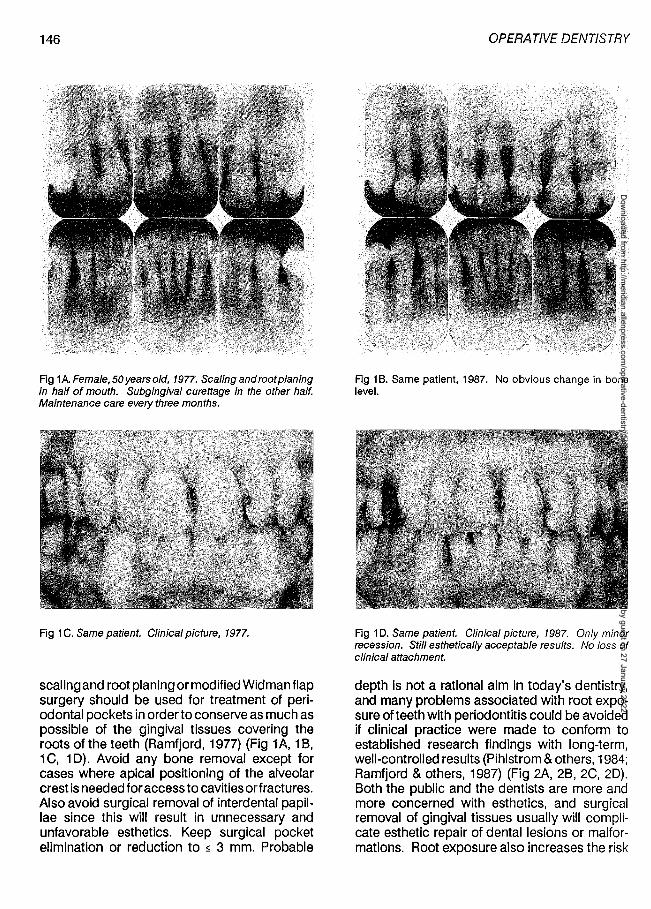
146
Fig 1AFemale,50yearsold, 1977. Scalingandrootplaning in half of mouth. Subgingival curettage in the other half. Maintenance care eve!}' three months.
Fig 1 C. Same patient. Clinical picture, 1977.
scaling and root planing or modified Widman flap surgery should be used for treatment of periodontal pockets in order to conserve as much as possible of the gingival tissues covering the roots of the teeth (Ramfjord, 1977) (Fig 1 A, 1 B, 1C, 10). Avoid any bone removal except for cases where apical positioning of the alveolar crest is needed for access to cavities orfractures. Also avoid surgical removal of interdental papillae since this will result in unnecessary and unfavorable esthetics. Keep surgical pocket elimination or reduction to s 3 mm. Probable
OPERATIVE DENTISTRY .
Fig 18. Same patient, 1987. No obvious change in bone level.
Fig 10. Same patient. Clinical picture, 1987. Only minor recession. Still esthetically acceptable results. No loss of clinical attachment.
depth is not a rational aim in today's dentistry, and many problems associated with root exposure of teeth with periodontitis could be avoided if clinical practice were made to conform to established research findings with long-term, well-controlled results (Pihlstrom & others, 1984; Ramfjord & others, 1987) (Fig 2A, 28, 2C, 20). Both the public and the dentists are more and more concerned with esthetics, and surgical removal of gingival tissues usually will complicate esthetic repair of dental lesions or malformations. Root exposure also increases the risk
Dow
nloaded from http://m
eridian.allenpress.com/operative-dentistry/article-pdf/13/3/1/1818880/1559-2863-13-3-1.pdf by guest on 27 January 2022

RAMFJORD: PERIODONTAL CONSIDERATIONS
INTERPROXIMAL RECESSION FOR MAXILLARY MOLARS BY TREATMENT METll)()-MOOERATE DISEASE
mm
2
--- Pocket Elim.
---- Curettage
------- Mod. Widman
,,..... ___ -_-_--------------~~ ,I ............. ------................ -:;;:,::---/ r- --- _________ .,,,,,.,, ............
r;
2 3 4 5 6 7 YEARS The interproximal recession is less following modi
fied Widman than following pocket elimination surgery (maxillary molars, 4- to 6-mm pockets).
Fig 2A lnterproximal recession for maxillary molars with moderately deep pockets is not significantly different over time, following various types of periodontal treatment.
INTERPROXIMAL RECESSION FOR MANDIBULAR ANTERIORS BY TREATMENT METHOD-SEVERE DISEASE
--- Pocket Elim.
mm - - Curettage
5 ------- Mod. Widman
4
,,,,. ........... , \ r-- ~=-::><.' \ ' , ' ' ' , ....._ '
' , -- \-I .. - ,
OOL---l.--2.L.,_--1.3--4.1...----'5--6"'-----'7-y-EARS
More recession is seen following pocket elimination surgery than following curettage an~ m.o~ijied Widman flap surgery (mandibular anterior teeth with mmal pockets of 7- to 12-mm.
Fig 2B. For deep mandibular anterior pockets, the recession is much greater following pocket elimination surgery than following subgingival curettage or modified Widman flap surgery.
147
I NTERPROXIMAL ATIACHMENT CHANGE FOR MANDIBULAR ANTERIORS BY TREATMENT METHOD-SEVERE DISEASE
--- Pocket Elim.
-- -- Curettage -------Mod. Widman
mm 3
2
,, ,,- -1\ / __:,-.:._, L ..... ~ r ,' \..... /,~--------
, \' I I .. __ ~
;/ ;/ /
0' 0 7 YEARS
Average gain in interproximal attachment following treatmentof7- to 12-mm deep pockets. Nole no gain following pocket elimination surgery. Mandibular anterior teeth.
Fig 2C. The lnterproximal attachment response is much more unfavorable following pocket elimination than following curettage or modified Widman flap surgery.
mm I
ATTACHMENT CHANGE (PATIENT MEANS) FOR INITIAL POCKETS 4·6 MM BY TREATMENT METHOD
-- Pocket Elim. --- Curettage ------ Mod. Widman
' ~::::::::::-... :-.:.-:-.:::::::.. ... ::-:=:::":.:.~::::.:::-.:.·==: 4 5 6 7 8YEARS
Fig 20. For all moderately deep pockets (4 - 6 mm) attachment levels are maintained equally well following all methods of periodontal treatment. It is not necessary to induce recession with pocket elimination surgery.
Dow
nloaded from http://m
eridian.allenpress.com/operative-dentistry/article-pdf/13/3/1/1818880/1559-2863-13-3-1.pdf by guest on 27 January 2022
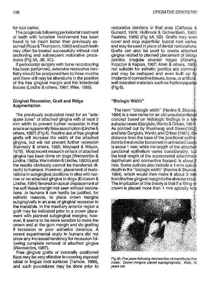
148
for root caries. The prognosis following periodontal treatment
of teeth with furcation involvement has been found to be much better than previously assumed (Ross & Thompson, 1980) and such teeth may often be treated successfully without root sectioning and subsequent restorative procedures (Fig 3A, 38, 3C).
If periodontal surgery with bone recontouring has been performed, extensive restorative dentistry should be postponed two to three months and there still may be alterations in the position of the free gingival margin and the interdental tissues (Lindhe & others, 1987; Wise, 1985).
Gingival Recession, Graft and Ridge Augmentation
The previously postulated need for an "adequate zone" of attached gingiva with at least 2 mm width to prevent further recession in that area is an apparently false assumption (Lind he & others, 1987) (Fig 4). Routine use of free gingival grafts will increase the width of the attached gingiva, but will not prevent further recession (Kennedy & others, 1985; Maynard & Wilson, 1979). Most recent research on lack of attached gingiva has been done on dogs (Wennstrom & Lind he, 1983a, Wennstrom & Lind he, 1983b) and the results obviously cannot be transferred directly to humans. However, placement of restorations in subgingival positions in sites with narrow or no attached gingiva in dogs (Ericsson & Lind he, 1984) favored an apical displacement of the soft tissue margin not seen without restorations. In humans it can hardly be justified, for esthetic reasons, to place crown margins subgingivally in an area of gingival recession in the mandible. In the maxillary anterior region a graft may be indicated prior to a crown placement with planned subgingival margins; however, it seems to be more sensible to make the crown end at the gum margin and do grafting if recession or poor esthetics develops. A recent experimental study in humans did not show any increased tendency for recession following complete removal of attached gingiva (Wennstrom, 1987).
Free gingival grafts or coronally positioned flaps may be very effective in covering exposed labial or lingual root surfaces (Tarnow, 1986), and such procedures may be done prior to
OPERATIVE DENTISTRY
restorative dentistry in that area (Caffesse & Guinard, 1978; Holbrook & Ochsenbein, 1983; Raetzke, 1985) (Fig 5A, 58). Grafts may even cover and stop superficial buccal root caries, and may be used in place of dental restorations. Grafts can also be used to create attached gingiva related to planned placement of bridge panties. Irregular alveolar ridges (Abrams, Kopczyk & Kaplan, 1987; Allen & others, 1985) not suitable for esthetic panties are common and may be reshaped and even built up by implants of connective tissues, bone, or artificial, well-tolerated materials such as hydroxyapatite (Fig 6).
"Biologic Width"
The term "biologic width" (Nevins & Skurow, 1984) is a new name for an old unsubstantiated concept based on histologic findings in a few autopsy cases (Gargiulo, Wentz & Orban, 1961 ). As pointed out by Waerhaug and Steen(1952) and later Gargiulo, Wentz, and Orban (1961 ), the distance from the base of the junctional epithelium to the alveolar bonecrest in untreated cases is about 1 mm, while the length of the attached junctional epithelium varies considerably, but the total length of the supracrestal attachment (epithelium and connective tissues) is about 2 mm. Some authors also include a 1-mm sulcus depth in the "biologic width" (Nevins & Skurow, 1984), which would then make it about 3 mm from the free gingival margin to the alveolar crest. The implication of this theory is that if a filling or crown is placed more than 1 mm apically to a
Fig 3A. Five years following hemisection of mandibular first molar. Crown margins placed supragingivally. Male, 75 years old.
Dow
nloaded from http://m
eridian.allenpress.com/operative-dentistry/article-pdf/13/3/1/1818880/1559-2863-13-3-1.pdf by guest on 27 January 2022
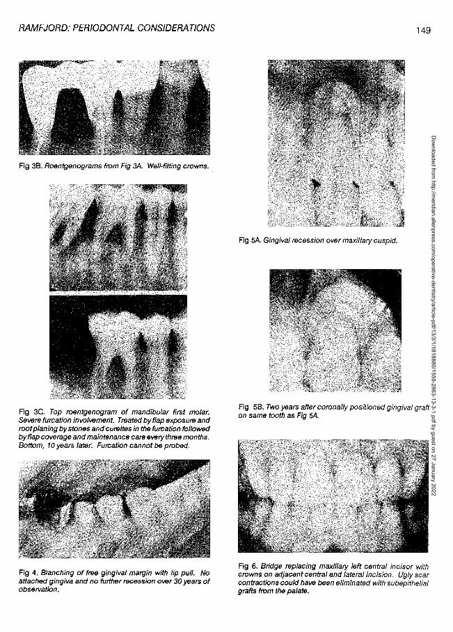
RAMFJORD: PERIODONTAL CONSIDERATIONS
Fig 38. Roentgenograms from Fig 3A. Well-fitting crowns.
Fig 3C. Top roentgenogram of mandibular first molar. Severe furcation involvement. Treated by flap exposure and root planing by stones and curettes in the furcation followed byflap coverage and maintenance care everythree months. Bottom, 10 years later. Furcation cannot be probed.
Fig 4. Blanching of free gingival margin with lip pull. No attached gingiva and no further recession over 30 years of observation.
149
Fig 5A. Gingival recession over maxillary cuspid.
Fig 58. Two years after coronal/y positioned gingival graft on same tooth as Fig 5A.
Fig 6. Bridge replacing maxillary left central incisor with crowns on adjacent central and lateral incision. Ugly scar contractions could have been eliminated with subepithelial grafts from the palate.
Dow
nloaded from http://m
eridian.allenpress.com/operative-dentistry/article-pdf/13/3/1/1818880/1559-2863-13-3-1.pdf by guest on 27 January 2022
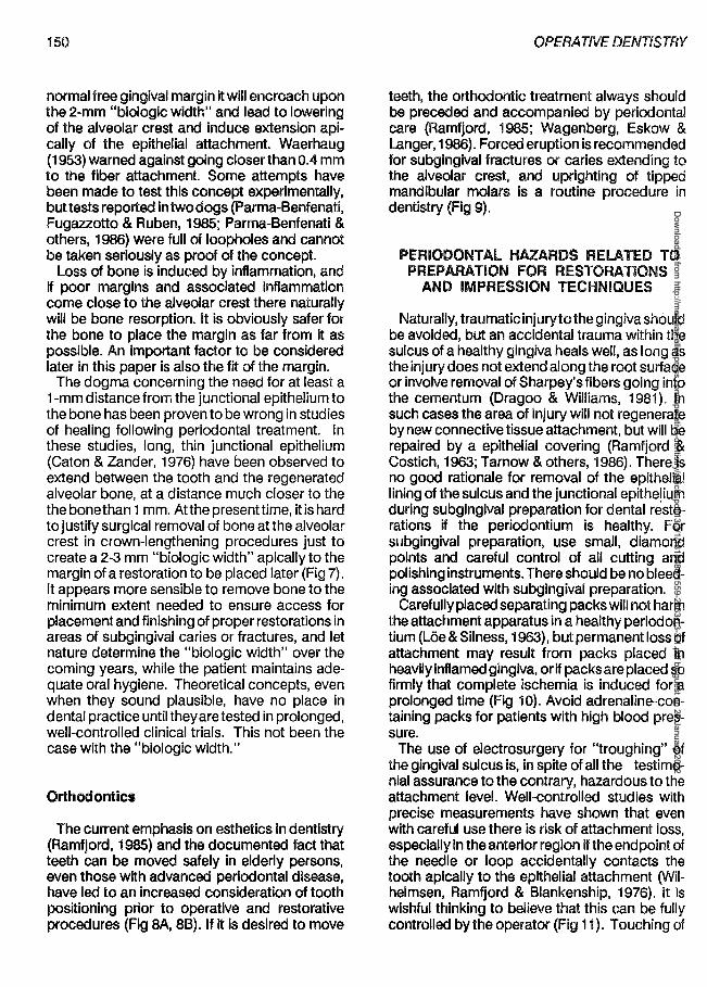
150
normal free gingival margin it will encroach upon the 2-mm "biologic width" and lead to lowering of the alveolar crest and induce extension apically of the epithelial attachment. Waerhaug (1953) warned against going closer than 0.4 mm to the fiber attachment. Some attempts have been made to test this concept experimentally, but tests reported in two dogs (Parma-Benfenati, Fugazzotto & Ruben, 1985; Parma-Benfenati & others, 1986) were full of loopholes and cannot be taken seriously as proof of the concept.
Loss of bone is induced by inflammation, and if poor margins and associated inflammation come close to the alveolar crest there naturally will be bone resorption. It is obviously safer for the bone to place the margin as far from it as possible. An important factor to be considered later in this paper is also the fit of the margin.
The dogma concerning the need for at least a 1-mm distance from the junctional epithelium to the bone has been proven to be wrong in studies of healing following periodontal treatment. In these studies, long, thin junctional epithelium (Caton & Zander, 1976) have been observed to extend between the tooth and the regenerated alveolar bone, at a distance much closer to the the bone than 1 mm. Atthe presenttime, it is hard to justify surgical removal of bone at the alveolar crest in crown-lengthening procedures just to create a 2-3 mm "biologic width" apically to the margin of a restoration to be placed later (Fig 7). It appears more sensible to remove bone to the minimum extent needed to ensure access for placement and finishing of proper restorations in areas of subgingival caries or fractures, and let nature determine the "biologic width" over the coming years, while the patient maintains adequate oral hygiene. Theoretical concepts, even when they sound plausible, have no place in dental practice until they are tested in prolonged, well-controlled clinical trials. This not been the case with the "biologic width."
Orthodontics
The current emphasis on esthetics in dentistry (Ramfjord, 1985) and the documented fact that teeth can be moved safely in elderly persons, even those with advanced periodontal disease, have led to an increased consideration of tooth positioning prior to operative and restorative procedures (Fig SA, SB). If it is desired to move
OPERATNE DENTISTRY
teeth, the orthodontic treatment always should be preceded and accompanied by periodontal care (Ramfjord, 1985; Wagenberg, Eskew & Langer, 1986). Forced eruption is recommended for subgingival fractures or caries extending to the alveolar crest, and uprighting of tipped mandibular molars Is a routine procedure in dentistry (Fig 9).
PERIODONTAL HAZARDS RELATED TO PREPARATION FOR RESTORATIONS
AND IMPRESSION TECHNIQUES
Naturally, traumatic injury to the gingiva should be avoided, but an accidental trauma within the sulcus of a healthy gingiva heals well, as long as the injury does not extend along the root surface or involve removal of Sharpey's fibers going into the cementum (Dragoo & Williams, 1981). In such cases the area of injury will not regenerate by new connective tissue attachment, but will be repaired by a epithelial covering (Ramfjord & Costich, 1963; Tarnow & others, 1986). There is no good rationale for removal of the epithelial lining of the sulcus and the junctional epithelium during subgingival preparation for dental restorations if the periodontium is healthy. For subgingival preparation, use small, diamond points and careful control of all cutting and polishing instruments. There should be no bleeding associated with subgingival preparation.
Carefully placed separating packs will not harm the attachment apparatus in a healthy perioclontium (Loe & Silness, 1963), but permanent loss of attachment may result from packs placed in heavUy inflamed gingiva, or if packs are placed so firmly that complete ischemia is induced for a prolonged time (Fig 10). Avoid adrenaline-containing packs for patients with high blood pressure.
The use of electrosurgery for "troughing" of the gingival sulcus is, in spite of all the testimonial assurance to the contrary, hazardous to the attachment level. Well-controlled studies with precise measurements have shown that even with careful use there is risk of attachment loss, especially in the anterior region if the endpoint of the needle or loop accidentally contacts the tooth apically to the epithelial attachment (Wilhelmsen, Ramfjord & Blankenship, 1976). It is wishful thinking to believe that this can be fully controlled by the operator (Fig 11). Touching of
Dow
nloaded from http://m
eridian.allenpress.com/operative-dentistry/article-pdf/13/3/1/1818880/1559-2863-13-3-1.pdf by guest on 27 January 2022
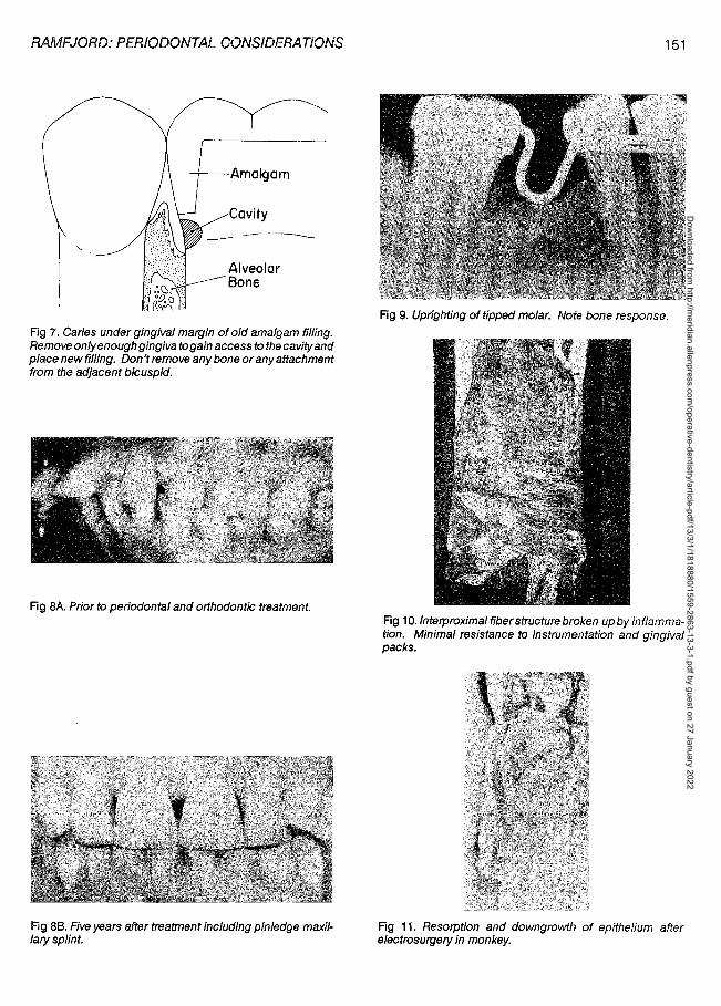
RAMFJORD: PERIODONTAL CONSIDERATIONS
Fig 7. Caries undergingival margin of old amalgam filling. Remove only enough gingiva to gain access to the cavity and place new filling. Don't remove any bone or any attachment from the adjacent bicuspid.
Fig SA. Prior to periodontal and orthodontic treatment.
Fig 88. Five years after treatment including pin/edge maxillary splint.
151
Fig 9. Uprighting of tipped molar. Note bone response.
Fig 1 O. lnterproximal fiber structure broken up by inflammation. Minimal resistance to instrumentation and gingival packs.
Fig 11. Resorption and downgrowth of epithelium after e/ectrosurgery in monkey.
Dow
nloaded from http://m
eridian.allenpress.com/operative-dentistry/article-pdf/13/3/1/1818880/1559-2863-13-3-1.pdf by guest on 27 January 2022
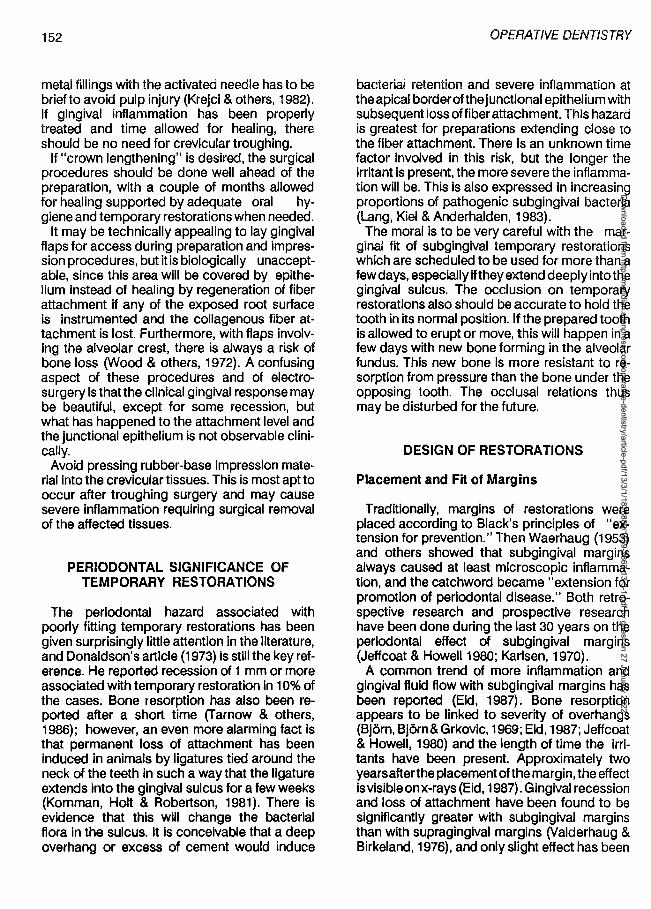
152
metal fillings with the activated needle has to be brief to avoid pulp injury (Krejci & others, 1982). If gingival inflammation has been properly treated and time allowed for healing, there should be no need for crevicular troughing.
If "crown lengthening" is desired, the surgical procedures should be done well ahead of the preparation, with a couple of months allowed for healing supported by adequate oral hygiene and temporary restorations when needed.
It may be technically appealing to lay gingival flaps for access during preparation and impression procedures, but it is biologically unacceptable, since this area will be covered by epithelium instead of healing by regeneration of fiber attachment if any of the exposed root surface is instrumented and the collagenous fiber attachment is lost. Furthermore, with flaps involving the alveolar crest, there is always a risk of bone loss C/Jood & others, 1972). A confusing aspect of these procedures and of electrosurgery is that the clinical gingival response may be beautiful, except for some recession, but what has happened to the attachment level and the junctional epithelium is not observable clinically.
Avoid pressing rubber-base impression material into the crevicular tissues. This is most apt to occur after troughing surgery and may cause severe inflammation requiring surgical removal of the affected tissues.
PERIODONTAL SIGNIFICANCE OF TEMPORARY RESTORATIONS
The periodontal hazard associated with poorly fitting temporary restorations has been given surprisingly little attention in the literature, and Donaldson's article (1973) is still the key reference. He reported recession of 1 mm or more associated with temporary restoration in 10% of the cases. Bone resorption has also been reported after a short time (T arnow & others, 1986); however, an even more alarming fact is that permanent loss of attachment has been induced in animals by ligatures tied around the neck of the teeth in such a way that the ligature extends into the gingival sulcus for a few weeks (Kornman, Holt & Robertson, 1981). There is evidence that this wm change the bacterial flora in the sulcus. It is conceivable that a deep overhang or excess of cement would induce
OPERATIVE DENTISTRY
bacterial retention and severe inflammation at the apical borderof the junctional epithelium with subsequent loss offiber attachment. This hazard is greatest for preparations extending close to the fiber attachment. There is an unknown time factor involved in this risk, but the longer the irritant is present, the more severe the inflammation will be. This is also expressed in increasing proportions of pathogenic subgingival bacteria (Lang, Kiel & Anderhalden, 1983).
The moral is to be very careful with the marginal fit of subgingival temporary restorations which are scheduled to be used for more than a few days, especially if they extend deeply into the gingival sulcus. The occlusion on temporary restorations also should be accurate to hold the tooth in its normal position. If the prepared tooth is allowed to erupt or move, this will happen in a few days with new bone forming in the alveolar fundus. This new bone is more resistant to resorption from pressure than the bone under the opposing tooth. The occlusal relations thus may be disturbed for the future.
DESIGN OF RESTORATIONS
Placement and Fit of Marg ins
Traditionally, margins of restorations were placed according to Black's principles of "extension for prevention." Then Waerhaug (1953) and others showed that subgingival margins always caused at least microscopic inflammation, and the catchword became "extension for promotion of periodontal disease." Both retrospective research and prospective research have been done during the last 30 years on the periodontal effect of subgingival margins (Jeffcoat & Howell 1980; Karlsen, 1970).
A common trend of more inflammation and gingival fluid flow with subgingival margins has been reported (Eid, 1987). Bone resorption appears to be linked to severity of overhangs (Bjorn, Bjorn&Grkovic, 1969; Eid, 1987; Jeffcoat & Howell, 1980) and the length of time the irritants have been present. Approximately two years after the placement of the margin, the effect isvisibleonx-rays (Eid, 1987). Gingival recession and loss of attachment have been found to be significantly greater with subgingival margins than with supragingival margins (Valderhaug & Birkeland, 1976), and only slight effect has been
Dow
nloaded from http://m
eridian.allenpress.com/operative-dentistry/article-pdf/13/3/1/1818880/1559-2863-13-3-1.pdf by guest on 27 January 2022

RAMFJORD: PERIODONTAL CONSIDERATIONS
noted when the restorative margins were placed at the gingival margin.
The best controlled, clinical, and bacteriological study on the effect of subgingival margins is an investigation by Lang, Kiel & Anderhalden (1983). Gold inlays with overhangs and others with clinically perfect subgingival margins were placed in first molars of dental students with healthy gingiva, and worn for approximately six months. Bacterial samples were obtained frequently. Pocket depth and bleeding caused by probing were also recorded. After six months the inlays with overhangs were replaced with well-fitting restorations, and the well-fitting inlays were replaced with fillings with overhangs to remain for another six months. The subgingival bacterial flora did not change with time, nor did bleeding tendency or crevice depth increase with the perfect fillings, but with overhangs the bacterial flora changed to an increasing proportion of melanin-containing bacteria, which is usually found in periodontitis.
The t;>leeding tendency gradually increased, but no measurable loss of periodontal attachment occurred. The bacterial flora and the clinical findings gradually returned to normal after a change from fillings with overhangs to wellfitting restorations without any other periodontal treatment or change in oral hygiene. Thus, it appeared that subgingival overhangs conditioned a change in the crevicular bacterial flora towards gram-negative melanogenic bacteria commonly associated with periodontitis. This led to increased inflammation and bleeding tendency, but for well-fitting restorations the bacterial flora and the bleeding tendency did not change significantly.
Thus, if there are compelling reasons to place margins of restorations subgingivally, it is essential for favorable periodontal response that they fit as perfectly as possible. All studies have shown that the detrimental periodontal effect of overhangs increases in proportion to the size of the overhangs and the length of time they have been present (Bjorn, Bjorn & Grkovic, 1969; Eid, 1987), with the possible exception of buccal overhangs where gingival recession may make the overhangs supragingival with time.
Many studies have been concerned with the subgingival effects of various dental materials (App, 1961; Eld, 1987). In general, the most favorable clinical response has been found with gold restorations, and less favorable with ce-
153
ments and plastics; however, how much of this reaction has to do with the marginal fit and the surface finish is not known. It appears that subgingival composite if properly applied may give a favorable response initially and for a couple of years, but this may later change to unfavorable as the surface of the composite is altered by oral hygiene wear (Dijken, Sjostrom & Wing, 1987).
Subgingival marginal leakage is always a problem as long as chemical bonding to cementum and dentin cannot be accomplished. It has been suggested that a pellicle seal may be established between the restoration and the tooth, thus preventing bacterial colonization into this junction. This probably will require a very good fit of the restorations and may explain the good bacteriological results obtained by Lang & others (1983) with their well-fitting inlays.
There are no experimental studies of the specific effect of placing the margin of fillings more or less close to the end of the junctional epithelium, but it stands to reason that it is preferable to have unavoidable inflammation associated with subgingival margins as far away as possible from this connective tissue attachment. Oral hygiene can also be made more effective at the opening of the gingival crevice than at the bottom. With fluorides and improved caries prevention there is no added security against caries from subgingival margins (Valderhaug & Birkeland, 1976).
There Is no question that from a periodontal standpoint subgingival margins are hazards to be avoided when possible. Supragingival margins can also be inspected and finished more reliably than subgingival margins.
These then are the compelling reasons for subgingival placement of margins:
1. caries or old restorations extending subgingivally,
2. esthetics on the labial aspects of maxillary teeth,
3. subgingival fractures,
4. establishing a ring support around the neck of fragile root canal-filled teeth, and
5. possible retention purposes with short clinical crowns.
Dow
nloaded from http://m
eridian.allenpress.com/operative-dentistry/article-pdf/13/3/1/1818880/1559-2863-13-3-1.pdf by guest on 27 January 2022

154
For esthetic reasons, one does not have to go more than 1 /2 mm subgingivally. A very common mistake to be avoided is placing interproximal crown and inlay margins too deep into the sulcus (Ramfjord, 1985). This can cause the frequently observed blue and swollen interproximal papillae contacting dental restorations.
Contour
There are numerous references to the importance of "proper contour of restorations," but there have been practically no experimental investigations of what is "proper" or of what influence changes in crown contour may have on gingival health. Plaque build-up over time is also a factor.
Numerous clinical observations indicate that overcontouring the subgingival part of restorations will lead to gingival irritation; whether this is from interference with sulcular physiology or increased plaque retention is not known (Ramfjord, 1985). In order to have a buccal and lingual "knife edge" gingival margin, the gingiva has to be adapted to a crown contour, but a "knife edge" gingival margin is not a prerequisite for gingival health, and there is no threat to gingival health from a flat crown contour. Thus, overcontouring restorations should be avoided (Fig 12), and restorations should never be placed subgingivally in order to promote a "knife edge" gingival margin.
Fig 12. Overcontoured maxillaryincisorcrowns. Also plaque and gingivitis around other teeth.
lnterproximal aspects are commonly overcontoured, thus crowding out the normal space for the interproximal part of the papillae, and
OPERATIVE DENTISTRY
leading to hyperplasia of the buccal and lingual part of the papillae. Lack of interproximal crown and filling contour is preferable to overcontouring, which is commonly found associated with multiple full crowns.
Contacts
Normally, the interproximal periodontium is protected from food impaction injury, and occlusion stability is maintained by the interproximal contact of the teeth; however, one may have open contacts without food impaction and with maintained, stable occlusion. Only with evidence of food impaction is open contact a periodontal hazard.
What type of interproximal contact is best, narrow or wide, has not been investigated, but it is important that the contact be placed coronally to allow for sufficient space for the interproximal papillae. It apparently is not important whether the interproximal margin ridges are of equal height or not, but the finish of interproximal soldering is important for access for efficient oral hygiene (Fig 13).
Fig 13. Poor finish of interproxima/ solderings impossible to keep clean.
PERIODONTAL SIGNIFICANCE OF OCCLUSION IN OPERATIVE DENTISTRY
Trauma from Occlusion
The periodontal significance of occlusal abberations is highly controversial, but apparently trauma from occlusion will not induce gingival
Dow
nloaded from http://m
eridian.allenpress.com/operative-dentistry/article-pdf/13/3/1/1818880/1559-2863-13-3-1.pdf by guest on 27 January 2022
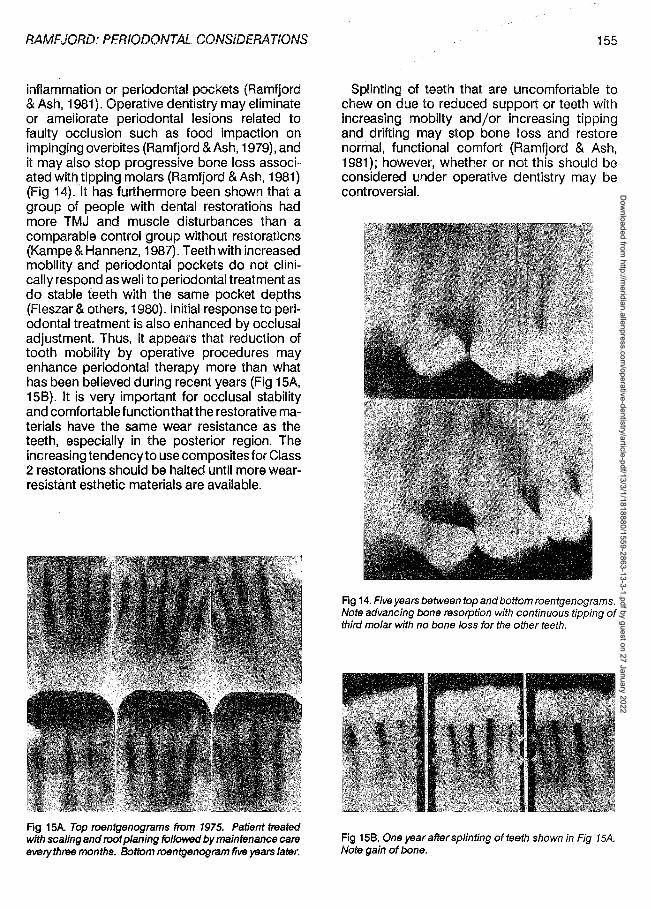
RAMFJORD: PERIODONTAL CONSIDERATIONS
inflammation or periodontal pockets (Ramfjord & Ash, 1981). Operative dentistry may eliminate or ameliorate periodontal lesions related to faulty occlusion such as food impaction on impinging overbites (Ramfjord & Ash, 1979), and it may also stop progressive bone loss associated wit!) tipping molars (Ramfjord & Ash, 1981) (Fig 14). It has furthermore been shown that a group of people with dental restorations had more TMJ and muscle disturbances than a comparable control group without restorations (Kampe & Hannenz, 1987). Teeth with increased mobility and periodontal pockets do not clinically respond as well to periodontal treatment as do stable teeth with the same pocket depths (Fleszar & others, 1980). Initial response to periodontal treatment is also enhanced by occlusal adjustment. Thus, it appears that reduction of tooth mobility by operative procedures may enhance periodontal therapy more than what has been believed during recent years (Fig 15A, 158). It is very important for occlusal stability and comfortablefunctionthatthe restorative materials have the same wear resistance as the teeth, especially in the posterior region. The increasing tendency to use composites for Class 2 restorations should be halted until more wearresistant esthetic materials are available.
Fig 15A Top roentgenograms from 1975. Patient treated with scaling and root planing followed by maintenance care everythree months. Bottom roentgenogram five yeatS later.
155
Splinting of teeth that are uncomfortable to chew on due to reduced support or teeth with increasing mobilty and/or increasing tipping and drifting may stop bone loss and restore normal, functional comfort (Ramfjord & Ash, 1981); however, whether or not this should be considered under operative dentistry may be controversial.
Fig 14. Five years between top and bottom roentgenograms. Note advancing bone resorption with continuous tipping of third molar with no bone loss for the other teeth.
Fig 158. One year after splinting of teeth shown in Fig 15A. Note gain of bone.
Dow
nloaded from http://m
eridian.allenpress.com/operative-dentistry/article-pdf/13/3/1/1818880/1559-2863-13-3-1.pdf by guest on 27 January 2022

156
MAINTENANCE CARE
Since no operative or periodontal treatment will assure immunity to recurrent or new periodontal disease, a periodic recall program is recommended for the maintenance of a healthy dentition (Ramfjord, 1987a). The frequency and extent of the recall program depends on the risk factor in the individual patient. Attempts have been made to assess this risk by bacteriologic, DNA, and other tests in addition to clinical appraisal. No formula is yet available to be used as a basis for frequency of recall. Axelsson and Lind he (197 4) recommended frequent professional tooth cleaning (every two weeks for one to two years), which provided spectacular reduction of caries and gingivitis in high-risk patients. But for maintenance care of treated patients with initial or mild periodontal disease and good oral hygiene, no advantage was found from frequent professional tooth cleaning over seeing the patients once a year (Eneroth & Sundberg, 1984), and in an animal study with controlled perfect oral hygiene, nothing was gained by periodic professional tooth cleaning (Morrison & others, 1979).
For moderate- to advanced-periodontitis patients there is a need for professional tooth cleaning every three to four months to maintain the periodontal attachment on a predictable basis over a prolonged time (Knowles & others, 1979; Ramfjord & others, 1987).
Thus, from a periodontal standpoint, recall for professional tooth cleaning and examination may be spaced as far apart as once a year for patients with normal gingiva or mild gingivitis and good oral hygiene, maybe once every six months for patients with incipient periodontitis, and every three months for patients who have had moderate to advanced periodontitis. Visits for professional maintenance care every three months is a must for the maintenance of the attachment levels of teeth that have been treated for advanced periodontal disease, regardless of modality of therapy. Recall and maintenance care should also take the caries status into consideration. Apparently, root surface caries is not decreasing in frequency as is enamel caries, and topical fluorides may have to be used. Defects at the cervical margins of restorations are also common, especially with composite fillings indicating need for adequate control (every three to six months, depending on
OPERATIVE DENTISTRY
the individual patient, oral hygiene, and nutrition). For patients with very low or no caries activity, control once a year may be adequate.
The following steps should be included in the maintenance care visits:
1. Apply disclosing solution.
2. Point out areas of inadequate oral hygiene and demonstrate correct procedures.
3. Polish away supragingival and some subgingival plaque with the EVA contra-angle and fluoride-containing tooth paste. Also polish buccally and lingually with soft, rubber cup and paste.
4. Probe and remove with curettes any calcarous deposits not eliminated by the polishing.
5. Probe for bleeding in areas of inflammation and deep crevices.
6. Examine for caries.
7. Apply fluoride. Use acidulated fluorophosphate if no ceramic restorations are present or neutral sodium fluoride if the patient has porcelain crowns.
8. Schedule patient for re-examination and retreatment two to three weeks later for areas with pus or bleeding from the bottom of the crevice.
9. Examine for caries, fractures, endodontic problems, and fillings needing replacement and occlusion.
Two aspects of maintenance care have come into recent focus (Ramfjord, 1987a): (1) the need for selective retreatment of periodontal pockets with active lesions (bleeding, pus, loss of attachment of abscesses), and (2) the need for topical fluoride for prevention of root caries and sensitivity.
Although plaque control by the patient is essential for a healthy dentition and periodontium, the teeth and the periodontal support can be maintained in the overwhelming number of cases by repeated professional maintenance care every three to four months, even if the home care is quite far from perfect (Ramfjord & others, 1982).
Dow
nloaded from http://m
eridian.allenpress.com/operative-dentistry/article-pdf/13/3/1/1818880/1559-2863-13-3-1.pdf by guest on 27 January 2022

RAMFJORD: PERIODONTAL CONSIDERATIONS
CONCLUSIONS
1. Restorative procedures should always be initiated after periodontal health has been established, with sufficient time allowed for healing.
2. Function and esthetics of dental restorations should be facilitated by the periodontal treatment, when possible.
3. The need for a specific "biologic width" has not been established.
4. Periodontal hazards with preparations, impression techniques, and temporary restorations should be considered.
5. Gingival recession and loss of attachment have been found to be significantly greater with subgingival margins than with supragingival margins, and supragingival margins can also be inspected and finished more reliably than subgingival margins.
6. Poorly fitting subgingival margins can condition a change in the crevicular bacterial flora towards gram-negative melanogenic bacteria commonly associated with periodontitis.
7. Response to periodontal treatment is enhanced by occlusal adjustment.
8. Emphasis should be given to maintenance care, including periodontal retreatment and topical application of fluorides.
References
ABRAMS, H, KOPCZVK, RA& KAPLAN, AL {1987) Incidence of anterior ridge deformities in partially endentulous patients Journal of Prosthetic Dentistry 57 191-194.
ALLEN, E P, GAINZA, CS, FARTHING, G G & NEWBOLD, D A (1985) Improved technique for localized ridge augmentation. A report of 21 cases Journal of Periodontology 56 195-199.
APP, GR {1961) Effect of silicate, amalgam, and cast gold on the gingiva Journal of Prosthetic Dentistry 11 522-532.
AXELSSON, P & LINDHE, J {1974) The effect of a preventive programme on dental plaque, gingivitis and caries in school children. Results after one and two years Journal
157
of Clinical Periodontology 1 126-138.
BJORN, A-L, BJORN, H & GRKOVIC, B {1969) Marginal fit of restorations and its relation to periodontal bone level. Part I: Metal fillings Odontologisk Revy 20 311-321.
CAFFESSE, R G & GUINARD, E A (1978) Treatment of localized gingival recessions. Part II: Coronally repositioned flap with a free gingival graft Journal of Periodontology 49 357-361.
CATON, J & ZANDER, HA {1976) Osseous repair of an infrabony pocket without new attachment of connective tissue Journal of Clinical Periodontology 3 54-58.
DIJKEN, J WV, SJOSTROM, S, WING, K (1987) The effect of different types of composite resin fillings on marginal gingiva Journal of Clinical Periodontology 14 185-189.
DONALDSON, D {1973) Gingival recession associated with temporary crowns Journal of Periodontology 44 691-696.
DRAGOO, MR & WILLIAMS, GB (1981) Periodontal tissue reactions to restorative procedures International Journal of Periodontics and Restorative Dentistry 1 8-23.
EID, M {1987) Relationship between overhanging amalgam restorations and periodontal disease Quintessence International 18 ns-781.
ENEROTH, L & SUNDBERG, H (1984) Effebten av forebyggande Tandvard Utfort av Specialutbildade Tandskoterskor. Delegationenfor Social Forsking. Social Departementet, Stockholm.
ERICSSON, I & LINDHE, J {1984) Recession in sites with inadequate width of the keratinized gingiva. An experimental study in the dog Journal of Clinical Periodontology 11 95-103.
FLESZAR, T J, KNOWLES, J W, MORRISON, E C, BURGETT, F G, NISSLE, RR & RAMFJORD, SP {1980) Tooth mobility and periodontal therapy Journal of Clinical Periodontology 7 495-505.
GARGIULO, AW, WENTZ, FM & ORBAN, B (1961) Dimensions and relations of the dentogingival junction in humans Journal of Periodontology 32 261-267.
HOLBROOK, T & OCHSENBEIN, C (1983) Complete coverage of denuded root surface with one-stage gingival graft International Journal of Periodontics and Restorative Dentistry 3 8-27.
JEFFCOAT, MK & HOWELL, T H {1980) Alveolar bone destruction due to overhanging amalgams in periodontal disease Journal of Periodontology 51 599-602.
KAMPE, T & HANNERZ, H (1987) Differences in occlusion and some functional variables in adolescents with intact and restored dentitions Acta Odontologica Scandinavica 45 31-39.
KARLSEN, K (1970) Gingival reactions to dental restorations
Dow
nloaded from http://m
eridian.allenpress.com/operative-dentistry/article-pdf/13/3/1/1818880/1559-2863-13-3-1.pdf by guest on 27 January 2022
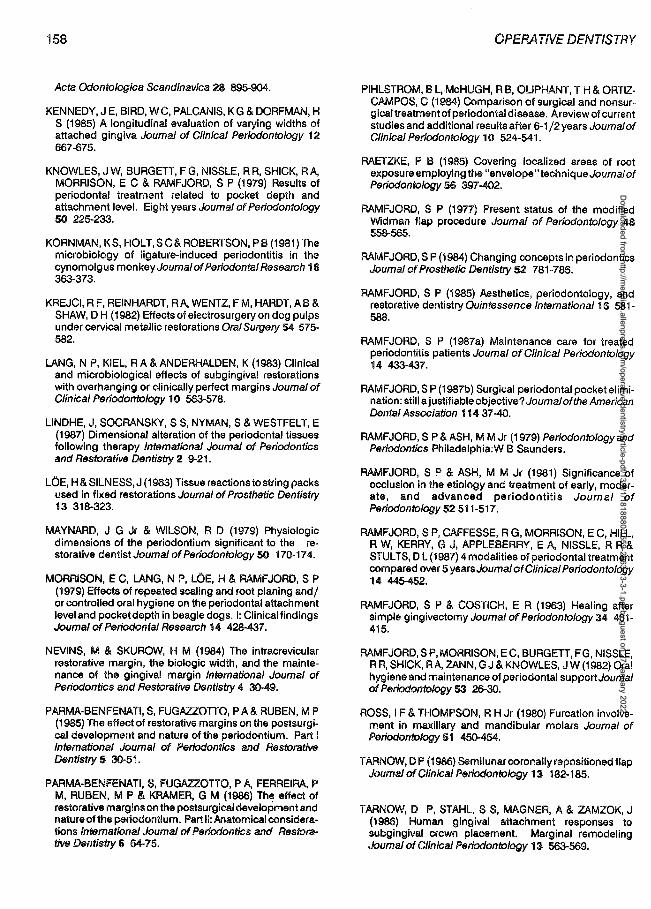
158
Acta Odontologica Scandinavica 28 895-904.
KENNEDY, J E, BIRD, WC, PALCANIS, KG & DORFMAN, H S (1985) A longitudinal evaluation of varying widths of attached gingiva Journal of Clinical Periodontology 12 667-675.
KNOWLES, J W, BURGETT, F G, NISSLE, RR, SHICK, RA, MORRISON, EC & RAMFJORD, SP (1979) Results of periodontal treatment related to pocket depth and attachment level. Eight years Journal of Periodontology 50 225-233.
KORNMAN, KS, HOLT,SC&ROBERTSON, PB(1981)The microbiology of ligature-induced periodontitis in the cynomolgus monkey Journal of Periodontal Research 16 363-373.
KREJCI, RF, REINHARDT, RA, WENTZ, FM, HARDT, AB & SHAW, DH (1982) Effects of electrosurgery on dog pulps under cervical metallic restorations Oral Surgery 54 575-582.
LANG, NP, KIEL, RA &ANDERHALDEN, K (1983) Clinical and microbiological effects of subgingival restorations with overhanging or clinically perfect margins Journal of Clinical Periodontology 10 563-578.
LINDHE, J, SOCRANSKY, SS, NYMAN, S & WESTFELT, E (1987) Dim.ensional alteration of the periodontal tissues following therapy International Journal of Periodontics and Restorative Dentistry 2 9-21.
LOE, H & SILNESS, J (1963) Tissue reactions to string packs used in fixed restorations Journal of Prosthetic Dentistry 13 318-323.
MAYNARD, J G Jr & WILSON, R D (1979) Physiologic dimensions of the periodontium significant to the restorative dentist Journal of Periodontology 50 170-17 4.
MORRISON, EC, LANG, N P, LOE, H & RAMFJORD, S P (1979) Effects of repeated scaling and root planing and/ or controlled oral hygiene on the periodontal attachment level and pocket depth in beagle dogs. I: Clinical findings Journal of Periodontal Research 14 428-437.
NEVINS, M & SKUROW, H M (1984) The intracrevicular restorative margin, the biologic width, and the maintenance of the gingival margin International Journal of Periodontics and Restorative Dentistry 4 30-49.
PARMA-BENFENATI, S, FUGAZZOTTO, PA& RUBEN, MP (1985) The effect of restorative margins on the postsurgical development and nature of the periodontium. Part I International Journal of Periodontics and Restorative Dentistry 5 30-51.
PARMA-BENFENATI, S, FUGAZZOTTO, PA, FERREIRA, P M, RUBEN, M P & KRAMER, G M (1986) The effect of restorative margins on the postsurgical development and nature of the periodontium. Part II: Anatomical considerations International Journal of Periodontics and Restorative Dentistry 6 64-75.
OPERA T/VE DENTISTRY
PIHLSTROM, BL, McHUGH, RB, OLIPHANT, T H & ORTIZCAMPOS, C (1984) Comparison of surgical and nonsurgical treatment of periodontal disease. A review of current studies and additional results after 6-1 /2 years Journal of Clinical Periodontology 1 O 524-541.
RAETZKE, P B (1985) Covering localized areas of root exposure employing the "envelope" technique Journal of Periodonto/ogy 56 397-402.
RAMFJORD, S P (19n) Present status of the modified Widman flap procedure Journal of Periodontology 48 558-565.
RAMFJORD, SP (1984) Changing concepts in periodontics Journal of Prosthetic Dentistry 52 781-786.
RAMFJORD, S P (1985) Aesthetics, periodontology, and restorative dentistry Quintessence International 16 581-588.
RAMFJORD, S P (1987a) Maintenance care for treated periodontitis patients Journal of Clinical Periodontology 14 433-437.
RAMFJORD, SP (1987b) Surgical periodontal pocket elimination: still a justifiable objective? Journal of the American Dental Association 114 37-40.
RAMFJORD, S P & ASH, M M Jr (1979) Periodontology and Periodontics Philadelphia:W B Saunders.
RAMFJORD, S P & ASH, M M Jr (1981) Significance of occlusion in the etiology and treatment of early, moderate, and advanced periodontitis Journal of Periodontology 52 511-517.
RAMFJORD, S P, CAFFESSE, R G, MORRISON, EC, HILL, R W, KERRY, G J, APPLEBERRY, E A, NISSLE, R R & STULTS, D L (1987) 4 modalities of periodontal treatment compared over 5 years.Journal of Clinical Periodontology 14 445-452.
RAMFJORD, S P & COSTICH, E R (1963) Healing after simple gingivectomy Journal of Periodontology 34 401-415.
RAMFJORD, SP, MORRISON, EC, BURGETT, FG, NISSLE, RR, SHICK, RA, ZANN, GJ &KNOWLES, JW (1982) Oral hygiene and maintenance of periodontal support Journal of Periodontology 53 26-30.
ROSS, IF & THOMPSON, RH Jr (1980) Furcation involvement in maxillary and mandibular molars Journal of Periodonto/ogy 51 450-454.
T ARNOW, DP (1986) Semilunar coronally repositioned flap Journal of Clinical Periodontology 13 182-185.
TARNOW, D P, STAHL, SS, MAGNER, A & ZAMZOK, J (1986) Human gingival attachment responses to subgingival crown placement. Marginal remodeling Journal of Clinical Periodonto/ogy 13 563-569.
Dow
nloaded from http://m
eridian.allenpress.com/operative-dentistry/article-pdf/13/3/1/1818880/1559-2863-13-3-1.pdf by guest on 27 January 2022
VALDERHAUG, J & BIRKELAND, J M (1976) Periodontal conditions in patients 5 years following insertion of fixed prosthesis. Pocket depth and loss of attachment Journal of Oral Rehabilitation 3 237-243.
WAERHAUG, J (1953) Tissue reactions around artificial crowns Journal of Periodontology 24 172- 185.
WAERHAUG, J & STEEN, E (1952) The presence or absence of bacteria in gingival pockets and the reaction in healthy pockets to certain pure culture!J. A bacterial and histological investigation Odontologisk Tidskrift 00 1-24.
WAGENBERG, B D, ESKOW, RN & LANGER, B (1986) Orthodontics: a solution for advanced periodontal or restorative problem International Journal of Periodontics and Restorative Dentistry 6 36-45.
WENNSTRQM, J L (1987) Lack of association between width of attached gingiva and development of soft tissue recession. A 5-year longitudinal study Journal of Clinical Periodontology 14 181-184.
Acknowledgment
159
WENNSTRQM, J & LINDHE, J (1983a) Role of attached gingiva for maintenance of periodontal health. Healing following excisional and grafting procedures in dogs Journal of Clinical Periodontology 10 206-221.
WENNSTRQM, J & LINDHE, J (1983b) Plaque-induced gingival inflammation in the absence of attached gingiva in dogs Journal of Clinical Periodontology 1 o 266-276.
WILHELMSEN, N R, RAMFJORD, SP & BLANKENSHIP, JR (1976) Effects of electrosurgery on the gingival attachment in Rhesus monkeys Journal of Periodontology 47 160-170.
WISE, MD (1985) Stabilityofgingival crest after surgery and before anterior crown placement Journal of Prosthetic Dentistry 53 20-23.
WOOD, D L, HOAG, P M, DONNENFELD, 0 W & ROSENFELD, L D (1972) Alveolar crest reduction following full and partial thickness flaps Journal of Periodontology 43 141-144.
The Buonocore Memorial Lecture is funded by the L D Caulk Company, Milford, Delaware.
Dow
nloaded from http://m
eridian.allenpress.com/operative-dentistry/article-pdf/13/3/1/1818880/1559-2863-13-3-1.pdf by guest on 27 January 2022
160
DEPARTMENTS
Book Reviews
DENTINE AND DENTINE REACTIONS IN THE ORAL CAVITY
A Thylstrup, S A Leach, and V Qvist
Published by IRL Press, Ltd, Oxford, England, 1987. 256 pages. $63.00.
This is the tenth proceedings published by the Research Group on Surface and Colloid Phenomena in the Oral Cavity under the auspices of the Council of Europe Committee on Science and Technology. It consists of 24 papers presented at a workshop in Koge, Denmark. The stated objective oft he workshop was "to attempt to assemble some of those research workers who have made an active contribution to our understanding of the carious process and its subsequent repair as it affects dentine."
Because the book is a series of individual papers, loosely grouped under five topic-headings, it is not intended for clinicians; it would be of more use for the research investigator. The topics include basic biological information, dentin reactions, dentin and root caries, and adhesion to dentin. Since there are 24 authors, all Europeans except for two Americans, the presented papers assume different styles. Some are research reports, some are philosophical discussions. Some are reviews of the literature, while others present new and interesting information.
Of particular interest to the operative dentist is an excellent paper by E C Munksgaard and EA Asmussen. Not only do they succinctly describe dentin-bonding mechanisms, but comparative bond strengths obtained by Gluma, Scotchbond, and acid-etching pretreatments. The superiority of Gluma is supported.
OPERATIVE DENTISTRY
The publication is recommended for the library of the researcher involved with dentin, with the caveat that it has limited scope; it does not encompass the broad body of knowledge available on the topics included.
WILLIAM R COTTON, DDS Georgetown University School of Dentistry Department of Operative Dentistry Washington, DC 20007
CHANGE YOUR SMILE
Ronald E Goldstein, DDS
Published by Quintessence Publishing Co, Inc, Chicago, 1988. 212 pages, 316 illustrations. $23.95.
This book, now in its second edition, was written to bea "true consumer guide to estheticdentistry," but has become a proven aid to dentists and their entire staff. The all-encompassing text ranges from bonding and bleaching, to esthetic fixed and removable restorations, to orthodontics and orthognathic surgery.
The text is wonderfully illustrated with beforeand-after photos on almost every page. Important points are emphasized with eye-catching bold print. Not only does Goldstein describe various treatment options, but he educates the consumer about tooth anatomy, the planning and care of restorations, and the importance of periodontal health. More importantly, he informs the reader of the limitations of restorations. Much of the material is presented in concise tables.
This book is a must for the reception area, and the dentist and hygienist can use it as a teaching and treatment-planning aid. It is also a worthy add it ion to the dentist's personal library, as it may enhance marketing strategies as well as supply tips and recommendations to suggest to patients.
Diantha J Berg, DDS 15945 NE 8th
Redmond, WA 98052
Dow
nloaded from http://m
eridian.allenpress.com/operative-dentistry/article-pdf/13/3/1/1818880/1559-2863-13-3-1.pdf by guest on 27 January 2022
Dow
nloaded from http://m
eridian.allenpress.com/operative-dentistry/article-pdf/13/3/1/1818880/1559-2863-13-3-1.pdf by guest on 27 January 2022
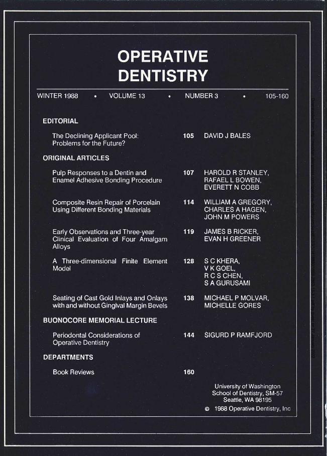
Dow
nloaded from http://m
eridian.allenpress.com/operative-dentistry/article-pdf/13/3/1/1818880/1559-2863-13-3-1.pdf by guest on 27 January 2022