276.pdf - rockefeller university press
TRANSCRIPT
S O R T I N G O U T O F N O R M A L A N D V I R U S - T R A N S F O R M E D
C E L L S I N C E L L U L A R A G G R E G A T E S
HOWARD GERSHMAN, JUDITH DRUMM, and LLOYD CULP
From the Departments of Biochemistry and Microbiology, Case Western Reserve University Medical School, Cleveland, Ohio 44106
A BST R ACT
The sorting-out behavior (self-segregation of two cell types from mixtures of the two) of five different established cell lines was studied. Eight of the ten possible binary combinations of these lines, cultured as cellular aggregates, were examined. Mouse BALB/c 3T3 cells sorted out internally to the corresponding malignant SV40 virus-transformed 3T3 cells. The transformed 3T3 line (SVT-2) did not sort out from a revertant line selected from SVT-2 cells by resistance to concanavalin A (con A). The revertant cells sorted out externally to the parent BALB/c 3T3 cells, although segregation was generally incomplete. BALB/c 3T3 cells did not sort out from another contact-inhibited line of 3T3 ceils derived from Swiss albino mice (Swiss 3T3).
Both BALB/c 3T3 and Swiss 3T3 cells sorted out from cells of the contact-inhib- ited hamster line, NIL B. Instead of a two-layered sphere, however, a three-layered structure was observed with most of the NIL B cells external to the 3T3 ceils, and a few NIL B cells comprising the center of the sphere. On the other hand, NIL B cells did not consistently sort out from either the SVT-2 or con A cells.
In general, sorting out between pairs of these five lines are slower and less com- plete than is generally observed between the more extensively studied chick embryonic tissue cells, suggesting that the cultured cells may be more closely related in their adhesive properties. The internal segregation of BALB/c 3T3 cells relative to SVT-2 cells is consistent with the hypothesis that transformed cells are less adhesive than their nontransformed counterparts.
The interaction of normal and malignant cells in culture has been the subject of considerable study. Much of this work has dealt with cells cultured on artificial substrata such as glass or plastic (e.g., 2, 4, 20, 21, 29, 32), especially that work utilizing established cell lines and their virus-transformed counterparts. While the study of such established lines has greatly contributed to our knowledge of how growth and movement are controlled, the presence of a nonphysiological substratum may interfere with the application of this knowledge to
in vivo situations, in particular, the study of adhesive interactions between normal and trans- formed cells in monolayer culture should be viewed with caution. Several workers have pointed out that in such situations the competition of cell- adhesions with cell-dish adhesions must be con- sidered (6, 26, 33) rather than limiting the analysis to cell-to-cell adhesions. In order to avoid this particular complication, we have studied the interaction of virus-transformed and nontrans- formed cells cultured as cellular aggregates or
276 THE JOURNAL OF CELL BIOLOGY �9 VOLUME 68, 1976 �9 pages 276-286
Dow
nloaded from http://rupress.org/jcb/article-pdf/68/2/276/1387337/276.pdf by guest on 09 January 2022
masses, in which cells are maintained in suspension in agitated liquid medium. Our purpose was to compare the properties of a nontransformed: transformed: revertant family of cell lines and also to compare the properties of a number of lines that exhibit varying degrees of contact inhibition.
The property of cells in these aggregates that was examined in this study was their ability to sort out or self-segregate. When cells of two suitable tissue types are intermixed, they sort out with the cells of one type forming a sphere surrounding the cells of the second type. Sorting out has been demonstrated for embryonic tissues from a num- ber of vertebrate species, including mouse (5, 13, 22), chick (23, 28), human (8), and amphibian (31). Steinberg (27, 28) has proposed that the equilib- rium configuration achieved after sorting out is the result of quantitative differences in adhesiveness between the two cell types. Although universal agreement on all the details of this hypothesis is still lacking, it is generally accepted that sorting- out behavior is a reflection of an adhesive interac- tion between the cells. For the purposes of discus- sion in this report, we will also tentatively consider that these adhesive interactions represent different strengths of intercellular adhesion.
M A T E R I A L S A N D M E T H O D S
Chemicals and Radiochemicals
All chemicals were purchased from commercial sources and were either biological grade or else the highest grade available.
[Methyl-~H]thymidine was purchased from New Eng- land Nuclear (Boston, Mass.) or Amersham/Searle Corp. (Arlington Hts., I11.) and had a spec act of 18-20 Ci/mmol.
Growth o f Cell Lines
The growth of mouse fibroblast BALB/c 3T3 (I) and SV40-transformed BALB/c 3T3 (designated SVT-2) (1), con A revertant (10, II, 12), hamster NIL B (14), and Swiss albino mice (Swiss 3T3) cells (1) has been de- scribed previously (18). Cells were found to be free of Mycoplasma contamination by an autoradiographic assay (I 8, ! 1 ).
Cells were routinely subcultured using trypsin- (ethylenedinitrilo)tetraacetic acid (EDTAp solutions (0.05% Difco trypsin 1:250 (Difco Laboratories, Detroit, Mich.) and 0.5 mM EDTA). For experimental use, cells
' Abbreviations used in this paper." EDTA, (ethylenedini- trilo)tetraacetic acid; EGTA, [ethylenebis(oxyethyleneni- trilo)]tetraacetic acid; and PBS, phosphate-buffered sa- line (Dulbecco's A solution).
were inoculated into 100-mm plastic culture dishes in 15-ml medium and grown as subconfluent cultures for 3-4 days (three to five generations). The medium was changed 24 h after plating and every 48 h thereafter. Cells were removed from the dishes with [ethylenebis- (oxyethylenenitrilo)]tetraacetic acid (EGTA) (0.5 mM in Ca++-Mg++-frer phosphate-buffered saline (Dul- becco's A solution) (PBS) and used either to inoculate dishes or to make aggregates. Thus, ceils used in these experiments were grown as subconfluent cultures (not more than 25% confluent) and were not harvested with trypsin. These cells reached the following densities if allowed to grow to saturation with daily changes of medium: BALB/c 3T3, 10 • 10' cells/cm=; SVT-2, 100 x 10' ceils/cmJ; con A, 20 • 10' cells/cm~; Swiss 3T3, 5 • 10' cells/cmS; NIL B, 15-20 x 10' cells/ c m 2.
Aggregates were formed by a technique previously reported (18). Briefly, this involves mixing together 1.5 • 10 e cells of each type, centrifuging the suspension at low speed, and after a 1.5-h incubation period, cutting the pellet into cubical fragments about 0.4 mm on a side. The aggregates are cultured in hanging drops for 24 h and on a gyratory shaker for an additional 1.5-3.5 days.
Labeling o f Ceils with [SH]Thymidine
Cells were radiolabeled by the addition of [SH]thymi- dine to the culture medium 3-4 days before the experi- ment. 24 h before the experiment, the radioactive me- dium was replaced by nonradioactive medium. If this chase period was omitted, the nonradioactive cells in the aggregates became radioactive within 1-2 days. This may have occurred by transfer of nucleotides between cells, since this process has already been suggested as a possible explanation for the contact-mediated "'meta- bolic co-operation" observed in culture (30).
In early experiments cells were labeled by the addition of 2.6 ,C i /ml [SH]thymidine to the medium. This concentration was reduced to 0. I ~aCi/ml for some of the experiments reported here. In each case in which the higher concentration was used the experiment was also repeated with the lower concentrations. No differences were found between these duplicate experiments. When 0.1 uCi/ml [SH]thymidine was added, however, total labeling per cell was decreased, and autoradiography was done by the method of Weingrad et al? Histology and autoradiography were performed as previously described (18).
Determination o f Plating Efficiency
EGTA-suspended cells were washed and either inocu- lated into dishes or formed into aggregates. The aggre- gates (pellet fragments) were cultured in hanging drops for one day and then in small vials on a gyratory shaker
Weingrad, D., R. M. Sade, C. Haudenschild, and J. Foikman. Manuscript in preparation.
H. GERSHMAN, J. DRUMM, and L. CULP Sorting Out of Normal and Transformed Cells 277
Dow
nloaded from http://rupress.org/jcb/article-pdf/68/2/276/1387337/276.pdf by guest on 09 January 2022
( 180-200 gyrations per min) for an additional 2 days. The cells inoculated into dishes were cultured in a 37 ~ C CO,-gassed incubator for the same 3 days.
After this period the aggregates and dishes were rinsed once in PBS and treated with trypsin (0.05% Difco 1:250 plus 0.5 mM EDTA) in PBS for 10 min at 37~ The single cell suspensions were counted and diluted into complete medium to the appropriate concentration. Cells were then seeded into 60-mm plastic dishes at 250, 500, or 1,000 cells per dish and cultured for 2 weeks, with changes of medium every 3-4 days. Colonies were counted after staining with Wright's solution.
In early experiments dish-maintained cells were inocu- lated at varying densities. In later experiments they were inoculated at the following densities, which were just sufficient to produce confluent cultures within 24 h: BALB/c 3T3, 1.8 • 106 cells/60-mm dish; SVT-2, 5 • 106 cells/60-mm dish; NIL B, 3 • 10 e ceUs/60-mm dish.
R E S U L T S
Plating Ef f ic iency Exper iments
In order to assess the viability of cells main- tained as aggregates, the ability of these cells to form colonies relative to cells mainta ined as mono- layers was studied. In one set of experiments, cells cultured as aggregates were compared to parallel cultures in which cells were mainta ined on dishes at low to moderate density. The results showed a modera te amount of variability, not only between cell lines but also between experiments with the same line. Relative plating efficiencies, i.e., plating efficiency of cells in aggregates divided by the
plating efficiency of cells in dishes, varied from a low of 44% to a high of 100%. Absolute plating efficiencies varied from 10 to 40 colonies per I00 cells plated.
A second series of experiments was performed using some of these lines in which the cell density of monolayer cultures was very high and therefore more comparable to the cell density in aggregates. The cells were seeded into 60-mm dishes at a density just sufficient to produce confluent mono- layers when the cells had at tached and spread. These data are presented in Table I. Both the absolute and relative plating efficiencies observed ranged from a low of 14.3 to a high of 32.9 colonies per 100 cells plated. These experiments suggest tha t the viability of cells mainta ined in aggregates for 3 days is similar to their viability when mainta ined in a confluent state in dishes for a similar period of time.
Sor t ing Out o f M o u s e - M o u s e Cell
Line Mix tures
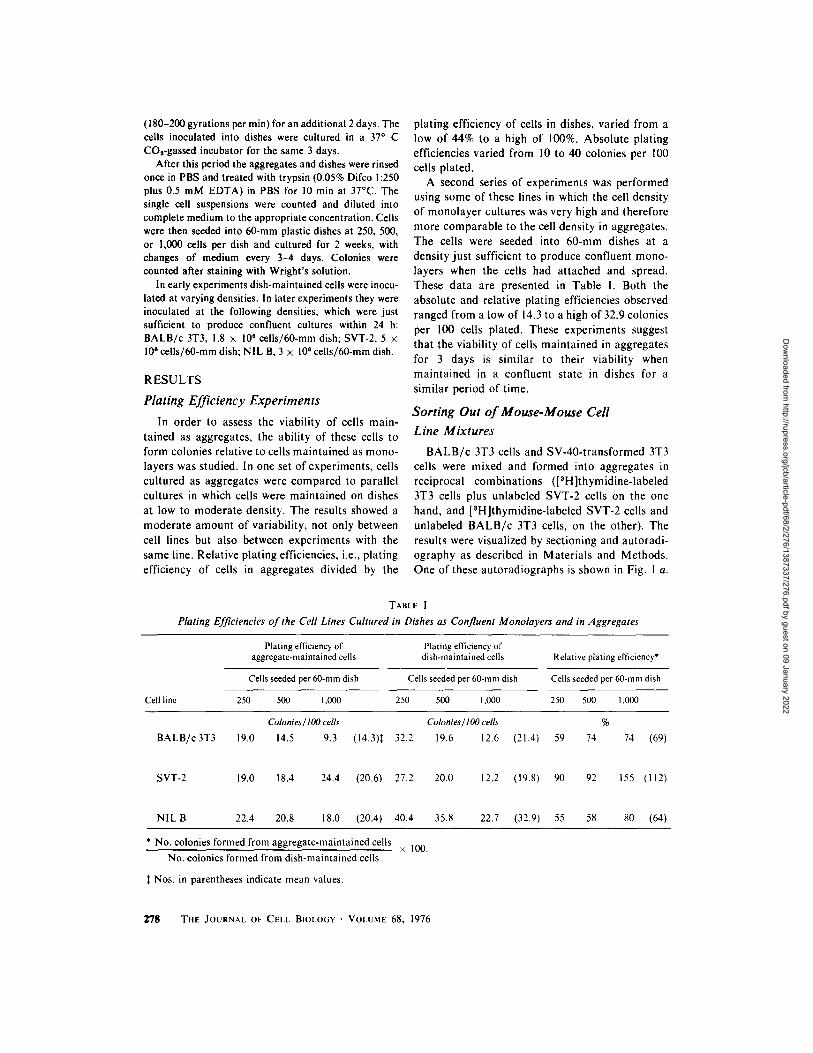
B A L B / c 3T3 cells and SV-40- t ransformed 3T3 cells were mixed and formed into aggregates in reciprocal combinat ions ([3H]thymidine-labeled 3T3 cells plus unlabeled SVT-2 cells on the one hand, and [3H]thymidine-labeled SVT-2 cells and unlabeled B A L B / c 3T3 cells, on the other). The results were visualized by sectioning and autoradi- ography as described in Mater ia ls and Methods. One of these autoradiographs is shown in Fig. 1 a.
TABLE I
Plating Efficiencies of the Cell Lines Cultured in Dishes as Confluent Monolayers and in Aggregates
Cell line
Plating efficiency of aggregate-maintained cells
Plating efficiency of dish-maintained cells
Cells seeded per 60-mm dish Cells seeded per 60-mm dish
250 500 1,000 250 500 1,000
Relative plating efficiency*
Cells seeded per 60-mm dish
250 500 1,000
Colonies/ lO0 cells Colonies/ lO0 cells %
BALB/c 3T3 19.0 14.5 9.3 (14.3)* 32.2 19.6 12.6 (21.4) 59 74 74 (69)
SVT-2 19.0 18.4 24.4 (20.6) 27.2 20.0 12.2 (19.8) 90 92 155 (112)
NIL B 22.4 20.8 18.0 (20.4) 40.4 35.8 22.7 (32.9) 55 58 80 (64)
* No. colonies formed from aggregate-maintained cells
No. colonies formed from dish-maintained cells
Nos. in parentheses indicate mean values.
• 100.
2"18 THE JOURNAL OF CELL BIOLOGY �9 VOLUME 68, 1976
Dow
nloaded from http://rupress.org/jcb/article-pdf/68/2/276/1387337/276.pdf by guest on 09 January 2022
FIGURE 1 (a) Autoradiograph of a histological section of an aggregate of [SH]thymidine-labeled BALB/c 3T3 cells and unlabeled SVT-2 cells. The aggregate was cultured for 2.5 days. While more than half of the cells visible in this section have silver grains over their nuclei (see Fig. 1 b), most of the grains are not in the focal plane of this photograph. This autoradiograph was exposed for a shorter time than usual in order to reveal as much nuclear detail as possible. The dark staining of the peripheral cell layer is common in these aggregates, although the cause is not known. The bar in this photograph represents 50 vm. Magnification, • 200. (b) Camera lucida drawing of the nuclei of the cells in the aggregate section shown in Fig. I a. The [SH]thymidine-labeled nuclei are indicated by filled ovals while unlabeled nuclei are indicated by open ovals. The presence or absence of silver grains was determined with a 100 • oil immersion objective. 10 grains located over the nucleus above background was the minimum used to identify a cell as being labeled. Sorting out is nearly complete in this experiment; only a few unlabeled cells remain in the center of the aggregate, while the outer rim is virtually devoid of labeled cells. Not all of the nuclei indicated in this figure are visible in Fig. 1 a, since some are above or below the focal plane of the photograph.
A zero-t ime control was also performed for this and all subsequent experiments. In each case, aggregates fixed immediately after cutt ing were found to be random mixtures of the two cell types. After 2.5 days of culture the aggregates had formed two concentr ic spheres, with the SVT-2 cells on the outside and the B A L B / c 3T3 cells internally. This experiment was performed four separate times, each in reciprocal combinat ion for a total of eight separate experiments. The results of these eight experiments indicated that SVT-2 cells sorted out externally to B A L B / c 3T3 cells.
Sort ing out was usually but not always complete after 2 days, a l though the pat tern was always clear. In those experiments carried out for 4.5 days, however, segregation was virtually complete, i.e., a complete shell of SVT-2 cells, containing no 3T3 cells, surrounded an inner core of B A L B / c 3T3 cells. These experiments, as well as the following sort ing-out experiments, are summarized in Table ! I. The reciprocal combinat ion of Figs. I a and b is shown in Figs. 2 a and b. In these eight experi- ments, the ratio of B A L B / c 3T3 to SVT-2 cells was either 1:2, l : l , or 2:1. No differences in
H. GERSHMAN, J. DRUMM, and L. CULP Sorting Out of Normal and Transformed Cells 279
Dow
nloaded from http://rupress.org/jcb/article-pdf/68/2/276/1387337/276.pdf by guest on 09 January 2022
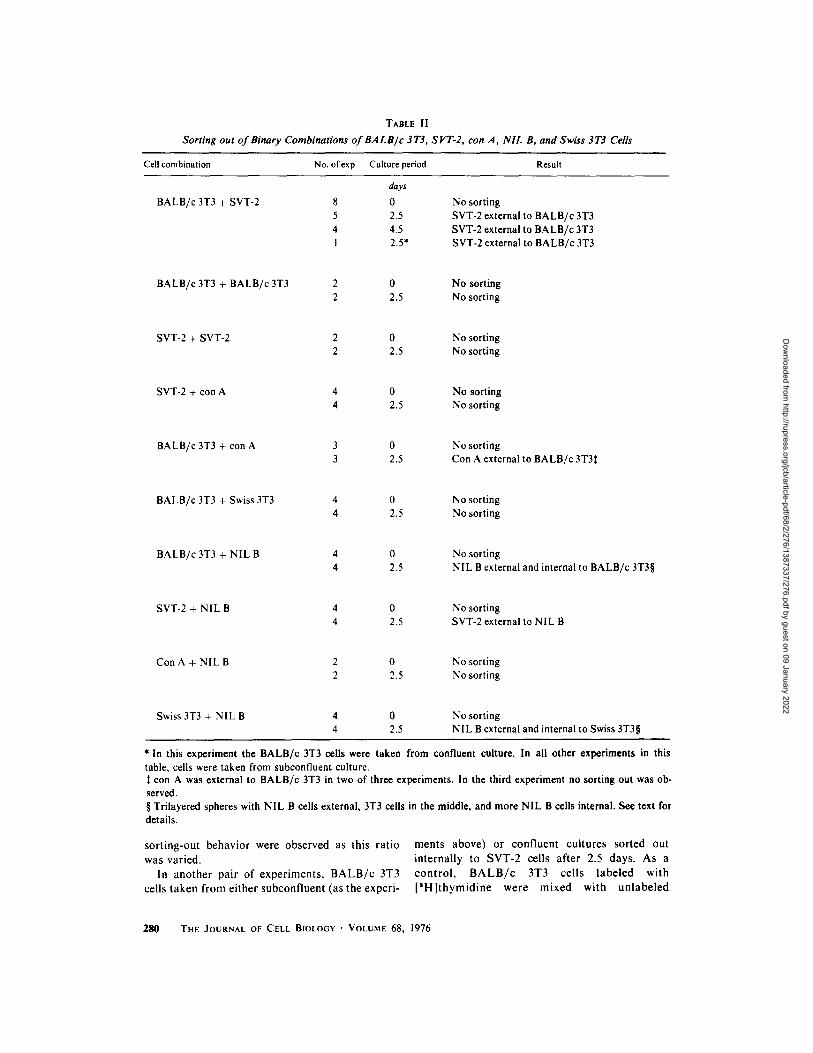
TABLE II
Sorting out of Binary Combinations of BALB/c 3T3, SVT-2, con A, NIL B, and Swiss 3T3 Cells
Cell combination No. ofexp Culture period Result
BALB/c 3T3 + SVT-2
days
8 0 5 2.5 4 4.5 1 2.5*
No sorting SVT-2 external to BALB/c 3T3 SVT-2 external to BALB/c 3T3 SVT-2 external to BALB/c 3T3
BALB/c 3T3 + BALB/c 3T3 2 0 No sorting 2 2.5 No sorting
SVT-2 + SVT-2 2 0 No sorting 2 2.5 No sorting
SVT-2 + con A 4 0 No sorting 4 2.5 No sorting
BALB/c 3T3 + con A 3 0 No sorting 3 2.5 Con A external to BALB/c 3T3:~
BALB/c 3T3 + Swiss 3T3 4 0 No sorting 4 2.5 No sorting
BALB/c3T3 + NIL B 4 0 4 2.5
No sorting NIL B external and internal to BALB/c 3T3w
SVT-2 + NIL B 4 0 No sorting 4 2.5 SVT-2 external to NIL B
Con A + NIL B 2 0 No sorting 2 2.5 No sorting
Swiss 3T3 + NIL B 4 0 4 2.5
No sorting NIL B external and internal to Swiss 3T3w
* In this experiment the BALB/c 3T3 cells were taken from confluent culture. In all other experiments in this table, cells were taken from subconfluent culture.
con A was external to BALB/c 3T3 in two of three experiments. In the third experiment no sorting out was ob- served. w Trilayered spheres with NIL B cells external, 3T3 cells in the middle, and more NIL B cells internal. See text for details.
sorting-out behavior were observed as this ratio was varied.
In another pair of experiments, BALB/c 3T3 cells taken from either subconfluent (as the experi-
ments above) or confluent cultures sorted out internally to SVT-2 cells after 2.5 days. As a control , B A L B / c 3T3 cells labeled with [SH]thymidine were mixed with unlabeled
280 THE JOURNAL OF CELL BIOLOGY �9 VOLUME 68, 1976
Dow
nloaded from http://rupress.org/jcb/article-pdf/68/2/276/1387337/276.pdf by guest on 09 January 2022
B A L B / c 3T3 cells. No sorting out was seen after 2.5 days. Similarly, [SH]thymidine-labeled SVT-2 cells did not sort out from unlabeled SVT-2 cells in control experiments.
When SVT-2 cells were mixed with con A revertant cells (variants of SVT-2 which have regained contact inhibition of growth), no sorting out was observed. Aggregates fixed 2.5 days after seeding showed the same random mixture of labeled and unlabeled cells observed in aggregates fixed immediately after mixing. The criteria for lack of sorting out were the following: (a) examina- tion of the outer four cell layers in the aggregate showed the same percentage of labeled versus unlabeled cells as in the aggregate interior; (b) the
outermost layer of cells contained both labeled and unlabeled cells; and (c) no discernible clumping of either labeled or unlabeled cells was seen in the interior ("patchy sorting" as described by Wise- man [34]).
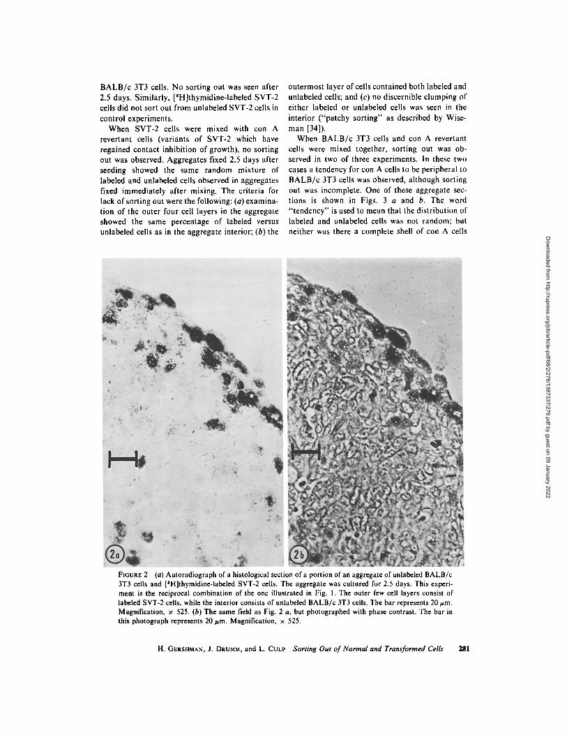
When B A L B / c 3T3 cells and con A revertant cells were mixed together, sorting out was ob- served in two of three experiments, in these two cases a tendency for con A cells to be peripheral to B A L B / c 3T3 cells was observed, although sorting out was incomplete. One of these aggregate sec- tions is shown in Figs. 3 a and b. The word " tendency" is used to mean that the distribution of labeled and unlabeled cells was not random; but neither was there a complete shell of con A cells
FIGURE 2 (a) Autoradiograph of a histological section of a portion of an aggregate of unlabeled BALB/c 3T3 cells and [SH]thymidine-labeled SVT-2 cells. The aggregate was cultured for 2.5 days. This experi- ment is the reciprocal combination of the one illustrated in Fig. 1. The outer few cell layers consist of labeled SVT-2 cells, while the interior consists of unlabeled BALB/c 3T3 cells. The bar represents 20 urn. Magnification, • 525. (b) The same field as Fig. 2 a, but photographed with phase contrast. The bar in this photograph represents 20 urn. Magnification, x 525.
H. GERSHMAN, J. DRUMM, and L. CULP Sorting Out of Normal and Transformed Cells 281
Dow
nloaded from http://rupress.org/jcb/article-pdf/68/2/276/1387337/276.pdf by guest on 09 January 2022
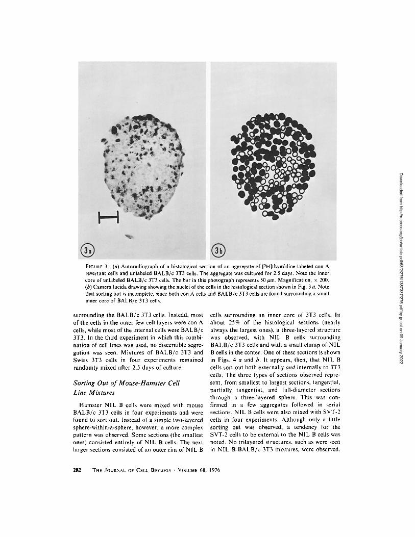
FIGURE 3 (a) Autoradiograph of a histological section of an aggregate of [3H]thymidine-labeled con A revertant cells and unlabeled BALB/c 3T3 cells. The aggregate was cultured for 2.5 days. Note the inner core of unlabeled BALB/c 3T3 cells. The bar in this photograph represents 50 #m. Magnification, x 200. (b) Camera lucida drawing showing the nuclei of the cells in the histological section shown in Fig. 3 a. Note that sorting out is incomplete, since both con A cells and BALB/c 3T3 cells are found surrounding a small inner core of BALB/c 3T3 cells.
surrounding the B A L B / c 3T3 cells. Instead, most of the cells in the outer few cell layers were con A cells, while most of the internal cells were B A L B / c 3T3. In the third experiment in which this combi- nat ion of cell lines was used, no discernible segre- gation was seen. Mixtures of B A L B / c 3T3 and Swiss 3T3 cells in four experiments remained randomly mixed after 2.5 days of culture.
Sorting Out of Mouse-Hamster Cell
Line Mixtures
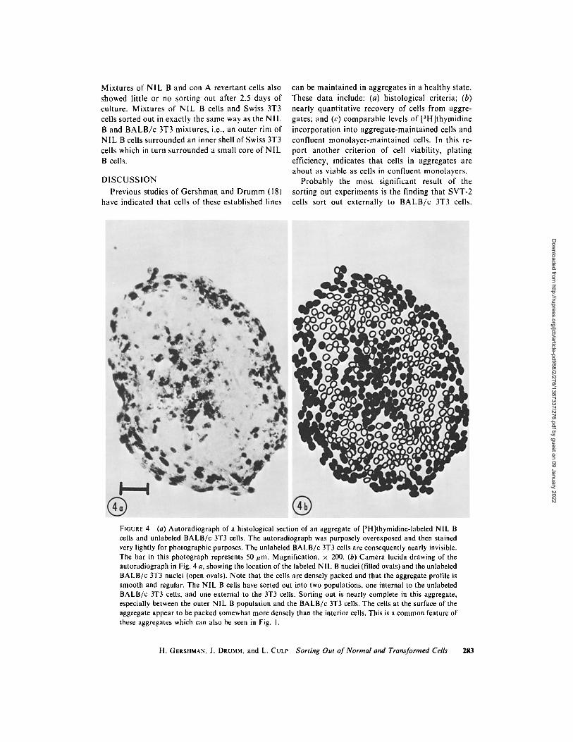
Hamste r NIL B cells were mixed with mouse B A L B / c 3T3 cells in four experiments and were found to sort out. Instead of a simple two-layered sphere-within-a-sphere, however, a more complex pat tern was observed. Some sections (the smallest ones) consisted entirely of NIL B cells. The next larger sections consisted of an outer rim of N IL B
cells surrounding an inner core of 3T3 cells. In about 25% of the histological sections (nearly always the largest ones), a three-layered structure was observed, with NIL B cells surrounding B A L B / c 3T3 cells and with a small clump of NIL B cells in the center. One of these sections is shown in Figs. 4 a and b. it appears, then, that N IL B cells sort out both externally and internally to 3T3 cells. The three types of sections observed repre- sent, from smallest to largest sections, tangential , partially tangential , and full-diameter sections through a three-layered sphere. This was con- firmed in a few aggregates followed in serial sections. NIL B cells were also mixed with SVT-2 cells in four experiments. Al though only a little sorting out was observed, a tendency for the SVT-2 cells to be external to the NIL B cells was noted. No trilayered structures, such as were seen in NIL B - B A L B / c 3T3 mixtures, were observed.
282 THE JOURNAL OF CELL BIOLOGY �9 VOLUME 68, 1976
Dow
nloaded from http://rupress.org/jcb/article-pdf/68/2/276/1387337/276.pdf by guest on 09 January 2022
Mixtures of NIL B and con A revertant cells also showed little or no sorting out after 2.5 days of culture. Mixtures of NIL B cells and Swiss 3T3 cells sorted out in exactly the same way as the N IL B and B A L B / c 3T3 mixtures, i.e., an outer rim of N I L B cells surrounded an inner shell of Swiss 3T3 cells which in turn surrounded a small core of NIL B cells.
D I S C U S S I O N
Previous studies of Ger shman and Drumm (18) have indicated that cells of these established lines
can be main ta ined in aggregates in a heal thy state. These data include: (a) histological criteria; (b) nearly quant i ta t ive recovery of cells from aggre- gates; and (c) comparable levels of [3H]thymidine incorporat ion into aggregate-mainta ined cells and confluent monolayer -main ta ined cells. In this re- port another criterion of cell viability, plating efficiency, indicates that cells in aggregates are about as viable as cells in confluent monolayers.
Probably the most significant result of the sorting out experiments is the finding that SVT-2 cells sort out externally to B A L B / c 3T3 cells.
FIGURE 4 (a) Autoradiograph of a histological section of an aggregate of [3H]thymidine-labeled NIL B cells and unlabeled BALB/c 3T3 cells. The autoradiograph was purposely overexposed and then stained very lightly for photographic purposes. The unlabeled BALB/c 3T3 cells are consequently nearly invisible. The bar in this photograph represents 50 #m. Magnification, • 200. (b) Camera lucida drawing of the autoradiograph in Fig. 4 a, showing the location of the labeled NIL B nuclei (filled ovals) and the unlabeled BALB/c 3T3 nuclei (open ovals). Note that the cells are densely packed and that the aggregate profile is smooth and regular. The NIL B cells have sorted out into two populations, one internal to the unlabeled BALB/c 3T3 cells, and one external to the 3T3 cells. Sorting out is nearly complete in this aggregate, especially between the outer NIL B population and the BALB/c 3T3 cells. The cells at the surface of the aggregate appear to be packed somewhat more densely than the interior cells. This is a common feature of these aggregates which can also be seen in Fig. 1.
H. GERSHMAN, J. DRUMM, and L. CULP Sorting Out of Normal and Transformed Ceils 283
Dow
nloaded from http://rupress.org/jcb/article-pdf/68/2/276/1387337/276.pdf by guest on 09 January 2022
Interpreted according to the differential adhesive- ness hypothesis of Steinberg (28), this means that SVT-2 cells are less adhesive than B A L B / c 3T3 cells. This interpretation is consistent with the notion (first set forth by Coman [9]) that malig- nant cells are less adhesive than their nonmalig- nant counterparts, and also with the concept of Carter (6, 7) that cells with decreased adhesiveness are more mobile. Increased mobility of SVT-2 cells compared to B A L B / c 3T3 cells has been previously observed in aggregates by Gershman and Drumm (18).
The results of sorting-out experiments between BALB/c 3T3, SVT-2, and con A revertant cells, if interpreted according to Steinberg's (28) hypothe- sis, suggest that these lines can be ranked in order of decreasing adhesiveness: B A L B / c 3T3 > con A > SVT-2. The failure of con A cells to completely segregate from SVT-2 cells and their incomplete segregation from B A L B / c 3T3 cells would then be seen as an indication that the differences in adhesiveness among these three lines is not very great, even between the extremes (BALB/c 3T3 and SVT-2). Between the intermediate cell type and one of the extremes, then, the differences are not great enough to permit clear sorting to occur during the 2.5-day culture period. It is also of interest that the three nontransformed contact- inhibited cell lines (BALB/c 3T3, Swiss 3T3, and NIL B) as well as the contact-inhibited phenotypic revertant line (con A) examined in this study showed no consistent pattern of sorting out.
NIL B cells sort out into two populations when mixed with either B A L B / c 3T3 or Swiss 3T3 cells. One population sorts internally to 3T3 cells, and one moves externally. A number of explanations for these two subpopulations of NIL B cells can be envisioned including: two genetically different sub- lines, two subpopulations frozen in different stages of the cell cycle, normal vs. altered or damaged cells, etc. Mixtures of NIL B cells with either SVT-2 cells or con A revertant cells showed modest or no sorting out. This probably indicates that NIL B, con A, and SVT-2 cells are very similar in their sorting-out properties. The failure to sort out, or incomplete sorting out, is typical of a number of the combinations studied here. On the other hand, cells of different embryonic tis- sues, either mouse, chick, amphibian, or human, invariably sort out from each other rapidly and completely. This suggests that cells of the lines studied here may be more similar to each other in their surface properties than are embryonic tis- sue cells.
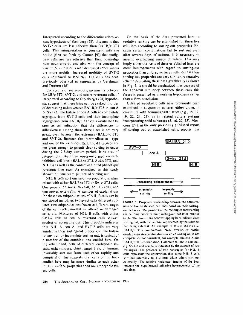
On the basis of the data presented here, a tentative ranking can be established for these five cell lines according to sorting-out properties. Be- cause certain combinations fail to sort out even after several days of culture, it is necessary to assume overlapping ranges of values. This may imply either that cells of these established lines are more heterogeneous with regard to sorting-out properties than embryonic tissue cells, or that their sorting-out properties are very similar. A tentative scheme presenting these data graphically is shown in Fig. 5. It should be emphasized that because of the apparent similarity between these cells this figure is presented as a working hypothesis rather than a firm conclusion.
Cultured neoplastic cells have previously been examined in suspension culture, either alone, in co-culture with nonmalignant tissues (e.g., 15, 17, 19, 22, 24, 25), or in related culture systems incorporating solid substrata (3, 16, 20, 35). Mos- cona (22), in the only previously published report of sorting out of established cells, reports that
I SVT-Z I con A
IBALB/c 3T$I
I
Increasing adhesiveness
externally internally < sorting sorting ~>
FIGURE 5. Proposed relationship between the adhesive- ness of five established cell lines based on their sorting- out behavior. The position of the rectangles representing the cell line indicates their sorting-out behavior relative to the other lines. Two nonoverlapping bars indicate clear sorting out, with the cell line represented by the leftmost bar being external. An example of this is the SVT-2- BALB/c 3T3 combination. Near overlap or partial overlap indicates combinations in which sorting out is not complete, or not consistent, for example, the con A and BALB/c 3T3 combination. Complete failure to sort out, e.g. SVT-2 and con A, is indicated by the overlap of two rectangles. The presence of two rectangles for NIL B cells represents the observation that some NIL B ceils sort out internally to 3T3 cells while others sort out externally. The relative horizontal lengths of the bars indicate the hypothesized adhesive heterogeneity of the cell lines.
284 THE JOURNAL OF CELL BIOLOGY �9 VOLUME 68, 1976
Dow
nloaded from http://rupress.org/jcb/article-pdf/68/2/276/1387337/276.pdf by guest on 09 January 2022
mouse me lanoma $91 cells sort out externally to chick embryonic cart i lage and internally to chick embryonic liver. By use of an isogenic family of cell lines ( B A L B / c 3T3:SVT-2:con A), we have been able to extend these compar isons to more similar combina t ions of cells than previously at- tempted.
C O N C L U S I O N S
Mouse B A L B / c 3T3 cells sort out internally to SVT-2 cells. This indicates tha t viral t rans forma- tion of these cells results in cell surface al terat ions tha t allow the cells to recognize each other in mixed cultures. The mechanism for this recogni- tion may operate through a change in the adhesive- ness of the cells, with the SVT-2 cells being less adhesive than the B A L B / c 3T3. Swiss 3T3 cells and a con A-selected rever tant of SVT-2 (con A cells) fail to sort out from B A L B / c 3T3 cells. On the other hand, cells of another contact- inhibi ted f ibroblast line, N I L B, do sort out from B A L B / c 3T3 cells. This suggests tha t the recognit ion phe- nomenon, acting during sorting out, is not specific to contact- inhibi ted cells.
The authors want to thank Josefina Buniel for her technical assistance. This research was supported by American Cancer Soci- ety Grant no. IN-57-L to Case Western Reserve Univer- sity and by a grant from the Cuyahoga County Unit of the American Cancer Society to Howard Gershman, and by National Cancer Institute grant no. I-ROI-CA13513 to Lloyd Culp.
Received for publication 19 June 1975, and in revised form 26 September I975,
R E F E R E N C E S
1. AARONSON, S. A., and G. J. TOOARO. 1968. Devel- opment of 3T3-1ike lines from BALB/c mouse embryo cultures: transformation susceptibility to SV40. J. Cell Physiol. 72:141-148.
2. ABERCROMBXE, M., J. E. M. HEAVSMAN, and H. M. KARTHAUSER. 1957. Social behavior of cells in tissue culture. 111. Mutual influence of sarcoma cells and fibroblasts. Exp. Cell Res. 13:276-291.
3. BARSKI, G., and J. BELEHRADEK. 1965. ~tude mi- crocin~matographique du m6chanism d'invasion canchreuse en cultures de tissue normal associ~ aux cellules malignes. Exp. Cell Res. 37:464 480.
4. BELL, P. B. 1972. Criss-crossing, contact inhibition, and cell movement in cultures of normal and trans- formed 3T3 cells. J. Cell Biol. 55(2, Pt. 2):16 a. (Abstr.).
5. BURDteK, M. L. 1970. Cell sorting out according to species in aggregates containing mouse and chick
embryonic limb mesoblast cells. J. Exp. Zool. 175:357-368.
6. CARTER, S. B. 1965. Principles of cell motility: the direction of cell movement and cancer invasion. Nature (Lond.). 208:1183-1187.
7. CARTER, S. B. 1967. Haptotaxis and the mechanism of cell motility. Nature (Lond.). 213:256-260.
8. CASStMAN, J. J., and M. R. BERNFIELD. 1974. Morphogenetic properties of human embryonic cells: aggregation of dissociated cells and histogenesis in cultured aggregates. Pediatr. Res. 8:184-192.
9. COMAN, D. R. 1944. Decreased mutual adhesiveness, a property of cells from squamous cell carcinomas. Cancer Res. 4:625-629.
10. CULV, L. 1974. Substrate-attached glycoproteins mediating adhesion of normal and virus-transformed mouse fibroblasts. J. Cell Biol. 63:71-83.
I I. CULp, L. A., and P. H. BLACK. 1972. Contact-inhib- ited revertant cell lines isolated from Simian virus 40-transformed cells. Ill. Concanavalin A-selected revertant cells. J. Virol. 9:611-620.
12. CULP, L. A., W. J. GRIMES, and P. H. BLACK. 1971. Contact-inhibited revertant cell lines isolated from SV40-transformed cells. I. Biologic, virologic, and chemical properties. J. Cell Biol. 50:682-690.
13. DELONG, G. R., and R. L. SIDMAN. 1970. Alignment defect of reaggregating cells in cultures of developing brains of reeler mutant mice. Dev. Biol. 22:584-600.
14. DIAMOND, L. 1967. Two spontaneously transformed cell lines derived from the same hamster embryo culture. Int. J. Cancer. 2:143-152.
15. DODSON, E. O. 1966. Aggregation of tumor cells. Nature (Lond.). 209:40 44.
16. EASTY, G. C., and D. M. EASTY. 1963. An organ culture system for the examination of tumor inva- sion. Nature (Lond.). 199:1104-1105.
17. FOLKMAN, J., M. HOCHBERG, and D. KNIGHTON. 1974. Self-regulation of growth in three dimensions: the role of surface area limitation. Cold Spring Harbor Conf. Cell Proliferation. 1:833-842.
18. GERSHMAN, H., and J. DRUMM. 1975. Mobility of normal and virus-transformed cells in cellular aggre- gates. J. Cell Biol. 67:419-435.
19. HALPERN, B., B. PEJSACHOWICZ, H. L. LEBVRE, and G. BARSKI. 1966. Differences in patterns of aggrega- tion of malignant and non-malignant mammalian cells. Nature (Lond.). 209: i 57-159.
20. HEAYSMAN, J. E. M., and S. M. PEGRUM. 1973. Early contacts between normal fibroblasts and mouse sarcoma cells. Exp. Cell Res. 78:479-481.
21. LUDFORD, R. J. 1932. Differences in the growth of transplantable tumors in plasma and serum culture media. Proc. R. Soc. Lond. B. Biol. Sci. 112: 250-263.
22. MOSCONA, A. 1957. The development in vitro of chimeric aggregates of dissociated embryonic chick and mouse cells. Proc. Natl. Acad. Sci. U. S. A. 43:184 194.
23. MOSCONA, A., and H. MOSCONA. 1952. The dis-
H. GERSHMAN, J. DRUMM, and L. CULV Sorting Out o f Normal and Transformed Cells 285
Dow
nloaded from http://rupress.org/jcb/article-pdf/68/2/276/1387337/276.pdf by guest on 09 January 2022
sociation and aggregation of cells from organ rudi- ments in the early chick embryo. J. Anat. 86:287-301.
24. MOSKOWITZ, M. 1963. Aggregation of cultured mammalian cells. Nature (Lond.). 200:854 856.
25. OPPENHEIMER, S. B., i . EOIDIN, C. W. ORR, and S. ROSEMAN. 1969. An L-glutamine requirement for intercellular adhesion. Proc. Natl. Acad. Sci. U. S. A. 63:1395 1402.
26. RUBIN, H. 1966. Fact and theory about the cell surface in carcinogenesis. In Major Problems in Developmental Biology. M. Locke, editor. Aca- demic Press, Inc., New York. 315-337.
27. STEINBERG, M. S. 1964. The problem of adhesive selectivity in cellular interactions. In Cellular Mem- branes in Development. M. Locke. editor. Aca- demic Press, Inc., New York. 321-366.
28. STEINBERG, M. S. 1970. Does differential adhesion govern self-assembly processes in histogenesis? Equi- librium configurations and the emergence of a hier- archy among populations of embryonic cells. J. Exp. Zool. 173:395-434.
29. STOKER, M. 1964. Regulation of growth and orienta- tion in hamster cells transformed by polyoma virus.
Virology. 24:165-174. 30. SUBAK-SHARPE, H., R. R. BURK, and J. D. P1TTS.
1969. Metabolic co-operation between biocbemically marked mammalian cells in tissue culture. J. Cell Sci. 4:353-367.
31. TOWNES, P. L., and J. HOLTFRETER. 1955. Directed movements and selective adhesion of embryonic amphibian cells. J. Exp. Zool. 128:53-120.
32. VASILIEV, J. M., and I. M. GELFANO. 1972. Interac- tions of normal and neoplastic fibroblasts with the substratum. Ciba Found. Syrup. 14:311 329.
33. WESTON, J. A., and S. A. ROTH. 1969. Contact inhibition: behavioral manifestations of cellular adhesive properties in vitro. In Cellular Recognition. Smith and Good, editors. Appleton-Century-Crofts, New York. 29-37.
34. WISEMAN, L. L. 1970. Experimental modulation of the assembly properties of embryonic cell popula- tions. Ph.D. Thesis. Princeton University, Princeton, N.J.
35. WOLFF, E., and N. SCHNEIDER. 1957. La culture d'un sarcome de souris sur des organes de poulet explantes in vitro. Arch. Anat. Microsc. Morphol. Exp. 46:173-197.
2~6 THE JOURNAL OF CELL BIOLOGY �9 VOLUME 68, 1976
Dow
nloaded from http://rupress.org/jcb/article-pdf/68/2/276/1387337/276.pdf by guest on 09 January 2022