(p:art 1 of 3). revision of microcionidae (porifera: demospongiae), with description of australian...
TRANSCRIPT
MEMOIRS OF THE
QUEENSLAND MUSEUM BRISBANE
© Queensland Museum PO Box 3300, South Brisbane 4101, Australia
Phone 06 7 3840 7555 Fax 06 7 3846 1226
Email [email protected] Website www.qm.qld.gov.au
National Library of Australia card number
ISSN 0079-8835
NOTE Papers published in this volume and in all previous volumes of the Memoirs of the
Queensland Museum maybe reproduced for scientific research, individual study or other educational purposes. Properly acknowledged quotations may be made but queries regarding the republication of any papers should be addressed to the Editor in Chief. Copies of the journal can be purchased from the Queensland Museum Shop.
A Guide to Authors is displayed at the Queensland Museum web site
A Queensland Government Project Typeset at the Queensland Museum
REVISION OF MICROCIONIDAE (PORIFERA: POECILOSCLERIDA:DEMOSPONGIAE), WITH DESCRIPTION OF AUSTRALIAN SPECIES.
JOHN N.A. HOOPER
Hooper, J.N.A. 1996 07 01: Revision of Microcionidae (Porifera: Poecilosclerida:Demospongiae), with description of Australian species. Memoirs of the Queensland Museum40: 1-626. Brisbane ISSN 0079-8835.
A phylogenetic revision of the poecilosclerid Microcionidae is based on type material, theworldwide literature, and comprehensive Australian collections. Of 73 available genericnames 7 genera and 12 subgenera are recognised here. Of 561 available species names 459are considered valid (10 virtually unrecognisable), including 52 new species. The Australianfauna, including Australian Antarctic Territory, contains 148 species (31 new), many newrecords, most are new combinations, and many illustrated for the first time. A synonymy ofworld species is provided. Valid taxa include: Clathria (with 7 subgenera: C. (Clathria) (with31 Australian species, 82 other species worldwide, with new species murphyi, noarlun-gae,biclathrata, borealis, burtoni , sarai, saraspinifera), C. (Wilsonella) (6, 8, abrolhosensis,ensiae, lindgreni spp. nov.), C. (Microciona) (5, 91, illawarrae, lizardensis, simae,brondstedi, campecheae, claudei, hen tscheli, leighensis, stephensae, tunisiae, urizae,vacelettia spp. nov.), C. (Dendrocia) (7 species endemic to Australia), C. (Axociella) (6, 6,patula , fromontae, georgiaensis spp. nov.), C. (Isociella) (4, 1, selachia, skia spp. nov.), andC. (Thalysias) (36, 53, aphylla, craspedia, darwinensis, fusterna, hallmanni, hesperia,lematolae, phorbasiformis, styloprothesis, tin gens, wesselensis, amiranteiensis, hechtelispp. nov.); Antho (with 3 subgenera: A. (Antho) (12, 10), A. (Plocamia) (2, 17) and A.(Isopeneclya) (3,1, punicea, saintvincenti spp. nov.); Echinoclathria (14, 15, bergquistae,levii, notialis, parkeri, riddlei spp. nov.); Holopsamma (9 species endemic to Australia, 1indeterminate species); Echinochalina (with 2 subgenera: E. (Echinochalina) (10, 2, felizisp. nov.), E. (Protophlitaspongia) (8 species endemic to Australia and New Caledonia,collata, favulosa, isaaci, tuberosa spp. nov.)); Artemisina (4, 10); and Pandaros (incertaesedis) (0, 2). Generic keys are provided. Morphometric characters of primary importanceinclude the origin, geometry and distribution of structural megascleres within the skeleton,modification of megascleres to monactinal or diactinal forms, the presence or absence of aspecialised ectosomal skeleton, presence of detritus incorporated into spongin fibres, andoverall skeletal structure (including compression of the axial skeleton and differentiation ofaxial and extra-axial regions). Brief zoogeographical comparisons are made between con-tinental Australian and adjacent Indo-west Pacific faunas. Australian species comprise about32% of the world's microcionid diversity; about 75% of species are endemic for theAustralian region, and temperate species (81%) have higher levels of endemism than tropicalspecies (59%). El Porifera, Demospongiae, Poecilosclerida, M icrocionidae, family revision,new species, taxonomy, biogeography, Australia.
John N.A. Hooper, Queensland Museum, PO Box 3300, South Brisbane, Queensland, 4101,Australia; received] December 1995.
INTRODUCTION
Microcionidae is one of the largest families ofDemospongiae, comprising about 8% of alldescribed (extant) Porifera species (Hooper &Levi, 1993a). The family has contained at onetime or another about 70 genera and 550 species,although fewer than these are now recognised asvalid. The family has a worldwide distributionand it is found from the intertidal zone to depthsexceeding 2000m. It is clearly one of the moreimportant, ecologically successful groups ofPorifera.
Within the Indo-Australian region microcionidsare particularly abundant, with some speciesbeing dominant components of the shallow watermacrobenthos. Previous works describing thisfauna (and other literature containing extra-limi-tal records of Australian species) include:Lamarck (1814, 1815, 1816), Gray (1858, 1867,1869, 1870), Bowerbank (1864, 1875, 1877), Bar-nard (1879), Kent (1871), Ridley (1884a), Ridley& Dendy (1886, 1887), Lendenfeld (1888, 1889a),Kieschnick (1896, 1900), Thiele (1898, 1899,1900, 1903), Kirkpatrick (1900a, b), Whitelegge,(1901, 1902), Hentschel (1909, 1911, 1912),
2^ MEMOIRS OF THE QUEENSLAND MUSEUM
Hallmann (1912, 1914a-c, 1916a-c, 1920),Dendy & Frederick (1924), Topsent (1897b,1930, 1932, 1933), Burton (1934a), Bergquist &Tizard (1967), Kelly-Borges & Bergquist (1988),Bergquist & Fromont (1988), Wiedenmayer(1989), Hooper (1990b), Hooper et al. (1991,1992), Hooper & Levi (1993a, 1994). A briefsynopsis of the fauna is given by Hooper &Wiedenmayer (1994), although some of thenomenclature and synonymies contained in thatearlier work are revised here.
Prior to the present study more than 200 speciesof Microcionidae had been described in theAustralian fauna (including its territorial waters),but many of these were found to be either com-posite (consisting of several sibling species), orsynonyms of other species. This study 1)describes 148 species (31 new), many newlocality records for Australia and new taxonomiccombinations; 2) provides an annotatedsynonymy for 311 other species worldwide (in-cluding 21 new species); 3) revises the mor-phometric characters used for classification andpopulation variability for particular species; and4) determines levels of endemism amongstprovincial faunas.
MATERIALS AND METHODS
COLLECTION AND HISTOLOGICAL TECH-NIQUES. Material examined in this study waspredominantly collected using SCUBA (0-40mdepth) or dredging and trawls (30-360m depth).Seasonal sampling for reproductive periodicitywas conducted over two years in the Darwin andCobourg Peninsula regions, NT. Immediatelyafter collection specimens were either fixed in80-100% methylated ethanol or frozen (which tosome extent fixes the pigments), and laterpreserved in 70% alcohol. Reproductive productswere searched for in fresh or frozen tissue.
Nitric acid spicule preparations, thick-sectionsand thin-section mounts were routinely made asfollows. Fragments of each sponge, includingectosomal and choanosomal regions, were heateddirectly on a glass microscope slide in severaldrops in nitric acid (the solution was evaporatedrather than boiled, using low heat), and mountedin Canada balsam once completely dry, andcooled. Thick, hand-cut sections were made per-pendicular to the surface, soaked in a saturatedsolution of phenol and xylene (for approximately24 hours), and mounted in Durcupan (ACMFluka Products) using glass slivers or card to raisethe coverslip level. Phenol-xylene precluded the
necessity for dehydration through an alcoholseries. Some microtome sections cut at 30-35mwere made for each species. Fragments werepassed through a dehydration series, cleared intoluene or Histosol, and wax embedded for atleast 2 hours. Sections were cut from trimmedwax blocks (cutting from the centre of the blockto the exterior so as to include both the outersurface and inner skeleton relatively intact),placed in clearing agent for an adequate period todissolve wax and/or dewaxing on a hot plate, thensoaked in ethanol until perfectly clear, floatedonto albumen-coated slides, orientated and flat-tened, stained with basic fuccsin and mounted.Fragments of dry specimens (e.g., type material)were reconstituted in 5% buffered fonnalin for 12hours, which produced rehydration of themesohyl and enabled cleaner histological sec-tions to be made.
MORPHOMETRIC ANALYSES. Spicu les weremeasured with a stage micrometer, either directlythrough a microscope or computer digitiser.Twenty five spicules, of each spicule category, inall specimens were measured. Acanthostylewidth measurements were taken immediatelybelow the base. Toxa lengths refer to chordlength; isochelae are measured from apex of alae;width measurements of other spicules refer tomaximum width.
Spicule dimensions were sorted and statistical-ly compared for various parameters (e.g., season,locality, depth), including one- and two-wayANOVs with replication, two-way ANOV withunequal replication, means differentiated usingtwo-tailed t tests. Line-drawings were made usinga calibrated camera lucida, and microphotographswere taken with an Olympus microphoto system.Taxonomic keys were constructed using orderedmultistate, disordered multistate, morphometricand binary characters, utilising the DELTA sys-tem (Dallwitz & Paine, 1986).
SCANNING ELECTRON MICROSCOPY. Sec-tions were prepared as follows:
1) Cut at 1-1.5mm thick, ensuring that both theectosome and choanosome were represented.
2) Placed in a cavity block and covered withseveral drops of sodium hypochlorite to etch themesohyl matrix from the skeleton. The etchingprocess was monitored through a dissectingmicroscope in order to prevent the skeleton fall-ing apart. Delicate structures (plumose,halichondroid, hymedesmoid skeletons) only re-quired a few seconds treatment with bleach;robust skeletons (reticulate, fibrous, articulated
REVISION OF MICROCIONIDAE^ 3
skeletons) required several minutes; but general-ly 30 seconds was adequate.
3) Bleach was pipetted off at the appropriatetime and 70% ethanol immediately added. Sec-tions were left to stand for several minutes toensure bleach was completely neutralised.
4) Steps 2-3 were repeated, without removingsection from cavity block, substituting con-centrated hydrogen peroxide in place of sodiumhypochlorite, finally rinsing in ethanol. Thehydrogen peroxide step was omitted for verydelicate sections.
5) Sections were placed on clean microscopeslides and let dry completely.
6) Sections mounted on SEM stubs usingdouble-sided tape, copper dag, or `Supa Glue'(Supa Glue, Selleys Chemical Company,Padstow). An alternative method used to fixsamples to stubs was to cover stub with`Aquadhere' wood glue (Aquadhere, SelleysChemical Company, Padstow), let dry complete-ly (usually several days), then prior to use ex-posed dry glue to vigorous steam (which softenedthe set glue), and placed the section on top of thestub (it would sink in a short way but was bondedreasonably well to the stub, and had the advantageof producing a perfectly smooth background).
7) The stub was sputter-coated well to ensurethat all fibres were well coated to reduce'charging'. In some cases uncoated sectionscould be viewed successfully under low ac-celerator voltage, but better results were general-ly obtained on coated specimens at higher voltage.Typical viewing conditions used were 25kV, atclose working distance to provide best depth offield and focus, and at low magnifications.
Spicule were prepared as follows:1) Thinly cut sections including both ectosome
and choanosome were placed in a durham tube(micro-test tube), to which drops of concentratednitric acid are added, using drop-by-drop additionso as to control the oxidation reaction and produc-tion of by-product oxides.
2) Upon completion of acid digestion the dur-ham tube was half filled with acid and gentlyheated over an alcohol flame, ensuring that onlysmall bubbles form (low heat, no boiling), for 1-2minutes.
3) Solution was let stand to cool, thencentrifuged (approximately 4000rpm for 30seconds).
4) Nitric acid was pipetted off leaving a spiculemass at the bottom of the tube, undisturbed.
5) Spicules were resuspended in fresh nitricacid and gently stirred using clean, fine, glass rod.
6) These steps were repeated if any collagenremained.
7) Spicules were resuspended firstly indemineralised water, 70% ethanol, then twoseries of 100% ethanol solutions, centrifugingand decanting the supernatant between eachchange of solution, finally ending with suspendedspicules in a solution of absolute ethanol.
8) A micro-cover glass was adhered to an SEMstub using double-sided tap or copper dag, severaldrops of suspended spicules placed onto the coverglass, the alcohol-spicule solution ignited andspread across the glass with a glass rod or forcepsuntil all ethanol was vaporised. Spicules bond toglass relatively firmly, but excess spicules couldbe blown off glass using compressed air, orspread out over the glass by adding furtherethanol and igniting. The distribution of spiculeson the cover glass was monitored under com-pound or dissecting microscope (magnificationdepending on spicule size). More drops of spiculesolution added and this step repeated if too fewspicules were present, ensuring not to overcrowdfield of view for SEM photographic purposes.
9) An alternative method was used to producea perfectly smooth background, using an`Aquadhere' glue-coated stub, dried for severaldays then softened with steam, and spiculesplaced directly onto soft glue (in this case ethanolwas not burnt but evaporated). Single spiculeswould sink into glue too far if it was too soft (i.e.,left in steam too long).
10) Spicule coated stubs were sputter coatedbriefly and viewed at 25kV, minimum workingdistance and smallest apperture for best resolu-tion.
ABBREVIATIONS
AAT, Australian Antarctic Territories; ABIP,Centro de Estudios Avanzados de Blanes, In-stitut° de Investigaciones Pesqueras Barcelona,Aquarium de Blanes, Gerona; ABRS, AustralianBiological Resources Survey, Canberra; AFZ,Australian Fishing Zone; AHF, Alan HancockFoundation, University of Southern California,Los Angeles; AIMS, Australian Institute ofMarine Science, Townsville; AM, AustralianMuseum, Sydney; AMNH, American Museumof Natural History, New York; BMNH, TheNatural History Museum, London; BPBM, Ber-nice P. Bishop Museum, Honolulu; CP, CobourgPeninsula, NT; CPMNP, Cobourg PeninsulaMarine National Park, NT; CSIRO, Common-wealth Scientific and Industrial Research Or-
4^ MEMOIRS OF THE QUEENSLAND MUSEUM
ganisation, Marine Laboratories, Hobart,Cleveland and Perth; DAR, Darwin region, NT;DELTA, Description Language for Taxonomycomputer software (Dallwitz & Paine, 1986);EIS, Environmental Impact Study; CSIROEMG,CSIRO Food Research Laboratory, Division ofFood Processing, North Ryde, Sydney. EMU,Ensight (formerly Environmental ManagementUnit), Water Board (Sydney, Illawarra, BlueMountains), Sydney; EPA, Environment Protec-tion Authority, Sydney; EPALR, East PointAquatic Life Reserve, Dudley Point, Darw;n Har-bour, NT; FNQ, far northern Queensland (Cook-town to Torres Straits); FUB, Freie UniversitatBerlin; GBR, Great Barrier Reef, Queensland;HNUK, Natural History Museum, Ham NamUniversity, South Korea; ICBUC, InstitutoCentral de Biologia, Universidad de Concepcion,Chile; ICZN, International Code of ZoologicalNomenclature (see Anonymous, 1985); IM, In-dian Museum (Zoological Survey of India), Cal-cutta; IMZUB, Istituto e Museo di Zoologia edAnatomia Comparata della Universita di Bari,Bari; IMZUN, Instituto e Museo di Zoologiadell'Universita di Napoli, Naples; INM, NationalMuseum of Ireland, Dublin; IZUG, MuseoCivico di Storia Naturale di Genova, Genova;JCU, James Cook University of NorthQueensland, Townsville; KFAU, ZoologischenSammlung der Universitat Erlangen-Niirnberg,Erlangen; LFM, Merseyside County Museums(formerly Liverpool Free Museum), Liverpool;LMJG, Abteilung fiir Zoologie am Landes-museum Joanneum (Landes Museum JubileumGraz), Graz; MABA, Museo Argentino de Cien-cias Naturales 'Bernardino Rivadavia', EaenosAires; MCNP, Div. Invest. del Museo de CienciasNaturales de la Plata, Argentina; MCZN,Museum of Comparative Zoology, HarvardUniversity, Cambridge (Mass.); MEQ, mid east-ern Queensland (Gladstone to Bowen); MHNG,Museum d'Histoire NatureIle de Geneve,Geneve; MLUM, Marine Laboratory of theUniversity of Miami, Miami; MMBS,Mukaishima Marine Biological Station, Facultyof Science, Hiroshima University, Onomichi;MNHN, Museum National d'Histoire Naturelle,Laboratoire de Biologie des Invertebres Marinset Malacologie, Paris (DT, Topsent collections;DCL, Levi collections; DJV, Vacelet collections;DNBE, Boury-Esnault collections); MOM,Musee Oceanographique de Monaco, Monaco;MRAC, Koninklijk Museum voor Midden-Afrika, Tervuren; MRHN, Musee Royal d'-Histoire Naturelle de Belgique, Bruxelles; MTQ,
Queensland Museum, Museum of TropicalQueensland, Townsville; NCIQ66C-, UnitedStates National Cancer Institute, Australian In-stitute of Marine Science shallow water collec-tion contract (1984-91), Townsville (primaryvoucher samples now lodged in QM, others inNTM and USNM); NC1OCDN-, United StatesNational Cancer Institute, Coral Reef ResearchFoundation shallow water collection contract,Chuuk State (voucher samples lodged in QM andUSNM); NEQ, northeast Queensland (Bowen toCooktown); NM, Natal Museum, Pieter-maritzburg; NMB, Naturhistorisches Museumszu Basel, Basel; NMCIC, National Museum ofNatural Sciences, National Museums of Canada,Ottawa; NMNZ, National Museum of NewZealand (formerly Dominion Museum), Wel-lington; NMV, Museum of Victoria (formerlyNational Museum of Victoria), Melbourne;NSM, National Science Museum, Tokyo; NSW,New South Wales; NT, Northern Territory; NTM,Northern Territory Museum of Arts and Sciences,Darwin; NTU, Northern Territory University,Darwin; NWS, Northwest Shelf region, WesternAustralia; PAUP, Phylogenetic Analysis UsingParsimony (see Swofford, 1991); PIBOC, PacificInstitute of Bio-organic Chemistry, Far EastScientific Centre, Academy of Sciences of theUSSR, Vladivostok; PMJ, Phyletisches Museum,Jena; PNG, Papua New Guinea; QFS,Queensland Fisheries Service, Department ofPrimary Industries, Brisbane and Cairns; QLD,Queensland; QM, Queensland Museum, Bris-bane; QVML, Queen Victoria Museum and ArtGallery, Launceston; RMBS, Roscoff MarineBiological Station, Roscoff, France; RMNH,Rijksmuseum van Natuurlijke Historie, Leiden;RRIMP, Roche Research Institute of MarinePharmacology, Sydney (discontinued; spongecollections now held in AM); RSME, Royal Scot-tish Museum, Edinburgh; SA, South Australia;SAM, South Australian Museum, Adelaide; SEQ,southeast Queensland (Tweed River toGladstone); SM, Musee Zoologique, Strasbourg;SME, Station Marine d'Endoume, Marseille;SMF, Natur-Museum und ForschungsinstitutSenckenberg, Frankfurt; TAS, Tasmania; TM,Museo e Istituto di Zoologia Sistematica dell'-Universita di Torino, Torino; TMAG, TasmanianMuseum and Art Gallery, Hobart; UAZD,University of Auckland, Zoology Department,Auckland; UB, Ubersee-Museum, Bremen;UCT, South African Museum of Natural History,Cape Town; UQ, University of Queensland, Bris-bane; USC, University of Southern California,
REVISION OF MICROCIONIDAE^ 5
Los Angeles; USNM, National Museum ofNatural History, Smithsonian Institution,Washington DC; UZM, Zoologisk Museum,Universitetsparken, Copenhagen; VIC, Victoria;WA, Western Australia; WAM, WesternAustralian Museum, Perth; YPM, PeabodyMuseum of Natural History, Yale University,New Haven (Conn.); ZIL, Zoological Institute ofLeningrad, Academy of Sciences Museum ofZoology, St Petersburg; ZMA, ZoOlogischMuseum, Universiteit van Amsterdam, Amster-dam; ZMB, Museum ftir Naturkunde an derHumboldt-Universitat zu Berlin, Berlin; ZMC,Zoologisk Museum, Copenhagen; ZMH,Zoologisches Institute und ZoologischesMuseum der Universitat Hamburg, Hamburg;ZMUU, Uppsala Universitets ZoologiskaMuseet, Zoologiska Institutet, Uppsala; ZSN,Aquarium e Museo della Stazione Zoologica diNapoli, Naples; ZRS, Zoologiska Rijkmuseum,Stockholm.
ACKNOWLEDGEMENTS
Patricia Bergquist (UAZD) encouraged thisstudy and provided inspiration to persevere withthe long learning curve associated with spongetaxonomy. Felix Wiedenmayer (NMB) providedaccess to his numerous unpublished personalnotes on museum collections (Sponge Archives,NMV; Wiedenmayer, 1989; Hooper & Wieden-mayer, 1994). Rob Van Soest (ZMA) providednumerous discussions on sponges, alternativeviews on the diagnostic importance and polarityof characters, and possible relationships betweensponge groups. Michelle Kelly-Borges (BMNH)and Peter Jell (QM) provided many positive com-ments on the manuscript.
I am particularly grateful to Claude Levi(MNHN) for providing a post-doctoral fellow-ship at the MNHN and giving me access to itstype collections, including all the Lamarckmaterial; the Sir Winston Churchill MemorialTrust (Canberra), the Australian BiologicalResources Study (Canberra), Klaus Riitzler(USNM), Willard Hartman (YPM), JoachimReitner (FUB, now Gottingen) and the Trusteesof the Queensland Museum (QM) for providinggrants at various times, enabling me to examinemajor Museum collections and to interact withcolleagues at several international forums (fromwhich many of the ideas in this present volumematured). The Northern Territory UniversityPlanning Authority (1983-1985), the Museumsand Art Galleries Board of the Northern Territory
(1983-1989) and Heritage Commission of theNorthern Territory (Darwin) provided additionalfunding to visit remote localities and the scatteredcollections in Australian museums.
I am particularly grateful to Leonie Hooper forline drawings and John Kennedy for many of theSEMs. I also thank Bob Hardy (UQ), CharlesWebb (NTU), and Clive Wilkinson (AIMS) forproviding assistance with SEM photography.
For financial or logistic assistance withfieldwork I acknowledge: George Elyakov,Valodya Krasochin, Y. Yakovlev, USSR RV`Akademik Opafin' (PIBOC); Alice Kay, LesterCannon (QM) and Queensland Fisheries Service;Mick Ready (FV 'Hydronaue); Peter Murphy,Martin Riddle, Shirley Sorokin, Rob McCauleyand other members of the NCI team (AIMS);Danny Roberts (EPA); Scott Chidgey, CalwellConnor and Associates; Patricia Byers (FV`Skeleton'); Bill Rudman (AM); Ian Poiner andTrevor Ward (CSIRO Fisheries, RV `Soela', RV'Sprightly', FV 'Clipper Bird', RV 'SouthernSurveyor'); Martin Riddle and Lisa Miller(EMU); Rob Capon (University of Melbourne);Alan Butler (University of Adelaide); Clay Bryce(WAM); Neville Coleman (Australian MarinePhotographic Index, Brisbane); ConservationCommission of the Northern Territory, Darwin;Darryl Grey, Dave Ramm and Anne Coleman(NT Fisheries Darwin); Neil Smit (NT UniversityDarwin) Barry Russell and Helen Larson (NTM);C.C. Lu (NMV); Cecile Debitus, George Bar-gibant, Jean-Louis Menou, Pierre Laboute(ORSTOM Noumea); Pat and Lori Colin (NCICRRF Chuuk and Palau); and Ian and Pam Low(FV 'Rachel').
I am grateful for competent field assistance andlaboratory technical assistance, during variousparts of this study, from Jodie Baxter, StevenCook, Lisa Hobbs, Alen Howard, CathyJohnston, John Kennedy, Daniel Loy Choy,Anne-Marie Mussig, Paula Tomkins and RexWilliams. I also thank Phil Alderslade (NTM) forassistance in developing computer digitisingsoftware, and Russell Hanley (NTM) for identify-ing commensal polychaetes.
I also thank many people for providing materialfor examination, or for other information cited inthe text: Penny Barents (AM), Nicole Boury-Es-nault (SME), Beatrice Burch (BPBM), SusanChambers (RSME), Frank Climo (NMNZ), RuthDesqueyroux-Faundez (MHNG and LMJG),Jane Fromont (JCU), Manfred Grasshoff (SMF),Jan Den Hartog (RMNH), Takomura Hoshino(MMBS), Frank von Knorre (PMJ), Deiter
6^ MEMOIRS OF THE QUEENSLAND MUSEUM
Kuhlmann (ZMB), Vladimir Krasochin(PIBOC), Romely Lockyer (CootamundraShoals Survey team, UK), Susan Boyd (NMV),Liz McCaffrey (UQ Brisbane), A.K. Mandal(IM), Loisette Marsh (WAM), C. O'Riordan(INM), Shane Parker (SAM), David Parry (NTUDarwin), Urs Rahm (NMB), Martin Riddle(EMU), Frank Rowe (AM), Klaus Riitzler andKathleen Smith (USNM), Shirley Stone(BMNH), B.R. Stuckenberg (NM), Ole Tendal(ZMC), Jean Vacelet (SME), Clare Valentine(BMNH), Clive Wilkinson (AIMS), andWolfgang Zeidler (SAM).
DEFINITION OF CHARACTERS
MINERAL SKELETON. The form, compositionand division of the skeleton remains the mostimportant character for classification ofDemospongiae. Recent attempts at higher sys-tematics of Demospongiae based on non-skeletalcharacters (e.g., Simpson, 1968a; Bergquist &Hartman, 1969; Bergquist, 1980a; Lee &Gilchrist, 1985; Hooper et al., 1992) have hadonly limited success because in many instancesthey are unable to corroborate all skeletal andnon-skeletal evidence into a single systematics.In some cases amongst Demospongiae (e.g.,Verongida), non-skeletal evidence has beendecisive and to some extent well correlated withother characters. In other cases (e.g., Axinellida)that evidence has merely highlighted inade-quacies in systematics based solely on skeletalmorphology (Simpson, 1968a; Bergquist &Hartman, 1969; Bergquist, 1980a; Vacelet, 1985;Hooper et al., 1992).
COMPOSITION OF THE SKELETON. Allmicrocionids are siliceous with discrete, freespicules. So far no desma-bearing species or hy-percalcified 'relict' species are known. Manyspecies undergo secondary acquisition, loss orreduction of spicule mineralisation, particularlywhen displaced by arenaceous particles (e.g.,Holopsamma). C. (Wilsonella) is partly definedby this feature, with various degrees ofarenaceous development among species; thistrend is widespread throughout thePoecilosclerida.
ORGANISATION OFTHE SKELETON. Gross organicand inorganic skeletal architecture, structural dif-ferentiation of the inorganic skeleton, and dis-tribution of mineral components in that structureare primary diagnostics (Levi, 1960a, 1973;Bergquist, 1978a; Hartman, 1982). However,
when used alone skeletal architecture is notnecessarily a reliable indicator of relationships.Hooper (1991, herein) noted that the so-calledtypical, compressed, axinellid-like skeleton ofmany Raspailiidae (Raspailia pinnatifida;Hooper, 1991: Fig.2b) also occurred in C.(Axociella) (Figs 7G, 119A). It is also probablethat skeletal structure is influenced to some extentby environmental conditions, and there is someevidence to suggest that flexible, compressedaxial skeletons are produced in response to highenergy environments (e.g., Palumbi, 1984).Similarly, skeletal characters such as those foundin encrusting species have obviously evolved in-dependently in many (otherwise unrelated) taxa.Review of microcionid skeletal structuresshowed that species which were similar in spiculegeometry had different skeletal architectures(Hooper, 1988).
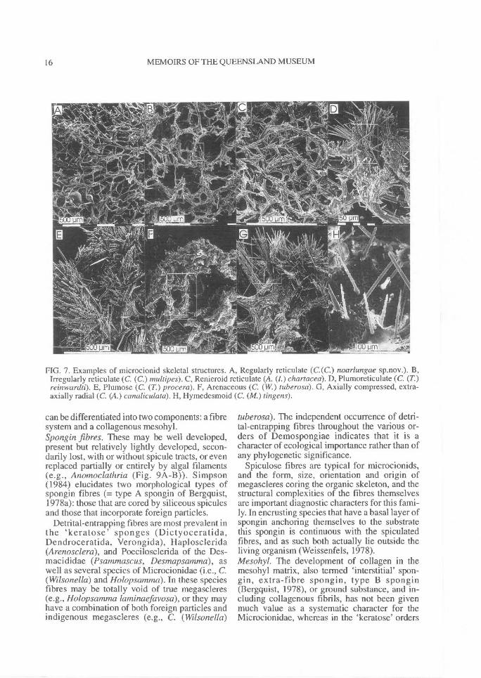
Architectural types amongst the Microcionidaeinclude: 1, hymedesmoid (with thin layer of basalspongin lying on the substrate containing erectmegascleres (Fig. 7H); 2, microcionid (with acompressed basal spongin, producing ascendingfibre nodes and plumose spicule columns) (Fig.100F); 3, renieroid reticulate (consisting of arectangular basal reticulation of uni- orpaucispicular tracts fully enclosed within sponginfibres or cemented at their nodes by loose col-lagen) (Fig. 7C); 4, isotropic reticulation (a dis-orientated, random uni-, pauci- or multispicularreticulation in erect or massive forms, in whichthere is no distinction between primary or secon-dary tracts (not figured; seen only in nominalgenus Qasimella); 5, isodictyal reticulation(reticulation with triangular meshes formed byuni- or paucispicular tracts of spicules, cementedat their nodes by collagen or fully enclosed withinspongin fibres) (Fig. 131A); 6, regularly or ir-regularly reticulate (with large multispiculartracts and/or fibres forming irregular oval or rec-tangular meshes (Fig. 7A-B); 7, plumo-reticulate(producing ascending and consecutively diverg-ing tracts and fibres, forming pauci- or multi-spicular primary lines, and interconnected bytransverse uni- or paucispicular tracts and fibres)(Fig. 7D); 8, dendro-reticulate (similar to thepreceding, but where ascending tracts are sinuousand more obviously diverging and branching thanthe less conspicuous transverse elements) (Fig.231C); 9, plumose (with ascending and divergingprimary lines that are not connected by transverseelements) (Fig. 7E); 10, axially or basally com-pressed (having a skeleton clearly divided into acompressed central or basal core of fibres and/or
REVISION OF MICROCIONIDAE^ 7
12
SUBECTOSOME
CHOANOSOME
SPONGIN FIBRE
4568
spicules, forming tight anastomosing meshes,and from which arise plumose or plumoreticulateextra-axial (subectosomal) fibres and/or spicules)(Fig. 7G). Some species have combinations ofthese skeletal structures with different structuraltypes found in different parts of the skeleton (e.g.,axis and periphery).
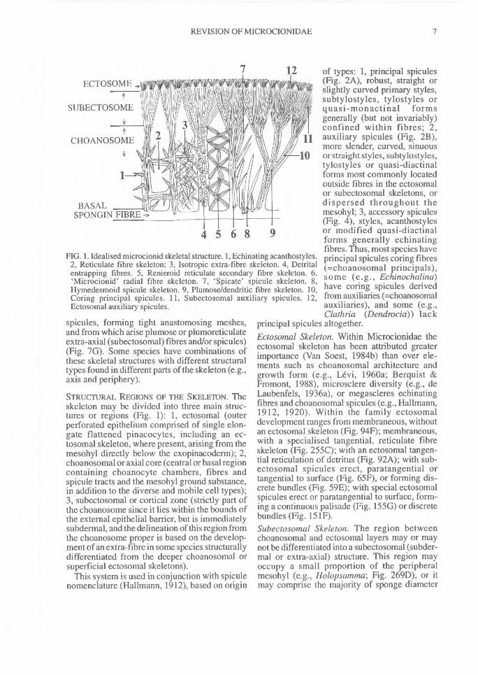
STRUCTURAL REGIONS OF THE SKELETON. Theskeleton may be divided into three main struc-tures or regions (Fig. 1): 1, ectosomal (outerperforated epithelium comprised of single elon-gate flattened pinacocytes, including an ec-tosomal skeleton, where present, arising from themesohyl directly below the exopinacoderm); 2,choanosomal or axial core (central or basal regioncontaining choanocyte chambers, fibres andspicule tracts and the mesohyl ground substance,in addition to the diverse and mobile cell types);3, subectosomal or cortical zone (strictly part ofthe choanosome since it lies within the bounds ofthe external epithelial barrier, but is immediatelysubdermal, and the delineation of this region fromthe choanosome proper is based on the develop-ment of an extra-fibre in some species structurallydifferentiated from the deeper choanosomal orsuperficial ectosomal skeletons).
This system is used in conjunction with spiculenomenclature (Hallmann, 1912), based on origin
of types: 1, principal spicules(Fig. 2A), robust, straight orslightly curved primary styles,subtylostyles, tylostyles orquasi-monactinal formsgenerally (but not invariably)confined within fibres; 2,auxiliary spicules (Fig. 2B),more slender, curved, sinuous
10 or straight styles, subtylostyles,tylostyles or quasi-diactinalforms most commonly locatedoutside fibres in the ectosomalor subectosomal skeletons, ordispersed throughout themesohyl; 3, accessory spicules(Fig. 4), styles, acanthostylesor modified quasi-diactinalforms generally echinatingfibres. Thus, most species haveprincipal spicules coring fibres(=choanosomal principals),some (e.g., Echinochalina)have coring spicules derivedfrom auxiliaries (=choanosomalauxiliaries), and some (e.g.,Clathria (Dendrocia)) lack
principal spicules altogether.Ectosomal Skeleton. Within Microcionidae theectosomal skeleton has been attributed greaterimportance (Van Soest, 1984b) than over ele-ments such as choanosomal architecture andgrowth form (e.g., Levi, 1960a; Berquist &Fromont, 1988), microsclere diversity (e.g., deLaubenfels, 1936a), or megascleres echinatingfibres and choanosomal spicules (e.g., Hallmann,1912, 1920). Within the family ectosomaldevelopment ranges from membraneous, withoutan ectosomal skeleton (Fig. 94F); membraneous,with a specialised tangential, reticulate fibreskeleton (Fig. 255C); with an ectosomal tangen-tial reticulation of detritus (Fig. 92A); with sub-ectosomal spicules erect, paratangential ortangential to surface (Fig. 65F), or forming dis-crete bundles (Fig. 59E); with special ectosomalspicules erect or paratangential to surface, form-ing a continuous palisade (Fig. 155G) or discretebundles (Fig. 151F).Subectosotnal Skeleton. The region betweenchoanosomal and ectosomal layers may or maynot be differentiated into a subectosomal (subder-mal or extra-axial) structure. This region mayoccupy a small proportion of the peripheralmesohyl (e.g., Holopsamma; Fig. 269D), or itmay comprise the majority of sponge diameter
FIG. 1. Idealised microcionid skeletal structure. 1, Echinating acanthostyles.2, Reticulate fibre skeleton; 3, Isotropic extra-fibre skeleton. 4, Detritalentrapping fibres. 5, Renieroid reticulate secondary fibre skeleton. 6,`Microcionid' radial fibre skeleton. 7, `Spicate' spicule skeleton. 8,Hymedesmoid spicule skeleton. 9, Plumose/dendritic fibre skeleton. 10,Coring principal spicules. 11, Subectosomal auxiliary spicules. 12,Ectosomal auxiliary spicules.
8^ MEMOIRS OF THE QUEENSLAND MUSEUM
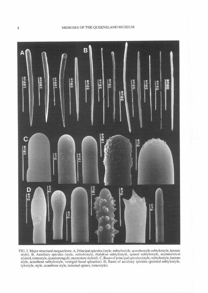
FIG. 2. Major structural megascleres. A, Principal spicules (style, subtylostyle, acanthostyle subtylostyle, hastatestyle). B, Auxiliary spicules (style, subtylostyle, rhabdose subtylostyle, spined subtylostyle, asymmetricalstyloid, tornostyle, quasistrongyle, mucronate styloid). C, Bases of principal spicules (style, subtylostyle, hastatestyle, acanthose subtylostyle, vestigial basal spination). D, Bases of auxiliary spicules (pointed subtylostyle,tylostyle, style, acanthose style, terminal spines, tornostyle).
REVISION OF MICROCIONIDAE^ 9
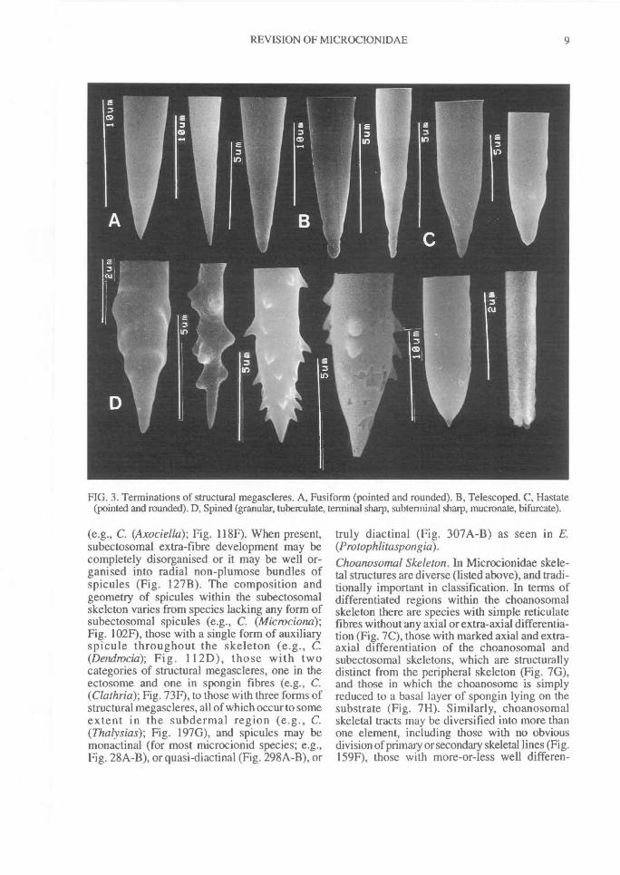
FIG. 3. Terminations of structural megascleres. A, Fusiform (pointed and rounded). B, Telescoped. C, Hastate(pointed and rounded). D, Spined (granular, tuberculate, terminal sharp, subterminal sharp, mucronate, bifurcate).
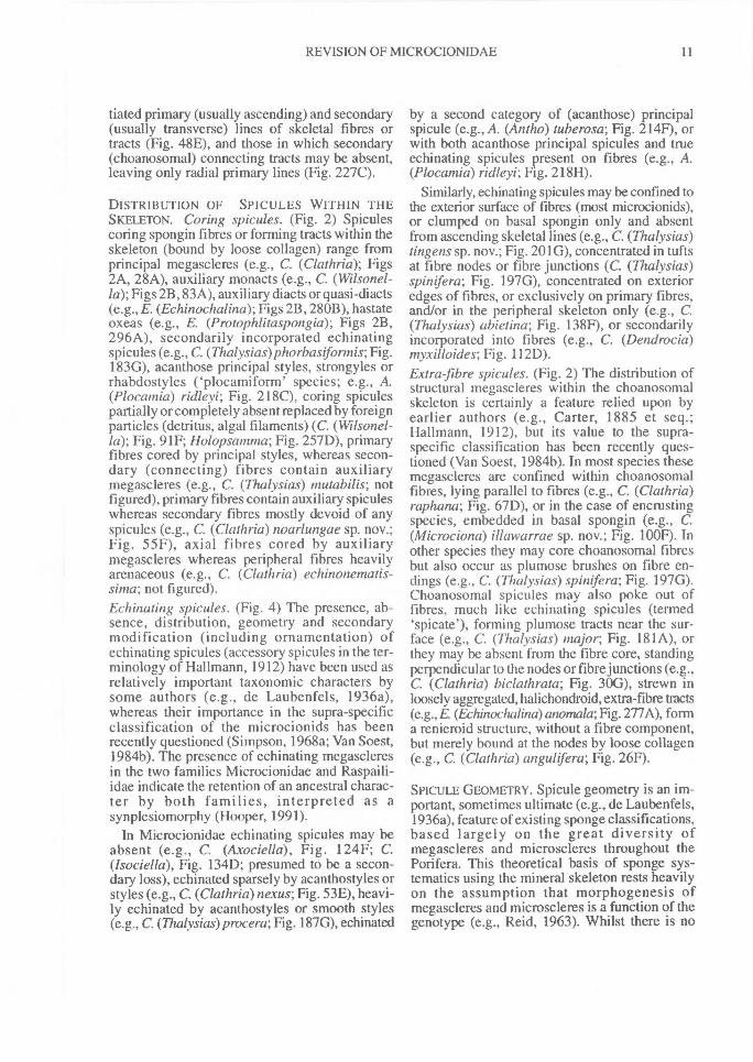
(e.g., C. (Axociella); Fig. 118F). When present,subectosomal extra-fibre development may becompletely disorganised or it may be well or-ganised into radial non-plumose bundles ofspicules (Fig. 127B). The composition andgeometry of spicules within the subectosomalskeleton varies from species lacking any form ofsubectosomal spicules (e.g., C. (Microciona);Fig. 102F), those with a single form of auxiliaryspicule throughout the skeleton (e.g., C.(Dendrocia); Fig. 112D), those with twocategories of structural megascleres, one in theectosome and one in spongin fibres (e.g., C.(Clathria); Fig. 73F), to those with three forms ofstructural megascleres, all of which occur to someextent in the subdermal region (e.g., C.(Thalysias); Fig. 197G), and spicules may bemonactinal (for most microcionid species; e.g.,Fig. 28A-B), or quasi-diactinal (Fig. 298A-B), or
truly diactinal (Fig. 307A-B) as seen in E.(Protophlitaspongia).Choanosomal Skeleton. In Microcionidae skele-tal structures are diverse (listed above), and tradi-tionally important in classification. In terms ofdifferentiated regions within the choanosomalskeleton there are species with simple reticulatefibres without any axial or extra-axial differentia-tion (Fig. 7C), those with marked axial and extra-axial differentiation of the choanosomal andsubectosomal skeletons, which are structurallydistinct from the peripheral skeleton (Fig. 7G),and those in which the choanosome is simplyreduced to a basal layer of spongin lying on thesubstrate (Fig. 7H). Similarly, choanosomalskeletal tracts may be diversified into more thanone element, including those with no obviousdivision of primary or secondary skeletal lines (Fig.159F), those with more-or-less well differen-
10^ MEMOIRS OF THE QUEENSLAND MUSEUM
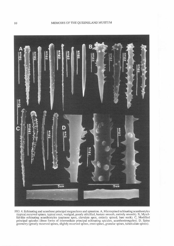
FIG. 4. Echinating and acanthose principal megascleres and spination. A, Microspined echinating acanthostyles(typical recurved spines, typical erect, vestigial, poorly silicified, hastate smooth, entirely smooth). B, Myxil-lid-like echinating acanthostyles (aspinose apex, clavulate apex, entirely spined, bare neck). C, Modifiedprincipal spicules (three forms of intermediate principal echinating spicules, acanthostrongyles). D, Spinegeometry (greatly recurved spines, slightly recurved spines, erect spines, granular spines, tuberculate spines).
REVISION OF MICROCIONIDAE^ 11
tiated primary (usually ascending) and secondary(usually transverse) lines of skeletal fibres ortracts (Fig. 48E), and those in which secondary(choanosomal) connecting tracts may be absent,leaving only radial primary lines (Fig. 227C).
DISTRIBUTION OF SPICULES WITHIN THESKELETON. Coring spicules. (Fig. 2) Spiculescoring spongin fibres or forming tracts within theskeleton (bound by loose collagen) range fromprincipal megascleres (e.g., C. (Clathria); Figs2A, 28A), auxiliary monacts (e.g., C. (Wilsonel-la); Figs 2B, 83A), auxiliary diacts or quasi-diacts(e.g., E. (Echinochalina); Figs 2B, 280B), hastateoxeas (e.g., E. (Protophlitaspongia); Figs 2B,296A), secondarily incorporated echinatingspicules (e.g., C. (Thalysias)phorbasifonnis; Fig.183G), acanthose principal styles, strongyles orrhabdostyles ('plocamiform' species; e.g., A.(Plocamia) ridleyi; Fig. 218C), coring spiculespartially or completely absent replaced by foreignparticles (detritus, algal filaments) (C. (Wilsonel-la); Fig. 91F; Holopsamma; Fig. 257D), primaryfibres cored by principal styles, whereas secon-dary (connecting) fibres contain auxiliarymegascleres (e.g., C. (Thalysias) mutabilis; notfigured), primary fibres contain auxiliary spiculeswhereas secondary fibres mostly devoid of anyspicules (e.g., C. (Clathria) noarlungae sp. nov.;Fig. 55F), axial fibres cored by auxiliarymegascleres whereas peripheral fibres heavilyarenaceous (e.g., C. (Clathria) echinonematis-sima; not figured).Echinating spicules. (Fig. 4) The presence, ab-sence, distribution, geometry and secondarymodification (including ornamentation) ofechinating spicules (accessory spicules in the ter-minology of Hallmann, 1912) have been used asrelatively important taxonomic characters bysome authors (e.g., de Laubenfels, 1936a),whereas their importance in the supra-specificclassification of the microcionids has beenrecently questioned (Simpson, 1968a; Van Soest,1984b). The presence of echinating megascleresin the two families Microcionidae and Raspaili-idae indicate the retention of an ancestral charac-ter by both families, interpreted as asynplesiomorphy (Hooper, 1991).
In Microcionidae echinating spicules may beabsent (e.g., C. (Axociella), Fig. 124F; C.(lsociella), Fig. 134D; presumed to be a secon-dary loss), echinated sparsely by acanthostyles orstyles (e.g., C. (Clathria) nexus; Fig. 53E), heavi-ly echinated by acanthostyles or smooth styles(e.g., C. (77talysias)procera; Fig. 187G), echinated
by a second category of (acanthose) principalspicule (e.g., A. (Antho) tuberosa; Fig. 214F), orwith both acanthose principal spicules and trueechinating spicules present on fibres (e.g., A.(Plocamia) ridleyi; Fig. 218H).
Similarly, echinating spicules may be confined tothe exterior surface of fibres (most microcionids),or clumped on basal spongin only and absentfrom ascending skeletal lines (e.g., C. (Thalysias)tingens sp. nov.; Fig. 201G), concentrated in tuftsat fibre nodes or fibre junctions (C. (Thalysias)spinifera; Fig. 197G), concentrated on exterioredges of fibres, or exclusively on primary fibres,and/or in the peripheral skeleton only (e.g., C.(Thalysias) abietina; Fig. 138F), or secondarilyincorporated into fibres (e.g., C. (Dendrocia)myxilloides; Fig. 112D).Extra-fibre spicules. (Fig. 2) The distribution ofstructural megascleres within the choanosomalskeleton is certainly a feature relied upon byearlier authors (e.g., Carter, 1885 et seq.;Hallmann, 1912), but its value to the supra-specific classification has been recently ques-tioned (Van Soest, 1984b). In most species thesemegascleres are confined within choanosomalfibres, lying parallel to fibres (e.g., C. (Clathria)raphana; Fig. 67D), or in the case of encrustingspecies, embedded in basal spongin (e.g., C.(Microciona) illawarrae sp. nov.; Fig. 100F). Inother species they may core choanosomal fibresbut also occur as plumose brushes on fibre en-dings (e.g., C. (Thalysias) spinifera; Fig. 197G).Choanosomal spicules may also poke out offibres, much like echinating spicules (termed`spicate'), forming plumose tracts near the sur-face (e.g., C. (Thalysias) major; Fig. 181A), orthey may be absent from the fibre core, standingperpendicular to the nodes or fibre junctions (e.g.,C. (Clathria) biclathrata; Fig. 30G), strewn inloosely aggregated, halichondroid, extra-fibre tracts(e.g., E. (Echinochalina)anomala; Fig. 277A), forma renieroid structure, without a fibre component,but merely bound at the nodes by loose collagen(e.g., C. (Clathria) angulifera; Fig. 26F).
SPICULE GEOMETRY. Spicule geometry is an im-portant, sometimes ultimate (e.g., de Laubenfels,1936a), feature of existing sponge classifications,based largely on the great diversity ofmegascleres and microscleres throughout thePorifera. This theoretical basis of sponge sys-tematics using the mineral skeleton rests heavilyon the assumption that morphogenesis ofmegascleres and microscleres is a function of thegenotype (e.g., Reid, 1963). Whilst there is no
12^ MEMOIRS OF THE QUEENSLAND MUSEUM
REVISION OF MICROCIONIDAE^ 13
evidence to reject this hypothesis there is certain-ly some experimental data to show that spiculegeometry and morphogenesis is at least partlyinfluenced by environmental perturbations (e.g.,Hartman, 1981; Jones, 1991), including ex-amples from the Microcionidae (e.g., influence ofseasonality (Simpson, 1978) and geographicaldistribution (Hooper et al., 1990) on spicule sizeand geometry). But the extent to which thesephenotypic modifications occur within naturalpopulations has not yet been examined rigorous-ly. In general, however, these features appear tobe relatively stable across wide geographical ran-ges as shown by studies on raspailiids (Hooper,1991) and microcionids (Hooper & Levi, 1993a)from east and west coasts of Australia and thewestern Pacific. Hartman (1981) and Simpson(1990) outline the various theories on the func-tional significance, process of silicification andevolution of demosponge spicules.
STRUCTURAL MEGASCLERES. Spicule axes.Microcionidae have exclusively monaxonicspicule axes. Megascleres are usually monac-tinal, although some may have modified secon-dary axes (i.e., anisoxeote diactinal modificationsto styles), and a few appear to have true diactinalforms (E. (Protophlitaspongia)). UnlikeTrikentrion and Cyamon in the allied Raspailiidae(Hooper, 1991) there are no tetraxonic spiculemodifications in this family (triacti nal, tetractinalor polyactinal forms). Furthermore, theMicrocionidae have a comparatively small rangeof structural megasclere types in the skeleton,whereas some raspailiids have many. Major typesof structural megascleres are illustrated in Figs2-4. These range from hastate styles or tylostyles(Fig. 87A), fusiform styles or tylostyles (Fig.77A), asymmetrical styloid, rounded, quasi-diac-tinal or strongylote spicules (Fig. 280B), andoxeote megascleres (Fig. 296A).Spicule ornamentation. Spines on megascleresare of dubious importance to supraspecific clas-
sification (e.g., Simpson, 1968a), although theyhave been used frequently in the past to definegenera (e.g., de Laubenfels, 1936a). Microcionidstructural spicules frequently have basalmicrospines (Fig. 2C-D), occasionally withspines on shafts (Fig. 180B) or points of spicules(Fig. 3D). Spicule ornamentation ranges fromentirely smooth (Fig. 28A), smooth shafts withacanthose bases (Fig. 30A), vestigial spination onthe proximal portions of shafts only (Fig. 153A),acanthose on both bases and points (Fig. 83A), orentirely acanthose (Fig. 98A).
ECH1NATING MEGASCLERES. There is a diverserange of echinating spicule geometries inMicrocionidae, although not as great as inRaspailiidae. Major types (Fig. 4) include: evenlyspined (granular), claviform or stump-like acan-thostyles; acanthose styles with aspinose bases;acanthose styles with aspinose points; acanthosestyles with aspinose 'necks' (i.e., area proximalto the basal swelling); acanthostrongyles; entirelysmooth styles identical in geometry to principalmegascleres; derived oxeotes; or entirely smoothstylotes of different geometry than principalspicules.
MICROSCLERES. The geometry, ornamentationand modification of microscleres is an importantcharacter for classification (Dendy, 1921), al-though it has probably been overemphasised bysome authors (de Laubenfels, 1936a) and itsprimary importance has been questioned (VanSoest, 1984b). Within Microcionidae there aretwo forms of diactinal microscleres: meniscoidforms (chelae) and toxas. Other poeciloscleridmicroscleres (microxeas, raphides and meniscoidforms such as true sigmas) are not present.Microcionids show many modifications to bothchelae and toxas, the latter sometimes resemblingmicroxeas, and frequently microscleres are lostaltogether.
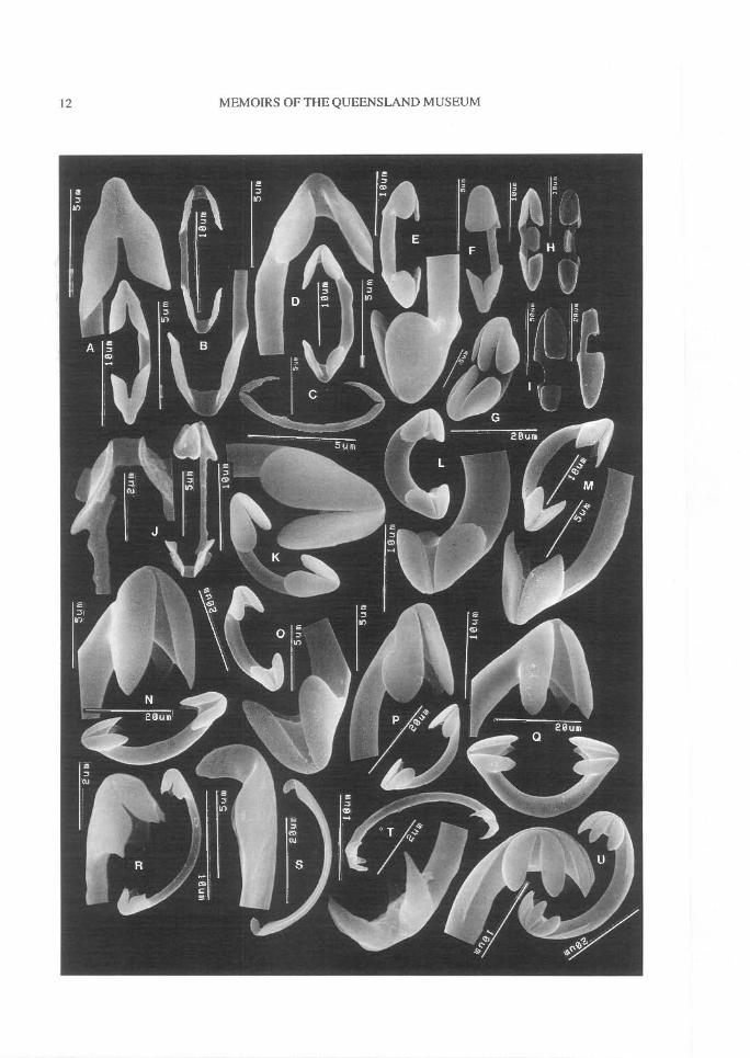
FIG. 5. Chelae geometry (A-H,J-N, Microcionidae; I,O-U, Other poecilosclerids). A, Palmate (C. australiensis).B, Palmate, reduced alae (C. australiensis). C, Palmate sigmoid, vestigial alae (C. hesperia sp.nov.). D, Palmate,arcuate-like alae with straight shaft (C. oxyphyla). E, Palmate, fused alae (C. curvichela). F, Palmate, contort(C. abietina). G, Palmate, arcuate-like fusion (C. macropora). H, Palmate, central wing on shaft (C.toxipraedita). I, Palmate, anisochelate (Mycale). J, Palmate, arcuate-like alae, fluted alae (C. macropora). K,Arcuate-like, fusion of alae, curved shaft, alae practically fused together (C. grisea). L, Arcuate-like, un-guiferous, detached alae (C. scabida). M, Anchorate-like, unguiferous, tooth-like alae (C. scabida). N,Arcuate-like, unguiferous, tooth-like alae (C. myxilloides). 0, True arcuate (Ectyodotyx). P, Arcuate, un-guiferous (Ectyodoryx). Q, Arcuate, unguiferous, tooth-like alae (Cretin). R, Palmate unguiferous (Crella). S,Palmate, unguiferous, vestigial alae (Hamigera). T, Arcuate, unguiferous, tooth-like alae (Monanchora). U,Anchorate, unguifcrous (Monanchora).
14^ MEMOIRS OF THE QUEENSLAND MUSEUM
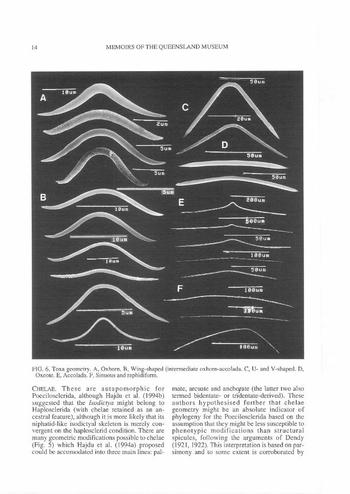
FIG. 6. Toxa geometry. A, Oxhorn. B, Wing-shaped (intermediate oxhorn-accolada. C, U- and V-shaped. D,Oxeote. E, Accolada. F, Sinuous and raphidiform.
CHELAE. These are autapomorphic forPoecilosclerida, although Hajdu et al. (1994b)suggested that the Isodictya might belong toHaplosclerida (with chelae retained as an an-cestral feature), although it is more likely that itsniphatid-like isodictyal skeleton is merely con-vergent on the haplosclerid condition. There aremany geometric modifications possible to chelae(Fig. 5) which Hajdu et al. (1994a) proposedcould be accomodated into three main lines: pal-
mate, arcuate and anchotrate (the latter two alsotermed bidentate- or tridentate-derived). Theseauthors hypothesised further that chelaegeometry might be an absolute indicator ofphylogeny for the Poecilosclerida based on theassumption that they might be less susceptible tophenotypic modifications than structuralspicules, following the arguments of Dendy(1921, 1922). This interpretation is based on par-simony and to some extent is corroborated by
REVISION OF MICROCIONIDAE^ 15
other evidence (such as congruence of structuralfeatures). There are, however, some anomolousexamples of chelae that fall between these threecategories (see Discussion).Palmate. (Fig. 5A-B) This is the simplest formwith 'typical' morphology consisting of straightshaft, front ala completely free and well developed,and lateral alae more-or-less completely fused tothe shaft along its longest dimension. Most micro-cionids have unmodified 'typical' palmateisochelae. Modifications to this 'typical' palmateform include: partial reduction of alae (Fig. 5B),nearly vestigial alae producing a sigmoid spicule(Fig. 5C), partial fusion of alae along lateral mar-gins producing spatulae (Fig. 5E), partial fusion(Fig.
in or complete fusion along medial mar-
gins n which both the front alae meet and/or fuseat the centre producing cleistochelae (Fig. 76G),sculpturing on fluting on alae (Fig. 5J), contortionof the shaft such that alae are at 90 0 to each otherat each end of the shaft (Fig. 5F), expansions ofthe lateral alae fused with the shaft producingwing-like process on the shaft (Fig. 5H), 'crocae'or j-shaped sigmoid forms where the alae arevestigial and asymmetrical, producing a simplehook-like spicule (Fig. 17F), and deep curvatureof shaft and reduction of alae to tooth-like struc-tures (termed unquiferous; Fig. 5R-T).Arcuate. (Fig. 50) Here the lateral alae are morefully developed than in palmate forms and be-come almost completely detached from the shaft,and the shaft is usually prominently curved andthickened. However, there is no clear transitionbetween the palmate and arcuate forms, wherebyan increase in curvature and thickening of theshaft (Fig. 5D-E) and partial detachment of lateralalae (Fig. 5J-K) extend along a continuum fromtrue palmate to true arcuate (compare Fig. 5D, G,J-L, N-Q, T). Somewhere along this continuumchelae are deemed to be arcuate (Fig. 50-P).Anchorate. (Fig. 5U) Further along the con-tinuum are anchorate chelae, in which all threealae are fully formed, the lateral ones completelydetached from the shaft, and there are also lateralridges on the shaft. In this study I use the terms'arcuate-like' or 'anchorate-like' for modifiedchelae although it is equivocal whether thesespicules are truly arcuates or anchorates.
TOXAS. Toxas are found in only a few families ofpoecilosclerids but also known fromHaplosclerida. There is also some evidence tosuggest that they may be particularly common inyoung or larval tissue (e.g., Simpson, 1968b).Eight major morphotypes are delineated here (Fig
6), although intermediates are also possible: 1,Oxhorn toxas (wide central curve, reflexed armsand greatly recurved points; usually thick) (Fig.6A); 2, Wing-shaped toxas (sharply curved atcentre, with recurved arms and reflexed points;usually thick) (Fig. 6B); 3, U-shaped toxas (withwide central curvature but lacking reflexed arms)(Fig. 6C); 4, V-shaped toxas (pinched hairpin-likecentral curvature, straight arms running more-or-less vertical, and slightly reflexed points; usuallythick) (Fig. 6C); 5, Oxeote toxas (virtuallystraight shaft and points) (Fig. 6D); 6, Accoladatoxas (wide or slightly pinched central curvature,strait arms running more-or-less horizontal, andstrait points; usually thin) (Fig. 6E); 7,Raphidiform toxas (sharply angular central cur-vature, straight arms and straight points; verythin, hair-like) (Fig. 6F); and 8, Sinuous toxas(asymmetrical, sinuous, raphidiform; very thin,hair-like) (Fig. 6F). The presence or absence ofmicrospines on toxas was at one time consideredto be an important supraspecific character (e.g.,de Laubenfels, 1936a), but these have since beenfound in many genera and may not be importantabove the species level.
SIZE OF SPICULES. Variation of spicule sizehas also been an important diagnostic criterion,but this has been applied mainly at the specieslevel of classification. Numerous (possibly amajority) of taxa have been erected solely on thebasis of megasclere and microsclere dimensions,but only a few studies have investigated the statis-tical variability of spicule size or commented onthe effects of physico-chemical factors on thatvariability (e.g., Hartman, 1958, 1981; Jones,1984). There is some evidence to show that intra-specific variability can be significant for a giventaxon, and spicule size-ranges can span acrossseveral closely related taxa which were otherwiseerected solely on that basis. Hooper et al. (1990)demonstrated that two sibling species of Clathria(Thalysias) could not be reliably distinguished bytheir absolute spicule sizes, and only statisticalcomparisons between these species were of anyvalue in this regard. Thus spicule dimensionsused as diagnostic characteristics are of mostsignificance at the species level of classification,and consequently their application is generallycomparative rather than absolute.
ORGANIC SKELETON. The development ofthe organic skeleton, the amount of spongin itcontains, its architecture and foreign inclusionscontained within it, are diagnostic features for theDemospongiae in general. The organic skeleton
16^ MEMOIRS OF THE QUEENSLAND MUSEUM
FIG. 7. Examples of microcionid skeletal structures. A, Regularly reticulate (C.(C.) noarlungae sp.nov.). B,Irregularly reticulate (C. (C.) multipes). C, Renieroid reticulate (A. (1.) chartacea). D, Plumoreticulate (C. (T.)reinwardti). E, Plumose (C. (T.) procera). F, Arenaceous (C. (W.) tuberosa). G, Axially compressed, extra-axially radial (C. (A.) canaliculata). H, Hymedesmoid (C. (M.) tingens).
can be differentiated into two components: a fibresystem and a collagenous mesohyl.Spongin fibres. These may be well developed,present but relatively lightly developed, secon-darily lost, with or without spicule tracts, or evenreplaced partially or entirely by algal filaments(e.g., Anomoclathria (Fig. 9A-B)). Simpson(1984) elucidates two morphological types ofspongin fibres (= type A spongin of Bergquist,1978a): those that are cored by siliceous spiculesand those that incorporate foreign particles.
Detrital-entrapping fibres are most prevalent inthe 'keratose' sponges (Dictyoceratida,Dendroceratida, Verongida), Haplosclerida(Arenosclera), and Poecilosclerida of the Des-macididae (Psammascus, Desmapsamnia), aswell as several species of Microcionidae (i.e., C.(Wilsonella) and Holopsatntna). In these speciesfibres may be totally void of true megascleres(e.g., Holopsamma laminaefavosa), or they mayhave a combination of both foreign particles andindigenous megascleres (e.g., C. (Wilsonella)
tuberosa). The independent occurrence of detri-tal-entrapping fibres throughout the various or-ders of Demospongiae indicates that it is acharacter of ecological importance rather than ofany phylogenetic significance.
Spiculose fibres are typical for microcionids,and the form, size, orientation and origin ofmegascleres coring the organic skeleton, and thestructural complexities of the fibres themselvesare important diagnostic characters for this fami-ly. In encrusting species that have a basal layer ofspongin anchoring themselves to the substratethis spongin is continuous with the spiculatedfibres, and as such both actually lie outside theliving organism (Weissenfels, 1978).Mesohyl. The development of collagen in themesohyl matrix, also termed 'interstitial' spon-gin, extra-fibre spongin, type B spongin(Bergquist, 1978), or ground substance, and in-cluding collagenous fibrils, has not been givenmuch value as a systematic character for theMicrocionidae, whereas in the 'keratose' orders
REVISION OF MICROCIONIDAE^ 17
these features have more significance, and inAaptos (Hadromerida) it has been used to dif-ferentiate species through deposition patterns(Kelly-Borges & Bergquist, 1995). However,within the Microcionidae there is evidence toshow that the development of collagenthroughout the mesohyl varies intra-specifically,especially between specimens in differentreproductive condition or as a consequence ofoverwintering behaviour (e.g., Pandaros acan-thifolium; Wiedenmayer, 1977; Van Soest,1984b; Microciona prolifera; Simpson, 1963,1968b; Knight & Fell, 1987). Simpson (1968a)attempted to define species and genera ofMicrocionidae on the basis of the organicskeleton and cytological characteristics but to alarge extent his results did not corroborate with aclassification based on the mineral skeleton, andin some cases evidence was directly conflicting.
GROWTH FORM. The use of external morphologyas an important or even crucial diagnostic char-acteristic has diminished since early systematics(e.g., Lamarck, 1814). Bowerbank (1864), indeveloping Grant's (1861) scheme for thePorifera, de-emphasised sponge habit in his sys-tematics although he recognised that growth formwas related to 'anatomical peculiarities'. Thatexternal morphology is often closely linked to theinternal architecture and composition of theskeleton has been well documented (e.g., Levi,1973; Bergquist, 1978). Although there are somegroups which are immediately recognisable bytheir growth form and skeletal architecture (e.g.,the honeycomb reticulate structure of Holopsam-ma), other groups show a higher degree of intra-specific variability in their morphology (e.g.,most Clathria), ranging from encrusting to mas-sive forms. Moreover, there is now evidence tosuggest that gross morphology is highly plastic,greatly influenced by prevailing environmentalconditions (temperature, depth, turbidity, cur-rents, substrate etc.) (e.g., Hartman, 1958;Simpson, 1968a; Fry, 1971; Palumbi, 1984). It isnot entirely clear to what extent abiotic factorsinfluence growth form, or the degree to whichgenotype dictates possible shapes attainable byparticular species, but it is becoming more ap-parent that the sponge 'species' is not as im-mutable as previously suspected. Palumbi (1984)proposed that sponges have evolved to be capableof producing a quick and decisive response toenvironmental adversities (unpredictable, highenergy environments), and those responses aremost readily seen as changes to both growth form
and skeletal structure (e.g., the degree to whichthe skeletal becomes compressed). It is also notclearly understood why some species seem to behighly plastic (e.g., C. (Thalysias) lendenfeldi)(Hooper et al., 1990), whereas others with com-parable depth and geographical distributions aremuch more conservative (e.g., C. (Thalysias)abietina). Growth forms, as characters used in aclassification, can be defined as determinate (e.g.,Holopsamma, C. (Microciona)) or indeterminate(e.g., most other Clathria, Antho).
CONSISTENCY. Sponge texture is a highly subjec-tive characteristic, which is difficult to quantify,but one which may provide clues as to the com-position of the skeleton, the amount of sponginpresent, whether or not detritus is incorporatedinto the sponge, and silicification of the skeleton(Bergquist, 1978). A description of sponge con-sistency is usually an integral part of any speciesdescription, but its application in systematics hasbeen mostly comparative rather than absolute.More recently de Weerdt (1985) used consistencyas an objective feature in the systematics ofHaplosclerida. She noted that it was not onlyuseful in characterising particular species but thatin broad terms,texture was able to be used at thefamily level of classification. For theMicrocionidae this character does not vary great-ly, with most species being firm, compressible,flexible (e.g., Clathria (Thalysias)) or soft, com-pressible, spongy (e.g., Holopsamma).
MACROSCOPIC FEATURES OF THE SURFACE. Sur-face sculpturing. Macroscopic features of thesponge surface are important for some spongegroups (e.g., Haplosclerida), and surface or-namentation, such as tangential webs of spiculesor fibres, perpendicular brushes of spicules, orelevated oscules may be diagnostic for particulargenera. Within the Poecilosclerida however, in-cluding the Microcionidae, these features are lessconsistent, and they are usually only reliable incharacterising particular species or occasionallygenera (Simpson, 1968a). Encrusting species fre-quently exhibit intricate drainage canals radiatingaway from oscules, or highly hispid (furry) sur-faces (e.g., C. (Thalysias) toxifera), whereasmore massive or digitate species may have sur-face papillae or conules (e.g., C. (Thalysias)abietina), or a surface which is composed ofreticulate ectosomal fibres (e.g., Holopsammaglobosa).Oscules. The distribution of oscules on the sur-face may vary considerably between relatedspecies, ranging from being confined to distinct
18^ MEMOIRS OF THE QUEENSLAND MUSEUM
pore areas, such as sieve-plates of Echinochalinatubulosa, or restricted to certain regions, such aslateral sides of branches, the tops of digits, or theexterior surface of vases, or scattered indiscriminant-ly over the surface. Oscules may also be con-spicuous, discrete, with a membraneous lip, slightlyraised or flush with the surface (e.g., C. (Thalysias)reinwardti), or terminal, raised on the apex ofsurface papillae or stoloniferous tubes (e.g., C.(Isociella) eccentrica), or scattered, conspicuous,producing a porous reticulate surface (e.g.,Holopsamma arborea), or minute and not easilyvisible optically (e.g., C. (Thalysias) coppingeri).
COLOURATION. Sponge pigments are not general-ly diagnostic (Bergquist, 1978). Some speciesmay show high intra-specific variation in livepigmentation, and this variability may be relateddirectly to microhabitat and depth distribution.The nature of these pigments, their distributionwithin the mesohyl and their specific cellularassociation is still poorly known, but it is wellestablished that carotenoids are predominant(Simpson, 1984). Isolation and identification ofthese pigments is more difficult, as is the deter-mination of whether they are produced ormodified by the host, or obtained directly fromsymbiotic associations (e.g., Litchfield & Liaaen-Jensen, 1980). The major proportion of spongecarotenoids are metabolised by the sponge (i.e.,primary metabolites) and are intracellular(Simpson, 1984), whereas it is suspected thatsome sponges have a variable proportion (up to20%) of pigments synthesised by symbiotic algae(e.g., Litchfield & Liaaen-Jensen, 1980; Liaaen-Jensen et al., 1982). Litchfield & Liaaen-Jensen(1980) studying C. (Microciona) prolifera sug-gested that the sponge could modify (aromatise)a large proportion of algal carotenoids, andLiaaen-Jensen et al. (1982) divide the classes ofcarotenoids into a phytoplankton-type,zooplankton-type, bacterial and/or fungal origin,and sponge metabolised (oxidative) groups.These authors found that phytoplankton derivedand sponge metabolised carotenoids comprisedthe major proportion of carotenes in Demospon-giae. The Poecilosclerida and Axinellida werefound to exhibit the highest capacity forcarotenoid accumulation and transformation, ex-plaining their diverse and often brightly colouredpigmentation, and furthermore they possessed asimilar carotenoid diversity.
Evidence suggests that carotenoid pigmentsmay be photoprotective, in which case it wouldbe expected that intertidal species contain a
higher proportion of these pigments than deeper-water species. But it is not clear why some sym-patric species have consistent pigmentation (e.gC. (Wilsonella) tuberosa), whereas in others pig-mentation is highly variable even in specimensgrowing side-by-side (e.g., C. (Thalysias)abietina; Plate 4D). Colour consistency is notgenerally used as a reliable diagnostic character,but it is also true that only very few authors haveinvestigated the intraspecific colour variability ofany species. It is therefore advantageous to deter-mine whether live colouration is stable and specificto a species, or has very narrow limits in variation(e.g., C. (Isociella) eccentrica). Alternatively,pigmentation may be highly unstable, not specificand without an accurately definable 'typical'colouration (e.g., C. (Thalysias) abietina).
REPRODUCTIVE PRODUCTS AND REPRODUCTIVECYCLES. Reproductive products and modes ofreproduction, as diagnostic characters, have beenused predominantly at higher levels of classifica-tion (e.g., Bergquist, 1980a), whereas breedingseasons and spawning cycles are most useful fordetecting sibling species (e.g., Fromont, 1989).As far as known, within the Microcionidae larvaeare viviparous parenchymella with bare posteriorpoles. The apparent form of sexuality varies fromgonochoristic to contemporaneous her-maphroditism (Fell, 1984, 1990; Simpson, 1984).Breeding seasons and/or spawning cycles may becontinuous or periodical.
CYTOLOGY. Simpson (1984) provided a defini-tive treatment of sponge cell biology, including adescription of diverse cell types and their func-tional morphology. He suggested that descrip-tions of characters such as cells with inclusionsand the morphology of choanocyte chambers willprobably provide further information directlyrelevant to demosponge systematics. For theMicrocionidae, Simpson (1968a) showed thatseemingly morphologically convergent generacould be readily differentiated by the presenceand morphology of special cell types (gray cells),and that their higher systematic relationshipscould be defined in terms of cytological charac-ters. However, there were many incongruitiesbetween systematics based on skeletal charac-teristics and those indicated by cytological data.Specific examples of these differences are dis-cussed below in the synopsis of genera, but somegeneral comments are appropriate.
Taxonomic groupings indicated by Simpson'smicrocionid cytological data suggested that manyskeletal characters used previously by authors
REVISION OF MICROCIONIDAE^ 19
had little importance in differentiating genera.These included the presence or absence of pal-mate isochelae, the presence of acanthose versussmooth echinating megascleres, quantity of spon-gin in the skeleton, plumose versus anastomosingfibres, megascleres with basal spination orsmooth bases, the presence of surface conules anddistinct oscules, the production of uprightbranches, and the presence or absence of an ec-tosomal skeleton. With the exception of the lastfeature these conclusions are supported in thecontemporary classification of Microcionidae(e.g., Van Soest, 1984b). However, othercytological evidence presented by Simpson(1968a) is more difficult to reconcile withmicrocionid skeletal data. For example, encrust-ing species (i.e., the nominal genera Microcionaand Ophlitaspongia) were cytologically relative-ly homogeneous and distinct from ramose forms(Clathria). The cytological characteristics ofthese encrusting species were more similar torenieroid microcionids (nominal genus Plocamil-la) than they were to the ramose forms (nominalgenera Rhaphidophlus and Thalysias) whichotherwise had the most similar spicule and sur-face characteristics.
Simpson concluded that generic definitionsbased primarily on spicule types did not lead tonatural classifications, and he proposed that thesedefinitions should include skeletal, cytologicaland histological evidence. He suggested that thenumerous classification systems that were basedsolely on various combinations of skeletal char-acters, such as those of Vosmaer (1933, 1935a-b),de Laubenfels (1936a) and Levi (1960a), couldbe defended with equal justification. Althoughsome cytological features have been incor-porated into existing systematics (e.g., morphol-ogy and arrangement of choanocytes), much ofSimpson's (1968a) important work cannot beused in classification based primarily on skeletalcharacters.
SYSTEMATICS
Class Demospongiae Sollas, 1885
Order Poecilosclerida Topsent, 1928
Suborder Microcionina Hajdu, Van Soest &Hooper, 1994
Poecilosclerida Topsent, 1928a: 64, 309.
REMARKS. This order is the largest and mostdiverse of Demospongiae (Bergquist 1978). It is
characterised by a skeleton of both spicule andspongin elements, usually well developed, some-times vestigial, in which megascleres are monac-tinal, diactinal or both, and spongin developmentvaries from well developed horny fibres enclos-ing spicules to an interspicular collagen cement(Bergquist, 1978; Hartman, 1982). Simpson(1984) suggested that the order is characterisedby at least two distinctly localised types ofmegascleres (with or without distinctivegeometry). Those megascleres are choanosomalprincipal spicules embedded in spongin fibres,and subectosomal auxiliary megascleres whichare free in the mesohyl or protrude from sponginfibres in which they are embedded. This defini-tion is consistent with the inclusion ofRaspailiidae in the Poecilosclerida as proposedby Hooper (1991). Poecilosclerids usually havean abundantly collagenous mesohyl matrix, andmicroscleres may include chelae (apomorphic forthe order), although not all taxa have them.Sexual reproduction is predominantlyviviparous, oviparous in two families, and inthose species incubating larvae they areparenchymella with uniform flagellum size andbare posterior poles.
The suborder Microcionina was established toinclude four families of Poecilosclerida(Microcionidae, Raspailiidae, Iophonidae andRhatxleremiidae), which have terminally spinedectosomal monactinal megascleres (occasionallymodified to quasidiactinal forms), isochelae ofpalmate origin, diverse forms of toxas, up to fivecategories of megascleres and lacking sigmas.The other suborders (Myxillina and Mycalina)were also defined by their chelae morphology(bidentate-derived and sigmancistra-derivedchelae, respectively), and absence of toxas andpresence of sigmas, respectively (Hajdu et al.,1994), but assignment of particular genera tothese suborders is still contentious.
The number of families recognised in the ordervaries according to different authors (e.g., Levi,1973; Wiedenmayer, 1977; Bergquist, 1978;Hartman, 1982; Van Soest, 1984b; Bergquist &Fromont, 1988). Recently Hooper & Wieden-mayer (1994) included 16 families in the order:12 with chelae microscleres, 3 without chelae,and 1 of uncertain placement, whereas Hajdu etal. (1994) recognise 17: Microcionina(Microcionidae, Raspailiidae, lophonidae, Rhab-deremiidae); Myxillina (Myxillidae, Crambidae,Coelosphaeridae, Crellidae, Hymedesmiidae,Anchinoidae, Phoriospongiidae, Tedaniidae);and Mycalina (Mycalidae, Hamacanthidae,
20^ MEMOIRS OF THE QUEENSLAND MUSEUM
Desmacellidae, Cladorhizidae, Guitarridae).Latrunculiidae, included in the order by Levi(1973) and Van Soest (1984b) has also beenassigned to Hadromerida (Reid, 1968;Bergquist, 1978; Hartman, 1982), but is nowconsidered to be polyphyletic (Kelly-Borges &Vacelet, 1995) with Latrunculia having affinitieswith Iophonidae and Diacamus, Sigmosceptrel-la, Negombata more closely related to theMycalidae.
Family Microcionidae Carter, 1875
Microcionina Carter, 1875. Microcionidae Hentschel,1923; Wiedenmayer, 1977.
Clathriidae Lendenfeld, 1884a; Hentschel, 1923; Top-sent, 1928a; Levi, 1960a; Simpson, 1968a;Bergquist, 1978; Hartman, 1982; Van Soest, 1984b;Bergquist & Fromont, 1988.
Ophlitaspongiidae de Laubenfels, 1936a; Thomas,1968; Hoshino, 1981.
Growth form encrusting, lobate, arborescent orflabellate; skeleton differentiated intochoanosomal (axial), subectosomal (extra-axial)and ectosomal regions; axial skeleton formed byunispicular or multispicular tracts ofchoanosomal (principal) megascleres, typicallycoring spongin fibres or sometimes simply boundtogether by collagen; fibres echinated by (acan-tho-) styles (accessory spicules); skeletal struc-tures include isodictyal, renieroid, reticulate,plumo-reticulate, plumose or hymedesmoid, butnever radial; extra-axial skeleton formed by tractsof subectosomal (auxiliary) spicules, usually dis-persed outside of fibres, rarely well organised butusually with some degree of difference betweenaxial and extra-axial regions; ectosomal skeletonranges from membraneous, or with protrudingsubectosomal (auxiliary) spicules, or with a spe-cial category of ectosomal (auxiliary) spicules;principal megascleres monactinal, predominant-ly smooth or partially spined only, occasionallyvestigial or absent completely, or sometimesreplaced by detritus in skeleton; auxiliarymegascleres usually monactinal, rarely quasi-diactinal, smooth shaft and basal spines, moreslender than choanosomal spicules; echinatingstyles or subtylostyles smooth, partially or com-pletely spined; microscleres include toxas ofseveral morphologies (including raphidiform andmicroxeotes), and isochelae primarily of palmateorigin (but occasionally with partial 'arcuate' and`anchorate' modifications); larvae viviparous.
REMARKS. There has been disagreement as towhich of Microcionidae Carter and ClathriidaeHentschel should be used. Wicdcnmayer (1977:
139) argued that Microcionidae was establishedin 1875, whereas Clathriidae did not appear until1884. He noted that under Article 40 of the Inter-national Code of Zoological Nomenclature(Anonymous, 1984), it was irrelevant whether ornot Clathria Schmidt (1862) had priority overMicrociona Bowerbank (1862; apparently pub-lished 1863). Conversely, Van Soest (1984b: 89)argued that the priority of Clathria overMicrociona did have bearing on the choice of thefamily name. Although 'Clathriidae' is in currentusage by most contemporary workers, itspreferred use is in direct contravention with theCode and to long term stability of the group andMicrocionidae is used here following Hooper &Wiedenmayer (1994).
The definition given above restrictsMicrocionidae to genera which possesspredominantly smooth monactinal ectosomal andchoanosomal spicules. It excludes certainmicrocionid-like genera which have true tylotesor strongylotes as their ectosomal spicules (e.g.,Acamus, Megaciella). These taxa are nowreferred to Iophonidae, as defined by their ec-tosomal features (Hajdu et al., 1994). However,the definition barely distinguishes species withmodified or reduced quasidiactinal (styloid)auxiliary megascleres (e.g., severalEchinoclathria, Holopsamma and Echinochalinaspecies), or quasimonactinal (amphistrongyloteor tornote-like) auxiliary megascleres (e.g., E.(Protophlitaspongia)). These modified auxiliaryspicules are usually asymmetrical and are inter-preted here as convergent upon true diactinalspicules. These anomalous microcionids sharecertain characteristics of both Microcionidae andDesmacididae, and the importance of these char-acters at higher levels of systematics must there-fore be questioned, or a certain level ofhomoplasy must be accomodated in thephylogeny of the order.
Similarly, the definition given above cannotalways clearly distinguish some Microcionidaeand Raspailiidae, but this is a problem of seman-tics rather than a biological one. As a general rulemost species of Raspailiidae have well compressedaxial skeletons, and well differentiated axial andextra-axial skeletons. In contrast, most Micro-cionidae lack these features or they are only poor-ly developed and probably convergent, perhapsrelated to growth form (e.g., Clathria (Axociel-la)). Nevertheless, there are examples in bothfamilies where the boundaries between taxa blur,such as the microcionid-like Raspailia (Clathrio-dendron) arbuscula (see Hooper 1991: Figs 19-
REVISION OF MICROCIONIDAE^ 21
20), and the raspailiid-like Clathria (Axociella)canaliculata (Figs 118-119)). These families areconsistently differentiated by their ectosomal fea-tures and microscleres, which appear to be moreimportant characters than skeletal structure.
Hajdu et al. (1994) restricted Microcionina(and hence Microcionidae) to taxa with only pal-mate isochelae, tacitly excluding severalmicrocionid-like genera specifically created forspecies with bidentate-derived (arcuate oranchorate) chelae. Theoretically this is a viablesystem for the suprafamily classification ofMicrocionidae but in practical terms it is notalways possible to distinguish between truebidentate-derived chelae and palmate chelae with'arcuate-' or 'anchorate-like' modifications. Thesecases are discussed indivually below.
REVIEW. There are several problems in thetaxonomy of Microcionidae that need to be ad-dressed in order to clearly recognise and definevalid genera and produce a phylogenetically validsystematics for the family.
1) The family is large, containing about 540described species and many other as yet un-described species known from various collec-tions. 79 nominal genera have been previouslyincluded, of which 69 are currently recognised asresiding here although fewer than this number arevalid. Some of these genera have been merged inothers by previous authors (e.g., Levi, 1960a;Simpson, 1968a; Van Soest, 1984b; Bergquist &Fromont, 1988; Hooper, 1990a), but in somecases these synonymies are now deemed wrongand have produced further nomenclatural com-plexities. Several contemporary studies have at-tempted partial revisions of Microcionidae (VanSoest, 1984; Bergquist & Fromont, 1988;Hooper, 1990a), but these have mainly focusedon smaller regional faunas without considerationof all the higher taxa. In the present work each ofthese genera is redefined and illustrated from itstype species (i.e., strict definition).
2) The literature on Microcionidae is vast, scat-tered, mostly antiquated (pre-1900), descriptionsare far too brief for modern purposes and manytaxa have never been illustrated. The presentwork deals primarily with museum material andliving populations of species, and decisions areless reliant on the literature than previous studies.
3) There are many characters in sponges whoseexpressions (character states) change subtlywithin populations of supposedly single speciesand across the whole range of species, usuallywithout clear boundaries between related taxa.
Some of these characters have been used as im-portant diagnostic criteria in earlier works. Thisstudy has examined large numbers of specimensand species, and documents the range of intra-specific and inter-specific character states in aneffort to clearly define taxa and understandrelationships between them. Inclusion of non-skeletal evidence into the systematics can furthersupport or refute opinions based solely on skeletalcharacters and gross morphology (to decidewhether one character is more important thananother, whether morphological characters arehomologous, and whether the observed highlevels of homoplasies within most Poriferan clas-sifications are in fact real or acceptable). Theprevious studies of Hooper et al. (1990) andHooper (1990a) are preliminary to this study.
4) There are nearly as many subjective inter-pretations between different authors, as to thephylogenetic importance of one character overanother in the systematics, as there are taxa. Thishas arisen partly as a consequence of overreliance on definitions of type species (and hencenominal genera) from the literature (especiallythe work of de Laubenfels, 1936a), given thatmany type species are poorly described, mis-described or barely differentiated from theircogeners. The present study uses a phylogeneticframework to produce an objective and consis-tent taxonomy for the family. Two previousstudies (Van Soest, 1984b; Hooper, 1990a) par-tially resolved intrafamily relationships withinMicrocionidae, both are preliminary to this work.
GENERIC NAMES INCLUDED INMICROCIONIDAE
Preoccupied generic names are shown in squarebrackets. The synonomy lists provided in thissection refer to works in which the name is usedand in the case of genera considered valid do notinclude the numerous synonyms. The diagnosesprovided in this list are based solely on the typematerial of the type species unless otherwisestated.
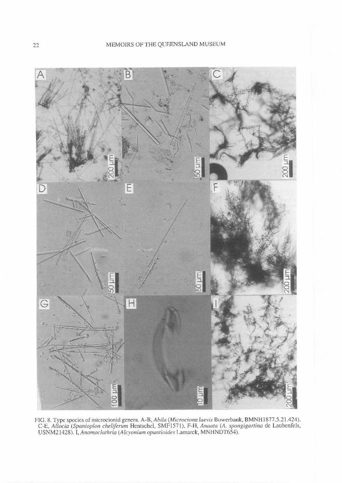
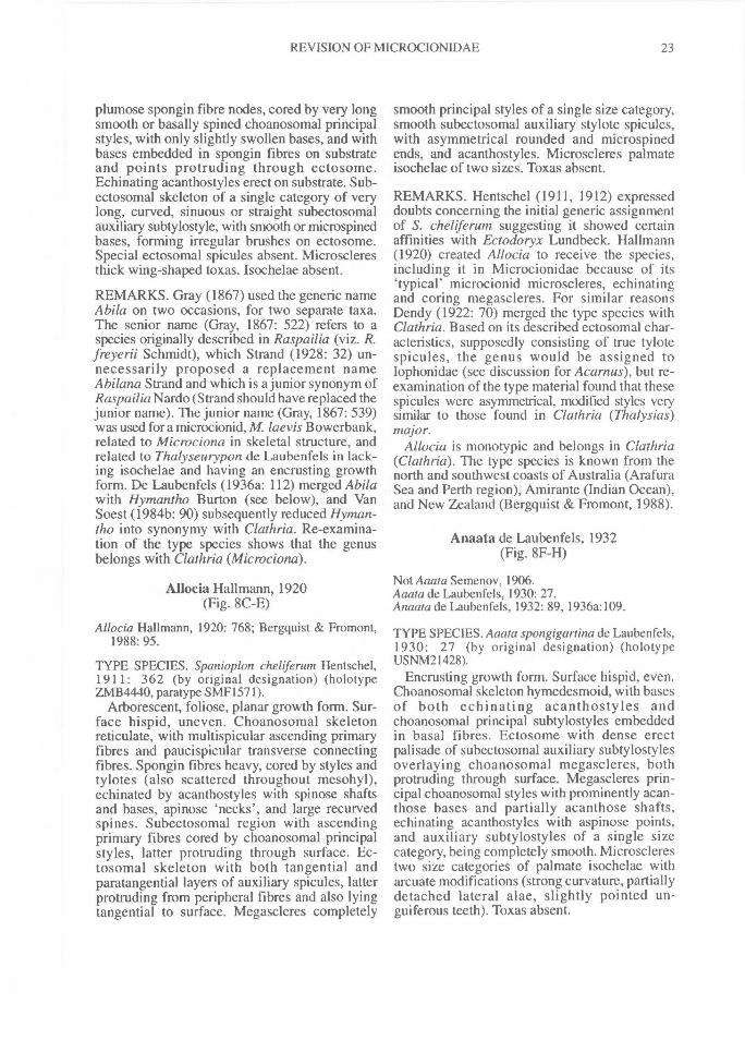
[Abila] Gray, 1867(Fig. 8A-B)
Abila Gray, 1867: 539.Not Abila Gray, 1867: 522.
TYPE SPECIES. Microciona laevis Bowerbank, 1866:124 (by monotypy) (holotypc BMNH1877.5.21.1543).
Encrusting growth form. Surface hispid, even.Choanosomal skeleton composed of short
22^ MEMOIRS OF THE QUEENSLAND MUSEUM
FIG. 8. Type species of microcionid genera. A-B, Abila (Microciona laevis Bowerbank, BMNH1877.5.21.424).C-E, Allocia (Spanioplon chel(erum Hentschel, SMF1571). F-H, Anaata (A. spongigartina de Laubenfels,USNM21428). I, Anomoclathria (Alcyonium opuntioides Lamarck, MNHNDT654).
REVISION OF MICROCIONIDAE^ 23
plumose spongin fibre nodes, cored by very longsmooth or basally spined choanosomal principalstyles, with only slightly swollen bases, and withbases embedded in spongin fibres on substrateand points protruding through ectosome.Echinating acanthostyles erect on substrate. Sub-ectosomal skeleton of a single category of verylong, curved, sinuous or straight subectosomalauxiliary subtylostyle, with smooth or microspinedbases, forming irregular brushes on ectosome.Special ectosomal spicules absent. Microscleresthick wing-shaped toxas. Isochelae absent.
REMARKS. Gray (1867) used the generic nameAbila on two occasions, for two separate taxa.The senior name (Gray, 1867: 522) refers to aspecies originally described in Raspailia (viz. R.freyerii Schmidt), which Strand (1928: 32) un-necessarily proposed a replacement nameAbilana Strand and which is a junior synonym ofRaspailia Nardo (Strand should have replaced thejunior name). The junior name (Gray, 1867: 539)was used fora microcionid, M. laevis Bowerbank,related to Microciona in skeletal structure, andrelated to Thalyseurypon de Laubenfels in lack-ing isochelae and having an encrusting growthform. De Laubenfels (1936a: 112) merged Abilawith Hymantho Burton (see below), and VanSoest (1984b: 90) subsequently reduced Hyman-tho into synonymy with Clathria. Re-examina-tion of the type species shows that the genusbelongs with Clathria (Microciona).
Allocia Hallmann, 1920(Fig. 8C-E)
Allocia Hallmann, 1920: 768; Bergyuist & Fromont,1988: 95.
TYPE SPECIES. Spanioplon chehferum Hentschel,1 9 1 1: 362 (by original designation) (holotypeZMB4440, paratype SMF1571).
Arborescent, foliose, planar growth form. Sur-face hispid, uneven. Choanosomal skeletonreticulate, with multispicular ascending primaryfibres and paucispicular transverse connectingfibres. Spongin fibres heavy, cored by styles andtylotes (also scattered throughout mesohyl),echinated by acanthostyles with spinose shaftsand bases, apinose 'necks', and large recurvedspines. Subectosomal region with ascendingprimary fibres cored by choanosomal principalstyles, latter protruding through surface. Ec-tosomal skeleton with both tangential andparatangential layers of auxiliary spicules, latterprotruding from peripheral fibres and also lyingtangential to surface. Megascleres completely
smooth principal styles of a single size category,smooth subectosomal auxiliary stylote spicules,with asymmetrical rounded and microspinedends, and acanthostyles. Microscleres palmateisochelae of two sizes. Toxas absent.
REMARKS. Hentschel (1911, 1912) expresseddoubts concerning the initial generic assignmentof S. cheliferum suggesting it showed certainaffinities with Ectodoryx Lundbeck. Hallmann(1920) created Allocia to receive the species,including it in Microcionidae because of its'typical' microcionid microscleres, echinatingand coring megascleres. For similar reasonsDendy (1922: 70) merged the type species withClathria. Based on its described ectosomal char-acteristics, supposedly consisting of true tylotespicules, the genus would be assigned toIophonidae (see discussion for Acarnus), but re-examination of the type material found that thesespicules were asymmetrical, modified styles verysimilar to those found in Clathria (Thalysias)major.
Allocia is monotypic and belongs in Clathria(Clathria). The type species is known from thenorth and southwest coasts of Australia (ArafuraSea and Perth region), Amirante (Indian Ocean),and New Zealand (Bergquist & Fromont, 1988).
Anaata de Laubenfels, 1932(Fig. 8F-H)
Not Aaata Semenov, 1906.Aaata de Laubenfels, 1930: 27.Anaata de Laubenfels, 1932: 89, 1936a:109.
TYPE SPECIES. Aaata spongigartina de Laubenfels,1 93 0: 27 (by original designation) (holotypeUSNM21428).
Encrusting growth form. Surface hispid, even.Choanosomal skeleton hymedesmoid, with basesof both echinating acanthostyles andchoanosomal principal subtylostyles embeddedin basal fibres. Ectosome with dense erectpalisade of subectosomal auxiliary subtylostylesoverlaying choanosomal megascleres, bothprotruding through surface. Megascleres prin-cipal choanosomal styles with prominently acan-those bases and partially acanthose shafts,echinating acanthostyles with aspinose points,and auxiliary subtylostyles of a single sizecategory, being completely smooth. Microsclerestwo size categories of palmate isochelae witharcuate modifications (strong curvature, partiallydetached lateral alae, slightly pointed un-guiferous teeth). Toxas absent.
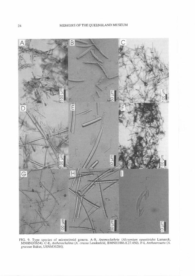
24^ MEMOIRS OF THE QUEENSLAND MUSEUM
FIG. 9. Type species of microcionid genera. A-B, Anomoclathria (Alcyonium opuntioides Lamarck,MNHNDT654). C-E, Antherochalina (A. crassa Lendenfeld, BMNH1886.8.27.450). F-I, Anthoarcuata (A.graceae Bakus, USNM36284).
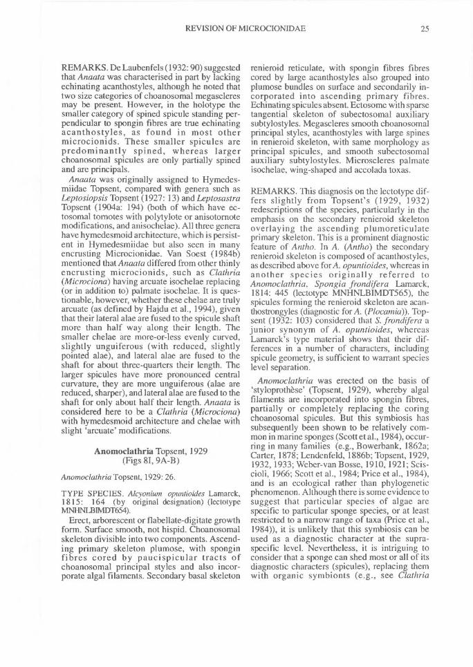
REVISION OF MICROCIONIDAE^ 25
REMARKS. De Laubenfels (1932: 90) suggestedthat Anaata was characterised in part by lackingechinating acanthostyles, although he noted thattwo size categories of choanosomal megascleresmay be present. However, in the holotype thesmaller category of spined spicule standing per-pendicular to spongin fibres are true echinatingacanthostyles, as found in most othermicrocionids. These smaller spicules arepredominantly spined, whereas largerchoanosomal spicules are only partially spinedand are principals.
Anaata was originally assigned to Hymedes-miidae Topsent, compared with genera such asLeptosiopsis Topsent (1927: 13) and LeptosastraTopsent (1904a: 194) (both of which have ec-tosomal tornotes with polytylote or anisotornotemodifications, and anisochelae). All three generahave hymedesmoid architecture, which is persist-ent in Hymedesmiidae but also seen in manyencrusting Microcionidae. Van Soest (1984b)mentioned that Anaata differed from other thinlyencrusting microcionids, such as Clathria(Microciona) having arcuate isochelae replacing(or in addition to) palmate isochelae. It is ques-tionable, however, whether these chelae are trulyarcuate (as defined by Hajdu et al., 1994), giventhat their lateral alae are fused to the spicule shaftmore than half way along their length. Thesmaller chelae are more-or-less evenly curved,slightly unguiferous (with reduced, slightlypointed alae), and lateral alae are fused to theshaft for about three-quarters their length. Thelarger spicules have more pronounced centralcurvature, they are more unguiferous (alae arereduced, sharper), and lateral alae are fused to theshaft for only about half their length. Anaata isconsidered here to be a Clathria (Microciona)with hymedesmoid architecture and chelae withslight 'arcuate' modifications.
Anomoclathria Topsent, 1929(Figs 81, 9A-B)
Anomoclaihria Topsent, 1929: 26.
TYPE SPECIES. Alcyonium opuntioides Lamarck,1 8 1 5: 164 (by original designation) (lectotypeMNHNLBIMDT654).
Erect, arborescent or flabellate-digitate growthform. Surface smooth, not hispid. Choanosomalskeleton divisible into two components. Ascend-ing primary skeleton plumose, with sponginfibres cored by paucispicular tracts ofchoanosomal principal styles and also incor-porate algal filaments. Secondary basal skeleton
renieroid reticulate, with spongin fibres fibrescored by large acanthostyles also grouped intoplumose bundles on surface and secondarily in-corporated into ascending primary fibres.Echinating spicules absent. Ectosome with sparsetangential skeleton of subectosomal auxiliarysubtylostyles. Megascleres smooth choanosomalprincipal styles, acanthostyles with large spinesin renieroid skeleton, with same morphology asprincipal spicules, and smooth subectosomalauxiliary subtylostyles. Microscleres palmateisochelae, wing-shaped and accolada toxas.
REMARKS. This diagnosis on the lectotype dif-fers slightly from Topsent's (1929, 1932)redescriptions of the species, particularly in theemphasis on the secondary renieroid skeletonoverlaying the ascending plumoreticulateprimary skeleton. This is a prominent diagnosticfeature of Antho. In A. (Antho) the secondaryrenieroid skeleton is composed of acanthostyles,as described above for A. opuntioides, whereas inanother species originally referred toAnomoclathria, Spongia frondifera Lamarck,1814: 445 (lectotype MNHNLB1MDT565), thespicules forming the renieroid skeleton are acan-thostrongyles (diagnostic for A. (Plocamia)). Top-sent (1932: 103) considered that S. frondifera ajunior synonym of A. opuntioides, whereasLamarck's type material shows that their dif-ferences in a number of characters, includingspicule geometry, is sufficient to warrant specieslevel separation.
Anomoclathria was erected on the basis of'styloprothese' (Topsent, 1929), whereby algalfilaments are incorporated into spongin fibres,partially or completely replacing the coringchoanosomal spicules. But this symbiosis hassubsequently been shown to be relatively com-mon in marine sponges (Scott et al., 1984), occur-ring in many families (e.g., Bowerbank, 1862a;Carter, 1878; Lendenfeld, 1886b; Topsent, 1929,1932, 1933; Weber-van Bosse, 1910,1921; Scis-cioli, 1966; Scott et al., 1984; Price et al., 1984),and is an ecological rather than phylogeneticphenomenon. Although there is some evidence tosuggest that particular species of algae arespecific to particular sponge species, or at leastrestricted to a narrow range of taxa (Price et al.,1984)), it is unlikely that this symbiosis can beused as a diagnostic character at the supra-specific level. Nevertheless, it is intriguing toconsider that a sponge can shed most or all of itsdiagnostic characters (spicules), replacing themwith organic symbionts (e.g., see Clathria
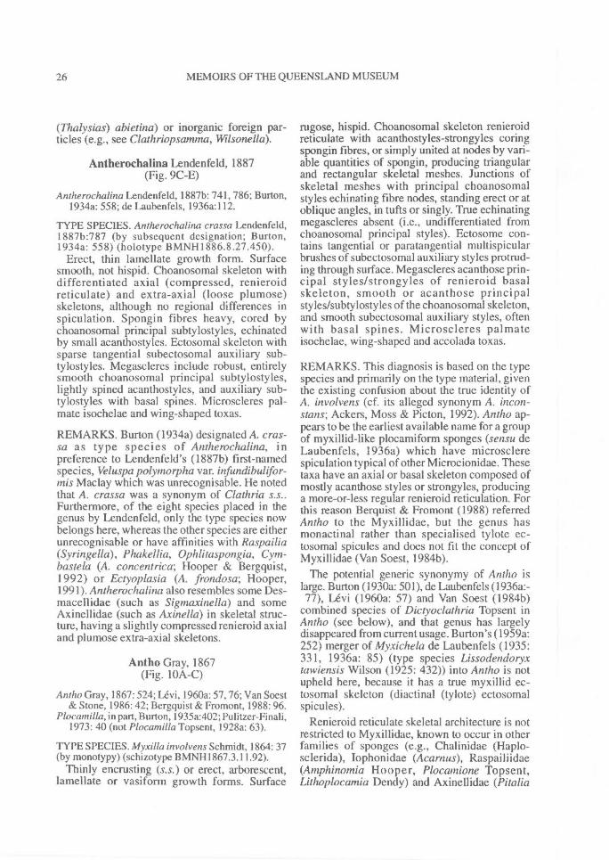
26^ MEMOIRS OF THE QUEENSLAND MUSEUM
(Thalysias) abietina) or inorganic foreign par-ticles (e.g., see Clathriopsamma, Wilsonella).
Antherochalina Lendenfeld, 1887(Fig. 9C-E)
Antherochalina Lendenfeld, 1887b: 741, 786; Burton,1934a: 558; de Laubenfels, 1936a:112.
TYPE SPECIES. Antherochalina crassa Lendenfeld,1887b:787 (by subsequent designation; Burton,1934a: 558) (holotype BMNH1886.8.27.450).
Erect, thin lamellate growth form. Surfacesmooth, not hispid. Choanosomal skeleton withdifferentiated axial (compressed, renieroidreticulate) and extra-axial (loose plumose)skeletons, although no regional differences inspiculation. Spongin fibres heavy, cored bychoanosomal principal subtylostyles, echinatedby small acanthostyles. Ectosomal skeleton withsparse tangential subectosomal auxiliary sub-tylostyles. Megascleres include robust, entirelysmooth choanosomal principal subtylostyles,lightly spined acanthostyles, and auxiliary sub-tylostyles with basal spines. Microscleres pal-mate isochelae and wing-shaped toxas.
REMARKS. Burton (1934a) designated A. cras-sa as type species of Antherochalina, inpreference to Lendenfeld's (1887b) first-namedspecies, Veluspa polymorpha var. infundibulifor-mis Maclay which was unrecognisable. He notedthat A. crassa was a synonym of Clathria s.s..Furthermore, of the eight species placed in thegenus by Lendenfeld, only the type species nowbelongs here, whereas the other species are eitherunrecognisable or have affinities with Raspailia(Syringe/la), Phakellia, Ophlitaspongia, Cym-bastela (A. concentrica; Hooper & Bergquist,1992) or Ectyoplasia (A. frondosa; Hooper,1991). Antherochalina also resembles some Des-macellidae (such as Sigmaxinella) and someAxinellidae (such as Axinella) in skeletal struc-ture, having a slightly compressed renieroid axialand plumose extra-axial skeletons.
Antho Gray, 1867(Fig. 10A-C)
Antho Gray, 1867: 524; Levi, 1960a: 57,76; Van Soest& Stone, 1986: 42; Bergquist & Fromont, 1988: 96.
Plocamilla, in part, Burton, 1935a:402; Pulitzer-Finali,1973:40 (not Plocamilla Topsent, 1928a: 63).
TYPE SPECIES. Myxilla involvens Schmidt, 1864: 37(by monotypy) (schizotype BMNH1867.3.11.92).
Thinly encrusting (s.s.) or erect, arborescent,lamellate or vasiform growth forms. Surface
rugose, hispid. Choanosomal skeleton renieroidreticulate with acanthostyles-strongyles coringspongin fibres, or simply united at nodes by vari-able quantities of spongin, producing triangularand rectangular skeletal meshes. Junctions ofskeletal meshes with principal choanosomalstyles echinating fibre nodes, standing erect or atoblique angles, in tufts or singly. True echinatingmegascleres absent (i.e., undifferentiated fromchoanosomal principal styles). Ectosome con-tains tangential or paratangential multispicularbrushes of subectosomal auxiliary styles protrud-ing through surface. Megascleres acanthose prin-cipal styles/strongyles of renieroid basalskeleton, smooth or acanthose principalstyles/subtylostyles of the choanosomal skeleton,and smooth subectosomal auxiliary styles, oftenwith basal spines. Microscleres palmateisochelae, wing-shaped and accolada toxas.
REMARKS. This diagnosis is based on the typespecies and primarily on the type material, giventhe existing confusion about the true identity ofA. involvens (cf. its alleged synonym A. incon-stans; Ackers, Moss & Picton, 1992). Antho ap-pears to be the earliest available name for a groupof myxillid-like plocamiform sponges (sensu deLaubenfels, 1936a) which have microsclerespiculation typical of other Microcionidae. Thesetaxa have an axial or basal skeleton composed ofmostly acanthose styles or strongyles, producinga more-or-less regular renieroid reticulation. Forthis reason Berquist & Fromont (1988) referredAntho to the Myxillidae, but the genus hasmonactinal rather than specialised tylote ec-tosomal spicules and does not fit the concept ofMyxillidae (Van Soest, 1984b).
The potential generic synonymy of Antho islarge. Burton (1930a: 501), de Laubenfels (1936a:-77), Levi (1960a: 57) and Van Soest ( I 984b)
combined species of Diayoclathria Topsent inAntho (see below), and that genus has largelydisappeared from current usage. Burton's (1959a:252) merger of Myxichela de Laubenfels (1935:331, 1936a: 85) (type species Lissodendoryxtawiensis Wilson (1925: 432)) into Antho is notupheld here, because it has a true myxillid ec-tosomal skeleton (di actinal (tylote) ectosomalspicules).
Renieroid reticulate skeletal architecture is notrestricted to Myxillidae, known to occur in otherfamilies of sponges (e.g., Chalinidae (Haplo-sclerida), Iophonidae (Acarnus), Raspailiidae(Amphinomia Hooper, Plocamione Topsent,Lithoplocamia Dendy) and Axinellidae (Pitalia
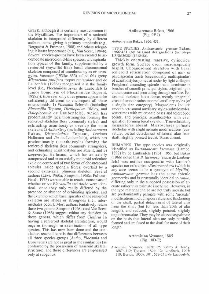
REVISION OF MICROCIONIDAE^ 27
Gray)), although it is certainly most common inthe Myxillidae. The importance of a renieroidskeleton is interpreted differently by differentauthors, some giving it primary emphasis (e.g.,Bergquist & Fromont, 1988) and others relegat-ing it lesser importance (e.g., Van Soest, 1984b).Several species-groups have been created to ac-comodate microcionid-like species, with spicula-tion typical of the family, supplemented by arenieroid (myxillid-like) basal choanosomalskeleton composed of acanthose styles or stron-gyles. Vosmaer (1935a: 653) called this groupMicrociona prolifera tropus renieroides and deLaubenfels (1936a) recognised it at the familylevel (i.e., Plocamiidae sensu de Laubenfels (ajunior homonym of Plocamiidae Topsent,1928a)). However, only three genera appear to besufficiently different to encompass all thesemicrocionids: 1) Plocamia Schmidt (includingPlocamilla Topsent, Dirrhopalum Ridley, andHoloplocamia de Laubenfels), which haspredominantly (acantho)strongyles forming therenieroid skeleton (less commonly styles), andechinating acanthostyles overlap the mainskeleton; 2)Antho Gray (including AnthoarcuataBakus, Dictyoclathria Topsent, IsocionaHallmann and Jia de Laubenfels)), which haspredominantly (acantho)styles forming therenieroid skeleton (less commonly strongyles),and echinating acanthostyles are absent; and 3)Isopenectya Hallmann, which has an axiallycompressed and extra-axially renieroid reticulateskeleton composed of two forms of choanosomalspicules inside spongin fibres, overlaid by asecond extra-axial plumose skeleton. Severalauthors (Levi, 1960a; Simpson, 1968a; Pulitzer-Finali, 1973) were unable to reach a consensus ofwhether or not Plocamilla and Antho were iden-tical, since they only really differed by thepresence or absence of echinating spicules, andthe extent to which basal spicules of the renieroidskeleton are styles or strongyles (i.e., inter-mediates occur). Most authors tentatively retainthese two genera; Simpson (1968a) and Van Soest& Stone (1986) suggest edthat any decision onthese genera, which differ from Clathria inhaving a renieroid skeletal architecture, wouldrequire thorough re-examination of all typespecies. This has now been done and the con-clusion reached here is that differences betweenall three species-groups (Antho, Plocamia andIsopenecta) are not as great as the similarities (asconferred by the possession of renieroid skeletalstructure), and these differences are emphasisedonly at subgenus.
Anthoarcuata Bakus, 1966(Fig. 9F-I)
Anthoarcuata Bakus, 1966: 431.
TYPE SPECIES. Anthoarcuata graceae Bakus,1966:431 (by original designation) (holotypeUSNM36284 (161848)).
Thickly encrusting, massive, cylindricalgrowth form. Surface even, microscopicallyhispid. Choanosomal skeleton with basalrenieroid reticulation composed of uni- orpaucispicular tracts (occasionally multispicular)of acanthostyles joined at nodes by light collagen.Peripheral ascending spicule tracts terminate inbrushes of smooth principal styles, originating inchoanosome and protruding through surface. Ec-tosomal skeleton has a dense, mostly tangentialcrust of smooth subectosomal auxiliary styles (ofa single size category). Megascleres includesmooth ectosomal auxiliary styles-subtylostyles,sometimes with mucronate bases and telescopedpoints, and principal acanthostyles with evenspination forming basal skeleton. True echinatingmegascleres absent. Microscleres palmateisochelae with slight arcuate modifications (cur-vature, partial detachment of lateral alae fromshaft, slightly pointed teeth). Toxas absent.
REMARKS. The type species was originallyidentified as Burtonancora lacunosa (Lambe,1892) by de Laubenfels (1961: 195), but Bakus(1966) noted that B. lacunosa (sensu de Lauben-fels) was neither conspecific with Lambe'sspecies nor referable to Burtonanchora (which inany case seems to be a synonym of Myxilla).Anthoarcuata graceae has the same spiculegeometries and is structurally identical to Antho,differing only in the supposed possession of ar-cuate rather than palmate isochelae. However, inthe type material chelae are not truly arcuate butare predominantly palmate with some 'arcuate'modifications including curvature and thickeningof the shaft, partial detachment of lateral alaefrom the shaft (but for less than 20% of alarlength), and reduced, slightly pointed, slightlyunguiferous alae. They may be classed as palmateon the basis that lateral alae are only partiallyformed and are fused to the shaft for most of theirlength.
Artemisina Vosmaer, 1885(Fig. 10D-E)
Artemisina Vosmaer, 18856: 25; Ridley & Dendy,1887: 112; Topsent, 1894: 12; Lundbeck, 1905:110; Burton, 1930a: 501, 528-531; de Laubenfels,
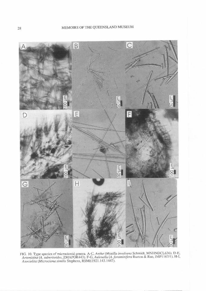
28^ MEMOIRS OF THE QUEENSLAND MUSEUM
FIG. 10. Type species of microcionid genera. A-C, Antho (Myxilla involyens Schmidt, MNHNDCL636). D-E,Artemisina (A. suberitoides, ZMAPOR443). F-G, Aulenella (A. foraminifera Burton & Rao, IM P I 167/1). H-I,Axocielita (Microciona similis Stephens, RSME1921.143.1447).
REVISION OF MICROCIONIDAE^ 29
1936a: 117; Levi, 1960a: 61, 83; Ristau, 1978: 585;Van Soest, 1984b: 122, 130; Bergquist & Fromont,1988: 119.
Artenisina;Burton, 1934b: 54 [lapsus].
TYPE SPECIES. Artemisina suberitoides Vosmaer,1885: 25 (by monotypy) (holotype ZMAPOR443);junior synonym of Suberitesarciger Schmidt, 1870:47(Burton, 1930a: 528) (schizotype BMNH1870.5.3.90).
Massive, subspherical growth form. Surfaceskin-like, microscopically hispid, with few raisedoscules; texture distinctly stringy. Choanosomalfibres indefinite or absent, overall architectureplumo-reticulate, nearly halichondroid in places,composed of multispicular ascending andpaucispicular transverse tracts of choanosomalprincipal styles, bound together within spongin.Echinating megascleres absent. Ectosomalskeleton plumose, composed of single categoryof subectosomal auxiliary styles forming discon-tinuous palisade of discrete brushes. Megascleressmooth choanosomal principal subtylostyles andsmooth fusiform subectosomal auxiliary styles orsubtylostyles. Microscleres palmate isochelae,and wing-shaped toxas with spinous extremities.
REMARKS. Defining Artemisina in phylo-genetic terms is problematic. The taxon has noreal distinctive features, although it differs fromother Microcionidae in lacking a distinctivechoanosomal skeleton or definite spongin fibres(also found in Qasimella Thomas), lackingechinating spicules, and having a nearly radialectosomal skeleton reminiscent of some Ceratop-sion (Raspailiidae; Hooper, 1991). These charac-teristics, two of which might be interpreted asreductions or secondary losses and the third as aconvergence, are the only definable morphologi-cal apomorphies. Moreover, ectosomal structurevaries between several species, ranging from thetypical condition composed of erect brushes (e.g.,A. arciger) to a tangential layer of spicules incriss-cross fashion (e.g., A. melana Van Soest).This variability is equivalent to (or analogues of)the Mycale subgenera Carmia and Aegagropila,respectively (e.g., Topsent, 1924b). At least onespecies lacks a specialised ectosomal skeletoncompletely (e.g., A. transiens Topsent).
Some species of Artemisina (e.g., A. foliata(Bowerbank)) have honeycombed reticulategrowth forms, approaching the characteristicHolopsamma morphology, but there is no consis-tency or pattern of gross morphologies amongstArtemisina, and in any case it is unlikely that thegenus can be solely defined by its growth form.De Laubenfels (1936a) and Ristau (1978b) sug-gested that the Artemisina was defined by the
absence of echinating acanthostyles and by thepresence of spinous extremities on toxas. Neithercharacter has much systematic value at thegeneric level. In the present interpretationechinating acanthostyles represent the retentionof an ancestral character, in which case theirpresence or absence does not constitute a validreason to define a phylogenetic grouping, and inany event they occur and disappear throughoutnumerous microcionid and raspailiid taxa.Similarly, toxas with spinous extremities are alsoknown to occur in many Microcionidae, includ-ing the type species of Clathria, C. cotnpressaSchmidt, Microciona spinarchus Carter & Hope,M. coccinea Bergquist, M. rubens Bergquist M.spinatoxa Hoshino, Eutypon acanthotoxaStephens, Stylostichon toxiferum Topsent,Labacea juncea Burton, Plocanzia ridleyiHentschel, and Ophlitaspongia thielei Burton.They also occur in genera which have an ec-tosomal structure consistent with the Myxillidae(e.g., Melonchela clathrata Koltun). Several speciesof Artemisina have smooth toxas (e.g., A. melana),and one (A. archegona Ristau) has oxeote toxassimilar to Paratenaciella Vacelet and Vasseur.
Thus, in the broad sense Artemisina contains aheterogenous assemblage of species, whichprompted Burton (1930a) to divide the group intothree sections based on the number of megasclerecategories present. The simplest forms have onlyone category of spicule (choanosomal principalmegascleres; e.g., A. transiens Topsent); the typi-cal form has two categories of megascleres (largerchoanosomal principal styles and smaller subec-tosomal auxiliary styles; e.g., A. arciger); and thethird form has an incompletely differentiatedseries of three megasclere types (two choano-somal spicules and one subectosomal spicule; A.plumosa Hentschel). In all these forms species areonly really united in their lax choanosomal skele-tal structure. The majority of species have beendescribed from Antarctic and Arctic regions.
[Aulena] sensu Lendenfeld, 1888
[Aulena]; Lendenfeld, 1888: 228, 1889a: 90; Topsent,1894a: 19; de Laubenfels, 1936a: 16; Wiedcnmayer,1989: 58.
Not Aulena Lendenfeld, 1885c: 309.
TYPE SPECIES. Aulena villosa Lendenfeld, 1885c:309 (by subsequent designation; de Laubenfels, 1936a:16) (syntypes AMZ130, G8901).
Bulbous, subspherical growth form, consistingof fused digitate projections. Surface highlypapillose but not hispid. Choanosomal skeleton
30^ MEMOIRS OF THE QUEENSLAND MUSEUM
regularly reticulate, with heavy spongin fibrescored by sparsely scattered sand grains mostly atnodes of fibres, and with a secondary fibre net-work between main skeleton. Ectosome lackssand cortex or any other mineral skeleton.Megascleres and microscleres absent.
REMARKS. Hallmann (1912:275) mergedAulena and Echinoclathria Carter, whereasWiedenmayer (1989) noted that in the strict sense(i.e., Lendenfeld, 1885c) it belongs to the Dic-tyoceratida (possibly related to Coscinoderma).Conversely, most other species referred to Aulenaby Lendenfeld (1888) (A. laxa (Lendenfeld), A.gigantea (Lendenfeld), A. crassa (Carter))belong to Holopsamma.
Aulenella Burton & Rao, 1932(Fig. 10E-G)
Aulenella Burton & Rao, 1932:345.
TYPE SPECIES. Aulenella foraminifera Burton &Rao, 1932: 345 (by original designation andmonotypy) (holotype IMP1167/1).
Flabello-digitate growth form, with subspheri-cal, closely reticulate, honeycomb branching pat-tern. Surface uneven, not hispid. Choanosomalskeleton irregularly reticulate, with sponginfibres more-or-less fully cored by foreign par-ticles and fewer choanosomal principal subtylos-tyles. Echinating acanthostyles abundant.Ectosome with single size category of subec-tosomal auxiliary subtylostyles tangential to sur-face and also strewn randomly throughoutsubectosome. Megascleres choanosomal principalsubtylostyles with spined and tuberculate bases,echinating acanthostyles with evenly dispersedlarge recurved spines, and subectosomal auxiliarysubtylostyles with or without spines on bases.Microscleres palmate isochelae, toxas absent.
REMARKS. The 'honeycombed' reticulategrowth form of Aulenella is reminiscent ofHolopsamma and some E. (Echinochalina). Theincorporation of foreign particles into theskeleton is also seen in species of Holopsamma,Aulena of authors, Clathriopsamma,Fisherispongia, Wilsonella and Anomoclathria.It also occurs in other Poecilosclerida Raspailia(Clathriodendron) (Raspailiidae), and manyspecies of Phoriospongiidae) and many Dic-tyoceratida, and is considered here to be a suc-cessful ecological adaptation independentlyacquired by several groups. Thus,Aulenella is notdefined by any unique features, and furthermorethe holotype of A. foraminifera has spiculation
identical to Clathria (s.s.), lacking only toxamicroscleres. Vacelet et al. (1976: 75) correctlysynonymised Aulenella and Clathriopsamtna,both of which now belong to C. (Wilsonella) (VanSoest, 1984b; Wiedenmayer, 1989; Hooper &Levi, 1993a).
Axocielita de Laubenfels, 1936(Fig. 10H-I)
Axocielita de Laubenfels, 1936a: 118; Hechtel, 1965:44; Wiedenmayer, 1977: 140.
TYPE SPECIES. Microciona similis Stephens, 1915:441 (by original designation) (holotype RSME-1921.143.1447).
Thickly encrusting growth form. Surface even,and hispid. Choanosomal skeleton hymedes-moid, with spongin fibres forming basal layer onsubstrate and ascending non-anastomosing fibrenodes, each node containing plumose short un-branched tracts of choanosomal principal sub-tylostyles, standing perpendicular to substratewith only bases embedded in spongin fibres.Echinating subtylostyles erect on basal sponginand also echinating erect plumose brushes ofchoanosomal megascleres. Ectosomal skeletonwith single category of subectosomal auxiliarysubtylostyles forming paratangential tracts at sur-face and plumose brushes extending from ends ofchoanosomal megascleres. Megascleres smoothor minutely basally spined choanosomal prin-cipal subtylostyles, echinating subtylostyles withonly bases spined, subectosomal auxiliary sub-tylostyles mostly smooth, occasionally basallyspined, of a single size category. Microsclerespalmate isochelae and small wing-shaped toxas.
REMARKS. De Laubenfels (1936a) createdAxocielita for thinly encrusting sponges resem-bling Axociella (i.e., lacking spined echinatingmegascleres). However, this is incorrect, basedon misconceptions of both the type species (i.e.,the published characters of Microciona similis donot agree with those seen in the holotype), andAxociella by de Laubenfels' (1936a). In theholotype of M. similis, diagnosed above, there isonly a single category of auxiliary spicule,whereas Axociella s.s. has two distinct categoriesof auxiliary spicules in the peripheral skeleton.Thus, on the basis of its ectosomal featuresAxociella is strictly a Thalysias, whereasAxocielita has an unspecialised ectosomalspiculation typical of Clathria. In havingplumose unbranched fibres forming a micro-cionid skeleton Hechtel (1965: 43) referredAxocielita to Microciona. He supported this
REVISION OF MICROCIONIDAE^ 31
decision on the basis that M. similis has bothspined and smooth styles, which he considered tobe diagnostic for Microciona (although in thestrict sense that feature is diagnostic for Anaata).Axocielita is referred here to Clathria(Microciona).
Axociella Hallmann, 1920(Fig. 11A-B)
Axociella Hallmann, 1920:779; de Laubenfels, 1936a:113; Wiedenmayer, 1977: 140; Bergquist &Fromont, 1988: 116.
TYPE SPECIES. Esperiopsis cylindrica Ridley &Dendy, 1886: 340 (by original designation) (holotypeBMNH1887.5.2.96).
Arborescent, dichotomously branched, stalkedgrowth form. Surface even, membraneous,transparent, hispid. Choanosomal fibre skeletoncompressed at centre of stalk, with heavy sponginfibres forming reticulate axis, with longitudinalprimary fibres cored by multispicular tracts andsecondary connecting fibres aspicular orpaucispicular tracts of choanosomal principalstyles. Echinating megascleres absent. Subec-tosomal extra-axial skeleton plumose or plumo-reticulate, well-differentiated from axial core,with light spongin fibres cored by multi- orpaucispicular larger subectosomal auxiliarystyles and few aspicular transverse fibres. Ec-tosomal skeleton composed of specialisedcategory of smaller ectosomal auxiliary stylesforming continuous palisade or individualbrushes of spicules. Megascleres entirely smoothprincipal choanosomal styles-subtylostyles, andtwo categories of entirely smooth auxiliarystyles-subtylostyles. Microscleres palmateisochelae and oxhorn toxas.
REMARKS. Axociella has been misinterpretedby all authors since it was first reviewed by deLaubenfels (1936a). It is similar to TenaciellaHallmann in lacking echinating megascleres butit has a distinctive, compressed reticulate axis andplumose or plumo-reticulate subectosomal(extra-axial) skeleton, reminiscent of the distinc-tive extra-axial skeletal architecture seen inRaspailiidae. The structure of the ectosomalskeleton in the type species of Axociella (i.e., thepossession of two categories of auxiliarymegascleres) is the same as seen in Thalysias, andVan Soest (1984b) merged the two genera on thisbasis. However, unlike typical species ofThalysias or Clathria, those of Axociella have adistinctive skeletal structure (compressed axisand a radial extra-axial skeleton), which is more-
or-less homogeneous amongst the several knownspecies, and this structure is interpreted here asindicative of supraspecific relationships. This in-terpretation is consistent with the treatment ofsimilar structures in Raspailiidae (Hooper, 1991).In fact, Axociella could be justifiably included inRaspailiidae (Hooper, 1991; Hooper et al., 1992),apart from having isochelae and toxa microscleresand lacking the unique raspailiid ectosomalspecialisation (large protruding auxiliary or prin-cipal spicules surrounded by bundles of smallerauxiliary spicules). True examples of these ec-tosomal spicules are not seen in the Microcionidae,although two species have analogues of this ec-tosomal condition: Esperiopsis canaliculataWhitelegge, Ophlitaspongia thetidis Hallmann— both belonging to Clathria (Axociella).
Axociella is convergent upon Raspailiidae inskeletal structure, best developed in threeAustralian species, Esperiopsis cylindrica, E.canaliculata and Ophlitaspongia thetidis, all ofwhich also have a Thalysias ectosomal skeleton.Another species from the NW Pacific,Microciona lambei Koltun, has a mix of bothRaspailiidae and Microcionidae skeletons, beingconvergent on Endectyon in structure (with amarkedly compressed axis, brushes of acanthos-tyles surrounding the bases of protrudingchoanosomal principal styles) but it also has pal-mate isochelae and a tangential layer of auxiliarystyles lying tangential to the surface (i.e., theClathria condition). Axociella is recognised hereas a subgenus of Clathria based on its specialisedcompressed skeletal structure.
Axosuberites Topsent, 1893(Fig. 11C-D)
Axosuberites Topsent, 1893a: 179; de Laubenfels,1936a: 118.
TYPE SPECIES. Axosuberitesfauroti Topsent, 1893a:179 (by monotypy) (portion of holotype MNHN-LBIMDT1859).
Flabellate, flattened digitate growth form. Sur-face hispid, conulose. Choanosome with com-pressed reticulate axis and plumose extra-axialskeleton, with only light spongin fibres. Axialfibres produce close-meshed reticulation of mul-tispicular tracts cored by choanosomal principalsubtylostyles forming criss-cross reticulation,tracts plumose near periphery. Echinatingmegascleres absent. Subectosomal extra-axialskeleton well differentiated from axial region,with ascending plumose columns of larger subec-tosomal auxiliary subtylostyles arising from
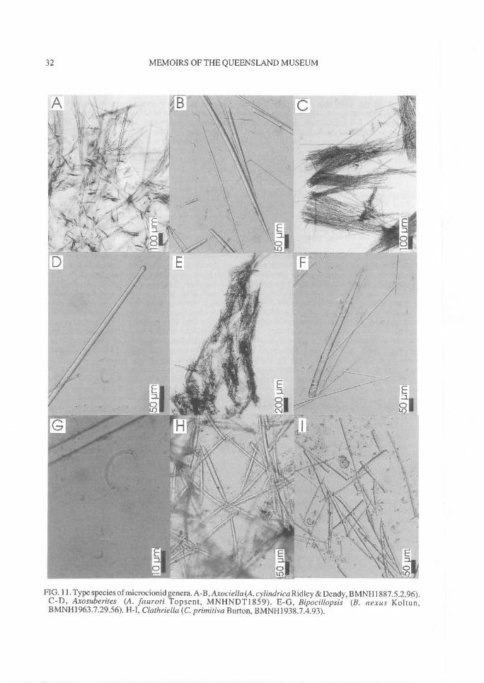
32^
MEMOIRS OF THE QUEENSLAND MUSEUM
FIG. 11. Type species of microcionid genera. A-B,Axociella(A. cylindrica Ridley & Dendy, BMNH1887.5.2.96).C-D, Axosuberites (A. fauroti Topsent, MNHNDT1859). E-G, Bipocillopsis (B. nexus Koltun,BMNH1963.7.29.56). H-I, Clathriella (C. primitiva Burton, BMNH1938.7.4.93).
REVISION OF MICROCIONIDAE^ 33
peripheral choanosomal skeleton. Ectosome withbrushes of smaller auxiliary subtylostyles over-laying larger subectosomal spicules. Megascleresentirely smooth choano somal principal subtylos-tyles-tylostyles, and two size classes of auxiliarysubtylostyles-tylostyles, both with smooth bases.Microscleres absent.
REMARKS. Redescription of the type speciesabove is based on examination of a slide prepara-tion in the MNHN collection and Topsent's(1893a) description; the corresponding specimenhas not yet been seen. Topsent (1893a) initiallycompared Axosube rites with Caulospongia Kentand Pseudosuberites Topsent in Suberitidae(Hadromefida), noting that it showed a certainlevel of morphological similarity in choanosomalfibres and geometry of tylostyle megascleres.However, in skeletal structure and spiculationAxosuberites appears to be a microcionid, mostsimilar to Axociella, both genera showingsimilarities in their ectosomal specialisation,axial and extra-axial differentiation, and absenceof echinating megascleres. Van Soest (1984b)also suggested that the genus was probably anAxociella without microscleres, and under hisscheme it was therefore referable to Thalysias,but in both genera axial and extra-axial skeletalstructures are well developed and closely com-parable, and it is suggested here that they bothshould be included in the same subgenus (i.e.,Clathria (Thalysias)).
Bipocillopsis Koltun, 1964(Fig. 11E-G)
Bipocillopsis Koltun, 1964a: 79.
TYPE SPECIES. Bipocillopsis nexus Koltun, 1964a:80 (by monotypy) (holotype ZIL10644, paratypeBMNH1963.7.29.56).
Erect, arborescent growth form with cylindricalreticulated branches. Surface hispid, raised intoirregular sharp conules. Choanosomal skeletonsubrenieroid reticulate, with ascending sponginfibres cored by multispicular plumose tracts andinterconnected by paucispicular transverse tractsof choanosomal principal subtylostyles, andechinated by acanthostyles. Subectosomal regionwith heavy paratangential bundles of subec-tosomal auxiliary styles protruding through sur-face and also scattered between fibres. Ectosomalskeleton without specialised spiculation but withbundles of subectosomal auxiliary styles sur-rounding protruding choanosomal spicules, andalso lying paratangential to ectosome.
Megascleres choanosomal principal subtylos-tyles invariably with basal spines and sometimeswith spines on shaft, echinating acanthostyles ofsimilar morphology to principal megascleres butshorter and more extensively spined, and singlecategory of entirely smooth subectosomalauxiliary style. Microscleres isochelae, stronglysigmoid, unguiferous with very reduced pointedalae, possibly anchorate. Toxas absent.
REMARKS. Bipocillopsis resembles Damoseniin lacking ectosomal specialisation and havingmodified sigmoid isochelae, but differs in growthform (arborescent versus encrusting) and subec-tosomal skeletal architecture (thickly paratan-gential versus tangential). The genus ismonotypic and may be included in Clathria(Clathria) by its skeletal structure, whereasDamoseni has hymedesmoid skeletal structuretypical of Clathria (Microciona). Chelae mor-phology has been described as tridentate sigmoid,allegedly related to the arcuate form, but this isvery difficult to tell with certainty given that alaeare nearly vestigial. There is no doubt that thesechelae are more highly modified than most othermicrocionids. They are very small, strongly un-guiferous (sigmoid curved with small pointedteeth), and lateral alae are completely free of theshaft and undifferentiated from the front ala, sug-gesting they may be of anchorate origin. How-ever, there is no lateral ridge on the shaft of chelaeto indicate an anchorate condition. The derivationof these chelae is indeterminable. Under thescheme of Hajdu et al. (1994) this genus could beincluded in Myxillina in possessing tridentate-derived isochelae, whereas all other features(skeletal structure, principal and auxiliarymegascleres and ectosomal skeleton) indicaterelationships with the Microcionidae, in which itis retained here tentatively. This decision is sup-ported by the case of Damoseni, discussed below,which have chelae of identical form to Bipocil-lopsis with the addition of oxhorn toxas (whichare not found in Myxillina).
Cionanchora de Laubenfels, 1936(Fig. 12E-G)
Cionanchora de Laubenfels, 1936a: 108.
TYPE SPECIES. Hymeraphia tuberosocaphata Top-sent, 1890b: 68 (by original designation) (fragment ofholotype MNHNLBIMDT939).
Encrusting growth form. Surface smooth, even,microscopically hispid. Choanosomal skeletonhymedesmoid. Spongin fibres reduced to basal
34^ MEMOIRS OF THE QUEENSLAND MUSEUM
layer lying on substrate, with choanosomal prin-cipal subtylostyles erect and forming unispicularascending columns protruding through ectosome,and echinating acanthostyles standing parallel tothese. Ectosome tangential skeleton of subec-tosomal auxiliary subtylostyles, of a singlecategory, forming brushes surrounding protrud-ing choanosomal principal spicules. Megasclereschoanosomal principal subtylostyles with tuber-culate bases, echinating acanthostyles with largespines evenly dispersed over entire spicule exceptfor bare point, and subectosomal auxiliary sub-tylostyles completely smooth. Microscleresanchorate-like isochelae. Toxas absent.
REMARKS. The diagnosis is based on a slide ofthe holotype and Topsent's (1890b) description;the corresponding specimen has not been seen.Cionanchora was erected for thinly encrustingsponges with hymedesmoid skeletal construc-tion, similar to Anaata, but with anchorate-likeinstead of arcuate-like modifications to isochelae(de Laubenfels, 1936a). Both those genera weremerged with Clathria (s.1.) by Van Soest (1984b)on the basis that modified microscleres were ahomoplasy throughout the Poecilosclerida, andare interspecific discriminators only. Anchorate-like modifications are also found in Folitispa(both of which were included in Clathria byHooper, 1990a). Microciona dubia fromChristmas Island (Kirkpatrick, 1900a: 136), wasreferred to Cionanchora by de Laubenfels(1936a: 108) supposedly in having anchorate-likechelae but these are of palmate origin. Bothspecies have skeletal architecture typical ofMicrociona where they are referred.
Clathria Schmidt, 1862(Fig. I2A-B)
Clathria Schmidt, 1862: 57; Ridley, 1884a: 443-449,612-615; Ridley & Dendy, 1887: 31; Hentschel,1911: 368; Hallmann, 1912: 205; Dendy, 1922: 64;Dendy, 1924a: 352-354; Wilson, 1925: 439; Top-sent, 1925: 645-658; Topsent, 1928a: 62,299; Bur-ton & Rao, 1932: 334-337; Burton, 1932a: 319;Burton, 1934a: 558; Koltun, 1959: 184; Levi,1960a: 50,52,61; Melone, 1963: 1-8; Sara &Melone, 1963: 362; Sara, 1964: 229; Simpson,1968a: 102, 104-106; Van Soest, 1984b: 90;Wiedenmayer, 1989: 56; Bergquist & Fromont,1988: 106.
Clathria Schmidt, plus Rhaphidophlus Ehlers; Ridley& Dendy, 1887: 146,151; Topsent, 1894a: 14-15,18.
Clatharia;Kumar, 1925: 221 [lapsus].
TYPE SPECIES. Clathria compressa Schmidt, 1862:58 (by subsequent designation (Schmidt, 1864: 35))(holotype LMJG15509).
Erect, arborescent, thinly lamellate, branchinggrowth form. Surface even, not hispid.Choanosomal skeleton regularly reticulate, withwell developed spongin fibres forming regular orirregular anastomoses of differentiated primaryand secondary spongin fibres. Fibres cored bychoanosomal principal subtylostyles in multi-spicular ascending tracts and uni- or bispiculartransverse connecting tracts, and echinated byacanthostyles perpendicular to or at acute anglesto spongin fibres. Ectosomal skeleton with tan-gential layer of subectosomal auxiliary subtylos-tyles, of a single size category. Megascleresbasally spined choanosomal principal subtylos-tyles, entirely smooth subectosomal auxiliarysubtylostyles, and echinating acanthostyles witheven spination. Microscleres palmate isochelaeand forceps-shaped or accolada toxas withspinose extremities.
REMARKS. Strictly defining Clathria is essen-tial in assigning a vast number of microcionidsincluded in the genus by numerous authors. Forexample, C. compressa has toxas with spinoseextremities, which therefore becomes a character'typical' of Clathria, whereas earlier authors con-sidered that this feature was diagnostic for Ar-temisina Vosmaer (de Laubenfels, 1936a).Clathria-like (viz. Labacea de Laubenfels) andArtemisina-like genera (viz. Ligrota de Lauben-fels) also have spinous toxas, indicating that thischaracter is homoplasious and probably not im-portant above the species level of classification.In general, the original definition of Clathria(s.s.) is upheld here, as re-examination ofSchmidt's syntype confirmed that all publishedcharacters cited in the species description(Schmidt, 1862: 58; Topsent, 1925: 647) arepresent in type material.
Topsent (1925: 648) noted that C. compressahas variable spicule dimensions, skeletal ar-chitecture, and live colouration. He correlatedthis variability with the diverse growth formsshown by the species: thinly encrusting exampleshad a hymedesmoid skeletal architecture, thicklyencrusting forms had a plumose skeleton, anderect ramose forms had anastomosing fibres (i.e.,encompassing the nominal genera Leptoclathria,Microciona and Clathria). He also found correla-tion between the size of megascleres and growthform variability, although he could find no ob-vious trends. Topsent's observations are invalu-able in deciding whether to maintain nominal
REVISION OF MICROCIONIDAE^ 35
encrusting genera and more massive sponges asdistinct genera, and whether to differentiate taxawith hymedesmoid, plumose or plumo-reticulateskeletal structure.
Clathria compressa is known only from thenorth Atlantic and Mediterranean regions, butjudging by its extensive synonymy it appears tobe (or have been) moderately common withinthose regions.
Clathriella Burton, 1935(Fig. 11H-I)
Clathriella Burton, 1935c: 73; Koltun, 1959: 186.
TYPE SPECIES. Clathriella primitiva Burton, 1935c:73 (by original designation) (holotypeBMNH1938.7.4.93).
Crumpled, irregular, massive growth form.Surface porous, uneven, hispid, with meanderingridges covered by thin transparent dermalmembrane. Choanosomal skeleton renieroidreticulate, with spongin fibres forming regularlytriangular meshes cored by multispicular tracts ofsmaller principal rhabdostyles (confined torenieroid network) and larger principal styles (lat-ter producing secondary plumose, subisodictyalskeleton of bi- or paucispicular ascending tracts).Echinating spicules absent. Ectosomal skeletonradially arranged tracts of subectosomal auxiliarystyles, of a single size category. Extra-fibreskeleton (apparently) has centrally curved (ar-cuate) oxeote megascleres scattered throughoutmesohyl. Megascleres two categories ofchoanosomal principal styles, larger smooth withrounded bases, smaller spined with rhabdosebases, and smooth or basally spined subec-tosomal auxiliary styles-subtylostyles.Microscleres absent.
REMARKS. Clathriella primitiva is obviouslyclosely related to Clathria chartacea Whiteleggein its skeletal structure and spiculation, conform-ing to the definition of Isopenectya (see below).Clathriella also shows similarities to Isociella inhaving an isodictyal reticulate skeleton and inlacking echinating spicules, although in Isociellaall megascleres are smooth and chelae and toxamicroscleres are present. The presence of smallerspined rhabdostyles and an isodictyal componentof the choanosomal skeleton are reminiscent ofRhabderemiidae, although the absence of thraus-tosigmata, thraustotoxa and other rhabderemiidmicroscleres in C. primitiva suggest that thesesimilarities are convergent.
Burton (1935c) considered that Clathriella wasa primitive member of the Microcionidae, inwhich styles and acanthostyles had not yet be-come differentiated or segregated into coring andechinating megascleres. He suggested furtherthat the toxiform oxeas, recorded by both Burton(1935c) and Koltun (1959), were derived fromacanthostyles, and that both forms were remnantsof a primitive condition. There is no empiricalevidence to support either argument, and thepresent study takes the alternative point of view,that species like Clathriella prinntiva are derivedor modified microcionids. Koltun (1959) alsosuggested that arcuate oxeas of C. primitiva weremicroxeas, and in that respect the genus shouldbe compared with Paratenaciella. However, inClathriella these spicules are supposedly large(200x7 p.m), indicating that they are truemegascleres, whereas in Paratenaciellamicroxeas are very small (40-75 x 0.7-311m). Inany case, re-examination of the holotype of C.primitiva (above) and more recent material fromthe Sakhalin Is, NW Pacific collected by PIBOC(QMG300052), did not find any toxiform oxeasalthough several examples of smaller auxiliarystyles were sinuous, and it may be these theauthors were referring to. The genus is referredhere into synonymy with Atitho (lsopenectya).
Clathriopsamma Lendenfeld, 1888(Fig. 12C-D)
Clathriopsamma Lendenfeld, 1888: 227; Topsent,1894a: 19; Hallmann, 1920: 771; de Laubenfels,1936a: 98; Levi, 1973: 614; Vacelet et al., 1976: 75.
TYPE SPECIES. Clathriopsamma reticulata Lenden-feld, 1888: 227 (by subsequent designation; Hallmann,1920: 771) (lectotype AMG9 1 35).
Erect, anastomosing, arborescent growth form.Surface uneven, arenaceous, microscopicallyhispid. Choanosomal skeleton irregularly reticu-late. Spongin fibres without well developedprimary or secondary differentiation, cored bychoanosomal principal subtylostyles and abun-dant foreign debris. In subectosomal skeletonprincipal subtylostyles also form plumosebrushes, protruding through ectosome. Fibresheavily echinated by acanthostyles also as-sociated with ectosomal spicule brushes. Ec-to some with paratangential tracts ofsubectosomal auxiliary subtylostyles, of a singlecategory, usually forming discrete brushes ofspicules at surface. Megascleres basally spined,fusi form choanosomal principal subtylostyles,smooth and basally spined subectosomal
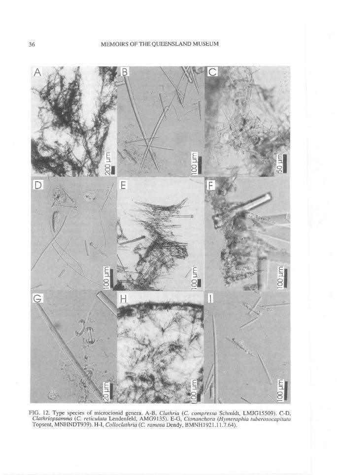
36^
MEMOIRS OF THE QUEENSLAND MUSEUM
FIG. 12. Type species of microcionid genera. A-B, Clathria (C. compressa Schmidt, LMJG15509). C-D,Clathriopsamma (C. reticulata Lendenfeld, AMG9135). E-G, Cionanchora (Hymeraphia tuberosocapaataTopsent, MNHNDT939). H-1, Colloclathria (C. ramosa Dendy, BMNH1921.11.7.64).
REVISION OF MICROCION1DAE^ 37
auxiliary subtylostyles, and echinating acanthos-tyles with large spines evenly dispersed.Microscleres palmate isochelae of two sizecategories, including contort forms, and sinuous-raphidiform or accolada toxas.
REMARKS. In Clathriopsamma the ectosome isstructurally close to the Thalysias condition, butthere is only one category of auxiliary spiculeproducing surface bundles. The genus is distin-guished from most other microcionid genera inhaving foreign particles incorporated into theskeleton (Hallmann, 1920) (see remarks forAulenella). Detrital entrapment is also known tooccur in other Microcionidae (e.g., some Holop-samma), other Poecilosclerida (e.g., Phoriospon-giidae), and it certainly also occurs commonly inother sponge orders (Dysideidae, Thorectidaeand Ircinidae in the Dictyoceratida, and manyHaplosclerida). This evidence indicates that thisfeature has arisen independently several timeswithin the Porifera, probably indicative ofecological specialisation, and obviously arisenindependently in several groups. However,species of Microcionidae that do incorporate sandappear to be relatively homogeneous in most oftheir other characters (i.e., there are no otherconflicting characters such as presence/absenceof ectosomal specialisation, or modifications tochelae), and consequently this specialisation isrecognised here at the subgenus level. Vacelet etal. (1976) synonymised Clathriopsamtna withAulenella, and Van Soest (1984b) mergedClathriopsatnma with Clathria, whereas in thiswork it is shown that the earliest available namefor these species is Wilsonella (see below).
Colloclathria Dendy, 1922(Fig. 12H-I)
Colloclathria Dendy, 1922: 74.
TYPE SPECIES. Colloclaihria ramosa Dendy, 1922:74 (by monotypy) (holotypeBMNH 1921.11.7.64).
Cylindrical, arborescent, digitate growth form.Surface even, hispid. Choanosomal skeletonreticulate, with slightly compressed axis andslightly more cavernous extra-axial skeleton.Spongin fibres cored by large principalchoanosomal styles, forming multispicular as-cending tracts interconnected by multispiculartransverse tracts. Echinating acanthostyles dis-tributed irregularly over fibres. Subectosomalskeleton reduced to immediate outer edge ofskeleton, with plumose tracts of larger subec-tosomal auxiliary subtylostyles extending
through ectosome. Larger auxiliary megascleresalso scattered between fibres and sometimescoring fibres. Ectosomal skeleton with smallerectosomal auxiliary subtylostyle forming discretebrushes overlying subectosomal spicules.Megascleres smooth principal choanosomalstyles, echinating acanthostyles with spinedbases and points (bare 'necks'), and two sizes ofauxiliary subtylostyles, usually with basal spines.Microscleres palmate isochelae, cleistochelaeand accolada toxas.
REMARKS. Colloclathria has a specialised ec-tosomal identical to Thalysias with twocategories of auxiliary spicules forming surfacespicule bundles, and on that basis Van Soest(1984b: 115) suggested the two genera should bemerged: it is included here in synonymy withClathria (Thalysias). The possession of cleis-tochelae in C. ramosa is not unique to Micro-cionidae also found in Plocamiopsis, Quizcionaand several species of Clathria (e.g., C.(Clathria) toxipraedita).
Damoseni de Laubenfels, 1936(Fig. 13A-C)
Damoseni de Laubenfels, 1936a: 110.
TYPE SPECIES. Hymeraphia michaelseni Hentschel,1911:351 (by original designation) (fragment ofholotype SMF969T).
Encrusting growth form. Surface sparselyhispid, even. Choanosomal skeleton hymedes-moid, with spongin fibres reduced to basal layerof spongin lying on substrate, with bases ofchoanosomal principal subtylostyles and acan-thostyles embedded in spongin, standing perpen-dicular to substrate, spicules protruding throughectosome. Ectosomal skeleton with paratangen-tial, slightly plumose tracts of both larger andsmaller auxiliary subtylostyles. Megasclereslarge principal choanosomal subtylostyles withspined bases, echinating acanthostyles withspined bases and shafts (bare 'necks'), and twosize classes of subectosomal auxiliary subtylos-tyles with either smooth or spined bases.Microscleres strongly unguiferous, sigmoidisochelae with vestigial alae, arcuate- or anchorate-like but of uncertain affinity, and large wing-shaped(oxhorn derived) toxas.
REMARKS. Hentschel's (1911) description ofHymeraphia michaelseni does not mention thepresence of toxa microscleres, nor that principalspicules protrude a long distance through thesurface. The modified unguiferous isochelae (of
38^ MEMOIRS OF THE QUEENSLAND MUSEUM
possible anchorate or arcuate derivation) areidentical to those of Bipocillopsis, and the com-bination of toxas and unguiferous isochelae ofpossible tridentate-derivation supports the in-clusion of such taxa in Microcionidae contrary toHajdu et al.'s (1994) proposal. Damoseni issimilar to other encrusting genera with hymedes-moid architecture and it could be included inClathria (Microciona) on this basis. However, thespecialised ectosomal skeleton composed ofsmaller ectosomal and larger subectosomalauxiliary spicules indicates affinities to Clathria(Thalysias).
Dendrocia Hallmann, 1920(Fig. 13D-E)
Dendrocia Hallmann, 1920: 767; de Laubenfels,1936a:109.
TYPE SPECIES. Clathria pyramida Lendenfeld,1888: 222 (by original designation) (holotypeAMG9047).
Massive, lobate-digitate growth form. Surfaceconulose, with subdermal sculpturing, osculesslightly raised above surface with membraneouslip. Choanosomal skeleton dendritic, slightlyplumo-reticulate near axis, without any obviousdivision of primary or secondary fibres, but withclear structural differences between choano-somal, subectosomal and ectosomal regions.Spongin fibres heavy, meandering, cored byauxiliary styles indistinguishable from those inectosomal skeleton, and heavily echinated by acan-thostyles (sometimes also secondarily incor-porated into fibres). Subectosomal skeletonplumose or radial, with spicule tracts composedof auxiliary styles diverging from ends ofperipheral choanosomal fibres and supportingoverlying ectosomal skeleton. Ectosomal skeletonwith continuous, heavy palisade of erect auxiliarystyles. Principal spicules absent, and megascleresinclude only echinating acanthostyles with spinedbases and necks but smooth points, and singlecategory of structural spicule (auxiliary styles-subtylostyles), entirely smooth with hastate pointsand sometimes secondarily pointed bases. Microscleres modified palmate isochelae with curved,thickened shaft and reduced alae. Toxas absent.
REMARKS. This strict diagnosis based on thetype species should be widened to include thepresence of modifications to isochelae, rangingfrom typical palmate forms (with straight shaft,completely fused lateral alae, as in Clathria(Dendrocia) abrolhensis sp. nov.), modified pal-mate isochelae (with greatly curved, thickened
shaft, partially detached lateral alae, as in C. (D.)dura), to anchorate-like forms (in which lateralalae are detached from shaft, shaft is curved,thickened and has a lateral ridge, as in C. (D.)myxilloides). One species also has oxhorn toxas(C.(D.) scabida) supporting the hypothesis thatDendrocia has affinities with Microcionidae.
Dendrocia, like Wilsonella, differs from othermicrocionids in having auxiliary styles bothcoring fibres and forming the ectosomal skeleton.However, whereas Wilsonella has two categoriesof auxiliary spicules and detritus is incorporatedinto the skeleton, Dendrocia has only onecategory of structural spicule throughout theskeleton. Dendrocia also has a characteristicdendritic or plumo-reticulate skeletal architec-ture, whereas Wilsonella is invariably reticulate.In ectosomal skeletal structure (with a continuousectosomal palisade of spicules) Dendrociaresembles the Thalysias condition, but it has onlyone geometric form of auxiliary spicule produc-ing the extra-fibre skeleton (i.e., subectosomeand ectosomal regions) more similar to Clathria.For this reason Dendrocia is enigmatic, andHooper (1990a) maintained it as a separate taxon,whereas earlier Van Soest (1984b) had indi-cated that it was probably a synonym ofClathria. In this work it is referred to Clathria(Dendrocia).
Two Australian species were initially includedin Dendrocia by Hallmann (1920), the typespecies and C. alata Dendy (holotype NMVG2280). Both are undoubtedly synonyms. Theyapparently differ only in their spicule dimensions(styles: 175-230x2-p,m versus 240-250x4-p,m;acanthostyles: 63-95x3-1p.m versus 79-154x8-1p.m; arcuate isochelae: 23-2p.m versus 20-2p,m,respectively). In contrast, D. antyaja Burton &Rao, from the Indian Ocean, should be removedfrom Dendrocia because it has regularly reticu-late choanosomal architecture, lacks an ectosomalskeleton, and has palmate isochelae indicatingaffinities to Clathria. Several other Australianspecies previously referred to Clathria s.s. shouldalso be referred to Dendrocia on the basis of theirskeletal architecture and spiculation.
Dictyociona Topsent, 1913(Fig. 13F-G)
Dictyociona Topsent, 1913a: 579, 618; de Laubenfels,1936a: 110; Levi, 1960a: 60; Bergquist & Fromont,1988: 104.
TYPE SPECIES. Microciona discreta Thiele, 1905:447 (by monotypy) (holotype ZMB3302).
REVISION OF MICROCIONIDAE^ 39
Thickly encrusting, lobate growth form. Sur-face composed of interconnected micropapillae.Choanosomal skeleton plumose, becomingplumo-reticulate in thicker sections. Sponginfibres divided into primary and secondary ele-ments, heavier in axis, lighter in subectosomalregion where dermal spicules implanted at nodesof peripheral fibres. Fibres cored by multispiculartracts of choanosomal principal subtylostyles,and heavily echinated by acanthostyles project-ing from fibres at all angles. Ectosomal skeletoncomposed of paratangential tracts of subec-tosomal auxiliary subtylostyles, of a single sizecategory, arising from peripheral fibres and pierc-ing surface. Megascleres choanosomal principalsubtylostyles with acanthose bases and acanthoseshafts near basal end, echinating acanthostyleswith spined bases and points (bare 'necks'), andsubectosomal auxiliary subtylostyles withmicrospined bases. Microscleres palmateisochelae and thin toxas intermediate betweenwing-shaped and oxhorn forms.
REMARKS. Dictyociona resembles Microcionain its plumose skeletal structure but has partiallyacanthose choanosomal megascleres similar tothose found in Anaata and Antho. These spiculesare also present in several Raspailiidae (e.g.,Hymeraphia, Eurypon), and they are consideredhere to be of minor diagnostic importance abovethe species level following Simpson (1968a). Levi(1960a: 60) merged Dictyociona with Clathria, al-though the species could as easily be placed inMicrociona. It is included here within Clathria(Clathria). Apart from the type species, other taxareferred to Dictyociona at one time or anotherinclude: Microciona clathrata Whitelegge, M.heterotoxa Hentschel, M. pyramidalis Brondsted,Hymedestnia oxneri Topsent, Eurypon asodes deLaubenfels, E. acanthotoxa Stephens, E. ditoxaStephens, E. microcizela Stephens, E. tenuissimaStephens, Clathria terra novae Dendy, D. contor-ta Bergquist & Fromont and D. atoxa Bergquist& Fromont.
Dictyoclathria Topsent, 1920(Fig. 13H-I)
Dictyoclathria Topsent, 1920b: 18; Burton, 1930a:501, 533, 1933: 50; de Laubenfels, 1936a: 77; Levi,1960a: 80.
Dyctioclathria [lapsus]; Ferrer Hernândez, 1921: 172.
TYPE SPECIES. Clathria morisca Schmidt, 1868: 9(by original designation) (schizotype BMNH-1868.3.2.21); junior synonym of Antho involvens
(Schmidt, 1864) (Levi, 1960a: 57) (schizotypeBMNH1867.3.11.92).
Arborescent growth form. Surface hispid, un-even. Choanosomal skeleton renieroid reticulatecomposed of spined acanthostyles forming trian-gular or rectangular meshes, enclosed withinfibres (axis) or bound together at nodes by col-lagen (near periphery). Echinating megascleresabsent. Subectosomal (extra-axial) skeletonplumose, with smooth principal styles standingperpendicular to fibre nodes, individually or inbundles, protruding through surface. Ectosomalskeleton with dense plumose or paratangentialbundles of subectosomal auxiliary subtylostyles,sometimes surrounding protruding principalstyles. Megascleres shorter lightly spined acan-thostyles (renieroid skeleton) and longer com-pletely smooth principal styles (extra-axialskeleton), and smaller subectosomal auxiliarysubtylostyles with spined bases. Microsclerespalmate isochelae and toxas intermediate be-tween wing-shaped and oxhorn forms.
REMARKS. Dictyoclathria is an objectivesynonym of Antho, since the type species of bothgenera are conspecific (Levi, 1960a). The typespecimen of Dictyoclathria is remarkable in havinga nearly raspailiid ectosomal condition withbundles of ectosomal spicules appearing to sur-round the protruding extra-axial spicules, but thisis not as perfectly developed as in many Raspailia.
Dirrhopalum Ridley, in Ridley & Duncan,1881
Plocamia Schmidt, 1870: 62.Dirrhopalum Ridley in Ridley & Duncan, 1881: 477.
See Plocamia.
Echinochalina Thiele, 1903(Fig. 14A-B)
Echinochalina Thiele, 1903a:961; Hallmann, 1912:288; Topsent, 1928a: 61; Burton, 1934a: 562; deLaubenfels, 1936a:118; Thomas, 1977: 115,
TYPE SPECIES. Ophlitaspongia australiensis Ridley,1884a: 442 (by subsequent designation (Hallmann,1912: 288)) (holotype BMNH1881.10.21.299).
Massive-digitate, reticulate growth form. Sur-face with low ridges and interconnected lamellae,producing angular cells with thin dermalmembrane stretched between. Choanosomalskeleton irregularly reticulate, with heavyprimary spongin fibres cored by paucispiculartracts of auxiliary spicules (tornotes), and heavysecondary spongin fibres cored by uni- or
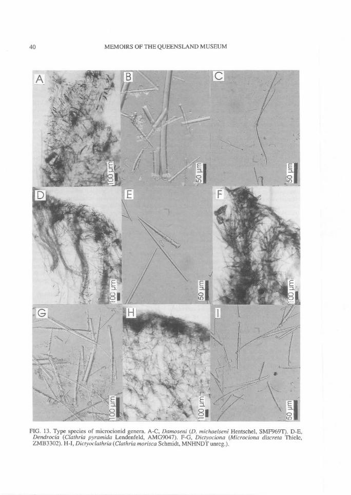
-
.01k1...
40^
MEMOIRS OF THE QUEENSLAND MUSEUM
FIG. 13. Type species of microcionid genera. A-C, Damoseni (D. michaelseni Hentschel, SMF969T). D-E,Dendrocia (Clathria pyramida Lendenfeld, AMG9047). F-G, Dictyociona (Microciona discreta Thiele,ZMB3302). H-I, Diayoclathria (Clathria morisca Schmidt, MNHNDT unreg.).
REVISION OF MICROCIONIDAE^ 41
bispicular tracts of auxiliary spicules. Fibreslightly echinated by principal subtylostyles. Ec-tosomal skeleton with undulating fibres and looseparatangential tracts of auxiliary spicules (alsodistributed throughout mesohyl). Megascleres in-clude quasidiactinal auxiliary tornotes with aym-metrical or symmetrical ends (coring fibres andon ectosome), and completely smooth principalsubtylostyles-styles (echinating fibres), some-times modified to oxeotes (asymmetrical withtwo pointed ends). Microscleres wing-shapedtoxas. Isochelae absent.
REMARKS. In the type species coring spiculesare tornotes, whereas in other species ofEchinochalina they range from true styles tooxeotes. Re-examination of the holotype of 0.australiensis, and Thiele's (1903a) specimenfrom Ternate, Indonesia (SMF1855) also foundthat echinating principal styles can sometimes bemodified to oxeote spicules, providing supportfor the otherwise tenuous placement of Protoph-litaspongia with this group of microcionids.
Hallmann (1912: 288) noted that Echinochalinadiffers from the allied Echinoclathria of authors(= Holopsamma as defined here) in having fibrescored by auxiliary spicules (tylotes or stron-gyles), which may be modified to quasi-monac-tinal forms (tylostrongyles, tornostrongyles), andechinated by smooth styles or subtylostyles. Bycomparison, Echinoclathria in the strict sense (orOphlitaspongia of authors) has principalchoanosomal styles (or modified monactinalmegascleres) which both core and echinate fibres,as well as a second category protruding throughthe surface; Holopsamma has only a singlecategory of coring and echinating principalspicule. As such, Van Soest (1984b: 129) sug-gested that Echinochalina possibly did notbelong with the Microcionidae, and might bemore appropriately placed elsewhere within thePoecilosclerida (e.g., Phoriospongiididae). How-ever, examination of a suite of species includedhere in Holopsamma found that this group alsoundergoes a reduction in coring spicules (e.g., H.pluritoxa (Pulitzer-Finali)), whilst retaining othercharacteristics common to the genus, and it ispossible that Echinochalina, sensu Hallmann, un-dergoes similar reduction.
Probably of greater importance in determiningthe appropriate placement of Echinochalina is itslack of any special ectosomal skeleton, and in thisrespect it is similar to Dendrocia (both with onlyone form of structural (auxiliary) spicule). WhereasEchinoclathria (s.s.) possesses monactinal auxiliary
spicules in the peripheral skeleton (sometimesreduced to quasi-diactinal forms), Echinochalinahas spicules which are closer to true diactinals(although sometimes modified to quasi-monac-tinal forms). Both genera lack definite ectosomalspecialisation. Some Echinochalina appear to haveaffinities with certain species of Echinodictyum(Raspailiidae) and with the Desmacididae, andboth Thiele (1903a) and Topsent (1904a) havealready noted this resemblance, suggesting thatthey differ mainly in the geometry, ornamentationand derivation of the echinating megascleres. Bycomparison, Hallmann (1912) considered thatthese differences, and the presence of long sub-ectosomal styles in many Echinodictyum species,are sufficient to maintain the two genera as dis-tinct taxa (see Hooper, 1991d).
Although Echinochalina usually has smoothechinating megascleres, linking it toEchinoclathria and Holopsamma, there are twospecies which were previously referred toEchinodictyum (E. ridleyi Dendy and E. spon-giosum Dendy), which have acanthose echinatingmegascleres but otherwise conform toEchinochalina in their spicule geometries andskeletal architectures. Hooper (1991) transferredthese species to Echinochalina.
Tablis de Laubenfels is an obvious synonym ofEchinochalina. The two genera differ only by theabsence of microscleres and the presence of areticulate architecture in Tab/is.
Echinoclathria Carter, 1884(Fig. 14C-E)
Echinoclathria Carter, 1884: 204 [nomen nudum];Carter, 1885E 355; Ridley & Dendy, 1887: 159;Topsent, 1894a: 18; Thiele, 1903a: 962; Hallmann,1912: 275-276; Dendy, 1922: 71 [in part]; Topsent,I928a: 61; de Laubenfels, 1936a: 119; Wieden-mayer, 1977: 143, 1989: 58. (not Uriz, 1988: 89].
TYPE SPECIES. Echinoclathria tenuis Carter, 1885E355 (by subsequent designation; Burton, 1934a: 562)(holotype BMNH1886.12.15.147); junior synonym ofSpongia leporina Lamarck, 1814: 444 (Topsent, 1932:101) (holotype MNHNLBIMDT567).
Thinly flabellate, flattened palmate, stalkedgrowth form. Surface membraneous, microscopi-cally hispid. Choanosomal skeleton renieroidreticulate, slightly compressed with welldeveloped spongin fibres in axis, more openlyreticulate, less compressed and with lighterspongin towards periphery. Axial fibres coredby pauci- or multispicular tracts of smaller prin-cipal styles producing rounded or irregularlyshaped meshes, and echinated by same spicules.
42^ MEMOIRS OF THE QUEENSLAND MUSEUM
Subectosomal (extra-axial) skeleton radial, uni-or paucispicular, with larger principal spiculeserect on terminal fibres and usually protrudingthrough surface. Ectosomal specialisation absent,with bundles of subectosomal auxiliary subtylos-tyles embedded perpendicularly and formingparatangential brushes surrounding larger prin-cipal spicules. Megascleres include smaller,robust, entirely smooth principal subtylostyles(coring and echinating fibres), larger principalsubtylostyles of similar geometry (projecting fromperipheral fibres and protruding through surface),and smooth subectosomal auxiliary subtylos-tyles, straight or flexuous. Microscleres absent.
REMARKS. There is substantial confusion con-cerning the precise definition of Echinoclathria,and its relationship with other nominal generasuch as Holopsamma, Halme, Aulena and Oph-litaspongia. Consequently the above diagnosis isstrict, pertaining only to the type species, and adetailed explanation is justified below.
Most authors follow Hallmann's (1912: 275)interpretation of Echinoclathria in which thegenus is essentially characterised by 'ahoneycomb mass of anastomosing flattenedtrabeculae', a reticulate skeleton of heavy spon-gin fibres cored and echinated by smooth monac-tinal principal megascleres of the same geometry,and with monactinal subectosomal auxiliarystyles distributed throughout the mesohyl (and insome species also forming a radial subectosomalskeleton). Hallmann also noted that in somespecies he assigned to Echinoclathria there areboth chelae and toxa microscleres, with quasi-monactinal auxiliary megascleres, or they mayhave their coring megascleres replaced partiallyor completely by detritus (e.g., Holopsammalaminaefavosa). Hallmann suggested further thatEchinoclathria and Ophlitaspongia essentiallydiffered only in growth form, a view perpetuatedby Wiedenmayer (1989). This interpretation isemended here.
Most species included in Echinoclathria priorto the present study do have the characteristicsoutlined by Hallmann (1912), and most are rela-tively homogeneous and easily recognisable inthe field by their characteristic 'honeycombreticulate' growth form. It is therefore unfor-tunate that Burton (1934a) subsequently desig-nated E. tenuis (a junior synonym of Spongialeporina) as the type species of Echinoclathria,because this species has a flabellate growth form(very dissimilar to 'honeycombed reticulate'species), skeletal architecture consisting of a dif-
ferentiated axis, extra-axis, a renieroid skeleton,and a second category of principal spicules protrud-ing through the ectosome. Echinoclathria leporinais typical of most Ophlitaspongia (of authors)(e.g., 0. axinelloides Dendy).
Confusing the generic boundaries even further,Spongia leporina closely resembles Antho(Isopenectya) in growth form and gross skeletalconstruction, but differentiated by their skeletalstructures, spicule ornamentation and localisa-tion of particular spicules to certain regions of theskeleton. Nevertheless, it could be argued thatIsopenectya could be included in Echinoclathriaas equally as in Antho. The former option istentatively rejected here based on the unequivo-cal possession of spined (versus smooth) stylescomposing the renieroid skeleton, and possessionof a secondary, longitudinal, subisodictyal secon-dary skeleton in Isopenectya. Similarities inrenieroid skeletal construction may link boththese genera into a clade based on secondaryreduction or loss. The difficulty in positively as-signing Isopenectya is discussed further below.
Wiedenmayer (1977: 144) suggested thatEchinoclathria should be restricted to Indo-Pacific species, although there were similaritiesin skeletal architecture with Pandaros from theWest Indies (which lacks microscleres). He notedfurther that Echinoclathria had mostly smoothchoanosomal spicules, except for occasional ves-tigial spines on the bases of some auxiliaryspicules, whereas Pandaros had acanthoseechinating spicules. Simpson (1968a) has alreadydemonstrated that the loss of spination onechinating spicules is common amongstmicrocionids and not correlated with anycytological differences (i.e., of low taxonomicvalue). Wiedenmayer (1977, 1989) concludedthat although two genera intergraded they couldnot be consistently differentiated, and Pandarosis not included in this group.
Thus, on the basis of evidence presented byHallmann (1912: 275), Burton (1959a: 246) andWiedenmayer (1989: 58), and re-examination ofall nominal species belonging to these groups, itis clear that we are dealing with two distinct,homogenous groups. One, agreeing with thedefinition of Spongia leporina above, includesthe genera Echinoclathria (in the strict senseonly, and not of authors), and Ophlitaspongia (ofauthors, and not in the strict sense).Echinoclathria is the earliest available name forthis group, and its characteristics should be takenas those traditionally associated with the conceptof Ophlitaspongia (of authors). This genus is
REVISION OF MICROCIONIDAE^ 43
most closely related to A ntho in its renieroid mainskeletal structure, differing in having smoothchoanosomal spicules and a single skeletal stuc-ture (i.e., Antho has 2 skeletal structures: arenieroid skeleton composed of spined spiculesand a plumose or subisodictyal skeleton com-posed of smooth spicules).
The second group contains honeycombedreticulate species traditionally associated withEchinoclathria (of authors, not in the strictsense), together with Holopsamma, HaIme, Plec-tispa and Aulena (of authors, not in the strictsense). The earliest available name for this groupis Holopsamma Carter (18850. There is no doubtthat this honeycombed reticulate group ofmicrocionids warrants inclusion in a separatetaxon, although its level of divergence is arguable(cf. Wiedenmayer, 1977, 1989; Hooper, 1991). Itspeculiar growth form is consistent for all 12Australian species (all from southern Australia(Gondwanan) faunas), and 4 non-Australian species(2 from South America (Gondwanan) and 2 fromthe Indo-west Pacific (Tethyan) faunas). Thisgrowth form is correlated with a reticulate skele-tal architecture, forming a homogeneous groupwhich is recognised here at the generic level.
A honeycombed reticulate growth form is alsoknown for Acamasina de Laubenfels (1936a: 117)in Mycalidae (Van Soest, 1984b) and Panda ros(see below), both known only from the WestIndies.
Echinonema Carter, 1875(Fig. 14F-G)
Echinonema Carter, 1875: 194 [nomen nudum];Carter, 1881a: 378; Ridley, 1884a: 615; Topsent,I 894a: 19; Dendy, 1896: 32; Whitelegge, 1901:80; Topsent, 1928a: 61, 1932: 89, 98; de Lauben-fels, 1936a: 112; Levi, 1960a: 56.
TYPE SPECIES. Echinonema typicum Carter, 1881a:377 (by typonymy) (lectotype BMNH1877.5.21.149);junior synonym of Spongia cactifonnis Lamarck,1814: 440 (lectotype MNHNLBIMDT580).
Arborescent, shrubby, lamellate growth form.Surface even, hispid, subectosomal striations.Choanosomal skeleton reticulate, with open rec-tangular or elongate open meshes although slight-ly compressed in axis. Primary spongin fibresascending, heavy, cored by multispicular tracts ofchoanosomal principal styles, interconnected bypauci- or aspicular secondary spongin fibres.Fibres echinated by acanthostyles heaviest inperipheral skeleton. Subectosomal skeletonplumose, with brushes of larger subectosomal
auxiliary subtylostyles erect on peripheralchoanosomal fibres. Ectosomal skeleton dense,with smaller ectosomal auxiliary subtylostylesforming a dense palisade on surface. Megascleresinclude smooth choanosomal principal styles,larger subectosomal auxiliary subtylostyles withsmooth or microspined bases, smaller ectosomalauxiliary subtylostyles with smooth ormicrospined bases, and short thick echinatingacanthostyles with spined base and point but bareneck. Microscleres palmate isochelae of twosizes, including contort forms, and thin accoladaand asymmetrical toxas.
REMARKS. It is confirmed here that Carter's(1881a) Echinonema typicum is identical toLamarck's (1814) Spongia cactifonnis, and con-sequently the name cactifonnis has seniority overthe better known junior synonym Clathria typica,widely used in the literature. Lendenfeld (1888),Whitelegge (1901) and Hallmann (1912) createdmany new subspecific names (as varieties) forthis species, and Hooper & Wiedenmayer (1994)assigned Lamarck's (1814) specimen to Clathria(Thalysias) cactifonnis cactiformis, and Carter's(1881) specimen to C. (T) cactiformis typica.Examination of type material of all these sub-species (var. typica (SMF1589); var. porrecta(SMF1653); var. brevispinus (AMZ931); var.favosus (AMZ944); var. geminus (AMZ928); var.obesus (AMZ937); var. proximus (AMZ930);var. stelligera (AME648); and three unnamedvarieties of Hallmann (1912) (AMZ1158;AMZ1430, G9135, Z938; and AMZ41), showedthat they were conspecific with the nominotypi-cal variety (based on spicule size, spiculegeometry and skeletal structure), whereas growthforms and surface features varied substantiallybetween each taxon. The recognition of thesesubspecies, a preoccupation of many earlierauthors, does at least demonstrate a high degreeof external morphological variability for thespecies, but is not of particular nomenclaturalimportance because there are no other mor-phological characters that correlate with thesedifferences in external morphology. It may beeventually determined from biochemical orgenetic data, that this polymorphism is indicativeof sibling species relationships, but no studies ofthis sort have yet been undertaken.
De Laubenfels (1936a: 112) suggested thatEchinonema was identical to Thalysias (sensu deLaubenfels), whereas Van Soest (1984b) andothers placed both genera into synonymy withRhaphidophlus (see below). The genus is in-
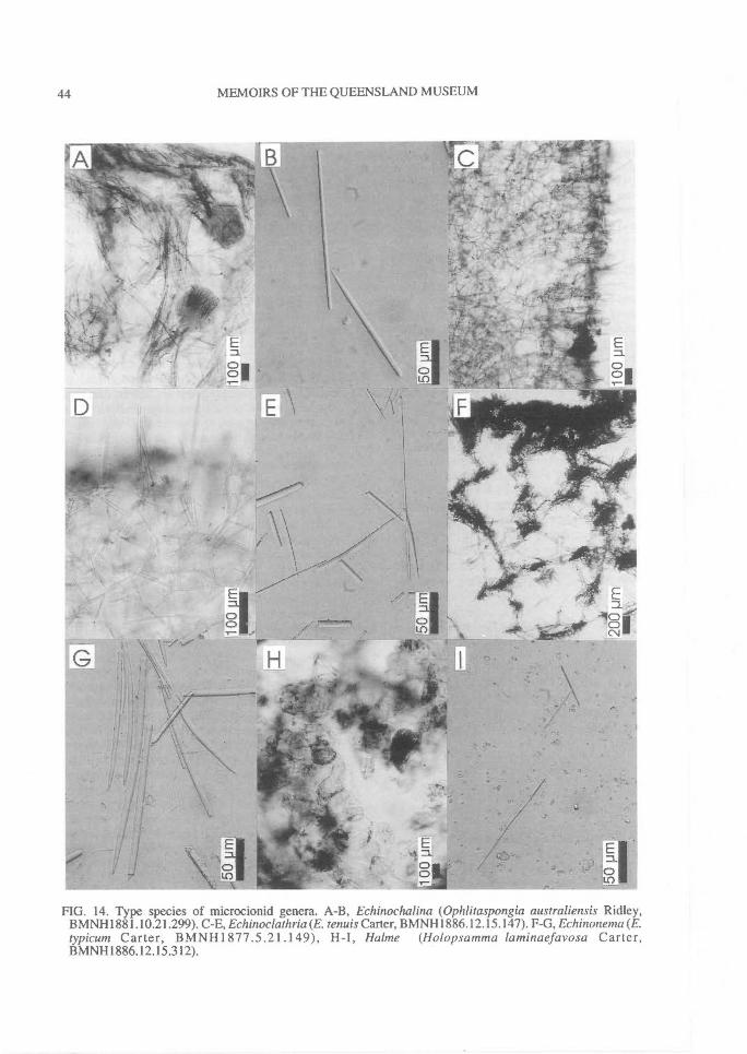
44^ MEMOIRS OF THE QUEENSLAND MUSEUM
FIG. 14. Type species of microcionid genera. A-B, Echinochalina (Ophlitaspongia australiensis Ridley,BMNHI881.10.21.299). C-E, Echinoclathria (E. tenuis Carter, BMNH1886.12.15.147). F-G, Echinonema (E.typicum Carter, BMNH1877.5.21.149), H-I, HaIme (Holopsamma laminaefavosa Carter,BMNHI886.12.15.312).

REVISION OF MICROCIONIDAE^ 45
eluded here in Clathria (Thalysias) on the basisof its ectosomal specialisation.
Fisherispongia de Laubenfels, 1936(Fig. 15A-C)
Fisherispongia de Laubenfels, 1936b: 460.
TYPE SPECIES. Fisherispongiaferrea de Laubenfels,1936b: 460 (by original designation) (holotypeUSNM22239).
Encrusting growth form. Surface tuberculate,arenaceous, hispid. Choanosomal skeletonhymedesmoid, with basal layer of spongin fibreincorporating detritus and bases of largerchoanosomal principal subtylostyles and smallerechinating styles, standing perpendicular to sub-strate, in groups or individually, ascending to butnot protruding through ectosome. Ectosomalskeleton plumose, with subectosomal auxiliarypolytylostyles, of a single category, arising fromends of choanosomal megascleres in multi-spicular bundles protruding through surface.Megascleres large choanosomal principal sub-tylostyles with smooth bases, smaller smoothechinating styles with smooth or microspinedbases, and polytylote auxiliary tylostyles withsmooth or microspined bases. Microscleres pal-mate isochelae, including contorted forms, andthick wing-shaped toxas.
REMARKS. De Laubenfels (1936b) distin-guished Fisherispongia from other microcionidsby the polytylote bases on their subectosomalauxiliary styles. In all other respects, however, thetype species resembles other encrusting specieswith hymedesmoid architecture (e.g., Lep-toclathria). Polytylote ectosomal megascleresare known in several other species ofMicrocionidae (e.g., Clathria aceratoobtusa,Paratenaciella microxea), as well as in otherPoecilosclerida (e.g., Camptisocale Topsent andPhellodenna Ridley & Dendy; Coelos-phaeridae). The incorporation of detritus into thechoanosome and fibres is well known for severalmicrocionids and other sponges (see remarks forAulenella) and on this basis the species is in-cluded in Clathria (Wilsonella). Re-examinationof the holotype found a marked contrast in sizebetween the smaller and larger (so-called prin-cipal) spicules, with no intermediate sizes, andthese smaller spicules are interpreted here asbeing smooth echinating styles. By its toxa mor-phology Fisherispongia ferrea (from the Atlanticcoast of Panama) is very similar to Clathriaaceratoobtusa (from the Indo-west Pacific).
Folitispa de Laubenfels, 1936(Fig. 15D-F)
Folitispa de Laubenfels, 1936a: 119.
TYPE SPECIES. Hymedesmia laevissima Dendy,1922:81 (by original designation) (holotypeBMNH1921.11.7.69).
Thickly encrusting growth form. Surface even,slightly hispid. Choanosomal skeleton hymedes-moid, with spongin fibres lying on substrate andbases of choanosomal principal subtylostyles em-bedded, standing perpendicular to substrate in-dividually or forming short multispicularplumose columns protruding through surface.Echinating megascleres absent. Subectosomalskeleton irregularly plumose, with loosely ag-gregated bundles of subectosomal auxiliary sub-tylostyles erect on surface or lying tangential toit. Megascleres include smooth choanosomalprincipal subtylostyles, and smooth subec-tosomal auxiliary subtylostyles with mucronateor telescoped points. Microscleres palmateisochelae with anchorate-like modifications (cur-vature, partially detached alae, continuous ridgeon shaft). Toxas absent.
REMARKS. The type species of Foliti.spa differsfrom other encrusting microcionids withhymedesmoid skeletal architecture (e.g., Lep-toclathria) in lacking echinating acanthostyles(cf. Axocielita) and having chelae withanchorate-like modifications instead of typicalpalmate isochelae (cf. Cionanchora). Thesechelae are strongly curved, with lateral alae par-tially detached from the shaft and a continuouslateral ridge running the length of the shaft. How-ever, these lateral alae are not fully formed (beingabout two-thirds the size of the front ala), nor arethey completely detached from the shaft (at-tached for approximately 50% of their length),and consequently they cannot be considered trueanchorate chelae but perhaps palmate isochelaewith substantial anchorate modifications. Thegenus is included here in Clathria (Microciona)based on its skeletal structure.
[Halme] Lendenfeld, 1885(Fig. 14H-I)
Ha/me Lendenfeld, 1885c: 285, 1889a: 446; deLaubenfels, 1936a: 17; Bergquist, 1980b: 454;Wiedenmayer, 1989: 58 (preoccupied).
Not Ha/me Pascoe, 1869.
TYPE SPECIES. Holopsamma laminaefavosa Carter,1885b: 212 (by subsequent designation; de Laubenfels,1936a: 17) (holotype BMNH1886.12.15.312).
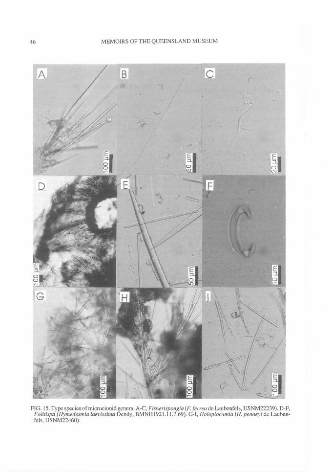
46^ MEMOIRS OF THE QUEENSLAND MUSEUM
FIG. 15. Type species of microcionid genera. A-C, Fisherispongia (F. ferrea de Laubenfels, USNM22239). D-F,Folitispa (Hymedesmia laevissima Dendy, BMNH1921.11.7.69). G-I, Holoplocamia (H. penneyi de Lauben-fels, USNM22460).
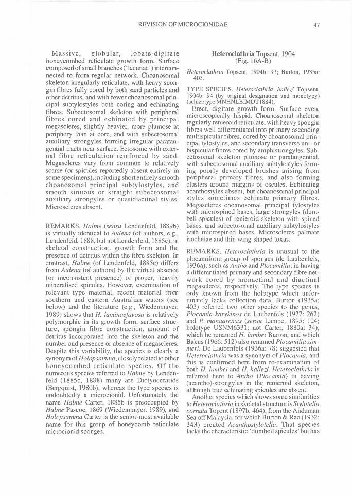
REVISION OF MICROCION1DAE^ 47
Massive, globular, lobate-digitatehoneycombed reticulate growth form. Surfacecomposed of small branches (lacunae') intercon-nected to form regular network. Choanosomalskeleton irregularly reticulate, with heavy spon-gin fibres fully cored by both sand particles andother detritus, and with fewer choanosomal prin-cipal subtylostyles both coring and echinatingfibres. Subectosomal skeleton with peripheralfibres cored and echinated by principalmegascleres, slightly heavier, more plumose atperiphery than at core, and with subectosomalauxiliary strongyles forming irregular paratan-gential tracts near surface. Ectosome with exter-nal fibre reticulation reinforced by sand.Megascleres vary from common to relativelyscarse (or spicules reportedly absent entirely insome specimens), including short entirely smoothchoanosomal principal subtylostyles, andsmooth sinuous or straight subectosomalauxiliary strongyles or quasidiactinal styles.Microscleres absent.
REMARKS. Ha/me (sensu Lendenfeld, 1889b)is virtually identical to Aulena (of authors, e.g.,Lendenfeld, 1888, but not Lendenfeld, 1885c), inskeletal construction, growth form and thepresence of detritus within the fibre skeleton. Incontrast, Hahne (of Lendenfeld, 1885c) differsfrom Aulena (of authors) by the virtual absence(or inconsistent presence) of proper, heavilymineralised spicules. However, examination ofrelevant type material, recent material fromsouthern and eastern Australian waters (seebelow) and the literature (e.g., Wiedenmayer,1989) shows that H. laminaefavosa is relativelypolymorphic in its growth form, surface struc-ture, spongin fibre construction, amount ofdetritus incorporated into the skeleton and thenumber and presence or absence of megascleres.Despite this variability, the species is clearly asynonym of Holopsamma, closely related to otherhoneycombed reticulate species. Of thenumerous species referred to Hahne by Lenden-feld (1885c, 1888) many are Dictyoceratids(Bergquist, 1980b), whereas the type species isundoubtedly a microcionid. Unfortunately thename Halme Carter, 1885b is preoccupied byHahne Pascoe, 1869 (Wiedenmayer, 1989), andHolopsamma Carter is the senior-most availablename for this group of honeycomb reticulatemicrocionid sponges.
Heteroclathria Topsent, 1904(Fig. 16A-B)
Heteroclathria Topsent, 1904b: 93; Burton, 1935a:403.
TYPE SPECIES. Heteroclathria hallez; Topsent,1904b: 94 (by original designation and monotypy)(schizotype MNHNLBIMDT1884).
Erect, digitate growth form. Surface even,microscopically hispid. Choanosomal skeletonregularly renieroid reticulate, with heavy sponginfibres well differentiated into primary ascendingmultispicular fibres, cored by choanosomal prin-cipal tylostyles, and secondary transverse uni- orbispicular fibres cored by amphistrongyles. Sub-ectosomal skeleton plumose or paratangential,with subectosomal auxiliary subtylostyles form-ing poorly developed brushes arising fromperipheral primary fibres, and also formingclusters around margins of oscules. Echinatingacanthostyles absent, but choanosomal principalstyles sometimes echinate primary fibres.Megascleres choanosomal principal tylostyleswith microspined bases, large strongyles (dum-bell spicules) of renieroid skeleton with spinedbases, and subectosomal auxiliary subtylostyleswith microspined bases. Microscleres palmateisochelae and thin wing-shaped toxas.
REMARKS. Heteroclathria is unusual to theplocamiform group of sponges (de Laubenfels,1936a), such as Antho and Plocamilla, in havinga differentiated primary and secondary fibre net-work cored by monactinal and diactinalmegascleres, respectively. The type species isonly known from the holotype which unfor-tunately lacks collection data. Burton (1935a:403) referred two other species to the genus,Plocamia karykinos de Laubenfels (1927: 262)and P. manaarensis (sensu Lambe, 1895: 124;holotype USNM6331; not Carter, 1880a: 34),which he renamed H. lambei Burton, and whichBakus (1966: 512) also renamed Plocamilla zhn-meri. De Laubenfels (1936a: 78) suggested thatHeteroclathria was a synonym of Plocamia, andthis is confirmed here from re-examination ofboth H. lambei and H. hallezi. Heteroclathria isreferred here to Antho (Plocamia) in having(acantho)-strongyles in the renieroid skeleton,although true echinating spicules are absent.
Another species which shows some similaritiesto Heteroclathria in skeletal structure is Stylotellacorn uta Topent (1897b: 464), from the AndamanSea off Malaysia, for which Burton & Rao (1932:343) created Acanthostylotella. That specieslacks the characteristic `dumbell spicules' but has
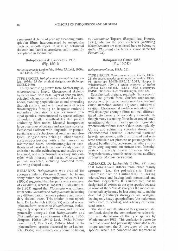
48^ MEMOIRS OF THE QUEENSLAND MUSEUM
a renieroid skeleton of primary ascending multi-spicular fibres interconnected by unispiculartracts of smooth styles. It lacks an ectosomalskeleton and lacks microscleres, and it possiblybest placed in Iophonidae.
Holoplocamia de Laubenfels, 1936(Fig. 15G-I)
Holoplocamia de Laubenfels, 1936a: 75; Levi, 1960a:80; Little, 1963: 47.
TYPE SPECIES. Holoplocamia penneyi de Lauben-fels, 1936a: 75 (by original designation) (holotypeUSNM22460).
Thinly encrusting growth form. Surface rugose,microscopically hispid. Choanosomal skeletonhymedesmoid, with basal layer of spongin fibre,principal choanosomal styles embedded in fibrenodes, standing perpendicular to and protrudingthrough surface, and with basal mass of acan-thostrongyles forming an irregular renieroidsecondary reticulation of spicules around prin-cipal spicules, interconnected by sparse collagenat nodes. Smaller acanthostyles also presentechinating fibre nodes. Mesohyl incorporateslarge quantities of detritus and auxiliary spicules.Ectosomal skeleton with tangential or paratan-gential tracts of subectosomal auxiliary subtylos-tyles. Megascleres principal choanosomalstyles-subtylostyles with either smooth ormicrospined bases, acanthostrongyles or acan-thostyles of basal skeleton more heavily spined atends than middle, echinating acanthostyles even-ly spined, and subectosomal auxiliary subtylos-tyles with microspined bases. Microsclerespalmate isochelae, including contorted forms,and wing-shaped toxas.
REMARKS. Holoplocamia was erected forsponges similar to Plocamia Schmidt, but havingspiny rather than smooth principal spicules. Levi(1960a) suggested that the genus was a synonymof Plocamilla, whereas Topsent (1928a) and Lit-tle (1963) argued that Plocatnilla was differentfrom both Plocamia and Holoplocamia in lackingany differentiation between primary and secon-dary skeletal tracts. This opinion is not upheldhere. De Laubenfels (1936a: 75) referred several`plocamiform' species to Holoplocamia, includ-ing the type species of Plocamilla, and it is nowgenerally accepted that Holoplocamia andPlocamilla are synonymous (Bakus, 1966;Simpson, 1968a; Levi & Levi, 1983a; Pulitzer-Finali, 1983; Van Soest, 1984b). Most of the`plocamiform' species discussed by de Lauben-fels (1936a) were subsequently found to belong
to Plocamione Topsent (Raspailiidae; Hooper,1991), whereas the poecilosclerids (includingHoloplocamia) are considered here to belong toAntho (Plocamia) (the latter a senior name forPlocamilla).
Holopsamma Carter, 1885(Fig. 16C-D)
Holopsamma Carter, 1885c: 211.
TYPE SPECIES. Holopsamma crassa Carter, 1885c:211 (by subsequent designation, de Laubenfels, 1936a:98) (lectotype BMNH 1886.12.15.313; Hooper &Wiedenmayer, 1994), a senior synonym of Ha/meglobosa Lendenfeld, 1885c: 303 (lectotypeBMNH1886.8.27.71) (cf. Wiedenmayer, 1989: 63).
Subspherical, digitate, regularly 'honeycomb'reticulate growth form. Surface arenaceous,porous, with tympanic membrane-like ectosomalcrust stretched across adjacent subdermalcavities. Choanosomal skeleton reticulate, withwell developed spongin fibres not well differen-tiated into primary or secondary elements, al-though many ascending fibres have core of smallquantities of detritus (mostly spicule fragments),whereas other fibres clear of detritus completely.Coring and echinating spicules absent fromchoanosomal skeleton. Ectosomal skeletonheavily arenaceous, with crust of sand and scat-tered reticulate (or plumose or paratangential inplaces) bundles of subectosomal auxiliary stron-gyles lying tangential on surface crust. Mesohylmatrix relatively heavy between fibres .Megascleres only smooth subectosomal auxiliarystrongyles. Microscleres absent.
REMARKS. De Laubenfels (1936a: 97) notedthat Holopsamma differs from other 'sandysponges' (i.e., the polyphyletic 'familyPsammascidae' de Laubenfels) in lackingmicroscleres and having both monactinal anddiactinal megascleres. It is unfortunate that hedesignated H. crassa as the type species becausein some of the 5 'valid' syntypes the monactinal(principal) styles may be lost completely, and thediactinal (auxiliary) strongyles are vestigial,leaving only heavy spongin fibres (the major oneswith a core of detritus), and a heavy ectosomalsand cortex.
The status and affinities of this genus are stillconfused, despite the comprehensive redescrip-tion and discussion of the type species byWiedenmayer (1989). This confusion is due to thefact that no-one had previously nominated a lec-totype amongst the 31 syntypes of the typespecies, which are composite and represent at
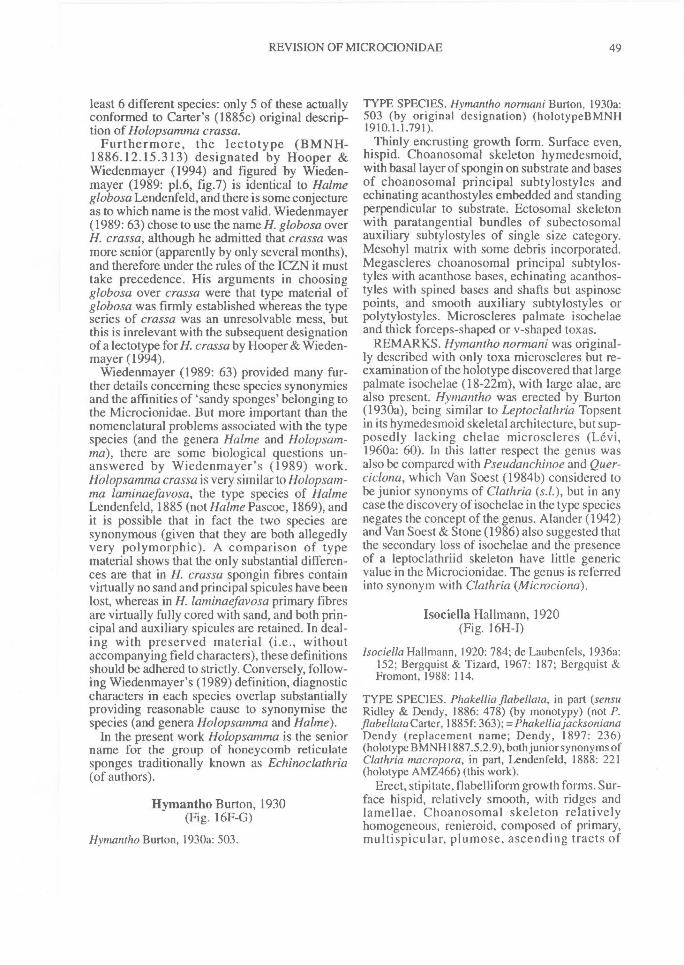
REVISION OF MICROCIONIDAE^ 49
least 6 different species: only 5 of these actuallyconformed to Carter's (1885c) original descrip-tion of Holopsamma crassa.
Furthermore, the lectotype (BMNH-1886.12.15.313) designated by Hooper &Wiedenmayer (1994) and figured by Wieden-mayer (1989: p1.6, fig.7) is identical to Ha/meglobosa Lendenfeld, and there is some conjectureas to which name is the most valid. Wiedenmayer(1989: 63) chose to use the name H. globosa overH. crassa, although he admitted that crassa wasmore senior (apparently by only several months),and therefore under the rules of the ICZN it musttake precedence. His arguments in choosingglobosa over crassa were that type material ofglobosa was firmly established whereas the typeseries of crassa was an unresolvable mess, butthis is inrelevant with the subsequent designationof a lectotype for H. crassa by Hooper & Wieden-mayer (1994).
Wiedenmayer (1989: 63) provided many fur-ther details concerning these species synonymiesand the affinities of 'sandy sponges' belonging tothe Microcionidae. But more important than thenomenclatural problems associated with the typespecies (and the genera Ha/me and Holopsam-ma), there are some biological questions un-answered by Wiedenmayer's (1989) work.Holopsamma crassa is very similar to Holopsam-ma laminaefavosa, the type species of Ha/meLendenfeld, 1885 (not Ha/me Pascoe, 1869), andit is possible that in fact the two species aresynonymous (given that they are both allegedlyvery polymorphic). A comparison of typematerial shows that the only substantial differen-ces are that in H. crassa spongin fibres containvirtually no sand and principal spicules have beenlost, whereas in H. laminaefavosa primary fibresare virtually fully cored with sand, and both prin-cipal and auxiliary spicules are retained. In deal-ing with preserved material (i.e., withoutaccompanying field characters), these definitionsshould be adhered to strictly. Conversely, follow-ing Wiedenmayer's (1989) definition, diagnosticcharacters in each species overlap substantiallyproviding reasonable cause to synonymise thespecies (and genera Holopsamma and Ha/me).
In the present work Holopsamma is the seniorname for the group of honeycomb reticulatesponges traditionally known as Echinoclathria(of authors).
Hymantho Burton, 1930(Fig. 16F-G)
Hymantho Burton, I930a: 503.
TYPE SPECIES. Hymantho normani Burton, 1930a:503 (by original designation) (holotypeBMNH1910.1.1.791).
Thinly encrusting growth form. Surface even,hispid. Choanosomal skeleton hymedesmoid,with basal layer of spongin on substrate and basesof choanosomal principal subtylostyles andechinating acanthostyles embedded and standingperpendicular to substrate. Ectosomal skeletonwith paratangential bundles of subectosomalauxiliary subtylostyles of single size category.Mesohyl matrix with some debris incorporated.Megascleres choanosomal principal subtylos-tyles with acanthose bases, echinating acanthos-tyles with spined bases and shafts but aspinosepoints, and smooth auxiliary subtylostyles orpolytylostyles. Microscleres palmate isochelaeand thick forceps-shaped or v-shaped toxas.
REMARKS. Hymantho normani was original-ly described with only toxa microscleres but re-examination of the holotype discovered that largepalmate isochelae (18-22m), with large alae, arealso present. Hymantho was erected by Burton(1930a), being similar to Leptoclathria Topsentin its hymedesmoid skeletal architecture, but sup-posedly lacking chelae microscleres (Levi,1960a: 60). In this latter respect the genus wasalso be compared with Pseudanchinoe and Quer-cicIona, which Van Soest (1984b) considered tobe junior synonyms of Clathria (s.1.), but in anycase the discovery of isochelae in the type speciesnegates the concept of the genus. Alander (1942)and Van Soest & Stone (1986) also suggested thatthe secondary loss of isochelae and the presenceof a leptoclathriid skeleton have little genericvalue in the Microcionidae. The genus is referredinto synonym with Clathria (Microciona).
Isociella Hallmann, 1920(Fig. 16H-I)
Isociella Hallmann, 1920: 784; de Laubenfels, 1936a:152; Bergquist & Tizard, 1967: 187; Bergquist &Fromont, 1988: 114.
TYPE SPECIES. Phakellia flabellata, in part (sensuRidley & Dendy, 1886: 478) (by monotypy) (not P.flabellata Carter, 1885f: 363); =Phakellia jacksonianaDendy (replacement name; Dendy, 1897: 236)(holotype BMNH1887.5.2.9), both junior synonyms ofClathria macropora, in part, Lendenfeld, 1888: 221(holotype AMZ466) (this work).
Erect, stipitate, flabelliform growth forms. Sur-face hispid, relatively smooth, with ridges andlamellae. Choanosomal skeleton relativelyhomogeneous, renieroid, composed of primary,multispicular, plumose, ascending tracts of
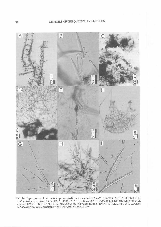
50^ MEMOIRS OF THE QUEENSLAND MUSEUM
FIG. 16. Type species of microcionid genera. A-B, Heteroclathria (H. hallezi Topsent, MNHNDT1884). C-D,Holopsamma (H. crassa Carter,BMNH1886.12.15.313). E, Ha/me (H. globosa Lendenfeld, synonym of H.crassa, BMNH1886.8.27.71). F-G, Hymantho (H. normani Burton, BMNHI910.1.1.791). H-I, Isociella(Phakelliaflabellata sensu Ridley & Dendy, BMNH1887.5.2.9).
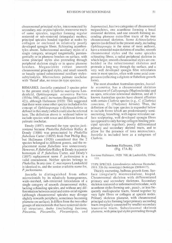
REVISION OF MICROCIONIDAE^ 51
choanosomal principal styles, interconnected bysecondary, uni- or paucispicular, transverse tractsof same spicules, together forming regularrenieroid or sub-renieroid (triangular) meshes;principal spicules bonded together at nodes bycollagen or enclosed in relatively poorlydeveloped spongin fibres. Echinating acanthos-tyles absent. Subectosomal auxiliary styles of asingle category, arranged tangentially, paratan-gentially or in plumose brushes on surface, withsome principal styles also protruding throughperipheral skeleton singly or in sparse plumosebrushes. Megascleres robust smoothchoanosomal principal styles, and small smoothor basally spined subectosomal auxiliary styles-subtylostyles. Microscleres palmate isochelaewith 'fluted' alae, no toxas (in type species).
REMARKS. Isociella contained 3 species priorto the present study (Clathria macropora Len-denfeld, Ophlitaspongia eccentrica Burton(1934a: 560), and !. incrustans Bergquist (1961a:42)), although Hallmann (1920: 784) suggestedthat there were some other species included in hisconcept of Ophlitaspongia (=Echinoclathria asdefined in this study) which could also be referredhere. The definition above is widened below toinclude species with toxas and different forms ofpalmate isochelae.
Dendy (1897) renamed the type species jack-soniana because Phakellia flabellata Ridley &Dendy (1886) was preoccupied by Phakelliaflabellata Carter (1885f) from Port Phillip Bay,but Hallmann (1920) considered that the 2species belonged to different genera, and the re-placement name flabellata was unnecessary.However, P.flabellata Ridley & Dendy is a juniorhomonym of P. flabellata Carter, and Dendy's(1897) replacement name P jacksoniana is avalid emendment. Neither species belongs toPhakellia. In any case C. macropora Lendenfeldis identical to, and the senior available name for,P jacksoniana.
Isociella is distinguished from othermicrocionids by its relatively homogeneous,wide-meshed, sub-renieroid reticulation of asingle category of smooth choanosomal styles,lacking echinating spicules, and without any dif-ferentiation between axial and extra-axial regions(although choanosomal spicules may divergeslightly towards periphery, sometimes becomingplumose on surface). It differs from the two othergroups of microcionids that have renieroid skele-tal structure. Antho (including Isociona,Plocamia, Plocamilla, Plocamiopsis, and
Isopenectya), has two categories of choanosomalmegascleres, one acanthose forming a basalrenieroid skeleton, and one smooth forming as-cending plumose extra-fibre tracts of the truechoanosomal skeleton. Some Echinoclathriaspecies (as defined in the present study, includingOphlitaspongia in the sense of most authors),have a renieroid main skeleton of smaller, smoothchoanosomal styles and the same spiculesechinating fibres, a radial peripheral skeleton inwhich larger, smooth choanosomal styles are em-bedded in the subectosomal skeleton andprotrude a long way through the surface, and avery well developed spongin fibre reticulationseen in most species, often with some axial com-pression (reflecting a digitate or flabellate growthform).
The most abundant Australian species, Isociel-la eccentrica, has a choanosomal skeletonreminiscent of Callyspongia (Haplosclerida) andan open, reticulate ectosomal skeleton, and all 3known Australian species have closest affinitieswith certain Clathria species (e.g., C. (Clathria)conectens, C. (Thalysias) hirsuta). Thus, thedefinition of the type species is expanded belowto include forms which are predominantly semi-encrusting, branching, with rugose reticulate sur-face sculpturing, well developed spongin fibres(as opposed to only having collagen binding prin-cipal spicules together); poorly differentiatedprimary and secondary skeletal tracts, and toallow for the presence of toxa microscleres.Isociella is included here as a subgenus ofClathria.
Isociona Hallmann, 1920(Fig. 17A-B)
Isocionu Hallmann, 1920: 768; de Laubenfels, 1936a:
TYPE SPECIES. Lissodendoryx tuberosa Hentschel,1911: 326 (by monotypy) (holotype ZMB4417).
Thickly encrusting, bulbous growth form. Sur-face irregularly microconulose, hispid.Choanosomal skeleton with differentiatedprimary and secondary skeletons. Secondaryskeleton renieroid or subrenieroid reticulate, withacanthose styles forming uni-, pauci-, or less fre-quently multispicular tracts, bound together byvery light fibres or collagen at spicule nodes.Primary skeleton plumose, with choanosomalprincipal styles forming larger primary ascendingtracts irregularly connected by smaller secondarytransverse tracts. Subectosomal skeletonplumose, with principal styles protruding through
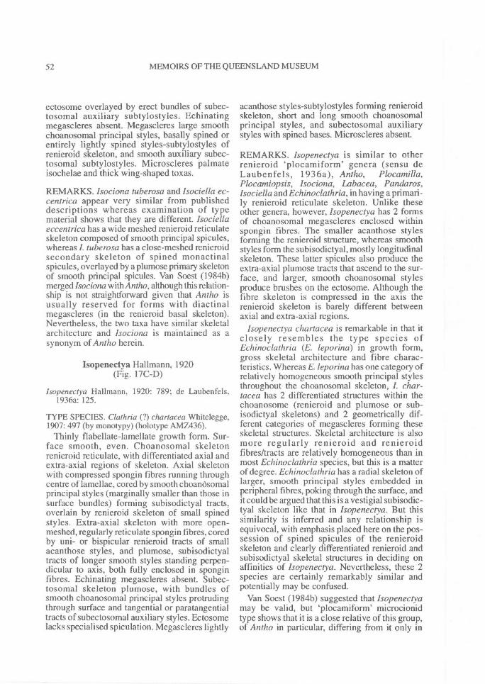
52^ MEMOIRS OF THE QUEENSLAND MUSEUM
ectosome overlayed by erect bundles of subec-tosomal auxiliary subtylostyles. Echinatingmegascleres absent. Megascleres large smoothchoanosomal principal styles, basally spined orentirely lightly spined styles-subtylostyles ofrenieroid skeleton, and smooth auxiliary subec-tosomal subtylostyles. Microscleres palmateisochelae and thick wing-shaped toxas.
REMARKS. Isociona tuberosa and Isociella ec-centrica appear very similar from publisheddescriptions whereas examination of typematerial shows that they are different. Isociellaeccentrica has a wide meshed renieroid reticulateskeleton composed of smooth principal spicules,whereas I. tube rosa has a close-meshed renieroidsecondary skeleton of spined monactinalspicules, overlayed by a plumose primary skeletonof smooth principal spicules. Van Soest (1984b)merged lsociona with Antho, although this relation-ship is not straightforward given that Antho isusually reserved for forms with diactinalmegascleres (in the renieroid basal skeleton).Nevertheless, the two taxa have similar skeletalarchitecture and Isociona is maintained as asynonym of Antho herein.
Isopenectya Hallmann, 1920(Fig. 17C-D)
Isopenectya Hallmann, 1920: 789; de Laubenfels,1936a: 125.
TYPE SPECIES. Clathria (?) chartacea Whitelegge,1907: 497 (by monotypy) (holotype AMZ436).
Thinly flabellate-lamellate growth form. Sur-face smooth, even. Choanosomal skeletonrenieroid reticulate, with differentiated axial andextra-axial regions of skeleton. Axial skeletonwith compressed spongin fibres running throughcentre of lamellae, cored by smooth choandsomalprincipal styles (marginally smaller than those insurface bundles) forming subisodictyal tracts,overlain by renieroid skeleton of small spinedstyles. Extra-axial skeleton with more open-meshed, regularly reticulate spongin fibres, coredby uni- or bispicular renieroid tracts of smallacanthose styles, and plumose, subisodictyaltracts of longer smooth styles standing perpen-dicular to axis, both fully enclosed in sponginfibres. Echinating megascleres absent. Subec-tosomal skeleton plumose, with bundles ofsmooth choanosomal principal styles protrudingthrough surface and tangential or paratangentialtracts of subectosomal auxiliary styles. Ectosomelacks specialised spiculation. Megascleres lightly
acanthose styles-subtylostyles forming renieroidskeleton, short and long smooth choanosomalprincipal styles, and subectosomal auxiliarystyles with spined bases. Microscleres absent.
REMARKS. Isopenectya is similar to otherrenieroid `plocamiform' genera (sensu deLaubenfels, 1936a), Antho, Plocamilla,Plocamiopsis, Isociona, Labacea, Panda ros,lsociella and Echinoclathria, in having a primari-ly renieroid reticulate skeleton. Unlike theseother genera, however, Isopenectya has 2 formsof choanosomal megascleres enclosed withinspongin fibres. The smaller acanthose stylesforming the renieroid structure, whereas smoothstyles form the subisodictyal, mostly longitudinalskeleton. These latter spicules also produce theextra-axial plumose tracts that ascend to the sur-face, and larger, smooth choanosomal stylesproduce brushes on the ectosome. Although thefibre skeleton is compressed in the axis therenieroid skeleton is barely different betweenaxial and extra-axial regions.
Isopenectya chartacea is remarkable in that itclosely resembles the type species ofEchinoclathria (E. leporina) in growth form,gross skeletal architecture and fibre charac-teristics. Whereas E. leporina has one category ofrelatively homogeneous smooth principal stylesthroughout the choanosomal skeleton, I. char-tacea has 2 differentiated structures within thechoanosome (renieroid and plumose or sub-isodictyal skeletons) and 2 geometrically dif-ferent categories of megascleres forming theseskeletal structures. Skeletal architecture is alsomore regularly renieroid and renieroidfibres/tracts are relatively homogeneous than inmost Echinoclathria species, but this is a matterof degree. Echinoclathria has a radial skeleton oflarger, smooth principal styles embedded inperipheral fibres, poking through the surface, andit could be argued that this is a vestigial subisodic-tyal skeleton like that in Isopenectya. But thissimilarity is inferred and any relationship isequivocal, with emphasis placed here on the pos-session of spined spicules of the renieroidskeleton and clearly differentiated renieroid andsubisodictyal skeletal structures in deciding onaffinities of Isopenectya. Nevertheless, these 2species are certainly remarkably similar andpotentially may be confused.
Van Soest (1984b) suggested that Isopenectyamay be valid, but `plocamiform' microcionidtype shows that it is a close relative of this group,of Antho in particular, differing from it only in
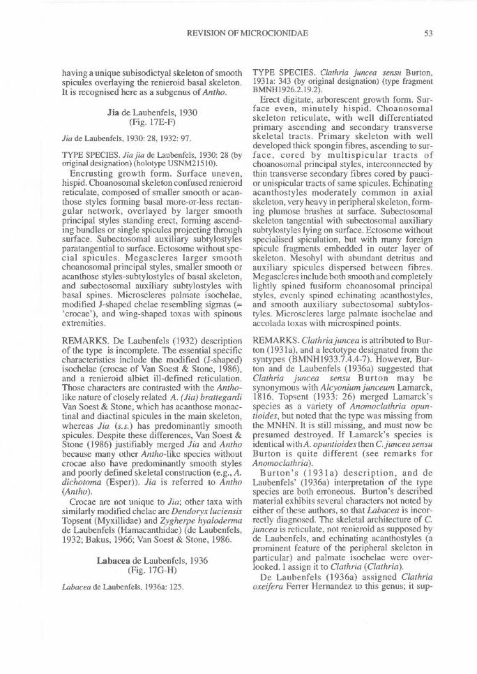
REVISION OF MICROCIONIDAE^ 53
having a unique subisodictyal skeleton of smoothspicules overlaying the renieroid basal skeleton.It is recognised here as a subgenus of Antho.
Jia de Laubenfels, 1930(Fig. 17E-F)
Jia de Laubenfels, 1930: 28, 1932: 97.
TYPE SPECIES. Jia jia de Laubenfels, 1930: 28 (byoriginal designation) (holotype USNM21510).
Encrusting growth form. Surface uneven,hispid. Choanosomal skeleton confused renieroidreticulate, composed of smaller smooth or acan-those styles forming basal more-or-less rectan-gular network, overlayed by larger smoothprincipal styles standing erect, forming ascend-ing bundles or single spicules projecting throughsurface. Subectosomal auxiliary subtylostylesparatangential to surface. Ectosome without spe-cial spicules. Megascleres larger smoothchoanosomal principal styles, smaller smooth oracanthose styles-subtylostyles of basal skeleton,and subectosomal auxiliary subtylostyles withbasal spines. Microscleres palmate isochelae,modified J-shaped chelae resembling sigmas (=`crocae'), and wing-shaped toxas with spinousextremities.
REMARKS. De Laubenfels (1932) descriptionof the type is incomplete. The essential specificcharacteristics include the modified (J-shaped)isochelae (crocae of Van Soest & Stone, 1986),and a renieroid albiet ill-defined reticulation.Those characters are contrasted with the Antho-like nature of closely related A. (Jia) brattegardiVan Soest & Stone, which has acanthose monac-tinal and diactinal spicules in the main skeleton,whereas Jia (s.s.) has predominantly smoothspicules. Despite these differences, Van Soest &Stone (1986) justifiably merged Jia and Anthobecause many other Antho-like species withoutcrocae also have predominantly smooth stylesand poorly defined skeletal construction (e.g., A.dichotoma (Esper)). Jia is referred to Antho(Ant ho).
Crocae are not unique to Jia; other taxa withsimilarly modified chelae are Dendoryx luciensisTopsent (Myxillidae) and Zygherpe hyalodermade Laubenfels (Hamacanthidae) (de Laubenfels,1932; Bakus, 1966; Van Soest & Stone, 1986.
Labacea de Laubenfels, 1936(Fig. 17G-H)
Labacea de Laubenfels, 1936a: 125.
TYPE SPECIES. Clathria juncea sensu Burton,1931a: 343 (by original designation) (type fragmentBMNH1926.2.19.2).
Erect digitate, arborescent growth form. Sur-face even, minutely hispid. Choanosomalskeleton reticulate, with well differentiatedprimary ascending and secondary transverseskeletal tracts. Primary skeleton with welldeveloped thick spongin fibres, ascending to sur-face, cored by multispicular tracts ofchoanosomal principal styles, interconnected bythin transverse secondary fibres cored by pauci-or unispicular tracts of same spicules. Echinatingacanthostyles moderately common in axialskeleton, very heavy in peripheral skeleton, form-ing plumose brushes at surface. Subectosomalskeleton tangential with subectosomal auxiliarysubtylostyles lying on surface. Ectosome withoutspecialised spiculation, but with many foreignspicule fragments embedded in outer layer ofskeleton. Mesohyl with abundant detritus andauxiliary spicules dispersed between fibres.Megascleres include both smooth and completelylightly spined fusiform choanosomal principalstyles, evenly spined echinating acanthostyles,and smooth auxiliary subectosomal subtylos-tyles. Microscleres large palmate isochelae andaccolada toxas with microspined points.
REMARKS. Clathria juncea is attributed to Bur-ton (1931a), and a lectotype designated from thesyntypes (BMNH1933.7.4.4-7). However, Bur-ton and de Laubenfels (1936a) suggested thatClathria juncea sensu Burton may besynonymous with Alcyonium junceutn Lamarck,1816. Topsent (1933: 26) merged Lamarck'sspecies as a variety of Anomoclathria opun-tioides, but noted that the type was missing fromthe MNHN. It is still missing, and must now bepresumed destroyed. If Lamarck's species isidentical with A. opuntioides then C. juncea sensuBurton is quite different (see remarks forAnomoclathria).
Burton's (1931a) description, and deLaubenfels' (1936a) interpretation of the typespecies are both erroneous. Burton's describedmaterial exhibits several characters not noted byeither of these authors, so that Labacea is incor-rectly diagnosed. The skeletal architecture of C.juncea is reticulate, not renieroid as supposed byde Laubenfels, and echinating acanthostyles (aprominent feature of the peripheral skeleton inparticular) and palmate isochelae were over-looked. I assign it to Clathria (Clathria).
De Laubenfels (1936a) assigned Clathriaoxeifera Ferrer Hernandez to this genus; it sup-
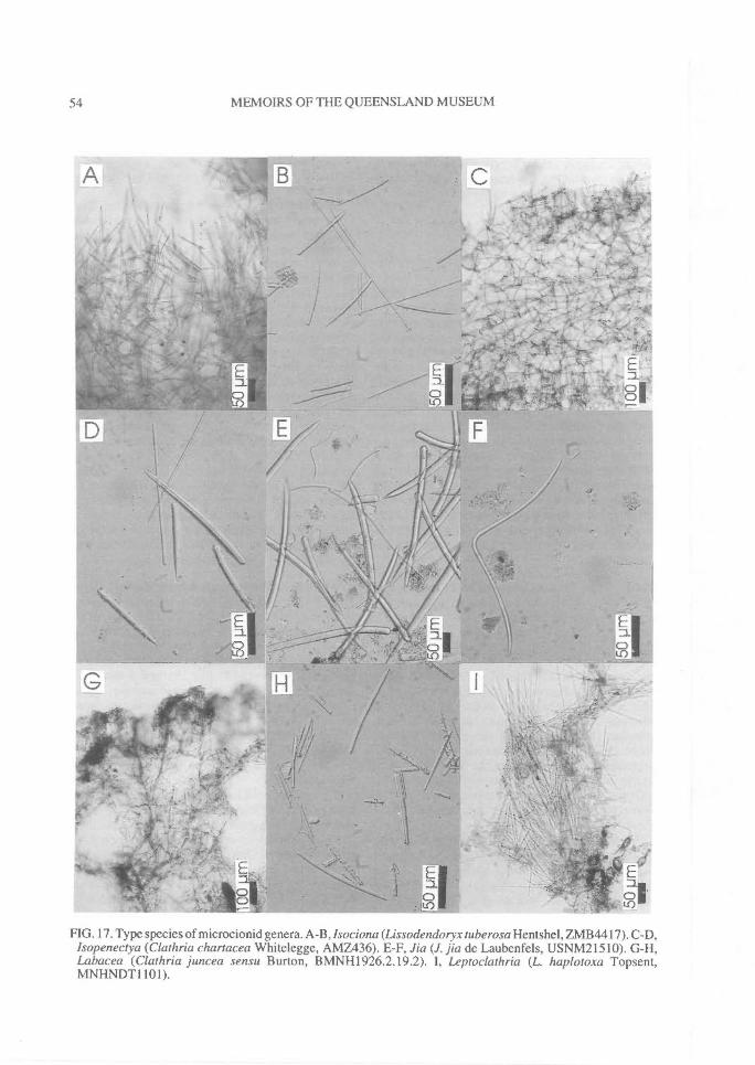
54^ MEMOIRS OF THE QUEENSLAND MUSEUM
FIG. 17. Type species of microcionid genera. A-B, lsociona (Lissodendoryx tube rosa Hentshel, ZM B4417). C-D,Isopenectya (Clathria chartacea Whitelegge, AMZ436). E-F, Jia (J. jia de Laubenfels, USNM21510). G-H,Labacea (Clathria juncea sensu Burton, BMNH1926.2.19.2). I, Leptoclathria (L haplotoxa Topsent,MNHNDT1101).
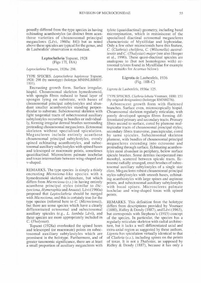
REVISION OF MICROCIONIDAE^ 55
posedly differed from the type species in havingechinating acanthostyles (as distinct from acan-those varieties of choanosomal principalmegascleres (Levi, 1960a: 84)) but as notedabove these spicules are typical for the genus, andde Laubenfels' observation is redundant.
Leptoclathria Topsent, 1928(Figs 171, I8A)
Leptoclathria Topsent, 1928a: 298.
TYPE SPECIES. Leptoclathria haplotoxa Topsent,1928: 298 (by monotypy) (holotype MNHNLBIMDT-1101).
Encrusting growth form. Surface irregular,hispid. Choanosomal skeleton hymedesmoid,with spongin fibres reduced to basal layer ofspongin lying on substrate, with bases ofchoanosomal principal subtylostyles and abun-dant smaller acanthostyles standing perpen-dicular to substrate. Subectosomal skeleton withlight tangential tracts of subectosomal auxiliarysubtylostyles occurring in bundles or individual-ly, forming irregular dermal brushes surroundingprotruding choanosomal megascleres. Ectosomalskeleton without specialised spiculation.Megascleres include entirely acanthosechoanosomal principal subtylostyles, evenlyspined echinating acanthostyles, and subec-tosomal auxiliary subtylostyles with spined basesand telescoped or mucronate points, sometimesquasidiactinal. Microscleres palmate isochelaeand toxas intermediate between wing-shaped andv-shaped.
REMARKS. The type species is simply a thinlyencrusting Microciona-like species with ahymedesmoid skeletal architecture, but whichdiffers from Microciona (s.s.) in having entirelyacanthose principal styles (similar to Dic-tyociona,Hymeraphia and Anaata). Levi (1960a)proposed that Leptoclathria should be mergedwith Microciona, and this is certainly true for thetype species (referred here to C. (Microciona)),but there are some species which have a clearlydifferentiated ectosomal and subectosomalauxiliary spicules (e.g., L. lambda Levi), andthese species are more appropriately included inC. (Thalysias).
Topsent (1928a) overlooked the spined basesand telescoped (or mucronate) points on subec-tosomal auxiliary subtylostyles which areprominent in the holotype. Furthermore, and ofgreater taxonomic significance, there are at leasta small proportion of auxiliary megascleres with
tylote (quasidiactinal) geometry, including basalmicrospination, which is reminiscent of thespecialised diactinal ectosomal megasclerescharacteristic of Myxillidae and Iophonidae.Only a few other microcionids have this feature,C. (Clathria) chelifera, C. (Wilsonella) austral-iensis and C. (Thalysias) major (see also Hooperet al., 1990). These quasi-diactinal spicules areanalogous to (but not homologous with) ec-tosomal tylotes found in Myxillidae for example(see remarks for Acamus below).
Ligrota de Laubenfels, 1936(Fig. 18B-C)
Ligrota de Laubenfels, 1936a: 125.
TYPE SPECIES. Clathria lobata Vosmaer, 1880: 151(by original designation) (holotype RMNH276).
Arborescent growth form with flattenedbranches. Surface even, microscopically hispid.Choanosomal skeleton regularly reticulate, withpoorly developed spongin fibres forming dif-ferentiated primary and secondary tracts. Primaryfibres ascend to surface, cored by plumose mul-tispicular tracts of choanosomal principal styles;secondary fibres transverse, paucispicular, coredby same spicules. Subectosomal skeletonplumose, with bundles of choanosomal principalmegascleres extending into ectosome andprotruding through surface. Echinating acanthos-tyles most abundant in periphery, below surfacespicule brushes. Some detritus incorporated intomesohyl, scattered between spicule tracts. Ec-tosome radially arranged, erect brushes of subec-tosomal auxiliary subtylostyles of a single sizeclass. Megascleres robust choanosomal principalstyles-subtylostyles with smooth bases, echinat-ing acanthostyles with large spines and aspinosepoints, and subectosomal auxiliary subtylostyleswith basal spines. Microscleres palmateisochelae and wing-shaped toxas with spinedpoints.
REMARKS. This definition from the holotypediffers from descriptions provided by Vosmaer(1880), Ridley & Dendy (1887), and Levi (1963),but corresponds with Stephens's (1915) conceptof the species. In particular, the species has aregularly reticulate skeleton with radial architec-ture, but it lacks a well differentiated axial andextra-axial region as suggested by these authors.Ligrota has spiculation virtually identical to thatof Clathria (s.s.), including spines on the pointsof toxas. It is not a Thalysias, as supposed byRidley & Dendy (1887), because it has only a
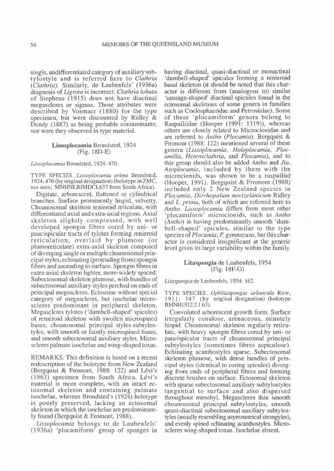
56^ MEMOIRS OF THE QUEENSLAND MUSEUM
single, undifferentiated category of auxiliary sub-tylostyle and is referred here to Clathria(Clathria). Similarly, de Laubenfels' (1936a)diagnosis of Ligrota is incorrect. Clathria lobataof Stephens (1915) does not have diactinalmegascleres or sigmas. Those attributes weredescribed by Vosmaer (1880) for the typespecimen, but were discounted by Ridley &Dendy (1887) as being probable contaminants;nor were they observed in type material.
Lissoplocamia Brondsted, 1924(Fig. 18D-E)
Lissoplocamia Brondsted, 1924: 470.
TYPE SPECIES. Lissoplocamia prima Brondsted,1924: 470 (by original designation) (holotype in ZMC,not seen; MNHNLBIMDCL637 from South Africa).
Digitate, arborescent, flattened or cylindricalbranches. Surface prominently hispid, velvetty.Choanosomal skeleton renieroid reticulate, withdifferentiated axial and extra-axial regions. Axialskeleton slightly compressed, with welldeveloped spongin fibres cored by uni- orpaucispicular tracts of tylotes forming renieroidreticulation, overlaid by plumose (orplumoreticulate) extra-axial skeleton composedof diverging single or multiple choanosomal prin-cipal styles, echinating (protruding from) sponginfibres and ascending to surface. Spongin fibres inextra-axial skeleton lighter, more-widely spaced.Subectosomal skeleton plumose, with bundles ofsubectosomal auxiliary styles perched on ends ofprincipal megascleres. Ectosome without specialcategory of megascleres, but isochelae micro-scleres predominant in peripheral skeleton.Megascleres tylotes ('dumbell-shaped' spicules)of renieroid skeleton with swollen microspinedbases, choanosomal principal styles-subtylos-tyles, with smooth or faintly microspined bases,and smooth subectosomal auxiliary styles. Micro-scleres palmate isochelae and wing-shaped toxas.
REMARKS. This definition is based on a recentredescription of the holotype from New Zealand(Bergquist & Fromont, 1988: 122) and Levi's(1963) specimen from South Africa. Levi'smaterial is more complete, with an intact ec-tosomal skeleton and containing palmateisochelae, whereas Brondsted's (1924) holotypeis poorly preserved, lacking an ectosomalskeleton in which the isochelae are predominant-ly found (Bergquist & Fromont, 1988).
Lissoplocamia belongs to de Laubenfels'(1936a) `plocamiform' group of sponges in
having diactinal, quasi-diactinal or monactinal`dumbell-shaped' spicules forming a renieroidbasal skeleton (it should be noted that this char-acter is different from (analogous to) similar'sausage-shaped' diactinal spicules found in theectosomal skeletons of some genera in familiessuch as Coelosphaeridae and Petrosiidae). Someof these `plocamiform' genera belong toRaspailiidae (Hooper (1991: 1319)), whereasothers are closely related to Microcionidae andare referred to Antho (Plocamia). Bergquist &Fromont (1988: 122) mentioned several of thesegenera (Lissoplocamia, Holoplocamia, Ploc-amilla, Heteroclathria, and Plocamia), and tothis group should also be added Antho and Jia.Axoplocamia, included by them with themicrocionids, was shown to be a raspailiid(Hooper, 1991). Bergquist & Fromont (1988)included only 2 New Zealand species inPlocamia, Dirrhopalum novizelanicum Ridleyand L. prima, both of which are referred here toAntho. Lissoplocamia differs from most other`plocamiform' microcionids, such as Antho(Antho) in having predominantly smooth 'clum-bell-shaped' spicules, similar to the typespecies of Plocamia, P gymnazusa, but this char-acter is considered insignificant at the genericlevel given its large variability within the family.
Litaspongia de Laubenfels, 1954(Fig. 18F-G)
Litaspongia de Laubenfels, 1954: 162.
TYPE SPECIES. Ophlitaspongia arbuscula Row,191 1: 347 (by original designation) (holotypeBMNH1912.2.1.63).
Convoluted arborescent growth form. Surfaceirregularly conulose, arenaceous, minutelyhispid. Choanosomal skeleton regularly reticu-late, with heavy spongin fibres cored by uni- orpaucispicular tracts of choanosomal principalsubtylostyles (sometimes fibres aspiculose).Echinating acanthostyles sparse. Subectosomalskeleton plumose, with dense bundles of prin-cipal styles (identical to coring spicules) diverg-ing from ends of peripheral fibres and formingdiscrete brushes on surface. Ectosomal skeletonwith sparse subectosomal auxiliary subtylostylestangential to surface and also dispersedthroughout mesohyl. Megascleres thin smoothchoanosomal principal subtylostyles, smoothquasi-diactinal subectosomal auxiliary subtylos-tyles (usually resembling asymmetrical strongyles),and evenly spilled echinating acanthostyles. MICID-scleres wing-shaped toxas. Isochelae absent.
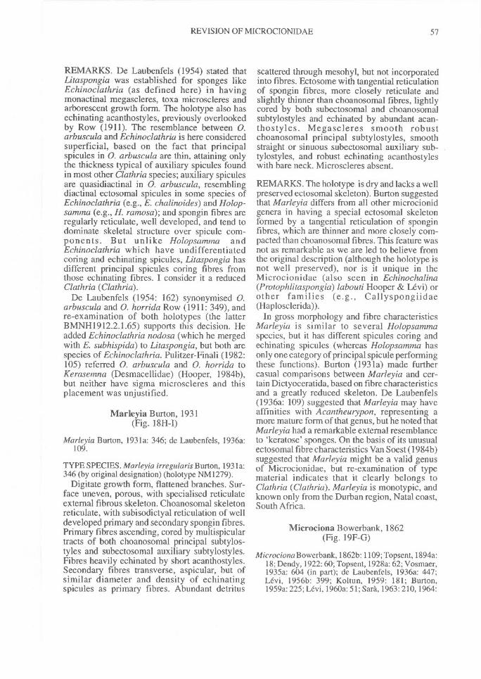
REVISION OF MICROCIONIDAE^ 57
REMARKS. De Laubenfels (1954) stated thatLitaspongia was established for sponges likeEchinoclathria (as defined here) in havingmonactinal megascleres, toxa microscleres andarborescent growth form. The holotype also hasechinating acanthostyles, previously overlookedby Row (1911). The resemblance between 0.arbuscula and Echinoclathria is here consideredsuperficial, based on the fact that principalspicules in 0. arbuscula are thin, attaining onlythe thickness typical of auxiliary spicules foundin most other Clathria species; auxiliary spiculesare quasidiactinal in 0. arbuscula, resemblingdiactinal ectosomal spicules in some species ofEchinoclathria (e.g., E. chalinoides) and Holop-samma (e.g., H. ramosa); and spongin fibres areregularly reticulate, well developed, and tend todominate skeletal structure over spicule com-ponents. But unlike Holopsamma andEchinoclathria which have undifferentiatedcoring and echinating spicules, Litaspongia hasdifferent principal spicules coring fibres fromthose echinating fibres. I consider it a reducedClathria (Clathria).
De Laubenfels (1954: 162) synonymised 0.arbuscula and 0. horrida Row (1911: 349), andre-examination of both holotypes (the latterBMNH1912.2.1.65) supports this decision. Headded Echinoclathria nodosa (which he mergedwith E. subhispida) to Litaspongia, but both arespecies of Echinoclathria. Pulitzer-Finali (1982:105) referred 0. arbuscula and 0. horrida toKerasemna (Desmacellidae) (Hooper, 1984b),but neither have sigma microscleres and thisplacement was unjustified.
Marleyia Burton, 1931(Fig. 18H-I)
Marleyia Burton, 1931a: 346; de Laubenfels, 1936a:109.
TYPE SPECIES. Marleyia irregularis Burton, 1931a:346 (by original designation) (holotype NM 1279).
Digitate growth form, flattened branches. Sur-face uneven, porous, with specialised reticulateexternal fibrous skeleton. Choanosomal skeletonreticulate, with subisodictyal reticulation of welldeveloped primary and secondary spongin fibres.Primary fibres ascending, cored by multispiculartracts of both choanosomal principal subtylos-tyles and subectosomal auxiliary subtylostyles.Fibres heavily echinated by short acanthostyles.Secondary fibres transverse, aspicular, but ofsimilar diameter and density of echinatingspicules as primary fibres. Abundant detritus
scattered through mesohyl, but not incorporatedinto fibres. Ectosome with tangential reticulationof spongin fibres, more closely reticulate andslightly thinner than choanosomal fibres, lightlycored by both subectosomal and choanosomalsubtylostyles and echinated by abundant acan-thostyles. Megascleres smooth robustchoanosomal principal subtylostyles, smoothstraight or sinuous subectosomal auxiliary sub-tylostyles, and robust echinating acanthostyleswith bare neck. Microscleres absent.
REMARKS. The holotype is dry and lacks a wellpreserved ectosomal skeleton). Burton suggestedthat Marleyia differs from all other microcionidgenera in having a special ectosomal skeletonformed by a tangential reticulation of sponginfibres, which are thinner and more closely com-pacted than choanosomal fibres. This feature wasnot as remarkable as we are led to believe fromthe original description (although the holotype isnot well preserved), nor is it unique in theMicrocionidae (also seen in Echinochalina(Protophlitaspongia) labouti Hooper & Levi) orother families (e.g., Callyspongiidae(Haplosclerida)).
In gross morphology and fibre characteristicsMarleyia is similar to several Holopsammaspecies, but it has different spicules coring andechinating spicules (whereas Holopsamma hasonly one category of principal spicule performingthese functions). Burton (1931a) made furthercasual comparisons between Marleyia and cer-tain Dictyoceratida, based on fibre characteristicsand a greatly reduced skeleton. De Laubenfels(1936a: 109) suggested that Marleyia may haveaffinities with Acantheurypon, representing amore mature form of that genus, but he noted thatMarleyia had a remarkable external resemblanceto `keratose' sponges. On the basis of its unusualectosomal fibre characteristics Van Soest (1984b)suggested that Marleyia might be a valid genusof Microcionidae, but re-examination of typematerial indicates that it clearly belongs toClathria (Clathria). Marleyia is monotypic, andknown only from the Durban region, Natal coast,South Africa.
Microciona Bowerbank, 1862(Fig. 19F-G)
Microciona Bowerbank,1862b: 1109; Topsent, 1894a:18; Dendy, 1922:60; Topsent, 1928a: 62; Vosmaer,1935a: 604 (in part); de Laubenfels, 1936a: 447;Levi, 1956b: 399; Koltun, 1959: 181; Burton,1959a: 225; Levi, 1960a: 51; Sara, 1963: 210, 1964:
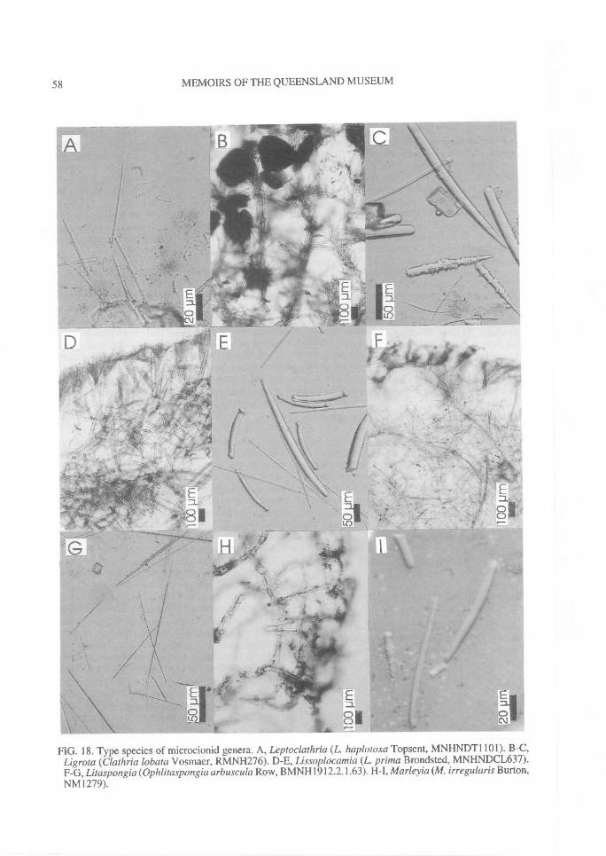
58^ MEMOIRS OF THE QUEENSLAND MUSEUM
FIG. 18. Type species of microcionid genera. A, Leptoclathria (L haplotoxa Topsent, MNHNDT1101). B-C,Ligrota (Clathria lobata Vosmaer, RMNH276). D-E, Lissoplocamia (L. prima Brondsted, MNHNDCL637).F-G, Litaspongia (Ophlitaspongia arbuscula Row, BMNH1912.2.1.63). H-I, Marleyia (M. irregularis Burton,NM1279).
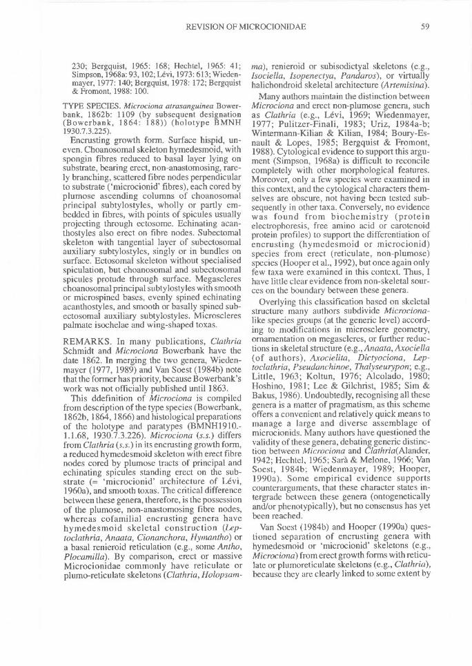
REVISION OF MICROCIONIDAE^ 59
230; Bergquist, 1965: 168; Hechtel, 1965: 41;Simpson, 1968a: 93, 102; Levi, 1973: 613; Wieden-mayer, 1977: 140; Bergquist, 1978: 172; Bergquist& Fromont, 1988: 100.
TYPE SPECIES. Microciona atrasanguinea Bower-bank, 1862b: 1109 (by subsequent designation(Bowerbank, 1864: 188)) (holotype BMNH1930.7.3.225).
Encrusting growth form. Surface hispid, un-even. Choanosomal skeleton hymedesmoid, withspongin fibres reduced to basal layer lying onsubstrate, bearing erect, non-anastomosing, rare-ly branching, scattered fibre nodes perpendicularto substrate ('microcionid' fibres), each cored byplumose ascending columns of choanosomalprincipal subtylostyles, wholly or partly em-bedded in fibres, with points of spicules usuallyprojecting through ectosome. Echinating acan-thostyles also erect on fibre nodes. Subectomalskeleton with tangential layer of subectosomalauxiliary subtylostyles, singly or in bundles onsurface. Ectosomal skeleton without specialisedspiculation, but choanosomal and subectosomalspicules protude through surface. Megasclereschoanosomal principal subtylostyles with smoothor microspined bases, evenly spined echinatingacanthostyles, and smooth or basally spined sub-ectosomal auxiliary subtylostyles. Microsclerespalmate isochelae and wing-shaped toxas.
REMARKS. In many publications, ClathriaSchmidt and Microciona Bowerbank have thedate 1862. In merging the two genera, Wieden-mayer (1977, 1989) and Van Soest (1984b) notethat the former has priority, because Bowerbank'swork was not officially published until 1863.
This ddefinition of Microciona is compiledfrom description of the type species (Bowerbank,1862b, 1864, 1866) and histological preparationsof the holotype and paratypes (BMNH1910.-1.1.68, 1930.7.3.226). Microciona (s.s.) differsfrom Clathria (s.s.) in its encrusting growth form,a reduced hymedesmoid skeleton with erect fibrenodes cored by plumose tracts of principal andechinating spicules standing erect on the sub-strate (= `microcionid' architecture of Levi,1960a), and smooth toxas. The critical differencebetween these genera, therefore, is the possessionof the plumose, non-anastomosing fibre nodes,whereas cofamilial encrusting genera havehymedesmoid skeletal construction (Lep-toclathria, Anaata, Cionanchora, Hymantho) ora basal renieroid reticulation (e.g., some Antho,Plocamilla). By comparison, erect or massiveMicrocionidae commonly have reticulate orplumo-reticulate skeletons (Clathria, Holopsam-
ma), renieroid or subisodictyal skeletons (e.g.,Isociella, Isopenectya, Pandaros), or virtuallyhalichondroid skeletal architecture (Artemisina).
Many authors maintain the distinction betweenMicrociona and erect non-plumose genera, suchas Clathria (e.g., Levi, 1969; Wiedenmayer,1977; Pulitzer-Finali, 1983; Uriz, 1984a-b;Wintermann-Kilian & Kilian, 1984; Boury-Es-nault & Lopes, 1985; Bergquist & Fromont,1988). Cytological evidence to support this argu-ment (Simpson, 1968a) is difficult to reconcilecompletely with other morphological features.Moreover, only a few species were examined inthis context, and the cytological characters them-selves are obscure, not having been tested sub-sequently in other taxa. Conversely, no evidencewas found from biochemistry (proteinelectrophoresis, free amino acid or carotenoidprotein profiles) to support the differentiation ofencrusting (hymedesmoid or microcionid)species from erect (reticulate, non-plumose)species (Hooper et al., 1992), but once again onlyfew taxa were examined in this context. Thus, Ihave little clear evidence from non-skeletal sour-ces on the boundary between these genera.
Overlying this classification based on skeletalstructure many authors subdivide Microciona-like species groups (at the generic level) accord-ing to modifications in microsclere geometry,ornamentation on megascleres, or further reduc-tions in skeletal structure (e.g., Anaata, Axociella(of authors), Axocielita, Dictyociona, Lep-toclathria, Pseudanchinoe, Thalyseurypon; e.g.,Little, 1963; Koltun, 1976; Alcolado, 1980;Hoshino, 1981; Lee & Gilchrist, 1985; Sim &Bakus, 1986). Undoubtedly, recognising all thesegenera is a matter of pragmatism, as this schemeoffers a convenient and relatively quick means tomanage a large and diverse assemblage ofmicrocionids. Many authors have questioned thevalidity of these genera, debating generic distinc-tion between Microciona and Clathria(Alander,1942; Hechtel, 1965; Sara & Melone, 1966; VanSoest, 1984b; Wiedenmayer, 1989; Hooper,1990a). Some empirical evidence supportscounterarguments, that these character states in-tergrade between these genera (ontogeneticallyand/or phenotypically), but no consensus has yetbeen reached.
Van Soest (1984b) and Hooper (1990a) ques-tioned separation of encrusting genera withhymedesmoid or `microcionid' skeletons (e.g.,Microciona) from erect growth forms with reticu-late or plumoreticul ate skeletons (e.g., Clathria),because they are clearly linked to some extent by
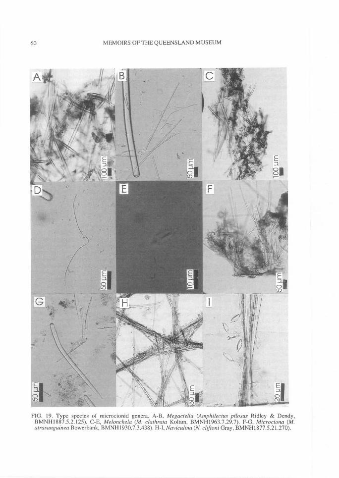
60^ MEMOIRS OF THE QUEENSLAND MUSEUM
FIG. 19. Type species of microcionid genera. A-B, Megaciella (Amphilectus pilosus Ridley & Dendy,BMNH1887.5.2.125). C-E, Melonchela (M. clathrata Koltun, BMNH1963.7.29.7). F-G, Microciona (M.atrasan guinea Bowerbank, BMNH1930.7.3.438). H-I, Naviculina (N. cliftoni Gray, BMNH1877.5.21.270).
REVISION OF MICROCIONIDAE^ 61
the ontogeny of the sponge individual (Simpson,1968a). They argued from a phylogenetic basisthat the recognition of this character as being aprimary one confers exceptionally high levels ofhomoplasy within the classification, cuttingacross lines of apparent evolutionary decentbased on a combination of other characters (suchas the origin and disposition of structuralmegascleres within the skeleton, spiculegeometry). For example, there are many thinlyencrusting species with hymedesmoid architec-ture and plumose spongin fibre nodes referable toMicrociona (e.g., M. maunaloa de Laubenfels, M.microchela Hechtel), but also having two dif-ferentiated categories of auxiliary spicules (i.e., aspecialised ectosomal skeleton), which is charac-teristic of Thalysias species. Which genus dothese species then belong to, Microciona orThalysias, or do we recognise a third genus be-cause they have both characters ? This third alter-native was adopted by Bergquist & Fromont(1988), largely following de Laubenfels' (1936a),who recognised Axociella for microcionids withMicrociona-like fibre skeletons and Thalysias-like ectosomal skeletons (although, like deLaubenfels (1936a), they misinterpreted Axociel-la which is neither encrusting nor hymedesmoid).This is a 'convenient classification' for managingspecies, but it is also responsible for most of the70 or so microcionid genera that exist today (i.e.,approximately 1 genus per 6 species).
Few authors agree on the level of taxonomicdivergence, or the phylogenetic interpretation ofcharacter polarity: is encrusting growth form andhymedesmoid architecture a primitive or derivedstrategy? Is a `microcionid' architecture(hymedesmoid with ascending plumose fibrenodes) a subsequent developmental stage ofhymedesmoid architecture (spongin fibres flatagainst the substrate)? It is anticipated thatevidence from biochemistry or genetic sourceswill eventually contribute towards the resolutionof these conflicts. A resolution is provided herebased on phylogenetic interpretation of morphol-ogy. We follow Levi's (1960a) arguments in con-sidering three alternatives.
1) One generic taxon may be used to encompassall the skeletal types from leptoclathriid(hymedesmoid), `microcionid', to reticulate. Thisoption was used by Vosmaer (1933, 1935a-b),who suggested that varying grades of skeletalconstruction (from hymedesmoid, plumose torenieroid reticulate) could be seen within popula-tions of particular species and was apparentlylargely related to ontogenetic development. Van
Soest (1984b), Hooper & Levi (1993a) and othersused this ontogenetic argument to mergeMicrociona and Clathria (as well as Thalysiasand Clathria), and Hooper et al. (1992) presentedsome empirical data to show that there was nohomogeneity in biochemical profiles to supportthe retention of distinct taxa for encrusting versuserect species.
2) Two separate taxa can be recognised forspecies with hymedesmoid plumose(Microciona) and reticulate (Clathria) architec-tures. This was the argument accepted by Levi(1960a), Bergquist & Fromont (1988), andothers, on the basis that these skeletal structureswere consistent for populations of particularspecies, and therefore represented fixed geneticdifferences. Simpson (1968a) suggested furtherthat this option had some empirical support fromcytological evidence, although (unfortunately)the cytological characters themselves are at themoment not particularly useful taxonomic char-acters. A more pragmatic argument for the reten-tion of the name Microciona is that it is in currentwidespread use by sponge biochemists,ecologists and experimental biologists, andretaining this name provides some sort ofnomenclatural stability consistent with the pre-vious literature. This argument, unfortunately,has a pragmatic rather than biological basis.
3) The name Clathria could be used for an adultterminal phase of skeletal architecture, relateddirectly to the ontogeny of the sponge, whereasMicrociona could be used for the juvenile phaseof the same species. Dictyociona is an exampleof a Clathria with an intermediate Microciona-like skeleton, and Pseudanchinoe is an exampleof a Microciona verging on a reticulate Clathria-like skeleton. This argument presupposes thatplumose skeletons are always precursors of, andjuvenile to, reticulate skeletons. But there areseveral thinly encrusting species which do havereticulate skeletons (e.g., Sara & Melone, 1966),even though most plumose species are also per-sistently encrusting.
In recognising M icrociona at the subgenericlevel, Van Soest (1984b) implicitly also recog-nised a phylogenetic basis for the `microcionid'skeletal specialisation, even though there were noother corroboratory characters, such as uniquespicule geometries. The phylogenetic interpreta-tion of this skeletal specialisation taken in thepresent study is that Microciona is a persistentlyencrusting sponge which consistently has non-anastomosing basal spongin fibres cored by non-anastomosing plumose spicule tracts or single
62^ MEMOIRS OF THE QUEENSLAND MUSEUM
spicules, also lacking any ectosomal specialisa-tion; it is an incompletely differentiated sistertaxon of Clathria and recognised here at thesubgenus level (Clathria (Microciona)). The Lep-toclathria (flat, hymedesmoid) and Microciona(ascending `microcionid' fibre nodes) conditionsare not differentiated, these being interpreted asbeing more likely to be related to the thickness ofencrusting growth forms than anything else.
Ophlitaspongia Bowerbank, 1866(Fig. 20A-B)
Ophlitaspongia Bowerbank, 1866: 14, 378; Wieden-mayer, 1989: 59; Bergquist & Fromont, 1988: 113.
Not Ophlitaspongia; Dendy, 1896: 36; Hallmann,1912: 253; Levi, 1960a: 58; Wiedenmayer, 1977:140.
Seriatula Gray, 1867: 515; de Laubenfels, 1936a: 122.
TYPE SPECIES. Ophlitaspongia papilla Bowerbank,1866: 378 (by original designation) (holotypeBMNH1910.1.1.395); = Spongia seriata Grant, 1826:116 (Simpson, 1968a:37) (holotype BMNH1847.9.7.14).
Encrusting bulbous growth form. Surfacemicroconulose, microscopically hispid.Choanosomal skeleton isodictyal reticulate, withcompressed layer of spongin fibre lying on sub-strate and regularly reticulate spongin fibres aris-ing from base, producing regular isodictyal fibrenetwork divided into primary ascending andsecondary transverse fibre elements. Primaryfibres cored by plumose tracts of choanosomalprincipal styles, which also protrude from fibresat acute angles resembling quasi-echinating spicules.Secondary transverse fibres uncored or with uni-spicular tracts of usually smaller choanosomalprincipal styles. Subectosomal skeleton paratan-gential, with scattered subectosomal auxiliary styleslying on or near surface and also dispersedthroughout mesohyl. Ectosome without specialisedspiculation, but plumose tracts of choanosomalprincipal megascleres protrude through surface.Megascleres large and small, entirely smoothchoanosomal principal styles-subtylostyles, andthin smooth subectosomal auxiliary styles.Microscleres u-shaped toxas. Chelae absent.
REMARKS. This definition is based on typematerial and Simpson's (1968a) description oflive populations. Ophlitaspongia papilla, wasshown by Simpson (1968a: 95) to be a synonymof the type species of Microciona (M. atrasan-guinea). A broader concept of Ophlitaspongia(Wiedenmayer, 1989), (not 0. seriata (Grant)), isidentical to Echinoclathria (s.s.) (see remarks for
Echinoclathria). Wiedenmayer (1989) providedfurther explanation of these relationships, al-though his nomenclatural decisions are not en-tirely correct.
Seriatula was erected for Spongia seriata Grant(Gray, 1867) which is conspecific with 0. papillaBowerbank (e.g., Simpson, 1968a), and thereforeSeriatula becomes an objective synonym of Oph-litaspongia.
Pandaros Duchassaing & Michelotti, 1864(Fig. 20C-E)
Pandaros Duchassaing & Michelotti, 1864: 88;Schmidt, 1870: 59; de Laubenfels, 1936a: 123;Wiedenmayer, 1977: 143; Van Soest, 1984b: 127.
TYPE SPECIES. Pandaros acanthifolium Duchassa-ing & Michelotti, 1864: 90 (by subsequent designationof de Laubenfels, 1936a: 123)) (lectotype TMPOR57).
Bushy arborescent growth form. Surface highlyconulose, with flattened or lobate lamellae.Choanosomal skeleton reticulate, with well-developed flattened spongin fibres (trabeculae)cored by choanosomal principal subtylostyleslying in all directions within fibres (from isodic-tyal reticulate to echinating) and with sparseacanthostyles echinating or also incorporated intofibres. Subectosomal skeleton radial, reduced tosingle long subectosomal auxiliary subtylostylesprotruding through surface and also scatteredthroughout mesohyl. Ectosome without specialspicules. Megascleres smooth choanosomal prin-cipal subtylostyles-tylostyles, often with slightlyrhabdose bases and terminal or subterminal basalswellings, long curved or straight subectosomalauxiliary subtylostyles, and lightly acanthose orrarely smooth styles `echinating' fibres.Microscleres absent.
REMARKS. This definition is based on the typeand a fragment of the type MNHNLBIMDNBE1309, specimen BMNH1884.7.11.2 and descrip-tion of live populations by Van Soest (1984b).Important features are: 1) the prominently flat-tened fibres cored by smooth slightly rhabdoseprincipal subtylostyles-tylostyles (more reminis-cent of Rhabderemiidae than of Microcionidae);2) the sparse, lightly spined styles which moreclosely resemble a second category of principalspicules than than they do echinating (accessory)spicules typical of other Microcionidae; further-more, these spicules are only rarely seen echinat-ing fibres, but more commonly they areincorporated into them together with the principalmegascleres; and 3) the long subectosomalauxiliary subtylostyles protruding through the
REVISION OF MICROCIONIDAE^ 63
surface (more similar to a reduced Raspailiidae,such as Echinodictyum or Ceratopsion, than totypical Microcionidae).
Pandaros could be legitimately included ineither Raspailiidae or Microcionidae. Van Soest(1984b) noted that only the rare echinating acan-thostyles in P acanthifolium gives any cause tolink it to the Microcionidae at all. He speculatedthat it might be necessary to erect a separatefamily for the species, or even remove it from thePoecilosclerida altogether, as it also shows af-finities with axinellids such as Ptilocaulis. Thereare no microscleres to give any further clues as toits affinities. Pandaros is maintained as a separategenus and tentatively included in Microcionidae.Of 12 species referred to Pandaros, only the typeclearly belongs here. Raspailia kasumiensisTanita (MMBS SIS-052) was assigned to Pan-daros (Hooper, 1990a).
Wiedenmayer (1977) merged Thalyseuryponwith Pandaros, because he considered that itstype species had architecture closely comparableto P acanthifolium (Hechtel, 1965), but this is notupheld here. The only features these genera havein common is lacking microscleres. Wieden-mayer (1977) also speculated that the genus hada close relationship with Echinoclathria (=Holopsamma as defined here), based on allegedsimilarities in skeletal architecture, and he sug-gested that the two genera probably intergrade inhabit and spiculation, but these suggested af-finities are not evident in relevant specimens.
Paradoryx Hallmann, 1920(Fig. 20E-G)
Paradoryx Hamann, 1920: 767; de Laubenfels,1936a: 109.
TYPE SPECIES. Clathria dura Whitelegge, 1901: 83(by original designation) (holotype AMG3046).
Arborescent, flabellate growth form with com-pressed branches. Surface even, smooth.Choanosomal skeleton reticulate, with heavyspongin fibres forming irregular anastomoses inaxis becoming more regular and rectangulartowards periphery. Spongin fibres differentiatedinto primary ascending and secondary connectingcomponents, cored by auxiliary styles and heavi-ly echinated by acanthostyles. Subectosomalskeleton plumose, with bundles of auxiliarystyles forming plumose brushes on peripheralfibres. Ectosome fibrous, without specialisedskeleton, and peripheral fibres form more-or-lessreticulate structure through which choanosomalspicules protrude. Megascleres entirely smooth,
hastate or quasidiactinal auxiliary styles or sub-tylostyles, and echinating acanthostyles withlarge and even spination. Microscleres palmateisochelae resembling arcuate forms with lateralalae fused to shaft for about 1/2 alae length, andshaft greatly curved, thickened. Toxas absent.
REMARKS. Ectosomal fibres of Paradoryx areunspecialised, identical to choanosomal fibres insize and form, unlike the Callyspongia-like ec-tosome of Marleyia. Hallmann (1920) erectedParadoryx for Wilsonella species (with auxiliarymegascleres coring fibres) which had arcuatechelae instead of palmate isochelae. Loss or re-placement of principal megascleres varies con-siderably between otherwise allied microcionidtaxa and this feature is considered to be of lessersignificance at the generic level than assumed byHallmann (Hooper, 1990a; Hooper et al., 1990).Similarly, isochelae described by Hallmann(1920) as arcuate in the type species are modified(curved, thickened) palmate forms.
Apart from the type species, Hallmann (1920)included: Dictyocylindrus pinifonnis Carter andW oxyphila (both of which belong to Clathria(Clathria)), and Clathria elegantula Ridley &Dendy and Wilsonella curvichela Hallmann(which are referred here to Clathria(Dendrocia)).
Paratenaciella Vacelet & Vasseur, 1971(Fig. 20H-0
Paratenaciella Vacelet & Vasseur, 1971: 103.
TYPE SPECIES. Paratenaciella microxea Vacelet &Vasseur, 1971: 103 (by original designation) (holotypeMNHNLBIMD.W27).
Encrusting growth form. Surface uneven,hispid. Choanosomal skeleton hymedesmoid,with spongin fibres reduced to basal layer onsubstrate and with ascending fibre nodes enclos-ing bases of choanosomal principal subtylostylesstanding perpendicular to substrate. In thickersections up to 3-4 spicules form ascendingplumose tracts, protruding through surface.Echinating megascleres absent. Subectosomalskeleton plumose or paratangential composed ofbundles of subectosomal auxiliary subtylostylesoften forming brushes around protruding prin-cipal spicules and protruding through ectosome.Megascleres entirely smooth choanosomal prin-cipal subtylostyles, and subectosomal auxiliarysubtylostyles with microspined bases andpolytylote shafts. Microscleres palmate

64^ MEMOIRS OF THE QUEENSLAND MUSEUM
FIG. 20. Type species of microcionid genera. A-B, Ophlitaspongia (0. papilla Bowerbank,BMNH1910.1.1.395). C-E, Pandaros (P. acanthifolium Duchassaing & Michelotti, BMNH1884.7.17.2). F-G,Paradoryx (Clathria dura Whitelegge, AMG3046). H-I, Paratenaciella (P. microxea Vacelet & Vasseur,MNHNDJV27).
REVISION OF MICROCIONIDAE^ 65
isochelae, including contort forms, and oxeotetoxas.
REMARKS. Paratenaciella has a Microciona-like choanosomal fibre skeleton composed ofnon-anastomosing, plumose fibre nodes, butlacks acanthostyles (similar to Axociella,Axocielita, Tenaciella), has polytylote modifica-tions to auxiliary spicules (similar to Fisherispon-gia ferrea de Laubenfels and Microcionaaceratoobtusa Carter), and has modified (oxeote)toxas, like those in Artemisina archegona Ristau(although type material shows that they doresemble true microxeas). This latter feature ap-pears to be the only unique character for the typespecies, and as far as is known it is also uniqueamongst the Microcionidae. Paratenaciellashows some similarities to Esperiopsiscanaliculata Whitelegge (which is referred hereto Axociella) in its spiculation, although P.microxea has a microcionid architecture,microxeote microscleres and undifferentiated ec-tosomal-subectosomal megascleres. The genus ismonotypic and is synonymised with Clathria(Microciona).
Plectispa Lendenfeld, 1888(Fig. 21C-D)
Plectispa Lendenfeld, 1888: 225; Topsent, 1894a: 19;de Laubenfels, 1954: 164.
TYPE SPECIES. Plectispa macropora, in part, Len-denfeld, 1888: 226 (by subsequent designation ofHallmann, 1912: 204) (holotype AMG9159).
Lobodigitate growth form. Surface 'honeycomb'reticulate. Choanosomal skeleton reticulate, withwell developed spongin fibres forming more-or-less regular oval or eliptical meshes, sparselycored and abundantly echinated by principal sub-tylostyles, and fewer auxiliary styles-tornotesalso in fibres. Echinating megascleres lesspredominant in periphery than in axis. Somedetritus also incorporated into choanosomalfibres. Subectosomal skeleton plumose, withbrushes of principal subtylostyles from ascendingfibres of peripheral skeleton protruding throughsurface, surrounded by paratangential loosebundles or individual auxiliary spicules.Megascleres smooth principal subtylostyles, andlong curved, setaceous or sinuous auxiliary stylesor quasi-diactinal tornotes, both with blackenedaxial canals. Microscleres palmate isochelae, in-cluding contort forms. Toxas absent.
REMARKS. This genus is poorly defined due tothe dubious status of all of Lendenfeld's syntypes,
inaccuracies in Lendenfeld's (1888) originaldescription, and misinterpretation of the species(e.g., Clathria macropora; Whitelegge (1901:91) is different from C. macropora Lendenfeld(1888)). Hallmann (1912: 165) attempted to sortout these problems but was only partially success-ful. He rejected Whitelegge's proposed merger ofP. macropora with Echinonema levis Lendenfeld(1888), but instead he synonymised the latter withC. macropora (sensu Whitelegge), as Crella in-crustans Carter, var. levis Lendenfeld. However,Hallmann's (1912) remarks and assumptionsconcerning the genus are not entirely correct (seeremarks below for Clathria macropora andHolopsamma macropora), and there is noevidence that he actually re-examined relevanttype material. His nomenclatural decisions areerroneous and not supported here.
There are 4 type specimens and one otherspecimen in the AM and BMNH which bear thename macropora'. 1) AMZ959 (with the label'dry, cotype') from Port Jackson, NSW, is theso-called 'type' of Clathria macropora (sensuWhitelegge), and is a species of Crella. It isprobably a syntype of Lendenfeld's (1888)Echinonema levis, but that assumption is difficultto corroborate due to the loss of Lendenfeld's'key-list' from the Australian Museum (E. Pope& E Rowe, pers.comm.). 2) AMZ466 (with labelstating 'spirit preserved, ?type') from PortStephens, NSW is the lectotype of Lendenfeld's(1888: 221) Clathria macropora, later referred toWilsonella by Hallmann (1912: 240). It is neithera Wilsonella, nor closely related to such speciesas W australiensis Carter, but is a Clathria lack-ing echinating megascleres and having an axiallycompressed skeleton (e.g., Clathria (Axociella)).3) AMG9159 (with the label stating 'dry, type'),probably from Port Jackson, NSW (i.e., there is adiscrepancy between the published locality ofTorres Strait, Qld. and the museum specimenlabel and register), is the holotype of Plectispamacropora. 4) BMNH1925.11.1.555 (with labelstating Plectispa macropora, dry, Dendy coll.')from Manly Beach, NSW, is identical to Clathriamultipes Hallmann. 5) Another specimen(BMNH1957.8.30.2, with label stating Plectispamacropora, dry') belongs to Clathria lendenfeldi.Consequently, only a single syntype is valid be-coming the holotype of P. macropora, and thediagnosis of Plectispa is based on this specimen.
Hallmann (1912:205) referred the type species(and genus Plectispa) to Wilsonella, but thenreturned it to Clathria (Hallmann, 1920: 768).The species has a 'honeycombed reticulate'
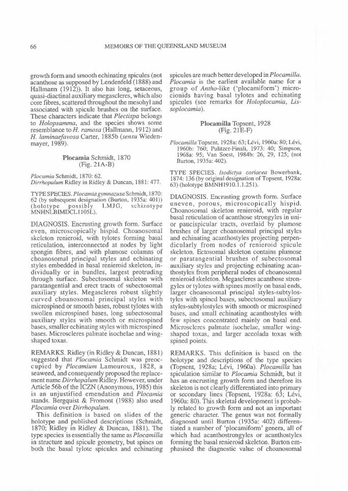
66^ MEMOIRS OF THE QUEENSLAND MUSEUM
growth form and smooth echinating spicules (notacanthose as supposed by Lendenfeld (1888) andHamann (1912)). It also has long, setaceous,quasi-diactinal auxiliary megascleres, which alsocore fibres, scattered throughout the mesohyl andassociated with spicule brushes on the surface.These characters indicate that Plectispa belongsto Holopsamtna, and the species shows someresemblance to H. ramosa (Hallmann, 1912) andH. laminaefavosa Carter, 1885b (sensu Wieden-mayer, 1989).
Plocamia Schmidt, 1870(Fig. 21A-B)
Plocamia Schmidt, 1870: 62.Dirrhopalum Ridley in Ridley & Duncan, 1881: 477.
TYPE SPECIES. Plocamia gymnazusa Schmidt, 1870:62 (by subsequent designation (Burton, 1935a: 401))(holotype possibly LMJG, schizotypeMNHNLBIMDCL1105L).
DIAGNOSIS. Encrusting growth form. Surfaceeven, microscopically hispid. Choanosomalskeleton renieroid, with tylotes forming basalreticulation, interconnected at nodes by lightspongin fibres, and with plumose columns ofchoanosomal principal styles and echinatingstyles embedded in basal renieroid skeleton, in-dividually or in bundles, largest protrudingthrough surface. Subectosomal skeleton withparatangential and erect tracts of subectosomalauxiliary styles. Megascleres robust slightlycurved choanosomal principal styles withmicrospined or smooth bases, robust tylotes withswollen microspined bases, long subectosomalauxiliary styles with smooth or microspinedbases, smaller echinating styles with microspinedbases. Microscleres palmate isochelae and wing-shaped toxas.
REMARKS. Ridley (in Ridley & Duncan, 1881)suggested that Plocamia Schmidt was preoc-cupied by Plocamium Lamouroux, 1828, aseaweed, and consequently proposed the replace-ment name Dirrhopalum Ridley. However, underArticle 56b of the ICZN (Anonymous, 1985) thisis an unjustified emendation and Plocamiastands. Bergquist & Fromont (1988) also usedPlocamia over Dirrhopalum.
This definition is based on slides of theholotype and published descriptions (Schmidt,1870; Ridley in Ridley & Duncan, 1881). Thetype species is essentially the same as Plocamillain structure and spicule geometry, but spines onboth the basal tylote spicules and echinating
spicules are much better developed in Plocamilla.Plocamia is the earliest available name for agroup of Antho-like ('plocamiform') micro-cionids having basal tylotes and echinatingspicules (see remarks for Holoplocamia, Lis-soplocamia).
Plocamilla Topsent, 1928(Fig. 21E-F)
Plocamilla Topsent, 1928a: 63; Levi, 1960a: 80; Levi,1960b: 760; Pulitzer-Finali, 1973: 40; Simpson,1968a: 95; Van Soest, 1984b: 26, 29, 125; (notBurton, 1935a: 402).
TYPE SPECIES. Isodiciya coriacea Bowerbank,1874: 136 (by original designation of Topsent, 1928a:63) (holotype BMNH1910.1.1.251).
DIAGNOSIS. Encrusting growth form. Surfaceuneven, porous, microscopically hispid.Choanosomal skeleton renieroid, with regularbasal reticulation of acanthose strongyles in uni-or paucispicular tracts, overlaid by plumosebrushes of larger choanosomal principal stylesand echinating acanthostyles projecting perpen-dicularly from nodes of renieroid spiculeskeleton. Ectosomal skeleton contains plumoseor paratangential brushes of subectosomalauxiliary styles and projecting echinating acan-thostyles from peripheral nodes of choanosomalrenieroid skeleton. Megascleres acanthose stron-gyles or tylotes with spines mostly on basal ends,larger choanosomal principal styles-subtylos-tyles with spined bases, subectosomal auxiliarystyles-subtylostyles with smooth or microspinedbases, and small echinating acanthostyles withfew spines concentrated mainly on basal end.Microscleres palmate isochelae, smaller wing-shaped toxas, and larger accolada toxas withspined points.
REMARKS. This definition is based on theholotype and descriptions of the type species(Topsent, 1928a; Levi, 1960a). Plocamilla hasspiculation similar to Plocamia Schmidt, but ithas an encrusting growth form and therefore itsskeleton is not clearly differentiated into primaryor secondary lines (Topsent, 1928a: 63; Levi,1960a: 80). This skeletal development is probab-ly related to growth form and not an importantgeneric character. The genus was not formallydiagnosed until Burton (1935a: 402) differen-tiated a number of `plocamiform' genera, all ofwhich had acanthostrongyles or acanthostylesforming the basal renieroid skeleton. Burton em-phasised the diagnostic value of choanosomal
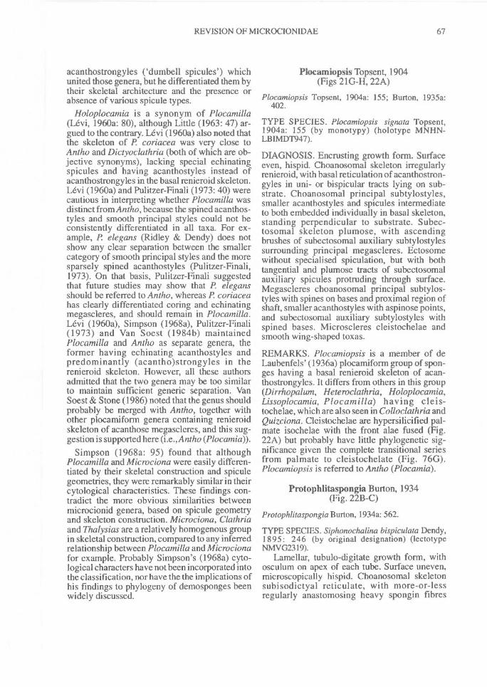
REVISION OF MICROCIONIDAE^ 67
acanthostrongyles ('dumbell spicules') whichunited those genera, but he differentiated them bytheir skeletal architecture and the presence orabsence of various spicule types.
Holoplocamia is a synonym of Plocamilla(Levi, 1960a: 80), although Little (1963: 47) ar-gued to the contrary. Levi (1960a) also noted thatthe skeleton of P coriacea was very close toAntho and Dictyoclathria (both of which are ob-jective synonyms), lacking special echinatingspicules and having acanthostyles instead ofacanthostrongyles in the basal renieroid skeleton.Levi (1960a) and Pulitzer-Finali (1973: 40) werecautious in interpreting whether Plocamilla wasdistinct from Antho, because the spined acanthos-tyles and smooth principal styles could not beconsistently differentiated in all taxa. For ex-ample, P. elegans (Ridley & Dendy) does notshow any clear separation between the smallercategory of smooth principal styles and the moresparsely spined acanthostyles (Pulitzer-Finali,1973). On that basis, Pulitzer-Finali suggestedthat future studies may show that P. elegansshould be referred to Antho, whereas P coriaceahas clearly differentiated coring and echinatingmegascleres, and should remain in Plocatnilla.Levi (1960a), Simpson (1968a), Pulitzer-Finali(1973) and Van Soest (1984b) maintainedPlocamilla and Antho as separate genera, theformer having echinating acanthostyles andpredominantly (acantho)strongyles in therenieroid skeleton. However, all these authorsadmitted that the two genera may be too similarto maintain sufficient generic separation. VanSoest & Stone (1986) noted that the genus shouldprobably be merged with Antho, together withother plocamiform genera containing renieroidskeleton of acanthose megascleres, and this sug-gestion is supported here (i.e., Antho (Plocamia)).
Simpson (1968a: 95) found that althoughPlocamilla and Microciona were easily differen-tiated by their skeletal construction and spiculegeometries, they were remarkably similar in theircytological characteristics. These findings con-tradict the more obvious similarities betweenmicrocionid genera, based on spicule geometryand skeleton construction. Microciona, Clathriaand Thalysias are a relatively homogenous groupin skeletal construction, compared to any inferredrelationship between Plocamilla and Microcionafor example. Probably Simpson's (1968a) cyto-logical characters have not been incorporated intothe classification, nor have the the implications ofhis findings to phylogeny of demosponges beenwidely discussed.
Plocamiopsis Topsent, 1904(Figs 21G-H, 22A)
Plocamiopsis Topsent, 1904a: 155; Burton, 1935a:402.
TYPE SPECIES. Plocamiopsis signora Topsent,1904a: 155 (by monotypy) (holotype MNHN-LBIMDT947).
DIAGNOSIS. Encrusting growth form. Surfaceeven, hispid. Choanosomal skeleton irregularlyrenieroid, with basal reticulation of acanthostron-gyles in uni- or bispicular tracts lying on sub-strate. Choanosomal principal subtylostyles,smaller acanthostyles and spicules intermediateto both embedded individually in basal skeleton,standing perpendicular to substrate. Subec-tosomal skeleton plumose, with ascendingbrushes of subectosomal auxiliary subtylostylessurrounding principal megascleres. Ectosomewithout specialised spiculation, but with bothtangential and plumose tracts of subectosomalauxiliary spicules protruding through surface.Megascleres choanosomal principal subtylos-tyles with spines on bases and proximal region ofshaft, smaller acanthostyles with aspinose points,and subectosomal auxiliary subtylostyles withspined bases. Microscleres cleistochelae andsmooth wing-shaped toxas.
REMARKS. Plocamiopsis is a member of deLaubenfels' (1936a) plocamiform group of spon-ges having a basal renieroid skeleton of acan-thostrongyles. It differs from others in this group(Dirrhopalum, Heteroclathria, Holoplocamia,Lissoplocamia, P locamilla) having cleis-tochelae, which are also seen in Colloclathria andQuizciona. Cleistochelae are hypersilicified pal-mate isochelae with the front alae fused (Fig.22A) but probably have little phylogenetic sig-nificance given the complete transitional seriesfrom palmate to cleistochelate (Fig. 76G).Plocamiopsis is referred to Antho (Plocamia).
Protophlitaspongia Burton, 1934(Fig. 22B-C)
Protophlitaspongia Burton, 1934a: 562.
TYPE SPECIES. Siphonochalina bispiculata Dendy,1895: 246 (by original designation) (lectotypeNMVG2319).
Lamellar, tubulo-digitate growth form, withosculum on apex of each tube. Surface uneven,microscopically hispid. Choanosomal skeletonsubisodictyal reticulate, with more-or-lessregularly anastomosing heavy spongin fibres
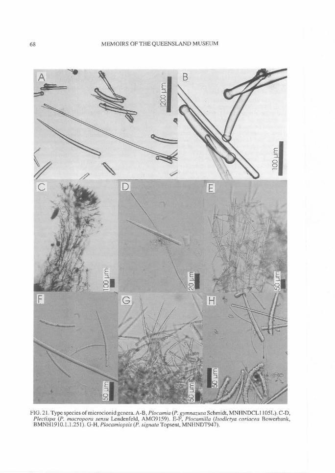
68^ MEMOIRS OF THE QUEENSLAND MUSEUM
FIG. 21. Type species of microcionid genera. A-B, Plocamia (P. gymnazusa Schmidt, MNHNDCL1105L). C-D,Plectispa (P. macropora sensu Lendenfeld, AMG9159). E-F, Plocamilla (Isodictya coriacea Bowerbank,BMNH1910.1.1.251). G-H, Plocamiopsis (P. signata Topsent, MNHNDT947).
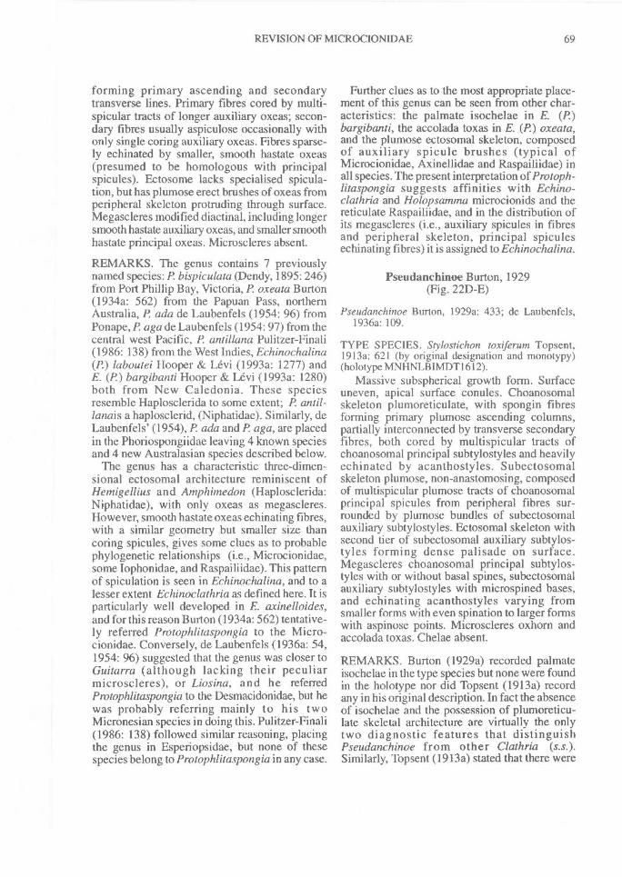
REVISION OF MICROCIONIDAE^ 69
forming primary ascending and secondarytransverse lines. Primary fibres cored by multi-spicular tracts of longer auxiliary oxeas; secon-dary fibres usually aspiculose occasionally withonly single coring auxiliary oxeas. Fibres sparse-ly echinated by smaller, smooth hastate oxeas(presumed to be homologous with principalspicules). Ectosome lacks specialised spicula-tion, but has plumose erect brushes of oxeas fromperipheral skeleton protruding through surface.Megascleres modified diactinal, including longersmooth hastate auxiliary oxeas, and smaller smoothhastate principal oxeas. Microscleres absent.
REMARKS. The genus contains 7 previouslynamed species: P bispiculata (Dendy, 1895: 246)from Port Phillip Bay, Victoria, P. oxeata Burton(1934a: 562) from the Papuan Pass, northernAustralia, P. ada de Laubenfels (1954: 96) fromPonape, P aga de Laubenfels (1954: 97) from thecentral west Pacific, P antillana Pulitzer-Finali(1986: 138) from the West Indies, Echinochalina(P.) laboutei Hooper & Levi (1993a: 1277) andE. (P.) bargibanti Hooper & Levi (1993a: 1280)both from New Caledonia. These speciesresemble Haplosclerida to some extent; P. anti!-lanais a haplosclerid, (Niphatidae). Similarly, deLaubenfels' (1954), P ada and P aga, are placedin the Phoriospongiidae leaving 4 known speciesand 4 new Australasian species described below.
The genus has a characteristic three-dimen-sional ectosomal architecture reminiscent ofHemigellius and Anzphimedon (Haplosclerida:Niphatidae), with only oxeas as megascleres.However, smooth hastate oxeas echinating fibres,with a similar geometry but smaller size thancoring spicules, gives some clues as to probablephylogenetic relationships (i.e., Microcionidae,some lophonidae, and Raspailiidae). This patternof spiculation is seen in Echinochalina, and to alesser extent Echinoclathria as defined here. It isparticularly well developed in E. axinelloides,and for this reason Burton (1934a: 562) tentative-ly referred Protophlitaspongia to the Micro-cionidae. Conversely, de Laubenfels (1936a: 54,1954: 96) suggested that the genus was closer toGuitarra (although lacking their peculiarmicroscleres), or Liosina, and he referredProtophlitaspongia to the Desmacidonidae, but hewas probably referring mainly to his twoMicronesian species in doing this. Pulitzer-Finali(1986: 138) followed similar reasoning, placingthe genus in Esperiopsidae, but none of thesespecies belong to Protophlitaspongia in any case.
Further clues as to the most appropriate place-ment of this genus can be seen from other char-acteristics: the palmate isochelae in E. (P.)bargibanti, the accolada toxas in E. (P.) oxeata,and the plumose ectosomal skeleton, composedof auxiliary spicule brushes (typical ofMicrocionidae, Axinellidae and Raspailiidae) inall species. The present interpretation of Protoph-litaspongia suggests affinities with Echino-clathria and Holopsanuna microcionids and thereticulate Raspailiidae, and in the distribution ofits megascleres (i.e., auxiliary spicules in fibresand peripheral skeleton, principal spiculesechinating fibres) it is assigned to Echinochalina.
Pseudanchinoe Burton, 1929(Fig. 22D-E)
Pseudanchinoe Burton, 1929a: 433; de Laubenfels,1936a: 109.
TYPE SPECIES. Stylostichon toxiferum Topsent,1913a: 621 (by original designation and monotypy)(holotype MNHNLBIMDT1612).
Massive subspherical growth form. Surfaceuneven, apical surface conules. Choanosomalskeleton plumoreticulate, with spongin fibresforming primary plumose ascending columns,partially interconnected by transverse secondaryfibres, both cored by multispicular tracts ofchoanosomal principal subtylostyles and heavilyechinated by acanthostyles. Subectosomalskeleton plumose, non-anastomosing, composedof multispicular plumose tracts of choanosomalprincipal spicules from peripheral fibres sur-rounded by plumose bundles of subectosomalauxiliary subtylostyles. Ectosomal skeleton withsecond tier of subectosomal auxiliary subtylos-tyles forming dense palisade on surface.Megascleres choanosomal principal subtylos-tyles with or without basal spines, subectosomalauxiliary subtylostyles with microspined bases,and echinating acanthostyles varying fromsmaller forms with even spination to larger formswith aspinose points. Microscleres oxhorn andaccolada toxas. Chelae absent.
REMARKS. Burton (1929a) recorded palmateisochelae in the type species but none were foundin the holotype nor did Topsent (1913a) recordany in his original description. In fact the absenceof isochelae and the possession of plumoreticu-late skeletal architecture are virtually the onlytwo diagnostic features that distinguishPseudanchinoe from other Clathria (s.s.).Similarly, Topsent (1913a) stated that there were
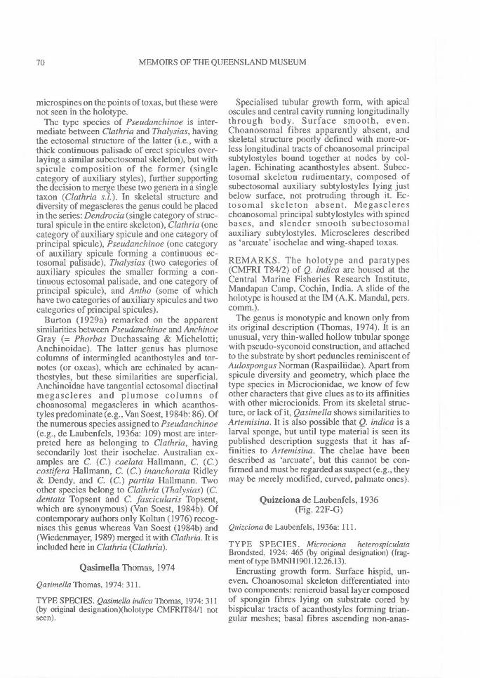
70^ MEMOIRS OF THE QUEENSLAND MUSEUM
microspines on the points of toxas, but these werenot seen in the holotype.
The type species of Pseudanchinoe is inter-mediate between Clathria and Thalysias, havingthe ectosomal structure of the latter (i.e., with athick continuous palisade of erect spicules over-laying a similar subectosomal skeleton), but withspicule composition of the former (singlecategory of auxiliary styles), further supportingthe decision to merge these two genera in a singletaxon (Clathria s.1.). In skeletal structure anddiversity of megascleres the genus could be placedin the series: Dendrocia (single category of struc-tural spicule in the entire skeleton), Clathria (onecategory of auxiliary spicule and one category ofprincipal spicule), Pseudanchinoe (one categoryof auxiliary spicule forming a continuous ec-tosomal palisade), Thalysias (two categories ofauxiliary spicules the smaller forming a con-tinuous ectosomal palisade, and one category ofprincipal spicule), and Antho (some of whichhave two categories of auxiliary spicules and twocategories of principal spicules).
Burton (1929a) remarked on the apparentsimilarities between Pseudanchinoe and AnchinoeGray (= Phorbas Duchassaing & Michelotti;Anchinoidae). The latter genus has plumosecolumns of intermingled acanthostyles and tor-notes (or oxeas), which are echinated by acan-thostyles, but these similarities are superficial.Anchinoidae have tangential ectosomal diactinalmegascleres and plumose columns ofchoanosomal megascleres in which acanthos-tyles predominate (e.g., Van Soest, 1984b: 86). Ofthe numerous species assigned to Pseudanchinoe(e.g., de Laubenfels, 1936a: 109) most are inter-preted here as belonging to Clathria, havingsecondarily lost their isochelae. Australian ex-amples are C. (C.) caelata Hallmann, C. (C.)costifera Hallmann, C. (C.) inanchorata Ridley& Dendy, and C. (C.) partita Hallmann. Twoother species belong to Clathria (Thalysias) (C.dentata Topsent and C. fascicularis Topsent,which are synonymous) (Van Soest, 19846). Ofcontemporary authors only Koltun (1976) recog-nises this genus whereas Van Soest (1984b) and(Wiedenmayer, 1989) merged it with Clathria. It isincluded here in Clathria (Clathria).
Qasimella Thomas, 1974
Qasimella Thomas, 1974: 311.
TYPE SPECIES. Qasimella indica Thomas, 1974: 311(by original designation)(holotype CMFRIT84/1 notseen).
Specialised tubular growth form, with apicaloscules and central cavity running longitudinallythrough body. Surface smooth, even.Choanosomal fibres apparently absent, andskeletal structure poorly defined with more-or-less longitudinal tracts of choanosomal principalsubtylostyles bound together at nodes by col-lagen. Echinating acanthostyles absent. Subec-tosomal skeleton rudimentary, composed ofsubectosomal auxiliary subtylostyles lying justbelow surface, not protruding through it. Ec-tosomal skeleton absent. Megasclereschoanosomal principal subtylostyles with spinedbases, and slender smooth subectosomalauxiliary subtylostyles. Microscleres describedas 'arcuate' isochelae and wing-shaped toxas.
REMARKS. The holotype and paratypes(CMFRI T8412) of Q. indica are housed at theCentral Marine Fisheries Research Institute,Mandapan Camp, Cochin, India. A slide of theholotype is housed at the IM (A.K. Mandal, pers.comm.).
The genus is monotypic and known only fromits original description (Thomas, 1974). It is anunusual, very thin-walled hollow tubular spongewith pseudo-syconoid construction, and attachedto the substrate by short peduncles reminiscent ofAulospongus Norman (Raspailiidae). Apart fromspicule diversity and geometry, which place thetype species in Microcionidae, we know of fewother characters that give clues as to its affinitieswith other microcionids. From its skeletal struc-ture, or lack of it, Qasimella shows similarities toArtemisina. It is also possible that Q. indica is alarval sponge, but until type material is seen itspublished description suggests that it has af-finities to Artemisina. The chelae have beendescribed as 'arcuate', but this cannot be con-firmed and must be regarded as suspect (e.g., theymay be merely modified, curved, palmate ones).
Quizciona de Laubenfels, 1936(Fig. 22F-G)
Quizciona de Laubenfels, 1936a: 111.
TYPE SPECIES. Microciona heterospiculataBrondsted, 1924: 465 (by original designation) (frag-ment of type BMNH1901.12.26.13).
Encrusting growth form. Surface hispid, un-even. Choanosomal skeleton differentiated intotwo components: renieroid basal layer composedof spongin fibres lying on substrate cored bybispicular tracts of acanthostyles forming trian-gular meshes; basal fibres ascending non-anas-
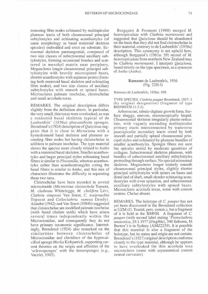
REVISION OF MICROCIONIDAE^ 71
tomosing fibre nodes echinated by multispicularplumose tracts of both choanosomal principalsubtylostyles and echinating acanthostyles (ofsame morphology as basal renieroid skeletonspicules) embedded and erect on substrate. Ec-tosomal skeleton paratangential, composed oftwo size classes of subectosomal auxiliary sub-tylostyles, forming occasional brushes and scat-tered in mesohyl matrix near periphery.Megascleres longer choanosomal principal sub-tylostyles with heavily microspined bases,shorter acanthostyles with aspinose points (form-ing both renieroid basal skeleton and echinatingfibre nodes), and two size classes of auxiliarysubtylostyles with smooth or spined bases.Microscleres palmate isochelae, cleistochelaeand small accolada toxas.
REMARKS. The original description differsslightly from the definition above. In particular,the very small, thin toxas were overlooked, as wasa renieroid basal skeleton typical of deLaubenfels' (1936a) plocamiform sponges.Brondsted's (1924) description of Quizciona sug-gests that it is close to Microciona with ahymedesmoid basal skeleton and plumose as-cending fibre nodes but having cleistochelae inaddition to palmate isochelae. The type materialshows the species more closely related to Anthowith a renieroid basal skeleton. Smaller acanthos-tyles and larger principal styles echinating basalfibres is similar to Plocamilla, whereas acanthos-tyles rather than acanthostrongyles coring thebasal fibres is similar to Antho, and this mix ofcharacters illustrates the difficulty in separatingthese two taxa.
Cleistochelae have been recorded in severalmicrocionids (Microciona cleistochela Topsent,M. clathrata Whitelegge, M. chelifera Levi,Clathria simpsoni Van Soest, C. toxipraeditaTopsent and Colloclathria ramosa Dendy).Alander (1942) and Van Soest (1984b) suggestedthat cleistochelae are modified palmate isochelae(with fused chelate teeth) which have arisenseveral times independently within theMicrocionidae, and consequently they do nothave primary taxonomic significance. Interest-ingly, Brondsted (1924) also remarked on thesimilarities between cleistochelae ofMicrocionidae and clavidiscs of the hypercal-cified sponge Merlia Kirkpatrick, supporting cur-rent theories on the origin and affinities of the`sclerosponges' with the demosponges (e.g.,Vacelet, 1985).
Bergquist & Fromont (1988) merged M.heterospiculata with Clathria mortensenii andsuggested that Quizciona should be abandonedon the basis that they did not find cleistochelae intheir material, contrary to de Laubenfels' (1936a)description. This synonymy is not upheld here,although Bergquist's (1961a: 39) record of M.heterospiculata from northern New Zealand maybe Clathria mortensenii. I interpret Quizciona,based strictly on the type specimen, as a synonymof Antho (Antho).
Ramoses de Laubenfels, 1936(Fig. 22H-I)
Ramoses de Laubenfels, 1936a: 109.
TYPE SPECIES. Clathria pauper Brondsted, 1927: 3(by original designation) (fragment of typeBMNH1930.11.5.2).
Arborescent, tubulo-digitate growth form. Sur-face shaggy, uneven, microscopically hispid.Choanosomal skeleton irregularly plumo-reticu-late, with vaguely ascending multispicularprimary tracts interconnected by transversepaucispicular secondary tracts cored by bothsmooth and partially spined choanosomal prin-cipal styles and echinated by plumose bundles ofsmaller acanthostyles. Spongin fibres not seenbut spicules united by moderate quantities ofcollagen. Subectosomal skeleton plumose, withbundles of subectosomal auxiliary subtylostylesprotruding through surface. No special ectosomalskeleton. Megascleres longer entirely smoothchoanosomal principal styles, slightly shorterprincipal subtylostyles with spines on bases anddistal end of shaft, small slender echinating acan-thostyles with even spination, and subectosomalauxiliary subtylostyles with spined bases.Microscleres accolada toxas, some with contortcentres. Chelae absent.
REMARKS. The holotype of C. pauper has notyet been discovered in the Brondsted collectionat UZM (0. Tendal, pers. comm.), but a fragmentof it is held at the BMNH. A fragment of C.pauper (with second label stating Trotoclathriaantarctica, 29.1.19?? [illegible], 340 fathoms, MBurton') is in Sydney (AMZ2239). It is possiblethat this material is also a fragment of theholotype, but its status and origin are not certain.Brondsted's (1927) original description conformsclosely to the type material, although he appearsto have overlooked the thin accolada toxamicroscleres (some with asymmetrical contortcentral curvature).

72^ MEMOIRS OF THE QUEENSLAND MUSEUM
FIG. 22. Type species of microcionid genera. A, Plocamiopsis (P. signata Topsent, MNHNDT947). B-C,Protophlitaspongia (Siphonochalina bispiculata Dendy, NMVG2319). D-E, Pseudanchinoe (Stylostichontoxiferum Topsent, MNHNDT1612). F-G, Quizciona (Microciona heterospiculata Brondsted,BMNH1901.12.26.13). H-I, Ramoses (Clathria pauper Brondsted, BMNH1930.11.5.2).
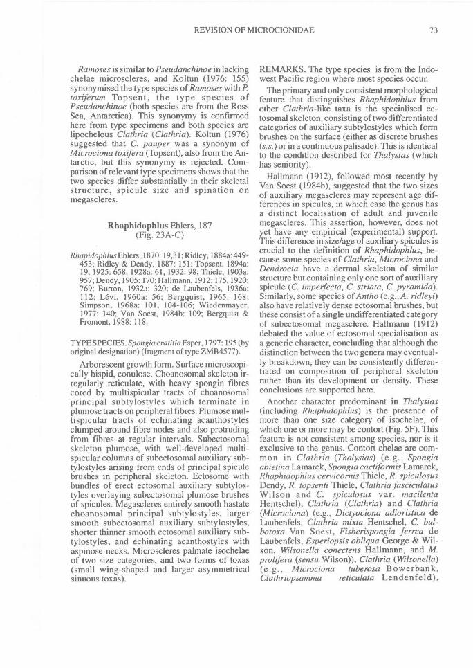
REVISION OF MICROCIONIDAE^ 73
Ramoses is similar to Pseudanchinoe in lackingchelae microscleres, and Koltun (1976: 155)synonymised the type species of Ramoses with P.toxiferuin Topsent, the type species ofPseudanchinoe (both species are from the RossSea, Antarctica). This synonymy is confirmedhere from type specimens and both species arelipochelous Clathria (Clathria). Koltun (1976)suggested that C. pauper was a synonym ofMicrociona toxifera (Topsent), also from the An-tarctic, but this synonymy is rejected. Com-parison of relevant type specimens shows that thetwo species differ substantially in their skeletalstructure, spicule size and spination onmegascleres.
Rhaphidophlus Ehlers, 187(Fig. 23A-C)
Rhapidophlus Ehlers, 1870: 19,31; Ridley, 1884a: 449-453; Ridley & Dendy, 1887: 151; Topsent, 1894a:19, 1925: 658, 1928a: 61, 1932: 98; Thiele, 1903a:957; Dendy, 1905:170; Hallmann, 1912: 175,1920:769; Burton, 1932a: 320; de Laubenfels, 1936a:112; Levi, 1960a: 56; Bergquist, 1965: 168;Simpson, 1968a: 101, 104-106; Wiedenmayer,1977: 140; Van Soest, 1984b: 109; Bergquist &Fromont, 1988: 118.
TYPE SPECIES. Spongia cratitia Esper, 1797: 195 (byoriginal designation) (fragment of type ZMB4577).
Arborescent growth form. Surface microscopi-cally hispid, conulose. Choanosomal skeleton ir-regularly reticulate, with heavy spongin fibrescored by multispicular tracts of choanosomalprincipal subtylostyles which terminate inplumose tracts on peripheral fibres. Plumose mul-tispicular tracts of echinating acanthostylesclumped around fibre nodes and also protrudingfrom fibres at regular intervals. Subectosomalskeleton plumose, with well-developed multi-spicular columns of subectosomal auxiliary sub-tylostyles arising from ends of principal spiculebrushes in peripheral skeleton. Ectosome withbundles of erect ectosomal auxiliary subtylos-tyles overlaying subectosomal plumose brushesof spicules. Megascleres entirely smooth hastatechoanosomal principal subtylostyles, largersmooth subectosomal auxiliary subtylostyles,shorter thinner smooth ectosomal auxiliary sub-tylostyles, and echinating acanthostyles withaspinose necks. Microscleres palmate isochelaeof two size categories, and two forms of toxas(small wing-shaped and larger asymmetricalsinuous toxas).
REMARKS. The type species is from the Indo-west Pacific region where most species occur.
The primary and only consistent morphologicalfeature that distinguishes Rhaphidophlus fromother Clathria-like taxa is the specialised ec-tosomal skeleton, consisting of two differentiatedcategories of auxiliary subtylostyles which formbrushes on the surface (either as discrete brushes(s.s.)or in a continuous palisade). This is identicalto the condition described for Thalysias (whichhas seniority).
Hallmann (1912), followed most recently byVan Soest (1984b), suggested that the two sizesof auxiliary megascleres may represent age dif-ferences in spicules, in which case the genus hasa distinct localisation of adult and juvenilemegascleres. This assertion, however, does notyet have any empirical (experimental) support.This difference in size/age of auxiliary spicules iscrucial to the definition of Rhaphidophlus, be-cause some species of Clathria, Microciona andDendrocia have a dermal skeleton of similarstructure but containing only one sort of auxiliaryspicule (C. impelfecta, C. striata, C. pyramida).Similarly, some species of Antho (e.g., A. ridleyi)also have relatively dense ectosomal brushes, butthese consist of a single undifferentiated categoryof subectosomal megasclere. Hallmann (1912)debated the value of ectosomal specialisation asa generic character, concluding that although thedistinction between the two genera may eventual-ly breakdown, they can be consistently differen-tiated on composition of peripheral skeletonrather than its development or density. Theseconclusions are supported here.
Another character predominant in Thalysias(including Rhaphidophlus) is the presence ofmore than one size category of isochelae, ofwhich one or more may be contort (Fig. 5F). Thisfeature is not consistent among species, nor is itexclusive to the genus. Contort chelae are com-mon in Clathria (Thalysias) (e.g., Spongiaabietina Lamarck, Spongia cactiforinis Lamarck,Rhaphidophlus cervicornis Thiele, R. spiculosusDendy, R. topsenti Thiele, Clathria fasciculatusWilson and C. spiculosus var. macilentaHentschel), Clathria (Clathria) and Clathria(Microciona) (e.g., Dictyociona adioristica deLaubenfels, Clathria mixta Hentschel, C. bul-botoxa Van Soest, Fisherispongia ferrea deLaubenfels, Esperiopsis obliqua George & Wil-son, Wilsonella conectens Hallmann, and M.prolifera (sensu Wilson)), Clathria (Wilsonella)(e.g., Microciona tuberosa Bowerbank,Clathriopsanuna^reticulata Lendenfeld),
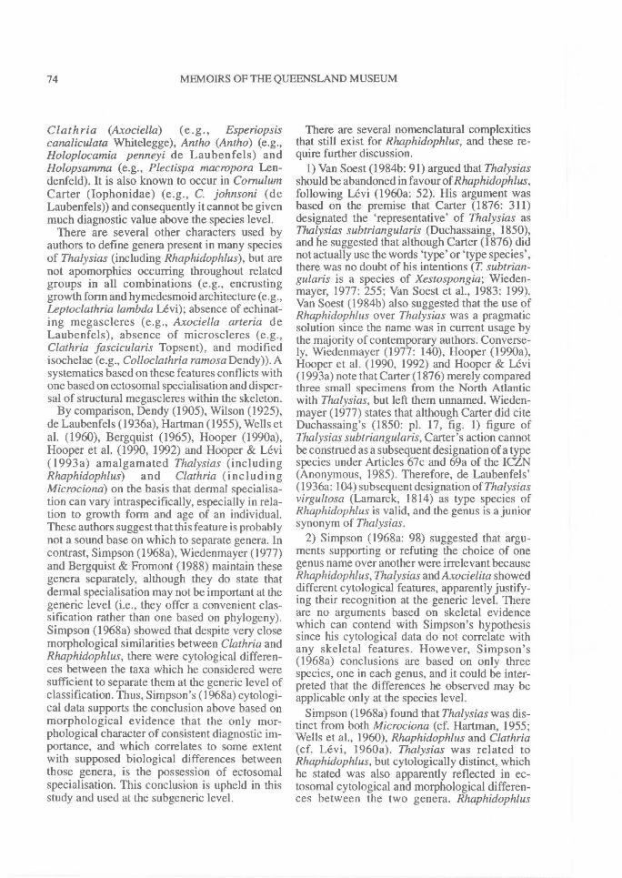
74^ MEMOIRS OF THE QUEENSLAND MUSEUM
Clathria (Axociella) (e.g., Esperiopsiscanaliculata Whitelegge), Antho (Antho) (e.g.,Holoplocamia penneyi de Laubenfels) andHolopsamma (e.g., Plectispa macropora Len-denfeld). It is also known to occur in CornulumCarter (Iophonidae) (e.g., C. johnsoni (deLaubenfels)) and consequently it cannot be givenmuch diagnostic value above the species level.
There are several other characters used byauthors to define genera present in many speciesof Thalysias (including Rhaphidophlus), but arenot apomorphies occurring throughout relatedgroups in all combinations (e.g., encrustinggrowth form and hymedesmoid architecture (e.g.,Leptoclathria lambda Levi); absence of echinat-ing megascleres (e.g., Axociella arteria deLaubenfels), absence of microscleres (e.g.,Clathria fascicularis Topsent), and modifiedisochelae (e.g., Colloclathria ramosa Dendy)). Asystematics based on these features conflicts withone based on ectosomal specialisation and disper-sal of structural megascleres within the skeleton.
By comparison, Dendy (1905), Wilson (1925),de Laubenfels (1936a), Hartman (1955), Wells etal. (1960), Bergquist (1965), Hooper (1990a),Hooper et al. (1990, 1992) and Hooper & Levi(1993a) amalgamated Thalysias (includingRhaphidophlus) and Clathria (includingMicrociona) on the basis that dermal specialisa-tion can vary intraspecifically, especially in rela-tion to growth form and age of an individual.These authors suggest that this feature is probablynot a sound base on which to separate genera. Incontrast, Simpson (1968a), Wiedenmayer (1977)and Bergquist & Fromont (1988) maintain thesegenera separately, although they do state thatdermal specialisation may not be important at thegeneric level (i.e., they offer a convenient clas-sification rather than one based on phylogeny).Simpson (1968a) showed that despite very closemorphological similarities between Clathria andRhaphidophlus, there were cytological differen-ces between the taxa which he considered weresufficient to separate them at the generic level ofclassification. Thus, Simpson's (1968a) cytologi-cal data supports the conclusion above based onmorphological evidence that the only mor-phological character of consistent diagnostic im-portance, and which correlates to some extentwith supposed biological differences betweenthose genera, is the possession of ectosomalspecialisation. This conclusion is upheld in thisstudy and used at the subgeneric level.
There are several nomenclatural complexitiesthat still exist for Rhaphidophlus, and these re-quire further discussion.
1) Van Soest (1984b: 91) argued that Thalysiasshould be abandoned in favour of Rhaphidophlus,following Levi (1960a: 52). His argument wasbased on the premise that Carter (1876: 311)designated the 'representative' of Thalysias asThalysias subtriangularis (Duchassaing, 1850),and he suggested that although Carter (1876) didnot actually use the words 'type' or 'type species',there was no doubt of his intentions (T subtrian-gularis is a species of Xestospongia; Wieden-mayer, 1977: 255; Van Soest et al., 1983: 199).Van Soest (1984b) also suggested that the use ofRhaphidophlus over Thalysias was a pragmaticsolution since the name was in current usage bythe majority of contemporary authors. Converse-ly, Wiedenmayer (1977: 140), Hooper (1990a),Hooper et al. (1990, 1992) and Hooper & Levi(1993a) note that Carter (1876) merely comparedthree small specimens from the North Atlanticwith Thalysias, but left them unnamed. Wieden-mayer (1977) states that although Carter did citeDuchassaing's (1850: pl. 17, fig. 1) figure ofThalysias subtriangularis, Carter's action cannotbe construed as a subsequent designation of a typespecies under Articles 67c and 69a of the ICZN(Anonymous, 1985). Therefore, de Laubenfels'(1936a: 104) subsequent designation of Thalysiasvirgultosa (Lamarck, 1814) as type species ofRhaphidophlus is valid, and the genus is a juniorsynonym of Thalysias.
2) Simpson (1968a: 98) suggested that argu-ments supporting or refuting the choice of onegenus name over another were irrelevant becauseRhaphidophlus,Thalysias and Axocielita showeddifferent cytological features, apparently justify-ing their recognition at the generic level. Thereare no arguments based on skeletal evidencewhich can contend with Simpson's hypothesissince his cytological data do not correlate withany skeletal features. However, Simpson's(1968a) conclusions are based on only threespecies, one in each genus, and it could be inter-preted that the differences he observed may beapplicable only at the species level.
Simpson (1968a) found that Thalysias was dis-tinct from both Microciona (cf. Hartman, 1955;Wells et al., 1960), Rhaphidophlus and Clathria(cf. Levi, 1960a). Thalysias was related toRhaphidophlus, but cytologically distinct, whichhe stated was also apparently reflected in ec-tosomal cytological and morphological differen-ces between the two genera. Rhaphidophlus
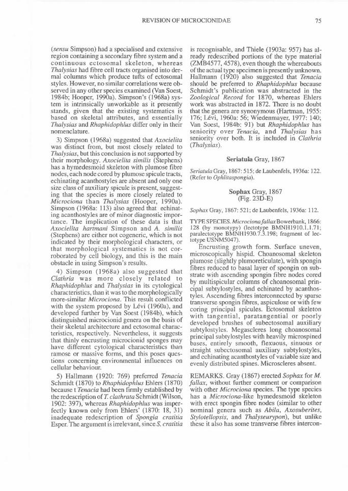
REVISION OF MICROCIONIDAE^ 75
(sensu Simpson) had a specialised and extensiveregion containing a secondary fibre system and acontinuous ectosomal skeleton, whereasThalysias had fibre cell tracts organised into der-mal columns which produce tufts of ectosomalstyles. However, no similar correlations were ob-served in any other species examined (Van Soest,1984b; Hooper, 1990a). Simpson's (1968a) sys-tem is intrinsically unworkable as it presentlystands, given that the existing systematics isbased on skeletal attributes, and essentiallyThalysias and Rhaphidophlus differ only in theirnomenclature.
3) Simpson (1968a) suggested that Axocielitawas distinct from, but most closely related toThalysias, but this conclusion is not supported bytheir morphology. Axocielita similis (Stephens)has a hymedesmoid skeleton with plumose fibrenodes, each node cored by plumose spicule tracts,echinating acanthostyles are absent and only onesize class of auxiliary spicule is present, suggest-ing that the species is more closely related toMicrociona than Thalysias (Hooper, 1990a).Simpson (1968a: 113) also agreed that echinat-ing acanthostyles are of minor diagnostic impor-tance. The implication of these data is thatAxocielita harttnani Simpson and A. simi/is(Stephens) are either not cogeneric, which is notindicated by their morphological characters, orthat morphological systematics is not cor-roborated by cell biology, and this is the mainobstacle in using Simpson's results.
4) Simpson (1968a) also suggested thatClathria was more closely related toRhaphidophlus and Thalysias in its cytologicalcharacteristics, than it was to the morphologicallymore-similar Microciona. This result conflictedwith the system proposed by Levi (1960a), anddeveloped further by Van Soest (1984b), whichdistinguished microcionid genera on the basis oftheir skeletal architecture and ectosomal charac-teristics, respectively. Nevertheless, it suggeststhat thinly encrusting microcionid sponges mayhave different cytological characteristics thanramose or massive forms, and this poses ques-tions concerning environmental influences oncellular behaviour.
5) Hallmann (1920: 769) preferred TenaciaSchmidt (1870) to Rhaphidophlus Ehlers (1870)because t Tenacia had been firmly established bythe redescription of T. clathrata Schmidt (Wilson,1902: 397), whereas Rhaphidophlus was imper-fectly known only from Ehlers' (1870: 18, 31)inadequate redescription of Spongia cratitiaEsper. The argument is irrelevant, since S. cratitia
is recognisable, and Thiele (1903a: 957) has al-ready redescribed portions of the type material(ZMB4577, 4578), even though the whereaboutsof the actual type specimen is presently unknown.Hallmann (1920) also suggested that Tenaciashould be preferred to Rhaphidophlus becauseSchmidt's publication was abstracted in theZoological Record for 1870, whereas Ehlerswork was abstracted in 1872. There is no doubtthat the genera are synonymous (Hartman, 1955:176; Levi, 1960a: 56; Wiedenmayer, 1977: 140;Van Soest, 1984b: 91) but Rhaphidophlus hasseniority over Tenacia, and Thalysias hasseniority over both. It is included in Clathria(Thalysias).
Seriatula Gray, 1867
Seriatula Gray, 1867: 515; de Laubenfels, 1936a: 122.(Refer to Ophlitaspongia).
Sophax Gray, 1867(Fig. 23D-E)
Sophax Gray, 1867: 521; de Laubenfels, 1936a: 112.
TYPE SPECIES. Microciona fallax Bowerbank, 1866:128 (by monotypy) (lectotype BMNH1910.1.1.71;paralectotype BMNH1930.7.3.198; fragment of lec-totype USNM5047).
Encrusting growth form. Surface uneven,microscopically hispid. Choanosomal skeletonplumose (slightly plumoreticulate), with sponginfibres reduced to basal layer of spongin on sub-strate with ascending spongin fibre nodes coredby multispicular columns of choanosomal prin-cipal subtylostyles, and echinated by acanthos-tyles. Ascending fibres interconnected by sparsetransverse spongin fibres, aspiculose or with fewcoring principal spicules. Ectosomal skeletonwith tangential, paratangential or poorlydeveloped brushes of subectosomal auxiliarysubtylostyles. Megascleres long choanosomalprincipal subtylostyles with heavily microspinedbases, entirely smooth, flexuous, sinuous orstraight subectosomal auxiliary subtylostyles,and echinating acanthostyles of vaiiable size andevenly distributed spines. Microscleres absent.
REMARKS. Gray (1867) erected Sophax for M.fallax, without further comment or comparisonwith other Microciona species. The type specieshas a Microciona-like hymedesmoid skeletonwith erect spongin fibre nodes (similar to othernominal genera such as Abila, Axosuberites,Stylotellopsis, and Thalyseurypon), but unlikethese it also has some transverse fibres intercon-
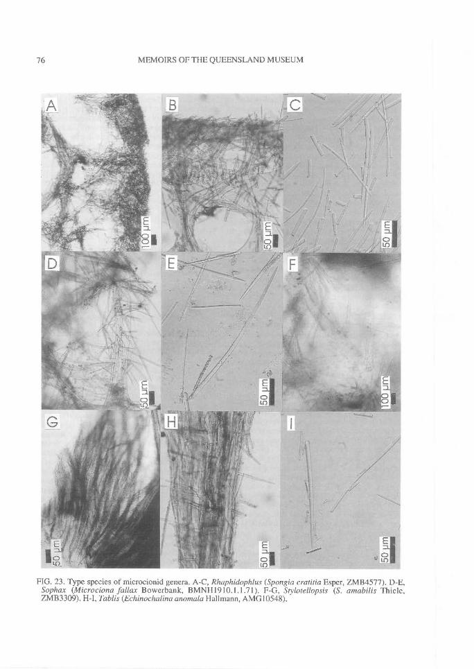
76^
MEMOIRS OF THE QUEENSLAND MUSEUM
FIG. 23. Type species of microcionid genera. A-C, Rhaphidophlus (Spongia cratitia Esper, ZMB4577). D-E,Sophax (Microciona fallax Bowerbank, BMNH1910.1.1.71). F-G, Stylotellopsis (S. amabilis Thiele,ZMB3309). H-I, Tablis (Echinochalina anomala Hallmann, AMG10548).
REVISION OF MICROCIONIDAE^ 77
necting multispicular tracts, producing a slightlyplumoreticulate architecture. Sophax is referredhere to Clathria (Microciona).
Stylotellopsis Thiele, 1905(Fig. 23F-G)
Stylotellopsis Thiele, 1905: 456; de Laubenfels, 1936a:112.
TYPE SPECIES. Stylotellopsis amabilis Thiele, 1905:456 (by monotypy) (holotype ZMB3309).
Thickly encrusting growth form. Surfacehispid, uneven. Choanosomal skeleton hymedes-moid, with basal layer of spongin fibres lying onsubstrate and plumose non-anastomosing fibrenodes. Spongin fibre nodes cored by largeechinating acanthostyles standing perpendicularto substrate, forming multispicular plumose tuftson basal skeleton. Subectosomal skeletonplumose, with multispicular columns of subec-tosomal auxiliary subtylostyles arising from endsof echinating spicule brushes, producingdendritic whispy tracts especially near periphery.Ectosomal skeleton without special spiculationbut erect subectosomal auxiliary styles formnearly continuous palisade on surface. Mesohylhas heavy deposits of loose spongin. Megascleressingle category of smooth subectsomal auxiliarystyles with tapering bases and hastate points, andlarge echinating acanthostyles with swollenbases, large spines, and aspinose points. Principalspicules undifferentiated from auxiliary spicules.Microscleres absent.
REMARKS. From published descriptions(Thiele, 1905; Topsent, 1928a; Koltun, 1964a)Stylotellopsis appears to be similar to Thalysiasin having differentiated subectosomal and ec-tosomal auxiliary spicules (i.e., a specialised ec-tosomal skeleton), and Van Soest (1984b)synonymised the two genera on this basis. Re-ex-amination of type material found that auxiliaryspicules range greatly in their length, and prin-cipal spicules are very similar to auxiliaryspicules scattered throughout the skeleton. Thegenus has a `microcionid' choanosomal skeletalstructure (i.e., hymedesmoid with plumose fibrenodes), and an unusual subectosomal skeletoncomposed of dendritic or sinuous auxiliaryspicule tracts. It also lacks microscleres (althoughseveral arcuate isochelae were seen in histologi-cal preparations of the holotype, but these areprobably foreign). Stylotellopsis is interpretedhere as being an encrusting (hymedesmoid)
species of Clathria (Thalysias). De Laubenfels(1936a) included the genus with Eurypon(Raspailiidae), but this is not upheld here:similarities between Stylotellopsis and Thalysiasin their peripheral skeletal structure and thegeometry of auxiliary spicules are closer thanwith the raspailiids (see Hooper, 1991).
Tablis de Laubenfels, 1936(Fig. 23H-I)
Tab/is de Laubenfels, 1936a: 76.
TYPE SPECIES. Echinochalina anomala Hallmann,1912: 292 (by original designation) (holotypeAMG10548).
Massive, lobate-digitate growth form. Surface'honeycomb' reticulate. Choanosomal skeletonreticulate, with heavy primary and secondaryspongin fibres forming wide-meshed reticulationcored by multispicular tracts of subectosomalauxiliary subtylostyles. Coring spicules inprimary fibres compressed, more dispersed insecondary fibres. Fibres heavily echinated byprincipal styles. Subectosomal skeleton withscattered auxiliary megascleres dispersed be-tween fibre meshes and protruding through sur-face in paratangential bundles. Megascleresentirely smooth principal styles-subtylostyles,and smooth subectosomal auxiliary subtylos-tyles, often secondarily modified to tornotes.Microscleres absent.
REMARKS. Tablis was erected for Whitelegge's(1907) specimen of Echinochalina glabra fromWoolongong, NSW, a junior homonym of Ridley& Dendy's (1887) species renamed by Hallmann(1912) to E. anomala. This species has also beenrecorded from the Great Barrier Reef (Burton,1934a). Three other species were referred toTab/is by de Laubenfels (1936a): Echinochalinaglabra Ridley & Dendy (a synonym of Spongiabarba Lamarck), E. reticulata Whitelegge, bothfrom SW Australia, and Spongia clavosaLamarck. The last mentioned species is un-known: it was not mentioned in Topsent's (1933)revision of Lamarck's species nor found in theLamarck collections (MNHN). These other 2species differ from E. anomala in havingmicroscleres but lacking an Holopsamma-like'honeycombed reticulate' growth form. Tab/is isreturned here to Echinochalina (Echinochalina).
78^ MEMOIRS OF THE QUEENSLAND MUSEUM
Tenacia Schmidt, 1870(Fig. 24A-C)
Tenacia Schmidt, 1870: 56; Hallmann, 1920: 769;Topsent, 1920b: 17; Burton & Rao, 1932:337; Levi,1960a: 56.
TYPE SPECIES. Tenacia clathrata Schmidt, 1870: 56(by monotypy) (fragment of type BMNH1870.-5.3.156); = Spongia virgultosa Lamarck, 1814: 444(fragment of type MNHNLBIMDNBE1344, 1338).
Erect, arborescent, reticulate branching growthform. Surface highly conulose, uneven, micros-copically hispid. Choanosomal skeleton ir-regularly reticulate, with well developed sponginfibres differentiated into ascending primary andtransverse secondary fibres. Principal spiculesconfined to axis of primary fibres, but absentcompletely from secondary connecting fibres,and all fibres cored by paucispicular tracts ofsubectosomal auxiliary subtylostyles, and heavi-ly echinated by small acanthostyles. Subec-tosomal skeleton plumose, with tracts ofsubectosomal auxiliary subtylostyles originatingfrom deeper regions of choanosomal skeleton;subectosomal auxiliary subtylostyles also scat-tered abundantly throughout mesohyl betweenfibre meshes. Ectosome plumose, with brushes ofsmaller ectosomal auxiliary subtylostyles arisingfrom ends of subdermal spicule brushes.Megascleres smooth choanosomal principalstyles, smooth subectosomal auxiliary subtylos-tyles, smooth ectosomal auxiliary subtylostyles,and short thick echinating acanthostyles withaspinose points and necks. Microscleres palmateisochelae of two sizes, and wing-shaped, ac-colada and sinuous asymmetrical toxas.
REMARKS. Tenacia and Rhaphidophlus aresynonyms (Hallmann, 1920; Topsent, 1932; Levi,1960a), and use of one name over another ismerely a nomenclatural decision (see remarks forRhaphidophlus). Topsent's (1932: 97) synonymyof T clathrata and Spongia juniperina Lamarckis not upheld here: the former is considered hereto be a synonym of T virgultosa and restricted toCaribbean and NE. Atlantic populations, whereasT. juniperina (including T clathrata of Hallmann,1912) is known only from the Indian Ocean. Thegenus is synonymised with Clathria (Thalysias).
Tenaciella Hallmann, 1920(Fig. 24D-E)
Tenaciella Hallmann, 1920: 772; de Laubenfels,1936a: 126.
TYPE SPECIES. Esperiopsis^canaliculataWhitelegge, 1906: 471 (by monotypy) (lectotypeAMG4325).
Erect, branching, digitate growth form. Sur-face even, hispid, with oscules dispersed overlateral margins of branches. Choanosomalskeleton irregularly reticulate, nearly radial, withcompressed axial and plumose extra-axialskeletons. In axis heavy spongin fibres cored bymultispicular tracts of choanosomal principalstyles, and in extra-axial skeleton principal stylesprotrude from peripheral fibres forming radial orplumose tracts, lying immediately below and sup-porting subectosomal skeleton. Echinatingmegascleres absent. Subectosomal skeletonplumose, with multispicular ascending tracts ofsubectosomal auxiliary styles at ends ofperipheral choanosomal fibres. Ectosomalskeleton with thick continuous palisade ofsmaller ectosomal auxiliary styles standing per-pendicular to surface. Megascleres smoothfusiform choanosomal principal styles, subec-tosomal auxiliary styles-subtylostyles withsmooth or microspined bases, and ectosomalauxiliary styles-subtylostyles with microspinedbases. Microscleres palmate isochelae of twosizes, including contorted forms, and oxhorn andwing-shaped toxas.
REMARKS. Hallmann (1920) erected Tenaciellafor species like Tenacia(= Thalysias), but lackingechinating megascleres. Wells et al. (1960) alsoreferred Esperiopsis obliqua George & Wilson toTenaciella, but this species lacks dermalspecialisation and is more closely allied withAxocielita (= Clathria). Ristau (1978) suggestedthat Artemisina archegona Ristau (USNM24528) was similar to the type species ofTenaciella in having prominent subectosomal(extra-axial) spicule columns, and he suggestedthat the two species differed only in growth form,shape of the toxa microscleres and the appreciab-ly more organised skeletal architecture in Tcanaliculata. However, differences observed intypes of these species are more noticable thantheir similarities (e.g., ectosomal skeleton, fibrereticulation, degree of axial and extra-axial dif-ferentiation), and their supposed affinities (Ris-tau, 1978), are superficial.
In its skeletal structure, differentiation of axialand extra-axial skeletons, fibre characteristics,the possession of similar extra-axial radialbundles of megascleres, and having a continuousectosomal palisade of spicules the type speciesshows close similarities to Ceratopsion axifera(Hentschel) (Raspailiidae) from the Arafura Sea
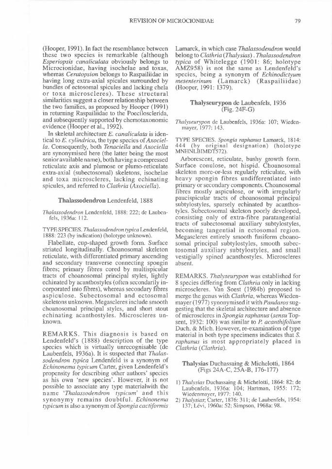
REVISION OF MICROCIONIDAE^ 79
(Hooper, 1991). In fact the resemblance betweenthese two species is remarkable (althoughEsperiopsis canaliculata obviously belongs toMicrocionidae, having isochelae and toxas,whereas Ceratopsion belongs to Raspailiidae inhaving long extra-axial spicules surrounded bybundles of ectosomal spicules and lacking chelaor toxa microscleres). These structuralsimilarities suggest a closer relationship betweenthe two families, as proposed by Hooper (1991)in returning Raspailiidae to the Poecilosclerida,and subsequently supported by chemotaxonomicevidence (Hooper et al., 1992).
In skeletal architecture E. canaliculata is iden-tical to E. cylindrica, the type species of Axociel-la. Consequently, both Tenaciella and Axociellaare synonymised here (the latter being the mostsenior available name), both having a compressedreticulate axis and plumose or plumo-reticulateextra-axial (subectosomal) skeletons, isochelaeand toxa microscleres, lacking echinatingspicules, and referred to Clathria (Axociella).
Thalassodendron Lendenfeld, 1888
Thalassodendron Lendenfeld, 1888: 222; de Lauben-fels, 1936a: 112.
TYPE SPECIES. Thalassodendron typica Lendenfeld,1888: 223 (by indication) (holotype unknown).
Flabellate, cup-shaped growth form. Surfacestriated longitudinally. Choanosomal skeletonreticulate, with differentiated primary ascendingand secondary transverse connecting sponginfibres; primary fibres cored by multispiculartracts of choanosomal principal styles, lightlyechinated by acanthostyles (often secondarily in-corporated into fibres), whereas secondary fibresaspiculose. Subectosomal and ectosomalskeletons unknown. Megascleres include smoothchoanosomal principal styles, and short stoutechinating acanthostyles. Microscleres un-known.
REMARKS. This diagnosis is based onLendenfeld's (1888) description of the typespecies which is virtually unrecognisable (deLaubenfels, 1936a). It is suspected that Thalas-sodendron typica Lendenfeld is a synonym ofEchinonema typicum Carter, given Lendenfeld'spropensity for describing other authors' speciesas his own 'new species'. However, it is notpossible to associate any type materialwith thename `Thalassodendron typicum' and thissynonymy remains doubtful. Echinonematypicum is also a synonym of Spongia cactifortnis
Lamarck, in which case Thalassodendron wouldbelong to Clathria (Thalysias) . Thalassodendrontypica of Whitelegge (1901: 86; holotypeAMZ958) is not the same as Lendenfeld'sspecies, being a synonym of Echinodictyummesenterinum (Lamarck) (Raspailiidae)(Hooper, 1991: 1379).
Thalyseurypon de Laubenfels, 1936(Fig. 24F-G)
Thalyseurypon de Laubenfels, 1936a: 107; Wieden-mayer, 1977: 143.
TYPE SPECIES. Spongia raphanus Lamarck, 1814:444 (by original designation) (holotypeMNHNLBIMDT572).
Arborescent, reticulate, bushy growth form.Surface conulose, not hispid. Choanosomalskeleton more-or-less regularly reticulate, withheavy spongin fibres undifferentiated intoprimary or secondary components. Choanosomalfibres mostly aspiculose, or with irregularlypaucispicular tracts of choanosomal principalsubtylostyles, sparsely echinated by acanthos-tyles. Subectosomal skeleton poorly developed,consisting only of extra-fibre paratangentialtracts of subectosomal auxiliary subtylostyles,becoming tangential in ectosomal region.Megascleres entirely smooth fusiform choano-somal principal subtylostyles, smooth subec-tosomal auxiliary subtylostyles, and smallvestigially spined acanthostyles. Microscleresabsent.
REMARKS. Thalyseurypon was established for8 species differing from Clathria only in lackingmicroscleres. Van Soest (1984b) proposed tomerge the genus with Clathria, whereas Wieden-mayer (1977) synonymised it with Pandaros sug-gesting that the skeletal architecture and absenceof microscleres in Spongia raphanus (sensu Top-sent, 1932: 100) was similar to P. acanthifoliumDuch. & Mich. However, re-examination of typematerial in both type specimens indicates that S.raphanus is most appropriately placed inClathria (Clathria).
Thalysias Duchassaing & Michelotti, 1864(Figs 24A-C, 25A-B, 176-177)
1) Thalysias Duchassaing & Michelotti, 1864: 82; deLaubenfels, 1936a: 104; Hartman, 1955: 172;Wiedenmayer, 1977: 140.
2) Thalysias; Carter, 1876: 311; de Laubenfels, 1954:137; Levi, 1960a: 52; Simpson, 1968a: 98.
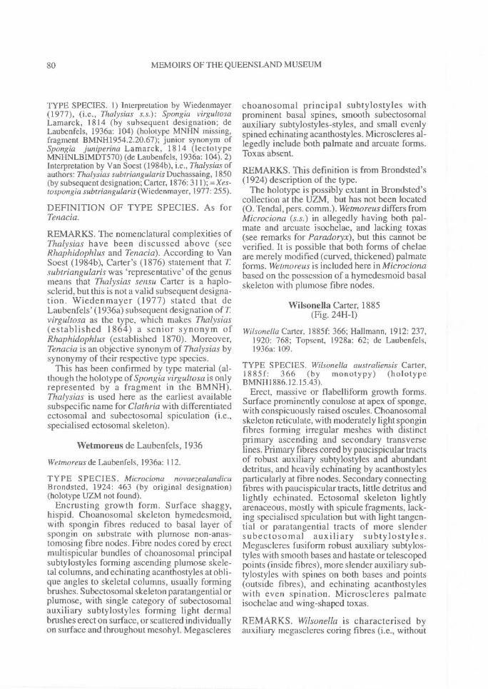
80^ MEMOIRS OF THE QUEENSLAND MUSEUM
TYPE SPECIES. 1) Interpretation by Wiedenmayer(1977), (i.e., Thalysias s.s.): Spongia virgultosaLamarck, 1814 (by subsequent designation; deLaubenfels, 1936a: 104) (holotype MNHN missing,fragment BMNH1954.2.20.67); junior synonym ofSpongia juniperina Lamarck, 1814 (lectotypeMNHNLBIMDT570) (de Laubenfels, 1936a: 104). 2)Interpretation by Van Soest (1984b), i.e., Thalysias ofauthors: Thalysias subtriangularis Duchassaing, 1850(by subsequent designation; Carter, 1876: 311); = Xes-tospongia subtriangularis (Wiedenmayer, 1977: 255).
DEFINITION OF TYPE SPECIES. As forTenacia.
REMARKS. The nomenclatural complexities ofThalysias have been discussed above (seeRhaphidophlus and Tenacia). According to VanSoest (1984b), Carter's (1876) statement that Tsubtriangularis was 'representative' of the genusmeans that Thalysias sensu Carter is a haplo-sclerid, but this is not a valid subsequent designa-tion. Wiedenmayer (1977) stated that deLaubenfels' (1936a) subsequent designation of T.virgultosa as the type, which makes Thalysias(established 1864) a senior synonym ofRhaphidophlus (established 1870). Moreover,Tenacia is an objective synonym of Thalysias bysynonymy of their respective type species.
This has been confirmed by type material (al-though the holotype of Spongia virgultosa is onlyrepresented by a fragment in the BMNH).Thalysias is used here as the earliest availablesubspecific name for Clathria with differentiatedectosomal and subectosomal spiculation (i.e.,specialised ectosomal skeleton).
Wetmoreus de Laubenfels, 1936
Wetmoreus de Laubenfels, 1936a: 112.
TYPE SPECIES. Microciona novaezealandicaBrondsted, 1924: 463 (by original designation)(holotype UZM not found).
Encrusting growth form. Surface shaggy,hispid. Choanosomal skeleton hymedesmoid,with spongin fibres reduced to basal layer ofspongin on substrate with plumose non-anas-tomosing fibre nodes. Fibre nodes cored by erectmultispicular bundles of choanosomal principalsubtylostyles forming ascending plumose skele-tal columns, and echinating acanthostyles at obli-que angles to skeletal columns, usually formingbrushes. Subectosomal skeleton paratangential orplumose, with single category of subectosomalauxiliary subtylostyles forming light dermalbrushes erect on surface, or scattered individuallyon surface and throughout mesohyl. Megascleres
choanosomal principal subtylostyles withprominent basal spines, smooth subectosomalauxiliary subtylostyles-styles, and small evenlyspined echinating acanthostyles. Microscleres al-legedly include both palmate and arcuate forms.Toxas absent.
REMARKS. This definition is from Brondsted's(1924) description of the type.
The holotype is possibly extant in Brondsted'scollection at the UZM, but has not been located(0. Tendal, pers. comm.). Wetmoreus differs fromMicrociona (s.s.) in allegedly having both pal-mate and arcuate isochelae, and lacking toxas(see remarks for Paradoryx), but this cannot beverified. It is possible that both forms of chelaeare merely modified (curved, thickened) palmateforms. Wetmoreus is included here in Microcionabased on the possession of a hymedesmoid basalskeleton with plumose fibre nodes.
Wilsonella Carter, 1885(Fig. 24H-I)
Wilsonella Carter, I885f: 366; Hallmann, 1912: 237,1920: 768; Topsent, 1928a: 62; de Laubenfels,1936a: 109.
TYPE SPECIES. Wilsonella australiensis Carter,1885f: 366 (by monotypy) (holotypeBMNH1886.12.15.43).
Erect, massive or flabelliform growth forms.Surface prominently conulose at apex of sponge,with conspicuously raised oscules. Choanosomalskeleton reticulate, with moderately light sponginfibres forming irregular meshes with distinctprimary ascending and secondary transverselines. Primary fibres cored by paucispicular tractsof robust auxiliary subtylostyles and abundantdetritus, and heavily echinating by acanthostylesparticularly at fibre nodes. Secondary connectingfibres with paucispicular tracts, little detritus andlightly echinated. Ectosomal skeleton lightlyarenaceous, mostly with spicule fragments, lack-ing specialised spiculation but with light tangen-tial or paratangential tracts of more slendersubectosomal auxiliary subtylostyles.Megascleres fusiform robust auxiliary subtylos-tyles with smooth bases and hastate or telescopedpoints (inside fibres), more slender auxiliary sub-tylostyles with spines on both bases and points(outside fibres), and echinating acanthostyleswith even spination. Microscleres palmateisochelae and wing-shaped toxas.
REMARKS. Wilsonella is characterised byauxiliary megascleres coring fibres (i.e., without
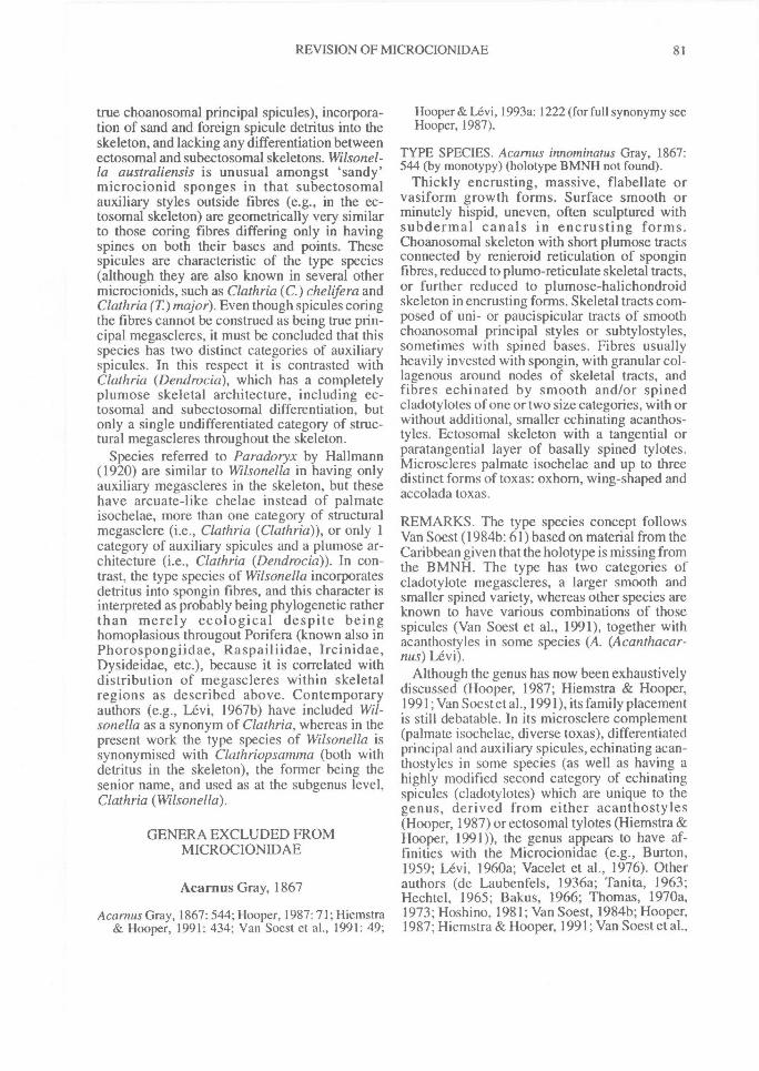
REVISION OF MICROCIONIDAE^ 81
true choanosomal principal spicules), incorpora-tion of sand and foreign spicule detritus into theskeleton, and lacking any differentiation betweenectosomal and subectosomal skeletons. Wilsonel-la australiensis is unusual amongst 'sandy'microcionid sponges in that subectosomalauxiliary styles outside fibres (e.g., in the ec-tosomal skeleton) are geometrically very similarto those coring fibres differing only in havingspines on both their bases and points. Thesespicules are characteristic of the type species(although they are also known in several othermicrocionids, such as Clathria (C.) chelifera andClathria (T) major). Even though spicules coringthe fibres cannot be construed as being true prin-cipal megascleres, it must be concluded that thisspecies has two distinct categories of auxiliaryspicules. In this respect it is contrasted withClathria (Dendrocia), which has a completelyplumose skeletal architecture, including ec-tosomal and subectosomal differentiation, butonly a single undifferentiated category of struc-tural megascleres throughout the skeleton.
Species referred to Paradoryx by Hallmann(1920) are similar to Wilsonella in having onlyauxiliary megascleres in the skeleton, but thesehave arcuate-like chelae instead of palmateisochelae, more than one category of structuralmegasclere (i.e., Clathria (Clathria)), or only 1category of auxiliary spicules and a plumose ar-chitecture (i.e., Clathria (Dendrocia)). In con-trast, the type species of Wilsonella incorporatesdetritus into spongin fibres, and this character isinterpreted as probably being phylogenetic ratherthan merely ecological despite beinghomoplasious througout Porifera (known also inPhorospongiidae, Raspailiidae, Ircinidae,Dysideidae, etc.), because it is correlated withdistribution of megascleres within skeletalregions as described above. Contemporaryauthors (e.g., Levi, 1967b) have included Wil-sonella as a synonym of Clathria, whereas in thepresent work the type species of Wilsonella issynonymised with Clathriopsamnza (both withdetritus in the skeleton), the former being thesenior name, and used as at the subgenus level,Clathria (Wilsonella).
GENERA EXCLUDED FROMMICROCIONIDAE
Acarnus Gray, 1867
Acarnus Gray, 1867: 544; Hooper, 1987: 71; Hiemstra& Hooper, 1991: 434; Van Soest et al., 1991: 49;
Hooper & Levi, 1993a: 1222 (for full synonymy seeHooper, 1987).
TYPE SPECIES. Acarnus innominatus Gray, 1867:544 (by monotypy) (holotype BMNH not found).
Thickly encrusting, massive, flabellate orvasiform growth forms. Surface smooth orminutely hispid, uneven, often sculptured withsubdermal canals in encrusting forms.Choanosomal skeleton with short plumose tractsconnected by renieroid reticulation of sponginfibres, reduced to plumo-reticulate skeletal tracts,or further reduced to plumose-halichondroidskeleton in encrusting forms. Skeletal tracts com-posed of uni- or paucispicular tracts of smoothchoanosomal principal styles or subtylostyles,sometimes with spined bases. Fibres usuallyheavily invested with spongin, with granular col-lagenous around nodes of skeletal tracts, andfibres echinated by smooth and/or spinedcladotylotes of one or two size categories, with orwithout additional, smaller echinating acanthos-tyles. Ectosomal skeleton with a tangential orparatangential layer of basally spined tylotes.Microscleres palmate isochelae and up to threedistinct forms of toxas: oxhom, wing-shaped andaccolada toxas.
REMARKS. The type species concept followsVan Soest (1984b: 61) based on material from theCaribbean given that the holotype is missing fromthe BMNH. The type has two categories ofcladotylote megascleres, a larger smooth andsmaller spined variety, whereas other species areknown to have various combinations of thosespicules (Van Soest et al., 1991), together withacanthostyles in some species (A. (Acanthacar-nus) Levi).
Although the genus has now been exhaustivelydiscussed (Hooper, 1987; Hiemstra & Hooper,1991; Van Soest et al., 1991), its family placementis still debatable. In its microsclere complement(palmate isochelae, diverse toxas), differentiatedprincipal and auxiliary spicules, echinating acan-thostyles in some species (as well as having ahighly modified second category of echinatingspicules (cladotylotes) which are unique to thegenus, derived from either acanthostyles(Hooper, 1987) or ectosomal tylotes (Hiemstra &Hooper, 1991)), the genus appears to have af-finities with the Microcionidae (e.g., Burton,1959; Levi, 1960a; Vacelet et al., 1976). Otherauthors (de Laubenfels, 1936a; Tanita, 1963;Hechtel, 1965; Bakus, 1966; Thomas, 1970a,1973; Hoshino, 1981; Van Soest, 1984b; Hooper,1987; Hiemstra & Hooper, 1991; Van Soest et al.,
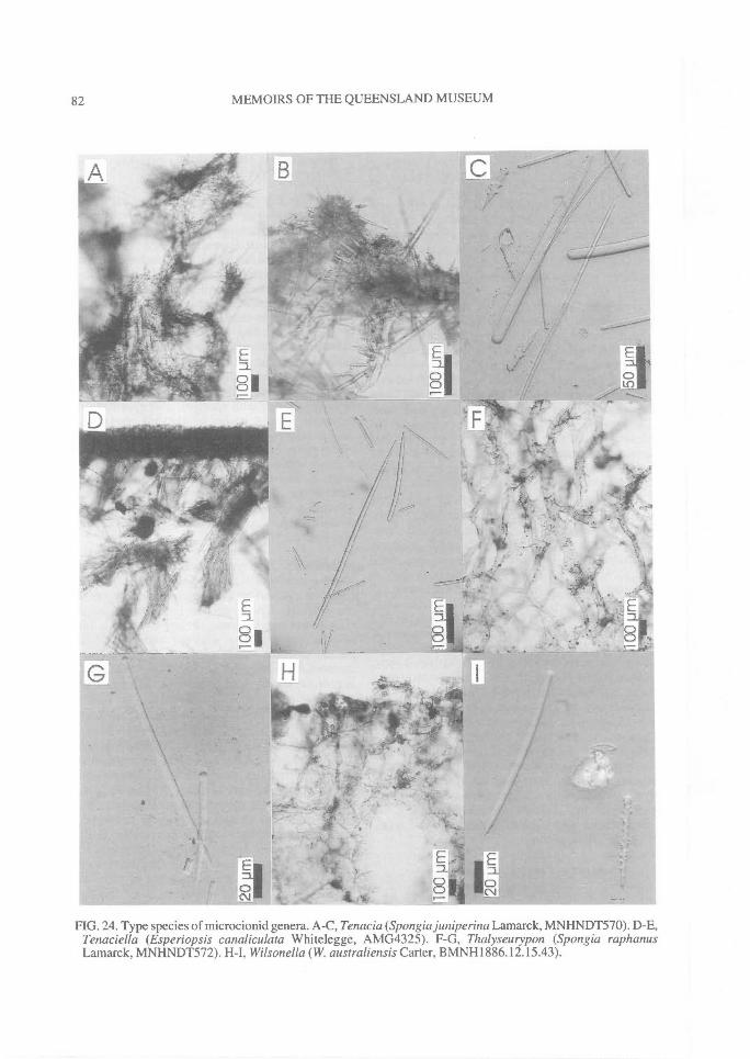
82^ MEMOIRS OF THE QUEENSLAND MUSEUM
FIG. 24. Type species of microcionid genera. A-C, Tenacia (Spongia juniperina Lamarck, MNHNDT570). D-E,Tenaciella (Esperiopsis canaliculata Whitelegge, AMG4325). F-G, Thalyseurypon (Spongia raphanusLamarck, MNHNDT572). H-I, Wilsonella (W. australiensis Carter, BMNH1886.12.15.43).
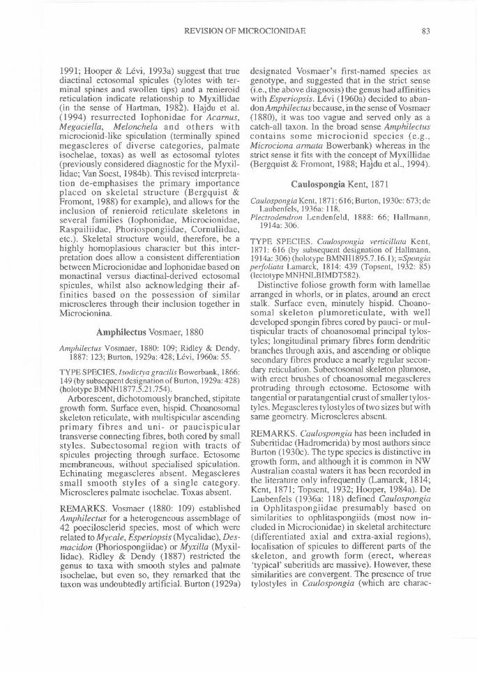
REVISION OF MICROCIONIDAE^ 83
1991; Hooper & Levi, 1993a) suggest that truediactinal ectosomal spicules (tylotes with ter-minal spines and swollen tips) and a renieroidreticulation indicate relationship to Myxillidae(in the sense of Hartman, 1982). Hajdu et al.(1994) resurrected Iophonidae for Acarnus,Megaciella, Melonchela and others withmicrocionid-like spiculation (terminally spinedmegascleres of diverse categories, palmateisochelae, toxas) as well as ectosomal tylotes(previously considered diagnostic for the Myxil-lidae; Van Soest, 1984b). This revised interpreta-tion de-emphasises the primary importanceplaced on skeletal structure (Bergquist &Fromont, 1988) for example), and allows for theinclusion of renieroid reticulate skeletons inseveral families (Iophonidae, Microcionidae,Raspailiidae, Phoriospongiidae, Cornuliidae,etc.). Skeletal structure would, therefore, be ahighly homoplasious character but this inter-pretation does allow a consistent differentiationbetween Microcionidae and Iophonidae based onmonactinal versus diactinal-derived ectosomalspicules, whilst also acknowledging their af-finities based on the possession of similarmicroscleres through their inclusion together inMicrocionina.
Amphilectus Vosmaer, 1880
Amphilectus Vosmaer, 1880: 109; Ridley & Dendy,1887: 123; Burton, 1929a: 428; Levi, 1960a: 55.
TYPE SPECIES. Isodiciya gracilis Bowerbank, 1866:149 (by subsequent designation of Burton, 1929a: 428)(holotype BMNH1877.5.21.754).
Arborescent, dichotomously branched, stipitategrowth form. Surface even, hispid. Choanosomalskeleton reticulate, with multispicular ascendingprimary fibres and uni- or paucispiculartransverse connecting fibres, both cored by smallstyles. Subectosomal region with tracts ofspicules projecting through surface. Ectosomemembraneous, without specialised spiculation.Echinating megascleres absent. Megascleressmall smooth styles of a single category.Microscleres palmate isochelae. Toxas absent.
REMARKS. Vosmaer (1880: 109) establishedAmphdectus for a heterogeneous assemblage of42 poecilosclerid species, most of which wererelated to Mycale, Esperiopsis (Mycalidae), Des-macidon (Phoriospongiidae) or Myxilla (Myxil-lidae). Ridley & Dendy (1887) restricted thegenus to taxa with smooth styles and palmateisochelae, but even so, they remarked that thetaxon was undoubtedly artificial. Burton (1929a)
designated Vosmaer's first-named species asgenotype, and suggested that in the strict sense(i.e., the above diagnosis) the genus had affinitieswith Esperiopsis. Levi (1960a) decided to aban-don Amphilectus because, in the sense of Vosmaer(1880), it was too vague and served only as acatch-all taxon. In the broad sense Amphilectuscontains some microcionid species (e.g.,Microciona annata Bowerbank) whereas in thestrict sense it fits with the concept of Myxillidae(Bergquist & Fromont, 1988; Hajdu et al., 1994).
Caulospongia Kent, 1871
Caulospongia Kent, 1871: 616; Burton, 1930c: 673; deLaubenfels, I 936a: 118.
Plectrodendron Lendenfeld, 1888: 66; Hallmann,1914a: 306.
TYPE SPECIES. Caulospongia verticillata Kent,1871: 616 (by subsequent designation of Hallmann,1914a: 306) (holotype BMNH1895.7.16.1); =Spongiaperfoliata Lamarck, 1814: 439 (Topsent, 1932: 85)(lectotype MNHNLBIMDT582).
Distinctive foliose growth form with lamellaearranged in whorls, or in plates, around an erectstalk. Surface even, minutely hispid. Choano-somal skeleton plumoreticulate, with welldeveloped spongin fibres cored by pauci- or mul-tispicular tracts of choanosomal principal tylos-tyles; longitudinal primary fibres form dendriticbranches through axis, and ascending or obliquesecondary fibres produce a nearly regular secon-dary reticulation. Subectosomal skeleton plumose,with erect brushes of choanosomal megascleresprotruding through ectosome. Ectosome withtangential or paratangential crust of smaller tylos-tyles. Megascleres tylostyles of two sizes but withsame geometry. Microscleres absent.
REMARKS. Caulospongia has been included inSuberitidae (Hadromerida) by most authors sinceBurton (1930c). The type species is distinctive ingrowth form, and although it is common in NWAustralian coastal waters it has been recorded inthe literature only infrequently (Lamarck, 1814;Kent, 1871; Topsent, 1932; Hooper, 1984a). DeLaubenfels (1936a: 118) defined Caulospongiain Ophlitaspongiidae presumably based onsimilarities to ophlitaspongiids (most now in-cluded in Microcionidae) in skeletal architecture(differentiated axial and extra-axial regions),localisation of spicules to different parts of theskeleton, and growth form (erect, whereas'typical' suberitids are massive). However, thesesimilarities are convergent. The presence of truetylostyles in Caulospongia (which are charac-
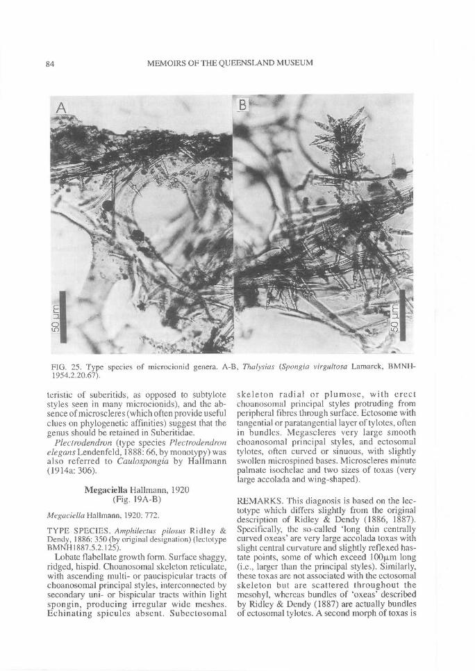
84^ MEMOIRS OF THE QUEENSLAND MUSEUM
FIG. 25. Type species of microcionid genera. A-B, Thalysias (Spongia virgultosa Lamarck, BMNH-1954.2.20.67).
teristic of suberitids, as opposed to subtylotestyles seen in many microcionids), and the ab-sence of microscleres (which often provide usefulclues on phylogenetic affinities) suggest that thegenus should be retained in Suberitidae.
Plectrodendron (type species Plectrodendronelegans Lendenfeld, 1888: 66, by monotypy) wasalso referred to Caulospongia by Hallmann(1914a: 306).
Megaciella Hallmann, 1920(Fig. 19A-B)
Megaciella Hallmann, 1920: 772.
TYPE SPECIES. Amphilectus pilosus Ridley &Dendy, 1886: 350 (by original designation) (lectotypeBMNH1887.5.2.125).
Lobate flabellate growth form. Surface shaggy,ridged, hispid. Choanosomal skeleton reticulate,with ascending multi- or paucispicular tracts ofchoanosomal principal styles, interconnected bysecondary uni- or bispicular tracts within lightspongin, producing irregular wide meshes.Echinating spicules absent. Subectosomal
skeleton radial or plumose, with erectchoanosomal principal styles protruding fromperipheral fibres through surface. Ectosome withtangential or paratangential layer of tylotes, oftenin bundles. Megascleres very large smoothchoanosomal principal styles, and ectosomaltylotes, often curved or sinuous, with slightlyswollen microspined bases. Microscleres minutepalmate isochelae and two sizes of toxas (verylarge accolada and wing-shaped).
REMARKS. This diagnosis is based on the lec-totype which differs slightly from the originaldescription of Ridley & Dendy (1886, 1887).Specifically, the so-called 'long thin centrallycurved oxeas' are very large accolada toxas withslight central curvature and slightly reflexed has-tate points, some of which exceed 100p,m long(i.e., larger than the principal styles). Similarly,these toxas are not associated with the ectosomalskeleton but are scattered throughout themesohyl, whereas bundles of `oxeas' describedby Ridley & Dendy (1887) are actually bundlesof ectosomal tylotes. A second morph of toxas is
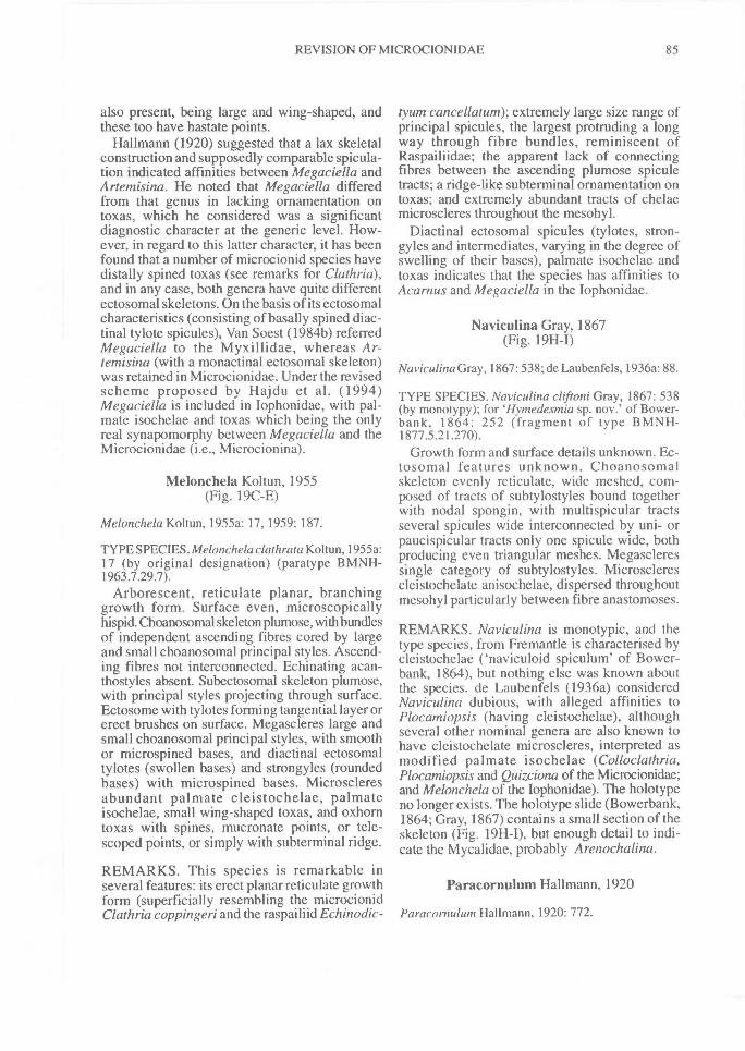
REVISION OF MICROCION1DAE^ 85
also present, being large and wing-shaped, andthese too have hastate points.
Hallmann (1920) suggested that a lax skeletalconstruction and supposedly comparable spicula-tion indicated affinities between Megaciella andArtemisina. He noted that Megaciella differedfrom that genus in lacking ornamentation ontoxas, which he considered was a significantdiagnostic character at the generic level. How-ever, in regard to this latter character, it has beenfound that a number of microcionid species havedistally spined toxas (see remarks for Clathria),and in any case, both genera have quite differentectosomal skeletons. On the basis of its ectosomalcharacteristics (consisting of basally spined diac-tinal tylote spicules), Van Soest (1984b) referredMegaciella to the Myxillidae, whereas Ar-temisina (with a monactinal ectosomal skeleton)was retained in Microcionidae. Under the revisedscheme proposed by Hajdu et al. (1994)Megaciella is included in Iophonidae, with pal-mate isochelae and toxas which being the onlyreal synapomorphy between Megaciella and theMicrocionidae (i.e., Microcionina).
Melonchela Koltun, 1955(Fig. 19C-E)
Melonchela Koltun, 1955a: 17, 1959: 187.
TYPE SPECIES. Melonchelaclathrata Koltun, 1955a:17 (by original designation) (paratype BMNH-1963.7.29.7).
Arborescent, reticulate planar, branchinggrowth form. Surface even, microscopicallyhispid. Choanosomal skeleton plumose, with bundlesof independent ascending fibres cored by largeand small choanosomal principal styles. Ascend-ing fibres not interconnected. Echinating acan-thostyles absent. Subectosomal skeleton plumose,with principal styles projecting through surface.Ectosome with tylotes forming tangential layer orerect brushes on surface. Megascleres large andsmall choanosomal principal styles, with smoothor microspined bases, and diactinal ectosomaltylotes (swollen bases) and strongyles (roundedbases) with microspined bases. Microscleresabundant palmate cleistochelae, palmateisochelae, small wing-shaped toxas, and oxhorntoxas with spines, mucronate points, or tele-scoped points, or simply with subterminal ridge.
REMARKS. This species is remarkable inseveral features: its erect planar reticulate growthform (superficially resembling the microcion idClathria coppingeri and the raspaili id Echinodic-
tyum cancellatum); extremely large size range ofprincipal spicules, the largest protruding a longway through fibre bundles, reminiscent ofRaspailiidae; the apparent lack of connectingfibres between the ascending plumose spiculetracts; a ridge-like subterminal ornamentation ontoxas; and extremely abundant tracts of chelaemicroscleres throughout the mesohyl.
Diactinal ectosomal spicules (tylotes, stron-gyles and intermediates, varying in the degree ofswelling of their bases), palmate isochelae andtoxas indicates that the species has affinities toAcarnus and Megaciella in the Iophonidae.
Naviculina Gray, 1867(Fig. 19H-I)
Naviculina Gray, 1867:538; de Laubenfels, 1936a: 88.
TYPE SPECIES. Naviculina cliftoni Gray, 1867: 538(by monotypy); for Ilymedesmia sp. nov.' of Bower-bank, 1864: 252 (fragment of type BMNH-1877.5.21.270).
Growth form and surface details unknown. Ec-tosomal features unknown. Choanosomalskeleton evenly reticulate, wide meshed, com-posed of tracts of subtylostyles bound togetherwith nodal spongin, with multispicular tractsseveral spicules wide interconnected by uni- orpaucispicular tracts only one spicule wide, bothproducing even triangular meshes. Megascleressingle category of subtylostyles. Microsclerescleistochelate anisochelae, dispersed throughoutmesohyl particularly between fibre anastomoses.
REMARKS. Naviculina is monotypic, and thetype species, from Fremantle is characterised bycleistochelae ('naviculoid spiculum' of Bower-bank, 1864), but nothing else was known aboutthe species. de Laubenfels (1936a) consideredNaviculina dubious, with alleged affinities toPlocamiopsis (having cleistochelae), althoughseveral other nominal genera are also known tohave cleistochelate microscleres, interpreted asmodified palmate isochelae (Colloclathria,Plocamiopsis and Quizciona of the Microcionidae;and Melonchela of the Iophonidae). The holotypeno longer exists. The holotype slide (Bowerbank,1864; Gray, 1867) contains a small section of theskeleton (Fig. 19H-I), but enough detail to indi-cate the Mycalidae, probably Arenochalina.
Paracornulum Hallmann, 1920
Paraconiulum Hallmann, 1920: 772.
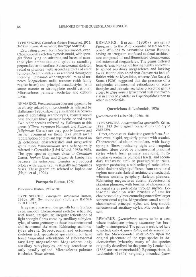
86^ MEMOIRS OF THE QUEENSLAND MUSEUM
TYPE SPECIES. Cornu/um dubium Hentschel, 1912:346 (by original designation) (holotype SMF964).
Encrusting growth form. Surface smooth, even.Choanosomal skeleton hymedesmoid, with spon-gin fibres lying on substrate and bases of acan-thostyles embedded and spicules standingperpendicular to surface. Subectosomal skeletonradial or plumose, with ascending tracts of erecttornotes. Acanthostyles also scattered throughoutmesohyl. Ectosome with tangential tracts of tor-notes. Megascleres radial tornotes (with fainlyrugose bases) and principal acanthostyles (withsome oxeote or strongylote modifications).Microscleres palmate isochelae and oxhorntoxas.
REMARKS. Paracornulum does not appear to beas closely related to microcionids as inferred byHallmann (1920), showing similarities in posses-sion of echinating acanthostyles, hymedesmoidbasal spongin fibres, palmate isochelae and toxas.Two other species referred by Hallmann to thisgenus (Cliona purpurea Hancock and Suberitesfuliginosus Carter) are very poorly known andfurther comment on those taxa must awaitredescription of relevant type material. Based onits ectosomal characteristics and megasclerespiculation Paracornulutn was subsequentlyreferred to Cornulidae (Levi & Levi, 1983a: 966).The type species appears close to CornulumCarter, lophon Gray and Zyzzya de Laubenfelsbecause the ectosomal tornotes are reducedtylotes with rugose (i.e., vestigially microspined)bases. These genera are referred to Iophonidae(Hajdu et al., 1994).
Paresperia Burton, 1930
Paresperia Burton, 1930a: 501.
TYPE SPECIES. Paresperia intermedia Burton,1930a: 501 (by monotypy) (holotype BMNH-1910.1.1.912).
Irregularly massive, low growth form. Surfaceeven, smooth. Choanosomal skeleton reticulate,with loose, unispicular, irregular reticulation oflight spongin fibres cored by auxiliary subtylos-tyles, of same geometry as those in subectosomaland ectosomal skeletons. Echinating acanthos-tyles absent. Subectosomal and ectosomalskeletons lack specialised spiculation, but haveloose tangential reticulation of subectosomalauxiliary megascleres. Megascleres onlyauxiliary subtylostyles, entirely acanthose oronly basally spined. Microscleres palmateisochelae. Toxas absent.
REMARKS. Burton (1930a) assignedParesperia to the Microcionidae based on sup-posed affinities to Artemisina (sensu Burton),having an irregular, confused skeletal architec-ture composed of undifferentiated choanosomaland ectosomal megascleres. The genus differedfrom Artemisina (s.s.) in having lightly and even-ly spined auxiliary megascleres and lackingtoxas. Burton also noted that Paresperia had af-finities with the Mycalidae, whereas Van Soest &Stone (1986) suggested that the presence of aunispicular choanosomal reticulation of acan-thostyles and palmate isochelae placed the genuscloser to Esperiopsis (placement still controver-sial in either Mycalidae or Esperiopsidae) than toother microcionids.
Querciclona de Laubenfels, 1936
Querciclona de Laubenfels, 1936a: 46.
TYPE SPECIES. Antherochalina quercifolia Keller,1889: 383 (by original designation) (holotypeZMB429).
Erect, arborescent, flabellate growth form. Sur-face even, hispid, regularly porous with oscules.Choanosomal skeleton reticulate, with heavyspongin fibres producing tight and irregularmeshes, fibres cored by choanosomal principalstyles which form primary ascending multi-spicular (eventually plumose) tracts, and secon-dary transverse uni- or paucispicular tracts,together producing a subisodictyal reticulation.Axial skeleton slightly different from extra-axialregion: near axis skeletal architecture isodictyal,whereas towards periphery skeleton plumose.Echinating megascleres absent. Subectosomalskeleton plumose, with brushes of choanosomalprincipal styles protruding through surface. Ec-tosomal skeleton with brushes of smallerchoanosomal styles surrounding one or few largersubectosomal styles. Megascleres small smoothchoanosomal principal styles, and long smoothsubectosomal auxiliary styles. Microscleres ab-sent.
REMARKS. Querciclona seems to be a casewhere inadequate primary taxonomy has beenbadly misinterpreted. The genus is restricted hereto include only A. quercifolia, and its associationwith the Microcionidae rests solely upon theoriginal placement of the type species in An-therochalina (whereby many of the speciesoriginally described for the genus by Lendenfeld(1888) are true microcionids). By comparison, deLaubenfels (1936a) originally intended Quer-
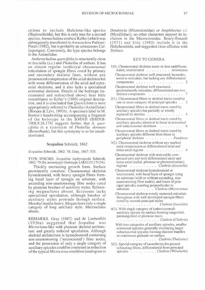
REVISION OF MICROCIONIDAE^ 87
ciclona to include Haliclona-like species(Haplosclerida), but this is only true for a secondspecies, Arenochalina arabica Keller (which wassubsequently transferred to A renosclera Pulitzer-Finali (1982), but is probably an arenaceous Cal-lyspongia). Conversely, the type species belongsto the Axinellidae.
Antherochalina quercifolia is structurally closeto Isociella (s.s.) and Phakellia of authors. It hasan almost regular isodictyal choanosomalreticulation of spongin fibres cored by primaryand secondary skeletal lines, without anypronounced compression of the axial skeleton butwith some differentiation of the axial and extra-axial skeletons, and it also lacks a specialisedectosomal skeleton. Details of the holotype (re-examined and redescribed above) bear littleresemblance to Keller's (1889) original descrip-tion, and it is concluded that Querciclona is mostappropriately referred to Phakellia (Axinellidae)(Hooper & Levi, 1993b). A specimen label in M.Burton's handwriting accompanying a fragmentof the holotype in the BMNH (BMNH-1908.9.24.179) suggests further that A. quer-cifolia is a synonym of Phakellia donnani(Bowerbank), but this synonymy is so far unsub-stantiated.
Scopalina Schmidt, 1862
Scopalina Schmidt, 1862: 78; Gray, 1867: 535.
TYPE SPECIES. Scopalina lophyropoda Schmidt,1862: 79 (by monotypy) (holotype LMJG15117/154).
Thickly encrusting growth form. Surfaceprominently conulose. Choanosomal skeletonhymedesmoid, with heavy spongin fibres form-ing basal layer of spongin on substrate, withascending non-anastomosing fibre nodes coredby plumose brushes of auxiliary styles. Echinat-ing megascleres absent. Ectosome lacksspecialised spiculation, although brushes ofauxiliary styles protrude through surface.Mesohyl matrix heavy. Megascleres only a singlecategory of long auxiliary style. Microscleresabsent.
REMARKS. Gray (1867) and de Laubenfels(1936a) suggested that Scopalina wasMicrociona-like with plumose skeletal architec-ture and greatly reduced spiculation. Althoughskeletal architecture is hymedesmoid containingnon-anastomosing ('microcionid') fibre nodes,and the possession of only a single category ofauxiliary spicules could be construed as reductionof the typical Microciona condition (analogous to
Dendrocia (Microcionidae) or Amphilectus s.l.(Myxillidae)), no other characters support its in-clusion in the Microcionidae. Boury-Esnault(1971) and Uriz (1982) include it in theHalichondrida, and suggested close affinites withStylinos.
KEY TO GENERA
1(0). Choanosomal skeleton more-or-less undifferen-tiated, unstructured ^ Artemisina
Choanosomal skeleton well structured, hymedes-moid to reticulate, but lacking any differentiatedcomponents 2
Choanosomal skeleton well structured,predominantly reticulate, differentiated into twodistinct components ^ 7
2(1). Choanosomal fibres or skeletal tracts cored byone or more category of principal spicules . ^ 3
Choanosomal fibres or skeletal tracts cored byauxiliary spicules but partially or whollyreplaced by detritus ^ 5
Choanosomal fibres or skeletal tracts cored byauxiliary spicules identical to those in ectosomaland subectosomal skeletons ^ 6
Choanosomal fibres or skeletal tracts cored byauxiliary spicules different from those inperipheral skeleton ^ Panda ros
3(2). Choanosomal skeleton without any markedaxial compression or differentiated axial andextra-axial regions 4
Choanosomal skeleton with noticeably com-pressed axis and well differentiated axial andextra-axial (radial, plumose or plumoreticulate)regions ^ Clathria (Axociella)
Choanosomal skeleton hymedesmoid ormicrocionid, with basal layer of spongin lyingon substrate (with or without ascending, non-anastomosing fibre nodes), and bases of prin-cipal spicules standing perpendicular tosubstrate ^ Clathria (Microciona)
Choanosomal skeleton evenly renieroid reticulatethroughout with well developed spongin fibrescored by smooth principal styles^ Clathria (Isociella)
4(3). With single category of (subectosomal)auxiliary spicule on surface forming tangential,paratangential or plumose tracts^ Clathria (Clathria)
With two categories of auxiliary spicules, smallerectosomal spicules generally overlaying largersubectosomal spicules forming discrete bundlesor continuous palisade on surface^ Clathria (Thalysias)
5(2). Special category of (acantho)styles presentechinating fibres, differentiated from principalspicules ^ Clathria (Wilsonella)
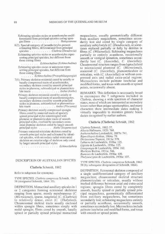
88^ MEMOIRS OF THE QUEENSLAND MUSEUM
Echinating spicules styles or acanthostyles undif-ferentiated from principal spicules coring spon-gin fibres Holopsamma
6(2). Special category of (acantho)styles presentechinating fibres, differentiated from principalspicules ^ Clathria (Dendrocia)
Echinating spicules styles or acanthostyles repre-senting principal spicules, but different fromthose coring fibres^ Echinochalina (Echinochalina)
Echinating spicules oxeas or anisoxeas repre-senting principal spicules, but different fromthose coring fibres^ Echinochalina (Protophlitaspongia)
7(1). Primary skeleton renieroid cored by axially orbasally compressed tracts of acanthostyles,secondary skeleton cored by smooth principalstyles in plumose, subisodictyal or plumoreticu-late tracts ^ Antho (Antho)
Primary skeleton renieroid cored by axially orbasally compressed tracts of acanthostrongyles,secondary skeleton cored by smooth principalstyles in plumose, subisodictyal or plumoreticu-late tracts An (ho (Plocamia)
Primary skeleton axially compressed sponginfibres cored by renieroid tracts of sparselyspined principal styles intermingled withplumose or plumoreticulate tracts of smoothprincipal styles, overlaid by secondary extra-axial plumose skeleton cored by larger smoothprincipal styles ^ Antho (lsopenectya)
Primary renieroid reticulate skeleton cored bysmooth principal styles and echinated by identi-cal spicules, with secondary radial extra-axialskeleton on exterior edge of skeleton only coredby larger smooth principal styles^ Echinoclathria
DESCRIPTION OF AUSTRALIAN SPECIES
Clathria Schmidt, 1862
Refer to subgenera for synonymy.
TYPE SPECIES. Clathria compressa Schmidt, 1862:58 (designated Schmidt, 1864: 35).
DEFINITION. Monactinal auxiliary spicules in 1or 2 categories forming ectosomal skeletonsranging from sparse, mostly membraneous (C.(Microciona)), sparse, tangential (C. (Clathria))to relatively dense, erect (C. (Thalysias)).Choanosomal skeletal tracts usually enclosedwithin spongin fibres, sometimes simply withnodal spongin; fibres cored by smooth, basallyspined or partially spined principal monactinal
megascleres, usually geometrically differentfrom auxiliary megascleres, sometimes secon-darily lost and cored by single category ofauxiliary subtylostyle (C. (Dendrocia)), or some-times replaced partially or fully by detritus infibres (C. (Wilsonella)). Echinating megasclerespartially or entirely acanthose, occasionallysmooth or vestigial spination, sometimes secon-darily lost (C. (Axociella), C. (Isociella)).Choanosomal structure ranges from leptoclathridto microcionid plumose (C. (Microciona)),renieroid (C. (Isociella)), plumoreticulate orreticulate, with (C. (Axociella)) or without com-pressed axis and radial extra-axial regions.Microscleres include palmate isochelae andmodified forms, and toxas with smooth or spinedpoints, occasionally absent.
REMARKS. This definition is necessarily broadto encompass the 7 subgenera included inClathria, showing a wide spectrum of characterstates, most of which are interpreted as secondarylosses rather than unique apomorphies, and manycharacters show intermediate states making itvirtually impossible to maintain generic boun-daries recognised by earlier authors.
Clathria (Clathria) Schmidt, 1862
Clathria Schmidt, 1862: 57.Allocia Hallmann, 1920: 768.Antherochalina Lendenfeld, 1887b: 741.Bipocillopsis Koltun, 1964a: 79.Diclyociona Topsent, 1913a: 579.Labacea de Laubenfels, 1936a: 125.Ligrota de Laubenfels, 1936a: 125.Litaspongia de Laubenfels, 1954: 162.Marleyia Burton, 1931a: 346.Ramoses de Laubenfels, 1936a: 109.Thalyseurypon de Laubenfels, 1936a: 107.
TYPE SPECIES. Clathria compressa Schmidt, 1862:58 (by subsequent designation of Schmidt, 1864).
DEFINITION. Ectosomal skeleton composed ofa single undifferentiated category of auxiliarymegasclere; choanosomal skeletal structureplumoreticulate or reticulate, usually withoutmarked difference between axial and extra-axialregions; spongin fibres cored by completelysmooth, basally spined or partially spined prin-cipal megascleres, geometrically differentiatedfrom auxiliary megascleres, but sometimessecondarily lost; echinating megascleres entirelyor partially acanthose, occasionally smooth,sometimes secondarily lost. Microscleres includepalmate isochelae and modified forms, and toxaswith smooth or spined points.
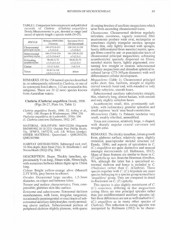
REVISION OF MICROCIONIDAE^ 89
TABLE 1. Comparison between present and publishedrecords of Clathria (Clathria) a nguliferaDendy.Measurements in p.m, denoted as range (andmean) of spicule length x spicule width (N=25).
SPICULE Holotype (NMO2286)(Victoria)
Specimens (N=2)(SE.Queensland)
Choanosomal 1444172.4)-211 128-(141.3)-150principal styles x 3-(4.3)-6 x 3-(3.8)-5
Subectosomal 168-(175.0)-254 181-(195.8)-223auxiliary styles x 2.5-(3.6)-4.5 x 342.3)-5Echinating 58-(64.2)-72 58-(61.8)-72acanthostyles x 3.5-(4.1)-4.5 x 2-(3.3)-5
Chelae 5.5-(7.1)-8.5 5-(6.7)-9
Toxas 18467.4)-101x 0.541.7)-3.3
absent
REMARKS. Of the 154 named species describedin, or subsequently referred to Clathria, or one ofits synonyms listed above, 112 are retained in thissubgenus. There are 31 (2 new) species knownfrom Australian waters.
Clathria (Clathria) angulifera Dendy, 1896(Figs 26-27, Plate 1A, Table 1)
Clathria anguhfera Dendy, 1896: 32; Ayling et al.,1982: 100; Hooper & Wiedenmayer, 1994: 258.
Thalysias angulifera; de Laubenfels, 1936a: 104.cf. Clathria conectens Hallmann, 1912: 247.
MATERIAL. HOLOTYPE: NMVG2286 (fragmentBMNH1902.10.18.323): Outside Port Phillip Heads,Vic, 38°09'S, 144°52'E, coll. J.B. Wilson (dredge).OTHER MATERIAL: Queensland — QMG303230,QMG303960.
HABITAT DISTRIBUTION. Submerged rock reef;10-30m depth; Bass Strait (Vic), N. Stradbroke I. andNoosa Heads (SEQ) (Fig. 260).
DESCRIPTION. Shape. Thickly lamellate, ap-proximately 9 cm long, 30mm wide, 50mm high,with numerous bulbous lobate digits up to 15mmlong.Colour Bright yellow-orange alive (Munsell2.5Y 8/10), grey-brown in ethanol.Oscules. Occasional large oscules, 1.5 - 3mmdiameter, on edges and between lobes.Texture and surface characteristics. Firm, com-pressible; glabrous skin-like surface.Ectosome and subectosome. Ectosomal skeletonmembraneous, with loose, irregular, tangential,occasionally paratangential or erect tracts of sub-ectosomal auxiliary subtylostyles; rarely protrud-ing above surface. Subectosomal portion ofperipheral skeleton slightly plumose, with sparse
diverging brushes of auxiliary megascleres whicharise from ascending choanosomal tracts.Choanosome. Choanosomal skeleton regularlyreticulate, cavernous, vaguely renieroid; fibreanastomoses produce wide oval, rectangular orsometimes slightly triangular meshes; sponginfibres thin, only lightly invested with spongin,barely differentiated from mesohyl matrix; spon-gin fibres cored by uni- or paucispicular tracts ofchoanosomal principal megascleres; echinatingacanthostyles sparsely dispersed on fibres;mesohyl matrix heavy, lightly pigmented, con-taining few straight or sinuous subectosomalauxiliary megascleres and abundant spherical in-cubated larvae (275-34511m diameter) with welldifferentiated cellular development.Megascleres (Table 1). Choanosomal principalstyles short, thin, fusiform, straight or slightlycurved towards basal end, with rounded or onlyslightly subtylote, smooth bases.
Subectosomal auxiliary subtylostyles straight,thin, relatively long, almost hastate, with smoothand only slightly subtylote bases.
Acanthostyles small, thin, prominently sub-tylote, with rudimentary granular spination andsmall aspinose 'neck' near basal constriction.Microscleres (Table 1). Palmate isochelae verysmall, weakly silicified, unmodified.
Toxas not common, relatively large, v-shaped,with sharply angular central curvature andstraight arms.
REMARKS. The thickly lamellate, lobate growthform, glabrous surface, relatively open, slightlyrenieroid, paucispicular skeletal structure (cf.Dendy, 1896), and aspects of spiculation in C.(C.) angulifera are quite distinctive and unusualamongst microcionids (cf. Hallmann, 1912).Many of these features are similar to those in C.(T)aphylla sp. nov. from the Houtman Abrolhos,WA, although the latter has a specialised ec-tosomal skeleton and hence is included in C.(Thalysias) rather than C. (Clathria). Thesespecies together with C. (C.) hispidula are sisterspecies belonging to a species group termed here`angulifera' group. They are compared in detailin discussion on C. (T) aphylla.
This species is also slightly reminiscent of C.(C.) conectens, differing in that megasclerescoring fibres are true principal spicules ratherthan just undifferentiated auxiliary spicules (al-biet these differences are not as well marked in C.(C.) angulifera as in many other species ofClathria). This reduction in coring spicules wasinterpreted by Hallmann (1912) to be charac-
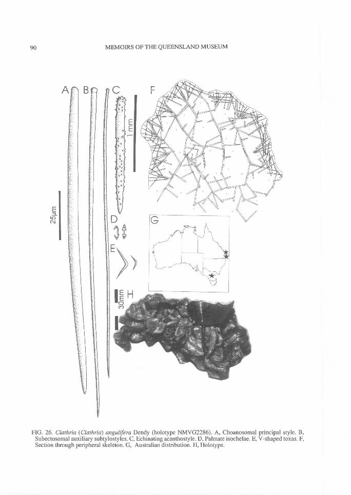
90^ MEMOIRS OF THE QUEENSLAND MUSEUM
FIG. 26. Clathria (Clathria) angulifera Dendy (holotype NMVG2286). A, Choanosomal principal style. B,Subectosomal auxiliary subtylostyles. C, Echinating acanthostyle. D, Palmate isochelae. E, V-shaped toxas. F,Section through peripheral skeleton. G, Australian distribution. H, Holotype.
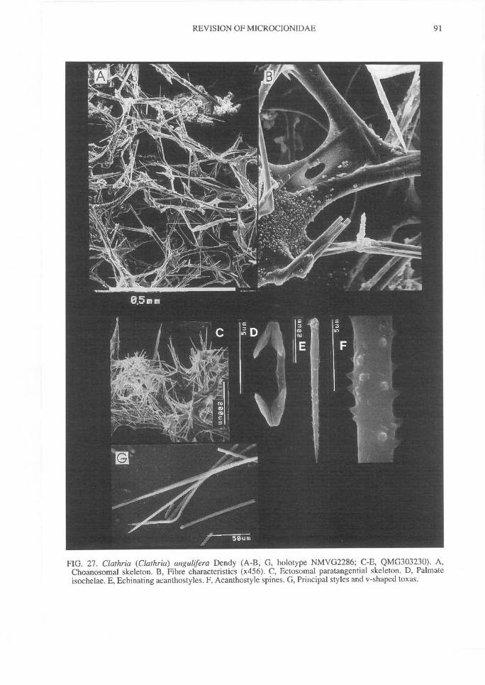
REVISION OF MICROCIONIDAE^
91
FIG. 27. Clathria (Clathria) angulifera Dendy (A-B, G, holotype NMVG2286; C-E, QMG303230). A,Choanosomal skeleton. B, Fibre characteristics (x456). C, Ectosomal paratangential skeleton. D, Palmateisochelae. E, Echinating acanthostyles. F, Acanthcstyle spines. G, Principal styles and v-shaped toxas.
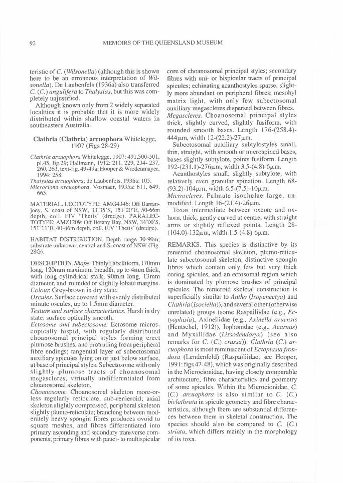
92^ MEMOIRS OF THE QUEENSLAND MUSEUM
teristic of C. (Wilsonella) (although this is shownhere to be an erroneous interpretation of Wil-sonella). De Laubenfels (1936a) also transferredC. (C.) angulifera to Thalysias, but this was com-pletely unjustified.
Although known only from 2 widely separatedlocalities it is probable that it is more widelydistributed within shallow coastal waters insoutheastern Australia.
Clathria (Clathria) arcuophora Whitelegge,1907 (Figs 28-29)
Clathria arcuophora Whitclegge, 1907: 491,500-501,p1.45, fig.29; Hallmann, 1912: 211, 229, 234- 237,260, 263, text-fig. 49-49a; Hooper & Wiedenmayer,1994: 258.
Thalysias arcuophora; de Laubenfels, 1936a: 105.Microciona arcuophora; Vosmaer, 1935a: 611, 649,
665.
MATERIAL. LECTOTYPE: AMG4346: Off Barran-joey, S. coast of NSW, 33 °35'S, 151 °20'E, 50-66mdepth, coll. FIV `Thetis' (dredge). PARALEC-TOTYPE: AMZ1209: Off Botany Bay, NSW, 34°00'S,151°11'E, 40-46m depth, coll. FIV `Thetis' (dredge).
HABITAT DISTRIBUTION. Depth range 30-90m;substrate unknown; central and S. coast of NSW (Fig.28G).
DESCRIPTION. Shape. Thinly flabelli form, 170mmlong, 120mm maximum breadth, up to 4mm thick,with long cylindrical stalk, 90mm long, 13mmdiameter, and rounded or slightly lobate margins.Colour Grey-brown in dry state.Oscules. Surface covered with evenly distributedminute oscules, up to 1.5mm diameter.Texture and surface characteristics. Harsh in drystate; surface optically smooth.Ectosome and subectosome. Ectosome micros-copically hispid, with regularly distributedchoanosomal principal styles forming erectplumose brushes, and protruding from peripheralfibre endings; tangential layer of subectosomalauxiliary spicules lying on or just below surface,at base of principal styles. Subectosome with onlyslightly plumose tracts of choanosomalmegascleres, virtually undifferentiated fromchoanosomal skeleton.Choanosome. Choanosomal skeleton more-or-less regularly reticulate, sub-renieroid; axialskeleton slightly compressed, peripheral skeletonslightly plumo-reticulate; branching between mod-erately heavy spongin fibres produces ovoid tosquare meshes, and fibres differentiated intoprimary ascending and secondary transverse com-ponents; primary fibres with pauci- to multispicular
core of choanosomal principal styles; secondaryfibres with uni- or bispicular tracts of principalspicules; echinating acanthostyles sparse, slight-ly more abundant on peripheral fibres; mesohylmatrix light, with only few subectosomalauxiliary megascleres dispersed between fibres.Megascleres. Choanosomal principal stylesthick, slightly curved, slightly fusiform, withrounded smooth bases. Length 1764258.4)-444p,m, width 12-(22.2)-27p.m.
Subectosomal auxiliary subtylostyles small,thin, straight, with smooth or microspined bases,bases slightly subtylote, points fusiform. Length1924231.1)-276pm, width 3.5-(4.8)-6pm.
Acanthostyles small, slightly subtylote, withrelatively even granular spination. Length 68-(93.2)-104pm, width 6.547.5)-10p.m.Microscleres. Palmate isochelae large, un-modified. Length 16-(21.4)-26p.m.
Toxas intermediate between oxeote and ox-horn, thick, gently curved at centre, with straightarms or slightly reflexed points. Length 28-(104.0)-132p.m, width 1.5-(4.8)-6pm.
REMARKS. This species is distinctive by itsrenieroid choanosomal skeleton, plumo-reticu-late subectosomal skeleton, distinctive sponginfibres which contain only few but very thickcoring spicules, and an ectosomal region whichis dominated by plumose brushes of principalspicules. The renieroid skeletal construction issuperficially similar to Antho (lsopenectya) andClathria(lsociella)), and several other (otherwiseunrelated) groups (some Raspailiidae (e.g., Ec-tyoplasia), Axinellidae (e.g., Axinella aruensis(Hentschel, 1912)), Iophonidae (e.g., Acarnus)and Myxillidae (Lissodendoryx) (see alsoremarks for C. (C.) crassa)). Clathria (C.) ar-cuophora is most reminiscent of Ectoplasia fron-dosa (Lendenfeld) (Raspailiidae; see Hooper,1991: figs 47-48), which was originally describedin the Microcionidae, having closely comparablearchitecture, fibre characteristics and geometryof some spicules. Within the Microcionidae, C.(C.) arcuophora is also similar to C. (C.)biclathrata in spicule geometry and fibre charac-teristics, although there are substantial differen-ces between them in skeletal construction. Thespecies should also be compared to C. (C.)striata, which differs mainly in the morphologyof its toxa.
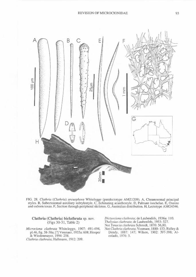
REVISION OF MICROCION1DAE^ 93
FIG. 28. Clathria (Clathria) arcuophora Whitelegge (paralectotype AMZI 209). A, Choanosomal principalstyles. B, Subectosomal auxiliary subtylostyle. C, Echinating acanthostyle. D, Palmate isochelae. E, Oxeoteand oxhorn toxas. F, Section through peripheral skeleton. G, Australian distribution. H, Lectotype AMG4346.
Clathria (Clathria) biclathrata sp. nov.(Figs 30-31, Table 2)
Microciona clathrata Whitelegge, 1907: 491-494,p1.46, fig. 38-38a; Vosmaer, 1935a: 608; Hooper& Wiedenmayer, 1994: 258.
Clathria clathrata; Hallmann, 1912: 209.
Dictyociona clathrata; de Laubenfels, 1936a: 110.Thalysias clathrata; de Laubenfels, 1953: 527.Not Tenacia clathrata Schmidt, 1870: 56,80.Not Clathria clathrata; Vosmaer, 1880: 153; Ridley &
Dendy, 1887: 147; Wilson, 1902: 397-398; Al-colado, 1976: 5.
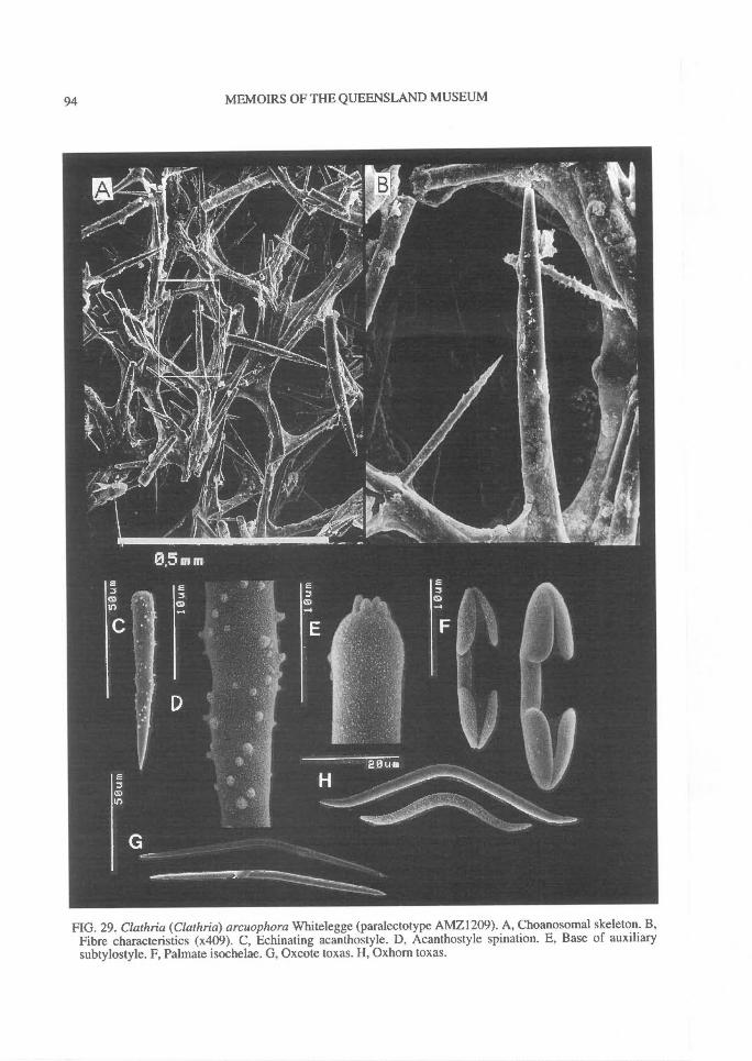
94^ MEMOIRS OF THE QUEENSLAND MUSEUM
FIG. 29. Clathria (Clathria) arcuophora Whitelegge (paralectotype AMZ1209). A, Choanosomal skeleton. B,Fibre characteristics (x409). C, Echinating acanthostyle. D, Acanthostyle spination. E, Base of auxiliarysubtylostyle. F, Palmate isochelae. G, Oxeote toxas. H, Oxhorn toxas.
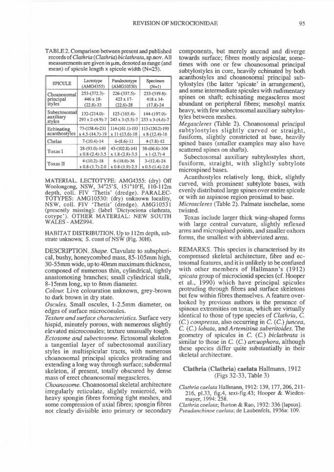
REVISION OF MICROCIONIDAE^ 95
TABLE 2. Comparison between present and publishedrecords of Clathria(Clathria)biclathrata, sp.nov. Allmeasurements are given in pm, denoted as range (andmean) of spicule length x spicule width (N=25).
SPICULE Lectotype(AMG4355)
Paralectotype(AMG10530)
Specimen(N=I)
Choanosomal 2534372 . 3)- 226(337.5)- 2334319.8)-principal 446 x 18- 423 x 17- 418 x 14-styles (22.8)-33 (22.6)-28 (17.8)-24Subectosomalauxiliarystyles
I 32-(214.0)-293 x 2-(4.9)-7
125-(165.4)-243 x 3-(5.5)-7
1444197.0)-253 x 3-(4.6)-7
Echinating 734158.4)-231 1144161.1)-193 1134150.2)-193acanthostyles x 4.5-(14.7)-I9 x 11-(13.6)-18 x 8-(12.4)-I6Chelae 7-(10.4)-I4 6-(8.6)-11 4-(7.8)-12
28-(93.0)-149 434102.8)-141 38-(66.6)-104Toxas I x 0.8-(2.4)-3.5 x I.8-(2.8)-3.5 x 1-(2.7)-4
4-(10.2)-I8 6-(I8.0)-36 3-(12.4)-24Toxas II x 0.841.7)-2.0
MATERIAL. LECTOTYPE: AMG4355: (dry) OffWoolongong, NSW, 34 °25'S, 151 ° 10'E, 110-112mdepth, coll. FIV 'Thetis' (dredge). PARALEC-TOTYPES: AMG10530: (dry) unknown locality,NSW, coll. FIV `Thetis' (dredge). AMG10531(presently missing): (label `Dictyociona clathrata,cotype'). OTHER MATERIAL: NEW SOUTHWALES - AMZ994.
HABITAT DISTRIBUTION. Up to 112m depth, sub-strate unknown; S. coast of NSW (Fig. 30H).
DESCRIPTION. Shape. Clavulate to subspheri-cal, bushy, honeycombed mass, 85-105mm high,30-55mm wide, up to 40mm maximum thickness,composed of numerous thin, cylindrical, tightlyanastomosing branches; small cylindrical stalk,8-15mm long, up to 8mm diameter.Colour Live colouration unknown, grey-brownto dark brown in dry state.Oscules. Small oscules, 1-2.5mm diameter, onedges of surface microconules.Texture and surface characteristics. Surface veryhispid, minutely porous, with numerous slightlyelevated microconules; texture unusually tough.Ectosome and subectosome. Ectosomal skeletona tangential layer of subectosomal auxiliarystyles in multispicular tracts, with numerouschoanosomal principal spicules protruding andextending a long way through surface; subdermalskeleton, if present, totally obscured by densemass of erect choanosomal megascleres.Choanosome. Choanosomal skeletal architectureirregularly reticulate, slightly renieroid, withheavy spongin fibres forming tight meshes, andsome compression of axial fibres; spongin fibresnot clearly divisible into primary or secondary
components, but merely ascend and divergetowards surface; fibres mostly aspicular, some-times with one or few choanosomal principalsubtylostyles in core, heavily echinated by bothacanthostyles and choanosomal principal sub-tylostyles (the latter `spicate' in arrangement),and some intermediate spicules with rudimentaryspines on shaft; echinating megascleres mostabundant on peripheral fibres; mesohyl matrixheavy, with few subectosomal auxiliary subtylos-tyles between meshes.Megascleres (Table 2). Choanosomal principalsubtylostyles slightly curved or straight,fusiform, slightly constricted at base, heavilyspined bases (smaller examples may also havescattered spines on shafts).
Subectosomal auxiliary subtylostyles short,fusiform, straight, with slightly subtylotemicrospined bases.
Acanthostyles relatively long, thick, slightlycurved, with prominent subtylote bases, withevenly distributed large spines over entire spiculeor with an aspinose region proximal to base.Microscleres (Table 2). Palmate isochelae, sometwisted.
Toxas include larger thick wing-shaped formswith large central curvature, slightly reflexedarms and microspined points, and smaller oxhornforms, the smallest with abbreviated arms.
REMARKS. This species is characterised by itscompressed skeletal architecture, fibre and ec-tosomal features, and it is unlikely to be confusedwith other members of Hallmann's (1912)spicata group of microcionid species (cf. Hooperet al., 1990) which have principal spiculesprotruding through fibres and surface skeletonsbut few within fibres themselves. A feature over-looked by previous authors is the presence ofspinous extremities on toxas, which are virtuallyidentical to those of type species of Clathria, C.(C.) compressa, also occurring in C. (C.) juncea,C. (C.) lobata, and Artemisina suberitoides. Thegeometry of spicules in C. (C.) biclathrata issimilar to those in C. (C.) arcuophora, althoughthese species differ quite substantially in theirskeletal architecture.
Clathria (Clathria) caelata Hallmann, 1912(Figs 32-33, Table 3)
Clathria caelata Hallmann, 1912: 139, 177, 206, 211-216, p1.33, fig.4, text-fig.43; Hooper & Wieden-mayer, 1994: 258.
Clathria coelata; Burton & Rao, 1932: 336 [lapsus].Pseudanchinoe caelata; de Laubenfels, 1936a: 109.
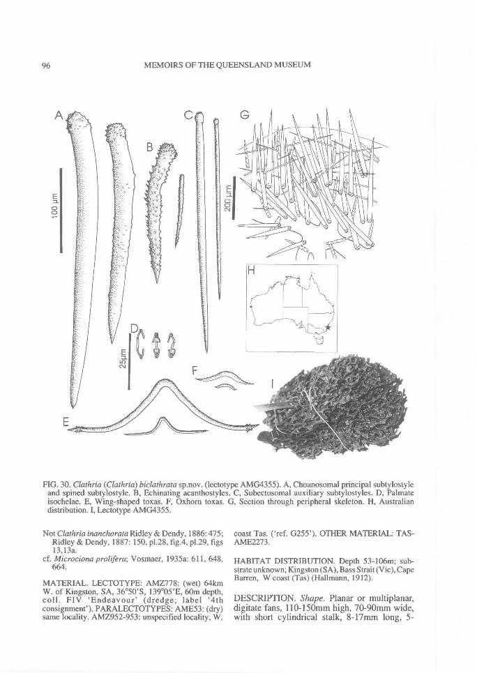
96^ MEMOIRS OF THE QUEENSLAND MUSEUM
FIG. 30. Clathria (Clathria) biclathrata sp.nov. (lectotype AMG4355). A, Choanosomal principal subtylostyleand spined subtylostyle. B, Echinating acanthostyles. C, Subectosomal auxiliary subtylostyles. D, Palmateisochelae. E, Wing-shaped toxas. F, Oxhorn toxas. G, Section through peripheral skeleton. H, Australiandistribution. I, Lectotype AMG4355.
Not Clathria inanchorata Ridley & Dendy, 1886:475;Ridley & Dendy, 1887: 150, p1.28, fig.4, p1.29, figs13,13a.
cf. Microciona prolifera; Vosmaer, 1935a: 611, 648,664.
MATERIAL. LECTOTYPE: AMZ778: (wet) 64kmW. of Kingston, SA, 36 °50'S, 139°05'E, 60m depth,coll. FIV 'Endeavour' (dredge; label `4thconsignment'). PARALECTOTYPES: AME53: (dry)same locality. AMZ952-953: unspecified locality, W.
coast Tas. ('ref. G255'). OTHER MATERIAL: TAS-AME2273.
HABITAT DISTRIBUTION. Depth 53-106m; sub-strate unknown; Kingston (SA), Bass Strait (Vic), CapeBarren, W coast (Tas) (Hallmann, 1912).
DESCRIPTION. Shape. Planar or multiplanar,digitate fans, 110-150mm high, 70-90mm wide,with short cylindrical stalk, 8-17mm long, 5-
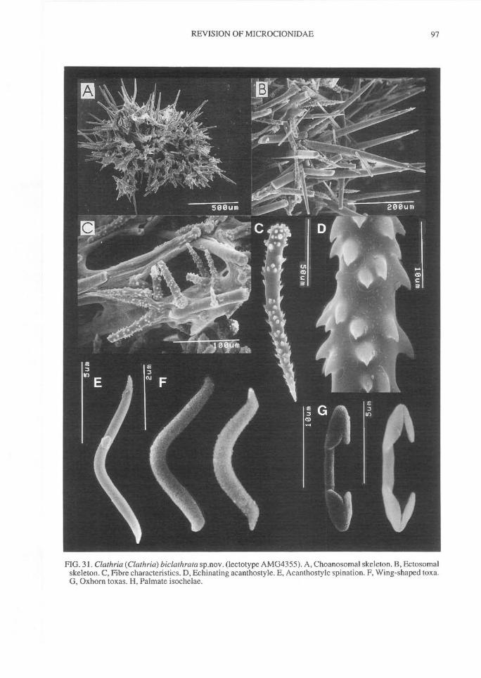
REVISION OF MICROCIONIDAE^ 97
FIG. 31. Clathria (Clathria) biclathrata sp.nov. (lectotype AMG4355). A, Choanosomal skeleton. B, Ectosomalskeleton. C, Fibre characteristics. D, Echinating acanthostyle. E, Acanthostyle spination. F, Wing-shaped toxa.G, Oxhorn toxas. H, Palmate isochelae.
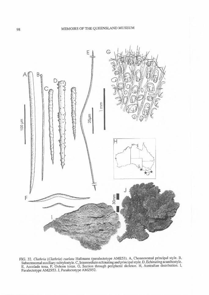
98^ MEMOIRS OF THE QUEENSLAND MUSEUM
FIG. 32. Clathria (Clathria) caelata Hallmann (paralectotype AME53). A, Choanosomal principal style. B,Subectosomal auxiliary subtylostyle. C, Intermediate echinating and principal style. D, Echinating acanthostyle.E, Accolada toxa. F, Oxhorn toxas. G, Section through peripheral skeleton. H, Australian distribution. I,Paralectotype AMZ953. J, Paralectotype AMZ952.
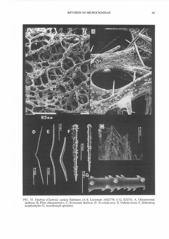
REVISION OF MICROCIONIDAE^
99
FIG. 33. Clathria (Clathria) caelata Hallmann (A-B, Lectotype AMZ778; C-G, E2273). A, Choanosomalskeleton. B, Fibre characteristics. C, Ectosomal skeleton. D, Accolada toxa. E, Oxhorn toxas, F, Echinatingacanthostyles. G, Acanthostyle spination.
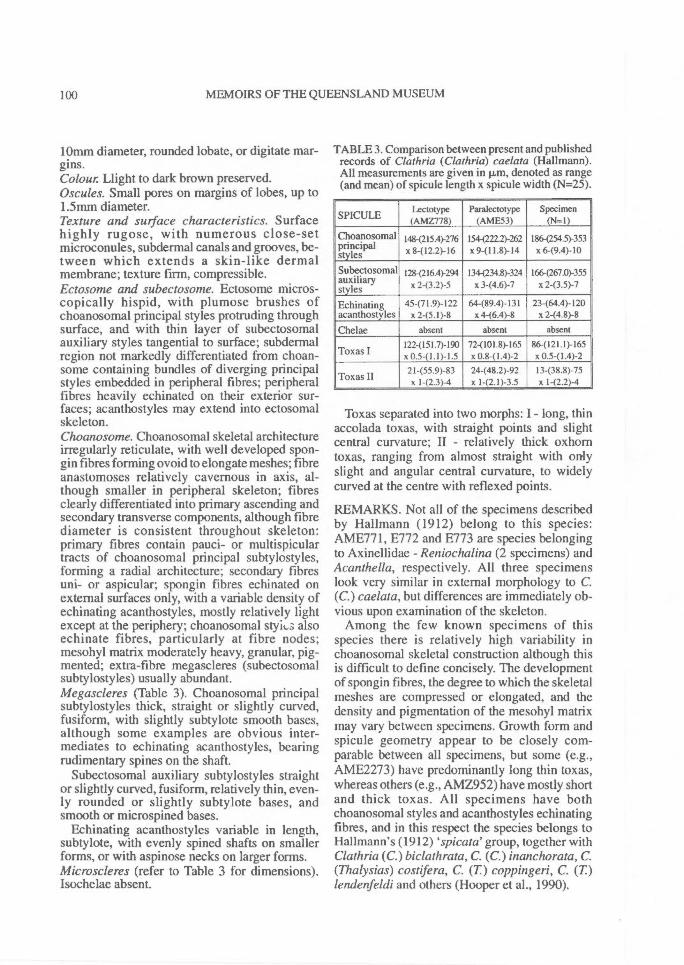
100^ MEMOIRS OF THE QUEENSLAND MUSEUM
lOmm diameter, rounded lobate, or digitate mar-gins.Colour Llight to dark brown preserved.Oscules. Small pores on margins of lobes, up to1.5mm diameter.Texture and surface characteristics. Surfacehighly rugose, with numerous close-setmicroconules, subdermal canals and grooves, be-tween which extends a skin-like dermalmembrane; texture firm, compressible.Ectosome and subectosome. Ectosome micros-copically hispid, with plumose brushes ofchoanosomal principal styles protruding throughsurface, and with thin layer of subectosomalauxiliary styles tangential to surface; subdermalregion not markedly differentiated from choan-some containing bundles of diverging principalstyles embedded in peripheral fibres; peripheralfibres heavily echinated on their exterior sur-faces; acanthostyles may extend into ectosomalskeleton.Choanosome. Choanosomal skeletal architectureirregularly reticulate, with well developed spon-gin fibres forming ovoid to elongate meshes; fibreanastomoses relatively cavernous in axis, al-though smaller in peripheral skeleton; fibresclearly differentiated into primary ascending andsecondary transverse components, although fibrediameter is consistent throughout skeleton:primary fibres contain pauci- or multispiculartracts of choanosomal principal subtylostyles,forming a radial architecture; secondary fibresuni- or aspicular; spongin fibres echinated onexternal surfaces only, with a variable density ofechinating acanthostyles, mostly relatively lightexcept at the periphery; choanosomal styks alsoechinate fibres, particularly at fibre nodes;mesohyl matrix moderately heavy, granular, pig-mented; extra-fibre megascleres (subectosomalsubtylostyles) usually abundant.Megascleres (Table 3). Choanosomal principalsubtylostyles thick, straight or slightly curved,fusiform, with slightly subtylote smooth bases,although some examples are obvious inter-mediates to echinating acanthostyles, bearingrudimentary spines on the shaft.
Subectosomal auxiliary subtylostyles straightor slightly curved, fusiform, relatively thin, even-ly rounded or slightly subtylote bases, andsmooth or microspined bases.
Echinating acanthostyles variable in length,subtylote, with evenly spined shafts on smallerforms, or with aspinose necks on larger forms.Microscleres (refer to Table 3 for dimensions).Isochelae absent.
TABLE 3. Comparison between present and publishedrecords of Clathria (Clathria) caelata (Hallmann).All measurements are given in m, denoted as range(and mean) of spicule length x spicule width (N=25)
SPICULELectotype(AMZ778)
Paralectotype(AME53)
Specimen(N=1)
Choanosomalprincipalstyles
1484215.4)-276x 8-(12.2)-16
1544222.2)-262x 9-(11.8)-14
186-(254.5)-353x 6-(9.4)-10
Subectosomalauxiliarystyles
128-(216.4)-294x 2-(3.2)-5
1344234.8)-324x 3-(4.6)-7
1664267.0)-355x 2-(3.5)-7
Echinating 45-(71.9)-122 64-(89.4)-131 23-(64.4)-120acanthostyles x 245.1)-8 x 4-(6.4)-8 x 2-(4.8)-8
Chelae absent absent absent1224151.7)-190 72-(101.8)-165 86-(121.1)-165
Toxas I x 0.5-(1.1)-1.5 x 0.8-(1.4)-2 x 0.5-(1.4)-22I-(55.9)-83 24-(48.2)-92 13-(38.8)-75
Toxas II x 1-(2.3)-4 x 1-(2.1)-3.5^, x 1-(2.2)-4
Toxas separated into two morphs: I - long, thinaccolada toxas, with straight points and slightcentral curvature; II - relatively thick oxhorntoxas, ranging from almost straight with onlyslight and angular central curvature, to widelycurved at the centre with reflexed points.
REMARKS. Not all of the specimens describedby Hallmann (1912) belong to this species:AME771, E772 and E773 are species belongingto Axinellidae - Reniochalina (2 specimens) andAcanthella, respectively. All three specimenslook very similar in external morphology to C.(C.) caelata, but differences are immediately ob-vious upon examination of the skeleton.
Among the few known specimens of thisspecies there is relatively high variability inchoanosomal skeletal construction although thisis difficult to define concisely. The developmentof spongin fibres, the degree to which the skeletalmeshes are compressed or elongated, and thedensity and pigmentation of the mesohyl matrixmay vary between specimens. Growth form andspicule geometry appear to be closely com-parable between all specimens, but some (e.g.,AME2273) have predominantly long thin toxas,whereas others (e.g., AMZ952) have mostly shortand thick toxas. All specimens have bothchoanosomal styles and acanthostyles echinatingfibres, and in this respect the species belongs toHallmann's (1912) spicata' group, together withClathria (C.) biclathrata, C. (C.) inanchorata, C.(Thalysias) costifera, C. (T) coppingeri, C. (T)lendenfeldi and others (Hooper et al., 1990).
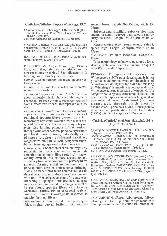
REVISION OF MICROCIONIDAE^ 101
Clathria (Clathria) calopora Whitelegge, 1907
Clathria calopora Whitelegge, 1907: 499-500, p1.46,fig.34; Hallmann, 1912: 211; Hooper & Wieden-mayer, 1994: 259.
Thalysias calopora; de Laubenfels, 1936a: 105.
MATERIAL. HOLOTYPE: AM (presently missing):Shoalhaven Bight, NSW, 34 °49'S, 151°04'E, 30-90mdepth, 1.vii.1911, coll. FIV 'Endeavour' (trawl).
HABITAT DISTRIBUTION. Depth 27-32m, sub-strate unknown; S. coast of NSW.
DESCRIPTION. Shape. Branching, 195mmhigh, with thin, bifurcate, cylindrical, mostlynon-anastomosing digits, 3-8mm diameter, withtapering points, short cylindrical stalk.Colour Live colouration unknown, greyish-yel-low preserved.Oscules. Small oscules, about lifun diameter,scattered over surface.Texture and surface characteristics. Surface un-even, minutely hispid, honeycomb-like, withprominent bulbous conulose processes scatteredover surface; texture hard, incompressible in drystate.Ectosome and subectosome. Ectosome micros-copically rugose, close-meshed reticulation ofperipheral spongin fibres covered by a thinmembrane; ectosomal skeleton with a thin tan-gential layer of subectosomal auxiliary subtylos-tyles, also forming plumose tufts on surface,through which choanosomal principal styles fromperipheral fibres protrude, individually or inplumose brushes; subdermal auxiliarymegascleres run parallel with peripheral fibres,but not forming organised extra-fibre tracts.Choanosome. Choanosomal skeleton irregularlyreticulate, with some axial and extra-axial dif-ferentiation; spongin fibres relatively heavy,clearly divided into primary ascending andsecondary transverse components; primary fibressinuous, forming radial architecture, with apaucispicular core of choanosomal principalstyles; primary fibres more compressed at axisthan at periphery; secondary fibres less common,with uni- or paucispicular core of megascleres;branching of spongin fibres produces elongatemeshes in axis and round or rectangular meshesin periphery; spongin fibres very heavilyechinated, particularly in peripheral regions;numerous sinuous toxodragmata dispersed inmesohyl between fibres.Megascleres. Choanosomal principal stylesthick, slightly curved, fusiform, with rounded
smooth bases. Length 300-500p,m, width 15-22m.
Subectosomal auxiliary subtylostyles thin,straight or slightly curved, with smooth slightlysubtylote bases. Length 150-200p,m, width 2-4p,m.
Acanthostyles short, stout, evenly spined,spines large. Length 30-80p,m, width up to12p.m.Microscleres. Palmate isochelae. Length 8-10t.m.
Toxa morphology unknown, apparently long,slender, with large central curvature. Length ?200-300p,m, width ? up to 21.1,m.
REMARKS. This species is known only fromWhitelegge's (1907) poor description. It is notpossible to determine whether dimensions ofechinating acanthostyle (cited as 0.3-0.8mm longby Whitelegge) is merely a typographical error.Whitelegge gave no indication of whether C. (C.)calopora has a special ectosomal skeleton, butdescribed the species as having a tangential orparatangential layer of subdermal (auxiliary)megascleres, through which protrudechoanosomal (principal) styles. Consequently,there was no justification for de Laubenfels(1936a) referring the species to Thalysias.
Clathria (Clathria) chelifera (Hentschel, 1911)(Figs 34-35, Table 4)
Spanioplon cheliferum Hentschel, 1911: 362-363,fig.42; Hentschel, 1912: 368-369.
Allocia chehfera; Hallmann, 1920: 768; Bergquist &Fromont, 1988: 96, fig. 8c, pis 45e-f, 46a-c, table73; Dawson, 1993: 44 (note).
Clathria chelifera; Dendy, 1922: 70-71, p1.14, fig.3a-e; Hooper & Wiedenmayer, 1994: 259.
Not Microciona chelifera Lëvi,1960a: 70, fig. 12.
MATERIAL. HOLOTYPE: ZMH (not seen) (frag-ment ZMB4440): precise locality unknown, Perthregion, WA, 1905, coll. W. Michaelsen & R.Hartmeyer (dredge). PARATYPE: SMF 1571 (frag-ment MNHNDCL2327): same locality. OTHERMATERIAL: VIETNAM - PIBOC-05-216 (fragmentQM G300058).
HABITAT DISTRIBUTION. 10-100m depth; rock orgravel substrates; Arafura Sea (NT) and Perth region(S. WA) (Fig. 34F). Also Indian Ocean (Amirante),New Zealand (Three Kings Is) and South China Sea(Hon Trung Lon, Vinh Loi coast, S. Vietnam).
DESCRIPTION. Shape. Arborescent, foliose,planar growth form, up to 50mm high made up offused porous-reticulate lamellae 10-15mm thick.
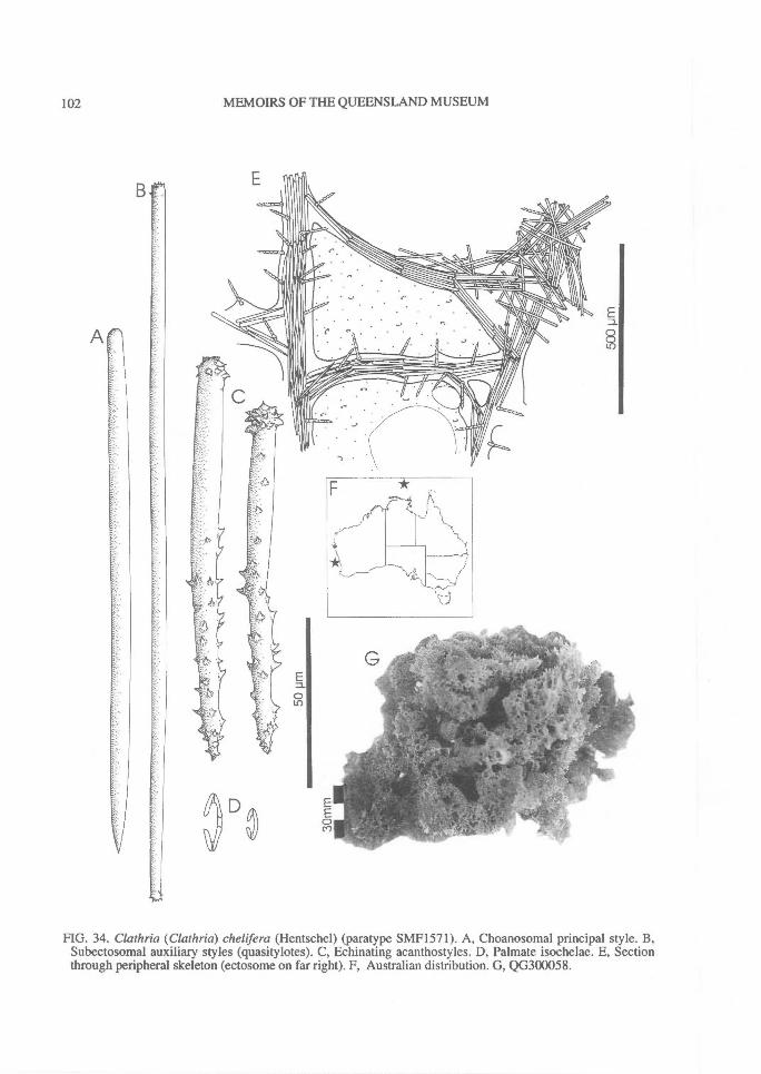
102^ MEMOIRS OF THE QUEENSLAND MUSEUM
FIG. 34. Clathria (Clathria) chelifera (Hentschel) (paratype SMF1571). A, Choanosomal principal style. B,Subectosomal auxiliary styles (quasitylotes). C, Echinating acanthostyles. D, Palmate isochelae. E, Sectionthrough peripheral skeleton (ectosome on far right). F, Australian distribution. G, QG300058.
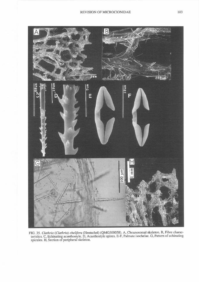
REVISION OF MICROCIONIDAE^
103
FIG. 35. Clathria (Clathria) chelifera (Hentschel) (QMG300058). A, Choanosomal skeleton. B, Fibre charac-teristics. C, Echinating acanthostyle. D, Acanthostyle spines. E-F, Palmate isochelae. G, Pattern of echinatingspicules. H, Section of peripheral skeleton.
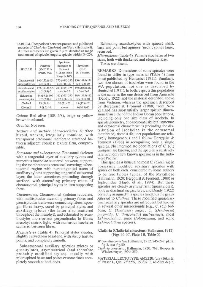
104^ MEMOIRS OF THE QUEENSLAND MUSEUM
TABLE 4. Comparison between present and publishedrecords of Clathria (Clathria)chelifera (Hentschel).All measurements are given in 1.1.m, denoted as range(and mean) of spicule length x spicule width (N=25).
SPICULEParatype
(SMF1571)(Perth, WA)
SpecimenBergquist &
Fromont(1988) (ThreeKings Is, NZ)
Specimen(N=1)
(S. Vietnam)
Choanosomal 1404150.1)-161 2704404)-550 150-(164.6)-174principal styles x 4-(6.1)-7 x 15-(18)-20 x 4-(6.4)-10Subectosomal 1734193.4)-205 280-(334)-375 1924204.0)-212auxiliary styles x 2-(3.9)-5 x 4-(5)-6.5 x 3-(4.7)-7
Echinating 84-(93.2)-100 162-(207)-290 10341165)-132acanthostyles x 7-(7.8)-9 x 12.5-(14)-19 x 5-(8.0)-10
Chelae I 13-(14.6)-1 20421)-23 15-(17.6)-20Chelae H 7-(8.7)-10 absent 9-(10.3)-12
Colour Red alive (10R 5/8), beige or yellowbrown in ethanol.Oscules. Not seen.Texture and stuface characteristics. Surfacehispid, uneven, irregularly conulose, withtransparent ectosomal membrane stretched be-tween adjacent conules; texture firm, compres-sible.Ectosome and subectosome. Ectosomal skeletonwith a tangential layer of auxiliary tylotes andnumerous isochelae scattered between, support-ing the membraneous ectosomal covering; subec-tosomal region with paratangential tracts ofauxiliary tylotes supporting tangential ectosomallayer, the latter sometimes protruding throughsurface, with ascending primary tracts ofchoanosomal principal styles in turn supportingthese.Choanosome. Choanosomal skeleton reticulate,with multispicular ascending primary fibres andpaucispicular transverse connecting fibres; spon-gin fibres heavy, cored by principal styles andauxiliary tylotes (the latter also scatteredthroughout the mesohyl), and echinated by acan-thostyles more-or-less perpendicular to fibres;mesohyl matrix light, with numerous isochelaescattered between fibres.Megascleres (Table 4). Principal styles slender,slightly curved near basal end, with abrupt hastatepoints, and completely smooth.
Subectosomal auxiliary spicules tylotes orquasitylotes, asymmetrical (and thereforeprobably modified styles), usually withmicrospined bases and points or sometimes com-pletely smooth at both ends.
Echinating acanthostyles with spinose shaft,base and point but apinose 'neck'; spines large,recurved.Microscleres (Table 4). Palmate isochelae of twosizes, both with thickened and elongate alae.
Toxas are absent.
REMARKS. Dimensions of some spicules werefound to differ in type material (Table 4) fromthose published by Hentschel (1911). Similarly,two size classes of isochelae were found in theWA population, not one as described byHentschel (1911). In both respects this populationis the same as the one described from Amirante(Dendy, 1922) and the material described abovefrom Vietnam, whereas the specimen describedby Bergquist & Fromont (1988) from NewZealand has substantially larger spicule dimen-sions than either of the Indian Ocean populations,including only one size class of isochela. Inspicule geometry, choanosomal skeletal structureand ectosomal characteristics (including the dis-tribution of isochelae in the ectosomalmembrane), these 4 disjunct populations are rela-tively homogeneous and I follow Bergquist &Fromont (1988) in recognising only a singlespecies. No intermediate populations of C. (C.)chelifera are known, and the species is relativelyrare with only few known specimens in the Indo-west Pacific.
This species is unusual to most C. (Clathria) inpossessing modified auxiliary spicules withspines on both ends, considered by some authorsto be true tylotes typical of the Myxillidae(Hall mann, 1920; Bergquist & Fromont, 1988) orIophonidae (Hajdu et al., 1994). But thesespicules are clearly asymmetrical (quasitylotes),not true diactinal megascleres, and Dendy (1922)correctly assigned this species (and thus the genusAllocia) to Clathria. These modified quasidiac-tinal auxiliary spicules are infrequent but knownin several other microcionids (e.g., C. (C.) bul-bosa, C. (Thalysias) major, C. (Dendrocia)pyramida, C. (Wilsonella) australiensis, mostEchinochalina, some Holopsamma, and someEchinoclathria species).
Clathria (Clathria) conectens (Hallmann, 1912)(Figs 36-37, Plate 1B, Table 5)
Wilsonella conectens Hallmann, 1912: 245-247, p1.32,fig.2, text-fig.50.
Clathria conectens; Hallmann, 1920: 768; Hooper &Wiedenmayer, 1994: 259.
MATERIAL. LECTOTYPE: AMZ220: (dry) 16km E.of Fraser I., Qld, 25°22'S, 153°07'E, 48-52m depth,
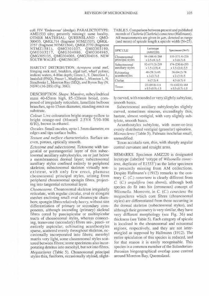
REVISION OF MICROCIONIDAE^
105
coll. FIV 'Endeavour' (dredge). PARALECTOTYPE:AME1533 (dry; presently missing): same locality.OTHER MATERIAL: QUEENSLAND - QMG-300455, QMGL714 (fragment NTMZ1537), QMGL-2757 (fragment NTMZ1564), QMGL2770 (fragmentNTMZ1581), QMG301037, QMG303190,QMG303217, QMG304980, QMG304985,QMG305135, QMG304005, QMG304016. NEWSOUTH WALES - QMG301387.
HABITAT DISTRIBUTION. Acropora coral reef,fringing rock reef, boulders, algal turf, wharf pylons;inshore waters, 4-80m depth; Green I., S. Direction I.,Innisfail (FNQ), Fraser I., Mudjimba I., Moreton I., N.Stradbroke I., Moreton Bay (SEQ), and Byron Bay (N.NSW) (16-28S) (Fig. 36G).
DESCRIPTION. Shape. Massive, subcylindricalmass 40-65mm high, 85-120mm broad, com-posed of irregularly reticulate, lamellate bulbousbranches, up to 15mm diameter, standing erect onsubstrate.Colour Live colouration bright orange-yellow tobright orange-red (Munsell 2.5YR 7/10-10R6/10), brown in ethanol.Oscules. Small oscules, up to 1.5mm diameter, onedges and tips surface bulbs.Texture and surface characteristics. Surface un-even, porous, optically smooth.Ectosome and subectosome. Ectosome with tan-gential or paratangential layer of thin subec-tosomal auxiliary subtylostyles, on or just belowa membraneous dermal layer; subectosomalauxiliary styles confined entirely to peripheralskeleton; subectosomal skeleton virtually non-existent, with only few erect, plumosechoanosomal principal styles, arising fromperipheral choanosomal spongin fibres, project-ing into tangential ectosomal layer.Choanosome. Choanosomal skeleton irregularlyreticulate, with regular circular, oval or elongatemeshes enclosing small oval choanocyte cham-bers; spongin fibres relatively heavy, without sizedifferentiation of primary or secondary com-ponents, although ascending (primary) skeletalfibres cored by paucispicular or multispiculartracts of choanosomal styles, whereas connect-ing, transverse (secondary) fibres uni-, pauci- orentirely aspicular; echinating acanthostylessparse, scattered evenly throughout skeleton, oc-casionally incorporated into fibres; mesohylmatrix very light, some choanosomal styles scat-tered between fibres; some specimens also incor-porating detritus into mesohyl, but not into fibres.Megascleres (Table 5). Choanosomal principalstyles thin, fusiform, occasionally styloid, slight-
TABLES. Comparison between present and publishedrecords of Clathria (Clathria) conectens (Hallmann).All measurements are given in p,m, denoted as range(and mean) of sp'cule length x spicule width (N=25).
SPICULELectotype(AMZ220) Specimens (N=7)
Choanosomal 944168.4)-268 1104171.4)-218principal styles x 2.5-(4.1)-5 x 3-(4.3)-6
Subectosomal 92-(171.3)-219 174-(230.6)-295auxiliary styles x 1.542.9)-4 x 1.5-(2.4)-3.5
Echinating 48-(58.3)-65 36-(64.2)-78acanthostyles x 3-(3.7)-5 x 2-(3.9)-5
Chelae 6-(7.2)-8 4.546.7)-922-(69.8)-111 51-(102.6)-164
Toxas x 0.5-(0.9)-1.5 x 0.5-(0.7)-1.0
ly curved, with rounded or very slightly subtylote,smooth bases.
Subectosomal auxiliary subtylostyles slightlycurved, sometimes sinuous, exceedingly thin,hastate, almost vestigial, with very slightly sub-tylote, smooth bases.
Acanthostyles subtylote, with more-or-lessevenly distributed vestigial (granular) spination.Microscleres (Table 5). Palmate isochelae small,unmodified.
Toxas accolada rare, thin, with sharply angularcentral curvature and straight arms.
REMARKS. Specimen AMZ220 is designatedlectotype (labelled `cotype of Wilsonella conec-tens, duplicate of E1533') as the latter specimenis presently missing from AM collections.Despite Hallmann's (1912) remarks to the con-trary C. (C.) conectens is clearly different fromC. (C.) angulifera (see above), although bothspecies do fit into his (erroneous) concept ofWilsonella. Moreover, in C. (C.) conectens themegascleres which core fibres (choanosomalstyles) are differentiated from those occurring inthe dermal skeleton (subectosomal styles), andalthough their geometry is very similar, they havevery different morphology (see Fig. 36) andthickness (see Table 5). Each category of spiculeis localised in the choanosomal and ectosomalregions, respectively, and they are not inter-mingled as supposed by Hallmann (1912). Theentire spiculation of this species is reduced, andfor that reason it is easily recognisable. Thisspecies is a common member of the Solanderian-Peronian biogeographical overlap zone centredaround Moreton Bay, Queensland.
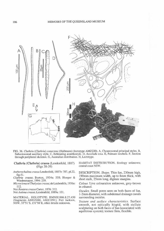
A
106^ MEMOIRS OF THE QUEENSLAND MUSEUM
FIG. 36. Clathria (Clathria) conectens (Hallmann) (lectotype AMZ220). A, Choanosomal principal styles. B,Subectosomal auxiliary style. C, Echinating acanthostyle. D, Accolada toxa. E, Palmate isochela. F, Sectionthrough peripheral skeleton. G, Australian distribution. H, Lectotype.
Clathria (Clathria) crassa (Lendenfeld, 1887)(Figs 38-39)
Antherochalina crassa Lendenfeld, 1887b: 787, p1.22,fig.41.
Clathria crassa; Burton, 1934a: 558; Hooper &Wiedenmayer, 1994: 259.
Microciona or Thalysias crassa; de Laubenfels, 1936a:112.
Not Reniera crassa Carter, 1876: 312.Not Aulena crassa; Lendenfeld, 1889a: 101.
MATERIAL. HOLOTYPE: BMNH1886.8.27.450(fragments AMG3460, AMZ1991): Port Jackson,NSW, 33 °51'S, 151 ° 16'E, other details unknown.
HABITAT DISTRIBUTION. Ecology unknown;central coast NSW.
DESCRIPTION. Shape. Thin fan, 230mm high,190mm maximum width, up to 8mm thick, withshort stalk, 25mm long, digitate margins.Colour Live colouration unknown, grey-brownin ethanol.Oscules. Small pores seen on both faces of fan,1-2mm diameter, with subdermal drainage canalssurrounding oscules.Texture and surface characteristics. Surfacesmooth, not optically hispid, with stellatesculpturing on both faces of fan (associated withaquiferous system); texture firm, flexible.
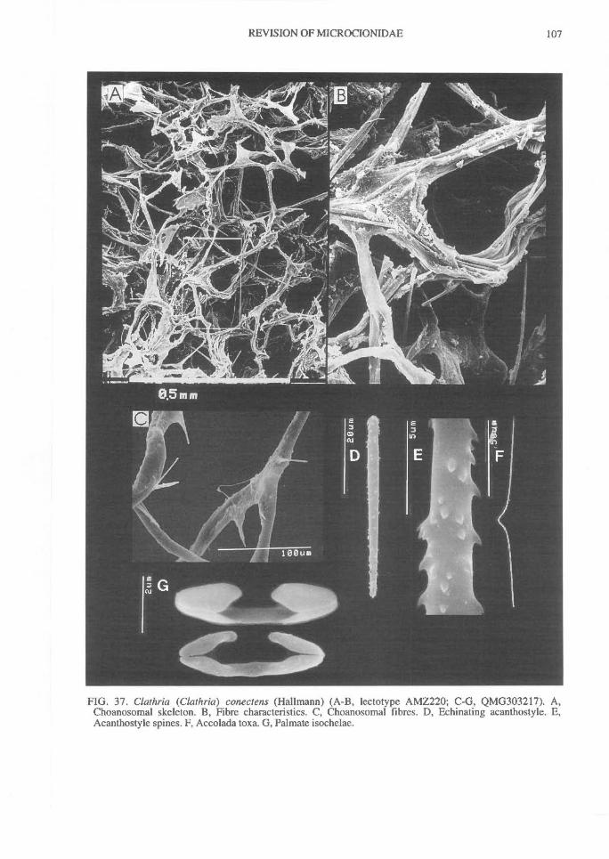
REVISION OF MICROCIONIDAE^
107
FIG. 37. Clathria (Clathria) conectens (Hallmann) (A-B, lectotype AMZ220; C-G, QMG303217). A,Choanosomal skeleton. B, Fibre characteristics. C, Choanosomal fibres. D, Echinating acanthostyle. E,Acanthostyle spines. F, Accolada toxa. G, Palmate isochelae.
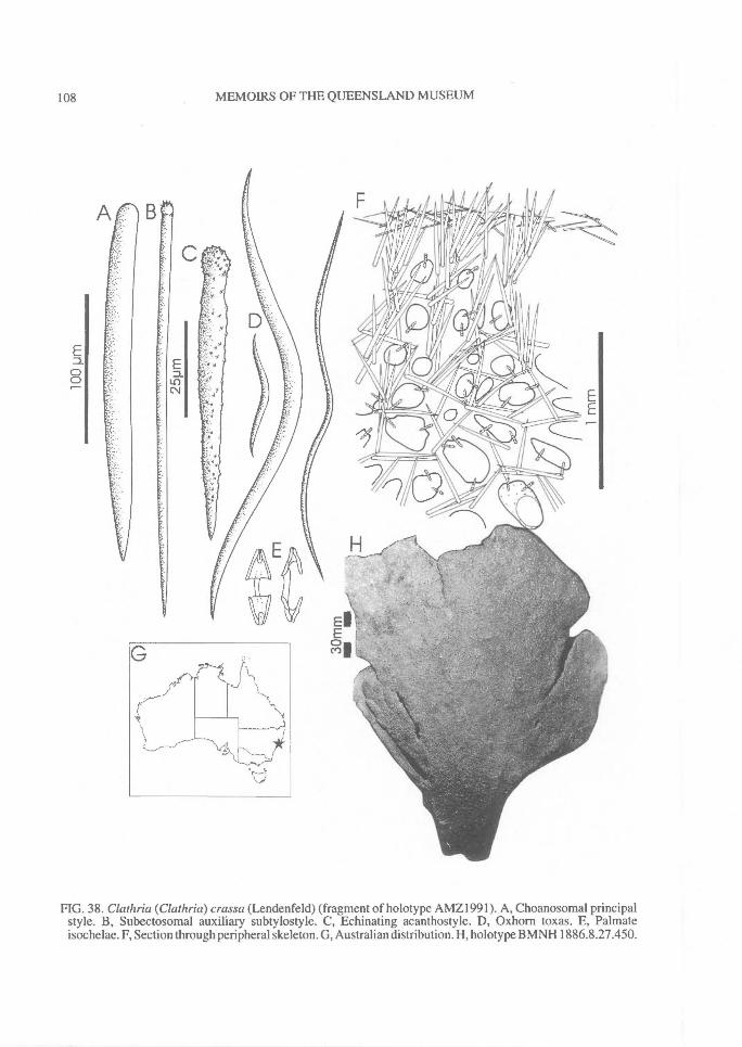
00
108^ MEMOIRS OF THE QUEENSLAND MUSEUM
FIG. 38. Clathria (Clathria) crassa (Lendenfeld) (fragment of holotype AMZ I 991). A, Choanosomal principalstyle. B, Subectosomal auxiliary subtylostyle. C, Echinating acanthostyle. D, Oxhorn toxas. E, Palmateisochelae. F, Section through peripheral skeleton. G, Australian distribution. H, holotype BMNH 1886.8.27.450.
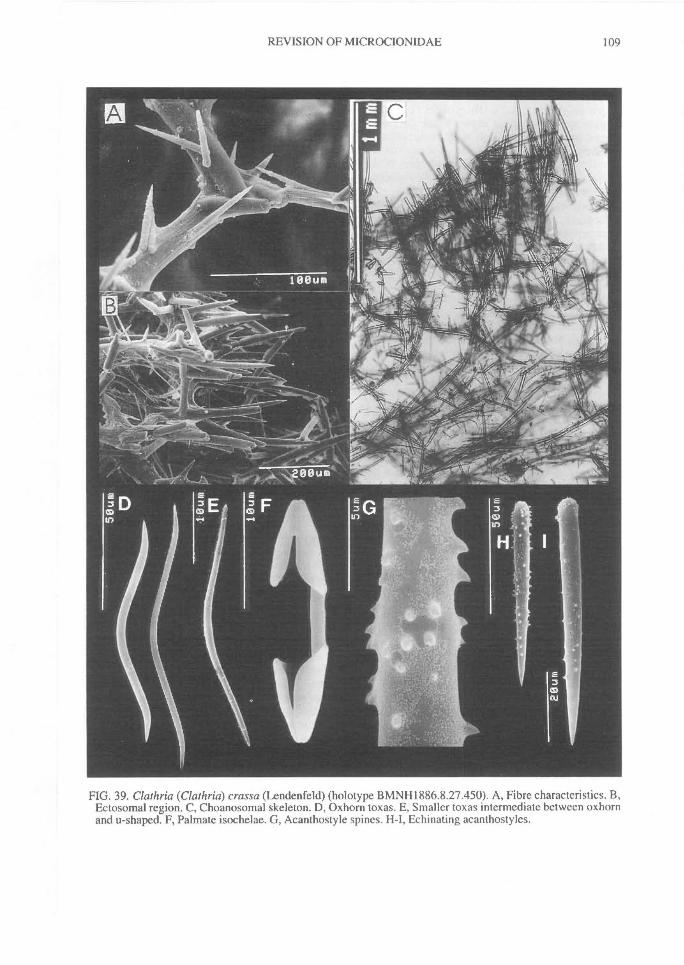
REVISION OF M1CROCIONIDAE^ 109
FIG. 39. Clathria (Clathria) crassa (Lendenfeld) (holotype BMNH1886.8.27.450). A, Fibre characteristics. B,Ectosomal region. C, Choanosomal skeleton. D, Oxhorn toxas. E, Smaller toxas intermediate between oxhornand u-shaped. F, Palmate isochelae. G, Acanthostyle spines. H-1, Echinating acanthostyles.
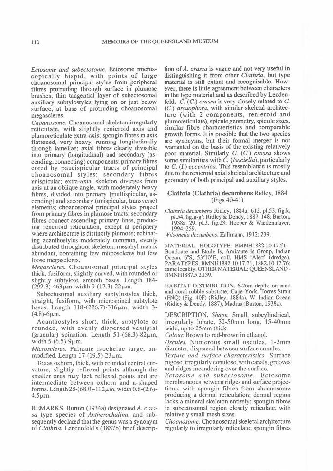
110^ MEMOIRS OF THE QUEENSLAND MUSEUM
Ectosome and subectosome. Ectosome micros-copically hispid, with points of largechoanosomal principal styles from peripheralfibres protruding through surface in plumosebrushes; thin tangential layer of subectosomalauxiliary subtylostyles lying on or just belowsurface, at base of protruding choanosomalmegascleres.Choanosome. Choanosomal skeleton irregularlyreticulate, with slightly renieroid axis andplumoreticulate extra-axis; spongin fibres in axisflattened, very heavy, running longitudinallythrough lamellae; axial fibres clearly divisibleinto primary (longitudinal) and secondary (as-cending, connecting) components; primary fibrescored by paucispicular tracts of principalchoanosomal styles; secondary fibresunispicular; extra-axial skeleton diverges fromaxis at an oblique angle, with moderately heavyfibres, divided into primary (multispicular, as-cending) and secondary (unispicular, transverse)elements; choanosomal principal styles projectfrom primary fibres in plumose tracts; secondaryfibres connect ascending primary lines, produc-ing reneiroid reticulation, except at peripherywhere architecture is distinctly plumose; echinat-ing acanthostyles moderately common, evenlydistributed throughout skeleton; mesohyl matrixabundant, containing few tnicroscleres but fewloose megascleres.Megascleres. Choanosomal principal stylesthick, fusiform, slightly curved, with rounded orslightly subtylote, smooth bases. Length 184-(292.3)-463p,m, width 9-(17.3)-22p,m.
Subectosomal auxiliary subtylostyles thick,straight, fusiform, with microspined subtylotebases. Length 118-(226.7)-316p,m, width 3-(4.8)-6 pun.
Acanthostyles short, thick, subtylote orrounded, with evenly dispersed vestigial(granular) spination. Length 51-(66.3)-82Rm,width 5-(6.5)-9p.m.Microscleres. Palmate isochelae large, un-modified. Length 17-(l9.5)-23p,m.
Toxas oxhorn, thick, with rounded central cur-vature, slightly reflexed points although thesmaller ones may lack reflexed points and areintermediate between oxhorn and u-shapedforms. Length 28-(68.0)-112p,m, width 0.842.6)-4.511M.
REMARKS. Burton (1934a) designated A. cras-sa type species of Antherochalina, and sub-sequently declared that the genus was a synonymof Clathria. Lendenfeld's (1887b) brief descrip-
tion of A. crassa is vague and not very useful indistinguishing it from other Clathria, but typematerial is still extant and recognisable. How-ever, there is little agreement between charactersin the type material and as described by Lenden-feld, C. (C.) crassa is very closely related to C.(C.) arcuophora, with similar skeletal architec-ture (with 2 components, renieroid andplumoreticulate), spicule geometry, spicule sizes,similar fibre characteristics and comparablegrowth forms. It is possible that the two speciesare synonyms, but their formal merger is notwarranted on the basis of the existing relativelypoor material. Similarly C. (C.) crassa showssome similarities with C. (Isociella), particularlyto C. (I.) eccentrica. This resemblance is mostlydue to the renieroid axial skeletal architecture andgeometry of both principal and auxiliary styles.
Clathria (Clathria) decumbens Ridley, 1884(Figs 40-41)
Cluihria decumbens Ridley, 1884a: 612, p1.53, fig.k,p1.54, fig.g-g'; Ridley & Dendy, 1887: 148; Burton,1938a: 29, p1.3, fig.23; Hooper & Wiedenmayer,1994: 259.
Wilsonella decumbens; Hallmann, 1912: 239.
MATERIAL. HOLOTYPE: BMNH1882.10.17.51:Boudouse and Etoile Is, Amirante Is Group, IndianOcean, 6°S, 53°10'E, coll. HMS 'Alert' (dredge).PARATYPES: BMNH1882.10.17.71, 1882.10.17.76:same locality. OTHER MATERIAL: QUEENSLAND -BMNH1887.5.2.139.
HABITAT DISTRIBUTION. 6-26m depth; on sandand coral rubble substrate; Cape York, Torres Strait(FNQ) (Fig. 40F) (Ridley, 1884a). W. Indian Ocean(Ridley & Dendy, 1887), Madras (Burton, 1938a).
DESCRIPTION. Shape. Small, subcylindrical,irregularly lobate, 32-50mm long, 15-40mmwide, up to 25mm thick.Colour Brown to red-brown in ethanol.Oscules. Numerous small oscules, 1-2mmdiameter, dispersed between surface conules.Texture and surface characteristics. Surfacerugose, irregularly conulose, with canals, groovesand ridges meandering over the surface.Ectosome and subectosome. Ectosomemembraneous between ridges and surface projec-tions, with spongin fibres from choanosomeproducing a dermal reticulation; dermal regionlacks a mineral skeleton entirely; spongin fibresin subectosomal region closely reticulate, withrelatively small mesh sizes.Choanosome. Choanosomal skeletal architectureregularly to irregularly reticulate; spongin fibres
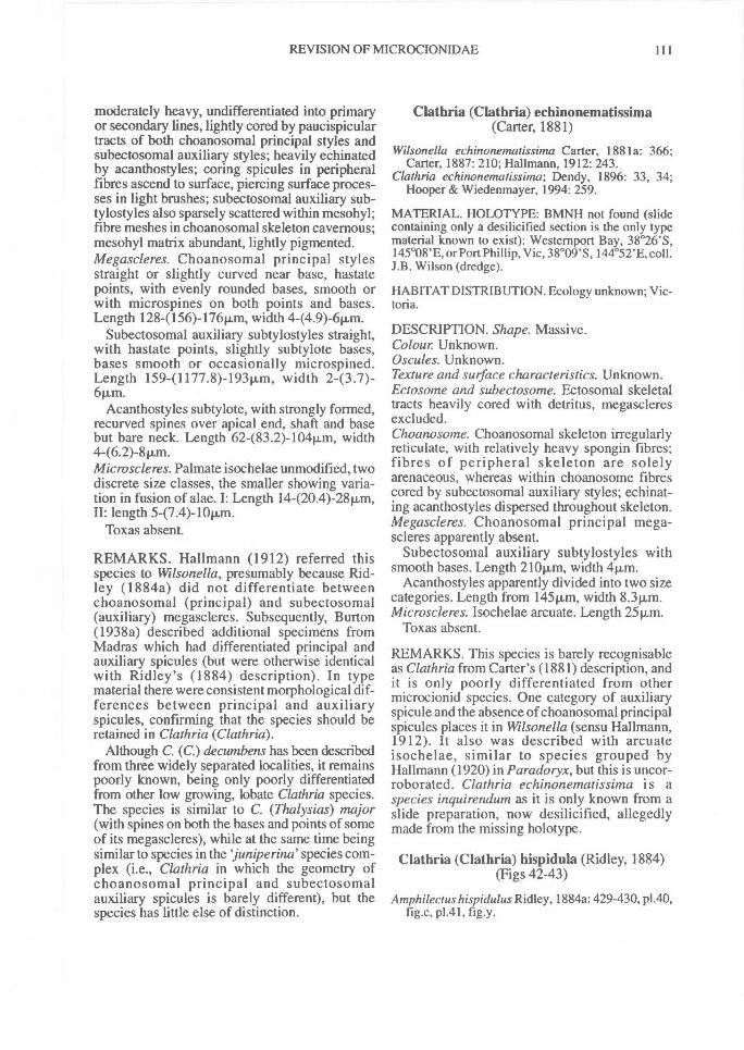
REVISION OF MICROCIONIDAE^ 111
moderately heavy, undifferentiated into primaryor secondary lines, lightly cored by paucispiculartracts of both choanosomal principal styles andsubectosomal auxiliary styles; heavily echinatedby acanthostyles; coring spicules in peripheralfibres ascend to surface, piercing surface proces-ses in light brushes; subectosomal auxiliary sub-tylostyles also sparsely scattered within mesohyl;fibre meshes in choanosomal skeleton cavernous;mesohyl matrix abundant, lightly pigmented.Megascleres. Choanosomal principal stylesstraight or slightly curved near base, hastatepoints, with evenly rounded bases, smooth orwith microspines on both points and bases.Length 128-(156)-176Rm, width 4-(4.9)-6p.m.
Subectosomal auxiliary subtylostyles straight,with hastate points, slightly subtylote bases,bases smooth or occasionally microspined.Length 159-(1177.8)-193Rm, width 243.7)-6 Km.
Acanthostyles subtylote, with strongly formed,recurved spines over apical end, shaft and basebut bare neck. Length 62-(83.2)-104Rm, width
Microscleres. Palmate isochelae unmodified, twodiscrete size classes, the smaller showing varia-tion in fusion of alae. I: Length 14-(20.4)-28Km,II: length 5-(7.4)-10Km.
Toxas absent.
REMARKS. Hallmann (1912) referred thisspecies to Wilsonella, presumably because Rid-ley (1884a) did not differentiate betweenchoanosomal (principal) and subectosomal(auxiliary) megascleres. Subsequently, Burton(1938a) described additional specimens fromMadras which had differentiated principal andauxiliary spicules (but were otherwise identicalwith Ridley's (1884) description). In typematerial there were consistent morphological dif-ferences between principal and auxiliaryspicules, confirming that the species should beretained in Clathria (Clathria).
Although C. (C.) decumbens has been describedfrom three widely separated localities, it remainspoorly known, being only poorly differentiatedfrom other low growing, lobate Clathria species.The species is similar to C. (Thalysias) major(with spines on both the bases and points of someof its megascleres), while at the same time beingsimilar to species in the juniperind species com-plex (i.e., Clathria in which the geometry ofchoanosomal principal and subectosomalauxiliary spicules is barely different), but thespecies has little else of distinction.
Clathria (Clathria) echinonematissima(Carter, 1881)
Wilsonella echinonematissima Carter, 1881a: 366;Carter, 1887: 210; Hallmann, 1912: 243.
Clathria echinonematissima; Dendy, 1896: 33, 34;Hooper & Wiedenmayer, 1994: 259.
MATERIAL. HOLOTYPE: BMNH not found (slidecontaining only a desilicified section is the only typematerial known to exist): Westernport Bay, 38 °26'S,145°08'E, or Port Phillip, Vic, 38°09'S, 144°52'E, coll.J.B. Wilson (dredge).
HABITAT DISTRIBUTION. Ecology unknown; Vic-toria.
DESCRIPTION. Shape. Massive.Colour. Unknown.Oscules. Unknown.Texture and surface characteristics. Unknown.Ectosome and subectosome. Ectosomal skeletaltracts heavily cored with detritus, megascleresexcluded.Choanosome. Choanosomal skeleton irregularlyreticulate, with relatively heavy spongin fibres;fibres of peripheral skeleton are solelyarenaceous, whereas within choanosome fibrescored by subectosomal auxiliary styles; echinat-ing acanthostyles dispersed throughout skeleton.Megascleres. Choanosomal principal mega-scleres apparently absent.
Subectosomal auxiliary subtylostyles withsmooth bases. Length 21011m, width 4t.t.m.
Acanthostyles apparently divided into two sizecategories. Length from 145Km, width 8.311m.Microscleres. Isochelae arcuate. Length 251.1,m.
Toxas absent.
REMARKS. This species is barely recognisableas Clathria from Carter's (1881) description, andit is only poorly differentiated from othermicrocionid species. One category of auxiliaryspicule and the absence of choanosomal principalspicules places it in Wilsonella (sensu Hallmann,1912). It also was described with arcuateisochelae, similar to species grouped byHallmann (1920) in Paradoryx, but this is uncor-roborated. Clathria echinonematissima is aspecies inquirendum as it is only known from aslide preparation, now desilicified, allegedlymade from the missing holotype.
Clathria (Clathria) hispidula (Ridley, 1884)(Figs 42-43)
Amphdectus hispidulus Ridley, 1884a: 429-430, p1.40,fig.c, p1.41, fig.y.
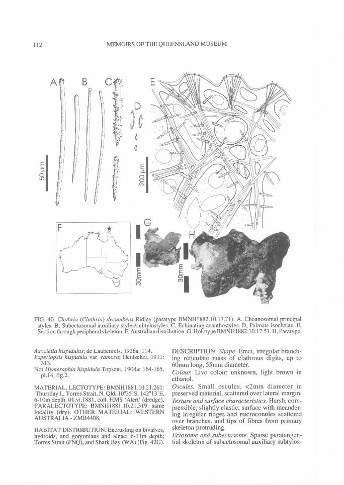
112^ MEMOIRS OF THE QUEENSLAND MUSEUM
FIG. 40. Clathria (Clathria) decumbens Ridley (paratype BMNH1882.10.17.71). A, Choanosomal principalstyles. B, Subectosomal auxiliary styles/subtylostyles. C, Echinating acanthostyles. D, Palmate isochelae. E,Section through peripheral skeleton. F, Australian distribution. G, Holotype BMNH1882.10.17.51. H, Paratype.
Axociella hispidulus; de Laubenfels, 1936a: 114.Esperiopsis hispidula var. ramosa; Hentschel, 1911:
313.Not Hymeraphia hispidula Topsent, 1904a: 164-165,
p1.14, fig.2.
MATERIAL. LECTOTYPE: BMNHI881.10.21.261:Thursday I., Torres Strait, N. Old, 10 °35'S, 142° 13'E,6-10m depth. 01.vi.1881, coll. HMS 'Alert' (dredge).PARALECTOTYPE: BMNH1881.10.21.319: samelocality (dry). OTHER MATERIAL: WESTERNAUSTRALIA - ZMB4408.
HABITAT DISTRIBUTION. Encrusting on bivalves,hydroids, and gorgonians and algae; 6-11rn depth;Torres Strait (FNQ), and Shark Bay (WA) (Fig. 42G).
DESCRIPTION. Shape. Erect, irregular branch-ing reticulate mass of clathrous digits, up to60mm long, 55mm diameter.Colour. Live colour unknown, light brown inethanol.Oscules. Small oscules, <2mm diameter inpreserved material, scattered over lateral margin.Texture and surface characteristics. Harsh, com-pressible, slightly elastic; surface with meander-ing irregular ridges and microconules scatteredover branches, and tips of fibres from primaryskeleton protruding.Ectosome and subectosome. Sparse paratangen-tial skeleton of subectosomal auxiliary subtylos-
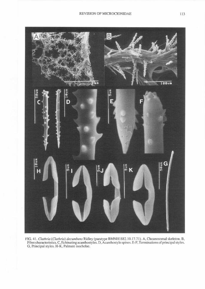
REVISION OF MICROCIONIDAE^ 113
FIG. 41. Clathria (Clathria) decumbens Ridley (paratype BMNH1882.10.17.71). A, Choanosomal skeleton. B,Fibre characteristics. C, Echinating acanthostyles. D, Acanthostyle spines. E-F, Terminations of principal styles.G, Principal styles. H-K, Palmate isochelae.
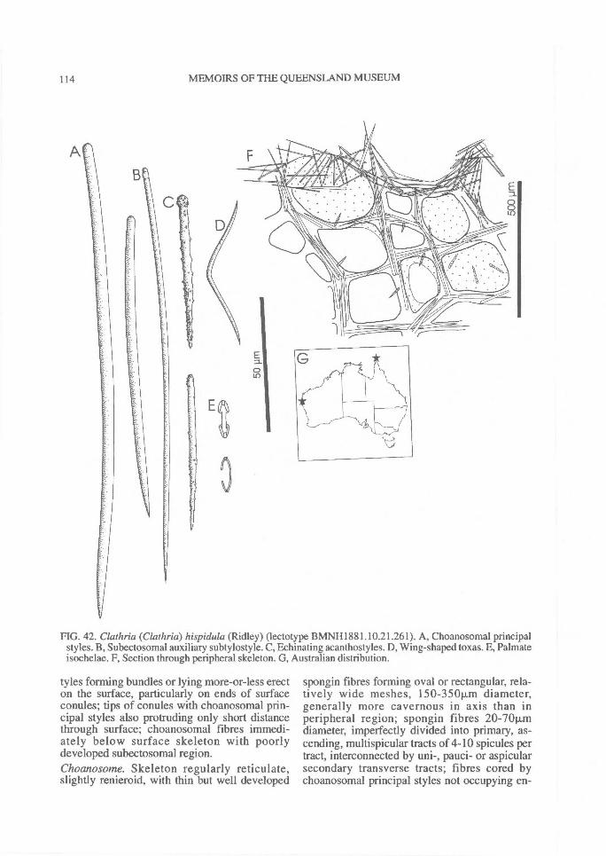
114^ MEMOIRS OF THE QUEENSLAND MUSEUM
FIG. 42. Clathria (Clathria) hispidula (Ridley) (lectotype BMNH1881.10.21.261). A, Choanosomal principalstyles. B, Subectosomal auxiliary subtylostyle. C, Echinating acanthostyles. D, Wing-shaped toxas. E, Palmateisochelae. F, Section through peripheral skeleton. G, Australian distribution.
tyles forming bundles or lying more-or-less erecton the surface, particularly on ends of surfaceconules; tips of conules with choanosomal prin-cipal styles also protruding only short distancethrough surface; choanosomal fibres immedi-ately below surface skeleton with poorlydeveloped subectosomal region.Choanosome. Skeleton regularly reticulate,slightly renieroid, with thin but well developed
spongin fibres forming oval or rectangular, rela-tively wide meshes, 150-350Rm diameter,generally more cavernous in axis than inperipheral region; spongin fibres 20-701J,mdiameter, imperfectly divided into primary, as-cending, multispicular tracts of 4-10 spicules pertract, interconnected by uni-, pauci- or aspicularsecondary transverse tracts; fibres cored bychoanosomal principal styles not occupying en-

REVISION OF MICROCIONIDAE^
115
FIG. 43. Clathria (Clathria) hispidula (Ridley) (lectotype BMNH1881.10.21.261). A, Choanosomal skeleton.B, Fibre characteristics. C, Echinating acanthostyles. D, Acanthostyle spination. E, Terminations of auxiliaryspicules. F, Palmate isochelae. G, Wing-shaped toxa.
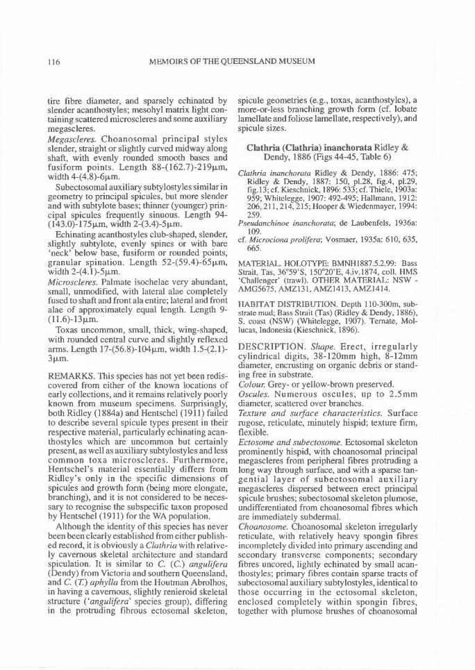
116^ MEMOIRS OF THE QUEENSLAND MUSEUM
tire fibre diameter, and sparsely echinated byslender acanthostyles; mesohyl matrix light con-taining scattered microscleres and some auxiliarymegascleres.Megascleres. Choanosomal principal stylesslender, straight or slightly curved midway alongshaft, with evenly rounded smooth bases andfusiform points. Length 884162.7)-21911m,width 4-(4.8)-611m.
Subectosomal auxiliary subtylostyles similar ingeometry to principal spicules, but more slenderand with subtylote bases; thinner (younger) prin-cipal spicules frequently sinuous. Length 94-(143.0)-175pAn, width 2-(3.4)-5p.m.
Echinating acanthostyles club-shaped, slender,slightly subtylote, evenly spines or with bare'neck' below base, fusiform or rounded points,granular spination. Length 52-(59.4)-65Rm,width 2-(4.1)-5p.m.Microscleres. Palmate isochelae very abundant,small, unmodified, with lateral alae completelyfused to shaft and front ala entire; lateral and frontalae of approximately equal length. Length 9-(11.6)-13p,m.
Toxas uncommon, small, thick, wing-shaped,with rounded central curve and slightly reflexedarms. Length 17-(56.8)-104Rm, width 1.5-(2.1)-3p.m.
REMARKS. This species has not yet been redis-covered from either of the known locations ofearly collections, and it remains relatively poorlyknown from museum specimens. Surprisingly,both Ridley (1884a) and Hentschel (1911) failedto describe several spicule types present in theirrespective material, particularly echinating acan-thostyles which are uncommon but certainlypresent, as well as auxiliary subtylostyles and lesscommon toxa microscleres. Furthermore,Hentschel's material essentially differs fromRidley's only in the specific dimensions ofspicules and growth form (being more elongate,branching), and it is not considered to be neces-sary to recognise the subspecific taxon proposedby Hentschel (1911) for the WA population.
Although the identity of this species has neverbeen been clearly established from either publish-ed record, it is obviously a Clathria with relative-ly cavernous skeletal architecture and standardspiculation. It is similar to C. (C.) angulifera(Dendy) from Victoria and southern Queensland,and C. (T) aphylla from the Houtman Abrolhos,in having a cavernous, slightly renieroid skeletalstructure ('angulifera' species group), differingin the protruding fibrous ectosomal skeleton,
spicule geometries (e.g., toxas, acanthostyles), amore-or-less branching growth form (cf. lobatelamellate and foliose lamellate, respectively), andspicule sizes.
Clathria (Clathria) inanchorata Ridley &Dendy, 1886 (Figs 44-45, Table 6)
Clathria inanchorata Ridley & Dendy, 1886: 475;Ridley & Dendy, 1887: 150, p1.28, fig.4, p1.29,fig. 13; cf. Kieschnick, 1896: 533; cf. Thiele, 1903a:959; Whitelegge, 1907: 492-495; Hallmann, 1912:206, 211, 214, 215; Hooper & Wiedenmayer, 1994:259.
Pseudanchinoe inanchorata; de Laubenfels, 1936a:109.
cf. Microciona prolifera; Vosmaer, 1935a: 610, 635,665.
MATERIAL. HOLOTYPE: BMNH1887.5.2.99: BassStrait, Tas, 36°59'S, 150°20'E, 4.iv.1874, coll. HMS'Challenger' (trawl). OTHER MATERIAL: NSW -AMG5675, AMZ131, AMZ1413, AMZ1414.
HABITAT DISTRIBUTION. Depth 110-300m, sub-strate mud; Bass Strait (Tas) (Ridley & Dendy, 1886),S. coast (NSW) (Whitelegge, 1907). Ternate, Mol-lucas, Indonesia (Kieschnick, 1896).
DESCRIPTION. Shape. Erect, irregularlycylindrical digits, 38-120mm high, 8-12mmdiameter, encrusting on organic debris or stand-ing free in substrate.Colour Grey- or yellow-brown preserved.Oscules. Numerous oscules, up to 2.5mmdiameter, scattered over branches.Texture and surface characteristics. Surfacerugose, reticulate, minutely hispid; texture firm,flexible.Ectosome and subectosome. Ectosomal skeletonprominently hispid, with choanosomal principalmegascleres from peripheral fibres protruding along way through surface, and with a sparse tan-gential layer of subectosomal auxiliarymegascleres dispersed between erect principalspicule brushes; subectosomal skeleton plumose,undifferentiated from choanosomal fibres whichare immediately subdermal.Choanosome. Choanosomal skeleton irregularlyreticulate, with relatively heavy spongin fibresincompletely divided into primary ascending andsecondary transverse components; secondaryfibres uncored, lightly echinated by small acan-thostyles; primary fibres contain sparse tracts ofsubectosomal auxiliary subtylostyles, identical tothose occurring in the ectosomal skeleton,enclosed completely within spongin fibres,together with plumose brushes of choanosomal
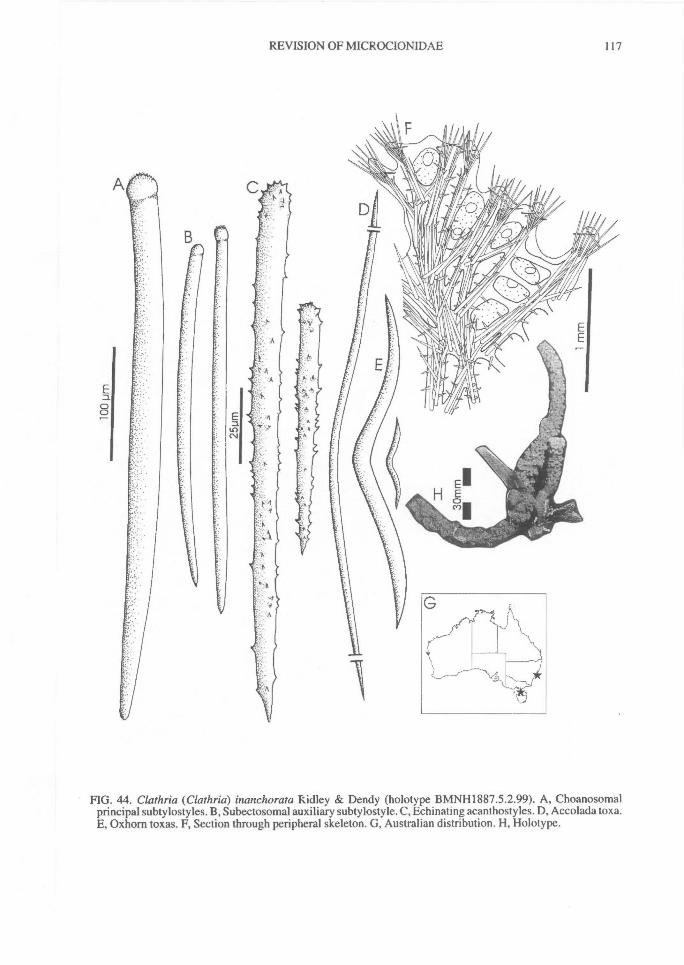
REVISION OF MICROCIONIDAE^
117
FIG. 44. Clathria (Clathria) inanchorata Ridley & Dendy (holotype BMNH1887.5.2.99). A, Choanosomalprincipal subtylostyles. B, Subectosomal auxiliary subtylostyle. C, Echinating acanthostyles. D, Accolada toxa.E, Oxhorn toxas. F, Section through peripheral skeleton. G, Australian distribution. H, Holotype.
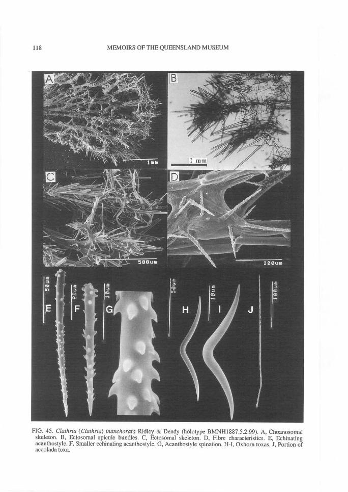
118^ MEMOIRS OF THE QUEENSLAND MUSEUM
FIG. 45. Clathria (Clathria) inanchorata Ridley & Dendy (holotype BMNH1887.5.2.99). A, Choanosomalskeleton. B, Ectosomal spicule bundles. C, Ectosomal skeleton. D, Fibre characteristics. E, Echinatingacanthostyle. F, Smaller echinating acanthostyle. G, Acanthostyle spination. H-I, Ozhorn toxas. J, Portion ofaccolada toxa.
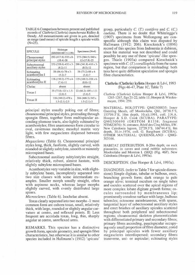
REVISION OF MICROCIONIDAE^ 119
TABLE 6.Comparison between present and publishedrecords of Clathria (Clathria) inanchorata Ridley &Dendy. All measurements are given in pm, denotedas range (and mean) of spicule length x spicule width(N=25).
SPICULEHolotype
(BMNH1887.5.2.99)Specimens (N=4)
Choanosomal 292-(417.6)-535 x 8- 273-(384.5)-540xprincipal styles (18.4)-28 12-(17.8)-25
Subectosomal 2524328.6)-432 x 3- 290-(341.8)-410 x 3-auxiliary styles (8.2)-11 (8.5)-12
Echinating 58-(66.4)-78 x 3- 58-(75.2)-88 x 4-acanthostyles I (5.2)-7 (5.4)-7
Echinating 1184150.2)-175 x 4- 119-(169.3)-228 x 6-acanthostyles II (7.4)-11 (8.8)-12
Chelae absent absent
35-(73.8)-121 x 1.5- 22-(66.2)-105 x 1.5-Toxas I (2.0)-3 (3.4)-5
118-(349.2)-478 x 3044408.3)-545 xToxas II 1.5-(2.1)-2.5 1.5-(2.1)-3
principal styles usually poking out of fibres;choanosomal principal styles, protruding throughspongin fibres, together form multispicular as-cending plumose tracts, also lightly echinated byacanthostyles; fibre anastomoses form circular tooval, cavernous meshes; mesohyl matrix verylight, with few megascleres dispersed betweenfibres.Megascleres (Table 6). Choanosomal principalstyles long, thick, fusiform, slightly curved, withrounded or slightly subtylote, smooth or minutelymicrospined bases.
Subectosomal auxiliary subtylostyles straight,relatively thick, robust, almost hastate, withslightly subtylote microspined bases.
Acanthostyles very variable in size, with slight-ly subtylote bases, incompletely separated intotwo size classes with some intermediate ex-amples. Smaller morph usually straight, oftenwith aspinose necks, whereas larger morphsslightly curved, with evenly distributed largespines.Microscleres. (Table 6). Isochelae absent.
Toxas clearly separated into two morphs - I: mostcommon form are oxhom toxas, small, relativelythick, with large, rounded or slightly angular cur-vature at centre, and reflexed points. II: Lessfrequent are accolada toxas, long, thin, sharplyangular at centre, unreflexed arms.
REMARKS. This species has a distinctivegrowth form, spicule geometry, and spongin fibrecharacteristics, but otherwise it is similar to otherspecies included in Hallmann's (1912) spicata'
group, particularly C. (T) costifera and C. (C.)caelata. There is no doubt that Whitelegge's(1907) specimens from Wollongong are con-specific although this claim was disputed byHallmann (1912: 206). Kiescknick's (1896)record of this species from Indonesia is dubious,since his material was not described and couldpossibly be any one of these spicata' -like spon-ges. Thiele (1903a) compared Kieschnick'sspecimen with C. (T)coralliophila from the sameregion, but that comparison is misleading: bothtaxa have quite different spiculation and sponginfibre characteristics.
Clathria (Clathria) kylista Hooper & Levi, 1993(Figs 46-47, Plate IC, Table 7)
Clathria (Clathria) kylista Hooper & Levi, 1993a:1265-1267, figs 21-22, table 11; Hooper & Wieden-mayer, 1994: 259.
MATERIAL. HOLOTYPE: QMG300035: InnerGneering Shoals, off Mooloolaba, Qld., 26°38.5'S,153°09.5'E, 10m depth, 10.xii.I 991, coll. J.N.A.Hooper & S.D. Cook (SCUBA). PARATYPE:QMG300690 (ORSTOM R1338; fragmentNTMZ3876): N. entrance, Recif des Cinq Milles, SW.New Caledonia lagoon, 22°29.3'S, 166°44.4'E, 8mdepth, 30.iv.1976, coll. G. Bargibant (SCUBA).OTHER MATERIAL: QUEENSLAND - QMG-303166.
HABITAT DISTRIBUTION. 8-20m depth; on rockpinnacles, in caves and coral rubble substrates;Mooloolaba and Moreton I. (SEQ) (Fig. 46F). NewCaledonia (Hooper & Levi, 1993a).
DESCRIPTION. (See Hooper & Levi, 1993a).
DIAGNOSIS. (refer to Table 7 for spicule dimen-sions) Simple digitate, tubular or bulbous, erect,branching growth form; dark orange to paleorange alive; terminal osculum on single tubesand oscules scattered over the apical regions ofmore complex lobate digitate growth forms; os-cules surrounded by membraneous lip;prominently conulose surface with large, bulboustubercles; ectosome membraneous, with sparse,tangential layer of subectosomal auxiliary stylesand erect brushes of auxiliary spicules scatteredthroughout both peripheral and subectosomalregions; choanosomal skeleton plumoreticulatewith differentiated primary and secondary fibres;primary fibres ascending, paucispicular occupy-ing only small proportion of fibre diameter, coredby principal spicules with fewer auxiliarymegascleres interdispersed; secondary fibrestransverse, uni- or aspicular; echinating styles
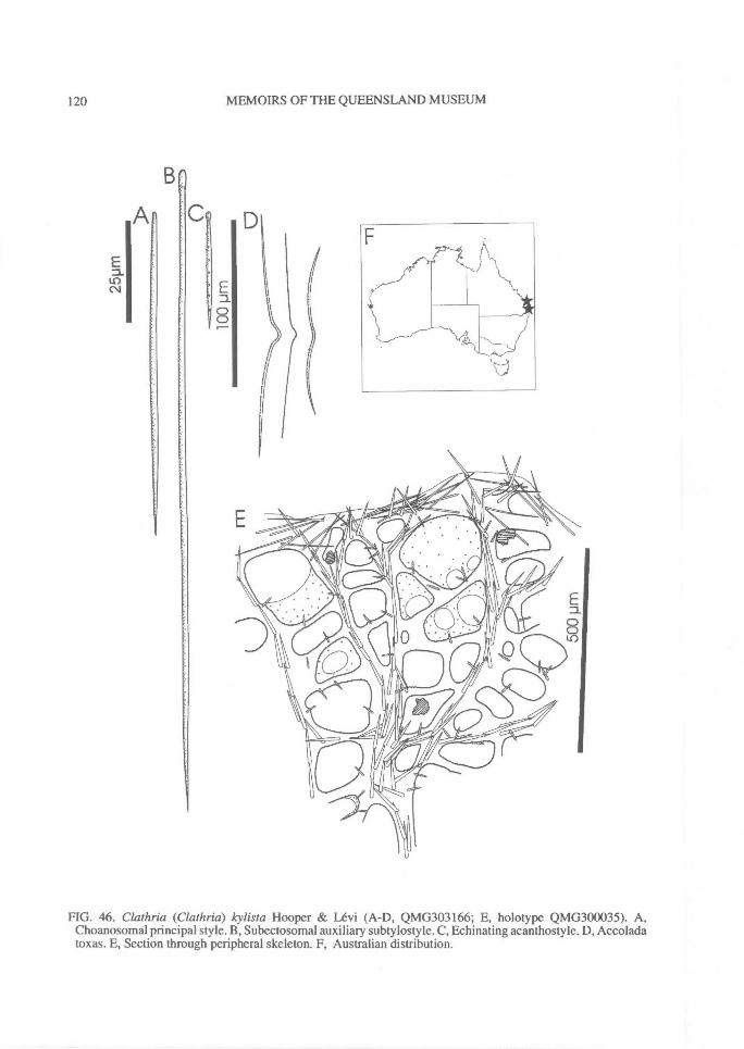
C
Ea00
120^ MEMOIRS OF THE QUEENSLAND MUSEUM
B
A
E1.0('.1
FIG. 46. Clathria (Clathria) kylista Hooper & Levi (A-D, QMG303166; E, holotype QMG300035). A,Choanosomal principal style. B, Subectosomal auxiliary subtylostyle. C, Echinating acanthostyle. D, Accoladatoxas. E, Section through peripheral skeleton. F, Australian distribution.
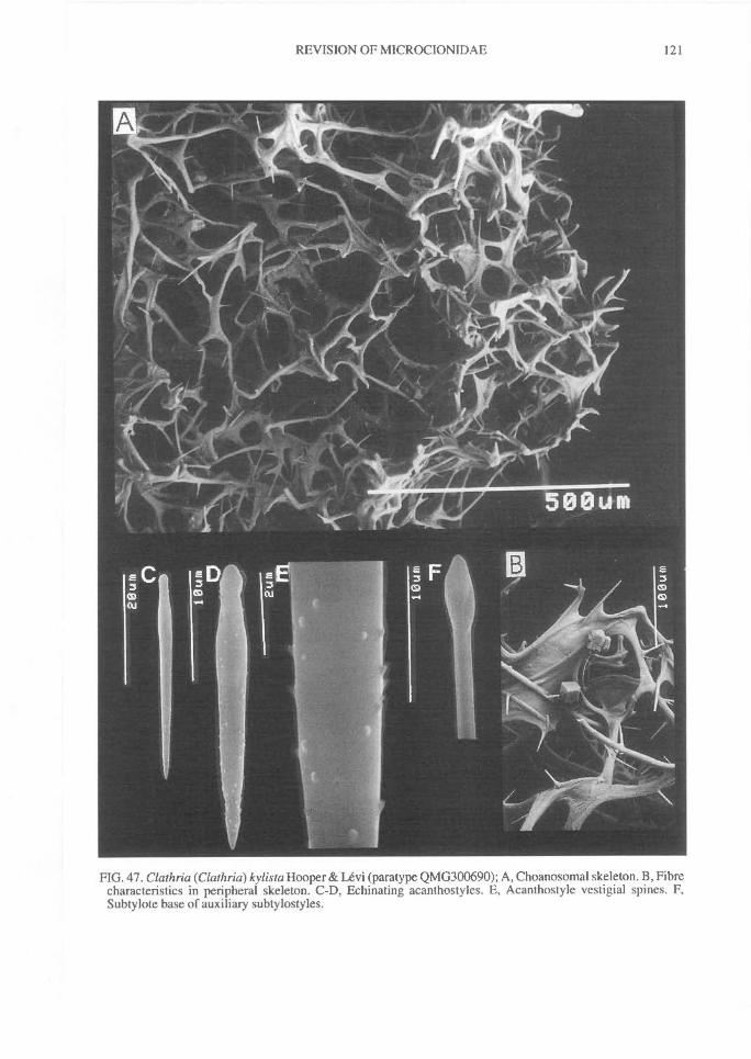
REVISION OF MICROCIONIDAE^
121
FIG. 47. Clathria (Clathria) kylista Hooper & Levi (paratype QMG300690); A, Choanosomal skeleton. B, Fibrecharacteristics in peripheral skeleton. C-D, Echinating acanthostyles. E, Acanthostyle vestigial spines. F,Subtylote base of auxiliary subtylostyles.
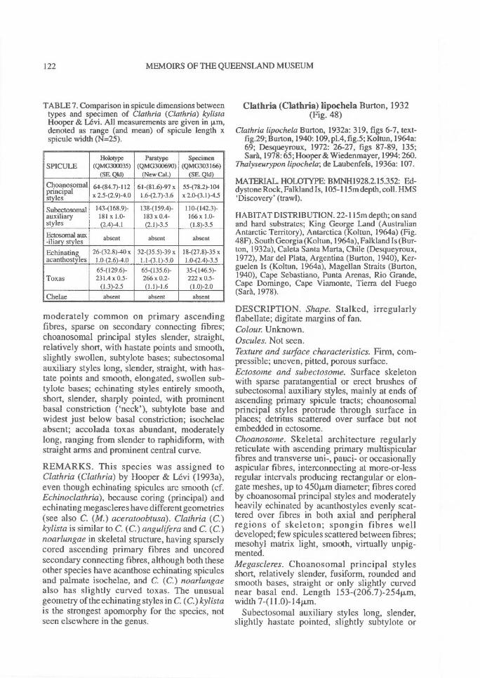
122^ MEMOIRS OF THE QUEENSLAND MUSEUM
TABLE 7. Comparison in spicule dimensions betweentypes and specimen of Clathria (Clathria) kylistaHooper & Levi. All measurements are given in p.m,denoted as range (and mean) of spicule length xspicule width (N=25).
SPICULEliolotype
(QMG300035)(SE. Qld)
Paratype
(QMG300690)(New Cal.)
Specimen
(QMG303166)(SE. Qld)
Choanosomalprincipalstyles
64484.7)-112x 2.5-(2.9)-4.0
61-(81.6)-97 x1.6-(2.7)-3.6
55478.2)-104x 2.043.1)-4.5
Subectosomal 143(168.9)- 138(159.4)- 110(142.3)-auxiliary 181 x 1.0- 183 x0.4- 166x 1.0-styles (2.4)-4.1 (2.1)-3.5 (1.8)-3.5
Ectosomal aux-iliary styles absent absent absent
Echinating 26-(32.8)-40 x 32-(35.5)-39 x 18-(27.8)-35 xacanthostyles 1.042.6)-4.0 1.1-(3.1)-5.0 1.0-(2.4)-3.5
65-(129.6)- 65-(135.6)- 35-( 146.5)-Toxas 231.4 x 0.5- 266 x 0.2- 222 x 0.5-
(1.3)-2.5 (1.1)-1.6 (1.0)-2.0Chelae absent absent absent
moderately common on primary ascendingfibres, sparse on secondary connecting fibres;choanosomal principal styles slender, straight,relatively short, with hastate points and smooth,slightly swollen, subtylote bases; subectosomalauxiliary styles long, slender, straight, with has-tate points and smooth, elongated, swollen sub-tylote bases; echinating styles entirely smooth,short, slender, sharply pointed, with prominentbasal constriction ('neck'), subtylote base andwidest just below basal constriction; isochelaeabsent; accolada toxas abundant, moderatelylong, ranging from slender to raphidiform, withstraight arms and prominent central curve.
REMARKS. This species was assigned toClathria (Clathria) by Hooper & Levi (1993a),even though echinating spicules are smooth (cf.Echinoclathria), because coring (principal) andechinating megascleres have different geometries(see also C. (M.) aceratoobtusa). Clathria (C.)kylista is similar to C. (C.) angulifera and C. (C.)noarlungae in skeletal structure, having sparselycored ascending primary fibres and uncoredsecondary connecting fibres, although both theseother species have acanthose echinating spiculesand palmate isochelae, and C. (C.) noarlungaealso has slightly curved toxas. The unusualgeometry of the echinating styles in C. (C.)kylistais the strongest apomorphy for the species, notseen elsewhere in the genus.
Clathria (Clathria) lipochela Burton, 1932(Fig. 48)
Clathria lipochela Burton, 1932a: 319, figs 6-7, text-fig.29; Burton, 1940: 109, p1.4, fig.5; Koltun, 1964a:69; Desqueyroux, 1972: 26-27, figs 87-89, 135;Sara, 1978: 65; Hooper & Wiedenmayer, 1994: 260.
Thalyseurypon lipochela; de Laubenfels, 1936a: 107.
MATERIAL. HOLOTYPE: BMNH1928.2.15.352: Ed-dystone Rock, Falkland Is, 105-115m depth, coll. HMS'Discovery' (trawl).
HABITAT DISTRIBUTION. 22-1I5m depth; on sandand hard substrates; King George Land (AustralianAntarctic Territory), Antarctica (Koltun, 1964a) (Fig.48F). South Georgia (Koltun, 1964a), Falkland Is (Bur-ton, 1932a), Caleta Santa Marta, Chile (Desqueyroux,1972), Mar del Plata, Argentina (Burton, 1940), Ker-guelen Is (Koltun, 1964a), Magellan Straits (Burton,1940), Cape Sebastiano, Punta Arenas, Rio Grande,Cape Domingo, Cape Viamonte, Tierra del Fuego(Sara, 1978).
DESCRIPTION. Shape. Stalked, irregularlyflabellate; digitate margins of fan.Colour. Unknown.Oscules. Not seen.Texture and surface characteristics. Firm, com-pressible; uneven, pitted, porous surface.Ectosome and subectosome. Surface skeletonwith sparse paratangential or erect brushes ofsubectosomal auxiliary styles, mainly at ends ofascending primary spicule tracts; choanosomalprincipal styles protrude through surface inplaces; detritus scattered over surface but notembedded in ectosome.Choanosome. Skeletal architecture regularlyreticulate with ascending primary multispicularfibres and transverse uni-, pauci- or occasionallyaspicular fibres, interconnecting at more-or-lessregular intervals producing rectangular or elon-gate meshes, up to 4501.1.m diameter; fibres coredby choanosomal principal styles and moderatelyheavily echinated by acanthostyles evenly scat-tered over fibres in both axial and peripheralregions of skeleton; spongin fibres welldeveloped; few spicules scattered between fibres;mesohyl matrix light, smooth, virtually unpig-mented.Megascleres. Choanosomal principal stylesshort, relatively slender, fusiform, rounded andsmooth bases, straight or only slightly curvednear basal end. Length 153-(206.7)-254Rm,width 7-(11.0)-14p.m.
Subectosomal auxiliary styles long, slender,slightly hastate pointed, slightly subtylote or

REVISION OF MICROCIONIDAE^ 123
FIG. 48. Clathria (Clathria) lipochela Burton (holotype BMNH1929.2.15.352). A, Choanosomal principal style.B, Subectosomal auxiliary subtylostyle and style. C, Echinating acanthostyle. D, Palmate isochelae. E, Sectionthrough peripheral skeleton. F, Antarctic distribution. G, Choanosomal skeletal structure. H, Fibre charac-teristics.
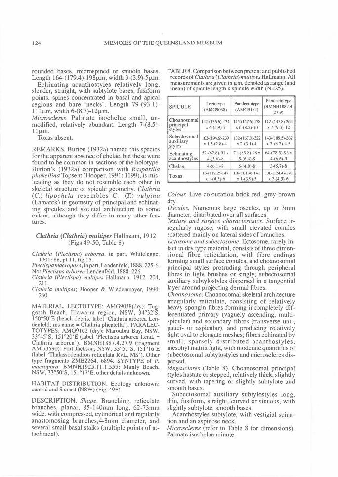
124^ MEMOIRS OF THE QUEENSLAND MUSEUM
rounded bases, microspined or smooth bases.Length 164-(179.4)-198p,m, width 3-(3.9)-5p.m.
Echinating acanthostyles relatively long,slender, straight, with subtylote bases, fusiformpoints, spines concentrated in basal and apicalregions and bare 'necks'. Length 79493.1)-111p.m, width 6-(8.7)-1211m.Microscleres. Palmate isochelae small, un-modified, relatively abundant. Length 7-(8.5)-11p,m.
Toxas absent.
REMARKS. Burton (1932a) named this speciesfor the apparent absence of chelae, but these werefound to be common in sections of the holotype.Burton's (1932a) comparison with Raspaxillaphakellina Topsent (Hooper, 1991: 1199), is mis-leading as they do not resemble each other inskeletal structure or spicule geometry. Clathria(C.) lipochela resembles C. (T) vulpina(Lamarck) in geometry of principal and echinat-ing spicules and skeletal architecture to someextent, although they differ in many other fea-tures.
Clathria (Clathria) multipes Hallmann, 1912(Figs 49-50, Table 8)
Clathria (Plectispa) arborea, in part, Whitelegge,1901: 88, p1.11, fig.15.
Plectispa macropora,in part, Lendenfeld, 1888: 225-6.Not Plectispa arborea Lendenfeld, 1888: 226.Clathria (Plectispa) multipes Hallmann, 1912: 204,
211.Clathria multipes; Hooper & Wiedenmayer, 1994:
260.
MATERIAL. LECTOTYPE: AMG9038(dry): Tug-gerah Beach, Illawarra region, NSW, 34 °32'S,150°50'E (beach debris, label 'Clathria arborea Len-denfeld; ms name = Clathria plicatella'). PARALEC-TOTYPES: AMG9162 (dry): Maroubra Bay, NSW,33°455, 151°20'E (label Tlectispa arborea Lend. =Clathria arborea'). BMNH1887.4.27.9 (fragmentAMG3590): Port Jackson, NSW, 33 °51'S, 151°16'E(label `Thalassodendron reticulata RvL, MS'). Othertype fragments ZMB2264, 6894. SYNTYPE of P.macropora: BMNH1925.11.1.555: Manly Beach,NSW, 33°50'S, 151°17'E, other details unknown.
HABITAT DISTRIBUTION. Ecology unknown;central and S coast (NSW) (Fig. 49F).
DESCRIPTION. Shape. Branching, reticulatebranches, planar, 85-140mm long, 62-73mmwide, with compressed, cylindrical and regularlyanastomosing branches,4-8mm diameter, andseveral small basal stalks (multiple points of at-tachment).
TABLE 8. Comparison between present and publishedrecords of Clathria (Clathria)multipes Hallmann. Allmeasurements are given in p,m, denoted as range (andmean) of sp'cule length x spicule width (N=25).
SPICULELectotype
(AMG9038)Paralectotype(AMG9162)
Paralectotype(BMNH1887.4.
27.9)
Citoaposomalprincipalstyles
142-(136.6)-174x 4-(5.9)-7
I45-(157.6)-178x 648.2)-10
112-(147.8)-262x 7-(9.3)- 12
Subectosomalauxiliarystyles
162-(194.6)-239x I.5-(2.8)-4
1324167.0)-222x 2-(3.1)-4
143-(189.5)-262x 2-(3.2)-4.5
Echinating 52462.8)-91 x 71-(85.8)-98 x 64-(78.5)-93 xacanthostyles 4-(5.6)-8 5-(6.4)-8 4-(6.6)-9
Chelae 4-(6.1)-8 5-(4.8)-8 3-(5.7)-816-(112.2)-147 19-(101.4)-141 1304124.4)-178
Toxas x 1-(4.3)-6 x I-(3.9)-5 x 2-(4.5)-6
Colour. Live colouration brick red, grey-browndry.Oscules. Numerous large oscules, up to 3mmdiameter, distributed over all surfaces.Texture and surface characteristics. Surface ir-regularly rugose, with small elevated conulesscattered mainly on lateral sides of branches.Ectosome and subectosome. Ectosome, rarely in-tact in dry type material, consists of three dimen-sional fibre reticulation, with fibre endingsforming small surface conules, and choanosomalprincipal styles protruding through peripheralfibres in light brushes or singly; subectosomalauxiliary subtylostyles dispersed in a tangentiallayer around projecting dermal fibres.Choanosome. Choanosomal skeletal architectureirregularly reticulate, consisting of relativelyheavy spongin fibres forming incompletely dif-ferentiated primary (vaguely ascending, multi-spicular) and secondary fibres (transverse uni-,pauci- or aspicular), and producing relativelytight oval to elongate meshes; fibres echinated bysmall, sparsely distributed acanthostyles;mesohyl matrix light, with moderate quantities ofsubectosomal subtylostyles and microscleres dis-persed.Megascleres (Table 8). Choanosomal principalstyles hastate or stepped, relatively thick, slightlycurved, with tapering or slightly subtylote andsmooth bases.
Subectosomal auxiliary subtylostyles long,thin, fusiform, straight, curved or sinuous, withslightly subtylote, smooth bases.
Acanthostyles subtylote, with vestigial spina-tion and an aspinose neck.Microscleres (refer to Table 8 for dimensions).Palmate isochelae minute.
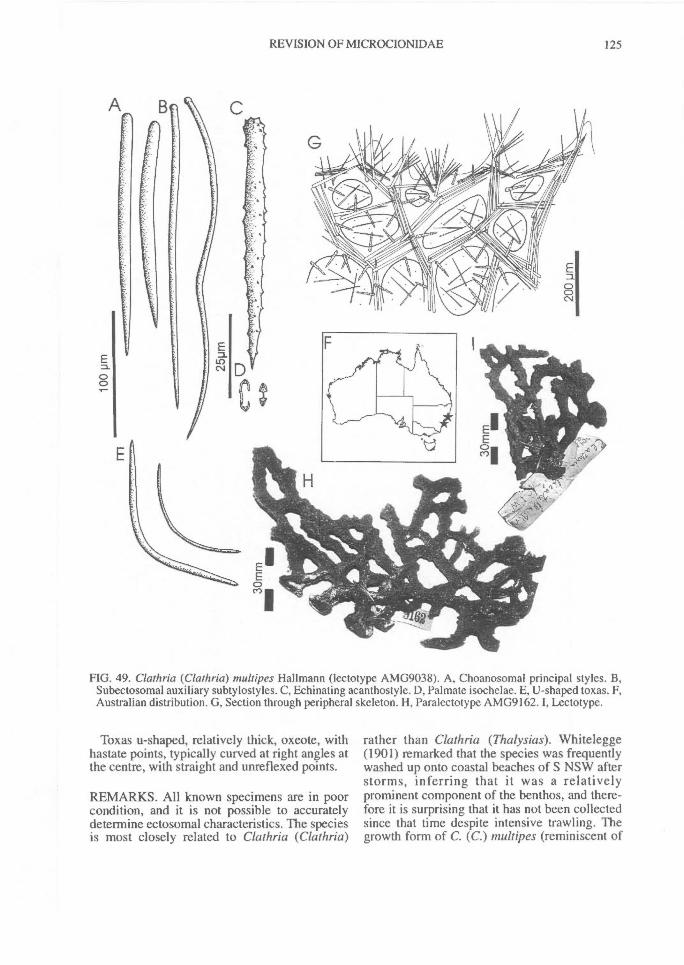
REVISION OF MICROCIONIDAE^ 125
FIG. 49. Clathria (Clathria) multipes Hallmann (lectotype AMG9038). A, Choanosomal principal styles. B,Subectosomal auxiliary subtylostyles. C, Echinating acanthostyle. D, Palmate isochelae. E, U-shaped toxas. F,Australian distribution. G, Section through peripheral skeleton. H, Paralectotype AMG9162. I, Lectotype.
Toxas u-shaped, relatively thick, oxeote, withhastate points, typically curved at right angles atthe centre, with straight and unreflexed points.
REMARKS. All known specimens are in poorcondition, and it is not possible to accuratelydetermine ectosomal characteristics. The speciesis most closely related to Clathria (Clathria)
rather than Clathria (Thalysias). Whitelegge(1901) remarked that the species was frequentlywashed up onto coastal beaches of S NSW afterstorms, inferring that it was a relativelyprominent component of the benthos, and there-fore it is surprising that it has not been collectedsince that time despite intensive trawling. Thegrowth form of C. (C.) multipes (reminiscent of
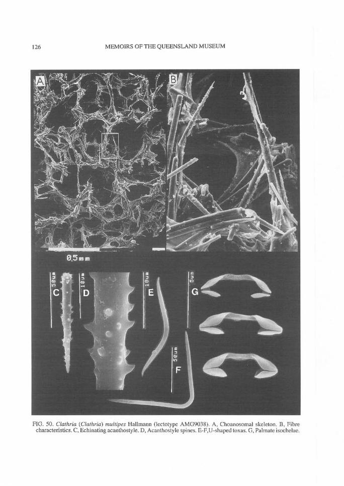
126^ MEMOIRS OF THE QUEENSLAND MUSEUM
FIG. 50. Clathria (Clathria) multipes Hallmann (lectotype AMG9038). A, Choanosomal skeleton. B, Fibrecharacteristics. C, Echinating acanthostyle. D, Acanthostyle spines. E-F,U-shaped toxas. G, Palmate isochelae.
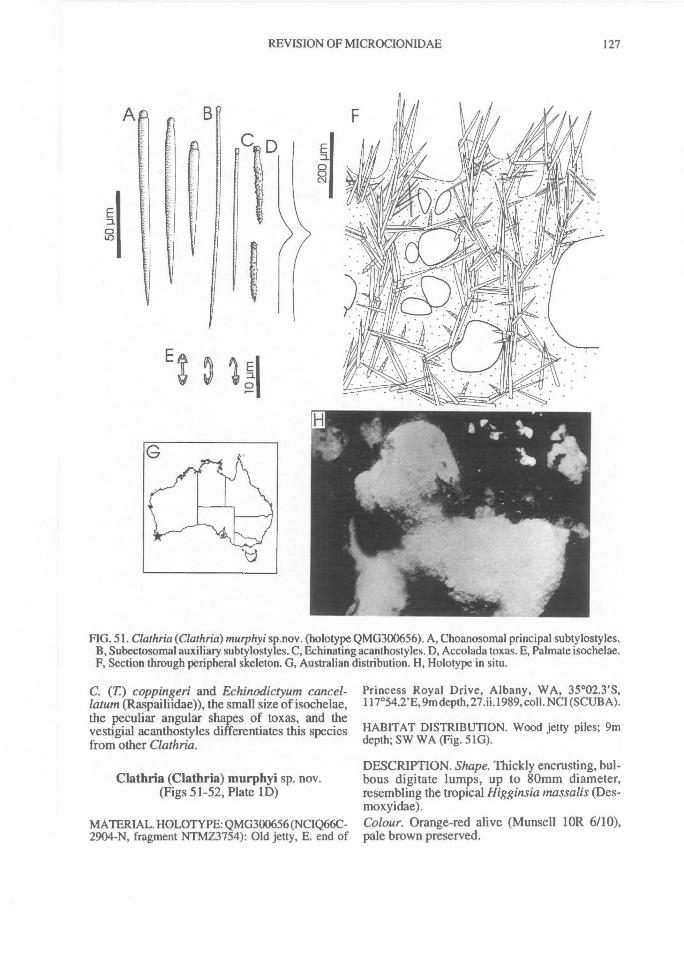
tO
E I
REVISION OF MICROCIONIDAE^ 127
FIG. 51. Clathria (Clathria) murphyi sp.nov. (holotype QMG300656). A, Choanosomal principal subtylostyles.B, Subectosomal auxiliary subtylostyles. C, Echinating acanthostyles. D, Accolada toxas. E, Palmate isochelae.F, Section through peripheral skeleton. G, Australian distribution. H, Holotype in situ.
C. (T) coppingeri and Echinodictyum cancel-latum (Raspailiidae)), the small size of isochelae,the peculiar angular shapes of toxas, and thevestigial acanthostyles differentiates this speciesfrom other Clathria.
Clathria (Clathria) murphyi sp. nov.(Figs 51-52, Plate 1D)
MATERIAL. HOLOTYPE: QMG300656 (NCIQ66C-2904-N, fragment NTMZ3754): Old jetty, E. end of
Princess Royal Drive, Albany, WA, 35°02.3'S,117°54.2'E, 9m depth, 27.ii.1989, coll. NCI (SCUBA).
HABITAT DISTRIBUTION. Wood jetty piles; 9mdepth; SW WA (Fig. 51G).
DESCRIPTION. Shape. Thickly encrusting, bul-bous digitate lumps, up to 80mm diameter,resembling the tropical Higginsia massalis (Des-moxyidae).Colour. Orange-red alive (Munsell lOR 6/10),pale brown preserved.
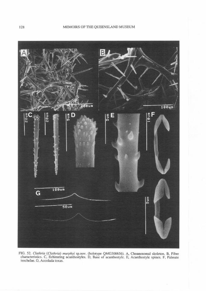
128^ MEMOIRS OF THE QUEENSLAND MUSEUM
FIG. 52. Clathria (Clathria) murphyi sp.nov. (holotype QMG300656). A, Choanosomal skeleton. B, Fibrecharacteristics. C, Echinating acanthostyles. D, Base of acanthostyle. E, Acanthostyle spines. F, Palmateisochelae. G, Accolada toxas.
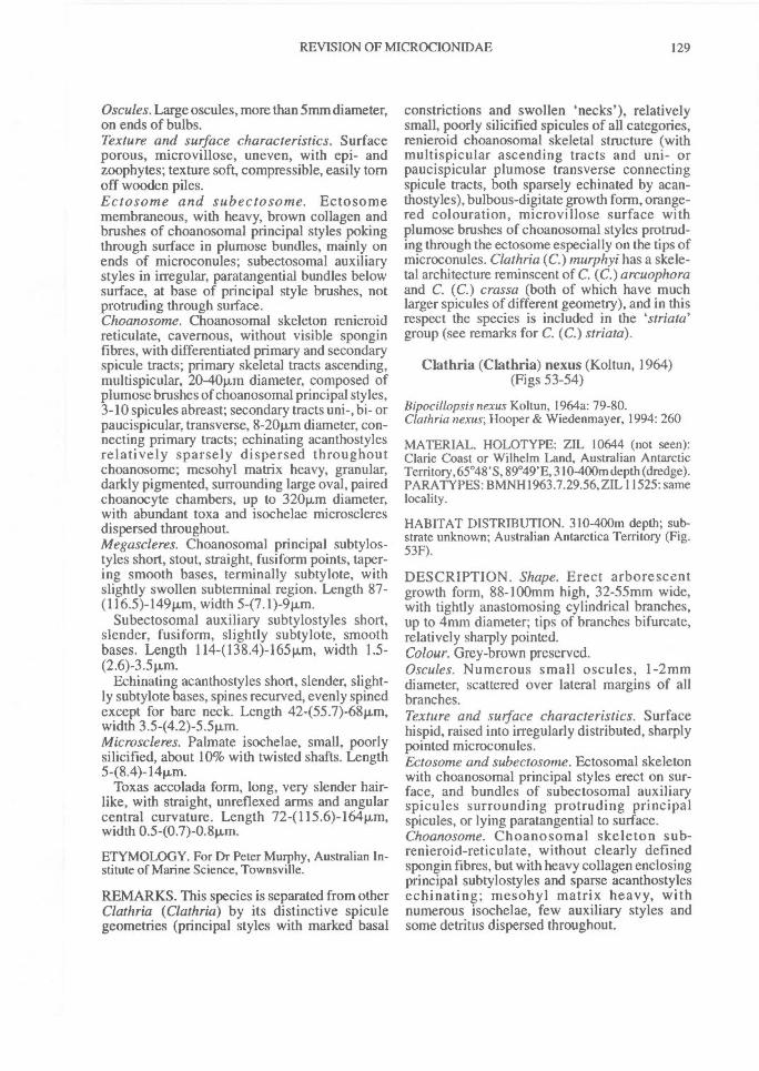
REVISION OF MICROCIONIDAE^ 129
Oscules. Large oscules, more than 5mm diameter,on ends of bulbs.Texture and surface characteristics. Surfaceporous, microvillose, uneven, with epi- andzoophytes; texture soft, compressible, easily tornoff wooden piles.Ectosome and subectosome. Ectosomemembraneous, with heavy, brown collagen andbrushes of choanosomal principal styles pokingthrough surface in plumose bundles, mainly onends of microconules; subectosomal auxiliarystyles in irregular, paratangential bundles belowsurface, at base of principal style brushes, notprotruding through surface.Choanosome. Choanosomal skeleton renieroidreticulate, cavernous, without visible sponginfibres, with differentiated primary and secondaryspicule tracts; primary skeletal tracts ascending,multispicular, 20-40p,m diameter, composed ofplumose brushes of choanosomal principal styles,3-10 spicules abreast; secondary tracts uni-, bi- orpaucispicular, transverse, 8-20p,m diameter, con-necting primary tracts; echinating acanthostylesrelatively sparsely dispersed throughoutchoanosome; mesohyl matrix heavy, granular,darkly pigmented, surrounding large oval, pairedchoanocyte chambers, up to 320Rm diameter,with abundant toxa and isochelae microscleresdispersed throughout.Megascleres. Choanosomal principal subtylos-tyles short, stout, straight, fusiform points, taper-ing smooth bases, terminally subtylote, withslightly swollen subterminal region. Length 87-(116.5)-149pin-1, width 5-(7.1)-911m.
Subectosomal auxiliary subtylostyles short,slender, fusiform, slightly subtylote, smoothbases. Length 114-(138.4)-165p..m, width 1.5-
Echinating acanthostyles short, slender, slight-ly subtylote bases, spines recurved, evenly spinedexcept for bare neck. Length 42455.7)-6811m,width 3.5-(4.2)-5.5vm.Microscleres. Palmate isochelae, small, poorlysilicified, about 10% with twisted shafts. Length5-(8.4)-14p.m.
Toxas accolada form, long, very slender hair-like, with straight, unreflexed arms and angularcentral curvature. Length 72-( 115.6)-16411m,width 0.5-(0.7)-0.4,m.
ETYMOLOGY. For Dr Peter Murphy, Australian In-stitute of Marine Science, Townsville.
REMARKS. This species is separated from otherClathria (Clathria) by its distinctive spiculegeometries (principal styles with marked basal
constrictions and swollen 'necks'), relativelysmall, poorly silicified spicules of all categories,renieroid choanosomal skeletal structure (withmultispicular ascending tracts and uni- orpaucispicular plumose transverse connectingspicule tracts, both sparsely echinated by acan-thostyles), bulbous-digitate growth form, orange-red colouration, microvillose surface withplumose brushes of choanosomal styles protrud-ing through the ectosome especially on the tips ofmicroconules. Clathria (C.)inurphyi has a skele-tal architecture reminscent of C. (C.) arcuophoraand C. (C.) crassa (both of which have muchlarger spicules of different geometry), and in thisrespect the species is included in the `striata'group (see remarks for C. (C.) striata).
Clathria (Clathria) nexus (Koltun, 1964)(Figs 53-54)
Bipocillopsis nexus Koltun, 1964a: 79-80.Clathria nexus; Hooper & Wiedenmayer, 1994: 260
MATERIAL. HOLOTYPE: ZIL 10644 (not seen):Clarie Coast or Wilhelm Land, Australian AntarcticTerritory, 65°48' S, 89°49'E, 310-400m depth (dredge).PARATYPES: BMNH1963.7.29.56, ZIL 11525: samelocality.
HABITAT DISTRIBUTION. 310-400m depth; sub-strate unknown; Australian Antarctica Territory (Fig.53F).
DESCRIPTION. Shape. Erect arborescentgrowth form, 88-100mm high, 32-55mm wide,with tightly anastomosing cylindrical branches,up to 4mm diameter; tips of branches bifurcate,relatively sharply pointed.Colour. Grey-brown preserved.Oscules. Numerous small oscules, 1-2mmdiameter, scattered over lateral margins of allbranches.Texture and surface characteristics. Surfacehispid, raised into irregularly distributed, sharplypointed microconules.Ectosome and subectosome. Ectosomal skeletonwith choanosomal principal styles erect on sur-face, and bundles of subectosomal auxiliaryspicules surrounding protruding principalspicules, or lying paratangential to surface.Choanosome. Choanosomal skeleton sub-renieroid-reticulate, without clearly definedspongin fibres, but with heavy collagen enclosingprincipal subtylostyles and sparse acanthostylesechinating; mesohyl matrix heavy, withnumerous isochelae, few auxiliary styles andsome detritus dispersed throughout.
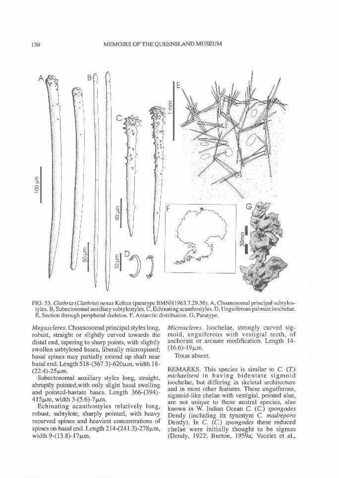
oEl
130^ MEMOIRS OF THE QUEENSLAND MUSEUM
FIG. 53. Clathria (Clathria) nexus Koltun (paratype BMNH1963.7.29.56). A, Choanosomal principal subtylos-tyles. B, Subectosomal auxiliary subtylostyles. C, Echinating acanthostyles. D, Unguiferous palmate isochclae.E, Section through peripheral skeleton. F, Antarctic distribution. G, Paratype.
Megascleres. Choanosomal principal styles long,robust, straight or slightly curved towards thedistal end, tapering to sharp points, with slightlyswollen subtyloted bases, liberally microspined;basal spines may partially extend up shaft nearbasal end. Length 5184567.3)-62011m, width 18-(22.4)-2511m.
Subectosomal auxiliary styles long, straight,abruptly pointed,with only slight basal swellingand pointed-hastate bases. Length 366-(394)-415m, width 3-(5.6)-7[Lm.
Echinating acanthostyles relatively long,robust, subtylote, sharply pointed, with heavyrecurved spines and heaviest concentrations ofspines on basal end. Length 214-(241.3)-278Rm,width 9-(13.8)-17Rm.
Microscleres. Isochelae, strongly curved sig-moid, unguiferous with vestigial teeth, ofanchorate or arcuate modification. Length 14-(16.6)-19iLm.
Toxas absent.
REMARKS. This species is similar to C. (T)michaelseni in having bidentate sigmoidisochelae, but differing in skeletal architectureand in most other features. These unguiferous,sigmoid-like chelae with vestigial, pointed alae,are not unique to these austral species, alsoknown in W. Indian Ocean C. (C.) spongodesDendy (including its synonym C. madreporaDendy). In C. (C.) spongodes these reducedchelae were initially thought to be sigmas(Dendy, 1922; Burton, 1959a; Vacelet et al.,
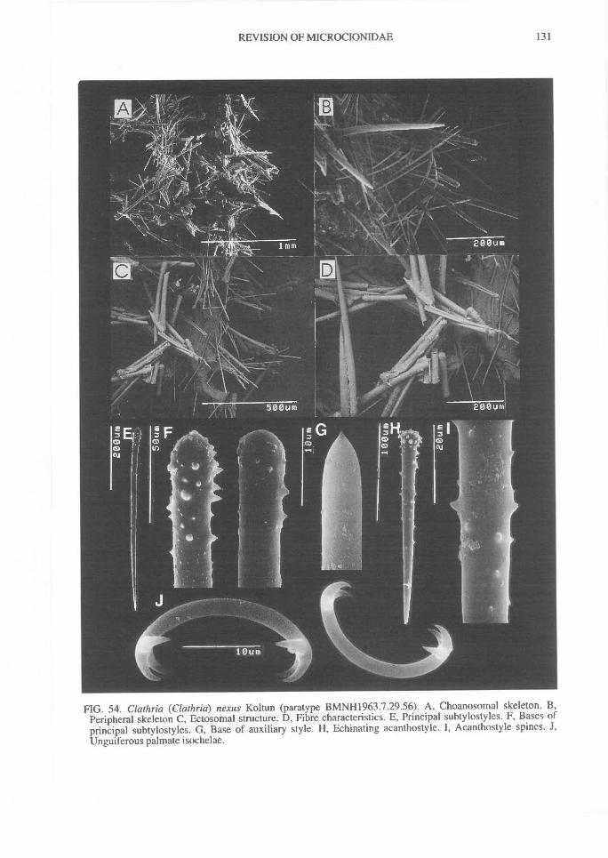
REVISION OF MICROCIONIDAE^
131
FIG. 54. Clathria (Clathria) nexus Koltun (paratype BMNH1963.7.29.56). A, Choanosomal skeleton. B,Peripheral skeleton C, Ectosomal structure. D, Fibre characteristics. E, Principal subtylostyles. F, Bases ofprincipal subtylostyles. G, Base of auxiliary style. H, Echinating acanthostyle. I, Acanthostyle spines. J,Unguiferous palmate isochelae.
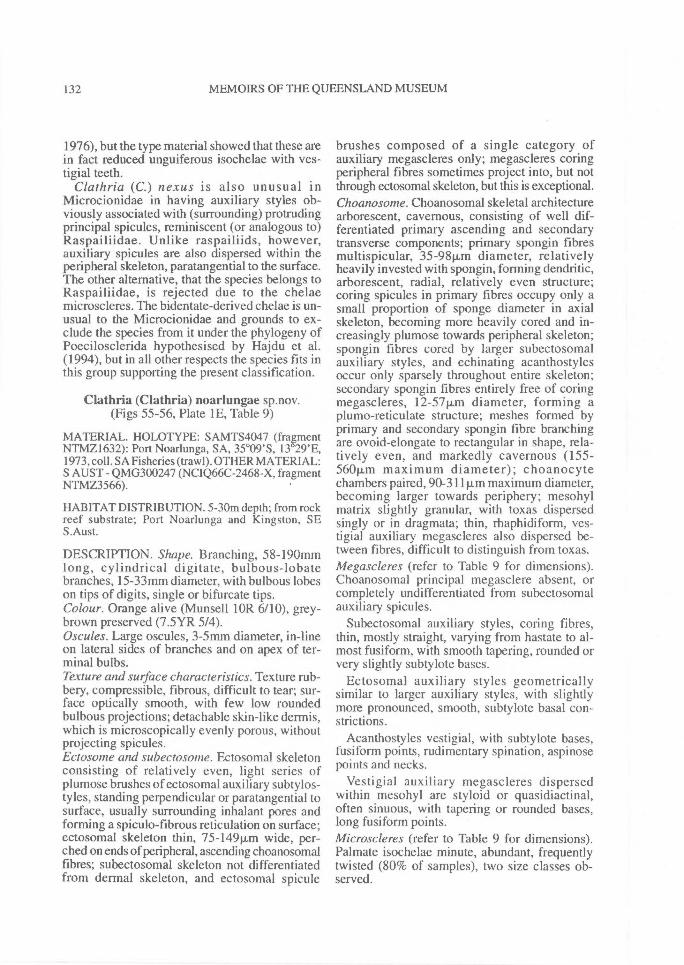
132^ MEMOIRS OF THE QUEENSLAND MUSEUM
1976), but the type material showed that these arein fact reduced unguiferous isochelae with ves-tigial teeth.
Clathria (C.) nexus is also unusual inMicrocionidae in having auxiliary styles ob-viously associated with (surrounding) protrudingprincipal spicules, reminiscent (or analogous to)Raspailiidae. Unlike raspailiids, however,auxiliary spicules are also dispersed within theperipheral skeleton, paratangential to the surface.The other alternative, that the species belongs toRaspailiidae, is rejected due to the chelaemicroscleres. The bidentate-derived chelae is un-usual to the Microcionidae and grounds to ex-clude the species from it under the phylogeny ofPoecilosclerida hypothesised by Hajdu et al.(1994), but in all other respects the species fits inthis group supporting the present classification.
Clathria (Clathria) noarlungae sp.nov.(Figs 55-56, Plate 1E, Table 9)
MATERIAL. HOLOTYPE: SAMTS4047 (fragmentNTMZ1632): Port Noarlunga, SA, 35 °09'S, 13 29'E,1973, coll. SA Fisheries (trawl). OTHER MATERIAL:S AUST - QMG300247 (NCIQ66C-2468-X, fragmentNTMZ3566).
HABITAT DISTRIBUTION. 5-30m depth; from rockreef substrate; Port Noarlunga and Kingston, SES.Aust.
DESCRIPTION. Shape. Branching, 58-190mmlong, cylindrical digitate, bulbous-lobatebranches, 15-33mm diameter, with bulbous lobeson tips of digits, single or bifurcate tips.Colour. Orange alive (Munsell 1OR 6/10), grey-brown preserved (7.5YR 5/4).Oscules. Large oscules, 3-5mm diameter, in-lineon lateral sides of branches and on apex of ter-minal bulbs.Texture and suiface characteristics. Texture rub-bery, compressible, fibrous, difficult to tear; sur-face optically smooth, with few low roundedbulbous projections; detachable skin-like dermis,which is microscopically evenly porous, withoutprojecting spicules.Ectosome and subectosonie. Ectosomal skeletonconsisting of relatively even, light series ofplumose brushes of ectosomal auxiliary subtylos-tyles, standing perpendicular or paratangential tosurface, usually surrounding inhalant pores andforming a spiculo-fibrous reticulation on surface;ectosomal skeleton thin, 75-149pm wide, per-ched on ends of peripheral, ascending choanosomalfibres; subectosomal skeleton not differentiatedfrom dermal skeleton, and ectosomal spicule
brushes composed of a single category ofauxiliary megascleres only; megascleres coringperipheral fibres sometimes project into, but notthrough ectosomal skeleton, but this is exceptional.Choanosome. Choanosomal skeletal architecturearborescent, cavernous, consisting of well dif-ferentiated primary ascending and secondarytransverse components; primary spongin fibresmultispicular, 35-98Rm diameter, relativelyheavily invested with spongin, forming dendritic,arborescent, radial, relatively even structure;coring spicules in primary fibres occupy only asmall proportion of sponge diameter in axialskeleton, becoming more heavily cored and in-creasingly plumose towards peripheral skeleton;spongin fibres cored by larger subectosomalauxiliary styles, and echinating acanthostylesoccur only sparsely throughout entire skeleton;secondary spongin fibres entirely free of coringmegascleres, 12-57p.m diameter, forming aplumo-reticulate structure; meshes formed byprimary and secondary spongin fibre branchingare ovoid-elongate to rectangular in shape, rela-tively even, and markedly cavernous (155-560p,m maximum diameter); choanocytechambers paired, 90-311 p.m maximum diameter,becoming larger towards periphery; mesohylmatrix slightly granular, with toxas dispersedsingly or in dragmata; thin, rhaphidiform, ves-tigial auxiliary megascleres also dispersed be-tween fibres, difficult to distinguish from toxas.Megascleres (refer to Table 9 for dimensions).Choanosomal principal megasclere absent, orcompletely undifferentiated from subectosomalauxiliary spicules.
Subectosomal auxiliary styles, coring fibres,thin, mostly straight, varying from hastate to al-most fusiform, with smooth tapering, rounded orvery slightly subtylote bases.
Ectosomal auxiliary styles geometricallysimilar to larger auxiliary styles, with slightlymore pronounced, smooth, subtylote basal con-strictions.
Acanthostyles vestigial, with subtylote bases,fusiform points, rudimentary spination, aspinosepoints and necks.
Vestigial auxiliary megascleres dispersedwithin mesohyl are styloid or quasidiactinal,often sinuous, with tapering or rounded bases,long fusiform points.Microscleres (refer to Table 9 for dimensions).Palmate isochelae minute, abundant, frequentlytwisted (80% of samples), two size classes ob-served.
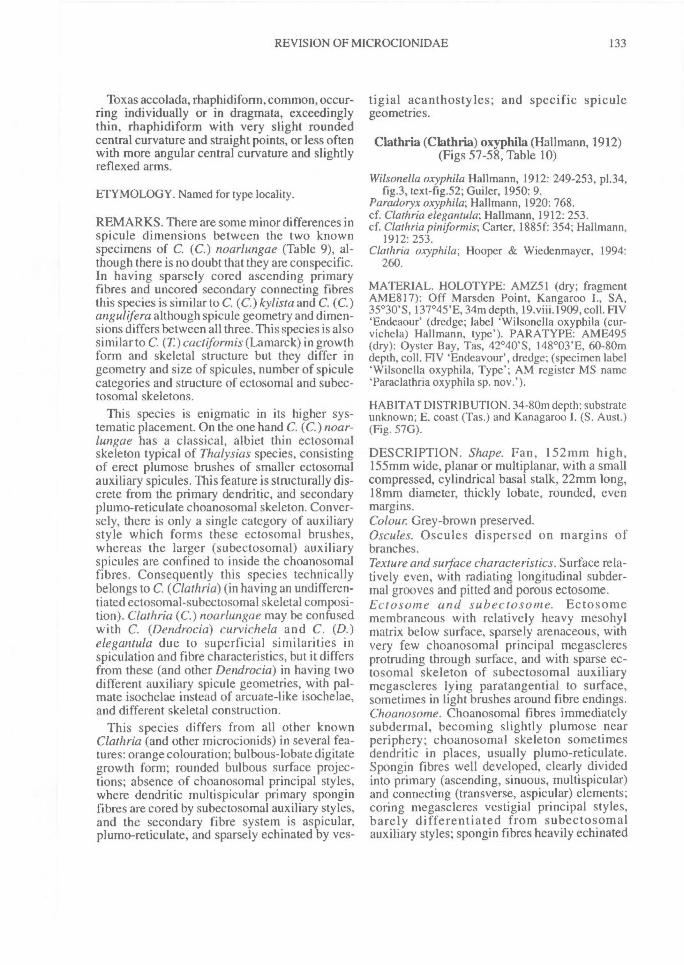
REVISION OF MICROCIONIDAE^ 133
Toxas accolada, rhaphidiform, common, occur-ring individually or in dragmata, exceedinglythin, rhaphidiform with very slight roundedcentral curvature and straight points, or less oftenwith more angular central curvature and slightlyreflexed arms.
ETYMOLOGY. Named for type locality.
REMARKS. There are some minor differences inspicule dimensions between the two knownspecimens of C. (C.) noarlungae (Table 9), al-though there is no doubt that they are conspecific.In having sparsely cored ascending primaryfibres and uncored secondary connecting fibresthis species is similar to C. (C.)kylista and C. (C.)angulifera although spicule geometry and dimen-sions differs between all three. This species is alsosimilar to C. (T) cactiformis (Lamarck) in growthform and skeletal structure but they differ ingeometry and size of spicules, number of spiculecategories and structure of ectosomal and subec-tosomal skeletons.
This species is enigmatic in its higher sys-tematic placement. On the one hand C. (C.) noar-lungae has a classical, albiet thin ectosomalskeleton typical of Thalysias species, consistingof erect plumose brushes of smaller ectosomalauxiliary spicules. This feature is structurally dis-crete from the primary dendritic, and secondaryplumo-reticulate choanosomal skeleton. Conver-sely, there is only a single category of auxiliarystyle which forms these ectosomal brushes,whereas the larger (subectosomal) auxiliaryspicules are confined to inside the choanosomalfibres. Consequently this species technicallybelongs to C. (Clathria) (in having an undifferen-tiated ectosomal-subectosomal skeletal composi-tion). Clathria (C.) noarlungae may be confusedwith C. (Dendrocia) curvichela and C. (D.)elegantula due to superficial similarities inspiculation and fibre characteristics, but it differsfrom these (and other Dendrocia) in having twodifferent auxiliary spicule geometries, with pal-mate isochelae instead of arcuate-like isochelae,and different skeletal construction.
This species differs from all other knownClathria (and other microcionids) in several fea-tures: orange colouration; bulbous-lobate digitategrowth form; rounded bulbous surface projec-tions; absence of choanosomal principal styles,where dendritic multispicular primary sponginfibres are cored by subectosomal auxiliary styles,and the secondary fibre system is aspicular,plumo-reticulate, and sparsely echinated by yes-
tigial acanthostyles; and specific spiculegeometries.
Clathria (Clathria) oxyphila (Hallmann, 1912)(Figs 57-58, Table 10)
Wilsonella oxyphila Hallmann, 1912: 249-253, p1.34,fig.3, text-fig.52; Guiler, 1950: 9.
Paradoryx oxyphda; Hallmann, 1920: 768.cf. Clathria elegantula; Hallmann, 1912: 253.cf. Clathria pintformis; Carter, 1885f: 354; Hallmann,
1912: 253.Clathria oxyphila; Hooper & Wiedenmayer, 1994:
260.
MATERIAL. HOLOTYPE: AMZ51 (dry; fragmentAME8 I 7): Off Marsden Point, Kangaroo I., SA,35°30'S, 137°45'E, 34m depth, 19.viii.1909, coll. FIV`Endeaour (dredge; label Wilsonella oxyphila (cur-vichela) Hallmann, type'). PARATYPE: AME495(dry): Oyster Bay, Tas, 42°40'S, 148°03'E, 60-80mdepth, coll. FIV 'Endeavour', dredge; (specimen label`Wilsonella oxyphila, Type'; AM register MS nameTaraclathria oxyphila sp. nov.').
HABITAT DISTRIBUTION. 34-80m depth; substrateunknown; E. coast (Tas.) and Kanagaroo I. (S. Aust.)(Fig. 57G).
DESCRIPTION. Shape. Fan, 152mm high,155mm wide, planar or multiplanar, with a smallcompressed, cylindrical basal stalk, 22mm long,18mm diameter, thickly lobate, rounded, evenmargins.Colour. Grey-brown preserved.Oscules. Oscules dispersed on margins ofbranches.Texture and surface characteristics. Surface rela-tively even, with radiating longitudinal subder-mal grooves and pitted and porous ectosome.Ectosome and subectosome. Ectosomemembraneous with relatively heavy mesohylmatrix below surface, sparsely arenaceous, withvery few choanosomal principal megascleresprotruding through surface, and with sparse ec-tosomal skeleton of subectosomal auxiliarymegascleres lying paratangential to surface,sometimes in light brushes around fibre endings.Choanosome. Choanosomal fibres immediatelysubdermal, becoming slightly plumose nearperiphery; choanosomal skeleton sometimesdendritic in places, usually plumo-reticulate.Spongin fibres well developed, clearly dividedinto primary (ascending, sinuous, multispicular)and connecting (transverse, aspicular) elements;coring megascleres vestigial principal styles,barely differentiated from subectosomalauxiliary styles; spongin fibres heavily echinated
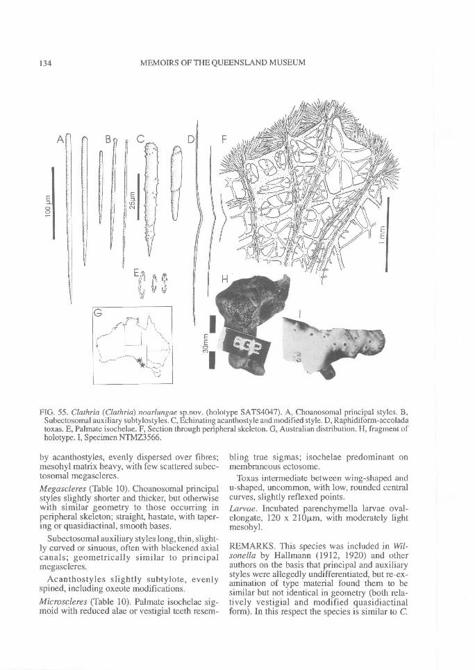
All
134^ MEMOIRS OF THE QUEENSLAND MUSEUM
FIG. 55. Clathria (Clathria) noarlungae sp.nov. (holotype SAT54047). A, Choanosomal principal styles. B,Subectosomal auxiliary subtylostyles. C, Echinating acanthostyle and modified style. D, Raphidiform-accoladatoxas. E, Palmate isochelae. F, Section through peripheral skeleton. G, Australian distribution. H, fragment ofholotype. I, Specimen NTMZ3566.
by acanthostyles, evenly dispersed over fibres;mesohyl matrix heavy, with few scattered subec-tosomal megascleres.Megascleres (Table 10). Choanosomal principalstyles slightly shorter and thicker, but otherwisewith similar geometry to those occurring inperipheral skeleton; straight, hastate, with taper-ing or quasidiactinal, smooth bases.
Subectosomal auxiliary styles long, thin, slight-ly curved or sinuous, often with blackened axialcanals; geometrically similar to principalmegascleres.
Acanthostyles slightly subtylote, evenlyspined, including oxeote modifications.Microscleres (Table 10). Palmate isochelae sig-moid with reduced alae or vestigial teeth resem-
bling true sigmas; isochelae predominant onmembraneous ectosome.
Toxas intermediate between wing-shaped andu-shaped, uncommon, with low, rounded centralcurves, slightly reflexed points.Larvae. Incubated parenchymella larvae oval-elongate, 120 x 210vt,m, with moderately lightmesohyl.
REMARKS. This species was included in Wil-sonella by Hallmann (1912, 1920) and otherauthors on the basis that principal and auxiliarystyles were allegedly undifferentiated, but re-ex-amination of type material found them to besimilar but not identical in geometry (both rela-tively vestigial and modified quasidiactinalform). In this respect the species is similar to C.
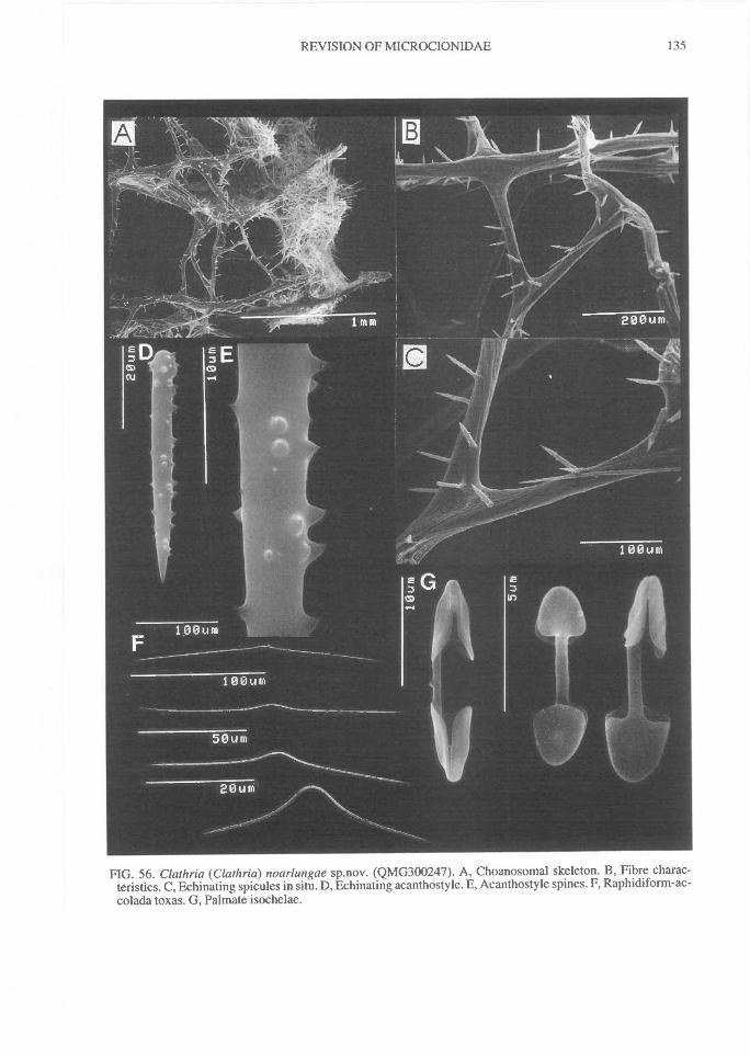
REVISION OF MICROCIONIDAE^
135
FIG. 56. Clathria (Clathria) noarlungae sp.nov. (QMG300247). A, Choanosomal skeleton. B, Fibre charac-teristics. C, Echinating spicules in situ. D, Echinating acanthostyle. E, Acanthostyle spines. F, Raphidiform-ac-colada toxas. G, Palmate isochelae.
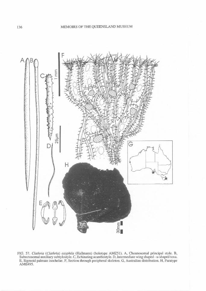
136^ MEMOIRS OF THE QUEENSLAND MUSEUM
FIG. 57. Clathria (Clathria) oxyphila (Hal!mann) (holotype AMZ51). A, Choanosomal principal style. B,Subectosomal auxiliary subtylostyle. C, Echinating acanthostyle. D, Intermediate wing shaped - u-shaped toxa.E, Sigmoid palmate isochelae. F, Section through peripheral skeleton. G, Australian distribution. H, ParatypeAMFA95.
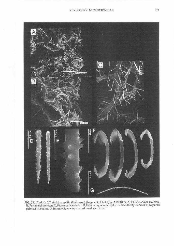
REVISION OF MICROCIONIDAE^
137
FIG. 58. Clathria (Clathria) oxyphila (Hallmann) (fragment of holotype AME817). A, Choanosomal skeleton.B, Peripheral skeleton. C, Fibre characteristics. D, Echinating acanthostyles. E, Acanthostyle spines. F, Sigmoidpalmate isochelae. G, Intermediate wing-shaped - u-shaped toxa.
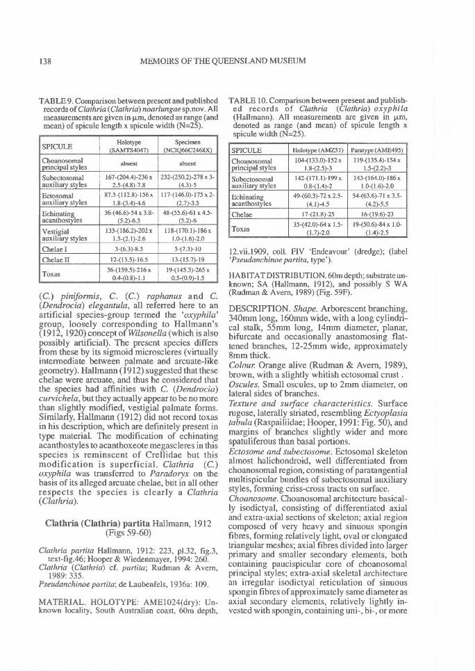
138^ MEMOIRS OF THE QUEENSLAND MUSEUM
TABLE 9. Comparison between present and publishedrecords of Clathria (Clathria)noarlungae sp.nov. Allmeasurements are given in p.m, denoted as range (andmean) of spicule length x spicule width (N=25).
SPICULEHolotype
(SAMTS4047)Specimen
(NC1Q66C2468X)
Choanosomalprincipal styles absent absent
Subectosomal 167-(204.4)-236 x 232-(250.2)-278 x 3-auxiliary styles 2.5-(4.8)-7.8 (4.3)-5
Ectosomal 87.5-(112.8)-156 x 117-(146.0)-175 x 2-auxiliary styles 1.8-(3.4)-4.6 (2.7)-3.5
Echinating 36-(46.6)-54 x 3.8- 48-(55.6)-61 x 4.5-acanthostyles (5.2)-6.5 (5.2)-6
Vestigial 133-(186.2)-202 x 118-(170.1)-186 xauxiliary styles 1.5-(2.1)-2.6 1.0-(1.6)-2.0
Chelae I 3-(6.3)-8.5 5-(7.3)-I0
Chelae II 12-(13.5)-16.5 13-(15.7)-I9
36-(159.5)-216 x 19-(145.3)-265 xToxas 0.4-(0.8)-1.1 0.5-(0.9)- 1.5
(C.) piniformis, C. (C.) raphanus and C.(Dendrocia) elegantula, all referred here to anartificial species-group termed the `oxyphila'group, loosely corresponding to Hallmann's(1912, 1920) concept of Wilsonella (which is alsopossibly artificial). The present species differsfrom these by its sigmoid microscleres (virtuallyintermediate between palmate and arcuate-likegeometry). Hallmann (1912) suggested that thesechelae were arcuate, and thus he considered thatthe species had affinities with C. (Dendrocia)curvichela, but they actually appear to be no morethan slightly modified, vestigial palmate forms.Similarly, Hallmann (1912) did not record toxasin his description, which are definitely present intype material. The modification of echinatingacanthostyles to acanthoxeote megascleres in thisspecies is reminscent of Crellidae but thismodification is superficial. Clathria (C.)oxyphila was transferred to Paradoryx on thebasis of its alleged arcuate chelae, but in all otherrespects the species is clearly a Clathria(Clathria).
Clathria (Clathria) partita Hallmann, 1912(Figs 59-60)
Clathria partita Hallmann, 1912: 223, p1.32, fig.3,text-fig.46; Hooper & Wiedenmayer, 1994: 260.
Clathria (Clathria) cf. partita; Rudman & Avern,1989: 335.
Pseudanchinoe partita; de Laubenfels, 1936a: 109.
MATERIAL. HOLOTYPE: AME1024(dry): Un-known locality, South Australian coast, 60m depth,
TABLE 10. Comparison between present and publish-ed records of Clathria (Clathria) oxyphila(Hallmann). All measurements are given in p,m,denoted as range (and mean) of spicule length xspicule width (N=25).
SPICULE Holotype (AMZ51) Paratype (AME495)
Choanosomal 104-(133.0)-152 x 119-(135.4)-154 xprincipal styles 1.8-(2.5)-3 1.5-(2.2)-3
Subectosomal 142-(171.1)-199 x 143-(164.0)-186 xauxiliary styles 0.8-(1.4)-2 1.0-(1.6)-2.0
Echinating 49-(60.3)-72 x 2.5- 54-(63.6)-71 x 3.5-acanthostyles (4.1)-4.5 (4.2)-5.5
Chelae 17-(21.8)-25 16-(19.6)-23
Toxas 35@20)-64x 1.5-(1.7)-2.0
I9-(50.6)-84x 1.0-(1.4)-2.5
12.vii.1909, coll. FIV `Endeavour' (dredge); (label`Pseudanchinoe partita, type').
HABITAT DISTRIBUTION. 60m depth; substrate un-known; SA (Hallmann, 1912), and possibly S WA(Rudman & Avern, 1989) (Fig. 59F).
DESCRIPTION. Shape. Arborescent branching,340mm long, 160mm wide, with a long cylindri-cal stalk, 55mm long, 14mm diameter, planar,bifurcate and occasionally anastomosing flat-tened branches, 12-25mm wide, approximately8mm thick.Colour. Orange alive (Rudman & Avern, 1989),brown, with a slightly whitish ectosomal crust.Oscules. Small oscules, up to 2mm diameter, onlateral sides of branches.Texture and surface characteristics. Surfacerugose, laterally striated, resembling Ectyoplasiatabula (Raspailiidae; Hooper, 1991: Fig. 50), andmargins of branches slightly wider and morespatuliferous than basal portions.Ectosome and subectosome. Ectosomal skeletonalmost halichondroid, well differentiated fromchoanosomal region, consisting of paratangentialmultispicular bundles of subectosomal auxiliarystyles, forming criss-cross tracts on surface.Choanosome. Choanosomal architecture basical-ly isodictyal, consisting of differentiated axialand extra-axial sections of skeleton; axial regioncomposed of very heavy and sinuous sponginfibres, forming relatively tight, oval or elongatedtriangular meshes; axial fibres divided into largerprimary and smaller secondary elements, bothcontaining paucispicular core of choanosomalprincipal styles; extra-axial skeletal architecturean irregular isodictyal reticulation of sinuousspongin fibres of approximately same diameter asaxial secondary elements, relatively lightly in-vested with spongin, containing uni-, bi-, or more
REVISION OF MICROCIONIDAE^ 139
rarely paucispicular tracts of choanosomal styles;extra-axial fibres incompletely divided intoprimary (ascending) and secondary (vaguelytransverse) components, differentiated mainly bynumber of coring spicules; echinating acanthos-tyles sparse, evenly distributed throughoutskeleton, rarely incorporated into fibres (cf.Hallmann, 1912); some choanosomal principalstyles may protrude through (echinate) fibres,particularly in extra-axial skeleton; mesohylmatrix very light containing few subectosomalauxiliary megascleres.Megascleres. Choanosomal principal stylesshort, slightly curved or straight, almost hastate,with smooth rounded or very slightly subtylotebases. Length 1124172.6)-23911m, width 5-(8.7)-1211m.
Subectosomal auxiliary styles long, slightlycurved or straight, fusiform, with rounded orslightly subtylote bases. Length 2224267.7)-315p.m, width 3.5-(5.1)-7p.m.
Acanthostyles slightly subtylote, with vestigialspines and an aspinous basal region. Length 56-(60.4)-8811m, width 3-(5.0)-61.m.Microscleres. Isochelae absent.
Toxas sinuous rhaphidiform, usually sym-metrical with slight angular central curvature andstraight points, but asymmetrical and reflexedexamples also occur. Length 724115.6)-2121m,width 0.5-(0.7)-1.2Rm.
REMARKS. This species was referred toPseudanchinoe by de Laubenfels (1936a) be-cause it lacked chelae microscleres, but that fea-ture has since been shown to be of littlesystematic value (e.g. Hooper, 1991: 288). Asnoted by Hallmann (1912), C. (C.) partita isknown only from a single dry specimen, and it ispossible that isochelae may be eventually dis-covered in other better preserved specimensshould additional material become available.
Clathria (C.) partita has an unusual skeletalconstruction, combining features of myxillid-likemicrocionids (e.g. a basically isodictyal extra-axial skeleton closely resembling C. (Isociella)eccentrica) and raspailiid-like microcionids (e.g.compressed axis of C. (Axociella) canaliculata),and features of the compressed axial skeleton arealso reminiscent of the skeletal plan seen in cer-tain Axinellidae such as Cymbastela. On the basisof these characteristics C. (C.) partita is similarto C. (C.) rubens (Lendenfeld).
The record of C. (C.)partita from WA (Rudman& Avern, 1989), is based on a photographwithout accompanying sample. The photograph
was taken of a nudibranch (Rostanga calumnusRudman & Avern) feeding on an orange spongefrom Esperance Bay (33°51'S, 121°57'E).
Clathria (Clathria) paucispicula (Burton, 1932)(Fig. 61)
Rhaphidophlus paucispiculus Burton, 1932a:320, p1.56, fig.1, text-fig.30; Burton, 1940: 111;Desqueyroux, 1975: 68; Koltun, 1964a: 75;Desqueyroux-Faundez & Moyano, 1987: 49.Clathria paucispicula; Hooper & Wiedenmayer,1994: 260
MATERIAL. HOLOTYPE: BMNH1928.2.15.243a: NearShag Rocks, South Georgia, 53°43.4'S, 40°57.0'W,177m depth, coll. R.R.S. 'Discovery' (dredge).
HABITAT DISTRIBUTION 74-198m depth; on mud,sand and rock substrates; Australian Antarctic Ter-ritory: Queen Mary Land (Koltun, 1964a) (Fig. 61D).Also Falkland Is, South Georgia and S. Shetland Is(Burton, 1932a), Mar del Plata, Argentina (Burton,1940), Low Is, Chilean Antarctic Territory(Desqueyroux, 1975), Tierra del Fuego (Desqueyroux-Faundez & Moyano, 1987).
DESCRIPTION. Shape. Massive, flabellate-digitate with irregular ridges and convolutedbranches.Colour Live colouration unknown, light brownpreserved.Oscules. Large oscules 3-5mm diameter sparselyscattered on apex of ridges.Texture and surface characteristics. Texture firm,tough, compressible; surface uneven, minutelyconulose.Ectosome and subectosome. Surface skeletontangential confused crust of smaller ectosomalauxiliary styles, choanosomal principal stylesand foreign spicule fragments together forming adense crust 0.5-1.0mm thick; immediately belowtangential ectosomal skeleton are erect bundlesof both auxiliary styles (producing an irregularradial palisade of spicules), and principal styles(more sparsely dispersed).Choanosome. Skeletal architecture reticulate,slightly plumo-reticulate near periphery, less or-ganised towards axis; spongin fibres moderatelywell developed cored by pauci- or multispicularascending tracts and interconnected by uni- orpaucispicular tracts of choanosomal principalstyles, producing rectangular or triangularmeshes up to 250Rm diameter; echinatingmegascleres absent; mesohyl matrix light,smooth, unpigmented.
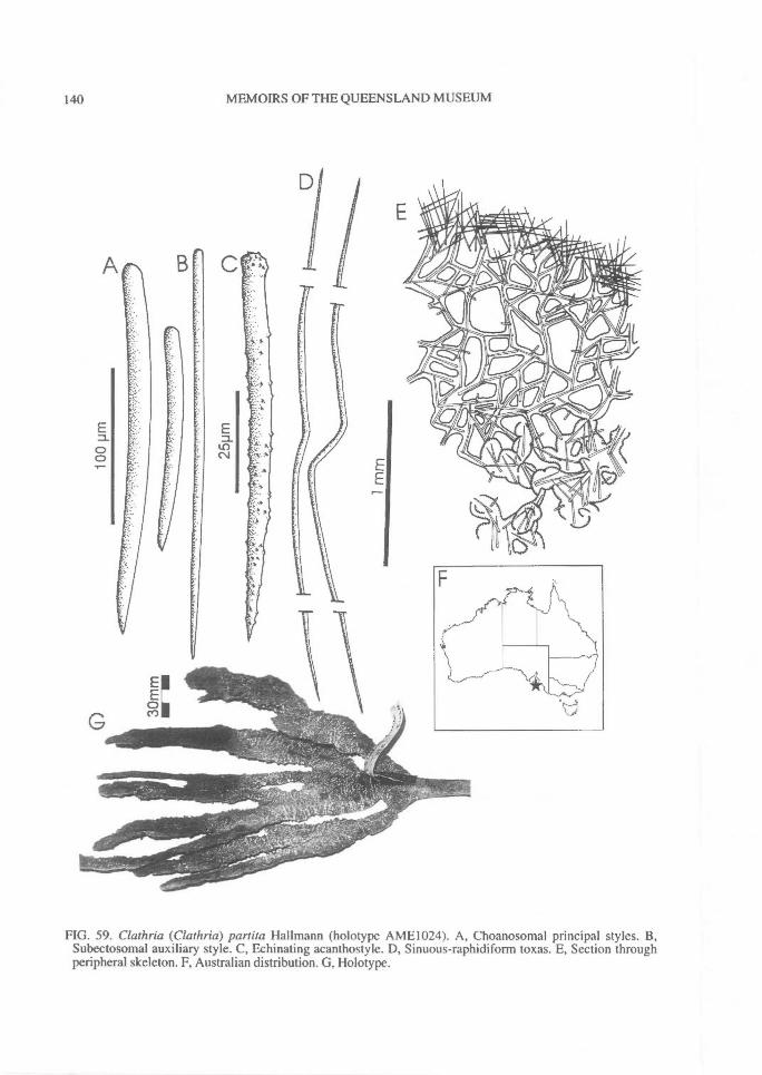
140^ MEMOIRS OF THE QUEENSLAND MUSEUM
FIG. 59. Clathria (Clathria) partita Hallmann (holotype AME1024). A, Choanosomal principal styles. B,Subectosomal auxiliary style. C, Echinating acanthostyle. D, Sinuous-raphidiform toxas. E, Section throughperipheral skeleton. F, Australian distribution. G, Holotype.
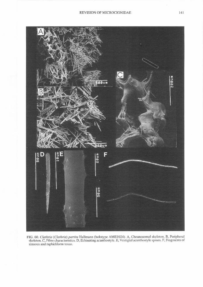
REVISION OF MICROCIONIDAE^
141
FIG. 60. Clathria (Clathria) part ita Hallmann (holotype AME1024). A, Choanosomal skeleton. B, Peripheralskeleton. C, Fibre characteristics. D, Echinating acanthostyle. E, Vestigial acanthostyle spines. F, Fragments ofsinuous and raphidiform toxas.
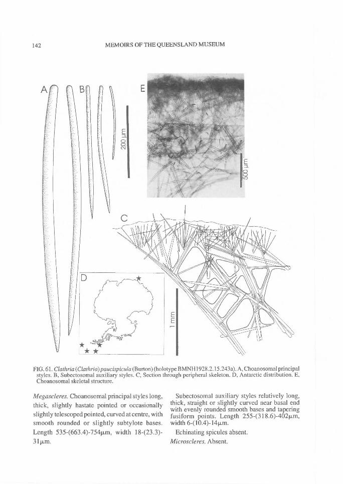
142^ MEMOIRS OF THE QUEENSLAND MUSEUM
FIG. 61. Clathria(Clathria)paucispicula (Burton) (holotype BMNH1928.2.15.243a). A, Choanosomal principalstyles. B, Subectosomal auxiliary styles. C, Section through peripheral skeleton. D, Antarctic distribution. E,Choanosomal skeletal structure.
Megascleres. Choanosomal principal styles long,thick, slightly hastate pointed or occasionallyslightly telescoped pointed, curved at centre, withsmooth rounded or slightly subtylote bases.Length 535-(663.4)-754Rm, width 18423.3)-31pm.
Subectosomal auxiliary styles relatively long,thick, straight or slightly curved near basal endwith evenly rounded smooth bases and taperingfusiform points. Length 255-(318.6)-402Rm,width 6410.4)-1411m.
Echinating spicules absent.Microscleres. Absent.
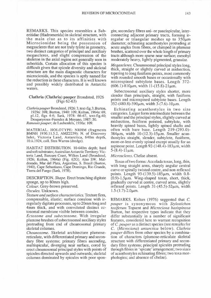
REVISION OF MICROCIONIDAE^ 143
REMARKS. This species resembles a Sub-eritidae (Hadromerida) in skeletal structure, withthe main clue as to its affinities withMicrocionidae being the possession ofmegascleres that are not truly tylote in geometry,two distinct categories of principal and auxiliarymegascleres, and slight compression of theskeleton in the axial region not generally seen insuberitids. Certain allocation of this species isdifficult given that spicule diversity and skeletalstructure are the main diagnostic characters formicrocionids, and the species is aptly named forthe reduction in these characters. It is well knownand possibly widely distributed in Antarcticwaters.
Clathria (Clathria) pauper Brondsted, 1926(Figs 62-63)
Clathria pauper Brondsted, 1926: 3, text-fig.3; Burton,1929a: 398; Burton, 1940: 109; Koltun, 1964a: 69,p1.12, figs 4-6; Sara, 1978: 66-67, text-fig.40;Desqueyroux-Faundez & Moyano, 1987: 50.
Ramoses pauper; de Laubenfels, 1936a: 109.
MATERIAL. HOLOTYPE: NRHM (fragmentsBMNH 1930.11.5.2, AMZ2239): N. of DiscoveryInlet, Victoria Land, Antarctica, 640m depth,10.ii.I924, coll. Sten Warren (dredge).
HABITAT DISTRIBUTION. 10-640m depth; hardand soft substrates; Australian Antarctic Territory: Vic-toria Land, Banzare Coast, Wilkes Land (Brondsted,1926; Koltun, 1964a) (Fig. 62G). Also SW. Mal-donado, Mar del Plata, Argentina, S. Brazil (Burton,1940), Cape Sebastiano, Cape Domingo, Rio Grande,Tierra del Fuego (Sara, 1978).
DESCRIPTION. Shape. Erect branching digitatesponge, up to 80mm high.Colour. Grey-brown preserved.Oscules. Unknown.Texture and surface characteristics. Texture firm,compressible, elastic; surface conulose with ir-regularly digitate processes, up to 20mm long and4mm thick, and with convoluted distinct ec-tosomal membrane visible between conules.Ectosome and subectosome. With irregularplumose brushes of subectosomal auxiliary stylesprotruding from end of choanosomal primaryskeletal columns.Choanosome. Skeletal architecture plumose-reticulate, with differentiated primary and secon-dary fibre systems; primary fibres ascending,multispicular, diverging near surface, cored byerect choanosomal principal styles with points ofspicules directed upwards and outwards; skeletalcolumns dominated by spicules with poor spon-
gin; secondary fibres uni- or paucispicular, inter-connecting adjacent primary tracts, forming ir-regular or triangular meshes up to 350Rmdiameter; echinating acanthostyles protruding atacute angles from fibres, or clumped in plumosebrushes, scattered over the whole length of primarytracts although more sparse near surface; mesohylmoderately heavy, lightly pigmented, granular.Megascleres. Choanosomal principal styles long,thick, straight or slightly curved at midsection,tapering to long fusiform points, most commonlywith rounded smooth bases or occasionally withmicrospined subtylote bases. Length 372-(606.1)-810p,m, width 11-(15.8)-21Rm.
Subectosomal auxiliary styles shorter, moreslender than principals, straight, with fusiforrnpoints and microspined subtylote bases. Length352-(480.8)-590Rm, width 3-(7.6)-10p.m.
Echinating acanthostyles in two sizecategories. Larger form intermediate between thesmaller and the principal styles, slightly curved atmidsection, fusiform pointed, subtylote, withheavily spined bases, lightly spined shafts andoften with bare base. Length 2194293.0)-384iim, width 10-(12.3)-15Rm. Smaller acan-thostyles straight, slender, subtylote, fusiform,more-or-less evenly spined except usually for anaspinose point. Length 92-(148.4)-183p.m, width
Microscleres. Chelae absent.Toxas of two forms: Accolada toxas, long, thin,
with long straight arms, sharply angular centralcurve or spirally twisted centre, and non-reflexedpoints. Length 934139.5)-18511m, width 0.8-(0.9)-1.511m. Wing-shaped toxas, short, thick,gradually curved at centre, curved arms, slightlyreflexed points. Length 31-(45.5)-52Rm, width
REMARKS. Koltun (1976) suggested that C.pauper is synonymous with Stylostichontoxiferum Topsent and Microciona basispinosaBurton, but respective types indicate that theydiffer substantially in a number of significantfeatures, considered here to warrant recognitionof C. pauper as a distinct species (see remarks forC. (Microciona) antarctica below). Clathriapauper differs from other species by a combina-tion of characters (plumose-reticulate skeletalstructure with differentiated primary and secon-dary fibre systems; principal spicules protrudingthrough fibres in `spicate' arrangement; two sizesof acanthostyles echinating fibres; two toxa mor-phologies; and absence of chelae).
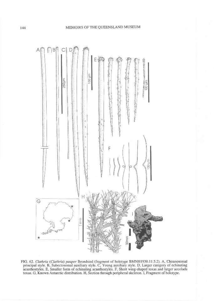
A
144^ MEMOIRS OF THE QUEENSLAND MUSEUM
FIG. 62. Clathria (Clathria) pauper Brondsted (fragment of holotype BMNH1930.11.5.2). A, Choanosomalprincipal style. B, Subectosomal auxiliary style. C, Young auxiliary style. D, Larger category of echinatingacanthostyles. E, Smaller form of echinating acanthostyles. F, Short wing-shaped toxas and larger accoladatoxas. G, Known Antarctic distribution. H, Section through peripheral skeleton. I, Fragment of holotype.
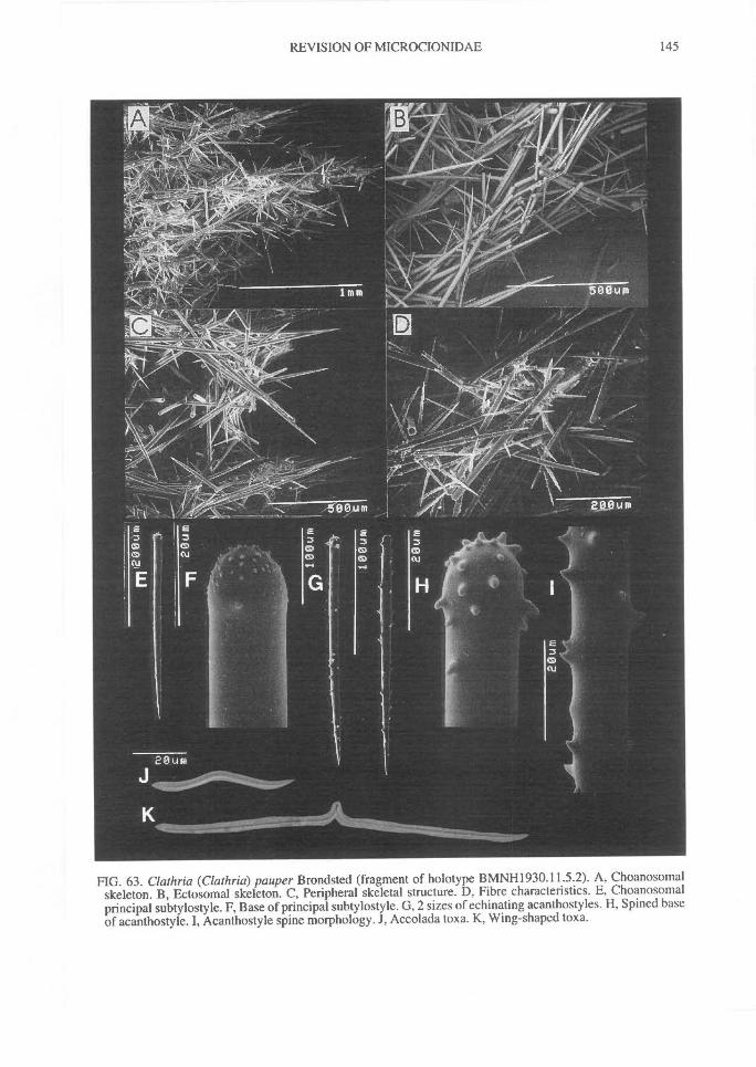
REVISION OF MICROCIONIDAE^ 145
FIG. 63. Clathria (Clathria) pauper Brondsted (fragment of holotype BMNH1930.11.5.2). A, Choanosomalskeleton. B, Ectosomal skeleton. C, Peripheral skeletal structure. D, Fibre characteristics. E, Choanosomalprincipal subtylostyle. F, Base of principal subtylostyle. G, 2 sizes of echinating acanthostyles. H, Spined baseof acanthostyle. I, Acanthostyle spine morphology. J, Accolada toxa. K, Wing-shaped toxa.
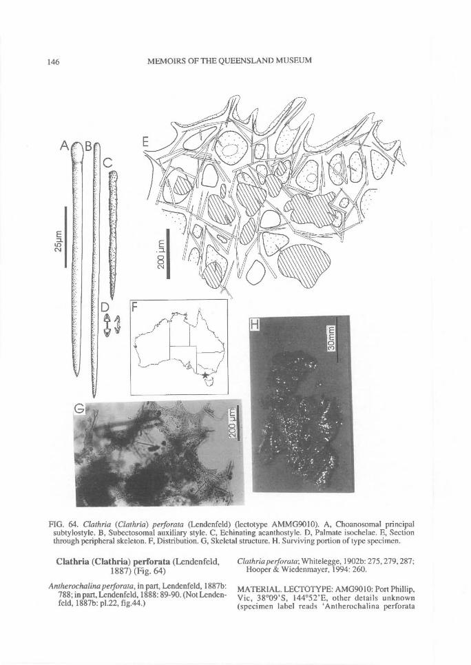
A
146^ MEMOIRS OF THE QUEENSLAND MUSEUM
FIG. 64. Clathria (Clathria) petforata (Lendenfeld) (lectotype AMMG9010). A, Choanosomal principalsubtylostyle. B, Subectosomal auxiliary style. C, Echinating acanthostyle. D, Palmate isochelae. E, Sectionthrough peripheral skeleton. F, Distribution. G, Skeletal structure. H. Surviving portion of type specimen.
Clathria (Clathria) perforata (Lendenfeld,1887) (Fig. 64)
Antherochalina perforata, in part, Lendenfeld, 1887b:788; in part, Lendenfeld, 1888: 89-90. (Not Lenden-feld, 1887b: p1.22, fig.44.)
Clathria perforata; Whitelegge, 1902b: 275, 279, 287;Hooper & Wiedenmayer, 1994: 260.
MATERIAL. LECTOTYPE: AMG9010: Port Phillip,Vic, 38°09'S, 144°52'E, other details unknown(specimen label reads `Antherochalina perforata
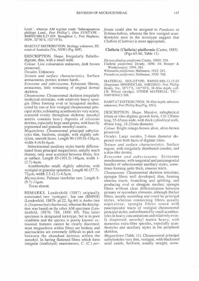
REVISION OF MICROCIONIDAE^ 147
Lend.', whereas AM register reads `Suberapsammaphilippi Lend., Port Phillip'). (Not SYNTYPE -BMNH1886.8.27.459: Broughton I., Port Stephens,NSW, 32°36'S, 152°19'E).
HABITAT DISTRIBUTION. Ecology unknown; SE.coast of Australia (Vic, NSW) (Fig. 64F).
DESCRIPTION. Shape. Irregularly flabello-digitate, thin, with a small stalk.Colour. Live colouration unknown, dark brownpreserved.Oscules. Unknown.Texture and surface characteristics. Surfacearenaceaous, porous; texture harsh..Ectosome and subectosome. Ectosome fibrous,arenacious, little remaining of original dermalskeleton.Choanosome. Choanosomal skeleton irregularlyisodictyal reticulate, with relatively heavy spon-gin fibres forming oval or hexagonal meshes,cored by one or few vestigial choanosomal prin-cipal styles; echinating acanthostyles very scarse,scattered evenly throughout skeleton; mesohylmatrix contains heavy deposits of siliceousdetritus, especially large sand grains, with heavilypigmented and granular mesohyl matrix.Megascleres. Choanosomal principal subtylos-tyles thin, fusiform, straight, with slightly sub-tylote, smooth bases. Length 96-(105.0)-11811m,width 4-(4.8)-6p.m.
Subectosomal auxiliary styles barely differen-tiated from principal megascleres, usually muchthinner, only seen scattered between fibres, fewat surface. Length 854103.3)-14611m, width I -(2.7) -411m.
Acanthostyles small, slightly subtylote, withvestigial or granular spination. Length 46457.7)-72p.m, width 2.5-(3.1)-4.5p.m.Microscleres. Palmate isochelae rare. Length 8-(9.7)-11[Lm.
Toxas absent.
REMARKS. Lendenfeld (1887) originallynominated two `syntypes', but one (BMNH)(Lendenfeld, 1887b: p1.22, fig.44) is Antho (seeA. (Isopenectya)chartacea), whereas the descrip-tion was based on the other AM specimen (Len-denfeld, 1887b: 788, 1888: 89). This latterspecimen is designated lectotype, but is in poorcondition and the species is poorly known: ec-tosomal features cannot be clearly discerned;most megascleres within fibres are broken; andmicroscleres are extremely difficult to pick outbetween the abundant detritus within themesohyl. In having flattened fibres which formirregular (isodictyal) anastomoses, C. (C.) per-
forata could also be assigned to Pandaros orEchinoclathria, whereas the few vestigial acan-thostyles seen in the lectotype suggest thatClathria (Clathria) is more appropriate.
Clathria (Clathria) piniformis (Carter, 1885)(Figs 65-66, Table 11)
Dictyocylindrus piniformis Carter, 1885f: 354.Clathria piniformis; Dendy, 1896: 34; Hooper &
Wiedenmayer, 1994: 261.Wilsonella piniformis; Hallmann, 1912: 241.Paradoryx piniformis; Hallmann, 1920: 768.
MATERIAL. HOLOTYPE: BMNH1886.12.15.62(fragments MNHNDCL60, AMG2803): Port PhillipHeads, Vic, 38°17'S, 144°39'E, 38-40m depth, coll.J.B. Wilson (dredge). OTHER MATERIAL: VIC -NMVRN412-508.
HABITAT DISTRIBUTION. 38-40m depth; substrateunknown; Port Phillip Bay(Fig. 65G).
DESCRIPTION. Shape. Massive, subsphericallobate or lobo-digitate growth form, 110-130mmlong, 55-65mm wide, with thick cylindrical stalk,45mm long, 18-22mm diameter.Colour Bright orange-brown alive, olive-brownpreserved.Oscules. Large oscules, 2-4mm diameter dis-persed over both faces of digitate lobes.Texture and surface characteristics. Surfacerugose, with irregularly distributed conules, anda skin-like dermis.Ectosoine and subectosome. Ectosomemembraneous, with tangential and paratangentialbundles of subectosomal auxiliary styles, some-times forming quite thick, sinuous tracts.Choanosome. Choanosomal skeleton reticulate;spongin fibres well developed, thin, formingsinuous tracts, branching and splitting, andproducing oval or elongate meshes; sponginfibres without clear differentiation betweenprimary or secondary elements, although thickerfibres, usually ascending and cored by principalstyles, whereas connecting fibres usuallyaspiculose; spongin fibres cored withpaucispicular tracts of vestigial choanosomalprincipal styles, and echinated by small acanthos-tyles in heavy concentrations and relatively even-ly dispersed; mesohyl matrix heavy, withnumerous extra-fibre spicules, especially acan-thostyles and auxiliary styles in the peripheralskeleton.Megascleres (Table 11). Choanosomal principalsubtylostyles very thin, vestigial, with blackenedaxial canals, fusiform, usually straight, some-
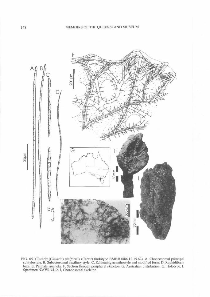
LOC
148^ MEMOIRS OF THE QUEENSLAND MUSEUM
FIG. 65. Clathria (Clathria) piniformis (Carter) (holotype BMNH1886.12.15.62). A, Choanosomal principalsubtylostyle. B, Subectosomal auxiliary style. C, Echinating acanthostyle and modified form. D, Raphidiformtoxa. E, Palmate isochela. F, Section through peripheral skeleton. G, Australian distribution. G, Holotype. I,Specimen NMVRN412. J, Choanosomal skeleton.
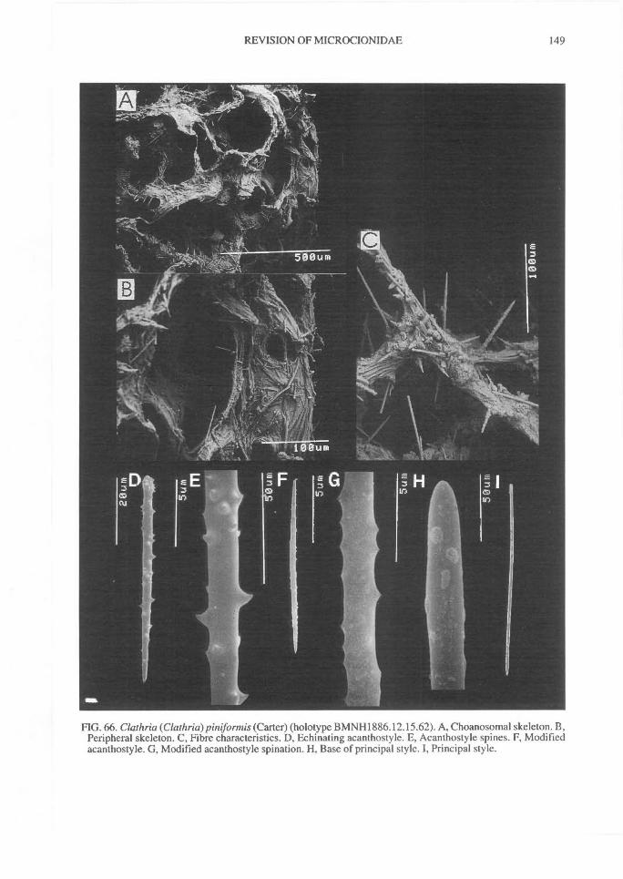
REVISION OF MICROCIONIDAE^
149
FIG. 66. Clathria (Clathria) piniformis (Carter) (holotype BMNH1886.12.15.62). A, Choanosomal skeleton. B,Peripheral skeleton. C, Fibre characteristics. D, Echinating acanthostyle. E, Acanthostyle spines. F, Modifiedacanthostyle. G, Modified acanthostyle spination. H, Base of principal style. 1, Principal style.
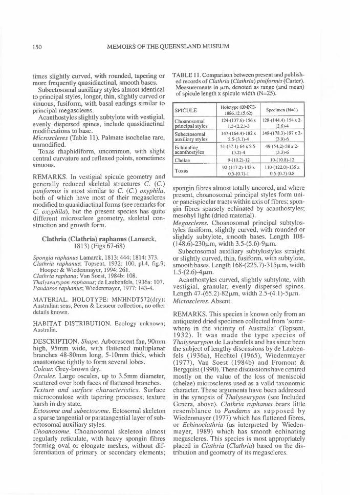
150^ MEMOIRS OF THE QUEENSLAND MUSEUM
times slightly curved, with rounded, tapering ormore frequently quasidiactinal, smooth bases.
Subectosomal auxiliary styles almost identicalto principal styles, longer, thin, slightly curved orsinuous, fusiform, with basal endings similar toprincipal megascleres.
Acanthostyles slightly subtylote with vestigial,evenly dispersed spines, include quasidiactinalmodifications to base.Microscleres (Table 11). Palmate isochelae rare,unmodified.
Toxas rhaphidiform, uncommon, with slightcentral curvature and reflexed points, sometimessinuous.
REMARKS. In vestigial spicule geometry andgenerally reduced skeletal structures C. (C.)pintformis is most similar to C. (C.) oxyphila,both of which have most of their megascleresmodified to quasidiactinal forms (see remarks forC. oxyphila), but the present species has quitedifferent microsclere geometry, skeletal con-struction and growth form.
Clathria (Clathria) raphanus (Lamarck,1813) (Figs 67-68)
Spongia raphanus Lamarck, 1813: 444; 1814: 373.Clathria raphanus; Topsent, 1932: 100, p1.4, fig.9;
Hooper & Wiedenmayer, 1994: 261.Clathria raphana; Van Soest, 1984b: 108.Thalyseurypon raphanus; de Laubenfels, 1936a: 107.Panda ros raphanus; Wiedenmayer, 1977: 143-4.
MATERIAL. HOLOTYPE: MNHNDT572(dry):Australian seas, Peron & Lesueur collection, no otherdetails known.
HABITAT DISTRIBUTION. Ecology unknown;Australia.
DESCRIPTION. Shape. Arborescent fan, 90mmhigh, 95mm wide, with flattened multiplanarbranches 48-80mm long, 5-10mm thick, whichanastomose tightly to form several lobes.Colour Grey-brown dry.Oscules. Large oscules, up to 3.5rnm diameter,scattered over both faces of flattened branches.Texture and surface characteristics. Surfacemicroconulose with tapering processes; textureharsh in dry state.Ectosome and subectosome. Ectosomal skeletona sparse tangential or paratangential layer of sub-ectosomal auxiliary styles.Choanosome. Choanosomal skeleton almostregularly reticulate, with heavy spongin fibresforming oval or elongate meshes, without dif-ferentiation of primary or secondary elements;
TABLE 11. Comparison between present and publish-ed records of Clathria (Clathria) piniformis (Carter).Measurements in pin, denoted as range (and mean)of spicule length x spicule width (N=25).
SPICULEHolotype (BWINH-
1886.12.15.62)Specimen (N=1)
Choanosomal 124-(137.6)-156 x 128-(144.4)-154 x 2-principal styles 1.5-(2.2.)-3 (2.6)4
Subectosomal 147-(164.4)-182 x 149-(178.3)-197 x 2-auxiliary styles 2.5-(3.1)-4 (3.9)-6
Echinating 51-(57.1)-64 x 2.5- 49-(54.2)-58 x 2-acanthostyles (3.2)-4 (3.3)-6
Chelae 9-(10.2)-12 10-(10.8)-1292-(117.2)-143 x 1104122.0)-135 x
Toxas 0.5-(0.7)-1 0.540.7)-0.8
spongin fibres almost totally uncored, and wherepresent, choanosomal principal styles form uni-or paucispicular tracts within axis of fibres; spon-gin fibres sparsely echinated by acanthostyles;mesohyl light (dried material).Megascleres. Choanosomal principal subtylos-tyles fusiform, slightly curved, with rounded orslightly subtylote, smooth bases. Length 108-(148.6)-230p.m, width 3.5-(5.6)-9p.m.
Subectosomal auxiliary subtylostyles straightor slightly curved, thin, fusiform, with subtylote,smooth bases. Length 168-(225.7)-315p,m, width1.5-(2.6)-4tim.
Acanthostyles curved, slightly subtylote, withvestigial, granular, evenly dispersed spines.Length 47-(65.2)-82p,m, width 2.5-(4.1)-51.tm.Microscleres. Absent.
REMARKS. This species is known only from anantiquated dried specimen collected from 'some-where in the vicinity of Australia' (Topsent,1932). It was made the type species ofThalyseurypon de Laubenfels and has since beenthe subject of lengthy discussions by de Lauben-fels (1936a), Hechtel (1965), Wiedenmayer(1977), Van Soest (1984b) and Fromont &Bergquist (1990). These discussions have centredmostly on the value of the loss of meniscoid(chelae) microscleres used as a valid taxonomiccharacter. These arguments have been addressedin the synopsis of Thalyseurypon (see IncludedGenera, above). Clathria raphanus bears littleresemblance to Pandaros as supposed byWiedenmayer (1977) which has flattened fibres,or Echinoclathria (as interpreted by Wieden-mayer, 1989) which has smooth echinatingmegascleres. This species is most appropriatelyplaced in Clathria (Clathria) based on the dis-tribution and geometry of its megascleres.
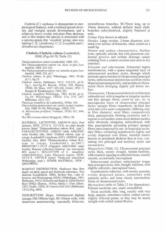
REVISION OF MICROCIONIDAE^ 151
Clathria (C.) raphanus is depauperate in mor-phological features, with a reduced spicule diver-sity and vestigial spicule development, and arelatively heavy evenly reticulate fibre skeleton,and in this respect the species is associated witha species-group (the oxyphila' group; also con-taining C. (C.) pinifonnis, C. (C.) oxyphila and C.(Dendrocia) elegantula).
Clathria (Clathria) rubens (Lendenfeld,1888) (Figs 69-70, Table 12)
Thalassodendron rubens Lendenfeld, 1888: 223.Not Thalassodendron rubens var. dura, in part, Len-
denfeld, 1888: 223-224.Not Thalassodendron rubens var. lamella, in part, Len-
denfeld, 1888: 224, p1.7.Clathria rubens, in part; Whitelegge, 1901: 85-86,
p1.11, fig.13.Clathria rubens; Hallmann, 1912: 218-223, p1.32,
fig.1, text-fig.45; Burton, 1934a: 558; Burton,1938b: 20; Shaw, 1927: 425-426; Guiler, 1950: 7;Hooper & Wiedenmayer, 1994: 261.
Clathria tenuifibra Whitelegge, 1901: 82-83, p1.11,fig.10; Whitelegge, 1902b: 274, 279, 287;Hallmann, 1912: 211.
Thalysias tenuifibra; de Laubenfels, 1936a: 105.Placochalina pedunculata var. monis, in part; Lenden-
feld, 1888: 91-92; Whitelegge, 1902b: 274.cf. Microciona prolifera; Vosmaer, 1935a: 611, 644,
668.Not Microciona rubens Bergquist, 1961a: 38.
MATERIAL. LECTOTYPE: AMG9119 (dry): PortJackson, NSW, 33°51'S, 151°16'E, no other detailsknown (label `Thalassodendron rubens RvL, type').PARALECTOTYPES: AMZ455 (slide AMG3585:same locality (dry, label 'Clathria rubens, type orcotype, Lendenfeld's duplicate A50'). AMZ634: samelocality; (dry, label `Thalassodendron rubens RvL,type or cotype, Lendenfeld's no.240').BMNH1887.1.24.35 (fragment AMG3586): samelocality, Ramsay collection (listed as 'var. intermediaMS name'). HOLOTYPE of C. tenuifibra:AMG3045(dry): Lake Illawarra region, NSW,34°32'S, 150°50'E (label `Thalysias tenuifibraWhitelegge; type'). OTHER MATERIAL: NSW -QMG300452.
HABITAT DISTRIBUTION. Shallower than 56mdepth; on shell, gravel and Halimeda substrates; PortJackson (Lendenfeld, 1888), Broken Bay, Lake II-lawarra, and Woolongong (NSW) (Whitelegge, 1901;Hallmann, 1912); Direction and Eagle Is, Great BarrierReef (FNQ) (Burton, 1934a); Maria I. (Tas) (Shaw,1927; Guiler, 1950); St Vincent Gulf (SA) (Hallmann,1912) (Fig. 69G).
DESCRIPTION. Shape. Arboresecent digitatesponge,100-190mm high, 60-110mm wide, withnumerous anastomosing, repeatedly bifurcate,
stoloniferous branches, 30-75mm long, up to19mm diameter, without definite basal stalk;branches subcylindrical, slightly flattened atends.Colour Grey-brown in ethanol.Oscules. Large oscules, 2-4mm diameter, scat-tered over surface of branches, often raised on asmall lip.Texture and surface characteristics. Surfaceeven, optically smooth, but with prominent sub-dermal grooves and stellate drainage canalsradiating from a central osculum (not seen in drymaterial).Ectosome and subectosome. Ectosomal regionmembraneous, with a sparse tangential layer ofsubectosomal auxiliary styles, through whichprotrude sparse brushes of choanosomal principalstyles; subectosomal skeleton has auxiliary stylesdispersed paratangentially, and peripheral choan-somal fibres diverging slightly just below sur-face.Choanosome. Choanosomal skeletal architecturesubisodictyal, with triangular or irregular meshesand thin spongin fibres, cored by uni-, bi- orpaucispicular tracts of choanosomal principalstyles; spongin fibres imperfectly divided intoaxial and extra-axial components, differing onlyin fibre diameter; axial fibres comparativelythick, paucispicular, forming cavernous and ir-regular ovoid meshes; extra-axial skeletal meshesmore obviously triangular, subisodictyal, withthin, paucispicular ascending primary sponginfibres interconnected by uni- or bispicular secon-dary fibres; echinating acanthostyles lightly andevenly dispersed over fibres; mesohyl matrixheavier in peripheral skeleton than at core, con-taining few principal and auxiliary styles andmicroscleres.Megascleres (Table 12). Choanosomal principalstyles thick, mostly straight, hastate-fusiform,with rounded, tapering or subtylote bases, usuallysmooth, occasionally microspined.
Subectosomal auxiliary subtylostyles longerthan principal styles, thin, straight, fusiform, withslightly subtylote, microspined bases.
Acanthostyles subtylote, with mostly granular,evenly dispersed spines, sometimes withaspinose necks, and some clearly intermediateforms of choanosomal styles.Microscleres (refer to Table 12 for dimensions).Palmate isochelae rare, small, unmodified.
Toxas accolada, thin, long, usually with verylarge, angular, sometimes coiled, central curves,slightly reflexed points, or they may be nearlystraight with coiled central flexion.
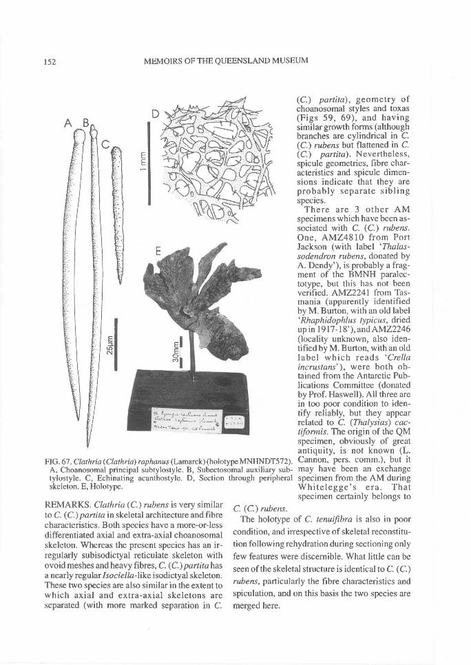
FIG. 67. Clathria (Clathria) raphanus (Lamarck) (holotype MNHNDT572).A, Choanosomal principal subtylostyle. B, Subectosomal auxiliary sub-tylostyle. C, Echinating acanthostyle. D, Section through peripheralskeleton. E, Holotype.
152^ MEMOIRS OF THE QUEENSLAND MUSEUM
REMARKS. Clathria (C.) rubens is very similarto C. (C.)partita in skeletal architecture and fibrecharacteristics. Both species have a more-or-lessdifferentiated axial and extra-axial choanosomalskeleton. Whereas the present species has an ir-regularly subisodictyal reticulate skeleton withovoid meshes and heavy fibres, C. (C.)partita hasa nearly regularlsociella-like isodictyal skeleton.These two species are also similar in the extent towhich axial and extra-axial skeletons areseparated (with more marked separation in C.
(C.) partita), geometry ofchoanosomal styles and toxas(Figs 59, 69), and havingsimilar growth forms (althoughbranches are cylindrical in C.(C.) rubens but flattened in C.(C.) part ita). Nevertheless,spicule geometries, fibre char-acteristics and spicule dimen-sions indicate that they areprobably separate siblingspecies.
There are 3 other AMspecimens which have been as-sociated with C. (C.) rubens.One, AMZ4810 from PortJackson (with label `Thalas-sodendron rubens, donated byA. Dendy'), is probably a frag-ment of the BMNH paralec-totype, but this has not beenverified. AMZ2241 from Tas-mania (apparently identifiedby M. Burton, with an old labelRhaphidophlus typicus, driedup in 1917-18'), and AMZ2246(locality unknown, also iden-tified by M. Burton, with an oldlabel which reads `Crellaincrustans'), were both ob-tained from the Antarctic Pub-lications Committee (donatedby Prof. Haswell). All three arein too poor condition to iden-tify reliably, but they appearrelated to C. (Thalysias) cac-tifonnis. The origin of the QMspecimen, obviously of greatantiquity, is not known (L.Cannon, pers. comm.), but itmay have been an exchangespecimen from the AM duringWhitelegge's era. Thatspecimen certainly belongs to
C. (C.) rubens.The holotype of C. tenuifibra is also in poor
condition, and irrespective of skeletal reconstitu-tion following rehydration during sectioning onlyfew features were discernible. What little can beseen of the skeletal structure is identical to C. (C.)rubens, particularly the fibre characteristics andspiculation, and on this basis the two species aremerged here.
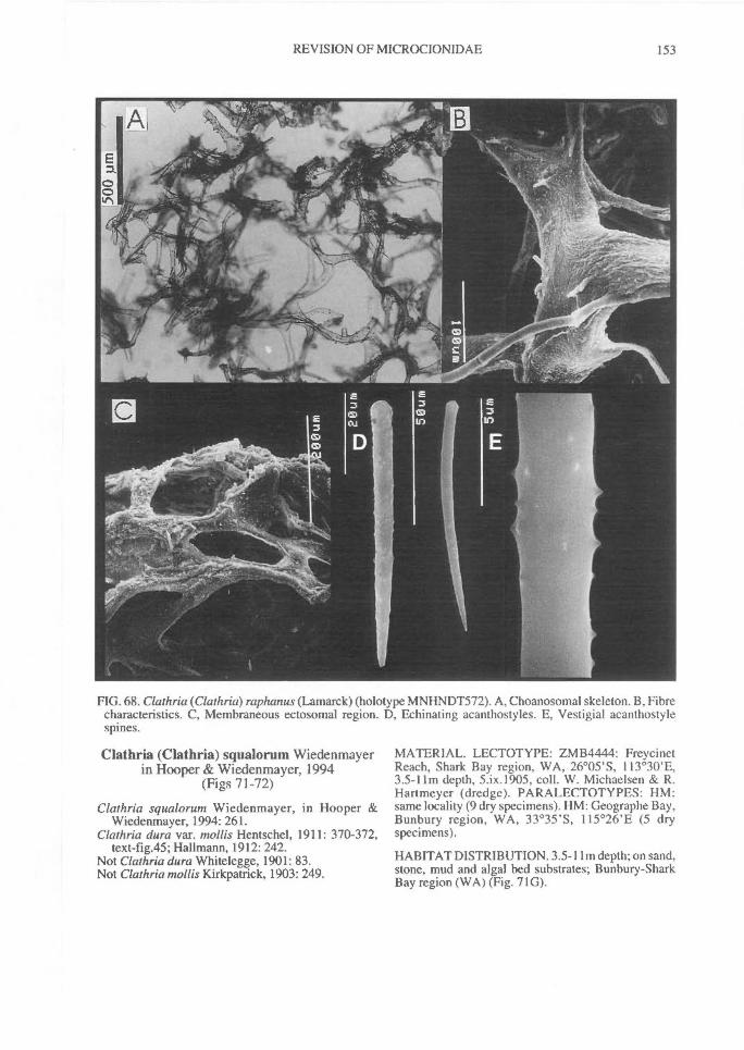
REVISION OF MICROCIONIDAE^ 153
FIG. 68. Clathria (Clathria) raphanus (Lamarck) (holotype MNHNDT572). A, Choanosomal skeleton. B, Fibrecharacteristics. C, Membraneous ectosomal region. D, Echinating acanthostyles. E, Vestigial acanthostylespines.
Clathria (Clathria) squalorum Wiedenmayerin Hooper & Wiedenmayer, 1994
(Figs 71-72)
Clathria squalorum Wiedenmayer, in Hooper &Wiedenmayer, 1994: 261.
Clathria dura var. moths Hentschel, 1911: 370-372,text-fig.45; Hallmann, 1912: 242.
Not Clathria dura Whitelegge, 1901: 83.Not Clathria mollis Kirkpatrick, 1903: 249.
MATERIAL. LECTOTYPE: ZMB4444: FreycinetReach, Shark Bay region, WA, 26°05'S, 113°30'E,3.5-11m depth, 5.ix.1905, coll. W. Michaelsen & R.Hartmeyer (dredge). PA RA LECTOTYPES: HM:same locality (9 dry specimens). HM: Geographe Bay,Bunbury region, WA, 33°35'S, 115°26'E (5 dryspecimens).
HABITAT DISTRIBUTION. 3.5-11m depth; on sand,stone, mud and algal bed substrates; Bunbury-SharkBay region (WA) (Fig. 71G).
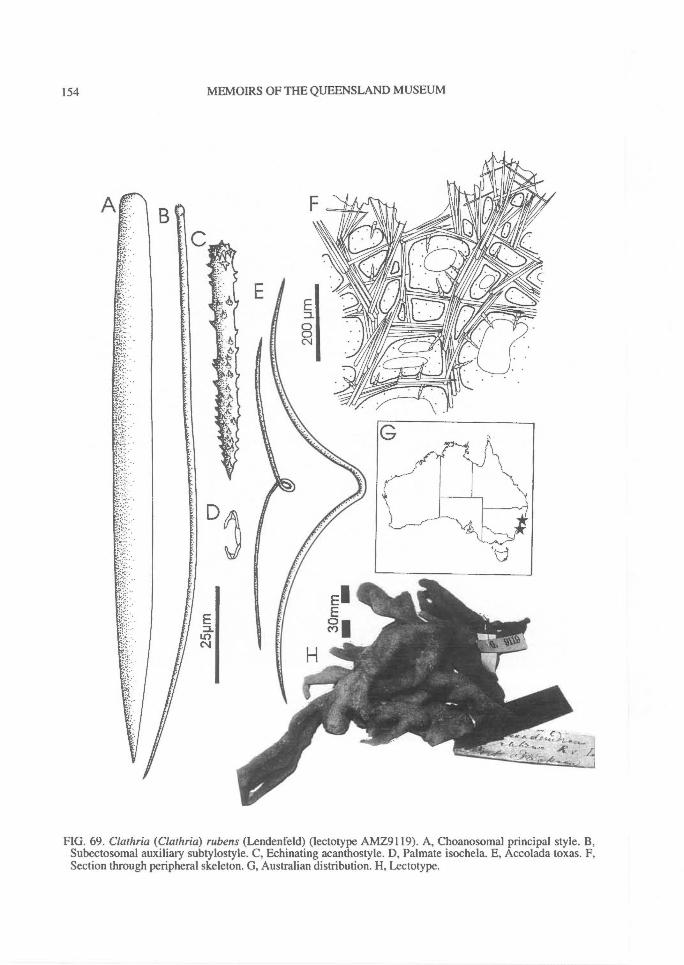
154^ MEMOIRS OF THE QUEENSLAND MUSEUM
FIG. 69. Clathria (Clathria) rubens (Lendenfeld) (lectotype AMZ9119). A, Choanosomal principal style. B,Subectosomal auxiliary subtylostyle. C, Echinating acanthostyle. D, Palmate isochela. E, Accolada toxas. F,Section through peripheral skeleton. G, Australian distribution. H, Lectotype.
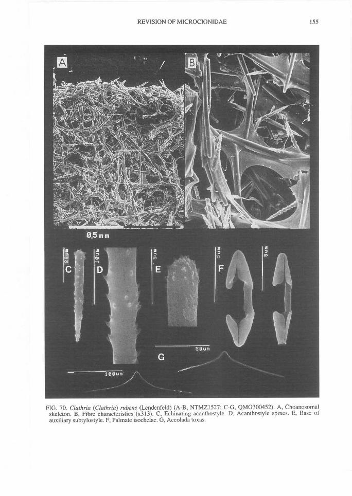
REVISION OF MICROCIONIDAE^
155
FIG. 70. Clathria (Clathria) rubens (Lendenfeld) (A-B, NTMZ1527; C-G, QMG300452). A, Choanosomalskeleton. B, Fibre characteristics (x313). C, Echinating acanthostyle. D, Acanthostyle spines. E, Base ofauxiliary subtylostyle. F, Palmate isochelae. G, Accolada toxas.

TABLE 12. Comparison between present and published records ofClathria (Clathria) rubens (Lendenfeld). All measurements are given inp.m, denoted as range (and mean) of spicule length x spicule width(N=25).
SPICULELectotype
(AMG9119)Paralectotypes
(N=3)
Holotype of C.tenuijibra
(AMG3045)Specimen (N=I)
Choanosomalprincipalstyles
96-(135.3)-162x 5-(9.0)-I I
128-(135.6)-147x 5.5-(7.1)-9
1124139.2)-159x 8-(9.4)-11
1184140.6)-155x 6-(7.8)-10
Subectosomalauxiliarystyles
106-(157.2)-212x 1.5-(2.9)-4
1324146.2)-168x 2.5-(2.9)-4
112-(153.7)-185x 2.543.7)-4.5
105-(139.2)-165x 2.5-(3.3)-4.5
Echinating 55-(71.8)-111 x 58-(77.3)-112 x 51-(68.6)-118 x 48-(69.4)-I08 xacanthostyles 3-(5.6)-7 4.5-(5.6)-6.5 5-(6.4)-8 4-(5.8)-7.5
Chelae 6-(9.6)-I3 7-(9.8)-13 8-(9.7)-11 6-(9.0)-12
Toxas101-(153.4)-215x 0.8-(1.3)-1.5
115-(161.6)-208x I-(1.4)-2
94-(126.3)-153x I-(1.2)-1.5
114-(138.6)-194x 1-(1.4)-2
156^ MEMOIRS OF THE QUEENSLAND MUSEUM
DESCRIPTION. Shape. Variable growth form,ranging from thickly encrusting with smallstoloniferous digits, to digitate planar arborescentwith anastomosing branches.Colour Live colouration unknown, beigepreserved.Oscules. Large oscules on apex and lateral mar-gins of stoloniferous branchesTexture and surface characteristics. Surfacemicroconulose.Ectosome and subectosome. Ectosomal skeletonhispid, with points of choanosomal principalstyles protruding through surface individually orin small bundles, piercing a sparse paratangentiallayer of subectosomal auxiliary subtylostyles.Choanosome. Choanosomal skeletal architecturewide-meshed nearly renieroid reticulate, moreregular in peripheral skeleton than in axis, withdifferentiated primary (ascending, multispicular)and secondary (transverse, pauci- or unispicular)spongin fibres; fibre diameter generally small(primaries 30-501..im; secondaries 10-22p,m),fibre anastomoses rectangular, and fibres coredby choanosomal principal styles; coring spiculesin axial region heavier, more plumose (protrudingthrough fibres) than in peripheral skeleton;echinating acanthostyles relatively sparse, evenlydistributed throughout skeleton; mesohyl matrixlight, usually containing choanosomal and/orsubectosomal megascleres dispersed between fibres.Megascleres. Choanosomal principal subtylos-tyles slightly curved, fusiform, entirely smooth,very slightly subtylote. Length 124-(152.4)-165Rm, width 4-(8.8)-12Rm.
Subectosomal auxiliary subtylostyles straight,thin, fusiform, with smooth slightly subtylote
bases. Length 854148.5)-184p.m, width 242.8)-411,m.
Acanthostyles slightly sub-tylote, with evenly dispersedrelatively large spines, slightlyless spinose below basal region.Length 48-(62.3)-7411m, width2-(4.2)-61.m.Microscleres. Palmate isochelaeunmodified. Length 448.6)-12Rm long.
Accolada toxas very thin,straight arms, sharply angularcentral curvature, occasionallyraphidiform. Length 105-220v,m, width 1-(2.2)-311,m.
REMARKS. Hallmann (1912)noted that C. dura var. mollis
Hentschel was distinctly different from C. duraWhitelegge and he considered that Hentschel's(1911) species required a new name. The sub-specific name 'moths' is preoccupied by C. monisKirkpatrick, 1903, and so a new name C.squalorwn was proposed by Wiedenmayer (inHooper & Wiedenmayer, 1994). Some of thecharacters and measurements of the lectotypediffer from those published by Hentschel (1911).His description was presumably based on a seriesof syntypes, but these have not been located orre-examined.
Comparison between the type material of C.(C.) squalorum and C. (Dendrocia) dura showthat both species are different in several sig-nificant respects: (1) differences in the geometryof structural spicules where C. (C.) squalorumhas differentiated principal and auxiliaryspicules (and hence is referrable to C.(Clathria)), and C. (D.) dura has completely un-differentiated structural megascleres (and henceis referrable to C. (Dendrocia)); (2) palmateisochelae versus arcuate-like isochelae; (3) longthick accolada toxas versus no toxas; (4) nearlyrenieroid reticulation of thin fibres versus aregularly reticulate skeleton with heavy fibres,respectively. In having a slightly renieroid skele-tal architecture C. (C.) squalorum is reminiscentof C. (lsociella) eccentrica, although spiculegeometry and spicule dimensions are otherwisedifferent.
Clathria (Clathria) striata Whitelegge, 1907(Figs 73-74, Plate 1F, Table 13)
Clathria striata Whitelegge, 1907: 495-496, p1.45,fig.27; Hooper, & Wiedenmayer, 1994: 261.
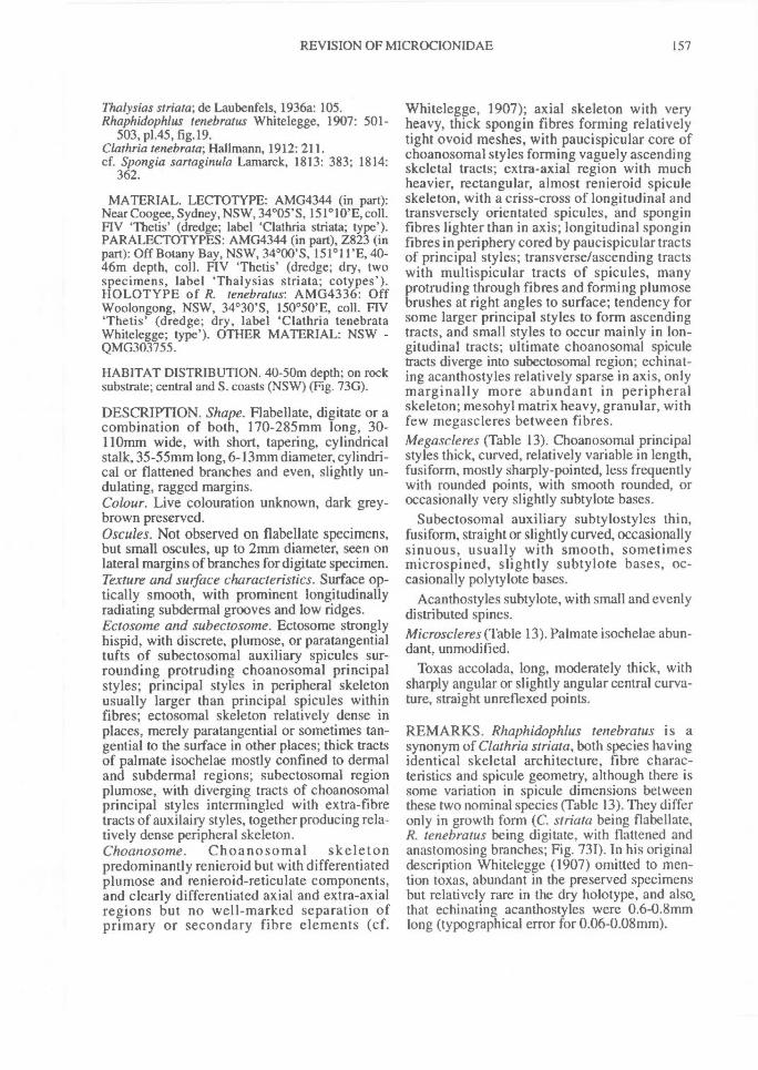
REVISION OF MICROCIONIDAE^ 157
Thalysias striata; de Laubenfels, 1936a: 105.Rhaphidophlus tenebratus Whitelegge, 1907: 501-
503, p1.45, fig.I9.Clathria tenebrata; Hallmann, 1912: 211.cf. Spongia sartaginula Lamarck, 1813: 383; 1814:
362.
MATERIAL. LECTOTYPE: AMG4344 (in part):Near Coogee, Sydney, NSW, 34°05'S, 151°10'E, coll.Hy 'Thetis' (dredge; label 'Clathria striata; type').PARALECTOTYPES: AMG4344 (in part), Z823 (inpart): Off Botany Bay, NSW, 34°00'S, 151°11'E, 40-46m depth, coll. FIV 'Thetis' (dredge; dry, twospecimens, label `Thalysias striata; cotypes').HOLOTYPE of R. tenebratus: AMG4336: OffWoolongong, NSW, 34°30'S, 150°50'E, coll. FIV'Thetis' (dredge; dry, label 'Clathria tenebrataWhitelegge; type'). OTHER MATERIAL: NSW -QMG303755.
HABITAT DISTRIBUTION. 40-50m depth; on rocksubstrate; central and S. coasts (NSW) (Fig. 73G).
DESCRIPTION. Shape. Flabellate, digitate or acombination of both, 170-285mm long, 30-110mm wide, with short, tapering, cylindricalstalk, 35-55mm long, 6-13mm diameter, cylindri-cal or flattened branches and even, slightly un-dulating, ragged margins.Colour. Live colouration unknown, dark grey-brown preserved.Oscules. Not observed on flabellate specimens,but small oscules, up to 2mm diameter, seen onlateral margins of branches for digitate specimen.Texture and surface characteristics. Surface op-tically smooth, with prominent longitudinallyradiating subdermal grooves and low ridges.Ectosome and subectosome. Ectosome stronglyhispid, with discrete, plumose, or paratangentialtufts of subectosomal auxiliary spicules sur-rounding protruding choanosomal principalstyles; principal styles in peripheral skeletonusually larger than principal spicules withinfibres; ectosomal skeleton relatively dense inplaces, merely paratangential or sometimes tan-gential to the surface in other places; thick tractsof palmate isochelae mostly confined to dermaland subdermal regions; subectosomal regionplumose, with diverging tracts of choanosomalprincipal styles intermingled with extra-fibretracts of auxilairy styles, together producing rela-tively dense peripheral skeleton.Choanosome. Choanosomal skeletonpredominantly renieroid but with differentiatedplumose and renieroid-reticulate components,and clearly differentiated axial and extra-axialregions but no well-marked separation ofprimary or secondary fibre elements (cf.
Whitelegge, 1907); axial skeleton with veryheavy, thick spongin fibres forming relativelytight ovoid meshes, with paucispicular core ofchoanosomal styles forming vaguely ascendingskeletal tracts; extra-axial region with muchheavier, rectangular, almost renieroid spiculeskeleton, with a criss-cross of longitudinal andtransversely orientated spicules, and sponginfibres lighter than in axis; longitudinal sponginfibres in periphery cored by paucispicular tractsof principal styles; transverse/ascending tractswith multispicular tracts of spicules, manyprotruding through fibres and forming plumosebrushes at right angles to surface; tendency forsome larger principal styles to form ascendingtracts, and small styles to occur mainly in lon-gitudinal tracts; ultimate choanosomal spiculetracts diverge into subectosomal region; echinat-ing acanthostyles relatively sparse in axis, onlymarginally more abundant in peripheralskeleton; mesohyl matrix heavy, granular, withfew megascleres between fibres.Megascleres (Table 13). Choanosomal principalstyles thick, curved, relatively variable in length,fusiform, mostly sharply-pointed, less frequentlywith rounded points, with smooth rounded, oroccasionally very slightly subtylote bases.
Subectosomal auxiliary subtylostyles thin,fusiform, straight or slightly curved, occasionallysinuous, usually with smooth, sometimesmicrospined, slightly subtylote bases, oc-casionally polytylote bases.
Acanthostyles subtylote, with small and evenlydistributed spines.Microscleres (Table 13). Palmate isochelae abun-dant, unmodified.
Toxas accolada, long, moderately thick, withsharply angular or slightly angular central curva-ture, straight unreflexed points.
REMARKS. Rhaphidophlus tenebratus is asynonym of Clathria striata, both species havingidentical skeletal architecture, fibre charac-teristics and spicule geometry, although there issome variation in spicule dimensions betweenthese two nominal species (Table 13). They differonly in growth form (C. striata being flabellate,R. tenebratus being digitate, with flattened andanastomosing branches; Fig. 731). In his originaldescription Whitelegge (1907) omitted to men-tion toxas, abundant in the preserved specimensbut relatively rare in the dry holotype, and also.that echinating acanthostyles were 0.6-0.8mmlong (typographical error for 0.06-0.08mm).
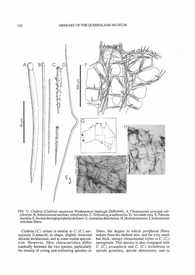
158^ MEMOIRS OF THE QUEENSLAND MUSEUM
FIG. 71. Clathria (Clathria) squalorum Wiedenmayer (holotype ZMB4444). A, Choanosomal principal sub-tylostyle. B, Subectosomal auxiliary subtylostyles. C, Echinating acanthostyles. D, Accolada toxa. E, Palmateisochela. F, Section through peripheral skeleton. G, Australian distribution. H, Skeletal structure. I, Subrenieroidreticulate fibres.
Clathria (C.) striata is similar to C. (C.) sar-taginula (Lamarck) in shape, slightly renieroidskeletal architecture, and to some extent spicula-tion. However, fibre characteristics differmarkedly between the two species, particularlythe density of coring and echinating spicules on
fibres, the degree to which peripheral fibresradiate from the skeletal axis, and the very smallbut thick, stumpy choanosomal styles in C. (C.)sartaginula. This species is also compared withC. (C.) arcuophora and C. (C.) biclathrata inspicule geometry, spicule dimensions, and to

REVISION OF MICROCIONIDAE^ 159
FIG. 72. Clathria (Clathria) squalorum Wiedenmayer (holotype ZMB4444). A, Choanosomal skeleton. B,Ectosomal skeleton. C, Fibre characteristics. D, Echinating acanthostyle. E, Acanthostyle spines. F, Base ofauxiliary subtylostyle. G, Accolada toxa. H, Palmate isochelae.
some extent fibre characteristics. Clathria (C.)striata also has a similar skeletal architecture asC. (C.) arcuophora, differing substantially onlyin toxa geometry. All these species (C. (C.)striata, C. (C.) arruophora, C. (C.) sartaginula,
C. (C.) crassa and C. (C.) biclathrata), are char-acterised by their slightly renieroid or sub-renieroid choanosomal skeletons and sponginfibres cored by few, thick principal spicules. Theyare grouped together here in the striata' species

160^ MEMOIRS OF THE QUEENSLAND MUSEUM
FIG. 73. Clathria (Clathria) striata Whitelegge (lectotype AMZ4344). A, Choanosomal principal style. B,Subectosomal auxiliary subtylostyle. C, Echinating acanthostyle. D, Accolada toxa. E, Palmate isochela. F,Section through peripheral skeleton. G, Australian distribution. H, Lectotype. I, Holotype of R. tenebratusAMG4336.

REVISION OF MICROCIONIDAE^
161
FIG. 74. Clathria (Clathria) striata Whitelegge (paralectotype AMZ823). A, Choanosomal skeleton. B,Peripheral skeleton. C, Fibre characteristics. D, Echinating acanthostyle. E, Acanthostyle spines. F, Palmateisochelae. G, Accolada toxas. H, Base of auxiliary subtylostyle.
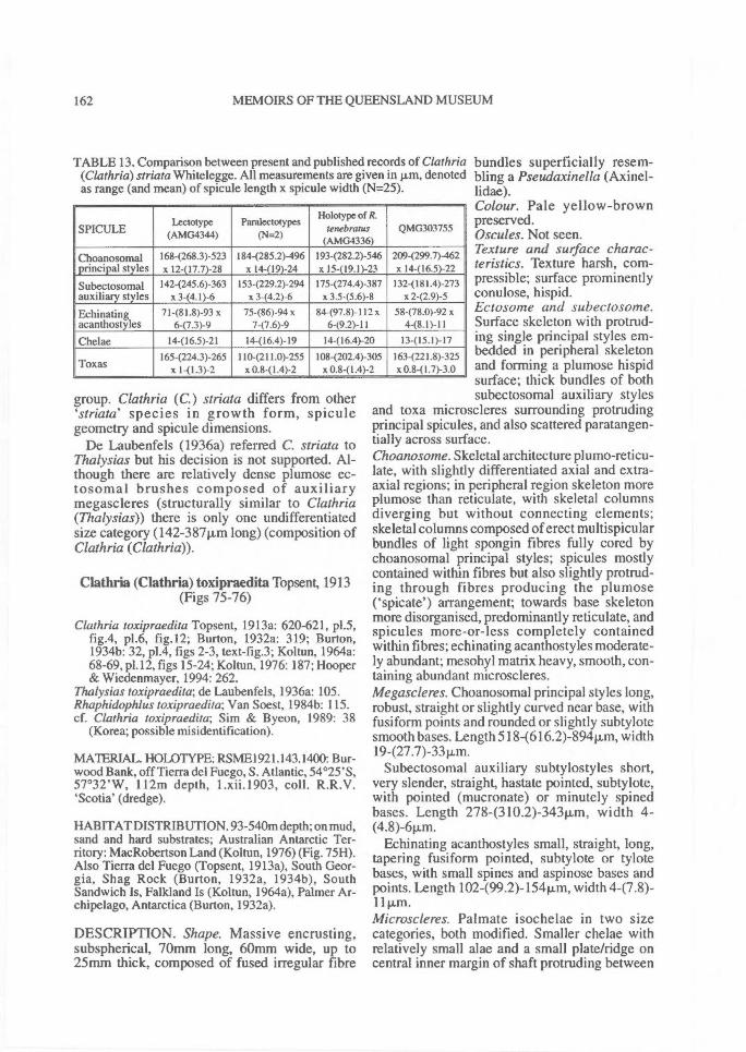
TABLE 13. Comparison between present and published records of Clathria(Clathria) striata Whitelegge. All measurements are given in pm, denotedas range (and mean) of spicule length x spicule width (N=25).
SPICULELectotype
(AMG4344)Paralectotypes
(N=2)
Holotype of R.tenebratus
(AMG4336)QMG303755
Choanosomal 1684268.3)-523 184-(285.2)-496 193-(282.2)-546 2094299.7)-462principal styles x 12-(17.7)-28 x 14-(19)-24 x 15-(19.1)-23 x 14-(16.5)-22
Subectosomal 142-(245.6)-363 153-(229.2)-294 175-(274.4)-387 132-(181.4)-273auxiliary styles x 3-(4. 1)-6 x 3-(4.2)-6 x 3.5-(5.6)-8 x 2-(2.9)-5
Echinating 71-(81.8)-93 x 75-(86)-94 x 84-(97.8)-112 x 58-(78.0)-92 xacanthostyles 6-(7.3)-9 7-(7.6)-9 6-(9.2)-11 4-(8.1)-11
Chelae 14-(16.5)-21 14-(16.4)-19 I4-(16.4)-20 13-(15.1)-17
165-(224.3)-265 110-(211.0)-255 I 08-(202.4)-305 163-(221.8)-325Toxas x 1-(1.3)-2 x 0.8-(1.4)-2 x 0.8-(1.4)-2 x 0.841.7)-3.0
162^ MEMOIRS OF THE QUEENSLAND MUSEUM
group. Clathria (C.) striata differs from otherstriata' species in growth form, spicule
geometry and spicule dimensions.De Laubenfels (1936a) referred C. striata to
Thalysias but his decision is not supported. Al-though there are relatively dense plumose ec-tosomal brushes composed of auxiliarymegascleres (structurally similar to Clathria(Thalysias)) there is only one undifferentiatedsize category (142-38711m long) (composition ofClathria (Clathria)).
Clathria (Clathria) toxipraedita Topsent, 1913(Figs 75-76)
Clathria toxipraedita Topsent, 1913a: 620-621, p1.5,fig.4, p1.6, fig.12; Burton, 1932a: 319; Burton,1934b: 32, p1.4, figs 2-3, text-fig.3; Koltun, 1964a:68-69, p1.12, figs 15-24; Koltun, 1976: 187; Hooper& Wiedenmayer, 1994: 262.
Thalysias toxipraedita; de Laubenfels, 1936a: 105.Rhaphidophlus toxipraedita; Van Soest, 1984b: 115.cf. Clathria toxipraedita; Sim & Byeon, 1989: 38
(Korea; possible misidentification).
MATERIAL. HOLOTYPE: RSME1921.143.1400: Bur-wood Bank, off Tierra del Fuego, S. Atlantic, 54°25'S,57°32'W, 112m depth, 1.xii.1903, coll. R.R.V.'Scotia' (dredge).
HABITAT DISTRIBUTION. 93-540m depth; on mud,sand and hard substrates; Australian Antarctic Ter-ritory: MacRobertson Land (Koltun, 1976) (Fig. 7511).Also Tierra del Fuego (Topsent, 1913a), South Geor-gia, Shag Rock (Burton, 1932a, 1934b), SouthSandwich Is, Falkland Is (Koltun, 1964a), Palmer Ar-chipelago, Antarctica (Burton, 1932a).
DESCRIPTION. Shape. Massive encrusting,subspherical, 70mm long, 60mtn wide, up to25mm thick, composed of fused irregular fibre
bundles superficially resem-bling a Pseudaxinella (Axinel-lidae).Colour. Pale yellow-brownpreserved.Oscules. Not seen.Texture and surface charac-teristics. Texture harsh, com-pressible; surface prominentlyconulose, hispid.Ectosome and subectosome.Surface skeleton with protrud-ing single principal styles em-bedded in peripheral skeletonand forming a plumose hispidsurface; thick bundles of bothsubectosomal auxiliary styles
and toxa microscleres surrounding protrudingprincipal spicules, and also scattered paratangen-tially across surface.Choanosonze. Skeletal architecture plumo-reticu-late, with slightly differentiated axial and extra-axial regions; in peripheral region skeleton moreplumose than reticulate, with skeletal columnsdiverging but without connecting elements;skeletal columns composed of erect multispicularbundles of light spongin fibres fully cored bychoanosomal principal styles; spicules mostlycontained within fibres but also slightly protrud-ing through fibres producing the plumose('spicate) arrangement; towards base skeletonmore disorganised, predominantly reticulate, andspicules more-or-less completely containedwithin fibres; echinating acanthostyles moderate-ly abundant; mesohyl matrix heavy, smooth, con-taining abundant microscleres.Megascleres. Choanosomal principal styles long,robust, straight or slightly curved near base, withfusiform points and rounded or slightly subtylotesmooth bases. Length 5184616.2)-894 width19-(27.7)-33Rm.
Subectosomal auxiliary subtylostyles short,very slender, straight, hastate pointed, subtylote,with pointed (mucronate) or minutely spinedbases. Length 278-(310.2)-343p.m, width 4-(4.8)-61.1,m.
Echinating acanthostyles small, straight, long,tapering fusiform pointed, subtylote or tylotebases, with small spines and aspinose bases andpoints. Length 102499.2)-1541m, width 4-(7.8)-11 vm.Microscleres. Palmate isochelae in two sizecategories, both modified. Smaller chelae withrelatively small alae and a small plate/ridge oncentral inner margin of shaft protruding between
REVISION OF MICROCIONIDAE^ 163
the alae. Length 12-(15.7)-18Rm. Larger chelaeoften cleistochelate, with teeth partially or com-pletely fused and a large central plate/ridge oninner margin of shaft between alae. Length 20-(22.3)-24Rm.
Two categories of toxas. Longer toxas ac-colada, very long, thick, with sharply angular butonly slight central curvature, straight arms andsmooth fusiform points. Length 63541022.6)-147011m, width 4-(5.2)-6Rm. Smaller toxaswing-shaped, more widely curved at centre, oc-casionally looped/twisted at centre, with slightlyreflexed arms and points. Length 56485.3)-132p,m, width 0.5-(1.1)-2.0pLm.
REMARKS. Clathria (C.) toxipraedita haspeculiar, large palmate cleistochelae resemblingsigmancistras (in some Mycalidae), with alaevarying from partially to completely fused, andwith the addition of a well formed central plate-like ridge protruding between the alae (Fig. 76G).Smaller chelae have smaller diffuse alae and asmaller central plate-like ridge, indicating thatthey are ontogenetic stages of the larger forms.Cleistochelae are known in a number of otherpoecilosclerids including a several microcionids(e.g. C. (Microciona)cleistochela Topsent, C. (T)ramosa (Dendy),Antho(Plocamia)signata (Top-sent)), whereas the central plate-like ridge ap-pears to be unique to the family and onlypreviously recorded from Mycalidae (E. Hajdu,pers.comm.). Clathria toxipraedita is unusualamongst the antiboreal species in having a greatdiversity of spicule geometries; generallymicrocionids from Antarctic waters appear tohave reduced spicule diversity in comparison tothe tropical fauna.
Clathria (Clathria) transiens Hallmann, 1912(Figs 77-78, Plate 2A, Table 14)
Clathriatransiens Hallmann, 1912: 205, 226-234, 253,254, p1.33, figs 1-3, p1.34, fig.2, text-figs 47-48;Shaw, 1927:426; Burton, 1934a: 599; Guiler, 1950:7; Burton, 1959a: 244; Wiedenmayer, 1989: 57,p1.5, fig.8, p1.23, fig.6, text-fig.38; Hooper &Wiedenmayer, 1994: 262.
Thalysias transiens; de Laubenfels, 1936a: 105.cf. Microciona prolifera, tropus stylota and tropus
senta, Vosmaer, 1935a: 611, 649-650, 666.
MATERIAL. LECTOTYPE: AME302: Off Devon-port, N. coast Tas, 41°11'S, 146°21'E, coll. FIV'Endeavour' (dredge) (label 'Clathria transiens; typicalform a'). PARALECTOTYPES: AMZ49 + E819:64km W. of Kingston, SA, 36°50'S, 139°05'E, 60mdepth, coll. FIV 'Endeavour' (dredge) (specimen cut inhalf, form b). AME779: same locality; (form c).
AMZ744: Port Phillip Bay, Vic, 38°09'S, 144°52'E,coll. J.B. Wilson (dredge) (label 'Clathria transitaHallmann', AM register ref. 342/85, JBW sp.3, cotype,form d). (uncertain type status - AMZ743: Port PhillipBay, Vic., 38°09'S, 144°52'E, coll. J.B. Wilson;(dredge; label 'Clathria transita Hallmann', dry,`type')). OTHER MATERIAL: VIC. - NCIQ66C-3231-C (fragment NTMZ3694). TAS. - QMG300268(NCIQ66C-3638-U, fragment NTMZ3802).
HABITAT DISTRIBUTION. Shallow subtida1-60mdepth; on rock substrates; Port Phillip (Vic) (Hallmann,1912; present study), Furneaux Is and Devonport (Tas)(Shaw, 1927; Guiler, 1950; Wiedenmayer, 1989;present study), Kingston (SA) (Hallmann, 1912), Qld.(Burton, 1934a) (Fig. 77G). Burton (1959a) recordedthe species from the Red Sea but his material was notdescribed, and his record is questionable.
DESCRIPTION. Shape. Usually arborescentwith short, bulbous branches, 75-185mm long,40-160mm wide; stipitate with short cylindricalstalk, 25-55mm long, 8-22mm diameter;branches simply bifurcate, or repeatedly bifur-cate and arborescent, flabello-digitate, or theymay anastomose to form thickly clathrous, bushylobes.Colour. Live colouration vermilion red (Munsell5R 3/10), beige-brown or dark brown preserved.Oscules. Small oscules, up to 3mm diameter, inridges and grooves of branches.Texture and surface characteristics. Surfacemarkedly conulose, with conules actually beingrounded vestigial branches; with or without ir-regular ridges striating surface; surface coveredby distinct membraneous skin-like ectosome.Ectosome and subectosome. Ectosome minutelyhispid, with the points of larger choanosomalprincipal styles protruding through membraneoussurface, with a paucispicular, usually tangentiallayer of subectosomal auxiliary subtylostyles,lying on or just below surface; occasionallyauxiliary styles form plumose brushes, surround-ing 1 or more principal style; subectosomal skele-tal structure vaguely plumose, formed bydiverging peripheral spongin fibres and spiculetracts of smaller choanosomal principal styles.Choanosome. Choanosomal skeletal architecturealmost regular, radial-renieroid, with poorly dif-ferentiated axial and extra-axial skeletons; axisformed by evenly anastomosing spongin fibres,forming relatively cavernous, rectangular or cir-cular meshes; spongin fibres cored by pauci- ormultispicular tracts of smaller choanosomal prin-cipal styles; primary and secondary skeletal tractsin axis vaguely differentiated, but notpronounced, and spongin fibre elements only dif-
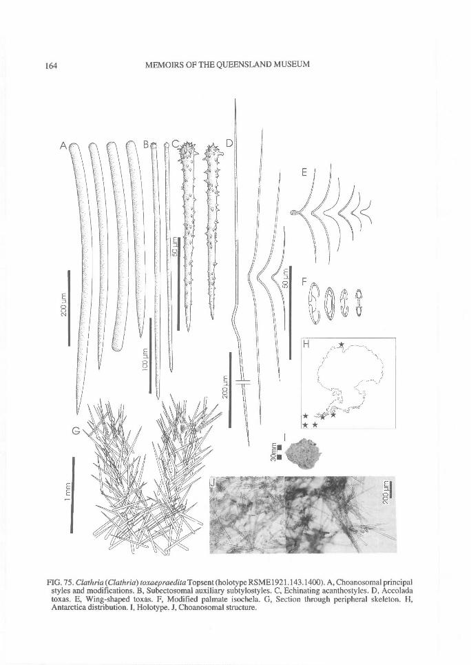
A
E00CV
164^ MEMOIRS OF THE QUEENSLAND MUSEUM
FIG. 75. Clathria(Clathria)toxaepraedita Topsent (holotype RSME1921.143.1400). A, Choanosomal principalstyles and modifications. B, Subectosomal auxiliary subtylostyles. C, Echinating acanthostyles. D, Accoladatoxas. E, Wing-shaped toxas. F, Modified palmate isochela. G, Section through peripheral skeleton. H,Antarctica distribution. I, Holotype. J, Choanosomal structure.
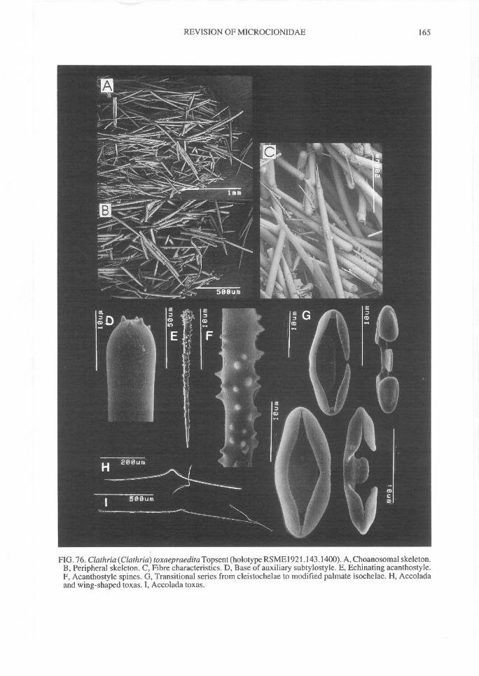
REVISION OF MICROCIONIDAE^
165
FIG. 76. Clathria(Clathria)toxaepraedita Topsent (holotype RSME1921.143.1400). A, Choanosomal skeleton.B, Peripheral skeleton. C, Fibre characteristics. D, Base of auxiliary subtylostyle. E, Echinating acanthostyle.F, Acanthostyle spines. G, Transitional series from cleistochelae to modified palmate isochelae. H, Accoladaand wing-shaped toxas. I, Accolada toxas.
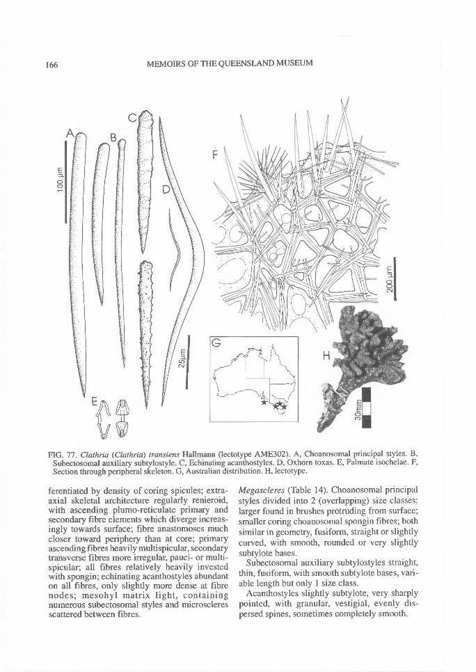
A
166^ MEMOIRS OF THE QUEENSLAND MUSEUM
FIG. 77. Clathria (Clathria) transiens Hallmann (lectotype AME302). A, Choanosomal principal styles. B,Subectosomal auxiliary subtylostyle. C, Echinating acanthostyles. D, Oxhorn toxas. E, Palmate isochelae. F,Section through peripheral skeleton. G, Australian distribution. H, lectotype.
ferentiated by density of coring spicules; extra-axial skeletal architecture regularly renieroid,with ascending plumo-reticulate primary andsecondary fibre elements which diverge increas-ingly towards surface; fibre anastomoses muchcloser toward periphery than at core; primaryascending fibres heavily multispicular, secondarytransverse fibres more irregular, pauci- or multi-spicular; all fibres relatively heavily investedwith spongin; echinating acanthostyles abundanton all fibres, only slightly more dense at fibrenodes; mesohyl matrix light, containingnumerous subectosomal styles and microscleresscattered between fibres.
Megascleres (Table 14). Choanosomal principalstyles divided into 2 (overlapping) size classes:larger found in brushes protruding from surface;smaller coring choanosomal spongin fibres; bothsimilar in geometry, fusiform, straight or slightlycurved, with smooth, rounded or very slightlysubtylote bases.
Subectosomal auxiliary subtylostyles straight,thin, fusiform, with smooth subtylote bases, vari-able length but only 1 size class.
Acanthostyles slightly subtylote, very sharplypointed, with granular, vestigial, evenly dis-persed spines, sometimes completely smooth.
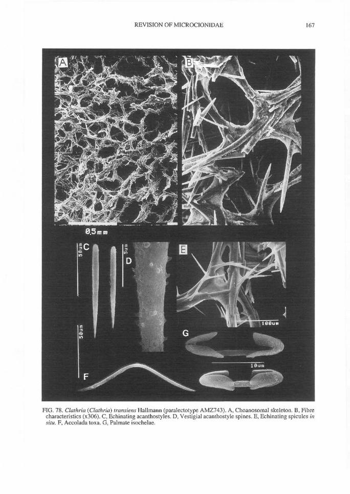
REVISION OF MICROCIONIDAE^ 167
FIG. 78. Clathria (Clathria) transiens Hallmann (paralectotype AMZ743). A, Choanosomal skeleton. B, Fibrecharacteristics (x306). C, Echinating acanthostyles. D, Vestigial acanthostyle spines. E, Echinating spicules insitu. F, Accolada toxa. G, Palmate isochelae.
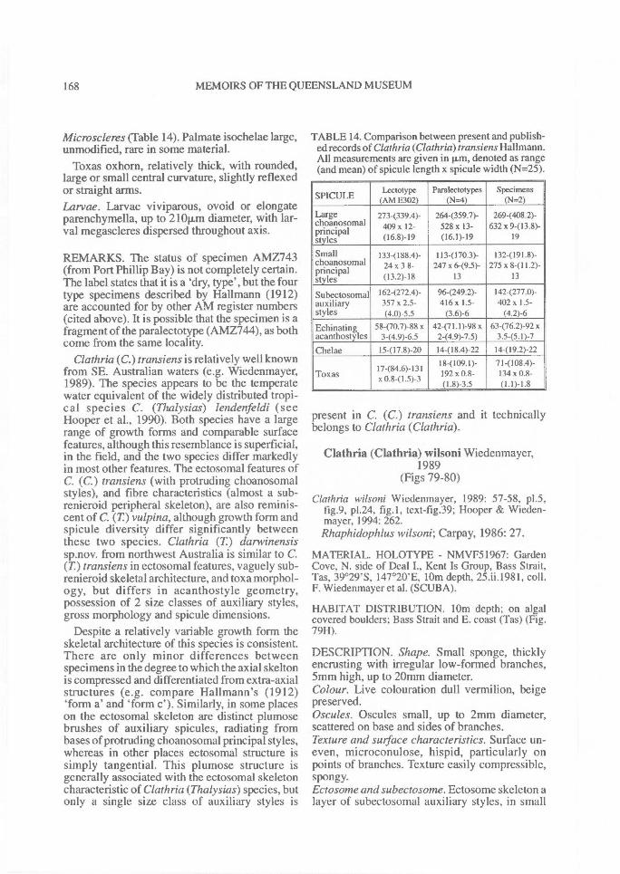
168^ MEMOIRS OF THE QUEENSLAND MUSEUM
Microscleres (Table 14). Palmate isochelae large,unmodified, rare in some material.
Toxas oxhorn, relatively thick, with rounded,large or small central curvature, slightly reflexedor straight arms.Larvae. Larvae viviparous, ovoid or elongateparenchymella, up to 210p,m diameter, with lar-val megascleres dispersed throughout axis.
REMARKS. The status of specimen AMZ743(from Port Phillip Bay) is not completely certain.The label states that it is a 'dry, type', but the fourtype specimens described by Hallmann (1912)are accounted for by other AM register numbers(cited above). It is possible that the specimen is afragment of the paralectotype (AMZ744), as bothcome from the same locality.
Clathria (C.) transiens is relatively well knownfrom SE. Australian waters (e.g. Wiedenmayer,1989). The species appears to be the temperatewater equivalent of the widely distributed tropi-cal species C. (Thalysias) lendenfeldi (seeHooper et al., 1990). Both species have a largerange of growth forms and comparable surfacefeatures, although this resemblance is superficial,in the field, and the two species differ markedlyin most other features. The ectosomal features ofC. (C.) transiens (with protruding choanosomalstyles), and fibre characteristics (almost a sub-renieroid peripheral skeleton), are also reminis-cent of C. (T) vulpina, although growth form andspicule diversity differ significantly betweenthese two species. Clathria (T) darwinensissp.nov. from northwest Australia is similar to C.(T) transiens in ectosomal features, vaguely sub-renieroid skeletal architecture, and toxa morphol-ogy, but differs in acanthostyle geometry,possession of 2 size classes of auxiliary styles,gross morphology and spicule dimensions.
Despite a relatively variable growth form theskeletal architecture of this species is consistent.There are only minor differences betweenspecimens in the degree to which the axial skeltonis compressed and differentiated from extra-axialstructures (e.g. compare Hallmann's (1912)'form a' and 'form c'). Similarly, in some placeson the ectosomal skeleton are distinct plumosebrushes of auxiliary spicules, radiating frombases of protruding choanosomal principal styles,whereas in other places ectosomal structure issimply tangential. This plumose structure isgenerally associated with the ectosomal skeletoncharacteristic of Clathria (Thalysias) species, butonly a single size class of auxiliary styles is
TABLE 14. Comparison between present and publish-ed records of Clathria (Clathria) transiens Hallmann.All measurements are given in p.m, denoted as range(and mean) of spicule length x spicule width (N=25).
SPICULELectotype
(AM E302)Paralectotypes
(N=4)Specimens
(N=2)
Largechoanosomalprincipal
273-(339.4)- 409 x 12-
264-(359.7)-528 x 13-
2694408.2)-632 x 9-(13.8)-
styles (16.8)-19 (16.1)-19 19
Small 133-(l 88.4)- 113-(170.3)- 1 32-(191.8)-choanosomalprincipalstyles
24 x 3 8-(13.2)-18
247 x 6-(9.5)-13
275 x 8411.2)-13
Subectosomal 162-(272.4)- 96-(249.2)- 142(277.0)-auxiliary 357 x 2.5- 416x 1.5- 402x 1.5-styles (4.0)-5.5 (3.6)-6 (4.2)-6
Echinating 58-(70.7)-88 x 42-(71.1)-98 x 63-(76.2)-92 xacanthostyles 3-(4.9)-6.5 2-(4.9)-7.5) 3.5-(5.1)-7
Chelae 15-(17.8)-20 14-(18.4)-22 I4-(19.2)-2218-(109.1)- 7I-(108.4)-
Toxas17-(84.6)-131x 0.8-(1.5)-3 192 x 0.8-
(1.8)-3.5134 x 0.8-(1.1)-1.8
present in C. (C.) transiens and it technicallybelongs to Clathria (Clathria).
Clathria (Clathria) wilsoni Wiedenmayer,1989
(Figs 79-80)
Clathria wilsoni Wiedenmayer, 1989: 57-58, p1.5,fig.9, p1.24, fig.1, text-fig.39; Hooper & Wieden-mayer, 1994: 262.Rhaphidophlus wilsoni; Carpay, 1986: 27.
MATERIAL. HOLOTYPE - NMVF51967: GardenCove, N. side of Deal I., Kent Is Group, Bass Strait,Tas, 39°29'S, 147°20'E, 10m depth, 25.ii.1981, coll.F. Wiedenmayer et al. (SCUBA).
HABITAT DISTRIBUTION. 10m depth; on algalcovered boulders; Bass Strait and E. coast (Tas) (Fig.79H).
DESCRIPTION. Shape. Small sponge, thicklyencrusting with irregular low-formed branches,5mm high, up to 20mm diameter.Colour. Live colouration dull vermilion, beigepreserved.Oscules. Oscules small, up to 2mm diameter,scattered on base and sides of branches.Texture and surface characteristics. Surface un-even, microconulose, hispid, particularly onpoints of branches. Texture easily compressible,spongy.Ectosome and subectosome. Ectosome skeleton alayer of subectosomal auxiliary styles, in small
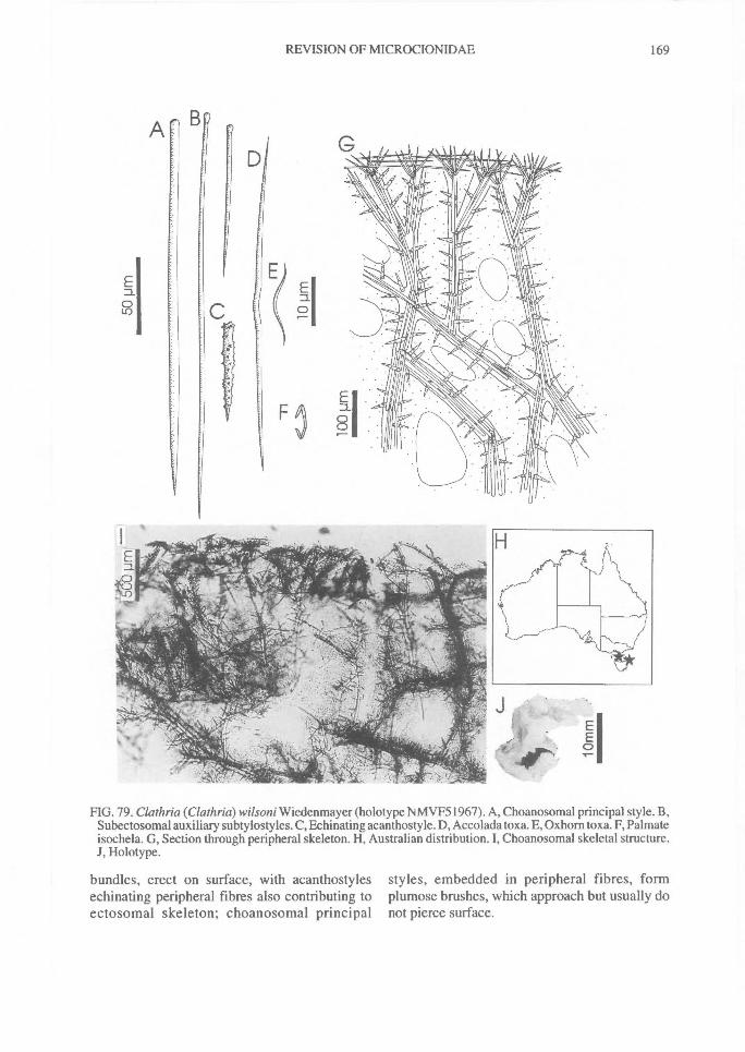
D
REVISION OF MICROCIONIDAE^ 169
FIG. 79. Clathria (Clathria) wilsoni Wiedenmayer (holotype NMVF51967). A, Choanosomal principal style. B,Subectosomal auxiliary subtylostyles. C, Echinating acanthostyle. D, Accolada toxa. E, Oxhorn toxa. F, Palmateisochela. G, Section through peripheral skeleton. H, Australian distribution. I, Choanosomal skeletal structure.J, Holotype.
bundles, erect on surface, with acanthostyles styles, embedded in peripheral fibres, formechinating peripheral fibres also contributing to plumose brushes, which approach but usually doectosomal skeleton; choanosomal principal not pierce surface.
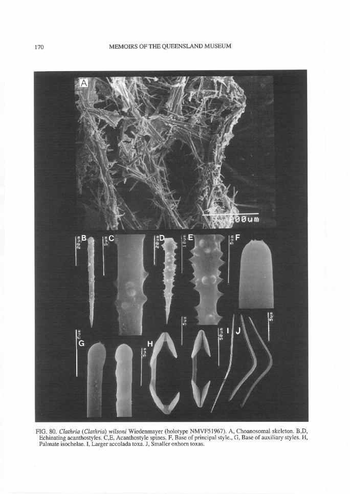
170^ MEMOIRS OF THE QUEENSLAND MUSEUM
FIG. 80. Clathria (Clathria) wilsoni Wiedenmayer (holotype NMVF51967). A, Choanosomal skeleton. B,D,Echinating acanthostyles. C,E, Acanthostyle spines. F, Base of principal style., G, Base of auxiliary styles. H,Palmate isochelae. I, Larger accolada toxa. J, Smaller oxhorn toxas.
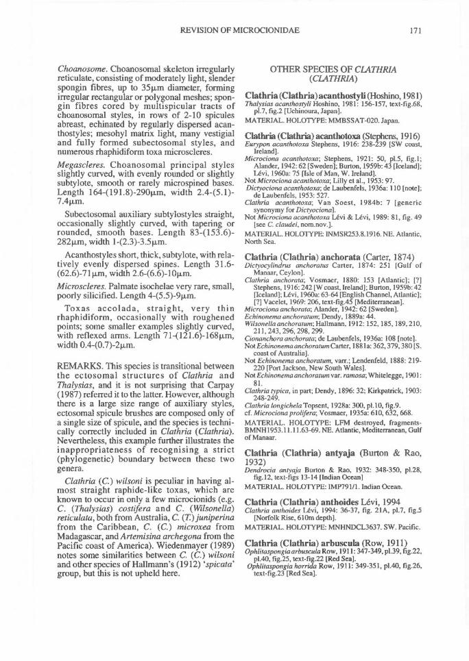
REVISION OF MICROCIONIDAE^ 171
Choanosome. Choanosomal skeleton irregularlyreticulate, consisting of moderately light, slenderspongin fibres, up to 35pm diameter, formingirregular rectangular or polygonal meshes; spon-gin fibres cored by multispicular tracts ofchoanosomal styles, in rows of 2-10 spiculesabreast, echinated by regularly dispersed acan-thostyles; mesohyl matrix light, many vestigialand fully formed subectosomal styles, andnumerous rhaphidiform toxa microscleres.Megascleres. Choanosomal principal stylesslightly curved, with evenly rounded or slightlysubtylote, smooth or rarely microspined bases.Length 164-(191.8)-290p,m, width 2.445.1)-
Subectosomal auxiliary subtylostyles straight,occasionally slightly curved, with tapering orrounded, smooth bases. Length 834153.6)-282p,m, width 1-(2.3)-3.5p,m.
Acanthostyles short, thick, subtylote, with rela-tively evenly dispersed spines. Length 31.6-(62.6)-71 p.m, width 2.6-(6.6)-10Rm.Microscleres. Palmate isochelae very rare, small,poorly silicified. Length 4-(5.5)-9p,m.
Toxas accolada, straight, very thinrhaphidiform, occasionally with roughenedpoints; some smaller examples slightly curved,with reflexed arms. Length 71-(121.6)-168p,m,width 0.4-(0.7)-2p.m.
REMARKS. This species is transitional betweenthe ectosomal structures of Clathria andThalysias, and it is not surprising that Carpay(1987) referred it to the latter. However, althoughthere is a large size range of auxiliary styles,ectosomal spicule brushes are composed only ofa single size of spicule, and the species is techni-cally correctly included in Clathria (Clathria).Nevertheless, this example further illustrates theinappropriateness of recognising a strict(phylogenetic) boundary between these twogenera.
Clathria (C.) wilsoni is peculiar in having al-most straight raphide-like toxas, which areknown to occur in only a few microcionids (e.g.C. (Thalysias) costifera and C. (Wilsonella)reticulata, both from Australia, C. (T.) juniperinafrom the Caribbean, C. (C.) microxea fromMadagascar, and Artemisina archegona from thePacific coast of America). Wiedenmayer (1989)notes some similarities between C. (C.) wilsoniand other species of Hallmann's (1912) spicata'group, but this is not upheld here.
OTHER SPECIES OF CLATHRIA(CLATHRIA)
Clathria (Clathria)acanthostyli (Hoshino, 1981)Thalysias acanthostyli Hoshino, 1981: 156-157, text-fig.68,
p1.7, fig.2 [Uchinoura, Japan].MATERIAL. HOLO'TYPE: MMBSSAT-020. Japan.
Clathria (Clathria) acanthotoxa (Stephens, 1916)Eurypon acanthotoxa Stephens, 1916: 238-239 [SW coast,
Ireland].Microciona acanthotoxa; Stephens, 1921: 50, p1.5, fig.!:
Alander, 1942:62 [Sweden]; Burton, 1959b: 43 [Iceland];Levi, 1960a: 75 [Isle of Man, W. Ireland].
Not Microciona acanthotoxa; Lilly et al., 1953: 97.Dictyociona acanthotoxa; de Laubenfels, 1936a: 110 [note];
de Laubenfels, 1953: 527.Clathria acanthotoxa; Van Soest, 1984b: 7 [generic
synonymy for Dictyociona).Not Microciona acanthotoxa Levi & Levi, 1989: 81, fig. 49
[see C. claudei, nom.nov.].MATERIAL. HOLOTYPE: INM5R253.8.1916. NE. Atlantic,North Sea.
Clathria (Clathria) anchorata (Carter, 1874)Dictyocylindrus anchoratus Carter, 1874: 251 [Gulf of
Manaar, Ceylon].Clathria anchorata; Vosmaer, 1880: 153 [Atlantic]; [?]
Stephens, 1916:242 [W coast, Ireland]; Burton, 1959b: 42[Iceland]; Levi, 1960a: 63-64 [English Channel, Atlantic];[7] Vacelet, 1969: 206, text-fig.45 [Mediterranean].
Microciona anchorata; Alander, 1942: 62 [Sweden].Echinonema anchoratum; Dendy, 1889a: 44.Wilsonella anchoratum; Hallmann, 1912: 152, 185, 189, 210,
211, 243, 296, 298, 299.Cionanchora anchorata; de Laubenfels, 1936a: 108 [note].Not Echinonema anchoratum Carter, 1881a: 362, 379, 380 [S.
coast of Australia].Not Echinonema anchoratum, varr.; Lendenfeld, 1888: 219-
220 [Port Jackson, New South Wales].Not Echinonema anchoratum var. ramosa; Whitelegge, 1901:
81.Clathria typica, in part; Dendy, 1896: 32; Kirkpatrick, 1903:
248-249.Clathria longichela Topsent, 1928a: 300, p1.10, fig.9.cf. Microciona prolifera; Vosmaer, 1935a: 610, 632, 668.MATERIAL. HOLOTYPE: LFM destroyed, fragments-BMNH1953.11.11.63-69. NE. Atlantic, Mediterranean, Gulfof Manaar.
Clathria (Clathria) antyaja (Burton & Rao,1932)Dendrocia antyaja Burton & Rao, 1932: 348-350, p1.28,
fig.12, text-figs 13-14 [Indian Ocean]MATERIAL. HOLOTYPE: IMP791/1. Indian Ocean.
Clathria (Clathria) anthoides Levi, 1994Clathria anthoides Levi, 1994: 36-37, fig. 21A, p1.7, fig.5
[Norfolk Rise, 610m depth].MATERIAL. HOLOTYPE: MNHNDCL3637. SW. Pacific.
Clathria (Clathria) arbuscula (Row, 1911)Ophlitaspongia arbuscula Row, 1911:347-349, p1.39, fig.22,
p1.40, fig.25, text-fig.22 [Red SeatOphlitaspongia horrida Row, 1911: 349-351, p1.40, fig.26,
text-fig.23 [Red Seat
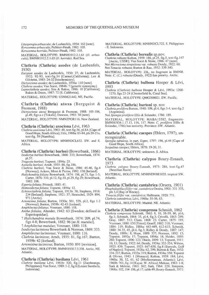
172^ MEMOIRS OF THE QUEENSLAND MUSEUM
Litaspongia arbuscula; de Laubenfels, 1954: 162 [note].Kerasemna arbuscula; Pulitzer-Finali, 1982: 105.Kerasemna horrida; Pulitzer-Finali, 1982: 105.MATERIAL. HOLOTYPE: BMNH1912.2.1.63 (0. arbus-cula); BMNH1912.2.1.65 (0. horrida). Red Sea.
Clathria (Clathria) asodes (de Laubenfels,1930)Eutypon asodes de Laubenfels, 1930: 27; de Laubenfels,
1932: 92-93, text-fig.54 [Carmel,California]; Lee &Gilchrist, 1985: 24-32 [biochemistry].
Dictyociona asodes; de Laubenfels, 1936a: 110 [note].Clathria asodes; Van Soest, 1984b: 7 [generic synonymy].Leptoclathria asodes; Sim & Bakus, 1986: 10 [California];
Bakus & Green, 1987: 72 [S. California].MATERIAL. HOLOTYPE: USNM21442. NE. Pacific.
Clathria (Clathria) atoxa (Bergquist &Fromont, 1988)Dictyociona atoxa Bergquist & Fromont, 1988: 105-106,
p1.49, figs a-c [Tokatu]; Dawson, 1993: 38 [note].MATERIAL. HOLOTYPE: NMNZPOR116. New Zealand.
Clathria (Clathria) axociona Levi, 1963Clathria axociona Levi, 1963: 49, text-fig.56, p1.8A [Cape of
Good Hope, South Africa]; Uriz, 1988a: 83-84, pls 20-21a,text-fig. 59 [Namibia].
MATERIAL. HOLOTYPE: MNHNDCL632. SW. and S.Africa.
Clathria (Clathria) barleei (Bowerbank, 1866)Isodiciya barleei Bowerbank, 1866: 333; Bowerbank, 1874:
p1.57.Tragosia barleei; Topsent, 1894a: 25.Axinella barleei; Arndt, 1935: 88, fig.186.Clathria barleei; Van Soest & Stone, 1986: 45-46, fig.4
[Norway]; Ackers, Moss & Picton, 1992: 138 [Ireland].Halichondria foliata Bowerbank, 1874: 198, p1.73, figs 1-5;
Carter, 1876: 310, p1.12, fig.10, p1.29, fig.29; Bowerbank,1882: 106.
Esperia foliata; Fristedt, 1885: 41.Homeodictya foliata; Topsent, 1894a: 12.Echinoclathria foliata; Topsent, 19 13b: 38; Stephens, 1916:
234 [Ireland]; Stephens, 1921: 57; Hentschel, 1929: 894,971 [Arctic].
Artemisina foliata; Burton, 1930a: 501, 529, p1.2, figs 1-2[Norway]; Burton, 1959b: 42-43 [Iceland].
Amphilectus foliatus; Vosmaer, 1880: 118.Antho foliata; Alander, 1942: 63 [Sweden; defined in
Esperiopsidae].? Halichondria mutula Bowerbank, 1874: 209, p1.74,
figs 4-8; Bowerbank, 1882: 96 [as H. mutulus].? Amphilectus mutulus; Vosmaer, 1880: 118.Isodiciya laciniosa Bowerbank & Norman, 1869: 333.Amphilectus laciniosus; Vosmaer, 1880: 116.Clathria laciniosa; Arndt, 1935: 81, fig.167; Burton,
1959b: 42 [Iceland].Art emisina laciniosa; Burton, 1950: 891 [revision].MATERIAL. HOLOTYPE: BMNI-11930.7.3.338. Arctic, NE.Atlantic.
Clathria (Clathria) basilana Levi, 1961Clathria basilana Levi, 1961b: 520, fig.11 [Zamboanga,
Philippines]; Van Soest, 1989: 1-2, fig.6 [Lesser Sumba Is,Indonesia].
MATERIAL. HOLOTYPE: MNHNDCL722. S. Philippines- E. Indonesia.
Clathria (Clathria) borealis sp.nov.Clathria robusta Koltun, 1959: 186, p1.25, fig.5, text-fig. 147
[Arctic, USSR]; Van Soest & Stone, 1986: 47 [note].Not Microciona strepsitoxa var. robusta Dendy, 1922: 60.Not Tenacia robusta; Burton & Rao, 1932: 339-340.MATERIAL. HOLOTYPE: ZIL, no fragment in BMNH.Note: C. (C.) robusta (Dendy, 1922) has priority. Arctic.
Clathria (Clathria) bulbosa Hooper & Levi,1993Clathria (Clathria) bulbosa Hooper & Levi, 1993a: 1268-
1270, figs 23-24 [Chesterfield Is, Coral Sea].MATERIAL. HOLOTYPE: QMG300021. SW. Pacific.
Clathria (Clathria) burtoni sp. novClathria proltfera Burton, 1940: 109, p1.4, figs 3-4, text-fig.2
[Argentina].Not Spongia prolifera Ellis & Solander, 1786: 189.MATERIAL. HOLOTYPE: MA BA 15582, fragments-BMNH1934.1.17.13, 116, 117. Note: C. prolifera (Ellis &Solander, 1786) has seniority. Province: SW. Atlantic.
Clathria (Clathria) caespes (Ehlers, 1797), un-recognizable.Spongia tubulosa, in part, Esper, 1797: 196, p1.44 [Cape of
Good Hope, South Africa].Scopalina caespes; Ehlers, 1870: 19-20, 31.MATERIAL. HOLOTYPE: unknown. S. Africa.
Clathria (Clathria) calypso Boury-Esnault,1973Clathria calypso Boury-Esnault, 1973: 286, text-fig.47
[Brazilian Basin]MATERIAL. HOLOTYPE: MNHNDNBE1035. tropical SW.Atlantic.
Clathria (Clathria) cantabrica (Orueta, 1901)Rhaphidophlus filifer var. cantabrica Orueta, 1901: 331-335,
pls 3,4 [Bay of Biscay].Tenacia filifer var. cantabrica; Hallmann, 1920: 771.
Clathria cantabrica; Levi, 1960a: 55-56, 63.MATERIAL. HOLOTYPE: Madrid. NE. Atlantic.
Clathria (Clathria) compressa Schmidt, 1862Clathria compressa Schmidt, 1862: 8, 10, 58-59, 86, p1.6,
fig.1; Schmidt, 1864: 35, p1.4, fig.3; Crivelli, 1863: 299;Gray, 1867: 513; Claus, 1868: 23; Carter, 1875: 195;Vosmaer, 1880: 150 [Triest]; Graeff, 1882: 318; Vosmaer,1884: 119; Ridley, 1884a: 443-449, 612-615; Schmidt,1880: 34-35, 45, p1.4, fig.3; Ridley & Dendy, 1887: 147;Dendy, 1889c: 8; Hope, 1889: 337; Norman, 1892: 13;Topsent, 1892a: 17; Topsent, 1894a: 18; Heider, 1895:280; Topsent, 1896: 123; Thiele, 1899: 13; Topsent, 1911:10, 13; Dendy, 1922:64; Dendy, 1924a: 352-354; Wilson,1925: 439; Topsent, 1925: 647-650, fig.8 [Banyuls, Gulfof Naples]; Topsent, 1928a: 62,299; Burton & Rao, 1932:334-337; Burton, 1932a: 319; Burton, 1934a: 558; Topsent& Olivier, 1943: 1 [Monaco]; Koltun, 1959: 184; Levi,1960a: 50, 52, 61, 62 [Mediterranean, Atlantic]; Levi,1960b: 761, fig. 14 [Kayar, W. Africa]; Melone, 1963: 1-8;Sara & Melone, 1963: 362; Sara, 1964: 229; Simpson,1968a: 102, 104-106, p1.17, table 49; Boury-Esnault, 1971:
REVISION OF MICROCIONIDAE^ 173
323 [Banyuls]; Pulitzer-Finali, 1983: 610; Desqueyroux-Faundez & Stone, 1992: 9, 35, 103 [list].
Thalysias compressa; de Laubenfels, 1936a: 105-106.? Spongia clathrus Esper, 1797; Vosmaer, 1880: 150.Not Citalina compressa; Carter, 1882b: 112-113.Not Halichondria compressa Carter, 1886g: 450
[Westemport Bay, Victoria].Not Pseudoclathria compressa; Dendy, 1897: 259.MATERIAL. HOLOTYPE: LMJG15509, fragmentsBMNH1867.7.26.78, BMNH1910.1.1.2362, 2363. Mediter-ranean, NE. Atlantic, W. Africa.
Clathria (Clathria) conica Levi, 1963Clathria conica Levi, 1963: 50-51, text-fig.57, p1.8E [Cape of
Good Hope].MATERIAL. HOLOTYPE: MNHNDCL617. S. Africa.
Clathria (Clathria) contorta (Bergquist &Fromont, 1988)Dictyociona contorta Bergquist & Fromont, 1988: 105, p1.48,
figs d-f [Manukau]; Dawson, 1993: 38 [note].MATERIAL. HOLOTYPE: NMNZPOR115. New Zealand.
Clathria (Clathria) coralloides (Olivi, 1792)Spongia coralloides Olivi, 1792: 264; Bertoloni, 1819: 228;
Blainville, 1819: 125; Lamouroux, 1824: 369; Martens,1824: 535.
Grantia coralloides; Nardo, 1833: 522.Halichondria corona Lieberkiihn, 1859: 521, 529, p1.11, fig.3.Clathria coralloides-, Schmidt, 1862:7, 10, 11, 58, 85, p1.5,
figs 10-11; Kiilliker, 1864: 52, 71; Schmidt, 1864: 34;Schmidt, 1868: 9,41; Schmidt, 1870: 56, 60; Gray, 1867:513, 533, 552; Dybowsky, 1880: 5, 70, p1.4, figs 7,9;Krukenberg, 1880: 70, 72; Vosmaer, 1880: 149-150[Mediterranean]; Ridley, 1881: 485; Vosmaer, 1881: 4;Vosmaer, 1882-6: 119, 121-122; Vosmaer, 1884b: 492;Vosmaer, 1885a: 186, 356; Carter, I882a: 281; Graeffe,1882: 318; Carter, 1884a: 204; Ridley & Dendy, 1887:147; Lendenfeld, 1889a: 410-415, 498-505, 586, 592, 594,598, 602, 604, 608, 612, 614, 618, 622, 624, 628, 634, 640,644, 646, 650, 669, p1.34, figs 193-205; Lendenfeld,1890a: 72; Maas, 1892:427-428; Norman, 1892: 13; Maas,1893: 331, 334-335, 414, 441, 444, p1.20, fig.19, p1.21,fig.32; Topsent, 1893d: 445; Topsent, 1894a: 18,24; Top-sent, 1894b: 35; Heider, 1895: 280; Dendy, 1896: 36;Loisel, 1898: 38; Zimmermann, 1907: 308; Korschelt &Heider, 1910: 321; Walther, 1910: 21; Babic, 1921: 84[Adriatic]; Babic, 1922: 244; Topsent, 1925: 646-647,fig.7 [Gulf of Naples]; Topsent & Olivier, 1943: 1[Banyuls and Naples]; de Laubenfels, 1954: 139; Levi,1960a: 61-62 [Mediterranean, Adriatic,Naples, Banyuls];Laubier, 1966 [Banyuls]; Vidal, 1967 [Mediterranean];Boury-Esnault, 1971: 323 [Banyuls]; Pulitzer-Finali,1983: 568-569, 610 [Mediterranean]; Boury-Esnault &Lopes, 1985: 194-195, fig.43 [Azores]; Pansini, 1987[Adriatic Sea]; Desqueyroux-Faundez & Stone, 1992: 9,35, 103 [list].
Ophlitaspongia coralloides; Bowerbank, 1874: 10.Ophlitaspongia corona; Levi, 1960a: 61MATERIAL. HOLOTYPE: unknown; fragments LMJG15356, BMNH1867.7.26.74, 1910.1.1.2364, 2365. Mediter-ranean, NE. Atlantic.
Clathria (Clathria) curvispiculifera (Carter,1880)Microciona curvispiculifera Carter, 1880a: 43,44,151,153,
p1.4, fig.6a-d [Gulf of Manaar]; Vosmaer, 1935a: 608.
? Rhabdoploca curvispiculifera; Topsent, 1904a: 157-159.MATERIAL. HOLOTYPE: LFM missing, no fragment inBMNH. Gulf of Manaar. Note: virtually unrecognisable.
Clathria (Clathria) dayi Levi, 1963Clathria dayi Levi, 1963:51, p1.8B, text-fig.58 [Cape of Good
Hope]; Sim & Byeon, 1989: 39, p1.4, figs 1-2 [Korea;possible misidentification].
MATERIAL. HOLOTYPE: MNHNDCL611. S. Africa.
Clathria (Clathria) depressa Sara & Melone,1966Clathria depressa Sara & Melone, 1966: 2-4, text-figs 1-2,
p1.1 [Olivetta, Portofino, Levante coast, Italy]; Pulitzer-Finali, 1983: 610.
MATERIAL. HOLOTYPE: IMZUB. E. Mediterranean.
Clathria (Clathria) discreta (Thiele, 1905)Microciona discreta Thiele, 1905: 447-449, 494, p1.3 I , fig.
65a-e [Calbuco, Chile].Diciyociona discreta; Topsent, 1913a: 580, 583, 614, 618-
620, 642, p1.3, fig.5 [Gough I., S. Atlantic Ocean]; Burton,1932: 324, p1.56, figs 3-4 [Falklands]; Burton, 1940: 112,p1.4, figs 1-2, p1.6, fig.2 [Argentina and Chile]; de Lauben-fels, 1953a: 527; Desqueyroux, 1972: 29-30, figs 95-102,136-137 [Chile]; Desqueyroux & Moyana, 1987: 49[Chile, Tierra del Fuego, Argentina, Falkland Is, Ker-guelen Is].
cf. Microciona prolifera; Vosmaer, 1935a: 608,646.MATERIAL. HOLOTYPE: ZMB3302, fragment:BMNH1908.9.24.159. SW. Atlantic, SE. Pacific, Suban-tarctic.
Clathria (Clathria) elastica Levi, 1963Clathria elastica Levi, 1963: 52, text-fig.59, p1.8C [Cape of
Good Hope].Not Clathria elastica Sara, 1978: 70 [see C. sarai, nom.novi.MATERIAL. HOLOTYPE: MNHNDCL604. S. Africa.
Clathria (Clathria) elegans Vosmaer, 1880Clathria elegans Vosmaer, 1880: 152 [North America]; Vos-
maer, 1884b: 492.? Anaata elegans; de Laubenfels, 1936a: 109.Not Plectispa elegans Lendenfeld, 1888: 226.Not Antherochalina elegans Lendenfeld, 18876: 787.cf. Microciona prolifera; Vosmaer, 1935a: 630.MATERIAL. HOLOTYPE: unknown. NE. Pacific. Note:virtually unrecognisable.
Clathria (Clathria) foliacea Topsent, 1889Clathria foliacea Topsent, 1889: 39-40, text-fig.5 [Banc de
Campeche]; Van Soest, 1984b: 107, table 4; Van Soest &Stentoft, 1988: 126 [table].
Thalyseurypon foliacea; de Laubenfels, 1936a: 107-108[Florida].
Pandaros foliaceum; Wiedenmayer, 1977: 144 [note].Clathria carteri Topsent, 1889: 38-39, text-fig.4B [Banc de
Campeche]; Van Soest, 1984b: 108.Thalyseurypon carteri; de Laubenfels, 1936a: 107.Pandaros carteri; Wiedenmayer, 1977: 144 [note].MATERIAL. HOLOTYPE: MNHN missing, referencespecimen USNM22516. NE. Atlantic, Caribbean.
Clathria (Clathria) foliascens Vacelet & Vas-seur, 1971
174^ MEMOIRS OF THE QUEENSLAND MUSEUM
Clathria foliascens Vacelet & Vasseur, 1971: 95-96, text-fig.49, p1.4,figs 5-6 [Tulear Madagascar]; Vacelet & Vas-seur, 1977: 114; Vacelet et al., 1976: 71 [Tulear,Madagascar]
MATERIAL. HOLOTYPE: MNHNDJV30. W. IndianOcean.
Clathria (Clathria) frondiculata (Schmidt,1864)Reniera (?) frondiculata Schmidt, 1864: 39, 40, 45, p1.4,
fig. 10 [Adriatic].Pitalia frondiculata; Gray, 1867: 524.Clathriafrondiculata; Vosmaer, 1880:153 [Triest]; Vosmaer,
1885:236; Norman, 1892: 13; Topsent, 1892b: 23; Heider,1895: 282; Burton, 1930a: 514.
cf. Clathria compressa and C. coralloides; Vosmaer, 1935a:625 [intermediate form].
MATERIAL. HOLOTYPE: LMJG, fragment: BMNH-1910.1.1.542. E. Mediterranean.
Clathria (Clathria) gorgonoides (Dendy, 1916)Echinodictyum gorgonoides Dendy, 1916a: 129 [Kattiawar,
Indian Ocean]; Hooper, 1991: 1348.MATERIAL. HOLOTYPE: BMNH1920.12.9.38. W. India.
Clathria (Clathria) hexagonopora Levi, 1963Clathria hexagonopora Levi, 1963: 53-54, text-fig.60, p1.8D
[Cape of Good Hope].MATERIAL. HOLOTYPE: MNHNDCL620. S. Africa.
Clathria (Clathria) indica Dendy, 1889Clathria indica Dendy, 1889b: 73, 84, p1.4, fig. 10 [Gulf of
Manaar, Ceylon]; Burton 8c Rao, 1932: 336-337, p1.18,figs 8-9 [Tuticorin, India]; Thomas, 1979a: 58, p1.3, fig. 10[Mozambique].
Wilsonella indica; Dendy, 1905: 171 [Ceylon]; Hallmann,1912: 242; Burton, 1931a: 346 [Natal Coast].
MATERIAL. HOLOTYPE: BMNH1887.8.4.19, fragment:MNHNDCL2528. E. Africa, SE. India, Gulf of Manaar.
Clathria (Clathria) inhacensis Thomas, 1979Clathria inhacensis Thomas, 1979: 27-28, p1.2, fig.2 [Inhaca
I., Mozambique].MATERIAL. HOLOTYPE: MRAC511. E. Africa.
Clathria (Clathria) intermedia Kirk, 1911Clathria intermedia Kirk, 1911: 579, text-fig.5 [Kermadec
Is]; Fell, 1950: 11, text-fig.2; Berquist & Fromont, 1988:109-110; Dawson, 1993:37 [note].
Thalysias intermedia; de Laubenfels, 1936a: 105.MATERIAL. HOLOTYPE: NMNZ unregistered. N. NewZealand.
Clathria (Clathria) irregularis (Burton, 1931)Marleyia irregularis Burton, 1931a: 346, p1.23, fig.6, text-
fig.5 [Durban, Natal]; de Laubenfels, 1936a: 109 [note];Van Soest, 1984b: 130 [note].
MATERIAL. HOLOTYPE: NM1279; paratype: BMNH-1934.10.1.17. S. Africa.
Clathria (Clathria) juncea Burton, 1931Clathria juncea Burton, 1931a: 343, p1.23, fig.5, text-fig.3
[Natal coast]; Levi, 1960a: 85; Levi, 1963: 66 [note].Labacea juncea; de Laubenfels, 1936a: 66 [note].cf. Alcyoniumjunceum Lamarck, 1816: 77.cf. Anomoclathria opuntioides var; Topsent, 1933: 26 [note].
MATERIAL. HOLOTYPE: MNHN missing (fragmentBMNH1954.2.20.85, unconfirmed); 'representativespecimen': BMNH1926.2.19.2. Note: Burton's species isprobably the same as Lamarck's but this cannot be verifiedand the species is taken in the sense of Burton (1931a). SouthAfrica.
Clathria (Clathria) labyrinthica (Schmidt,1864)Reniera labyrinthica Schmidt, 1864:39, p1.4, fig.9 [Adriatic].Clathria labyrinthica; Burton, 1930a: 514 [imperfectly
known].MATERIAL. HOLOTYPE: LMJG. Mediterranean.
Clathria (Clathria) laevigata Lambe, 1893Clathria laevigata Lambe, 1893:31-32, p1.2, figs 9-9f [Pacific
coast, Canada]; Lambe, 1900: 160; Koltun, 1959: 185,text-fig. 146 [Kurile Is].
Thalysias laevigata; de Laubenfels, 1936a: 105.cf. Microciona prolifera, tropus senta; Vosmaer, 1935a: 641.MATERIAL. HOLOTYPE: NMCIC1900-2892. NW. andNE. Pacific.
Clathria (Clathria) lissosclera Bergquist &Fromont, 1988Clathria lissosclera Bergquist & Fromont, 1988: 106-107,
p1.49, figs d-f [Mayor I.]; Dawson, 1993: 37 [note].MATERIAL. HOLOTYPE: NMNZPOR112. New Zealand.
Clathria (Clathria) lobata Vosmaer, 1880Clathria lobata Vosmaer, 1880: 151 [Cape of Good Hope];
Vosmaer, 1882: 45; Carter & Hope, 1889: 101, 105;Stephens, 1915: 444-445 [Cape of Good Hope]; Levi,1963: 54, text-fig.61, p1.8F [Cape of Good Hope].
Rhaphidophlus lobatus, var. horrida; Ridley & Dendy, 1887:153-155, p1.28, fig.1, p1.29, fig.4a-c [Simon's Bay, Capeof Good Hope].
Ligrota lobata; de Laubenfels, 1936a: 125.A rtemisina lobata; Van Soest, 1984b: 130 [generic synonymy
for Ligrota].cf. Microciona prolifera; Vosmaer, 1935a: 631-632.MATERIAL. HOLOTYPE: unknown, fragmentsRMNH276-277, BMNH1887.5.2.100. S Africa.
Clathria (Clathria) macroisochela Levi, 1994Clathria (Clathria) macroisochela Levi, 1994: 37, fig.21b,
p1.7, figs 7-9 [Norfolk Rise, 680m depth].MATERIAL. HOLOTYPE: MNHNDCL3638. SW Pacific.
Clathria (Clathria) maeandrina Ridley, 1884Clathria maeandrina Ridley, 1884a: 588, 614, p1.53, fig.1,
p1.54, fig.H [Amirante Is]; Burton, 1930c: 668 [Gulf ofManaar]; Burton, 1959: 244 [Red Sea].
MATERIAL. HOLOTYPE: BMNH1882.10.17.55. Red Sea, WIndian Ocean, Gulf of Manaar.
Clathria (Clathria) marissuperi Pulitzer-Finali, 1983Clathria marissuperi Pulitzer-Finali, 1983: 569-571, text-
fig.67 [Italy].MATERIAL. HOLOTYPE: IZUG(MSNG 47179). Mediter-ranean.
Clathria (Clathria) menoui Hooper & Levi,1993
REVISION OF MICROCIONIDAE^ 175
Clathria (Clathria) menoui Hooper & Levi, 1993a: 1270-1273, figs 25-26 [New Caledonia].
MATERIAL. HOLOTYPE: QMG301267. SW. Pacific.
Clathria (Clathria) meyeri (Bowerbank, 1877)Ophlitaspongia meyeri Bowerbank, 1877: 456 [Selat Japen,
Irian Jaya, E. Indonesia].Clathria meyeri; Vosmaer, 1880: 154.? Anaata meyeri; de Laubenfels, 1936a: 109.MATERIAL. HOLOTYPE: Dresden Musuem destroyed(fragments BMNH1877.5.21.1306-1309). Indonesia.
Clathria (Clathria) microchela (Stephens,1916)Eurypon microchela Stephens, 1916: 240-241 [SW coast,
Ireland]; Lilly et al., 1953 [Lough me, Ireland]; Burton,1959b: 44 [Iceland]; Van Soest & Weinberg, 1980: 10[Lough Inc. Ireland].
Dictyociona microchela; de Laubenfels, 1936a: 110 [note]; deLaubenfels, 1953: 528.
Clathria microchela; Van Soest, 1984b: 7 [genericsynonymy].
Not Microciona microchela Hechtel, 1965: 41.MATERIAL. HOLOTYPE: INMSR353.10.1916. NE. Atlan-tic.
Clathria (Clathria) microxa Desqueyroux,1972Clathria microxa Desqueyroux, 1972: 27-28, text-figs 76-
80,134 [Gulf of Corcovado, Chile].MATERIAL. HOLOTYPE: ICBUC. SE Pacific.
Clathria (Clathria) mortensenii Brondsted,1923Clathria mortensenii Brondsted, 1923: 143-144, text-fig.22
[Campbell Is]; Burton, 1940: 111 [Argentina]; Bergquist& Fromont, 1988: 107-108, p1.50, figs a-c [N. NewZealand]; Dawson, 1993: 37 [note].
Microciona mortensenii; de Laubenfels,I936a: 111; Burton,1940: 111 [Argentina].
Microcionaheterospiculata; Bergquist, 1961a: 39 [N of NewZealand].
Not Microciona heterospiculata Brondsted, 1924: 465, text-fig.20.
MATERIAL. HOLOTYPE: UZM, fragment:BMNH1930.8.11.10. SW. Atlantic, Subantarctic, NewZealand.
Clathria (Clathria) mosulpia Sim & Bakus,1989Clathria mosulpia Sim & Byeon, 1989: 38-39, p1.3, figs 1-5.
[South Korea].MATERIAL. HOLOTYPE: HNUKP0r9. S. China Sea.
Clathria (Clathria) obliqua (George & Wilson,1919)Esperiopsis obliqua George & Wilson, 1919: 148-150, p1.60,
figs 20-23, p1.66, fig.58 [North Carolina].Microciona obliqua; de Laubenfels, 1936a: 111.cf. Microciona prolifera; de Laubenfels, 1947; de Laubenfels,
1949a.Haliclona oculata; Pearse & Williams, 1951 [North
Carolina].Chalina arbuscula; Coues & Yarrow, 1879: 312; Verrill &
Smith, 1873: 743 [North Carolina].
Tenaciella obliqua; Wells et al., 1960: 218-219, text-figs16,25 [North Carolina].
Not Tenaciella obliqua; Alcolado, 1976: 5.Clathriaobliqua; Van Soest, 1984b: 104, 108, table 4 [affinity
with Clathria proliferatNot Dictyociona adioristica de Laubenfels, 1953a: 526; Van
Soest, I984b: 108 [possible synonym].MATERIAL. HOLOTYPE: USNM23612, paratype-USNM23613. NW. Atlantic-Caribbean.
Clathria (Clathria) oculata Burton, 1933Clathria oculata Burton, 1933: 250-251, text-fig.2 [Natal];
Levi, 1963: 67 [Natal].Thalysias oculata; de Laubenfels, 1936a: 105.MATERIAL. HOLOTYPE: BMNH1933.7.4.65. S. Africa.
Clathria (Clathria) oxneri (Topsent, 1928)Hymedesmia oxneri Topsent, 1928a: 256 [Azores].Dictyociona oxneri; de Laubenfels, 1936a: 110 [note]; de
Laubenfels, 1953a: 528.Clathria oxneri; Van Soest, 1984b: 7 [generic synonymy for
Dictyociona].MATERIAL. HOLOTYPE: MOM (fragment MNHNDT-963). NE. Atlantic.
Clathria (Clathria) pachystyla Levi, 1963Clathria pachystyla Levi, 1963: 56, text-fig.63, p1.8G [Cape
of Good Hope].MATERIAL. HOLOTYPE: MNHNDCL609. S. Africa.
Clathria (Clathria) papillosa Thiele, 1905Clathria papillosa Thiele, 1905: 449-450, text-fig.66 [Cal-
buco, Chile]; Burton, 1932a: 319 [Falkland Is];Desqueyroux-Faundez & Moyana, 1987: 49 [Chile, Ar-gentina, Falkland Is, Kerguelen Is].
Pseudanchinoepapillosa; de Laubenfels, 1936a: 109; Burton,1940: 115, p1.5, figs 1-7 [Argentina].
MATERIAL. HOLOTYPE: ZMB not located, fragment:BMNH1908.9.24.162. SE. Pacific, SW. Atlantic.
Clathria (Clathria) pellicula Whitelegge, 1897Clathria pellicula Whitelegge, 1897: 327-328 [Funafuti (El-
lice Is)].Hymeraphia pellicula; Hallmann, 1912: 208 -209.cf. Microciona prolifera tropus tegens; Vosmaer, 1935a: 641-2.MATERIAL. HOLOTYPE: AMG1660. SW central Pacific.
Clathria (Clathria) plurityla Pulitzer-Finali,1983Clathria plurityla Pulitzer-Finali, 1983: 571-572, 610, text-
fig.68 [Italy].MATERIAL. HOLOTYPE: IZUG(MSNG47180). Mediter-ranean.
Clathria (Clathria) productitoxa (Hoshino,1981)Thalysias productitoxa Hoshino, 1981: 157-159, text-fig.69,
p1.7, fig.3 [Uchinoura].MATERIAL. HOLOTYPE: MMBSSAT-018. Japan.
Clathria (Clathria) prolifera (Ellis & Solander,1786)Spongia prolifera Ellis & Solander, 1786: 189-190, p1.58,
fig.5; Linnaeus, 1791: 3822; Esper, 1794: 178,281; Bose,1802:143; Lamarck, 1814: 372; Lamouroux, 1816: 81-82;
176^ MEMOIRS OF THE QUEENSLAND MUSEUM
Montagu, 1818: 86; Blainville, 1819: 106; Lamouroux,1821: 31, 109, p1.58, fig.5; Lamouroux, 1824: 368;Templeton, 1836: 472; Johnston, 1842: 170-171; Gray,1848: 19, 23.
Not Spongia prolifera; Grant, 1826: 115-116, 123, 135, 138,347.
Microciona prolifera; Verrill, 1873: 741-742, pls 1-5 [USA];Verrill, 1880: 232; Coues & Yarrow, 1879: 312 [NorthCarolina]; Hyatt, 1885: 131; Norman, 1892: 14; Wilson,1900: 350 [Beaufort, N.Carolina]; Wilson, 1902:396-397;Wilson, 1907: 246; Wilson, 1910: 1269; Wilson, 1911:3-11, 14, 29-30, p1.1, figs 1-6, p1.2, figs 7-12, p1.3, figs13-20, p1.4, figs 21-25, p1.5, figs 26-32; George & Wilson,1919: 157-158, p1.62, figs 31, 33, p1.63, figs 35-36, p1.66,fig.57 [North Carolina]; Allee, 1923: 175; Galtsoff &Pertzoff, 1926: 239-254 [physiology]; Burton & Rao,1932: 344 [Arabian Sea; probable misidentification]; Proc-tor, 1933: 104; de Laubenfels, 1936a: 111; McDougall,1943: 331-332; Bergmann, Schedl & Low, 1945: 580; deLaubenfels, 1947:35; Hopkins, 1956a: 44; Hopkins, 1962:124; Hartmann, 1958a: 36-41, text-fig.10, table 11 [NewEngland, USA]; Levi, 1960a: 52; Wells eta!., 1960: 213-216, text-figs 18,22 [North Carolina]; Little, 1963: 49[Honda]; Wells et al., 1964: 757-758 [North Carolina];Bagby, 1966: 167-181, pls 3-5 [cytology]; Simpson,1968a: 18, pls 1-5 [Connecticut, USA]; Simpson, 1968b:252-277 [reproduction]; Wendt, 1970: 3500-B [cytology];Sindelar, 1970: 3771-B [cytology]; Bagby, 1970: 579-594[ultrastructure]; Kunen et al., 1970:565-576 [physiology];Madri et al., 1970: 1-5 [biochemistry]; Swartz, 1972: 17[ecology]; Bagby, 1972: 217-244 [ultrastructure]; Bito,1972: 65 [biochemistry]; Bose et al., 1972: 217-222[biochemistry]; Turner & Burger, 1973: 509-510, text-fig.! [cytology]; Weinbaum & Burger, 1973: 510-512,[biochemistry]; Stempien, 1973: 363; Sutherland, 1974:859-873 [ecology]; Turner et al., 1974: 35 [cytology];Bose, 1974: 476-490 [chemistry]; Jefferts et al., 1974:244-247 [biochemistry]; Reiswig, 1975:493-502 [physiol-ogy]; Reed et al., 1976: 153-169 [cytology]; Jumblatt etal., 1976: 73-86 [cytology]; Morales & Litchfield, 1976:206-216 [biochemistry]; Burger, 1977: 357-376 [cytol-ogy]; Greenberg et al., 1977: 95-102 [cytochemistry];Morales, 1977: 5043 [biochemistry]; Morales &Litchfield, 1977: 570-576 [biochemistry]; Simpson, 1978:31-42 [morphology]; Burkart eta!., 1979: 239-246 [cytol-ogy]; Leith, 1979: 212-223 [cytology]; Litchfield &Liaaen-Jensen, 1980: 359-365 [biochemistry]; Jumblatt etal., 1980: 1038-1042 [biochemistry-cytology]; Saxegaardeta!., 1981: 325-327 [biochemistry]; Lee & Nicol, 1981:445 [chemistry]; Biernbaum, 1981: 85-96 [ecology];Liaaen-Jensen et al., 1982: 170-171 [biochemistry];Misevic & Burger, 1982: 200 [biochemistry-cytology];Misevic eta!., 1982: 6931-6936 [biochemistry-cytology];Kuhns et al., 1980: 73-79 [cell reaggregation]; Dunham etal., 1983: 4756 [cell reaggregation]; Rice & Humphreys,1983: 6394-9 [biochemistry]; Akiyama & Johnson, 1983:687-694 [biochemistry]; Collier, 1983: 428-432[biochemistry]; Lee & Gilchrist, 1985: 24-32[biochemistry]; Dunham et al., 1985: 2914 [cytology];Sharma et al., 1985: 241 [biochemistry]; Knight & Fell,1987: 263 [physiology]; Misevic eta!., 1987: 5870 [cytol-ogy]; Stanley-Samuelson, 1987:92 [chemistry]; Sliwka etal., 1987: 245 [chemistry]; Knight & Fell, 1987: 253[cytology]; Misevic & Burger, 1988: 134-152; Fell, Knight& Rieders, 1989: 195; Misevic & Burger, 1988: 134;1990a: 307; 1990b: 20577 [cytology]; Kuhns, Misevic &Burger, 1990: 358 [biochemistry]; Leamon & Fell, 1990:265 [cytology]; Ayanoglu et al., 1990: 597; Lam et al.,1991: 372 [biochemistry]; Misevic & Burger, 1990c: 81
[chemistry]; Fell, 1990: 497 [ecology]; Misevic et al.,1990: 182 [ontogeny]; Spillmann et al., 1993: 13378[chemistry]; Aho eta!., 1993: 7288 [genetics].
Not Microcionaprolifera; de Laubenfels, 1949a: 12, text-figs8-10 (Pearse & Williams, 1951: 135); Johnson, 1971:110-111, text-fig.14 (Van Soest, 19846: 93).
cf. Microciona prolifera, in part; Vosmaer, 1935a: 612-613[excessive lumping of species].
Clathria (Clathria) prolifera; Van Soest, 19846: 91-93, text-fig.35, table 4 [North Carolina].
Not Clathria prolifera Burton, 1940: 109 [see C. burtoni,nom.nov.].
Spongia ostacina Rafinesque, 1819: 150.Spongia urceolata Desor, 1851: 67.Clathria delicata Lambe, 1896: 12, 160, 192, p1.2, fig.2[St.Lawrence Gulf]; Lambe, 1900: 160; Whiteaves, 1901: 18[Canada]; Hentschel, 1912: 365; Hentschel, 1929: 971;Hartman, 1958: 37.Thalysias delicata; de Laubenfels, 1936a: 105.Esperiopsis obliqua, in part; de Laubenfels, 1947: 5.MATERIAL. HOLOTYPE: BMNH missing; representativespecimens: USNM23562, ZMAPOR38, MCZH6907,PMNH754, PMNH1912E, NMCIC1900-2874. NW. Atlantic.
Clathria (Clathria) pyramidalis (Brondsted,1924)MicrocionapyramidalisBrondsted, 1924:466, text-figs 21a-e[Slipper Is]; Dawson, 1993: 37 [note].Dictyociona pyramidalis; de Laubenfels, 1936a: 110 [note];de Laubenfels, 1953a: 528; Bergquist & Fromont, 1988: 104.MATERIAL. HOLOTYPE: UZM (not located). NewZealand.
Clathria (Clathria) rectangulosa Schmidt,1870Clathria rectangulosa Schmidt, 1870: 60 [Tortugas, Florida];Vosmaer, 1880: 149; Ridley & Duncan, 1881: 485; Wieden-mayer, 1977: 261, table 52 [imperfectly known];Desqueyroux-Faundez & Stone, 1992: 36, 103.cf. Clathria coralloides; Vosmaer, 1880: 149.MATERIAL. HOLOTYPE: BMNH1870.5.3.68. NW. Atlan-tic - Caribbean.
Clathria (Clathria) rhaphidotoxa Stephens,1915Clathria rhaphidotoxa Stephens, 1915: 445-447, p1.38, fig.2,p1.40, fig. 15 [Saldanha Bay]; Levi, 1963: 57-58, text-fig. 65,p1.8H [St. Helena and Saldanha Bays].Thalysias raphidotoxa; de Laubenfels, 1936a: 105.MATERIAL. HOLOTYPE: RSME 1921.143.1451 (fragmentBMNH 1953.11.11.144). S Africa.
Clathria (Clathria) sarai sp.nov.Clathria elastica Sara, 1978: 70-73,text-figs 44-46 [Cape SanSebastiono, Tierra del Fuego]; Desqueyroux-Faundez &Moyano, 1987: 50 [Tierra del Fuego, Argentina].Not Clathria elastica Levi, 1963: 52.MATERIAL. HOLOTYPE: IZUG116, fragment:MNHNDCL604. Note: C. elastica Levi, 1963 has seniority.SW Atlantic.
Clathria (Clathria) saraspinifera sp. nov.
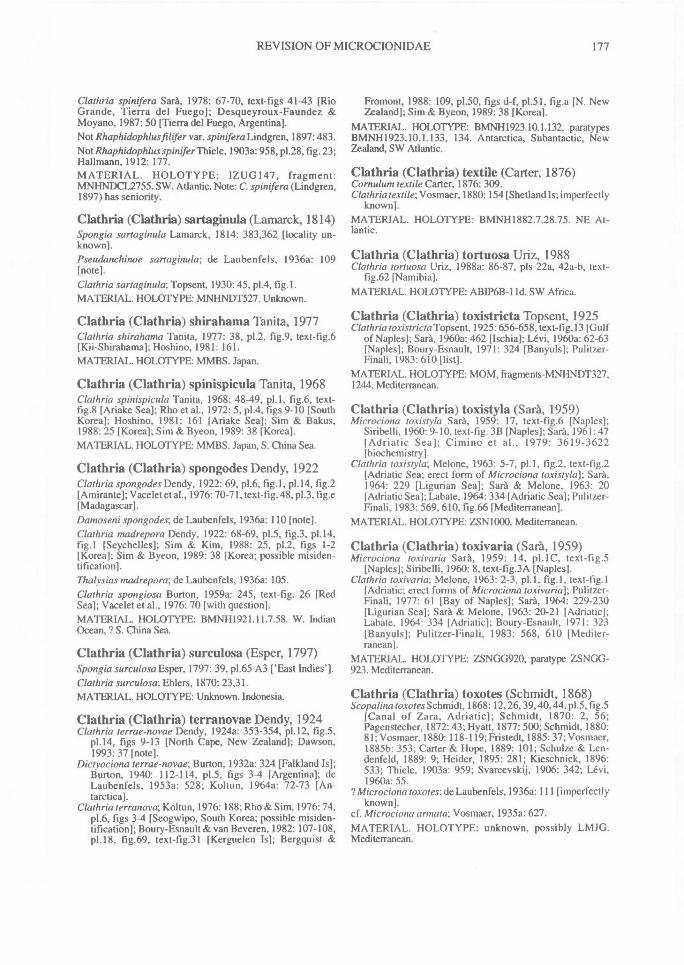
REVISION OF MICROCIONIDAE^ 177
Clathria spinifera Sara, 1978: 67-70, text-figs 41-43 [RioGrande, Tierra del Fuego]; Desqueyroux-Faundez &Moyano, 1987: 50 [Tierra del Fuego, Argentina].Not Rhaphidophlus filifer var. spinifera Lindgren, 1897: 483.Not Rhaphidophlus spinifer Thiele, 1903a: 958, p1.28, fig. 23;Hallmann, 1912: 177.MATERIAL. HOLOTYPE: IZUG147, fragment:MNHNDCL2755. SW. Atlantic. Note: C. spinifera (Lindgren,1897) has seniority.
Clathria (Clathria) sartaginula (Lamarck, 1814)Spongia sartaginula Lamarck, 1814: 383,362 [locality un-known].Pseudanchinoe sartaginula; de Laubenfels, 1936a: 109[note].Clathria sartaginula; Topsent, 1930:45, p1.4, fig. 1 .MATERIAL. HOLOTYPE: MNHNDT527. Unknown.
Clathria (Clathria) shirahama Tanita, 1977Clathria shirahama Tanita, 1977: 38, p1.2, fig.9, text-fig.6[Kii-Shirahama]; Hoshino, 1981: 161.MATERIAL. HOLOTYPE: MMBS. Japan.
Clathria (Clathria) spinispicula Tanita, 1968Clathria spinispicula Tanita, 1968: 48-49, p1.1, fig.6, text-fig.8 [Ariake Sea]; Rho et al., 1972: 5, p1.4, figs 9-10 [SouthKorea]; Hoshino, 1981: 161 [Ariake Sea]; Sim & Bakus,1988: 25 [Korea]; Sim & Byeon, 1989: 38 [Korea].MATERIAL. HOLOTYPE: MMBS. Japan, S. China Sea.
Clathria (Clathria) spongodes Dendy, 1922Clathria spongodesDendy, 1922: 69, p1.6, fig.1, p1.14, fig.2[Amirante]; Vacelet et al., 1976: 70-71, text-fig. 48, p1.3, fig.e[Madagascar].Damoseni spongodes; de Laubenfels, 1936a: 110 [note].Clathria madrepora Dendy, 1922: 68-69, p1.5, fig.3, p1.14,fig.1 [Seychelles]; Sim & Kim, 1988: 25, p1.2, figs 1-2[Korea]; Sim & Byeon, 1989: 38 [Korea; possible misiden-tification].Thalysias madrepora; de Laubenfels, 1936a: 105.Clathria spongiosa Burton, I959a: 245, text-fig. 26 [RedSea]; Vacelet et al., 1976: 70 [with question].MATERIAL. HOLOTYPE: BMNH1921.11.7.58. W. IndianOcean, ? S. China Sea.
Clathria (Clathria) surculosa (Esper, 1797)Spongia surculosa Esper, 1797: 39, p1.65 A3 ['East Indies'].Clathria surculosa; Ehlers, 1870: 23,31.MATERIAL. HOLOTYPE: Unknown. Indonesia.
Clathria (Clathria) terranovae Dendy, 1924Clathria terrae-novae Dendy, 1924a: 353-354, p1.12, fig.5,
p1.14, figs 9-13 [North Cape, New Zealand]; Dawson,1993: 37 [note].
Dictyociona terrae-novae; Burton, 1932a: 324 [Falkland Is];Burton, 1940: 112-114, p1.5, figs 3-4 [Argentina]; deLaubenfels, 1953a: 528; Koltun, 1964a: 72-73 [An-tarctica].
Clathria terranova; Koltun, 1976: 188; Rho & Sim, 1976:74,p1.6, figs 3-4 [Seogwipo, South Korea; possible misiden-tification]; Boury-Esnault & van Beveren, 1982: 107-108,p1.18, fig.69, text-fig.31 [Kerguelen Is]; Bergquist &
Fromont, 1988: 109, p1.50, figs d-f, p1.51, fig.a [N. NewZealand]; Sim & Byeon, 1989: 38 [Korea].
MATERIAL. HOLOTYPE: BMNH1923.10.1.132, paratypesBMNH1923.10.1.133, 134. Antarctica, Subantactic, NewZealand, SW Atlantic.
Clathria (Clathria) textile (Carter, 1876)Cornulum textile Carter, 1876: 309.Clathria textile; Vosmaer, 1880: 154 [Shetland Is; imperfectly
known].MATERIAL. HOLOTYPE: BMNH1882.7.28.75. NE At-lantic.
Clathria (Clathria) tortuosa Uriz, 1988Clathria tortuosa Uriz, 1988a: 86-87, pls 22a, 42a-b, text-
fig.62 [Namibia].MATERIAL. HOLOTYPE: ABIP6B-11d. SW Africa.
Clathria (Clathria) toxistricta Topsent, 1925Clathria toxistricta Topsent, 1925:656-658, text-fig. 13 [Gulf
of Naples]; Sara, 1960a: 462 [Ischia]; Levi, 1960a: 62-63[Naples]; Boury-Esnault, 1971: 324 [Banyuls]; Pulitzer-Finali, 1983: 610 [list].
MATERIAL. HOLOTYPE: MOM, fragments-MNHNDT327,1244. Mediterranean.
Clathria (Clathria) toxistyla (Sara, 1959)Microciona toxistyla Sara, 1959: 17, text-fig.6 [Naples];
Siribelli, 1960:9-10, text-fig. 3B [Naples]; Sara, 1961:47[Adriatic Sea]; Cimino et al., 1979: 3619-3622[biochemistry].
Clathria toxistyla; Melone, 1963: 5-7, p1.1, fig.2, text-fig.2[Adriatic Sea; erect form of Microciona toxistyla]; Sara,1964: 229 [Ligurian Sea]; Sara & Melone, 1963: 20[Adriatic Sea]; Labate, 1964: 334 [Adriatic Sea]; Pulitzer-Finali, 1983: 569, 610, fig.66 [Mediterranean].
MATERIAL. HOLOTYPE: ZSN1000. Mediterranean.
Clathria (Clathria) toxivaria (Sara, 1959)Microciona toxivaria Sara, 1959: 14, p1.1C, text-fig.5
[Naples]; Siribelli, 1960: 8, text-fig.3A [Naples].Clathria toxivaria; Melone, 1963: 2-3, p1.1, fig.1, text-tig.1
[Adriatic; erect forms of Microciona toxivaria]; Pulitzer-Finali, 1977: 61 [Bay of Naples]; Sara, 1964: 229-230[Ligurian Sea]; Sara & Melone, 1963: 20-21 [Adriatic];Labate, 1964: 334 [Adriatic]; Boury-Esnault, 1971: 323[Banyuls]; Pulitzer-Finali, 1983: 568, 610 [Mediter-ranean].
MATERIAL. HOLOTYPE: ZSNGG920, paratype ZSNGG-923. Mediterranean.
Clathria (Clathria) toxotes (Schmidt, 1868)Scopalina toxotes Schmidt, 1868: 12, 26, 39, 40, 44, p1.5, fi g.5
[Canal of Zara, Adriatic]; Schmidt, 1870: 2, 56;Pagenstecher, 1872:43; Hyatt, 1877: 500; Schmidt, 1880:81; Vosmaer, 1880: 118-119; Fristedt, 1885:37; Vosmaer,1885b: 353; Carter & Hope, 1889: 101; Schulze & Len-denfeld, 1889: 9; Heider, 1895: 281; Kieschnick, 1896:533; Thiele, 1903a: 959; Svarcevskij, 1906: 342; Levi,1960a: 55.
? Microciona toxotes; de Laubenfels, 1936a: 111 [imperfectlyknown].
cf. Microciona armata; Vosmaer, 1935a: 627.MATERIAL. HOLOTYPE: unknown, possibly LMJG.Mediterranean.
178^ MEMOIRS OF THE QUEENSLAND MUSEUM
Clathria (Clathria) typica Kirkpatrick, 1904Clathria typica Kirkpatrick, 1904: 148 [Natal]; Hallmann,
1912: 208 [anomalous species].? Tenacia clathrata; Carter, 1875: 195 [nomen nudum];
Carter, 1878: 160,163; sensuV osmaer, 1935a: 628.Not Echinonema typicum Carter, 1881a: 362.MATERIAL. HOLOTYPE: BMNH1902.11.16.31. Note: imper-fectly known; listed in BMNH register as C. typica Carter. SAfrica.
Clathria (Clathria) ulmus Vosmaer, 1880Clathria ulmus Vosmaer, 1880: 151 [locality unknown]; Rid-
ley, 1884a: 444.Thalysias ulmus; de Laubenfels, 1936a: 105 [note].MATERIAL. HOLOTYPE: possibly RNHL. Unknownlocality.
Clathria (Clathria) unica Cuartas, 1993Clathria unica Cuartas, 1993: 112 [Argentina].MATERIAL. HOLOTYPE: Division Invertebrados del Museode Ciencias Naturales de La Plata, Argentina. SW Atlantic.
Clathria (Clathria) vasiformis (de Laubenfels,1953)Thalyseutypon vasiformis de Laubenfels, 1953a: 525, text-
fig.4 [Gulf of Mexico]; Little, 1963:50 [note].Clathria vasiformis ., Van Soest, 1984b: 109, table 4.Pandaros vasiformis; Wiedenmayer, 1977: 144 [note].MATERIAL. HOLOTYPE: USNM23403, paratype MLUM-ML4-232. E Pacific.
Clathria (Clathria) zoanthifera Levi, 1963Clathria zoanthifera Levi, 1963: 58, text-fig.66, p1.9D [Cape
of Good Hope].MATERIAL. HOLOTYPE: UCT (fragment IVINHNDCL607).S Africa.
TRANSFERS
List of other species described in Clathria(Clathria), or one of its synonyms, but now trans-ferred to another genus.ClathrM australis Lendenfeld, 1888: 222 [Port Phillip, Vic-
toria].Wilsonella australis; Hallmann, 1912: 239.Not Clathria australis Whitelegge, 1901: 90.Not Plumohalichondria australis; Whitelegge, 1901: 90,
p1.11, fig.14.MATERIAL. LECTOTYPE: AMZ957. Note: referred Crel-lithe, synonym of Crella incrustans var. arenacea (Hallmann,1914c: 411).
Plumohalichondria australis Whitelegge, 1901: 90-92,p1.11, fig.14a,b [New South Wales coast]; Whitelegge,1907: 492 [New South Wales coast].
MATERIAL. HOLOTYPE: AMG9042. Note: referred Crel-lidae, synonym of Crella incrustans (Hallmann, 1912: 160).
Isodictya beanii Bowerbank, 1866: 274, 334, 335 [Britain];Gray, 1868: 164; Schmidt, 1870: 77; Bowerbank, 1874:147, p1.58, figs 1-6.
Amphilectus beanii; Vosmaer, 1880: 115.Clathria beanii; Ridley, 1881: 485, 486; Bowerbank, 1882:
13, 23, 150; Topsent, 1890c: 203.
Myxilla beani; Topsent, 1892b: 23; Topsent, 1894a: 8, 9, 25;Hanitsch, 1894: 179.
cf. Microciona prolifera; Vosmaer, 1933: 610.MATERIAL. HOLOTYPE: BMNH1930.7.3.339. Note:Synonym of Antho involvens (Levi, 1960a: 76).
Echinonema caespitosa Carter, 1885f: 352 [Port Phillip,Victoria].
Plumohalichondria caespitosa; Dendy, 1896: 41.MATERIAL. HOLOTYPE: BMNH1886.12.15.453. Note:referred to Anchinoidae, Plumohalichondria.
Antherochalina concentrica Lendenfeld, 1887b: 788, p1.22,fig.42 [Port Molle (Airlee Beach), Q1d.].
Cymbastela concentrica; Hooper & Bergquist, 1992: 114.MATERIAL. HOLOTYPE: AMZ1993 (lectotype), fragment:BMNH1886.8.27.451, 460 (paralectotypes). Note: referred toAxinellidae, Cymbastela.
Clathria corallorhizoides Fristedt, 1887: 460, p1.25, figs 73-77, p1.29, fig.23.
MATERIAL. HOLOTYPE: unknown, fragment:BMNH1910.1.1.1445. Note: referred to Myxillidae, synonymof Lissodendoryx complicata (Lundbeck, 1905: 166).
Antherochalina elegans Lendenfeld, 1887b: 787, p1.22,fig.40.
Syringella elegans; Burton, 1934a: 558.Raspailia (Syringe/la) elegans; Hooper, 1991: 1262.MATERIAL. HOLOTYPE: BMNHI886.8.27.452. Note:referred to Raspailiidae, Raspailia.
Echinonema incrustans Carter, 1885f: 353 [Port Phillip,Victoria].
Plumohalichondria incrustans; Dendy, 1896: 42.Plumohalichondria mammillata; Carter, 1885f: 355; Ridley
& Dendy, 1887: 156, p1.30, fig.4, p1.47, fig.4.Crella incrustans-, Hallmann, 1912: 152-156, p1.23, figs 2-3,
p1.24, text-figs 28-34.MATERIAL. HOLOTYPE: BMNH1886.12.15.123, paratypes1886.12.15.249, AME651, AMZ957. Note: referred to Crel-
Crella.
Cornulum johnsoni de Laubenfels, 1934: 15.Clathria johnsoni; Van Soest, 1984b: 104 [possible synonym
of Clathria].MATERIAL. HOLOTYPE: USNM Note: referred to Coelos-phaeridae, possibly Cornulum (imperfectly known).
Echinonema levis Lendenfeld, 1888:220 [Port Jackson, NewSouth Wales].
Plumohalichondria australis, in part; Whitelegge, 1901: 65,91,92; Whitelegge, 1902a: 212.
Crella incrustans var. levis; Hallmann, 1912: 164-167, text-figs 33-34.
MATERIAL. LECTOTYPE: AMZ959, paralectotypeAMG9708. Note: referred to Crellidae, synonym of Crellaincrustans (Hallmann, 1912: 164).
Clathria loveni Fristedt, 1887: 458, p1.25, figs 70-72, p1.30,fig.24 [Cape Jakan, Siberian Arctic Ocean]; Lambe, 1900:160.
[?] Esperella loveni; Lambe, 1895: 123, p1.4, fig.1 (W coast,North America).
Esperia loveni; de Laubenfels, 1936a: 120 [note].MATERIAL. HOLOTYPE: unknown, fragmentsBMNH1927.2.22.2. Note: referred to Mycalidae, Mycale.
REVISION OF MICROCIONIDAE^ 179
Plumohalichondria microcionides Carter, 1876: 236, p1.12,fig.!!, p1.15, fig.30 [between Scotland and Faroe Is].
Clathria microcionides; Vosmaer, 1880: 154.Plocamionida microcionides; Alander, 1942: 53 [Sweden].Not Hymeraphia microcionides Carter, 1876: 390.Not Plocamia microcionides; Topsent, 1891a: 529, 544-545.MATERIAL. HOLOTYPE: BMNH1954.3.9.173. Note:referred to Anchinoidae, Plocamionida.
Clathria mollis Kirkpatrick, 1903: 249-250, p1.5, fig. 15, p1.6,fig. 16 [East London Coast, Natal].
Pronax mollis; Levi, 1963: 66.Not Wilsonella mollis; Hallmann, 1912: 243.Not Clathria dura var. molls; Hentschel, 1911: 370.MATERIAL. HOLOTYPE: BMNH1902.11.16.32. Note:referred to Anchinoidae, Pronax.
Clathria morisca Schmidt, 1868: 9, 41, 43, p1.2, fig.7 [Al-giers, Mediterranean]; Vosmaer, 1880: 150-151 [Algiers];Topsent, 1902: 329; Topsent, 1938: 11; Desqueyroux-Faundez & Stone, 1992: 10, 35.
Diciyoclathria morisca; Topsent, 1920b: 18-21 [re-examina-tion of holotype]; Topsent, 1925: 660-661, p1.8, fig.! [Gulf ofNaples]; Topsent, 1928a: 301-302, p1.3, fig.3 [Porto Santo,Azores]; Levi, 1959: 134, text-fig.27, p1.5, fig.! [Rio de Oro,Gulf of Guinea]; Levi, 1960b: 761-762, text-fig.15 [var.anisotyla; SW. Cape of Naze, W. Africa]; Sara, 1960a: 462[Ischia, Mediterranean].Artemisina mediterranea Babic, 1921: 87 [Adriatic]; Babic,
1922: 258, text-fig.3; Burton, 1930a: 528.Myxilla banyulensis, in part, Topsent, 1892b: 23; Topsent,
1902: 351, 363, 366; Cotte, 1903:423.cf. Clathria coralloides and C. compressa; Vosmaer, 1935a:
626 [intermediate form].MATERIAL. HOLOTYPE: MNHN DT2170, fragmentBMNH 1868.3.2.21. Note: Synonym of Antho involvens (Levi,1960a: 57).
Clathriaoroides Schmidt, 1864:35, p1.4, figs 1-2 [Quarnerno,Adriatic]; Carter, 1875: 195; Vosmaer, 1880: 155; Dendy,1889a: 41; Desqueyroux-Faundez & Stone, 1992: 10, 35,103.
? Oroidea adriatica; Gray, 1867: 520.Chalinopsis oroides; Schmidt, 1870: 60.Ophlitaspongia oroides; Bowerbank, 1874: 10.Age/as oroides; Riitzler, 1965: 34 [Banyuls]; Boury-Esnault,
1971: 322 [Banyuls]; Laubier, 1966 [Banyuls]; Pulitzer-Finali, 1983: 534 [Mediterranean].
MATERIAL. HOLOTYPE: LMJG 15957, fragmentBMNH1868.3.2.22. Note: referred to Agelasidae, Agelas.
Artemisina paradoxa Babic, 1921:87; Babic, 1922:258-261,p1.8, fig.6, text-fig.c [Adriatic]; Levi, 1960a: 85-86[Adriatic; with question]
Clathria paradoxa; Burton, 1930a: 528.Dictyoclathria morisca; Topsent, 1925: 660; Ristau, 1978:
585-586 [note on affinities].MATERIAL. HOLOTYPE: unknown. Note: synonym ofAnthoinvolvens (Topsent, 1925: 660).
Clathria pelligera Schmidt, 1864: 34, p1.3, fig.13 [Lesina,Adriatic]; Desqueyroux-Faundez & Stone, 1992: 10, 36,103.
Rhaphidostyla pelligera; Burton, 1935b: 652; Sara, 1958:246-247, fig.15 [Gulf of Naples].
Stylotella pelligera; Topsent, 1925: 638; Boury-Esnault,1971: 328 [Banyuls].
Dictyonella pelligera; Pulitzer-Finali, 1983: 545.
MATERIAL. HOLOTYPE: LMJG 15517; fragment BMNI-11867.3.11.29. Note: referred to Dictyoriellidae, Dictyonella.
Clathriaprocumbens Brondsted, 1923; Brondsted, 1926:329[probably a typographical error for Clathrina (Calcarea),and possibly synonym of Ascetta procumbens Lendenfeld,1885a].
Antherochalina quercifolia Keller, 1889: 383-384, p1.23,fig.34 [Red Sea].
Querciclona quercifolia; de Laubenfels, 1936a: 46 [note].MATERIAL. HOLOTYPE: ZMB429, fragmentBMNH1908.9.24.179. Note: referred to Axinellidae, Phakellia.
Clathria raphida Hechtel, 1976:244; Van Soest, 1984b: 153.Note: cited in a list of Brazilian endemic sponges; at-tributed to Boury-Esnault (1973); probably a misprint forCliona rhaphida Boury-Esnault, 1973.
Antherochalina renieroides Lendenfeld, 1887b: 788, p1.28,figs 18, 23 [New Zealand].
MATERIAL. HOLOTYPE: BMN1-11886.8.27.449. Referred toAxinellidae, synonym of Phakellia flabellata (Carter) (seeHooper, 1991).
Hymeraphia tuberosocapitata Topsent, 1890b: 68 [Azores,Atlantic]; Topsent, 1892a:113, p1.11, fig.6 [Atlantic].
Cionanchora tuberosocapitata; de Laubenfels, 1936a: 108[note].
Clathria tuberosocapitata; Van Soest, 1984b: 7 [genericsynonymy for Cionanchora].
Discorhabdella tuberosocapitata; Boury-Esnault, Pansini &Uriz, 1992: 2-6.
MATERIAL. HOLOTYPE: MOM040323, fragmentMNHNDT938. referred to Hymedesmiidae, Discorhabdella.
Clathria (Wilsonella) Carter, 1885Hooper & Wiedenmayer, 1994
Wilsonella Carter, 1885f: 366 (not Hallmann, 1912:242).Clathriopsamma Lendenfeld, 1888: 227.Aulenella Burton & Rao, 1932: 345.
TYPE SPECIES. Wilsonella australiensis Carter,1885f: 366 (by monotypy).
DEFINITION. Sand grains and foreign spiculespartially or completely replacing coring spiculesinside fibres; coring spicules same or very similargeometry to auxiliary spicules located outsidefibres; skeletal architecture reticulate.
REMARKS. Microcionids with sand and foreigndebris incorporated into fibres ('arenaceoussponges') are a predominant feature of SAustralian waters. Of the 17 species in Wilsonellaor a synonym, 14 are valid and 6 are known tolive in this region including 2 new species.
This group of arenaceous microcionids is wellknown as Clathriopsamma (Hooper, 1990a;Hooper & Levi, 1993a). However, the inclusionof C. (W) australiensis in this group means thatWilsonella (1885) takes precedence over Clath-riopsamma (1888).
A
180^ MEMOIRS OF THE QUEENSLAND MUSEUM
FIG. 81. Clathria (Wilsonella) abrolholensis sp.nov. (holotype NTMZ3218). A, Auxiliary style. B, Echinatingacanthostyles. C, Palmate isochelae. D, Wing-shaped toxas. E, Section through peripheral skeleton (hatchesforeign spicules). F, Australian distribution. G, Preserved holotype. H, Holotype.
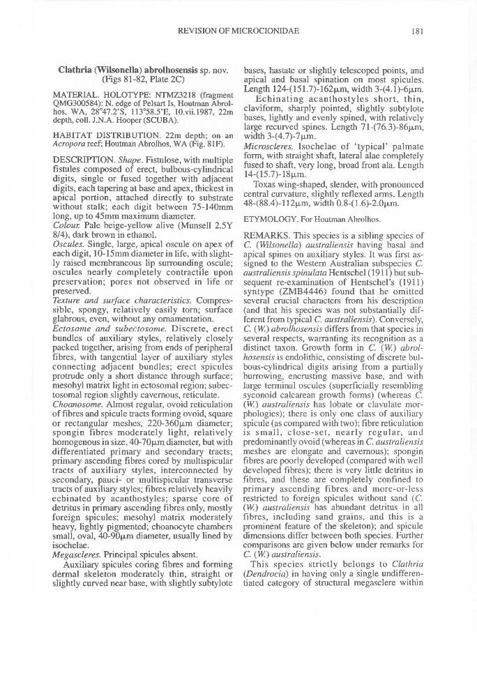
REVISION OF MICROCIONIDAE^ 181
Clathria (VVilsonella) abrolhosensis sp. nov.(Figs 81-82, Plate 2C)
MATERIAL. HOLOTYPE: NTMZ3218 (fragmentQMG300584): N. edge of Pelsart Is, Houtman Abrol-hos, WA, 28°47.2'S, 113°58.5'E, 10.vii.I987, 22mdepth, coll. J.N.A. Hooper (SCUBA).
HABITAT DISTRIBUTION. 22m depth; on anAcropora reef; Houtman Abrolhos, WA (Fig. 81F).
DESCRIPTION. Shape. Fistulose, with multiplefistules composed of erect, bulbous-cylindricaldigits, single or fused together with adjacentdigits, each tapering at base and apex, thickest inapical portion, attached directly to substratewithout stalk; each digit between 75-140mmlong, up to 45mm maximum diameter.Colour Pale beige-yellow alive (Munsell 2.5Y8/4), dark brown in ethanol.Oscules. Single, large, apical oscule on apex ofeach digit, 10-15mm diameter in life, with slight-ly raised membraneous lip surrounding oscule;oscules nearly completely contractile uponpreservation; pores not observed in life orpreserved.Texture and surface characteristics. Compres-sible, spongy, relatively easily torn; surfaceglabrous, even, without any ornamentation.Ectosome and subectosome. Discrete, erectbundles of auxiliary styles, relatively closelypacked together, arising from ends of peripheralfibres, with tangential layer of auxiliary stylesconnecting adjacent bundles; erect spiculesprotrude only a short distance through surface;mesohyl matrix light in ectosomal region; subec-tosomal region slightly cavernous, reticulate.Choanosome. Almost regular, ovoid reticulationof fibres and spicule tracts forming ovoid, squareor rectangular meshes, 220-360vm diameter;spongin fibres moderately light, relativelyhomogenous in size, 40-70Rm diameter, but withdifferentiated primary and secondary tracts;primary ascending fibres cored by multispiculartracts of auxiliary styles, interconnected bysecondary, pauci- or multispicular transversetracts of auxiliary styles; fibres relatively heavilyechinated by acanthostyles; sparse core ofdetritus in primary ascending fibres only, mostlyforeign spicules; mesohyl matrix moderatelyheavy, lightly pigmented; choanocyte chamberssmall, oval, 40-901im diameter, usually lined byisochelae.Megascleres. Principal spicules absent.
Auxiliary spicules coring fibres and formingdermal skeleton moderately thin, straight orslightly curved near base, with slightly subtylote
bases, hastate or slightly telescoped points, andapical and basal spination on most spicules.Length 1244151.7)-164,m, width 3-(4.1)-6p.m.
Echinating acanthostyles short, thin,claviform, sharply pointed, slightly subtylotebases, lightly and evenly spined, with relativelylarge recurved spines. Length 71-(76.3)-86Rm,width 3-(4.7)-7p,m.Microscleres. Isochelae of 'typical' palmateform, with straight shaft, lateral alae completelyfused to shaft, very long, broad front ala. Length14-(15.7)-18Rm.
Toxas wing-shaped, slender, with pronouncedcentral curvature, slightly reflexed arms. Length48488.4)-11211m, width 0.841.6)-2.0p.m.
ETYMOLOGY. For Houtman Abrolhos.
REMARKS. This species is a sibling species ofC. (Wilsonella) australiensis having basal andapical spines on auxiliary styles. It was first as-signed to the Western Australian subspecies C.australiensis spinulata Hentschel (1911) but sub-sequent re-examination of Hentschel's (1911)syntype (ZMB4446) found that he omittedseveral crucial characters from his description(and that his species was not substantially dif-ferent from typical C. australiensis). Conversely,C. (W) abrolhosensis differs from that species inseveral respects, warranting its recognition as adistinct taxon. Growth form in C. (W) abrol-hosensis is endolithic, consisting of discrete bul-bous-cylindrical digits arising from a partiallyburrowing, encrusting massive base, and withlarge terminal oscules (superficially resemblingsyconoid calcarean growth forms) (whereas C.(W) australiensis has lobate or clavulate mor-phologies); there is only one class of auxiliaryspicule (as compared with two); fibre reticulationis small, close-set, nearly regular, andpredominantly ovoid (whereas in C. australiensismeshes are elongate and cavernous); sponginfibres are poorly developed (compared with welldeveloped fibres); there is very little detritus infibres, and these are completely confined toprimary ascending fibres and more-or-lessrestricted to foreign spicules without sand (C.(W) australiensis has abundant detritus in allfibres, including sand grains, and this is aprominent feature of the skeleton); and spiculedimensions differ between both species. Furthercomparisons are given below under remarks forC. (W) australiensis.
This species strictly belongs to Clathria(Dendrocia) in having only a single undifferen-tiated category of structural megasclere within
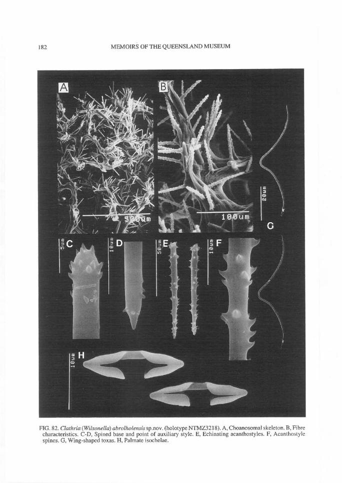
182^ MEMOIRS OF THE QUEENSLAND MUSEUM
FIG. 82. Clathria (Wilsonella) abrolholensis sp.nov. (holotype NTMZ3218). A, Choanosomal skeleton. B, Fibrecharacteristics. C-D, Spined base and point of auxiliary style. E, Echinating acanthostyles. F, Acanthosty lespines. G, Wing-shaped toxas. H, Palmate isochelae.
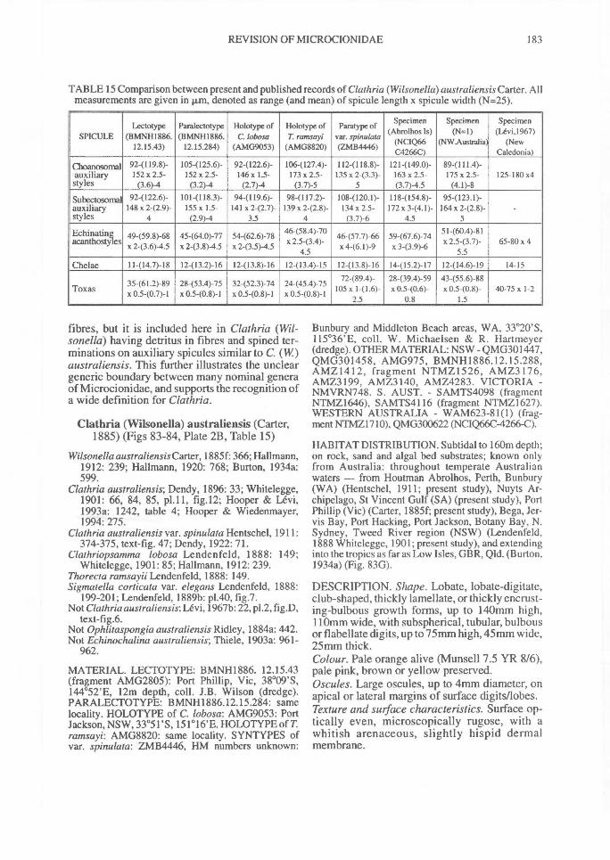
REVISION OF MICROCIONIDAE^ 183
TABLE 15 Comparison between present and published records of Clathria (Wilsonella)australiensis Carter. Allmeasurements are given in rim, denoted as range (and mean) of spicule length x spicule width (N=25).
SPICULELectotype
(BMNH1886.12.15.43)
Paralectotype(BMNH1886.
12.15.284)
Holotype ofC. lobosa
(AMG9053)
Holotype ofT. ramsayi
(AMG8820)
Paratype ofvar. spinulata(ZMB4446)
Specimen(Abrolhos Is)
(NCIQ66C4266C)
Specimen(N=1)
(NWAustralia)
Specimen(I-evi,1967)
(NewCaledonia)
choanosomai 92(119.8)- 105-(125.6)- 92(122.6)- 106-(127.4)- 112(118.8)- 121-(149.0)- 89(111.4)-auxiliary 152 x 2.5- 152 x 2.5- 146 x 1.5- 173 x 2.5- 135 x 2-(3.3)- 163 x 2.5- 175 x 2.5- 125-180 x4styles (3.6)4 (3.2)4 (2.7)-4 (3.7)-5 5 (3.7)4.5 (4.1)-8
Subect^mal 92(122.6)- 101(118.3)- 94(119.6)- 98(117.2)- 108(120.1)- 118(154.8)- 95(123.1)-auxiliary 148 x 2-(2.9)- 155 x 1.5- 141 x 2-(2.7)- 139 x 2-(2.8)- 134 x 2.5- 172 x 3-(4.1)- 164 x 2-(2.8)-styles 4 (2.9)4 3.5 4 (3.7)-6 4.5 5
Echinatini 49-(59.8)-68 45-(64.0)-77 54-(62.6)-78 46(58.4)-70 46-(57.7)-66 59-(67.6)-74 51(60.4)-81acanthosty es x 2-(3.6)-4.5 x 2-(3.8)-4.5 x 2-(3.5)-4.5 x 2.5-(3.4)- x 4-(6.1)-9 x 3-(3.9)-6 x 2.5-(3.7)- 65-80 x 4
4.5 5.5
Chelae 11-(14.7)-18 12-(13.2)-16 12-(13.8)-16 12-(13.4)-15 12-(13.8)-16 14-(15.2)-17 12-(14.6)-19 14-1572-(89.4)- 28-(39.4)-59 43-(55.6)-88
Toxas35-(61 .2)-89 28-(53.4)-75 32-(52.3)-74 24-(45.4)-75
105 x 1-(1.6)- x 0.5-(0.6)- x 0.5-(0.8)- 40-75 x 1-2x 0.5-(0.7)-1 x 0.5-(0.8)-1 x 0.5-(0.8)-1 x 0.5-(0.8)-1
2.5 0.8 1.5
fibres, but it is included here in Clathria (Wil-sonella) having detritus in fibres and spined ter-minations on auxiliary spicules similar to C. (W.)australiensis. This further illustrates the uncleargeneric boundary between many nominal generaof Microcionidae, and supports the recognition ofa wide definition for Clathria.
Clathria (Wilsonella) australiensis (Carter,1885) (Figs 83-84, Plate 2B, Table 15)
Wilsonella australiensis Carter, 1885f: 366; Hallmann,1912: 239; Hallmann, 1920: 768; Burton, 1934a:599.
Clathria australiensis; Dendy, 1896: 33; Whitelegge,1901: 66, 84, 85, p1.11, fig.12; Hooper & Levi,1993a: 1242, table 4; Hooper & Wiedenmayer,1994: 275.
Clathria australiensis var. spinulata Hentschel, 1911:374-375, text-fig. 47; Dendy, 1922: 71.
Clathriopsamma lobosa Lendenfeld, 1888: 149;Whitelegge, 1901: 85; Hallmann, 1912: 239.
Thorecta ramsayii Lendenfeld, 1888: 149.Sigmatella corticata var. elegans Lendenfeld, 1888:
199-201; Lendenfeld, 1889b: p1.40, fig.7.Not Clathria australiensis; Levi, 1967b: 22, p1.2, fig.D,
text-fig.6.Not Ophlitaspongia australiensis Ridley, 1884a: 442.Not Echinochalina australiensis; Thiele, 1903a: 961-
962.
MATERIAL. LECTOTYPE: BMNH1886. 12.15.43(fragment AMG2805): Port Phillip, Vic, 38 °09'S,144°52'E, 12m depth, coll. J.B. Wilson (dredge).PARALECTOTYPE: BMNH1886.12.15.284: samelocality. HOLOTYPE of C. lobosa: AMG9053: PortJackson, NSW, 33 °51'S, 151 ° 16'E. HOLOTYPE of T.ramsayi: AMG8820: same locality. SYNTYPES ofvar. spinulata: ZMB4446, HM numbers unknown:
Bunbury and Middleton Beach areas, WA, 33 °20'S,115 °36'E, coll. W. Michaelsen & R. Hartmeyer(dredge). OTHER MATERIAL: NSW - QMG301447,QMG301458, AMG975, BMNH1886.12.15.288,AMZI412, fragment NTMZ1526, AMZ3176,AMZ3199, AMZ3140, AMZ4283. VICTORIA -NMVRN748. S. AUST. - SAMTS4098 (fragmentNTMZ1646), SAMTS4116 (fragment NTMZ1627).WESTERN AUSTRALIA - WAM623-81(1) (frag-ment NTMZ1710), QMG300622 (NCIQ66C-4266-C).
HABITAT DISTRIBUTION. Subtidal to 160m depth;on rock, sand and algal bed substrates; known onlyfrom Australia: throughout temperate Australianwaters - from Houtman Abrolhos, Perth, Bunbury(WA) (Hentschel, 1911; present study), Nuyts Ar-chipelago, St Vincent Gulf (SA) (present study), PortPhillip (Vic) (Carter, 1885f; present study), Bega, Jer-vis Bay, Port Hacking, Port Jackson, Botany Bay, N.Sydney, Tweed River region (NSW) (Lendenfeld,1888 Whitelegge, 1901; present study), and extendinginto the tropics as far as Low Isles, GBR, Qld. (Burton,1934a) (Fig. 83G).
DESCRIPTION. Shape. Lobate, lobate-digitate,club-shaped, thickly lamellate, or thickly encrust-ing-bulbous growth forms, up to 140mm high,110mm wide, with subspherical, tubular, bulbousor flabellate digits, up to 75n-un high, 45mm wide,25mm thick.Colour. Pale orange alive (Munsell 7.5 YR 8/6),pale pink, brown or yellow preserved.Oscules. Large oscules, up to 4mm diameter, onapical or lateral margins of surface digits/lobes.Texture and surface characteristics. Surface op-tically even, microscopically rugose, with awhitish arenaceous, slightly hispid dermalmembrane.
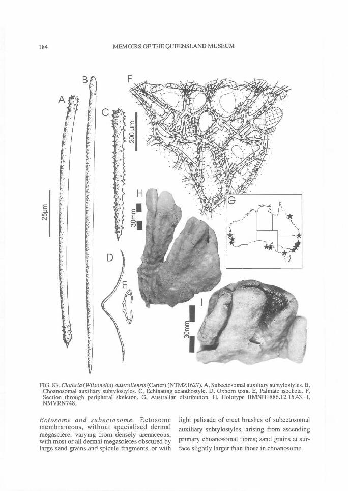
184^ MEMOIRS OF THE QUEENSLAND MUSEUM
FIG. 83. Clathria(Wilsonella)australiensis (Carter) (NTMZ1627). A, Subectosomal auxiliary subtylostyles. B,Choanosomal auxiliary subtylostyles. C, Echinating acanthostyle. D, Oxhorn toxa. E, Palmate isochela. F,Section through peripheral skeleton. G, Australian distribution. H, Holotype BMNH1886.12.15.43. I,NMVRN748.
Ectosome and subectosome. Ectosomemembraneous, without specialised dermalmegasclere, varying from densely arenaceous,with most or all dermal megascleres obscured bylarge sand grains and spicule fragments, or with
light palisade of erect brushes of subectosomalauxiliary subtylostyles, arising from ascendingprimary choanosomal fibres; sand grains at sur-face slightly larger than those in choanosome.

REVISION OF MICROCIONIDAE^ 185
FIG. 84. Clathria (Wilsonella) australiensis (Carter) (A-B, AMG9053, QMG301447). A, Choanosomalskeleton. B, Peripheral skeleton. C, Fibre characteristics. D, Echinating acanthostyle. E, Acanthostyle spines.F, Palmate and modified isochelae. G, Oxhorn toxas. H-I, Continuum in basal and apical spination of auxiliarysubtylostyles.
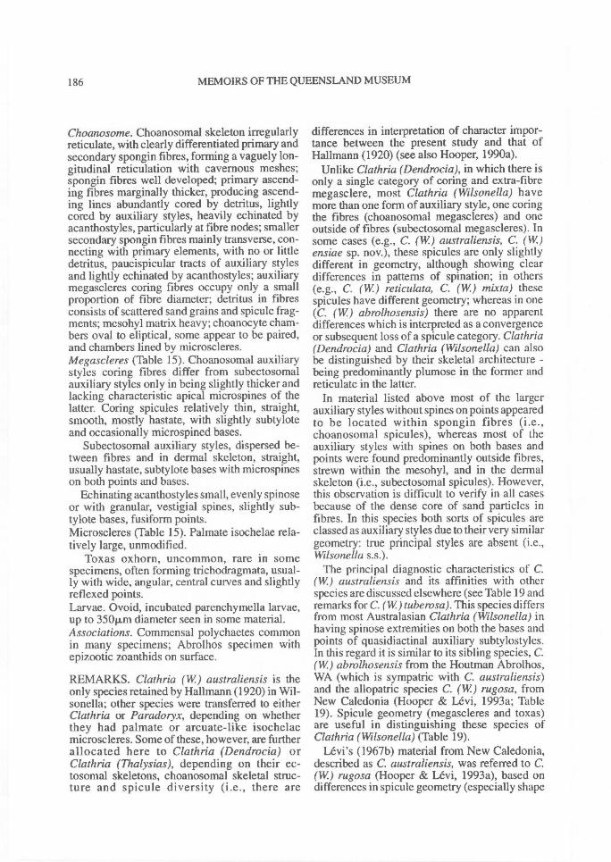
186^ MEMOIRS OF THE QUEENSLAND MUSEUM
Choanosome. Choanosomal skeleton irregularlyreticulate, with clearly differentiated primary andsecondary spongin fibres, forming a vaguely lon-gitudinal reticulation with cavernous meshes;spongin fibres well developed; primary ascend-ing fibres marginally thicker, producing ascend-ing lines abundantly cored by detritus, lightlycored by auxiliary styles, heavily echinated byacanthostyles, particularly at fibre nodes; smallersecondary spongin fibres mainly transverse, con-necting with primary elements, with no or littledetritus, paucispicular tracts of auxiliary stylesand lightly echinated by acanthostyles; auxiliarymegascleres coring fibres occupy only a smallproportion of fibre diameter; detritus in fibresconsists of scattered sand grains and spicule frag-ments; mesohyl matrix heavy; choanocyte cham-bers oval to eliptical, some appear to be paired,and chambers lined by microscleres.Megascleres (Table 15). Choanosomal auxiliarystyles coring fibres differ from subectosomalauxiliary styles only in being slightly thicker andlacking characteristic apical microspines of thelatter. Coring spicules relatively thin, straight,smooth, mostly hastate, with slightly subtyloteand occasionally microspined bases.
Subectosomal auxiliary styles, dispersed be-tween fibres and in dermal skeleton, straight,usually hastate, subtylote bases with microspineson both points and bases.
Echinating acanthostyles small, evenly spinoseor with granular, vestigial spines, slightly sub-tylote bases, fusiform points.Microscleres (Table 15). Palmate isochelae rela-tively large, unmodified.
Toxas oxhorn, uncommon, rare in somespecimens, often forming trichodragmata, usual-ly with wide, angular, central curves and slightlyreflexed points.Larvae. Ovoid, incubated parenchymella larvae,up to 350p.m diameter seen in some material.Associations. Commensal polychaetes commonin many specimens; Abrolhos specimen withepizootic zoanthids on surface.
REMARKS. Clathria (W) australiensis is theonly species retained by Hallmann (1920) in Wil-sonella; other species were transferred to eitherClathria or Paradoryx, depending on whetherthey had palmate or arcuate-like isochelaemicroscleres. Some of these, however, are furtherallocated here to Clathria (Dendrocia) orClathria (Thalysias), depending on their ec-tosomal skeletons, choanosomal skeletal struc-ture and spicule diversity (i.e., there are
differences in interpretation of character impor-tance between the present study and that ofHallmann (1920) (see also Hooper, 1990a).
Unlike Clathria (Dendrocia), in which there isonly a single category of coring and extra-fibremegasclere, most Clathria (Wilsonella) havemore than one form of auxiliary style, one coringthe fibres (choanosomal megascleres) and oneoutside of fibres (subectosomal megascleres). Insome cases (e.g., C. (W) australiensis, C. (W)ensiae sp. nov.), these spicules are only slightlydifferent in geometry, although showing cleardifferences in patterns of spination; in others(e.g., C. (W) reticulata, C. (W) mixta) thesespicules have different geometry; whereas in one(C. (W) abrolhosensis) there are no apparentdifferences which is interpreted as a convergenceor subsequent loss of a spicule category. Clathria(Dendrocia) and Clathria (Wilsonella) can alsobe distinguished by their skeletal architecture -being predominantly plumose in the former andreticulate in the latter.
In material listed above most of the largerauxiliary styles without spines on points appearedto be located within spongin fibres (i.e.,choanosomal spicules), whereas most of theauxiliary styles with spines on both bases andpoints were found predominantly outside fibres,strewn within the mesohyl, and in the dermalskeleton (i.e., subectosomal spicules). However,this observation is difficult to verify in all casesbecause of the dense core of sand particles infibres. In this species both sorts of spicules areclassed as auxiliary styles due to their very similargeometry: true principal styles are absent (i.e.,Wilsonella s.s.).
The principal diagnostic characteristics of C.(W) australiensis and its affinities with otherspecies are discussed elsewhere (see Table 19 andremarks for C. (W) tube rosa). This species differsfrom most Australasian Clathria (Wilsonella) inhaving spinose extremities on both the bases andpoints of quasidiactinal auxiliary subtylostyles.In this regard it is similar to its sibling species, C.(W) abrolhosensis from the Houtman Abrolhos,WA (which is sympatric with C. australiensis)and the allopatric species C. (W) rugosa, fromNew Caledonia (Hooper & Levi, 1993a; Table19). Spicule geometry (megascleres and toxas)are useful in distinguishing these species ofClathria (Wilsonella) (Table 19).
Levi's (1967b) material from New Caledonia,described as C. australiensis, was referred to C.(W) rugosa (Hooper & Levi, 1993a), based ondifferences in spicule geometry (especially shape
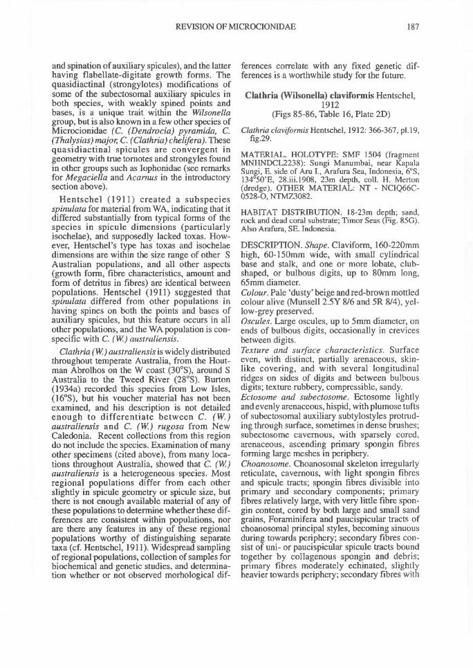
REVISION OF MICROCIONIDAE^ 187
and spination of auxiliary spicules), and the latterhaving flabellate-digitate growth forms. Thequasidiactinal (strongylotes) modifications ofsome of the subectosomal auxiliary spicules inboth species, with weakly spined points andbases, is a unique trait within the Wilsonellagroup, but is also known in a few other species ofMicrocionidae (C. (Dendrocia) pyramida, C.(Thalysias) major, C. (Clathria) chelifera). Thesequasidiactinal spicules are convergent ingeometry with true tornotes and strongyles foundin other groups such as Iophonidae (see remarksfor Megaciella and Acarnus in the introductorysection above).
Hentschel (1911) created a subspeciesspinulata for material from WA, indicating that itdiffered substantially from typical forms of thespecies in spicule dimensions (particularlyisochelae), and supposedly lacked toxas. How-ever, Hentschel's type has toxas and isochelaedimensions are within the size range of other SAustralian populations, and all other aspects(growth form, fibre characteristics, amount andform of detritus in fibres) are identical betweenpopulations. Hentschel (1911) suggested thatspinulata differed from other populations inhaving spines on both the points and bases ofauxiliary spicules, but this feature occurs in allother populations, and the WA population is con-specific with C. (W) australiensis.
Clathria (W.) australiensis is widely distributedthroughout temperate Australia, from the Hout-man Abrolhos on the W coast (30 °S), around SAustralia to the Tweed River (28 °S). Burton(1934a) recorded this species from Low Isles,(16°S), but his voucher material has not beenexamined, and his description is not detailedenough to differentiate between C. (W.)australiensis and C. (W) rugosa from NewCaledonia. Recent collections from this regiondo not include the species. Examination of manyother specimens (cited above), from many loca-tions throughout Australia, showed that C. (W)australiensis is a heterogeneous species. Mostregional populations differ from each otherslightly in spicule geometry or spicule size, butthere is not enough available material of any ofthese populations to determine whether these dif-ferences are consistent within populations, norare there any features in any of these regionalpopulations worthy of distinguishing separatetaxa (cf. Hentschel, 1911). Widespread samplingof regional populations, collection of samples forbiochemical and genetic studies, and determina-tion whether or not observed morhological dif-
ferences correlate with any fixed genetic dif-ferences is a worthwhile study for the future.
Clathria (Wilsonella) claviformis Hentschel,1912
(Figs 85-86, Table 16, Plate 2D)
Clathria claveformis Hentschel, 1912: 366-367, p1.19,fig.29.
MATERIAL. HOLOTYPE: SMF 1504 (fragmentMNHNDCL2238): Sungi Manumbai, near KapalaSungi, E. side of Aru I., Arafura Sea, Indonesia, 6°S,134°50'E, 28.iii.1908, 23m depth, coll. H. Merton(dredge). OTHER MATERIAL: NT - NCIQ66C-0528-0, NTMZ3082.
HABITAT DISTRIBUTION. 18-23m depth; sand,rock and dead coral substrate; Timor Seas (Fig. 85G).Also Arafura, SE. Indonesia.
DESCRIPTION. Shape. Claviform, 160-220mmhigh, 60-150mm wide, with small cylindricalbase and stalk, and one or more lobate, club-shaped, or bulbous digits, up to 80mm long,65mm diameter.Colour. Pale 'dusty' beige and red-brown mottledcolour alive (Munsell 2.5Y 8/6 and 5R 8/4), yel-low-grey preserved.Oscules. Large oscules, up to 5min diameter, onends of bulbous digits, occasionally in crevicesbetween digits.Texture and surface characteristics. Surfaceeven, with distinct, partially arenaceous, skin-like covering, and with several longitudinalridges on sides of digits and between bulbousdigits; texture rubbery, compressible, sandy.Ectosome and subectosome. Ectosome lightlyand evenly arenaceous, hispid, with plumose tuftsof subectosomal auxiliary subtylostyles protrud-ing through surface, sometimes in dense brushes;subectosome cavernous, with sparsely cored,arenaceous, ascending primary spongin fibresforming large meshes in periphery.Choanosome. Choanosomal skeleton irregularlyreticulate, cavernous, with light spongin fibresand spicule tracts; spongin fibres divisible intoprimary and secondary components; primaryfibres relatively large, with very little fibre spon-gin content, cored by both large and small sandgrains, Foraminifera and paucispicular tracts ofchoanosomal principal styles, becoming sinuousduring towards periphery; secondary fibres con-sist of uni- or paucispicular spicule tracts boundtogether by collagenous spongin and debris;primary fibres moderately echinated, slightlyheavier towards periphery; secondary fibres with
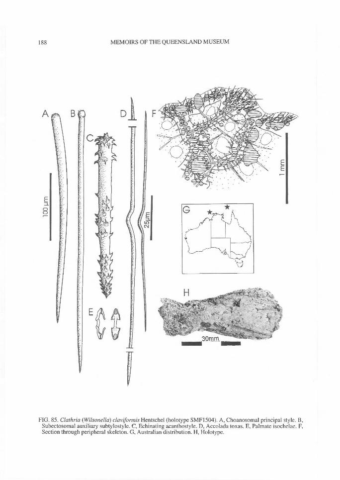
188^ MEMOIRS OF THE QUEENSLAND MUSEUM
FIG. 85. Clathria (Wilsonella) claviformis Hentschel (holotype SMF1504). A, Choanosomal principal style. B,Subectosomal auxiliary subtylostyle. C, Echinating acanthostyle. D, Accolada toxas. E, Palmate isochelae. F,Section through peripheral skeleton. G, Australian distribution. H, Holotype.
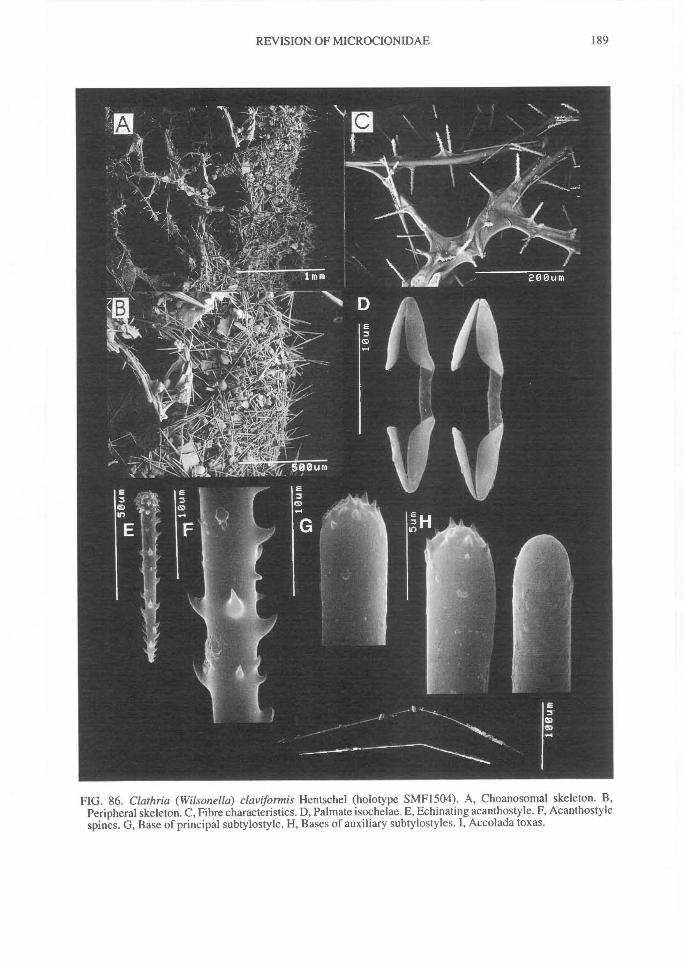
REVISION OF MICROCIONIDAE^
189
FIG. 86. Clathria (Wilsonella) claviformis Hentschel (holotype SMF1504). A, Choanosomal skeleton. B,Peripheral skeleton. C, Fibre characteristics. D, Palmate isochelae. E, Echinating acanthostyle. F, Acanthostylespines. G, Base of principal subtylostyle. H, Bases of auxiliary subtylostyles. I, Accolada toxas.
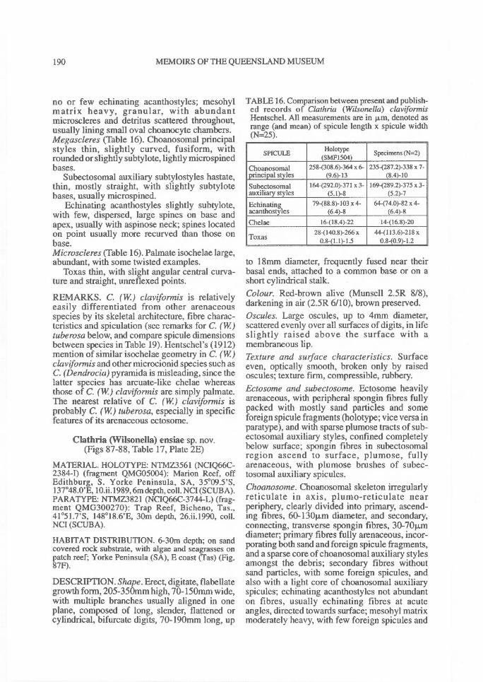
190^ MEMOIRS OF THE QUEENSLAND MUSEUM
no or few echinating acanthostyles; mesohylmatrix heavy, granular, with abundantmicroscleres and detritus scattered throughout,usually lining small oval choanocyte chambers.Megascleres (Table 16). Choanosomal principalstyles thin, slightly curved, fusiform, withrounded or slightly subtylote, lightly microspinedbases.
Subectosomal auxiliary subtylostyles hastate,thin, mostly straight, with slightly subtylotebases, usually microspined.
Echinating acanthostyles slightly subtylote,with few, dispersed, large spines on base andapex, usually with aspinose neck; spines locatedon point usually more recurved than those onbase.Microscleres (Table 16). Palmate isochelae large,abundant, with some twisted examples.
Toxas thin, with slight angular central curva-ture and straight, unreflexed points.
REMARKS. C. (W) claviformis is relativelyeasily differentiated from other arenaceousspecies by its skeletal architecture, fibre charac-teristics and spiculation (see remarks for C. (W)tube rosa below, and compare spicule dimensionsbetween species in Table 19). Hentschel's (1912)mention of similar isochelae geometry in C. (W)clavifonnis and other microcionid species such asC. (Dendrocia) pyramida is misleading, since thelatter species has arcuate-like chelae whereasthose of C. (W.) claviformis are simply palmate.The nearest relative of C. (W) claviformis isprobably C. (W) tube rosa, especially in specificfeatures of its arenaceous ectosome.
Clathria (VVilsonella) ensiae sp. nov.(Figs 87-88, Table 17, Plate 2E)
MATERIAL. HOLOTYPE: NTMZ3561 (NCIQ66C-2384-0 (fragment QMG05004): Marion Reef, offEdithburg, S. Yorke Peninsula, SA, 35°09.5'S,137°48.0'E, 10.ii.1989, 6m depth, coll. NCI (SCUBA).PARATYPE: NTMZ3821 (NCIQ66C-3744-L) (frag-ment QMG300270): Trap Reef, Bicheno, Tas.,41°51.7'S, 148°18.6'E, 30m depth, 26.ii.1990, coll.NCI (SCUBA).
HABITAT DISTRIBUTION. 6-30m depth; on sandcovered rock substrate, with algae and seagrasses onpatch reef; Yorke Peninsula (SA), E coast (Tas) (Fig.87F).
DESCRIPTION. Shape. Erect, digitate, flabellategrowth form, 205-350mm high, 70-150mm wide,with multiple branches usually aligned in oneplane, composed of long, slender, flattened orcylindrical, bifurcate digits, 70-190mm long, up
TABLE 16. Comparison between present and publish-ed records of Clathria (Wilsonella) claviformisHentschel. All measurements are in p,m, denoted asrange (and mean) of spicule length x spicule width(N=25).
SPICULE Holotype(SMF1504)
Specimens (N=2)
Choanosomal 2584308.6)-364 x 6- 235-(287.2)-338 x 7-principal styles (9.6)-13 (8.4)-10
Subectosomal 164-(292.0)-371 x 3- 169-(289.2)-375 x 3-auxiliary styles (5.1)-8 (5.2)-7
Echinating 79-(88.8)-103 x 4- 64-(74.0)-82 x 4-acanthostyles (6.4)-8 (6.4)-8
Chelae 16-(18.4)-22 14-(16.8)-2028-(140.8)-266 x 44-(113.6)-2I8 x
Toxas 0.8-(1.1)-1.5 0.8-(0.9)-1.2
to 18nun diameter, frequently fused near theirbasal ends, attached to a common base or on ashort cylindrical stalk.Colour. Red-brown alive (Munsell 2.5R 8/8),darkening in air (2.5R 6/10), brown preserved.Oscules. Large oscules, up to 4nun diameter,scattered evenly over all surfaces of digits, in lifeslightly raised above the surface with amembraneous lip.Texture and surface characteristics. Surfaceeven, optically smooth, broken only by raisedoscules; texture firm, compressible, rubbery.Ectosome and subectosome. Ectosome heavilyarenaceous, with peripheral spongin fibres fullypacked with mostly sand particles and someforeign spicule fragments (holotype; vice versa inparatype), and with sparse plumose tracts of sub-ectosomal auxiliary styles, confined completelybelow surface; spongin fibres in subectosomalregion ascend to surface, plumose, fullyarenaceous, with plumose brushes of subec-tosomal auxiliary spicules.Choanosome. Choanosomal skeleton irregularlyreticulate in axis, plumo-reticulate nearperiphery, clearly divided into primary, ascend-ing fibres, 60-130Rm diameter, and secondary,connecting, transverse spongin fibres, 30-70p..mdiameter; primary fibres fully arenaceous, incor-porating both sand and foreign spicule fragments,and a sparse core of choanosomal auxiliary stylesamongst the debris; secondary fibres withoutsand particles, with some foreign spicules, andalso with a light core of choanosomal auxiliaryspicules; echinating acanthostyles not abundanton fibres, usually echinating fibres at acuteangles, directed towards surface; mesohyl matrixmoderately heavy, with few foreign spicules and
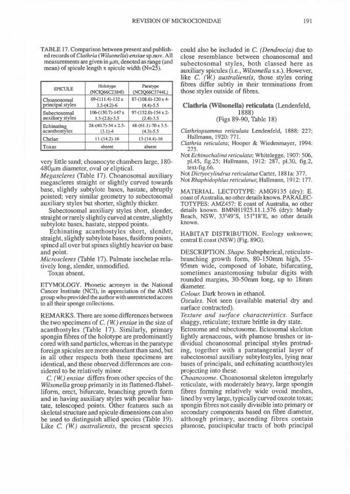
REVISION OF MICROCIONIDAE^
191
TABLE 17. Comparison between present and publish-ed records of Clathria (Wilsonella) ensiae sp.nov. Allmeasurements are given in 1.1m, denoted as range (andmean) of spicule length x spicule width (N=25).
SPICULE Holotype(NCIQ66C2384I)
Paratype(NCIQ66C3744L)
Choanosomal 69-(111.4)-132 x 87-(108.6)-120 x 4-principal styles 3.5-(4.2)-6 (4.4)-5.5
Subectosomal 106-(130.7)-147 x 97-(132.0)-154 x 2-auxiliary styles 1.5-(2.6)-3.5 (2.4)-3.5
F,chinating 28-(40.7)-54 x 2.5- 48-(61.1)-70 x 3.5-acanthostyles (3.1)-4 (4.3)-5.5
Chelae 11-(14.2)-16 13-(14.4)-16
Toxas absent absent-
very little sand; choanocyte chambers large, 180-480p.m diameter, oval or eliptical.Megascleres (Table 17). Choanosomal auxiliarymegascleres straight or slightly curved towardsbase, slightly subtylote bases, hastate, abruptlypointed; very similar geometry to subectosomalauxiliary styles but shorter, slightly thicker.
Subectosomal auxiliary styles short, slender,straight or rarely slightly curved at centre, slightlysubtylote bases, hastate, stepped points.
Echinating acanthostyles short, slender,straight, slightly subtylote bases, fusiform points,spined all over but spines slightly heavier on baseand point.Microscleres (Table 17). Palmate isochelae rela-tively long, slender, unmodified.
Toxas absent.
ETYMOLOGY. Phonetic acronym in the NationalCancer Institute (NCI), in appreciation of the AIMSgroup who provided the author with unrestricted accessto all their sponge collections.
REMARKS. There are some differences betweenthe two specimens of C. (W) ensiae in the size ofacanthostyles (Table 17). Similarly, primaryspongin fibres of the holotype are predominantlycored with sand particles, whereas in the paratypeforeign spicules are more abundant than sand, butin all other respects both these specimens areidentical, and these observed differences are con-sidered to be relatively minor.
C. (W) ensiae differs from other species of theWilsonella group primarily in its flattened-flabel-liform, erect, bifurcate, branching growth formand in having auxiliary styles with peculiar has-tate, telescoped points. Other features such asskeletal structure and spicule dimensions can alsobe used to distinguish allied species (Table 19).Like C. (W) australiensis, the present species
could also be included in C. (Dendrocia) due toclose resemblance between choanosomal andsubectosomal styles, both classed here asauxiliary spicules (i.e., Wilsonella s.s.). However,like C. (W) australiensis, those styles coringfibres differ subtly in their terminations fromthose styles outside of fibres.
Clathria (Wilsonella) reticulata (Lendenfeld,1888)
(Figs 89-90, Table 18)
Clathriopsamma reticulata Lendenfeld, 1888: 227;Hallmann, 1920: 771.
Clathria reticulata; Hooper & Wiedenmayer, 1994:275.
Not Echinochalina reticulata; Whitelegge, 1907: 506,p1.45, fig.25; Hallmann, 1912: 287, p1.30, fig.2,text-fig.66.
Not Dictyocylindrus reticulatus Carter, 1881a: 377.Not Rhaphidophlus reticulatus; Hallmann, 1912: 177.
MATERIAL. LECTOTYPE: AMG9135 (dry): E.coast of Australia, no other details known. PARALEC-TOTYPES: AMZ457: E coast of Australia, no otherdetails known. BMNH1925.11.1.576 (dry): ManlyBeach, NSW, 33°49'S, 151°18'E, no other detailsknown.
HABITAT DISTRIBUTION. Ecology unknown;central E coast (NSW) (Fig. 890).
DESCRIPTION. Shape. Subspherical, reticulate-branching growth form, 80-150mm high, 55-95nun wide, composed of lobate, bifurcating,sometimes anastomosing tubular digits withrounded margins, 30-50nun long, up to 18mmdiameter.Colour. Dark brown in ethanol.Oscules. Not seen (available material dry andsurface contracted).Texture and surface characteristics. Surfaceshaggy, reticulate; texture brittle in dry state.Ectosome and subectosome. Ectosomal skeletonlightly arenaceous, with plumose brushes or in-dividual choanosomal principal styles protrud-ing, together with a paratangential layer ofsubectosomal auxiliary subtylostyles, lying nearbases of principals, and echinating acanthostylesprojecting into these.Choanosome. Choanosomal skeleton irregularlyreticulate, with moderately heavy, large sponginfibres forming relatively wide ovoid meshes,lined by very large, typically curved oxeote toxas;spongin fibres not easily divisible into primary orsecondary components based on fibre diameter,although primary, ascending fibres containplumose, paucispicular tracts of both principal
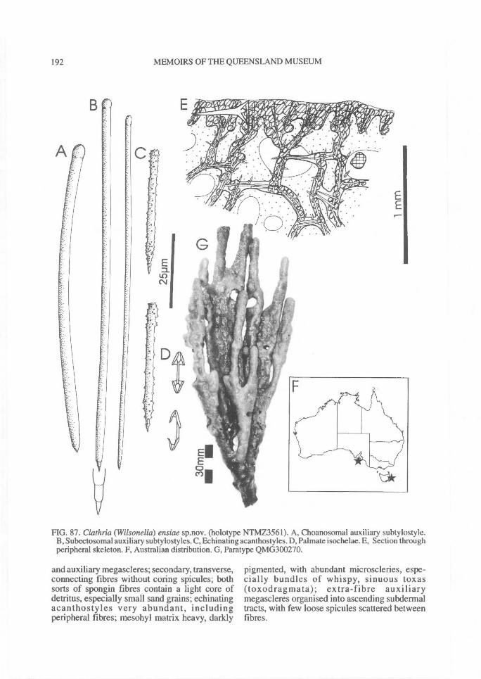
192^ MEMOIRS OF THE QUEENSLAND MUSEUM
FIG. 87. Clathria (Wilsonella) ensiae sp.nov. (holotype NTMZ3561). A, Choanosomal auxiliary subtylostyle.B, Subectosomal auxiliary subtylostyles. C, Echinating acanthostyles. D, Palmate isochelae. E, Section throughperipheral skeleton. F, Australian distribution. G, Paratype QMG300270.
and auxiliary megascleres; secondary, transverse,connecting fibres without coring spicules; bothsorts of spongin fibres contain a light core ofdetritus, especially small sand grains; echinatingacanthostyles very abundant, includingperipheral fibres; mesohyl matrix heavy, darkly
pigmented, with abundant microscleries, espe-cially bundles of whispy, sinuous toxas(toxodragmata); extra-fibre auxiliarymegascleres organised into ascending subdermaltracts, with few loose spicules scattered betweenfibres.
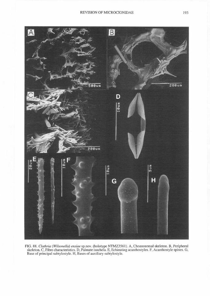
REVISION OF MICROCIONIDAE^ 193
FIG. 88. Clathria (Wilsonella) ensiae sp.nov. (holotype NTMZ3561). A, Choanosomal skeleton. B, Peripheralskeleton. C, Fibre characteristics. D, Palmate isochela. E, Echinating acanthostyles. F, Acanthostyle spines. G,Base of principal subtylostyle. H, Bases of auxiliary subtylostyle.
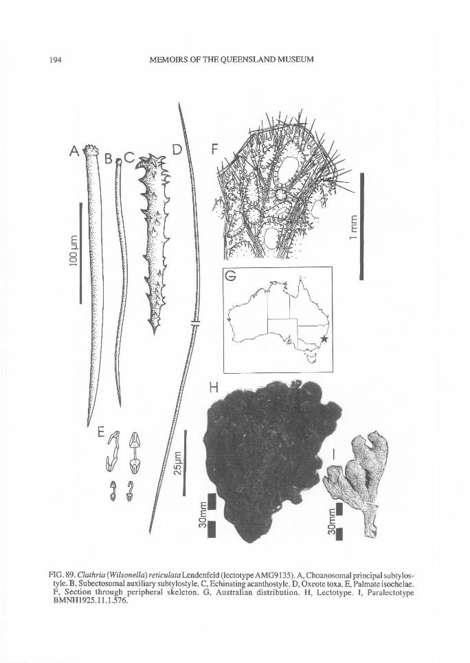
BC
n2 3
Em..LOCV
194^ MEMOIRS OF THE QUEENSLAND MUSEUM
FIG. 89. Clathria (W ilsonella) reticulata Lendenfeld (lectotype AMG9135). A, Choanosomal principal subtylos-tyle. B, Subectosomal auxiliary subtylostyle. C, Echinating acanthostyle. D, Oxeote toxa. E, Palmate isochelae.F, Section through peripheral skeleton. G, Australian distribution. H, Lectotype. I, ParalectotypeBMNH1925.11.1.576.
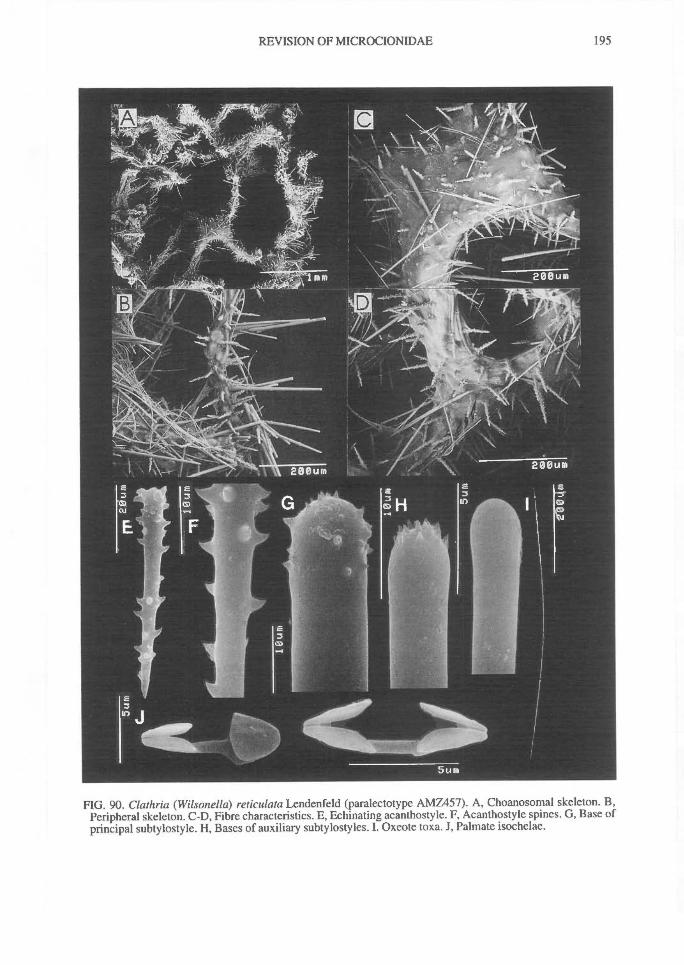
REVISION OF MICROCIONIDAE^
195
FIG. 90. Clathria (Wilsonella) reticulata Lendenfeld (paralectotype AMZ457). A, Choanosomal skeleton. B,Peripheral skeleton. C-D, Fibre characteristics. E, Echinating acanthostyle. F, Acanthostyle spines. G, Base ofprincipal subtylostyle. H, Bases of auxiliary subtylostyles. I. Oxeote toxa. J, Palmate isochelae.
196^ MEMOIRS OF THE QUEENSLAND MUSEUM
Megascleres (Table 18). Choanosomal principalsubtylostyles well differentiated from auxiliaryspicules, mostly straight, fusiform, with profuse-ly microspined, slightly subtylote bases.
Subectosomal auxiliary subtylostyles thin,fusiform, straight, slightly curved, sometimessinuous, with minutely rnicrospined, subtylotebases.
Echinating acanthostyles with rounded bases,relatively even spination, although basal and dis-tal portions slightly more heavily spined thanpoints.Microscleres (Table 18). Palmate isochelae dif-ferentiated into 2 size classes, smaller with ap-proximately 75% of contort forms.
Toxas extremely abundant, long, thin, sinuous,characteristically oxeote, with only very slight orno central curvature and straight tapering points.
REMARKS. This species is poorly known from3 specimens but differentiated from other speciesin the Wilsonella group having only a very lightcore of detritus in fibres, usually composed ofsmall sand and spicule particles, differentiatedprimary and secondary fibres, and prominentbundles of sinuous toxas lining aquiferous cham-bers (Fig. 90). Affinities with other species (Table19) are discussed elsewhere (remarks under C.(W) tuberosa and C. (W) australiensis). In somerespects (growth form, geometry of somespicules, presence of two sizes of isochelae withcontort forms) the species is closest to C. (W)tuberosa, but toxa geometry is quite differentbetween these two species.
Clathria (Wilsonella) tuberosa (Bowerbanlc, 1875)(Figs 91-93, Table 19, Plate 2F)
Microciona tube rosa Bowerbank, 1875: 281, 282, 286;Vosmaer, 1935a: 607.
Clathria tuberosa; Ridley, 1881: 121; Ridley, 1884a:444-445, p1.42, fig.d; Hentschel, 1912: 365-366;Hooper & Wiedenmayer, 1994: 275.
MATERIAL. HOLOTYPE: BMNH1877. 5.21.1312:Straits of Malacca, Malaysia, vicinity of 2 °N, 102°E,coll. Capt. Parish (dredge). OTHER MATERIAL: IN-DONESIA - SMF978 (fragment MNHNDCL2346).QLD - BMNH1881.10.21.325, BMNH1882.2.23.198,253, 283, 334. NT - AMZ4559 (RRIMP FN989),NTMZ777, NTMZ809, NTMZ920, NTMZ933,NTMZ946, NTMZ1091, NTMZ2087, NTMZ2400,NTMZ2708, QMG303336, NTMZ2157, NTMZ2189,NTMZ1959, NTMZI980, NTMZ1987, NTMZ2020,NTMZ2098, QMG303428, NTMZ2107, NTMZ234,NTMZ235, NTMZ236 - NTMZ2496, NTMZ540,NTMZ107, NTMZ112, NTMZ128, NTMZ554. WES-SEL ISLANDS; NT - NTMZ3955.
TABLE 18. Comparison between present and publish-ed records of Clathria (Wilsonella) reticulata (Len-denfeld). All measurements are given in p.m, denotedas range (and mean) of spicule length x spicule width(N=25).
SPICULELectotype
(AMG9135)Paralectotype
(AMZ457)
Paralectotype(BMNH
1925.11.1.576)
Choanosomal 173-(209.4)- 182(240.2)- 227-(251.5)-principal 258 x 7-(9.1)- 289 x 8-(9.3)- 296 x 8-(9.4)-styles 11 11 ii
Subectosomalauxiliarystyles
I63-(236.8)-324 x 3-(4.0)-6
187-(259.0)-322 x 3-(4.2)-5
1984243.4)-309 x 3-(4.6)-7
Echinating 52-(61.7)-71 x 54-(63.6)-75 x 65-(67.8)-72 xacanthostyles 3-(6.5)-9 4-(7.2)-9 6-(7.3)-9
Chelae I 5-(6.6)-9 4-(5.8)-7 6-(6.1)-9
Chelae II 12-(14.4)-17 13-(14.8)-17 14-(15.2)-17238-(402.7)- 45-(231.3)- 302-(427.7)-
Toxas 684 x 0.8- 433 x 0.5- 593 x I -(2.2)-4(2.4)-4 (1.9)-3.5
HABITAT DISTRIBUTION. Semi-encrusting onrock, dead or live coral heads, epizootic on other spon-ges and gorgonians; usually associated with shallowcoral reef habitats; 2-19m depth range; prevalent in thetropical, Australian and Indo-Malay shallow watermacrobenthic community, extending as far south as13°S latitude: Torres Strait (FNQ) (Ridley, 1884a),Bynoe Harbour, Darwin Harbour, Coral Bay, PortBremer, Wessel Is (NT). Also Straits of Malacca(Bowerbank, 1875; Ridley, 1881) and Aru Is, In-donesia (Hentschel, 1912).
DESCRIPTION. Shape. Subspherical,predominantly bulbous growth form, 60-135mmdiameter, less often club-shaped with apicallobate digits, or pseudo-vasiform on low stalkwith convoluted, apical, lobate digits; surfaceprojections (or branches) rounded lobate, rela-tively close-set, attached to common centre,which in turn is usually attached to substrate by asmall peduncle; lobate digits usually bifurcatewith rounded margins. In life lobes prominentlybulbous, evenly rounded; after preservation lobesbecome slightly flattened and angular.Colour. Live pigmentation dusty pale pink-red(Munsell 2.5R 6/10) to pink (5RP 8/6), with adarker choanosome (5RP 7/8); lighter ectosomalcolouration due to arenaceous nature of ec-tosome; dessicated colouration darkens to brown(2.5Y 8/2), red-brown (5Y 8/4), or red-purple(5RP 3/6), as paler ectosome collapses. In situ,subdermal ridges and canals red-pink in life,showing darker choanosomal pigmentation.Oscules. Exhalant pores variable in diameter,ranging from 1-3mm, each with prominent,
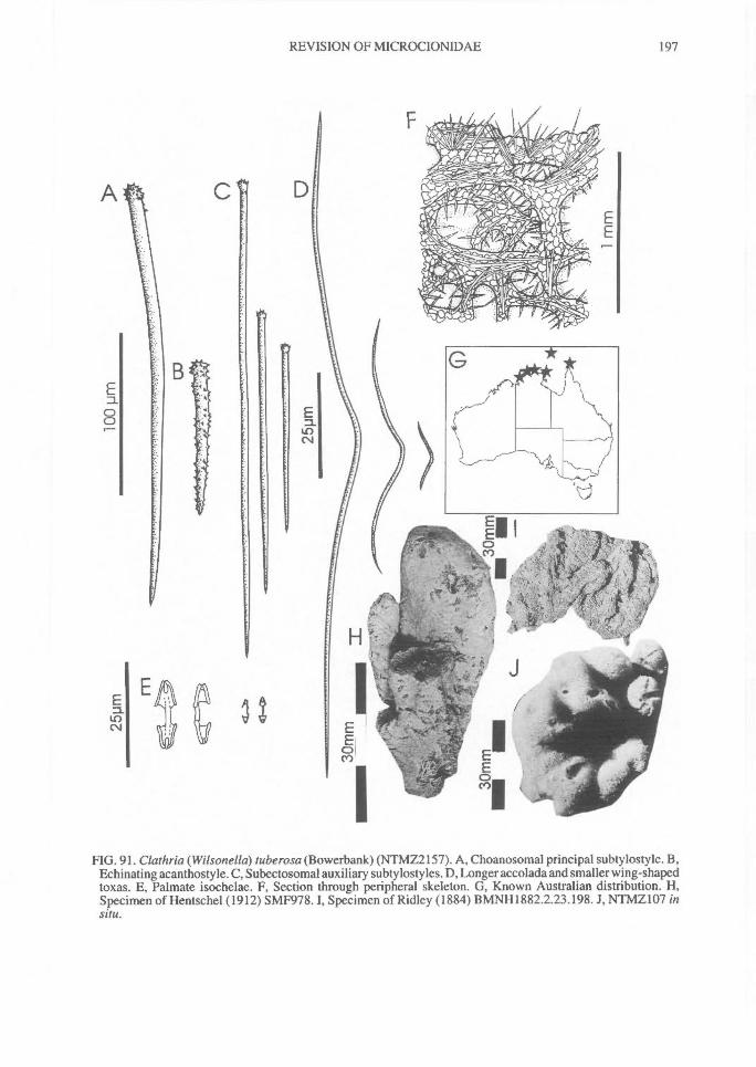
REVISION OF MICROCIONIDAE^
197
FIG. 91. Clathria (Wilsonella) tuberosa (Bowerbank) (NTMZ2157). A, Choanosomal principal subtylostyle. B,Echinating acanthostyle. C, Subectosomal auxiliary subtylostyles. D, Longer accolada and smaller wing-shapedtoxas. E, Palmate isochelae. F, Section through peripheral skeleton. G, Known Australian distribution. H,Specimen of Hentschel (1912) SMF978. I, Specimen of Ridley (1884) BMNH1882.2.23.198. J, NTMZ107 insitu.
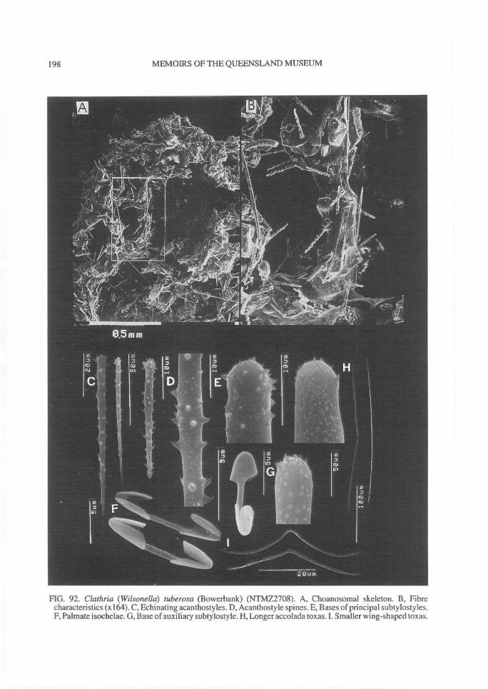
198^ MEMOIRS OF THE QUEENSLAND MUSEUM
FIG. 92. Clathria (Wilsonella) tuberosa (Bowerbank) (NTMZ2708). A, Choanosomal skeleton. B, Fibrecharacteristics (x164). C, Echinating acanthostyles. D, Acanthostyle spines. E, Bases of principal subtylostyles.F, Palmate isochelae. G, Base of auxiliary subtylostyle. H, Longer accolada toxas. I. Smaller wing-shaped toxas.
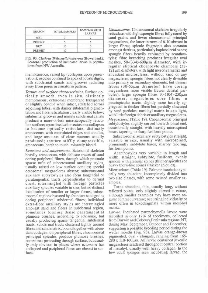
REVISION OF MICROCIONIDAE^ 199
SEASON TOTAL SAMPLES SAMPLES WITHLARVAE
WET 3 2PREDRY 5 5
DRY 10 0PREWET 10 5
FIG. 93. Clathria (Wilsonella) tube rosa (Bowerbank).Seasonal production of incubated larvae in popula-tions from NW Australia.
membraneous, raised lip (collapses upon preser-vation); oscules confined to apex of lobate digits,with subdermal canals and grooves radiatingaway from pores in cruciform pattern.Texture and surface characteristics. Surface op-tically smooth, even in situ, distinctlymembranous; ectosomal membrane transparentor slightly opaque when intact, stretched acrossadjoining lobes, with darker subdermal pigmen-tation and fibre reticulation clearly visible below;subdermal grooves and minute subdermal canalsproduce a more-or-less microscopically reticu-late surface; upon dessication ectosome collapsesto become optically reticulate, distinctlyarenaceous, with convoluted ridges and conules,and large amounts of clear mucous usuallyproduced; texture stiffly compressible,arenaceous, harsh to touch, minutely hispid.Ectosome and subectosome. Ectosomal skeletonheavily arenaceous, with delicate traces of sandcoring peripheral fibres, through which protrudesparse tufts of subectosomal auxiliary styles,usually raised on low surface conules; specialectosomal megascleres absent; subectosomalauxiliary subtylostyles also form tangential orparatangential tracts perpendicular to dermalcrust, intermingled with foreign particlesauxiliary spicules variable in size, but no distinctlocalisation of smaller or larger forms; subec-tosomal region obscured by abundant sand grainscoring peripheral subdermal fibres; individualextra-fibre auxiliary styles are intermingledamongst sand and fibres in subdermal region,sometimes forming dense paratangentialplumose brushes, ascending to ectosome, butusually producing sparse tangential subdermaltracts; subdermal tracts clustered tightly aroundfibres and sand matrix, bound together with abun-dant collagen; on peripheral fibres, choanosomalprincipal spicules produce plumose brushes,sometimes protruding through surface, but usual-ly only obvious in places where ectosome hascollapsed and peripheral fibres are closest to sur-face.
Choanosome. Choanosomal skeleton irregularlyreticulate, with light spongin fibres fully cored bysand grains and fewer choanosomal principalmegascleres, the latter in rows of 6-10 abreast inlarger fibres; spicule fragments also commonamongst detritus, particularly haplosclerid oxeas;spongin fibres heavily echinated by acanthos-tyles; fibre branching produces irregular ovalmeshes, 50-(334)-600p.m diameter, with ir-regular eliptical choanocyte chambers (38-121p,m diameter), with light mesohyl matrix andabundant microscleres, without sand or anymegascleres; spongin fibres not clearly divisibleinto primary or secondary elements, but thinnerfibres (30-551J,m diameter) have coringmegascleres more visible (fewer detrital par-ticles); larger spongin fibres 70-(104)-230p„mdiameter; megascleres core fibres inpaucispicular tracts, slightly more heavily ag-gregated in thicker fibres but partially obscuredby sand particles; mesohyl matrix in axis light,with little foreign debris or auxiliary megascleres.Megascleres (Table 19). Choanosomal principalsubtylostyles slightly curved towards basal end,occasionally straight, with heavily microspinedbases, tapering to sharp fusiform points.
Subectosomal auxiliary subtylostyles straight,variable in size, usually with microspined,prominently subtylote bases, sharply tapering,fusiform points.
Acanthostyles very variable in length andwidth, straight, subtylote, fusiform, evenlyspi nose with granular spines (thinner spicules) orheavy thorn-like spines (thicker spicules).Microscleres (Table 19). Palmate isochelae typi-cally very abundant, incompletely divided intotwo size classes, with some twisted smaller ex-amples.
Toxas abundant, thin, usually long, withoutreflexed points, only slightly curved at centre,although smaller examples may have more an-gular central curvature; occurring individually ormore often in toxodragmata within mesohylmatrix.Larvae. Incubated parenchymella larvae wererecorded in only 17% of specimens, collectedfrom Darwin and Cobourg Peninsula regions, NT,during May, September, October and December,suggesting a possible breeding period during thewetter months (Fig. 93). Larvae orange-brownpigmented, oval - elongate, ranging from 165-280 x 110-160iLm. All larvae contained juvenilemegascleres scattered throughout central portionof mesohyl, usually with heavy collagen. In thefew adult sponges seen incubating larvae, the
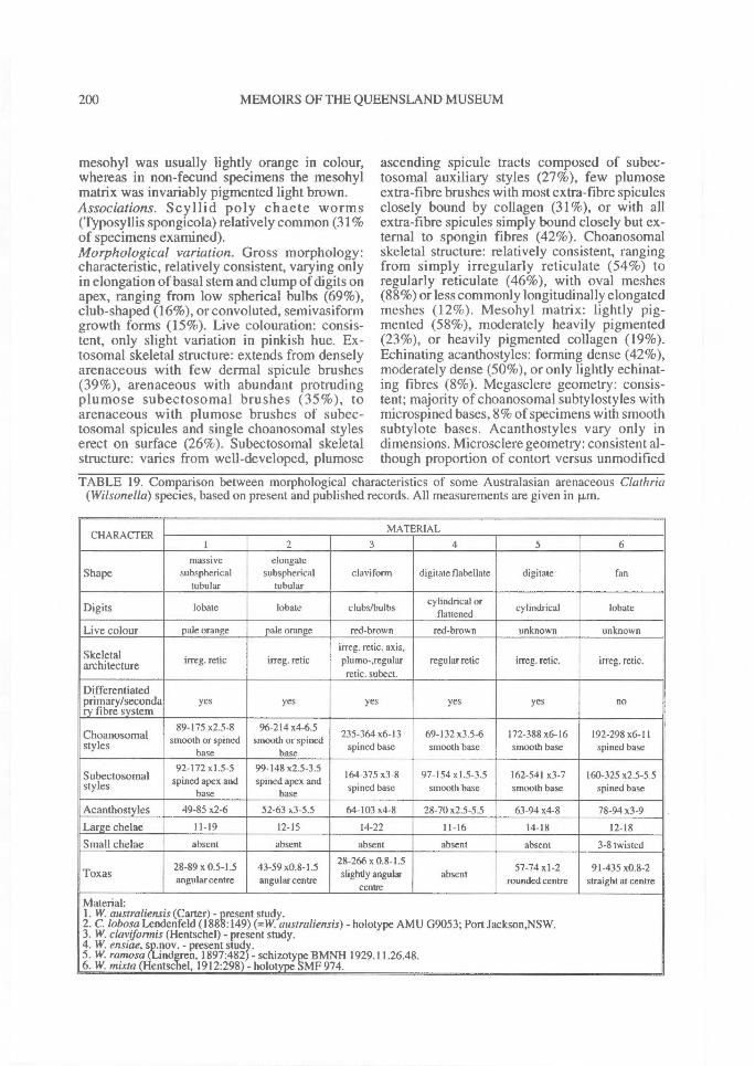
200^ MEMOIRS OF THE QUEENSLAND MUSEUM
mesohyl was usually lightly orange in colour,whereas in non-fecund specimens the mesohylmatrix was invariably pigmented light brown.Associations. Scyllid poly chaete worms(Typosyllis spongicola) relatively common (31%of specimens examined).Morphological variation. Gross morphology:characteristic, relatively consistent, varying onlyin elongation of basal stem and clump of digits onapex, ranging from low spherical bulbs (69%),club-shaped (16%), or convoluted, semivasiformgrowth forms (15%). Live colouration: consis-tent, only slight variation in pinkish hue. Ex-tosomal skeletal structure: extends from denselyarenaceous with few dermal spicule brushes(39%), arenaceous with abundant protrudingplumose subectosomal brushes (35%), toarenaceous with plumose brushes of subec-tosomal spicules and single choanosomal styleserect on surface (26%). Subectosomal skeletalstructure: varies from well-developed, plumose
ascending spicule tracts composed of subec-tosomal auxiliary styles (27%), few plumoseextra-fibre brushes with most extra-fibre spiculesclosely bound by collagen (31%), or with allextra-fibre spicules simply bound closely but ex-ternal to spongin fibres (42%). Choanosomalskeletal structure: relatively consistent, rangingfrom simply irregularly reticulate (54%) toregularly reticulate (46%), with oval meshes(88%) or less commonly longitudinally elongatedmeshes (12%). Mesohyl matrix: lightly pig-mented (58%), moderately heavily pigmented(23%), or heavily pigmented collagen (19%).Echinating acanthostyles: forming dense (42%),moderately dense (50%), or only lightly echinat-ing fibres (8%). Megasclere geometry: consis-tent; majority of choanosomal subtylostyles withmicrospined bases, 8% of specimens with smoothsubtylote bases. Acanthostyles vary only indimensions. Microsclere geometry: consistent al-though proportion of contort versus unmodified
TABLE 19. Comparison between morphological characteristics of some Australasian arenaceous Clathria(Wilsonella) species, based on present and published records. All measurements are given in p.m.
CHARACTERMATERIAL
1 2 3 4 5 6
Shapemassive
subsphericaltubular
elongatesubspherical
tubularclay iform digitate flabellate digitate fan
Digits lobate lobate clubs/bulbscylindrical or
flattenedcylindrical lobate
Live colour pale orange pale orange red-brown red-brown unknown unknown
Skeletalarchitecture irreg. relic irreg. relic
irreg. relic, axis,plumo-,regularrelic. subect.
regular relic irreg. retic. irreg. relic.
Differentiatedprimary/secondary fibre system
yes yes yes yes yes no
Choanosomalstyles
89-175 x2.5-8smooth or spined
base
96-214x4-6.5smooth or spined
base
235-364 x6-13spined base
69-132 x3.5-6smooth base
172-388 x6-16smooth base
192-298 x6-11spined base
Subectosomalstyles
92-172 x1.5-5spined apex and
base
99-148 x2.5-3.5spined apex and
base
164-375 x3-8spined base
97-154 x1.5-3.5smooth base
162-541 x3-7smooth base
160-325 x2.5-5.5spined base
Acanthostyles 49-85x2-6 52-63x3-5.5 64-103x4-8 28-70x2.5-5.5 63-94x4-8 78-94x3-9
Large chelae 11-19 12-15 14-22 11-16 14-18 12-18
Small chelae absent absent absent absent absent 3-8 twisted
Toxas 28-89 x 0.5-1.5angular centre
43-59 x0.8-1.5angular centre
28-266 x 0.8-1.5slightly angular
centreabsent
57-74 x1-2rounded centre
91-435 x0.8-2straight at centre
Material:1. W. australiensis (Carter) - present study.2. C. lobosa Lendenfeld (1888:149) (=W. australiensis) - holotype AMU G9053; Port Jackson,NSW.3. W. clavifonnis (Hentschel) - present study.4. W. ensiae, sp.nov. - present study.5. W. ramosa (Lindgren, 1897:482- schizotype BMNH 1929.11.26.48.6. W. mixta (Hentschel, 1912:298) - holotype SMF 974.

REVISION OF MICROCIONIDAE^ 201
small isochelae varies from 0-20% of contortspicules (12%), 20-40% (19%), 40-60% (24%),60-80 (35%), to 80-10% of spicules (12%).Spicule dimensions: Few specimens atypical butvariation apparently random with no statisticalsignificance between specimens irrespective ofseasonal or geographical distribution of samples.
REMARKS. C. (W) tuberosa is distinctivein thefield: pink colour, bulbous growth form, soft tex-ture. However, it is more difficult to differentiatedescriptively. Pertinent differences are:
Choanosomal architecture and fibre charac-teristics of C. (W) reticulata are identical to thoseof C. (W) tuberosa; acanthostyles are as equallyabundant in both species, but many acanthostyleshave heavier spines on the distal part than onpoints; and there are two sizes of isochelae, 75%of the smaller being contort. Thus, the majorfeatures distinguishing the two taxa are thestraight or sinuous oxeote toxas in C. (W)reticulata, which are never present in C. (W)tuberosa, and the light deposits of debris into
fibres. Nevertheless, the two species are closelyrelated.
Clathria (W) australiensis has choanosomalfibres divided into ascending and radial primaryelements, fully cored by detritus, with less heavi-ly cored secondary transverse elements. Subec-tosomal auxiliary subtylostyles have spines onboth bases and points, typically with a prominentterminal spine/point and also smaller spines oc-curing at least part of the way along the shaft.Choanosomal subtylostyles are also auxiliaryspicules, with smooth or spined bases only. Acan-thostyles are pointed and tapering; there is onlyone size of isochelae; and toxas have stronglycurved (rounded or sharply angular) central cur-ves.
Clathria (W) rugosa is a sister species of C.(W) australiensis, differing in having prominentsubectosomal drainage canals ('astrorhizae').Subectosomal auxiliary styles are also spined onboth ends, but spines are perched only on the veryextremity of the spicule point (not on the shaft);acanthostyles have bulbous points; there is only
CHARACTERMATERIAL
7 8 9 10 11 12
Shapefan palmate-
digitatemassive spherical
tubularmassive spherical
globular
elongate
subspherical
tubular
massive spherical
globular
cupriform
reticulate branches
Digits cylindrical lobate lobate lobate lobate lobate tubular
Live colour bt.red-orangepale pink or pink-
red
pale pink or pink-
red
pale pink or pink-
red
pale pink or pink-
redunknown
Skeletalarchitecture
irreg.retic. irreg.retic. irreg.retic. irreg.retic.
Differentiatedprimary/secondary fibre system
yes none none none none partial
Choanosomalstyles
134-159x4.5-8smooth base
167 -278 x4 - 11
most spined bases
144-286x4 -8most spined bases
l72-302x6-8.5most smooth bases
1334226.3)-343x4-(8.2)-14 most
spined bases
173-296 x7-11 allspined bases
Subectosomalstyles
162-206 x2.5-4spined apex and
base
159 -333 x2-6
spined bases
148-223 x2-5
spined bases
154-324 x2-5spined bases
129-(228.9)-375x1.5-(4.1)-8
spined bases
163-324 x3-7spined bases
Acanthostyles 58-91 x4-7 44-97 x4-9 43-75 x3-8 69-88 x3.5-560-(79.9)-112 x4-
(6.7)-1152-75 x3-9
Large chelae 12 - 17 11 - 19 11 - 16 13 - 19 10- (13.3) - 18 14 - 17
Small chelae absent 4-8 twisted 4-7 twisted 6-8 rare 4-(6.1)-9 twisted 4-9 twisted
Toxas24-122 x0.8-3
slight curve at
centre
55-255 x0.5-1.5
rounded centre
95-215 x0.5-1.5
angular& rounded
centre
81-94 x1-2
rounded centre
30-(140.3)-388
x0.4-(1.I)-2
rounded centre
45-684 x0.5-4
sinuous oxeote
Material:7. W. rugosa (Hooper8. W. tuberosa (Bowerbank,9. W. tuberosa; Ridley,10. W. tuberosa;11. W. tuberosa;12. W. reticulata
& Levi, 1993) - New Caledonian population (included for comparative purposes).1875:281); holotype BMNH 1877.5.21.1312; Straits of Malacca (19=25).
1884a:444; BMNH 1881.10, 21.325, 1882.2.23.198, 253, 283, 334; Tones Strait (N=125).Hentschel, 1912:365; SMF 978; Arafura Sea (N=25).present material - NW Australia (N=650).(Lendenfeld) - .present study.
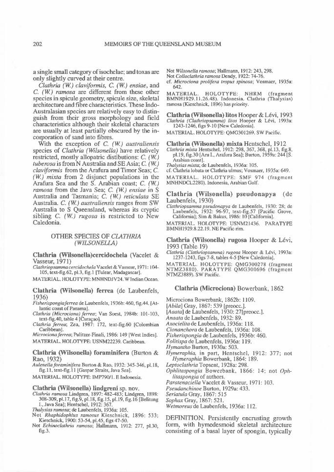
202^ MEMOIRS OF THE QUEENSLAND MUSEUM
a single small category of isochelae; and toxas areonly slightly curved at their centre.
Clathria (W) clavtformis, C. (W) ensiae, andC. (W) ramosa are different from these otherspecies in spicule geometry, spicule size, skeletalarchitecture and fibre characteristics. These Indo-Australasian species are relatively easy to distin-guish from their gross morphology and fieldcharacteristics although their skeletal charactersare usually at least partially obscured by the in-corporation of sand into fibres.
With the exception of C. (W.) australiensisspecies of Clathria (Wilsonella) have relativelyrestricted, mostly allopatric distibutions: C. (W)tube rosa is from N Australia and SE Asia; C. (W)claviforrnis from the Arafura and Timor Seas; C.(W) mixta from 2 disjunct populations in theArafura Sea and the S. Arabian coast; C. (W)ramosa from the Java Sea; C. (W) ensiae in SAustralia and Tasmania; C. (W) reticulata SEAustralia. C. (W.) australiensis ranges from SWAustralia to S Queensland, whereas its crypticsibling C. (W) rugosa is restricted to NewCaledonia.
OTHER SPECIES OF CLATHRIA(WILSONELLA)
Clathria (Wilsonella)cercidochela (Vacelet &Vasseur, 1971)Clathriopsammacercidochela Vacelet& Vasseur, 1971: 104-
105, text-fig.62, p1.3, fig.! [Tulear, Madagascar].MATERIAL. HOLOTYPE: MNHNDJV24. W Indian Ocean.
Clathria (Wilsonella) ferrea (de Laubenfels,1936)Fisherispongiaferrea de Laubenfels, 1936b: 460, fig.44. [At-
lantic coast of Panama].Clathria (Microciona) ferrea; Van Soest, 1984b: 101-103,
text-fig.40, table 4 [Curacao].Clathria ferrea; Zea, 1987: 172, text-fig.60 [Colombian
Caribbean].Microciona ferrea; Pulitzer-Finali, 1986: 149 [West Indies].MATERIAL. HOLOTYPE: USNM22239. Caribbean.
Clathria (VVilsonella) foraminifera (Burton &Rao, 1932)Aulenella foraminifera Burton & Rao, 1932: 345-346, p1.18,
fig.11, text-fig.11 [Gaspar Straits, Java Sea].MATERIAL. HOLOTYPE: IMP790/1. E Indonesia.
Clathria (VVilsonella) lindgreni sp. nov.Clathria ramosa Lindgren, 1897: 482-483; Lindgren, 1898:
308-309, p1.17, fig.9, p1.18, fig.15, p1.19, fig.16 [BelitungI., Java Sea]; Hentschel, 1912: 367.
Thalysias ramosa; de Laubenfels, 1936a: 105.Not Rhaphidophlus ramosus Kieschnick, 1896: 533;
Kieschnick, 1900: 53-54, p1.45, figs 47-50.Not Echinoclathria ramosa; Hallmann, 1912: 277, p130,
fig.3.
Not Wilsonella ramosa; Hallmann, 1912: 243, 298.Not Colloclathria ramosa Dendy, 1922: 74-76.cf. Microciona prolifera tropus spinosa; Vosmaer, 1935a:
642.MATERIAL. HOLOTYPE: NHRM (fragmentBMNH1929.11.26.48). Indonesia. Clathria (Thalysias)ramosa (Kieschnick, 1896) has priority.
Clathria (Wilsonella) litos Hooper & Levi, 1993Clathria (Clathriopsamma) litos Hooper & Levi, 1993a:
1243-1246, figs 9-10 [New Caledonia].MATERIAL. HOLOTYPE: QMG301269. SW Pacific.
Clathria (Wilsonella) mixta Hentschel, 1912Clathria mixta Hentschel, 1912: 298, 367, 368, p1.13, fig.8,
p1.19, fig.30 [Aru I., Arafura Sea]; Burton, 1959a: 244 [S.Arabian coast].
Thalysias mixta; de Laubenfels, 1936a: 105.cf. Clathria lobata or Clathria ulmus; Vosmaer, 1935a: 649.MATERIAL. HOLOTYPE: SMF 974 (fragmentMNHNDCL2280). Indonesia, Arabian Gulf.
Clathria (Wilsonella) pseudonapya (deLaubenfels, 1930)Clathriopsamma pseudonapya de Laubenfels, 1930: 28; de
Laubenfels, 1932: 96-97, text-fig.57 [Pacific Grove,California]; Sim & Bakus, 1986: 10 [California].
MATERIAL. HOLOTYPE: USNM21436. PARATYPEBMNH1929.8.22.19. NE Pacific rim.
Clathria (Wilsonella) rugosa Hooper & Levi,1993 (Table 19)Clathria (Clathriopsamma) rugosa Hooper & Levi, 1993a:
1237-1243, figs 7-8, tables 4-5 [New Caledonia].MATERIAL. HOLOTYPE: QMG300278 (fragmentNTMZ3880). PARATYPE QMG300696 (fragmentNTMZ3889). SW Pacific.
Clathria (Microciona) Bowerbank, 1862
Microciona Bowerbank, 1862b: 1109.[Abila] Gray, 1867: 539 [preocci.[Aaata] de Laubenfels, 1930: 271preocci.Anaata de Laubenfels, 1932: 89.Axocielita de Laubenfels, 1936a: 118.Cionanchora de Laubenfels, 1936a: 108.Fisherispongia de Laubenfels, 1936b: 460.Folitispa de Laubenfels, 1936a: 119.Hymantho Burton, 1930a: 503.Hymeraphia, in part, Hentschel, 1912: 377; not
Hymeraphia Bowerbank, 1864: 189.Leptoclathria Topsent, 1928a: 298.Ophlitaspongia Bowerbank, 1866: 14; not Oph-
litaspongia of authors.Paratenaciella Vacelet & Vasseur, 1971: 103.Pseudanchinoe Burton, 1929a: 433.Seriatula Gray, 1867: 515Sophax Gray, 1867: 521.Wetmoreus de Laubenfels, 1936a: 112.
DEFINITION. Persistently encrusting growthform, with hymedesmoid skeletal architectureconsisting of a basal layer of spongin, typically
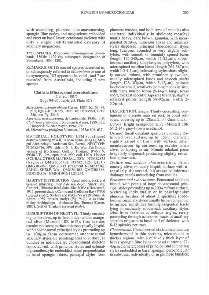
REVISION OF MICROCIONIDAE^ 203
with ascending, plumose, non-anastomosing,spongin fibre nodes, and megascleres embeddedand erect on basal layer; ectosomal skeleton withonly a single undifferentiated category ofauxiliary megasclere.
TYPE SPECIES. Microciona atrasanguinea Bower-bank, 1862b: 1109 (by subsequent designation ofBowerbank, 1864: 188).
REMARKS. Of 118 named species described in,or subsequently referred to Microciona or one ofits synonyms, 103 appear to be valid, and 7 arerecorded from Australasia, including 2 newspecies.
Clathria (Microciona) aceratoobtusa(Carter, 1887)
(Figs 94-95, Table 20, Plate 3C)
Microciona acerato-obtusa Carter, 1887: 62, 67, 83,p1.5, figs 7-10; Dendy, 1896: 18; Hentschel, 1911:348, text-fig. 32a-f.
Axocielita aceratoobtusa; de Laubenfels, 1936a: 118.Clathria aceratoobtusa; Rudman & Avern, 1989: 335;
Hooper & Wiedenmayer, 1994: 266.cf. Microciona prolifera; Vosmaer, 1935a: 608, 637.
MATERIAL. HOLOTYPE: LFM (confirmeddestroyed during WWII): Kadan Kyun (King I.), Mer-gui Archipelago, Andaman Sea, Burma. NEOTYPE:NTMZ3676: NW. side of N. I., Ko Wao Yai Group,vicinity of Ko Samui, Gulf of Thailand, 9°46.7'N,99°40.3'E, 12m depth, 6.vi.1990, coll. J.N.A. Hooper(SCUBA). OTHER MATERIAL: NSW - NTMZ2835(fragment QMG300543), NTMZ3125. QLD -QMG303089, QMGL713 (fragment NTMZ1536).SAHUL SHELF, WA - QMG301083, QMG301188.INDONESIA - BMNH1946.11.25.244.
HABITAT DISTRIBUTION. Coral rubble, rock andbivalve substrata; intertidal-14m depth; Shark Bay,Cartier I., Hibernia Reef, Sahul Shelf (WA) (Hentschel,1911, present study); Cairns and Shelburne Bay (FNQ)(present study); Sydney and Iluka (NSW) (Rudman &Avern, 1989, present study) (Fig. 940). Also Indo-Malay Archipelago — Andaman Sea (Burma) (Carter,1887), Gulf of Thailand (present study).
DESCRIPTION OF NEOTYPE. Thinly encrust-ing on bivalves, up to lnun thick; colour orange-red alive (Munsell 1 OR 6/12); firm texture;oscules not seen; surface microscopically hispid,with choanosomal principal styles protruding upto 100p,m from ectosome; subectosomalauxiliary styles lie paratangential to surface, inbundles or individually; choanosomal skeletonleptoclathriid, with principal styles and echinat-ing acanthostyles embedded in and perpendicularto basal spongin fibres; principal styles form
plumose brushes, and both sorts of spicules alsoscattered individually in skeleton; mesohylmatrix heavy, dark brown, granular, with incor-porated detritus, numerous toxas and auxiliarystyles dispersed; principal choanosomal styleslong, fusiform, rounded or very slightly sub-tylote, with smooth or minutely spined bases(length 175-548p.m, width 11 - 22 p,m); subec-tosomal auxiliary subtylostyles polytylote, withmicrospined swollen bases (length 264-387p.m,width 1.5-4.5p,m); echinating styles short, slight-ly curved, robust, with prominently swollen,usually rnicrospined bases and smooth shafts(length 128-183p,m, width 5-12p,m); palmateisochelae small, relatively homogeneous in size,with many twisted forms (9-14p.m long); toxasshort, thickest at centre, tapering to sharp, slightlyreflexed points (length 58-92p,m, width 2-5.5p,m).
DESCRIPTION. Shape. Thinly encrusting, con-tiguous or discrete mats on rock or coral sub-strata, covering up to 120mm2, 0.4-2mm thick.Colour. Bright orange-red alive (Munsell lOR6/12-14), grey-brown in ethanol.Oscules. Small exhalant apertures unevenly dis-tributed over surface, up to 1.5mm diameter,slightly raised or flush with surface; smallmembraneous lip surrounding oscules whenalive, collapsing in air. Minute inhalant poresirregularly dispersed, producing slightly reticu-late appearance.Texture and surface characteristics. Firm,mucusy alive, minutely hispid; surface with ir-regularly dispersed, bifurcate subdermaldrainage canals meandering from oscules.Ectosome and subectosome. Ectosomal skeletonhispid, with points of large choanosomal prin-cipal styles protruding up to 200p.m from surface,occurring individually or in paucispicularplumose brushes of about 5 spicules; subec-tosomal auxiliary styles usually lie paratangentialto surface, sometimes forming tangential tractslying immediately subdermal; auxiliary stylesarise from skeleton at oblique angles, rarelyprotruding through ectosome; tracts of auxiliaryspicules originate in basal half of skeleton, with8-12 spicules per tract.Choanosome. Choanosomal skeletal architecturehymedesmoid in thin sections, microcionid inthicker regions, with a relatively thick layer ofheavy spongin fibre lying on basal substrate, 22-43 um diameter; bases of principal and echinatingstyles embedded in basal spongin, perpendicularto substrate, individually or in plumose bundles;
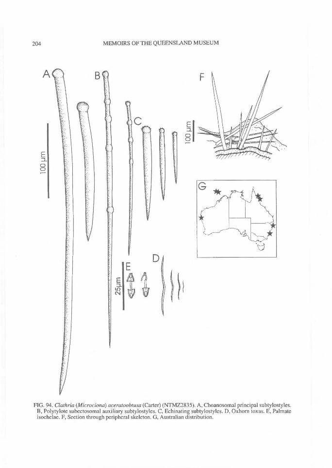
D1
204^ MEMOIRS OF THE QUEENSLAND MUSEUM
FIG. 94. Clathria (Microciona) aceratoobtusa (Carter) (NTMZ2835). A, Choanosomal principal subtylostyles.B, Polytylote subectosomal auxiliary subtylostyles. C, Echinating subtylostyles. D, Oxhorn toxas. E, Palmateisochelae. F, Section through peripheral skeleton. G, Australian distribution.
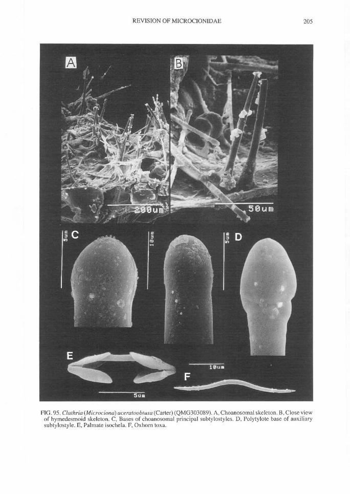
REVISION OF MICROCIONIDAE^ 205
FIG. 95. Clathria (Microciona) aceratoobtusa (Carter) (QMG303089). A, Choanosomal skeleton. B, Close viewof hymedesmoid skeleton. C, Bases of choanosomal principal subtylostyles. D, Polytylote base of auxiliarysubtylostyle. E, Palmate isochela. F, Oxhorn toxa.
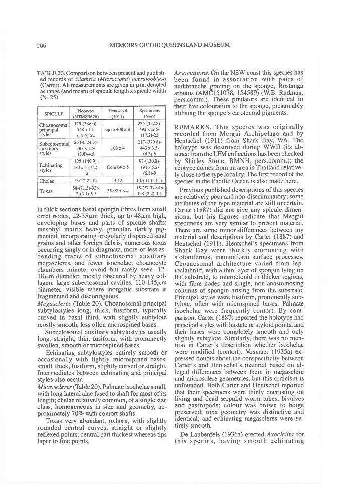
206^ MEMOIRS OF THE QUEENSLAND MUSEUM
TABLE 20. Comparison between present and publish-ed records of Clathria (Microciona) aceratoobtusa(Carter). All measurements are given in p,m, denotedas range (and mean) of spicule length x spicule width(N=25).
SPICULENeotype
(NTMZ3676)Hentschel
(1911)Specimens
(N=6)
Choanosomal 175(386.0)- 235-(352.8)-principal 548 x11- up to 408 x 8 492 x12.5-styles (15.5)-22 (17.2)-22
Subectosomal 264-(324.3)- 217(379.8)-auxiliary 387x 1.5- 168 x 4 443 x 3.5-styles (3.8)4.5 (4.3)-6
128-(149.9)- 97-(
styles 183 x 5-(7.2)- from 64 x 5 194 x 5.2-12 (6.8)-9
Chelae 9-(12.2)-14 9-12 10.5-(13.5)-1658-(71.5)-92 x 18-(57.3)-84 x
Toxas 2-(3.1)-5.5 35-92 x 3-4 0.8-(2.2)-3.5
in thick sections basal spongin fibres form smallerect nodes, 22-35 p.m thick, up to 48p,m high,enveloping bases and parts of spicule shafts;mesohyl matrix heavy, granular, darkly pig-mented, incorporating irregularly dispersed sandgrains and other foreign debris, numerous toxasoccurring singly or in dragmata, more-or-less as-cending tracts of subectosomal auxiliarymegascleres, and fewer isochelae; choanocytechambers minute, ovoid but rarely seen, 12-18p,m diameter, mostly obscured by heavy col-lagen; large subectosomal cavities, 110-145p.mdiameter, visible where inorganic substrate isfragmented and discontiguous.Megascleres (Table 20). Choanosomal principalsubtylostyles long, thick, fusiform, typicallycurved in basal third, with slightly subtylotemostly smooth, less often microspined bases.
Subectosomal auxiliary subtylostyles usuallylong, straight, thin, fusiform, with prominentlyswollen, smooth or microspined bases.
Echinating subtylostyles entirely smooth oroccasionally with lightly microspined bases,small, thick, fusiform, slightly curved or straight.Intermediates between echinating and principalstyles also occur.Microscleres (Table 20). Palmate isochelae small,with long lateral alae fused to shaft for most of itslength; chelae relatively common, of a single sizeclass, homogeneous in size and geometry, ap-proximately 70% with contort shafts.
Toxas very abundant, oxhorn, with slightlyrounded central curves, straight or slightlyreflexed points; central part thickest whereas tipstaper to fine points.
Associations. On the NSW coast this species hasbeen found in association with pairs ofnudibranchs grazing on the sponge, Rostangaarbutus (AMC151078, 154589) (W.B. Rudman,pers.comm.). These predators are identical intheir live colouration to the sponge, presumablyutilising the sponge's carotenoid pigments.
REMARKS. This species was originallyrecorded from Mergui Archipelago and byHentschel (1911) from Shark Bay, WA. Theholotype was destroyed during WWII (its ab-sence from the LFM collections has been checkedby Shirley Stone, BMNH, pers.comm.); theneotype comes from an area in Thailand relative-ly close to the type locality. The first record of thespecies in the Pacific Ocean is also made here.
Previous published descriptions of this speciesare relatively poor and non-discriminatory; someattributes of the type material are still uncertain.Carter (1887) did not give any spicule dimen-sions, but his figures indicate that Merguispecimens are very similar to present material.There are some minor differences between mymaterial and descriptions by Carter (1887) andHentschel (1911). Hentschel's specimens fromShark Bay were thickly encrusting withstoloniferous, mammiform surface processes.Choanosomal architecture varied from lep-toclathriid, with a thin layer of spongin lying onthe substrate, to microcionid in thicker regions,with fibre nodes and single, non-anastomosingcolumns of spongin arising from the substrate.Principal styles were fusiform, prominently sub-tylote, often with microspined bases. Palmateisochelae were frequently contort. By com-parison, Carter (1887) reported the holotype hadprincipal styles with hastate or styloid points, andtheir bases were completely smooth and onlyslightly subtylote. Similarly, there was no men-tion in Carter's description whether isochelaewere modified (contort). Vosmaer (1935a) ex-pressed doubts about the conspecificity betweenCarter's and Hentschel's material based on al-leged differences between them in megasclereand microsclere geometries, but this criticism isunfounded. Both Carter and Hentschel reportedthat their specimens were thinly encrusting onliving and dead serpulid worm tubes, bivalvesand gastropods; colour was brown to beigepreserved; toxa geometry was distinctive andidentical; and echinating megascleres were en-tirely smooth.
De Laubenfels (1936a) erected Axocielita forthis species, having smooth echinating
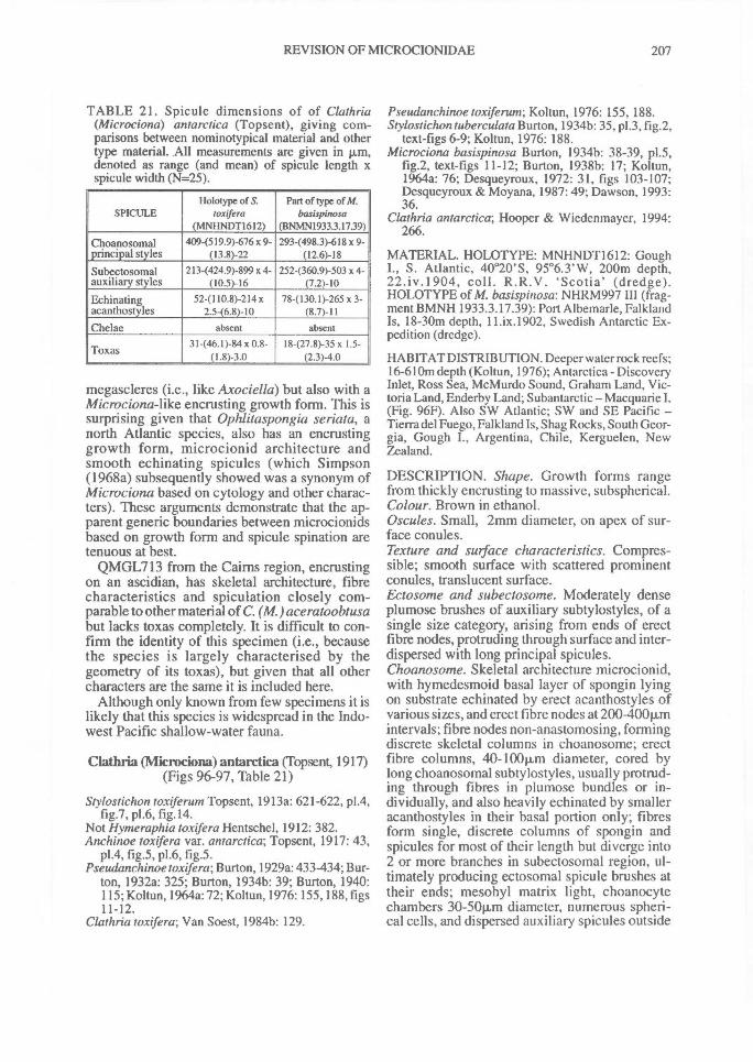
REVISION OF MICROCIONIDAE^ 207
TABLE 21. Spicule dimensions of of Clathria(Microciona) antarctica (Topsent), giving com-parisons between nominotypical material and othertype material. All measurements are given in p.m,denoted as range (and mean) of spicule length xspicule width (N=25).
SPICULEHolotype of S.
toxtfera(MNHNDT1612)
Part of type of M.basispinosa
(BNMN1933.3.1739)
Choanosomal 409-(519.9)-676 x 9- 293-(498.3)-618 x 9-principal styles (13.8)-22 (12.6)-18
Subectosomal 2I3-(424.9)-899 x 4- 252-(360.9)-503 x 4-auxiliary styles (10.5)-16 (7.2)-10
Echinating 52-(110.8)-2I4 x 78-(130.1)-265 x 3-acanthostyles 2.5-(6.8)-10 (8.7)-11Chelae absent absent
Toxas 31-(46.1)-84 x 0.8-(1.8)-3.0
18-(27.8)-35 x 1.5-(2.3)4.0
megascleres (i.e., like Axociella) but also with aMicrociona-like encrusting growth form. This issurprising given that Ophlitaspongia seriata, anorth Atlantic species, also has an encrustinggrowth form, microcionid architecture andsmooth echinating spicules (which Simpson(1968a) subsequently showed was a synonym ofMicrociona based on cytology and other charac-ters). These arguments demonstrate that the ap-parent generic boundaries between microcionidsbased on growth form and spicule spination aretenuous at best.
QMGL713 from the Cairns region, encrustingon an ascidian, has skeletal architecture, fibrecharacteristics and spiculation closely com-parable to other material of C. (M.)aceratoobtusabut lacks toxas completely. It is difficult to con-firm the identity of this specimen (i.e., becausethe species is largely characterised by thegeometry of its toxas), but given that all othercharacters are the same it is included here.
Although only known from few specimens it islikely that this species is widespread in the Indo-west Pacific shallow-water fauna.
Clathria (Microciona) antarctica (Topsent, 1917)(Figs 96-97, Table 21)
Stylostichon toxiferum Topsent, 1913a: 621-622, p1.4,fig.7, p1.6, fig.14.
Not Hymeraphia toxifera Hentschel, 1912: 382.Anchinoe toxifera var. antarctica; Topsent, 1917: 43,
p1.4, fig.5, p1.6, fig.5.Pseudanchinoe toxifera; Burton, 1929a: 433-434; Bur-
ton, 1932a: 325; Burton, 1934b: 39; Burton, 1940:115; Koltun, 1964a: 72; Koltun, 1976: 155, 188, figs11-12.
Clathria toxifera; Van Soest, 1984b: 129.
Pseudanchinoe toxiferum; Koltun, 1976: 155, 188.Stylostichon tuberculata Burton, 1934b: 35, p1.3, fig.2,
text-figs 6-9; Koltun, 1976: 188.Microciona basispinosa Burton, 1934b: 38-39, p1.5,
fig.2, text-figs 11-12; Burton, 1938b: 17; Koltun,1964a: 76; Desqueyroux, 1972: 31, figs 103-107;Desqueyroux & Moyana, 1987: 49; Dawson, 1993:36.
Clathria antarctica; Hooper & Wiedenmayer, 1994:266.
MATERIAL. HOLOTYPE: MNHNDT1612: GoughI., S. Atlantic, 40°20'S, 95°6.3'W, 200m depth,22.iv.1904, coll. R.R.V. 'Scotia' (dredge).HOLOTYPE of M. basispitzosa: NHRM997 III (frag-ment BMNH 1933.3.17.39): Port Albemarle, FalklandIs, 18-30m depth, 11.ix.1902, Swedish Antarctic Ex-pedition (dredge).
HABITAT DISTRIBUTION. Deeper water rock reefs;16-610m depth (Koltun, 1976); Antarctica - DiscoveryInlet, Ross Sea, McMurdo Sound, Graham Land, Vic-toria Land, Enderby Land; Subantarctic — Macquarie I.(Fig. 96F). Also SW Atlantic; SW and SE Pacific —Tierra del Fuego, Falkland Is, Shag Rocks, South Geor-gia, Gough I., Argentina, Chile, Kerguelen, NewZealand.
DESCRIPTION. Shape. Growth forms rangefrom thickly encrusting to massive, subspherical.Colour. Brown in ethanol.Oscules. Small, 2mm diameter, on apex of sur-face conules.Texture and surface characteristics. Compres-sible; smooth surface with scattered prominentconules, translucent surface.Ectosome and subectosome. Moderately denseplumose brushes of auxiliary subtylostyles, of asingle size category, arising from ends of erectfibre nodes, protruding through surface and inter-dispersed with long principal spicules.Choanosome. Skeletal architecture microcionid,with hymedesmoid basal layer of spongin lyingon substrate echinated by erect acanthostyles ofvarious sizes, and erect fibre nodes at 200-40011Anintervals; fibre nodes non-anastomosing, formingdiscrete skeletal columns in choanosome; erectfibre columns, 40-100p,m diameter, cored bylong choanosomal subtylostyles, usually protrud-ing through fibres in plumose bundles or in-dividually, and also heavily echinated by smalleracanthostyles in their basal portion only; fibresform single, discrete columns of spongin andspicules for most of their length but diverge into2 or more branches in subectosomal region, ul-timately producing ectosomal spicule brushes attheir ends; mesohyl matrix light, choanocytechambers 30-50p.m diameter, numerous spheri-cal cells, and dispersed auxiliary spicules outside
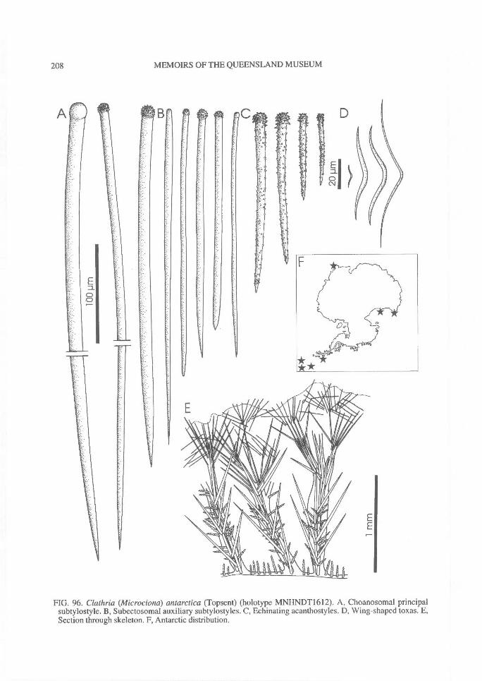
ED.0C\J
208^ MEMOIRS OF THE QUEENSLAND MUSEUM
FIG. 96. Clathria (Microciona) antarctica (Topsent) (holotype MNHNDT1612). A, Choanosomal principalsubtylostyle. B, Subectosomal auxiliary subtylostyles. C, Echinating acanthostyles. D, Wing-shaped toxas. E,Section through skeleton. F, Antarctic distribution.
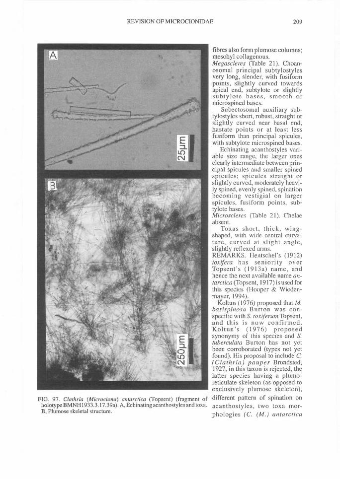
FIG. 97. Clathria (Microciona) antarctica (Topsent) (fragment ofholotype BMNH1933.3.17.39a). A, Echinating acanthostyles and toxa.B, Plumose skeletal structure.
REVISION OF MICROCIONIDAE^ 209
fibres also form plumose columns;mesohyl collagenous.Megascleres (Table 21). Choan-osomal principal subtylostylesvery long, slender, with fusifonnpoints, slightly curved towardsapical end, subtylote or slightlysubtylote bases, smooth ormicrospined bases.
Subectosomal auxiliary sub-tylostyles short, robust, straight orslightly curved near basal end,hastate points or at least lessfusiform than principal spicules,with subtylote microspined bases.
Echinating acanthostyles vari-able size range, the larger onesclearly intermediate between prin-cipal spicules and smaller spinedspicules; spicules straight orslightly curved, moderately heavi-ly spined, evenly spined, spinationbecoming vestigial on largerspicules, fusiform points, sub-tylote bases.Microscleres (Table 21). Chelaeabsent.
Toxas short, thick, wing-shaped, with wide central curva-ture, curved at slight angle,slightly reflexed arms.REMARKS. Hentschel's (1912)toxifera has seniority overTopsent's (1913a) name, andhence the next available name an-tarctica (Topsent, 1917) is used forthis species (Hooper & Wieden-mayer, 1994).
Koltun (1976) proposed that M.basispinosa Burton was con-specific with S. toxiferum Topsent,and this is now confirmed.Koltun's (1976) proposedsynonymy of this species and S.tuberculata Burton has not yetbeen corroborated (types not yetfound). His proposal to include C.(Clathria) pauper Brondsted,1927, in this taxon is rejected, thelatter species having a plumo-reticulate skeleton (as opposed toexclusively plumose skeleton),different pattern of spination onacanthostyles, two toxa mor-phologies (C. (M.) antarctica
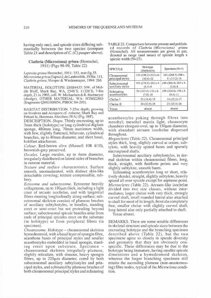
210^ MEMOIRS OF THE QUEENSLAND MUSEUM
having only one), and spicule sizes differing sub-stantially between the two species (compareTable 21 and description of C. (C)pauper above).
Clathria (Microciona) grisea (Hentschel,1911) (Figs 98-99, Table 22)
Leptosia grisea Hentschel, 1911: 353, text-fig.35.Microcionagrissa [lapsus]; de Laubenfels, 1936a: 111.Clathria grisea; Hooper & Wiedenmayer, 1994: 266
MATERIAL. HOLOTYPE: ZMB4435: NW. of Mid-dle Bluff, Shark Bay, WA, 2548'S, 11326'E, 7-8mdepth, 21.ix.1905, coll. W. Michaelsen & R. Hartmeyer(dredge). OTHER MATERIAL: WA- NTMZ2863(fragments QMG300054, PIBOC 04-295).
HABITAT DISTRIBUTION. 7-25m depth; growingon bivalves and Acropora cf. robusta; Shark Bay andPelsart Is, Houtman Abrolhos (WA) (Fig. 98F).DESCRIPTION. Shape. Thinly encrusting, up to3mm thick (holotype) or long cylindrical digitatesponge, 480mm long, 70mm maximum width,with few, slightly flattened, bifurcate, cylindricalbranches, up to 40mm diameter, and short basal,holdfast attachment.Colour. Red-brown alive (Munsell lOR 4/10),brownish-grey preserved.Oscules. Large oscules, up to 4mm diameter,irregularly distributed on lateral sides of branchesin ramose material.Texture and sutface characteristics. Surfacesmooth, unornamented, with distinct skin-likedetachable covering; texture compressible, rub-bery.Ectosome and subectosome. Ectosome heavilycollagenous, up to 1801.1,m thick, including a lightcrust of arcuate isochelae, and with tangentialfibres running longitudinally along surface; sub-ectosomal skeleton consists of plumose brushesof auxiliary subtylostyles, in bundles, standingerect or semi-erect but not protruding beyondsurface; subectosomal spicule bundles arise fromends of principal spicules erect on the substrate(in holotype) or from peripheral fibres (inspecimen).Choanosome. Holotype — choanosomal skeletonhymedesmoid, with a basal layer of spongin fibre,acanthose bases of principal styles and smalleracanthostyles embedded in basal spongin, stand-ing erect upon substrate. Specimen —choanosomal skeleton irregularly plumose,slightly reticulate, with sinuous, heavy sponginfibres, up to 220..m diameter, cored by bothsubectosomal auxiliary subtylostyles and prin-cipal styles, and echinated by plumose brushes ofboth choanosomal principal styles and echinating
TABLE 22. Comparison between present and publish-ed records of Clathria (Microciona) grisea(Hentschel). All measurements are given in gm,denoted as range (and mean) of spicule length xspicule width (N=25).
SPICULEHolotype
(ZMB4435)Specimen (N=1)
Choanosomal 135-(198.2)-212 x 6- 241-(268.5)-298 xprincipal styles (10.4)-12 11-(13.2)-16
Subectosomal 195-(218.9)-242 x 4- 246-(264.4)-283 x 4-auxiliary styles (6.3)-8 (5.8)-8
Echinating 954103.4)-116 x 6- 109-(124.6)-158 x 8-acanthostyles (7.8)-10 (9.4)-11
Chelae I 15-(16.8)-19 14-(15.2)-17
Chelae II 20-(22.9)-28 23-(25.5)-28
Toxas absent absent
acanthostyles poking through fibres intomesohyl; mesohyl matrix light, choanocytechambers elongate-oval, up to 150p,m diameter,with abundant arcuate isochelae dispersedthroughout.Megascleres (Table 22). Choanosomal principalstyles thick, long, slightly curved at centre, sub-tylote, with heavily spined bases and sparselymicrospined shafts.
Subectosomal auxiliary subtylostyles, in der-mal skeleton within choanosomal fibres, long,thick, straight, with fusiform points and veryslightly subtylote, smooth bases.
Echinating acanthostyles long or short, rela-tively slender, straight, slightly subtylote, heavilyspined all over spicule except for aspinose point.Microscleres (Table 22). Arcuate-like isochelaedivided into two size classes, without inter-mediates; larger chelae with very thick, stronglycurved shaft, small rounded lateral alae attachedto shaft for most of its length, front ala completelyfree; smaller chelae with slightly curved shaft,long lateral alae only partially attached to shaft.
Toxas absent.
REMARKS. There are some notable differencesin skeletal structure and spicule sizes between theencrusting holotype and the branching specimendescribed above (Table 22), but the twospecimens agree so closely in spicule diversityand geometry that they are obviously con-specific. These differences may be due to theholotype being immature, having smaller spiculedimensions and a hymedesmoid skeleton,whereas the larger branching specimen stillretains the ascending plumose (non-anastomos-ing) fibre nodes, typical of the Microciona condi-tion.
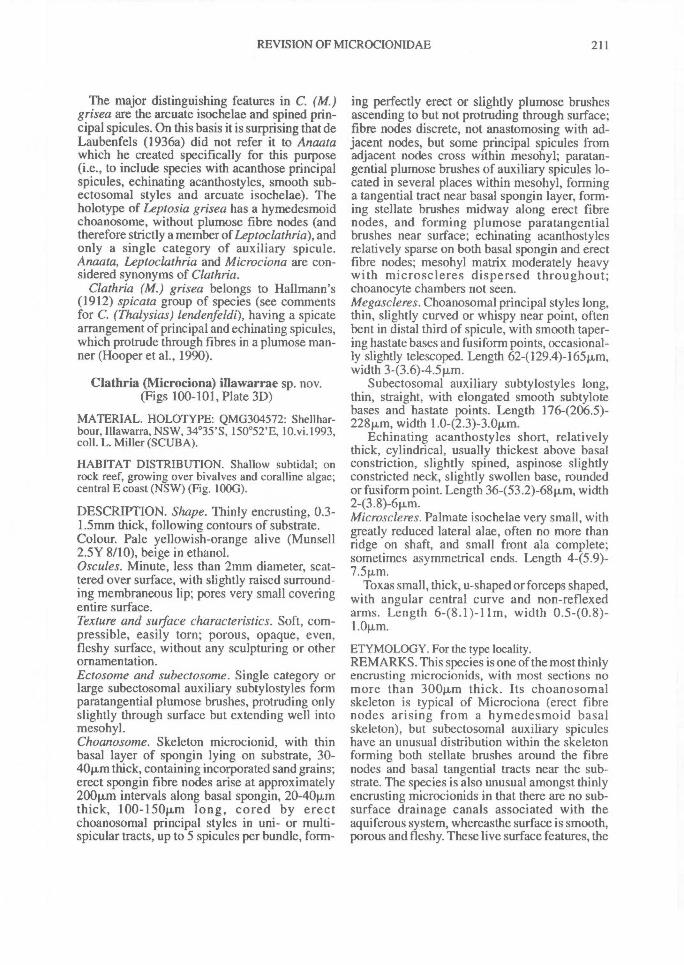
REVISION OF MICROCIONIDAE^ 211
The major distinguishing features in C. (M.)grisea are the arcuate isochelae and spined prin-cipal spicules. On this basis it is surprising that deLaubenfels (1936a) did not refer it to Anaatawhich he created specifically for this purpose(i.e., to include species with acanthose principalspicules, echinating acanthostyles, smooth sub-ectosomal styles and arcuate isochelae). Theholotype of Leptosia grisea has a hymedesmoidchoanosome, without plumose fibre nodes (andtherefore strictly a member of Leptoclathria), andonly a single category of auxiliary spicule.Anaata, Leptoclathria and Microciona are con-sidered synonyms of Clathria.
Clathria (M.) grisea belongs to Hallmann's(1912) spicata group of species (see commentsfor C. (Thalysias) lendenfeldi), having a spicatearrangement of principal and echinating spicules,which protrude through fibres in a plumose man-ner (Hooper et al., 1990).
Clathria (Microciona) illawarrae sp. nov.(Figs 100-101, Plate 3D)
MATERIAL. HOLOTYPE: QMG304572: Shellhar-bour, Illawarra, NSW, 34 °35'S, 150°52'E, 10.vi.1993,coll. L. Miller (SCUBA).
HABITAT DISTRIBUTION. Shallow subtidal; onrock reef, growing over bivalves and coralline algae;central E coast (NSW) (Fig. 100G).
DESCRIPTION. Shape. Thinly encrusting, 0.3-1.5mm thick, following contours of substrate.Colour. Pale yellowish-orange alive (Munsell2.5Y 8/10), beige in ethanol.Oscules. Minute, less than 2mm diameter, scat-tered over surface, with slightly raised surround-ing membraneous lip; pores very small coveringentire surface.Texture and surface characteristics. Soft, com-pressible, easily torn; porous, opaque, even,fleshy surface, without any sculpturing or otherornamentation.Ectosome and subectosome. Single category orlarge subectosomal auxiliary subtylostyles formparatangential plumose brushes, protruding onlyslightly through surface but extending well intomesohyl.Choanosome. Skeleton microcionid, with thinbasal layer of spongin lying on substrate, 30-40p..m thick, containing incorporated sand grains;erect spongin fibre nodes arise at approximately200Rm intervals along basal spongin, 20-4011mthick, 100-150p,m long, cored by erectchoanosomal principal styles in uni- or multi-spicular tracts, up to 5 spicules per bundle, form-
ing perfectly erect or slightly plumose brushesascending to but not protruding through surface;fibre nodes discrete, not anastomosing with ad-jacent nodes, but some principal spicules fromadjacent nodes cross within mesohyl; paratan-gential plumose brushes of auxiliary spicules lo-cated in several places within mesohyl, forminga tangential tract near basal spongin layer, form-ing stellate brushes midway along erect fibrenodes, and forming plumose paratangentialbrushes near surface; echinating acanthostylesrelatively sparse on both basal spongin and erectfibre nodes; mesohyl matrix moderately heavywith microscleres dispersed throughout;choanocyte chambers not seen.Megascleres. Choanosomal principal styles long,thin, slightly curved or whispy near point, oftenbent in distal third of spicule, with smooth taper-ing hastate bases and fiisiform points, occasional-ly slightly telescoped. Length 62-(129.4)-165Rm,width 3-(3.6)-4.5p,m.
Subectosomal auxiliary subtylostyles long,thin, straight, with elongated smooth subtylotebases and hastate points. Length 1764206.5)-228p,m, width 1.0-(2.3)-3.0Rm.
Echinating acanthostyles short, relativelythick, cylindrical, usually thickest above basalconstriction, slightly spined, aspinose slightlyconstricted neck, slightly swollen base, roundedor fusiform point. Length 36-(53.2)-68p,m, width2-(3.8)-6Rm.Microscleres. Palmate isochelae very small, withgreatly reduced lateral alae, often no more thanridge on shaft, and small front ala complete;sometimes asymmetrical ends. Length 445.9)-
Toxas small, thick, u-shaped or forceps shaped,with angular central curve and non-reflexedarms. Length 6-(8.1)-11m, width 0.540.8)-1.01/m.
ETYMOLOGY. For the type locality.REMARKS. This species is one of the most thinlyencrusting microcionids, with most sections nomore than 300jLm thick. Its choanosomalskeleton is typical of Microciona (erect fibrenodes arising from a hymedesmoid basalskeleton), but subectosomal auxiliary spiculeshave an unusual distribution within the skeletonforming both stellate brushes around the fibrenodes and basal tangential tracts near the sub-strate. The species is also unusual amongst thinlyencrusting microcionids in that there are no sub-surface drainage canals associated with theaquiferous system, whereasthe surface is smooth,porous and fleshy. These live surface features, the
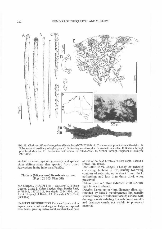
212^ MEMOIRS OF THE QUEENSLAND MUSEUM
FIG. 98. Clathria (Microciona) grisea (Hentschel) (NTMZ2863). A, Choanosomal principal acanthostyles. B,Subectosomal auxiliary subtylostyles. C, Echinating acanthostyles. D, Arcuate isochelae. E, Section throughperipheral skeleton. F, Australian distribution. G, NTMZ2863. H, Section through fragment of holotypeZMB4435.
skeletal structure, spicule geometry, and spiculesizes differentiate this species from otherMicrociona in the Indo-west Pacific.
Clathria (Mierociona) lizardensis sp. nov.(Figs 102-103, Plate 3E)
MATERIAL. HOLOTYPE - QMG304121: BlueLagoon, Lizard I., Cairns Section, Great Barrier Reef,14°41.0'S, 145 °27.5'E, 9m depth, 03.iv.1994, coll.J.N.A. Hooper, L.J. Hobbs, J.A. Kennedy & S.D. Cook(SCUBA).
HABITAT DISTRIBUTION. Coral reef, patch reef inlagoon, under coral overhangs, on ledges or exposedcoral heads, growing on live coral, coral rubble at base
of reef or on dead bivalves; 9-12m depth; Lizard I.(FNQ) (Fig. 102G).DESCRIPTION. Shape. Thinly or thicklyencrusting, bulbous in life, usually followingcontours of substrate, up to about 101nm thick,collapsing and less than 4mm thick whenpreserved.Colour. Pale red alive (Munsell 2.5R 6-5/10),light brown in ethanol.Oscules. Large, up to 4mm diameter alive, sur-rounded by raised membraneous lip, usuallysituated on apex of bulbous (flaccid) surface, withdrainage canals radiating towards pores; osculesand drainage canals not visible in preservedmaterial.
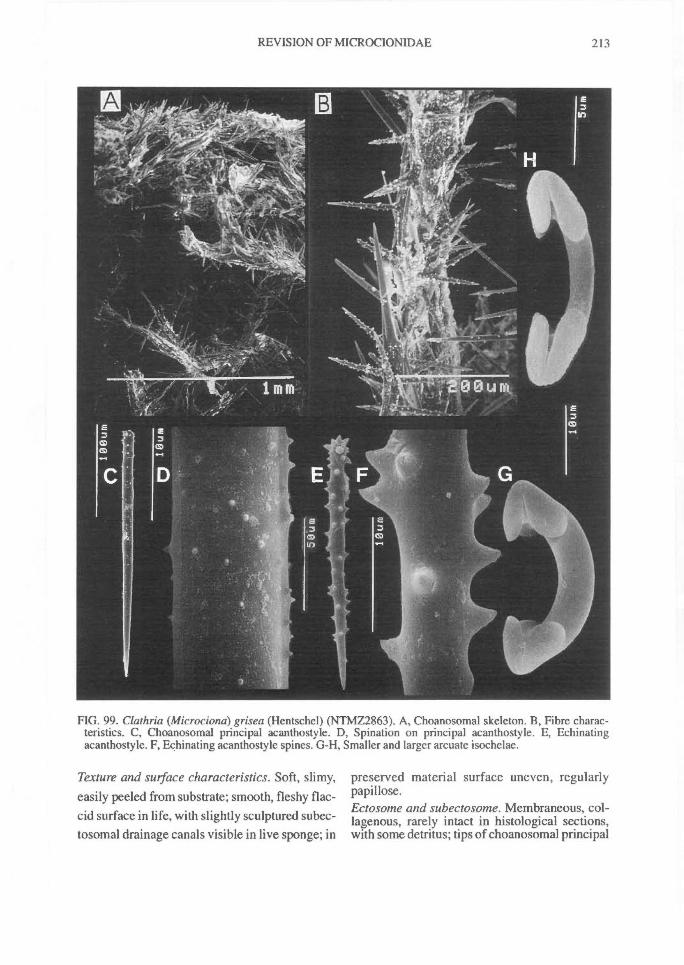
REVISION OF MICROCIONIDAE^
213
FIG. 99. Clathria (Microciona) grisea (Hentschel) (NTMZ2863). A, Choanosomal skeleton. B, Fibre charac-teristics. C, Choanosomal principal acanthostyle. D, Spination on principal acanthostyle. E, Echinatingacanthostyle. F, F,chinating acanthostyle spines. G-H, Smaller and larger arcuate isochelae.
Texture and surface characteristics. Soft, slimy,easily peeled from substrate; smooth, fleshy flac-cid surface in life, with slightly sculptured subec-tosomal drainage canals visible in live sponge; in
preserved material surface uneven, regularlypapillose.Ectosome and subectosome. Membraneous, col-lagenous, rarely intact in histological sections,with some detritus; tips of choanosomal principal
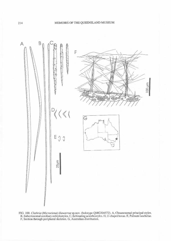
214^ MEMOIRS OF THE QUEENSLAND MUSEUM
A
FIG. 100. Clathria (Microciona) illawarrae sp.nov. (holotype QMG304572). A, Choanosomal principal styles.B, Subectosomal auxiliary subtylostyles. C, Echinating acanthostyles. D, U-shaped toxas. E, Palmate isochelae.F, Section through peripheral skeleton. G, Australian distribution.
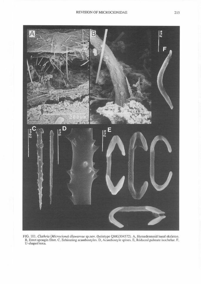
REVISION OF MICROCIONIDAE^
215
FIG. 101. Clathria (Microciona) illawarrae sp.nov. (holotype QMG304572). A, Hymedesmoid basal skeleton.B, Erect spongin fibre. C, Echinating acanthostyles. D, Acanthostyle spines. E, Reduced palmate isochelae. F,U-shaped toxa.
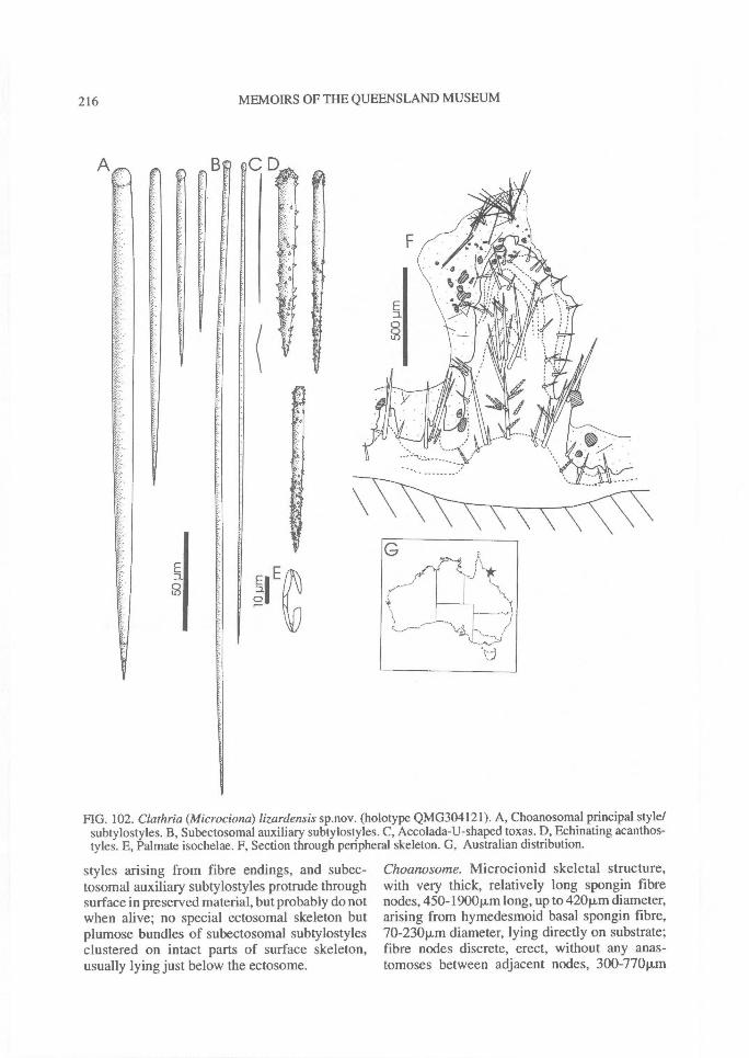
CD
216^ MEMOIRS OF THE QUEENSLAND MUSEUM
FIG. 102. Clathria (Microciona) lizardensis sp.nov. (holotype QMG304121). A, Choanosomal principal style/subtylostyles. B, Subectosomal auxiliary subtylostyles. C, Accolada-U-shaped toxas. D, Echinating acanthos-tyles. E, Palmate isochelae. F, Section through peripheral skeleton. G, Australian distribution.
styles arising from fibre endings, and subec-tosomal auxiliary subtylostyles protrude throughsurface in preserved material, but probably do notwhen alive; no special ectosomal skeleton butplumose bundles of subectosomal subtylostylesclustered on intact parts of surface skeleton,usually lying just below the ectosome.
Choanosome. Microcionid skeletal structure,with very thick, relatively long spongin fibrenodes, 450-1900p,m long, up to 420p.m diameter,arising from hymedesmoid basal spongin fibre,70-23011in diameter, lying directly on substrate;fibre nodes discrete, erect, without any anas-tomoses between adjacent nodes, 300-770p.m
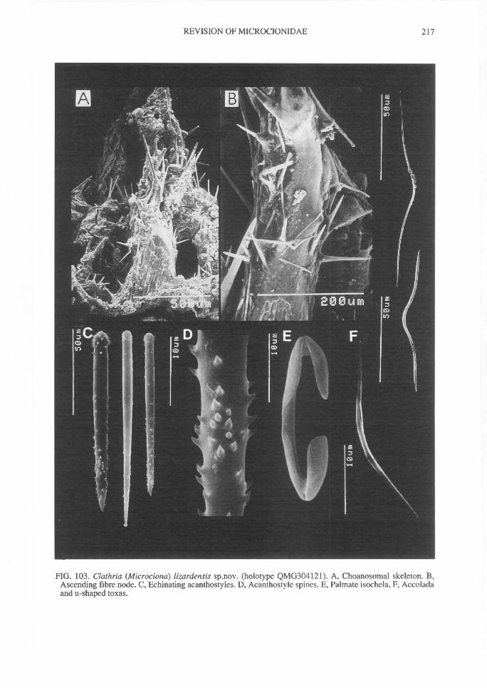
REVISION OF MICROCIONIDAE^
217
FIG. 103. Clathria (Microciona) lizardensis sp.nov. (holotype QMG304121). A, Choanosomal skeleton. B,Ascending fibre node. C, Echinating acanthostyles. D, Acanthostyle spines. E, Palmate isochela. F, Accoladaand u-shaped toxas.
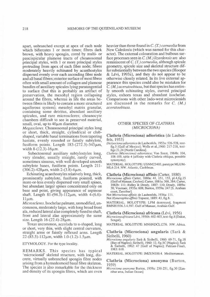
218^ MEMOIRS OF THE QUEENSLAND MUSEUM
apart, unbranched except at apex of each nodewhich bifurcates 1 or more times; fibres darkbrown, with heavy spongin, cored by multi- orpaucispicular plumose tracts of choanosomalprincipal styles, with 1 or more principal stylesprotruding from apex of each fibre node; fibresmoderately heavily echinated by acanthostylesdispersed evenly over each ascending fibre nodeand all basal fibres; exterior surface of most fibresoften with small amount of collagen and plumosebundles of auxiliary spicules lying paratangentialto surface (but this is probably an artifact ofpreservation, the mesohyl region collapsingaround the fibres, whereas in life the areas be-tween fibres is likely to contain a more structuredaquiferous system); mesohyl matrix granular,containing some detritus, abundant auxiliaryspicules, and rare microscleres; choanocytechambers difficult to see in preserved material,small, oval, up to 40v.m diameter.Megascleres. Choanosomal principal styles longor short, thick, straight, cylindrical or club-shaped, variable basal terminations from taperinghastate, evenly rounded or faintly subtylote,fusiform points. Length 183-(272.3)-345p,m,width 8-(l2.3)-1611m.
Subectosomal auxiliary subtylostyles long,very slender, usually straight, rarely curved,sometimes sinuous, with well developed smoothsubtylote bases, fusiform points. Length 211-(306.2)-428p.m, width 2-(3.8)-6p.m.
Echinating acanthostyles relatively long, thick,prominently subtylote, fusiform pointed, withmore-or-less evenly dispersed very small spines,but abundant larger spines concentrated only onbase and point, giving appearance of aspinoseshaft. Length 81-(94.3)-112Rm, width 446.6)-111.1.m.Microscleres. Isochelae palmate, unmodified, un-common, moderately large, with long broad frontala, reduced lateral alae completely fused to shaft,front and lateral alae approximately the samesize. Length 16-(22.4)-28p,m.
Toxas uncommon, accolada to u-shaped, longor short, very thin, with slight central curvature,straight arms or faintly reflexed arms. Length22-(85.5)-112Rm, width 1.0-(1.2)-1.511m.
ETYMOLOGY. For the type locality.
REMARKS. This species has typical`microcionid' skeletal structure, with long, dis-crete, virtually unbranched spongin fibre nodesarising from a hymedesmoid basal fibre skeleton.The species is also remarkable for the thicknessand density of its spongin fibres, which are even
heavier than those found in C. (T) corneolia fromNew Caledonia (which was named for this char-acter). The external colouration and bulbous sur-face processes seen in C. (M.) lizardensis are alsoreminiscent of C. (T) corneolia, although spiculegeometry, spicule size and skeletal structure dif-fer substantially between the two species (Hooper& Levi, 1993a), and they do not appear to beotherwise closely related. In its live external ap-pearance this species could also be mistaken forC. (M.) aceratoobtusa, but that species has entire-ly smooth echinating styles, curved principalstyles, oxhorn toxas and abundant isochelae.Comparisons with other Indo-west microcionidsare discussed in the remarks for C. (M.)aceratoobtusa.
OTHER SPECIES OF CLATHR1A(MICROCIONA)
Clathria (Microciona) adioristica (de Lauben-fels, 1953)Dictyociona adioristica de Laubenfels, 1953a: 526-528, text-
fig.5 [Gulf of Mexico]; Wells et al.,1960: 217-218, text-figs 21,24 [North Carolina].
Clathria (Microciona) adioristica; Van Soest, 1984b: 104,108-109, table 4 [affinity with Clathria obliqua, possiblesynonymy].
MATERIAL. HOLOTYPE: USNM23403, paratype MLUM-ML4-214. NW. Atlantic, Caribbean.
Clathria (Microciona) affinis (Carter, 1880)Microciona affinis Carter, 1880a: 41, 151, 153, p1.4,fig.15
[Gulf of Manaar, Ceylon]; Carter, 1881a: 368,384; Carter,1882b: 111; Ridley & Dendy, 1887: 110; Dendy, 1889a:38; Vosmaer, 1935a: 608; Burton, 1959a: 247 [S. Arabiancoast, Zanzibar].
Not Microciona affinis; de Laubenfels, 1936a: 111.Not Hymeraphia affinis Topsent, 1889: 43, fig.8.
MATERIAL. HOLOTYPE: LFM destroyed, fragmentBMNH1936.3.4.597. Gulf of Manaar, Arabian Gulf.
Clathria (Microciona) africana (Levi, 1956)Microciona africana Levi, 1956b: 402-403, text-fig.8 [Dakar,
Sengal].
MATERIAL. HOLOTYPE: MNHNDCL1276. NW. Africa.
Clathria (Microciona) angularis (Sara &Siribelli, 1960)Microciona angularis Sara & Siribelli, 1960: 69-71, fig.18
[Bay of Naples]; Siribelli, 1960: 12, fig.5C [Naples]; Sara& Siribelli, 1962: 47 [Gulf of Naples]; Pulitzer-Finali,1983: 610.
MATERIAL. HOLOTYPE: IMZUN100.4. Mediterranean.
Clathria (Microciona) anonyma (Burton,1959)Microciona anonyma Burton, 1959a: 250-251, fig.30 [Zan-
zibar area, Indian Ocean]
REVISION OF MICROCIONIDAE^ 219
MATERIAL. HOLOTYPE: BMNHI936. 3.4.575. Central EAfrica.
Clathria (Microciona) armata (Bowerbank,1862)Microcionaarmata Bowerbank, 1862a: 779, 1858, p1.24, figs
26-28; Bowerbank, 1864:41, p1.4, figs 96-98; Bowerbank,1866: 124, 129-131, 141 [Britain]; Gray, 1867: 535; Nor-man, 1869:330; Schmidt, 1870:76; Bowerbank, 1874: 60,p1.23, figs 17-24; Carter, 1874b: 405, p1.21, fig.27; Carter,1874c: 456-457; Carter, 1876: 310; Carter, 1880a: 40-41,151 [Gulf of Manaar]; Koehler, 1886a: 62 [English Chan-nel]; Topsent, 1888: 117, 124, 125, 141, 156, 158, p1.6,fig.8; Topsent, 1891a: 528 [Roscoff]; Stephens, 1916:234[W coast, Ireland]; Stephens, 1921; Vosmaer, 1935a: 6-7,666; Alander, 1942: 62 [Sweden]; Levi, 1956b: 399-400,text-fig.6 [Dakar, Senegal]; Koltun, 1959: 181-182, text-fig. 141 [White Sea]; Levi, 1960a: 73, text-figs 16, 17[Roscoff,Sidmouth]; Poggiano, 1965: 3, 7, 11-14, text-fig.5, tables 1, 2 [Italy]; Pulitzer-Finali, 1983: 573-574,610, text-fig. 69 [Mediterranean]; Wintermann-Kilian &Kilian, 1984: 134 [Colombia]; Ackers, Moss & Picton,1992: 143 [Ireland].
Microciona armatus; Bowerbank, 1882: 7, 18, 53; Sollas,1884: 614; Vosmaer, 1884a: 121; Vosmaer, I885b: 353;Koehler, 1886a: 11, 55; Carter, 1889a: 287; Carter & Hope,1889: 99, 101-106; Dendy, 1889c: 17; Hope, 1889: 333,336, 337; Chatin, 1890: 889; Topsent, 1890c: 202, 204;Topsent, 1892a: 17; Topsent, 1893d: 445; Norman, 1892:6, 11; Hanitsch, 1894: 176; Topsent, 1894a: 8,23; Topsent1899: 105; Topsent, 1900: 255; Topsent, 1904a: 189;Loisel, 1898:38; Minchin, 1898:529; Minchin, 1909:215;Stephens, 1912: 27; Ferrer Hernandez, 1914: 41.
Clathria armata; Topsent, 1925:649 [discussion]; Van Soest& Stone, 1986: 45 [Norway].
Scopalina armata; Wright, 1868: 224.Amphilectus armatus; Vosmaer, 1880: 118-119; Vosmaer,
1889: 353; Svarcevskij, 1906: 342; Babic, 1921: 87-88[Adriatic]; Babic, 1922: 261-262, text-fig.6.
Esperia armata Fristedt, 1885: 36-38.Not Microciona armatus; de Laubenfels, 1936a: Ill.Microciona svarchevskyi de Laubenfels, 1936a: 111; Levi,
1960a: 73.cf. Microciona prolifera; Vosmaer, 1935a: 607.MATERIAL. HOLOTYPE: BMNH1910.1.1.66(1930.7.3.209). Caribbean, NE Atlantic, Gulf of Manaar, NWAfrica, Mediterranean. Many of these records are suspectgiven their disjunct distribution and this taxon is likely toconsist of a species complex.
Clathria (Microciona) ascendens(Cabioch, 1968)Microcionaascendens Cabioch, 1968a: 239, text-fig.11 [Ros-
coff, France]; Rodriguez SolOrzano et al., 1979: 44, 59-60,text-fig.15 [Galicia, Spain].
MATERIAL. HOLOTYPE: RMBS. NE Atlantic.
Clathria (Microciona) assimilis Topsent, 1925Clathria assimilis Topsent, 1925: 649; Topsent & Olivier,
1943: 1 [no diagnosis; Adriatic].Pseudanchinoe assimilis; de Laubenfels, 1936a: 109 [note].Microcionaassimilis; Levi, 1960a: 76 [Adriatic, Naples, Mar-
seilles]; Siribelli, 1960: 18, text-fig.7C [Naples]; Pog-giano, 1965: 3, table 1; Vacelet, 1969: 207, text-fig.46[Mediterranean]; Pulitzer-Finali, 1977: 61 [Bay ofNaples]; Pulitzer-Finali, 1983: 610; Pansini, 1987: 170[Alboran Sea].
cf. Clathria compressa; Topsent, 1925: 649.
MATERIAL. HOLOTYPE: MOM, fragment MNHNDT125.Mediterranean. Possible synonym of Clathria spinarcus(Carter & Hope) (Maldonado, 1992: 1152).
Clathria (Microciona) atrasanguinea (Bower-bank, 1862)Microciona atrasanguinea Bowerbank, 1862c: 824, 1109,
1110, 1135, p1.30, fig. 1, p1.74, fig.2 [British Seas]; Bower-bank, 1864: 188, 286, p1.33, fig.368, p1.34, fig.369; Bower-bank, 1866: 7, 124, 138-141 [Britain]; Bowerbank, 1874:63, p1.24, figs 14-19; Topsent, 1888: 141, 157; Topsent,1890c: 202,204; Topsent, 1891a: 528 [Roscoff]; Topsent,1892c: 17 [Banyuls]; Dendy, 1922: 60, p1.13, fig. la-e[Egmont Reef]; Burton, 1934b: 37, text-fig.10; Burton,1938a: 30, p1.4, fig.24 [Madras]; Lilly et al., 1953 [record-Lough Inc, Ireland]; Levi, 1960a: 72-73, text-fig.I5[English Channel, Atlantic]; Sara & Siribelli, 1962: 47[Gulf of Naples]; Levi, 1965: 18-19, text-fig.21 [Red Sea];Simpson, I968a: 33, text-fig.! [Plymouth,England];Juniper & Steele, 1969: 161 [Portsmouth, England]; VanSoest & Weinberg, 1980: 10 [Lough Inc. Ireland]; Boury-Esnault, 1971: 326 [Banyuls]; Ackers, Moss & Picton,1992: 142 [Ireland].
Microciona atrosanguinea; Gray, 1867: 535; Norman, 1869:330; Schmidt, 1870: 76; Carter, I870a: 332, 339, 340;Carter, 1871a: 272, 274; Carter, 1871b: 8; Carter, 1872a:106, 111, p1.10, figs 17-20; Carter, 1874c: 457; Carter,1875: 195; Carter, 1876: 308; Carter, 1880a: 38-41, 151[Gulf of Manaar]; Carter, 1880b: 59 [Indian Ocean];Carter, 1881a: 384 [record]; Bowerbank, 1882: 7, 18, 54;Koehler, 1885: 53, 55; Vosmaer, 1885b: 209; Koehler,1886a: 61, 62 [English Channel]; Carter, 1887b: 355;Carter & Hope, 1889: 102, 104-106; Dendy, 1889c: 18;Hope, 1889: 334; Topsent, 1889: 39; Topsent, 1890c: 202;Topsent, 1891d: 232; Topsent, 1893d: 445; Topsent,1894a: 8, 10, 23; Norman, 1892: 6; Hanitsch, 1894: 176;Heider, 1895: 280; Loisel, 1898: 38; Topsent, 1900: 255[note]; Woodland, 1908: 140, 145; Minchin, 1909: 217;Burton & Rao, 1932: 344-345; [coasts of Bengal, Burma,India and Arabian Sea]; de Laubenfels, 1936b: 448-449[Panama]; Pulitzer-Finali, 1983: 610; RodriguezSolOrzano et al., 1991: 177 [Galicia, Spain].
Microciona atrasanguineum; Cuenot, 1903: 4 [Arcachon].Clathria (Microciona) atrasanguinea; Van Soest, 1993: 103
[Mauritius].Amphilectus atrasanguineus; Vosmaer, 1880: 115.Plumohalichondriaatrasanguinea; Hanitsch, 1890: 207-208,
210 [England].Scopalina lophyropoda; Schmidt, 1868: 26, 40.Scopalina atrosanguthea; Schmidt, 1866a: 149; Schmidt,
1866b: 15.cf. Microciona prohfera; Vosmaer, 1935a: 604, 607.MATERIAL. HOLOTYPE: BMNH1930.7. 3.225, paratypesBMNH1930.7.3.226, 1910.1.1.68. Caribbean, NE Atlantic,Mediterranean, Red Sea, Arabian Gulf, W Indian Ocean, WIndia, Gulf of Manaar, Bay of Bengal, Andaman Sea.
Clathria (1Vlicrociona) basifixa(Topsent, 1913)Ophlitaspongia basifixa Topsent, 1913b: 39 [Norway]; Bur-
ton, 1935c: 74 [Japan; probable misidentification]; deLaubenfels, 1954: 162 [note].
Clathria (Microciona) basifixa; Van Soest & Stone, 1986:45[Norway].
MATERIAL. HOLOTYPE: MOM, fragmentMNHNDT1957. NE Atlantic.
220^ MEMOIRS OF THE QUEENSLAND MUSEUM
Clathria (Microciona) bitoxa (Burton, 1930)Hymantho bitoxa Burton, 1930a: 503, text-fig.2 [Norway];
Alander, 1942: 63 [Sweden].Microciona levis; Fristedt, 1887: 416.Clathria bitoxa; Van Soest, 1984b: 90 [generic synonymy];
Van Soest & Stone, 1986: 47 [note].Microciona bitoxa Rodriguez SolOrzano & Rodriguez Babio,
1993: 62 [Iberian Peninsula].MATERIAL. HOLOTYPE: BMNH1910.1. 1.787.v. NE At-lantic.
Clathria (Microciona) brepha(de Laubenfels, 1930)Aaata brepha de Laubenfels, 1930: 27 [California]Anaata brepha; de Laubenfels, 1936a: 91, text-fig.53.Clathria brepha; Van Soest, 1984b: 7 [generic synonymy].MATERIAL. HOLOTYPE: USNM2I427. PARATYPESBMNH1929.8.22.36, 57. NE Pacific.
Clathria (Microciona) brondstedi sp. nov.Hymedesmia pennata Brondsted, 1932: 12 [Faeroe Is].Anaata pennata; de Laubenfels, 1936a: 109.Clathria pennata; Van Soest,1984b: 7 [generic synonymy for
Anaata].Not Desmacella pennata Lambe, 1895: 129.MATERIAL. HOLOTYPE: UZM (not located). NE Atlantic.Clathria ( Microciona) pennata (Lambe, 1895) has seniority.
Clathria (Microciona) bulboretorta (Carter,1880)Microcionabulboretorta Carter, I880a: 41, 42, 151, 153, p1.4,
fig.3a-e [Gulf of Manaar, Ceylon]; Vosmaer, 1935a: 608.MATERIAL. HOLOTYPE: LFM destroyed. Gulf of Manaar.
Clathria (Microciona) bulbotoxa Van Soest,1984Clathria (Microciona)bulbotoxa Van Soest, 1984b: 103-104,
p1.7, figs 5-8, text-fig.41, table 4 [Curacao, West Indies].Microciona bulbotoxa; Pulitzer-Finali, 1986: 149-150 [West
Indies].MATERIAL. HOLOTYPE: ZMAPOR4789. Caribbean.
Clathria (Microciona) calla (de Laubenfels,1934)Axociella calla de Laubenfels, 1934: 16 [Puerto Rico].Avocielita calla; de Laubenfels, 1954: 149 [note]; Sim &
Byeon, 1989: 40, p1.5, figs 3-5 [Korea; probable misiden-tification].
Clathria calla; Boury-Esnault, 1973: 286, text-fig.46[Brazilian Basin]; Zea, 1987: 170, text-fig.59, p1.2, fig. 3[Colombian Caribbean].
Clathria(Microciona)calla; Van Soest,1984b: 100-101, p1.7,fig.1, text-fig.39, table 4 [Curacao, Florida; affinity withClathria coralloides from Mediterranean].
Microciona calla; Pulitzer-Finali, 1986: 150 [West Indies].Microciona rarispinosa Hechtel, 1965: 42-44, text-fig.8 [Port
Royal, Jamaica]; Wintermann-Kilian & Kilian, 1984: 135[Colombia].
Tenaciella obliqua; Alcolado, 1976:5; Alcolado, 1980: 10.MATERIAL. HOLOTYPE: USNM. Caribbean, tropical SWAtlantic.
Clathria (Microciona) campecheae nom. nov.II ymeraphia affinis Topsent, 1889: 43, fig.8A [Banc de
Campeche]; Topsent, 1904a: 162-3 [Azores].
Microciona affinis; de Laubenfels, 1936a: 111.Clathria (Microciona)affinis; Van Soest, 1984b: 93-95, 108,
text-fig.36, table 4 [Curacao, West Indies].Clathria cf. affinis; Kobluk & Van Soest, 1989: 1216
[Bonaire].Not Microciona affinis Carter, 1880a: 41, p1.14, fig. 15; Vos-
maer, 1933: 608.MATERIAL. HOLOTYPE: MNHNDT1841, paratypeMNHNDT3584. Caribbean, NE Atlantic. Clathria(Microciona) affinis (Carter, I880a) has priority.
Clathria (Microciona) carnosa (Bowerbank,1862)Microciona camosa Bowerbank, I 862a: 804, 1110 [Britain];
Bowerbank, 1866: 133; Vosmaer, 1935a: 607? Halichondria incrustans; Schmidt, 1866a: 150MATERIAL. HOLOTYPE: BMNH1930.7. 3.203, fragmentBMNH1910.1.1.666. NE Atlantic, tropical SW. Atlantic.
Clathria (Microciona) claudei sp. nov.Microciona acanthotoxa Levi & Levi, 1989: 81, fig.49
[Philippines].MATERIAL. HOLOTYPE: MNHNDCL341 I. Philippines.Clathria acanthotoxa (Stephens) has seniority.
Clathria (Microciona) cleistochela Topsent,1925Clathria cleistochela Topsent, 1925: 650-651, fig.9 [Gulf of
Naples].Microciona cleistochela; de Laubenfels, 1936a: 1 1 1 [note];
Levi, 1960a: 72, fig. I 4 [ Naples,Banyuls]; Siribelli, 1960:12-14, fig.5B [Naples]; de Laubenfels, 1951b: 214 [BlackSea]; Pulitzer-Finali, 1983: 610; Boury-Esnault & Lopes,1985: 193-194, fig.42 [Azores].
MATERIAL. HOLOTYPE: MOM, fragment MNHNDT329.Mediterranean, NE Atlantic.
Clathria (Microciona) coccinea (Bergquist,1961)Microciona coccinea Bergquist, 1961a: 38, fig.8a,b [N. New
Zealand]; Bergquist & Sinclair, 1968: 427, 428, fig. 1 a[morphology and larvae]; Bergquist & Sinclair, 1973: 43;Bergquist et al., 1970: 248, 254; Evans & Bergquist, 1977:195-196; Bergquist & Fromont, 1988: 102-103, p1.47,fig.f, p1.48, fig.a; Rudman & Avern, 1989: 335; Dawson,1993: 36 [note].
Not Thalisias coccinea Duchassaing & Michelotti, 1864: 84,p1.18, fig.5 [St.Thomas]; Wiedenmayer, 1977a: 253, table49.
MATERIAL. HOLOTYPE: NMNZ unregistered. NewZealand.
Clathria (Microciona) ctenichela (Alander,1942)M wrociona ctenichela Alander, 1942: 61-62, p1.15, fig.20
[Sweden].Clathria (Microciotia) ctenichela; Van Soest & Stone, 1986:
44-45 [Norway].MATERIAL. HOLOTYPE: ZMA. NE Atlantic.
Clathria (Microciona) dendyi (Bergquist &Fromont, 1988)Microciona dendyi Bergquist & Fromont, 1988: 100-102,
p1.47, figs d,e [Slipper 1.]; Dawson, 1993: 37 [note].MATERIAL. HOLOTYPE: NMNZPOR114. New Zealand.
REVISION OF MICROCIONIDAE^ 221
Clathria (Microciona) densa (Burton, 1959)Microciona densa Burton, 1959a: 248, text-fig.28 [S Arabian
coast]MATERIAL. HOLOTYPE: BMNH1936.3. 4.456. ArabianGulf.
Clathria (Microciona) dianae (Schmidt, 1875)Suberites dianae Schmidt, 1875: 116, p1.1,fig.1 [Norway];
Czerniawsky, 1880: 70.Microciona dianae; Thiele, 1903b: 394, 395, 398, p1.21,
fig.28a-e; Vosmaer, 1935a: 608, 630; de Laubenfels,1936a: 1 1 1 [note].
Not A rtemisina dianae Topsent, 1907: 69; Vosmaer, 1935a:630.
Clathria dianae; Van Soest & Stone,1986: 47 [note].MATERIAL. HOLOTYPE: unknown. NE Atlantic.
Clathria (Microciona) ditoxa (Stephens, 1916)Eurypon ditoxa Stephens, 1916:239-240 [W coast of Ireland];
Stephens, 1921: 51, p1.5; Burton, 1959b: 44-45 [Iceland].Microciona ditoxa; Levi, 1960a: 66 [W coast of Ireland,
Atlantic].Dictyociona ditoxa; de Laubenfels, 1936a: 110 [note].MATERIAL. HOLOTYPE: INMSR151.9.1916. NE Atlan-tic.
Clathria (Microciona) duplex Sara, 1958Clathria duplex Sara, 1958: 262-264, fig.24 [Gulf of Naples].Microciona duplex; Levi, 1960a: 69 [Naples]; Siribelli, 1960:
14-15, fig.6A [Naples]; Sara, 1963: 210 [Gulf ofPolicastro]; Pulitzer-Finali, 1983: 610; RodriguezSolOrzano et al., 1991: 179, fig.3 [Galicia, Spain].
MATERIAL. • HOLOTYPE: IMZUN93.26.x.58. Mediter-ranean.
Clathria (Microciona) echinata (Alcolado,1984)Axociella echinata Alcolado, 1984: 7 [Cuba].Clathria echinata; Kobluk & Van Soest, 1989: 1216;
Meesters et al., 1991: 194 [Curacao, Bonaire].Clathria (Microciona) simpsoni Van Soest, 1984b: 97-99,
p1.7, figs 2-4, text-fig.38, table 4 [Puerto Rico, Curacao];Pulitzer-Finali, 1986: 150 [West Indies].
Clathria simpsoni; Zea, 1987: 168, text-fig.58, p1.3, fig.1[Colombian Caribbean].
MATERIAL. HOLOTYPE: Cuba. Holotype of simpsoni:ZMAPOR3332. Caribbean.
Clathria (Microciona) elliptichela (Alander,1942)Microciona elliptichela Alander, 1942: 58-61 [Sweden].Clathria elliptichela; Van Soest & Stone, 1986:45 [note].MATERIAL. HOLOTYPE: ZMA. NE Atlantic.
Clathria (Microciona) fallax (Bowerbank,1866)Microciona falla.v Bowerbank, 1866: 124, 128, 129, 135
[Hastings, Britain]; Vosmaer, 1935a: 607; Curtis, 1970:260-261 [cytology]; Ackers, Moss & Picton, 1992: 147[Ireland].
Sophax fallav; Gray, 1867: 521.MATERIAL. LECTOTYPE-BMNH 1910.1. 1.71.PARALECTOTYPE BMNH1930.7.3.198, fragmentUSNM5047. NE Atlantic.
Clathria (Microciona) fascispiculifera (Carter,1880)Microciona fascispiculifera Carter, 1880a: 44, 45, 151, 153,
p1.4, fig.7a-g [Gulf of Manaar]; Hallmann, 1916c: 637[note]; [?] Vosmaer, 1935a: 608.
Damoseni fascispiculifera; de Laubenfels, 1936a: 110.MATERIAL. HOLOTYPE: LFM destroyed, no extant frag-ment in BMNH. Gulf of Manaar.
Clathria (Nlicrociona) fraudata (Bowerbank,1874)Microciona fraudata Bowerbank, 1874: 273, 275, 277, p1.83,
figs 7-11 [Polperro, Fowey Hbr.]; Vosmaer, 1935a: 607.MATERIAL. HOLOTYPE: BMNH1930.7.3. 205. NE Atlan-tic.
Clathria (Microciona) frogeti (Vacelet, 1969)Microciona frogeti Vacelet, 1969: 208, text-fig.47 [Mediter-
ranean].Microciona fregeti [sic.]; Pulitzer-Finali, 1983: 610 [list].MATERIAL. HOLOTYPE: MNHN missing. W Mediter-ranean.
Clathria (Microciona) gradalis Topsent, 1925Clathria gradalis Topsent, 1925: 651-653 [Gulf of Naples];
Topsent & Olivier, 1943: 1 [Monaco]; Sara, 1958: 258-260, text-fig.22 [Gulf of Naples]; Sara, 1960a: 461 [Is-chia].
Clathria gradalis var. atoxa; Topsent, 1928a: 299, p1.10,fig.14 [Boavista I., Senegal].
Microciona gradalis; de Laubenfels, 1936a: 111; Levi, 1960a:75 [W Mediterranean]; Sara & Siribelli, 1960: 67 [Bay ofNaples]; Siribelli, 1960: 16, text-fig.6B [Naples]; Sara &Siribelli, 1962:47-48 [Gulf of Naples]; Poggiano, 1965:3,table 1; Cabioch, 1968a: 244 [Roscoff, N. France];Vacelet, 1969: 207 [W Mediterranean]; Pulitzer-Finali,1977: 63 [Bay of Naples]; Pulitzer-Finali, 1983: 610.
MATERIAL. HOLOTYPE: MOM, fragment MNHNDT328.Mediterranean, NE Atlantic, NW Africa.
Clathria (Microciona) haematodes (de Laubenfels,1957)Microciona haematodes de Laubenfels, 1957: 240, text-fig.6
[Oahu, Hawaii]; Bergquist, 1977: 67 [Hawaii].MATERIAL. HOLOTYPE: USNM23533. Hawaii.
Clathria (Microciona) haplotoxa (Topsent,1928)Leptoclathria haplotoxa Topsent, 1928a: 298, p1.10, fig.16
[Madeira I.]; [cf.] Topsent, 1934b: 24.Microciona haplotoxa;Topsent, 1934a: 92-93 [Gulf of Gabes,
Tunisia]; Levi, 19566: 400-402, text-fig.7 [Dakar,Senegal]; Levi, 1960a: 70 [Madeira, Tunisia]; Pulitzer-Finali, 1983: 610.
MATERIAL. HOLOTYPE: MNHNDT1101. Mediterranean,NE Atlantic, NW Africa.
Clathria (Microciona) hentscheli sp. nov.Ilymeraphia lendenfeldi Hentschel, 1912: 378-379, p1.20, fig.
35 [Mimien Bay, Am I., Arafura Sea].Eurypon lendenfeldi; de Laubenfels, 1936a: 110.Not Clathria lendenfeldi Ridley & Dendy, 1886: 474.MATERIAL. LECTOTYPE: SMF 1705. Indonesia. Clathria(Thalysias) lendenfeldi Ridley & Dendy has priority.

222^ MEMOIRS OF THE QUEENSLAND MUSEUM
Clathria (Microciona) heterotoxa(Hentschel, 1929)Microciona heterotoxa Hentschel, 1929: 891-892, 970, p1.14,
fig.5 [White Sea]; Koltun, 1959: 182-183, text-fig.142[Arctic, USSR].
Dictyociona heterotoxa; de Laubenfels, 1936a: 110 [note]; deLaubenfels, 1953a: 527.
MATERIAL. HOLOTYPE: HM or ZMB (not located).Arctic.
Clathria (Microciona) hymedesmioides VanSoest, 1984Clathria (Microciona) hymedesmioides Van Soest, 1984b:
104-105, p1.7, figs 9-10, text-fig.42, table 4 [Curacao].MATERIAL. HOLOTYPE: ZMAPOR4790. Caribbean.
Clathria (Microciona) ixauda (Levi, 1969)Microciona ixauda Levi, 1969: 965, text-fig.7a [Vema
Seamount].MATERIAL. HOLOTYPE: MNHNDCL1415. S Atlantic.
Clathria (Microciona) jecusculum (Bower-bank, 1866)Hymeniacidon jecusculum Bowerbank, 1866: 198 [Harris I.,
Hebrides].Microciona jecusculum; Bowerbank, 1874: 273-275, p1.83,
figs 1-6; Carter, 1876: 237 [Cape St. Vincent; Faroe Is];Vosmaer, 1933: 607 [imperfectly known].
MATERIAL. HOLOTYPE: unknown; fragmentsBMNH1954.3.9.176, 177. NE Atlantic.
Clathria (Microciona) kentii (Bowerbank,1874)Microciona kentii Bowerbank, 1874: 311, 312, 317-319,
p1.89, figs 9-13 [Jersey, Strangford Lough]; Vosmaer,1935a: 607.
MATERIAL. HOLOTYPE: BMNH1910.1.1.77, fragmentUSNM5044. NE Atlantic.
Clathria (Microciona) laevis (Bowerbank,1866)Microciona laevis Bowerbank, 1866: 124, 127-128 [Britain];
Stephens, 1917: 12, p1.1, fig.3 [N. of Bolus Head, Ireland];Vosmaer, 1935a: 607; Burton, 1959b: 43 [Iceland].
Not Microciona laevis; Fristedt, 1887: 415.Abila laevis; Gray, 1867: 539.Hymantho laevis; de Laubenfels, 1936a: 111; Alander, 1942:
63 [Sweden].MATERIAL. HOLOTYPE: BMNH1930.7.3. 215. NE Atlan-tic.
Clathria (Microciona) laevissima(Dendy, 1922)Hymedesmia laevissima Dendy, 1922: 81-82, p1.15, fig.1
[Mauritius].Folitispa laevissima; de Laubenfels, 1936a: 119 [note].MATERIAL. HOLOTYPE: BMNH1921.11. 7.69. W IndianOcean.
Clathria (Microciona) lajorei (de Laubenfels,1954)Anaata lajorei de Laubenfels, 1954: 147-148, text-fig. 95
[Ailing-lap-lap Atoll].Clathria lajorei; Van Soest, 1984a: 129 [generic synonymy].
MATERIAL. HOLOTYPE: USNM22827. W central Pacific.
Clathria (Microciona) leighensis sp. nov.Microciona rubens Bergquist, 1961a: 38, text-fig.9 [NNew
Zealand]; Bergquist & Green, 1977b: 289-302 [ontogeny];Bergquist & Fromont, 1988: 103, p1.48, figs b-c [N NewZealand]; Dawson, 1993: 37 [note].
Not Thalassodendron rubens Lendenfeld, 1888: 223.MATERIAL. HOLOTYPE: NMNZ unregistered. NewZealand. C. (Clathria) rubens (Lendenfeld, 1888) haspriority.
Clathria (Microciona) levii (Sara & Siribelli,1960)Microciona levii Sara & Siribelli, 1960: 71-73, text-fig.19
[Bay of Naples]; Siribelli, 1960: 6-8, text-fig.2 [Naples];Poggiano, 1965:3, table 1; Pulitzer-Finali, 1983:610 [list].
MATERIAL. HOLOTYPE: IMZUN31. Mediterranean.
Clathria (Microciona) longispiculum(Carter, 1876)
Microciona longispiculum Carter, 1876: 231, 237, 238, 470,p1.12, fig. 1 h, p1.15, fig.31a-c [N. Scotland]; Vosmaer,1935a: 608 [insufficiently known].
MATERIAL. HOLOTYPE: BMNH1887.10. 29.3. NE Atlan-tic.
Clathria (Microciona) longistyla(Burton, 1959)
Microciona longistyla Burton, 1959a: 249-250, text-fig.29 [S.Arabian coast]; Sim & Kim, 1988: 26 [Korea]; Sim &Byeon, 1989: 40 [Korea; possible misidentifications].
MATERIAL. HOLOTYPE: BMNH1936.3.4. 583. ArabianGulf, ? S China Sea.
Clathria (Microciona) macrochela(Levi, 1960)
Microciona macrochela Levi, 1960a: 70, text-fig.13 [Ros-coff].
MATERIAL. HOLOTYPE: MNHNDCL940. NE Atlantic.
Clathria (Microciona) microjoanna(de Laubenfels, 1930)
Microciona microjoanna de Laubenfels, 1930: 27 [Carmel,California]; de Laubenfels, 1932: 93-95, text-fig.55[California]; Bakus, 1966:433-435, text-fig.4, table 4 [SanJuan Arch., Washington State]; Lee & Gilchrist, 1985:24-32 [biochemistry]; Bakus & Green, 1987: 72 [S.California].
MATERIAL. HOLOTYPE: USNM21468. PARATYPEUSNM21469, fragment BMNH1929.8.22.28. NE Pacific.
Clathria (Microciona) micronesia(de Laubenfels, 1954)
Microciona micronesia de Laubenfels, 1954: 145-146, text-fig.93 [Majuro Atoll].
MATERIAL. HOLOTYPE: USNM22833. W central Pacific.
Clathria (Microciona) microxea(Vacelet & Vasseur, 1971)Paratenaciella microxea V acelet & Vasseur, 1971: 103, text-
fig.61 [Tulear, Madagascar].MATERIAL. HOLOTYPE: MNHNDIV27. W Indian Ocean.
REVISION OF MICROCIONIDAE^ 223
Clathria (Microciona) mima(de Laubenfels, 1954)
Ophlitaspongia mima de Laubenfels, 1954: 161-162, text-fig.105 [Majuro Atoll, central W. Pacific]; Wintermann-Kilian & Kilian, 1984: 135 [Colombia].
MATERIAL. HOLOTYPE: USNM22839. NW centralPacific, Caribbean.
Clathria (Microciona) namibiensis (Uriz,1984)Microciona namibiensis Uriz, 1984b: 111-113, text-figs 3
A-B, 5D; Uriz, 1988a: 87 [Namibia].MATERIAL. HOLOTYPE: ABIPB-12. SW Africa.
Clathria (Microciona) normani (Burton, 1930)Hymantho norrnani Burton, 1930a: 503, text-fig.! [Norway].Clathria normani; Van Soest, 1984b: 90 [generic synonymy];
Van Soest & Stone, 1986: 46-47 [note].MATERIAL. HOLOTYPE: BMNH1910.1.1. 791.iii. NE At-lantic.
Clathria (Microciona) novaezealandiae(Brondsted, 1924)Microciona novaezealandiae Brondsted, 1924: 463-464, text-
fig.19 [Slipper Is]; Bergquist & Fromont, 1988: 103-104;Dawson, 1993: 37.
Wetmoreus novaezealandica; de Laubenfels, 1936a: 112.MATERIAL. HOLOTYPE: UZM not located, no fragment inBMNH. New Zealand
Clathria (Microciona) osismica (Cabioch,1968)Microciona osismica Cabioch, 1968a: 240-244, text-fig. 12
[Roscoff, France].MATERIAL. HOLOTYPE: RMBS. NE Atlantic.
Clathria (Microciona) parthena(de Laubenfels, 1930)
Microciona parthena de Laubenfels, 1930:27 [California]; deLaubenfels, 1932: 95-96, text-fig.56 [California]; Henkartet al., 1973: 3045-3050, text-figs 1-5 [biochem.];Cauldwell et al., 1973: 3051-3058, text-figs 1-5[biochem.]; Lee & Gilchrist, 1985: 24-32 [biocheml; Sim
Bakus, 1986: 9 [California]; Bakus & Green, 1987:72-73 [S. California].
MATERIAL. HOLOTYPE: USNM21383. PARATYPEBMNH1929.9.30.6. NE Pacific.
Clathria (Microciona) pennata (Lambe, 1895)Desmacella pennata Lambe, 1895: 129, p1.4, figs 6a-d
[Sooke, Vancouver I., Canada]; Ricketts & Calvin, 1948:34 [Vancouver I., Canada].
Ophlitaspongia pennata; de Laubenfels, 1927: 265-266, text-figs 1-4 [Laguna Beach and Monterey Pen., California]; deLaubenfels, 1930: 28; Burton, 1930a: 521; de Laubenfels,1932: 103, text-fig.62 [var. californiana]; Hewatt, 1946:193 [California]; Hartman (in Light, 1954): 20 [Califor-nia]; de Laubenfels, 1954: 162 [note]; Burton, 1959a: 247[key to species]; de Laubenfels, 1961: 198, fig.1 [Califor-nia and Washington State]; Bakus, 1966: 435-440, text-fig.5, tables 5-6 [et var., San Juan Arch., Washington];Anderson, 1973:5668 [associates]; Lee & Gilchrist, 1985:24-32 [biochem.]; Sim & Bakus, 1986: 10; Bakus & Green,1987:73 [var. californiana]; Sim & Byeon, 1989: 37, p1.1,
figs 1-4 [var. californiana; Korea]; Lee & Klontz, 1991:61 [chemistry].
Tylodesma pennata; Koltun, 1959: 96 text-fig.51, 1-3 [S.Kuriles].
Biemna pennata; Koltun, 1958: 54.Ophlaaspongia affinis basifixa; Burton, 1935c: 74 [Possiet
Bay, Sea of Japan].Microciona pennata; Simpson, 1968a: 40, p1.15 [Washington
State].Clathria (Ophlitaspongia)pennata; Rudman & Avem, 1989:
335 [associates; probable misidentification of sponge].Not Pandaros pennata Duchassaing & Michelotti,1864: 88.Not Hymedesmia pennata Brondsted, 1932: 12 [see C.
brondstedi sp. nov.].MATERIAL. HOLOTYPE: USNM7488. PARATYPENMCIC1900-2826. NE Pacific, NW. Pacific, Japan.
Clathria (Microciona) plana (Carter, 1876)Microciona planum Carter, 1876: 238, 472 [Cape St. Vincent,
Hebrides].Microciona plana; Topsent, 1889: 41-42 [Banc de
Campêche]; Norman, 1892: 9.Amphilectus planus; Vosmaer, 1880: 121; Dendy, 1889: 18.Clathria planer; Topsent, 1894b: 30; Van Soest, 1984b: 108
[unrecognisable].cf. Microciona prolifera; Vosmaer, 1935a: 608.MATERIAL. HOLOTYPE: BMNH1890.4. 10.13. NE Atlan-tic.
Clathria (Microciona) plinthina (de Laubenfels,1954)Microciona plinthina de Laubenfels, 1954: 144-145, text-
fig.92 [Ailing-lap-lap Atoll].MATERIAL. HOLOTYPE: USNM22949. W central Pacific.
Clathria (Microciona) poecilosclera (Sara &Siribelli, 1960)Microciona poecilosclera Sara & Siribelli, 1960: 73-75, text-
fig.20 [Bay of Naples]; Siribelli, 1960: 18, text-fig.7B[Naples]; Poggiano, 1965:3, table 1; Pulitzer-Finali, 1983:610.
MATERIAL. HOLOTYPE: IMZUN350. Mediterranean.
Clathria (Microciona) polita (Ridley, 1881)Hymedesmia polita Ridley, 1881: 121-122, p1.10, fig.9
[Sandy Point, Magellan Straits; supposed affinities withClathria tuberosa (Bowerbank)].
Microciona polita; Topsent, 1900: 113 [English Channel;identified with doubt].
MATERIAL. HOLOTYPE: BMNH1879.12.27. 22. SW At-lantic, NE. Atlantic.
Clathria (Microciona) primitiva (Koltun,1955)Microciona prirnitiva Koltun, 1955a: 16-17, text-fig.6 [Behr-
ing Sea]; Burton, 1959: 43 [Iceland]; Koltun, 1959: 184,text-fig. 144 [USSR].
Not Clathriella primitiva Burton, 1935c: 73.MATERIAL. HOLOTYPE: ZIL, fragmentsBMNH1932.1.1.241, 572. Boreal region.
Clathria (Microciona) proxima (Lundbeck,1910)Hymedesmia proxima Lundbeck, 1910: 81 [Denmark Strait].Anaata proxima; de Laubenfels, 1936a: 109 [imperfectly
known].
224^ MEMOIRS OF THE QUEENSLAND MUSEUM
Clathriaproxima; Van Soest, 1984b: 7 [generic synonymy forAnaata].
MATERIAL. HOLOTYPE: ZRS. NE Atlantic.
Clathria (Microciona) pugio (Lundbeck, 1910)Hymedesmia pugio Lundbeck, 1910: 94 [Denmark Strait].Anaata pugio; de Laubenfels, 1936a: 109.Clathria pugio; Van Soest, 1984b: 7 [generic synonymy for
Anaata].MATERIAL. HOLOTYPE: ZRS. NE Atlantic.
Clathria (Microciona) pustulosa (Carter, 1882)Halichondria pustulosa Carter, 1882a: 285, p1.11, fig.1
[vicinity of Pantagonia and Falkland Is].Not Halichondria pustulosa; Carter, 1886g: 450 [Port Phillip
Bay, Victoria].Anaata pustulosa; de Laubenfels, 1936a: 109.MATERIAL. HOLOTYPE: BMNH not found, fragmentBMNH1954.3.9.233. SW Atlantic.
Clathria (Microciona) quadriradiata (Carter,1880)Microciona quadriradiata Carter, 1880a: 42, 43, 151, 153,
p1.4, figs 4a-d [Gulf of Manaar]; [?] Vosmaer, 1935a: 608.MATERIAL. HOLOTYPE: LFM destroyed. Gulf of Manaar.
Clathria (Microciona) rhopalophora(Hentschel, 1912)Hymeraphia rhopalophora Hentschel, 1912: 380, p1.20,
fig.37 [Aru I., Arafura Sea].Microciona rhopalophora; Burton, 1959a: 248 [Maldives];
Thomas, 1970b: 206, text-fig.7 [Cocos-Keeling Basin,Gulf of Manaar].
Eurypon rhopalophora; de Laubenfels, 1936a: 111.MATERIAL. HOLOTYPE: HM (fragment 5MF992). In-donesia.
Clathria (Microciona) scotti Dendy, 1924Clathria scotti Dendy, 1924a: 352, p1.10, fig.!, p1.14, figs 5-8
[E of North Cape, New Zealand].Pseudanchinoe scold; de Laubenfels, 1936a: 109 [note];
Bergquist & Fromont, 1988: 110-111, p1.51, figs b-d [N.New Zealand]; Dawson, 1993: 39 [note].
MATERIAL. HOLOTYPE: BMNH1923.10. 1.128,paratypes BMNH1923.10.1.129-131, AMZ2568. NewZealand.
Clathria (Microciona) seriata (Grant, 1826)Spongia seriata Grant, 1826: 116.Halichondria seriata; Johnston, 1842:74, 125, 197, 248, 258,
p1.14, fig.2; Gray, 1848: 12,16.Halic lona seriata; Bowerbank, 1861: 235; Bowerbank,
1862a: 769, 824, p1.29, fig.10.Chalitza seriata; Bowerbank, 1864: 24,2275, p1.17, fig.287;
Bowerbank, 1866: 139, 294, 361, 376-378; Wright, 1868:228; Norman, 1869:298-299; Schmidt, 1870: 3, 77; Carter,1871b: 196.
Clathriaseriata; Schmidt, 1866b: 10, 24, p1.1, fig.7; Vosmaer,1935a: 619 [uncertain affinity]; Hanitsch, 1889: 158;Hanitsch, 1890: 205-207 [England]; Babic, 1921: 84[Adriatic]; Babic, 1922: 244-245, text-fig.T [Adriatic].
Seriatula seriata; Gray, 1867: 515.Desmacidon seriata; Schmidt, 1868: 12.? Desmacodes seriatus; Vosmaer, 1880: 107.Ophlitaspongia seriata; Bowerbank, 1874:6, 167, p1.65, figs
1-4; Carter, 1875: 196; Bowerbank, 1882: 14, 24, 120,
186-188; Carter, 1883b: 314; Vosmaer, 1885: 357; Mac-Munn, 1888: 12, 14, 20; Dendy, 1889c: 14; Topsent,1890c: 204; Topsent, 1891a: 529; Norman, 1892: 8;Minchin, 1900: 20, fig.32; Thiele, 1905: 450-451;Kirkpatrick, 1907: 274; Kirkpatrick, 1908a: 26; Weltner,1910a: 23; Hallmann, 1912: 254; Stephens, 1912: 3, 28;Stephens, 1916: 234 [Ireland]; Ferrer Hernandez, 1914:43; Lilly et al., 1953 [Ireland]; de Laubenfels, 1954: 161-162 [note]; Burton, 1959a: 247 [key to species of Oph-litaspongia]; Levi, 1960a: 64-65, text-fig.9 [Atlantic];Levi, 1963: 59-60, text-fig. 69, p1.9B-C [Cape Town,South Africa]; Bergquist & Sinclair, 1968: 427, 428, text-fig.1B [larvae, New Zealand]; Bergquist & Hogg, 1969:207, 210; Fry, 1970: 135-157 [ecology]; Fry, 1971: 155-178 [larvae]; Bergquist &Sinclair, 1973:37-39; Fry, 1973:159-170 [ecology]; Van Soest & Weinberg, 1980: 10[Lough Ine, Ireland]; Lee & Gilchrist, 1985: 24-32[biochemistry]; Rudman & Avern, 1989: 335 [associates];Ackers, Moss & Picton, 1992: 147 [Ireland]; Dawson,1993: 38 [note].
Echinoclathria seriata;Topsent, I893d: 445; Hanitsch, 1894:179; Hanitsch, 1895: 212; Heider, 1895: 281; Topsent,1896: 114; Loisel, 1898: 38; Minchin, 1900: 20;Whitelegge, 1907: 503.
Microciona seriata; Simpson, 1968a: 37, pls 9-10, text-fig.2[Plymouth, England].
Not Rhaphidophlus seriatus Thiele, 1899: 14, p1.1, fig.6, p1.5,fig.7 [Celebes].
Ophlitaspongia papilla Bowerbank, 1866: 14, 378-380[Vazon Bay, Guernsey]; Bowerbank, 1874, p1.70, figs 1-4;Bowerbank, 1882: 187 [Westport Bay, Guernsey]; Koeh-ler, 1886a: 62 [English Channel]; Hallmann, 1912: 254[note].
? Clathria papilla; Schmidt, 1870: 77; Vosmaer, 1880: 155.Echinoclathria papilla; Hanitsch, 1894: 8-10, 16, 25, 26.Ophistospongia papilla; Gray, 1867: 515.MATERIAL. HOLOTYPE: BMNH1847.9.7.14, fragmentsBMNH1910.1.1.2368, 2369. NE Atlantic, S Africa, NewZealand, Mediterranean. This species is undoubtedly com-posite, consisting of at least two sibling species (Atlantic andIndo-Pacific populations).
Clathria (Microciona) sigmoidea (Cuartas,1992)Microciona sigmoidea Cuartas, 1992: 85-88, figs 53-57, 67
[Mar del Plata, Argentina].MATERIAL. HOLOTYPE: MCNPC04-81-43. SW Atlantic.
C1athria (Microciona) simae sp. nov.Axociella cylindrica; Sim & Byeon, 1989: 39-40, p1.5, figs
1-2 [S. Korea].Not Esperiopsis cylindrica Ridley & Dendy, 1886: 340; Rid-
ley & Dendy, 1887: 79-80, p1.19, figs 2a-b.Not Axociella cylindrica; Hamann, 1920: 780-783, p1.37,
figs 2-4, text-fig.2.Not Rhaphidophlus cylindricus Kieschnick, 1900: 53, p1.44,
fig. 10.MATERIAL. HOLOTYPE: Department of Biology, HanNam University, Korea 18/vii/1987. S China Sea. C. (Axociel-la) cylindrica (Ridley & Dendy, 1886) has priority.ETYMOLOGY. Named for Dr C.J. Sim.
Clathria (Microciona) similis (Thiele, 1903)Hymeraphia finials Thiele, 1903a: 957, fig.22 [Ternate,
Moluccas]; Hentschel, 1912: 377 [Am I., Arafura Sea].Eurypon sinidis; de Laubenfels, 1936a: 111.Not Microciona similis Stephens, 1915:441.
REVISION OF MICROCIONIDAE^ 225
Not Eurypon similis; Uriz, 1988a: 53-54, text-fig.29.MATERIAL. HOLOTYPE: ZMB7215. Indonesia.
Clathria (Microciona) spinarcus (Carter &Hope, 1889)Microcionaspinarcus Carter & Hope, 1889:99-106, p1.6, figs
1-6 [Hastings, England]; Carter, 18896: 250; Hope, 1889:339; Topsent, 1890c: 199, 202, 205; Topsent, 1892a: 113[Banyuls]; Topsent, 1892c: 17; Topsent, 1894a: 8, 11;Topsent, 1896: 115; Ferrer Hernandez, 1914: 14; Topsent,1928a: 62; Levi, 1960a: 74, 76, text-fig.18 [Atlantic];Boury-Esnault, 1971: 324-325 [Banyuls]; Van Soest &Weinberg, 1980: 6-8, 10, text-fig.8 [Lough Inc, Ireland];Boury-Esnault & Lopes, 1985: 193, fig.41 [Azores]; Uriz,1988a: 88-89 [Namibia]; Uriz, 19886: 68 [Namibia]; Mal-donado, 1992: 1152 [Aldoboran Sea]; Ackers, Moss &Picton, 1992: 146 [Ireland].
Microciona armata, in part; Carter, 1874c: 457; Carter &Hope, 1889: 101-106.
Ligrota spinarcus; de Laubenfels, 1936a: 126 [transferredwith hesitation].
Microciona acanthotoxa; Lilly et al., 1953 (Van Soest &Weinberg, 1980: 10).
cf. Microciona prolifera; Vosmaer, 1935a: 608.MATERIAL. HOLOTYPE: BMNH1910.1.1. 501, fragmentBMNH1954.3.9.175. NE Atlantic, W Mediterranean, SWAfrica.
Clathria (Microciona) spinatoxa (Hoshino,1981)Microciona spinatoxa Hoshino, 1981: 155, text-fig.67, p1.7,
fig.! [Sasajima].MATERIAL. HOLOTYPE: MMBSSIS-033. Japan.
Clathria (Microciona) spinosa (Wilson, 1902)Microciona spinosa Wilson, 1902: 396-397 [St.Thomas,
Puerto Rico]; Simpson, I968a: 37, pls 9-10, text-fig.2[Bahamas]; Simpson, 19686: 26, pls 6-8, tables 5-7;Wiedenmayer, 1977: 141-142, text-fig. 145 [Bimini].
Axociella spinosa; de Laubenfels, 1936a: 113 [Florida]; deLaubenfels, 1949a: 16 [Bimini]; Storr, 1964: 42 [W coastFlorida]; Hechtel, 1965: 43.
Clathria (Microciona) spinosa; Van Soest, 19846: 95-96,table 4, text-fig.37 [Curacao].
Clathria spinosa; Zea, 1987: 167, text-fig. 57 [ColombianCaribbean].
cf. Microciona prolifera; Vosmaer, 1935a: 608, 645.MATERIAL. HOLOTYPE: USNM7680. Caribbean.
Clathria (Microciona) spongigartina (deLaubenfels, 1930)Aaata spongigartina de Laubenfels, 1930: 27 [Carmel,
California].Anaata spongigartina; de Laubenfels, 1932: 89-91, text-fig.
52; Sim & Bakus, 1986: 12 [California].Clathria spongigartina; Van Soest, 19846: 7 [generic
synonymy].MATERIAL. HOLOTYPE: USNM21428, fragmentBMNH1929.8.22.13. NE Pacific.
Clathria (Microciona) stephensae sp. nov.Microciona similis Stephens, 1915: 441, p1.40, fig.6 [Saldan-
ha Bay]; Levi, 1963: 58-59, text-fig.67, p1.9A [St. HelenaBay]; Hechtel, 1965: 43-44 [note].
Axocielita sitnilis; de Laubenfels, 1936a: 118.Not Hyrneraphia similis Thiele, 1903a: 957.
Not Euryponsimilis; Uriz, 1988:53 [see C. urizae, nom. nov.].MATERIAL. HOLOTYPE: RSME1921.143. 1447, frag-ments INM.31.1914, BMNH1939. 3.20.11. South Africa.Clathria (Microciona)similis (Thiele, 1903a) has seniority.
Clathria (Microciona) strepsitoxa (Hope,1889)Microciona strepsitoxa Hope, 1889: 334-338, 342, p1.16, figs
1-10; Topsent, 1890c: 199, 205; Topsent, 1891a: 529[Roscoff]; Topsent, 1892c: 17 [Banyuls]; Topsent, 1894a:8; Topsent, I934a: 90-92 [Gulf of Gabes]; Alander, 1942:62 [Sweden]; Levi, 1960a: 67, text-fig.10 [English Chan-nel, Atlantic, Mediterranean]; Siribelli, 1960: 4, text-fig. IA [Naples]; Sara & Siribelli, 1960: 67-69, text-fig.17[Bay of Naples]; Poggiano, 1965: 3, table 1; Boury-Es-nault, 1971: 326 [Banyuls]; Pulitzer-Finali, 1977:62 [Bayof Naples]; Rodriguez SolOrzano et al., 1979: 44, 58-59,text-fig. 14 [Galacia, Spain]; Van Soest & Weinberg, 1980:6, text-fig. 7 [Lough Ine, Ireland]; Pulitzer-Finali, 1983:573, 610 [Mediterranean]; Wintermann-Kilian & Kilian,1984: 134 [Colombia]; Boury-Esnault & Lopes, 1985:192-193, fig.40 [Azores]; Ackers, Moss & Picton, 1992:145 [Ireland].
cf. Microciona prolifera; Vosmaer, 1935a: 608, 640.MATERIAL. HOLOTYPE: BMNH1910.1.1. 500. NE Atlan-tic, Mediterranean, Caribbean.
Clathria (Microciona) tenuis (Stephens, 1915)Microciona tenuis Stephens, 1915:443, p1.40, fig.5 [Saldanha
Bay]; Levi, 1963: 67.Hymantho tenuis; de Laubenfels, 1936a: Ill.MATERIAL. HOLOTYPE: RSME missing. South Africa.
Clathria (Microciona) tenuissima (Stephens,1916)Eurypon tenuissima Stephens, 1916: 240 [W coast Ireland;
780m depth]; Stephens, 1921.Dictyociona tenuissima; de Laubenfels, 1936a: 110 [note]; de
Laubenfels, 1953a: 528.Microciona tenuissima; Levi, 1960a: 69 [W coast Ireland];
Biblioni, 1993: 312-3, fig.8 [W. Mediterranean].Not Leptolabis tenuissima Hentschel, 1911: 360-361, text-
fig.41 [Shark Bay, Western Australia].MATERIAL. HOLOTYPE: INMSR151.11. 1916. NE Atlan-tic.
Clathria (Microciona) tetrastyla (Hentschel,1912)Hymeraphia tetrastyla Hentschel, 1912: 379-380, p1.20,
fig.36 [Am I., Arafura Sea].Eurypon tetrastyla; de Laubenfels, 1936a: 111.MATERIAL. HOLOTYPE: SMF 954. Indonesia.
Clathria (Microciona) thielei (Hentschel, 1912)Hymeraphia thieleiHentschel, 1912:377-378 [Aru I., Arafura
SeatNot Ophlitaspongia thielei Burton, 1932a: 322, p1.55, fig.8,
text-fig.32; Koltun, 1964a: 70.MATERIAL. HOLOTYPE: SMF 1708. Indonesia.
Clathria (Microciona) toximajor Topsent, 1925Clathria toximajorTopsent, 1925: 653-655, text-fig.11 [Gulf
of Naples]; Sara, 1960a: 461 [Ischia].Microciona toximajor, Levi, 1960a: 67 [Naples]; Siribelli,
1960: 6, text-fig.1B [Naples]; Sara & Siribelli, 1962: 48[Gulf of Naples]; Poggiano, 1965: 3, table 1; Riitzler, 1966
226^ MEMOIRS OF THE QUEENSLAND MUSEUM
[Banyuls]; Boury-Esnault, 1971: 325 [Banyuls]; Pulitzer-Finali, 1983: 610 [list].
MATERIAL. HOLOTYPE: MOM, fragment MNHNDT326.Mediterranean
Clathria (Microciona) toxirecta (Sara &Siribelli, 1960)Microciona toxirecta Sara & Siribelli, 1960: 75-77, text-
fig.21 [Bay of Naples]; Siribelli, 1960: 14, text-fig.5A[Naples]; Sara & Siribelli, 1962: 48 [Gulf of Naples];Pulitzer-Finali, 1983: 610 [list].
MATERIAL. HOLOTYPE: IMZUN382. Mediterranean.
Clathria (Microciona) toxitenuis Topsent,1925Clathria toxitenuis Topsent, 1925:655 [Gulf of Naples]; Sara,
1958: 261-262, text-fig.23 [Gulf of Naples]; Sara, 1960a:462 [Ischia].
Pseudanchinoe toxitenuis; de Laubenfels, 1936a: 109 [note].Microciona toxitenuis; Levi, 1960a: 69, text-fig.11 [Marseil-
les, Naples]; Sara & Siribelli, 1960:69-71 [Bay of Naples];Siribelli, 1960: 10-12, text-fig.4 [Naples]; Sara & Siribelli,1962: 48 [Gulf of Naples]; Labate, 1964: 334-335, p1.2,fig.8 [Adriatic Sea]; Sara, 1964: 230 [Ligurian Sea]; Pog-giano, 1965: 3, table 1; Cabioch, 1968a: 244 [Roscoff,France]; Pulitzer-Finali, 1977: 62 [Bay of Naples]; Boury-Esnault, 1971: 325 [Banyuls]; Pulitzer-Finali, 1983: 573,610 [Mediterranean]; Wintermann-Kilian & Kilian, 1984:134 [Colombia; possible misidentification].
MATERIAL. HOLOTYPE: MOM, fragment MNHNDT325.Mediterranean, NE Atlantic.
Clathria (Microciona) tumulosa (Bowerbank,1882)Microciona tumulosa Bowerbank, 1882: 7, 18, 50-52, p1.11,
figs 1-4 [Westport Bay, Ireland]; Vosmaer, 1935a: 608.MATERIAL. HOLOTYPE: BMNH1910.1.1.82. NE Atlantic.
Clathria (Microciona) tunisiae sp. nov.Microciona chelifera Levi, 1960a: 70, fig. 12 [Sicily-Tunisian
Strait]; Pulitzer-Finali, 1983: 610.MATERIAL. HOLOTYPE: MNHN missing. W Mediter-ranean. Spanioplon (= Clathria) cheliferum Hentschel, 1911has seniority.
Clathria (Microciona) urizae sp. nov.Eurypon similis; Uriz, 1988a: 53-55, text-fig. 29 [Namibia].Not Hymeraphia similis Thiele, 1903a: 957.Not Microciona sitnilis Stephens, 1915: 441 [see C.(M)
stephensae sp. nov.].MATERIAL. SPECIMENS: ABIP7B-58, 7B-59, 7B-61. SWAfrica. Note: E. similis of Uriz (1988a) is conspecific withneither Clathria (Microciona) similis (Thiele, 1903a), norMicrociona similis Stephens, 1915 (= C. (M.) stephensae sp.nov.), and requires a new name.ETYMOLOGY. Named for Dr. M.J. Uriz.
Clathria (Microciona) vacelettia nom. nov.Microciona curvichela Vacelet & Vasseur, 1965: 106-108,
p1.9, fig.31 [Madagascar].Not Wilsonella curvichela Hallmann, 1912: 247.MATERIAL. HOLOTYPE: MNHN missing. W IndianOcean. C. (D.) curvichela (Hallmann, 1912) has priority.
TRANSFERS
Other species described in Clathria(Microciona), or a synonym, but now transferredto other genera.Microciona ambigua Bowerbank, 1862b: 1110; Bowerbank,
1864: 188; Bowerbank, 1866: 124, 136-138 [Britain];Gray, 1867: 535; Norman, 1869: 330; Schmidt, 1870: 76;Bowerbank, 1874: 65, p1.25, figs 1-9; Marenzeller, 1878:2, 4, 5, 14-15, 370, p1.1, fig.3, p1.2, figs 3-3a; Urban, 1880:257; Bowerbank, 1882:7,18,53; Topsent, 1891a: 528,543,554 [Roscoff, France]; Norman, 1892:6,11; Vosmaer,1935a: 607.
Hastatusambiguus; Fristedt, 1885: 31-32, p1.3, fig. la-h; Fris-tedt, 1887: 443, 465.
Plocamia ambigua; Topsent, 1894a: 8, 14, 21-22, 23, 26;Topsent, 1895: 214, 216; Topsent, 1896: 115, 127; Top-sent, 1898: 226; Topsent, 1900: 112-113; Topsent, 1904a:10, 24, 26, 154-155, 201 [et var. elegans; Azores]; Ar-nesen, 1903: 22-23, p1.3, fig.5 [Norway]; Thiele, 1903b:389, 395, 397, p1.21, fig.21; Arndt, 1913: 119; Topsent,1913b: 6, 7,32, 63, p1.5, fig.15; Hentschel, 1914: 120;Burton, 1930a: 494 [Norway]; Hentschel, 1929: 895, 973[White Sea].
Plocamionida ambigua; Topsent, 1927: 1-19; Alander, 1942:53 [Sweden]; Lilly et al., 1953 [Lough Ine, Ireland]; Bur-ton, 1959b: 39 [Iceland]; Vacelet, 1969: 208 [Mediter-ranean]; Van Soest & Weinberg, 1980: 10 [Lough Inc.Ireland]; Uriz & Rosell, 1990:387-388, figs 4g-k [Mediter-ranean]; Ackers, Moss & Picton, 1992: 137 [Ireland].
Scopalina ambigua; Schmidt, 1866a: 149; Schmidt, 1866b:15; Schmidt, 1868: 26, 40.
Amphilectus ambiguus; Vosmaer, 1880: 116.Stylostichon ambiguum; Hanitsch, 1894: 176, 195.Hymedesmia indistincta Bowerbank, 1874: 303-306, p1.87,
figs 1-10.Myxilla indistincta; Vosmaer, 1880: 129.Hymeraphia indistincta; Hanitsch, 1894: 181, 196.Plocamia microcionides; Carter, 1876: 390 [Cape St. Vin-
cent]; Topsent, 1891a: 529, 544-545 [Roscoff]; Topsent,1892a: 117.
MATERIAL. HOLOTYPE: BMNH1930.7.3.227, fragmentBMNH1910.1.1.65. Referred to Anchinoidae, Plocamionida.
Microciona bihamigera Waller, 1877: 261 [Torbay,Britain;nomen nudum]; Waller, 1878: 1, pis 1-2 [new, cf. Zool.Rec. 1877]; Ridley & Dendy, 1887:139; Vosmaer, I935a:608.
Stylostichon bihamigera; Lilly et al., 1953 [Lough 1ne, Ire.].Pronaxbihamigera; Van Soest & Weinberg, 1980: 10 [Lough
Ine,lreland].MATERIAL. HOLOTYPE: unknown. Referred toAnchinoidae, Pronax.
Microciona dives Topsent, 1891a: 529, 543-544, 554, p1.22,figs 2-3 [Roscoff, France].
Stylostichon dives; Pulitzer-Finali, 1983: 567 [Mediter-ranean]; Ackers, Moss & Picton, 1992: 136 [Ireland].
cf. Hymedesmia zetlandica; Vosmaer, 1935a: 608.MATERIAL. HOLOTYPE: MOM, fragmentsBMNH1910.1.1.498-9. Referred to Anchinoidae, probablyPlumohalichondria.
Microciona fictitia Bowerbank, 1866: 124-126 [Guernsey,Britain]; Vosmaer, 1935a: 607.
Hytnedestnia fictitia; Alander, 1942: 36 [ Sweden].Anchinoe fictitius; Stephens, 1916: 242 [W coast of Ireland].
REVISION OF MICROCIONIDAE^ 227
Phorbas fictitius; Lilly et al., 1953 [Lough the, Ireland]; VanSoest & Weinberg, 1980: 10 [Lough Inc. Ireland].
MATERIAL. HOLOTYPE: BMNH1930.7.3. 199, fragmentUSNM5043. Referred to Anchinoidae, Phorbas.
Microciona intexta Carter, 1876: 238-239, p1.15, fig.43a-c[Cape St. Vincent, Hebrides]; Vosmaer, 1935a: 607.
Rhabderemia intexta; Topsent, 1892a: 116; Topsent, 1904a:152 [transferred with hesitation]; Hooper, 1990: 72; VanSoest & Hooper, 1993: 337.
Rhabdosigma intexta; Hallmann, 1916b: 520; Hallmann,1917: 398-399.
MATERIAL. HOLOTYPE: BMNH1890.4. 10.12. Referredto Rhabderemiidae, Rhabderemia.
Microciona minutula Carter, 1876: 479, p1.16, fig.5 I [jus-tified emendation]; Carter, 1880a: 44; Vosmaer, 1935a:608.
Rhabderemiaminutula; Topsent, 1904a: 152-153, p1.1, fig. 10,p1.13, fig. 13 [Banyuls]; Levi, 1956b: 393, fig.2; Boury-Es-nault, 1971: 306 [Banyuls]; Biblioni & Gili, 1982: 231;Pulitzer-Finali, 1983: 533-534, text-fig.51 [Mediter-ranean].
Microciona pusilla Carter, 1876: 239, p1.16; Carter, 1880c:437; Topsent, 1889: 41, text-fig. 7.
Rhabderemia pusilla; Topsent, 1892a: 116; Hallmann, 1917:399; Dendy, 1922: 85; Van Soest, 1984b: 534; Hooper,1990: 72 [note]; Van Soest & Hooper, 1993: 323.
Rhabderemia indica, in part; Sara, 1961: 44, text-fig. 8;Pulitzer-Finali, 1983: 534.
MATERIAL. HOLOTYPE: BMNHI902.11. 16.32, fragmentBMNH1954.3.9.178. Referred to RhabderemiicLae, Rhab-deremia.
Axinella monticularis Ridley & Dendy, 1886: 481; Ridley& Dendy, 1887: 185, p1.38, fig.5 [Cape Verde Is].
Aulospongus monticularis; Hallmann, 1917: 373 [footnote];Hooper, 1991: 1307 [note].
Microciona monticularis; Burton, 1956: 132 [Sao Vincente,W. Africa].
Aulospongiella monticularis; Burton, 1956: 141.MATERIAL. HOLOTYPE: BMNH1887.5.2.20, paratypeBMNH1887.5.2.273. Referred to Raspailiidae, A ulospongus.
Spongia plumosa Montagu, 1818: 116 [Devon, UK].Hymeniacidon plumosa; Bowerbank, 1866:195, figs 141-143.Pronax plutnosa; Gray, 1867: 536.Microciona plumosa; Bowerbank, 1874: 61-63, p1.24, figs
7-13; Topsent, 1891b: 128 [France].Myxilla (?) plumosa; Ridley & Dendy, 1887: 145-146 [Bahia,
Brazil].Stylostichon plumosa; Topsent, 189Ia: 529; Lilly et al., 1953
[Lough Ine, Ireland].Plumohalichondria plumosa; Kerville, 1901: 175 [Norman-
dy].Hymedesmia plumosa; Vosmaer, 1935a: 607.Pronax plumosus; Van Soest & Weinberg, 1980: 10 [Lough
[tie, Ireland].MATERIAL. HOLOTYPE: unknown, fragmentsBMNH1930.7.3.216, 224. Referred to Anchinoidae, Pronax.
Microciona pusilla Carter, 1876: 239, p1.16, figs 51a-d [?tropical]; Carter, 1880c: 437 [name emended to M.minutula]; Topsent, 1889: 41, text-fig. 7 [Banc deCampeche].
Rhabderemia pusilla; Topsent, 1892a: 116; Hallmann, 1917a:399; Dendy, 1922: 85; Pulitzer-Finali, 1983: 533-534,fig.51 [Mediterranean]; Van Soest, 1984b: 108. Van Soest& Hooper, 1993: 323.
MATERIAL. HOLOTYPE: BMNH1902.11. 16.32. Referredto Rhabderemiidae, synonym of Rhabderemia minutula(Dendy, 1905: 180).
Microciona quinqueradiata Carter, 1880a: 43, 153, p1.4, figs5a-e [Gulf of Manaar].
cf.Microciona prolifera; Vosmaer, 1935a: 608.MATERIAL. HOLOTYPE: LFM destroyed, no fragment inBMNH. Referred to Raspailiidae, Cyamon.
Microciona simplicissima Norman, 1869: 330; Bowerbank,1874: 198, 204-205, p1.73, figs 16-19 [Shetland]; Vos-maer, 1935a: 607.
Tedania simplicissima; Hanitsch, 1890: 192.Bubaris simplicissima; de Laubenfels, I936a: 131.Not Leptosia simplicissima Hentschel, 1911: 359-360, text-
fig.40 [Shark Bay, Western Australia].Not Protoclathria simplicissima Burton, 1932a: 321, p1.56,
fig.2, text-fig.31 [Falkland Is].MATERIAL. HOLOTYPE: BMNH1930.7. 30.212. Referredto Axinellidae, Bubaris.
Microciona spinulenta Bowerbank, 1866: 124, 132, 133[Britain]; Gray, 1867: 534; Vosmaer, 1935a: 607.
Pocillon spinulenta; Topsent, 1893b: 34 [plus Isodictya im-plicita Bowerbank].
MATERIAL. HOLOTYPE: BMNH1930.7. 3.213. Referredto Myxillidae, lophon.
Microciona virgula Sara & Siribelli, 1960: 77-79, text-fig.22[Bay of Naples]; Siribelli, 1960: 16-17, text-fig.7A[Naples].
MATERIAL. HOLOTYPE: IMZUN237: 0.5. Synonym ofAntho involvens (Sara, 1964: 228-229).
Clathria (Dendrocia) Hallmann, 1920
Dendrocia Hallmann, 1920: 767.Paradoryx Hallmann, 1920: 767.Wilsonella in part; sensu Hallmann, 1912: 242 (not
Carter, 1885f: 366).DEFINITION. Single undifferentiated categoryof smooth auxiliary spicule (style, subtylostyle ormodified style) forming plumose or plumo-reticulate choanosomal tracts, ectosomal brushesand dispersed between skeletal tracts; echinatingacanthostyles usually heavily spined and dis-tributed evenly over skeletal tracts; microscleresinclude isochelae ranging from typical palmateform (straight shaft, lateral alae fused to shaft),modified palmate forms (thickened, curved shaft,partially detached lateral alae) to anchorate-likeforms (alae detached from shaft, shaft with lateralridge); toxas if present include oxhorns.
TYPE SPECIES. Claihria pyramida Lendenfeld,1888: 222 (by original designation).
REMARKS. Seven species are included inClathria (Dendrocia), all of which are endemicto temperate Australian waters, with anhypothesised Gondwanan origin.
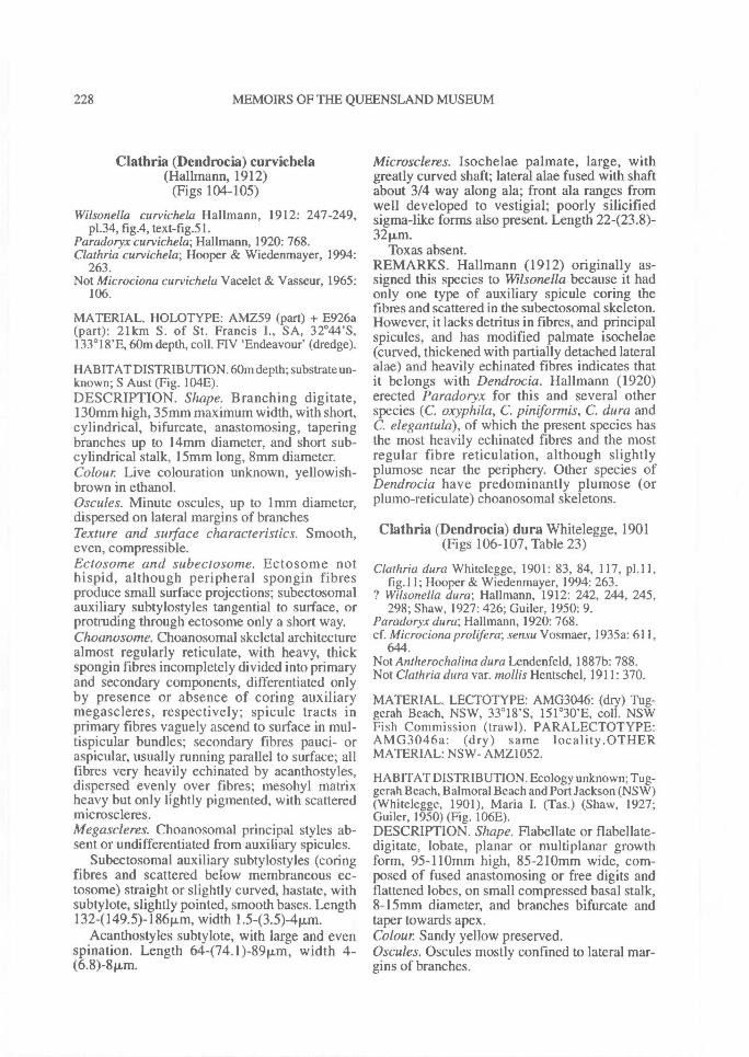
228^ MEMOIRS OF THE QUEENSLAND MUSEUM
Clathria (Dendrocia) curvichela(Hallmann, 1912)
(Figs 104-105)
Wilsonella curvichela Hallmann, 1912: 247-249,p1.34, fig.4, text-fig.51.
Paradoryx curvichela; Hallmann, 1920: 768.Clathria curvichela; Hooper & Wiedenmayer, 1994:
263.Not Microciona curvichela Vacelet & Vasseur, 1965:
106.
MATERIAL. HOLOTYPE: AMZ59 (part) + E926a(part): 21km S. of St. Francis I., SA, 32°44'S,133 ° 18'E, 60m depth, coll. FIV `Endeavour' (dredge).
HABITAT DISTRIBUTION. 60m depth; substrate un-known; S Aust (Fig. 104E).DESCRIPTION. Shape. Branching digitate,130mm high, 35mm maximum width, with short,cylindrical, bifurcate, anastomosing, taperingbranches up to 14mm diameter, and short sub-cylindrical stalk, 15mm long, 8mm diameter.Colour Live colouration unknown, yellowish-brown in ethanol.Oscules. Minute oscules, up to 1 mm diameter,dispersed on lateral margins of branchesTexture and surface characteristics. Smooth,even, compressible.Ectosome and subectosome. Ectosome nothispid, although peripheral spongin fibresproduce small surface projections; subectosomalauxiliary subtylostyles tangential to surface, orprotruding through ectosome only a short way.Choanosome. Choanosomal skeletal architecturealmost regularly reticulate, with heavy, thickspongin fibres incompletely divided into primaryand secondary components, differentiated onlyby presence or absence of coring auxiliarymegascleres, respectively; spicule tracts inprimary fibres vaguely ascend to surface in mul-tispicular bundles; secondary fibres pauci- oraspicular, usually running parallel to surface; allfibres very heavily echinated by acanthostyles,dispersed evenly over fibres; mesohyl matrixheavy but only lightly pigmented, with scatteredmicroscleres.Megascleres. Choanosomal principal styles ab-sent or undifferentiated from auxiliary spicules.
Subectosomal auxiliary subtylostyles (coringfibres and scattered below membraneous ec-tosome) straight or slightly curved, hastate, withsubtylote, slightly pointed, smooth bases. Length132-(149.5)-186p.,m, width 1.543.5)-411m.
Acanthostyles subtylote, with large and evenspination. Length 64474.1)-8911m, width 4-(6.8)-811m.
Microscleres. Isochelae palmate, large, withgreatly curved shaft; lateral alae fused with shaftabout 3/4 way along ala; front ala ranges fromwell developed to vestigial; poorly silicifiedsigma-like forms also present. Length 22423.8)-32p,m.
Toxas absent.REMARKS. Hallmann (1912) originally as-signed this species to Wilsonella because it hadonly one type of auxiliary spicule coring thefibres and scattered in the subectosomal skeleton.However, it lacks detritus in fibres, and principalspicules, and has modified palmate isochelae(curved, thickened with partially detached lateralalae) and heavily echinated fibres indicates thatit belongs with Derzdrocia. Hallmann (1920)erected Paradoryx for this and several otherspecies (C. oxyphila, C. piniformis, C. dura andC. elegantula), of which the present species hasthe most heavily echinated fibres and the mostregular fibre reticulation, although slightlyplumose near the periphery. Other species ofDendrocia have predominantly plumose (orplumo-reticulate) choanosomal skeletons.
Clathria (Dendrocia) dura Whitelegge, 1901(Figs 106-107, Table 23)
Clathria dura Whitelegge, 1901: 83, 84, 117, p1.11,fig. 11; Hooper & Wiedenmayer, 1994: 263.
? Wilsonella dura; Hallmann, 1912: 242, 244, 245,298; Shaw, 1927: 426; Guiler, 1950: 9.
Paradoryx dura; Hallmann, 1920: 768.cf. Microciona prolifera; sensu Vosmaer, 1935a: 611,
644.Not Antherochalina dura Lendenfeld, 1887b: 788.Not Clathria dura var. mo//is Hentschel, 1911: 370.
MATERIAL. LECTOTYPE: AMG3046: (dry) Tug-gerah Beach, NSW, 3318'S, 151 °30'E, coll. NSWFish Commission (trawl). PARALECTOTYPE:AMG3046a: (dry) same locality.OTHERMATERIAL: NSW- AMZ1052.
HABITAT DISTRIBUTION. Ecology unknown; Tug-gerah Beach, Balmoral Beach and Port Jackson (NSW)(Whitelegge, 1901), Maria I. (Tas.) (Shaw, 1927;Guiler, 1950) (Fig. 106E).DESCRIPTION. Shape. Flabellate or flabellate-digitate, lobate, planar or multiplanar growthform, 95-110mm high, 85-210mm wide, com-posed of fused anastomosing or free digits andflattened lobes, on small compressed basal stalk,8-15rrim diameter, and branches bifurcate andtaper towards apex.Colour Sandy yellow preserved.Oscules. Oscules mostly confined to lateral mar-gins of branches.
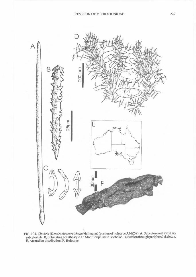
REVISION OF MICROCIONIDAE^ 229
FIG. 104. Claihria (Dendrocia) curvichela (Hallmann) (portion of holotype AMZ59). A, Subectosomal auxiliarysubtylostyle. B, Echinating acanthosty le. C, Modified palmate isochelae. D, Section through peripheral skeleton.E, Australian distribution. F, Holotype.
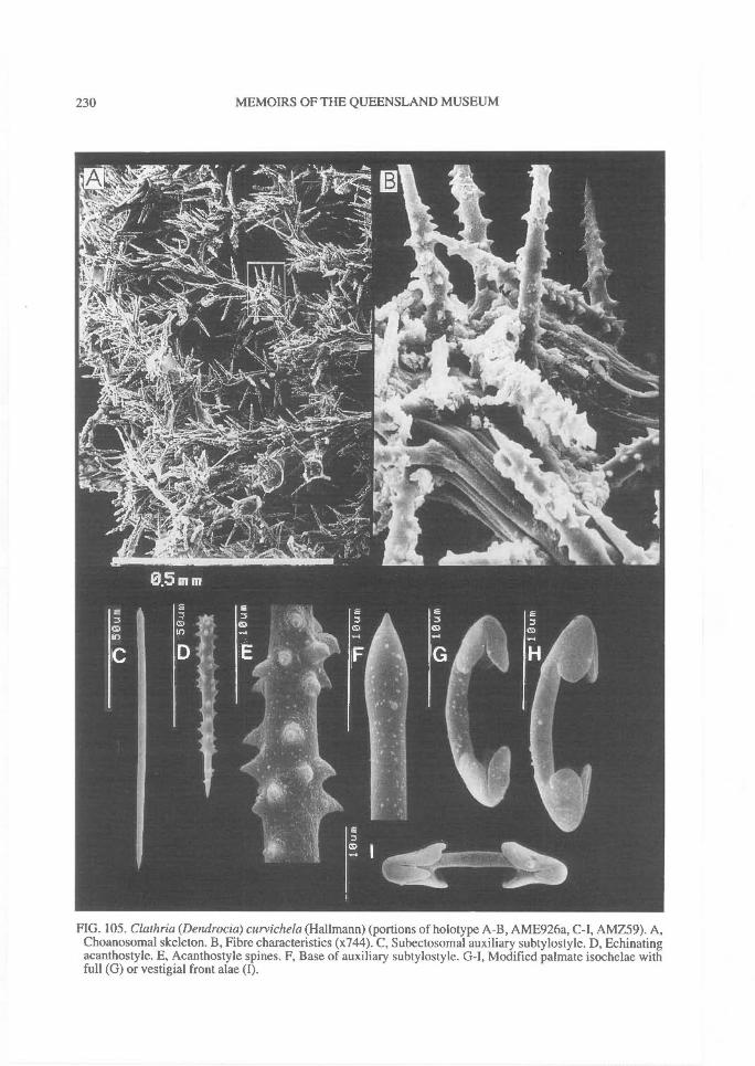
230^ MEMOIRS OF THE QUEENSLAND MUSEUM
FIG. 105. Clathria (Dendrocia) curvichela (Hallmann) (portions of holotype A-B, AME926a, AMZ59). A,Choanosomal skeleton. B, Fibre characteristics (x744). C, Subectosomal auxiliary subtylostyle. D, Echinatingacanthostyle. E, Acanthostyle spines. F, Base of auxiliary subtylostyle. G-I, Modified palmate isochelae withfull (G) or vestigial front alae (I).
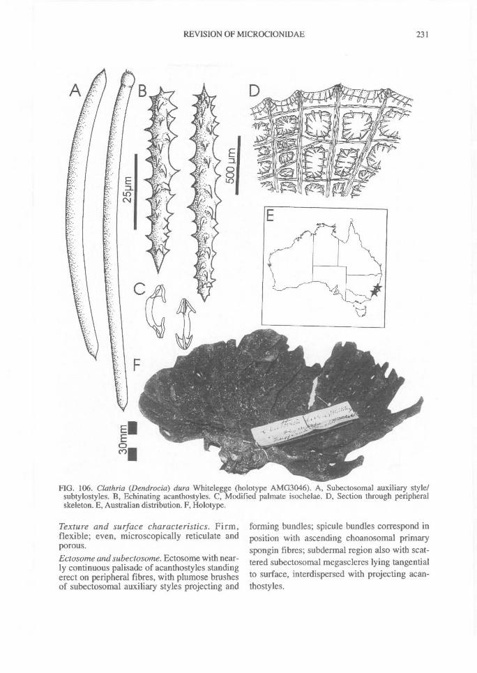
REVISION OF MICROCIONIDAE^ 231
FIG. 106. Clathria (Dendrocia) dura Whitelegge (holotype AMG3046). A, Subectosomal auxiliary style/subtylostyles. B, Echinating acanthostyles. C, Modified palmate isochelae. D, Section through peripheralskeleton. E, Australian distribution. F, Holotype.
Texture and surface characteristics. Firm,flexible; even, microscopically reticulate andporous.Ectosome and subectosome. Ectosome with near-ly continuous palisade of acanthostyles standingerect on peripheral fibres, with plumose brushesof subectosomal auxiliary styles projecting and
forming bundles; spicule bundles correspond inposition with ascending choanosomal primaryspongin fibres; subdermal region also with scat-tered subectosomal megascleres lying tangentialto surface, interdispersed with projecting acan-thostyles.
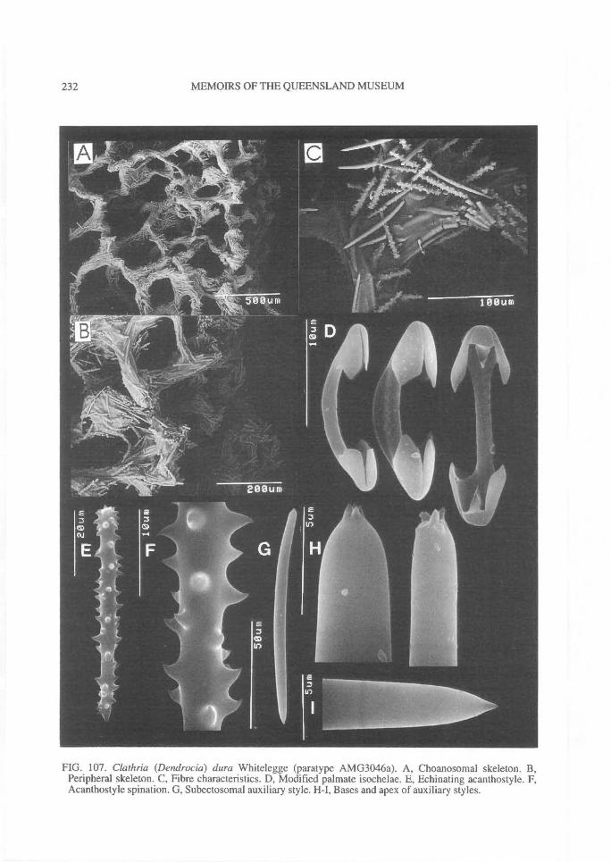
232^ MEMOIRS OF THE QUEENSLAND MUSEUM
FIG. 107. Clathria (Dendrocia) dura Whitelegge (paratype AMG3046a). A, Choanosomal skeleton. B,Peripheral skeleton. C, Fibre characteristics. D, Modified palmate isochelae. E, Echinating acanthostyle. F,Acanthostyle spination. G, Subectosomal auxiliary style. H-I, Bases and apex of auxiliary styles.
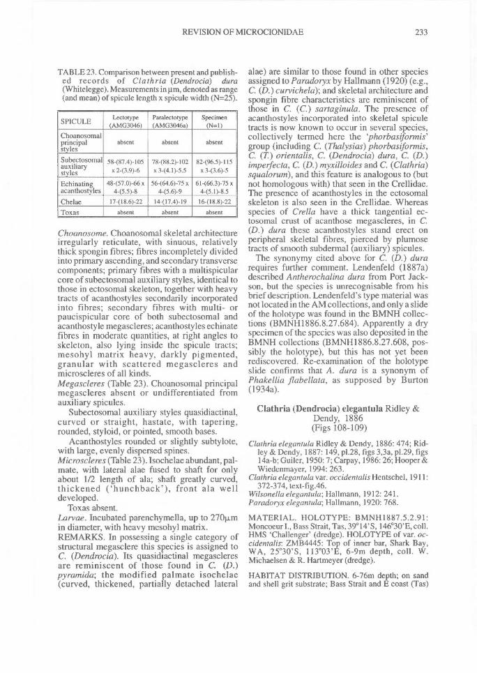
REVISION OF MICROCIONIDAE^ 233
TABLE 23. Comparison between present and publish-ed records of Clathria (Dendrocia) dura(Whitelegge). Measurements in pm, denoted as range(and mean) of spicule length x spicule width (N=25).
SPICULE Lectotype(AMG3046)
Paralectotype(AMG3046a)
Specimen(N=1)
Choanosomalprincipalstyles
absent absent absent
Subectosomalauxiliarystyles
58487.4)-105x 2 -(3.9) -6
78-(88.2)-102x 3 - (4.1) -5.5
82496.5)-115x 3 -(3.6) -5
Echinatingacanthostyles
48-(57.0)-66 x4-(5.5)-8
56-(64.6)-75 x4-(5.6)-9
61-(66.3)-75 x4 -(5.1) - 8.5
Chelae 17-(18.6)-22 14-(17.4)-19 16-(18.8)-22Toxas absent absent absent
Choanosome. Choanosomal skeletal architectureirregularly reticulate, with sinuous, relativelythick spongin fibres; fibres incompletely dividedinto primary ascending, and secondary transversecomponents; primary fibres with a multispicularcore of subectosomal auxiliary styles, identical tothose in ectosomal skeleton, together with heavytracts of acanthostyles secondarily incorporatedinto fibres; secondary fibres with multi- orpaucispicular core of both subectosomal andacanthostyle megascleres; acanthostyles echinatefibres in moderate quantities, at right angles toskeleton, also lying inside the spicule tracts;mesohyl matrix heavy, darkly pigmented,granular with scattered megascleres andmicroscleres of all kinds.Megascleres (Table 23). Choanosomal principalmegascleres absent or undifferentiated fromauxiliary spicules.
Subectosomal auxiliary styles quasidiactinal,curved or straight, hastate, with tapering,rounded, styloid, or pointed, smooth bases.
Acanthostyles rounded or slightly subtylote,with large, evenly dispersed spines.Microscleres (Table 23). Isochelae abundant, pal-mate, with lateral alae fused to shaft for onlyabout 1/2 length of ala; shaft greatly curved,thickened ('hunchback'), front ala welldeveloped.
Toxas absent.Larvae. Incubated parenchymella, up to 2701Lmin diameter, with heavy mesohyl matrix.REMARKS. In possessing a single category ofstructural megasclere this species is assigned toC. (Dendrocia). Its quasidiactinal megascleresare reminiscent of those found in C. (D.)pyramida; the modified palmate isochelae(curved, thickened, partially detached lateral
alae) are similar to those found in other speciesassigned to Paradoryx by Hallmann (1920) (e.g.,C. (D.) curvichela); and skeletal architecture andspongin fibre characteristics are reminiscent ofthose in C. (C.) sartaginula. The presence ofacanthostyles incorporated into skeletal spiculetracts is now known to occur in several species,collectively termed here the `phorbasiformis'group (including C. (Thalysias) phorbasiformis,C. (T) orientalis, C. (Dendrocia) dura, C. (D.)impelfecta, C. (D.) myxilloides and C. (Clathria)squalorum), and this feature is analogous to (butnot homologous with) that seen in the Crellidae.The presence of acanthostyles in the ectosomalskeleton is also seen in the Crellidae. Whereasspecies of Crella have a thick tangential ec-tosomal crust of acanthose megascleres, in C.(D.) dura these acanthostyles stand erect onperipheral skeletal fibres, pierced by plumosetracts of smooth subdermal (auxiliary) spicules.
The synonymy cited above for C. (D.) durarequires further comment. Lendenfeld (1887a)described Antherochalina dura from Port Jack-son, but the species is unrecognisable from hisbrief description. Lendenfeld's type material wasnot located in the AM collections, and only a slideof the holotype was found in the BMNH collec-tions (BMNH1886.8.27.684). Apparently a dryspecimen of the species was also deposited in theBMNH collections (BMNH1886.8.27.608, pos-sibly the holotype), but this has not yet beenrediscovered. Re-examination of the holotypeslide confirms that A. dura is a synonym ofPhakellia flabellata, as supposed by Burton(1934a).
Clathria (Dendrocia) elegantula Ridley &Dendy, 1886(Figs 108-109)
Clathria elegantula Ridley & Dendy, 1886: 474; Rid-ley & Dendy, 1887: 149, p1.28, figs 3,3a, p1.29, figs14a-b; Guiler, 1950: 7; Carpay, 1986: 26; Hooper &Wiedenmayer, 1994: 263.
Clathria elegantula var. occidentalis Hentschel, 1911:372-374, text-fig.46.
Wilsonella elegantula; Hallmann, 1912: 241.Paradoryx elegantula; Hallmann, 1920: 768.
MATERIAL. HOLOTYPE: BMNH1887.5.2.91:Moncoeur I., Bass Strait, Tas, 39° 14'S, 146°30'E, coll.HMS 'Challenger' (dredge). HOLOTYPE of var. oc-cidentalis: ZMB4445: Top of inner bar, Shark Bay,WA, 25°30'S, 113°03'E, 6-9m depth, coll. W.Michael sen & R. Hartmeyer (dredge).
HABITAT DISTRIBUTION. 6-76m depth; on sandand shell grit substrate; Bass Strait and E coast (Tas)
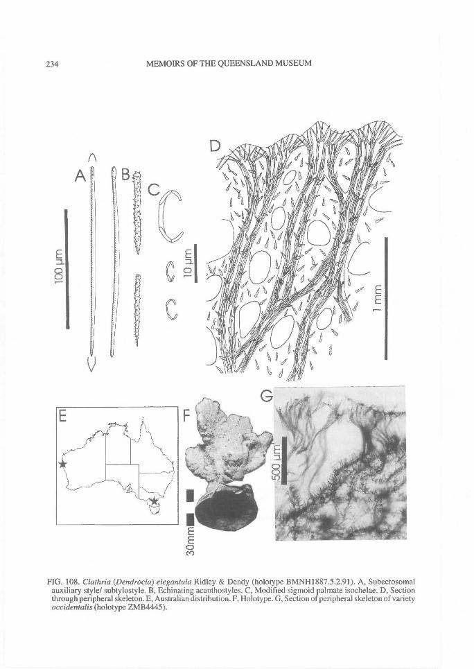
234^ MEMOIRS OF THE QUEENSLAND MUSEUM
FIG. 108. Clathria (Dendrocia) elegantula Ridley & Dendy (holotype BMNH1887.5.2.91). A, Subectosomalauxiliary style/ subtylostyle. B, Echinating acanthostyles. C, Modified sigmoid palmate isochelae. D, Sectionthrough peripheral skeleton. E, Australian distribution. F, Holotype. G, Section of peripheral skeleton of varietyoccidentalis (holotype ZMB4445).
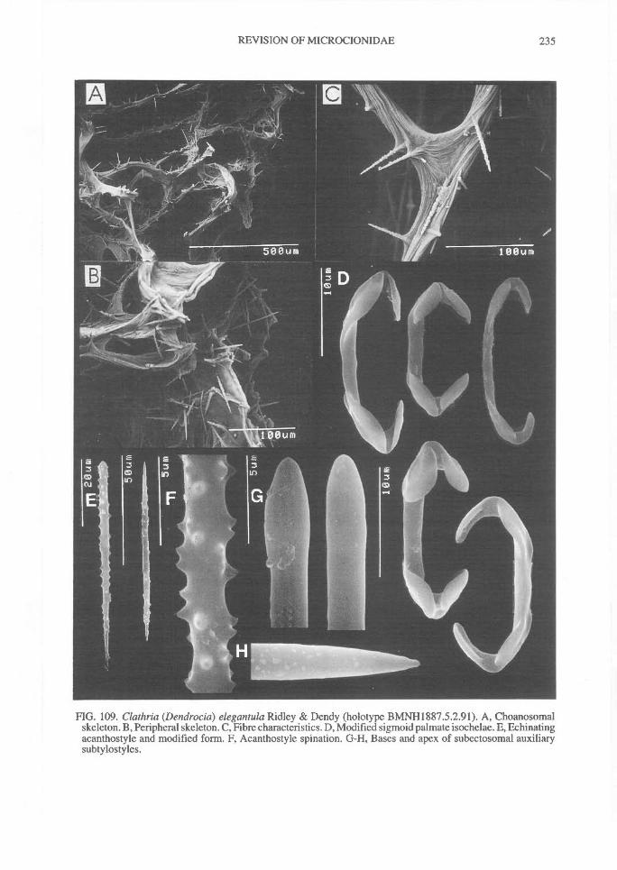
REVISION OF MICROCIONIDAE^ 235
FIG. 109. Clathria (Dendrocia) elegantula Ridley & Dendy (holotype BMNH1887.5.2.91). A, Choanosomalskeleton. B, Peripheral skeleton. C, Fibre characteristics. D, Modified sigmoid palmate isochelae. E, Echinatingacanthostyle and modified form. F, Acanthostyle spination. G-H, Bases and apex of subectosomal auxiliarysubtylostyles.
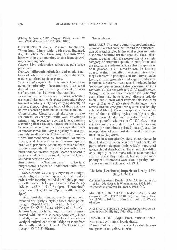
236^ MEMOIRS OF THE QUEENSLAND MUSEUM
(Ridley & Dendy, 1886; Carpay, 1986), central Wcoast (WA) (Hentschel, 1911) (Fig. 108E).
DESCRIPTION. Shape. Massive, lobate fan,73mm long, 75mm wide, with erect, flattened,digitate lobes, 22-31mm long, 8-20mm wide,lobes with uneven margins, arising from sprawl-ing encrusting base.Colour Live colouration unknown, pale beigepreserved.Oscules. Differentiated inhalant and exhalant sur-faces of lobes; ostia scattered, 1-2mm diameter,oscules confined to sieve-plates.Texture and surface characteristics. Harsh; un-even, prominently microconulose, translucentdermal membrane, covering reticulate fibroussurface, stretched between microconules.Ectosome and subectosome. Fibrous, reticulateectosomal skeleton, with sparse tangential subec-tosomal auxiliary subtylostyles lying directly onsurface; sinuous plumose tracts of these spiculesbelow, ascending from choanosomal skeleton.Choanosome. Choanosomal skeleton plumo-reticulate, cavernous, with well developedprimary and secondary spongin fibres; primaryascending fibres sinuous, almost dendritic, coredby several discrete multi- or paucispicular tractsof subectosomal auxiliary subtylostyles, occupy-ing only small portion of fibre diameter; primaryfibres interconnected by reticulate secondaryfibres, and terminating in plumose spiculebundles at periphery; secondary transverse fibrespauci- or aspicular, thin; echinating acanthostylesmost abundant in axial region, sparse or absent inperipheral skeleton; mesohyl matrix light, withabundant scattered chelae.Megascleres. Choanosomal principalmegascleres absent or undifferentiated fromauxiliary spicules.
Subectosomal auxiliary subtylostyles straight,rarely slightly curved, quasidiactinal, hastatepoints, with tapering, rounded or slightly pointed,smooth bases. Holotype: Length 145-(160.6)-168Rm, width 1.5 -(2.6)-4p,m. (Hentschel'sspecimen: 133-(142.3)-152Rm, width 243.5)-4p,m).
Acanthostyles slender, evenly spined, withrounded or slightly subtylote bases, sharp points.Length 53-(64.1)-74,m, width 2-(3.6)-5p,m.(Length 52468.2)-84pm, width 3-(6.4)-81.1.,m).Microscleres. Isochelae large, palmate, sigmoidcurved, with lateral alae nearly completely fusedto shaft, sometimes well developed, sometimesvestigial and reduced to small ridge on shaft; frontala usually reduced. Length 13415.4)-1711m.(Length 13417.2)-2011m).
Toxas absent.
REMARKS. The nearly dendri tic, predominantlyplumose skeletal architecture and the concentra-tion of acanthostyles in the axial region are quitedistinctive features for this species. These char-acters, together with the possession of a singlecategory of structural spicule in both fibres andthe ectosomal skeleton indicate that the species isbest placed in C. (Dendrocia). In havingquasidiactinal modified, vestigial structuralmegascleres with principal and auxiliary spiculeshaving similar geometry, and vague similaritiesin skeletal structure, this species is included in the`oxyphila' species group (also containing C. (C.)rap hana, C. (C.) oxyphila and C. (C.) piniformis).Spongin fibres are also characteristic (wherebyeach fibre may have several discrete spiculetracts), but in most other respects this species isvery similar to C. (D.) dura Whitelegge (bothhaving sinuous spongin fibre systems and heavilyechinated fibres). These two species differ in thesize of their auxiliary styles (notably straight,longer, more slender, with subtylote bases in C.(D.) elegantula, whereas in C. (D.) dura thesespicules are curved, short, thick, with taperinghastate (or sometimes pointed) bases)), and theincorporation of acanthostyles into skeletal fibretracts in C. (D.) dura.
There is a remarkably close concordance inthese features between Bass Strait and Shark Baypopulations, despite their widely separatedgeographical distribution. These samples differonly slightly in the more robust acanthostylesseen in Shark Bay material, but no other mor-phological differences were seen to justify sub-species separation (Hentschel, 1911).
Clathria (Dendrocia) imperfecta Dendy, 1896(Figs 110-111)
Clathria imperfecta Dendy, 1896: 35; Ayling et al.,1982: 103; Hooper & Wiedenmayer, 1994: 264.
Wilsonella imperfecta; Hallmann, 1912: 242.
MATERIAL. HOLOTYPE: NMVG2369 (RN376)(fragment BMNH1902.10.18.335): Port Phillip Bay,Vic, 38°09'S, 144°52'E, 36m depth, coll. J.B. Wilson(dredge).
HABITAT DISTRIBUTION. 36m depth; substrate un-known; Port Phillip Bay (Vic) (Fig. 110E).
DESCRIPTION. Shape. Erect, bulbous-lobate,with small cylindrical lobate digits.Colour Colour in life recorded as dull brown-orange exterior, yellow interior.
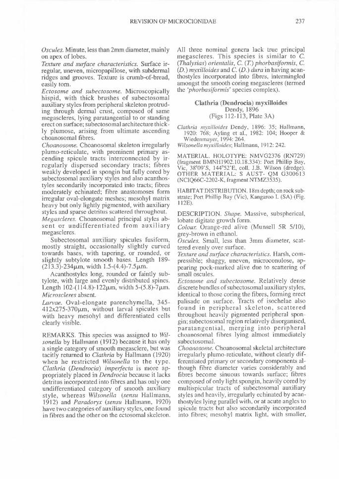
REVISION OF MICROCIONIDAE^ 237
Oscules. Minute, less than 2mm diameter, mainlyon apex of lobes.Texture and surface characteristics. Surface ir-regular, uneven, micropapillose, with subdermalridges and grooves. Texture is crumb-of-bread,easily torn.Ectosome and subectosome. Microscopicallyhispid, with thick brushes of subectosomalauxiliary styles from peripheral skeleton protrud-ing through dermal crust, composed of samemegascleres, lying paratangential to or standingerect on surface; subectosomal architecture thick-ly plumose, arising from ultimate ascendingchoanosomal fibres.Choanosome. Choanosomal skeleton irregularlyplumo-reticulate, with prominent primary as-cending spicule tracts interconnected by ir-regularly dispersed secondary tracts; fibresweakly developed in spongin but fully cored bysubectosomal auxiliary styles and also acanthos-tyles secondarily incorporated into tracts; fibresmoderately echinated; fibre anastomoses formirregular oval-elongate meshes; mesohyl matrixheavy but only lightly pigmented, with auxiliarystyles and sparse detritus scattered thrroughout.Megascleres. Choanosomal principal styles ab-sent or undifferentiated from auxiliarymegascleres.
Subectosomal auxiliary spicules fusiform,mostly straight, occasionally slightly curvedtowards bases, with tapering, or rounded, orslightly subtylote smooth bases. Length 189-(213.3)-234p.m, width 1.544.4)-7.5p.m.
Acanthostyles long, rounded or faintly sub-tylote, with large and evenly distributed spines.Length 102-(114.8)-122p,m, width 5-(5.8)-7p.m.Microscleres absent.Larvae. Oval-elongate parenchymella, 345-412x275-370p.m, without larval spicules butwith heavy mesohyl and differentiated cellsclearly visible.
REMARKS. This species was assigned to Wil-sonella by Hallmann (1912) because it has onlya single category of smooth megasclere, but wastacitly returned to Clathria by Hallmann (1920)when he restricted Wilsonella to the type.Clathria (Dendrocia) impeifecta is more ap-propriately placed in Dendrocia because it lacksdetritus incorporated into fibres and has only oneundifferentiated category of smooth auxiliarystyle, whereas Wilsonella (sensu Hallmann,1912) and Paradoryx (sensu Hallmann, 1920)have two categories of auxiliary styles, one foundin fibres and the other on the ectosomal skeleton.
All three nominal genera lack true principalmegascleres. This species is similar to C.(Thalysias) orientalis, C. (T) phorbasifonnis, C.(D.) myxilloides and C. (D.) dura in having acan-thostyles incorporated into fibres, intermingledamongst the smooth coring megascleres (termedthe `phorbasifonnis' species complex).
Clathria (Dendrocia) myxilloidesDendy, 1896
(Figs 112-113, Plate 3A)
Clathria myxilloides Dendy, 1896: 35; Hallmann,1920: 768; Ayling et al., 1982: 104; Hooper &Wiedenmayer, 1994: 264.
Wilsonella myxdloides; Hallmann, 1912: 242.
MATERIAL. HOLOTYPE: NMVG2376 (RN729)(fragment BMNH1902.10.18.334): Port Phillip Bay,Vic, 38°09'S, 144°52'E, coll. J.B. Wilson (dredge).OTHER MATERIAL: S AUST- QM G300613(NCIQ66C-2202-K, fragment NTMZ3535).
HABITAT DISTRIBUTION. 18m depth; on rock sub-strate; Port Phillip Bay (Vic), Kangaroo I. (SA) (Fig.112E).
DESCRIPTION. Shape. Massive, subspherical,lobate digitate growth form.Colour. Orange-red alive (Munsell 5R 5/10),grey-brown in ethanol.Oscules. Small, less than 3mm diameter, scat-tered evenly over surface.Texture and surface characteristics. Harsh, com-pressible; shaggy, uneven, microconulose, ap-pearing pock-marked alive due to scattering ofsmall oscules.Ectosome and subectosome. Relatively densediscrete bundles of subectosomal auxiliary styles,identical to those coring the fibres, forming erectpalisade on surface. Tracts of isochelae alsofound in peripheral skeleton, scatteredthroughout heavily pigmented peripheral spon-gin; subectosomal region relatively disorganised,paratangential, merging into peripheralchoanosomal fibres lying almost immediatelysubectosomal.Choanosome. Choanosomal skeletal architectureirregularly plumo-reticulate, without clearly dif-ferentiated primary or secondary components al-though fibre diameter varies considerably andfibres become sinuous towards surface; fibrescomposed of only light spongin, heavily cored bymultispicular tracts of subectosomal auxiliarystyles and heavily, irregularly echinated by acan-thostyles lying parallel with, or at acute angles tospicule tracts but also secondarily incorporatedinto fibres; mesohyl matrix light, with smaller,
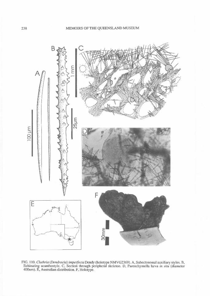
238^ MEMOIRS OF THE QUEENSLAND MUSEUM
FIG. 110. Clathria (Dendrocia) impelfecta Dendy (holotype NMVG2369). A, Subectosomal auxiliary styles. B,Echinating acanthostyle. C, Section through peripheral skeleton. D, Parenchymella larva in situ (diameter400um). E, Australian distribution. F, Holotype.
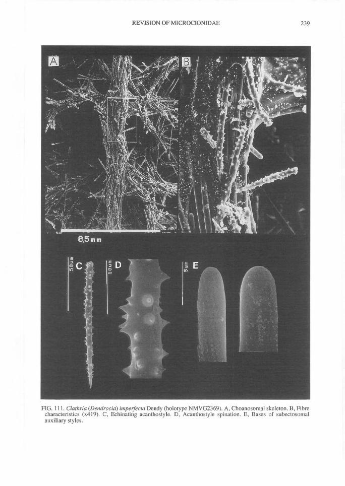
REVISION OF MICROCIONIDAE^
239
FIG. 111. Clathria (Dendrocia) impetfecta Dendy (holotype NMVG2369). A, Choanosomal skeleton. B, Fibrecharacteristics (x419). C, Echinating acanthostyle. D, Acanthostyle spination. E, Bases of subectosomalauxiliary styles.
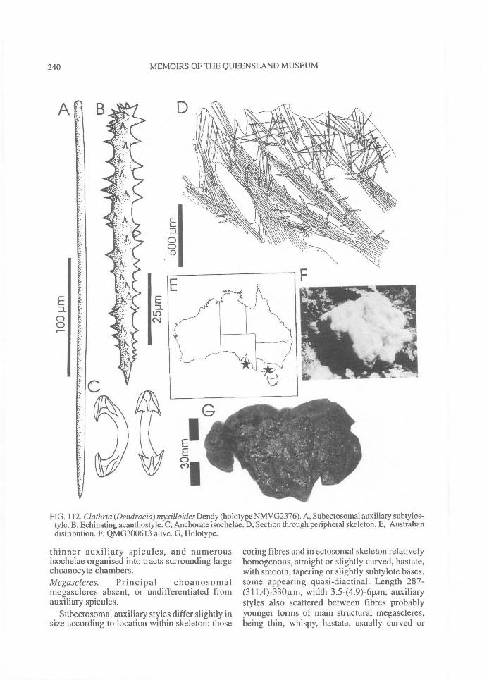
240^ MEMOIRS OF THE QUEENSLAND MUSEUM
FIG. 112. Clathria (Dendrocia) myxilloides Dendy (holotype NMVG2376). A, Subectosomal auxiliary subtylos-tyle. B, Echinating acanthostyle. C, Anchorate isochelae. D, Section through peripheral skeleton. E, Australiandistribution. F, QMG300613 alive. G, Holotype.
thinner auxiliary spicules, and numerousisochelae organised into tracts surrounding largechoanocyte chambers.Megascleres.^Principal^choanosomalmegascleres absent, or undifferentiated fromauxiliary spicules.
Subectosomal auxiliary styles differ slightly insize according to location within skeleton: those
coring fibres and in ectosomal skeleton relativelyhomogenous, straight or slightly curved, hastate,with smooth, tapering or slightly subtylote bases,some appearing quasi-diactinal. Length 287-(311.4)-330p,m, width 3.5-(4.9)-6Rm; auxiliarystyles also scattered between fibres probablyyounger forms of main structural megascleres,being thin, whispy, hastate, usually curved or
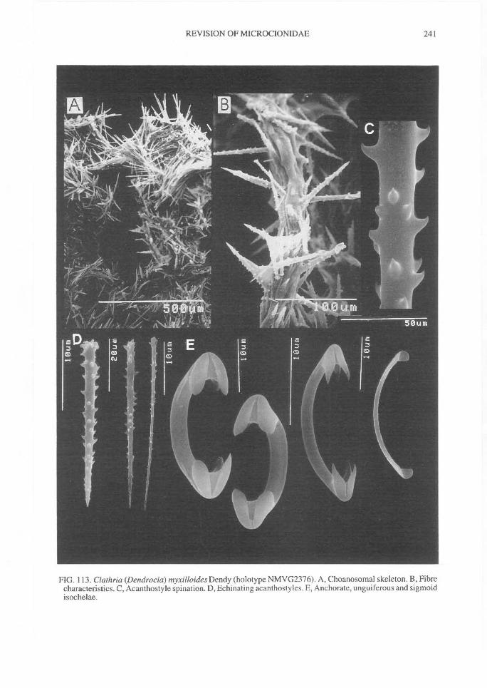
REVISION OF MICROCIONIDAE^
241
FIG. 113. Clathria (Dendrocia) myxilloides Dendy (holotype NMVG2376). A, Choanosomal skeleton. B, Fibrecharacteristics. C, Acanthostyle spination. D, Echinating acanthostyles. E, Anchorate, unguiferous and sigmoidisochelae.
242^ MEMOIRS OF THE QUEENSLAND MUSEUM
sinuous, with rounded or tapering bases. Length212-(233.0)-284Rm, width 2-(2.2)-31.1.m.
Acanthostyles long, slender, straight or slightlycurved near basal end, evenly and heavily spined.Length 84-(112.6)-133t.tm, width 6-(7.4)-10iLm.Microscieres. Isochelae relatively large, abun-dant, anchorate, with well formed lateral alaedetached from shaft nearly completely, lateralridge on shaft (possibly vestigial point of attach-ment of lateral alae); some modified unguiferousforms with pointed alae, or sigmoid forms withgreatly reduced alae also present. Length 21-(24.4)-29Rm.
Toxas absent.
REMARKS. This species is similar to C. (D.)impetfecta in growth form and incorporation ofsome acanthostyles into fibres (see remarks for C.(Thalysias) phorbastfonnis). It also showssimilarities to C. (D.) pyratnida in growth formand megasclere morphology, although all threespecies differ in isochelae geometry.
Clathria (Dendrocia) pyramida Lendenfeld,1888
(Figs 114-115, Table 24, Plate 3B)
Clathria pyramida Lendenfeld, 1888: 222; Capon &MacLeod, 1987: 1200; Chernoff, 1987: 160;Hooper et al., 1992: 263; Hooper & Wiedenmayer,1994: 264.
Wilsonella pyramida; Hallmann, 1912: 240, 244;Shaw, 1927: 426; Guiler, 1950: 9.
Dendrocia pyramida; Hallmann, 1920: 767.Clathriaalata Dendy, 1896: 34; Hentschel, 1911: 375-
377, text-fig.48; Hentschel, 1923: 387, fig.356;Ayling et al., 1982: 100; Van Soest, 1984b: 120.
Wilsonella alata; Hallmann, 1912: 241.Dendrocia alata; Hallmann, 1920: 767.
MATERIAL. LECTOTYPE: AMG9047: Port Jack-son, NSW, 33°51'E, 151 ° 16'E, no other details known.PARALECTOTYPE: BMNH1887.1. 24.62 (fragmentAMG3579): same locality. LECTOTYPE of C. alata:NMVG2280 (fragment BMNH1902.10.18.330): PortPhillip Bay, Vic, 38°09'S, 144°52'E, coll. J.B. Wilson(dredge, RN 752). PARALECTOTYPES of C. alata:NMVG2281 (fragment AMG2686): same locality (RN763). NMVG2282 (fragment BMNH1902.10.18.31):Sorrento Jetty, Port Phillip Bay, 38°33'S, 145 °21'E(dredge, RN 792). NMVG2283 (fragmentBMNH1902.10.18.49): same locality (RN 801).NMVG2284 (fragment BMNH1902.10.18.332): samelocality (RN 842). NMVG2285 (fragmentBMNH1902.10.18.333): same locality (RN 843).OTHER MATERIAL: VIC- AME298, AMZ1145,AM unreg. (small vial, label 'donated by A Dendy'),AM unreg. (fragment BMNH1887.4.27.103) (smallvial, label 'Clathria tethyopsis, donated by A Dendy,
Lendenfeld or Burton MS name'). NSW- AMZ3216,NTMZ2667, QMG304507, QMG304522,QMG304574. S AUST- SAMTS4095 (fragmentNTMZ1629), SAMTS 6290, QMG3005 03(NCIQ66C-2119-T) (fragment NTMZ3520),QMG301354, QMG301358, QMG301374,QMG301382, QMG304041, QMG304049,QMG304051, QMG304053, QMG304054,QMG304063, QMG304064, QMG304069,QMG304070. WA- QM G300604 (NCIQ66C-4271-J), QM G300191 (NCIQ66C-4651-V). (Not AMunreg. (label 'Clathria alba. Port Jackson, NSW', =Lendenfeld or Burton MS name; = Crella incrustans)).
HABITAT DISTRIBUTION. 0.8-25m depth; on sand,rock reef or muddy bottoms; associated with oystersand algal beds, Spondylus, Pinna and Chalamus asper-rimus bivalves, rock or wood jetty substrates; Illawar-ra, Port Jackson, N. Sydney, Port Hacking and DurrasI. (NSW); Maria I. (Tas); Port Phillip Bay (Vic); St.Vincent Gulf, Yorke Peninsula and Kangaroo I. (SA);Albany and Pelsart Is, Houtman Abrolhos (WA) (Fig.114E).
DESCRIPTION. Shape. Massive, lobate, lobo-digitate, irregular growth form with small lobatesurface projections.Colour. Dark brown or orange-brown live ex-terior (Munsell 5YR 4/6-5/10), palerchoanosome, brown in ethanol.Oscules. Few, relatively large oscules (up to 8mmdiameter) on apex of lobes, each with slightlyraised membraneous lip, and with membraneous,divided exhalant canals visible inside oscule; os-cules collapse on preservation.Texture and surface characteristics. Firm, com-pressible; smooth, relatively even, bulbous,fleshy surface in live state; optically smooth,membraneous surface in preserved state, withirregularly rugose microscopic conulose andlobate projections.Ectosome and subectosome. Microscopicallyhispid, with dense crust of subectosomalauxiliary styles forming continuous, erect,regular (straight) or irregular (stellate, paratan-gential), plumose palisade.Choanosome. Skeletal architecture irregularlyplumo-reticulate, with sinuous spongin fibres as-cending to surface in meandering tracts; fibresanastomose more frequently at axis than inperipheral skeleton; peripheral fibres and skeletaltracts often diverge becoming plumose in subec-tosomal region, or forming a paratangential layerimmediately below erect ectosomal skeleton;fibres contain moderately light to very heavyspongin, cored by multispicular, sinuous tracts ofsubectosomal auxiliary styles; fibres not obvious-ly divided into primary or secondary components,
REVISION OF MICROCIONIDAE^ 243
although fibre diameter varies within same sec-tions of skeleton; acanthostyles heavily echinat-ing most fibres, occasionally rare or absent fromsome; mesohyl matrix very heavy but only lightlypigmented, with abundant isochelae andauxiliary megascleres; microalgae present inmesohyl of some specimens.Megascleres (Table 24). Choanosomal principalmegascleres absent, or at least undifferentiatedfrom auxiliary spicules.
Subectosomal auxiliary styles are thin, hastate,straight or slightly curved towards basal end,usually with smooth, slightly subtylote bases thattaper towards end into a small point (partiallymucronate), points hastate.
Acanthostyles vary considerably in length,usually club-shaped, subtylote, with large spinesmostly confined on base and more-or-lessaspinose towards point; spines usually robust.Microscleres (Table 24). Isochelae large, veryabundant, primarily palmate, heavily silicified,usually with thickened and slightly curved shaft,large lateral alae completely fused to shaft resem-bling swings', with front ala free, but sometimeswith reduced alae and sigmoid curvature.
Toxas absent.Larvae. Larvae oval-elongate parenchymella, upto 518x34011m, with larval raphidiform spiculesscattered throughout the axis and forming stellatetufts at the posterior pole.
REMARKS. Type material of Clathria pyramidaand of C. alata are virtually identical in theirgrowth form, surface features, spicule geometry,spicule distribution and fibre characteristics, dif-fering only slightly in choanosomal skeletal con-struction (predominantly plumo-reticulate versuspredominantly plumose to slightly plumo-reticu-late), and ectosomal structure (either producing acontinuous erect palisade (C. alata s.s.) orpunctuated palisade resulting in stellate plumosebrushes (C. pyramida s.s.), respectively). Butthere are no consistent or significant differencesbetween these species in the spongin content offibres, density of spicule tracts, presence or ab-sence of conecting secondary fibres, or spiculedimensions (Table 24), as supposed by Hallmann(1912: 241). On this basis the two species aremerged here. For all other specimens examinedthe main variability involves the degree to whichfibres were compacted (mesh size), the spongincontent of skeletal tracts ('fibre' diameter), andspicule dimensions between different geographicpopulations (Table 24). In this latter regardpopulations from NSW and WA had relatively
larger, more robust acanthostyles than otherpopulations (VIC, SA). In particular, spiculesfrom a small population in the Gulf of St Vincent,SA, were noticeably more poorly silicified andless robust than 'typical' specimens found in allother localities.
In spiculation and skeletal architecture C. (D.)pyramida is remarkably similar to Crella incrus-tans var. digitata ('cotype' AME503), althoughupon careful examination of both species thereare obvious differences in skeletal structure(Crella with a tangential ectosomal layer), com-position of the ectosomal skeleton (styles versusacanthostyles or acanthoxeas), and spiculegeometry (quasi-diactinal auxiliary styles versusanisoxeas or quasi-monactinal megascleres,respectively). Nevertheless, these speciesdemonstrate remarkably close convergences inseveral prominent features making them easilyconfused in the field and laboratory.
Specimens from the south coast of NSW havepeculiar biochemistry and significant biologicalactivity against both gram positive and gramnegative bacteria (Hooper et al., 1992). Unlikemany other bioactive sponges, however, the ac-tivity in C. (D.) pyramida was found to be relatedto a unique, modified free sugar, 5-Thio-D-man-nose (Capon & MacLeod, 1987). This discoveryrepresents the only known occurrence to date ofthat class of thiosugar in nature, although thecompound has a glucose-based synthesisedanalogue which has been known for severalyears. Thio sugars have considerable phar-maceutical and medical potential: they inhibit therelease of insulin and transport of glucose, and areable to cause reversible inhibition of sperm-celldevelopment without displaying acute toxicity(R. Capon, pers. comm.). The taxonomic sig-nificance of those compounds is not clear, but itis possible to speculate on the biological role ofthose chemicals. Recent evidence (A. Butler,pers.comm.) suggests that C. (D.) pyramidatogether with a few other species (e.g., Crellaincrustans) occur in association with (areepizootic on) scallops (Chlamys asperrima) fromSouth Australian waters. Chemoff (1987) foundthat the presence of those epizootics increased thesurvival of the scallop in both field and cagingexperiments and it is possible that the spongesprovide some sort of chemical defence of thehost: thio-mannose sugar may play some sort ofrole in that defence.
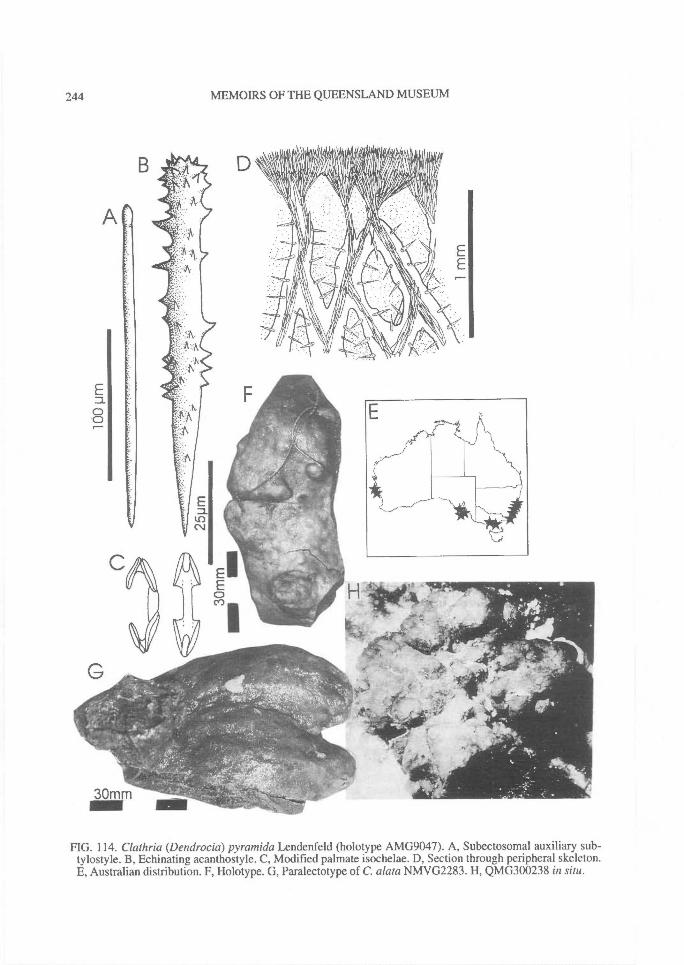
244^ MEMOIRS OF THE QUEENSLAND MUSEUM
FIG. 114. Clathria (Dendrocia) pyramida Lendenfeld (holotype AMG9047). A, Subectosomal auxiliary sub-tylostyle. B, Echinating acanthostyle. C, Modified palmate isochelae. D, Section through peripheral skeleton.E, Australian distribution. F, Holotype. G, Paralectotype of C. alata NMVG2283. H, QMG300238 in situ.
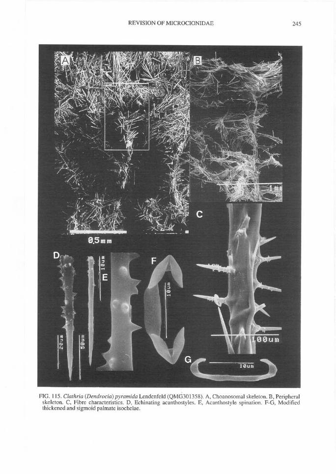
REVISION OF MICROCIONIDAE^ 245
FIG. 115. Clathria (Dendrocia) pyramida Lendenfeld (QMG301358). A, Choanosomal skeleton. B, Peripheralskeleton. C, Fibre characteristics. D, Echinating acanthostyles. E, Acanthostyle spination. F-G, Modifiedthickened and sigmoid palmate isochelae.
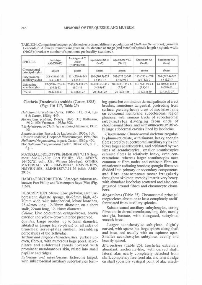
246^ MEMOIRS OF THE QUEENSLAND MUSEUM
TABLE 24. Comparison between published records and different populations of Clathria (Dendrocia)pyramidaLendenfeld. All measurements are given in pm, denoted as range (and mean) of spicule length x spicule width(N=25) (bracket = number of specimens per locality examined).
-
SPICULE
-^-Lectotype
(AMG9047)
Lectotype of C.Alata
(NMVG2280)
Specimens NSW(N=7)
Specimens VIC(N=10)
Specimens SA(N=16)
Specimens WA(N=2)
Choanosomalprincipal styles absent absent absent absent absent absent
Subectosomal 208-(220.6)-231 211-(229.4)-243 190-(209.5)-225 2024222.6)-247 195-(213.4)-228 214(227.4)-242auxiliary styles x 4-(6.4)-8 x 3-(4.8)-7 x 4-(5.1)-7 x 4-(5.9)-9 x 4-(4.9)-7 x 4-(5.3)-7
Echinating 87-(99.0)-135 x 8- 75-(85.2)- 118 x 7- 82-(103.9)-145 x 68-(89.6)-I25 x 4- 64-(78.6)-90x 4- 82-(103.5)-155 xacanthostyles (10.2)-12 (9.2)-11 5-(8.4)-12 (7.2)-12 (7.4)-11 6-(9.0)-11
Chelae 21-(23.8)-27 23-(24.5)-27 20423.6)-27 20-(22.5)-25 17-(22.1)-30 22-(24.3)-27
Clathria (Dendrocia) scabida (Carter, 1885)(Figs 116-117, Table 25)
Halichondria scabida Carter, 1885b: 112, p1.4, figs4-5; Carter, 1886g: 449.
Microciona scabida; Dendy, 1896: 31; Hal'mann,1912: 150; Vosmaer, 1935a: 608.
? Stylotellopsis or Clathrissa scabida; Hallmann, 1912:151.
Anaata scabita Papsusl; de Laubenfels, 1936a: 109.Clathria scabida; Hooper & Wiedenmayer, 1994: 264Halichondria pustulosa, in part; Carter, 1886g: 450.Not Halichondria pustulosa Carter, 1882a: 285, p1.11,
fig. 1.
MATERIAL. HOLOTYPE: BMNH1887.7.11.9 (frag-ment AMG2760): Port Phillip, Vic, 38°09'S,144°52'E, coll. J.B. Wilson (dredge). OTHERMATERIAL: VIC - NMVRN413, NMVRN1025,NMVRN1038, BMNH1887.7.11.26 (slide AMG-2916).
HABITAT DISTRIBUTION. 38m depth; substrate un-known; Port Phillip and Westernport Bays (Vic) (Fig.116F).
DESCRIPTION. Shape. Low, globular, erect, ar-borescent, digitate sponge, 80-95mm high, 45-70mm wide, with subspherical, lobate branches,28-42mm long, 12-28mm diameter, on a shortstalk, 22mm long, 12-15mm diameter.Colour Live colouration orange-brown, brownexterior and yellow-brown interior preserved.Oscules. Large oscules, up to 5mm diameter,situated in groups (seive-plates) on all sides ofbranches; seive-plates sunken, resemblingporocalyses of the Tethyidae.Texture and surface characteristics. Surface un-even, fibrous, with numerous large pores, seive-plates and subdermal canals covered withprominent membraneous skin, raised into smallpapillae and ridges.Ectosome and subectosome. Ectosome hispid,with subectosomal auxiliary subtylostyles form-
ing sparse but continuous dermal palisade of erectbrushes, sometimes tangential, protruding fromsurface, piercing heavy crust of isochelae lyingon ectosomal membrane; subectosomal regionplumose, with sinuous tracts of subectosomalsubtylostyles diverging from ends ofchoanosomal fibres, and with numerous, relative-ly large subdermal cavities lined by isochelae.
Choanosome. Choanosomal skeleton irregular-ly plumo-reticulate, with sinuous, heavy, sponginfibres cored by subectosomal auxiliary styles andfewer larger acanthostyles, and echinated by twosizes of acanthostyles; smaller acanthostylesechinate fibres in relatively heavy, even con-centrations, whereas larger acanthostyles mostcommon at fibre nodes and echinate fibre ter-minations in radiating brushes; spongin fibres notdivided into primary or secondary components,and fibre anastomoses occur irregularlythroughout skeleton; mesohyl matrix very heavy,with abundant isochelae scattered and also con-gregated around fibres and choanocyte cham-bers.Megascleres (Table 25). Choanosomal principalmegascleres absent or at least completely undif-ferentiated from auxiliary spicules.
Subectosomal auxiliary subtylostyles, coringfibres and in dermal membrane, long, thin, mostlystraight, hastate, with elongated, subtylote,smooth bases.
Larger acanthostyles subtylote, slightlycurved, with sparse but large spines along shaftand base, and usually with an aspinose apex.Smaller acanthostyles subtylote, evenly andheavily spined.Microscleres (Table 25). Isochelae extremelyabundant, anchorate-like, with curved shaft,lateral alae nearly completely detached fromshaft, completely free front ala, and lateral ridgeon shaft (possibly vestigial point of alae attach-
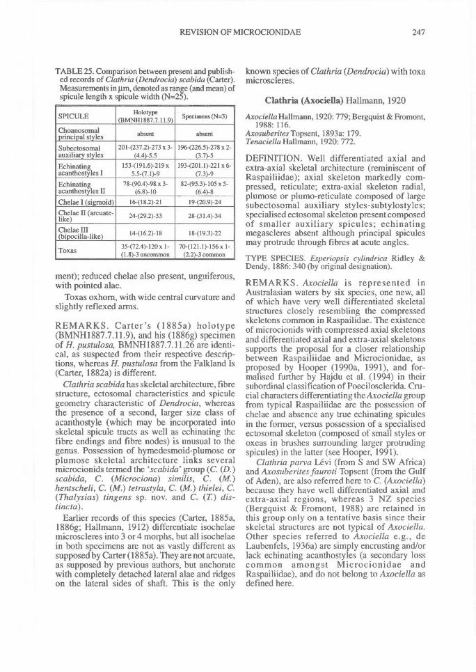
REVISION OF MICROCIONIDAE^ 247
TABLE 25. Comparison between present and publish-ed records of Clathria (Dendrocia) scabida (Carter).Measurements in i.tm, denoted as range (and mean) ofspicule length x spicule width (N=25).
SPICULE Holotype(BMNH1887.7.11.9)
Specimens (N.3)
Choanosomalprincipal styles absent absent
Subectosomalauxiliary styles
201-(237.2)-273 x 3-(4.4)-5.5
1964226.5)-278 x 2-(3.7)-5
Echinatingacanthostyles I
153-(191.6)-219 x5.5-(7.1)-9
193-(201.1)-221 x6-(7.3)-9
Echinatingacanthostyles II
78490.4)-98 x 3-(6.8)-10
82-(95.3)-105 x 5-(6.4)-8
Chelae I (sigmoid) 16-(18.2)-21 19-(20.9)-24Chelae II (arcuate-like) 24-(29.2) - 33 28-(31.4)-34
Chelae III(bipocilla-like) 14416.2)-18 18-(19.3)-22
Toxas 35-(72.4)-120x 1-(1.8) -3 uncommon
70-(121.1)-156x 1-(2.2)-3 common
ment); reduced chelae also present, unguiferous,with pointed alae.
Toxas oxhorn, with wide central curvature andslightly reflexed arms.
REMARKS. Carter's (1885a) holotype(BMNH1887.7.11.9), and his (1886g) specimenof H. pustulosa, BMNH1887.7.11.26 are identi-cal, as suspected from their respective descrip-tions, whereas H. pustulosa from the Falkland Is(Carter, 1882a) is different.
Clathria scabida has skeletal architecture, fibrestructure, ectosomal characteristics and spiculegeometry characteristic of Dendrocia, whereasthe presence of a second, larger size class ofacanthostyle (which may be incorporated intoskeletal spicule tracts as well as echinating thefibre endings and fibre nodes) is unusual to thegenus. Possession of hymedesmoid-plumose orplumose skeletal architecture links severalmicrocionids termed the scabida' group (C. (D.)scabida, C. (Microciona) si,nilis, C. (M.)hentscheli, C. (M.) tetrastyla, C. (M.) thielei, C.(Thalysias) tingens sp. nov. and C. (T) dis-tincta).
Earlier records of this species (Carter, 1885a,1886g; Hallmann, 1912) differentiate isochelaemicroscleres into 3 or 4 morphs, but all isochelaein both specimens are not as vastly different assupposed by Carter (1885a). They are not arcuate,as supposed by previous authors, but anchoratewith completely detached lateral alae and ridgeson the lateral sides of shaft. This is the only
known species of Clathria (Dendrocia) with toxamicroscleres.
Clathria (Axociella) Hallmann, 1920
Axociella Hallmann, 1920: 779; Bergquist & Fromont,1988: 116.
Axosuberites Topsent, 1893a: 179.Tenaciella Hallmann, 1920: 772.
DEFINITION. Well differentiated axial andextra-axial skeletal architecture (reminiscent ofRaspailiidae); axial skeleton markedly com-pressed, reticulate; extra-axial skeleton radial,plumose or plumo-reticulate composed of largesubectosomal auxiliary styles-subtylostyles;specialised ectosomal skeleton present composedof smaller auxiliary spicules; echinatingmegascleres absent although principal spiculesmay protrude through fibres at acute angles.
TYPE SPECIES. Esperiopsis cylindrica Ridley &Dendy, 1886: 340 (by original designation).
REMARKS. Axociella is represented inAustralasian waters by six species, one new, allof which have very well differentiated skeletalstructures closely resembling the compressedskeletons common in Raspailidae. The existenceof microcionids with compressed axial skeletonsand differentiated axial and extra-axial skeletonssupports the proposal for a closer relationshipbetween Raspailiidae and Microcionidae, asproposed by Hooper (1990a, 1991), and for-malised further by Hajdu et al. (1994) in theirsubordinal classification of Poecilosclerida. Cru-cial characters differentiating the Axociella groupfrom typical Raspailiidae are the possession ofchelae and absence any true echinating spiculesin the former, versus possession of a specialisedectosomal skeleton (composed of small styles oroxeas in brushes surrounding larger protrudingspicules) in the latter (see Hooper, 1991).
Clathria parva Levi (from S and SW Africa)and Axosuberites fauroti Topsent (from the Gulfof Aden), are also referred here to C. (Axociella)because they have well differentiated axial andextra-axial regions, whereas 3 NZ species(Bergquist & Fromont, 1988) are retained inthis group only on a tentative basis since theirskeletal structures are not typical of Axociella.Other species referred to Axociella e.g., deLaubenfels, 1936a) are simply encrusting and/orlack echinating acanthostyles (a secondary losscommon amongst Microcionidae andRaspailiidae), and do not belong to Axociella asdefined here.
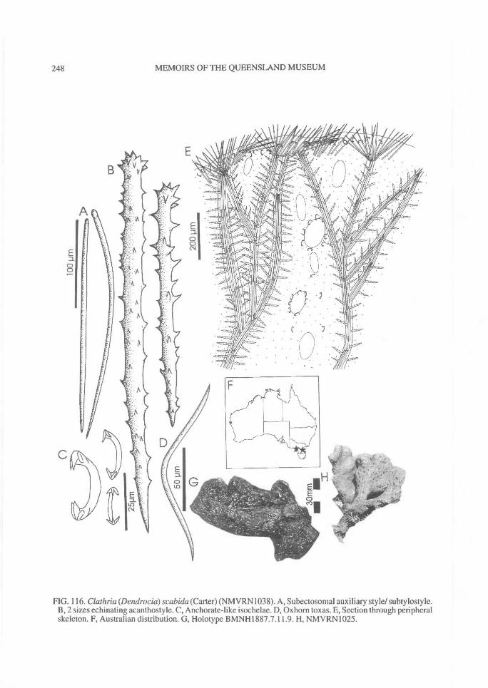
248^ MEMOIRS OF THE QUEENSLAND MUSEUM
FIG. 116. Clathria (Dendrocia) scabida (Carter) (NMVRN1038). A, Subectosomal auxiliary style/ subtylostyle.B, 2 sizes echinating acanthostyle. C, Anchorate-like isochelae. D, Oxhom toxas. E, Section through peripheralskeleton. F, Australian distribution. G, Holotype BMNH1887.7.11.9. H, NMVRN1025.
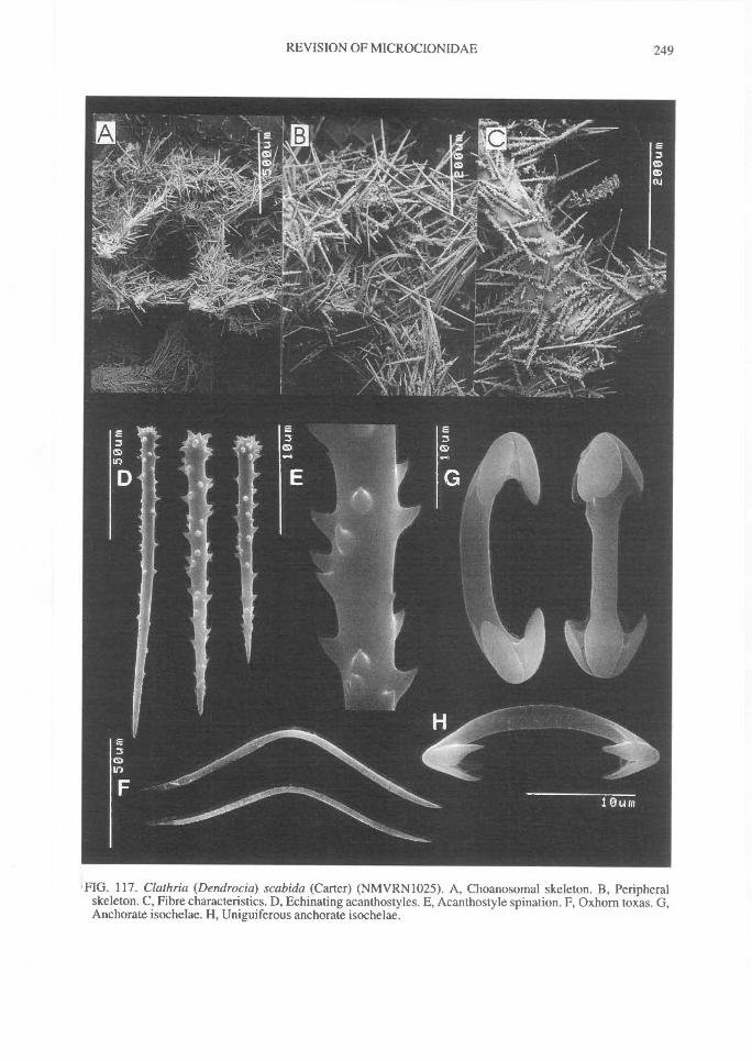
REVISION OF MICROCIONIDAE^ 249
'FIG. 117. Clathria (Dendrocia) scabida (Carter) (NMVRN1025). A, Choanosomal skeleton. B, Peripheralskeleton. C, Fibre characteristics. D, Echinating acanthostyles. E, Acanthostyle spination. F, Oxhorn toxas. G,Anchorate isochelae. H, Uniguiferous anchorate isochelae.
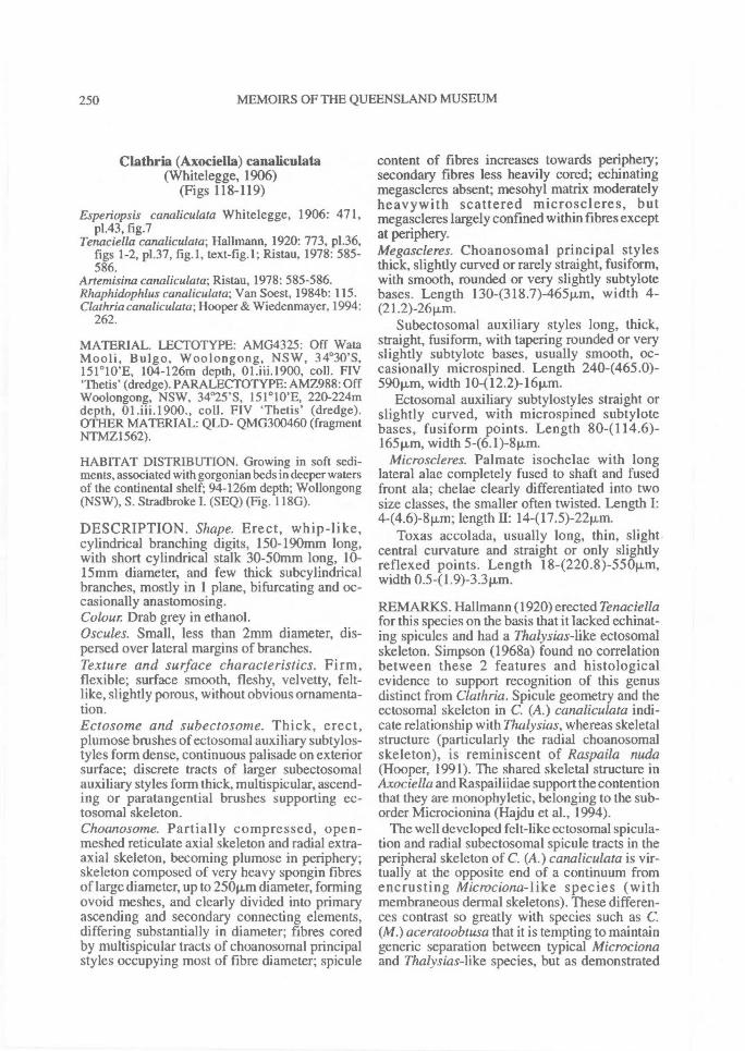
250^ MEMOIRS OF THE QUEENSLAND MUSEUM
Clathria (Axociella) canaliculata(Whitelegge, 1906)
(Figs 118-119)
Esperiopsis canaliculata Whitelegge, 1906: 471,p1.43, fig.7
Tenaciella canaliculata; Hallmann, 1920: 773, p1.36,figs 1-2, p1.37, fig.1, text-fig.1; Ristau, 1978: 585-586.
Artemisina canaliculata; Ristau, 1978: 585-586.Rhaphidophlus canaliculata; Van Soest, 1984b: 115.Clathriacanaliculata; Hooper & Wiedenmayer, 1994:
262.
MATERIAL. LECTOTYPE: AMG4325: Off WataMooli, Bulgo, Woolongong, NSW, 34 °30'S,151 ° 10'E, 104-126m depth, 01.iii.1900, coll. FIV'Thetis' (dredge). PARALECTOTYPE: AMZ988: OffWoolongong, NSW, 34°25'S, 151 ° 10'E, 220-224mdepth, 01.iii.1900., coll. FIV 'Thetis' (dredge).OTHER MATERIAL: QLD- QMG300460 (fragmentNTMZ1562).
HABITAT DISTRIBUTION. Growing in soft sedi-ments, associated with gorgonian beds in deeper watersof the continental shelf; 94-126m depth; Wollongong(NSW), S. Stradbroke I. (SEQ) (Fig. 118G).
DESCRIPTION. Shape. Erect, whip-like,cylindrical branching digits, 150-190mm long,with short cylindrical stalk 30-50mm long, 10-15mm diameter, and few thick subcylindricalbranches, mostly in 1 plane, bifurcating and oc-casionally anastomosing.Colour Drab grey in ethanol.Oscules. Small, less than 2mm diameter, dis-persed over lateral margins of branches.Texture and surface characteristics. Firm,flexible; surface smooth, fleshy, velvetty, felt-like, slightly porous, without obvious ornamenta-tion.Ectosome and subectosome. Thick, erect,plumose brushes of ectosomal auxiliary subtylos-tyles form dense, continuous palisade on exteriorsurface; discrete tracts of larger subectosomalauxiliary styles form thick, multispicular, ascend-ing or paratangential brushes supporting ec-tosomal skeleton.Choanosome. Partially compressed, open-meshed reticulate axial skeleton and radial extra-axial skeleton, becoming plumose in periphery;skeleton composed of very heavy spongin fibresof large diameter, up to 250p,m diameter, formingovoid meshes, and clearly divided into primaryascending and secondary connecting elements,differing substantially in diameter; fibres coredby multispicular tracts of choanosomal principalstyles occupying most of fibre diameter; spicule
content of fibres increases towards periphery;secondary fibres less heavily cored; echinatingmegascleres absent; mesohyl matrix moderatelyheavywith scattered microscleres, butmegascleres largely confined within fibres exceptat periphery.Megascleres. Choanosomal principal stylesthick, slightly curved or rarely straight, fusiform,with smooth, rounded or very slightly subtylotebases. Length 130-(318.7)-465p,m, width 4-(21.2)-26pm.
Subectosomal auxiliary styles long, thick,straight, fiisiform, with tapering rounded or veryslightly subtylote bases, usually smooth, oc-casionally microspined. Length 2404465.0)-590p,m, width 10-(12.2)-16p.m.
Ectosomal auxiliary subtylostyles straight orslightly curved, with microspined subtylotebases, fusiform points. Length 80-(114.6)-165m, width 5-(6.1)-8p,m.
Microscleres. Palmate isochelae with longlateral alae completely fused to shaft and fusedfront ala; chelae clearly differentiated into twosize classes, the smaller often twisted. Length I:4-(4.6)-8jun; length II: 14-(17.5)-22p.m.
Toxas accolada, usually long, thin, slightcentral curvature and straight or only slightlyreflexed points. Length 184220.8 )-55011m,width 0.541.9)-3.311m.
REMARKS. Hallmann (1920) erected Tenaciellafor this species on the basis that it lacked echinat-ing spicules and had a Thalysias-like ectosomalskeleton. Simpson (1968a) found no correlationbetween these 2 features and histologicalevidence to support recognition of this genusdistinct from Clathria. Spicule geometry and theectosomal skeleton in C. (A.) canaliculata indi-cate relationship with Thalysias, whereas skeletalstructure (particularly the radial choanosomalskeleton), is reminiscent of Raspaila nuda(Hooper, 1991). The shared skeletal structure inAxociella and Raspailiidae support the contentionthat they are monophyletic, belonging to the sub-order Microcionina (Hajdu et al., 1994).
The well developed felt-like ectosomal spicula-tion and radial subectosomal spicule tracts in theperipheral skeleton of C. (A.) canaliculata is vir-tually at the opposite end of a continuum fromencrusting Microciona-like species (withmembraneous dermal skeletons). These differen-ces contrast so greatly with species such as C.(M.) aceratoobtusa that it is tempting to maintaingeneric separation between typical Microcionaand Thalysias-like species, but as demonstrated
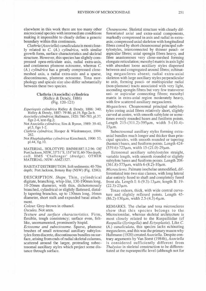
REVISION OF MICROCIONIDAE^ 251
elsewhere in this work there are too many othermicrocionid species with intermediate conditionsmaking it impossible to clearly define a genericboundary within this continuum.
Clathria(Axociella)canaliculata is most close-ly related to C. (A.) cylindrica, with similargrowth form, surface characteristics and skeletalstructure. However, this species has slightly com-pressed open-reticulate axis, radial extra-axis,and continuous plumose ectosome, whereas C.(A.) cylindrica has a greatly compressed close-meshed axis, a radial extra-axis and a sparse,discontinuous, plumose ectosome. Toxa mor-phology and spicule size also differ substantiallybetween these two species.
CIathria (Axociella) cylindrica(Ridley & Dendy, 1886)
(Fig. 120-121)
Esperiopsis cylindrica Ridley & Dendy, 1886: 340;Ridley & Dendy, 1887: 79-80, p1.19, figs 2a-b.
Axociella cylindrica; Hallmann, 1920: 780-783, p1.37,figs 2-4, text-fig.2;
Not Axociella cylindrica; Sim & Byeon, 1989: 39-40,p1.5, figs 1-2.
Clathria cylindrica; Hooper & Wiedenmayer, 1994:262.
Not Rhaphidophlus cylindricus Kieschnick, 1900: 53,p1.44, fig.10.
MATERIAL. HOLOTYPE: BMNH1887.5.2.96: OffPort Jackson, NSW, 33°51'S, 151 ° 16'E, 60-70m depth,coll. HMS 'Challenger' (dredge). OTHERMATERIAL: NSW- AMZ1527.
HABITAT DISTRIBUTION. Soft sediments; 40-70mdepth; Port Jackson, Botany Bay (NSW) (Fig. 1200).
DESCRIPTION. Shape. Thin, cylindricaldigitate, branching, whip-like, 130-190mm long,10-20mm diameter, with thin, dichotomouslybranched, cylindrical or slightly flattened, distal-ly tapering branches, up to 130mm long, 16mmdiameter, short stalk and expanded basal attach-ment.Colour Grey-brown in ethanol.Oscules. Not seen.Texture and surface characteristics. Firm,flexible, tough consistency; surface even, felt-like, unornamented, prominently hispid.Ectosome and subectosome. Sparse, plumosebrushes of small ectosomal auxiliary subtylos-tyles form discrete, discontinuous bundles on sur-face, arising from ends of radial skeletal columns,scattered around the larger, protruding subec-tosomal auxiliary styles which project some dis-tance through surface.
Choanosome. Skeletal structure with clearly dif-ferentiated axial and extra-axial components,markedly compressed in axis and radial in extra-axis; compressed axial skeleton with longitudinalfibres cored by short choanosomal principal sub-tylostyles, interconnected by thinner pauci- oraspicular fibres; axial spongin fibres heavy, andfibre anastomoses very close-meshed formingelongate reticulation; mesohyl matrix in axis lightwith abundant loose auxiliary styles dispersedbetween and congregated around fibres; echinat-ing megascleres absent; radial extra-axialskeleton with large auxiliary styles perpendicularto axis, forming pauci- or multispicular radial(non-plumose) tracts associated with very light,ascending spongin fibres but very few transverseuni- or aspicular connecting fibres; mesohylmatrix in extra-axial region moderately heavy,with few scattered auxiliary megascleres.Megascleres. Choanosomal principal subtylos-tyles coring axial fibres relatively short, slightlycurved at centre, with smooth subtylote or some-times evenly rounded bases and fusiform points.Length 215-(311.2)-395pm, width 5-(
Subectosomal auxiliary styles forming extra-axial bundles much longer and thicker than prin-cipal spicules, with smooth rounded or tapering(hastate) bases, and fusiform points. Length 424-(559.6)-725p.m, width 15-(21.0)-29p,m.
Ectosomal auxiliary subtylostyles straight,variable length, with smooth rounded or slightlysubtylote bases and fusiform points. Length 208-(361.8)-575p.m, width 6-(8.2)-10iim.Microscleres. Palmate isochelae unmodified, dif-ferentiated into two size classes, with long lateralalae entirely fused to shaft and completely fusedfront ala. Length I: 6-(9.3)-13p.m; length^19-(22.3)-25p.m.
Toxas oxhorn, thick, with wide central curva-ture and slightly reflexed points. Length 45-(86.2)-130pm, width 2.5-(4.3)-6p.m.
REMARKS. The chelae and toxa microscleresshow that this species belongs to theMicrocionidae, whereas skeletal architecture ismost closely related to the Raspailiidae (cfRaspailia (Syringe/la) and Ectyoplasia). Like C.(A.) canaliculata, this species lacks echinatingmegascleres, and this was the primary reason whyHallmann (1920) created Axociella. Despite con-trary arguments by Van Soest (1984b), Axociellais considered sufficiently different fromThalysias in skeletal construction to be differen-tiated at the supraspecific level (although not for
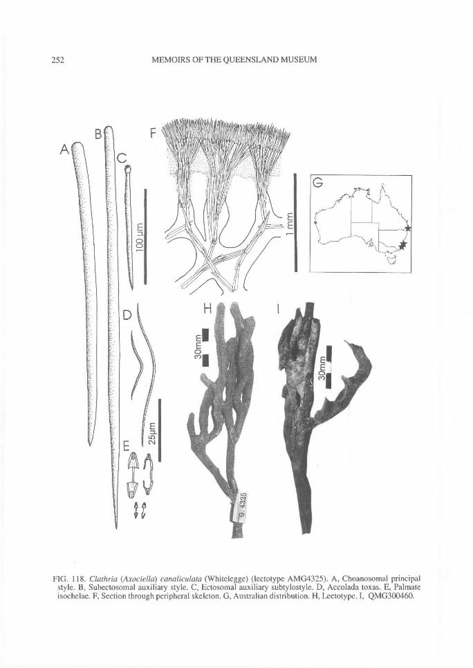
252^ MEMOIRS OF THE QUEENSLAND MUSEUM
FIG. 118. Clathria (Axociella) canaliculata (Whitelegge) (lectotype AMG4325). A, Choanosomal principalstyle. B, Subectosomal auxiliary style. C, Ectosomal auxiliary subtylostyle. D, Accolada toxas. E, Palmateisochelae. F, Section through peripheral skeleton. G, Australian distribution. FI, Lectotype. I, QMG300460.
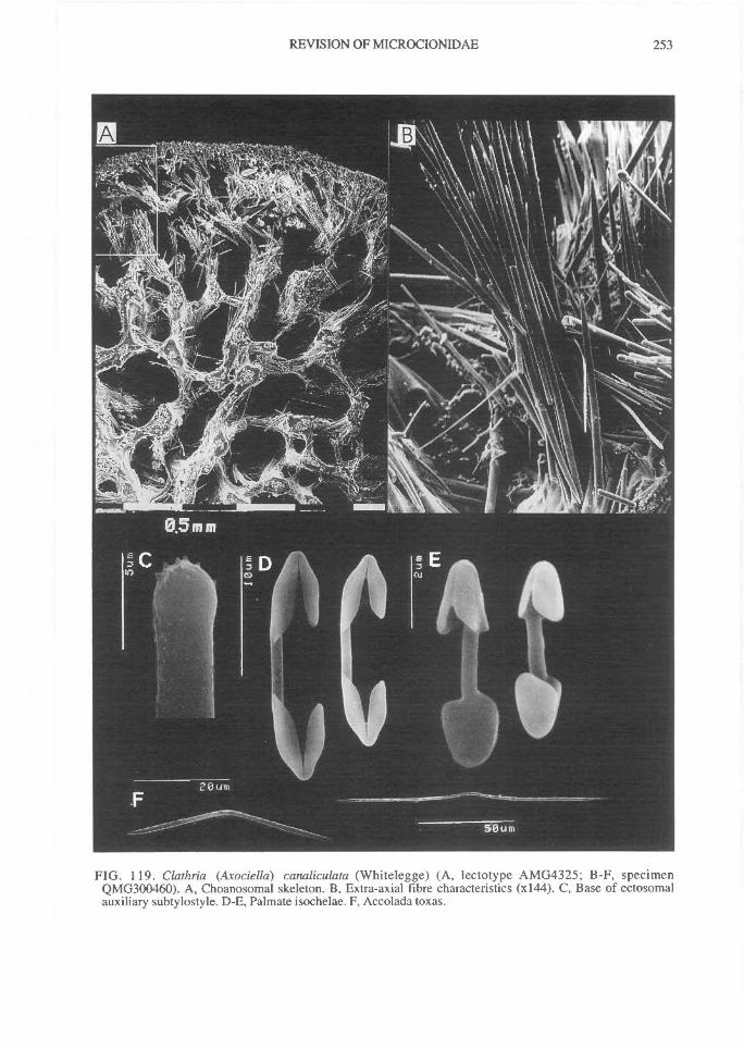
REVISION OF MICROCIONIDAE^
253
FIG. 119. Clathria (Axociella) canaliculata (Whitelegge) (A, lectotype AMG4325; B-F, specimenQMG300460). A, Choanosomal skeleton. B, Extra-axial fibre characteristics (x144). C, Base of ectosomalauxiliary subtylostyle. D-E, Palmate isochelae. F, Accolada toxas.
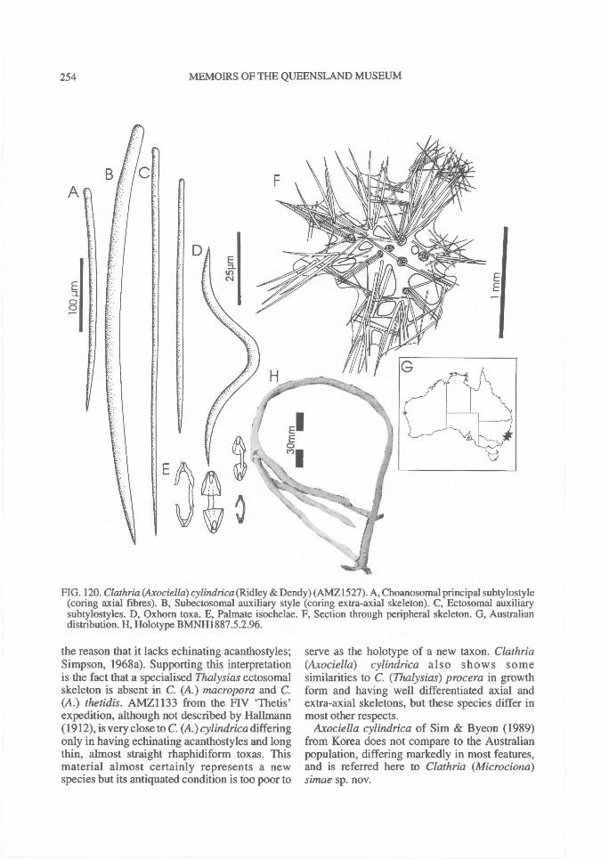
A
a
254^ MEMOIRS OF THE QUEENSLAND MUSEUM
FIG. 120. Clathria (Axociella) cylindrica (Ridley & Dendy) (AMZ1527). A, Choanosomal principal subtylostyle(coring axial fibres). B, Subectosomal auxiliary style (coring extra-axial skeleton). C, Ectosomal auxiliarysubtylostyles. D, Oxhorn toxa. E, Palmate isochelae. F, Section through peripheral skeleton. G, Australiandistribution. H, Holotype BMNH1887.5.2.96.
the reason that it lacks echinating acanthostyles;Simpson, 1968a). Supporting this interpretationis the fact that a specialised Thalysias ectosomalskeleton is absent in C. (A.) macropora and C.(A.) thetidis. AMZ1133 from the F1V 'Thetis'expedition, although not described by Hallmann(1912), is very close to C. (A.) cylindrica differingonly in having echinating acanthostyles and longthin, almost straight rhaphidiform toxas. Thismaterial almost certainly represents a newspecies but its antiquated condition is too poor to
serve as the holotype of a new taxon. Clathria(Axociella) cylindrica also shows somesimilarities to C. (Thalysias) procera in growthform and having well differentiated axial andextra-axial skeletons, but these species differ inmost other respects.
Axociella cylindrica of Sim & Byeon (1989)from Korea does not compare to the Australianpopulation, differing markedly in most features,and is referred here to Clathria (Microciona)simae sp. nov.
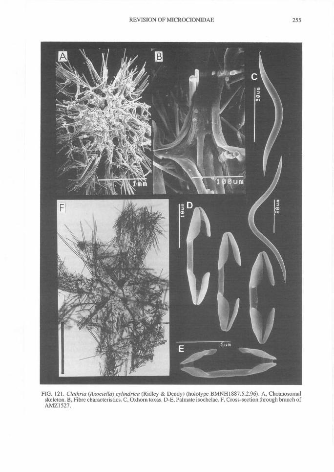
REVISION OF MICROCIONIDAE^ 255
FIG. 121. Clathria (Axociella) cylindrica (Ridley & Dendy) (holotype BMNH1887.5.2.96). A, Choanosomalskeleton. B, Fibre characteristics. C, Oxhorn toxas. D-E, Palmate isochelae. F, Cross-section through branch ofAMZ1527.

256^ MEMOIRS OF THE QUEENSLAND MUSEUM
Clathria (Axociella) nidificata(Kirkpatrick, 1907)
(Figs 122-123)
Ophlitaspongia nidificata Kirkpatrick, 1907: 274;Kirkpatrick, 1908b: 25, p1.22, fig.6, p1.24, fig.5;Hallmann, 1912: 254; Burton, 1929a: 433; Burton,1932a: 324; de Laubenfels, 1936a: 120; de Lauben-fels, 1954: 162.
Axociella nidificata; Burton, 1940: 116; Koltun,1964a: 70, p1.12,figs 7-11; Koltun, 1976: 155, 190;Desqueyroux, 1975: 67-68, p1.4, figs 47-50;Hoshino, 1977a: 45-46, table 1.
Clathria (Axociella) nidificata; Hooper & Wieden-mayer, 1994:263; Mothes & Lerner, 1995: 159-160,figs 22-27, 55.
Ophlitaspongia flabellata Topsent, 1916: 167; Top-sent, 1917: 41, p1.1, fig.4, p1.6, fig.2; Burton, 1929:433; Burton, 1932: 325; Burton, 1934b: 34.
Axociella flabellata; Koltun, 1964a: 70-71, p1.12, figs12-14; Hoshino, 1977a: 45-46, table 1.
Axociella rameus Koltun,1964a: 71, text-fig.17;Hoshino, 1977a: 45-46, table 1.
MATERIAL. HOLOTYPE: BMNH1908.2.5. 131:Coulman I., Victoria Land, Ross Dependency, An-tarctica, 73°30'S, 17°00'E, 200m depth, coll. HMS'Discovery' (dredge).
HABITAT DISTRIBUTION. On hard substrates; 93-540m depth; Antarctica: BANZARE coast; BuddCoast, Wilkes Land; Kemp Land, Enderby Land;Mawson Peninsula, Oates Land; Coulman I., VictoriaLand; Prydz Bay, MacKenzie Bay and Mawson Coast,MacRobertson Land; King Edward Ice Shelf, EnderbyLand; Joinville and Elefante Is; Palmer Archipelago;Graham Land; also South Georgia, S. Shetland Is (Fig.122E).
DESCRIPTION. Shape. Erect, claviform-flabel-late, narrow stalk.Colour Yellow-brown in ethanol.Oscules. Oscules on upper surface of club, I -2mm diameter.Texture and surface characteristics. Texturecompressible, tough, harsh surface; surfacehispid, conulose, membraneous between conules.Ectosome and subectosome. Ectosomal skeletonplumose, composed of discrete bundles of subec-tosomal auxiliary subtylostyles perched on endsof protruding principal spicules, corresponding tosurface conules.Choanosome. Skeleton compressed in axis andplumose in extra-axis; axial region slightly com-pressed containing heavy spongin fibres cored byplumo-reticulate pauci- or multispicular tracts ofchoanosomal principal styles; skeletal tracts formoval meshes, up to 250p.m diameter; extra-axialtracts plumose, diverging towards periphery, withpauci- or multispicular tracts of choanosomal
principal styles coring heavy spongin fibres;styles in peripheral regions of fibres oftenprotrude from fibres at acute angles, but these areidentical to principal megascleres and cannot beconsidered as true echinating spicules; mesohylmatrix moderately heavy, granular, containingabundant toxas, often in bundles, and occasionaldetritus.Megascleres. Choanosomal principal styles long,thick, entirely smooth, slightly curved towardsbase, with slightly subtylote evenly rounded orslightly constricted hastate bases and fusiformpoints. Length 595-(914.7)-1140p-m, width 21-(43.7)-61p.m.
Subectosomal auxiliary subtylostyles short,slender, straight, with slightly subtylotemicrospined bases, fusiform pointed. Length331-(405.4)-447p.m, width 6-(9.4)-12p.m.
No echinating spicules.Microscleres. Chelae absent.
Toxas accolada, thick, variable in length, withprominent but narrow angular central curvature,straight arms and straight or slightly reflexed tips;larger toxas with spined tips, small toxas entirelysmooth. Length 98-(286.2)-546p.m, width 1.5-(3.2)-5p.m.
REMARKS. The synonymy given above followsKoltun (1976). The species appears to vary ingrowth form, from bushy, branching, reticulate,digitate to flabellate, but skeletal architecture andspicule geometry are relatively consistent.Several echinating acanthostyles were seen inspicule preparations made from the holotype(evenly spined, club-shaped, 94-123x5-9Rm),but none were seen in section preparations and itis presumed that these are contaminants, althoughit is possible that they are present but rare.
The specialised ectosomal skeleton of discreteauxiliary spicule brushes perched over largerprincipal spicules, and compressed `axinellid'skeletal construction, are reminiscent ofRaspailiidae (e.g., Aulospongus), and were it notfor the toxas in this species it could be includedin Raspailiidae. This species fits best in Clathria(Axociella) although it lacks differentiated ec-tosomal and subectosomal spicules (i.e., it has 1size class of auxiliary styles). Its toxa and spina-tion should be compared with C. (A.) georgiaen-sis sp. nov. (see below).
Clathria (Axociella) patula sp. nov.(Figs 124-125)
MATERIAL. HOLOTYPE: NTMZ2909: BetweenNorth and East Wallabi Is, Houtman Abrolhos, WA,
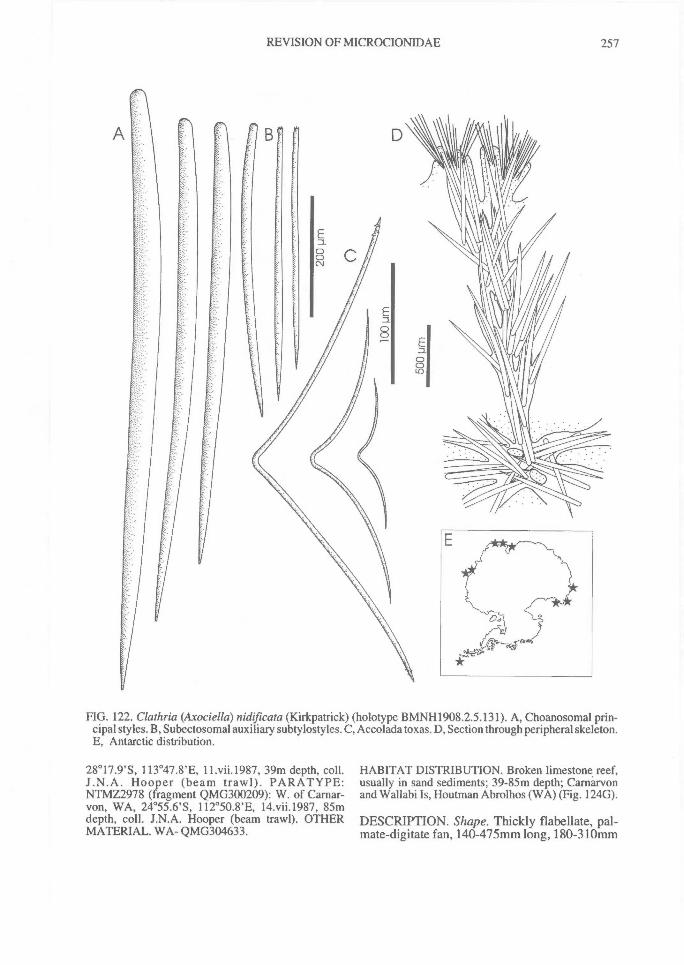
REVISION OF MICROCIONIDAE^ 257
FIG. 122. Clathria (Axociella) nidificata (Kirkpatrick) (holotype BMNH1908.2.5.131). A, Choanosomal prin-cipal styles. B, Subectosomal auxiliary subtylostyles. C, Accolada toxas. D, Section through peripheral skeleton.E, Antarctic distribution.
28°17.9'S, 113°47.8'E, 11.vii.1987, 39m depth, coll.J.N.A. Hooper (beam trawl). PARATYPE:NTMZ2978 (fragment QMG300209): W. of Carnar-von, WA, 24°55.6'S, 112°50.8'E, 14.vii.1987, 85mdepth, coll. J.N.A. Hooper (beam trawl). OTHERMATERIAL. WA- QMG304633.
HABITAT DISTRIBUTION. Broken limestone reef,usually in sand sediments; 39-85m depth; Camarvonand Wallabi Is, Houtman Abrolhos (WA) (Fig. 124G).
DESCRIPTION. Shape. Thickly flabellate, pal-mate-digitate fan, 140-475mm long, 180-310mm
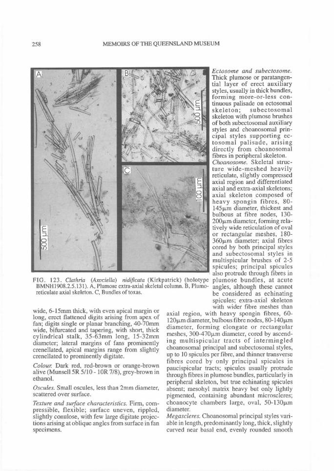
FIG. 123. Clathria (Axociella) nidificata (Kirkpatrick) (holotypeBMNH1908.2.5.131). A, Plumose extra-axial skeletal column. B, Plumo-reticulate axial skeleton. C, Bundles of toxas.
258^ MEMOIRS OF THE QUEENSLAND MUSEUM
wide, 6-15mm thick, with even apical margin orlong, erect flattened digits arising from apex offan; digits single or planar branching, 40-70mmwide, bifurcated and tapering, with short, thickcylindrical stalk, 35-63mm long, 15-32mmdiameter; lateral margins of fans prominentlycrenellated, apical margins range from slightlycrenellated to prominently digitate.Colour Dark red, red-brown or orange-brownalive (Munsell 5R 5/10 - 1OR 7/8), grey-brown inethanol.Oscules. Small oscules, less than 2mm diameter,scattered over surface.Texture and surface characteristics. Firm, com-pressible, flexible; surface uneven, rippled,slightly conulose, with few large digitate projec-tions arising at oblique angles from surface in fanspecimens.
Ectosome and subectosome.Thick plumose or paratangen-tial layer of erect auxiliarystyles, usually in thick bundles,forming more-or-less con-tinuous palisade on ectosomalskeleton; subectosomalskeleton with plumose brushesof both subectosomal auxiliarystyles and choanosomal prin-cipal styles supporting ec-tosomal palisade, arisingdirectly from choanosomalfibres in peripheral skeleton.Choanosome. Skeletal struc-ture wide-meshed heavilyreticulate, slightly compressedaxial region and differentiatedaxial and extra-axial skeletons;axial skeleton composed ofheavy spongin fibres, 80-145p,m diameter, thickest andbulbous at fibre nodes, 130-200p,rn diameter, forming rela-tively wide reticulation of ovalor rectangular meshes, 180-360Rm diameter; axial fibrescored by both principal stylesand subectosomal styles inmultispicular brushes of 2-5spicules; principal spiculesalso protrude through fibres inplumose bundles, at acuteangles, although these cannotbe considered as echinatingspicules; extra-axial skeletonwith wider fibre meshes than
axial region, with heavy spongin fibres, 60-12011m diameter, bulbous fibre nodes, 80-140p.mdiameter, forming elongate or rectangularmeshes, 300-470Rm diameter, cored by ascend-ing multispicular tracts of intermingledchoanosomal principal and subectosomal styles,up to 10 spicules per fibre, and thinner transversefibres cored by only principal spicules inpaucispicular tracts; spicules usually protrudethrough fibres in plumose bundles, particularly inperipheral skeleton, but true echinating spiculesabsent; mesohyl matrix heavy but only lightlypigmented, containing abundant microscleres;choanocyte chambers large, oval, 50-1301.Lmdiameter.Megascleres. Choanosomal principal styles vari-able in length, predominantly long, thick, slightlycurved near basal end, evenly rounded smooth
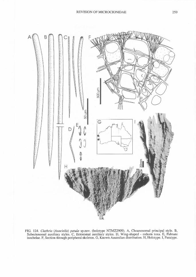
REVISION OF MICROCIONIDAE^ 259
FIG. 124. Clathria (Axociella) patula sp.nov. (holotype NTMZ2909). A, Choanosomal principal style. B,Subectosomal auxiliary styles. C, Ectosomal auxiliary styles. D, Wing-shaped - oxhorn toxa. E, Palmateisochelae. F, Section through peripheral skeleton. G, Known Australian distribution. H, Holotype. I, Paratype.
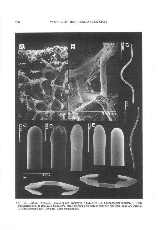
260^ MEMOIRS OF THE QUEENSLAND MUSEUM
FIG. 125. Clathria (Axociella) panda sp.nov. (holotype NTMZ2978). A, Choanosomal skeleton. B, Fibrecharacteristics. C-E, Bases of choanosomal principal, subectosomal auxiliary and ectosomal auxiliary spicules.F, Palmate isochelae. G, Oxhorn - wing-shaped toxas.
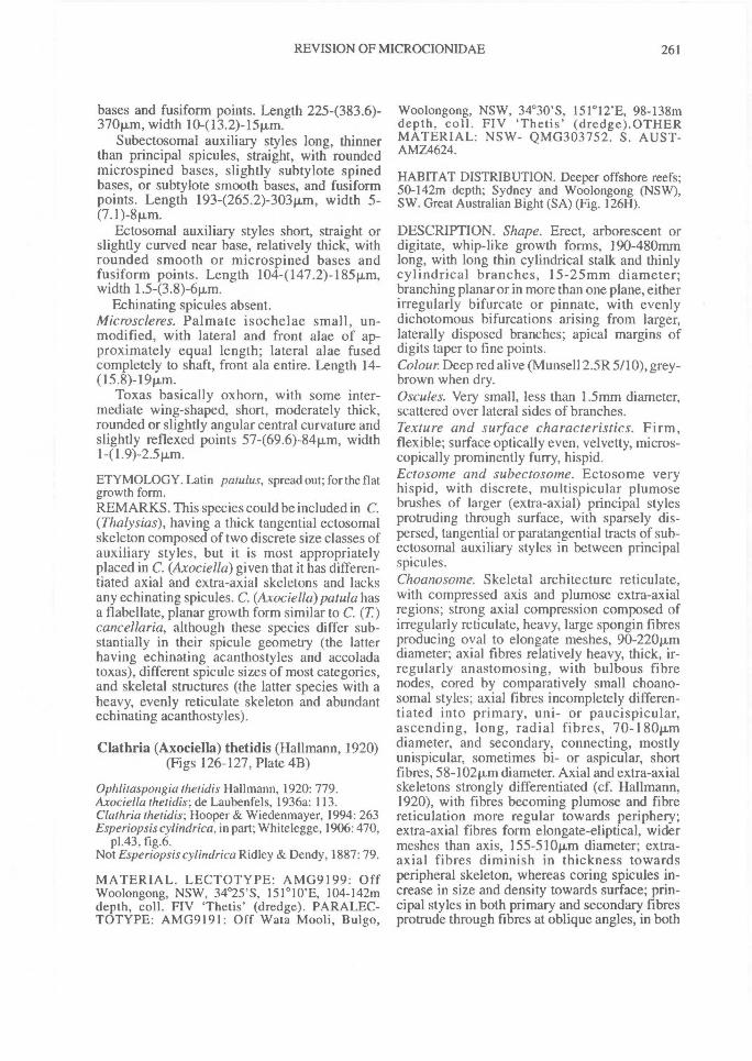
REVISION OF MICROCIONIDAE^ 261
bases and fusiform points. Length 2254383.6)-370p,m, width 10413.2)-1*m.
Subectosomal auxiliary styles long, thinnerthan principal spicules, straight, with roundedmicrospined bases, slightly subtylote spinedbases, or subtylote smooth bases, and fusiformpoints. Length 1934265.2)-30311m, width 5-(7.1)-8p.m.
Ectosomal auxiliary styles short, straight orslightly curved near base, relatively thick, withrounded smooth or microspined bases andfusiform points. Length 104-(147.2)-185Rm,width 1.5-(3.8)-6Rm.
Echinating spicules absent.Microscleres. Palmate isochelae small, un-modified, with lateral and front alae of ap-proximately equal length; lateral alae fusedcompletely to shaft, front ala entire. Length 14-(15.8)-19pm.
Toxas basically oxhorn, with some inter-mediate wing-shaped, short, moderately thick,rounded or slightly angular central curvature andslightly reflexed points 57-(69.6)-84Rm, width141.9)-2.511m.
ETYMOLOGY. Latin patulus, spread out; for the flatgrowth form.REMARKS. This species could be included in C.(Thalysias), having a thick tangential ectosomalskeleton composed of two discrete size classes ofauxiliary styles, but it is most appropriatelyplaced in C. (Axociella) given that it has differen-tiated axial and extra-axial skeletons and lacksany echinating spicules. C. (Axociella)patula hasa flabellate, planar growth form similar to C. (T)cancellaria, although these species differ sub-stantially in their spicule geometry (the latterhaving echinating acanthostyles and accoladatoxas), different spicule sizes of most categories,and skeletal structures (the latter species with aheavy, evenly reticulate skeleton and abundantechinating acanthostyles).
Clathria (Axociella) thetidis (Hallmann, 1920)(Figs 126-127, Plate 4B)
Ophlitaspongia thetidis Hal!mann, 1920: 779.Axociella thetidis; de Laubenfels, 1936a: 113.Clathria thetidis; Hooper & Wiedenmayer, 1994: 263Esperiopsis cylindrica, in part; Whitelegge, 1906: 470,
p1.43, fig.6.Not Esperiopsis cylindrica Ridley & Dendy, 1887: 79.
MATERIAL. LECTOTYPE: AMG9199: OffWoolongong, NSW, 34 °25'S, 151 ° 10'E, 104-142mdepth, coll. FIV 'Thetis' (dredge). PARALEC-TOTYPE: AMG9191: Off Wata Mooli, Bulgo,
Woolongong, NSW, 34°30'S, 151 ° 12'E, 98-138mdepth, coll. FIV 'Thetis' (dredge).0THERMATERIAL: NSW- QMG303752. S. AUST-AMZ4624.
HABITAT DISTRIBUTION. Deeper offshore reefs;50-142m depth; Sydney and Woolongong (NSW),SW. Great Australian Bight (SA) (Fig. 126H).
DESCRIPTION. Shape. Erect, arborescent ordigitate, whip-like growth forms, 190-480mmlong, with long thin cylindrical stalk and thinlycylindrical branches, 15-25mm diameter;branching planar or in more than one plane, eitherirregularly bifurcate or pinnate, with evenlydichotomous bifurcations arising from larger,laterally disposed branches; apical margins ofdigits taper to fine points.Colour. Deep red alive (Munsell 2.5R 5/10), grey-brown when dry.Oscules. Very small, less than 1.5mm diameter,scattered over lateral sides of branches.Texture and surface characteristics. Firm,flexible; surface optically even, velvetty, micros-copically prominently furry, hispid.Ectosome and subectosome. Ectosome veryhi spid, with discrete, multispicular plumosebrushes of larger (extra-axial) principal stylesprotruding through surface, with sparsely dis-persed, tangential or paratangential tracts of sub-ectosomal auxiliary styles in between principalspicules.Choanosome. Skeletal architecture reticulate,with compressed axis and plumose extra-axialregions; strong axial compression composed ofirregularly reticulate, heavy, large spongin fibresproducing oval to elongate meshes, 90-220p.,mdiameter; axial fibres relatively heavy, thick, ir-regularly anastomosing, with bulbous fibrenodes, cored by comparatively small choano-somal styles; axial fibres incompletely differen-tiated into primary, uni- or paucispicular,ascending, long, radial fibres, 70-1801.imdiameter, and secondary, connecting, mostlyunispicular, sometimes bi- or aspicular, shortfibres, 58-102p,m diameter. Axial and extra-axialskeletons strongly differentiated (cf. Hallmann,1920), with fibres becoming plumose and fibrereticulation more regular towards periphery;extra-axial fibres form elongate-eliptical, widermeshes than axis, 155-510Rm diameter; extra-axial fibres diminish in thickness towardsperipheral skeleton, whereas coring spicules in-crease in size and density towards surface; prin-cipal styles in both primary and secondary fibresprotrude through fibres at oblique angles, in both
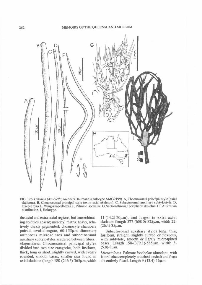
262^ MEMOIRS OF THE QUEENSLAND MUSEUM
FIG. 126. Clathria (Axociella) thetidis (Hal'mann) (holotype AMG9199). A, Choanosomal principal style (axialskeleton). B, Choanosomal principal style (extra-axial skeleton). C, Subectosomal auxiliary subtylostyle. D,Oxeote toxa. E, Wing-shaped toxas. F, Palmate isochelae. G, Section through peripheral skeleton. H, Australiandistribution. I, Holotype.
the axial and extra-axial regions, but true echinat-ing spicules absent; mesohyl matrix heavy, rela-tively darkly pigmented; choanocyte chamberspaired, oval-elongate, 60-1541,m diameter;numerous microscleres and subectosomalauxiliary subtylostyles scattered between fibres.Megascleres. Choanosomal principal stylesdivided into two size categories, both fusiform,thick, long or short, slightly curved, with evenlyrounded, smooth bases; smaller size found inaxial skeleton (length 1804246.5)-365 pm, width
11-(14.2)-20p,m), and larger in extra-axialskeleton (length 377-(608.0)-825iim, width 22-(26.4)-35p.m.
Subectosomal auxiliary styles long, thin,fusiform, straight, slightly curved or flexuous,with subtylote, smooth or lightly rnicrospinedbases. Length 158-(379.1)-585p,m, width 3-(5.8)-811m.Microscleres. Palmate isochelae abundant, withlateral alae completely attached to shaft and frontala entirely fused. Length 9413.4)-1611m.

REVISION OF MICROCIONIDAE^ 263
FIG. 127. Clathria (Axociella) thetidis (Hallmann) (QMG303752). A, Choanosomal skeleton. B, Extra-axialfibre and spicule skeleton (x63). C, Palmate isochelae. D, Wing-shaped toxas. E, Oxeote toxas.
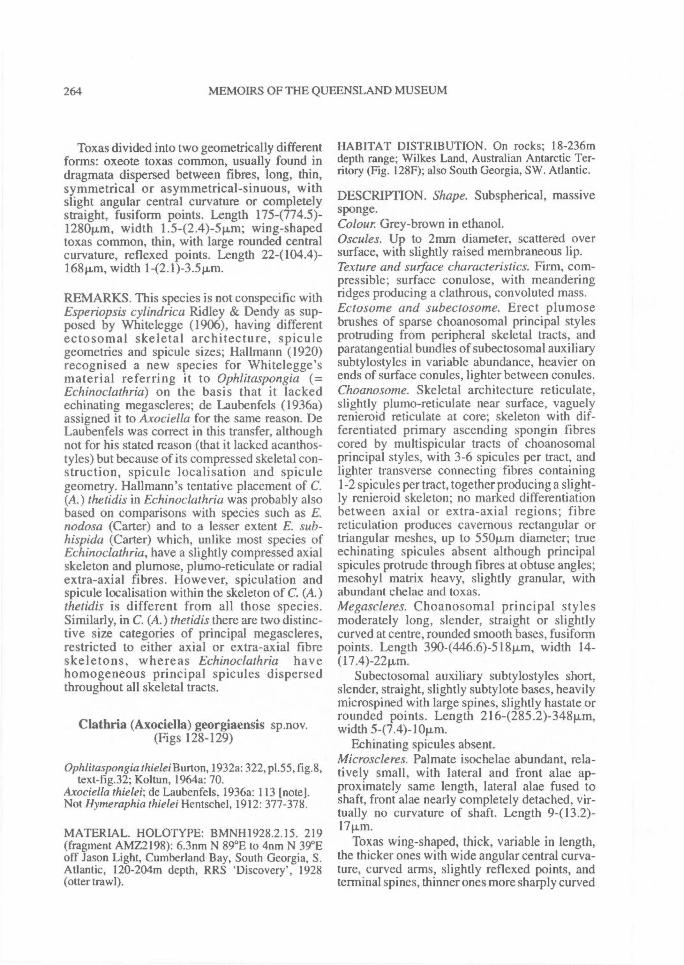
264^ MEMOIRS OF THE QUEENSLAND MUSEUM
Toxas divided into two geometrically differentforms: oxeote toxas common, usually found indragmata dispersed between fibres, long, thin,symmetrical or asymmetrical-sinuous, withslight angular central curvature or completelystraight, fusiform points. Length 175-(774.5)-1280p,m, width 1.542.4)-5 p.m; wing-shapedtoxas common, thin, with large rounded centralcurvature, reflexed points. Length 224104.4)-168p.,m, width 142.1)-3.41,m.
REMARKS. This species is not conspecific withEsperiopsis cylindrica Ridley & Dendy as sup-posed by Whitelegge (1906), having differentectosomal skeletal architecture, spiculegeometries and spicule sizes; Hallmann (1920)recognised a new species for Whitelegge'smaterial referring it to Ophlitaspongia (=Echinoclathria) on the basis that it lackedechinating megascleres; de Laubenfels (1936a)assigned it to Axociella for the same reason. DeLaubenfels was correct in this transfer, althoughnot for his stated reason (that it lacked acanthos-tyles) but because of its compressed skeletal con-struction, spicule localisation and spiculegeometry. Hallmann's tentative placement of C.(A.) thetidis in Echinoclathria was probably alsobased on comparisons with species such as E.nodosa (Carter) and to a lesser extent E. sub-hispida (Carter) which, unlike most species ofEchinoclathria, have a slightly compressed axialskeleton and plumose, plumo-reticulate or radialextra-axial fibres. However, spiculation andspicule localisation within the skeleton of C. (A.)thetidis is different from all those species.Similarly, in C. (A.) thetidis there are two distinc-tive size categories of principal megascleres,restricted to either axial or extra-axial fibreskeletons, whereas Echinoclathria havehomogeneous principal spicules dispersedthroughout all skeletal tracts.
Clathria (Axociella) georgiaensis sp.nov.(Figs 128-129)
Ophlitaspongia thielei Burton, 1932a: 322, p1.55, fig.8,text-fig.32; Koltun, 1964a: 70.
Axociella thielei; de Laubenfels, 1936a: 113 [note].Not Hymeraphia thielei Hentschel, 1912: 377-378.
MATERIAL. HOLOTYPE: BMNH1928.2.15. 219(fragment AMZ2198): 6.3nm N 89 °E to 4nm N 39°Eoff Jason Light, Cumberland Bay, South Georgia, S.Atlantic, 120-204m depth, RRS 'Discovery', 1928(otter trawl).
HABITAT DISTRIBUTION. On rocks; 18-236mdepth range; Wilkes Land, Australian Antarctic Ter-ritory (Fig. 128F); also South Georgia, SW. Atlantic.
DESCRIPTION. Shape. Subspherical, massivesponge.Colour Grey-brown in ethanol.Oscules. Up to 2mm diameter, scattered oversurface, with slightly raised membraneous lip.Texture and surface characteristics. Firm, com-pressible; surface conulose, with meanderingridges producing a clathrous, convoluted mass.Ectosome and subectosome. Erect plumosebrushes of sparse choanosomal principal stylesprotruding from peripheral skeletal tracts, andparatangential bundles of subectosomal auxiliarysubtylostyles in variable abundance, heavier onends of surface conules, lighter between conules.Choanosome. Skeletal architecture reticulate,slightly plumo-reticulate near surface, vaguelyrenieroid reticulate at core; skeleton with dif-ferentiated primary ascending spongin fibrescored by multispicular tracts of choanosomalprincipal styles, with 3-6 spicules per tract, andlighter transverse connecting fibres containing1-2 spicules per tract, together producing a slight-ly renieroid skeleton; no marked differentiationbetween axial or extra-axial regions; fibrereticulation produces cavernous rectangular ortriangular meshes, up to 550p..m diameter; trueechinating spicules absent although principalspicules protrude through fibres at obtuse angles;mesohyl matrix heavy, slightly granular, withabundant chelae and toxas.Megascleres. Choanosomal principal stylesmoderately long, slender, straight or slightlycurved at centre, rounded smooth bases, fusiformpoints. Length 3904446.6)-51811m, width 14-(17.4)-2211m.
Subectosomal auxiliary subtylostyles short,slender, straight, slightly subtylote bases, heavilymicrospined with large spines, slightly hastate orrounded points. Length 216-(285.2)-348itm,width 5-(7.4)-10Rm.
Echinating spicules absent.Microscleres. Palmate isochelae abundant, rela-tively small, with lateral and front alae ap-proximately same length, lateral alae fused toshaft, front alae nearly completely detached, vir-tually no curvature of shaft. Length 9-(l3.2)-17 p.m.
Toxas wing-shaped, thick, variable in length,the thicker ones with wide angular central curva-ture, curved arms, slightly reflexed points, andterminal spines, thinner ones more sharply curved
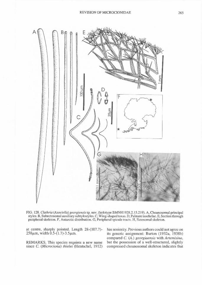
REVISION OF MICROCIONIDAE^ 265
FIG. 128. Clathria (Axociella) georgiensis sp. nov. (holotype BMNH1928.2.15.219). A, Choanosomal principalstyles. B, Subectosomal auxiliary subty losty les. C, Wing-shaped toxas. D, Palmate isochelae. E, Section throughperipheral skeleton. F, Antarctic distribution. G, Peripheral spicule tracts. H, Ectosomal skeleton.
at centre, sharply pointed. Length 284107.7)-258p.m, width 0.541.7)-3.511m.
REMARKS. This species requires a new namesince C. (Microciona) thielei (Hentschel, 1912)
has seniority. Previous authors could not agree onits generic assignment: Burton (1932a, 1938b)compared C. (A.) georgiaensis with Artemisina,but the possession of a well-structured, slightlycompressed choanosomal skeleton indicates that
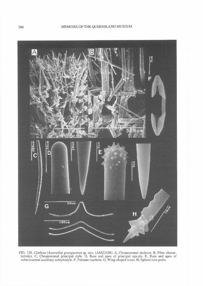
266^ MEMOIRS OF THE QUEENSLAND MUSEUM
FIG. 129. Clathria (Axociella) georgiaensis sp. nov. (AMZ2198). A, Choanosomal skeleton. B, Fibre charac-teristics. C, Choanosomal principal style. D, Base and apex of principal spicule. E, Base and apex ofsubectosomal auxiliary subtylostyle. F, Palmate isochela. G, Wing-shaped toxas. H, Spined toxa point.
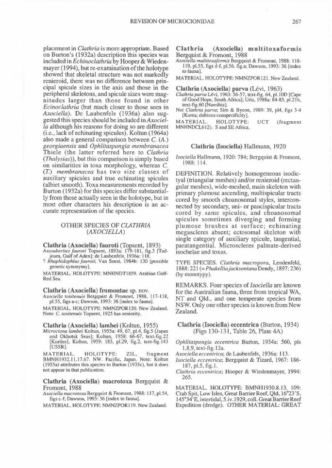
REVISION OF MICROCIONIDAE^ 267
placement in Clathria is more appropriate. Basedon Burton's (1932a) description this species wasincluded in Echinoclathria by Hooper & Wieden-mayer (1994), but re-examination of the holotypeshowed that skeletal structure was not markedlyrenieroid, there was no difference between prin-cipal spicule sizes in the axis and those in theperipheral skeletons, and spicule sizes were mag-nitudes larger than those found in otherEcinoclathria (but much closer to those seen inAxociella). De Laubenfels (1936a) also sug-gested this species should be included in Axociel-la although his reasons for doing so are different(i.e., lack of echinating spicules). Koltun (1964a)also made a general comparison between C. (A.)georgiaensis and Ophlitaspongia membranaceaThiele (the latter referred here to Clathria(Thalysias)), but this comparison is simply basedon similarities in toxa morphology, whereas C.(T) membranacea has two size classes ofauxiliary spicules and true echinating spicules(albiet smooth). Toxa measurements recorded byBurton (1932a) for this species differ substantial-ly from those actually seen in the holotype, but inmost other characters his description is an ac-curate representation of the species.
OTHER SPECIES OF CLATHRIA(AXOCIELLA)
Clathria (Axociella) fauroti (Topsent, 1893)Axosuberites fauroti Topsent, 1893a: 179-181, fig.3 [Tad-
joura. Gulf of Aden]; de Laubenfels, 1936a: 118.? Rhaphidophlus fauroti; Van Soest, 1984b: 130 [possible
generic synonymy].MATERIAL. HOLOTYPE: MNHNDT1859. Arabian Gulf-Red Sea.
Clathria (Axociella) fromontae sp. nov.Axociella toxitenuis Bergquist & Fromont, 1988, 117-118,
p1.55, figs a-c; Dawson, 1993: 36 [index to fauna].MATERIAL. HOLOTYPE: NMNZPOR120. New Zealand.Note: C. toxitenuis Topsent, 1925 has seniority.
Clathria (Axociella) lambei (Koltun, 1955)Microciona lambei Koltun, 1955a: 49, 67, p1.4, fig.5 [Japan
and Okhotsk Seas]; Koltun, 1958: 66-67, text-fig.22[Kuriles]; Koltun, 1959: 183, p1.29, fig.2, text-fig.143[USSR].
MATERIAL. HOLOTYPE: ZIL, fragmentBMNH1932.11.17.67. NW. Pacific, Japan. Note: Koltun(1955a) attributes this species to Burton (1935c), but it doesnot appear in that publication.
Clathria (Axociella) macrotoxa Bergquist &Fromont, 1988Axociella macrotoxa Bergquist & Fromont, 1988: 117, p1.54,
figs c-f; Dawson, 1993: 36 [index to fauna].MATERIAL. HOLOTYPE: NMNZPOR119. New Zealand.
Clathria (Axociella) multitoxaformisBergquist & Fromont, 1988Axociella multitoxaformis Bergquist & Fromont, 1988: 118-
119, p1.55, figs d-f, p1.56. fig.a; Dawson, 1993: 36 [indexto fauna].
MATERIAL. HOLOTYPE: NMNZPOR121. New Zealand.
Clathria (Axociella) parva (Levi, 1963)Clathria parva Levi, 1963: 56-57, text-fig. 64, p1.10D [Cape
of Good Hope, South Africa]; Uriz, 1988a: 84-85, p1.21b,text-fig.60 [Namibia].
Not Clathria parva; Sim & Byeon, 1989: 39, pI4, figs 3-4[Korea; dubious conspecificity].
MATERIAL. HOLOTYPE: UCT (fragmentMNHNDCL612). S and SE Africa.
Clathria (Isociella) Hallmann, 1920
Isociella Hallmann, 1920: 784; Bergquist & Fromont,1988: 114.
DEFINITION. Relatively homogeneous isodic-tyal (triangular meshes) and/or renieroid (rectan-gular meshes), wide-meshed, main skeleton withprimary plumose ascending, multispicular tractscored by smooth choanosomal styles, intercon-nected by secondary, uni- or paucispicular tractscored by ,same spicules, and choanosomalspicules sometimes diverging and formingplumose brushes at surface; echinatingmegascleres absent; ectosomal skeleton withsingle category of auxiliary spicule, tangential,paratangential. Microscleres palmate-derivedisochelae and toxas.
TYPE SPECIES. Clathria macropora, Lendenfeld,1888: 221 (=Phakelliajacksoniana Dendy, 1897:236)(by monotypy).
REMARKS. Four species of Isociella are knownfor the Australian fauna, three from tropical WA,NT and Qld., and one temperate species fromNSW. Only one other species is known from NewZealand.
Clathria (Isociella) eccentrica (Burton, 1934)(Figs 130-131, Table 26, Plate 4A)
Ophlitaspongia eccentrica Burton, 1934a: 560, pls1,8,9, text-fig.12a.
Axociella eccentrica; de Laubenfels, 1936a: 113.Isociella eccentrica; Bergquist & Tizard, 1967: 186-
187, p1.5, fig. 1.Clathria eccentrica; Hooper & Wiedenmayer, 1994:
265.
MATERIAL. HOLOTYPE: BMNH1930.8.13. 109:Crab Spit, Low Isles, Great Barrier Reef, Qld, 16 °23'S,145°34'E, intertidal, 5.iv.1929, coll. Great Barrier ReefExpedition (dredge). OTHER MATERIAL: GREAT
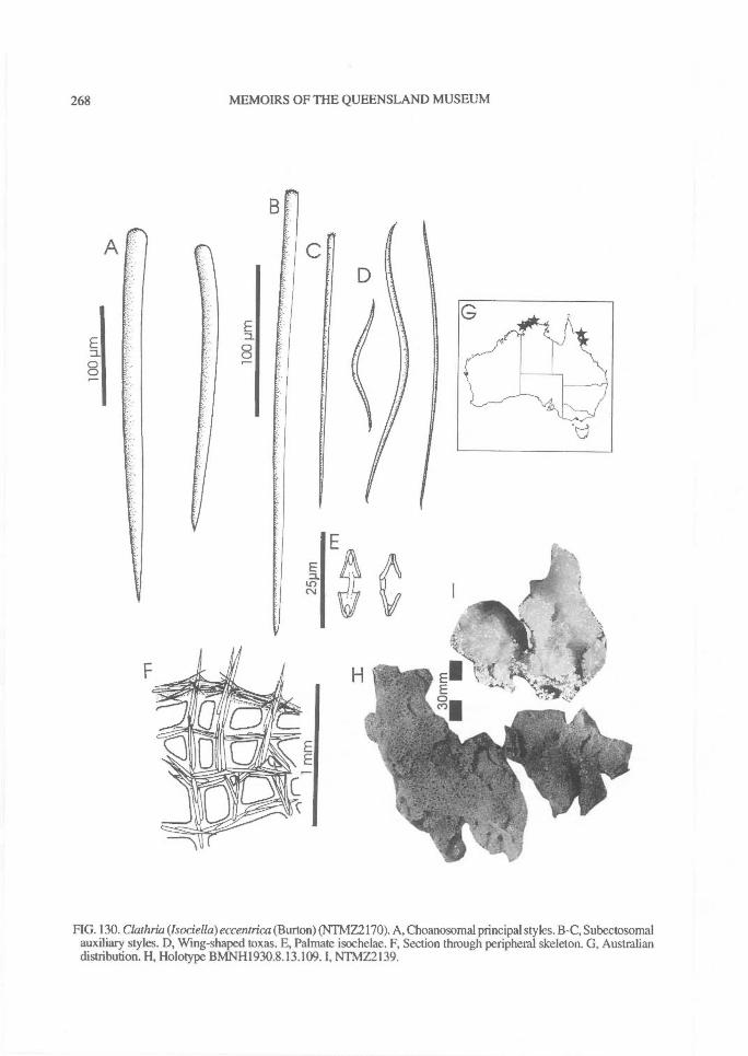
A
0
268^ MEMOIRS OF THE QUEENSLAND MUSEUM
FIG. 130. Clathria (Isociella) eccentrica (Burton) (NTMZ2170). A, Choanosomal principal styles. B-C, Subectosomalauxiliary styles. D, Wing-shaped toxas. E, Palmate isochelae. F, Section through peripheral skeleton. G, Australiandistribution. H, Holotype BMNH1930.8.13.109. I, NTMZ2139.
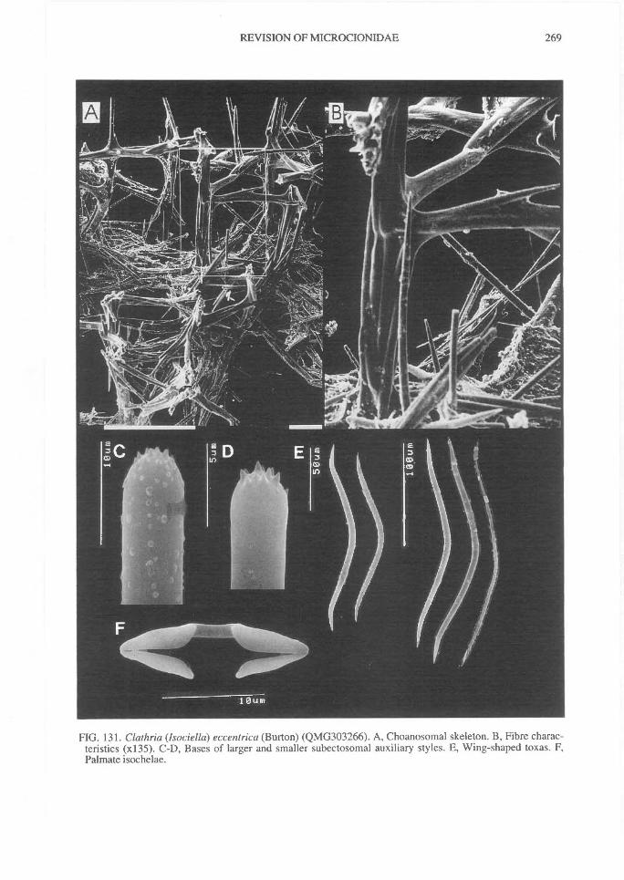
REVISION OF MICROCIONIDAE^
269
FIG. 131. Clathria (Isociella) eccentrica (Burton) (QMG303266). A, Choanosomal skeleton. B, Fibre charac-teristics (x135). C-D, Bases of larger and smaller subectosomal auxiliary styles. E, Wing-shaped toxas. F,Palmate isochelae.
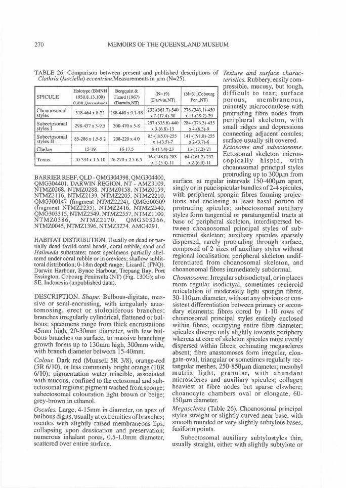
270^ MEMOIRS OF THE QUEENSLAND MUSEUM
TABLE 26. Comparison between present and published descriptions of Texture and surface charac-Claihria (Isociella) eccentrica.Measurements in p.m (N=25). teristics. Rubbery, easily com-
pressible, mucusy, but tough,difficult to tear; surfaceporous, membraneous,minutely microconulose withprotruding fibre nodes fromperipheral skeleton, withsmall ridges and depressionsconnecting adjacent conules;surface usually silt covered.Ectosome and subectosome.Ectosomal skeleton micros-copically hispid, withchoanosomal principal styles
SPICULEHolotype (BMNH
1930.8.13.109)(GBR,Queensland)
Bergquist &Tizard (1967)(Danvin,NT)
(19=19)(Darwin,NT)
(N=3) (CobourgPen.,NT)
Choanosomalstyles 318-464 x 8-22 288-440 x 9.1-18 2324361.7)-540
x7-(17.4)-30276-(343.1)-450x 11-(19.2)-29
Subectosomal 298-457 x 3-9.5 300-470 x 5-8 2574335.6)-440 284-(373.3)-455styles I x 3-(6.8)-13 x 4-(6.3)-9
Subectosomalstyles II 85-286 x 1.5-5.2 208-220 x 4.0 85-(185.0)-255
x 1-(3.5)-7141-(191.8)-255
x 2-(3.7)-6Chelae 15-19 16-17.5 8-(17.4)-23 13-(17.2)-21
Toxas 10-334 x 1.5-10 76-270x 2.5-6.5 16-(148.0)-283x 1-(5.4)-11
64-(161.2)-292x 2-(6.0)-11
BARRIER REEF, QLD - QMG304398, QMG304400,QMG304401. DARWIN REGION, NT - AMZ3109,NTMZ0268, NTMZ0288, NTMZ0158, NTMZ0159,NTMZ2116, NTMZ2139, NTMZ2205, NTMZ2210,QMG300147 (fragment NTMZ2224), QMG300509(fragment NTMZ2235), NTMZ2416, NTMZ2540,QMG303315, NTMZ2549, NTMZ2557, NTMZ1100,NTMZ0386, NTMZ2170, QMG303266,NTMZ0045, NTMZ1396, NTMZ3274. AMG4291.
HABITAT DISTRIBUTION. Usually on dead or par-tially dead faviid coral heads, coral rubble, sand andHalimeda substrates; most specimens partially shel-tered under coral rubble or in crevices; shallow sublit-toral distribution; 0-18m depth range; Lizard I. (FNQ),Darwin Harbour, Bynoe Harbour, Trepang Bay, PortEssington, Cobourg Peninsula (NT) (Fig. 1306); alsoSE. Indonesia (unpublished data).
DESCRIPTION. Shape. Bulbous-digitate, mas-sive or semi-encrusting, with irregularly anas-tomosing, erect or stoloniferous branches;branches irregularly cylindrical, flattened or bul-bous; specimens range from thick encrustations45mm high, 20-30mm diameter, with few bul-bous branches on surface, to massive branchinggrowth forms up to 130mm high, 300mm wide,with branch diameter between 15-40mm.Colour Dark red (Munsell 5R 3/8), orange-red(5R 6/10), or less commonly bright orange (10R6/10); pigmentation water miscible, associatedwith mucous, confined to the ectosomal and sub-ectosomal regions; pigment washed from sponge;subectosomal colouration light brown or beige;grey-brown in ethanol.Oscules. Large, 4-15mm in diameter, on apex ofbulbous digits, usually at extremities of branches;oscules with slightly raised membraneous lips,collapsing upon dessication and preservation;numerous inhalant pores, 0.5-1.0mm diameter,scattered over entire surface.
protruding up to 300p,m fromsurface, at regular intervals 150-400p.m apart,singly or in paucispicular bundles of 2-4 spicules,with peripheral spongin fibres forming projec-tions and enclosing at least basal portion ofprotruding spicules; subectosomal auxiliarystyles form tangential or paratangential tracts atbase of peripheral skeleton, interdispersed be-tween choanosomal principal styles of sub-renieroid skeleton; auxiliary spicules sparselydispersed, rarely protruding through surface,composed of 2 sizes of auxiliary styles withoutregional localisation; peripheral skeleton undif-ferentiated from choanosomal skeleton, andchoanosomal fibres immediately subdermal.Choanosome. Irregular subisodictyal, or in placesmore regular isodictyal, sometimes renieroidreticulation of moderately light spongin fibres,30-110v1,m diameter, without any obvious or con-sistent differentiation between primary or secon-dary elements; fibres cored by 1-10 rows ofchoanosomal principal styles entirely enclosedwithin fibres, occupying entire fibre diameter;spicules diverge only slightly towards peripherywhereas at core of skeleton spicules more evenlydispersed within fibres; echinating megascleresabsent; fibre anastomoses form irregular, elon-gate-oval, triangular or sometimes regularly rec-tangular meshes, 250-850Rm diameter; mesohylmatrix light, granular, with abundantmicroscleres and auxiliary spicules; collagenheaviest at fibre nodes but sparse elswhere;choanocyte chambers oval or elongate, 60-150p,m diameter.Megascleres (Table 26). Choanosomal principalstyles straight or slightly curved near base, withsmooth rounded or very slightly subtylote bases,fusiform points.
Subectosomal auxiliary subtylostyles thin,usually straight, either with slightly subtylote or
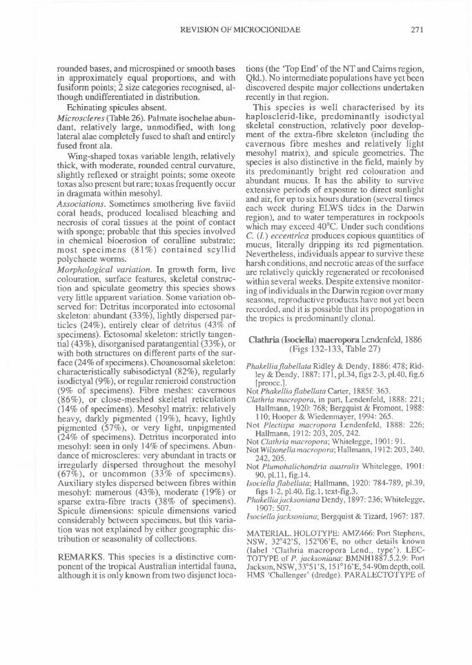
REVISION OF MICROCIONIDAE^ 271
rounded bases, and microspined or smooth basesin approximately equal proportions, and withfusiform points; 2 size categories recognised, al-though undifferentiated in distribution.
Echinating spicules absent.Microscleres (Table 26). Palmate isochelae abun-dant, relatively large, unmodified, with longlateral alae completely fused to shaft and entirelyfused front ala.
Wing-shaped toxas variable length, relativelythick, with moderate, rounded central curvature,slightly reflexed or straight points; some oxeotetoxas also present but rare; toxas frequently occurin dragmata within mesohyl.Associations. Sometimes smothering live faviidcoral heads, produced localised bleaching andnecrosis of coral tissues at the point of contactwith sponge; probable that this species involvedin chemical bioerosion of coralline substrate;most specimens (81%) contained scyllidpolychaete worms.Morphological variation. In growth form, livecolouration, surface features, skeletal construc-tion and spiculate geometry this species showsvery little apparent variation. Some variation ob-served for: Detritus incorporated into ectosomalskeleton: abundant (33%), lightly dispersed par-ticles (24%), entirely clear of detritus (43% ofspecimens). Ectosomal skeleton: strictly tangen-tial (43%), disorganised paratangential (33%), orwith both structures on different parts of the sur-face (24% of specimens). Choanosomal skeleton:characteristically subisodictyal (82%), regularlyisodictyal (9%), or regular renieroid construction(9% of specimens). Fibre meshes: cavernous(86%), or close-meshed skeletal reticulation(14% of specimens). Mesohyl matrix: relativelyheavy, darkly pigmented (19%), heavy, lightlypigmented (57%), or very light, unpigmented(24% of specimens). Detritus incorporated intomesohyl: seen in only 14% of specimens. Abun-dance of microscleres: very abundant in tracts orirregularly dispersed throughout the mesohyl(67%), or uncommon (33% of specimens).Auxiliary styles dispersed between fibres withinmesohyl: numerous (43%), moderate (19%) orsparse extra-fibre tracts (38% of specimens).Spicule dimensions: spicule dimensions variedconsiderably between specimens, but this varia-tion was not explained by either geographic dis-tribution or seasonality of collections.
REMARKS. This species is a distinctive com-ponent of the tropical Australian intertidal fauna,although it is only known from two disjunct loca-
tions (the 'Top End' of the NT and Cairns region,Qld.). No intermediate populations have yet beendiscovered despite major collections undertakenrecently in that region.
This species is well characterised by itshaplosclerid-like, predominantly isodictyalskeletal construction, relatively poor develop-ment of the extra-fibre skeleton (including thecavernous fibre meshes and relatively lightmesohyl matrix), and spicule geometries. Thespecies is also distinctive in the field, mainly byits predominantly bright red colouration andabundant mucus. It has the ability to surviveextensive periods of exposure to direct sunlightand air, for up to six hours duration (several timeseach week during ELWS tides in the Darwinregion), and to water temperatures in rockpoolswhich may exceed 40°C. Under such conditionsC. (I.) eccentrica produces copious quantities ofmucus, literally dripping its red pigmentation.Nevertheless, individuals appear to survive theseharsh conditions, and necrotic areas of the surfaceare relatively quickly regenerated or recolonisedwithin several weeks. Despite extensive monitor-ing of individuals in the Darwin region over manyseasons, reproductive products have not yet beenrecorded, and it is possible that its propogation inthe tropics is predominantly clonal.
Clathria (Isociella) macropora Lendenfeld, 1886(Figs 132-133, Table 27)
Phakelhaflabellata Ridley & Dendy, 1886: 478; Rid-ley & Dendy, 1887: 171, p1.34, figs 2-3, p1.40, fig.6[preocc.1.
Not Phakelliaflabellata Carter, 1885f: 363.Clathria macropora, in part, Lendenfeld, 1888: 221;
Hallmann, 1920: 768; Bergquist & Fromont, 1988:110; Hooper & Wiedenmayer, 1994: 265.
Not Plectispa macropora Lendenfeld, 1888: 226;Hallmann, 1912: 203, 205, 242.
Not Clathria macropora; Whitelegge, 1901: 91.Not Wilsonellamacropora; Hallmann, 1912: 203, 240,
242, 205.Not Plumohalichondria australis Whitelegge, 1901:
90, p1.11, fig.14.Isociella flabellata; Hallmann, 1920: 784-789, p1.39,
figs 1-2, p1.40, fig.1, text-fig.3.Phakelliajacksoniana Dendy, 1897: 236; Whitelegge,
1907: 507.Isociella jacksoniana; Bergquist & Tizard, 1967: 187.
MATERIAL. HOLOTYPE: AMZ466: Port Stephens,NSW, 32°42'S, 152°06'E, no other details known(label 'Clathria macropora Lend., type'). LEC-TOTYPE of P. jacksoniana: BMNH1887.5.2.9: PortJackson, NSW, 33°51'S, 151 ° 16'E, 54-90m depth, coll.HMS 'Challenger' (dredge). PARALECTOTYPE of
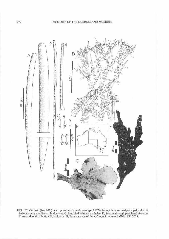
cD0
272^ MEMOIRS OF THE QUEENSLAND MUSEUM
FIG. 132. Clathria (Isociella)macropora Lendenfeld (holotype AMZ466). A, Choanosomal principal styles. B,Subectosomal auxiliary subtylostyles. C, Modified palmate isochelae. D, Section through peripheral skeleton.E, Australian distribution. F, Holotype. G, Paralectotype of Phakellia jacksoniana BMNH1887.5.2.8.

REVISION OF MICROCIONIDAE^ 273
FIG. 133. Clathria (lsociella) macropora Lendenfeld (holotype AMZ466). A, Choanosomal skeleton. B,Semi-renieroid fibres. C, Bases of subectosomal auxiliary styles. D, Modified palmate isochelae.
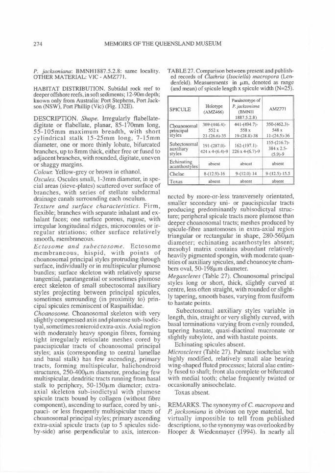
274^ MEMOIRS OF THE QUEENSLAND MUSEUM
P. jacksoniana: BMNH1887.5.2.8: same locality.OTHER MATERIAL: VIC - AMZ771.
HABITAT DISTRIBUTION. Subtidal rock reef todeeper offshore reefs, in soft sediments; 12-90m depth .
known only from Australia: Port Stephens, Port Jack-son (NSW), Port Phillip (Vic) (Fig. 132E).
DESCRIPTION. Shape. Irregularly flabellate-digitate or flabellate, planar, 85-170nrun long,55-105mm maximum breadth, with shortcylindrical stalk 15-25mm long, 7-15mmdiameter, one or more thinly lobate, bifurcatedbranches, up to 8mm thick, either free or fused toadjacent branches, with rounded, digitate, unevenor shaggy margins.Colour Yellow-grey or brown in ethanol.Oscules. Oscules small, 1-3mm diameter, in spe-cial areas (sieve-plates) scattered over surface ofbranches, with series of stellate subdermaldrainage canals surrounding each osculum.Texture and surface characteristics. Firm,flexible; branches with separate inhalant and ex-halant faces; one surface porous, rugose, withirregular longitudinal ridges, microconules or ir-regular striations; other surface relativelysmooth, membraneous.Ectosome and subectosome. Ectosomemembraneous, hispid, with points ofchoanosomal principal styles protruding throughsurface, individually or in multispicular plumosebundles; surface skeleton with relatively sparsetangential, paratangential or sometimes plumoseerect skeleton of small subectosomal auxiliarystyles projecting between principal spicules,sometimes surrounding (in proximity to) prin-cipal spicules reminiscent of Raspailiidae.Choanosome. Choanosomal skeleton with veryslightly compressed axis and plumose sub-isodic-tyal, sometimes renieroid extra-axis. Axial regionwith moderately heavy spongin fibres, formingtight irregularly reticulate meshes cored bypaucispicular tracts of choanosomal principalstyles; axis (corresponding to central lamellaeand basal stalk) has few ascending, primarytracts, forming multispicular, halichondroidstructures, 250-400p,m diameter, producing fewmultispicular, dendritic tracts running from basalstalk to periphery, 50-150Rm diameter; extra-axial skeleton sub-isodictyal with plumosespicule tracts bound by collagen (without fibrecomponent), ascending to surface, cored by uni-,pauci- or less frequently multispicular tracts ofchoanosomal principal styles; primary ascendingextra-axial spicule tracts (up to 5 spicules side-by-side) arise perpendicular to axis, intercon-
TABLE 27. Comparison between present and publish-ed records of Clathria (lsociella) macropora (Len-denfeld). Measurements in p,m, denoted as range(and mean) of spicule length x spicule width (N=25).
SPICULEHolotype
(AMZ466)
Para1ectotype ofP. jacksoniana
(BMNH1887.5.2.8)
AMZ77I
Choanosomal 3694446.4y 441-(494.7)- 350(462.3)-principal 552x 558 x 548 xstyles 21 -(26.6)-35 19 - (28.8) -38 11 -(24.5)-36
Subectosomalauxiliarystyles
191 -(287.0) -
424 x 4 -(6.4) -9162 -(197.1)-
226 x 4-(6.7)-9
155(216.7) -
384 x 2.5 -
(5.9)-9Echinatingacanthostyles absent absent absent
Chelae 8-(12.9)-16 9412.0)-14 9-(12.5)-15.5
Toxas absent absent absent—
nected by more-or- less transversely orientated,smaller secondary uni- or paucispicular tractsproducing predominantly subisodictyal struc-ture; peripheral spicule tracts more plumose thandeeper choanosomal tracts; meshes produced byspicule-fibre anastomoses in extra-axial regiontriangular or rectangular in shape, 280-560Rmdiameter; echinating acanthostyles absent;mesohyl matrix contains abundant relativelyheavily pigmented spongin, with moderate quan-tities of auxiliary spicules, and choanocyte cham-bers oval, 50-19811m diameter.Megascleres (Table 27). Choanosomal principalstyles long or short, thick, slightly curved atcentre, less often straight, with rounded or slight-ly tapering, smooth bases, varying from fusiformto hastate points.
Subectosomal auxiliary styles variable inlength, thin, straight or very slightly curved, withbasal terminations varying from evenly rounded,tapering hastate, quasi-diactinal mucronate orslightly subtylote, and with hastate points.
Echinating spicules absent.Microscleres (Table 27). Palmate isochelae withhighly modified, relatively small alae bearingwing-shaped fluted processes; lateral alae entire-ly fused to shaft; front ala complete or bifurcatedwith medial tooth; chelae frequently twisted oroccasionally anisochelate.
Toxas absent.
REMARKS. The synonymy of C. macropora andP. jacksoniana is obvious on type material, butvirtually impossible to tell from publisheddescriptions, so the synonymy was overlooked byHooper & Wiedenmayer (1994). In nearly all
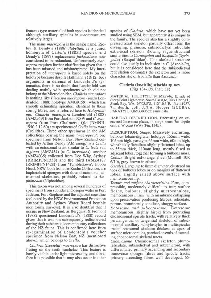
REVISION OF MICROCIONIDAE^ 275
features type material of both species is identicalalthough auxiliary spicules in macropora arerelatively larger.
The name macropora is the senior name. Rid-ley & Dendy's (1886) flabellata is a juniorhomonym of Carter's (18850 species, andDendy's (1897) replacement jacksoniana nowconsidered to be redundant. Unfortunately mac-ropora requires further clarification given that ithas been misused and misinterpreted. My inter-pretation of macropora is based solely on theholotype because despite Hallmann's (1912: 166)arguments in defense of Lendenfeld's sys-tematics, there is no doubt that Lendenfeld wasdealing mainly with specimens which did notbelong to the Microcionidae. Clathria macroporais nothing like Plectispa macropora (sensu Len-denfeld, 1888; holotype AMG9159), which hassmooth echinating spicules, identical to thosecoring fibres, and is referred here to Holopsam-ma. Clathria macropora Lendenfeld (1888)(AMZ959) from Port Jackson, NSW and C. mac-ropora from Port Chalmers, Qld (BMNH-1950.2.12.60) are specimens of Crella incrustans(Crellidae). Three other specimens in the AMcollections bearing the name macropora': onespecimen from Nelson Bay, New Zealand col-lected by Arthur Dendy (AM unreg.) is a Crellawith an ectosomal crust similar to C. levis var.digitata (AMZ454) (= C. incrustans); another(AMZ4035) collected from Dee Why, Sydney(RRIMPFN1338) and the third (AMZ4187(RRIMPFN1428)) from 'Tumbledown', JibbonHead, NSW, both from the Roche Collection, arehaplosclerid sponges with three dimensional ec-tosomal skeletons, probably related to Am-phimedon (Niphatidae).
This taxon was not among several hundreds ofspecimens from subtidal and deeper water in PortJackson, Port Stephens and the adjacent coastline(collected by the NSW Environmental ProtectionAuthority and Sydney Water Board benthicmonitoring surveys). It is also doubtful that itoccurs in New Zealand, as Bergquist & Fromont(1988) questioned Lendenfeld's (1888) recordgiven that it was not subsequently rediscoveredduring their substantial contemporary collectionsof the NZ fauna. This is confirmed here fromre-examination of Lendenfeld's voucherspecimen from Nelson Bay, NZ (mentionedabove), which belongs to Crella.
Clathria (Isociella) macropora has distinctivefluting on the teeth isochelae. This feature isbarely visible under light microscopy, and there-fore it is possible that it may also occur in other
species of Clathria, which have not yet beenstudied using SEM, but apparently it is unique tothe family. The species also has a slightly com-pressed axial skeleton partially offset from thediverging, plumose, subisodictyal reticulateextra-axial skeleton, showing vague structuralsimilarities to Ceratopsion and Raspailia (Syrin-gella) (Raspailiidae). This skeletal structurecould also justify its inclusion in C. (Axociella),but it is considered here that the subisodictyalreticulation dominates the skeleton and is morecharacteristic of lsociella than Axociella.
Clathria (Isociella) selachia sp. nov.(Figs 134-135, Plate 3F)
MATERIAL. HOLOTYPE: NTMZ2946: E. side ofSteep Point Lighthouse, South Passage, Dirk Hartog I.,Shark Bay, WA, 26°08.5'S, 113 ° 10.3'E, 13.vii.1987,7m depth, coll. J.N.A. Hooper (SCUBA).PARATYPE: QMG300562: same data.
HABITAT DISTRIBUTION. Encrusting on ex-cavated limestone plates, in surge zone; 7m depth;central W coast (WA) (Fig. 134E).
DESCRIPTION. Shape. Massively encrusting,bulbous lobate-digitate, holotype 155mm wide,105mm high, paratype 65mm wide, 60mm high,with thickly flabellate, slightly flattened lobes, upto 55mm thick, 110mm long, mostly fused toadjacent lobes, together forming a bulbous mass.Colour Bright red-orange alive (Munsell lOR6/10), grey-brown in ethanol.Oscules. Large, up to 8mm diameter, clustered ontops of bulbous lobes or on margins of flattenedlobes, slightly raised above surface withmembraneous lip.Texture and surface characteristics. Firm, com-pressible, moderately difficult to tear; surfacefleshy, bulbous, slightly microconulose,membraneous in situ, with membrane collapsingupon preservation producing fibrous, reticulate,porous, prominently conulose, shaggy surface.Ectosome and subectosome. Ectosomemembraneous, slightly hispid from protrudingchoanosomal spicule tracts, with relatively thickparatangential or tangential skeleton of subec-tosomal auxiliary subtylostyles in multispiculartracts; ectosomal skeleton thickest at apex ofsurface microconules, perched on ends of ascend-ing choanosomal skeletal tracts.Choanosome. Choanosomal skeleton plumo-reticulate, subisodictyal and subrenieroid, withdifferentiated primary ascending and secondarytransverse spongin fibres and spicule tracts;primary ascending fibres well developed, 65-
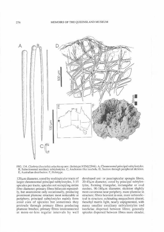
0
276^ MEMOIRS OF THE QUEENSLAND MUSEUM
c
FIG. 134. Clathria (Isociella) selachia sp.nov. (holotype NTMZ2946). A, Choanosomal principal subtylostyles.B, Subectosomal auxiliary subtylostyles. C, Anchorate-like isochela. D, Section through peripheral skeleton.E, Australian distribution. F, Holotype.
A
1201J,m diameter, cored by multispicular tracts oflarger choanosomal principal subtylostyles, 3-15spicules per tracts; spicules not occupying entirefibre diameter; primary fibres bifurcate repeated-ly, but anastomose only occasionally, producingprominent plumose structure most noticeable atperiphery; principal subtylostyles mainly formaxial core of spicules but sometimes theyprotrude through primary fibres producingplumose brushes; primary fibres interconnectedat more-or-less regular intervals by well
developed uni- or paucispicular spongin fibres,20-451im diameter, cored by principal subtylos-tyles, forming triangular, rectangular or ovalmeshes, 90-180p.m diameter; skeleton slightlymore cavernous near periphery, more plumose instructure; fibres heaviest in axis, more subisodic-tyal in structure; echinating megascleres absent;mesohyl matrix light, nearly unpigmented, withmany smaller auxiliary subtylostyles andisochelae dispersed between fibres; generallyspicules dispersed between fibres more slender,
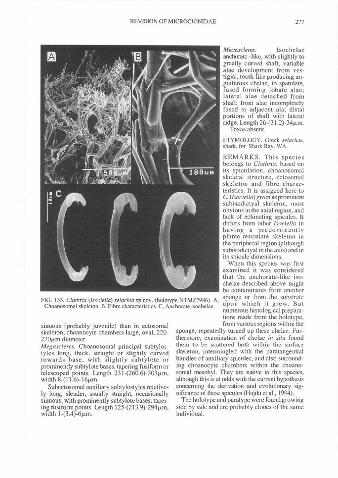
FIG. 135. Clathria (Isociella) selachia sp.nov. (holotype NTMZ2946). A,Choanosomal skeleton. B, Fibre characteristics. C, Anchorate isochelae.
REVISION OF MICROCIONIDAE^ 277
sinuous (probably juvenile) than in ectosomalskeleton; choanocyte chambers large, oval, 220-27011m diameter.Megascleres. Choanosomal principal subtylos-tyles long, thick, straight or slightly curvedtowards base, with slightly subtylote orprominently subtylote bases, tapering fusiform ortelescoped points. Length 231-(260.6)-303Rm,width 8-(11.6)-16vim.
Subectosomal auxiliary subtylostyles relative-ly long, slender, usually straight, occasionallysinuous, with prominently subtylote bases, taper-ing fusiform points. Length 125-(213.9)-294Rm,width 1-(3.4)-6Rm.
Microscleres. Isochelaeanchorate -like, with slightly togreatly curved shaft, variablealae development from ves-tigial, tooth-like producing un-guiferous chelae, to spatulate,fused forming lobate alae;lateral alae detached fromshaft; front alae incompletelyfused to adjacent ala; distalportions of shaft with lateralridge. Length 26-(31.2)-34p.m.
Toxas absent.
ETYMOLOGY. Greek selachos,shark, for Shark Bay, WA.
REMARKS. This speciesbelongs to Clathria, based onits spiculation, choanosomalskeletal structure, ectosomalskeleton and fibre charac-teristics. It is assigned here toC. (Isociella) given its prominentsubisodictyal skeleton, mostobvious in the axial region, andlack of echinating spicules. Itdiffers from other Isociella inhaving a predominentlyplumo-reticulate skeleton inthe peripheral region (althoughsubisodictyal in the axis) and inits spicule dimensions.
When this species was firstexamined it was consideredthat the anchorate-like iso-chelae described above mightbe contaminants from anothersponge or from the substrateupon which it grew. Butnumerous histological prepara-tions made from the holotype,from various regions within the
sponge, repeatedly turned up these chelae. Fur-thermore, examination of chelae in situ foundthem to be scattered both within the surfaceskeleton, intermingled with the paratangentialbundles of auxiliary spicules, and also surround-ing choanocyte chambers within the choano-somal mesohyl. They are native to this species,although this is at odds with the current hypothesisconcerning the derivation and evolutionary sig-nificance of these spicules (Hajdu et al., 1994).
The holotype and paratype were found growingside by side and are probably clones of the sameindividual.
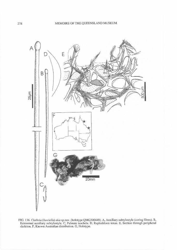
278^ MEMOIRS OF THE QUEENSLAND MUSEUM
FIG. 136. Clathria (lsociella) skia sp.nov. (holotype QMG300449). A, Auxiliary subtylostyle (coring fibres). B,Ectosomal auxiliary subtylostyle. C, Palmate isochela. D, Raphidiform toxas. E, Section through peripheralskeleton. F, Known Australian distribution. G, Holotype.
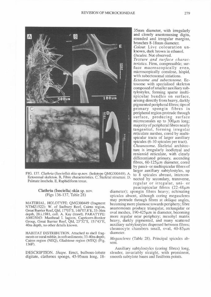
FIG. 137. Clathria (Isociella) skia sp.nov. (holotype QMG300449). A,Ectosomal skeleton. B, Fibre characteristics. C, Skeletal structure. D,Palmate isochela. E, Raphidiform toxas.
REVISION OF MICROCIONIDAE^ 279
Clathria (Isociella) skia sp. nov.(Figs 136-137, Table 28)
MATERIAL. HOLOTYPE: QMG300449 (fragmentNTMZ1522): W. of Sudbury Reef, Cairns region,Great Barrier Reef, Qld, 17 °03'S, 146°07.8'E, 33-36mdepth, 28.i.1981, coll. A. Kay (trawl). PARATYPE:AMG5043: Masthead I. lagoon, Capricorn-BunkerGroup, Great Barrier Reef, Qld, 23 °32'S, 151 °43'E,40m depth, no other details known.
HABITAT DISTRIBUTION. Attached to shell frag-ments or coral rubble, in soft sediments; 33-40m depth;Cairns region (NEQ), Gladstone region (MEQ) (Fig.136F).
DESCRIPTION. Shape. Erect, bulbous-lobatedigitate, clathrous sponge, 45-95mm long, 18-
35mm diameter, with irregularlyand closely anastomosing digits,rounded and irregular margins,branches 8-18rrim diameter.Colour. Live colouration un-known, dark brown in ethanol.Oscules. Not observed.Texture and surface charac-teristics. Firm, compressible; sur-face macroscopically even,microscopically conulose, hispid,with subectosomal striations.Ectosome and subectosome. Ec-tosome with specialised skeletoncomposed of smaller auxiliary sub-tylostyles, forming sparse multi-spicular bundles on surface,arising directly from heavy, darklypigmented peripheral fibres; tips ofprimary spongin fibres inperipheral region protrude throughsurface, producing surfacemicroconules up to 300pLm long;majority of peripheral fibres nearlytangential, forming irregularreticulate meshes, cored by multi-spicular tracts of larger auxiliaryspicules (6-10 spicules per tract).Choanosome. Skeletal architec-ture is irregularly isodictyal andrenieroid reticulate, with clearlydifferentiated primary, ascendingfibres, 60-125Rm diameter, coredby pauci- or multispicular fibres oflarger auxiliary subtylostyles, upto 8 spicules abreast, intercon-nected by secondary, transverse,regular or irregular, uni- orpaucispicular fibres (22-48p.m
diameter); spongin fibres heavy; echinatingspicules absent, although coring megascleresmay protrude through fibres at oblique angles,becoming more plumose towards periphery; fibreanastomoses produce triangular, rectangular oroval meshes, 190-425Rm in diameter, becomingmore regular near periphery; mesohyl matrixheavy, darkly pigmented, and many scatteredauxiliary subtylostyles dispersed between fibres;choanocyte chambers small, oval, 40-85 p.mdiameter.Me gascleres (Table 28). Principal spicules ab-sent.
Auxiliary subtylostyles (coring fibres) long,slender, invariably straight, with prominent,smooth subtylote bases and fusiform points.
280^ MEMOIRS OF THE QUEENSLAND MUSEUM
Ectosomal auxiliary subtylostyles significant-ly smaller than coring spicules but otherwise ofidentical geometry.
Echinating spicules absent.Microscleres (Table 28). Palmate isochelae un-common, unmodified, with long lateral alae en-tirely fused to shaft and front ala completelyfused, slightly spatulate.
Toxas raphidiform, extremely thin, slightlycurved at centre, sometimes nearly straight orbow shaped, rarely asymmetrical.
ETYMOLOGY. Greek skias , dark, dim.
REMARKS. This species is a greatly reducedClathria, lacking true principal spicules, echinat-ing spicules and having containing relativelysparse microscleres. Conversely its spongin fibresystem is well developed and fibres are heavy. Itis assigned to the Isociella group on the basis thatits skeletal structure is essentially subisodictyal,and it lacks echinating spicules, but it is admittedthat its reduced characteristics make its true af-finities speculative.
The sparsely developed ectosomal skeleton,composed of plumose bundles of smallerauxiliary styles, also indicates affinities to theThalysias group, but in most respects (spiculegeometry, skeletal structure, fibre developmentand growth form) it differs from all other speciesof either lsociella and Thalysias. Clathria(Isociella) skia was initially thought to be Am-philectus hispidulus Ridley, from Torres Strait(FNQ). From both published descriptions of A.hispidulus (Ridley, 1884a; Hentschel, 1911) itapparently lacked echinating megascleres, lackedprincipal spicules and had well developed sub-isodictyal skeletal structure, but re-examinationof the holotype showed that it does have echinat-ing acanthostyles, and differentiated principaland auxiliary megascleres (thus more ap-propriately included in Clathria (Clathria); seeabove), and it also has substantially different fibrecharacteristics from C. (I.) skia indicating thatthey are not conspecific.
OTHER SPECIES OF CLATHRIA(ISOCIELLA)
Clathria (Isociella) incrustans (Bergquist, 1961)Isociella incrustans Bergquist, 1961a: 42-43, text-figs
15a-b [Ahipara Bay, N. New Zealand; originallyassigned to the Suberitidae, Hadromerida].Bergquist & Fromont, 1988: 114-116, p1.53, figs e-f,p1.54, figs a-b; Dawson, 1993: 36.
MATERIAL. HOLOTYPE: NMNZ unregistered. NZ.
TABLE 28. Comparison between of Clathria (lsociel-la) skia sp.nov. Measurements in p.m, denoted asrange (and mean) of spicule length x spicule width(N=25).
SPICULEHolotype
(QMG300449)Paratype
(AMG5043)
Choanosomalprincipal styles
absent absent
Auxiliary I62-(248.4)-368x 1544242.1)-318 x(coring) styles 2.5-(4.5)-8 244.1)-6
Auxiliary 97-(111.9)-129 x 87-(110.2)-132 x(ectosomal) styles l.542.4)-4 1.5-(2.4)-4
Echinatingacanthostyles
absent absent
Chelae 9-(15.3)-18 14-(16.3)-I9
724104.4)-145 x 65-(81.1)-108 xToxas
0.240.4)-0.8 0.2-(0.5)-0.8
Clathria (Thalysias)Duchassaing & Michelotti, 1864
Thalysias Duchassaing & Michelotti, 1864: 82.Rhaphidophlus Ehlers, 1870: 19.Tenacia Schmidt, 1870: 56.Echinonema Carter, 1881a: 378.? Thalassodendron Lendenfeld, 1888: 222.StylotellopsisThiele, 1905:456; de Laubenfels, 1936a:
112.Colloclathria Dendy, 1922: 74.Damoseni de Laubenfels, 1936a: 110.
DEFINITION. Specialised ectosomal skeletoncomposed of two size classes of auxiliary (sub-tylo)styles, with smaller ectosomal spiculesusually overlaying larger subectosomal onesforming a continuous palisade, or discretebundles, mainly erect, sometimes paratangential,or rarely tangential to surface; choanosomalskeleton without any marked differentiation be-tween axial and extra-axial regions; echinatingacanthostyles usually present.
TYPE SPECIES. Spongia juniperina Lamarck, 1814:444 (by synonymy).
REMARKS. Of 137 named species described in,or referred to Thalysias or one of its synonyms,93 are thought to be valid of which 37 arerecorded here from Australian waters including10 new species.
Clathria (Thalysias) abietina (Lamarck, 1814)(Figs 138-141, Tables 29-30, Plate 4C-D)
Spongia abietina Lamarck, 1814: 450, 377.Clathriaabietina; de Laubenfels, 1954: 141-142, text-
fig.90; Hooper & Wiedenmayer, 1994: 267.
REVISION OF MICROCIONIDAE^ 281
Clathria aculeata Ridley, 1884a: 443-444, p1.40, fig. 1,p1.42, fig.k; Ridley & Dendy, 1887: 147, 246, 254;Topsent, 1897b: 447; Burton, 1934a: 558; Burton,1959a: 243; Levi & Levi, 1989: 80-81, p1.7, fig.3.
Rhaphidophlus aculeatus; Topsent, 1932: 115, p1.4,fig. 10; Vacelet & Vasseur, 1977: 114; Vacelet et al.,1976: 73-74.
Rhaphidophlus cervicornis, in part; Vacelet & Vas-seur, 1971: 73.
cf. Microciona prolifera; Vosmaer, 1935a: 610, 633,664.
MATERIAL. LECTOTYPE: MNHNDT634: preciselocality unknown (suspected to be 'Australia'; Topsent(1932: 115)). PARALECTOTYPES: MNHNDT3342,3343: same details. HOLOTYPE of C. aculeata:BMNH1882.2. 23.258: Torres Strait, Qld, 9°41'S,142°17'E, 6-8m depth, coll. HMS 'Alert' (dredge).OTHER MATERIAL. MICRONESIA - USNM22808, USNM23090. QLD - BMNH1887.5.2.110,QMG300791. DARWIN HARBOUR, NT -NTMZ226, NTMZ426, NT MZ458, NTMZ468,NTMZ498, NTMZ512, NTMZ515, NTMZ886,NTMZ903, NTMZ928, NTMZ955, NTMZ2050,NTMZ2078, NTMZ 2079, NTMZ2085, NTMZ2258,NTMZ2390, NTMZ2391, NTMZ2395, NTMZ2399,NTMZ 2611, NTMZ2642, QMG300169, NTMZ2646,QMG304077, NTMZ I 943, NTMZ1958, NTMZ 1963,QMG303373, QMG303382, NTMZ2089, NTM-Z2161, NTMZ2163, NTMZ2186, NTMZ2191,NTMZ2194, NTMZ2195, NTMZ820, NTMZ835,QMG300414. BYNOE HARBOUR, NT - NTMZ1073,NTMZ2106, QMG303447, QMG303534. SHOALBAY, NT - QMG303539, QMG303571. TIMOR SEA,NT - NTMZ3090. PORT ESSINGTON, COBOURGPENINSULA, NT - NTMZ68, NTMZ69, NTMZ90,NTMZ1393, NTMZ3304, NTMZ577, NTMZ 1328,NTMZ1329, NTMZ1330, NTMZ1331, NTMZ1332,NTMZ1333, NTMZ1334, NTMZ 1343, NTMZ2500,NTMZ2501, NTMZ2509, NTMZ2510, NTMZ3245,NTMZ3255, NTMZ 3258, NTMZ3260, NTMZ3268,NTMZ3272, NTMZ3277, NTMZ3278, NTMZ3284,NTMZ 3289, NTMZ3295, QMG300386, NTMZ1352.ARAFURA SEA, NT - NTMZ2521, NTMZ 2522,NTMZ2523, NTMZ129, NTMZ130, NTMZ138.WESSEL ISLANDS, NT - NTMZ 3902, QMG300764(NCIQ66C-4692-Q), NTMZ3921, NTMZ3930,QMG300757 (NCIQ 66C-4773-F), QMG300508(NCIQ66C-4772-C), NTMZ3947, QMG300765(NCIQ66C-4808-R). NORTHWEST SHELFREGION, WA - NTMZ1036, NTMZ1209, NTMZ-1217, NTMZ 1244, NTMZ1272, NTMZ1314,NTMZ1411, NTMZ1423, WAM151-82, WAM155-82 (fragments NTMZ1731, NTMZ1732), NTMZ1770,NTMZ1801, NTMZ1820, NTMZ1824, NTMZ 1852,NTMZ2272, NTMZ2329, NTMZ2349, NTMZ2486,NTMZ3017, NTMZ3030, NTMZ 3031, NTMZ3032,NTMZ3033, NTMZ3396, QMG300448 (NCIQ66C-1517-P), QMG300117 (NCIQ66C1518-Q) (fragmentsNTMZ3488, NTMZ3489), PIB0004-595 (fragmentQMG300051)
HABITAT DISTRIBUTION. Shallow-water0-25mdepth, predominantly on sides and tops of rock anddead coral heads, invariably exposed to currents.Deeper-water specimens (26-86m depth) mostly as-sociated with exposed rock substrates in gravel, silt orshell-grit substrates; central SW Pacific Ocean (LowIsles, Great Barrier Reef (Burton, 1934a), Torres Strait(Ridley, 1884a; Ridley & Dendy, 1887)); E IndianOcean (Arafura Sea, Timor Sea, mid-WA coast(present study)); also tropical Indo-west Pacific:central NW. Pacific Ocean (Marshall and Caroline Is(de Laubenfels, 1954), Philippines (Levi & Levi,1989)); W. Indian Ocean (Madagascar (Vacelet et al.,1976, 1977), Red Sea (Burton, 1959a)). WithinAustralian waters this species extends across the N andNW coasts, from the Cairns region, Torres Strait andGulf of Carpentaria, Qld, to the Exmouth Gulf region,WA (Fig. 138J). It is only rarely encountered on the EQld. coast and must be considered a predominantlyIndian Ocean species.
DESCRIPTION. Shape. Arborescent; thicklycylindrical digitate branches on long or short,thick stalk (3-21mm basal diameter) with ex-panded basal attachment, never rhizomous;branches rarely anastomosing, branching mostlyirregular, bushy, occasionally regular growing inI plane, sometimes flagelliform, occasionally ex-panded, club-shaped; total length and branchdiameter variable (50-460mm; 2-22mm, respec-tively).Colour. Live colouration highly variable, rangingfrom maroon, red, orange, yellow, brown to grey;colour in ethanol ranges from black, brown togrey (Munsell values given below); pigmentsoxidise in air; maroon colouration is highly al-cohol soluble; yellow pigmentation more stablein ethanol but easily scratched from surface ofliving sponge, leaving a maroon or mauve pig-mentation beneath; pigmentation extends intoperiphery of choanosomal mesohyl.Oscules. Exhalent pores usually very small, 0.6-2.0mm diameter, barely visible optically, dis-persed on distal ends of branches surrounded bysurface conules, occasionally scattered along en-tire lateral margins of branches.Texture and surface characteristics. Firm, barelycompressible; branches rubbery whereas stalkmore rigid; surface dense, entirely opaque in life,with characteristic regularly scattered surfaceconules; conules tapering, distally rounded orblunt, usually absent from basal and distal ex-tremities of branches, 2-6mm maximum basaldiameter.Ectosome and subectosome. Very dense, con-tinuous palisade of small ectosomal auxiliarysubtylostyles forming discrete brushes overlay-
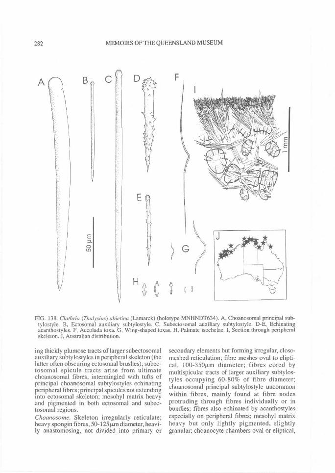
282^ MEMOIRS OF THE QUEENSLAND MUSEUM
FIG. 138. Clathria (Thalysias) abietina (Lamarck) (holotype MNHNDT634). A, Choanosomal principal sub-tylostyle. B, Ectosomal auxiliary subtylostyle. C, Subectosomal auxiliary subtylostyle. D-E, Echinatingacanthostyles. F, Accolada toxa. G, Wing-shaped toxas. H, Palmate isochelae. I, Section through peripheralskeleton. J, Australian distribution.
ing thickly plumose tracts of larger subectosomalauxiliary subtylostyles in peripheral skeleton (thelatter often obscuring ectosomal brushes); subec-tosomal spicule tracts arise from ultimatechoanosomal fibres, intermingled with tufts ofprincipal choanosomal subtylostyles echinatingperipheral fibres; principal spicules not extendinginto ectosomal skeleton; mesohyl matrix heavyand pigmented in both ectosomal and subec-tosomal regions.Choanosome. Skeleton irregularly reticulate;heavy spongin fibres, 50-125 t.i.m diameter, heavi-ly anastomosing, not divided into primary or
secondary elements but forming irregular, close-meshed reticulation; fibre meshes oval to elipti-cal, l00-350ttm diameter; fibres cored bymultispicular tracts of larger auxiliary subtylos-tyles occupying 60-80% of fibre diameter;choanosomal principal subtylostyle uncommonwithin fibres, mainly found at fibre nodesprotruding through fibres individually or inbundles; fibres also echinated by acanthostylesespecially on peripheral fibres; mesohyl matrixheavy but only lightly pigmented, slightlygranular; choanocyte chambers oval or eliptical,
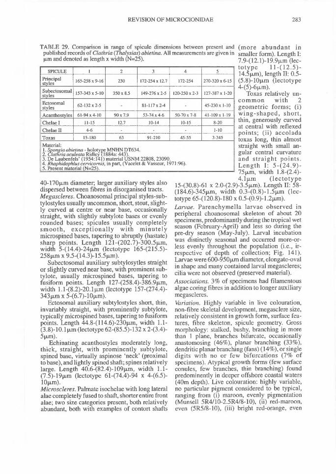
TABLE 29. Comparison in range of spicule dimensions between present andpublished records of Clathria (Thalysias) abietina. All measurements are given inp.m and denoted as length x width (N=25).
SPICULE 1 2 3 4 5
Principalstyles 165-258 x 9-16 230 172-254x 12.7 172-254 270-320 x 6-15
Subectosomalstyles 157-343 x 5-10 350 x 8.5 149-276 x 2-5 120-250 x 2-3 127-387 x 1-20
Ectosomalstyles 62-132 x 2-5 - 81-117 x 2-4 45-230x 1-10
Acanthostyles 61-94 x 4-10 90 x 7.9 53-74 x 4-6 50-70 x 7-8 41-109 x 1-19
Chelae I 11-15 12.7 10-14 10-15 8-20
Chelae II 4-6 1-10
Toxas 15-180 63 91-210 45-55 3-345
Material:1. Spongia abietina - holotype MNHN DT634.2. Clathria aculeata Ridley (1884a: 443).3. De Laubenfels' (1954:141) material USNM 22808, 23090.4. Rhaphidophlus cervicornis, in part, (Vacelet & Vasseur, 1971:96).5. Present material (N=25).
REVISION OF MICROCIONIDAE^ 283
40-170p.m diameter; larger auxiliary styles alsodispersed between fibres in disorganised tracts.Megascleres. Choanosomal principal styles-sub-tylostyles usually uncommon, short, stout, slight-ly curved at centre or near base, occasionallystraight, with slightly subtylote bases or evenlyrounded bases; spicules usually completelysmooth, exceptionally with minutelymicrospined bases, tapering to abruptly (hastate)sharp points. Length 1214202 .7)-300.5p,m,width 5-(14.4)-24pm (lectotype 1654215.5)-258pm x 9.5-(14.3)-15.5pm).
Subectosomal auxiliary subtylosytles straightor slightly curved near base, with prominent sub-tylote, usually microspined bases, tapering tofusiform points. Length 127-(258.4)-386.9p,m,width 1.148.2)-20.1 p.m (lectotype 1574274.4)-343pm x 5-(6.7)-10p,m).
Ectosomal auxiliary subtylostyles short, thin,invariably straight, with prominently subtylote,typically microspined bases, tapering to fusiformpoints. Length 44.8-(114.6)-230p,m, width 1.1-(3.8)-10.1pm (lectotype 62-(85.5)-132 x 243.4)-5p,m).
Echinating acanthostyles moderately long,thick, straight, with prominently subtylote,spined base, virtually aspinose 'neck' (proximalto base), and lightly spined shaft; spines relativelylarge. Length 40.6-(82.4)-109pm, width 1.1-(7.5)-19pm (lectotype 61-(74.4)-94 x 446.5)-1011,m).Microscleres. Palmate isochelae with long lateralalae completely fused to shaft, shorter entire frontalae; two size categories present, both relativelyabundant, both with examples of contort shafts
(more abundant insmaller form). Length I:7.9412.1)-19.9pm (lec-totype 11 -(12 .5 )-14.5p,m), length II: 0.5-(5.8)-10Rm (lectotype4-(5)-6p,m).
Toxas relatively un-common with 2geometric forms; (i)wing-shaped, short,thin, generously curvedat central with reflexedpoints; (ii) accoladatoxas long, thin almoststraight with small an-gular central curvatureand straight points.Length I: 5424.9)-75pm, width 1.8-(2.4)-4.1pm (lectotype
15-(30.8)-61 x 2.0-(2.9)-3.5p.,m). Length II: 58-(184.6)-345p,m, width 0.3-(0.8)-1.5pm (lec-totype 65-(120.8)-180 x 0.540.9)-1.211m).Larvae. Parenchymella larvae observed inperipheral choanosomal skeleton of about 20specimens, predominantly during the tropical wetseason (February-April) and less so during thepre-dry season (May-July). Larval incubationwas distinctly seasonal and occurred more-or-less evenly throughout the population (i.e., ir-respective of depth of collection; Fig. 141).Larvae were 600-950pm diameter, elongate-ovalin shape and many contained larval megascleres;cilia were not observed (preserved material).Associations. 3% of specimens had filamentousalgae coring fibres in addition to longer auxiliarymegascleres.Variation. Highly variable in live colouration,non-fibre skeletal development, megasclere size,relatively consistent in growth form, surface fea-tures, fibre skeleton, spicule geometry. Grossmorphology: stalked, bushy, branching in morethan 1 plane, branches bifurcate, occasionallyanastomosing (46%), planar branching (33%),dendritic planar branching (fans) (14%), or singledigits with no or few bifurcations (7% ofspecimens). Atypical growth forms (few surfaceconules, few branches, thin branching) foundpredominently in deeper offshore coastal waters(40m depth). Live colouration: highly variable,no particular pigment considered to be typical,ranging from (i) maroon, evenly pigmentation(Munsell 5R4/10-2.5R4/8-10), (ii) red-maroon,even (5R518-10), (iii) bright red-orange, even
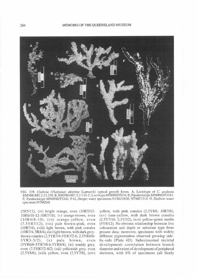
284^ MEMOIRS OF THE QUEENSLAND MUSEUM
FIG. 139. Clathria (Thalysias) abietina (Lamarck) typical growth forms. A, Lectotype of C. aculeataBMNH1882.2.23.258. B, BMNH1887.5.2.110. C, Lectotype MNHNDT634. D, Paralectotype MNHNDT3343.E, Paralectotype MNHNDT3342. F-G, Deeper water specimens NTMZ1820, NTMZ1314. H, Shallow-waterspecimen NTMZ68.
(5R5/12), (iv) bright orange, even (10R5112-10R6/10-12-10R7/10), (v) orange-brown, even(10R418-10), (vi) orange-yellow, even(7.5YR7112), (vii) pale brown-pink, even(10R7/4), (viii) light brown, with pink conules(10R7/4, 5R8/4), (ix) light brown, with dark grey-brown conules (2.5YR714-5YR712-6, 2.5YR616-5 YR3-5/2), (x) pale brown, even(5YR616-5YR7/4-6-5YR8/4), (xi) muddy grey,even (7.5YR7/2-8/2) (xii) yellowish grey, even(2.5Y816), (xiii) yellow, even (2.5Y7/8), (xiv)
yellow, with pink conules (2.5Y8/6, 10R7/6),(xv) lime-yellow, with dark brown conules(2.5Y7/10, 2.5Y5/2), (xvi) yellow-green mottle(5Y8/12). No obvious relationship between livecolouration and depth or substrate type frompresent data; moreover, specimens with widelydifferent pigmentation observed growing side-by-side (Plate 4D). Subectosomal skeletaldevelopment: correlation between branchdiameter and extent of development of peripheralskeleton, with 8% of specimens (all thinly
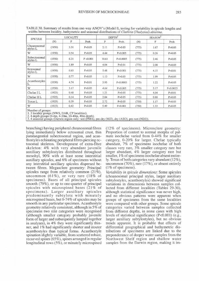
REVISION OF MICROCIONIDAE^ 285
TABLE 30. Summary of results from one-way ANOV's (Model I), testing for variability in spicule lengths andwidths between locality, bathymetric and seasonal distributions of Clathria (Thalysias) abietina.
SPICULE LOCALITY DEPTH2 SEASON3(N) F Prob. F Prob. (N) F Prob.
Choanosomalstyles L (1950) 3.31 P<0.05 2.11 P>0.05 (775) 1.67 P>0.05
W (1950) 0.54 P>0.05 4.44 P<0.005 (775) 0.26 P>0.05Subectosomalstyles L (1950) 6.21 P<0.001 10.61 P<0.0005 (775) 2.46 P>0.05
W (1950) 1.89 P>0.05 4.04 P<0.01 (775) 2.08 P>0.05Ectosomalstyles L (1950) 0.85 P>0.05 5.48 P<0.001 (775) 4.15 P<0.01
W (1950) 0.77 P>0.05 1.13 P>0.05 (775) 1.99 P>0.05
AcanthostylesL (1950) 4.74 P<0.01 5.95 P<0.0005 (775) 1.22 P>0.05
W (1950) 3.17 P<0.05 4.64 P<0.005 (775) 5.17 P<0.0025
Chelae I L (1925) 0.48 P>0.05 1.21 P>0.05 (775) 4.04 P<0.01
Chelae II L (1925) 0.14 P>0.05 2.04 P>0.05 (775) 0.88 P>0.05Toxas L (1925) 0.39 P>0.05 2.72 P<0.05 (750) 1.17 P>0.05
W (1925) 0.42 P>0.05 5.49 P<0.001 (750) 1.33 P>0.05
Number of groups:1. 3 locality groups (NWS, DAR, CP localities).2.4 depth groups (0-4m, 4-10m, I0-40m, 40m depth).3. 4 seasonal groups (Darwin region only: wet (FMA), pre-dry (MJJ), dry (ASO), pre-wet (NDJ)).
branching) having peripheral choanosomal fibreslying immediately below ectosomal crust, thinparatangential subectosomal region, and acan-thostyles echinating peripheral fibres piercing ec-tosomal skeleton. Development of extra-fibreskeleton: 4% with very abundant juvenileauxiliary subtylostyles distributed throughoutmesohyl, 90% with at least some interstitialauxiliary spicules, and 6% of specimens withoutany interstitial auxiliary spicules dispersed be-tween fibres. Megasclere geometry: Principalspicules range from relatively common (21%),uncommon (61%), or very rare (18% ofspecimens). Bases of all principal spiculessmooth (79%), or up to one-quarter of principalspicules with microspined bases (21% ofspecimens). Larger auxiliary spiculespredominantly subtylote with minutelymicrospined bases, but 0-74% of spicules may besmooth in any particular specimen. Acanthostylegeometry relatively consistent, although in 5% ofspecimens two size categories were recognised(although smaller category probably juvenileform of larger and subsequently lumped togetherin analyses), in 4% they were significantly thin-ner, and 1% had significantly shorter and stouteracanthostyles than typical forms. Acanthostylespination slightly variable, from scattered robust,recurved spines (65%), spines arranged in regularlongitudinal rows (5%), or minutely microspined
(12% of specimens). Microsclere geometry:Proportion of contort to normal morphs of pal-mate isochelae varied from 0-44% for smallercategory, 0-20% for larger. Chelae typicallyabundant, 7% of specimens isochelae of bothclasses very rare, 3% smaller category rare butlarger abundant, 4% larger category rare butsmaller, 1% of specimens isochelae absent entire-ly. Toxas of both categories very abundant (12%),uncommon (70%), rare (17%), or absent entirely(1% of specimens).Variability in spicule dimensions: Some spicules(choanosomal principal styles, larger auxiliarysubtylostyles, acanthostyles) showed significantvariations in dimensions between samples col-lected from different localities (Tables 29-30),although statistical significance was never high,and no obvious patterns were apparent whengroups of specimens from the same localitieswere compared with other groups. Some spiculecategories varied between samples collectedfrom different depths, in some cases with highlevels of statistical significance (P<0.001) (e.g.,larger auxiliary subtylostyles), but no obvioustrends apparent. It is probable that effects ofdifferential geographical and bathymetric dis-tributions of specimens are linked due to thepreponderance of deeper water samples from theNorthwest Shelf region and shallow watersamples from the Darwin region, making it im-
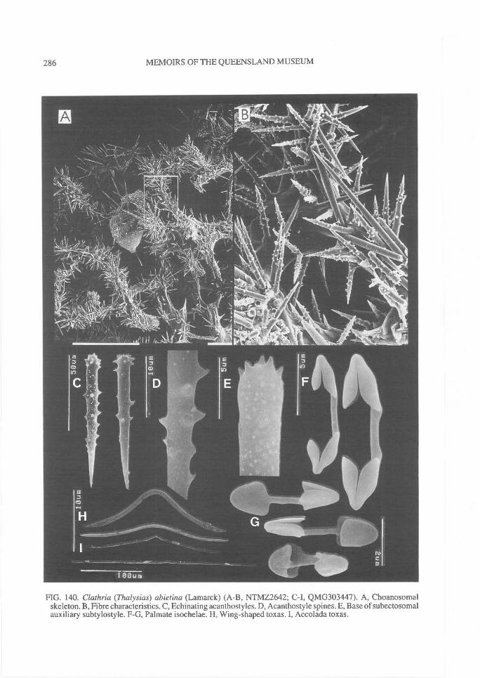
286^ MEMOIRS OF THE QUEENSLAND MUSEUM
FIG. 140. Clathria (Thalysias) abietina (Lamarck) (A-B, NTMZ2642; C-I, QMG303447). A, Choanosomalskeleton. B, Fibre characteristics. C, Echinating acanthostyles. D, Acanthostyle spines. E, Base of subectosomalauxiliary subtylostyle. F-G, Palmate isochelae. H, Wing-shaped toxas. I, Accolada toxas.
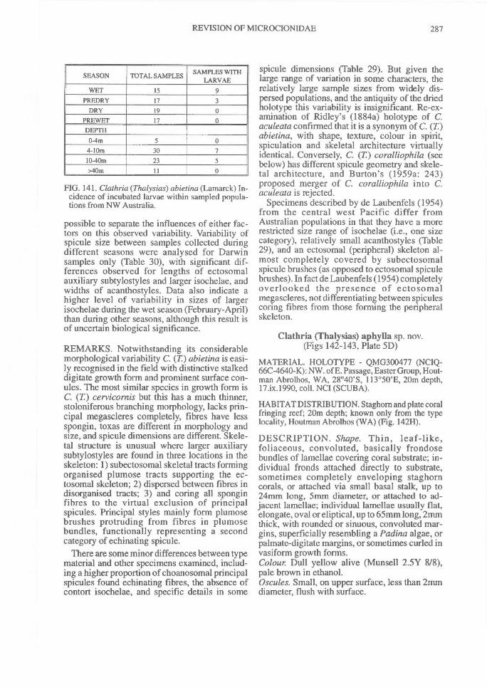
REVISION OF MICROCIONIDAE^ 287
SEASON TOTAL SAMPLES SAMPLES WITHLARVAE
WET 15 9PREDRY 17 3
DRY 19 0PREWET 17 0DEPTH
0-4m 5 04-10m 30 710-40m 23 5>40m 11 0
FIG. 141. Clathria (Thalysias) abietina (Lamarck) In-cidence of incubated larvae within sampled popula-tions from NW Australia.
possible to separate the influences of either fac-tors on this observed variability. Variability ofspicule size between samples collected duringdifferent seasons were analysed for Darwinsamples only (Table 30), with significant dif-ferences observed for lengths of ectosomalauxiliary subtylostyles and larger isochelae, andwidths of acanthostyles. Data also indicate ahigher level of variability in sizes of largerisochelae during the wet season (February-April)than during other seasons, although this result isof uncertain biological significance.
REMARKS. Notwithstanding its considerablemorphological variability C. (T.) abietina is easi-ly recognised in the field with distinctive stalkeddigitate growth form and prominent surface con-ules. The most similar species in growth form isC. (T) cervicornis but this has a much thinner,stoloniferous branching morphology, lacks prin-cipal megascleres completely, fibres have lessspongin, toxas are different in morphology andsize, and spicule dimensions are different. Skele-tal structure is unusual where larger auxiliarysubtylostyles are found in three locations in theskeleton: 1) subectosomal skeletal tracts formingorganised plumose tracts supporting the ec-tosomal skeleton; 2) dispersed between fibres indisorganised tracts; 3) and coring all sponginfibres to the virtual exclusion of principalspicules. Principal styles mainly form plumosebrushes protruding from fibres in plumosebundles, functionally representing a secondcategory of echinating spicule.
There are some minor differences between typematerial and other specimens examined, includ-ing a higher proportion of choanosomal principalspicules found echinating fibres, the absence ofcontort isochelae, and specific details in some
spicule dimensions (Table 29). But given thelarge range of variation in some characters, therelatively large sample sizes from widely dis-persed populations, and the antiquity of the driedholotype this variability is insignificant. Re-ex-amination of Ridley's (1884a) holotype of C.aculeata confirmed that it is a synonym of C. (T)abietina, with shape, texture, colour in spirit,spiculation and skeletal architecture virtuallyidentical. Conversely, C. (T) coralliophila (seebelow) has different spicule geometry and skele-tal architecture, and Burton's (1959a: 243)proposed merger of C. coralliophila into C.aculeata is rejected.
Specimens described by de Laubenfels (1954)from the central west Pacific differ fromAustralian populations in that they have a morerestricted size range of isochelae (i.e., one sizecategory), relatively small acanthostyles (Table29), and an ectosomal (peripheral) skeleton al-most completely covered by subectosomalspicule brushes (as opposed to ectosomal spiculebrushes). In fact de Laubenfels (1954) completelyoverlooked the presence of ectosomalmegascleres, not differentiating between spiculescoring fibres from those forming the peripheralskeleton.
Clathria (Thalysias) aphylla sp. nov.(Figs 142-143, Plate 5D)
MATERIAL. HOLOTYPE - QMG300477 (NCIQ-66C-4640-K): NW. of E. Passage, Easter Group, Hout-man Abrolhos, WA, 28°40'S, 113°50'E, 20m depth,17.ix.1990, coll. NCI (SCUBA).
HABITAT DISTRIBUTION. Staghom and plate coralfringing reef; 20m depth; known only from the typelocality, Houtman Abrolhos (WA) (Fig. 142H).
DESCRIPTION. Shape. Thin, leaf-like,foliaceous, convoluted, basically frondosebundles of lamellae covering coral substrate; in-dividual fronds attached directly to substrate,sometimes completely enveloping staghorncorals, or attached via small basal stalk, up to24mm long, 5mm diameter, or attached to ad-jacent lamellae; individual lamellae usually flat,elongate, oval or eliptical, up to 65mm long, 2nunthick, with rounded or sinuous, convoluted mar-gins, superficially resembling a Padina algae, orpalmate-digitate margins, or sometimes curled invasiform growth forms.Colour. Dull yellow alive (Munsell 2.5Y 8/8),pale brown in ethanol.Oscules. Small, on upper surface, less than 2rnmdiameter, flush with surface.
288^ MEMOIRS OF THE QUEENSLAND MUSEUM
Texture and surface characteristics. Soft, com-pressible but difficult to tear; upper osculiferoussurface slightly concave, smooth, usually even,occasionally concentrically striated, or producingshort fronds of folds on surface; lower poroussurface slightly convex, even or slightly striated,with parasitic zoanthids covering parts of surface.Ectosome and subectosome. Erect or paratangen-tial brushes of ectosomal auxiliary styles formingthin surface layer, with choanosomal principalstyles protruding through ectosome perpen-dicular to surface, singly or several together, ex-tending up to 150p.m through surface;choanosomal fibres immediately below ec-tosome, with reduced subectosomal skeleton;subectosomal auxiliary styles tangential orparatangential, lying immediately below surfaceunderlying ectosomal spicule brushes; mesohylmatrix in peripheral skeleton heavy, darkly pig-mented.Choanosome. Regularly reticulate, slightlyrenieroid skeleton; without compression or majordifferences between peripheral skeleton and core;spongin fibres heavy, 20-70p,m diameter, not ob-viously differentiated into primary or secondaryelements, cored by multispicular (up to 15spicules abreast) or paucispicular (2 or morespicules abreast) tracts of principal choanosomalstyles; at core of skeleton principal styles con-fined entirely within fibres whereas in peripheralfibres, principal spicules erect, perpendicular tofibres, protruding through surface in plumosebundles; fibres moderately lightly echinated byacanthostyles, mainly at fibre nodes; fibre meshesmainly rectangular (fibres oval), 90-170iimdiameter, with some triangular meshes, relativelyeven throughout skeleton; mesohyl matrix light,with few scattered megascleres; choanocytechambers oval to eliptical, 25-40Rm diameter,lined by abundant, minute isochelae.Megascleres. Choanosomal principal styles long,thick, straight or slightly curved towards basalend, rounded or slightly tapering bases, invariab-ly smooth bases, fusiform points. Length 152-(205.1)-252pm, width 4.5-(8.4)-11p.m.
Subectosomal auxiliary styles long, slender,straight, with rounded or faintly subtylote smoothbases, fusiform points. Length 208-(258.l)-297 p.m, width 2-(2.7)-4.5 p.m.
Ectosomal auxiliary styles short, slender,straight, rounded or slightly subtylote bases,smooth or microspined (or mucronate) bases,fusiform points. Length 162-(178.2)-204Rm,width 1-(1.6)-2.5p.m.
Echinating acanthostyles relatively long,slender, subtylote, sharply pointed, evenly spinedalthough fewer spines in 'neck' region proximalto base (but not aspinose); spines small, sharp,recurved. Length 45-(86.3)-102Rm, width 3-(5.5)-81.1.m.Microscleres. Palmate isochelae very abundant,minute, with lateral alae completely fused to shaftand partially fused to front ala. Length 445.1)-6p.m.
Toxas not abundant, predominantly v-shaped,variable in size, with angular central curvatureand arms bent at approximately right angles toeach other, non-reflexed arms, occasionally toxasforceps shaped with pinched central curve andnearly parallel arms. Length 34-(62.3)-106p.m,width 0.8-(1.3)-2.5pan.
ETYMOLOGY. Greek phyllon , leaf; for the growthform.
REMARKS. This species is a sister of C. (C.)angulifera (Vic. and S Qld), having similar livecolour, slightly renieroid skeletal structure (main-ly rectangular meshes), similar toxa morphologyand chelae size. In particular both species havelarge v-shaped toxas, and a skeletal architecturethat verges on isodictyal (some triangularmeshes). However, they are clearly differentspecies showing both obvious and subtle dif-ferences in a number of characters. Clathria (T)aphylla has a very thin, leaf-like growth form(whereas C. (C.) angulifera is thickly lamellate,lobate); very thick, well developed, evenlyspaced fibres cored by multispicular tracts oflarge principal styles (versus widely spaced, ves-tigial fibres with light spongin, cored by muchsmaller principal spicules in uni- or paucispiculartracts); a specialised ectosomal skeleton com-posed of two size classes of auxiliary spiculesforming brushes and principal spicules protrud-ing through the surface forming plumose brushesand piercing the ectosome (versus a singlecategory of auxiliary spicule forming a tangentialor occasionally paratangential ectosomalskeleton, without participation of principalspicules in the ectosomal skeleton); clearly dif-ferentiated principal and auxiliary spiculegeometry (versus more subtle differences, mainlyin thickness and basal termination); roundedbases of principal and auxiliary megascleres withthe smaller microspined (versus subtylote andcompletely smooth); and substantially largerdimensions for most megascleres. Anotherspecies with a similar, slightly renieroid skeletonreminiscent of C. (T.) aphylla is C. (C.) hispidula
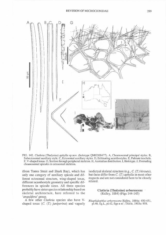
REVISION OF MICROCIONIDAE^
289
FIG. 142. Clathria (Thalysias) aphylla sp.nov. (holotype QMG300477). A, Choanosomal principal styles. B,Subectosomal auxiliary style. C, Ectosomal auxiliary styles. D, Echinating acanthostyles. E, Palmate isochela.F, V-shaped toxas. G, Section through peripheral skeleton. H, Australian distribution. I, Holotype. J, Protrudingchoanosomal spicules in ectosomal skeleton.
(from Torres Strait and Shark Bay), which hasonly one category of auxiliary spicule and dif-ferent ectosomal structure, wing-shaped toxas,different acanthostyle geometry and specific dif-ferences in spicule sizes. All three speciesprobably have sister species relationship based onskeletal architecture, here referred to the`anguliferd group.
A few other Clathria species also have V-shaped toxas (C. (T) juniperina) and vaguely
isodictyal skeletal structure (e.g., C. (T)hirsuta),but these differ from C. (T) aphylla in most otherrespects and are not considered here to be closelyrelated.
Clathria (Thalysias) arborescens(Ridley, 1884) (Figs 144-145)
Rhaphidophlus arborescens Ridley, 1884a: 450-451,p1.40, fig.L, p1.42, figs n-n'; Thiele, 1903a: 958.
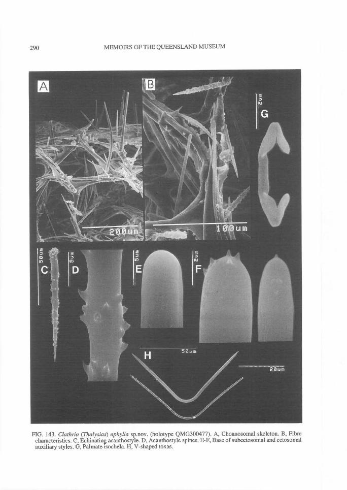
290^ MEMOIRS OF THE QUEENSLAND MUSEUM
FIG. 143. Clathria (Thalysias) aphylla sp.nov. (holotype QMG300477). A, Choanosomal skeleton. B, Fibrecharacteristics. C, Echinating acanthostyle. D, Acanthostyle spines. E-F, Base of subectosomal and ectosomalauxiliary styles. G, Palmate isochela. H, V-shaped toxas.
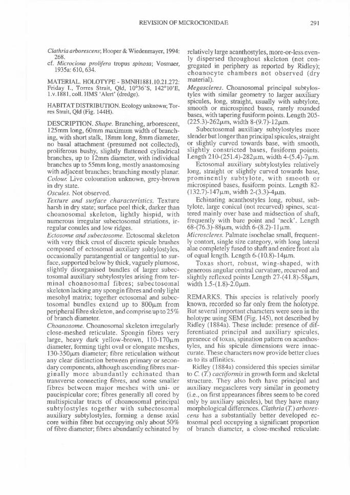
REVISION OF MICROCIONIDAE^ 291
Clathria arborescens; Hooper & Wiedenmayer, 1994:268.
cf. Microciona prolifera tropus spinosa; Vosmaer,1935a: 610, 634.
MATERIAL. HOLOTYPE - BMNH1881.10.21.272:Friday I., Torres Strait, Qld, 10°36'S, 142°10'E,1.v.1881, coll. HMS 'Alert' (dredge).
HABITAT DISTRIBUTION. Ecology unknown; Tor-res Strait, Qld (Fig. 14411).
DESCRIPTION. Shape. Branching, arborescent,125rrun long, 60mm maximum width of branch-ing, with short stalk, 18mm long, 8mm diameter,no basal attachment (presumed not collected),proliferous bushy, slightly flattened cylindricalbranches, up to 12mm diameter, with individualbranches up to 55mm long, mostly anastomosingwith adjacent branches; branching mostly planar.Colour Live colouration unknown, grey-brownin dry state.Oscules. Not observed.Texture and surface characteristics. Textureharsh in dry state; surface peel thick, darker thanchoanosomal skeleton, lightly hispid, withnumerous irregular subectosomal striations, ir-regular conules and low ridges.Ectosome and subectosome. Ectosomal skeletonwith very thick crust of discrete spicule brushescomposed of ectosomal auxiliary subtylostyles,occasionally paratangential or tangential to sur-face, supported below by thick, vaguely plumose,slightly disorganised bundles of larger subec-tosomal auxiliary subtylostyles arising from ter-minal choanosomal fibres; subectosomalskeleton lacking any spongin fibres and only lightmesohyl matrix; together ectosomal and subec-tosomal bundles extend up to 800Rm fromperipheral fibre skeleton, and comprise up to 25%of branch diameter.Choanosome. Choanosomal skeleton irregularlyclose-meshed reticulate. Spongin fibres verylarge, heavy dark yellow-brown, 110-17011,mdiameter, forming tight oval or elongate meshes,130-350Rm diameter; fibre reticulation withoutany clear distinction between primary or secon-dary components, although ascending fibres mar-ginally more abundantly echinated thantransverse connecting fibres, and some smallerfibres between major meshes with uni- orpaucispicular core; fibres generally all cored bymultispicular tracts of choanosomal principalsubtylostyles together with subectosomalauxiliary subtylostyles, forming a dense axialcore within fibre but occupying only about 50%of fibre diameter; fibres abundantly echinated by
relatively large acanthostyles, more-or-less even-ly dispersed throughout skeleton (not con-gregated in periphery as reported by Ridley);choanocyte chambers not observed (drymaterial).Megascleres. Choanosomal principal subtylos-tyles with similar geometry to larger auxiliaryspicules, long, straight, usually with subtylote,smooth or microspined bases, rarely roundedbases, with tapering fusiform points. Length 205-(225.3)-262m, width 8-(9.7)-12p.m.
Subectosomal auxiliary subtylostyles moreslender but longer than principal spicules, straightor slightly curved towards base, with smooth,slightly constricted bases, fusiform points.Length 210-(251.4)-282p,m, width 4-(5.4)-7p.m.
Ectosomal auxiliary subtylostyles relativelylong, straight or slightly curved towards base,prominently subtylote, with smooth ormicrospined bases, fusiform points. Length 82-(132.7)-147p.m, width 2-(3.3)-4pLm.
Echinating acanthostyles long, robust, sub-tylote, large conical (not recurved) spines, scat-tered mainly over base and midsection of shaft,frequently with bare point and 'neck'. Length68-(76.3)-88p.m, width 6-(8.2)-11p,m.Microscleres. Palmate isochelae small, frequent-ly contort, single size category, with long lateralalae completely fused to shaft and entire front alaof equal length. Length 6-(10.8)-14p.m.
Toxas short, robust, wing-shaped, withgenerous angular central curvature, recurved andslightly reflexed points Length 27-(41.8)-58p.,m,width 1.5-(1.8)-2.0p.m.
REMARKS. This species is relatively poorlyknown, recorded so far only from the holotype.But several important characters were seen in theholotype using SEM (Fig. 145), not described byRidley (1884a). These include: presence of dif-ferentiated principal and auxiliary spicules,presence of toxas, spination pattern on acanthos-tyles, and his spicule dimensions were innac-curate. These characters now provide better cluesas to its affinities.
Ridley (1884a) considered this species similarto C. (T) cactiformis in growth form and skeletalstructure. They also both have principal andauxiliary megascleres very similar in geometry(i.e., on first appearances fibres seem to be coredonly by auxiliary spicules), but they have manymorphological differences. Clathria (T)arbores-cens has a substantially better developed ec-tosomal peel occupying a significant proportionof branch diameter, a close-meshed reticulate
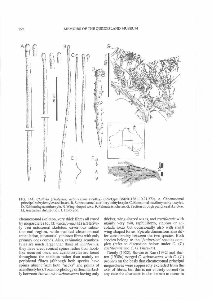
292^ MEMOIRS OF THE QUEENSLAND MUSEUM
FIG. 144. Clathria (Thalysias) arborescens (Ridley) (holotype BMNH1881.10.21.272). A, Choanosomalprincipal subtylostyles and bases. B, Subectosomal auxiliary subtylostyle. C, Ectosomal auxiliary subtylostyles.D, Echinating acanthostyle. E, Wing-shaped toxa. F, Palmate isochelae. G, Section through peripheral skeleton.H, Australian distribution. I, Holotype.
choanosomal skeleton, very thick fibres all coredby megascleres (C. (T) cactifonnis has a relative-ly thin ectosomal skeleton, cavernous subec-tosomal region, wide-meshed choanosomalreticulation, substantially thinner fibres with onlyprimary ones cored). Also, echinating acanthos-tyles are much larger than those of cactiformis,they have erect conical spines rather than hook-like recurved ones, and acanthostyles are foundthroughout the skeleton rather than mainly onperipheral fibres (although both species havespines absent from both "necks" and points ofacanthostyles). Toxa morphology differs marked-ly between the two, with arborescens having only
thicker, wing-shaped toxas, and cactiforniis withmainly very thin, raphidiform, sinuous or ac-colada toxas but occasionally also with smallwing-shaped forms. Spicule dimensions also dif-fer considerably between the two species. Bothspecies belong to the juniperina' species com-plex (refer to discussion below under C. (T)cactifonnis and C. (T.) hirsuta).
Dendy (1922), Burton & Rao (1932) and Bur-ton (1938a) merged C. arborescens with C. (T)procera on the basis that choanosomal principalmegascleres were supposedly excluded from theaxis of fibres, but this is not entirely correct (inany case the character is also known to occur in
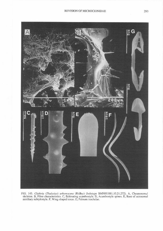
REVISION OF MICROCIONIDAE^ 293
FIG. 145. Clathria (Thalysias) arborescens (Ridley) (holotype BMNH1881.10.21.272). A, Choanosomaiskeleton. B, Fibre characteristics. C, Echinating acanthostyle. D, Acanthostyle spines. E, Base of ectosomalauxiliary subtylostyle. F, Wing-shaped toxas. G, Palmate isochelae.
294^ MEMOIRS OF THE QUEENSLAND MUSEUM
at least eight other species (see below under C.cactiformis)). Thiele (1903a) also compared C.(T) arborescens and C. (T) cervicornis, for thesame reason (i.e., supposed absence of principalspicules), but C. procera does have principalspicules (exclusively echinating peripheralfibres), and those in C. (T) arborescens are inter-mingled with auxiliary subtylostyles withinfibres. Skeletal structure and growth forms alsodiffer considerably between these two taxa.
Clathria (Thalysias) cactiformis (Lamarck, 1814)(Figs 146-148, Table 31, Plate 4E-F)
Spongia cactifortnis Lamarck, 1814: 440, 370Clathria cactiformis; Rudman & Avern, 1989: 335;
Hooper & Wiedenmayer, 1994: 268.Spongia appendiculata, in part, Lamarck, 1814: 383,
362.Wilsonella appendiculata; Topsent, 1930: 46, p1.2,
fig.3; Topsent, 1933: 22.Echinonema typicum Carter, 1875: 195 [nomen
nudum]; Carter, 1881: 362, 377, 388, 384.Thalassodendron typica; Lendenfeld, 1888: 223;
Hallmann, 1912: 242; Hal!mann, 1914a: 267.Not Thalassodendron typica; Whitelegge, 1901: 86.Clathria typica; Vosmaer, 1885b: 357; Ridley &
Dendy, 1887: 153; Dendy, 1889c: 11;[in part]Dendy, 1896: 32; Whitelegge, 1901: 80, 117;Hentschel, 1911: 280, 364-367, text-fig. 43.
Not Clathria ypica var. porrecta Hentschel, 1912:298, 359-360.
Not Clathria typica; Kirkpatrick, 1903: 248-249;Kirkpatrick, 1904: 148; Vacelet & Vasseur, 1971:94-95, p1.3, fig.5, text-fig. 48.
Wilsonella typica; Hallmann, 1912: 203, 242.Not Tenacia typica var. porrecta Hallmann, 1920: 771.Rhaphidophlus typicus; Hallmann, 1912: 176-177,
184-203, 234, 299, 300, p1.27, p1.28, figs 1-4, p1.29,text-figs 38-42 [et yard; Shaw, 1927:424-425; Top-sent, 1932:88, p1.4, fig.4; Burton, 1938a: 12; Guiler,1950: 8; Vacelet & Vasseur, 1977: 115; Vacelet etal., 1976: 73; Liaaen-Jensen et al., 1982: 170-172.
Echinonema anchoratum Carter, 1881a: 362, 379, 380;Lendenfeld, 1888: 219-220.
Echinonema anchoratum var. dura Whitelegge, 1901:81.
Echinonema anchoratum var. lamellosa; Lendenfeld,1888: 219-220.
Rhaphidophlus anchoratum; Bergquist & Fromont,1988: 112; Dawson, 1993: 39.
Wilsonella anchoratum var. lamellosa; Hall mann,1912: 211 [imperfectly known].
Not Echinonema anchorata var. lamellosa;Whitelegge, 1901: 82.
Echinonema flabelhformis Carter, 1885f: 352.Echinonema pectiniformis Carter, 1885f: 353.Phakelliaventilabrum var. australiensis Carter, 1886g:
379.
Thalassodendron brevispina Lendenfeld, 1888: 225;Whitelegge, 1901: 87.
Clathria favosa Whitelegge, 1907: 498-499.Clathria indurata Hallmann, 1912: 299.Clathria acanthodes Hentschel, 1911: 367-370, text-
fig.44; Hentschel, 1923: 380, fig.349.Rhaphidophlus acanthodes; Hallmann, 1912: 177.Rhaphidophlus sp. 2; Vacelet & Vasseur, 1971: 97-98,
text-fig. 52.cf. Microciona proltfera; Vosmaer, 1935a: 609, 628-
629, 647, 667.
MATERIAL. LECTOTYPE: MNHNDT580:'Southern Seas', Peron & Lesueur collection, no otherdetails known. PARALECTOTYPE: MNHNDT3360:same details (= R. typicus var. cactiformis; Topsent,1932: 88). LECTOTYPE of S. appendiculata:MNHNDT526 (fragment BMNH1953.4.9.83) (dry).PARALECTOTYPE of S. appendiculata: MNHNDT-3394: King George Sound (Albany), WA. LEC-TOTYPE of T. brevispina: AMZ931: Port Jackson,NSW, 33°48'S, 151°20'E, coll. R. von Lendenfeld(dredge). SYNTYPE of E. typicum: BMNH-1877.5.21.149: Fremantle, WA, 32°03'S, 115°38'E,coll. E. Clifton (dredge). HOLOTYPE of C. favosa:AMZ944: Off Port Jackson, NSW, 33°50'S, 151°30'E,98-100m depth, coll. FIV 'Thetis' (dredge). LEC-TOTYPE of R. typicus var. proximus: AMZ930: Hen-ley Beach, St. Vincent Gulf, SA, 34 0455, 137°57'E,coll. T. Worsnop (dredge). PARATYPE of R. typicusvar. obesus: AMZ937: Tuggerah Beach, NSW,33°18'S, 151°30'E, coll. NSW Fish Commission(trawl). HOLOTYPE of R. typicus var. geminus:AMZ928: specific locality unknown, WA, no otherdetails known. HOLOTYPE of R. typicus var. stelliferAME648: E. coast of Hinders I., Bass Strait, Tas,40°01'S, 148°02'E, no other details known. SYN-TYPE of E. anchoratum: BMNH1886.12.15.423: PortPhillip, Vic, 38°09'S, 144°52'E, no other detailsknown. SYNTYPE of E. anchoratum var. dura andLECTOTYPE of C. indurata: AMG9113: specificlocality unknown, WA, no other details known.PARALECTOTYPE of C. indurata: AM unreg.:specific locality unknown, WA, no other detailsknown. HOLOTYPE of E. pectiniformis: BMNH-1886.12.15.141 (fragment AMG2777): Port Phillip,Vic, 38°09'S, 144°52'E, 40m depth, no other detailsknown. Fragment of HOLOTYPE C. acanthodes:ZMB4443: Surf Point, outer bar exit to South Passage,Shark Bay, WA, 26°08'S, 113°08.5'E, 0.5-3.5m depth,coll. W. Michaelsen & R. Hartmeyer (dredge). OTHERMATERIAL. NSW - QMG301403. TAS - AME624,AMZ1415, AMZ2203, QMG311412 (NCIQ66C-3713-A) (fragment NTMZ3811). VICTORIA -AMZ1430, AMZ1158, NMV RN359, NMVRN43 I,NMVRN436, NMVRN 438, NMVRN551, NMVRN-677, NMVRN797, NMVRN840, NMVRN900,NM V RN959, AMZ3921 (RRIMPFN3527/000/02),AMZ4277 (RRIMPFN1906), NTMZ2832. S AUST -AMZ41, SAM unreg. (fragment NTMZ1657), SAMunreg. (fragment NTMZ 1693), SAM unreg. (fragmentNTMZI628), SW COAST, WA - MNHN unregistered
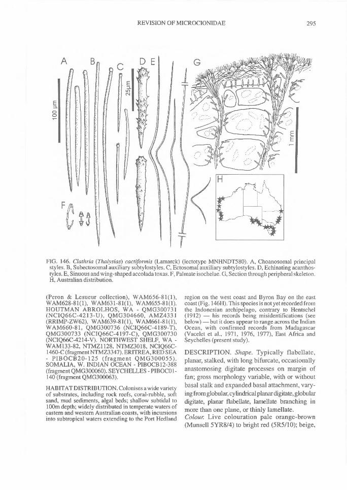
REVISION OF MICROCIONIDAE^ 295
A^B
FIG. 146. Clathria (Thalysias) cactiformis (Lamarck) (lectotype MNHNDT580). A, Choanosomal principalstyles. B, Subectosomal auxiliary subtylostyles. C, Ectosomal auxiliary subtylostyles. D, Echinating acanthos-tyles. E, Sinuous and wing-shaped accolada toxas. F, Palmate isochelae. G, Section through peripheral skeleton.H, Australian distribution.
(Peron & Lesueur collection), WAM656-81(1),WAM628-81(1), WAM631-81(1), WAM655-81(1).HOUTMAN ABROLHOS, WA - QMG300731(NCIQ66C-4213-U), QMG3046 60, AMZ4331(RRIMP-ZW62), WAM639-81(1), WAM661-81(1),W A M660-81, QMG300736 (NCIQ66C -4189-T),QMG300733 (NCIQ66C-4197-C), QMG300730(NCIQ66C-4214-V). NORTHWEST SHELF, WA -WAM133-82, NTMZ1128, NTMZ3018, NCIQ66C-1460-C (fragment NTMZ3347). ERITREA, RED SEA- PIBOCB20-125 (fragment QMG300055).SOMALIA, W. INDIAN OCEAN - PIBOCB12-388(fragment QMG300060). SEYCHELLES - PIB0001-140 (fragment QMG300063).
HABITAT DISTRIBUTION. Colonises a wide varietyof substrates, including rock reefs, coral-rubble, softsand, mud sediments, algal beds; shallow subtidal to100m depth; widely distributed in temperate waters ofeastern and western Australian coasts, with incursionsinto subtropical waters extending to the Port Hedland
region on the west coast and Byron Bay on the eastcoast (Fig. 146H). This species is not yet recorded fromthe Indonesian archipelago, contrary to Hentschel(1912) — his records being misidentifications (seebelow) — but it does appear to range across the IndianOcean, with confirmed records from Madagascar(Vacelet et al., 1971, 1976, 1977), East Africa andSeychelles (present study).
DESCRIPTION. Shape. Typically flabellate,planar, stalked, with long bifurcate, occasionallyanastomosing digitate processes on margin offan; gross morphology variable, with or withoutbasal stalk and expanded basal attachment, vary-ing from globular, cylindrical planar digitate, globulardigitate, planar flabellate, lamellate branching inmore than one plane, or thinly lamellate.Colour. Live colouration pale orange-brown(Munsell 5YR8/4) to bright red (5R5110); beige,
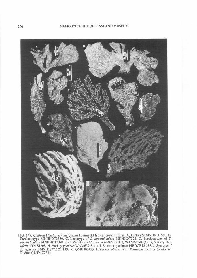
296^ MEMOIRS OF THE QUEENSLAND MUSEUM
FIG. 147. Clathria (Thalysias) cactiformis (Lamarck) typical growth forms. A, Lectotype MNHNDT580. B,Paralectotype MNHNDT3360. C, Lectotype of S. appendiculata MNHNDT526. D, Paralectotype of S.appendiculata MNHNDT3394. E-F, Variety cactiformis WAM656-81(1), WAM655-81(1). G, Variety stel-lifera NTMZ1708. H, Variety geminus WAM639-81(1). I, Somalia specimen PIBOCB12-388. J, Syntype ofE. typicum BMNH1877.5.21.149. K, QMG300453. L,Variety obesus with Rostanga feeding (photo W.Rudman) NTMZ2832.
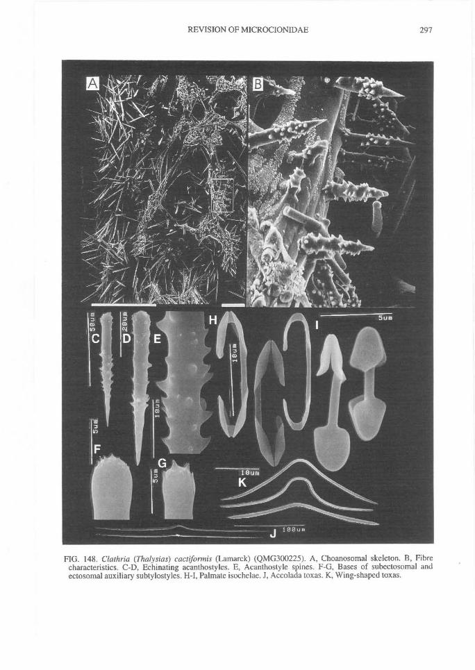
REVISION OF MICROCIONIDAE^ 297
FIG. 148. Clathria (Thalysias) cactiformis (Lamarck) (QMG300225). A, Choanosomal skeleton. B, Fibrecharacteristics. C-D, Echinating acanthostyles. E, Acanthostyle spines. F-G, Bases of subectosomal andectosomal auxiliary subtylostyles. H-I, Palmate isochelae. J, Accolada toxas. K, Wing-shaped toxas.
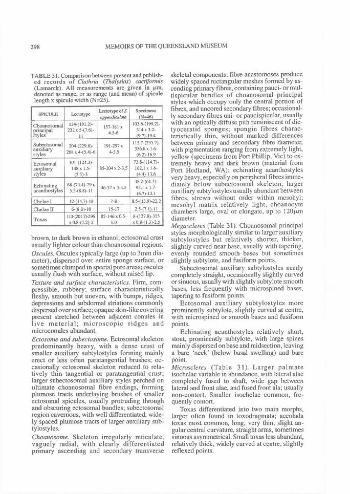
298^ MEMOIRS OF THE QUEENSLAND MUSEUM
TABLE 31. Comparison between present and publish-ed records of Clathria (Thalysias) cactiformis(Lamarck). All measurements are given in p.m,denoted as range, or as range (and mean) of spiculelength x spicule width (N=25).
SPICULE Lectotype Lectotype of S.appendiculata
Specimens(N=-46)
Choanosomal 134(191.2)- 157-181 x 103.6(199.2)-principalstyles
232 x 5-(7.6)-11
4.5-6 314 x 3.2-(9.7)-19.4
Subectos°mal 2044229.8)- 191-297x 11534235.7)-auxiliarystyles 8 x 4-(5.4)-6 4-5.5 356.6 x 1.6-
(6.2)-16.9
Ectosomal 101(124.3)- 73.8(114.7)-auxiliary 148x 1.5- 85-104 x 2-3.5 162.1 x 1.6-styles (2.5)-3 (4.4)-13.6
Echinating 68-(74.4)-79 x 46-57 x 3-4.538.2-(61.3)-93.1 x 1.7-
acanthostyles 5.5-(8.8)-11 (6.7)-13.1
Chelae I 12-(14.7)-18 7-8 8.5-(13.9)-22.2
Chelae II 6-(8.8)-I0 15-17 2.5-(7.1)-111134201.7)-296 82-146 x 0.5- 8-(127.8)-355
Toxas x 0.8-(1.2)-2 1.0 x 0.8-(1.3)-2.5
brown, to dark brown in ethanol; ectosomal crustusually lighter colour than choanosomal regions.Oscules. Oscules typically large (up to 3mm dia-meter), dispersed over entire sponge surface, orsometimes clumped in special pore areas; osculesusually flush with surface, without raised lip.Texture and surface characteristics. Firm, com-pressible, rubbery; surface characteristicallyfleshy, smooth but uneven, with bumps, ridges,depressions and subdermal striations commonlydispersed over surface; opaque skin-like coveringpresent stretched between adjacent conules inlive material; microscopic ridges andmicroconules abundant.Ectosome and subectosome. Ectosomal skeletonpredominantly heavy, with a dense crust ofsmaller auxiliary subtylostyles forming mainlyerect or less often paratangential brushes; oc-casionally ectosomal skeleton reduced to rela-tively thin tangential or paratangential crust;larger subectosomal auxiliary styles perched onultimate choanosomal fibre endings, formingplumose tracts underlaying brushes of smallerectosomal spicules, usually protruding throughand obscuring ectosomal bundles; subectosomalregion cavernous, with well differentiated, wide-ly spaced plumose tracts of larger auxiliary sub-tylostyles.Choanosome. Skeleton irregularly reticulate,vaguely radial, with clearly differentiatedprimary ascending and secondary transverse
skeletal components; fibre anastomoses producewidely spaced rectangular meshes formed by as-cending primary fibres, containing pauci- or mul-tispicular bundles of choanosomal principalstyles which occupy only the central portion offibres, and uncored secondary fibres; occasional-ly secondary fibres uni- or paucispicular, usuallywith an optically diffuse pith reminiscent of dic-tyoceratid sponges; spongin fibres charac-teristically thin, without marked differencesbetween primary and secondary fibre diameter,with pigmentation ranging from extremely light,yellow (specimens from Port Phillip, Vic) to ex-tremely heavy and dark brown (material fromPort Hedland, WA); echinating acanthostylesvery heavy, especially on peripheral fibres imme-diately below subectosomal skeleton; largerauxiliary subtylostyles usually abundant betweenfibres, strewn without order within mesohyl;mesohyl matrix relatively light, choanocytechambers large, oval or elongate, up to 12011mdiameter.Megascleres (Table 31). Choanosomal principalstyles morphologically similar to larger auxiliarysubtylostyles but relatively shorter, thicker,slightly curved near base, usually with tapering,evenly rounded smooth bases but sometimesslightly subtylote, and fusiform points.
Subectosomal auxiliary subtylostyles nearlycompletely straight, occasionally slightly curvedor sinuous, usually with slightly subtylote smoothbases, less frequently with microspined bases,tapering to fusiform points.
Ectosomal auxiliary subtylostyles moreprominently subtylote, slightly curved at centre,with microspined or smooth bases and fusiformpoints.
Echinating acanthostyles relatively short,stout, prominently subtylote, with large spinesmainly dispersed on base and midsection, leavinga bare 'neck' (below basal swelling) and barepoint.Microscleres (Table 31). Larger palmateisochelae variable in abundance, with lateral alaecompletely fused to shaft, wide gap betweenlateral and front alae, and fused front ala; usuallynon-contort. Smaller isochelae common, fre-quently contort.
Toxas differentiated into two main morphs,larger often found in toxodragmata; accoladatoxas most common, long, very thin, slight an-gular central curvature, straight arms, sometimessinuous asymmetrical. Small toxas less abundant,relatively thick, widely curved at centre, slightlyreflexed points.
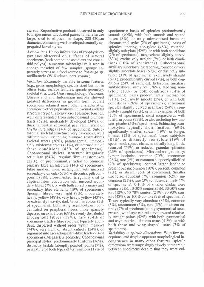
REVISION OF MICROCIONIDAE^ 299
Larvae. Reproductive products observed in onlyfour specimens. Incubated parenchymella larvaelarge, oval to eliptical in shape, 220-420Rmdiameter, containing well developed centrally ag-gregated larval styles.Associations. Heavy infestations of zoophytic or-ganisms observed on surfaces of severalspecimens (both compound ascidians and zooan-thid polyps); numerous microalgal cells seen insponge mesohyl of few samples; sponge ap-parently serves as a food source to Rostanga sp.nudibranchs (W. Rudman, pers. comm.).Variation. Extremely variable in some features(e.g., gross morphology, spicule size) but not inothers (e.g., surface features, spicule geometry,skeletal structure). Gross morphology: Victorian,Queensland and Indonesian morphs showedgreatest differences in growth form, but allspecimens retained most other characteristicscommon to other populations. Ectosomal skeletalstructure: typically heavy, erect plumose brushes,well differentiated from subectosomal plumosetracts (52%), moderately developed (34%), orthick tangential ectosomal peel reminiscent ofCrella (Crellidae) (14% of specimens). Subec-tosomal skeletal structure: very cavernous, welldifferentiated ascending plumose (extra-fibre)skeletal tracts (36%), paratangential, immedi-ately subdermal tracts (21%), or intermediate ofthese conditions (43% of specimens).Choanosomal skeletal structure: irregularlyreticulate (64%), regular fibre anastomoses(22%), or predominantly radial to plumoseprimary fibre architecture (14% of specimens).Fibre meshes: wide, rectangular, with uncoredsecondary elements (67%), with central pith com-ponent (7%), close-meshed, irregularly oval toeliptical fibre reticulation with uncored secon-dary fibres (7%), or with both cored primary andsecondary fibre elements (19% of specimens).Spongin fibres: very light (7%), moderatelyheavy, yellow (48%), very heavy, yellow (43%),or extremely heavily, dark brown in colour (2%of specimens). Echinating acanthostyles: con-centrated on peripheral fibres, more sparselydipersed on axial fibres (69%), evenly distributedthroughout fibres (17%), rare (14% ofspecimens). Extra-fibre spicules: typically abun-dant, dispersed without order within mesohyl(74%), very light or absent entirely (24%), ororganised into ascending extra-fibre tracts (2% ofspecimens). Megasclere geometry: Choanosomalprincipal styles: predominantly fusiform (76%),distinctly hastate (abruptly pointed) points (7%),or mixture of both types of terminations (17% of
specimens); bases of spicules predominantlysmooth (90%), with both smooth and spinedbases (8%), or only microspined bases onchoanosomal styles (2% of specimens); bases ofspicules tapering, non-tylote (46%), rounded,slightly subtylote (52%), or with both conditions(2% of specimens); megascleres slightly curved(83%), exclusively straight (7%), or both condi-tions (10% of specimens). Subectosomalauxiliary subtylostyles: tapering, rounded or veryslightly subtylote bases (69%), or distinctly sub-tylote (31% of specimens); exclusively straight(69%), predominantly curved (7%), or both con-ditions (24% of samples). Ectosomal auxiliarysubtylostyles: subtylote (76%), tapering non-tylote (10%) or both conditions (14% ofspecimens); bases predominantly microspined(71%), exclusively smooth (3%), or with bothconditions (26% of specimens); ectosomalspicules slightly curved near base (54%), com-pletely straight (29%), or with examples of both(17% of specimens); most megascleres withfusiform points (95%), or also including few has-tate spicules (5% of specimens). Echinating acan-thostyles: typically short, fusiform (69%),significantly smaller, stouter (19%), or longer,thinner (12% of specimens); bases subtylote(81%), or distinctly non-tylote (19% ofspecimens); spines characteristically long, thick,recurved (74%), or reduced, granular spination(26% of specimens). Microsclere geometry:Larger isochelae: common (70%), uncommon(26%), rare (2%), or common but poorly silicified(2% of specimens); contort larger isochelaepresent but uncommon (10%), present, common(2%), or absent (86% of specimens). Smallerisochelae: abundant (7%), common (62%), un-common (21%), rare (3%) or absent entirely (7%of specimens); 0-10% of smaller chelae werecontort (2%), 10-30% contort (5%), 30-50% con-tort (12%), 50-70% contort (24%), 70-90% con-tort (43%), or 100% contort (7% of specimens).Toxas: typically very abundant (82%), common(3%), uncommon (5%), rare (3%), or absent en-tirely (7% of specimens); only symmetrical toxaspresent, with large central curvature and relative-ly straight points (52%), with both symmetricaland asymmetrical, sinuous toxas (41%), or withboth these and wing-shaped toxas (7% ofspecimens).Variability in spicule dimensions: With few ex-ceptions, and despite apparent morphological in-congruence in many other features, spiculedimensions were surprisingly closely comparablebetween specimens despite that they were col-
300^ MEMOIRS OF THE QUEENSLAND MUSEUM
lected from widely separated geographiclocalities. Results were not analysed statisticallybecause bathymetric, habitat and season datawere not available for the majority of material.However, scatterplot comparisons betweensamples found that for each spicule categorythere were only few anomolous specimens whichconsistently differed from typical forms of thespecies. These anomalies are discussed furtherbelow.
REMARKS. Clathria (Thalysias) cactiformis ispredominantly temperate. It is characterised byits typically flabellate or flabello-digitate growthform, a smooth surface with a detachable thickectosomal peel which is usually paler than thechoanosome, relatively thin spongin fibres heavi-ly echinated but poorly cored, and especially itsshort, thick, squat acanthostyles with a bare neck,bare points and large recurved spines over theremainder of the spicule. There is considerablemorphological variability between regionalpopulations (see Variation, above), to some ex-tent vindicating Hallmann's (1912) subdivisionof the species into 'varieties' (subspecies), basedmainly on differences in growth form. In factsome specimens examined were assigned to thisspecies with hesitation given their diverse exter-nal morphologies, but no other consistent skeletalor other characters were found to correlate withthese morphotypes. Nevertheless, this taxon isprobably composed of several cryptic siblingspecies (morphospecies) which might only bereliably differentiated using biochemical, geneticand other non-skeletal data.
NMVRN0436 and RN0551 from Port PhillipBay, Vic. have acanthostyles which are long,evenly spined, spines are sparse and small;echinating spicules occur in heavy concentrationsand are evenly dispersed throughout thechoanosome, not predominant in the peripheralskeleton; ectosomal skeleton is very welldeveloped and well differentiated from subec-tosomal plumose tracts; the subectosomal regionis noticeably more cavernous than in most typicalforms; the choanosome is distinctly plumo-reticulate; mesohyl matrix is very heavy andfibres are heavily invested with spongin; thesmaller category of isochelae and toxas are ab-sent. In some respects these features correspondto Spongia appendiculata, perhaps justifying theretention of that species separately form cactifor-mis, but other general morphological featuresindicate that these specimens cannot be otherwisereliably separated.
WAM133-82 from the Northwest Shelf of WAhas consistently basally spined choanosomalstyles, and isochelae are completely unmodified.The ectosome and subectosomal regions arepoorly developed and incompletely differen-tiated, but the specimen is otherwise comparablewith typical forms.
Some further comment is necessary on thesynonymy above. Spongia appendiculata is in-cluded here as a new synonym of C. (T) cacnfor-mis (cf. Hooper & Wiedenmayer, 1994) withvirtually identical growth form, spicule geometryand skeletal structure as typical forms of thisspecies. However, there are some notable dif-ferences between these nominal species, espe-cially in the markedly smaller dimensions of mostspicules in appendiculata, acanthostyles have avestigial granular spination, and they are alsoalmost entirely incorporated into the fibres them-selves (i.e., enveloped by collagen). These dif-ferences are not presently considered sufficient torecognise it as a distinct taxon. Incorporation ofechinating acanthostyles into spongin fibres isalso known for several other species of Clathria(termed here the `phorbastformis' species com-plex, including C. (T) phorbasifonnis, C. (D.)dura, C. (D.) myxilloides, and others). However,in Spongia appendiculata acanthostyles are in-serted perpendicular to fibres, much like typicalechinating megascleres, but they have 'sunk' intothem, whereas in these other species acanthos-tyles run parallel to fibres and replace or supple-ment the usual coring spicules.
Clathria acanthodes is considered a synonymof C. (T) cactifonnis, despite having slightlydifferent acanthostyle morphology ('neck' oftenspined). The characters used by Hentschel (1911)to differentiate C. acanthodes from other species(i.e., similar geometry between principal andauxiliary spicules, presence or absence ofmicrospines on bases of principal and auxiliaryspicules, presence of two size classes of palmateisochelae, and possession of rhaphidiform toxas),are found in most other populations of C. (T)cactifonnis and are not considered to be ofprimary importance in differentiating species.Clathria typica var. porrecta, also described byHentschel (1912) from Indonesia, is a synonymof Clathria (T) reinwardti not of C. (T.) cactifor-mis (this syonymy was overlooked by Hooper &Wiedenmayer, 1994). The type material ofClathria indurata and Echinonema anchoratwnvar. dura from WA have a uni- or paucispicularcore of choanosomal styles in secondary fibres,whereas typical populations of C. (T) cacnformis