p53 domains: structure, oligomerization, and transformation
TRANSCRIPT
MOLECULAR AND CELLULAR BIOLOGY, Aug. 1994, P. 5182-51910270-7306/94/$04.00+0Copyright © 1994, American Society for Microbiology
p53 Domains: Structure, Oligomerization, and TransformationPIN WANG, MICHAEL REED, YUN WANG, GREGORY MAYR, JUDITH E. STENGER,
MARY E. ANDERSON, JOHN F. SCHWEDES, AND PETER TEGTMEYER*
Department of Molecular Genetics and Microbiology, State University ofNew York, Stony Brook, New York 11794
Received 28 March 1994/Returned for modification 6 May 1994/Accepted 19 May 1994
Wild-type p53 forms tetramers and multiples of tetramers. Friedman et al. (P. N. Friedman, X. B. Chen, J.Bargonetti, and C. Prives, Proc. Natl. Acad. Sci. USA 90:3319-3323, 1993) have reported that human p53behaves as a larger molecule during gel filtration than it does during sucrose gradient sedimentation. Thesedifferences argue that wild-type p53 has a nonglobular shape. To identify structural and oligomerizationdomains in p53, we have investigated the physical properties of purified segments of p53. The central, specificDNA-binding domain within murine amino acids 80 to 320 and human amino acids 83 to 323 behavespredominantly as monomers during analysis by sedimentation, gel filtration, and gel electrophoresis. Thisconsistent behavior argues that the central region of p53 is globular in shape. Under appropriate conditions,however, this segment can form transient oligomers without apparent preference for a single oligomericstructure. This region does not enhance transformation by other oncogenes. The biological implications oftransient oligomerization by this central segment, therefore, remain to be demonstrated. Like wild-type p53,the C terminus, consisting of murine amino acids 280 to 390 and human amino acids 283 to 393, behavesanomalously during gel filtration and apparently has a nonglobular shape. Within this region, murine aminoacids 315 to 350 and human amino acids 323 to 355 are sufficient for assembly of stable tetramers. The findingthat murine amino acids 315 to 360 enhance transformation by other oncogenes strongly supports the role ofp53 tetramerization in oncogenesis. Amino acids 330 to 390 of murine p53 and amino acids 340 to 393 of humanp53, which have been implicated by Sturzbecher et al. in tetramerization (H.-W. Sturzbecher, R. Brain, C.Addison, K. Rudge, M. Remm, M. Grimaldi, E. Keenan, and J. R. Jenkins, Oncogene 7:1513-1523, 1992), donot form stable tetramers under our conditions. Our findings indicate that p53 has at least two autonomousoligomerization domains: a strong tetramerization domain in its C-terminal region and a weaker oligomer-ization domain in the central DNA binding region of p53. Together, these domains account for the formationof tetramers and multiples of tetramers by wild-type p53. The tetramerization domain is the major determinantof the dominant negative phenotype leading to transformation by mutant p53s.
Many human cancers are associated with a loss of p53function (14, 17). This correlation argues that wild-type (WT)p53 suppresses cellular proliferation and tumor formation.Indeed, in experimental systems, the expression of WT p53suppresses transformation by other oncogenes (4, 7, 13). Incontrast, many mutant p53s enhance transformation under thesame conditions. These findings argue that mutant p53 inter-feres with the suppression functions of endogenous WT p53.Because p53 forms oligomeric structures, the dominant nega-tive phenotype of the mutants has been attributed to theformation of mixed WT and mutant oligomers (12). In supportof this hypothesis, the C-terminal region of p53 has botholigomerization and transformation functions (18, 22, 23, 28).The oligomerization domains of p53, however, have not beenmapped in detail.Although the suppression functions of p53 are not com-
pletely understood, one mechanism by which p53 acts is thedirect activation of transcription via site-specific DNA binding(5, 9, 15). Presumably, p53 activates genes that inhibit cellularproliferation such as the WAF] (Cipl) gene (3, 6, 11). The roleof p53 oligomerization in DNA binding and transactivation isnot clear. Recently, we and others showed that p53 has twoautonomous DNA-binding regions (1,20,26,29). One domain,from amino acids 280 to 390 (segment 280-390), forms stabletetramers and binds DNA nonspecifically. The biological sig-nificance, if any, of this DNA binding activity remains to bedetermined. A second domain, from amino acids 80 to 290
* Corresponding author.
(segment 80-290), does not form stable tetramers but bindsDNA specifically. Furthermore, amino acids 1 to 290, whichinclude both the specific DNA-binding domain and the N-terminal acidic region, activate a p53-specific promoter in vivo.The specific DNA binding and transactivating functions of p53,therefore, do not require stable tetramerization. Because theDNA recognition site for p53 consists of four imperfectrepeats, however, it would be surprising if oligomerization didnot contribute to the DNA binding function of p53.We have undertaken the present study to define further the
oligomerization properties of p53 and the role of oligomeriza-tion in cellular transformation. There has been some disagree-ment about the nature of the various quaternary structuresformed by p53. This confusion is the result of a number offactors. First, purified p53 consists of many oligomeric formsthat require high-resolution techniques for analysis. Second,p53 appears to have an unusual shape that leads to anomalousbehavior during gel filtration (8). Finally, murine and humanpS3s have been studied independently but have not beendirectly compared. We have, therefore, used a variety oftechniques to define the quaternary structures of murine andhuman p53s. The availability of purified segments of p53 hasallowed us to separate two autonomous oligomerization do-mains of the protein. We find that murine amino acids 315 to350 and human amino acids 323 to 355 encode a strongtetramerization domain. Murine amino acids 80 to 320 andhuman amino acids 83 to 323 form unstable oligomers withouta preferred species. We also present evidence that the Cterminus of p53 is a major determinant of its nonglobular
5182
Vol. 14, No. 8

OLIGOMERIZATION OF p53 5183
shape. In contrast, the central conserved region behaves as aglobular protein during sedimentation and gel filtration.
MATERIALS AND METHODS
Expression plasmids. We have previously described plas-mids used for expression of WT p53 or segments of p53. ThepIT plasmid was designed for recombination with baculovirusvectors and overexpression of p53 in insect cells (22). The pBTplasmid utilizes an inducible T7 promoter for transient over-expression of p53 in bacteria (29). The pCMH6K plasmidincludes the immediate-early promoter of cytomegalovirus forstrong expression of p53 in animal cells (29). These plasmidsexpress WT p53 or segments of p53 with a 22-amino-acidN-terminal tag that encodes six histidines for purification bymetal affinity chromatography and a hemagglutinin epitope foridentification with monoclonal antibodies. To exclude thepossibility of mutations within p53 segments, we isolated andtested two independent clones expressing each p53 segment.Plasmids were purified by CsCl centrifugation.
Overexpression and metal affinity purification of p53. Be-cause WT p53 is mostly insoluble when produced in bacteria,we overexpressed WT p53 by infecting insect cells with bacu-lovirus expression vectors and purified p53 as previouslydescribed (29). In brief, p53 was extracted from SF9 cells inlysis buffer (50 mM Tris-HCl [pH 9.0], 150 mM NaCl, 0.1%Nonidet P-40, 10% glycerol, 1 mM phenylmethylsulfonyl fluor-ide, 50 ,ug of aprotinin per ml, 50 ,ug of leupeptin per ml, 10 ,ugof pepstatin A per ml, 1 mM benzamidine, 5 mM ,-mercap-toethanol), clarified by centrifugation, and bound to Ni-nitrilo-triacetic acid (NTA)-agarose (Qiagen, Inc.) at 4°C. After beingwashed extensively with lysis buffer (pH 7.0), Ni-NTA-agarosewas eluted with lysis buffer (pH 7.0) containing increasingconcentrations of imidazole (25, 50, 100, and 250 mM).p53-containing fractions were dialyzed overnight at 4°C against20 mM Tris-HCl (pH 8.0)-100 mM NaCl-50% glycerol (dial-ysis buffer) and stored in aliquots at -70°C.
All p53 segments were overexpressed in bacteria (HMS174or BL21) as previously described (29). In brief, bacteriatransformed by pBT plasmids were expanded from a singlecolony to a 500-ml culture in two steps, and cells were inducedwith IPTG (isopropyl-,-D-thiogalactopyranoside) at a finalconcentration of 1 mM and incubated for 4 h at 30°C. Cellswere suspended in lysis buffer and broken with a French pressat 1,500 lb/in2. Lysates were cleared by centrifugation andbound to Ni-NTA-agarose in a small column. The bound p53was washed, eluted, dialyzed, and stored as described above.SDS-PAGE. Purified proteins were separated by 15% poly-
acrylamide gel electrophoresis (PAGE) in the presence ofsodium dodecyl sulfate (SDS) as described by Laemmli andstained with Coomassie blue (16). Aliquots of the same p53preparations were used for analysis of quaternary structure bysucrose gradient analysis, gel filtration, and protein cross-linking in conjunction with SDS-PAGE (see below).
Sucrose gradient analysis of p53 oligomers. A mixture (in0.14 ml) of purified WT p53 and fragments of p53 (10 jig each)was layered on a 4-ml linear 5 to 20% (wt/wt) sucrose densitygradient in 50 mM Tris-HCl (pH 7.0)-150 mM NaCl-50 mMimidazole-10% glycerol-1 mM dithiothreitol. The gradientwas centrifuged for 20 h at 50,000 rpm in a Beckman SW60rotor at 5°C. Fractions of approximately 0.2 ml were collectedfrom the bottom of the gradient, precipitated with 10%trichloroacetic acid in the presence of 20 jig of bovine serumalbumin as a carrier, and analyzed by SDS-15% PAGE.Molecular weight markers were analyzed in a parallel gradientunder the same conditions.
Gel filtration of p53 oligomers. A mixture (in 0.5 ml) ofpurified WT p53 samples and segments of p53 (10 to 40 jigeach) was fractionated by gel filtration through a Superose 12HR column (10 by 300 mm; Pharmacia) equilibrated with 50mM Tris-HCl (pH 7.0)-150 mM NaCl-50 mM imidazole-10%glycerol-0.01% sodium azide. The column was run with aPharmacia fast protein liquid chromatography system at 30ml/h at 5°C. Fractions (0.2 ml each) were collected, trichloro-acetic acid precipitated, and analyzed by SDS-15% PAGE.Molecular weight protein standards were analyzed under thesame conditions.
Gradient gel electrophoresis of cross-linked oligomers ofp53. The quaternary structures of p53 segments were analyzedby protein cross-linking in conjunction with SDS gradient gelelectrophoresis as previously described (27). Purified p53segments (2 jig each) were incubated for 30 min at 30°C in 30jil of the same buffer used for sedimentation and gel filtration.Increasing concentrations of freshly diluted glutaraldehyde(Sigma) were added as indicated in the figures. Products of thecross-linking reactions were analyzed by SDS-PAGE. All ofthe gel components were those described by Laemmli (16)except that we used an 80:1 ratio of acrylamide to bisacryl-amide (pH 8.8) to prevent protein retardation in the stackinggel (empirically determined). Samples were diluted twofold insample buffer containing 4% (wt/vol) SDS and 5% (vol/vol),-mercaptoethanol and heated for 5 min at 100°C immediatelyprior to loading onto the gel. Samples were electrophoresedthrough 4 to 20% or 8 to 20% polyacrylamide gels in 0.1% SDSat 200 V for 4 h at room temperature. Following electrophore-sis, gels were silver stained (19).
Suppression and transformation assays. We previously de-scribed a combined assay for suppression and enhancement oftransformation (22). In brief, fourth-passage rat embryo fibro-blasts (REF) were grown to subconfluency in 10-cm-diameterFalcon plates in Dulbecco modified Eagle medium with 10%fetal bovine serum and antibiotics. DOTAP (BoehringerMannheim) was used to triply transfect REF cells with 2.5 jigof pSP72-RAS (expressing activated ras), 2.5 jig of pBS-ElA(expressing adenovirus E1A), and 5.0 jig of pCMH6K DNAwith or without apS3 insert. Cells were incubated at 37°C in amoist CO2 incubator and fed with Dulbecco modified Eaglemedium and 10% fetal bovine serum every 3 to 4 days. After 12days, cells were washed twice with phosphate-buffered saline,fixed with methanol for 5 min, and stained with 0.05% Coom-assie blue in 50% methanol-10% acetic acid for 1 h. Plateswere washed with water and air dried, and transformed fociwere counted.
Immunoprecipitation and immunoblotting. REF cells in two10-cm-diameter plates were transfected with 20 jig ofpCMH6K plasmid DNAs expressing WT p53 or segments ofp53 by calcium phosphate precipitation (2). After 40 h, cellswere washed with phosphate-buffered saline, and p53 wasextracted with 0.5 ml of lysis buffer for 30 min on ice. Extractswere clarified by low-speed centrifugation, and tagged p53swere bound to 100 jil of Ni-NTA-agarose overnight at 4°C.After beads were washed twice with 1 ml of lysis buffer, taggedp53s were released with 100 jil of SDS loading buffer for gelelectrophoresis. Concentrated and partially purified p53s wereanalyzed by SDS-PAGE and immunoblotting with a kit forenhanced chemiluminescence (Amersham). We used 600 jigof monoclonal antibody against the hemagglutinin epitopewithin the N-terminal tag of p53 (12CA5; Babco) in 100 ml ofTris-buffered saline with 5% milk for immunoblotting.
VOL. 14, 1994

5184 WANG ET AL.
A. Gel electrophorsis
:......
~~~~~~~~~~~~~~~~~~~~~~~~~~~~~~~~~~~~~~~~~~~~........................7 4. ...:
a=.......... . ...
...,........~~~~~~~~~~~~~~~~~~~~~..20
.. . . ...~~~~... .. ...... ........ .. . .. ...... .. .... X_r . ~~~~~~~~~~~~~~~~~......... B. Apparent mass
3380 390.~~~~~~~~~~~~~~~~~~~~~~~~~0 10 20 30 40 50 60 70 80
mm
FIG. 1. SDS-PAGE of murine WT p53 and segments of p53. (A) SDS-PAGE and Coomassie blue staining of purified WT p53 without (-) or
with (+) 22-amino-acid tags and segments of p53 segments with tags. Molecular mass markers consisting of transferrin (74 kDa), bovine serum
albumin (45 kDa), carbonic anhydrase (29 kDa), soybean trypsin inhibitor (20 kDa), and cytochrome c (11 kDa) are shown on the right. (B)Molecular mass plot of standard markers. The positions of p53 segments are related to their apparent molecular masses by horizontal and verticallines.
RESULTS
SDS gel electrophoresis of purified murine WT p53 andsegments of p53. We wanted to identify autonomous structuraland oligomerization domains in p53 to develop a betterunderstanding of p53 functions. For this purpose, we analyzedthe structures of murine p53 segments that were previouslyused to investigate the DNA binding and transformationfunctions of p53 (22, 29). p53 segments are identified by theiramino acid components, which are numbered by the system ofPennica et al. (21). WT p53 and segments of p53 with22-amino-acid N-terminal tags containing six histidines were
purified by metal affinity chromatography. Because WT p53produced in bacteria is mostly insoluble, we used soluble WTp53 produced in insect cells for studies of purified WT protein.In contrast, segments of p53 produced in bacteria were solubleand could be easily purified. Figure 1A relates purified p53segments to standard molecular mass markers after SDS-PAGE and Coomassie blue staining. Figure 1B shows the bestfit plot of the standards as a diagonal line relating the log of themolecular mass to gel migration for each standard. The verticaland horizontal lines relate the gel migrations of WT p53 andsegments of p53 to their apparent molecular masses. Table 1summarizes these results and compares them with otheranalyses. Tagged WT p53 and segment 1-320 had apparentmolecular masses 10 to 12 kDa larger than predicted by theiramino acid compositions. Because tagged WT p53 was only 3to 4 kDa larger than untagged p53, the 10 to 12 kDa anomalieswere not a result of the histidine tag. In contrast, p53 segments80-320, 280-390, 330-390, and 315-360 had apparent molec-ular masses about 3 kDa larger than their calculated masses.Anomalies of this size are frequently associated with histidinetags. These findings argue that the major determinant of theanomalous SDS-PAGE of WT p53, tagged or untagged, re-sides in the first 80 amino acids of p53. The extra proteinfragment migrating ahead of segment 330-390 appears to be a
proteolytic fragment.Sucrose gradient sedimentation of murine p53. We used
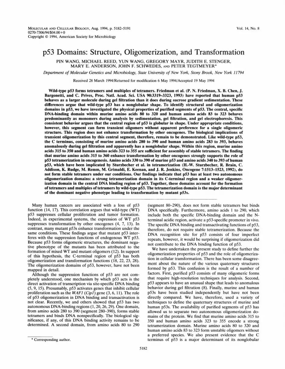
sedimentation through sucrose gradients to study the quater-nary structures of purified p53 segments. Mixtures of molecu-
lar mass markers (Fig. 2A) and of p53 segments (Fig. 2B) werelayered onto two identical 5 to 20% sucrose gradients andcentrifuged in parallel as described in Materials and Methods.Gradient fractions were acid precipitated after the addition ofcarrier bovine serum albumin and were analyzed by SDS-PAGE. Figure 2C shows a diagonal plot of apparent molecularmasses as determined by the sedimentation of the markers.Aldolase is a tetramer (161 kDa) while transferrin (74 kDa),ovalbumin (45 kDa), carbonic anhydrase (29 kDa), and cyto-chrome c (11 kDa) are monomers in their native conforma-tions. The vertical and horizontal lines relate the sedimenta-tion of WT p53 and segments of p53 to their apparentmolecular masses. WT p53 sediments at the position ofaldolase (161 kDa). This result is in good agreement with thesedimentation of human WT p53 reported by Friedman et al.with similar markers (8). Segments 1-320, 80-320, and 330-390sedimented at positions most consistent with the masses oftheir monomeric forms (Table 1). Segments 280-390 and315-360 sedimented ahead of the larger p53 segments 1-320and 330-390, respectively. This sedimentation profile stronglysuggests that the C-terminal segments 280-390 and 315-360form stable oligomeric forms (Table 1). Nevertheless, thelimited resolution of the standard markers and the possibility
TABLE 1. Comparison of apparent molecular masses of p53determined by different methods
Apparent mass (kDa)p53 segment
Sequence SDS-PAGE Sedimentation Gel filtration
WT 43.5, 174a 53 NDb NDWT tagged 45.9, 184 57 160-180 5001-320 37.9 50 30-40 5580-320 29.3 33 30-40 30280-390 15.0, 60 18 40-50 110330-390 9.3 13 10-20 15315-360 7.9, 32 11 20-30 40
a Monomeric and tetrameric masses by sequence composition.b ND, not done.
MOL. CELL. BIOL.
OLIGOMERIZATION OF p53 5185
A. Markers
transferrincarrier BSA
ovalbuminaldolase
C. anhydrase
cytochromeB. p53carrier BSA
WT p531 -320
80 -32 0
280- 390
330 -39031 5-360
..: ; . :: . '_S: wiF ~~~~~~~~~.
I~~~~~~~~~~~~~~~~~~~~~~~~~~~~~~~~~~~~~~~~~~~~. ... ..
.1.,w...s~- .-
1000.x
\ MWV markers
WT-
100
280-390 El
1-320 & 80-320315-360 _ I,330-390 1
10 4 6 8 12 1 12 4 6 8 1 0 1 2 1 4 1 6 1 8 2(
fractions
FIG. 2. Sucrose gradient analysis of murine WT p53 and segmentsof p53. After proteins (10 ,ug) were centrifuged through 5 to 20%gradients, fractions were concentrated by trichloroacetic acid precipi-tation in the presence of added bovine serum albumin (carrier BSA)and were analyzed by SDS-PAGE and Coomassie blue staining. (A)Sucrose gradient of a mixture of molecular mass markers: aldolase (atetramer of 161 kDa), transferrin (74 kDa), ovalbumin (45 kDa),carbonic anhydrase (C. anhydrase) (29 kDa), and cytochrome c (11kDa). (B) Sucrose gradient of a mixture of WT p53 and p53 segments1-320, 80-320, 280-390, 330-390, and 315-360. (C) Molecular mass
plot of standard markers. The positions of p53 segments are related totheir apparent molecular masses by horizontal and vertical lines.
of anomalies in p53 shape preclude identification of specificoligomeric structures by sedimentation analysis alone.
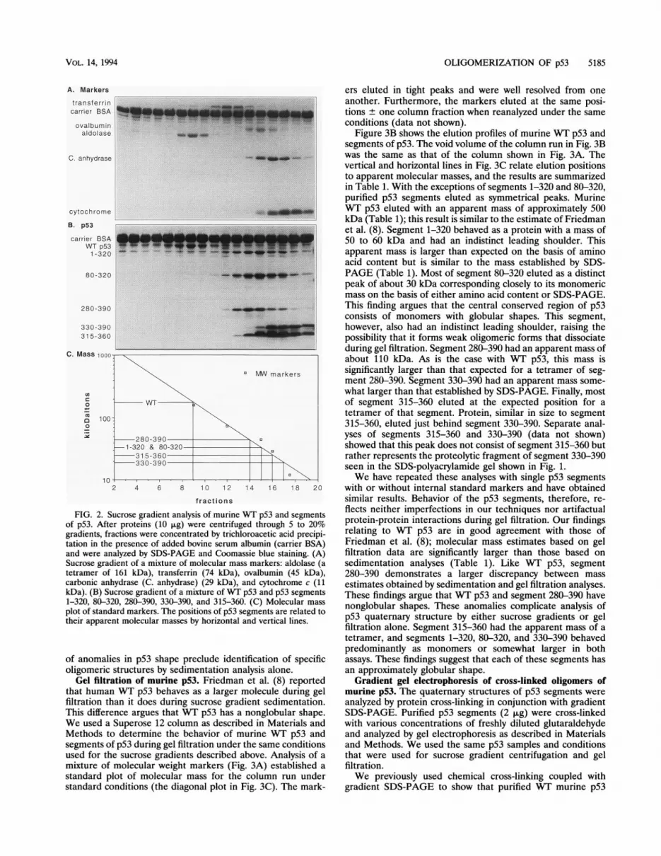
Gel filtration of murine p53. Friedman et al. (8) reportedthat human WT p53 behaves as a larger molecule during gelfiltration than it does during sucrose gradient sedimentation.This difference argues that WT p53 has a nonglobular shape.We used a Superose 12 column as described in Materials andMethods to determine the behavior of murine WT p53 andsegments of p53 during gel filtration under the same conditionsused for the sucrose gradients described above. Analysis of a
mixture of molecular weight markers (Fig. 3A) established a
standard plot of molecular mass for the column run understandard conditions (the diagonal plot in Fig. 3C). The mark-
ers eluted in tight peaks and were well resolved from oneanother. Furthermore, the markers eluted at the same posi-tions ± one column fraction when reanalyzed under the sameconditions (data not shown).
Figure 3B shows the elution profiles of murine WT p53 andsegments of p53. The void volume of the column run in Fig. 3Bwas the same as that of the column shown in Fig. 3A. Thevertical and horizontal lines in Fig. 3C relate elution positionsto apparent molecular masses, and the results are summarizedin Table 1. With the exceptions of segments 1-320 and 80-320,purified p53 segments eluted as symmetrical peaks. MurineWT p53 eluted with an apparent mass of approximately 500kDa (Table 1); this result is similar to the estimate of Friedmanet al. (8). Segment 1-320 behaved as a protein with a mass of50 to 60 kDa and had an indistinct leading shoulder. Thisapparent mass is larger than expected on the basis of aminoacid content but is similar to the mass established by SDS-PAGE (Table 1). Most of segment 80-320 eluted as a distinctpeak of about 30 kDa corresponding closely to its monomericmass on the basis of either amino acid content or SDS-PAGE.This finding argues that the central conserved region of p53consists of monomers with globular shapes. This segment,however, also had an indistinct leading shoulder, raising thepossibility that it forms weak oligomeric forms that dissociateduring gel filtration. Segment 280-390 had an apparent mass ofabout 110 kDa. As is the case with WT p53, this mass issignificantly larger than that expected for a tetramer of seg-ment 280-390. Segment 330-390 had an apparent mass some-what larger than that established by SDS-PAGE. Finally, mostof segment 315-360 eluted at the expected position for atetramer of that segment. Protein, similar in size to segment315-360, eluted just behind segment 330-390. Separate anal-yses of segments 315-360 and 330-390 (data not shown)showed that this peak does not consist of segment 315-360 butrather represents the proteolytic fragment of segment 330-390seen in the SDS-polyacrylamide gel shown in Fig. 1.We have repeated these analyses with single p53 segments
with or without internal standard markers and have obtainedsimilar results. Behavior of the p53 segments, therefore, re-flects neither imperfections in our techniques nor artifactualprotein-protein interactions during gel filtration. Our findingsrelating to WT p53 are in good agreement with those ofFriedman et al. (8); molecular mass estimates based on gelfiltration data are significantly larger than those based onsedimentation analyses (Table 1). Like WT p53, segment280-390 demonstrates a larger discrepancy between massestimates obtained by sedimentation and gel filtration analyses.These findings argue that WT p53 and segment 280-390 havenonglobular shapes. These anomalies complicate analysis ofp53 quaternary structure by either sucrose gradients or gelfiltration alone. Segment 315-360 had the apparent mass of atetramer, and segments 1-320, 80-320, and 330-390 behavedpredominantly as monomers or somewhat larger in bothassays. These findings suggest that each of these segments hasan approximately globular shape.
Gradient gel electrophoresis of cross-linked oligomers ofmurine p53. The quaternary structures of p53 segments wereanalyzed by protein cross-linking in conjunction with gradientSDS-PAGE. Purified p53 segments (2 ,ug) were cross-linkedwith various concentrations of freshly diluted glutaraldehydeand analyzed by gel electrophoresis as described in Materialsand Methods. We used the same p53 samples and conditionsthat were used for sucrose gradient centrifugation and gelfiltration.We previously used chemical cross-linking coupled with
gradient SDS-PAGE to show that purified WT murine p53
C. Mass
(A
0Cu
0a
._
VOL. 14, 1994
0
..
.--- -ow
.-
.: ..... M

5186 WANG ET AL.
A. Markerstransferrin
catalaseovalbumin
aldolase
C. anhydrase
ferr itin
cytochrome
B. p53
WT p531 - 3 2 0
8 0 - 3 2 0
280-390
330-39031 5-360
C. Mass
0
a
a2-l._
34 36 38 40 42 44 46 48 50 52 54 56 58 60 62 64 66 68 70fractions
FIG. 3. Gel filtration analysis of murine WT p53 and segments of pS3. After proteins (30 ,ug) were separated by chromatography through aSuperose 12 column, fractions were trichloroacetic acid precipitated and analyzed by SDS-PAGE and Coomassie blue staining. (A) Gel filtrationof a mixture of molecular mass markers consisting of ferritin (a multimer of 440 kDa), catalase (a tetramer of 232 kDa), aldolase (a tetramer of161 kDa), transferrin (74 kDa), ovalbumin (43 kDa), carbonic anhydrase (C. anhydrase) (29 kDa), and cytochrome c (11 kDa). (B) Gel filtrationof a mixture of WT p53 and segments 1-320, 80-320, 280-390, 330-390, and 315-360. (C) Molecular mass plot of standard markers. The positionsof p53 segments are related to their apparent molecular masses by horizontal and vertical lines.
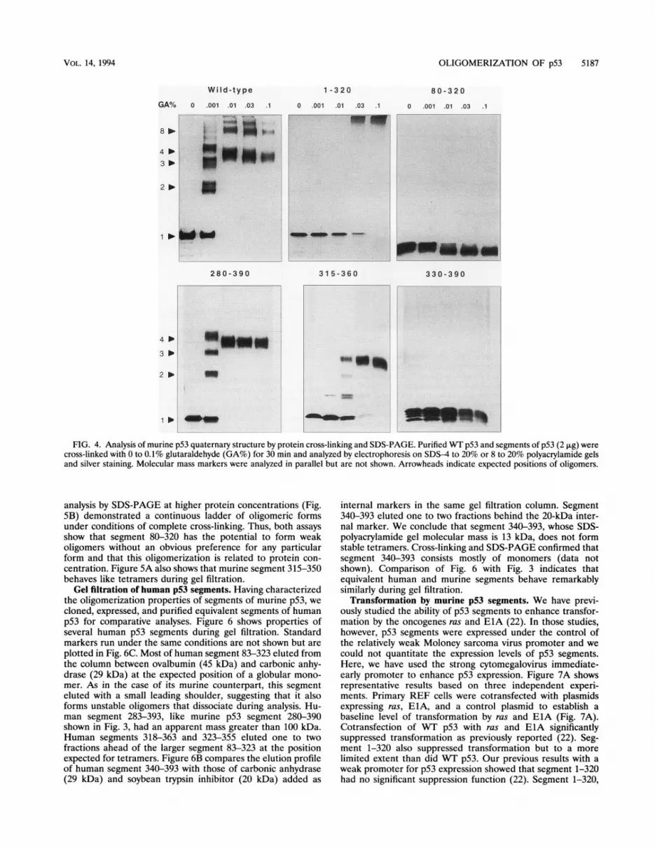
forms tetramers and multiples of tetramers (27). Figure 4compares the oligomerization ofWT p53 with that of segmentsof p53. In the absence of glutaraldehyde, WT p53 electropho-resed as a monomer in the SDS-polyacrylamide gel. In thepresence of 0.001% glutaraldehyde, p53 tetramers were onlypartially cross-linked and, therefore, were partially dissociatedduring SDS-PAGE. Monomers, dimers, trimers, and tetramerswere evident under these conditions. At higher glutaraldehydeconcentrations, preformed oligomers were fully cross-linkedand were not dissociated by the SDS-polyacrylamide gel bufferat all. Because intramolecular cross-linking results in a morecompact and less flexible molecular structure, increasing glu-taraldehyde concentrations led to slightly more rapid electro-phoretic mobilities for each oligomeric species. When fullycross-linked, WT p53 formed tetramers and multiples oftetramers. The complete absence of other oligomeric formsconfirms that glutaraldehyde did not cause artifactual intermo-lecular cross-linking. Segments 280-390 and 315-360 formedtetramers but not multiples of tetramers.
Cross-linking of segment 1-320 did not demonstrate anyoligomerization at low concentrations of glutaraldehyde. Re-tardation of this segment to the top of the gel at higherglutaraldehyde concentrations, however, suggests that segment1-320 has a tendency to aggregate. Segments 80-320 and330-390 formed mostly monomers under our experimental
conditions regardless of the concentration of glutaraldehydeused to cross-link preparations. Migration of these segmentsduring SDS-PAGE increased with increasing glutaraldehydeconcentrations. This finding confirms that the cross-linkingreactions were successful and that they resulted in a morecompact intramolecular structure but not in intermolecularcross-linking. We conclude that p53 segments 1-320, 80-320,and 330-390 do not form stable tetramers under the conditionsthat favor efficient tetramerization ofWT p53 and of segments280-390 and 315-360.
Oligomerization of murine segment 80-320 at increasedprotein concentrations. During gel filtration, a small portion ofsegments 1-320 and 80-320 eluted as indistinct leading shoul-ders ahead of the predominant monomer peaks. This findingsuggested to us that these segments may form transientoligomeric forms. Figure 5 shows further analyses of segment80-320 at a protein concentration four times that used in Fig.3. Under these conditions, gel filtration (Fig. SA) revealed amore prominent leading shoulder in the elution profile ofsegment 80-320 but not those of other segments included asinternal markers. Ahead of the major monomer peak, therewas a less distinct peak at the expected position of dimers. Inaddition, the segment eluted well ahead of the apparent dimerpeak, even though no distinct peaks were evident at thesepositions. Furthermore, cross-linking of segment 80-320 and
----plow
i::.ix.........
MOL. CELL. BIOL.
OLIGOMERIZATION OF p53 5187
Wild-type
GA% 0 .001 .01 .03
1 - 3 2 0
0 .001 .01 .03 .1- = d-
i - i.^ _^. 1FS ;-t<:S . : .: ... - .. ; l' 1 ;5: w _ .i.: :-s
"
r_ ...... ___280-390
3 * w
1 * --
3 1 5-360
i~~~~~~~~~~~~~~~~~~~~~~~~~~~~~~~~~~~~~~~~~~~~~~~~
i_
_i~~~~~~~~~~~~~~~~~~~~~~~~~~~~~~~~~~~~~~~~~~
i
8 0 - 3 2 0
0 .001 .01 .03 .1
330-3 90
FIG. 4. Analysis of murine p53 quaternary structure by protein cross-linking and SDS-PAGE. Purified WT p53 and segments of p53 (2 ,ug) werecross-linked with 0 to 0.1% glutaraldehyde (GA%) for 30 min and analyzed by electrophoresis on SDS-4 to 20% or 8 to 20% polyacrylamide gelsand silver staining. Molecular mass markers were analyzed in parallel but are not shown. Arrowheads indicate expected positions of oligomers.
analysis by SDS-PAGE at higher protein concentrations (Fig.5B) demonstrated a continuous ladder of oligomeric formsunder conditions of complete cross-linking. Thus, both assaysshow that segment 80-320 has the potential to form weakoligomers without an obvious preference for any particularform and that this oligomerization is related to protein con-centration. Figure 5A also shows that murine segment 315-350behaves like tetramers during gel filtration.
Gel filtration of human p53 segments. Having characterizedthe oligomerization properties of segments of murine p53, wecloned, expressed, and purified equivalent segments of humanp53 for comparative analyses. Figure 6 shows properties ofseveral human p53 segments during gel filtration. Standardmarkers run under the same conditions are not shown but areplotted in Fig. 6C. Most of human segment 83-323 eluted fromthe column between ovalbumin (45 kDa) and carbonic anhy-drase (29 kDa) at the expected position of a globular mono-mer. As in the case of its murine counterpart, this segmenteluted with a small leading shoulder, suggesting that it alsoforms unstable oligomers that dissociate during analysis. Hu-man segment 283-393, like murine p53 segment 280-390shown in Fig. 3, had an apparent mass greater than 100 kDa.Human segments 318-363 and 323-355 eluted one to twofractions ahead of the larger segment 83-323 at the positionexpected for tetramers. Figure 6B compares the elution profileof human segment 340-393 with those of carbonic anhydrase(29 kDa) and soybean trypsin inhibitor (20 kDa) added as
internal markers in the same gel filtration column. Segment340-393 eluted one to two fractions behind the 20-kDa inter-nal marker. We conclude that segment 340-393, whose SDS-polyacrylamide gel molecular mass is 13 kDa, does not formstable tetramers. Cross-linking and SDS-PAGE confirmed thatsegment 340-393 consists mostly of monomers (data notshown). Comparison of Fig. 6 with Fig. 3 indicates thatequivalent human and murine segments behave remarkablysimilarly during gel filtration.
Transformation by murine p53 segments. We have previ-ously studied the ability of p53 segments to enhance transfor-mation by the oncogenes ras and ElA (22). In those studies,however, p53 segments were expressed under the control ofthe relatively weak Moloney sarcoma virus promoter and wecould not quantitate the expression levels of p53 segments.Here, we have used the strong cytomegalovirus immediate-early promoter to enhance p53 expression. Figure 7A showsrepresentative results based on three independent experi-ments. Primary REF cells were cotransfected with plasmidsexpressing ras, ElA, and a control plasmid to establish abaseline level of transformation by ras and ElA (Fig. 7A).Cotransfection of WT p53 with ras and ElA significantlysuppressed transformation as previously reported (22). Seg-ment 1-320 also suppressed transformation but to a morelimited extent than did WT p53. Our previous results with aweak promoter for p53 expression showed that segment 1-320had no significant suppression function (22). Segment 1-320,
VOL. 14, 1994

5188 WANG ET AL.
A. Gel filtration4 3 2If v
80-320
2 8 0 - 3 9 0 |1 : -:i :....
330 - 3 9031 5 -350
B. Gel electrophoresis of segment 80-320
1
.E0_01,0,0 10..s.
A.
83-323
283-393 U
31 8-36_323-355
B. --- -----------.---_ 29 kD
20 kD4 3 2
0.1 ~~~~~~~~~~~~~~~~~~. st...-..... ....
sE ' ''EN- -_
0 .... ....
.... ....... ........
gluaradehyde 0.01 ......................
0.00 1
0........ .. ..
Top
l
340-393
C.
0
xr_
Bottom
FIG. 5. Oligomerization of murine p53 segments at increasedprotein concentrations. (A) Gel filtration. After proteins (40 p.g) wereseparated by chromatography through a Superose 12 column, fractionswere trichloroacetic acid precipitated and analyzed by SDS-PAGE andCoomassie blue staining. Arrowheads at the top of each panel indicateexpected positions of oligomers of segment 80-320. (B) Cross-linkingand SDS-PAGE. Segment 80-320 (8 ,ug) was cross-linked with 0 to0.1% glutaraldehyde for 30 min and analyzed by electrophoresis onSDS-8 to 20% polyacrylamide gels and silver staining.
...........: ........
40 42 44 46 48 50 52 54 56 58 60 62 64 66 68 70fractions
FIG. 6. Oligomerization of segments of human p53. (A) Gel filtra-tion of a mixture of segments 83-323, 283-393, 318-363, and 323-355.After proteins (10 ,ug) were separated by chromatography through aSuperose 12 column, fractions were trichloroacetic acid precipitatedand analyzed by SDS-PAGE and Coomassie blue staining. (B) Gelfiltration of a mixture of segment 340-390 with carbonic anhydrase (29kDa) and soybean trypsin inhibitor (20 kDa). (C) Molecular mass plotof standard markers. The positions of p53 segments are related to theirapparent molecular masses by horizontal and vertical lines.
therefore, has partial suppression activity but only when ex-pressed at high levels. Segment 80-320 had no significant effecton transformation by ras and ElA in either study. In contrast,segments 280-390 and 315-360 enhanced transformation sig-nificantly. Finally, segment 330-390 had no apparent effect ontransformation. We conclude that the ability of p53 segments280-390 and 315-360 to tetramerize correlates very well withtheir ability to enhance transformation by other oncogenes.We cannot completely exclude the possibility that segments280-390 and 315-360 have autonomous positive transformingactivities.We compared the levels of protein expression in REF cells
transfected by plasmids expressing the same segments used inthe transformation studies described above. We used a com-bination of metal affinity precipitation and immunoblottingtechniques to quantitate the levels of accumulated p53 (Fig.7B). With the exception of segment 315-350 (data not shown)and segment 330-390, p53 segments accumulated at significantlevels. Although segment 280-390 accumulated to very highlevels, it did not transform more efficiently than segment315-360 did. We conclude that the levels of segment 315-360are probably sufficient to saturate and inactivate the suppres-sion activity of endogenous p53. With the exception of seg-ments 315-350 and 330-390, therefore, the remaining seg-ments were probably expressed at levels sufficient todemonstrate their intrinsic functions. The lack of accumulationof segments 315-350 and 330-390 in animal cells precludesmeaningful interpretation of the role of these segments intransformation.
DISCUSSIONWe have analyzed the structural properties of purified WT
p53 and segments of p53 by a variety of approaches. Inagreement with others (8), we find that WT p53 appears tohave a nonglobular shape. We have shown here that the Cterminus is the major determinant of that shape. Interestingly,we find that murine p53 has two autonomous oligomerizationdomains. Segments 280-390, 315-360, and 315-350 form stabletetramers. Segments 1-320 and 80-320 form transient oli-gomers without apparent preference for specific oligomericforms. We hereafter refer to oligomerization by the twodomains as tetramerization and nontetrameric oligomerizationto indicate the differences in the domains. Finally, our studiesshow that corresponding segments of murine and human p53have similar properties.
Contribution of p53 domains to the apparent nonglobularshape of p53. Protein cross-linking in conjunction with SDS-PAGE indicates that purified WT p53 consists predominantlyof tetramers and to a lesser extent of multiples of tetramers.After gel filtration, however, WT p53 elutes at a position thatcorresponds to a mass significantly larger than that of aglobular tetramer. These results are very similar to those ofFriedman et al. (8) and support the idea that p53 has anonglobular shape. After gel filtration, segment 1-320 elutedsomewhat ahead of the position one would expect if it were amonomer. Perhaps this behavior is related to its aberrantbehavior during SDS-PAGE. Segment 80-320 behaves pre-dominantly as a monomer during gel filtration and, therefore,
..... ... ...... v lmmmm.mm. -....... "i ::.":Im.. -..
MOL. CELL. BIOL.

OLIGOMERIZATION OF p53 5189
A- 80
600
L.4........0
0)
2 0
B._
.0(U
x
Ue)
a,
L.
p53 segments
FIG. 7. Suppression and transformation by murine WVT pS3 andsegments of p53. (A) Histogram of transformed colonies. REF cellswere cotransfected with plasmids expressing adenovirus ElA and rasto establish a baseline of transformation. The effects of cotransfectionof a control plasmid or plasmids expressing pS3 on transformation areshown in the histogram. (B) Accumulation of p53. REF cells weretransfected with plasmids expressing VvT pS3 or segments of p53. Thetagged pS3s were concentrated by metal affinity selection and analyzedby immunoblotting with monoclonal antibodies against the hemagglu-tinin tag as described in Materials and Methods.
does not contribute significantly to the anomalous behavior ofW-T p53. In contrast, the largest C-terminal segment, consist-ing of amino acids 280 to 390, behaved quite anomalouslyduring gel filtration even when its quaternary structure wastaken into account. Segment 280-390 would have a molecularmass of 60 kDa as a tetramer but eluted from gel filtrationcolumns at the position of a globular mass of well over 100kDa. These findings could mean that WT pS3 and segment280}390 form octomers, but tetramers were the predominantforms evident by cross-linking analyses. Alternatively, WVT p53and segment 280-390 could possibly assume larger masses bybinding to nucleic acids contaminating our preparations ofpurified p53. DNase and RNase treatments had no effect onthe gel filtration of WVT p53 or segment 80-320, and no DNAor RNA contamination was evident in our preparations byelectron microscopy (data not shown). Our findings, therefore,argue that the intrinsic structure of WT pS3 and segment2800390is nonglobular.
Because both WVT p53 and segment 280-390 form stabletetramers, our findings suggest that byrmurineion may con-tribute to the nonglobular shape of WTpt3. The minimaltetramerization domain, however, is not sufficient to accountfor the apparent nonglobular shape of pS3; tetramers ofsegments 315-360 and 315-350 demonstrate no significantanomalies during gel filtration. Tetramerization, therefore,
may be necessary but not sufficient to account for the contri-bution of the C terminus to the unusual shape of p53.The tetramerization domain. Murine p53 segments 280-390,
315-360, and 315-350 assemble stable tetramers. When fullycross-linked, only tetramers are evident as judged by gelelectrophoresis. Because the same segments form tight sym-metrical peaks during gel filtration in the absence of cross-linking, differences in the amount of glutaraldehyde requiredfor complete cross-linking of these three segments probablyreflect the availability of free amino groups for cross-linkingrather than the stability of the tetramers. Wide ranges in pHsand ionic concentrations do not dissociate the tetramers, nordo chelating agents, reducing reagents, or nonionic detergentshave a significant effect on the tetramers (data not shown). Ourfindings are in good agreement with those of Pavletich et al.(20), who showed recently that a proteolytic fragment ofhuman p53 consisting of amino acids 311 to 365 forms stabletetramers, and with those of Halazonetis and Kandil (10), whopresented evidence that murine segment 1-360 forms tetra-mers. Because murine amino acids 320 to 350 (human aminoacids 323 to 355) are highly conserved in p53s from all speciesidentified to date (22), we propose that this region representsthe core tetramerization domain.We were unable to detect significant oligomerization by
murine segment 330-390 or human p53 segment 340-393under our conditions. These findings differ from those ofSturzbecher et al. (28), who reported that human segment340-393 forms tetramers. Their evidence for oligomerizationwas based on analysis by gel filtration and SDS-PAGE ofcross-linked proteins, two of the assays that we used. Theyreported that human segment 340-393 elutes ahead of soybeantrypsin inhibitor during gel filtration. In contrast, we found thatsegment 340-393 elutes behind soybean trypsin inhibitor usedas an internal marker in our gel filtration column. Because ofthese differences, we sequenced the DNA encoding our C-terminal segments and found no errors in our constructions.Furthermore, the apparent masses of segments 330-390 and340-393 analyzed by SDS-PAGE argue that these segmentswere not significantly digested by proteinase during prepara-tion. We cannot account for the differences between ourfindings and those of Sturzbecher et al. Perhaps segment340-393, which overlaps the tetramerization domain in seg-ment 323-355, has weak oligomerization properties underother conditions. A side-by-side comparison, however, indi-cates that segment 315-350 forms tetramers efficiently whilesegment 340-393 consists mostly of monomers.The function of the tetramerization domain is not clear. It is
not absolutely required for site-specific DNA binding in vitroor for transactivation in vivo (1, 20, 29). It may, however,contribute to the suppression of transformation by otheroncogenes. When expressed under the control of the relativelyweak Moloney sarcoma virus promoter, segment 1-320 fails tosuppress transformation by other oncogenes to any significantextent (22). In contrast, when expressed under the control of astrong cytomegalovirus promoter, segment 1-320 partiallysuppresses transformation. These findings are consistent withthose of Shaulian et al. (24) and Slingerland et al. (25), whohave shown that mutations within the tetramerization domainreduce suppression by p53 in a dose-dependent manner. Incontrast, the tetramerization domain is both necessary andsufficient for the enhancement of transformation by otheroncogenes. This finding supports the idea that the dominantnegative phenotype of mutant p53s could be explained by theformation of mixed tetramers consisting of WT and mutantsubunits. Nevertheless, the primary molecular function oftetramerization remains unclear. The strong tetramerization
VOL. 14, 1994

5190 WANG ET AL.
A. Tetramerization of p53
wild-type 315-350 Mixed tetramer
B. Non-tetrameric oligomerization of p53
wild-type 80-320 Mixed oligomer
FIG. 8. Model for the oligomerization of p53. (A) Tetramerizationby p53 and segment 315-360. (B) Nontetrameric oligomerization byWT p53 and segment 80-320.
domain, consisting of amino acids 315 to 350, is a part of p53segment 315-390 that is capable of autonomous nonspecificDNA binding (29). The biological function of nonspecificDNA binding by p53, however, remains to be determined.
Nontetrameric oligomerization. Amino acids 80 to 320represent a second autonomous oligomerization domain that isweak relative to the tetramerization domain within segment315-350. The existence of this oligomerization domain insegment 80-320 has not been previously reported. We call thisdomain the nontetrameric oligomerization domain. One po-tential role for this oligomerization domain is apparent fromits location. It is associated with the same p53 segment that iscapable of specific binding to a p53 response element consist-ing of repeated sequences (1, 20, 29). Indeed, we have directevidence by scanning transmission electron microscopy thatmultiple subunits of segment 80-320 bind to the DNA consen-
sus sequence for specific binding by p53 (26a). This interactionleads to transactivation in vivo when the consensus DNA site ispart of a model promoter (29).Model for p53 oligomerization functions. Figure 8 summa-
rizes the role of p53 domains in oligomerization. WT p53forms two kinds of oligomers: tetramers and multiples oftetramers. In the figure, tetramers are shown as flat structuresto represent their apparently nonglobular shapes. Isolatedsegments of p53 demonstrate two kinds of oligomerization.Segment 315-360, shown in gray in the figure, forms stabletetramers in a solution. Segment 1-320 forms mostly mono-mers in a solution but forms unstable oligomers under appro-priate conditions. Taken together, these interactions wouldaccount for the formation of tetramers and multiples oftetramers by WT p53. Both isolated segments could also formmixed oligomers with WT p53 during intracellular synthesis.Segment 315-360 could form mixed tetramers with WT p53.Because these mixed oligomers would be stable and alsodeficient in transactivating functions, they would reduce thesuppression function of endogenous WT p53 in cells andenhance transformation by other oncogenes. Segment 80-320could form a different kind of mixed oligomer consisting of
subunits of 80-320 and homotetramers of WT p53. This mixedoligomer would be unstable but might, in theory, reduce thefunction of WT tetramers within complexes. In addition,segment 80-320, either as monomers or as nontetramericoligomers, might bind p53 response elements with sufficientaffinity to compete with the binding and function of tetramersofWT p53. Under the conditions of our experiments, however,these potential interactions were insufficient to enhance trans-formation by other oncogenes. We conclude, therefore, thatthe tetramerization domain is the major determinant of thedominant negative phenotype leading to transformation bymutant p53s.
ACKNOWLEDGMENTS
This investigation was supported by Public Health Service grantsNIH CA-28146 and NIH CA-18808 awarded by the National CancerInstitute.We thank Carl Anderson, Kevin Harris, and Xiang Wenkai for their
advice and assistance in gel filtration analyses.
REFERENCES1. Bargonetti, J., J. Manfredi, X. Chen, D. R. Marshak, and C.
Prives. 1993. A proteolytic fragment from the central region of p53has marked sequence-specific DNA-binding activity when gener-ated from wild-type but not from oncogenic mutant p53 protein.Genes Dev. 7:2565-2574.
2. Chen, C., and H. Okayama. 1987. High-efficiency transformation ofmammalian cells by plasmid DNA. Mol. Cell. Biol. 7:2745-2752.
3. El-Deiry, W. S., T. Tokino, V. E. Velculescu, D. B. Levy, R.Parsons, J. M. Trent, D. Lin, W. E. Mercer, K. W. Kinzler, and B.Vogelstein. 1993. WAFI, a potential mediator of p53 tumorsuppression. Cell 75:817-825.
4. Eliyahu, D., D. Michalovitz, S. Eliyahu, 0. Pinhasi-Kimhi, and M.Oren. 1989. Wild-type p53 can inhibit oncogene-mediated focusformation. Proc. Natl. Acad. Sci. USA 86:8763-8767.
5. Farmer, G., J. Bargonetti, H. Zhu, P. Friedman, R. Prywes, and C.Prives. 1992. Wild-type p53 activates transcription in vitro. Nature(London) 358:83-86.
6. Fields, S., and S. K. Jang. 1990. Presence of a potent transcriptionactivating sequence in the p53 protein. Science 249:1046-1049.
7. Finlay, C. A., P. W. Hinds, and A. J. Levine. 1989. The p53proto-oncogene can act as a suppressor of transformation. Cell57:1083-1093.
8. Friedman, P. N., X. B. Chen, J. Bargonetti, and C. Prives. 1993.The p53 protein is an unusually shaped tetramer that bindsdirectly to DNA. Proc. Natl. Acad. Sci. USA 90:3319-3323.
9. Funk, W. D., D. T. Pak, R. H. Karas, W. E. Wright, and J. W. Shay.1992. A transcriptionally active DNA-binding site for human p53protein complexes. Mol. Cell. Biol. 12:2866-2871.
10. Halazonetis, T. D., and A. N. Kandil. 1993. Conformational shiftspropagate from the oligomerization domain of p53 to its tet-rameric DNA binding domain and restore DNA binding to selectp53 mutants. EMBO J. 12:5057-5064.
11. Harper, J. W., G. R. Adami, N. Wei, K. Keyomarsi, and S. J.Elledge. 1993. The p21 Cdk-interacting protein Cipl is a potentinhibitor of Gl cyclin-dependent kinases. Cell 75:805-816.
12. Herskowitz, I. 1987. Functional inactivation of genes by dominantnegative mutations. Nature (London) 329:219-222.
13. Hinds, P., C. Finlay, and A. J. Levine. 1989. Mutation is requiredto activate the p53 gene for cooperation with the ras oncogene andtransformation. J. Virol. 63:739-746.
14. Hollstein, M., D. Sidransky, B. Vogelstein, and C. C. Harris. 1991.p53 mutations in human cancers. Science 253:49-53.
15. Kern, S. E., J. A. Pietenpol, S. Thiagalingam, A. Seymour, K. W.Kinzler, and B. Vogelstein. 1992. Oncogenic forms of p53 inhibitp53-regulated gene expression. Science 256:827-830.
16. Laemmli, U. K. 1970. Cleavage of structural proteins during theassembly of the head of bacteriophage T4. Nature (London)227:680-685.
17. Levine, A. J., J. Momand, and C. A. Finlay. 1991. The p53 tumoursuppressor gene. Nature (London) 351:453-456.
MOL. CELL. BIOL.

OLIGOMERIZATION OF p53 5191
18. Milner, J., E. A. Medcalf, and A. C. Cook. 1991. Tumor suppressor
p53: analysis of wild-type and mutant p53 complexes. Mol. Cell.Biol. 11:12-19.
19. Oakley, B. R., D. R. Kirsch, and N. R. Morris. 1980. A simplifiedultrasensitive silver stain for detecting proteins in polyacrylamidegels. Anal. Biochem. 105:361-363.
20. Pavletich, N. P., K. A. Chambers, and C. 0. Pabo. 1993. TheDNA-binding domain of p53 contains the four conserved regionsand the major mutation hot spots. Genes Dev. 7:2556-2564.
21. Pennica, D., D. V. Goeddel, J. S. Hayflick, N. C. Reich, C. W.Anderson, and A. J. Levine. 1984. The amino acid sequence ofmurine p53 determined from a c-DNA clone. Virology 134:477-482.
22. Reed, M., Y. Wang, G. Mayr, M. E. Anderson, J. F. Schwedes, andP. Tegtmeyer. 1993. p53 domains: suppression, transformation,and transactivation. Gene Expression 3:95-107.
23. Shaulian, E., A. Zauberman, D. Ginsberg, and M. Oren. 1992.Identification of a minimal transforming domain of p53: negativedominance through abrogation of sequence-specific DNA binding.Mol. Cell. Biol. 12:5581-5592.
24. Shaulian, E., A. Zauberman, J. Milner, E. A. Davies, and M. Oren.1993. Tight DNA binding and oligomerization are dispensable for
the ability of p53 to transactivate target genes and suppress
transformation. EMBO J. 12:2789-2797.25. Slingerland, J. M., J. R. Jenkins, and S. Benchimol. 1993. The
transforming and suppressor functions of p53 alleles-effects ofmutations that disrupt phosphorylation, oligomerization and nu-
clear translocation. EMBO J. 12:1029-1037.26. Srinivasan, R., J. A. Roth, and S. A. Maxwell. 1993. Sequence-
specific interaction of a conformational domain of p53 with DNA.Cancer Res. 53:5361-5364.
26a.Stenger, J. E., et al. Personal communication.27. Stenger, J. E., G. A. Mayr, K. Mann, and P. Tegtmeyer. 1992.
Formation of stable p53 homotetramers and multiples of tetram-ers. Mol. Carcinog. 5:102-106.
28. Sturzbecher, H.-W., R. Brain, C. Addison, K. Rudge, M. Remm,M. Grimaldi, E. Keenan, and J. R. Jenkins. 1992. A C-terminala-helix plus basic region motif is the primary structural determi-nant of p53 tetramerization. Oncogene 7:1513-1523.
29. Wang, Y., M. Reed, P. Wang, J. E. Stenger, G. Mayr, M. E.Anderson, J. F. Schwedes, and P. Tegtmeyer. 1993. p53 domains:identification and characterization of two autonomous DNA-binding regions. Genes Dev. 7:2575-2586.
VOL. 14, 1994