operative treatment of anterior thoracic spinal cord herniation
TRANSCRIPT

SPINETechnique Assessment
NEUROSURGERY VOLUME 64 | OPERATIVE NEUROSURGERY 1 | MARCH 2009 | ons145
Rob J.M. Groen, M.D., Ph.D.Department of Neurosurgery,University Medical Center Groningen,University of Groningen,Groningen, The Netherlands
Berrie Middel, Ph.D.Departments of Health Sciences andOral Health and Clinical Epidemiology,University Medical Center Groningen,University of Groningen,Groningen, The Netherlands
Jan F. Meilof, M.D., Ph.D.Department of Neurology,Martini Ziekenhuis Groningen,Groningen, The Netherlands
J.B. Margot de Vos- van deBiezenbos, M.D.Department of Neurology,Martini Ziekenhuis Groningen,Groningen, The Netherlands
Roelien H. Enting, M.D., Ph.D.Department of Neurology,University Medical Center Groningen,University of Groningen,Groningen, The Netherlands
Maarten H. Coppes, M.D., Ph.D.Department of Neurosurgery,University Medical Center Groningen,University of Groningen,Groningen, The Netherlands
Louis H. Journee, M.D., Ph.D.Department of Neurosurgery,University Medical Center Groningen,University of Groningen,Groningen, The Netherlands
Reprint requests:Rob J.M. Groen, M.D., Ph.D.,Department of Neurosurgery,University Medical Center Groningen,University of Groningen,P.O. Box 30.001,9700 RB Groningen, The NetherlandsEmail: [email protected]
Received, December 25, 2007.
Accepted, June 13, 2008.
Copyright © 2009 by theCongress of Neurological Surgeons
Anterior thoracic spinal cord herniation(ATSCH) is a rare cause of progressivemyelopathy. Since the introduction of
magnetic resonance imaging (MRI), ATSCHhas been increasingly recognized and reportedin the international medical literature. It is char-acterized by herniation of the thoracic spinal
cord through an anterior or anterolateral duradefect. Most patients present with a graduallyprogressive sensorimotor Brown- Séquard- likesyndrome, which may evolve into a severeparaparesis (56). Because of its rarity, clinicalexperience with ATSCH within a single insti-tution is very limited. As a result, treatment
ABBREVIATIONS: ADP, anterior dura patch; ATSCH, anterior thoracic spinal cord herniation; CI, confidenceinterval; CR, cord release; CSF, cerebrospinal fluid; CTM, computed tomographic myelography; DS, directsuture; eMEP, epidural motor evoked potential; IONM, intraoperative neurophysiological monitoring; IPD,individual participant data; mMEP, muscular motor evoked potential; MRI, magnetic resonance imaging;PAC, posterior arachnoid cyst; SSEP, somatosensory evoked potential; WDD, widening of the dura defect
OPERATIVE TREATMENT OF ANTERIOR THORACICSPINAL CORD HERNIATION: THREE NEW CASES ANDAN INDIVIDUAL PATIENT DATA META- ANALYSIS OF126 CASE REPORTS
OBJECTIVE: Anterior thoracic spinal cord herniation is a rare cause of progressivemyelopathy. Much has been speculated about the best operative treatment. However,no evidence in favor of any of the promoted techniques is available to date. Therefore,we decided to analyze treatment procedures and treatment outcomes of anterior tho-racic spinal cord herniation to identify those factors that determine postoperative outcome.METHODS: An individual patient data meta- analysis was conducted, focusing on age,gender, vertebral segment of herniation, preoperative neurological status, operativeinterval, operative findings, operative techniques, intraoperative neurophysiologicalmonitoring, postoperative imaging, neurological outcome and follow- up. Three casesfrom our own institution were added to the material collected. Bivariate analysis testsand multivariate logistic regression tests were used so as to define which variables wereassociated with outcome after surgical treatment of anterior thoracic spinal cord herniation.RESULTS: Brown- Séquard syndrome and release of the herniated spinal cord appearedto be strong independent factors, associated with favorable postoperative outcome.Widening of the dura defect is associated with the highest prevalence of postoperativemotor function improvement when compared with the application of an anterior durapatch (P � 0.036).CONCLUSION: Most patients with anterior thoracic spinal cord herniation requireoperative treatment because of progressive myelopathy. Patients with Brown- Séquard syn-drome have a better prognosis with respect to postoperative motor function improve-ment. In this review, spinal cord release and subsequent widening of the dura defect wereassociated with the highest prevalence of motor function improvement. D- wave record-ing can be a very useful tool for the surgeon during operative treatment of this disorder.
KEY WORDS: Anterior thoracic spinal cord herniation, Brown- Séquard syndrome, Cord release, D waves,Dura widening, Follow- up, Intraoperative neurophysiological monitoring
Neurosurgery 64[ONS Suppl 1]:ons145–ons160, 2009 DOI: 10.1227/01.NEU.0000327686.99072.E7

ons146 | VOLUME 64 | OPERATIVE NEUROSURGERY 1 | MARCH 2009 www.neurosurgery-online.com
GROEN ET AL.
strategies are based on individual cases and reports in the lit-erature, or they result from ad hoc decisions during operativeexploration of preoperatively unrecognized cases. Surgicaltreatment of spinal cord herniation is usually followed by sta-bilization or improvement in the neurological symptomatol-ogy, but unfavorable postoperative outcomes are also reported(4, 9, 14, 24). During the operation, release of the herniated partof the spinal cord and subsequent repositioning of the cordinto the normal anatomic position is advocated as essential(10). To prevent reherniation/reincarceration of the spinal cord,enlargement of the ventral dura defect has been promoted byseveral authors (49, 51, 58, 61, 68, 76, 85, 86). Others stronglyadvocate closure of the defect by direct suturing (15, 34, 42, 86,89) or by insertion of a patch (8, 10, 17, 22, 24, 25, 43–45, 47, 48,52, 53, 55, 56, 72, 78, 79). However, the literature has not pro-vided evidence in favor of any of these strategies from eithercontrolled studies or meta- analysis. To remedy this, we per-formed a meta- analysis using the case report data for 126 indi-vidual patients described in the international medical litera-ture and added to this sample 3 patients who were treated inour own department. To identify those factors that determinepostoperative outcome, special attention was paid to neuro-logical presentation, interval until diagnosis, operative find-ings and technique, including intraoperative neurophysiologi-cal monitoring (IONM), and postoperative imaging findingsand neurological outcome.
ILLUSTRATIVE CASES
Patient 1A 42- year- old man presented with a 5-year history of numbness and
burning sensation in the right foot and lower leg, followed in subse-quent years by similar sensations in the left leg and dysesthesia on theleft side of his abdomen. Numbness in both legs progressed, with asensory level at the T12 dermatome. For 2 years, he was unable to runor take part in sports because of weakness and clumsiness in the left leg.In addition, a change in the pattern of defecation and micturition wasnoted. On MRI, a posterior arachnoid cyst (PAC) with spinal cord com-pression was suspected, and an exploratory laminectomy was per-formed in another hospital. Postoperatively, the neurological conditionof the patient continued to deteriorate. At the time of presentation at ourdepartment, an incomplete Brown- Séquard syndrome was present, withweakness of the left leg (hamstring and lower leg muscles MedicalResearch Council Grade 4), a decrease in pain and temperature sensa-tion below T7–T8, and a decrease in vibration sensation in both legs.MRI showed anterior displacement of the spinal cord at T5–T6, withanterior transdural cord herniation (Fig. 1A). An operation was per-formed, using IONM, recording somatosensory evoked potentials(SSEPs) and using transcranial electrical motor cortex stimulation toelicit muscular and epidural motor evoked potentials (mMEPs andeMEPs). Laminectomy was performed, and intradural exploration of thespinal cord revealed cord herniation through a ventral dural defect.The spinal cord was released microsurgically, and the herniated part ofthe cord was redressed intradurally. During the final part of release ofthe very adherent cord, mMEPs in the lower extremities were lost. Atthe same time, D- wave amplitude of the distal eMEP decreased dra-matically to 30% from baseline, whereas the proximal eMEP amplitudeand latency time remained unchanged. The latency time was increased
by 25% over the cord area between both spinal epidural electrodes.After a pause, the amplitude of the distally recorded D waves returnedto 60% from baseline level, and at the end of the monitoring, the latencytime recovered slightly to 15%. A GORE- TEX sleeve was inserted tocover the dura defect, ventral to the spinal cord, to prevent reherniation.The patch was held in place with stay sutures on both sides. After thepatch was brought into place, mMEPs returned in both lower legs andin the quadriceps muscle on the right. SSEPs remained stable during theoperation. Immediately after surgery, the patient experienced diffuseparesis of his left leg (MRC Grade 4). Three months after the operation,sensory disturbances had improved, and defecation and micturitionhad normalized. Postoperative MRI showed realignment of the spinalcord and cord atrophy with myelopathic changes at the site of previousherniation (T5–T6) (Fig. 1B). At 1-year follow- up, improvement in motorfunction of the proximal left leg was noted. According to the patient,functionality of the left leg was judged to be worse than before theoperation. One year later, the patient returned, reporting an increase inthe sensory deficit and a slight increase in the paresis of the left lowerleg. This was confirmed after neurological examination (sensory level atT9 on the right side and at T4 on the left side and an increase in the pare-sis of the flexors and extensors [MRC Grade 3–4] of the left foot). MRIdid not reveal any changes. Neurological examination 30 months afterthe second operation was stable.
Patient 2This 68- year- old man had a 5-year history of paresthesia in the left leg
and later in the right leg as well. He gradually noticed stiffness in bothlegs and problems with walking. Later on, he experienced intermittent,uncontrollable spasms in both legs and, finally, problems with bladder
FIGURE 1. A, preoperative sagittal T1-weighted unenhanced magneticresonance imaging (MRI) scan showing anterior displacement of thespinal cord at the T5–T6 vertebral level, with anterior transdural cord her-niation. B, postoperative sagittal T2-weighted MRI scan of the same areashowing realignment of the spinal cord, with some atrophy and myelo-pathic changes at the site of previous herniation.
A B
NEUROSURGERY VOLUME 64 | OPERATIVE NEUROSURGERY 1 | MARCH 2009 | ons147
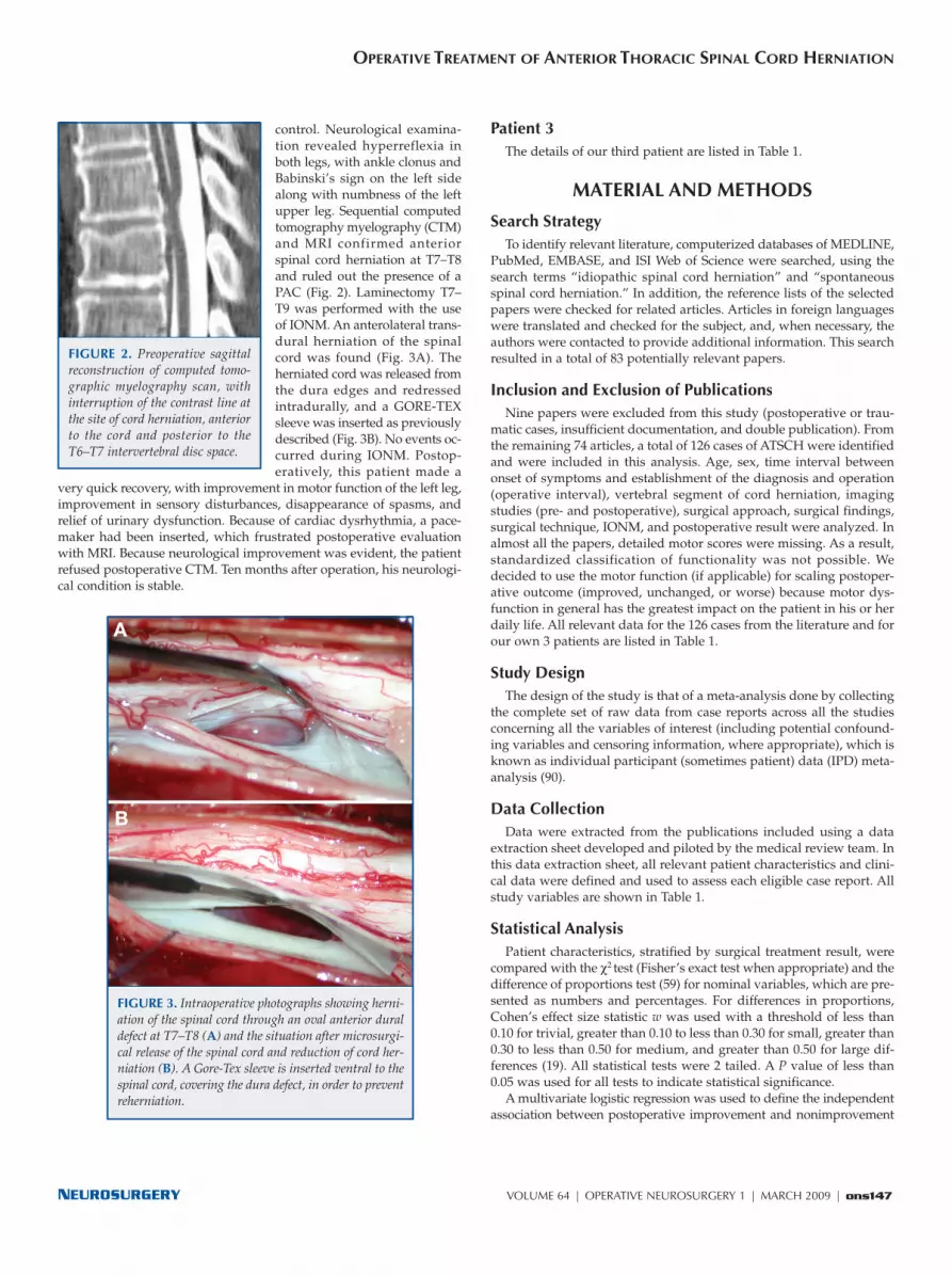
control. Neurological examina-tion revealed hyperreflexia inboth legs, with ankle clonus andBabinski’s sign on the left sidealong with numbness of the leftupper leg. Sequential computedtomography myelography (CTM)and MRI confirmed anteriorspinal cord herniation at T7–T8and ruled out the presence of aPAC (Fig. 2). Laminectomy T7–T9 was performed with the useof IONM. An anterolateral trans-dural herniation of the spinalcord was found (Fig. 3A). Theher niated cord was released fromthe dura edges and redressedintradurally, and a GORE- TEXsleeve was inserted as previouslydescribed (Fig. 3B). No events oc -curred during IONM. Post op -eratively, this patient made a
very quick recovery, with improvement in motor function of the left leg,improvement in sensory disturbances, disappearance of spasms, andrelief of urinary dysfunction. Because of cardiac dysrhythmia, a pace-maker had been inserted, which frustrated postoperative evaluationwith MRI. Because neurological improvement was evident, the patientrefused postoperative CTM. Ten months after operation, his neurologi-cal condition is stable.
Patient 3The details of our third patient are listed in Table 1.
MATERIAL AND METHODS
Search StrategyTo identify relevant literature, computerized databases of MEDLINE,
PubMed, EMBASE, and ISI Web of Science were searched, using thesearch terms “idiopathic spinal cord herniation” and “spontaneousspinal cord herniation.” In addition, the reference lists of the selectedpapers were checked for related articles. Articles in foreign languageswere translated and checked for the subject, and, when necessary, theauthors were contacted to provide additional information. This searchresulted in a total of 83 potentially relevant papers.
Inclusion and Exclusion of PublicationsNine papers were excluded from this study (postoperative or trau-
matic cases, insufficient documentation, and double publication). Fromthe remaining 74 articles, a total of 126 cases of ATSCH were identifiedand were included in this analysis. Age, sex, time interval betweenonset of symptoms and establishment of the diagnosis and operation(operative interval), vertebral segment of cord herniation, imagingstudies (pre- and postoperative), surgical approach, surgical findings,surgical technique, IONM, and postoperative result were analyzed. Inalmost all the papers, detailed motor scores were missing. As a result,standardized classification of functionality was not possible. Wedecided to use the motor function (if applicable) for scaling postoper-ative outcome (improved, unchanged, or worse) because motor dys-function in general has the greatest impact on the patient in his or herdaily life. All relevant data for the 126 cases from the literature and forour own 3 patients are listed in Table 1.
Study DesignThe design of the study is that of a meta- analysis done by collecting
the complete set of raw data from case reports across all the studiesconcerning all the variables of interest (including potential confound-ing variables and censoring information, where appropriate), which isknown as individual participant (sometimes patient) data (IPD) meta- analysis (90).
Data CollectionData were extracted from the publications included using a data
extraction sheet developed and piloted by the medical review team. Inthis data extraction sheet, all relevant patient characteristics and clini-cal data were defined and used to assess each eligible case report. Allstudy variables are shown in Table 1.
Statistical AnalysisPatient characteristics, stratified by surgical treatment result, were
compared with the χ2 test (Fisher’s exact test when appropriate) and thedifference of proportions test (59) for nominal variables, which are pre-sented as numbers and percentages. For differences in proportions,Cohen’s effect size statistic w was used with a threshold of less than0.10 for trivial, greater than 0.10 to less than 0.30 for small, greater than0.30 to less than 0.50 for medium, and greater than 0.50 for large dif-ferences (19). All statistical tests were 2 tailed. A P value of less than0.05 was used for all tests to indicate statistical significance.
A multivariate logistic regression was used to define the independentassociation between postoperative improvement and nonimprovement
OPERATIVE TREATMENT OF ANTERIOR THORACIC SPINAL CORD HERNIATION
FIGURE 2. Preoperative sagittalreconstruction of computed tomo-graphic myelography scan, withinterruption of the contrast line atthe site of cord herniation, anteriorto the cord and posterior to theT6–T7 intervertebral disc space.
FIGURE 3. Intraoperative photographs showing herni-ation of the spinal cord through an oval anterior duraldefect at T7–T8 (A) and the situation after microsurgi-cal release of the spinal cord and reduction of cord her-niation (B). A Gore- Tex sleeve is inserted ventral to thespinal cord, covering the dura defect, in order to preventreherniation.
A
B

TABLE 1. Relevant data for cases of anterior thoracic spinal cord herniationa
Caseno.
Series (ref. no.)Age
(y)/sexInter-val (y)
Symptoms LevelCTM/MRI
Surgicalfinding
Surgicaltechnique
IONMOperative
resultMRI
FU(mo)
1 Abe et al. (1) 58/M 4 B-S T8–T9 �/� VDD, SCH CR, WDD imprSM RC 36
2 Aizawa et al. (3) 44/M 5 B-S T8–T9 �/� DDM, SCH CR, WDD imprM, unchS RC 12
3 Aizawa et al. (3) 60/F 3 B-S T4–T5 �/� DDM, SCH CR, WDD imprM, unchS RC 12
4 Aizawa et al. (3) 59/F 20 SM para T4–T5 �/� VDD, SCH CR, WDD imprM, unchS RC 12
5 Ammar et al. (4) 50/F 1 B-S T7–T8 ��� VDD, SCH CR, WDD imprM, unchS RC 84
6 Aquilina et al. (5) 37/F 1 SM para T4–T5 �/� VDD, SCH CR, ADP imprSM RC 3
7 Arts et al. (6) 58/F ? B-S T7–T8 �/� VDD, SCH CR, ADP Unchanged
8 Arts et al. (6) 43/M ? B-S T4–T5 �/� 1PAC/2VDD,SCH
1NE/2CR, ADP imprSM RC
9 Bandai et al. (7) 63/F 5 SM para T2–T3 �/� DDM, SCH CR, ADP imprSM
10 Barbagallo et al. (8) 28/F 5 SM para T6 �/� VDD, SCH CR, ADP Unchanged RC 6
11 Barbagallo et al. (8) 64/M 10 SM para T6 �/� BD, VDD,SCH
BDF, CR, ADP Worse
12 Barrenechea et al. (9) 65/F 3 B-S T4–T5 �/� 1PAC/2VDD,SCH
1Cyst/2CR,ADP
SSEP,mMEP
Unchanged 60
13 Barrenechea et al. (9) 32/M 1 SM para T7–T8 �/� 1PAC/2VDD,SCH
1Cyst/2CR,ADP
SSEP,mMEP
Worse 144
14 Barrenechea et al. (9) 54/F 6,5 B-S T2–T3 �/� 1PAC/2VDD,SCH
1Cyst/2CR,ADP
SSEP,mMEP�
eMEP
Unchanged 16
15 Barrenechea et al. (9) 60/F 2,5 B-S T2–T3 �/� VDD, SCH CR, ADP imprSM 42
16 Barrenechea et al. (9) 59/F 1,5 B-S T5–T6 �/� VDD, SCH CR, ADP SSEP,mMEP
Worse 40
17 Barrenechea et al. (9) 34/M 5 SM para T7–T8 �/� VDD, SCH CR, ADP Unchanged 32
18 Barrenechea et al. (9) 72/M 5 B-S T4–T5 �/� VDD, SCH CR, ADP SSEP,mMEP
Unchanged 10
19 Bartolomei et al. (10) 61/F 10 B-S T3–T4 �/� VDD, SCH CR, ADP imprM, unchS RC 3
20 Batzdorf (11) 23/F 2 B-S T6 ? 1PAC/2VDD,SCH
1Cyst/2CR,ADP
imprSM
21 Baur et al. (12) 66/F 7 B-S T10 �/� VDD, SCH CR, DS imprSM RC
22 Berbel et al. (13) 56/M ? B-S T5–T6 �/� PAC, VDD,SCH
NE Unchanged 1
23 Bode et al. (14) 60/F 2 SM para T8 �/� VDD, SCH CR, ADP Worse RC 60
24 Bode et al. (14) 33/F 4 SM para T5 �/� VDD, SCH 1CR, ADP/2CR Unchanged 3
25 Borges et al. (15) 68/F 12 B-S T7–T8 �/� VDD, SCH CR, DS imprSM 12
26 Borges et al. (15) 68/M 8 B-S T2–T3 �/� VDD, SCH CR, ADP imprSM
27 Borges et al. (15) 48/F 9 B-S T7 �/� VDD, SCH CR, DS imprM, unchS 2
28 Brugieres et al. (16) 54/F 5 B-S T6 �/� VDD, SCH CB, CR, DS imprSM
29 Brugieres et al. (16) 70/M 0,5 S bilat T5–T6 �/� DDM, SCH CR, DS Improved RC
30 Cellerini et al. (17) 53/M 1 B-S T8–T9 �/� VDD, SCH CR, ADP imprM, unchS 3
31 Cellerini et al. (17) 37/F 0,5 B-S T4–T5 �/� VDD, SCH CR, ADP imprSM 3
32 Darbar et al. (20) 41/M 3 B-S T5 �/� VDD, SCH CR, DS imprSM 1
Continues
GROEN ET AL.
ons148 | VOLUME 64 | OPERATIVE NEUROSURGERY 1 | MARCH 2009 www.neurosurgery-online.com

TABLE 1. Continued
Caseno.
Series (ref. no.)Age
(y)/sexInter-val (y)
Symptoms LevelCTM/MRI
Surgicalfinding
Surgicaltechnique
IONMOperative
resultMRI
FU(mo)
33 Darbar et al. (20) 63/F ? B-S T4–T6 �/� 1PAC/2VDD,SCH
1Cyst/2CR,ADP
imprM, unchS
34 Darbar et al. (20) 34/F 5 SM para T7–T8 Myelo VDD, SCH CR, ADP imprM
35 Dietemann et al.(21)
68/M ? SM para T5–T6 �/� VDD, SCH CR, ? ?
36 Dix et al. (22) 44/F ? B-S T7–T8 �/� VDD, SCH CR, ADP imprSM 6
37 Ellger et al. (23) 59/f 2,5 B-S T2–T3 �/� VDD, SCH CR, DS imprSM RC 4
38 Ewald et al. (24, 25) 51/F 2 B-S T5–T6 �/� VDD, SCH 1CR, ADP/2NE Worse RC 2
39 Ewald et al. (25) 46/M 3 B-S T2 �/� VDD, SCH CR, ADP Unchanged
40 Ewald et al. (25) 50/F 8 SM para T6 �/� VDD, SCH CR, ADP Unchanged
41 Ferré et al. (26) 70/M 1,5 SM para T10–T11 �/� VDD, SCH CR, ADP imprSM PAD 24
42 Ferré et al. (26) 75/F 1 B-S T5–T6 �/� VDD, SCH CR, WDD Worse RC 12
43 Francis et al. (27) 28/F 1,5 B-S T6 �/� VDD, SCH CR, WDD imprSM RC
44 Gwinn andHenderson (30)
47/F 6 B-S T7–T8 �/� VDD, SCH CR, ADP SSEP imprSM RC 3
45 Gwinn andHenderson (30)
51/F ? B-S T8 �/� VDD, SCH CR, ADP SSEP imprSM PAD 3
46 Gwinn andHenderson (30)
55/M 0,5 B-S T6–T7 �/� VDD, SCH CR, ADP imprSM 3
47 Hausmann andMoseley (32)
58/F 8 B-S T6 �/� BD, VDD,SCH
BDF, CR, ADP imprSM
48 Hausmann andMoseley (32)
36/M 7 SM para T6–T7 �/� SCT CB Worse RC
49 van den Hauwe etal. (83)
65/F ? B-S T5–T6 �/� PAC, VDD,SCH
CR, ADP ?
50 Inoue et al. (34) 21/M 2 B-S T3 �/� VDD, SCH CR, DS imprSM 24
51 Isu et al. (35) 43/F 1 B-S T5–T6 �/� PAC Cyst unchM, imprS
52 Isu et al. (35) 45/F 2 SM para T2–T3 �/� PAC Cyst unchM, imprS
53 Iyer et al. (36) 59/F 3 B-S T3–T4 �/� 1NE/2VDD,SCH
1NE/2CR, ADP imprSM RC 6
54 Karadeniz-Bilgili etal. (37)
36/F 1,5 M left leg T2–T3 �/� 1NE/2VDD,SCH
1Cyst/2CR,ADP
Improved RC 2
55 Kumar et al. (42) 38/M 2 B-S T7–T8 �/� VDD, SCH CB, CR, ADP imprSM RC 2
56 Maira et al. (43) 31/F 7 SM para T8–T9 �/� VDD, SCH CR, WDD imprSM RC 156
57 Maira et al. (43) 54/F 8 B-S T5 �/� VDD, SCH CR, ADP imprSM RC 132
58 Maira et al. (43) 45/F 24 B-S T4–T5 �/� VDD, SCH,DP
CR, ADP,discotomy
Worse RC 48
59 Maira et al. (43) 50/M 2 SM para T6–T7 �/� VDD, SCH CR, WDD imprSM RC 36
60 Maira et al. (43) 57/F 6 B-S T4–T5 �/� VDD, SCH CR, ADP imprSM RC 24
61 Marshman et al. (45) 55/F 14 SM para T7–T8 �/� VDD, SCH CR, ADP imprSM 12
62 Maruichi et al. (46) 53/F 5 S right leg T4–T5 �/� VDD, SCH CR, ADP ? RC 7
63 Massicotte et al. (47) 39/F ? SM para T6–T7 �/� VDD, SCH CR, ADP imprSM RC
Continues
OPERATIVE TREATMENT OF ANTERIOR THORACIC SPINAL CORD HERNIATION
NEUROSURGERY VOLUME 64 | OPERATIVE NEUROSURGERY 1 | MARCH 2009 | ons149

TABLE 1. Continued
Caseno.
Series (ref. no.)Age
(y)/sexInter-val (y)
Symptoms LevelCTM/MRI
Surgicalfinding
Surgicaltechnique
IONMOperative
resultMRI
FU(mo)
64 Massicotte et al. (47) 44/F 7 B-S T5–T6 �/� 1NE/2VDD,SCH
1NE/2CR, ADP EP Unchanged 48
65 Massicotte et al. (47) 57/F 8 SM para T6 �/� 1PAC/2VDD,SCH
1Cyst/2CR,ADP
Unchanged 36
66 Massicotte et al. (47) 27/M 1 B-S T9 �/� VDD, SCH CR, ADP imprM, unchS 12
67 Masuzawa et al. (48) 36/M 3 B-S T4 �/� LDD, SCH CB, CR, ADP,PDP
imprSM 36
68 Matsumura et al. (49) 63/F ? B-S T3–T4 �/� PAC, DDM,SCH
CR, WDD Improved
69 Miura et al. (51) 49/M 1 SM para T5–T6 �/� VDD, SCH CR, WDD imprSM RC 6
70 Miyaguchi et al. (52) 54/F 2 B-S T3–T4 �/� DP, VDD,SCH
discect, CR,ADP
SSEP imprSM RC 6
71 Miyake et al. (53) 45/F 4 B-S T3–T4 �/� VDD, SCH CR, ADP imprSM RC 1
72 Miyake et al. (53) 53/M 6 B-S T2–T3 �/� VDD, SCH CR, ADP imprSM RC 1
73 Morley et al. (54) 28/F 2 B-S T5–T6 �/� VDD, SCH CR, ADP imprM, unchS 24
74 Morokoff et al. (55) 33/F 8 B-S T8 �/� VDD, SCH CR, WDD unchM, imprS 3
75 Najjar et al. (56) 32/M 5 B-S T8–T9 �/� 1PAC/2VDD,SCH
1Cyst/2CR,ADP
Improved RC 2
76 Nakagawa et al. (57) 77/F 9 SM para T6–T7 �/� VDD, SCH BDF, CR, ADP imprSM RC 12
77 Nakazawa et al. (58) 43/F 5 B-S T2 �/� DDM, SCH CR, WDD unchM, imprS RC 48
78 Nakazawa et al. (58) 39/F 4 B-S T4 �/� DDM, SCH CR, WDD imprSM RC
79 Novak et al. (60) 64/F 6 SM para T6 �/� VDD, SCH CR, ADP mMEP+eMEP
Unchanged RC 12
80 Novak et al. (60) 45/F 2 SM para T4–T5 �/� VDD, SCH CR, ADP mMEP Improved RC 3
81 Novak et al. (60) 73/M 6 SM para T4–T5 �/� VDD, SCH CR, ADP mMEP+eMEP
Unchanged 9
82 Oe et al. (61) 61/M ? SM para T4 ?/? PAC, DDM,SCH
CR, WDD Unchanged
83 Pereira et al. (62) 55/M 4 SM para T2–T3 �/� VDD, SCH CR, ADP imprSM RC 18
84 Rivas et al. (64) 49/M 3 B-S T6–T7 �/� VDD, SCH CR, ADP imprM, unchS 48
85 Roland et al. (65) 50/F ? B-S T4 �/� VDD, SCH CR, ADP imprSM
86 Sagiuchi et al. (66) 48/M 20 B-S T7–T8 �/� DP, VDD,SCH
CR, ADP imprSM
87 Sahl et al. (67) 51/M 16 SM para T3–T4 �/� VDD, SCH CR, DS imprSM 12
88 Saito et al. (69) 57/M 13 SM para T2–T3 �/� DDM, SCH CR, ADP imprSM RC 1
89 Saito et al. (68) 68/F 36 SM para T6–T7 �/� VDD, SCH CR, WDD Unchanged PAD 40
90 Sasaoka et al. (71) 57/M 15 SM para T2–T3 �/� VDD, SCH CR, ADP, PDP EP unch, paindisapp
RC 24
91 Sioutos et al. (72) 34/F 1 SM para T6–T7 �/� PAC, VDD,SCH
CB, CR, ADP Worse RC 3
92 Slavotineket al. (73) 22/F 4 B-S T5 �/� 1PAC/2VDD,SCH
1Cyst/2CR, fatgraft
imprSM
93 Spissu et al. (74) 56/F 1 B-S T7 �/� VDD, SCH CR, DS imprM, unchS 12
Continues
GROEN ET AL.
ons150 | VOLUME 64 | OPERATIVE NEUROSURGERY 1 | MARCH 2009 www.neurosurgery-online.com

TABLE 1. Continued
Caseno.
Series (ref. no.)Age
(y)/sexInter-val (y)
Symptoms LevelCTM/MRI
Surgicalfinding
Surgicaltechnique
IONMOperative
resultMRI
FU(mo)
94 Srinivasan et al. (75) 69/F ? B-S T3–T4 �/� ? ? ?
95 Sugimoto et al. (76) 48/M 1 SM para T4–T5 �/� DDM, SCH CR, WDD imprSM RC 12
96 Taghipour et al. (77) 32/M 3 B-S T10–T11 �/� DDM, SCH CR,WDD imprSM
97 Tekkök (78) 49/F 3 B-S T3–T4 �/� VDD, SCH CR, ADP imprM, unchS RC 5
98 Tronnier et al. (79) 45/F 4 S level T5 T3–T4 �/� PAC, VDD,SCH
Cyst, CR, ADP Worse RC 4
99 Uchino et al. (80) 71/F 2 B-S T4–T5 �/� PAC, VDD,SCH
CR, DS Unchanged RC
100 Uchino et al. (80) 61/F 2 B-S T5–T6 �/� NE NE Improved PAD
101 Urbach et al. (81) 44/M 2 S level T6 T5–T6 �/� VDD, SCH CR, ADP Improved RC
102 Vallee et al. (82) 28/F 2 B-S T3–T4 �/� DDM, SCH CR, WDD imprSM PAD 2
103 Vallee et al. (82) 58/F 7 B-S T4–T5 �/� DDM, SCH CR, WDD imprSM PAD 2
104 Vallee et al. (82) 40/F 1,5 B-S T5–T6 �/� VDD, SCH CR, ADP SSEP Unchanged 6
105 Vallee et al. (82) 49/F 3,5 B-S T4–T5 �/� VDD, SCH CR, DS, PDP SSEP Unchanged 6
106 Verny et al. (84) 28/F 1,5 B-S T3–T4 �/� DDM, SCH CR, WDD imprSM PAD 2
107 Verny et al. (84) 58/F 6 B-S T4–T5 �/� DDM, SCH CR, ADP imprSM RC
108 Wada et al. (85) 59/M 4 B-S T4–T5 �/� PAC, VDD,SCH
Cyst, CR, WDD imprM, unchS RC 48
109 Wada et al. (85) 48/M 2 B-S T5–T6 �/� VDD, SCH CR, WDD imprSM RC 48
110 Wada et al. (85) 63/F 10 B-S T3–T4 �/� VDD, SCH CR, WDD imprM RC 48
111 Watanabe et al. (86) 43/F 5 B-S T4 �/� DDM, SCH CR, WDD Improved RC 156
112 Watanabe et al. (86) 39/F 3 B-S T3 �/� DDM, SCH CR, WDD Improved RC 132
113 Watanabe et al. (86) 54/F 4 B-S T4 �/� DDM, SCH CR, WDD Improved RC 24
114 Watanabe et al. (86) 71/F 5 SM para T4 �/� DDM, SCH CR, WDD Worse 24
115 Watanabe et al. (86) 49/M 5 B-S T4 �/� DDM, SCH CR, WDD Improved RC 21
116 Watanabe et al. (86) 47/F 5 B-S T5 �/� DDM, SCH CR, WDD Improved RC 18
117 Watanabe et al. (86) 78/F 4 SM para T4 �/� DDM, SCH CR, WDD Improved RC 12
118 Watanabe et al. (86) 56/M 2 B-S T6 �/� 1PAC/2DDM,SCH
1Cyst/2CR,WDD
Improved RC 9
119 Watanabe et al. (86) 47/F 3 SM para T3 �/� DDM, SCH CR, WDD Improved RC 6
120 Watters et al. (87) 55/F 10 B-S T3–T4 �/� 1NE/2VDD,SCH
1NE/2CR, DS imprSM RC 18
121 White and Firth (88) 61/F 1,5 B-S T4 �/� VDD, SCH CR, ADP Unchanged RC
122 White and Firth (88) 39/M 1,5 B-S T8 �/� VDD, SCH CR, ADP Unchanged RC
123 White and Tsegaye(89)
61/M 4 B-S T7 �/� VDD, SCH CR, ADP imprSM 12
124 White and Tsegaye(89)
62/F 1,5 B-S T6–T7 �/� VDD, SCH CR, ADP, PDP MEP imprSM RC 12
125 White and Tsegaye(89)
66/F 8 SM para T7 �/� BD, VDD,SCH
BDF, CR, ADP,PDP
MEP imprSM RC 9
126 Wortzman et al. (91) 63/M 2 SM para T7 Myelo BD, VDD,SCH
CB, CR, DS imprSM 24
Continues
OPERATIVE TREATMENT OF ANTERIOR THORACIC SPINAL CORD HERNIATION
NEUROSURGERY VOLUME 64 | OPERATIVE NEUROSURGERY 1 | MARCH 2009 | ons151

TABLE 1. Continued
Caseno.
Series (ref. no.)Age
(y)/sexInter-val (y)
Symptoms LevelCTM/MRI
Surgicalfinding
Surgicaltechnique
IONMOperative
resultMRI
FU(mo)
127 Case 1 42/M 5 B-S T5–T6 �/� 1PAC/2VDD,SCH
1Cyst/2CR,ADP
SSEP,mMep+eMEP
Worse RC 30
128 Case 2 60/M 2 B-S T6–T7 �/� VDD, SCH CR, ADP SSEP,mMEP+eMEP
imprSM RC 20
129 Case 3 69/M 5 SM para T7–T8 �/� VDD, SCH CR, ADP SSEP,mMep+eMEP
imprSM RC 10
a CTM, computed tomography myelography; MRI, magnetic resonance imaging; IONM, intraoperative neurophysiological monitoring; FU, follow-up; B-S, Brown-Séquard syndrome; VDD, ventral dura defect; SCH, spinal cord herniation; CR, cord release; WDD, widening dura defect; imprSM, motor and sensory functionimprovement; RC, realigned spinal cord; DDM, duplicated dura mater; imprM, motor function improvement; unchS, unchanged sensory function; SM para,paraparesis and sensory deficit; ?, unknown; ADP, anterior dura patch; 1, first exploration; PAC, posterior arachnoid cyst; 2, second exploration; NE, negative exploration;BD, bony defect; BDF, bony defect filling; Cyst, excision of arachnoid cyst; SSEP, somatosensory evoked potentials; mMEP, muscular recorded motor evokedpotentials; eMEP, epidurally recorded motor evoked potentials; DS, direct suture dura defect; CB, cord biopsy; S bilat, bilateral sensory disturbance; PAD, persistentanterior displacement; SCT, suspicion of cord tumor; unchM, motor function unchanged; imprS, sensory function improved; Mleft leg, isolated motor dysfunctionleft leg; EP, evoked potential monitoring (unspecified); LDD, lateral dura defect; PDP, posterior dura patch; DP, disc prolapse; discect, discectomy; Myelo,myelography.
FIGURE 4. Segmental distribution of anterior thoracic spinal cord herni-ation in 129 cases.
GROEN ET AL.
operative results (21, 46, 75, 83) were excluded. Improvement in(sensory) motor function was reported in 88 patients (73%). In24 patients (20%), (sensory) motor function did not change afterthe operation, and in 9 patients (7%), motor function deterio-rated because of operative intervention (Table 2).
Follow- up period was specified in 93 cases (72%), rangingfrom 1 month to 13 years. Mean follow- up was 24.4 months.
Improved patients did not differ from stable (unchanged) ordeteriorated (worse) subjects in terms of gender, age, and oper-ative interval. However, cord herniation at T3–T4 and T8–T9had a statistically significant higher prevalence among improvedpatients (18.3% versus 4.2% and 8.3% versus 0%, respectively).
(unchanged or worse) in motor function as the binary dependent variable.After adjusting for age, gender, operative interval, and vertebral segmentof cord herniation, we analyzed outcomes of imaging, surgical approach,surgical findings, and surgical technique as independent variables toallow us to define which variables were associated with improvement orno improvement (unchanged) or with deterioration (worsening) aftersurgical intervention. A P value of 0.05 was used as a cutoff for sequen-tially entering and removing each variable. We reported the odds ratioswith 95% confidence intervals (CIs) and applied the Hosmer- Lemeshowtest to evaluate the model calibration. All statistical analyses were per-formed using SPSS 14.0 for Windows (SPSS Inc., Chicago, IL).
RESULTS
All 129 patients were adults. Age was normally distributed (Shapiro- Wilk test, P � 0.05) and ranged from 21 to 78 years(mean age, 51 years). Women (n � 80) were more affected(64%) than men (male- to- female ratio, 1:1.8). Brown- Séquardsyndrome was present in 85 cases (66%), paraparesis in 39 cases(30%), isolated sensory deficit in 4 cases (3%), and isolatedmotor deficit in 1 patient (1%). Operative interval (intervalbetween the onset of symptoms and surgical treatment) rangedfrom 6 months to 36 years (mean interval, 5.2 years). In 17cases, the operative interval was unknown. Segmental distri-bution is shown in Figure 4. ATSCH has a predisposition forthe T3–T7 vertebral segments (80%; P � 0.001). In 129 patients,218 vertebral segments were involved: 40 dura defects were atthe level of the vertebral body; in 88 patients, the dura defectwas on the level of the intervertebral disc; and in 1 patient, thedura defect extended over 3 segments (T4–T6) (20).
Operative results were analyzed in 121 patients. Two patientsthat worsened after spinal cord biopsy (32, 33, 72), 2 patientsafter negative exploration (13, 80), and 4 patients with unknown
ons152 | VOLUME 64 | OPERATIVE NEUROSURGERY 1 | MARCH 2009 www.neurosurgery-online.com
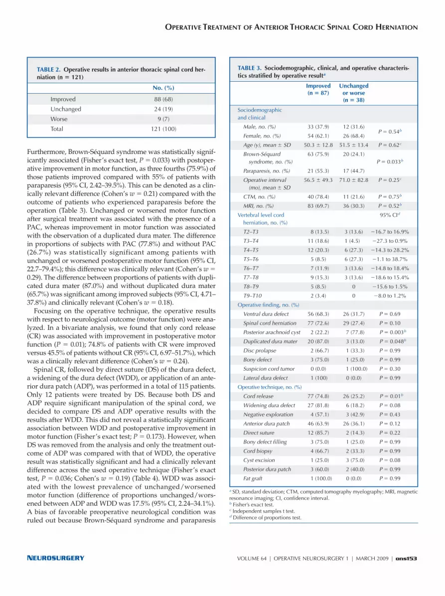
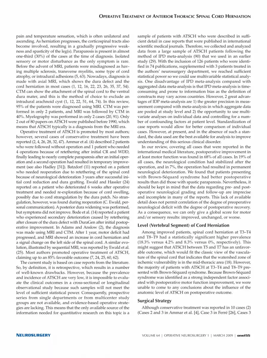
TABLE 2. Operative results in anterior thoracic spinal cord her-niation (n � 121)
No. (%)
Improved 88 (68)
Unchanged 24 (19)
Worse 9 (7)
Total 121 (100)
a SD, standard deviation; CTM, computed tomography myelography; MRI, magneticresonance imaging; CI, confidence interval.b Fisher’s exact test.c Independent samples t test.d Difference of proportions test.
TABLE 3. Sociodemographic, clinical, and operative characteris-tics stratified by operative resulta
Improved Unchanged (n � 87) or worse
(n � 38)
Sociodemographicand clinical
Male, no. (%) 33 (37.9) 12 (31.6)
Female, no. (%) 54 (62.1) 26 (68.4)P � 0.54b
Age (y), mean � SD 50.3 � 12.8 51.5 � 13.4 P � 0.62c
Brown-Séquard 63 (75.9) 20 (24.1)syndrome, no. (%) P � 0.033b
Paraparesis, no. (%) 21 (55.3) 17 (44.7)
Operative interval 56.5 � 49.3 71.0 � 82.8 P � 0.25c
(mo), mean � SD
CTM, no. (%) 40 (78.4) 11 (21.6) P � 0.75b
MRI, no. (%) 83 (69.7) 36 (30.3) P � 0.52b
Vertebral level cord 95% CId
herniation, no. (%)
T2–T3 8 (13.5) 3 (13.6) �16.7 to 16.9%
T3–T4 11 (18.6) 1 (4.5) �27.3 to 0.9%
T4–T5 12 (20.3) 6 (27.3) �14.3 to 28.2%
T5–T6 5 (8.5) 6 (27.3) �1.1 to 38.7%
T6–T7 7 (11.9) 3 (13.6) �14.8 to 18.4%
T7–T8 9 (15.3) 3 (13.6) �18.6 to 15.4%
T8–T9 5 (8.5) 0 �15.6 to 1.5%
T9–T10 2 (3.4) 0 �8.0 to 1.2%
Operative finding, no. (%)
Ventral dura defect 56 (68.3) 26 (31.7) P � 0.69
Spinal cord herniation 77 (72.6) 29 (27.4) P � 0.10
Posterior arachnoid cyst 2 (22.2) 7 (77.8) P � 0.003b
Duplicated dura mater 20 (87.0) 3 (13.0) P � 0.048b
Disc prolapse 2 (66.7) 1 (33.3) P � 0.99
Bony defect 3 (75.0) 1 (25.0) P � 0.99
Suspicion cord tumor 0 (0.0) 1 (100.0) P � 0.30
Lateral dura defect 1 (100) 0 (0.0) P � 0.99
Operative technique, no. (%)
Cord release 77 (74.8) 26 (25.2) P � 0.01b
Widening dura defect 27 (81.8) 6 (18.2) P � 0.08
Negative exploration 4 (57.1) 3 (42.9) P � 0.43
Anterior dura patch 46 (63.9) 26 (36.1) P � 0.12
Direct suture 12 (85.7) 2 (14.3) P � 0.22
Bony defect filling 3 (75.0) 1 (25.0) P � 0.99
Cord biopsy 4 (66.7) 2 (33.3) P � 0.99
Cyst excision 1 (25.0) 3 (75.0) P � 0.08
Posterior dura patch 3 (60.0) 2 (40.0) P � 0.99
Fat graft 1 (100.0) 0 (0.0) P � 0.99
OPERATIVE TREATMENT OF ANTERIOR THORACIC SPINAL CORD HERNIATION
Furthermore, Brown- Séquard syndrome was statistically signif-icantly associated (Fisher’s exact test, P � 0.033) with postoper-ative improvement in motor function, as three fourths (75.9%) ofthose patients improved compared with 55% of patients withparaparesis (95% CI, 2.42–39.5%). This can be denoted as a clin-ically relevant difference (Cohen’s w � 0.21) compared with theoutcome of patients who experienced paraparesis before theoperation (Table 3). Unchanged or worsened motor functionafter surgical treatment was associated with the presence of aPAC, whereas improvement in motor function was associatedwith the observation of a duplicated dura mater. The differencein proportions of subjects with PAC (77.8%) and without PAC(26.7%) was statistically significant among patients withunchanged or worsened postoperative motor function (95% CI,22.7–79.4%); this difference was clinically relevant (Cohen’s w �0.29). The difference between proportions of patients with dupli-cated dura mater (87.0%) and without duplicated dura mater(65.7%) was significant among improved subjects (95% CI, 4.71–37.8%) and clinically relevant (Cohen’s w � 0.18).
Focusing on the operative technique, the operative resultswith respect to neurological outcome (motor function) were ana-lyzed. In a bivariate analysis, we found that only cord release(CR) was associated with improvement in postoperative motorfunction (P � 0.01); 74.8% of patients with CR were improvedversus 45.5% of patients without CR (95% CI, 6.97–51.7%), whichwas a clinically relevant difference (Cohen’s w � 0.24).
Spinal CR, followed by direct suture (DS) of the dura defect,a widening of the dura defect (WDD), or application of an ante-rior dura patch (ADP), was performed in a total of 115 patients.Only 12 patients were treated by DS. Because both DS andADP require significant manipulation of the spinal cord, wedecided to compare DS and ADP operative results with theresults after WDD. This did not reveal a statistically significantassociation between WDD and postoperative improvement inmotor function (Fisher’s exact test; P � 0.173). However, whenDS was removed from the analysis and only the treatment out-come of ADP was compared with that of WDD, the operativeresult was statistically significant and had a clinically relevantdifference across the used operative technique (Fisher’s exacttest, P � 0.036; Cohen’s w � 0.19) (Table 4). WDD was associ-ated with the lowest prevalence of unchanged/worsenedmotor function (difference of proportions unchanged/wors-ened between ADP and WDD was 17.5% (95% CI, 2.24–34.1%).A bias of favorable preoperative neurological condition wasruled out because Brown- Séquard syndrome and paraparesis
NEUROSURGERY VOLUME 64 | OPERATIVE NEUROSURGERY 1 | MARCH 2009 | ons153

a Fisher’s exact test: P = 0.036.
TABLE 4. Operative result related to operative technique (n � 103)a
Anterior WideningTotal
dura patch dura defect
Improved 45 (64.3%) 27 (81.8%) 72
Unchanged/worse 25 (35.7%) 6 (18.2%) 31
Total 70 33 103
a SM, sensorimotor.b Fisher’s exact test: P = 0.129 (not significant).
TABLE 5. Preoperative neurological symptoms related to opera-tive technique (n � 101)a, b
Anterior WideningTotal
dura patch dura defect
Brown-Séquard syndrome 45 (66.2%) 23 (33.8%) 68
Paraparesis (SM) 23 (69.7%) 10 (30.3%) 33
Total 68 33 101
GROEN ET AL.
ment of cord herniation, only Brown- Séquard syndrome andCR showed a statistically significant association with motorimprovement (odds ratio, 2.91; 95% CI, 1.15–7.39%; P � 0.02, Brown- Séquard syndrome; and odds ratio, 0.16; 95% CI, 0.05–0.52; P � 0.02, CR, respectively). Comparison of the number ofpatients actually classified in each group with the number pre-dicted for each group was evaluated with the Hosmer- Lemeshow statistic, producing a nonsignificant χ2 test result.The Hosmer- Lemeshow statistic for the model was 2.35 (P �0.93), indicating a good fit.
DISCUSSION
ATSCH refers to herniation of the thoracic spinal cordthrough a mediolateral defect of the ventral (thoracic) duramater. In the literature, different terminology was found to be inuse (idiopathic spinal cord herniation, thoracic idiopathic spinalcord herniation, spontaneous spinal cord herniation, sponta-neous thoracic spinal cord herniation) for the same entity. Thecause of anterior thoracic spinal cord herniation remains spec-ulative. A number of theories to explain the occurrence of a ven-tral thoracic dural defect have been postulated, such as a historyof trauma (15, 27, 67, 81, 87, 91), pressure erosion of the ventralthoracic dura mater (35), thoracic disc herniation (32, 87, 91),congenital disorder (preexisting ventral meningocele) (48, 91), aduplication of the ventral dura mater (1, 3, 4, 43, 49, 51, 52, 58,61, 68, 69, 76, 77, 82, 84–86), a congenital extradural arachnoidcyst (43), or an inflammatory process (56). Because the causeremains unknown, the rationale for a distinction between idio-pathic or traumatic is missing. Therefore, we find it more appro-priate to use the term ATSCH because this focuses on the radi-ological features that all these cases have in common.
Whatever the mechanism of the dura defect might be, thepathogenesis of herniation seems to be related to the fact that thespinal cord comes to lie against the defect and arachnoidal adhe-sions develop at the edges of the dura defect over time. Initially,cerebrospinal fluid (CSF) freely moves in and out of the defectwith each pulsation. Possibly, herniation of an arachnoid mem-brane results in wedging and in widening of the dura defect.Conditions such as the negative pressure in the thoracicextradural space or variation in the force and amplitude of CSFpulsations during the cardiac cycle may also influence the courseof the disorder (17). At a certain moment, the opening becomesblocked by the spinal cord, which will be sucked out by negativepressure in the epidural space (53) or pushed out by dorsal pres-sure (35) into the dura defect, finally leading to transdural spinalcord herniation (42, 77). Two out of 3 of our patients had a historyof spinal trauma before the development of symptomatic ATSCH.It might be that a ventral dura rupture occurred at the time oftrauma, possibly caused by an abrupt increase in intrathoracicand intradural pressure.
ATSCH predominantly occurs in middle- aged adults in thethird and eighth thoracic vertebral segment and has a femalepreponderance (male- to- female ratio, 1:1.8). Patients usuallypresent with Brown- Séquard syndrome. Initially, herniation ofthe lateral spinothalamic tract occurs, resulting in diminished
were represented equally in both groups (ADP and WDD)(Table 5). Persistent anterior displacement or realignment ofthe spinal cord on postoperative MRI did not correlate withpostoperative outcome. Also, no statistically significant associ-ation was found between operative techniques (WDD, DS, orADP) and spinal cord position on postoperative MRI (realign-ment of the spinal cord or persistent anterior displacement).
Fifteen patients were operated on twice because of a misdi-agnosis (6, 9, 11, 20, 36, 37, 47, 56, 73, 86, 87). At the initial oper-ation, a PAC was detected and was thought to be responsible forthe neurological symptoms in 11 cases. In 4 patients, no abnor-malities were found (negative exploration). All 15 patients dete-riorated postoperatively and were re- explored, which finallyresulted in treatment of spinal cord herniation. Operative resultsin those patients were not significantly unfavorable comparedwith those patients who were operated on only once.
IONM was performed in 20 cases: unspecified evoked poten-tials were reported in 2 patients, SSEPs in 5 patients, mMEPs in3 patients, mMEPs + eMEPs in 2 patients, SSEPs + mMEPs in4 patients, and SSEPs + mMEPs + eMEPs in 4 patients. Becauseof the small number of cases and the absence of uniformity inmonitoring, no statistical analysis was performed on this item(i.e., IONM in operative treatment of ATSCH).
After exclusion of cases with negative exploration (13, 80),cord biopsy (16, 32, 42, 48, 72, 91), absence of preoperativemotor deficit (16, 46, 79, 81), arachnoid cyst removal withoutCR (21, 35, 75, 83), and unknown postoperative results (21, 75,83), a sample of 111 patients was available for multivariablelogistic regression to identify sociodemographic and clinicalcharacteristics, operative findings, and surgical techniques asdeterminants of postoperative motor function improvement.Adjusted for age, gender, operative interval, and vertebral seg-
ons154 | VOLUME 64 | OPERATIVE NEUROSURGERY 1 | MARCH 2009 www.neurosurgery-online.com
OPERATIVE TREATMENT OF ANTERIOR THORACIC SPINAL CORD HERNIATION
sample of patients with ATSCH who were described in suffi-cient detail in case reports that were published in internationalscientific medical journals. Therefore, we collected and analyzeddata from a large sample of ATSCH patients following themethod of IPD meta- analysis (90) that we used in an earlierstudy (29). With the inclusion of 126 patients who were identi-fied in 74 publications, supplemented with 3 patients treated inthe authors’ neurosurgery department, we reached sufficientstatistical power so we could use multivariable statistical analy-sis. One disadvantage of IPD meta- analysis compared withaggregated data meta- analysis is that IPD meta- analysis is time- consuming and prone to information bias as the definition ofconstructs may vary across countries. However, 2 great advan-tages of IDP meta- analysis are 1) the greater precision in meas-urement compared with meta- analysis in which aggregate dataare pooled at study level and 2) the opportunity to use multi-variate analyses on individual data and controlling for a num-ber of confounding factors at patient level. Standardization ofcase reports would allow for better comparison of individualcases. However, at present, and in the absence of such a stan-dard, the data used are the best available for analysis to improveunderstanding of this serious clinical disorder.
In our review, covering all cases that were reported in theinternational medical literature, postoperative improvement inat least motor function was found in 68% of all cases. In 19% ofall cases, the neurological condition had stabilized after theoperation, and in 7%, the operation had resulted in permanentneurological deterioration. We found that patients presentingwith Brown- Séquard syndrome had better postoperativeresults than did those with spastic paraparesis. Nevertheless, itshould be kept in mind that the data regarding pre- and post-operative neurological grading and follow- up are impreciseand incomplete in many of the reports. This lack of availabledetail does not permit correlation of the degree of preoperativeneurological deficit with the degree of postoperative recovery.As a consequence, we can only give a global score for motorand/or sensory results: improved, unchanged, or worse.
Level (Vertebral Segment) of Cord HerniationAmong improved patients, spinal cord herniation at T3–T4
and T8–T9 had a statistically significant higher prevalence(18.3% versus 4.2% and 8.3% versus 0%, respectively). Thismight suggest that ATSCH between T5 and T7 has an unfavor-able outcome, which would fit the classic view of the vascula-ture of the spinal cord that indicates that the watershed zone ofischemic vulnerability is in the mid- thoracic area (18). However,the majority of patients with ATSCH at T3–T4 and T8–T9 pre-sented with Brown- Séquard syndrome. Because Brown- Séquardsyndrome was identified as a strong independent factor associ-ated with postoperative motor function improvement, we wereunable to come to any conclusions about the influence of theanatomic level of ATSCH on postoperative outcome.
Surgical StrategyAlthough conservative treatment was reported in 10 cases (2)
(Cases 2 and 3 in Ammar et al. [4], Case 3 in Ferré [26], Cases 3
pain and temperature sensation, which is often unilateral andascending. As herniation progresses, the corticospinal tracts alsobecome involved, resulting in a gradually progressive weak-ness and spasticity of the leg(s). Paraparesis is present in almostone-third (30%) of the cases at the time of diagnosis. Isolatedsensory or motor disturbance as the only symptom is rare.Before the advent of MRI, patients were misdiagnosed as hav-ing multiple sclerosis, transverse myelitis, some type of cordatrophy, or intradural adhesions (5, 63). Nowadays, diagnosis ismade with axial MRI, which shows the dura defect and thecord herniation in most cases (1, 12, 16, 22, 23, 26, 35, 37, 54).CTM can show the attachment of the spinal cord to the ventraldura mater, and this is the method of choice to exclude anintradural arachnoid cyst (1, 12, 22, 51, 64, 74). In this review,95% of the patients were diagnosed using MRI. CTM was per-formed in only 2 patients, and MRI was followed by CTM in40%. Myelography was performed in only 2 cases (20, 91). Only2 out of 80 papers on ATSCH were published before 1990, whichmeans that ATSCH typically is a diagnosis from the MRI era.
Operative treatment of ATSCH is promoted by most authors;however, several cases of conservative treatment have beenreported (2, 4, 26, 28, 32, 47). Ammar et al. (4) described 2 patientswho were followed without operation and 1 patient who needed4 operations because of retethering after initial CR and WDD,finally leading to nearly complete paraparesis after an initial oper-ation and a second operation had resulted in temporary improve-ment (see also Hadley [31]). Batzdorf (11) reported on a patientwho needed reoperation due to retethering of the spinal cordbecause of neurological deterioration 3 years after successful ini-tial cord reduction and dura grafting. Ewald and Hassler (24)reported on a patient who deteriorated 6 weeks after operativetreatment and needed re- exploration because of cord swelling,possibly due to cord strangulation by the dura patch. No stran-gulation, however, was found during reoperation (C. Ewald, per-sonal communication). A posterior dura widening was performed,but symptoms did not improve. Bode et al. (14) reported a patientwho experienced secondary deterioration caused by retetheringafter closure of the dura defect with DuraGen after initial postop-erative improvement. In Adams and Anslow (2), the diagnosiswas made using MRI and CTM. After 1 year, motor deficit hadprogressed, and MRI showed an increase in cord herniation anda signal change on the left side of the spinal cord. A similar evo-lution, illustrated by sequential MRI, was reported by Ewald et al.(25). Most authors promoted operative treatment of ATSCH,claiming up to an 85% favorable outcome (7, 24, 25, 60, 62).
The current study is based on case reports from the literature.So, by definition, it is retrospective, which results in a numberof well- known drawbacks. However, because the prevalenceand incidence of ATSCH are very low, it is impossible to evalu-ate the clinical outcomes in a cross- sectional or longitudinalobservational study because such samples will not meet thelevel of sufficient statistical power. Consequently, prospectiveseries from single departments or from multicenter studygroups are not available, and evidence- based operative strate-gies are lacking. This means that the only available source of theinformation needed for quantitative research on this topic is a
NEUROSURGERY VOLUME 64 | OPERATIVE NEUROSURGERY 1 | MARCH 2009 | ons155

ons156 | VOLUME 64 | OPERATIVE NEUROSURGERY 1 | MARCH 2009 www.neurosurgery-online.com
and 4 in Hausmann and Moseley [32], Cases 1, 3, 5, and 8 inMassicotte et al. [47]), most patients with ATSCH were indicatedfor operation. The aim of surgery is to release the strangulatedspinal cord (CR), restore the cord to its normal intradural posi-tion, and prevent recurrence of cord herniation. The goal of sur-gical treatment is to stop and eventually reverse neurologicaldeterioration.
Principally, there are 2 surgical strategies for preventingreherniation of the spinal cord. The first strategy is closure ofthe dura defect, either by DS of the ventral dural defect (12, 15,16, 34, 80, 82, 87, 91) or by filling the dural defect with a fatgraft (73), or by using a patch (fascia, pericard, or some otherartificial material) to cover or suture the dural defect (ADP) (5,8, 9, 11, 15, 17, 22, 24–26, 30, 32, 36, 37, 42–48, 52, 53, 56, 57, 62,65, 66, 71, 72, 78, 79, 81, 82, 84, 88, 89) after releasing the spinalcord and reducing the herniation. Promoters of this strategystate that closure of the dura defect is necessary to preventpostoperative CSF circulation disturbances that may be pro-duced by an extradural CSF collection (43) and because of therisk of recurrent spinal cord herniation (31, 78). Also, realign-ment of the spinal cord is thought to be essential, and it isclaimed that this can only be achieved by covering or closure ofthe ventral dura defect (78).
The second strategy is WDD. This technique is mainly pro-moted in Japanese articles and is based on the perception of a so- called duplicated ventral dura mater. The spinal cord is saidto be herniated through a defect in this dura mater duplication(1, 3, 4, 43, 51, 55, 58, 68, 69, 76, 77, 82, 84–86). Relief of the stran-gulated spinal cord can be obtained by resecting the dural ringaround the cord, which at the same time results in a wideningof the dura defect and also prevents reherniation and restran-gulation of the cord. A number of authors claim that this tech-nique is safer and is associated with the best operative out-come because the cord manipulations that are needed whenclosing the ventral dura defect (directly or by means of sometype of patch) can be avoided (35, 56, 69, 86). Isu et al. (35)state that the aim of the operation is to resolve the incarcerationof the spinal cord. In their opinion, it is not necessary to reducethe spinal cord herniation.
In multivariate logistic regression, spinal CR appears to be astrong independent factor associated with a favorable postop-erative outcome (i.e., motor function improvement), irrespec-tive of whether CR is followed by WDD, DS of the defect, orapplication of an ADP. After analysis of the data from 103 oper-ative cases of ATSCH, we found statistical evidence in favor ofWDD with respect to postoperative motor function improve-ment (Table 4). In 70 patients, postoperative MRI was per-formed to determine whether the spinal cord had returned toits natural position. In 7 patients, there was persistent anteriordisplacement (after WDD in 4 patients and after ADP in 3patients), but this did not result in an unfavorable outcomecompared with cases in which realignment of the spinal cordwas confirmed. In this review, we were not able to find anysupport for the idea that closure of the ventral dura defect (byeither DS or ADP) was in favor of WDD, when aiming for cordrealignment on postoperative MRI. However, postoperative
imaging was performed in only 56% of the 129 cases. To betterdefine operative results from the perspective of operative tech-nique, subsequent postoperative imaging (MRI or CTM) isneeded to check whether cord herniation has been resolvedand whether cord realignment has occurred.
It is evident that cord biopsy should not be part of the oper-ative procedure in ATSCH.
Nowadays, ATSCH can be recognized and diagnosed pre-operatively, using modern imaging techniques such as MRIand CTM. This helps avoid tissue sampling during exploration.In 6 cases, the spinal cord was biopsied during operation,which resulted in worsening of the neurological deficit in 2patients (32, 33, 72) (separately reporting about the samepatient). Interestingly, despite biopsy, 4 patients improved post-operatively (16, 42, 48, 91).
IONMIONM is increasingly being used in operative treatment of
spinal cord lesions (39–41). We monitored SSEPs and MEPs inall 3 patients. Motor potentials were evoked using transcranialelectrical stimulation and were recorded epidurally as D waves(eMEPs) or muscularly as mMEPs. SSEP recording is not ofmuch use in detecting and preventing a procedure- relatedmotor deficit. In recent years, intraoperative spinal eMEPrecording has proven very valuable in spinal cord tumor sur-gery because it allows monitoring of D waves and the intraop-erative decrease in its amplitude (41, 50). The major advantageof combined mMEP and eMEP monitoring is that it identifiesimpairment of the functional integrity of the motor pathwaysbefore a permanent deficit has occurred (38, 40, 41, 70).
In our first patient, detethering of the spinal cord temporar-ily resulted in complete loss of motor potentials (mMEPs) anddecrease in the distally recorded D- wave amplitude. Afterrelease of the cord and subsequent covering of the anteriordural defect with a GORE- TEX sleeve, the D waves returned toas much as 60% of the baseline, whereas mMEPs became visi-ble again. Despite some improvement during the first postop-erative year, motor function remained worse when comparedwith the preoperative condition. This was consistent with thedecrease in mMEPs to the left leg at follow- up after 1 year.Novak et al. (60) reported significant change in MEPs (tempo-rary loss of mMEPs and temporary reduction of eMEPs) in 1patient with ATSCH (Case 1) at the time of insertion of thedural graft. Removal of the graft was followed by recovery ofeMEPs and mMEPs. Reinsertion of the graft at the end of theprocedure produced no new neurophysiological events. Thepostoperative outcome for this patient was initially worse, butduring follow- up 1 year after the operation, neurological func-tion had returned to preoperative status.
The IONM techniques used are based on the reported find-ings during spinal cord tumor surgery (41). Monitoring ofeMEPs and mMEPs during operative treatment of ATSCH fol-lows the same principles (38). In this review, 20 cases withIONM were identified, but only 6 recordings included bothmMEPs and eMEPs. This very small number (n � 6) does notallow any statistical analysis or valid conclusions with regard
GROEN ET AL.
NEUROSURGERY VOLUME 64 | OPERATIVE NEUROSURGERY 1 | MARCH 2009 | ons157
OPERATIVE TREATMENT OF ANTERIOR THORACIC SPINAL CORD HERNIATION
to the benefit of D- wave monitoring in ATSCH surgery. How -ever, it is our experience that this type of monitoring can be avery useful tool for the surgeon in identifying impairment ofthe functional integrity of the motor pathways before a per-manent spinal cord deficit occurs. Therefore, in our neurosur-gical praxis, it has become routine in spinal cord surgery.
Follow- upA small number of patients with secondary neurological
deterioration after initial postoperative improvement werereported, 1 after WDD (4) and 3 after ADP (9, 14, 24). In 3 cases(4, 9, 14), reoperation revealed severe retethering of the spinalcord. It can be speculated that incomplete CR, reherniation,inadequate dura widening or induction of arachnoidal scar-ring by the graft material (Alloderm [9] and DuraGen [14])were responsible for this. It remains unclear whether reopera-tion was performed in the Barrenechea et al. (9) case. Ewaldand Hassler (24) suspected that strangulation of the edema-tous spinal cord by the dura patch was the cause of early dete-rioration (6 weeks after the initial operation), but this couldnot be confirmed at reoperation (personal communication).Ammar et al. (4) pointed out that the mean postoperative follow- up period for the 38 cases reported in their literaturereview was only 5.4 months, which is very short. Secondarydeterioration in their patients occurred 18 months after the ini-tial operation and finally resulted (after 3 reoperations, includ-ing attempted drainage of a suspected syrinx) in nearly com-plete paraparesis. This raises questions about the efficacy ofsurgical intervention. Because a number of cases with mildclinical symptoms that remain stable without operation (2)(Cases 2 and 3 in Ammar et al. [4], Case 3 in Ferré [26], Cases 3and 4 in Hausmann and Moseley [32], Cases 1, 3, 5, and 8 inMassicotte et al. [47]) have been reported, it is mentioned in theliterature that long- term follow- up is needed to further definethe outcomes for those patients treated surgically. This wouldalso help to better understand the natural history of the diseaseamong those being managed conservatively (4). In 15 patientswith thoracic myelopathy, however, a second operation wasnecessary because of progression of the neurological deficit.The initial operation failed to establish the proper diagnosis (11patients with PAC and 4 patients with a negative exploration)and to resolve cord herniation. It seems plausible here that theinitial diagnosis was incorrect and that those ATSCH cases (asin our index case) were misinterpreted as patients with a PAC.The diagnosis of ATSCH was made during reoperation, fol-lowed by release of the spinal cord and resolution of the cordherniation. This procedure resulted in improvement in 9patients and in stabilization of the neurological deficit in 3patients. Only 2 patients worsened (including our own Patient1). In our opinion, this indicates that operative treatment isneeded in the majority of patients with ATSCH and that con-servative treatment is only an option in a limited number ofcases. However, because the course of ATSCH is not welldefined, the decision for operative treatment needs to be madeon an individual basis.
CONCLUSIONATSCH is a rare and, most probably, underdiagnosed disor-
der that is now receiving more attention with the increasingavailability of MRI. The ventral dura defect plays a central rolein the pathophysiology of ATSCH, but its cause remains unclear.The natural history is variable, but it typically presents as Brown- Séquard syndrome that may evolve over several yearsand can result in severe paraparesis. A few mild nonprogressivecases are reported, and it may be appropriate to treat such casesconservatively. However, operative treatment is indicated in themajority of cases, resulting in motor function improvement in asmany as 70% of patients. The aim of surgery is spinal CR,realignment of the spinal cord, and prevention of reherniation.This can be achieved by WDD, or by release of the herniatedcord, and closure/covering (DS) or some type of anterior patch-ing (ADP) of the dura defect. Much dispute is found in the lit-erature on this issue. However, in our analysis of 126 verifiedoperative cases from the literature, together with 3 cases thatwere treated in our own department, we found a statisticallysignificant association between motor function improvementand WDD and subsequent spinal CR. Postoperative cordrealignment on MRI did not correlate with the operative tech-nique used. Postoperative imaging, however, was performedin only 56% of the cases. Brown- Séquard syndrome appeared tobe a strong independent factor associated with postoperativemotor function improvement. This emphasizes the importanceof early identification of ATSCH. Spinal CR in general (CR incombination with DS, ADP, or WDD) results in a high percent-age of postoperative motor improvement (68% in the presentanalysis) or stabilization of the neurological deficit (19%).
In surgical treatment of ATSCH, IONM of the spinal cord,using transcranial electrical stimulation for recording of Dwaves, eMEPS, and mMEPS, appears to match the warningcriteria used in the resection of intramedullary tumors. Thistype of monitoring can be very supportive for the surgeonbecause it provides online information about the functionalityof the motor tracts and may be helpful in the preservation ofmotor function in surgery for ATSCH.
Because ATSCH is a rare disorder, clinical experience withtreatment of this entity is very limited. Proper documentationis lacking in a significant number of reports, which hindersdetailed analysis and obstructs the development of usefulguidelines for proper treatment. Clinicians should be encour-aged to report new cases and to describe the details that are dis-cussed in the present review. Long- term follow- up of patientsis essential, for both the patients who underwent operativetreatment and the patients who were treated conservatively.
DisclosureThe authors have no personal financial or institutional interest in any of the
drugs, materials, or devices described in this article.
REFERENCES1. Abe M, Komori H, Yamaura I, Kayano T: Spinal cord herniation into an exten-
sive extradural meningeal cyst: Postoperative analysis of intracystic flow by phase- contrast cine MRI. J Orthop Sci 4:450–456, 1999.

ons158 | VOLUME 64 | OPERATIVE NEUROSURGERY 1 | MARCH 2009 www.neurosurgery-online.com
2. Adams RF, Anslow P: The natural history of transdural herniation of thespinal cord: Case report. Neuroradiology 43:383–387, 2001.
3. Aizawa T, Sato T, Tanaka Y, Kotajima S, Sekiya M, Kokubun S: Idiopathic herni-ation of the thoracic spinal cord: Report of three cases. Spine 26:E488–E491, 2001.
4. Ammar KN, Pritchard PR, Matz PG, Hadley MN: Spontaneous thoracicspinal cord herniation: Three cases with long- term follow- up. Neurosurgery57:E1067, 2005.
5. Aquilina K, Nanra JS, Rawluk D: Idiopathic spinal cord hernia. Ir Med J97:115–116, 2004.
6. Arts MP, Lycklama à Nijeholt G, Wurzer JA: Surgical treatment of idiopathictransdural spinal cord herniation: A new technique to untether the spinalcord. Acta Neurochir (Wien) 148:1005–1009, 2006.
7. Bandai H, Ohara Y, Dei F, Mitsuoka H, Bando K: A case of idiopathic thoracicspinal cord herniation [in Japanese]. No To Shinkei 58:893–897, 2006.
8. Barbagallo GM, Marshman LA, Hardwidge C, Gullan RW: Thoracic idio-pathic spinal cord herniation at the vertebral body level: A subgroup with apoor prognosis? Case reports and review of the literature. J Neurosurg97:369–374, 2002.
9. Barrenechea IJ, Lesser JB, Gidekel AL, Turjanski L, Perin NI: Diagnosis andtreatment of spinal cord herniation: A combined experience. J NeurosurgSpine 5:294–302, 2006.
10. Bartolomei J, Wong J, Awad IA, Dickman CA, Das K, Kalfas I, Rodts G: Caseproblems conference: Thoracic spinal cord hernia. Neurosurgery 46:1408–1415, 2000.
11. Batzdorf U: Idiopathic spinal cord herniation: A treatable cause of the Brown- Sequard Syndrome: Case report comments. Neurosurgery 36:1032–1033, 1995.
12. Baur A, Stäbler A, Psenner K, Hamburger C, Reiser M: Imaging findings inpatients with ventral dural defects and herniation of neural tissue. Eur Radiol7:1259–1263, 1997.
13. Berbel A, Porta- Etessam J, Martinez- Salio A, Pérez- Martínez DA, Sáiz- DiazRA, Rivas JJ, Ruiz J: Idiopathic spinal cord herniation. Presentation of a newcase and review of the literature [in Spanish]. Rev Neurol 32:54–57, 2001.
14. Bode MK, Tikkakoski T, Tuisku S, Jartti P, Karttunen A, Heiskari M,Hernesniemi JA: Transdural herniation of the spinal cord due to dural tear [inFinnish]. Duodecim 119:61–66, 2003.
15. Borges LF, Zervas NT, Lehrich JR: Idiopathic spinal cord herniation: A treat-able cause of the Brown- Sequard syndrome–case report. Neurosurgery36:1028–1033, 1995.
16. Brugières P, Malapert D, Adle- Biassette H, Fuerxer F, Djindjian M, Gaston A:Idiopathic spinal cord herniation: Value of MR phase- contrast imaging. AJNRAm J Neuroradiol 20:935–939, 1999.
17. Cellerini M, Bayon S, Scazzeri F, Mangiafico S, Amantini A, Guizzardi GC,Giordano GP: Idiopathic spinal cord herniation: A treatable cause of Brown- Séquard syndrome. Acta Neurochir (Wien) 144:321–325, 2002.
18. Cheshire WP, Santos CC, Massey EW, Howard JF Jr: Spinal cord infarction:Etiology and outcome. Neurology 47:321–330, 1996.
19. Cohen J: Chi- Square Tests for Goodness of Fit and Contingency Tables, inStatistical Power Analysis for the Behavioral Sciences. Hillsdale, LawrenceErlbaum Associates, 1988, pp 215–271.
20. Darbar A, Krishnamurthy S, Holsapple JW, Hodge CJ Jr: Ventral thoracic spinalcord herniation: Frequently misdiagnosed entity. Spine 31:E600-E605, 2006.
21. Dietemann JL: Case No. 3. Spontaneous hernia of the thoracic spinal cord inD5-D6 [in French]. J Radiol 87 [Part 1]:706–707, 2006.
22. Dix JE, Griffitt W, Yates C, Johnson B: Spontaneous thoracic spinal cord her-niation through an anterior dural defect. AJNR Am J Neuroradiol 19:1345–1348, 1998.
23. Ellger T, Schul C, Heindel W, Evers S, Ringelstein EB: Idiopathic spinal cordherniation causing progressive Brown- Séquard syndrome. Clin NeurolNeurosurg 108:388–391, 2006.
24. Ewald C, Hassler WE: Spontaneous herniation of the thoracic spinal cord asthe etiology of progressive Brown- Sequard syndrome. A description of 3cases [in German]. Nervenarzt 72:441–444, 2001.
25. Ewald C, Kühne D, Hassler WE: Progressive spontaneous herniation of thethoracic spinal cord: Case report. Neurosurgery 46:493–495, 2000.
26. Ferré JC, Carsin- Nicol B, Hamlat A, Carsin M, Morandi X: MR imaging fea-tures of idiopathic thoracic spinal cord herniations using combined 3D- fiestaand 2D- PC Cine techniques. J Neuroradiol 32:125–130, 2005.
27. Francis D, Batchelor P, Gates P: Posttraumatic spinal cord herniation. J ClinNeurosci 13:582–586, 2006.
28. Gimena- Reyes B, Sánchez- Herrero J, Sousa- Otero J, Larrañaga- Fernández JR:Transdural herniation of the spinal cord: A rare cause of myelopathy [inSpanish]. Rev Neurol 41:757, 2005.
29. Groen RJ, van Alphen HA: Operative treatment of spontaneous spinalepidural hematomas: A study of the factors determining postoperative out-come. Neurosurgery 39:494–509, 1996.
30. Gwinn R, Henderson F: Transdural herniation of the thoracic spinal cord:Untethering via a posterolateral transpedicular approach. Report of threecases. J Neurosurg Spine 1:223–227, 2004.
31. Hadley MN: Spontaneaous spinal cord herniation: Case report and review ofthe literature. Neurosurgery 46:491–492, 2000.
32. Hausmann ON, Moseley IF: Idiopathic dural herniation of the thoracic spinalcord. Neuroradiology 38:503–510, 1996.
33. Henry A, Tunkel R, Arbit E, Ku A, Lachmann E: Tethered thoracic cord result-ing from spinal cord herniation. Arch Phys Med Rehabil 78:530–533, 1997.
34. Inoue T, Cohen- Gadol AA, Krauss WE: Low- pressure headaches and spinalcord herniation. Case report. J Neurosurg 98:93–95, 2003.
35. Isu T, Iizuka T, Iwasaki Y, Nagashima M, Akino M, Abe H: Spinal cord her-niation associated with an intradural spinal arachnoid cyst diagnosed bymagnetic resonance imaging. Neurosurgery 29:137–139, 1991.
36. Iyer RV, Coutinho C, Lye RH: Spontaneous spinal cord herniation. Br JNeurosurg 16:507–510, 2002.
37. Karadeniz- Bilgili MY, Castillo M, Bernard E: Transdural spinal cord hernia-tion: Pre- and postoperative MRI findings. Clin Imaging 29:288–290, 2005.
38. Kothbauer KF: Motor evoked potential monitoring for intramedullary spinalcord tumor surgery, in Deletis V, Shils JL (eds): Neurophysiology inNeurosurgery. New York, Academic Press, 2002, pp 72–117.
39. Kothbauer KF, Novak K: Intraoperative monitoring for tethered cord sur-gery: An update. Neurosurg Focus 16:E8, 2004.
40. Kothbauer K, Deletis V, Epstein FJ: Intraoperative spinal cord monitoring forintramedullary surgery: An essential adjunct. Pediatr Neurosurg 26:247–254, 1997.
41. Kothbauer KF, Deletis V, Epstein FJ: Motor- evoked potential monitoring forintramedullary spinal cord tumor surgery: Correlation of clinical and neuro-physiological data in a series of 100 consecutive procedures. Neurosurg Focus4:e1, 1998.
42. Kumar R, Taha J, Greiner AL: Herniation of the spinal cord. Case report. JNeurosurg 82:131–136, 1995.
43. Maira G, Denaro L, Doglietto F, Mangiola A, Colosimo C: Idiopathic spinalcord herniation: Diagnostic, surgical, and follow- up data obtained in fivecases. J Neurosurg Spine 4:10–19, 2006.
44. Marshman L: Progressive spontaneous spinal cord herniation: Case report.Neurosurgery 47:1469, 2000.
45. Marshman LA, Hardwidge C, Ford- Dunn SC, Olney JS: Idiopathic spinalcord herniation: Case report and review of the literature. Neurosurgery44:1129–1133, 1999.
46. Maruichi K, Hida K, Seki T, Iwasaki Y: Idiopathic spinal cord herniationwhich extended remarkably up- and downward from dural defect: Casereport [in Japanese]. No Shinkei Geka 32:509–512, 2004.
47. Massicotte EM, Montanera W, Ross Fleming JF, Tucker WS, Willinsky R, terBrugge K, Fehlings MG: Idiopathic spinal cord herniation: Report of eightcases and review of the literature. Spine 27:E233–E241, 2002.
48. Masuzawa H, Nakayama H, Shitara N, Suzuki T: Spinal cord herniation intoa congenital extradural arachnoid cyst causing Brown- Séquard syndrome.Case report. J Neurosurg 55:983–986, 1981.
49. Matsumura T, Takahashi MP, Nozaki S, Kang J: A case of idiopathic spinalcord herniation [in Japanese]. Rinsho Shinkeigaku 36:566–570, 1996.
50. Minahan RE, Sepkuty JP, Lesser RP, Sponseller PD, Kostuik JP: Anteriorspinal cord injury with preserved neurogenic ‘motor’ evoked potentials. ClinNeurophysiol 112:1442–1450, 2001.
51. Miura Y, Mimatsu K, Matsuyama Y, Yoneda M, Iwata H: Idiopathic spinalcord herniation. Neuroradiology 38:155–156, 1996.
52. Miyaguchi M, Nakamura H, Shakudo M, Inoue Y, Yamano Y: Idiopathicspinal cord herniation associated with intervertebral disc extrusion: A casereport and review of the literature. Spine 26:1090–1094, 2001.
53. Miyake S, Tamaki N, Nagashima T, Kurata H, Eguchi T, Kimura H: Idiopathicspinal cord herniation. Report of two cases and review of the literature.Neurosurg Focus 7:e6, 1999.
54. Morley S, Naidoo P, Robertson A, Chong W: Thoracic ventral dural defect:Idiopathic spinal cord herniation. Australas Radiol 50:168–170, 2006.
GROEN ET AL.
NEUROSURGERY VOLUME 64 | OPERATIVE NEUROSURGERY 1 | MARCH 2009 | ons159
55. Morokoff AP, Tress BM, Kaye AH: Idiopathic spinal cord herniation. J ClinNeurosci 8:180–183, 2001.
56. Najjar MW, Baeesa SS, Lingawi SS: Idiopathic spinal cord herniation: A newtheory of pathogenesis. Surg Neurol 62:161–171, 2004.
57. Nakagawa H, Kamimura M, Uchiyama S, Takahara K, Itsubo T, Miyasaka T:Idiopathic spinal cord herniation associated with a large erosive bone defect: Acase report and review of the literature. J Spinal Disord Tech 16:299–305, 2003.
58. Nakazawa H, Toyama Y, Satomi K, Fujimura Y, Hirabayashi K: Idiopathicspinal cord herniation. Report of two cases and review of the literature. Spine18:2138–2141, 1993.
59. Newcombe RG, Altman DG: Proportions and their differences, in AltmanDG, Machin D, Bryant TN, Gardner MJ (eds): Statistics with Confidence. Bristol,British Medical Journal, 2005, pp 45–56.
60. Novak K, Bueno de Camargo A, Knosp E, Perin N, Widhalm G, Deletis V,Jallo G: Motor evoked potentials in three cases of thoracic spinal cord herni-ation. Clin Neurophysiol 117:115–116, 2006.
61. Oe T, Hoshino Y, Kurokawa T: A case of idiopathic herniation of the spinalcord associated with duplicated dura mater and with an arachnoid cyst [inJapanese]. Nippon Seikeigeka Gakkai Zasshi 64:43–49, 1990.
62. Pereira P, Duarte F, Lamas R, Vaz R: Idiopathic spinal cord herniation: Casereport and literature review. Acta Neurochir (Wien) 143:401–406, 2001.
63. Quencer RM: Transdural spinal cord herniation: Acquired or developmental?AJNR Am J Neuroradiol 19:1185, 1998.
64. Rivas JJ, de la Lama A, Gonza Lez P, Ramos A, Zurdo M, Alday R:Spontaneous spinal cord herniation [in Spanish]. Neurocirugia (Astur)15:484–489, 2004.
65. Roland T, Haven F, Gille M, Van Campenhoudt M: Idiopathic spinal cord her-niation. JBR- BTR 89:101, 2006.
66. Sagiuchi T, Iida H, Tachibana S, Utsuki S, Tanaka R, Fujii K: Idiopathic spinalcord herniation associated with calcified thoracic disc extrusion- case report.Neurol Med Chir (Tokyo) 43:364–368, 2003.
67. Sahl H, Forsting M, Sartor K: Post- traumatic herniation of the spinal cord: Arare cause of slowly progressing transverse spinal cord syndrome [inGerman]. Rofo 162:350–352, 1995.
68. Saito T, Anamizu Y, Nakamura K, Seichi A: Case of idiopathic thoracic spinalcord herniation with a chronic history: A case report and review of the liter-ature. J Orthop Sci 9:94–98, 2004.
69. Saito A, Takahashi T, Sato S, Kumabe T, Tominaga T: Modified surgical tech-nique for the treatment of idiopathic spinal cord herniation. Minim InvasiveNeurosurg 49:120–123, 2006.
70. Sala F, Palandri G, Basso E, Lanteri P, Deletis V, Faccioli F, Bricolo A: Motorevoked potential monitoring improves outcome after surgery forintramedullary spinal cord tumors: A historical control study. Neurosurgery58:1129–1143, 2006.
71. Sasaoka R, Nakamura H, Yamano Y: Idiopathic spinal cord herniation in thethoracic spine as a cause of intractable leg pain: Case report and review of theliterature. J Spinal Disord Tech 16:288–294, 2003.
72. Sioutos P, Arbit E, Tsairis P, Gargan R: Spontaneous thoracic spinal cord her-niation. A case report. Spine 21:1710–1713, 1996.
73. Slavotinek JP, Sage MR, Brophy BP: An unusual spinal intradural arachnoidcyst. Neuroradiology 38:152–154, 1996.
74. Spissu A, Peltz MT, Matta G, Cannas A: Traumatic transdural spinal cord her-niation and the nuclear trail sign: Case report. Neurol Sci 25:151–153, 2004.
75. Srinivasan A, Bourque P, Goyal M: Spontaneous thoracic spinal cord hernia-tion. Neurology 63:2187, 2004.
76. Sugimoto T, Kasai Y, Takegami K, Morimoto R, Maeda M, Uchida A: A caseof idiopathic spinal cord herniation with duplicated dura mater. J SpinalDisord Tech 18:106–111, 2005.
77. Taghipour M, Zamanizadeh B, Zare Z, Haghnegahdar A, Javadi S: Herniationof the spinal cord—Case report and review of the literature. Neurosurg Quart14:105–107, 2004.
78. Tekkök IH: Spontaneous spinal cord herniation: Case report and review of theliterature. Neurosurgery 46:485–492, 2000.
79. Tronnier VM, Steinmetz A, Albert FK, Scharf J, Kunze S: Hernia of the spinalcord: Case report and review of the literature. Neurosurgery 29:916–919, 1991.
80. Uchino A, Kato A, Momozaki N, Yukitake M, Kudo S: Spinal cord herniation:Report of two cases and review of the literature. Eur Radiol 7:289–292, 1997.
81. Urbach H, Kaden B, Pechstein U, Solymosi L: Herniation of the spinal cord38 years after childhood trauma. Neuroradiology 38:157–158, 1996.
OPERATIVE TREATMENT OF ANTERIOR THORACIC SPINAL CORD HERNIATION
82. Vallée B, Mercier P, Menei P, Bouhour F, Fischer C, Fournier D, Bougeard R,Diabira S, Mahla K: Ventral transdural herniation of the thoracic spinal cord:Surgical treatment in four cases and review of literature. Acta Neurochir(Wien) 141:907–913, 1999.
83. van den Hauwe L, Van Goethem JW, Goedseels K, Merlevede K, Degryse H,Parizel PM: Thoracic spinal cord herniation and arachnoid cyst. JBR- BTR89:150–151, 2006.
84. Verny C, Mercier P, Hayek G, Fournier D, Menei P, Guy G: Spontaneousspinal cord herniation: A little- known cause of Brown- Sequard syndrome.Report of two cases and review of the literature [in French]. Neurochirurgie45:225–231, 1999.
85. Wada E, Yonenobu K, Kang J: Idiopathic spinal cord herniation: Report ofthree cases and review of the literature. Spine 25:1984–1988, 2000.
86. Watanabe M, Chiba K, Matsumoto M, Maruiwa H, Fujimura Y, Toyama Y:Surgical management of idiopathic spinal cord herniation: A review of ninecases treated by the enlargement of the dural defect. J Neurosurg 95:169–172,2001.
87. Watters MR, Stears JC, Osborn AG, Turner GE, Burton BS, Lillehei K, Yuh WT:Transdural spinal cord herniation: Imaging and clinical spectra. AJNR Am JNeuroradiol 19:1337–1344, 1998.
88. White BD, Firth JL: Anterior spinal hernia: An increasingly recognised cause ofthoracic cord dysfunction. J Neurol Neurosurg Psychiatry 57:1433–1435, 1994.
89. White BD, Tsegaye M: Idiopathic anterior spinal cord hernia: Under- recognized cause of thoracic myelopathy. Br J Neurosurg 18:246–249, 2004.
90. Woodward M: Meta- analysis, in Epidemiology: Study Design and Data Analysis.London, Chapman & Hall, 2008, ed 3, pp 673–724.
91. Wortzman G, Tasker RR, Rewcastle NB, Richardson JC, Pearson FG:Spontaneous incarcerated herniation of the spinal cord into a vertebral body:A unique cause of paraplegia. Case report. J Neurosurg 41:631–635, 1974.
AcknowledgmentsWe thank Christian Ewald, M.D., Klaus Novak, M.D., Christoph Schul, M.D.,
and Tapani Tikkakoski, M.D., for providing additional information and forexplaining some of the details about their reported cases.
COMMENTS
Groen et al. offer a sincere attempt at making sense of an uncommoncause of thoracic paraparesis/myelopathy. The authors’ retro-
spective review draws data together from multiple individual cases togive general information to clinicians faced with the management ofsimilar patients. Even with this general insight, each patient must bemanaged individually. We agonized over the management of a singlepatient with thoracic cord herniation and neurological deficits for over8 years. She had an aggressive connective tissue disease, which likelycontributed to the ultimate failure. She responded well to our initialdetethering procedure without suture or sling repair of the anteriordefect for a long while. She developed recurrent symptoms and signs(and ultimately retethering) but was managed in her home state as apotential syrinx and was treated with attempted cyst to pleura shunt-ing. When this failed and worsened her circumstances, she returned tous. We performed a second detethering procedure and this time placedan autographic fascia lata sling to cover the anterior defect and suspendthe cord. She again improved for a period of time, only to return withremarkable contraction and scarring of the sling with constriction ofher cord. We performed a third procedure, which eliminated the con-centric constriction and detethered the cord yet again, but with a worsesurgical and neurological outcome. Did her connective tissue diseasecontribute to advance the scarring and contraction of her own fascialgraft and ultimately her poor outcome? (I think it, unfortunately, did.)The point is that individual patients must be managed individuallyeven in the presence of medical evidence/reviews of treatment strate-gies about the entity from which they suffer.
The fact that thoracic cord herniation is identified on magnetic res-onance imaging does not mean that surgery is indicated. We neuro-
GROEN ET AL.
111 patients, and only Brown-Séquard syndrome on presentation andspinal cord release as an operative technique were found to have a sta-tistically significant association with motor improvement.
Absent from the authors’ analysis is a description of complicationsfrom the various surgical techniques used, most notably cerebrospinalfluid leak and pleural fistulae associated with failure to close a ventraldural defect. Likewise, the mean follow-up interval of 24 months seemsrather short for a disease process for which the natural history is notwell defined but does appear to have the potential for a relapsing,remitting course, especially in light of the fact that, in the treatment ofthis condition, dural closure is not universally accepted as necessary oreven beneficial.
Mark DannenbaumDaniel H. KimHouston, Texas
In this article, Groen et al. present a very thorough meta-analysis ofthe current literature regarding the natural history and treatment of
anterior thoracic spinal cord herniation. In addition, they present 3new cases from their own institution. These data showed that presen-tation with Brown-Séquard syndrome and surgical treatment bywidening of the dural defect are associated with a more favorable prog-nosis. Because anterior thoracic spinal cord herniation is a rare andrelatively newly described condition, definitive statements regardingoptimal treatment are difficult to make. However, analyses such as thisone currently represent the most efficient way to aggregate and drawconclusions from the infrequent and widely distributed experiences ofthe global neurosurgical community.
Christopher WolflaMilwaukee, Wisconsin
Spinal cord herniation is a rare disease. It can present as a progressivemyelopathy. The authors have performed a meta-analysis of the lit-
erature to determine those factors associated with better surgical out-comes. These factors were clinical presentation with a Brown-Séquardsyndrome, surgical release of the spinal cord, and widening of thedural defect. Meta-analysis is 1 way of studying rare conditions. It canprovide some insights into disease management. However, one mustinterpret the results of such studies with caution.
The major controversy in the management of spinal cord herniationis the appropriate surgical technique for repairing the herniation. Mostsurgeons with experience in managing this condition would agree thatreducing or releasing the incarcerated spinal cord is absolutely neces-sary. The remaining question is how to manage the dural defect. Someauthors, mainly those from Japan, have recommended widening thedural defect. In doing so, the surgeon eliminates the possibility of cordstrangulation through a narrow defect. These studies report excellentresults with this option. Alternatively, other surgeons have advocatedprimary or patch repair of the defect. My practice has been to repair thedefect primarily if it can be done with a minimum of cord manipula-tion. I will use a patch graft or sling when this is not possible. Havingtreated 6 of these patients, I have yet to see a case in which I felt thateither of these 2 options was not safe. I have concerns, perhapsunfounded, that simple expansion of the dural defect may not bedurable. Still, the results of this study favor widening of the duraldefect over dural repair. I think this is a good option when dural repairis not easy or safe.
William E. KraussRochester, Minnesota
surgeons do not treat x-rays, and, as the authors state, the procedure isdirected at improving on or minimizing progressive thoracic spinalcord neurological loss in truly symptomatic patients. Two patients withdocumented thoracic cord herniation and intermittent symptoms with-out neurological signs, whom we have followed for years, have neverundergone operations. This review suggests that careful microsurgicaltreatment does help most progressively symptomatic patients. We havesubsequently treated 1 other markedly symptomatic patient withdetethering only, with a good result and no late sequelae (at 3 years).We agree with the authors that surgical treatment should be based ondocumented progression of neurological symptoms and signs, not onthe radiographic diagnosis alone.
The authors have collected and summarized meaningful Class IIImedical evidence on this subject. Wisely, they caution against drawingfirm conclusions from their review. The authors do favor the recordingof somatosensory evoked potentials and motor evoked potentials dur-ing these procedures. There is no convincing medical evidence (ClassII or Class I) in support of the use of neurophysiological spinal cordmonitoring in the management of patients with spinal cord tethering ofany cause. I absolutely support their use as an adjunct during surgeryif a surgeon feels that their use makes the patient “more comfortable”or “better” or improves the patient’s outcome. I will just as sincerelydefend those surgeons who do not use monitoring during spinal cordprocedures. Neurophysiological monitoring remains an option for sur-geons in the management of patients with spinal cord pathology whorequire surgical treatment. To recommend that somatosensory evokedpotentials and motor evoked potentials be used during these proce-dures is an educated yet biased opinion (which is acceptable), not amedical fact or certainty. The authors present an exhaustive review ofa difficult and variable pathological entity in a challenging and diversegroup of patients with this uncommon disorder.
Mark N. HadleyBirmingham, Alabama
Groen et al. have performed a review of the literature regardingoperative management of anterior thoracic spinal cord herniation
in addition to reporting 3 cases of their own. In a literature search,they located 74 articles with data on 126 patients. Specific areas ofanalysis included time interval between onset of symptoms and treat-ment, vertebral segment of cord herniation, imaging findings, surgicalapproach, operative technique and findings, intraoperative neuro-physiological monitoring, and postoperative motor function. Two-thirds of the analyzed patients presented with Brown-Séquard syn-drome, and one-third presented with lower-extremity paraparesis. Theoperative interval ranged from 6 months to 36 years from the time ofsymptom onset to treatment. Eighty percent of patients in the analysisexperienced cord herniation between T3 and T7, with the dural defectlocated in the disc space twice as often as in the vertebral body.
Postoperative motor outcome was analyzed in 121 patients, withimprovement noted in 73%, no change noted in 20%, and neurologicaldeterioration seen in only 7%. Of the 93 cases followed for a mean of 24months, cord herniation at T3–T4 or T8–T9 as well as Brown-Séquardsyndrome on presentation had a statistically significant higher preva-lence among improved subjects.
The surgical techniques used included cord release, direct suturingof the dural defect, anterior dural patch, and widening of the duraldefect. The authors analyzed the results for 115 patients, and only cordrelease was associated with improved postoperative motor function.Postoperative magnetic resonance imaging findings of persistent ante-rior cord displacement or realignment of the cord did not correlatewith postoperative outcome. Multivariate analysis was conducted on
ons160 | VOLUME 64 | OPERATIVE NEUROSURGERY 1 | MARCH 2009 www.neurosurgery-online.com