op24.06: comparison of subjective patients' experience; evacuation proctography or dynamic 3d...
TRANSCRIPT
DIAGNOSIS AND TREATMENT OF DISORDERS
OF THE POSTERIOR PELVIC COMPARTMENT
Daniëlla M.J. Oom DIAGNOSIS AND
TREATMENT OF DISORDERS OF THE
POSTERIOR PELVIC COMPARTMENT Daniëlla
M.J. Oom DIAGNOSIS AND TREATMENT
OF DISORDERS OF THE POSTERIOR PELVIC
COMPARTMENT Daniëlla M.J. Oom
DIAGNOSIS AND TREATMENT OF DISORDERS
OF THE POSTERIOR PELVIC COMPARTMENT
Daniëlla M.J. Oom DIAGNOSIS AND
TREATMENT OF DISORDERS OF THE
POSTERIOR PELVIC COMPARTMENT Daniëlla
M.J. Oom DIAGNOSIS AND TREATMENT
OF DISORDERS OF THE POSTERIOR PELVIC
COMPARTMENT Daniëlla M.J. Oom
DIAGNOSIS AND TREATMENT OF DISORDERS
OF THE POSTERIOR PELVIC COMPARTMENT
Daniëlla M.J. Oom
ISBN 978-90-9024594-2
Copyright © 2009 D.M.J. Oom
No part of this thesis may be reproduced, stored in a retrieval system of any nature, or
transmitt ed in any form or by any means, without permission of the author, or the copyright-
owing journals for previously published chapters.
Cover: Daniëlla M.J. Oom
Lay-out: Legatron Electronic Publishing, Rott erdam
Printi ng: Ipskamp Drukkers BV, Enschede
This research was fi nancial supported by Medtronic Trading NL B.V., Heerlen, the Netherlands.
Financial support for this thesis was generously provided by:
Erasmus MC, Afd. Alg. Heelkunde
Hollister B.V.
Johnson & Johnson Medical B.V.
Medtronic Trading NL B.V.
MMS B.V.
Olympus Nederland B.V.
van Straten Medical
Diagnosis and Treatment of Disorders of the Posterior
Pelvic Compartment
Diagnose en Behandeling van Aandoeningen van het Achterste
Comparti ment van het Kleine Bekken
Proefschrift ter verkrijging van de graad van doctor aan de
Erasmus Universiteit Rott erdamop gezag van de
rector magnifi cusProf.dr. H.G. Schmidt
en volgens besluit van het College voor Promoti es.
De openbare verdediging zal plaatsvinden op donderdag 19 november 2009 om 15.30 uur
door
Daniëlla Maria Jacqueline Oom
geboren te Rott erdam
Promoti ecommissie
Promotor: Prof.dr. J.J.B. van Lanschot
Overige leden: Prof.dr. C.W. Burger Prof.dr. C.G.M.I. Baeten Dr. C.J. van der Woude
Copromotor: Dr. W.R. Schouten

“In the middle of diffi culty lies opportunity”
A. Einstein (1879 - 1955)

Table of Contents
Introducti on
Chapter 1 Introducti on and outline of the thesis 11
Part I Posterior Pelvic Compartment Prolapse
Chapter 2 Enterocele repair by abdominal obliterati on of the pelvic inlet: 29
long-term outcome on obstructed defaecati on and symptoms of
pelvic discomfort
Chapter 3 Rectocele repair by anterolateral rectopexy: 41
long-term functi onal outcome
Chapter 4 Assessment of posterior compartment prolapse; a comparison of 53
evacuati on proctography and transperineal ultrasound
Part II Faecal Inconti nence
Chapter 5 Anterior sphincteroplasty for faecal inconti nence; 69
a single center experience in the era of sacral neuromodulati on
Chapter 6 Anterior sphincteroplasty for faecal inconti nence; is the outcome 83
compromised in pati ents with associated pelvic fl oor injury?
Chapter 7 Is sacral neuromodulati on for faecal inconti nence worthwhile in 95
pati ents with associated pelvic fl oor injury?
Part III Rectovaginal Fistula
Chapter 8 Puborectal sling interpositi on for the treatment of rectovaginal fi stulas 109
Chapter 9 Rectal sleeve advancement for the treatment of persistent rectovaginal 119
fi stulas; a feasibility study

Summary
Chapter 10 Summary and discussion 133
Samenvatti ng voor niet-ingewijden 139
Appendices
Publicati ons 147
Dankwoord 151
Curriculum Vitae 157

Introducti on


Chapter 1 Introducti on and outline of the thesis
Daniëlla M.J. Oom

Chap
ter
1
12
The pelvic fl oor is an important structure, mandatory to maintain urinary and faecal conti nence
and to prevent descent of pelvic viscera. Simultaneously it should also permit micturiti on,
defaecati on and sexual intercourse. The female pelvic fl oor can additi onally be challenged by
pregnancy and delivery, with someti mes devastati ng consequences 1, 2. Disorders associated
with the pelvic fl oor are common, especially in older multi parous women, and have a signifi cant
impact on quality of life 3, 4. According to the related pelvic viscera, these disorders can be
separated into three groups, which are the anterior, middle, and posterior pelvic compartment
disorders. This thesis will focus on the diagnosis and treatment of disorders of the posterior
pelvic compartment.
Anatomy of the Posterior Pelvic Compartment
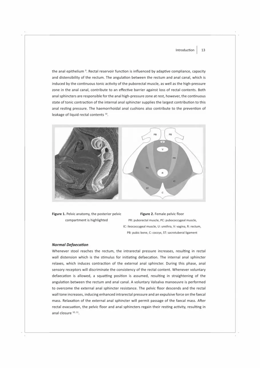
The posterior pelvic compartment encloses the area located posterior to the vagina and
the uterus. It runs from the perineal body, along with the rectovaginal septum, towards the
posterior fornix of the vagina and includes the anal sphincters, the rectum, and the pouch
of Douglas 5 (Figure 1). The pelvic fl oor is att ached to the internal surface of the pelvis and
forms the hammock that supports the pelvic viscera. The muscular layer of the pelvic fl oor is
subdivided into three parts according to their att achments and the pelvic viscera to which they
are related, namely the ileococcygeal, pubococcygeal, and puborectal muscle 6, 7 (Figure 2). The
puborectal muscle represents the most central and distal part of the pelvic fl oor. This muscle
originates at the pubic bone and forms a U-shaped sling around the rectum. The pubo- and
ileococcygeal muscles run more lateral and cranial to the puborectal muscle. The anal sphincter
complex consists of two cylindrical layers. The internal anal sphincter forms the inner cylinder
and is a thickened conti nuati on of the circular smooth muscle of the bowel. The outer cylinder,
the external anal sphincter, is made up of striated muscle. The deep bundles of this external
anal sphincter are inti mately fused with the most distal part of the puborectal muscle 8.
Physiology of the Posterior Pelvic Compartment
Faecal Conti nence
Faecal conti nence is the ability to prevent unintended loss of rectal contents. Maintaining
faecal conti nence depends on a highly integrated series of complex events. Several aspects
such as stool volume and consistency, rectal and anal sensory percepti on, rectal reservoir
functi on, mechanical factors, and anal sphincter functi on play a role in maintaining conti nence.
The ability to prevent unintended loss might depend on whether the rectal contents are solid,
liquid, or gas. This discriminati on of stool consistency mainly depends on sensory receptors in
Introducti on 13
the anal epithelium 9. Rectal reservoir functi on is infl uenced by adapti ve compliance, capacity
and distensibility of the rectum. The angulati on between the rectum and anal canal, which is
induced by the conti nuous tonic acti vity of the puborectal muscle, as well as the high-pressure
zone in the anal canal, contribute to an eff ecti ve barrier against loss of rectal contents. Both
anal sphincters are responsible for the anal high-pressure zone at rest, however, the conti nuous
state of tonic contracti on of the internal anal sphincter supplies the largest contributi on to this
anal resti ng pressure. The haemorrhoidal anal cushions also contribute to the preventi on of
leakage of liquid rectal contents 10.
Figure 1. Pelvic anatomy, the posterior pelvic Figure 2. Female pelvic fl oor
compartment is highlighted PR: puborectal muscle, PC: pubococcygeal muscle,
IC: ileococcygeal muscle, U: urethra, V: vagina, R: rectum,
PB: pubic bone, C: coccyx, ST: sacrotuberal ligament
Normal Defaecati on
Whenever stool reaches the rectum, the intrarectal pressure increases, resulti ng in rectal
wall distension which is the sti mulus for initi ati ng defaecati on. The internal anal sphincter
relaxes, which induces contracti on of the external anal sphincter. During this phase, anal
sensory receptors will discriminate the consistency of the rectal content. Whenever voluntary
defaecati on is allowed, a squatti ng positi on is assumed, resulti ng in straightening of the
angulati on between the rectum and anal canal. A voluntary Valsalva manoeuvre is performed
to overcome the external anal sphincter resistance. The pelvic fl oor descends and the rectal
wall tone increases, inducing enhanced intrarectal pressure and an expulsive force on the faecal
mass. Relaxati on of the external anal sphincter will permit passage of the faecal mass. Aft er
rectal evacuati on, the pelvic fl oor and anal sphincters regain their resti ng acti vity, resulti ng in
anal closure 10, 11.
Chap
ter
1
14
Disorders of the Posterior Pelvic Compartment
Posterior pelvic compartment disorders generally refer to functi onal anorectal disorders, such
as faecal inconti nence and obstructed defaecati on. These disorders are oft en non-specifi c and
might be related to diff erent functi onal and anatomical defi cits. Disorders, specifi c for this
thesis are pointed out, other disorders (e.g. anorectal malignancies) are beyond the scope of
this thesis and therefore not further described.
Functi onal Posterior Pelvic Compartment Disorders
Faecal Inconti nence
Functi onal posterior pelvic compartment disorders can be subdivided into faecal inconti nence
and obstructed defaecati on. Faecal inconti nence occurs when the complex mechanism that
maintains conti nence is disturbed. This disorder is reported to aff ect 11 to 15 percent of the
general Western populati on 12, 13. Numerous diff erent factors, which might exist simultaneously,
can be identi fi ed in the aeti ology of faecal inconti nence. The eff ects of aging refl ect the most
prominent associati on 14. Leaving these elderly pati ents out of considerati on, the most common
causes of faecal inconti nence are pregnancy and delivery 15. Anal sphincter defects are present
in approximately one out of three multi parous women 16. Other well-known contributi ng
factors are previous anorectal surgery, pelvic trauma, pudendal nerve neuropathy, complete
rectal prolapse, and diarrhoea 14, 17, 18.
Faecal inconti nence ranges in severity from mild loss of gas to complete loss of control over
solid stool. Several grading systems are available for the assessment of faecal inconti nence
severity. In 1975, Parks was the fi rst to describe a classifi cati on system, which is sti ll frequently
used 19 (Table 1). The Parks classifi cati on is simple and solely based on the type of unintended loss,
whereas other classifi cati ons address other components, such as the number of inconti nence
episodes, pad use, and lifestyle alterati ons 20-22. Unfortunately, none of these classifi cati ons is
universally accepted and it remains questi onable whether some of these grading systems are
reliable in refl ecti ng inconti nence severity 23.
Faecal inconti nence can be treated by diff erent conservati ve and surgical therapies,
depending on the severity, impact, and underlying pathophysiologic mechanism. In general,
pati ents will fi rst be off ered conservati ve treatment, such as medicati on and / or pelvic fl oor
muscle training 24, 25. When these therapies fail, pati ents are eligible for surgical treatment.
Table 1. Parks classifi cati on
I Full conti nence
II Inconti nence for gas or soiling
III Inconti nence for liquid stool
IV Inconti nence for solid stool
Introducti on 15
Obstructed Defaecati on
Rectal evacuati on refl ects a functi onal event requiring coordinati on of a complex series of
events. Diff erent mechanisms can lead to a disturbance of these events and might eventually
lead to obstructed defaecati on 26. Defaecati on diffi culti es are reported to aff ect approximately
15 percent of the adult Western populati on 27. Mechanical outlet obstructi on and shift ing of the
directi on of the expulsive force vector are thought to play a role in pati ents with posterior pelvic
compartment prolapse. Other well-known mechanisms are a defecti ve rectal fi lling sensati on
and functi onal outlet obstructi on due to ineff ecti ve muscle relaxati on.
Pati ents complaining of consti pati on present with a wide range of symptoms regarding
defaecati on diffi culti es, referring to hard stools, inability to defaecate, infrequent defaecati ons,
and feelings of incomplete evacuati on. However, these symptoms are relati vely common and
do not necessarily refl ect obstructed defaecati on. To date, obstructed defaecati on is defi ned
according to the Rome III criteria 28 (Table 2).
When conservati ve treatment, such as increasing dietary fi bre and pelvic fl oor muscle
training, is unsuccessful, appropriate additi onal therapy should rely on proper identi fi cati on
of the underlying pathophysiologic mechanism. It is important to disti nguish those pati ents
with delayed colonic transit from those with obstructed defaecati on, because slow transit
consti pati on requires a diff erent therapeuti c approach. As anatomic fi ndings (i.e. enterocele
and rectocele) are associated with obstructed defaecati on, several diff erent surgical techniques
are described 29-31. However, it remains uncertain whether these procedures are capable of
restoring normal defaecati on.
Table 2. Rome III criteria
Criteria fulfi lled for the last three months, onset at least six months prior to diagnosis
I Must include two or more of the following:
a. Straining during at least 25 percent of defaecati ons
b. Lumpy or hard stool in at least 25 percent of defaecati ons
c. Sensati on of incomplete evacuati on for at least 25 percent of defaecati ons
d. Sensati on of anorectal blockage for at least 25 percent of defaecati ons
e. Manual manoeuvres to facilitate at least 25 percent of defaecati ons
f. Fewer than three defaecati ons per week
II Loose stools are rarely present without the use of laxati ves
III There are insuffi cient criteria for irritable bowel syndrome
Chap
ter
1
16
Anatomic Posterior Pelvic Compartment Disorders
Posterior Pelvic Compartment Prolapse
Posterior pelvic compartment prolapse involves loss of support, which results in descent of
one or more pelvic viscera in this compartment. Posterior pelvic compartment prolapses are
divided in enterocele, rectocele, intussuscepti on, and complete rectal prolapse. Enterocele
is described as a herniati on of small bowel or rectosigmoid into the vagina. A herniati on of
the anterior rectal wall into the lumen of the vagina is called rectocele and intussuscepti on is
defi ned as an infolding of the rectal wall into the distal part of the rectum or anal canal. A full-
thickness protrusion of rectal wall through the anal canal is a complete rectal prolapse.
The prevalence of pelvic organ prolapse is esti mated at 30 percent in women between 20
and 60 years 32, but is even higher in older women. It has been reported that these prolapses
are mainly caused by injury related to pregnancy and vaginal delivery 5, 33. The prevalence of
pelvic organ prolapse is strongly associated with parity. The risk increases with each child, but
the rate of increase declines once a woman has had two children 33. Other known risk factors
include age, obesity, hysterectomy, smoking, chronic bronchiti s, chronic consti pati on, heredity,
and collagen disorders 34-37.
Diff erent symptoms are att ributed to prolapse of the posterior pelvic compartment.
However, these symptoms are oft en non-specifi c and might also be related to prolapse of the
anterior and middle pelvic compartment. Pati ents oft en complain about pelvic discomfort, such
as feelings of a vaginal lump, heaviness, and pelvic pressure, which might disappear when lying
down 38. Anorectal disturbances, which include obstructed defaecati on, urgency, and faecal
inconti nence, are also frequently found 39. Other symptoms, att ributed to these prolapses, are
sexual dysfuncti on and pain 29, 38. Women with posterior pelvic compartment prolapse oft en
have concomitant prolapses in the anterior and / or middle compartment 40, 41, which might
result in concomitant symptoms and possibly in the need for a diff erent therapeuti c approach.
Surgical repair is the treatment of choice for women, in whom conservati ve treatment, such
as pelvic fl oor muscle training and vaginal pessary, fails. The goal of surgical repair is to restore
the original anatomic state and to resolve the associated symptoms. Many diff erent types of
repair have been described, all with their own advantages and disadvantages 29, 42. Although
most of these procedures provide successful anatomic correcti on, the associated symptoms are
not so easy to resolve and persist in a substanti al number of pati ents.
Rectovaginal Fistula
A rectovaginal fi stula is an abnormal communicati on between the two epithelial-lined surfaces
of the rectum (or anal canal) and the vagina. Rectovaginal fi stulas are classifi ed as low,
intermediate, and high fi stulas, depending on the site of the vaginal opening 43. Most of the
rectovaginal fi stulas are low fi stulas, which are in fact anovaginal fi stulas. Obstetric injury is
the most frequent cause of rectovaginal fi stula formati on 44, 45. Approximately two percent of

Introducti on 17
vaginal deliveries in Western society result in a perineal tear involving the anal sphincters 46.
Subsequent rectovaginal fi stula formati on is reported in approximately three percent of the
pati ents with such a perineal tear 2. Perianal infecti ons, prior anorectal surgery, carcinoma, and
radiati on might also lead to rectovaginal fi stula formati on 10. The most common symptoms are
passage of gas and stool through the vagina 47. Furthermore, these fi stulas might lead to vaginal
discharge, chronic vaginiti s, and urinary tract infecti on 10.
Despite some anecdoti c reports, there is no evidence that rectovaginal fi stulas heal
spontaneously. Surgical repair is considered as the treatment of choice for pati ents with
such a fi stula. Despite a multi tude of diff erent surgical repair strategies 48-50, healing rates are
oft en disappointi ng, especially in women in whom the fi stula persisted aft er a prior repair
procedure 51-53.
Investi gati ons of the Posterior Pelvic Compartment
Diff erent methods of investi gati on are available for the examinati on of pati ents with
symptoms related to posterior pelvic compartment disorders, such as anorectal manometry,
endoluminal imaging, evacuati on proctography, transperineal ultrasound, and colonic transit
ti me measurement. Other tests, such as rectal balloon expulsion test, pudendal nerve terminal
motor latency measurement, and electromyography are beyond the scope of this thesis and
therefore not further described.
Anorectal Manometry
Since its clinical introducti on in the 1960s 54, anorectal manometry has become a standard
method to quanti fy anorectal functi on 55. Internal and external anal sphincter functi on is assessed
by the evaluati on of anal resti ng and squeeze pressure. Furthermore, manometry can be used
to elicit the rectoanal inhibitory refl ex and rectal compliance. Despite the well-established use
of anorectal manometry, there is no standardised method to perform anorectal manometry.
Diff erent systems, including water-perfused catheters, closed balloon systems, and microti p
transducers, have been used, all with their own advantages and disadvantages 56-58. Recently,
high-resoluti on manometry has been described 59, however this sophisti cated technique has
not been established as superior to more conventi onal techniques.
Endoluminal Imaging
Endoluminal imaging provides a tool for the assessment of anal sphincter and rectal
wall integrity. Both ultrasound and magneti c resonance (MR) imaging can be performed
endoluminally. These imaging techniques play an important role in the diagnosti c work-up of
pati ents with faecal inconti nence and rectovaginal fi stulas 60, 61. The internal and external anal

Chap
ter
1
18
sphincters are evaluated separately for the presence of defects (Figure 3). Both endoluminal
ultrasound and MR imaging have been found accurate in detecti ng anal sphincter defects 62-64.
In additi on, MR imaging has the advantage of simultaneously detecti ng external anal sphincter
atrophy 65, which might infl uence the outcome of treatment 66. However, ultrasound is less
expensive and more widespread available.
Figure 3. Endo-anal ultrasound
left : normal anal sphincters, right: combined internal and external anal sphincter defect
IAS: internal anal sphincter, EAS: external anal sphincter, P: probe
Evacuati on Proctography
Evacuati on proctography, also called defaecography, can be used for visualisati on of
abnormaliti es which might be related to defaecati on diffi culti es 61, 67. The rectosigmoid and
preferably also the small bowel and vagina are opacifi ed with liquid contrast. With the pati ents
seated on a radiolucent toilet chair, lateral radiographs are taken at rest, during squeezing, and
during straining, which mimics defaecati on 68. Evacuati on proctography, which is performed in

Introducti on 19
semi-physiologic circumstances, enables objecti ve detecti on of the presence and extensiveness
of posterior pelvic compartment prolapse (Figure 4). Simultaneous opacifying of the bladder can
be used for imaging of concomitant micturiti on disturbances 69. Exposure to ionising radiati on is
an important drawback of evacuati on proctography. MR defaecography has been introduced in
the last decade for the detecti on of pelvic fl oor dysfuncti on 70. However, to date, the role of this
new technique is limited, since it is only available in a minority of specialised clinics.
Figure 4. Evacuati on proctography with opacifi cati on of rectosigmoid, small bowel and vagina
left : normal situati on, middle: large rectocele, right: large enterocele
SB: small bowel, R: rectum, V: vagina, C: coccyx, RC: rectocele, EC: enterocele
Transperineal Ultrasound
Another imaging technique for posterior pelvic compartment disorders is dynamic
transperineal ultrasound imaging, which has recently been introduced by Dietz et al. 71, 72. This
type of ultrasound imaging is performed with a transducer placed at the perineum and is non-
invasive compared to endoluminal imaging. Another advantage of this imaging technique is
the possibility of simultaneous detecti on of abnormaliti es in all three pelvic compartments.
Evaluati on of these three compartments simultaneously seems important, since it has been
reported that symptoms originati ng from one compartment do not imply absent pathology
Chap
ter
1
20
in another compartment 40, 41. Furthermore, transperineal ultrasound can be used for the
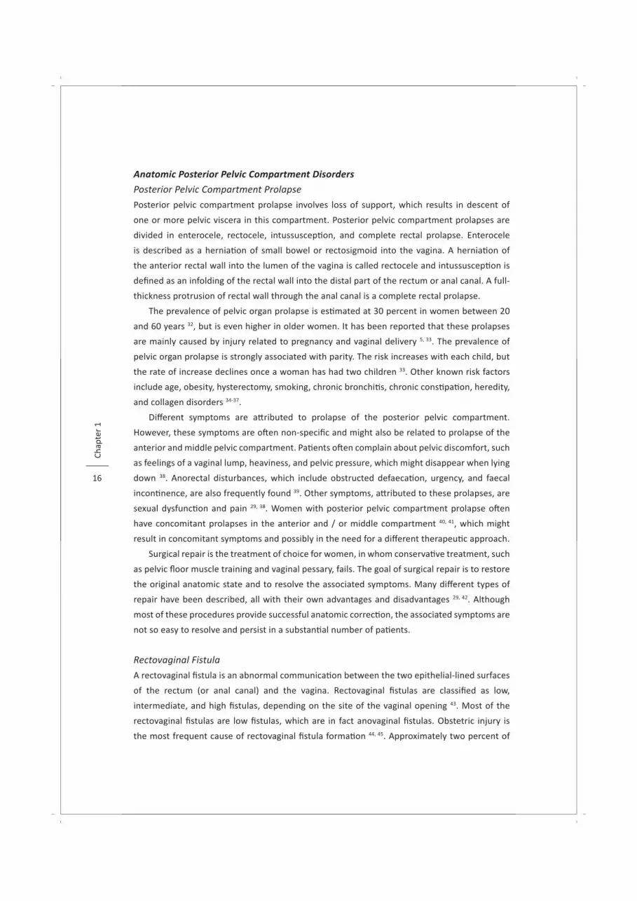
evaluati on of pelvic fl oor functi on, as well as pelvic fl oor and anal sphincter integrity 73-75 (Figure
5). Since transperineal ultrasound requires additi onal training and equipment, its use is, at
present, limited to specialised pelvic fl oor centers.
Figure 5. Transperineal ultrasound
left : normal three-compartment view, middle: normal pelvic fl oor, right: normal anal sphincters
PB: pubic bone, B: bladder, U: urethra, V: vagina, R: rectum, PF: pelvic fl oor, IAS: internal anal sphincter, EAS: external anal sphincter, M: mucosa
Colonic Transit Time Measurement
A useful investi gati on in the evaluati on of pati ents complaining of consti pati on is colonic transit
ti me measurement. This technique determines how much ti me is required for passage of stool
through the large bowel. Pati ents should ingest a standardised number of radiopaque markers
for a consecuti ve series of days. Simultaneously the use of laxati ves and / or enemas should be
refrained. Progression of the markers is followed by daily abdominal radiographs or by a single
radiograph on the fi rst day aft er the last ingesti on of markers 76-78. The large bowel, visualised by
one or more abdominal X-ray’s, is subdivided into three segments, which are the right and the
left colon and the rectosigmoid. In those three segments, the markers, which are sti ll present,
are counted and segmental and total transit ti mes are calculated 79. This technique enables the
disti ncti on between delayed colonic transit and obstructed defaecati on.
Introducti on 21
Aims and Outline of the Thesis
This thesis is aimed at evaluati ng diagnosis and treatment of disorders of the posterior
pelvic compartment. It is mainly focussed on the outcome of diff erent surgical modaliti es
in the treatment of these posterior pelvic compartment disorders. Furthermore, the value
of transperineal ultrasound in the assessment of posterior pelvic compartment prolapse is
investi gated.
Various procedures have been developed for the surgical treatment of symptomati c
enterocele. It has been reported that an abdominal approach provides an eff ecti ve tool for
anatomic correcti on of this type of pelvic organ prolapse. Furthermore, short-term follow-up
has shown that abdominal enterocele repair is benefi cial, especially for those pati ents with
pelvic discomfort. However, it is unknown whether this eff ect is sustained in the long-term. In
Chapter 2 the long-term outcome of abdominal obliterati on of the pelvic inlet for symptomati c
enterocele is described, especially regarding the eff ect of the repair on symptoms of pelvic
discomfort and obstructed defaecati on.
Rectocele repair is mostly performed by a transvaginal or a transanal approach. It is
well known that these types of rectocele repair are associated with de novo dyspareunia.
Transabdominal repair of rectocele seems to off er a limited risk of de novo dyspareunia as a
side eff ect. The outcome of abdominal anterolateral rectopexy for symptomati c rectoceles is
evaluated in Chapter 3.
Evacuati on proctography is considered as the gold standard imaging technique for the
assessment of pati ents with symptoms of posterior pelvic compartment prolapse. However,
evacuati on proctography is relati vely invasive and requires exposure to ionising radiati on.
Transperineal ultrasound, which has recently been developed, enables dynamic and non-
invasive investi gati on of all three pelvic compartments simultaneously, without ionising radiati on
and at low cost. In Chapter 4, the level of agreement between evacuati on proctography and
transperineal ultrasound in diagnosing posterior pelvic compartment prolapse is presented.
Anterior sphincteroplasty is the surgical treatment of choice for pati ents with faecal
inconti nence, associated with an external anal sphincter defect. Recently, it has been reported
that pati ents with such a defect may also benefi t from sacral neuromodulati on. The questi on
is whether anterior sphincteroplasty sti ll deserves a place in the surgical treatment of faecal
inconti nence. In Chapter 5 the long-term outcome of anterior sphincteroplasty in a large cohort
of pati ents is described. A comparable outcome classifi cati on was used to facilitate comparison
between anterior sphincteroplasty and sacral neuromodulati on.
It has been shown that vaginal delivery may result not only in anal sphincter defects but
also in pelvic fl oor injury. Unti l now it is unknown whether this type of injury plays a role in
the aeti ology of faecal inconti nence and whether it aff ects the outcome of treatment. The
prevalence of pelvic fl oor injury in faecal inconti nent pati ents who were eligible for surgical
Chap
ter
1
22
treatment in the past is reported in Chapters 6 and 7. In additi on, the impact of this type of
injury on the outcome of anterior sphincteroplasty (Chapter 6) and sacral neuromodulati on
(Chapter 7) is presented.
Several techniques are available for the surgical treatment of rectovaginal fi stulas, however
the results are oft en rather disappointi ng, especially in women in whom the fi stula persisted
aft er a prior procedure. It has been suggested that interpositi on of healthy, well-vascularised
ti ssue may be the key to rectovaginal fi stula healing. In Chapter 8 the outcome of interpositi on
of the healthy and well-vascularised puborectal sling in the treatment of rectovaginal fi stulas
is described. Puborectal sling interpositi on is not effi cient in pati ents who have undergone
previous att empts at repair. Based on this fi nding, we introduced an adapted rectal sleeve
advancement for the treatment of pati ents with such a persistent rectovaginal fi stula. The early
experience with this new type of rectovaginal fi stula repair is presented in Chapter 9.
The results as obtained from the studies described in this thesis, are summarised and
discussed in Chapter 10. Moreover, suggesti ons for future research are made.
Introducti on 23
References
1. Dietz HP. Pelvic fl oor trauma following vaginal delivery. Curr Opin Obstet Gynecol 2006;18(5):528-37.2. Venkatesh KS, Ramanujam PS, Larson DM, Haywood MA. Anorectal complicati ons of vaginal delivery.
Dis Colon Rectum 1989;32(12):1039-41.3. Deutekom M, Terra MP, Dobben AC, Dijkgraaf MG, Baeten CG, Stoker J, et al. Impact of faecal
inconti nence severity on health domains. Colorectal Dis 2005;7(3):263-9.4. Glia A, Lindberg G. Quality of life in pati ents with diff erent types of functi onal consti pati on. Scand J
Gastroenterol 1997;32(11):1083-9.5. Ginger VA, Kobashi KC. Posterior compartment defect repair in vaginal surgery: update on surgical
techniques. Curr Urol Rep 2007;8(5):387-93.6. Ashton-Miller JA, DeLancey JO. Functi onal anatomy of the female pelvic fl oor. Ann N Y Acad Sci
2007;1101:266-96.7. DeLancey JO. The anatomy of the pelvic fl oor. Curr Opin Obstet Gynecol 1994;6(4):313-6.8. Williams PL, Warwick Re. Splanchnology. In: Gray’s Anatomy, 36th edn. London: Churchill Livingstone
1980:1356-64.9. Duthie HL, Gairns FW. Sensory nerve-endings and sensati on in the anal region of man. Br J Surg
1960;47:585-95.10. Gordon PH, Nivatvongs S. Principles and practi ce of surgery for the colon, rectum, and anus. 3th
editi on. New York: 2007.11. Schouten WR, Gosselink MJ, Boerma MO, Ginai AZ. Rectal wall contracti lity in response to an evoked
urge to defecate in pati ents with obstructed defecati on. Dis Colon Rectum 1998;41(4):473-9.12. Sharma A, Bissett I, Merrie A, Macmillan A, Reid P. The community prevalence of fecal inconti nence:
a New-Zealand cross-secti onal study. Dis Colon Rectum 2008;51(5-S):40.13. Macmillan AK, Merrie AE, Marshall RJ, Parry BR. The prevalence of fecal inconti nence in community-
dwelling adults: a systemati c review of the literature. Dis Colon Rectum 2004;47(8):1341-9.14. Nelson RL. Epidemiology of fecal inconti nence. Gastroenterology 2004;126(1 Suppl 1):S3-7.15. Madoff RD, Williams JG, Caushaj PF. Fecal inconti nence. N Engl J Med 1992;326(15):1002-7.16. Oberwalder M, Connor J, Wexner SD. Meta-analysis to determine the incidence of obstetric anal
sphincter damage. Br J Surg 2003;90(11):1333-7.17. Lindsey I, Jones OM, Smilgin-Humphreys MM, Cunningham C, Mortensen NJ. Patt erns of fecal
inconti nence aft er anal surgery. Dis Colon Rectum 2004;47(10):1643-9.18. Engel AF, Kamm MA, Hawley PR. Civilian and war injuries of the perineum and anal sphincters. Br J
Surg 1994;81(7):1069-73.19. Parks AG. Anorectal inconti nence. Proc R Soc Med 1975;68:681-90.20. Rockwood TH, Church JM, Fleshman JW, Kane RL, Mavrantonis C, Thorson AG, et al. Pati ent
and surgeon ranking of the severity of symptoms associated with fecal inconti nence: the fecal inconti nence severity index. Dis Colon Rectum 1999;42(12):1525-32.
21. Vaizey CJ, Carapeti E, Cahill JA, Kamm MA. Prospecti ve comparison of faecal inconti nence grading systems. Gut 1999;44(1):77-80.
22. Pescatori M, Anastasio G, Botti ni C, Mentasti A. New grading and scoring for anal inconti nence. Evaluati on of 335 pati ents. Dis Colon Rectum 1992;35(5):482-7.
23. Baxter NN, Rothenberger DA, Lowry AC. Measuring fecal inconti nence. Dis Colon Rectum 2003;46(12):1591-605.
24. Scarlett Y. Medical management of fecal inconti nence. Gastroenterology 2004;126(1 Suppl 1):S55-63.
25. Norton C. Behavioral management of fecal inconti nence in adults. Gastroenterology 2004;126(1 Suppl 1):S64-70.
26. D‘Hoore A, Penninckx F. Obstructed defecati on. Colorectal Dis 2003;5(4):280-7.
Chap
ter
1
24
27. Stewart WF, Liberman JN, Sandler RS, Woods MS, Stemhagen A, Chee E, et al. Epidemiology of consti pati on (EPOC) study in the United States: relati on of clinical subtypes to sociodemographic features. Am J Gastroenterol 1999;94(12):3530-40.
28. Longstreth GF, Thompson WG, Chey WD, Houghton LA, Mearin F, Spiller RC. Functi onal bowel disorders. Gastroenterology 2006;130(5):1480-91.
29. Cundiff GW, Fenner D. Evaluati on and treatment of women with rectocele: focus on associated defecatory and sexual dysfuncti on. Obstet Gynecol 2004;104(6):1403-21.
30. Holley RL. Enterocele: a review. Obstet Gynecol Surv 1994;49(4):284-93.31. Oom DM, Gosselink MP, Schouten WR. Enterocele - Diagnosis and treatment. Gastroenterol Clin Biol
2009;33(2):135-7.32. Samuelsson EC, Victor FT, Tibblin G, Svardsudd KF. Signs of genital prolapse in a Swedish populati on
of women 20 to 59 years of age and possible related factors. Am J Obstet Gynecol 1999;180(2 Pt 1):299-305.
33. Mant J, Painter R, Vessey M. Epidemiology of genital prolapse: observati ons from the Oxford Family Planning Associati on Study. Br J Obstet Gynaecol 1997;104(5):579-85.
34. Olsen AL, Smith VJ, Bergstrom JO, Colling JC, Clark AL. Epidemiology of surgically managed pelvic organ prolapse and urinary inconti nence. Obstet Gynecol 1997;89(4):501-6.
35. Uustal Fornell E, Wingren G, Kjolhede P. Factors associated with pelvic fl oor dysfuncti on with emphasis on urinary and fecal inconti nence and genital prolapse: an epidemiological study. Acta Obstet Gynecol Scand 2004;83(4):383-9.
36. Neill ME, Parks AG, Swash M. Physiological studies of the anal sphincter musculature in faecal inconti nence and rectal prolapse. Br J Surg 1981;68(8):531-6.
37. Swift S, Woodman P, O’Boyle A, Kahn M, Valley M, Bland D, et al. Pelvic Organ Support Study (POSST): the distributi on, clinical defi niti on, and epidemiologic conditi on of pelvic organ support defects. Am J Obstet Gynecol 2005;192(3):795-806.
38. Barber MD. Symptoms and outcome measures of pelvic organ prolapse. Clin Obstet Gynecol 2005;48(3):648-61.
39. Klingele CJ, Bharucha AE, Fletcher JG, Gebhart JB, Riederer SG, Zinsmeister AR. Pelvic organ prolapse in defecatory disorders. Obstet Gynecol 2005;106(2):315-20.
40. Mellgren A, Bremmer S, Johansson C, Dolk A, Uden R, Ahlback SO, et al. Defecography. Results of investi gati ons in 2,816 pati ents. Dis Colon Rectum 1994;37(11):1133-41.
41. Lapalus MG, Henry L, Barth X, Mellier G, Gauti er G, Mion F, et al. [Enterocele: clinical risk factors and associati on with others pelvic fl oor disorders (about 544 defecographies)]. Gynecol Obstet Ferti l 2004;32(7-8):595-600.
42. Brubaker L. Controversies and uncertainti es: abdominal versus vaginal surgery for pelvic organ prolapse. Am J Obstet Gynecol 2005;192(3):690-3.
43. Daniels BT. Rectovaginal fi stula: A clinical and pathological study. Minneapolis, University of Minnesota Graduate School. 1949.
44. Belt RL, Jr. Repair of anorectal vaginal fi stula uti lizing segmental advancement of the internal sphincter muscle. Dis Colon Rectum 1969;12(2):99-104.
45. Hibbard LT. Surgical management of rectovaginal fi stulas and complete perineal tears. Am J Obstet Gynecol 1978;130(2):139-41.
46. de Leeuw JW, Struijk PC, Vierhout ME, Wallenburg HC. Risk factors for third degree perineal ruptures during delivery. Bjog 2001;108(4):383-7.
47. Tsang CB, Rothenberger DA. Rectovaginal fi stulas. Therapeuti c opti ons. Surg Clin North Am 1997;77(1):95-114.
48. Rothenberger DA, Christenson CE, Balcos EG, Schott ler JL, Nemer FD, Nivatvongs S, et al. Endorectal advancement fl ap for treatment of simple rectovaginal fi stula. Dis Colon Rectum 1982;25(4):297-300.

Introducti on 25
49. Bauer JJ, Sher ME, Jaffi n H, Present D, Gelerent I. Transvaginal approach for repair of rectovaginal fi stulae complicati ng Crohn’s disease. Ann Surg 1991;213(2):151-8.
50. Ellis CN. Outcomes aft er repair of rectovaginal fi stulas using bioprostheti cs. Dis Colon Rectum 2008;51(7):1084-8.
51. Halverson AL, Hull TL, Fazio VW, Church J, Hammel J, Floruta C. Repair of recurrent rectovaginal fi stulas. Surgery 2001;130(4):753-8.
52. Tsang CB, Madoff RD, Wong WD, Rothenberger DA, Finne CO, Singer D, et al. Anal sphincter integrity and functi on infl uences outcome in rectovaginal fi stula repair. Dis Colon Rectum 1998;41(9):1141-6.
53. Lowry AC, Thorson AG, Rothenberger DA, Goldberg SM. Repair of simple rectovaginal fi stulas. Infl uence of previous repairs. Dis Colon Rectum 1988;31(9):676-8.
54. Schuster MM, Hookman P, Hsphincteroplasty. Br J Surg 1999;86(10):1322-7.55. Yang XM, Partanen K, Farin P, Soimakallio S. Defecography. Acta Radiol 1995;36(5):460-8.56. Womack NR, Williams NS, Holmfi eld JH, Morrison JF, Simpkins KC. New method for the dynamic
assessment of anorectal functi on in consti pati on. Br J Surg 1985;72(12):994-8.57. Hock D, Lombard R, Jehaes C, Markiewicz S, Penders L, Fontaine F, et al. Colpocystodefecography. Dis
Colon Rectum 1993;36(11):1015-21.endrix TR, Mendeloff AI. Simultaneous Manometric Recording of Internal and External Anal Sphincteric Refl exes. Bull Johns Hopkins Hosp 1965;116:79-88.
58. Bharucha AE, Fletcher JG. Recent advances in assessing anorectal structure and functi ons. Gastroenterology 2007;133(4):1069-74.
59. Hancock BD. Measurement of anal pressure and moti lity. Gut 1976;17(8):645-51.60. Miller R, Bartolo DC, James D, Mortensen NJ. Air-fi lled microballoon manometry for use in anorectal
physiology. Br J Surg 1989;76(1):72-5.61. Miller R, Bartolo DC, Roe AM, Mortensen NJ. Assessment of microtransducers in anorectal
manometry. Br J Surg 1988;75(1):40-3.62. Jones MP, Post J, Crowell MD. High-resoluti on manometry in the evaluati on of anorectal disorders: a
simultaneous comparison with water-perfused manometry. Am J Gastroenterol 2007;102(4):850-5.63. Terra MP, Stoker J. The current role of imaging techniques in faecal inconti nence. Eur Radiol
2006;16(8):1727-36.64. Bartram CI. Functi onal anorectal imaging. Abdom Imaging 2005;30(2):195-203.65. Deen KI, Kumar D, Williams JG, Olliff J, Keighley MR. Anal sphincter defects. Correlati on between
endoanal ultrasound and surgery. Ann Surg 1993;218(2):201-5.66. Rociu E, Stoker J, Eijkemans MJ, Schouten WR, Lameris JS. Fecal inconti nence: endoanal US versus
endoanal MR imaging. Radiology 1999;212(2):453-8.67. Cazemier M, Terra MP, Stoker J, de Lange-de Klerk ES, Boeckxstaens GE, Mulder CJ, et al. Atrophy
and defects detecti on of the external anal sphincter: comparison between three-dimensional anal endosonography and endoanal magneti c resonance imaging. Dis Colon Rectum 2006;49(1):20-7.
68. Williams AB, Bartram CI, Modhwadia D, Nicholls T, Halligan S, Kamm MA, et al. Endocoil magneti c resonance imaging quanti fi cati on of external anal sphincter atrophy. Br J Surg 2001;88(6):853-9.
69. Briel JW, Stoker J, Rociu E, Lameris JS, Hop WC, Schouten WR. External anal sphincter atrophy on endoanal magneti c resonance imaging adversely aff ects conti nence aft er
70. Roos JE, Weishaupt D, Wildermuth S, Willmann JK, Marincek B, Hilfi ker PR. Experience of 4 years with open MR defecography: pictorial review of anorectal anatomy and disease. Radiographics 2002;22(4):817-32.
71. Dietz HP, Steensma AB, Hasti ngs R. Three-dimensional ultrasound imaging of the pelvic fl oor: the eff ect of parturiti on on paravaginal support structures. Ultrasound Obstet Gynecol 2003;21(6):589-95.
72. Dietz HP, Steensma AB. Posterior compartment prolapse on two-dimensional and three-dimensional pelvic fl oor ultrasound: the disti ncti on between true rectocele, perineal hypermobility and enterocele. Ultrasound Obstet Gynecol 2005;26(1):73-7.

Chap
ter
1
26
73. Weinstein MM, Pretorius DH, Jung SA, Nager CW, Mitt al RK. Transperineal three-dimensional ultrasound imaging for detecti on of anatomic defects in the anal sphincter complex muscles. Clin Gastroenterol Hepatol 2009;7(2):205-11.
74. Dietz HP, Steensma AB. The prevalence of major abnormaliti es of the levator ani in urogynaecological pati ents. Bjog 2006;113(2):225-30.
75. Weinstein MM, Pretorius D, Nager CW, Mitt al R. Inter-rater reliability of pelvic fl oor muscle imaging abnormaliti es with 3D ultrasound. Ultrasound Obstet Gynecol 2007;30:538.
76. Metcalf AM, Phillips SF, Zinsmeister AR, MacCarty RL, Beart RW, Wolff BG. Simplifi ed assessment of segmental colonic transit. Gastroenterology 1987;92(1):40-7.
77. Chaussade S, Guerre J, Couturier D. Measurement of colonic transit. Gastroenterology 1987;92(6):2053.
78. Dorval D, Barbieux JP, Picon L, Alison D, Codjovi P, Rouleau P. Mesure simplifée du temps de transit colique par une seule radiographie de l’abdomen et un seul type de marqueur. Gastoenterol Clin Biol 1994;18:141-144.
79. Arhan P, Devroede G, Jehannin B, Lanza M, Faverdin C, Dornic C, et al. Segmental colonic transit ti me. Dis Colon Rectum 1981;24(8):625-9.

Part I
Posterior Pelvic Compartment Prolapse


Chapter 2 Enterocele repair by abdominal obliterati on of the pelvic inlet:
long-term outcome on obstructed defaecati on and symptoms
of pelvic discomfort
Colorectal Dis 2007;9(9):845-50.
Daniëlla M.J. Oom
Valeria R.M. van Dijl
Marti jn P. Gosselink
Jan J. van Wijk
W. Ruud Schouten

30
Chap
ter
2
Abstract
Introducti on Enterocele is defi ned as a herniati on of the peritoneal sac between the vagina and the rectum.
This may contain either sigmoid colon or small bowel. It has been reported that enterocele is
associated with obstructed defaecati on and symptoms of pelvic discomfort. The aim of the
present study was to evaluate the long-term eff ect of enterocele repair.
Pati ents and Methods In the ti me period between 1994 and 2003, 54 women (median age: 54 years, range: 31 – 80)
with a symptomati c enterocele underwent obliterati on of the pelvic inlet with a U-shaped
Mersilene® mesh. All pati ents underwent evacuati on proctography, which was repeated six
months aft er the repair. In additi on, they were contacted by telephone in order to assess the
long-term eff ect of enterocele repair. Forty-nine pati ents were willing to answer the questi ons
over the telephone. Five pati ents were lost to follow-up (response rate: 91%).
ResultsSix months aft er the procedure, evacuati on proctography revealed a recurrent or persistent
enterocele in fi ve pati ents (9%), which was symptomati c in two, both of whom underwent a
second repair. Among the 49 pati ents without an enterocele aft er six months, 10 women (23%)
encountered recurrent symptoms of pelvic discomfort at a median follow-up of 85 months
(range: 3 – 137). Despite adequate correcti on of the enterocele, obstructed defaecati on
persisted in 21 of 28 pati ents (75%), who presented with this problem before the procedure.
De novo dyspareunia occurred in fi ve percent of the women aft er the procedure.
Conclusion Obliterati on of the pelvic inlet with a U-shaped Mersilene® mesh provides an eff ecti ve tool for
anatomical correcti on of enteroceles. However, in the long-term one of four pati ents encounters
recurrent symptoms of pelvic discomfort. It seems unlikely that enterocele contributes to
obstructed defaecati on, as evacuati on diffi culti es persist in around three quarters of the
pati ents.
Abdominal obliterati on of the pelvic inlet 31
Introducti on
Enterocele is defi ned as a herniati on of the peritoneal sac between the vagina and the
rectum. This may contain either sigmoid colon or small bowel. Enterocele was fi rst described
by Garengeot 1 in 1736. It is well known that this conditi on is associated with symptoms
of pelvic discomfort, including feelings of prolapse and pelvic pressure 2, 3. It has also been
suggested that enterocele, especially if it contains a sigmoid loop, is an underlying cause of
obstructed defaecati on 4, 5. Enterocele is most frequently found in elderly, multi parous females.
The exact prevalence of this abnormality is unknown. In a review in 1994, Holley 6 stated that
enteroceles are uncommon. However, Cronjé et al. 7 reported that of all cases in their pelvic
fl oor dysfuncti on database, 45 percent presented with an enterocele. Evacuati on proctography
revealed an enterocele in up to 18 percent of the women who underwent hysterectomy 8
and in 10 percent of healthy female volunteers 9, 10. Almost two-third of the women with a
symptomati c enterocele had undergone a hysterectomy in the past 4, 11, 12.
Various procedures have been developed for the surgical treatment of symptomati c
enteroceles. In 1912, Moschcowitz 13 was the fi rst to describe obliterati on of the distal part of
the peritoneal sac by an abdominal approach. Originally, he developed this technique for the
treatment of complete rectal prolapse. Later on, gynaecologists adapted this procedure for
the treatment of enterocele repair. In 1922, Ward 14 described a transvaginal approach. It has
been reported that this transvaginal repair is eff ecti ve in the majority of pati ents. However,
in several studies, the durati on of follow-up was short and the clinical outcome of this type
of repair has only been assessed by physical examinati on and subjecti ve criteria and not by
evacuati on proctography 4, 15-18. A drawback of the transvaginal repair is the potenti al risk of
dyspareunia 4, 15, 18. More recently, diff erent types of abdominal repair such as rectovaginopexy
and obliterati on of the pelvic inlet have been reported 2, 3, 19. In two small studies, the effi cacy
of these abdominal procedures has been assessed by evacuati on proctography aft er the
procedure 2, 19. Based on the outcome of these studies, it seems likely that an abdominal
approach provides an eff ecti ve tool for anatomical correcti on of enterocele.
Short-term follow-up has revealed that enterocele repair is benefi cial, especially for
those pati ents with symptoms of pelvic discomfort 2, 3. The questi on is whether this eff ect is
sustained in the long-term. In a recent study, Jean et al. 3 observed that none of their pati ents
had symptoms of pelvic discomfort three months aft er abdominal enterocele repair. However,
at median-term follow-up of 27 months, they encountered recurrent symptoms of pelvic
discomfort in 27 percent of their pati ents. The aim of the present study was to assess the
effi cacy of abdominal enterocele repair uti lising objecti ve radiological criteria. The other goal
was to evaluate the long-term eff ect of enterocele repair on symptoms of pelvic discomfort and
obstructed defaecati on.
32
Chap
ter
2
Pati ents and Methods
Between 1994 and 2003, 54 female pati ents (median age: 54 years, range: 31 - 80) underwent
obliterati on of the pelvic inlet with a U-shaped Mersilene® (Ethicon, Johnson & Johnson,
Somerville, USA) mesh to repair their symptomati c enterocele. Fift y-one pati ents (94%)
presented with symptoms of pelvic discomfort, including feelings of prolapse and pelvic
pressure. Thirty-one pati ents (57%) also complained of evacuati on diffi culti es, which were
classifi ed as obstructed defaecati on if the pati ent fulfi lled two or more of the Rome II criteria 20 (Table 1). The history of 45 pati ents (83%) revealed one or more previous gynaecologic
procedures. All these 45 pati ents had undergone a hysterectomy in the past.
Table 1. Rome II criteria 20
I Two or more of the following for at least 12 weeks in the preceding 12 months:
a. Straining during at least 25 percent of defaecati ons
b. Lumpy or hard stool in at least 25 percent of defaecati ons
c. Sensati on of incomplete evacuati on for at least 25 percent of defaecati ons
d. Sensati on of anorectal blockage for at least 25 percent of defaecati ons
e. Manual manoeuvres to facilitate at least 25 percent of defaecati ons
f. Fewer than three defaecati ons per week
II Loose stools are rarely present without the use of laxati ves
III There are insuffi cient criteria for irritable bowel syndrome
All pati ents underwent evacuati on proctography with opacifi cati on of the small bowel and
vagina in order to confi rm the diagnosis and to classify the enterocele prior to surgery (Figure
1a). Six months aft er the procedure evacuati on proctography was repeated in all pati ents
(Figure 1b). For the classifi cati on of enteroceles a special grading system was used (Table 2). In
45 pati ents (83%) the peritoneal sac between the vagina and the rectum contained small bowel
loops. A sigmoid loop was found in six pati ents (11%), whereas three pati ents (6%) presented
with both small bowel loops and a sigmoid loop. A coexisti ng rectocele and intussuscepti on
were observed in 17 and 36 pati ents respecti vely (31% and 67%). In pati ents in whom the
rectocele was considered symptomati c, the procedure was combined with an anterolateral
rectopexy. In one pati ent a vaginal hysterectomy was performed simultaneously.
Hospital records and outpati ent clinic charts were analysed, and follow-up informati on was
obtained from both review of charts and personal telephone communicati on by one author
(DMO), who had not parti cipated in the surgical procedure. A standardised questi onnaire was
used to determine clinical outcome. This form included questi ons about pelvic discomfort,
obstructed defaecati on and dyspareunia. Five pati ents were lost to follow-up (response rate:
91%).

Abdominal obliterati on of the pelvic inlet 33
Figure 1a. Evacuati on proctography Figure 1b. Evacuati on proctography aft er
beforen enterocele repair beforen enterocele repair
Table 2. Preoperati ve grading of enterocele
Grade n %
1 Enterocele descending to the upper one-third of the vagina 2 4
2 Enterocele descending to the middle one-third of the vagina 9 16
3 Enterocele descending to the lower one-third of the vagina 43 80
Surgical Technique
Before surgery all pati ents underwent conventi onal bowel preparati on with KleanprepTM
(Helsinn Birex Pharmaceuti cals, Dublin, Ireland). At the ti me of inducti on of anaesthesia,
cefuroxime (1.500 mg) and metronidazole (500 mg) were administered intravenously.
Laparatomy was performed by a midline incision through the lower abdominal wall. On both
sides of the pelvic inlet, the peritoneum was incised above the level of the ureters. In 45
pati ents, who had undergone hysterectomy previously, the apex of the vagina was mobilised.
In additi on, the proximal part of the mesorectum was mobilised posteriorly. During this phase
of the procedure, both limbs of the superior hypogastric plexus were identi fi ed. A U-shaped
Mersilene® mesh was sutured to the apex of the vagina. In the pati ents without previous
hysterectomy, the mesh was sutured to the posterior side of the vagina at the level of the
posterior fornix. Both limbs of the U-shaped mesh were fi xed to the incised peritoneum at
both sides of the pelvic inlet proximal to the level of the ureters. The distal end of both limbs
of the mesh were then wrapped around the rectum and sutured to the presacral fascia at the
level of the promontory, taking care of both limbs of the superior hypogastric plexus. Tension
was avoided to prevent narrowing of the rectum. The peritoneal defects were then closed. All
procedures were performed by one surgeon (WRS).
34
Chap
ter
2
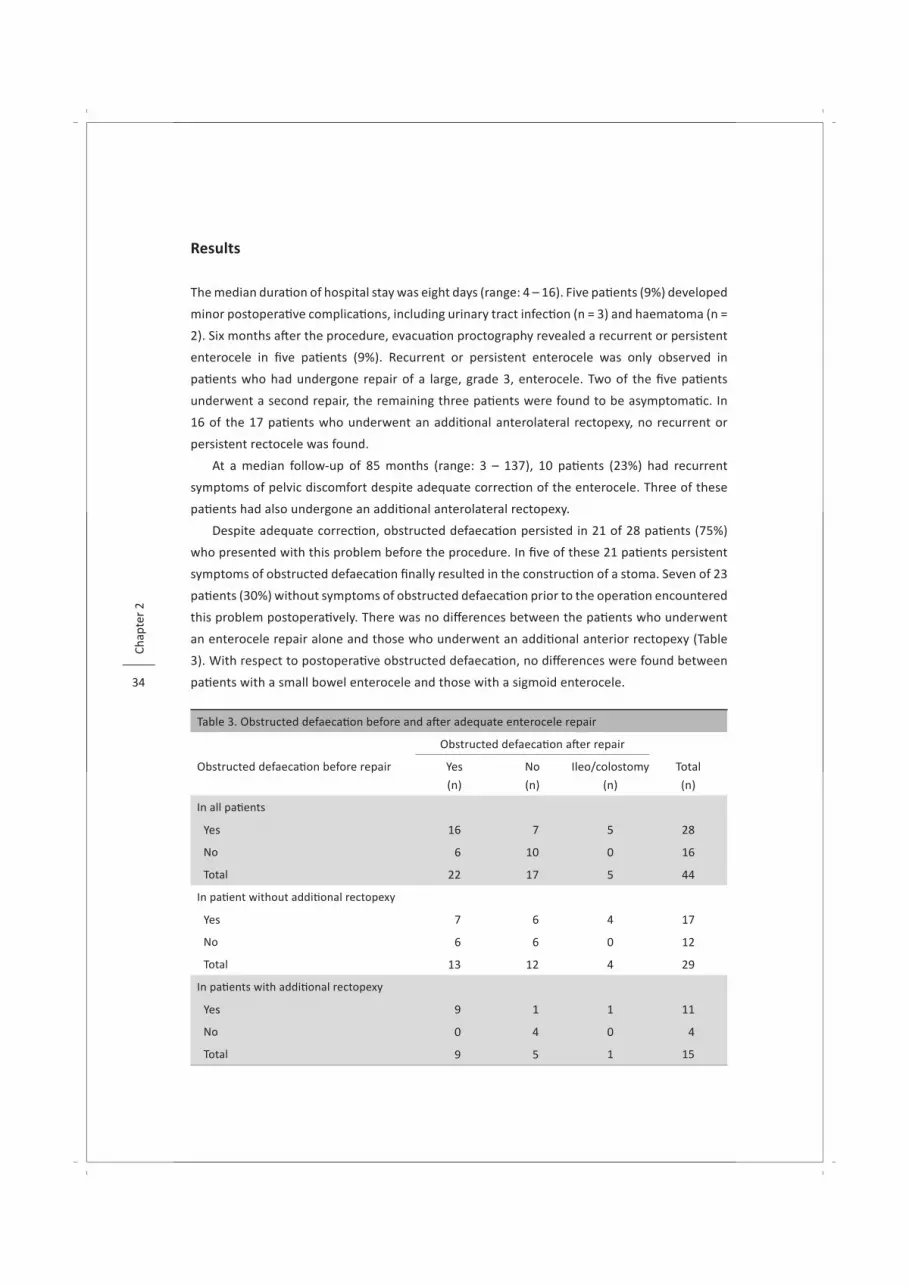
Results
The median durati on of hospital stay was eight days (range: 4 – 16). Five pati ents (9%) developed
minor postoperati ve complicati ons, including urinary tract infecti on (n = 3) and haematoma (n =
2). Six months aft er the procedure, evacuati on proctography revealed a recurrent or persistent
enterocele in fi ve pati ents (9%). Recurrent or persistent enterocele was only observed in
pati ents who had undergone repair of a large, grade 3, enterocele. Two of the fi ve pati ents
underwent a second repair, the remaining three pati ents were found to be asymptomati c. In
16 of the 17 pati ents who underwent an additi onal anterolateral rectopexy, no recurrent or
persistent rectocele was found.
At a median follow-up of 85 months (range: 3 – 137), 10 pati ents (23%) had recurrent
symptoms of pelvic discomfort despite adequate correcti on of the enterocele. Three of these
pati ents had also undergone an additi onal anterolateral rectopexy.
Despite adequate correcti on, obstructed defaecati on persisted in 21 of 28 pati ents (75%)
who presented with this problem before the procedure. In fi ve of these 21 pati ents persistent
symptoms of obstructed defaecati on fi nally resulted in the constructi on of a stoma. Seven of 23
pati ents (30%) without symptoms of obstructed defaecati on prior to the operati on encountered
this problem postoperati vely. There was no diff erences between the pati ents who underwent
an enterocele repair alone and those who underwent an additi onal anterior rectopexy (Table
3). With respect to postoperati ve obstructed defaecati on, no diff erences were found between
pati ents with a small bowel enterocele and those with a sigmoid enterocele.
Table 3. Obstructed defaecati on before and aft er adequate enterocele repair
Obstructed defaecati on aft er repair
Obstructed defaecati on before repair Yes
(n)
No
(n)
Ileo/colostomy
(n)
Total
(n)
In all pati ents
Yes 16 7 5 28
No 6 10 0 16
Total 22 17 5 44
In pati ent without additi onal rectopexy
Yes 7 6 4 17
No 6 6 0 12
Total 13 12 4 29
In pati ents with additi onal rectopexy
Yes 9 1 1 11
No 0 4 0 4
Total 9 5 1 15

Abdominal obliterati on of the pelvic inlet 35
Thirty-three of the 49 pati ents (67%), who were available for follow-up, answered the
questi ons about dyspareunia. Eleven of these 33 pati ents (33%) experienced painful intercourse
before the operati on. This symptom disappeared aft er the procedure in three pati ents (27%).
One of the 22 pati ents (5%), who did not report pain during intercourse before the procedure,
experienced de novo dyspareunia aft er the operati on.
Discussion
Abdominal obliterati on of the pouch of Douglas, according to Moschcowitz 13, was the
treatment of choice for pati ents with an enterocele. Despite the popularity of this procedure,
outcome data are scarce. In 1922, Ward 14 described a transvaginal approach. Five observati onal
studies from the last two decades do suggest that this approach results in a good anatomical
repair 4, 15-18. However, in none of these studies was evacuati on proctography performed
postoperati vely. Furthermore, except for one 4, in none of these studies was the eff ect on
associated symptoms such as pelvic discomfort and obstructed defaecati on investi gated. In
three of these series the durati on of follow-up was short. Recently, it has been suggested that
enteroceles can be corrected by an abdominal vaginal suspension technique. Cronjé et al. 7
described the results of four modifi cati ons of abdominal vaginal suspension procedures. At
follow-up (mean durati on: 7 months) physical examinati on revealed no persistent or recurrent
enterocele. However, they did not perform evacuati on proctography to confi rm this fi nding. In
our opinion, it seems unlikely that colpopexy alone is effi cient, because this procedure results
in obliterati on of the central part of the pouch of Douglas whereas both lateral parts, especially
that on the left side, remain open and deep.
In 1998, Silvis et al. 19 described a novel approach by a combined rectovaginopexy. They
performed evacuati on proctography before and three months aft er the procedure and found
a persistent enterocele in one out of 10 pati ents. A similar technique has been used by Jean
et al. 3 in 62 pati ents. According to these authors, the procedure was eff ecti ve in 98 percent of
the cases. In an earlier study from 1999, Gosselink et al. 2 reported that complete obliterati on
of the pelvic inlet with a U-shaped Mersilene® mesh was highly effi cient for pati ents with an
enterocele. Anteriorly, the mesh was sutured to the apex of the vagina and posteriorly to the
presacral fascia at the level of the promontory, thereby resulti ng not only in obliterati on of
the pelvic inlet but also in a sacral colpopexy. Evacuati on proctography, performed in all their
pati ents aft er surgery, revealed that the repair was adequate in all pati ents. Althought this
technique was eff ecti ve regarding symptoms of pelvic discomfort, obstructed defaecati on
persisted in almost all pati ents. It remains unknown whether this benefi cial eff ect on symptoms
of pelvic discomfort is sustained in the long-term. The present study was conducted to answer
this questi on.
36
Chap
ter
2
At a median follow-up of 85 months, we found that 23 percent of our pati ents had
recurrent symptoms of pelvic discomfort despite adequate correcti on of the enterocele. A
similar fi nding has been reported by Jean et al. 3. They observed that none of their pati ents had
symptoms of pelvic discomfort three months aft er treatment of the enterocele by abdominal
colporectosacropexy. At a median follow-up of 27 months recurrent symptoms of pelvic
discomfort were encountered in 27 percent of their pati ents, however they evaluated their
pati ents aft er the procedure by bidigital examinati on and not by evacuati on proctography.
All our pati ents underwent evacuati on proctography, which was repeated six months aft er
the repair. In our opinion, this imaging technique is mandatory to confi rm the effi cacy of the
repair. Enterocele cannot always be detected by physical examinati on. Kelvin et al. 21 examined
74 female pati ents with symptoms of pelvic organ prolapse. Evacuati on proctography revealed
an enterocele in 17 pati ents (23%). Physical examinati on resulted in detecti on of only seven of
these enteroceles. This fi nding illustrates that evacuati on proctography is a more reliable tool
to demonstrate the presence or absence of an enterocele.
In many textbooks, enterocele is considered to be an important cause of obstructed
defaecati on. Althought many women with an enterocele present with symptoms of obstructed
defaecati on, it is questi onable whether the two are linked. Chou et al. 22 examined 310 female
pati ents. All had completed a preoperati ve questi onnaires and had their prolapses graded
according to the Internati onal Conti nence Society System. The signs and symptoms in 77
women with an enterocele, confi rmed at surgery, were compared with those from 233 women
without an enterocele. The women with an enterocele had more advanced apical and posterior
vaginal prolapses than those without an enterocele, but there was no diff erence from them in
bowel functi on. In a recent study, Klingele et al. 23 found no associati on between the severity
of pelvic organ prolapse and symptoms of obstructed defaecati on. Kahn et al. 24 reported
that most associati ons between bowel symptoms and pelvic organ descent are weak. These
fi ndings are in accordance with those reported by Jelovsek et al. 25, who found no relati onship
between consti pati on and the stage of pelvic organ prolapse. In the present study, obstructed
defaecati on persisted in 75 percent of our pati ents who presented with this problem prior to the
operati on. Mellgren et al. 26 performed a slightly modifi cated Ripstein rectopexy in 22 pati ents
with an enterocele and concomitant (in)complete rectal prolapse. Obstructed defaecati on
persisted in 80 percent of their pati ents. Jean et al. 3 reported that 34 of 40 pati ents (85%)
with an enterocele, who presented with obstructed defaecati on prior to colporectosacropexy,
encountered the same problem 27 months aft er the procedure. Recently, Mølsted-Pederson
et al. 18 reported the outcome of transvaginal repair of enterocele with concomitant vaginal
vault prolapse using autologous fascia lata graft . According to these authors, consti pati on
persisted in 65 percent of the pati ents despite adequate repair. Based on these fi ndings, it
seems unlikely that enteroceles are a major cause of obstructed defaecati on. In our opinion it
is more likely that other abnormaliti es, such as abnormal rectal sensory percepti on, or altered

Abdominal obliterati on of the pelvic inlet 37
rectal wall contracti lity, contribute to the problem of obstructed defaecati on in pati ents with
an enterocele.
Our study shows that abdominal obliterati on of the pelvic inlet with a U-shaped Mersilene®
mesh provides an eff ecti ve tool for anatomical correcti on of enteroceles without dyspareunia
as side eff ect. However, in the long-term one of four pati ents encounters recurrent symptoms
of pelvic discomfort. It has been demonstrated that most pati ents with an enterocele have
other types of pelvic organ prolapse 12. Althought the persistent or recurrent symptoms of
pelvic discomfort might be due to these co-existi ng prolapses, they may also be att ributed to
progression of the underlying disorder causing physiologic changes that surpass the otherwise
eff ecti ve repair. The most appropiate surgical approach for enterocele is sti ll controversial.
There is also a lack of concensus about the indicati ons for enterocele repair and the need for
concomitant surgery. The limited evidence that exists at present suggests that an abdominal
approach uti lising syntheti c graft material provides a more durable support than a transvaginal
approach. Randomised controlled trials are warranted to clarify which procedure is opti mal.
38
Chap
ter
2
References
1. Bueerman W. Vaginal enterocele - report of three cases. J Am Med Assoc 1932; 99:1138.2. Gosselink MJ, van Dam JH, Huisman WM, Ginai AZ, Schouten WR. Treatment of enterocele by
obliterati on of the pelvic inlet. Dis Colon Rectum 1999;42(7):940-4.3. Jean F, Tanneau Y, Le Blanc-Louvry I, Leroi AM, Denis P, Michot F. Treatment of enterocele by
abdominal colporectosacropexy - effi cacy on pelvic pressure. Colorectal Dis 2002;4(5): 321-325.4. van der Plas-de Koning YW, Vierhout ME. Low rate of recurrence at a follow-up study of vaginal repair
of enterocele. Ned Tijdschr Geneeskd 2001;145(8):366-70.5. Jorge JM, Yang YK, Wexner SD. Incidence and clinical signifi cance of sigmoidoceles as determined by
a new classifi cati on system. Dis Colon Rectum 1994;37(11):1112-7.6. Holley RL. Enterocele: a review. Obstet Gynecol Surv 1994;49(4):284-93.7. Cronje HS, De Beer JA, Bam R. The pathophysiology of an enterocele and its management. J Obstet
Gynaecol 2004;24(4): 408-13.8. Ranney B. Enterocele, vaginal prolapse, pelvic hernia: recogniti on and treatment. Am J Obstet
Gynecol 1981; 140(1):53-61.9. Freimanis MG, Wald A, Caruana B, Bauman DH. Evacuati on proctography in normal volunteers.
Invest Radiol 1991;26(6): 581-5.10. Shorvon PJ, McHugh S, Diamant NE, Somers S, Stevenson GW. Defecography in normal volunteers:
results and implicati ons. Gut 1989;30(12):1737-49.11. Delest A, Cosson M, Coutrelant C, Querleu D, Crepin G. Enterocele. Retrospecti ve study of 134 cases:
risk factors and comparison between abdominal and perineal routes. J Gynecol Obstet Biol Reprod (Paris) 1996;25(5):464-70.
12. Lapalus MG, Henry L, Barth X, Mellier G, Gati er G, Mion F, et al. Enterocele: clinical risk factors and associati on with others pelvic fl oor disorders (about 544 defecographies). Gynecol Obstet Ferti l 2004;32(7-8):595-600.
13. Moschcowitz A. The pathogenesis, anatomy, and cure of prolapse of the rectum. Surg Gynecol Obstet 1912;15: 7-21.
14. Ward G. Technique of repair of enterocele (posterior vaginal hernia) and rectocele. J Am Med Assoc 1922;79:709.
15. Tulikangas PK, Piedmonte MR, Weber AM. Functi onal and anatomic follow-up of enterocele repairs. Obstet Gynecol 2001;98(2):265-8.
16. Raz S, Nitti VW, Bregg KJ. Transvaginal repair of enterocele. J Urol 1993;149(4):724-30.17. Miklos JR, Kohli N, Lucente V, Saye WB. Site-specifi c fascial defects in the diagnosis and surgical
management of enterocele. Am J Obstet Gynecol 1998;179(6):1418-22.18. Molsted-Pedersen L, Rudnicki M, Lose G. Transvaginal repair of enterocele and vaginal vault prolapse
using autologous fascia lata graft . Acta Obstet Gynecol Scand 2006;85(7):874-8.19. Silvis R, Gooszen HG, Kahraman T, Groenendijk AG, Lock MT, Italiaander MV, et al. Novel approach
to combined defaecati on and micturiti on disorders with rectovaginovesicopexy. Br J Surg 1998;85(6):813-7.
20. Thompson WG, Longstreth GF, Drossman DA, Heaton KW, Irvine EJ, Muller-Lissner SA. Functi onal bowel disorders and functi onal abdominal pain. Gut 1999;45:II43-7.
21. Kelvin FM, Maglinte DD, Hornback JA, Benson JT. Pelvic prolapse: assessment with evacuati on proctography (defecography). Radiology 1992;184(2):547-51.
22. Chou Q, Weber AM, Piedmonte MR. Clinical presentati on of enterocele. Obstet Gynecol 2000;96(4):599-603.
23. Klingele CJ, Bharucha AE, Fletcher JG, Gebhart JB, Riederer SG, Zinsmeister AR. Pelvic organ prolapse in defecatory disorders. Obstet Gynecol 2005;106(2):315-20.

Abdominal obliterati on of the pelvic inlet 39
24. Kahn MA, Breitkopf CR, Valley MT, Woodman PJ, O’Boyle AL, Bland DI, et al. Pelvic Organ Support Study (POSST) and bowel symptoms: straining at stool is associated with perineal and anterior vaginal descent in a general gynecologic populati on. Am J Obstet Gynecol 2005;192(5):1516-22.
25. Jelovsek JE, Barber MD, Paraiso MF, Walters MD. Functi onal bowel and anorectal disorders in pati ents with pelvic organ prolapse and inconti nence. Am J Obstet Gynecol 2005;193(6):2105-11.
26. Mellgren A, Dolk A, Johansson C, Bremmer S, Anzen B, Holmstrom B. Enterocele is correctable using the Ripstein rectopexy. Dis Colon Rectum 1994;37(8):800-4.


Chapter 3 Rectocele repair by anterolateral rectopexy:
long-term functi onal outcome
Colorectal Dis 2008;10(9):925-30.
Daniëlla M.J. Oom
Marti jn P. Gosselink
Jan J. van Wijk
Valeria R.M. van Dijl
W. Ruud Schouten
42
Chap
ter
3
Abstract
Introducti on Rectocele is frequently associated with feelings of pelvic discomfort and symptoms of
obstructed defaecati on. Repair by a transvaginal or transanal approach might result in de novo
dyspareunia in up to approximately 40 percent of the pati ents. This study was designed to
investi gate whether anterolateral rectopexy provides an adequate rectocele repair without
dyspareunia as a side eff ect.
Pati ents and Methods A consecuti ve series of 33 women (median age: 55 years, range: 37 - 73) with a symptomati c
rectocele (depth > 3 centi meters) underwent anterolateral rectopexy. Before the operati on, all
pati ents underwent evacuati on proctography, which was repeated six months aft er the repair
in all but three pati ents. A standardised questi onnaire concerning pelvic discomfort, obstructed
defaecati on and dyspareunia was used to assess the long-term eff ect of rectocele repair. The
response rate was 91 percent.
ResultsSix months aft er the procedure evacuati on proctography revealed a recurrent or persistent
rectocele in six pati ents (20%). However, in four of these pati ents, the depth of the rectocele
was less than three centi meters. The median durati on of follow-up was 74 months (range: 2 –
96). Among the pati ents with an adequate repair, signs of obstructed defaecati on persisted in
55 percent. None of the pati ents encountered de novo dyspareunia aft er the procedure.
Conclusion Anterolateral rectopexy provides an eff ecti ve tool for anatomical correcti on of rectocele and
does not result in dyspareunia as a side eff ect. However, despite adequate repair, obstructed
defaecati on persists in the majority of pati ents.
Rectocele repair by anterolateral rectopexy 43
Introducti on
Rectocele is defi ned as a herniati on of the anterior rectal wall into the lumen of the vagina. This
type of pelvic organ prolapse is frequently associated with symptoms of pelvic discomfort and
signs of obstructed defaecati on 1.
Traditi onally, rectoceles were repaired by gynaecologists. They performed a posterior
colporrhaphy by a transvaginal approach. Their principal goal was to alleviate the associated
feelings of pelvic discomfort. Unti l now it is not clear whether such a transvaginal approach
enables the diminuti on of obstructed defaecati on. Several reports do suggest that transvaginal
rectocele repair is benefi cial for pati ents with obstructed defaecati on 2, 3. Other studies,
however, have revealed that this procedure does not reduce obstructed defaecati on in the
majority of pati ents 4-7. Some authors even noted de novo signs of obstructed defaecati on 4, 6, 7.
In additi on, de novo dyspareunia is frequently reported aft er a transvaginal rectocele repair 3-6.
Colorectal surgeons prefer a transanal approach. Comparing the transvaginal and the
transanal approach, it is sti ll not clear which procedure is the most opti mum one. Several
authors have reported that transanal repair of rectocele alleviates the problem of obstructed
defaecati on 2, 8-10. However, two recent studies have revealed that transanal repair does not
reduce obstructed defaecati on in the majority of pati ents 11, 12. Unti l now, only one randomised
trial has been conducted comparing transanal with transvaginal rectocele repair 13. This study
does not elucidate which technique is the most opti mal one. Besides de novo dyspareunia,
deteriorati on of anal sphincter functi on is another drawback of transanal repair 14, 15.
Rectocele can also be repaired by a transperineal approach, with or without the use of
a mesh 16, 17. Based on the results of several small series it seems unlikely that this approach
improves the outcome.
More recently, transabdominal approaches for rectocele repair have been introduced, such
as the sacrocolpo(perineo)pexy 18, 19, the rectovaginal(vesico)pexy as described by Silvis et al. 20
and the anterolateral rectopexy 21.
Based on the data, reported in the literature, it is very diffi cult to assess which of the
procedures menti oned above is the most opti mum one for the treatment of obstructed
defaecati on related to a rectocele. In most studies, obstructed defaecati on is not defi ned
with the help of the diagnosti c Rome II criteria 22. Furthermore, the effi cacy of the diff erent
procedures is not assessed by postoperati ve evacuati on proctography in the majority of the
studies. Comparing the diff erent procedures, the transabdominal approach seems to off er a
potenti al benefi t based on the limited risk of de novo dyspareunia. Aim of the present study
was to examine whether abdominal anterolateral rectopexy provides an adequate rectocele
repair without dyspareunia as a side eff ect.

44
Chap
ter
3
Pati ents and Methods
Between 1998 and 2005, a consecuti ve series of 33 female pati ents (median age: 55 years,
range: 37 – 73) with a large (> 3 centi meters) rectocele underwent anterolateral rectopexy.
In all pati ents, the rectocele was associated with obstructed defaecati on. We used four of the
six ROME II criteria 22 to defi ne obstructed defaecati on: straining, sensati on of incomplete
evacuati on, sensati on of anorectal obstructi on or blockage, and manual assistance to facilitate
evacuati on. Pati ents were included only when they fulfi lled two or more of these criteria and
these symptoms occurred regularly over at least 12 consecuti ve weeks in the preceding 12
months. Regarding obstructed defaecati on, the procedure was considered unsuccessful if the
pati ent sti ll fulfi lled two or more of these four criteria. Prior to the procedure, colonic transit
ti me was assessed in all pati ents by daily ingesti on of radiopaque markers and a single X-ray at
day 7. All except three pati ents had normal colonic transit ti me. In 76 percent of the pati ents
(n = 25), rectocele was also associated with pelvic discomfort, including feelings of prolapse
and pelvic pressure. The history of 28 pati ents (85%) revealed one or more previous surgical
procedures of the pelvis. Most of the pati ents (94%) were multi parous. All pati ents underwent
evacuati on proctography with opacifi cati on of the small bowel and vagina in order to confi rm
the diagnosis and to assess the depth of the rectocele prior to the operati on. Six months aft er
the procedure, evacuati on proctography was repeated to evaluate the effi cacy of the repair.
Hospital records and outpati ent clinic charts were analysed, and follow-up informati on was
obtained both from a standardised questi onnaire and review of charts by one author (DMO),
who had not parti cipated in the surgical procedure. The standardised questi onnaire included
questi ons about obstructed defaecati on as defi ned by four of the six ROME II criteria, pelvic
discomfort, and dyspareunia. Twenty-seven pati ents (82%) completed whole postoperati ve
follow-up (evacuati on proctography and questi onnaire). In 13 pati ents the procedure was
combined with obliterati on of the pelvic inlet with a U-shaped Mersilene® mesh to correct
a co-existi ng enterocele 23. In one pati ent, an abdominal hysterectomy was performed
simultaneously.
Surgical Technique
At the ti me of inducti on of anaesthesia, cefuroxime (1.500 mg) and metronidazole (500 mg) were
administered intravenously. With the pati ent in lithotomy positi on, a lower midline laparatomy
was performed. The mesosigmoid was retracted ventrally and to the left . The right ureter was
visualised. A peritoneal incision was made over the sacral promontory. The deepest part of
the pouch of Douglas was retracted and incised. The rectovaginal septum was opened down
to the pelvic fl oor and both anterolateral sides of the distal part of the rectum were exposed.
Using nonabsorbable sutures, a strip of Marlex® (CR Bard, Cranston, USA), with a width of 3 cm,
was sutured to both anterolateral sides of the distal rectum. Proximal to the incised pouch of

Rectocele repair by anterolateral rectopexy 45
Douglas, further sutures were used to fi x the mesh to the anterior border of the rectum. Under
tracti on, the mesh was fi xed upon the sacral promontory using nonabsorbable sutures. The
incised peritoneal pouch of Douglas was closed over the distal part of the mesh with resorbable
sutures. The proximal part of the mesh was not covered with peritoneum. All procedures were
performed by the same surgeon (WRS).
Results
No intraoperati ve complicati ons were observed and no blood transfusions were required.
Median hospital stay was eight days (range: 6 – 17). There was no postoperati ve death.
One pati ent developed an abscess in the pouch of Douglas. This serious complicati on was
treated by ultrasound guided drainage without long-term sequaelae for the Marlex strip. Five
other pati ents (15%) developed minor postoperati ve complicati ons, including urinary tract
infecti on (n = 4) and postoperati ve ileus combined with urinary tract infecti on (n = 1). No late
complicati ons, including mesh related problems, were encountered.
Three pati ents refused evacuati on proctography six months aft er the procedure. The
pre- and postoperati ve fi ndings of evacuati on proctography are shown in Table 1. Evacuati on
proctography revealed a recurrent or persistent rectocele in six pati ents (20%). In four of
these pati ents, the depth of the rectocele was less than three centi meters. In all pati ents who
underwent an additi onal obliterati on of the pelvic inlet, no recurrent or persistent enterocele
was found. Evacuati on proctography revealed that in all pati ents with an intussuscepti on this
abnormality had disappeared aft er the procedure.
Table 1. Findings of evacuati on proctography
Prior to surgery n (%) Aft er surgery* n (%)
Rectocele > 3 cm 33 (100) 2 (7)
< 3 cm 0 (0) 4 (13)
Enterocele 15 (45) 0 (0)
Intussuscepti on 8 (24) 0 (0)
* 3 pati ents refused evacuati on proctography aft er surgery.
The median durati on of follow-up was 74 months (range: 2 – 96). In three pati ents in whom
obstructed defaecati on was associated with delayed colonic transit, the defaecatory problems
persisted. In the pati ents in whom obstructed defaecati on was associated with a normal colonic
transit ti me, symptoms of obstructed defaecati on persisted in 11 of 20 pati ents (55%) despite
adequate rectocele repair (Table 2).
46
Chap
ter
3
Table 2. Impact on obstructed defaecati on in pati ents with an adequate repair
Evacuati on aft er repair
Evacuati on before repair Normal (n) OD (n) Total (n)*
OD 9 11 20
OD + STC – 3 3
* Pati ents, who completed whole postoperati ve follow-up.Pati ents with recurrent or persistent rectocele on postoperati ve evacuati on proctography were excluded.
OD: obstructed defaecati on, STC: slow transit consti pati on.
Twenty-fi ve of the 33 pati ents (76%) also reported symptoms of pelvic discomfort,
including feelings of pelvic prolapse and pelvic pressure. In 10 of these 25 pati ents, the
rectocele was associated with a concomitant enterocele. In the pati ents without an enterocele
the anterolateral rectopexy resolved the symptoms of pelvic discomfort in 77 percent of the
pati ents. In the pati ents with a concomitant enterocele (n = 13), the anterolateral rectopexy
was combined with obliterati on of the pelvic inlet. Aft er this combined procedure, symptoms
of pelvic discomfort resolved in 80 percent.
Twenty-one of the 30 pati ents (70%), who were available for follow-up, answered the
questi ons about dyspareunia. Four of these 21 pati ents (19%) experienced painful intercourse
before the operati on. This problem disappeared aft er the procedure in one pati ent. None of
the pati ents who did not report pain during intercourse before the procedure experienced de
novo dyspareunia aft er surgery.
Discussion
It is generally accepted that rectoceles contribute to the problem of obstructed defaecati on.
In the past, rectocele repair was almost exclusively performed by gynaecologists. Their
approach consisted of a posterior colporrhaphy, characterised by midline plicati on of the
levator muscles, with or without plicati on of the rectovaginal fascia, in order to strengthen the
posterior vaginal wall 4, 24. Later on, modifi ed techniques were introduced, including posterior
colporrhaphy using diff erent types of syntheti c meshes or collagen implants and site-specifi c
repair techniques of the rectovaginal fascia defects 3, 25. The main goal of these transvaginal
repairs was to alleviate symptoms of pelvic discomfort. Unti l the 1990s, data regarding the
impact of this type of repair on associated symptoms of obstructed defaecati on were lacking.
During the last decade, more att enti on has been paid to this aspect. However in none of the
studies conducted so far, validated criteria have been used to defi ne obstructed defaecati on.
Although some reports do suggest that transvaginal rectocele repair alleviates obstructed
defaecati on in almost all pati ents 2, 3, most studies indicate that this type of repair is benefi cial

Rectocele repair by anterolateral rectopexy 47
for less than half of the pati ents 4-7. According to some authors, transvaginal rectocele repair
can even induce obstructed defaecati on 4, 6, 7. A potenti al drawback of transvaginal repair is de
novo dyspareunia 3-6. This side-eff ect has been reported in up to 27 percent of the pati ents 6.
In contrast to gynaecologists, most colorectal surgeons performed a rectocele repair by a
transanal approach. Their main goal was diminuti on of obstructed defaecati on. Comparing the
transvaginal and the transanal approach, it is not clear which procedure is the most opti mum
one for pati ents with obstructed defaecati on. Although some authors 2, 8-10, 26 have reported that
transanal rectocele repair or a combined transanal / transvaginal repair is eff ecti ve in more
than half of the pati ents, two recent studies have revealed that transanal repair has no eff ect
on obstructed defaecati on in the majority of pati ents 11, 12. Unti l now, only one randomised
controlled trial 13 has been conducted comparing transanal with transvaginal rectocele repair.
This study did not elucidate which technique is the most opti mum one. Like transvaginal repair,
transanal repair can also result in de novo dyspareunia. This side eff ect is reported in up to
41 percent of the pati ents 26. Another drawback of the transanal approach is deteriorati on of
anal sphincter functi on 14, 15. An adequate transanal repair can only be performed with the use
of a Parks anal retractor or another comparable instrument. In a randomised controlled trial,
comparing two diff erent types of anal retractors, Zimmerman et al. 27 observed that the use of
a Parks retractor during perianal fi stula repair has a deteriorati ng eff ect on faecal conti nence,
probably because of damage to the internal anal sphincter.
In recent years, transabdominal approaches for rectocele repair have been introduced.
Some gynaecologists prefer an abdominal sacrocolpo(perineo)pexy in pati ents in whom the
rectocele is associated with vaginal vault prolapse 18, 19. In 1998, Silvis et al. 20 introduced the
rectovagino(vesico)pexy to alleviate combined defaecati on and micturiti on disorders. This
approach was performed in 22 pati ents with a rectocele. Unfortunately, the authors did not
report whether this type of repair diminuted obstructed defaecati on in this selected group of
pati ents. In 2004, D’Hoore and coworkers 28 reported the long-term outcome of laparascopic
ventral rectopexy, not for rectocele but for complete rectal prolapse. Fourteen of 42 pati ents
were categorised as having obstructed defaecati on. In the majority of the pati ents, symptoms
of obstructed defaecati on resolved. According to these authors, the ventral positi on of the
mesh att ributes to the benefi cial eff ect on obstructed defaecati on. Despite these promising
results it is questi onable whether a similar outcome can be obtained aft er ventral rectopexy in
pati ents with a symptomati c rectocele. Vermeulen et al. 21 were the fi rst to report a modifi ed
anterolateral rectopexy for rectocele repair. Postoperati ve evacuati on proctography revealed
adequate correcti on of rectocele in all pati ents. Despite this adequate repair, only 13 percent
of the pati ents encountered complete relieve of obstructed defaecati on aft er a median follow-
up of 18 months. However, in their study, obstructed defaecati on was not defi ned with the
help of the diagnosti c Rome II criteria. Uti lising these criteria, we observed a success rate of
45 percent.
48
Chap
ter
3
In contrast with earlier reports, these fi ndings and the data recently presented by others
indicate that surgical correcti on of the bulging anterior rectal wall alone is not benefi cial for
more than half of the women with obstructed defaecati on. The questi on is whether rectocele
is the cause or a result of obstructed defaecati on.
During the last decade, more att enti on has been paid to other causes of obstructed
defaecati on, especially those related to rectal wall properti es. Gosselink and Schouten 29
reported that in women with obstructed defaecati on the threshold for rectal sensory percepti on
is signifi cantly increased, indicati ng rectal hyposensiti vity. They also found that the tonic
response of the rectum to an evoked urge to defaecate is absent or signifi cantly blunted 30.
Rectal hyposensiti vity in pati ents with obstructed defaecati on has also been described by
others. Gladman et al. 31, for example, observed rectal hyposensiti vity in one of three pati ents
with obstructed defaecati on. Comparing pati ents with and without rectocele, the incidence of
rectal hyposensiti vity was found to be equal. There is limited evidence that rectal hyposensiti vity
is caused by impairment of aff erent pathways due to pelvic nerve injury 32. Recently, it has been
shown that the proximal colonic motor response to mechanical and chemical sti mulati on of the
rectum is impaired in women with obstructed defaecati on 33. Based on these fi ndings, it seems
unlikely that obstructed defaecati on is caused by one single abnormality, such as a rectocele.
It has been reported that biofeedback therapy, especially sensory retraining, improves
rectal sensiti vity as assessed during physiologic evaluati on 34, 35. Improvement of rectal
hyposensiti vity was associated with clinical improvement. There is litt le evidence that
pati ents with more severely impaired rectal sensiti vity are less likely to improve their sensory
functi on 36. In recent years, it has been suggested that biofeedback therapy also improves
obstructed defaecati on in pati ents with a rectocele 37, 38. However, the number of studied
pati ent are small and the durati on of follow-up is limited to the short-term. Furthermore, in
the majority of studied pati ents, rectocele was associated with concomitant anismus. Based on
the data, obtained from these studies, it is not clear whether biofeedback therapy is eff ecti ve
by resolving rectal hyposensiti vity or by dissolving the problem of anismus. Chang et al. 39
reported that electrical sti mulati on therapy, using an anal plug with pulse generator, resulted
in clinical improvement and enhancement of rectal sensiti vity on physiologic testi ng. It has
been suggested that improvement in rectal sensiti vity may be one of the mechanisms by which
symptoms of obstructed defaecati on improve following sacral nerve modulati on 40, 41, although
direct evidence of this is currently lacking 42.
Two recently introduced surgical procedures are also based on the assumpti on that impaired
rectal sensiti vity contributes to obstructed defaecati on. First, the verti cal reducti on rectoplasty
has been introduced, based on the premise that reducing rectal capacity and compliance would
restore the percepti on of rectal fullness in pati ents with idiopathic megarectum 42, 43. The other
procedure is the stapled trans-anal rectal resecti on (STARR). The main goal of this operati on is
to remove the distal part of the insensiti ve and dysfuncti onal rectum. Preliminary results are
Rectocele repair by anterolateral rectopexy 49
promising 44, although some authors have reported signifi cant complicati ons such as faecal
urgency, inconti nence, anastomoti c stenosis, and new onset rectovaginal fi stula 45, 46. Recently,
the results from the European STARR registry have been reported 47. These results, obtained
from 1.058 pati ents, indicate that this procedure is safe and benefi cial for many pati ents
with obstructed defaecati on. Although rectocele repair by anterolateral rectopexy results in
adequate rectocele repair, symptoms of obstructed defaecati on persist in more than half of
the pati ents. The assumed relati on between rectal hyposensiti vity and obstructed defaecati on
might explain why anatomical correcti on of a rectocele alone, as obtained in the present study,
fails to restore symptoms of obstructed defaecati on in many women.
50
Chap
ter
3
References
1. Dietz HP, Korda A. Which bowel symptoms are most strongly associated with a true rectocele? Aust N Z J Obstet Gynaecol 2005;45(6):505-8.
2. Tsujinaka S, Tsujinaka Y, Matsuo K, Akagi K, Hamahata Y. Changes in Bowel Functi on following Transanal and Transvaginal Rectocele Repair. Dig Surg 2007;24(1):46-53.
3. Kenton K, Shott S, Brubaker L. Outcome aft er rectovaginal fascia reatt achment for rectocele repair. Am J Obstet Gynecol 1999;181(6):1360-4.
4. Kahn MA, Stanton SL. Posterior colporrhaphy: its eff ects on bowel and sexual functi on. Br J Obstet Gynaecol 1997;104(1):82-6.
5. Yamana T, Takahashi T, Iwadare J. Clinical and physiologic outcomes aft er transvaginal rectocele repair. Dis Colon Rectum 2006;49(5):661-7.
6. Lim YN, Muller R, Corsti aans A, Hitchins S, Barry C, Rane A. A long-term review of posterior colporrhaphy with Vypro 2 mesh. Int Urogynecol J Pelvic Floor Dysfunct 2007.
7. Gusti lo-Ashby AM, Paraiso MF, Jelovsek JE, Walters MD, Barber MD. Bowel symptoms 1 year aft er surgery for prolapse: further analysis of a randomized trial of rectocele repair. Am J Obstet Gynecol 2007;197(1):76 e1-5.
8. Abbas SM, Bissett IP, Neill ME, Macmillan AK, Milne D, Parry BR. Long-term results of the anterior Delorme’s operati on in the management of symptomati c rectocele. Dis Colon Rectum 2005;48(2):317-22.
9. Ayav A, Bresler L, Brunaud L, Boissel P. Long-term results of transanal repair of rectocele using linear stapler. Dis Colon Rectum 2004;47(6):889-94.
10. Roman H, Michot F. Long-term outcomes of transanal rectocele repair. Dis Colon Rectum 2005;48(3):510-7.
11. Puigdollers A, Fernandez-Fraga X, Azpiroz F. Persistent symptoms of functi onal outlet obstructi on aft er rectocele repair. Colorectal Dis 2007;9(3):262-5.
12. Heriot AG, Skull A, Kumar D. Functi onal and physiological outcome following transanal repair of rectocele. Br J Surg 2004;91(10):1340-4.
13. Nieminen K, Hiltunen KM, Laiti nen J, Oksala J, Heinonen PK. Transanal or vaginal approach to rectocele repair: a prospecti ve, randomized pilot study. Dis Colon Rectum 2004;47(10):1636-42.
14. Ho YH, Ang M, Nyam D, Tan M, Seow-Choen F. Transanal approach to rectocele repair may compromise anal sphincter pressures. Dis Colon Rectum 1998;41(3):354-8.
15. van Dam JH, Huisman WM, Hop WC, Schouten WR. Fecal conti nence aft er rectocele repair: a prospecti ve study. Int J Colorectal Dis 2000;15(1):54-7.
16. Mercer-Jones MA, Sprowson A, Varma JS. Outcome aft er transperineal mesh repair of rectocele: a case series. Dis Colon Rectum 2004;47(6):864-8.
17. Watson SJ, Loder PB, Halligan S, Bartram CI, Kamm MA, Phillips RK. Transperineal repair of symptomati c rectocele with Marlex mesh: a clinical, physiological and radiologic assessment of treatment. J Am Coll Surg 1996;183(3):257-61.
18. Baessler K, Schuessler B. Abdominal sacrocolpopexy and anatomy and functi on of the posterior compartment. Obstet Gynecol 2001;97(5 Pt 1):678-84.
19. Cundiff GW, Harris RL, Coates K, Low VH, Bump RC, Addison WA. Abdominal sacral colpoperineopexy: a new approach for correcti on of posterior compartment defects and perineal descent associated with vaginal vault prolapse. Am J Obstet Gynecol 1997;177(6):1345-55.
20. Silvis R, Gooszen HG, Kahraman T, Groenendijk AG, Lock MT, Italiaander MV, et al. Novel approach to combined defaecati on and micturiti on disorders with rectovaginovesicopexy. Br J Surg 1998;85(6):813-7.
21. Vermeulen J, Lange JF, Sikkenk AC, van der Harst E. Anterolateral rectopexy for correcti on of rectoceles leads to good anatomical but poor functi onal results. Tech Coloproctol 2005;9(1):35-41.

Rectocele repair by anterolateral rectopexy 51
22. Thompson WG, Longstreth GF, Drossman DA, Heaton KW, Irvine EJ, Muller-Lissner SA. Functi onal bowel disorders and functi onal abdominal pain. Gut 1999;45 Suppl 2:II43-7.
23. Oom DM, van Dijl VR, Gosselink MP, van Wijk JJ, Schouten WR. Enterocele repair by abdominal obliterati on of the pelvic inlet: long-term outcome on obstructed defaecati on and symptoms of pelvic discomfort. Colorectal Dis 2007.
24. Maher CF, Qatawneh AM, Baessler K, Schluter PJ. Midline rectovaginal fascial plicati on for repair of rectocele and obstructed defecati on. Obstet Gynecol 2004;104(4):685-9.
25. Paraiso MF, Barber MD, Muir TW, Walters MD. Rectocele repair: a randomized trial of three surgical techniques including graft augmentati on. Am J Obstet Gynecol 2006;195(6):1762-71.
26. van Dam JH. Thesis: Rectocele repair in women with obstructed defecati on. 1999.27. Zimmerman DD, Gosselink MP, Hop WC, Darby M, Briel JW, Schouten WR. Impact of two diff erent
types of anal retractor on fecal conti nence aft er fi stula repair: a prospecti ve, randomized, clinical trial. Dis Colon Rectum 2003;46(12):1674-9.
28. D’Hoore A, Cadoni R, Penninckx F. Long-term outcome of laparoscopic ventral rectopexy for total rectal prolapse. Br J Surg 2004;91(11):1500-5.
29. Gosselink MJ, Schouten WR. Rectal sensory percepti on in females with obstructed defecati on. Dis Colon Rectum 2001;44(9):1337-44.
30. Schouten WR, Gosselink MJ, Boerma MO, Ginai AZ. Rectal wall contracti lity in response to an evoked urge to defecate in pati ents with obstructed defecati on. Dis Colon Rectum 1998;41(4):473-9.
31. Gladman MA, Scott SM, Chan CL, Williams NS, Lunniss PJ. Rectal hyposensiti vity: prevalence and clinical impact in pati ents with intractable consti pati on and fecal inconti nence. Dis Colon Rectum 2003;46(2):238-46.
32. Gladman MA, Dvorkin LS, Lunniss PJ, Williams NS, Scott SM. Rectal hyposensiti vity: a disorder of the rectal wall or the aff erent pathway? An assessment using the barostat. Am J Gastroenterol 2005;100(1):106-14.
33. Dinning PG, Bampton PA, Kennedy ML, Lubowski DZ, King D, Cook IJ. Impaired proximal colonic motor response to rectal mechanical and chemical sti mulati on in obstructed defecati on. Dis Colon Rectum 2005;48(9):1777-84.
34. De Medici A, Badiali D, Corazziari E, Bausano G, Anzini F. Rectal sensiti vity in chronic consti pati on. Dig Dis Sci 1989;34(5):747-53.
35. Peti cca L, Pescatori M. Outlet obstructi on due to anismus and rectal hyposensati on: eff ect of biofeedback training. Colorectal Dis 2002;4(1):67.
36. Chiarioni G, Bassotti G, Stanganini S, Vanti ni I, Whitehead WE. Sensory retraining is key to biofeedback therapy for formed stool fecal inconti nence. Am J Gastroenterol 2002;97(1):109-17.
37. Mimura T, Roy AJ, Storrie JB, Kamm MA. Treatment of impaired defecati on associated with rectocele by behavorial retraining (biofeedback). Dis Colon Rectum 2000;43(9):1267-72.
38. Kairaluoma M, Raivio P, Kupila J, Aarnio M, Kellokumpu I. The role of biofeedback therapy in functi onal proctologic disorders. Scand J Surg 2004;93(3):184-90.
39. Chang HS, Myung SJ, Yang SK, Jung HY, Kim TH, Yoon IJ, et al. Eff ect of electrical sti mulati on in consti pated pati ents with impaired rectal sensati on. Int J Colorectal Dis 2003;18(5):433-8.
40. Kenefi ck NJ, Nicholls RJ, Cohen RG, Kamm MA. Permanent sacral nerve sti mulati on for treatment of idiopathic consti pati on. Br J Surg 2002;89(7):882-8.
41. Ganio E, Masin A, Ratt o C, Altomare DF, Ripetti V, Clerico G, et al. Short-term sacral nerve sti mulati on for functi onal anorectal and urinary disturbances: results in 40 pati ents: evaluati on of a new opti on for anorectal functi onal disorders. Dis Colon Rectum 2001;44(9):1261-7.
42. Gladman MA, Lunniss PJ, Scott SM, Swash M. Rectal hyposensiti vity. Am J Gastroenterol 2006;101(5):1140-51.
43. Williams NS, Fajobi OA, Lunniss PJ, Scott SM, Eccersley AJ, Ogunbiyi OA. Verti cal reducti on rectoplasty: a new treatment for idiopathic megarectum. Br J Surg 2000;87(9):1203-8.
52
Chap
ter
3
44. Boccasanta P, Venturi M, Stuto A, Botti ni C, Caviglia A, Carriero A, et al. Stapled transanal rectal resecti on for outlet obstructi on: a prospecti ve, multi center trial. Dis Colon Rectum 2004;47(8):1285-97.
45. Dodi G, Pietroletti R, Milito G, Binda G, Pescatori M. Bleeding, inconti nence, pain and consti pati on aft er STARR transanal double stapling rectotomy for obstructed defecati on. Tech Coloproctol 2003;7(3):148-53.
46. Binda GA, Pescatori M, Romano G. The dark side of double-stapled transanal rectal resecti on. Dis Colon Rectum 2005;48(9):1830-2.
47. Stuto A. SO, Jayne D. Assessing safety of the STARR procedure for ODS: preliminary results of the European STARR Registry. Dis Colon Rectum 2007;50(5):724.

Chapter 4 Assessment of posterior compartment prolapse;
a comparison of evacuati on proctography and transperineal
ultrasound
Colorectal Dis 2009; in press.
Anneke B. Steensma
Daniëlla M.J. Oom
Curt W. Burger
W. Ruud Schouten

54
Chap
ter
4
Abstract
Introducti onEvacuati on proctography is considered to be the gold standard investi gati on for the diagnosis
of posterior compartment prolapse. Transperineal ultrasound imaging of the pelvic fl oor is
a non-invasive investi gati on for detecti on of pelvic fl oor abnormaliti es. This study compared
evacuati on proctography with transperineal ultrasound in diagnosing posterior compartment
prolapse.
Pati ents and Methods In a prospecti ve observati onal study, all pati ents with symptoms related to posterior
compartment prolapse were invited to parti cipate in a standardised interview, clinical
examinati on, transperineal ultrasound, and evacuati on proctography. Both exams were
analysed separately by two experienced investi gators, blinded against the clinical data and
against the results of the other imaging technique. Aft er the examinati ons, all pati ents were
asked to fi ll out a standardised questi onnaire concerning subjecti ve experience.
Results Between 2005 and 2007, 75 pati ents were included with a median age of 59 years (range: 22 –
83). The Cohen’s kappa coeffi cient for enterocele was 0.65 (good) and for rectoceles it was 0.55
(moderate). The level of agreement for intussuscepti on was fair (κ = 0.21).
Conclusion This study showed moderate to good agreement between transperineal and evacuati on
proctography for detecti ng enterocele and rectocele.

Assessment of posterior compartment prolapse 55
Introducti on
Pati ents with pelvic organ prolapse symptoms will be referred to a (uro)gynaecologist or
colorectal surgeon for investi gati on and treatment. For the classifi cati on of pelvic organ
prolapse a subdivision into three pelvic compartments is uti lised (anterior, middle, and
posterior). Voiding dysfuncti on and urinary inconti nence are commonly related to prolapse
of the anterior compartment 1. Symptoms such as feelings of a lump, heaviness, and pelvic
pressure are att ributed to all pelvic compartments. Symptoms of obstructed defaecati on and
faecal inconti nence have been correlated with the posterior compartment 2-4. It is important to
be able to diff erenti ate between diff erent anatomic abnormaliti es, especially when considering
surgery.
Evacuati on proctography, with opacifi cati on of the small bowel and vagina, has been
claimed to be the gold standard investi gati on for objecti ve diagnosis of posterior compartment
prolapse. Evacuati on proctography is uncomfortable for the pati ent and requires exposure to
ionising radiati on. Furthermore, when used without opacifi cati on of the bladder it lacks the
ability to visualise the anterior and middle compartments.
Recently, more advanced imaging techniques such as dynamic magneti c resonance (MR)
imaging and transperineal ultrasound, have become available 5, 6. These are able to show all
three pelvic compartments. Furthermore, they are less invasive and ionising radiati on is not
required. However, dynamic MR imaging, especially open-architecture MR imaging, is very
expensive and not generally available. Transperineal ultrasound enables dynamic investi gati on
of all three pelvic compartments at low cost. Dietz et al. 6, 7 reported that transperineal
ultrasound can diff erenti ate between diff erent forms of posterior anatomic abnormaliti es, such
as rectocele and enterocele.
At present four studies have been published to assess the level of agreement between
evacuati on proctography and transperineal ultrasound, but the pati ent groups have been small
in most of these studies and in some there is a selecti on bias, with pati ents having a history of
longstanding obstructed defaecati on 8-11.
This study was designed to assess the level of agreement between evacuati on proctography
and transperineal ultrasound in diagnosing posterior compartment prolapse in pati ents with
related symptoms.

56
Chap
ter
4
Pati ents and Methods
All women with symptoms related to posterior compartment prolapse referred to our
terti ary pelvic fl oor unit were included in this prospecti ve observati onal study. They were
interviewed using a standardised questi onnaire, concerning medical history, urinary functi on,
pelvic discomfort, and bowel functi on. A clinical examinati on was performed according
to the Internati onal Conti nence Society guidelines, using the POP-Q system 12. The pati ents
underwent standardised evacuati on proctography and dynamic transperineal ultrasound, with
a maximum interval of six months. Aft er both examinati ons, all pati ents were asked to complete
a standardised questi onnaire concerning subjecti ve experience. Writt en informed consent was
obtained from all parti cipants. This study was approved by the hospital’s human research ethics
committ ee.
Evacuati on Proctography
Evacuati on proctography was performed using a standardised technique with opacifi cati on of
the rectosigmoid, small bowel, and vagina, using liquid barium contrast. Imaging was acquired
at rest, during pelvic fl oor contracti on, and during straining and a video recording was obtained
during evacuati on of contrast. Quanti tati ve measurements were made for comparison
with transperineal ultrasound. Enterocele was described as a herniati on of small bowel or
rectosigmoid into the vagina (Figure 1a). Rectocele was defi ned as a herniati on of the anterior
rectal wall into the lumen of the vagina and intussuscepti on was defi ned as an infolding of
the rectal wall into the rectum or anus. When an external component was present, it was
called complete rectal prolapse. Enterocele, rectocele, and intussuscepti on were classifi ed into
grades, see Table 1 13, 14. Rectocele depth was measured perpendicular to a line projected along
the expected contour of the anterior rectal wall (Figure 2a). All video fi les were analysed by
one colorectal surgeon (WRS), blinded against all clinical data and the results of transperineal
ultrasound.
Transperineal Ultrasound
Transperineal ultrasound was performed using a GE Kretz Voluson 730 expert system (GE
Healthcare, clinical systems, Hoevelaken, the Netherlands), using an abdominal 4 – 8 MHz
transducer. Pati ents were examined aft er voiding and in supine positi on. 2D cine loop volumes
(3D) were obtained at rest, during pelvic fl oor contracti on, and during maximal Valsalva
manoeuvre, as previously described by Dietz et al. 6, 7. Off line evaluati on of the cine loop
volumes was performed by one gynaecologist (ABS), blinded against all clinical data and the
results of evacuati on proctography, using 4D view soft ware (GE Healthcare). Enterocele was
diagnosed if a herniati on of abdominal contents developed anterior to the anorectal juncti on
and extended into the vagina (Figure 1b). Rectocele was defi ned as a defect in the rectovaginal
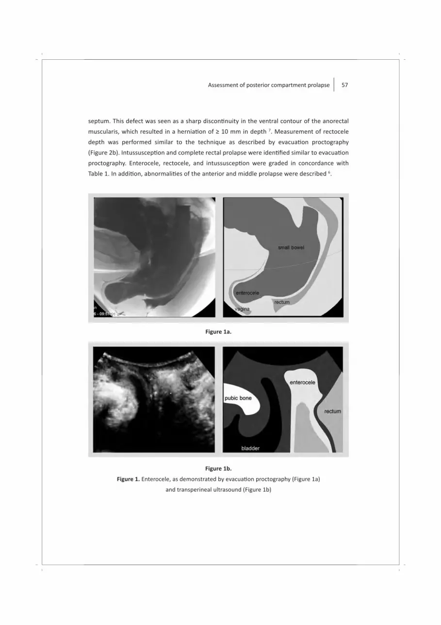
Assessment of posterior compartment prolapse 57
septum. This defect was seen as a sharp disconti nuity in the ventral contour of the anorectal
muscularis, which resulted in a herniati on of ≥ 10 mm in depth 7. Measurement of rectocele
depth was performed similar to the technique as described by evacuati on proctography
(Figure 2b). Intussuscepti on and complete rectal prolapse were identi fi ed similar to evacuati on
proctography. Enterocele, rectocele, and intussuscepti on were graded in concordance with
Table 1. In additi on, abnormaliti es of the anterior and middle prolapse were described 6.
Figure 1a.
Figure 1b.
Figure 1. Enterocele, as demonstrated by evacuati on proctography (Figure 1a)
and transperineal ultrasound (Figure 1b)

58
Chap
ter
4
Table 1. Grading system for enterocele, rectocele, and intussuscepti on.
Prolapse Descripti on
Enterocele
Grade 1 Most distal part descending to the upper ⅓ of the vagina
Grade 2 Most distal part descending to the middle ⅓ of the vagina
Grade 3 Most distal part descending to the lower ⅓ of the vagina
Rectocele
Grade 1 Depth < 2 centi meters
Grade 2 Depth 2 – 4 centi meters
Grade 3 Depth > 4 centi meters
Intussuscepti on
Grade 1 Most distal part remains completely intrarectal
Grade 2 Most distal part descending into anal canal
Aft er both examinati ons, all pati ents received a writt en standardised questi onnaire
designed to assess pati ent expectati ons, inconvenience, and discomfort. Pati ents were asked
to rate any discomfort on a visual analogue scale (VAS, 0: no discomfort, 10: severe discomfort).
Stati sti cal Analysis
Stati sti cal analysis was performed using the SPSS soft ware package (14.0 version, SPSS
Inc., Chicago, IL, USA). The Cohen’s kappa coeffi cient was obtained to compare evacuati on
proctography and transperineal ultrasound in the detecti on of enterocele, rectocele, and
intussuscepti on (Table 2). Spearman’s correlati on coeffi cient was used to calculate the level
of correlati on between mean rectocele depths. To compare questi onnaires, the chi-squared
test was used. Wilcoxon’s signed ranks test was used for comparing VAS scores. Results were
considered stati sti cally signifi cant when p ≤ 0.05 (two-sided). Evacuati on proctography was
considered to be the gold standard investi gati on to calculate the sensiti viti es and specifi citi es
for detecti on of diff erent prolapses with transperineal ultrasound.
Table 2. Cohen’s kappa coeffi cient Table 3. Symptoms as reported by the pati ents
Value n (%)
Poor agreement 0.00 – 0.20 Stress urinary inconti nence 38 (51)
Fair agreement 0.21 – 0.40 Urge urinary inconti nence 25 (33)
Moderate agreement 0.41 – 0.60 Pelvic discomfort 50 (67)
Good agreement 0.61 – 0.80 Obstructed defaecati on 36 (48)
Excellent agreement 0.81 – 1.00 Faecal inconti nence 26 (35)

Assessment of posterior compartment prolapse 59
Figure 2a.
Figure 2b.
Figure 2. Rectocele, as demonstrated by evacuati on proctography (Figure 2a) and transperineal
ultrasound (Figure 2b) arrow indicates rectocele depth
Results
Between September 2005 and July 2007, 75 women were referred with symptoms related to
posterior compartment prolapse. These included pelvic discomfort, obstructed defaecati on
and faecal inconti nence or a combinati on (Table 3). The median age was 59 years (range: 22 –
83). A previous hysterectomy had been carried out in 31 women (41%) and a previous pelvic
organ prolapse repair in 37 (49%). Median parity was 2 (range: 0 – 10 vaginal deliveries, four
nulliparous women).
60
Chap
ter
4
Clinical examinati on showed absence of prolapse in 16 pati ents (21%), stage 1 prolapse in
27 pati ents (36%), stage 2 in 21 pati ents (28%) and stage 3 in 11 pati ents (15%). Evacuati on
proctography revealed in seven of the 16 pati ents (44%) without prolapse during clinical
examinati on some form of posterior compartment prolapse. Transperineal ultrasound
revealed some form of prolapse in six of these pati ents (38%). The clinical diagnosis of
posterior compartment prolapse (stage 1 – 3) was confi rmed by evacuati on proctography and
transperineal ultrasound in 83 and 81 percent respecti vely.
Evacuati on proctography revealed an enterocele in 25 pati ents (33%), rectocele in
36 (48%), and intussuscepti on in 27 (36%). In 25 pati ents a second, concomitant form of
posterior compartment prolapse was found. Transperineal ultrasound showed an enterocele
in 18 pati ents (24%), rectocele in 37 (49%), and intussuscepti on in eight (11%). A second,
concomitant form of posterior compartment prolapse was found in 24 pati ents (32%). In 23
pati ents (31%) transperineal ultrasound revealed some type of (concomitant) prolapse of the
anterior or middle compartment, which could not be detected by evacuati on proctography.
Neither examinati ons revealed any complete rectal prolapses.
Comparing both methods (Table 4), a Cohen’s kappa coeffi cient of 0.65 was found
for diagnosing all enteroceles (grade 1 – 3). Of the nine grade 1 enteroceles detected by
evacuati on proctography, seven (78%) were not detected by ultrasound. However, only two
of 16 grade 2 and 3 enteroceles detected with evacuati on proctography were not detected
with transperineal ultrasound, resulti ng in a Cohen’s kappa coeffi cient of 0.77 for detecti on of
grade 2 and 3 enteroceles. Furthermore, transperineal ultrasound revealed an enterocele in
two other pati ents, which was not diagnosed by evacuati on proctography. Overall, we found a
sensiti vity of 64 percent and a specifi city of 96 percent for the detecti on of all enteroceles with
ultrasound.
A Cohen’s kappa coeffi cient of 0.55 was found for detecti on of rectocele. The mean rectocele
depth was 2.82 centi metres at evacuati on proctography and 2.32 centi metres with ultrasound
(r = 0.47, p < 0.05). Eight rectoceles detected by evacuati on proctography were not detected
by ultrasound, however, nine rectoceles detected by ultrasound were not found by evacuati on
proctography. Of all grade 2 and 3 rectoceles, 87 percent was confi rmed with transperineal
ultrasound in contrast to only 25 percent of all grade 1 rectoceles. The sensiti vity and specifi city
of diagnosing rectocele with ultrasound in general was 78 and 77 percent respecti vely.
For the detecti on of intussuscepti on a Cohen’s kappa coeffi cient of 0.21 was found,
with no diff erences for grade 1 and grade 2 intussuscepti ons. Overall, the detecti on rate of
intussuscepti ons with ultrasound was low, 21 of the 27 (78%) intussuscepti ons detected with
evacuati on proctography were not confi rmed by ultrasound. The sensiti vity and specifi city of
diagnosing intussuscepti on with transperineal ultrasound were 22 and 96 percent respecti vely.
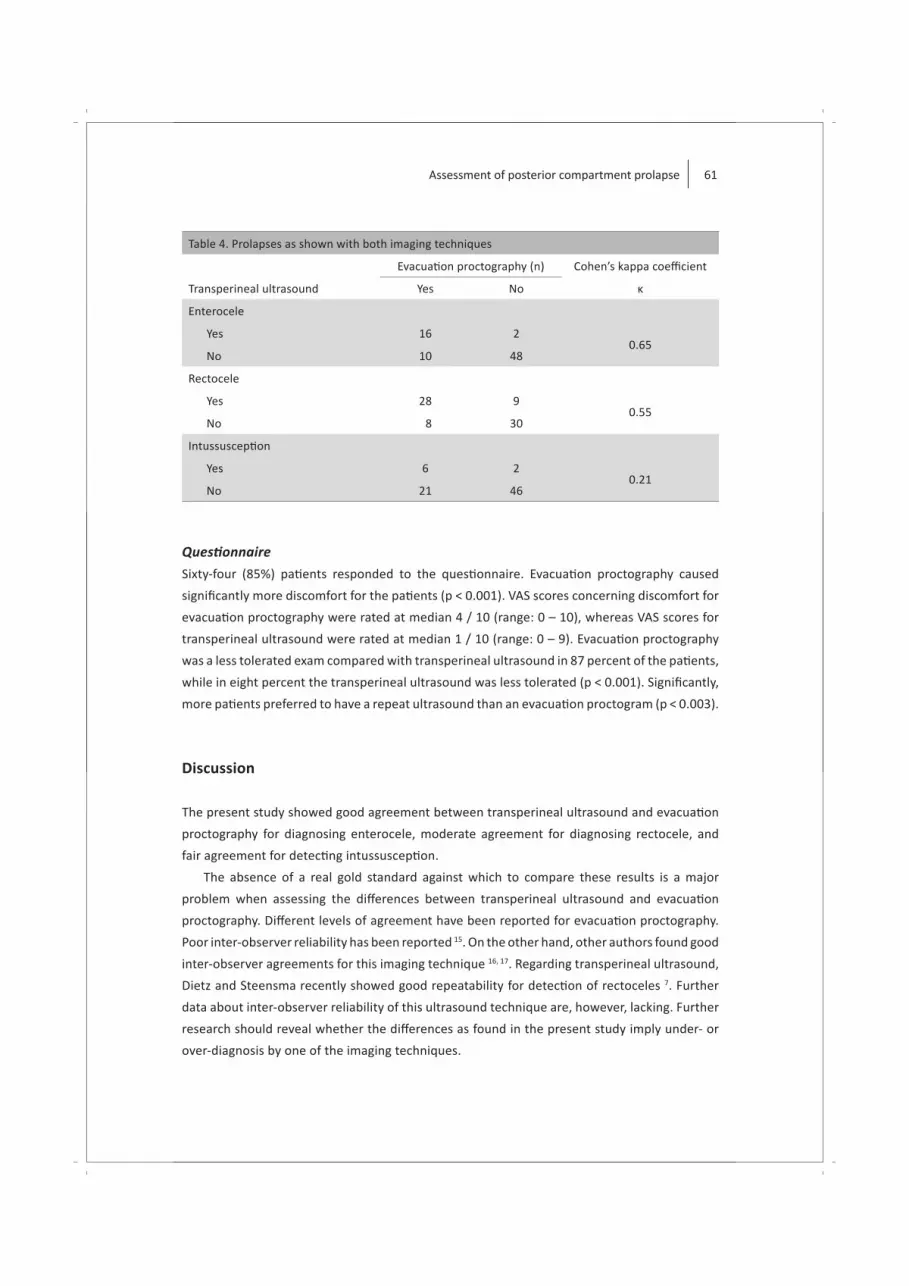
Assessment of posterior compartment prolapse 61
Table 4. Prolapses as shown with both imaging techniques
Evacuati on proctography (n) Cohen’s kappa coeffi cient
Transperineal ultrasound Yes No κ
Enterocele
Yes 16 20.65
No 10 48
Rectocele
Yes 28 90.55
No 8 30
Intussuscepti on
Yes 6 20.21
No 21 46
Questi onnaire
Sixty-four (85%) pati ents responded to the questi onnaire. Evacuati on proctography caused
signifi cantly more discomfort for the pati ents (p < 0.001). VAS scores concerning discomfort for
evacuati on proctography were rated at median 4 / 10 (range: 0 – 10), whereas VAS scores for
transperineal ultrasound were rated at median 1 / 10 (range: 0 – 9). Evacuati on proctography
was a less tolerated exam compared with transperineal ultrasound in 87 percent of the pati ents,
while in eight percent the transperineal ultrasound was less tolerated (p < 0.001). Signifi cantly,
more pati ents preferred to have a repeat ultrasound than an evacuati on proctogram (p < 0.003).
Discussion
The present study showed good agreement between transperineal ultrasound and evacuati on
proctography for diagnosing enterocele, moderate agreement for diagnosing rectocele, and
fair agreement for detecti ng intussuscepti on.
The absence of a real gold standard against which to compare these results is a major
problem when assessing the diff erences between transperineal ultrasound and evacuati on
proctography. Diff erent levels of agreement have been reported for evacuati on proctography.
Poor inter-observer reliability has been reported 15. On the other hand, other authors found good
inter-observer agreements for this imaging technique 16, 17. Regarding transperineal ultrasound,
Dietz and Steensma recently showed good repeatability for detecti on of rectoceles 7. Further
data about inter-observer reliability of this ultrasound technique are, however, lacking. Further
research should reveal whether the diff erences as found in the present study imply under- or
over-diagnosis by one of the imaging techniques.

62
Chap
ter
4
The agreement for diagnosing enterocele is comparable with the results recently reported
by others (Table 5). Regarding detecti on of rectocele and intussuscepti on, varying Cohen’s
kappa coeffi cients (both lower and higher as compared to the present study) have been
reported. These diff erences may be explained by the use of rectal contrast during transperineal
ultrasound. Beer-Gabel et al. 8, 10 performed transperineal ultrasound aft er injecti on of contrast
medium into the rectum and reported higher levels of agreement for the detecti on of both
rectocele and intussuscepti on. Another possible explanati on may be due to selecti on bias.
In three studies, only pati ents with longstanding complaints of obstructed defaecati on were
included 8, 10, 11, whereas in another study only pati ents without prior history of pelvic organ
prolapse were studied 9. The present study is the fi rst to include all pati ents with symptoms of
posterior compartment prolapse.
Table 5. Overview of literature
Author Year n Enterocele (κ) Rectocele (κ) Intussuscepti on (κ)
Beer-Gabel et al. 8 2004 33 0.7 0.88 0.88
Grasso et al. 9 2007 43 – 0.41 0.91
Beer-Gabel et al. 10 2008 62 0.78 0.78 –
Perniola et al. 11 2008 37 – 0.26 0.09
The present study 2009 75 0.65 0.55 0.21
κ = Cohen’s Kappa coeffi cient
Based on the present study, it seems that enteroceles and rectoceles were in a more
advanced stage when detected by transperineal ultrasound than when detected by evacuati on
proctography. These fi ndings are in concordance with the results as recently reported by others 9
and might be explained by the supine positi on adopted during the ultrasound examinati on in
contrast with the sitti ng positi on during evacuati on proctography. It is conceivable that a non-
physiologic supine positi on will lead to under-diagnosis of prolapse. Evacuati on proctography
may seem more physiologic but the pati ent may feel embarrassed to defaecate in public,
resulti ng in reduced cooperati on 18. Furthermore, evacuati on proctography seems unlikely
to refl ect complete physiological defaecati on because of the lack of urge to defaecate and
the use of non-physiologic liquid contrast. It is suggested that evacuati on proctography
can furthermore result in over-diagnosis of posterior compartment prolapse. For example,
rectocele, smaller than 2 cm, is oft en diagnosed with evacuati on proctography in asymptomati c
women 19. The questi on arises therefore whether under-diagnosis of grade 1 rectoceles and
grade 1 enteroceles with ultrasound has clinical consequences. In our opinion, these lesions are
oft en asymptomati c and, therefore, do not require treatment.
The majority of intussuscepti ons found during evacuati on proctography were not
adequately detected with ultrasound. Doubt has risen about the clinical signifi cance and

Assessment of posterior compartment prolapse 63
required treatment for this anatomic abnormality. Rectal intussuscepti on has been reported in
up to 50 percent of healthy volunteers 19. Furthermore, a recent study showed that evacuati on
parameters fall within the normal range in most pati ents and that obstructed defaecati on do
not imply proctographic evidence of occlusion 20.
Retrospecti ve evaluati on of the enteroceles, which were missed with ultrasound
(n = 2), revealed that one pati ent did not perform an adequate Valsalva manoeuvre during
the examinati on and that the other pati ents had a concomitant severe uterine prolapse.
Both pati ents in whom an enterocele was missed with evacuati on proctography showed an
intussuscepti on during evacuati on proctography, which was probably seen as an enterocele
during transperineal ultrasound. An adequately performed Valsalva manoeuvre is essenti al for
the right interpretati on. Inadequate performed Valsalva manoeuvre might be an important
cause of under-diagnoses.
A major advantage of transperineal ultrasound in comparison with evacuati on proctography
is the preventi on of the use of ionising radiati on. Goei and Kemerink reported that evacuati on
proctography results in a mean eff ecti ve radiati on dose of 4.9 mSv 21. To compare, a single chest
X-ray results in an organ dose of 0.01 mSv and an abdominal CT scan results in a dose of 10
mSv. Although a radiati on dose of 4.9 mSv is considerable but not extremely high, preventi on
of exposure to ionising radiati on is preferable, especially in female pati ents of reproducti ve age.
Transperineal ultrasound revealed (concomitant) pathology in the anterior and middle
compartment in 31 percent of the pati ents. Unfortunately, these compartments were not
visualised by evacuati on proctography, as used in the present study. Evacuati on proctography
can be supplemented by opacifi cati on of the bladder to include imaging of the anterior
compartment. However, this procedure is even more invasive, poorly tolerated, and requires
an additi onal radiati on dose.
Another advantage of transperineal ultrasound is the pati ent-friendly character, as shown
in the present study. The majority of pati ents indicated transperineal ultrasound as the last
unpleasant examinati on. No endovaginal or endoanal contrast is used and no evacuati on of
contrast is required for imaging of posterior compartment prolapse.
In recent years, other techniques, such as MR imaging and MR defaecography, have been
used for the detecti on of anatomic abnormaliti es in pati ents with pelvic organ prolapse. MR
imaging provides a good and direct demonstrati on of all three pelvic compartments, and in
additi on, does not involve the use of ionising radiati on 22. Furthermore, MR imaging is capable
of visualising the soft ti ssues and the pelvic fl oor similar to ultrasound, whereas evacuati on
proctography (without opacifi cati on of the bladder) supplies no informati on about the anterior
and middle compartment at all. A drawback of dynamic MR imaging is the lateral or supine
positi on during the examinati on, which is demonstrated not as accuratly as evacuati on
proctography 23. MR defaecography can be performed with the pati ent in the sitti ng positi on,
however, this requires an open technique, which is very expensive and not generally available 24.
64
Chap
ter
4
Furthermore, to date, standardised reference points and normal values are lacking 25. Compared
with MR imaging, transperineal ultrasound off ers a less expensive investi gati on, ensures a more
opti mal Valsalva manoeuvre and is, therefore, easier to control.
Conclusions
The present study showed good agreement for the detecti on of clinically relevant enterocele
and rectocele. A fair agreement was found for detecti on of intussuscepti on, however, the
clinical relevance of this incomplete rectal prolapse is unknown. In additi on, transperineal
ultrasound is signifi cantly bett er tolerated by pati ents. Therefore, transperineal ultrasound is
an alternati ve to evacuati on proctography in clinically relevant posterior compartment prolapse
and may be used as the fi rst diagnosti c tool for symptomati c pati ents.

Assessment of posterior compartment prolapse 65
References
1. Burrows LJ, Meyn LA, Walters MD, Weber AM. Pelvic symptoms in women with pelvic organ prolapse. Obstet Gynecol 2004;104(5 Pt 1):982-8.
2. Weber AM, Walters MD, Ballard LA, Booher DL, Piedmonte MR. Posterior vaginal prolapse and bowel functi on. Am J Obstet Gynecol 1998;179:1446-50.
3. Jelovsek JE, Barber MD, Paraiso MF, Walters MD. Functi onal bowel and anorectal disorders in pati ents with pelvic organ prolapse and inconti nence. Am J Obstet Gynecol 2005;193(6):2105-11.
4. Dietz HP, Korda A. Which bowel symptoms are most strongly associated with a true rectocele? Aust N Z J Obstet Gynaecol 2005;45(6):505-8.
5. Vanbeckevoort D, Van Hoe L, Oyen R, Ponett e E, De Ridder D, Deprest J. Pelvic fl oor descent in females: comparati ve study of colpocystodefecography and dynamic fast MR imaging. J Magn Reson Imaging 1999;9(3):373-7.
6. Dietz HP. Ultrasound imaging of the pelvic fl oor. Part II: three-dimensional or volume imaging. Ultrasound Obstet Gynecol 2004;23(6):615-25.
7. Dietz HP, Steensma AB. Posterior compartment prolapse on two-dimensional and three-dimensional pelvic fl oor ultrasound: the disti ncti on between true rectocele, perineal hypermobility and enterocele. Ultrasound Obstet Gynecol 2005;26(1):73-7.
8. Beer-Gabel M, Teshler M, Schechtman E, Zbar AP. Dynamic transperineal ultrasound vs. defecography in pati ents with evacuatory diffi culty: a pilot study. Int J Colorectal Dis 2004;19(1):60-7.
9. Grasso RF, Piciucchi S, Quatt rocchi CC, Sammarra M, Ripetti V, Zobel BB. Posterior pelvic fl oor disorders: a prospecti ve comparison using introital ultrasound and colpocystodefecography. Ultrasound Obstet Gynecol 2007;30(1):86-94.
10. Beer-Gabel M, Assoulin Y, Amitai M, Bardan E. A comparison of dynamic transperineal ultrasound (DTP-US) with dynamic evacuati on proctography (DEP) in the diagnosis of cul de sac hernia (enterocele) in pati ents with evacuatory dysfuncti on. Int J Colorectal Dis 2008;23(5):513-9.
11. Perniola G, Shek C, Chong CC, Chew S, Cartmill J, Dietz HP. Defecati on proctography and translabial ultrasound in the investi gati on of defecatory disorders. Ultrasound Obstet Gynecol 2008;31(5):567-71.
12. Bump RC, Matti asson A, Bo K, Brubaker LP, DeLancey JO, Klarskov P, et al. The standardizati on of terminology of female pelvic organ prolapse and pelvic fl oor dysfuncti on. Am J Obstet Gynecol 1996;175(1):10-7.
13. Oom DM, van Dijl VR, Gosselink MP, van Wijk JJ, Schouten WR. Enterocele repair by abdominal obliterati on of the pelvic inlet: long-term outcome on obstructed defaecati on and symptoms of pelvic discomfort. Colorectal Dis 2007;9(9):845-50.
14. Mellgren A, Bremmer S, Johansson C, Dolk A, Uden R, Ahlback SO, et al. Defecography. Results of investi gati ons in 2,816 pati ents. Dis Colon Rectum 1994;37(11):1133-41.
15. Muller-Lissner SA, Bartolo DC, Christi ansen J, Ekberg O, Goei R, Hopfner W, et al. Interobserver agreement in defecography--an internati onal study. Z Gastroenterol 1998;36(4):273-9.
16. Dobben AC, Wiersma TG, Janssen LW, de Vos R, Terra MP, Baeten CG, et al. Prospecti ve assessment of interobserver agreement for defecography in fecal inconti nence. AJR Am J Roentgenol 2005;185(5):1166-72.
17. Pfeifer J, Oliveira L, Park UC, Gonzalez A, Agachan F, Wexner SD. Are interpretati ons of video defecographies reliable and reproducible? Int J Colorectal Dis 1997;12(2):67-72.
18. Maglinte DD, Bartram C. Dynamic imaging of posterior compartment pelvic fl oor dysfuncti on by evacuati on proctography: techniques, indicati ons, results and limitati ons. Eur J Radiol 2007;61(3):454-61.

66
Chap
ter
4
19. Shorvon PJ, McHugh S, Diamant NE, Somers S, Stevenson GW. Defecography in normal volunteers: results and implicati ons. Gut 1989;30(12):1737-49.
20. Dvorkin LS, Gladman MA, Epstein J, Scott SM, Williams NS, Lunniss PJ. Rectal intussuscepti on in symptomati c pati ents is diff erent from that in asymptomati c volunteers. Br J Surg 2005;92(7):866-72.
21. Goei R, Kemerink G. Radiati on dose in defecography. Radiology 1990;176(1):137-9.22. Healy JC, Halligan S, Reznek RH, Watson S, Bartram CI, Phillips R, et al. Dynamic MR imaging compared
with evacuati on proctography when evaluati ng anorectal confi gurati on and pelvic fl oor movement. AJR Am J Roentgenol 1997;169(3):775-9.
23. Matsuoka H, Wexner SD, Desai MB, Nakamura T, Nogueras JJ, Weiss EG, et al. A comparison between dynamic pelvic magneti c resonance imaging and videoproctography in pati ents with consti pati on. Dis Colon Rectum 2001;44(4):571-6.
24. Roos JE, Weishaupt D, Wildermuth S, Willmann JK, Marincek B, Hilfi ker PR. Experience of 4 years with open MR defecography: pictorial review of anorectal anatomy and disease. Radiographics 2002;22(4):817-32.
25. Lienemann A, Sprenger D, Janssen U, Grosch E, Pellengahr C, Anthuber C. Assessment of pelvic organ descent by use of functi onal cine-MRI: which reference line should be used? Neurourol Urodyn 2004;23(1):33-7.

Part II
Faecal Inconti nence

Chapter 5 Anterior sphincteroplasty for faecal inconti nence;
a single center experience in the era of sacral neuromodulati on
Dis Colon Rectum 2009; in press.
Daniëlla M.J. Oom
Marti jn P. Gosselink
W. Ruud Schouten
70
Chap
ter
5
Abstract
Introducti onAnterior sphincteroplasty is the surgical treatment of choice for pati ents with faecal inconti nence,
associated with an external anal sphincter defect. Recently, it has been reported that pati ents
with such a defect may also benefi t from sacral neuromodulati on. The questi on is whether
anterior sphincteroplasty sti ll deserves a place in the surgical treatment of faecal inconti nence.
The aim of the present study was to investi gate the outcome of anterior sphincteroplasty in a
large cohort of pati ents.
Pati ents and Methods A consecuti ve series of 172 pati ents underwent anterior overlapping sphincteroplasty. A
standardised questi onnaire concerning current conti nence status, overall sati sfacti on, and
quality of life was used to assess the outcome.
ResultsFollow-up data were obtained from 75 percent of the 160 pati ents who were sti ll alive. Aft er
a median follow-up of 111 months (range: 12 – 207), the outcome was sti ll good to excellent
in 44 pati ents (37%). In 28 pati ents (23%) the result was classifi ed as moderate, since these
pati ents sti ll encountered regular inconti nence for stool. However, they were sati sfi ed with
their outcome, as they experienced a reducti on of inconti nence episodes of 50 percent or
more. The outcome was poor in 40 percent of the pati ents. Predictors of poor outcome were
older age at surgery (≥ 50 years), deep wound infecti on, and isolated external anal sphincter
defects. Pati ents with long-term follow-up (≥ 5 years) had the same outcome as pati ents with
medium-term follow-up (< 5 years).
ConclusionAnterior sphincteroplasty results in an acceptable to excellent long-term outcome in 60 percent
of the pati ents, especially in those under the age of 50 years at surgery.

Anterior sphincteroplasty for faecal inconti nence 71
Introducti on
Faecal inconti nence is the inability to prevent unintended loss of rectal contents. Social and
psychological consequences of inconti nence are devastati ng for both pati ents and their
families 1. Pregnancy and vaginal delivery are the most common causes of faecal inconti nence.
A recent meta-analysis revealed that the incidence of anal sphincter defects aft er childbirth in
primiparous women is 27 percent and that in women without sphincter damage aft er their fi rst
delivery, the risk of sphincter defects aft er subsequent deliveries is 8.5 percent 2. Instrumental
delivery, large birth weight, and prolonged second stage of labour have been shown to increase
the risk of faecal inconti nence aft er vaginal delivery 3, 4. Aft er primary repair of obstetric tears
involving the anal sphincter, persistent defects have been demonstrated in up to 85 percent of
the cases 4. Traumati c injuries to the pelvis and anorectal procedures may also lead to sphincter
damage and faecal inconti nence 5, 6.
To date, anterior sphincteroplasty is considered as the treatment of choice for inconti nent
pati ents with an external anal sphincter defect, in whom supporti ve management fails. This
procedure was fi rst described by Parks and McPartlin in 1971 7. Initi ally, the short-term success
rates of anterior sphincteroplasty were encouraging, with up to 76 percent of the pati ents who
became conti nent for liquid and solid stool 8. Although, the long-term results are not as good as
thought in those early days 9-12, anterior sphincteroplasty sti ll results in a signifi cant reducti on
of inconti nence episodes, which sustains in the long-term in the majority of the pati ents 11, 13.
However, the number of studies, conducted to assess the long-term outcome aft er anterior
sphincteroplasty, is limited. Furthermore, in all but one study relati vely small series of pati ents
have been analysed 11.
In 1995, Matzel and co-workers were the fi rst to introduce sacral neuromodulati on for the
treatment of faecal inconti nence 14. Based on short- and medium-term follow-up, this technique
has evolved to an att racti ve alternati ve for pati ents with faecal inconti nence. Although sacral
neuromodulati on was indicated for pati ents without structural defects of the anal sphincters,
it has recently been reported that sacral neuromodulati on can also be used in pati ents with
an external anal sphincter defect without prior sphincter reconstructi on. Melenhorst and co-
workers 15 showed that sacral neuromodulati on is as eff ecti ve in pati ents with an external anal
sphincter defect without prior repair, as in those pati ents who underwent previous sphincter
reconstructi on. Therefore, the questi on arises whether pati ents with faecal inconti nence
associated with an anterior external anal sphincter defect should be treated primarily by
anterior sphincteroplasty or directly by sacral neuromodulati on.
The results of anterior sphincteroplasty are hardly to compare with the results of sacral
neuromodulati on, since in most studies regarding anterior sphincteroplasty other outcome
classifi cati ons are used than in the studies regarding sacral neuromodulati on. A reducti on
of inconti nence episodes of 50 percent or more is widely accepted as a successful outcome
72
Chap
ter
5
of sacral neuromodulati on. This criterion was also used in the present study to classify the
outcome of anterior sphincteroplasty, in order to facilitate comparison between anterior
sphincteroplasty and sacral neuromodulati on.
Pati ents and Methods
Between 1990 and 2007, a consecuti ve series of 172 pati ents with faecal inconti nence underwent
overlapping anterior sphincteroplasty. All pati ents presented with three or more inconti nence
episodes per week. The procedure was only performed in pati ents who experienced their
involuntary loss of stool as disabling. Almost all pati ents were female (97%). Most pati ents
(90%) underwent preoperati ve endoanal ultrasound and / or endoanal magneti c resonance
(MR) imaging in order to assess the presence and the extensiveness of anal sphincter defects.
An isolated external anal sphincter defect was found in 24 percent of the pati ents, whereas
defects in both sphincters were observed in 76 percent of the pati ents. The median extent
of the external anal sphincter defects was 120 degrees of the anal circumference (range: 60
– 180). A concomitant rectovaginal fi stula was detected in 11 pati ents (6%). This rectovaginal
fi stula was treated during anterior sphincteroplasty by additi onal labial fat fl ap transpositi on
(n = 8), transanal advancement fl ap repair (n = 1) or puborectal sling interpositi on (n = 2).
Seventeen pati ents (10%) were operated before the introducti on of endoanal ultrasound and
endoanal MR imaging in our clinic. Obstetric trauma, prior anal surgery, and pelvic trauma
were the principal cause of the anterior anal sphincter defect in 84, 11, and 3 percent of the
pati ents respecti vely. In two percent of the pati ents the cause of sphincter damage could not
be identi fi ed.
For this study, hospital records and outpati ent clinic charts were analysed, and follow-up
informati on was obtained from both a questi onnaire and review of charts by one author (DMO),
who had not parti cipated in the surgical procedure. Conti nence improvement, degree of faecal
inconti nence, overall sati sfacti on, and quality of life were determined by a writt en standardised
questi onnaire. Pati ents were asked to rate their experienced reducti on of inconti nence episodes
on a visual analogue scale (0 – 100 percent reducti on). Rockwood faecal inconti nence severity
index (RFISI) score 16 was used to assess conti nence status during this assessment. Rockwood
faecal inconti nence quality of life scale (FIQLS) 17 was used to determine disease-specifi c quality
of life and EQ-5D questi onnaire 18 was used to evaluate generic quality of life.
The outcome was classifi ed as excellent, good, moderate, or poor. This classifi cati on
is based on objecti ve conti nence status, as well as on subjecti ve assessment of sati sfacti on.
The outcome was classifi ed as excellent if the pati ent reported full conti nence. If the pati ent
encountered inconti nence for fl atus or sporadic (< 1 per month) loss of liquid stool only, the
outcome was graded as good. The result was classifi ed as moderate if the pati ent sti ll suff ered

Anterior sphincteroplasty for faecal inconti nence 73
from regular inconti nence for liquid and / or solid stool but reported to be sati sfi ed, since
they experienced a reducti on of inconti nence episodes of 50 percent or more. In pati ents who
underwent an additi onal surgical procedure for persistent faecal inconti nence and in pati ents
who encountered regular inconti nence for liquid and / or solid stool, with less than 50 percent
reducti on of inconti nence episodes, and who were not sati sfi ed with their current situati on,
the outcome was defi ned as poor. This study was approved by the hospital’s human research
ethics committ ee.
Surgical Technique
All pati ents underwent complete mechanical bowel preparati on (polyethylene glycol; Klean-
prep, Helsinn Birex Pharmaceuti cals, Dublin, Ireland). Aft er inducti on of general endotracheal
anaesthesia, metronidazole (500 mg) and cefuroxime (1.500 mg) were administered
intravenously. All pati ents were operated in the lithotomy positi on. The repair was performed
through a curvilinear incision, paralleling the outer edge of the superfi cial part of the external
anal sphincter. The posterior vaginal wall was separated from the anterior rectal wall. The
intersphincteric plane was entered bilaterally to separate the external and the internal anal
sphincter. The enti re scar ti ssue in the anterior midline was divided completely. The anterior
rectal wall was plicated and the internal anal sphincter was imbricated. Next, both limbs
of the puborectal sling were approximated in the midline with interrupted sutures. Finally,
an overlapping repair of the external anal sphincter was performed. Monocryl 2-0 sutures
(Ethicon, Johnson & Johnson, Somerville, USA) were used during the whole operati on. The
procedure was performed without covering ileostomy. All procedures were performed by or
supervised by the same colorectal surgeon (WRS).
Stati sti cal Analysis
Stati sti cal analysis was performed with the SPSS soft ware package (15.0 version, SPSS
Inc., Chicago, IL, USA). Pati ent characteristi cs: age during surgery, ti me since anterior
sphincteroplasty, preoperati ve sphincter defects, cause of anal sphincter defects, and
postoperati ve complicati ons, were compared between responders and non-responders and
between pati ents with and without a poor outcome. When appropriate, pati ent groups were
compared with the chi-square test or Fisher’s exact test. Conti nuous variables were compared
with the Mann–Whitney test. A multi variate logisti c regression analysis of the signifi cant
clinical variables in univariate analysis was done with logisti c regression models. Diff erences
were considered stati sti cally signifi cant at a two-tailed p-value of ≤ 0.05.

74
Chap
ter
5
Results
Thirty-nine pati ents (23%) encountered a postoperati ve complicati on. Wound infecti on
occurred in the majority of these pati ents (n = 35), resulti ng in an abscess in 21 pati ents with
subsequent fi stula formati on in 15 of them. All these 21 pati ents required further surgery to
treat their complicati on. Two pati ents developed postoperati ve ileus, one pati ent encountered
deep venous thrombosis and one other pati ent suff ered from lung embolism. These four
pati ents fully recovered.
During the follow-up period, 12 pati ents died from unrelated causes. One hundred and
twenty of the remaining 160 pati ents (75%) returned a completed questi onnaire. The median
ti me interval between the procedure and the current analysis was 111 months (range: 12 – 207).
The median age at follow-up was 58 years (range: 30 – 85). The responders were comparable
with the non-responders regarding age, durati on of follow-up, extensiveness of anal sphincter
defects, cause of anal sphincter defects, and postoperati ve complicati ons (Table 1).
Table 1. Comparison of responders and non-responders
Responders
(n=120)
Non-responders
(n=40)
p-value
Age at follow-up, median (range) 58 (30 – 85) 54 (31 – 87) 0.25
Time interval since surgery in months, median (range) 111 (12 – 207) 127 (12 – 207) 0.42
Preoperati ve sphincter defect (%)
No imaging available 6 7
0.70 Isolated EAS defect 23 20
IAS and EAS defect 71 73
Cause of anal sphincter defect (%)
0.19
Obstetric trauma 85 85
Prior anal surgery 2 8
Major pelvic trauma 2 2
Idiopathic 11 5
Postoperati ve complicati on (%)
Yes 24 20 0.59
EAS: external anal sphincter, IAS: internal anal sphincter
The outcome was excellent in seven pati ents (6%). A good outcome was observed in 37
pati ents (31%). In 28 pati ents (23%), the long-term outcome was classifi ed as moderate. A
poor outcome was observed in 48 pati ents (40%). Twenty-two pati ents with a poor outcome
underwent an additi onal surgical procedure for persistent faecal inconti nence. In 13

Anterior sphincteroplasty for faecal inconti nence 75
pati ents a permanent colostomy was constructed, six pati ents underwent additi onal sacral
neuromodulati on, one pati ent underwent repeat sphincteroplasty, and in two pati ents dynamic
graciloplasty was performed.
Median RFISI score did not diff er between pati ents with a moderate outcome (RFISI = 42,
range: 27 – 56) and pati ents with a poor outcome who did not underwent an additi onal surgical
procedure (RFISI = 47, range: 27 - 61) (Figure 1a). However, the median number of inconti nence
episodes per month was signifi cantly lower in pati ents with a moderate outcome (8 episodes,
range: 1 – 30), compared with pati ents with a poor outcome (45 episodes, range: 3 – 120)
(Figure 1b).
Figure 1. Faecal conti nence
Figure 1a. Median RFISI score Figure 1b. Median number of
inconti nence episodes
Pati ents who underwent further surgery are not presented in both fi gures.
Pati ents with a poor outcome, who did not underwent further surgery, had lower disease
specifi c quality of life scores (FIQLS) than pati ents with a moderate, good, or excellent outcome.
Generic quality of life, which was evaluated by the EQ5D questi onnaire, did not diff er between
pati ents with a poor outcome and those with a moderate, good, and excellent outcome (Table
2).
Predictors of outcome are presented in Tables 3 and 4. The causes of anterior anal sphincter
defects and durati on of follow-up did not diff er between pati ents with and those pati ents
without a poor outcome. Comparing pati ents followed less than fi ve years (n = 29) and those
followed fi ve years or more (n = 91), the outcome was found to be poor in 38 and 41 percent
respecti vely (p = 0.79). Pati ents with a poor outcome were signifi cantly older at surgery.
Pati ents under the age of 50 years at surgery had a poor outcome in 29 percent, whether
pati ents with an age of 50 years or older at surgery had a poor outcome in 53 percent (Figure 2).
Pati ents who encountered a deep wound infecti on were likely to have a less favorable outcome.
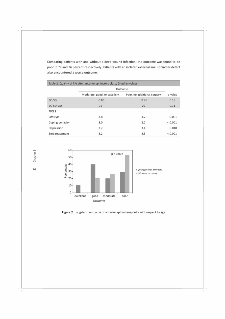
76
Chap
ter
5
Comparing pati ents with and without a deep wound infecti on; the outcome was found to be
poor in 79 and 36 percent respecti vely. Pati ents with an isolated external anal sphincter defect
also encountered a worse outcome.
Table 2. Quality of life aft er anterior sphincteroplasty (median values)
Outcome
Moderate, good, or excellent Poor, no additi onal surgery p-value
EQ 5D 0.80 0.76 0.18
EQ 5D VAS 75 70 0.11
FIQLS
Lifestyle 3.8 3.2 0.001
Coping behavior 3.0 2.0 < 0.001
Depression 3.7 3.4 0.010
Embarrassment 3.2 2.3 < 0.001
Figure 2. Long-term outcome of anterior sphincteroplasty with respect to age

Anterior sphincteroplasty for faecal inconti nence 77
Table 3. Factors predicti ve of long-term outcome, univariate analysis
Outcome
Moderate, good, or excellent
(n = 72)
Poor
(n = 48)
p-value
Age at surgery, median (range) 42 (21 – 80) 54 (28 – 72) 0.003
Time interval since surgery in months,
median (range)107 (14 – 207) 113 (12 – 202) 0.97
Preoperati ve sphincter defects (%)
Isolated EAS defect 15 390.003
IAS and EAS defect 85 61
Cause of anal sphincter defects (%)
Obstetric trauma 83 88
0.43 Prior anal surgery 3 0
Major pelvic trauma 3 0
Idiopathic 11 12
Deep wound infecti on (%)
Yes 4 23 0.002
EAS: external anal sphincter, IAS: internal anal sphincter
Table 4. Factors predicti ve of long-term outcome, multi variate logisti c regression analysis
OR 95% CI p-value
Age at surgery 0.96 0.93-0.99 0.008
Isolated EAS defect 2.94 1.11-7.76 0.03
Deep wound infecti on 7.93 1.86-33.79 0.005
OR: odds rati o, CI: confi dence interval, EAS: external anal sphincter
Discussion
To date, anterior sphincteroplasty is considered as the surgical treatment of choice for
faecal inconti nent pati ents with an external anal sphincter defect, in whom supporti ve
management, such as dietary measures and biofeedback therapy, fails. Complete conti nence
for liquid and solid stool has been reported in up to 76 percent of the pati ents aft er short-term
follow-up 19, 20. However, according to some authors these promising results do not sustain in
the long-term 9-12. The number of studies, conducted to assess the long-term outcome aft er
anterior sphincteroplasty, is limited. Furthermore, in all but one study relati vely small series of
pati ents have been analysed.

78
Chap
ter
5
Bravo Gutti erez et al. 11 assessed the outcome of anterior sphincteroplasty in 191 pati ents.
Aft er a median follow-up of 10 years, bowel control was considered to be bett er than before the
procedure by 62 percent of the pati ents, although some of them sti ll experienced involuntary
loss of solid stool. Despite the use of a diff erent outcome classifi cati on, the present study is
comparable with the study conducted by Bravo Gutti erez et al. with respect to the number
of studied pati ents, the durati on of follow-up, and the long-term results. Our study revealed
that the long-term outcome was good to excellent in 37 percent of the pati ents. Twenty-three
percent of the pati ents were sati sfi ed with their current status, despite persistent inconti nence,
since they experienced a reducti on of inconti nence episodes of 50 percent or more. Based on
these fi ndings, the overall long-term outcome was considered acceptable to excellent in 60
percent of the pati ents.
Comparing pati ents followed less than fi ve years and those followed fi ve years or more, the
outcome was found to be similar, indicati ng that a signifi cant deteriorati on of conti nence with
ti me is unlikely. However, the results obtained from the present study are based on one single
assessment, performed more than fi ve years aft er the procedure in the majority of pati ents.
In contrast with this fi nding, worsening of conti nence in the long-term has been reported
by several authors 9-12. Based on a three- and ten-year assessment, Bravo Gutti erez et al. 11
concluded that conti nence deteriorates with ti me. In the other studies regarding this subject,
the long-term outcome was assessed in a relati vely small number of pati ents 9. Furthermore, in
one of these studies the short-term assessment was only based on a review of outpati ent clinic
charts 12. It is questi onable whether the informati on obtained from such a review is reliable.
In another study, no short-term assessment was performed 10. In our opinion, the evidence for
worsening of conti nence with ti me is not convincing.
Comparing pati ents under the age of 50 years with those of 50 years or more, an acceptable
long-term outcome was observed in 71 and 47 percent respecti vely. A similar fi nding has been
reported by others 11, 21. Since the external anal sphincter defects in older women are most
likely caused by vaginal delivery many years before, it might be possible that other factors play
a role in the pathophysiological mechanism of faecal inconti nence in these female pati ents.
Therefore, the questi on arises whether repair of the damaged external anal sphincter is justi fi ed
as the surgical treatment of choice in these older inconti nent pati ents.
All pati ents underwent complete mechanical bowel preparati on and anti bioti cs were
administered intravenously. Despite these measures we encountered a high rate of wound
infecti on. In the present study, wound infecti on was related to a worse outcome. Although a
similar fi nding has been reported by Londono-Schimmer et al. 22, other authors were not able
to demonstrate such a relati on 23.
It has been reported that the outcome of anterior sphincteroplasty in pati ents with an
isolated external anal sphincter defect and in those with a combined defect is comparable 24.
In contrast with this observati on, the present study revealed a worse outcome in pati ents with
an isolated external anal sphincter defect. However, an explanati on for this fi nding is lacking.
Anterior sphincteroplasty for faecal inconti nence 79
The present study is limited by its retrospecti ve nature, whereas no specifi c follow-up
protocol was used. Pati ents were asked to rate the reducti on of inconti nence episodes in
retrospecti on. Therefore the results as obtained from the present study should be handled
with care. Another limitati on of this study is the relati vely low parti cipati on rate. However,
considering the median durati on of follow-up of more than nine years, the parti cipati on rate of
75 percent is acceptable. A reducti on of inconti nence episodes of 50 percent or more is widely
accepted as a successful outcome of sacral neuromodulati on. This criterion was also used in
the present study to classify the outcome of anterior sphincteroplasty, in order to facilitate
comparison between anterior sphincteroplasty and sacral neuromodulati on. The observati on
that pati ents with a moderate outcome encountered signifi cantly less inconti nence episodes
than pati ents with a poor outcome indicates that our classifi cati on can be justi fi ed.
Originally, sacral neuromodulati on was advocated for the treatment of inconti nent pati ents
without an external anal sphincter defect. Complete conti nence for liquid and solid stool
can be obtained aft er sacral neuromodulati on in up to 75 percent 25. However, according to
other reports the percentage of pati ents with complete conti nence varies between 21 to 57
percent 26-29. A reducti on of the number of inconti nence episodes of 50 percent or more has
been reported in 68 to 96 percent of the pati ents 28-31. However, these data are based on studies
with a short- or medium-term follow-up, since studies regarding the long-term outcome of
sacral neuromodulati on are scarce. Recently, two reports did suggest that inconti nent pati ents
with an external anal sphincter defect could be eff ecti vely treated with sacral neuromodula-
ti on 15, 32. In additi on, it has also been suggested that sacral neuromodulati on can be used
primarily in pati ents with an external anal sphincter defect 15. Therefore, the questi on arises
whether inconti nent pati ents with an external anal sphincter defect should be treated primarily
by anterior sphincteroplasty or directly by sacral neuromodulati on. However, the results of
anterior sphincteroplasty are hardly to compare with the results of sacral neuromodulati on,
since in most studies regarding anterior sphincteroplasty other outcome classifi cati ons are used
than in the studies regarding sacral neuromodulati on. Despite its limitati ons, the present study
facilitates a bett er comparison between the outcome of anterior sphincteroplasty and sacral
neuromodulati on, since a comparable outcome classifi cati on was used. However, to assess the
defi nite role of anterior sphincteroplasty in this era of sacral neuromodulati on, randomised
controlled trials are warranted, comparing both procedures in pati ents with an external anal
sphincter defect. These studies are also necessary to compare the cost-eff ecti veness of both
procedures.
Conclusions
This study shows that anterior sphincteroplasty can stand the challenge of ti me. Acceptable to
excellent results are maintained in 60 percent of the pati ents, especially in those who are under
the age of 50 years at surgery.

80
Chap
ter
5
References
1. Deutekom M, Terra MP, Dobben AC, Dijkgraaf MG, Baeten CG, Stoker J, et al. Impact of faecal inconti nence severity on health domains. Colorectal Dis 2005;7(3):263-9.
2. Oberwalder M, Connor J, Wexner SD. Meta-analysis to determine the incidence of obstetric anal sphincter damage. Br J Surg 2003;90(11):1333-7.
3. Sultan AH, Kamm MA, Hudson CN, Thomas JM, Bartram CI. Anal-sphincter disrupti on during vaginal delivery. N Engl J Med 1993;329(26):1905-11.
4. Sultan AH, Kamm MA, Hudson CN, Bartram CI. Third degree obstetric anal sphincter tears: risk factors and outcome of primary repair. Bmj 1994;308(6933):887-91.
5. Engel AF, Kamm MA, Hawley PR. Civilian and war injuries of the perineum and anal sphincters. Br J Surg 1994;81(7):1069-73.
6. Lindsey I, Jones OM, Smilgin-Humphreys MM, Cunningham C, Mortensen NJ. Patt erns of fecal inconti nence aft er anal surgery. Dis Colon Rectum 2004;47(10):1643-9.
7. Parks AG, McPartlin JF. Late repair of injuries of the anal sphincter. Proc R Soc Med 1971;64(12):1187-9.
8. Dudding TC, Vaizey CJ, Kamm MA. Obstetric anal sphincter injury: incidence, risk factors, and management. Ann Surg 2008;247(2):224-37.
9. Malouf AJ, Norton CS, Engel AF, Nicholls RJ, Kamm MA. Long-term results of overlapping anterior anal-sphincter repair for obstetric trauma. Lancet 2000;355(9200):260-5.
10. Halverson AL, Hull TL. Long-term outcome of overlapping anal sphincter repair. Dis Colon Rectum 2002;45(3):345-8.
11. Bravo Guti errez A, Madoff RD, Lowry AC, Parker SC, Buie WD, Baxter NN. Long-term results of anterior sphincteroplasty. Dis Colon Rectum 2004;47(5):727-32.
12. Zorcolo L, Covott a L, Bartolo DC. Outcome of anterior sphincter repair for obstetric injury: comparison of early and late results. Dis Colon Rectum 2005;48(3):524-31.
13. Maslekar S, Gardiner AB, Duthie GS. Anterior anal sphincter repair for fecal inconti nence: Good longterm results are possible. J Am Coll Surg 2007;204(1):40-6.
14. Matzel KE, Stadelmaier U, Hohenfellner M, Gall FP. Electrical sti mulati on of sacral spinal nerves for treatment of faecal inconti nence. Lancet 1995;346(8983):1124-7.
15. Melenhorst J, Koch SM, Uludag O, van Gemert WG, Baeten CG. Is a morphologically intact anal sphincter necessary for success with sacral nerve modulati on in pati ents with faecal inconti nence? Colorectal Dis 2008;10(3):257-62.
16. Rockwood TH, Church JM, Fleshman JW, Kane RL, Mavrantonis C, Thorson AG, et al. Pati ent and surgeon ranking of the severity of symptoms associated with fecal inconti nence: the fecal inconti nence severity index. Dis Colon Rectum 1999;42(12):1525-32.
17. Rockwood TH, Church JM, Fleshman JW, Kane RL, Mavrantonis C, Thorson AG, et al. Fecal Inconti nence Quality of Life Scale: quality of life instrument for pati ents with fecal inconti nence. Dis Colon Rectum 2000;43(1):9-17.
18. Kind P, Dolan P, Gudex C, Williams A. Variati ons in populati on health status: results from a United Kingdom nati onal questi onnaire survey. Bmj 1998;316(7133):736-41.
19. Rothbarth J, Bemelman WA, Meijerink WJ, Buyze-Westerweel ME, van Dijk JG, Delemarre JB. Long-term results of anterior anal sphincter repair for fecal inconti nence due to obstetric injury / with invited commentaries. Dig Surg 2000;17(4):390-3; discussion 394.
20. Engel AF, Kamm MA, Sultan AH, Bartram CI, Nicholls RJ. Anterior anal sphincter repair in pati ents with obstetric trauma. Br J Surg 1994;81(8):1231-4.
21. Nikiteas N, Korsgen S, Kumar D, Keighley MR. Audit of sphincter repair. Factors associated with poor outcome. Dis Colon Rectum 1996;39(10):1164-70.
Anterior sphincteroplasty for faecal inconti nence 81
22. Londono-Schimmer EE, Garcia-Duperly R, Nicholls RJ, Ritchie JK, Hawley PR, Thomson JP. Overlapping anal sphincter repair for faecal inconti nence due to sphincter trauma: fi ve year follow-up functi onal results. Int J Colorectal Dis 1994;9(2):110-3.
23. Rasmussen OO, Puggaard L, Christi ansen J. Anal sphincter repair in pati ents with obstetric trauma: age aff ects outcome. Dis Colon Rectum 1999;42(2):193-5.
24. Oberwalder M, Dinnewitzer A, Baig MK, Nogueras JJ, Weiss EG, Efron J, et al. Do internal anal sphincter defects decrease the success rate of anal sphincter repair? Tech Coloproctol 2006;10(2):94-7.
25. Matzel KE, Bitt orf B, Stadelmaier U, Hohenberger W. [Sacral nerve sti mulati on in the treatment of faecal inconti nence]. Chirurg 2003;74(1):26-32.
26. Dudding TC, Pares D, Vaizey CJ, Kamm MA. Predicti ve factors for successful sacral nerve sti mulati on in the treatment of faecal inconti nence: a 10-year cohort analysis. Colorectal Dis 2008;10(3):249-56.
27. Matzel KE, Kamm MA, Stosser M, Baeten CG, Christi ansen J, Madoff R, et al. Sacral spinal nerve sti mulati on for faecal inconti nence: multi centre study. Lancet 2004;363(9417):1270-6.
28. Gourcerol G, Gallas S, Michot F, Denis P, Leroi AM. Sacral nerve sti mulati on in fecal inconti nence: are there factors associated with success? Dis Colon Rectum 2007;50(1):3-12.
29. Tjandra JJ, Chan MK, Yeh CH, Murray-Green C. Sacral nerve sti mulati on is more eff ecti ve than opti mal medical therapy for severe fecal inconti nence: a randomized, controlled study. Dis Colon Rectum 2008;51(5):494-502.
30. Melenhorst J, Koch SM, Uludag O, van Gemert WG, Baeten CG. Sacral neuromodulati on in pati ents with faecal inconti nence: results of the fi rst 100 permanent implantati ons. Colorectal Dis 2007;9(8):725-30.
31. Jarrett ME, Varma JS, Duthie GS, Nicholls RJ, Kamm MA. Sacral nerve sti mulati on for faecal inconti nence in the UK. Br J Surg 2004;91(6):755-61.
32. Chan MK, Tjandra JJ. Sacral nerve sti mulati on for fecal inconti nence: external anal sphincter defect vs. intact anal sphincter. Dis Colon Rectum 2008;51(7):1015-25.


Chapter 6 Anterior sphincteroplasty for faecal inconti nence;
is the outcome compromised in pati ents with associated pelvic
fl oor injury?
Dis Colon Rectum 2009; in press.
Daniëlla M.J. Oom
Anneke B. Steensma
David D.E. Zimmerman
W. Ruud Schouten

84
Chap
ter
6
Abstract
Introducti onIt has been shown that vaginal delivery may result not only in sphincter defects but also in pelvic
fl oor injury. However, the infl uence of this type of injury on the aeti ology of faecal inconti nence
and its treatment is unknown. Aim of the present study was to assess the prevalence of
pelvic fl oor injury in pati ents who underwent anterior sphincteroplasty for the treatment of
faecal inconti nence and to determine the impact of this type of injury on the outcome of this
procedure.
Pati ents and Methods Women, who underwent anterior sphincteroplasty in the past, were invited to parti cipate in
the present study. With transperineal ultrasound, which has been developed recently, pelvic
fl oor integrity was examined in 70 of the 117 pati ents (60%). Follow-up was obtained from a
standardised questi onnaire.
ResultsThe median ti me period between anterior sphincteroplasty and the current assessment was
106 months (range: 15 – 211). Pelvic fl oor injury was diagnosed in 43 pati ents (61%). Despite
the prior sphincteroplasty, an external anal sphincter defect was found in 20 pati ents (29%).
Outcome did not diff er, neither between pati ents with and those without pelvic fl oor injury,
nor between pati ents with and those without an adequate repair. However, pati ents with an
adequate repair and an intact pelvic fl oor did have a bett er outcome than pati ents with one or
both abnormaliti es.
ConclusionThe majority of pati ents who were eligible for anterior sphincteroplasty have concomitant
pelvic fl oor injury. Based on the present study, it seems unlikely that this type of injury itself
has an impact on the outcome of anterior sphincteroplasty.
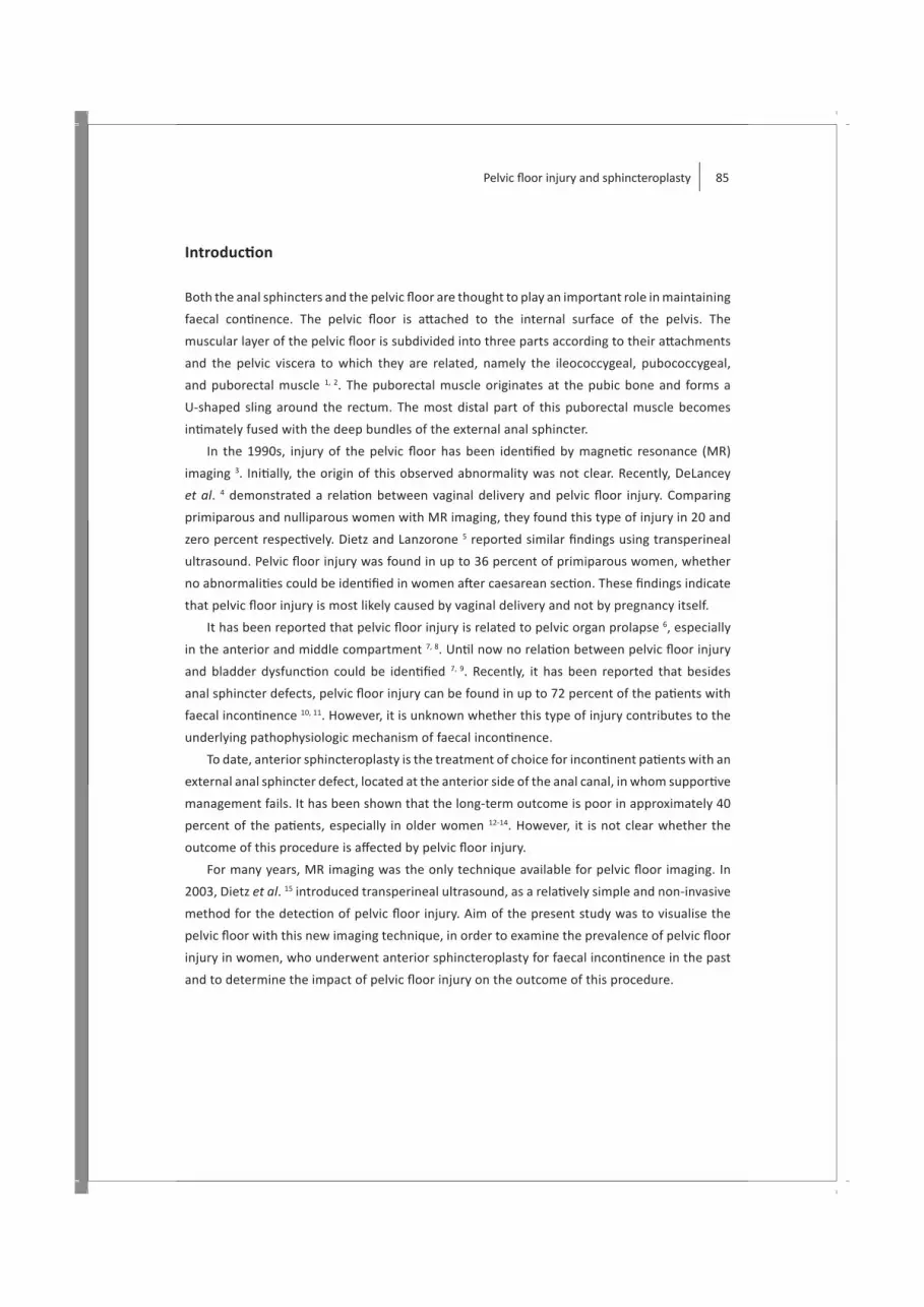
Pelvic fl oor injury and sphincteroplasty 85
Introducti on
Both the anal sphincters and the pelvic fl oor are thought to play an important role in maintaining
faecal conti nence. The pelvic fl oor is att ached to the internal surface of the pelvis. The
muscular layer of the pelvic fl oor is subdivided into three parts according to their att achments
and the pelvic viscera to which they are related, namely the ileococcygeal, pubococcygeal,
and puborectal muscle 1, 2. The puborectal muscle originates at the pubic bone and forms a
U-shaped sling around the rectum. The most distal part of this puborectal muscle becomes
inti mately fused with the deep bundles of the external anal sphincter.
In the 1990s, injury of the pelvic fl oor has been identi fi ed by magneti c resonance (MR)
imaging 3. Initi ally, the origin of this observed abnormality was not clear. Recently, DeLancey
et al. 4 demonstrated a relati on between vaginal delivery and pelvic fl oor injury. Comparing
primiparous and nulliparous women with MR imaging, they found this type of injury in 20 and
zero percent respecti vely. Dietz and Lanzorone 5 reported similar fi ndings using transperineal
ultrasound. Pelvic fl oor injury was found in up to 36 percent of primiparous women, whether
no abnormaliti es could be identi fi ed in women aft er caesarean secti on. These fi ndings indicate
that pelvic fl oor injury is most likely caused by vaginal delivery and not by pregnancy itself.
It has been reported that pelvic fl oor injury is related to pelvic organ prolapse 6, especially
in the anterior and middle compartment 7, 8. Unti l now no relati on between pelvic fl oor injury
and bladder dysfuncti on could be identi fi ed 7, 9. Recently, it has been reported that besides
anal sphincter defects, pelvic fl oor injury can be found in up to 72 percent of the pati ents with
faecal inconti nence 10, 11. However, it is unknown whether this type of injury contributes to the
underlying pathophysiologic mechanism of faecal inconti nence.
To date, anterior sphincteroplasty is the treatment of choice for inconti nent pati ents with an
external anal sphincter defect, located at the anterior side of the anal canal, in whom supporti ve
management fails. It has been shown that the long-term outcome is poor in approximately 40
percent of the pati ents, especially in older women 12-14. However, it is not clear whether the
outcome of this procedure is aff ected by pelvic fl oor injury.
For many years, MR imaging was the only technique available for pelvic fl oor imaging. In
2003, Dietz et al. 15 introduced transperineal ultrasound, as a relati vely simple and non-invasive
method for the detecti on of pelvic fl oor injury. Aim of the present study was to visualise the
pelvic fl oor with this new imaging technique, in order to examine the prevalence of pelvic fl oor
injury in women, who underwent anterior sphincteroplasty for faecal inconti nence in the past
and to determine the impact of pelvic fl oor injury on the outcome of this procedure.
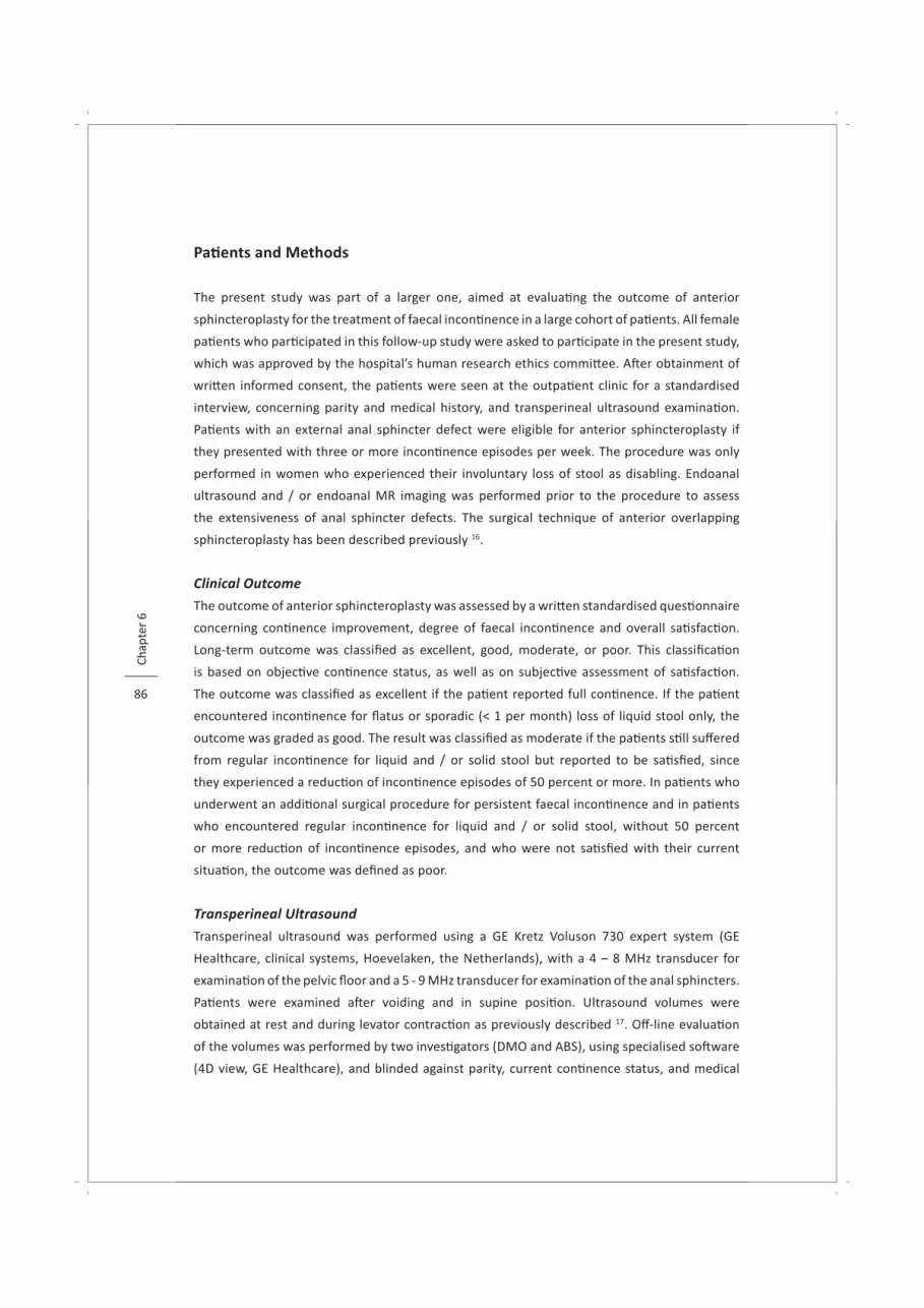
86
Chap
ter
6
Pati ents and Methods
The present study was part of a larger one, aimed at evaluati ng the outcome of anterior
sphincteroplasty for the treatment of faecal inconti nence in a large cohort of pati ents. All female
pati ents who parti cipated in this follow-up study were asked to parti cipate in the present study,
which was approved by the hospital’s human research ethics committ ee. Aft er obtainment of
writt en informed consent, the pati ents were seen at the outpati ent clinic for a standardised
interview, concerning parity and medical history, and transperineal ultrasound examinati on.
Pati ents with an external anal sphincter defect were eligible for anterior sphincteroplasty if
they presented with three or more inconti nence episodes per week. The procedure was only
performed in women who experienced their involuntary loss of stool as disabling. Endoanal
ultrasound and / or endoanal MR imaging was performed prior to the procedure to assess
the extensiveness of anal sphincter defects. The surgical technique of anterior overlapping
sphincteroplasty has been described previously 16.
Clinical Outcome
The outcome of anterior sphincteroplasty was assessed by a writt en standardised questi onnaire
concerning conti nence improvement, degree of faecal inconti nence and overall sati sfacti on.
Long-term outcome was classifi ed as excellent, good, moderate, or poor. This classifi cati on
is based on objecti ve conti nence status, as well as on subjecti ve assessment of sati sfacti on.
The outcome was classifi ed as excellent if the pati ent reported full conti nence. If the pati ent
encountered inconti nence for fl atus or sporadic (< 1 per month) loss of liquid stool only, the
outcome was graded as good. The result was classifi ed as moderate if the pati ents sti ll suff ered
from regular inconti nence for liquid and / or solid stool but reported to be sati sfi ed, since
they experienced a reducti on of inconti nence episodes of 50 percent or more. In pati ents who
underwent an additi onal surgical procedure for persistent faecal inconti nence and in pati ents
who encountered regular inconti nence for liquid and / or solid stool, without 50 percent
or more reducti on of inconti nence episodes, and who were not sati sfi ed with their current
situati on, the outcome was defi ned as poor.
Transperineal Ultrasound
Transperineal ultrasound was performed using a GE Kretz Voluson 730 expert system (GE
Healthcare, clinical systems, Hoevelaken, the Netherlands), with a 4 – 8 MHz transducer for
examinati on of the pelvic fl oor and a 5 - 9 MHz transducer for examinati on of the anal sphincters.
Pati ents were examined aft er voiding and in supine positi on. Ultrasound volumes were
obtained at rest and during levator contracti on as previously described 17. Off -line evaluati on
of the volumes was performed by two investi gators (DMO and ABS), using specialised soft ware
(4D view, GE Healthcare), and blinded against parity, current conti nence status, and medical
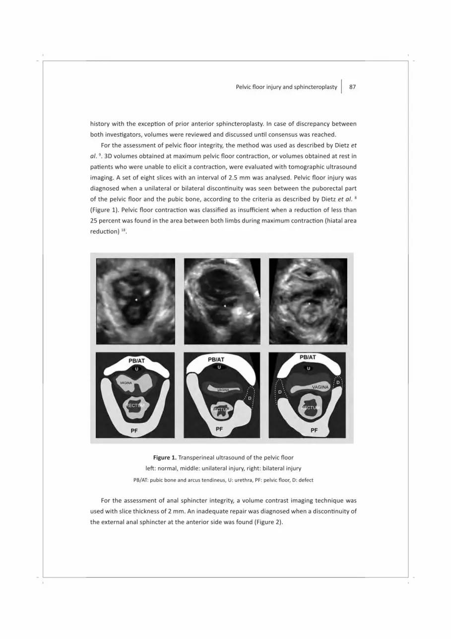
Pelvic fl oor injury and sphincteroplasty 87
history with the excepti on of prior anterior sphincteroplasty. In case of discrepancy between
both investi gators, volumes were reviewed and discussed unti l consensus was reached.
For the assessment of pelvic fl oor integrity, the method was used as described by Dietz et
al. 9. 3D volumes obtained at maximum pelvic fl oor contracti on, or volumes obtained at rest in
pati ents who were unable to elicit a contracti on, were evaluated with tomographic ultrasound
imaging. A set of eight slices with an interval of 2.5 mm was analysed. Pelvic fl oor injury was
diagnosed when a unilateral or bilateral disconti nuity was seen between the puborectal part
of the pelvic fl oor and the pubic bone, according to the criteria as described by Dietz et al. 8
(Figure 1). Pelvic fl oor contracti on was classifi ed as insuffi cient when a reducti on of less than
25 percent was found in the area between both limbs during maximum contracti on (hiatal area
reducti on) 18.
Figure 1. Transperineal ultrasound of the pelvic fl oor
left : normal, middle: unilateral injury, right: bilateral injury
PB/AT: pubic bone and arcus tendineus, U: urethra, PF: pelvic fl oor, D: defect
For the assessment of anal sphincter integrity, a volume contrast imaging technique was
used with slice thickness of 2 mm. An inadequate repair was diagnosed when a disconti nuity of
the external anal sphincter at the anterior side was found (Figure 2).
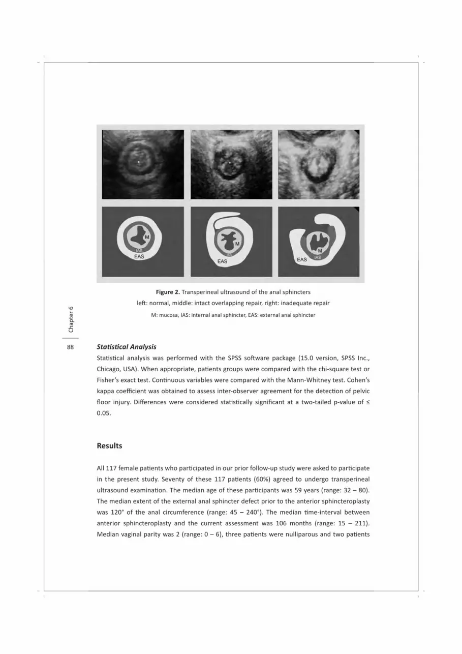
88
Chap
ter
6
Figure 2. Transperineal ultrasound of the anal sphincters
left : normal, middle: intact overlapping repair, right: inadequate repair
M: mucosa, IAS: internal anal sphincter, EAS: external anal sphincter
Stati sti cal Analysis
Stati sti cal analysis was performed with the SPSS soft ware package (15.0 version, SPSS Inc.,
Chicago, USA). When appropriate, pati ents groups were compared with the chi-square test or
Fisher’s exact test. Conti nuous variables were compared with the Mann-Whitney test. Cohen’s
kappa coeffi cient was obtained to assess inter-observer agreement for the detecti on of pelvic
fl oor injury. Diff erences were considered stati sti cally signifi cant at a two-tailed p-value of ≤
0.05.
Results
All 117 female pati ents who parti cipated in our prior follow-up study were asked to parti cipate
in the present study. Seventy of these 117 pati ents (60%) agreed to undergo transperineal
ultrasound examinati on. The median age of these parti cipants was 59 years (range: 32 – 80).
The median extent of the external anal sphincter defect prior to the anterior sphincteroplasty
was 120° of the anal circumference (range: 45 – 240°). The median ti me-interval between
anterior sphincteroplasty and the current assessment was 106 months (range: 15 – 211).
Median vaginal parity was 2 (range: 0 – 6), three pati ents were nulliparous and two pati ents
Pelvic fl oor injury and sphincteroplasty 89
had only caesarean secti on delivery. Thirteen pati ents had a history of forceps delivery and in
fi ve pati ents a vontuse was used during labour. None of the parti cipants had a vaginal delivery
since anterior sphincteroplasty. The parti cipants were comparable with the non-parti cipants
regarding age, ti me period since surgery, and clinical outcome (Table 1).
Table 1. Comparison of parti cipants and non-parti cipants
Parti cipants
(n=70)
Non-parti cipants
(n=47)
p-value
Median age (range) 59 (32 – 80) 58 (31 – 86) 0.72
Median ti me period since surgery in months (range) 106 (15 – 211) 119 (17 – 205) 0.96
Clinical outcome (%)
Excellent 6 4
0.54 Good 27 36
Moderate 27 17
Poor 40 43
The long-term outcome was excellent in four pati ents (6%). A good outcome was observed
in 19 pati ents (27%). The outcome was classifi ed as moderate in 19 pati ents (27%). A poor
outcome was observed in 28 pati ents (40%). Fourteen pati ents with a poor outcome underwent
an additi onal surgical procedure for persistent faecal inconti nence. In six pati ents a permanent
colostomy was constructed, six pati ents underwent additi onal sacral neuromodulati on, and in
two pati ents dynamic graciloplasty was performed.
Pelvic fl oor injury was diagnosed in 43 pati ents (61%). This injury involved one or both
sides of the pelvic fl oor in 15 and 28 pati ents respecti vely. Fift een pati ents with pelvic fl oor
injury (35%) had a history of instrumental delivery. No pelvic fl oor injury was detected in the
fi ve pati ents without a history of vaginal delivery. Pelvic fl oor contracti on was insuffi cient in
35 pati ents with pelvic fl oor injury (81%) and in eight pati ents with an intact pelvic fl oor (30%)
(p < 0.0001). An inadequate sphincter repair was diagnosed in 20 pati ents (29%), 12 of these
pati ents had concomitant pelvic fl oor injury. The inter-observer agreement for the detecti on of
pelvic fl oor injury was found to be good (Cohen’s kappa coeffi cient: 0.76).
In pati ents with an intact pelvic fl oor and in those with pelvic fl oor injury, the outcome
was poor in 33 and 44 percent respecti vely (p = 0.37). In pati ents with an adequate repair and
in those with an inadequate repair, the outcome was poor in 34 and 55 percent respecti vely
(p = 0.11). However, pati ents with an adequate repair and intact pelvic fl oor did have a bett er
outcome than pati ents with one or both abnormaliti es (p = 0.048) (Figure 3).

90
Chap
ter
6
Figure 3. Long-term outcome of anterior sphincteroplasty in pati ents with an adequate repair and
intact pelvic fl oor and in those with one or both abnormaliti es
Discussion
The present study showed that pelvic fl oor injury is present in the majority of faecal inconti nent
pati ents who were eligible for anterior sphincteroplasty. A clear relati on between pelvic fl oor
injury and a worse outcome of this procedure was not found. However, pati ents with an intact
external anal sphincter and an intact pelvic fl oor did have a bett er outcome than pati ents with
one or both abnormaliti es.
Recently, it has been reported that the observed pelvic fl oor injury most frequently involves
the anterior side of the puborectal and probably also the pubococcygeal muscle near their
inserti on to the pubic bone 5, 19. It has been suggested that this type of injury represents
avulsion of the puborectal-pubococcygeal muscles from the pubic bone 5, 9. Unfortunately, to
date, anatomic studies supporti ng this premise are lacking. Therefore, it cannot be precluded
that other forms of trauma, such as excessive stretching of the pelvic fl oor and nerve stretching
or compression, resulti ng in denervati on and muscle atrophy, could also play a role in the
mechanism, which causes these abnormaliti es.
It has been reported that pelvic fl oor injury is related to pelvic organ prolapse 6, especially
to prolapse in the anterior and middle compartment 7, 8. Comparing pati ents with and without
pelvic organ prolapse, DeLancey et al. 6 found pelvic fl oor injury in respecti vely 55 and 16
percent. Unti l now no relati on between pelvic fl oor injury and bladder dysfuncti on could be
identi fi ed 7, 9. Recently, Terra et al. 10 performed endo-anal MR imaging in 105 female pati ents
with faecal inconti nence. They showed that anal sphincter defects and pelvic fl oor injury
were present in 79 and 35 percent of the pati ents respecti vely. Pelvic fl oor abnormaliti es
were found in 39 percent of the pati ents with anal sphincter defects and in 23 percent of the
pati ents without anal sphincter defects. This latt er fi nding indicates that pelvic fl oor injury can

Pelvic fl oor injury and sphincteroplasty 91
occur without concomitant damage of the anal sphincters. However, unti l now it is unknown
whether pelvic fl oor injury contributes to the underlying pathophysiologic mechanism of faecal
inconti nence.
The present study, using transperineal ultrasound in female pati ents, performed many
years aft er anterior sphincteroplasty, revealed pelvic fl oor injury in 61 percent of the cases.
This imaging technique has been shown to be a reliable tool for the detecti on of both
pelvic fl oor injury and anal sphincter defects, with a good repeatability and inter-observer
agreement 7, 11, 20. The present study also revealed a good inter-observer agreement with a
Cohen’s kappa coeffi cient of 0.76.
Pelvic fl oor injury is most likely caused by vaginal delivery and not by pregnancy itself 4, 5.
Since none of our pati ents had a vaginal delivery aft er the anterior sphincteroplasty, it seems
obvious that the observed pelvic fl oor injury was pre-existent at the moment of the operati on.
The questi on arises whether the high prevalence of pelvic fl oor injury in our pati ents refl ects a
major contributi ng role of this type of injury in the aeti ology of faecal inconti nence.
With the same imaging technique, Weinstein et al. 11 also observed a high prevalence of
pelvic fl oor injury (72%) in faecal inconti nent women. According to these authors, pati ents
with faecal inconti nence had a higher prevalence of pelvic fl oor injury and concomitant anal
sphincter defects as compared with asymptomati c parous controls. It might be possible that
women with anal sphincter defects due to vaginal delivery plus concomitant pelvic fl oor injury
have a higher risk for developing faecal inconti nence as compared to those women with only
anal sphincter defects. Based on our fi ndings, it seems unlikely that associated pelvic fl oor injury
itself has a detrimental eff ect on the outcome of anterior sphincteroplasty. However, pati ents
with an adequate repair and an intact pelvic fl oor did have a bett er outcome than pati ents
with one or both abnormaliti es. This fi nding supports the premise that the anal sphincters,
as well as the pelvic fl oor, play a signifi cant role in maintaining faecal conti nence and that
abnormaliti es in one or both structures might lead to the development of faecal inconti nence.
Further research is warranted to determine the exact role of pelvic fl oor injury in the aeti ology
of faecal inconti nence and to assess a possible synergisti c eff ect between pelvic fl oor injury and
anal sphincter defects.
The present study is limited by the retrospecti ve assessment of pelvic fl oor integrity.
Transperineal ultrasound was not performed as a part of the preoperati ve workup but many
years aft er anterior sphincteroplasty. It is unknown whether aging has an eff ect on pelvic fl oor
and anal sphincter appearance during transperineal ultrasound and, therefore, might lead to
subopti mal evaluati on of the integrity of both structures. Another limitati on of the present
study is the relati vely low parti cipati on rate (60%). However, considering the median durati on
of follow-up of more than nine years, the parti cipati on rate seems acceptable.
Transperineal ultrasound, as used in the present study, revealed external anal sphincter
defects in 29 percent of our pati ents, indicati ng an inadequate repair. Breakdown of the

92
Chap
ter
6
sphincter repair has been reported in up to 36 percent of the cases 21, 22. This fi nding is based
on short-term follow-up, demonstrati ng that breakdown of the sphincter repair occurs shortly
aft er the procedure. It seems unlikely that the external anal sphincter defects, observed in the
present study, developed many years aft er the procedure. Although it has been reported that
breakdown of the sphincter repair has a detrimental eff ect on the short-term outcome, we
were not able to demonstrate a relati on between inadequate repair and long-term outcome.
Predictors of long-term outcome of anterior sphincteroplasty are unknown, except for age
at surgery. It has been reported that pati ents with a poor outcome are signifi cantly older at
surgery 13. The present study revealed that both pelvic fl oor injury and inadequate repair itself
are no predictors of outcome. Therefore, it seems obvious that anterior sphincteroplasty sti ll
can be considered as the treatment of choice for relati vely young inconti nent pati ents with an
external anal sphincter defect, even in those with associated pelvic fl oor injury.
Conclusions
Pelvic fl oor injury is present in the majority of pati ents who were eligible for surgical treatment
of an external anal sphincter defect. This type of injury itself has no signifi cant impact on the
outcome of anterior sphincteroplasty. Pati ents with an adequate repair and intact pelvic fl oor
have a bett er outcome than pati ents with one or both abnormaliti es.
Pelvic fl oor injury and sphincteroplasty 93
References
1. DeLancey JO. The anatomy of the pelvic fl oor. Curr Opin Obstet Gynecol 1994;6(4):313-6.2. Ashton-Miller JA, DeLancey JO. Functi onal anatomy of the female pelvic fl oor. Ann N Y Acad Sci
2007;1101:266-96.3. Kirschner-Hermanns R, Wein B, Niehaus S, Schaefer W, Jakse G. The contributi on of magneti c
resonance imaging of the pelvic fl oor to the understanding of urinary inconti nence. Br J Urol 1993;72(5 Pt 2):715-8.
4. DeLancey JO, Kearney R, Chou Q, Speights S, Binno S. The appearance of levator ani muscle abnormaliti es in magneti c resonance images aft er vaginal delivery. Obstet Gynecol 2003;101(1):46-53.
5. Dietz HP, Lanzarone V. Levator trauma aft er vaginal delivery. Obstet Gynecol 2005;106(4):707-12.6. DeLancey JO, Morgan DM, Fenner DE, Kearney R, Guire K, Miller JM, et al. Comparison of levator
ani muscle defects and functi on in women with and without pelvic organ prolapse. Obstet Gynecol 2007;109(2 Pt 1):295-302.
7. Dietz HP, Steensma AB. The prevalence of major abnormaliti es of the levator ani in urogynaecological pati ents. Bjog 2006;113(2):225-30.
8. Dietz HP, Simpson JM. Levator trauma is associated with pelvic organ prolapse. Bjog 2008;115(8):979-84.
9. Dietz HP. Quanti fi cati on of major morphological abnormaliti es of the levator ani. Ultrasound Obstet Gynecol 2007;29(3):329-34.
10. Terra MP, Beets-Tan RG, Vervoorn I, Deutekom M, Wasser MN, Witkamp TD, et al. Pelvic fl oor muscle lesions at endoanal MR imaging in female pati ents with faecal inconti nence. Eur Radiol 2008;18(9):1892-901.
11. Weinstein MM, Pretorius DH, Jung SA, Nager CW, Mitt al RK. Transperineal three-dimensional ultrasound imaging for detecti on of anatomic defects in the anal sphincter complex muscles. Clin Gastroenterol Hepatol 2009;7(2):205-11.
12. Zorcolo L, Covott a L, Bartolo DC. Outcome of anterior sphincter repair for obstetric injury: comparison of early and late results. Dis Colon Rectum 2005;48(3):524-31.
13. Bravo Guti errez A, Madoff RD, Lowry AC, Parker SC, Buie WD, Baxter NN. Long-term results of anterior sphincteroplasty. Dis Colon Rectum 2004;47(5):727-32.
14. Malouf AJ, Norton CS, Engel AF, Nicholls RJ, Kamm MA. Long-term results of overlapping anterior anal-sphincter repair for obstetric trauma. Lancet 2000;355(9200):260-5.
15. Dietz HP, Steensma AB, Hasti ngs R. Three-dimensional ultrasound imaging of the pelvic fl oor: the eff ect of parturiti on on paravaginal support structures. Ultrasound Obstet Gynecol 2003;21(6):589-95.
16. Briel JW, de Boer LM, Hop WC, Schouten WR. Clinical outcome of anterior overlapping external anal sphincter repair with internal anal sphincter imbricati on. Dis Colon Rectum 1998;41(2):209-14.
17. Dietz HP. Ultrasound imaging of the pelvic fl oor. Part II: three-dimensional or volume imaging. Ultrasound Obstet Gynecol 2004;23(6):615-25.
18. Braekken IH, Majida M, Engh ME, Bo K. Test-retest reliability of pelvic fl oor muscle contracti on measured by 4D ultrasound. Neurourol Urodyn 2009;28(1):68-73.
19. Margulies RU, Huebner M, DeLancey JO. Origin and inserti on points involved in levator ani muscle defects. Am J Obstet Gynecol 2007;196(3):251 e1-5.
20. Weinstein MM, Pretorius D, Nager CW, Mitt al R. Inter-rater reliability of pelvic fl oor muscle imaging abnormaliti es with 3D ultrasound. Ultrasound Obstet Gynecol 2007;30:538.
21. Savoye-Collet C, Savoye G, Koning E, Thoumas D, Michot F, Denis P, et al. Anal endosonography aft er sphincter repair: specifi c patt erns related to clinical outcome. Abdom Imaging 1999;24(6):569-73.
22. Dobben AC, Terra MP, Deutekom M, Slors JF, Janssen LW, Bossuyt PM, et al. The role of endoluminal imaging in clinical outcome of overlapping anterior anal sphincter repair in pati ents with fecal inconti nence. AJR Am J Roentgenol 2007;189(2):W70-7.

Chapter 7 Is sacral neuromodulati on for faecal inconti nence
worthwhile in pati ents with associated pelvic fl oor injury?
Submitt ed for publicati on.
Daniëlla M.J. Oom
Anneke B. Steensma
J. Jan B. van Lanschot
W. Ruud Schouten
96
Chap
ter
7
Abstract
Introducti onIt has been shown that vaginal delivery may result in pelvic fl oor injury. Unti l now it is unknown
whether this type of injury plays a role in the aeti ology of faecal inconti nence and whether it
aff ects the outcome of treatment. Aim of the present study was to assess the prevalence of
pelvic fl oor injury in faecal inconti nent pati ents who were eligible for sacral neuromodulati on
and to determine whether sacral neuromodulati on is worthwhile or not in pati ents with pelvic
fl oor injury.
Pati ents and Methods All women with faecal inconti nence, who were eligible for sacral neuromodulati on in the past,
were invited to parti cipate in the present study. With transperineal ultrasound, which has been
developed recently, pelvic fl oor integrity was examined in 46 of the 66 pati ents (70%). Follow-
up was obtained from a standardised questi onnaire.
ResultsPelvic fl oor injury was found in 29 of the 46 parti cipants (63%). No diff erences regarding the
effi cacy of sacral neuromodulati on were found between pati ents with and those without pelvic
fl oor injury. Successful test sti mulati on was obtained in 86 percent of the pati ents with pelvic
fl oor injury and in 71 percent of the pati ents without this type of injury. Aft er implantati on of
a defi niti ve pulse generator, a successful outcome was found in 84 percent of the pati ents with
pelvic fl oor injury and in 75 percent of the pati ents with an intact pelvic fl oor.
ConclusionPelvic fl oor injury is present in the majority of inconti nent pati ents who were eligible for sacral
neuromodulati on. This type of injury has no detrimental eff ect on the treatment outcome.
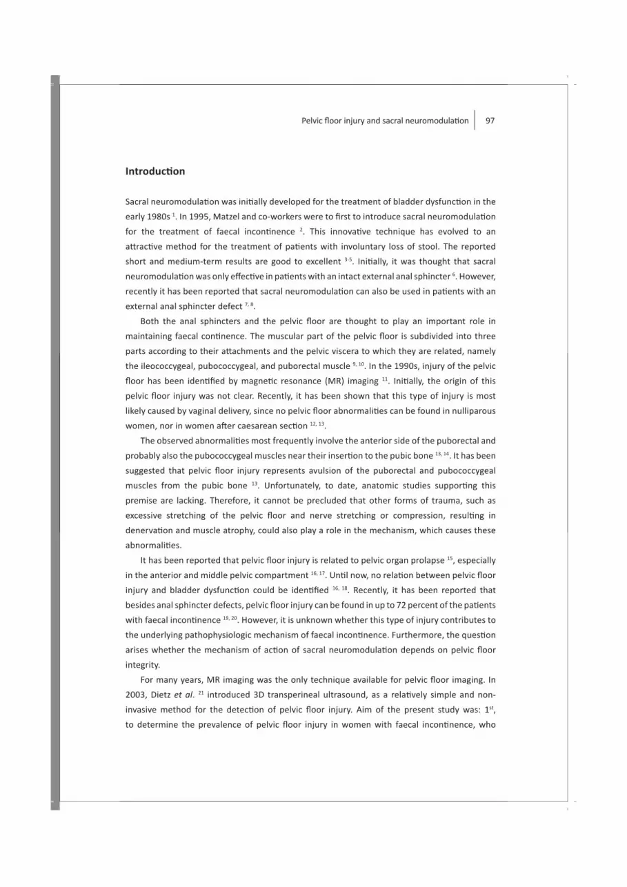
Pelvic fl oor injury and sacral neuromodulati on 97
Introducti on
Sacral neuromodulati on was initi ally developed for the treatment of bladder dysfuncti on in the
early 1980s 1. In 1995, Matzel and co-workers were to fi rst to introduce sacral neuromodulati on
for the treatment of faecal inconti nence 2. This innovati ve technique has evolved to an
att racti ve method for the treatment of pati ents with involuntary loss of stool. The reported
short and medium-term results are good to excellent 3-5. Initi ally, it was thought that sacral
neuromodulati on was only eff ecti ve in pati ents with an intact external anal sphincter 6. However,
recently it has been reported that sacral neuromodulati on can also be used in pati ents with an
external anal sphincter defect 7, 8.
Both the anal sphincters and the pelvic fl oor are thought to play an important role in
maintaining faecal conti nence. The muscular part of the pelvic fl oor is subdivided into three
parts according to their att achments and the pelvic viscera to which they are related, namely
the ileococcygeal, pubococcygeal, and puborectal muscle 9, 10. In the 1990s, injury of the pelvic
fl oor has been identi fi ed by magneti c resonance (MR) imaging 11. Initi ally, the origin of this
pelvic fl oor injury was not clear. Recently, it has been shown that this type of injury is most
likely caused by vaginal delivery, since no pelvic fl oor abnormaliti es can be found in nulliparous
women, nor in women aft er caesarean secti on 12, 13.
The observed abnormaliti es most frequently involve the anterior side of the puborectal and
probably also the pubococcygeal muscles near their inserti on to the pubic bone 13, 14. It has been
suggested that pelvic fl oor injury represents avulsion of the puborectal and pubococcygeal
muscles from the pubic bone 13. Unfortunately, to date, anatomic studies supporti ng this
premise are lacking. Therefore, it cannot be precluded that other forms of trauma, such as
excessive stretching of the pelvic fl oor and nerve stretching or compression, resulti ng in
denervati on and muscle atrophy, could also play a role in the mechanism, which causes these
abnormaliti es.
It has been reported that pelvic fl oor injury is related to pelvic organ prolapse 15, especially
in the anterior and middle pelvic compartment 16, 17. Unti l now, no relati on between pelvic fl oor
injury and bladder dysfuncti on could be identi fi ed 16, 18. Recently, it has been reported that
besides anal sphincter defects, pelvic fl oor injury can be found in up to 72 percent of the pati ents
with faecal inconti nence 19, 20. However, it is unknown whether this type of injury contributes to
the underlying pathophysiologic mechanism of faecal inconti nence. Furthermore, the questi on
arises whether the mechanism of acti on of sacral neuromodulati on depends on pelvic fl oor
integrity.
For many years, MR imaging was the only technique available for pelvic fl oor imaging. In
2003, Dietz et al. 21 introduced 3D transperineal ultrasound, as a relati vely simple and non-
invasive method for the detecti on of pelvic fl oor injury. Aim of the present study was: 1st,
to determine the prevalence of pelvic fl oor injury in women with faecal inconti nence, who
98
Chap
ter
7
were eligible for sacral neuromodulati on in the past and 2nd, to determine whether sacral
neuromodulati on is worthwhile in pati ents with pelvic fl oor injury, as detected by transperineal
ultrasound.
Pati ents and Methods
All female pati ents, who underwent test sti mulati on during the last nine years in our insti tuti on,
were invited to parti cipate in the present study, which was approved by the hospital’s human
research ethics committ ee. Pati ents with disabling faecal inconti nence were considered for test
sti mulati on when they met the criteria as listed in Table 1. Aft er writt en informed consent was
obtained, transperineal ultrasound was performed to assess pelvic fl oor integrity. A standardised
interview concerning parity and medical history was obtained at the ti me of the assessment
of pelvic fl oor integrity. In pati ents who received a permanent pulse generator, follow-up was
obtained with a standardised questi onnaire concerning conti nence improvement, degree of
faecal inconti nence, and the current number of inconti nence episodes.
Table 1. Criteria for test sti mulati on
Inclusion criteria
At least three inconti nence episode per week (confi rmed by bowel diary)
Under the age of 80 years
Failed conservati ve treatment
External anal sphincter: No defect
Or a small defect (< 90°)
Or failed previous sphincter repair
Exclusion criteria
Congenital anorectal malformati on
Neurological disease
Chronic diarrhea
Infl ammatory bowel disease
Psychiatric disease
Pregnancy
Test sti mulati on and permanent implantati on of a pulse generator were performed as
previously described 6. All pati ents completed a bowel diary before, during, and aft er test
sti mulati on. Hospital records and outpati ent clinic charts were retrospecti vely analysed by one
author (DMO), who had not parti cipated in the surgical procedure. All pati ents underwent test
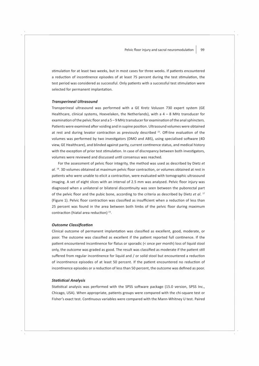
Pelvic fl oor injury and sacral neuromodulati on 99
sti mulati on for at least two weeks, but in most cases for three weeks. If pati ents encountered
a reducti on of inconti nence episodes of at least 75 percent during the test sti mulati on, the
test period was considered as successful. Only pati ents with a successful test sti mulati on were
selected for permanent implantati on.
Transperineal Ultrasound
Transperineal ultrasound was performed with a GE Kretz Voluson 730 expert system (GE
Healthcare, clinical systems, Hoevelaken, the Netherlands), with a 4 – 8 MHz transducer for
examinati on of the pelvic fl oor and a 5 – 9 MHz transducer for examinati on of the anal sphincters.
Pati ents were examined aft er voiding and in supine positi on. Ultrasound volumes were obtained
at rest and during levator contracti on as previously described 22. Off -line evaluati on of the
volumes was performed by two investi gators (DMO and ABS), using specialised soft ware (4D
view, GE Healthcare), and blinded against parity, current conti nence status, and medical history
with the excepti on of prior test sti mulati on. In case of discrepancy between both investi gators,
volumes were reviewed and discussed unti l consensus was reached.
For the assessment of pelvic fl oor integrity, the method was used as described by Dietz et
al. 18. 3D volumes obtained at maximum pelvic fl oor contracti on, or volumes obtained at rest in
pati ents who were unable to elicit a contracti on, were evaluated with tomographic ultrasound
imaging. A set of eight slices with an interval of 2.5 mm was analysed. Pelvic fl oor injury was
diagnosed when a unilateral or bilateral disconti nuity was seen between the puborectal part
of the pelvic fl oor and the pubic bone, according to the criteria as described by Dietz et al. 17
(Figure 1). Pelvic fl oor contracti on was classifi ed as insuffi cient when a reducti on of less than
25 percent was found in the area between both limbs of the pelvic fl oor during maximum
contracti on (hiatal area reducti on) 23.
Outcome Classifi cati on
Clinical outcome of permanent implantati on was classifi ed as excellent, good, moderate, or
poor. The outcome was classifi ed as excellent if the pati ent reported full conti nence. If the
pati ent encountered inconti nence for fl atus or sporadic (< once per month) loss of liquid stool
only, the outcome was graded as good. The result was classifi ed as moderate if the pati ent sti ll
suff ered from regular inconti nence for liquid and / or solid stool but encountered a reducti on
of inconti nence episodes of at least 50 percent. If the pati ent encountered no reducti on of
inconti nence episodes or a reducti on of less than 50 percent, the outcome was defi ned as poor.
Stati sti cal Analysis
Stati sti cal analysis was performed with the SPSS soft ware package (15.0 version, SPSS Inc.,
Chicago, USA). When appropriate, pati ents groups were compared with the chi-square test or
Fisher’s exact test. Conti nuous variables were compared with the Mann-Whitney U test. Paired
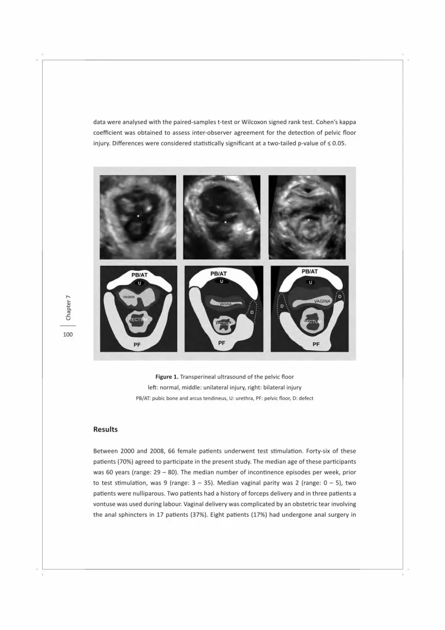
100
Chap
ter
7
data were analysed with the paired-samples t-test or Wilcoxon signed rank test. Cohen’s kappa
coeffi cient was obtained to assess inter-observer agreement for the detecti on of pelvic fl oor
injury. Diff erences were considered stati sti cally signifi cant at a two-tailed p-value of ≤ 0.05.
Figure 1. Transperineal ultrasound of the pelvic fl oor
left : normal, middle: unilateral injury, right: bilateral injury
PB/AT: pubic bone and arcus tendineus, U: urethra, PF: pelvic fl oor, D: defect
Results
Between 2000 and 2008, 66 female pati ents underwent test sti mulati on. Forty-six of these
pati ents (70%) agreed to parti cipate in the present study. The median age of these parti cipants
was 60 years (range: 29 – 80). The median number of inconti nence episodes per week, prior
to test sti mulati on, was 9 (range: 3 – 35). Median vaginal parity was 2 (range: 0 – 5), two
pati ents were nulliparous. Two pati ents had a history of forceps delivery and in three pati ents a
vontuse was used during labour. Vaginal delivery was complicated by an obstetric tear involving
the anal sphincters in 17 pati ents (37%). Eight pati ents (17%) had undergone anal surgery in
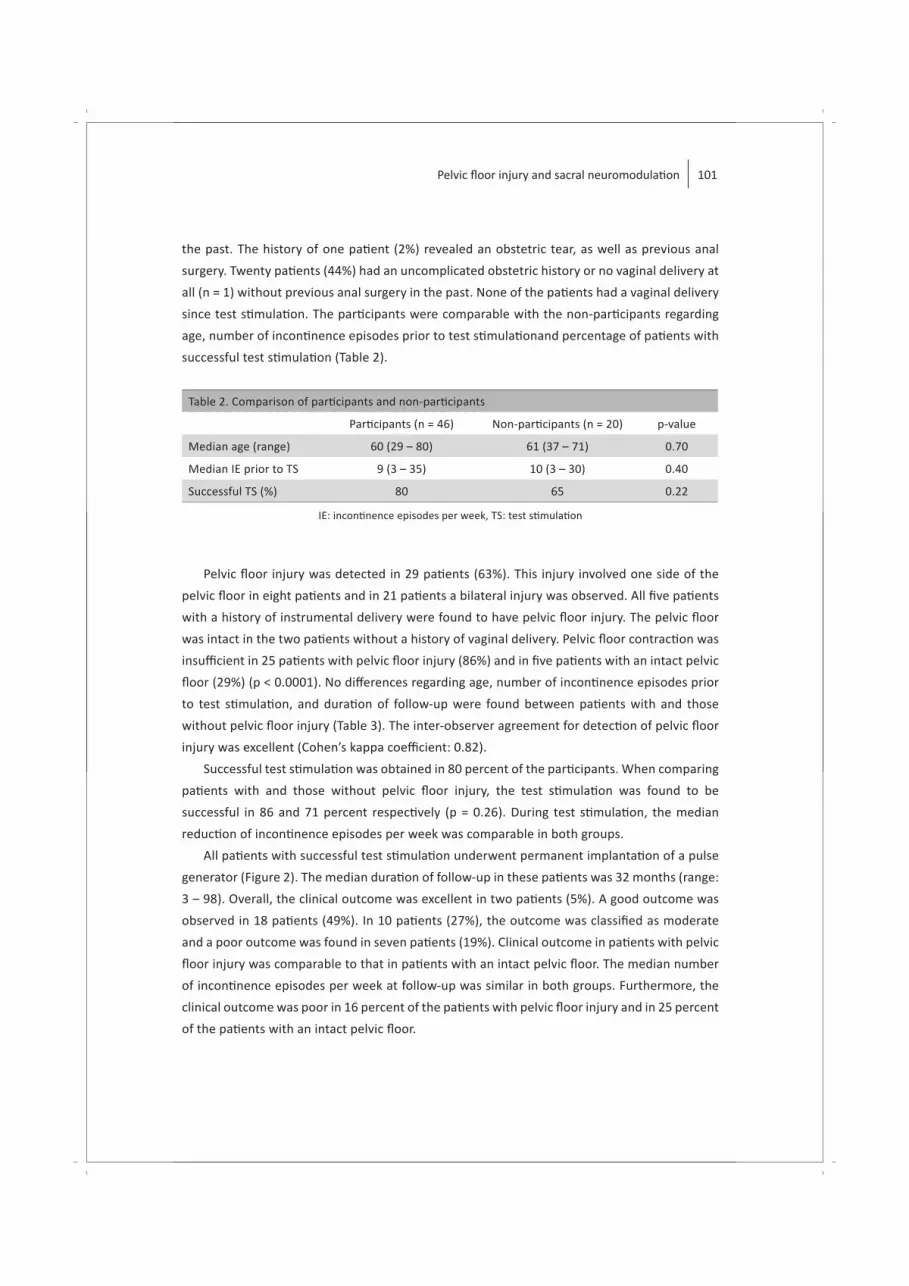
Pelvic fl oor injury and sacral neuromodulati on 101
the past. The history of one pati ent (2%) revealed an obstetric tear, as well as previous anal
surgery. Twenty pati ents (44%) had an uncomplicated obstetric history or no vaginal delivery at
all (n = 1) without previous anal surgery in the past. None of the pati ents had a vaginal delivery
since test sti mulati on. The parti cipants were comparable with the non-parti cipants regarding
age, number of inconti nence episodes prior to test sti mulati onand percentage of pati ents with
successful test sti mulati on (Table 2).
Table 2. Comparison of parti cipants and non-parti cipants
Parti cipants (n = 46) Non-parti cipants (n = 20) p-value
Median age (range) 60 (29 – 80) 61 (37 – 71) 0.70
Median IE prior to TS 9 (3 – 35) 10 (3 – 30) 0.40
Successful TS (%) 80 65 0.22
IE: inconti nence episodes per week, TS: test sti mulati on
Pelvic fl oor injury was detected in 29 pati ents (63%). This injury involved one side of the
pelvic fl oor in eight pati ents and in 21 pati ents a bilateral injury was observed. All fi ve pati ents
with a history of instrumental delivery were found to have pelvic fl oor injury. The pelvic fl oor
was intact in the two pati ents without a history of vaginal delivery. Pelvic fl oor contracti on was
insuffi cient in 25 pati ents with pelvic fl oor injury (86%) and in fi ve pati ents with an intact pelvic
fl oor (29%) (p < 0.0001). No diff erences regarding age, number of inconti nence episodes prior
to test sti mulati on, and durati on of follow-up were found between pati ents with and those
without pelvic fl oor injury (Table 3). The inter-observer agreement for detecti on of pelvic fl oor
injury was excellent (Cohen’s kappa coeffi cient: 0.82).
Successful test sti mulati on was obtained in 80 percent of the parti cipants. When comparing
pati ents with and those without pelvic fl oor injury, the test sti mulati on was found to be
successful in 86 and 71 percent respecti vely (p = 0.26). During test sti mulati on, the median
reducti on of inconti nence episodes per week was comparable in both groups.
All pati ents with successful test sti mulati on underwent permanent implantati on of a pulse
generator (Figure 2). The median durati on of follow-up in these pati ents was 32 months (range:
3 – 98). Overall, the clinical outcome was excellent in two pati ents (5%). A good outcome was
observed in 18 pati ents (49%). In 10 pati ents (27%), the outcome was classifi ed as moderate
and a poor outcome was found in seven pati ents (19%). Clinical outcome in pati ents with pelvic
fl oor injury was comparable to that in pati ents with an intact pelvic fl oor. The median number
of inconti nence episodes per week at follow-up was similar in both groups. Furthermore, the
clinical outcome was poor in 16 percent of the pati ents with pelvic fl oor injury and in 25 percent
of the pati ents with an intact pelvic fl oor.
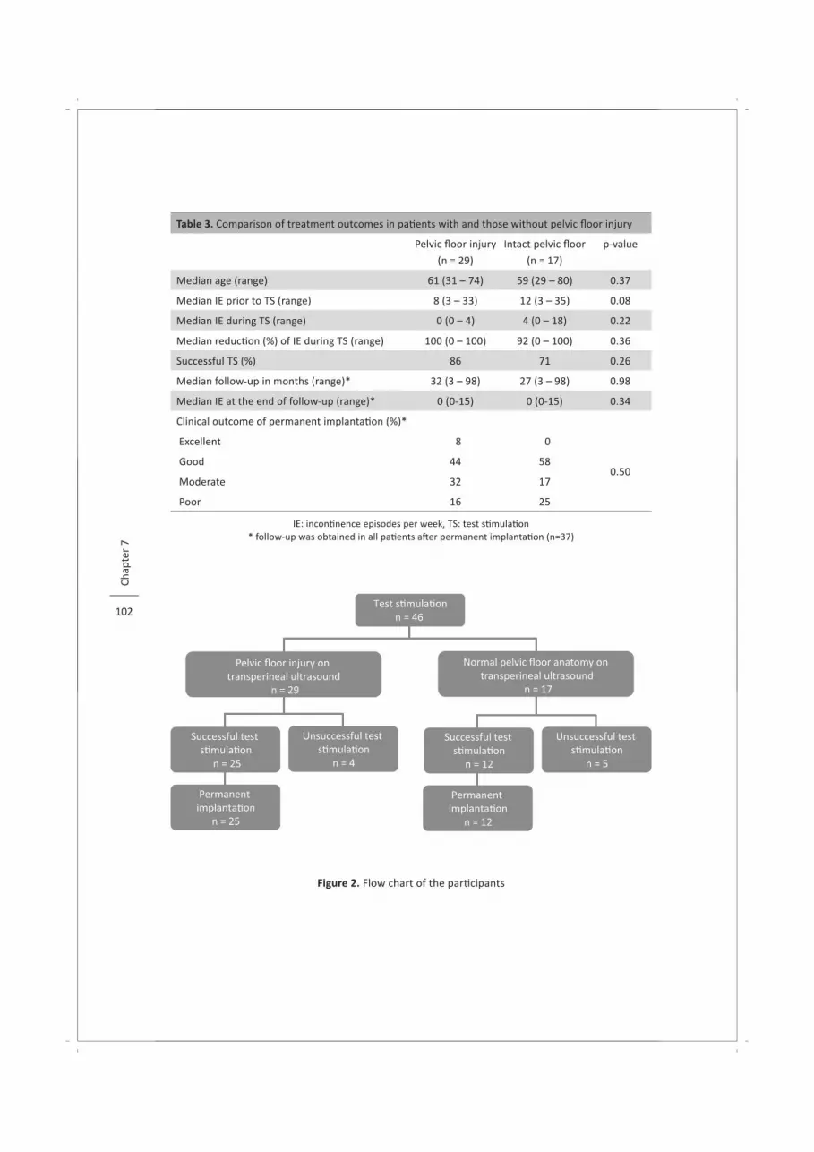
102
Chap
ter
7
Table 3. Comparison of treatment outcomes in pati ents with and those without pelvic fl oor injury
Pelvic fl oor injury
(n = 29)
Intact pelvic fl oor
(n = 17)
p-value
Median age (range) 61 (31 – 74) 59 (29 – 80) 0.37
Median IE prior to TS (range) 8 (3 – 33) 12 (3 – 35) 0.08
Median IE during TS (range) 0 (0 – 4) 4 (0 – 18) 0.22
Median reducti on (%) of IE during TS (range) 100 (0 – 100) 92 (0 – 100) 0.36
Successful TS (%) 86 71 0.26
Median follow-up in months (range)* 32 (3 – 98) 27 (3 – 98) 0.98
Median IE at the end of follow-up (range)* 0 (0-15) 0 (0-15) 0.34
Clinical outcome of permanent implantati on (%)*
Excellent 8 0
0.50 Good 44 58
Moderate 32 17
Poor 16 25
IE: inconti nence episodes per week, TS: test sti mulati on* follow-up was obtained in all pati ents aft er permanent implantati on (n=37)
Figure 2. Flow chart of the parti cipants
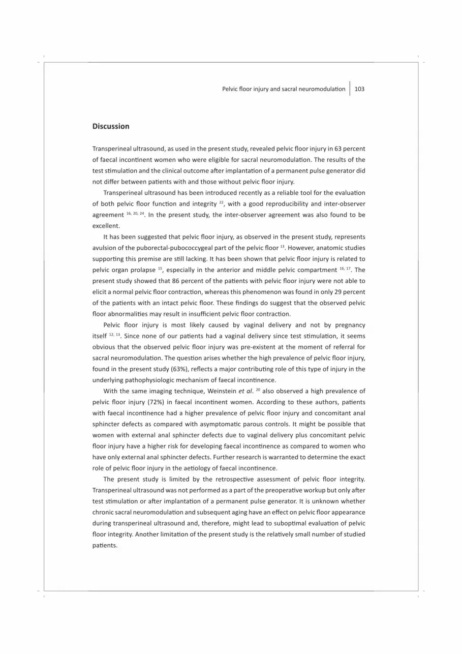
Pelvic fl oor injury and sacral neuromodulati on 103
Discussion
Transperineal ultrasound, as used in the present study, revealed pelvic fl oor injury in 63 percent
of faecal inconti nent women who were eligible for sacral neuromodulati on. The results of the
test sti mulati on and the clinical outcome aft er implantati on of a permanent pulse generator did
not diff er between pati ents with and those without pelvic fl oor injury.
Transperineal ultrasound has been introduced recently as a reliable tool for the evaluati on
of both pelvic fl oor functi on and integrity 22, with a good reproducibility and inter-observer
agreement 16, 20, 24. In the present study, the inter-observer agreement was also found to be
excellent.
It has been suggested that pelvic fl oor injury, as observed in the present study, represents
avulsion of the puborectal-pubococcygeal part of the pelvic fl oor 13. However, anatomic studies
supporti ng this premise are sti ll lacking. It has been shown that pelvic fl oor injury is related to
pelvic organ prolapse 15, especially in the anterior and middle pelvic compartment 16, 17. The
present study showed that 86 percent of the pati ents with pelvic fl oor injury were not able to
elicit a normal pelvic fl oor contracti on, whereas this phenomenon was found in only 29 percent
of the pati ents with an intact pelvic fl oor. These fi ndings do suggest that the observed pelvic
fl oor abnormaliti es may result in insuffi cient pelvic fl oor contracti on.
Pelvic fl oor injury is most likely caused by vaginal delivery and not by pregnancy
itself 12, 13. Since none of our pati ents had a vaginal delivery since test sti mulati on, it seems
obvious that the observed pelvic fl oor injury was pre-existent at the moment of referral for
sacral neuromodulati on. The questi on arises whether the high prevalence of pelvic fl oor injury,
found in the present study (63%), refl ects a major contributi ng role of this type of injury in the
underlying pathophysiologic mechanism of faecal inconti nence.
With the same imaging technique, Weinstein et al. 20 also observed a high prevalence of
pelvic fl oor injury (72%) in faecal inconti nent women. According to these authors, pati ents
with faecal inconti nence had a higher prevalence of pelvic fl oor injury and concomitant anal
sphincter defects as compared with asymptomati c parous controls. It might be possible that
women with external anal sphincter defects due to vaginal delivery plus concomitant pelvic
fl oor injury have a higher risk for developing faecal inconti nence as compared to women who
have only external anal sphincter defects. Further research is warranted to determine the exact
role of pelvic fl oor injury in the aeti ology of faecal inconti nence.
The present study is limited by the retrospecti ve assessment of pelvic fl oor integrity.
Transperineal ultrasound was not performed as a part of the preoperati ve workup but only aft er
test sti mulati on or aft er implantati on of a permanent pulse generator. It is unknown whether
chronic sacral neuromodulati on and subsequent aging have an eff ect on pelvic fl oor appearance
during transperineal ultrasound and, therefore, might lead to subopti mal evaluati on of pelvic
fl oor integrity. Another limitati on of the present study is the relati vely small number of studied
pati ents.
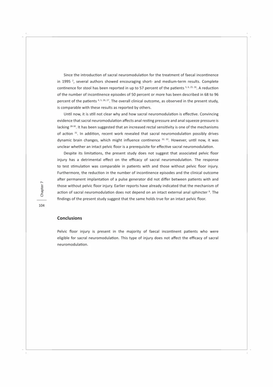
104
Chap
ter
7
Since the introducti on of sacral neuromodulati on for the treatment of faecal inconti nence
in 1995 2, several authors showed encouraging short- and medium-term results. Complete
conti nence for stool has been reported in up to 57 percent of the pati ents 5, 6, 25, 26. A reducti on
of the number of inconti nence episodes of 50 percent or more has been described in 68 to 96
percent of the pati ents 4, 5, 26, 27. The overall clinical outcome, as observed in the present study,
is comparable with these results as reported by others.
Unti l now, it is sti ll not clear why and how sacral neuromodulati on is eff ecti ve. Convincing
evidence that sacral neuromodulati on aff ects anal resti ng pressure and anal squeeze pressure is
lacking 28-30. It has been suggested that an increased rectal sensiti vity is one of the mechanisms
of acti on 31. In additi on, recent work revealed that sacral neuromodulati on possibly drives
dynamic brain changes, which might infl uence conti nence 32, 33. However, unti l now, it was
unclear whether an intact pelvic fl oor is a prerequisite for eff ecti ve sacral neuromodulati on.
Despite its limitati ons, the present study does not suggest that associated pelvic fl oor
injury has a detrimental eff ect on the effi cacy of sacral neuromodulati on. The response
to test sti mulati on was comparable in pati ents with and those without pelvic fl oor injury.
Furthermore, the reducti on in the number of inconti nence episodes and the clinical outcome
aft er permanent implantati on of a pulse generator did not diff er between pati ents with and
those without pelvic fl oor injury. Earlier reports have already indicated that the mechanism of
acti on of sacral neuromodulati on does not depend on an intact external anal sphincter 8. The
fi ndings of the present study suggest that the same holds true for an intact pelvic fl oor.
Conclusions
Pelvic fl oor injury is present in the majority of faecal inconti nent pati ents who were
eligible for sacral neuromodulati on. This type of injury does not aff ect the effi cacy of sacral
neuromodulati on.
Pelvic fl oor injury and sacral neuromodulati on 105
References
1. Tanagho EA, Schmidt RA. Bladder pacemaker: scienti fi c basis and clinical future. Urology 1982;20(6):614-9.
2. Matzel KE, Stadelmaier U, Hohenfellner M, Gall FP. Electrical sti mulati on of sacral spinal nerves for treatment of faecal inconti nence. Lancet 1995;346(8983):1124-7.
3. Jarrett ME, Mowatt G, Glazener CM, Fraser C, Nicholls RJ, Grant AM, et al. Systemati c review of sacral nerve sti mulati on for faecal inconti nence and consti pati on. Br J Surg 2004;91(12):1559-69.
4. Melenhorst J, Koch SM, Uludag O, van Gemert WG, Baeten CG. Sacral neuromodulati on in pati ents with faecal inconti nence: results of the fi rst 100 permanent implantati ons. Colorectal Dis 2007;9(8):725-30.
5. Tjandra JJ, Chan MK, Yeh CH, Murray-Green C. Sacral nerve sti mulati on is more eff ecti ve than opti mal medical therapy for severe fecal inconti nence: a randomized, controlled study. Dis Colon Rectum 2008;51(5):494-502.
6. Matzel KE, Kamm MA, Stosser M, Baeten CG, Christi ansen J, Madoff R, et al. Sacral spinal nerve sti mulati on for faecal inconti nence: multi centre study. Lancet 2004;363(9417):1270-6.
7. Chan MK, Tjandra JJ. Sacral nerve sti mulati on for fecal inconti nence: external anal sphincter defect vs. intact anal sphincter. Dis Colon Rectum 2008;51(7):1015-25.
8. Melenhorst J, Koch SM, Uludag O, van Gemert WG, Baeten CG. Is a morphologically intact anal sphincter necessary for success with sacral nerve modulati on in pati ents with faecal inconti nence? Colorectal Dis 2008;10(3):257-62.
9. DeLancey JO. The anatomy of the pelvic fl oor. Curr Opin Obstet Gynecol 1994;6(4):313-6.10. Ashton-Miller JA, DeLancey JO. Functi onal anatomy of the female pelvic fl oor. Ann N Y Acad Sci
2007;1101:266-96.11. Kirschner-Hermanns R, Wein B, Niehaus S, Schaefer W, Jakse G. The contributi on of magneti c
resonance imaging of the pelvic fl oor to the understanding of urinary inconti nence. Br J Urol 1993;72(5 Pt 2):715-8.
12. DeLancey JO, Kearney R, Chou Q, Speights S, Binno S. The appearance of levator ani muscle abnormaliti es in magneti c resonance images aft er vaginal delivery. Obstet Gynecol 2003;101(1):46-53.
13. Dietz HP, Lanzarone V. Levator trauma aft er vaginal delivery. Obstet Gynecol 2005;106(4):707-12.14. Margulies RU, Huebner M, DeLancey JO. Origin and inserti on points involved in levator ani muscle
defects. Am J Obstet Gynecol 2007;196(3):251 e1-5.15. DeLancey JO, Morgan DM, Fenner DE, Kearney R, Guire K, Miller JM, et al. Comparison of levator
ani muscle defects and functi on in women with and without pelvic organ prolapse. Obstet Gynecol 2007;109(2 Pt 1):295-302.
16. Dietz HP, Steensma AB. The prevalence of major abnormaliti es of the levator ani in urogynaecological pati ents. Bjog 2006;113(2):225-30.
17. Dietz HP, Simpson JM. Levator trauma is associated with pelvic organ prolapse. Bjog 2008;115(8):979-84.
18. Dietz HP. Quanti fi cati on of major morphological abnormaliti es of the levator ani. Ultrasound Obstet Gynecol 2007;29(3):329-34.
19. Terra MP, Beets-Tan RG, Vervoorn I, Deutekom M, Wasser MN, Witkamp TD, et al. Pelvic fl oor muscle lesions at endoanal MR imaging in female pati ents with faecal inconti nence. Eur Radiol 2008;18(9):1892-901.
20. Weinstein MM, Pretorius DH, Jung SA, Nager CW, Mitt al RK. Transperineal three-dimensional ultrasound imaging for detecti on of anatomic defects in the anal sphincter complex muscles. Clin Gastroenterol Hepatol 2009;7(2):205-11.
106
Chap
ter
7
21. Dietz HP, Steensma AB, Hasti ngs R. Three-dimensional ultrasound imaging of the pelvic fl oor: the eff ect of parturiti on on paravaginal support structures. Ultrasound Obstet Gynecol 2003;21(6):589-95.
22. Dietz HP. Ultrasound imaging of the pelvic fl oor. Part II: three-dimensional or volume imaging. Ultrasound Obstet Gynecol 2004;23(6):615-25.
23. Braekken IH, Majida M, Engh ME, Bo K. Test-retest reliability of pelvic fl oor muscle contracti on measured by 4D ultrasound. Neurourol Urodyn 2009;28(1):68-73.
24. Weinstein MM, Pretorius D, Nager CW, Mitt al R. Inter-rater reliability of pelvic fl oor muscle imaging abnormaliti es with 3D ultrasound. Ultrasound Obstet Gynecol 2007;30:538.
25. Dudding TC, Pares D, Vaizey CJ, Kamm MA. Predicti ve factors for successful sacral nerve sti mulati on in the treatment of faecal inconti nence: a 10-year cohort analysis. Colorectal Dis 2008;10(3):249-56.
26. Gourcerol G, Gallas S, Michot F, Denis P, Leroi AM. Sacral nerve sti mulati on in fecal inconti nence: are there factors associated with success? Dis Colon Rectum 2007;50(1):3-12.
27. Jarrett ME, Varma JS, Duthie GS, Nicholls RJ, Kamm MA. Sacral nerve sti mulati on for faecal inconti nence in the UK. Br J Surg 2004;91(6):755-61.
28. Vaizey CJ, Kamm MA, Turner IC, Nicholls RJ, Woloszko J. Eff ects of short term sacral nerve sti mulati on on anal and rectal functi on in pati ents with anal inconti nence. Gut 1999;44(3):407-12.
29. Leroi AM, Michot F, Grise P, Denis P. Eff ect of sacral nerve sti mulati on in pati ents with fecal and urinary inconti nence. Dis Colon Rectum 2001;44(6):779-89.
30. Altomare DF, Rinaldi M, Petrolino M, Ripetti V, Masin A, Ratt o C, et al. Reliability of electrophysiologic anal tests in predicti ng the outcome of sacral nerve modulati on for fecal inconti nence. Dis Colon Rectum 2004;47(6):853-7.
31. Uludag O, Morren GL, Dejong CH, Baeten CG. Eff ect of sacral neuromodulati on on the rectum. Br J Surg 2005;92(8):1017-23.
32. Sheldon R, Kiff ES, Clarke A, Harris ML, Hamdy S. Sacral nerve sti mulati on reduces corti coanal excitability in pati ents with faecal inconti nence. Br J Surg 2005;92(11):1423-31.
33. Blok BF, Groen J, Bosch JL, Veltman DJ, Lammertsma AA. Diff erent brain eff ects during chronic and acute sacral neuromodulati on in urge inconti nent pati ents with implanted neurosti mulators. BJU Int 2006;98(6):1238-43.
Part III
Rectovaginal Fistula


Chapter 8 Puborectal sling interpositi on for the treatment of
rectovaginal fi stulas
Tech Coloproctol 2006;10(2):125-30.
Daniëlla M.J. Oom
Marti jn P. Gosselink
Valeria R.M. van Dijl
David D.E. Zimmerman
W. Ruud Schouten

110
Chap
ter
8
Abstract
Background Several techniques are available for the surgical treatment of rectovaginal fi stulas. However,
the results are oft en rather disappointi ng. It has been suggested that interpositi on of healthy,
well-vascularised ti ssue may be the key to rectovaginal fi stula healing. The present study was
aimed at evaluati ng the outcome of the puborectal sling interpositi on in the treatment of
rectovaginal fi stulas.
Pati ents and Methods Between 2001 and 2004, 26 consecuti ve pati ents (median age: 41 years, range: 15 – 69) with
a rectovaginal fi stula underwent a puborectal sling interpositi on. The aeti ology of the fi stulas
was: obstetric injury (n = 11), complicati ons aft er prior surgery (n = 2), bartholiniti s (n = 4)
cryptoglandular fi stulous disease (n = 2), infl ammatory bowel disease (n = 2), and idiopathic
(n = 5). The pati ents received a standardised questi onnaire about faecal conti nence and
dyspareunia.
Results The median follow-up was 14 months (range: 3 – 50). The rectovaginal fi stula healed in 16
of 26 pati ents. In pati ents who had undergone one or more previous repairs, the healing
rate was only 31 percent versus 92 percent in pati ents without previous repairs (p < 0.01).
The Rockwood faecal inconti nence severity index score did not change. Seventeen percent
of pati ents experienced painful intercourse before the operati on. Aft er the procedure this
problem was encountered by 57 percent of the pati ents.
Conclusion The puborectal sling interpositi on is only successful in pati ents without previous repairs and
in those with an uneventf ul postoperati ve course. However, de novo dyspareunia is a major
drawback of this procedure.
Puborectal sling interpositi on 111
Introducti on
A rectovaginal fi stula is an abnormal tract between the rectum and the vagina. Such a fi stula is
frequently associated with physical and emoti onal burden. Rectovaginal fi stulas are commonly
classifi ed as low, intermediate, and high, depending on the locati on of the vaginal opening. In
most cases the pati ent presents with a low rectovaginal fi stula, which is in fact an anovaginal
fi stula. Obstetric injury is by far the most frequently reported cause of these low rectovaginal
fi stulas 1-4. In the Western society, vaginal deliveries result in a third or fourth degree perineal
tear in two percent of the women 5. Disrupti on of the external anal sphincter, despite a proper
repair, with a concomitant wound infecti on are major contributi ng factors.
Several techniques are available for the repair of rectovaginal fi stulas. In the early 1980s,
transanal advancement fl ap repair was advocated as the treatment of choice for pati ents with
a low rectovaginal fi stula. Initi ally, the reported results were very promising, with healing rates
varying between 78 and 95 percent 4, 6-8. More recently, a signifi cantly lower healing rate has
been reported, especially in women who had previous repairs 9-11. In our own insti tuti on we
also encountered low healing rates aft er transanal advancement fl ap repair. In an att empt to
improve our results, we added labial fat fl ap transpositi on to the advancement fl ap repair.
Unfortunately, this combined procedure did not improve the outcome 12.
It has been suggested that interpositi on of healthy, well-vascularised ti ssue may be the
key to rectovaginal fi stula healing. Reviewing the literature from the past, we noti ced that
gynaecologists frequently applied interpositi on of healthy, well-vascularised sphincter muscle.
In the early 1990s, the benefi cial eff ect of a puborectal sling interpositi on has been reported 13-15.
The high success rate, varying between 92 and 100 percent, inspired us to conduct the present
study, aimed at evaluati ng the outcome of the puborectal sling interpositi on in the treatment
of rectovaginal fi stulas.
Pati ents and Methods
Between 2001 and 2004, 26 consecuti ve pati ents with a low rectovaginal fi stula (median age:
41 years, range: 15 – 69) underwent a puborectal sling interpositi on. In 11 pati ents the fi stula
developed aft er vaginal delivery, complicated by a perineal tear. In two pati ents the fi stula
was due to complicati ons aft er sphincter and rectocele repair respecti vely. In four pati ents the
fi stula was a late result of a prior bartholiniti s. Other causes were cryptoglandular fi stulous
disease (n = 2) and infl ammatory bowel disease (n = 2). The underlying cause of the fi stula
could not be identi fi ed in fi ve pati ents. Thirteen pati ents had undergone one or more previous
att empts at repair before referral to our hospital. A transperineal approach was applied in 10
pati ents. In 16 pati ents an approach trough the posterior vaginal fourchett e was used. Prior to
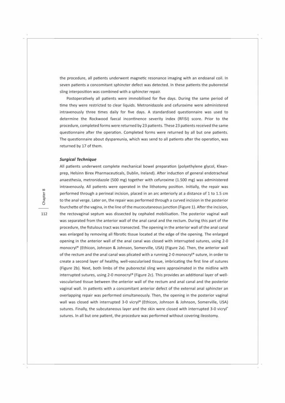
112
Chap
ter
8
the procedure, all pati ents underwent magneti c resonance imaging with an endoanal coil. In
seven pati ents a concomitant sphincter defect was detected. In these pati ents the puborectal
sling interpositi on was combined with a sphincter repair.
Postoperati vely all pati ents were immobilised for fi ve days. During the same period of
ti me they were restricted to clear liquids. Metronidazole and cefuroxime were administered
intravenously three ti mes daily for fi ve days. A standardised questi onnaire was used to
determine the Rockwood faecal inconti nence severity index (RFISI) score. Prior to the
procedure, completed forms were returned by 23 pati ents. These 23 pati ents received the same
questi onnaire aft er the operati on. Completed forms were returned by all but one pati ents.
The questi onnaire about dyspareunia, which was send to all pati ents aft er the operati on, was
returned by 17 of them.
Surgical Technique
All pati ents underwent complete mechanical bowel preparati on (polyethylene glycol, Klean-
prep, Helsinn Birex Pharmaceuti cals, Dublin, Ireland). Aft er inducti on of general endotracheal
anaesthesia, metronidazole (500 mg) together with cefuroxime (1.500 mg) was administered
intravenously. All pati ents were operated in the lithotomy positi on. Initi ally, the repair was
performed through a perineal incision, placed in an arc anteriorly at a distance of 1 to 1.5 cm
to the anal verge. Later on, the repair was performed through a curved incision in the posterior
fourchett e of the vagina, in the line of the mucocutaneous juncti on (Figure 1). Aft er the incision,
the rectovaginal septum was dissected by cephaled mobilisati on. The posterior vaginal wall
was separated from the anterior wall of the anal canal and the rectum. During this part of the
procedure, the fi stulous tract was transected. The opening in the anterior wall of the anal canal
was enlarged by removing all fi broti c ti ssue located at the edge of the opening. The enlarged
opening in the anterior wall of the anal canal was closed with interrupted sutures, using 2-0
monocryl® (Ethicon, Johnson & Johnson, Somerville, USA) (Figure 2a). Then, the anterior wall
of the rectum and the anal canal was plicated with a running 2-0 monocryl® suture, in order to
create a second layer of healthy, well-vascularised ti ssue, imbricati ng the fi rst line of sutures
(Figure 2b). Next, both limbs of the puborectal sling were approximated in the midline with
interrupted sutures, using 2-0 monocryl® (Figure 2c). This provides an additi onal layer of well-
vascularised ti ssue between the anterior wall of the rectum and anal canal and the posterior
vaginal wall. In pati ents with a concomitant anterior defect of the external anal sphincter an
overlapping repair was performed simultaneously. Then, the opening in the posterior vaginal
wall was closed with interrupted 3-0 vicryl® (Ethicon, Johnson & Johnson, Somerville, USA)
sutures. Finally, the subcutaneous layer and the skin were closed with interrupted 3-0 vicryl®
sutures. In all but one pati ent, the procedure was performed without covering ileostomy.

Puborectal sling interpositi on 113
Figure 1. Exposure of the rectovaginal fi stula
PFI: posterior fourchett e incision, PI: perineal incision,
VOF: vaginal opening of the fi stulous tract
Figure 2a. Figure 2b. Figure 2c.
Figure 2. Closure of the rectovaginal fi stula
Figure 2a. First layer: closure of the enlarged opening in the anterior wall of the anal canal
Figure 2b. Second layer: plicati on of the anterior wall of the anal canal
Figure 2c. Third layer: plicati on of both arms of the puborectal sling
PS: puborectal sling
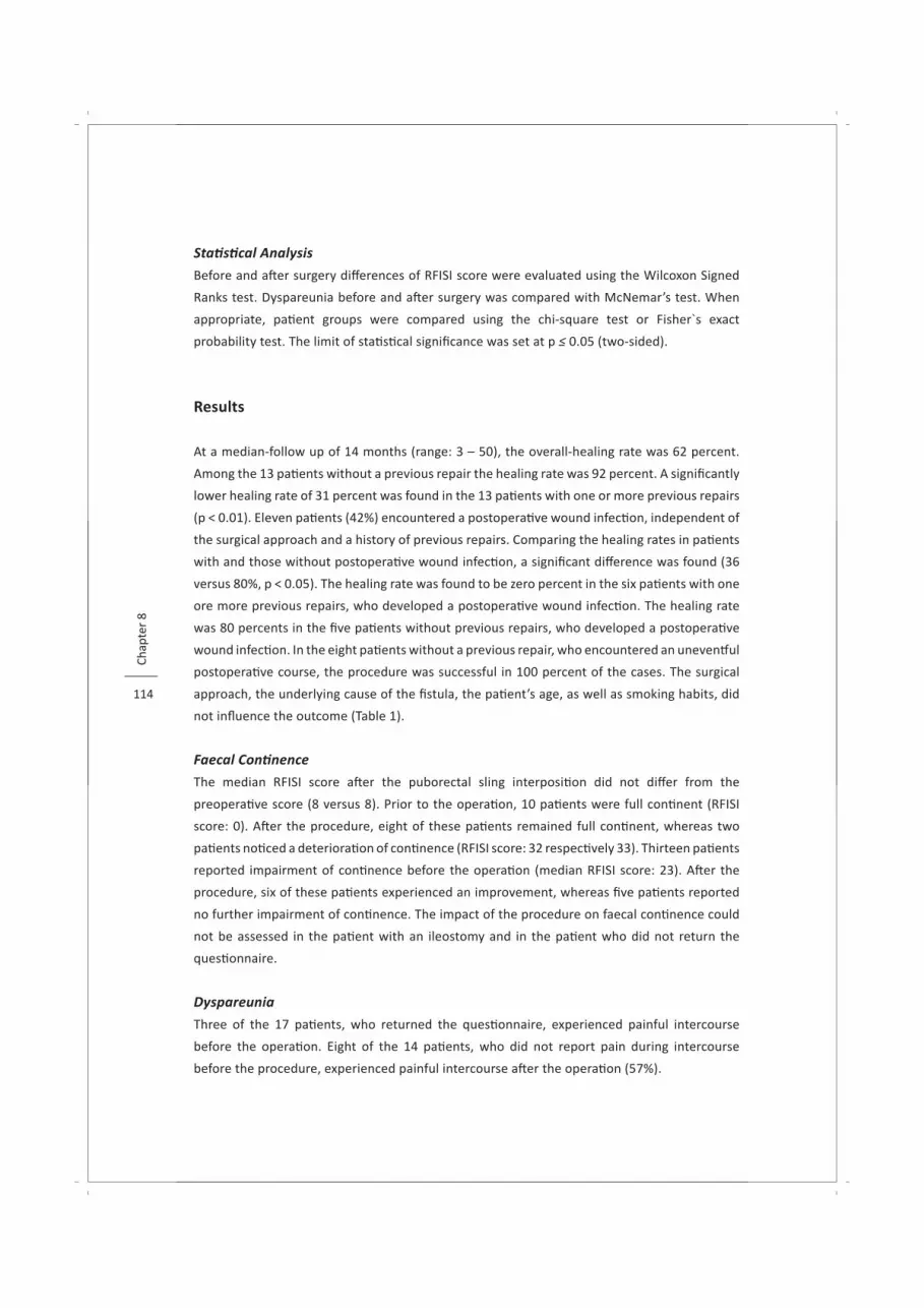
114
Chap
ter
8
Stati sti cal Analysis
Before and aft er surgery diff erences of RFISI score were evaluated using the Wilcoxon Signed
Ranks test. Dyspareunia before and aft er surgery was compared with McNemar’s test. When
appropriate, pati ent groups were compared using the chi-square test or Fisher`s exact
probability test. The limit of stati sti cal signifi cance was set at p ≤ 0.05 (two-sided).
Results
At a median-follow up of 14 months (range: 3 – 50), the overall-healing rate was 62 percent.
Among the 13 pati ents without a previous repair the healing rate was 92 percent. A signifi cantly
lower healing rate of 31 percent was found in the 13 pati ents with one or more previous repairs
(p < 0.01). Eleven pati ent s (42%) encountered a postoperati ve wound infecti on, independent of
the surgical approach and a history of previous repairs. Comparing the healing rates in pati ents
with and those without postoperati ve wound infecti on, a signifi cant diff erence was found (36
versus 80%, p < 0.05). The healing rate was found to be zero percent in the six pati ents with one
ore more previous repairs, who developed a postoperati ve wound infecti on. The healing rate
was 80 percents in the fi ve pati ents without previous repairs, who developed a postoperati ve
wound infecti on. In the eight pati ents without a previous repair, who encountered an uneventf ul
postoperati ve course, the procedure was successful in 100 percent of the cases. The surgical
approach, the underlying cause of the fi stula, the pati ent’s age, as well as smoking habits, did
not infl uence the outcome (Table 1).
Faecal Conti nence
The median RFISI score aft er the puborectal sling interpositi on did not diff er from the
preoperati ve score (8 versus 8). Prior to the operati on, 10 pati ents were full conti nent (RFISI
score: 0). Aft er the procedure, eight of these pati ents remained full conti nent, whereas two
pati ents noti ced a deteriorati on of conti nence (RFISI score: 32 respecti vely 33). Thirteen pati ents
reported impairment of conti nence before the operati on (median RFISI score: 23). Aft er the
procedure, six of these pati ents experienced an improvement, whereas fi ve pati ents reported
no further impairment of conti nence. The impact of the procedure on faecal conti nence could
not be assessed in the pati ent with an ileostomy and in the pati ent who did not return the
questi onnaire.
Dyspareunia
Three of the 17 pati ents, who returned the questi onnaire, experienced painful intercourse
before the operati on. Eight of the 14 pati ents, who did not report pain during intercourse
before the procedure, experienced painful intercourse aft er the operati on (57%).

Puborectal sling interpositi on 115
Table 1. Factors predicti ve of outcome
Success (n) Failure (n) Healing rate (%)
Age
< 35 years 6 4 60
> 35 years 10 6 63
Previous repairs*
Yes 4 9 31
No 12 1 92
Wound infecti on#
Yes 4 7 36
No 12 3 80
Eti ology
Obstetric 7 4 64
Complicati ons aft er prior surgery 1 1 50
Barholiniti s 3 1 75
Cryptoglandular fi stulous disease 1 1 50
IBD 2 0 100
Idiopathic 2 3 40
Approach
Perineal 7 3 70
Fourchett e 9 7 56
Smoking
Yes 5 2 71
No 11 8 58
* p < 0.01# p < 0.05
IBD: infl ammatory bowel disease
Discussion
Despite some anecdoti c reports, there is no evidence that rectovaginal fi stulas heal
spontaneously. Unti l now it is not clear whether a non-surgical approach, such as the use of
fi brin glue and treatment by electrocauterisati on, is successful or not 16-19. Therefore, it seems
likely that closure of a rectovaginal fi stula can only be obtained by surgical repair.
Traditi onally, rectovaginal fi stulas were repaired by gynaecologists uti lising a transvaginal
approach 20, 21. According to Greenwald and Hoexter, a low rectovaginal fi stula has the
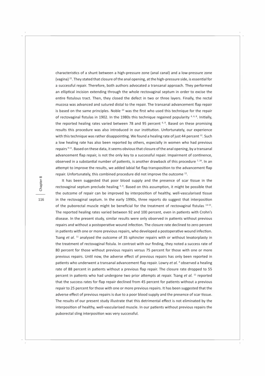
116
Chap
ter
8
characteristi cs of a shunt between a high-pressure zone (anal canal) and a low-pressure zone
(vagina) 22. They stated that closure of the anal opening, at the high-pressure side, is essenti al for
a successful repair. Therefore, both authors advocated a transanal approach. They performed
an ellipti cal incision extending through the whole rectovaginal septum in order to excise the
enti re fi stulous tract. Then, they closed the defect in two or three layers. Finally, the rectal
mucosa was advanced and sutured distal to the repair. The transanal advancement fl ap repair
is based on the same principles. Noble 23 was the fi rst who used this technique for the repair
of rectovaginal fi stulas in 1902. In the 1980s this technique regained popularity 4, 6, 8. Initi ally,
the reported healing rates varied between 78 and 95 percent 6, 8. Based on these promising
results this procedure was also introduced in our insti tuti on. Unfortunately, our experience
with this technique was rather disappointi ng. We found a healing rate of just 44 percent 12. Such
a low healing rate has also been reported by others, especially in women who had previous
repairs 9-11. Based on these data, it seems obvious that closure of the anal opening, by a transanal
advancement fl ap repair, is not the only key to a successful repair. Impairment of conti nence,
observed in a substanti al number of pati ents, is another drawback of this procedure 7, 24. In an
att empt to improve the results, we added labial fat fl ap transpositi on to the advancement fl ap
repair. Unfortunately, this combined procedure did not improve the outcome 12.
It has been suggested that poor blood supply and the presence of scar ti ssue in the
rectovaginal septum preclude healing 4, 9. Based on this assumpti on, it might be possible that
the outcome of repair can be improved by interpositi on of healthy, well-vascularised ti ssue
in the rectovaginal septum. In the early 1990s, three reports do suggest that interpositi on
of the puborectal muscle might be benefi cial for the treatment of rectovaginal fi stulas 13-15.
The reported healing rates varied between 92 and 100 percent, even in pati ents with Crohn’s
disease. In the present study, similar results were only observed in pati ents without previous
repairs and without a postoperati ve wound infecti on. The closure rate declined to zero percent
in pati ents with one or more previous repairs, who developed a postoperati ve wound infecti on.
Tsang et al. 11 analysed the outcome of 35 sphincter repairs with or without levatorplasty in
the treatment of rectovaginal fi stula. In contrast with our fi nding, they noted a success rate of
80 percent for those without previous repairs versus 75 percent for those with one or more
previous repairs. Unti l now, the adverse eff ect of previous repairs has only been reported in
pati ents who underwent a transanal advancement fl ap repair. Lowry et al. 4 observed a healing
rate of 88 percent in pati ents without a previous fl ap repair. The closure rate dropped to 55
percent in pati ents who had undergone two prior att empts at repair. Tsang et al. 11 reported
that the success rates for fl ap repair declined from 45 percent for pati ents without a previous
repair to 25 percent for those with one or more previous repairs. It has been suggested that the
adverse eff ect of previous repairs is due to a poor blood supply and the presence of scar ti ssue.
The results of our present study illustrate that this detrimental eff ect is not eliminated by the
interpositi on of healthy, well-vascularised muscle. In our pati ents without previous repairs the
puborectal sling interpositi on was very successful.

Puborectal sling interpositi on 117
Initi ally, we used a transperineal approach for the puborectal sling interpositi on. Later
on, we used an incision at the posterior vaginal fourchett e. The most important reason for
this change in policy was the relati vely high incidence of wound complicati ons aft er the
perineal approach. Maybelle Tan and co-workers 25 conducted a randomised controlled trial
comparing the conventi onal perineal incision with the posterior vaginal fourchett e incision.
Postoperati ve wound complicati ons were fewer when a posterior vaginal fourchett e incision
was used compared with a perineal incision. In contrast with this fi nding, we were not able to
demonstrate a signifi cant diff erence between both approaches with respect to the number of
wound complicati ons.
In the present study, the puborectal sling interpositi on, either by a perineal approach or
by a posterior fourchett e approach, did not adversely aff ect faecal conti nence. Comparing the
Rockwood faecal inconti nence severity index score before and aft er surgery, no diff erences
were found. It has been shown that aft er transanal advancement fl ap repair, impairment of
faecal conti nence does occur in a substanti al number of pati ents 7, 24.
Although the puborectal sling interpositi on does not result in impairment of conti nence, this
procedure has two disadvantages. First, the outcome is not successful in pati ents with previous
repairs and in those who developed a postoperati ve wound infecti on. The high incidence of de
novo dyspareunia is another drawback. In the present study, 57 percent of pati ents complained
about dyspareunia aft er the procedure.
The results of the present study indicate that the outcome of rectovaginal fi stula repair
can be improved by the puborectal sling interpositi on. However, this benefi cial eff ect was only
observed in pati ents without previous repairs and in those with an uneventf ul postoperati ve
course. Based on the poor outcome in pati ents who had undergone one or more previous
repairs and the high incidence of de novo dyspareunia aft er the procedure, the puborectal
sling interpositi on cannot be considered as the treatment of choice for women with a low
rectovaginal fi stula. In our opinion, therefore, further studies are warranted in order to examine
other techniques.
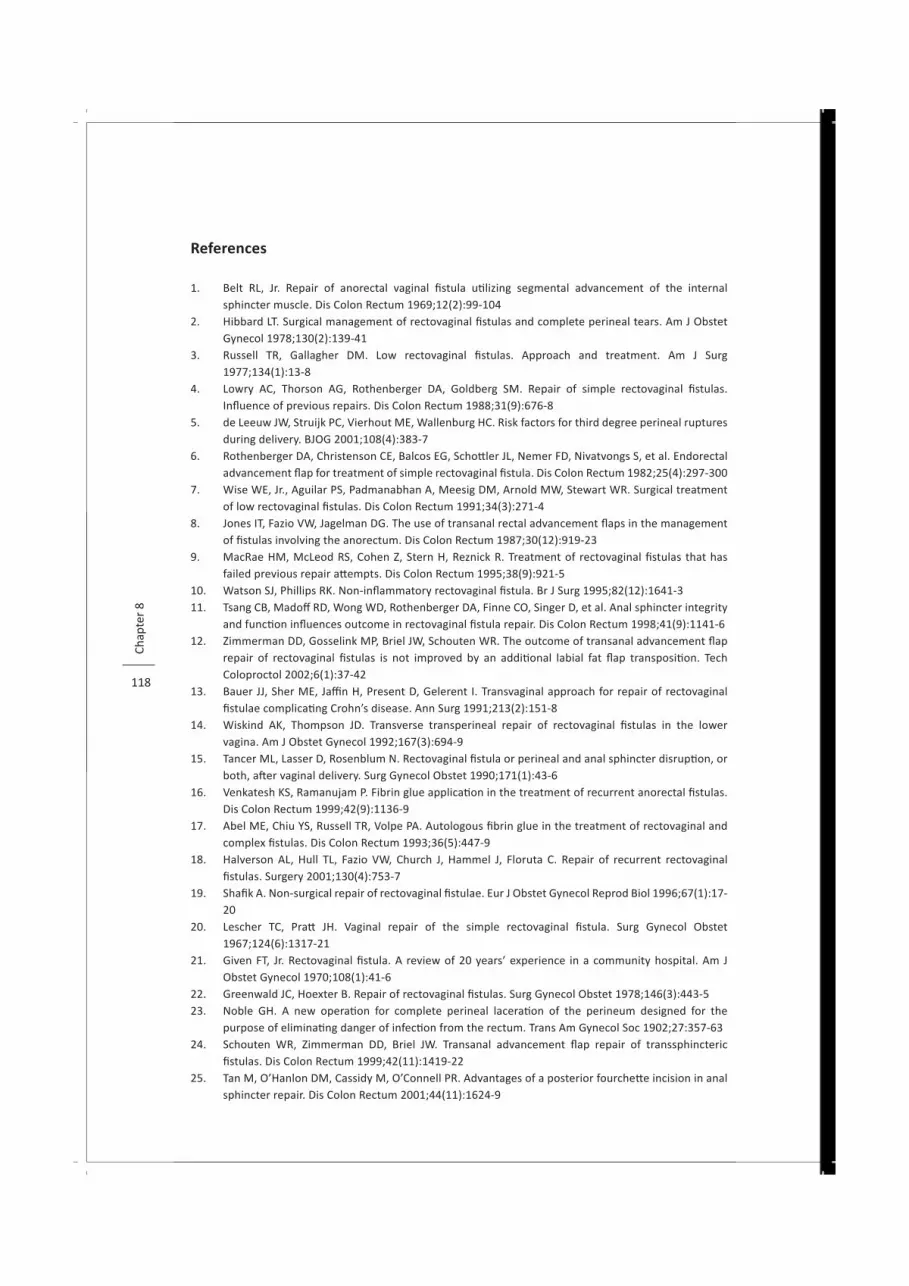
118
Chap
ter
8
References
1. Belt RL, Jr. Repair of anorectal vaginal fi stula uti lizing segmental advancement of the internal sphincter muscle. Dis Colon Rectum 1969;12(2):99-104
2. Hibbard LT. Surgical management of rectovaginal fi stulas and complete perineal tears. Am J Obstet Gynecol 1978;130(2):139-41
3. Russell TR, Gallagher DM. Low rectovaginal fi stulas. Approach and treatment. Am J Surg 1977;134(1):13-8
4. Lowry AC, Thorson AG, Rothenberger DA, Goldberg SM. Repair of simple rectovaginal fi stulas. Infl uence of previous repairs. Dis Colon Rectum 1988;31(9):676-8
5. de Leeuw JW, Struijk PC, Vierhout ME, Wallenburg HC. Risk factors for third degree perineal ruptures during delivery. BJOG 2001;108(4):383-7
6. Rothenberger DA, Christenson CE, Balcos EG, Schott ler JL, Nemer FD, Nivatvongs S, et al. Endorectal advancement fl ap for treatment of simple rectovaginal fi stula. Dis Colon Rectum 1982;25(4):297-300
7. Wise WE, Jr., Aguilar PS, Padmanabhan A, Meesig DM, Arnold MW, Stewart WR. Surgical treatment of low rectovaginal fi stulas. Dis Colon Rectum 1991;34(3):271-4
8. Jones IT, Fazio VW, Jagelman DG. The use of transanal rectal advancement fl aps in the management of fi stulas involving the anorectum. Dis Colon Rectum 1987;30(12):919-23
9. MacRae HM, McLeod RS, Cohen Z, Stern H, Reznick R. Treatment of rectovaginal fi stulas that has failed previous repair att empts. Dis Colon Rectum 1995;38(9):921-5
10. Watson SJ, Phillips RK. Non-infl ammatory rectovaginal fi stula. Br J Surg 1995;82(12):1641-311. Tsang CB, Madoff RD, Wong WD, Rothenberger DA, Finne CO, Singer D, et al. Anal sphincter integrity
and functi on infl uences outcome in rectovaginal fi stula repair. Dis Colon Rectum 1998;41(9):1141-612. Zimmerman DD, Gosselink MP, Briel JW, Schouten WR. The outcome of transanal advancement fl ap
repair of rectovaginal fi stulas is not improved by an additi onal labial fat fl ap transpositi on. Tech Coloproctol 2002;6(1):37-42
13. Bauer JJ, Sher ME, Jaffi n H, Present D, Gelerent I. Transvaginal approach for repair of rectovaginal fi stulae complicati ng Crohn’s disease. Ann Surg 1991;213(2):151-8
14. Wiskind AK, Thompson JD. Transverse transperineal repair of rectovaginal fi stulas in the lower vagina. Am J Obstet Gynecol 1992;167(3):694-9
15. Tancer ML, Lasser D, Rosenblum N. Rectovaginal fi stula or perineal and anal sphincter disrupti on, or both, aft er vaginal delivery. Surg Gynecol Obstet 1990;171(1):43-6
16. Venkatesh KS, Ramanujam P. Fibrin glue applicati on in the treatment of recurrent anorectal fi stulas. Dis Colon Rectum 1999;42(9):1136-9
17. Abel ME, Chiu YS, Russell TR, Volpe PA. Autologous fi brin glue in the treatment of rectovaginal and complex fi stulas. Dis Colon Rectum 1993;36(5):447-9
18. Halverson AL, Hull TL, Fazio VW, Church J, Hammel J, Floruta C. Repair of recurrent rectovaginal fi stulas. Surgery 2001;130(4):753-7
19. Shafi k A. Non-surgical repair of rectovaginal fi stulae. Eur J Obstet Gynecol Reprod Biol 1996;67(1):17-20
20. Lescher TC, Pratt JH. Vaginal repair of the simple rectovaginal fi stula. Surg Gynecol Obstet 1967;124(6):1317-21
21. Given FT, Jr. Rectovaginal fi stula. A review of 20 years’ experience in a community hospital. Am J Obstet Gynecol 1970;108(1):41-6
22. Greenwald JC, Hoexter B. Repair of rectovaginal fi stulas. Surg Gynecol Obstet 1978;146(3):443-523. Noble GH. A new operati on for complete perineal lacerati on of the perineum designed for the
purpose of eliminati ng danger of infecti on from the rectum. Trans Am Gynecol Soc 1902;27:357-6324. Schouten WR, Zimmerman DD, Briel JW. Transanal advancement fl ap repair of transsphincteric
fi stulas. Dis Colon Rectum 1999;42(11):1419-2225. Tan M, O’Hanlon DM, Cassidy M, O’Connell PR. Advantages of a posterior fourchett e incision in anal
sphincter repair. Dis Colon Rectum 2001;44(11):1624-9
Chapter 9 Rectal sleeve advancement for the treatment of persistent
rectovaginal fi stulas; a feasibility study
Tech Coloproctol 2009; in press.
W. Ruud Schouten
Daniëlla M.J. Oom

120
Chap
ter
9
Abstract
Introducti onDiff erent surgical procedures are available for rectovaginal fi stula repair. Most of these
procedures fail in a substanti al number of women, especially in those with a persistent fi stula
aft er prior att empts at repair. In additi on, these procedures have a potenti al risk of dyspareunia.
The questi on was whether rectal sleeve advancement could be a valuable opti on for women
with such a persistent rectovaginal fi stula. Aim of the present study was to examine the
feasibility of this new procedure.
Pati ents and Methods Eight consecuti ve women (median age: 41 years, range: 28 - 53) with a persistent, low
rectovaginal fi stula underwent rectal sleeve advancement, six pati ents by a posterior “Kraske”
approach and two pati ents by an abdominal approach. The aeti ologies were obstetric trauma
(n = 4), prior anal surgery (n = 2), and cryptoglandular fi stulous disease (n = 2).
ResultsFive pati ents had an uneventf ul postoperati ve course. Three pati ents encountered recurrent
symptoms shortly aft er the procedure. In these three pati ents an additi onal transanal approach
was performed to close the anal opening of the fi stulous tract. This additi onal approach was
successful in one pati ent. The median durati on of follow-up was 12 months (range: 3 – 17). The
overall healing rate was 75 percent. None of the pati ents encountered de novo dyspareunia.
One pati ent encountered involuntary loss of stool during the night postoperati vely.
ConclusionBased on these early results, rectal sleeve advancement, by a posterior “Kraske” approach, or
by an abdominal approach, seems to be a valuable alternati ve for the treatment of persistent
rectovaginal fi stulas.

Rectal sleeve advancement 121
Introducti on
A rectovaginal fi stula is an abnormal tract between the rectum and the vagina. Rectovaginal
fi stulas are commonly classifi ed as low, intermediate, and high, depending on the locati on of
the vaginal opening 1. In most cases the pati ent presents with a low rectovaginal fi stula, which
is in fact an anovaginal fi stula. Obstetric injury is by far the most frequently reported cause of
these low rectovaginal fi stulas 2, 3. In the Western society, vaginal deliveries result in a third or
fourth degree perineal tear in two percent of the women 4. Rectovaginal fi stula formati on is
reported in approximately three percent of the pati ents with such a perineal tear 5. Perianal
infecti ons, including infl ammatory bowel disease, prior anorectal surgery, carcinoma, and
radiati on may also lead to rectovaginal fi stula formati on.
Diff erent surgical techniques are available for rectovaginal fi stula repair. In the early 1980s,
transanal advancement fl ap repair was advocated as the treatment of choice for women
with a low rectovaginal fi stula. Initi ally, the reported healing rates varied between 78 and 95
percent 6-9. Recently, lower healing rates have been reported, especially in women in whom the
fi stula persisted aft er prior surgery 10-13. Based on the assumpti on that insuffi cient blood supply
and the presence of scar ti ssue preclude healing, it has been suggested that interpositi on of
healthy, well-vascularised ti ssue might improve the outcome of rectovaginal fi stula repair.
However, a previous study revealed that additi on of labial fat fl ap transpositi on to transanal
advancement fl ap repair does not improve the outcome 13. In additi on, in another study, it
was shown that puborectal sling interpositi on was only successful in pati ents without prior
att empts at repair 14. Data regarding the effi cacy of gracilis muscle interpositi on in women with
persistent, low rectovaginal fi stulas are scarce. Furthermore, it is well known that de novo
dyspareunia is a potenti al drawback of ti ssue interpositi on in the rectovaginal septum, but this
serious side eff ect is seldom menti oned.
Rectal sleeve advancement by a transanal approach has been described previously for the
treatment of pati ents with a rectovaginal fi stula, almost exclusively in women with Crohn’s
disease 15-19. Data regarding the effi cacy of rectal sleeve advancement in women with a
persistent, low rectovaginal fi stula is lacking. The present study was aimed at evaluati ng the
feasibility of rectal sleeve advancement, either by a posterior “Kraske” approach, or by an
abdominal approach, for the treatment of pati ents with such a fi stula.
Pati ents and Methods
Between 2006 and 2009, eight consecuti ve pati ents with a persistent, low rectovaginal fi stula
underwent rectal sleeve advancement (Table 1). A posterior “Kraske” approach was intended
in six pati ents and an abdominal approach was preferred in two pati ents, based on the
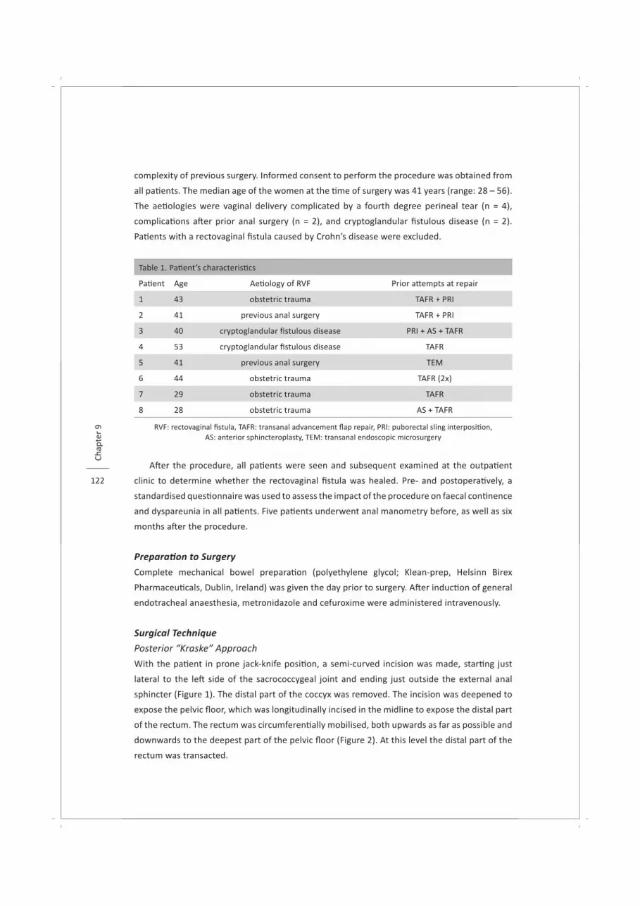
122
Chap
ter
9
complexity of previous surgery. Informed consent to perform the procedure was obtained from
all pati ents. The median age of the women at the ti me of surgery was 41 years (range: 28 – 56).
The aeti ologies were vaginal delivery complicated by a fourth degree perineal tear (n = 4),
complicati ons aft er prior anal surgery (n = 2), and cryptoglandular fi stulous disease (n = 2).
Pati ents with a rectovaginal fi stula caused by Crohn’s disease were excluded.
Table 1. Pati ent’s characteristi cs
Pati ent Age Aeti ology of RVF Prior att empts at repair
1 43 obstetric trauma TAFR + PRI
2 41 previous anal surgery TAFR + PRI
3 40 cryptoglandular fi stulous disease PRI + AS + TAFR
4 53 cryptoglandular fi stulous disease TAFR
5 41 previous anal surgery TEM
6 44 obstetric trauma TAFR (2x)
7 29 obstetric trauma TAFR
8 28 obstetric trauma AS + TAFR
RVF: rectovaginal fi stula, TAFR: transanal advancement fl ap repair, PRI: puborectal sling interpositi on, AS: anterior sphincteroplasty, TEM: transanal endoscopic microsurgery
Aft er the procedure, all pati ents were seen and subsequent examined at the outpati ent
clinic to determine whether the rectovaginal fi stula was healed. Pre- and postoperati vely, a
standardised questi onnaire was used to assess the impact of the procedure on faecal conti nence
and dyspareunia in all pati ents. Five pati ents underwent anal manometry before, as well as six
months aft er the procedure.
Preparati on to Surgery
Complete mechanical bowel preparati on (polyethylene glycol; Klean-prep, Helsinn Birex
Pharmaceuti cals, Dublin, Ireland) was given the day prior to surgery. Aft er inducti on of general
endotracheal anaesthesia, metronidazole and cefuroxime were administered intravenously.
Surgical Technique
Posterior “Kraske” Approach
With the pati ent in prone jack-knife positi on, a semi-curved incision was made, starti ng just
lateral to the left side of the sacrococcygeal joint and ending just outside the external anal
sphincter (Figure 1). The distal part of the coccyx was removed. The incision was deepened to
expose the pelvic fl oor, which was longitudinally incised in the midline to expose the distal part
of the rectum. The rectum was circumferenti ally mobilised, both upwards as far as possible and
downwards to the deepest part of the pelvic fl oor (Figure 2). At this level the distal part of the
rectum was transacted.

Rectal sleeve advancement 123
Figure 1. With the pati ent in
prone jackknife positi on, a
semi-curved incision is made
according to the marked line
Figure 2. The distal part of
the rectum is circumferenti ally
mobilised
In order to gain access to the anal canal a Lonestar retractor (Lone Star Retractor System,
Lone Star Medical Products, Houston, Texas) was used. Using electrocautery, a circumferenti al
mucosectomy was performed, starti ng at the dentate line and carried up to the level of
transecti on (Figure 3). The anal opening of the fi stula was enlarged and the enti re fi stulous
tract was excised. Thereaft er, the defect in the rectovaginal septum was closed in two layers,
leaving the vaginal mucosa open for drainage (Figure 4). Aft er advancement through the anal
canal (Figure 5), the mobilised and transected distal part of the rectum was anastomosed to
the dentate line with interrupted absorbable sutures (Figure 6). Aft er closing of the pelvic fl oor
with running absorbable sutures, a sucti on drain was placed and the skin was closed.
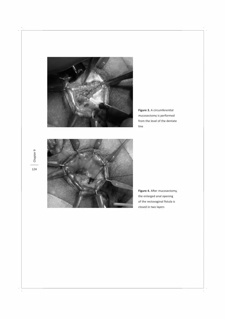
124
Chap
ter
9
Figure 3. A circumferenti al
mucosectomy is performed
from the level of the dentate
line
Figure 4. Aft er mucosectomy,
the enlarged anal opening
of the rectovaginal fi stula is
closed in two layers

Rectal sleeve advancement 125
Figure 5. The mobilised and
transected rectum is advanced
through the anal canal
Figure 6. The rectoanal
anastomosis is made with
interrupted absorbable
sutures
Abdominal Approach
With the pati ent in lithotomy positi on, a lower midline laparotomy was performed. The sigmoid
colon was mobilised by incising the lateral peritoneal refl ecti on. At the level of the sacral
promontory the presacral space was entered and developed. Under direct vision, posterior
dissecti on was extended and conti nued towards the level of the pelvic fl oor. The arterial supply
of the rectum was preserved. Anteriorly, the peritoneum was incised at the deepest part of
the pouch of Douglas and the rectovaginal septum was opened down to the pelvic fl oor. Both
anterolateral sides of the distal part of the rectum were mobilised. The rectum remained
att ached laterally because both lateral ligaments were not divided. Just above the deepest
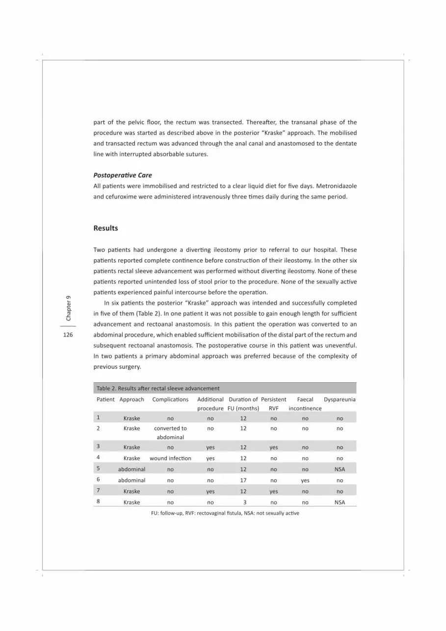
126
Chap
ter
9
part of the pelvic fl oor, the rectum was transected. Thereaft er, the transanal phase of the
procedure was started as described above in the posterior “Kraske” approach. The mobilised
and transacted rectum was advanced through the anal canal and anastomosed to the dentate
line with interrupted absorbable sutures.
Postoperati ve Care
All pati ents were immobilised and restricted to a clear liquid diet for fi ve days. Metronidazole
and cefuroxime were administered intravenously three ti mes daily during the same period.
Results
Two pati ents had undergone a diverti ng ileostomy prior to referral to our hospital. These
pati ents reported complete conti nence before constructi on of their ileostomy. In the other six
pati ents rectal sleeve advancement was performed without diverti ng ileostomy. None of these
pati ents reported unintended loss of stool prior to the procedure. None of the sexually acti ve
pati ents experienced painful intercourse before the operati on.
In six pati ents the posterior “Kraske” approach was intended and successfully completed
in fi ve of them (Table 2). In one pati ent it was not possible to gain enough length for suffi cient
advancement and rectoanal anastomosis. In this pati ent the operati on was converted to an
abdominal procedure, which enabled suffi cient mobilisati on of the distal part of the rectum and
subsequent rectoanal anastomosis. The postoperati ve course in this pati ent was uneventf ul.
In two pati ents a primary abdominal approach was preferred because of the complexity of
previous surgery.
Table 2. Results aft er rectal sleeve advancement
Pati ent Approach Complicati ons Additi onal
procedure
Durati on of
FU (months)
Persistent
RVF
Faecal
inconti nence
Dyspareunia
1 Kraske no no 12 no no no
2 Kraske converted to
abdominal
no 12 no no no
3 Kraske no yes 12 yes no no
4 Kraske wound infecti on yes 12 no no no
5 abdominal no no 12 no no NSA
6 abdominal no no 17 no yes no
7 Kraske no yes 12 yes no no
8 Kraske no no 3 no no NSA
FU: follow-up, RVF: rectovaginal fi stula, NSA: not sexually acti ve
Rectal sleeve advancement 127
Examinati on at the outpati ent clinic, revealed no signs of a persistent fi stula in fi ve pati ents.
Three pati ents encountered recurrent loss of gas and stool through their vagina shortly aft er
the procedure. In these three pati ents an additi onal transanal approach was performed to
close the anal opening of the fi stulous tract, which was successful in one pati ent. One of these
pati ents also encountered a postoperati ve wound infecti on. In the two pati ents with a diverti ng
ileostomy, the ileostomy was closed aft er seven and twelve months respecti vely.
Follow-up informati on was obtained from a standardised questi onnaire in all pati ents aft er
a median follow-up of 12 months (range: 3 – 17). Impairment of conti nence was noti ced in one
pati ent. She reported loss of solid stool, which occurred only during the night. In fi ve pati ents
anal manometry was performed before and aft er rectal sleeve advancement. No deteriorati on
of anal sphincter pressures was observed. None of the sexually acti ve women encountered de
novo dyspareunia aft er the procedure.
Discussion
Rectovaginal fi stulas, persisti ng aft er prior att empts at repair, are diffi cult to treat. Transanal
advancement fl ap repair, for example, fails in many women with such a persistent fi stula.
Lowry and co-workers 6 observed a healing rate of 88 percent aft er fl ap repair in pati ents
without previous repairs, whereas the healing rate dropped to 55 percent in pati ents who had
undergone two or more prior att empts at repair. Tsang et al. 12 reported that the closure rate
aft er fl ap repair declined from 45 percent in pati ents without a previous repair to 25 percent
in pati ents with a history of one or more previous repairs. Halverson et al. 18 reported an
overall healing rate of only 30 percent in pati ents who underwent transanal advancement fl ap
repair for persistent rectovaginal fi stula. Based on these data it seems obvious that persistent
rectovaginal fi stulas are diffi cult to treat by a transanal advancement repair.
It is thought that the adverse eff ects of previous repairs may be att ributed to insuffi cient
blood supply and the presence of scar ti ssue. Based on this assumpti on, it has been stated that
the outcome of rectovaginal fi stula repair can be improved by interpositi on of healthy, well-
vascularised ti ssue in the rectovaginal septum. However, in a previous study, it has been shown
that the healing rate aft er puborectal sling interpositi on was 92 percent in pati ents without
previous surgery and dropped to 31 percent in pati ents with one or more previous att empts
at repair 14. Since the treatment of persistent rectovaginal fi stulas is associated with a poor
outcome, we introduced an adapted rectal sleeve advancement for the treatment of these
fi stulas. In our opinion, this procedure has two potenti al benefi ts. First, the anal opening of the
fi stula is covered by a full-thickness layer of the rectum. Secondly, interpositi on of ti ssue in the
rectovaginal septum is avoided, thereby minimising the risk of de novo dyspareunia.
128
Chap
ter
9
Rectal sleeve advancement has been previously described by others 15-19. So far, this
procedure has only been performed by a transanal approach, almost exclusively in a limited
number of pati ents with a rectovaginal fi stula caused by Crohn’s disease. In most of these
studies, regarding transanal rectal sleeve advancement, the impact on faecal inconti nence and
dyspareunia was not assessed. To our knowledge, the present study is the fi rst one to describe
rectal sleeve advancement, either by a posterior “Kraske” approach, or by an abdominal
approach, for the treatment of persistent, low rectovaginal fi stulas in pati ents without Crohn’s
disease.
Although performed in only eight female pati ents, the observed healing rate aft er rectal
sleeve advancement of 75 percent is favourable as compared to the reported healing rates
aft er other techniques for the treatment of persistent rectovaginal fi stulas. It is obvious that
further research is warranted to assess the exact healing rate aft er rectal sleeve advancement
in a larger number of pati ents.
Faecal conti nence was adversely aff ected by the procedure in one pati ent. This pati ent
experienced involuntary loss of solid stool during the night. Prior to the rectal sleeve
advancement, she had undergone several transanal procedures in order to repair her
rectovaginal fi stula. Due to the limited number of selected pati ents, included in the present
feasibility study, it is not possible to determine whether the previous repairs or the rectal
sleeve advancement itself contributes to deteriorati on of conti nence.
Repair of rectovaginal fi stulas by interpositi on of ti ssue in the rectovaginal septum has a
potenti al risk of de novo dyspareunia 14. However, most authors do not menti on this serious
side eff ect. Since most pati ents with a rectovaginal fi stula are relati vely young and sexual acti ve,
the onset of dyspareunia, which has a signifi cant impact on quality of life, should be prevented
if possible. Rectal sleeve advancement, either by a posterior “Kraske” approach, or by an
abdominal approach, minimises the risk of de novo dyspareunia, as shown in the present study.
Conclusions
Rectal sleeve advancement, performed by a posterior “Kraske” approach, or by an abdominal
approach, results in fi stula healing in 75 percent of the pati ents with a persistent rectovaginal
fi stula, without dyspareunia as side eff ect. Based on these early results, it seems likely that this
adapted rectal sleeve advancement is a valuable alternati ve for the treatment of persistent
rectovaginal fi stulas.
Rectal sleeve advancement 129
References
1. Daniels BT. Rectovaginal fi stula: A clinical and pathological study. Minneapolis, University of Minnesota Graduate School. 1949.
2. Belt RL, Jr. Repair of anorectal vaginal fi stula uti lizing segmental advancement of the internal sphincter muscle. Dis Colon Rectum 1969;12(2):99-104.
3. Hibbard LT. Surgical management of rectovaginal fi stulas and complete perineal tears. Am J Obstet Gynecol 1978;130(2):139-41.
4. de Leeuw JW, Struijk PC, Vierhout ME, Wallenburg HC. Risk factors for third degree perineal ruptures during delivery. Bjog 2001;108(4):383-7.
5. Venkatesh KS, Ramanujam PS, Larson DM, Haywood MA. Anorectal complicati ons of vaginal delivery. Dis Colon Rectum 1989;32(12):1039-41.
6. Lowry AC, Thorson AG, Rothenberger DA, Goldberg SM. Repair of simple rectovaginal fi stulas. Infl uence of previous repairs. Dis Colon Rectum 1988;31(9):676-8.
7. Rothenberger DA, Christenson CE, Balcos EG, Schott ler JL, Nemer FD, Nivatvongs S, et al. Endorectal advancement fl ap for treatment of simple rectovaginal fi stula. Dis Colon Rectum 1982;25(4):297-300.
8. Wise WE, Jr., Aguilar PS, Padmanabhan A, Meesig DM, Arnold MW, Stewart WR. Surgical treatment of low rectovaginal fi stulas. Dis Colon Rectum 1991;34(3):271-4.
9. Jones IT, Fazio VW, Jagelman DG. The use of transanal rectal advancement fl aps in the management of fi stulas involving the anorectum. Dis Colon Rectum 1987;30(12):919-23.
10. 10. MacRae HM, McLeod RS, Cohen Z, Stern H, Reznick R. Treatment of rectovaginal fi stulas that has failed previous repair att empts. Dis Colon Rectum 1995;38(9):921-5.
11. Watson SJ, Phillips RK. Non-infl ammatory rectovaginal fi stula. Br J Surg 1995;82(12):1641-3.12. Tsang CB, Madoff RD, Wong WD, Rothenberger DA, Finne CO, Singer D, et al. Anal sphincter integrity
and functi on infl uences outcome in rectovaginal fi stula repair. Dis Colon Rectum 1998;41(9):1141-6.13. Zimmerman DD, Gosselink MP, Briel JW, Schouten WR. The outcome of transanal advancement fl ap
repair of rectovaginal fi stulas is not improved by an additi onal labial fat fl ap transpositi on. Tech Coloproctol 2002;6(1):37-42.
14. Oom DM, Gosselink MP, Van Dijl VR, Zimmerman DD, Schouten WR. Puborectal sling interpositi on for the treatment of rectovaginal fi stulas. Tech Coloproctol 2006;10(2):125-30.
15. Berman IR. Sleeve advancement anorectoplasty for complicated anorectal/vaginal fi stula. Dis Colon Rectum 1991;34(11):1032-7.
16. Hull TL, Fazio VW. Surgical approaches to low anovaginal fi stula in Crohn’s disease. Am J Surg 1997;173(2):95-8.
17. Marchesa P, Hull TL, Fazio VW. Advancement sleeve fl aps for treatment of severe perianal Crohn’s disease. Br J Surg 1998;85(12):1695-8.
18. Halverson AL, Hull TL, Fazio VW, Church J, Hammel J, Floruta C. Repair of recurrent rectovaginal fi stulas. Surgery 2001;130(4):753-8.
19. Simmang CL, Lacey SW, Huber PJ, Jr. Rectal sleeve advancement: repair of rectovaginal fi stula associated with anorectal stricture in Crohn’s disease. Dis Colon Rectum 1998;41(6):787-9.

Summary


Chapter 10Summary and discussion
Samenvatti ng voor niet-ingewijden
134
Chap
ter
10
Posterior pelvic compartment disorders are relati vely common, especially in older multi parous
women, and have a signifi cant impact on quality of life. This thesis is aimed at evaluati ng diagnosis
and treatment of disorders of the posterior pelvic compartment. A general introducti on to this
subject, as well as the aims and outline of this thesis, are presented in Chapter 1.
Part I. Posterior Pelvic Compartment Prolapse
Short-term follow-up has shown that transabdominal enterocele repair, uti lising syntheti c graft
material, is benefi cial, especially for pati ents with symptoms of pelvic discomfort. However,
it is questi onable whether this benefi cial eff ect does sustain in the long-term. The long-term
eff ect of transabdominal enterocele repair by obliterati on of the pelvic inlet was evaluated and
the results of this study were described in Chapter 2. Despite an adequate enterocele repair,
assessed by evacuati on proctography six months aft er the procedure, recurrent symptoms
of pelvic discomfort were observed in approximately one of four pati ents aft er a median
follow-up of 85 months. Although co-existi ng prolapses in the other pelvic compartments
might explain the recurrence of symptoms, they may also be att ributed to progression of the
underlying disorder causing physiologic changes that surpass the otherwise eff ecti ve repair. It
seems unlikely that enteroceles are a major cause of obstructed defaecati on, since defaecati on
diffi culti es persisted in the majority of pati ents who presented with this problem prior to the
procedure. It is more likely that other abnormaliti es, such as impaired rectal sensory percepti on
and altered rectal wall contracti lity, also contribute to the problem of obstructed defaecati on
in pati ents with an enterocele. To date, there is no consensus regarding the indicati ons for
enterocele repair and the need for concomitant surgery for associated abnormaliti es. It seems
likely that surgical repair should mainly depend on pati ent’s symptoms. Enterocele repair can
be off ered to pati ents with pelvic discomfort. However, this procedure is not recommended if
obstructed defaecati on is the most prominent complaint. Enterocele should be considered as a
part of pelvic organ prolapse and concomitant prolapses should, therefore, be diagnosed and
treated if necessary in all pati ents.
A major drawback of transvaginal and transanal rectocele repair is the potenti al risk of
dyspareunia aft er the procedure. It has been suggested that rectoceles can also be treated
by a transabdominal approach without this serious side eff ect. In Chapter 3 the results of
transabdominal anterolateral rectopexy for the treatment of pati ents with rectocele and
associated obstructed defaecati on are described. Anterolateral rectopexy resulted in an
adequate anatomic correcti on in 80 percent of the pati ents, without dyspareunia as a side
eff ect. Despite this adequate repair, obstructed defaecati on persisted in the majority of
pati ents. Since obstructed defaecati on also persists aft er other types of rectocele repair, the
questi on arises whether rectocele is the cause or rather the result of obstructed defaecati on.

Summary and discussion 135
In the last decade more att enti on has been paid to the underlying pathophysiologic mechanism
of obstructed defaecati on. It has been questi oned whether obstructed defaecati on is the result
of one single abnormality, such as rectocele. It seems more likely that obstructed defaecati on is
caused by multi ple factors, which might explain why anatomical correcti on of a rectocele alone,
as obtained by anterolateral rectopexy, fails to restore symptoms of obstructed defaecati on
in a substanti al number of pati ents. Further research is warranted to reveal the exact
pathophysiologic mechanism of obstructed defaecati on and to identi fy adequate treatment
strategies for pati ents with defaecati on diffi culti es.
Evacuati on proctography provides a useful tool for the assessment of posterior pelvic
compartment prolapse. However, this imaging technique is relati vely invasive and requires
substanti al exposure to ionising radiati on. Furthermore, evacuati on proctography, without
opacifi cati on of the bladder, lacks the ability to visualise the anterior and middle pelvic
compartment. Imaging of all three pelvic compartments seems to be important, since it
has been shown that pati ents with one type of prolapse oft en have concomitant prolapses,
requiring treatment as well. Transperineal ultrasound, which has recently been developed,
off ers the possibility to visualise abnormaliti es in all three pelvic compartments simultaneously,
without exposure to ionising radiati on and at low cost. In Chapter 4 the results are presented
of a study, conducted to assess the level of agreement between evacuati on proctography
and transperineal ultrasound in diagnosing posterior pelvic compartment prolapse. Good
agreement was found for the detecti on of enteroceles. Regarding the detecti on of rectoceles,
a moderate agreement was found. For the detecti on of intussuscepti on the level of correlati on
was fair. However, it has been reported that intussuscepti on is present in up to 50 percent
of healthy volunteers, therefore, the clinical signifi cance of this incomplete rectal prolapse
is unknown. In additi on, transperineal ultrasound was signifi cantly bett er tolerated by the
pati ents. Based on these fi ndings, transperineal ultrasound is considered to be an alternati ve
to evacuati on proctography in diagnosing posterior pelvic compartment prolapse. Therefore, it
may be used as the fi rst diagnosti c tool for symptomati c pati ents.
Part II. Faecal Inconti nence
To date, anterior sphincteroplasty is considered as the treatment of choice for faecal
inconti nent pati ents with an external anal sphincter defect, in whom supporti ve management
fails. It has recently been reported that pati ents with an external anal sphincter defect may
also benefi t from sacral neuromodulati on. Therefore, it is questi onable whether pati ents with
such an external anal sphincter defect should be treated primarily by anterior sphincteroplasty
or directly by sacral neuromodulati on. Unfortunately, the results of both procedures are
hardly to compare, since in most studies regarding anterior sphincteroplasty other outcome
136
Chap
ter
10
classifi cati ons are used than in studies regarding sacral neuromodulati on. In Chapter 5 the
results of anterior sphincteroplasty are described, based on an outcome classifi cati on similar
to that used in studies regarding sacral neuromodulati on. Using this outcome classifi cati on,
anterior sphincteroplasty was found to result in a successful outcome in 60 percent of the
pati ents, which is sustained in the long-term. However, a less favourable outcome was found
in pati ents aged 50 years or more at the ti me of the procedure. A similar fi nding has been
reported by others. It seems obvious that additi onal factors play a role in the aeti ology of
faecal inconti nence in these older women, since their external anal sphincter defect was most
likely caused by vaginal delivery many years before. The questi on arises whether external
anal sphincter repair can be justi fi ed as the surgical treatment of choice in these older faecal
inconti nent pati ents. The results obtained from this study facilitate a bett er comparison
between the outcomes of anterior sphincteroplasty and sacral neuromodulati on. However,
to assess the defi nite role of both procedures in the management of faecal inconti nence,
associated with external anal sphincter defects, randomised controlled trials are warranted.
Vaginal delivery may result not only in anal sphincter defects but also in pelvic fl oor injury.
However, unti l now, the infl uence of this type of injury on the aeti ology of faecal inconti nence
and its treatment is unknown. The prevalence of pelvic fl oor injury in pati ents with faecal
inconti nence, who were eligible for anterior sphincteroplasty or sacral neuromodulati on in the
past, is described in Chapter 6 and 7 respecti vely. In additi on, the impact of this type of injury on
the outcome of both procedures is verifi ed. Transperineal ultrasound revealed that pelvic fl oor
injury is present in the majority of pati ents who were eligible for surgical treatment of their
inconti nence. However, based on the results of both studies, it seems unlikely that this type
of injury itself has a detrimental eff ect, or on the outcome of anterior sphincteroplasty, either
on the effi cacy of sacral neuromodulati on. It is questi onable whether the high prevalence of
pelvic fl oor injury in the studied pati ents refl ects a major contributi ng role of this type of injury
in the aeti ology of faecal inconti nence. Recently, is has been reported that pati ents with faecal
inconti nence have a higher prevalence of pelvic fl oor injury and concomitant anal sphincter
defects as compared to asymptomati c parous controls. It might be possible that women with an
anal sphincter defect due to vaginal delivery plus concomitant pelvic fl oor injury have a higher
risk for developing faecal inconti nence as compared to women who have only an external anal
sphincter defect. Further research should reveal the exact role of pelvic fl oor injury in the
aeti ology of faecal inconti nence.
Summary and discussion 137
Part III. Rectovaginal Fistula
Various techniques are available for the surgical treatment of rectovaginal fi stulas. However,
the results aft er these procedures are oft en disappointi ng, especially in women in whom the
fi stula persisted aft er prior surgery. It has been suggested that interpositi on of healthy, well-
vascularised ti ssue, such as puborectal muscle, may be the key to rectovaginal fi stula healing. In
Chapter 8 the results aft er puborectal sling interpositi on in the surgical treatment of rectovaginal
fi stulas are presented. Puborectal sling interpositi on leads to rectovaginal fi stula healing in the
majority of pati ents without previous repairs. However, in most pati ents who had undergone
one or more previous att empts at repair, the fi stula persisted. In additi on, a high prevalence of
de novo dyspareunia was observed aft er this procedure. Based on the less favourable outcome
and the high risk of de novo dyspareunia, puborectal sling interpositi on cannot be considered
as the treatment of choice for women with rectovaginal fi stulas, especially not for those with a
persistent fi stula aft er prior att empts at repair.
Therefore, rectal sleeve advancement was adapted for the treatment of pati ents with a
persistent rectovaginal fi stula. In Chapter 9 the feasibility of this new type of rectovaginal
fi stula repair is described. Although performed in a limited number of pati ents, rectal sleeve
advancement resulted in fi stula healing in 75 percent of the pati ents with a persistent
rectovaginal fi stula, without serious postoperati ve complicati ons and without dyspareunia
as a side eff ect. Based on these early results, it seems likely that this adapted rectal sleeve
advancement is a valuable alternati ve for the treatment of women with persistent rectovaginal
fi stulas. However, further research, conducted in a larger number of pati ents, is warranted to
assess the exact role of this procedure.
Conclusions
Transabdominal obliterati on of the pelvic inlet is only benefi cial for pati ents with an enterocele
and associated symptoms of pelvic discomfort (Chapter 2).
Rectocele repair by transabdominal anterolateral rectopexy is not benefi cial for pati ents with
obstructed defaecati on (Chapter 3).
Transperineal ultrasound can be used as the fi rst diagnosti c tool for the assessment of pati ents
with symptoms suggesti ng posterior pelvic compartment prolapse (Chapter 4).
Improvement of faecal conti nence aft er anterior sphincteroplasty sustains in the long-term
(Chapter 5).
138
Chap
ter
10
Pelvic fl oor injury is present in the majority of faecal inconti nent pati ents who were eligible for
surgical treatment in the past (Chapters 6 & 7).
The effi cacy of the surgical treatment of women with faecal inconti nence is not infl uenced by
pelvic fl oor injury (Chapters 6 & 7).
Dyspareunia is a serious side eff ect of puborectal sling interpositi on (Chapter 8).
Rectal sleeve advancement seems a valuable alternati ve for the treatment of women with
persistent rectovaginal fi stulas (Chapter 9).
Samenvatti ng voor niet-ingewijden 139
Aandoeningen van het achterste comparti ment van het kleine bekken komen relati ef vaak
voor, voornamelijk bij vrouwen op een hogere leeft ijd en na één of meerdere vaginale
bevallingen. Deze aandoeningen belemmeren de pati ënt vaak in het dagelijks functi oneren.
Dit proefschrift richt zich op de diagnose en de behandeling van deze aandoeningen van het
achterste comparti ment van het kleine bekken. Hoofdstuk 1 vormt de algemene inleiding van
dit proefschrift . Tevens worden in dit hoofdstuk de doelen en de opbouw van het proefschrift
beschreven.
Deel I. Verzakking van het Achterste Comparti ment van het Kleine Bekken
Het herstel van een enterocèle door middel van een transabdominale ingreep, waarbij gebruik
gemaakt wordt van een syntheti sch matje, is op de korte termijn zinvol gebleken bij pati ënten
met klachten van een verzakkingsgevoel. Het is echter de vraag of dit eff ect ook op de lange
termijn blijft bestaan. In Hoofdstuk 2 wordt het lange-termijn resultaat beschreven van een
operati e waarbij de bekkeningang, via een transabdominale ingreep, wordt afgesloten. Ondanks
dat bleek dat de enterocèle was verdwenen na de ingreep, keerde het verzakkingsgevoel bij één
op de vier vrouwen uiteindelijk terug. Dit zou mogelijk kunnen passen bij de aanwezigheid van
andere verzakkingen, bijvoorbeeld van de blaas of van de baarmoeder, maar het zou ook goed
kunnen dat dit gewijd moet worden aan langzame uitbreiding van de onderliggende oorzaak,
waardoor in eerste instanti e de enterocèle is ontstaan. Daarnaast bleef een bemoeilijkte
stoelgang na de operati e bestaan bij de meeste pati ënten waarbij dit probleem voor de operati e
gezien werd. Het lijkt dus niet waarschijnlijk dat de enterocèle alleen de oorzaak is voor het
ontstaan van een bemoeilijkte stoelgang. Het is waarschijnlijker dat andere afwijkingen,
bijvoorbeeld een veranderde werking van de wand van de endeldarm, ook bijdragen aan de
bemoeilijkte stoelgang die sommige vrouwen met een enterocèle ervaren. De beslissing of
een pati ënt met een enterocèle geopereerd zal worden, dient gebaseerd te worden op het
klachtenpatroon van de vrouw. Indien het belangrijkste probleem het verzakkingsgevoel is,
kan een afsluiti ng van de bekkeningang overwogen worden. Echter, deze ingreep dient niet
aanbevolen te worden als een bemoeilijkte stoelgang op de voorgrond staat. Daarnaast dient
ook acti ef gezocht te worden naar de aanwezigheid van eventuele andere verzakkingen, die
indien zij aanwezig zijn ook behandeld dienen te worden.
Een potenti eel risico van het herstel van een rectocèle, door middel van een transvaginale
of transanale ingreep, is het ontstaan van pijn ti jdens het vrijen na de operati e. Een
transabdominale ingreep lijkt dit risico niet te hebben. In Hoofdstuk 3 worden de resultaten
beschreven van een behandeling van rectocèles door middel van de transabdominale voorste
rectopexie. Na deze ingreep bleek de rectocèle adequaat corrigeerd te zijn bij 80 procent van de
pati ënten, zonder dat er pijn ti jdens het vrijen ontstond. Echter, ondanks het verdwijnen van de
140
Chap
ter
10
rectocèle na de operati e, bleef bij de meeste vrouwen een bemoeilijkte stoelgang bestaan. Dit
teleurstellende resultaat wordt ook gevonden na andere typen ingrepen voor de behandeling
van een rectocèle. Het is daarom de vraag of de rectocèle de oorzaak of juist het gevolg is van
een bemoeilijkte stoelgang. Op basis van recent onderzoek lijkt het waarschijnlijker dat een
bemoeilijkte stoelgang ontstaat door meerdere factoren, wat mogelijk kan verklaren waarom
het herstel van de rectocèle alleen niet leidt tot een vermindering van de klachten van de
pati ënt. Vervolgonderzoek is nodig om aan te tonen wat het exacte onderliggende mechanisme
van een bemoeilijkte stoelgang is. Op basis van deze informati e kunnen dan mogelijk andere
behandelingsopti es worden ontwikkeld.
Het defaecogram is een goede manier om pati ënten te onderzoeken die zich presenteren
met klachten welke mogelijk kunnen passen bij verzakking in het achterste comparti ment
van het kleine bekken. Belangrijke nadelen van deze beeldvormende techniek zijn echter
de blootstelling aan röntgenstraling en de benodigde vulling van de endeldarm, de dunne
darm en de vagina met contrastmiddel. Daarnaast is het niet mogelijk om met deze techniek
informati e te verkrijgen over de toestand van het voorste en het middelste comparti ment.
Omdat pati ënten met één verzakking vaak tegelijkerti jd andere verzakkingen hebben, die
tevens behandeld dienen te worden, is het duidelijk dat het verkrijgen van informati e over
alle comparti menten van het kleine bekken onmisbaar is. Transperineale echografi e is een
nieuwe techniek, die de mogelijkheid biedt om alle comparti menten tegelijkerti jd in beeld te
brengen. Daarnaast gebruikt deze techniek geen röntgenstraling en is dit onderzoek relati ef
goedkoop uit te voeren. In Hoofdstuk 4 wordt beschreven wat de mate van overeenkomst is
tussen het defaecogram en de nieuwe transperineale echografi e als het gaat om het aantonen
van verzakking in het achterste comparti ment van het kleine bekken. Tijdens de studie hebben
alle pati ënten met klachten welke mogelijk zouden kunnen passen bij deze verzakkingen
beide onderzoeken ondergaan. Een goede mate van overeenkomst werd gevonden voor het
aantonen van een enterocèle. Voor het aantonen van een rectocèle werd een gemiddelde
overeenkomst gevonden en een mati ge overeenkomst werd gevonden voor het aantonen van
een intussuscepti e. Het is echter niet duidelijk of een intussuscepti e klinische betekenis heeft .
Tevens gaven de pati ënten aan dat zij de transperineale echografi e een stuk minder belastend
vonden dan het defaecogram. Gebaseerd op deze bevindingen is transperineale echografi e
een goed alternati ef voor het defaecogram bij pati ënten met klachten welke mogelijk kunnen
passen bij verzakking in het achterste comparti ment van het kleine bekken.
.

Samenvatti ng voor niet-ingewijden 141
Deel II. Faecale Inconti nenti e
Voor pati ënten met faecale inconti nenti e op basis van een defect in de buitenste anale sfi ncter,
die geen baat hebben gehad bij ondersteunende maatregelen, is een voorste sfi ncterplasti ek
op dit moment de behandeling van keuze. Recent is duidelijk geworden dat deze pati ënten
ook geholpen kunnen worden door middel van sacrale neuromodulati e. Het is daarom de
vraag of deze inconti nente pati ënten met een defect in de buitenste anale sfi ncter primair
behandeld dienen te worden door middel van een voorste sfi ncterplasti ek of door middel van
sacrale neuromodulati e. Omdat in de meeste studies naar de voorste sfi ncterplasti ek gebruik
gemaakt wordt van andere uitkomstmaten dan in de studies naar sacrale neuromodulati e
zijn de resultaten van beide behandelmethoden tot op heden lasti g te vergelijken. In
Hoofdstuk 5 worden de resultaten beschreven van een studie naar de uitkomst van de voorste
sfi ncterplasti ek. Hierbij is gebruik gemaakt van een uitkomstmaat die vergelijkbaar is met de
uitkomstmaat die vaak gebruikt wordt in studies naar sacrale neuromodulati e. Het is hierdoor
beter mogelijk om de resultaten van beide behandelmethoden met elkaar te vergelijken. De
voorste sfi ncterplasti ek bleek over het algemeen te resulteren in een substanti ële verbetering
van de faecale conti nenti e bij 60 procent van de pati ënten. Daarnaast bleef dit gunsti ge eff ect
ook op de lange termijn bestaan. Pati ënten met een leeft ijd van 50 jaar of ouder ti jdens de
ingreep bleken helaas een stuk minder baat te hebben bij de ingreep. Het is bekend dat een
beschadiging van de sfi ncters voornamelijk ontstaat ti jdens een vaginale bevalling. Deze
beschadiging is bij deze vrouwen dus al jaren aanwezig. Het lijkt daarom logisch dat er meerdere
factoren een rol spelen bij het ontstaan van faecale inconti nenti e bij deze vrouwen. Het is dan
ook de vraag of het uitvoeren van een voorste sfi ncterplasti ek bij deze vrouwen van 50 jaar of
ouder gerechtvaardigd kan worden.
Een vaginale bevalling kan niet alleen tot beschadiging van de anale sfi ncters leiden,
maar ook tot beschadiging van de bekkenbodem. Het is echter tot op heden onbekend of
bekkenbodemschade een rol speelt in de ontstaanswijze van faecale inconti nenti e en van
invloed is op het resultaat van een eventuele behandeling van dit probleem. In Hoofdstuk 6 en
7 wordt beschreven in welk deel van de vrouwen, die in het verleden chirurgisch zijn behandeld
voor faecale inconti nenti e, een beschadiging van de bekkenbodem gevonden wordt. Tevens
wordt beschreven of deze beschadiging een invloed heeft op het resultaat van zowel een
voorste sfi ncterplasti ek (Hoofdstuk 6) als sacrale neuromodulati e (Hoofdstuk 7). Op basis van
de uitgevoerde transperineale echografi e bleek dat de meeste pati ënten, na een chirurgische
behandeling van faecale inconti nenti e, bekkenbodemschade hadden. De aanwezigheid van
deze schade had echter geen invloed op het wel of niet slagen van de uitgevoerde chirurgische
behandeling. Mogelijk kan het hoge percentage vrouwen met een beschadigde bekkenbodem
in deze groep pati ënten worden verklaard doordat de aanwezigheid van bekkenbodemschade
een rol speelt in de ontstaanswijze van faecale inconti nenti e. Het blijkt dat de combinati e van

142
Chap
ter
10
een beschadigde bekkenbodem met beschadigde sfi ncters vaker voorkomt bij vrouwen met
faecale inconti nenti e dan bij vrouwen zonder klachten. Het is dus mogelijk dat vrouwen met
schade aan de anale sfi ncters en tegelijkerti jd schade aan de bekkenbodem een hoger risico
lopen om faecaal inconti nent te worden dan vrouwen met alleen schade aan de anale sfi ncters.
Vervolgonderzoek zal moeten uitwijzen wat de exacte rol van bekkenbodemschade is in de
ontstaanswijze van faecale inconti nenti e.
Deel III. Rectovaginale Fistels
Verschillende technieken zijn beschreven voor de chirurgische behandeling van een
rectovaginale fi stel. Echter, het is gebleken dat deze operati eve procedures vaak tot een
teleurstellend resultaat leiden, voornamelijk bij vrouwen met een rectovaginale fi stel, die al
een eerdere en dus mislukte operati e voor de behandeling van deze fi stel hebben ondergaan.
Het is gesuggereerd dat er een beter resultaat behaald kan worden als er gezond en goed
doorbloed weefsel aangebracht wordt tussen de endeldarm en de vagina. In Hoofdstuk 8
worden de resultaten van een dergelijk ingreep, de puborectale sling interpositi e, beschreven.
Na deze operati e genas de fi stel in de meerderheid van de pati ënten die nog niet eerder
geopereerd waren aan deze fi stel. Echter, de fi stel persisteerde in de meeste pati ënten die wel
al een eerdere operati e hadden ondergaan. Daarnaast ontstond in een aanzienlijk deel van
de pati ënten pijn ti jdens het vrijen na de operati e. Op basis van deze ongunsti ge resultaten
blijkt de puborectale sling interpositi e niet de behandeling van keuze voor vrouwen met een
rectovaginale fi stel, zeker niet voor diegene die al een eerdere operati e voor de behandeling
van deze fi stel hebben ondergaan.
Hierop voortbordurend werd een andere operati e, de rectale verschuivingsplasti ek,
aangepast voor de behandeling van pati ënten met een nog bestaande rectovaginale fi stel na
een eerdere operati e. In Hoofdstuk 9 wordt de haalbaarheid van deze nieuwe operati e voor
de behandeling van vrouwen met deze persisterende rectovaginale fi stels beschreven. De fi stel
genas in driekwart van de vrouwen, zonder serieuze complicati es en zonder dat er pijn ti jdens
het vrijen ontstond na de operati e. Echter, ti jdens dit onderzoek is de procedure uitgevoerd
bij slechts een klein aantal pati ënten. Daarom kan op basis van deze voorlopige resultaten
alleen geconcludeerd worden dat de rectale verschuivingsplasti ek een waardevol alternati ef
lijkt te zijn voor de behandeling van vrouwen met een persisterende rectovaginale fi stel.
Vervolgonderzoek zal moeten uitwijzen wat de uiteindelijke waarde van deze ingreep is.
Samenvatti ng voor niet-ingewijden 143
Conclusies
Transabdominale afsluiti ng van de bekkeningang is alleen zinvol bij pati ënten met een
enterocèle en daarbij passende klachten van een verzakkingsgevoel (Hoofdstuk 2).
Herstel van een rectocèle door middel van een transabdominale voorste rectopexie is niet
zinvol bij pati ënten met een bemoeilijkte stoelgang (Hoofdstuk 3).
Transperineale echografi e kan worden gebruikt als het primair beeldvormend onderzoek bij
pati ënten met klachten die zouden kunnen passen bij verzakking in het achterste comparti ment
van het kleine bekken (Hoofdstuk 4).
Na een voorste sfi ncterplasti ek blijft de verbetering van faecale conti nenti e op de lange termijn
bestaan (Hoofdstuk 5).
Bekkenbodemschade is aanwezig bij het merendeel van de pati ënten die in het verleden
chirurgisch zijn behandeld voor faecale inconti nenti e (Hoofdstuk 6 & 7).
De eff ecti viteit van een chirurgische behandeling van faecale inconti nenti e wordt niet beïnvloed
door de aanwezigheid van bekkenbodemschade (Hoofdstuk 6 & 7).
Pijn bij het vrijen is een belangrijke bijwerking van een puborectale sling interpositi e (Hoofdstuk
8).
Een rectale verschuivingsplasti ek lijkt een waardevol alternati ef te zijn voor de behandeling van
vrouwen met een persisterende rectovaginale fi stel (Hoofdstuk 9).


Appendices


Publicati ons

Publicati ons 149
Publicati ons
Oom DMJ, Gosselink MP, van Dijl VRM, Zimmerman DDE, Schouten WR. Puborectal sling
interpositi on for the treatment of rectovaginal fi stulas. Tech Coloproctol 2006;10(2):125-30.
Oom DMJ, van der Sluijs JW, den Hoed PT. Infrascapulair elastofi broom: een radiologische
diagnose? Ned Tijdschr Heelkd 2007;16(2);323-324.
Oom DMJ, Gosselink MP, Schouten WR. Muscle interpositi on in pati ents with fi stulas between
the rectum and urethra or vagina. Dis Colon Rectum 2007;50(4):548.
Oom DMJ, van Dijl VRM, Gosselink MP, van Wijk JJ, Schouten WR. Enterocele repair by
abdominal obliterati on of the pelvic inlet. Long-term outcome on obstructed defaecati on and
symptoms of pelvic discomfort. Colorectal Dis 2007;9(9):845-50.
Oom DMJ, Gosselink MP, van Wijk JJ, van Dijl VRM, Schouten WR. Rectocele repair by
anterolateral rectopexy, long-term functi onal outcome. Colorectal Dis 2008;10(9):925-30.
Alexander SM, Mitalas LE, Gosselink MP, Oom DMJ, Zimmerman DDE, Schouten WR. Obliterati on
of the fi stulous tract with Bioglue adversely aff ects the outcome of transanal advancement fl ap
repair. Tech Coloproctol 2008;12(3):225-8.
Gosselink MP, Oom DMJ, Zimmerman DDE, Schouten WR. Marti us fl ap: an adjunct for complex,
low rectovaginal fi stulas. Am J Surg 2009;197(6):833-4.
Mitalas LE, Gosselink MP, Oom DMJ, Zimmerman DDE, Schouten WR. Required length of
follow-up aft er transanal advancement fl ap repair of high transsphincteric fi stulas. Colorectal
Dis 2008;[Epub ahead of print].
Oom DMJ, Schouten WR. Role of rectocele in obstructed defaecati on. Colorectal Dis
2008;10(8):849-850.
Oom DMJ, Gosselink MP, Schouten WR. Enterocele – Diagnosis and treatment. Gastroenterol
Clin Biol 2009;33(2):135-7.
Mitalas LE, Gosselink MP, Oom DMJ, Zimmerman DDE, Schouten WR. Adverse eff ect of BioGlue
on the outcome of transanal advancement fl ap repair. Dis Colon Rectum 2009;52(4):754.
150
App
endi
ces
Mitalas LE, Schouten SB, Gosselink MP, Oom DMJ, Zimmerman DDE, Hop WJC, Schouten WR.
Does rectal mucosal bloodfl ow aff ect the outcome of transanal advancement fl ap repair of high
transsphincteric fi stulas? Dis Colon Rectum 2009;52(8):1395-9.
Steensma AB, Oom DMJ, Burger CW, Schouten WR. Assessment of posterior compartment
prolapse; a comparison of evacuati on proctography and 3D transperineal ultrasound. Colorectal
Dis 2009;[Epub ahead of print].
Oom DMJ, Gosselink MP, Schouten WR. Anterior spincteroplasty for fecal inconti nence; a single
center experience in the era of sacral neuromodulati on. Dis Colon Rectum 2009; in press.
Oom DMJ, Steensma AB, Zimmerman DDE, Schouten WR. Anterior sphincteroplasty for fecal
inconti nence: is the outcome compromised in pati ents with associated pelvic fl oor injury? Dis
Colon Rectum 2009; in press.
Oom DMJ, Steensma AB, van Lanschot JJB, Schouten WR. Is sacral neuromodulati on worthwhile
in pati ents with associated pelvic fl oor injury? Submitt ed for publicati on.
Schouten WR, Oom DMJ. Rectal sleeve advancement for persistent rectovaginal fi stulas; a
feasibility study. Tech Coloproctol 2009; in press.

Dankwoord

Dankwoord 153
Dankwoord
Dit proefschrift is tot stand gekomen met de hulp en steun van vele mensen. Hen wil ik natuurlijk
allen hartelijk bedanken. Enkelen wil ik hierbij in het bijzonder noemen.
In de eerste plaats mijn copromotor: dr. W.R. Schouten. Het is een voorrecht om te mogen
werken binnen uw colorectale onderzoeksgroep. Uw gedrevenheid en verhelderende kijk op
de zaak zijn telkens weer buitengewoon enthousiasmerend. Ik heb uw inspirerende manier
van denken, de wetenschappelijke scholing en de nimmer afl atende begeleiding die ik in de
afgelopen jaren heb mogen ervaren alti jd ontzett end gewaardeerd. Bedankt dat u alti jd voor
mij heeft klaar gestaan en voor de mooie en leerzame jaren die ik heb gehad op het functi elab.
Ik zal het zeker ontzett end gaan missen.
Prof. dr. J.J.B van Lanschot, mijn promotor, ik ben u veel dank verschuldigd voor het in mij
gestelde vertrouwen en de geboden kans om een jaar onder uw supervisie te werken aan
de afronding van dit proefschrift . Uw belangstelling en visie heb ik steeds als ontzett end
sti mulerend ervaren. Dank voor de pretti ge begeleiding en de soepele coördinati e van mijn
promoti e.
De leden van de kleine commissie, prof. dr. C.W. Burger, prof. dr. C.G.M.I. Baeten en dr. C.J. van
der Woude, wil ik hartelijk bedanken voor de vlott e beoordeling van het manuscript.
De leden van de grote commissie, prof. dr. M.E. Vierhout en prof. dr. H.W. Tilanus, ben ik zeer
erkentelijk voor hun bereidheid zitti ng te nemen in de promoti ecommissie.
Dr. A.B. Steensma wil ik bedanken voor haar bereidheid om als deskundige zitti ng te nemen in
de promoti ecommissie. Lieve Anneke, beschermvrouwe van de transperineale echografi e in
het Erasmus MC en ver daarbuiten, ik kan je niet genoeg bedanken voor het feit dat jij naast
jouw promoti eonderzoek alti jd weer het geduld kon opbrengen om mij de transperineale
echotechniek bij te brengen. Daarnaast ben ik je zeer erkentelijk voor de coauteurschappen
en mijn introducti e in de wereld van de gynaecologie. Ik hoop nog veel van je te kunnen leren.
Dr. D.D.E. Zimmerman, beste David, dank voor jouw hulp, inspirati e en (grafi sche) ondersteuning.
Ik heb het alti jd erg gewaardeerd dat jij naast jouw drukke opleiding en gezin nog ti jd wilde en
kon vrijmaken om steeds weer mee te denken en prachti ge schemati sche fi guren te creëren.
Daarnaast ook veel dank voor alle adviezen en bemoedigende woorden, die ik soms zo hard
nodig had. Ik hoop dat we in de toekomst nog eens kunnen samenwerken.
154
App
endi
ces
Mijn collega-onderzoeker en kamergenoot Litza Mitalas. Lieve Litza, heel veel dank voor jouw
grote relati veringsvermogen, waarmee jij mij geregeld weer even de realiteit liet inzien. Dank
voor jouw steun, de gezelligheid, jouw taalkundige hulp en de vele hilarische momenten die
we samen hebben meegemaakt op het functi elab. Wat ontzett end mooi dat we het nu kunnen
afsluiten met onze beide promoti es. Ik wens je heel veel succes met jouw carrière.
Dr. M.P. Gosselink, beste Marti jn, zonder jou was dit proefschrift er waarschijnlijk niet gekomen.
Jij opperde bij mij het idee om onderzoek te komen doen bij de colorectale onderzoeksgroep,
wat al snel van een bijzaak een hoofdzaak werd. Ik dank jou voor de fi jne samenwerking en
jouw uitgebreide wetenschappelijke inbreng. Het is mij nog immer een raadsel waar jij toch
steeds alle energie vandaan haalt.
Dr. B.F.M. Blok en alle overige leden van het bekkenbodem- en inconti nenti ecentrum wil ik
graag bedanken voor de soepele samenwerking bij het uitvoeren van de verschillende studies.
Veel leerzame en plezierige momenten heb ik ervaren ti jdens de wekelijkse bekkenbodem-
bespreking. Tevens wil ik de dames van de polikliniek gynaecologie bedanken voor hun
medewerking aan de echo studies en voor het ontvangen van alle deelnemende pati ënten.
Anneke van der Meulen en Adrie van Houwelingen, ik wil jullie ontzett end bedanken voor de
trouwe support in de afgelopen ti jd. Bedankt voor jullie geduld en accurate administrati eve
begeleiding.
Mijn collega arts-assistenten, dr. A.M. van Heusden en de overige leden van de maatschap
Obstetrie en Gynaecologie in het Maasstad Ziekenhuis wil ik bedanken voor de ruimte die
ik heb gekregen om naast mijn arts-assistentschap de laatste loodjes van dit proefschrift te
kunnen afronden. Tevens heel veel dank voor de soepele wijze waarop ik mijn eerste klinische
ervaring heb kunnen opdoen.
Mijn paranimfen en goede vriendinnen: Bianca Mostert en Nicole Wegman. Lieve Bianca
en lieve Nicole, ik ben ontzett end trots dat jullie mij bij willen staan als paranimfen. Jullie
vriendschap, steun en opti misme waardeer ik ontzett end. Ik kijk uit naar alle mooie momenten
van vriendschap die nog zullen komen.
Lieve ouders, ontzett end bedankt voor jullie interesse en enorme enthousiasme voor mijn
studie en later ook voor mijn promoti eonderzoek. Zonder onvoorwaardelijke steun en liefde
van het thuisfront was dit allemaal niet gelukt. Bedankt voor alles.

Dankwoord 155
Patrick, lieve broer, ondanks dat onze beroepskeuzes ervoor zorgen dat wij elkaar niet zo vaak
kunnen zien, ben jij er steeds voor me als het erop aankomt.
Tenslott e, lieve Michiel, bij jou kom ik pas echt tot rust. Ik hoop dat we nog heel lang samen
gelukkig zullen zijn.


Curriculum Vitae

Curriculum Vitae 159
Curriculum Vitae
Daniëlla Maria Jacqueline Oom werd geboren op 27 september 1983 te Rott erdam. Na het
eindexamen VWO, aan het Develstein College te Zwijndrecht, studeerde zij geneeskunde aan
de Erasmus Universiteit Rott erdam. Tijdens haar doctoraalfase verrichtt e zij multi disciplinair
onderzoek in het Erasmus MC te Rott erdam, zowel op de afdeling Algemene Heelkunde als
op de afdeling Obstetrie en Gynaecologie, onder leiding van dr. W.R. Schouten. Tijdens deze
onderzoeksperiode legde zij de basis voor dit proefschrift . In mei 2008 werd het artsexamen
Cum Laude behaald. Hierna volgde een aanstelling als arts-onderzoeker op de afdeling
Algemene Heelkunde (prof. dr. J.J.B. van Lanschot). Sinds juni 2009 is zij werkzaam als ANIOS
Obstetrie en Gynaecologie in het Maasstad Ziekenhuis te Rott erdam (dr. A.M. van Heusden).
