nur transcription factors in stress and addiction
TRANSCRIPT
REVIEW ARTICLEpublished: 02 December 2013
doi: 10.3389/fnmol.2013.00044
Nur transcription factors in stress and addictionDanae Campos-Melo†, Danny Galleguillos†, Natalia Sánchez , Katia Gysling and María E. Andrés*
Nucleus Millennium in Stress and Addiction, Department of Cellular and Molecular Biology, Faculty of Biological Sciences, Pontificia Universidad Católica de Chile,Santiago, Chile
Edited by:
Nicola Maggio, The Chaim ShebaMedical Center, Israel
Reviewed by:
Ted Abel, University ofPennsylvania, USAIzhak Michaelevski, Tel AvivUniversity, Israel
*Correspondence:
María E. Andrés, NucleusMillennium in Stress and Addiction,Department of Cellular andMolecular Biology, Faculty ofBiological Sciences, PontificiaUniversidad Católica de Chile, POBox 114D, Santiago 8331150, Chilee-mail: [email protected]†Present address:
Danae Campos-Melo, MolecularMedicine Group, Robarts ResearchInstitute, Western University,London, Canada;Danny Galleguillos, Department ofPharmacology, University of Alberta,Edmonton, Canada
The Nur transcription factors Nur77 (NGFI-B, NR4A1), Nurr1 (NR4A2), and Nor-1 (NR4A3)are a sub-family of orphan members of the nuclear receptor superfamily. Thesetranscription factors are products of immediate early genes, whose expression is rapidlyand transiently induced in the central nervous system by several types of stimuli. Nurfactors are present throughout the hypothalamus-pituitary-adrenal (HPA) axis where areprominently induced in response to stress. Drugs of abuse and stress also induce theexpression of Nur factors in nuclei of the motivation/reward circuit of the brain, indicatingtheir participation in the process of drug addiction and in non-hypothalamic responsesto stress. Repeated use of addictive drugs and chronic stress induce long-lastingdysregulation of the brain motivation/reward circuit due to reprogramming of geneexpression and enduring alterations in neuronal function. Here, we review the datasupporting that Nur transcription factors are key players in the molecular basis of thedysregulation of neuronal circuits involved in chronic stress and addiction.
Keywords: Nurr1, Nur77, Nor1, corticotropin releasing factor, addiction, stress, nuclear receptors, gene expression
regulation
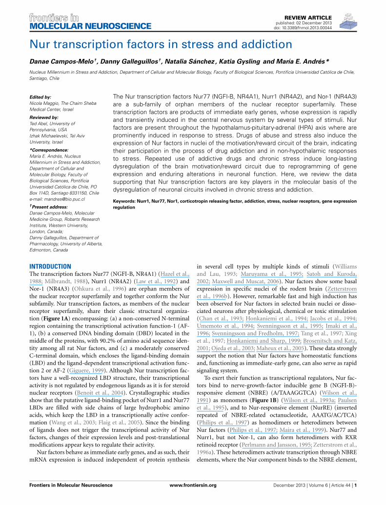
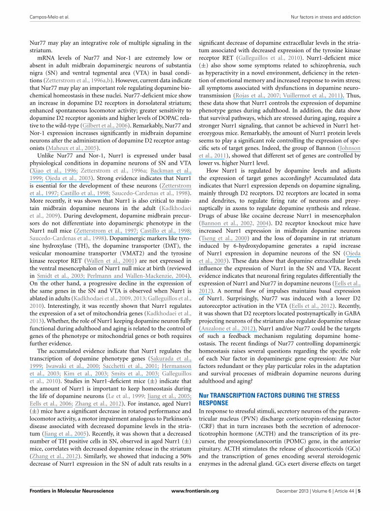
INTRODUCTIONThe transcription factors Nur77 (NGFI-B, NR4A1) (Hazel et al.,1988; Milbrandt, 1988), Nurr1 (NR4A2) (Law et al., 1992) andNor-1 (NR4A3) (Ohkura et al., 1996) are orphan members ofthe nuclear receptor superfamily and together conform the Nursubfamily. Nur transcription factors, as members of the nuclearreceptor superfamily, share their classic structural organiza-tion (Figure 1A) encompassing: (a) a non-conserved N-terminalregion containing the transcriptional activation function-1 (AF-1), (b) a conserved DNA binding domain (DBD) located in themiddle of the proteins, with 90.2% of amino acid sequence iden-tity among all rat Nur factors, and (c) a moderately conservedC-terminal domain, which encloses the ligand-binding domain(LBD) and the ligand-dependent transcriptional activation func-tion 2 or AF-2 (Giguere, 1999). Although Nur transcription fac-tors have a well-recognized LBD structure, their transcriptionalactivity is not regulated by endogenous ligands as it is for steroidnuclear receptors (Benoit et al., 2004). Crystallographic studiesshow that the putative ligand-binding pocket of Nurr1 and Nur77LBDs are filled with side chains of large hydrophobic aminoacids, which keep the LBD in a transcriptionally active confor-mation (Wang et al., 2003; Flaig et al., 2005). Since the bindingof ligands does not trigger the transcriptional activity of Nurfactors, changes of their expression levels and post-translationalmodifications appear keys to regulate their activity.
Nur factors behave as immediate early genes, and as such, theirmRNA expression is induced independent of protein synthesis
in several cell types by multiple kinds of stimuli (Williamsand Lau, 1993; Maruyama et al., 1995; Satoh and Kuroda,2002; Maxwell and Muscat, 2006). Nur factors show some basalexpression in specific nuclei of the rodent brain (Zetterstromet al., 1996b). However, remarkable fast and high induction hasbeen observed for Nur factors in selected brain nuclei or disso-ciated neurons after physiological, chemical or toxic stimulation(Chan et al., 1993; Honkaniemi et al., 1994; Jacobs et al., 1994;Umemoto et al., 1994; Svenningsson et al., 1995; Imaki et al.,1996; Svenningsson and Fredholm, 1997; Tang et al., 1997; Xinget al., 1997; Honkaniemi and Sharp, 1999; Brosenitsch and Katz,2001; Ojeda et al., 2003; Maheux et al., 2005). These data stronglysupport the notion that Nur factors have homeostatic functionsand, functioning as immediate-early gene, can also serve as rapidsignaling system.
To exert their function as transcriptional regulators, Nur fac-tors bind to nerve-growth-factor inducible gene B (NGFI-B)-responsive element (NBRE) (A/TAAAGGTCA) (Wilson et al.,1991) as monomers (Figure 1B) (Wilson et al., 1993a; Paulsenet al., 1995), and to Nur-responsive element (NurRE) (invertedrepeated of NBRE-related octanucleotide, AAATG/AC/TCA)(Philips et al., 1997) as homodimers or heterodimers betweenNur factors (Philips et al., 1997; Maira et al., 1999). Nur77 andNurr1, but not Nor-1, can also form heterodimers with RXRretinoid receptor (Perlmann and Jansson, 1995; Zetterstrom et al.,1996a). These heterodimers activate transcription through NBREelements, where the Nur component binds to the NBRE element,
Frontiers in Molecular Neuroscience www.frontiersin.org December 2013 | Volume 6 | Article 44 | 1
MOLECULAR NEUROSCIENCE
Campos-Melo et al. Nur factors in stress and addiction
Monomer
AAAGGTCA NBRE
Heterodimer
AAAGGTCA NBRE
9-cis-RA
Nur Nur
Homo/heterodimer
TCAGGTTTcctccAAACCTGA NurRE
Heterodimer
GGTTCAnnnnnAGGTCA
DR5
9-cis-RA
Nurr1
NF
Tethered
Nur77 Nurr1 RXR Nur
RXR
Nur AF1
AF2
DBD LBD
A
B
FIGURE 1 | Scheme of Nur factors structure and DNA-binding
elements. (A) Nur factors common structure, AF: activation functiondomain; DBD: DNA Binding Domain; LBD: Ligand Binding Domain. (B) Nurfactors DNA binding elements. RXR: retinoic-X-receptor; 9-cis RA: 9-cisretinoic acid.
or through DR5 elements (two direct repeats of the consen-sus nuclear receptor binding motif separated by five nucleotides,GGTTCAnnnnnAGGTCA). In the case of DR5 elements, bothnuclear receptors bind to the DNA (Perlmann and Jansson,1995). In addition, Nur factors can regulate transcription indi-rectly by binding to another transcription factor. For instance,Nurr1 represses transcription of inflammatory genes in microgliaindirectly by forming a complex with NF-kB transcription factor(Saijo et al., 2009).
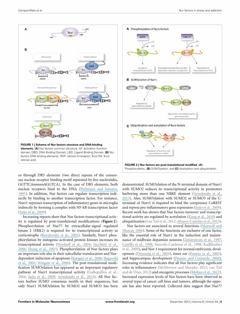
Increasing reports show that Nur factors transcriptional activ-ity is regulated by post-translational modifications (Figure 2).Phosphorylation of Nur77 by extracellular-signal regulatedkinase 2 (ERK2) is required for its transcriptional activity incorticotrophs (Kovalovsky et al., 2002). Similarly, Nurr1 phos-phorylation by mitogenic-activated protein kinases increases itstranscriptional activity (Nordzell et al., 2004; Sacchetti et al.,2006; Zhang et al., 2007). Phosphorylation of Nur factors playsan important role also in their subcellular translocation and Nur-dependent induction of apoptosis (Katagiri et al., 2000; Slagsvoldet al., 2002; Wingate et al., 2006). The post-translational modi-fication SUMOylation has appeared as an important regulatorypathway of Nurr1 transcriptional activity (Galleguillos et al.,2004; Saijo et al., 2009; Arredondo et al., 2013). All Nur fac-tors harbor SUMO consensus motifs in their sequences, butonly Nurr1 SUMOylation by SUMO2 and SUMO3 has been
FIGURE 2 | Nur factors are post-translational modified. (A)
Phosphorylation, (B) SUMOylation, and (C) Acetylation and ubiquitination.
demonstrated. SUMOylation of the N-terminal domain of Nurr1with SUMO2 reduces its transcriptional activity in promotersharboring more than one NBRE element (Arredondo et al.,2013). Also, SUMOylation with SUMO2 or SUMO3 of the C-terminal of Nurr1 is required to bind the corepressor CoRESTand repress pro-inflammatory gene expression (Saijo et al., 2009).Recent work has shown that Nur factors turnover and transcrip-tional activity are regulated by acetylation (Kang et al., 2010) andubiquitination (van Tiel et al., 2012; Alvarez-Castelao et al., 2013).
Nur factors are associated to several functions (Maxwell andMuscat, 2006). Some of the functions are exclusive of one factor,like the essential role of Nurr1 in the induction and mainte-nance of midbrain dopamine neurons (Zetterstrom et al., 1997;Castillo et al., 1998; Saucedo-Cardenas et al., 1998; Kadkhodaeiet al., 2009), and Nor-1 requirement for normal embryonic devel-opment (Deyoung et al., 2003), inner ear (Ponnio et al., 2002),and hippocampus development (Ponnio and Conneely, 2004).Increasing evidence indicates that all Nur factors play significantroles in inflammatory (McMorrow and Murphy, 2011; van Tieland de Vries, 2012) and oncogenic processes (Mohan et al., 2012).Increased expression levels of Nur factors have been observed inseveral types of cancer cell lines and tumors, although the oppo-site has also been reported. Collected data suggest that Nur77
Frontiers in Molecular Neuroscience www.frontiersin.org December 2013 | Volume 6 | Article 44 | 2
Campos-Melo et al. Nur factors in stress and addiction
behaves as a pro-oncogenic factor (Lee et al., 2011). However,the double knockout of Nur77 and Nor-1 induced a fatal acutemyeloid leukemia in mice (Mullican et al., 2007), indicating thatthese nuclear receptors may also play a role as tumor suppres-sors (Mullican et al., 2007). The apparent controversy or dual roleof Nur factors as tumor suppressors and/or pro-oncogenic fac-tors could be explained by a dual role in transcription, behavingas transcriptional activators or repressors. Since Nur factors aretranscriptionally active in their native form, less attention hasbeen paid to their role as potential transcriptional repressors.Interestingly, there are several reports showing the interactionof Nur factors with transcriptional corepressors. For example,we showed that PIASγ interacts and represses Nurr1-dependenttranscriptional activity (Galleguillos et al., 2004). Nurr1 alsointeracts with the transcriptional repressors SMRT (Lammi et al.,2008; Jacobs et al., 2009) and CoREST (Saijo et al., 2009). Theinteraction with SMRT maintains Nurr1 in a transcriptionalrepressive complex impeding the induction of its dopaminergictarget genes (Jacobs et al., 2009). It has also been shown the inter-action between Nur77 and SMRT (Sohn et al., 2001). Regardingthe role of Nur77 and Nor-1 repressing the expression of targetgenes, it was shown that abrogation of these transcription fac-tors correlates with an increased expression of MYC oncogene(Boudreaux et al., 2012). In addition, it was demonstrated thatMYC is a direct target gene of Nur77/Nor-1, whose expressionwas strongly repressed in a Nur DNA-binding dependent way(Boudreaux et al., 2012). Nur77 and Nor-1 play also an importantrole triggering apoptosis in several cell types. Interestingly, thiseffect is due to the translocation of Nur77/Nor-1 from the nucleito the mitochondria, where Nur77 triggers cytochrome c releaseand apoptosis (Li et al., 2000). Through this mechanism, Nur77and Nor-1 play a central function in the clonal deletion of autore-active thymocytes (Sohn et al., 2007). Nur77 and Nor-1 colocalizein several cell types, including CNS neurons, and apparentlythey replace each other in some functions. This colocalization ofNur77 and Nor-1 explain the lack of deleterious effects in thesingle knockout mice, while lack of both induces a catastrophicderegulation in the immune system. Similarly, colocalization ofNur77 with Nor-1 in the CNS may explain an apparent lack ofstrong effect of each knockout in the stress and rewarding systemsof the brain.
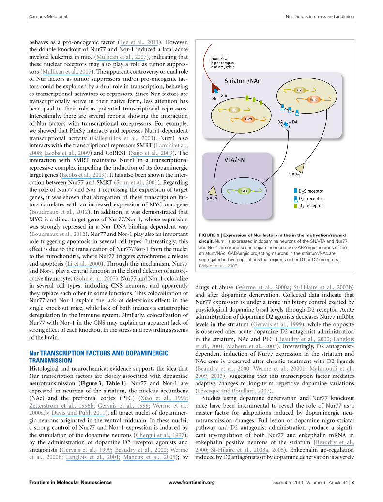
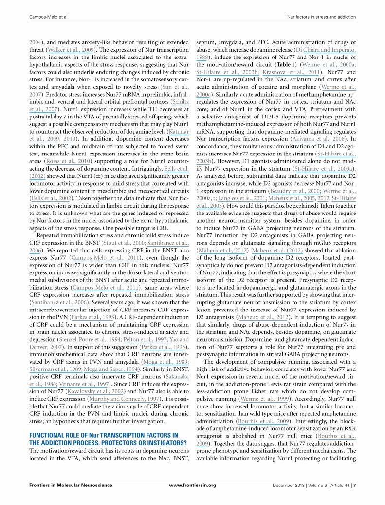
Nur TRANSCRIPTION FACTORS AND DOPAMINERGICTRANSMISSIONHistological and neurochemical evidence supports the idea thatNur transcription factors are closely associated with dopamineneurotransmission (Figure 3, Table 1). Nur77 and Nor-1 areexpressed in neurons of the striatum, the nucleus accumbens(NAc) and the prefrontal cortex (PFC) (Xiao et al., 1996;Zetterstrom et al., 1996b; Gervais et al., 1999; Werme et al.,2000a,b; Davis and Puhl, 2011), all target nuclei of dopaminer-gic neurons originated in the ventral midbrain. In these nuclei,a strong control of Nur77 and Nor-1 expression is induced bythe stimulation of the dopamine neurons (Chergui et al., 1997);by the administration of dopamine D2 receptor agonists andantagonists (Gervais et al., 1999; Beaudry et al., 2000; Wermeet al., 2000b; Langlois et al., 2001; Maheux et al., 2005); by
FIGURE 3 | Expression of Nur factors in the in the motivation/reward
circuit. Nurr1 is expressed in dopamine neurons of the SN/VTA and Nur77and Nor-1 are expressed in dopamine-receptive GABAergic neurons of thestriatum/NAc. GABAergic projecting neurons in the striatum/NAc aresegregated in two populations that express either D1 or D2 receptors(Valjent et al., 2009).
drugs of abuse (Werme et al., 2000a; St-Hilaire et al., 2003b)and after dopamine denervation. Collected data indicate thatNur77 expression is under a tonic inhibitory control exerted byphysiological dopamine basal levels through D2 receptor. Acuteadministration of dopamine D2 agonists decreases Nur77 mRNAlevels in the striatum (Gervais et al., 1999), while the oppositeis observed after acute dopamine D2 antagonist administrationin the striatum, NAc and PFC (Beaudry et al., 2000; Langloiset al., 2001; Maheux et al., 2005). Interestingly, D2 antagonist-dependent induction of Nur77 expression in the striatum andNAc core is preserved after chronic treatment with D2 ligands(Beaudry et al., 2000; Werme et al., 2000b; Mahmoudi et al.,2009, 2013), suggesting that this transcription factor mediatesadaptive changes to long-term repetitive dopamine variations(Levesque and Rouillard, 2007).
Studies using dopamine denervation and Nur77 knockoutmice have been instrumental to reveal the role of Nur77 as amaster factor for adaptations induced by dopaminergic neu-rotransmission changes. Full lesion of dopamine nigro-striatalpathway and D2 antagonist administration produce a signifi-cant up-regulation of both Nur77 and enkephalin mRNA inenkephalin positive neurons of the striatum (Beaudry et al.,2000; St-Hilaire et al., 2003a, 2005). Enkephalin up-regulationinduced by D2 antagonists or by dopamine denervation is severely
Frontiers in Molecular Neuroscience www.frontiersin.org December 2013 | Volume 6 | Article 44 | 3
Campos-Melo et al. Nur factors in stress and addiction
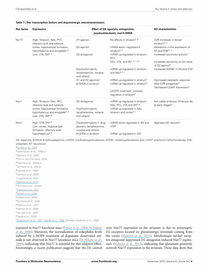
Table 1 | Nur transcription factors and dopaminergic neurotransmission.
Nur factor Expression Effect of DA agonists, antagonists, KO characteristics
psychostimulants, and 6-OHDA
Nur77 High: Striatum, NAc, PFC,olfactory bulb and tubercle,cortex, hippocampal formation,hypothalamus and amygdalaa−e
Low: VTA, SNa−e
D1 agonist:
D2 agonist:
D2 antagonist:
Psychostimulants:(amphetamine, cocaine,and others)D1 and D2 agonists:6-OHDA in striatum:
No effects in striatumf, g
mRNA down- regulated instriatumf, g
mRNA up-regulated in striatum,PFC,NAc, VTA, and SNc, h, i, j, p
mRNA up-regulated in striatumand NAce, k, l
mRNA up-regulated in striatumf
mRNA up-regulated in striatumf
L-DOPA treatment: complexregulation in striatumf
D2R increases in dorsalstriatumm, n
Alterations in the expression ofNT and ENKm, o
Increased locomotor activityn
Increased sensitivity to low doseof D2 agonistn
Increased DOPAC in SN and VTAn
Decreased cataleptic responseafter D2R antagonistm
Decreased COMT expressionn
Nor-1 High: Striatum, NAc, PFC,olfactory bulb and tubercle,cortex, hippocampal formation,hypothalamus and amygdalaa−e
Low: VTA, SNa−e
D2 Antagonist:
Psychostimulants:(amphetamine, cocaineand others)
mRNA up-regulated in striatum,NAc, PFC, VTA and SNh, p
mRNA up-regulated in NAc,striatum and cortexe, l
Not viable embryos. Embryos dieat early stagesq
Nurr1 High: VTA, SNa, b
Low: cortex, hippocampalformation, olfactory bulb,hypothalamusa, b
Psychostimulants in drugabusers: (amphetamine,cocaine and others)6-OHDA in striatum:
mRNA down-regulated in SN andVTAq, r
mRNA up-regulated in SNs
Agenesis DA neuronst
DA: dopamine; 6-OHDA: 6-hydroxydopamine; L-DOPA: 3,4-dihydroxyphenylalanine; DOPAC: dihydroxyphenylacetic acid; COMT: catechol-O-methyltransferase; ENK:
enkephalin; NT: neurotensin.aGofflot et al., 2007bZetterstrom et al., 1996a,bcBeaudry et al., 2000d Ponnio and Conneely, 2004eWerme et al., 2000a,bf St-Hilaire et al., 2003agGervais et al., 1999hMaheux et al., 2005i Langlois et al., 2001j Maheux et al., 2012k St-Hilaire et al., 2003bl Krasnova et al., 2011mEthier et al., 2004nGilbert et al., 2006oSt-Hilaire et al., 2006pMaheux et al., 2012qNielsen et al., 2008r Horvath et al., 2007sOjeda et al., 2003t Zetterstrom et al., 1997; Castillo et al., 1998; Saucedo-Cardenas et al., 1998.
impaired in Nur77 knockout mice (Ethier et al., 2004; St-Hilaireet al., 2006). Moreover, the normalization of enkephalin levelsinduced by L-DOPA treatment of dopamine denervated ani-mals is not observed in Nur77 knockout mice (St-Hilaire et al.,2006), indicating that Nur77 is essential for this adaptive effect.Interestingly, a recent publication suggests that the D2 control
over Nur77 expression in the striatum is due to presynapticD2 receptors located on glutamatergic terminals coming fromthe cortex (Maheux et al., 2012). Metabotropic mGlu5 recep-tor antagonist suppressed D2 antagonist-induced Nur77 expres-sion (Maheux et al., 2012), indicating that glutamate positivelycontrols Nur77 expression in the striatum. These data show that
Frontiers in Molecular Neuroscience www.frontiersin.org December 2013 | Volume 6 | Article 44 | 4
Campos-Melo et al. Nur factors in stress and addiction
Nur77 may play an integrative role of multiple signaling in thestriatum.
mRNA levels of Nur77 and Nor-1 are extremely low orabsent in adult midbrain dopaminergic neurons of substantianigra (SN) and ventral tegmental area (VTA) in basal condi-tions (Zetterstrom et al., 1996a,b). However, current data indicatethat Nur77 may play an important role regulating dopamine bio-chemical homeostasis in these nuclei. Nur77-deficient mice showan increase in dopamine D2 receptors in dorsolateral striatum;enhanced spontaneous locomotor activity; greater sensitivity todopamine D2 receptor agonists and higher levels of DOPAC rela-tive to the wild-type (Gilbert et al., 2006). Remarkably, Nur77 andNor-1 expression increases significantly in midbrain dopamineneurons after the administration of dopamine D2 receptor antag-onists (Maheux et al., 2005).
Unlike Nur77 and Nor-1, Nurr1 is expressed under basalphysiological conditions in dopamine neurons of SN and VTA(Xiao et al., 1996; Zetterstrom et al., 1996a; Backman et al.,1999; Ojeda et al., 2003). Strong evidence indicates that Nurr1is essential for the development of these neurons (Zetterstromet al., 1997; Castillo et al., 1998; Saucedo-Cardenas et al., 1998).More recently, it was shown that Nurr1 is also critical to main-tain midbrain dopamine neurons in the adult (Kadkhodaeiet al., 2009). During development, dopamine midbrain precur-sors do not differentiate into dopaminergic phenotype in theNurr1 null mice (Zetterstrom et al., 1997; Castillo et al., 1998;Saucedo-Cardenas et al., 1998). Dopaminergic markers like tyro-sine hydroxylase (TH), the dopamine transporter (DAT), thevesicular monoamine transporter (VMAT2) and the tyrosinekinase receptor RET (Wallen et al., 2001) are not expressed inthe ventral mesencephalon of Nurr1 null mice at birth (reviewedin Smidt et al., 2003; Perlmann and Wallen-Mackenzie, 2004).On the other hand, a progressive decline in the expression ofthe same genes in the SN and VTA is observed when Nurr1 isablated in adults (Kadkhodaei et al., 2009, 2013; Galleguillos et al.,2010). Interestingly, it was recently shown that Nurr1 regulatesthe expression of a set of mitochondria genes (Kadkhodaei et al.,2013). Whether, the role of Nurr1 keeping dopamine neuron fullyfunctional during adulthood and aging is related to the control ofgenes of the phenotype or mitochondrial genes or both requiresfurther evidence.
The accumulated evidence indicate that Nurr1 regulates thetranscription of dopamine phenotype genes (Sakurada et al.,1999; Iwawaki et al., 2000; Sacchetti et al., 2001; Hermansonet al., 2003; Kim et al., 2003; Smits et al., 2003; Galleguilloset al., 2010). Studies in Nurr1-deficient mice (±) indicate thatthe amount of Nurr1 is important to keep homeostasis duringthe life of dopamine neurons (Le et al., 1999; Jiang et al., 2005;Eells et al., 2006; Zhang et al., 2012). For instance, aged Nurr1(±) mice have a significant decrease in rotarod performance andlocomotor activity, a motor impairment analogous to Parkinson’sdisease associated with decreased dopamine levels in the stria-tum (Jiang et al., 2005). Recently, it was shown that a decreasednumber of TH positive cells in SN, observed in aged Nurr1 (±)mice, correlates with decreased dopamine release in the striatum(Zhang et al., 2012). Similarly, we showed that inducing a 50%decrease of Nurr1 expression in the SN of adult rats results in a
significant decrease of dopamine extracellular levels in the stria-tum associated with decreased expression of the tyrosine kinasereceptor RET (Galleguillos et al., 2010). Nurr1-deficient mice(±) also show some symptoms related to schizophrenia, suchas hyperactivity in a novel environment, deficiency in the reten-tion of emotional memory and increased response to swim stress;all symptoms associated with dysfunctions in dopamine neuro-transmission (Rojas et al., 2007; Vuillermot et al., 2011). Thus,these data show that Nurr1 controls the expression of dopaminephenotype genes during adulthood. In addition, the data showthat survival pathways, which are stressed during aging, require astronger Nurr1 signaling, that cannot be achieved in Nurr1 het-erozygous mice. Remarkably, the amount of Nurr1 protein levelsseems to play a significant role controlling the expression of spe-cific sets of target genes. Indeed, the group of Bannon (Johnsonet al., 2011), showed that different set of genes are controlled bylower vs. higher Nurr1 level.
How Nurr1 is regulated by dopamine levels and adjuststhe expression of target genes accordingly? Accumulated dataindicates that Nurr1 expression depends on dopamine signaling,mainly through D2 receptors. D2 receptors are located in somaand dendrites, to regulate firing rate of neurons and presy-naptically in axons to regulate dopamine synthesis and release.Drugs of abuse like cocaine decrease Nurr1 in mesencephalon(Bannon et al., 2002, 2004). D2 receptor knockout mice haveincreased Nurr1 expression in midbrain dopamine neurons(Tseng et al., 2000) and the loss of dopamine in rat striatuminduced by 6-hydroxydopamine generates a rapid increaseof Nurr1 expression in dopamine neurons of the SN (Ojedaet al., 2003). These data show that dopamine extracellular levelsinfluence the expression of Nurr1 in the SN and VTA. Recentevidence indicates that neuronal firing regulates differentially theexpression of Nurr1 and Nur77 in dopamine neurons (Eells et al.,2012). A normal flow of impulses maintains basal expressionof Nurr1. Surprisingly, Nur77 was induced with a lower D2autoreceptor activation in the VTA (Eells et al., 2012). Recently,it was shown that D2 receptors located postsynaptically in GABAprojecting neurons of the striatum also regulate dopamine release(Anzalone et al., 2012). Nurr1 and/or Nur77 could be the targetsof such a feedback mechanism regulating dopamine home-ostasis. The recent findings of Nur77 controlling dopaminergichomeostasis raises several questions regarding the specific roleof each Nur factor in dopaminergic gene expression: Are Nurfactors redundant or they play particular roles in the adaptationand survival processes of midbrain dopamine neurons duringadulthood and aging?
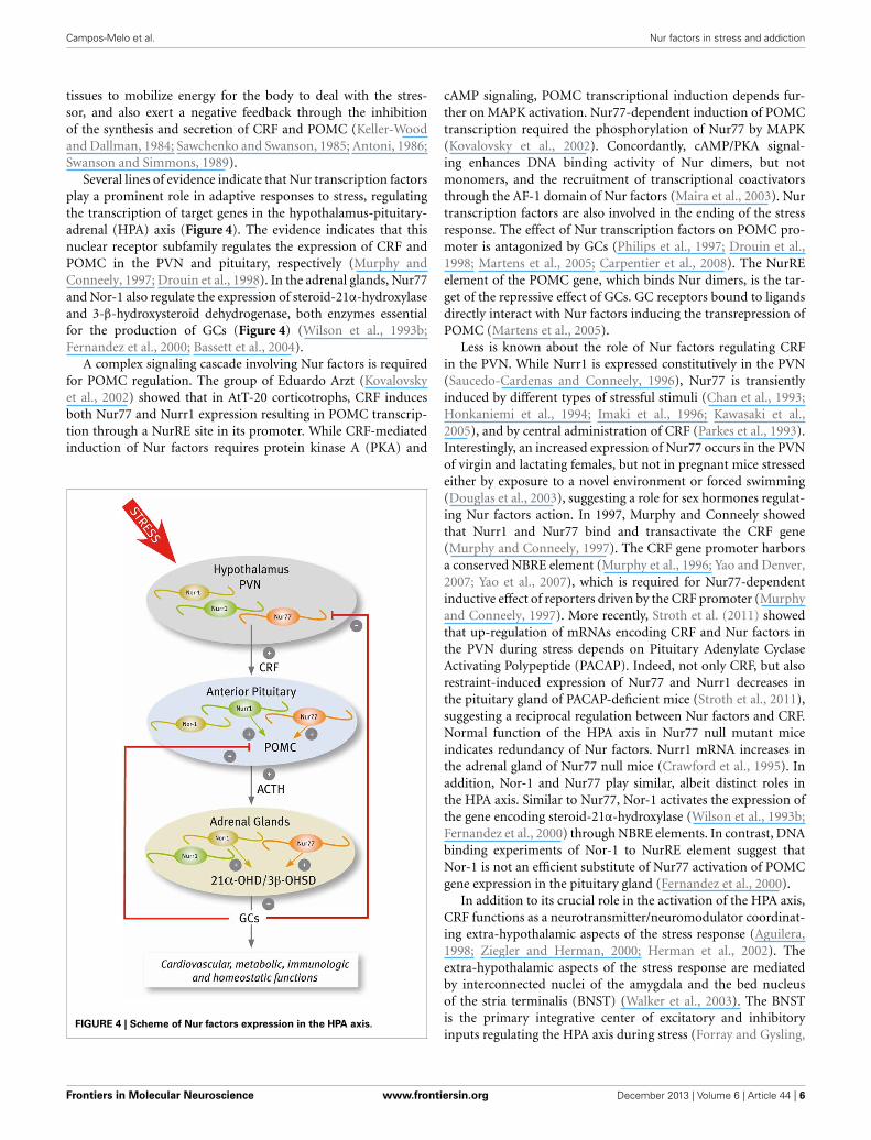
Nur TRANSCRIPTION FACTORS DURING THE STRESSRESPONSEIn response to stressful stimuli, secretory neurons of the paraven-tricular nucleus (PVN) discharge corticotropin-releasing factor(CRF) that in turn increases both the secretion of adrenocor-ticotrophin hormone (ACTH) and the transcription of its pre-cursor, the proopiomelanocortin (POMC) gene, in the anteriorpituitary. ACTH stimulates the release of glucocorticoids (GCs)and the transcription of genes encoding several steroidogenicenzymes in the adrenal gland. GCs exert diverse effects on target
Frontiers in Molecular Neuroscience www.frontiersin.org December 2013 | Volume 6 | Article 44 | 5
Campos-Melo et al. Nur factors in stress and addiction
tissues to mobilize energy for the body to deal with the stres-sor, and also exert a negative feedback through the inhibitionof the synthesis and secretion of CRF and POMC (Keller-Woodand Dallman, 1984; Sawchenko and Swanson, 1985; Antoni, 1986;Swanson and Simmons, 1989).
Several lines of evidence indicate that Nur transcription factorsplay a prominent role in adaptive responses to stress, regulatingthe transcription of target genes in the hypothalamus-pituitary-adrenal (HPA) axis (Figure 4). The evidence indicates that thisnuclear receptor subfamily regulates the expression of CRF andPOMC in the PVN and pituitary, respectively (Murphy andConneely, 1997; Drouin et al., 1998). In the adrenal glands, Nur77and Nor-1 also regulate the expression of steroid-21α-hydroxylaseand 3-β-hydroxysteroid dehydrogenase, both enzymes essentialfor the production of GCs (Figure 4) (Wilson et al., 1993b;Fernandez et al., 2000; Bassett et al., 2004).
A complex signaling cascade involving Nur factors is requiredfor POMC regulation. The group of Eduardo Arzt (Kovalovskyet al., 2002) showed that in AtT-20 corticotrophs, CRF inducesboth Nur77 and Nurr1 expression resulting in POMC transcrip-tion through a NurRE site in its promoter. While CRF-mediatedinduction of Nur factors requires protein kinase A (PKA) and
FIGURE 4 | Scheme of Nur factors expression in the HPA axis.
cAMP signaling, POMC transcriptional induction depends fur-ther on MAPK activation. Nur77-dependent induction of POMCtranscription required the phosphorylation of Nur77 by MAPK(Kovalovsky et al., 2002). Concordantly, cAMP/PKA signal-ing enhances DNA binding activity of Nur dimers, but notmonomers, and the recruitment of transcriptional coactivatorsthrough the AF-1 domain of Nur factors (Maira et al., 2003). Nurtranscription factors are also involved in the ending of the stressresponse. The effect of Nur transcription factors on POMC pro-moter is antagonized by GCs (Philips et al., 1997; Drouin et al.,1998; Martens et al., 2005; Carpentier et al., 2008). The NurREelement of the POMC gene, which binds Nur dimers, is the tar-get of the repressive effect of GCs. GC receptors bound to ligandsdirectly interact with Nur factors inducing the transrepression ofPOMC (Martens et al., 2005).
Less is known about the role of Nur factors regulating CRFin the PVN. While Nurr1 is expressed constitutively in the PVN(Saucedo-Cardenas and Conneely, 1996), Nur77 is transientlyinduced by different types of stressful stimuli (Chan et al., 1993;Honkaniemi et al., 1994; Imaki et al., 1996; Kawasaki et al.,2005), and by central administration of CRF (Parkes et al., 1993).Interestingly, an increased expression of Nur77 occurs in the PVNof virgin and lactating females, but not in pregnant mice stressedeither by exposure to a novel environment or forced swimming(Douglas et al., 2003), suggesting a role for sex hormones regulat-ing Nur factors action. In 1997, Murphy and Conneely showedthat Nurr1 and Nur77 bind and transactivate the CRF gene(Murphy and Conneely, 1997). The CRF gene promoter harborsa conserved NBRE element (Murphy et al., 1996; Yao and Denver,2007; Yao et al., 2007), which is required for Nur77-dependentinductive effect of reporters driven by the CRF promoter (Murphyand Conneely, 1997). More recently, Stroth et al. (2011) showedthat up-regulation of mRNAs encoding CRF and Nur factors inthe PVN during stress depends on Pituitary Adenylate CyclaseActivating Polypeptide (PACAP). Indeed, not only CRF, but alsorestraint-induced expression of Nur77 and Nurr1 decreases inthe pituitary gland of PACAP-deficient mice (Stroth et al., 2011),suggesting a reciprocal regulation between Nur factors and CRF.Normal function of the HPA axis in Nur77 null mutant miceindicates redundancy of Nur factors. Nurr1 mRNA increases inthe adrenal gland of Nur77 null mice (Crawford et al., 1995). Inaddition, Nor-1 and Nur77 play similar, albeit distinct roles inthe HPA axis. Similar to Nur77, Nor-1 activates the expression ofthe gene encoding steroid-21α-hydroxylase (Wilson et al., 1993b;Fernandez et al., 2000) through NBRE elements. In contrast, DNAbinding experiments of Nor-1 to NurRE element suggest thatNor-1 is not an efficient substitute of Nur77 activation of POMCgene expression in the pituitary gland (Fernandez et al., 2000).
In addition to its crucial role in the activation of the HPA axis,CRF functions as a neurotransmitter/neuromodulator coordinat-ing extra-hypothalamic aspects of the stress response (Aguilera,1998; Ziegler and Herman, 2000; Herman et al., 2002). Theextra-hypothalamic aspects of the stress response are mediatedby interconnected nuclei of the amygdala and the bed nucleusof the stria terminalis (BNST) (Walker et al., 2003). The BNSTis the primary integrative center of excitatory and inhibitoryinputs regulating the HPA axis during stress (Forray and Gysling,
Frontiers in Molecular Neuroscience www.frontiersin.org December 2013 | Volume 6 | Article 44 | 6
Campos-Melo et al. Nur factors in stress and addiction
2004), and mediates anxiety-like behavior resulting of extendedthreat (Walker et al., 2009). The expression of Nur transcriptionfactors increases in the limbic nuclei associated to the extra-hypothalamic aspects of the stress response, suggesting that Nurfactors could also underlie enduring changes induced by chronicstress. For instance, Nor-1 is increased in the somatosensory cor-tex and amygdala when exposed to novelty stress (Sun et al.,2007). Predator stress increases Nur77 mRNA in prelimbic, infral-imbic and, ventral and lateral orbital prefrontal cortexes (Schiltzet al., 2007). Nurr1 expression increases while TH decreases atpostnatal day 7 in the VTA of prenatally stressed offspring, whichsuggest a possible compensatory mechanism that may play Nurr1to counteract the observed reduction of dopamine levels (Katunaret al., 2009, 2010). In addition, dopamine content decreaseswithin the PFC and midbrain of rats subjected to forced swimtest, meanwhile Nurr1 expression increases in the same brainareas (Rojas et al., 2010) supporting a role for Nurr1 counter-acting the decrease of dopamine content. Intriguingly, Eells et al.(2002) showed that Nurr1 (±) mice displayed significantly greaterlocomotor activity in response to mild stress that correlated withlower dopamine content in mesolimbic and mesocortical circuits(Eells et al., 2002). Taken together the data indicate that Nur fac-tors expression is modulated in limbic circuit during the responseto stress. It is unknown what are the genes induced or repressedby Nur factors in the nuclei associated to the extra-hypothalamicaspects of the stress response. One possible target is CRF.
Repeated immobilization stress and chronic mild stress induceCRF expression in the BNST (Stout et al., 2000; Santibanez et al.,2006). We reported that cells expressing CRF in the BNST alsoexpress Nur77 (Campos-Melo et al., 2011), even though theexpression of Nur77 is wider than CRF in this nucleus. Nur77expression increases significantly in the dorso-lateral and ventro-medial subdivisions of the BNST after acute and repeated immo-bilization stress (Campos-Melo et al., 2011), same areas whereCRF expression increases after repeated immobilization stress(Santibanez et al., 2006). Several years ago, it was shown that theintracerebroventricular injection of CRF increases CRF expres-sion in the PVN (Parkes et al., 1993). A CRF-dependent inductionof CRF could be a mechanism of maintaining CRF expressionin brain nuclei associated to chronic stress-induced anxiety anddepression (Stenzel-Poore et al., 1994; Pelton et al., 1997; Yao andDenver, 2007). In support of this suggestion (Parkes et al., 1993),immunohistochemical data show that CRF neurons are inner-vated by CRF axons in PVN and amygdala (Moga et al., 1989;Silverman et al., 1989; Moga and Saper, 1994). Similarly, in BNST,positive CRF terminals also innervate CRF neurons (Sakanakaet al., 1986; Veinante et al., 1997). Since CRF induces the expres-sion of Nur77 (Kovalovsky et al., 2002) and Nur77 also is able toinduce CRF expression (Murphy and Conneely, 1997), it is possi-ble that Nur77 could mediate the vicious cycle of CRF-dependentCRF induction in the PVN and limbic nuclei, during chronicstress; an hypothesis that requires further investigation.
FUNCTIONAL ROLE OF Nur TRANSCRIPTION FACTORS INTHE ADDICTION PROCESS. PROTECTORS OR INSTIGATORS?The motivation/reward circuit has its roots in dopamine neuronslocated in the VTA, which send afferences to the NAc, BNST,
septum, amygdala, and PFC. Acute administration of drugs ofabuse, which increase dopamine release (Di Chiara and Imperato,1988), induce the expression of Nur77 and Nor-1 in nuclei ofthe motivation/reward circuit (Table 1) (Werme et al., 2000a;St-Hilaire et al., 2003b; Krasnova et al., 2011). Nur77 andNor-1 are up-regulated in the NAc, striatum, and cortex afteracute administration of cocaine and morphine (Werme et al.,2000a). Similarly, acute administration of methamphetamine up-regulates the expression of Nur77 in cortex, striatum and NAccore; and of Nurr1 in the cortex and VTA. Pretreatment witha selective antagonist of D1/D5 dopamine receptors preventsmethamphetamine-induced expression of both Nur77 and Nurr1mRNA, supporting that dopamine-mediated signaling regulatesNur transcription factors expression (Akiyama et al., 2008). Inconcordance, the simultaneous administration of D1 and D2 ago-nists increases Nur77 expression in the striatum (St-Hilaire et al.,2003b). However, D1 agonists administered alone do not mod-ify Nur77 expression in the striatum (St-Hilaire et al., 2003a).As analyzed before, substantial data indicate that dopamine D2antagonists increase, while D2 agonists decrease Nur77 and Nor-1 expression in the striatum (Beaudry et al., 2000; Werme et al.,2000a,b; Langlois et al., 2001; Maheux et al., 2005, 2012; St-Hilaireet al., 2005). How could this paradox be explained? Taken togetherthe available evidence suggests that drugs of abuse would requireanother neurotransmitter system, besides dopamine, in orderto induce Nur77 in GABA projecting neurons of the striatum.Nur77 induction by D2 antagonists in GABA projecting neu-rons depends on glutamate signaling through mGlu5 receptors(Maheux et al., 2012). Maheux et al. (2012) showed that ablationof the long isoform of dopamine D2 receptors, located post-synaptically do not prevent D2 antagonists-dependent inductionof Nur77, indicating that the effect is presynaptic, where the shortisoform of the D2 receptor is present. Presynaptic D2 recep-tors are located in dopaminergic and glutamatergic axons in thestriatum. This result was further supported by showing that inter-rupting glutamate neurotransmission to the striatum by cortexlesion prevented the increase of Nur77 expression induced byD2 antagonists (Maheux et al., 2012). It is tempting to suggestthat similarly, drugs of abuse-dependent induction of Nur77 inthe striatum and NAc depends, besides dopamine, on glutamateneurotransmission. Dopamine- and glutamate-dependent induc-tion of Nur77 supports a role for Nur77 integrating pre andpostsynaptic information in striatal GABA projecting neurons.
The development of compulsive running, associated with ahigh risk of addictive behavior, correlates with lower Nur77 andNor1 expression in several nuclei of the motivation/reward cir-cuit, in the addiction-prone Lewis rat strain compared with theless-addiction prone Fisher rats which do not develop com-pulsive running (Werme et al., 1999). Accordingly, Nur77 nullmice show increased locomotor activity, but a similar locomo-tor sensitization than wild type mice after repeated amphetamineadministration (Bourhis et al., 2009). Interestingly, the block-ade of amphetamine-induced locomotor sensitization by an RXRantagonist is abolished in Nur77 null mice (Bourhis et al.,2009). Together the data suggest that Nur77 regulates addiction-prone phenotype and sensitization by different mechanisms. Theavailable information regarding Nurr1 protecting or facilitating
Frontiers in Molecular Neuroscience www.frontiersin.org December 2013 | Volume 6 | Article 44 | 7
Campos-Melo et al. Nur factors in stress and addiction
addictive behaviors is unclear. In one study it was shown thatNurr1 (±) mice do not develop compulsive running behavior andhigh ethanol consumption compared to wild type mice (Wermeet al., 2003). In another study it was shown that Nurr1 (±)mice have an increased basal locomotor activity and augmentedlocomotor response to acute methamphetamine administration(Backman et al., 2003). Mice genetic background, behavioralprotocols, among other parameters, may influence these observa-tions. Remarkably, in a recent work it was shown that ablation ofthe histone deacetylase HDAC3 in the NAc facilitates condition-place preference induced by cocaine (Rogge et al., 2013). Thiseffect was correlated with increased Nurr1 expression in thisnucleus, supporting a role for Nurr1 facilitating addiction behav-iors (Rogge et al., 2013).
It has been proposed that the persistent behavioral and cog-nitive effects of chronic intake of drugs of abuse depend onnew programs of gene expression triggered by immediate-earlygenes. Tolerance and sensitization of Nur factors expression innuclei of the motivation/reward and stress brain circuits sug-gest that these early genes play a signaling role in the plasticchanges underlying long-term adaptations. Studies in cocaineabusers showed a reduction of Nurr1 expression in SN neu-rons (Nielsen et al., 2008). This decreased expression of Nurr1correlates with a reduction of DAT expression in the same neu-rons (Bannon et al., 2002). Similarly, rats chronically treatedwith cocaine show a down-regulation of the expression of Nurr1mRNA and protein in the ventral midbrain (Leo et al., 2007).Additionally, chronic use of heroin decreases Nurr1 mRNAto a greater extent with age in the paranigral nucleus of theVTA (Horvath et al., 2007). Nurr1 expression decreases afterchronic intake of drugs of abuse, could be an adaptive changeto excessive dopamine stimulation, and also could be indica-tive of Nurr1 role adjusting the expression of dopamine tar-get genes during the addiction process. In this regard, it wasshown that an acute methamphetamine challenge to animalspretreated with methamphetamine causes a further decrease inNurr1 mRNA levels (McCoy et al., 2011), indicating that thesignaling system regulating Nurr1 expression adapts to newparameters.
Opposing to a tolerance effect observed for Nurr1; Nur77seems to adapt to dopamine changes and its levels are stillinducible after chronic drug intake. For instance, it has beenshown that Nur77 expression increases in the frontal cortex ofrats after 10 days of cocaine self-administration (Freeman et al.,2002b) or 14 days in a binge model of cocaine administration(Freeman et al., 2002a), and in the dorsal striatum after 7 daysof cocaine self-administration (Lynch et al., 2008). On otherhand, it was shown that the expression of several early-genes,including Nur factors, is no longer induced in the striatum afterchronic exposure to methamphetamine (McCoy et al., 2011). Itis noteworthy that Nor-1 is still significantly induced in the stria-tum of methamphetamine chronically-treated rats, indicating thecapacity of the system to respond in new settings.
Chronic stress also triggers tolerance and sensitization of Nurfactors expression. In the PVN, it has been shown that Nur77 is nolonger induced after chronic stress stimuli (Umemoto et al., 1994,1997). In contrast, in the ventral region of the BNST, we observed
a higher number of cells expressing Nur77 after repeated immo-bilization stress compared with acute stress (Campos-Melo et al.,2011). Interestingly, the data of the group of Marta Antonelli(Katunar et al., 2010) suggest that Nurr1 may be the transcrip-tion factor setting up the new parameters of the dopamine systemafter prenatal stress. Using prenatal restraint stress, they showedthat Nurr1 is permanently higher in the VTA, but not in the SN,in the offspring of stressed mothers. This increment of Nurr1expression in the VTA correlates with several changes in the moti-vation/reward dopamine system that persist to adulthood (Baieret al., 2012). An exciting work indicates that Nur factors integratestress and drug addiction signaling. Postweaning isolation causeselevation in amphetamine-induced dopamine overflow in Nurr1(−/+) mice, but a reduction in (+/+) mice (Moore et al., 2008).These data demonstrate that a deletion of a single allele of Nurr1,which produced only subtle phenotypic changes, when coupledwith a developmental stressor such as postweaning isolation, candramatically alter mesoaccumbens dopamine neurotransmission(Moore et al., 2008).
CONCLUSIONSChronic stress plays a primary role in the origin of severalbrain pathologies such as anxiety and depression (McCormickand Green, 2013), and facilitates and perpetuates drug addic-tion (Koob, 2008; Sinha, 2008). Chronic stress and repeated useof addictive drugs induce long-lasting alterations of the moti-vation/reward circuit and HPA axis (Koob and Le Moal, 2001).The evidence presented points to Nur transcription factors asorchestrators of the molecular bases of the reorganization of thesecircuits under stressful stimuli and exposure to addictive drugs,since their expression is fast, transient and strongly regulatedby dopamine, glutamate, and CRF in the nuclei of the motiva-tion/reward circuit and HPA axis. The features of Nur factorsas early genes and orphan nuclear receptors allow them to inte-grate and transmit fast responses to incoming neurotransmittersignals in neurons. The transient nature of the changes in Nurfactor levels suggest that they can re-program the expression oftarget genes in response to acute and chronic dopamine changesby adjusting their inducibility to the new conditions, as occur-ring during chronic stress or after repeated exposure to drugsof abuse. Finally, the localization of Nurr1 in dopamine neuronsand Nur77/Nor-1 in dopamine-receptive neurons, positions themto translate the dopaminergic information simultaneously to thegenome of pre and post-synaptic neurons, allowing an integra-tive signaling of the motivation/reward circuit. Identifying theintracellular signaling pathways inducing Nur factors expressionand their target genes is essential to elucidate their function innormal physiology as well as in addiction and anxiety disorders.These findings might offer novel targets to treat these devastatingconditions.
ACKNOWLEDGMENTSOur work cited in this manuscript was supported by FONDECYTgrant N◦ 3085027 to Danae Campos-Melo, and FONDECYTgrants N◦ 1070349 and 1110352 to María E. Andrés. TheMillennium Nucleus in Stress and Addiction is supported by MSIgrant N◦ P10/063-F.
Frontiers in Molecular Neuroscience www.frontiersin.org December 2013 | Volume 6 | Article 44 | 8
Campos-Melo et al. Nur factors in stress and addiction
REFERENCESAguilera, G. (1998). Corticotropin releasing hormone, receptor regulation and
the stress response. Trends Endocrinol. Metab. 9, 329–336. doi: 10.1016/S1043-2760(98)00079-4
Akiyama, K., Isao, T., Ide, S., Ishikawa, M., and Saito, A. (2008). mRNA expres-sion of the Nurr1 and NGFI-B nuclear receptor families following acute andchronic administration of methamphetamine. Prog. Neuropsychopharmacol.Biol. Psychiatry 32, 1957–1966. doi: 10.1016/j.pnpbp.2008.09.021
Alvarez-Castelao, B., Losada, F., Ahicart, P., and Castano, J. G. (2013). The N-terminal region of Nurr1 (a.a 1-31) is essential for its efficient degradationby the ubiquitin proteasome pathway. PLoS ONE 8:e55999. doi: 10.1371/jour-nal.pone.0055999
Antoni, F. A. (1986). Hypothalamic control of adrenocorticotropin secretion:advances since the discovery of 41-residue corticotropin-releasing factor.Endocr. Rev. 7, 351–378. doi: 10.1210/edrv-7-4-351
Anzalone, A., Lizardi-Ortiz, J. E., Ramos, M., De Mei, C., Hopf, F. W., Iaccarino,C., et al. (2012). Dual control of dopamine synthesis and release by presynap-tic and postsynaptic dopamine D2 receptors. J. Neurosci. 32, 9023–9034. doi:10.1523/JNEUROSCI.0918-12.2012
Arredondo, C., Orellana, M., Vecchiola, A., Pereira, L. A., Galdames, L., andAndres, M. E. (2013). PIASgamma enhanced SUMO-2 modification of Nurr1activation-function-1 domain limits Nurr1 transcriptional synergy. PLoS ONE8:e55035. doi: 10.1371/journal.pone.0055035
Backman, C., Perlmann, T., Wallen, A., Hoffer, B. J., and Morales, M.(1999). A selective group of dopaminergic neurons express Nurr1 inthe adult mouse brain. Brain Res. 851, 125–132. doi: 10.1016/S0006-8993(99)02149-6
Backman, C., You, Z. B., Perlmann, T., and Hoffer, B. J. (2003). Elevated locomotoractivity without altered striatal dopamine contents in Nurr1 heterozygous miceafter acute exposure to methamphetamine. Behav. Brain Res. 143, 95–100. doi:10.1016/S0166-4328(03)00029-9
Baier, C. J., Katunar, M. R., Adrover, E., Pallares, M. E., and Antonelli, M. C.(2012). Gestational restraint stress and the developing dopaminergic system: anoverview. Neurotox. Res. 22, 16–32. doi: 10.1007/s12640-011-9305-4
Bannon, M. J., Pruetz, B., Barfield, E., and Schmidt, C. J. (2004). Transcription fac-tors specifying dopamine phenotype are decreased in cocaine users. Neuroreport15, 401–404. doi: 10.1097/00001756-200403010-00003
Bannon, M. J., Pruetz, B., Manning-Bog, A. B., Whitty, C. J., Michelhaugh, S.K., Sacchetti, P., et al. (2002). Decreased expression of the transcription factorNURR1 in dopamine neurons of cocaine abusers. Proc. Natl. Acad. Sci. U.S.A.99, 6382–6385. doi: 10.1073/pnas.092654299
Bassett, M. H., Suzuki, T., Sasano, H., De Vries, C. J., Jimenez, P. T., Carr, B.R., et al. (2004). The orphan nuclear receptor NGFIB regulates transcrip-tion of 3beta-hydroxysteroid dehydrogenase. implications for the control ofadrenal functional zonation. J. Biol. Chem. 279, 37622–37630. doi: 10.1074/jbc.M405431200
Beaudry, G., Langlois, M. C., Weppe, I., Rouillard, C., and Levesque, D. (2000).Contrasting patterns and cellular specificity of transcriptional regulation of thenuclear receptor nerve growth factor-inducible B by haloperidol and cloza-pine in the rat forebrain. J. Neurochem. 75, 1694–1702. doi: 10.1046/j.1471-4159.2000.0751694.x
Benoit, G., Malewicz, M., and Perlmann, T. (2004). Digging deep into the pocketsof orphan nuclear receptors: insights from structural studies. Trends Cell Biol.14, 369–376. doi: 10.1016/j.tcb.2004.05.007
Boudreaux, S. P., Ramirez-Herrick, A. M., Duren, R. P., and Conneely, O. M. (2012).Genome-wide profiling reveals transcriptional repression of MYC as a corecomponent of NR4A tumor suppression in acute myeloid leukemia. Oncogenesis1, e19. doi: 10.1038/oncsis.2012.19
Bourhis, E., Maheux, J., Paquet, B., Kagechika, H., Shudo, K., Rompre, P. P., et al.(2009). The transcription factors Nur77 and retinoid X receptors participatein amphetamine-induced locomotor activities. Psychopharmacology (Berl.) 202,635–648. doi: 10.1007/s00213-008-1343-0
Brosenitsch, T. A., and Katz, D. M. (2001). Physiological patterns of electricalstimulation can induce neuronal gene expression by activating N-type calciumchannels. J. Neurosci. 21, 2571–2579.
Campos-Melo, D., Quiroz, G., Noches, V., Gysling, K., Forray, M. I., and Andres, M.E. (2011). Repeated immobilization stress increases nur77 expression in the bednucleus of the stria terminalis. Neurotox. Res. 20, 289–300. doi: 10.1007/s12640-011-9243-1
Carpentier, R., Sacchetti, P., Segard, P., Staels, B., and Lefebvre, P. (2008). The glu-cocorticoid receptor is a co-regulator of the orphan nuclear receptor Nurr1.J. Neurochem. 104, 777–789. doi: 10.1111/j.1471-4159.2007.05055.x
Castillo, S. O., Baffi, J. S., Palkovits, M., Goldstein, D. S., Kopin, I. J., Witta,J., et al. (1998). Dopamine biosynthesis is selectively abolished in substantianigra/ventral tegmental area but not in hypothalamic neurons in mice withtargeted disruption of the Nurr1 gene. Mol. Cell. Neurosci. 11, 36–46. doi:10.1006/mcne.1998.0673
Chan, R. K., Brown, E. R., Ericsson, A., Kovacs, K. J., and Sawchenko, P. E. (1993).A comparison of two immediate-early genes, c-fos and NGFI-B, as markers forfunctional activation in stress-related neuroendocrine circuitry. J. Neurosci. 13,5126–5138.
Chergui, K., Svenningsson, P., Nomikos, G. G., Gonon, F., Fredholm, B. B., andSvennson, T. H. (1997). Increased expression of NGFI-A mRNA in the rat stria-tum following burst stimulation of the medial forebrain bundle. Eur. J. Neurosci.9, 2370–2382. doi: 10.1111/j.1460-9568.1997.tb01654.x
Crawford, P. A., Sadovsky, Y., Woodson, K., Lee, S. L., and Milbrandt, J. (1995).Adrenocortical function and regulation of the steroid 21-hydroxylase gene inNGFI-B-deficient mice. Mol. Cell. Biol. 15, 4331.
Davis, M. I., and Puhl, H. L. 3rd. (2011). Nr4a1-eGFP is a marker of striosome-matrix architecture, development and activity in the extended striatum. PLoSONE 6:e16619. doi: 10.1371/journal.pone.0016619
Deyoung, R. A., Baker, J. C., Cado, D., and Winoto, A. (2003). Theorphan steroid receptor Nur77 family member Nor-1 is essential for earlymouse embryogenesis. J. Biol. Chem. 278, 47104–47109. doi: 10.1074/jbc.M307496200
Di Chiara, G., and Imperato, A. (1988). Drugs abused by humans preferen-tially increase synaptic dopamine concentrations in the mesolimbic systemof freely moving rats. Proc. Natl. Acad. Sci. U.S.A. 85, 5274–5278. doi:10.1073/pnas.85.14.5274
Douglas, A. J., Brunton, P. J., Bosch, O. J., Russell, J. A., and Neumann, I. D. (2003).Neuroendocrine responses to stress in mice: hyporesponsiveness in pregnancyand parturition. Endocrinology 144, 5268–5276. doi: 10.1210/en.2003-0461
Drouin, J., Maira, M., and Philips, A. (1998). Novel mechanism of action for Nur77and antagonism by glucocorticoids: a convergent mechanism for CRH acti-vation and glucocorticoid repression of POMC gene transcription. J. SteroidBiochem. Mol. Biol. 65, 59–63. doi: 10.1016/S0960-0760(97)00180-5
Eells, J. B., Lipska, B. K., Yeung, S. K., Misler, J. A., and Nikodem, V. M. (2002).Nurr1-null heterozygous mice have reduced mesolimbic and mesocorticaldopamine levels and increased stress-induced locomotor activity. Behav. BrainRes. 136, 267–275. doi: 10.1016/S0166-4328(02)00185-7
Eells, J. B., Misler, J. A., and Nikodem, V. M. (2006). Reduced tyrosine hydroxylaseand GTP cyclohydrolase mRNA expression, tyrosine hydroxylase activity, andassociated neurochemical alterations in Nurr1-null heterozygous mice. BrainRes. Bull. 70, 186–195. doi: 10.1016/j.brainresbull.2006.05.004
Eells, J. B., Wilcots, J., Sisk, S., and Guo-Ross, S. X. (2012). NR4A gene expressionis dynamically regulated in the ventral tegmental area dopamine neurons andis related to expression of dopamine neurotransmission genes. J. Mol. Neurosci.46, 545–553. doi: 10.1007/s12031-011-9642-z
Ethier, I., Beaudry, G., St-Hilaire, M., Milbrandt, J., Rouillard, C., and Levesque,D. (2004). The transcription factor NGFI-B (Nur77) and retinoids play acritical role in acute neuroleptic-induced extrapyramidal effect and striatalneuropeptide gene expression. Neuropsychopharmacology 29, 335–346. doi:10.1038/sj.npp.1300318
Fernandez, P. M., Brunel, F., Jimenez, M. A., Saez, J. M., Cereghini, S., and Zakin,M. M. (2000). Nuclear receptors Nor1 and NGFI-B/Nur77 play similar, albeitdistinct, roles in the hypothalamo-pituitary-adrenal axis. Endocrinology 141,2392–2400. doi: 10.1210/en.141.7.2392
Flaig, R., Greschik, H., Peluso-Iltis, C., and Moras, D. (2005). Structural basis forthe cell-specific activities of the NGFI-B and the Nurr1 ligand-binding domain.J. Biol. Chem. 280, 19250–19258. doi: 10.1074/jbc.M413175200
Forray, M. I., and Gysling, K. (2004). Role of noradrenergic projectionsto the bed nucleus of the stria terminalis in the regulation of thehypothalamic-pituitary-adrenal axis. Brain Res. Brain Res. Rev. 47, 145–160. doi:10.1016/j.brainresrev.2004.07.011
Freeman, W. M., Brebner, K., Lynch, W. J., Patel, K. M., Robertson, D. J., Roberts,D. C., et al. (2002a). Changes in rat frontal cortex gene expression followingchronic cocaine. Brain Res. Mol. Brain Res. 104, 11–20. doi: 10.1016/S0169-328X(02)00197-3
Frontiers in Molecular Neuroscience www.frontiersin.org December 2013 | Volume 6 | Article 44 | 9
Campos-Melo et al. Nur factors in stress and addiction
Freeman, W. M., Brebner, K., Patel, K. M., Lynch, W. J., Roberts, D. C., and Vrana,K. E. (2002b). Repeated cocaine self-administration causes multiple changesin rat frontal cortex gene expression. Neurochem. Res. 27, 1181–1192. doi:10.1023/A:1020929526688
Galleguillos, D., Fuentealba, J. A., Gomez, L. M., Saver, M., Gomez, A., Nash,K., et al. (2010). Nurr1 regulates RET expression in dopamine neuronsof adult rat midbrain. J. Neurochem. 114, 1158–1167. doi: 10.1111/j.1471-4159.2010.06841.x
Galleguillos, D., Vecchiola, A., Fuentealba, J. A., Ojeda, V., Alvarez, K.,Gomez, A., et al. (2004). PIASgamma represses the transcriptional activationinduced by the nuclear receptor Nurr1. J. Biol. Chem. 279, 2005–2011. doi:10.1074/jbc.M308113200
Gervais, J., Soghomonian, J. J., Richard, D., and Rouillard, C. (1999). Dopamineand serotonin interactions in the modulation of the expression of theimmediate-early transcription factor, nerve growth factor-inducible B, in thestriatum. Neuroscience 91, 1045–1054. doi: 10.1016/S0306-4522(98)00688-5
Giguere, V. (1999). Orphan nuclear receptors: from gene to function. Endocr. Rev.20, 689–725. doi: 10.1210/er.20.5.689
Gilbert, F., Morissette, M., St-Hilaire, M., Paquet, B., Rouillard, C., Di Paolo,T., et al. (2006). Nur77 gene knockout alters dopamine neuron biochem-ical activity and dopamine turnover. Biol. Psychiatry 60, 538–547. doi:10.1016/j.biopsych.2006.04.023
Gofflot, F., Chartoire, N., Vasseur, L., Heikkinen, S., Dembele, D., Le Merrer,J., et al. (2007). Systematic gene expression mapping clusters nuclear recep-tors according to their function in the brain. Cell 131, 405–418. doi:10.1016/j.cell.2007.09.012
Hazel, T. G., Nathans, D., and Lau, L. F. (1988). A gene inducible by serum growthfactors encodes a member of the steroid and thyroid hormone receptor super-family. Proc. Natl. Acad. Sci. U.S.A. 85, 8444–8448. doi: 10.1073/pnas.85.22.8444
Herman, J. P., Cullinan, W. E., Ziegler, D. R., and Tasker, J. G. (2002). Role ofthe paraventricular nucleus microenvironment in stress integration. Eur. J.Neurosci. 16, 381–385. doi: 10.1046/j.1460-9568.2002.02133.x
Hermanson, E., Joseph, B., Castro, D., Lindqvist, E., Aarnisalo, P., Wallen, A.,et al. (2003). Nurr1 regulates dopamine synthesis and storage in MN9Ddopamine cells. Exp. Cell Res. 288, 324–334. doi: 10.1016/S0014-4827(03)00216-7
Honkaniemi, J., Kononen, J., Kainu, T., Pyykonen, I., and Pelto-Huikko, M.(1994). Induction of multiple immediate early genes in rat hypothalamic par-aventricular nucleus after stress. Brain Res. Mol. Brain Res. 25, 234–241. doi:10.1016/0169-328X(94)90158-9
Honkaniemi, J., and Sharp, F. R. (1999). Prolonged expression of zinc fin-ger immediate-early gene mRNAs and decreased protein synthesis followingkainic acid induced seizures. Eur. J. Neurosci. 11, 10–17. doi: 10.1046/j.1460-9568.1999.00401.x
Horvath, M. C., Kovacs, G. G., Kovari, V., Majtenyi, K., Hurd, Y. L., and Keller,E. (2007). Heroin abuse is characterized by discrete mesolimbic dopamine andopioid abnormalities and exaggerated nuclear receptor-related 1 transcriptionaldecline with age. J. Neurosci. 27, 13371–13375. doi: 10.1523/JNEUROSCI.2398-07.2007
Imaki, T., Shibasaki, T., Chikada, N., Harada, S., Naruse, M., and Demura, H.(1996). Different expression of immediate-early genes in the rat paraventric-ular nucleus induced by stress: relation to corticotropin-releasing factor genetranscription. Endocr. J. 43, 629–638. doi: 10.1507/endocrj.43.629
Iwawaki, T., Kohno, K., and Kobayashi, K. (2000). Identification of a potentialnurr1 response element that activates the tyrosine hydroxylase gene pro-moter in cultured cells. Biochem. Biophys. Res. Commun. 274, 590–595. doi:10.1006/bbrc.2000.3204
Jacobs, F. M., van Erp, S., van der Linden, A. J., von Oerthel, L., Burbach, J. P.,and Smidt, M. P. (2009). Pitx3 potentiates Nurr1 in dopamine neuron terminaldifferentiation through release of SMRT-mediated repression. Development 136,531–540. doi: 10.1242/dev.029769
Jacobs, O., Van Bree, L., Mailleux, P., Zhang, F., Schiffmann, S. N., Halleux, P.,et al. (1994). Homolateral cerebrocortical increase of immediate early gene andneurotransmitter messenger RNAs after minimal cortical lesion: blockade byN-methyl-D-aspartate antagonist. Neuroscience 59, 827–836. doi: 10.1016/0306-4522(94)90287-9
Jiang, C., Wan, X., He, Y., Pan, T., Jankovic, J., and Le, W. (2005). Age-dependentdopaminergic dysfunction in Nurr1 knockout mice. Exp. Neurol. 191, 154–162.doi: 10.1016/j.expneurol.2004.08.035
Johnson, M. M., Michelhaugh, S. K., Bouhamdan, M., Schmidt, C. J., and Bannon,M. J. (2011). The transcription factor NURR1 exerts concentration-dependenteffects on target genes mediating distinct biological processes. Front. Neurosci.5:135. doi: 10.3389/fnins.2011.00135
Kadkhodaei, B., Alvarsson, A., Schintu, N., Ramskold, D., Volakakis, N., Joodmardi,E., et al. (2013). Transcription factor Nurr1 maintains fiber integrity andnuclear-encoded mitochondrial gene expression in dopamine neurons. Proc.Natl. Acad. Sci. U.S.A. 110, 2360–2365. doi: 10.1073/pnas.1221077110
Kadkhodaei, B., Ito, T., Joodmardi, E., Mattsson, B., Rouillard, C., Carta,M., et al. (2009). Nurr1 is required for maintenance of maturing andadult midbrain dopamine neurons. J. Neurosci. 29, 15923–15932. doi:10.1523/JNEUROSCI.3910-09.2009
Kang, S. A., Na, H., Kang, H. J., Kim, S. H., Lee, M. H., and Lee, M. O.(2010). Regulation of Nur77 protein turnover through acetylation and deacety-lation induced by p300 and HDAC1. Biochem. Pharmacol. 80, 867–873. doi:10.1016/j.bcp.2010.04.026
Katagiri, Y., Takeda, K., Yu, Z. X., Ferrans, V. J., Ozato, K., and Guroff, G. (2000).Modulation of retinoid signalling through NGF-induced nuclear export ofNGFI-B. Nat. Cell Biol. 2, 435–440. doi: 10.1038/35017072
Katunar, M. R., Saez, T., Brusco, A., and Antonelli, M. C. (2009).Immunocytochemical expression of dopamine-related transcription fac-tors Pitx3 and Nurr1 in prenatally stressed adult rats. J. Neurosci. Res. 87,1014–1022. doi: 10.1002/jnr.21911
Katunar, M. R., Saez, T., Brusco, A., and Antonelli, M. C. (2010). Ontogeneticexpression of dopamine-related transcription factors and tyrosine hydroxylasein prenatally stressed rats. Neurotox. Res. 18, 69–81. doi: 10.1007/s12640-009-9132-z
Kawasaki, M., Yamaguchi, K., Saito, J., Ozaki, Y., Mera, T., Hashimoto, H.,et al. (2005). Expression of immediate early genes and vasopressin het-eronuclear RNA in the paraventricular and supraoptic nuclei of rats afteracute osmotic stimulus. J. Neuroendocrinol. 17, 227–237. doi: 10.1111/j.1365-2826.2005.01297.x
Keller-Wood, M. E., and Dallman, M. F. (1984). Corticosteroid inhibition of ACTHsecretion. Endocr. Rev. 5, 1–24. doi: 10.1210/edrv-5-1-1
Kim, K. S., Kim, C. H., Hwang, D. Y., Seo, H., Chung, S., Hong, S. J., et al. (2003).Orphan nuclear receptor Nurr1 directly transactivates the promoter activityof the tyrosine hydroxylase gene in a cell-specific manner. J. Neurochem. 85,622–634. doi: 10.1046/j.1471-4159.2003.01671.x
Koob, G. F. (2008). A role for brain stress systems in addiction. Neuron 59, 11–34.doi: 10.1016/j.neuron.2008.06.012
Koob, G. F., and Le Moal, M. (2001). Drug addiction, dysregulation of reward,and allostasis. Neuropsychopharmacology 24, 97–129. doi: 10.1016/S0893-133X(00)00195-0
Kovalovsky, D., Refojo, D., Liberman, A. C., Hochbaum, D., Pereda, M. P.,Coso, O. A., et al. (2002). Activation and induction of NUR77/NURR1in corticotrophs by CRH/cAMP: involvement of calcium, protein kinaseA, and MAPK pathways. Mol. Endocrinol. 16, 1638–1651. doi: 10.1210/me.16.7.1638
Krasnova, I. N., Ladenheim, B., Hodges, A. B., Volkow, N. D., and Cadet, J. L.(2011). Chronic methamphetamine administration causes differential regula-tion of transcription factors in the rat midbrain. PLoS ONE 6:e19179. doi:10.1371/journal.pone.0019179
Lammi, J., Perlmann, T., and Aarnisalo, P. (2008). Corepressor interaction differ-entiates the permissive and non-permissive retinoid X receptor heterodimers.Arch. Biochem. Biophys. 472, 105–114. doi: 10.1016/j.abb.2008.02.003
Langlois, M. C., Beaudry, G., Zekki, H., Rouillard, C., and Levesque, D. (2001).Impact of antipsychotic drug administration on the expression of nuclear recep-tors in the neocortex and striatum of the rat brain. Neuroscience 106, 117–128.doi: 10.1016/S0306-4522(01)00248-2
Law, S. W., Conneely, O. M., Demayo, F. J., and O’Malley, B. W. (1992).Identification of a new brain-specific transcription factor, NURR1. Mol.Endocrinol. 6, 2129–2135. doi: 10.1210/me.6.12.2129
Le, W., Conneely, O. M., He, Y., Jankovic, J., and Appel, S. H. (1999).Reduced Nurr1 expression increases the vulnerability of mesencephalicdopamine neurons to MPTP-induced injury. J. Neurochem. 73, 2218–2221. doi:10.1046/j.1471-4159.1999.02218.x
Lee, S. O., Li, X., Khan, S., and Safe, S. (2011). Targeting NR4A1 (TR3)in cancer cells and tumors. Expert Opin. Ther. Targets 15, 195–206. doi:10.1517/14728222.2011.547481
Frontiers in Molecular Neuroscience www.frontiersin.org December 2013 | Volume 6 | Article 44 | 10
Campos-Melo et al. Nur factors in stress and addiction
Leo, D., di Porzio, U., Racagni, G., Riva, M. A., Fumagalli, F., and Perrone-Capano,C. (2007). Chronic cocaine administration modulates the expression of tran-scription factors involved in midbrain dopaminergic neuron function. Exp.Neurol. 203, 472–480. doi: 10.1016/j.expneurol.2006.08.024
Levesque, D., and Rouillard, C. (2007). Nur77 and retinoid X receptors: crucialfactors in dopamine-related neuroadaptation. Trends Neurosci. 30, 22–30. doi:10.1016/j.tins.2006.11.006
Li, H., Kolluri, S. K., Gu, J., Dawson, M. I., Cao, X., Hobbs, P. D., et al.(2000). Cytochrome c release and apoptosis induced by mitochondrial targetingof nuclear orphan receptor TR3. Science 289, 1159–1164. doi: 10.1126/sci-ence.289.5482.1159
Lynch, W. J., Girgenti, M. J., Breslin, F. J., Newton, S. S., and Taylor, J. R.(2008). Gene profiling the response to repeated cocaine self-administrationin dorsal striatum: a focus on circadian genes. Brain Res. 1213, 166–177. doi:10.1016/j.brainres.2008.02.106
Maheux, J., Ethier, I., Rouillard, C., and Levesque, D. (2005). Induction pat-terns of transcription factors of the nur family (nurr1, nur77, and nor-1) bytypical and atypical antipsychotics in the mouse brain: implication for theirmechanism of action. J. Pharmacol. Exp. Ther. 313, 460–473. doi: 10.1124/jpet.104.080184
Maheux, J., St-Hilaire, M., Voyer, D., Tirotta, E., Borrelli, E., Rouillard, C., et al.(2012). Dopamine D(2) antagonist-induced striatal Nur77 expression requiresactivation of mGlu5 receptors by cortical afferents. Front. Pharmacol. 3:153. doi:10.3389/fphar.2012.00153
Mahmoudi, S., Blanchet, P. J., and Levesque, D. (2013). Haloperidol-induced stri-atal Nur77 expression in a non-human primate model of tardive dyskinesia. Eur.J. Neurosci. 38, 2192–2198. doi: 10.1111/ejn.12198
Mahmoudi, S., Samadi, P., Gilbert, F., Ouattara, B., Morissette, M., Gregoire, L.,et al. (2009). Nur77 mRNA levels and L-Dopa-induced dyskinesias in MPTPmonkeys treated with docosahexaenoic acid. Neurobiol. Dis. 36, 213–222. doi:10.1016/j.nbd.2009.07.017
Maira, M., Martens, C., Batsche, E., Gauthier, Y., and Drouin, J. (2003). Dimer-specific potentiation of NGFI-B (Nur77) transcriptional activity by the proteinkinase A pathway and AF-1-dependent coactivator recruitment. Mol. Cell. Biol.23, 763–776. doi: 10.1128/MCB.23.3.763-776.2003
Maira, M., Martens, C., Philips, A., and Drouin, J. (1999). Heterodimerizationbetween members of the Nur subfamily of orphan nuclear receptors as a novelmechanism for gene activation. Mol. Cell. Biol. 19, 7549–7557.
Martens, C., Bilodeau, S., Maira, M., Gauthier, Y., and Drouin, J. (2005). Protein-protein interactions and transcriptional antagonism between the subfamilyof NGFI-B/Nur77 orphan nuclear receptors and glucocorticoid receptor. Mol.Endocrinol. 19, 885–897. doi: 10.1210/me.2004-0333
Maruyama, K., Tsukada, T., Bandoh, S., Sasaki, K., Ohkura, N., and Yamaguchi,K. (1995). Expression of NOR-1 and its closely related members of thesteroid/thyroid hormone receptor superfamily in human neuroblastoma celllines. Cancer Lett. 96, 117–122. doi: 10.1016/0304-3835(95)03921-I
Maxwell, M. A., and Muscat, G. E. (2006). The NR4A subgroup: immediate earlyresponse genes with pleiotropic physiological roles. Nucl. Recept. Signal. 4, e002.doi: 10.1621/nrs.04002
McCormick, C. M., and Green, M. R. (2013). From the stressed adolescent to theanxious and depressed adult: investigations in rodent models. Neuroscience 249,242–257. doi: 10.1016/j.neuroscience.2012.08.063
McCoy, M. T., Jayanthi, S., Wulu, J. A., Beauvais, G., Ladenheim, B., Martin,T. A., et al. (2011). Chronic methamphetamine exposure suppresses the stri-atal expression of members of multiple families of immediate early genes(IEGs) in the rat: normalization by an acute methamphetamine injection.Psychopharmacology (Berl.) 215, 353–365. doi: 10.1007/s00213-010-2146-7
McMorrow, J. P., and Murphy, E. P. (2011). Inflammation: a role for NR4A orphannuclear receptors? Biochem. Soc. Trans. 39, 688–693. doi: 10.1042/BST0390688
Milbrandt, J. (1988). Nerve growth factor induces a gene homologous to theglucocorticoid receptor gene. Neuron 1, 183–188. doi: 10.1016/0896-6273(88)90138-9
Moga, M. M., and Saper, C. B. (1994). Neuropeptide-immunoreactive neurons pro-jecting to the paraventricular hypothalamic nucleus in the rat. J. Comp. Neurol.346, 137–150. doi: 10.1002/cne.903460110
Moga, M. M., Saper, C. B., and Gray, T. S. (1989). Bed nucleus of thestria terminalis: cytoarchitecture, immunohistochemistry, and projection tothe parabrachial nucleus in the rat. J. Comp. Neurol. 283, 315–332. doi:10.1002/cne.902830302
Mohan, H. M., Aherne, C. M., Rogers, A. C., Baird, A. W., Winter, D. C.,and Murphy, E. P. (2012). Molecular pathways: the role of NR4A orphannuclear receptors in cancer. Clin. Cancer Res. 18, 3223–3228. doi: 10.1158/1078-0432.CCR-11-2953
Moore, T. M., Brown, T., Cade, M., and Eells, J. B. (2008). Alterations inamphetamine-stimulated dopamine overflow due to the Nurr1-null het-erozygous genotype and postweaning isolation. Synapse 62, 764–774. doi:10.1002/syn.20550
Mullican, S. E., Zhang, S., Konopleva, M., Ruvolo, V., Andreeff, M.,Milbrandt, J., et al. (2007). Abrogation of nuclear receptors Nr4a3 andNr4a1 leads to development of acute myeloid leukemia. Nat. Med. 13,730–735. doi: 10.1038/nm1579
Murphy, E. P., and Conneely, O. M. (1997). Neuroendocrine regulation of thehypothalamic pituitary adrenal axis by the nurr1/nur77 subfamily of nuclearreceptors. Mol. Endocrinol. 11, 39–47. doi: 10.1210/me.11.1.39
Murphy, E. P., Dobson, A. D., Keller, C., and Conneely, O. M. (1996). Differentialregulation of transcription by the NURR1/NUR77 subfamily of nuclear tran-scription factors. Gene Expr. 5, 169–179.
Nielsen, D. A., Ji, F., Yuferov, V., Ho, A., Chen, A., Levran, O., et al. (2008). Genotypepatterns that contribute to increased risk for or protection from developingheroin addiction. Mol. Psychiatry 13, 417–428. doi: 10.1038/sj.mp.4002147
Nordzell, M., Aarnisalo, P., Benoit, G., Castro, D. S., and Perlmann, T.(2004). Defining an N-terminal activation domain of the orphan nuclearreceptor Nurr1. Biochem. Biophys. Res. Commun. 313, 205–211. doi:10.1016/j.bbrc.2003.11.079
Ohkura, N., Ito, M., Tsukada, T., Sasaki, K., Yamaguchi, K., and Miki, K. (1996).Structure, mapping and expression of a human NOR-1 gene, the third mem-ber of the Nur77/NGFI-B family. Biochim. Biophys. Acta 1308, 205–214. doi:10.1016/0167-4781(96)00101-7
Ojeda, V., Fuentealba, J. A., Galleguillos, D., and Andres, M. E. (2003). Rapidincrease of Nurr1 expression in the substantia nigra after 6-hydroxydopaminelesion in the striatum of the rat. J. Neurosci. Res. 73, 686–697. doi:10.1002/jnr.10705
Parkes, D., Rivest, S., Lee, S., Rivier, C., and Vale, W. (1993). Corticotropin-releasing factor activates c-fos, NGFI-B, and corticotropin-releasing factor geneexpression within the paraventricular nucleus of the rat hypothalamus. Mol.Endocrinol. 7, 1357–1367. doi: 10.1210/me.7.10.1357
Paulsen, R. F., Granas, K., Johnsen, H., Rolseth, V., and Sterri, S. (1995). Threerelated brain nuclear receptors, NGFI-B, Nurr1, and NOR-1, as transcriptionalactivators. J. Mol. Neurosci. 6, 249–255. doi: 10.1007/BF02736784
Pelton, G. H., Lee, Y., and Davis, M. (1997). Repeated stress, like vasopressin, sensi-tizes the excitatory effects of corticotropin releasing factor on the acoustic startlereflex. Brain Res. 778, 381–387. doi: 10.1016/S0006-8993(97)00669-0
Perlmann, T., and Jansson, L. (1995). A novel pathway for vitamin A signalingmediated by RXR heterodimerization with NGFI-B and NURR1. Genes Dev.9, 769–782. doi: 10.1101/gad.9.7.769
Perlmann, T., and Wallen-Mackenzie, A. (2004). Nurr1, an orphan nuclear recep-tor with essential functions in developing dopamine cells. Cell Tissue Res. 318,45–52. doi: 10.1007/s00441-004-0974-7
Philips, A., Lesage, S., Gingras, R., Maira, M. H., Gauthier, Y., Hugo, P., et al. (1997).Novel dimeric Nur77 signaling mechanism in endocrine and lymphoid cells.Mol. Cell. Biol. 17, 5946–5951.
Ponnio, T., Burton, Q., Pereira, F. A., Wu, D. K., and Conneely, O. M. (2002). Thenuclear receptor Nor-1 is essential for proliferation of the semicircular canals ofthe mouse inner ear. Mol. Cell. Biol. 22, 935–945. doi: 10.1128/MCB.22.3.935-945.2002
Ponnio, T., and Conneely, O. M. (2004). nor-1 regulates hippocampal axon guid-ance, pyramidal cell survival, and seizure susceptibility. Mol. Cell. Biol. 24,9070–9078. doi: 10.1128/MCB.24.20.9070-9078.2004
Rogge, G. A., Singh, H., Dang, R., and Wood, M. A. (2013). HDAC3 is a nega-tive regulator of cocaine-context-associated memory formation. J. Neurosci. 33,6623–6632. doi: 10.1523/JNEUROSCI.4472-12.2013
Rojas, P., Joodmardi, E., Hong, Y., Perlmann, T., and Ogren, S. O. (2007). Adultmice with reduced Nurr1 expression: an animal model for schizophrenia. Mol.Psychiatry 12, 756–766. doi: 10.1038/sj.mp.4001993
Rojas, P., Joodmardi, E., Perlmann, T., and Ogren, S. O. (2010). Rapid increase ofNurr1 mRNA expression in limbic and cortical brain structures related to cop-ing with depression-like behavior in mice. J. Neurosci. Res. 88, 2284–2293. doi:10.1002/jnr.22377
Frontiers in Molecular Neuroscience www.frontiersin.org December 2013 | Volume 6 | Article 44 | 11
Campos-Melo et al. Nur factors in stress and addiction
Sacchetti, P., Carpentier, R., Segard, P., Olive-Cren, C., and Lefebvre, P. (2006).Multiple signaling pathways regulate the transcriptional activity of theorphan nuclear receptor NURR1. Nucleic Acids Res. 34, 5515–5527. doi:10.1093/nar/gkl712
Sacchetti, P., Mitchell, T. R., Granneman, J. G., and Bannon, M. J. (2001).Nurr1 enhances transcription of the human dopamine transporter genethrough a novel mechanism. J. Neurochem. 76, 1565–1572. doi: 10.1046/j.1471-4159.2001.00181.x
Saijo, K., Winner, B., Carson, C. T., Collier, J. G., Boyer, L., Rosenfeld, M. G.,et al. (2009). A Nurr1/CoREST pathway in microglia and astrocytes protectsdopaminergic neurons from inflammation-induced death. Cell 137, 47–59. doi:10.1016/j.cell.2009.01.038
Sakanaka, M., Shibasaki, T., and Lederis, K. (1986). Distribution and efferent pro-jections of corticotropin-releasing factor-like immunoreactivity in the rat amyg-daloid complex. Brain Res. 382, 213–238. doi: 10.1016/0006-8993(86)91332-6
Sakurada, K., Ohshima-Sakurada, M., Palmer, T. D., and Gage, F. H. (1999). Nurr1,an orphan nuclear receptor, is a transcriptional activator of endogenous tyrosinehydroxylase in neural progenitor cells derived from the adult brain. Development126, 4017–4026.
Santibanez, M., Gysling, K., and Forray, M. I. (2006). Desipramine prevents thesustained increase in corticotropin-releasing hormone-like immunoreactivityinduced by repeated immobilization stress in the rat central extended amygdala.J. Neurosci. Res. 84, 1270–1281. doi: 10.1002/jnr.21023
Satoh, J., and Kuroda, Y. (2002). The constitutive and inducible expression ofNurr1, a key regulator of dopaminergic neuronal differentiation, in human neu-ral and non-neural cell lines. Neuropathology 22, 219–232. doi: 10.1046/j.1440-1789.2002.00460.x
Saucedo-Cardenas, O., and Conneely, O. M. (1996). Comparative distribution ofNURR1 and NUR77 nuclear receptors in the mouse central nervous system.J. Mol. Neurosci. 7, 51–63. doi: 10.1007/BF02736848
Saucedo-Cardenas, O., Quintana-Hau, J. D., Le, W. D., Smidt, M. P., Cox, J. J., DeMayo, F., et al. (1998). Nurr1 is essential for the induction of the dopamin-ergic phenotype and the survival of ventral mesencephalic late dopamin-ergic precursor neurons. Proc. Natl. Acad. Sci. U.S.A. 95, 4013–4018. doi:10.1073/pnas.95.7.4013
Sawchenko, P. E., and Swanson, L. W. (1985). Localization, colocalization, and plas-ticity of corticotropin-releasing factor immunoreactivity in rat brain. Fed. Proc.44, 221–227.
Schiltz, C. A., Kelley, A. E., and Landry, C. F. (2007). Acute stress andnicotine cues interact to unveil locomotor arousal and activity-dependentgene expression in the prefrontal cortex. Biol. Psychiatry 61, 127–135. doi:10.1016/j.biopsych.2006.03.002
Silverman, A. J., Hou-Yu, A., and Chen, W. P. (1989). Corticotropin-releasingfactor synapses within the paraventricular nucleus of the hypothalamus.Neuroendocrinology 49, 291–299. doi: 10.1159/000125131
Sinha, R. (2008). Chronic stress, drug use, and vulnerability to addiction. Ann. N.Y.Acad. Sci. 1141, 105–130. doi: 10.1196/annals.1441.030
Slagsvold, H. H., Ostvold, A. C., Fallgren, A. B., and Paulsen, R. E. (2002).Nuclear receptor and apoptosis initiator NGFI-B is a substrate for kinaseERK2. Biochem. Biophys. Res. Commun. 291, 1146–1150. doi: 10.1006/bbrc.2002.6579
Smidt, M. P., Smits, S. M., and Burbach, J. P. (2003). Molecular mechanisms under-lying midbrain dopamine neuron development and function. Eur. J. Pharmacol.480, 75–88. doi: 10.1016/j.ejphar.2003.08.094
Smits, S. M., Ponnio, T., Conneely, O. M., Burbach, J. P., and Smidt, M. P.(2003). Involvement of Nurr1 in specifying the neurotransmitter identity ofventral midbrain dopaminergic neurons. Eur. J. Neurosci. 18, 1731–1738. doi:10.1046/j.1460-9568.2003.02885.x
Sohn, S. J., Thompson, J., and Winoto, A. (2007). Apoptosis during negativeselection of autoreactive thymocytes. Curr. Opin. Immunol. 19, 510–515. doi:10.1016/j.coi.2007.06.001
Sohn, Y. C., Kwak, E., Na, Y., Lee, J. W., and Lee, S. K. (2001). Silencing mediatorof retinoid and thyroid hormone receptors and activating signal cointegrator-2 as transcriptional coregulators of the orphan nuclear receptor Nur77. J. Biol.Chem. 276, 43734–43739. doi: 10.1074/jbc.M107208200
Stenzel-Poore, M. P., Heinrichs, S. C., Rivest, S., Koob, G. F., and Vale, W. W. (1994).Overproduction of corticotropin-releasing factor in transgenic mice: a geneticmodel of anxiogenic behavior. J. Neurosci. 14, 2579–2584.
St-Hilaire, M., Bourhis, E., Levesque, D., and Rouillard, C. (2006). Impairedbehavioural and molecular adaptations to dopamine denervation and repeatedL-DOPA treatment in Nur77-knockout mice. Eur. J. Neurosci. 24, 795–805. doi:10.1111/j.1460-9568.2006.04954.x
St-Hilaire, M., Landry, E., Levesque, D., and Rouillard, C. (2003a). Denervationand repeated L-DOPA induce a coordinate expression of the transcription factorNGFI-B in striatal projection pathways in hemi-parkinsonian rats. Neurobiol.Dis. 14, 98–109. doi: 10.1016/S0969-9961(03)00081-0
St-Hilaire, M., Tremblay, P. O., Levesque, D., Barden, N., and Rouillard, C.(2003b). Effects of cocaine on c-fos and NGFI-B mRNA expression in trans-genic mice underexpressing glucocorticoid receptors. Neuropsychopharmacology28, 478–489. doi: 10.1038/sj.npp.1300067
St-Hilaire, M., Landry, E., Levesque, D., and Rouillard, C. (2005). Denervation andrepeated L-DOPA induce complex regulatory changes in neurochemical pheno-types of striatal neurons: implication of a dopamine D1-dependent mechanism.Neurobiol. Dis. 20, 450–460. doi: 10.1016/j.nbd.2005.04.001
Stout, S. C., Mortas, P., Owens, M. J., Nemeroff, C. B., and Moreau, J. (2000).Increased corticotropin-releasing factor concentrations in the bed nucleus ofthe stria terminalis of anhedonic rats. Eur. J. Pharmacol. 401, 39–46. doi:10.1016/S0014-2999(00)00412-X
Stroth, N., Liu, Y., Aguilera, G., and Eiden, L. E. (2011). Pituitary adeny-late cyclase-activating polypeptide controls stimulus-transcription couplingin the hypothalamic-pituitary-adrenal axis to mediate sustained hormonesecretion during stress. J. Neuroendocrinol. 23, 944–955. doi: 10.1111/j.1365-2826.2011.02202.x
Sun, W., Choi, S. H., Park, S. K., Kim, S. J., Noh, M. R., Kim, E. H., et al.(2007). Identification and characterization of novel activity-dependent tran-scription factors in rat cortical neurons. J. Neurochem. 100, 269–278. doi:10.1111/j.1471-4159.2006.04214.x
Svenningsson, P., and Fredholm, B. B. (1997). Caffeine mimics the effect of adopamine D2/3 receptor agonist on the expression of immediate early genesin globus pallidus. Neuropharmacology 36, 1309–1317. doi: 10.1016/S0028-3908(97)00091-9
Svenningsson, P., Nomikos, G. G., and Fredholm, B. B. (1995). Biphasic changesin locomotor behavior and in expression of mRNA for NGFI-A and NGFI-B in rat striatum following acute caffeine administration. J. Neurosci. 15,7612–7624.
Swanson, L. W., and Simmons, D. M. (1989). Differential steroid hormone andneural influences on peptide mRNA levels in CRH cells of the paraventricularnucleus: a hybridization histochemical study in the rat. J. Comp. Neurol. 285,413–435. doi: 10.1002/cne.902850402
Tang, Y. P., Murata, Y., Nagaya, T., Noda, Y., Seo, H., and Nabeshima, T. (1997).NGFI-B, c-fos, and c-jun mRNA expression in mouse brain after acute car-bon monoxide intoxication. J. Cereb. Blood Flow Metab. 17, 771–780. doi:10.1097/00004647-199707000-00007
Tseng, K. Y., Roubert, C., Do, L., Rubinstein, M., Kelly, M. A., Grandy, D. K.,et al. (2000). Selective increase of Nurr1 mRNA expression in mesencephalicdopaminergic neurons of D2 dopamine receptor-deficient mice. Brain Res. Mol.Brain Res. 80, 1–6. doi: 10.1016/S0169-328X(00)00107-8
Umemoto, S., Kawai, Y., and Senba, E. (1994). Differential regulation of IEGs inthe rat PVH in single and repeated stress models. Neuroreport 6, 201–204. doi:10.1097/00001756-199412300-00051
Umemoto, S., Kawai, Y., Ueyama, T., and Senba, E. (1997). Chronic glucocorticoidadministration as well as repeated stress affects the subsequent acute immo-bilization stress-induced expression of immediate early genes but not that ofNGFI-A. Neuroscience 80, 763–773. doi: 10.1016/S0306-4522(97)00050-X
Valjent, E., Bertran-Gonzalez, J., Herve, D., Fisone, G., and Girault, J. A. (2009).Looking BAC at striatal signaling: cell-specific analysis in new transgenic mice.Trends Neurosci. 32, 538–547. doi: 10.1016/j.tins.2009.06.005
van Tiel, C. M., and de Vries, C. J. (2012). NR4All in the vessel wall. J. SteroidBiochem. Mol. Biol. 130, 186–193. doi: 10.1016/j.jsbmb.2011.01.010
van Tiel, C. M., Kurakula, K., Koenis, D. S., van der Wal, E., and de Vries, C. J.(2012). Dual function of Pin1 in NR4A nuclear receptor activation: enhancedactivity of NR4As and increased Nur77 protein stability. Biochim. Biophys. Acta1823, 1894–1904. doi: 10.1016/j.bbamcr.2012.06.030
Veinante, P., Stoeckel, M. E., and Freund-Mercier, M. J. (1997). GABA- andpeptide-immunoreactivities co-localize in the rat central extended amygdala.Neuroreport 8, 2985–2989. doi: 10.1097/00001756-199709080-00035
Frontiers in Molecular Neuroscience www.frontiersin.org December 2013 | Volume 6 | Article 44 | 12
Campos-Melo et al. Nur factors in stress and addiction
Vuillermot, S., Joodmardi, E., Perlmann, T., Ove Ogren, S., Feldon, J., and Meyer,U. (2011). Schizophrenia-relevant behaviors in a genetic mouse model of con-stitutive Nurr1 deficiency. Genes Brain Behav. 10, 589–603. doi: 10.1111/j.1601-183X.2011.00698.x
Walker, D. L., Miles, L. A., and Davis, M. (2009). Selective participation of the bednucleus of the stria terminalis and CRF in sustained anxiety-like versus phasicfear-like responses. Prog. Neuropsychopharmacol. Biol. Psychiatry 33, 1291–1308.doi: 10.1016/j.pnpbp.2009.06.022
Walker, D. L., Toufexis, D. J., and Davis, M. (2003). Role of the bed nucleus of thestria terminalis versus the amygdala in fear, stress, and anxiety. Eur. J. Pharmacol.463, 199–216. doi: 10.1016/S0014-2999(03)01282-2
Wallen, A. A., Castro, D. S., Zetterstrom, R. H., Karlen, M., Olson, L., Ericson,J., et al. (2001). Orphan nuclear receptor Nurr1 is essential for Ret expressionin midbrain dopamine neurons and in the brain stem. Mol. Cell. Neurosci. 18,649–663. doi: 10.1006/mcne.2001.1057
Wang, Z., Benoit, G., Liu, J., Prasad, S., Aarnisalo, P., Liu, X., et al. (2003). Structureand function of Nurr1 identifies a class of ligand-independent nuclear receptors.Nature 423, 555–560. doi: 10.1038/nature01645
Werme, M., Hermanson, E., Carmine, A., Buervenich, S., Zetterstrom, R. H.,Thoren, P., et al. (2003). Decreased ethanol preference and wheel runningin Nurr1-deficient mice. Eur. J. Neurosci. 17, 2418–2424. doi: 10.1046/j.1460-9568.2003.02666.x
Werme, M., Olson, L., and Brene, S. (2000a). NGFI-B and nor1 mRNAs areupregulated in brain reward pathways by drugs of abuse: different effects inFischer and Lewis rats. Brain Res. Mol. Brain Res. 76, 18–24. doi: 10.1016/S0169-328X(99)00327-7
Werme, M., Ringholm, A., Olson, L., and Brene, S. (2000b). Differential pat-terns of induction of NGFI-B, Nor1 and c-fos mRNAs in striatal subregionsby haloperidol and clozapine. Brain Res. 863, 112–119. doi: 10.1016/S0006-8993(00)02109-0
Werme, M., Thoren, P., Olson, L., and Brene, S. (1999). Addiction-prone Lewis butnot Fischer rats develop compulsive running that coincides with downregula-tion of nerve growth factor inducible-B and neuron-derived orphan receptor 1.J. Neurosci. 19, 6169–6174.
Williams, G. T., and Lau, L. F. (1993). Activation of the inducible orphanreceptor gene nur77 by serum growth factors: dissociation of immediate-early and delayed-early responses. Mol. Cell. Biol. 13, 6124–6136. doi:10.1128/MCB.13.10.6124
Wilson, T. E., Fahrner, T. J., Johnston, M., and Milbrandt, J. (1991). Identificationof the DNA binding site for NGFI-B by genetic selection in yeast. Science 252,1296–1300. doi: 10.1126/science.1925541
Wilson, T. E., Fahrner, T. J., and Milbrandt, J. (1993a). The orphan receptorsNGFI-B and steroidogenic factor 1 establish monomer binding as a thirdparadigm of nuclear receptor-DNA interaction. Mol. Cell. Biol. 13, 5794–5804.doi: 10.1128/MCB.13.9.5794
Wilson, T. E., Mouw, A. R., Weaver, C. A., Milbrandt, J., and Parker, K. L. (1993b).The orphan nuclear receptor NGFI-B regulates expression of the gene encodingsteroid 21-hydroxylase. Mol. Cell. Biol. 13, 861–868. doi: 10.1128/MCB.13.2.861
Wingate, A. D., Campbell, D. G., Peggie, M., and Arthur, J. S. (2006). Nur77 isphosphorylated in cells by RSK in response to mitogenic stimulation. Biochem.J. 393, 715–724. doi: 10.1042/BJ20050967
Xiao, Q., Castillo, S. O., and Nikodem, V. M. (1996). Distribution of messengerRNAs for the orphan nuclear receptors Nurr1 and Nur77 (NGFI-B) in adult rat
brain using in situ hybridization. Neuroscience 75, 221–230. doi: 10.1016/0306-4522(96)00159-5
Xing, G., Zhang, L., Zhang, L., Heynen, T., Li, X. L., Smith, M. A., et al. (1997). Ratnurr1 is prominently expressed in perirhinal cortex, and differentially inducedin the hippocampal dentate gyrus by electroconvulsive vs. kindled seizures.Brain Res. Mol. Brain Res. 47, 251–261. doi: 10.1016/S0169-328X(97)00056-9
Yao, M., and Denver, R. J. (2007). Regulation of vertebrate corticotropin-releasing factor genes. Gen. Comp. Endocrinol. 153, 200–216. doi:10.1016/j.ygcen.2007.01.046
Yao, M., Stenzel-Poore, M., and Denver, R. J. (2007). Structural and functionalconservation of vertebrate corticotropin-releasing factor genes: evidence for acritical role for a conserved cyclic AMP response element. Endocrinology 148,2518–2531. doi: 10.1210/en.2006-1413
Zetterstrom, R. H., Solomin, L., Jansson, L., Hoffer, B. J., Olson, L., and Perlmann,T. (1997). Dopamine neuron agenesis in Nurr1-deficient mice. Science 276,248–250. doi: 10.1126/science.276.5310.248
Zetterstrom, R. H., Solomin, L., Mitsiadis, T., Olson, L., and Perlmann, T. (1996a).Retinoid X receptor heterodimerization and developmental expression distin-guish the orphan nuclear receptors NGFI-B, Nurr1, and Nor1. Mol. Endocrinol.10, 1656–1666. doi: 10.1210/me.10.12.1656
Zetterstrom, R. H., Williams, R., Perlmann, T., and Olson, L. (1996b). Cellularexpression of the immediate early transcription factors Nurr1 and NGFI-B sug-gests a gene regulatory role in several brain regions including the nigrostriataldopamine system. Brain Res. Mol. Brain Res. 41, 111–120. doi: 10.1016/0169-328X(96)00074-5
Zhang, L., Le, W., Xie, W., and Dani, J. A. (2012). Age-related changes in dopaminesignaling in Nurr1 deficient mice as a model of Parkinson’s disease. Neurobiol.Aging 33, 1001.e1007–1001.e1016. doi: 10.1016/j.neurobiolaging.2011.03.022
Zhang, T., Jia, N., Fei, E., Wang, P., Liao, Z., Ding, L., et al. (2007). Nurr1 isphosphorylated by ERK2 in vitro and its phosphorylation upregulates tyro-sine hydroxylase expression in SH-SY5Y cells. Neurosci. Lett. 423, 118–122. doi:10.1016/j.neulet.2007.06.041
Ziegler, D. R., and Herman, J. P. (2000). Local integration of glutamate signalingin the hypothalamic paraventricular region: regulation of glucocorticoid stressresponses. Endocrinology 141, 4801–4804. doi: 10.1210/en.141.12.4801
Conflict of Interest Statement: The authors declare that the research was con-ducted in the absence of any commercial or financial relationships that could beconstrued as a potential conflict of interest.
Received: 24 September 2013; accepted: 09 November 2013; published online: 02December 2013.Citation: Campos-Melo D, Galleguillos D, Sánchez N, Gysling K and Andrés ME(2013) Nur transcription factors in stress and addiction. Front. Mol. Neurosci. 6:44.doi: 10.3389/fnmol.2013.00044This article was submitted to the journal Frontiers in Molecular Neuroscience.Copyright © 2013 Campos-Melo, Galleguillos, Sánchez, Gysling and Andrés. Thisis an open-access article distributed under the terms of the Creative CommonsAttribution License (CC BY). The use, distribution or reproduction in other forumsis permitted, provided the original author(s) or licensor are credited and that the orig-inal publication in this journal is cited, in accordance with accepted academic practice.No use, distribution or reproduction is permitted which does not comply with theseterms.
Frontiers in Molecular Neuroscience www.frontiersin.org December 2013 | Volume 6 | Article 44 | 13