nuclear autoantigenic sperm protein (nasp), a linker histone chaperone that is required for cell...
TRANSCRIPT
Nuclear Autoantigenic Sperm Protein (NASP), a LinkerHistone Chaperone That Is Required for Cell Proliferation*
Received for publication, April 20, 2006, and in revised form, May 24, 2006 Published, JBC Papers in Press, May 25, 2006, DOI 10.1074/jbc.M603816200
Richard T. Richardson‡, Oleg M. Alekseev‡, Gail Grossman‡, Esther E. Widgren‡, Randy Thresher§, Eric J. Wagner§1,Kelly D. Sullivan§, William F. Marzluff§, and Michael G. O’Rand‡2
From the ‡Department of Cell and Developmental Biology and the §Program in Molecular Biology and Biotechnology, University ofNorth Carolina, Chapel Hill, North Carolina 27599-7090
A multichaperone nucleosome-remodeling complex that con-tains theH1 linkerhistonechaperonenuclearautoantigenic spermprotein (NASP) has recently been described. Linker histones (H1)are required for the proper completion of normal development,and NASP transports H1 histones into nuclei and exchanges H1histoneswithDNA.Consequently, we investigatedwhetherNASPis required for normal cell cycle progression anddevelopment.Wenow report that without sufficient NASP, HeLa cells and U2OScells are unable to replicate their DNA and progress through thecell cycle and that the NASP�/� null mutation causes embryoniclethality. Although the null mutationNASP�/� caused embryoniclethality,null embryos surviveuntil theblastocyst stage,whichmaybe explained by the presence of stored NASP protein in the cyto-plasm of oocytes. We conclude from this study that NASP andtherefore the linker histones are key players in the assembly ofchromatin after DNA replication.
Chromatin is a nucleoprotein complex containing repeatingnucleosome units that allow the chromatin to be folded intohigher-ordered structures and orchestrate numerous interac-tions through their histone tails. Nucleosomes typically are sep-arated by short pieces of DNA that link the nucleosomes to oneanother, bind H1 linker histones, and maintain nucleosomespacing that is critical for normal development (1–3). DNA-binding proteins that regulate replication and repair of thegenomemust successfully interact with chromatin during eachcell cycle. Consequently, the nucleosome-remodeling proteinmachinery and the DNA replication machinery coordinatetheir activities to ensure a faithful transition of chromatin fromone cell cycle to the next (4, 5). One of the key players in thiscoordination of activity is CAF-1,3 the chromatin assembly fac-
tor that is necessary for progression through S phase (6, 7).CAF-1 exists in a multichaperone nucleosome-remodelingcomplex that contains, in addition to CAF-1 (p150, p60, p48),H3.1, H4, Asfa, Asfb, NASP, Chk2,HAT1, and importin 4 (8, 9).This complex is thought to coordinate the deposition ofH3.1–H4 histones into new nucleosomes and signal the repli-cation complex via phosphorylation events through variouscheckpoints that DNA replication (S phase) can proceed. Thepresence of nuclear autoantigenic sperm protein (NASP) in theCAF-1 multichaperone complex during nucleosome remodel-ing (9) implies that NASP may have a role in the completion ofS phase in a normal cell cycle. H1 linker histones (H1) arerequired for the proper completion of normal development (2,10) andNASP, anH1 chaperone, transports H1 into nuclei (11)and exchangesH1withDNA (12). NASP has also been found tobe a binding partner of Ku70/Ku80 and DNA-PK in HeLa cells(13), indicating that NASP may play an additional role in DNArepair events.Previous studies demonstrated that NASP expression is regu-
lated during the cell cycle (14) and that progression through theG1/S border is delayedby the overexpressionofNASP (12). Linkerhistones not bound to DNA are bound to NASP (12), which isfound in all dividing cells in either a somatic/embryo (sNASP) ortestis/embryo (tNASP) form (14, 15). To address the role ofNASPin cell proliferation and normal development, we used a smallinterferingRNA (siRNA) knockdownofNASP inHeLa andU2OScells and inactivated the NASP gene by homologous recombina-tion (NASP�/�) in mice. We now report that without sufficientNASP, HeLa cells and U2OS cells are unable to replicate theirDNA and progress through the cell cycle and that the NASP�/�
null mutation causes embryonic lethality. Interestingly, becauseNASP is expressed in the oocyte and NASP protein is present inthe cytoplasm of the one-cell embryo, the embryos survive untilthe depletion of thematernally derivedNASPprotein.Our resultsimply thatNASPorchestrates the supplyof linkerhistones that arenecessary for nucleosome remodeling during DNA replication orrepair and support the conclusion thatH1 linker histones are nec-essary for normal development (2, 3).
EXPERIMENTAL PROCEDURES
Materials—Miscellaneous chemicals were the highest possi-blemolecular biology grade (Sigma). Restriction andmodifyingenzymes were purchased from New England Biolabs (Beverly,MA). DNA sequencing was performed by the University ofNorth Carolina at Chapel Hill automated sequencing facilityusing an ABI PRISMmodel 377 DNA sequencer (Applied Bio-
* This work was supported in part by NICHD, National Institutes of Health,through Cooperative Agreement U54HD35041 as part of the specializedcooperative centers program in reproductive research. The costs of publi-cation of this article were defrayed in part by the payment of page charges.This article must therefore be hereby marked “advertisement” in accord-ance with 18 U.S.C. Section 1734 solely to indicate this fact.
1 Supported by National Institutes of Health Grant F32 GM070101-02 and theCottrell Foundation.
2 To whom correspondence should be addressed: Department of Cell and Devel-opmental Biology, CB#7090, University of North Carolina, Chapel Hill, NC27599-7090. Tel.: 919-966-5698; Fax: 919-966-1856; E-mail: [email protected].
3 The abbreviations used are: CAF, chromatin assembly factor; NASP, nuclear auto-antigenic sperm protein; sNASP, somatic/embryo nuclear autoantigenicsperm protein; tNASP, testis/embryo nuclear autoantigenic sperm protein; siRNA,small interfering RNA; SLBP, stem-loop-binding protein; FACS, fluorescence-activated cell sorting; dpc, days postcoitum; PBS, phosphate-buffered saline.
THE JOURNAL OF BIOLOGICAL CHEMISTRY VOL. 281, NO. 30, pp. 21526 –21534, July 28, 2006© 2006 by The American Society for Biochemistry and Molecular Biology, Inc. Printed in the U.S.A.
21526 JOURNAL OF BIOLOGICAL CHEMISTRY VOLUME 281 • NUMBER 30 • JULY 28, 2006
by guest on March 10, 2016
http://ww
w.jbc.org/
Dow
nloaded from

systems, Foster City, CA). Sequence data were analyzed usingDNAsis (Hitachi Corp., South San Francisco, CA) andSequencher (Gene Codes Corp., Ann Arbor, MI) software. Oli-gonucleotides were synthesized at the University of NorthCarolina at Chapel Hill Nucleic Acids Core Facility. Purifica-tion of plasmid and PCR DNAs was performed using theirrespective kits from Qiagen (Valencia, CA). Anti-symplekinantibodies were obtained from BD Biosciences.Targeting Vector—The mouse NASP arms of homology
inserted into the pOSdupdel targeting vectorwere generated byPCRusingmouse genomicDNA (strain 129 Sv/Ev) as template.PCRs were performed with Takara LA TaqDNA polymerase(Fisher) according to the manufacturer’s protocol. The cycleconditions were as follows: 1 min at 94 °C, 1 cycle; 20 s at 98 °Cfollowed by 4.5 min at 68 °C, 30 cycles; 10 min at 72 °C, 1 cycle.The 7508-bp long arm was generated with NotI and PacIrestriction sites (5� and 3�, respectively), and the 1805-bp shortarm had BbsI and PmlI restriction sites (5� and 3�, respectively).Both the vector and inserts were sequentially digested with theappropriate restriction enzymes and purified, and the insertswere ligated into the vector overnight at 4 °C using T-4 DNAligase. A clone of the correct size was picked, tested by vari-ous restriction digests, and sequenced to confirm its validity.The DNA sequences of the PCR primers used to generate theNASP arms of homology are as follows: 1) long arm upstreamprimer, 5�-AGAGCGGCCGCGGCTATTCCTCAGTTGA-CACTCAG-3�; 2) long arm downstream primer, 5�-GCGT-TAATTAAATACCTGGCTAACTCCAGAAGTGAC-3�; 3)short arm upstream primer, 5�-GCGCACGTGATGTTAA-CTTCTGTCGCTGTGGAGG-3�; and 4) long arm down-stream primer, 5�-GCGGGATCCTAAGAATCCTCCAGC-TTTCCAAACAGCCAC-3�. Homologous recombinationwith this targeting construct deletes 9682 bp from the NASPgene, which includes exons 6–11 (see Fig. 4). This deletionincludes exon 7, which is expressed only in the adult testisand in all embryonic tissues during development (14) andcontains a histone-binding domain (16).Targeting—The targeting plasmid was linearized with NotI,
and mouse ES cells (2 � 107 embryonic stem cells, strain 129Sv/Ev) were electroporated with 25�g of vector. After positive-negative selection with ganciclovir and G418 (17), survivingcolonies were picked and screened using PCR with one primerfrom the neo gene and the other from the region just 3� to the3� arm of homology. Several clones were identified, and thecorrectness of the targeting was verified by Southern blots.ES cells from a positive clone were injected into blastocystsfromC57BL/6mice, which were implanted into pseudopreg-nant CD1 females. Male chimeras were bred to C57BL/6females, and the agouti coat color was used as an indicator oftransmission of the 129 genome. Transmitting agouti-coatedprogeny were back-crossed to C57BL/6 animals to obtainprogeny that were interbred to produce the experimentalanimals used in this study.Genotypic Analysis—Genomic DNA for PCRs was extracted
from 2-mm mouse tail clips by incubation in 100 �l of 25 mMNaOH/0.2 mM EDTA for 1 h at 95 °C followed by addition of100 �l of 40 mM Tris-HCl. After a brief vortexing, the sampleswere centrifuged at 1000 � g for 10 min and the supernatant
removed. Genomic DNA for Southern blots was isolated from6-mm mouse tail clips by overnight incubation at 55 °C in 400�l of 50 mM Tris-HCl (pH 7.5), 100 mM EDTA, 100 mM NaCl,1% SDS and 20 �l Proteinase K (10 mg/ml). After incubation,200 �l of saturated NaCl was added and the mixture vortexedfor 30 s followed by centrifugation at 14,000� g for 20min. Fivehundred �l of supernatant was removed and precipitated with100% ethanol and centrifuged at 14,000 � g for 10 min; thepellet was washed with 70% ethanol and centrifuged. The finalair-dried DNA pellet was dissolved in 50 �l of TE (10 mM Tris-HCl, 1 mM EDTA (pH 8.0)). Genotyping PCRs were performedusing Takara LA Taq as described above except that 2 �l ofgenomic DNA (described above as DNA for PCR) was used astemplate, and the primers were as follows: for the wild-typeallele sense and antisense, 5�-GAAGCAAGGGAAGAGTTG-AGAG-3� and 5�-TTCACTCTCAGAGGTAGC-3�, respec-tively; and for the targeted allele sense and antisense, 5�-GTT-TCCACCCAATGTCGAGCAAAC-3� and 5�-TTCAGTGAC-AACGTCGAGCAC-3�, respectively. Southern blots wereperformed on 10�g of SacI- and PstI-digestedDNA (describedabove as DNA for Southern blots) as described previously(15) except that restriction digests contained 3 mM spermi-dine, and the 20 �l samples were incubated in 1 �l of 1 mg/mlRNase A for 1 h at 37 °C before loading onto the gel. Theblots were probed with a 32P-labeled, random-primed PCR-generated cDNA that represents NASP exon 12 (see Fig. 3)and visualized using a Storm 860 PhosphorImager (Amer-sham Biosciences).Embryo Isolation, Blastocyst Culture, and Genotyping—
NASP�/� females were superovulated by injection with 5 unitsof pregnant mare serum gonadotropin (Sigma) followed 48 hlater by 5 units of human chorionic gonadotropin (Sigma) andmated with NASP�/� males. Successful matings were detectedby the presence of a vaginal plug. Embryos at various stages ineither the uterus or oviducts were collected for histologicalexamination or flushed as blastocysts from the uterus.Flushed blastocysts were washed two times in medium M16(Sigma) and then cultured in 96-well plates precoated with1% gelatin in Glasgow’s minimal essential medium (Fisher)with 15% fetal bovine serum and leukemia inhibiting factor(0.2 units/ml) for up to 10 days. To isolate DNA for PCRtemplate, individual blastocyst cultures were suspended bypipetting and incubating them in 20 �l of 25 mM NaOH/0.2mM EDTA for 1 h at 95 °C followed by addition of 100 �l of 40mM Tris-HCl. After a brief vortexing, the samples were cen-trifuged at 1000 � g for 10 min, and the supernatant wasremoved.Immunohistochemistry—Embryos were fixed in situ in excised
oviduct anduterine tissueby immersion inBouin’s fixative for1–4days, paraffin-embedded, and sectioned (8�m).Every fifth sectionwas stainedwithhematoxylin andeosin to locate theembryos, andadjacent serial sectionswere stainedwithanti-NASPantibody (14)or with aNASP probe for in situ hybridization.In Situ Hybridization—A high affinity NASP antisense
hybridization probe was designed using Oligo Analysis soft-ware, version 6.65 (Cascade, CO). The 5�-digoxigenen-labeledprobe, 5�-CGGCGATGGCAGCAG-3� (where boldface andunderlined letters indicate locked nucleic acids), was prepared
NASP Knock-out
JULY 28, 2006 • VOLUME 281 • NUMBER 30 JOURNAL OF BIOLOGICAL CHEMISTRY 21527
by guest on March 10, 2016
http://ww
w.jbc.org/
Dow
nloaded from

by Proligo Primers & Probes (Boulder, CO), and an identicalunlabeled probe was prepared as a control to demonstrate thespecificity of the probe when used in 100� excess. In situhybridization was performed in the University of North Caro-lina at Chapel Hill Laboratories for Reproductive BiologyImmunohistochemistry Core Facility based on methodsdescribed by Schwarzacher and Heslop-Harrison (18).Analysis of theNASPTranscript—Testeswere collected from
NASP�/� andNASP�/� mice, and after disruption in a Douncehomogenizer, total and poly(A)� RNA were prepared using anRNeasyMini Kit and anOligotexmRNAKit, respectively (Qia-gen). Equal quantities of mRNA were electrophoresed andNorthern blotted as described previously (14). After hybridiza-
tion with a 32P-labeled, random-primed NASP exon 12 probe, theblots were visualized as above withthe PhosphorImager.siRNA Transfection—HeLa cells
were maintained as described (12).A series of siRNAs targeting thehuman NASP open reading framewere designed (Dharmacon, Lafay-ette, CO), and one (GCACAGUU-CAGCAAAUCUAdTdT) was foundto effectively deplete NASP fromHeLa cells. Transfection with C2siRNA, which had no cellular target,served as a negative control (19).HeLa cells (8.5–10 � 105 cells perwell in a 24-well plate) were trans-fected with NASP and C2 siRNAsusing a two-hit siRNA transfectionmethod with LipofectamineTM2000 for 18 h as described (19).Twenty-four h after the first trans-fection, cells were trypsinized andsplit into 6-well plates. Forty-eight hafter the first transfection, cells wereretransfected. Ninety h after the ini-tial transfection, cells were har-vested forWestern blot analysis. Forrescue experiments, cells weretransfectedwith tNASP as describedpreviously (12) 72 h after siRNAtransfection. Control cells weretransfected with the transfectionreagent (Effectene transfection rea-gent, Qiagen). siRNA transfectionof U2OS cells was performed iden-tically to that described above forHeLa cells. Anti-SLBP antibodieswere used as described previously(20).BrdUrd Cell Proliferation Assay—
TheBrdUrdCell Proliferation Assaykit (Calbiochem) was used to meas-ure BrdUrd incorporation. HeLacells were trypsinized 90 h after the
first transfection. Equal numbers of cells were placed in a pro-pylene round-bottom tube and suspended in growth mediumwith 10�MBrdUrd (37 °C, 5%CO2, 2 h) with regular shaking toprevent cells from anchoring. BrdUrd-containing medium wasthen removed by centrifugation, and the cells were washedtwicewith PBS, resuspended in growthmedium, and plated in a96-well plate for overnight attachment. Plates were processedaccording to the manufacturer’s recommendations. Absorb-ance was measured at 450 nm. An average reading was deter-mined by measuring five independent wells. Significance wasdetermined by the Student’s t test.Fluorescence-activated Cell Sorting (FACS) Analysis—Con-
trol and transfected cells were collected 90 h after the first
FIGURE 1. NASP knockdown in HeLa cells. A, Western blot of NASP in HeLa cells treated with C2 siRNA (controlsiRNA having no cellular target) (lane 1) or NASP siRNA (lane 2) and untreated HeLa cells (lane 3). Proteins weredetected 90 h after siRNA transfection. Western blot was probed with anti-NASP antibody. B, BrdUrd incorpo-ration assay. Samples were incubated with BrdUrd for 2 h, 90 h after transfection. Incorporation was detectedby the BrdUrd Cell Proliferation Assay kit. Column 1, HeLa cells with no treatment; column 2, HeLa cells trans-fected with NASP siRNA; column 3, HeLa cells transfected with control (C2) siRNA; column 4, HeLa cells withoutBrdUrd added. Data depict results from one typical experiment. C, cell proliferation assay. The total number ofHeLa cells after 24, 66, and 90 h in culture was counted. Cells were either not treated, transfected with C2 siRNA,or transfected with NASP siRNA. Data depict results from one typical experiment. D, cell cycle changes inducedby transfection of HeLa cells with NASP siRNA. FACS analysis demonstrates an increase (8%) of cells in G1 phaseand a decrease of cells in S-G2.
NASP Knock-out
21528 JOURNAL OF BIOLOGICAL CHEMISTRY VOLUME 281 • NUMBER 30 • JULY 28, 2006
by guest on March 10, 2016
http://ww
w.jbc.org/
Dow
nloaded from
transfection.Cellswere trypsinized,washedwithPBS, and fixedin 70% ethanol for �2 h on ice. Cells were washed in PBS andstained overnight (4 °C)with 50�g/ml propidium iodide in PBS(containing 200 �g/ml RNase A and 0.1% Triton X-100). Sam-ples were analyzed at the University of North Carolina atChapel Hill Flow Cytometry Facility. For each sample, at least10,000 cells were counted. After gating out doublets and debris,cell cycle distribution was analyzed using Summit version 3.1software (Cytomation Inc., Fort Collins, CO).Immunofluorescence Microscopy—For immunostaining, cells
were trypsinized and split into a cell culture slide (Falcon, Bed-ford, MA) at 24 h posttransfection and retransfected at 48 h (asabove). Following 6 h in growthmedia, cells were incubated for18 h in 20 �M BrdUrd (staining 90 h after transfection). Afterexposure to BrdUrd (final concentration 20 �M) slides werebriefly washed in PBS and fixed in 3% formaldehyde in PBS for15 min, rinsed twice in PBS, and permeabilized with 0.5% Tri-ton X-100 in PBS containing 1% fetal bovine serum for 5 min.Cells were washed and incubated with anti-NASP antibody for1 h, washed in PBS, and incubated in secondary antibody (AlexaFluor-568 goat anti-rabbit IgG conjugate; Molecular Probes,Eugene,OR). After awash in PBS, cells were fixed a second time
in 3% formaldehyde, incubated for 1 h with 4 M HCl (7), andincubated in Alexa Fluor-488-conjugated anti-BrdUrd (1:50)antibody (Molecular Probes).
RESULTS
NASP Is Required for Cell Proliferation—HeLa cells weretransfected with NASP siRNA or a control (C2), nonspecificsiRNA (20) at 0 and 48 h. Forty-two h later (90 h after initialsiRNA transfection), HeLa cells were examined for NASP pro-tein by Western blotting. As shown in Fig. 1A, tNASP andsNASP proteins were depleted from siRNA-transfected cells,whereas control (C2)-transfected cells contained both tNASPand sNASP. To determine the effect on DNA replication, cellswere pulse-labeled for 2 h with BrdUrd 90 h after initial siRNAtransfection. BrdUrd incorporation into DNA inNASP siRNA-transfected cells significantly decreased (p � 0.017), whereasincorporation into control (C2)-transfected cellswas not signif-icantly different (p � 0.8) from untreated cells (Fig. 1B). Theseresults demonstrate that concomitant with the loss of NASPprotein 90 h after initial siRNA transfection, there is a decreasein DNA replication.
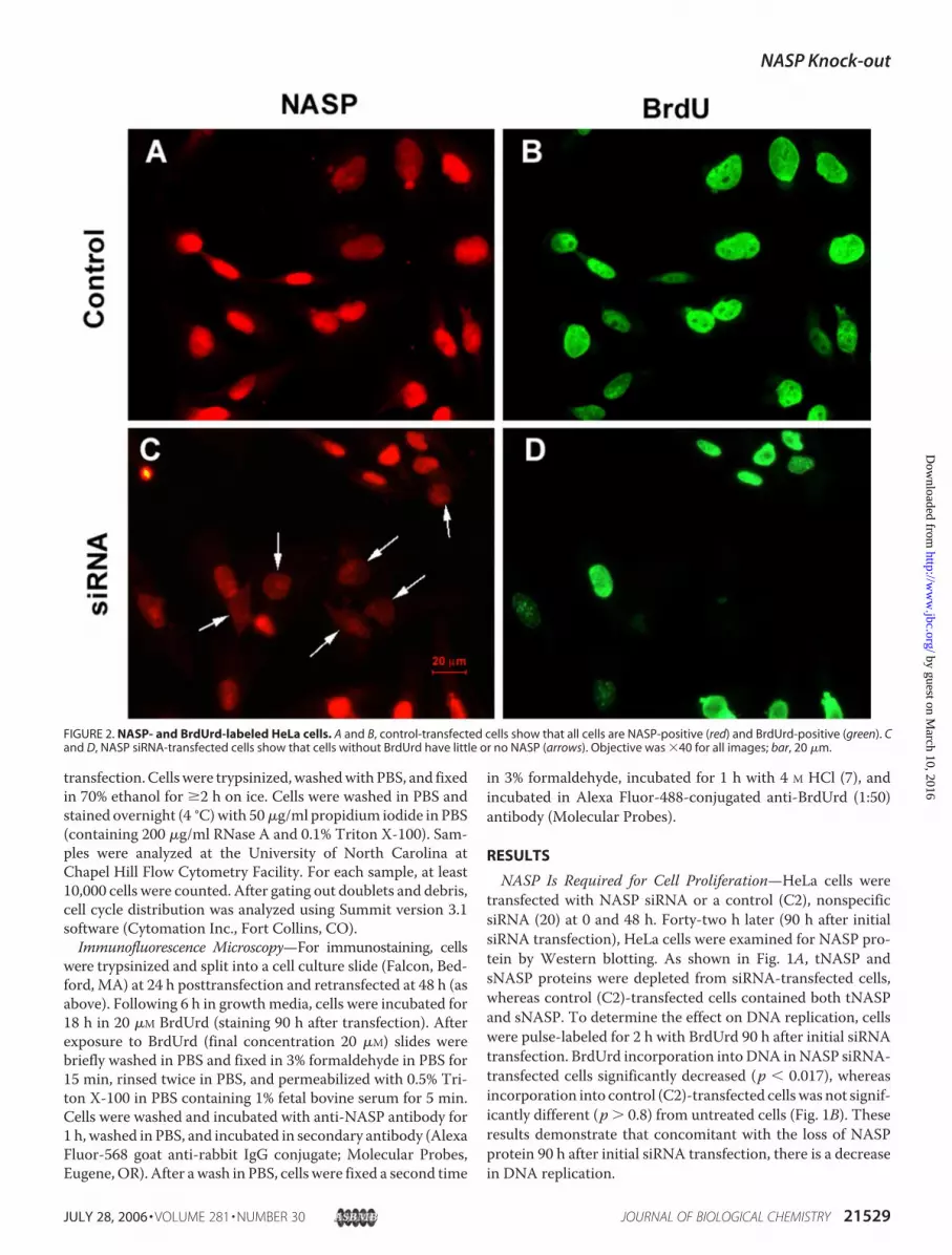
FIGURE 2. NASP- and BrdUrd-labeled HeLa cells. A and B, control-transfected cells show that all cells are NASP-positive (red) and BrdUrd-positive (green). Cand D, NASP siRNA-transfected cells show that cells without BrdUrd have little or no NASP (arrows). Objective was �40 for all images; bar, 20 �m.
NASP Knock-out
JULY 28, 2006 • VOLUME 281 • NUMBER 30 JOURNAL OF BIOLOGICAL CHEMISTRY 21529
by guest on March 10, 2016
http://ww
w.jbc.org/
Dow
nloaded from

With the loss of NASP protein, there was also a noticeableeffect on cell proliferation, which was determined by directlycounting cells. The total number of control (C2)-transfectedcells increased significantly during the 90 h following initialtransfection (�6-fold; Fig. 1C), whereas the total number ofNASP siRNA-transfected cells increased during the first 66 hafter transfection (4-fold) and did not significantly increasethereafter from 66 to 90 h (Fig. 1C). The number of NASPsiRNA-transfected cells was significantly less than the numberof control (C2)-transfected cells (p � 0.01) at 90 h (Fig. 1C).Examination of the live/dead cell ratio at 24 h posttransfectionindicated that �1% of the cells were dead (data not shown).These results demonstrate that the NASP siRNA treatment didnot eliminate cells from the culture dish but rather held them ina nonproliferation state.The effect of a loss of NASP protein on the cell cycle was
determined by FACS analysis of control (C2)- and NASPsiRNA-transfected cells at 90 h following initial transfection. Asshown in Fig. 1D, the number of cells in G1 phase of the cellcycle increased after NASP siRNA transfection concomitantwith a decrease in the number of cells in S andG2 phases. Theseresults indicate that a loss of NASP protein causes a delay in cellcycle progression; cells in G1 cannot progress past the G1/Sborder. Previously, when NASP was overexpressed in HeLacells, a similar delay in cell cycle progression was observed (12).Consequently, attempts to rescue the siRNA treated cells bytransfecting with tNASP cDNA resulted in a large increase intNASP protein as shown by Western blots and a modestincrease in BrdUrd incorporation, but the overall effect was acontinued delay in cell cycle progression and increased num-bers of cells in G1 as reported previously (Ref. 12 and data notshown).Immunolocalization of BrdUrd and NASP in the nuclei of
control cells after incubation with BrdUrd for 18 h, demon-strated that all cells contained both NASP and BrdUrd in theirnuclei (Fig. 2,A and B); however, 90 h after NASP siRNA trans-fection, immunostaining for NASP protein had decreased inmany cells (Fig. 2C, arrows), and BrdUrd staining was absent inthese cells (Fig. 2D). We conclude from these experiments thatthe loss of NASP from the nucleus coincided with the loss ofBrdUrd incorporation into DNA and the completion of S phaseinHeLa cells. However, because of a reservoir of NASP protein,it takes at least 3–4 days before the lack of NASP protein affectscell proliferation.Comparison of NASP and SLBP Knockdown in U2OS Cells—
SLBP binds histone mRNA stem loops and is required for his-tone message synthesis and translation (21). Cells synthesizeSLBP just prior to entering S phase (22) and require SLBP forproper DNA replication (20). When treated with SLBP, siRNA
FIGURE 3. NASP and SLBP knockdown in U2OS cells. A, Western blot ofNASP and SLBP in U2OS cells treated with C2 siRNA (control siRNA having nocellular target) (lane 1), NASP siRNA (lane 2), or U2OS siRNA (lane 3). Proteinswere detected 90 h after siRNA transfection. Symplekin was the loading con-trol protein. Western blot was probed with anti-NASP, anti-SLBP, or anti-symplekin antibody. B, cell cycle changes induced by transfection of U2OScells with NASP, SLBP, or control (C2) siRNA. FACS analysis demonstrates anincrease in NASP siRNA treated cells in G1 phase and a decrease of cells in S-G2;in contrast, SLBP siRNA-treated cells demonstrate an increase of cells in Sphase and a decrease of cells in G1.
NASP Knock-out
21530 JOURNAL OF BIOLOGICAL CHEMISTRY VOLUME 281 • NUMBER 30 • JULY 28, 2006
by guest on March 10, 2016
http://ww
w.jbc.org/
Dow
nloaded from
cells accumulate in S phase (20); thus a comparison, of NASPand SLBP siRNA treatments in the same cell type allows a cleardistinction between cell cycle phases. U2OS cells were trans-fected with NASP siRNA, control (C2) nonspecific siRNA, orSLBP siRNA. As shown in Fig. 3A, when tNASP and sNASPproteins were depleted from siRNA-transfected U2OS cells,SLBP was not depleted.When SLBP protein was depleted fromsiRNA-transfected U2OS cells, tNASP and sNASP proteinswere not depleted (Fig. 3A). The difference between the loss ofNASP protein and the loss of SLBP protein on cell cycle progres-sion is shown by FACS analysis in Fig. 3B. Loss of SLBP clearlycauses cells to accumulate in S phase, as previously reported (20,23), whereas loss of NASP protein clearly causes cells to accumu-late at theG1/Sborder.CellswithoutNASPaccumulateSLBP(Fig.3A); therefore, it is most likely that cells lacking NASP enter Sphasebut then immediatelyarrest.Weconclude fromtheseexper-iments that inbothHeLaandU2OScells,NASP is required for cellproliferation. These results are similar to the results seenwhenG1
cells are treated with aphidicolin orhydroxyurea; cells reach the G1/Sborder, and SLBP reaches maximallevels (24).NASP Is Required for Normal
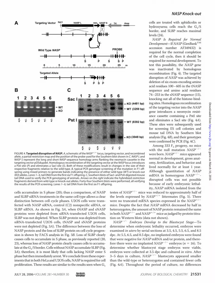
Development—IfNASP (GenBankTMaccession number AF349432) isrequired for the normal completionof the cell cycle, then it should berequired for normal development. Totest this possibility, the NASP genewas inactivated by homologousrecombination (Fig. 4). The targeteddisruption of NASP was achieved bydeletion of six exons encoding aminoacid residues 100–605 in the tNASPsequence and amino acid residues74–253 in the sNASP sequence (15),knocking out all of the histone-bind-ing sites.Homologous recombinationof the targeting vector into theNASPgene introduces a neomycin resist-ance cassette containing a PstI siteand eliminates a SacI site (Fig. 4A).These sites were subsequently usedfor screening ES cell colonies andmouse tail DNA by Southern blotanalysis (Fig. 4B), and their genotypeswere confirmed by PCR (Fig. 4C).
Among 333 F2 progeny, no micewith the null mutation NASP�/�
were born.NASP�/�mice appearednormal in development, gross anat-omy, fertilization, and behavior andlived normally for at least 1 year.Although quantitation of NASPmRNA in homozygous NASP�/�-disrupted cells was not possiblebecause of early embryonic lethal-ity, NASP mRNA isolated from the
testes of NASP�/� mice was reduced to approximately half ofthe levels expressed by NASP�/� littermates (Fig. 5). Therewere no truncated mRNA species expressed in the NASP�/�
mice. Despite the fact that NASP mRNA decreased by half inheterozygotes, the amount ofNASP protein remained the samein bothNASP�/� andNASP�/�mice as judged by protein titra-tion on Western blots (data not shown).NASP�/� Embryos Develop to the Blastocyst Stage—To
determine when embryonic lethality occurred, embryos wereexamined in utero by serial sections at 3.5, 4.5, 5.5, 6.5, and 8.5dpc. At 5.5, 6.5, and 8.5 dpc, no implanted embryos were foundthat were negative forNASPmRNA and/or protein, and there-fore there were no implanted NASP�/� embryos (n � 16). Todetermine whether blastocyst stage embryos were viable,embryos were collected at 3.5 dpc and cultured in vitro. After3–5 days in culture, NASP�/� blastocysts appeared smallerthan the wild-type or heterozygotes and contained fewer cells(Fig. 6A). Throughout the period of culture, no differences
FIGURE 4. Targeted disruption of NASP. A, schematic of the NASP�/� locus, targeting vector, and recombinedallele, a partial restriction map and the position of the probe used for the Southern blot shown in C. NASP L andNASP S represent the long and short NASP sequence homology arms flanking the neomycin cassette in thetargeting vector pOSdupdel. Homologous recombination of the targeting vector at the NASP locus introducesa PstI site (P) and eliminates a SacI site (S). Both of these modifications result in changes in the size of theirrespective fragments relative to the wild-type. B, typical PCR genotype screening of the mutation in F1 off-spring using mixed primers to generate bands indicating the presence of either wild-type (WT) or knock-out(KO) alleles. Lanes 1– 6, tail DNA from the first six F1 offspring. C, Southern blots of SacI- and PstI-digested mousetail DNA used to verify the PCR genotyping of animals. Arrows on the right indicate the hybridized restrictionfragments derived from wild-type or knock-out alleles. Forty-four Southern blots were performed, all verifyingthe results of the PCR screening. Lanes 1– 6, tail DNA from the first six F1 offspring.
NASP Knock-out
JULY 28, 2006 • VOLUME 281 • NUMBER 30 JOURNAL OF BIOLOGICAL CHEMISTRY 21531
by guest on March 10, 2016
http://ww
w.jbc.org/
Dow
nloaded from

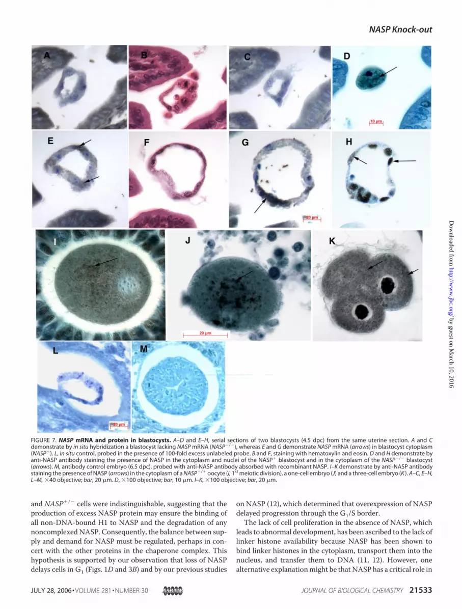
could be seen in the growth ofNASP�/� versus NASP�/� blas-tocyst colonies. By 10 days in culture, theNASP�/� blastocystshad completely failed to develop (Fig. 6A). As determined byPCR (Fig. 6B),NASP�/� mice transmitted the targeted allele to86% of their blastocysts (n� 56;NASP�/� � 8,NASP�/� � 33,NASP�/� � 15). In situ hybridization of serial sections of 4.5dpc embryos (n � 22) with a NASP oligonucleotide proberevealed an embryo that was negative for NASP mRNA (andtherefore NASP�/�; Fig. 7, A–C). An adjacent embryo in thesame serial section of uterus was considerably larger in size andpositive for NASP mRNA (and therefore NASP�/� orNASP�/�; Fig. 7, E andG, arrows). Control sections of embryostreatedwith the in situ probe in the presence of 100� unlabeledprobe were negative (Fig. 7L). Serial sections of the NASP�/�
and the NASP�/� embryos at 4.5 dpc were both positive forNASP protein (Fig. 7, D and H, respectively, arrows).The death of NASP�/� embryos most likely coincides with
the depletion of thematernally derivedNASPprotein reservoir.To determine whether a reservoir of stored maternal NASPprotein exists, normal embryos were examined for NASP pro-tein. As shown in Fig. 7, I–K, wild-type oocytes in the first mei-otic division (Fig. 7I ), one-cell embryos (Fig. 7J ), and three-cellembryos (Fig. 7K ) all contain NASP protein in their cytoplasm(arrows). Control sections of embryos treated with anti-NASPantibody absorbed with recombinant NASP antigen (Fig. 7M)were negative. Consequently, after fertilization during S phasein the one-cell embryo and in subsequent cell divisions, amaternalNASPprotein reservoir exists in the oocyte cytoplasmthat could sustain development for 3–4 days. We concludefrom these observations that the NASP�/� null mutationcauses embryonic lethality by the end of the preimplantationstage.
DISCUSSION
Wehave demonstrated that theH1 histone chaperoneNASPis required for cell proliferation and normal development.NASP, Asf1 (9, 25), and CAF-1 (6, 7) have all been identified asmembers of a multichaperone protein complex that appears toshuttle histones during nucleosome remodeling and all appearto be required for normal progression through S phase (26, 27).NASP mRNA expression parallels histone mRNA expressionduring the cell cycle (14), increasing during S phase and declin-
ing during G2, and is usually undetectable in nonmitotic cells.However,NASPprotein is often retained in nonmitotic cells forseveral days after the last cell division, and this is particularlyapparent in postmeiotic oocytes and round spermatids (14, 28).Although the null mutation NASP�/� caused embryoniclethality, null embryos survive until the blastocyst stage, andweconclude that this may be explained by the presence of storedNASP protein in the cytoplasm of oocytes (Fig. 7). We do notknow whether stored NASP has bound histones in its threehistone-binding sites (16) or whether it is associated withHSP90, which facilitates histone binding to NASP (11).The cellular requirement forNASP to complete the cell cycle
(Figs. 1–3) and consequently the implicit requirement for H1histones (2, 3, 29) reflect the fact that both NASP (12) and H1(30, 31) are in a dynamic equilibrium within the nucleus. Theequilibrium of NASP with H1 and H1 with DNA appears criti-cal for nucleosome spacing and transcriptional activation.Embryos lacking several H1 variants die in midgestation (2),and embryos lacking NASP die at the blastula stage (Figs. 6 and7). Both processes suggest that supplies of NASP and H1 pro-teins are used as long as possible either by storage in the cyto-plasm (Fig. 7) or by binding to higher-order chromatin struc-ture (10). Although NASP mRNA was reduced by half inheterozygotes (Fig. 5,NASP�/�), the protein levels inNASP�/�
FIGURE 5. NASP�/� mice express decreased levels of tNASP mRNA in theirtestes. Northern blot analysis was performed using 5, 2, 1, and 0.5 �g ofpoly(A)� RNA from either wild-type (Wt) or heterozygous (Het) mice followedby probing with a cDNA that recognizes both testis and somatic NASP. Het-erozygous to wild-type ratios of the PhosphorImager volume quantitation ofpixel intensities are shown below the blot.
FIGURE 6. Deletion of NASP leads to degeneration of blastocysts in cul-ture. A, day 3.5 postcoitum blastocysts were isolated from NASP�/� inter-crosses and were cultured in individual wells of 96-well plates for 10 days.Inner cell mass (ICM) and trophoblast giant cells (TGC) are indicated. B, exam-ples of PCR genotyping of blastocysts cultured as described in A. Bands indi-cate the presence of either wild-type (WT) or knock-out (KO) alleles.
NASP Knock-out
21532 JOURNAL OF BIOLOGICAL CHEMISTRY VOLUME 281 • NUMBER 30 • JULY 28, 2006
by guest on March 10, 2016
http://ww
w.jbc.org/
Dow
nloaded from
and NASP�/� cells were indistinguishable, suggesting that theproduction of excess NASP protein may ensure the binding ofall non-DNA-bound H1 to NASP and the degradation of anynoncomplexedNASP. Consequently, the balance between sup-ply and demand for NASP must be regulated, perhaps in con-cert with the other proteins in the chaperone complex. Thishypothesis is supported by our observation that loss of NASPdelays cells in G1 (Figs. 1D and 3B) and by our previous studies
on NASP (12), which determined that overexpression of NASPdelayed progression through the G1/S border.The lack of cell proliferation in the absence of NASP, which
leads to abnormal development, has been ascribed to the lack oflinker histone availability because NASP has been shown tobind linker histones in the cytoplasm, transport them into thenucleus, and transfer them to DNA (11, 12). However, onealternative explanationmight be thatNASP has a critical role in
FIGURE 7. NASP mRNA and protein in blastocysts. A–D and E–H, serial sections of two blastocysts (4.5 dpc) from the same uterine section. A and Cdemonstrate by in situ hybridization a blastocyst lacking NASP mRNA (NASP�/�), whereas E and G demonstrate NASP mRNA (arrows) in blastocyst cytoplasm(NASP�). L, in situ control, probed in the presence of 100-fold excess unlabeled probe. B and F, staining with hematoxylin and eosin. D and H demonstrate byanti-NASP antibody staining the presence of NASP in the cytoplasm and nuclei of the NASP� blastocyst and in the cytoplasm of the NASP�/� blastocyst(arrows). M, antibody control embryo (6.5 dpc), probed with anti-NASP antibody absorbed with recombinant NASP. I–K demonstrate by anti-NASP antibodystaining the presence of NASP (arrows) in the cytoplasm of a NASP�/� oocyte (I, 1st meiotic division), a one-cell embryo (J) and a three-cell embryo (K ). A–C, E–H,L–M, �40 objective; bar, 20 �m. D, �100 objective; bar, 10 �m. I–K, �100 objective; bar, 20 �m.
NASP Knock-out
JULY 28, 2006 • VOLUME 281 • NUMBER 30 JOURNAL OF BIOLOGICAL CHEMISTRY 21533
by guest on March 10, 2016
http://ww
w.jbc.org/
Dow
nloaded from

the multichaperone protein complex that is independent of H1transport into the nucleus. If the lack of NASP activated a G1/Scheckpoint (Chk2) similar to that observed in a dominant-neg-ative mutant of CAF-1 (6), then DNA replication might bedelayed independently of the availability of linker histones. InHeLa cells, NASP is a binding partner of the heterodimerKu70/Ku80 and its catalytic subunit DNA-PK (13). Ku70/Ku80 andDNA-PK are necessary for DNA double-strand break repair bynonhomologous end joining (4, 32), and H1 histones have animportant role in protectingDNAdegradation during end join-ing (33). Thus NASP could have a role in nucleosome remod-eling during DNA repair by mediating H1-DNA binding.Several characteristics of the NASP protein strengthen the
hypothesis that NASP has a role in nucleosome remodeling.Mouse sNASP is an acidic protein (pI � 4.23) of 45.8 kDa,which is identical to the tNASP form (pI � 4.16; 83.9 kDa),except that two internal segments of the protein have beendeleted (14). Both forms have a leucine zipper, coiled-coildomains, and tetratricopeptide repeat domains, all of whichmay contribute to the interaction of NASP with other proteins,including HSP90 (11). Whether NASP binds DNA directlyremains to be determined. Interestingly, motifs for bindingChk2 and docking the coactivator p300 exist within the NASPsequence and may prove important in future investigations offunction.The multichaperone protein complexes (9) that coordinate
the assembly of new nucleosomes in both DNA-dependent(CAF-1) and DNA-independent (HIRA) pathways contain sev-eral proteins in common, includingAsf1a, HAT1, andNASP (8,34). Themultichaperone complexes containing CAF-1 interactwith the DNA replication machinery via proliferating cellnuclear antigen (35), which is the sliding clamp of the replica-tion fork (5, 36). Amultiprotein complex containingHAT1 anda NASP-like protein has been reported in yeast; the histonechaperoneHif1p associates with theHat1p/Hat2p complex fol-lowing entry into the nucleus and participates in histone-DNAinteraction (37). Hif1p and NASP share sequence similaritieswith the Xenopus protein N1/N2 (37, 38). Consequently NASPappears to be a conserved and critical member of the mul-tichaperone complexes that participate in nucleosome remod-eling. We conclude from this study that NASP and, by implica-tion, the linker histones are key players in the assembly ofchromatin after DNA is newly replicated and that both NASPandH1 are required to ensure a faithful transition of chromatinfrom one cell cycle to the next.
REFERENCES1. Becker, P. B., and Horz, W. (2002) Annu. Rev. Biochem. 71, 247–2732. Fan, Y., Nikitina, T., Morin-Kensicki, E. M., Zhao, J., Magnuson, T. R.,
Woodcock, C. L., and Skoultchi, A. I. (2003)Mol Cell Biol. 23, 4559–45723. Fan, Y., Nikitina, T., Zhao, J., Fleury, T. J., Bhattacharyya, R., Bouhassira,
E. E., Stein, A., Woodcock, C. L., and Skoultchi, A. L. (2005) Cell 123,1199–1212
4. Sancar, A., Lindsey-Boltz, L. A., Unsal-Kacmaz, K., and Linn, S. (2004)
Annu. Rev. Biochem. 73, 39–855. Johnson, A., and O’Donnell, M. (2005) Annu. Rev. Biochem. 74, 283–3156. Ye, X. F., Franco, A. A., Santos, H., Nelson, D. M., Kaufman, P. D., and
Adams, P. D. (2003)Mol. Cell 11, 341–3517. Hoek, M., and Stillman, B. (2003) Proc. Natl. Acad. Sci. U. S. A. 100,
12183–121888. Tagami, H., Ray-Gallet, D., Almouzni, G., and Nakatani, Y. (2004) Cell
116, 51–619. Groth, A., Ray-Gallet, D., Quivy, J.-P., Lukas, J., Bartek, J., and Almouzni,
G. (2005)Mol. Cell 17, 301–31110. Bustin, M., Catez, F., and Lim, J.-H. (2005)Mol. Cell 17, 617–62011. Alekseev, O. M., Widgren, E. E., Richardson, R. T., and O’Rand, M. G.
(2005) J. Biol. Chem. 280, 2904–291112. Alekseev, O. M., Bencic, D. C., Richardson, R. T., Widgren, E. E., and
O’Rand, M. G. (2003) J. Biol. Chem. 278, 8846–885213. Alekseev, O. M., Richardson, R. T., Pope, M., and O’Rand, M. G. (2005)
Proteins 61, 1–514. Richardson, R. T., Batova, I. N.,Widgren, E. E., Zheng, L.-X.,Whitfield,M.
Marzluff, W. F., and O’Rand, M. G. (2000) J. Biol. Chem. 275,30378–30386
15. Richardson, R. T., Bencic, D. C., and O’Rand, M. G. (2001) Gene 274,67–75
16. Batova, I., and O’Rand, M. G. (1996) Biol. Reprod. 54, 1238–124417. Mansour, S. L., Thomas, K. R., Deng, C. X., and Capecchi, M. R. (1988)
Proc. Natl. Acad. Sci. U. S. A. 87, 7688–769218. Schwarzacher, T., andHeslop-Harrison, P. (2000)Practical in situHybrid-
ization, pp. 126–145, Springer-Verlag, New York19. Wagner, E. J., and Garcia-Blanco, M. A. (2002)Mol. Cell 10, 943–94920. Wagner, E. J., Berkow, A., and Marzluff, W.F. (2005) Biochem. Soc. Trans.
33, 471–47321. Marzluff, W. F. (2005) Curr. Opin. Cell Biol. 17, 274–28022. Whitfield, M. L., Zheng, L. X., Baldwin, A., Ohta, T., Hurt, M. M., and
Marzluff, W. F. (2000)Mol. Cell. Biol. 20, 4188–419823. Zhao, X., McKillop-Smith, S., and Muller, B. (2004) J. Cell Sci. 117,
6043–605124. Whitfield, M. L., Kaygun, H., Erkmann, J. A., Townley-Tilson, W. H. D.,
Dominski, Z., and Marzluff, W. F. (2004) Nucleic Acids Res. 32,4833–4842
25. Schulz, L. L., and Tyler, J. K. (2006) FASEB J. 20, 488–49026. Franco, A. A., Lam, W. M., Burgers, P. M., and Kaufman, P. D. (2005)
Genes Dev. 19, 1365–137527. Lewis, L. K., Karthikeyan, G., Cassiano, J., and Resnick, M. A. (2005) Nu-
cleic Acids Res. 33, 4928–493928. Welch, J. E., and O’Rand, M. G. (1990) Biol. Reprod. 43, 569–57829. Woodcock, C. L., Skoultchi, A. I., and Fan, Y. (2006)Chromosome Res. 14,
17–2530. Lever, M. A., Thng, J. P. H., Sun, X. J., and Hendzel, M. J. (2000) Nature
408, 873–87631. Misteli, T., Gunjan, A., Hock, R., Bustin, M., and Brown, D. T. (2000)
Nature 408, 877–88132. Koike, M. (2002) J. Radiat. Res. 43, 223–23633. Liang, L., Deng, L., Chen, Y., Li, G. C., Shao, C., and Tischfield, J. A. (2005)
J. Biol. Chem. 280, 31442–3144934. Nakatani, Y., Ray-Gallet, D., Quivy, J.-P., Tagami, H., and Almouzni, G.
(2004) Cold Spring Harb. Symp. Quant. Biol. 69, 273–28035. Shibahara, K., and Stillman, B. (1999) Cell 96, 575–58536. Tyler, J. K. (2002) Eur. J. Biochem. 269, 2268–227437. Ai, X., and Parthum, M. R. (2004)Mol. Cell 14, 195–20538. Welch, J. E., Zimmerman, L. J., Joseph, D. R., and O’Rand, M. G. (1990)
Biol. Reprod. 43, 559–568
NASP Knock-out
21534 JOURNAL OF BIOLOGICAL CHEMISTRY VOLUME 281 • NUMBER 30 • JULY 28, 2006
by guest on March 10, 2016
http://ww
w.jbc.org/
Dow
nloaded from

O'RandThresher, Eric J. Wagner, Kelly D. Sullivan, William F. Marzluff and Michael G.
Richard T. Richardson, Oleg M. Alekseev, Gail Grossman, Esther E. Widgren, RandyIs Required for Cell Proliferation
Nuclear Autoantigenic Sperm Protein (NASP), a Linker Histone Chaperone That
doi: 10.1074/jbc.M603816200 originally published online May 25, 20062006, 281:21526-21534.J. Biol. Chem.
10.1074/jbc.M603816200Access the most updated version of this article at doi:
Alerts:
When a correction for this article is posted•
When this article is cited•
to choose from all of JBC's e-mail alertsClick here
http://www.jbc.org/content/281/30/21526.full.html#ref-list-1
This article cites 37 references, 15 of which can be accessed free at
by guest on March 10, 2016
http://ww
w.jbc.org/
Dow
nloaded from