novel peptides with tyrosinase inhibitory activity
TRANSCRIPT
Novel peptides with tyrosinase inhibitory activity
Marloes Schurink a,b,*, Willem J.H. van Berkel a, Harry J. Wichers b, Carmen G. Boeriu b
a Laboratory of Biochemistry, Wageningen University, P.O. Box 9101, 6700 HB Wageningen, The NetherlandsbAgrotechnology and Food Sciences Group, Wageningen University, P.O. Box 17, 6700 AA Wageningen, The Netherlands
a r t i c l e i n f o
Article history:
Received 27 October 2006
Received in revised form
22 November 2006
Accepted 28 November 2006
Published on line 22 January 2007
Keywords:
Tyrosinase
Inhibitors
Protein–protein interaction
Peptide SPOT synthesis
Fluorescent protein labeling
a b s t r a c t
Tyrosinase inhibition by peptides may find its application in food, cosmetics or medicine. In
order to identify novel tyrosinase inhibitory peptides, protein-based peptide libraries made
by SPOT synthesis were used to screen for peptides that show direct interaction with
tyrosinase. One of the peptide libraries studied consists of overlapping, octameric peptides
derived from industrial proteins as b-casein, a-lactalbumin, b-lactoglobulin, ovalbumin,
gliadin, glycinin, and b-conglycinin. On-membrane activity staining resulted in a set of
peptides that are not only able to bind to tyrosinase, but are able to inhibit tyrosinase as well.
Peptides containing aspartic or glutamic acid residues usually do not bind very well to
tyrosinase. Strong tyrosinase-binding peptides always contain one or more arginine resi-
dues, often in combination with phenylalanine, while lysine residues can be found equally
among nonbinding peptides as well as moderate tyrosinase-binding peptides. The presence
of the hydrophobic, aliphatic residues valine, alanine or leucine appears to be important for
tyrosinase inhibition. Therefore, good tyrosinase inhibitory peptides preferably contain
arginine and/or phenylalanine in combination with valine, alanine and/or leucine.
# 2006 Elsevier Inc. All rights reserved.
1. Introduction
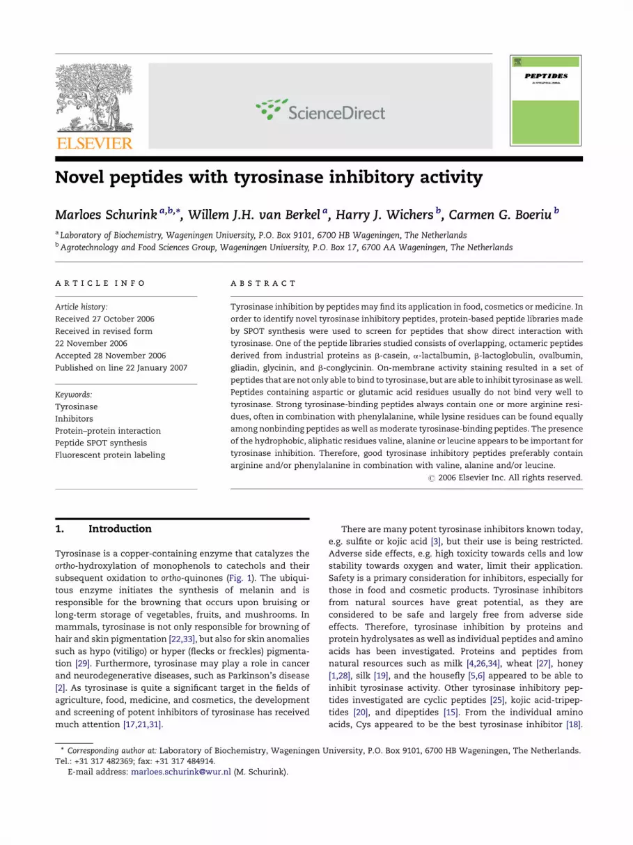
Tyrosinase is a copper-containing enzyme that catalyzes the
ortho-hydroxylation of monophenols to catechols and their
subsequent oxidation to ortho-quinones (Fig. 1). The ubiqui-
tous enzyme initiates the synthesis of melanin and is
responsible for the browning that occurs upon bruising or
long-term storage of vegetables, fruits, and mushrooms. In
mammals, tyrosinase is not only responsible for browning of
hair and skin pigmentation [22,33], but also for skin anomalies
such as hypo (vitiligo) or hyper (flecks or freckles) pigmenta-
tion [29]. Furthermore, tyrosinase may play a role in cancer
and neurodegenerative diseases, such as Parkinson’s disease
[2]. As tyrosinase is quite a significant target in the fields of
agriculture, food, medicine, and cosmetics, the development
and screening of potent inhibitors of tyrosinase has received
much attention [17,21,31].
* Corresponding author at: Laboratory of Biochemistry, Wageningen UTel.: +31 317 482369; fax: +31 317 484914.
E-mail address: [email protected] (M. Schurink).
There are many potent tyrosinase inhibitors known today,
e.g. sulfite or kojic acid [3], but their use is being restricted.
Adverse side effects, e.g. high toxicity towards cells and low
stability towards oxygen and water, limit their application.
Safety is a primary consideration for inhibitors, especially for
those in food and cosmetic products. Tyrosinase inhibitors
from natural sources have great potential, as they are
considered to be safe and largely free from adverse side
effects. Therefore, tyrosinase inhibition by proteins and
protein hydrolysates as well as individual peptides and amino
acids has been investigated. Proteins and peptides from
natural resources such as milk [4,26,34], wheat [27], honey
[1,28], silk [19], and the housefly [5,6] appeared to be able to
inhibit tyrosinase activity. Other tyrosinase inhibitory pep-
tides investigated are cyclic peptides [25], kojic acid-tripep-
tides [20], and dipeptides [15]. From the individual amino
acids, Cys appeared to be the best tyrosinase inhibitor [18].
niversity, P.O. Box 9101, 6700 HB Wageningen, The Netherlands.
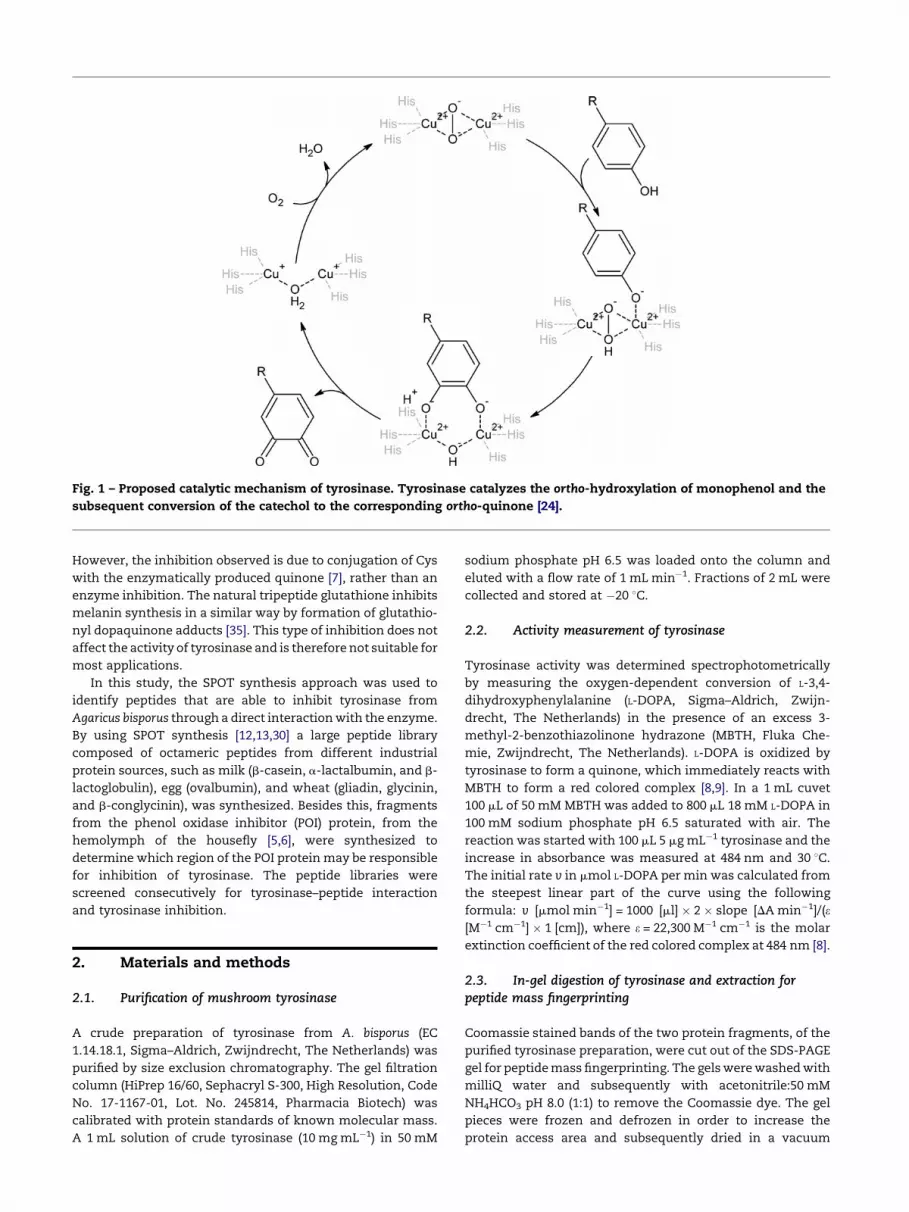
Fig. 1 – Proposed catalytic mechanism of tyrosinase. Tyrosinase catalyzes the ortho-hydroxylation of monophenol and the
subsequent conversion of the catechol to the corresponding ortho-quinone [24].
However, the inhibition observed is due to conjugation of Cys
with the enzymatically produced quinone [7], rather than an
enzyme inhibition. The natural tripeptide glutathione inhibits
melanin synthesis in a similar way by formation of glutathio-
nyl dopaquinone adducts [35]. This type of inhibition does not
affect the activity of tyrosinase and is therefore not suitable for
most applications.
In this study, the SPOT synthesis approach was used to
identify peptides that are able to inhibit tyrosinase from
Agaricus bisporus through a direct interaction with the enzyme.
By using SPOT synthesis [12,13,30] a large peptide library
composed of octameric peptides from different industrial
protein sources, such as milk (b-casein, a-lactalbumin, and b-
lactoglobulin), egg (ovalbumin), and wheat (gliadin, glycinin,
and b-conglycinin), was synthesized. Besides this, fragments
from the phenol oxidase inhibitor (POI) protein, from the
hemolymph of the housefly [5,6], were synthesized to
determine which region of the POI protein may be responsible
for inhibition of tyrosinase. The peptide libraries were
screened consecutively for tyrosinase–peptide interaction
and tyrosinase inhibition.
2. Materials and methods
2.1. Purification of mushroom tyrosinase
A crude preparation of tyrosinase from A. bisporus (EC
1.14.18.1, Sigma–Aldrich, Zwijndrecht, The Netherlands) was
purified by size exclusion chromatography. The gel filtration
column (HiPrep 16/60, Sephacryl S-300, High Resolution, Code
No. 17-1167-01, Lot. No. 245814, Pharmacia Biotech) was
calibrated with protein standards of known molecular mass.
A 1 mL solution of crude tyrosinase (10 mg mL�1) in 50 mM
sodium phosphate pH 6.5 was loaded onto the column and
eluted with a flow rate of 1 mL min�1. Fractions of 2 mL were
collected and stored at �20 8C.
2.2. Activity measurement of tyrosinase
Tyrosinase activity was determined spectrophotometrically
by measuring the oxygen-dependent conversion of L-3,4-
dihydroxyphenylalanine (L-DOPA, Sigma–Aldrich, Zwijn-
drecht, The Netherlands) in the presence of an excess 3-
methyl-2-benzothiazolinone hydrazone (MBTH, Fluka Che-
mie, Zwijndrecht, The Netherlands). L-DOPA is oxidized by
tyrosinase to form a quinone, which immediately reacts with
MBTH to form a red colored complex [8,9]. In a 1 mL cuvet
100 mL of 50 mM MBTH was added to 800 mL 18 mM L-DOPA in
100 mM sodium phosphate pH 6.5 saturated with air. The
reaction was started with 100 mL 5 mg mL�1 tyrosinase and the
increase in absorbance was measured at 484 nm and 30 8C.
The initial rate v in mmol L-DOPA per min was calculated from
the steepest linear part of the curve using the following
formula: v [mmol min�1] = 1000 [ml] � 2 � slope [DA min�1]/(e[M�1 cm�1] � 1 [cm]), where e = 22,300 M�1 cm�1 is the molar
extinction coefficient of the red colored complex at 484 nm [8].
2.3. In-gel digestion of tyrosinase and extraction forpeptide mass fingerprinting
Coomassie stained bands of the two protein fragments, of the
purified tyrosinase preparation, were cut out of the SDS-PAGE
gel for peptide mass fingerprinting. The gels were washed with
milliQ water and subsequently with acetonitrile:50 mM
NH4HCO3 pH 8.0 (1:1) to remove the Coomassie dye. The gel
pieces were frozen and defrozen in order to increase the
protein access area and subsequently dried in a vacuum

centrifuge. The cysteines were reduced during at least 1 h at
56 8C, after adding 50 mM dithiothreitol in 50 mM NH4HCO3 pH
8.0. After cooling to room temperature and washing once with
50 mM NH4HCO3 pH 8.0, the cysteines were alkylated, by
adding 10 mL 55 mM iodoacetamide in 50 mM NH4HCO3 pH 8.0,
during at least 45 min in the dark with occasional vortexing.
Then the gel pieces were washed thoroughly with 50 mM
NH4HCO3 pH 8.0 and dried in the vacuum centrifuge in 30 to
120 min. The gels were rehydrated with 5 mL cold trypsin
(bovine sequencing grade, Roche, Mannheim, Germany)
50 ng mL�1 in 50 mM NH4HCO3 pH 8.0 and incubated overnight
while shaking at room temperature. The liquid phase contain-
ing the peptides was collected and the remaining gel pieces
were extracted with 10 mL 50 mM NH4HCO3 pH 8.0 with 5%
trifluoroacetic acid (TFA) followed by 10 mL 10% acetonitrile
with 1% TFA and combined with the liquid phase. The samples
were analyzed by LC-MS. An electrospray potential of 1.8 kV
was applied with a capillary temperature of 150 8C. Full scan
positive mode MS spectra with three microscans were
measured between m/z 350 and 1400 on a LCQ classic (Thermo
electron, San Jose, USA). MS/MS scans of the three most
abundant peaks in the MS scan were recorded in data
dependent mode at 35% collision energy with a 4 Da isolation
width. Each run with all MS/MS spectra obtained was analyzed
with Bioworks 3.2 using a database containing all relevant
protein sequences.
2.4. SPOT synthesis of the peptide libraries
Peptides derived from the sequences of POI [5,6], b-casein
(SwissProt accession no. P02666), a-lactalbumin (SwissProt
accession no. P00711), b-lactoglobulin (SwissProt accession
no. P02754), ovalbumin (RefSeq accession no. NP_990483),
gliadin (SwissProt accession no. P02863), glycinin (SwissProt
accession no. P04776), and b-conglycinin (SwissProt acces-
sion no. P13916) were synthesized in duplicate on derivatized
cellulose membranes (Amino-PEG membrane, substitution
400 nmol cm�2, Intavis AG, Bergisch Gladbach, Germany)
using the standard 9-fluorenylmethoxycarbonyl (Fmoc)
strategy. The activated amino acid derivatives were delivered
on defined positions on the membranes using a pipetting
robot (Autospot upgrade kit for the Automated Multiple
Peptide Synthesizer AMS 422, software AutoSpot A, Abimed
Analysen-Technik, Langenfeld, Germany). When the desired
peptides were fully assembled the membranes were
dried and stored under vacuum at �20 8C until use.
2.5. Fluorescent labeling of tyrosinase
Purified tyrosinase fractions with the highest tyrosinase
activity containing �0.25 mg mL�1 tyrosinase were labeled
with Marina Blue (MB, FluoReporter Protein Labeling Kit F-
10230, Molecular Probes, Leiden, The Netherlands). In total
3 mg of pure tyrosinase was labeled with 0.5 mg MB. After
separation of the reactants using PD-10 desalting columns
(Amersham Biosciences, Uppsala, Sweden) the average
degree of labeling of the tyrosinase-MB preparation was �5
labels per protein molecule, as determined by MB absorption
at 365 nm. The labeled protein was analyzed by SDS-PAGE to
confirm that the label is covalently linked to the protein.
The enzyme activity was not affected by the labeling
procedure.
2.6. Incubation of the cellulose-bound peptides withtyrosinase-MB
The peptide libraries were pre-incubated in 100 mL 50 mM
sodium phosphate pH 6.5. The background fluorescence of the
membranes was recorded with excitation at 365 nm using the
FluorChem 8800 (Alpha Innotech Corporation, Hoechst
Blue HB-500 emission filter 465 nm, AlphaEaseFCTM Software
Version 3.1.2), before adding tyrosinase-MB (5 mL, 0.15
mg mL�1) to each membrane. The membranes were incubated
in the dark for about 1 h with tyrosinase-MB, while gently
shaken at room temperature. Then the membranes were
washed three times with buffer and the fluorescence of the
bound tyrosinase-MB was recorded using the same settings as
for the background. The light density of each spot on the
membranes was measured on a linear scale from 0 to 100%,
with 0% = black (no light) and 100% = white (maximum light
yield) at an illumination time of 16 s. The background
fluorescence was substracted to obtain the fluorescence
intensity resulting from the bound tyrosinase-MB only.
2.7. On-membrane activity staining of bound tyrosinase-MB
Immediately after recording the fluorescence of the bound
tyrosinase-MB, the membranes were homogeneously sprayed
with a freshly prepared mixture of 50 mM MBTH:18 mM L-
DOPA (1:8). When the enzymatically initiated red color was
developed, the membranes were gently washed with milliQ
water to remove excess of substrate and to stop the reaction.
After drying under vacuum, the membranes were scanned for
analysis and sealed in plastic for conservation. The relative
intensities of the red color on the spots were measured using
the same software as for the intensities of tyrosinase-MB
fluorescence.
3. Results
3.1. Tyrosinase purification and labeling
Commercial tyrosinase from A. bisporus has a specific
activity of 44 � 4 mmol min�1 mg�1 and a KM for L-DOPA of
0.66 � 0.06 mM (Fig. 2). The crude tyrosinase preparation was
purified in order to screen the peptide libraries for peptides
that bind to tyrosinase. Peptides that interact with tyrosi-
nase are potential inhibitors of tyrosinase. In this approach,
it is of great importance to remove any other protein present
in the tyrosinase preparation to avoid false-positive results.
A purification of 7.5 times was obtained by a single gel
filtration step (Fig. 3) resulting in a specific activity of
331 � 11 mmol min�1 mg�1. Based on its elution behavior
compared to standard proteins, the apparent molecular
mass of native tyrosinase was estimated to be around
127 � 5 kDa.
Fractions with the highest tyrosinase activity were used for
labeling with Marina Blue (MB). Fluorescent labeling of
Fig. 2 – Michaelis–Menten kinetics of L-DOPA oxidation by
tyrosinase at pH 6.5 and 30 8C. KM = 0.66 W 0.06 mM W L-
DOPA and Vmax = 22.3 W 0.36 nmol minS1 at 0.5 mg crude
tyrosinase in the assay.
Fig. 4 – (A) SDS-PAGE of tyrosinase. From left to right:
marker (lane 1), crude tyrosinase (lane 2), tyrosinase-MB
(lane 3), and tyrosinase (lane 4). The right part of the figure
shows the fluorescence of tyrosinase-MB at 365 nm before
staining. The two bands, H and L, are 49 W 1.7 kDa and
14 W 0.5 kDa, respectively. (B) Absorption spectrum of
tyrosinase-MB ( ) and tyrosinase ( ) at 0.25 mg mLS1.
Tyrosinase-MB has an absorbance maximum at 365 nm.
The molar extinction coefficient of purified tyrosinase at
280 nm is estimated be around �27 T 104 MS1 cmS1.
p
tyrosinase allowed on-membrane detection of the tyrosinase–
peptide interaction. Analysis of the fluorescent tyrosinase-MB
by SDS-PAGE clearly revealed the presence of a high (H) and
low (L) molecular mass chain (Fig. 4). The H chain has an
apparent molecular mass of 49 � 1.7 kDa, whereas the L chain
has an apparent molecular mass of 14 � 0.5 kDa. Assuming a
heterotetrameric quaternary structure of (HL)2, the molecular
mass of native tyrosinase would be around 126 � 4 kDa. This is
in agreement with the gel filtration results and with the
structure described for A. bisporus tyrosinase [14,32].
In order to determine the origin of both H and L chains, the
protein was blotted onto a PVDF membrane for N-terminal
sequence analysis by Edman degradation (Midwest Analytical
Inc., St. Louis, USA). The first 12 amino acid residues of the L
chain were identified to be ATNSGTLIIFDQ. This fragment
does not match the known protein sequences of tyrosinase
fromA. bisporus strain U1 (PPO1 and PPO2, SwissProt accession
nos. Q00024 and O42713, respectively) [36]. The N-terminal
sequence of the H chain could not be determined since it
appeared to be blocked. Peptide mass fingerprinting of the H
Fig. 3 – Elution profile of the gel filtration of tyrosinase. Fractions of 2 mL were collected and analyzed for protein
concentration c (*) and tyrosinase activity v (*). Tyrosinase elutes at 65 mL. The specific activity increased from
44 W 4 mmol minS1 mgS1 before to 331 W 11 mmol minS1 mgS1 after purification. According to calibration series, tyrosinase
has a molecular mass of 127 W 5 kDa.
chain showed unambiguously the presence of two fragments,
FFTLYVR (Xcorr = 2.3) and LNIVDFVKNEKFFTLYVR (Xcorr =
3.6), that are both identical to the N-terminal part of PPO2
isoenzyme. MS/MS scans of other trypsine fragments of the H
chain did not correspond to PPO1 or PPO2. Also, peptide
fragments of the L chain showed no coverage with any of the
known tyrosinase sequences.
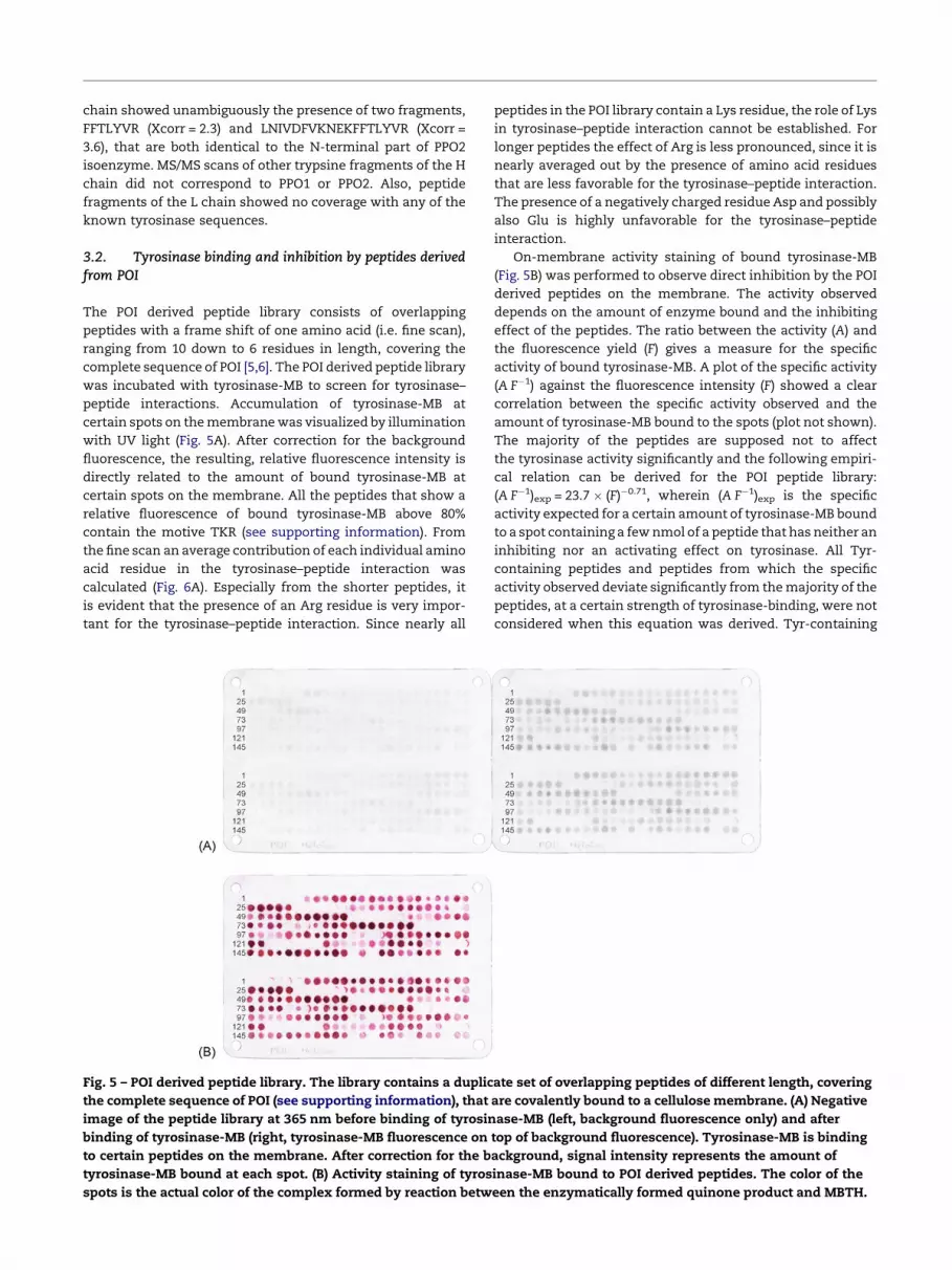
3.2. Tyrosinase binding and inhibition by peptides derivedfrom POI
The POI derived peptide library consists of overlapping
peptides with a frame shift of one amino acid (i.e. fine scan),
ranging from 10 down to 6 residues in length, covering the
complete sequence of POI [5,6]. The POI derived peptide library
was incubated with tyrosinase-MB to screen for tyrosinase–
peptide interactions. Accumulation of tyrosinase-MB at
certain spots on the membrane was visualized by illumination
with UV light (Fig. 5A). After correction for the background
fluorescence, the resulting, relative fluorescence intensity is
directly related to the amount of bound tyrosinase-MB at
certain spots on the membrane. All the peptides that show a
relative fluorescence of bound tyrosinase-MB above 80%
contain the motive TKR (see supporting information). From
the fine scan an average contribution of each individual amino
acid residue in the tyrosinase–peptide interaction was
calculated (Fig. 6A). Especially from the shorter peptides, it
is evident that the presence of an Arg residue is very impor-
tant for the tyrosinase–peptide interaction. Since nearly all
Fig. 5 – POI derived peptide library. The library contains a duplic
the complete sequence of POI (see supporting information), that
image of the peptide library at 365 nm before binding of tyrosin
binding of tyrosinase-MB (right, tyrosinase-MB fluorescence on
to certain peptides on the membrane. After correction for the b
tyrosinase-MB bound at each spot. (B) Activity staining of tyros
spots is the actual color of the complex formed by reaction betw
peptides in the POI library contain a Lys residue, the role of Lys
in tyrosinase–peptide interaction cannot be established. For
longer peptides the effect of Arg is less pronounced, since it is
nearly averaged out by the presence of amino acid residues
that are less favorable for the tyrosinase–peptide interaction.
The presence of a negatively charged residue Asp and possibly
also Glu is highly unfavorable for the tyrosinase–peptide
interaction.
On-membrane activity staining of bound tyrosinase-MB
(Fig. 5B) was performed to observe direct inhibition by the POI
derived peptides on the membrane. The activity observed
depends on the amount of enzyme bound and the inhibiting
effect of the peptides. The ratio between the activity (A) and
the fluorescence yield (F) gives a measure for the specific
activity of bound tyrosinase-MB. A plot of the specific activity
(A F�1) against the fluorescence intensity (F) showed a clear
correlation between the specific activity observed and the
amount of tyrosinase-MB bound to the spots (plot not shown).
The majority of the peptides are supposed not to affect
the tyrosinase activity significantly and the following empiri-
cal relation can be derived for the POI peptide library:
(A F�1)exp = 23.7 � (F)�0.71, wherein (A F�1)exp is the specific
activity expected for a certain amount of tyrosinase-MB bound
to a spot containing a few nmol of a peptide that has neither an
inhibiting nor an activating effect on tyrosinase. All Tyr-
containing peptides and peptides from which the specific
activity observed deviate significantly from the majority of the
peptides, at a certain strength of tyrosinase-binding, were not
considered when this equation was derived. Tyr-containing
ate set of overlapping peptides of different length, covering
are covalently bound to a cellulose membrane. (A) Negative
ase-MB (left, background fluorescence only) and after
top of background fluorescence). Tyrosinase-MB is binding
ackground, signal intensity represents the amount of
inase-MB bound to POI derived peptides. The color of the
een the enzymatically formed quinone product and MBTH.
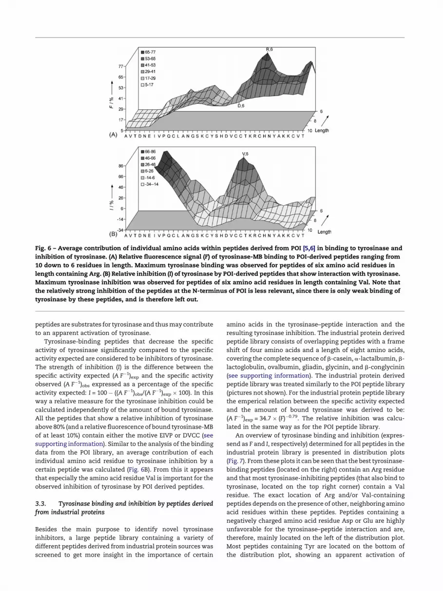
Fig. 6 – Average contribution of individual amino acids within peptides derived from POI [5,6] in binding to tyrosinase and
inhibition of tyrosinase. (A) Relative fluorescence signal (F) of tyrosinase-MB binding to POI-derived peptides ranging from
10 down to 6 residues in length. Maximum tyrosinase binding was observed for peptides of six amino acid residues in
length containing Arg. (B) Relative inhibition (I) of tyrosinase by POI-derived peptides that show interaction with tyrosinase.
Maximum tyrosinase inhibition was observed for peptides of six amino acid residues in length containing Val. Note that
the relatively strong inhibition of the peptides at the N-terminus of POI is less relevant, since there is only weak binding of
tyrosinase by these peptides, and is therefore left out.
peptides are substrates for tyrosinase and thus may contribute
to an apparent activation of tyrosinase.
Tyrosinase-binding peptides that decrease the specific
activity of tyrosinase significantly compared to the specific
activity expected are considered to be inhibitors of tyrosinase.
The strength of inhibition (I) is the difference between the
specific activity expected (A F�1)exp and the specific activity
observed (A F�1)obs expressed as a percentage of the specific
activity expected: I = 100 � ((A F�1)obs/(A F�1)exp � 100). In this
way a relative measure for the tyrosinase inhibition could be
calculated independently of the amount of bound tyrosinase.
All the peptides that show a relative inhibition of tyrosinase
above 80% (and a relative fluorescence of bound tyrosinase-MB
of at least 10%) contain either the motive EIVP or DVCC (see
supporting information). Similar to the analysis of the binding
data from the POI library, an average contribution of each
individual amino acid residue to tyrosinase inhibition by a
certain peptide was calculated (Fig. 6B). From this it appears
that especially the amino acid residue Val is important for the
observed inhibition of tyrosinase by POI derived peptides.
3.3. Tyrosinase binding and inhibition by peptides derivedfrom industrial proteins
Besides the main purpose to identify novel tyrosinase
inhibitors, a large peptide library containing a variety of
different peptides derived from industrial protein sources was
screened to get more insight in the importance of certain
amino acids in the tyrosinase–peptide interaction and the
resulting tyrosinase inhibition. The industrial protein derived
peptide library consists of overlapping peptides with a frame
shift of four amino acids and a length of eight amino acids,
covering the complete sequence of b-casein, a-lactalbumin, b-
lactoglobulin, ovalbumin, gliadin, glycinin, and b-conglycinin
(see supporting information). The industrial protein derived
peptide library was treated similarly to the POI peptide library
(pictures not shown). For the industrial protein peptide library
the emperical relation between the specific activity expected
and the amount of bound tyrosinase was derived to be:
(A F�1)exp = 34.7 � (F)�0.79. The relative inhibition was calcu-
lated in the same way as for the POI peptide library.
An overview of tyrosinase binding and inhibition (expres-
send as F and I, respectively) determined for all peptides in the
industrial protein library is presented in distribution plots
(Fig. 7). From these plots it can be seen that the best tyrosinase-
binding peptides (located on the right) contain an Arg residue
and that most tyrosinase-inhibiting peptides (that also bind to
tyrosinase, located on the top right corner) contain a Val
residue. The exact location of Arg and/or Val-containing
peptides depends on the presence of other, neighboring amino
acid residues within these peptides. Peptides containing a
negatively charged amino acid residue Asp or Glu are highly
unfavorable for the tyrosinase–peptide interaction and are,
therefore, mainly located on the left of the distribution plot.
Most peptides containing Tyr are located on the bottom of
the distribution plot, showing an apparent activation of
Fig. 7 – Distribution of the octameric peptides derived from industrial proteins (see supporting information) in plots of
tyrosinase inhibition (I) versus tyrosinase binding (F). (A) Location of peptides containing Asp and/or Glu, Arg, Tyr and Val
indicated by dark circles. The open circles represent all other peptides present in the library. Strong tyrosinase-binding
peptides are located on the right and strong tyrosinase-inhibiting peptides are located on the top. Nonbinding peptides or
peptides with a relative error larger than 100% of the inhibition are not shown in these plots. (B) Peptides that are binding to
and inhibiting tyrosinase. The best tyrosinase-binding and inhibiting peptides (F > 20% and I > 10%) are indicated with their
spot number (Table 1).
tyrosinase, since they may be converted by tyrosinase and
hence contribute to an increase in tyrosinase activity
observed. These results, regarding the importance of amino
acid residues within tyrosinase-inhibiting peptides obtained
from the industrial protein peptide library, are in accordance
with the results obtained from the POI peptide library.
The best tyrosinase-binding and tyrosinase-inhibiting
peptides are located on the top right corner of the distribution
plot (Fig. 7B) and originate from different protein sources, but
most of them are derived from ovalbumin (Table 1). Strong
tyrosinase-binding peptides (F > 75% and I > 0%) always
contain at least one Arg residue, often in combination with
a Phe residue. Strong tyrosinase-inhibiting peptides (F > 10%
and I > 80%) contain (combinations of) Val, Ala and/or Leu
residues and are therefore hydrophobic in nature. Good
tyrosinase-binding and inhibiting peptides usually have a
combination of Arg and/or Phe with Val, Ala and/or Leu, for
example peptide 186 with the sequence LFRVASMA from
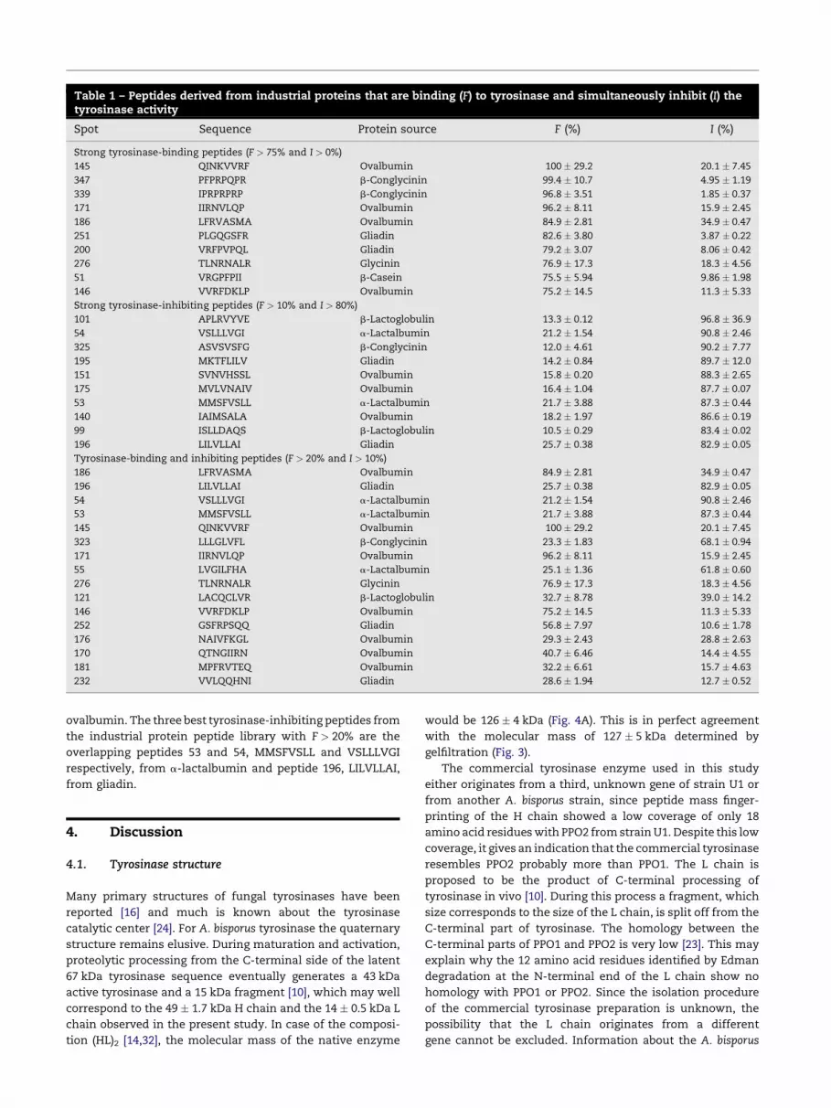
Table 1 – Peptides derived from industrial proteins that are binding (F) to tyrosinase and simultaneously inhibit (I) thetyrosinase activity
Spot Sequence Protein source F (%) I (%)
Strong tyrosinase-binding peptides (F > 75% and I > 0%)
145 QINKVVRF Ovalbumin 100 � 29.2 20.1 � 7.45
347 PFPRPQPR b-Conglycinin 99.4 � 10.7 4.95 � 1.19
339 IPRPRPRP b-Conglycinin 96.8 � 3.51 1.85 � 0.37
171 IIRNVLQP Ovalbumin 96.2 � 8.11 15.9 � 2.45
186 LFRVASMA Ovalbumin 84.9 � 2.81 34.9 � 0.47
251 PLGQGSFR Gliadin 82.6 � 3.80 3.87 � 0.22
200 VRFPVPQL Gliadin 79.2 � 3.07 8.06 � 0.42
276 TLNRNALR Glycinin 76.9 � 17.3 18.3 � 4.56
51 VRGPFPII b-Casein 75.5 � 5.94 9.86 � 1.98
146 VVRFDKLP Ovalbumin 75.2 � 14.5 11.3 � 5.33
Strong tyrosinase-inhibiting peptides (F > 10% and I > 80%)
101 APLRVYVE b-Lactoglobulin 13.3 � 0.12 96.8 � 36.9
54 VSLLLVGI a-Lactalbumin 21.2 � 1.54 90.8 � 2.46
325 ASVSVSFG b-Conglycinin 12.0 � 4.61 90.2 � 7.77
195 MKTFLILV Gliadin 14.2 � 0.84 89.7 � 12.0
151 SVNVHSSL Ovalbumin 15.8 � 0.20 88.3 � 2.65
175 MVLVNAIV Ovalbumin 16.4 � 1.04 87.7 � 0.07
53 MMSFVSLL a-Lactalbumin 21.7 � 3.88 87.3 � 0.44
140 IAIMSALA Ovalbumin 18.2 � 1.97 86.6 � 0.19
99 ISLLDAQS b-Lactoglobulin 10.5 � 0.29 83.4 � 0.02
196 LILVLLAI Gliadin 25.7 � 0.38 82.9 � 0.05
Tyrosinase-binding and inhibiting peptides (F > 20% and I > 10%)
186 LFRVASMA Ovalbumin 84.9 � 2.81 34.9 � 0.47
196 LILVLLAI Gliadin 25.7 � 0.38 82.9 � 0.05
54 VSLLLVGI a-Lactalbumin 21.2 � 1.54 90.8 � 2.46
53 MMSFVSLL a-Lactalbumin 21.7 � 3.88 87.3 � 0.44
145 QINKVVRF Ovalbumin 100 � 29.2 20.1 � 7.45
323 LLLGLVFL b-Conglycinin 23.3 � 1.83 68.1 � 0.94
171 IIRNVLQP Ovalbumin 96.2 � 8.11 15.9 � 2.45
55 LVGILFHA a-Lactalbumin 25.1 � 1.36 61.8 � 0.60
276 TLNRNALR Glycinin 76.9 � 17.3 18.3 � 4.56
121 LACQCLVR b-Lactoglobulin 32.7 � 8.78 39.0 � 14.2
146 VVRFDKLP Ovalbumin 75.2 � 14.5 11.3 � 5.33
252 GSFRPSQQ Gliadin 56.8 � 7.97 10.6 � 1.78
176 NAIVFKGL Ovalbumin 29.3 � 2.43 28.8 � 2.63
170 QTNGIIRN Ovalbumin 40.7 � 6.46 14.4 � 4.55
181 MPFRVTEQ Ovalbumin 32.2 � 6.61 15.7 � 4.63
232 VVLQQHNI Gliadin 28.6 � 1.94 12.7 � 0.52
ovalbumin. The three best tyrosinase-inhibiting peptides from
the industrial protein peptide library with F > 20% are the
overlapping peptides 53 and 54, MMSFVSLL and VSLLLVGI
respectively, from a-lactalbumin and peptide 196, LILVLLAI,
from gliadin.
4. Discussion
4.1. Tyrosinase structure
Many primary structures of fungal tyrosinases have been
reported [16] and much is known about the tyrosinase
catalytic center [24]. For A. bisporus tyrosinase the quaternary
structure remains elusive. During maturation and activation,
proteolytic processing from the C-terminal side of the latent
67 kDa tyrosinase sequence eventually generates a 43 kDa
active tyrosinase and a 15 kDa fragment [10], which may well
correspond to the 49 � 1.7 kDa H chain and the 14 � 0.5 kDa L
chain observed in the present study. In case of the composi-
tion (HL)2 [14,32], the molecular mass of the native enzyme
would be 126 � 4 kDa (Fig. 4A). This is in perfect agreement
with the molecular mass of 127 � 5 kDa determined by
gelfiltration (Fig. 3).
The commercial tyrosinase enzyme used in this study
either originates from a third, unknown gene of strain U1 or
from another A. bisporus strain, since peptide mass finger-
printing of the H chain showed a low coverage of only 18
amino acid residues with PPO2 from strain U1. Despite this low
coverage, it gives an indication that the commercial tyrosinase
resembles PPO2 probably more than PPO1. The L chain is
proposed to be the product of C-terminal processing of
tyrosinase in vivo [10]. During this process a fragment, which
size corresponds to the size of the L chain, is split off from the
C-terminal part of tyrosinase. The homology between the
C-terminal parts of PPO1 and PPO2 is very low [23]. This may
explain why the 12 amino acid residues identified by Edman
degradation at the N-terminal end of the L chain show no
homology with PPO1 or PPO2. Since the isolation procedure
of the commercial tyrosinase preparation is unknown, the
possibility that the L chain originates from a different
gene cannot be excluded. Information about the A. bisporus

Fig. 8 – Importance of amino acid residues within octameric
peptides derived from industrial proteins (see supporting
information) in binding to and inhibition of tyrosinase.
The amino acids are ordered based on chemical and/or
structural similarities and the connecting curve, to be
considered as an ‘‘amino acid spectrum’’, is added only to
support the presentation of the results. The smaller open
circles show the effect when one of the 20 amino acids is
excluded from the analysis. Note that Tyr is most probably
converted to dopaquinone during the screening. (A)
Average increase in tyrosinase binding, which is
proportional to tyrosinase-MB fluorescence change (4F),
upon introduction of a certain amino acid residue in the
peptide. (B) Average increase in tyrosinase inhibition (4I)
upon introduction of a certain amino acid residue in the
peptide. Peptides that show relative fluorescence of
tyrosinase-MB lower than 5% or show a relative error
larger than 100% of the inhibition calculated were
excluded from the tyrosinase inhibition analysis.
genome is required to confirm whether or not the H and the L
chain originate from another gene encoding a third tyrosinase
isoenzyme.
4.2. Tyrosinase binding and inhibition by peptides
Two independently analyzed peptide libraries were screened
for peptides binding to and inhibiting tyrosinase. From the POI
peptide library it appeared that the only Arg residue (Fig. 6A)
present in POI plays an important role in the tyrosinase–
peptide interaction. Furthermore, good tyrosinase-binding
peptides containing Val (Fig. 6B) are relatively good inhibitors
of tyrosinase. Although the Cys residues within the native POI
protein are all involved in disulfide bridges [5,6], the average
contribution of Val in tyrosinase inhibition by peptides derived
from POI may be affected by the adjacent, reduced Cys
residues. However the importance of Val and other residues,
regardless the effect of Cys residues, is confirmed with the
results obtained from a larger peptide library containing a
variety of peptides derived from different industrial protein
sources. The results obtained with the POI derived peptide
library are in agreement with the results obtained from the
industrial protein derived peptide library.
Good tyrosinase-binding peptides derived from industrial
proteins usually contain the amino acid residues Arg and/or
Phe (Fig. 8A). Because Lys is not found to be important for the
tyrosinase–peptide interaction, it may be concluded that the
interaction between tyrosinase and the Arg-containing pep-
tides is rather specific and not only based on electrostatics.
The importance of Phe in the tyrosinase–peptide interaction
might be explained by the fact that Phe is structurally similar
to Tyr, which is the natural substrate for tyrosinase (see also
below). However, the binding of Tyr-containing peptides
cannot be established, since Tyr is oxidized upon incubation
of the peptide library with tyrosinase-MB. Apparently peptides
containing the dopaquinone residue do not bind very well to
tyrosinase. Also peptides containing an Asp or Glu residue do
not bind very well to tyrosinase, suggesting that a negative
charge is not favorable for tyrosinase binding.
Good tyrosinase-inhibiting peptides derived from industrial
proteins usually contain a hydrophobic, aliphatic amino acid
residue Val, Ala, Leu, Met and/or Ile (Fig. 8B). The hydrophobic,
aromatic residue Phe, which is relatively important in the
tyrosinase–peptide interaction, is less important in tyrosinase
inhibition. Apparently, despite its structural similarities with
Tyr, Phe is presumably not binding to the active site of
tyrosinase. These findings are in accordance with results
reported earlier, where Phe appears not to be a very good
inhibitor of tyrosinase [11]. Besides the hydrophobic residues
mentioned above, peptides containing the polar, uncharged
residues Cys and Ser, and to a minor extent Thr, are good
tyrosinase inhibitors as well. The tyrosinase inhibition
observed for peptides containing Ser and/or Thr is most
probably similar to the mechanism of inhibition by Cys [7,35].
In some cases tyrosinase-binding by peptides may result in
an activation of tyrosinase. This usually occurs with peptides
containing the amino acid residues Tyr and/or Trp (Fig. 8B).
The activating effect of Trp containing peptides is less
established, since there are only a few peptides in the library
containing Trp. As explained in the results (Sections 3.2 and
3.3), the presence of Tyr residues in tyrosinase-binding
peptides cause an apparent tyrosinase activation since the
Tyr residues are oxidized by tyrosinase. Of course, Tyr-
containing peptides may still act like competitive inhibitors of
L-DOPA oxidation by tyrosinase, but the tyrosinase activity
and thus the formation of quinones is not affected. Some
relatively good tyrosinase-binding peptides do not contain Tyr
or Trp and are still activating tyrosinase. An example of a
relatively good tyrosinase-binding peptide that simulta-
neously activates tyrosinase is RINKKIEK (spots 7 and 384
from the industrial protein peptide library, see supporting
information). RINKKIEK is a lipoxygenase inhibitory peptide
identified from b-casein [30]. Although RINKKIEK is also
binding to tyrosinase, it appears to have an opposite effect
on the activity of this enzyme.
4.3. Conclusions
Considering the relative fluorescence and the relative activity
measured for tyrosinase-MB binding to peptides within the
libraries studied, conclusions can be drawn regarding the
importance of certain amino acid residues within tyrosinase-
binding and tyrosinase-inhibiting peptides. From both the POI
and the industrial protein peptide library, it appeared that the
presence of an Arg residue within a peptide is very beneficial
for the tyrosinase–peptide interaction. This is probably not
only due to a simple electrostatic interaction, since the
similarly charged amino acid residue Lys is not important
for tyrosinase binding at all. Apparently, the structure of the
Arg side chain is required for a strong tyrosinase–peptide
interaction.
The observed tyrosinase inhibition by peptides containing
the polar, uncharged amino acid residues Cys or Ser is due to
their ability to form adducts with the enzymatically formed
dopaquinone. Therefore peptides containing these residues as
inhibitors of melanin formation are not very effective, since
they are consumed in the reaction and the tyrosinase activity
is not affected. The presence of other amino acid residues, like
the hydrophobic, aliphatic Val, Ala or Leu, is probably more
important in inhibition of tyrosinase by peptides, since they
may inhibit the formation of quinones by direct interaction
with tyrosinase. Further experiments are needed to confirm
and optimize the tyrosinase-inhibiting activity of potential
tyrosinase-inhibiting peptides containing Arg and/or Phe in
combination with Val, Ala and/or Leu.
Acknowledgements
This work was supported by the Ministry of Economic Affairs
of The Netherlands, the innovation driven program IOP
Industrial Proteins (project IIE00022) and by Cargill, Cebeco
Egg Research, Cosmoferm BV and DMV International. We want
to thank H. Vilaseca and J. Liberman for their assistance in
synthesizing the peptide libraries and purifying the tyrosinase
enzyme, respectively. Furthermore we thank J.A. Boeren for
performing the MS analyses.
Appendix A. Supplementary data
Supplementary data associated with this article can
be found, in the online version, at doi:10.1016/j.pep-
tides.2006.11.023.
r e f e r e n c e s
[1] Ates S, Pekyardmc S, Cokmus C. Partial characterization ofa peptide from honey that inhibits mushroom polyphenoloxidase. J Food Biochem 2001;25:127–37.
[2] Cavalieri EL, Li K-M, Balu N, Saeed M, Devanesan P,Higginbotham S, et al. Catechol ortho-quinones: theelectrophilic compounds that form depurinating DNAadducts and could initiate cancer and other diseases.Carcinogenesis 2002;23:1071–7.
[3] Chen JS, Wei CI, Marshall MR. Inhibition mechanism ofkojic acid on polyphenol oxidase. J Agric Food Chem1991;39:1897–901.
[4] Chen MJ, Liu JR, Sheu JF, Lin CW, Chuang CL. Study on skincare properties of milk kefir whey. Asian Aust J Anim2006;19:905–8.
[5] Daquinag AC, Sato T, Koda H, Takao T, Fukuda M,Shimonishi Y, et al. A novel endogenous inhibitor ofphenoloxidase from Musca domestica has a cystine motifcommonly found in snail and spider toxins. Biochemistry1999;38:2179–88.
[6] Daquinag Alexes C, Nakamura S, Takao T, Shimonishi Y,Tsukamoto T. Primary structure of a potent endogenousdopa-containing inhibitor of phenol oxidase from Muscadomestica. Proc Natl Acad Sci USA 1995;92:2964–8.
[7] Dudley ED, Hotchkiss JH. Cysteine as an inhibitor ofpolyphenol oxidase. J Food Biochem 1989;13:65–75.
[8] Espin JC, Morales M, Garcia Ruiz Pedro A, Tudela J, GarciaCanovas F. Improvement of a continuousspectrophotometric method for determining themonophenolase and diphenolase activities of mushroompolyphenol oxidase. J Agric Food Chem 1997;45:1084–90.
[9] Espin JC, Morales M, Varon R, Tudela J, Garcia Canovas F. Acontinuous spectrophotometric method for determiningthe monophenolase and diphenolase activities of applepolyphenol oxidase. Anal Biochem 1995;231:237–46.
[10] Espin JC, Soler Rivas C, Wichers HJ. Maturation and activationof latent tyrosinase from Agaricus bisporus. In: Griensven,LJLD van, editor. Proceedings of the 15th internationalcongress on the science and cultivation of edible fungi;2000 15–19 May. A.A. Balkema Rotterdam; 2000. p. 79–86.
[11] Farishian RA, Writtaker JH. Phenylalanine lowers melaninsynthesis in mammalian melanocytes by reducingtyrosinase uptake: implications for pigment reduction inphenylketonuria. J Invest Dermatol 1980;74:85–9.
[12] Frank R. The SPOT-synthesis technique: synthetic peptidearrays on membrane supports-principles and applications.J Immunol Methods 2002;267:13–26.
[13] Frank R. Spot-synthesis: an easy technique for thepositionally addressable, parallel chemical synthesis on amembrane support. Tetrahedron 1992;48:9217–32.
[14] Gelder CWG van, Flurkey WH, Wichers HJ. Sequence andstructural features of plant and fungal tyrosinases.Phytochemistry 1997;45:1309–23.
[15] Girelli AM, Mattei E, Messina A, Tarola AM. Inhibition ofpolyphenol oxidases activity by various dipeptides. J AgricFood Chem 2004;52:2741–5.
[16] Halaouli S, Asther M, Sigoillot JC, Hamdi M, Lomascolo A.Fungal tyrosinases: new prospects in molecularcharacteristics, bioengineering and biotechnologicalapplications. J Appl Microbiol 2006;100:219–32.
[17] Iyengar R, McEvily AJ. Anti-browning agents: alternatives tothe use of sulfites in foods. Trends Food Sci Tech 1992;3:60–4.
[18] Kahn V. Effect of proteins, protein hydrolysates and aminoacids on o-dihydroxyphenolase activity of polyphenoloxidase of mushroom, avocado, and banana. J Food Sci1985;50:111–5.
[19] Kato N, Sato S, Yamanaka A, Yamada H, Fuwa N, NomuraM. Silk protein, sericin, inhibits lipid peroxidation andtyrosinase activity. Biosci Biotech Biochem 1998;62:145–7.
[20] Kim H, Choi H, Cho JK, Kim SY, Lee YS. Solid-phasesynthesis of kojic acid-tripeptides and their tyrosinaseinhibitory activity, storage stability, and toxicity. BioorgMed Chem Lett 2004;14:2843–6.
[21] Kim YJ, Uyama H. Tyrosinase inhibitors from natural andsynthetic sources: structure, inhibition mechanism andperspective for the future. Cell Mol Life Sci 2005;62:1707–23.
[22] Marmol V del, Beermann F. Tyrosinase and related proteinsin mammalian pigmentation. FEBS Lett 1996;381:165–8.
[23] Marusek CM, Trobaugh NM, Flurkey WH, Inlow JK.Comparative analysis of polyphenol oxidase from plantand fungal species. J Inorg Biochem 2006;100:108–23.
[24] Matoba Y, Kumagai T, Yamamoto A, Yoshitsu H, SugiyamaM. Crystallographic evidence that the dinuclear coppercenter of tyrosinase is flexible during catalysis. J Biol Chem2006;281:8981–90.
[25] Morita H, Kayashita T, Kobata H, Gonda A, Takeya K,Itokawa H. Pseudostellarins D - F, new tyrosinase inhibitorycyclic peptides from Pseudostellaria heterophylla.Tetrahedron 1994;50:9975–82.
[26] Nakajima M, Shinoda I, Samejima Y, Miyauchi H,Fukuwatari Y, Hayasawa H. Kappa-casein suppressesmelanogenesis in cultured pigment cells. Pig Cell Res1996;9:235–9.
[27] Okot-Kotber M, Liavoga A, Yong KJ, Bagorogoza K. Activityand inhibition of polyphenol oxidase in extracts of branand other milling fractions from a variety of wheatcultivars. Cereal Chem 2001;78:514–20.
[28] Oszmianski J, Lee CY. Inhibition of polyphenol oxidaseactivity and browning by honey. J Agric Food Chem1990;38:1892–5.
[29] Piamphongsant T. Treatment of melasma: A review withpersonal experience. Int J Dermatol 1998;37:897–903.
[30] Schurink M, Berkel WJH van, Wichers HJ, Boeriu CG.Identification of lipoxygenase inhibitory peptides frombeta-casein by using SPOT synthesis. Chembiochem2006;7:743–7.
[31] Seo S-Y, Sharma VK, Sharma N. Mushroom tyrosinase:recent prospects. J Agric Food Chem 2003;51:2837–53.
[32] Strothkamp KG, Jolley RL, Mason HS. Quaternary structureof mushroom tyrosinase. Biochem Biophys Res Commun1976;70:519–24.
[33] Sturm RA, Teasdale RD, Box NF. Human pigmentationgenes: identification, structure and consequences ofpolymorphic variation. Gene Amsterdam 2001;277:49–62.
[34] Tomita M, Miyakawa H, Shimamura S, Kobayashi S.Inventors: an agent for tyrosinase inhibition. Japan patent91306244.4; 1993.
[35] Villarama CD, Maibach HI. Glutathione as a depigmentingagent: an overview. Int J Cosm Sci 2005;27:147–53.
[36] Wichers HJ, Recourt K, Hendriks M, Ebbelaar CEM, BianconeG, Hoeberichts FA, et al. Cloning, expression andcharacterisation of two tyrosinase cDNAs from Agaricusbisporus. Appl Microb Biotech 2003;61:336–41.