novel anti double-stranded nucleic acids full-length ... - mdpi
TRANSCRIPT
Citation: Czarnecka, M.; Weichelt, U.;
Rödiger, S.; Hanack, K. Novel Anti
Double-Stranded Nucleic Acids
Full-Length Recombinant Camelid
Heavy-Chain Antibody for the
Detection of miRNA. Int. J. Mol. Sci.
2022, 23, 6275. https://doi.org/
10.3390/ijms23116275
Academic Editor: Shigetaka
Shimodaira
Received: 26 April 2022
Accepted: 2 June 2022
Published: 3 June 2022
Publisher’s Note: MDPI stays neutral
with regard to jurisdictional claims in
published maps and institutional affil-
iations.
Copyright: © 2022 by the authors.
Licensee MDPI, Basel, Switzerland.
This article is an open access article
distributed under the terms and
conditions of the Creative Commons
Attribution (CC BY) license (https://
creativecommons.org/licenses/by/
4.0/).
International Journal of
Molecular Sciences
Article
Novel Anti Double-Stranded Nucleic Acids Full-LengthRecombinant Camelid Heavy-Chain Antibody for the Detectionof miRNAMalgorzata Czarnecka 1, Ulrike Weichelt 1, Stefan Rödiger 2 and Katja Hanack 1,*
1 Institute of Biochemistry and Biology, University of Potsdam, Karl-Liebknecht-Str. 24-25,14476 Potsdam, Germany; [email protected] (M.C.); [email protected] (U.W.)
2 Faculty of Health Sciences Brandenburg, Brandenburg University of Technology, Cottbus-Senftenberg,Universitätsplatz 1, 01968 Senftenberg, Germany; [email protected]
* Correspondence: [email protected]; Tel.: +49-3319-775-348
Abstract: The discovery that certain diseases have specific miRNA signatures which correspond todisease progression opens a new biomarker category. The detection of these small non-coding RNAsis performed routinely using body fluids or tissues with real-time PCR, next-generation sequencing, oramplification-based miRNA assays. Antibody-based detection systems allow an easy onset handlingcompared to PCR or sequencing and can be considered as alternative methods to support miRNAdiagnostic in the future. In this study, we describe the generation of a camelid heavy-chain-onlyantibody specifically recognizing miRNAs to establish an antibody-based detection method. Thegeneration of nucleic acid-specific binders is a challenge. We selected camelid binders via phagedisplay, expressed them as VHH as well as full-length antibodies, and characterized the binding toseveral miRNAs from a signature specific for dilated cardiomyopathy. The described workflow canbe used to create miRNA-specific binders and establish antibody-based detection methods to providean additional way to analyze disease-specific miRNA signatures.
Keywords: antibody; camelid antibody; heavy-chain-only antibody; miRNA; nucleic acids;novel biomarkers
1. Introduction
Micro ribonucleic acids (miRNAs) are small (17–25 nucleotides) non-coding RNA, thatplay an essential role in regulating post-transcriptionally gene expression. As a part ofthe RNA-induced silencing complex (RISC), they bind complementary imperfect mRNAsequences thus modulating or silencing the activity of their mRNA targets [1]. AlteredmiRNA profiles have been discovered in multiple tissues and body fluids, that have beenassociated with the onset, progress, and prognosis of several serious diseases such as cancer,neurological disorders, and cardiovascular and myocardial diseases [2–9]. In associationwith inflammatory and virally induced cardiomyopathies and dilated cardiomyopathy(DCM), the miRNAs homo sapiens (hsa)-let-7f-5p, hsa-miR-30a-3p, hsa-miR-93-5p, hsa-miR-197-3p, hsa-miR-223, and hsa-miR-379-5p showed an altered expression profile [7,10]. Thereis rising interest in elucidating miRNA expression patterns and their functions becausethey represent promising second-generation biomarkers for new diagnostic approachesunder physiological and pathophysiological conditions. We took it as an opportunity todevelop and establish a phage display protocol for the selection of anti-nucleic acid bindersusing the altered miRNA expression profile of DCM.
The generation of nucleic acid-specific antibodies is a high challenge, especially withregard to specificity and cross-reactivity. In certain autoimmune diseases such as systemiclupus erythematosus (SLE) specific immunoglobulins against double-stranded DNA (dsDNA) are generated in vivo and used as specific biomarkers in the diagnostics of such
Int. J. Mol. Sci. 2022, 23, 6275. https://doi.org/10.3390/ijms23116275 https://www.mdpi.com/journal/ijms
Int. J. Mol. Sci. 2022, 23, 6275 2 of 18
disorders [11–14]. This implies that the human immune system is able to address thischallenge. Antibodies from autoimmune patients and autoimmune disease-related an-imal models have been successfully isolated and engineered for use as diagnostic andresearch tools.
In the last century, there have been several approaches to generate antibodies againstDNA, alpha oligonucleotides, DNA:RNA hybrids, virus RNA, nucleotides, and RNAamong others by hybridoma technology [15–19]. Hu et al. summarized several studiesin which anti-nucleic acid antibodies were generated and proposed their possible use inclinical and or genomic detection and diagnostics [20]. The experimental in vivo generationhas been proven to be very challenging or unsuccessful since native DNA and RNAs arepoor antigens that will be tolerated or degraded by the animal host reaction. To inducemeasurable immune reactions, it is recommended to use nucleic acids complexed with car-rier proteins or synthetic peptides, chemically modified ribonucleotides, or high molecularweight polynucleotides in general [21–23]. Further, it is difficult to elicit antibodies havinga high affinity to each type of nucleic acid without showing cross-reactivity with others.The anti-DNA:RNA hybrid antibody based on the one generated by Nakazato in the 1970sagainst synthetic ϕX174 DNA:RNA hybrid [17] is one of the few antibodies that madeit to a (commercially available) customized product, that can be purchased via variouscompanies. This antibody was proven to bind DNA:RNA hybrids and poly(I)-poly(dC)equally but not single-stranded DNA, ds DNA, or RNA [24].
In recent years, the variable domains of camelid heavy-chain-only antibodies havebecome more important for their possible application in the diagnostic due to their ad-vantages [25]. The variable domains of camelid heavy-chain-only antibodies (VHHs ornanobodies) serve as the smallest known antigen-binding domains with a molecular weightof only 12–15 kDa derived from naturally occurring antibodies. Further, they possess avery high thermal resistance and physicochemical stability resulting from the decreased hy-drophobicity and are stable at high pH values, high alcohol concentration, and chaotropicagents [26–28]. The VHH domain is composed of four frameworks and three domainsreferred to as complementarity determining regions (CDRs) instead of six as in the variabledomains of heavy and light chain in a conventional antibody [29]. Within the framework2 the highly conserved amino acids Val37, Gly44, Leu45, and Trp47 are substituted bysmaller and/or hydrophilic amino acids such as Phe or Tyr37, Glu44, Arg or Cys45, andGly or Ser, Leu, Phe47 (the position of the amino acids are numbered according to theKabat numbering system) [30]. These four amino acid substitutions are referred to ashallmarks and are used to identify the antigen-binding domain as VHH. The hydropho-bic amino acids (Val37, Gly44, Leu45, Trp47) assure the linkage between VH and VL inconventional antibodies.
To address the point of establishing a workflow for the generation of nucleic acid-specific antibodies and using them for diagnostic assay systems, we created a camelid-naïve cDNA library encoding diverse VHHs. VHH fragments against nucleic acids wereselected by several panning rounds. The miRNA-223 which belongs to the altered miRNAexpression profile of DCM was chosen as a model miRNA as antigen. By analyzing potentialmiRNA binders, we identified one clone (VHH_19) that showed a specific response to dsmiRNA. By introducing the camelid IgG2b constant region to the VHH_19 we generatedthe recombinant full-length heavy-chain antibody L19 (Figure 1), which was expressed inHEK-293 cells. The purified L19 antibody was tested in a direct ELISA showing the bindingspecificity for ds miRNA. Our results demonstrate that anti-ds miRNA antibodies maycontribute as useful tools for detecting and analyzing miRNA.
Int. J. Mol. Sci. 2022, 23, 6275 3 of 18
Int. J. Mol. Sci. 2022, 23, x FOR PEER REVIEW 3 of 19
(a) (b)
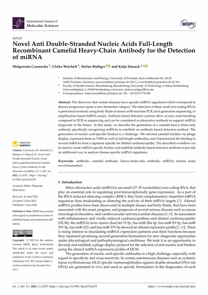
Figure 1. Schematic overview of performed phage display procedure to generate anti-short nucleic acid camelid binders. (a) The first step was the generation of a naïve camelid VHH phage library. The peripheral blood mononuclear cells (PBMCs) were isolated from the camelid whole blood. Next, the total RNA was extracted. cDNA synthesis and VHH amplification were performed to build a naïve camelid VHH phage library. (b) After several panning rounds, the potential anti-DCM-miRNA VHH_19 binder was identified. In order to produce the recombinant full-length camelid antibody, the DNA sequence was expanded by the introduction of the Fc region of camelid IgG2b instead of His (blue bar) and HA (red bar) tags.
2. Results 2.1. Selection of ds miRNA VHH Binder
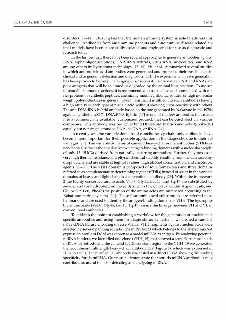
The total RNA isolated from naïve camelid PBMCs was transcribed into cDNA. The first PCR amplified the region between the leader sequence and the CH2 domain of both VH and VHH genes of conventional and heavy-chain antibodies (non-conventional anti-bodies) resulting in two bands with the sizes of about 800 bp and about 600 bp (Figure 2a). The 600 bp band was used as a template for the nested PCR to amplify the camelid VHH gene repertoire providing a 400 bp band (Figure 2b). The amplified VHH gene rep-ertoire was cloned into the digested phagemid pCOMB3x to obtain a naïve VHH phage library with 6.2 × 10⁶ members.
(a)
Figure 1. Schematic overview of performed phage display procedure to generate anti-short nucleicacid camelid binders. (a) The first step was the generation of a naïve camelid VHH phage library.The peripheral blood mononuclear cells (PBMCs) were isolated from the camelid whole blood. Next,the total RNA was extracted. cDNA synthesis and VHH amplification were performed to build anaïve camelid VHH phage library. (b) After several panning rounds, the potential anti-DCM-miRNAVHH_19 binder was identified. In order to produce the recombinant full-length camelid antibody,the DNA sequence was expanded by the introduction of the Fc region of camelid IgG2b instead ofHis (blue bar) and HA (red bar) tags.
2. Results2.1. Selection of ds miRNA VHH Binder
The total RNA isolated from naïve camelid PBMCs was transcribed into cDNA. Thefirst PCR amplified the region between the leader sequence and the CH2 domain of both VHand VHH genes of conventional and heavy-chain antibodies (non-conventional antibodies)resulting in two bands with the sizes of about 800 bp and about 600 bp (Figure 2a). The600 bp band was used as a template for the nested PCR to amplify the camelid VHH generepertoire providing a 400 bp band (Figure 2b). The amplified VHH gene repertoire wascloned into the digested phagemid pCOMB3x to obtain a naïve VHH phage library with6.2 × 106 members.
Int. J. Mol. Sci. 2022, 23, x FOR PEER REVIEW 3 of 19
(a) (b)
Figure 1. Schematic overview of performed phage display procedure to generate anti-short nucleic acid camelid binders. (a) The first step was the generation of a naïve camelid VHH phage library. The peripheral blood mononuclear cells (PBMCs) were isolated from the camelid whole blood. Next, the total RNA was extracted. cDNA synthesis and VHH amplification were performed to build a naïve camelid VHH phage library. (b) After several panning rounds, the potential anti-DCM-miRNA VHH_19 binder was identified. In order to produce the recombinant full-length camelid antibody, the DNA sequence was expanded by the introduction of the Fc region of camelid IgG2b instead of His (blue bar) and HA (red bar) tags.
2. Results 2.1. Selection of ds miRNA VHH Binder
(a)
(b)
Figure 2. Agarose gel (1.5%) electrophoresis after the amplification of camelid VHs and VHHs. (a) The upper band at about 800 bp corresponds to the leader-VH-hinge-CH1-CH2 sequence of camelid conventional antibodies (IgG1) and the lower band at about 600 bp to the leader-VHH-hinge-CH2 sequence of unique heavy-chain-only antibodies. Amplicons of 600 bp were used as templates for the nested PCR. (b) Nested PCR amplified the VHH sequences of about 400 bp only (without leader and hinge region) using the forward primer mL93 and two reverse primers mL94 and mL95. For
Figure 2. Agarose gel (1.5%) electrophoresis after the amplification of camelid VHs and VHHs. (a) Theupper band at about 800 bp corresponds to the leader-VH-hinge-CH1-CH2 sequence of camelidconventional antibodies (IgG1) and the lower band at about 600 bp to the leader-VHH-hinge-CH2sequence of unique heavy-chain-only antibodies. Amplicons of 600 bp were used as templates for thenested PCR. (b) Nested PCR amplified the VHH sequences of about 400 bp only (without leader andhinge region) using the forward primer mL93 and two reverse primers mL94 and mL95. For eachPCR, DEPC-treated water was included as a negative control (NC, no template control). HyperladderI™ 1 kb was used as DNA molecular weight marker (Bioline, London, Great Britain).
Int. J. Mol. Sci. 2022, 23, 6275 4 of 18
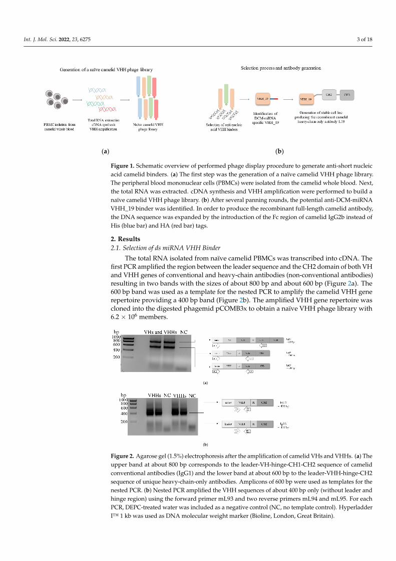
The naïve phage library was panned over three rounds for anti-miRNA VHHs. Thepolyclonal phage particles were pre-incubated with the carrier protein avidin and theunbound particles were then incubated with the synthetic model ds miRNA-223. The titerof the eluted phage particles increased during the three panning rounds indicating theenrichment of phage particles (Figure 3).
Int. J. Mol. Sci. 2022, 23, x FOR PEER REVIEW 4 of 19
(b)
Figure 2. Agarose gel (1.5%) electrophoresis after the amplification of camelid VHs and VHHs. (a) The upper band at about 800 bp corresponds to the leader-VH-hinge-CH1-CH2 sequence of camelid conventional antibodies (IgG1) and the lower band at about 600 bp to the leader-VHH-hinge-CH2 sequence of unique heavy-chain-only antibodies. Amplicons of 600 bp were used as templates for the nested PCR. (b) Nested PCR amplified the VHH sequences of about 400 bp only (without leader and hinge region) using the forward primer mL93 and two reverse primers mL94 and mL95. For each PCR, DEPC-treated water was included as a negative control (NC, no template control). Hy-perladder I™ 1 kb was used as DNA molecular weight marker (Bioline, London, Great Britain).
The naïve phage library was panned over three rounds for anti-miRNA VHHs. The polyclonal phage particles were pre-incubated with the carrier protein avidin and the un-bound particles were then incubated with the synthetic model ds miRNA-223. The titer of the eluted phage particles increased during the three panning rounds indicating the en-richment of phage particles (Figure 3).
Figure 3. Percentage enrichment of polyclonal phage particles displaying different VHH fragments on their surfaces. The percentage enrichment was calculated by the division of the titer of output phages and titer of input phages multiplied by 100% for each panning round.
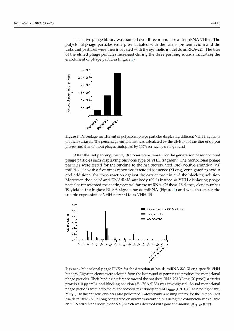
After the last panning round, 18 clones were chosen for the generation of monoclonal phage particles each displaying only one type of VHH fragment. The monoclonal phage particles were tested for the binding to the hsa biotinylated (bio) double-stranded (ds) miRNA-223 with a five times repetitive extended sequence (XLong) conjugated to avidin and additional for cross-reaction against the carrier protein and the blocking solution. Moreover, the use of anti-DNA:RNA antibody (S9.6) instead of VHH displaying phage particles represented the coating control for the miRNA. Of these 18 clones, clone number 19 yielded the highest ELISA signals for ds miRNA (Figure 4) and was chosen for the soluble expression of VHH referred to as VHH_19.
Figure 3. Percentage enrichment of polyclonal phage particles displaying different VHH fragmentson their surfaces. The percentage enrichment was calculated by the division of the titer of outputphages and titer of input phages multiplied by 100% for each panning round.
After the last panning round, 18 clones were chosen for the generation of monoclonalphage particles each displaying only one type of VHH fragment. The monoclonal phageparticles were tested for the binding to the hsa biotinylated (bio) double-stranded (ds)miRNA-223 with a five times repetitive extended sequence (XLong) conjugated to avidinand additional for cross-reaction against the carrier protein and the blocking solution.Moreover, the use of anti-DNA:RNA antibody (S9.6) instead of VHH displaying phageparticles represented the coating control for the miRNA. Of these 18 clones, clone number19 yielded the highest ELISA signals for ds miRNA (Figure 4) and was chosen for thesoluble expression of VHH referred to as VHH_19.
Int. J. Mol. Sci. 2022, 23, x FOR PEER REVIEW 5 of 19
Figure 4. Monoclonal phage ELISA for the detection of hsa ds miRNA-223 XLong-specific VHH binders. Eighteen clones were selected from the last round of panning to produce the monoclonal phage particles. Their binding preference toward the hsa ds miRNA-223 XLong (20 pmol), a carrier protein (10 µg/mL), and blocking solution (3% BSA/PBS) was investigated. Bound monoclonal phage particles were detected by the secondary antibody anti-M13HRP (1:7000). The binding of anti-M13HRP to the antigens only was also performed. Additionally, a coating control for the immobilized hsa ds miRNA-223 XLong conjugated on avidin was carried out using the commercially available anti-DNA:RNA antibody (clone S9.6) which was detected with goat anti-mouse IgGHRP (Fcγ).
2.2. Expression and Purification of Recombinant VHH_19 From the positive clone 19, the plasmid pCOMB3x-VHH_19 was isolated and se-
quenced. The amino acid sequence of VHH_19 (Figure 5) was numbered according to the Kabat numbering system. The four typical aa substitutions were detected at positions 37, 44, 45, and 47. Moreover, the sequence possesses two conserved Cys residues at positions 22 and 9, which are typical for VHHs. The sequences for His-tag (for purification) and HA-tag (for detection) are present after framework 4. VHH_19 clone was expressed in a soluble form in the periplasmic space of E. coli HB2151 and purified via Ni-NTA affinity chromatography. The presence of VHH_19 in the elution fractions was confirmed by Western blot (WB). The protein bands (Figure 6) between 15 and 20 kDa were detected by an anti-HA-tag antibody in the following collected samples: total protein, periplasmic fraction 1 and 2 (before and after the dialysis with native binding buffer (NBB) containing 10 mM imidazole) and in the elution fractions 1 and 2 indicating the expressed VHH_19. No protein bands could be detected in the flow-through or in the wash fraction. The VHH_19 concentration of 2284 µg/mL was determined via the bicinchoninic acid (BCA) assay.
Figure 4. Monoclonal phage ELISA for the detection of hsa ds miRNA-223 XLong-specific VHHbinders. Eighteen clones were selected from the last round of panning to produce the monoclonalphage particles. Their binding preference toward the hsa ds miRNA-223 XLong (20 pmol), a carrierprotein (10 µg/mL), and blocking solution (3% BSA/PBS) was investigated. Bound monoclonalphage particles were detected by the secondary antibody anti-M13HRP (1:7000). The binding of anti-M13HRP to the antigens only was also performed. Additionally, a coating control for the immobilizedhsa ds miRNA-223 XLong conjugated on avidin was carried out using the commercially availableanti-DNA:RNA antibody (clone S9.6) which was detected with goat anti-mouse IgGHRP (Fcγ).
Int. J. Mol. Sci. 2022, 23, 6275 5 of 18
2.2. Expression and Purification of Recombinant VHH_19
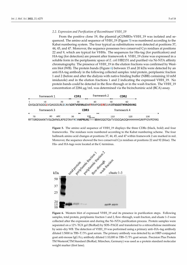
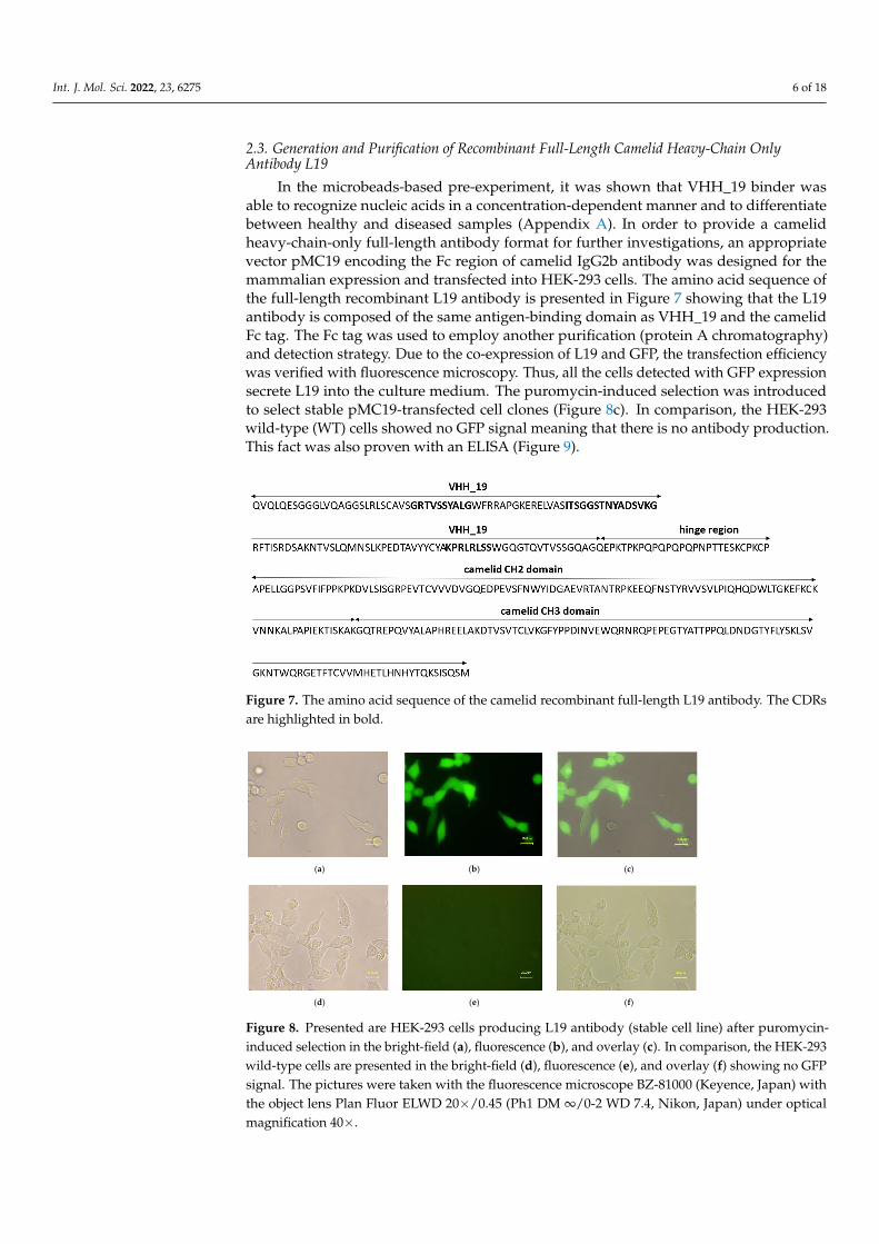
From the positive clone 19, the plasmid pCOMB3x-VHH_19 was isolated and se-quenced. The amino acid sequence of VHH_19 (Figure 5) was numbered according to theKabat numbering system. The four typical aa substitutions were detected at positions 37,44, 45, and 47. Moreover, the sequence possesses two conserved Cys residues at positions22 and 9, which are typical for VHHs. The sequences for His-tag (for purification) andHA-tag (for detection) are present after framework 4. VHH_19 clone was expressed in asoluble form in the periplasmic space of E. coli HB2151 and purified via Ni-NTA affinitychromatography. The presence of VHH_19 in the elution fractions was confirmed by West-ern blot (WB). The protein bands (Figure 6) between 15 and 20 kDa were detected by ananti-HA-tag antibody in the following collected samples: total protein, periplasmic fraction1 and 2 (before and after the dialysis with native binding buffer (NBB) containing 10 mMimidazole) and in the elution fractions 1 and 2 indicating the expressed VHH_19. Noprotein bands could be detected in the flow-through or in the wash fraction. The VHH_19concentration of 2284 µg/mL was determined via the bicinchoninic acid (BCA) assay.
Int. J. Mol. Sci. 2022, 23, x FOR PEER REVIEW 5 of 19
Figure 4. Monoclonal phage ELISA for the detection of hsa ds miRNA-223 XLong-specific VHH binders. Eighteen clones were selected from the last round of panning to produce the monoclonal phage particles. Their binding preference toward the hsa ds miRNA-223 XLong (20 pmol), a carrier protein (10 µg/mL), and blocking solution (3% BSA/PBS) was investigated. Bound monoclonal phage particles were detected by the secondary antibody anti-M13HRP (1:7000). The binding of anti-M13HRP to the antigens only was also performed. Additionally, a coating control for the immobilized hsa ds miRNA-223 XLong conjugated on avidin was carried out using the commercially available anti-DNA:RNA antibody (clone S9.6) which was detected with goat anti-mouse IgGHRP (Fcγ).
2.2. Expression and Purification of Recombinant VHH_19 From the positive clone 19, the plasmid pCOMB3x-VHH_19 was isolated and se-
quenced. The amino acid sequence of VHH_19 (Figure 5) was numbered according to the Kabat numbering system. The four typical aa substitutions were detected at positions 37, 44, 45, and 47. Moreover, the sequence possesses two conserved Cys residues at positions 22 and 9, which are typical for VHHs. The sequences for His-tag (for purification) and HA-tag (for detection) are present after framework 4. VHH_19 clone was expressed in a soluble form in the periplasmic space of E. coli HB2151 and purified via Ni-NTA affinity chromatography. The presence of VHH_19 in the elution fractions was confirmed by Western blot (WB). The protein bands (Figure 6) between 15 and 20 kDa were detected by an anti-HA-tag antibody in the following collected samples: total protein, periplasmic fraction 1 and 2 (before and after the dialysis with native binding buffer (NBB) containing 10 mM imidazole) and in the elution fractions 1 and 2 indicating the expressed VHH_19. No protein bands could be detected in the flow-through or in the wash fraction. The VHH_19 concentration of 2284 µg/mL was determined via the bicinchoninic acid (BCA) assay.
Figure 5. The amino acid sequence of VHH_19 displays the three CDRs (black, bold) and fourframeworks. The residues were numbered according to the Kabat numbering scheme. The fourhallmark amino acid changes at positions 37, 44, 45, and 47 within framework 2 are marked in red.Moreover, the sequence showed the two conserved Cys residues at positions 22 and 92 (blue). TheHis- and HA-tags were located at the C-terminus.
Int. J. Mol. Sci. 2022, 23, x FOR PEER REVIEW 6 of 19
Figure 5. The amino acid sequence of VHH_19 displays the three CDRs (black, bold) and four frame-works. The residues were numbered according to the Kabat numbering scheme. The four hallmark amino acid changes at positions 37, 44, 45, and 47 within framework 2 are marked in red. Moreover, the sequence showed the two conserved Cys residues at positions 22 and 92 (blue). The His- and HA-tags were located at the C-terminus.
Figure 6. Western blot of expressed VHH_19 and its presence in purification steps. Following sam-ples, total protein, periplasmic fraction 1 and 2, flow–through, wash fraction, and eluate 1–3 were collected after the expression and during the Ni–NTA purification process. Protein samples were separated on a 12% TGX gel (BioRad) by SDS–PAGE and transferred to a nitrocellulose membrane by semi–dry WB. The detection of VHH_19 was performed using a primary anti–HA–tag antibody diluted 1:5000 in TBS–T/5% goat serum. The primary antibody was detected by an HRP-conjugated goat anti-mouse IgG Fcɣ antibody diluted 1:10,000 in TBS–T/5% goat serum. Precision Plus Protein TM WesternCTM Standard (BioRad, München, Germany) was used as a protein standard molecular weight marker (first lane).
2.3. Generation and Purification of Recombinant Full-Length Camelid Heavy-Chain Only Antibody L19
In the microbeads-based pre-experiment, it was shown that VHH_19 binder was able to recognize nucleic acids in a concentration-dependent manner and to differentiate be-tween healthy and diseased samples (Appendix A). In order to provide a camelid heavy-chain-only full-length antibody format for further investigations, an appropriate vector pMC19 encoding the Fc region of camelid IgG2b antibody was designed for the mamma-lian expression and transfected into HEK-293 cells. The amino acid sequence of the full-length recombinant L19 antibody is presented in Figure 7 showing that the L19 antibody is composed of the same antigen-binding domain as VHH_19 and the camelid Fc tag. The Fc tag was used to employ another purification (protein A chromatography) and detection strategy. Due to the co-expression of L19 and GFP, the transfection efficiency was verified with fluorescence microscopy. Thus, all the cells detected with GFP expression secrete L19 into the culture medium. The puromycin-induced selection was introduced to select stable pMC19-transfected cell clones (Figure 8c). In comparison, the HEK-293 wild-type (WT) cells showed no GFP signal meaning that there is no antibody production. This fact was also proven with an ELISA (Figure 9).
Figure 6. Western blot of expressed VHH_19 and its presence in purification steps. Followingsamples, total protein, periplasmic fraction 1 and 2, flow–through, wash fraction, and eluate 1–3 werecollected after the expression and during the Ni–NTA purification process. Protein samples wereseparated on a 12% TGX gel (BioRad) by SDS–PAGE and transferred to a nitrocellulose membraneby semi–dry WB. The detection of VHH_19 was performed using a primary anti–HA–tag antibodydiluted 1:5000 in TBS–T/5% goat serum. The primary antibody was detected by an HRP-conjugatedgoat anti-mouse IgG FcG antibody diluted 1:10,000 in TBS–T/5% goat serum. Precision Plus ProteinTM WesternCTM Standard (BioRad, München, Germany) was used as a protein standard molecularweight marker (first lane).
Int. J. Mol. Sci. 2022, 23, 6275 6 of 18
2.3. Generation and Purification of Recombinant Full-Length Camelid Heavy-Chain OnlyAntibody L19
In the microbeads-based pre-experiment, it was shown that VHH_19 binder wasable to recognize nucleic acids in a concentration-dependent manner and to differentiatebetween healthy and diseased samples (Appendix A). In order to provide a camelidheavy-chain-only full-length antibody format for further investigations, an appropriatevector pMC19 encoding the Fc region of camelid IgG2b antibody was designed for themammalian expression and transfected into HEK-293 cells. The amino acid sequence ofthe full-length recombinant L19 antibody is presented in Figure 7 showing that the L19antibody is composed of the same antigen-binding domain as VHH_19 and the camelidFc tag. The Fc tag was used to employ another purification (protein A chromatography)and detection strategy. Due to the co-expression of L19 and GFP, the transfection efficiencywas verified with fluorescence microscopy. Thus, all the cells detected with GFP expressionsecrete L19 into the culture medium. The puromycin-induced selection was introducedto select stable pMC19-transfected cell clones (Figure 8c). In comparison, the HEK-293wild-type (WT) cells showed no GFP signal meaning that there is no antibody production.This fact was also proven with an ELISA (Figure 9).
Int. J. Mol. Sci. 2022, 23, x FOR PEER REVIEW 7 of 19
Figure 7. The amino acid sequence of the camelid recombinant full-length L19 antibody. The CDRs are highlighted in bold.
(a) (b) (c)
(d) (e) (f)
Figure 8. Presented are HEK-293 cells producing L19 antibody (stable cell line) after puromycin-induced selection in the bright-field (a), fluorescence (b), and overlay (c). In comparison, the HEK-293 wild-type cells are presented in the bright-field (d), fluorescence (e), and overlay (f) showing no GFP signal. The pictures were taken with the fluorescence microscope BZ-81000 (Keyence, Japan) with the object lens Plan Fluor ELWD 20x/0.45 (Ph1 DM ∞/0-2 WD 7.4, Nikon, Japan) under optical magnification 40x.
Figure 7. The amino acid sequence of the camelid recombinant full-length L19 antibody. The CDRsare highlighted in bold.
Int. J. Mol. Sci. 2022, 23, x FOR PEER REVIEW 7 of 19
Figure 7. The amino acid sequence of the camelid recombinant full-length L19 antibody. The CDRs are highlighted in bold.
(a) (b) (c)
(d) (e) (f)
Figure 8. Presented are HEK-293 cells producing L19 antibody (stable cell line) after puromycin-induced selection in the bright-field (a), fluorescence (b), and overlay (c). In comparison, the HEK-293 wild-type cells are presented in the bright-field (d), fluorescence (e), and overlay (f) showing no GFP signal. The pictures were taken with the fluorescence microscope BZ-81000 (Keyence, Japan) with the object lens Plan Fluor ELWD 20x/0.45 (Ph1 DM ∞/0-2 WD 7.4, Nikon, Japan) under optical magnification 40x.
Figure 8. Presented are HEK-293 cells producing L19 antibody (stable cell line) after puromycin-induced selection in the bright-field (a), fluorescence (b), and overlay (c). In comparison, the HEK-293wild-type cells are presented in the bright-field (d), fluorescence (e), and overlay (f) showing no GFPsignal. The pictures were taken with the fluorescence microscope BZ-81000 (Keyence, Japan) withthe object lens Plan Fluor ELWD 20×/0.45 (Ph1 DM ∞/0-2 WD 7.4, Nikon, Japan) under opticalmagnification 40×.
Int. J. Mol. Sci. 2022, 23, 6275 7 of 18
Int. J. Mol. Sci. 2022, 23, x FOR PEER REVIEW 8 of 19
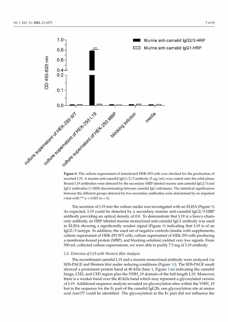
Figure 9. The culture supernatant of transfected HEK-293 cells was checked for the production of secreted L19. A murine anti-camelid IgG1/2/3 antibody (5 µg/mL) was coated onto the solid phase. Bound L19 antibodies were detected by the secondary HRP-labeled murine anti-camelid IgG2/3 and IgG1 antibodies (1:1000) discriminating between camelid IgG subclasses. The statistical signifi-cances between the different groups detected by two secondary antibodies were determined by an unpaired t-test with *** p < 0.001 (n = 3).
The secretion of L19 into the culture media was investigated with an ELISA (Figure 9). As expected, L19 could be detected by a secondary murine anti-camelid IgG2/3-HRP antibody providing an optical density of 0.8. To demonstrate that L19 is a heavy-chain-only antibody, an HRP-labeled murine monoclonal anti-camelid IgG1 antibody was used in ELISA showing a significantly weaker signal (Figure 9) indicating that L19 is of an IgG2/3 isotype. In addition, the used set of negative controls (media with supplements, culture supernatant of HEK-293 WT cells, culture supernatant of HEK-293 cells producing a membrane-bound protein (MBP), and blocking solution) yielded very low signals. From 500 mL collected culture supernatants, we were able to purify 7.5 mg of L19 antibody.
2.4. Detection of L19 with Western Blot Analysis The recombinant camelid L19 and a murine monoclonal antibody were analyzed via
SDS-PAGE and Western blot under reducing conditions (Figure 10). The SDS-PAGE result showed a prominent protein band at 40 kDa (lane 1, Figure 10a) indicating the camelid hinge, CH2, and CH3 region plus the VHH_19 domain of the full-length L19. Moreover, there is a weaker band over the 40 kDa band which may represent a glycosylated version of L19. Additional sequence analysis revealed no glycosylation sites within the VHH_19 but in the sequence for the Fc part of the camelid IgG2b, one glycosylation site at amino acid Asn177 could be identified. The glycosylation in the Fc part did not influence the antigen-binding of L19 but led to the weaker band above 40 kDa as shown in Figure 10a. The murine monoclonal antibody was used to visualize the structural difference between the conventional antibody consisting of a heavy (55 kDa) and a light chain (25 kDa) and the heavy-chain-only antibody (lanes 1 and 2, Figure 10a).
Figure 9. The culture supernatant of transfected HEK-293 cells was checked for the production ofsecreted L19. A murine anti-camelid IgG1/2/3 antibody (5 µg/mL) was coated onto the solid phase.Bound L19 antibodies were detected by the secondary HRP-labeled murine anti-camelid IgG2/3 andIgG1 antibodies (1:1000) discriminating between camelid IgG subclasses. The statistical significancesbetween the different groups detected by two secondary antibodies were determined by an unpairedt-test with *** p < 0.001 (n = 3).
The secretion of L19 into the culture media was investigated with an ELISA (Figure 9).As expected, L19 could be detected by a secondary murine anti-camelid IgG2/3-HRPantibody providing an optical density of 0.8. To demonstrate that L19 is a heavy-chain-only antibody, an HRP-labeled murine monoclonal anti-camelid IgG1 antibody was usedin ELISA showing a significantly weaker signal (Figure 9) indicating that L19 is of anIgG2/3 isotype. In addition, the used set of negative controls (media with supplements,culture supernatant of HEK-293 WT cells, culture supernatant of HEK-293 cells producinga membrane-bound protein (MBP), and blocking solution) yielded very low signals. From500 mL collected culture supernatants, we were able to purify 7.5 mg of L19 antibody.
2.4. Detection of L19 with Western Blot Analysis
The recombinant camelid L19 and a murine monoclonal antibody were analyzed viaSDS-PAGE and Western blot under reducing conditions (Figure 10). The SDS-PAGE resultshowed a prominent protein band at 40 kDa (lane 1, Figure 10a) indicating the camelidhinge, CH2, and CH3 region plus the VHH_19 domain of the full-length L19. Moreover,there is a weaker band over the 40 kDa band which may represent a glycosylated versionof L19. Additional sequence analysis revealed no glycosylation sites within the VHH_19but in the sequence for the Fc part of the camelid IgG2b, one glycosylation site at aminoacid Asn177 could be identified. The glycosylation in the Fc part did not influence the
Int. J. Mol. Sci. 2022, 23, 6275 8 of 18
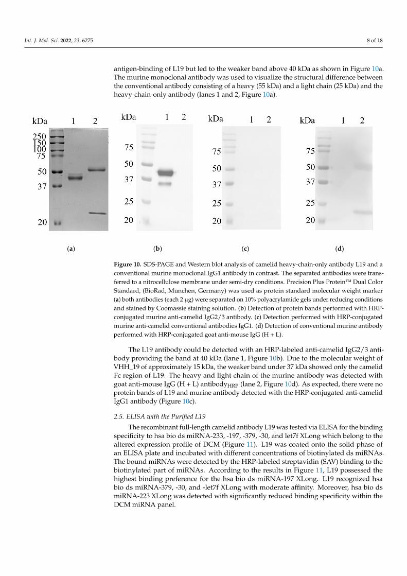
antigen-binding of L19 but led to the weaker band above 40 kDa as shown in Figure 10a.The murine monoclonal antibody was used to visualize the structural difference betweenthe conventional antibody consisting of a heavy (55 kDa) and a light chain (25 kDa) and theheavy-chain-only antibody (lanes 1 and 2, Figure 10a).
Int. J. Mol. Sci. 2022, 23, x FOR PEER REVIEW 9 of 19
(a) (b) (c) (d)
Figure 10. SDS-PAGE and Western blot analysis of camelid heavy-chain-only antibody L19 and a conventional murine monoclonal IgG1 antibody in contrast. The separated antibodies were trans-ferred to a nitrocellulose membrane under semi-dry conditions. Precision Plus Protein™ Dual Color Standard, (BioRad, München, Germany) was used as protein standard molecular weight marker (a) both antibodies (each 2 µg) were separated on 10% polyacrylamide gels under reducing conditions and stained by Coomassie staining solution. (b) Detection of protein bands performed with HRP-conjugated murine anti-camelid IgG2/3 antibody. (c) Detection performed with HRP-conjugated murine anti-camelid conventional antibodies IgG1. (d) Detection of conventional murine antibody performed with HRP-conjugated goat anti-mouse IgG (H + L).
The L19 antibody could be detected with an HRP-labeled anti-camelid IgG2/3 anti-body providing the band at 40 kDa (lane 1, Figure 10b). Due to the molecular weight of VHH_19 of approximately 15 kDa, the weaker band under 37 kDa showed only the camelid Fc region of L19. The heavy and light chain of the murine antibody was detected with goat anti-mouse IgG (H + L) antibodyHRP (lane 2, Figure 10d). As expected, there were no protein bands of L19 and murine antibody detected with the HRP-conjugated anti-camelid IgG1 antibody (Figure 10c).
2.5. ELISA with the Purified L19 The recombinant full-length camelid antibody L19 was tested via ELISA for the bind-
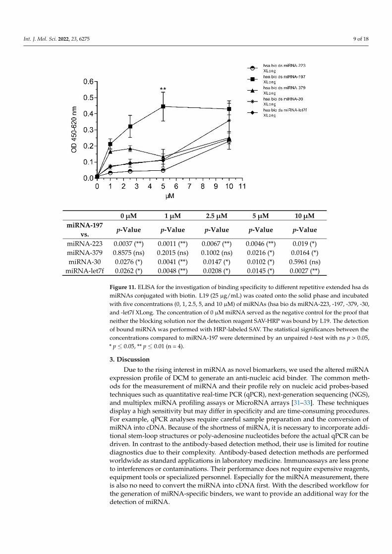
ing specificity to hsa bio ds miRNA-233, -197, -379, -30, and let7f XLong which belong to the altered expression profile of DCM (Figure 11). L19 was coated onto the solid phase of an ELISA plate and incubated with different concentrations of biotinylated ds miRNAs. The bound miRNAs were detected by the HRP-labeled streptavidin (SAV) binding to the biotinylated part of miRNAs. According to the results in Figure 11, L19 possessed the highest binding preference for the hsa bio ds miRNA-197 XLong. L19 recognized hsa bio ds miRNA-379, -30, and -let7f XLong with moderate affinity. Moreover, hsa bio ds miRNA-223 XLong was detected with significantly reduced binding specificity within the DCM miRNA panel.
Figure 10. SDS-PAGE and Western blot analysis of camelid heavy-chain-only antibody L19 and aconventional murine monoclonal IgG1 antibody in contrast. The separated antibodies were trans-ferred to a nitrocellulose membrane under semi-dry conditions. Precision Plus Protein™ Dual ColorStandard, (BioRad, München, Germany) was used as protein standard molecular weight marker(a) both antibodies (each 2 µg) were separated on 10% polyacrylamide gels under reducing conditionsand stained by Coomassie staining solution. (b) Detection of protein bands performed with HRP-conjugated murine anti-camelid IgG2/3 antibody. (c) Detection performed with HRP-conjugatedmurine anti-camelid conventional antibodies IgG1. (d) Detection of conventional murine antibodyperformed with HRP-conjugated goat anti-mouse IgG (H + L).
The L19 antibody could be detected with an HRP-labeled anti-camelid IgG2/3 anti-body providing the band at 40 kDa (lane 1, Figure 10b). Due to the molecular weight ofVHH_19 of approximately 15 kDa, the weaker band under 37 kDa showed only the camelidFc region of L19. The heavy and light chain of the murine antibody was detected withgoat anti-mouse IgG (H + L) antibodyHRP (lane 2, Figure 10d). As expected, there were noprotein bands of L19 and murine antibody detected with the HRP-conjugated anti-camelidIgG1 antibody (Figure 10c).
2.5. ELISA with the Purified L19
The recombinant full-length camelid antibody L19 was tested via ELISA for the bindingspecificity to hsa bio ds miRNA-233, -197, -379, -30, and let7f XLong which belong to thealtered expression profile of DCM (Figure 11). L19 was coated onto the solid phase ofan ELISA plate and incubated with different concentrations of biotinylated ds miRNAs.The bound miRNAs were detected by the HRP-labeled streptavidin (SAV) binding to thebiotinylated part of miRNAs. According to the results in Figure 11, L19 possessed thehighest binding preference for the hsa bio ds miRNA-197 XLong. L19 recognized hsabio ds miRNA-379, -30, and -let7f XLong with moderate affinity. Moreover, hsa bio dsmiRNA-223 XLong was detected with significantly reduced binding specificity within theDCM miRNA panel.
Int. J. Mol. Sci. 2022, 23, 6275 9 of 18Int. J. Mol. Sci. 2022, 23, x FOR PEER REVIEW 10 of 19
0 µM 1 µM 2.5 µM 5 µM 10 µM miRNA-197
vs. p-Value p-Value p-Value p-Value p-Value
miRNA-223 0.0037 (**) 0.0011 (**) 0.0067 (**) 0.0046 (**) 0.019 (*) miRNA-379 0.8575 (ns) 0.2015 (ns) 0.1002 (ns) 0.0216 (*) 0.0164 (*) miRNA-30 0.0276 (*) 0.0041 (**) 0.0147 (*) 0.0102 (*) 0.5961 (ns)
miRNA-let7f 0.0262 (*) 0.0048 (**) 0.0208 (*) 0.0145 (*) 0.0027 (**)
Figure 11. ELISA for the investigation of binding specificity to different repetitive extended hsa ds miRNAs conjugated with biotin. L19 (25 µg/mL) was coated onto the solid phase and incubated with five concentrations (0, 1, 2.5, 5, and 10 µM) of miRNAs (hsa bio ds miRNA-223, -197, -379, -30, and -let7f XLong. The concentration of 0 µM miRNA served as the negative control for the proof that neither the blocking solution nor the detection reagent SAV-HRP was bound by L19. The de-tection of bound miRNA was performed with HRP-labeled SAV. The statistical significances be-tween the concentrations compared to miRNA-197 were determined by an unpaired t-test with ns p > 0.05, * p ≤ 0.05, ** p ≤ 0.01 (n = 4).
3. Discussion Due to the rising interest in miRNA as novel biomarkers, we used the altered miRNA
expression profile of DCM to generate an anti-nucleic acid binder. The common methods for the measurement of miRNA and their profile rely on nucleic acid probes-based tech-niques such as quantitative real-time PCR (qPCR), next-generation sequencing (NGS), and multiplex miRNA profiling assays or MicroRNA arrays [31–33]. These techniques display a high sensitivity but may differ in specificity and are time-consuming procedures. For example, qPCR analyses require careful sample preparation and the conversion of miRNA into cDNA. Because of the shortness of miRNA, it is necessary to incorporate additional stem-loop structures or poly-adenosine nucleotides before the actual qPCR can be driven. In contrast to the antibody-based detection method, their use is limited for routine diag-nostics due to their complexity. Antibody-based detection methods are performed world-wide as standard applications in laboratory medicine. Immunoassays are less prone to interferences or contaminations. Their performance does not require expensive reagents, equipment tools or specialized personnel. Especially for the miRNA measurement, there is also no need to convert the miRNA into cDNA first. With the described workflow for the generation of miRNA-specific binders, we want to provide an additional way for the detection of miRNA.
Figure 11. ELISA for the investigation of binding specificity to different repetitive extended hsa dsmiRNAs conjugated with biotin. L19 (25 µg/mL) was coated onto the solid phase and incubatedwith five concentrations (0, 1, 2.5, 5, and 10 µM) of miRNAs (hsa bio ds miRNA-223, -197, -379, -30,and -let7f XLong. The concentration of 0 µM miRNA served as the negative control for the proof thatneither the blocking solution nor the detection reagent SAV-HRP was bound by L19. The detectionof bound miRNA was performed with HRP-labeled SAV. The statistical significances between theconcentrations compared to miRNA-197 were determined by an unpaired t-test with ns p > 0.05,* p ≤ 0.05, ** p ≤ 0.01 (n = 4).
3. Discussion
Due to the rising interest in miRNA as novel biomarkers, we used the altered miRNAexpression profile of DCM to generate an anti-nucleic acid binder. The common meth-ods for the measurement of miRNA and their profile rely on nucleic acid probes-basedtechniques such as quantitative real-time PCR (qPCR), next-generation sequencing (NGS),and multiplex miRNA profiling assays or MicroRNA arrays [31–33]. These techniquesdisplay a high sensitivity but may differ in specificity and are time-consuming procedures.For example, qPCR analyses require careful sample preparation and the conversion ofmiRNA into cDNA. Because of the shortness of miRNA, it is necessary to incorporate addi-tional stem-loop structures or poly-adenosine nucleotides before the actual qPCR can bedriven. In contrast to the antibody-based detection method, their use is limited for routinediagnostics due to their complexity. Antibody-based detection methods are performedworldwide as standard applications in laboratory medicine. Immunoassays are less proneto interferences or contaminations. Their performance does not require expensive reagents,equipment tools or specialized personnel. Especially for the miRNA measurement, thereis also no need to convert the miRNA into cDNA first. With the described workflow forthe generation of miRNA-specific binders, we want to provide an additional way for thedetection of miRNA.
Int. J. Mol. Sci. 2022, 23, 6275 10 of 18
As mentioned before, the development of anti-nucleic acid antibodies by the commonhybridoma technology is challenging due to the low immunogenicity of nucleic acids.The data presented in this study may give new insights into the suitability of camelidphage display technology for the generation of VHHs against miRNAs. Here, we showedthe identification of miRNA binders from the generated naive VHH phage library with6.2 × 106 members. The constructed VHH library is noticeably smaller than the reportedones in the literature (up to 1011 members) [34]. Nevertheless, it was described that antigen-binding domains such as VHHs could be recovered also from relatively small naïve phagelibraries [35].
We selected the model antigen ds miRNA-223 from the altered miRNA expressionprofile of DCM. After several panning rounds against the ds miRNA-223, we have founda potential anti-nucleic acid binder referred to as VHH_19. In the first pre-experiments,VHH_19 was immobilized onto microbeads and the recognition of 0.1 fmol/µL miRNA-93was detected (Appendix A). Moreover, the first tests with miRNA isolated from the serumof healthy and diseased donors were performed. The distinction in the recognition betweenthe miRNA of healthy and diseased donors by the immobilized VHH_19 could be shown(Appendix A). However, the small size of VHH (12–15 kDa) and a decreased probe accessi-bility can be disadvantageous, especially when used for in vitro diagnostic systems [36].To improve the probe accessibility, VHHs can be modified by introducing peptides at theC-terminus or by the fusion to the Fc region [36,37]. With the introduction of the Fc region,higher signals can be achieved and other assay formats can be performed. Thus, we decidedto expand the identified VHH_19 with camelid Fc of IgG2b to generate a full-length camelidrecombinant antibody L19 expressed in a mammalian expression system.
We were able to perform the ELISA test with a full-length L19 antibody to verify theanti- nucleic acid binding specificity. The highest signal was achieved for ds miRNA-197 at5 µM. In comparison, the signals for the other ds extended miRNA detected by L19 wereweaker. The sequence homology between the single miRNAs within the DCM profile is nothigh and there is no clear pattern or base pair motif that could be linked to the preference formiRNA-197. One point that might explain the phenomenon is the formation of loops withinthe miRNA sequences during hybridization. Two single RNA strands in directions 5′-3′ and3′-5′ were synthesized and hybridized to generate the double-stranded miRNA used for thedirect ELISA. The perfectly matched double-stranded miRNA sequence is not guaranteedduring the hybridization step. The formation of loops within the sequence may occurwith a high probability of causing different miRNA structures. There is no possibility topredict the miRNA structure with ds miRNA because the available bioinformatic structureprediction tools use single-stranded RNA sequences as input. The batch of ds miRNA-197used for the direct ELISA could have a high degree of imperfect hybridization meaningthe presence of inner loops in comparison to the other miRNAs. There is no possibilityto measure or analyze the degree of hybridization as a melting curve analysis would notprovide reliable results for such short ds miRNAs.
The ds miRNA-93 and the ds miRNA-223 bound by VHH_19 in the preliminary exper-iment (Tables A1 and A2) and the ds miRNA-197 bound by L19 showed a higher homologydegree between each other than with other miRNAs in the DCM miRNA panel (align-ment not shown). Moreover, the ds miRNA-223 and the ds miRNA-197 were describedto be implicated in endovascular inflammation and platelet activation and can be usedas biomarkers to diagnose coronary artery disease [38]. The novel recombinant camelidantibody L19 can, therefore, be a useful tool for the further establishment of antibody-basedmiRNA detection. Recently, a new study was published that describes the antibody-basedmiRNA detection on the basis of the multiplex microchamber diffusion assay using theanti-hybrid DNA:RNA antibody (clone S9.6) [39]. These results in combination with thenovel generated camelid miRNA binder may contribute to the further establishment or tothe development of novel strategies for miRNA detection in a sequence-specific manner.The anti-hybrid DNA:RNA antibody (S9.6) is the most commonly used antibody for thedevelopment of antibody-based assays for the detection of miRNA [40,41]. In 2008, Ye
Int. J. Mol. Sci. 2022, 23, 6275 11 of 18
and colleagues developed synthetic antibodies by using the phage display method torecognize structured RNA [42]. Recently, a Fab fragment was generated from a naïveantigen-binding fragment combinatorial phage library against the brain cytoplasmic 200(BC200) RNA [43]. The isolated Fab is believed to recognize a domain of BC200 RNAin a sequence-dependent and conformation-based manner. However, these recombinantantibody formats are designed to recognize large and structured nucleic acid sequences.
We have demonstrated that by using the well-established phage display technology itis possible to generate recombinant antibodies against very short nucleic acid sequenceswhich can be used to establish novel strategies for miRNA detection.
4. Materials and Methods4.1. Used miRNAs
For the purpose of this study the miRNAs of the altered miRNA expression profilein association with DCM were purchased from Riboxx GmbH (Dresden, Germany). Thesequence of each miRNA was repetitively extended five times and biotin was conjugatedat the 5′end of the sense strand. The synthetic generated miRNAs were double stranded.
4.2. Extraction of Total RNA from Camelid Peripheral Blood Mononuclear Cells
Peripheral blood mononuclear cells (PBMCs) were isolated from fresh whole bloodfrom three different naïve camelids (llama, alpaca, and huarizo) according to [44,45]. Finally,pellets were homogenized in 5 mL RNAPureTM peqGOLD (VWR, Dresden, Germany)and stored at −80 ◦C for further treatments. Total RNA isolation was performed by theconventional phenol–chloroform extraction following the manufacturer’s instructions.Phase lock gel tubes (PLG, Quantabio) were used to yield higher RNA amounts. Finally,RNA concentrations were measured by UV spectroscopy at 260 and 280 nm.
4.3. Construction of Naïve Phage Library
CopyDNA (cDNA) was synthesized from 1 µg total RNA using the RevertAid FirstStrand cDNA Synthesis Kit (Thermo Fisher Scientific, Waltham, MA, USA). VHH sequenceswere amplified by two-step PCR using the primers listed in Table 1. The amplification of thecamelid VHH repertoire was performed as previously described [44,46,47]. PCR productswith a size of 400 bp were used for the construction of the naïve phage library. After thepurification from the 1.5% agarose gel and digestion with SfiI, the VHH gene fragmentswere ligated into SfiI digested and dephosphorylated pCOMB3x (kindly provided by theScripps Research Institute, La Jolla, CA 92037, USA) using the T4 ligase (Thermo FisherScientific, Waltham, MA, USA). Plasmids were cloned into electrocompetent E. coli XL1 Bluesuper competent cells (Agilent, Santa Clara, CA, USA). The bacteria cells were co-infectedwith M13KO7 helper phages (1013 pfu) to generate the naïve VHH phage library accordingto Barbas et al. [48]. Phage particles were precipitated with ice-cold 20% PEG-8000 in 2.5 MNaCl, incubated for 30 min on ice, and centrifuged for 18 min at 13,000× g and 4 ◦C. Phageparticles were resuspended in phosphate-buffered saline with 1% bovine serum albumin(3% BSA/1x PBS (v/w)) and used directly for panning.
Table 1. Primer sequences for the amplification of VHH DNA sequences.
Primer Name Sequence (5′ → 3′) Reference
P12 GTCCTGGCTGCTCTTCTACAAGG [46]P13 ATGGAGAGGACGTCCTTGGGT [46]
mL93 ACCGTGGCCCAGGCGGCCCAGGTGCAGCTGCAGGAGTCTGGRGGAGG [47] modifiedmL94 GTGCTGGCCGGCCTGGCCGCTGGAGACGGTGACCTGGGT [47] modifiedmL95 GTGCTGGCCGGCCTGGCCTGAGGAGACGGTGACCTGGGT [47] modified
Int. J. Mol. Sci. 2022, 23, 6275 12 of 18
4.4. Panning the Naïve VHH Phage Library
Panning rounds were performed according to [48] with slight modifications. Beforeeach panning round, microtiter plates (Nunc, Rochester, NY, USA) were coated with carrierprotein avidin (10 µg/mL) alone and with Homo sapiens biotinylated, repetitive extendeddouble-stranded (ds) miRNA-223 (hsa bio ds miRNA-223 XLong, Riboxx GmbH, Dresden,Germany) conjugated on 10 µg/mL avidin. The hsa bio ds miRNA-223 XLong was usedas a model ds miRNA construct. The fixation of antigens was performed for 2 h underhumid conditions at 37 ◦C. After blocking steps with 100 µL 3% BSA/1x PBS (w/v), apre-panning round was performed. The VHH phage library was pre-incubated against thecarrier protein avidin for 30 min at 37 ◦C. Unbound phage particles were then incubatedwith decreasing amounts of hsa bio ds miRNA-XLong starting with 100 pmol, 50 pmolto 20 pmol in subsequent panning rounds for 2 h at 37 ◦C. Bound phage particles wereeluted by a pH shift with 100 mM glycine-HCl (pH 2.2) and neutralized with 2 M Tris-HCl,pH 8.0. Eluted phages were used to re-infect the E. coli XL1 blue cells (NEB 5-alpha F‘Iq
Competent E. coli, Ipswich, MA, USA) according to Barbas et al. for the enrichment ofantigen-specific phage particles [48]. After each panning, the output and input titer ofphages were determined. The percentage enrichment was calculated by the division of thetiter of output phages and titer of input phages multiplied by 100% for each panning round.
4.5. Monoclonal Phage Enzyme-Linked Immunosorbent Assay (ELISA)
After the third panning round, individual colonies were randomly selected to producemonoclonal phage particles as described previously [49]. The obtained monoclonal phageparticles were tested for the hsa ds miRNA-223 XLong recognition in a direct ELISA.Additionally, the monoclonal phage particles were tested for cross-reactivity to avidinand the blocking solution. Antigens (10 µg/mL avidin, 20 pmol hsa bio ds miRNA-XLong (Riboxx GmbH, Radebeul, Germany) conjugated on 10 µg/mL avidin and 3%BSA/PBS) were coated on a microtiter plate (Nunc, Rochester, NY, USA) for 2 h at 37 ◦C.The undiluted monoclonal phage supernatants were incubated for 2 h at 37 ◦C. Boundphages were detected with horseradish peroxidase (HRP)-conjugated monoclonal mouseanti-M13 antibody (1:7000; MM05H, antibodies-online GmbH, Aachen, Germany). Thesecondary HRP-labeled anti-M13 antibody was incubated for 1 h at 37 ◦C. Additionally,a coating control for the immobilized hsa ds miRNA-223 XLong conjugated on avidinwas carried out using the commercially available murine anti-DNA:RNA antibody (cloneS9.6, MABE 1095, Merck, Darmstadt Deutschland), that was detected with HRP-labeledgoat anti-mouse IgG (FcG) antibody respective (1:10,000; 115-035-071, Dianova, Hamburg,Germany). Colorimetric signals were detected after adding tetramethylbenzidine (TMB)peroxidase substrate and 1 M sulfuric acid after 7 min to stop the reaction. The absorbancewas measured at 450 nm and 620 nm in an ELISA plate reader.
4.6. Expression and Purification of Recombinant VHH_19
The positive clone (pCOMB3x-VHH_19) was first purified with the NucleoSpin® Plas-mid Kit (Macherey-Nagel, Dueren, Germany) and sequenced by LGC Genomics GmbH(Berlin, Germany) with M13rev2 sequencing primer provided by the company. The re-sulting DNA sequence was translated into an amino acid sequence (Kabat numberingscheme) and analyzed for the identification of the four hallmarks indicating a camelidsingle-domain antibody (VHH). pCOMB3x-VHH_19 was transferred into chemically com-petent non-suppressor E. coli strain HB2151 and expressed as previously described in [44].The soluble His-tagged VHH_19 was purified from the periplasmic fraction via Ni-NTAaffinity chromatography according to the manufacturer’s standard protocol (Protino®
Ni-NTA Agarose, Macherey-Nagel, Dueren, Germany).
4.7. Generation of Full-Length Recombinant Camelid Heavy-Chain Only Antibody L19
The full-length recombinant camelid heavy chain antibody named L19 was composedof the camelid single-domain antibody (VHH_19) and the Lama glama IgG2b constant region
Int. J. Mol. Sci. 2022, 23, 6275 13 of 18
(Fc region; GeneBank, accession number AY874455). The DNA sequences encoding for theVHH_19, camelid Fc of IgG2b, GFP, and for the targeting signal ensuring the secretion ofexpressed antibody into the culture medium were cloned into the expression vector pMC19using the In-Fusion Cloning Kit following the manufacturer’s protocol (Takara Bio). Theexpression vector was transfected into the HEK-293 cells using the NeonTM transfectionsystem (Invitrogen, Carlsbad, CA, USA) following the manufacturer’s instructions. Thecells were cultivated in Gibco RPMI-1640 medium (Thermo Fisher, Waltham, MA, USA)supplemented with 10% fetal calf serum (FCS) (Thermo Fisher, Waltham, MA, USA), 1% L-glutamine, and 1% beta-mercaptoethanol at 37 ◦C. Seven days after the transfection, apuromycin-induced selection (2 µg/mL) was performed for two weeks to establish stablyproducing cell clones.
4.8. ELISA to Detect Secreted L19 Antibodies
To prove the presence of the secreted camelid heavy-chain-only antibody L19, cul-ture supernatants were tested in a sandwich ELISA. The murine monoclonal anti-camelidIgG2/3 antibody (5 µg/mL; ABIN1981268, antibodies-online, Germany) was coated ontothe solid phase of an ELISA microtiter plate for 2 h at 37 ◦C under humid conditions.After blocking with 5% newborn calf serum (NCS) in 1x PBS for 30 min, undiluted cul-ture supernatants were incubated for 45 min at RT. Moreover, a set of negative controlswere considered such as the blocking solution, the pure RPMI medium with supplements,culture supernatants of HEK-293 cells producing a therefore irrelevant membrane-boundprotein (MBP), and the culture supernatants of wild-type HEK-293 cells. Next, boundantibodies were detected with 1 µg/mL HRP-conjugated anti-camelid IgG1/2/3 antibod-ies (murine anti-camelid IgG antibody, ABIN1981270, antibodies-online, Germany) andmurine anti-camelid IgG1 antibody (ABIN1981271, antibodies-online, Germany). Detectionantibodies were incubated for 45 min at RT. Colorimetric signals were detected after addingtetramethylbenzidine (TMB) peroxidase substrate and 1 M sulfuric acid stop solution after10 min. The absorbance was measured at 450 nm and 620 nm in an ELISA plate reader.
4.9. Purification of L19 from the Culture Supernatant
Protein A affinity chromatography (ProSep-vA Ultra Chromatography Media, Milli-pore, Schwalbach, Germany) was used for the purification of L19 from the culture super-natant. For the purification, 500 mL of the collected culture supernatant containing thesecreted L19 was mixed 2:1 with the protein A binding buffer (4 M NaCl, 2 M glycine, pH8.5) and 750 mL were loaded onto the protein A column overnight at 4 ◦C. The column waswashed with a washing buffer (protein A binding buffer diluted 1:3 in ddH2O). The elutionof the recombinant heavy chain antibody was performed according to [50]. The purifiedL19 was dialyzed (Snake SkinTM Dialysis Tubing, cut-off 30 kDa, Thermo Fisher Scientific,Waltham, MA, USA) against 1x PBS overnight at 4 ◦C. The concentration of dialyzed L19was determined by using the BCA protein assay method according to the manufacturer’sinstructions (PierceTM BCA Protein Assay Kit, Thermo Fisher Scientific, Waltham, MA,USA). L19 was stored at 4 ◦C.
4.10. Sodium Dodecyl Sulfate Polyacrylamide Gel Electrophoresis (SDS-PAGE) and WesternBlot Analysis
Samples collected during the Ni-NTA purification of His-tagged VHH_19, the full-length antibody L19, and a murine monoclonal antibody were analyzed via SDS-PAGEand WB. VHH_19 samples were separated on a 12% precast Mini-PROTEAN TGX gel(BioRad, München, Germany). Camelid L19 (2 µg) and the murine antibody were separatedon 10% SDS polyacrylamide gel under reducing conditions using a buffer containing ß-mercaptoethanol (ROTI®Load 1; Carl Roth GmbH, Karlsruhe, Germany). All sampleswere prepared according to the manufacturer’s instructions. Next, the protein bandswere electrophoretically transferred to a nitrocellulose membrane (Trans-Blot TurboTMmini-size nitrocellulose, 0.2 µm, BioRad, München, Germany) under semi-dry conditions
Int. J. Mol. Sci. 2022, 23, 6275 14 of 18
(2.6 A, 7 min) with the Trans-Blot Turbo Transfer System (BioRad, München, Germany).To reduce non-specific binding, a blocking step was performed with 1x Tris-bufferedsaline/0.05% Tween20 (v/v) (TBS-T)/5% goat serum (v/v) (Biowest, Riverside, CA, USA)for 30 min at RT. For the detection of HA-tagged VHH_19, the membrane was incubatedwith a mouse anti-HA-tag antibody (1:5000; 16B12, BioLegend, San Diego, CA, USA)overnight at 4 ◦C, washed three times with 1x TBS-T, and incubated with the secondaryantibody (1:10,000; HRP-conjugated goat-anti-mouse IgG (FcG) antibody, 115-035-071,Dianova, Hamburg, Germany) for 1 h at RT. For detection of camelid L19 and murineantibodies, the membranes were incubated with HRP-conjugated murine anti-camelidIgG2/3 antibody (1:7000; ABIN1981272, antibodies-online, Germany), murine anti-camelidIgG1 antibody (1:7000; ABIN1981271, antibodies-online, Germany) and HRP-conjugatedgoat anti-mouse IgG (H + L) antibody respectively (1:10,000; 0300-0108P, BioRad, München,Germany) overnight at 4 ◦C. After three washing steps with TBS-T, protein bands werevisualized by the substrate solution (ClarityTM Western ECL Substrate, BioRad, München,Germany) on ChemiDoc MP Imaging Systems (BioRad, München, Germany).
4.11. ELISA with the Purified L19
L19 (25 µg/mL) was coated onto the solid phase of a microtiter ELISA plate for 2 h at37 ◦C under humid conditions. After blocking with 1x PBS/5% NCS (v/v) for 30 min atRT, hsa bio ds miRNA-223, -197, -379, -30, and -let7f XLong (10 µM, 5 µM, 2.5 µM, 1 µM)were incubated for 45 min at RT. The blocking solution served as the negative controland was added instead of any miRNA probe. Next, the bound biotinylated ds miRNAswere incubated with SAV-HRP (1:10,000; Roche, Mannheim, Germany) for 45 min at RT.Colorimetric signals were induced by adding tetramethylbenzidine (TMB) peroxidasesubstrate. The reaction was stopped after 15 min with 1 M sulfuric acid. The absorbancewas measured at 450 nm and 620 nm in an ELISA plate reader.
4.12. Software
Statistical analyses were performed using GraphPad Prism (version 8.4.1). The primermodifications were generated with SnapGene (version 4.2.6). Chemiluminescence sig-nals of protein bands were analyzed with Image Lab Software (version 2.0, BioRad,München, Germany).
Author Contributions: Conceptualization, M.C. and K.H.; methodology, M.C., U.W. and K.H.;software, M.C.; validation, M.C.; formal analysis, M.C.; investigation, M.C.; resources, U.W.; datacuration, M.C.; writing—original draft preparation, M.C., U.W. and K.H.; writing—review andediting, M.C., U.W. and K.H.; visualization, M.C.; supervision, S.R.; project administration, K.H.;funding acquisition, K.H. All authors have read and agreed to the published version of the manuscript.
Funding: This research was funded by the German Federal Ministry of Education and Research,grant number WKP55A.
Institutional Review Board Statement: The animal blood used for the isolation of PBMC wasordered from the company preclinics GmbH (Potsdam, Germany) according to the relevant nationaland international guidelines. The preparation of miRNA from the human serum of healthy anddiseased donors undertook the cooperation partner IKDT (Berlin, Germany).
Informed Consent Statement: Not applicable.
Data Availability Statement: The data presented in this study are available on request from thecorresponding author. The data are not publicly available.
Acknowledgments: We want to thank Ganna Aleshcheva from IKDT (Berlin, Germany) for perform-ing the bead-based assay with human miRNA samples from healthy and diseased donors and for hersupport in result interpretation.
Int. J. Mol. Sci. 2022, 23, 6275 15 of 18
Conflicts of Interest: The corresponding author Katja Hanack is president and CEO of new/era/mabs.The other authors declare no conflict of interest. The funders had no role in the design of the study; inthe collection, analyses, or interpretation of data; in the writing of the manuscript, or in the decisionto publish the results.
Appendix A
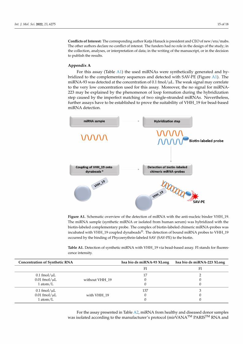
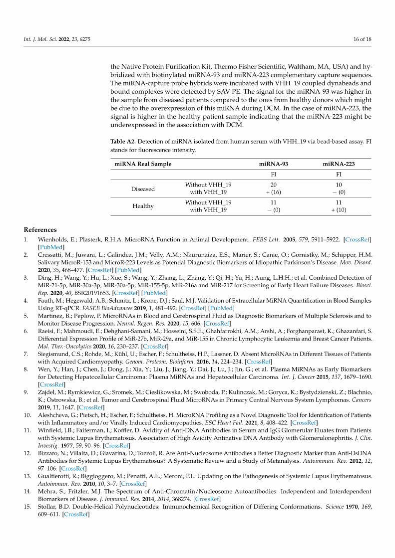
For this assay (Table A1) the used miRNAs were synthetically generated and hy-bridized to the complementary sequences and detected with SAV-PE (Figure A1). ThemiRNA-93 was detected at the concentration of 0.1 fmol/µL. The weak signal may correlateto the very low concentration used for this assay. Moreover, the no signal for miRNA-223 may be explained by the phenomenon of loop formation during the hybridizationstep caused by the imperfect matching of two single-stranded miRNAs. Nevertheless,further assays have to be established to prove the suitability of VHH_19 for bead-basedmiRNA detection.
Int. J. Mol. Sci. 2022, 23, x FOR PEER REVIEW 16 of 19
Informed Consent Statement: Not applicable.
Data Availability Statement: The data presented in this study are available on request from the corresponding author. The data are not publicly available.
Acknowledgments: We want to thank Dr. Ganna Aleshcheva from IKDT (Berlin, Germany) for per-forming the bead-based assay with human miRNA samples from healthy and diseased donors and for her support in result interpretation.
Conflicts of Interest: The corresponding author Katja Hanack is president and CEO of new/era/mabs. The other authors declare no conflict of interest. The funders had no role in the de-sign of the study; in the collection, analyses, or interpretation of data; in the writing of the manu-script, or in the decision to publish the results.
Appendix A For this assay (Table A1) the used miRNAs were synthetically generated and hybrid-
ized to the complementary sequences and detected with SAV-PE (Figure A1). The miRNA-93 was detected at the concentration of 0.1 fmol/µL. The weak signal may corre-late to the very low concentration used for this assay. Moreover, the no signal for miRNA-223 may be explained by the phenomenon of loop formation during the hybridization step caused by the imperfect matching of two single-stranded miRNAs. Nevertheless, further assays have to be established to prove the suitability of VHH_19 for bead-based miRNA detection.
Figure A1. Schematic overview of the detection of miRNA with the anti-nucleic binder VHH_19. The miRNA sample (synthetic miRNA or isolated from human serum) was hybridized with the biotin-labeled complementary probe. The complex of biotin-labeled chimeric miRNA-probes was incubated with VHH_19 coupled dynabeads®. The detection of bound miRNA probes to VHH_19 occurred by the binding of Phycoerythrin-labeled SAV (SAV-PE) to the biotin.
Table A1. Detection of synthetic miRNA with VHH_19 via bead-based assay. FI stands for fluores-cence intensity.
Figure A1. Schematic overview of the detection of miRNA with the anti-nucleic binder VHH_19.The miRNA sample (synthetic miRNA or isolated from human serum) was hybridized with thebiotin-labeled complementary probe. The complex of biotin-labeled chimeric miRNA-probes wasincubated with VHH_19 coupled dynabeads®. The detection of bound miRNA probes to VHH_19occurred by the binding of Phycoerythrin-labeled SAV (SAV-PE) to the biotin.
Table A1. Detection of synthetic miRNA with VHH_19 via bead-based assay. FI stands for fluores-cence intensity.
Concentration of Synthetic RNA hsa bio ds miRNA-93 XLong hsa bio ds miRNA-223 XLong
FI FI
0.1 fmol/µLwithout VHH_19
17 20.01 fmol/µL 0 0
1 atom/L 0 0
0.1 fmol/µLwith VHH_19
137 30.01 fmol/µL 0 0
1 atom/L 0 0
For the assay presented in Table A2, miRNA from healthy and diseased donor sampleswas isolated according to the manufacturer’s protocol (mirVANATM PARISTM RNA and
Int. J. Mol. Sci. 2022, 23, 6275 16 of 18
the Native Protein Purification Kit, Thermo Fisher Scientific, Waltham, MA, USA) and hy-bridized with biotinylated miRNA-93 and miRNA-223 complementary capture sequences.The miRNA-capture probe hybrids were incubated with VHH_19 coupled dynabeads andbound complexes were detected by SAV-PE. The signal for the miRNA-93 was higher inthe sample from diseased patients compared to the ones from healthy donors which mightbe due to the overexpression of this miRNA during DCM. In the case of miRNA-223, thesignal is higher in the healthy patient sample indicating that the miRNA-223 might beunderexpressed in the association with DCM.
Table A2. Detection of miRNA isolated from human serum with VHH_19 via bead-based assay. FIstands for fluorescence intensity.
miRNA Real Sample miRNA-93 miRNA-223
FI FI
DiseasedWithout VHH_19 20 10
with VHH_19 + (16) − (0)
Healthy Without VHH_19 11 11with VHH_19 − (0) + (10)
References1. Wienholds, E.; Plasterk, R.H.A. MicroRNA Function in Animal Development. FEBS Lett. 2005, 579, 5911–5922. [CrossRef]
[PubMed]2. Cressatti, M.; Juwara, L.; Galindez, J.M.; Velly, A.M.; Nkurunziza, E.S.; Marier, S.; Canie, O.; Gornistky, M.; Schipper, H.M.
Salivary MicroR-153 and MicroR-223 Levels as Potential Diagnostic Biomarkers of Idiopathic Parkinson’s Disease. Mov. Disord.2020, 35, 468–477. [CrossRef] [PubMed]
3. Ding, H.; Wang, Y.; Hu, L.; Xue, S.; Wang, Y.; Zhang, L.; Zhang, Y.; Qi, H.; Yu, H.; Aung, L.H.H.; et al. Combined Detection ofMiR-21-5p, MiR-30a-3p, MiR-30a-5p, MiR-155-5p, MiR-216a and MiR-217 for Screening of Early Heart Failure Diseases. Biosci.Rep. 2020, 40, BSR20191653. [CrossRef] [PubMed]
4. Fauth, M.; Hegewald, A.B.; Schmitz, L.; Krone, D.J.; Saul, M.J. Validation of Extracellular MiRNA Quantification in Blood SamplesUsing RT-qPCR. FASEB BioAdvances 2019, 1, 481–492. [CrossRef] [PubMed]
5. Martinez, B.; Peplow, P. MicroRNAs in Blood and Cerebrospinal Fluid as Diagnostic Biomarkers of Multiple Sclerosis and toMonitor Disease Progression. Neural. Regen. Res. 2020, 15, 606. [CrossRef]
6. Raeisi, F.; Mahmoudi, E.; Dehghani-Samani, M.; Hosseini, S.S.E.; Ghahfarrokhi, A.M.; Arshi, A.; Forghanparast, K.; Ghazanfari, S.Differential Expression Profile of MiR-27b, MiR-29a, and MiR-155 in Chronic Lymphocytic Leukemia and Breast Cancer Patients.Mol. Ther.-Oncolytics 2020, 16, 230–237. [CrossRef]
7. Siegismund, C.S.; Rohde, M.; Kühl, U.; Escher, F.; Schultheiss, H.P.; Lassner, D. Absent MicroRNAs in Different Tissues of Patientswith Acquired Cardiomyopathy. Genom. Proteom. Bioinform. 2016, 14, 224–234. [CrossRef]
8. Wen, Y.; Han, J.; Chen, J.; Dong, J.; Xia, Y.; Liu, J.; Jiang, Y.; Dai, J.; Lu, J.; Jin, G.; et al. Plasma MiRNAs as Early Biomarkersfor Detecting Hepatocellular Carcinoma: Plasma MiRNAs and Hepatocellular Carcinoma. Int. J. Cancer 2015, 137, 1679–1690.[CrossRef]
9. Zajdel, M.; Rymkiewicz, G.; Sromek, M.; Cieslikowska, M.; Swoboda, P.; Kulinczak, M.; Goryca, K.; Bystydzienski, Z.; Blachnio,K.; Ostrowska, B.; et al. Tumor and Cerebrospinal Fluid MicroRNAs in Primary Central Nervous System Lymphomas. Cancers2019, 11, 1647. [CrossRef]
10. Aleshcheva, G.; Pietsch, H.; Escher, F.; Schultheiss, H. MicroRNA Profiling as a Novel Diagnostic Tool for Identification of Patientswith Inflammatory and/or Virally Induced Cardiomyopathies. ESC Heart Fail. 2021, 8, 408–422. [CrossRef]
11. Winfield, J.B.; Faiferman, I.; Koffler, D. Avidity of Anti-DNA Antibodies in Serum and IgG Glomerular Eluates from Patientswith Systemic Lupus Erythematosus. Association of High Avidity Antinative DNA Antibody with Glomerulonephritis. J. Clin.Investig. 1977, 59, 90–96. [CrossRef]
12. Bizzaro, N.; Villalta, D.; Giavarina, D.; Tozzoli, R. Are Anti-Nucleosome Antibodies a Better Diagnostic Marker than Anti-DsDNAAntibodies for Systemic Lupus Erythematosus? A Systematic Review and a Study of Metanalysis. Autoimmun. Rev. 2012, 12,97–106. [CrossRef]
13. Gualtierotti, R.; Biggioggero, M.; Penatti, A.E.; Meroni, P.L. Updating on the Pathogenesis of Systemic Lupus Erythematosus.Autoimmun. Rev. 2010, 10, 3–7. [CrossRef]
14. Mehra, S.; Fritzler, M.J. The Spectrum of Anti-Chromatin/Nucleosome Autoantibodies: Independent and InterdependentBiomarkers of Disease. J. Immunol. Res. 2014, 2014, 368274. [CrossRef]
15. Stollar, B.D. Double-Helical Polynucleotides: Immunochemical Recognition of Differing Conformations. Science 1970, 169,609–611. [CrossRef]
Int. J. Mol. Sci. 2022, 23, 6275 17 of 18
16. Cros, P.; Kurfiirst, R.; Allibert, P.; Battail, N.; Piga, N.; Roig, V.; Thuong, N.T.; Mandrand, B.; Hélène, C. Monoclonal AntibodiesTargeted to A-Oligonucleotides. Characterisation and Application in Nucleic Acid Detection. Nucleic Acids Res. 1994, 22,2951–2957. [CrossRef]
17. Nakazato, H. Radioimmunoassay of an Antibody to ΦX174 DNA·RNA Hybrid. Anal. Biochem. 1979, 98, 74–80. [CrossRef]18. Kitagawa, Y.; Matsumoto, T.; Okuhara, E.; Shikata, E. Immunogenicity of Rice Dwarf Virus-Ribonucleic Acid. Tohoku J. Exp. Med.
1977, 122, 337–343. [CrossRef]19. Ikegami, M.; Francki, R.I.B. Presence of Antibodies to Double-Stranded RNA in Sera of Rabbits Immunized with Rice Dwarf and
Maize Rough Dwarf Viruses. Virology 1973, 56, 404–406. [CrossRef]20. Hu, Z.; Leppla, S.H.; Li, B.; Elkins, C.A. Antibodies Specific for Nucleic Acids and Applications in Genomic Detection and Clinical
Diagnostics. Expert Rev. Mol. Diagn. 2014, 14, 895–916. [CrossRef]21. Stollar, B.D.; Voss, E.W. Antibodies to DN. Crit. Rev. Biochem. 1986, 20, 1–36. [CrossRef] [PubMed]22. David Stollar, B. The Experimental Induction of Antibodies to Nucleic Acids. In Methods in Enzymology; Elsevier: Amsterdam,
The Netherlands, 1980; Volume 70, pp. 70–85. ISBN 978-0-12-181970-5.23. Erlanger, B.F.; Beiser, S.M. Antibodies specific for ribonucleosides and ribonucleotides and their reaction with dna. Proc. Natl.
Acad. Sci. USA 1964, 52, 68–74. [CrossRef] [PubMed]24. Boguslawski, S.J.; Smith, D.E.; Michalak, M.A.; Mickelson, K.E.; Yehle, C.O.; Patterson, W.L.; Carrico, R.J. Characterization of
Monoclonal Antibody to DNA · RNA and Its Application to Immunodetection of Hybrids. J. Immunol. Methods 1986, 89, 123–130.[CrossRef]
25. De Meyer, T.; Muyldermans, S.; Depicker, A. Nanobody-Based Products as Research and Diagnostic Tools. Trends Biotechnol. 2014,32, 263–270. [CrossRef]
26. Van der Linden, R.H.J.; Frenken, L.G.J.; de Geus, B.; Harmsen, M.M.; Ruuls, R.C.; Stok, W.; de Ron, L.; Wilson, S.; Davis, P.; Verrips,C.T. Comparison of Physical Chemical Properties of Llama VHH Antibody Fragments and Mouse Monoclonal Antibodies.Biochim. Et Biophys. Acta (BBA)—Protein Struct. Mol. Enzymol. 1999, 1431, 37–46. [CrossRef]
27. Conrath, K.; Vincke, C.; Stijlemans, B.; Schymkowitz, J.; Decanniere, K.; Wyns, L.; Muyldermans, S.; Loris, R. Antigen Binding andSolubility Effects upon the Veneering of a Camel VHH in Framework-2 to Mimic a VH. J. Mol. Biol. 2005, 350, 112–125. [CrossRef]
28. Dumoulin, M.; Conrath, K.; Van Meirhaeghe, A.; Meersman, F.; Heremans, K.; Frenken, L.G.J.; Muyldermans, S.; Wyns, L.;Matagne, A. Single-Domain Antibody Fragments with High Conformational Stability. Protein Sci. 2009, 11, 500–515. [CrossRef]
29. Muyldermans, S. Nanobodies: Natural Single-Domain Antibodies. Annu. Rev. Biochem. 2013, 82, 775–797. [CrossRef]30. Kabat, E.A.; Wu, T.T.; Perry, H.M.; Gottesman, K.S.; Foeller, C. Sequences of Proteins of Immunological Interest; US Department of
Health and Human Services; US Public Health Service NIH: Bethesda, MD, USA, 1991.31. Benes, V.; Castoldi, M. Expression Profiling of MicroRNA Using Real-Time Quantitative PCR, How to Use It and What Is Available.
Methods 2010, 50, 244–249. [CrossRef]32. Creighton, C.J.; Reid, J.G.; Gunaratne, P.H. Expression Profiling of MicroRNAs by Deep Sequencing. Brief. Bioinform. 2009, 10,
490–497. [CrossRef]33. Davison, T.S.; Johnson, C.D.; Andruss, B.F. Analyzing Micro-RNA Expression Using Microarrays. In Methods in Enzymology;
Elsevier: Amsterdam, The Netherlands, 2006; Volume 411, pp. 14–34, ISBN 978-0-12-182816-5.34. Ponsel, D.; Neugebauer, J.; Ladetzki-Baehs, K.; Tissot, K. High Affinity, Developability and Functional Size: The Holy Grail of
Combinatorial Antibody Library Generation. Molecules 2011, 16, 3675–3700. [CrossRef]35. Sabir, J.S.M.; Atef, A.; El-Domyati, F.M.; Edris, S.; Hajrah, N.; Alzohairy, A.M.; Bahieldin, A. Construction of Naïve Camelids
VHH Repertoire in Phage Display-Based Library. Comptes Rendus Biol. 2014, 337, 244–249. [CrossRef]36. Saerens, D.; Frederix, F.; Reekmans, G.; Conrath, K.; Jans, K.; Brys, L.; Huang, L.; Bosmans, E.; Maes, G.; Borghs, G.; et al.
Engineering Camel Single-Domain Antibodies and Immobilization Chemistry for Human Prostate-Specific Antigen Sensing.Anal. Chem. 2005, 77, 7547–7555. [CrossRef]
37. Harmsen, M.M.; Fijten, H.P.D. Improved functional immobilization of llama single-domain antibody fragments to polystyrenesurfaces using small peptides. J. Immunoass. Immunochem. 2012, 33, 234–251. [CrossRef]
38. Orenes-Piñero, E.; Marín, F.; Lip, G.Y.H. MiRNA-197 and MiRNA-223 and Cardiovascular Death in Coronary Artery DiseasePatients. Ann. Transl. Med. 2016, 4, 200. [CrossRef]
39. Geithe, C.; Zeng, B.; Schmidt, C.; Dinter, F.; Roggenbuck, D.; Lehmann, W.; Dame, G.; Schierack, P.; Hanack, K.; Rödiger, S. AMultiplex Microchamber Diffusion Assay for the Antibody-Based Detection of MicroRNAs on Randomly Ordered Microbeads.Biochemistry 2021.
40. Kappel, A.; Backes, C.; Huang, Y.; Zafari, S.; Leidinger, P.; Meder, B.; Schwarz, H.; Gumbrecht, W.; Meese, E.; Staehler, C.F.; et al.MicroRNA In Vitro Diagnostics Using Immunoassay Analyzers. Clin. Chem. 2015, 61, 600–607. [CrossRef]
41. Tran, H.V.; Piro, B.; Reisberg, S.; Duc, H.T.; Pham, M.C. Antibodies Directed to RNA/DNA Hybrids: An ElectrochemicalImmunosensor for MicroRNAs Detection Using Graphene-Composite Electrodes. Anal. Chem. 2013, 85, 8469–8474. [CrossRef]
42. Ye, J.-D.; Tereshko, V.; Frederiksen, J.K.; Koide, A.; Fellouse, F.A.; Sidhu, S.S.; Koide, S.; Kossiakoff, A.A.; Piccirilli, J.A. SyntheticAntibodies for Specific Recognition and Crystallization of Structured RNA. Proc. Natl. Acad. Sci. USA 2008, 105, 82–87. [CrossRef]
43. Jung, E.; Lee, J.; Hong, H.J.; Park, I.; Lee, Y. RNA Recognition by a Human Antibody against Brain Cytoplasmic 200 RNA. RNA2014, 20, 805–814. [CrossRef]
Int. J. Mol. Sci. 2022, 23, 6275 18 of 18
44. Schlör, A.; Holzlöhner, P.; Listek, M.; Grieß, C.; Butze, M.; Micheel, B.; Hentschel, C.; Sowa, M.; Roggenbuck, D.; Schierack, P.;et al. Generation and Validation of Murine Monoclonal and Camelid Recombinant Single Domain Antibodies Specific for HumanPancreatic Glycoprotein 2. New Biotechnol. 2018, 45, 60–68. [CrossRef] [PubMed]
45. Zola, H.; Fusco, M.; Macardle, P.J.; Flego, L.; Roberton, D. Expression of Cytokine Receptors by Human Cord Blood Lymphocytes:Comparison with Adult Blood Lymphocytes. Pediatr. Res. 1995, 38, 397–403. [CrossRef]
46. Dong, J.; Thompson, A.A.; Fan, Y.; Lou, J.; Conrad, F.; Ho, M.; Pires-Alves, M.; Wilson, B.A.; Stevens, R.C.; Marks, J.D. A Single-Domain Llama Antibody Potently Inhibits the Enzymatic Activity of Botulinum Neurotoxin by Binding to the Non-Catalyticα-Exosite Binding Region. J. Mol. Biol. 2010, 397, 1106–1118. [CrossRef] [PubMed]
47. Pardon, E.; Laeremans, T.; Triest, S.; Rasmussen, S.G.F.; Wohlkönig, A.; Ruf, A.; Muyldermans, S.; Hol, W.G.J.; Kobilka, B.K.;Steyaert, J. A General Protocol for the Generation of Nanobodies for Structural Biology. Nat. Protoc. 2014, 9, 674–693. [CrossRef][PubMed]
48. Phage Display; Cold Spring Harbor Laboratory Pr: 2004; The Scripps Research Institute: San Diego, CA, USA, 2004; ISBN 978-0-87969-740-2.
49. Coomber, D.W.J. Panning of Antibody Phage-Display Libraries: Standard Protocols. In Antibody Phage Display; Humana Press:Totowa, NJ, USA, 2001; Volume 178, pp. 133–145, ISBN 978-1-59259-240-1.
50. Holzlöhner, P.; Butze, M.; Maier, N.; Hebel, N.; Schliebs, E.; Micheel, B.; Füner, J.; Heidicke, G.; Hanack, K. Generation of MurineMonoclonal Antibodies with Specificity against Conventional Camelid IgG1 and Heavy-Chain Only IgG2/3. Vet. Immunol.Immunopathol. 2018, 197, 1–6. [CrossRef] [PubMed]