microvessel length density, total length, and length per neuron in five subcortical regions in...
TRANSCRIPT
ORIGINAL PAPER
Microvessel length density, total length, and length per neuronin five subcortical regions in schizophrenia
Pawel Kreczmanski Æ Helmut Heinsen Æ Valentina Mantua Æ Fritz Woltersdorf ÆThorsten Masson Æ Norbert Ulfig Æ Rainald Schmidt-Kastner Æ Hubert Korr ÆHarry W. M. Steinbusch Æ Patrick R. Hof Æ Christoph Schmitz
Received: 20 August 2008 / Revised: 3 January 2009 / Accepted: 3 January 2009 / Published online: 6 February 2009
� Springer-Verlag 2009
Abstract Recent studies (Prabakaran et al. in Mol Psy-
chiat 9:684–697, 2004; Hanson and Gottesman in BMC
Med Genet 6:7, 2005; Harris et al. in PLoS ONE 3:e3964,
2008) have suggested that microvascular abnormalities
occur in the brains of patients with schizophrenia. To
assess the integrity of the microvasculature in subcortical
brain regions in schizophrenia, we investigated the
microvessel length density, total microvessel length, and
microvessel length per neuron using design-based stereo-
logic methods in the caudate nucleus, putamen, nucleus
accumbens, mediodorsal nucleus of the thalamus, and lat-
eral nucleus of the amygdala in both hemispheres of 13
postmortem brains from male patients with schizophrenia
and 13 age-matched male controls. A general linear model
multivariate analysis of variance with diagnosis and
hemisphere as fixed factors and illness duration (patients
with schizophrenia) or age (controls), postmortem interval
and fixation time as covariates showed no statistically
significant differences in the brains from the patients with
schizophrenia compared to the controls. These data extend
our earlier findings in prefrontal cortex area 9 and anterior
cingulate cortex area 24 from the same brains (Krecz-
manski et al. in Acta Neuropathol 109:510–518, 2005), that
alterations in microvessel length density, total length, and
particularly length per neuron cannot be considered char-
acteristic features of schizophrenia. As such, compromised
F. Woltersdorf � N. Ulfig
Department of Anatomy, University of Rostock,
Rostock, Germany
T. Masson
Department of Anatomy and Cell Biology,
RWTH Aachen University, Aachen, Germany
P. R. Hof (&)
Department of Neuroscience, Mount Sinai School of Medicine,
Box 1065, One Gustave L. Levy Place,
New York, NY 10029, USA
e-mail: [email protected]
P. Kreczmanski � V. Mantua � R. Schmidt-Kastner � H. Korr �H. W. M. Steinbusch � C. Schmitz (&)
Division of Cellular Neuroscience,
School for Mental Health and Neurosciences,
Maastricht University, P.O. Box 616,
6200 MD Maastricht, The Netherlands
e-mail: [email protected]
P. Kreczmanski � H. Korr � H. W. M. Steinbusch � C. Schmitz
European Graduate School of Neuroscience (EURON),
Maastricht, The Netherlands
H. Heinsen
Morphological Brain Research Unit, Psychiatric Clinic,
University of Wuerzburg, Wuerzburg, Germany
V. Mantua
Department of Psychiatry, Section of Clinical
Neuropharmacology, Institute of Psychiatry,
Kings College London, London, UK
V. Mantua
Department of Psychiatric Sciences and Psychological Medicine,
Psychiatric Clinic III, University of Rome La Sapienza,
Rome, Italy
123
Acta Neuropathol (2009) 117:409–421
DOI 10.1007/s00401-009-0482-7

brain metabolism and occurrence of oxidative stress in the
brains of patients with schizophrenia are likely caused by
other mechanisms such as functional disruption in the
coupling of cerebral blood flow to neuronal metabolic
needs.
Keywords Amygdala � Design-based stereology �Schizophrenia � Striatum � Thalamus
Introduction
Schizophrenia is a mental illness characterized by psy-
chosis, cognitive impairment and social withdrawal, and is
the most disabling among psychiatric disorders [57]. A
combination of genetic susceptibility and environmental
perturbations appears to be necessary for the expression of
the disease [1, 30, 65, 69, 71]. However, the mechanisms
involved in the pathogenesis of schizophrenia remain elu-
sive [29, 30, 69]. Possibilities include alterations of highly
specific developmental processes at the synaptic level [28,
51], abnormalities in myelination [25, 69], as well as global
metabolic changes of brain function and oxidative stress
[2, 7, 55]. Subtle metabolic deficits such as hypoxia have
long been suspected to cause widespread neuronal dys-
function in schizophrenia [41, 42] (see also [43]). Because
the function of neurons depends on adequate oxygen and
energy substrate supply, subtle changes in vascular integ-
rity may play a role in the development of schizophrenia.
Routine neuropathological observations indicate that there
are no differences in the frequency of large or small blood
vessel alterations in the brain of patients with chronic
schizophrenia compared to controls [2], but (except of
[45]) there are no systematic studies of the brain micro-
vasculature system in this disease. Whereas a genetic and
inflammatory vascular mechanism has been proposed
for schizophrenia [26], neuropathological evidence for
inflammatory vascular changes in the brain of patients with
schizophrenia is lacking (recently even a hypo-inflamma-
tory state was proposed [27]. Furthermore, studies of
regional cerebral blood flow (rCBF) in the frontal cortex of
patients with schizophrenia under resting conditions have
yielded inconsistent results [15, 53, 63]. Following the
pioneering studies of Ingvar and Franzen [38] and Franzen
and Ingvar [23], a number of abnormalities in the coupling
between task-related increases in brain activity and rCBF
have been described in frontal cortical regions of patients
with schizophrenia [1, 33, 54, 74], but these changes have
generally been assumed to be due to primary neuronal
dysfunction. Imaging studies with dynamic susceptibility
contrast MRI pointed to an increase of cerebral vascular
volume in patients with schizophrenia and postulated
abnormalities of the configuration of cerebral vessels [16],
whereas a more recent study found a decrease of cerebral
vascular volume in schizophrenia [10].
A recent study employing a combined transcriptomics,
proteomics, and metabolomics approach found evidence
for mitochondrial dysfunction, compromised brain metab-
olism, and oxidative stress in the frontal cortex of patients
with schizophrenia [60], thereby confirming and expanding
earlier reports [7, 55]. Prabakaran and colleagues [60]
hypothesized that the observed alterations can be explained
by abnormalities of the microvasculature in schizophrenia.
In a previous study, we analyzed the microvessel length
density in prefrontal cortex area 9 and anterior cingulate
cortex area 24 (both areas are involved in the neuropa-
thology of schizophrenia, as reviewed in [28]), with a
design-based stereology approach, and found no differ-
ences between patients with schizophrenia and controls
[45]. In the present study we extended these investigations
by analyzing the microvessel length density in the caudate
nucleus, putamen, nucleus accumbens, mediodorsal
nucleus of the thalamus, and lateral nucleus of the amyg-
dala, structures that are also considered to be involved in
the neuropathology of schizophrenia [6, 8, 12, 17, 18, 21,
46, 49, 58, 59, 75]. To this end, we used the same sample of
brains from patients with schizophrenia and controls that
was used in our earlier study [45]. In a parallel study, we
have already investigated these five subcortical regions for
their volume, total number of neurons, and neuron density
[46]. Based on these data we could also assess possible
changes in total microvessel length and length per neuron
in these five subcortical regions.
Materials and methods
Brain specimens
This study was performed on postmortem brains (both
hemispheres) from 13 male patients with schizophrenia
[mean age 51.5 ± 3.3 years; mean postmortem interval
(time between death and autopsy) 27.5 ± 6.0 h; mean
fixation time 912 ± 372 days; data given as mean ± SEM]
and from 13 age-matched male controls (mean age
51.9 ± 3.1 years; mean postmortem interval 23.7 ± 3.8 h;
mean fixation time 247 ± 53 days). The same brains were
investigated in our previous studies for mean cell spacing
abnormalities in the neocortex [14], microvessel length
densities in the frontal cortex [45], and volume, neuron
density, and total neuron number in the caudate nucleus,
putamen, nucleus accumbens, mediodorsal nucleus of the
thalamus, and lateral nucleus of the amygdala [46]. The age
of the patients, illness duration, clinical diagnoses, causes
of death, the postmortem interval and the fixation time are
summarized in Table 1. The patients with schizophrenia
410 Acta Neuropathol (2009) 117:409–421
123

did not differ from the controls with respect to mean age
(Student’s two-tailed t test; p = 0.946), mean postmortem
interval (p = 0.581), and mean fixation time (p = 0.089).
All patients with schizophrenia had been patients either in
German university hospitals or in German State psychiatric
hospitals (six hospitals including one university hospital in
which some control patients were also treated for non-
psychiatric or neurologic illnesses) and full clinical records
were available. All controls had been patients either in
German university hospitals or in German local district
hospitals (five hospitals including one university hospital in
which patients with schizophrenia were also treated).
Records from autopsy (including a summary of the medical
history) were available for all patients with schizophrenia
and all controls. All pathologists involved in the autopsies
were instructed by H.H. and adhered to identical handling
and processing conditions of the brains. All patients with
schizophrenia met the Diagnostic Statistical Manual, 4th
revision (DSM-IV) and International Statistical Classifi-
cation of Diseases and Related Health Problems, 10th
revision (ICD-10) diagnostic criteria. The clinical notes
were assessed by two experienced clinical psychiatrists to
ensure that the brains from the controls were free from
psychopathology, and for clear evidence that the diagnosis
of patients with schizophrenia was concordant with DSM-
IV criteria for schizophrenia. The mean age at onset was
22.9 ± 1.5 years. Exclusion criteria for both patients
with schizophrenia and controls comprised neurological
Table 1 Clinical characteristics of all cases included in this study
No. Age (years) Cause of death PMI (hours) Fix (days) Diagnosis
DSM-IV ICD-10
S1 22 Suicideb 88 130 295.30 F20.00
S2a 36 Suicideb \72 115 295.30 F20.00
S3 46 Systemic hypothermia \24 327 295.30 F20.01
S4 50 Peritonitis \24 203 295.30 F20.00
S5 50 Suicide 18 170 295.30 F20.00
S6 51 Septicemia 33 127 295.60 F20.50
S7 54 Septicemia 27 250 295.60 F20.50
S8 55 Right-sided heart failure 25 84 295.30 F20.00
S9 57 Septicemia 76 163 295.30 F20.00
S10 60 Pulmonary embolism \48 311 295.30 F20.01
S11 62 Aspiration 7 171 295.30 F20.00
S12 63 Acute myocardial infarct 15 338 295.60 F20.50
S13 64 Pulmonary embolism 6 817 295.60 F20.50
C1a 25 Cardiac tamponade 14 119 – –
C2a 36 Gunshot 24 143 – –
C3 47 Acute myocardial infarct \24 133 – –
C4 50 Acute myocardial infarct 35 433 – –
C5 50 Avalanche accident 23 498 – –
C6a 51 Septicemia 7 285 – –
C7 54 Acute myocardial infarct 18 168 – –
C8 56 Acute myocardial infarct 60 3,570 – –
C9 58 Acute myocardial infarct 28 126 – –
C10 60 Gastrointestinal hemorrhage 18 101 – –
C11 60 Gastrointestinal hemorrhage 27 302 – –
C12a 62 Acute myocardial infarct \24 3,696 – –
C13 65 Bronchopneumonia 6 2,289 – –
S patient with schizophrenia, C control, PMI postmortem interval (time between death and autopsy), Fix fixation timea The microvessel length densities in the left and right hemispheres of the cases C1 and C2 (as well as in the right hemispheres in C6, C12 and
S2, respectively) could not be determined because the corresponding sections not stained with gallocyanin were missingb These two patients had relatively long postmortem intervals. However, both had committed suicide (one by hanging, the other by jumping from
a building), were found within one hour of death and were kept at 4�C until autopsy. Accordingly, the postmortem intervals between death and
autopsy of these patients cannot be compared to the corresponding intervals of the other cases and were thus excluded from the calculation of the
mean postmortem intervals
Acta Neuropathol (2009) 117:409–421 411
123
problems that required intervention or interfered with
cognitive assessment (e.g., stroke with aphasia), history of
recurrent seizure disorder, history of severe head injury
with loss of consciousness, diabetes mellitus with free
plasma glucose[200 mg/dl, and history of drug or alcohol
abuse. Patients with schizophrenia and controls were sim-
ilar in terms of the ethnic backgrounds. However, they
were not fully matched for socio-economical status and
education, which would have placed severe constraints on
our sample. Moreover, all patients with schizophrenia were
subjected to long-term treatment with typical neuroleptics
(because most of the patients were not hospitalized
throughout the duration of their illness and their clinical
records did not cover fully the entire medication histories,
it was not possible to calculate lifetime medication expo-
sures). In all of the cases, autopsy was performed after
consent was obtained from a relative according to the laws
of the Federal Republic of Germany. The use of these
autopsy cases for scientific investigations as outlined here
has been approved by the relevant Institutional Review
Boards. Brains were fixed by immersion in 10% formalin
(1 part commercial 40% aqueous formaldehyde in 9 parts
H2O) prior to histologic processing. Fixation and tissue
processing were performed at the Morphological Brain
Research Unit, University of Wuerzburg (Germany) under
identical conditions for all brains (except for brain C7 that
was embedded in celloidin instead of gelatin).
Tissue processing and immunohistochemistry
After separating the brainstem with the cerebellum from
the forebrain and dividing the hemispheres mediosagittally,
both hemispheres were cut into entire series of 440- to
700-lm-thick coronal sections (for details see [46]). Every
second or third section was stained with gallocyanin
(a Nissl stain) as previously described [31]. From the
remaining sections, two each showing the striatum, the
thalamus and/or the amygdala was randomly selected, and
approximately 5 9 5 cm small pieces containing these
regions were cut out. After rinsing in tap water for several
hours, these pieces were stored in Tris-buffered saline
(TBS) overnight (pH 9.0) at room temperature. Then they
were placed in TBS (pH 9.0) and heated in a microwave
oven for 15 min at 90�C for antigen retrieval. After cooling
down tissue pieces were rinsed in TBS twice (pH 7.4) and
were placed in TBS based 30% sucrose for cryoprotection.
Pieces were then frozen and cut into 50-lm-thick coronal
sections on a cryostat (Type HM 500 OMV, Microm,
Walldorf, Germany). One 50-lm-thick section per piece
was mounted on a glass slide, dried, and stained with cresyl
violet (0.01%, 25 min). Slides were dehydrated and cov-
erslipped using DPX (Serva, Heidelberg, Germany). These
50-lm-thick sections stained with cresyl violet as well as
the adjacent 440–700-lm-thick sections stained with
gallocyanin were used to identify the caudate nucleus,
putamen, nucleus accumbens, mediodorsal nucleus of the
thalamus, and lateral nucleus of the amygdala according to
established criteria in the literature (caudate nucleus and
nucleus accumbens: [11, 37, 48, 50]; mediodorsal nucleus
of the thalamus: [20, 21, 32, 34, 40, 59]; lateral nucleus of
the amygdala: [9, 64, 66, 67]. In the case of the medio-
dorsal nucleus of the thalamus, the magnocellular,
parvocellular, and densocellular regions as well as the
caudodorsal subdivision were included in the delineations,
as also done in other studies [12, 21, 59]. The boundaries of
the investigated brain regions were identified on all sec-
tions showing the corresponding region using an Olympus
SZX9 stereo microscope (Olympus; Tokyo, Japan) and
were marked on the back side of the glass slides with a felt-
tip pen. Identification and delineation of boundaries were
performed by H.H. (caudate nucleus, putamen), V.M.
(nucleus accumbens), T.M. (mediodorsal nucleus of the
thalamus), and F.W. (lateral, basal and accessory basal
nuclei of the amygdala). Cross-validation (and, if neces-
sary, slight modifications of the delineations) was
performed by C.S. (caudate nucleus, putamen) and C.S.,
H.H., and P.R.H. (all other brain regions). The remaining
50-lm-thick sections were stored in TBS (pH 7.4) at 4�C.
For immunohistochemical processing, another 50-lm-
thick section per piece (free-floating sections) was rinsed
in TBS after each step of the incubation procedure.
Non-specific background staining was blocked by two
preincubation steps: first, 10% methanol and 7% hydrogen
peroxide in TBS (30 min), and then 1.5% lysine, 0.25%
Triton X-100 and 10% bovine serum albumin (BSA) in
TBS (60 min) (all from Sigma-Aldrich; St Louis, MO,
USA). Then, sections were incubated with mouse mono-
clonal anti-collagen IV primary antibody (#C1926, clone
COL-94; Sigma) at 4�C for 48 h. Antibody dilution was
1:500 in TBS containing 2% BSA. Next, sections were
incubated with secondary antibody (biotinylated anti-
mouse IgG 1:200, Jackson ImmunoResearch, West Grove,
PA, USA; secondary antibody diluted in TBS containing
0.2% donkey serum, Sigma) for 2 hours at room temper-
ature, and then with avidin-biotin peroxidase complex
(ABC Vectastain; 1:25 in TBS; Sigma) for another 2 h.
3,30-diaminobenzidine (DAB; 0.8 mg/ml with 0.003%
hydrogen peroxide; Sigma) was used as a chromogen. To
prevent shrinkage in section thickness, sections were not
dehydrated but mounted on gelatin-coated slides and cov-
erslipped with 80% glycerol in TBS. Note that the actual
average section thickness after immunohistochemical pro-
cessing was 45.7 lm (see also Table 2) as determined
during the stereologic analyses described in detail below.
Accordingly, shrinkage in section thickness was less than
10%. This was important because the space balls method
412 Acta Neuropathol (2009) 117:409–421
123

used to estimate microvessel length density is sensitive to
shrinkage in section thickness [24].
Stereologic analyses
Stereologic analyses were performed with a stereology
workstation, consisting of a modified light microscope
[Olympus BX50 with PlanApo objective 1.25 9 (N.A. =
0.04) and UPlanApo objectives 10 9 (N.A. = 0.4),
20 9 (oil; N.A. = 0.8) and 40 9 (oil; N.A = 1.0);
Olympus, Tokyo, Japan], motorized specimen stage for
automatic sampling (Ludl Electronics; Hawthorne, NY,
USA), electronic microcator (Heidenhain, Traunreut,
Germany), CCD color video camera (HV-C20AMP;
Hitachi, Tokyo, Japan), and stereology software (Stereo-
Investigator; MBF Biosciences, Williston, VT, USA).
Microvessel length density was investigated with the
space balls method [13, 56] (see also Fig. 8 in [62])
(Fig. 1). Briefly, the 50-lm-thick sections stained with
cresyl violet were laid onto the corresponding adjacent
sections processed with immunohistochemistry and were
exactly aligned under the microscope (the 50-lm-thick
sections stained with cresyl violet did not show differences
in shrinkage in direction X and Y compared to the sections
processed with immunohistochemistry). Then the sections
stained with cresyl violet were brought into focus, and the
five regions of interest were delineated by tracing their
boundaries on each section on video images displayed on
the monitor of the stereology workstation. Afterwards, the
sections stained with cresyl violet were removed and
the sections processed with immunohistochemistry were
brought into focus. Then hemispheres with a radius of
30 lm (generated by the stereology software while focus-
sing through the section thickness) were systematically and
randomly placed within the sections through the regions of
interest (with a guard zone of 5 lm between the upper
surface of the section and the top of the hemispheres;
details are provided in Table 2). The microvessel length
density was obtained from the total number of intersections
between the hemispheres and the microvessels (data given
in Table 2) using the following formula derived from the
description of the space balls method in the literature [13,
56] according to the sampling scheme applied in the
present study and considering the area of the delineated
Table 2 Details of the stereologic procedure to measure microvessel
length densities with the space balls method
CN P NA MDNT LNA
Obj. 409 409 409 409 409
r (lm) 30 30 30 30 30
t (lm) 45.6 45.7 45.6 46.2 45.5
D (lm) 725 925 550 600 400
R hs 388 398 390 396 497
R is 500 550 376 507 374
CN caudate nucleus, P putamen, NA nucleus accumbens, MDNTmediodorsal nucleus of the thalamus, LNA lateral nucleus of the
amygdala, Obj. objective used, r radius of the hemispheres, D dis-
tance between the midpoints of the centers of the hemispheres in
directions X and Y. R hs, total number of hemispheres used; R is, total
number of intersections between the hemispheres and the capillaries
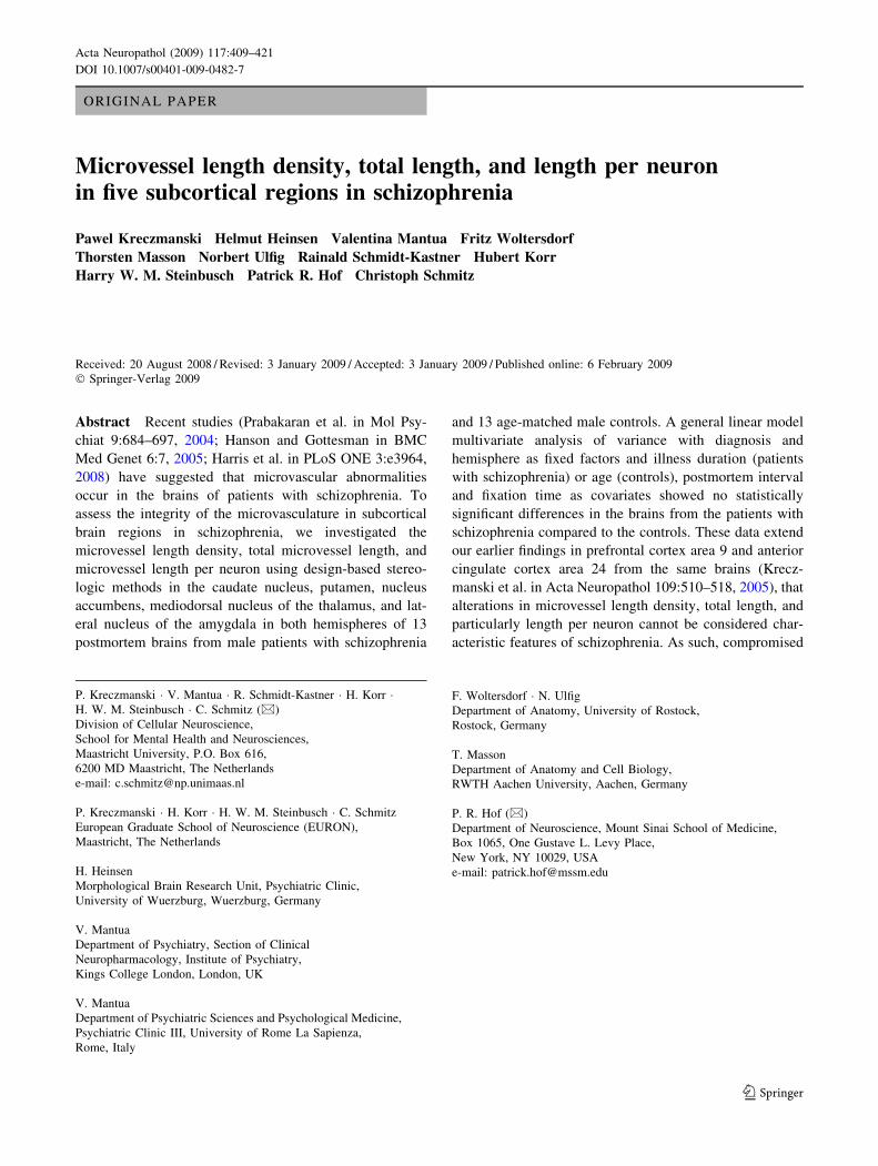
Fig. 1 Estimating microvessel length density with the space balls
method, illustrated for the caudate nucleus of control brain no. C4.
The section was immunoprocessed for the detection of collagen IV.
a Low-power overview of capillaries, superimposed by a rectangular
lattice with uniform distance between the lines in the X and Y axes.
This lattice defines the position of microscopic fields (indicated as
gray rectangles), at which the section is inspected at higher
magnification to perform space ball analyses. b High-power photo-
micrographs of one of these microscopic fields at the upper surface of
the section, at ten consecutive focal planes below the upper surface
with a distance of 4 lm between the focal planes, and at the lower
surface of the section that was found 43 lm below the upper surface.
Between -8 and -36 lm the microscopic field is superimposed by
intersections of a hemisphere (a semi-space ball) with the focal plane
at the investigated focal depth, illustrated as circles. The semi-space
ball was centered at a depth of -8 lm and had a radius of 30 lm.
Intersections between the semi-space ball and the microvessels in
focus were found at -12, -16, -24 and -28 lm (asterisks). The
average microvessel length density within the caudate nucleus was
calculated from the total number of intersections and the number and
size of the semi-space balls. Scale bar 700 lm in a, and 40 lm in the
high-power photomicrographs
Acta Neuropathol (2009) 117:409–421 413
123

regions of interest and the actual section thickness after
tissue processing (see also [45])
CLD ¼ 2�P
is� DX � DY � tð Þ2� p� r2
� 1
V
where CLD represents the microvessel length density, R is
the total number of intersections between the hemispheres
and the microvessels, DX and DY the distance between the
midpoints of the centers of the hemispheres in the X and Y
axes, t the actual average section thickness after histologic
processing, r the radius of the hemispheres and V the
investigated tissue volume. It is important to note that these
microvessel length density estimates were based on the
analysis of thick sections in three dimensions (3D) and
therefore not dependent on the plane of section, the spatial
arrangement of the microvessels or their diameter. To focus
the analysis on microvessels and to exclude arterioles and
venules, the diameter of all vessels coming into view
during the space ball analysis was measured with the ‘‘Fast
measure line’’ tool of the stereology software, and inter-
sections between the (space ball) hemispheres and vessels
with a diameter greater than 9 lm were not counted. In
addition, we always focused on the middle of a given
vessel’s diameter when recording intersections between the
space ball hemispheres and the microvessel in order to
minimize the space ball bias due to the non-zero diameter
of the vessels [25]. The actual section thickness after
immunohistochemical processing was measured at the
position of every tenth space ball, yielding on average
approximately 40 measures per section. Note that the
actual section thickness after immunohistochemical pro-
cessing (on average 45.7 lm) guaranteed that the space
balls were placed always fully within the section thickness.
Finally, total microvessel lengths and lengths per neuron
were calculated individually for each subcortical region as
the product of the microvessel length density in and the
volume of the region (total microvessel lengths), and as the
ratio of the total microvessel length and the total neuron
number in the region (microvessel lengths per neuron),
respectively. Volume and total neuron number data were
taken from [46]. Furthermore, it was tested whether there
was a principal correlation between the neuron density and
microvessel length density when all subcortical structures
were combined.
Statistical analysis
For both patients with schizophrenia and controls, mean
and standard error of the mean were calculated for all
investigated variables separately for the left and the right
hemispheres. Comparisons between patients with schizo-
phrenia and controls were performed using generalized
linear model multivariate analysis (MANOVA), with
diagnosis and hemisphere as fixed factors and the following
variables as covariates: (1) the adjusted illness duration of
the patients with schizophrenia (calculated as individual
age at death minus age at onset plus the mean age at onset
of all patients with schizophrenia) or the age of the con-
trols, respectively, (2) the postmortem interval and (3) the
fixation time [note that use of the actual individual illness
duration of the patients with schizophrenia instead of the
adjusted ones as covariate would have caused invalid
results of the MANOVA model because there was no ill-
ness duration of the controls, and the mean illness duration
of the patients with schizophrenia was significantly dif-
ferent from the mean age of the controls (Student’s two-
tailed t test; P \ 0.001) whereas the mean adjusted illness
duration was not (p = 0.974)]. For each investigated
variable all ilnvestigated brain regions were tested simul-
taneously. In addition, linear regression analysis was
carried out for all investigated variables as function of the
adjusted illness duration of the patients with schizophrenia
(or the age of the controls, respectively), as well as between
neuronal densities and microvessel length densities across
the five subcortical structures. Calculations were performed
using SPSS (Version 17.0.0 for Windows, SPSS, Chicago,
IL, USA) and GraphPad Prism (Version 5.00 for Windows,
GraphPad Software, San Diego, CA, USA).
Photography
Photomicrographs shown in Figs. 1 and 2 were produced
using an Olympus DP 70 digital camera attached to an
Olympus AX 70 microscope and cellP software Version 2.3
(Soft Imaging System, Munster, Germany). The final fig-
ures were constructed using Corel Photo-Paint v.11 and
Corel Draw v.11 (Corel, Ottawa, Canada). Adjustments of
contrast and brightness were made to facilitate recognition
of the immunohistochemical signal at low and high mag-
nification, without altering the appearance of the original
materials.
Results
No major abnormalities such as irregular size of the
vascular wall, stenosis, or dilation were noted in the immuno-
histochemical preparations for collagen IV in the materials
from the patients with schizophrenia (Fig. 2).
Compared to the controls, the patients with schizo-
phrenia showed no statistically significant differences in
any of the investigated variables (Figs. 3, 4, 5; Table 3
shows all P values of the MANOVAs performed). Fur-
thermore, except for the microvessel length density per
neuron in the right lateral nucleus of the amygdala of the
patients with schizophrenia (Fig. 5p), linear regression
414 Acta Neuropathol (2009) 117:409–421
123

analysis showed for none of the investigated variables a
significant deviation of the slope of the regression line from
zero or a significant deviation from linearity. The signifi-
cant linear relationship between the microvessel length
density per neuron in the right lateral nucleus of the
amygdala and the illness duration of the patients with
schizophrenia was not found anymore when removing the
data from the youngest patient. Accordingly, the micro-
vessel length density, microvessel length and microvessel
length per neuron did not depend on the illness duration of
the patients with schizophrenia (or the age of the controls,
respectively), and the significant linear relationship
between the microvessel length density per neuron in the
right lateral nucleus of the amygdala and the illness dura-
tion of the patients with schizophrenia was due to a single
outlier.
In addition, no correlation was found between the
microvessel length density and the neuron density in the five
investigated subcortical regions, neither in the patients with
schizophrenia nor in the controls (controls: r2 = 0.006;
p = 0.900; patients with schizophrenia: r2 = 0.054;
p = 0.708; Fig. 6).
Finally it should be mentioned that the results obtained
for the single brain embedded in celloidin (C7) showed no
systematic deviation from the results obtained for the other
brains (Figs. 3, 4, 5).
Discussion
The present data reveal no alterations in mean microvessel
length density and mean total microvessel length in five
subcortical regions in schizophrenia. Also, as the total
neuron numbers were assessed in the same subcortical
regions in this brain series [46], microvessel length per
neuron could be calculated. There were no changes in the
mean microvessel length per neuron in any of the five
subcortical structures in the brains from the patients with
schizophrenia, suggesting that modest changes in neuron
number are likely to be paralleled by minor vascular
changes. It should be noted that the results reported in the
present study do not reflect absolute values found in the
brain of living humans because postmortem brains show
shrinkage during formalin fixation. Accordingly, the results
presented here should only be used to interpret potential
differences between patients with schizophrenia and mat-
ched controls whose postmortem brains were processed
under exactly identical conditions. In this regard one could
speculate that postmortem brains from patients with
schizophrenia may shrink differently than postmortem
brains from controls, but there is no available evidence for
this in the literature.
The present study confirms and extends observations of
normal microvessel lengths in the prefrontal cortex area 9
and anterior cingulate cortex area 24 in schizophrenia [45].
These data do not support the hypothesis of alterations in
the microvasculature in chronic schizophrenia. Because
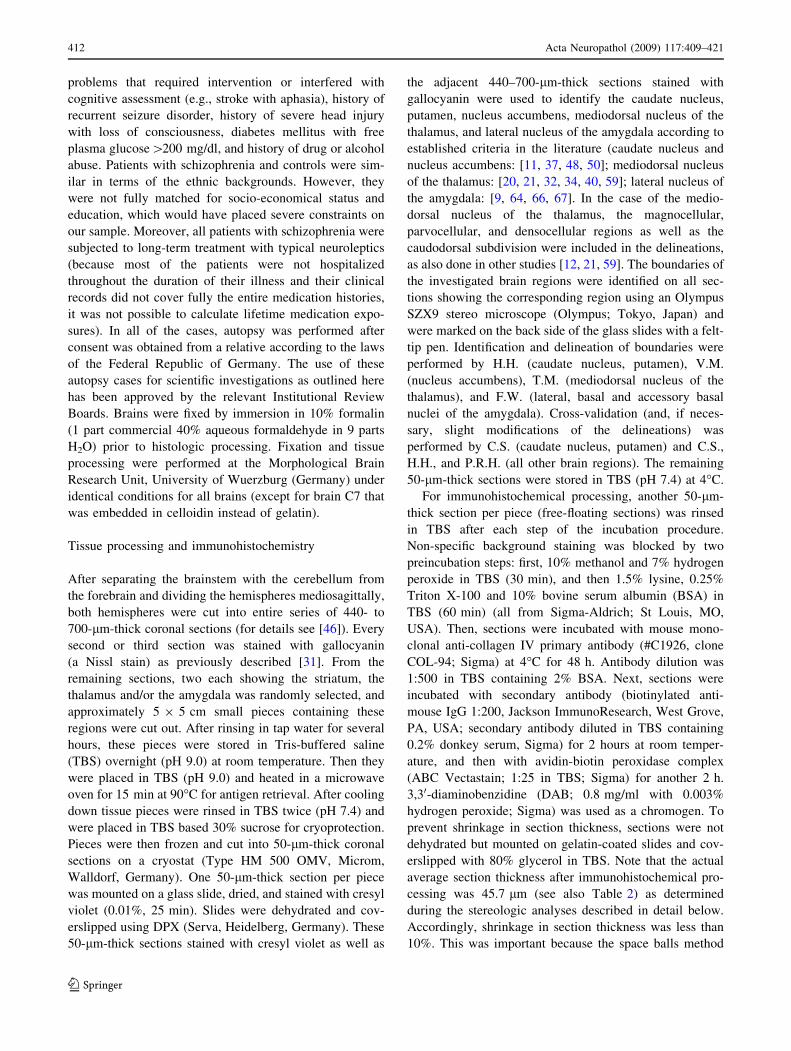
Fig. 2 Representative high-power photomicrographs of 50-lm-thick
coronal sections from the left brain half of a patient with
schizophrenia (a, c, e, g, i) and a control (b, d, f, h, k) showing
immunohistochemical detection of collagen IV in the caudate nucleus
(a, b), putamen (c, d), nucleus accumbens (e, f), mediodorsal nucleus
of the thalamus (g, h) and lateral nucleus of the amygdala (i, k). Note
that no major abnormalities such as irregular size of the vascular wall,
stenosis or dilation were noted in the immunohistochemical prepa-
rations for collagen IV in the materials from the patients with
schizophrenia. Furthermore, differences in microvessel length density
between patients with schizophrenia and controls were not obvious at
simple observation of the tissue but necessitated the type of rigorous
quantitative analysis undertaken in this study to be revealed. Scalebar 50 lm
Acta Neuropathol (2009) 117:409–421 415
123

these studies were performed in regions considered to
represent ‘‘hot spots’’ for structural pathology in schizo-
phrenia, the present negative outcome indicates that there
might be no anatomical substrate at the microvascular level
for abnormal rCBF and cerebral blood volume, alterations
reported in the brains of patients afflicted by schizophrenia
[1, 10, 16, 23, 33, 54, 74].
A survey of gene expression studies with microarrays in
the brain of patients with schizophrenia showed changes in
a few genes primarily associated with the vasculature,
although no consistent directional trends emerged [61].
A hypothesis paper suggested that genetic factors and
inflammatory responses of cerebral vessels could underlie
some of the changes seen in schizophrenia [26]. However,
Fig. 3 Microvessel length density in caudate nucleus (CN; a, f, l),putamen (P; b, g, m), nucleus accumbens (NA; c, h, n), mediodorsal
nucleus of the thalamus (MDNT; d, i, o) and lateral nucleus of the
amygdala (LNA; e, k, p) in both hemispheres of the brains from 13
male patients with schizophrenia (S; open bars in a–e, and dots in
f–p) and 13 age-matched controls (C; closed bars in a–e, and dots in
f–p; squares in case of the results obtained for the single brain
embedded in celloidin [C7]). In a–e, data are shown as mean and
standard error of the mean for the left (l) and right (r) hemispheres
from patients with schizophrenia (S-l and S-r) and controls (C-l and
C-r). In f–p, individual data for the left hemispheres (closed dots or
squares, respectively) and right hemispheres (open dots or squares,
respectively) from controls (f–k) and patients with schizophrenia (l–p) are shown as function of the control patients’ age (or the illness
duration of the patients with schizophrenia, respectively). *p \ 0.05
for the fixed factor diagnosis in general linear model multivariate
analysis of variance (MANOVA)
416 Acta Neuropathol (2009) 117:409–421
123

the data of the present study altogether do not support the
hypothesis of involvement of brain microvessels that would
be otherwise expected if a major inflammatory process
took place in schizophrenia. Because only vessels with a
maximal diameter of 9 lm were counted in the present
study, subtle abnormalities may still occur upstream in
arterioles, involving vascular smooth muscle cells and
regulation of tissue perfusion. Indeed, changes in the
activity-dependent regulation of rCBF have been observed
in the prefrontal cortex in schizophrenia [23, 74]. However,
hemodynamic responses appear to be normal in visual,
motor, and somatosensory cortices in this disorder [4]. It is
unlikely that primary differences in the structure and
function of arterioles occur selectively in some cortical
Fig. 4 Microvessel length in caudate nucleus (CN; a, f, l), putamen
(P; b, g, m), nucleus accumbens (NA; c, h, n), mediodorsal nucleus of
the thalamus (MDNT; d, i, o) and lateral nucleus of the amygdala
(LNA; e, k, p) in both hemispheres of the brains from 13 male patients
with schizophrenia (S; open bars in a–e, and dots in f–p) and 13 age-
matched controls (C; closed bars in a–e, and dots in f–p; squares in
case of the results obtained for the single brain embedded in celloidin
[C7]). In a–e, data are shown as mean and standard error of the mean
for the left (l) and right (r) hemispheres from patients with
schizophrenia (S-l and S-r) and controls (C-l and C-r). In f–p,
individual data for the left hemispheres (closed dots or squares,
respectively) and right hemispheres (open dots or squares, respec-
tively) from controls (f–k) and patients with schizophrenia (l–p) are
shown as function of the control patients’ age (or the illness duration
of the patients with schizophrenia, respectively). *p \ 0.05 for the
fixed factor diagnosis in general linear model multivariate analysis of
variance (MANOVA)
Acta Neuropathol (2009) 117:409–421 417
123

regions, and therefore changes in rCBF activation most
likely are due to region-specific neuronal or glial factors. In
addition, it appears that changes in gene and protein
expression found in cortical samples from chronic schizo-
phrenia patients probably reflect an abnormal metabolic
state [60]. This could be equally due to the primary disease
process and to secondary long-term neuronal dysfunction,
chronic medication, or imbalances in agonal events influ-
encing microarray measurements [39, 52, 70, 72]. It
remains to be shown whether the microvasculature of the
white matter in schizophrenia is normal, as several recent
findings point to subtle white matter changes in
Fig. 5 Microvessel length per neuron in caudate nucleus (CN; a, f, l),putamen (P; b, g, m), nucleus accumbens (NA; c, h, n), mediodorsal
nucleus of the thalamus (MDNT; d, i, o) and lateral nucleus of the
amygdala (LNA; e, k, p) in both hemispheres of the brains from 13
male patients with schizophrenia (S; open bars in a–e, and dotsin f–p) and 13 age-matched controls (C; closed bars in a–e, and dotsin f–p; squares in case of the results obtained for the single brain
embedded in celloidin [C7]). In a–e, data are shown as mean and
standard error of the mean for the left (l) and right (r) hemispheres
from patients with schizophrenia (S-l and S-r) and controls (C-l and
C-r). In f–p, individual data for the left hemispheres (closed dots or
squares, respectively) and right hemispheres (open dots or squares,
respectively) from controls (f–k) and patients with schizophrenia
(l–p) are shown as function of the control patients’ age (or the illness
duration of the patients with schizophrenia, respectively). The solidline in p indicates a statistically significant linear relationship between
the microvessel length density per neuron in the right lateral nucleus
of the amygdala and the illness duration of the patients with
schizophrenia
418 Acta Neuropathol (2009) 117:409–421
123

schizophrenia [19, 35, 36], and in view of the vulnerability
of the white matter to prolonged hypoperfusion [22]. Fur-
thermore, an abnormal decrease in perivascular labeling of
GFAP in astrocytes has been reported in the white matter in
schizophrenia [73].
As a side observation it is of interest that the neuron
density was not correlated with the microvessel length
density when five subcortical regions were combined for
analysis in controls or in patients with schizophrenia.
Baborie and Kuschinksy [3] did not find a correlation
between the density of all nuclei and microvessel densities
across multiple areas of the rat brain. It is likely that the
microvessel length density is linked to metabolic expen-
ditures in neuronal and glial processes whereby neuron
density (measured in the present study) or overall cell
density (measured in [3]) do not tightly correlate with the
total amount of functional processes in each structure.
As all patients were treated with neuroleptics, we
cannot exclude that the lack of changes in microvessel
measurements was due to the treatment; in other words,
untreated patients with schizophrenia could have an
abnormal microvasculature. However, the influence of
neuroleptics on brain microvessels in chronically treated
patients with schizophrenia is presently unknown. Dopa-
minergic processes make close contacts with capillaries in
the prefrontal cortex and dopamine reduces local perfusion
[47]. Dopamine has been shown to have anti-angiogenic
properties in peripheral tissues, tumors, and placenta [5, 44,
68]. Thus, one cannot exclude that abnormal dopamine
levels in schizophrenia may have functional and structural
effects on cerebral microvasculature before the onset of
clinical symptoms that may be masked by chronic dopa-
mine receptor blockade with neuroleptics. Further research
will be necessary in this regard.
Finally, it should be mentioned that the present obser-
vations in chronic schizophrenia cases must be separated
from the concept that abnormal interactions between
hypoxia, the microvasculature, and developing neurons
could be important for the neurodevelopmental perturba-
tions that are thought to underlie schizophrenia [61].
Specifically, one cannot exclude the possible occurrence of
alterations in microvessel length and microvessel length
per neuron in the fetal brain of people who will develop
schizophrenia later in life. If this were the case, however,
these alterations do not persist during postnatal life in
patients with schizophrenia. Rather, compromised brain
metabolism and occurrence of oxidative stress in the brains
from patients with schizophrenia are likely caused by other
mechanisms. These are important issues that need to be
addressed in specific studies. It remains possible that more
subtle alterations could compromise the blood-brain barrier
or alter certain cellular components of the microvessels,
which in turn may contribute to possible disruption in the
Table 3 Results of statistical analysis (p values) with generalized
linear model multivariate analysis of variance (MANOVA)
Variable A/ID PMI Fix D H D 9 H
Microvessel length
density
0.660 0.817 0.876 0.146 0.168 0.564
Total microvessel length 0.379 0.259 0.319 0.283 0.372 0.985
Microvessel length per
neuron
0.195 0.710 0.503 0.528 0.415 0.996
A/ID age (controls) or adjusted illness duration (patients with
schizophrenia), respectively (adjusted illness duration calculated as
age at death minus age at onset plus mean age at onset, details are
provided in ‘‘Statistical analysis’’); PMI postmortem interval (time
between death and autopsy); Fix fixation time; D diagnosis;
H hemisphere
Fig. 6 Neuron density (ND) in the caudate nucleus (CN), putamen
(P), nucleus accumbens (NA), mediodorsal nucleus of the thalamus
(MDNT) and lateral nucleus of the amygdala (LNA) as a function of
microvessel length density (CLD) in these subcortical regions in both
hemispheres of postmortem brains from 13 male controls (a) and
13 male patients with schizophrenia (b). Region-specific values
for microvessel length density and neuron density are shown as
mean ± standard error of the mean (SEM). Linear regression analysis
showed no significant correlation between the microvessel length
density and the neuron density in these subcortical regions (controls:
r2 = 0.006; p = 0.900; patients with schizophrenia: r2 = 0.054;
p = 0.708). The regression lines and their 95% confidence intervals
are also shown in the graphs
Acta Neuropathol (2009) 117:409–421 419
123

coupling of cerebral blood flow to neuronal metabolic
needs in schizophrenia [26].
Acknowledgments We thank E.K. Broschk and H. Steinbusch for
expert technical assistance, Dr. M. Schuler (Bayreuth, Germany) for
valuable help in retrieving clinical data of the investigated patients, and
G. Greene and B. Boehringer (MicroBrightField, Williston, VT, USA)
for valuable help in implementing the space balls method. This work
was supported by the Stanley Medical Research Institute (#02R-258,
#04R-674 to HH, PRH, and CS), the European Community (Quality of
Life and Management of Living Resources, QLK6-CT-2000-60042,
QLK6-GH-00-60042-46, to PK), and NIH grant MH66392 to PRH.
References
1. Andreasen NC, Rezai K, Alliger R et al (1992) Hypofrontality in
neuroleptic-naive patients and in patients with chronic schizo-
phrenia. Assessment with xenon 133 single-photon emission
computed tomography and the Tower of London. Arch Gen
Psychiatry 49:943–958
2. Arnold SE, Trojanowski JQ (1996) Recent advances in defining
the neuropathology of schizophrenia. Acta Neuropathol 92:217–
231
3. Baborie A, Kuschinksy W (2006) Lack of relationship between
cellular density and either capillary density or metabolic rate in
different regions of the brain. Neurosci Lett 404:20–22
4. Barch DM, Mathews JR, Buckner RL et al (2003) Hemodynamic
responses in visual, motor, and somatosensory cortices in
schizophrenia. Neuroimage 20:1884–1893
5. Basu S, Nagy JA, Pal S et al (2001) The neurotransmitter
dopamine inhibits angiogenesis induced by vascular permeability
factor/vascular endothelial growth factor. Nat Med 7:569–574
6. Beckmann H, Lauer M (1997) The human striatum in schizo-
phrenia. II. Increased number of striatal neurons in
schizophrenics. Psychiatry Res 68:99–109
7. Berman KF, Zec RF, Weinberger DR (1986) Physiologic dys-
function of dorsolateral prefrontal cortex in schizophrenia. II.
Role of neuroleptic treatment, attention, and mental effort. Arch
Gen Psychiatry 43:126–135
8. Bogerts B (1984) Zur Neuropathologie der Schizophrenien.
Fortschr Neurol Psychiatr 52:428–437
9. Braak H, Braak E (1983) Neuronal types in the basolateral
amygdaloid nuclei of man. Brain Res Bull 11:349–365
10. Brambilla P, Cerini R, Fabene PF et al (2007) Assessment of
cerebral blood volume in schizophrenia: a magnetic resonance
imaging study. J Psychiatr Res 41:502–510
11. Brockhaus H (1942) Zur feineren Anatomie des Septum und des
Striatum. J Psychol Neurol 5:1–56
12. Byne W, Buchsbaum MS, Mattiace LA et al (2002) Postmortem
assessment of thalamic nuclear volumes in subjects with
schizophrenia. Am J Psychiatry 159:59–65
13. Calhoun ME, Mouton PR (2000) Length measurement: new
developments in neurostereology and 3D imagery. J Chem
Neuroanat 21:257–265
14. Casanova MF, de Zeeuw L, Switala A et al (2005) Mean cell
spacing abnormalities in the neocortex of patients with schizo-
phrenia. Psychiatry Res 133:1–12
15. Catafau AM, Parellada E, Lomena FJ et al (1994) Prefrontal and
temporal blood flow in schizophrenia: resting and activation
Technetium-99 m-HMPAO-SPECT patterns in young neurolep-
tic-naive patients with acute disease. J Nucl Med 35:935–941
16. Cohen BM, Yurgelun-Todd D, English CD et al (1995) Abnor-
malities of regional distribution of cerebral vasculature in
schizophrenia detected by dynamic susceptibility contrast MRI.
Am J Psychiatry 152:1801–1803
17. Cullen TJ, Walker MA, Parkinson N et al (2003) A postmortem
study of the mediodorsal nucleus of the thalamus in schizo-
phrenia. Schizophr Res 60:157–166
18. Danos P, Schmidt A, Baumann B et al (2005) Volume and neuron
number of the mediodorsal thalamic nucleus in schizophrenia: a
replication study. Psychiatry Res 140:281–289
19. Davis KL, Stewart DG, Friedman JI et al (2003) White matter
changes in schizophrenia: evidence for myelin-related dysfunc-
tion. Arch Gen Psychiatry 60:443–456
20. Dewulf A (1971) Anatomy of the normal human thalamus: top-
ometry and standardized nomenclature. Elsevier, Amsterdam
21. Dorph-Petersen KA, Pierri JN, Sun Z et al (2004) Stereological
analysis of the mediodorsal thalamic nucleus in schizophrenia:
volume, neuron number, and cell types. J Comp Neurol 472:449–
462
22. Farkas E, Donka G, de Vos RA et al (2004) Experimental cere-
bral hypoperfusion induces white matter injury and microglial
activation in the rat brain. Acta Neuropathol 108:57–64
23. Franzen G, Ingvar DH (1975) Absence of activation in frontal
structures during psychological testing of chronic schizophrenics.
J Neurol Neurosurg Psychiatry 38:1027–1032
24. Gundersen HJG (2002) Stereological estimation of tubular length.
J Microsc 207:155–160
25. Hakak Y, Walker JR, Li C et al (2001) Genome-wide expression
analysis reveals dysregulation of myelination-related genes in
chronic schizophrenia. Proc Natl Acad Sci USA 98:4746–4751
26. Hanson DR, Gottesman II (2005) Theories of schizophrenia: a
genetic-inflammatory-vascular synthesis. BMC Med Genet 6:7
27. Harris LW, Wayland M, Lan M et al (2008) The cerebral
microvasculature in schizophrenia: a laser capture microdissec-
tion study. PLoS ONE 3:e3964
28. Harrison PJ (1999) The neuropathology of schizophrenia. A critical
review of the data and their interpretation. Brain 122:593–624
29. Harrison PJ, Owen MJ (2003) Genes for schizophrenia? Recent
findings and their pathophysiological implications. Lancet
361:417–419
30. Harrison PJ, Weinberger DR (2005) Schizophrenia genes, gene
expression, and neuropathology: on the matter of their conver-
gence. Mol Psychiatry 10:40–68
31. Heinsen H, Heinsen YL (1991) Serial thick, frozen, gallocyanin
stained sections of human central nervous system. J Histotechnol
14:167–173
32. Heinsen H, Rub U, Bauer M et al (1999) Nerve cell loss in the
thalamic mediodorsal nucleus in Huntington’s disease. Acta
Neuropathol 97:613–622
33. Hill K, Mann L, Laws KR et al (2004) Hypofrontality in
schizophrenia: a meta-analysis of functional imaging studies.
Acta Psychiatr Scand 110:243–256
34. Hirai T, Jones EG (1989) A new parcellation of the human
thalamus on the basis of histochemical staining. Brain Res Rev
14:1–34
35. Hof PR, Haroutunian V, Copland C et al (2002) Molecular and
cellular evidence for an oligodendrocyte abnormality in schizo-
phrenia. Neurochem Res 27:1193–1200
36. Hof PR, Haroutunian V, Friedrich VL Jr et al (2003) Loss and
altered spatial distribution of oligodendrocytes in the superior
frontal gyrus in schizophrenia. Biol Psychiatry 53:1075–1085
37. Holt DJ, Herman MM, Hyde TM et al (1999) Evidence for a
deficit in cholinergic interneurons in the striatum in schizophre-
nia. Neuroscience 94:21–31
38. Ingvar DH, Franzen G (1974) Distribution of cerebral activity in
chronic schizophrenia. Lancet 2:1484–1486
39. Iwamoto K, Bundo M, Kato T (2005) Altered expression of
mitochondria-related genes in postmortem brains of patients with
420 Acta Neuropathol (2009) 117:409–421
123

bipolar disorder or schizophrenia, as revealed by large-scale
DNA microarray analysis. Hum Mol Genet 14:241–253
40. Jones EG (1997) A description of the human thalamus. In:
Steriade M, Jones EG, McCormick DA (eds) Thalamus, vol II.
Experimental and clinical aspects. Elsevier Science, Oxford,
pp 425–500
41. Kety SS (1959) Biochemical theories of schizophrenia. I. Science
129:1528–1532
42. Kety SS (1959) Biochemical theories of schizophrenia. II. Sci-
ence 129:1590–1596
43. Khaitovich P, Lockstone HE, Wayland MT et al (2008) Meta-
bolic changes in schizophrenia and human brain evolution.
Genome Biol 9:R124
44. Kim HJ, Koh PO, Kang SS et al (2001) The localization
of dopamine D2 receptor mRNA in the human placenta and the
anti-angiogenic effect of apomorphine in the chorioallantoic
membrane. Life Sci 68:1031–1040
45. Kreczmanski P, Schmidt-Kastner R, Heinsen H et al (2005)
Stereological studies of capillary length density in the frontal
cortex of schizophrenics. Acta Neuropathol 109:510–518
46. Kreczmanski P, Heinsen H, Mantua V et al (2007) Volume,
neuron density, and total neuron number in five subcortical
regions in schizophrenia. Brain 130:678–692
47. Krimer LS, Muly EC 3rd, Williams GV et al (1998) Dopami-
nergic regulation of cerebral cortical microcirculation. Nat
Neurosci 1:286–289
48. Lauer M, Heinsen H (1996) Cytoarchitectonics of the human
nucleus accumbens. J Hirnforsch 37:243–254
49. Lauer M, Beckmann H (1997) The human striatum in schizo-
phrenia. I. Increase in overall relative striatal volume in
schizophrenics. Psychiatry Res 68:87–98
50. Lauer M, Senitz D, Beckmann H (2001) Increased volume of the
nucleus accumbens in schizophrenia. J Neural Transm 108:645–
660
51. Lewis DA, Lewitt P (2002) Schizophrenia as a disorder of neu-
rodevelopment. Annu Rev Neurosci 25:409–432
52. Li JZ, Vawter MP, Walsh DM et al (2004) Systematic changes in
gene expression in postmortem human brains associated with
tissue pH and terminal medical conditions. Hum Mol Genet
13:609–616
53. Malaspina D, Harkavy-Friedman J, Corcoran C et al (2004)
Resting neural activity distinguishes subgroups of schizophrenia
patients. Biol Psychiatry 56:931–937
54. Meyer-Lindenberg A, Miletich RS, Kohn PD et al (2002)
Reduced prefrontal activity predicts exaggerated striatal dopa-
minergic function in schizophrenia. Nat Neurosci 5:267–271
55. Middleton FA, Mirnics K, Pierri JN et al (2002) Gene expression
profiling reveals alterations of specific metabolic pathways in
schizophrenia. J Neurosci 22:2718–2729
56. Mouton PR, Gokhale AM, Ward NL et al (2002) Stereological
length estimation using spherical probes. J Microsc 206:54–64
57. Mueser KT, McGurk SR (2004) Schizophrenia. Lancet 363:2063–
2072
58. Pakkenberg B (1990) Pronounced reduction of total neuron
number in mediodorsal thalamic nucleus and nucleus accumbens
in schizophrenics. Arch Gen Psychiatry 47:1023–1028
59. Popken GJ, Bunney WE, Potkin SG et al (2000) Subnucleus-
specific loss of neurons in medial thalamus of schizophrenics.
Proc Natl Acad Sci USA 97:9276–9280
60. Prabakaran S, Swatton JE, Ryan MM et al (2004) Mitochondrial
dysfunction in schizophrenia: evidence for compromised brain
metabolism and oxidative stress. Mol Psychiatry 9:684–697
61. Schmidt-Kastner R, van Os J, WM Steinbusch H et al (2006)
Gene regulation by hypoxia and the neurodevelopmental origin of
schizophrenia. Schizophr Res 84:253–271
62. Schmitz C, Hof PR (2005) Design-based stereology in neuro-
science. Neuroscience 130:813–831
63. Schultz SK, O’Leary DS, Boles Ponto LL et al (2002) Age and
regional cerebral blood flow in schizophrenia: age effects in
anterior cingulate, frontal, and parietal cortex. J Neuropsychiatry
Clin Neurosci 14:19–24
64. Schumann CM, Amaral DG (2005) Stereological estimation of
the number of neurons in the human amygdaloid complex.
J Comp Neurol 491:320–329
65. Siever LJ, Davis KL (2004) The pathophysiology of schizophrenia
disorders: perspectives from the spectrum. Am J Psychiatry
161:398–413
66. Sims KS, Williams RS (1990) The human amygdaloid complex:
a cytologic and histochemical atlas using Nissl, myelin, acetyl-
cholinesterase and nicotinamide adenine dinucleotide phosphate
diaphorase staining. Neuroscience 36:449–472
67. Sorvari H, Soininen H, Pitkanen A (1996) Calbindin-D28K-
immunoreactive cells and fibres in the human amygdaloid com-
plex. Neuroscience 75:421–443
68. Teunis MA, Kavelaars A, Voest E (2002) Reduced tumor
growth, experimental metastasis formation, and angiogenesis in
rats with a hyperreactive dopaminergic system. FASEB J 16:1465–
1467
69. Tkachev D, Mimmack ML, Ryan MM et al (2003) Oligoden-
drocyte dysfunction in schizophrenia and bipolar disorder. Lancet
362:798–805
70. Tomita H, Vawter MP, Walsh DM et al (2004) Effect of agonal
and postmortem factors on gene expression profile: quality con-
trol in microarray analyses of postmortem human brain. Biol
Psychiatry 55:346–352
71. Tsuang M (2000) Schizophrenia: genes and environment. Biol
Psychiatry 47:210–220
72. Vawter MP, Tomita H, Meng F et al (2006) Mitochondrial-
related gene expression changes are sensitive to agonal-pH state:
implications for brain disorders. Mol Psychiatry 615:663–679
73. Webster MJ, Knable MB, Johnston-Wilson N et al (2001)
Immunohistochemical localization of phosphorylated glial
fibrillary acidic protein in the prefrontal cortex and hippocampus
from patients with schizophrenia, bipolar disorder, and depres-
sion. Brain Behav Immunol 15:388–400
74. Weinberger DR, Berman KF, Zec RF (1986) Physiologic dys-
function of dorsolateral prefrontal cortex in schizophrenia. I.
Regional cerebral blood flow evidence. Arch Gen Psychiatry
43:114–124
75. Young KA, Manaye KF, Liang C et al (2000) Reduced number of
mediodorsal and anterior thalamic neurons in schizophrenia. Biol
Psychiatry 47:944–953
Acta Neuropathol (2009) 117:409–421 421
123