nucleic acids in chemistry and biology - citeseerx
TRANSCRIPT

Nucleic Acids in Chemistry and Biology
3rd Edition

Nucleic Acids in Chemistryand Biology3rd Edition
Edited by
G. Michael BlackburnCentre for Chemical Biology, Department of Chemistry, University of Sheffield, Sheffield, UK
Michael J. GaitMedical Research Council, Laboratory of Molecular Biology, Cambridge, UK
David LoakesMedical Research Council, Laboratory of Molecular Biology, Cambridge, UK
David M. WilliamsCentre for Chemical Biology, Department of Chemistry, University of Sheffield, Sheffield, UK

ISBN-10: 0-85404-654-2ISBN-13: 978-0-85404-654-6
A catalogue record for this book is available from the British Library
© The Royal Society of Chemistry 2006
All rights reserved
Apart from fair dealing for the purposes of research for non-commercial purposes or for private study, criticismor review, as permitted under the Copyright, Designs and Patents Act 1988 and the Copyright and Related RightsRegulations 2003, this publication may not be reproduced, stored or transmitted, in any form or by any means,without the prior permission in writing of The Royal Society of Chemistry, or in the case of reproduction inaccordance with the terms of licences issued by the Copyright Licensing Agency in the UK, or in accordance withthe terms of the licences issued by the appropriate Reproduction Rights Organization outside the UK. Enquiriesconcerning reproduction outside the terms stated here should be sent to The Royal Society of Chemistry at theaddress printed on this page.
Published by The Royal Society of Chemistry,Thomas Graham House, Science Park, Milton Road,Cambridge CB4 0WF, UK
Registered Charity Number 207890
For further information see our web site at www.rsc.org
Typeset by Macmillan India Ltd, Bangalore, IndiaPrinted by Henry Ling Ltd, Dorchester, Dorset, UK
Foreword
It was just 62 years ago that we finally learned that DNA was the genetic material – the master blueprintof life. Since then, the nucleic acids DNA and RNA have been studied in exquisite detail and both theirchemical and biochemical properties are firmly established. Indeed, the double helical structure of DNAhas become an icon of our time appearing widely not only in the scientific literature, but also in the popu-lar press and most recently as jewelry. A thorough knowledge of nucleic acids and their properties is nowa key ingredient in the education of both biologists and chemists. Ten years ago the second edition of“Blackburn & Gait” was published and seemed sufficiently comprehensive that only small additionswould be needed if it were ever to be rewritten. Its popularity is attested to by its now being out of print –it has also inevitably become out of date. Much has changed in the last 10 years and a new edition is nowboth necessary and most welcome.
One major discovery within the biological arena has been the phenomenon of RNA interference, whichwas not even mentioned in the last edition, and yet at this time several companies have been formed to cap-italize on it and at least one product is heading into clinical trials. We also now know that short micro-RNAs play key roles in development and are probably of ubiquitous importance in controlling geneexpression. These and other small RNAs are likely to play a much more critical and subtle role in the livesof cells than we might ever have imagined. I find this personally very satisfying, since, when we discov-ered split genes and RNA splicing in 1977, the introns were almost immediately labeled “junk”. It nowseems that at least some of these intronic sequences play positive roles in controlling gene expression andtheir involvement in other processes may still await discovery. Studies of small RNAs in eukaryotes areproceeding quickly and I eagerly await the results from similar studies in bacteria and archaea. It seemslikely that great discoveries lie ahead although new methods may be required to make them. The develop-ment of such methods will be greatly facilitated by a thorough knowledge of the chemistry and biology ofnucleic acids – the subject of this book.
Among the great technical achievements of the last 10 years have been several breakthroughs in thescale of DNA sequencing. First came the complete sequence of a simple bacterium, Haemophilus influen-zae, quickly followed by that of the first archaea, Methanocaldococcus jannaschii. A key feature of theseprojects was the use whole-genome shotgun sequencing pioneered by Craig Venter. These “small” genomeswere soon followed by draft sequences for a number of eukaryotic genomes including, of course, the drafthuman genome sequence announced in 2003 and coinciding with the 50th anniversary of the determina-tion of the structure of DNA by Jim Watson and Francis Crick. With more recent advances in sequencingtechnologies that use highly parallel methodology, one machine can now generate enough data for a smallbacterial genome in a few hours, at a quite reasonable price. We can anticipate an even more massiveinflux of new data in the next few years. The accumulation of sequence data far exceeds our experimentalcapacity to probe it. Fortunately, bioinformatics stands ready to help and with appropriate experimentalinput, should allow us to make sense of the terabases (1012) of DNA sequence data that will soon be pres-ent in GenBank. In parallel with these improvements in DNA sequence determination, techniques forDNA synthesis have progressed rapidly. It has now become so simple and inexpensive that many labora-tories find it more expedient to have the genes of interest synthesized rather than to clone them. Amongother things, this allows the introduction of desirable codons tailored to the expression system to be used.

vi Foreword
All of this new work serves to highlight the intertwining of chemistry and biology that has taken place overthe last 50 years. Those wishing to understand this interrelationship and appreciate the excitement cur-rently present in the field can do no better than browse the many excellent chapters in this third edition ofNucleic Acids in Chemistry and Biology.
Richard J. Roberts
Preface
The first edition of Nucleic Acids in Chemistry and Biology in 1990 met the pressing need for a single vol-ume that integrated the chemistry and biology of nucleic acids in an introductory yet authoritative text.That book was so very well received that in 1996 we produced the second edition, which was completelyrevised and rewritten by very much the same team of international experts.
Ten years on we have responded to the still growing need for this book with a fully revised and updatedthird edition. Two irresistible pressures have driven this activity. First, the expansion in the chemistry andbiology of nucleic acids continues unabated. The human and numerous lesser genomes have been fullysequenced since we presented the second edition and there has been a veritable explosion in the chemistryand biology of RNA. Many exciting crystal and NMR structures of nucleic acids and their protein com-plexes, including the ribosome, have been published. Changes of such magnitude have inevitably madesignificant parts of the 1996 text out of date. We have addressed these issues by expansion of the appro-priate sections of the book and also by new authorship. Second, the second edition sold out several yearsago. Indeed second-hand copies are occasionally available on the web at a handsome premium!
In planning this third edition, we first expanded the team of editors to include two younger colleagues,David Loakes and David Williams. We then changed publishing house to move under the roof of the RoyalSociety of Chemistry. For a variety of reasons it has been necessary to make changes to the team of prin-cipal authors and we thank especially Stephanie Allen, Martin Egli, Julie Fisher, Andy Flavell, IhtshamulHaq, Charles Laughton, Ben Luisi, Anna Marie Pyle, Elliott Stollar, and Nick Williams for their essentialand scholarly contributions. With the active support of the Royal Society of Chemistry and its commis-sioning and production teams, we have made significant changes in the style of presentation of this newedition. It now has a bibliography of primary and secondary sources that are referenced throughout thetext. While we have maintained a number of multi-colour illustrations in addition to our standard two-colour format, we have abandoned the use of stereo-pair illustrations and the end-of-section summaries.These changes have created space for some expansion – but not enough for our needs: the third edition hasgrown substantially compared to its predecessor! This has enabled the authors to introduce a great deal ofnew material. In doing so, we have retained the essential core of chemistry and biology that has made thisbook so effective as a teaching resource at every level of study and an initiation into the molecular basicsof nucleic acids. A selection of figures that may have value for course teachers are available electronicallyat the following website: http://www.rsc.org/books/nucleicacids
Above all, we have endeavoured to maintain the quality of the earlier editions, both of which have beenwidely appreciated for their easy readability, simplicity of exposition, clarity of illustration, and uniformityof style. That has underpinned our efforts to deliver a new edition that once again fulfils the needs of studentsand new research workers, primarily those having a chemical and biochemical background who seek tounderstand this great subject at a molecular level. Indeed, we know that Nucleic Acids in Chemistry and

Biology has become the course-book of choice in universities across three continents. At the same time, frommany favourable comments on editions 1 and 2 we know that this book has also reached out to more seniorscientists across many disciplines.
G Michael BlackburnMichael J GaitDavid Loakes
David M Williams
viii Preface
Acknowledgements
Two Mikes and two Davids are extremely grateful for the efforts of all who have supported the productionof this book. Our unqualified thanks are given above all to our 10 expert and understanding co-authors,whose contributions have made possible this third edition. We express our sincere appreciation of theirpatience, tolerance, and enthusiastic diligence during the numerous revision processes required for theproduction of the completed work.
We are also very grateful to very many colleagues and fellow scientists who have provided us with valu-able comments on the first two editions of the text and especially those who have read portions of the newedition. They include Jason Betley, Chris Calladine, Rick Cosstick, Steve Fodor, Dan Gewirth, AlecJeffreys, David Lilley, Kiyoshi Nagai, Barbara Nawrot, Frank Seela, Jean Thomas, Andrew Travers, andDavid Wilson. We are once again indebted to Joachim Engels for updating and expanding the glossary andto Rich Roberts for writing the forward for this edition.
The technical production of this book has been enabled by many skilled individuals. We particularlyappreciate the efforts of all the staff involved in the Royal Society of Chemistry for their enthusiasm,patience, and highly professional production of the completed work. We and our co-authors gratefullyacknowledge the efforts of Fred Anston, John Brazier, Pat Mellor, Sabuj Pattanayek, and Wenke Zhangwho have facilitated the completion of figures and text in various chapters. In particular we are greatlyindebted to Annette Lenton who has redrawn, recoloured, or reworked very many of the figures in order toachieve a homogeneous standard and style. We thank Venki Ramakrishnan for providing original graphicsfor the cover of the third edition of the book and Richard Dickerson, Stephen Lippard, and Dinshaw Patelfor illustrative figures.
Last but not least, we have enjoyed receiving a large number of positive and helpful comments fromreaders of the first two editions. We have endeavoured to incorporate all constructive criticisms into thenew edition. In particular we thank colleagues around the world whose strong support provided muchmotivation for the creation of this third edition.
Despite all our careful work, it is inevitable that there will be some errors and we accept full responsi-bility for them. Finally, we look forward to your advice, comments, and suggestions for future revisions.


Contents
Glossary xxi
Chapter 1Introduction and Overview 1
1.1 The Biological Importance of DNA 11.2 The Origins of Nucleic Acids Research 21.3 Early Structural Studies on Nucleic Acids 21.4 The Discovery of the Structure of DNA 41.5 The Advent of Molecular Biology 71.6 The Partnership of Chemistry and Biology 81.7 Frontiers in Nucleic Acids Research 10
References 11
Chapter 2DNA and RNA Structure 13
2.1 Structures of Components 142.1.1 Nucleosides and Nucleotides 142.1.2 Physical Properties of Nucleosides and Nucleotides 162.1.3 Spectroscopic Properties of Nucleosides and Nucleotides 192.1.4 Shapes of Nucleotides 20
2.2 Standard DNA Structures 242.2.1 Primary Structure of DNA 242.2.2 Secondary Structure of DNA 242.2.3 A-DNA 272.2.4 The B-DNA Family 302.2.5 Z-DNA 31
2.3 Real DNA Structures 332.3.1 Sequence-Dependent Modulation of DNA Structure 332.3.2 Mismatched Base–Pairs 362.3.3 Unusual DNA Structures 382.3.4 B–Z Junctions and B–Z Transitions 452.3.5 Circular DNA and Supercoiling 462.3.6 Triple-Stranded DNA 492.3.7 Other Non-Canonical DNA Structures 52

xii Contents
2.4 Structures of RNA Species 552.4.1 Primary Structure of RNA 562.4.2 Secondary Structure of RNA: A-RNA and A�-RNA 572.4.3 RNA�DNA Duplexes 592.4.4 RNA Bulges, Hairpins and Loops 612.4.5 Triple-Stranded RNAs 64
2.5 Dynamics of Nucleic Acid Structures 642.5.1 Helix-Coil Transitions of Duplexes 642.5.2 DNA Breathing 662.5.3 Energetics of the B–Z Transition 672.5.4 Rapid DNA Motions 68
2.6 Higher-Order DNA Structures 682.6.1 Nucleosome Structure 682.6.2 Chromatin Structure 69References 72
Chapter 3Nucleosides and Nucleotides 77
3.1 Chemical Synthesis of Nucleosides 773.1.1 Formation of the Glycosylic Bond 793.1.2 Building the Base onto a C-1 Substituent of the Sugar 873.1.3 Synthesis of Acyclonucleosides 903.1.4 Syntheses of Base and Sugar-Modified Nucleosides 92
3.2 Chemistry of Esters and Anhydrides of Phosphorus 100Oxyacids3.2.1 Phosphate Esters 1003.2.2 Hydrolysis of Phosphate Esters 1013.2.3 Synthesis of Phosphate Diesters and Monoesters 107
3.3 Nucleoside Esters of Polyphosphates 1113.3.1 Structures of Nucleoside Polyphosphates and Co-Enzymes 1113.3.2 Synthesis of Nucleoside Polyphosphate Esters 113
3.4 Biosynthesis of Nucleotides 1163.4.1 Biosynthesis of Purine Nucleotides 1163.4.2 Biosynthesis of Pyrimidine Nucleotides 1193.4.3 Nucleoside Di- and Triphosphates 1213.4.4 Deoxyribonucleotides 121
3.5 Catabolism of Nucleotides 1223.6 Polymerisation of Nucleotides 124
3.6.1 DNA Polymerases 1243.6.2 RNA Polymerases 125
3.7 Therapeutic Applications of Nucleoside Analogues 1253.7.1 Anti-Cancer Chemotherapy 1253.7.2 Anti-Viral Chemotherapy 129References 136
Chapter 4Synthesis of Oligonucleotides 143
4.1 Synthesis of Oligodeoxyribonucleotides 1434.1.1 Overall Strategy for Chemical Synthesis 144
Contents xiii
4.1.2 Protected 2�-Deoxyribonucleoside Units 1444.1.3 Ways of Making an Internucleotide Bond 1474.1.4 Solid-Phase Synthesis 150
4.2 Synthesis of Oligoribonucleotides 1534.2.1 Protected Ribonucleoside Units 1544.2.2 Oligoribonucleotide Synthesis 155
4.3 Enzymatic Synthesis of Oligonucleotides 1564.3.1 Enzymatic Synthesis of Oligodeoxyribonucleotides 1564.3.2 Enzymatic Synthesis of Oligoribonucleotides 157
4.4 Synthesis of Modified Oligonucleotides 1584.4.1 Modified Nucleobases 1584.4.2 Modifications of the 5�- and 3�-Termini 1594.4.3 Backbone and Sugar Modifications 160References 165
Chapter 5Nucleic Acids in Biotechnology 167
5.1 DNA Sequence Determination 1685.1.1 Principles of DNA Sequencing 1685.1.2 Automated Fluorescent DNA Sequencing 1695.1.3 RNA Sequencing by Reverse Transcription 170
5.2 Gene Cloning 1705.2.1 Classical Cloning 1705.2.2 The Polymerase Chain Reaction 173
5.3 Enzymes Useful in Gene Manipulation 1745.3.1 Restriction Endonucleases 1745.3.2 Other Nucleases 1755.3.3 Polynucleotide Kinase 1765.3.4 Alkaline Phosphatase 1765.3.5 DNA Ligase 176
5.4 Gene Synthesis 1775.4.1 Classical Gene Synthesis 1775.4.2 Gene Synthesis by the Polymerase Chain Reaction 178
5.5 The Detection of Nucleic Acid Sequences by Hybridisation 1785.5.1 Parameters that Affect Nucleic Acid Hybridisation 1795.5.2 Southern and Northern Blot Analyses 1805.5.3 DNA Fingerprinting 1815.5.4 DNA Microarrays 1845.5.5 In Situ Analysis of RNA in Whole Organisms 188
5.6 Gene Mutagenesis 1885.6.1 Site-Specific In Vitro Mutagenesis 1885.6.2 Random Mutagenesis 1915.6.3 Gene Therapy 192
5.7 Oligonucleotides as Reagents and Therapeutics 1935.7.1 Antisense and Steric Block Oligonucleotides 1935.7.2 RNA Interference 1975.7.3 In Vitro Selection 198
5.8 DNA Footprinting 203References 205
Chapter 6Genes and Genomes 209
6.1 Gene Structure 2106.1.1 Conventional Eukaryotic Gene Structure – The � Globin Gene as an Example 2116.1.2 Complex Gene Structures 211
6.2 Gene Families 2136.3 Intergenic DNA 2156.4 Chromosomes 216
6.4.1 Eukaryotic Chromosomes 2166.4.2 Packaging of DNA in Eukaryotic Chromosomes 2166.4.3 Prokaryotic Chromosomes 2186.4.4 Plasmid and Plastid Chromosomes 2186.4.5 Eukaryotic Chromosome Structural Features 2186.4.6 Viral Genomes 219
6.5 DNA Sequence and Bioinformatics 2206.5.1 Finding Genes 2206.5.2 Genome Maps 2226.5.3 Molecular Marker Maps 2226.5.4 Molecular Marker Types 2226.5.5 Composite Maps for Genomes 223
6.6 Copying DNA 2236.6.1 A Comparison of Transcription with DNA Replication 2236.6.2 Transcription in Prokaryotes 2246.6.3 Transcription in Eukaryotes 2266.6.4 DNA Replication 2316.6.5 Telomerases, Transposons and the Maintenance of Chromosome Ends 235
6.7 DNA Mutation and Genome Repair 2366.7.1 Types of DNA Mutation 2366.7.2 Mechanisms of DNA Repair 236
6.8 DNA Recombination 2386.8.1 Homologous DNA Recombination 2386.8.2 Site-Specific Recombination 2426.8.3 Transposition and Transposable Elements 242References 249
Chapter 7RNA Structure and Function 253
7.1 RNA Structural Motifs 2537.1.1 Basic Structural Features of RNA 2547.1.2 Base Pairings in RNA 2557.1.3 RNA Multiple Interactions 2567.1.4 RNA Tertiary Structure 257
7.2 RNA Processing and Modification 2637.2.1 Protecting and Targeting the Transcript: Capping and Polyadenylation 2637.2.2 Splicing and Trimming the RNA 2647.2.3 Editing the Sequence of RNA 2697.2.4 Modified Nucleotides Increase the Diversity of RNA Functional Groups 2717.2.5 RNA Removal and Decay 272
xiv Contents

7.3 RNAs in the Protein Factory: Translation 2737.3.1 Messenger RNA and the Genetic Code 2737.3.2 Transfer RNA and Aminoacylation 2757.3.3 Ribosomal RNAs and the Ribosome 276
7.4 RNAs Involved in Export and Transport 2807.4.1 Transport of RNA 2807.4.2 RNA that Transports Protein: the Signal Recognition Particle 280
7.5 RNAs and Epigenetic Phenomena 2817.5.1 RNA Mobile Elements 2817.5.2 SnoRNAs: Guides for Modification of Ribosomal RNA 2827.5.3 Small RNAs Involved in Gene Silencing and Regulation 283
7.6 RNA Structure and Function in Viral Systems 2837.6.1 RNA as an Engine Part: The Bacteriophage Packaging Motor 2837.6.2 RNA as a Catalyst: Self-Cleaving Motifs from Viral RNA 2857.6.3 RNA Tertiary Structure and Viral Function 287References 290
Chapter 8Covalent Interactions of Nucleic Acids with Small Molecules and Their Repair 295
8.1 Hydrolysis of Nucleosides, Nucleotides and Nucleic Acids 2968.2 Reduction of Nucleosides 2968.3 Oxidation of Nucleosides, Nucleotides and Nucleic Acids 2978.4 Reactions with Nucleophiles 2988.5 Reactions with Electrophiles 298
8.5.1 Halogenation of Nucleic Acid Residues 2988.5.2 Reactions with Nitrogen Electrophiles 3008.5.3 Reactions with Carbon Electrophiles 3008.5.4 Metallation Reactions 302
8.6 Reactions with Metabolically Activated Carcinogens 3038.6.1 Aromatic Nitrogen Compounds 3048.6.2 N-Nitroso Compounds 3068.6.3 Polycyclic Aromatic Hydrocarbons 307
8.7 Reactions with Anti-Cancer Drugs 3088.7.1 Aziridine Antibiotics 3108.7.2 Pyrrolo[1,4]benzodiazepines, P[1,4]Bs 3118.7.3 Enediyne Antibiotics 3138.7.4 Antibiotics Generating Superoxide 316
8.8 Photochemical Modification of Nucleic Acids 3168.8.1 Pyrimidine Photoproducts 3168.8.2 Psoralen–DNA Photoproducts 3198.8.3 Purine Photoproducts 3208.8.4 DNA and the Ozone Barrier 321
8.9 Effects of Ionizing Radiation on Nucleic Acids 3228.9.1 Deoxyribose Products in Aerobic Solution 3228.9.2 Pyrimidine Base Products in Solution 3228.9.3 Purine Base Products 322
8.10 Biological Consequences of DNA Alkylation 3238.10.1 N-Alkylated Bases 3238.10.2 O-Alkylated Lesions 325
Contents xv

8.11 DNA Repair 3258.11.1 Direct Reversal of Damage 3268.11.2 Base Excision Repair of Altered Residues 3288.11.3 Mechanisms and Inhibitors of DNA Glycohydrolases 3298.11.4 Nucleotide Excision Repair 3298.11.5 Crosslink Repair 3308.11.6 Base Mismatch Repair 3308.11.7 Preferential Repair of Transcriptionally Active DNA 3318.11.8 Post-replication Repair 3328.11.9 Bypass Mutagenesis 332References 334
Chapter 9Reversible Small Molecule-Nucleic Acid Interactions 341
9.1 Introduction 3429.2 Binding Modes and Sites of Interaction 3429.3 Counter-Ion Condensation and Polyelectrolyte Theory 343
9.3.1 Intercalation and Polyelectrolyte Theory 3459.4 Non-specific Outside-Edge Interactions 3459.5 Hydration Effects and Water–DNA Interactions 346
9.5.1 Cation Binding in the Minor Groove 3479.6 DNA Intercalation 347
9.6.1 The Classical Model 3479.6.2 The Anthracycline Antibiotic Daunomycin 3509.6.3 The Neighbour Exclusion Principle 3539.6.4 Apportioning the Free Energy for DNA Intercalation Reactions 3549.6.5 Bisintercalation 3559.6.6 Nonclassical Intercalation: The Threading Mode 358
9.7 Interactions in the Minor Groove 3619.7.1 General Characteristics of Groove Binding 3619.7.2 Netropsin and Distamycin 3619.7.3 Lexitropsins 3649.7.4 Hairpin Polyamides 3659.7.5 Hoechst 33258 366
9.8 Intercalation Versus Minor Groove Binding 3709.9 Co-operativity in Ligand–DNA Interactions 372
9.10 Small Molecule Interactions with Higher-Order DNA 3729.10.1 Triplex DNA and its Interactions with Small Molecules 3729.10.2 Quadruplex DNA and its Interactions with Small Molecules 375References 379
Chapter 10Protein-Nucleic Acid Interactions 383
10.1 Features of DNA Recognized by Proteins 38410.2 The Physical Chemistry of Protein–Nucleic Acid Interactions 387
10.2.1 Hydrogen-Bonding Interactions 38710.2.2 Salt Bridges 389
xvi Contents
10.2.3 The Hydrophobic Effect 38910.2.4 How Dispersions Attract: van der Waals Interactions and
Base Stacking 39010.3 Representative DNA Recognition Motifs 391
10.3.1 The Tree of Life and its Fruitful Proteins 39110.3.2 The Structural Economy of �-Helical Motifs 39210.3.3 Zinc-Bearing Motifs 39310.3.4 The Orientations of �-Helices in the DNA Major Groove 39410.3.5 Minor Groove Recognition via �-Helices 39410.3.6 �-Motifs 39410.3.7 Loops and Others Elements 39510.3.8 Single-Stranded DNA Recognition 396
10.4 Kinetic and Thermodynamic Aspects of Protein–Nucleic Acid Interactions 39810.4.1 The Delicate Balance of Sequence-Specificity 39810.4.2 The Role of Water 39810.4.3 Specific versus Non-Specific Complexes 40010.4.4 Electrostatic Effects 40010.4.5 DNA Conformability 40010.4.6 Co-operativity through Protein–Protein and DNA–Protein Interactions 40210.4.7 Kinetic and Non-Equilibrium Aspects of DNA Recognition 403
10.5 The Specificity of DNA Enzymes 40410.5.1 Restriction Enzymes: Recognition through the Transition State 40410.5.2 DNA-Repair Endonucleases 40510.5.3 DNA Glycosylases 40610.5.4 Photolyases 40710.5.5 Structure-Selective Nucleases 407
10.6 DNA Packaging 40810.6.1 Nucleosomes and Chromatin of the Eukaryotes 40810.6.2 Packaging and Architectural Proteins in Archaebacteria and Eubacteria 409
10.7 Polymerases 40910.7.1 DNA-Directed DNA Polymerases 40910.7.2 DNA-Directed RNA Polymerases 410
10.8 Machines that Manipulate Duplex DNA 41310.8.1 Helicases 41310.8.2 DNA Pumps 41310.8.3 DNA Topoisomerases 416
10.9 RNA–Protein Interactions and RNA-Mediated Assemblies 41610.9.1 Single-Stranded RNA Recognition 41710.9.2 Duplex RNA Recognition 41710.9.3 Transfer RNA Synthetases 41710.9.4 Small Interfering RNA Recognition 421Web Resources 421References 422
Chapter 11Physical and Structural Techniques Applied to Nucleic Acids 427
11.1 Spectroscopic Techniques 42811.1.1 Ultraviolet Absorption 42811.1.2 Fluorescence 429
Contents xvii
11.1.3 Circular and Linear Dichroism 43111.1.4 Infrared and Raman Spectroscopy 432
11.2 Nuclear Magnetic Resonance 43311.3 X-ray Crystallography 43811.4 Hydrodynamic and Separation Methods 439
11.4.1 Centrifugation 43911.4.2 Light Scattering 44111.4.3 Gel Electrophoresis 44211.4.4 Microcalorimetry 443
11.5 Microscopy 44611.5.1 Electron Microscopy 44611.5.2 Scanning Probe Microscopy 447
11.6 Mass Spectrometry 44911.6.1 Matrix-Assisted Laser Desorption/Ionization Time-of-Flight
Mass Spectrometry 45011.6.2 Electrospray Ionization Mass Spectrometry 450
11.7 Molecular Modelling and Dynamics 45111.7.1 Molecular Mechanics and Energy Minimisation 45311.7.2 Molecular Dynamics 45311.7.3 Mesoscopic Modelling 454References 455
Subject Index 459
xviii Contents

Contributors
Stephanie Allen, School of Pharmacy, University of Nottingham, University Park, Nottingham NG7 2RD, UK.
G. Michael Blackburn, Department of Chemistry, University of Sheffield, Brook Hill, Sheffield S3 7HF, UK.
Martin Egli, Department of Biochemistry, Vanderbilt University, School of Medicine, Nashville, TN37232, USA.
Julie Fisher, School of Chemistry, University of Leeds, Woodhouse Lane, Leeds LS2 9JT, UK.
Andrew J. Flavell, Plant Research Unit, University of Dundee at SCRI, Invergowrie, Dundee DD2 5DA, UK.
Michael J. Gait, MRC Laboratory of Molecular Biology, Hills Road, Cambridge CB2 2QH, UK.
Ihtshamul Haq, Department of Chemistry, University of Sheffield, Brook Hill, Sheffield S3 7HF, UK.
Charles Laughton, School of Pharmacy, University of Nottingham, University Park, Nottingham NG72RD, UK.
David Loakes, MRC Laboratory of Molecular Biology, Hills Road, Cambridge CB2 2QH, UK.
Ben Luisi, Department of Biochemistry, University of Cambridge, 80 Tennis Court Road, Cambridge CB21GA, UK.
Anna Marie Pyle, Department of Molecular Biophysics and Biochemistry, Yale University, 266 WhitneyAvenue, P.O. Box 208114, New Haven, CT 06520-8114, USA.
Elliott Stollar, The Hospital for Sick Children Research Institute, 555 University Avenue, Toronto, Ont.,Canada M5G 1X8.
David M. Williams, Department of Chemistry, University of Sheffield, Brook Hill, Sheffield S3 7HF, UK.
Nicholas H. Williams, Department of Chemistry, University of Sheffield, Brook Hill, Sheffield S3 7HF, UK.


Glossary
AGAROSE: A polysaccharide isolated from seaweed used as a matrix in gel electrophoresis.
ALLELE: One of two alternate forms of a gene occupying a given locus on the chromosome.
ALLOSTERIC CONTROL: The ability of an interaction at one site of a protein to influence (positively or neg-atively) the activity at another site.
ALU FAMILY: A set of short (ca. 300 bp) related sequences dispersed throughout the human genome.Refers to the property of these sequences to be cleaved once by the restriction enzyme AluI. Genomesof other mammals contain similar families. Their role is unknown.
AMPLIFICATION: The production of extra copies of a chromosomal sequence found either as intra- orextra-chromosomal DNA. With respect to plasmids it refers to the increase in the number of plasmidcopies per cell induced by certain treatments of transformed cells.
ANNEAL (RE-ANNEAL): The (re)establishment of base pairing between complementary strands of DNA ora DNA and an RNA strand.
ANOMERIZATION: The interconversion of stereoisomers of a sugar that differ only in the stereochemistryat the carbonyl carbon in their cyclic (furanose or pyranose) form. For D-ribofuranose and D-2-deoxyri-bofuranose this relates to the �- and �-forms at C-1.
ANTIBODY: A protein that is produced in response to and specifically recognizes and binds to an antigen.
ANTICODON: A triplet of nucleotides in a constant position in the structure of tRNA that is complemen-tary to the triplet codon(s) in mRNA to which the tRNA responds.
ANTIGEN: Any molecule which, upon entry into the organism, causes the production of antibodies(immunoglobulins).
ANTISENSE: A strand of DNA or RNA that has the sequence complementary to mRNA (also non-codingstrand).
APOPTOSIS: The programmed death of a cell within a multi-cellular organism, which follows an orderedprocess.
APTAMER: DNA or RNA molecules that have been selected from random pools based on their ability tobind other molecules.
ARRAY: A spatial arrangement of e.g. oligonucleotides or peptides, which can be at high density(�10,000 individual sequences).
AUTORADIOGRAPHY: The detection of radioactively labelled molecules present for example in a gel or ona filter by exposing an X-ray film to it.
AUXOTROPHY: The inability of microorganisms to live on minimal medium without supplemented (auxil-iary) nutrients.

BACK MUTATION: Reverses the effect of a mutation that had inactivated a gene.
BACTERIOPHAGE: A virus that infects bacteria; often abbreviated as phage.
BASE PAIR (BP): A duplex of A with T or of C with G in a DNA or RNA double helix; other pairs are pos-sible in RNA under some circumstances.
BLOTTING: Transfer of DNA, RNA, or protein from a gel to nitrocellulose or other “paper”.
CAP: The structure at the 5�-end of eukaryotic mRNA introduced after transcription by linking the 5�-endof a guanine nucleotide to the terminal base of the mRNA and methylating at least the additional G; thestructure is 7MeG
5�ppp5�Np.
CATENANE: A molecule in which two or more closed rings are interlocked thus holding the structuretogether without any covalent bond between the separate rings. A DNA catenane is a topoisomer of itscomponents, i.e. it is a distinct topological structure that can be acted on by topoisomerase.
CDNA: A single-stranded DNA complementary to the RNA synthesized from it by in vitro reverse transcription.
CENTROMERE: The most condensed and constricted region of a chromosome; point of attachment of thespindle fiber during mitosis.
CHAIN TERMINATION SEQUENCING: See Sanger–Coulson sequencing.
CHROMATIN: Basic organizational unit of eukaryotic chromosomes; consists of DNA and associated pro-teins assembled into fibers of average diameter 30 nm that are produced by the compaction of 10-nmnucleosome fibers.
CHROMOSOME: A discrete unit of the genome carrying many genes, consisting of a very long molecule ofDNA, complexed with a large number of different proteins (mostly histones). Chromosomes are visibleas a morphological entity only during the act of cell division.
cis-ACTING: The ability of a DNA (or RNA) sequence to effect its influence only on the molecule fromwhich it forms a part. Usually implies that the sequence does not code for a protein. When applied to aprotein it means that the protein acts only on the DNA (or RNA) molecule from which it was expressed.
CISTRON: The genetic unit defined by the cis/trans test; equivalent to gene in comprising a unit of DNArepresenting a protein.
CLONE: A large number of cells or molecules genetically identical with a single ancestral cell or molecule.
CODON: A triplet of nucleotides that corresponds to an amino acid or a termination signal.
COMPETENT: A culture of bacteria or yeast cells treated in such a way that their ability to take up DNAmolecules without transduction or conjugation has been enhanced.
COMPLEMENTARY SEQUENCE: Nucleic acid sequence of bases that can form a double-stranded structure byvirtue of Watson–Crick base pairing e.g. A-T, C-G.
COMPLEMENTATION: The ability of independent (non-allelic) genes to provide diffusible products that pro-duce wild phenotype when two mutants are tested in trans-configuration in a heterozygote.
CONJUGATION: Directional transfer of DNA between two bacteria.
CONSENSUS SEQUENCE: An idealized sequence in which each position represents the base most oftenfound when many actual sequences are compared.
COPY NUMBER: The average number of copies of a particular (recombinant) plasmid present in a singlehost cell. Also used for individual genes.
xxii Glossary
COSMIDS: Plasmids into which phage lambda cos sites have been inserted; as a result, the plasmid DNAcan be packaged in vitro into the phage coat.
CO-TRANSFORMATION: Introduction of two or more genes carried on separate DNA molecules into a cell.
CROSS-LINKING: Introduction of covalent intra- or intermolecular bonds between groups that are normallynot covalently linked. Used to detect proximity of (parts of) (macro) molecules.
CUT: A double-strand scission in the duplex polynucleotide in distinction to the single-strand “nick”.
DELETION: The removal of a sequence of DNA, the regions on either side being joined together.
DENATURATION (OF PROTEIN): Conversion from the native conformation into some other (inactive)conformation.
DIFFERENTIAL LYSIS: A method to enrich for sperm DNA in a mixture of sperm and epithelial cells bypreferentially lysing the latter using detergent and protease, so that sperm nuclei can be recovered by centrifugation.
DIRECT REPEATS: Identical (or closely related) sequences present in two or more copies in the same orienta-tion on the same DNA (or RNA) molecule; they are not necessarily adjacent.
DNA FINGERPRINTING: Generation of a pattern of bands, by Southern blotting and hybridization with amulti-locus probe, which is highly individual-specific.
DNAZYME: A short catalytic single-stranded DNA molecule.
DOMAIN (OF A CHROMOSOME): Ether a discrete structural entity defined as a region within which super-coiling is independent of other domains, or an extensive region including an expressed gene that hasheightened sensitivity to degradation by the enzyme DNase I.
DOMAIN (OF A PROTEIN): A discrete continuous part of the amino acid sequence that can be equated witha particular function or a particular substructure of the tertiary structure.
DOMINANT (ALLELE): Determines the phenotype displayed in a heterozygote with another (recessive) allele.
DOWNSTREAM: Sequences that proceed further in the direction of expression; for example, the codingregion is downstream from the initation codon.
ELECTROPHEROGRAM: The graphical output of electrophoresis devices in STR (see short tandem repeat)and sequencing analysis, showing fluorescence intensity as a function of molecular weight. The peak ata particular wavelength (colour) corresponds to a specifically labelled molecule of a particular size.
END LABELLING: The addition of a radioactively labelled group to one end (5� or 3�) of a DNA or RNA strand.
ENDONUCLEASE: An enzyme that cleaves bonds within a nucleic acid chain. It may be specific for RNAor for single-stranded or double-stranded DNA.
ENHANCER ELEMENT: A DNA sequence that increases the utilization of (some) eukaryotic promoters incis-configuration, but can function in any location, upstream or downstream, relative to the promoter.
EPITOPE: Any part of a molecule that acts as an antigenic determinant. A macromolecule can have manydifferent epitopes each stimulating the production of a different specific antibody.
EUKARYOTIC: Any organism that contains a nucleus.
EXCISION-REPAIR: A repair system that removes a single-stranded sequence of DNA containing damagedor mispaired bases and replaces it in the duplex by synthesis of a sequence complementary to theremaining strand.
EXON: Any segment of an interrupted gene that is represented in the mature RNA product.
Glossary xxiii
EXONUCLEASE: An enzyme that cleaves nucleotides one at a time from the end of a polynucleotide chain.Such enzymes may be specific for either the 5�- or 3�-end of DNA or RNA.
EXPRESSION VECTOR: A cloning vector designed in such a way that a foreign gene inserted into the vectorwill be expressed in the host organism.
FINGERPRINT: The characteristic array of oligopeptides or oligonucleotides obtained upon two-dimensionalelectrophoresis of a protein digested with a specific endopeptidase or an RNA digested with a specificendonuclease.
FOOTPRINTING: A technique for identification of the site of DNA or RNA bound by some protein byvirtue of the protection of bonds in this region against attack by nucleases or by chemicals.
FORENSIC GENETICS: The application of genetics for the resolution of disputes at law.
FUSION GENE: A recombinant gene constructed from parts of two different genes.
FUSION PROTEIN: The protein expressed by a fusion gene containing parts of the coding sequence of twodifferent genes.
GAPMER: An antisense oligonucleotide where the central section is either unmodified or contains modi-fications, such as phosphorothioate, that permit recognition by RNase H, and where the 5�- and 3�-flankingregions contain other chemical modifications.
GEL ELECTROPHORESIS: Electrophoresis performed in a gel matrix (usually agarose or polyacrylamide)that allows separation of molecules of similar electric charge density on the basis of their difference inmolecular weight.
GENE: A DNA sequence involved in the production of an RNA or protein molecule as the final product.Includes both the transcribed region and any sequences upstream and/or downstream responsible for itscorrect and regulated expression (e.g. promotor and operator sequences).
GENETIC CODE: The complete set of codons specifying the various amino acids, including the nonsensecodons. The code is usually written in the form in which it occurs in mRNA. (It can be different in mito-chondrial DNA.)
GENOME: The entire genetic material of a cell.
G-TETRAD: A structure that involves four oligonucleotide strands in which there is participation from oneguanine base in each strand.
HAIRPIN: The double-stranded region formed by base pairing of adjacent complementary sequences inthe same DNA or RNA strand.
HAPTEN: A small molecule that acts as an antigen when it is conjugated to a large (carrier) molecule.
HETERODUPLEX (HYBRID) DNA: DNA that is generated by base pairing between partly non-complementarysingle strands derived from the different parental duplex molecules. It occurs during genetic recombination.
HOLLIDAY JUNCTION: A structure that occurs during homologous recombination between two chromo-somes; with the two chromosomes side-by-side, one strand of DNA on each chromosome is broken andthen attached to the broken strand of DNA on the alternate chromosome. The crossover point is called theHolliday junction.
HOLOENZYME: The complete enzyme including all its subunits. Often used in reference to RNA and DNApolymerases.
HOMOLOGY: The degree of identity existing between the nucleotide sequences of two related but not complementary DNA or RNA molecules. 70% homology means that on average 70 out of every 100nucleotides are identical. The same term is used in comparing the amino acid sequences of related proteins.
xxiv Glossary
HYBRIDIZATION: The pairing of complementary RNA and DNA strands to give an RNA–DNA hybrid. Itis also used to describe the pairing of two single-stranded DNA molecules.
HYBRIDOMA: The cell line produced by fusion of a myeloma cell with a lymphocyte. It continues indefi-nitely to express the immunoglobulins of both parents.
HYPERCHROMICITY: The increase of optical density that occurs when DNA is denatured.
i-MOTIF: A structure composed of two parallel-stranded duplexes held together in an antiparallel orien-tation. The structure is stabilised by hemiprotonated C:C� base pairs.
INCOMPATIBILITY: The inability of certain bacterial plasmids to coexist in the same cell.
INDUCER: A small molecule that triggers gene transcription by binding to a regulator protein.
INITATION CODON: AUG (sometimes GUG), three bases that code for the first amino acid in a proteinsequence (N-formylmethionine in prokaryotes). This fMet is often removed post-translationally.
IN SITU HYBRIDIZATION: A technique in which the DNA of cells is denatured by squashing on a micro-scope slide so that reaction is possible with an added single-stranded RNA or DNA. The added prepa-ration is radioactively labelled and its hybridization is followed by autoradiography.
INTASOME: A protein–DNA complex between the phage lambda integrase (Int) and the phage lambdaattachment site (attP).
INTRON: A segment of DNA that is transcribed, but is removed from within the transcript by splicingtogether the sequences (exons) on either side of it. The occurrence of introns is almost exclusively limitedto eukaryotic cells.
IN VITRO: (lit. “in glass”): Any experimental (biological) process that occurs outside the living cell.
IN VIVO: Any biological process that occurs within the living cell or organism.
IPTG: Isopropyl �-D-thiogalactoside; an artificial inducer of the lac operon (physiological inducer:allolactose).
kb: Abbreviation for 1000 base pairs of DNA or 1000 bases of RNA.
KINASE: An enzyme that catalyzes the transfer of a phosphate group from ATP or GTP to an acceptor, usu-ally a protein or a nucleotide.
KLENOW FRAGMENT: An N-terminal truncation of DNA Polymerase I that retains polymerase activity, buthas lost the 5�→3� exonuclease activity.
LAC OPERON: An inducible operon in Escherichia coli that codes for three genes involved in the metabo-lism of lactose.
LEADER SEQUENCE: The sequence at the 5�-end of an mRNA that is not translated into protein. It containsthe coded information that the ribosome and special proteins read to tell it where to begin the synthesisof the polypeptide.
LIBRARY: A set of cloned fragments together representing the entire genome.
LIGASE: (DNA LIGASE): An enzyme that catalyzes the formation of a phosphodiester bond at the site of asingle-strand break in duplex DNA. Some DNA ligases can also ligate blunt-end DNA molecules. RNA ligase covalently links separate RNA molecules.
LIGATION: The formation of a phosphate diester linkage between two adjacent nucleosides separated by anick in one strand of a double helix of DNA. (The term can also be applied to blunt-end ligation and tojoining of RNA.)
Glossary xxv
LINKER (FRAGMENT): A short synthetic duplex oligonucleotide containing the target site for some restric-tion enzyme. A linker may be added to the end of a DNA fragment prepared by cleavage with some otherenzyme during reconstruction of recombinant DNA.
LTR: An abbreviation for long-terminal repeat, a sequence directly repeated at both ends of a retroviralDNA.
LYSIS: The death of bacteria at the end of a phage infective cycle when they burst open to release the prog-eny of an infecting phage.
M13: An E. coli phage containing single-stranded circular DNA that forms the basis for a series ofcloning vectors.
MATCH PROBABILITY: The chance of two unrelated people sharing a DNA profile.
MAXAM–GILBERT SEQUENCING: A DNA sequencing technique based on specific chemical modification ofeach of the four bases.
MELTING TEMPERATURE (Tm): The temperature where hyperchromicity is half-maximal.
MINIMAL MEDIUM: A chemically fully defined medium containing only inorganic sources of the essentialelements as well as an organic carbon source.
MINISATELLITES: Loci made up of a number (�10–1000) of tandemly repeated sequences, each typically10–100 bp in length, which are usually GC-rich and often hypervariable.
MODIFIED BASES: All those except the usual five from which DNA and RNA (A, C, G, T, and U) are syn-thesized. They result from post-synthetic changes in the nucleic acid or chemical synthesis.
MONOCLONAL ANTIBODY: The unique immunoglobulin molecule (1° protein sequence) produced by aclone of cells derived from the fusion of a B lymphocyte with a myeloma cell. The antibody is directedagainst a single epitope of the antigen used to raise the antibody.
MULTICOPY PLASMIDS: Present in bacteria at amounts greater than one per chromosome.
MULTIPLE DISPLACEMENT AMPLIFICATION: A method for whole-genome amplification using a highly proces-sive polymerase from bacteriophage �29 and random primers to synthesize long molecules from thetemplate.
MUTAGENS: Molecules that increase the rate of mutation by causing changes in DNA.
MUTATION: Any change in the sequence of genomic DNA.
NICK TRANSLATION: The ability of E. coli DNA polymerase I to use a nick as a starting point from whichone strand of a duplex DNA can be degraded and replaced by resynthesis of new material; is used tointroduce radioactively labelled nucleotides into DNA in vitro.
NONSENSE CODON: Any one of three triplets (UAG, UAA, UGA) that cause termination of protein syn-thesis (UAG is known as amber, UAA as ochre, UGA as opal).
NORTHERN BLOTTING: A technique for transferring RNA from an agarose gel to a nitrocellulose filter onwhich it can be hybridized to a complementary DNA.
NUCLEOLUS: The region in the nucleus where rRNA synthesis takes place.
NUCLEOSOME: The fundamental repeating unit of a eukaryotic cell and which consists of DNA and histones.
OKAZAKI FRAGMENTS: Separate, contiguous DNA sequences of 1000–2000 bases produced during dis-continuous replication; they are later joined together to give an intact strand.
xxvi Glossary
OLIGOMER: Term often used in place of oligonucleotide.
OLIGONUCLEOTIDE: Polymer comprising of nucleotide units (usually less than 50) joined typically by5�→3� phosphate diester linkages. Those comprised of DNA and RNA can be distinguished where nec-essary by using ‘oligodeoxyribonucleotide’ and ‘oligoribonucleotide’ respectively.
ONCOGENE: A retroviral gene that causes transformation of the mammalian infected cell. Oncogenes areslightly changed equivalents of normal cellular genes called proto-oncogenes. The viral version is desig-nated by the prefix v, the cellular version by the prefix c.
OPEN READING FRAME (ORF): A series of triplets coding for amino acids terminated by a terminationcodon; sequence is (potentially) translatable into protein.
OPERATOR: The site on DNA at which a repressor protein binds to prevent transcription from initiating atthe adjacent promoter.
OPERON: A complete unit of bacterial gene expression and regulation, including structural genes, regula-tor gene(s), and control elements in DNA recognized by regulator gene product(s).
ORIGIN (ORI): A sequence of DNA at which replication is initiated.
PALINDROME: A sequence of double-stranded DNA that is the same when one strand is read left to rightor its complement is read right to left; consists of adjacent inverted repeats.
PATERNITY TESTING: The determination of whether or not a particular man is the father of a child, usinggenetic analysis. This generally uses similar autosomal markers to individual identification work.
pBR322: One of the standard plasmid cloning vectors.
PCR: Polymerase chain reaction, an in vitro amplification of DNA based on primer, template, and a ther-mostable DNA polymerase.
PCR STUTTER: A PCR artefact in which, as well as a band of the expected size, an additional band isseen that is typically one repeat unit smaller, resulting from slippage synthesis errors by the PCRpolymerase.
PHAGE (BACTERIOPHAGE): A bacterial virus.
PLASMID: An autonomous self-replicating extrachromosomal circular DNA.
PLASTID: A family of membrane-bound organelles unique to plant cells; only one type is found in eachcell while all types derive from a common precursor organelle called a proplastid.
POLYADENYLATION: The post-transcriptional attachment of up to 200 AMP residues to the 3�-terminus ofmost eukaryotic mRNAs.
POLYLINKER: A synthetic double-stranded DNA oligonucleotide containing a number of different restric-tion sites.
POLYMERASE: An enzyme that catalyzes the assembly of nucleotides into RNA or of deoxynucleotidesinto DNA; usually the enzyme requires single-stranded DNA (sometimes RNA) as a template.
POLYMORPHISM: The simultaneous occurrence in the population of genomes showing allelic variations(as seen either on alleles producing different phenotypes or, for example, in changes in DNA affectingthe restriction pattern).
PHOSPHATASE: A class of enzymes that hydrolyses (terminal) phosphoryl groups from nucleotides as wellas from proteins.
Glossary xxvii
PRIMER: A short sequence (of DNA or RNA) that is paired with one strand of DNA and provides a free 3�-OH end at which a DNA polymerase starts synthesis of a deoxyribonucleotide chain.
PROBE (HYBRIDIZATION): A labelled DNA or RNA molecule used to detect a complementary sequence bymolecular hybridization.
PROKARYOTIC: Any organism that lacks a membrane-enclosed nucleus.
PROMOTER: (IN BACTERIA): The region of the gene involved in binding of the RNA polymerase. (Ineukaryotes) usually all regions of the gene required for maximum expression (excluding enhancersequences).
PROTEIN A: A protein from Staphylococcus aureus that binds specifically to immunoglobulin G mole-cules. Used in detection of proteins by immunological techniques.
PROTEINASE K: A protease used to remove contaminating protein from preparations of nucleic acids. Theenzyme also degrades itself.
PROTEIN KINASE: A class of enzymes that phosphorylates a protein with the help of ATP, the phosphory-lation takes place preferentially at tyrosines.
PROTOPLAST: A cell without cell wall but with intact cell membrane; gram-positive bacterium afterremoval of the cell wall.
PSEUDOKNOT: An RNA secondary structure that is minimally composed of two helical segments con-nected by single-stranded regions or loops.
QUADRUPLEX: A four-stranded box-like structure, with a central cavity, composed of successive stackingof two or more G-tetrads.
RECOMBINANT DNA: Any DNA molecule created by ligating pieces of DNA that normally are not con-tiguous.
RECOMBINATION: A genetic rearrangement occurring during sperm and egg cell formation.
RENATURATION (OF DNA OR RNA): The re-establishment of the DNA duplex or intrastrand hairpin structuresin an RNA molecule after denaturation. (Of a protein); the conversion from an inactive into a biologi-cally active conformation.
REPLICON: The regulatory unit of an origin and proteins necessary for initiation of replication (specificfor this origin).
REPRESSION: The blocking of the synthesis of certain enzymes when their products are present; moregenerally, refers to inhibition of transcription (or translation) by binding of repressor protein to specificsite on DNA (or mRNA).
RESTRICTION ENZYME: An enzyme that recognizes specific short sequences of (usually) unmethylatedDNA and cleaves the respective DNA molecule (sometimes at target site, sometimes elsewhere (intrans), depending on type).
RESTRICTION FRAGMENT: A duplex DNA fragment obtained by cutting a larger fragment with either a sin-gle or two different restriction enzymes.
RETROTRANSPOSON: The major class of eukaryotic transposable elements, which are able to transposeinto other genomic DNA sites via an RNA intermediate by use of retrotransposon-encoded reverse tran-scriptase.
RETROVIRUS: A virus containing a single-stranded RNA genome that propagates via conversion into dou-ble-stranded DNA by reverse transcription.
xxviii Glossary

REVERSE TRANSCRIPTASE: RNA-dependent DNA polymerase. Originally detected in retroviruses. It is, how-ever, also present in normal eukaryotic cells and even in E. coli.
REVERSION (OF MUTATION): A change in DNA that either reverses the original alteration (true reversion)or compensates for it (second site reversion in the same gene).
RIBOSOMES: Subcellular particles consisting of several RNA and numerous protein molecules. Involvedin translating the genetic code in mRNA into the amino acid sequence of the corresponding protein.
RIBOSWITCH: A part of an mRNA molecule that can directly bind a small target molecule, where the bind-ing of the target affects the activity of the RNA.
RIBOZYME: A naturally occurring folded RNA structure that cuts cognate RNA through an intramoleculartrans-esterification reaction. Can also refer to any single-stranded catalytic RNA molecule.
RNA EDITING: A series of consecutive “cut and paste” reactions carried out by complex cell machinery;results in a change of sequence of RNA following transcription.
SIRNA: Short interfering RNA; an intermediate in the RNAi process in which the long double-strandedRNA has been cut up into short (�21 nucleotides) double-stranded RNA. The SIRNA stimulates the cel-lular machinery to cut up other single-stranded RNA having the same sequence as the SIRNA.
SANGER–COULSON SEQUENCING: DNA sequencing technique based on transcription of single-stranded DNA bya polymerase in the presence of dideoxynucleotides. The same technique can also be used for sequenc-ing of RNA.
SATELLITE DNA: The many tandem repeats (identical or related) of a short basic repeating unit.
SDS (SODIUM DODECYLSULFATE): A detergent.
SDS GEL ELECTROPHORESIS: Gel electrophoresis of proteins in polyacrylamide gels in the presence of SDS.Molecules of SDS associate with the protein molecules giving them all a similar electric charge densityand thus allowing separation on the basis of differences in molecular weight.
SELECTION: The use of particular conditions to allow survival only of cells with a particular phenotype.
SELEX: A technique that allows the simultaneous screening of highly diverse pools of different RNA orDNA molecules in order to obtain a particular feature.
SEQUENCING GEL: A very thin (0.1–1 mm) high-resolution polyacrylamide gel.
SHINE–DALGARNO SEQUENCE: Part or all of the polypurine sequence AGGAGG located on bacterialmRNA just prior to an AUG initiation condon; is complementary to the sequence at the 3�-end of 16SrRNA; involved in binding of ribosome to mRNA.
SHORT TANDEM REPEAT (STR): A DNA sequence containing a variable number (typically �50) of tandemlyrepeated short (2–6 bp) sequences, such as (GATA)n. Forensic STRs are usually tetranucleotide repeats,which show little PCR stutter.
SHUTTLE VECTOR: A vector which is able to replicate in different host organisms e.g. E. coli, COS cells.
SIGMA FACTOR: A subunit of bacterial RNA polymerase needed for initiation; is the major influence onselection of binding sites (promoters).
SIGNAL HYPOTHESIS: The process by which proteins synthesized in the cytoplasm are exported either out ofthe cell or into one of the cellular organelles. The signal peptide of the protein plays an important rolein this process.
SIGNAL PEPTIDE: The region (usually N-terminal) of a protein that ensures its export out of the cell or itsimport into one of the cellular organelles (s. leader).
Glossary xxix
SIGNAL TRANSDUCTION: Molecular mechanism of transferring the information from the outside of a cell, areceptor, to the nucleus. The stimulus may be, e.g. a hormone or cytokine, the transferring moleculesare second messengers, protein kinases, and phosphatases and finally transcription factors.
SIMPLE STRS: Short tandem repeat loci composed of uninterrupted runs of a single repeat type.
SINGLE NUCLEOTIDE POLYMORPHISM (SNP): A common DNA sequence variation among individuals of thesame species.
SITE-DIRECTED MUTAGENESIS: Introduction in the test tube of a specific mutation(s) into a DNA moleculeat a predetermined site.
SOUTHERN BLOTTING: A procedure for transferring denatured DNA from an agarose gel to a nitrocellulosefilter where it can be hybridized with a complementary nucleic acid.
SPLICEOSOME: A complex of several RNAs and proteins responsible for removing the non-coding parts ofRNA (introns) from unprocessed mRNA.
SPLICING: Describes the removal of introns and joining of exons in RNA; thus introns are spliced out,while exons are spliced together.
STEM: The base-paired segment of a hairpin.
STOP CODON: Same as termination codon.
STRUCTURAL GENE: Gene coding for any RNA or protein product other than a regulator.
STUTTER: See PCR Stutter.
SUBCLONING: The cloning of fragments of an already cloned DNA sequence.
SUPERCOIL: A closed circular double-stranded DNA molecule that is twisted on itself. Typically a con-formation of a circular double-stranded nucleic acid in which strain derived from an excess or deficit ofturns of the double-stranded helix is relieved by a counter-helical winding of the circular nucleic acid(imaged as in a skein of wool).
TAC-PROMOTOR: A chimeric bacterial promotor of high strength constructed from parts of the Trp and lacpromotors of E. coli.
TATA (HOGENESS) BOX: A conserved A-T-rich heptamer found about 25 bp before the start-point of eacheukaryotic RNA polymerase II transcription unit; involved in positioning the enzyme for correct initiation.
TELOMERE: A region of highly repetitive DNA at the end of a chromosome.
TEMPLATE: Portion of single-stranded DNA or RNA used to direct the synthesis of a complementarypolynucleotide.
TERMINATION CODON: One of three triplet sequences, UAG (amber), UAA (ochre), or UGA (opal), thatcause termination of protein synthesis; they are also called nonsense codons.
TOLL-LIKE RECEPTOR: In vertebrates, receptor molecules that are able to stimulate activation of the adap-tive immune system, linking innate and acquired immune responses.
TOPOISOMERASES: Enzymes that act on the topology of DNA; needed to unravel DNA strands that aretopologically linked or knotted; they catalyze and guide the unknotting of DNA.
TRANS-ACTING: Referring to mutations of, for example, a repressor gene, that act through a diffusable pro-tein product and can therefore act at a distance not simply on the DNA molecule in which they occur.
TRANSCRIPTION: Usually the synthesis of RNA on a DNA template. Also used to describe the synthesisof DNA on an RNA template by reverse transcriptase, the copying of a (primed) single-stranded DNAby DNA polymerase and the copying of RNA by (viral) RNA polymerase.
xxx Glossary
TRANSDUCTION: The transfer of a bacterial gene from one bacterium to another by a phage; phage carry-ing host as well as its own genes is called transducing phage.
TRANSFECTION: The acquisition of native protein-free DNA of a phage by bacteria.
TRANSFORMATION: The acquisition by a cell of new genetic markers by incorporation of added DNA. Ineukaryotic cells it also refers to conversion to a state of unrestrained growth in culture resembling or iden-tical to the tumorigenic condition.
TRANSITION: A mutation in which a purine is replaced by another purine (e.g. G to A) or a pyrimidine byanother pyrimidine (e.g. T to C).
TRANSPOSABLE ELEMENT: A heterogeneous class of genetic element that can insert into a new locationwithin chromosomes.
TRANSVERSION: A mutation in which a purine is replaced by a pyrimidine or vice versa.
TRIPLET: A sequence of three nucleotides in DNA or RNA. Usually means the same as codon.
TWO-DIMENSIONAL GEL ELECTROPHORESIS: A technique in which a second electrophoretic separation iscarried out perpendicular to the first. The two separations are based on different criteria (e.g. electriccharge and molecular weight).
UPSTREAM: Sequences that proceed in the opposite direction from expression. For example, the bacterialpromoter is upstream from the transcription unit, the initiation codon is upstream from the codingregion.
WATSON–CRICK RULES: The base-pairing rules that underlie gene structure and expression. G pairs with C;A pairs with T (A pairs with U in RNA).
WESTERN BLOTTING: Transfer of proteins from a gel to a nitrocellulose filter on which they can subse-quently be detected by immunological screening.
WILD-TYPE: The genotype or phenotype that is found in nature or in the standard laboratory stock for agiven organism; the phenotype of a particular organism when first seen in nature.
WOBBLE HYPOTHESIS: The ability of a tRNA to recognize more than one codon by non-Watson–Crick (non-G-C, A-T) pairing with the third base of a codon.
Glossary xxxi

CHAPTER 1
Introduction and Overview
CONTENTS
1.1 The Biological Importance of DNA 11.2 The Origins of Nucleic Acids Research 21.3 Early Structural Studies on Nucleic Acids 21.4 The Discovery of the Structure of DNA 41.5 The Advent of Molecular Biology 71.6 The Partnership of Chemistry and Biology 81.7 Frontiers in Nucleic Acids Research 10
References 11
1.1 THE BIOLOGICAL IMPORTANCE OF DNA
From the beginning, the study of nucleic acids has drawn together, as though by a powerful unseen force,a galaxy of scientists of the highest ability.1,2 Striving to tease apart its secrets, these talented individualshave brought with them a broad range of skills from other disciplines while many of the problems theyhave encountered have proved to be soluble only by new inventions. Looking at their work, one is con-stantly made aware that scientists in this field appear to have enjoyed a greater sense of excitement in theirwork than is given to most. Why?
For over 60 years, such men and women have been fascinated and stimulated by their awareness that thestudy of nucleic acids is central to the knowledge of life. Let us start by looking at Fred Griffith, who wasemployed as a scientific civil servant in the British Ministry of Health investigating the nature of epidemics.In 1923, he was able to identify the difference between a virulent, S, and a non-virulent, R, form of the pneu-monia bacterium. Griffith went on to show that this bacterium could be made to undergo a permanent,hereditable change from non-virulent to virulent type. This discovery was a bombshell in bacterial genetics.
Oswald Avery and his group at the Rockefeller Institute in New York set out to identify the molecularmechanism responsible for the change Griffith had discovered, now technically called bacterial transform-ation. They achieved a breakthrough in 1940 when they found that non-virulent R pneumococci could betransformed irreversibly into a virulent species by treatment with a pure sample of high molecular weightDNA.3 Avery had purified this DNA from heat-killed bacteria of a virulent strain and showed that it wasactive at a dilution of 1 part in 109.
Avery concluded that ‘DNA is responsible for the transforming activity’ and published that analysis in1944, just 3 years after Griffith had died in a London air-raid. The staggering implications of Avery’s work
turned a searchlight on the molecular nature of nucleic acids and it soon became evident that ideas on thechemistry of nucleic acid structure at that time were wholly inadequate to explain such a momentous dis-covery. As a result, a new wave of scientists directed their attention to DNA and discovered that large partsof the accepted tenets of nucleic acid chemistry had to be set aside before real progress was possible. We needto examine some of the earliest features of that chemistry to fully appreciate the significance of later progress.
1.2 THE ORIGINS OF NUCLEIC ACIDS RESEARCH
Friedrich Miescher started his research career in Tübingen by looking into the physiology of human lymphcells. In 1868, seeking a more readily available material, he began to study human pus cells, which heobtained in abundant supply from the bandages discarded from the local hospital. After defatting the cellswith alcohol, he incubated them with a crude preparation of pepsin from pig stomach and so obtained agrey precipitate of pure cell nuclei. Treatment of this with alkali followed by acid gave Miescher a pre-cipitate of a phosphorus-containing substance, which he named nuclein. He later found this material to bea common constituent of yeast, kidney, liver, testicular and nucleated red blood cells.4
After Miescher moved to Basel in 1872, he found the sperm of Rhine salmon to be a more plentifulsource of nuclein. The pure nuclein was a strongly acidic substance, which existed in a salt-like combin-ation with a nitrogenous base that Miescher crystallized and called protamine. In fact, his nuclein wasreally a nucleoprotein and it fell subsequently to Richard Altman in 1889 to obtain the first protein-freematerial, to which he gave the name nucleic acid.
Following William Perkin’s invention of mauveine in 1856, the development of aniline dyes had stimu-lated a systematic study of the colour-staining of biological specimens. Cell nuclei were characteristicallystained by basic dyes, and around 1880, Walter Flemming applied that property in his study of the rod-likesegments of chromatin (called so because of their colour-staining characteristic), which became visiblewithin the cell nucleus only at certain stages of cell division. Flemming’s speculation that the chemicalcomposition of these chromosomes was identical to that of Miescher’s nuclein was confirmed in 1900 byE.B. Wilson who wrote
Now chromatin is known to be closely similar to, if not identical with, a substance known as nuclein whichanalysis shows to be a tolerably definite chemical compound of nucleic acid and albumin. And thus we reachthe remarkable conclusion that inheritance may, perhaps, be affected by the physical transmission of a particu-lar compound from parent to offspring.
While this insight was later to be realized in Griffith’s 1928 experiments, all of this work was really farahead of its time. We have to recognize that, at the turn of the century, tests for the purity and identity of sub-stances were relatively primitive. Emil Fischer’s classic studies on the chemistry of high molecular weight,polymeric organic molecules were in question until well into the twentieth century. Even in 1920, it was pos-sible to argue that there were only two species of nucleic acids in nature: animal cells were believed to providethymus nucleic acid (DNA), while nuclei of plant cells were thought to give pentose nucleic acid (RNA).
1.3 EARLY STRUCTURAL STUDIES ON NUCLEIC ACIDS
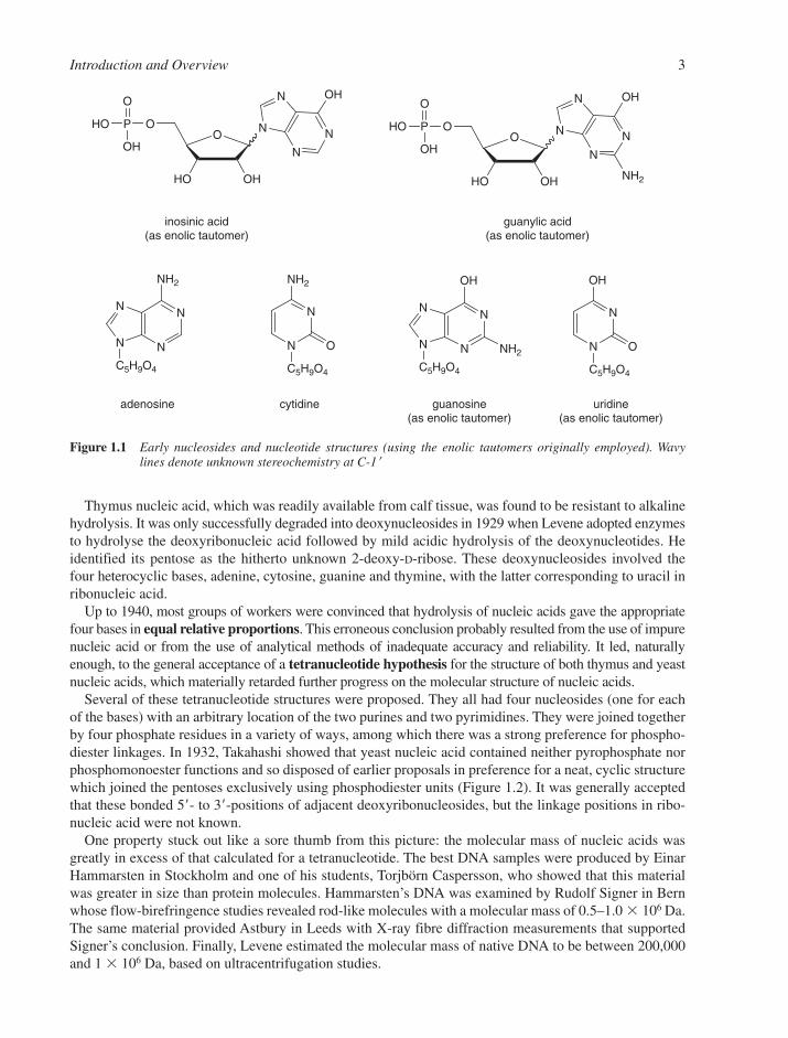
Accurate molecular studies on nucleic acids essentially date back to 1909 when Levene and Jacobs begana reinvestigation of the structure of nucleotides at the Rockefeller Institute. Inosinic acid, which Liebighad isolated from beef muscle in 1847, proved to be hypoxanthine-riboside 5�-phosphate. Guanylic acid,isolated from the nucleoprotein of pancreas glands, was identified as guanine-riboside 5�-phosphate(Figure 1.1). Each of these nucleotides was cleaved by alkaline hydrolysis to give phosphate and the cor-responding nucleosides, inosine and guanosine, respectively. Since then, all nucleosides are characterizedas the condensation products of a pentose and a nitrogenous base while nucleotides are the phosphateesters of one of the hydroxyl groups of the pentose.
2 Chapter 1
Thymus nucleic acid, which was readily available from calf tissue, was found to be resistant to alkalinehydrolysis. It was only successfully degraded into deoxynucleosides in 1929 when Levene adopted enzymesto hydrolyse the deoxyribonucleic acid followed by mild acidic hydrolysis of the deoxynucleotides. Heidentified its pentose as the hitherto unknown 2-deoxy-D-ribose. These deoxynucleosides involved thefour heterocyclic bases, adenine, cytosine, guanine and thymine, with the latter corresponding to uracil inribonucleic acid.
Up to 1940, most groups of workers were convinced that hydrolysis of nucleic acids gave the appropriatefour bases in equal relative proportions. This erroneous conclusion probably resulted from the use of impurenucleic acid or from the use of analytical methods of inadequate accuracy and reliability. It led, naturallyenough, to the general acceptance of a tetranucleotide hypothesis for the structure of both thymus and yeastnucleic acids, which materially retarded further progress on the molecular structure of nucleic acids.
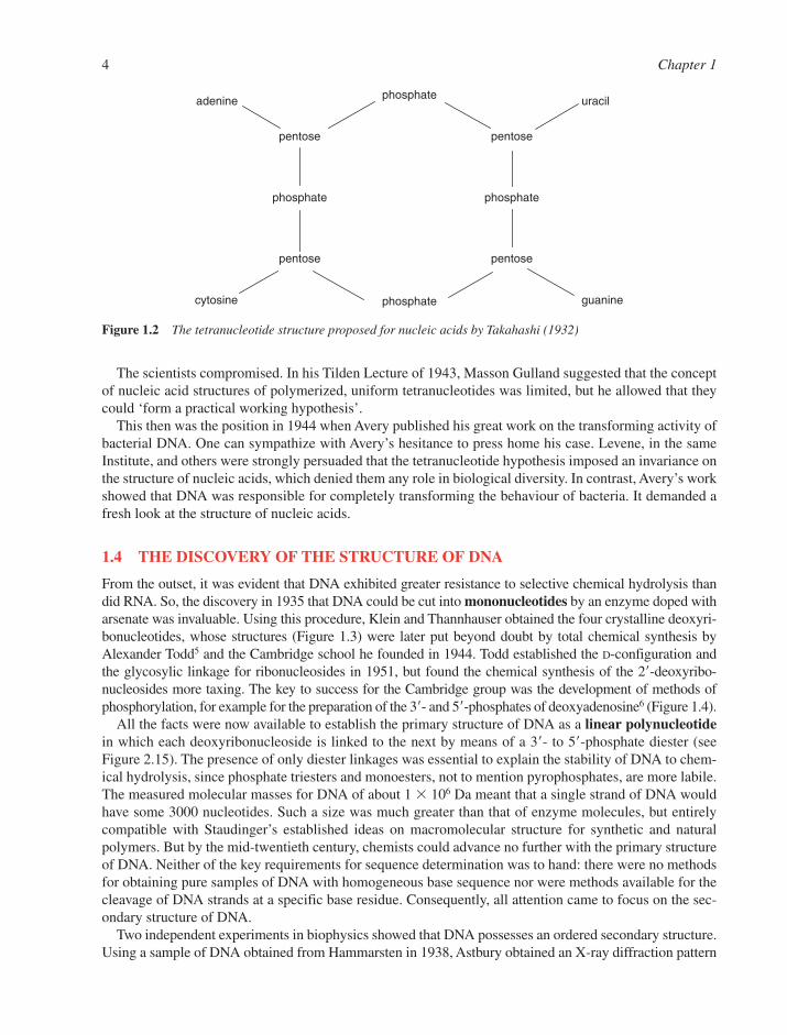
Several of these tetranucleotide structures were proposed. They all had four nucleosides (one for eachof the bases) with an arbitrary location of the two purines and two pyrimidines. They were joined togetherby four phosphate residues in a variety of ways, among which there was a strong preference for phospho-diester linkages. In 1932, Takahashi showed that yeast nucleic acid contained neither pyrophosphate norphosphomonoester functions and so disposed of earlier proposals in preference for a neat, cyclic structurewhich joined the pentoses exclusively using phosphodiester units (Figure 1.2). It was generally acceptedthat these bonded 5�- to 3�-positions of adjacent deoxyribonucleosides, but the linkage positions in ribo-nucleic acid were not known.
One property stuck out like a sore thumb from this picture: the molecular mass of nucleic acids wasgreatly in excess of that calculated for a tetranucleotide. The best DNA samples were produced by EinarHammarsten in Stockholm and one of his students, Torjbörn Caspersson, who showed that this materialwas greater in size than protein molecules. Hammarsten’s DNA was examined by Rudolf Signer in Bernwhose flow-birefringence studies revealed rod-like molecules with a molecular mass of 0.5–1.0 � 106 Da.The same material provided Astbury in Leeds with X-ray fibre diffraction measurements that supportedSigner’s conclusion. Finally, Levene estimated the molecular mass of native DNA to be between 200,000and 1 � 106 Da, based on ultracentrifugation studies.
Introduction and Overview 3
N
NN
N
NH2
C5H9O4
N
N
NH2
O
C5H9O4
N
NN
N
OH
NH2
C5H9O4
N
N
OH
O
C5H9O4
adenosine cytidine guanosine(as enolic tautomer)
uridine(as enolic tautomer)
N
N
N
N OH
O
OHHO
OP
OH
HO
O
N
N
N
N OH
O
OHHO
OP
OH
HO
O
NH2
inosinic acid(as enolic tautomer)
guanylic acid(as enolic tautomer)
Figure 1.1 Early nucleosides and nucleotide structures (using the enolic tautomers originally employed). Wavylines denote unknown stereochemistry at C-1�
The scientists compromised. In his Tilden Lecture of 1943, Masson Gulland suggested that the conceptof nucleic acid structures of polymerized, uniform tetranucleotides was limited, but he allowed that theycould ‘form a practical working hypothesis’.
This then was the position in 1944 when Avery published his great work on the transforming activity ofbacterial DNA. One can sympathize with Avery’s hesitance to press home his case. Levene, in the sameInstitute, and others were strongly persuaded that the tetranucleotide hypothesis imposed an invariance onthe structure of nucleic acids, which denied them any role in biological diversity. In contrast, Avery’s workshowed that DNA was responsible for completely transforming the behaviour of bacteria. It demanded afresh look at the structure of nucleic acids.
1.4 THE DISCOVERY OF THE STRUCTURE OF DNA
From the outset, it was evident that DNA exhibited greater resistance to selective chemical hydrolysis thandid RNA. So, the discovery in 1935 that DNA could be cut into mononucleotides by an enzyme doped witharsenate was invaluable. Using this procedure, Klein and Thannhauser obtained the four crystalline deoxyri-bonucleotides, whose structures (Figure 1.3) were later put beyond doubt by total chemical synthesis byAlexander Todd5 and the Cambridge school he founded in 1944. Todd established the D-configuration andthe glycosylic linkage for ribonucleosides in 1951, but found the chemical synthesis of the 2�-deoxyribo-nucleosides more taxing. The key to success for the Cambridge group was the development of methods ofphosphorylation, for example for the preparation of the 3�- and 5�-phosphates of deoxyadenosine6 (Figure 1.4).
All the facts were now available to establish the primary structure of DNA as a linear polynucleotidein which each deoxyribonucleoside is linked to the next by means of a 3�- to 5�-phosphate diester (seeFigure 2.15). The presence of only diester linkages was essential to explain the stability of DNA to chem-ical hydrolysis, since phosphate triesters and monoesters, not to mention pyrophosphates, are more labile.The measured molecular masses for DNA of about 1 � 106 Da meant that a single strand of DNA wouldhave some 3000 nucleotides. Such a size was much greater than that of enzyme molecules, but entirelycompatible with Staudinger’s established ideas on macromolecular structure for synthetic and naturalpolymers. But by the mid-twentieth century, chemists could advance no further with the primary structureof DNA. Neither of the key requirements for sequence determination was to hand: there were no methodsfor obtaining pure samples of DNA with homogeneous base sequence nor were methods available for thecleavage of DNA strands at a specific base residue. Consequently, all attention came to focus on the sec-ondary structure of DNA.
Two independent experiments in biophysics showed that DNA possesses an ordered secondary structure.Using a sample of DNA obtained from Hammarsten in 1938, Astbury obtained an X-ray diffraction pattern
4 Chapter 1
phosphate
phosphatephosphate
phosphate
pentose pentose
pentose pentose
adenine uracil
cytosine guanine
Figure 1.2 The tetranucleotide structure proposed for nucleic acids by Takahashi (1932)

Introduction and Overview 5
N
N
N
N NH2
O
HO
OP
OH
HO
O
N
N
N
N OH
O
HO
OP
OH
HO
O
NH2
deoxyadenylic acid[dAMP]
deoxycytidylic acid[dCMP]
O
HO
OP
OH
HO
O
O
HO
OP
OH
HO
ON
N
NH2
O
N
N
OH
O
H3C
deoxyguanylic acid[dGMP]
(as enolic tautomer)
deoxythymidylic acid[dTMP]
(as enolic tautomer)
Figure 1.3 Structures of 5�-deoxyribonucleotides (original tautomers for dGMP and dTMP)
N
N
N
N NH2
O
HO
AcO
N
N
N
N NH2
O
AcO
HON
N
N
N NH2
O
AcO
AcO
N
N
N
N NH2
O
O
HO
PO OH
OH
N
N
N
N NH2
O
HO
OP
O
HO
OH
+i
ii,iiii, iv
ii,iiii, iv
3'-dAMP 5'-dAMP
Figure 1.4 Todd’s synthesis of deoxyadenosine 3�- and 5�-phosphates Reagents: (i) MeOH, NH3 (ii) (PhO)2P(O)OP(H)(O)OCH2Ph (iii) N-chlorosuccinimide (iv) H2 /PdC(D.H. Hayes, A.M. Michelson and A.R. Todd, J. Chem. Soc., 1955, 808–815)
from stretched, dry fibres of DNA. From the rather obscure data he deduced ‘… A spacing of 3.34 Å alongthe fibre axis corresponds to that of a close succession of flat or flattish nucleotides standing out perpendicu-larly to the long axis of the molecule to form a relatively rigid structure.’ These conclusions roundly contra-dicted the tetranucleotide hypothesis.
Some years later, Gulland studied the viscosity and flow-birefringence of calf thymus DNA and thencepostulated the presence of hydrogen bonds linking the purine–pyrimidine hydroxyl groups and some of theamino groups. He suggested that these hydrogen bonds could involve nucleotides either in adjacent chains orwithin a single chain, but he somewhat hedged his bets between these alternatives. Sadly, Astbury returnedto the investigation of proteins and Gulland died prematurely in a train derailment in 1947. Both of themleft work that was vital for their successors to follow, but each contribution contained a misconception thatwas to prove a stumbling block for the next half-a-dozen years. Thus, Linus Pauling’s attempt to create ahelical model for DNA located the pentose-phosphate backbone in its core and the bases pointing out-wards – as Astbury had decided. Gulland had subscribed to the wrong tautomeric forms for the hetero-cyclic bases thymine and guanine, believing them to be enolic and having hydroxyl groups. The importanceof the true keto forms was only appreciated in 1952.
Erwin Chargaff began to investigate a very different type of order in DNA structure. He studied the basecomposition of DNA from a variety of sources using the new technique of paper chromatography to sep-arate the products of hydrolysis of DNA and employing one of the first commercial ultraviolet spectropho-tometers to quantify their relative abundance.7 His data showed that there is a variation in base compositionof DNA between species that is overridden by a universal 1:1 ratio of adenine with thymine and guaninewith cytosine. This meant that the proportion of purines, (A � G), is always equal to the proportion ofpyrimidines, (C � T). Although the ratio (G � C)/(A � T) varies from species to species, different tissuesfrom a single species give DNA of the same composition. Chargaff’s results finally discredited the tetra-nucleotide hypothesis, because it called for equal proportions of all four bases in DNA.
In 1951, Francis Crick and Jim Watson joined forces in the Cavendish Laboratory in Cambridge to tacklethe problem of DNA structure. Both of them were persuaded that the model-building approach that had ledPauling and Corey to the �-helix structure for peptides should work just as well for DNA. Almost incredibly,they attempted no other line of direct experimentation but drew on the published and unpublished resultsof other research teams in order to construct a variety of models, each to be discarded in favour of the nextuntil they created one which satisfied all the facts.8,9
The best X-ray diffraction results were to be found in King’s College, London. There, Maurice Wilkinshad observed the importance of keeping DNA fibres in a moist state and Rosalind Franklin had found thatthe X-ray diffraction pattern obtained from such fibres showed the existence of an A-form of DNA at lowhumidity, which changed into a B-form at high humidity. Both forms of DNA were highly crystalline andclearly helical in structure. Consequently, Franklin decided that this behaviour required the phosphategroups to be exposed to water on the outside of the helix, with the corollary that the bases were on theinside of the helix.
Watson decided that the number of nucleotides in the unit crystallographic cell favoured a double-stranded helix. Crick’s physics-trained mind recognized the symmetry implications of the space-group of the A-form diffraction pattern, monoclinic C2. There had to be local twofold symmetry axes normal to the helix, a feature, which called for a double-stranded helix, whose two chains must run in oppositedirections.
Crick and Watson thus needed merely to solve the final problem: how to construct the core of the helixby packing the bases together in a regular structure. Watson knew about Gulland’s conclusions regardinghydrogen bonds joining the DNA bases. This convinced him that the crux of the matter had to be a rulegoverning hydrogen bonding between bases. Accordingly, Watson experimented with models using theenolic tautomeric forms of the bases (Figure 1.3) and pairing like with like. This structure was quicklyrejected by Crick because it had the wrong symmetry for B-DNA. Self-pairing had to be rejected becauseit could not explain Chargaff’s 1:1 base ratios, which Crick had perceived were bound to result if you hadcomplementary base pairing.
6 Chapter 1
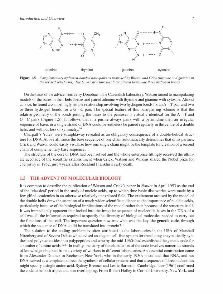
On the basis of the advice from Jerry Donohue in the Cavendish Laboratory, Watson turned to manipulatingmodels of the bases in their keto forms and paired adenine with thymine and guanine with cytosine. Almostat once, he found a compellingly simple relationship involving two hydrogen bonds for an A…T pair and twoor three hydrogen bonds for a G…C pair. The special feature of this base-pairing scheme is that the relative geometry of the bonds joining the bases to the pentoses is virtually identical for the A…T and G…C pairs (Figure 1.5). It follows that if a purine always pairs with a pyrimidine then an irregularsequence of bases in a single strand of DNA could nevertheless be paired regularly in the centre of a doublehelix and without loss of symmetry.10
Chargaff’s ‘rules’ were straightaway revealed as an obligatory consequence of a double-helical struc-ture for DNA. Above all, since the base sequence of one chain automatically determines that of its partner,Crick and Watson could easily visualize how one single chain might be the template for creation of a secondchain of complementary base sequence.
The structure of the core of DNA had been solved and the whole enterprise fittingly received the ultim-ate accolade of the scientific establishment when Crick, Watson and Wilkins shared the Nobel prize forchemistry in 1962, just 4 years after Rosalind Franklin’s early death.
1.5 THE ADVENT OF MOLECULAR BIOLOGY
It is common to describe the publication of Watson and Crick’s paper in Nature in April 1953 as the endof the ‘classical’ period in the study of nucleic acids, up to which time basic discoveries were made by afew gifted academics in an otherwise relatively unexplored field. The excitement aroused by the model ofthe double helix drew the attention of a much wider scientific audience to the importance of nucleic acids,particularly because of the biological implications of the model rather than because of the structure itself.It was immediately apparent that locked into the irregular sequence of nucleotide bases in the DNA of acell was all the information required to specify the diversity of biological molecules needed to carry outthe functions of that cell. The important question now was what was the key, the genetic code, throughwhich the sequence of DNA could be translated into protein?11
The solution to the coding problem is often attributed to the laboratories in the USA of MarshallNirenberg and of Severo Ochoa who devised an elegant cell-free system for translating enzymatically syn-thesized polynucleotides into polypeptides and who by the mid-1960s had established the genetic code fora number of amino acids.12,13 In reality, the story of the elucidation of the code involves numerous strandsof knowledge obtained from a variety of workers in different laboratories. An essential contribution camefrom Alexander Dounce in Rochester, New York, who in the early 1950s postulated that RNA, and notDNA, served as a template to direct the synthesis of cellular proteins and that a sequence of three nucleotidesmight specify a single amino acid. Sydney Brenner and Leslie Barnett in Cambridge, later (1961) confirmedthe code to be both triplet and non-overlapping. From Robert Holley in Cornell University, New York, and
Introduction and Overview 7
N
N
N
N N
H
H
N
N
O
O
CH3
H N
N
N
N O
H
N HH
N
N
N
O
H
H
adenine thymine guanine cytosine
Figure 1.5 Complementary hydrogen-bonded base-pairs as proposed by Watson and Crick (thymine and guanine inthe revised keto forms). The G…C structure was later altered to include three hydrogen bonds

Hans Zachau in Cologne, came the isolation and determination of the sequence of three transfer RNAs(tRNA) ‘adapter’ molecules that each carry an individual amino acid ready for incorporation into proteinand which are also responsible for recognizing the triplet code on the messenger RNA (mRNA). The mRNAspecies contain the sequences of individual genes copied from DNA (see Chapter 6). Gobind Khorana andhis group in Madison, Wisconsin, chemically synthesized all 64 ribotrinucleoside diphosphates and, usinga combination of chemistry and enzymology, synthesized a number of polyribonucleotides with repeating di-, tri-, and tetranucleotide sequences.14 These were used as synthetic mRNA to help identify each triplet inthe code. This work was recognized by awarding the Nobel Prize for Medicine in 1968 jointly to Holley,Khorana and Nirenberg.
Nucleic acid research in the 1950s and 1960s was preoccupied by the solution to the coding problemand the establishment of the biological roles of tRNA and mRNA. This was not surprising bearing in mindthat at that time the smaller size and attainable homogeneity made isolation and purification of RNA amuch easier task than it was for DNA. It was clear that in order to approach the fundamental question ofwhat constituted a gene – a single hereditable element of DNA that up to then could be defined geneticallybut not chemically – it was going to be necessary to break down DNA into smaller, more tractable piecesin a specific and predictable way.
The breakthrough came in 1968 when Meselson and Yuan reported the isolation of a restriction enzymefrom the bacterium Escherichia coli. Here at last was an enzyme, a nuclease, which could recognize adefined sequence in a DNA and cut it specifically (see Section 5.3.1). The bacterium used this activity tobreak down and hence inactive invading (e.g. phage) DNA. It was soon realized that this was a generalproperty of bacteria, and the isolation of other restriction enzymes with different specificities soon fol-lowed. But it was not until 1973 that the importance of these enzymes became apparent. At this time,Chang and Cohen at Stanford and Helling and Boyer at the University of California were able to constructin a test tube, a biologically functional DNA that combined genetic information from two differentsources. This chimera was created by cleaving DNA from one source with a restriction enzyme to give afragment that could then be joined to a carrier DNA, a plasmid. The resultant recombinant DNA wasshown to be able to replicate and express itself in E. coli.15
This remarkable demonstration of genetic manipulation was to revolutionize biology. It soon becamepossible to dissect out an individual gene from its source DNA, to amplify it in a bacterium or other organ-ism (cloning, see Section 5.2), and to study its expression by the synthesis first of RNA and then of pro-tein (see Chapters 6 and 7). This single advance by the groups of Cohen and Boyer truly marked the dawnof modern molecular biology.
1.6 THE PARTNERSHIP OF CHEMISTRY AND BIOLOGY
In the 1940s and 1950s, the disciplines of chemistry and biology were so separate that it was a rare occur-rence for an individual to embrace both. Two young scientists who were just setting out on their careers atthat time were exceptional in recognizing the potential of chemistry in the solution of biological problemsand both, in their different ways, were to have a substantial and lasting effect in the field of nucleic acids.
One was Frederick Sanger, a product of the Cambridge Biochemistry School, who in the early 1940s setout to determine the sequence of a protein, insulin. This feat had been thought unattainable, since it waswidely supposed that proteins were not discrete species with defined primary sequence. Even moreremarkably, he went on to develop methods for sequence determination first of RNA and then of DNA (seeSection 5.1). These methods involved a subtle blend of enzymology and chemistry that few would havethought possible to combine.16 The results of his efforts transformed DNA sequencing in only a few yearsinto a routine procedure. In the late 1980s, the procedure was adapted for use in automated sequencingmachines and the 1990s saw worldwide efforts to sequence whole organism genomes. In 2003, exactly 50years after the discovery of the structure of the DNA double helix, it was announced that the humangenome sequence had been completed. The award of two Nobel prizes to Sanger (1958 and 1980) hardlyseems recognition enough!
8 Chapter 1
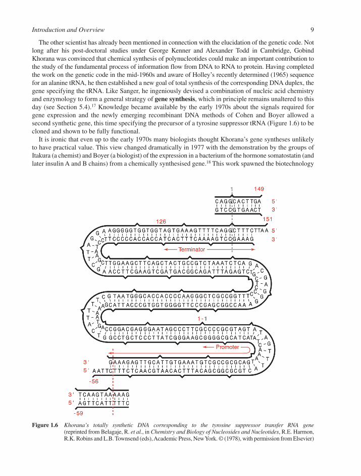
The other scientist has already been mentioned in connection with the elucidation of the genetic code. Notlong after his post-doctoral studies under George Kenner and Alexander Todd in Cambridge, GobindKhorana was convinced that chemical synthesis of polynucleotides could make an important contribution tothe study of the fundamental process of information flow from DNA to RNA to protein. Having completedthe work on the genetic code in the mid-1960s and aware of Holley’s recently determined (1965) sequencefor an alanine tRNA, he then established a new goal of total synthesis of the corresponding DNA duplex, thegene specifying the tRNA. Like Sanger, he ingeniously devised a combination of nucleic acid chemistryand enzymology to form a general strategy of gene synthesis, which in principle remains unaltered to thisday (see Section 5.4).17 Knowledge became available by the early 1970s about the signals required forgene expression and the newly emerging recombinant DNA methods of Cohen and Boyer allowed a second synthetic gene, this time specifying the precursor of a tyrosine suppressor tRNA (Figure 1.6) to becloned and shown to be fully functional.
It is ironic that even up to the early 1970s many biologists thought Khorana’s gene syntheses unlikelyto have practical value. This view changed dramatically in 1977 with the demonstration by the groups ofItakura (a chemist) and Boyer (a biologist) of the expression in a bacterium of the hormone somatostatin (andlater insulin A and B chains) from a chemically synthesised gene.18 This work spawned the biotechnology
Introduction and Overview 9
Figure 1.6 Khorana’s totally synthetic DNA corresponding to the tyrosine suppressor transfer RNA gene(reprinted from Belagaje, R. et al., in Chemistry and Biology of Nucleosides and Nucleotides, R.E. Harmon,R.K. Robins and L.B. Townsend (eds), Academic Press, New York. © (1978), with permission from Elsevier)

industry and synthetic genes became routinely used in the production of proteins. Further, oligodeoxyri-bonucleotides, the short pieces of single-stranded DNA for which Khorana developed the first chemicalsyntheses, became invaluable general tools in the manipulation of DNA, for example, as primers in DNAsequencing, as probes in gene detection and isolation, and as mutagenic agents to alter the sequence ofDNA. From the late 1980s, research accelerated into synthetic oligonucleotide analogues as antisensemodulators of gene expression in cells, as therapeutic agents (see Section 5.7) and for the construction ofmicroarray chips for gene expression analysis.
The availability of synthetic DNA also provided new impetus in the study of DNA structure. In the early1970s, new X-ray crystallographic techniques had been developed and applied to solve the structure of thedinucleoside phosphate, ApU, by Rich and co-workers in Cambridge, USA. This was followed by thecomplete structure of yeast phenylalanine tRNA, determined independently by Rich and by Klug and col-leagues in Cambridge, England. For the first time, the complementary base pairing between two strandscould be seen in greater detail than was previously possible from studies of DNA and RNA fibres. ApUformed a double helix by end-to-end packing of molecules, with Watson–Crick pairing clearly in evidencebetween each strand. The tRNA showed not only Watson–Crick pairs, but also a variety of alternative basepairs and base triples, many of which were entirely novel (see Sections 2.3.3 and 7.1.2).
Then in 1978, the structure of synthetic d(pATAT) was solved by Kennard and her group in Cambridge.This tetramer also formed an extended double helix, but excitingly revealed that there was a substantialsequence-dependence in its conformation. The angles between neighbouring dA and dT residues werequite different between the A–T sequence and the T–A sequence elements. Soon after, Wang and col-leagues discovered that synthetic d(CGCGCG) adopted a totally unpredicted, left-handed Z-conformation.This was soon followed by the demonstration of both a B-DNA helix in a synthetic dodecamer byDickerson in California and an A-DNA helix in an octamer by Kennard, and finally put paid to the con-cept that DNA had a rigid, rod-like structure. Clearly, DNA could adopt different conformations depend-ent on sequence and also on its external environment (see Section 2.3). More importantly, an immediateinference could be drawn that conformational differences in DNA (or the potential for their formation) mightbe recognized by other molecules. Thus, it was not long before synthetic DNA was also being used in thestudy of DNA binding to carcinogens and drugs (see Chapters 8 and 9) and to proteins (see Chapter 10).
These spectacular advances were only possible because of the equally dramatic improvements in methodsof oligonucleotide synthesis that took place in the late 1970s and early 1980s. The laborious manual workof the early gene synthesis days was replaced by reliable automated DNA synthesis machines, which,within hours, could assemble sequences well in excess of 100 residues (see Section 4.1.4). Khorana’svision of the importance of synthetic DNA has been fully realized.
1.7 FRONTIERS IN NUCLEIC ACIDS RESEARCH
The last decade of the twentieth century was characterized by the quest to determine the complete DNAsequence of the human genome. Efforts by a publicly funded international consortium gathered consid-erable pace in the late 1990s in response to a challenge from a private company and the resultant concernsover the availability of sequencing data to the research community. The completion of the human genomesequence was duly announced by the consortium in April 2003, 50 years after papers on the discovery ofthe structure of the DNA double helix had been published and only 25 years since the first simple bac-teriophage genome sequences were obtained. Genome sequences of many other organisms have also beencompleted, for example, mouse, nematode, zebrafish, yeast and parasites such as Plasmodium falciparium(see Section 6.5). The vast quantity of DNA sequence information generated has led to the founding of thenew discipline of Bioinformatics in order to analyse and compare sequence data. One big surprise wasthat the human genome contains far fewer genes than expected, only about 24,500. We now know that pro-duction of the considerably larger number of human proteins and their regulation during cell division andbiological development involves control of gene expression at many different stages (e.g. transcription,alternative splicing, RNA editing, translation, see Chapters 6 and 7), a full understanding of which is likely
10 Chapter 1
to occupy biologists well into the twenty-first century. A recent technical advance here is the developmentof microarrays of synthetic oligonucleotides or cDNAs as hybridisation probes of DNA or RNAsequences both for mutational and gene expression analysis (see Section 5.5.4). This has led to the scienceof ‘-omics’, such as genomics and ribonomics, where DNA sequence variations can be studied and globaleffects of particular pathological states or external stimuli can be gauged on a whole genome basis.
A number of other advances have also been made in nucleic acids chemistry. First, a strong revival in thesynthesis of nucleoside analogues has led to a number of therapeutic agents being approved for clinical usein treatment of AIDS and HIV infection as well as herpes and hepatitis viruses (see Section 3.7.2). Further,synthetic oligonucleotide analogues have become clinical agents for the treatment of viral infections andsome cancers, although few have passed full regulatory approval as yet. The exploitation of the ‘antisense’technology as a principle of therapeutic gene modulation has led to the investigation of a large number ofnucleic acid analogues to enhance activity (see Section 5.7.1). As the twenty-first century arrived, genemodulation technology was finding increasing use to validate gene targets in cell lines and animals. At thesame time, there was increasing recognition that other mechanisms of action can contribute to therapeuticeffects of oligonucleotides in humans, such as stimulation of the immune system by ‘CpG’ domains (seeSection 5.7.1), which may be harnessed perhaps for use as vaccine adjuvants.
The provision of synthetic RNA has also become routine (see Section 4.2) resulting in major advances inour understanding of catalytic RNA (ribozymes, see Sections 5.7.3 and 7.6.2) and protein-RNA inter-actions (see Section 10.9). New techniques of in vitro selection of RNA sequences have extended the poten-tial of ribozymes and aptamers to carry out artificial reactions or bind unusual substrates, for example toact as ‘riboswitches’ responsive to certain analytes (see Section 5.7.3). A considerable upsurge of researchin RNA biology has paralleled the availability of synthetic RNA. New ways have been elucidated for spe-cific RNA sequences and structures to play important roles in gene regulation (e.g. microRNA, see Section5.7.2). The exciting discovery of ‘RNA interference’ as a natural cell mechanism has led to the develop-ment of short synthetic RNA duplexes (siRNA and shRNA) as new gene control reagents that now rival,and may well surpass, antisense oligonucleotides for therapeutic and diagnostic use (see Section 5.7.2).
Dramatic advances have also been made in high-resolution structural determination of DNA and RNAsequences and their complexes with proteins (see Chapter 10), which are providing useful insights into molecu-lar recognition and suggesting new approaches for drug design. In addition, the study of DNA recognition bysmall molecules in the minor groove has taken a major leap forward with the development of hairpinpolyamides as a novel class of DNA-specific reagents with potential as drugs (see Section 9.7.4). Targeting ofunusual DNA telomeric G-tetraplex structures is also an active area of current drug design (see Section 9.10).
The heady days of the discovery of the double helix and the elucidation of the genetic code are long gone,but in their place have come even more exciting times when many more of us now have the opportunity toanswer fundamental questions about genetic structure and function and can utilise the insights and toolsnow available in the nucleic acids. ‘You ain’t heard nothin’yet folks’ (Al Jolson, The Jazz Singer; July 1927).
REFERENCES
1. J.S. Fruton, Molecules and life. Wiley Interscience, New York, 1972, 180–224.2. F.H. Portugal and J.S. Cohen, A century of DNA. MIT Press, Cambridge, MA, 1977.3. O.T. Avery, C.M. MacLeod and M. McCarty, Studies on the chemical nature of the substance indu-
cing transformation of pneumococcal types. J. Exp. Med., 1944, 79, 137–158.4. F. Miescher, Die histochemischen und physiologischen arbeiten. Vogel, Leipzig, 1897.5. J.G. Buchanan and Lord Todd. Adv. Carbohydr. Chem., 2000, 55, 1–13.6. D.H. Hayes, A.M. Michelson and A.R. Todd, Mononucleotides derived from deoxyadenosine and
deoxyguanosine. J. Chem. Soc., 1955, 808–815.7. E. Chargaff, Chemical specificity of nucleic acids and mechanism of their enzymatic degradation.
Experientia, 1950, 6, 201–209.8. J.D. Watson, The Double Helix. Athenaeum Press, New York, 1968.
Introduction and Overview 11
9. R. Olby, The Path to the Double Helix. Macmillan, London, 1973.10. J.D. Watson and F.H.C. Crick, A structure for deoxyribose nucleic acid. Nature, 1953, 171, 737–738.11. H.F. Judson, The Eighth Day of Creation. Jonathan Cape, London, 1979.12. M.W. Nirenberg, J.H. Matthei, O.W. Jones, R.G. Martin and S.H. Barondes, Approximation of
genetic code via cell-free protein synthesis directed by template RNA. Fed. Proc., 1963, 22, 55–61.13. S. Ochoa, Synthetic polynucleotides and the genetic code. Fed. Proc., 1963, 22, 62–74.14. H.G. Khorana, Polynucleotide synthesis and the genetic code. Fed. Proc., 1965, 24, 1473–1487.15. S.N. Cohen, The manipulation of genes. Sci. Am., 1975, 233, 24–33.16. F. Sanger, Sequences, sequences and sequences. Ann. Rev. Biochem., 1988, 57, 1–28.17. H.G. Khorana, Total synthesis of a gene. Science, 1979, 203, 614–625.18. K. Itakura, T. Hirose, R. Crea, A.D. Riggs, H.L. Heyneker, F. Bolivar and H.W. Boyer, Expression in
Escherichia coli of a chemically synthesized gene for the hormone somatostatin. Science, 1977, 198,1056–1063.
12 Chapter 1
CHAPTER 2
DNA and RNA Structure
CONTENTS
2.1 Structures of Components 142.1.1 Nucleosides and Nucleotides 142.1.2 Physical Properties of Nucleosides and Nucleotides 162.1.3 Spectroscopic Properties of Nucleosides and Nucleotides 192.1.4 Shapes of Nucleotides 20
2.2 Standard DNA Structures 242.2.1 Primary Structure of DNA 242.2.2 Secondary Structure of DNA 242.2.3 A-DNA 272.2.4 The B-DNA Family 302.2.5 Z-DNA 31
2.3 Real DNA Structures 332.3.1 Sequence-Dependent Modulation of DNA Structure 332.3.2 Mismatched Base–Pairs 362.3.3 Unusual DNA Structures 382.3.4 B–Z Junctions and B–Z Transitions 452.3.5 Circular DNA and Supercoiling 462.3.6 Triple-Stranded DNA 492.3.7 Other Non-Canonical DNA Structures 52
2.4 Structures of RNA Species 552.4.1 Primary Structure of RNA 562.4.2 Secondary Structure of RNA: A-RNA and A�-RNA 572.4.3 RNA�DNA Duplexes 592.4.4 RNA Bulges, Hairpins and Loops 612.4.5 Triple-Stranded RNAs 64
2.5 Dynamics of Nucleic Acid Structures 642.5.1 Helix-Coil Transitions of Duplexes 642.5.2 DNA Breathing 662.5.3 Energetics of the B–Z Transition 672.5.4 Rapid DNA Motions 68
2.6 Higher-Order DNA Structures 682.6.1 Nucleosome Structure 682.6.2 Chromatin Structure 69References 72

2.1 STRUCTURES OF COMPONENTS
Nucleic acids are very long, thread-like polymers, made up of a linear array of monomers called nucleotides.Different nucleic acids can have from around 80 nucleotides, as in tRNA, to over 108 nucleotide pairs in asingle eukaryotic chromosome. The unit of size of a nucleic acid is the base pair (for double-strandedspecies) or base (for single-stranded species). The abbreviation* bp is generally used, as are the larger unitsMbp (million base pairs) and kbp (thousand base pairs). The chromosome in Escherichia coli has 4 � 106
base pairs, 4 Mbp, which gives it a molecular mass of 3 � 109 Da and a length of 1.5 mm. The size of the fruitfly genome (haploid) is 180 Mbp which, shared between four chromosomes, gives a total length of 56 mm.The genomic DNA of a single human cell has 3900 Mbp and is 990 mm long. How are these extraordinar-ily long molecules constructed?
2.1.1 Nucleosides and Nucleotides
Nucleotides are the phosphate esters of nucleosides and these are components of both ribonucleic acid(RNA) and deoxyribonucleic acid (DNA). RNA is made up of ribonucleotides whereas the monomers ofDNA are 2�-deoxyribonucleotides.
All nucleotides are constructed from three components: a nitrogen heterocyclic base, a pentose sugarand a phosphate residue. The major bases are monocyclic pyrimidines or bicyclic purines, some speciesof tRNA have tricyclic minor bases such as the Wye (Figure 3.17). The major purines are adenine (A) andguanine (G) and are found in both DNA and RNA. The major pyrimidines are cytosine (C), thymine (T)and uracil (U) (Figure 2.1).
In nucleosides, the purine or pyrimidine base is joined from a ring nitrogen to carbon-1 of a pentosesugar. In RNA, the pentose is D-ribose which is locked into a five-membered furanose ring by the bondfrom C-1 of the sugar to N-1 of C or U or to N-9 of A or G. This bond is on the same side of the sugar ringas the C-5 hydroxymethyl group and is defined as a �-glycosylic linkage (Figure 2.2).
In DNA, the pentose is 2-deoxy-D-ribose and the four nucleosides are deoxyadenosine, deoxyguano-sine, deoxycytidine and deoxythymidine (Figure 2.3). In DNA, the methylated pyrimidine base thyminetakes the place of uracil in RNA, and its nucleoside with deoxyribose is still commonly called thymidine.However, since the discovery of ribothymidine as a regular component of tRNA species, it has beenpreferable to use the name deoxythymidine rather than thymidine. Unless indicated otherwise, it is assumedthat nucleosides, nucleotides and oligonucleotides are derived from D-pentofuranose sugars.
The phosphate esters of nucleosides are nucleotides, and the simplest of them have one of the hydroxylgroups of the pentose esterified by a single phosphate monoester function. Adenosine 5�-phosphate is a 5��-ribonucleotide, also called adenylic acid and abbreviated to AMP (Figure 2.4). Similarly, deoxycytidine3�-phosphate is a 3��-deoxyribonucleotide, identified as 3�-dCMP. Nucleotides containing two phosphate
14 Chapter 2
Figure 2.1 Structures of the five major purine and pyrimidine bases of nucleic acids in their dominant tautomericforms and with the IUPAC numbering systems for purines and pyrimidines
*A useful source for IUPAC nomenclature of nucleic acids can be found at http://www.chem.qmul.ac.uk/iupac/misc/naabb.htmland for polynucleotide conformation at http://www.chem.qmul.ac.uk/iupac/misc/pnuc2.html#300.
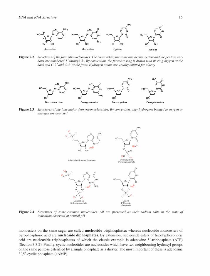
monoesters on the same sugar are called nucleoside bisphosphates whereas nucleoside monoesters ofpyrophosphoric acid are nucleoside diphosphates. By extension, nucleoside esters of tripolyphosphoricacid are nucleoside triphosphates of which the classic example is adenosine 5�-triphosphate (ATP)(Section 3.3.2). Finally, cyclic nucleotides are nucleosides which have two neighbouring hydroxyl groupson the same pentose esterified by a single phosphate as a diester. The most important of these is adenosine3�,5�-cyclic phosphate (cAMP).
DNA and RNA Structure 15
O
HO OH
N
N
N
N
NH2OP
O
O
O
2Na
O
OH
N
NH
N
N
OOP
O
O
O
NH2
OP
O
OO
4Na
2Na
ON
N
NH2
HOO
OO
P
OO
Na
ON
NH
O
HOO
O
O
P
O
O
Adenosine 5'-monophosphate Deoxycytidine3'-monophosphate
Guanosine3',5'-bisphosphate
Uridine2',3'-cyclicphosphate
Figure 2.4 Structures of some common nucleotides. All are presented as their sodium salts in the state ofionization observed at neutral pH
Figure 2.3 Structures of the four major deoxyribonucleosides. By convention, only hydrogens bonded to oxygen ornitrogen are depicted
Figure 2.2 Structures of the four ribonucleosides. The bases retain the same numbering system and the pentose car-bons are numbered 1� through 5�. By convention, the furanose ring is drawn with its ring oxygen at theback and C-2� and C-3� at the front. Hydrogen atoms are usually omitted for clarity

In the most abbreviated nomenclature currently employed, pN stands for 5�-nucleotide, Np for a 3�-nucleotide and dNp for a 3�-deoxynucleotide (to be precise, a 2�-deoxyribonucleoside 3�-phosphate). Thisshorthand notation is based on the convention that an oligonucleotide chain is drawn horizontally with its5�-hydroxyl group at the left- and its 3�-hydroxyl group at the right-hand end. Thus, pppGpp is the short-hand representation of the ‘magic spot’ nucleotide, guanosine 3�-diphosphate 5�-triphosphate, whereasApG is short for adenylyl-(3�→5�)-guanosine, whose 3�→5� internucleotide linkage runs from the nucleo-side on the left to that on the right of the phosphate.
2.1.2 Physical Properties of Nucleosides and Nucleotides
Owing to their polyionic character, nucleic acids are soluble in water up to about 1% w/v according to size andare precipitated by the addition of alcohol. Their solutions are quite viscous, and the long nucleic acid moleculesare easily sheared by stirring or by passage through a fine nozzle such as a hypodermic needle or a fine pipette.
2.1.2.1 Ionisation. The acid–base behaviour of a nucleotide is its most important physical character-istic. It determines its charge, its tautomeric structure, and thus its ability to donate and accept hydrogenbonds, which is the key feature of the base:base recognition. The pKa values for the five bases in the majornucleosides and nucleotides are listed in Table 2.1.
It is clear that all of the bases are uncharged in the physiological range 5 � pH � 9. The same is true forthe pentoses, where the ribose 2�,3�-diol only loses a proton above pH 12 while isolated hydroxyl groupsionise only above pH 15. The nucleotide phosphates lose one proton at pH 1 and a second proton (in thecase of monoesters) at pH 7. This pattern of proton equilibria is shown for AMP across the whole pH range(Figure 2.5).
The three amino bases, A, C and G, each becomes protonated on one of the ring nitrogens rather thanon the exocyclic amino group since this does not interfere with de-localisation of the NH2 electron lonepair into the aromatic system. The C�NH2 bonds of A, C and G are about 1.34 Å long, which means thatthey have 40–50% double bond order, while the C��O bonds of C, G, T and U have some 85–90% double bond order. It is also noteworthy that the proximity of negative charge of the phosphate residueshas a secondary effect, making the ring nitrogens more basic (�pKa � �0.4) and the amine protons lessacidic (�pKa � �0.6).
2.1.2.2 Tautomerism. A tautomeric equilibrium involves alternative structures that differ only in thelocation of hydrogen atoms. The choices available to nucleic acid bases are illustrated by the keto–enolequilibrium between 2-pyridone and 2-hydroxypyridine and the amine–imine equilibrium for 2-aminopy-ridine (Figure 2.6). Ultraviolet, NMR and IR spectroscopies have established that the five major basesexist overwhelmingly (99.99%) in the amino- and keto-tautomeric forms at physiological pH (Figure2.1) and not in the benzene-like enol tautomers, in common use before 1950 (Figure 1.3).
16 Chapter 2
Table 2.1 pKa values for bases in nucleosides and nucleotides
Bases (site of protonation) Nucleoside 3�-Nucleotide 5�-Nucleotide
Adenine (N-1) 3.63 3.74 3.74Cytosine (N-3) 4.11 4.30 4.56Guanine (N-7) 2.20 2.30 2.40Guanine (N-1) 9.50 9.36 9.40Thymine (N-3) 9.80 — 10.00Uracil (N-3) 9.25 9.43 9.50
Note: These data approximate to 20°C and zero salt concentration. They correspond to loss of aproton for pKa 9 and capture of a proton for pKa 5.

2.1.2.3 Hydrogen Bonding. The mutual recognition of A by T and of C by G uses hydrogen bondsto establish the fidelity of DNA transcription and translation. The NH groups of the bases are good hydrogenbond donors (d), while the sp2-hybridised electron pairs on the oxygens of the base C��O groups and onthe ring nitrogens are much better hydrogen bond acceptors (a) than are the oxygens of either the phosphateor the pentose. The a��d hydrogen bonds so formed are largely electrostatic in character, with a charge ofabout �0.2e on the hydrogens and about – 0.2e on the oxygens and nitrogens, and they seem to have anaverage strength of 6–10 kJ mol1.
The predominant amino–keto tautomer for cytosine has a pattern of hydrogen bond acceptor and donor sitesfor which O-2�N-3�N-4 can be expressed as a��a��d (Figure 2.7). Its minor tautomer has a very different pat-tern: a��d��a. In the same way, we can establish that the corresponding pattern for the dominant tautomer of dTis a��d��a whereas the pattern for N-2�N-1�O-6 of dG is d��d��a (Figure 2.7) and that for dA is (–)��a��d.
When Jim Watson was engaged in DNA model-building studies in 1952 (Section 1.4), he recognisedthat the hydrogen bonding capability of an A�T base pair uses complementarity of (–)��a��d to a��d��awhereas a C�G pair uses the complementarity of a��a��d to d��d��a. This base-pairing pattern rapidly becameknown as Watson–Crick pairing (Figure 2.8). There are two hydrogen bonds in an A�T pair and three ina C�G pair. The geometry of the pairs has been fully analysed in many structures from dinucleoside phos-phates through oligonucleotides to tRNA species, both by the use of X-ray crystallography and, morerecently, by NMR spectroscopy.
In planar base pairs, the hydrogen bonds join nitrogen and oxygen atoms that are 2.8–2.95 Å apart. Thisgeometry gives a C-1����C-1� distance of 10.60 � 0.15 Å with an angle of 68 � 2° between the two glycosylic
DNA and RNA Structure 17
Figure 2.6 Keto–enol tautomers for 2-pyridone:2-hydroxypyridine (left) and amine–imine tautomerism for 2-aminopyridine (right)
O
HO OH
N
NH
N
N
NH2
OP
O
OO
O
HO OH
N
NH
N
N
NH2
OP
O
OHO
O
HO OH
N
N
N
N
NH2
OP
O
OHO
O
HO OH
N
N
N
N
NH2
OP
O
OO
O
O O
N
N
N
N
NH2
OP
O
OO
H
pH < 1 pH 3.8
pH 6.8 pH > 12.5
Strongly acidicsolution
Strongly alkalinesolution
Physiologically important species
Figure 2.5 States of protonation of adenosine 5�-phosphate (AMP) from strongly acidic solution (left) to stronglyalkaline solution (right)
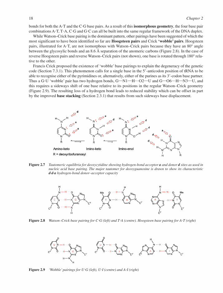
bonds for both the A�T and the C�G base pairs. As a result of this isomorphous geometry, the four base paircombinations A�T, T�A, C�G and G�C can all be built into the same regular framework of the DNA duplex.
While Watson–Crick base pairing is the dominant pattern, other pairings have been suggested of which themost significant to have been identified so far are Hoogsteen pairs and Crick ‘wobble’ pairs. Hoogsteenpairs, illustrated for A�T, are not isomorphous with Watson–Crick pairs because they have an 80° anglebetween the glycosylic bonds and an 8.6 Å separation of the anomeric carbons (Figure 2.8). In the case ofreverse Hoogsteen pairs and reverse Watson–Crick pairs (not shown), one base is rotated through 180° rela-tive to the other.
Francis Crick proposed the existence of ‘wobble’ base pairings to explain the degeneracy of the geneticcode (Section 7.3.1). This phenomenon calls for a single base in the 5�-anticodon position of tRNA to beable to recognise either of the pyrimidines or, alternatively, either of the purines as its 3�-codon base partner.Thus a G�U ‘wobble’ pair has two hydrogen bonds, G�N1�H���O2�U and G�O6���H�N3�U, andthis requires a sideways shift of one base relative to its positions in the regular Watson–Crick geometry(Figure 2.9). The resulting loss of a hydrogen bond leads to reduced stability which can be offset in partby the improved base stacking (Section 2.3.1) that results from such sideways base displacement.
18 Chapter 2
Figure 2.7 Tautomeric equilibria for deoxycytidine showing hydrogen-bond acceptor a and donor d sites as used innucleic acid base pairing. The major tautomer for deoxyguanosine is drawn to show its characteristicd.d.a hydrogen-bond donor–acceptor capacity
Figure 2.8 Watson–Crick base pairing for C�G (left) and T�A (centre). Hoogsteen base pairing for A�T (right)
Figure 2.9 ‘Wobble’ pairings for U�G (left), U�I (centre) and A�I (right)
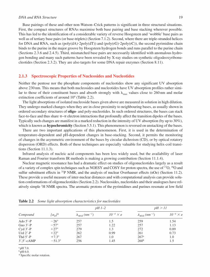
Base pairings of these and other non-Watson–Crick patterns is significant in three structural situations.First, the compact structures of RNAs maximise both base pairing and base stacking wherever possible.This has led to the identification of a considerable variety of reverse Hoogsteen and ‘wobble’ base pairs aswell as of tertiary base pairs (or base-triplets) (Section 7.1.2). Second, where there are triple-stranded helicesfor DNA and RNA, such as (poly(dA)�2poly(dT)) and (poly(rG)�2poly(rC)), the second pyrimidine chainbinds to the purine in the major groove by Hoogsteen hydrogen bonds and runs parallel to the purine chain(Sections 2.3.6 and 2.4.5). Third, mismatched base pairs are necessarily identified with anomalous hydro-gen bonding and many such patterns have been revealed by X-ray studies on synthetic oligodeoxyribonu-cleotides (Section 2.3.2). They are also targets for some DNA repair enzymes (Section 8.11).
2.1.3 Spectroscopic Properties of Nucleosides and Nucleotides
Neither the pentose nor the phosphate components of nucleotides show any significant UV absorptionabove 230 nm. This means that both nucleosides and nucleotides have UV absorption profiles rather simi-lar to those of their constituent bases and absorb strongly with �max values close to 260 nm and molarextinction coefficients of around 104 (Table 2.2).
The light absorptions of isolated nucleoside bases given above are measured in solution in high dilution.They undergo marked changes when they are in close proximity to neighbouring bases, as usually shown inordered secondary structures of oligo- and poly-nucleotides. In such ordered structures, the bases can stackface-to-face and thus share – electron interactions that profoundly affect the transition dipoles of the bases.Typically such changes are manifest in a marked reduction in the intensity of UV absorption (by up to 30%),which is known as hypochromicity (Section 5.5.1). This phenomenon is reversed on unstacking of the bases.
There are two important applications of this phenomenon. First, it is used in the determination of temperature-dependent and pH-dependent changes in base-stacking. Second, it permits the monitoring of changes in the asymmetric environment of the bases by circular dichroism (CD), or by optical rotatorydispersion (ORD) effects. Both of these techniques are especially valuable for studying helix-coil transi-tions (Section 11.1.3).
Infrared analysis of nucleic acid components has been less widely used, but the availability of laserRaman and Fourier transform IR methods is making a growing contribution (Section 11.1.4).
Nuclear magnetic resonance has had a dramatic effect on studies of oligonucleotides largely as a resultof a variety of complex spin techniques such as NOESY and COSY for proton spectra, the use of 17O, 18O andsulfur substituent effects in 31P NMR, and the analysis of nuclear Overhauser effects (nOe) (Section 11.2).These provide a useful measure of inter-nuclear distances and with computational analysis can provide solu-tion conformations of oligonucleotides (Section 2.2). Nucleosides, nucleotides and their analogues have rel-atively simple 1H NMR spectra. The aromatic protons of the pyrimidines and purines resonate at low field
DNA and RNA Structure 19
Table 2.2 Some light absorption characteristics for nucleotides
pH 1–2 pH 11
Compound [aD]* lMAX (nm1) 104 � � lMAX (nm1) 104 � �
Ado 5�-P 26° 257 1.5 259 1.54Guo 3�-P 57° 257 1.22 257 1.13Cyd 3�-P �27° 279 1.3 272 0.89Urd 2�-P �22° 262 0.99 261 0.73Thd 5�-P �7.3° 267 1.0 267a 1.03�,5�-cAMP 51.3° 256 1.45 260b 1.5
a pH 7.0.b pH 6.0.* Specific molar rotation.

(�7.6 to �8.3 with C5–H close to �5.9). The anomeric hydrogen is a doublet for ribonucleosides and a double-doublet for 2�-deoxynucleosides at �5.8–6.4. The pentoses provide a multi-spin system that gen-erally moves from low to high field in the series: H-2�, H-3�, H-4�, H-5� and H-5�� in the region �4.3 to 3.7.Lastly, 2�-deoxynucleosides have H-2� and H-2� as an ABMX system near �2.5. The 400 MHz spectrumof a simple nucleoside, cytidine (Figure 2.10), shows why two-dimensional (2D) spin techniques arerequired for the complete analysis of the spectrum in a large oligomer, which may be equivalent to a dozensuch monomer spectra superimposed.
2.1.4 Shapes of Nucleotides
Nucleotides have rather compact shapes with several interactions between non-bonded atoms. Their molecu-lar geometry is so closely related to that of the corresponding nucleotide units in oligomers and nucleicacid helices that it was once argued that helix structure is a consequence of the conformational preferencesof individual nucleotides. However, the current view is that sugar–phosphate backbone appears to act asno more than a constraint on the range of conformational space accessible to the base pairs and that – interactions between the base pairs provide the driving force for the different conformations of DNA(Section 2.3.1).
The details of conformational structure are accurately defined by the torsion angles �, �, �, �, �, and � inthe phosphate backbone, �0–�4 in the furanose ring, and � for the glycosylic bond (Figure 2.11). Becausemany of these torsional angles are inter-dependent, we can more simply describe the shapes of nucleotidesin terms of four parameters: the sugar pucker, the syn–anti conformation of the glycosylic bond, the orien-tation of C4�–C5� and the shape of the phosphate ester bonds.
2.1.4.1 Sugar Pucker. The furanose rings are twisted out of plane to minimise non-bonded inter-actions between their substituents. This ‘puckering’ is described by identifying the major displacement ofcarbons-2� and -3� from the median plane of C1�–O4�–C4�. Thus, if the endo displacement of C-2� isgreater than the exo displacement of C-3�, the conformation is called C2�-endo and so on (Figure 2.11).The endo face of the furanose is on the same side as C-5� and the base; the exo face is on the opposite faceto the base. These sugar puckers are located in the north (N) and south (S) domains of the pseudorotationcycle of the furanose ring and so spectroscopists frequently use N and S designations, which also fortuitouslyreflect the relative shapes of the C�C�C�C bonds in the C2�-endo and -exo forms, respectively.1
In solution, the N and S conformations are in rapid equilibrium and are separated by an energy barrierof less than 20 kJ mol1. The average position of the equilibrium can be estimated from the magnitudes ofthe 3J NMR coupling constants linking H1��H2� and H3��H4�. This is influenced by (1) the preferenceof electronegative substituents at C-2� and C-3� for axial orientation, (2) the orientation of the base (syn goes
20 Chapter 2
Figure 2.10 Proton NMR spectrum for cytidine (run in D2O at 400 MHz)

with C2�-endo), and (3) the formation of an intra-strand hydrogen bond from O-2� in one RNA residue toO-4� in the next which favours C3�-endo pucker. However, in RNA helical regions, this latter hydrogenbond is not often observed and an axial C�H���O interaction between the C2��H2� (n) group and the O-4� (n � 1) atom appears to make a more important contribution to the stability of RNA helices.
2.1.4.2 Syn–Anti Conformation. The plane of the bases is almost perpendicular to that of the sugarsand approximately bisects the O4��C1��C2� angle. This allows the bases to occupy either of two principalorientations. The anti conformer has the smaller H-6 (pyrimidine) or H-8 (purine) atom above the sugar ring,whereas the syn conformer has the larger O-2 (pyrimidine) or N-3 (purine) in that position. Pyrimidinesoccupy a narrow range of anti conformations (Figure 2.12) whereas purines are found in a wider range of anticonformations that can even extend into the high-anti range for 8-azapurine nucleosides such as formycin.
One inevitable consequence of this anti conformation for the glycosylic bonds is that the backbonechains for A- and B-forms DNA run downwards on the right of the minor groove and run upwards on theleft of the minor groove, depicted as (↑↓).
There is one important exception to the general preference for anti forms. Nuclear magnetic resonance,CD and X-ray analyses all show that guanine prefers the syn glycoside in mono-nucleotides, in alternating
DNA and RNA Structure 21
Figure 2.11 (a) Torsion angle notation (IUPAC) for poly-nucleotide chains and structures for the C2�-endo(S) andC3�-endo(N) preferred sugar puckers. (b) Schematic of the pseudorotation phase angle (P) cycle withthe angle ranges of selected pucker types indicated

oligomers such as d(CpGpCpG) and in Z-DNA. Theoretical calculations suggest that this effect comesfrom a favourable electrostatic attraction between the phosphate anion and the C2-amino group in guaninenucleotides. It results from polarisation of one of the nitrogen non-bonding electrons towards the ring.Most unusually, this syn conformation can only be built into left-handed helices.
2.1.4.3 C4��C5� Orientation. The conformation of the exocyclic C4��C5� bond determines the pos-ition of the 5�-phosphate relative to the sugar ring. The three favoured conformers for this bond are the classi-cal synclinal (sc) and antiperiplanar (ap) rotamers. For pyrimidine nucleosides, �sc is preferred whereas forpurine nucleosides �sc and ap are equally populated. However, in the nucleotides, the 5�-phosphate reducesthe conformational freedom and the dominant conformer for this �-bond is �sc (Figure 2.13). Once again, thedemands of Z-DNA have a major effect and the ap conformer is found for the syn guanine deoxynucleotides.
2.1.4.4 C�O and P�O Ester Bonds. Phosphate diesters are tetrahedral at phosphorus and show anti-periplanar conformations for the C5��O5� bond. Similarly, the C3��O3� bond lies in the antiperiplanarto anticlinal sector. This conformational uniformity has led to the use of the virtual bond concept inwhich the chains P5��O5��C5��C4� and P3��O3��C3��C4� can be analysed as rigid, planar unitslinked at phosphorus and at C-4�. Such a simplification has been used to speed up initial calculations ofsome complex polymeric structures.
Our knowledge of P�O bond conformations comes largely from X-ray structures of tRNA and DNAoligomers. In general, H4��C4��C5��O5��P adopts an extended W-conformation in these structures.A skewed conformation for the C�O�P�O�C system has been observed in structures of simple phos-phate diesters such as dimethyl phosphate and also for polynucleotides. This has been described as ananomeric effect and attributed to the favourable interactions of a non-bonding electron pair on O-5� with the P�O3� bond, and vice versa for the P�O5� bond (Figure 2.14). This may arise from interaction of the
22 Chapter 2
Figure 2.12 Anti and syn conformational ranges for glycosylic bonds in pyrimidine (left) and purine (right) nucleo-sides, and drawings of the anti conformation for deoxycytidine (lower left) and the syn conformationfor deoxyguanosine 5�-phosphate (lower right)
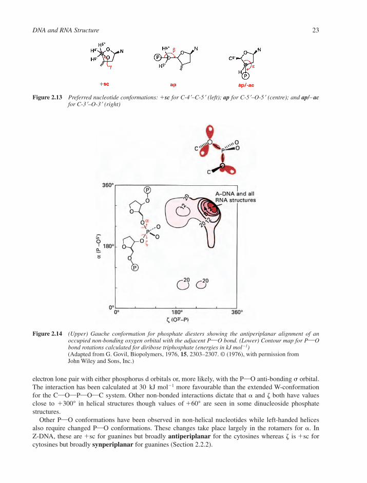
electron lone pair with either phosphorus d orbitals or, more likely, with the P�O anti-bonding � orbital.The interaction has been calculated at 30 kJ mol1 more favourable than the extended W-conformation for the C�O�P�O�C system. Other non-bonded interactions dictate that � and � both have valuesclose to �300° in helical structures though values of �60° are seen in some dinucleoside phosphate structures.
Other P�O conformations have been observed in non-helical nucleotides while left-handed helicesalso require changed P�O conformations. These changes take place largely in the rotamers for �. In Z-DNA, these are �sc for guanines but broadly antiperiplanar for the cytosines whereas � is �sc forcytosines but broadly synperiplanar for guanines (Section 2.2.2).
DNA and RNA Structure 23
Figure 2.13 Preferred nucleotide conformations: �sc for C-4�–C-5� (left); ap for C-5�–O-5� (centre); and ap/– acfor C-3�–O-3� (right)
Figure 2.14 (Upper) Gauche conformation for phosphate diesters showing the antiperiplanar alignment of anoccupied non-bonding oxygen orbital with the adjacent P�O bond. (Lower) Contour map for P�Obond rotations calculated for diribose triphosphate (energies in kJ mol1)(Adapted from G. Govil, Biopolymers, 1976, 15, 2303–2307. © (1976), with permission from John Wiley and Sons, Inc.)
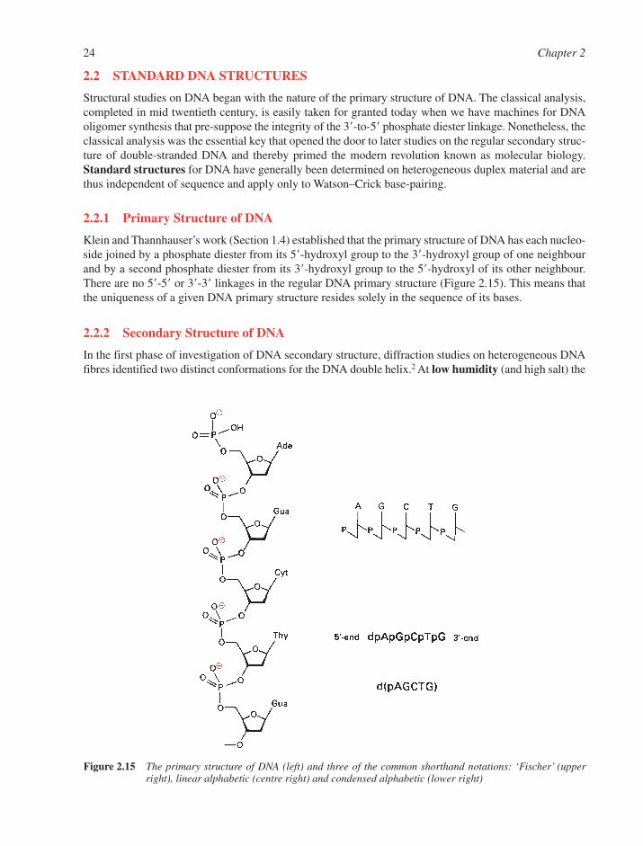
2.2 STANDARD DNA STRUCTURES
Structural studies on DNA began with the nature of the primary structure of DNA. The classical analysis,completed in mid twentieth century, is easily taken for granted today when we have machines for DNAoligomer synthesis that pre-suppose the integrity of the 3�-to-5� phosphate diester linkage. Nonetheless, theclassical analysis was the essential key that opened the door to later studies on the regular secondary struc-ture of double-stranded DNA and thereby primed the modern revolution known as molecular biology.Standard structures for DNA have generally been determined on heterogeneous duplex material and arethus independent of sequence and apply only to Watson–Crick base-pairing.
2.2.1 Primary Structure of DNA
Klein and Thannhauser’s work (Section 1.4) established that the primary structure of DNA has each nucleo-side joined by a phosphate diester from its 5�-hydroxyl group to the 3�-hydroxyl group of one neighbourand by a second phosphate diester from its 3�-hydroxyl group to the 5�-hydroxyl of its other neighbour.There are no 5�-5� or 3�-3� linkages in the regular DNA primary structure (Figure 2.15). This means thatthe uniqueness of a given DNA primary structure resides solely in the sequence of its bases.
2.2.2 Secondary Structure of DNA
In the first phase of investigation of DNA secondary structure, diffraction studies on heterogeneous DNAfibres identified two distinct conformations for the DNA double helix.2 At low humidity (and high salt) the
24 Chapter 2
Figure 2.15 The primary structure of DNA (left) and three of the common shorthand notations: ‘Fischer’ (upperright), linear alphabetic (centre right) and condensed alphabetic (lower right)

favoured form is the highly crystalline A-DNA whereas at high humidity (and low salt) the dominant struc-ture is B-DNA. We now recognise that there is a wide variety of right-handed double helical DNA conforma-tions and this structural polymorphism is denoted by the use of the letters A to T as illustrated by A, A�, B,�–B�, �–B�, C, C�, D, E and T forms of DNA. In broad terms, all of these can be classified in two genericallydifferent DNA families: A and B. These are associated with the sugar pucker C3�-endo for the A-family andC2�-endo (or the equivalent C3�-exo) for the B-family. However, as we shall see later it is the energetics ofbase-stacking which determines the conformation of the helix and sugar pucker is largely consequential. Weshall also see that in B-form DNA the base pairs sit directly on the helix axis and are nearly perpendicular toit. In A-form DNA the base pairs are displaced off-axis towards the minor groove and are inclined.
The unexpected discovery by Wang, Rich and co-workers in 1979 that the hexamer d(CGCGCG) adoptsa left-handed helical structure, now named Z-DNA, was one of the first dramatic results to stem from thesynthesis of oligonucleotides in sufficient quantity for crystallisation and X-ray diffraction analysis.3
Since then, over 100 different oligodeoxynucleotide structures have been solved and these have providedthe details on which standard DNA structures are now based.4,5 The main features of A-, B- and Z-DNAare shown in Figures 2.16–2.19 and structural parameters are provided for a range of standard helices inTables 2.3 and 2.4.
As more highly resolved structures have become available, the idea that these three families of DNAconformations are restricted to standard structures has been whittled away.6,7 We now accept that there arelocal, sequence-dependent modulations of structures that are primarily associated with the changes in theorientation of bases. Such changes seek to minimise non-bonded interactions between adjacent bases and
DNA and RNA Structure 25
Figure 2.16 Van der Waals representation of 10 bp of A-form DNA. The view is across the major (bottom) and minorgrooves (top). Atoms of the sugar–phosphate backbones of strands are coloured in red and green,respectively, and the corresponding nucleoside bases are coloured in pink and blue, respectively.Phosphorus atoms are highlighted in black
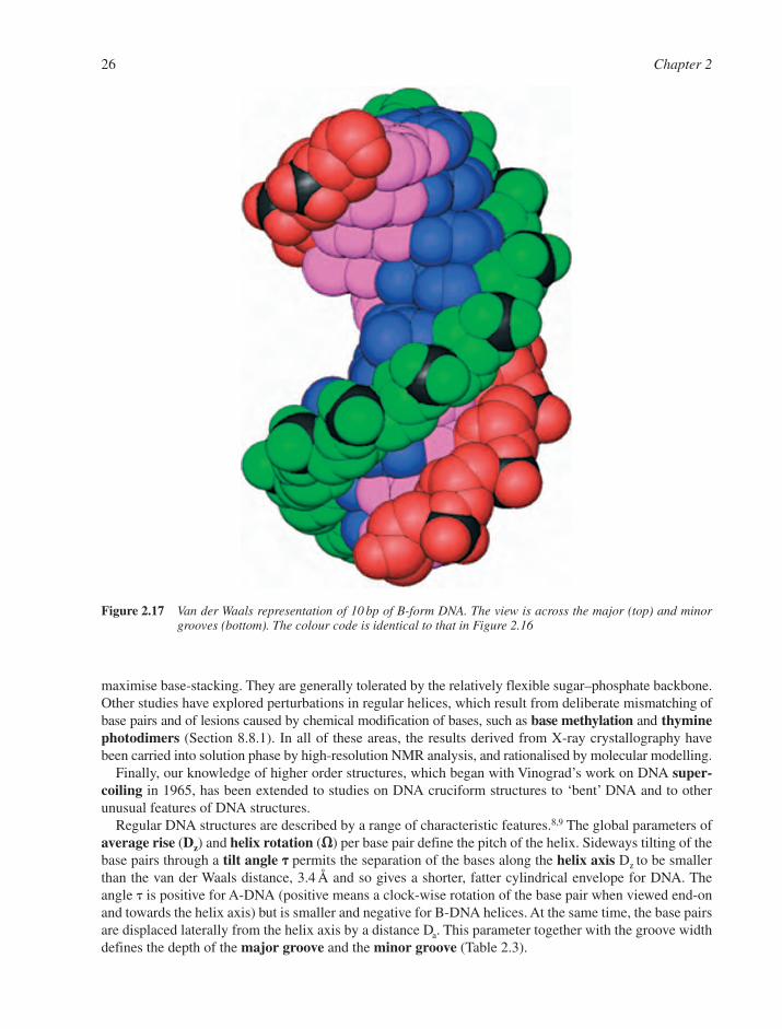
26 Chapter 2
Figure 2.17 Van der Waals representation of 10 bp of B-form DNA. The view is across the major (top) and minorgrooves (bottom). The colour code is identical to that in Figure 2.16
maximise base-stacking. They are generally tolerated by the relatively flexible sugar–phosphate backbone.Other studies have explored perturbations in regular helices, which result from deliberate mismatching ofbase pairs and of lesions caused by chemical modification of bases, such as base methylation and thyminephotodimers (Section 8.8.1). In all of these areas, the results derived from X-ray crystallography havebeen carried into solution phase by high-resolution NMR analysis, and rationalised by molecular modelling.
Finally, our knowledge of higher order structures, which began with Vinograd’s work on DNA super-coiling in 1965, has been extended to studies on DNA cruciform structures to ‘bent’ DNA and to otherunusual features of DNA structures.
Regular DNA structures are described by a range of characteristic features.8,9 The global parameters ofaverage rise (Dz) and helix rotation (��) per base pair define the pitch of the helix. Sideways tilting of thebase pairs through a tilt angle �� permits the separation of the bases along the helix axis Dz to be smallerthan the van der Waals distance, 3.4 Šand so gives a shorter, fatter cylindrical envelope for DNA. Theangle � is positive for A-DNA (positive means a clock-wise rotation of the base pair when viewed end-onand towards the helix axis) but is smaller and negative for B-DNA helices. At the same time, the base pairsare displaced laterally from the helix axis by a distance Da. This parameter together with the groove widthdefines the depth of the major groove and the minor groove (Table 2.3).

DNA and RNA Structure 27
2.2.3 A-DNA
Among the first synthetic oligonucleotides to be crystallised in the late 1970s were d(GGTATACC), aniodinated-d(CCGG) and d(GGCCGGCC). They all proved to have A-type DNA structures, similar to theclassical A-DNA deduced from fibre analysis at low resolution. Several other oligomers, mostly octamers,also form crystals of the A-structure, but NMR studies suggest that some of these may have the B-form insolution. It is conceivable that crystal packing might especially favour A-DNA for octanucleotides.
The general anatomy of A-DNA follows the Watson–Crick model with anti-parallel, right-handed double helices. The sugar rings are parallel to the helix axis and the phosphate backbone is on the outsideof a cylinder of about 24 Å diameter (Figure 2.16).
X-ray diffraction at atomic resolution shows that the bases are displaced 4.5 Å away from the helix axisand this creates a hollow core down the axis around 3 Å in diameter. There are 11 bases in each turn of 28 Å, which gives a vertical rise of 2.56 Å per base pair. To maintain the normal van der Waals separationof 3.4 Å, the stacked bases are tilted sideways through 20°. The sugar backbone has skewed phosphateester bonds, and anti-periplanar conformations for the adjacent C�O ester bonds. Finally, the furanose ringhas a C3�-endo pucker and the glycosylic bond is in the anti conformation (Table 2.3). As a result of thesefeatures, the major groove of A-DNA is cavernously deep and the minor groove is extremely shallow, as
Figure 2.18 The minor groove hydration ‘ribbon’ in the dodecamer d(CGCGAATTCGCG). The inner and outerwater (1�–9�) spines define four fused hexagons that dissect the minor groove. Only 10 bp are shownand terminal residues are numbered(Adapted from V. Tereshko et al., J. Am. Chem. Soc., 1999, 121, 3590–3595. © (1999), with permissionfrom the American Chemical Society)
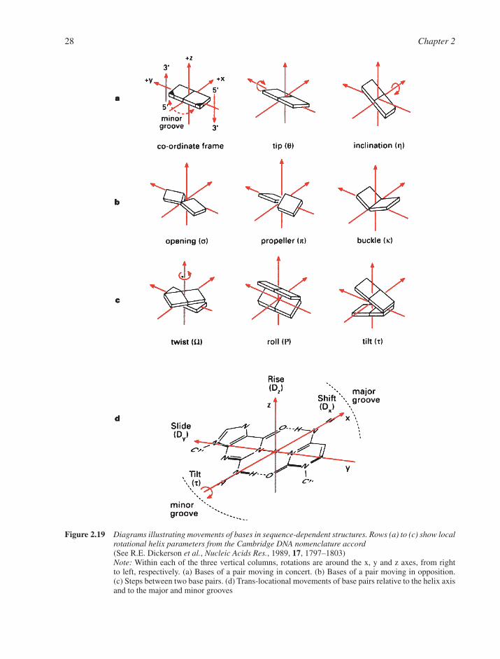
28 Chapter 2
Figure 2.19 Diagrams illustrating movements of bases in sequence-dependent structures. Rows (a) to (c) show localrotational helix parameters from the Cambridge DNA nomenclature accord(See R.E. Dickerson et al., Nucleic Acids Res., 1989, 17, 1797–1803)Note: Within each of the three vertical columns, rotations are around the x, y and z axes, from rightto left, respectively. (a) Bases of a pair moving in concert. (b) Bases of a pair moving in opposition.(c) Steps between two base pairs. (d) Trans-locational movements of base pairs relative to the helix axisand to the major and minor grooves
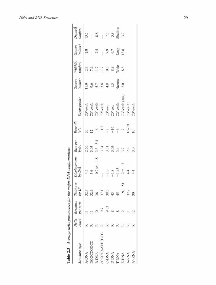
DNA and RNA Structure 29
Tabl
e 2.
3A
vera
ge h
elix
par
amet
ers
for
the
maj
or D
NA
con
form
atio
ns
Hel
ix
Res
idue
s Tw
ist p
er
Dis
plac
emen
t R
ise
per
Bas
e ti
lt
Gro
ove
Wid
th/Å
G
roov
e D
epth
/Å
Stru
ctur
e ty
pese
nse
per
turn
bp �
°bp
D/Å
bp/Å
(t°)
Suga
r pu
cker
(min
or)
(maj
or)
(min
or)
(maj
or)
A-D
NA
R11
32.7
4.5
2.56
20C
3�-e
ndo
11.0
2.7
2.8
13.5
DG
GC
CG
GC
CR
1132
.63.
63.
0312
C3�
-end
o9.
67.
9—
—
B-D
NA
R10
36
0.2
to
1.8
3.3
3.4
6
C2�
-end
o5.
711
.77.
58.
8
dCG
CG
AA
TT
CG
CG
R9.
737
.1—
3.34
1.
2C
2�-e
ndo
3.8
11.7
——
C-D
NA
R9.
3338
.5
1.0
3.31
8
C3�
-exo
4.8
10.5
7.9
7.5
D-D
NA
R8
45
1.8
3.03
16
C3�
-exo
1.3
8.9
6.7
5.8
T-D
NA
R8
45
1.43
3.4
6
C2�
-end
oN
arro
wW
ide
Dee
pSh
allo
w
Z-D
NA
L12
9,
51
2
to
33.
7
7C
3�-e
ndo
(syn
)2.
08.
813
.83.
7
A-R
NA
R11
32.7
4.4
2.8
16–1
9C
3�-e
ndo
A�-
RN
AR
1230
4.4
3.0
10C
3�-e
ndo
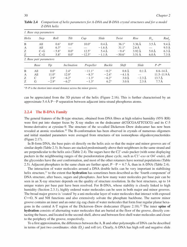
30 Chapter 2
can be appreciated from the 3D picture of the helix (Figure 2.16). This is further characterised by anapproximate 5.4 ŠP���P separation between adjacent intra-strand phosphorus atoms.
2.2.4 The B-DNA Family
The general features of the B-type structure, obtained from DNA fibres at high relative humidity (95% RH)were first put into sharper focus by X-ray studies on the dodecamer d(CGCGAATTCGCG) and its C-5 bromo-derivative at cytosine-9. The structure of the so-called Dickerson–Drew dodecamer has now beenrevealed at atomic resolution.10 The B-conformation has been observed in crystals of numerous oligomers and initial standard parameters were averaged from structures of ten isomorphous oligodeoxynucleotides(Figure 2.17).
In B-form DNA, the base pairs sit directly on the helix axis so that the major and minor grooves are ofsimilar depth (Table 2.3). Its bases are stacked predominantly above their neighbours in the same strand andare perpendicular to the helix axis (Table 2.4). The sugars have the C2�-endo pucker (with some displayingpuckers in the neighbouring ranges of the pseudorotation phase cycle, such as C1�-exo or O4�-endo), allthe glycosides have the anti conformation, and most of the other rotamers have normal populations (Table2.5). Adjacent phosphates in the same chain are further apart, P…P � 6.7 Å, than in A-DNA (Table 2.4).
The interaction of water molecules around a DNA double helix can be very important in stabilisinghelix structure,11 to the extent that hydration has sometimes been described as the ‘fourth component’ ofDNA structure, after bases, sugars and phosphates. Just how many water molecules per base pair can beseen in an X-ray structure depends on the quality of structure resolution. In the best structures, up to 14unique waters per base pair have been resolved. For B-DNA, whose stability is closely linked to highhumidity (Section 2.3.1), highly ordered water molecules can be seen in both major and minor grooves.The broad major groove is ‘coated’ by a uni-molecular layer of water molecules that interact with exposedC��O, N and NH functions and also extensively solvate the phosphate backbone. The narrow minorgroove contains an inner and an outer zig-zag chain of water molecules that form four regular planar hexa-gons in the central A�T region of the Dickerson–Drew dodecamer (Figure 2.18).12 The inner spine ofhydration consists of alternating water molecules that are buried at the floor of the groove, directly con-tacting the bases, and located in the second-shell, above and between first-shell water molecules and closerto the periphery of the groove, respectively.
To a first approximation, the differences between the A, B and other polymorphs of DNA can be describedin terms of just two coordinates: slide (Dy) and roll (�). Clearly, A-DNA has high roll and negative slide
Table 2.4 Comparison of helix parameters for A-DNA and B-DNA crystal structures and for a modelZ-DNA helix
1. Base step parameters
Helix Step Roll Tilt Cup Slide Twist Rise Dxy Radp
B All 0.6° 0.0° 10.0° 0.4 Å 36.1° 3.36 Å 3.5 Å 9.4 ÅA All 6.3° — — 1.6 Å 31.1° 2.6 Å — 9.5 ÅZ C–G 5.8° 0.0° 12.5° 5.4 Å 9.4° 3.92 Å 5.0 Å 6.3 ÅZ C–G 5.8° 0.0° 12.5° 1.1 Å 50.6° 3.51 Å 6.0 Å 7.3 Å
2. Base pair parameters
Base Tip Inclination Propeller Buckle Shift Slide P–Pa
B All 0.0° 2.4° 11.1° 0.2° 0.8 Å 0.1 Å 8.8–14 ÅA All 11.0° 12.0° 8.3° 2.4° 4.1 Å — 11.5–11.9 ÅZ C 2.9° 6.2° 1.3° 6.2° 3.0 Å 2.3 Å 13.7 ÅZ G 2.9° 6.2° 1.3° 6.2° 3.0 Å 2.3 Å 7.7 Å
a P–P is the shortest inter-strand distance across the minor groove.

DNA and RNA Structure 31
whereas B-DNA has little roll and small positive slide. These and other movements of base pairs are illus-trated in Figure 2.19(a) and values of the parameters given in Table 2.4. This results in a greater hydropho-bic surface area of the bases being exposed in A-DNA per base pair. From this, it has been argued thatB-DNA will have the lesser energy of solvation, explaining its greater stability at high humidity (95%) andthat this hydrophobic effect may well tip the balance between the A- and B-form helices.
Other B-DNA structures have much lower significance. C-DNA is obtained from the lithium salt of nat-ural DNA at rather low humidity.2 It has 28 bases and three full turns of the helix. D-DNA is observed foralternating A�T regions of DNA and has an overwound helix compared to B-DNA with 8 bp per turn. Inphage T2 DNA, where cytosine bases have been replaced by glucosylated 5-hydroxymethylcytosines, theB-conformation observed at high humidity changes into a T-DNA form at low humidity (�60% RH),which also has eightfold symmetry around the helix (see Table 2.3).
2.2.5 Z-DNA
Two of the earliest crystalline oligodeoxyribonucleotides, d(CGCGCG) and d(CGCG), provided structuresof a new type of DNA conformer, the left-handed Z-DNA, which has also been found for d(CGCATGCG).Initially it was thought that left-handed DNA had a strict requirement for alternating purine–pyrimidinesequences. We now know that this condition is neither necessary nor sufficient since left-handed structureshave been found for crystals of d(CGATCG) in which cytosines have been modified by C-5 brominationor methylation and have been identified for GTTTG and GACTG sequences by supercoil relaxation stud-ies (Section 2.3.4).
The Z-helix is also an anti-parallel duplex but is a radical departure from the A- and B-forms of DNA.It is best typified by an alternating (dG–dC)n polymer. Its two backbone strands run downwards at the leftof the minor groove and upwards at the right (↓↑), and this is the opposite from those of A- and B-DNA(↑↓) (NB: the forward direction is defined as the sequence O3�→P→O5�). In an idealised left-handedduplex, such reversed chain directions would require all the nucleosides to have the syn conformation fortheir glycosylic bonds. However, this is not possible for the pyrimidines because of the clash between O-2of the pyrimidine and the sugar furanose ring (Section 2.1.4). So the cytosines take the anti conformationand the guanines the syn conformation. The name Z-DNA results from this anti–syn feature of the glyco-sylic bonds that alternates regularly along the backbone (Figure 2.20). It causes a local chain reversal thatgenerates a zig-zag backbone path and produces a helical repeat consisting of two successive bases(purine-plus-pyrimidine) and with an overall chain sense that is the opposite of that of A- and B-DNA. Thesyn conformation of Z-DNA guanines is represented by glycosylic angles � close to 60° while the sugarpucker is C2�-endo at dC and C3�-endo at dG residues (Table 2.3).13
The switch from B- to Z-DNA conformation appears to be driven by the energetics of – base-stacking.In Z-DNA the GpC step is characterised by helical twist of –50.6° and a base pair slide of –1.1 Å. However,for the CpG steps the twist is –9° and the slide is 5.4 Å (Table 2.4; see Figure 2.19 for an explanation ofthese terms). These preferences occupy the two extremes of the slide axis and thus appear to be incompatible
Table 2.5 Average torsion angles (°) for DNA helices
Structure type � � � � � � �
A-DNAa 50 172 41 79 146 78 154GGCCGGCC 75 185 56 91 166 75 149B-DNAa 41 136 38 139 133 157 102CGCGAATTCGCG 63 171 54 123 169 108 117Z-DNA (C residues) 137 139 56 138 95 80 159Z-DNA (G residues) 47 179 169 99 104 69 68DNA–RNA decamer 69 175 55 82 151 75 162A-RNA 68 178 54 82 153 71 158
a Fibres.
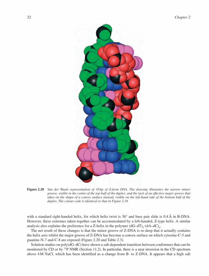
with a standard right-handed helix, for which helix twist is 36° and base pair slide is 0.4 Å in B-DNA.However, these extremes taken together can be accommodated by a left-handed, Z-type helix. A similaranalysis also explains the preference for a Z-helix in the polymer (dG–dT)n�(dA–dC)n.
The net result of these changes is that the minor groove of Z-DNA is so deep that it actually contains the helix axis whilst the major groove of Z-DNA has become a convex surface on which cytosine-C-5 andguanine-N-7 and-C-8 are exposed (Figure 2.20 and Table 2.3).
Solution studies on poly(dG–dC) have shown a salt-dependent transition between conformers that can bemonitored by CD or by 31P NMR (Section 11.2). In particular, there is a near inversion in the CD spectrumabove 4 M NaCl, which has been identified as a change from B- to Z-DNA. It appears that a high salt
32 Chapter 2
Figure 2.20 Van der Waals representation of 10 bp of Z-form DNA. The drawing illustrates the narrow minorgroove, visible in the centre of the top half of the duplex, and the lack of an effective major groove thattakes on the shape of a convex surface instead, visible on the left-hand side of the bottom half of theduplex. The colour code is identical to that in Figure 2.16
concentration stabilises the Z-conformation because it has a much smaller separation between the phos-phate anions in opposing strands than is the case for B-DNA, 8 Å as opposed to 11.7 Å. A detailed stereo-chemical examination of this conformational change shows that it calls for an elaborate mechanism andthis has posed a problem known as the chain–sense paradox: ‘How does one reverse the sense of directionof the chains in a B-helix (↑↓) to its opposite in a Z-helix (↓↑) without unpairing the bases?’ Furtherconsideration to this problem will be given later (Section 2.5.4).
The scanning tunnelling microscope has the power to resolve the structure of biological molecules withatomic detail (Section 11.5.2). Much progress has been made with dried samples of duplex DNA, inrecording images of DNA in wet state, and in revealing details of single-stranded poly(dA). Such STMmicroscopy has provided images of poly(dG-me5dC)�poly(dG-me5dC) in the Z-form. Both the generalappearance of the fibres and measurements of helical parameters are in good agreement with modelsderived from X-ray diffraction data.
2.3 REAL DNA STRUCTURES
2.3.1 Sequence-Dependent Modulation of DNA Structure
So far we have emphasised the importance of hydrogen bonds in base-pairing and DNA structure and havesaid little about base stacking. We shall see later that both these two features are important for the ener-getics and dynamics of DNA helices (Section 2.5), but it is now time to look at the major part played bybase stacking in real DNA structures. Two particular hallmarks of B-DNA, in contrast to the A- and Z-forms, are its flexibility and its capacity to make small adjustments in local helix structure in responseto particular base sequences.14
Different base sequences have their own characteristic signature: they influence groove width, helicaltwist, curvature, mechanical rigidity and resistance to bending. It seems probable that these features helpproteins to read and recognise one base sequence in preference to another (Chapter 10), possibly onlythrough changes in the positions of the phosphates in the backbone. What do we know about thesesequence-dependent structural features?
One surprise to emerge from single-crystal structure analyses of synthetic DNA oligomers has been thebreadth of variation of local helix parameters relative to the mean values broadly derived from fibre dif-fraction analysis and used for the standard A- and B-form DNA structures described earlier. Dickerson hascompared eight dodecamer and three decamer B-DNA structures.15 The mean value of the helical twistangle between neighbouring base pairs is 36.1° but the standard deviation (SD) is 5.9° and the range isfrom 24° to 51°. Likewise, the mean helical rise per base pair is 3.36 Å with a SD of 0.46 Å but with arange from 2.5 to 4.4 Å. (NB: because rise is a parameter measured between the C-1� atoms of adjacentbase-pairs, it can be smaller than the thickness of a base pair if the ends of the two base pairs bow towardseach other. Such bowing is also defined as ‘positive cup’.) Roll angles between successive base pairs aver-age �0.6° but with a SD of 6.0° and a range from –18° to �16°. These variations in twist and roll have theeffect of substantially re-orienting the potential hydrogen bond acceptors and donors at the edges of thebases along the floor of the DNA grooves, so they may well be a significant component of the sequence-recognition process used by drugs and proteins (Chapters 9 and 10). These and other modes of localchanges in the geometry of base pairs are illustrated in Figure 2.19.
The major irregularities in the positions of the bases in real DNA structures contrast with only second-ary, small conformational changes in their sugar–phosphate backbones. The main characteristic of thesesequence-dependent modulations is propeller twist. This results when the bases rotate by some 5° to 25°relative to their hydrogen-bonded partner around the long axis through C-8 of the purine and C-6 of thepyrimidine (Figure 2.19b, centre). Sections of oligonucleotides with consecutive A residues, as ind(CGCAAAAAAGCG)�d(CGCTTTTTTGCG) have unusually high propeller twist (approximately 25°)and these permit the formation of a three-centred hydrogen bonding network in the major groove betweenadenine-N-6 and two thymine-O-4 residues, the first being the Watson–Crick base pair partner and the second
DNA and RNA Structure 33
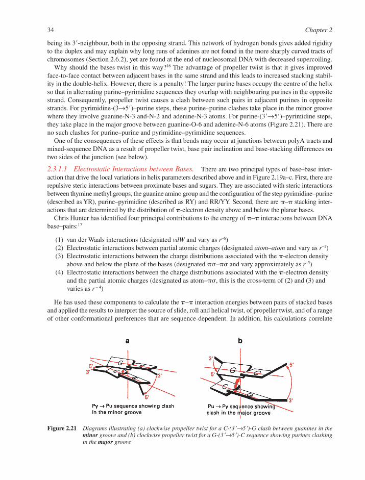
being its 3�-neighbour, both in the opposing strand. This network of hydrogen bonds gives added rigidityto the duplex and may explain why long runs of adenines are not found in the more sharply curved tracts ofchromosomes (Section 2.6.2), yet are found at the end of nucleosomal DNA with decreased supercoiling.
Why should the bases twist in this way?16 The advantage of propeller twist is that it gives improvedface-to-face contact between adjacent bases in the same strand and this leads to increased stacking stabil-ity in the double-helix. However, there is a penalty! The larger purine bases occupy the centre of the helixso that in alternating purine–pyrimidine sequences they overlap with neighbouring purines in the oppositestrand. Consequently, propeller twist causes a clash between such pairs in adjacent purines in oppositestrands. For pyrimidine-(3→5�)–purine steps, these purine–purine clashes take place in the minor groovewhere they involve guanine-N-3 and-N-2 and adenine-N-3 atoms. For purine-(3�→5�)–pyrimidine steps,they take place in the major groove between guanine-O-6 and adenine-N-6 atoms (Figure 2.21). There areno such clashes for purine–purine and pyrimidine–pyrimidine sequences.
One of the consequences of these effects is that bends may occur at junctions between polyA tracts andmixed-sequence DNA as a result of propeller twist, base pair inclination and base-stacking differences ontwo sides of the junction (see below).
2.3.1.1 Electrostatic Interactions between Bases. There are two principal types of base–base inter-action that drive the local variations in helix parameters described above and in Figure 2.19a–c. First, there arerepulsive steric interactions between proximate bases and sugars. They are associated with steric interactionsbetween thymine methyl groups, the guanine amino group and the configuration of the step pyrimidine–purine(described as YR), purine–pyrimidine (described as RY) and RR/YY. Second, there are – stacking inter-actions that are determined by the distribution of -electron density above and below the planar bases.
Chris Hunter has identified four principal contributions to the energy of – interactions between DNAbase–pairs:17
(1) van der Waals interactions (designated vdW and vary as r–6)(2) Electrostatic interactions between partial atomic charges (designated atom–atom and vary as r–1)(3) Electrostatic interactions between the charge distributions associated with the -electron density
above and below the plane of the bases (designated �– � and vary approximately as r–5)(4) Electrostatic interactions between the charge distributions associated with the -electron density
and the partial atomic charges (designated as atom– �, this is the cross-term of (2) and (3) andvaries as r – 4)
He has used these components to calculate the – interaction energies between pairs of stacked basesand applied the results to interpret the source of slide, roll and helical twist, of propeller twist, and of a rangeof other conformational preferences that are sequence-dependent. In addition, his calculations correlate
34 Chapter 2
Figure 2.21 Diagrams illustrating (a) clockwise propeller twist for a C-(3�→5�)-G clash between guanines in theminor groove and (b) clockwise propeller twist for a G-(3�→5�)-C sequence showing purines clashingin the major groove
very well with experimental observations on polymorphic forms of DNA. The main conclusions canbroadly be summarised as follows:
� vdW–steric interactions are seen cross-strand at pyrimidine–purine (YR) and CX/XG steps and canbe diminished by reducing propeller twist, reducing helical twist, or by positive slide or positiveroll. They are seen as same-strand clashes between the thymine methyl group and the neighbouring5�-sugar in AX/XT steps which are avoided by introducing negative propeller twist, reducinghelical twist, or generating negative slide coupled with negative roll.
� Electrostatic interactions cause positive or negative slide with the sole exception of AA/TT. Theseslide effects are opposed by the hydrophobic effect, which tends to force maximum base overlapand favours a zero-slide B-type conformation.
� Atom–atom interactions are most important for C�G base-pairs, where there are large regions ofcharge and lead to strong conformational preferences for positive slide in CG steps and negativeslide in GC steps (see Table 2.4). This leads poly(dCG) to adopt the Z-form left-handed duplex.
� Atom– � interactions lead to sequence-dependent effects, which are repulsive in AX/XT, TX/XAand CX/XG steps where they can be reduced by negative propeller twist, by positive or negativeslide, or by introducing buckle.
� �– � electrostatic interactions tend to be swamped by other effects and play a relatively minorrole in sequence-dependent conformations.
In sequence-dependent structures, propeller twist is most marked for purines on opposing strands insuccessive base pairs. The ‘purine–purine’ clash is much more pronounced for YR steps, where the clashis in the minor groove (Figure 2.21a), than for RY steps, where the clash is seen in the major groove(Figure 2.21b). Although its origin was at first thought to result solely from van der Waals interactions, itseems now to be better explained by the total electrostatic interaction picture (see above).
Taken together, these sequence-dependent features suggest that DNA should most easily be unwoundand/or unpaired in A-T rich sequences, which have only two hydrogen bonds per base pair, and in pyrimidine–purine steps. It is noteworthy that the dinucleotide TpA satisfies both of these requirementsand has been identified as the base step that serves as a nucleus for DNA unwinding in many enzymaticreactions requiring strand separation.
2.3.1.2 Calladine’s Rules. Notwithstanding the apparent success of the above calculations, the evi-dence from analyses of X-ray structures suggests that base step conformations are influenced by the natureof neighbouring steps. It follows that a better sequence–structure correlation is likely to emerge fromexamining each step in the context of its flankers: three successive base steps, or a tetrad of four succes-sive base pairs. However, until a majority of the 136 possible triads has been sampled by analysis of realstructures, a set of empirical rules enunciated by Chris Calladine in 1982 will remain useful.9
Calladine observed that B-DNA structures respond to minimise the problems of sequence-dependentbase clashes in four ways, which he articulated as follows:
� Flatten the propeller twist locally for either or both base-pairs� Roll the base pairs away from their clashing edges� Slide one or both of the base pairs along their axis to push the purine away from the helix axis� Unwind the helix axis locally to diminish inter-strand purine–purine overlap.
The relative motions required to achieve these effects are described by six parameters, of which themost significant are � for roll, Dy for slide and � for helix twist. These motions are illustrated for neigh-bouring G�C base pairs (Figure 2.19).
In practice, the structures of crystalline oligomers have exhibited the following six types of conforma-tional modulation which are sequence-dependent and which support these rules:
1. The B-DNA helix axis need not be straight but can curve with a radius of 112 Å.2. The twist angle, � is not constant at 36° but can vary from 28° to 43°.
DNA and RNA Structure 35
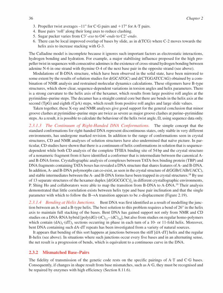
3. Propeller twist averages –11° for C�G pairs and �17° for A�T pairs.4. Base pairs ‘roll’ along their long axes to reduce clashing.5. Sugar pucker varies from C3�-exo to O4�-endo to C2�-endo.6. There can be local improved overlap of bases by slide, as in d(TCG) where C-2 moves towards the
helix axis to increase stacking with G-3.
The Calladine model is incomplete because it ignores such important factors as electrostatic interactions,hydrogen bonding and hydration. For example, a major stabilising influence proposed for the high pro-peller twist in sequences with consecutive adenines is the existence of cross-strand hydrogen bonding betweenadenine N-6 in one strand with thymine O-4 of the next base pair in the opposite strand (see above).
Modulations of B-DNA structure, which have been observed in the solid state, have been mirrored tosome extent by the results of solution studies for d(GCATGC) and d(CTGGATCCAG) obtained by a com-bination of NMR analysis and restrained molecular dynamics calculations. These oligomers have B-typestructures, which show clear, sequence-dependent variations in torsion angles and helix parameters. Thereis a strong curvature to the helix axis of the hexamer, which results from large positive roll angles at thepyrimidine–purine steps. The decamer has a straight central core but there are bends in the helix axis at thesecond (TpG) and eighth (CpA) steps, which result from positive roll angles and large slide values.
Taken together, these X-ray and NMR analyses give good support for the general conclusion that minorgroove clashes at pyrimidine–purine steps are twice as severe as major groove clashes at purine–pyrimidinesteps. As a result, it is possible to calculate the behaviour of the helix twist angle, �, using sequence data only.
2.3.1.3 The Continuum of Right-Handed DNA Conformations. The simple concept that thestandard conformations for right-handed DNA represent discontinuous states, only stable in very differentenvironments, has undergone marked revision. In addition to the range of conformations seen in crystalstructures, CD and NMR analyses of solution structures have also undermined that naïve picture. In par-ticular, CD studies have shown that there is a continuum of helix conformations in solution that is sequence-dependent while both CD analysis of the complete TFIIIA binding site of 54 bp and the crystal structureof a nonameric fragment from it have identified a conformer that is intermediate between the canonical A-and B-DNA forms. Crystallographic analysis of complexes between TATA-box binding protein (TBP) andDNA fragments containing TATA boxes has revealed a DNA structure that shares features of A- and B-DNA.In addition, A- and B-DNA polymorphs can co-exist, as seen in the crystal structure of d(GGBrUABrUACC),and stable intermediates between the A- and B-DNA forms have been trapped in crystal structures.18 By useof 13 separate structures of the hexamer duplex [d(GGCGCC)]2 in different crystallographic environments,P. Shing Ho and collaborators were able to map the transition from B-DNA to A-DNA.19 Their analysisdemonstrated that little correlation exists between helix type and base pair inclination and that the singleparameter with which to follow the B→A transition appears to be x-displacement (Figure 2.19).
2.3.1.4 Bending at Helix Junctions. Bent DNA was first identified as a result of modelling the junc-tion between an A- and a B-type helix. The best solution to this problem requires a bend of 26° in the helixaxis to maintain full stacking of the bases. Bent DNA has gained support not only from NMR and CDstudies on a DNA�RNA hybrid [poly(dG)�(rC)11– (dC)16], but also from studies on regular homo-polymerswhich contain (dA)5�(dT)5 sections occurring in phase in each turn of a 10- or 11-fold helix. Moreover,bent DNA containing such dA�dT repeats has been investigated from a variety of natural sources.
It appears that bending of this sort happens at junctions between the stiff [dA�dT] helix and the regularB-helix (see above). In situations where such junctions occur every five bases and in an alternating sense,the net result is a progression of bends, which is equivalent to a continuous curve in the DNA.
2.3.2 Mismatched Base–Pairs
The fidelity of transmission of the genetic code rests on the specific pairings of A�T and C�G bases.Consequently, if changes in shape result from base mismatches, such as A�G, they must be recognised andbe repaired by enzymes with high efficiency (Section 8.11.6).
36 Chapter 2
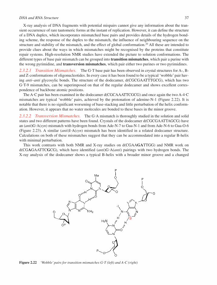
X-ray analysis of DNA fragments with potential mispairs cannot give any information about the tran-sient occurrence of rare tautomeric forms at the instant of replication. However, it can define the structureof a DNA duplex, which incorporates mismatched base pairs and provides details of the hydrogen bond-ing scheme, the response of the duplex to the mismatch, the influence of neighbouring sequence on thestructure and stability of the mismatch, and the effect of global conformation.20 All these are intended toprovide clues about the ways in which mismatches might be recognised by the proteins that constituterepair systems. High-resolution NMR studies have extended the picture to solution conformations. Thedifferent types of base pair mismatch can be grouped into transition mismatches, which pair a purine withthe wrong pyrimidine, and transversion mismatches, which pair either two purines or two pyrimidines.
2.3.2.1 Transition Mismatches. The G�T base pair has been observed in crystal structures for A-, B-and Z-conformations of oligonucleotides. In every case it has been found to be a typical ‘wobble’ pair hav-ing anti–anti glycosylic bonds. The structure of the dodecamer, d(CGCGAATTTGCG), which has twoG�T-9 mismatches, can be superimposed on that of the regular dodecamer and shows excellent corres-pondence of backbone atomic positions.
The A�C pair has been examined in the dodecamer d(CGCAAATTCGCG) and once again the two A-4�Cmismatches are typical ‘wobble’ pairs, achieved by the protonation of adenine-N-1 (Figure 2.22). It isnotable that there is no significant worsening of base-stacking and little perturbation of the helix conform-ation. However, it appears that no water molecules are bonded to these bases in the minor groove.
2.3.2.2 Transversion Mismatches. The G�A mismatch is thoroughly studied in the solution and solidstates and two different patterns have been found. Crystals of the dodecamer d(CGCGAATTAGCG) havean (anti)G�A(syn) mismatch with hydrogen bonds from Ade-N-7 to Gua-N-1 and from Ade-N-6 to Gua-O-6(Figure 2.23). A similar (anti)I�A(syn) mismatch has been identified in a related dodecamer structure.Calculations on both of these mismatches suggest that they can be accommodated into a regular B-helixwith minimal perturbation.
This work contrasts with both NMR and X-ray studies on d(CGAAGATTGG) and NMR work ond(CGAGAATTCGCG), which have identified (anti)G�A(anti) pairings with two hydrogen bonds. The X-ray analysis of the dodecamer shows a typical B-helix with a broader minor groove and a changed
DNA and RNA Structure 37
Figure 2.22 ‘Wobble’ pairs for transition mismatches G�T (left) and A�C (right)
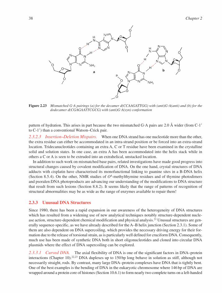
pattern of hydration. This arises in part because the two mismatched G�A pairs are 2.0 Å wider (from C-1�to C-1�) than a conventional Watson–Crick pair.
2.3.2.3 Insertion–Deletion Mispairs. When one DNA strand has one nucleotide more than the other,the extra residue can either be accommodated in an intra-strand position or be forced into an extra-strandlocation. Tridecanucleotides containing an extra A, C or T residue have been examined in the crystallinesolid and solution states. In one case, an extra A has been accommodated into the helix stack while in others a C or A is seen to be extruded into an extrahelical, unstacked location.
In addition to such work on mismatched base pairs, related investigations have made good progress intostructural changes caused by covalent modification of DNA. On the one hand, crystal structures of DNAadducts with cisplatin have characterised its monofunctional linking to guanine sites in a B-DNA helix(Section 8.5.4). On the other, NMR studies of O4-methylthymine residues and of thymine photodimersand psoralen:DNA photoproducts are advancing our understanding of the modifications to DNA structurethat result from such lesions (Section 8.8.2). It seems likely that the range of patterns of recognition ofstructural abnormalities may be as wide as the range of enzymes available to repair them!
2.3.3 Unusual DNA Structures
Since 1980, there has been a rapid expansion in our awareness of the heterogeneity of DNA structureswhich has resulted from a widening use of new analytical techniques notably structure-dependent nucle-ase action, structure-dependent chemical modification and physical analysis.21 Unusual structures are gen-erally sequence-specific, as we have already described for the A–B helix junction (Section 2.3.1). Some ofthem are also dependent on DNA supercoiling, which provides the necessary driving energy for their for-mation due to the release of torsional strain, as is particularly well defined for cruciform DNA. Consequently,much use has been made of synthetic DNA both in short oligonucleotides and cloned into circular DNAplasmids where the effect of DNA supercoiling can be explored.
2.3.3.1 Curved DNA. The axial flexibility of DNA is one of the significant factors in DNA–proteininteractions (Chapter 10).22,23 DNA duplexes up to 150 bp long behave in solution as stiff, although notnecessarily straight, rods. By contrast, many large DNA–protein complexes have DNA that is tightly bent.One of the best examples is the bending of DNA in the eukaryotic chromosome where 146 bp of DNA arewrapped around a protein core of histones (Section 10.6.1) to form nearly two complete turns on a left-handed
38 Chapter 2
Figure 2.23 Mismatched G�A pairings (a) for the decamer d(CCAAGATTGG) with (anti)G�A(anti) and (b) for thedodecamer d(CGAGAATTCGCG) with (anti)G�A(syn) conformation
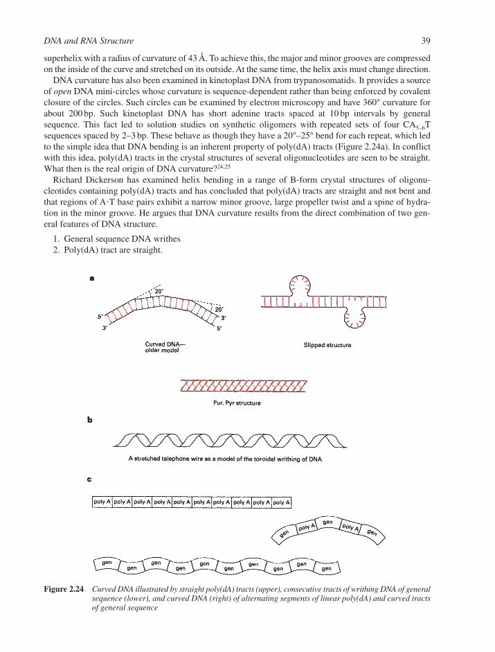
superhelix with a radius of curvature of 43 Å. To achieve this, the major and minor grooves are compressedon the inside of the curve and stretched on its outside. At the same time, the helix axis must change direction.
DNA curvature has also been examined in kinetoplast DNA from trypanosomatids. It provides a sourceof open DNA mini-circles whose curvature is sequence-dependent rather than being enforced by covalentclosure of the circles. Such circles can be examined by electron microscopy and have 360° curvature forabout 200 bp. Such kinetoplast DNA has short adenine tracts spaced at 10 bp intervals by generalsequence. This fact led to solution studies on synthetic oligomers with repeated sets of four CA5–6Tsequences spaced by 2–3 bp. These behave as though they have a 20°–25° bend for each repeat, which ledto the simple idea that DNA bending is an inherent property of poly(dA) tracts (Figure 2.24a). In conflictwith this idea, poly(dA) tracts in the crystal structures of several oligonucleotides are seen to be straight.What then is the real origin of DNA curvature?24,25
Richard Dickerson has examined helix bending in a range of B-form crystal structures of oligonu-cleotides containing poly(dA) tracts and has concluded that poly(dA) tracts are straight and not bent andthat regions of A�T base pairs exhibit a narrow minor groove, large propeller twist and a spine of hydra-tion in the minor groove. He argues that DNA curvature results from the direct combination of two gen-eral features of DNA structure.
1. General sequence DNA writhes2. Poly(dA) tract are straight.
DNA and RNA Structure 39
Figure 2.24 Curved DNA illustrated by straight poly(dA) tracts (upper), consecutive tracts of writhing DNA of generalsequence (lower), and curved DNA (right) of alternating segments of linear poly(dA) and curved tractsof general sequence

Studies on the hydrodynamic properties of DNA show that general-sequence DNA migrates throughgels more slowly than expected which is because the DNA helix occupies a cylindrical volume that has alarger diameter than that of a simple B-helix (Section 11.4.3). This phenomenon is a result of DNA writhing,which involves a continuously curved distortion of the helix axis to generate a spiral form and is nicely illus-trated by the extension of a coiled telephone wire (Figure 2.24b). It follows that the repeated alternation ofstraight A-tracts with short sections of general sequence, each having half of a writhing turn, will generatecurved DNA (Figure 2.24c). A detailed structural analysis of this explanation says that curvature of B-DNAinvolves rolling of base pairs, compresses the major groove (which corresponds to positive roll), has asequence-determined continuum in the bending behaviour, and shows anisotropy of flexible bending.
2.3.3.2 DNA Bending. Such intrinsic, sequence-dependent curvature must be distinguished from thebending of DNA, which results from the application of an external force. Dickerson has also examined thebending of the DNA helix that occurs in many crystal structures of the B-form. It is associated with the step from a G�C to an A�T base pair and results from rolling one base pair over the next along theirlong axes in a direction that compresses the major groove (Figure 2.19c). He suggests that this junction isa flexible hinge that is capable of bending or not bending. Such ‘facultative bending’ responds to the influ-ence of local forces, typically interactions with other macromolecules, for example control proteins or anucleosome core. By contrast, poly(dA) tracts are known to resist bending in nucleosome reconstitutionexperiments. It can thus be seen that sequence-dependent variation in DNA bendability is an important fac-tor in DNA recognition by proteins.
One important conclusion emerges: DNA has evolved conformationally to interact with other macro-molecules. A free, linear DNA helix in solution may, in fact, be the least biologically relevant state of all.24
Slipped structures have been postulated to occur at direct repeat sequences, and they have been foundup-stream of important regulatory sites. The structures described (Figure 2.24a) are consistent with thepattern of cleavage by single-strand nucleases but otherwise are not well characterised.
Purine–pyrimidine tracts manifest an unusual structure at low temperature with a long-range, sequence-dependent single base shift in base-pairing in the major groove. For the dodecamer d(ACCGGCGCCACA)�d(TGTGGCGCCGGT), the bases in the d(CA)n tract have high propeller twist (–32°) and are so stronglytilted in the 3�-direction that there is disruption of Watson–Crick pairing in the major groove and forma-tion of interactions with the 5�-neighbour of the complementary base. This alteration propagates along theB-form helix for at least half a turn with a domino-like motion. As a result, the DNA structure is normalwhen viewed from the minor groove and mismatched when seen from the major groove. Since (CA)n tractsare involved both in recombination and in transcription, this new recognition pattern has to be consideredin the analysis of the various processes involved with reading of genetic information.
Anisomorphic DNA is the description given to DNA conformations associated with direct repair, DR2,sequences at ‘joint regions’ in viral DNA, which are known to have unusual chemical and physical properties.The two complementary strands have different structures and this leads to structural aberrations at the centreof the tandem sequences that can be seen under conditions of torsional stress induced by negative supercoiling.
Hairpin loops are formed by oligonucleotide single strands which have a segment of inverted comple-mentary sequence. For example, the 16-mer d(CGCGCGTTTTCGCGCG) has a hexamer repeat and itscrystal structure shows a hairpin with a loop of four Ts and a Z-DNA hexamer stem (Figure 2.25a). Whensuch inverted sequences are located in a DNA duplex, the conditions exist for formation of a cruciform.
Cruciforms involve intra-strand base-pairing and generate two stems and two hairpin loops from a sin-gle unwound duplex region.26 The inverted sequence repeats are known as palindromes, which have agiven DNA duplex sequence followed after a short break by the same duplex sequence in the oppositedirection. This is illustrated for a segment of the bacterial plasmid pBR322 (Figure 2.25b), where a palin-drome of two undecamer sequences exists.
X-ray, NMR and sedimentation studies of such stem–loop structures show that the four arms are aligned in pairs to give an oblique X structure with continuity of base-stacking and helical axes across the junc-tions (Section 6.8.1). Also, the loops have an optimum size of from four to six bases. Residues in the loops
40 Chapter 2
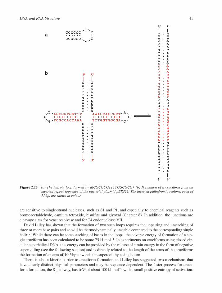
are sensitive to single-strand nucleases, such as S1 and P1, and especially to chemical reagents such as bromoacetaldehyde, osmium tetroxide, bisulfite and glyoxal (Chapter 8). In addition, the junctions arecleavage sites for yeast resolvase and for T4 endonuclease VII.
David Lilley has shown that the formation of two such loops requires the unpairing and unstacking ofthree or more base pairs and so will be thermodynamically unstable compared to the corresponding singlehelix.27 While there can be some stacking of bases in the loops, the adverse energy of formation of a sin-gle cruciform has been calculated to be some 75 kJ mol1. In experiments on cruciforms using closed cir-cular superhelical DNA, this energy can be provided by the release of strain energy in the form of negativesupercoiling (see the following section) and is directly related to the length of the arms of the cruciform:the formation of an arm of 10.5 bp unwinds the supercoil by a single turn.
There is also a kinetic barrier to cruciform formation and Lilley has suggested two mechanisms thathave clearly distinct physical parameters and may be sequence-dependent. The faster process for cruci-form formation, the S-pathway, has �G‡ of about 100 kJ mol1 with a small positive entropy of activation.
DNA and RNA Structure 41
Figure 2.25 (a) The hairpin loop formed by d(CGCGCGTTTTCGCGCG). (b) Formation of a cruciform from aninverted repeat sequence of the bacterial plasmid pBR322. The inverted palindromic regions, each of11 bp, are shown in colour
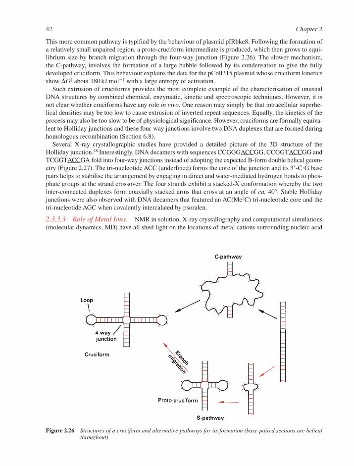
This more common pathway is typified by the behaviour of plasmid pIRbke8. Following the formation ofa relatively small unpaired region, a proto-cruciform intermediate is produced, which then grows to equi-librium size by branch migration through the four-way junction (Figure 2.26). The slower mechanism, the C-pathway, involves the formation of a large bubble followed by its condensation to give the fullydeveloped cruciform. This behaviour explains the data for the pColl315 plasmid whose cruciform kineticsshow �G‡ about 180 kJ mol1 with a large entropy of activation.
Such extrusion of cruciforms provides the most complete example of the characterisation of unusualDNA structures by combined chemical, enzymatic, kinetic and spectroscopic techniques. However, it isnot clear whether cruciforms have any role in vivo. One reason may simply be that intracellular superhe-lical densities may be too low to cause extrusion of inverted repeat sequences. Equally, the kinetics of theprocess may also be too slow to be of physiological significance. However, cruciforms are formally equiva-lent to Holliday junctions and these four-way junctions involve two DNA duplexes that are formed duringhomologous recombination (Section 6.8).
Several X-ray crystallographic studies have provided a detailed picture of the 3D structure of theHolliday junction.28 Interestingly, DNA decamers with sequences CCGGGACCGG, CCGGTACCGG andTCGGTACCGA fold into four-way junctions instead of adopting the expected B-form double helical geom-etry (Figure 2.27). The tri-nucleotide ACC (underlined) forms the core of the junction and its 3�-C�G basepairs helps to stabilise the arrangement by engaging in direct and water-mediated hydrogen bonds to phos-phate groups at the strand crossover. The four strands exhibit a stacked-X conformation whereby the twointer-connected duplexes form coaxially stacked arms that cross at an angle of ca. 40°. Stable Hollidayjunctions were also observed with DNA decamers that featured an AC(Me5C) tri-nucleotide core and thetri-nucleotide AGC when covalently intercalated by psoralen.
2.3.3.3 Role of Metal Ions. NMR in solution, X-ray crystallography and computational simulations(molecular dynamics, MD) have all shed light on the locations of metal cations surrounding nucleic acid
42 Chapter 2
Figure 2.26 Structures of a cruciform and alternative pathways for its formation (base-paired sections are helicalthroughout)

molecules.29 But while ions are often visible in structures of nucleic acids, it is not straightforward todetermine how they affect the structure. The question of whether cations can assume specific roles in thecontrol of DNA duplex conformation has stirred up controversy in recent years. Some in the field, notablyNicholas Hud and Loren Williams, believe that cation localisation within the grooves of DNA representsa significant factor in sequence-specific helical structure. By contrast, probably a majority of those study-ing the structure of DNA is of the opinion that the specific sequence dictates local DNA conformation andthus binding of metal cations. According to this second view, metal ions can bind to DNA in a sequence-specific manner and in turn modulate the local structure, but ions should not be considered the single mostsignificant driving force of a number of DNA conformational phenomena.
To study the possible effects of metal ions on DNA conformation, all sequences can be divided intothree principal groups: A-tracts, G-tracts and generic DNA, the latter representing the vast majority of DNAsequences.30 A-tracts have an unusually narrow minor groove, are straight and have high base pair pro-peller twist (see also Figures 2.18 and 2.19). G-tracts have a propensity to undergo the B-form→A-formtransition at increased ionic strength. The proponents of the ‘ions are dominant’model believe that the DNAgrooves are flexible ionophores and that DNA duplex structure is modulated by a tug of war between thetwo grooves for cation localisation. They argue that the duplex geometry adopted by A-tracts (referred toas B*-DNA, Figure 2.28a) is due to ion localisation in the minor groove as a result of the highly negativeelectrostatic potentials there. Conversely, G-tract DNA exhibits a highly negative electrostatic potential in themajor groove (Figure 2.28b), leading to preferred localisation of cations there and consequently a collapseof the DNA around the ions. Generic DNA on the other hand would have a more balanced occupation of
DNA and RNA Structure 43
Figure 2.27 The Holliday junction adopted by four DNA decamers with sequence TCGGTACCGA (PDB: 1M6G).The view illustrates the side-by-side arrangement of the two duplex portions, with the A nucleotides (red) Cnucleotides (pink) of two decamers that form the core of the junction visible near the centre. The positionsof phosphate groups in the backbones of individual oligonucleotides are traced with ribbons
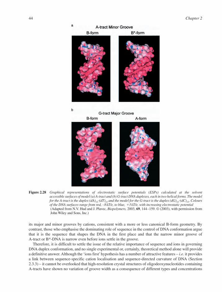
its major and minor grooves by cations, consistent with a more or less canonical B-form geometry. Bycontrast, those who emphasise the dominating role of sequence in the control of DNA conformation arguethat it is the sequence that shapes the DNA in the first place and that the narrow minor groove of A-tract or B*-DNA is narrow even before ions settle in the groove.
Therefore, it is difficult to settle the issue of the relative importance of sequence and ions in governingDNA duplex conformation, and no single experimental or, certainly, theoretical method alone will providea definitive answer. Although the ‘ions first’ hypothesis has a number of attractive features – i.e. it providesa link between sequence-specific cation localisation and sequence-directed curvature of DNA (Section2.3.3) – it cannot be overlooked that high-resolution crystal structures of oligodeoxynucleotides containingA-tracts have shown no variation of groove width as a consequence of different types and concentrations
44 Chapter 2
Figure 2.28 Graphical representations of electrostatic surface potentials (ESPs) calculated at the solventaccessible surfaces of model (a) A-tract and (b) G-tract DNA duplexes, each in two helical forms. The modelfor the A-tract is the duplex (dA)12�(dT)12 and the model for the G-tract is the duplex (dG)12�(dC)12. Coloursof the DNA surfaces range from red, –8 kT/e, to blue, �3 kT/e, with increasing electrostatic potential(Adapted from N.V. Hud and J. Plavec, Biopolymers, 2003, 69, 144–159. © (2003), with permission fromJohn Wiley and Sons, Inc.)
of alkali metal ions present in the crystallisations. Moreover, MD simulations of A-tract DNA in the pres-ence of different classes and varying localisations of metal cations have not provided a picture that is con-sistent with a crucial role for metal ions with regard to the structure of duplex DNA. Thus, there willundoubtedly be more studies directed at a refined understanding of the relative importance of sequenceand cation co-ordination in governing the structure of double helical DNA.
2.3.4 B–Z Junctions and B–Z Transitions
Segments of left-handed Z-DNA can exist in a single duplex in continuity with segments of right-handedB-DNA. This phenomenon has been observed both in vitro and in vivo. Because the backbone chains of thesepolymorphs run in opposite directions (↓↑and ↑↓) respectively (Section 2.2.5), there has to be a transi-tional region between two such segments, and this boundary is known as a B–Z junction. Such structuresare polymorphic and sequence-specific and six features have been described:
1. B–Z junction can be as small as 3 bp.2. At least one base pair has neither the B- nor the Z-conformation.3. Hydrogen bonds between the base pairs are intact below 50°C.4. Chemical reagents specific for single-stranded DNA (chloroacetaldehyde, bromoacetaldehyde and
glyoxal; Section 8.5.3) show high reactivity with the junction bases.5. Junctions are sites for enhanced intercalation for psoralens (Section 8.8.2).6. Junctions are neither strongly bent nor particularly flexible.
This conformational B–Z transition between the right- and left-handed helices has a high energy of acti-vation (about 90 kJ mol1) but is practically independent of temperature (�G° about 0 kJ mol1) (Section2.5.3). Thus, the B–Z transition is co-operative and propagates readily along the helix chains.
In the absence of structural data at high resolution, two different models had been suggested to explainthe conformational switch that has to occur as a B–Z junction migrates, rather like a bubble, along a dou-ble helix. In the first model, the bases unpair, guanine flips into the syn conformation, the entire deoxycy-tidine undergoes a conformational switch, and the base pairs reform their hydrogen bonds. This modelappears to be at variance with NMR studies that suggest the bases remain paired because their imino-protons do not become free to exchange with solvent water. In the second model, the backbone is stretcheduntil one base pair has sufficient room to rotate 180° about its glycosyl bonds (tip, as shown in Figure 2.19a),and the bases re-stack. However, one might expect this ‘expand–rotate–collapse’ process to be impeded bylinking bulky molecules to the edge of the base pairs. Yet, bonding N-acetoxy-N-acetyl-2-aminofluoreneto guanine actually facilitates the B–Z transition. Thus, the dynamics of the B–Z transition poses a majorconformational problem, and this has sometimes been called the chain–sense paradox.
In addition, Ansevin and Wang suggested an alternative zig-zag model for the left-handed double-helical form of DNA that avoids this paradox and is accessible from B-DNA by simple untwisting. TheirW-DNA has a Watson–Crick chain sense (↑↓) like B-DNA but similar glycosyl geometry to that of Z-DNA. It has reversed sugar puckers, C3�-endo at cytosine and C2�-endo at guanine, while in both W- andZ-DNA the minor groove is deep and the major groove broad and very shallow. In addition, this W-modelexplains (1) the incompatibility of poly(dA–dT)�poly(dA–dT) with a left-handed state, (2) the very slowrate of exchange of hydrogens in the 2-NH2 group of guanine in left-handed DNA and (3) the incompati-bility of left-handed helix with replacement of OR oxygens by a methyl group. They argue that Z-DNA hasa lower energy than W-DNA and so is adopted in crystals of short oligonucleotides but it may be confor-mationally inaccessible to longer stretches of DNA in solution.
Finally, more than 25 years after the discovery of Z-DNA, a crystal structure of a B–Z junction has solvedthe mystery of how DNA switches from the right-handed low energy to the left-handed high energy form.31
The junction was trapped in the crystal by stabilising the Z-DNA portion at one end of a 15-bp segmentwith a Z-DNA binding protein, with the rest of the DNA assuming the B-form geometry. Continuousstacking of bases between B-DNA and Z-DNA is found with the breaking of one base pair at the junction
DNA and RNA Structure 45

and the two bases extruded from the helix on either side. A sharp turn accommodates the reversal in thebackbone direction and at the junction the DNA is bent by ca. 10° and the helical axes of the B- and Z-form duplexes are displaced from each other by ca. 5 Å.
2.3.5 Circular DNA and Supercoiling
The replicative form of bacteriophage �X174 DNA was found to be a double-stranded closed circle. It waslater shown that bacterial DNA exists as closed circular duplexes, that DNA viruses have either single- ordouble-helical circular DNA, and that RNA viroids have circular single-stranded RNA as their genomicmaterial. Plasmid DNAs also exist as small, closed circular duplexes.
Topologically unconstrained dsDNA in its linear, relaxed state is either biologically inactive or displaysreduced activity in key processes such as recombination, replication or transcription. It follows that topo-logical changes associated with the constraints of circularisation of dsDNA have a profound biologicalsignificance.32 Although such circularisation can be achieved directly by covalent closure, the same effect canbe achieved for eukaryotic DNA as a result of holding DNA loops together by means of a protein scaffold.
The molecular topology of closed circular DNA was described by Vinograd in 1965 and is especiallyassociated with the phenomenon of superhelical DNA, which is also called supercoiled or supertwisted DNA.Vinograd’s basic observation was that when a planar, relaxed circle of DNA is strained by changing the pitchof its helical turns, it relieves this torsional strain by winding around itself to form a superhelix whose axisis a diameter of the original circle.
This behaviour is most directly observed by following the sedimentation of negatively supercoiled DNAas the pitch of its helix is changed by intercalating a drug, typically ethidium bromide (Section 9.6).Intercalation is the process of slotting the planar drug molecules between adjacent base pairs in the helix.For each ethidium molecule intercalated into the helix there is an increase of about 3.4 Å and a linkeddecrease of about 36° in twist (Figure 2.19c,d). The DNA helix responds first by reducing the number ofnegative, right-handed supercoils until it is fully relaxed and then by increasing the number of positive,left-handed supercoils. As this happens, the sedimentation coefficient of the DNA first decreases, reachinga minimum when fully relaxed, and then increases as it becomes positively supercoiled. As a control process,the same circular DNA can be nicked in one strand to make it fully relaxed. The result is that it now showsa low sedimentation coefficient at all concentrations of the intercalator species (Figure 2.29) (Section 11.4.1).
Vinograd showed that the topological state of these covalently closed circles can be defined by threeparameters and that the fundamental topological property is linkage. The topological winding number, Tw,is the number of right-handed helical turns in the relaxed, planar DNA circle and the writhing number, Wr,gives the number of left-handed crossovers in the supercoil. The sum of these two is the linking number,Lk, which is the number of times one strand of the helix winds around the other (clock-wise is positive)when the circle is constrained to lie in a plane. The simple equation is Lk � Tw � Wr.
Such behaviour can be illustrated simply (Figure 2.30) for a relaxed closed circle with 20 helical turnsTw � 20, Lk � 20, Wr � 0. One strand is now cut, unwound two turns, and resealed to give Lk � Tw � 18.This circle is thus under-wound by two turns. To restore fully the normal B-DNA base-pairing and base-stacking, the circle needs to gain two right-handed helical turns, �Tw � �2 to give Tw � 20. Since theDNA circles have remained closed and the linking number stays at 18, the formation of the right-handedhelical turns is balanced by the creation of one right-handed supercoil, making Wr � –2.
The behaviour of a supercoil can be modelled using a length of rubber tubing. The ends are first heldtogether to form a relaxed closed circle. If the end in your right hand is given one turn clock-wise (right-handed twist) and the other end is given one turn in the opposite sense, the tube will relieve this strain byforming one left-handed supercoil. This is equivalent to unwinding the DNA helix by two turns, whichgenerates one positive supercoil (four turns generate two supercoils, and so on). This model shows therelationship: two turns equals one supercoil.
In practice, it is sometimes useful to describe the degree of supercoiling using the super-helical density,� � Wr /Tw, which is close to the number of superhelical turns per 10 bp and is typically around 0.06 for
46 Chapter 2

superhelical DNA from cells and virions. The energy of supercoiling is a quadratic function of the densityof supercoils as described by the equation
where R is the gas constant, T the absolute temperature and N the number of base pairs.B–Z transitions are especially important for supercoiling since the conversion of one right-handed B-
turn into a left-handed Z-turn causes a change in Tw of –2. This must be complemented by �Wr � 2through the formation of one left-handed superturn.
� � � GRT
NLks kJ mol1050 2 1
DNA and RNA Structure 47
Figure 2.30 Supercoil formation in closed circular DNA. (a) Closed circle of 20 duplex turns (alternate turns in colour). (b) Circle nicked, under-wound two turns, and re-sealed. (c) Base pairing and stackingforces result in the formation of B-helix with two new right-handed helix turns and one compensatingright-handed supercoil
Figure 2.29 Sedimentation velocity for SV40 DNA as a function of bound ethidium (a) for closed circular DNA(– � – � – � –) and (b) for nicked circular DNA (–�–�–�– ) showing the transition from a negativesupercoil (left) through a relaxed circle (centre) to a positive supercoil (right)(Adapted from W. Bauer and J. Vinograd, J. Mol. Biol., 1968, 33, 141. © (1968), with permission fromElsevier)
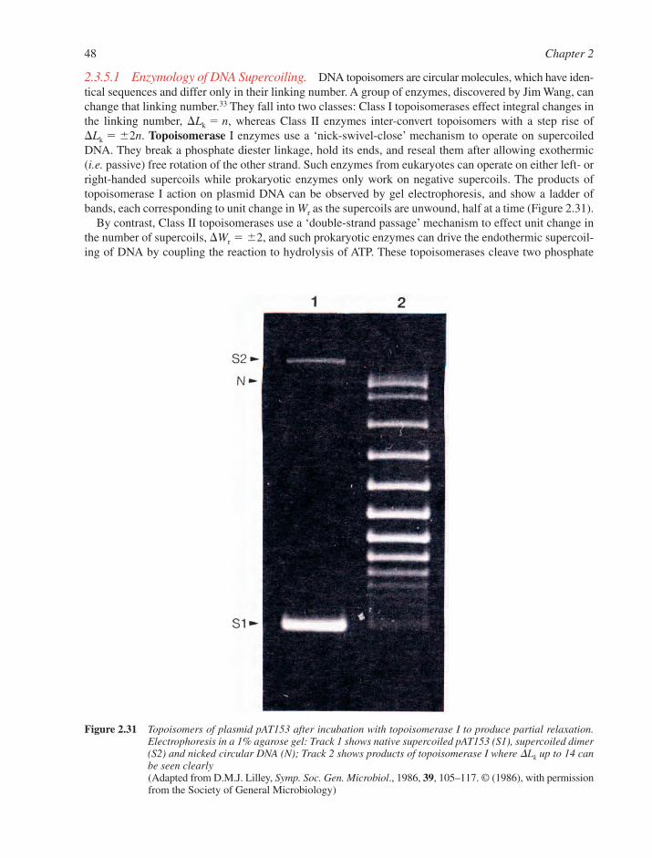
2.3.5.1 Enzymology of DNA Supercoiling. DNA topoisomers are circular molecules, which have iden-tical sequences and differ only in their linking number. A group of enzymes, discovered by Jim Wang, canchange that linking number.33 They fall into two classes: Class I topoisomerases effect integral changes inthe linking number, �Lk � n, whereas Class II enzymes inter-convert topoisomers with a step rise of�Lk � �2n. Topoisomerase I enzymes use a ‘nick-swivel-close’ mechanism to operate on supercoiledDNA. They break a phosphate diester linkage, hold its ends, and reseal them after allowing exothermic(i.e. passive) free rotation of the other strand. Such enzymes from eukaryotes can operate on either left- orright-handed supercoils while prokaryotic enzymes only work on negative supercoils. The products oftopoisomerase I action on plasmid DNA can be observed by gel electrophoresis, and show a ladder ofbands, each corresponding to unit change in Wr as the supercoils are unwound, half at a time (Figure 2.31).
By contrast, Class II topoisomerases use a ‘double-strand passage’ mechanism to effect unit change inthe number of supercoils, �Wr � �2, and such prokaryotic enzymes can drive the endothermic supercoil-ing of DNA by coupling the reaction to hydrolysis of ATP. These topoisomerases cleave two phosphate
48 Chapter 2
Figure 2.31 Topoisomers of plasmid pAT153 after incubation with topoisomerase I to produce partial relaxation.Electrophoresis in a 1% agarose gel: Track 1 shows native supercoiled pAT153 (S1), supercoiled dimer(S2) and nicked circular DNA (N); Track 2 shows products of topoisomerase I where �Lk up to 14 canbe seen clearly(Adapted from D.M.J. Lilley, Symp. Soc. Gen. Microbiol., 1986, 39, 105–117. © (1986), with permissionfrom the Society of General Microbiology)
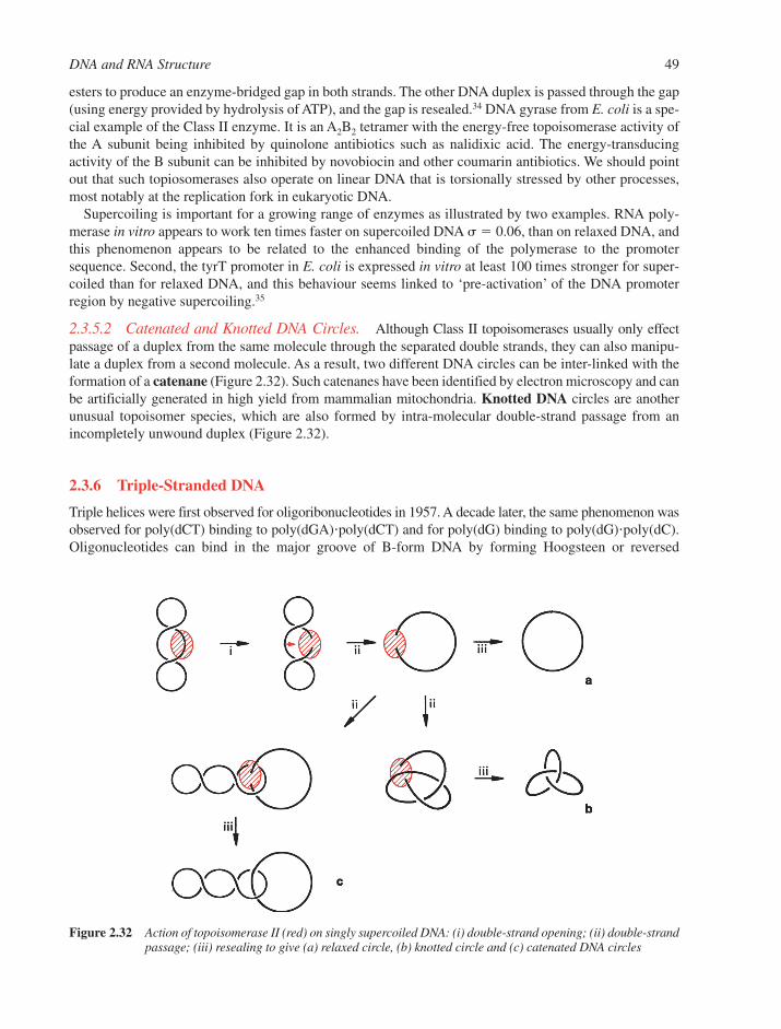
esters to produce an enzyme-bridged gap in both strands. The other DNA duplex is passed through the gap(using energy provided by hydrolysis of ATP), and the gap is resealed.34 DNA gyrase from E. coli is a spe-cial example of the Class II enzyme. It is an A2B2 tetramer with the energy-free topoisomerase activity ofthe A subunit being inhibited by quinolone antibiotics such as nalidixic acid. The energy-transducingactivity of the B subunit can be inhibited by novobiocin and other coumarin antibiotics. We should pointout that such topiosomerases also operate on linear DNA that is torsionally stressed by other processes,most notably at the replication fork in eukaryotic DNA.
Supercoiling is important for a growing range of enzymes as illustrated by two examples. RNA poly-merase in vitro appears to work ten times faster on supercoiled DNA � � 0.06, than on relaxed DNA, andthis phenomenon appears to be related to the enhanced binding of the polymerase to the promotersequence. Second, the tyrT promoter in E. coli is expressed in vitro at least 100 times stronger for super-coiled than for relaxed DNA, and this behaviour seems linked to ‘pre-activation’ of the DNA promoterregion by negative supercoiling.35
2.3.5.2 Catenated and Knotted DNA Circles. Although Class II topoisomerases usually only effectpassage of a duplex from the same molecule through the separated double strands, they can also manipu-late a duplex from a second molecule. As a result, two different DNA circles can be inter-linked with theformation of a catenane (Figure 2.32). Such catenanes have been identified by electron microscopy and canbe artificially generated in high yield from mammalian mitochondria. Knotted DNA circles are anotherunusual topoisomer species, which are also formed by intra-molecular double-strand passage from anincompletely unwound duplex (Figure 2.32).
2.3.6 Triple-Stranded DNA
Triple helices were first observed for oligoribonucleotides in 1957. A decade later, the same phenomenon wasobserved for poly(dCT) binding to poly(dGA)�poly(dCT) and for poly(dG) binding to poly(dG)�poly(dC).Oligonucleotides can bind in the major groove of B-form DNA by forming Hoogsteen or reversed
DNA and RNA Structure 49
Figure 2.32 Action of topoisomerase II (red) on singly supercoiled DNA: (i) double-strand opening; (ii) double-strandpassage; (iii) resealing to give (a) relaxed circle, (b) knotted circle and (c) catenated DNA circles
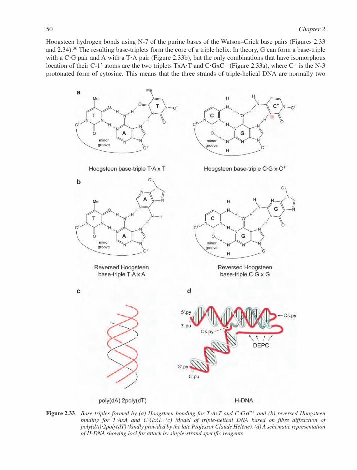
Hoogsteen hydrogen bonds using N-7 of the purine bases of the Watson–Crick base pairs (Figures 2.33and 2.34).36 The resulting base-triplets form the core of a triple helix. In theory, G can form a base-triplewith a C�G pair and A with a T�A pair (Figure 2.33b), but the only combinations that have isomorphouslocation of their C-1� atoms are the two triplets TxA�T and C�GxC� (Figure 2.33a), where C� is the N-3protonated form of cytosine. This means that the three strands of triple-helical DNA are normally two
50 Chapter 2
Figure 2.33 Base triples formed by (a) Hoogsteen bonding for T�AxT and C�GxC� and (b) reversed Hoogsteenbinding for T�AxA and C�GxG. (c) Model of triple-helical DNA based on fibre diffraction ofpoly(dA)�2poly(dT) (kindly provided by the late Professor Claude Hélène). (d) A schematic representationof H-DNA showing loci for attack by single-strand specific reagents

homo-pyrimidines and one homo-purine (Figure 2.33c). However, despite the backbone distortion thatmust result from the hetero-morphism of other base triplet combinations, oligonucleotides containing Gand T, G and A, or G, T and C have been shown to form helices.
Intermolecular triple helices are now well characterised for short oligonucleotides binding in the grooveof a longer DNA duplex,37,38 H-DNA provides an example of an intramolecular triple helix because it hasa mirror-repeat sequence relating homo-purine and homo-pyrimidine tracts in a circular double-strandedDNA molecule, and triplex formation is driven by supercoiling (Figure 2.33d).
DNA and RNA Structure 51
Figure 2.34 The triple helical structure formed in the crystal structure of the 1:1 complex between the DNA 12 merd(CTCCTCCGCGCC) and the 9 mer d(CGCGCGGAG) (PDB: 1D3R; available at: http://www.rcsb.org).39 The triplex segment consists of two 5�-halves of the 12 mer (underlined, top and bottom left)and two 3�-terminal trimers GAG (underlined; top and bottom right) from two 9 mers that are stackedtail-to-tail in the crystal: 5�-…-GAG-3� \ 3�-GAG-…-5� (visible near the centre, left of drawing). DNAbases are coloured red, grey, pink and black for G, A, C and T, respectively, and the directions of thesugar–phosphate backbones are traced by ribbons
Several studies on third-strand binding to a homo-purine � homo-pyrimidine duplex have establishedthe following features:
� A third homo-pyrimidine strand binds parallel to the homo-purine strand using Hoogsteenhydrogen bonds (i.e. the homo-pyrimidine strands are anti-parallel).
� A third homo-purine strand binds anti-parallel to the original homo-purine strand using reversedHoogsteen hydrogen bonds.
� The bases in the third strand have a regular anti conformation of the glycosylic bond.� Synthetic oligodeoxyribonucleotides having an �-glycosylic linkage also bind as a third strand, par-
allel for poly(d-�-T) and anti-parallel for poly(d-�-TC).
Triple helices are less stable than duplexes. Thermodynamic parameters have been obtained from meltingcurves, from kinetics, and from the use of differential scanning calorimetry (DSC) (Section 11.4.4). Usingthis last technique, values of �H° � 22 � 2 kJ mol1 and �S° � 70 � 7 J mol1 K1 have been found ford(C�TTC�C�TC�C�TC�T). The pKa value of cytidine in isolation is 4.3 but it is higher in oligonucleo-tides because of their polyanionic phosphate backbone. So it is to be expected that the stability of triplehelices is seen to decrease as the pH rises above 5.
Triple helix stability can be enhanced by the use of modified nucleotides. 5-Methylcytosine increasesstability at neutral pH, probably by a hydrophobic effect, and 5-bromouracil can usefully replace thymine.Oligoribonucleotides bind more strongly than do deoxyribonucleotides and 2�-O-methylribonucleotidesbind even better. Finally, Hélène has shown that at the attachment of an intercalating agent to the 5�- (or3�-end) of the third strand can greatly enhance the stability of the triple-stranded helix.
The major application of triple helices relates to the specificity of the interaction between the singlestrand and a much larger DNA duplex. This is because homo-pyrimidines have been identified as poten-tial vehicles for the sequence-specific delivery of agents that can modify DNA and thereby control genes.The DNA of the bacterium E. coli has 4.5 Mbp, so the minimum number of base pairs needed to define aunique sequence in its genome is 11bp (i.e. 411 � 4,194,304 assuming a statistically random distributionof the four bases). The corresponding number for the human genome is about 17 bp. Thus, a synthetic 17-mercould be expected to identify and bind to a unique human DNA target and thus deliver a lethal agent to aspecific sequence of DNA. In practice, the energetics of mismatched base triples is complex and dependson nearest neighbours, metal ions and other parameters. However, a value of about 1.5 kJ mol1 per mis-match seems to fit much of the data and suggests that the specificity of triple helices is at least as good asthat of double-helical complexes.
2.3.6.1 H-DNA. A new polymorph of DNA was discovered in 1985 within a sequence of d(A-G)16 inthe polypurine strand of a recombinant plasmid pEJ4. Its requirement for protons led to the name H-DNA (halfof its C residues are protonated, so the transition depends on acid pH as well as on a degree of negativesupercoiling). Probes for single-stranded regions of DNA (especially osmium tetroxide:pyridine (Section 8.3)and nuclease P1 cleavage) were used to identify specific sites and provide experimental support for themodel advanced earlier for a triple helical H-DNA (Figure 2.33d). This has a Watson–Crick duplex whichextends to the centre of the (dT–dC)n�(dG–dA)n tract and the second half of the homo-pyrimidine tractthen folds back on itself, anti-parallel to the first half and winding down the major groove of the helix. Thesecond half of the poly-purine tract also folds back, probably in an unstructured single-stranded form. Theenergetics of nucleation of H-DNA suggests that it requires at least 15 bp for stability and the consequentloss of twist makes H-DNA favoured by negative supercoiling.
Although antibodies have been raised to detect triple-stranded structures, no evidence has yet beenfound for their natural existence in cells in vivo.
2.3.7 Other Non-Canonical DNA Structures
2.3.7.1 Four-Stranded Motifs. Both G- and C-rich DNA sequences have been found to adopt four-stranded motifs, also called tetraplexes or quadruplexes (see also Section 9.10.2). Sequences containing
52 Chapter 2

G- and C-rich strands are found at the telomeric ends of chromosomes (see Section 6.4.5), and suchsequences are of fundamental importance in protecting the cell from re-combination and degradation. It isknown that when DNA containing a palindromic sequence of bases is subjected to supercoiling stress(Section 2.3.5), a cruciform can be extruded (Figure 2.26). If the tips of the cruciform extrusions containC residues in one limb and G residues in the other, G- and C-rich quadruplexes can be formed by com-bining two cruciforms of this type. So it is possible that formation of four-stranded G- and C-rich motifsprovides the physical basis for identical DNA sequences to bind together, i.e. during meiosis when identi-cal chromosomes line up with each other. Proteins that bind to G-rich quadruplexes have been identifiedand it is unlikely that the C-rich motif is stable at neutral pH without also binding a protein factor, sincethe motif is held together by hemiprotonated C�C� base pairs.
The structures of G- and C-rich quadruplexes are fundamentally different. In the case of four-strandedG-rich motifs, guanines join together via cyclic hydrogen bonding that involves four guanines at each level(often called a G-tetrad or G-quartet) (Figure 2.35). Each G base is engaged in four hydrogen bonds via itsHoogsteen and Watson–Crick faces, such that guanines are related by a four-fold rotation axis and are nearlyco-planar. In this way, each guanine directs its O-6 carbonyl oxygen into the central core of the tetrad.Although it had been found as early as 1910 that concentrated solutions of guanylic acid were unusuallyviscous and formed a clear gel upon cooling (see Section 1.3) and Gellert and Davies had described a four-stranded helix for guanylic acid based on fibre diffraction experiments more than 40 years ago, detailed 3D
DNA and RNA Structure 53
Figure 2.35 (a) The parallel-stranded G-tetrad motif formed by two molecules d(TAGGGTTAGGGT) (PDB: 1K8P).The directions of the sugar–phosphate backbones are traced by ribbons. The view is from the side,illustrating the disc-like shape with the three G-tetrad layers on the inside and the TTA ‘propellers’on the outside. The 5�-termini of both strands are pointing downwards. (b) Schematic line diagramillustrating the relative orientation of the two strands and the formation of three layers of G-quartets
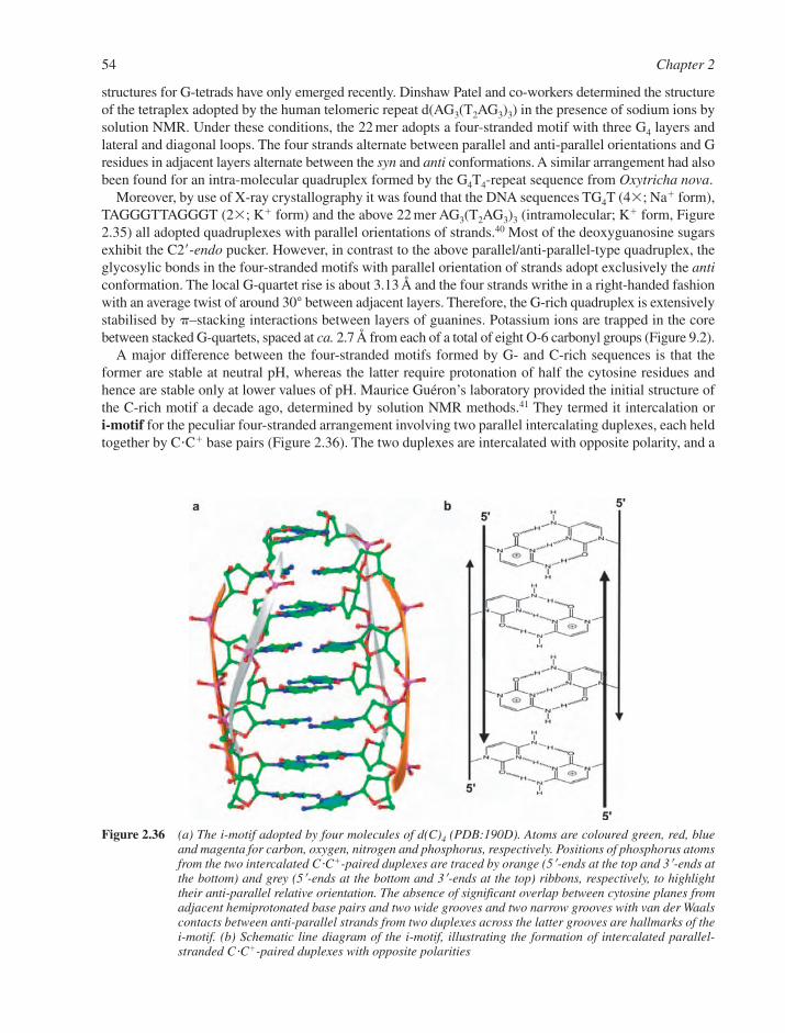
structures for G-tetrads have only emerged recently. Dinshaw Patel and co-workers determined the structureof the tetraplex adopted by the human telomeric repeat d(AG3(T2AG3)3) in the presence of sodium ions bysolution NMR. Under these conditions, the 22 mer adopts a four-stranded motif with three G4 layers andlateral and diagonal loops. The four strands alternate between parallel and anti-parallel orientations and Gresidues in adjacent layers alternate between the syn and anti conformations. A similar arrangement had alsobeen found for an intra-molecular quadruplex formed by the G4T4-repeat sequence from Oxytricha nova.
Moreover, by use of X-ray crystallography it was found that the DNA sequences TG4T (4�; Na� form),TAGGGTTAGGGT (2�; K� form) and the above 22 mer AG3(T2AG3)3 (intramolecular; K� form, Figure2.35) all adopted quadruplexes with parallel orientations of strands.40 Most of the deoxyguanosine sugarsexhibit the C2�-endo pucker. However, in contrast to the above parallel/anti-parallel-type quadruplex, theglycosylic bonds in the four-stranded motifs with parallel orientation of strands adopt exclusively the anticonformation. The local G-quartet rise is about 3.13 Å and the four strands writhe in a right-handed fashionwith an average twist of around 30° between adjacent layers. Therefore, the G-rich quadruplex is extensivelystabilised by –stacking interactions between layers of guanines. Potassium ions are trapped in the corebetween stacked G-quartets, spaced at ca. 2.7 Å from each of a total of eight O-6 carbonyl groups (Figure 9.2).
A major difference between the four-stranded motifs formed by G- and C-rich sequences is that theformer are stable at neutral pH, whereas the latter require protonation of half the cytosine residues andhence are stable only at lower values of pH. Maurice Guéron’s laboratory provided the initial structure ofthe C-rich motif a decade ago, determined by solution NMR methods.41 They termed it intercalation or i-motif for the peculiar four-stranded arrangement involving two parallel intercalating duplexes, each heldtogether by C�C� base pairs (Figure 2.36). The two duplexes are intercalated with opposite polarity, and a
54 Chapter 2
Figure 2.36 (a) The i-motif adopted by four molecules of d(C)4 (PDB:190D). Atoms are coloured green, red, blueand magenta for carbon, oxygen, nitrogen and phosphorus, respectively. Positions of phosphorus atomsfrom the two intercalated C�C�-paired duplexes are traced by orange (5�-ends at the top and 3�-ends atthe bottom) and grey (5�-ends at the bottom and 3�-ends at the top) ribbons, respectively, to highlighttheir anti-parallel relative orientation. The absence of significant overlap between cytosine planes fromadjacent hemiprotonated base pairs and two wide grooves and two narrow grooves with van der Waalscontacts between anti-parallel strands from two duplexes across the latter grooves are hallmarks of thei-motif. (b) Schematic line diagram of the i-motif, illustrating the formation of intercalated parallel-stranded C�C�-paired duplexes with opposite polarities

gentle right-handed helical twist (12°–20°, the rise is 6.2 Å) between covalently linked residues gives theC-rich quadruplex a quasi 2D form. The structure has two broad and two narrow grooves. In the latter, theanti-parallel backbone pairs within intercalated duplexes are in van der Waals contact.
Several crystal structures of C-rich sequences, such as d(CCCC), d(CCCT), d(TCCCCC), d(TCC),d(CCCAAT) and d(TAACCC), have provided details of the conformation, stabilisation and hydration ofthe i-motif.42 One surprising finding is the absence of effective stacking between the cytosine rings of adja-cent hemi-protonated C�C� base pairs from intercalated duplexes, an obvious difference to the above G-richquadruplex. However, a systematic base-on-deoxyribose stacking pattern as well as intracytidine C–H…Ohydrogen bonds may partially compensate for the lack of effective base–base-stacking between layers ofcytosines. The most unusual feature of the i-motif is a systematic, potentially stabilising C–H…O hydro-gen bonding network between the C2�-endo puckered deoxyribose sugars of anti-parallel backbones.
2.3.7.2 The Hoogsteen Duplex. Virtually all nucleic acid duplexes studied in the last 20 years containeither all G�C base pairs or A�T base pairs flanked by G�C base pairs. Very little 3D-structural work hasbeen carried out on AT-rich sequences, although the functional relevance of such sequences is well known. Forexample, the promoters of many eukaryotic structural genes contain stretches composed exclusively of A�Tbase pairs. Further, coding sequences in the yeast genome tend to be clustered with AT-rich sequences sep-arating them, and AT-rich sequences are common in transposable elements (see Section 6.8.3). Crystalstructures of TATA boxes bound to the TBP revealed highly distorted B-form conformations of the DNA.Fibre diffraction studies of AT-rich sequences provided indications for considerable structural polymorphism.By contrast, the mostly canonical geometries observed for A�T paired regions in the structures of oligonu-cleotide duplexes may have resulted from the constraints exerted by the G�C base pair clamps at both ends.
A new crystal structure determined by Juan Subirana and co-workers of the alternating hexamerd(ATATAT) raises interesting questions with regard to the existence of double-stranded DNA species thatlack base-pairing of the Watson–Crick type and the possible biological relevance of such alternative DNAconformations.43 In the crystal structure, but apparently not in solution, two hexametric fragments adoptanti-parallel orientation with Hoogsteen pairing between adenine and thymine (Figure 2.37). TheHoogsteen duplex features an average of 10.6 bp per turn, similar to B-DNA, and all sugars adopt the C2�-endo pucker. The diameter of the Hoogsteen duplex is also similar to B-form DNA and the minorgroove widths of the two duplexes differ only marginally. A unique characteristic of the Hoogsteen duplexis the syn conformation of purine nucleosides. This arrangement generates a pattern of hydrogen bonddonors and acceptors in the major and minor grooves that differs between B-DNA and Hoogsteen DNA.It also confers on the latter – a less electro-negative environment in the minor groove – that may lead topreferred interactions with relatively hydrophobic groups at that site. Hoogsteen DNA also differs clearlyfrom triple-stranded arrangements (Section 2.3.6). Thus, in the TxA�T triplex, adenine is always in the anticonformation. Moreover, T and A form the Hoogsteen pair in a parallel orientation while in the case of ananti-parallel orientation (Figure 2.33a), base-pairing between A and T is of the reverse Hoogsteen type(Figure 2.33b). Furthermore, in the Ax(A�T) triplex, third-strand adenines always base pair with adenineof the Watson–Crick base pair (Figure 2.33b). Therefore, the anti-parallel Hoogsteen DNA duplex foundfor d(ATATAT) is not simply a component of the triple helical motifs.
2.4 STRUCTURES OF RNA SPECIES
As with DNA, studies on RNA structure began with its primary structure. This quest was pursued in par-allel with that of DNA, but had to deal with the extra complexity of the 2�-hydroxyl group in ribonucleo-sides. Today, we recognise also that RNA has greater structural versatility than DNA in the variety of itsspecies, in its diversity of conformations and in its chemical reactivity. Different natural RNAs can eitherform long, double-stranded structures or adopt a globular shape composed of short duplex domains con-nected by single-stranded segments. Watson–Crick base-pairing seems to be the norm, though tRNAstructures have provided a rich source of unusual base pairs and base-triplets (Section 7.1.2). In general, it
DNA and RNA Structure 55
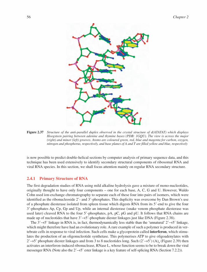
is now possible to predict double-helical sections by computer analysis of primary sequence data, and thistechnique has been used extensively to identify secondary structural components of ribosomal RNA andviral RNA species. In this section, we shall focus attention mainly on regular RNA secondary structure.
2.4.1 Primary Structure of RNA
The first degradation studies of RNA using mild alkaline hydrolysis gave a mixture of mono-nucleotides,originally thought to have only four components – one for each base, A, C, G and U. However, WaldoCohn used ion-exchange chromatography to separate each of these four into pairs of isomers, which wereidentified as the ribonucleoside 2�- and 3�-phosphates. This duplicity was overcome by Dan Brown’s useof a phosphate diesterase isolated from spleen tissue which digests RNA from its 5�-end to give the four3�-phosphates Ap, Cp, Gp and Up, while an internal diesterase (snake venom phosphate diesterase wasused later) cleaved RNA to the four 5�-phosphates, pA, pC, pG and pU. It follows that RNA chains aremade up of nucleotides that have 3�→5�-phosphate diester linkages just like DNA (Figure 2.38).
The 3�→5� linkage in RNA is, in fact, thermodynamically less stable than the ‘unnatural’ 2�→5� linkage,which might therefore have had an evolutionary role. A rare example of such a polymer is produced in ver-tebrate cells in response to viral infection. Such cells make a glycoprotein called interferon, which stimu-lates the production of an oligonucleotide synthetase. This polymerises ATP to give oligoadenylates with 2�→5� phosphate diester linkages and from 3 to 8 nucleotides long. Such (2�→5�) (A)n (Figure 2.39) thenactivates an interferon-induced ribonuclease, RNase L, whose function seems to be to break down the viralmessenger RNA (Note also the 2�→5� ester linkage is a key feature of self-splicing RNA (Section 7.2.2)).
56 Chapter 2
Figure 2.37 Structure of the anti-parallel duplex observed in the crystal structure of d(ATATAT) which displaysHoogsteen pairing between adenine and thymine bases (PDB: 1GQU). The view is across the major(right) and minor (left) grooves. Atoms are coloured green, red, blue and magenta for carbon, oxygen,nitrogen and phosphorus, respectively, and base planes of A and T are filled yellow and blue, respectively

2.4.2 Secondary Structure of RNA: A-RNA and A��-RNA
Two varieties of A-type helices have been observed for fibres of RNA species such as poly(rA)�poly(rU).At low ionic strength, A-RNA has 11 bp per turn in a right-handed, anti-parallel double-helix. The sugarsadopt a C3�-endo pucker and the other geometric parameters are all very similar to those for A-DNA (seeTables 2.3 and 2.4). If the salt concentration is raised above 20%, an A�-RNA form is observed which has
DNA and RNA Structure 57
Figure 2.39 Structure and formation of interferon-induced (2�→5�)(A)n
Figure 2.38 The primary structure of RNA (left) and cleavage patterns with spleen (centre) and snake venom (right)phosphate diesterases
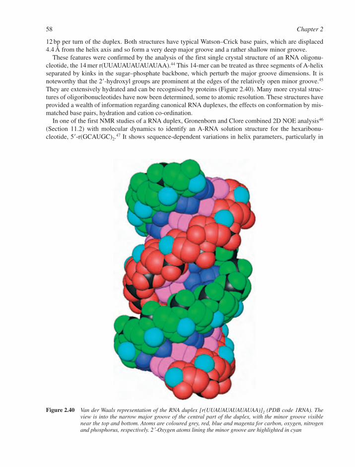
12 bp per turn of the duplex. Both structures have typical Watson–Crick base pairs, which are displaced4.4 Å from the helix axis and so form a very deep major groove and a rather shallow minor groove.
These features were confirmed by the analysis of the first single crystal structure of an RNA oligonu-cleotide, the 14 mer r(UUAUAUAUAUAUAA).44 This 14-mer can be treated as three segments of A-helixseparated by kinks in the sugar–phosphate backbone, which perturb the major groove dimensions. It isnoteworthy that the 2�-hydroxyl groups are prominent at the edges of the relatively open minor groove.45
They are extensively hydrated and can be recognised by proteins (Figure 2.40). Many more crystal struc-tures of oligoribonucleotides have now been determined, some to atomic resolution. These structures haveprovided a wealth of information regarding canonical RNA duplexes, the effects on conformation by mis-matched base pairs, hydration and cation co-ordination.
In one of the first NMR studies of a RNA duplex, Gronenborn and Clore combined 2D NOE analysis46
(Section 11.2) with molecular dynamics to identify an A-RNA solution structure for the hexaribonu-cleotide, 5�-r(GCAUGC)2.
47 It shows sequence-dependent variations in helix parameters, particularly in
58 Chapter 2
Figure 2.40 Van der Waals representation of the RNA duplex [r(UUAUAUAUAUAUAA)]2 (PDB code 1RNA). Theview is into the narrow major groove of the central part of the duplex, with the minor groove visiblenear the top and bottom. Atoms are coloured grey, red, blue and magenta for carbon, oxygen, nitrogenand phosphorus, respectively. 2�-Oxygen atoms lining the minor groove are highlighted in cyan
helix twist and in base pair roll, slide and propeller twist (Figure 2.19). The extent of variation from baseto base is much less than for the corresponding DNA hexanucleotide and seems to be dominated by theneed of the structure to achieve very nearly optimal base stacking. This picture supports experimentalstudies that indicate that base stacking and hydrogen bonding are equally important as determinants ofRNA helix stability.
Antisense RNA is defined as a short RNA transcript that lacks coding capacity, but has a high degreeof complementarity to another RNA, which enables the two to hybridise.48 The consequence is that suchanti-sense, or complementary, RNA can act as a repressor of the normal function or expression of the tar-geted RNA. Such species have been detected in prokaryotic cells with suggested functions concerningRNA-primed replication of plasmid DNA, transcription of bacterial genes, and messenger translation inbacteria and bacteriophages. Quite clearly, such regulation of gene expression depends on the integrity ofRNA duplexes.
A crucial cellular ‘security’ machinery that also depends on double-stranded RNA is RNA interferenceor RNAi (see Section 5.7.2).49–51 This mechanism has evolved to protect cells from hostile genes as wellas to regulate the activity of normal genes during growth and development. Tiny RNAs that are termedshort interfering RNAs (siRNAs) or micro RNAs (miRNAs), depending on their origin, are capable ofdown-regulating gene expression by binding to complementary mRNAs, resulting either in mRNA elimin-ation or arrest of translation. Although only discovered some 13 years ago in plants, RNA interference hasnow been found to be ubiquitous in all eukaryotes. The extraordinary specificity of RNAi and the sim-plicity of administering double-stranded RNAs to organisms with fully sequenced genomes (i.e. C. ele-gans, D. melanogaster and X. lavis) render RNAi a method of choice for functional genomics. As withpotential applications of the anti-sense strategy for therapeutic purposes, the success of RNAi as a drugwill depend on breakthroughs in cellular uptake and delivery.
2.4.3 RNA��DNA Duplexes
Helices that have one strand of RNA and one of DNA are very important species in biology.
� They are formed when reverse transcriptase makes a DNA complement to the viral RNA.� They occur when RNA polymerase transcribes DNA into complementary messenger RNA.� They are a feature in DNA replication of the short primer sequences in Okazaki fragments (Section
6.6.4).� Anti-sense DNA is a single-stranded oligodeoxynucleotide designed to bind to a short complementary
segment of a target nucleic acid (RNA or single-stranded DNA) with the potential for regulation ofgene expression (Section 5.7.1).
Such hybrids are formed in vitro by annealing together two strands with complementary sequences, suchas poly(rA)�poly(dT) and poly(rI)�poly(dC). These two hetero-duplexes adopt the A-conformation commonto RNA and DNA, the former giving an 11-fold helix typical of A-RNA and the latter a 12-fold helix char-acteristic of A�-RNA.
A self-complementary decamer r(GCG)d(TATACGC) also generates a hybrid duplex with Watson–Crickbase pairs. It has a helix rotation of 330° with a step-rise of 2.6 Å and C-3�-endo sugar pucker typical of A-DNA and A-RNA (see Table 2.1). The thermodynamic stability of RNA�DNA hybrids relative to thecorresponding DNA�DNA duplexes is a function of the deoxypyrimidine content in the DNA strands ofthe former.52 Hybrids with DNA strands containing 70–100% pyrimidines are more stable to thermal de-naturation than their DNA�DNA counterparts, whereas those with less than 30% deoxypyrimidines areless stable than the DNAs. A pyrimidine content of ca. 50% is the ‘break-even’ point.
The greater stability in some cases of RNA�DNA hetero-duplexes over DNA�DNA homo-duplexes is thebasis of the construction of antisense DNA oligomers.53–56 These are intended to enter the cell where theycan pair with, and so inactivate, complementary mRNA sequences. Additional desirable features such asmembrane permeability and resistance to enzymatic degradation have focused attention on oligonucleotides
DNA and RNA Structure 59
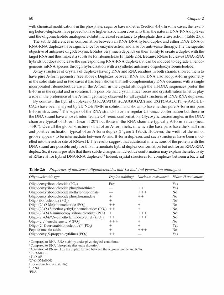
with chemical modifications in the phosphate, sugar or base moieties (Section 4.4). In some cases, the result-ing hetero-duplexes have proved to have higher association constants than the natural DNA�RNA duplexesand the oligonucleotide analogues exhibit increased resistance to phosphate diesterase action (Table 2.6).
The subtle differences in conformation between an RNA�DNA hybrid duplex and either DNA�DNA orRNA�RNA duplexes have significance for enzyme action and also for anti-sense therapy. The therapeuticobjective of antisense oligodeoxynucleotides very much depends on their ability to create a duplex with thetarget RNA and thus make it a substrate for ribonuclease H (Table 2.6). Because RNase H cleaves DNA�RNAhybrids but does not cleave the corresponding RNA�RNA duplexes, it can be induced to degrade an endo-geneous mRNA species through hybridisation with a synthetic antisense oligodeoxyribonucleotide.
X-ray structures of crystals of duplexes having DNA and RNA residues in both strands showed them tohave pure A-form geometry (see above). Duplexes between RNA and DNA also adopt A-form geometryin the solid state and in two cases it has been shown that self-complementary DNA decamers with a singleincorporated ribonucleotide are in the A-form in the crystal although the all-DNA sequences prefer the B-form in the crystal and in solution. It is possible that crystal lattice forces and crystallisation kinetics playa role in the preference of the A-form geometry observed for all crystal structures of DNA�RNA duplexes.
By contrast, the hybrid duplexes d(GTCACATG)�r(CAUGUGAC) and d(GTGAACCTT)�r(AAGUU-CAC) have been analysed by 2D NOE NMR in solution and shown to have neither pure A-form nor pureB-form structure.57 The sugars of the RNA strands have the regular C3�-endo conformation but those inthe DNA strand have a novel, intermediate C4�-endo conformation. Glycosylic torsion angles in the DNAchain are typical of B-form (near –120°) but those in the RNA chain are typically A-form values (near–140°). Overall the global structure is that of an A-form helix in which the base pairs have the small riseand positive inclination typical of an A-form duplex (Figure 2.19a,d). However, the width of the minorgroove appears to be intermediate between A- and B-form duplexes and such structures have been mod-elled into the active site of RNase H. The results suggest that additional interactions of the protein with theDNA strand are possible only for this intermediate hybrid duplex conformation but not for an RNA�RNAduplex. So, it seems possible that these subtle changes in nucleotide conformation may explain the selectivityof RNase H for hybrid DNA�RNA duplexes.58 Indeed, crystal structures for complexes between a bacterial
60 Chapter 2
Table 2.6 Properties of antisense oligonucleotides and 1st and 2nd generation analogues
Oligonucleotide type Duplex stabilitya Nuclease resistanceb RNase H activationc
Oligodeoxyribonucleotide (PO2) Para — YesOligodeoxyribonucleotide phosphorothioate — �� YesOligodeoxyribonucleotide methylphosphonate — ��� NoOligodeoxyribonucleotide phosphoramidate � �� NoOligoribonucleotide (PO2) � — NoOligo (2�-O-Me)ribonucleotide (PO2) �� � NoOligo (2�-O-(2-methoxyethyl)ribonucleotided (PO2) �� � NoOligo (2�-O-(3-aminopropyl)ribonucleotidee (PO2) � ��� NoOligo (2�-O-(N,N-dimethylaminooxyethyl)f (PO2) �� ��� NoOligo (2�,4�-methylene …)g (PO2) ��� � NoOligo (2�-fluoroarabinonucleotide)h (PO2) � � YesPeptide nucleic acidsi � ��� NoOligodeoxy(5-propyne-cytidine) (PO2) �� — Yes
a Compared to DNA–RNA stability under physiological conditions.b Compared to DNA (phosphate diesterase digestion).c Activation of RNase H by the duplex formed between the oligonucleotide and RNA.d 2�-O-MOE.e 2�-O-AP.f 2�-O-DMAEOE.g Locked nucleic acid (LNA).h FANA.i PNA.
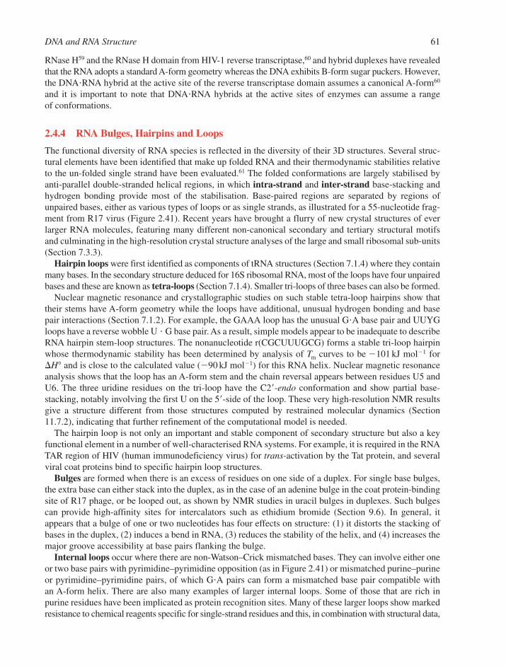
RNase H59 and the RNase H domain from HIV-1 reverse transcriptase,60 and hybrid duplexes have revealedthat the RNA adopts a standard A-form geometry whereas the DNA exhibits B-form sugar puckers. However,the DNA�RNA hybrid at the active site of the reverse transcriptase domain assumes a canonical A-form60
and it is important to note that DNA�RNA hybrids at the active sites of enzymes can assume a range of conformations.
2.4.4 RNA Bulges, Hairpins and Loops
The functional diversity of RNA species is reflected in the diversity of their 3D structures. Several struc-tural elements have been identified that make up folded RNA and their thermodynamic stabilities relativeto the un-folded single strand have been evaluated.61 The folded conformations are largely stabilised byanti-parallel double-stranded helical regions, in which intra-strand and inter-strand base-stacking andhydrogen bonding provide most of the stabilisation. Base-paired regions are separated by regions ofunpaired bases, either as various types of loops or as single strands, as illustrated for a 55-nucleotide frag-ment from R17 virus (Figure 2.41). Recent years have brought a flurry of new crystal structures of everlarger RNA molecules, featuring many different non-canonical secondary and tertiary structural motifsand culminating in the high-resolution crystal structure analyses of the large and small ribosomal sub-units(Section 7.3.3).
Hairpin loops were first identified as components of tRNA structures (Section 7.1.4) where they containmany bases. In the secondary structure deduced for 16S ribosomal RNA, most of the loops have four unpairedbases and these are known as tetra-loops (Section 7.1.4). Smaller tri-loops of three bases can also be formed.
Nuclear magnetic resonance and crystallographic studies on such stable tetra-loop hairpins show thattheir stems have A-form geometry while the loops have additional, unusual hydrogen bonding and basepair interactions (Section 7.1.2). For example, the GAAA loop has the unusual G�A base pair and UUYGloops have a reverse wobble U � G base pair. As a result, simple models appear to be inadequate to describeRNA hairpin stem-loop structures. The nonanucleotide r(CGCUUUGCG) forms a stable tri-loop hairpinwhose thermodynamic stability has been determined by analysis of Tm curves to be 101 kJ mol1 for�H° and is close to the calculated value (90 kJ mol1) for this RNA helix. Nuclear magnetic resonanceanalysis shows that the loop has an A-form stem and the chain reversal appears between residues U5 andU6. The three uridine residues on the tri-loop have the C2�-endo conformation and show partial base-stacking, notably involving the first U on the 5�-side of the loop. These very high-resolution NMR resultsgive a structure different from those structures computed by restrained molecular dynamics (Section11.7.2), indicating that further refinement of the computational model is needed.
The hairpin loop is not only an important and stable component of secondary structure but also a keyfunctional element in a number of well-characterised RNA systems. For example, it is required in the RNATAR region of HIV (human immunodeficiency virus) for trans-activation by the Tat protein, and severalviral coat proteins bind to specific hairpin loop structures.
Bulges are formed when there is an excess of residues on one side of a duplex. For single base bulges,the extra base can either stack into the duplex, as in the case of an adenine bulge in the coat protein-bindingsite of R17 phage, or be looped out, as shown by NMR studies in uracil bulges in duplexes. Such bulgescan provide high-affinity sites for intercalators such as ethidium bromide (Section 9.6). In general, itappears that a bulge of one or two nucleotides has four effects on structure: (1) it distorts the stacking ofbases in the duplex, (2) induces a bend in RNA, (3) reduces the stability of the helix, and (4) increases themajor groove accessibility at base pairs flanking the bulge.
Internal loops occur where there are non-Watson–Crick mismatched bases. They can involve either oneor two base pairs with pyrimidine–pyrimidine opposition (as in Figure 2.41) or mismatched purine–purineor pyrimidine–pyrimidine pairs, of which G�A pairs can form a mismatched base pair compatible with an A-form helix. There are also many examples of larger internal loops. Some of those that are rich inpurine residues have been implicated as protein recognition sites. Many of these larger loops show markedresistance to chemical reagents specific for single-strand residues and this, in combination with structural data,
DNA and RNA Structure 61
suggests that there is probably a high level of order in such loops, notably of base-stacking and base-triples.One general opinion is that the major differences between loop and stem regions are dynamic rather thanstructural.
Junctions are regions that connect three or more stems (the connecting region for two stems is an internalloop) and are a common feature of computer-generated secondary structures for large RNAs. A primeexample is the four-stem junction in the cloverleaf structure of tRNAs in which stacking continuitybetween the acceptor and the T stems and between the anti-codon and D stems is maintained (Section7.1.4).62 A junction of three stems forms the hammerhead structure of self-cleaving RNA (Section 7.6.2)and junctions of up to five stems have been observed for 16S RNA.
62 Chapter 2
Figure 2.41 A possible secondary structure for a 55-nucleotide fragment from R17 virus which illustrates hairpinloop, interior loop and bulge structures. The free energy of this structure has been calculated to have anet �G° of –90 kJ mol1 using appropriate values for base pairs (Table 2.6) and for loops and bulges
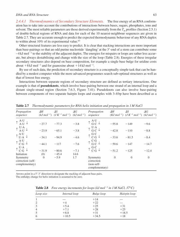
2.4.4.1 Thermodynamics of Secondary Structure Elements. The free energy of an RNA conform-ation has to take into account the contributions of interactions between bases, sugars, phosphates, ions andsolvent. The most reliable parameters are those derived experimentally from the Tm profiles (Section 2.5.1)of double-helical regions of RNA and data for each of the 10 nearest-neighbour sequences are given inTable 2.7. They are accurate enough to predict the expected thermodynamic behaviour of any RNA duplexto within about 10% of its experimental value.63
Other structural features are less easy to predict. It is clear that stacking interactions are more importantthan base-pairings so that an odd purine nucleotide ‘dangling’ at the 3�-end of a stem can contribute some–4 kJ mol1 to the stability of the adjacent duplex. The energies for mispairs or loops are rather less accur-ate, but always destabilising and change with the size of the loop (Table 2.8). Energies of these irregularsecondary structures also depend on base composition, for example a single base bulge for uridine costsabout �8 kJ mol1 and for guanosine about �14 kJ mol1.
By use of such data, the prediction of secondary structure is a conceptually simple task that can be han-dled by a modest computer while the more advanced programmes search sub-optimal structures as well asthat of lowest free energy.
Interactions between separate regions of secondary structure are defined as tertiary interactions. Oneexample is that of pseudoknots, which involve base-pairing between one strand of an internal loop and adistant single-strand region (Section 7.6.3, Figure 7.41). Pseudoknots can also involve base-pairingbetween components of two separate hairpin loops and examples with 3–8 bp have been described as a
DNA and RNA Structure 63
Table 2.7 Thermodynamic parameters for RNA helix initiation and propagation in 1 M NaCl
Propagation �H �S �G Propagation �H �S �G sequence (kJ mol1) (J K1 mol1) (kJ mol1) sequence (KJ mol1) (J K1 mol1) (kJ mol1)
↑A�U
↑A�U
A�U ↓ 27.7 77.3 3.8 G�C ↓ 55.8 149 9.6
↑U�A
↑U�A
A�U ↓ 23.9 65.1 3.8 G�C ↓ 42.8 110 8.8
↑A�U
↑G�C
U�A ↓ 34.1 94.9 4.6 C�G ↓ 33.6 81.5 8.4
↑A�U
↑C�G
C�G ↓ 44.1 117 7.6 G�C ↓ 59.6 147 14.7
↑U�A
↑G�C
C�G ↓ 31.9 80.6 7.1 C�G ↓ 51.2 125 12.4Initiation (0) 45.4 14.6Symmetry 0 5.9 1.7 Symmetry 0 0 0correction (self- correction complementary) (non-self-
complementary)
Arrows point in a 5�-3� direction to designate the stacking of adjacent base pairs.The enthalpy change for helix initiation is assumed to be zero.
Table 2.8 Free energy increments for loops (kJ mol1 in 1 M NaCl, 37°C)
Loop size Internal loop Bulge loop Hairpin loop
1 — �14 —2 �4 �22 —3 �5.4 �25 �314 �7.1 �28 �255 �8.8 �31 �18.56 �10.5 �34.5 �18

result of both NMR and X-ray analysis. However, the computer prediction of tertiary interactions andbase-triples appears to be still beyond the scope of present methodology.
2.4.5 Triple-Stranded RNAs
The first triple-stranded nucleic acid was described in 1957 when poly(rU)�poly(rA) was found to form a sta-ble 2:1 complex in the presence of magnesium chloride. The extra poly(rU) strand is parallel to the poly(rA)strand and forms Hoogsteen base-triples in the major groove of an A-form Watson–Crick helix. Triplexes of2poly(rA)�poly(rU) can also be formed while poly (rC) can form a triplex with poly(rG) at pH 6 which hastwo cytidines per guanine, one of them being protonated to give the C�xG�C base-triple also seen for triple-helical DNA (Figure 2.33). Base triples are also a very common feature of tRNA structure (Section 7.1.4).62
The importance of added cations to overcome the repulsion between the anionic chains of theWatson–Crick duplex and the poly-pyrimidine third strand is an essential feature of triple-helix formation.Co3�(NH3)6 and spermine are also effective counter-ions as well as the more usual Mg2�.
Poly(rG) as well as guanosine and GMP can form structures with four equivalent hydrogen-bonded bases ina plane, with all four strands parallel. It is not clear whether this structure has any relevance to RNA folding.
2.5 DYNAMICS OF NUCLEIC ACID STRUCTURES
Any over-emphasis on the stable structures of nucleic acids runs the risk of playing down the dynamicactivity of nucleic acids that is intrinsic to their function. Pairing and unpairing, breathing and winding areintegral features of the behaviour of these species.64
Established studies on structural transitions of nucleic acids have for a long time used classical physicalmethods, which include light absorption, NMR spectroscopy, ultra-centrifugation, viscometry and X-raydiffraction (Chapter 11). More recently, these techniques have been augmented by a range of powerfulcomputational methods (Section 11.7). In each case, the choice of experiment is linked to the time-scaleand amplitude of the molecular motion under investigation.
2.5.1 Helix-Coil Transitions of Duplexes
Double helices have a lower molecular absorptivity for UV light than would be predicted from the sum oftheir constituent bases. This hypochromicity is usually measured at 256 nm while C�G base pairs can alsobe monitored at 280 nm. It results from coupling of the transition dipoles between neighbouring stackedbases and is larger in amplitude for A�U and A�T pairs than for C�G pairs. As a result, the UV absorptionof a DNA duplex increases typically by 20–30% when it is denatured. This transition from a helix to anunstacked, strand-separated coil has a strong entropic component and so is temperature dependent. Themid-point of this thermal transition is known as the melting temperature (Tm).
Such dissociation of nucleic acid helices in solution to give single-stranded DNA is a function of basecomposition, sequence and chain length as well as of temperature, salt concentration and pH of the solv-ent. In particular, early observations of the relationship between Tm and base composition for differentDNAs showed that A�T pairs are less stable than C�G pairs, a fact which is now expressed in a linear cor-relation between Tm and the gross composition of a DNA polymer by the equation:
Tm � X � 0.41[% (C � G)] (°C)
The constant X is dependent on salt concentration and pH and has a value of 69.3°C for 0.3 M sodium ionsat pH 7 (Figure 2.42).
A second consequence is that the steepness of the transition also depends on base sequence. Thus, melt-ing curves for homo-polymers have much sharper transitions than those for random-sequence polymers.This is because A�T rich regions melt first to give unpaired regions, which then extend gradually with ris-ing temperature until, finally, even the pure C�G regions have melted (Figures 2.42 and 2.43). In some
64 Chapter 2
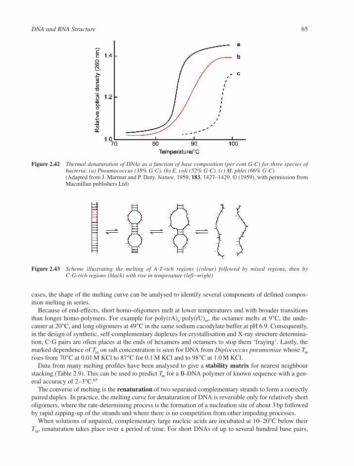
cases, the shape of the melting curve can be analysed to identify several components of defined compos-ition melting in series.
Because of end-effects, short homo-oligomers melt at lower temperatures and with broader transitionsthan longer homo-polymers. For example for poly(rA)n�poly(rU)m, the octamer melts at 9°C, the unde-camer at 20°C, and long oligomers at 49°C in the same sodium cacodylate buffer at pH 6.9. Consequently,in the design of synthetic, self-complementary duplexes for crystallisation and X-ray structure determina-tion, C�G pairs are often places at the ends of hexamers and octamers to stop them ‘fraying’. Lastly, themarked dependence of Tm on salt concentration is seen for DNA from Diplococcus pneumoniae whose Tm
rises from 70°C at 0.01 M KCl to 87°C for 0.1 M KCl and to 98°C at 1.0 M KCl.Data from many melting profiles have been analysed to give a stability matrix for nearest neighbour
stacking (Table 2.9). This can be used to predict Tm for a B-DNA polymer of known sequence with a gen-eral accuracy of 2–3°C.65
The converse of melting is the renaturation of two separated complementary strands to form a correctlypaired duplex. In practice, the melting curve for denaturation of DNA is reversible only for relatively shortoligomers, where the rate-determining process is the formation of a nucleation site of about 3 bp followedby rapid zipping-up of the strands and where there is no competition from other impeding processes.
When solutions of unpaired, complementary large nucleic acids are incubated at 10–20°C below theirTm, renaturation takes place over a period of time. For short DNAs of up to several hundred base pairs,
DNA and RNA Structure 65
Figure 2.43 Scheme illustrating the melting of A�T-rich regions (colour) followed by mixed regions, then byC�G-rich regions (black) with rise in temperature (left→right)
Figure 2.42 Thermal denaturation of DNAs as a function of base composition (per cent G�C) for three species ofbacteria: (a) Pneumococcus (38% G�C). (b) E. coli (52% G�C). (c) M. phlei (66% G�C)(Adapted from J. Marmur and P. Doty, Nature, 1959, 183, 1427–1429. © (1959), with permission fromMacmillan publishers Ltd)
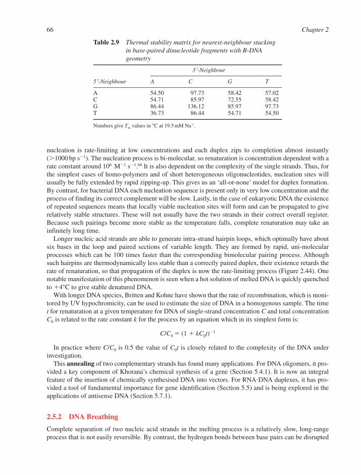
nucleation is rate-limiting at low concentrations and each duplex zips to completion almost instantly(1000 bp s1). The nucleation process is bi-molecular, so renaturation is concentration dependent with arate constant around 106 M1 s1.66 It is also dependent on the complexity of the single strands. Thus, forthe simplest cases of homo-polymers and of short heterogeneous oligonucleotides, nucleation sites willusually be fully extended by rapid zipping-up. This gives us an ‘all-or-none’ model for duplex formation.By contrast, for bacterial DNA each nucleation sequence is present only in very low concentration and theprocess of finding its correct complement will be slow. Lastly, in the case of eukaryotic DNA the existenceof repeated sequences means that locally viable nucleation sites will form and can be propagated to giverelatively stable structures. These will not usually have the two strands in their correct overall register.Because such pairings become more stable as the temperature falls, complete renaturation may take aninfinitely long time.
Longer nucleic acid strands are able to generate intra-strand hairpin loops, which optimally have aboutsix bases in the loop and paired sections of variable length. They are formed by rapid, uni-molecularprocesses which can be 100 times faster than the corresponding bimolecular pairing process. Althoughsuch hairpins are thermodynamically less stable than a correctly paired duplex, their existence retards therate of renaturation, so that propagation of the duplex is now the rate-limiting process (Figure 2.44). Onenotable manifestation of this phenomenon is seen when a hot solution of melted DNA is quickly quenchedto �4°C to give stable denatured DNA.
With longer DNA species, Britten and Kohne have shown that the rate of recombination, which is moni-tored by UV hypochromicity, can be used to estimate the size of DNA in a homogenous sample. The timet for renaturation at a given temperature for DNA of single-strand concentration C and total concentrationC0 is related to the rate constant k for the process by an equation which in its simplest form is:
C/C0 � (1 � kC0t)1
In practice where C/C0 is 0.5 the value of C0t is closely related to the complexity of the DNA underinvestigation.
This annealing of two complementary strands has found many applications. For DNA oligomers, it pro-vided a key component of Khorana’s chemical synthesis of a gene (Section 5.4.1). It is now an integral feature of the insertion of chemically synthesised DNA into vectors. For RNA�DNA duplexes, it has pro-vided a tool of fundamental importance for gene identification (Section 5.5) and is being explored in theapplications of antisense DNA (Section 5.7.1).
2.5.2 DNA Breathing
Complete separation of two nucleic acid strands in the melting process is a relatively slow, long-rangeprocess that is not easily reversible. By contrast, the hydrogen bonds between base pairs can be disrupted
66 Chapter 2
Table 2.9 Thermal stability matrix for nearest-neighbour stackingin base-paired dinucleotide fragments with B-DNAgeometry
3�-Neighbour
5�-Neighbour A C G T
A 54.50 97.73 58.42 57.02C 54.71 85.97 72.55 58.42G 86.44 136.12 85.97 97.73T 36.73 86.44 54.71 54.50
Numbers give Tm values in °C at 19.5 mM Na�.

at temperatures well below the melting temperature to give local, short-range separation of the strands.This readily reversible process is known as breathing.
The evidence for such dynamic motion comes from chemical reactions, which take place at atoms that arecompletely blocked by normal base-pairings. Those used include tritium exchange studies in hydrogen-bondedprotons in base pairs, the reactivity of formaldehyde with base NH groups, and NMR studies of imino–protonexchange with solvent water. This last technique can be used on a time scale from minutes down to 10 ms. Itshows that in linear DNA the base pairs open singly and transiently with a life time around 10 ms at 15°C.
Because NMR can distinguish between imino- and amino–proton exchange, it can also be used to iden-tify breathing in specific sequences.67 Some of the most detailed work of this sort has come from studieson tRNA molecules, which show that, with increasing temperature, base-triplets (Figure 2.33) are desta-bilised first followed by the ribothymidine helix and then the dihydrouridine helix. Finally, the acceptorhelix ‘melts’ after the anti-codon helix (Section 7.1.4).
Another possible motion that might be important for the creation of intercalation sites is known as ‘soli-ton excitation’. The concept here is of a stretching vibration of the DNA chain, which travels like a wavealong the helix axis until, given sufficient energy, it leads to local unstacking of adjacent bases with asso-ciated deformation of sugar pucker and other bond conformations.
Such pre-melting behaviour may well relate to the process of drug intercalation, to the association ofsingle-strand specific DNA binding proteins (Section 10.3.8), and to the reaction of small electrophilicreagents with imino and amino groups such as cytosine-N-3 (Section 8.5).
2.5.3 Energetics of the B–Z Transition
The isomerisation equilibrium between the right-handed B-form and the left-handed Z-form of DNA isdetermined by three factors:
1. Chemical structure of the polynucleotide (sequence, modified bases)2. Environmental conditions (solvent, pH, temperature, etc.)3. Degree of topological stress (supercoiling, cruciform formation).
Many quantitative data have been obtained from spectroscopic, hydrodynamic and calorimetric studies andlinked to theoretical calculations. Although these have not yet defined the kinetics or complex mechanisms ofthe B–Z transition, it is evident that the small transition enthalpies involved lie within the range of the thermal
DNA and RNA Structure 67
Figure 2.44 Renaturation processes (a) for short oligonucleotide and longer homo-polymers and (b) for naturalDNA strands
energies available from the environment. So, for example, the intrinsic free energy difference between Z- andB-forms is close to 2 kJ mol1 for poly-d(G�C) base pairs, only 1 kJ mol1 for poly-d(G�m5C) base pairs, andgreater than 5 kJ mol1 for poly-d(A�T) base pairs. It thus appears that local structural fluctuations may be keyelements in the mediation of biological regulatory functions through the B–Z transition.68,69
2.5.4 Rapid DNA Motions
Rotations of single bonds, either alone or in combination, are responsible for a range of very rapid DNAmotions with time scales down to fractions of a nanosecond. For example, the twisting of base pairsaround the helix axis has a life time around 10–8 s while crankshaft rotations of the �, �, � and � C-O-P-O-C bonds (see Figure 2.11) lead to an oscillation in the position of the phosphorus atoms on a millisecondtime scale. Various calculations on the inter-conversion of C3�-endo and C2�-endo sugar pucker havegiven low activation energy barriers for their inter-conversion, in the range 3–20 kJ mol1, showing thatthe conformers are in rapid, although weighted, equilibrium at 37°C. Lastly, rapid fluctuations in propellertwist can result from oscillations of the glycosylic bond.
2.6 HIGHER-ORDER DNA STRUCTURES
The way in which eukaryotic DNA is packaged in the cell nucleus is one of the wonders of the macro-molecular structure. In general, higher organisms have more DNA than lower ones (Table 2.10) and thiscalls for correspondingly greater condensation of the double helix. Human cells contain a total of7.8 � 109 bp, which corresponds to an extended length of about 2 m. The DNA is packed into 46 cylin-drical chromosomes of total length 200 �m, which gives a net packaging ratio of about 104 for suchmetaphase human chromosomes (see also Section 6.4). The overall process has been broken down intotwo stages: the formation of nucleosomes and the condensation of nucleosomes into chromatin.70
2.6.1 Nucleosome Structure
The first stage in the condensation of DNA is the nucleosome, whose core has been crystallised by AaronKlug and John Finch and analysed using X-ray diffraction. The DNA duplex is wrapped around a block ofeight histone proteins to give 1.75 turns of a left-handed superhelix (Section 10.6, Figure 10.15).71,72 Thisprocess achieves a packing ratio of 7. The number of base pairs involved in nucleosome structures variesfrom species to species, being 165 bp for yeast, 183 bp for HeLa cells, 196 bp for rat liver and 241 bp forsea urchin sperm. Such nucleosomes are joined by linker DNA whose length ranges from 0 bp in neuronsto 80 bp in sea urchin sperm but usually averages 30–40 bp. The details of packaging the histone proteinsare discussed later (Section 10.6.1).73
68 Chapter 2
Table 2.10 Cellular DNA content of various species
Organism Numbers of base pairs DNA length (mm) Number of chromosomes
Escherichia coli 4 � 106 1.4 1Yeast(Saccharomyces cerevisiae) 1.4 � 106 4.6 16Fruit fly(Drosophila melanogaster) 1.7 � 107 56.0 4Humans(Homo sapiens) 3.9 � 109 990.0 23
Note: Values are provided for haploid genomes.
As the DNA winds around the nucleosome core, the major and minor grooves are compressed on theinside with complementary widening of the grooves on the outside of the curved duplex. Runs of A�T basepairs, which have an intrinsically narrow minor groove should be most favourably placed on the inside ofthe curved segment while runs of G�C base pairs should be more favourably aligned with minor groovesfacing outwards, where they are more accessible to enzyme cleavage. In practice, Drew and Travers meas-ured the periodicities of A�T and G�C base pairs by cleavage with DNase I and found them to be exactlyout of phase and having a periodicity of 10.17 � 0. 5 bp.74,75 This result was later confirmed by hydroxylradical cleavage, which avoids the steric constraints of DNase I.
2.6.2 Chromatin Structure
Chromatin is too large and heterogeneous to yield its secrets to X-ray analysis, so electron microscopy is thechosen experimental probe (Section 11.5.1). At intermediate salt concentration (�1 mM NaCl) the nucle-osomes are revealed as ‘beads on a string’. Spherical nucleosomes can be seen with a diameter of 7–10 nmjoined by variable-length filaments, often about 14 nm long. If the salt concentration is increased to 0.1 MNaCl, the spacing filaments get shorter and a zig-zag arrangement of nucleosomes is seen in a fibre 10–11 nmwide (Section 10.6, Figure 10.15). At even higher salt concentration and in the presence of magnesium, thesecondense into a 30 nm diameter fibre, called a solenoid, which is thought to be either a right-handed or aleft-handed helix made up of close-packed nucleosomes with a packing ratio of around 40.
For the further stages in DNA condensation, one of the models proposed suggests that loops of these30 nm fibres, each containing about 50 solenoid turns and possibly wound in a supercoil, are attached to acentral protein core from which they radiate outwards.76 Organisation of these loops around a cylindricalscaffold could give rise to the observed mini-band structure of chromosomes, which is some 0.84 �m indiameter and 30 nm in thickness. A continuous helix of loops would then constitute the chromosome.These ideas are illustrated in a possible scheme (Figure 2.45).
It is clear from all of the relevant biological experiments that the single DNA duplex has to be continu-ously accessible despite all this condensed structure in order for replication to take place. Some of themost exciting electron micrographs of DNA have been obtained from samples where the histones havebeen digested away leaving only the DNA as a tangled network of inter-wound superhelices radiating froma central nuclear region where the scaffold proteins remain intact (Figure 2.46).
Even then, the most condensed packing of nucleic acid is found in the sperm cell. Here a series of arginine-rich proteins called protamines bind to DNA, probably with their �-helices in the major groove ofthe DNA where they neutralise the phosphate charge, and so enable very tight packing of DNA duplexes.
Bacterial DNA is also condensed into a highly organised state (Section 10.6.2). In E. coli the genomehas 4400 kbp in a closed circle, which is negatively supercoiled. It is condensed around histone-like pro-teins, HU and HI, to form a nucleoid and achieve a compaction of 1000-fold, which is followed by furthercondensation into supercoil domains. Unlike chromosomal DNA in eukaryotes, there is some additionalnegative supercoiling in prokaryotes that is not accounted for by protein binding.77 This is probably a con-sequence of the activity of the bacterial DNA gyrase, which is capable of actively introducing further negative supercoiling, driven by hydrolysis of ATP. This whole process differs in several respects fromassembly of chromatin in eukaryotes.
� There is no apparent regular repeating structure equivalent to the eukaryotic nucleosome althoughshort DNA segments of 60–129 bp are organised by means of their interaction with abundantDNA-binding proteins.
� There is no prokaryotic equivalent to the solenoid structure.� Bacterial DNA seems to be torsionally strained in vivo and organised into independently
supercoiled domains of about 100 kbp.
The establishment of DNA architecture in the bacterial chromosome has progressed through the analy-sis of two types of structure. First, the interaction of a dimer of the HU protein from B. stearothermophilus
DNA and RNA Structure 69
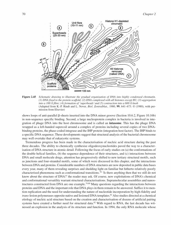
shows loops of anti-parallel �-sheets inserted into the DNA minor groove (Section 10.6.2, Figure 10.16b)in non-sequence specific binding. Second, a large nucleoprotein complex in bacteria is involved in inte-gration of phage DNA into the host chromosome and is called an intasome. This has the phage DNAwrapped as a left-handed supercoil around a complex of proteins including several copies of two DNA-binding proteins, the phase-coded integrase and the IHP protein (integration host factor). The IHP binds toa specific DNA sequence. These developments suggest that structural analysis of the bacterial chromosomemay well overtake that of eukaryotic systems.
Tremendous progress has been made in the characterisation of nucleic acid structure during the pastthree decades. The ability to chemically synthesise oligodeoxynucleotides paved the way to a character-isation of DNA structure in atomic detail. Following the focus of early studies on (a) the conformations ofthe double helical families, (b) the sequence dependence of their structures, and (c) interactions betweenDNA and small molecule drugs, attention has progressively shifted to new tertiary structural motifs, suchas junctions and four-stranded motifs, some of which were discussed in this chapter, and the interactionsbetween DNA and proteins. Considerable numbers of DNA structures are now deposited in public data basesevery year, many of them revealing surprises and shedding light on familiar but hitherto relatively poorlycharacterised phenomena such as conformational transitions.78 ‘Is there anything then that we still do notknow about the structure of DNA?’ the reader may ask. Of course, new exploitations of DNA’s chemicaland conformational versatility warrant structural characterisations. Supramolecular assemblies and nanos-tructures constructed from DNA are one example.79,80 Many questions regarding the interactions betweenproteins and DNA and the important role that DNA plays in them remain to be answered. Suffice it to men-tion replication and the need for understanding the nature of nucleotide incorporation by high-fidelity andtrans-lesion polymerases opposite native and lesioned DNA templates.81 Also studies directed at a chemicaletiology of nucleic acid structure based on the creation and characterisation of dozens of artificial pairingsystems have created a further need for structural data.82 With regard to RNA, the last decade has wit-nessed an explosion in the analysis of its structure and function. In 1994, the only RNA molecule whose
70 Chapter 2
Figure 2.45 Schematic drawing to illustrate the gradual organisation of DNA into highly condensed chromatin.(1) DNA fixed to the protein scaffold; (2) DNA complexed with all histones except H1; (3) aggregationinto a 100 Å fibre; (4) formation of ‘superbeads’ and (5) contraction into a 600 Å knob(Adapted from K.-P. Rindt and L. Nover, Biol. Zentralblat., 1980, 99, 641–673. © (1980), with per-mission from Elsevier)
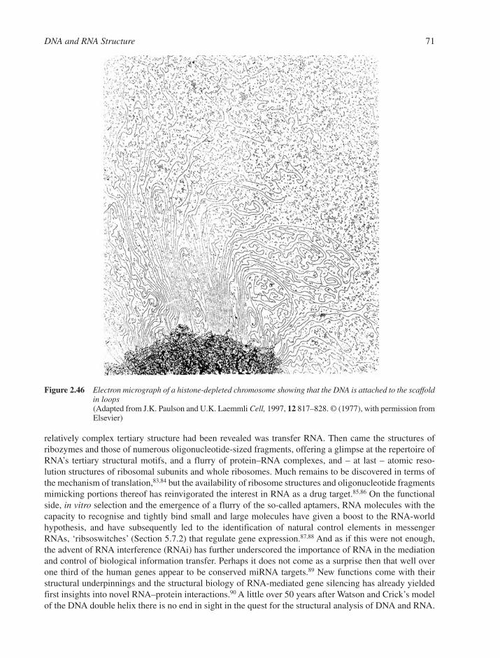
relatively complex tertiary structure had been revealed was transfer RNA. Then came the structures ofribozymes and those of numerous oligonucleotide-sized fragments, offering a glimpse at the repertoire ofRNA’s tertiary structural motifs, and a flurry of protein–RNA complexes, and – at last – atomic reso-lution structures of ribosomal subunits and whole ribosomes. Much remains to be discovered in terms ofthe mechanism of translation,83,84 but the availability of ribosome structures and oligonucleotide fragmentsmimicking portions thereof has reinvigorated the interest in RNA as a drug target.85,86 On the functionalside, in vitro selection and the emergence of a flurry of the so-called aptamers, RNA molecules with thecapacity to recognise and tightly bind small and large molecules have given a boost to the RNA-worldhypothesis, and have subsequently led to the identification of natural control elements in messengerRNAs, ‘ribsoswitches’ (Section 5.7.2) that regulate gene expression.87,88 And as if this were not enough,the advent of RNA interference (RNAi) has further underscored the importance of RNA in the mediationand control of biological information transfer. Perhaps it does not come as a surprise then that well overone third of the human genes appear to be conserved miRNA targets.89 New functions come with theirstructural underpinnings and the structural biology of RNA-mediated gene silencing has already yieldedfirst insights into novel RNA–protein interactions.90 A little over 50 years after Watson and Crick’s modelof the DNA double helix there is no end in sight in the quest for the structural analysis of DNA and RNA.
DNA and RNA Structure 71
Figure 2.46 Electron micrograph of a histone-depleted chromosome showing that the DNA is attached to the scaffoldin loops(Adapted from J.K. Paulson and U.K. Laemmli Cell, 1997, 12 817–828. © (1977), with permission fromElsevier)
REFERENCES
1. C. Altona and M. Sundaralingam, Conformational analysis of the sugar ring in nucleosides andnucleotides. A new description using the concept of pseudorotation. J. Am. Chem. Soc., 1972, 94,8205–8212.
2. R. Chandrasekaran and S. Arnott, The structures of DNA and RNA in oriented fibres. Springer, Berlin.3. A. Rich and S. Zhang, Z-DNA: the long road to biological function Nat. Rev. Genet., 2003, 4, 566–572.4. O. Kennard and W.N. Hunter, Single-crystal X-ray diffraction studies of oligonucleotides and
oligonucleotide-drug complexes. Angew. Chem. Intl. Ed. Engl., 1991, 30, 1254–1277.5. R.E. Dickerson, Nucleic acids, in Crystallography of Biological Macromolecules, Vol. F, M.G. Rossmann
and E. Arnold (eds), International Tables of Crystallography, Kluwer Academic Publishers, Dordrecht,2001, 588–622.
6. F.A. Jurmak and A. McPherson (eds), Biological Macromolecules and Assemblies, Vol. 1, Wiley, NewYork, 1984.
7. B. Hartmann and R. Lavery, DNA structural forms. Quart. Rev. Biophys., 1996, 29, 309–368.8. W. Saenger, Principles of Nucleic Acid Structure. Springer, New York, 1984.9. C.R. Calladine and H.R. Drew, Understanding DNA. The Molecule and How it Works. Academic
Press Ltd, London, 1997.10. G. Minasov, V. Tereshko and M. Egli, Atomic-resolution crystal structures of B-DNA reveal specific
influences of divalent metal ions on conformation and packing. J. Mol. Biol., 1999, 291, 83–99.11. M. Egli, V. Tereshko, M. Teplova, G. Minasov, A. Joachimiak, R. Sanishvili, C.M. Weeks, R. Miller,
M.A. Maier, H. An, P.D. Cook and M. Manoharan, X-ray crystallographic analysis of the hydration ofA- and B-form DNA at atomic resolution. Biopolymers (Nucl. Acid Sci.), 2000, 48, 234–252.
12. V. Tereshko, G. Minasov and M. Egli, A “hydration” spine in a B-DNA minor groove. J. Am. Chem.Soc., 1999, 121, 3590–3595.
13. A. Rich, The nucleic acids. A backward glance. In DNA: The double helix, Ann. NY Acad. Sci., 1995,758, 97–142.
14. A.A. Gorin, V.B. Zhurkin and W.K. Olson, B-DNA twisting correlates with base-pair morphology.J. Mol. Biol., 1995, 247, 34–48.
15. K. Yanagi, G.G. Privé and R.E. Dickerson, Analysis of the local helix geometry in three B-DNAdecamers and eight dodecamers. J. Mol. Biol., 1991, 217, 201–214.
16. M.A. El Hassan and C.R. Calladine, Propeller-twisting of base-pairs and the conformationalmobility of dinucleotide steps in DNA. J. Mol. Biol., 1996, 259, 95–103.
17. C.A. Hunter, Sequence-dependent DNA structure, the role of base-stacking interactions. J. Mol. Biol.,1993, 230, 1025–1054.
18. H.-L. Ng, M.L. Kopka and R.E. Dickerson, The structure of a stable intermediate in the A ⇔ B DNAhelix transition. Proc. Natl. Acad. Sci. USA, 2000, 97, 2035–2039.
19. J.M. Vargason, K. Henderson and P.S. Ho, A crystallographic map of the transition from B-DNA toA-DNA. Proc. Natl. Acad. Sci. USA, 2001, 98, 7265–7270.
20. T. Brown and O. Kennard, Structural basis of DNA mutagenesis. Curr. Opin. Struct. Biol., 1992, 2,354–360.
21. R.D. Wells, Unusual DNA structures. J. Biol. Chem., 1988, 263, 1095–1098.22. C.R. Calladine, H.R. Drew and M.J. McCall, The intrinsic curvature of DNA in solution. J. Mol. Biol.,
1988, 210, 127–137.23. D.M. Crothers, M.R. Gartenberg and T.R. Shrader, DNA bending in protein–DNA complexes. Prog.
Nucl. Acids Mol. Biol., 1992, 208, 118–145.24. R.E. Dickerson, D.S. Goodsell and S.A. Neidle, “…The tyranny of the lattice…,” Proc. Natl. Acad.
Sci. USA, 1994, 91, 3579–3583.25. R.E. Dickerson, DNA bending: the prevalence of kinkiness and the virtues of normality. Nucleic Acids
Res., 1998, 26, 1906–1926.
72 Chapter 2
26. Y. Timsit and D. Moras, Cruciform structures and functions. Quart. Rev. Biophys., 1996, 29, 279–307.27. D.M. Lilley, K.M. Sullivan, A.I.H. Murchie and J. Furlong, Cruciform extrusion in supercoiled DNA-
mechanisms and contextual influence, in Unusual DNA structures, R.D. Wells and S.C. Harvey (eds),Springer, Heidelberg, 1988, 55–72.
28. M. Ortiz-Lombardía, A. González, R. Eritja, J. Aymamí, F. Azorín and M. Coll, Crystal structure of aDNA Holliday junction. Nat. Struct. Biol., 1999, 6, 913–917.
29. M. Egli, DNA–cation interactions: quo vadis? Chem. Biol., 2002, 9, 277–286.30. N.V. Hud and J. Plavec, A unified model for the origin of DNA sequence-directed curvature.
Biopolymers, 2003, 69, 144–159.31. S.C. Ha, K. Lowenhaupt, A. Rich, Y.-G. Kim and K.K. Kim, Crystal structure of a junction between
B-DNA and Z-DNA reveals two extruded bases. Nature, 2005, 437, 1183–1186.32. P. Palacek, Local supercoil-stabilized DNA structures. Crit. Rev. Biochem. Mol. Biol., 1991, 26, 151–226.33. J.C. Wang, DNA topoisomerases. Ann. Rev. Biochem., 1985, 54, 665–697.34. L.F. Liv, DNA topoisomerase poisons as antitumour drugs. Ann. Rev. Biochem., 1989, 58, 351–375.35. H.R. Drew and A.A. Travers, DNA structural variations in the E. coli tyrT promoter. Cell, 1984, 37,
491–502.36. V.N. Soyfer and V.N. Potaman, Triple-Helical Nucleic Acids, Springer, New York, 1996.37. H.E. Moser and P.B. Dervan, Sequence-specific cleavage of double helical DNA by triple-helix
formation. Science, 1987, 238, 645–650.38. N.T. Thuong and C. Hélène, Sequence-specific recognition and modification of double-helical DNA
by oligonucleotides. Angew. Chem. Int. Ed. Engl., 1993, 32, 666–690.39. S. Rhee, Z.-J. Han, K. Liu, T. Miles and D.R. Davies, Structure of a triplex helical DNA with a triplex-
duplex junction. Biochemistry, 1999, 38, 16810–16815.40. G.N. Parkinson, M.P.H. Lee and S. Neidle, Crystal structure of parallel quadruplexes from human
telomeric DNA. Nature, 2002, 417, 876–880.41. K. Gehring, J.-L. Leroy and M. Guéron, A tetrameric DNA structure with protonated cytosine–
cytosine base pairs. Nature, 1993, 363, 561–565.42. L. Chen, L. Cai, X. Zhang and A. Rich, Crystal structure of a four-stranded intercalated DNA: d(C4).
Biochemistry, 1994, 33, 13540–13546.43. N.G.A. Abrescia, A. Thompson, T. Hyunh-Dinh and J.A. Subirana, Crystal structure of an antiparallel
DNA fragment with Hoogsteen base-pairing. Proc. Natl. Acad. Sci. USA, 2002, 99, 2806–2811.44. A.C. Dock-Bregeon, B. Chevrier, A. Podjarny, D. Moras, J.S. deBear, G.R. Gough, P.T. Gilham and
J.E. Johnson, High resolution structure of the RNA duplex [U(U-A)6A]2. Nature, 1988, 335, 375–378.45. M. Egli, S. Portmann and N. Usman, RNA hydration: a detailed look. Biochemistry, 1996, 35, 8489–8494.46. D. Neuhaus and M.P. Williamson, The Nuclear Overhauser Effect in Structural and Conformation
Analysis, Chapters 5 and 12, VCH, Weinheim, 1989.47. S.C. Happ, E. Happ, M. Nilges, A.M. Gronenborn and G.M. Clore, Refinement of the solution struc-
ture of the ribonucleotide 5�r(GCAUGC)2. Biochemistry, 1988, 27, 1735–1743.48. N. Houba-He’rin and M. Inouye, Antisense RNA in Nucleic Acids and Molecular Biology,
F. Eckstein and D.M.J. Lilley (eds), Vol. 1, Springer, Berlin, 210–221.49. G.J. Hannon, RNA interference. Nature, 2002, 418, 244–251.50. J.C. Carrington and V. Ambros, Role of microRNAs in plant and animal development. Science, 2003,
301, 336–338.51. N.C. Lau and D.P. Bartel, Censors of the genome. Sci. Am. 2003, 239, 34–41.52. E.A. Lesnik and S.M. Freier, Relative thermodynamic stability of DNA, RNA, and DNA:RNA hybrid
duplexes: relationship with base composition and structure. Biochemistry, 1995, 34, 10807–10815.53. J.S. Cohen (ed), Oligodeoxynucleotides, Antisense Inhibitors of Gene Expression. Macmillan, London,
1989.54. E. Uhlmann and A. Peyman, Antisense oligonucleotides, a new therapeutic principle. Chem. Rev., 1990,
90, 543–584.
DNA and RNA Structure 73
55. Y.S. Sanghvi and P.D. Cook (eds), Carbohydrate modifications in antisense research. ACS Symp. Ser.,Vol. 580, American Chemical Society, Washington, DC, 1994.
56. P.E. Nielsen (ed), Oligonucleotide antisense. Biochim. Biophys. Acta, 1999, 1489, 1–206.57. A.M. Lane, S. Ebel and T. Brown, NMR assignments and solution conformation of the DNA:RNA
hybrid d(GCGAACTT).r(AAGUUCAC). Eur. J. Biochem., 1993, 213, 297–306.58. O.Y. Fedoroff, M. Salazar and B.R. Reid, Structure of a DNA:RNA hybrid duplex, Why RNase H
does not cleave pure RNA. J. Mol. Biol., 1993, 233, 509–523.59. M. Nowotny, S.A. Gaidamakov, R.J. Crouch and W. Yang, Crystal structures of RNase H bound to an
RNA/DNA hybrid: substrate specificity and metal-dependent catalysis. Cell, 2005, 121, 1005–1016.60. S.G. Sarafianos, K. Das, C. Tantillo, A.D. Clark Jr., J. Ding, J.M. Whitcomb, P.L. Boyer, S.H. Hughes and
E. Arnold, Crystal structure of HIV-1 reverse transcriptase in complex with a polypurine tract RNA:DNA, EMBO J., 2001, 20, 1449–1461.
61. J.A. Jaeger, J. SantaLucia and I. Tinoco, Determination of RNA structure and thermodynamics. Ann.Rev. Biochem., 1993, 62, 255–287.
62. P.R. Schimmel, D. Söll and J.N. Abelson, Transfer RNA: Structure and dynamics of RNA. NATO ASISeries. Plenum, New York, 1979.
63. S.M. Ereler, R. Kierzek, J.A. Jaeger, N. Sugimoto, M.H. Caruthers, T. Neilson and D.H. Turner,Improved free energy parameters for predictions of RNA duplex stability. Proc. Natl. Acad. Sci. USA,1986, 83, 9373–9377.
64. J.A. McCammon and S.C. Harvey, Dynamics of Proteins and Nucleic Acids. Cambridge UniversityPress, Cambridge, 1987.
65. K.J. Breslauer, R. Frank, H. Blöcker and L.A. Marky, Predicting DNA duplex stability from basesequence. Proc. Natl. Acad. Sci. USA, 1986, 83, 3746–3750.
66. J.G. Wetmur, Hybridization and renaturation kinetics of nucleic acids. Ann. Rev. Biophys. Bioeng.,1976, 5, 337–361.
67. T.L. James, Relaxation behaviour of nucleic acids, in Phosphorus-31 NMR, D.G. Gorenstein (ed),Academic Press, New York, 349–400.
68. D.M. Soumpasis and T.M. Jovin, Energetics of the B–Z transition, in Nucleic Acids and MolecularBiology, Vol. 1, F. Eckstein and D.M.J. Lilley (eds), Springer, Heidelberg, 85–111.
69. M. Guéron and J.-P. Demaret, A simple explanation of the electrostatics of the B-to-Z transition ofDNA. Proc. Natl. Acad. Sci. USA, 1992, 89, 5740–5743.
70. Cold Spring Harbor Symposia, Chromatin. Cold Spring Harbor Symp. Quant. Biol., 1978, 42, 1–1353.71. K. Luger, A.W. Mäder, R.K. Richmond, D.F. Sargent and T.J. Richmond, Crystal structure of the
nucleosome core particle at 2.8 Å resolution. Nature, 1997, 389, 251–260.72. T.J. Richmond and C.A. Davey, The structure of DNA in the nucleosome core. Nature, 2003, 423,
145–150.73. D.S. Pederson, F. Thorma and R.T. Simpson, Core particles, fibre and transcriptionally active
chromatin structure. Ann. Rev. Cell Biol., 1986, 2, 117–147.74. A.A. Travers and A. Klug, The bending of DNA in nucleosomes and its wider implications. Phil.
Trans. Roy. Soc. Lond. B, 1987, 317, 537–561.75. A.A. Travers, DNA conformation and protein binding. Ann. Rev. Biochem., 1989, 58, 427–452.76. E.U. Selker, DNA methylation and chromatin structure: a view from below. Trends Biol. Sci., 1990,
15, 103–107.77. M.B. Schmid, Structure and function of the bacterial chromosome. Trends Biol. Sci., 1988, 13,
131–135.78. M. Egli, Nucleic acid crystallography: current progress. Curr. Opin. Chem. Biol., 2004, 8, 580–591.79. P.J. Paukstelis, J. Nowakowski, J.J. Birkoft and N.C. Seeman, Crystal structure of a continuous three-
dimensional DNA lattice. Chem. Biol., 2004, 11, 1119–1126.80. M. Egli, “Deoxyribo nanonucleic acid”: antiparallel, parallel and unparalleled. Chem. Biol., 2004, 11,
1027–1029.
74 Chapter 2
81. D.T. Nair, R.E. Johnson, S. Prakash, L. Prakash and A.K. Aggarwal, Replication by human DNApolymerase occurs by Hoogsteen base-pairing. Nature, 2004, 430, 377–380.
82. A. Eschenmoser, Chemical etiology of nucleic acid structure. Science, 1999, 284, 2118–2124.83. L. Ferbitz, T. Maier, H. Pratzelt, B. Bukau, E. Deuerling and N. Ban, Trigger factor in complex with
the ribosome forms a molecular cradle for nascent proteins. Nature, 2004, 431, 590–596.84. S. Takyar, R.P. Hickerson and H.F. Noller, mRNA helicase activity of the ribosome. Cell, 2005, 120,
49–58.85. A.P. Carter, W.M. Clemons, D.E. Broderson, R.J. Morgan-Warren, B.T. Wimberly and V. Ramakrishnan,
Functional insights from the structure of the 30S ribosomal subunit and its interactions with antibiotics.Nature, 2000, 407, 340–348.
86. D. Vourloumis, G.C. Winters, K.B. Simonsen, M. Takahashi, B.K. Ayida, S. Shandrick, Q. Zhao, Q. Hanand T. Hermann, Aminoglycoside-hybrid ligands targeting the ribosomal decoding site. ChemBioChem.,2005, 6, 58–65.
87. W. Winkler, A. Nahvi and R.R. Breaker, Thiamine derivatives bind messenger RNAs directly toregulate bacterial gene expression. Nature, 2002, 419, 952–956.
88. R.T. Batey, S.D. Gilbert and R.K. Montange, Structure of a natural guanine-responsive riboswitchcomplexed with the metabolite hypoxanthine. Nature, 2004, 432, 411–415.
89. B.P. Lewis, C.B. Burge and D.P. Bartel, Conserved seed pairing, often flanked by adenosines, indicatesthat thousands of human genes are microRNA targets. Cell, 2005, 120, 15–20.
90. J.-B. Ma, K. Ye and D.J. Patel, Structural basis for overhang-specific small interfering RNA recognitionby the PAZ domain. Nature, 2004, 429, 318–322.
DNA and RNA Structure 75

CHAPTER 3
Nucleosides and Nucleotides
CONTENTS
3.1 Chemical Synthesis of Nucleosides 773.1.1 Formation of the Glycosylic Bond 793.1.2 Building the Base onto a C-1 Substituent of the Sugar 873.1.3 Synthesis of Acyclonucleosides 903.1.4 Syntheses of Base and Sugar-Modified Nucleosides 92
3.2 Chemistry of Esters and Anhydrides of Phosphorus Oxyacids 1003.2.1 Phosphate Esters 1003.2.2 Hydrolysis of Phosphate Esters 1013.2.3 Synthesis of Phosphate Diesters and Monoesters 107
3.3 Nucleoside Esters of Polyphosphates 1113.3.1 Structures of Nucleoside Polyphosphates and Co-Enzymes 1113.3.2 Synthesis of Nucleoside Polyphosphate Esters 113
3.4 Biosynthesis of Nucleotides 1163.4.1 Biosynthesis of Purine Nucleotides 1163.4.2 Biosynthesis of Pyrimidine Nucleotides 1193.4.3 Nucleoside Di- and Triphosphates 1213.4.4 Deoxyribonucleotides 121
3.5 Catabolism of Nucleotides 1223.6 Polymerisation of Nucleotides 124
3.6.1 DNA Polymerases 1243.6.2 RNA Polymerases 125
3.7 Therapeutic Applications of Nucleoside Analogues 1253.7.1 Anti-Cancer Chemotherapy 1253.7.2 Anti-Viral Chemotherapy 129References 136
3.1 CHEMICAL SYNTHESIS OF NUCLEOSIDES
The first nucleoside syntheses were planned to prove the structures of adenosine and the other ribo- anddeoxyribonucleosides. Modern syntheses have been aimed at producing nucleoside analogues for using them as inhibitors of nucleic acid metabolism (Section 3.7) and for incorporation into synthetic oligonu-cleotides (Section 4.4.1). These have a variety of uses such as therapeutic applications using antigene or
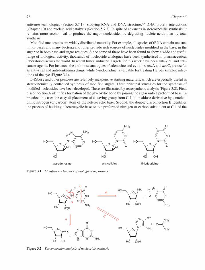
antisense technologies (Section 5.7.1),1 studying RNA and DNA structure,2,3 DNA–protein interactions(Chapter 10) and nucleic acid catalysis (Section 5.7.3). In spite of advances in stereospecific synthesis, itremains more economical to produce the major nucleosides by degrading nucleic acids than by total synthesis.
Modified nucleosides are widely distributed naturally. For example, all species of tRNA contain unusualminor bases and many bacteria and fungi provide rich sources of nucleosides modified in the base, in thesugar or in both base and sugar residues. Since some of these have been found to show a wide and usefulrange of biological activity, thousands of nucleoside analogues have been synthesised in pharmaceuticallaboratories across the world. In recent times, industrial targets for this work have been anti-viral and anti-cancer agents. For instance, the arabinose analogues of adenosine and cytidine, araA and araC, are usefulas anti-viral and anti-leukaemia drugs, while 5-iodouridine is valuable for treating Herpes simplex infec-tions of the eye (Figure 3.1).
D-Ribose and other pentoses are relatively inexpensive starting materials, which are especially useful instereochemically controlled synthesis of modified sugars. Three principal strategies for the synthesis ofmodified nucleosides have been developed. These are illustrated by retrosynthetic analysis (Figure 3.2). First,disconnection A identifies formation of the glycosylic bond by joining the sugar onto a preformed base. Inpractice, this uses the easy displacement of a leaving group from C-1 of an aldose derivative by a nucleo-philic nitrogen (or carbon) atom of the heterocyclic base. Second, the double disconnection B identifiesthe process of building a heterocyclic base onto a preformed nitrogen or carbon substituent at C-1 of the
78 Chapter 3
OHO
HO
OH
N
N
NH2
OOHO
HO
OH N
N
N
N
NH2
OHO
HO
N
NH
O
O
I
OH
ara-adenosine ara-cytidine 5-iodouridine
Figure 3.1 Modified nucleosides of biological importance
N
NHN
NH
O
O
NH2
X
HO
HO (O)H
N
NH
N
N
O
NH2
OHO
HO (O)H
N
NH
NYC
O
NH2
OHO
HO (O)H
NH2
Z
OHO
HO (O)H
N
N
NH2
CY
+
+
NH2
NH2
Z+
A
A
BB
C
C
Figure 3.2 Disconnection analysis of nucleoside synthesis

sugar moiety. Third, a double disconnection C shows the formation of a purine base onto a preformed imi-dazole ribonucleoside. We shall now explore each of these three routes in turn.
3.1.1 Formation of the Glycosylic Bond
The synthesis of nucleosides through glycosyl bond formation should ideally address both stereoselectivity(formation of nucleosides with the natural �-configuration at C-1) and regioselectivity (glycosylation ofpyrimidines at N-1 and purines at N-9). There are essentially three methods that are used: (a) metal salt proce-dures, (b) silyl base procedures, and (c) fusion reactions together with various modifications of these. The firsttwo methods are generally more widely applicable and used most frequently. While the following sectionsdescribe methods that may be used for the preparation of nucleosides, these mainly refer to ribonucleosides.Although such methods may be extended to the syntheses of 2�-deoxyribonucleosides, they often give poorstereoselectivity during glycosyl bond formation. Modifications to these methods that are more suited to thepreparation of 2�-deoxyribonucleosides have been developed and are dealt with later in the chapter.
3.1.1.1 Heavy Metal Salts of Bases. Fischer and Helferich,4 and Koenigs and Knorr introduced theuse of a heavy metal salt (initially silver(I)) of a purine to catalyse the nucleophilic displacement of a halo-gen substituent from C-1 of a protected sugar. In the late 1940s, Todd’s group adapted this chemistry toachieve a synthesis of adenosine5 and guanosine6 following an initial glycosylation between a protected 1-bromo-ribofuranose derivative and 2,8-dichloroadenine. In a later modification, Davoll and Lowy usedmercury(II) salts to improve the yields of products.7 Typically, chloromercuri-6-benzamidopurine reactswith 2,3,5-tri-O-acetyl-D-ribofuranosyl chloride or bromide to give a protected nucleoside from whichadenosine is obtained by removal of the protecting groups (Figure 3.3). These syntheses almost invariablygave the desired stereoselectivity, predominantly providing the ��-anomer at C-1 of ribose owing to theformation of an intermediate acyloxonium ion by the sugar component (see Section 3.1.1.7). Thechloromercuri salts of a range of purines can be used, provided the nucleophilic substituents are protected.Thus, amino groups have to be protected by acylation, as shown in a synthesis of guanine nucleosidesusing 2-acetamido-6-chloropurine followed by appropriate hydrolysis (Figure 3.3).
The chloromercuri derivatives of suitable pyrimidines can be used in much the same way as illustratedby a synthesis of cytidine from 4-ethoxypyrimidine-2-one (Figure 3.4).8 While this type of glycosylation gives
Nucleosides and Nucleotides 79
N
NN
N
O
NHBz
Br
AcO
AcO OAc
HgCl OAcO
AcO OAc
N
N
N
N
NHBz
OHO
HO OH
N
N
N
N
NH2
N
NN
N
Cl
HgCl
NHAc
OAcO
AcO OAc
N
N
N
N
Cl
NHAc
OHO
HO OH
N
NH
N
N
O
NH2
(i)
(i)
(ii)
(iii)
Figure 3.3 Chloromercuri route for synthesis of purine nucleosides. Reagents: (i) xylene, 120°C; (ii) NH3, MeOH;and (iii) NaOH aq
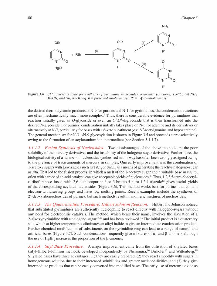
the desired thermodynamic products at N-9 for purines and N-1 for pyrimidines, the condensation reactionsare often mechanistically much more complex.9 Thus, there is considerable evidence for pyrimidines thatreaction initially gives an O-glycoside or even an O2,O4-diglycoside that is then transformed into thedesired N-glycoside. For purines, condensation initially takes place on N-3 for adenine and its derivatives oralternatively at N-7, particularly for bases with a 6-keto substituent (e.g. N2-acetylguanine and hypoxanthine).The general mechanism for N-3→N-9 glycosylation is shown in Figure 3.5 and proceeds stereoselectivelyowing to the formation of an acyloxonium ion intermediate (see Section 3.1.1.7).
3.1.1.2 Fusion Synthesis of Nucleosides. Two disadvantages of the above methods are the poorsolubility of the mercury derivatives and the instability of the halogeno-sugar derivative. Furthermore, thebiological activity of a number of nucleosides synthesised in this way has often been wrongly assigned owingto the presence of trace amounts of mercury in samples. One early improvement was the combination of1-acetoxy sugars with Lewis acids such as TiCl4 or SnCl4 as a means of generating the reactive halogeno-sugarin situ. That led to the fusion process, in which a melt of the 1-acetoxy sugar and a suitable base in vacuo,often with a trace of an acid catalyst, can give acceptable yields of nucleosides.10 Thus, 1,2,3,5-tetra-O-acetyl-D-ribofuranose fused with 2,6-dichloropurine11 or 3-bromo-5-nitro-1,2,4-triazole12 gives useful yields of the corresponding acylated nucleosides (Figure 3.6). This method works best for purines that containelectron-withdrawing groups and have low melting points. Recent examples include the syntheses of 2�-deoxyribonucleosides of purines, but such methods result in anomeric mixtures of nucleosides.
3.1.1.3 The Quaternization Procedure: Hilbert Johnson Reaction. Hilbert and Johnson noticedthat substituted pyrimidines are sufficiently nucleophilic to react directly with halogeno-sugars withoutany need for electrophilic catalysis. The method, which bears their name, involves the alkylation of a 2-alkoxypyrimidine with a halogeno-sugar13,14 and has been reviewed.15 The initial product is a quaternarysalt, which at higher temperatures eliminates an alkyl halide to give an intermediate condensation product.Further chemical modification of substituents on the pyrimidine ring can lead to a range of natural andartificial bases (Figure 3.7). Such condensations frequently give mixtures of �- and �-anomers althoughthe use of HgBr2 increases the proportion of the �-anomer.
3.1.1.4 Silyl Base Procedure. A major improvement came from the utilisation of silylated bases(silyl-Hilbert–Johnson method), developed independently by Nishimura,16 Birkofer17 and Wittenberg.18
Silylated bases have three advantages: (1) they are easily prepared, (2) they react smoothly with sugars inhomogeneous solution due to their increased solubilities and greater nucleophilicities, and (3) they giveintermediate products that can be easily converted into modified bases. The early use of mercuric oxide as
80 Chapter 3
N
N
OEt
HgCl
O
+ RBr
N
N
OEt
R
O
N
N
NH2
R'
O
N
NH
O
R'
O
(i)
(ii)
(iii)
Figure 3.4 Chloromercuri route for synthesis of pyrimidine nucleosides. Reagents: (i) xylene, 120°C; (ii) NH3,MeOH; and (iii) NaOH aq. R � protected ribofuranosyl; R� � 1-�-D-ribofuranosyl
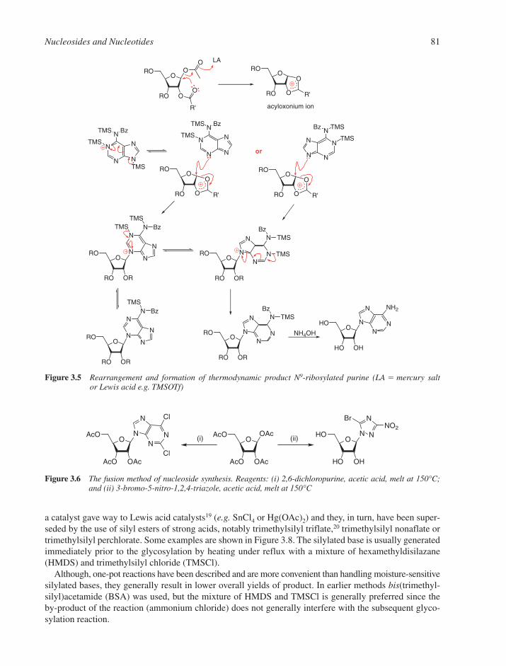
a catalyst gave way to Lewis acid catalysts19 (e.g. SnCl4 or Hg(OAc)2) and they, in turn, have been super-seded by the use of silyl esters of strong acids, notably trimethylsilyl triflate,20 trimethylsilyl nonaflate ortrimethylsilyl perchlorate. Some examples are shown in Figure 3.8. The silylated base is usually generatedimmediately prior to the glycosylation by heating under reflux with a mixture of hexamethyldisilazane(HMDS) and trimethylsilyl chloride (TMSCl).
Although, one-pot reactions have been described and are more convenient than handling moisture-sensitivesilylated bases, they generally result in lower overall yields of product. In earlier methods bis(trimethyl-silyl)acetamide (BSA) was used, but the mixture of HMDS and TMSCl is generally preferred since the by-product of the reaction (ammonium chloride) does not generally interfere with the subsequent glyco-sylation reaction.
Nucleosides and Nucleotides 81
N
N N
N
N
TMS
ORO
RO O
OO
O
R'
LA
ORO
RO O
O
R'
ORO
RO O
O
R'
ORO
RO OR
N
N
N
N
N
OHO
HO OH
NN
N
N
NH2
acyloxonium ion
TMS Bz
TMS N
N N
N
NTMS Bz
TMS
TMS BzTMS
ORO
RO O
O
R'
N
NN
N
N TMSBz
TMS
ORO
RO OR
NN
N
N
N
TMS
TMSBz
ORO
RO OR
N
N
N
N
N BzTMS
ORO
RO OR
NN
N
N
N TMSBz
or
NH4OH
Figure 3.5 Rearrangement and formation of thermodynamic product N9-ribosylated purine (LA � mercury salt or Lewis acid e.g. TMSOTf)
OAcO
AcO OAc
NN
N
N
Cl
Cl
OHO
HO OH
N
N
N(ii)O
AcO
AcO OAc
OAc(i)
BrNO2
Figure 3.6 The fusion method of nucleoside synthesis. Reagents: (i) 2,6-dichloropurine, acetic acid, melt at 150°C;and (ii) 3-bromo-5-nitro-1,2,4-triazole, acetic acid, melt at 150°C
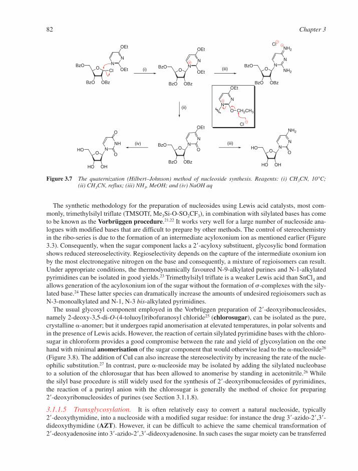
The synthetic methodology for the preparation of nucleosides using Lewis acid catalysts, most com-monly, trimethylsilyl triflate (TMSOTf, Me3Si-O-SO2CF3), in combination with silylated bases has cometo be known as the Vorbrüggen procedure.21,22 It works very well for a large number of nucleoside ana-logues with modified bases that are difficult to prepare by other methods. The control of stereochemistryin the ribo-series is due to the formation of an intermediate acyloxonium ion as mentioned earlier (Figure3.3). Consequently, when the sugar component lacks a 2�-acyloxy substituent, glycosylic bond formationshows reduced stereoselectivity. Regioselectivity depends on the capture of the intermediate oxonium ionby the most electronegative nitrogen on the base and consequently, a mixture of regioisomers can result.Under appropriate conditions, the thermodynamically favoured N-9-alkylated purines and N-1-alkylatedpyrimidines can be isolated in good yields.23 Trimethylsilyl triflate is a weaker Lewis acid than SnCl4 andallows generation of the acyloxonium ion of the sugar without the formation of �-complexes with the sily-lated base.24 These latter species can dramatically increase the amounts of undesired regioisomers such asN-3-monoalkylated and N-1, N-3 bis-alkylated pyrimidines.
The usual glycosyl component employed in the Vorbrüggen preparation of 2�-deoxyribonucleosides,namely 2-deoxy-3,5-di-O-(4-toluoyl)ribofuranosyl chloride25 (chlorosugar), can be isolated as the pure,crystalline �-anomer; but it undergoes rapid anomerisation at elevated temperatures, in polar solvents andin the presence of Lewis acids. However, the reaction of certain silylated pyrimidine bases with the chloro-sugar in chloroform provides a good compromise between the rate and yield of glycosylation on the onehand with minimal anomerisation of the sugar component that would otherwise lead to the �-nucleoside26
(Figure 3.8). The addition of CuI can also increase the stereoselectivity by increasing the rate of the nucle-ophilic substitution.27 In contrast, pure �-nucleoside may be isolated by adding the silylated nucleobase to a solution of the chlorosugar that has been allowed to anomerise by standing in acetonitrile.26 While the silyl base procedure is still widely used for the synthesis of 2�-deoxyribonucleosides of pyrimidines,the reaction of a purinyl anion with the chlorosugar is generally the method of choice for preparing 2�-deoxyribonucleosides of purines (see Section 3.1.1.8).
3.1.1.5 Transglycosylation. It is often relatively easy to convert a natural nucleoside, typically 2�-deoxythymidine, into a nucleoside with a modified sugar residue: for instance the drug 3�-azido-2�,3�-dideoxythymidine (AZT). However, it can be difficult to achieve the same chemical transformation of 2�-deoxyadenosine into 3�-azido-2�,3�-dideoxyadenosine. In such cases the sugar moiety can be transferred
82 Chapter 3
NN
OEt
OEt (i) OBzO
BzO OBz
O ClBzO
BzO OBz
NN
OEt
OEtO
BzO
BzO OBz
NN
NH2
NH2
(ii)
(iii)
Cl
NN
OEt
O CH2CH3
Cl
OBzO
BzO OBz
NN
OEt
O(iii)(iv)
OHO
HO OH
NNH
O
OO
HO
HO OH
NN
NH2
O
Figure 3.7 The quaternization (Hilbert–Johnson) method of nucleoside synthesis. Reagents: (i) CH3CN, 10°C; (ii) CH3CN, reflux; (iii) NH3, MeOH; and (iv) NaOH aq
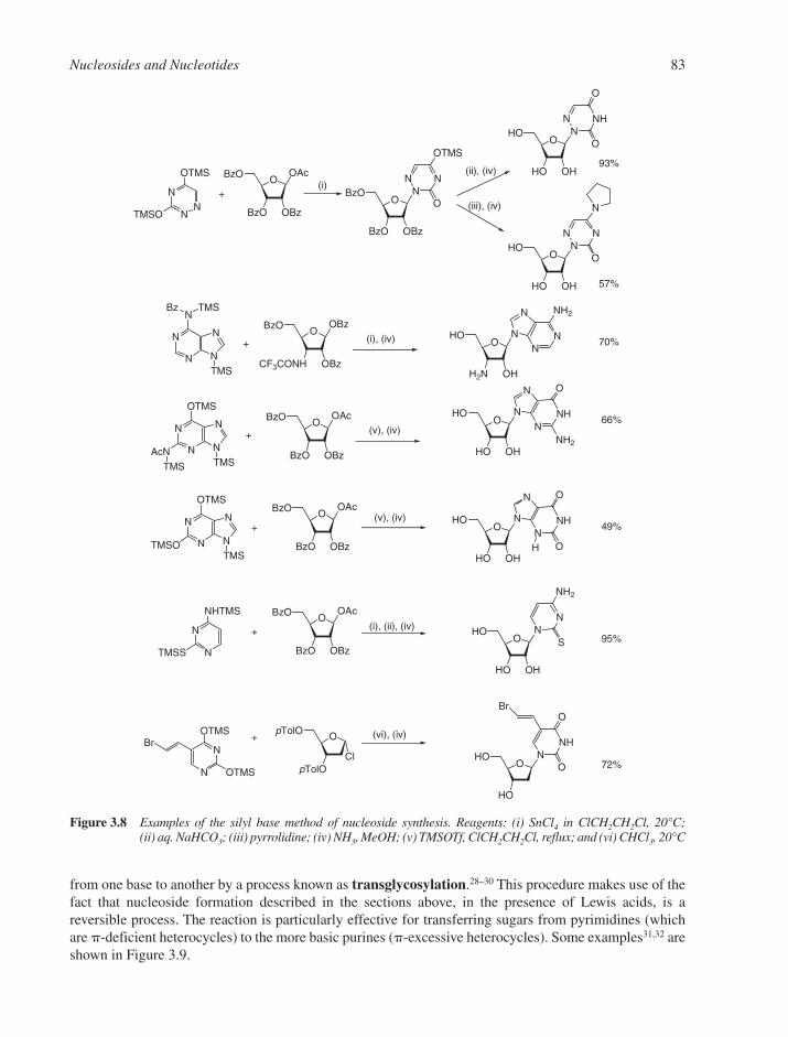
from one base to another by a process known as transglycosylation.28–30 This procedure makes use of thefact that nucleoside formation described in the sections above, in the presence of Lewis acids, is areversible process. The reaction is particularly effective for transferring sugars from pyrimidines (whichare �-deficient heterocycles) to the more basic purines (�-excessive heterocycles). Some examples31,32 areshown in Figure 3.9.
Nucleosides and Nucleotides 83
OOAcBzO
BzO OBz
N
NN
OTMS
TMSO
+O
BzO
BzO OBz
NN
N
OTMS
O
OHO
HO OH
NN
NH
O
O
(i)(ii), (iv)
(iii), (iv)
OHO
HO OH
NN
N
N
O
93%
57%
OHO
HO OH
NNH
N
N
O
NH2N
N N
N
OTMS
AcN
OOAcBzO
BzO OBz
(v), (iv)66%
TMS TMS
N
N N
N
OTMS
TMSOTMS
+
+O
OAcBzO
BzO OBz
OHO
HO OH
NNH
N
N
O
O
(v), (iv)49%
OOAcBzO
BzO OBz
N
N
NHTMS
TMSS
+O
HO
HO OH
NN
NH2
S
(i), (ii), (iv)
OpTolO
pTolO
N
N
OTMS
OTMS
+
OHO
HO
NNH
O
O
(vi), (iv)Br
Cl
Br
72%
95%
OHO
H2N OH
NN
N
N
NH2
N
N N
N
N
OOBzBzO
CF3CONH OBz
(i), (iv) 70%
TMS
+
TMSBz
H
Figure 3.8 Examples of the silyl base method of nucleoside synthesis. Reagents: (i) SnCl4 in ClCH2CH2Cl, 20°C;(ii) aq. NaHCO3; (iii) pyrrolidine; (iv) NH3, MeOH; (v) TMSOTf, ClCH2CH2Cl, reflux; and (vi) CHCl3, 20°C

This reaction has all the hallmarks of an SN1 ionization process, as shown both by the intramoleculartransfer of a sugar residue from N-7 to N-9 of 6-chloro-1-deazapurine and by the anomerisation of �- into�-nucleosides. Transglycosylation is also a useful method for the preparation of �-anomers of nucleosidesfrom their natural isomers. The mixture of �- and �-species that is usually formed can be separated bychromatography. However, the thermodynamically favoured regioselectivity of these processes is not eas-ily predictable.
3.1.1.6 Enzymatic Methods. Hóly, Hutchinson and others have made good use of biotransforma-tions of readily available nucleosides into novel derivatives by enzyme-catalysed transglycosylation.33,34
Uridine phosphorylase and thymidine phosphorylase degrade uridine, 1-�-D-arabinofuranosyluracil (ara-U)and thymidine into the corresponding pentose-1-�-phosphates. These may be converted into the corres-ponding nucleosides containing purines, modified purines or substituted imidazoles in situ in the presenceof the new nucleobase and the enzyme purine nucleoside phosphorylase (PNP). The method also worksfor some 3-deazapurines.35 Some examples are shown in Figure 3.10.
A number of other enzymes have also been used, such as PNP from Enterobacter aerogenes that can trans-form inosine into virazole in the presence of 1,2,4-triazole-3-carboxamide36 and the enzyme nucleoside2�-deoxyribosyltransferase from Lactobacillus leichmannii that has been used for the large scale transfor-mation of thymidine or 2�-deoxycytidine into corresponding purine nucleosides,37 as well as into a number of1-deazapurine nucleosides.38 The transfer of the sugars 2,3-dideoxy-D-ribofuranose and D-arabinofuranoseare also practicable propositions. In general, enzymatic transglycosylations are relatively efficient and highlystereospecific as only �-glycosides are formed, and they can often be employed on a gram scale.
3.1.1.7 Control of Anomeric Stereochemistry. Condensation of sugars having a 2-acyloxy substituentwith a base invariably gives N-glycoside products that have the 1,2-trans-configuration. This control ofanomeric stereochemistry led Baker to suggest that neighbouring group participation by the acyloxy moietyat the 2-position is responsible.39 In the case of ribonucleosides, ionization of the leaving group at C-1 ofthe sugar generates a carbocation that is then captured by the carbonyl group of the adjacent acyl group to form an acyloxonium ion on the lower face of the sugar (Figure 3.3). This is independent of the initial con-figuration of the sugar halide and is followed by nucleophilic displacement of the base from the opposite
84 Chapter 3
OAcO
N3
NNH
O
O
H3C
OHO
N3
NN
N
N
NH2
NNH
O
OOHO
HO NHCOCF3
(iii), (ii)N
NH
N
N
O
NH2
OHO
HO NH2
(i),(ii)
N
NN
N
NTMS COC7H15
TMS
+ 27% 9-α35% 9-β
60%+
OAcO
N3
NNH
O
O
H3C
OHO
N3
NNH
N
N
O
(i),(ii)
N
NN
N
OTMS
TMS
+14% 9-α28% 9-β13% 7-α β,
NTMS
COC15H31
NH2
N
NN
N
OTMS
TMSN
TMS
COC15H31
Figure 3.9 Transglycosylation synthesis of nucleosides. Reagents: (i) TMSOTf in CH3CN, reflux; (ii) NH3, MeOH;and (iii) TMSOTf, BSA in CH3CN, reflux
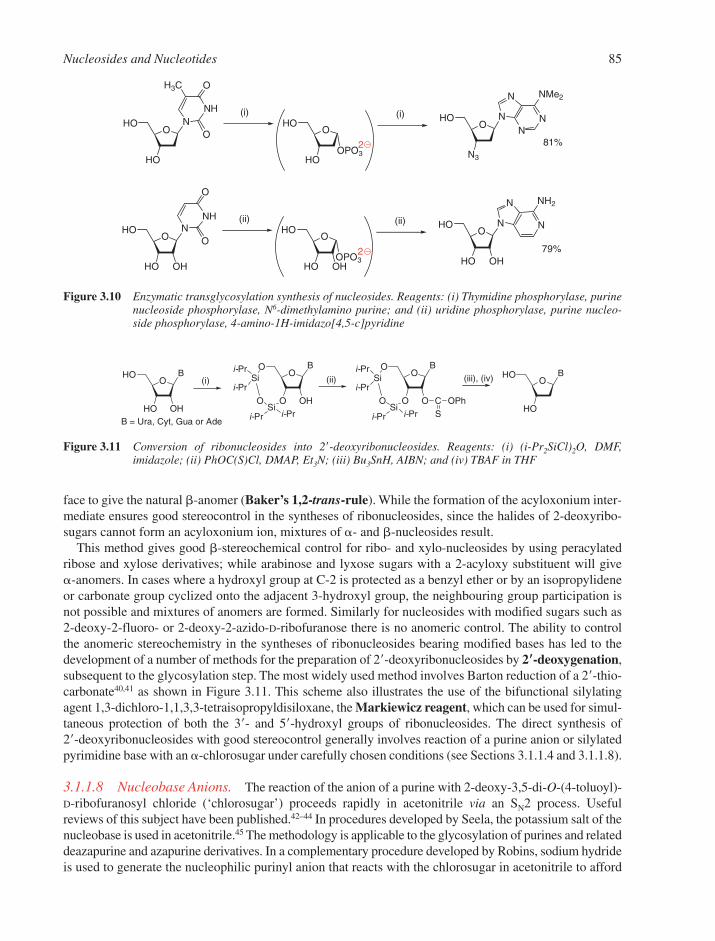
face to give the natural �-anomer (Baker’s 1,2-trans-rule). While the formation of the acyloxonium inter-mediate ensures good stereocontrol in the syntheses of ribonucleosides, since the halides of 2-deoxyribo-sugars cannot form an acyloxonium ion, mixtures of �- and �-nucleosides result.
This method gives good �-stereochemical control for ribo- and xylo-nucleosides by using peracylatedribose and xylose derivatives; while arabinose and lyxose sugars with a 2-acyloxy substituent will give �-anomers. In cases where a hydroxyl group at C-2 is protected as a benzyl ether or by an isopropylideneor carbonate group cyclized onto the adjacent 3-hydroxyl group, the neighbouring group participation isnot possible and mixtures of anomers are formed. Similarly for nucleosides with modified sugars such as2-deoxy-2-fluoro- or 2-deoxy-2-azido-D-ribofuranose there is no anomeric control. The ability to controlthe anomeric stereochemistry in the syntheses of ribonucleosides bearing modified bases has led to thedevelopment of a number of methods for the preparation of 2�-deoxyribonucleosides by 2��-deoxygenation,subsequent to the glycosylation step. The most widely used method involves Barton reduction of a 2�-thio-carbonate40,41 as shown in Figure 3.11. This scheme also illustrates the use of the bifunctional silylatingagent 1,3-dichloro-1,1,3,3-tetraisopropyldisiloxane, the Markiewicz reagent, which can be used for simul-taneous protection of both the 3�- and 5�-hydroxyl groups of ribonucleosides. The direct synthesis of 2�-deoxyribonucleosides with good stereocontrol generally involves reaction of a purine anion or silylatedpyrimidine base with an �-chlorosugar under carefully chosen conditions (see Sections 3.1.1.4 and 3.1.1.8).
3.1.1.8 Nucleobase Anions. The reaction of the anion of a purine with 2-deoxy-3,5-di-O-(4-toluoyl)-D-ribofuranosyl chloride (‘chlorosugar’) proceeds rapidly in acetonitrile via an SN2 process. Usefulreviews of this subject have been published.42–44 In procedures developed by Seela, the potassium salt of thenucleobase is used in acetonitrile.45 The methodology is applicable to the glycosylation of purines and relateddeazapurine and azapurine derivatives. In a complementary procedure developed by Robins, sodium hydrideis used to generate the nucleophilic purinyl anion that reacts with the chlorosugar in acetonitrile to afford
Nucleosides and Nucleotides 85
OHO
HO
NNH
O
O
H3C
OHO
N3
NN
N
N
NMe2
81%O
HO
HOOPO3
2
(i) (i)
OHO
HO
NNH
O
OO
HO
HO
N
N
N
NH2
79%O
HO
HOOPO3
2
(ii) (ii)
OH OH OH
Figure 3.10 Enzymatic transglycosylation synthesis of nucleosides. Reagents: (i) Thymidine phosphorylase, purinenucleoside phosphorylase, N6-dimethylamino purine; and (ii) uridine phosphorylase, purine nucleo-side phosphorylase, 4-amino-1H-imidazo[4,5-c]pyridine
OO
O OH
BSi
Sii-Pri-Pr
O
i-Pr
i-Pr
OO
O O
BSi
Sii-Pri-Pr
O
i-Pr
i-Pr
C
S
OPh
OHO
HO OH
B(i) (ii) (iii), (iv) O
HO
HO
B
B = Ura, Cyt, Gua or Ade
Figure 3.11 Conversion of ribonucleosides into 2�-deoxyribonucleosides. Reagents: (i) (i-Pr2SiCl)2O, DMF, imidazole; (ii) PhOC(S)Cl, DMAP, Et3N; (iii) Bu3SnH, AIBN; and (iv) TBAF in THF

good yields of the corresponding 2�-deoxyribonucleoside with the natural �-configuration.42 The usefulstereocontrol achieved in these reactions arises since the anomerisation of the chlorosugar in acetonitrileis much slower than the nucleophilic displacement of chloride by the purinyl anion. The regioselectivityof glycosylation (N-7/N-9) is variable, depending on the purine derivative, but such isomers can usually beseparated by chromatography. Some examples are shown in Figure 3.12. Treatment of the nucleosidic prod-ucts with ammonia in methanol removes the sugar protecting groups, while heating with the same reagentprovides a useful route to amino-substituted purine nucleosides (Figure 3.13).
The nucleobase anion glycosylation procedure has also been used to synthesise a wide variety of 2�-deoxyribonucleosides of deazapurines (Figure 3.14).42–44
3.1.1.9 C-nucleosides. A few C-nucleosides have been made by carbanion displacement reactions atC-1 of a suitably protected sugar, although the high basicity of the carbanion can lead to an unwanted 1,2-elimination. A classic example is Brown’s synthesis of pseudouridine,46 a common component of tRNAspecies, by the reaction of 2,4-bis-(t-butoxy)-5-lithiopyrimidine with 2,4;3,5-bis-O-benzylidene-D-ribose.This gave more of the �-pseudouridine (18%) than the �-anomer (8%) (Figure 3.15). Grignard reagentshave also been used in carbanion condensations at C-1 of 2-deoxyribose precursors; for example, in thesynthesis of fluorinated nucleobase analogues by Kool47 (Figure 3.15). The use of palladium chemistry hasalso been exploited.48
86 Chapter 3
N
NN
N
X
M
Y
Z
Op-TolO
p-TolO
N
N
N
N
X
Y
Z
Op-TolO
p-TolOCl
Op-TolO
p-TolO
N
NN
N
Z
Y
X
X Y
H H
H H
Cl H H
NH2 H
Cl Cl Cl
Cl Cl H
Cl NH2 H
Br Br H
(i) or (ii)
M = Na or K
N-9 isomer
N-7 isomer
Conditions Z N-9 N-7
(i) 44 28
(i) 43 9
(i) 59 13
(i) 48 24
(ii) 56 -
(ii) 59 13
(ii) 57 -
(ii) 57 7
Yield (%)
OMe
SMe
OMe
Figure 3.12 Nucleobase anion route for synthesis of purine nucleosides. Reagents: (i) powdered KOH, TDA-1,CH3CN; and (ii) NaH, CH3CN
N
NZ
N
Cl
H
X
Y
N
NZ
N
Cl
dR'
X
Y
N
NZ
N
NH
dR'
X
Y
X
Cl H
H H
H
Cl I
Y
H
H
H
H
H H N
X
Cl H
H H
H
Cl I
Y
H
H
H
H
H H N
(i) (ii)
Z yield(%)60
71
66
71
32
Z yield(%)61
85
72
88
85
MeS MeS
Figure 3.13 Aminopurine nucleoside analogues prepared by the nucleobase anion route. Reagents: (i) NaH,CH3CN then 3,5-di-O-p-toluoyl-b-D-ribofuranosyl chloride; and (ii) NH3/MeOH, heat
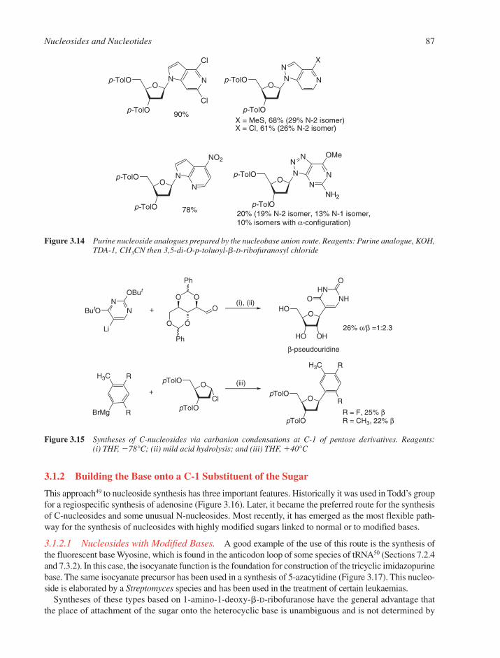
3.1.2 Building the Base onto a C-1 Substituent of the Sugar
This approach49 to nucleoside synthesis has three important features. Historically it was used in Todd’s groupfor a regiospecific synthesis of adenosine (Figure 3.16). Later, it became the preferred route for the synthesisof C-nucleosides and some unusual N-nucleosides. Most recently, it has emerged as the most flexible path-way for the synthesis of nucleosides with highly modified sugars linked to normal or to modified bases.
3.1.2.1 Nucleosides with Modified Bases. A good example of the use of this route is the synthesis ofthe fluorescent base Wyosine, which is found in the anticodon loop of some species of tRNA50 (Sections 7.2.4and 7.3.2). In this case, the isocyanate function is the foundation for construction of the tricyclic imidazopurinebase. The same isocyanate precursor has been used in a synthesis of 5-azacytidine (Figure 3.17). This nucleo-side is elaborated by a Streptomyces species and has been used in the treatment of certain leukaemias.
Syntheses of these types based on 1-amino-1-deoxy-�-D-ribofuranose have the general advantage thatthe place of attachment of the sugar onto the heterocyclic base is unambiguous and is not determined by
Nucleosides and Nucleotides 87
90% X = MeS, 68% (29% N-2 isomer)X = Cl, 61% (26% N-2 isomer)
Op-TolO
p-TolO
NN
Cl
Cl
Op-TolO
p-TolO
N
N
N
X
Op-TolO
p-TolO
N
N
NO2
78%
Op-TolO
p-TolO
NN
NN
N
OMe
20% (19% N-2 isomer, 13% N-1 isomer,10% isomers with α-configuration)
NH2
Figure 3.14 Purine nucleoside analogues prepared by the nucleobase anion route. Reagents: Purine analogue, KOH,TDA-1, CH3CN then 3,5-di-O-p-toluoyl-b-D-ribofuranosyl chloride
HNNH
O
OHO
HO OH
ONN
OBut
ButO
Li
O O
O O
Ph
Ph
O
R
RBrMg
H3COpTolO
pTolOCl
R
OpTolO
pTolO
R
H3C
+
β-pseudouridine
26% α/β =1:2.3
(i), (ii)
(iii)
R = F, 25% βR = CH3, 22% β
+
Figure 3.15 Syntheses of C-nucleosides via carbanion condensations at C-1 of pentose derivatives. Reagents: (i) THF, �78°C; (ii) mild acid hydrolysis; and (iii) THF, �40°C

the most nucleophilic heteroatom on the base component. Such syntheses have, therefore, been widelyemployed for the preparation of the imidazole nucleosides involved in the de novo biosynthesis of purinenucleosides (Section 3.4.1), and of modified pyrimidine and purine nucleosides. A typical example of thework of Gordon Shaw in this area is the synthesis of 2-thioribothymidine (Figure 3.18).51
In a similar way, a cyanomethyl group at C-1 of D-ribose supports the syntheses of 9-deazainosine,antibiotic oxazinomycin and pseudouridine (Figure 3.18).52,53
3.1.2.2 C-nucleosides. With the growing availability of chemical reactions having a high degree ofstereochemical selectivity, the synthesis of C-nucleosides by this route has moved away from sugars asstarting materials. Showdomycin is a product of Streptomyces showdowensis and has useful cytotoxic andenzyme inhibitory properties. A route starting from a tricyclic precursor can branch to give either show-domycin or psuedouridine in a stereospecific fashion (Figure 3.19).54
The formal replacement of the 4�-oxygen in the sugar by a methylene group gives a carbocyclic nucleoside.Much of the activity in the synthesis of carbocyclic nucleosides has been carried out in the search for poten-tial anti-tumour and anti-viral agents, especially provoked by the search for agents effective against humanimmunodeficiency virus (HIV) (Section 3.7.2). One of the particular values of carbocyclic nucleosides is
88 Chapter 3
OHO
HO OH
N
N
N
N
O
NMe Me
OHO
HO OH
N
NCONH2
NHMe
OAcO
AcO OAc
NCO
OAcO
AcO OAc
O
HN
N
H
OMe
OAcO
AcO
N
N
N
OMe
O
OAc
OHO
HO
N
N
N
NH2
O
OH
(i) (ii) - (iv)
5-azacytidine
Wyosine
Figure 3.17 Building the Wye base and 5-azacytosine onto a C-1 isocyanate. Reagents: (i) three carbon fragment;(ii) CNBr; (iii) NaOEt, EtOH; and (iv) BrCH2COCH3
N
N
H2N
NH2
SMe
OBnO
HO OH
N
NHN
NH2
SMe
OHO
HO OH
N
N
N
N
NH2
OBnO
HO OH
N
NHN
NH2
SMe
H2N
(i), (ii) (iii)
(iv), (v)
(vi)
Figure 3.16 Todd’s synthesis of adenosine. Reagents: (i) 5-O-benzyl-2,3,4-tri-O-acetyl-D-ribose; (ii) NH3 (iii) dia-zonium coupling or nitrosation followed by reduction; (iv) thiourea; (v) Raney nickel desulfurisation;and (vi) H2/Pd-C debenzylation

their great metabolic stability to the phosphorylase enzymes, which cleaves the glycosylic bond of normalnucleosides. The carbocyclic analogues of adenosine (aristeromycin) and neplanocin A (Figure 3.20) areboth naturally occurring and display anti-tumour and antibiotic activity respectively. Carbovir (Figure 3.20),a carbocyclic 2�,3�-dideoxy-2�,3�-didehydro analogue of guanosine, is a potent inhibitor of HIV replication,as indeed are several corresponding 2�,3�-dideoxy and 2�,3�-dideoxy-2�,3�-didehydro nucleoside analoguesof inosine, guanosine and adenosine.
Nucleosides and Nucleotides 89
OHO
HO OH
NNH
HN O
OBzO
BzO OBz
NH2
HNNH
O
OHO
HO OH
O
MeO
SCN O
Me
NNH
O
OHO
HO OH
S
OTrO
O O
CNO
TrO
O O
CN
NMe2
OTrO
O O
N
CH2Ph
CO2Et
NH2
HNO
O
OHO
HO OH
O
pseudouridine 9-deazainosineoxazinomycin
Figure 3.18 Syntheses of N- and C-nucleosides by building the base onto the sugar
HNNH
O
OHO
HO OH
O
OH
O
O O
O
NMe2O O
O
O
O
O
O O
OHO
O O
CO2Me
O
OHO
HO OH
NH
O
O
β-pseudouridine
(i)-(iv) (v)
(vi), (vii)(viii)
(ix),(x)
showdomycin
Figure 3.19 Synthesis of showdomycin and pseudouridine from furan. Reagents: (i) OsO4, H2O2; (ii) acetone, H�;(iii) CF3CO3H; (iv) resolution; (v) dimethylformamide; (vi) urea; (vii) H3O
�; (viii) furan-1-carbaldehyde,NaOMe; (ix) ozone; and (x) Ph3P � CHCONH2
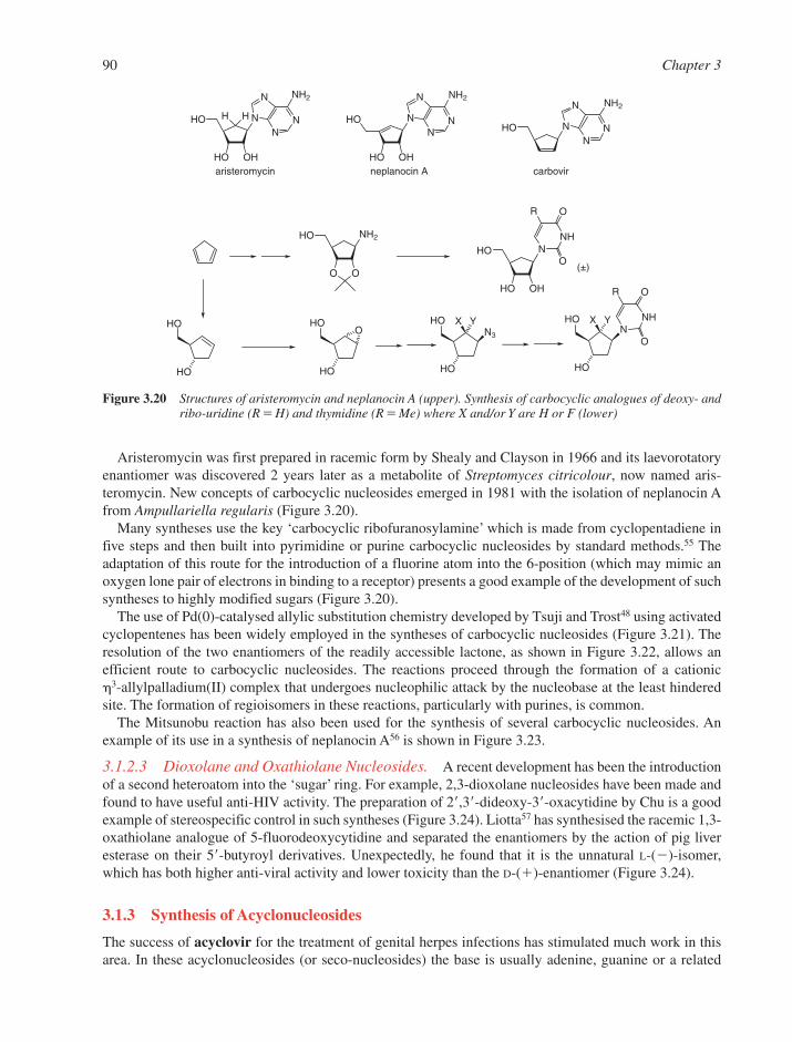
Aristeromycin was first prepared in racemic form by Shealy and Clayson in 1966 and its laevorotatoryenantiomer was discovered 2 years later as a metabolite of Streptomyces citricolour, now named aris-teromycin. New concepts of carbocyclic nucleosides emerged in 1981 with the isolation of neplanocin Afrom Ampullariella regularis (Figure 3.20).
Many syntheses use the key ‘carbocyclic ribofuranosylamine’ which is made from cyclopentadiene infive steps and then built into pyrimidine or purine carbocyclic nucleosides by standard methods.55 Theadaptation of this route for the introduction of a fluorine atom into the 6-position (which may mimic anoxygen lone pair of electrons in binding to a receptor) presents a good example of the development of suchsyntheses to highly modified sugars (Figure 3.20).
The use of Pd(0)-catalysed allylic substitution chemistry developed by Tsuji and Trost48 using activatedcyclopentenes has been widely employed in the syntheses of carbocyclic nucleosides (Figure 3.21). Theresolution of the two enantiomers of the readily accessible lactone, as shown in Figure 3.22, allows an efficient route to carbocyclic nucleosides. The reactions proceed through the formation of a cationic 3-allylpalladium(II) complex that undergoes nucleophilic attack by the nucleobase at the least hinderedsite. The formation of regioisomers in these reactions, particularly with purines, is common.
The Mitsunobu reaction has also been used for the synthesis of several carbocyclic nucleosides. Anexample of its use in a synthesis of neplanocin A56 is shown in Figure 3.23.
3.1.2.3 Dioxolane and Oxathiolane Nucleosides. A recent development has been the introductionof a second heteroatom into the ‘sugar’ ring. For example, 2,3-dioxolane nucleosides have been made andfound to have useful anti-HIV activity. The preparation of 2�,3�-dideoxy-3�-oxacytidine by Chu is a goodexample of stereospecific control in such syntheses (Figure 3.24). Liotta57 has synthesised the racemic 1,3-oxathiolane analogue of 5-fluorodeoxycytidine and separated the enantiomers by the action of pig liveresterase on their 5�-butyroyl derivatives. Unexpectedly, he found that it is the unnatural L-(�)-isomer,which has both higher anti-viral activity and lower toxicity than the D-(�)-enantiomer (Figure 3.24).
3.1.3 Synthesis of Acyclonucleosides
The success of acyclovir for the treatment of genital herpes infections has stimulated much work in thisarea. In these acyclonucleosides (or seco-nucleosides) the base is usually adenine, guanine or a related
90 Chapter 3
HO
HO OH
NN
N
N
NH2
H H
HO
HO
NNH
O
O
R
OH
HO
HO OH
NN
N
N
NH2
NH2HO
O O
HO
HO
HO
HO
OHO
HO
N3
X Y HO
HO
X YN
NH
O
O
R
HON
N
N
N
NH2
aristeromycin neplanocin A
(±)
carbovir
Figure 3.20 Structures of aristeromycin and neplanocin A (upper). Synthesis of carbocyclic analogues of deoxy- andribo-uridine (R � H) and thymidine (R � Me) where X and/or Y are H or F (lower)
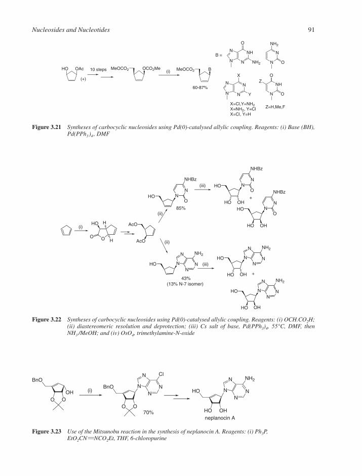
Nucleosides and Nucleotides 91
N
NN
N
X
Y
N
N
NH2
O
NH
N
O
O
Z
NH
NN
N
O
NH2
HO OAc OCO2MeMeOCO2 BMeOCO2
(+)
10 steps (i)
B =
X=Cl,Y=NH2X=NH2, Y=ClX=Cl, Y=H
Z=H,Me,F
60-87%
Figure 3.21 Syntheses of carbocyclic nucleosides using Pd(0)-catalysed allylic coupling. Reagents: (i) Base (BH),Pd(PPh3)4 , DMF
OO
HO H
H
HO NN
NHBz
O
AcO
AcO
HON
N
N
N
NH2
HO NN
NHBz
O
HO OH
HON
N
N
N
NH2
HO OH
HON
N
N
N
NH2
HO OH
HO NN
NHBz
O
HO OH(i)
85%
43%
(ii)
(ii)
(iii)
(iii)
(13% N-7 isomer)
+
+
Figure 3.22 Syntheses of carbocyclic nucleosides using Pd(0)-catalysed allylic coupling. Reagents: (i) OCH.CO2H;(ii) diastereomeric resolution and deprotection; (iii) Cs salt of base, Pd(PPh3)4, 55°C, DMF, thenNH3 /MeOH; and (iv) OsO4, trimethylamine-N-oxide
HO
HO OH
NN
N
N
NH2BnO
O OOH
BnO
O O
NN
N
N
Cl
neplanocin A70%
(i)
Figure 3.23 Use of the Mitsunobu reaction in the synthesis of neplanocin A. Reagents: (i) Ph3P, EtO2CN NCO2Et, THF, 6-chloropurine

purine, which can be converted into adenine or guanine as a result of metabolic deamination or hydroxy-lation (i.e. prodrugs, Section 3.7). The mode of action of acyclovir in Herpes Simplex Virus (HSV)-infected cells involves its specific phosphorylation by the thymidine kinase expressed by the virus. This isfollowed by further phosphorylation by cellular kinases to afford the triphosphate, which is a selective andpotent inhibitor of the HSV DNA polymerase. In principle, four sections of the sugar ring can be ‘cutaway’ and promising biological results have been found in three of these areas. Formally, one can excise(1) C-2�, (2) C-3�, (3) C-2��C-3�, or (4) O-4��C-4��C-5� as shown in Figure 3.25. The syntheses of allof these types of acyclonucleoside are invariably based on N-9 alkylation of a chloropurine precursor, withsubsequent amination and manipulation of the necessary protecting groups. Alkylation of the silylatedchloropurine in the presence of mercury(II) cyanide normally gives excellent yields of the desired N-9regioisomer58 (Figure 3.25). Seco-carbocyclic nucleosides have also been found to have useful antiviralactivity. One example is penciclovir, N-9-(4-hydroxy-3-hydroxymethylbutyl)guanine.
3.1.4 Syntheses of Base and Sugar-Modified Nucleosides
A vast number of nucleosides bearing modified bases or sugars have been made. Many display significantbiological activity, while others have been used for applications in molecular biology, such as nucleic acidsequencing and labelling and investigations of nucleic-acid structure and protein–nucleic acid inter-actions. However, the majority of these involve modification to the heterocyclic base or modification to theC-2� and/or C-3� positions of the sugar. The following is a selective overview of the chemical syntheses ofpentofuranosyl nucleosides either modified at C-2� or C-3� or on the heterocyclic base. By contrast, modi-fications at the 5�-position of the nucleoside do not involve stereochemical control. Many transformationstypical for chemical modification of primary hydroxyl groups have been used on nucleosides, e.g., dis-placement of a tosylate, halogenation in the presence of triphenylphosphine or the Mitsunobu reaction.
3.1.4.1 Modified Bases. The halogenation of pyrimidine nucleosides at C-559 and purine nucleo-sides at C-860 is known for all four halogens, although the 5-iodopyrimidine and 8-bromopurine analogueshave been most widely used for subsequent functionalization at these positions of the nucleobases. Thenucleoside bases 5-iodocytosine and 5-iodouracil can be readily transformed into other 5-substituted analogues by use of palladium chemistry; while nucleophilic displacement of bromine or the palladium-catalysed modification at the 8-position of purine nucleosides gives a variety of 8-substituted analogues.48
Uridine and cytidine and their analogues can be halogenated using bromine water or iodine in aqueousacid/chloroform. These reactions appear to involve a 5-halogeno-6-hydroxy-5,6-dihydropyrimidineadduct (Figure 3.26), which is subsequently dehydrated to give the 5-substituted nucleoside. For 2�,3�-isopropylidine-protected ribonucleosides, there is some evidence that an analogous intermediate is formed
92 Chapter 3
O
O
OH
OH
OBz
O
O
TBDPSO
OH
OH
O
O
HO N
N
NH2
O
N
N
NH2
O
F
O
S
HO
O
O
TBDPSOCOOH
O
O
TBDPSOOAc
(i) (i),(ii),(iii)
(iv)
(v)
L-2',3'-dideoxy-3'-thia-5-fluorocytidineD- 2',3'-dideoxy-3'-oxacytidine
Figure 3.24 Synthesis of D-2�,3�-dideoxy-3�-oxacytidine from 4-O-benzoyl-1,6-anhydro-D-mannose (upper) and structureof L-2�,3�-dideoxy-3�-thia-5-fluorocytidine (lower right). Reagents: (i) NaIO4; (ii) NaBH4; (iii) TBDPSCl,pyridine; (iv) Pb(OAc)4; and (v) silylated N4-acetylcytosine, TMS triflate, 1,2-dichloroethane
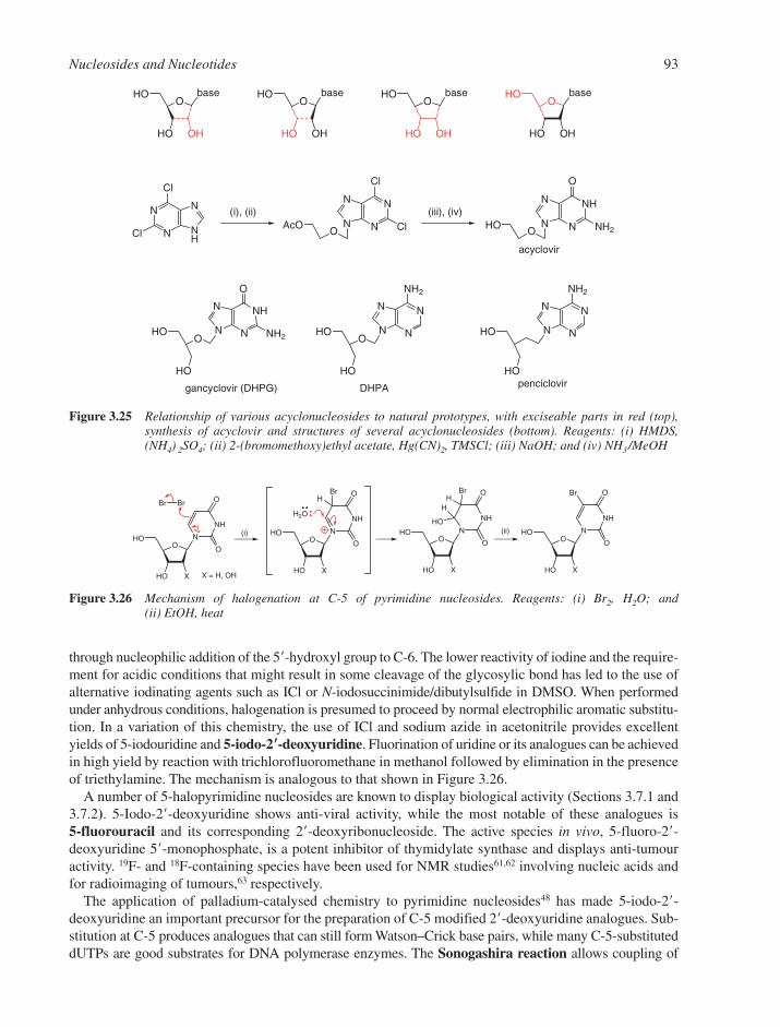
through nucleophilic addition of the 5�-hydroxyl group to C-6. The lower reactivity of iodine and the require-ment for acidic conditions that might result in some cleavage of the glycosylic bond has led to the use ofalternative iodinating agents such as ICl or N-iodosuccinimide/dibutylsulfide in DMSO. When performedunder anhydrous conditions, halogenation is presumed to proceed by normal electrophilic aromatic substitu-tion. In a variation of this chemistry, the use of ICl and sodium azide in acetonitrile provides excellentyields of 5-iodouridine and 5-iodo-2��-deoxyuridine. Fluorination of uridine or its analogues can be achievedin high yield by reaction with trichlorofluoromethane in methanol followed by elimination in the presenceof triethylamine. The mechanism is analogous to that shown in Figure 3.26.
A number of 5-halopyrimidine nucleosides are known to display biological activity (Sections 3.7.1 and3.7.2). 5-Iodo-2�-deoxyuridine shows anti-viral activity, while the most notable of these analogues is 5-fluorouracil and its corresponding 2�-deoxyribonucleoside. The active species in vivo, 5-fluoro-2�-deoxyuridine 5�-monophosphate, is a potent inhibitor of thymidylate synthase and displays anti-tumouractivity. 19F- and 18F-containing species have been used for NMR studies61,62 involving nucleic acids andfor radioimaging of tumours,63 respectively.
The application of palladium-catalysed chemistry to pyrimidine nucleosides48 has made 5-iodo-2�-deoxyuridine an important precursor for the preparation of C-5 modified 2�-deoxyuridine analogues. Sub-stitution at C-5 produces analogues that can still form Watson–Crick base pairs, while many C-5-substituteddUTPs are good substrates for DNA polymerase enzymes. The Sonogashira reaction allows coupling of
Nucleosides and Nucleotides 93
OHO
HO X
N
NH
O
O
BrBr
OHO
HO X
N
NH
O
O
HBr
H O
OHO
HO X
N
NH
O
O
HBr
H
HO
OHO
HO X
N
NH
O
O
Br
X = H, OH
(i) (ii)
Figure 3.26 Mechanism of halogenation at C-5 of pyrimidine nucleosides. Reagents: (i) Br2, H2O; and (ii) EtOH, heat
N
N NH
N
Cl
Cl
N
NN
N
Cl
ClOAcO
NH
NN
N
O
NH2OHO
N
NHN
O
OHO
HO
N NH2
OHO
HO OH
baseO
HO
HO OH
baseO
HO
HO OH
baseO
HO
HO OH
base
HO
HO
N
NN
N
NH2
OHO
HO
N
NN
N
NH2
DHPAgancyclovir (DHPG) penciclovir
(i), (ii) (iii), (iv)
acyclovir
Figure 3.25 Relationship of various acyclonucleosides to natural prototypes, with exciseable parts in red (top), synthesis of acyclovir and structures of several acyclonucleosides (bottom). Reagents: (i) HMDS,(NH4) 2SO4; (ii) 2-(bromomethoxy)ethyl acetate, Hg(CN)2, TMSCl; (iii) NaOH; and (iv) NH3/MeOH

terminal alkynes to 5-iodo- or 5-triflate esters of 2�-deoxyuridine (Figure 3.27). Coupling with allylamineor propargylamine (Figure 3.27c) allows the functionalization of 2�-deoxyuridine or its 5�-triphosphate withnucleophilic amino groups that allow further elaboration of the nucleoside through reaction with N-hydrox-ysuccinimidyl esters. Examples include coupling with carboxylic acids of compounds such as imidazole foruse in SELEX64,65 (Section 5.7.3), or in the preparation of 2�,3�-dideoxy-UTP analogues labelled with, for exam-ple, biotin or fluorescein,2,66,67 the latter finding application in dideoxy DNA sequencing (Section 5.1).
In a number of cases, a minor, fluorescent bicyclic furanopyrimidine has been isolated during Sonogashiracoupling reactions involving 5-iodo-2�-deoxyuridine, which takes place in the presence of CuI. This latter
94 Chapter 3
OHO
HO
NNH
O
O
CF3SO2O
OHO
HO
NNH
O
O
H
OHO
HO
NNH
O
O
R
R H
OHO
HO
NNH
O
O
I
OHO
HO
NNH
O
O
X
H X OHO
HO
NN
O
O
X
OHO
HO
NNH
O
O
I
HNHCOCF3
OHO
HO
NNH
O
O
CF3CONH
OHO
HO
NNH
O
O
I
OHO
HO
NN
O
O
(CH2)9OR
(CH2)9H OR
OHO
HO
NNH
O
O
(CH2)9
RO
OHO
HO
NN
O
O
CF3CONHO
HO
NNH
O
O
H2N
O9P3O
b
(i) (i), (ii)
R = HOMe2C, Ph, 4-MeOC6H4-,4-CF3C6H4, 4-F-C6H5
R = TMS
(i)
X = H, Me, Br or Cl
(i)
(iv) (v)
R = C4H9, C5H11, (CH2)4Cl
minor
minor
4
(iii)
a
c
d
+
+ +
major
+
+
+
Figure 3.27 Palladium-catalysed coupling reactions as routes to C-5-substituted pyrimidine nucleosides. Reagents:(i) Pd(PPh3)4, CuI, Et3N, DMF; (ii) TBAF, MeOH; (iii) synthesis of triphosphate then NH4OH;(iv) Pd(PPh3)4, CuI, i-Pr2EtN, DMF; and (v) CuI, Et3N, MeOH, heat
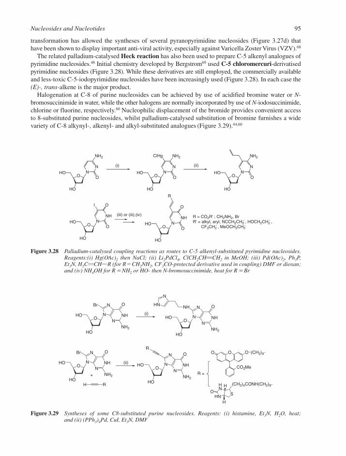
transformation has allowed the syntheses of several pyranopyrimidine nucleosides (Figure 3.27d) thathave been shown to display important anti-viral activity, especially against Varicella Zoster Virus (VZV).68
The related palladium-catalysed Heck reaction has also been used to prepare C-5 alkenyl analogues ofpyrimidine nucleosides.48 Initial chemistry developed by Bergstrom69 used C-5 chloromercuri-derivatisedpyrimidine nucleosides (Figure 3.28). While these derivatives are still employed, the commercially availableand less-toxic C-5-iodopyrimidine nucleosides have been increasingly used (Figure 3.28). In each case the(E)-, trans-alkene is the major product.
Halogenation at C-8 of purine nucleosides can be achieved by use of acidified bromine water or N-bromosuccinimide in water, while the other halogens are normally incorporated by use of N-iodosuccinimide,chlorine or fluorine, respectively.60 Nucleophilic displacement of the bromide provides convenient accessto 8-substituted purine nucleosides, whilst palladium-catalysed substitution of bromine furnishes a widevariety of C-8 alkynyl-, alkenyl- and alkyl-substituted analogues (Figure 3.29).44,60
Nucleosides and Nucleotides 95
OHO
HO
NNH
O
O
I
OHO
HO
NNH
O
O
R
OHO
HO
NN
NH2
O OHO
HO
NN
NH2
O
ClHg
OHO
HO
NN
NH2
O
R = CO2R' ; CH2NH2, BrR' = alkyl, aryl, NCCH2CH2
− , HOCH2CH2− ,
CF3CH2− , MeOCH2CH2
−
(iii) or (iii),(iv)
(i) (ii)
Figure 3.28 Palladium-catalysed coupling reactions as routes to C-5 alkenyl-substituted pyrimidine nucleosides.Reagents:(i) Hg(OAc)2 then NaCl; (ii) Li2PdCl4, ClCH2CHCH2 in MeOH; (iii) Pd(OAc)2, Ph3P,Et3N, H2CCHR (for R� CH2NH2, CF3CO-protected derivative used in coupling) DMF or dioxan;and (iv) NH4OH for R � NH2 or HO- then N-bromosuccinimide, heat for R � Br
OHO
HO
NNH
N
N
O
NH2
Br NHHN
N
OHO
HO
NNH
N
N
O
NH2
OHO
HO
NNH
N
N
O
NH2
Br
H R
OHO
HO
NNH
N
N
O
NH2
R
CO2Me
O O O (CH2)9−
(CH2)4CONH(CH2)9−HN
OH
HNH
S
(i)
(ii)
R =+
Figure 3.29 Syntheses of some C8-substituted purine nucleosides. Reagents: (i) histamine, Et3N, H2O, heat; and (ii) (PPh3)4Pd, CuI, Et3N, DMF

The Sonogashira substitution has also been widely employed by Seela in the syntheses of C7-modified7-deazapurine (pyrrolo[2,3-d]pyrimidine) nucleosides43,44 (Figure 3.30). These analogues have attractedwidespread interest since they represent 7-substituted purine nucleosides that can maintain Watson–Crickbase pairing while retaining the normal anti-conformation about the glycosylic bond (Section 2.1.4). The5�-triphosphates of 7-deaza-2�-deoxyadenosine, 7-deaza-2�-deoxyguanosine and their 7-substituted analogues are generally excellent substrates for many DNA polymerases and have been widely used indeveloping DNA sequencing methodology.67 In addition, 7-deazapurine forms the basis of a number ofnaturally occurring antibiotics such as tubercidin, toyocamycin, sangivamycin and cadeguomycin(Figure 3.30), while several 7-substituted 7-deazaguanosines such as nucleoside Q are found in sometRNAs. Furthermore, many 7-substituted 7-deazapurine nucleoside analogues have been shown to stabi-lize DNA duplexes.70
Many nucleosides have been modified at the 4-position of pyrimidine or 6-position of purine, starting fromuridine or guanosine respectively. The modification of pyrimidine nucleosides at C-4 can be achieved throughnucleophilic substitution of 4-triazolo-pyrimidine nucleoside derivatives, which are stable, isolable compoundsthat can be transformed into a variety of analogues (Figure 3.31).71 O4-Methylthymidine is an important ana-logue formed during DNA damage by alkylating agents, while the highly mutagenic analogues N4-amino- andN4-hydroxy-2�-deoxycytidine are formed by the action of hydrazine and hydroxylamine on DNA (Section 8.4).The reaction of triazolo derivatives with ammonia is particularly useful for converting uracil into cytosine-con-taining nucleosides, especially for analogues containing modified sugars.72 Activation at O-4 for subsequentnucleophilic displacement may also be achieved by use of nitrophenoxy-, dinitrophenylthio- and sulfonateesters, although these are often generated and used in situ because of their high reactivity.
Nucleosides containing bases with 6-keto functions, such as guanine and hypoxanthine, can also betransformed in a similar way into 6-substituted purine nucleosides. O6-Sulfonate esters of purines can bedisplaced by a variety of nucleophiles, although hard nucleophiles such as alkoxide react at sulfur, and so6-alkoxypurines are made via the highly reactive trimethylammonium salt (Figure 3.32).
Thus, syntheses of compounds such as 2-aminopurine nucleosides (a fluorescent base analogue),73
2-amino-6-vinylpurine-2��-deoxyriboside74 (allows covalent cross-linking within DNA) and O6-methyl-2��-deoxyguanosine75 (an important analogue resulting from DNA alkylation damage) are possible, while a variation on this theme leads to 6-thio-2��-deoxyguanosine76 and other analogues (Figure 3.33).
96 Chapter 3
OHO
HO
NNN
XI
Y
OHO
HO
NNN
X
R
Y
OHO
HO OH
NNHN
O
NH2
HO2C
OHO
HO
NNN
NH2
X
(i)
X=NH2, Y=H:R = TMS, t-Bu, C10H21,C4H9,Me, CH2NHCOCF3,cholesteryl-,CH2CH2OTHP, CH2TMSX=OH, Y=NH2:R = Me,CH2NHCOCF3(CH2)3 or 10NHCOCF3
X = H, tubercidinX = CN, toyocamycinX = CONH2, sangivamycin
cadeguomycin
Figure 3.30 Syntheses of 7-substituted 7-deaza-2�-deoxyadenosine and 7-deaza-2�-deoxyguanosine nucleosides(upper). Reagents: (i) terminal alkyne, Pd(PPh3)4, CuI, Et3N, DMF. Some naturally occuring antibioticnucleosides (lower)
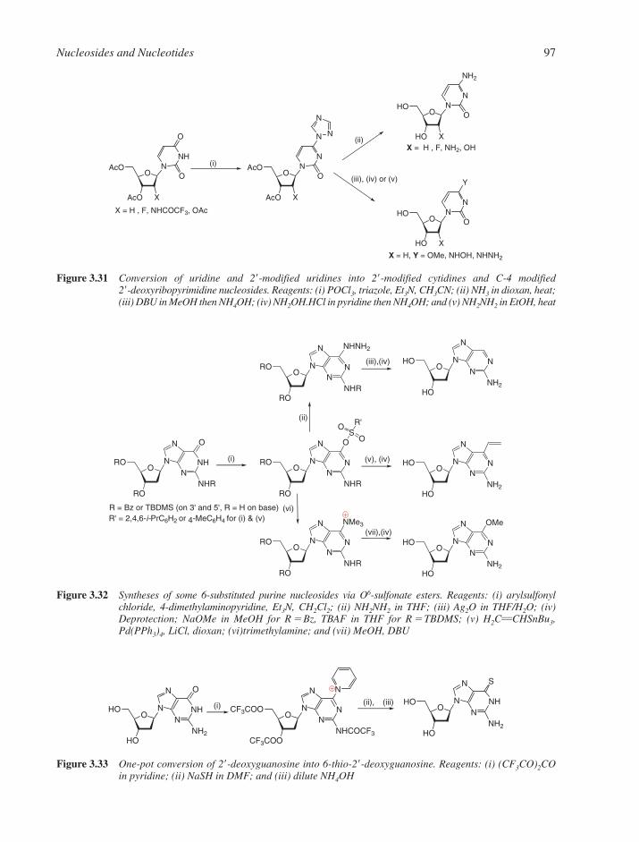
Nucleosides and Nucleotides 97
OAcO
AcO
NNH
O
O
X
OAcO
AcO
NN
N
O
X
N
NO
HO
HO
NN
NH2
O
X
OHO
HO
NN
Y
O
X
X = H , F, NHCOCF3, OAc
X = H , F, NH2, OH
(i)
X = H, Y = OMe, NHOH, NHNH2
(ii)
(iii), (iv) or (v)
Figure 3.31 Conversion of uridine and 2�-modified uridines into 2�-modified cytidines and C-4 modified 2�-deoxyribopyrimidine nucleosides. Reagents: (i) POCl3, triazole, Et3N, CH3CN; (ii) NH3 in dioxan, heat;(iii) DBU in MeOH then NH4OH; (iv) NH2OH.HCl in pyridine then NH4OH; and (v) NH2NH2 in EtOH, heat
ORO
RO
NNH
N
N
O
NHR
OS
O
O
ORO
RO
NN
N
N
NHR
R'
OHO
HO
NN
N
N
NH2
ORO
RO
NN
N
N
NHR
NHNH2
OMe
OHO
HO
NN
N
N
NH2
NMe3
ORO
RO
NN
N
N
NHR
OHO
HO
NN
N
N
NH2
(i) (v), (iv)
(ii)
R = Bz or TBDMS (on 3' and 5', R = H on base)R' = 2,4,6-i-PrC6H2 or 4-MeC6H4 for (i) & (v)
(iii),(iv)
(vii),(iv)
(vi)
Figure 3.32 Syntheses of some 6-substituted purine nucleosides via O6-sulfonate esters. Reagents: (i) arylsulfonyl chloride, 4-dimethylaminopyridine, Et3N, CH2Cl2; (ii) NH2NH2 in THF; (iii) Ag2O in THF/H2O; (iv)Deprotection; NaOMe in MeOH for R � Bz, TBAF in THF for R � TBDMS; (v) H2CCHSnBu3,Pd(PPh3)4, LiCl, dioxan; (vi)trimethylamine; and (vii) MeOH, DBU
OHO
HO
NNH
N
N
O
NH2
N
OCF3COO
CF3COO
NN
N
N
NHCOCF3
S
OHO
HO
NNH
N
N
NH2
(ii), (iii)(i)
Figure 3.33 One-pot conversion of 2�-deoxyguanosine into 6-thio-2�-deoxyguanosine. Reagents: (i) (CF3CO)2COin pyridine; (ii) NaSH in DMF; and (iii) dilute NH4OH
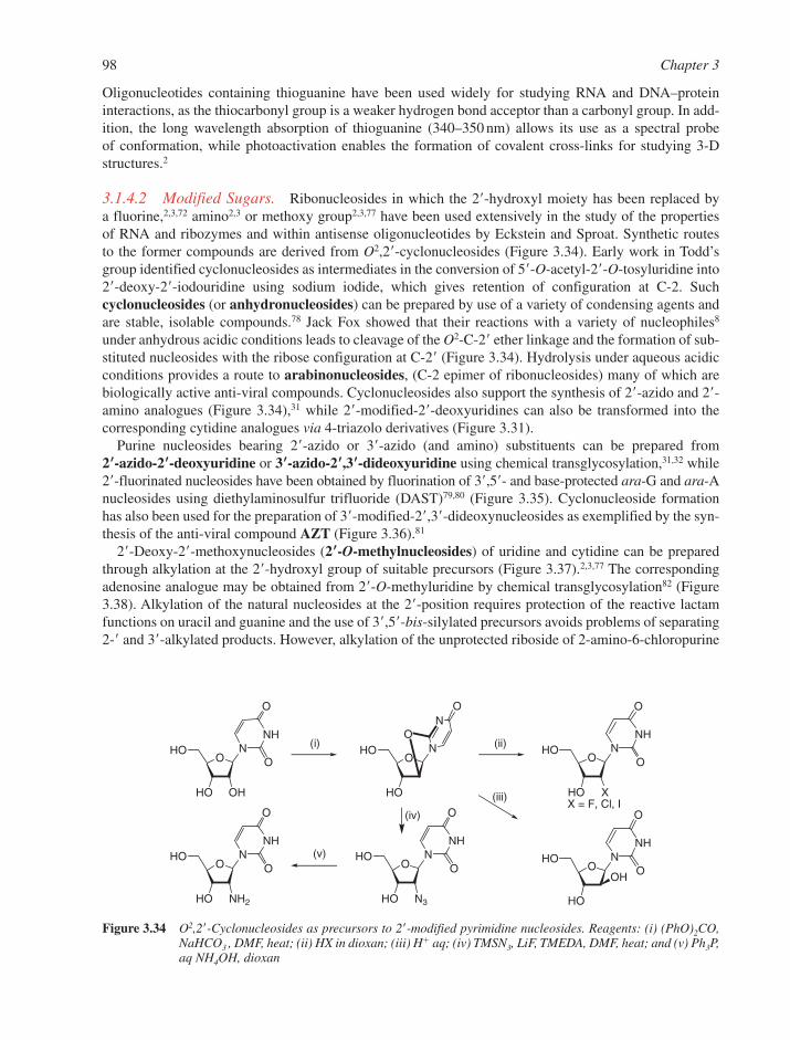
Oligonucleotides containing thioguanine have been used widely for studying RNA and DNA–proteininteractions, as the thiocarbonyl group is a weaker hydrogen bond acceptor than a carbonyl group. In add-ition, the long wavelength absorption of thioguanine (340–350 nm) allows its use as a spectral probe of conformation, while photoactivation enables the formation of covalent cross-links for studying 3-Dstructures.2
3.1.4.2 Modified Sugars. Ribonucleosides in which the 2�-hydroxyl moiety has been replaced by a fluorine,2,3,72 amino2,3 or methoxy group2,3,77 have been used extensively in the study of the properties of RNA and ribozymes and within antisense oligonucleotides by Eckstein and Sproat. Synthetic routes to the former compounds are derived from O2,2�-cyclonucleosides (Figure 3.34). Early work in Todd’sgroup identified cyclonucleosides as intermediates in the conversion of 5�-O-acetyl-2�-O-tosyluridine into2�-deoxy-2�-iodouridine using sodium iodide, which gives retention of configuration at C-2. Suchcyclonucleosides (or anhydronucleosides) can be prepared by use of a variety of condensing agents andare stable, isolable compounds.78 Jack Fox showed that their reactions with a variety of nucleophiles8
under anhydrous acidic conditions leads to cleavage of the O2-C-2� ether linkage and the formation of sub-stituted nucleosides with the ribose configuration at C-2� (Figure 3.34). Hydrolysis under aqueous acidicconditions provides a route to arabinonucleosides, (C-2 epimer of ribonucleosides) many of which arebiologically active anti-viral compounds. Cyclonucleosides also support the synthesis of 2�-azido and 2�-amino analogues (Figure 3.34),31 while 2�-modified-2�-deoxyuridines can also be transformed into thecorresponding cytidine analogues via 4-triazolo derivatives (Figure 3.31).
Purine nucleosides bearing 2�-azido or 3�-azido (and amino) substituents can be prepared from 2��-azido-2��-deoxyuridine or 3��-azido-2��,3��-dideoxyuridine using chemical transglycosylation,31,32 while2�-fluorinated nucleosides have been obtained by fluorination of 3�,5�- and base-protected ara-G and ara-Anucleosides using diethylaminosulfur trifluoride (DAST)79,80 (Figure 3.35). Cyclonucleoside formationhas also been used for the preparation of 3�-modified-2�,3�-dideoxynucleosides as exemplified by the syn-thesis of the anti-viral compound AZT (Figure 3.36).81
2�-Deoxy-2�-methoxynucleosides (2��-O-methylnucleosides) of uridine and cytidine can be preparedthrough alkylation at the 2�-hydroxyl group of suitable precursors (Figure 3.37).2,3,77 The correspondingadenosine analogue may be obtained from 2�-O-methyluridine by chemical transglycosylation82 (Figure3.38). Alkylation of the natural nucleosides at the 2�-position requires protection of the reactive lactamfunctions on uracil and guanine and the use of 3�,5�-bis-silylated precursors avoids problems of separating2-� and 3�-alkylated products. However, alkylation of the unprotected riboside of 2-amino-6-chloropurine
98 Chapter 3
OHO
HO
N
NO
O
OHO
HO X
NNH
O
O
OHO
HO
OH
NNH
O
OO
HO
HO N3
NNH
O
OOHO
HO NH2
NNH
O
O
OHO
HO OH
NNH
O
O
(ii)
X = F, Cl, I(iii)
(iv)
(v)
(i)
Figure 3.34 O2,2�-Cyclonucleosides as precursors to 2�-modified pyrimidine nucleosides. Reagents: (i) (PhO)2CO,NaHCO3 , DMF, heat; (ii) HX in dioxan; (iii) H� aq; (iv) TMSN3, LiF, TMEDA, DMF, heat; and (v) Ph3P,aq NH4OH, dioxan
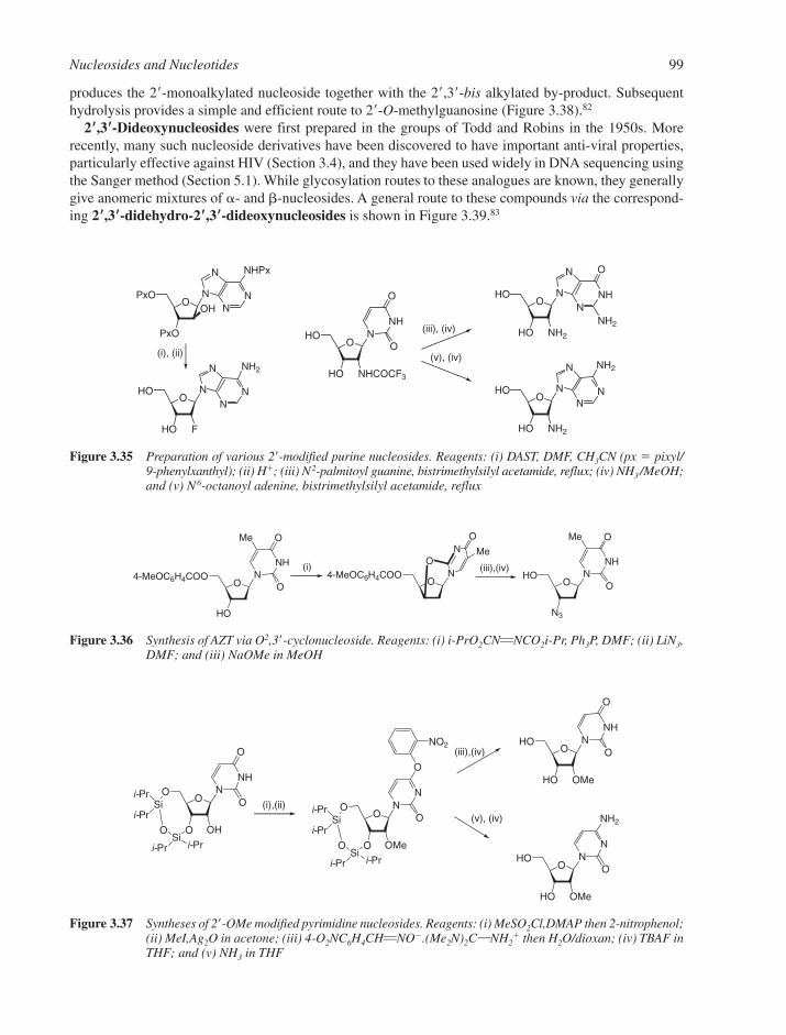
produces the 2�-monoalkylated nucleoside together with the 2�,3�-bis alkylated by-product. Subsequenthydrolysis provides a simple and efficient route to 2�-O-methylguanosine (Figure 3.38).82
2��,3��-Dideoxynucleosides were first prepared in the groups of Todd and Robins in the 1950s. Morerecently, many such nucleoside derivatives have been discovered to have important anti-viral properties,particularly effective against HIV (Section 3.4), and they have been used widely in DNA sequencing usingthe Sanger method (Section 5.1). While glycosylation routes to these analogues are known, they generallygive anomeric mixtures of �- and �-nucleosides. A general route to these compounds via the correspond-ing 2��,3��-didehydro-2��,3��-dideoxynucleosides is shown in Figure 3.39.83
Nucleosides and Nucleotides 99
OPxO
PxO
OH NN
N
N
NHPx
OHO
HO NH2
NNH
N
N
O
NH2
OHO
HO NHCOCF3
NNH
O
O
OHO
HO F
NN
N
N
NH2
OHO
HO NH2
NN
N
N
NH2
(iii), (iv)
(v), (iv)(i), (ii)
Figure 3.35 Preparation of various 2�-modified purine nucleosides. Reagents: (i) DAST, DMF, CH3CN (px � pixyl/ 9-phenylxanthyl); (ii) H�; (iii) N2-palmitoyl guanine, bistrimethylsilyl acetamide, reflux; (iv) NH3/MeOH;and (v) N6-octanoyl adenine, bistrimethylsilyl acetamide, reflux
O4-MeOC6H4COO N
NO
O
Me
OHO
N3
NNH
O
O
Me
O4-MeOC6H4COO
HO
NNH
O
O
Me
(iii),(iv)(i)
Figure 3.36 Synthesis of AZT via O2,3�-cyclonucleoside. Reagents: (i) i-PrO2CNNCO2i-Pr, Ph3P, DMF; (ii) LiN3,DMF; and (iii) NaOMe in MeOH
OHO
HO OMe
NNH
O
O
OHO
HO OMe
NN
NH2
O
i-Pr
O
i-Pr
i-Pr
O
OH
NNH
O
OO
O
Si
Sii-Pr
NN
O
O
NO2
O
OMe
O
O
Si
Sii-Pri-Pr
O
i-Pr
i-Pr
(iii),(iv)
(i),(ii)(v), (iv)
Figure 3.37 Syntheses of 2�-OMe modified pyrimidine nucleosides. Reagents: (i) MeSO2Cl,DMAP then 2-nitrophenol;(ii) MeI,Ag2O in acetone; (iii) 4-O2NC6H4CHNO�.(Me2N)2CNH2
� then H2O/dioxan; (iv) TBAF inTHF; and (v) NH3 in THF

3.2 CHEMISTRY OF ESTERS AND ANHYDRIDES OF PHOSPHORUS OXYACIDS
3.2.1 Phosphate Esters
The predominant forms of phosphorus in biology are orthophosphoric acid (HO)3PO, its esters, anhydridesand some amides. Orthophosphates are tetra-substituted at phosphorus, which is in the P(V) oxidation stateand has tetrahedral geometry. The bonding can be described by using sp3 hybrid orbitals at phosphorus forthe ‘single’PO bonds, which are �1.6 Å long. In triesters the PO ‘double’bond is shorter, �1.46 Å, andinvolves additional �-bonding from d�–p� overlap between the phosphorus and oxygen (Figure 3.40).Phosphorus can participate in such bonding simultaneously to more than one oxygen ligand and so any neg-ative charge is delocalized across all unsubstituted oxygen atoms (Figure 3.41). The corresponding �-bondingto neutral nitrogen ligands in phosphoramidates is rather weak, so the nitrogen remains moderately basic.
3.2.1.1 Phosphate Triesters. Triesters have all three hydrogen atoms of phosphoric acid replaced byalkyl or aryl groups. They are non-ionic, soluble in many organic solvents, and sufficiently stable to bepurified by chromatography. The PO bond is effectively transparent in the UV region and has an IRabsorption at 1280 cm�1. When all three ester groups are different, the phosphorus atom is a stereogeniccentre, as in SP methyl ethyl phenyl phosphate (Figure 3.42a), and so optically active triesters can be made.
3.2.1.2 Phosphate Diesters. Diesters have two hydrogen atoms replaced by alkyl or aryl groups.The remaining OH group is strongly acidic (pKa �1.5). Consequently, phosphate diesters exist as monoan-ions at pH �2, and are usually water-soluble. The negative charge is shared equally between the twounsubstituted oxygen atoms (Figure 3.42b). When the two ester groups are different, the two unsubstituted
100 Chapter 3
OHO
HO OMe
NNH
N
N
O
NH2
OHO
HO OH
NN
N
N
Cl
NH2
ONHO
HO OMe
NN
N NH2
OHO
HO OMe
NN
NH2
O OAcO
AcO OMe
NN
NHAc
O
OHO
HO OMe
NN
N
N
Cl
NH2
(ii),(iii)(i)
65% (+ 15% 2',3' bis-methylated)
(iv) (v)
Figure 3.38 Syntheses of 2�-OMe modified purine nucleosides. Reagents: (i) Ac2O, DMF, pyridine; (ii) N6-benzoyladenine, bistrimethylsilyl acetamide, TMSOTf, CH3CN, heat; (iii) NH3 / MeOH; (iv) MeI, NaH,DMF, �20°C; and (v) 1,4-diazabicyclo[2.2.2]octane (DABCO)/water, heat
OHO
HO OH
BO
RO
AcO
Br
BO
RO
Br
OAc
B
O
O MeMe
Me
O
OHO B
OHO B
(i)
B = Ade, Cyt, Gua, Ura, purine & 7-deazapurineanalogues
R =and/ or acyl groups derived from α-acetoxyisobutyryl bromide
(ii), (iii) (iv)
2',3'-didehydro-2',3'-dideoxyribonucleosides
+
2',3'-dideoxyribo-nucleosides
Figure 3.39 Syntheses of 2�,3�-didehydro-2�,3�-dideoxyribonucleosides and 2�,3�-dideoxyribonucleosides. Reagents:(i) Me2C(OAc)COBr in CH3CN/H2O; (ii) Zn-Cu/DMF or Zn/HOAc/DMF; (iii) NH3 /MeOH; and (iv) H2,Pd-C in EtOH
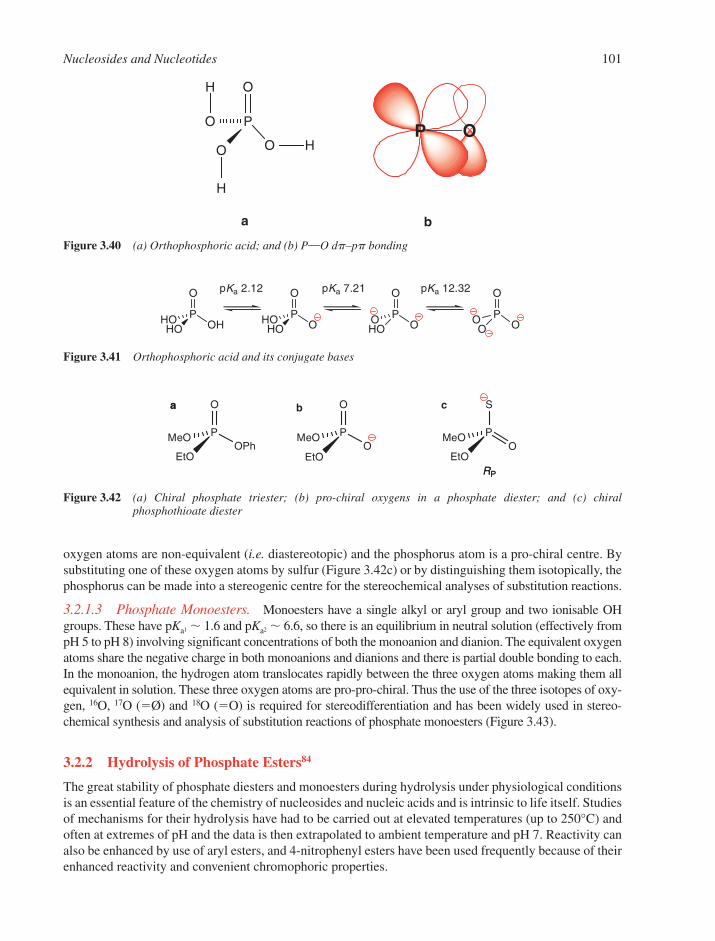
oxygen atoms are non-equivalent (i.e. diastereotopic) and the phosphorus atom is a pro-chiral centre. Bysubstituting one of these oxygen atoms by sulfur (Figure 3.42c) or by distinguishing them isotopically, thephosphorus can be made into a stereogenic centre for the stereochemical analyses of substitution reactions.
3.2.1.3 Phosphate Monoesters. Monoesters have a single alkyl or aryl group and two ionisable OHgroups. These have pKa1 � 1.6 and pKa2 � 6.6, so there is an equilibrium in neutral solution (effectively frompH 5 to pH 8) involving significant concentrations of both the monoanion and dianion. The equivalent oxygenatoms share the negative charge in both monoanions and dianions and there is partial double bonding to each.In the monoanion, the hydrogen atom translocates rapidly between the three oxygen atoms making them allequivalent in solution. These three oxygen atoms are pro-pro-chiral. Thus the use of the three isotopes of oxy-gen, 16O, 17O (�Ø) and 18O (�O) is required for stereodifferentiation and has been widely used in stereo-chemical synthesis and analysis of substitution reactions of phosphate monoesters (Figure 3.43).
3.2.2 Hydrolysis of Phosphate Esters84
The great stability of phosphate diesters and monoesters during hydrolysis under physiological conditionsis an essential feature of the chemistry of nucleosides and nucleic acids and is intrinsic to life itself. Studiesof mechanisms for their hydrolysis have had to be carried out at elevated temperatures (up to 250°C) andoften at extremes of pH and the data is then extrapolated to ambient temperature and pH 7. Reactivity canalso be enhanced by use of aryl esters, and 4-nitrophenyl esters have been used frequently because of theirenhanced reactivity and convenient chromophoric properties.
Nucleosides and Nucleotides 101
O
POHHO
HO
O
POHO
HO
O
POO
HO
O
POO
O
pKa 2.12 pKa 7.21 pKa 12.32
Figure 3.41 Orthophosphoric acid and its conjugate bases
P
EtOO
MeO
S
PO
EtO
O
MeOPOPh
EtO
O
MeO
aa b c
RPRP
Figure 3.42 (a) Chiral phosphate triester; (b) pro-chiral oxygens in a phosphate diester; and (c) chiral phosphothioate diester
P
O
O
O O H
H
H
a b
OP
Figure 3.40 (a) Orthophosphoric acid; and (b) PO dp–pp bonding
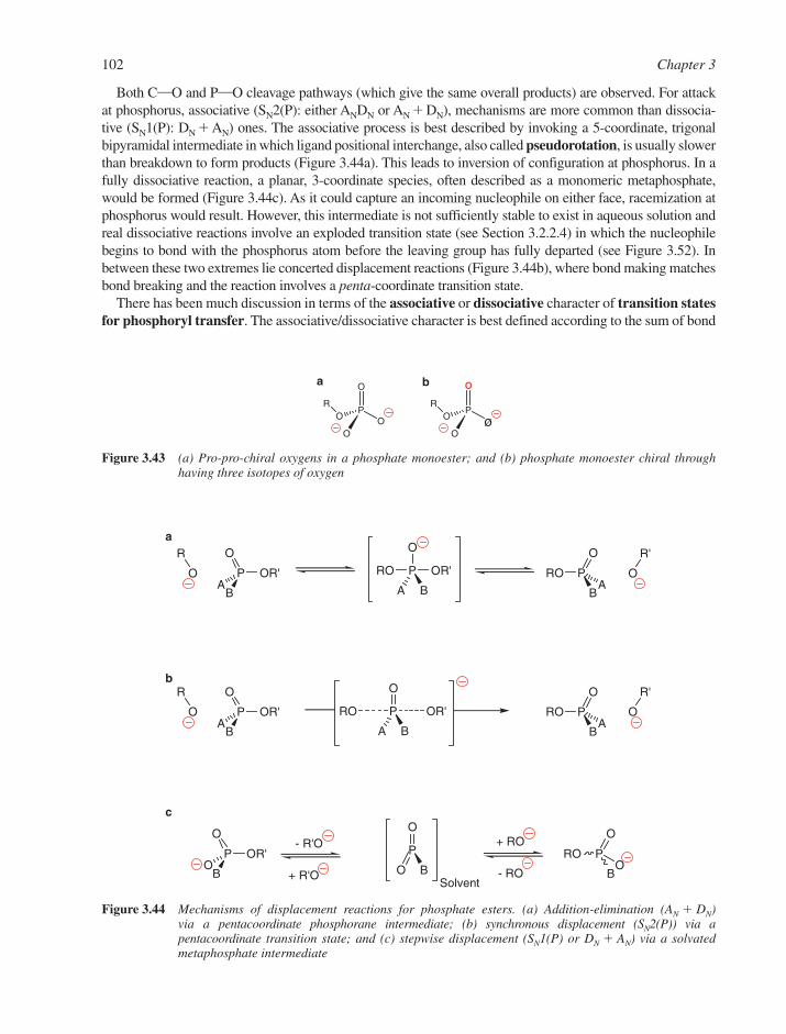
Both CO and PO cleavage pathways (which give the same overall products) are observed. For attackat phosphorus, associative (SN2(P): either ANDN or AN � DN), mechanisms are more common than dissocia-tive (SN1(P): DN � AN) ones. The associative process is best described by invoking a 5-coordinate, trigonalbipyramidal intermediate in which ligand positional interchange, also called pseudorotation, is usually slowerthan breakdown to form products (Figure 3.44a). This leads to inversion of configuration at phosphorus. In afully dissociative reaction, a planar, 3-coordinate species, often described as a monomeric metaphosphate,would be formed (Figure 3.44c). As it could capture an incoming nucleophile on either face, racemization atphosphorus would result. However, this intermediate is not sufficiently stable to exist in aqueous solution andreal dissociative reactions involve an exploded transition state (see Section 3.2.2.4) in which the nucleophilebegins to bond with the phosphorus atom before the leaving group has fully departed (see Figure 3.52). Inbetween these two extremes lie concerted displacement reactions (Figure 3.44b), where bond making matchesbond breaking and the reaction involves a penta-coordinate transition state.
There has been much discussion in terms of the associative or dissociative character of transition statesfor phosphoryl transfer. The associative/dissociative character is best defined according to the sum of bond
102 Chapter 3
P OR'
O
BA
R
O RO P OR'
O
A B
PRO
O
BA
R'
O
P OR'
O
BA
R
O RO P OR'
O
A B
PRO
O
BA
R'
O
P OR'
O
BO
+ ROP
O
O BPRO
O
BO
+ R'OSolvent
- R'O
- RO
a
b
c
Figure 3.44 Mechanisms of displacement reactions for phosphate esters. (a) Addition-elimination (AN � DN)via a pentacoordinate phosphorane intermediate; (b) synchronous displacement (SN2(P)) via apentacoordinate transition state; and (c) stepwise displacement (SN1(P) or DN � AN) via a solvatedmetaphosphate intermediate
P
O
OO
OR
O
Pø
O
OR
a b
Figure 3.43 (a) Pro-pro-chiral oxygens in a phosphate monoester; and (b) phosphate monoester chiral throughhaving three isotopes of oxygen

formation from phosphorus to the nucleophile and the leaving group. If this is greater in the transition statethan in the starting state (i.e. greater than one), then the transition state is considered associative (startingto resemble the intermediate in Figure 3.44a); if it is less than one, then the transition state is dissociativein character (starting to resemble the intermediate in Figure 3.44c). This issue is most important forenzymes, which have evolved to stabilize transition states effectively, and considerable progress has beenmade through the use of ‘metaphosphate’ mimics, such as AlF3 and MgF3, in X-ray structures ofnucleotide complexes with enzymes that utilise ATP or GTP.
3.2.2.1 Hydrolysis of Alkyl Triesters. Trimethyl phosphate is hydrolysed in alkaline solution in anSN2(P) process (kOH� � 1.6 � 10�4 M�1 s�1 at 25°C). With H2
18O as solvent, no Me18OH is formed, whichshows that the reaction involves exclusively PO cleavage. Other ‘hard’ nucleophiles such as F� react similarly, and indeed, fluoride catalysis of the trans-esterification of triesters is a useful process (Figure 3.45).The intra-molecular migration of phosphorus in a triester to a vicinal hydroxyl group is especially easyand must be avoided in the synthesis of oligoribonucleotide and inositol phosphate precursors.
Trimethyl phosphate is hydrolysed extremely slowly in neutral and acidic conditions (kw � 2 � 10�8 s�1
at 25°C) with CO cleavage. Soft nucleophiles, such as RS�, Br� or I�, also dealkylate phosphate triesterswith CO cleavage (Figure 3.46a). Such reactions are typical SN2 processes and show a clear preference fordealkylation in the order of Me � Et � R2CH. This characteristic is particularly well exploited in the thio-phenolate deprotection of methyl phosphate triesters used in the phosphodiester chemistry of oligonu-cleotide synthesis (Section 4.1) (Figure 3.46b). However in this case, some (�20%) oligonucleotide chaincleavage can result through attack of thiophenolate at the 5�-carbon of the 3�-nucleotide unit.
Alkyl phosphate triesters are sensitive to �-elimination processes as has been exploited for the selectivedeprotection of phosphate triesters in oligonucleotide synthesis (Section 4.1). The 2-cyanoethyl grouppossesses an acidic �-hydrogen atom and may be removed by �-elimination mechanism under mildly
Nucleosides and Nucleotides 103
O(MeO)2 P
O(MeO)2 P
OBn
O(MeO)2 P
F
OHO (MeO)3P=O F BnOH
Figure 3.45 PO cleavage reactions with hard nucleophiles for triesters
(MeO)3P=O
(MeO)2PO2HH2
18O
LiBr
PhSMeOH
OO
O
BP
P
MeO
OO
OBP
O
OO
O
BP
P
O
OO
OBP
O
PhS + PhSMe
+
(MeO)2PO2Li + MeBr
(MeO)2PO2 + PhSMe
5'
3'
5'
3'
a
b
P = protecting group
Figure 3.46 CO cleavage reactions of triesters with soft nucleophiles. (a) trimethyl phosphate; (b) a phosphatetriester following DNA synthesis

basic conditions without competing cleavage of the oligonucleotide chain (Figure 3.47). Consequently, itis the protecting group of choice in oligonucleotide synthesis (Sections 4.1 and 4.2). �-Elimination is alsoimportant biologically in the base excision repair pathway involving the enzymatic cleavage of phosphatediesters following removal of damaged bases by glycosylase enzymes (Section 8.11.4).
3.2.2.2 Hydrolysis of Aryl Triesters. Because aryl phosphates are much more reactive than alkyl phos-phate triesters, it is possible to achieve selective, nucleophilic displacement of the phenolic residue in a dialkylaryl phosphate on account of its better leaving group ability (pKa � 5). One of the best nucleophiles for thispurpose is the oximate anion (Figure 3.48). Although aryl triesters were used historically during oligonu-cleotide synthesis, they were replaced by the cyanoethyl group, since this group may be removed in a singlestep along with the base protecting groups and oligomer cleavage from the solid support (Section 4.1).
3.2.2.3 Hydrolysis of Phosphate Diesters.85,86 At pH � 2, phosphate diesters exist as theirmonoanions, which are extremely stable kinetically (Table 3.1). Even in strongly alkaline conditions, diestershydrolyse with predominant CO cleavage (the extent depending on the nature of the alkyl group) and farmore slowly than triesters, since attack at the phosphorus atom is impeded by anion–anion repulsion. Thespontaneous (pH-independent) reaction of the monoanion is so slow that it is yet to be quantified for simplephosphate diesters with alkoxy leaving groups. In acidic conditions, their hydrolysis occurs through the neu-tral species, which are similar to trialkyl phosphates in reactivity. The diaryl esters are rather more reactiveunder alkaline conditions, as is to be expected for reactions involving a better leaving group, and also allowthe pH-independent reaction to be observed (which is very sensitive to the pKa of the aryloxy leaving group).
This marked stability of the phosphate diester linkage during hydrolysis is a vital feature of the biological roleof DNA, where maintenance of the primary structure is required to preserve the genetic code. It is dramaticallychanged for esters of 1,2-diols, such as the ones found in RNA. Here, the vicinal hydroxyl group enormouslyenhances the rate of hydrolysis of di- and triesters. Similarly, the cyclic phosphates of 1,2-diols hydrolyse morethan 107 times faster than their acyclic or 6- and 7-membered cyclic relatives. This corresponds to a decrease in G‡ of 36 kJ mol�1. About 60% of this acceleration is attributed to relief of strain in the five-membered cyclicester, which has a 98° OPO angle and an enhanced enthalpy of hydrolysis of �20 kJ mol�1.
104 Chapter 3
OO
O
BP
P
O
OO
OBP
O
NC
H
OO
O
BP
P
O
OO
OBP
O
Base (B:)B
αβ
P=protecting group
Figure 3.47 Selective deprotection of oligonucleotide phosphate triesters by b-elimination
H
N
NO2
O
Cl
OP
O
R'O OR'H
N
NO2
OP
O
R'O OR'
BO
PO
R'O OR'
CN
NO2
Figure 3.48 Selective nucleophilic displacement in an aryl ester

The essential observation is the acceleration of both ring closure and ring opening. Furthermore, exo-cyclic PO bond cleavage is also accelerated in five-membered cyclic esters (Figure 3.49).
How can ring strain accelerate both endocyclic and exocyclic substitution at phosphorus? The hydroly-sis of ethylene phosphate shows incorporation of isotopic label from H2
18O solvent into PO bonds in bothacidic and alkaline conditions, and these reactions must involve an AN � DN (i.e. an associative) process(Figure 3.44a). It is generally agreed that a transient penta-coordinated phosphorane intermediate is bothstabilized and made kinetically more accessible relative to its acyclic counterpart because of the geometryof the five-membered ring. It is then reasonable to invoke topoisomerism (in this case the pseudorotationof the trigonal bipyramidal species) to explain most of the phenomena associated with this remarkablyenhanced reactivity (Figure 3.50).
This phenomenon is clearly important in the hydrolysis of RNA by alkali and ribonucleases. In bothcases, the 5-membered 2�,3�-cyclic phosphates of nucleosides are formed by the displacement of the
Nucleosides and Nucleotides 105
Table 3.1 Some rate constants for the hydrolysis of phosphate esters (25°C) and patterns of bondcleavage
Phosphate � H2O Phosphate monoanion � Phosphate � HO�
kw (s�1) H2O kw (s�1) kOH� (M�1 s�1)
(MeO)3PO 2 � 10�8 (CO) — 2 � 10�4 (PO)(MeO)2PO2H 5 � 10�10 (CO) �2 � 10�14 7 � 10�12 (CO)*
1 � 10�10 (PO) 1 � 10�15 (PO)*138
(MeO)PO3H2 1 � 10�10 (CO) 3 � 10�10 (PO) 2 � 10�20 (PO) †139
(PhO)3PO — — 5 � 10�3 (PO)140
(PhO)2PO2H — 5 � 10�15 (PO)141 2 � 10�7 (PO)*142
(PhO)PO3H2 — 2 � 10�8 (PO)143 1 � 10�13 (PO)†138
UpU86 — — 2 � 10�3 (PO)‡
7 � 10�5 (PO)§,¶ 5 � 10�6 (PO)¶ 5 (PO)¶
Ethylene phosphate — — 5 � 10�4 (PO)*144
* monoanion. † pH-independent reaction of dianion, s�1.‡ intramolecular transesterification at phosphate diester monoanion.§ intramolecular attack by 2�OH.¶ at 90°C.
H218O
O
O
H
P
O
OR
OR
O
P
O
O
OR
O
P
O
OR
O
O
O
H
P
O
OH
OR
O
P
O
O
OH
exchange
ring cleavage
endocyclic exocyclic
Figure 3.49 Accelerated PO cleavages associated with 5-membered ring phosphate esters in acidic and in alkaline solution

5�-O-nucleoside residue. The enzymatic reaction is completed by the regioselective ring opening of thecyclic phosphate to give only a 3�-nucleoside phosphate (Figure 3.51). By contrast, alkaline hydrolysisleads to a mixture of 2�- and 3�- phosphates. These reactions exhibit overall retention of configuration atthe phosphorus centre. This is accounted for by the double inversion of stereochemistry that occurs in thetwo successive ‘in-line’ displacement processes.
It must be emphasised that this remarkable reactivity appears to be exclusive to five-membered cyclicphosphate esters and esters of 1,2-diols. This contrasts with the relative stability of esters of 1,3-diols and6-ring cyclic phosphates. An important example is 3�,5�-cAMP, whose key role as the second messengerin cell signalling is dependent on its kinetic stability to non-enzymatic hydrolysis.
3.2.2.4 Phosphate Monoesters.87 The hydrolysis of monoalkyl phosphates at very low pH proceedsvia the conjugate acid, and is similar in mechanism to that of triesters and neutral diesters (Table 3.1).These esters are very resistant to hydrolysis under alkaline conditions where they exist as dianions (andcatalysis by hydroxide is never observed). The reaction of the dianion proceeds by PO cleavage and hasmany of the characteristics of a dissociative process via a hypothetical metaphosphate intermediate. A bet-ter description invokes the idea of an ‘exploded’ transition state in which there is very weak bonding to theincoming nucleophile and outgoing leaving group (i.e. a concerted reaction which is very dissociative incharacter). The reaction is extremely sensitive to the pKa of the leaving group, and alkyl phosphate monoesterdianions are even more stable than the corresponding diesters. By contrast, the monoanion shows both anunusually high relative reactivity towards hydrolysis and is very insensitive to the leaving group pKa. Thisis explained by involving the minor tautomer (where the leaving group O carries the proton) as the react-ive ionic form, which then hydrolyses through a similar transition state to the dianion (Figure 3.52). For
106 Chapter 3
O
P
O
O
OHPO
O
OH
OH
OH
H2O
PHO
O
OH
O
OH O
P
O
O
OH
P
O
O OHOHHO
P
O
O OHOHHO
Ψrot
endocyclic cleavage
endocyclic cleavage
exocyclic cleavage
exocyclic cleavage
Figure 3.50 Role of trigonal bipyramidal pseudorotation (crot) in 18O isotope (O) incorporation into ethylenephosphate
O
O
O
B
H
OO
O
B
PO
O
OH
O
B
OP
OO
OO
OH
O
BOH
OOH
O
B
P O
OHO
N
NH
His12
H
H
His119
N
N N
NH
His119
HO
H
N
NH
His12H
N
NH
His12
5'
5'
2'
2'3'
RNaseA RNaseA
Figure 3.51 Ribonuclease A hydrolysis of RNA via 2�,3�-cyclic phosphate. Imidazoles (of His-12 and His-119residues) act as a general acid and general base

very good leaving groups, the difference between mono- and dianion reactivity is small, but for poor leavinggroups, the monoanion is by far the more reactive ionic form and accounts totally for the observed reaction evenat high pH. Similar phenomena have been analysed for spontaneous hydrolyses of acetyl phosphate, creatinephosphate and ATP (loss of the �-phosphate), all of which have good leaving groups on a terminal phosphate.
3.2.3 Synthesis of Phosphate Diesters and Monoesters
The most common approaches to dinucleoside phosphate ester synthesis use phosphorylation reactions inwhich a 3�-nucleotide component is converted into a reactive phosphorylating species by a condensingagent. One of the major problems is that the more reactive condensing agents not only activate the phos-phate, but may also react with nucleoside bases or the product, leading to unacceptable reduction in yield.The ideal condensing agent should have a high rate of activation of the phosphate species and a negligiblerate of reaction with the alcohol component or N-protected bases.
3.2.3.1 Interrelationships of Esters of Phosphorus Oxyacids. The formal relationship betweenphosphorus halides (X � Cl) and the mono-, di- and tri-alkyl esters of phosphorus oxyacids is shownschematically (Figure 3.53). Actual reaction conditions have to be controlled carefully to avoid the for-mation of by-products, especially of alkyl halides. Many of the interconversions shown are best accom-plished using nitrogen ligands at phosphorus (X � i-Pr2N or an azole).
Nucleosides and Nucleotides 107
P OO
OHO
RO P O
O
O O
H
H
R
HP O
O
OO
R
HPO
O
OOH
HPO
O
OO
H
H
+ H2O - ROH
Figure 3.52 Hydrolysis of a phosphate monoester monoanion through an ‘exploded’ transition state
XPX
X XPRO
ROOR
PRORO
O
XPRO
X
O
ORPRO
RO
O
XPRO
RO
O
XPX
X
H2O
O
OPRO
O
O
OPRO
RO
hydrolysis
XPRO
X
O
HPRO
HOOHPRO
HO
O
HPRO
RO OHPRO
RO
H2O H2O
trialkyl phosphite(phosphite triester)
ROH, base
phosphorus (V) chemistry
trialkyl phosphate(phosphate triester)
ROH, baseROH, base
monoalkyl phosphate(phosphate monoester)
ROH, DCC
dialkyl phosphate(phosphate diester)
ROH,MST or MSNT
oxidation t-BuOOH(oxidation)
ROH, base ROH, baseROH, base
phosphorus (III) chemistry
ROH, t-BuCOCl
H-phosphonate diester
oxidation oxidation
Figure 3.53 Formal relationship between exters of P(III) and P(V) oxyacids. DCC � N,N�-dicyclohexylcarbodiimide,MST � mesitylenesulfonyl tetrazolide, MSNT � mesitylenesulfonyl (3-nitrotriazolide)
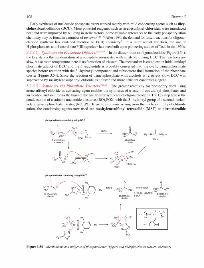
Early syntheses of nucleoside phosphate esters worked mainly with mild condensing agents such as dicy-clohexylcarbodiimide (DCC). More powerful reagents, such as arenesulfonyl chlorides, were introducednext and were improved by building in steric factors. Some valuable references to the early phosphorylationchemistry may be found in a number of reviews.34,88–90 After 1980, the demand for faster reactions for oligonu-cleotide synthesis has switched attention to P(III) chemistry.91 In a more recent variation, the use of H-phosphonates as a 4-coordinate P(III) species92 has been built upon pioneering studies of Todd in the 1950s.
3.2.3.2 Syntheses via Phosphate Diesters.88,89,93 In the diester route to oligonucleotides (Figure 3.54),the key step is the condensation of a phosphate monoester with an alcohol using DCC. The reactions areslow, but at room temperature there is no formation of triesters. The mechanism is complex: an initial imidoylphosphate adduct of DCC and the 5�-nucleotide is probably converted into the cyclic trimetaphosphatespecies before reaction with the 3�-hydroxyl component and subsequent final formation of the phosphatediester (Figure 3.54). Since the reaction of trimetaphosphate with alcohols is relatively slow, DCC wassuperseded by mesitylenesulphonyl chloride as a faster and more efficient condensing agent.
3.2.3.3 Syntheses via Phosphate Triesters.88,89 The greater reactivity for phosphorylation usingarenesulfonyl chloride as activating agent enables the syntheses of triesters from dialkyl phosphates andan alcohol, and so it forms the basis of the first triester syntheses of oligonucleotides. The key step here is thecondensation of a suitable nucleotide diester as (RO)2POX, with the 3�-hydroxyl group of a second nucleo-side to give a phosphate triester, (RO)3PO. To avoid problems arising from the nucleophilicity of chlorideanion, the condensing agents now used are mesitylenesulfonyl tetrazolide (MST) or nitrotriazolide
108 Chapter 3
O
OP
ROArO
O
OP
ROR'O
O
OR'P
ArORO
HO
NCN
C6H11
C6H11
O
OP
ROO C
NC6H11
NHC6H11
C6H11NH C HNC6H11
OO
OP
ROR'O
DCC
HO
Cl
Me
Me
MeS
O
O
N
N
N
O2N
O
OP
ROArO
Me
Me
MeS
O
ONN
NO2N
O
NP
ROArO
NN
NO2
O
H
O
OP
OOR
H
N C N
C6H11
C6H11
H
O
OP
OOR
R'OH
H+
DCU
R'
3'-phosphatediester
phosphodiester chemistry using DCC
phosphatetriester
R = 3'
R'OH
5'-phosphate monoester
phosphatediester
5'R' =
phosphotriester chemistry using MSNT
Ar =
2-NO2C6H4C=NOH& Et3N
phosphatediester
deprotectionR'OH
Figure 3.54 Mechanisms and reagents of phosphodiester (upper) and phosphotriester (lower) chemistry

(MSNT). A mixed phosphoryl–sulfonyl anhydride is produced initially, in which the methyl groups of themesitylene ring provide steric hindrance to reaction at the sulfur atom and ensure condensation at thephosphorus atom. Subsequent complex reactions, which may also involve condensed phosphate intermedi-ates, lead to the triester product (Figure 3.54). The final conversion of the triester into the desired diesteruses one of the specific cleavages described earlier (Section 3.2.2).
3.2.3.4 Syntheses via Phosphite Triesters.88,91,94 The P(III) triester route to oligonucleotides ini-tially introduced by Letsinger (Figures 3.53 and 3.55) was designed to exploit the intrinsically greaterreactivity shown by PCl3 as compared to POCl3 to achieve faster coupling steps. A major breakthroughwas achieved by Caruthers,95,96 who established the value of alkyl phosphoramidites (X � i-Pr2N) as stable3�-derivatives of nucleosides, which nonetheless react rapidly and efficiently with nucleoside 5�-hydroxylgroups in the presence of azole catalysts. The resulting product is an unstable phosphite triester, whichmust be oxidised immediately to give the stable phosphate triester in a process that can be cycled up to 100times on a solid-phase support. Removal of the phosphate-protecting group affords the phosphate diester(Figure 3.55). Racemization that occurs during reactions carried out with purified Rp and Sp diastereoiso-mers have confirmed the intermediacy of a phosphorotetrazolide during formation of the phosphite triester(Figure 3.56).97
3.2.3.5 Syntheses via H-phosphonate Diesters.92 The H-phosphonate monoesters of protected 3�-nucleosides are readily prepared using PCl3 and excess imidazole followed by mild hydrolysis. The
Nucleosides and Nucleotides 109
HO
HO
PO
i-Pr2N
XNC P
O
i-Pr2N
ORNC
PO
OR'
ORNCP
O
R'O OR
NC
O
PCl3P
O
Cl
ClNC
PO
R'O OR
OR = 3'
5'R' =
(i) (ii) (iii)
(iv)
(v)(vi)
Figure 3.55 Synthesis of phosphate triesters and diesters via phosphite triesters. Reagents: (i) 2-cyanoethanol,pyridine, ether �78°C; (ii) i-Pr2NH, ether, �20°C: X � Cl, 2eq. or X � i-Pr2N 4 eq.; (iii) X � Cl:ROH, i-Pr2EtN,THF, or X � i-Pr2N: ROH, i-Pr2EtNH2
� tetrazolide� in CH2Cl2 or THF; (iv) tetrazolein CH3CN and R�OH; (v) I2 in pyridine/H2O/THF; and (vi) aq NH4OH
R'O
H
PO
i -Pr2N
ORNC
PO
i -Pr2N
ORNC
H
NN
N
N
PO
N
ORNC
N
N N
PO
OR'
ORNC
NN
N
N
H
PO OR'RO
NC
O oxidation
phosphate triester [P(V)] phosphite triester [P(III)]
Figure 3.56 Synthesis of phosphate triesters using phosphoramidite reagents

intermediates do not require further protection at phosphorus and are rapidly and efficiently activated by arange of condensing agents, such as pivaloyl chloride (Figure 3.53). This can be carried out before or afterthe addition of the second nucleoside if excess condensing agent is avoided. A dinucleoside H-phosphonateis rapidly formed in high yield. The procedure can be repeated many times before a single oxidation stepfinally converts the H-phosphonate diesters into phosphate diesters (Figure 3.57). Studies on the mech-anism of condensation indicate that a mixed phosphonate anhydride is the likely intermediate.
3.2.3.6 Synthesis of Phosphate Monoesters.34,90 Monoesters are usually made either from triestersby selective deprotection or by direct condensation of an alcohol with a reactive phosphorylating agent,usually a polyfunctional species such as phosphoryl chloride (POCl3). The first formed product is imme-diately hydrolysed to give the desired monoester.
Triester procedures almost always call for selective protection of the nucleoside hydroxyl groups toleave only the reaction centre free. Reagents such as dibenzyl phosphorochloridate (with deprotection viacatalytic hydrogenolysis), bis-(2,2,2-trichloroethyl) phosphorochloridate (deprotection by zinc reduction)or 1,2-phenylene phosphorochloridate (deprotection by hydrolysis to the diester and then oxidative removalof catechol) give good yields of phosphate monoester (Figure 3.58a). Selective reaction at the 5�-hydroxyl
110 Chapter 3
Me
MeCl3C
Me
Me
Cl3C
O
P
O O
Cl
O
P
O O
Cl
OHO
R
B
HO
OPO
HO
B P
XPO
O
O
CN
O
HO R
BOPCl
X
Cl
O
P
O O
OR
RCH2OP(OCMe2CCl3)2
O
OHO
O
B
P OO
X
ORPO
O
O
ORPO
O
O
O
HO R
BOPO
O
X
B=Ade, Cyt, Gua, Thy, Ura or modified base X=O,S
R = OH, H, F
+ ROH
+ RCH2OH
a
b
c
P = protecting group
+
X=O,S
(i) (ii), (iii)
(i) (iv)
(v) (ii)
(vi), (vii)
Figure 3.58 Syntheses of phosphate monoesters. (a) Triester procedures, (b) using POCl3 or PSCl3, (c) synthesis of3�-phosphate monoesters. Reagents: (i) base; (ii) aq. Et3NH� HCO3
�; (iii) Br2 / H2O (oxidation);(iv) Zn, MeCO2H; (v) POCl3 [PSCl3 (�collidine for pyrimidine nucleosides)] in (MeO)3PO, 0°C; (vi) DCC, pyridine; and (vii) aq. NH4OH(X�O) or tBuNH2/pyridine then aq. NH4OH(X�S)
PCl3P
O
H
O
ORP
H
R'O
O
ORP
O
R'O
O
OR HO
HO
R = 3'5'
R' =(i) (ii) (iii)
Figure 3.57 Synthesis of phosphate diesters via H-phosphonates. Reagents: (i) ROH then aq. Et3N; (ii) (CH3)3COCland R�OH; and (iii) I2 in aq. Pyridine

group may be achieved using bulky phosphorylating agents such as bis-(2,2,2-trichloro-1,1-dimethyl) phos-phorochloridate (Figure 3.58a).
For a wide range of sugars and bases, direct phosphorylation at the 5�-position of unprotected nucleo-sides and 2�-deoxyribonucleosides using POCl3 in a trialkyl phosphate solvent system (the Yoshikawamethod98) is probably the simplest and most convenient method. Hydrolysis of the highly reactive phos-phorodichloridate intermediate with aqueous buffer gives the monophosphate (Figure 3.58b). No protec-tion of the base or sugar is required, although by-products arising from phosphorylation of the secondaryhydroxyl groups of the sugar vary, depending on the nature of the nucleoside. In the same way, isotopicoxygen can be readily introduced by the generation of P18OCl3 in situ, while the use of thiophosphorylchloride (PSCl3) leads to phosphorothioates.99 In a modification to the procedure, the use of aqueous pyri-dine in acetonitrile as solvent (the Sowa–Ouchi method)100 can provide yields greater than 80% with over90% regioselectivity.
Synthesis of nucleoside 3�-phosphate monoesters requires protection of the 5�-hydroxyl group and baseand can be achieved, for example, by DCC-mediated condensation with 2-cyanoethyl phosphate. Themethod also allows the preparation of phosphorothioate monoesters (Figure 3.58c).
3.3 NUCLEOSIDE ESTERS OF POLYPHOSPHATES
3.3.1 Structures of Nucleoside Polyphosphates and Co-Enzymes
Phosphoric acid can form chains of alternating oxygen–phosphorus linkages, which are relatively stablein neutral aqueous solution (Figure 3.59). The major condensed phosphates of biological importance arepyrophosphoric acid, (HO)2P(O)OP(O)(OH)2, its esters and esters of tripolyphosphoric acid. The stabilityof such species can be related to that of the corresponding phosphates after due allowance for (1) thechanged stability of the anionic charge and (2) improved leaving group characteristics.
Thus, tetraethyl pyrophosphate is an ethylating agent towards hard nucleophiles and also it is a phosphor-ylating agent. Tetraphenyl pyrophosphate is exclusively a phosphorylating agent. P1,P2-Dialkyl pyrophosphateshave considerable stability towards hydrolysis at ambient pHs where they exist exclusively in the form of adianion. This feature is very important for the stability of the pyrophosphate link as a structural feature in manyco-enzymes, including NADH, FAD and CoA. Similarly, P1,P3-diesters of tripolyphosphoric acid are stablecomponents of the ‘cap’ structure of eukaryotic mRNA (Figure 3.60) and P1,P4-diadenosyl tetraphosphate
Nucleosides and Nucleotides 111
OP
OP
OP
O
O O OO O On
Figure 3.59 Structure of polyphosphates
NHN
N
N
O
H2N
O
OHHO
Me
OP
OP
OP
O
O O OO O O
OOH
OP
O O
O
base
OOH
OP
O O
O
base
OOH
OP
O O
O
base
Figure 3.60 P1, P3-Dinucleosidyl triphosphate in the ‘cap’ structure at the 5�-end of eukaryotic mRNA
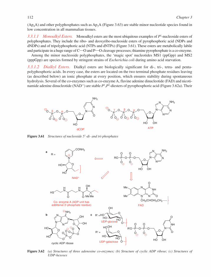
(Ap4A) and other polyphosphates such as Ap3A (Figure 3.63) are stable minor nucleotide species found inlow concentration in all mammalian tissues.
3.3.1.1 Monoalkyl Esters. Monoalkyl esters are the most ubiquitous examples of P1-nucleoside esters ofpolyphosphates. They include the ribo- and deoxyribo-nucleoside esters of pyrophosphoric acid (NDPs anddNDPs) and of tripolyphosphoric acid (NTPs and dNTPs) (Figure 3.61). These esters are metabolically labileand participate in a huge range of CO and PO cleavage processes; thiamine pyrophosphate is a co-enzyme.
Among the minor nucleoside polyphosphates, the ‘magic spot’ nucleotides MS1 (ppGpp) and MS2(pppGpp) are species formed by stringent strains of Escherichia coli during amino acid starvation.
3.3.1.2 Dialkyl Esters. Dialkyl esters are biologically significant for di-, tri-, tetra- and penta-polyphosphoric acids. In every case, the esters are located on the two terminal phosphate residues leaving(as described below) an ionic phosphate at every position, which ensures stability during spontaneoushydrolysis. Several of the co-enzymes such as co-enzyme A, flavine adenine dinucleotide (FAD) and nicoti-namide adenine dinucleotide (NAD�) are stable P1,P2-diesters of pyrophosphoric acid (Figure 3.62a). Their
112 Chapter 3
OHO
HO
OHO
OH
O
HO
HO
OHO
OHO OH
OH
N
NN
N
NH2
OHO
HO
O
PO
OO
PO
OO
OO
HO OH
NN
N
N
NH2
P
O
O
O
PRO
O
O
CONH2
N
OHO
HO
NAD+
HO OH
OPP
ON
NH
O
OOR'O
O
O
O O
NH
NN
N
O
O
CH2(CHOH)3CH2
Me
Me
FAD
HS
HN
HN
O O Me Me
OH
R =a
b
Co- enzyme A (ADP unit has additional 3'-phosphate residue)
cyclic ADP ribose
R' =
R' =
UDP-glucose
UDP-galactose
c
Figure 3.62 (a) Structures of three adenosine co-enzymes; (b) Structure of cyclic ADP ribose; (c) Structures ofUDP-hexoses
OO
HO OH
NN
N
N
NH2
P
O
O
O
PO
PO
OO O OO
HO
N
N
NH2
OO
P
O
O
O
PO
O O
dCDP
αβγαβ
ATP
Figure 3.61 Structures of nucleoside 5�-di- and tri-phosphates

biosynthesis involves the condensation of ATP with a monoalkyl phosphate, and the pyrophosphate appearsto act generally as a structural unit providing coulombic binding to appropriate enzyme residues.
Cyclic ADP ribose (Figure 3.62b) is a metabolic product formed by the cyclization of NAD� to closeC-1 of the second ribose onto N-1 of the adenine ring. The instability of this nucleotide appears to ori-ginate not from its pyrophosphate diester but from the second glycosylic linkage. It has an important roleas a second messenger and is involved in calcium signalling in cells. The active forms of many hexoses arefound as pyrophosphate esters of uridine 5�-diphosphate. These include UDP-glucose, UDP-galactose andUDP-N-acetylglucosamine (Figure 3.62c) that are formed biosynthetically from UTP and hexose-1-�-phosphate. The pyrophosphate ester is a good leaving group and is employed in catabolic processes involv-ing C-1 of the hexose residue.
The P1,P4-dinucleosidyl tetraphosphates, Ap4A and Ap4G, are found along with Ap3A in all cells, especiallyunder conditions of metabolic stress (Figure 3.63). They are produced as a result of the phosphorolysis ofaminoacyl adenylates, particularly tryptophanyl and lysyl adenylates, with ATP or GTP. Although these minornucleotides were discovered by Zamecnik in 1966, their purpose remains uncertain. They may have a role inthe initiation of DNA biosynthesis, and their analogues inhibit the aggregation of blood platelets. Ap3A isinvolved in signalling for apoptosis. Somewhat related structures are found in the ‘caps’ at the 5�-ends ofeukaryotic mRNAs, which have a 7-methylguanosin-5�-yl residue linked to the 5�-triphosphate. Both of thesespecies and their analogues have been targets for synthesis as a means of discovering their biological function.
3.3.2 Synthesis of Nucleoside Polyphosphate Esters
All of the naturally occurring nucleoside polyphosphates have at least one negative charge on each phosphateresidue. This is because uncharged phosphoryl residues in a string of phosphates are readily hydrolysedspontaneously. As a result, most syntheses have avoided the formation of fully esterified intermediates,though an early synthesis of UTP was achieved (in low yield) by the catalytic hydrogenolysis of its tetra-benzyl ester. Generally, syntheses of monoalkyl esters fall into two classes; they involve CO bond orPOP bond formation.
The exploitation of the alkylating properties of nucleoside 5�-O-tosylates towards pyrophosphate ortripolyphosphate anions and their methylene analogues is put to good use in the Poulter reaction.101 This hasmade direct syntheses of nucleoside 5�-di- and tri-phosphates and their analogues possible (Figure 3.64).
Nucleosides and Nucleotides 113
O
HO OH
N
N
N
N
NH2
O
OHHO
B OP
O
O
O
PO
PO
OO O O
Ap3A n=1, B = AdeAp4A n=2, B = AdeAp4G n=2, B = Gua
n
Figure 3.63 Structures of P1, P3-Dinucleoside triphosphate and P1, P4-dinucleoside tetraphosphates
O
HO OH
NN
N
N
NH2
OP
O
X
O
PO
O O
OTsO
HO OH
NN
N
N
NH2
OP
O
X
O
PO
O O +
X = O, CH2, CF2, NH
Figure 3.64 Synthesis of ADP and its analogues by CO bond formation

A related but more general route to nucleoside 5�-triphosphates or �,�-substituted analogues thereof involvesthe reaction of an activated nucleoside monophosphate, which is then able to condense with pyrophos-phate or a pyrophosphate analogue102,103 such as methylenebisphosphonate. Among the condensing agentswhich have been used widely are DCC to prepare the reactive nucleoside 5��-phosphoromorpholidates andcarbonyl diimidazole (Figure 3.65).34,90,104,105 This procedure is well suited to the introduction of isotopicoxygen into nucleotides in a non-stereochemically controlled fashion, for subsequent use in positional iso-tope exchange (PIX) studies.
Base and sugar-modified nucleoside 5�-triphosphates and 5��-(1-thio)triphosphates (��-thiotriphosphates)are generally more often made in a simple one-pot reaction exploiting the Yoshikawa phosphorylation.Here a nucleoside-5�-phosphorodichloridate (Figure 3.66) is reacted in situ with tetrakis (tributylammo-nium) pyrophosphate.106 The resulting cyclic triphosphate formed is then hydrolysed to afford linear triphos-phates107 or (1-thio)triphosphates,99 while hydrolysis by other nucleophiles (e.g. morpholine), results in�-substituted triphosphates (Figure 3.66).
In related methodology developed by Ludwig and Eckstein, the more reactive P(III) reagent, salicylchlorophosphite, is used and this allows the preparation of nucleoside 5�-triphosphates after final oxidation.108
Modifications also furnish routes to 5�-(1-thio)triphosphates and dithiotriphosphate derivatives,109,110 andBarbara Shaw has used the procedure for the preparation of nucleoside 5��-(1-borano)triphosphates (��-boranotriphosphates)111 (Figure 3.67).
114 Chapter 3
O
HO R
BOPCl
X
ClO
HO R
BOP
X
O
P
O P
OO
O
O
O
O
HO R
BOP
O
O
X
PO
PN
OO O O
O
O
HO R
BOP
O
O
X
PO
PO
OO O O
B = Ade, Cyt, Gua, Thy, Ura or modified base
X=O,SR = OH, H, F;
(i)
(iii)
(ii)
Figure 3.66 Syntheses of nucleoside triphosphates and analogues using phosphorus(V) chemistry. Reagents: (i) tetrakis (tributylammonium) pyrophosphate in DMF; (ii) Et3NH� HCO3
�; and (iii) morphiline
O
HO
OPO
O
O
Ade
OH
O
HO
Ade
OH
OP
O
O
O
PPhO
PhO O
ADP
O
HO
OPO
O
N
Ade
OOH
OO
HO OH
AdeP
O
O
O
PO
PO
OO O O ATP
X = 18O, CH2, CF2, NH etc.
(i) (iii)
(ii)
(v)
(iv),(v) (iv), (v)
Figure 3.65 Synthesis of nucleoside diphosphates and triphosphates and analogues by PO bond formation.Reagents: (i) (PhO)2POCl in DMF; (ii) DCC, morpholine, pyridine; (iii) tributylammonium phosphate inDMF; (iv) CDI in pyridine; and (v) tributylammonium pyrophosphate or pyrophosphate analogue in DMF

All of these methods produce nucleotide phosphorothioates as racemic mixtures, although the diastereoiso-mers can generally be resolved by reversed phase HPLC (the Sp diastereoisomer always elutes first) andcharacterized by 31P NMR (the signal for the �-phosphate of the Sp diastereoisomer is downfield).108,112
Nucleotide thioesters have become prime tools for the investigation of the stereochemistry of enzyme-catalysed phosphoryl transfer processes.112 For example, the (Sp) isomer of adenosine 5�-O-(1-thio)triphos-phate, ATP�S, is readily made from AMPS by the combined action of adenylate kinase and pyruvate kinase(both enzymes can be immobilized on a polymer support for large scale syntheses) and by use of phos-phoenol pyruvate with a little ATP to start the cycle. This synthesis illustrates the stereospecificity ofadenylate kinase. The 31P NMR of this product has been used to identify the (Rp) and (Sp) diastereoisomersof dATP�S, which have been synthesised and separated by HPLC. Such species have been employed interalia as substrates for DNA and RNA polymerases, which only incorporate nucleotide thiotriphosphates ofthe Sp configuration, to show that polymerases, as well as T4 RNA ligase and adenylate cyclase, operateon adenine nucleotides with inversion of configuration at P�.113 For such purposes, ATP has been madewith incorporation of either 17O or 18O in just about every possible position in the three phosphate residues.114
The more useful species for nucleic acid chemistry are the �-phosphate substituted nucleotides. These can bemade either by ab initio synthesis or the stereochemically controlled replacement of sulfur of an �-thio-phosphate residue by isotopic oxygen. This transformation is best carried out by controlled bromine oxi-dation in 17O- or 18O-enriched water (Figure 3.68). While this reaction proceeds with inversion ofconfiguration, similar oxidations with N-bromosuccinimide or cyanogen bromide have been found to be lessstereoselective. An alternative procedure, although less widely applied, has used [18O]-styrene oxide, whenthe substitution of sulfur by oxygen proceeds with exclusive retention of stereochemistry at phosphorus.
The P1,P2-diesters of pyrophosphoric acid are most often made by coupling together two phosphatemonoesters using DCC, by a morpholidate procedure or by diphenyl phosphorochloridate.115–117 A clas-sical example is Khorana’s synthesis of co-enzyme A.118 The same methods have worked well for synthesesof Ap4A and its analogues, where the use of an excess of activated AMP and limiting pyrophosphate (or oneof its analogues) gives acceptable yields of P1,P2-dinucleosidyl tetraphosphate or analogue (Figure 3.69).
Making the P1,P2-dialkyl triphosphates of the mRNA ‘cap’ structures has called for more sophisticatedcoupling procedures.119 This is partly on account of the lability of the glycosylic bond in the 7-MeGuoresidues and partly because of the unsymmetrical character of the diester (Figure 3.70). In general, themajor problem encountered in the syntheses of all these species has arisen during purification as thereappears to be no good alternative to ion-exchange chromatography, although high-performance reversephase silica chromatography has some uses.
Nucleosides and Nucleotides 115
O
AcO
B'OPOP
OP O
O
O
O
OR'
O
HO R
BOP
O
O
X
PO
PO
OO O O
O
AcO
B'OC O
PO
O
R'
O
AcO
B'HO
R'
O
N3
ThyOP
O
O
S
PO
PS
OO O O
R' = OAc, H, FB' = Adebz, Cytbz, Guaib, Ura or Thy
(ii)(i)
R = OH, H, FB = Ade, Cyt, Gua, Thy, Ura X = O,S, BH3
(iii)
Figure 3.67 Syntheses of nucleoside triphosphates and analogues using phosphorus(III) chemistry. Inset AZT-a,g-phosphorodithioate formed through ring opening of cyclic thiotriphosphate with Li2S in DMF.Reagents: (i) salicyl chlorophosphite in dioxan/DMF; (ii) tetrakis (tributylammonium) pyrophosphatein DMF; and (iii) X � O: iodine in aq pyridine, then NH4OH; X � S: S8 , then NH4OH; X � BH3:borane-i-Pr2EtN complex, then NH4OH

3.4 BIOSYNTHESIS OF NUCLEOTIDES
Nucleotides play a key role as the precursors of DNA and RNA, as activated intermediates in many biosyn-thetic processes and as metabolic regulators. One particular nucleotide, adenosine 5�-triphosphate (ATP),is an important energy source. For example, an average human turns over 40 kg of ATP per day and can require0.5 kg min�1 during exercise. The biosynthesis of nucleotides involves both constructive (anabolic) anddestructive (catabolic) pathways. In the following sections, we will concentrate on only the general prin-ciples of nucleotide and nucleic acid metabolism and then show how certain steps are prime targets forbiosynthetic interference, especially for the design of anti-cancer and anti-viral agents.
3.4.1 Biosynthesis of Purine Nucleotides120,121
3.4.1.1 De novo Pathways. The key intermediate in the biosynthesis of both pyrimidines and purinesis �-D-5-phosphoribosyl 1-pyrophosphate (PRPP), which is formed from �-D-5-ribose 5-phosphate by a
116 Chapter 3
OO
HO OH
AdeP
O
O
Y O
HO OH
AdeO
OHHO
AdeO
P
O
O
Y
PX
PO
OO O O
PO
O Y
(i), (ii)
Figure 3.69 Syntheses of Ap4A and some analogues. Reagents: (i) (PhO)2POCl, pyridine; and (ii) O3PXPO34�
(X � O, CH2, CF2, NH etc.; Y � O or S)
OO
O O
N
NH
N
N
O
P
O
X
O
PO
O
S
Me
NH2
OMeH
OMe
(i), (ii)+ pAUG
m7G(5'-)pppAUG
Figure 3.70 Synthesis of the ‘cap’ structure of mRNA. Reagents: (i) Ag�, imidazole, DMF; and (ii) H3O�
AdoOP
S
O
O
P
OO
OAdoOP
S
O
Ø
P
OO
O
P
O
O
OP
O
O
OAdoOP
S
ØO
AdoOP
S
OØ
Sp Rp
Sp
(i)
(ii)
(iii)
Figure 3.68 Syntheses of (Rp)-[a�17O] ATP and (Sp) AMP. Reagents: (i) Br2, H217O; (ii) snake venom phosphodi-
esterase, H218O (retention); and (iii) pyruvate kinase, Mg2�, K�, phosphoenol pyruvate
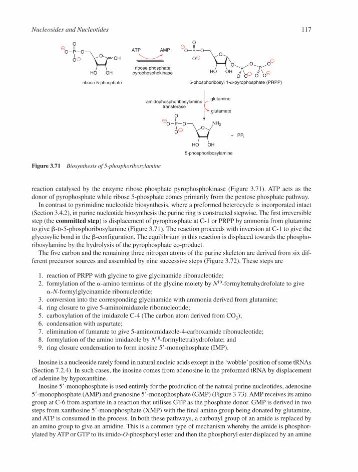
reaction catalysed by the enzyme ribose phosphate pyrophosphokinase (Figure 3.71). ATP acts as thedonor of pyrophosphate while ribose 5-phosphate comes primarily from the pentose phosphate pathway.
In contrast to pyrimidine nucleotide biosynthesis, where a preformed heterocycle is incorporated intact(Section 3.4.2), in purine nucleotide biosynthesis the purine ring is constructed stepwise. The first irreversiblestep (the committed step) is displacement of pyrophosphate at C-1 or PRPP by ammonia from glutamineto give �-D-5-phosphoribosylamine (Figure 3.71). The reaction proceeds with inversion at C-1 to give theglycosylic bond in the �-configuration. The equilibrium in this reaction is displaced towards the phospho-ribosylamine by the hydrolysis of the pyrophosphate co-product.
The five carbon and the remaining three nitrogen atoms of the purine skeleton are derived from six dif-ferent precursor sources and assembled by nine successive steps (Figure 3.72). These steps are
1. reaction of PRPP with glycine to give glycinamide ribonucleotide;2. formylation of the �-amino terminus of the glycine moiety by N10-formyltetrahydrofolate to give
�-N-formylglycinamide ribonucleotide;3. conversion into the corresponding glycinamide with ammonia derived from glutamine;4. ring closure to give 5-aminoimidazole ribonucleotide;5. carboxylation of the imidazole C-4 (The carbon atom derived from CO2);6. condensation with aspartate;7. elimination of fumarate to give 5-aminoimidazole-4-carboxamide ribonucleotide;8. formylation of the amino imidazole by N10-formyltetrahydrofolate; and9. ring closure condensation to form inosine 5�-monophosphate (IMP).
Inosine is a nucleoside rarely found in natural nucleic acids except in the ‘wobble’position of some tRNAs(Section 7.2.4). In such cases, the inosine comes from adenosine in the preformed tRNA by displacementof adenine by hypoxanthine.
Inosine 5�-monophosphate is used entirely for the production of the natural purine nucleotides, adenosine5�-monophosphate (AMP) and guanosine 5�-monophosphate (GMP) (Figure 3.73). AMP receives its aminogroup at C-6 from aspartate in a reaction that utilises GTP as the phosphate donor. GMP is derived in twosteps from xanthosine 5�-monophosphate (XMP) with the final amino group being donated by glutamine,and ATP is consumed in the process. In both these pathways, a carbonyl group of an amide is replaced byan amino group to give an amidine. This is a common type of mechanism whereby the amide is phosphor-ylated by ATP or GTP to its imido-O-phosphoryl ester and then the phosphoryl ester displaced by an amine
Nucleosides and Nucleotides 117
NH2O
O
HO OH
P
O
O
O
OO
HO
OH
OH
P
O
O
O OO
HO OH
P
O
O
O
OP
OP
O O O
O
O
ribose 5-phosphate
ribose phosphatepyrophosphokinase
5-phosphoribosyl 1-α-pyrophosphate (PRPP)
5-phosphoribosylamine
glutamine
glutamate
amidophosphoribosylaminetransferase
+ PPi
ATP AMP
Figure 3.71 Biosynthesis of 5-phosphoribosylamine

or ammonia. The leaving group can be inorganic phosphate, pyrophosphate or AMP, while the displacingnucleophile can be ammonia, the side chain of glutamine or the �-amino group of aspartate (Figure 3.74).
Steps in the biosynthesis of purine nucleotides furnish good examples of a standard control mechanismin metabolic pathways. This is feedback inhibition, where an enzyme catalysing an early step in the pathway
118 Chapter 3
NH2
Ribose-5P
Ribose-5P
HN
C O
H2C
NH3
OHCHN
H2N N
N
O
Ribose-5P
Ribose-5P
HN
C O
H2C
NHCHO
N
N
Ribose-5P
H2N
O
H2N
Ribose-5P
HN
C NH
H2C
NHCHO
N
N
Ribose-5P
H2N
CO2
N
N
Ribose-5P
H2N
O
HNCH
H2C
COO
OOC
N
HN N
N
O
Ribose-5P
N
N
Ribose-5P
H2N
O
O
1 2 3 4
phosphoribosylamine
glycinamideribonucleotide
formylglycinamideribonucleotide
formylglycinamidineribonucleotide
5-aminoimidazoleribonucleotide
5
678
9
inosine 5'-monophosphate (IMP)
5-aminoimidazole-4-carboxylateribonucleotide
5-aminoimidazole-4-N-succinocarboxamide
ribonucleotide
5-aminoimidazole-4-carboxamideribonucleotide
5-formamidinoimidazole-4-carboxamideribonucleotide
N 5, N 10 -methylenetetrahydrofolate
glutamine
N 10 - formyltetrahydrofolate
aspartateglycine
Figure 3.72 Formation of the purine ring; biosynthesis of IMP
N
N N
N
NH
Ribose-5P
CHCH2 COO
OOC
HN
NH
N
N
O
Ribose-5PO
HN
N N
N
O
Ribose-5P
N
N N
N
NH2
Ribose-5P
HN
N N
N
O
Ribose-5PH2N
adenylosuccinate
xanthosine 5'-monophosphate(XMP)
GMP
GTP
aspartate
oxidation
"NH3"
fumarate
IMP
AMP
ATP
Figure 3.73 Formation of AMP and GMP from IMP
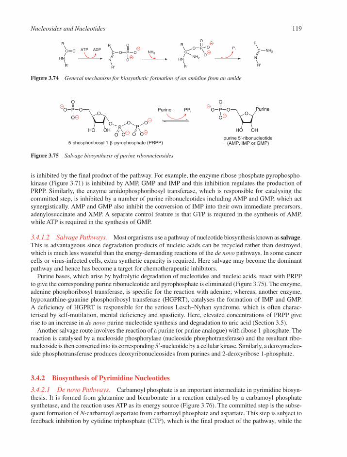
is inhibited by the final product of the pathway. For example, the enzyme ribose phosphate pyrophospho-kinase (Figure 3.71) is inhibited by AMP, GMP and IMP and this inhibition regulates the production ofPRPP. Similarly, the enzyme amidophosphoribosyl transferase, which is responsible for catalysing thecommitted step, is inhibited by a number of purine ribonucleotides including AMP and GMP, which actsynergistically. AMP and GMP also inhibit the conversion of IMP into their own immediate precursors,adenylosuccinate and XMP. A separate control feature is that GTP is required in the synthesis of AMP,while ATP is required in the synthesis of GMP.
3.4.1.2 Salvage Pathways. Most organisms use a pathway of nucleotide biosynthesis known as salvage.This is advantageous since degradation products of nucleic acids can be recycled rather than destroyed,which is much less wasteful than the energy-demanding reactions of the de novo pathways. In some cancercells or virus-infected cells, extra synthetic capacity is required. Here salvage may become the dominantpathway and hence has become a target for chemotherapeutic inhibitors.
Purine bases, which arise by hydrolytic degradation of nucleotides and nucleic acids, react with PRPPto give the corresponding purine ribonucleotide and pyrophosphate is eliminated (Figure 3.75). The enzyme,adenine phosphoribosyl transferase, is specific for the reaction with adenine; whereas, another enzyme,hypoxanthine-guanine phosphoribosyl transferase (HGPRT), catalyses the formation of IMP and GMP. A deficiency of HGPRT is responsible for the serious Lesch–Nyhan syndrome, which is often charac-terised by self-mutilation, mental deficiency and spasticity. Here, elevated concentrations of PRPP giverise to an increase in de novo purine nucleotide synthesis and degradation to uric acid (Section 3.5).
Another salvage route involves the reaction of a purine (or purine analogue) with ribose 1-phosphate. Thereaction is catalysed by a nucleoside phosphorylase (nucleoside phosphotransferase) and the resultant ribo-nucleoside is then converted into its corresponding 5�-nucleotide by a cellular kinase. Similarly, a deoxynucleo-side phosphotransferase produces deoxyribonucleosides from purines and 2-deoxyribose 1-phosphate.
3.4.2 Biosynthesis of Pyrimidine Nucleotides
3.4.2.1 De novo Pathways. Carbamoyl phosphate is an important intermediate in pyrimidine biosyn-thesis. It is formed from glutamine and bicarbonate in a reaction catalysed by a carbamoyl phosphate synthetase, and the reaction uses ATP as its energy source (Figure 3.76). The committed step is the subse-quent formation of N-carbamoyl aspartate from carbamoyl phosphate and aspartate. This step is subject tofeedback inhibition by cytidine triphosphate (CTP), which is the final product of the pathway, while the
Nucleosides and Nucleotides 119
R
C O
HN
R'
ATP ADPR
C O
N
R'
P
O
O
O NH3
R
CO
HN
R'
O
O
O
NH2
PR
C
N
R'
NH2Pi
Figure 3.74 General mechanism for biosynthetic formation of an amidine from an amide
OO
HO OH
P
O
O
O
OP
OP
O O O
O
O
5-phosphoribosyl 1-β-pyrophosphate (PRPP)
PurineO
O
HO OH
P
O
O
O
purine 5'-ribonucleotide(AMP, IMP or GMP)
Purine PPi
Figure 3.75 Salvage biosynthesis of purine ribonucleosides
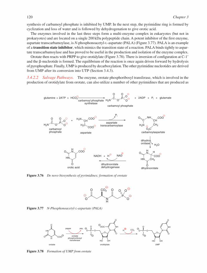
synthesis of carbamoyl phosphate is inhibited by UMP. In the next step, the pyrimidine ring is formed bycyclization and loss of water and is followed by dehydrogenation to give orotic acid.
The enzymes involved in the last three steps form a multi-enzyme complex in eukaryotes (but not inprokaryotes) and are located on a single 200 kDa polypeptide chain. A potent inhibitor of the first enzyme,aspartate transcarbamoylase, is N-phosphonoacetyl-L-aspartate (PALA) (Figure 3.77). PALA is an exampleof a transition state inhibitor, which mimics the transition state of a reaction. PALA binds tightly to aspar-tate transcarbamoylase and has proved to be useful in the production and isolation of the enzyme complex.
Orotate then reacts with PRPP to give orotidylate (Figure 3.78). There is inversion of configuration at C-1�and the �-nucleotide is formed. The equilibrium of the reaction is once again driven forward by hydrolysisof pyrophosphate. Finally, UMP is produced by decarboxylation. The other pyrimidine nucleotides are derivedfrom UMP after its conversion into UTP (Section 3.4.3).
3.4.2.2 Salvage Pathways. The enzyme, orotate phosphoribosyl transferase, which is involved in theproduction of orotidylate from orotate, can also utilize a number of other pyrimidines that are produced as
120 Chapter 3
O
CO
P
O
O
OH2N
CHCOOH2N
H2CCOO
CHCOONH
CH2
O2C
C
H2N
O
HN
HN
CH2
O
OCOO
H
HN
HN
O
O COO
NAD
H2O
O
CO
P
O
O
OH2N
+ 2ADP + Pi + glutamateglutamine + 2ATP + HCO3carbamoyl phosphate
synthetase
carbamoylphosphate
+aspartate
transcarbamoylase
Pi
aspartate carbamoylaspartate
dihydro-orotase
dihydroorotateorotic aciddihydroorotatedehydrogenase
NADH + H
carbamoyl phosphate
Figure 3.76 De novo biosynthesis of pyrimidines; formation of orotate
O NH
PO
O OOCO2O
Figure 3.77 N-Phosphonoacetyl-L-aspartate (PALA)
HN
NH
O
O COO
PRPP PPi
O
HO OH
OPO
O
O
N
NH
O
O
OOC
H COO
HO OH
OPO
O
O
N
NH
O
O
UMPorotate orotidylate
orotate phosphoribosyl
transferase
Figure 3.78 Formation of UMP from orotate

a result of hydrolysis of DNA or RNA. In a similar way as in the salvage of purines, phosphorylases catal-yse nucleoside formation from a variety of pyrimidines with either ribose 1-phosphate or 2-deoxyribose 1-phosphate. A cellular kinase is also required to convert the nucleoside product into the corresponding 5�-nucleotide. Thus, uridine kinase will accept both uridine and cytidine as substrates while thymidine kinasewill accept deoxyuridine as well as deoxythymidine. The fact that many viral thymidine kinases have areduced specificity for their substrates enables a distinction to be made between normal and virally-infected cells and has led to a strategy for viral interference (Section 3.7.2). Nucleoside transferases willcatalyse base exchange between nucleosides exclusively in the 2�-deoxy series.
3.4.3 Nucleoside Di- and Triphosphates
The immediate biosynthetic precursors of the nucleic acids are normally the nucleoside triphosphates;whereas, diphosphates can also be used in energy conversions. Diphosphates are obtained from the corres-ponding monophosphates by means of a specific nucleoside, monophosphate kinase. Adenylate kinaseconverts AMP into ADP while UMP kinase converts UMP into UDP. Both enzymes use ATP as the phos-phoryl donor. Nucleoside triphosphates are interconvertible with diphosphates through nucleoside diphos-phate kinase, an enzyme that has a broad specificity. Thus Y and Z (Figure 3.79) can be any of the severalpurine or pyrimidine ribo- or deoxyribonucleosides.
Cytidine triphosphate is formed from UTP by replacement of the oxygen atom at C-4 by an amino group.In E. coli the donor is ammonia, but in mammals the ammonia comes from the amide group of glutamine.In both cases, ATP is required for the reaction (Figure 3.74).
3.4.4 Deoxyribonucleotides
Deoxyribonucleotides are formed by the reduction of the corresponding ribonucleotides. The 2�-hydroxylgroup of the ribose is replaced by a hydrogen atom in a reaction that takes place at the level of the ribonu-cleoside 5�-diphosphate. The mechanism is rather complicated. The key enzyme is ribonucleotide reductase(ribonucleoside diphosphate reductase) and the electrons required for the reduction of the ribose are trans-ferred from NADPH to sulfhydryl groups at the catalytic site of the enzyme. The enzyme from E. coli is aprototype for most eukaryotic reductases. A larger subunit (2 � 86 kDa) binds the NTP substrate and thesmaller subunit (2 � 43 kDa) contains a binuclear iron centre and a tyrosyl radical at residue-122. A mech-anism based on all the available data is shown (Figure 3.80). The reduction of ribonucleoside diphosphatesis controlled by allosteric interactions (an allosteric enzyme is one in which the binding of another substance,usually product, alters its kinetic behaviour) through the use of two allosteric sites that bind a number ofnucleoside 5�-triphosphates and lead to a variety of conformations, each with different catalytic properties.
In the event that any dUTP is formed from dUDP, it is rapidly hydrolysed to dUMP by an active dUTPase,which thereby limits the incorporation of dUTP into DNA. Nonetheless, some uracil residues do occur inDNA, which may in part arise through deamination of cytosines. These premutagenic events are repairedby uracil DNA glycosylase (Section 8.11.3). As a result, deoxythymidine 5�-triphosphate (dTTP) is the
Nucleosides and Nucleotides 121
UMP + ATP UDP + ADPUMP kinase
AMP + ATPUMP kinase
YDP + ZTP YTP + ZDPnucleoside diphosphate
kinase
2 ADP
Figure 3.79 Biosynthesis of nucleoside di- and triphosphates

predominant dioxopyrimidine nucleotide incorporated into DNA. First, deoxythymidine 5�-monophosphate(dTMP) is biosynthesised from dUMP via the enzyme thymidylate synthase. The methyl group is providedby N5,N10-methylenetetrahydrofolate, which also acts as an electron donor (Figure 3.81) and becomes oxidisedto dihydrofolate. Tetrahydrofolate is regenerated by dihydrofolate reductase (DHFR) using NADPH as thereductant. These two enzymes are excellent targets for cancer chemotherapy because cancer cells have anincreased level of DNA synthesis and, thus, a heavy requirement for dTMP (see 5-fluorouracil andmethotrexate in Section 3.7.1).
dTMP is next converted into dTTP in two stages by means of a thymidylate kinase and then a nucleo-side diphosphate kinase. In virus-infected cells, the viral thymidine kinase (Section 3.7.2) often also playsthe role of a thymidylate kinase.
3.5 CATABOLISM OF NUCLEOTIDES
The degradation of nucleotides is of major importance as a target for drug design. RNA is metabolicallymuch more labile than DNA and is constantly being synthesised and degraded. Degradation occurs ini-tially through the action of ribonucleases and deoxyribonucleases, which form oligonucleotides that arefurther broken down to nucleotides by phosphodiesterases.
Nucleotides are hydrolysed to nucleosides by nucleotidases (and by phosphatases). Of great importance isthe final cleavage of nucleosides by inorganic phosphate to base and ribose 1-�-phosphate (or 2-deoxyribose1-�-phosphate) catalysed by the widely distributed enzyme PNP (Figure 3.82). The ribose phosphate canthen be isomerized to ribose 5-phosphate and reused for the synthesis of PRPP. In mammalian tissues, adeno-sine and deoxyadenosine are resistant to the phosphorylase. AMP is therefore first deaminated by adenylate
122 Chapter 3
ORO
HO OH
B
Ha Hb
X
HS SH
ORO
HO OH
B
Hb
XHa
HS SH
ORO
HO OH2
B
Hb
XHa
HS S
ORO
HO H
B
Ha Hb
X
S S
ORO
HO H
B
Hb
XHa
S S
ORO
HO Hb
B
XHa
HS S
-H2O
protein
proteinprotein
protein
proteinprotein
Figure 3.80 Postulated mechanism for reduction of nucleotides to deoxynucleotides by E. coli ribonucleotidereductase (X � Tyr122�O)
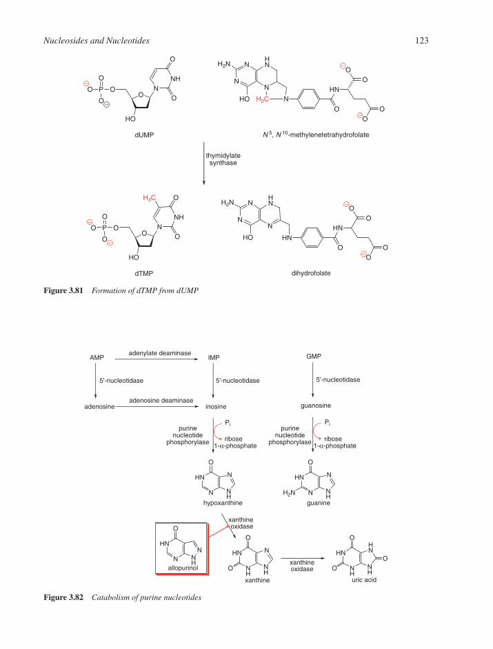
Nucleosides and Nucleotides 123
N
HN N
NH
O
H2NN
HN N
NH
O
NH
HN N
NH
O
ON
HNN
NH
O
NH
HNHN
NH
O
O
O
GMP
adenosine inosine guanosine
5'-nucleotidase 5'-nucleotidase 5'-nucleotidase
adenosine deaminase
adenylate deaminase
Pi
ribose1-α-phosphate
Pipurine
nucleotidephosphorylase
purinenucleotide
phosphorylase
hypoxanthine guanine
xanthine uric acid
allopurinolxanthineoxidase
xanthineoxidase
ribose1-α-phosphate
AMP IMP
Figure 3.82 Catabolism of purine nucleotides
O
HO
OPO
O
O
NNH
O
O
OO
HN
O
N
N
HO
N
H2N NHN
OO
H2C
O
HO
OPO
O
O
NNH
O
O
H3C
OO
HN
OHN
N
HO
N
H2N NHN
OO
dUMP N 5, N 10-methylenetetrahydrofolate
thymidylatesynthase
dTMP dihydrofolate
Figure 3.81 Formation of dTMP from dUMP
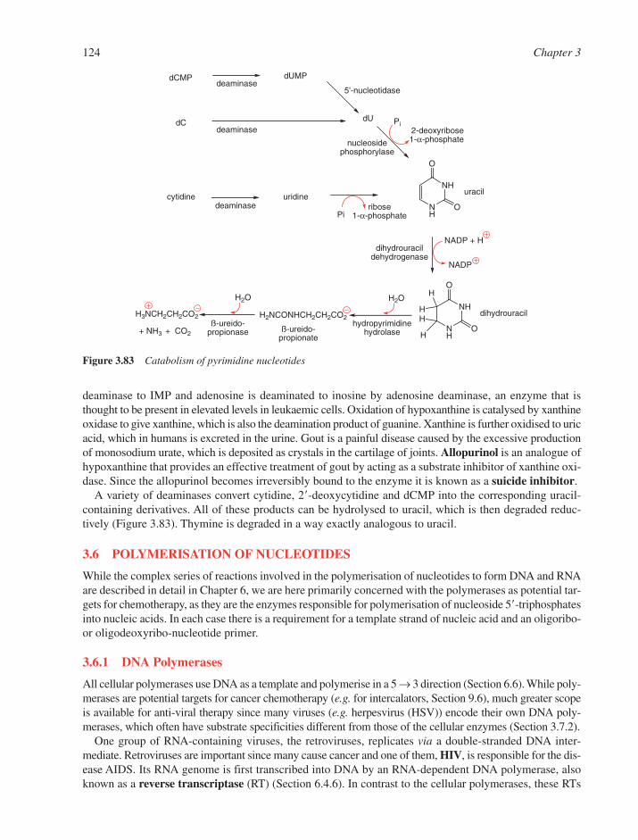
deaminase to IMP and adenosine is deaminated to inosine by adenosine deaminase, an enzyme that isthought to be present in elevated levels in leukaemic cells. Oxidation of hypoxanthine is catalysed by xanthineoxidase to give xanthine, which is also the deamination product of guanine. Xanthine is further oxidised to uricacid, which in humans is excreted in the urine. Gout is a painful disease caused by the excessive productionof monosodium urate, which is deposited as crystals in the cartilage of joints. Allopurinol is an analogue ofhypoxanthine that provides an effective treatment of gout by acting as a substrate inhibitor of xanthine oxi-dase. Since the allopurinol becomes irreversibly bound to the enzyme it is known as a suicide inhibitor.
A variety of deaminases convert cytidine, 2�-deoxycytidine and dCMP into the corresponding uracil-containing derivatives. All of these products can be hydrolysed to uracil, which is then degraded reduc-tively (Figure 3.83). Thymine is degraded in a way exactly analogous to uracil.
3.6 POLYMERISATION OF NUCLEOTIDES
While the complex series of reactions involved in the polymerisation of nucleotides to form DNA and RNAare described in detail in Chapter 6, we are here primarily concerned with the polymerases as potential tar-gets for chemotherapy, as they are the enzymes responsible for polymerisation of nucleoside 5�-triphosphatesinto nucleic acids. In each case there is a requirement for a template strand of nucleic acid and an oligoribo-or oligodeoxyribo-nucleotide primer.
3.6.1 DNA Polymerases
All cellular polymerases use DNA as a template and polymerise in a 5 → 3 direction (Section 6.6). While poly-merases are potential targets for cancer chemotherapy (e.g. for intercalators, Section 9.6), much greater scopeis available for anti-viral therapy since many viruses (e.g. herpesvirus (HSV)) encode their own DNA poly-merases, which often have substrate specificities different from those of the cellular enzymes (Section 3.7.2).
One group of RNA-containing viruses, the retroviruses, replicates via a double-stranded DNA inter-mediate. Retroviruses are important since many cause cancer and one of them, HIV, is responsible for the dis-ease AIDS. Its RNA genome is first transcribed into DNA by an RNA-dependent DNA polymerase, alsoknown as a reverse transcriptase (RT) (Section 6.4.6). In contrast to the cellular polymerases, these RTs
124 Chapter 3
NH
NH
O
O
NH
NH
O
O
H
H
H
H
H2O
H2NCONHCH2CH2CO2H3NCH2CH2CO2
H2O
Piribose
1-α-phosphate
dCMP dUMP
5'-nucleotidase
dC dU
nucleosidephosphorylase
uracilcytidine
deaminase
deaminase
deaminase
uridine
NADP + Hdihydrouracil
dehydrogenase
dihydrouracilhydropyrimidine
hydrolase+ NH3 + CO2
ß-ureido-propionase ß-ureido-
propionate
Pi2-deoxyribose1-α-phosphate
NADP
Figure 3.83 Catabolism of pyrimidine nucleotides
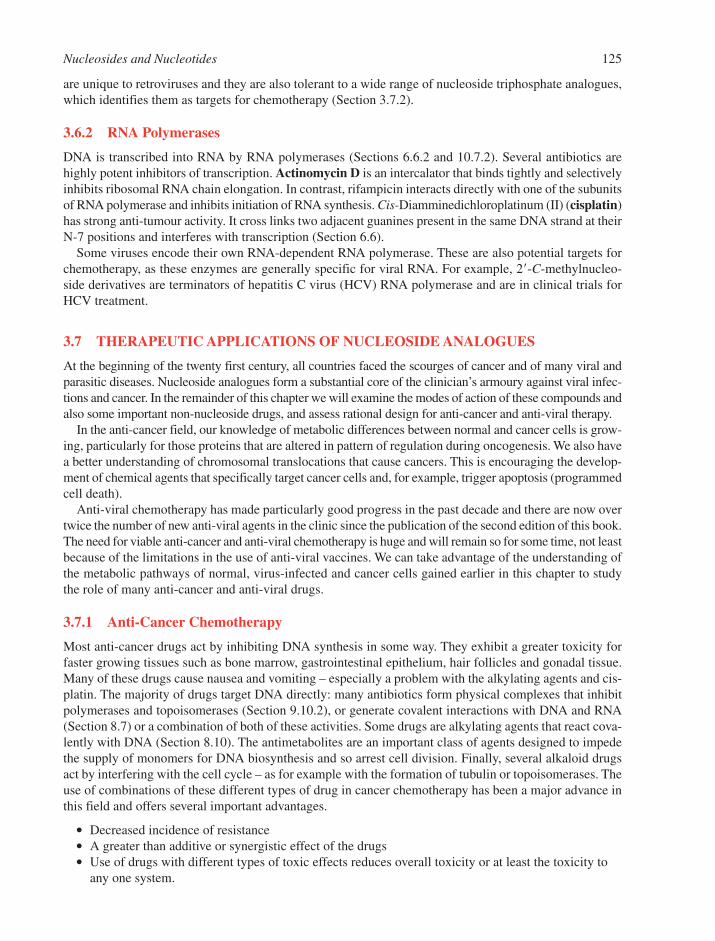
are unique to retroviruses and they are also tolerant to a wide range of nucleoside triphosphate analogues,which identifies them as targets for chemotherapy (Section 3.7.2).
3.6.2 RNA Polymerases
DNA is transcribed into RNA by RNA polymerases (Sections 6.6.2 and 10.7.2). Several antibiotics arehighly potent inhibitors of transcription. Actinomycin D is an intercalator that binds tightly and selectivelyinhibits ribosomal RNA chain elongation. In contrast, rifampicin interacts directly with one of the subunitsof RNA polymerase and inhibits initiation of RNA synthesis. Cis-Diamminedichloroplatinum (II) (cisplatin)has strong anti-tumour activity. It cross links two adjacent guanines present in the same DNA strand at theirN-7 positions and interferes with transcription (Section 6.6).
Some viruses encode their own RNA-dependent RNA polymerase. These are also potential targets forchemotherapy, as these enzymes are generally specific for viral RNA. For example, 2�-C-methylnucleo-side derivatives are terminators of hepatitis C virus (HCV) RNA polymerase and are in clinical trials forHCV treatment.
3.7 THERAPEUTIC APPLICATIONS OF NUCLEOSIDE ANALOGUES
At the beginning of the twenty first century, all countries faced the scourges of cancer and of many viral andparasitic diseases. Nucleoside analogues form a substantial core of the clinician’s armoury against viral infec-tions and cancer. In the remainder of this chapter we will examine the modes of action of these compounds andalso some important non-nucleoside drugs, and assess rational design for anti-cancer and anti-viral therapy.
In the anti-cancer field, our knowledge of metabolic differences between normal and cancer cells is grow-ing, particularly for those proteins that are altered in pattern of regulation during oncogenesis. We also havea better understanding of chromosomal translocations that cause cancers. This is encouraging the develop-ment of chemical agents that specifically target cancer cells and, for example, trigger apoptosis (programmedcell death).
Anti-viral chemotherapy has made particularly good progress in the past decade and there are now overtwice the number of new anti-viral agents in the clinic since the publication of the second edition of this book.The need for viable anti-cancer and anti-viral chemotherapy is huge and will remain so for some time, not leastbecause of the limitations in the use of anti-viral vaccines. We can take advantage of the understanding ofthe metabolic pathways of normal, virus-infected and cancer cells gained earlier in this chapter to studythe role of many anti-cancer and anti-viral drugs.
3.7.1 Anti-Cancer Chemotherapy
Most anti-cancer drugs act by inhibiting DNA synthesis in some way. They exhibit a greater toxicity forfaster growing tissues such as bone marrow, gastrointestinal epithelium, hair follicles and gonadal tissue.Many of these drugs cause nausea and vomiting – especially a problem with the alkylating agents and cis-platin. The majority of drugs target DNA directly: many antibiotics form physical complexes that inhibitpolymerases and topoisomerases (Section 9.10.2), or generate covalent interactions with DNA and RNA(Section 8.7) or a combination of both of these activities. Some drugs are alkylating agents that react cova-lently with DNA (Section 8.10). The antimetabolites are an important class of agents designed to impedethe supply of monomers for DNA biosynthesis and so arrest cell division. Finally, several alkaloid drugsact by interfering with the cell cycle – as for example with the formation of tubulin or topoisomerases. Theuse of combinations of these different types of drug in cancer chemotherapy has been a major advance inthis field and offers several important advantages.
● Decreased incidence of resistance● A greater than additive or synergistic effect of the drugs● Use of drugs with different types of toxic effects reduces overall toxicity or at least the toxicity to
any one system.
Nucleosides and Nucleotides 125
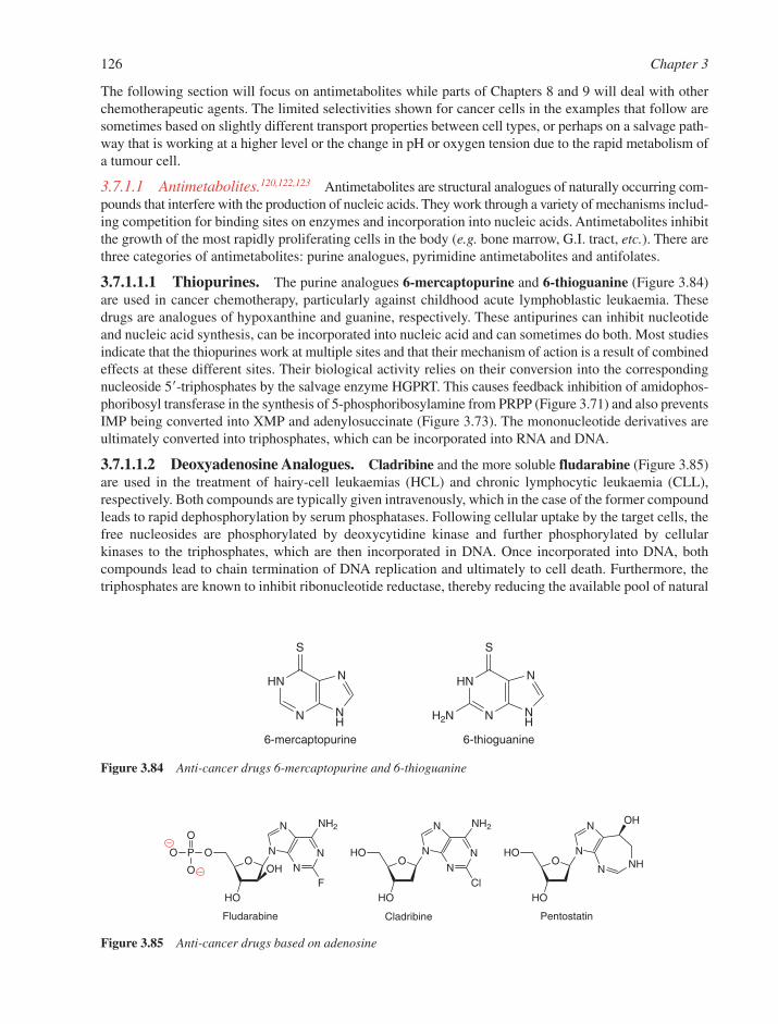
The following section will focus on antimetabolites while parts of Chapters 8 and 9 will deal with otherchemotherapeutic agents. The limited selectivities shown for cancer cells in the examples that follow aresometimes based on slightly different transport properties between cell types, or perhaps on a salvage path-way that is working at a higher level or the change in pH or oxygen tension due to the rapid metabolism ofa tumour cell.
3.7.1.1 Antimetabolites.120,122,123 Antimetabolites are structural analogues of naturally occurring com-pounds that interfere with the production of nucleic acids. They work through a variety of mechanisms includ-ing competition for binding sites on enzymes and incorporation into nucleic acids. Antimetabolites inhibitthe growth of the most rapidly proliferating cells in the body (e.g. bone marrow, G.I. tract, etc.). There arethree categories of antimetabolites: purine analogues, pyrimidine antimetabolites and antifolates.
3.7.1.1.1 Thiopurines. The purine analogues 6-mercaptopurine and 6-thioguanine (Figure 3.84)are used in cancer chemotherapy, particularly against childhood acute lymphoblastic leukaemia. Thesedrugs are analogues of hypoxanthine and guanine, respectively. These antipurines can inhibit nucleotideand nucleic acid synthesis, can be incorporated into nucleic acid and can sometimes do both. Most studiesindicate that the thiopurines work at multiple sites and that their mechanism of action is a result of combinedeffects at these different sites. Their biological activity relies on their conversion into the correspondingnucleoside 5�-triphosphates by the salvage enzyme HGPRT. This causes feedback inhibition of amidophos-phoribosyl transferase in the synthesis of 5-phosphoribosylamine from PRPP (Figure 3.71) and also preventsIMP being converted into XMP and adenylosuccinate (Figure 3.73). The mononucleotide derivatives areultimately converted into triphosphates, which can be incorporated into RNA and DNA.
3.7.1.1.2 Deoxyadenosine Analogues. Cladribine and the more soluble fludarabine (Figure 3.85)are used in the treatment of hairy-cell leukaemias (HCL) and chronic lymphocytic leukaemia (CLL),respectively. Both compounds are typically given intravenously, which in the case of the former compoundleads to rapid dephosphorylation by serum phosphatases. Following cellular uptake by the target cells, thefree nucleosides are phosphorylated by deoxycytidine kinase and further phosphorylated by cellularkinases to the triphosphates, which are then incorporated in DNA. Once incorporated into DNA, bothcompounds lead to chain termination of DNA replication and ultimately to cell death. Furthermore, thetriphosphates are known to inhibit ribonucleotide reductase, thereby reducing the available pool of natural
126 Chapter 3
N
HN N
NH
S
H2NN
HN N
NH
S
6-thioguanine6-mercaptopurine
Figure 3.84 Anti-cancer drugs 6-mercaptopurine and 6-thioguanine
OHO
HO
NN
N
N
NH2
Cl
OHO
HO
N
N
N NH
OH
OH
P
O
O
OO
O
HO
NN
N
N
NH2
F
Fludarabine Cladribine Pentostatin
Figure 3.85 Anti-cancer drugs based on adenosine
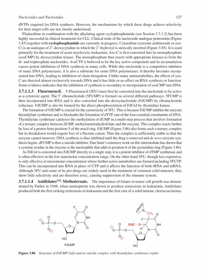
dNTPs required for DNA synthesis. However, the mechanisms by which these drugs achieve selectivityfor their target cells are not clearly understood.
Fludarabine in combination with the alkylating agent cyclophosphamide (see Section 3.7.1.2) has beenhighly successful in clinical treatments for CLL. Clinical trials of the nucleoside analogue pentostatin (Figure3.85) together with cyclophosphamide are currently in progress. Cytarabine (cytosine arabinoside or ara-C) is an analogue of 2�-deoxycytidine in which the 2�-hydroxyl is sterically inverted (Figure 3.85). It is usedprimarily for the treatment of acute myelocytic leukaemia. Ara-C is first converted into its monophosphate(araCMP) by deoxycytidine kinase. The monophosphate then reacts with appropriate kinases to form thedi- and triphosphate nucleotides. AraCTP is believed to be the key active component and its accumulationcauses potent inhibition of DNA synthesis in many cells. While this nucleotide is a competitive inhibitorof many DNA polymerases, it is also a substrate for some DNA polymerases. It thereby becomes incorp-orated into DNA, leading to inhibition of chain elongation. Unlike many antimetabolites, the effects of ara-C are directed almost exclusively towards DNA and it has little or no effect on RNA synthesis or function.Some evidence indicates that the inhibition of synthesis is secondary to incorporation of araCMP into DNA.
3.7.1.1.3 Fluorouracil. 5-Fluorouracil (5FU) must first be converted into the nucleotide to be activeas a cytotoxic agent. The 5� ribonucleotide (5FUMP) is formed via several different pathways. 5FUMP isthen incorporated into RNA and is also converted into the deoxynucleotide (FdUMP) by ribonucleotidereductase. FdUMP is also be formed by the direct phosphorylation of FdUrd by thymidine kinase.
The formation of FdUMP is crucial for the cytotoxicity of 5FU. This is because FdUMP inhibits the enzymethymidylate synthetase and so blockades the formation of dTTP, one of the four essential constituents of DNA.Thymidylate synthetase catalyses the methylation of dUMP in a multi-step process that involves formationof a ternary complex between dUMP, methylenetetrahydrofolate and the enzyme. This complex reacts furtherby loss of a proton from position-5 of the uracil ring. FdUMP (Figure 3.86) also forms such a ternary complexbut its breakdown would require loss of a fluorine cation. Thus the complex is sufficiently stable so that theenzyme cannot turnover. DNA synthesis is thus inhibited until the drug is removed and de novo enzyme syn-thesis begins. dFUMP is thus a suicide inhibitor. Dan Santi’s extensive work on this intermediate has shown thata cysteine residue in the enzyme is the nucleophile that adds to position-6 of the pyrimidine ring (Figure 3.86).
As FdUrd is converted into FdUMP directly in a single step, it is a potent inhibitor of dTMP synthetase andis often effective in the low nanomolar concentration range. On the other hand 5FU, though less expensive,is only effective at micromolar concentration where further active metabolites are formed including 5FUTP.This can be incorporated into RNA in place of UTP and it affects the function of both rRNA and mRNA.Although 5FU and some of its pro-drugs are widely used in the treatment of common solid tumours, theyshow little selectivity and are therefore toxic, causing suppression of the immune system.
3.7.1.1.4 Antifolates124–Methotrexate. The importance of folates in tumor cell growth was demon-strated by Farber in 1948, when aminopterin was shown to produce remissions in leukaemia. Antifolatesproduced both the first striking remissions in leukaemia and the first cure of a solid tumour, choriocarcinoma.
Nucleosides and Nucleotides 127
O
HO
OPO
O
O
N
NH
O
O
F
O
HO
OPO
O
O
N
NH
O
O
SENZ
F
CH2
N
H
FdUMP
cofa
ctor
Figure 3.86 Structure of FdUMP (left) and its suicide complex with thymidylate synthetase (right)
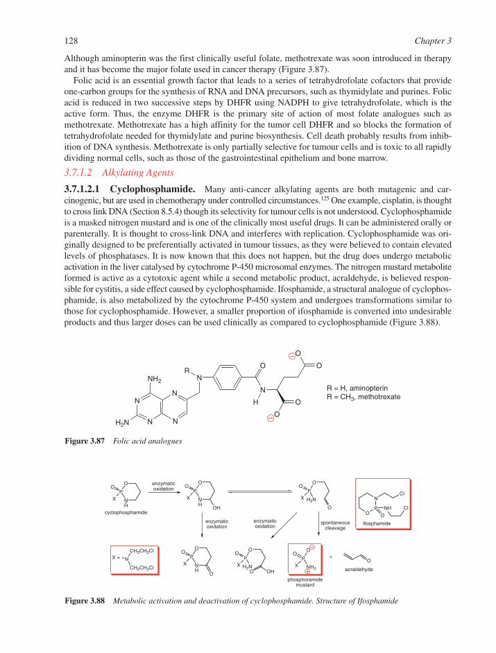
Although aminopterin was the first clinically useful folate, methotrexate was soon introduced in therapyand it has become the major folate used in cancer therapy (Figure 3.87).
Folic acid is an essential growth factor that leads to a series of tetrahydrofolate cofactors that provideone-carbon groups for the synthesis of RNA and DNA precursors, such as thymidylate and purines. Folicacid is reduced in two successive steps by DHFR using NADPH to give tetrahydrofolate, which is theactive form. Thus, the enzyme DHFR is the primary site of action of most folate analogues such asmethotrexate. Methotrexate has a high affinity for the tumor cell DHFR and so blocks the formation oftetrahydrofolate needed for thymidylate and purine biosynthesis. Cell death probably results from inhib-ition of DNA synthesis. Methotrexate is only partially selective for tumour cells and is toxic to all rapidlydividing normal cells, such as those of the gastrointestinal epithelium and bone marrow.
3.7.1.2 Alkylating Agents
3.7.1.2.1 Cyclophosphamide. Many anti-cancer alkylating agents are both mutagenic and car-cinogenic, but are used in chemotherapy under controlled circumstances.125 One example, cisplatin, is thoughtto cross link DNA (Section 8.5.4) though its selectivity for tumour cells is not understood. Cyclophosphamideis a masked nitrogen mustard and is one of the clinically most useful drugs. It can be administered orally orparenterally. It is thought to cross-link DNA and interferes with replication. Cyclophosphamide was ori-ginally designed to be preferentially activated in tumour tissues, as they were believed to contain elevatedlevels of phosphatases. It is now known that this does not happen, but the drug does undergo metabolicactivation in the liver catalysed by cytochrome P-450 microsomal enzymes. The nitrogen mustard metaboliteformed is active as a cytotoxic agent while a second metabolic product, acraldehyde, is believed respon-sible for cystitis, a side effect caused by cyclophosphamide. Ifosphamide, a structural analogue of cyclophos-phamide, is also metabolized by the cytochrome P-450 system and undergoes transformations similar tothose for cyclophosphamide. However, a smaller proportion of ifosphamide is converted into undesirableproducts and thus larger doses can be used clinically as compared to cyclophosphamide (Figure 3.88).
128 Chapter 3
O
O
N
O
N
N
NH2
N
H2N N N
O
O
R
H
R = H, aminopterinR = CH3, methotrexate
Figure 3.87 Folic acid analogues
O
P
N
O
XH
O
P
N
O
XH
OH
O
P
H N
O
XO OH
O
P
N
O
XH
O
N
CH CH Cl
CH CH Cl
O
P
NH
O
X
O
P
H N
O
X
O
O
OP
NCl
O
NH Cl
enzymaticoxidation
spontaneouscleavage
+
phosphoramidemustard
enzymaticoxidation
cyclophosphamide
enzymaticoxidation
acraldehyde
X =
Ifosphamide
Figure 3.88 Metabolic activation and deactivation of cyclophosphamide. Structure of Ifosphamide

Other important alkylating agents used in cancer treatment include the nitrosoureas carmustine (BCNU)and lomustine (CCNU) which alkylate the O-6 and N-1 positions of guanine bases to give N1,O6-ethenogua-nine (Figure 3.89),126 which reacts to give an interstrand cross link with cytosine. Other alkylating agentssuch as temozolomide (Figure 3.89) alkylate the O-6-position of guanines, which causes G→A mutationsduring DNA replication. O6-Alkylguanine DNA alkyltransferase is a protein that can repair such DNAdamage and is expressed at elevated levels in some tumours. As a result, it is a key target for inhibition bya number of compounds.126 Both O6-benzylguanine and O6-(4-bromothenyl)guanine (Figure 3.89) havebeen used clinically in combination with chemotherapeutic regimes that employ alkylating agents.
In all the above compounds, the therapeutic target is inhibition of DNA replication, whether by pre-venting synthesis of precursors or by alkylating the DNA itself. As most of the effective drugs are alsotoxic, patients receiving cancer chemotherapy are unusually susceptible to viral and bacterial infections.
3.7.2 Anti-Viral Chemotherapy127
The life cycle of a virus involves a combination of its own enzymes and those of the host cell. Thus the designof anti-viral agents can be more directly targeted than that of anti-cancer agents. Since most virus classes areunrelated to each other and have unique replication cycles, there appears little chance for the discovery ofwide-activity anti-viral agents comparable to the broad-spectrum antibiotics, such as the �-lactams. Differentapproaches have been successful for retroviruses such as HIV and DNA viruses such as HSV and hepa-titis B virus (HBV).
3.7.2.1 Retrovirus Inhibitors. Much effort has been expended on finding a chemotherapeutic agentto alleviate the symptoms of AIDS. The HIV is a member of the lentivirus family (a sub-class of retrovirus)and its reverse transcriptase has been an obvious target. Virtually all the compounds currently used for thetreatment of HIV infections, or in advanced research, belong to one of four main classes:
● nucleoside/nucleotide reverse transcriptase inhibitors (NRTIs);● non-nucleoside reverse transcriptase inhibitors (NNRTIs);● protease inhibitors (PIs); and● host-cell receptor based therapeutic agents.
3.7.2.1.1 Nucleoside Reverse Transcriptase Inhibitors. There are around 40 anti-viral com-pounds in clinical use (Table 3.2), with over half of them being used in the treatment of HIV patients. Themajority are nucleoside analogues effective against HIV or HSV infections. The key structural feature of anucleoside analogue as a chain terminator for RT is the absence of a 3�-OH function. Thus after incorporation
Nucleosides and Nucleotides 129
N
NN
N
O
NH2
H
N
NN
N
O
NH2
H
N
NN
N
O
NH2
S
Br
N NX
O
NO H
Cl
Cl
N
NN
NN
O
Me
ONH2
N1,O6-ethenoguanine (in DNA)
O6-benzylguanine O6-(4-bromothenyl)guanine
X = carmustine
X = lomustine
temozolomide
Figure 3.89 Structures of N1,O6-ethenoguanine, O6-benzylguanine, O6-(4-bromothenyl)guanine and various DNAalkylating agents

at the growing 3�-end, this moiety cannot become phosphorylated by the next dNTP monomer. One of thefirst compounds to be widely used was AZT (Figure 3.90) as it was found to be the least toxic of severalanalogues in clinical use. A range of effective NRTIs is shown in Figure 3.90.
The active species for all nucleoside analogues of this type is the 2�-deoxynucleoside 5�-triphosphate. Sincethe retrovirus does not encode its own kinase and because it is difficult to get highly anionic nucleotides intocells, it is necessary for the native human cellular thymidine kinase to perform the initial phosphorylation
130 Chapter 3
Table 3.2 Anti-viral compounds in clinical use
Drug Virus
Amantadine, Rimantadine Influenza AOseltamivir, Zanamivir Influenza A and BZidovudine (AZT), Retrovir® HIVDidanosine (ddI), Videx® HIVZalcitabine (ddC), Hivid® HIVStavudine (d4T), Zerit® HIVLamivudine (3TC), Epivir®, Zeffix® HIV and HBVAbacavir, Ziagen® HIVEmtricitabine, Emtriva® HIV and HBVTenofovir disoproxil (oral prodrug of PMPA) HIV and HBVNevirapine, Delavirdine, Efavirenz (non-nucleoside HIV
RT inhibitors NNRTIs)Saquinavir, Ritonavir, Indinavir, Nelfinavir, Amprenavir, HIV protease inhibitors
Lopinavir, AtazanavirEnfuvirtide HIV viral entry inhibitorAdefovir dipivoxil (oral prodrug of PMEA) HIV and HBVAcyclovir (ACV), Zovirax® Herpes Simplex Virus (HSV-1 and HSV-2) and
Varicella Zoster Virus (VZV)Valaciclovir, Zelitrex®, Valtrex® oral prodrug of acyclovir) HSV-1, HSV-2 and VZVPenciclovir, Denavir®, Vectavir® HSV-1, HSV-2 and VZVFamciclovir, Famvir® HSV-1, HSV-2 and VZVIdoxuridine, Herpid®, Idoxene®, etc. HSV-1, HSV-2 and VZVTrifluridine (TFT), Viroptic® HSV-1, HSV-2 and VZVBrivudin (BVDU), Zostex®, Zerpex®, etc. HSV-1 and VZVGanciclovir (DHPG), (GCV), Cymevene®, Cytovene® HSV-1 and HSV-2 cytomegalovirus (CMV)Valganciclovir (VGCV), Valcyte® HSV-1, HSV-2 and CMVFoscarnet, Foscavir® HSV-1, HSV-2, VZV, CMV and HIVCidofovir (HPMPC), (CDV), Vistide® Herpes viruses, papilloma-, polyoma-,
adeno- and poxvirusesFomiversen, Vitravene® CMV retinitisRibavirin Influenza A and B, measles, respiratory
syncytial virus (RSV) and adenovirus
OHO B
OHO
NN
N
N
HN
NH2
O OHNN
H2N
OS
X
OHO Thy
N3
OHO Thy
X = H, lamivudine (3TC) (Epivir)X = F, emtricitabine [(-)FTC]
abacavir (ABC)(Ziagen)
B = Cyt; zalcitabine (ddC) (Hivid)B = 6-oxopurine; didanosine (ddI) (Videx)
zidovudine (AZT)(Retrovir)
stavudine (d4T)(Zerit)
Figure 3.90 Structures of some licensed nucleoside-based reverse transcriptase inhibitors
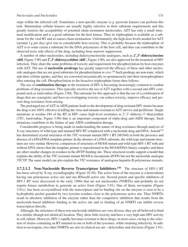
steps within the infected cell. Sometimes a non-specific enzyme (e.g. pyruvate kinase) can perform thistask. Mammalian cellular kinases are usually highly selective in their substrate requirements and thisgreatly restricts the acceptability of potential chain terminator nucleosides. AZT has only a small struc-tural modification and is a good substrate for the host kinase. Thus its triphosphate is available as a sub-strate for the viral RT and so causes chain termination. Unfortunately, the high dose levels needed for AZT(around 1 g per day) give rise to considerable host toxicity. This is probably because the triphosphate ofAZT is to some extent a substrate for the DNA polymerase of the host cell, and thus can contribute to theobserved toxic side effects of the drug, including bone marrow suppression.
A number of other nucleosides, including dideoxynucleoside analogues, such as 2��,3��-dideoxyinosine(ddI, Figure 3.90) and 2��,3��-dideoxycytidine (ddC, Figure 3.90), are also approved for the treatment of HIVinfection. They share the same problems of toxicity and requirement for phosphorylation by host enzymeswith AZT. The use of nucleotide prodrugs has greatly improved the efficacy of these and other nucleo-side analogues that are not good substrates for phosphorylation in vivo.128 Such prodrugs are non-ionic, whichaids their cellular uptake, and they are converted enzymatically or spontaneously into their monophosphatesafter entering the cell. Phosphorylation to the bioactive triphosphate forms then follows.
The use of combination therapy in the treatment of HIV is becoming increasingly common to combatproblems of drug resistance. This typically involves the use of AZT together with a second anti-HIV com-pound such as lamivudine (Figure 3.90). The rationale for this approach is that the use of a combination ofdrugs that are synergistic and have no overlapping toxicity can reduce toxicity, improve efficacy and pre-vent drug resistance from arising.
The prolonged use of AZT in AIDS patients leads to the development of drug-resistant HIV strains becausethe drug is not 100% effective in killing the virus and mutants resistant to AZT survive and proliferate. Singlemutations at residue-184 of the RT in HIV cause high-level resistance to 2�,3�-dideoxy-3�-thiacytidine(3TC, lamivudine, Figure 3.90) that is an important component of triple-drug anti-AIDS therapy. Suchmutations contribute to the failure of anti-AIDS combination therapy.
Considerable progress is being made in understanding the nature of drug resistance through analysis ofX-ray structures of wild-type and mutated HIV RT complexed with a nucleotide drug and DNA. Arnold129
has determined crystal structures of the 3TC-resistant mutant HIV-1 RT (M184I) in both the presence andabsence of a DNA/DNA template-primer. In the absence of a DNA substrate, the wild-type and mutant struc-tures are very similar. However, comparison of structures of M184I mutant and wild-type HIV-1 RT with andwithout DNA shows that the template-primer is repositioned in the M184I/DNA binary complex and thereare other smaller changes in residues in the dNTP-binding site. These structural results support a model thatexplains the ability of the 3TC-resistant mutant M184I to incorporate dNTPs but not the nucleotide analogue3TCTP. The same model can also explain the 3TC resistance of analogous hepatitis B polymerase mutants.
3.7.2.1.2 Non-Nucleoside Reverse Transcriptase Inhibitors.130 The structure of HIV-1 RThas been solved by X-ray crystallography (Figure 10.30). The active form of the enzyme is a heterodimerhaving one polymerase active site and one RNaseH active site. Several potent and specific inhibitors ofHIV-1 RT were discovered in the early 1990s that are not nucleosides (NNRTIs) and probably do notrequire kinase metabolism to generate an active form (Figure 3.91). One of them, nevirapine (Figure3.91c), has been co-crystallised with the transcriptase and its binding site on the enzyme is seen to be ahydrophobic pocket guarded by two tyrosine residues close to the polymerase active site. Thus NNRTIsresult in allosteric inhibition of the enzyme rather than the competitive inhibition that results from thenucleoside-based inhibitors binding to the active site and so binding of an NNRTI can inhibit reverse transcription directly.
Although, the structures of these non-nucleoside inhibitors are very diverse, they are all believed to bindin a similar (though not identical) location. They show little toxicity and have a very high anti-HIV activityin cell culture. However, HIV-1 rapidly becomes resistant to these drugs, in most cases, owing to the selec-tion of strains containing an RT mutated at one or both of the tyrosines, while retaining infectivity. In add-ition to nevirapine, two other NNRTIs are also in clinical use are – delavirdine and efavirenz (Figure 3.91).
Nucleosides and Nucleotides 131
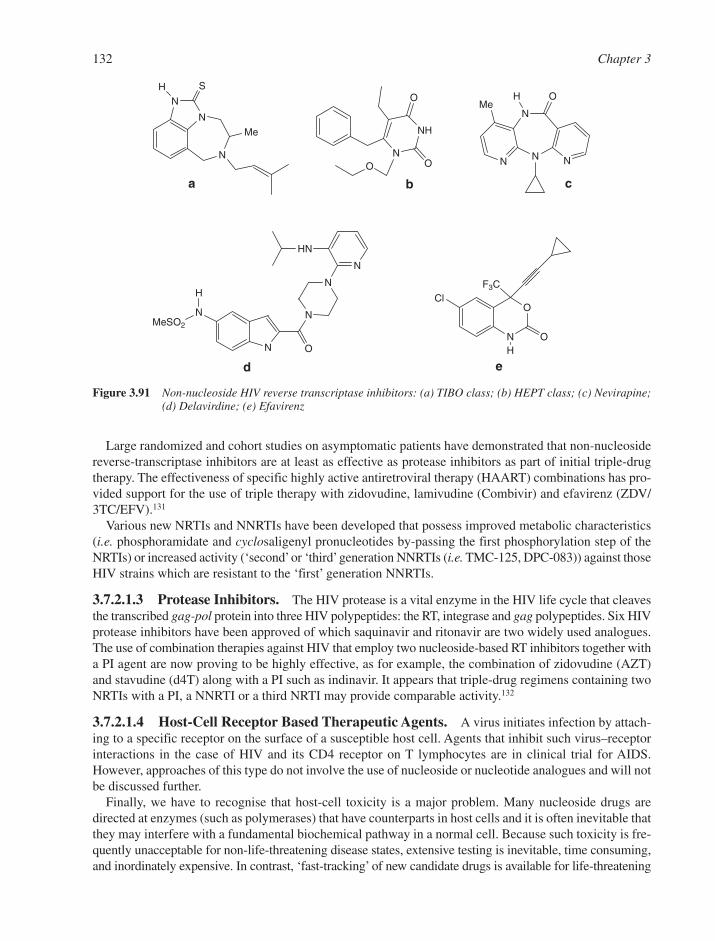
Large randomized and cohort studies on asymptomatic patients have demonstrated that non-nucleosidereverse-transcriptase inhibitors are at least as effective as protease inhibitors as part of initial triple-drugtherapy. The effectiveness of specific highly active antiretroviral therapy (HAART) combinations has pro-vided support for the use of triple therapy with zidovudine, lamivudine (Combivir) and efavirenz (ZDV/3TC/EFV).131
Various new NRTIs and NNRTIs have been developed that possess improved metabolic characteristics(i.e. phosphoramidate and cyclosaligenyl pronucleotides by-passing the first phosphorylation step of theNRTIs) or increased activity (‘second’or ‘third’generation NNRTIs (i.e. TMC-125, DPC-083)) against thoseHIV strains which are resistant to the ‘first’ generation NNRTIs.
3.7.2.1.3 Protease Inhibitors. The HIV protease is a vital enzyme in the HIV life cycle that cleavesthe transcribed gag-pol protein into three HIV polypeptides: the RT, integrase and gag polypeptides. Six HIVprotease inhibitors have been approved of which saquinavir and ritonavir are two widely used analogues.The use of combination therapies against HIV that employ two nucleoside-based RT inhibitors together witha PI agent are now proving to be highly effective, as for example, the combination of zidovudine (AZT)and stavudine (d4T) along with a PI such as indinavir. It appears that triple-drug regimens containing twoNRTIs with a PI, a NNRTI or a third NRTI may provide comparable activity.132
3.7.2.1.4 Host-Cell Receptor Based Therapeutic Agents. A virus initiates infection by attach-ing to a specific receptor on the surface of a susceptible host cell. Agents that inhibit such virus–receptorinteractions in the case of HIV and its CD4 receptor on T lymphocytes are in clinical trial for AIDS.However, approaches of this type do not involve the use of nucleoside or nucleotide analogues and will notbe discussed further.
Finally, we have to recognise that host-cell toxicity is a major problem. Many nucleoside drugs aredirected at enzymes (such as polymerases) that have counterparts in host cells and it is often inevitable thatthey may interfere with a fundamental biochemical pathway in a normal cell. Because such toxicity is fre-quently unacceptable for non-life-threatening disease states, extensive testing is inevitable, time consuming,and inordinately expensive. In contrast, ‘fast-tracking’ of new candidate drugs is available for life-threatening
132 Chapter 3
N N
N
N
H OMe
N
NH
O
OO
N
N
N
S
Me
H
N
N
H
MeSO2
O
N
N
N
HN
Cl
N
O
H
F3C
O
a b c
d e
Figure 3.91 Non-nucleoside HIV reverse transcriptase inhibitors: (a) TIBO class; (b) HEPT class; (c) Nevirapine;(d) Delavirdine; (e) Efavirenz

viral infections, such as HIV. For example, the anti-AIDS drug AZT (Figure 3.90) was marketed within ayear of the report of its in vitro properties.
3.7.2.2 Anti-DNA Virus Drug Design. Herpesviruses are double-stranded DNA viruses that causea variety of diseases in humans: cold sores, eye infections (keratitis), genital sores, chickenpox, shinglesand glandular fever (infectious mononucleosis). They all exhibit latency, which means that followinginfection of a cell, the virus produced can go into a latent state in nerve endings from where it can be reacti-vated by various stimuli (stress, UV light, other viral infections, etc.). Since it has so far been impossibleto destroy the virus in the latent state (i.e. prevent it from replicating), antiviral chemotherapy must bedirected first against primary infection and then against subsequent recurrent episodes.
Herpesviruses code for many enzymes involved in their own replication and metabolism. These are suf-ficiently different from the corresponding ones in the host cell to give an opportunity for selective inter-ference. For example, HSVs rely largely on the salvage pathway for the production of dTTP for DNAsynthesis and so the viruses encode their own thymidine kinase, TK. The specificity of the viral TK is notas great as that of the host cell and it phosphorylates a wide range of nucleoside analogues that, once acti-vated, inhibit viral replication. HSVs also code for their own DNA polymerase, which has a different speci-ficity from the cellular polymerases and hence presents a target for selective attack.
3.7.2.2.1 Acyclovir and Related Acyclonucleosides.133 Acyclovir (Figure 3.92) is effectiveagainst HSVs, and its metabolic conversion into the active form is remarkable. Although acyclovir is apurine nucleoside analogue lacking C-2� and C-3� of the sugar ring, it is specifically phosphorylated at theposition equivalent to the 5�-hydroxyl group by the thymidine kinase of the HSV. Not surprisingly, no suchmetabolism occurs in an uninfected cell. However, in the virally infected cell, 5�-phosphorylated acycloviris now recognised by the host-cell guanylate kinase and is taken to the diphosphate, from which a nucleo-side diphosphate kinase produces the 5�-triphosphate. This is now a substrate for the HSV-encoded DNApolymerase and it is incorporated into viral DNA. Since the analogue has no 3�-hydroxyl group, it is achain terminator and thus stops the synthesis of viral DNA (Figure 3.92).
Developments aimed at enhancing the oral bioavailability of acyclovir have resulted in the discovery ofvalaciclovir (Figure 3.93), the L-valyl ester of acyclovir. This acts as prodrug of the parent nucleoside and,as it has increased solubility, up to threefold higher plasma levels of acyclovir can be achieved.
The acyclonucleoside ganciclovir (Figure 3.93) is active against both HSV and cytomegalovirus (CMV)infections. In the latter case, the initial phosphorylation is carried out by a CMV-encoded protein kinase.
Nucleosides and Nucleotides 133
OO Gua
PO
PO
PO
O O O OOO
OHO Gua
OO Gua
PO
PO
O O OO
OO Gua
PO
O O
viral thymidine kinase
nucleosidediphosphate
kinase
acyclovir
GMPkinase
acyclovir triphosphate
Figure 3.92 Enzymatic phosphorylation of acyclovir
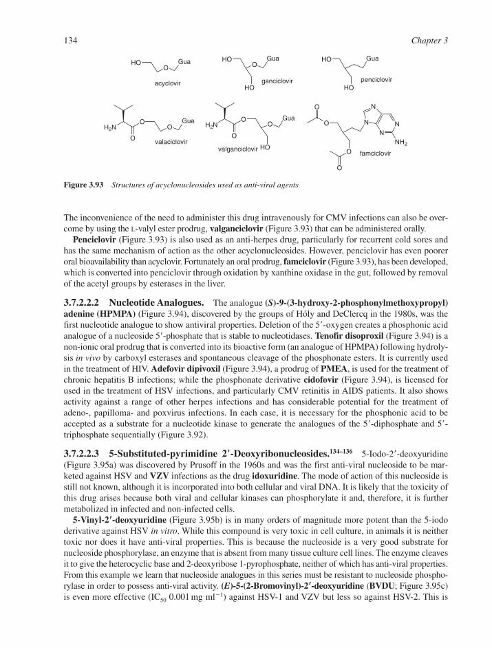
The inconvenience of the need to administer this drug intravenously for CMV infections can also be over-come by using the L-valyl ester prodrug, valganciclovir (Figure 3.93) that can be administered orally.
Penciclovir (Figure 3.93) is also used as an anti-herpes drug, particularly for recurrent cold sores andhas the same mechanism of action as the other acyclonucleosides. However, penciclovir has even pooreroral bioavailability than acyclovir. Fortunately an oral prodrug, famciclovir (Figure 3.93), has been developed,which is converted into penciclovir through oxidation by xanthine oxidase in the gut, followed by removalof the acetyl groups by esterases in the liver.
3.7.2.2.2 Nucleotide Analogues. The analogue (S)-9-(3-hydroxy-2-phosphonylmethoxypropyl)adenine (HPMPA) (Figure 3.94), discovered by the groups of Hóly and DeClercq in the 1980s, was thefirst nucleotide analogue to show antiviral properties. Deletion of the 5�-oxygen creates a phosphonic acidanalogue of a nucleoside 5�-phosphate that is stable to nucleotidases. Tenofir disoproxil (Figure 3.94) is anon-ionic oral prodrug that is converted into its bioactive form (an analogue of HPMPA) following hydroly-sis in vivo by carboxyl esterases and spontaneous cleavage of the phosphonate esters. It is currently usedin the treatment of HIV. Adefovir dipivoxil (Figure 3.94), a prodrug of PMEA, is used for the treatment ofchronic hepatitis B infections; while the phosphonate derivative cidofovir (Figure 3.94), is licensed forused in the treatment of HSV infections, and particularly CMV retinitis in AIDS patients. It also showsactivity against a range of other herpes infections and has considerable potential for the treatment ofadeno-, papilloma- and poxvirus infections. In each case, it is necessary for the phosphonic acid to beaccepted as a substrate for a nucleotide kinase to generate the analogues of the 5�-diphosphate and 5�-triphosphate sequentially (Figure 3.92).
3.7.2.2.3 5-Substituted-pyrimidine 2�-Deoxyribonucleosides.134–136 5-Iodo-2�-deoxyuridine(Figure 3.95a) was discovered by Prusoff in the 1960s and was the first anti-viral nucleoside to be mar-keted against HSV and VZV infections as the drug idoxuridine. The mode of action of this nucleoside isstill not known, although it is incorporated into both cellular and viral DNA. It is likely that the toxicity ofthis drug arises because both viral and cellular kinases can phosphorylate it and, therefore, it is furthermetabolized in infected and non-infected cells.
5-Vinyl-2��-deoxyuridine (Figure 3.95b) is in many orders of magnitude more potent than the 5-iododerivative against HSV in vitro. While this compound is very toxic in cell culture, in animals it is neithertoxic nor does it have anti-viral properties. This is because the nucleoside is a very good substrate fornucleoside phosphorylase, an enzyme that is absent from many tissue culture cell lines. The enzyme cleavesit to give the heterocyclic base and 2-deoxyribose 1-pyrophosphate, neither of which has anti-viral properties.From this example we learn that nucleoside analogues in this series must be resistant to nucleoside phospho-rylase in order to possess anti-viral activity. (E)-5-(2-Bromovinyl)-2��-deoxyuridine (BVDU; Figure 3.95c)is even more effective (IC50 0.001 mg ml�1) against HSV-1 and VZV but less so against HSV-2. This is
134 Chapter 3
HO Gua
HO
O
NN
N
N
NH2
O
O
O
OHO Gua
HO
OH2N
O
OGua O
Gua
HO
OH2N
O
HOO
Gua
valaciclovir
ganciclovir penciclovir
famciclovirvalganciclovir
acyclovir
Figure 3.93 Structures of acyclonucleosides used as anti-viral agents

because the nucleoside is a substrate for the viral thymidine kinase but not for the host-cell TK. This viralthymidine kinase is also a thymidylate kinase and produces the BVDU 5�-diphosphate, but only in HSV-1infected cells because diphosphate formation does not occur efficiently with the HSV-2 encoded enzyme.BVDU is a substrate for nucleoside phosphorylase but it sufficiently avoids degradation to show usefulclinical activity. The base (E)-5-(2-bromovinyl)uracil is an inhibitor for pyrimidine-5,6-dihydroreductase,the first enzyme in pyrimidine catabolism and thus can actually be salvaged and the 2�-deoxyribonucleosidecan be regenerated. BVDU triphosphate is an inhibitor of the virally encoded DNA polymerase. A nucle-oside phosphorylase-resistant analogue of BVDU (Figure 3.95d) has been described which has substan-tially greater activity in vivo because it has a much longer half-life in serum. The carbocyclic analogue ofBVDU (Figure 3.95e) also has significant activity.
3.7.2.2.4 Ribavirin. The broad spectrum anti-viral activity of ribavirin (1-�-D-ribofuranosyl-1,2,4-triazole-3-carboxamide; Figure 3.96) was first described in 1972.137 Since then, it has been studied in moreanimals and against more viruses than any other anti-viral agent. It is also apparently active in cell cultureagainst about 85% of all virus species studied and shows little or no cellular toxicity. Until recently its mainclinical use in the USA was in aerosol form against respiratory syncytial virus in young children. Very recentlyit has been approved as a combination treatment against chronic hepatitis C infections. It is also known tobe effective against Lassa fever and influenza and its potential for the treatment of severe acute respiratorysyndrome (SARS) is currently under investigation.
The mode of action of ribavirin is somewhat controversial. The most abundant form of ribavirin in cells is the triphosphate (Figure 3.96) and this was originally thought to inhibit inosine monophosphate
Nucleosides and Nucleotides 135
X
HO
HO
O
N
NH
OR
H
HH
H
BrH
H
BrH
H
BrH
(a) R = I, X = O
(b) R = X = O
(c) R = X = O
(d) R = X = S
(e) R = X = CH2
Figure 3.95 Anti-viral pyrimidine 5-substituted 2�-deoxyuridines
O
HO
HO
OH
N N
N
O
NH2
OP
OP
OP
O
O O O OOO
O
HO OH
N N
NCONH2
Figure 3.96 Ribavirin (left) and its triphosphate (right)
O
Ade
CH3
PO
O
OO
O
OOO
O
O
AdeP
O
O
Ot-Bu
O
OOt-Bu
O
O
CytP
HO
O
HOOH
O
AdeP
HO
O
HOOH
HPMPA tenofovir disoproxil adefovir dipivoxil cidofovir
Figure 3.94 Structures of acyclonucleoside phosphonates used as anti-viral agents
dehydrogenase, resulting in a depletion of cellular GTP pools. This in turn means that ribavirin 5�-triphosphateis an effective competitive inhibitor of the viral-specific RNA polymerase for some viruses. Ribavirin 5�-triphosphate is also known to inhibit the viral-specific mRNA-capping enzymes, guanyl transferase andN7-methyl transferase, so that viral protein synthesis is interrupted.
3.7.2.2.5 Phosphonoformic Acid. Phosphonoacetic acid (PAA, Figure 3.97a) was discovered tohave antiherpetic activity in vitro following random screening in 1973. Two years later it was shown to bea selective inhibitor of the virally encoded DNA polymerase, and the related phosphonoformic acid (PFA,Figure 3.97b) was subsequently found to be an even stronger inhibitor of this enzyme. PFA (foscarnet,Foscavir®) is used clinically for the treatment of CMV retinitis in AIDS patients. Both PAA and PFA areanalogues of pyrophosphate, a product of polymerases, and presumably bind to the corresponding site onthe enzyme and thus prevent replication. One problem with compounds of this sort is that they require noprior activation and therefore the difference in affinity between the virus-encoded and the host-cell poly-merase determines their effectiveness.
Hepatitis B virus has a partially double stranded DNA genome with an ORF that codes for a DNA poly-merase that is also an RT. It thus presents a very focused target for anti-viral nucleoside development. A wide range of nucleoside analogues have been used for HBV treatment (Table 3.2 and Figures 3.90 and3.94) illustrating the breadth of modification to the 2-deoxy-D-ribose that has been explored by chemicalsynthesis. They reveal that the DNA polymerase of HBV has a preference for the L- over the D-enantiomersof some dNTP analogues, an advantage enhanced by the fact that L-enantiomers are often less toxic andmore stable to metabolism than their D-counterparts.
There is still much to be done in this area of research. The rational design of novel inhibitors will con-tinue to rely on knowledge of the details of viral replication at the molecular level. The number of struc-tures of important virus-target enzymes will steadily rise. But success in designing analogues that areeffective inhibitors in vitro and then converting such knowledge into a useful drug still calls for intactdelivery of the drug at an effective concentration to the desired location and with minimum toxicity. Orallyactive forms of drugs are increasingly desirable for non-lethal infections.
REFERENCES
1. J. Kurreck, Antisense technologies – improvement through novel chemical modifications. Eur. J.Biochem., 2003, 270, 1628–1644.
2. S. Verma and F. Eckstein, Modified oligonucleotides: synthesis and strategy for users. Ann. Rev.Biochem., 1998, 67, 99–134.
3. B.E. Eaton and W.A. Pieken, Ribonucleosides and RNA. Ann. Rev. Biochem., 1995, 64, 837–863.4. E. Fischer and B. Helferich, Synthetische glucosine der purine. Chem. Ber., 1914, 47, 210–235.5. J. Davoll, A.R. Lythgoe and A.R. Todd, Experiments on the synthesis of purine nucleosides. Part XIX.
A synthesis of adenosine. J. Chem. Soc., 1948, 967–969.6. J. Davoll, A.R. Lythgoe and A.R. Todd, Experiments on the synthesis of purine nucleosides. Part XX.
A synthesis of guanosine. J. Chem. Soc., 1948, 1685–1687.7. J. Davoll and B.A. Lowy, A new synthesis of purine nucleosides. The synthesis of adenosine, guanosine
and 2,6-diamino-9-�-D-ribofuranosylpurine. J. Am. Chem. Soc., 1951, 73, 1650–1655.
136 Chapter 3
OP O
O O
O
OP
O O
O
O
a b
PAA PFA
Figure 3.97 Anti-viral analogues of pyrophosphate. (a) Phosphonoacetic acid; (b) Phosphonoformic acid
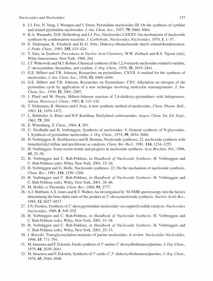
8. J.J. Fox, N. Yung, I. Wempen and J. Doerr, Pyrimidine nucleosides III. On the synthesis of cytidineand related pyrimidine nucleosides. J. Am. Chem. Soc., 1957, 79, 5060–5064.
9. K.A. Watanabe, D.H. Hollenberg and J.J. Fox, Nucleosides LXXXV. On mechanisms of nucleosidesynthesis by condensation reactions. J. Carbohydr., Nucleosides, Nucleotides, 1974, 1, 1–37.
10. E. Diekmann, K. Friedrich and H.-G. Fritz, Dideoxy-ribonucleoside durch schmelzkondensation.J. Prakt. Chem., 1993, 335, 415–424.
11. T. Sato, in Synthetic Procedures in Nucleic Acid Chemistry. W.W. Zorbach and R.S. Tipson (eds),Wiley Interscience, New York, 1968, 264.
12. J.T. Witkowski and M.J. Robins, Chemical synthesis of the 1,2,4-triazole nucleosides related to uridine,2�-deoxyuridine, thymidine, and cytidine. J. Org. Chem., 1970, 35, 2635–2641.
13. G.E. Hilbert and T.B. Johnson, Researches on pyrimidines. CXVII. A method for the synthesis ofnucleosides. J. Am. Chem. Soc., 1930, 52, 4489–4494.
14. G.E. Hilbert and T.B. Johnson, Researches on Pyrimidines. CXV. Alkylation on nitrogen of thepyrimidine cycle by application of a new technique involving molecular rearrangements. J. Am.Chem. Soc., 1930, 52, 2001–2007.
15. J. Pliml and M. Prysta, Hilbert-Johnson reaction of 2,4-dialkoxy-pyrimidines with halogenoses.Advan. Heterocycl. Chem., 1967, 8, 115–142.
16. T. Nishimura, B. Shimizu and I. Iwai, A new synthetic method of nucleosides. Chem. Pharm. Bull.,1963, 11, 1470–1472.
17. L. Birkhofer, A. Ritter and H.P. Kuelthau, Disilylated carboxamides. Angew. Chem. Int. Ed. Engl.,1963, 75, 209.
18. E. Wittenburg, Z. Chem., 1964, 4, 303.19. U. Niedballa and H. Vorbrüggen, Synthesis of nucleosides. 9. General synthesis of N-glycosides.
I. Synthesis of pyrimidine nucleosides. J. Org. Chem., 1974, 39, 3654–3660.20. H. Vorbrüggen, K. Krolikiewicz and B. Bennua, Nucleoside syntheses. 22. nucleoside synthesis with
trimethylsilyl triflate and perchlorate as catalysts. Chem. Ber. Recl., 1981, 114, 1234–1255.21. H. Vorbrüggen, Some recent trends and progress in nucleoside synthesis. Acta Biochim. Pol., 1996,
43, 25–36.22. H. Vorbrüggen and C. Ruh-Pohlenz, in Handbook of Nucleoside Synthesis. H. Vörbruggen and
C. Ruh-Pohlenz (eds), Wiley, New York, 2001, 15–24.23. H. Vorbrüggen and G. Hofle, Nucleoside syntheses. 23. On the mechanism of nucleoside synthesis.
Chem. Ber., 1981, 114, 1256–1268.24. H. Vorbrüggen and C. Ruh-Pohlenz, in Handbook of Nucleoside Synthesis. H. Vörbruggen and
C. Ruh-Pohlenz (eds), Wiley, New York, 2001, 38–46.25. M. Hoffer, �-Thymidin. Chem. Ber., 1960, 93, 2777.26. A.J. Hubbard, A.S. Jones and R.T. Walker, An investigation by 1H-NMR spectroscopy into the factors
determining the beta-alpha ratio of the product in 2�-deoxynucleoside synthesis. Nucleic Acids Res.,1984, 12, 6827–6837.
27. J.N. Freskos, Synthesis of 2�-deoxypyrimidine nucleosides via copper(I) iodide catalysis. NucleosidesNucleotides, 1989, 8, 549–555.
28. H. Vorbrüggen and C. Ruh-Pohlenz, in Handbook of Nucleoside Synthesis. H. Vörbruggen andC. Ruh-Pohlenz (eds), Wiley, New York, 2001, 33–38.
29. H. Vorbrüggen and C. Ruh-Pohlenz, in Handbook of Nucleoside Synthesis. H. Vörbruggen andC. Ruh-Pohlenz (eds), Wiley, New York, 2001, 25–33.
30. J. Boryski, Transglycosylation reactions of purine nucleosides. A review. Nucleosides Nucleotides,1996, 15, 771–791.
31. M. Imazawa and F. Eckstein, Facile synthesis of 2�-amino-2�-deoxyribofuranosylpurines. J. Org. Chem.,1979, 44, 2039–2041.
32. M. Imazawa and F. Eckstein, Synthesis of 3�-azido-2�,3�-dideoxyribofuranosylpurines. J. Org. Chem.,1978, 43, 3044–3048.
Nucleosides and Nucleotides 137
33. J.R. Hanrahan and D.W. Hutchinson, The enzymatic-synthesis of antiviral agents. J. Biotechnol.,1992, 23, 193–210.
34. D.W. Hutchinson, in Comprehensive Organic Chemistry, vol 5, D.H.R. Barton and W.D. Ollis (eds),Pergamon Press, Oxford, 1979, 105–145.
35. T.A. Krenitsky, J.L. Rideout, E.Y. Chao, G.W. Koszalka, F. Gurney, R.C. Crouch, N.K. Cohn,G. Wolberg and R. Vinegar, Imidazo[4,5-c]pyridines (3-deazapurines) and their nucleosides asimmunosuppressive and antiinflammatory agents. J. Med. Chem., 1986, 29, 138–143.
36. T. Utagawa, H. Morisawa, S. Yamanaka, A. Yamazaki and Y. Hirose, Enzymatic synthesis ofnucleoside antibiotics. 6. Enzymatic synthesis of virazole by purine nucleoside phosphorylase ofenterobacter-aerogenes Agr. Biol. Chem., 1986, 50, 121.
37. M.G. Stout, D.E. Hoard, M.J. Holman, E.S. Wu and J.M. Siegel, Preparation of 2�-deoxyribonucleo-sides via nucleoside deoxyribosyl transferase. Methods Carbohydr. Chem., 1976, 7, 19–29.
38. D. Betbeder, D.W. Hutchinson and A.O. Richards, The stereoselective enzymatic synthesis of9-�-D-2�-deoxyribofuranosyl 1-deazapurine. Nucleic Acids Res., 1989, 17, 4217–4222.
39. B.R. Baker, in The CIBA Foundation Symposium on the Chemistry and Biology of the Purines.G.E.W. Wolstenholme and C.M. O’Connor (eds), Churchill, London, 1957, 120.
40. R.A. Lessor and N.J. Leonard, Synthesis of 2�-deoxynucleosides by deoxygenation of ribonucleosides.J. Org. Chem., 1981, 46, 4300–4301.
41. M.J. Robins and J.S. Wilson, Nucleic-acid related compounds.32. Smooth and efficient deoxygenationof secondary alcohols – a general procedure for the conversion of ribonucleosides to 2�-deoxynucleo-sides. J. Am. Chem. Soc., 1981, 103, 932–933.
42. Z. Kazimierczuk, H.B. Cottam, G.R. Revankar and R.K. Robins, Synthesis of 2�-deoxytubercidin,2�-deoxyadenosine, and related 2�-deoxynucleosides via a novel direct stereospecific sodium-saltglycosylation procedure. J. Am. Chem. Soc., 1984, 106, 6379–6382.
43. F. Seela, Base-modified nucleosides and oligonucleotides: synthesis and application. Collect. Czech.Chem. Commun. Special Issue, 2002, 5, 1–15.
44. F. Seela, N. Ramzaeva and H. Rosemeyer, Houben–Weyl, Methods of Organic Synthesis. vol E9b,Thieme, Stuttgart, 1997, 304–550.
45. F. Seela, B. Westermann and U. Bindig, Liquid-liquid and solid-liquid phase-transfer glycosylationof pyrrolo 2,3-D-pyrimidines – stereospecific synthesis of 2-deoxy-�-D-ribofuranosides related to 2�-deoxy-7-carbaguanosine. J. Chem. Soc. Perkin Trans. 1, 1988, 697–702.
46. D.M. Brown and R.C. Ogden, A synthesis of pseudouridine. J. Chem. Soc., 1981, 723–725.47. B.A. Schweitzer and E.T. Kool, Aromatic non-polar nucleosides as hydrophobic isosteres of
pyrimidine and purine nucleosides. J. Org. Chem., 1994, 59, 7238–7242.48. L.A. Agrofoglio, I. Gillaizeau and Y. Saito, Palladium-assisted routes to nucleosides. Chem. Rev.,
2003, 103, 1875–1916.49. L.B. Townsend, Imidazole nucleosides and nucleotides. Chem. Rev., 1967, 67, 553–563.50. S.-I. Nagatsuka, T. Ohgi and T. Goto, Synthesis of wyosine (nucleoside Yt), a strongly fluorescent
nucleoside found in Torulopsis utilis tRNAPhe and 3-methylguanosine. Tetrahedron Lett., 1978, 29,2579–2582.
51. G. Shaw and R.N. Warrener, Synthesis of 2-thiouridine. Proc. Chem. Soc., 1957, 351.52. S. De Bernado and M. Weigele, C-Nucleoside antibiotics 2. Synthesis of oxazinomycin
(minimycin). J. Org. Chem., 1977, 42, 109–112.53. M.-I. Lim, R.S. Klein and J.J. Fox, Synthesis of the pyrrolo[3,2-d]pyrimidine C-nucleoside isostere
of inosine. Tetrahedron Lett., 1980, 21, 1013–1016.54. R. Noyori, T. Sato and Y. Hayakawa, A stereocontrolled general synthesis of C-nucleosides.
J. Am. Chem. Soc., 1978, 100, 2561–2563.55. F. Burlina, A. Favre, J.L. Fourrey and M. Thomas, An expeditious route to carbocyclic nucleosides:
(�)- aristeromycin and (�)-carbodine. Bioorg. Med. Chem. Lett., 1997, 7, 247–250.
138 Chapter 3

56. S. Ohira, T. Sawamoto and M. Yamato, Synthesis of (�)-neplanocin-a via CH insertion of alkyli-denecarbene. Tetrahedron Lett., 1995, 36, 1537–1538.
57. W.B. Choi, S. Yeola, D.C. Liotta, R.F. Schinazi, G.R. Painter, M. Davis, M. Stclair and P.A. Furman,Synthesis, anti-Human-Immunodeficiency-Virus, and anti-Hepatitis-B Virus activity of pyrimidineoxathiolane nucleosides. Bioorg. Med. Chem. Lett., 1993, 3, 693–696.
58. H. Gao and A.K. Mitra, Synthesis of acyclovir, ganciclovir and their prodrugs: A review. Synthesis,2000, 329–351.
59. M.J. Robins, in Nucleoside Analogues: Chemistry, Biology, and Medical Applications.R.T. Walker, E. DeClercq and F. Eckstein (eds), Plenum Press, New York, 1979, 165–192.
60. L.B. Townsend, in Nucleoside Analogues: Chemistry, Biology, and Medical Applications. R.T. Walker,E. DeClercq and F. Eckstein (eds), Plenum Press, New York, 1979, 193–223.
61. B. Luy and J.P. Marino, Measurement and application of 1H-19F dipolar couplings in the structuredetermination of 2�-fluorolabeled RNA. J. Biomol. NMR, 2001, 20, 39–47.
62. P. Bachert, Pharmacokinetics using fluorine NMR in vivo. Prog. Nucl. Magn. Reson. Spectrosc.,1998, 33, 1–56.
63. J.X. Yu, V.D. Kodibagkar, W.N. Cui and R.P. Mason, 19F: A versatile reporter for non-invasive phys-iology and pharmacology using magnetic resonance. Curr. Med. Chem., 2005, 12, 819–848.
64. S.E. Lee, A. Sidorov, T. Gourlain, N. Mignet, S.J. Thorpe, J.A. Brazier, M.J. Dickman, D.P. Hornby,J.A. Grasby and D.M. Williams, Enhancing the catalytic repertoire of nucleic acids: a systematicstudy of linker length and rigidity. Nucleic Acids Res., 2001, 29, 1565–1573.
65. K. Sakthivel and C.F. Barbas, Expanding the potential of DNA for binding and catalysis: Highlyfunctionalized dUTP derivatives that are substrates for thermostable DNA polymerases. Angew.Chem. Int. Ed-Engl., 1998, 37, 2872–2875.
66. J.L. Ruth, in Oligonucleotides and Analogues. F. Eckstein (ed), OUP, New York, 1991, 255–281.67. J.P. Anderson, B. Angerer and L.A. Loeb, Incorporation of reporter-labeled nucleotides by DNA
polymerases. Biotechniques, 2005, 38, 257–264.68. C. McGuigan, A. Brancale, G. Andrei, R. Snoeck, E. De Clercq and J. Balzarini, Novel bicyclic fura-
nopyrimidines with dual anti-VZV and -HCMV activity. Bioorg. Med. Chem. Lett., 2003, 13,4511–4513.
69. D.E. Bergstrom and J.L. Ruth, Preparation of C-5 mercurated pyrimidine nucleosides. J. Carbohydr.,Nucleosides, Nucleotides, 1977, 4, 257–269.
70. N. Ramzaeva and F. Seela, Duplex stability of 7-deazapurine DNA: oligonucleotides containing7-bromo- or 7-iodo-7-deazaguanine. Helv. Chim. Acta, 1996, 79, 1549–1558.
71. K.J. Divakar and C.B. Reese, 4-(1,2,4-triazol-1-yl) and 4-(3-nitro-1,2,4-triazol-1-yl)-1-(�-D-2,3,5-tri-O-acetylarabinofuranosyl)pyrimidin-2(1h)-ones – valuable intermediates in the synthesis of derivativesof 1-(�-D-arabinofuranosyl)cytosine (Ara-C). J. Chem. Soc.-Perkin Trans., 1, 1982, 1171–1176.
72. K.W. Pankiewicz, Fluorinated nucleosides. Carbohydr. Res., 2000, 327, 87–105.73. B.A. Connolly, in Oligonucleotides and Analogues. F. Eckstein (ed), OUP, New York, 1991, 155–183.74. F. Nagatsugi, K. Uemura, S. Nakashima, M. Maeda and S. Sasaki, 2-aminopurine derivatives
with C6-substituted olefin as novel cross-linking agents and the synthesis of the corresponding �-phosphoramidite precursors. Tetrahedron, 1997, 53, 3035–3044.
75. B.L. Gaffney and R.A. Jones, Synthesis of O6-alkylated deoxyguanosine nucleosides. TetrahedronLett., 1982, 23, 2253–2256.
76. P.P. Kung and R.A. Jones, One-flask syntheses of 6-thioguanosine and 2�-deoxy-6-thioguanosine.Tetrahedron Lett., 1991, 32, 3919–3922.
77. B.S. Sproat and A.I. Lamond, in Oligonucleotides and Analogues. F. Eckstein (ed), OUP, New York,1991, 49–86.
78. J.J. Fox, Pyrimidine nucleoside transformations via anhydronucleosides. Pure Appl. Chem., 1969,18, 223–255.
Nucleosides and Nucleotides 139

79. D.B. Olsen, F. Benseler, H. Aurup, W.A. Pieken and F. Eckstein, Study of a hammerhead ribozymecontaining 2�-modified adenosine residues. Biochemistry, 1991, 30, 9735–9741.
80. F. Benseler, D.M. Williams and F. Eckstein, Synthesis of suitably-protected phosphoramidites of2�-fluoro-2�-deoxyguanosine and 2�-amino-2�-deoxyguanosine for incorporation into oligoribonu-cleotides. Nucleosides Nucleotides, 1992, 11, 1333–1351.
81. A.V.R. Rao, M.K. Gurjar and S.V.S. Lalitha, Discovery of a Novel Route to �-thymidine – aprecursor for anti-AIDS compounds. J. Chem. Soc. Chem. Commun., 1994, 1255–1256.
82. L. Beigelman, P. Haeberli, D. Sweedler and A. Karpeisky, Improved synthetic approaches toward 2�-O-methyl-adenosine and guanosine and their N-acyl derivatives. Tetrahedron, 2000, 56, 1047–1056.
83. M.J. Robins, J.S. Wilson, D. Madej, N.H. Low, F. Hansske and S.F. Wnuk, Nucleic-acid relatedcompounds.88. Efficient conversions of ribonucleosides into their 2�,3�-anhydro, 2�-(and 3�)-deoxy,2�,3�-didehydro-2�,3�-dideoxy, and 2�,3�-dideoxynucleoside analogs. J. Org. Chem., 1995, 60,7902–7908.
84. G.R.J. Thatcher and R. Kluger, Mechanism and catalysis of nucleophilic substitution in phosphateesters. Adv. Phys. Org. Chem., 1989, 25, 99–265.
85. E.V. Anslyn and D.M. Perreault, Unifying the current data on the mechanism of cleavage-transesterification of RNA. Angew. Chem. Int. Ed. Engl., 1997, 36, 432–450.
86. M. Oivanen, S. Kuusela and H. Lönnberg, Kinetics and mechanisms for the cleavage and isomerisation of the phosphodiester bonds of RNA by Brønsted acids and bases. Chem. Rev., 1988, 98, 961–990.
87. A.C. Hengge, in Comprehensive Biological Catalysis: A Mechanistic Reference, vol 1. M. Sinnott(ed), Academic Press, New York, 1988, 517–542.
88. D.M. Brown, in Methods in Molecular Biology, vol 20. S. Agarwal (ed), Humana Press Inc., Totowa,NJ, 1993, 1–17.
89. C.B. Reese, The chemical synthesis of oligo- and poly-nucleotides by the phosphotriester approach.Tetrahedron, 1978, 34, 3143–3179.
90. L.A. Slotin, Current methods of phosphorylation of biological molecules. Synthesis, 1975, 11,737–752.
91. S.L. Beaucage and R.P. Iyer, Advances in the synthesis of oligonucleotides by the phosphoramiditeapproach. Tetrahedron, 1992, 48, 2223–2311.
92. J. Stawinski and A. Kraszewski, How to get the most out of two phosphorus chemistries studies on H-phosphonates. Acc. Chem. Res., 2002, 35, 952–960.
93. S. Narang, DNA synthesis. Tetrahedron, 1983, 39, 3–22.94. C.B. Reese, The chemical synthesis of oligo- and poly-nucleotides: a personal commentary.
Tetrahedron, 2002, 58, 8893–8920.95. M.D. Matteucci and M.H. Caruthers, Nucleotide chemistry.4. Synthesis of deoxyoligonucleotides
on a polymer support. J. Am. Chem. Soc., 1981, 103, 3185–3191.96. S.L. Beaucage and M.H. Caruthers, Deoxynucleoside phosphoramidites – a new class of key
intermediates for deoxypolynucleotide synthesis. Tetrahedron Lett., 1981, 22, 1859–1862.97. W.J. Stec, G. Zon, W. Egan and B. Stec, Automated solid-phase synthesis, separation, and
stereochemistry of phosphorothioate analogs of oligodeoxyribonucleotides. J. Am. Chem. Soc.,1984, 106, 6077–6079.
98. M. Yoshikawa, T. Kato and T. Takenishi, Studies of phosphorylation. III. Selective phosphorylationof unprotected nucleosides. Bull. Chem. Soc. Jpn., 1969, 42, 3505–3508.
99. A. Arabshahi and P.A. Frey, A simplified procedure for synthesizing nucleoside 1-thiotriphosphates –dATP(�)S, dGTP(�)S, UTP(�)S, and dTTP(�)S. Biochem. Biophys. Res. Commun., 1994, 204,150–155.
100. T. Sowa and S. Ouchi, Facile synthesis of 5�-nucleotides by the selective phosphorylation of aprimary hydroxyl group of nucleosides with phosphoryl chloride. Bull. Chem. Soc. Jpn., 1975, 48,2084–2090.
140 Chapter 3
101. V.J. Davisson, D.R. Davis, V.M. Dixit and C.D. Poulter, Synthesis of nucleotide 5�-diphosphatesfrom 5�-O-tosyl nucleosides. J. Org. Chem., 1987, 52, 1794–1801.
102. G.M. Blackburn, D.E. Kent and F. Kolkmann, The synthesis and metal-binding characteristics ofnovel, isopolar phosphonate analogs of nucleotides. J. Chem. Soc.-Perkin Trans., 1, 1984, 1119–1125.
103. T.C. Myers, K. Nakamura and J.W. Flesher, Phosphonic acid analogs of nucleoside phosphates.I. The synthesis of 5�-adenylyl methylenediphosphonate, a phosphonic acid analog of adenosine.J. Am. Chem. Soc., 1963, 85, 3292–3295.
104. K.-H. Scheit, in Nucleotide Analogues. Synthesis and Biological Function. K.-H. Scheit (ed), WileyInterscience, New York, 1980, 96–141.
105. K.-H. Scheit, in Nucleotide Analogues. Synthesis and Biological Function. K.-H. Scheit (ed), WileyInterscience, New York, 1980, 195–218.
106. K. Burgess and D. Cook, Syntheses of nucleoside triphosphates. Chem. Rev., 2000, 100, 2047–2059.107. J. Ludwig, in Biophosphates and Their Analogues – Synthesis, Structure, Metabolism and Activity.
K.S. Bruzik and W.J. Stec (eds), Elsevier, Amsterdam, 1987, 131–133.108. J. Ludwig and F. Eckstein, Rapid and efficient synthesis of nucleoside 5�-O-(1-thiotriphosphates),
5�-triphosphates and 2�,3�-cyclophosphorothioates using 2-chloro-4H-1,3,2-benzodioxaphosphorin-4-one. J. Org. Chem., 1989, 54, 631–635.
109. J. Ludwig and F. Eckstein, Stereospecific synthesis of guanosine 5�-O-(1,2-dithiotriphosphates).J. Org. Chem., 1991, 56, 5860–5865.
110. J. Ludwig and F. Eckstein, Synthesis of nucleoside 5�-O-(1,3-dithiotriphosphates) and 5�-O-(1,1-dithiotriphosphates). J. Org. Chem., 1991, 56, 1777–1783.
111. B.R. Shaw, M. Dobrikov, X. Wang, J. Wan, K.Z. He, J.L. Lin, P. Li, V. Rait, Z.A. Sergueeva and D. Sergueev, Therapeutic Oligonucleotides, S. Cho-chung, A.M. Gewirtz and C.A. Stein (ed) vol1002. New York Academy of Science, New York, 2003, 12–29.
112. F. Eckstein, Nucleoside phosphorothioates. Ann. Rev. Biochem., 1985, 54, 367–402.113. F. Eckstein and J.B. Thomson, DNA Replication, vol 262. Academic Press, San Diego, 1995, 189–202.114. J.R. Knowles, Enzyme catalysed phosphoryl transfer reactions. Ann. Rev. Biochem., 1980, 49,
877–919.115. G.M. Blackburn, G.E. Taylor, G.R.J. Thatcher, M. Prescott and A.G. McLennan, Synthesis and
resistance to enzymatic hydrolysis of stereochemically-defined phosphonate and thiophosphateanalogs of P1,P4-bis(5�-adenosyl) tetraphosphate. Nucleic Acids Res., 1987, 15, 6991–7004.
116. N.B. Tarussova, T.I. Osipova, P.P. Purygin and I.A. Yakimova, The synthesis of P1, P3-bis(5�-adenosyl)triphosphate, P1,P4-bis(5�-adenosyl)tetraphosphate and its phosphonate analog with the use of carbonyl derivatives of nitrogen-containing heterocycles. Bioorg. Khim.,1986, 12, 404–407.
117. G.M. Blackburn, F. Eckstein, D.E. Kent and T.D. Perrée, Isopolar vs isosteric phosphonate analogsof nucleotides. Nucleosides Nucleotides, 1985, 4, 165–167.
118. J.G. Moffatt and H.G. Khorana, The total synthesis of coenzyme A. J. Am. Chem. Soc., 1961, 83,663–675.
119. M. Sekine, S. Nishiyama, T. Kamimura, Y. Osaki and T. Hata, Chemical synthesis of capped oligori-bonucleotides, m7g5�pppAUG and m7g5�pppAUGCC. Bull. Chem. Soc. Jpn., 1985, 58, 850–860.
120. R.I. Christopherson, S.D. Lyons and P.K. Wilson, Inhibitors of de novo nucleotide biosynthesis asdrugs. Acc. Chem. Res., 2002, 35, 961–971.
121. J.M. Berg, J.L. Tymoczko and L. Stryer, in Biochemistry, 5th edn. J.M. Berg, J.L. Tymoczko andL. Stryer (eds), Freeman, New York, 2002, 693–714.
122. P.M.J. Burgers, E.V. Koonin, E. Bruford, L. Blanco, K.C. Burtis, M.F. Christman, W.C. Copeland,E.C. Friedberg, F. Hanaoka, D.C. Hinkle et al., Eukaryotic DNA polymerases: Proposal for a revisednomenclature. J. Biol. Chem., 2001, 276, 43487–43490.
123. C.M. Galamarini, J.R. Mackey and C. Dumontet, Nucleoside analogues and nucleobases in cancertreatment. Lancet Oncol., 2002, 3, 415–424.
Nucleosides and Nucleotides 141

124. S. Miura and S. Izuta, DNA polymerases as targets of anticancer nucleosides. Curr. Drug Targets,2004, 5, 191–195.
125. I.M. Kompis, K. Islam and R.L. Then, DNA and RNA synthesis: Antifolates. Chem. Rev., 2005, 105,593–620.
126. D.R. Newell, How to develop a successful cancer drug – molecules to medicines or targets totreatments? Eur. J. Cancer, 2005, 41, 676–682.
127. S.L. Gerson, Clinical relevance of MGMT in the treatment of cancer. J. Clin. Oncol., 2002, 20,2388–2399.
128. E. DeClercq, Antiviral drugs in current clinical use. J. Clin. Virol., 2004, 30, 115–133.129. J.S. Copperwood, G. Gumina, F.D. Boudinot and C.K. Chu, in Recent Advances in Nucleosides:
Chemistry and Chemotherapy. C.K. Chu (ed), Elsevier, Amsterdam, 2002, 91–147.130. S.G. Sarafianos, K. Das, A.D. Clark, J.P. Ding, P.L. Boyer, S.H. Hughes and E. Arnold, Lamivudine
(3TC) resistance in HIV-1 reverse transcriptase involves steric hindrance with beta-branched amino acids. Proc. Natl. Acad. Sci. USA, 1999, 96, 10027–10032.
131. E. De Clercq, Non-nucleoside reverse transcriptase inhibitors (NNRTIs): Past, present, and future.Chem. Biodivers., 2004, 1, 44–64.
132. C. Orkin, J. Stebbing, M. Nelson, M. Bower, M. Johnson, S. Mandalia, R. Jones, G. Moyle,M. Fisher and B. Gazzard, A randomized study comparing a three- and four-drug HAART regimenin first-line therapy (QUAD study). J. Antimicrob. Chemother., 2005, 55, 246–251.
133. J.A. Bartlett, R. DeMasi, J. Quinn, C. Moxham and F. Rousseau, Overview of the effectiveness oftriple combination therapy in antiretroviral-naive HIV-1 infected adults. AIDS, 2001, 15, 1369–1377.
134. A. Hóly, in Recent Advances in Nucleosides: Chemistry and Chemotherapy. C.K. Chu (ed), Elsevier,Amsterdam, 2002, 167–238.
135. E. DeClercq and R.T. Walker, Synthesis and antiviral properties of 5-vinylpyrimidine nucleosideanalogs. Pharmacol. Ther., 1984, 26, 1–44.
136. E. DeClercq, Recent Advances in Nucleosides: Chemistry and Chemotherapy. Elsevier, Amsterdam,2002, 433–454.
137. R.W. Sidwell, Jt. Witkowsk, L.B. Allen, R.K. Robins, G.P. Khare and J.H. Huffman, Broad-spectrum antiviral activity of virazole – 1-�-D-ribofuranosyl-1,2,4-triazole-3-carboxamide. Science,1972, 177, 705–706.
138. N.H. Williams and P. Wyman, Base catalysed phosphate diester hydrolysis. Chem. Commun., 2001,1268–1269.
139. C. Lad, N.H. Williams and R. Wolfenden, The rate of hydrolysis of phosphomonoester dianions andthe exceptional catalytic proficiencies of protein and inositol phosphatases. Proc. Natl. Acad. Sci.USA, 2003, 100, 5607–5610.
140. P.W. Barnard, C.A. Bunton, D.R. Llewellyn, C.A. Vernon and V.A. Welch, The reactions of organicphosphates. Part IV. Oxygen exchange between water and orthophosphoric acid. J. Chem. Soc.,1961, 2670–2676.
141. A.J. Kirby and M. Younas, Reactivity of phosphate esters – diester hydrolysis. J. Chem. Soc. B, 1970,510–513.
142. E.T. Kaiser and K. Kudo, Alkaline hydrolysis of aromatic esters of phosphoric acid. J. Amer. Chem.Soc., 1967, 89, 6725–6728.
143. A.J. Kirby and A.G. Varvoglis, The reactivity of phosphate esters. Monoester hydrolysis. J. Am.Chem. Soc., 1967, 89, 415–423.
144. J. Kumamoto, J.R. Cox and F.H. Westheimer, Barium ethylene phosphate. J. Am. Chem. Soc., 1956,78, 4858–4860.
142 Chapter 3
CHAPTER 4
Synthesis of Oligonucleotides
CONTENTS
4.1 Synthesis of Oligodeoxyribonucleotides 1434.1.1 Overall Strategy for Chemical Synthesis 1444.1.2 Protected 2�-Deoxyribonucleoside Units 1444.1.3 Ways of Making an Internucleotide Bond 1474.1.4 Solid-Phase Synthesis 150
4.2 Synthesis of Oligoribonucleotides 1534.2.1 Protected Ribonucleoside Units 1544.2.2 Oligoribonucleotide Synthesis 155
4.3 Enzymatic Synthesis of Oligonucleotides 1564.3.1 Enzymatic Synthesis of Oligodeoxyribonucleotides 1564.3.2 Enzymatic Synthesis of Oligoribonucleotides 157
4.4 Synthesis of Modified Oligonucleotides 1584.4.1 Modified Nucleobases 1584.4.2 Modifications of the 5�- and 3�-Termini 1594.4.3 Backbone and Sugar Modifications 160References 165
4.1 SYNTHESIS OF OLIGODEOXYRIBONUCLEOTIDES
An oligonucleotide is a single-stranded chain consisting of a number of nucleoside units linked togetherby phosphodiester bridges. Generally in oligonucleotide synthesis, phosphodiesters are formed between a 3�-hydroxyl group bearing a phosphate derivative and a 5�-hydroxyl group of another nucleoside(Section 4.1.4). In the context of nucleic acids, the prefix ‘oligo’ is usually taken to denote a few nucleo-side residues, while the prefix ‘poly’ means many. However, it has become a common practice to refer toall chemically synthesised nucleic acid chains as oligonucleotides, even if they are in excess of 100residues in length. The term polynucleotide is more often taken to mean single-stranded nucleic acids ofless-defined length and sequence, often obtained by a polymerisation reaction, for example, polycytidylicacid, polyC.
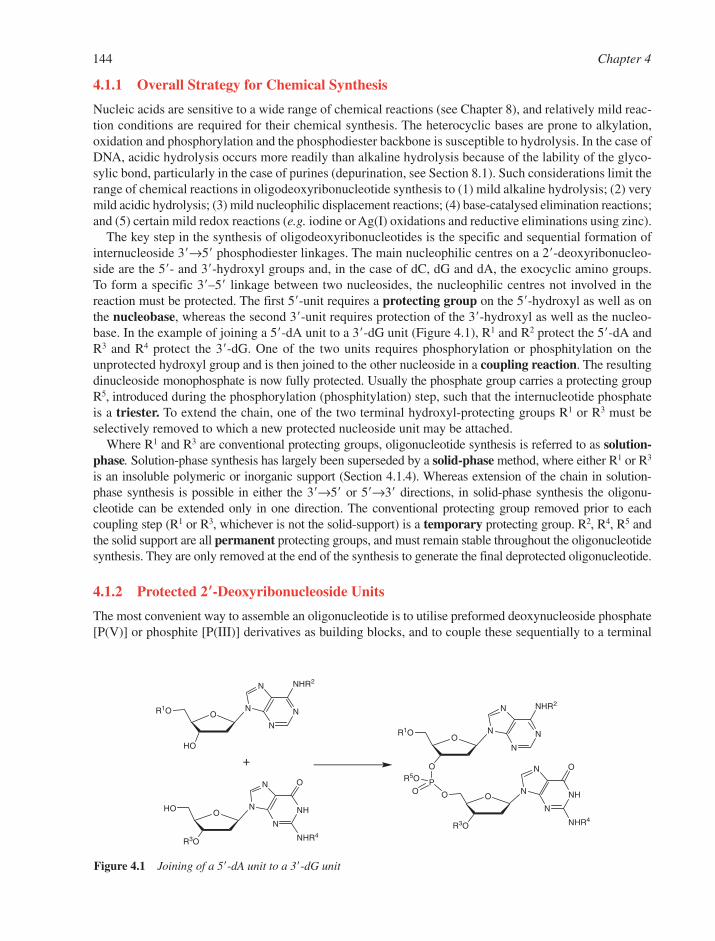
144 Chapter 4
4.1.1 Overall Strategy for Chemical Synthesis
Nucleic acids are sensitive to a wide range of chemical reactions (see Chapter 8), and relatively mild reac-tion conditions are required for their chemical synthesis. The heterocyclic bases are prone to alkylation,oxidation and phosphorylation and the phosphodiester backbone is susceptible to hydrolysis. In the case ofDNA, acidic hydrolysis occurs more readily than alkaline hydrolysis because of the lability of the glyco-sylic bond, particularly in the case of purines (depurination, see Section 8.1). Such considerations limit therange of chemical reactions in oligodeoxyribonucleotide synthesis to (1) mild alkaline hydrolysis; (2) verymild acidic hydrolysis; (3) mild nucleophilic displacement reactions; (4) base-catalysed elimination reactions;and (5) certain mild redox reactions (e.g. iodine or Ag(I) oxidations and reductive eliminations using zinc).
The key step in the synthesis of oligodeoxyribonucleotides is the specific and sequential formation ofinternucleoside 3�→5� phosphodiester linkages. The main nucleophilic centres on a 2�-deoxyribonucleo-side are the 5�- and 3�-hydroxyl groups and, in the case of dC, dG and dA, the exocyclic amino groups.To form a specific 3�–5� linkage between two nucleosides, the nucleophilic centres not involved in thereaction must be protected. The first 5�-unit requires a protecting group on the 5�-hydroxyl as well as onthe nucleobase, whereas the second 3�-unit requires protection of the 3�-hydroxyl as well as the nucleo-base. In the example of joining a 5�-dA unit to a 3�-dG unit (Figure 4.1), R1 and R2 protect the 5�-dA andR3 and R4 protect the 3�-dG. One of the two units requires phosphorylation or phosphitylation on theunprotected hydroxyl group and is then joined to the other nucleoside in a coupling reaction. The resultingdinucleoside monophosphate is now fully protected. Usually the phosphate group carries a protecting groupR5, introduced during the phosphorylation (phosphitylation) step, such that the internucleotide phosphateis a triester. To extend the chain, one of the two terminal hydroxyl-protecting groups R1 or R3 must beselectively removed to which a new protected nucleoside unit may be attached.
Where R1 and R3 are conventional protecting groups, oligonucleotide synthesis is referred to as solution-phase. Solution-phase synthesis has largely been superseded by a solid-phase method, where either R1 or R3
is an insoluble polymeric or inorganic support (Section 4.1.4). Whereas extension of the chain in solution-phase synthesis is possible in either the 3�→5� or 5�→3� directions, in solid-phase synthesis the oligonu-cleotide can be extended only in one direction. The conventional protecting group removed prior to eachcoupling step (R1 or R3, whichever is not the solid-support) is a temporary protecting group. R2, R4, R5 andthe solid support are all permanent protecting groups, and must remain stable throughout the oligonucleotidesynthesis. They are only removed at the end of the synthesis to generate the final deprotected oligonucleotide.
4.1.2 Protected 2��-Deoxyribonucleoside Units
The most convenient way to assemble an oligonucleotide is to utilise preformed deoxynucleoside phosphate[P(V)] or phosphite [P(III)] derivatives as building blocks, and to couple these sequentially to a terminal
O
O
R1O
O
R3O
O
PR5O
O
N
N
N
N
NHR2
N
NH
N
N
O
NHR4
O
HO
R1O
N
N
N
N
NHR2
O
R3O
HO NH
N
N
O
NHR4
N
+
Figure 4.1 Joining of a 5�-dA unit to a 3�-dG unit

Synthesis of Oligonucleotides 145
nucleoside attached to a solid support. Since the 5�-hydroxyl group is a more effective nucleophile thanthe secondary 3�-hydroxyl group, the phosphate/phosphite group is best placed on the 3�-position. To achievethis selectively it is necessary to protect the nucleobase exocyclic amino groups and the 5�-hydroxyl group.
4.1.2.1 Nucleobases. Permanent protecting groups for the exocyclic amino groups of adenine, cyto-sine and guanine have been used for many years in oligonucleotide synthesis.1 Acyl protecting groups werechosen, since they are stable for long periods during mildly basic and acidic conditions used duringoligonucleotide synthesis, and are removed with concentrated ammonia at the end of the synthesis(Section 4.1.4). The benzoyl group is used to protect both adenine and cytosine, while isobutyryl is usedto protect guanine (Figure 4.2). Thymidine does not require protection since it does not have an exocyclicamino group. While these acyl protecting groups are still suitable for oligonucleotide synthesis today, newchemistries and new nucleoside building blocks have been introduced, which require milder deprotectionconditions at the end of the synthesis. For example, a matched set of phenoxyacetyl (PAC) for dA, isopropylphenoxyacetyl for dG and acetyl for dC can be removed by treatment with 0.05 M potassium car-bonate in methanol at room temperature within a few hours.
When nucleosides are prepared for incorporation into oligonucleotides, it is usual to protect nucleobaseexocyclic amino groups first. There are two common methods for the synthesis of acylated nucleosides,per-acylation and transient protection (Figure 4.3). The per-acylation method involves use of an excess ofacylating agent such that the hydroxyl groups and the exocyclic amino groups are each acylated (bis-acylatedin the case of the amino groups), and then the hydroxylic and one of the amino acyl groups are removedselectively under mild basic conditions. The selectivity arises because of the greater stability of amidescompared to esters (and bis-amides) at high pH. In the transient protection route, the nucleoside is treatedwith trimethylsilyl chloride (TMSCl), which reacts selectively with the hydroxyl groups. Treatment withbenzoyl chloride is then selective for the exocyclic amino group (again the bis-acylated product may be formed). The silyl protecting groups are removed under basic conditions to give the desired N6-benzoyl-2�-deoxyadenosine. This method may also be used for protection of 2�-deoxycytidine.
In the case of 2�-deoxyguanosine protection, the reaction may be carried out with isobutyric anhydride byeither per-acylation or the transient protection route. However, in the case of dG, the O6-position is susceptibleto reaction under certain conditions, particularly with coupling agents and phosphorylating agents, or inthe synthesis of G-rich oligonucleotides. Under these conditions it is necessary to protect the O6-positionusing alkyl or aryl protecting groups. However, such protection is not necessary in the case of the phos-phoramidite method (Section 4.1.3). Another common protecting group for dG is the dimethylformami-dine group that is readily introduced using dimethylformamide dimethylacetal.
4.1.2.2 5�-Hydroxyl Group. By far the most common protecting group for the 5�-hydroxyl group is the4,4�-dimethoxytrityl group (DMT) (Figure 4.4). The DMT group is readily removed under acidic conditions.It is introduced onto the 5�-hydroxyl group of N-acylated nucleosides with DMT–Cl in the presence of a basesuch as pyridine or 4-dimethylaminopyridine. Reaction occurs principally at the 5�-hydroxyl rather than atthe secondary 3�-hydroxyl group because of steric effects. The DMT group is removed during oligonucleotide
O
HO
HON
N
O
HN
O
Ph
O
HO
HON
NH
N
N
O
HN
OO
HO
HO
N
N
N
N
HN
O
Ph
dABz dGiB dCBz
Figure 4.2 Common protecting groups for the heterocyclic bases of dA, dG and dC

146 Chapter 4
synthesis with either dichloroacetic acid or trichloroacetic acid in non-aqueous solvent, conditions thatprevent other side reactions, such as depurination. During deprotection, the bright orange-red DMT cationis liberated and is used as a measure of the yield of coupling of that nucleoside unit (Section 4.1.4).
4.1.2.3 Introduction of Phosphate. In the original chemistry developed by Khorana and co-workers(phosphodiester, Section 4.1.3), deoxynucleoside 5�-phosphates were used as building blocks. In otherchemistries developed more recently 5�-O-dimethoxytrityl-(N-acylated)-2�-deoxynucleosides are phosphor-ylated or phosphitylated at the 3�-hydroxyl group (Figure 4.5). In these cases the products of synthesis afterassembly of the oligonucleotide are phosphate triesters, where the internucleoside phosphate carries a pro-tecting group. In phosphotriester chemistry [P(V)] the best protecting groups are aryl (usually mono- ordi-chlorophenyl derivatives). This is because an aryl phosphodiester is a much more reactive deoxynucleo-side building block than an alkyl phosphodiester in a coupling reaction. For example, 5�-O-dimethoxytrityl-N6-benzoyl-2�-deoxyadenosine gives the corresponding 3�-O-(2-chlorophenyl) phosphodiester by reaction
OMe
MeO
Figure 4.4 The 4,4�-dimethoxyphenylmethyl (dimethoxytrityl, DMT) group
N
N
N
N NHBz
O
HO
HO
BzCl
BzCl
TMSCl
NaOH
NH4OH
N
N
N
N NBz2
O
BzO
BzO
Route APer-acylation
Route BTransient protection
N
N
N
N NH2
O
HO
HO
N
N
N
N NH2
O
TMSO
TMSO N
N
N
N NBz2
O
TMSO
TMSO
Figure 4.3 Routes to N6-benzoyl-2�-deoxyadenosine
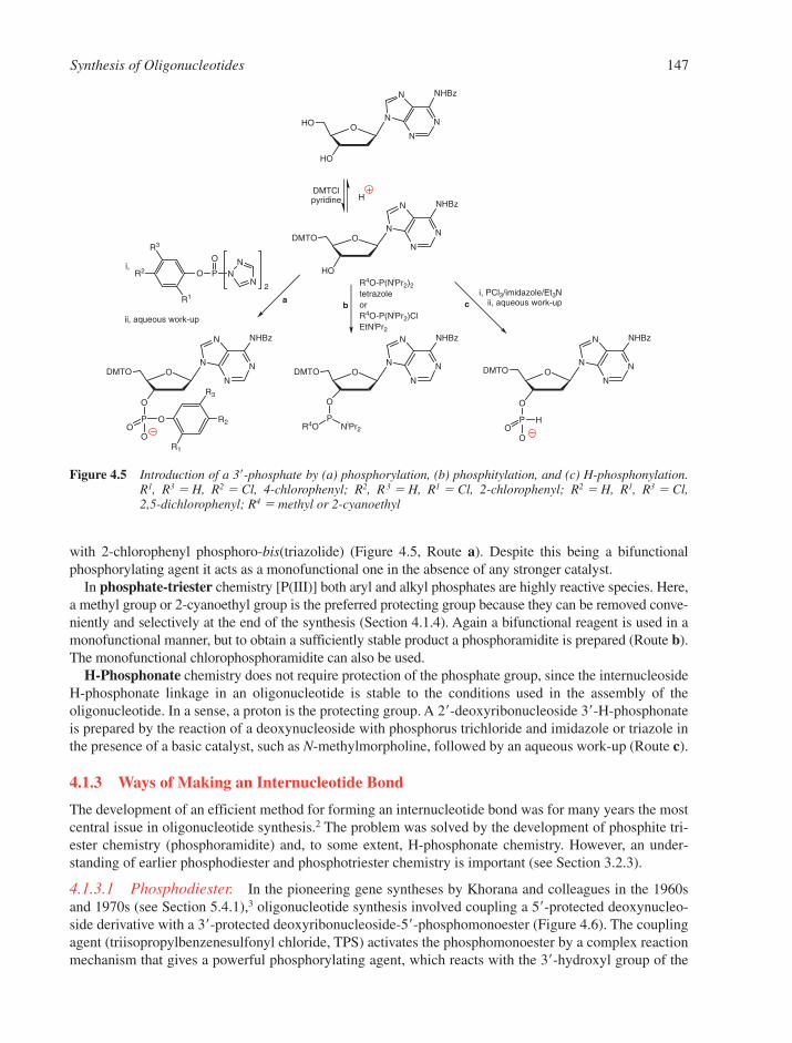
Synthesis of Oligonucleotides 147
with 2-chlorophenyl phosphoro-bis(triazolide) (Figure 4.5, Route a). Despite this being a bifunctionalphosphorylating agent it acts as a monofunctional one in the absence of any stronger catalyst.
In phosphate-triester chemistry [P(III)] both aryl and alkyl phosphates are highly reactive species. Here,a methyl group or 2-cyanoethyl group is the preferred protecting group because they can be removed conve-niently and selectively at the end of the synthesis (Section 4.1.4). Again a bifunctional reagent is used in amonofunctional manner, but to obtain a sufficiently stable product a phosphoramidite is prepared (Route b).The monofunctional chlorophosphoramidite can also be used.
H-Phosphonate chemistry does not require protection of the phosphate group, since the internucleosideH-phosphonate linkage in an oligonucleotide is stable to the conditions used in the assembly of theoligonucleotide. In a sense, a proton is the protecting group. A 2�-deoxyribonucleoside 3�-H-phosphonateis prepared by the reaction of a deoxynucleoside with phosphorus trichloride and imidazole or triazole inthe presence of a basic catalyst, such as N-methylmorpholine, followed by an aqueous work-up (Route c).
4.1.3 Ways of Making an Internucleotide Bond
The development of an efficient method for forming an internucleotide bond was for many years the mostcentral issue in oligonucleotide synthesis.2 The problem was solved by the development of phosphite tri-ester chemistry (phosphoramidite) and, to some extent, H-phosphonate chemistry. However, an under-standing of earlier phosphodiester and phosphotriester chemistry is important (see Section 3.2.3).
4.1.3.1 Phosphodiester. In the pioneering gene syntheses by Khorana and colleagues in the 1960sand 1970s (see Section 5.4.1),3 oligonucleotide synthesis involved coupling a 5�-protected deoxynucleo-side derivative with a 3�-protected deoxyribonucleoside-5�-phosphomonoester (Figure 4.6). The couplingagent (triisopropylbenzenesulfonyl chloride, TPS) activates the phosphomonoester by a complex reactionmechanism that gives a powerful phosphorylating agent, which reacts with the 3�-hydroxyl group of the
N
N
N
N NHBz
O
HO
HO
N
N
N
N NHBz
O
HO
DMTO
N
N
N
N NHBz
O
O
DMTO
PR4O NiPr2
N
N
N
N NHBz
O
O
DMTO
PO
O
O
R3
R2
R1
N
N
N
N NHBz
O
O
DMTO
PO
H
O
R3
R2
R1
O P
O
N
NN
HDMTClpyridine
b
R4O-P(NiPr2)2tetrazoleorR4O-P(NiPr2)ClEtNiPr2
a ci, PCl3/imidazole/Et3N
ii, aqueous work-up
2
i,
ii, aqueous work-up
Figure 4.5 Introduction of a 3�-phosphate by (a) phosphorylation, (b) phosphitylation, and (c) H-phosphonylation.R1, R3 � H, R2 � Cl, 4-chlorophenyl; R2, R3 � H, R1 � Cl, 2-chlorophenyl; R2 � H, R1, R3 � Cl, 2,5-dichlorophenyl; R4 � methyl or 2-cyanoethyl

148 Chapter 4
5�-unit to yield a dinucleoside phosphodiester. The main drawback is that the product phosphodiester isalso vulnerable to phosphorylation by the activated deoxyribonucleoside phosphomonoester to give a trisub-stituted pyrophosphate derivative. An aqueous work-up is necessary to regenerate the desired phosphodiester.Extension of the chain involves removal of the 3�-protecting group with alkali (for R � acetyl) or fluoride ion(for R � tert-butyldiphenylsilyl, TBDPS) and coupling with another deoxyribonucleoside 5�-phosphatederivative. To prepare oligonucleotides beyond five units, preformed blocks containing two or more deoxyri-bonucleotide residues must be coupled. Such blocks require significant effort to synthesise and contain unpro-tected phosphodiesters that undergo considerable side reactions. The synthetic products of coupling reactionsrequire lengthy purification. Thus, synthesis of an oligonucleotide of 10–15 residues (the effective limit of themethod) took upwards of 3 months. Although in the late 1970s phosphodiester chemistry was successfullyapplied to solid-phase synthesis (Section 4.1.4), the low yields intrinsic to phosphodiester chemistry remained.
4.1.3.2 Phosphotriester. Although this chemistry was first applied to solution-phase synthesis, it proved particularly successful when applied to solid-phase synthesis in the early 1980s.2 A 5�-O-(chlorophenyl phosphate) is coupled to a deoxynucleoside attached at its 3�-position to a solid support(Figure 4.7). The coupling agent (mesitylenesulfonyl 3-nitro-1,2,4-triazolide, MSNT) is similar to that usedin phosphodiester synthesis, except that 3-nitrotriazolide replaces chloride. The coupling agent activatesthe deoxyribonucleoside 3�-phosphodiester and allows reaction with the hydroxyl group of the support-bounddeoxyribonucleoside. The rate of reaction can be enhanced by addition of a nucleophilic catalyst such as N-methylimidazole. This participates in the reaction by forming a more activated phosphorylating inter-mediate (an N-methylimidazolium phosphodiester), since the N-methylimidazole is a better leaving group.The product is a phosphodiester and accordingly is protected from further reaction with phosphorylatingagents. The yield is therefore much better than in the case of a phosphodiester coupling, but phosphotri-ester chemistry could only be used satisfactorily after the development of selective reagents for cleavageof the aryl protecting group. To extend the chain, the DMT group is removed by the treatment with acid to liberate the hydroxyl group for further coupling. Note the direction of extension is 3�→5�, in contrast to solid-phase phosphodiester chemistry.
Two side reactions give rise to limitations. During coupling there is a competitive reaction (about 1%) ofsulfonylation of the 5�-hydroxyl group by the coupling agent. This limits the efficiency of phosphotriestercoupling to 97–98%, and thus also the length of oligonucleotide attainable to about 40 residues. More ser-iously, deoxyguanosine residues are subject to both phosphorylation and nitrotriazole substitution at theO-6-position unless the O-6-position is protected. O6-Phosphorylation is particularly serious since this is noteasily reversible (in contrast to phosphitylation) and leads to chain branching and eventually chain degradation.
The phosphotriester method is particularly useful for large-scale (multi-gram) synthesis of short oligonu-cleotides. Here the solid support is usually replaced by an acetyl or benzoyl group for solution phase stepwisesynthesis, or by a soluble polymeric carrier.
B1
O
HO
MMTO
B2
O
RO
OP
O
O
O
B1
O
O
MMTO
PO
B2
O
RO
O
O
SO2Cl
+
R = COCH3 or TBDPS
i, pyridine
ii, aqueous work-up
Figure 4.6 Formation of an internucleotide bond by the phosphodiester method. B � T, CBz, ABz or GiB. MMT ismonomethoxytriphenylmethyl
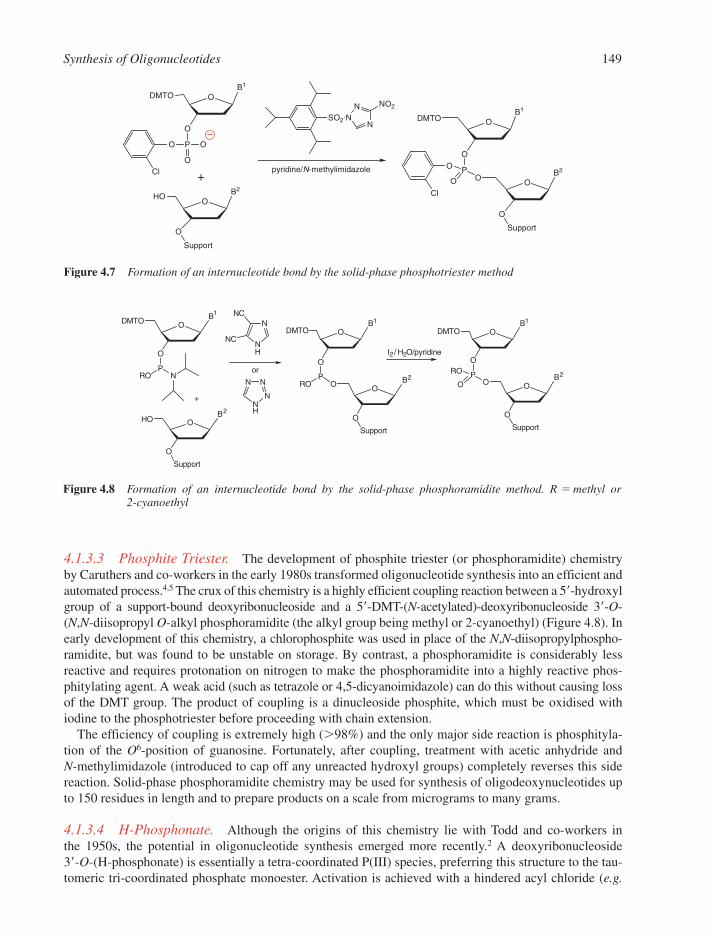
Synthesis of Oligonucleotides 149
4.1.3.3 Phosphite Triester. The development of phosphite triester (or phosphoramidite) chemistryby Caruthers and co-workers in the early 1980s transformed oligonucleotide synthesis into an efficient andautomated process.4,5 The crux of this chemistry is a highly efficient coupling reaction between a 5�-hydroxylgroup of a support-bound deoxyribonucleoside and a 5�-DMT-(N-acetylated)-deoxyribonucleoside 3�-O-(N,N-diisopropyl O-alkyl phosphoramidite (the alkyl group being methyl or 2-cyanoethyl) (Figure 4.8). Inearly development of this chemistry, a chlorophosphite was used in place of the N,N-diisopropylphospho-ramidite, but was found to be unstable on storage. By contrast, a phosphoramidite is considerably lessreactive and requires protonation on nitrogen to make the phosphoramidite into a highly reactive phos-phitylating agent. A weak acid (such as tetrazole or 4,5-dicyanoimidazole) can do this without causing lossof the DMT group. The product of coupling is a dinucleoside phosphite, which must be oxidised withiodine to the phosphotriester before proceeding with chain extension.
The efficiency of coupling is extremely high (�98%) and the only major side reaction is phosphityla-tion of the O6-position of guanosine. Fortunately, after coupling, treatment with acetic anhydride and N-methylimidazole (introduced to cap off any unreacted hydroxyl groups) completely reverses this sidereaction. Solid-phase phosphoramidite chemistry may be used for synthesis of oligodeoxynucleotides upto 150 residues in length and to prepare products on a scale from micrograms to many grams.
4.1.3.4 H-Phosphonate. Although the origins of this chemistry lie with Todd and co-workers in the 1950s, the potential in oligonucleotide synthesis emerged more recently.2 A deoxyribonucleoside 3�-O-(H-phosphonate) is essentially a tetra-coordinated P(III) species, preferring this structure to the tau-tomeric tri-coordinated phosphate monoester. Activation is achieved with a hindered acyl chloride (e.g.
B1
O
O
DMTO
PRO N
B2
O
O
HO
Support
B1
O
O
DMTO
PO B2
O
O
RO
Support
B1
O
O
DMTO
PO
B2
O
O
RO
O
Support
N
NH
NC
NC
NN
NH
N+
I2/ H2O/pyridine
or
Figure 4.8 Formation of an internucleotide bond by the solid-phase phosphoramidite method. R � methyl or2-cyanoethyl
B1
O
O
DMTO
P
O
O
Cl
O
+B2
O
O
HO
Support
B1
O
O
DMTO
PO
B2
O
O
O
O
Cl
Support
SO2N
NN
NO2
pyridine/N-methylimidazole
Figure 4.7 Formation of an internucleotide bond by the solid-phase phosphotriester method
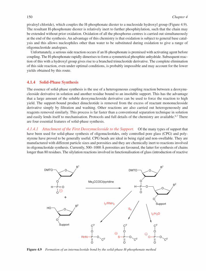
150 Chapter 4
pivaloyl chloride), which couples the H-phosphonate diester to a nucleoside hydroxyl group (Figure 4.9).The resultant H-phosphonate diester is relatively inert to further phosphitylation, such that the chain maybe extended without prior oxidation. Oxidation of all the phosphorus centres is carried out simultaneouslyat the end of the synthesis. An advantage of this chemistry is that oxidation is subject to general base catal-ysis and this allows nucleophiles other than water to be substituted during oxidation to give a range ofoligonucleotide analogues.
Unfortunately, a serious side reaction occurs if an H-phosphonate is premixed with activating agent beforecoupling. The H-phosphonate rapidly dimerises to form a symmetrical phosphite anhydride. Subsequent reac-tion of this with a hydroxyl group gives rise to a branched trinucleotide derivative. The complete eliminationof this side reaction, even under optimal conditions, is probably impossible and may account for the loweryields obtained by this route.
4.1.4 Solid-Phase Synthesis
The essence of solid-phase synthesis is the use of a heterogeneous coupling reaction between a deoxynu-cleoside derivative in solution and another residue bound to an insoluble support. This has the advantagethat a large amount of the soluble deoxynucleoside derivative can be used to force the reaction to highyield. The support-bound product dinucleotide is removed from the excess of reactant mononucleosidederivative simply by filtration and washing. Other reactions are also carried out heterogeneously andreagents removed similarly. This process is far faster than a conventional separation technique in solutionand easily lends itself to mechanisation. Protocols and full details of the chemistry are available.6,7 Thereare four essential features of solid-phase synthesis.
4.1.4.1 Attachment of the First Deoxynucleoside to the Support. Of the many types of support thathave been used for solid-phase synthesis of oligonucleotides, only controlled pore glass (CPG) and poly-styrene have proved to be generally useful. CPG beads are ideal in being rigid and non-swellable. They aremanufactured with different particle sizes and porosities and they are chemically inert to reactions involvedin oligonucleotide synthesis. Currently, 500–1000 Å porosities are favoured, the latter for synthesis of chainslonger than 80 residues. The silylation reactions involved in functionalisation of glass (introduction of reactive
B1
O
O
DMTO
PH O
O
B2
O
O
HO
Support
B1
O
O
DMTO
P O B2
O
O
H
O
Support
O
P OC5'
O
O
C3'
O
P OC5'
RHN
O
C3'
O
P OC5'
S
O
C3'
S8
+
I2/H2O
I2/RNH2
Me3CCOCl/pyridine
Figure 4.9 Formation of an internucleotide bond by the solid-phase H-phosphonate method
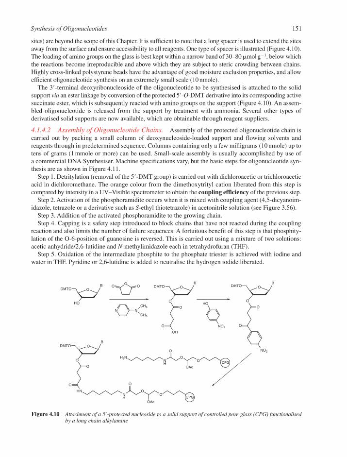
Synthesis of Oligonucleotides 151
sites) are beyond the scope of this Chapter. It is sufficient to note that a long spacer is used to extend the sitesaway from the surface and ensure accessibility to all reagents. One type of spacer is illustrated (Figure 4.10).The loading of amino groups on the glass is best kept within a narrow band of 30–80 �mol g�1, below whichthe reactions become irreproducible and above which they are subject to steric crowding between chains.Highly cross-linked polystyrene beads have the advantage of good moisture exclusion properties, and allowefficient oligonucleotide synthesis on an extremely small scale (10 nmole).
The 3�-terminal deoxyribonucleoside of the oligonucleotide to be synthesised is attached to the solidsupport via an ester linkage by conversion of the protected 5�-O-DMT derivative into its corresponding activesuccinate ester, which is subsequently reacted with amino groups on the support (Figure 4.10). An assem-bled oligonucleotide is released from the support by treatment with ammonia. Several other types ofderivatised solid supports are now available, which are obtainable through reagent suppliers.
4.1.4.2 Assembly of Oligonucleotide Chains. Assembly of the protected oligonucleotide chain iscarried out by packing a small column of deoxynucleoside-loaded support and flowing solvents andreagents through in predetermined sequence. Columns containing only a few milligrams (10 nmole) up totens of grams (1 mmole or more) can be used. Small-scale assembly is usually accomplished by use of a commercial DNA Synthesiser. Machine specifications vary, but the basic steps for oligonucleotide syn-thesis are as shown in Figure 4.11.
Step 1. Detritylation (removal of the 5�-DMT group) is carried out with dichloroacetic or trichloroaceticacid in dichloromethane. The orange colour from the dimethoxytrityl cation liberated from this step iscompared by intensity in a UV–Visible spectrometer to obtain the coupling efficiency of the previous step.
Step 2. Activation of the phosphoramidite occurs when it is mixed with coupling agent (4,5-dicyanoim-idazole, tetrazole or a derivative such as S-ethyl thiotetrazole) in acetonitrile solution (see Figure 3.56).
Step 3. Addition of the activated phosphoramidite to the growing chain.Step 4. Capping is a safety step introduced to block chains that have not reacted during the coupling
reaction and also limits the number of failure sequences. A fortuitous benefit of this step is that phosphity-lation of the O-6-position of guanosine is reversed. This is carried out using a mixture of two solutions:acetic anhydride/2,6-lutidine and N-methylimidazole each in tetrahydrofuran (THF).
Step 5. Oxidation of the intermediate phosphite to the phosphate triester is achieved with iodine andwater in THF. Pyridine or 2,6-lutidine is added to neutralise the hydrogen iodide liberated.
BO
HO
DMTO
BO
O
DMTO
O
O
OH
BO
O
DMTO
O
O
NO2
BO
O
DMTO
O
O
HNNH
OO
O
OAc
H2NNH
OO
O
OAc
OOO
N NCH3
CH3
HO
NO2
CPG
CPG
Figure 4.10 Attachment of a 5�-protected nucleoside to a solid support of controlled pore glass (CPG) functionalisedby a long chain alkylamine
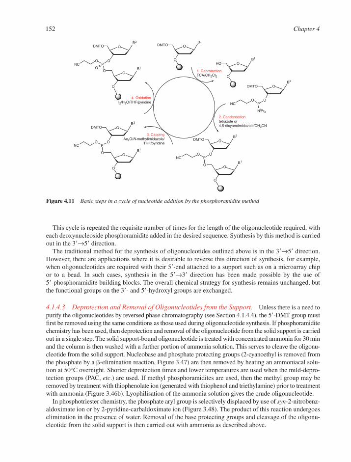
152 Chapter 4
This cycle is repeated the requisite number of times for the length of the oligonucleotide required, witheach deoxynucleoside phosphoramidite added in the desired sequence. Synthesis by this method is carriedout in the 3�→5� direction.
The traditional method for the synthesis of oligonucleotides outlined above is in the 3�→5� direction.However, there are applications where it is desirable to reverse this direction of synthesis, for example,when oligonucleotides are required with their 5�-end attached to a support such as on a microarray chipor to a bead. In such cases, synthesis in the 5�→3� direction has been made possible by the use of 5�-phosphoramidite building blocks. The overall chemical strategy for synthesis remains unchanged, butthe functional groups on the 3�- and 5�-hydroxyl groups are exchanged.
4.1.4.3 Deprotection and Removal of Oligonucleotides from the Support. Unless there is a need topurify the oligonucleotides by reversed phase chromatography (see Section 4.1.4.4), the 5�-DMT group mustfirst be removed using the same conditions as those used during oligonucleotide synthesis. If phosphoramiditechemistry has been used, then deprotection and removal of the oligonucleotide from the solid support is carriedout in a single step. The solid support-bound oligonucleotide is treated with concentrated ammonia for 30 minand the column is then washed with a further portion of ammonia solution. This serves to cleave the oligonu-cleotide from the solid support. Nucleobase and phosphate protecting groups (2-cyanoethyl is removed fromthe phosphate by a �-elimination reaction, Figure 3.47) are then removed by heating an ammoniacal solu-tion at 50°C overnight. Shorter deprotection times and lower temperatures are used when the mild-depro-tection groups (PAC, etc.) are used. If methyl phosphoramidites are used, then the methyl group may beremoved by treatment with thiophenolate ion (generated with thiophenol and triethylamine) prior to treatmentwith ammonia (Figure 3.46b). Lyophilisation of the ammonia solution gives the crude oligonucleotide.
In phosphotriester chemistry, the phosphate aryl group is selectively displaced by use of syn-2-nitrobenz-aldoximate ion or by 2-pyridine-carbaldoximate ion (Figure 3.48). The product of this reaction undergoeselimination in the presence of water. Removal of the base protecting groups and cleavage of the oligonu-cleotide from the solid support is then carried out with ammonia as described above.
B1O
O
DMTO
B1
O
O
HO
B2
O
O
DMTO
PO
NC
NiPr2
B1
O
O
O
B2
O
O
DMTO
PO
NC
B1
O
O
O
B2
O
O
DMTO
PO
NC
B1
O
O
O
B2
O
O
DMTO
PO
NCO
1. DeprotectionTCA/CH2Cl2
2. Condensationtetrazole or4,5-dicyanoimidazole/CH3CN
3. CappingAc2O/N-methylimidazole/
THF/pyridine
4. OxidationI2/H2O/THF/pyridine
Figure 4.11 Basic steps in a cycle of nucleotide addition by the phosphoramidite method
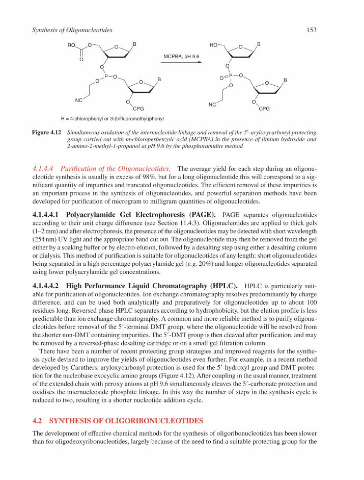
Synthesis of Oligonucleotides 153
4.1.4.4 Purification of the Oligonucleotides. The average yield for each step during an oligonu-cleotide synthesis is usually in excess of 98%, but for a long oligonucleotide this will correspond to a sig-nificant quantity of impurities and truncated oligonucleotides. The efficient removal of these impurities isan important process in the synthesis of oligonucleotides, and powerful separation methods have beendeveloped for purification of microgram to milligram quantities of oligonucleotides.
4.1.4.4.1 Polyacrylamide Gel Electrophoresis (PAGE). PAGE separates oligonucleotidesaccording to their unit charge difference (see Section 11.4.3). Oligonucleotides are applied to thick gels(1–2 mm) and after electrophoresis, the presence of the oligonucleotides may be detected with short wavelength(254 nm) UV light and the appropriate band cut out. The oligonucleotide may then be removed from the geleither by a soaking buffer or by electro-elution, followed by a desalting step using either a desalting columnor dialysis. This method of purification is suitable for oligonucleotides of any length: short oligonucleotidesbeing separated in a high percentage polyacrylamide gel (e.g. 20%) and longer oligonucleotides separatedusing lower polyacrylamide gel concentrations.
4.1.4.4.2 High Performance Liquid Chromatography (HPLC). HPLC is particularly suit-able for purification of oligonucleotides. Ion exchange chromatography resolves predominantly by chargedifference, and can be used both analytically and preparatively for oligonucleotides up to about 100residues long. Reversed phase HPLC separates according to hydrophobicity, but the elution profile is lesspredictable than ion exchange chromatography. A common and more reliable method is to purify oligonu-cleotides before removal of the 5�-terminal DMT group, where the oligonucleotide will be resolved fromthe shorter non-DMT containing impurities. The 5�-DMT group is then cleaved after purification, and maybe removed by a reversed-phase desalting cartridge or on a small gel filtration column.
There have been a number of recent protecting group strategies and improved reagents for the synthe-sis cycle devised to improve the yields of oligonucleotides even further. For example, in a recent methoddeveloped by Caruthers, aryloxycarbonyl protection is used for the 5�-hydroxyl group and DMT protec-tion for the nucleobase exocyclic amino groups (Figure 4.12). After coupling in the usual manner, treatmentof the extended chain with peroxy anions at pH 9.6 simultaneously cleaves the 5�-carbonate protection andoxidises the internucleoside phosphite linkage. In this way the number of steps in the synthesis cycle isreduced to two, resulting in a shorter nucleotide addition cycle.
4.2 SYNTHESIS OF OLIGORIBONUCLEOTIDES
The development of effective chemical methods for the synthesis of oligoribonucleotides has been slowerthan for oligodeoxyribonucleotides, largely because of the need to find a suitable protecting group for the
BO
O
BO
O
O
O
RO
PO
NC
O
CPG
BO
O
BO
O
HO
P
O
NC
O
CPG
O
R = 4-chlorophenyl or 3-(trifluoromethyl)phenyl
MCPBA, pH 9.6
Figure 4.12 Simultaneous oxidation of the internucleotide linkage and removal of the 5�-aryloxycarbonyl protectinggroup carried out with m-chloroperbenzoic acid (MCPBA) in the presence of lithium hydroxide and 2-amino-2-methyl-1-propanol at pH 9.6 by the phosphoramidite method
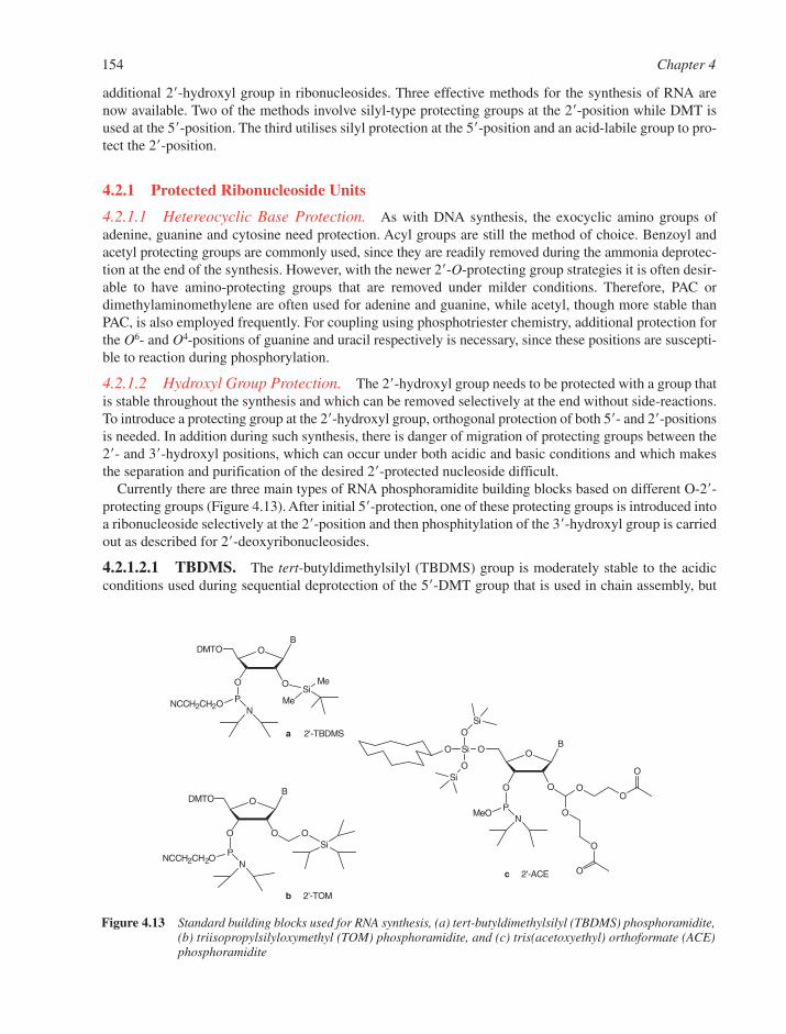
154 Chapter 4
BODMTO
O O
PNCCH2CH2O N
OSi
O
PNCCH2CH2ON
BODMTO
O
BO
O O OO
O
O
O
O
PMeO
N
OSi
O
O
Oa 2'-TBDMS
b 2'-TOM
c 2'-ACE
SiMe
Me
Si
Si
Figure 4.13 Standard building blocks used for RNA synthesis, (a) tert-butyldimethylsilyl (TBDMS) phosphoramidite,(b) triisopropylsilyloxymethyl (TOM) phosphoramidite, and (c) tris(acetoxyethyl) orthoformate (ACE)phosphoramidite
additional 2�-hydroxyl group in ribonucleosides. Three effective methods for the synthesis of RNA arenow available. Two of the methods involve silyl-type protecting groups at the 2�-position while DMT isused at the 5�-position. The third utilises silyl protection at the 5�-position and an acid-labile group to pro-tect the 2�-position.
4.2.1 Protected Ribonucleoside Units
4.2.1.1 Hetereocyclic Base Protection. As with DNA synthesis, the exocyclic amino groups ofadenine, guanine and cytosine need protection. Acyl groups are still the method of choice. Benzoyl andacetyl protecting groups are commonly used, since they are readily removed during the ammonia deprotec-tion at the end of the synthesis. However, with the newer 2�-O-protecting group strategies it is often desir-able to have amino-protecting groups that are removed under milder conditions. Therefore, PAC ordimethylaminomethylene are often used for adenine and guanine, while acetyl, though more stable thanPAC, is also employed frequently. For coupling using phosphotriester chemistry, additional protection forthe O6- and O4-positions of guanine and uracil respectively is necessary, since these positions are suscepti-ble to reaction during phosphorylation.
4.2.1.2 Hydroxyl Group Protection. The 2�-hydroxyl group needs to be protected with a group thatis stable throughout the synthesis and which can be removed selectively at the end without side-reactions.To introduce a protecting group at the 2�-hydroxyl group, orthogonal protection of both 5�- and 2�-positionsis needed. In addition during such synthesis, there is danger of migration of protecting groups between the2�- and 3�-hydroxyl positions, which can occur under both acidic and basic conditions and which makesthe separation and purification of the desired 2�-protected nucleoside difficult.
Currently there are three main types of RNA phosphoramidite building blocks based on different O-2�-protecting groups (Figure 4.13). After initial 5�-protection, one of these protecting groups is introduced intoa ribonucleoside selectively at the 2�-position and then phosphitylation of the 3�-hydroxyl group is carriedout as described for 2�-deoxyribonucleosides.
4.2.1.2.1 TBDMS. The tert-butyldimethylsilyl (TBDMS) group is moderately stable to the acidicconditions used during sequential deprotection of the 5�-DMT group that is used in chain assembly, but
Synthesis of Oligonucleotides 155
can be removed by treatment with fluoride ion at the end of assembly (Section 4.2.2). TBDMS chemistryis useful both on small and larger production scale (Figure 4.13a).8
4.2.1.2.2 TOM. Triisopropylsilyloxymethyl (TOM) is a silyl-protected acetal, which has the advan-tage that no 2�↔3� migration occurs under the usual basic conditions of introduction.9 The nucleobaseprotecting groups are N-acetyl and the 5�-hydroxyl group is protected with DMT. TOM chemistry mayhave an advantage over TBDMS of slightly improved overall yields in RNA synthesis, but this view is notuniversally held (Figure 4.13b).
4.2.1.2.3 ACE. The 2�-O-[bis[2-(acetyloxy)ethoxy]methyl] (ACE) protecting group is entirely differentfrom the other two RNA chemistries in being a protected protecting group. It is a protected orthoester.10
The nucleobases are protected by acyl groups (N4-acetyl-C, N 6-benzoyl-A and N 2-isopropyl-G), but at the5�-position there is a cyclododecyloxy-bis(trimethylsiloxy)silyl (SIL) group rather than the usual acid-labile DMT group (Figure 4.13c). The ACE protecting group becomes acid-labile once the two acetylgroups of ACE have been removed (Section 4.2.2). The 3�-phosphoramidite has a methyl protecting groupinstead of the usual 2-cyanoethyl group, because the latter is unstable under the fluoride ion conditionsneeded to remove the 5�-SIL protecting group. ACE chemistry is currently only used for relatively small-scale syntheses on a polystyrene support (glass or silica is not compatible with the 5�-deprotection condi-tions) but has become particularly popular for siRNA synthesis (see Section 5.7.2).
4.2.2 Oligoribonucleotide Synthesis
4.2.2.1 Assembly. The assembly cycle for each of the three different RNA chemistries follows a simi-lar overall route to that for DNA assembly (Section 4.1.4) and involves
(i) Deprotection of the 5�-protecting group. For TBDMS and TOM chemistry, this involves removalof DMT groups with di- or trichloroacetic acid, but for ACE chemistry, deprotection of the 5�-silylgroup (SIL) is accomplished with triethylamine.3HF.
(ii) Coupling of the 3�-phosphoramidite to the free 5�-hydroxyl group using S-ethylthio tetrazole asactivator.
(iii) Capping of any unreacted 5�-hydroxyl groups (acetic anhydride/2,6-lutidine and N-methylimida-zole in MeCN, same reagents as for DNA assembly).
(iv) Oxidation with iodine/pyridine/water (same reagent as for DNA assembly).
4.2.2.2 Deprotection and Purification. Deprotection and removal of oligonucleotides from thesolid support also uses procedures similar to those described for DNA synthesis (Section 4.1.4), but thereagents and conditions depend on the choice of protection strategy.
4.2.2.2.1 TBDMS Chemistry. 5�-Deprotection of DMT groups is usually carried out first by acidictreatment, while oligoribonucleotides are still attached to the support. Subsequent ammonia treatment thenresults in cleavage of the linkage of the oligonucleotide to the solid support simultaneously with theremoval of nucleobase and phosphate (2-cyanoethyl) protecting groups. Mild methanolic ammonia (PACprotection) or aqueous ammonia (dimethylaminomethylene protection) at room temperature is suitable.Lastly, removal of the 2�-TBDMS group is effected by treatment with 1 M tetrabutylammonium fluoride(TBAF) in THF for 16–24 h or with triethylamine�(3HF). Final purification of deprotected oligoribonu-cleotides is carried out by polyacrylamide gel electrophoresis or by HPLC on ion exchange columns (Section4.1.4). For ‘DMT-on’ purification, steps two and three are performed and the DMT-protected oligoribonu-cleotide is purified by HPLC and stored. The DMT group is removed by mild acid treatment before use.
4.2.2.2.2 TOM Chemistry. 5�-Deprotection of DMT groups is the same as for the TBDMS route.Cleavage from the solid support and removal of nucleobase protecting groups is effected with a 1:1 mix-ture of 40% aqueous methylamine and 33% ethanolic methylamine at room temperature overnight or 6 h
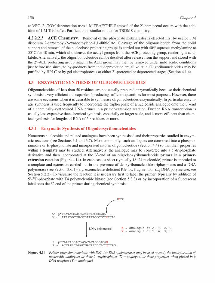
156 Chapter 4
5’-p*TAATACGACTACATATAGGGAGA3’- ATTATGCTGAGTGATATCCCTCTYTCAG
dXTP
5’-p*TAATACGACTACATATAGGGAGAX3’- ATTATGCTGAGTGATATCCCTCTYTCAG
X = analogue or A, T, C, GY = analogue or T, A, G, C
DNA polymerase
Figure 4.14 Primer extension reactions with DNA (or RNA) polymerases may be used to study the incorporation ofnucleoside analogues as their 5�-triphosphates (X � analogue) or their properties when placed in aDNA template (Y � analogue)
at 35°C. 2�-TOM deprotection uses 1 M TBAF/THF. Removal of the 2�-hemiacetal occurs with the add-ition of 1 M Tris buffer. Purification is similar to that for TBDMS chemistry.
4.2.2.2.3 ACE Chemistry. Removal of the phosphate methyl ester is effected first by use of 1 Mdisodium 2-carbamoyl-2-cyanoethylene-1,1-dithiolate. Cleavage of the oligonucleotide from the solidsupport and removal of the nucleobase protecting groups is carried out with 40% aqueous methylamine at55°C for 10 min, which also cleaves the acetyl groups from the ACE protecting group, rendering it acid-labile. Alternatively, the oligoribonucleotide can be desalted after release from the support and stored withthe 2�-ACE protecting group intact. The ACE group may then be removed under mild acidic conditionsjust before use since the by-products from that deprotection are all volatile. Oligoribonucleotides may bepurified by HPLC or by gel electrophoresis at either 2�-protected or deprotected stages (Section 4.1.4).
4.3 ENZYMATIC SYNTHESIS OF OLIGONUCLEOTIDES
Oligonucleotides of less than 50 residues are not usually prepared enzymatically because their chemicalsynthesis is very efficient and capable of producing sufficient quantities for most purposes. However, thereare some occasions when it is desirable to synthesise oligonucleotides enzymatically. In particular enzym-atic synthesis is used frequently to incorporate the triphosphate of a nucleoside analogue onto the 3�-endof a chemically-synthesised DNA primer in a primer-extension reaction. Further, RNA transcription isusually less expensive than chemical synthesis, especially on larger scale, and is more efficient than chem-ical synthesis for lengths of RNA of 50 residues or more.
4.3.1 Enzymatic Synthesis of Oligodeoxyribonucleotides
Numerous nucleoside and related analogues have been synthesised and their properties studied in enzym-atic reactions (see Sections 3.1 and 3.7). Most commonly, such analogues are converted into a phospho-ramidite or H-phosphonate and incorporated into an oligonucleotide (Section 4.4) so that their propertieswithin a template may be studied. Alternatively, the analogue may be converted into a 5�-triphosphatederivative and then incorporated at the 3�-end of an oligodeoxyribonucleotide primer in a primer-extension reaction (Figure 4.14). In each case, a short (typically 18–24 nucleotide) primer is annealed toa template and extension carried out in the presence of deoxyribonucleoside triphosphates and a DNApolymerase (see Section 3.6.1) (e.g. exonuclease-deficient Klenow fragment, or Taq DNA polymerase, seeSection 5.2.2). To visualise the reaction it is necessary first to label the primer, typically by addition of 5�-32P-phosphate with T4 polynucleotide kinase (see Section 5.3.3) or by incorporation of a fluorescentlabel onto the 5�-end of the primer during chemical synthesis.
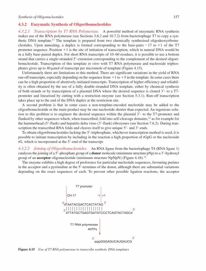
Synthesis of Oligonucleotides 157
4.3.2 Enzymatic Synthesis of Oligoribonucleotides
4.3.2.1 Transcription by T7 RNA Polymerase. A powerful method of enzymatic RNA synthesismakes use of the RNA polymerase (see Sections 3.6.2 and 10.7.2) from bacteriophage T7 to copy a syn-thetic DNA template.11 The template is prepared from two chemically synthesised oligodeoxyribonu-cleotides. Upon annealing, a duplex is formed corresponding to the base-pairs �17 to �1 of the T7promoter sequence. Position �1 is the site of initiation of transcription, which in natural DNA would bein a fully base-paired duplex. For short RNA transcripts of 10–60 residues, it is possible to use a bottomstrand that carries a single-stranded 5�-extension corresponding to the complement of the desired oligori-bonucleotide. Transcription of this template in vitro with T7 RNA polymerase and nucleoside triphos-phates gives up to 40 �mol of transcript per micromole of template (Figure 4.15).
Unfortunately there are limitations to this method. There are significant variations in the yield of RNArun-off transcripts, especially depending on the sequence from �1 to �5 in the template. In some cases therecan be a high proportion of abortively-initiated transcripts. Transcription of higher efficiency and reliabil-ity is often obtained by the use of a fully double-stranded DNA template, either by chemical synthesis of both strands or by transcription of a plasmid DNA where the desired sequence is cloned 3�- to a T7-promoter and linearised by cutting with a restriction enzyme (see Section 5.3.1). Run-off transcriptiontakes place up to the end of the DNA duplex at the restriction site.
A second problem is that in some cases a non-template-encoded nucleotide may be added to the oligoribonucleotide or the main product may be one nucleotide shorter than expected. An ingenious solu-tion to this problem is to engineer the desired sequence within the plasmid 3�- to the T7-promoter andflanked by other sequences which, when transcribed, fold into self-cleavage domains,12 as for example forthe hammerhead (5�-flank) and hepatitis delta virus (3�-flank) ribozymes (see Section 7.6.2). During tran-scription the transcribed RNA folds and cleaves itself to give unique 5�- and 3�-ends.
To obtain oligoribonucleotides lacking the 5�-triphosphate, whichever transcription method is used, it ispossible to initiate transcription by including in the reaction a high proportion of rGpG or the nucleosiderG, which is incorporated at the 5�-end of the transcript.
4.3.2.2 Joining of Oligoribonucleotides. An RNA ligase from the bacteriophage T4 (RNA ligase 1)catalyses the joining of a 5�-phosphate group of a donor molecule (minimum structure pNp) to a 3�-hydroxylgroup of an acceptor oligonucleotide (minimum structure NpNpN) (Figure 4.16).13
The enzyme exhibits a high degree of preference for particular nucleotide sequences, favouring purinesin the acceptor and a pyrimidine at the 5�-terminus of the donor, although there are substantial variationsdepending on the exact sequences of each. To prevent other possible ligation reactions, the acceptor
dTAATACGACTCACTATAG
ATTATGCTGAGTGATATCCCTCAGTACTAGCd
rpppGGGAGUCAUGAUCG
5'
3' 5'
3'
+1-1CH-17
5' 3'
4NTPs
T7 promoter
T7 RNA polymerase
Figure 4.15 Use of T7 RNA polymerase to transcribe synthetic DNA templates
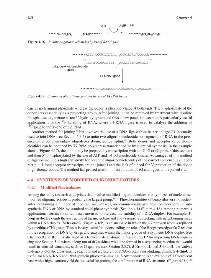
158 Chapter 4
carries no terminal phosphate whereas the donor is phosphorylated at both ends. The 3�-phosphate of thedonor acts essentially as a protecting group. After joining it can be removed by treatment with alkalinephosphatase to generate a free 3�-hydroxyl group and thus a new potential acceptor. A particularly usefulapplication is in the 32P-labelling of RNA, where T4 RNA ligase is used to catalyse the addition of[32P]pCp to the 3�-end of the RNA.
Another method for joining RNA involves the use of a DNA ligase from bacteriophage T4 (normallyused to join DNA, see Section 5.3.5) to unite two oligoribonucleotides or segments of RNA in the pres-ence of a complementary oligodeoxyribonucleotide splint.14 Both donor and acceptor oligoribonu-cleotides can be obtained by T7 RNA polymerase transcription or by chemical synthesis. In the exampleshown (Figure 4.17), the donor may be prepared by transcription with an rGpG or rG primer (this section)and then 5�-phosphorylated by the use of ATP and T4 polynucleotide kinase. Advantages of this methodof ligation include a high sensitivity for acceptor oligoribonucleotides of the correct sequence (i.e. incor-rect n � 1 long acceptor transcripts are not joined) and the lack of a need for 3�-protection of the donoroligoribonucleotide. The method has proved useful in incorporation of rG analogues at the joined site.
4.4 SYNTHESIS OF MODIFIED OLIGONUCLEOTIDES
4.4.1 Modified Nucleobases
Among the many research enterprises that involve modified oligonucleotides, the synthesis of nucleobase-modified oligonucleotides is probably the largest group.15–19 Phosphoramidites of deoxyribo- or ribonucleo-sides, containing a number of modified nucleobases, are commercially available for incorporation intosynthetic DNA or RNA by standard solid-phase synthesis (Section 4.1) (Figure 4.18). Among numerousapplications, certain modified bases are used to increase the stability of a DNA duplex. For example, 5-propynyl-dU extends the -structure of the nucleobase and allows improved stacking with neighbouring baseswithin a DNA duplex. 7-Deaza-dG (Figure 4.18b) is an analogue in which the N7 nitrogen atom is replacedby a methine (CH) group. Thus, it is very useful for understanding the role of the Hoogsteen edge of a G residuein the recognition of DNA by drugs and enzymes within the major groove of a synthetic DNA duplex (seeChapters 9 and 10). It is also used as a triphosphate analogue in place of dGTP for improving DNA sequen-cing (see Section 5.1) where a long run of dG residues would be formed in a sequencing reaction that wouldresult in unusual structures, such as G-quartets (see Section 2.3.7). 5-BromodU and 5-iododU derivativesundergo photolytic cross-linking reactions and are useful for DNA–protein cross-linking. Similarly 4-thioU isuseful for RNA–RNA and RNA–protein photocross-linking. 2-Aminopurine is an example of a fluorescentbase with a high quantum yield that is useful for probing the conformation of RNA structures (Figure 4.18e).20
p
AGGUAGCUUGGACCAOH
TCCATCGAACCTGGT
PGGUGAGAUAUGGCCA
CCACTCTAUACCGGT
AGGUAGCUUGGACCAGGUGAGAUAUGGCCA
T4 DNA ligase
3' 5'
oligodeoxyribonucleotidesplint
Figure 4.17 Joining of oligoribonucleotides by use of T4 DNA ligase
N1pN2pN3 pN4p N1pN2pN3pN4p+
AMP + PPiATP
Figure 4.16 Joining oligoribonucleotides by use of RNA ligase
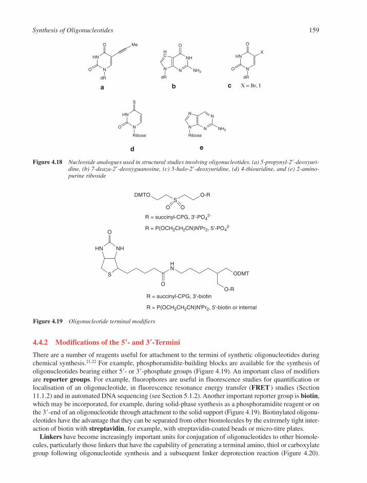
Synthesis of Oligonucleotides 159
4.4.2 Modifications of the 5��- and 3��-Termini
There are a number of reagents useful for attachment to the termini of synthetic oligonucleotides duringchemical synthesis.21,22 For example, phosphoramidite-building blocks are available for the synthesis ofoligonucleotides bearing either 5�- or 3�-phosphate groups (Figure 4.19). An important class of modifiersare reporter groups. For example, fluorophores are useful in fluorescence studies for quantification orlocalisation of an oligonucleotide, in fluorescence resonance energy transfer (FRET ) studies (Section11.1.2) and in automated DNA sequencing (see Section 5.1.2). Another important reporter group is biotin,which may be incorporated, for example, during solid-phase synthesis as a phosphoramidite reagent or onthe 3�-end of an oligonucleotide through attachment to the solid support (Figure 4.19). Biotinylated oligonu-cleotides have the advantage that they can be separated from other biomolecules by the extremely tight inter-action of biotin with streptavidin, for example, with streptavidin-coated beads or micro-titre plates.
Linkers have become increasingly important units for conjugation of oligonucleotides to other biomole-cules, particularly those linkers that have the capability of generating a terminal amino, thiol or carboxylategroup following oligonucleotide synthesis and a subsequent linker deprotection reaction (Figure 4.20).
DMTOS
O-R
O O
R = succinyl-CPG, 3'-PO42-
R = P(OCH2CH2CN)NiPr2, 5'-PO42-
NHHN
S
HN
ODMT
OO-R
O
R = succinyl-CPG, 3'-biotin
R = P(OCH2CH2CN)NiPr2, 5'-biotin or internal
Figure 4.19 Oligonucleotide terminal modifiers
N
HN
MeO
O
dR
N
NHHC
N
dR
O
NH2N
HNX
O
O
dR
N
HN
S
O
Ribose
N
NN
N
Ribose
NH2
X = Br, Ia b c
d e
Figure 4.18 Nucleoside analogues used in structural studies involving oligonucleotides. (a) 5-propynyl-2�-deoxyuri-dine, (b) 7-deaza-2�-deoxyguanosine, (c) 5-halo-2�-deoxyuridine, (d) 4-thiouridine, and (e) 2-amino-purine riboside
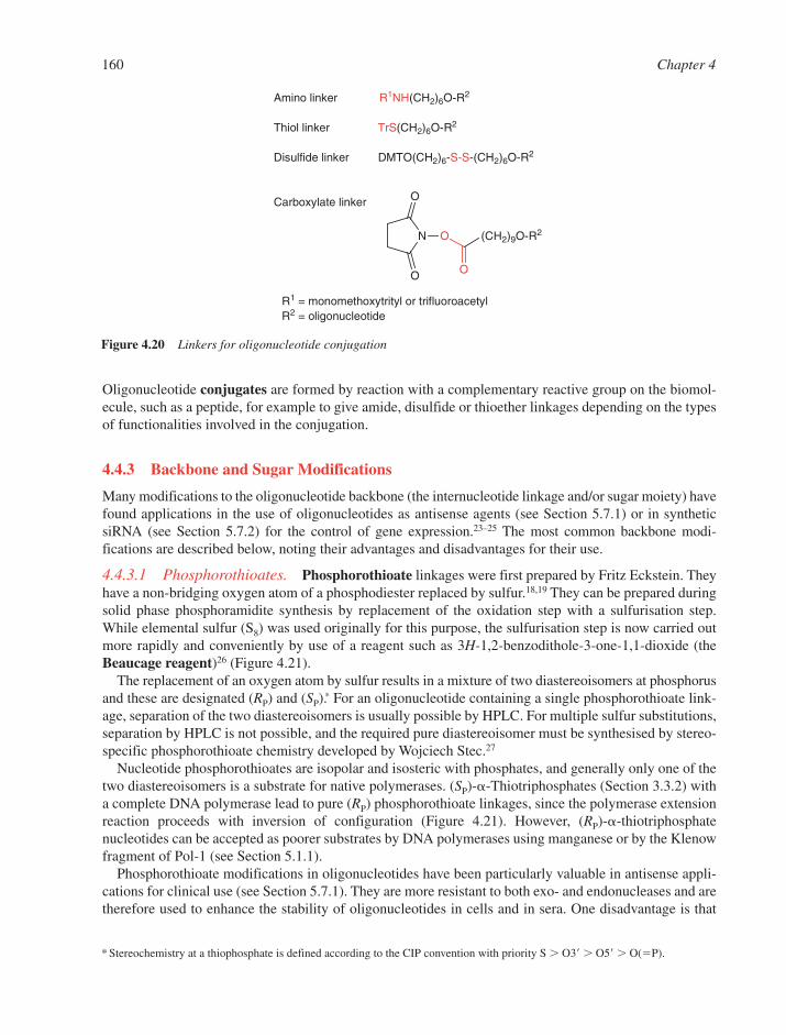
160 Chapter 4
* Stereochemistry at a thiophosphate is defined according to the CIP convention with priority S � O3� � O5� � O(�P).
Oligonucleotide conjugates are formed by reaction with a complementary reactive group on the biomol-ecule, such as a peptide, for example to give amide, disulfide or thioether linkages depending on the typesof functionalities involved in the conjugation.
4.4.3 Backbone and Sugar Modifications
Many modifications to the oligonucleotide backbone (the internucleotide linkage and/or sugar moiety) havefound applications in the use of oligonucleotides as antisense agents (see Section 5.7.1) or in syntheticsiRNA (see Section 5.7.2) for the control of gene expression.23–25 The most common backbone modi-fications are described below, noting their advantages and disadvantages for their use.
4.4.3.1 Phosphorothioates. Phosphorothioate linkages were first prepared by Fritz Eckstein. Theyhave a non-bridging oxygen atom of a phosphodiester replaced by sulfur.18,19 They can be prepared duringsolid phase phosphoramidite synthesis by replacement of the oxidation step with a sulfurisation step.While elemental sulfur (S8) was used originally for this purpose, the sulfurisation step is now carried outmore rapidly and conveniently by use of a reagent such as 3H-1,2-benzodithole-3-one-1,1-dioxide (theBeaucage reagent)26 (Figure 4.21).
The replacement of an oxygen atom by sulfur results in a mixture of two diastereoisomers at phosphorusand these are designated (RP) and (SP).∗ For an oligonucleotide containing a single phosphorothioate link-age, separation of the two diastereoisomers is usually possible by HPLC. For multiple sulfur substitutions,separation by HPLC is not possible, and the required pure diastereoisomer must be synthesised by stereo-specific phosphorothioate chemistry developed by Wojciech Stec.27
Nucleotide phosphorothioates are isopolar and isosteric with phosphates, and generally only one of thetwo diastereoisomers is a substrate for native polymerases. (SP)--Thiotriphosphates (Section 3.3.2) witha complete DNA polymerase lead to pure (RP) phosphorothioate linkages, since the polymerase extensionreaction proceeds with inversion of configuration (Figure 4.21). However, (RP)--thiotriphosphatenucleotides can be accepted as poorer substrates by DNA polymerases using manganese or by the Klenowfragment of Pol-1 (see Section 5.1.1).
Phosphorothioate modifications in oligonucleotides have been particularly valuable in antisense appli-cations for clinical use (see Section 5.7.1). They are more resistant to both exo- and endonucleases and aretherefore used to enhance the stability of oligonucleotides in cells and in sera. One disadvantage is that
N
O
O
O (CH2)9O-R2
O
Amino linker R1NH(CH2)6O-R2
Thiol linker TrS(CH2)6O-R2
Disulfide linker DMTO(CH2)6-S-S-(CH2)6O-R2
Carboxylate linker
R1 = monomethoxytrityl or trifluoroacetylR2 = oligonucleotide
Figure 4.20 Linkers for oligonucleotide conjugation
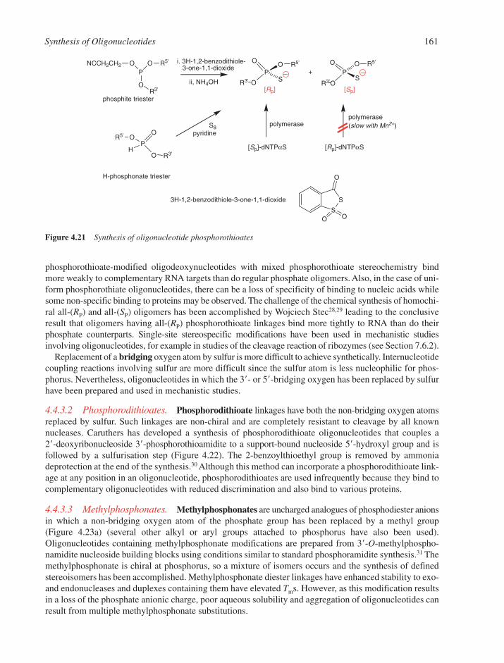
Synthesis of Oligonucleotides 161
phosphorothioate-modified oligodeoxynucleotides with mixed phosphorothioate stereochemistry bindmore weakly to complementary RNA targets than do regular phosphate oligomers. Also, in the case of uni-form phosphorothiate oligonucleotides, there can be a loss of specificity of binding to nucleic acids whilesome non-specific binding to proteins may be observed. The challenge of the chemical synthesis of homochi-ral all-(RP) and all-(SP) oligomers has been accomplished by Wojciech Stec28,29 leading to the conclusiveresult that oligomers having all-(RP) phosphorothioate linkages bind more tightly to RNA than do theirphosphate counterparts. Single-site stereospecific modifications have been used in mechanistic studiesinvolving oligonucleotides, for example in studies of the cleavage reaction of ribozymes (see Section 7.6.2).
Replacement of a bridging oxygen atom by sulfur is more difficult to achieve synthetically. Internucleotidecoupling reactions involving sulfur are more difficult since the sulfur atom is less nucleophilic for phos-phorus. Nevertheless, oligonucleotides in which the 3�- or 5�-bridging oxygen has been replaced by sulfurhave been prepared and used in mechanistic studies.
4.4.3.2 Phosphorodithioates. Phosphorodithioate linkages have both the non-bridging oxygen atomsreplaced by sulfur. Such linkages are non-chiral and are completely resistant to cleavage by all knownnucleases. Caruthers has developed a synthesis of phosphorodithioate oligonucleotides that couples a 2�-deoxyribonucleoside 3�-phosphorothioamidite to a support-bound nucleoside 5�-hydroxyl group and isfollowed by a sulfurisation step (Figure 4.22). The 2-benzoylthioethyl group is removed by ammoniadeprotection at the end of the synthesis.30 Although this method can incorporate a phosphorodithioate link-age at any position in an oligonucleotide, phosphorodithioates are used infrequently because they bind tocomplementary oligonucleotides with reduced discrimination and also bind to various proteins.
4.4.3.3 Methylphosphonates. Methylphosphonates are uncharged analogues of phosphodiester anionsin which a non-bridging oxygen atom of the phosphate group has been replaced by a methyl group (Figure 4.23a) (several other alkyl or aryl groups attached to phosphorus have also been used).Oligonucleotides containing methylphosphonate modifications are prepared from 3�-O-methylphospho-namidite nucleoside building blocks using conditions similar to standard phosphoramidite synthesis.31 Themethylphosphonate is chiral at phosphorus, so a mixture of isomers occurs and the synthesis of definedstereoisomers has been accomplished. Methylphosphonate diester linkages have enhanced stability to exo-and endonucleases and duplexes containing them have elevated Tms. However, as this modification resultsin a loss of the phosphate anionic charge, poor aqueous solubility and aggregation of oligonucleotides canresult from multiple methylphosphonate substitutions.
P
OR3'
O R5'ONCCH2CH2
P
O
O
R3'H
OR5'
P
OR3'
O
S
O R5'
P
OR3'
O
S
O R5'
S
S
O O
O
+
polymerase
phosphite triester
H-phosphonate triester
ii, NH4OH
S8pyridine
[Sp]-dNTPαS [Rp]-dNTPαS
[Rp] [Sp]
polymerase(slow with Mn2+)
i. 3H-1,2-benzodithiole-3-one-1,1-dioxide
3H-1,2-benzodithiole-3-one-1,1-dioxide
Figure 4.21 Synthesis of oligonucleotide phosphorothioates
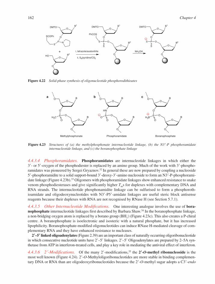
162 Chapter 4
4.4.3.4 Phosphoramidates. Phosphoramidates are internucleotide linkages in which either the 3�- or 5�-oxygen of the phosphodiester is replaced by an amino group. Much of the work with 3�-phospho-ramidates was pioneered by Sergei Gryaznov.32 In general these are now prepared by coupling a nucleoside5�-phosphoramidite to a solid support-bound 3�-deoxy-3�-amino nucleoside to form an N3�-P-phosphorami-date linkage (Figure 4.23b).33 Oligomers with phosphoramidate linkages show enhanced resistance to snakevenom phosphodiesterases and give significantly higher Tms for duplexes with complementary DNA andRNA strands. The internucleotide phosphonamidite linkage can be sulfurised to form a phosphoroth-ioamidate and oligodeoxynucleotides with N3�-P5�-amidate linkages are useful steric block antisensereagents because their duplexes with RNA are not recognised by RNase H (see Section 5.7.1).
4.4.3.5 Other Internucleotide Modifications. One interesting analogue involves the use of bora-nophosphate internucleotide linkages first described by Barbara Shaw.34 In the boranophosphate linkage,a non-bridging oxygen atom is replaced by a borano group (BH3
�) (Figure 4.23c). This also creates a P-chiralcentre. A boranophosphate is isoelectronic and isosteric with a natural phosphate, but it has increasedlipophilicity. Boranophosphate-modified oligonucleotides can induce RNase H-mediated cleavage of com-plementary RNA and they have enhanced resistance to nucleases.
2��–5�� linked oligoadenylates (Figure 2.39) are an important class of naturally occurring oligoribonucleotidein which consecutive nucleotide units have 2�–5� linkages. 2�–5� Oligoadenylates are prepared by 2–5A syn-thetase from ATP in interferon-treated cells, and play a key role in mediating the antiviral effect of interferon.
4.4.3.6 2�-Modifications. Of the many 2�-modifications,35 the 2��-O-methyl ribonucleoside is themost well known (Figure 4.24). 2�-O-Methyloligoribonucleotides are more stable in binding complemen-tary DNA or RNA than are oligodeoxyribonucleotides because the 2�-O-methyl sugar adopts a C3�-endo
BO
O
O
BO
O
P
OH3C
O
BO
O
HN
BO
O
P
OO
O
BO
O
O
BO
O
P
OH3B
O
a b c
BoranophosphateMethylphosphonate Phosphoramidate
Figure 4.23 Structures of (a) the methylphosphonate internucleotide linkage, (b) the N3�-P phosphoramidateinternucleotide linkage, and (c) the boranophosphate linkage
B2
OHO
O
B2
O
O
B1
ODMTO
O
PS O
S
PhCOS
B1
ODMTO
O
P NS
SCOPh
+ i, tetrazole/acetonitrile
ii, S8/pyridine/CS2
B2
O
O
B1
ODMTO
O
PS O
S
NH4OH
Figure 4.22 Solid-phase synthesis of oligonucleotide phosphorodithioates
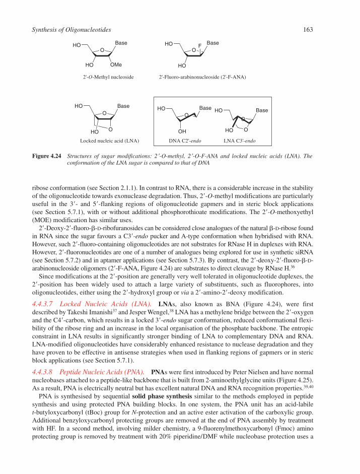
Synthesis of Oligonucleotides 163
ribose conformation (see Section 2.1.1). In contrast to RNA, there is a considerable increase in the stabilityof the oligonucleotide towards exonuclease degradation. Thus, 2�-O-methyl modifications are particularlyuseful in the 3�- and 5�-flanking regions of oligonucleotide gapmers and in steric block applications (see Section 5.7.1), with or without additional phosphorothioate modifications. The 2�-O-methoxyethyl(MOE) modification has similar uses.
2�-Deoxy-2�-fluoro-�-D-ribofuranosides can be considered close analogues of the natural �-D-ribose foundin RNA since the sugar favours a C3�-endo pucker and A-type conformation when hybridised with RNA.However, such 2�-fluoro-containing oligonucleotides are not substrates for RNase H in duplexes with RNA.However, 2�-fluoronucleotides are one of a number of analogues being explored for use in synthetic siRNA(see Section 5.7.2) and in aptamer applications (see Section 5.7.3). By contrast, the 2�-deoxy-2�-fluoro-�-D-arabinonucleoside oligomers (2�-F-ANA, Figure 4.24) are substrates to direct cleavage by RNase H.36
Since modifications at the 2�-position are generally very well tolerated in oligonucleotide duplexes, the2�-position has been widely used to attach a large variety of substituents, such as fluorophores, intooligonucleotides, either using the 2�-hydroxyl group or via a 2�-amino-2�-deoxy modification.
4.4.3.7 Locked Nucleic Acids (LNA). LNAs, also known as BNA (Figure 4.24), were firstdescribed by Takeshi Imanishi37 and Jesper Wengel,38 LNA has a methylene bridge between the 2�-oxygenand the C4�-carbon, which results in a locked 3�-endo sugar conformation, reduced conformational flexi-bility of the ribose ring and an increase in the local organisation of the phosphate backbone. The entropicconstraint in LNA results in significantly stronger binding of LNA to complementary DNA and RNA.LNA-modified oligonucleotides have considerably enhanced resistance to nuclease degradation and theyhave proven to be effective in antisense strategies when used in flanking regions of gapmers or in stericblock applications (see Section 5.7.1).
4.4.3.8 Peptide Nucleic Acids (PNA). PNAs were first introduced by Peter Nielsen and have normalnucleobases attached to a peptide-like backbone that is built from 2-aminoethylglycine units (Figure 4.25).As a result, PNA is electrically neutral but has excellent natural DNA and RNA recognition properties.39,40
PNA is synthesised by sequential solid phase synthesis similar to the methods employed in peptidesynthesis and using protected PNA building blocks. In one system, the PNA unit has an acid-labile t-butyloxycarbonyl (tBoc) group for N-protection and an active ester activation of the carboxylic group.Additional benzyloxycarbonyl protecting groups are removed at the end of PNA assembly by treatmentwith HF. In a second method, involving milder chemistry, a 9-fluorenylmethoxycarbonyl (Fmoc) amino protecting group is removed by treatment with 20% piperidine/DMF while nucleobase protection uses a
O
OMe
BaseHO
HO
OBaseHO
HO
F
OBaseHO
OH
O
BaseHO
OHO
O
O
BaseHO
HO
2'-O-Methyl nucleoside 2'-Fluoro-arabinonucleoside (2'-F-ANA)
Locked nucleic acid (LNA) DNA C2'-endo LNA C3'-endo
Figure 4.24 Structures of sugar modifications: 2�-O-methyl, 2�-O-F-ANA and locked nucleic acids (LNA). Theconformation of the LNA sugar is compared to that of DNA
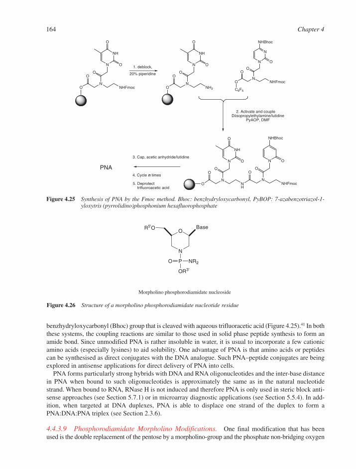
164 Chapter 4
N
OBaseR5'O
PO NR2
OR3'
Morpholino phosphorodiamidate nucleoside
Figure 4.26 Structure of a morpholino phosphorodiamidate nucleotide residue
benzhydryloxycarbonyl (Bhoc) group that is cleaved with aqueous trifluoracetic acid (Figure 4.25).41 In boththese systems, the coupling reactions are similar to those used in solid phase peptide synthesis to form anamide bond. Since unmodified PNA is rather insoluble in water, it is usual to incorporate a few cationicamino acids (especially lysines) to aid solubility. One advantage of PNA is that amino acids or peptidescan be synthesised as direct conjugates with the DNA analogue. Such PNA–peptide conjugates are beingexplored in antisense applications for direct delivery of PNA into cells.
PNA forms particularly strong hybrids with DNA and RNA oligonucleotides and the inter-base distancein PNA when bound to such oligonucleotides is approximately the same as in the natural nucleotidestrand. When bound to RNA, RNase H is not induced and therefore PNA is only used in steric block anti-sense approaches (see Section 5.7.1) or in microarray diagnostic applications (see Section 5.5.4). In add-ition, when targeted at DNA duplexes, PNA is able to displace one strand of the duplex to form aPNA:DNA:PNA triplex (see Section 2.3.6).
4.4.3.9 Phosphorodiamidate Morpholino Modifications. One final modification that has beenused is the double replacement of the pentose by a morpholino-group and the phosphate non-bridging oxygen
N
NH
O
O
N
OO
O NHFmoc
N
NH
O
O
N
OO
O NH
O
NNHFmoc
O
N O
NHBhoc
N
OO
O NH2
N
NH
O
ON
N
O
O
N
O
O NHFmoc
NHBhoc
C6F5
PNA
1. deblock,
20% piperidine
2. Activate and coupleDiisopropylethylamine/lutidine
PyAOP, DMF
3. Cap, acetic anhydride/lutidine
4. Cycle n times
5. Deprotecttrifluoroacetic acid
Figure 4.25 Synthesis of PNA by the Fmoc method. Bhoc: benzhydryloxycarbonyl, PyBOP: 7-azabenzotriazol-1-yloxytris (pyrrolidino)phosphonium hexafluorophosphate
Synthesis of Oligonucleotides 165
by an amino group to give a phosphorodiamidate morpholino (PMO) linkage (Figure 4.26). This type of mod-ification has a number of advantages that have warranted its use in steric block antisense applications.42
Such morpholino modifications give oligonucleotide analogues that are electrically neutral, show enhancedbinding to DNA and RNA, are completely nuclease resistant, and have lower toxicity and greater specificitythan phosphorothioate modifications. They have been used successfully in gene knockdown experimentsby microinjection into cells and embryos.
REFERENCES
1. S.L. Beaucage, in Current Protocols in Nucleic Acids Chemistry, Vol. 1, E.W. Harkins (ed). Wiley,2005, 2.1.
2. C.B. Reese, The chemical synthesis of oligo- and polynucleotides: a personal commentary.Tetrahedron, 2002, 58, 8893–8920.
3. H.G. Khorana, Total synthesis of a gene. Science, 1979, 203, 614–625.4. M.H. Caruthers, Gene synthesis machines – DNA chemistry and its uses. Science, 1985, 4723, 281–285.5. M.H. Caruthers, Chemical synthesis of DNA and DNA analogues. Acc. Chem. Res., 1991, 24, 278–284.6. S. Agrawal, Protocols for oligonucleotides and analogs. Humana Press, Totowa, New Jersey, 1993.7. E.W. Harkins, Current Protocols in Nucleic Acids Chemistry. Wiley, 2004.8. M.J. Gait, C.E. Pritchard and G. Slim, in Oligonucleotides and Analogues: A Practical Approach,
F. Eckstein (ed). Oxford University Press, Oxford, UK, 1991, 25–48.9. S. Pitsch, P.A. Weiss, L. Jenny, A. Stutz and X. Wu, Reliable synthesis of oligoribonucleotides (RNA)
with 2�-O-[(triisopropylsilyl)oxy]methyl (2�-O-tom)-protected phosphoramidites. Helv. Chim. Acta,2001, 84, 3773–3795.
10. S.A. Scaringe, F.E. Wincott and M.H. Caruthers, Novel RNA synthesis method using5�-silyl-2�-orthoester protecting groups. J. Am. Chem. Soc., 1998, 120, 11820–11821.
11. J.F. Milligan, D.R. Groebe, G.W. Witherell and O.C. Uhlenbeck, Oligoribonucleotide synthesis usingT7 RNA polymerase and synthetic DNA templates. Nucleic Acids Res., 1987, 15, 8783–8798.
12. S.R. Price, C. Oubridge, G. Varani and K. Nagai, in RNA: Protein Interactions. A Practical Approach,C.W.J. Smith (ed). OUP, Oxford, 1998, 37–74.
13. T. Middleton, W.C. Herlihy, P. Schimmel and H.N. Munro, Synthesis and purification of oligoribonu-cleotides using T4 RNA ligase and reverse phase chromatography. Anal. Biochem., 1985, 144, 110–117.
14. M.J. Moore and P.A. Sharp, Site-specific modification of pre-mRNA: the 2�-hydroxyl groups at thesplice sites. Science, 1992, 256, 992–997.
15. P. Herdewijn, Heterocyclic modifications of oligonucleotides and antisense technology. AntisenseNucl. Acid Drug Dev., 2000, 10, 297–310.
16. P. Hensley, Defining the structure and stability of macromolecular assemblies in solution: the re-emergence of analytical ultracentrifugation as a practical tool. Structure, 1996, 4, 367–373.
17. I. Luyten and P. Herdewijn, Hybridisation properties of base-modified oligonucleotides within thedouble and triple helix motif. Eur. J. Med. Chem., 1998, 33, 515–576.
18. F. Eckstein, Nucleoside phosphorothioates. Ann. Rev. Biochem., 1985, 54, 367–402.19. F. Eckstein and G. Gish, Phosphorothioates in molecular biology. Trends Biol. Sci., 1989, 14, 97–100.20. Specialist Periodical Reports: Organophosphorus Chemistry. Royal Society of Chemistry, Cambridge,
2004, Vol. 33.21. S.L. Beaucage and R.P. Iyer, The functionalization of oligonucleotides via phosphoramidite derivatives.
Tetrahedron, 1993, 49, 1925–1963.22. M. Manoharan, Oligonucleotide conjugates as potential antisense drugs with improved uptake, biodistri-
bution, targeted delivery, and mechanism of action. Antisense Nucl. Acid Drug Dev., 2002, 12, 103–128.23. B.S. Sproat, Chemistry and applications of oligonucleotide analogues. J. Biotechnol., 1995, 41, 221–238.24. R.P. Iyer, A. Roland, W. Zhou and K. Ghosh, Modified oligonucleotides-synthesis, properties and
applications. Curr. Opin. Mol. Ther., 1999, 1, 344–358.
25. C. Leumann, DNA analogues: from supramolecular principles to biological properties. Bioorg. Med.Chem., 2002, 10, 841–854.
26. R.P. Iyer, W. Egan, J.B. Regan and S.L. Beaucage, 3H-1,2-Benzodithiole-3-one 1,1-dioxide as animproved sulfurizing reagent in the solid-phase synthesis of oligodeoxyribonucleoside phosphoroth-ioates. J. Am. Chem. Soc., 1990, 112, 1253–1254.
27. W.J. Stec and A. Wilk, Stereocontrolled synthesis of oligo(nucleoside phosphorothioate)s. Angew.Chem. Int. Ed., 1994, 33, 709–722.
28. P. Guga and W.J. Stec, Synthesis of phosphorothioate oligonucleotides with stereodefinedphosphorothioate linkages. Wiley, Hoboken, NJ, 2003.
29. M. Boczkowskaa, P. Guga and W.J. Stec, Stereodefined phosphorothioate analogues of DNA: relativethermodynamic stability of the model PS-DNA/DNA and PS-DNA/RNA complexes. Biochemistry,2002, 41, 12483–12487.
30. W.T. Wiesler and M.H. Caruthers, Synthesis of phosphorodithioate DNA via sulfur-linked base-labileprotecting groups. J. Am. Chem. Soc., 1996, 61, 4272–4281.
31. P.S. Miller, M.P. Reddy, A. Murakami, K.R. Blake, S.-B. Lin and C.H. Agris, Solid-phase synthesis ofoligodeoxyribonucleoside methylphosphonates. Biochemistry, 1986, 25, 5092–5097.
32. J.-K. Chen, R.G. Schultz, D.H. Lloyd and S.M. Gryaznov, Synthesis of oligodeoxyribonucleotideN3�-P5� phosphoramidates. Nucleic Acids Res., 1995, 23, 2661–2668.
33. J.S. Nelson, K.L. Fearon, M.Q. Nguyen, S.N. McCurdy, J.E. Frediant, M.F. Foy andB.L. Hirschbein, N3�-P5� oligodeoxyribonucleotides phosphoramidates: a new method of synthesisbased on a phosphoramidite amin-exchange reaction. J. Org. Chem., 1997, 62, 7278–2287.
34. J.S. Summers and B.R. Shaw, Boranophosphates as mimics of natural phosphodiesters in DNA. Curr.Med. Chem., 2001, 8, 1147–1155.
35. S.M. Frier and K.-H. Altmann, The ups and downs of nucleic acid duplex stability: structure-stability studies on chemically modified DNA:RNA duplexes. Nucleic Acids Res., 1997, 25, 4429–4443.
36. M.J. Damha, C.J. Wilds, A. Noronha, I. Brukner, G. Borkow, D. Arion and M.A. Parniak, Hybrids ofRNA and arabinonucleic acids (ANA abd 2�-F-ANA) are substrated of ribonuclease H. J. Am. Chem.Soc., 1998, 120, 12976–12977.
37. S. Obika, D. Nanbu, Y. Hari, K. Morio, in Synthesis of 2�-O,4�-C-methylenuridine and -cytidine.Novel bicyclic nucleosides having a fixed C-3�,-endo sugar puckering. Tetrahedron Lett., Y. Ishidaand T. Imanishi (eds), 1997, 38, 8735–8738.
38. S.K. Singh, P. Nielsen, A.A. Koshkin and J. Wengel, LNA (locked nucleic acids): synthesis and high-affinity nucleic acid recognition. J. Chem. Soc. Chem. Commun., 1998, 455–456.
39. P.E. Nielsen, M. Egholm, Berg and O. Buchardt, Sequence-selective recognition of DNA by stranddisplacement with a thymine-substituted polyamide. Science, 1991, 5037, 1497–1500.
40. M. Egholm, O. Buchardt, L. Christensen, C. Behrens, S.M. Freier, D.A. Driver, R.H. Berg,S.K. Kim, B. Norden and P. Nielsen, PNA hybridizes to complementary oligonucleotides obeying theWatson–Crick hydrogen bonding rules. Nature, 1993, 365, 566–568.
41. S.A. Thomson, J.A. Josey, R. Cadilla, M.D. Gaul, C.F. Hassman, M.J. Luzzio, A.J. Pipe, K.L. Reed,D.J. Ricca, R.W. Wiethe et al., Fmoc mediated synthesis of peptide nucleic acids. Tetrahedron, 1995,51, 6179–6194.
42. J. Summerton, D. Stein, S.B. Huang, P. Matthews, D.D. Weller and M. Partridge, Morpholino andphosphorothioate antisense oligomers compared in cell-free and in cell systems. Antisense Nucl. AcidDrug Dev., 1997, 7, 63–70.
166 Chapter 4
CHAPTER 5
Nucleic Acids in Biotechnology
CONTENTS
5.1 DNA Sequence Determination 1685.1.1 Principles of DNA Sequencing 1685.1.2 Automated Fluorescent DNA Sequencing 1695.1.3 RNA Sequencing by Reverse Transcription 170
5.2 Gene Cloning 1705.2.1 Classical Cloning 1705.2.2 The Polymerase Chain Reaction 173
5.3 Enzymes Useful in Gene Manipulation 1745.3.1 Restriction Endonucleases 1745.3.2 Other Nucleases 1755.3.3 Polynucleotide Kinase 1765.3.4 Alkaline Phosphatase 1765.3.5 DNA Ligase 176
5.4 Gene Synthesis 1775.4.1 Classical Gene Synthesis 1775.4.2 Gene Synthesis by the Polymerase Chain Reaction 178
5.5 The Detection of Nucleic Acid Sequences by Hybridisation 1785.5.1 Parameters that Affect Nucleic Acid Hybridisation 1795.5.2 Southern and Northern Blot Analyses 1805.5.3 DNA Fingerprinting 1815.5.4 DNA Microarrays 1845.5.5 In Situ Analysis of RNA in Whole Organisms 188
5.6 Gene Mutagenesis 1885.6.1 Site-Specific In Vitro Mutagenesis 1885.6.2 Random Mutagenesis 1915.6.3 Gene Therapy 192
5.7 Oligonucleotides as Reagents and Therapeutics 1935.7.1 Antisense and Steric Block Oligonucleotides 1935.7.2 RNA Interference 1975.7.3 In Vitro Selection 198
5.8 DNA Footprinting 203References 205
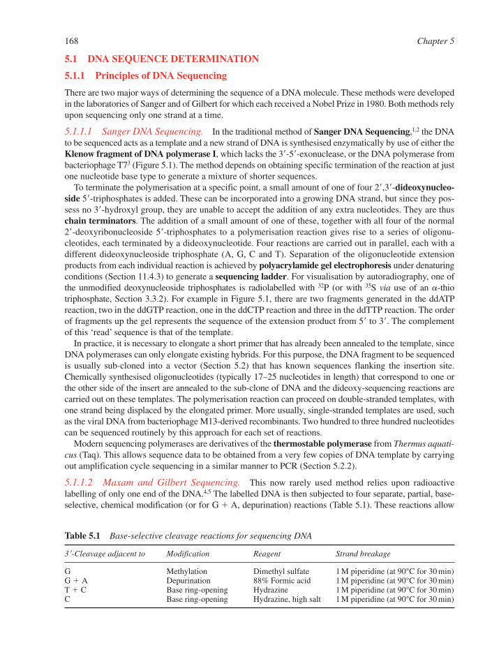
5.1 DNA SEQUENCE DETERMINATION
5.1.1 Principles of DNA Sequencing
There are two major ways of determining the sequence of a DNA molecule. These methods were developedin the laboratories of Sanger and of Gilbert for which each received a Nobel Prize in 1980. Both methods relyupon sequencing only one strand at a time.
5.1.1.1 Sanger DNA Sequencing. In the traditional method of Sanger DNA Sequencing,1,2 the DNAto be sequenced acts as a template and a new strand of DNA is synthesised enzymatically by use of either theKlenow fragment of DNA polymerase I, which lacks the 3�-5�-exonuclease, or the DNA polymerase frombacteriophage T73 (Figure 5.1). The method depends on obtaining specific termination of the reaction at justone nucleotide base type to generate a mixture of shorter sequences.
To terminate the polymerisation at a specific point, a small amount of one of four 2�,3�-dideoxynucleo-side 5�-triphosphates is added. These can be incorporated into a growing DNA strand, but since they pos-sess no 3�-hydroxyl group, they are unable to accept the addition of any extra nucleotides. They are thuschain terminators. The addition of a small amount of one of these, together with all four of the normal 2�-deoxyribonucleoside 5�-triphosphates to a polymerisation reaction gives rise to a series of oligonu-cleotides, each terminated by a dideoxynucleotide. Four reactions are carried out in parallel, each with adifferent dideoxynucleoside triphosphate (A, G, C and T). Separation of the oligonucleotide extensionproducts from each individual reaction is achieved by polyacrylamide gel electrophoresis under denaturingconditions (Section 11.4.3) to generate a sequencing ladder. For visualisation by autoradiography, one ofthe unmodified deoxynucleoside triphosphates is radiolabelled with 32P (or with 35S via use of an �-thiotriphosphate, Section 3.3.2). For example in Figure 5.1, there are two fragments generated in the ddATPreaction, two in the ddGTP reaction, one in the ddCTP reaction and three in the ddTTP reaction. The orderof fragments up the gel represents the sequence of the extension product from 5� to 3�. The complementof this ‘read’ sequence is that of the template.
In practice, it is necessary to elongate a short primer that has already been annealed to the template, sinceDNA polymerases can only elongate existing hybrids. For this purpose, the DNA fragment to be sequencedis usually sub-cloned into a vector (Section 5.2) that has known sequences flanking the insertion site.Chemically synthesised oligonucleotides (typically 17–25 nucleotides in length) that correspond to one orthe other side of the insert are annealed to the sub-clone of DNA and the dideoxy-sequencing reactions arecarried out on these templates. The polymerisation reaction can proceed on double-stranded templates, withone strand being displaced by the elongated primer. More usually, single-stranded templates are used, suchas the viral DNA from bacteriophage M13-derived recombinants. Two hundred to three hundred nucleotidescan be sequenced routinely by this approach for each set of reactions.
Modern sequencing polymerases are derivatives of the thermostable polymerase from Thermus aquati-cus (Taq). This allows sequence data to be obtained from a very few copies of DNA template by carryingout amplification cycle sequencing in a similar manner to PCR (Section 5.2.2).
5.1.1.2 Maxam and Gilbert Sequencing. This now rarely used method relies upon radioactivelabelling of only one end of the DNA.4,5 The labelled DNA is then subjected to four separate, partial, base-selective, chemical modification (or for G � A, depurination) reactions (Table 5.1). These reactions allow
168 Chapter 5
Table 5.1 Base-selective cleavage reactions for sequencing DNA
3�-Cleavage adjacent to Modification Reagent Strand breakage
G Methylation Dimethyl sulfate 1 M piperidine (at 90°C for 30 min)G � A Depurination 88% Formic acid 1 M piperidine (at 90°C for 30 min)T � C Base ring-opening Hydrazine 1 M piperidine (at 90°C for 30 min)C Base ring-opening Hydrazine, high salt 1 M piperidine (at 90°C for 30 min)

Nucleic Acids in Biotechnology 169
Figure 5.1 The principles of classical DNA sequencing. A primer oligodeoxynucleotide is annealed to the DNA tem-plate to be sequenced (top) and four separate extension reactions are carried out in the presenceof DNA polymerase I, the four deoxynucleoside triphosphates (one usually 32P-labelled and a single 2�-3�-dideoxynucleoside triphosphate) to produce a series of truncated extension products (middle). Theproducts of the reactions are separated by denaturing polyacrylamide gel electrophoresis and the gelautoradiographed to obtain a DNA sequence ladder (bottom)
the DNA to become sensitive at these sites to cleavage by alkaline hydrolysis. The fragments created are thenseparated by polyacrylamide gel electrophoresis in very much the same way as for Sanger sequencing.
5.1.2 Automated Fluorescent DNA Sequencing
Machines have now been developed to separate and identify the products of dideoxy-sequencing reac-tions. Here a fluorescent label is built into a set of four alternative dideoxynucleotide chain terminators

each with a different dye attached (Section 8.5.4, Figure 8.11). The four sequencing reactions are carriedout together in one reaction and subjected to capillary electrophoresis in free solution. As each length offluorescently labelled oligonucleotide emerges from the capillary column, it is detected by a fluorescencedetector, the particular colour in each case corresponding to one of the four dideoxynucleotides. A com-puter analyses the data and produces a series of fluorescent signals that correspond to the read sequence.Such machines are capable of generating sequence reads of 500–1000 residues.
Sequencing machines have proved to be essential in large-scale DNA sequencing of genomes. For example,the DNA sequences of the yeast Saccharomyces cerevisiae, the fruitfly Drosophila melanogaster and Homosapiens have been determined in this way. In genome sequencing, it is usual for the sequence to be deter-mined three to ten times from different clones or fragments, including from both strands of the DNA, toobtain higher accuracy. Powerful computer programmes are then able to determine overlaps between frag-ments, and align the sequences against genome maps (Section 6.5.2). The positions of genes, introns and alter-native splicing patterns can be predicted and genomes compared between different organisms to obtainknowledge of the RNA transcripts as well encoded proteins.
5.1.3 RNA Sequencing by Reverse Transcription
It is possible to carry out base-specific chemical treatments or digestions by nuclease enzymes to determine anRNA sequence directly. However, a more common procedure is to use a reverse transcriptase enzyme tomake a cDNA copy of the single-stranded RNA. The reaction is initiated by use of an oligodeoxyribonucleotideprimer from a known part of the sequence. This cDNA can then be amplified by the polymerase chain reaction (PCR) (Section 5.2.2) and sequence determined by standard DNA sequencing. The method onlygives information regarding the base sequence and not regarding RNA modifications.
5.2 GENE CLONING
Cloning is the technique of growing large quantities of genetically identical cells or organisms that arederived from a single ancestor (clones). Gene cloning is an extension of this whereby a particular gene, groupof genes or a fragment of DNA is selected from a mixed population (often a complete genome) and ampli-fied to a huge extent. This can be carried out by insertion of the chosen DNA into a vector DNA and intro-duction of the hybrid (recombinant DNA) into cells by transformation (transfection). The cells containingthe recombinant DNA are propagated and each cell in a colony contains an exact copy (or copies) of the gene‘cloned’ in the vector. Cloning and recombinant DNA technology are well documented in established text-books and manuals.6,7
Nowadays, a separate and complementary approach that uses PCR can achieve the same objective in afraction of the time, of amplification of DNA segments without involving living cells.
5.2.1 Classical Cloning
5.2.1.1 Vectors. Several classes of vector exist into which a foreign DNA can be inserted and ampli-fied. The major classes are plasmids, bacteriophage and cosmids and bacterial or yeast artificial chromo-somes (YACs). Prokaryotic plasmids are almost always circular double-stranded DNAs that contain antibioticresistance genes as markers and a variety of restriction sites that can be used for insertion of the foreign DNA.A large number of plasmids are available for use in E. coli. The most useful bacteriophage is �, which hasbeen engineered in many ways to accept inserts of many different sizes and types, up to approximately20,000 base pairs. Because the transformation frequency is high, screening is easy. Cosmids are large plas-mids that contain the packaging site for bacteriophage � DNA. Therefore they can either be packaged intophage particles or they can be replicated as plasmids. Since the amount of DNA that can be packaged in a� phage particle is 50,000 base pairs, the potential size of cosmid inserts is very large. Cosmids have beenused in chromosome walking (see later this section), but they are somewhat more difficult to manipulatethan is �. Artificial chromosomes are vectors containing the constituents of natural yeast chromosomes,
170 Chapter 5
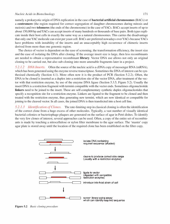
namely a prokaryotic origin of DNA replication in the case of bacterial artificial chromosomes (BACs) ora centromere (the region required for correct segregation of daughter chromosomes during mitosis andmeiosis) and two telomeres (the ends of the chromosome) in the case of YACs. BACs accept inserts of up toabout 150,000 bp and YACs can accept inserts of many hundreds or thousands of base pairs. Both types repli-cate inside their host cells in exactly the same way as a natural chromosomes. This carries the disadvantagethat only one YAC molecule can exist per yeast cell. BACs are preferred nowadays over YACs because YACshave problems with instability of the inserts and an unacceptably high occurrence of chimeric insertsderived from more than one genomic region.
The choice of vector is dependent on the ease of screening, the transformation efficiency, the insert sizeand the ease of isolating the DNA after cloning. If the average insert size is large, then less recombinantsare needed to obtain a representative recombinant library. Vector DNA use allows not only an originalcloning to be carried out, but also sub-cloning into more amenable fragments later in a project.
5.2.1.2 DNA Inserts. Often the source of the nucleic acid is a cDNA copy of messenger RNA (mRNA),which has been generated using the enzyme reverse transcriptase. Sometimes the DNA of interest can be syn-thesised chemically (Section 4.1). More often now it is the product of PCR (Section 5.2.2). Often, theDNA to be cloned is inserted as a duplex into a restriction site of the vector DNA, after treatment of the vec-tor with that restriction enzyme, by use of the enzyme DNA ligase (Section 5.3.5, Figure 5.2). Usually theinsert DNA is a restriction fragment with termini compatible with the vector ends. Sometimes oligonucleotidelinkers need to be joined to the insert. These are self-complementary synthetic duplex oligonucleotides thatspecify a recognition site for a restriction enzyme. Linkers are ligated to the fragment to be cloned and thentreated with the restriction enzyme, thus generating new termini, which are now identical or compatible forjoining to the cleaved vector. In all cases, the joined DNA is then transfected into a host cell line.
5.2.1.3 Identification of Clones. The rate-limiting step in classical cloning is often the identificationof the correct clone from a huge excess of other molecules. Typically, a vast number of visually identicalbacterial colonies or bacteriophage plaques are generated on the surface of agar in Petri dishes. To identifythe very few clones of interest, several approaches can be used. Often, a copy of the entire set of recombin-ants is made by touching a nitrocellulose or nylon filter membrane to the agar surface. The ‘master’ copyagar plate is stored away until the location of the required clone has been established on the filter copy.
Nucleic Acids in Biotechnology 171
Figure 5.2 Basic cloning procedure
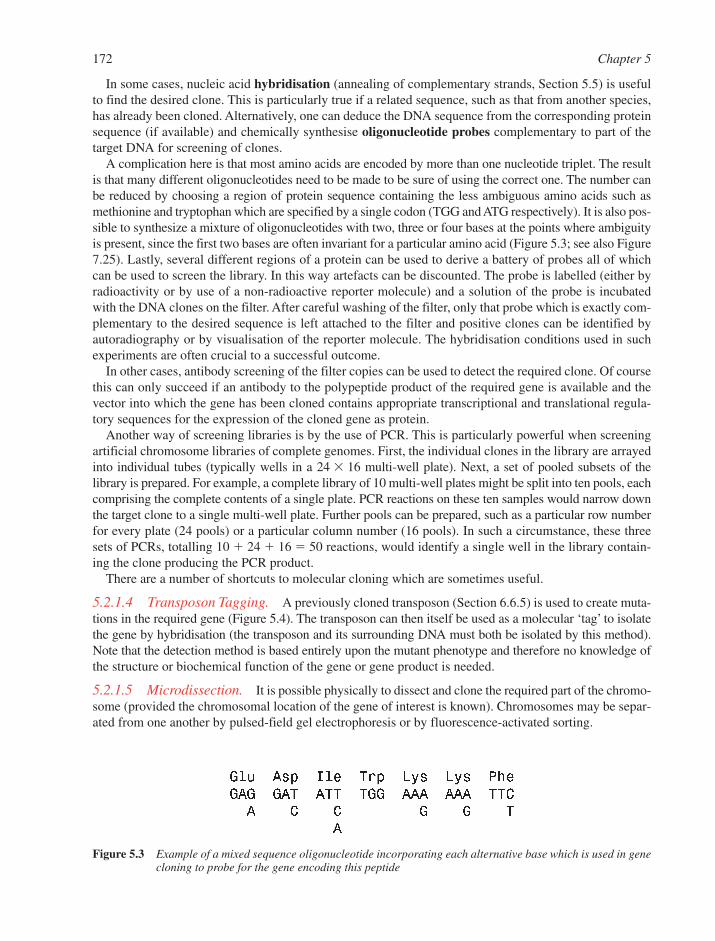
In some cases, nucleic acid hybridisation (annealing of complementary strands, Section 5.5) is usefulto find the desired clone. This is particularly true if a related sequence, such as that from another species,has already been cloned. Alternatively, one can deduce the DNA sequence from the corresponding proteinsequence (if available) and chemically synthesise oligonucleotide probes complementary to part of thetarget DNA for screening of clones.
A complication here is that most amino acids are encoded by more than one nucleotide triplet. The resultis that many different oligonucleotides need to be made to be sure of using the correct one. The number canbe reduced by choosing a region of protein sequence containing the less ambiguous amino acids such asmethionine and tryptophan which are specified by a single codon (TGG and ATG respectively). It is also pos-sible to synthesize a mixture of oligonucleotides with two, three or four bases at the points where ambiguityis present, since the first two bases are often invariant for a particular amino acid (Figure 5.3; see also Figure7.25). Lastly, several different regions of a protein can be used to derive a battery of probes all of whichcan be used to screen the library. In this way artefacts can be discounted. The probe is labelled (either byradioactivity or by use of a non-radioactive reporter molecule) and a solution of the probe is incubatedwith the DNA clones on the filter. After careful washing of the filter, only that probe which is exactly com-plementary to the desired sequence is left attached to the filter and positive clones can be identified byautoradiography or by visualisation of the reporter molecule. The hybridisation conditions used in suchexperiments are often crucial to a successful outcome.
In other cases, antibody screening of the filter copies can be used to detect the required clone. Of coursethis can only succeed if an antibody to the polypeptide product of the required gene is available and thevector into which the gene has been cloned contains appropriate transcriptional and translational regula-tory sequences for the expression of the cloned gene as protein.
Another way of screening libraries is by the use of PCR. This is particularly powerful when screeningartificial chromosome libraries of complete genomes. First, the individual clones in the library are arrayedinto individual tubes (typically wells in a 24 � 16 multi-well plate). Next, a set of pooled subsets of thelibrary is prepared. For example, a complete library of 10 multi-well plates might be split into ten pools, eachcomprising the complete contents of a single plate. PCR reactions on these ten samples would narrow downthe target clone to a single multi-well plate. Further pools can be prepared, such as a particular row numberfor every plate (24 pools) or a particular column number (16 pools). In such a circumstance, these threesets of PCRs, totalling 10 � 24 � 16 � 50 reactions, would identify a single well in the library contain-ing the clone producing the PCR product.
There are a number of shortcuts to molecular cloning which are sometimes useful.
5.2.1.4 Transposon Tagging. A previously cloned transposon (Section 6.6.5) is used to create muta-tions in the required gene (Figure 5.4). The transposon can then itself be used as a molecular ‘tag’ to isolatethe gene by hybridisation (the transposon and its surrounding DNA must both be isolated by this method).Note that the detection method is based entirely upon the mutant phenotype and therefore no knowledge ofthe structure or biochemical function of the gene or gene product is needed.
5.2.1.5 Microdissection. It is possible physically to dissect and clone the required part of the chromo-some (provided the chromosomal location of the gene of interest is known). Chromosomes may be separ-ated from one another by pulsed-field gel electrophoresis or by fluorescence-activated sorting.
172 Chapter 5
Figure 5.3 Example of a mixed sequence oligonucleotide incorporating each alternative base which is used in genecloning to probe for the gene encoding this peptide
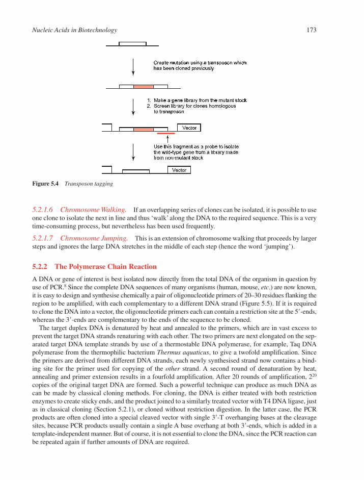
Nucleic Acids in Biotechnology 173
Figure 5.4 Transposon tagging
5.2.1.6 Chromosome Walking. If an overlapping series of clones can be isolated, it is possible to useone clone to isolate the next in line and thus ‘walk’ along the DNA to the required sequence. This is a verytime-consuming process, but nevertheless has been used frequently.
5.2.1.7 Chromosome Jumping. This is an extension of chromosome walking that proceeds by largersteps and ignores the large DNA stretches in the middle of each step (hence the word ‘jumping’).
5.2.2 The Polymerase Chain Reaction
A DNA or gene of interest is best isolated now directly from the total DNA of the organism in question byuse of PCR.8 Since the complete DNA sequences of many organisms (human, mouse, etc.) are now known,it is easy to design and synthesise chemically a pair of oligonucleotide primers of 20–30 residues flanking theregion to be amplified, with each complementary to a different DNA strand (Figure 5.5). If it is requiredto clone the DNA into a vector, the oligonucleotide primers each can contain a restriction site at the 5�-ends,whereas the 3�-ends are complementary to the ends of the sequence to be cloned.
The target duplex DNA is denatured by heat and annealed to the primers, which are in vast excess toprevent the target DNA strands renaturing with each other. The two primers are next elongated on the sep-arated target DNA template strands by use of a thermostable DNA polymerase, for example, Taq DNApolymerase from the thermophilic bacterium Thermus aquaticus, to give a twofold amplification. Sincethe primers are derived from different DNA strands, each newly synthesised strand now contains a bind-ing site for the primer used for copying of the other strand. A second round of denaturation by heat,annealing and primer extension results in a fourfold amplification. After 20 rounds of amplification, 220
copies of the original target DNA are formed. Such a powerful technique can produce as much DNA ascan be made by classical cloning methods. For cloning, the DNA is either treated with both restrictionenzymes to create sticky ends, and the product joined to a similarly treated vector with T4 DNA ligase, justas in classical cloning (Section 5.2.1), or cloned without restriction digestion. In the latter case, the PCRproducts are often cloned into a special cleaved vector with single 3�-T overhanging bases at the cleavagesites, because PCR products usually contain a single A base overhang at both 3�-ends, which is added in atemplate-independent manner. But of course, it is not essential to clone the DNA, since the PCR reaction canbe repeated again if further amounts of DNA are required.
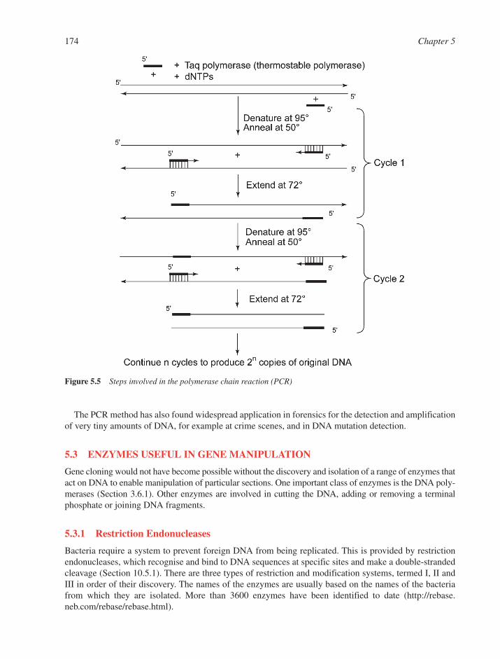
The PCR method has also found widespread application in forensics for the detection and amplificationof very tiny amounts of DNA, for example at crime scenes, and in DNA mutation detection.
5.3 ENZYMES USEFUL IN GENE MANIPULATION
Gene cloning would not have become possible without the discovery and isolation of a range of enzymes thatact on DNA to enable manipulation of particular sections. One important class of enzymes is the DNA poly-merases (Section 3.6.1). Other enzymes are involved in cutting the DNA, adding or removing a terminalphosphate or joining DNA fragments.
5.3.1 Restriction Endonucleases
Bacteria require a system to prevent foreign DNA from being replicated. This is provided by restrictionendonucleases, which recognise and bind to DNA sequences at specific sites and make a double-strandedcleavage (Section 10.5.1). There are three types of restriction and modification systems, termed I, II andIII in order of their discovery. The names of the enzymes are usually based on the names of the bacteriafrom which they are isolated. More than 3600 enzymes have been identified to date (http://rebase.neb.com/rebase/rebase.html).
174 Chapter 5
Figure 5.5 Steps involved in the polymerase chain reaction (PCR)
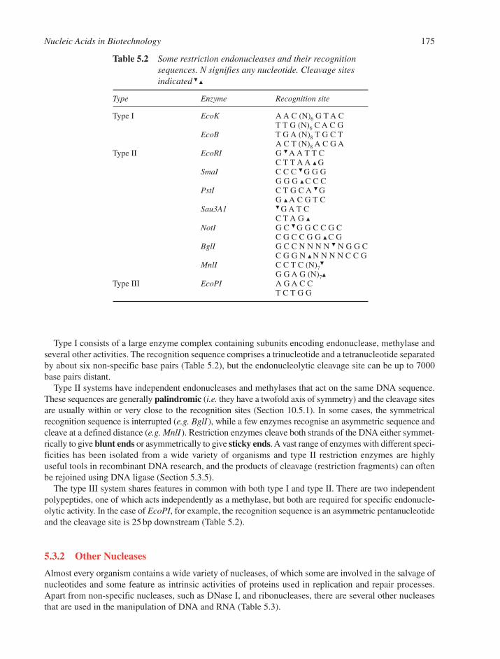
Type I consists of a large enzyme complex containing subunits encoding endonuclease, methylase andseveral other activities. The recognition sequence comprises a trinucleotide and a tetranucleotide separatedby about six non-specific base pairs (Table 5.2), but the endonucleolytic cleavage site can be up to 7000base pairs distant.
Type II systems have independent endonucleases and methylases that act on the same DNA sequence.These sequences are generally palindromic (i.e. they have a twofold axis of symmetry) and the cleavage sitesare usually within or very close to the recognition sites (Section 10.5.1). In some cases, the symmetricalrecognition sequence is interrupted (e.g. BglI), while a few enzymes recognise an asymmetric sequence andcleave at a defined distance (e.g. MnlI). Restriction enzymes cleave both strands of the DNA either symmet-rically to give blunt ends or asymmetrically to give sticky ends. A vast range of enzymes with different speci-ficities has been isolated from a wide variety of organisms and type II restriction enzymes are highlyuseful tools in recombinant DNA research, and the products of cleavage (restriction fragments) can oftenbe rejoined using DNA ligase (Section 5.3.5).
The type III system shares features in common with both type I and type II. There are two independentpolypeptides, one of which acts independently as a methylase, but both are required for specific endonucle-olytic activity. In the case of EcoPI, for example, the recognition sequence is an asymmetric pentanucleotideand the cleavage site is 25 bp downstream (Table 5.2).
5.3.2 Other Nucleases
Almost every organism contains a wide variety of nucleases, of which some are involved in the salvage ofnucleotides and some feature as intrinsic activities of proteins used in replication and repair processes.Apart from non-specific nucleases, such as DNase I, and ribonucleases, there are several other nucleasesthat are used in the manipulation of DNA and RNA (Table 5.3).
Nucleic Acids in Biotechnology 175
Table 5.2 Some restriction endonucleases and their recognitionsequences. N signifies any nucleotide. Cleavage sitesindicated �
�
Type Enzyme Recognition site
Type I EcoK A A C (N)6 G T A CT T G (N)6 C A C G
EcoB T G A (N)8 T G C TA C T (N)8 A C G A
Type II EcoRI G � A A T T CC T T A A � G
SmaI C C C � G G GG G G � C C C
PstI C T G C A � GG � A C G T C
Sau3A1 � G A T CC T A G �
NotI G C � G G C C G CC G C C G G � C G
BglI G C C N N N N � N G G CC G G N � N N N N C C G
MnlI C C T C (N)7�
G G A G (N)7�
Type III EcoPI A G A C CT C T G G

5.3.3 Polynucleotide Kinase
A polynucleotide kinase isolated from bacteriophage T4 catalyses the transfer of the �-phosphate of ATPto the 5�-hydroxyl terminus of DNA, RNA or an oligonucleotide in a reaction that requires magnesiumions. The enzyme is particularly useful for introducing a radioactive label on to the end of a polynu-cleotide, where the phosphate donor is �-32P-ATP. Both single- and double-stranded polynucleotides canbe phosphorylated, although recessed 5�-hydroxyl groups in double-stranded DNA, such as those obtainedby cleavage with certain restriction enzymes, are poorly phosphorylated. This sort of polynucleotidekinase activity, though not found in bacteria, has been found in some mammalian cells. The T4 enzyme is the only well-characterised kinase that has polynucleotides as substrates. This T4 protein also has a 5�-phosphatase activity which is unusually specific for a 3�-phosphate of a nucleoside or polynucleotide.
5.3.4 Alkaline Phosphatase
Phosphatases catalyse the hydrolysis of phosphate monoesters to produce inorganic phosphate and the corres-ponding alcohol. Most phosphatases are non-specific. Alkaline phosphatases are found in bacteria, fungi andhigher animals (but not plants) and will remove terminal phosphates from polynucleotides, carbohydratesand phospholipids. The E. coli enzyme is a dimer of molecular weight about 89 kDa, requires a zinc (II) ion,and is allosterically activated by magnesium ions. During dephosphorylation of the substrate, its phosphateis transferred to a serine residue on the enzyme located in the sequence Asp-Ser-Ala. This same sequence isfound in mammalian alkaline phosphatases (the calf intestinal enzyme is particularly well characterised) andit is similar to the active centre of serine proteases. Acidic phosphatases are also common, but these do notusually operate on polynucleotides as substrates.
5.3.5 DNA Ligase
A ligase is an enzyme that catalyses the formation of a phosphodiester linkage between two polynucleotidechains.9 In the case of DNA ligases, a 5�-phosphate group is esterified by an adjacent 3�-hydroxyl group andthere is concomitant hydrolysis of pyrophosphate in NAD� (bacterial enzymes) or ATP (phage and eukary-otic enzymes). Particularly efficient joining takes place when the phosphate and hydroxyl groups are heldclose together within a double helix, typically where the joining process seals a ‘nick’ and creates a perfectduplex (Figure 5.6). This situation occurs both in gene synthesis (Section 5.4) and in recombinant DNA tech-nology (Section 5.2) in ligation of identical ‘sticky ends’ formed by cleavage with a restriction endonuclease.E. coli and phage T4 DNA ligases are well-characterised enzymes which have an important role in DNA repli-cation (Section 6.6.4). T4 DNA ligase will join blunt DNA duplex ends when used at high concentrations and
176 Chapter 5
Table 5.3 Some endonucleases and their activities
Nuclease Origin Activities
Exonuclease III E. coli (1) ss exo-cleavage from 3�-ends of dsDNA(2) endo-cleavage for apurinic DNA(3) RNase H(4) 3�-phosphatase
Exonuclease VII E. coli ss exo-cleavage from 5�- or 3�-end of ssDNABal31 Alteromonas espejiana (1) ss exo- and endo-cleavage from 5�- or 3�-end
of dsDNA(2) ssDNA endo-cleavage
S1 Aspergillus oryzae ssDNA or RNA exo- and endo-cleavageLambda exonuclease Infected E. coli ss exo-cleavage from 5�-end of dsDNAPhosphodiesterase I Bovine spleen ss exo-cleavage from 5�-end of ssDNA or RNAPhosphodiesterase II Crotalus adamanteus ss exo-cleavage from 3�-end of ssDNA or RNA
(or other snakes)
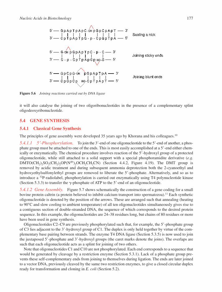
it will also catalyse the joining of two oligoribonucleotides in the presence of a complementary splintoligodeoxyribonucleotide.
5.4 GENE SYNTHESIS
5.4.1 Classical Gene Synthesis
The principles of gene assembly were developed 35 years ago by Khorana and his colleagues.10
5.4.1.1 5�-Phosphorylation. To join the 3�-end of one oligonucleotide to the 5�-end of another, a phos-phate group must be attached to one of the ends. This is most easily accomplished at a 5�-end either chem-ically or enzymatically. The chemical procedure involves reaction of the 5�-hydroxyl group of a protectedoligonucleotide, while still attached to a solid support with a special phosphoramidite derivative (e.g.DMTO(CH2)2SO2(CH2)2OP(NiPr)2OCH2CH2CN) (Section 4.4.2, Figure 4.19). The DMT group isremoved by acidic treatment and during subsequent ammonia deprotection both the 2-cyanoethyl andhydroxyethylsulfonylethyl groups are removed to liberate the 5�-phosphate. Alternatively, and so as tointroduce a 32P-radiolabel, phosphorylation is carried out enzymatically using T4 polynucleotide kinase(Section 5.3.3) to transfer the �-phosphate of ATP to the 5�-end of an oligonucleotide.
5.4.1.2 Gene Assembly. Figure 5.7 shows schematically the construction of a gene coding for a smallbovine protein caltrin (a protein believed to inhibit calcium transport into spermatozoa).11 Each syntheticoligonucleotide is denoted by the position of the arrows. These are arranged such that annealing (heatingto 90°C and slow cooling to ambient temperature) of all ten oligonucleotides simultaneously gives rise toa contiguous section of double-stranded DNA, the sequence of which corresponds to the desired proteinsequence. In this example, the oligonucleotides are 24–38 residues long, but chains of 80 residues or morehave been used in gene synthesis.
Oligonucleotides C2–C9 are previously phosphorylated such that, for example, the 5�-phosphate groupof C3 lies adjacent to the 3�-hydroxyl group of C1. The duplex is only held together by virtue of the com-plementary base pairing between strands. The enzyme T4 DNA ligase (Section 5.3.5) is now used to jointhe juxtaposed 5�-phosphate and 3�-hydroxyl groups (the caret marks denote the joins). The overlaps aresuch that each oligonucleotide acts as a splint for joining of two others.
Note that oligonucleotides C1 and C10 are not phosphorylated. Each end corresponds to a sequence thatwould be generated by cleavage by a restriction enzyme (Section 5.3.1). Lack of a phosphate group pre-vents these self-complementary ends from joining to themselves during ligation. The ends are later joinedto a vector DNA, previously cleaved by the same two restriction enzymes, to give a closed circular duplexready for transformation and cloning in E. coli (Section 5.2).
Nucleic Acids in Biotechnology 177
Figure 5.6 Joining reactions carried out by DNA ligase
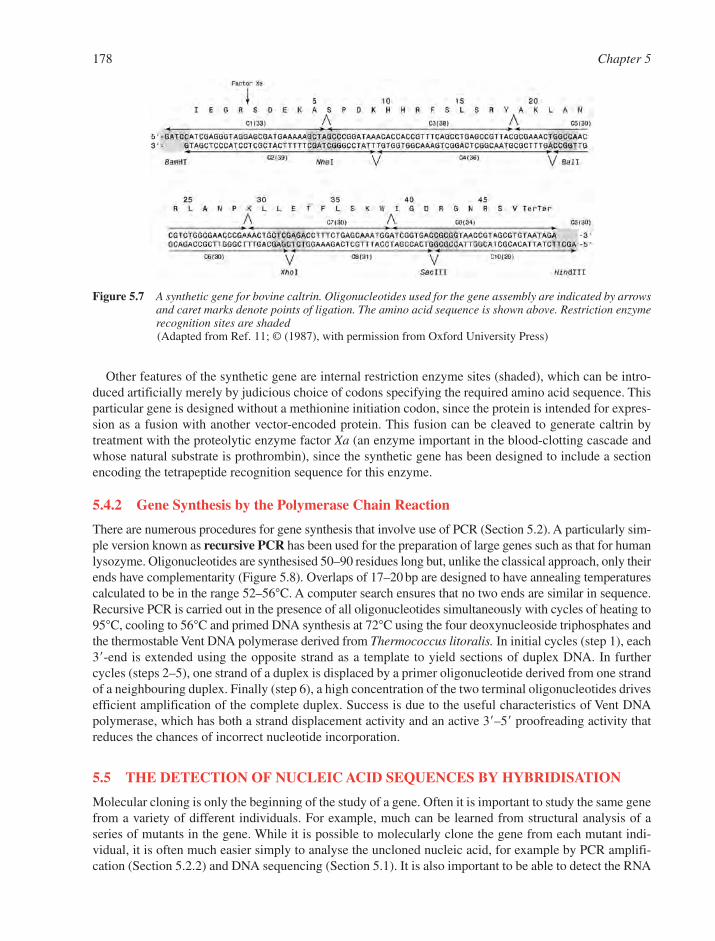
Other features of the synthetic gene are internal restriction enzyme sites (shaded), which can be intro-duced artificially merely by judicious choice of codons specifying the required amino acid sequence. Thisparticular gene is designed without a methionine initiation codon, since the protein is intended for expres-sion as a fusion with another vector-encoded protein. This fusion can be cleaved to generate caltrin bytreatment with the proteolytic enzyme factor Xa (an enzyme important in the blood-clotting cascade andwhose natural substrate is prothrombin), since the synthetic gene has been designed to include a sectionencoding the tetrapeptide recognition sequence for this enzyme.
5.4.2 Gene Synthesis by the Polymerase Chain Reaction
There are numerous procedures for gene synthesis that involve use of PCR (Section 5.2). A particularly sim-ple version known as recursive PCR has been used for the preparation of large genes such as that for humanlysozyme. Oligonucleotides are synthesised 50–90 residues long but, unlike the classical approach, only theirends have complementarity (Figure 5.8). Overlaps of 17–20 bp are designed to have annealing temperaturescalculated to be in the range 52–56°C. A computer search ensures that no two ends are similar in sequence.Recursive PCR is carried out in the presence of all oligonucleotides simultaneously with cycles of heating to95°C, cooling to 56°C and primed DNA synthesis at 72°C using the four deoxynucleoside triphosphates andthe thermostable Vent DNA polymerase derived from Thermococcus litoralis. In initial cycles (step 1), each3�-end is extended using the opposite strand as a template to yield sections of duplex DNA. In furthercycles (steps 2–5), one strand of a duplex is displaced by a primer oligonucleotide derived from one strandof a neighbouring duplex. Finally (step 6), a high concentration of the two terminal oligonucleotides drivesefficient amplification of the complete duplex. Success is due to the useful characteristics of Vent DNApolymerase, which has both a strand displacement activity and an active 3�–5� proofreading activity thatreduces the chances of incorrect nucleotide incorporation.
5.5 THE DETECTION OF NUCLEIC ACID SEQUENCES BY HYBRIDISATION
Molecular cloning is only the beginning of the study of a gene. Often it is important to study the same genefrom a variety of different individuals. For example, much can be learned from structural analysis of aseries of mutants in the gene. While it is possible to molecularly clone the gene from each mutant indi-vidual, it is often much easier simply to analyse the uncloned nucleic acid, for example by PCR amplifi-cation (Section 5.2.2) and DNA sequencing (Section 5.1). It is also important to be able to detect the RNA
178 Chapter 5
Figure 5.7 A synthetic gene for bovine caltrin. Oligonucleotides used for the gene assembly are indicated by arrowsand caret marks denote points of ligation. The amino acid sequence is shown above. Restriction enzymerecognition sites are shaded(Adapted from Ref. 11; © (1987), with permission from Oxford University Press)

encoded by a gene and to determine its levels of transcription and tissue specificity. Methods exist for bothof these purposes and both depend upon the ability of a single-stranded nucleic acid to pair specificallywith its complementary strand.
5.5.1 Parameters that Affect Nucleic Acid Hybridisation
Hybridisation is the annealing of two single strands of a nucleic acid to form a duplex. Duplex strength ismeasured by observation of the melting temperature (Tm) and is affected by several parameters.
5.5.1.1 Base Composition (%GC). Since G–C base pairs have three hydrogen bonds, they arestronger than A–T base pairs, which have only two. Thus duplexes with higher G–C content have highermelting temperatures.
5.5.1.2 Temperature (T). The rate of association of single-stranded DNA into a duplex variesmarkedly with temperature (Figure 5.9: see also Section 2.5.1). The shape of this curve is governed by two
Nucleic Acids in Biotechnology 179
Figure 5.8 Gene synthesis by recursive PCR. Bars represent oligonucleotides and their extension productsafter PCR

factors. At low temperatures, the re-association rate is determined by the difference in free energy betweenthe unassociated and the transition state.
(5.1)
where k is the re-association rate constant, Ea the activation free energy, R the gas constant and T the absolutetemperature. At higher temperatures, the stability of the duplex is markedly reduced until eventually it isunstable and the hybrid melts. Thus there is a fall off in re-association rate as this point is approached.
5.5.1.3 Monovalent Cation Concentration (M). The melting temperature of a hybrid (Section2.5.1) is reduced at lower salt concentration because cations help to stabilise a duplex. Divalent cationssuch as magnesium are much more effective in stabilisation of hybrids, but are less frequently used inhybridisation studies (Section 9.3).
5.5.1.4 Duplex Length (L). The melting temperature of a duplex shorter than a few hundred basepairs is length dependent. In practice, these four factors can be combined into an empirical equation giv-ing the melting temperature Tm of a hybrid DNA.
Tm � 69.3 � 0.41(%GC) � 18.5log10 M � 500 L�1/°C (5.2)
Web-based algorithms are available now for calculating Tm from knowledge of the various parameters(e.g. see http://www.basic.northwestern.edu/biotools/OligoCalc.html). Use of hybridisation temperaturesfrom 10 to 20°C below the calculated Tm of the hybrid is optimal in practice to ensure annealing of strands.
For synthetic oligonucleotide probes of 15–20 residues, the calculation of Tm is simplified to 2°C per dA·dTand 4°C per dG·dC base pair in 1 M sodium chloride solution. This is known as the Wallace rule. In the caseof the quaternary ammonium salt tetramethylammonium chloride (3 M TMAC), the Tm of a duplex is inde-pendent of base composition and is thus directly proportional to its length. This is of practical value forexample in cloning applications that involve hybridisation of mixed sequence oligonucleotides (Section 5.2.2).
5.5.2 Southern and Northern Blot Analyses
It is possible to use nucleic acid hybridisation to detect uncloned genomic DNA. Genomic DNA is immo-bilised on a nitrocellulose or nylon filter, in basically the same way as described for gene cloning (Section5.2.1). The gene of interest is detected on the filter by hybridising a complementary nucleic acid strandlabelled either with radioactivity or an affinity label such as biotin, which can be detected with great sensi-tivity. Of course, if the DNA is just spotted onto the filter, all that is seen is a spot whose intensity reflects the
k Z E RT� �e a /
180 Chapter 5
Figure 5.9 Dependence of the reassociation rate of DNA upon temperature
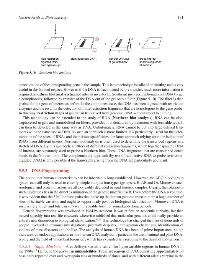
concentration of the corresponding gene in the sample. This latter technique is called dot blotting and is veryuseful in this limited respect. However, if the DNA is fractionated before transfer, much more information isacquired. Southern blot analysis (named after its inventor Ed Southern) involves fractionation of DNA by gelelectrophoresis, followed by transfer of the DNA out of the gel onto a filter (Figure 5.10). The filter is thenprobed for the gene of interest as before. In the commonest case, the DNA has been digested with restrictionenzymes and the result is the detection of those restriction fragments that are homologous to the gene probe.In this way, restriction maps of genes can be derived from genomic DNA without resort to cloning.
This technology can be extended to the study of RNA (Northern blot analysis). RNA can be elec-trophoresed in gels and immobilised on filters, provided it is denatured by treatment with formaldehyde. Itcan then be detected in the same way as DNA. Unfortunately, RNA cannot be cut into large defined frag-ments with the same ease as DNA, so such an approach is more limited. It is particularly useful for the deter-mination of the sizes of RNAs and their tissue specificities, the latter approach relying upon the isolation ofRNAs from different tissues. Northern blot analysis is often used to determine the transcribed regions in astretch of DNA. By this approach, a battery of different restriction fragments, which together span the DNAof interest, are separately used to probe a Northern blot. Those DNA fragments that are transcribed detectbands in the Northern blot. The complementary approach (by use of radioactive RNA to probe restriction-digested DNA) is only possible if the transcripts arising from the DNA are particularly abundant.
5.5.3 DNA Fingerprinting
The notion that human characteristics can be inherited is long established. However, the ABO blood-groupsystem can still only be used to classify people into just four types (groups A, B, AB and O). Moreover, suchserological and protein markers are all too readily degraded in aged forensic samples. Clearly, the solution tosuch limitations lies in the direct examination of the genetic material itself. Even before the DNA revolution,it was evident that the 3 billion base pairs that make up the human genome must contain a huge number ofsites of heritable variation and ought to support truly positive biological identification. Moreover, DNA issurprisingly tough and bits can survive in typeable form for remarkably long periods.
Genetic fingerprinting was developed in 1984 by accident. It was at first an academic curiosity, but thenmoved speedily into real-life casework where it established that molecular genetics could really provide anentirely new dimension to biological identification.12,13 This technology has changed the lives of thousands ofpeople involved in criminal investigations, paternity disputes, immigration challenges, identification ofvictims of mass disasters and the like. The analysis of human DNA has been of prime importance thoughthere are tremendous applications in non-human DNA analysis, in particular the use of animal and plant DNA-typing and the field of ‘microbial forensics’, which has expanded as a response to the threat of bio-terrorism.
5.5.3.1 Super Markers. Alec Jeffreys started a search for hypervariable regions in human DNA inthe 1980s.13 He found the answer in minisatellites. These are regions of DNA consisting approximately 30base pairs repeated over and over again tens or hundreds of times, and with different alleles varying in the
Nucleic Acids in Biotechnology 181
Figure 5.10 Southern blot analysis

number of stutters. The problem was how to access them. Jeffreys observed that a chance-studied min-isatellite tucked away inside a human gene looked rather familiar, not unlike the stutters in the few otherminisatellites described in the literature. The implications were clear – a hybridisation probe consisting ofthis DNA sequence motif shared by different minisatellites should latch onto many different minisatellitessimultaneously, giving unlimited access to these potentially extremely informative genetic markers.Minisatellites are simply detected by hybridisation of probes to Southern blots of restriction-enzyme-digested genomic DNA.
5.5.3.2 Stumbling upon DNA Fingerprinting. In September 1984, Jeffreys tested a range of samplesthat included DNA from a human father/mother/child trio. The results provided multiple, highly variableDNA fragments. While mother and father were obviously different, the child seemed to be a union of theDNA patterns of the parents.14 Improved technology was able to resolve large numbers of extremely vari-able DNA fragments containing these minisatellites (Figure 5.11), not just in humans but in other organismsas well. In humans, the banding patterns are individual-specific, with essentially zero chance of matchingeven between close relatives or members of an isolated inbred community. For any individual, the patternsare constant, irrespective of the source of DNA. The multiple markers that make up a DNA fingerprint areinherited in a simple Mendelian fashion, with each child receiving a random selection of about half of thefather’s bands and half of the mother’s. Happily, the term ‘DNA fingerprint’ was chosen rather than themore accurate description ‘idiosyncratic Southern blot minisatellite hybridisation profile’ (Section 5.5.2).13
5.5.3.3 The Evolution of Forensic Genetics. The amount of variation currently accessible in DNAis extremely informative. Sequence variations between different minisatellite loci allows probes to detectmany independent minisatellites simultaneously, yielding the hypervariable multi-band patterns known asDNA fingerprints.14,15 By use of only a single probe, the match probability is estimated to be 3 � 10�11,while two probes together give a value of 5 � 10�19. This is so low that the only individuals sharingDNA fingerprints are monozygotic twins.14 At the same time, a method known as differential lysis wasdeveloped15 that selectively enriches sperm concentration in vaginal fluid/semen mixtures, thereby avoid-ing the problem of the victim’s DNA (which is in great excess) masking that of a rapist.
5.5.3.4 Single-Locus Probes. Although use of DNA fingerprinting persisted for some years in pater-nity testing, criminal casework soon concentrated on the use of specific cloned minisatellites. Each ofthese ‘single-locus probes’ (SLPs) revealed only a single, highly polymorphic, restriction fragment lengthpolymorphism, thereby simplifying interpretation. Typically, four SLPs were used successively to probe aSouthern blot, yielding eight hyper-variable fragments per individual. SLPs were used in the first DNA-based criminal investigation in the UK in 1986.16
5.5.3.5 Profiling DNA. DNA fingerprints are excellent for some applications, but not for forensicinvestigations that have to identify the origin of a biological sample with as much certainty as possible.12
This is because fingerprint patterns are complex and their interpretation is readily open to challenge incourt, they are not easy to computerise, and they require significant amounts of good quality DNA, equiva-lent to that obtained from a drop of fresh blood. The solution to these problems was simple – the isolationand cloning of minisatellites. Each cloned minisatellite, used as a hybridisation probe, produces a muchsimpler pattern of just two bands per person, corresponding to the two alleles in an individual (Figure5.11c). Such simple profiles can be obtained using considerably less DNA (one hair root is enough), andthe estimated lengths of the DNA fragments easily support database construction. These DNA profileshave exposed the true variability of human minisatellites, some showing 100 or more different length alleles in human populations.
DNA profiles are not individual-specific no matter how variable the minisatellite is between unrelatedpeople. This is particularly true for siblings, who have a one in four chance of sharing exactly the sameprofile. Nevertheless, by typing DNA sequentially, typically with a battery of five different minisatellites,excellent levels of individual specificity are obtained, leading to routine match frequencies of one in a bil-lion with DNA profiling.
182 Chapter 5

5.5.3.6 PCR-Based Methods. The discovery of short tandem repeats (STRs) together with theintroduction of automated sequencing technology has led to the current powerful systems for the identifi-cation of individuals. Human forensic casework is now carried out using commercially developed auto-somal STR multiplexes (single-tube PCR (Section 5.2.2) that amplify multiple loci) and is establishedworldwide because of its advantages of high discriminating power, sensitivity, ability to resolve simplemixtures, speed and automation. The resulting reduced cost has paved the way for the creation of national STRDNA databases (http://www.cstl.nist.gov/div831/strbase/). For example, the UK National DNA Databasecontained some 2.5 million reference profiles and about 200,000 crime-scene profiles as at July 2004(http://www.forensic.gov.uk/forensic/news/press_releases/2003/NDNAD_Annual_Report_02-03.pdf).Automated sequencing equipment for multiplex analysis typically uses multi-channel capillary elec-trophoresis systems that detect fluorescently labelled PCR products. These are combined with robotics
Nucleic Acids in Biotechnology 183
Figure 5.11 The evolution of DNA typing systems. (a) The very first DNA fingerprints with a family group at left (M,mother; F, father; C, child) plus DNA from various non-human species. (b) Improved DNA fingerprintsfrom a single family with the father (analysed twice) and his 11 children. Note how DNA fingerprints read-ily distinguish even close relatives and how bands in the missing mother can be easily identified as bandsin the children that are not present in the father. (c) Simpler DNA profiles of unrelated people. (d) DNAprofiling using PCR-amplified microsatellites. Several microsatellites are amplified at the same time andthe resulting profiles are displayed on a computer and automatically interpreted for databasing(Courtesy of Orchid-Cellmark13; A.J. Jeffreys, Genetic Fingerprinting, The Darwin Lectures 2003,Darwin College, University of Cambridge, 2003, 49–67. © (2003), with permission from CambridgeUniversity Press)
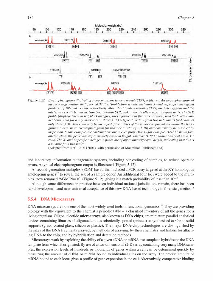
and laboratory information management systems, including bar coding of samples, to reduce operatorerrors. A typical electropherogram output is illustrated (Figure 5.12).
A ‘second-generation multiplex’ (SGM) has further included a PCR assay targeted at the XY-homologousamelogenin genes17 to reveal the sex of a sample donor. An additional four loci were added to the multi-plex, now renamed ‘SGM Plus10’ (Figure 5.12), giving it a match probability of less than 10–13.
Although some differences in practice between individual national jurisdictions remain, there has beenrapid development and near-universal acceptance of this new DNA-based technology in forensic genetics.18
5.5.4 DNA Microarrays
DNA microarrays are now one of the most widely used tools in functional genomics.19 They are providingbiology with the equivalent to the chemist’s periodic table – a classified inventory of all the genes for a living organism. Oligonucleotide microarrays, also known as DNA chips, are miniature parallel analyticaldevices containing libraries of oligonucleotides robotically spotted (printed) or synthesised in situ on solidsupports (glass, coated glass, silicon or plastic). The major DNA-chip technologies are distinguished bythe sizes of the DNA fragments arrayed, by methods of arraying, by their chemistry and linkers for attach-ing DNA to the chip, and by hybridisation and detection methods.
Microarrays work by exploiting the ability of a given cDNA or mRNA test sample to hybridise to the DNAtemplate from which it originated. By use of a two-dimensional (2-D) array containing very many DNA sam-ples, the expression levels of hundreds or thousands of genes within a cell can be determined quickly bymeasuring the amount of cDNA or mRNA bound to individual sites on the array. The precise amount ofmRNA bound to each locus gives a profile of gene expression in the cell. Alternatively, comparative binding
184 Chapter 5
Figure 5.12 Electropherograms illustrating autosomal short tandem repeat (STR) profiles. (a) An electropherogram ofthe second-generation multiplex ‘SGM Plus’profile from a male, including X- and Y-specific amelogeninproducts of 106 and 112 bp, respectively. Most short tandem repeats (STRs) are heterozygous and thealleles are evenly balanced. Numbers beneath STR peaks indicate allele sizes in repeat units. The STRprofile (displayed here as red, black and grey) uses a four-colour fluorescent system, with the fourth chan-nel being used for a size marker (not shown). (b) A typical mixture from two individuals (red channelonly shown). Mixtures can only be identified if the alleles of the minor component are above the back-ground ‘noise’ in an electropherogram (in practice a ratio of �1:10) and can usually be resolved byinspection. In this example, the contributions are in even proportions – for example, D21S11 shows fouralleles where the peaks are approximately equal in height, whereas D18S51 shows two peaks in a 3:1ratio. The X- and Y-specific amelogenin peaks are of approximately equal height, indicating that this isa mixture from two males(Adapted from Ref. 12; © (2004), with permission of Macmillan Publishers Ltd)
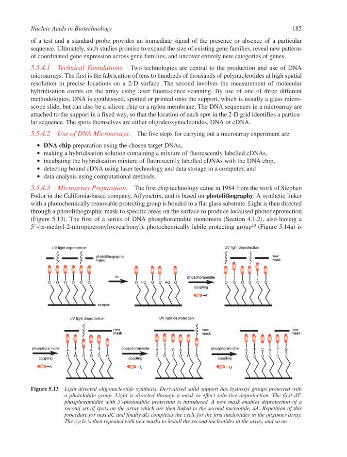
of a test and a standard probe provides an immediate signal of the presence or absence of a particularsequence. Ultimately, such studies promise to expand the size of existing gene families, reveal new patternsof coordinated gene expression across gene families, and uncover entirely new categories of genes.
5.5.4.1 Technical Foundations. Two technologies are central to the production and use of DNAmicroarrays. The first is the fabrication of tens to hundreds of thousands of polynucleotides at high spatialresolution in precise locations on a 2-D surface. The second involves the measurement of molecularhybridisation events on the array using laser fluorescence scanning. By use of one of three differentmethodologies, DNA is synthesised, spotted or printed onto the support, which is usually a glass micro-scope slide, but can also be a silicon chip or a nylon membrane. The DNA sequences in a microarray areattached to the support in a fixed way, so that the location of each spot in the 2-D grid identifies a particu-lar sequence. The spots themselves are either oligodeoxynucleotides, DNA or cDNA.
5.5.4.2 Use of DNA Microarrays. The five steps for carrying out a microarray experiment are
� DNA chip preparation using the chosen target DNAs,� making a hybridisation solution containing a mixture of fluorescently labelled cDNAs,� incubating the hybridisation mixture of fluorescently labelled cDNAs with the DNA chip,� detecting bound cDNA using laser technology and data storage in a computer, and� data analysis using computational methods.
5.5.4.3 Microarray Preparation. The first chip technology came in 1984 from the work of StephenFodor in the California-based company, Affymetrix, and is based on photolithography. A synthetic linkerwith a photochemically removable protecting group is bonded to a flat glass substrate. Light is then directedthrough a photolithographic mask to specific areas on the surface to produce localised photodeprotection(Figure 5.13). The first of a series of DNA phosphoramidite monomers (Section 4.1.2), also having a 5�-(�-methyl-2-nitropiperonyloxycarbonyl), photochemically labile protecting group20 (Figure 5.14a) is
Nucleic Acids in Biotechnology 185
Figure 5.13 Light directed oligonucleotide synthesis. Derivatised solid support has hydroxyl groups protected with a photolabile group. Light is directed through a mask to effect selective deprotection. The first dT-phosphoramidite with 5�-photolabile protection is introduced. A new mask enables deprotection of a second set of spots on the array which are then linked to the second nucleotide, dA. Repetition of this procedure for next dC and finally dG completes the cycle for the first nucleotides in the oligomer array.The cycle is then repeated with new masks to install the second nucleotides in the array, and so on
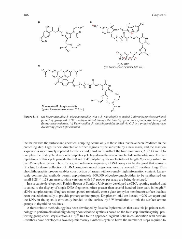
incubated with the surface and chemical coupling occurs only at those sites that have been irradiated in thepreceding step. Light is next directed at further regions of the substrate by a new mask, and the reactionsequence is successively repeated for the second, third and fourth of the four monomers, A, C, G and T tocomplete the first cycle. A second complete cycle lays down the second nucleotide in the oligomer. Furtherrepetitions of this cycle provide the full set of 4N polydeoxyribonucleotides of length N, or any subset, injust N complete cycles. Thus, for a given reference sequence, a DNA array can be designed that consistsof a highly dense collection of DNA single-stranded oligomers, usually around 25 residues long. Thisphotolithographic process enables construction of arrays with extremely high information content. Large-scale commercial methods permit approximately 300,000 oligodeoxynucleotides to be synthesised onsmall 1.28 � 1.28 cm arrays, while versions with 106 probes per array are being developed.
In a separate development, Patrick Brown at Stanford University developed a cDNA spotting method thatis suited to the display of single-DNA fragments, often greater than several hundred base pairs in length.21
cDNA samples (about 15 ng) are micro-spotted robotically onto a glass (or nylon membrane) surface that hasbeen treated chemically to provide primary amino groups. Droplets (�1 nL) are located �200 m apart andthe DNA in the spots is covalently bonded to the surface by UV irradiation to link the surface aminogroups to thymidine residues.
A third robotic methodology has been developed by Rosetta Inpharmatics that uses ink-jet printer tech-nology to perform classical oligodeoxyribonucleotide synthesis based on the four-step dimethoxytrityl pro-tecting group chemistry (Section 4.1.2).22 In a fourth approach, Agilent Labs in collaboration with MarvinCaruthers have developed a two-step microarray synthesis cycle to halve the number of steps required to
186 Chapter 5
Figure 5.14 (a) Deoxythymidine 3�-phosphoramidite with a 5�-photolabile �-methyl-2-nitropiperonyloxycarbonylprotecting group. (b) dUTP analogue linked through the 5-methyl group to a cyanine dye having red fluorescence emission. (c) Deoxyuridine 3�-phosphoramidite linked via C-5 to a protected fluoresceindye having green light emission
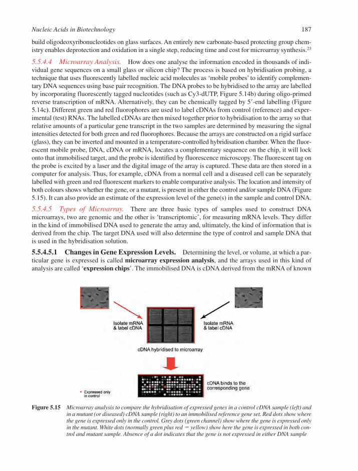
build oligodeoxyribonucleotides on glass surfaces. An entirely new carbonate-based protecting group chem-istry enables deprotection and oxidation in a single step, reducing time and cost for microarray synthesis.23
5.5.4.4 Microarray Analysis. How does one analyse the information encoded in thousands of indi-vidual gene sequences on a small glass or silicon chip? The process is based on hybridisation probing, atechnique that uses fluorescently labelled nucleic acid molecules as ‘mobile probes’ to identify complemen-tary DNA sequences using base pair recognition. The DNA probes to be hybridised to the array are labelledby incorporating fluorescently tagged nucleotides (such as Cy3-dUTP, Figure 5.14b) during oligo-primedreverse transcription of mRNA. Alternatively, they can be chemically tagged by 5�-end labelling (Figure5.14c). Different green and red fluorophores are used to label cDNAs from control (reference) and exper-imental (test) RNAs. The labelled cDNAs are then mixed together prior to hybridisation to the array so thatrelative amounts of a particular gene transcript in the two samples are determined by measuring the signalintensities detected for both green and red fluorophores. Because the arrays are constructed on a rigid surface(glass), they can be inverted and mounted in a temperature-controlled hybridisation chamber. When the fluor-escent mobile probe, DNA, cDNA or mRNA, locates a complementary sequence on the chip, it will lockonto that immobilised target, and the probe is identified by fluorescence microscopy. The fluorescent tag onthe probe is excited by a laser and the digital image of the array is captured. These data are then stored in acomputer for analysis. Thus, for example, cDNA from a normal cell and a diseased cell can be separatelylabelled with green and red fluorescent markers to enable comparative analysis. The location and intensity ofboth colours shows whether the gene, or a mutant, is present in either the control and/or sample DNA (Figure5.15). It can also provide an estimate of the expression level of the gene(s) in the sample and control DNA.
5.5.4.5 Types of Microarray. There are three basic types of samples used to construct DNAmicroarrays, two are genomic and the other is ‘transcriptomic’, for measuring mRNA levels. They differin the kind of immobilised DNA used to generate the array and, ultimately, the kind of information that isderived from the chip. The target DNA used will also determine the type of control and sample DNA thatis used in the hybridisation solution.
5.5.4.5.1 Changes in Gene Expression Levels. Determining the level, or volume, at which a par-ticular gene is expressed is called microarray expression analysis, and the arrays used in this kind ofanalysis are called ‘expression chips’. The immobilised DNA is cDNA derived from the mRNA of known
Nucleic Acids in Biotechnology 187
Figure 5.15 Microarray analysis to compare the hybridisation of expressed genes in a control cDNA sample (left) andin a mutant (or diseased) cDNA sample (right) to an immobilised reference gene set. Red dots show wherethe gene is expressed only in the control. Grey dots (green channel) show where the gene is expressed onlyin the mutant. White dots (normally green plus red � yellow) show here the gene is expressed in both con-trol and mutant sample. Absence of a dot indicates that the gene is not expressed in either DNA sample
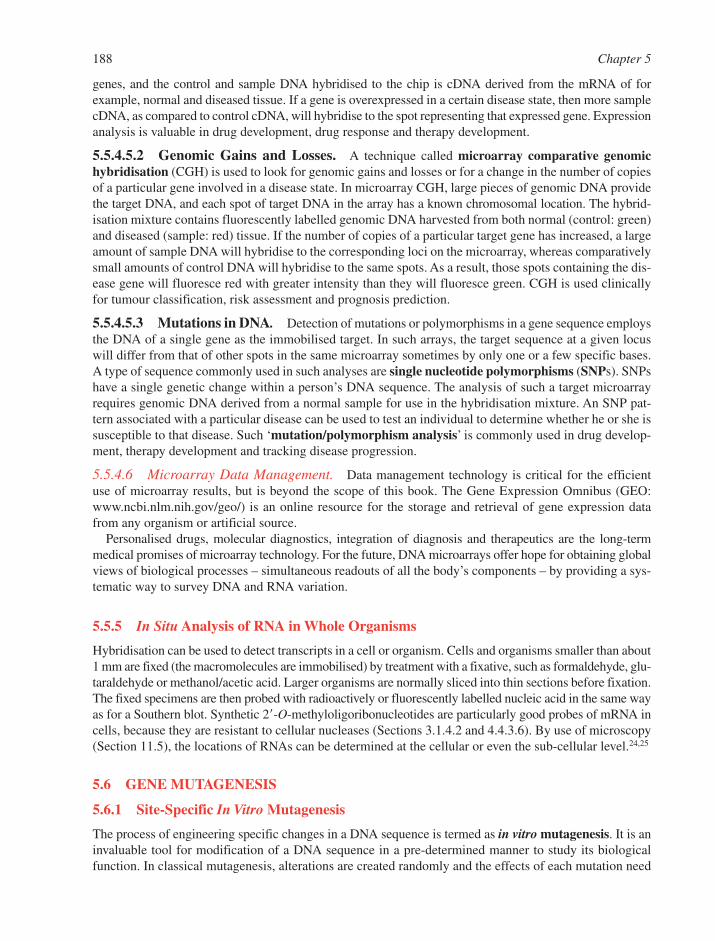
genes, and the control and sample DNA hybridised to the chip is cDNA derived from the mRNA of for example, normal and diseased tissue. If a gene is overexpressed in a certain disease state, then more samplecDNA, as compared to control cDNA, will hybridise to the spot representing that expressed gene. Expressionanalysis is valuable in drug development, drug response and therapy development.
5.5.4.5.2 Genomic Gains and Losses. A technique called microarray comparative genomichybridisation (CGH) is used to look for genomic gains and losses or for a change in the number of copiesof a particular gene involved in a disease state. In microarray CGH, large pieces of genomic DNA providethe target DNA, and each spot of target DNA in the array has a known chromosomal location. The hybrid-isation mixture contains fluorescently labelled genomic DNA harvested from both normal (control: green)and diseased (sample: red) tissue. If the number of copies of a particular target gene has increased, a largeamount of sample DNA will hybridise to the corresponding loci on the microarray, whereas comparativelysmall amounts of control DNA will hybridise to the same spots. As a result, those spots containing the dis-ease gene will fluoresce red with greater intensity than they will fluoresce green. CGH is used clinicallyfor tumour classification, risk assessment and prognosis prediction.
5.5.4.5.3 Mutations in DNA. Detection of mutations or polymorphisms in a gene sequence employsthe DNA of a single gene as the immobilised target. In such arrays, the target sequence at a given locuswill differ from that of other spots in the same microarray sometimes by only one or a few specific bases.A type of sequence commonly used in such analyses are single nucleotide polymorphisms (SNPs). SNPshave a single genetic change within a person’s DNA sequence. The analysis of such a target microarrayrequires genomic DNA derived from a normal sample for use in the hybridisation mixture. An SNP pat-tern associated with a particular disease can be used to test an individual to determine whether he or she issusceptible to that disease. Such ‘mutation/polymorphism analysis’ is commonly used in drug develop-ment, therapy development and tracking disease progression.
5.5.4.6 Microarray Data Management. Data management technology is critical for the efficientuse of microarray results, but is beyond the scope of this book. The Gene Expression Omnibus (GEO:www.ncbi.nlm.nih.gov/geo/) is an online resource for the storage and retrieval of gene expression datafrom any organism or artificial source.
Personalised drugs, molecular diagnostics, integration of diagnosis and therapeutics are the long-termmedical promises of microarray technology. For the future, DNA microarrays offer hope for obtaining globalviews of biological processes – simultaneous readouts of all the body’s components – by providing a sys-tematic way to survey DNA and RNA variation.
5.5.5 In Situ Analysis of RNA in Whole Organisms
Hybridisation can be used to detect transcripts in a cell or organism. Cells and organisms smaller than about1 mm are fixed (the macromolecules are immobilised) by treatment with a fixative, such as formaldehyde, glu-taraldehyde or methanol/acetic acid. Larger organisms are normally sliced into thin sections before fixation.The fixed specimens are then probed with radioactively or fluorescently labelled nucleic acid in the same wayas for a Southern blot. Synthetic 2�-O-methyloligoribonucleotides are particularly good probes of mRNA incells, because they are resistant to cellular nucleases (Sections 3.1.4.2 and 4.4.3.6). By use of microscopy(Section 11.5), the locations of RNAs can be determined at the cellular or even the sub-cellular level.24,25
5.6 GENE MUTAGENESIS
5.6.1 Site-Specific In Vitro Mutagenesis
The process of engineering specific changes in a DNA sequence is termed as in vitro mutagenesis. It is aninvaluable tool for modification of a DNA sequence in a pre-determined manner to study its biologicalfunction. In classical mutagenesis, alterations are created randomly and the effects of each mutation need
188 Chapter 5
Nucleic Acids in Biotechnology 189
to be screened separately, which is time consuming. Now more directed methods are standard, where theDNA is first cloned for ease of manipulation and then deletions, insertions or replacements made.
5.6.1.1 Deletions. Deletions can be created at restriction sites (Section 5.3.1) by cleavage with thecorresponding enzyme and then by treatment for a short period with an exonuclease enzyme. For example,the exonuclease Bal 31 is used to remove both double- and single-stranded DNA from both ends.Alternatively, the enzyme Exo III is used to generate single-stranded ends followed by treatment with SInuclease to trim the created single strands (Table 5.3). Re-ligation of the two new double-stranded endsgenerates deletion mutations of the parent DNA. This method has the serious limitation that deletions canonly be made around restriction sites.
A more general deletion method involves use of synthetic oligonucleotides (Figure 5.16). In this procedure,an oligonucleotide complementary to the desired site of deletion on the DNA, but not containing the nucleotidesrequired for deletion, is used as a primer for synthesis of a second DNA strand. In the process of cloning,mutant DNA segregates from wild type DNA and clones containing mutant the deletion can be selected.
One problem associated with this technique is that bacteria will often attempt to repair the mutagenisedstrand because the in vivo-generated DNA strand is methylated. This can result in low yields of themutated sequence. Eckstein has developed a reliable method that involves incorporation of phosphorothioate-modified nucleotides (Section 4.4.3) into the in vitro-generated strand. Such nucleotides are more resist-ant to nuclease degradation, with the result that the unmutagenised DNA strand can be removed byexonuclease digestion and the gap filled to generate the mutation in both strands (Figure 5.17). Deletionmutants can also be generated by this method by use of PCR (Section 5.2.2).
Deletion mutants can also be generated by PCR (Figure 5.18). This method relies upon the fact that PCRprimers are tolerant of primer-template mismatch to create a mutation at the priming site in an analogousway to that shown in Figure 5.17. Unfortunately, this raises the problem that PCR-based mutagenesis canonly make a mutated site at an end of the PCR fragment. However, this problem can be solved by gener-ating two PCR products sharing a common central mutated region (Figure 5.18). Denaturation and anneal-ing of these two products, followed by extension of the duplex with Taq DNA polymerase yields a largerproduct with the mutated site in the centre.
5.6.1.2 Insertions. Insertions may be generated by ligation of a synthetic oligonucleotide duplex intoa restriction site after cutting with the appropriate restriction enzyme. Sequence additions at other sites can
Figure 5.16 Oligonucleotide site-directed deletion mutagenesis

be achieved by means of site-directed mutagenesis using oligonucleotides in an analogous way to thatdescribed for deletions (Figures 5.17 and 5.18), but in this case the synthetic oligonucleotide primer con-tains the desired additional nucleotide(s).
5.6.1.3 Replacements. A common type of mutation is that which maintains the same number ofnucleotides but where part of a sequence is replaced. This is particularly useful for single-base alterationsthat lead to a change of amino acid codon. Expression of the mutated gene leads to the production of a pro-tein with a single amino acid alteration ( protein engineering). One replacement method is to introduce asmall deletion at a restriction site followed by ligation into the gap of an oligonucleotide duplex of thesame size but of different sequence. A more general approach involves use of a synthetic oligonucleotideprimer in an analogous way to the introduction of deletions and insertions, but with the same number ofnucleotides in the mutant strand as wild type (Figures 5.17 and 5.18).
190 Chapter 5
Figure 5.17 The use of phosphorothioate-modified nucleotides for in vitro mutagenesis
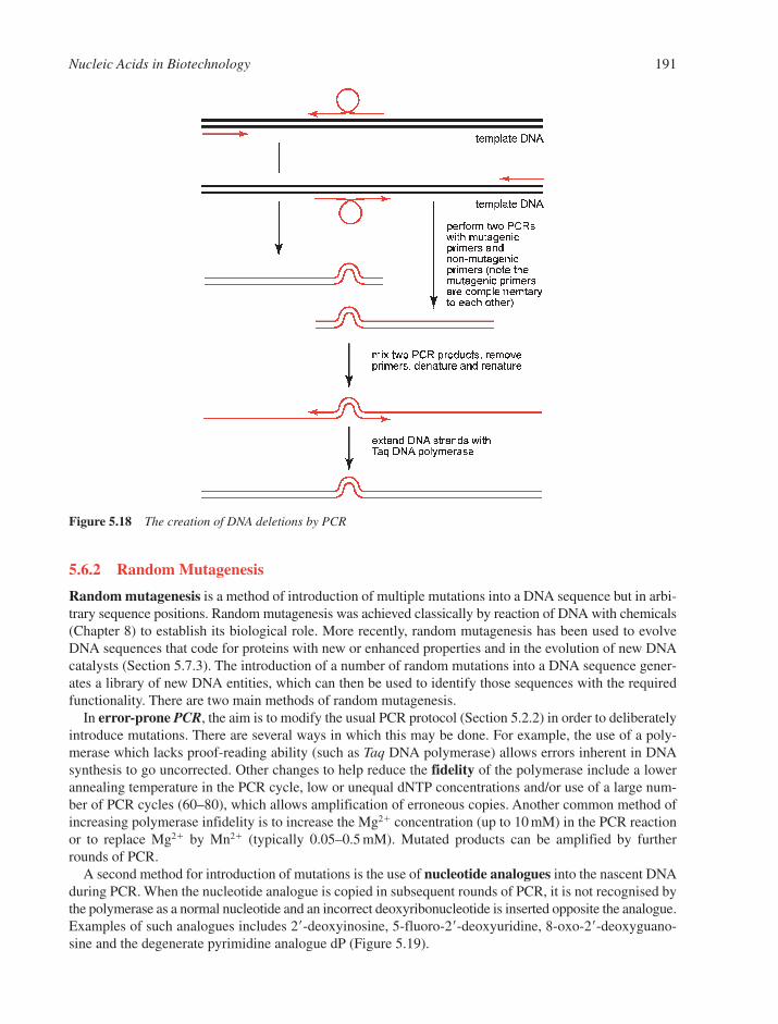
5.6.2 Random Mutagenesis
Random mutagenesis is a method of introduction of multiple mutations into a DNA sequence but in arbi-trary sequence positions. Random mutagenesis was achieved classically by reaction of DNA with chemicals(Chapter 8) to establish its biological role. More recently, random mutagenesis has been used to evolveDNA sequences that code for proteins with new or enhanced properties and in the evolution of new DNAcatalysts (Section 5.7.3). The introduction of a number of random mutations into a DNA sequence gener-ates a library of new DNA entities, which can then be used to identify those sequences with the requiredfunctionality. There are two main methods of random mutagenesis.
In error-prone PCR, the aim is to modify the usual PCR protocol (Section 5.2.2) in order to deliberatelyintroduce mutations. There are several ways in which this may be done. For example, the use of a poly-merase which lacks proof-reading ability (such as Taq DNA polymerase) allows errors inherent in DNAsynthesis to go uncorrected. Other changes to help reduce the fidelity of the polymerase include a lowerannealing temperature in the PCR cycle, low or unequal dNTP concentrations and/or use of a large num-ber of PCR cycles (60–80), which allows amplification of erroneous copies. Another common method ofincreasing polymerase infidelity is to increase the Mg2� concentration (up to 10 mM) in the PCR reactionor to replace Mg2� by Mn2� (typically 0.05–0.5 mM). Mutated products can be amplified by furtherrounds of PCR.
A second method for introduction of mutations is the use of nucleotide analogues into the nascent DNAduring PCR. When the nucleotide analogue is copied in subsequent rounds of PCR, it is not recognised bythe polymerase as a normal nucleotide and an incorrect deoxyribonucleotide is inserted opposite the analogue.Examples of such analogues includes 2�-deoxyinosine, 5-fluoro-2�-deoxyuridine, 8-oxo-2�-deoxyguano-sine and the degenerate pyrimidine analogue dP (Figure 5.19).
Nucleic Acids in Biotechnology 191
Figure 5.18 The creation of DNA deletions by PCR
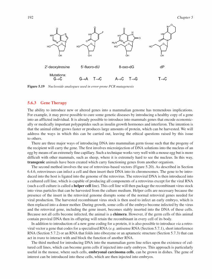
5.6.3 Gene Therapy
The ability to introduce new or altered genes into a mammalian genome has tremendous implications. For example, it may prove possible to cure some genetic diseases by introducing a healthy copy of a geneinto an afflicted individual. It is already possible to introduce into mammals genes that encode economic-ally or medically important polypeptides such as insulin growth hormones and interferon. The intention isthat the animal either grows faster or produces large amounts of protein, which can be harvested. We willaddress the ways in which this can be carried out, leaving the ethical questions raised by this issueto others.
There are three major ways of introducing DNA into mammalian germ tissue such that the progeny ofthe recipient will carry the gene. The first involves microinjection of DNA solutions into the nucleus of anegg by means of an extremely fine capillary. Such a technique works very well with a mouse egg but is moredifficult with other mammals, such as sheep, where it is extremely hard to see the nucleus. In this way,transgenic animals have been created which carry functioning genes from another organism.
The second method involves the use of retrovirus-based vectors (Figure 5.20). As described in Section6.4.6, retroviruses can infect a cell and then insert their DNA into its chromosomes. The gene to be intro-duced into the host is ligated into the genome of the retrovirus. The retroviral DNA is then introduced intoa cultured cell line, which is capable of producing all components of a retrovirus except for the viral RNA(such a cell culture is called a helper cell line). This cell line will then package the recombinant virus stockinto virus particles that can be harvested from the culture medium. Helper cells are necessary because thepresence of the insert in the retroviral genome disrupts some of the normal retroviral genes needed forviral production. The harvested recombinant virus stock is then used to infect an early embryo, which isthen replaced into a donor mother. During growth, some cells of the embryo become infected by the virusand the retroviral gene, including the gene insert, becomes stably inserted into the DNA of these cells.Because not all cells become infected, the animal is a chimera. However, if the germ cells of this animalcontain proviral DNA then its offspring will retain the recombinant in every cell of its body.
In addition to introduction of a new gene coding for a protein, it is also possible to introduce via a retro-viral vector a gene that codes for a specialised RNA (e.g. antisense RNA (Section 5.7.1), short interferenceRNA (Section 5.7.2) or an RNA that folds into ribozyme or an aptameric structure (Section 5.7.3) that canact in trans to interact with and block the function of another RNA.
The third method for introducing DNA into the mammalian germ line relies upon the existence of cul-tured cell lines, which can become germ cells if injected into early embryos. This approach is particularlyuseful in the mouse, where such cells, embryonal carcinoma cells, can be grown in dishes. The gene ofinterest can be introduced into these cells, which are then injected into embryos.
192 Chapter 5
Figure 5.19 Nucleoside analogues used in error-prone PCR mutagenesis

5.7 OLIGONUCLEOTIDES AS REAGENTS AND THERAPEUTICS
The ability to synthesise DNA and RNA oligonucleotides of defined sequence rapidly (Sections 4.1 and 4.2),including a range of nucleotide analogues (Section 4.4), has led to a large number of applications as thera-peutic and diagnostic agents.26 Many applications involve the principle of recognition of a linear sequence ofRNA or DNA by the oligonucleotide. For example, antisense, steric block and short interfering RNA(siRNA) all involve targeting of RNA within cells to form duplexes as a means of control of gene expres-sion27 (Sections 5.7.1 and 5.7.2). Synthetic oligonucleotides have been used also to form triplexes (Sections2.3.6 and 9.10.1) with double-stranded DNA to block gene expression,28 but this principle has so far not ledto therapeutic products. Other types of application include in vitro selection and design of oligonucleotidesthat recognise and bind to nucleic acids structures, to proteins or to small molecule ligands (Section 5.7.3).
5.7.1 Antisense and Steric Block Oligonucleotides
In 1979, Zamecnik and Stephenson29 were the first to show that a synthetic oligonucleotide could be usedto block specific gene expression in Rous Sarcoma Virus. This pioneering work led to the study ofoligonucleotides and their analogues as therapeutic agents. This field is commonly known as ‘antisense’,since the principle of biological activity usually involves either degradation or steric blocking of the sense
Nucleic Acids in Biotechnology 193
Figure 5.20 Creation of transgenic animals by use of retroviral vectors

strand (the coding strand) of RNA (commonly mRNA or viral RNA) through formation of an exactly basepaired duplex between the target RNA and an added complementary strand (the antisense strand) (Figure5.21). Formation of the duplex causes inhibition of expression of a particular gene within cells or in vivoand the aim is to do this without affecting any other gene.30,31
5.7.1.1 Basic Mechanisms
5.7.1.1.1 Steric Block. This mechanism involves formation of an RNA–DNA duplex to physicallyblock the RNA and to prevent recognition by a protein or other cellular machinery. For example, bindingof an oligonucleotide close to the 5�-cap site32 (Section 7.2.1) or at the site of initiation of translation inmRNA33 (Section 7.3.3) may prevent the ribosome or associated machinery from binding to the RNA andinitiating translation (Figure 5.22). Other RNA processing events that can be interfered with sterically byduplex formation include nuclear splicing34 and polyadenylation35 (Section 7.2.1), both of which arerequired for the processing of most mammalian gene transcripts and which involve numerous steps ofRNA–protein recognition. In the case of viral RNA, it is possible to block recognition of essential RNAbinding proteins that are required for virus-specific gene regulation.
5.7.1.1.2 Induction of RNase H. Although steric block activity requires stoichiometric amounts ofcomplementary added oligonucleotide, a more potent inhibitory effect can often be obtained throughrecognition of an RNA–oligonucleotide duplex by the ubiquitous cellular enzyme Ribonuclease H (RNaseH). The normal function of this enzyme is to help the removal of RNA primers in DNA replication(Section 6.6.3). However, when an RNA sequence is targeted in cells by a complementary oligodeoxynu-cleotide, RNase H-induced cleavage can occur rapidly, usually close to the centre of the targeted RNA sec-tion.36 The loss of intact RNA leads to rapid degradation of the RNA. Thus in the case of mRNA, there isa concomitant reduction in the level of the encoded protein expressed. Most regions of an mRNA can usu-ally be targeted by such oligonucleotides, including 3�- and 5�-untranslated regions.
194 Chapter 5
Figure 5.22 Three alternative mechanisms of steric block action of antisense oligonucleotides acting upon RNA
Figure 5.21 Duplex formed by an antisense oligodeoxyribonucleotide and a target mRNA
5.7.1.2 Optimal Oligonucleotide Characteristics. There are many factors that influence cellularor in vivo antisense activity. In practice, oligonucleotide optimisation is carried out by experimentationthrough use of in vitro, cell-based and ultimately in vivo assays, although some general principles can beused in oligonucleotide design.
5.7.1.2.1 Duplex Stability. For intracellular antisense activity, an oligonucleotide must be of suffi-cient length to form a strong duplex with its RNA target at 37°C under cellular conditions. In general,binding strength increases as a function of length as well as the number of G:C pairs (Section 5.5.1). In addition, the type of nucleotide analogues used and their placement within the oligonucleotide are alsocrucial and those nucleoside analogues that adopt an RNA-like, 3�-endo sugar conformation (such as 2�-O-methylribonucleosides) tend to result in increased binding strength, since there is a tendency to forma more compact A-helix (Section 2.2.3). It is important also that the oligonucleotide does not form unusualsecondary structures (such as G quadruplexes, Section 2.3.7) that may hinder duplex formation. Anotherimportant consideration is whether the target RNA site is easily accessible, i.e. does not exhibit tight RNAsecondary or tertiary structure or is not strongly bound by cellular proteins. In this regard, experimentalapproaches to target choice are often more reliable than RNA structure prediction.
5.7.1.2.2 Specificity. For unique sequence recognition within the human genome (i.e. no other likelyexact match for an oligonucleotide of typically mixed composition), a minimum length of around 12nucleotides is usually required. However, the longer the chosen sequence, the greater the chance for theoligonucleotide to form a mismatched duplex with an incorrect RNA sequence. This is particularly of con-cern in the case of RNase H induction where an incorrect RNA may be cleaved in addition to that targeted,leading to side effects. In practice, a compromise between duplex stability and target specificity limitsoligonucleotide length usually to 12–25 residues.
5.7.1.2.3 Nuclease Stability. Unmodified single-stranded DNA and RNA oligonucleotides aredegraded very fast by cellular nucleases in cells and serum. 3��-Exonucleases are the most prevalent, such thatminimally the 3�-end of an antisense oligonucleotide must be protected, usually by chemical modification.But 5�-exonucleases as well as endonucleases are also present in cells, and thus for therapeutic applications,nuclease protection of each internucleotide linkage by inclusion of analogues is often thought necessary.
5.7.1.2.4 Cellular Uptake. A significant difficulty is that oligonucleotides and their analoguesrarely penetrate cells in culture without co-addition of a carrier or cell delivery agent. For example, popu-lar delivery agents for many cultured mammalian cell lines are cationic lipids, which can form complexeswith negatively charged oligonucleotides to help cell association, uptake through the endosomal pathwayand subsequent release into the cytosol by endosome destabilisation. Oligonucleotides are able to entercell nuclei readily once they have been released into the cytosol. Oligonucleotides are usually adminis-tered in vivo without carrier, and here there may be special mechanisms available for cell uptake, but thisremains a difficult and controversial subject of study.
5.7.1.2.5 Pharmaceutical Considerations. One positive feature of many clinically investigatedoligonucleotides to date is their relative lack of toxicity during systemic delivery into animals and man. Bycontrast, a major concern has been the frequently observed, rapid clearance through the kidney, which istypical of many macromolecules. It is thus not surprising that the greatest success to date for therapeuticoligonucleotides has been in local or topical administration. Pharmaceutical development remains a signifi-cant challenge in terms of reaching the required tissue or organ, efficacy of action at the site and the main-tenance of a therapeutic dose at manageable and affordable concentration levels. Many studies continue thatfocus on investigations of new nucleotide analogue types and combinations, conjugates and formulations31.
5.7.1.3 Nucleotide Analogues Used in Antisense Applications. The most potent antisenseoligonucleotides to date have been those shown to induce RNase H cleavage within cellular models and sev-eral have been taken to clinical trials.26 Strong recognition by RNase H requires there to be a contiguous stretchof minimally 6–10 residues of 2�-deoxyribonucleosides where internucleotide linkages are phosphodiesters
Nucleic Acids in Biotechnology 195

or the close analogue phosphorothioate (Section 4.4.3). Phosphorothioates are considerably more resistantthan phosphodiesters to nuclease degradation and are well tolerated in humans. Thus, first generation thera-peutic oligonucleotides contained only 2�-deoxyribonucleotide phosphorothioates, such as the clinicallyapproved drug Vitravene, which is a 26-mer used for treatment of CMV-induced retinitis in AIDS patients.
Second-generation oligonucleotides employ the principle of a gapmer. Such oligonucleotides containa section of 6–10 residues (usually centrally placed) of 2�-deoxyribonucleoside phosphorothioates, but theflanking regions on each side comprised of other analogues that enhance binding to the RNA target andfurther increase the oligonucleotide stability to nuclease, but which do not direct RNase H cleavage. Suchanalogues are generally ribonucleoside analogues, such as 2�-O-methyl, 2�-O-methoxyethyl or lockednucleic acids (LNA), where the sugar conformation is 3�-endo. Overall, gapmers are recognised and directRNA cleavage by RNase H. Whereas several first generation antisense oligonucleotides, such as ISIS3521 (Affinatak) targeted to the mRNA for protein kinase C �,37 failed clinical trials, there is more hopeof clinical benefit for some higher potency gapmers against viral and cancer targets (Figure 5.23).26,31
For steric block applications, there is no restriction in principle to the type or placement of an analoguewithin a sequence as long as other antisense considerations are met. The variety of analogues that have beeninvestigated is very large. In some cases analogues can be combined in one oligonucleotide to give mixmers.In addition to 2�-O-methyl and 2�-O-methoxyethyl ribonucleotides described above, other important ana-logues used in steric block applications fall into two classes: (a) those that contain a phosphate group, suchas LNA, tricyclo DNA, 3�-amino phosphoroamidate and phosphorothioamidate, and (b) non-phosphate con-taining analogues such as peptide nucleic acids (PNA) and morpholinodiamidates. In class (b) it was hopedthat the absence of the negative charges on the oligonucleotide would enhance cell uptake, but this is not thecase. Attachment of a cationic or other cell penetrating peptide appears to improve cell uptake, but the uni-versality of this approach is still under study. A steric blocking, phosphorothioamidate oligonucleotide tar-geted to the essential RNA involved in the enzyme telomerase (Section 6.6.5) is moving close to clinicaltrials as an anti-cancer agent.38
5.7.1.4 Non-Duplex Therapeutic Activities of Single-Stranded Oligonucleotides. Recently,other biological activities of oligonucleotides have been found that are sequence-dependent but which areindependent of duplex formation with an RNA target.
5.7.1.4.1 Immune Modulation. Single-stranded oligodeoxynucleotide phosphodiesters and phos-phorothioates that contain the dinucleotide sequence CpG can trigger an immune response when adminis-tered to humans and animals.39,40 The response is mediated through binding to a ‘toll-like receptor’TLR9that is present in cytosolic vesicles and the binding stimulates signalling pathways that activate transcrip-tion factors. By contrast, double-stranded RNA and siRNA (Section 5.7.2) binds to another receptor TLR7and may stimulate a different immune response. The context of the CpG determines the immune modula-tion specificity, such that mouse TLR9 prefers CpG when flanked at 5� by two purines and at 3� by twopyrimidines, while human TLR9 is recognised optimally by GTCGTT and TTCGTT sequences. Such activ-ities are now recognised to have contributed being harnessed for therapeutic applications and as vaccine
196 Chapter 5
Figure 5.23 First and second generation clinically used phosphorothioate-containing oligodeoxyribonucleotides

adjuvants. Immune stimulation of this or similar type has also been used to explain in vivo activities ofsome supposed antisense oligonucleotides that contain CpG sequences.
5.7.1.4.2 DNA Aptamers. Oligodeoxynucleotides have been selected (Section 5.7.3) that bind spe-cific cellular proteins. Such aptamers can fold into unusual structures (such as a G quadruplex, Section2.3.7). A chemically synthesised DNA aptamer that binds strongly to vascular endothelial growth factorhas been formulated as a conjugate with polyethylene glycol and has recently been given regulatoryapproval (Macugen) for treatment of patients with the wet form of age-related macular degeneration, acommon cause of blindness due to abnormal blood vessel growth.41
5.7.2 RNA Interference
In the late 1990s, gene silencing by double-stranded RNA was observed in plants in the laboratory ofDavid Baulcombe42 and in the worm C. elegans in that of Craig Mello.43 Within a very few years genesilencing activities were found in many diverse organisms and have gone on to be harnessed as powerfuldiagnostic reagents in genome research and as potential therapeutics.44–46 RNA interference (RNAi) prob-ably evolved from the need for eukaryotic cellular defence against foreign (e.g. viral) duplex RNA or DNAthat is transcribed into RNA. Distinct but overlapping pathways have been elucidated that represent dif-ferent forms of genetic regulation (Figure 5.24).
Primary RNA transcripts in the nucleus has been found to contain hundreds of endogenous sequencesknown as microRNAs (miRNAs) that can fold into hairpins that contain imperfect matches (Section 7.5.3).The transcripts are processed by the nuclear complex Drosha, which contains an RNase III activity thatcleaves such RNAs to produce hairpins of about 70 residues (pre-microRNAs). After export to the cytosol, the
Nucleic Acids in Biotechnology 197
Figure 5.24 Mechanisms of action involved in RNA interference (RNAi). Pathway (a) shows steps in the processingof microRNA (miRNA) eventually leading to inhibition of translation. Pathway (b) shows the processingof double-stranded viral RNA to form short interfering RNA (siRNA) and eventual cleavage of mRNAby the RNA-induced silencing complex (RISC) complex. ShortRNA (shRNA) (centre) can in principleenter either pathway

hairpins are further processed by the enzyme complex Dicer (Dcr-1),47 a multi-subunit protein complex thatalso contains an RNase III activity, to give imperfectly paired RNA duplexes of about 21–23 residues(miRNAs). These are recognised by the RNA-induced silencing complex (RISC), which directs one of thetwo RNA strands to bind to a selected sequence in the 3�-untranslated region of a gene (a microRNArecognition element) to form an imperfect complement, which results in a block to translation (Figure5.24, pathway a).
A second pathway is triggered by the introduction into a cell of double-stranded RNA, such as viral RNA.A second DICER variant, Dcr-2, is responsible for processing this duplex RNA into 21–23 residue perfectduplexes (siRNA). SiRNAs are then utilised by the RISC complex to direct one strand (antisense or guide) toform a duplex with an exact complement on a mRNA and cleave the phosphodiester bond precisely betweenresidue 10 and 11 counting from the 5�-end of the complement, as directed by a member of the Argonaute fam-ily (Ago 2), an RNA endonuclease within RISC (Figure 5.24, pathway b). The other strand (sense or passen-ger) is discarded and then degraded.
Thomas Tuschl and colleagues48 found that when synthetic siRNAs of 21 residues were transfected intomammalian cells, the RISC-dependent cleavage pathway could be triggered, thus allowing site-specificcleavage of mRNA and subsequent inhibition of gene expression. Synthetic siRNAs have now become usedwidely as reagents for specific gene inhibition in many cell types and seem to be applicable to almost allgenes. Generally two-nucleotide 3�-overhangs are added on each strand for optimal activity, similar to thosefound following natural DICER cleavage of duplex RNA. Although siRNA duplexes appears to be stable tonuclease degradation for hours to days within cells, unlike single-stranded RNA, much effort has beenexpended on investigation of the tolerance to incorporation of analogues or conjugates that might haveadvantages in vivo to aid stability or pharmacology.49 The sense strand appears to be highly tolerant of chem-ical modification, but the antisense strand, which is the one introduced by the RISC complex to pair withmRNA, is less so. A recent demonstration of efficacy in a transgenic mouse model promises that modifiedsiRNAs may have therapeutic value.50
A third RNAi pathway is triggered by introduction into cells of short hairpin RNAs (shRNAs) of around29-base pairs (Figure 5.24).51 Such shRNAs are recognised by DICER and processed to give siRNAs ofhigh potency, presumably because they are generated endogenously and may be more readily utilised byRISC. ShRNAs are also potential precursors of imperfectly matched miRNA.
Although siRNAs have been shown to generate some immune response effects, it is not clear at presentwhether these will present significant problems or not for their in vivo use.
5.7.3 In Vitro Selection
5.7.3.1 Principles of In Vitro Selection (SELEX). The advent of in vitro selection or SELEX (sys-tematic evolution of ligands by exponential enrichment) in the early 1990s by the groups of Gold,52
Szostak and Joyce marked the beginning of a new age in the design of functional nucleic acid moleculesas both ligands for given targets and as catalysts. SELEX is a combinatorial technique in which nucleicacids with specific properties, such as binding with high affinity to a given target molecule (an aptamer)or catalysis of a chemical reaction, are selected from a pool of typically 1012–1015 RNA or DNA moleculesof randomised sequence.53,54 The technique exploits the wide range of structures that single-strandednucleic acids can adopt and mimics the natural processes of evolution.
5.7.3.2 Selection of Aptamers. The basic principle of SELEX (Figure 5.25) involves the creation byautomated chemical synthesis of an initial oligonucleotide library consisting of an internal randomnucleotide sequence flanked by 5�- and 3�-tails of a constant sequence which act as primer binding sites forsubsequent amplification of the library by PCR (Section 5.2.2). The random sequence, typically 10–100nucleotides long, is generated by delivering a mixture of all four nucleoside phosphoramidites simultane-ously during automated synthesis (Section 4.1). Since the library contains just a few copies of each sequence,it is first amplified by PCR in which one of the oligonucleotide primers carries a biotin modification. To actas ligands to a specific target molecule, the nucleic acids within the library must be free to fold into a wide
198 Chapter 5

range of 3-D structures and hence must be single stranded. Thus the selection of RNA aptamers, the duplexDNA library contains a T7 RNA promoter sequence upstream of the 5�-constant sequence. This enables asingle-stranded RNA pool to be generated through transcription by use of T7 RNA polymerase (Figure 5.25).A single-stranded DNA pool required for the selection of DNA aptamers is produced by capture of only the5�-biotinylated strand from the DNA duplex on streptavidin-derivatised beads (Figure 5.26).
The selection of DNA or RNA aptamers, which can bind to a given target molecule is achieved by useof affinity chromatography. Thus the target molecule is immobilised on a solid support usually within asmall column and a solution of the nucleic acid pool is passed through. Unbound nucleic acids are elutedby simple washing of the column with a suitable buffer. Sequences that have some affinity for the targetare bound by the column and subsequently eluted by washing with a buffer that contains the free targetmolecule. For RNA aptamers, a cDNA library is then generated from the bound fraction using enzymereverse transcriptase, which is then amplified by PCR (Figure 5.25). For DNA aptamers, the aptamer-containing fraction is subjected directly to PCR (Figure 5.26). In subsequent rounds of SELEX, the strin-gency of the washing protocols is increased or the concentration of immobilised target is reduced such thatthe affinity chromatography step leads to an enrichment of high-affinity binding sequences within thelibrary. Typically about ten cycles of SELEX are carried out after which perhaps about 100 or so differentsequences remain. These aptamers are then cloned into a vector and characterised by DNA sequencing.
SELEX has been used to identify a wide range of aptamers to a variety of diverse molecular targets, forexample ions, small molecules such as organic dyes, nucleotides and their bases, amino acids, co-factors,antibiotics, transition state analogues, as well as peptides and proteins.55 The remarkable selectivity of
Nucleic Acids in Biotechnology 199
Figure 5.25 The generation of RNA aptamers using SELEX (systematic evolution of ligands by exponential enrichment)
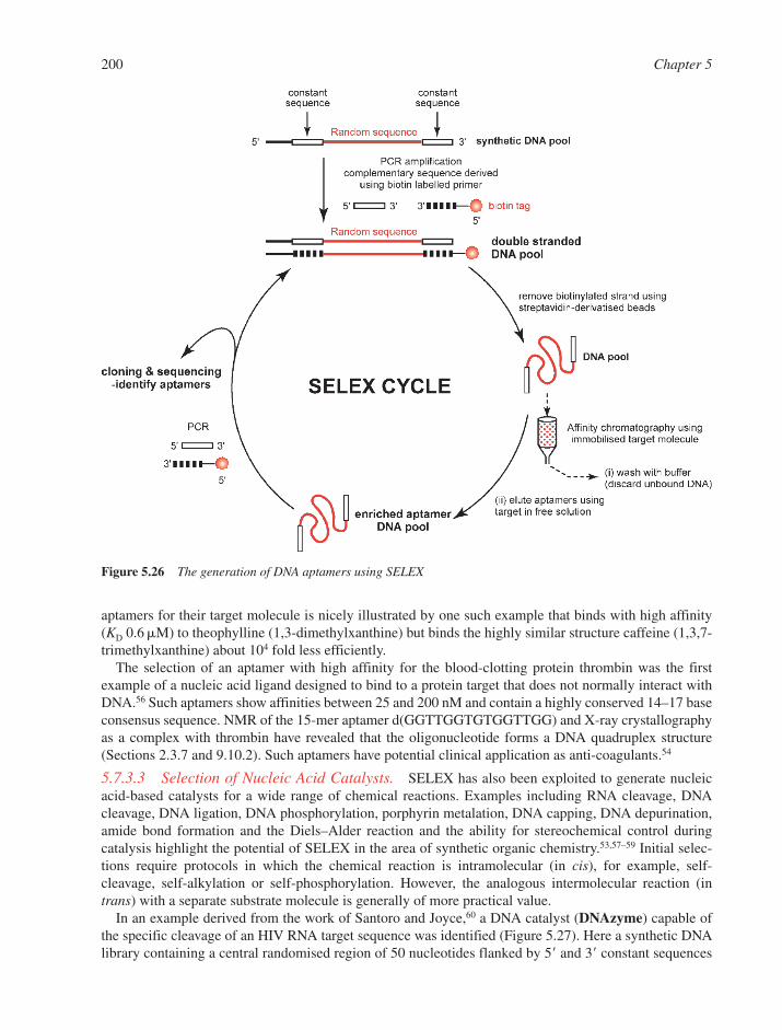
aptamers for their target molecule is nicely illustrated by one such example that binds with high affinity(KD 0.6 M) to theophylline (1,3-dimethylxanthine) but binds the highly similar structure caffeine (1,3,7-trimethylxanthine) about 104 fold less efficiently.
The selection of an aptamer with high affinity for the blood-clotting protein thrombin was the firstexample of a nucleic acid ligand designed to bind to a protein target that does not normally interact withDNA.56 Such aptamers show affinities between 25 and 200 nM and contain a highly conserved 14–17 baseconsensus sequence. NMR of the 15-mer aptamer d(GGTTGGTGTGGTTGG) and X-ray crystallographyas a complex with thrombin have revealed that the oligonucleotide forms a DNA quadruplex structure(Sections 2.3.7 and 9.10.2). Such aptamers have potential clinical application as anti-coagulants.54
5.7.3.3 Selection of Nucleic Acid Catalysts. SELEX has also been exploited to generate nucleicacid-based catalysts for a wide range of chemical reactions. Examples including RNA cleavage, DNAcleavage, DNA ligation, DNA phosphorylation, porphyrin metalation, DNA capping, DNA depurination,amide bond formation and the Diels–Alder reaction and the ability for stereochemical control duringcatalysis highlight the potential of SELEX in the area of synthetic organic chemistry.53,57–59 Initial selec-tions require protocols in which the chemical reaction is intramolecular (in cis), for example, self-cleavage, self-alkylation or self-phosphorylation. However, the analogous intermolecular reaction (intrans) with a separate substrate molecule is generally of more practical value.
In an example derived from the work of Santoro and Joyce,60 a DNA catalyst (DNAzyme) capable ofthe specific cleavage of an HIV RNA target sequence was identified (Figure 5.27). Here a synthetic DNAlibrary containing a central randomised region of 50 nucleotides flanked by 5� and 3� constant sequences
200 Chapter 5
Figure 5.26 The generation of DNA aptamers using SELEX
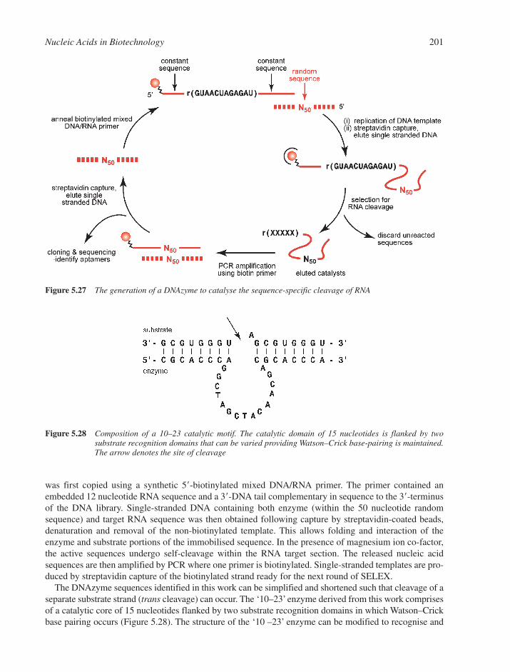
was first copied using a synthetic 5�-biotinylated mixed DNA/RNA primer. The primer contained anembedded 12 nucleotide RNA sequence and a 3�-DNA tail complementary in sequence to the 3�-terminusof the DNA library. Single-stranded DNA containing both enzyme (within the 50 nucleotide randomsequence) and target RNA sequence was then obtained following capture by streptavidin-coated beads,denaturation and removal of the non-biotinylated template. This allows folding and interaction of theenzyme and substrate portions of the immobilised sequence. In the presence of magnesium ion co-factor,the active sequences undergo self-cleavage within the RNA target section. The released nucleic acidsequences are then amplified by PCR where one primer is biotinylated. Single-stranded templates are pro-duced by streptavidin capture of the biotinylated strand ready for the next round of SELEX.
The DNAzyme sequences identified in this work can be simplified and shortened such that cleavage of aseparate substrate strand (trans cleavage) can occur. The ‘10–23’ enzyme derived from this work comprisesof a catalytic core of 15 nucleotides flanked by two substrate recognition domains in which Watson–Crickbase pairing occurs (Figure 5.28). The structure of the ‘10 –23’ enzyme can be modified to recognise and
Nucleic Acids in Biotechnology 201
Figure 5.27 The generation of a DNAzyme to catalyse the sequence-specific cleavage of RNA
Figure 5.28 Composition of a 10–23 catalytic motif. The catalytic domain of 15 nucleotides is flanked by twosubstrate recognition domains that can be varied providing Watson–Crick base-pairing is maintained.The arrow denotes the site of cleavage
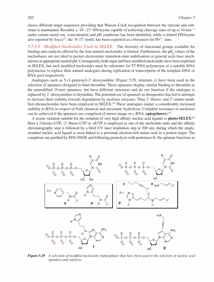
cleave different target sequences providing that Watson–Crick recognition between the enzyme and sub-strate is maintained. Recently a ‘10 –23’ DNAzyme capable of achieving cleavage rates of up to 10 min�1
under certain metal ion, concentration and pH conditions has been identified, while a related DNAzymealso reported by Joyce53, the ‘8–17’ motif, has been exploited as a biosensor for Pb2� ions.
5.7.3.4 Modified Nucleotides Used in SELEX. The diversity of functional groups available forbinding and catalysis offered by the four natural nucleotides is limited. Furthermore, the pKa values of thenucleobases are not ideal to permit electrostatic transition-state stabilisation or general acid–base mech-anisms at appropriate neutral pH. Consequently, both sugar and base modified nucleotides have been exploitedin SELEX, but such modified nucleotides must be substrates for T7 RNA polymerase or a suitable DNApolymerase to replace their natural analogues during replication or transcription of the template DNA orRNA pool respectively.
Analogues such as 5-(1-pentynyl)-2�-deoxyuridine (Figure 5.29, structure c) have been used in theselection of aptamers designed to bind thrombin. These aptamers display similar binding to thrombin asthe unmodified 15-mer aptamers, but have different structures and do not function if the analogue isreplaced by 2�-deoxyuridine or thymidine. The potential use of aptamers as therapeutics has led to attemptsto increase their stability towards degradation by nuclease enzymes. Thus 2�-fluoro- and 2�-amino modi-fied ribonucleotides have been employed in SELEX.54 These analogues impart a considerably increasedstability to RNA in respect of both chemical and enzymatic hydrolysis. Complete resistance to nucleasescan be achieved if the aptamers are comprised of mirror image or L-RNA (spiegelmers).61
A recent variation suitable for the isolation of very high affinity nucleic acid ligands is photo-SELEX.62
Here a 5-bromo-UTP, -2�-fluoro-UTP or -dUTP is employed as one of the nucleotide units and the affinitychromatography step is followed by a brief UV laser irradiation step at 308 nm, during which the single-stranded nucleic acid ligand is cross-linked to a proximal electron-rich amino acid in a protein target. Thecomplexes are purified by SDS–PAGE and following proteolysis with proteinase K, the aptamer functions as
202 Chapter 5
Figure 5.29 A selection of modified nucleoside triphosphates that have been used in the selection of nucleic acidaptamers and catalysts
a template for Taq DNA polymerase (DNA aptamer) or reverse transcriptase (RNA aptamer) in further roundsof SELEX. The technique has been used to isolate aptamers with nM and pM affinities to the HIV-1 Revprotein and basic fibroblast growth factor (bFGF)63 respectively. Only high affinity aptamers have the preciseorientation of functional groups to permit cross-linking, and a harsh washing step removes all non-cross-linkedproteins interacting with the immobilised aptamer. This reduces background signals due to binding of non-cognate proteins and allows a highly reliable diagnostic assay for proteins attached to a micro-chip.63
The incorporation of base modified nucleosides with potential catalytic groups was first demonstratedby Eaton in which a C-5-modified UTP bearing an appended pyridyl or imidazole function (Figure 5.29,structures a and b) was employed in the selection of an RNA capable of acting as a Diels Alderase or anamide synthase.53 More recently imidazole- and amine-modified nucleotides have been employed to selectDNAzymes capable of the sequence-specific cleavage of RNA (Figure 5.29, structures d, f and g). 55,64,65
For example, Perrin64 and Williams65 selected DNAzymes functionalised with imidazolyl and amino func-tional groups that catalyse the sequence specific cleavage of RNA in the absence of metal ions with rateenhancements of about 105 compared to the uncatalysed reaction. Such DNAzymes display the functionalside chains that are utilised by protein ribonuclease RNaseA in metal-independent RNA cleavage.
5.7.3.5 Riboswitches. While the catalytic potential of natural RNA has been known since the mid-1980s,it is only recently that a natural biological role for RNA aptamers has been revealed. Such naturally occurringaptamers or ‘riboswitches’ have been found within the leader sequences of several metabolic genes wherethey have important roles in regulating both transcription and translation of the respective gene.66 The functionof these riboswitches is to assess cellular levels of certain metabolites, which in turn control expression of thatgene. Thus the riboswitch functions in much the same way as synthetic aptamer that can recognise and bind toa small molecule target. The flavin mononucleotide (FMN)-sensing riboswitch found in bacteria was one ofthe first such examples.66 FMN and flavin adenine dinucleotide (FAD) are synthesised from riboflavin (vita-min B2). The enzymes that are responsible for riboflavin biosynthesis from GTP are derived from five genesthat comprise the riboflavin operon. The first of these genes contains a 300 nucleotide untranslated regionwhich, upon binding of FMN or FAD, changes conformation so as to cause the termination of transcription.In this work, a number of other riboswitches have been described, including examples, which recognise thi-amine pyrophosphate, adenosylcobalamin, S-adenosyl methionine, lysine and guanine.66
5.8 DNA FOOTPRINTING
Footprinting is a method for determining the precise DNA sequence of bases that is the site for attachment ofa particular DNA enzyme or binding-protein or of a DNA-binding drug. DNA footprinting utilises a DNAcleaving agent, which can be either a nuclease or a chemical reagent. The agent must be able to cut DNA non-selectively at every exposed base pair while such DNA cleavage is inhibited at the site where the protein or drugbinds to DNA. Thus a ‘footprint’of the target sequence is identified as the region where no cutting is observed.
The steps in a DNA ‘footprinting’ experiment are
� a fragment of dsDNA containing the target sequence (usually 200–300 base pairs) is labelled at the5�-ends with 32P and then the label is removed preferentially from one end (e.g. the 3�-end of agene) by a suitable restriction endonuclease (Section 5.3.1);
� this dsDNA fragment is incubated with the DNA binding-protein so that the protein protects thetarget region of DNA from DNase I digestion (Section 5.3.2);
� limited DNase I digestion is carried out, so that there is about one cut per strand and the sites ofcleavage are randomly distributed among the accessible sites;
� the resulting DNA fragments are analysed by gel electrophoresis (Section 11.4.3), and give aladder that has a ‘footprint’ region where there are no cuts, corresponding to the binding site(Figure 5.30). If a control track is generated from a Maxam–Gilbert G � A chemical sequencingreaction (Section 5.1.1) using the same probe as template, then the exact footprint sequence can beread out by comparing the location of the blank with the sequencing reaction.
Nucleic Acids in Biotechnology 203
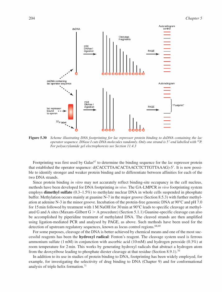
Footprinting was first used by Galas67 to determine the binding sequence for the lac repressor proteinthat established the operator sequence: d(CACCTTAACACTAACCTCTTGTTAAAG)-5�. It is now possi-ble to identify stronger and weaker protein binding and to differentiate between affinities for each of thetwo DNA strands.
Since protein binding in vitro may not accurately reflect binding-site occupancy in the cell nucleus,methods have been developed for DNA footprinting in vivo. The GA-LMPCR in vivo footprinting systememploys dimethyl sulfate (0.3–1.5%) to methylate nuclear DNA in whole cells suspended in phosphatebuffer. Methylation occurs mainly at guanine N-7 in the major groove (Section 8.5.3) with further methyl-ation at adenine N-3 in the minor groove. Incubation of the protein-free genomic DNA at 90°C and pH 7.0for 15 min followed by treatment with 1 M NaOH for 30 min at 90°C leads to specific cleavage at methyl-ated G and A sites (Maxam–Gilbert G � A procedure) (Section 5.1.1) Guanine-specific cleavage can alsobe accomplished by piperidine treatment of methylated DNA. The cleaved strands are then amplifiedusing ligation-mediated PCR and analysed by PAGE, as above. Such methods have been used for thedetection of upstream regulatory sequences, known as locus control regions.68,69
For some purposes, cleavage of the DNA is better achieved by chemical means and one of the most suc-cessful reagents has been the hydroxyl radical: Fenton’s reagent. The cleavage system used is ferrousammonium sulfate (1 mM) in conjunction with ascorbic acid (10 mM) and hydrogen peroxide (0.3%) atroom temperature for 2 min. This works by generating hydroxyl radicals that abstract a hydrogen atomfrom the deoxyribose leading to phosphate diester cleavage at that residue (Section 8.9.1).70
In addition to its use in studies of protein binding to DNA, footprinting has been widely employed, forexample, for investigating the selectivity of drug binding to DNA (Chapter 9) and for conformationalanalysis of triple helix formation.71
204 Chapter 5
Figure 5.30 Scheme illustrating DNA footprinting for lac repressor protein binding to dsDNA containing the lacoperator sequence. DNase I cuts DNA molecules randomly. Only one strand is 5�-end labelled with 32P.For polyacrylamide gel electrophoresis see Section 11.4.3
REFERENCES
1. F. Sanger, A. Nicklen and A.R. Coulson, DNA sequencing with chain-terminating inhibitors. Proc.Natl. Acad. Sci. USA, 1977, 74, 5463–5467.
2. F. Sanger, Sequences, sequences and sequences. Ann. Rev. Biochem., 1988, 57, 1–28.3. C.W. Fuller, Modified T7 DNA polymerase for DNA sequencing. Methods Enzymol., 1992, 216,
329–354.4. A.M. Maxam and W. Gilbert, A new method for sequencing DNA. Proc. Natl. Acad. Sci. USA, 1977,
74, 560–564.5. A.M. Maxam and W. Gilbert, Sequencing end-labeled DNA with base-specific chemical cleavages.
Methods Enzymol, 1980, 65, 499–560.6. J. Sambrook and D.W. Russell, Molecular Cloning: a Laboratory Manual. Cold Spring Harbor Press,
New York, 2000.7. T.A. Brown, Genomes 2. Wiley, New York, 2000.8. M.J. McPherson, P. Quirke and G.R. Taylor, PCR. A Practical Approach. Oxford University Press,
Oxford, 1991.9. S. Shuman and B. Schwer, RNA capping enzyme and DNA ligase: a superfamily of covalent
nucleotidyl transferases. Mol. Microbiol., 1995, 17, 405–410.10. H.G. Khorana, Total synthesis of a gene. Science, 1979, 203, 614–625.11. S. Heaphy, M. Singh and M.J. Gait, Cloning and expression in E. coli of a synthetic gene for the
bacteriocidal protein caltrin/seminalplasmin. Protein Eng., 1987, 1, 425–431.12. M.A. Jobling and P. Gill, Encoded evidence: DNA in forensic analysis. Nat. Rev. Genet., 2004, 6,
739–751.13. A.J. Jeffreys, Genetic fingerprinting. Nat. Med. 2005, 11, 1035–1039.14. A.J. Jeffreys, V. Wilson and S.-L. Thein, Individual-specific ‘fingerprints’ of human DNA. Nature,
1985, 316, 76–79.15. P. Gill, A.J. Jeffreys and D.J. Werrett, Forensic application of DNA ‘fingerprints’. Nature, 1985, 318,
577–579.16. N. Rudin and K. Inman, An Introduction to Forensic DNA Analysis. 2nd edn. CRC Press, Boca Raton,
FL, 2002.17. K.M. Sullivan, A. Mannucci, C.P. Kimpton and P. Gill, A rapid and quantitative DNA sex test:
fluorescence-based PCR analysis of X–Y homologous gene amelogenin. Biotechniques, 1993, 15,636–641.
18. J.M. Butler, Forensic DNA Typing: Biology and Technology Behind STR Markers. Academic Press,New York, 2001.
19. E.M. Southern, K. Mir and M. Shchepinov, Molecular interactions on microarrays. Nat. Genet., 1999,21(Suppl. 1), 5–9.
20. G.H. McGall, A.D. Barone, M. Diggelmann, S.P.A. Fodor, E. Gentalen and N. Ngo, The efficiency oflight-directed synthesis of DNA arrays on glass substrates. J. Am. Chem. Soc., 1997, 119, 5081–5090.
21. P.O. Brown, Genome scanning methods. Curr. Opin. Genet. Dev., 1994, 4, 366–373.22. T.R. Hughes, M. Mao, A.R. Jones, J. Burchard, M.J. Marton, K.W. Shannon, S.M. Lefkowitz,
M. Ziman, J.M. Schelter, M.R. Meyer et al., Expression profiling using microarrays fabricated by an ink-jet oligonucleotiode synthesizer. Nat. Biotech., 2001, 19, 342–347.
23. A.B. Sierzchala, D.J. Dellinger, J.R. Betley, T.K. Wyrzykiewicz, C.M. Yamada and M.J. Caruthers,Solid-phase oligodeoxynucleotide synthesis: a two-step cycle using peroxy anion deprotection. J. Am. Chem. Soc., 2003, 125, 13427–13441.
24. D.P. Bratu, C. B.-K., M.M. Mhlanga, F.R. Kramer and S. Tyagi, Visualizing the distribution and trans-port of mRNAs in living cells. Proc. Natl. Acad. Sci. USA, 2003, 100, 13308–13313.
25. R.W. Dirks, C. Molenaar and H.J. Tanke, Visualizing RNA molecules inside the nucleus of livingcells. Methods, 2003, 29, 51–57.
Nucleic Acids in Biotechnology 205
26. J.B. Opalinska and A.M. Gewirtz, Nucleic-acid therapeutics: basic principles and recent applications.Nat. Rev. Drug Discovery, 2002, 1, 503–514.
27. L.J. Scherer and J.J. Rossi, Approaches for the sequence-specific knockdown of mRNA. Nat.Biotechnol., 2003, 21, 1457–1465.
28. M. Faria, C.D. Wood, L. Perrouault, J.S. Nelson, A. Winter, M.R.H. White, C. Hélène and C. Giovannangeli, Targeted inhibition of transcription elongation in cells mediated by triplex-formingoligonucleotides. Proc. Natl. Acad. Sci. USA, 2000, 97, 3862–3867.
29. P.C. Zamecnik and M.L. Stephenson, Inhibition of Rous sarcoma virus replication and transformationby a specific oligodeoxynucleotide. Proc. Natl. Acad. Sci. USA, 1978, 75, 280–284.
30. P. Sazani, M.M. Vacek and R. Kole, Short-term and long-term modulation of gene expression by anti-sense therapeutics. Curr. Opin. Biotech., 2002, 13, 468–472.
31. J. Kurreck, Antisense technologies. Improvement through novel chemical modifications. Eur. J.Biochem., 2003, 270, 1628–1644.
32. B.F. Baker, S.S. Lot, T.P. Condon, S. Cheng-Flourney, E.A. Lesnik, H.M. Sasmor and C.F. Bennett,2�-O-(2-methoxy)ethyl-modified anti-intercellular adhesion molecule 1 (ICAM-1) oligonucleotidesselectively increase the ICAM-1 mRNA level and inhibit formation of the ICAM-1 translation initiationcomplex in human umbilical vein endothelial cells. J. Biol. Chem., 1997, 272, 11994–12000.
33. M. Faria, D.G. Spiller, C. Dubertret, J.S. Nelson, M.R.H. White, D. Scherman, C. Hélène and C. Giovannangeli, Phosphoramidate oligonucleotides as potent antisense molecules in cells in vivo.Nature Biotech., 2001, 19, 40–44.
34. D.R. Mercatante and R. Kole, Control of alternative splicing by antisense oligonucleotides as a poten-tial chemotherapy: effects on gene expression. Biochim. Biophys. Acta, 2002, 1587, 126–132.
35. T.A. Vickers, J.R. Wyatt, T. Burckin, C.F. Bennett and S.M. Freier, Fully modified 2�-MOE oligonucleotides redirect polyadenylation. Nucl. Acids Res., 2001, 29, 1293–1299.
36. J.J. Toulmé, C. Boiziau, B. Larrouy, P. Frank, S. Albert and R. Ahmadi, in DNA and RNA Cleaversand Chemotherapy of Cancer and Viral Diseases, B. Meunier (ed). Kluwer Academic Publishers, TheNetherlands, 1996, 271–288.
37. R.A. McKay, L.J. Miraglia, L.L. Cummins, S.R. Owens, H. Sasmor and N.M. Dean, Characterizationof a potent and specific class of antisense oligonucleotide inhibitor of human protein kinase C-�expression. J. Biol. Chem., 1999, 274, 1715–1722.
38. A. Asai, Y. Oshima, Y. Yamamoto, T. Uochi, H. Kusaka, S. Akinaga, Y. Yamashita, K. Pongracz, R. Pruzan, E. Wunder et al., A novel telomerase template antagonist (GRN163) as a potential anticancer agent. Cancer Res., 2003, 63, 3931–3939.
39. S. Agrawal and E.R. Kandimella, Medicinal chemistry and therapeutic potential of CpG DNA. TrendsMol. Med., 2002, 8, 114–121.
40. S. Agrawal and E.R. Kandimella, Antisense and siRNA as agonists of Toll-like receptors. Nat.Biotech., 2004, 22, 1533–1537.
41. E.S. Gragoudas, A.P. Adamis, E.T. Cunningham, M. Feinsod and D.R. Guyer, Pegaptanib forneovascular age-related macular degeneration. New Engl. J. Med., 2004, 351, 2805–2816.
42. A.J. Hamilton and D.C. Baulcombe, A species of small antisense RNA in post-transcriptional genesilencing in plants. Science, 1999, 286, 950–952.
43. A. Fire, S. Xu, M.K. Montgomery, S.A. Kostas, S.E. Driver and C.C. Mello, Potent and specificgenetic interference by double-stranded RNA in Caenorhabditis elegans. Nature, 1998, 391, 806–811.
44. G. Hannon and J.J. Rossi, Unlocking the potential of the human genome with RNA interference.Nature, 2004, 431, 371–378.
45. O.A. Kent and A.M. MacMillan, RNAi: running interference for the cell. Org. Biomol. Chem.,2004, 2, 1957–1961.
46. S.W. Jones, D.S. P.M. and M.A. Lindsay, siRNA for gene silencing: a route to drug target discovery.Curr. Opin. Pharmacol., 2004, 4, 522–527.
47. M. Tijsterman and R.H.A. Plasterk, Dicers at RISC: the mechanism, of RNAi. Cell, 2004, 117, 1–4.
206 Chapter 5
48. S.M. Elbashir, J. Harborth, W. Lendeckel, A. Yalcin, K. Weber and T. Tuschl, Duplexes of21-nucleotide RNAs mediate RNA interference in cultured mammalian cells. Nature, 2001, 411, 494–498.
49. M. Manoharan, RNA interference and chemically modified small interfering RNAs. Curr. Opin.Chem. Biol., 2004, 8, 1–10.
50. J. Soutschek, A. Akinc, B. Bramlage, K. Charisse, R. Constien, M. Donoghue, S.M. Elbashir, A. Geick, P. Hadwiger, J. Harborth et al., Therapeutic silencing of an endogenous gene by systemicadministration of modified siRNAs. Nature, 2004, 432, 173–177.
51. P.J. Paddison, A.A. Caudy, E. Bernstein, G.J. Hannon and D.S. Conklin, Short hairpin RNAs(shRNAs) induce sequence-specific silencing in mammalian cells. Genes Dev., 2002, 16, 948–958.
52. L. Gold, B. Polisky, O. Uhlenbeck and M. Yarus, Diversity of Oligonucleotide Functions. Ann. Rev.Biochem., 1995, 64, 763–797.
53. G.F. Joyce, Directed evolution of nucleic acid enzymes. Ann. Rev. Biochem., 2004, 73, 791–836.54. D.S. Wilson and J.W. Szostak, In vitro selection of functional nucleic acids. Ann. Rev. Biochem., 1999,
68, 611–647.55. S.W. Santoro, G.F. Joyce, K. Sakthivel, S. Gramatikova and C.F. Barbas, RNA cleavage by a DNA
enzyme with extended chemical functionality. J. Am. Chem. Soc., 2000, 122, 2433–2439.56. L.C. Bock, L.C. Griffin, J.A. Latham, E.H. Vermaas and J.J. Toole, Selection of single-stranded-DNA
molecules that bind and inhibit human thrombin. Nature, 1992, 355, 564–566.57. C. Frauendorf and A. Jaschke, Catalysis of organic reactions by RNA. Angew. Chem. Int. Ed., 1998,
37, 1378–1381.58. A. Jaschke, C. Frauendorf and F. Hausch, In vitro selected oligonucleotides as tools in organic chem-
istry. Synlett, 1999, 6, 825–833.59. A. Jaschke and B. Seelig, Evolution of DNA and RNA as catalysts for chemical reactions. Curr. Opin.
Chem. Biol., 2000, 4, 257–262.60. S.W. Santoro and G.F. Joyce, A general purpose RNA-cleaving DNA enzyme. Proc. Natl. Acad. Sci.
USA, 1997, 94, 4262–4266.61. D. Eulberg and S. Klussmann, Spiegelmers: biostable aptamers. ChemBiochem, 2003, 4, 979–983.62. K.B. Jensen, B.L. Atkinson, M.C. Willis, T.H. Koch and L. Gold, Using in vitro selection to direct the
covalent attachment of human immunodeficiency virus type 1 Rev protein to high-affinity RNA lig-ands. Proc. Natl. Acad. Sci. USA, 1995, 92, 12220–12224.
63. E.N. Brody, M.C. Willis, J.D. Smith, S. Jayasena, D. Zichi and L. Gold, The use of aptamers in largearrays for molecular diagnostics. Mol. Diag., 1999, 4, 381–388.
64. L. Lermer, Y. Roupioz, R. Ting and D.M. Perrin, Toward an RNaseA mimic: a DNAzyme withimidazoles and cationic amines. J. Am. Chem. Soc., 2002, 124, 9960–9961.
65. A.V. Sidorov, J.A. Grasby and D.M. Williams, Sequence-specific cleavage of RNA in the absence ofdivalent metal ions by a DNAzyme incorporating imidazolyl and amino functionalities. Nucl. AcidsRes., 2004, 32, 1591–1601.
66. E. Nudler and A.S. Mironov, The riboswitch control of bacterial metabolism. Trends Biochem. Sci.,2004, 29, 11–17.
67. D.J. Galas, The invention of footprinting. Trends Biochem. Sci., 2001, 26, 690–693.68. E.C. Strauss and S.H. Orkin, In vivo interactions at hypersensitive site 3 of the human �-globin locus
control region. Proc. Natl. Acad. Sci. USA, 1992, 89, 5809–5813.69. I.L. Cartwright and S.E. Kelly, Probing the nature of chromosomal DNA-protein contacts by in vivo
footprinting. Biotechniques, 1991, 11, 188–196.70. W.J. Dixon, J.J. Hayes, J.R. Levin, M.F. Weidner, B.A. Dombroski and T.D. Tullius, Hydroxyl
radical footprinting. Meth. Enzymol., 1991, 208, 380–413.71. K.R. Fox and M.J. Waring, High-resolution footprinting studies of drug-DNA complexes using chem-
ical and enzymatic probes. Meth. Enzymol., 2001, 340, 412–430.
Nucleic Acids in Biotechnology 207

CHAPTER 6
Genes and Genomes
CONTENTS
6.1 Gene Structure 2106.1.1 Conventional Eukaryotic Gene Structure – The � Globin Gene as an Example 2116.1.2 Complex Gene Structures 211
6.2 Gene Families 2136.3 Intergenic DNA 2156.4 Chromosomes 216
6.4.1 Eukaryotic Chromosomes 2166.4.2 Packaging of DNA in Eukaryotic Chromosomes 2166.4.3 Prokaryotic Chromosomes 2186.4.4 Plasmid and Plastid Chromosomes 2186.4.5 Eukaryotic Chromosome Structural Features 2186.4.6 Viral Genomes 219
6.5 DNA Sequence and Bioinformatics 2206.5.1 Finding Genes 2206.5.2 Genome Maps 2226.5.3 Molecular Marker Maps 2226.5.4 Molecular Marker Types 2226.5.5 Composite Maps for Genomes 223
6.6 Copying DNA 2236.6.1 A Comparison of Transcription with DNA Replication 2236.6.2 Transcription in Prokaryotes 2246.6.3 Transcription in Eukaryotes 2266.6.4 DNA Replication 2316.6.5 Telomerases, Transposons and the Maintenance of Chromosome Ends 235
6.7 DNA Mutation and Genome Repair 2366.7.1 Types of DNA Mutation 2366.7.2 Mechanisms of DNA Repair 236
6.8 DNA Recombination 2386.8.1 Homologous DNA Recombination 2386.8.2 Site-Specific Recombination 2426.8.3 Transposition and Transposable Elements 242References 249
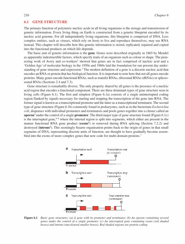
6.1 GENE STRUCTURE
The primary function of polymeric nucleic acids in all living organisms is the storage and transmission ofgenetic information. Every living thing on Earth is constructed from a genetic blueprint encoded by itsnucleic acid genome. For all independently living organisms, this blueprint is comprised of DNA. Lesscomplex entities, such as viruses, which rely on hosts to live and reproduce themselves, may use RNAinstead. This chapter will describe how this genetic information is stored, replicated, repaired and copiedinto the functional products on which life depends.
The basic unit of genetic information is the gene. Genes were described originally in 1865 by Mendelas apparently indestructible factors, which specify traits of an organism such as colour or shape. The pion-eering work of Avery and co-workers1 showed that genes are in fact comprised of nucleic acid and a‘Golden Age’ of molecular biology in the 1950s and 1960s laid the foundation for our present day under-standing of gene structure and expression.2 The modern definition of a gene is a discrete nucleic acid thatencodes an RNA or protein that has biological function. It is important to note here that not all genes encodeproteins. Many genes encode functional RNAs, such as transfer RNAs, ribosomal RNAs (rRNAs) or spliceo-somal RNAs (Sections 2.4 and 7.3).
Gene structure is remarkably diverse. The only property shared by all genes is the presence of a nucleicacid region that encodes a functional component. There are three dominant types of gene structure seen inliving cells (Figure 6.1). The first and simplest (Figure 6.1a) consists of a single uninterrupted codingregion flanked by signals necessary for starting and stopping the transcription of the gene into RNA. Theformer signal is known as a transcriptional promoter and the latter as a transcriptional terminator. The secondtype of gene structure (Figure 6.1b) commonly found in prokaryotes, such as in the bacterium Escherichiacoli, dispenses with individual promoters and terminators and pools genes together into a cluster called anoperon3 under the control of a single promoter. The third major type of gene structure found (Figure 6.1c)is the interrupted gene,4–6 where the internal region is split into segments, which either are present in themature functional RNA gene product (exons6) or removed during RNA splicing (Section 7.2.2) anddestroyed (introns6). This seemingly bizarre organisation points back to the origin of genes in that smallsegments of DNA, representing discrete units of function, are thought to have gradually become assem-bled into the exons of more complex genes that now code for multi-domain proteins.7
210 Chapter 6
Figure 6.1 Basic gene structures. (a) A gene with its promoter and terminator. (b) An operon containing severalgenes under the control of a single promoter. (c) An interrupted gene containing exons (red shadedboxes) and introns (uncoloured smaller boxes). Red shaded regions are protein coding
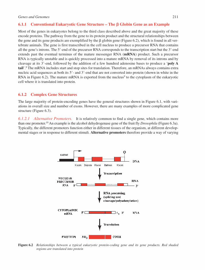
6.1.1 Conventional Eukaryotic Gene Structure – The �� Globin Gene as an Example
Most of the genes in eukaryotes belong to the third class described above and the great majority of theseencode proteins. The pathway from the gene to its protein product and the structural relationships betweenthe gene and its gene products are exemplified by the � globin gene (Figure 6.2), which is found in all ver-tebrate animals. The gene is first transcribed in the cell nucleus to produce a precursor RNA that containsall the gene’s introns. The 5�-end of the precursor RNA corresponds to the transcription start but the 3�-endextends past the eventual terminus of the mature messenger RNA (mRNA) product. Such a precursorRNA is typically unstable and is quickly processed into a mature mRNA by removal of its introns and bycleavage at its 3�-end, followed by the addition of a few hundred adenosine bases to produce a ‘poly Atail’.8 The mRNA includes start and stop sites for translation. Therefore, an mRNAs always contains extranucleic acid sequences at both its 5�- and 3�-end that are not converted into protein (shown in white in theRNA in Figure 6.2). The mature mRNA is exported from the nucleus9 to the cytoplasm of the eukaryoticcell where it is translated into protein.
6.1.2 Complex Gene Structures
The large majority of protein-encoding genes have the general structures shown in Figure 6.1, with vari-ations in overall size and number of exons. However, there are many examples of more complicated genestructure (Figure 6.3).
6.1.2.1 Alternative Promoters. It is relatively common to find a single gene, which contains morethan one promoter.10 An example is the alcohol dehydrogenase gene of the fruit fly Drosophila (Figure 6.3a).Typically, the different promoters function either in different tissues of the organism, at different develop-mental stages or in response to different stimuli. Alternative promoters therefore provide a way of varying
Genes and Genomes 211
Figure 6.2 Relationships between a typical eukaryotic protein-coding gene and its gene products. Red shadedregions are translated into protein
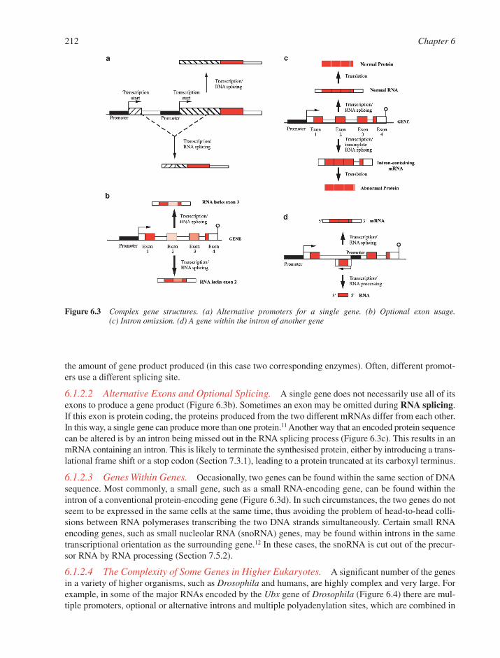
the amount of gene product produced (in this case two corresponding enzymes). Often, different promot-ers use a different splicing site.
6.1.2.2 Alternative Exons and Optional Splicing. A single gene does not necessarily use all of itsexons to produce a gene product (Figure 6.3b). Sometimes an exon may be omitted during RNA splicing.If this exon is protein coding, the proteins produced from the two different mRNAs differ from each other.In this way, a single gene can produce more than one protein.11 Another way that an encoded protein sequencecan be altered is by an intron being missed out in the RNA splicing process (Figure 6.3c). This results in anmRNA containing an intron. This is likely to terminate the synthesised protein, either by introducing a trans-lational frame shift or a stop codon (Section 7.3.1), leading to a protein truncated at its carboxyl terminus.
6.1.2.3 Genes Within Genes. Occasionally, two genes can be found within the same section of DNAsequence. Most commonly, a small gene, such as a small RNA-encoding gene, can be found within theintron of a conventional protein-encoding gene (Figure 6.3d). In such circumstances, the two genes do notseem to be expressed in the same cells at the same time, thus avoiding the problem of head-to-head colli-sions between RNA polymerases transcribing the two DNA strands simultaneously. Certain small RNAencoding genes, such as small nucleolar RNA (snoRNA) genes, may be found within introns in the sametranscriptional orientation as the surrounding gene.12 In these cases, the snoRNA is cut out of the precur-sor RNA by RNA processing (Section 7.5.2).
6.1.2.4 The Complexity of Some Genes in Higher Eukaryotes. A significant number of the genesin a variety of higher organisms, such as Drosophila and humans, are highly complex and very large. Forexample, in some of the major RNAs encoded by the Ubx gene of Drosophila (Figure 6.4) there are mul-tiple promoters, optional or alternative introns and multiple polyadenylation sites, which are combined in
212 Chapter 6
Figure 6.3 Complex gene structures. (a) Alternative promoters for a single gene. (b) Optional exon usage. (c) Intron omission. (d) A gene within the intron of another gene
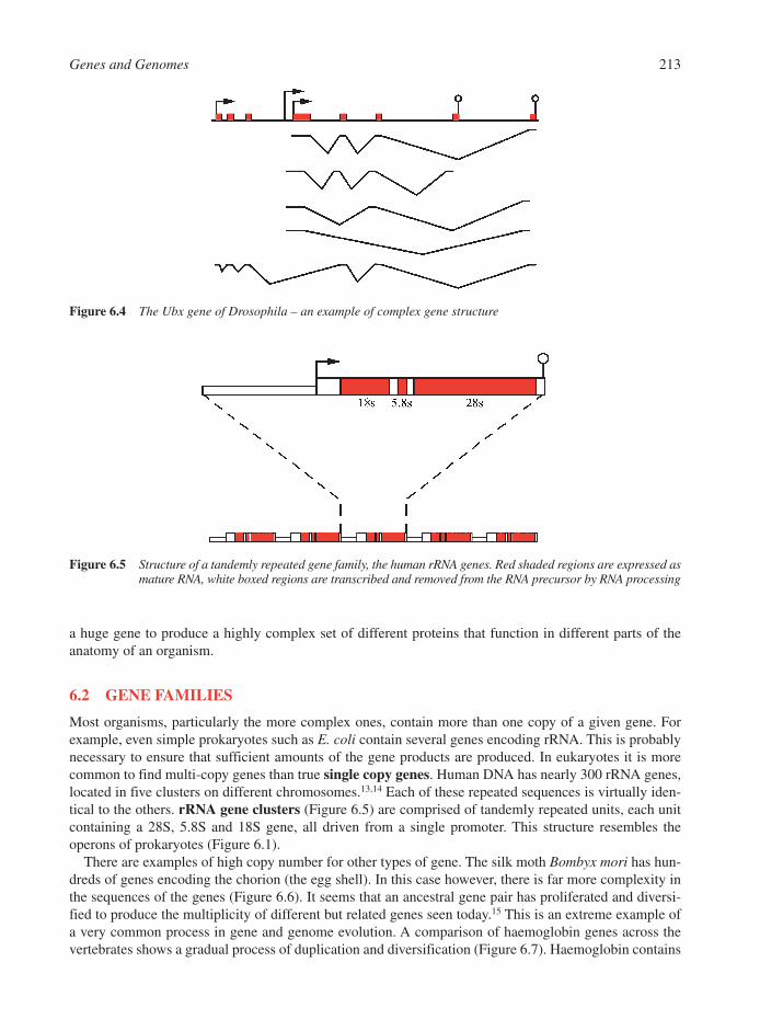
a huge gene to produce a highly complex set of different proteins that function in different parts of theanatomy of an organism.
6.2 GENE FAMILIES
Most organisms, particularly the more complex ones, contain more than one copy of a given gene. Forexample, even simple prokaryotes such as E. coli contain several genes encoding rRNA. This is probablynecessary to ensure that sufficient amounts of the gene products are produced. In eukaryotes it is morecommon to find multi-copy genes than true single copy genes. Human DNA has nearly 300 rRNA genes,located in five clusters on different chromosomes.13,14 Each of these repeated sequences is virtually iden-tical to the others. rRNA gene clusters (Figure 6.5) are comprised of tandemly repeated units, each unitcontaining a 28S, 5.8S and 18S gene, all driven from a single promoter. This structure resembles the operons of prokaryotes (Figure 6.1).
There are examples of high copy number for other types of gene. The silk moth Bombyx mori has hun-dreds of genes encoding the chorion (the egg shell). In this case however, there is far more complexity inthe sequences of the genes (Figure 6.6). It seems that an ancestral gene pair has proliferated and diversi-fied to produce the multiplicity of different but related genes seen today.15 This is an extreme example ofa very common process in gene and genome evolution. A comparison of haemoglobin genes across thevertebrates shows a gradual process of duplication and diversification (Figure 6.7). Haemoglobin contains
Genes and Genomes 213
Figure 6.4 The Ubx gene of Drosophila – an example of complex gene structure
Figure 6.5 Structure of a tandemly repeated gene family, the human rRNA genes. Red shaded regions are expressed asmature RNA, white boxed regions are transcribed and removed from the RNA precursor by RNA processing
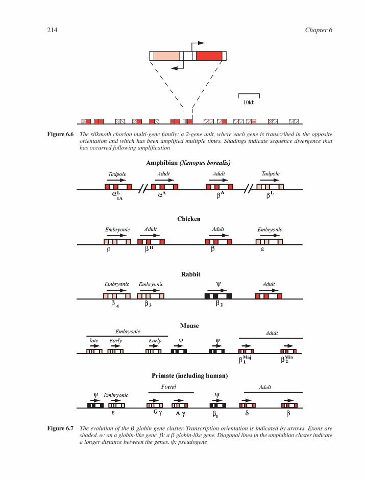
214 Chapter 6
Figure 6.7 The evolution of the b globin gene cluster. Transcription orientation is indicated by arrows. Exons areshaded. a: an a globin-like gene. b: a b globin-like gene. Diagonal lines in the amphibian cluster indicatea longer distance between the genes. c: pseudogene
Figure 6.6 The silkmoth chorion multi-gene family: a 2-gene unit, where each gene is transcribed in the oppositeorientation and which has been amplified multiple times. Shadings indicate sequence divergence thathas occurred following amplification
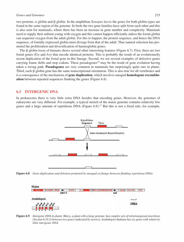
two proteins, � globin and � globin. In the amphibian Xenopus laevis the genes for both globin types arefound in the same region of the genome. In birds the two gene families have split from each other and thisis also seen for mammals, where there has been an increase in gene number and complexity. Mammalsneed to supply their unborn young with oxygen and this cannot happen efficiently unless the foetal globincan sequester oxygen from the adult globin. For this to happen, the protein sequence, and hence the DNAsequence, of foetally expressed globin must diverge from that of the adult. Thus natural selection has pro-moted the proliferation and diversification of haemoglobin genes.
The � globin locus of humans shows several other interesting features (Figure 6.7). First, there are twofoetal genes (G� and A�) that encode identical proteins. This is probably the result of an evolutionarilyrecent duplication of the foetal gene in this lineage. Second, we see several examples of defective genescarrying frame shifts and stop codons. These pseudogenes16 may be the result of gene evolution havingtaken a wrong path. Pseudogenes are very common in mammals but surprisingly quite rare in plants.Third, each � globin gene has the same transcriptional orientation. This is also true for all vertebrates andis a consequence of the mechanism of gene duplication, which involves unequal homologous recombin-ation between repeated sequences flanking the genes (Figure 6.8).
6.3 INTERGENIC DNA
In prokaryotes there is very little extra DNA besides that encoding genes. However, the genomes ofeukaryotes are very different. For example, a typical stretch of the maize genome contains relatively fewgenes and a large amount of repetitious DNA (Figure 6.9).17 But this is not a fixed rule; for example,
Genes and Genomes 215
Figure 6.8 Gene duplication and deletion promoted by unequal exchange between flanking repetitious DNAs
Figure 6.9 Intergenic DNA in plants. Maize, a plant with a large genome, has complex sets of retrotransposon insertions(Section 6.10.3) between two genes (indicated by arrows). Arabidopsis thaliana has six genes with relativelylittle intergenic DNA

another plant, the weed Arabidopsis thaliana, has far less intergenic repetitious DNA (Figure 6.9).18 Infact, the intergenic DNA of eukaryotes is much more susceptible to change than their genes.
For some genomes such as maize the repetitious DNA can outweigh the DNA devoted to genes, lead-ing to ‘genomic obesity’.19 This observation explains an old puzzle for molecular geneticists, namely thatthe sizes of eukaryotic genomes are extremely variable and there is no obvious correlation betweengenome size and evolutionary level (Figure 6.10). For example, one amphibian may possess 50-fold moregenetic information than another. The puzzle has been called the C-value paradox.
Two major classes of repetitious DNA make up the majority of this so-called ‘junk DNA’.20 The first issatellite DNA, which is comprised of seemingly endless tandem repeats of a simple sequence. For example,the fruit fly Drosophila virilis possesses a huge number of ACAAACT repeats that together add up to almosta quarter of the entire genome. Such DNA is also found in heterochromatin21 (Section 6.4.2), particularly atthe centromeres of chromosomes (Section 6.4.5). The second major class of repetitious DNA is transposableelements, particularly retrotransposons (Section 6.8.3).
6.4 CHROMOSOMES
The DNA of an organism is arranged on one or more chromosomes. The number of chromosomes per speciesis invariant but can vary a lot between related species. Each chromosome is comprised of a double-strandedDNA molecule, packed together with a set of associated proteins and other components into a complexcalled chromatin.
6.4.1 Eukaryotic Chromosomes
All eukaryotes contain at least two chromosomes. There is no clear correlation between the chromosomenumber and the type of organism. For example, the yeast Saccharomyces cerevisiae has 16 chromosomesper haploid cell, the fruit fly Drosophila melanogaster has 4 and the human has 23. Most multi-cellulareukaryotes contain mostly diploid cells and for these cells the chromosome number is doubled. Alleukaryotic nuclear chromosomes studied to date contain simple linear double-stranded DNA.
6.4.2 Packaging of DNA in Eukaryotic Chromosomes
The DNA of a eukaryotic chromosome must fit into a space far smaller than its total length. For example, ahuman chromosome has around 3–10 cm of DNA that must fit into a cell nucleus a thousand times smaller.
216 Chapter 6
Figure 6.10 Variation in genome size across the five kingdoms of life
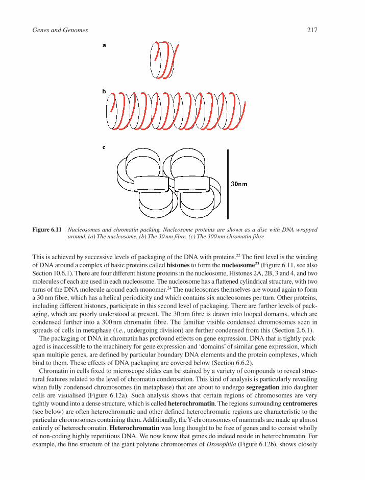
This is achieved by successive levels of packaging of the DNA with proteins.22 The first level is the windingof DNA around a complex of basic proteins called histones to form the nucleosome23 (Figure 6.11, see alsoSection 10.6.1). There are four different histone proteins in the nucleosome, Histones 2A, 2B, 3 and 4, and twomolecules of each are used in each nucleosome. The nucleosome has a flattened cylindrical structure, with twoturns of the DNA molecule around each monomer.24 The nucleosomes themselves are wound again to forma 30 nm fibre, which has a helical periodicity and which contains six nucleosomes per turn. Other proteins,including different histones, participate in this second level of packaging. There are further levels of pack-aging, which are poorly understood at present. The 30 nm fibre is drawn into looped domains, which arecondensed further into a 300 nm chromatin fibre. The familiar visible condensed chromosomes seen inspreads of cells in metaphase (i.e., undergoing division) are further condensed from this (Section 2.6.1).
The packaging of DNA in chromatin has profound effects on gene expression. DNA that is tightly pack-aged is inaccessible to the machinery for gene expression and ‘domains’ of similar gene expression, whichspan multiple genes, are defined by particular boundary DNA elements and the protein complexes, whichbind to them. These effects of DNA packaging are covered below (Section 6.6.2).
Chromatin in cells fixed to microscope slides can be stained by a variety of compounds to reveal struc-tural features related to the level of chromatin condensation. This kind of analysis is particularly revealingwhen fully condensed chromosomes (in metaphase) that are about to undergo segregation into daughtercells are visualised (Figure 6.12a). Such analysis shows that certain regions of chromosomes are verytightly wound into a dense structure, which is called heterochromatin. The regions surrounding centromeres(see below) are often heterochromatic and other defined heterochromatic regions are characteristic to theparticular chromosomes containing them. Additionally, the Y-chromosomes of mammals are made up almostentirely of heterochromatin. Heterochromatin was long thought to be free of genes and to consist whollyof non-coding highly repetitious DNA. We now know that genes do indeed reside in heterochromatin. Forexample, the fine structure of the giant polytene chromosomes of Drosophila (Figure 6.12b), shows closely
Genes and Genomes 217
Figure 6.11 Nucleosomes and chromatin packing. Nucleosome proteins are shown as a disc with DNA wrappedaround. (a) The nucleosome. (b) The 30 nm fibre. (c) The 300 nm chromatin fibre
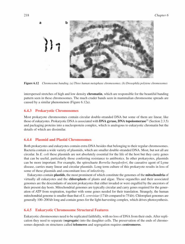
interspersed stretches of high and low density chromatin, which are responsible for the beautiful bandingpattern seen in these chromosomes. The much cruder bands seen in mammalian chromosome spreads arecaused by a similar phenomenon (Figure 6.12a).
6.4.3 Prokaryotic Chromosomes
Most prokaryote chromosomes contain circular double-stranded DNA but some of them are linear, likethose of eukaryotes. Prokaryotic DNA is associated with DNA gyrase, DNA topoisomerase25 (Section 2.3.5)and packaging proteins into a nucleoprotein complex, which is analogous to eukaryotic chromatin but thedetails of which are dissimilar.
6.4.4 Plasmid and Plastid Chromosomes
Both prokaryotes and eukaryotes contain extra DNA besides that belonging to their regular chromosomes.Bacteria contain a wide variety of plasmids, which are smaller double-stranded DNA. Most, but not all arecircular. In E. coli these plasmids are not absolutely essential for the life of the host but they carry genesthat can be useful, particularly those conferring resistance to antibiotics. In other prokaryotes, plasmidscan be more important. For example, the spirochaete Borrelia burgdorferi, the causative agent of Lymedisease, carries many linear and circular plasmids. Long term culture of this prokaryote results in loss ofsome of these plasmids and concomitant loss of infectivity.
Eukaryotes contain plastids, the most prominent of which constitute the genomes of the mitochondria ofvirtually all eukaryotes and the chloroplasts of plants and algae. These organelles and their associatedgenomes are the descendants of ancient prokaryotes that either invaded or were engulfed by the ancestors oftheir present day hosts. Mitochondrial genomes are typically circular and carry genes required for the gener-ation of ATP from respiration, together with some genes needed for their translation. Strangely, the humanmitochondrial genome is smaller than that of S. cerevisiae (17 kb compared to 75 kb). Chloroplast genomes aregenerally 100–200 kb long and contain genes for the light-harvesting complex, which drives photosynthesis.
6.4.5 Eukaryotic Chromosome Structural Features
Eukaryotic chromosomes need to be replicated faithfully, with no loss of DNA from their ends. After repli-cation they need to separate (segregate) into the daughter cells. The preservation of the ends of chromo-somes depends on structures called telomeres and segregation requires centromeres.
218 Chapter 6
Figure 6.12 Chromosome banding. (a) Three human metaphase chromosomes. (b) Drosophila polytene chromosomes

6.4.5.1 Centromeres. Centromeres can be found at different regions depending on the particularchromosome. A metacentric chromosome has its centromere near the middle of the chromosome and atelocentric chromosome has one at the telomere. The centromere has been intensively studied by genetic,molecular and microscopic analysis. Genetic and molecular analysis in the yeast S. cerevisiae has identi-fied a minimal structure necessary for centromere function (Figure 6.13).26,27 Two regions, CDE1 andCDE3 have important conserved sequences necessary for centromere function. The first of these regionsbinds to a protein called CBF1 and the second binds to a complex of three proteins, namely CBF3b,NDC10 and CTF13. The DNA sequence CDE2 that separates these regions is about 80 bp in length, hasapproximately 90% A/T content, and binds to the MIF2 protein.The centromeres of more complex eukary-otes are much larger than this. Additionally, centromeric regions tend to accumulate even more repetitiousDNA than the rest of the genome. Repetitious DNA presents serious problems in sequence determinationand computer-based structural analysis and is often difficult to clone.
6.4.5.2 Telomeres. Telomeres are also essential chromosomal components.28 In their absence, thechromosome shortens until essential genes are lost and the cell dies. The extreme ends of chromosomesdo not have complex structures; they are simply double-stranded DNA of repeating sequence, exemplifiedby the common sequence shown in Figure 6.14. Telomeres counter their natural tendency to becomeshorter at their ends by generating new copies of these repeats. The mechanisms whereby this occurs aredescribed below (Section 6.6.5).
6.4.6 Viral Genomes
Viruses are parasites that can only replicate inside a host cell.29 Probably all organisms can act as hosts toviruses. Viruses can have genomes made up of DNA or RNA (Table 6.1). The simplest virus has only a shortnucleic acid encoding a handful of genes, which is packaged into a protein particle. More complicated
Genes and Genomes 219
Figure 6.13 Structure of the yeast centromere
Figure 6.14 Sequence of the human telomere repeat
Table 6.1 Eukaryotic viruses
Genome Type Example Size (base pairs) Structure
ds DNA Poxvirus Smallpox 250,000 LinearssDNA Parvovirus AAV 2000 Circularss/ds DNA Hepadnavirus Hepatitis B 3000 CirculardsRNA Reovirus Reovirus 25,000 LinearssRNA � strand Picornavirus Poliovirus 7000 LinearssRNA � strand Myxovirus Influenza 12,000 Linear (several pieces)
viruses may have many enzymes within the particle and hundreds of genes encoded by the nucleic acid.Viral genomes are always very compact, with almost every nucleotide devoted to genes.
6.4.6.1 The Viral Life Cycle. First, a virus must enter its host cell. The simpler viruses then uncoatcompletely and the DNA enters the nucleus where parts of its genome are transcribed. More complexviruses, such as poxviruses preserve an internal core structure inside the cell, which stays in the cytoplasm.In poxviruses, the host cell’s RNA polymerase components migrate to the cytoplasm.
For most viruses, there is more than one stage of viral infection. For the simpler viruses there is an earlyphase (pre-DNA replication) and a late phase (post-DNA replication). For DNA viruses the early phaseinvolves the transcription of ‘early’ genes, the jobs of which are to make sure the cell is not in a restingphase and to coordinate the switching on of viral DNA replication and transcription of ‘late’ genes thattypically encode the components of the virus particle. For more complex viruses there are several phases,for example herpesviruses have three distinct phases.
6.4.6.2 RNA Viruses. RNA viruses all encode their own polymerases for replication, because hostcells do not contain enzymes capable of copying RNA. (�) Strand viruses contain RNA that can act as anmRNA for the production of the viral polymerase that is responsible for synthesis of a minus strand.The �/� duplex (or replication intermediate) is then transcribed to generate further (�) strand, to thusproduce more mRNA as well as viral RNA. (�) Strand viruses cannot act as mRNA when they enter thecell, so they must carry polymerase inside their virus particles. This polymerase then replicates the (�)strand, usually to form a �/� RNA duplex as before, which is then copied to produce mRNA.
One of the most interesting and important classes of RNA virus is the retroviruses.30 These are (�) strandviruses that depart from a normal life cycle by going through a DNA intermediate. The AIDS retrovirusesHIV 1 and 2 are major pathogens, which are responsible for the deaths of millions of people. Retrovirusescarry an RNA-dependent DNA polymerase (reverse transcriptase) that is responsible for the synthesis ofa double-stranded DNA copy of the viral RNA. This DNA copy is inserted into the chromosomal DNA byanother virus-encoded enzyme (integrase). The integrated DNA is then transcribed to produce (�) strandRNA, which then can be processed to become mRNA or virus particle RNA.
An important feature of viruses is their high rate of sequence mutation. In some cases the consequencesare serious, since such mutation can lead to resistance to drug treatment or to antibodies raised by thehuman immune system. Rapid sequence mutation in a virus is often due to the viral polymerase beingmore error-prone than the cellular DNA polymerase. Since the viral life cycle is typically measured inhours or a few days, a virus will go through many successive replications of its genome during an infec-tion, each one of which can give rise to new mutations.
6.5 DNA SEQUENCE AND BIOINFORMATICS
The remarkable advances in cloning and sequencing technologies since the 1970s have made it reasonablyeasy to sequence very long stretches of DNA. Whole genome sequences are being deposited in sequencedatabases at an accelerating rate.13,14 The enormous amount of data involved in a genome sequence must bestored in such databases and analysed. To get an idea of the volume of information, the complete sequenceof a single, smaller than average-sized human gene, the � globin gene is shown (Figure 6.15). One thousandsuch genes are represented below and 30 such sets are roughly equivalent to the complete human gene com-plement. This constitutes about 1/60th of the entire human genome, with repetitious DNA making up the rest.
6.5.1 Finding Genes
The acquisition of complete genome sequences allows potential access to every gene in the organism. Torealise this goal, genes must be found within the ‘haystack’ of non-coding DNA. Bioinformatics providesreasonably reliable search algorithms to predict gene structure, particularly for location of exons within
220 Chapter 6

Genes and Genomes 221
Figure 6.15 The information content of the human genome

genomic sequences. But when these are carefully tested on well-characterised genomic regions containingknown genes, they often fall short, either by missing exons, by predicting exons to be complete genes orby predicting exons where none exist. Such failings are particularly pronounced for complex genes suchas Ubx (Figure 6.4).
One way to help find the locations of genes in DNA is by sequence analysis of large numbers of tran-scribed sequences, because in general only genes are transcribed, and by comparison with the completegenome sequence. Typically, a cDNA library is made by reverse transcription from the RNA of the organ-ism. Then thousands of individual sequences are determined. To find the rare RNAs, often single sequen-cing experiments are carried out (typically ca. 500 bp), on a large number of subclones, instead ofdetermination of the complete sequence for relatively few RNAs.
6.5.2 Genome Maps
Once the genomic sequence is reasonably well ordered into accurate, large contiguous pieces (contigs),which eventually extend to whole chromosomes, the cDNA sequences can be mapped onto the respectivegenome. Such maps are useful in identifying genes, which may be associated with important traits, suchas the predisposition to inherited diseases. The gene map obtained from such a study can be aligned againstother important maps, showing the extents of large insert clones (BAC, YAC clones, etc., see Section 5.2.1).BAC and YAC contigs are obtained by sequence analysis of the ends of randomly selected large insertclones and then by a search of previously acquired data for identical sequences.
6.5.3 Molecular Marker Maps
Another important map, which can be aligned against the genome and cDNA maps, is a molecular markermap. A molecular marker is any difference in DNA sequence observed at a precise genomic locationbetween two individuals of an organism, for example two human beings. Such differences represent just atiny fraction of the huge amount of genetic variation in a species and mostly lie in non-coding DNA thatis not subject to natural selection to preserve its sequence. Molecular markers are useful research tools inthat they can be mapped genetically in the same way as visible traits, such as Mendel’s pea seed traits. Theycan also be physically mapped on genomic DNA. Indeed, there are now hundreds of times more molecularmarkers mapped, both genetically and physically, on the human genome than there are genes identified.Genetic markers that are tightly linked to particular gene variants (alleles) can be of medical importance. Forexample, whether a baby carries a defective cystic fibrosis gene can now be assessed by a simple markerassay on DNA isolated from a pinprick of blood, rather than having to clone and sequence the gene itself.
6.5.4 Molecular Marker Types
The first types of molecular markers are restriction fragment length polymorphisms (RFLPs). These DNAsequence variants (usually point mutations or small insertions or deletions) result in the creation ordestruction of a restriction enzyme cleavage site (Section 5.3.1). Such mutations sometimes alter therestriction map of the genomic region in which they reside. Such DNA alterations can be detected eitherby Southern blot analysis (Section 5.5.2) or more often nowadays by PCR (Section 5.2.2) followed byrestriction digestion of the amplified DNA to reveal the polymorphic restriction site.
Two more important molecular marker types in use now are microsatellites31 and single nucleotide poly-morphisms (SNPs) (Section 5.5.3).32 Microsatellites are also called simple sequence repeats (SSRs). SSRscontain a varying number of repeats of typically 2–3 base pairs. At a given locus (genomic region), one indi-vidual might have six repeats of the dinucleotide GT whilst another may have nine such repeats. These dif-ferences are revealed by DNA amplification of the region containing the repeat and by determination of itslength. SNPs are merely single nucleotide changes in a given genomic region, e.g. a G substitution by anA at position 543. Much effort is currently being invested in finding cheap and efficient methods for identify-ing such simple SNPs.
222 Chapter 6
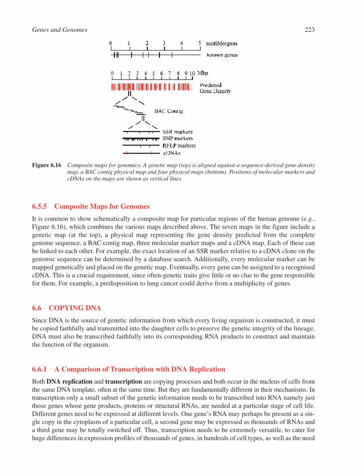
6.5.5 Composite Maps for Genomes
It is common to show schematically a composite map for particular regions of the human genome (e.g.,Figure 6.16), which combines the various maps described above. The seven maps in the figure include agenetic map (at the top), a physical map representing the gene density predicted from the completegenome sequence, a BAC contig map, three molecular marker maps and a cDNA map. Each of these canbe linked to each other. For example, the exact location of an SSR marker relative to a cDNA clone on thegenomic sequence can be determined by a database search. Additionally, every molecular marker can bemapped genetically and placed on the genetic map. Eventually, every gene can be assigned to a recognisedcDNA. This is a crucial requirement, since often-genetic traits give little or no clue to the gene responsiblefor them. For example, a predisposition to lung cancer could derive from a multiplicity of genes.
6.6 COPYING DNA
Since DNA is the source of genetic information from which every living organism is constructed, it mustbe copied faithfully and transmitted into the daughter cells to preserve the genetic integrity of the lineage.DNA must also be transcribed faithfully into its corresponding RNA products to construct and maintainthe function of the organism.
6.6.1 A Comparison of Transcription with DNA Replication
Both DNA replication and transcription are copying processes and both occur in the nucleus of cells fromthe same DNA template, often at the same time. But they are fundamentally different in their mechanisms. Intranscription only a small subset of the genetic information needs to be transcribed into RNA namely justthose genes whose gene products, proteins or structural RNAs, are needed at a particular stage of cell life.Different genes need to be expressed at different levels. One gene’s RNA may perhaps be present as a sin-gle copy in the cytoplasm of a particular cell, a second gene may be expressed as thousands of RNAs anda third gene may be totally switched off. Thus, transcription needs to be extremely versatile, to cater forhuge differences in expression profiles of thousands of genes, in hundreds of cell types, as well as the need
Genes and Genomes 223
Figure 6.16 Composite maps for genomics. A genetic map (top) is aligned against a sequence-derived gene densitymap, a BAC contig physical map and four physical maps (bottom). Positions of molecular markers andcDNAs on the maps are shown as vertical lines

to alter transcription profiles multiple times during cell development. However, transcription does not needto be extremely accurate, because a cell can still operate fully even if 0.1% of its RNAs are not functional.
In contrast, DNA replication requires that all of the genetic information in the cell be copied, and copiedonly once, into a single daughter molecule that is as identical to the parent molecule as possible. DNAreplication is therefore extremely accurate, with rounds of proofreading and error correction, as well aschecks to make sure that a DNA strand only becomes copied once per cell cycle.
6.6.2 Transcription in Prokaryotes
Transcription involves the copying of a gene into an RNA molecule. Several phases are involved in thisprocess, namely, initiation of transcription, elongation, termination and RNA processing (Section 7.2).There are many similarities between prokaryotes and the eukaryotes in these processes.
6.6.2.1 Prokaryotic RNA Polymerases. There are two types of RNA polymerase. Viral polymerasesare simple, single subunit enzymes in the range of 1�2 � 104 Da in mass. These polymerases can onlyinitiate transcription from one or a very small number of very similar promoters. In contrast, all RNA poly-merases that transcribe cellular genes are large, multi-subunit enzymes, which are more versatile in theirability to recognise different promoters.
In prokaryotes, a single RNA polymerase is responsible for the synthesis of all RNA. The completeenzyme (holoenzyme) has a molecular mass approximately 4.8 � 105 Da. It is pentameric in structure andis comprised of two a subunits and two related B subunits, b and b�, together with an associated unit called� (sigma). There are several sigma factors available33 and these modulate the specificity of the RNA poly-merase for different promoters (see below).
6.6.2.2 Prokaryotic Transcriptional Initiation. Transcriptional initiation is the first event in copyingthe DNA template into RNA.34 This occurs at a specific region of the gene, called the promoter. E. coli RNApolymerase that lacks a sigma factor (the core enzyme) has a relatively weak affinity for all DNA, with nogreat preference for promoter regions. The function of � is to make sure that RNA polymerase binds stablyto DNA only at promoters. RNA polymerase containing a sigma factor (holoenzyme) has a far lower affin-ity for DNA in general but a higher affinity for promoter regions in particular. Different promoters showlarge differences in their affinities for the holoenzyme, with ‘strong’ promoters having far higher affinities.
6.6.2.2.1 Steps in Prokaryotic Transcriptional Initiation. The process of transcriptional initi-ation has been elucidated in vitro using purified RNA polymerase holoenzyme and a DNA template. Fourdistinct stages are observed (Figure 6.17). First, the core enzyme binds to a region from about 40 basesupstream (the ��35 box) to about �20 bases downstream of the transcription start site to form a closedpromoter complex. At this point the DNA template is still an intact double helix. Second, the RNA poly-merase moves downstream and a limited region of the helix at another conserved sequence (the Pribnowor ��10 box) is unwound to form an open promoter complex. Third, the polymerase begins to synthesise ashort RNA molecule on the template DNA strand at the start site. Usually, several abortive short RNAs ofbetween two and nine nucleotides are synthesised before the polymerase succeeds in clearing the pro-moter. At this point the � factor detaches from the holoenzyme.
6.6.2.2.2 Promoter Identification. Promoters, both prokaryotic and eukaryotic, have been identi-fied in one or a combination of the following ways:
(i) Consensus searches. Many promoters are aligned with each other and conserved regions are thusidentified. The Pribnow and �35 boxes were originally identified in this way.
(ii) Mutation analysis. Naturally occurring or mutagen-induced mutations that affect transcriptionalinitiation are examined by sequence analysis of the promoters to determine the molecular basisfor the mutations.
224 Chapter 6
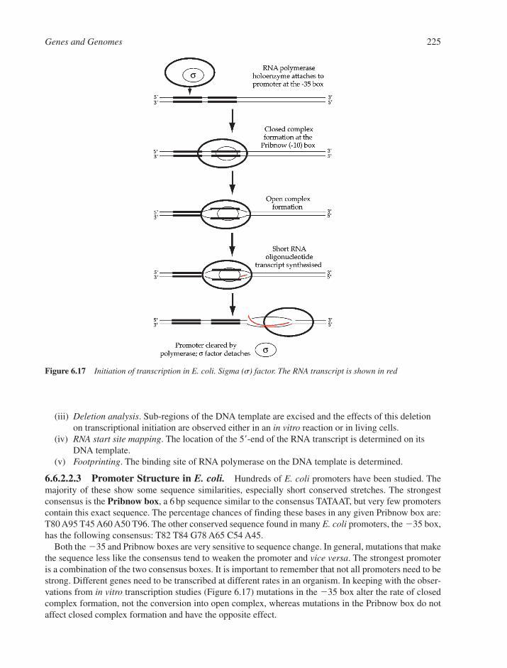
(iii) Deletion analysis. Sub-regions of the DNA template are excised and the effects of this deletionon transcriptional initiation are observed either in an in vitro reaction or in living cells.
(iv) RNA start site mapping. The location of the 5�-end of the RNA transcript is determined on itsDNA template.
(v) Footprinting. The binding site of RNA polymerase on the DNA template is determined.
6.6.2.2.3 Promoter Structure in E. coli. Hundreds of E. coli promoters have been studied. Themajority of these show some sequence similarities, especially short conserved stretches. The strongestconsensus is the Pribnow box, a 6 bp sequence similar to the consensus TATAAT, but very few promoterscontain this exact sequence. The percentage chances of finding these bases in any given Pribnow box are:T80 A95 T45 A60 A50 T96. The other conserved sequence found in many E. coli promoters, the �35 box,has the following consensus: T82 T84 G78 A65 C54 A45.
Both the �35 and Pribnow boxes are very sensitive to sequence change. In general, mutations that makethe sequence less like the consensus tend to weaken the promoter and vice versa. The strongest promoteris a combination of the two consensus boxes. It is important to remember that not all promoters need to bestrong. Different genes need to be transcribed at different rates in an organism. In keeping with the obser-vations from in vitro transcription studies (Figure 6.17) mutations in the �35 box alter the rate of closedcomplex formation, not the conversion into open complex, whereas mutations in the Pribnow box do notaffect closed complex formation and have the opposite effect.
Genes and Genomes 225
Figure 6.17 Initiation of transcription in E. coli. Sigma (s) factor. The RNA transcript is shown in red
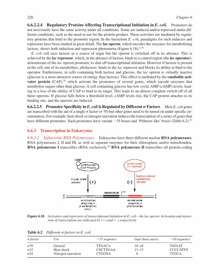
6.6.2.2.4 Regulatory Proteins Affecting Transcriptional Initiation in E. coli. Promoters donot necessarily have the same activity under all conditions. Some are induced and/or repressed under dif-ferent conditions, such as the need or not for the protein product. These activities are mediated by regula-tory proteins that bind to the promoter region. In the bacterium E. coli, paradigms for such inducers andrepressors have been studied in great detail. The lac operon, which encodes the enzymes for metabolisinglactose, shows both induction and repression phenomena (Figure 6.18).3,35
E. coli cell uses lactose as a source of sugar but the operon is switched off in its absence. This isachieved by the lac repressor, which, in the absence of lactose, binds to a control region (the lac operator),downstream of the lac operon promoter, to shut off transcriptional initiation. However if lactose is presentin the cell, one of its metabolites, allolactose, binds to the lac repressor and blocks its ability to bind to theoperator. Furthermore, in cells containing both lactose and glucose, the lac operon is virtually inactive(glucose is a more attractive source of energy than lactose). This effect is mediated by the catabolite acti-vator protein (CAP),35 which activate the promoters of several genes, which encode enzymes thatmetabolise sugars other than glucose. A cell containing glucose has low cyclic AMP (cAMP) levels, lead-ing to a loss of the ability of CAP to bind to its target. This leads to an almost complete switch off of allthese operons. If glucose falls below a threshold level, cAMP levels rise, the CAP protein attaches to itsbinding site, and the operons are induced.
6.6.2.2.5 Promoter Specificity in E. coli is Regulated by Different � Factors. Most E. coli genesare transcribed with the aid of a single � factor (� 70) but other genes need to be turned on under specific cir-cumstances. For example, heat shock or nitrogen starvation induces the transcription of a series of genes thathave different promoters. Such promoters have variant �35 boxes and ‘Pribnow-like’ boxes (Table 6.2).33
6.6.3 Transcription in Eukaryotes
6.6.3.1 Eukaryotic RNA Polymerases. Eukaryotes have three different nuclear RNA polymerases,RNA polymerases I, II and III, as well as separate enzymes for their chloroplasts and/or mitochondria.RNA polymerase I transcribes rRNA exclusively.36 RNA polymerase II transcribes all protein-coding
226 Chapter 6
Table 6.2 Different s factors in E. coli
� factor Use �35 sequence Gap (base pairs) �10 sequence
�70 General TTGACA 16–18 TATAAT�32 Heat shock CNCTTGAA 13–15 CCCCATNT�54 Nitrogen starvation CTGGNA 6 TTGCA
Figure 6.18 Activators and repressors of transcriptional initiation in E. coli – the lac operon. Activation and repres-sion of transcription are indicated by (�) and (�) respectively
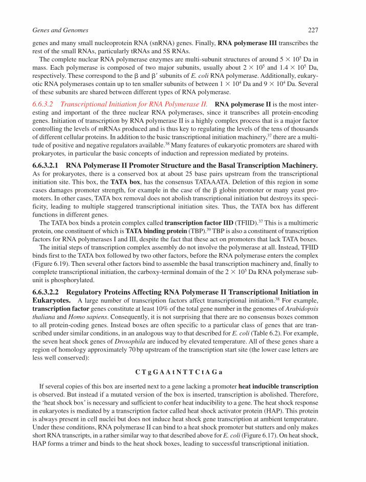
genes and many small nucleoprotein RNA (snRNA) genes. Finally, RNA polymerase III transcribes therest of the small RNAs, particularly tRNAs and 5S RNAs.
The complete nuclear RNA polymerase enzymes are multi-subunit structures of around 5 � 105 Da inmass. Each polymerase is composed of two major subunits, usually about 2 � 105 and 1.4 � 105 Da,respectively. These correspond to the � and �� subunits of E. coli RNA polymerase. Additionally, eukary-otic RNA polymerases contain up to ten smaller subunits of between 1 � 104 Da and 9 � 104 Da. Severalof these subunits are shared between different types of RNA polymerase.
6.6.3.2 Transcriptional Initiation for RNA Polymerase II. RNA polymerase II is the most inter-esting and important of the three nuclear RNA polymerases, since it transcribes all protein-encodinggenes. Initiation of transcription by RNA polymerase II is a highly complex process that is a major factorcontrolling the levels of mRNAs produced and is thus key to regulating the levels of the tens of thousandsof different cellular proteins. In addition to the basic transcriptional initiation machinery,37 there are a multi-tude of positive and negative regulators available.38 Many features of eukaryotic promoters are shared withprokaryotes, in particular the basic concepts of induction and repression mediated by proteins.
6.6.3.2.1 RNA Polymerase II Promoter Structure and the Basal Transcription Machinery.As for prokaryotes, there is a conserved box at about 25 base pairs upstream from the transcriptional initiation site. This box, the TATA box, has the consensus TATAAATA. Deletion of this region in somecases damages promoter strength, for example in the case of the � globin promoter or many yeast pro-moters. In other cases, TATA box removal does not abolish transcriptional initiation but destroys its speci-ficity, leading to multiple staggered transcriptional initiation sites. Thus, the TATA box has differentfunctions in different genes.
The TATA box binds a protein complex called transcription factor IID (TFIID).37 This is a multimericprotein, one constituent of which is TATA binding protein (TBP).39 TBP is also a constituent of transcriptionfactors for RNA polymerases I and III, despite the fact that these act on promoters that lack TATA boxes.
The initial steps of transcription complex assembly do not involve the polymerase at all. Instead, TFIIDbinds first to the TATA box followed by two other factors, before the RNA polymerase enters the complex(Figure 6.19). Then several other factors bind to assemble the basal transcription machinery and, finally tocomplete transcriptional initiation, the carboxy-terminal domain of the 2 � 105 Da RNA polymerase sub-unit is phosphorylated.
6.6.3.2.2 Regulatory Proteins Affecting RNA Polymerase II Transcriptional Initiation inEukaryotes. A large number of transcription factors affect transcriptional initiation.38 For example,transcription factor genes constitute at least 10% of the total gene number in the genomes of Arabidopsisthaliana and Homo sapiens. Consequently, it is not surprising that there are no consensus boxes commonto all protein-coding genes. Instead boxes are often specific to a particular class of genes that are tran-scribed under similar conditions, in an analogous way to that described for E. coli (Table 6.2). For example,the seven heat shock genes of Drosophila are induced by elevated temperature. All of these genes share aregion of homology approximately 70 bp upstream of the transcription start site (the lower case letters areless well conserved):
C T g G A A t N T T C t A G a
If several copies of this box are inserted next to a gene lacking a promoter heat inducible transcriptionis observed. But instead if a mutated version of the box is inserted, transcription is abolished. Therefore,the ‘heat shock box’ is necessary and sufficient to confer heat inducibility to a gene. The heat shock responsein eukaryotes is mediated by a transcription factor called heat shock activator protein (HAP). This proteinis always present in cell nuclei but does not induce heat shock gene transcription at ambient temperature.Under these conditions, RNA polymerase II can bind to a heat shock promoter but stutters and only makesshort RNA transcripts, in a rather similar way to that described above for E. coli (Figure 6.17). On heat shock,HAP forms a trimer and binds to the heat shock boxes, leading to successful transcriptional initiation.
Genes and Genomes 227
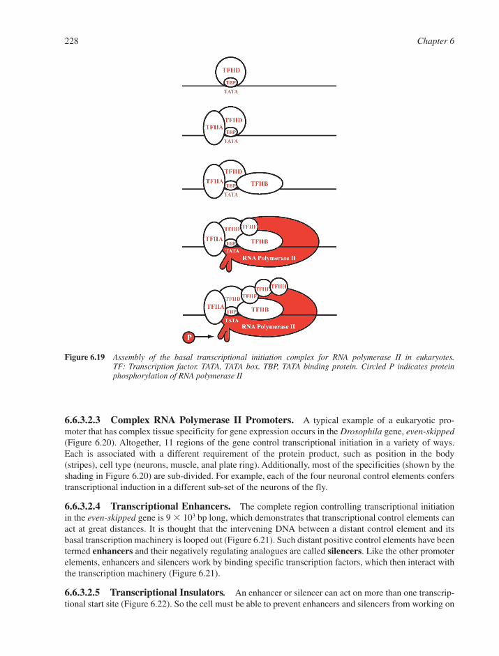
6.6.3.2.3 Complex RNA Polymerase II Promoters. A typical example of a eukaryotic pro-moter that has complex tissue specificity for gene expression occurs in the Drosophila gene, even-skipped(Figure 6.20). Altogether, 11 regions of the gene control transcriptional initiation in a variety of ways.Each is associated with a different requirement of the protein product, such as position in the body(stripes), cell type (neurons, muscle, anal plate ring). Additionally, most of the specificities (shown by theshading in Figure 6.20) are sub-divided. For example, each of the four neuronal control elements conferstranscriptional induction in a different sub-set of the neurons of the fly.
6.6.3.2.4 Transcriptional Enhancers. The complete region controlling transcriptional initiationin the even-skipped gene is 9 � 103 bp long, which demonstrates that transcriptional control elements canact at great distances. It is thought that the intervening DNA between a distant control element and itsbasal transcription machinery is looped out (Figure 6.21). Such distant positive control elements have beentermed enhancers and their negatively regulating analogues are called silencers. Like the other promoterelements, enhancers and silencers work by binding specific transcription factors, which then interact withthe transcription machinery (Figure 6.21).
6.6.3.2.5 Transcriptional Insulators. An enhancer or silencer can act on more than one transcrip-tional start site (Figure 6.22). So the cell must be able to prevent enhancers and silencers from working on
228 Chapter 6
Figure 6.19 Assembly of the basal transcriptional initiation complex for RNA polymerase II in eukaryotes. TF: Transcription factor. TATA, TATA box. TBP, TATA binding protein. Circled P indicates proteinphosphorylation of RNA polymerase II
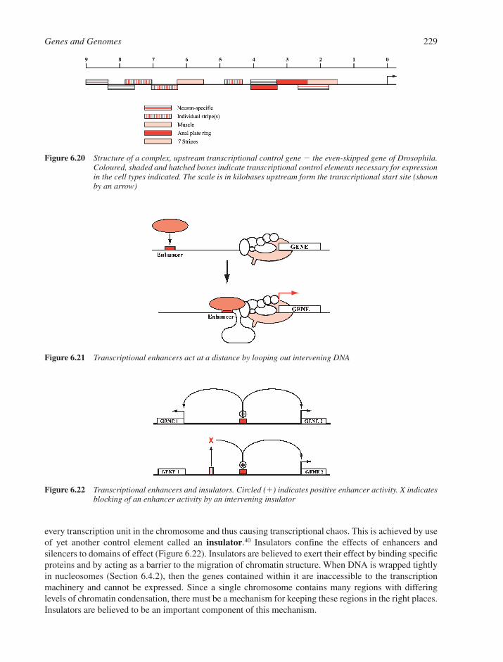
every transcription unit in the chromosome and thus causing transcriptional chaos. This is achieved by useof yet another control element called an insulator.40 Insulators confine the effects of enhancers andsilencers to domains of effect (Figure 6.22). Insulators are believed to exert their effect by binding specificproteins and by acting as a barrier to the migration of chromatin structure. When DNA is wrapped tightlyin nucleosomes (Section 6.4.2), then the genes contained within it are inaccessible to the transcriptionmachinery and cannot be expressed. Since a single chromosome contains many regions with differing levels of chromatin condensation, there must be a mechanism for keeping these regions in the right places.Insulators are believed to be an important component of this mechanism.
Genes and Genomes 229
Figure 6.20 Structure of a complex, upstream transcriptional control gene � the even-skipped gene of Drosophila.Coloured, shaded and hatched boxes indicate transcriptional control elements necessary for expressionin the cell types indicated. The scale is in kilobases upstream form the transcriptional start site (shownby an arrow)
Figure 6.21 Transcriptional enhancers act at a distance by looping out intervening DNA
Figure 6.22 Transcriptional enhancers and insulators. Circled (�) indicates positive enhancer activity. X indicatesblocking of an enhancer activity by an intervening insulator
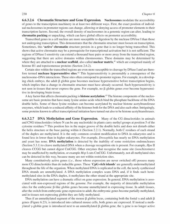
6.6.3.2.6 Chromatin Structure and Gene Expression. Nucleosomes modulate the accessibilityof genes to the transcription machinery in at least two different ways. First, the exact position of individ-ual nucleosomes in promoter regions can change, allowing or denying access of promoter elements to theirtranscription factors. Second, the overall density of nucleosomes in a genomic region can alter, leading tochromatin packing or unpacking, which can have global effects on promoter accessibility.
Transcribed genes in a cell nucleus are more susceptible to digestion by the nuclease DNAse I than thosenot undergoing transcription. This demonstrates that the chromatin structure must loosen on transcription.Sometimes, this ‘active’ chromatin structure persists in a gene that is no longer being transcribed. Thisshows that active chromatin may be a prerequisite for transcriptional activation but it is not sufficient. Theregions of DNase I sensitivity can extend a thousand base pairs or more away from the transcribed region,suggesting that there are active domains within chromosomes. These domains may be determined bywhere they are attached to a nuclear scaffold, also called nuclear matrix,41 which are comprised mainly ofhistone H1 and topoisomerase proteins (Section 2.6.2).
Certain sites within the transcribed regions are even more susceptible to cleavage by DNase I and are there-fore termed nuclease hypersensitive sites.42 This hypersensitivity is presumably a consequence of thenucleosome–DNA interactions. These sites often correspond to promoter regions. For example, in a develop-ing chick embryo, the adult � globin gene becomes nuclease hypersensitive before transcription begins,which implies that a change in chromatin structure must have already occurred. Such hypersensitivity isnot seen in tissues that never express the gene. For example, no � globin genes ever become hypersensi-tive in developing brain tissue.
A key factor that affects chromatin packing is histone acetylation.43 The histone components of the nucleo-some are basic proteins that have many lysine amino acids which bind the phosphate backbone of the DNAdouble helix. Some of these lysine residues can become acetylated by nuclear histone acetyltransferaseenzymes, which leads to a reduced affinity of the histones both for the DNA and also each other. Intriguingly,some proteins known to affect transcriptional initiation have turned out also to be histone acetyltransferases.
6.6.3.2.7 DNA Methylation and Gene Expression. Many of the CG dinucleotides in animalsand CNG trinucleotides (where N can be any nucleotide) in plants carry methyl groups at position 5 of thecytosine residues.44 This position lies in the major groove of the double helix and does not disturb eitherthe helix structure or the base pairing within it (Section 2.2.1). Normally, both C residues of each strandof the duplex are methylated. It is the only common covalent modification to DNA in eukaryotes and isfound less in lower than in higher eukaryotes. For example, Drosophila has nearly no DNA methylationand yeast has none. DNA methylation is detected by the inability of some restriction endonucleases(Section 5.3.1) to cleave methylated DNA when a cleavage recognition site is present. For example, Hpa IIcleaves CCGG but cannot digest CmCGG. Other enzymes that recognise the same site (isoschizomers)may be unaffected by methylation, or example Msp I cuts CmCGG. Unfortunately, not all methylated sitescan be detected in this way, because many are not within restriction sites.
Many constitutively active genes (i.e., those whose expression are never switched off) possess manymore CG dinucleotides than do inducible genes. These ‘CpG-rich islands’ are generally undermethylatedthroughout the life of the organism. When methylated DNA is replicated in the cell, the newly synthesisedDNA strands are unmethylated. A DNA methylation complex scans DNA and, if it finds such hemi-methylated sites in the DNA duplex, it methylates the other strand at the appropriate site.
DNA methylation can have a dramatic effect on gene expression. In general, DNA methylation is asso-ciated with non-expressed regions of the genome. For example, the majority of detectable methylationsites for the embryonic �-like globin genes become unmethylated in expressing tissue. In adult tissues,after the switch from embryonic gene expression to adult, the embryonic genes become partially methylated,and in tissues not expressing globin they are fully methylated.
Thus if an unmethylated segment of the mouse � globin locus, containing both the foetal � and adult �genes (Figure 6.23), is introduced into cultured mouse cells, both genes are expressed. If instead a meth-ylated �-globin gene is introduced next to an unmethylated � globin gene, the � gene is no longer active
230 Chapter 6
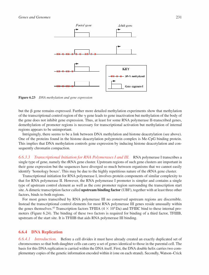
but the � gene remains expressed. Further more detailed methylation experiments show that methylationof the transcriptional control region of the � gene leads to gene inactivation but methylation of the body ofthe gene does not inhibit gene expression. Thus, at least for some RNA polymerase II-transcribed genes,demethylation of promoter regions is necessary for transcriptional activation but methylation of internalregions appears to be unimportant.
Intriguingly, there seems to be a link between DNA methylation and histone deacetylation (see above).One of the proteins found in the histone deacetylation polyprotein complex is Me-CpG binding protein.This implies that DNA methylation controls gene expression by inducing histone deacetylation and con-sequently chromatin compaction.
6.6.3.3 Transcriptional Initiation for RNA Polymerases I and III. RNA polymerase I transcribes asingle type of gene, namely the rRNA gene cluster. Upstream regions of such gene clusters are important intheir gene expression but the sequences have diverged so much between organisms that we cannot easilyidentify ‘homology boxes’. This may be due to the highly repetitious nature of the rRNA gene cluster.
Transcriptional initiation for RNA polymerase I, involves protein components of similar complexity tothat for RNA polymerase II. However, the RNA polymerase I promoter is simpler and contains a singletype of upstream control element as well as the core promoter region surrounding the transcription startsite. A dimeric transcription factor called upstream binding factor (UBF), together with at least three otherfactors, binds to both regions.
For most genes transcribed by RNA polymerase III no conserved upstream regions are discernible.Instead the transcriptional control elements for most RNA polymerase III genes reside unusually withinthe genes themselves.45 Transcription factors TFIIIA (4 � 104 Da) and TFIIIC bind to these internal pro-moters (Figure 6.24). The binding of these two factors is required for binding of a third factor, TFIIIB,upstream of the start site. It is TFIIIB that aids RNA polymerase III binding.
6.6.4 DNA Replication
6.6.4.1 Introduction. Before a cell divides it must have already created an exactly duplicated set ofchromosomes so that both daughter cells can carry a set of genes identical to those in the parental cell. Thebasis for this DNA replication is carried within the DNA itself. First, the DNA double helix carries two com-plementary copies of the genetic information encoded within it (one on each strand). Secondly, Watson–Crick
Genes and Genomes 231
Figure 6.23 DNA methylation and gene expression

base pairing determines the identity of the nucleotide to be added at each step during the replicationprocess. In addition to DNA, many other components are required to ensure faithful copying of the DNA.
6.6.4.1.1 DNA Topology. DNA cannot be copied unless the complementary DNA strands are firstunwound. One problem is that because one strand is wound round the other, unwinding one region bypulling the two strands apart leads to an increase in the number of superhelical turns (supercoils) inanother adjacent region (Section 2.3.5). The DNA replication machinery therefore needs to relieve theseextra turns as replication proceeds. This problem is solved by the use of a DNA topoisomerase25 to relaxthese extra supercoils as they are generated during replication.
6.6.4.1.2 Strand Polarity. The two DNA strands in a double helix have opposite polarities (Section2.2). Every enzyme in nature that copies DNA or RNA into DNA or RNA does so by adding single nucleotidesto the 3�-end of the elongating strand (i.e., replication proceeds in a 5� to 3� direction). If DNA replicationis to proceed in a given direction along a duplex, a ‘replication fork’ must migrate in that direction.46 Onlyone DNA strand can be copied in that direction, the ‘leading strand’ and the new DNA elongated continu-ously. The other strand (the ‘lagging strand’) must be copied in the reverse direction and is elongated dis-continuously47 (Figure 6.25).
6.6.4.1.3 Semi-Conservative DNA Replication. In principle there are at least three differentways that DNA could be copied (Figure 6.26). The correct mechanism was shown by Meselson and Stahl
232 Chapter 6
Figure 6.25 The replication fork and the polarity of DNA replication. Black lines indicate old DNA and red linesindicate newly synthesised DNA. The direction of movement of the replication fork and the direction ofsynthesis of DNA are both shown by red arrows
TFIIIA TFIIIC
Figure 6.24 DNA control elements and transcription factors involved in transcriptional initiation by RNApolymerase III. TF: Transcription factor

to involve the preservation of one of the two parent strands in each of the two newly synthesised duplexes.This is called semi-conservative replication, because half of the strands generated are old and half are new.
6.6.4.1.4 Origins and Direction of DNA Replication. In bacteria, fungi and viruses, DNA replica-tion starts at distinct origins, but in higher eukaryotes such sites are far less easy to identify.48 Replication ofall chromosomes proceeds in both directions, creating ‘bubbles’ that can be seen in electron micrographs(Figure 6.27).
6.6.4.2 Priming of DNA Replication. The enzymes responsible for making a complementary copyof a DNA are called DNA polymerases. But they can only elongate an existing duplex. Therefore, anotherenzyme is needed to initiate the synthesis of a new strand on the DNA template. This enzyme is a specialisedRNA polymerase called a primase,49 which occurs in both prokaryotes and eukaryotes. A short RNAoligonucleotide is synthesised first on the DNA template strand and then a DNA strand is synthesised fromthe 3��-end of this short RNA molecule. The unwanted RNA primer is removed by a specialised ribonucleasecalled RNase H, which digests only the RNA strand within an RNA–DNA duplex.
Genes and Genomes 233
Figure 6.26 DNA replication is semi-conservative. Only the top mode of DNA replication (semi-conservativereplication) is observed for replication of prokaryote and eukaryote chromosomes
Figure 6.27 DNA replication for eukaryotic and prokaryotic chromosomes is bi-directional. Newly synthesised DNAis shown in red

6.6.4.3 Initiation of DNA Replication. Most of our current knowledge of how DNA replication isinitiated comes from prokaryotes. Bacterial chromosomes have individual origins of replication. In E. colithis is called oriC. OriC is about 250 nucleotide pairs long and has the structure shown in Figure 6.28. Variousproteins interact with the origin of replication to initiate copying of the DNA. DnaA protein moleculesbind first to a 9 bp motif, then to each other to form a complex. This leads to unwinding of the helix in theA/T-rich 13 bp motifs. Thereafter, a DNA helicase, the DnaB protein, in concert with the DnaC protein, bindsto form a pre-priming complex.
6.6.4.4 DNA Elongation. During the elongation process (Figure 6.29), the DnaB helicase proteinmigrates along the duplex, attached to the ‘lagging strand’, breaking base pairs as it goes.50 Every turn of thedouble helix that is removed in this way generates an extra turn ‘upstream’ of the fork, which is relieved bythe enzyme DNA topoisomerase (Section 2.3.5). The newly generated DNA single strands are protected bysingle strand binding proteins (SSBs) from DNA damage or unwanted binding to other nucleic acids or pro-teins. These are later displaced as DNA polymerase moves in to make the new complementary lagging strand.
The leading strand can be copied without any discontinuity (Figure 6.30) but the lagging strand requiresa new primer every thousand or so nucleotides. This is synthesised by the primase and then the DNA poly-merase (DNA polymerase III in E. coli) takes over, extending the primer for about 1000 nucleotides before
234 Chapter 6
Figure 6.29 Elongation of DNA synthesis involves DnaB helicase and DNA topoisomerase
Figure 6.28 Structure of the E. coli origin of DNA replication OriC
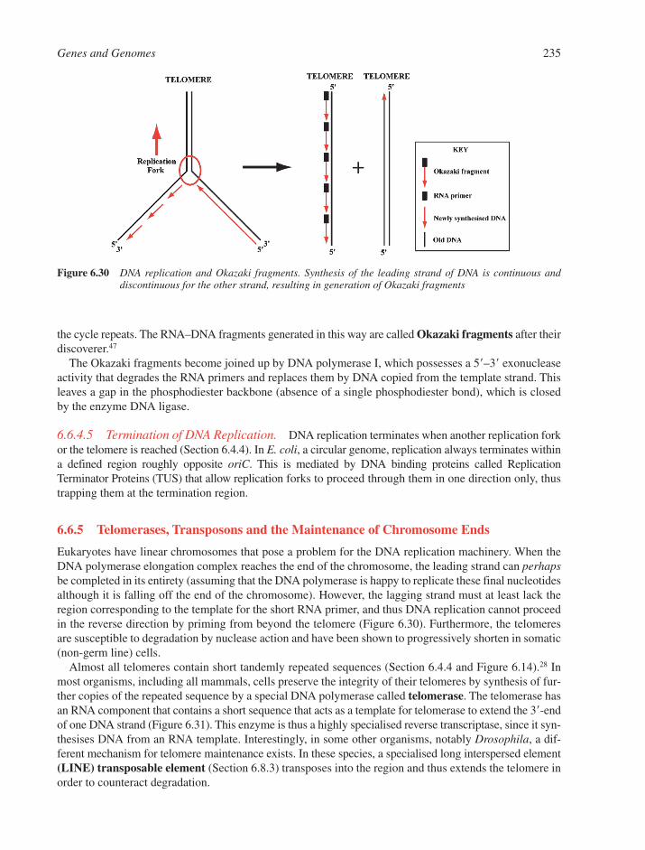
the cycle repeats. The RNA–DNA fragments generated in this way are called Okazaki fragments after theirdiscoverer.47
The Okazaki fragments become joined up by DNA polymerase I, which possesses a 5�–3� exonucleaseactivity that degrades the RNA primers and replaces them by DNA copied from the template strand. Thisleaves a gap in the phosphodiester backbone (absence of a single phosphodiester bond), which is closedby the enzyme DNA ligase.
6.6.4.5 Termination of DNA Replication. DNA replication terminates when another replication forkor the telomere is reached (Section 6.4.4). In E. coli, a circular genome, replication always terminates withina defined region roughly opposite oriC. This is mediated by DNA binding proteins called ReplicationTerminator Proteins (TUS) that allow replication forks to proceed through them in one direction only, thustrapping them at the termination region.
6.6.5 Telomerases, Transposons and the Maintenance of Chromosome Ends
Eukaryotes have linear chromosomes that pose a problem for the DNA replication machinery. When theDNA polymerase elongation complex reaches the end of the chromosome, the leading strand can perhapsbe completed in its entirety (assuming that the DNA polymerase is happy to replicate these final nucleotidesalthough it is falling off the end of the chromosome). However, the lagging strand must at least lack theregion corresponding to the template for the short RNA primer, and thus DNA replication cannot proceedin the reverse direction by priming from beyond the telomere (Figure 6.30). Furthermore, the telomeresare susceptible to degradation by nuclease action and have been shown to progressively shorten in somatic(non-germ line) cells.
Almost all telomeres contain short tandemly repeated sequences (Section 6.4.4 and Figure 6.14).28 Inmost organisms, including all mammals, cells preserve the integrity of their telomeres by synthesis of fur-ther copies of the repeated sequence by a special DNA polymerase called telomerase. The telomerase hasan RNA component that contains a short sequence that acts as a template for telomerase to extend the 3�-endof one DNA strand (Figure 6.31). This enzyme is thus a highly specialised reverse transcriptase, since it syn-thesises DNA from an RNA template. Interestingly, in some other organisms, notably Drosophila, a dif-ferent mechanism for telomere maintenance exists. In these species, a specialised long interspersed element(LINE) transposable element (Section 6.8.3) transposes into the region and thus extends the telomere inorder to counteract degradation.
Genes and Genomes 235
Figure 6.30 DNA replication and Okazaki fragments. Synthesis of the leading strand of DNA is continuous anddiscontinuous for the other strand, resulting in generation of Okazaki fragments
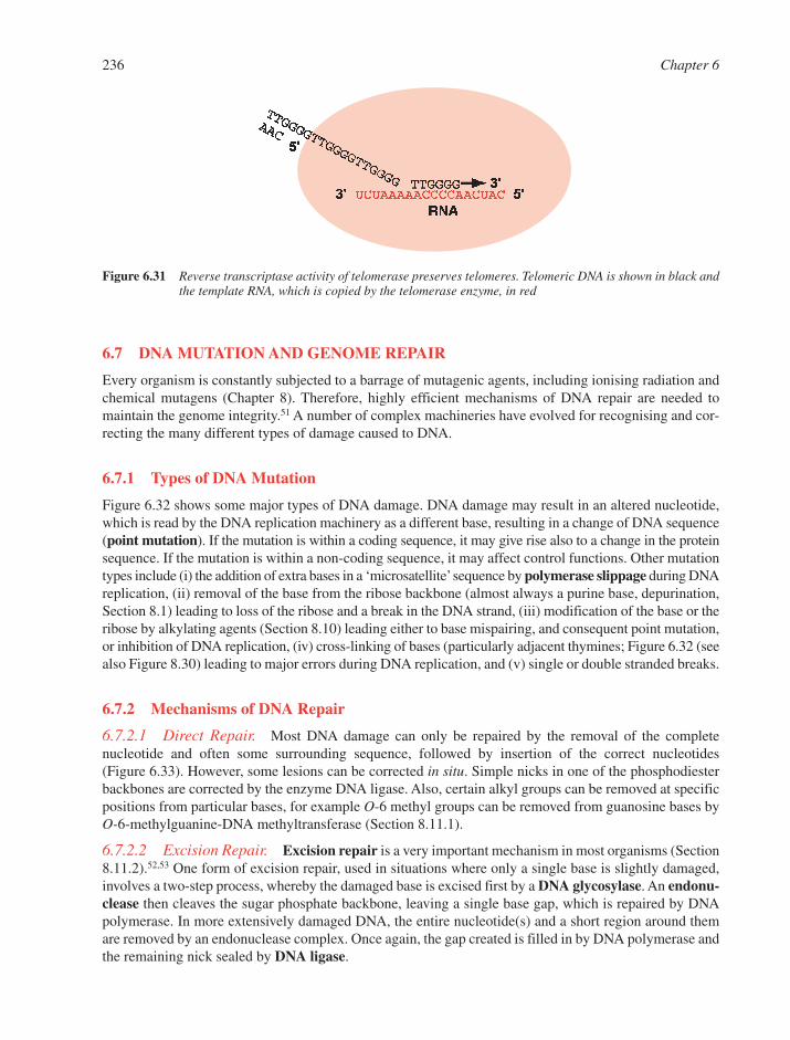
6.7 DNA MUTATION AND GENOME REPAIR
Every organism is constantly subjected to a barrage of mutagenic agents, including ionising radiation andchemical mutagens (Chapter 8). Therefore, highly efficient mechanisms of DNA repair are needed tomaintain the genome integrity.51 A number of complex machineries have evolved for recognising and cor-recting the many different types of damage caused to DNA.
6.7.1 Types of DNA Mutation
Figure 6.32 shows some major types of DNA damage. DNA damage may result in an altered nucleotide,which is read by the DNA replication machinery as a different base, resulting in a change of DNA sequence(point mutation). If the mutation is within a coding sequence, it may give rise also to a change in the proteinsequence. If the mutation is within a non-coding sequence, it may affect control functions. Other mutationtypes include (i) the addition of extra bases in a ‘microsatellite’sequence by polymerase slippage during DNAreplication, (ii) removal of the base from the ribose backbone (almost always a purine base, depurination,Section 8.1) leading to loss of the ribose and a break in the DNA strand, (iii) modification of the base or theribose by alkylating agents (Section 8.10) leading either to base mispairing, and consequent point mutation,or inhibition of DNA replication, (iv) cross-linking of bases (particularly adjacent thymines; Figure 6.32 (seealso Figure 8.30) leading to major errors during DNA replication, and (v) single or double stranded breaks.
6.7.2 Mechanisms of DNA Repair
6.7.2.1 Direct Repair. Most DNA damage can only be repaired by the removal of the completenucleotide and often some surrounding sequence, followed by insertion of the correct nucleotides (Figure 6.33). However, some lesions can be corrected in situ. Simple nicks in one of the phosphodiesterbackbones are corrected by the enzyme DNA ligase. Also, certain alkyl groups can be removed at specificpositions from particular bases, for example O-6 methyl groups can be removed from guanosine bases byO-6-methylguanine-DNA methyltransferase (Section 8.11.1).
6.7.2.2 Excision Repair. Excision repair is a very important mechanism in most organisms (Section8.11.2).52,53 One form of excision repair, used in situations where only a single base is slightly damaged,involves a two-step process, whereby the damaged base is excised first by a DNA glycosylase. An endonu-clease then cleaves the sugar phosphate backbone, leaving a single base gap, which is repaired by DNApolymerase. In more extensively damaged DNA, the entire nucleotide(s) and a short region around themare removed by an endonuclease complex. Once again, the gap created is filled in by DNA polymerase andthe remaining nick sealed by DNA ligase.
236 Chapter 6
Figure 6.31 Reverse transcriptase activity of telomerase preserves telomeres. Telomeric DNA is shown in black andthe template RNA, which is copied by the telomerase enzyme, in red
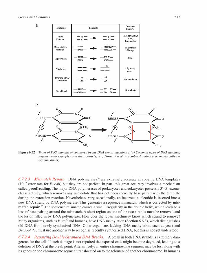
6.7.2.3 Mismatch Repair. DNA polymerases54 are extremely accurate at copying DNA templates(10�7 error rate for E. coli) but they are not perfect. In part, this great accuracy involves a mechanismcalled proofreading. The major DNA polymerases of prokaryotes and eukaryotes possess a 3�–5� exonu-clease activity, which removes any nucleotide that has not been correctly base paired with the templateduring the extension reaction. Nevertheless, very occasionally, an incorrect nucleotide is inserted into anew DNA strand by DNA polymerase. This generates a sequence mismatch, which is corrected by mis-match repair.55 The sequence mismatch causes a small irregularity in the double helix, which leads to aloss of base pairing around the mismatch. A short region on one of the two strands must be removed andthe lesion filled in by DNA polymerase. How does the repair machinery know which strand to remove?Many organisms, such as E. coli and humans, have DNA methylation (Section 6.6.3), which distinguishesold DNA from newly synthesised DNA. Other organisms lacking DNA methylation, such as yeast andDrosophila, must use another way to recognise recently synthesised DNA, but this is not yet understood.
6.7.2.4 Repairing Double-Stranded DNA Breaks. A break in both DNA strands is extremely dan-gerous for the cell. If such damage is not repaired the exposed ends might become degraded, leading to adeletion of DNA at the break point. Alternatively, an entire chromosome segment may be lost along withits genes or one chromosome segment translocated on to the telomere of another chromosome. In humans
Genes and Genomes 237
Figure 6.32 Types of DNA damage encountered by the DNA repair machinery. (a) Common types of DNA damage,together with examples and their cause(s). (b) Formation of a cyclobutyl adduct (commonly called athymine dimer)

double-stranded DNA breaks are repaired by DNA ligase in concert with a multi-subunit complex con-taining DNA protein kinase and two so-called Ku proteins (Ku70 and Ku80).56 The protein complex bringsthe broken ends together, a few bases are removed at the ends and the break is repaired (Figure 6.34).
6.8 DNA RECOMBINATION
6.8.1 Homologous DNA Recombination
One of the seminal discoveries of genetics was the observation that the different genes on the parentalchromosome pairs are shuffled before donation to the offspring. How do these ‘beads on a string’ becomeassorted? The answer is by homologous recombination. A diploid cell in the germinal lineage (the lineagewill give rise to haploid germ cells or gametes) contains two copies of a given chromosome. Segments fromone chromosome can recombine with corresponding segments of the other (Figure 6.35). As we shall see belowthis recombination is ultimately dependent on the DNA sequence homology between the two chromosomes.
What advantages does homologous recombination provide? It enables the host organism to sort alleles (differing copies of the same gene) into novel groups. If a copy of a particular gene in a fruit fly hasrandomly acquired a mutation that yields a more efficient enzyme, then Darwinian natural selection can oper-ate on that gene, provided it is not shackled to all the other genes on the chromosome. Thus, favourable andunfavourable alleles can be shuffled randomly and then the many combinations in the population can betested by natural selection. Another advantage which recombination provides is the ability to repair a dam-aged gene in an otherwise favourable chromosome. If no ability to assort different alleles existed then asingle unfavourable mutation in a chromosome would consign the whole of it to oblivion.
6.8.1.1 The Mechanism of Homologous Recombination. Homologous recombination is linked toDNA replication but does not occur during replication. Rather, it takes place between intact doublehelices. Genetic and DNA sequence analysis has shown that recombination is accurate to a single base
238 Chapter 6
Figure 6.33 Major pathways for DNA repair. Circled * indicates damage to a base or ribose. Longer vertical linesindicate base pairs and shorter lines show unpaired bases. Thick horizontal lines indicate the ribosebackbone

pair. The inference from this is that base pairing is involved during the process. Damage to DNA stimu-lates recombination, suggesting strongly that homologous recombination is initiated from broken DNAstrands. Presumably, under normal circumstances the cell creates such breaks enzymatically to promotehomologous recombination.
6.8.1.1.1 The Holliday Junction. Numerous models have been proposed to explain the mechanismof homologous recombination. A key intermediate in almost all of these is the Holliday junction, named afterits proposer (Figure 6.36).57 One of the key properties of this structure is its ability to migrate along theDNA helices by a process called branch migration. A reversal of the Holliday junction formation gives astrand swapping event. Finally, DNA replication fixes the new arrangement in the daughter chromosomes.
Genes and Genomes 239
Figure 6.34 Repair of double-stranded DNA breaks in eukaryotes
Figure 6.35 Homologous recombination involves the reciprocal exchange of DNA between chromosomes
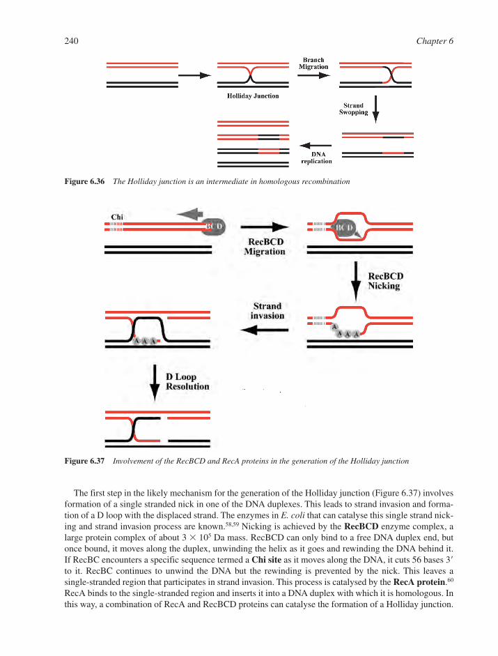
The first step in the likely mechanism for the generation of the Holliday junction (Figure 6.37) involvesformation of a single stranded nick in one of the DNA duplexes. This leads to strand invasion and forma-tion of a D loop with the displaced strand. The enzymes in E. coli that can catalyse this single strand nick-ing and strand invasion process are known.58,59 Nicking is achieved by the RecBCD enzyme complex, alarge protein complex of about 3 � 105 Da mass. RecBCD can only bind to a free DNA duplex end, butonce bound, it moves along the duplex, unwinding the helix as it goes and rewinding the DNA behind it.If RecBC encounters a specific sequence termed a Chi site as it moves along the DNA, it cuts 56 bases 3�to it. RecBC continues to unwind the DNA but the rewinding is prevented by the nick. This leaves a single-stranded region that participates in strand invasion. This process is catalysed by the RecA protein.60
RecA binds to the single-stranded region and inserts it into a DNA duplex with which it is homologous. Inthis way, a combination of RecA and RecBCD proteins can catalyse the formation of a Holliday junction.
240 Chapter 6
Figure 6.37 Involvement of the RecBCD and RecA proteins in the generation of the Holliday junction
Figure 6.36 The Holliday junction is an intermediate in homologous recombination
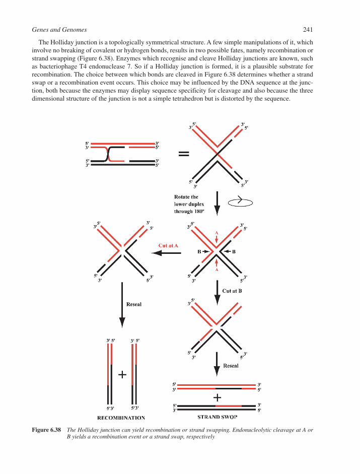
The Holliday junction is a topologically symmetrical structure. A few simple manipulations of it, whichinvolve no breaking of covalent or hydrogen bonds, results in two possible fates, namely recombination orstrand swapping (Figure 6.38). Enzymes which recognise and cleave Holliday junctions are known, suchas bacteriophage T4 endonuclease 7. So if a Holliday junction is formed, it is a plausible substrate forrecombination. The choice between which bonds are cleaved in Figure 6.38 determines whether a strandswap or a recombination event occurs. This choice may be influenced by the DNA sequence at the junc-tion, both because the enzymes may display sequence specificity for cleavage and also because the threedimensional structure of the junction is not a simple tetrahedron but is distorted by the sequence.
Genes and Genomes 241
Figure 6.38 The Holliday junction can yield recombination or strand swapping. Endonucleolytic cleavage at A orB yields a recombination event or a strand swap, respectively
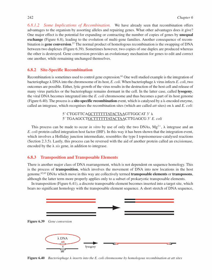
6.8.1.2 Some Implications of Recombination. We have already seen that recombination offersadvantages to the organism by assorting alleles and repairing genes. What other advantages does it give?One major effect is the potential for expanding or contracting the number of copies of genes by unequalexchange (Figure 6.8), leading to the evolution of multi-gene families. Another consequence of recom-bination is gene conversion.57 The normal product of homologous recombination is the swapping of DNAbetween two duplexes (Figure 6.39). Sometimes however, two copies of one duplex are produced whereasthe other is destroyed. Gene conversion provides an evolutionary mechanism for genes to edit and correctone another, while remaining unchanged themselves.
6.8.2 Site-Specific Recombination
Recombination is sometimes used to control gene expression.61 One well studied example is the integration ofbacteriophage DNA into the chromosome of its host, E. coli. When bacteriophage virus infects E. coli, twooutcomes are possible. Either, lytic growth of the virus results in the destruction of the host cell and release ofmany virus particles or the bacteriophage remains dormant in the cell. In the latter case, called lysogeny,the viral DNA becomes integrated into the E. coli chromosome and thus becomes a part of its host genome(Figure 6.40). The process is a site-specific recombination event, which is catalysed by a -encoded enzyme,called an integrase, which recognises the recombination sites (which are called att sites) on and E. coli:
5� CTGGTTCAGCTTTTTTATACTAAGTTGGCAT 3� 5� TGAAGCCTGCTTTTTTATACTAACTTGAGCG 3� E. coli
This process can be made to occur in vitro by use of only the two DNAs, Mg2�, integrase and an E. coli protein called integration host factor (IHF). In this way it has been shown that the integration event,which involves a Holliday junction intermediate, resembles the type I topoisomerase-catalysed reactions(Section 2.3.5). Lastly, this process can be reversed with the aid of another protein called an excisionase,encoded by the xis gene, in addition to integrase.
6.8.3 Transposition and Transposable Elements
There is another major class of DNA rearrangement, which is not dependent on sequence homology. Thisis the process of transposition, which involves the movement of DNA into new locations in the hostgenome.62,63 DNAs which move in this way are collectively termed transposable elements or transposons,although the latter term more properly applies only to a subset of prokaryotic transposable elements.
In transposition (Figure 6.41), a discrete transposable element becomes inserted into a target site, whichbears no significant homology with the transposable element sequence. A short stretch of DNA sequence,
242 Chapter 6
Figure 6.39 Gene conversion
Figure 6.40 Bacteriophage l inserts into the E. coli chromosome by homologous recombination at att sites
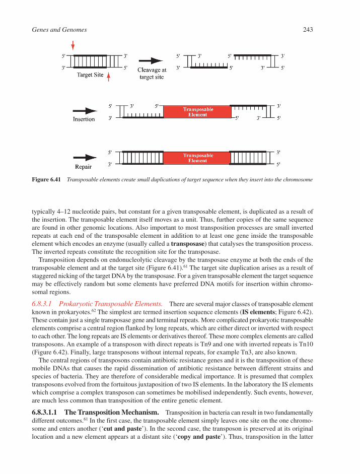
typically 4–12 nucleotide pairs, but constant for a given transposable element, is duplicated as a result ofthe insertion. The transposable element itself moves as a unit. Thus, further copies of the same sequenceare found in other genomic locations. Also important to most transposition processes are small invertedrepeats at each end of the transposable element in addition to at least one gene inside the transposable element which encodes an enzyme (usually called a transposase) that catalyses the transposition process.The inverted repeats constitute the recognition site for the transposase.
Transposition depends on endonucleolytic cleavage by the transposase enzyme at both the ends of thetransposable element and at the target site (Figure 6.41).61 The target site duplication arises as a result ofstaggered nicking of the target DNA by the transposase. For a given transposable element the target sequencemay be effectively random but some elements have preferred DNA motifs for insertion within chromo-somal regions.
6.8.3.1 Prokaryotic Transposable Elements. There are several major classes of transposable elementknown in prokaryotes.62 The simplest are termed insertion sequence elements (IS elements; Figure 6.42).These contain just a single transposase gene and terminal repeats. More complicated prokaryotic transposableelements comprise a central region flanked by long repeats, which are either direct or inverted with respectto each other. The long repeats are IS elements or derivatives thereof. These more complex elements are calledtransposons. An example of a transposon with direct repeats is Tn9 and one with inverted repeats is Tn10(Figure 6.42). Finally, large transposons without internal repeats, for example Tn3, are also known.
The central regions of transposons contain antibiotic resistance genes and it is the transposition of thesemobile DNAs that causes the rapid dissemination of antibiotic resistance between different strains andspecies of bacteria. They are therefore of considerable medical importance. It is presumed that complextransposons evolved from the fortuitous juxtaposition of two IS elements. In the laboratory the IS elementswhich comprise a complex transposon can sometimes be mobilised independently. Such events, however,are much less common than transposition of the entire genetic element.
6.8.3.1.1 The Transposition Mechanism. Transposition in bacteria can result in two fundamentallydifferent outcomes.61 In the first case, the transposable element simply leaves one site on the one chromo-some and enters another (‘cut and paste’). In the second case, the transposon is preserved at its originallocation and a new element appears at a distant site (‘copy and paste’). Thus, transposition in the latter
Genes and Genomes 243
Figure 6.41 Transposable elements create small duplications of target sequence when they insert into the chromosome
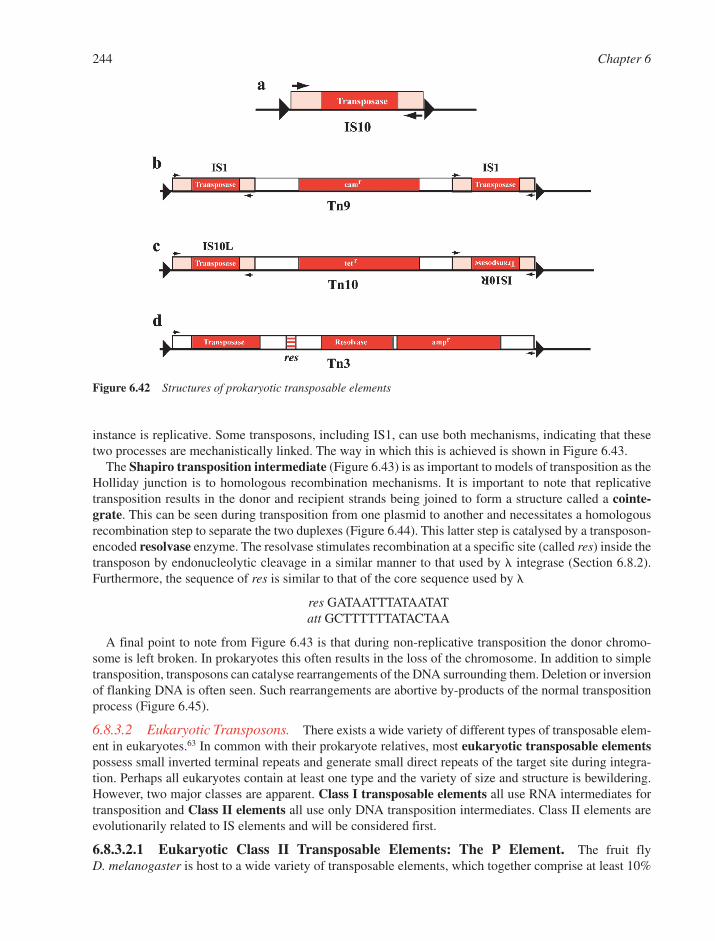
instance is replicative. Some transposons, including IS1, can use both mechanisms, indicating that thesetwo processes are mechanistically linked. The way in which this is achieved is shown in Figure 6.43.
The Shapiro transposition intermediate (Figure 6.43) is as important to models of transposition as theHolliday junction is to homologous recombination mechanisms. It is important to note that replicativetransposition results in the donor and recipient strands being joined to form a structure called a cointe-grate. This can be seen during transposition from one plasmid to another and necessitates a homologousrecombination step to separate the two duplexes (Figure 6.44). This latter step is catalysed by a transposon-encoded resolvase enzyme. The resolvase stimulates recombination at a specific site (called res) inside thetransposon by endonucleolytic cleavage in a similar manner to that used by integrase (Section 6.8.2).Furthermore, the sequence of res is similar to that of the core sequence used by
res GATAATTTATAATATatt GCTTTTTTATACTAA
A final point to note from Figure 6.43 is that during non-replicative transposition the donor chromo-some is left broken. In prokaryotes this often results in the loss of the chromosome. In addition to simpletransposition, transposons can catalyse rearrangements of the DNA surrounding them. Deletion or inversionof flanking DNA is often seen. Such rearrangements are abortive by-products of the normal transpositionprocess (Figure 6.45).
6.8.3.2 Eukaryotic Transposons. There exists a wide variety of different types of transposable elem-ent in eukaryotes.63 In common with their prokaryote relatives, most eukaryotic transposable elementspossess small inverted terminal repeats and generate small direct repeats of the target site during integra-tion. Perhaps all eukaryotes contain at least one type and the variety of size and structure is bewildering.However, two major classes are apparent. Class I transposable elements all use RNA intermediates fortransposition and Class II elements all use only DNA transposition intermediates. Class II elements areevolutionarily related to IS elements and will be considered first.
6.8.3.2.1 Eukaryotic Class II Transposable Elements: The P Element. The fruit fly D. melanogaster is host to a wide variety of transposable elements, which together comprise at least 10%
244 Chapter 6
Figure 6.42 Structures of prokaryotic transposable elements
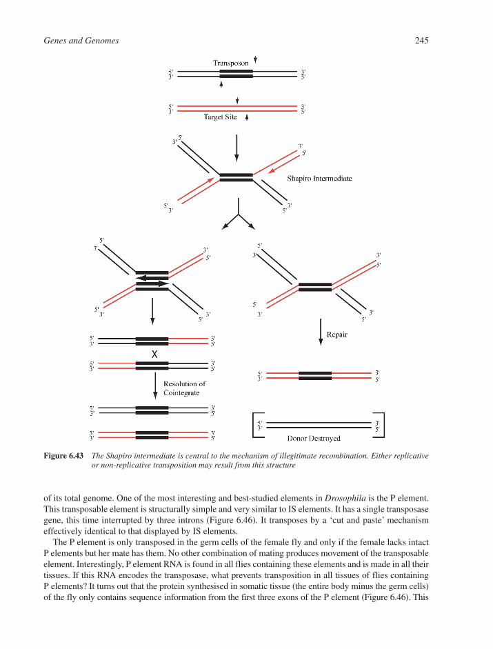
of its total genome. One of the most interesting and best-studied elements in Drosophila is the P element.This transposable element is structurally simple and very similar to IS elements. It has a single transposasegene, this time interrupted by three introns (Figure 6.46). It transposes by a ‘cut and paste’ mechanismeffectively identical to that displayed by IS elements.
The P element is only transposed in the germ cells of the female fly and only if the female lacks intactP elements but her mate has them. No other combination of mating produces movement of the transposableelement. Interestingly, P element RNA is found in all flies containing these elements and is made in all theirtissues. If this RNA encodes the transposase, what prevents transposition in all tissues of flies containing P elements? It turns out that the protein synthesised in somatic tissue (the entire body minus the germ cells)of the fly only contains sequence information from the first three exons of the P element (Figure 6.46). This
Genes and Genomes 245
Figure 6.43 The Shapiro intermediate is central to the mechanism of illegitimate recombination. Either replicativeor non-replicative transposition may result from this structure
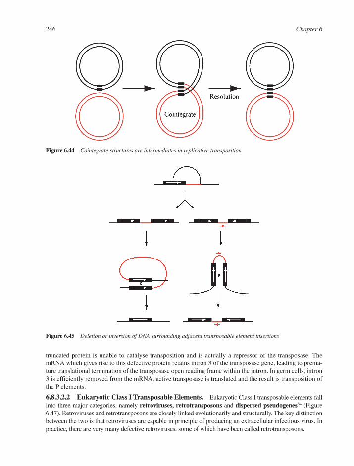
truncated protein is unable to catalyse transposition and is actually a repressor of the transposase. ThemRNA which gives rise to this defective protein retains intron 3 of the transposase gene, leading to prema-ture translational termination of the transposase open reading frame within the intron. In germ cells, intron3 is efficiently removed from the mRNA, active transposase is translated and the result is transposition ofthe P elements.
6.8.3.2.2 Eukaryotic Class I Transposable Elements. Eukaryotic Class I transposable elements fallinto three major categories, namely retroviruses, retrotransposons and dispersed pseudogenes64 (Figure6.47). Retroviruses and retrotransposons are closely linked evolutionarily and structurally. The key distinctionbetween the two is that retroviruses are capable in principle of producing an extracellular infectious virus. Inpractice, there are very many defective retroviruses, some of which have been called retrotransposons.
246 Chapter 6
Figure 6.45 Deletion or inversion of DNA surrounding adjacent transposable element insertions
Figure 6.44 Cointegrate structures are intermediates in replicative transposition
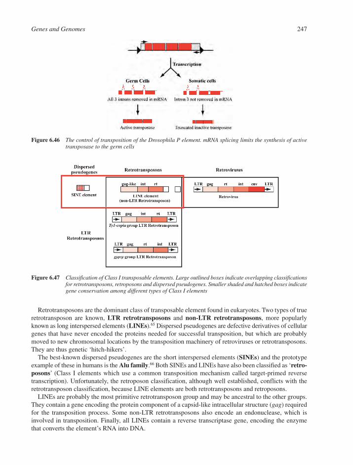
Retrotransposons are the dominant class of transposable element found in eukaryotes. Two types of trueretrotransposon are known, LTR retrotransposons and non-LTR retrotransposons, more popularlyknown as long interspersed elements (LINEs).65 Dispersed pseudogenes are defective derivatives of cellulargenes that have never encoded the proteins needed for successful transposition, but which are probablymoved to new chromosomal locations by the transposition machinery of retroviruses or retrotransposons.They are thus genetic ‘hitch-hikers’.
The best-known dispersed pseudogenes are the short interspersed elements (SINEs) and the prototypeexample of these in humans is the Alu family.66 Both SINEs and LINEs have also been classified as ‘retro-posons’ (Class I elements which use a common transposition mechanism called target-primed reversetranscription). Unfortunately, the retroposon classification, although well established, conflicts with theretrotransposon classification, because LINE elements are both retrotransposons and retroposons.
LINEs are probably the most primitive retrotransposon group and may be ancestral to the other groups.They contain a gene encoding the protein component of a capsid-like intracellular structure (gag) requiredfor the transposition process. Some non-LTR retrotransposons also encode an endonuclease, which isinvolved in transposition. Finally, all LINEs contain a reverse transcriptase gene, encoding the enzymethat converts the element’s RNA into DNA.
Genes and Genomes 247
Figure 6.46 The control of transposition of the Drosophila P element. mRNA splicing limits the synthesis of activetransposase to the germ cells
Figure 6.47 Classification of Class I transposable elements. Large outlined boxes indicate overlapping classificationsfor retrotransposons, retroposons and dispersed pseudogenes. Smaller shaded and hatched boxes indicategene conservation among different types of Class I elements

LTR retrotransposons are sub-divided on the basis of gene order and sequence similarity into the Ty1-copia group and the gypsy group, named after prototype examples discovered in Drosophila and yeast.64
Both groups are also found in plants, fish, amphibia and reptiles but neither has been described in mam-mals (including humans) or in birds.
6.8.3.2.3 Transposition Mechanisms. Only the transposition cycle for LTR retrotransposons willbe discussed here, since it is much better understood than LINE transposition mechanisms (Figure 6.48).64
First, an integrated DNA copy of the retrotransposon in the host genome is transcribed into an RNA.Transcription is initiated within one LTR and terminated in the other. It thus contains the entire geneticinformation of the transposable element. Some of this RNA is spliced before export into the cytoplasm andsome is exported unspliced. Both spliced and unspliced RNAs can be translated into the various proteinproducts encoded by the retrotransposon. Most of the protein produced comprises the components of anintracellular virus-like particle. This process is similar to that seen for many retroviruses. A subset of thefull-length unspliced retrotransposon RNAs is encapsidated into this particle, along with two enzymesnecessary for transposition, namely reverse transcriptase and integrase.
Reverse transcription is carried out by the encapsidated reverse transcriptase. The process is completedwhen the RNA has been converted into a linear unintegrated DNA. Insertion of this DNA into a new chromo-somal location is catalysed by the integrase protein. As a rule, retrotransposons insert at random sites intotheir host genome, although some LTR retrotransposons of Drosophila and Saccharomyces have prefer-ences for particular insertion sequences or genomic regions.
6.8.3.2.4 Retrotransposons and the Human Genome. Approximately half of human genomeconsists of repeated elements interspersed among the genes. Much of this interspersed DNA is comprisedof Class I transposable elements, predominantly SINEs (approximately 1 million copies or 10–15% of thegenome), LINEs (15–20% of the genome) and pseudogenes of human retroviruses (ca. 1% of the genome).67
6.8.3.2.5 Retroviruses. Retroviruses were first identified as agents involved in the onset of cancer about80 years ago.30 More recently the AIDS epidemic has been shown to be due to the HIV retrovirus. In theearly 1970s it was discovered that retroviruses could replicate their RNA genomes via conversion intoDNA, which becomes stably integrated in the DNA of the host cell. It is only comparatively recently that
248 Chapter 6
Figure 6.48 The life cycle of LTR retrotransposons. All intermediates are intracellular. Shaded boxes and circlesindicate genes and their protein products respectively
retroviruses have been recognised as particularly specialised forms of eukaryotic transposon. In effect theyare retrotransposons, which usually can leave the host cells and infect other cells. This ability is donatedby an extra gene (env) encoding a glycoprotein, which coats the virus, allowing entry and exit from thehost cell. The integrated DNA form (or provirus) of the retrovirus is almost identical to a retrotransposon.
REFERENCES
1. O.T. Avery, C.M. MacLeod and M. McCarty, Studies on the chemical nature of the substance indu-cing transformation of pneumococcal types. J. Exp. Med., 1944, 79, 137–158.
2. J. Cairns, G.S. Stent and J.D. Watson, Phage and the Origins of Molecular Biology. Cold SpringHarbor Laboratory Press, Cold Spring Harbor, NY, 1966.
3. F. Jacob and J. Monod, Genetic and regulatory mechanisms in the synthesis of proteins. J. Mol. Biol.,1961, 3, 318–356.
4. G.M. Cooper and G.E. Hausman, The Cell. Sinauer Associates, Sunderland, MA, 2004, 142–143.5. R. Breathnach, J.L. Mandel and P. Chambon, Ovalbumin gene is split in chicken DNA. Nature, 1977,
270, 314–319.6. W. Gilbert, Why genes in pieces? Nature, 1978, 271, 501.7. A. Stoltzfus, D.F. Spencer, M. Zuker, J.M. Logsdon Jr. and W.F. Doolittle, Testing the exon theory of
genes: The evidence from protein structure. Science, 1994, 265, 202–207.8. D.F. Colgan and J.L. Manley, Mechanism and regulation of mRNA polyadenylation. Genes Dev.,
1997, 11, 2755–2766.9. E.P. Lei and P.A. Silver, Protein and RNA export from the nucleus. Dev. Cell., 2002, 2, 261–272.
10. T.A. Ayoubi and W.J. Van De Ven, Regulation of gene expression by alternative promoters. FASEB J.,1996, 10, 453–460.
11. A.J. Lopez, Alternative splicing of pre-mRNA: developmental consequences and mechanisms of reg-ulation. Ann. Rev. Genet., 1998, 32, 279–305.
12. E. Enerly, Ø. L. Mikkelesen, M. Lyamouri and A. Lambertsson, Evolutionary profiling of the U49snoRNA gene. Hereditas, 2003, 138, 73–79.
13. International Human Genome Sequencing Consortium, Initial sequencing and analysis of the humangenome. Nature, 2001, 409, 860–921.
14. J.C. Ventor and 273 others, The sequence of the human genome. Science, 2001, 291, 1304–1351.15. T.H. Eickbush and F.C. Kafatos, A walk in the chorion locus of Bombyx mori. Cell, 1982, 29, 633–643.16. P.F.R. Little, Globin pseudogenes. Cell, 1982, 28, 683–684.17. P. SanMiguel, A. Tikhonov, Y.K. Jin, N. Motchoulskaia, D. Zakharov, A. Melake-Berhan, P.S. Springer,
K.J. Edwards, M. Lee, Z. Avramova and J.L. Bennetzen, Nested retrotransposons in the intergenicregions of the maize genome. Science, 1996, 274, 765–768.
18. M. Rossberg, K. Theres, A. Acarkana, R. Herrero, T. Schmitt, K. Schumacher, G. Schmitz and R. Schmidt,Comparative sequence analysis reveals extensive microcolinearity in the lateral suppressor regions ofthe tomato, Arabidopsis, and Capsella genomes. Plant Cell, 2001, 13, 979–988.
19. J.L. Bennetzen and E.A. Kellogg, Do plants have a one-way ticket to genomic obesity? Plant Cell,1997, 9, 1509–1514.
20. G. Martin, D. Wiernasz and P. Schedl, Evolution of Drosophila repetitive-dispersed DNA. J. Mol.Evol., 1983, 19, 203–213.
21. M.L. Pardue and J.G. Gall, Chromosomal localization of mouse satellite DNA. Science, 1970, 168,1356–1358.
22. K.E. Van Holde and J. Zlatanovai, Chromatin higher order structure: Chasing a mirage. J. Biol. Chem.,1995, 270, 8373–8376.
23. R.D. Kornberg, Chromatin structure: a repeating unit of histones and DNA. Science, 1974, 184, 868–871.24. K. Luger, A.W. Mader, R.K. Richmond, D.F. Sargeant and T.J. Richmond, Crystal structure of the
nucleosome core particle at 2.8Å resolution. Nature, 1997, 389, 251–260.
Genes and Genomes 249
25. J.J. Champoux, DNA topoisomerases: Structure, function and mechanism. Ann. Rev. Biochem., 2001,70, 369–413.
26. J. Carbon, Yeast centromeres: Structure and function. Cell, 1984, 37, 351–353.27. D. Kippling and P.E. Warburton, Centromeres, CEN-P and Tigger too. Trends Genet., 1997, 13,
141–144.28. C.W. Grieder and E.H. Blackburn, Telomeres, telomerases and cancer. Sci. Am., 1996, 274, 80–85.29. I. Liljas, Viruses. Curr. Opin. Struct. Biol., 1996, 6, 151–156.30. J.M. Coffin, S.H. Hughes and H.E. Varmus, Retroviruses. Cold Spring Harbor Laboratory Press, Cold
Spring Harbor, NY, 1997.31. A.J. Jeffreys, V. Wilson and S.L. Thein, Hypervariable ‘minisatellite’ regions in human DNA. Nature,
1985, 314, 67–73.32. A. Chakravarti, Single nucleotide polymorphisms: to a future of genetic medicine. Nature, 2001, 409,
822–823.33. C.A. Gross, C. Chan, A. Dombroski, T. Gruber, M. Sharp, J. Tupy and B. Young, The functional and
regulatory roles of sigma factors in transcription. Cold Spring Harbor Symp. Quant. Biol., 1998, 63,141–155.
34. S. Busby and R.H. Ebright, Promoter structure, promoter recognition and transcription activation inprokaryotes. Cell, 1994, 79, 743–746.
35. S. Busby and R.H. Ebright, Transcription activation by catabolite activator protein (CAP). J. Mol.Biol., 1999, 293, 199–213.
36. A. Sentenac, Eukaryotic RNA polymerases. CRC Crit. Rev. Biochem., 1985, 18, 31–90.37. D.B. Nikolov and S.K. Burley, RNA polymerase II transcription initiation: a structural view. Proc.
Natl. Acad. Sci. USA, 1997, 94, 15–22.38. D.S. Latchman, Eukaryotic transcription factors. Academic Press, London, 1995.39. B.F. Pugh, Control of gene expression through regulation of the TATA-binding protein. Gene, 2000,
255, 1–14.40. T.I. Gerasimova and V.G. Corces, Chromatin insulators and boundaries: effects on transcription and
nuclear organization. Ann. Rev. Genet., 2001, 35, 193–208.41. R. Berezney, M. Mortillaro, H. Ma, X. Wei and J. Samarabandu, The nuclear matrix: a structural
milieu for genomic function. in International Reviews of Cytology, vol 162A, R. Berezney and K.W. Jeon (eds). Academic Press, New York, 1995, 1–65.
42. D.S. Gross and W.T. Garrard, Nuclease hypersensitive sites in chromatin. Ann. Rev. Biochem., 1988,57, 159–197.
43. J.T. Kadonaga, Eukaryotic transcription: an interlaced network of transcription factors andchromatin modifying machines. Cell, 1998, 92, 307–313.
44. K.D. Robertson and P.A. Jones, DNA methylation: past, present and future directions.Carcinogenesis, 2000, 21, 461–467.
45. Y. Huang and R.J. Maraia, Comparison of the RNA polymerase III transcription machinery inSchizosaccharomyces pombe, Saccharomyces cerevisiae and human. Nucleic Acids Res., 2001, 29,2675–2690.
46. S. Waga and B. Stillman, The DNA replication fork in eukaryotic cells. Ann. Rev. Biochem., 1998, 67,721–751.
47. T. Ogawa and T. Okazaki, Discontinuous DNA replication. Ann. Rev. Biochem., 1980, 49, 421–457.48. S.P. Bell and A. Dutta, DNA replication in eukaryotic cells. Ann. Rev. Biochem., 2002, 71, 333–374.49. D.N. Frick and C.C. Richardson, DNA primases. Ann. Rev. Biochem., 2001, 70, 39–80.50. T.M. Lohman and K.P. Bjornson, Mechanism of helicase-catalysed DNA unwinding. Ann. Rev.
Biochem., 1996, 65, 169–214.51. E.C. Friedberg et al., Trends Biochem. Sci., 1995, 20(10), 381–439 (11 articles).52. W.L. de Laat, N.G.J. Jaspers and J.H.J. Hoeijmakers, Molecular mechanism of nucleotide excision
repair. Genes Dev., 1999, 13, 768–785.
250 Chapter 6

53. A. Sancar, DNA excision repair. Ann. Rev. Biochem., 1996, 65, 43–81.54. U. Hubscher, G. Maga and S. Spadari, Eukaryotic DNA polymerases. Ann. Rev. Biochem., 7, 133–163.55. B.D. Harfe and S. Jinks-Robertson, DNA mismatch repair and genetic instability. Ann. Rev. Genet.,
2000, 34, 359–399.56. W.S. Dynan and S. Yoo, Interaction of Ku protein and DNA-dependent protein kinase catalytic
subunit with nucleic acids. Nucleic Acids Res., 1998, 26, 1551–1559.57. R. Holliday, A mechanism for gene conversion in fungi. Genet. Res., 1964, 5, 282–304.58. S.C. West, Enzymes and molecular mechanisms of genetic recombination. Ann. Rev. Biochem., 1992,
61, 603–640.59. S.C. Kowalczykowski and A.K. Eggleston, Homologous pairing and DNA strand exchange proteins.
Ann. Rev. Biochem., 1994, 63, 991–1043.60. C.M. Radding, Helical interactions in homologous pairing and strand exchange driven by RecA
protein. J. Biol. Chem., 1991, 266, 5355–5358.61. B. Hallet and D.J. Sherratt, Transposition and site-specific recombination: adapting DNA cut-and-paste
mechanisms to a variety of genetic rearrangements. FEMS Microbiol. Rev., 1997, 21, 157–178.62. J.A. Shapiro (ed), Mobile Genetic Elements, Academic Press, London, 1983.63. M.G. Kidwell and D. Lisch, Transposable elements as sources of variation in animals and plants.
Proc. Natl. Acad. Sci. USA, 1997, 94, 7704–7711.64. A.J. Flavell, Retroelements, reverse transcriptase and evolution. Comp. Biochem. Physiol. B, 1995,
110B, 3–15.65. E.M. Ostertag and H.H. Kazazian Jr., Biology of mammalian L1 retrotransposons. Ann. Rev. Genet.,
2001, 35, 501–538.66. C.W. Schmid and W.R. Jelinek, The Alu family of dispersed repetitive sequences. Science, 1982, 216,
1065–1070.67. R. Lower, J. Lower and R. Kurth, The viruses in all of us: characteristics and biological significance
of human endogenous retrovirus sequences. Proc. Natl. Acad. Sci. USA, 1996, 93, 5177–5184.
Genes and Genomes 251

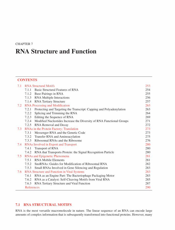
CHAPTER 7
RNA Structure and Function
CONTENTS
7.1 RNA Structural Motifs 2537.1.1 Basic Structural Features of RNA 2547.1.2 Base Pairings in RNA 2557.1.3 RNA Multiple Interactions 2567.1.4 RNA Tertiary Structure 257
7.2 RNA Processing and Modification 2637.2.1 Protecting and Targeting the Transcript: Capping and Polyadenylation 2637.2.2 Splicing and Trimming the RNA 2647.2.3 Editing the Sequence of RNA 2697.2.4 Modified Nucleotides Increase the Diversity of RNA Functional Groups 2717.2.5 RNA Removal and Decay 272
7.3 RNAs in the Protein Factory: Translation 2737.3.1 Messenger RNA and the Genetic Code 2737.3.2 Transfer RNA and Aminoacylation 2757.3.3 Ribosomal RNAs and the Ribosome 276
7.4 RNAs Involved in Export and Transport 2807.4.1 Transport of RNA 2807.4.2 RNA that Transports Protein: the Signal Recognition Particle 280
7.5 RNAs and Epigenetic Phenomena 2817.5.1 RNA Mobile Elements 2817.5.2 SnoRNAs: Guides for Modification of Ribosomal RNA 2827.5.3 Small RNAs Involved in Gene Silencing and Regulation 283
7.6 RNA Structure and Function in Viral Systems 2837.6.1 RNA as an Engine Part: The Bacteriophage Packaging Motor 2837.6.2 RNA as a Catalyst: Self-Cleaving Motifs from Viral RNA 2857.6.3 RNA Tertiary Structure and Viral Function 287References 290
7.1 RNA STRUCTURAL MOTIFS
RNA is the most versatile macromolecule in nature. The linear sequence of an RNA can encode largeamounts of complex information that is subsequently transformed into functional proteins. However, many
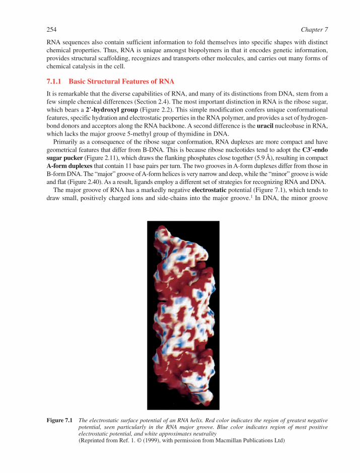
254 Chapter 7
RNA sequences also contain sufficient information to fold themselves into specific shapes with distinctchemical properties. Thus, RNA is unique amongst biopolymers in that it encodes genetic information,provides structural scaffolding, recognizes and transports other molecules, and carries out many forms ofchemical catalysis in the cell.
7.1.1 Basic Structural Features of RNA
It is remarkable that the diverse capabilities of RNA, and many of its distinctions from DNA, stem from afew simple chemical differences (Section 2.4). The most important distinction in RNA is the ribose sugar,which bears a 2�-hydroxyl group (Figure 2.2). This simple modification confers unique conformationalfeatures, specific hydration and electrostatic properties in the RNA polymer, and provides a set of hydrogen-bond donors and acceptors along the RNA backbone. A second difference is the uracil nucleobase in RNA,which lacks the major groove 5-methyl group of thymidine in DNA.
Primarily as a consequence of the ribose sugar conformation, RNA duplexes are more compact and havegeometrical features that differ from B-DNA. This is because ribose nucleotides tend to adopt the C3�-endosugar pucker (Figure 2.11), which draws the flanking phosphates close together (5.9 Å), resulting in compactA-form duplexes that contain 11 base pairs per turn. The two grooves in A-form duplexes differ from those inB-form DNA. The “major” groove of A-form helices is very narrow and deep, while the “minor” groove is wideand flat (Figure 2.40). As a result, ligands employ a different set of strategies for recognizing RNA and DNA.
The major groove of RNA has a markedly negative electrostatic potential (Figure 7.1), which tends todraw small, positively charged ions and side-chains into the major groove.1 In DNA, the minor groove
Figure 7.1 The electrostatic surface potential of an RNA helix. Red color indicates the region of greatest negativepotential, seen particularly in the RNA major groove. Blue color indicates region of most positive electrostatic potential, and white approximates neutrality(Reprinted from Ref. 1. © (1999), with permission from Macmillan Publications Ltd)
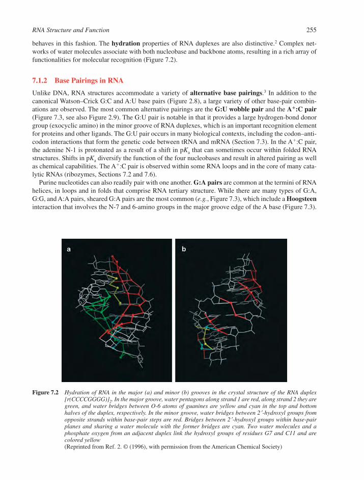
RNA Structure and Function 255
Figure 7.2 Hydration of RNA in the major (a) and minor (b) grooves in the crystal structure of the RNA duplex[r(CCCCGGGG)]2. In the major groove, water pentagons along strand 1 are red, along strand 2 they aregreen, and water bridges between O-6 atoms of guanines are yellow and cyan in the top and bottomhalves of the duplex, respectively. In the minor groove, water bridges between 2�-hydroxyl groups fromopposite strands within base-pair steps are red. Bridges between 2�-hydroxyl groups within base-pairplanes and sharing a water molecule with the former bridges are cyan. Two water molecules and a phosphate oxygen from an adjacent duplex link the hydroxyl groups of residues G7 and C11 and are colored yellow(Reprinted from Ref. 2. © (1996), with permission from the American Chemical Society)
behaves in this fashion. The hydration properties of RNA duplexes are also distinctive.2 Complex net-works of water molecules associate with both nucleobase and backbone atoms, resulting in a rich array offunctionalities for molecular recognition (Figure 7.2).
7.1.2 Base Pairings in RNA
Unlike DNA, RNA structures accommodate a variety of alternative base pairings.3 In addition to thecanonical Watson–Crick G:C and A:U base pairs (Figure 2.8), a large variety of other base-pair combin-ations are observed. The most common alternative pairings are the G:U wobble pair and the A�:C pair(Figure 7.3, see also Figure 2.9). The G:U pair is notable in that it provides a large hydrogen-bond donorgroup (exocyclic amino) in the minor groove of RNA duplexes, which is an important recognition elementfor proteins and other ligands. The G:U pair occurs in many biological contexts, including the codon–anti-codon interactions that form the genetic code between tRNA and mRNA (Section 7.3). In the A�:C pair,the adenine N-1 is protonated as a result of a shift in pKa that can sometimes occur within folded RNAstructures. Shifts in pKa diversify the function of the four nucleobases and result in altered pairing as wellas chemical capabilities. The A�:C pair is observed within some RNA loops and in the core of many cata-lytic RNAs (ribozymes, Sections 7.2 and 7.6).
Purine nucleotides can also readily pair with one another. G:A pairs are common at the termini of RNAhelices, in loops and in folds that comprise RNA tertiary structure. While there are many types of G:A,G:G, and A:A pairs, sheared G:A pairs are the most common (e.g., Figure 7.3), which include a Hoogsteeninteraction that involves the N-7 and 6-amino groups in the major groove edge of the A base (Figure 7.3).
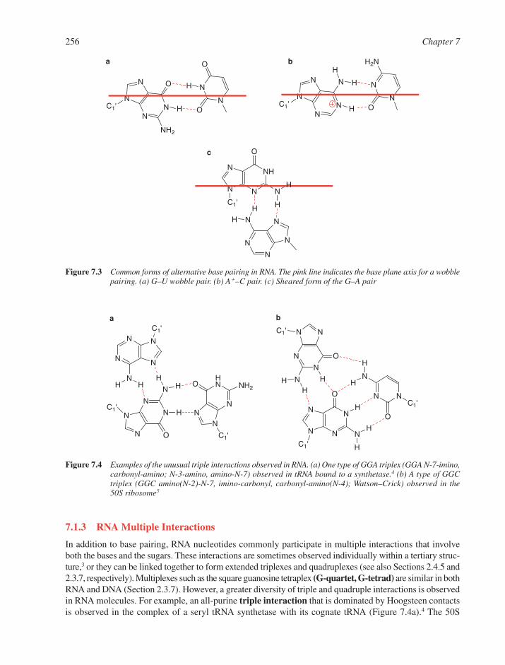
256 Chapter 7
N
N N
N
N
C1'
N
N
N
N
C1'
O
N
N
HN
NN
O NH2
C1'
a
N
NN
N
O
NC1'
NNC1'
N
O
N
N
NNC1'
O
N
b
HHH
H
H
H
H
H
HHH
H
H
Figure 7.4 Examples of the unusual triple interactions observed in RNA. (a) One type of GGA triplex (GGA N-7-imino,carbonyl-amino; N-3-amino, amino-N-7) observed in tRNA bound to a synthetase.4 (b) A type of GGCtriplex (GGC amino(N-2)-N-7, imino-carbonyl, carbonyl-amino(N-4); Watson–Crick) observed in the 50S ribosome5
7.1.3 RNA Multiple Interactions
In addition to base pairing, RNA nucleotides commonly participate in multiple interactions that involveboth the bases and the sugars. These interactions are sometimes observed individually within a tertiary struc-ture,3 or they can be linked together to form extended triplexes and quadruplexes (see also Sections 2.4.5 and2.3.7, respectively). Multiplexes such as the square guanosine tetraplex (G-quartet, G-tetrad) are similar in bothRNA and DNA (Section 2.3.7). However, a greater diversity of triple and quadruple interactions is observedin RNA molecules. For example, an all-purine triple interaction that is dominated by Hoogsteen contactsis observed in the complex of a seryl tRNA synthetase with its cognate tRNA (Figure 7.4a).4 The 50S
N
N
NN
N
N
O
NH2
C1'
O
O
H
H
a
N
N N
NN
N
NHN
N
C1'N
Oc
H
H
H
H
N
N
NN
N
N
N
C1'
H2N
O
bH
H
H
Figure 7.3 Common forms of alternative base pairing in RNA. The pink line indicates the base plane axis for a wobblepairing. (a) G–U wobble pair. (b) A�–C pair. (c) Sheared form of the G–A pair

ribosomal subunit contains a striking diversity of RNA triple interactions, one of which involves a networkof hydrogen bonds that are shared amongst all three nucleotides (Figure 7.4b).5
Highly folded RNA molecules can contain quadruple interactions or even larger arrays of hydrogen-bonded bases. For example, an interesting quadruple variation is observed in the structure of a frameshift-ing viral pseudoknot, that includes a protonated cytosine N-3 (Figure 7.5a).6 Another remarkablequadruple interaction occurs within an in vitro-selected aptamer, which consists of a central G base encir-cled by hydrogen bonds to three other bases (Figure 7.5b).7 An aptamer is an RNA or DNA molecule thathas been selected from a pool of random sequence, based on its ability to bind with high affinity to a par-ticular ligand (Section 5.7.3).8
7.1.4 RNA Tertiary Structure
7.1.4.1 Tertiary Structural Motifs. Although RNA molecules are commonly represented as two-dimensional secondary structures, most RNAs are folded into compact and defined tertiary structures thatare necessary for function (Figure 7.6). Specific types of tertiary structures are seen for tRNA, rRNA,snRNA, certain introns, and ribozymes.9,10 But even mRNA can adopt complex tertiary structures, particu-larly in the untranslated terminal regions (UTRs), which can be essential for proper gene expression.
One of the most important contributions to the three-dimensional form of a folded RNA is coaxialstacking between adjacent sets of short RNA duplex,11 for example as seen in a kissing hairpin complexFigure 7.7. Among the most common substructures for stabilizing long-range interactions in RNA is thetetraloop-receptor motif.9 These involve a specific arrangement of base stacking and hydrogen bondsbetween the GNRA tetraloop (a highly conserved loop with a defined structure) and a conserved stem-loop sequence (Figure 7.8).
The A-minor motif is a ubiquitous interaction that involves contact between an adenosine and theminor groove of a Watson–Crick base pair, thereby forming a type of triple interaction.12 A-minor motifsare observed in self-splicing introns and throughout the ribosome.10 They can occur in isolation but areoften arranged in stacked arrays that may confer additional stabilization (Figure 7.9).
RNA Structure and Function 257
N
NN
N
O
NC1'
N NC1'
O
N
N
NO
N
C1'
N
N
NN
C1'
N
a
H
H H
H
H
H
H
H
H
HN
N
N
N
C1'
N
O
NN
N N C1'
N
NN C1'
O
N
N
NN
N
C1'
O
N
b
H
H
H
H
H
H
H
H
H
H
Figure 7.5 Examples of quadruple interactions observed in RNA. (a) An ACGC quadruplex (ACGC amino-carbonyl,amino(C)-N-7(G):N-3(�)C-carbonyl(G): carbonyl(C)-amino(C); Watson–Crick) from a frame-shiftingpseudoknot 6. (b) The GGCA quadruplex (GGCA imino-N-7, carbonyl-amino; Watson–Crick; N-3-amino(G), amino-N-1(G)) from a dye-binding aptamer7
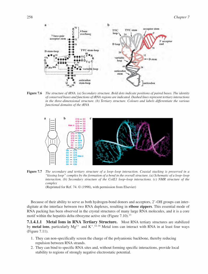
258 Chapter 7
Figure 7.7 The secondary and tertiary structure of a loop–loop interaction. Coaxial stacking is preserved in a“kissing loop” complex by the formation of a bend in the overall structure. (a) Schematic of a loop–loopinteraction. (b) Secondary structure of the ColE1 loop–loop interactions. (c) NMR structure of the complex(Reprinted for Ref. 74. © (1998), with permission from Elsevier)
Because of their ability to serve as both hydrogen-bond donors and acceptors, 2�-OH groups can inter-digitate at the interface between two RNA duplexes, resulting in ribose zippers. This essential mode ofRNA packing has been observed in the crystal structures of many large RNA molecules, and it is a coremotif within the hepatitis delta ribozyme active site (Figure 7.10).13
7.1.4.1.1 Metal Ions in RNA Tertiary Structure. Most RNA tertiary structures are stabilized by metal ions, particularly Mg2� and K�.14–16 Metal ions can interact with RNA in at least four ways(Figure 7.11).
1. They can non-specifically screen the charge of the polyanionic backbone, thereby reducing repulsion between RNA strands.
2. They can bind to specific RNA sites and, without forming specific interactions, provide local stability to regions of strongly negative electrostatic potential.
Figure 7.6 The structure of tRNA. (a) Secondary structure. Bold dots indicate positions of paired bases. The identityof conserved bases and functions of tRNA regions are indicated. Dashed lines represent tertiary interactionsin the three-dimensional structure. (b) Tertiary structure. Colours and labels differentiate the variousfunctional domains of the tRNA
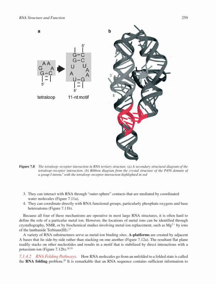
3. They can interact with RNA through “outer-sphere” contacts that are mediated by coordinatedwater molecules (Figure 7.11a).
4. They can coordinate directly with RNA functional groups, particularly phosphate oxygens and baseheteroatoms (Figure 7.11b).
Because all four of these mechanisms are operative in most large RNA structures, it is often hard todefine the role of a particular metal ion. However, the locations of metal ions can be identified throughcrystallography, NMR, or by biochemical studies involving metal-ion replacement, such as Mg2� by ionsof the lanthanide Terbium(III).17
A variety of RNA substructures serve as metal-ion binding sites. A-platforms are created by adjacentA bases that lie side-by-side rather than stacking on one another (Figure 7.12a). The resultant flat planereadily stacks on other nucleotides and results in a motif that is stabilized by direct interactions with apotassium ion (Figure 7.12b).18,19
7.1.4.2 RNA Folding Pathways. How RNA molecules go from an unfolded to a folded state is calledthe RNA folding problem.20 It is remarkable that an RNA sequence contains sufficient information to
RNA Structure and Function 259
Figure 7.8 The tetraloop–receptor interaction in RNA tertiary structure. (a) A secondary structural diagram of thetetraloop–receptor interaction. (b) Ribbon diagram from the crystal structure of the P456 domain ofa group I intron,7 with the tetraloop–receptor interaction highlighted in red

260 Chapter 7
assemble usually into a unique folded structure. However, this process is very sensitive to reaction condi-tions, such as temperature changes, and requires cations to overcome electrostatic repulsion.
An early event in RNA folding is the formation of secondary structure. This is promoted by cations,including monovalent ions, since duplex formation is stimulated by simple charge screening. Tertiarystructure formation involves the tight packing of RNA strands and the formation of cavities. Most RNAmolecules begin to form tertiary structure upon interaction with divalent cations, particularly Mg2�. RNAmolecules tend to fold in a hierarchical manner in which one domain of the molecule precedes the forma-tion of other domains. This is particularly easy to observe in cases where the folding intermediate is stable (as in group I intron folding), but it can still occur in cases where the intermediate is transient. In some
a
b
N
N
O
NO
OO
O
H
H
H
N
N
NO
N
NO
O O
O
H
H
H
NN
N
NNH2O
O
O
O
H H
Figure 7.9 A-minor motifs and ribosomal RNA packing. (a) A class of A-minor motif, showing adenosine dockedinto the minor groove of a G–C pair. (b) A-minor interactions (adenosine in red) in the 50S ribosomalsubunit (Reprinted from Ref. 10. © (2001), with permission from the National Academy of Sciences, USA)
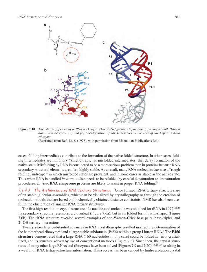
RNA Structure and Function 261
Figure 7.10 The ribose-zipper motif in RNA packing. (a) The 2�-OH group is bifunctional, serving as both H-bonddonor and acceptor. (b) and (c) Interdigitation of ribose residues in the core of the hepatitis deltaribozyme(Reprinted from Ref. 13. © (1998), with permission from Macmillan Publications Ltd)
cases, folding intermediates contribute to the formation of the native folded structure. In other cases, fold-ing intermediates are inhibitory “kinetic traps,” or misfolded intermediates, that delay formation of thenative state. Misfolding by RNA is considered to be a more serious problem than in proteins because RNAsecondary structural elements are often highly stable. As a result, many RNA molecules traverse a “roughfolding landscape,” in which misfolded states are prevalent, and in some cases as stable as the native state.Thus when RNA is handled in vitro, it often needs to be refolded by careful denaturation and renaturationprocedures. In vivo, RNA chaperone proteins are likely to assist in proper RNA folding.21
7.1.4.3 The Architecture of RNA Tertiary Structures. Once formed, RNA tertiary structures areoften stable, globular assemblies, which can be visualized by crystallography or through the creation ofmolecular models that are based on biochemically obtained distance constraints. NMR has also been use-ful in the elucidation of smaller RNA tertiary structures.
The first high-resolution crystal structure of a nucleic acid molecule was obtained for tRNA in 1972.22,23
Its secondary structure resembles a cloverleaf (Figure 7.6a), but in its folded form it is L-shaped (Figure7.6b). The tRNA structure revealed several examples of non-Watson–Crick base pairs, base-triples, and2�-OH tertiary interactions.
Twenty years later, substantial advances in RNA crystallography resulted in structure determination ofthe hammerhead ribozyme24 and a large stable subdomain (P456) within a group I intron RNA.9 The P456structure demonstrated that a large RNA (160 nucleotides in this case) could be folded in vitro, crystal-lized, and its structure solved by use of conventional methods (Figure 7.8). Since then, the crystal struc-tures of many other large RNAs and ribozymes have been solved (Figures 7.9 and 7.20),13,25–27 resulting ina wealth of RNA tertiary-structure information. This success has been capped by high-resolution crystal
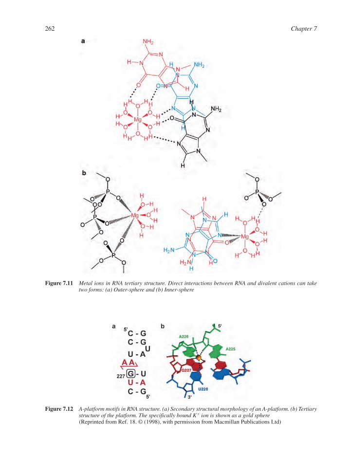
262 Chapter 7
Figure 7.11 Metal ions in RNA tertiary structure. Direct interactions between RNA and divalent cations can taketwo forms: (a) Outer-sphere and (b) Inner-sphere
Figure 7.12 A-platform motifs in RNA structure. (a) Secondary structural morphology of an A-platform. (b) Tertiarystructure of the platform. The specifically bound K� ion is shown as a gold sphere(Reprinted from Ref. 18. © (1998), with permission from Macmillan Publications Ltd)

structures of the complete 70S ribosome,28 a complex of many proteins and 3 RNAs, as well as the 30Sand 50S ribosomal subunits (Section 7.3.3).5,29
7.2 RNA PROCESSING AND MODIFICATION
RNA molecules are transcribed from DNA by RNA polymerase enzymes, which initiate transcriptionafter binding to specific DNA promoter sequences. By use of the adjacent DNA antisense strand as a tem-plate, polymerases synthesize RNA transcripts from the 5�- to the 3�-terminus (Section 6.6). Each organ-ism has at least one, and often several, distinct RNA polymerases. After it has been transcribed, an RNAmolecule is typically not functional until it has undergone RNA processing.30 The attachment of modifi-cations, the removal of long sequences, and in some cases changes to the base sequence itself, are oftenrequired before an RNA can be transported to the proper cellular compartment and carry out its function.
There are many different types of RNA in the cell. For example, messenger RNA (mRNA) encodes pro-tein sequences, transfer RNA (tRNA) acts at the ribosome to decode mRNA information to specify par-ticular amino acids, ribosomal RNAs (rRNA) assemble into the ribosome where protein is manufactured,small nuclear RNAs (snRNAs) tailor other RNAs to the proper size, and microRNAs (miRNAs) are tinysequences that bind and regulate the function of other RNAs. The cellular localization and biological func-tion of RNA molecules dictate the type of processing that they undergo. For the sake of simplicity, the pro-cessing of eukaryotic mRNA will be our major focus.
7.2.1 Protecting and Targeting the Transcript: Capping and Polyadenylation
As the 5�-terminus of a new mRNA emerges from a eukaryotic RNA polymerase, it is immediately pro-tected by attachment of a trimethyl G cap (Figure 7.13). During this process, the terminal nucleotide of thetranscript is joined by a 5�–5� linkage to a guanosine triphosphate. The guanosine itself is methylated, andoften hypermethylated, to distinguish it from other guanosines in the cell. As a result of capping, the nas-cent transcript is protected from 5�-exonucleases that would otherwise digest the RNA as it emerged.
RNA Structure and Function 263
N
NN
O
N
NO
OHO
PO
O
O
CH3
H
H
GuaO
OHO
3'
OH
3
triphosphate5'-5' linkage
RNAtranscript
methylatedguanosine cap
first nucleotide ofthe nascent strand
H
Figure 7.13 The cap structure of eukaryotic mRNA molecules. A red circle indicates the site of methylation on N-7of guanosine, while arrows indicate common sites of additional methylation. Note the unusual 5�-5�triphosphate linkage that joins the two guanosine nucleotides of the cap
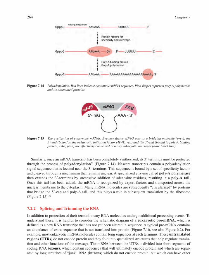
Similarly, once an mRNA transcript has been completely synthesized, its 3� terminus must be protectedthrough the process of polyadenylation31 (Figure 7.14). Nascent transcripts contain a polyadenylationsignal sequence that is located near the 3�-terminus. This sequence is bound by a set of specificity factorsand cleaved through a mechanism that remains unclear. A specialized enzyme called poly-A polymerasethen extends the 3�-terminus by successive addition of adenosine residues, resulting in a poly-A tail. Once this tail has been added, the mRNA is recognized by export factors and transported across thenuclear membrane to the cytoplasm. Many mRNA molecules are subsequently “circularized” by proteinsthat bridge the 5�-cap and poly-A tail, and this plays a role in subsequent translation by the ribosome(Figure 7.15).32
7.2.2 Splicing and Trimming the RNA
In addition to protection of their termini, many RNA molecules undergo additional processing events. Tounderstand these, it is helpful to consider the schematic diagram of a eukaryotic pre-mRNA, which isdefined as a new RNA transcript that has not yet been altered in sequence. A typical pre-mRNA containsan abundance of extra sequence that is not translated into protein (Figure 7.16, see also Figure 6.2). Forexample, most eukaryotic mRNA molecules contain long sequences at each terminus. These untranslatedregions (UTRs) do not encode protein and they fold into specialized structures that help regulate transla-tion and other functions of the message. The mRNA between the UTRs is divided into short segments ofcoding RNA (exons), which contain sequences that will ultimately encode protein and which are separ-ated by long stretches of “junk” RNA (introns) which do not encode protein, but which can have other
264 Chapter 7
Figure 7.15 The cyclization of eukaryotic mRNAs. Because factor eIF4G acts as a bridging molecule (grey), the 5�-end (bound to the eukaryotic initiation factor eIF4E, red) and the 3�-end (bound to poly-A bindingprotein, PAB, pink) are effectively connected in many eukaryotic messages (dark black line)
Figure 7.14 Polyadenylation. Red lines indicate continuous mRNA sequence. Pink shapes represent poly-A polymeraseand its associated proteins

RNA Structure and Function 265
BaseO
OHO
P
O
O O
BaseO
OHO
O
5'
BaseO
OHO
P
O
O
BaseO
OHO
OO
O
Nuc
H
BaseO
OHOH
P
O
O
BaseO
OHO
OO
Nuc
O
Nuc OH
3'3'
5'5'
3'
‡
Figure 7.17 Reaction mechanism for the spliceosome, group II introns, group I introns, and RNase P. Duringspliceosomal and group II intron splicing, the nucleophile (Nuc-OH) is the 2�-OH of a specific bulgedadenosine within the spliced intron. For group I intron splicing, the nucleophile is the 3�-OH of aguanosine moiety. For RNase P, and for alternative reactions by group II introns, the nucleophile is water
Figure 7.16 The structure of a eukaryotic precursor mRNA. The untranslated regions (UTRs) are shown as stem-loop structures and the 5�-cap is a diamond. Exons are black, introns are pink, and an edited exon(undergoing a C→U transversion) is shown in red. The poly-A tail is indicated at the 3�-end
functions. Remarkably, in higher eukaryotes the exons are very short (�200 nt) while the introns can bevery long (�1000 nucleotides) (see Section 6.1).
7.2.2.1 Nuclear pre-mRNA Splicing. Before an mRNA can be functional and provide a proper tem-plate, obviously the introns must be removed and the exons stitched together so that the processed mRNAcontains a coherent coding sequence that is specific for a particular protein. Thus, mature mRNA is muchshorter than the precursor transcript. This pre-mRNA splicing is carried out by the spliceosome, which isa dynamic ribonucleoprotein (RNP) machine that specifically recognizes the sequences at exon/intronboundaries (splice sites), and carries out the chemical reactions for cutting and pasting the exons together.33
The spliceosome is composed of five highly conserved RNA molecules (the small nuclear RNAs orsnRNAs U1, U2, U4, U5, and U6) and a host of specialized proteins that bind RNA or remodel it throughthe consumption of ATP. Splicing proceeds through two sequential trans-esterification reactions, each ofwhich involves an SN2 reaction at phosphorus (Figure 7.17). The nucleophile during the first step of spli-cing is the 2�-hydroxyl group of an adenosine within the intron (the branch-point A), which attacks a spe-cific sequence at the 5�-splice site and releases a 3�-hydroxyl leaving group. During the second step ofsplicing, this 3�-OH group attacks the 3�-splice site, thereby ligating the exons and releasing an intron lar-iat molecule (Figure 7.18).
Most pre-mRNA transcripts from mammals contain numerous exons, with ten being an approximateaverage (Section 6.1). Depending on the tissue and developmental stage of the organism, these exons can

266 Chapter 7
Figure 7.19 The four classes of RNA splicing
either be stitched together sequentially, or certain exons can be skipped and left out of the mature message(see Figure 6.4). As a result of this process, called alternative splicing, a single pre-mRNA gene can gen-erate many different types of proteins, thereby providing a form of combinatorial diversity that is notgenetically encoded.34
7.2.2.2 Self-Splicing and Other Splicing Pathways. While most eukaryotic splicing is carried outby the spliceosome, there are specialized genes and introns that are spliced through different mechanisms(Figure 7.19). For example, certain tRNA genes contain introns in the anticodon, and these are removedby the sequential action of protein endonucleases and ligases. But perhaps the most remarkable pathwaysfor splicing involve introns that are inherently reactive, and which can splice themselves out of flankingexons without the aid of spliceosomal machinery. These self-splicing introns fall into two categories, thegroup I and group II introns.35,36 The discovery of these autocatalytic RNA molecules, or ribozymes,along with other families of catalytic RNA molecules, was one of the most exciting developments in 20thcentury biochemistry (Section 7.6.2).
Figure 7.18 The two steps of RNA splicing catalyzed by the spliceosome and group II introns. The 5�-exon is shownin red, the 3�-exon is shown in grey, and the nucleophilic adenosine is indicated. The intron is shown asa black line. Note that the lariat structure is connected by 2�–3�–5� linkages to the adenosine

Group I and group II introns are common in lower eukaryotes, such as fungi and yeast, although the lat-ter is abundant in plants as well. Remarkably, group I and group II introns have also been found in bacteria,and constitute the only type of intron found so far among prokaryotic organisms. Group I introns (togetherwith RNase P) were the very first autocatalytic RNA molecules to be discovered.37 During experiments onthe splicing of a ribosomal gene in the protozoan Tetrahymena thermophila, Thomas R. Cech and col-leagues found that the rRNA gene repeatedly spliced in control reactions to which enzymatic extracts hadnot been added. In fact, the only cofactors required for group I intron splicing were found to be thenucleotide guanosine and Mg2� ions. Subsequent mechanistic study has revealed that group I introns foldinto an elaborate three-dimensional structure that positions both splice sites and binds a guanosine mol-ecule for use as a nucleophile during the first step of splicing (Figure 7.20).25 As in spliceosomal process-ing, group I intron splicing is the result of two sequential SN2 trans-esterifications that ultimately releaseintron and ligate the exons (Figures 7.17 and 7.18). Both the folding of the molecule and subsequent cata-lysis require Mg2� ions, which has been shown to play an important and general role in the tertiary fold-ing of RNA molecules.
Group II introns are highly abundant in the organellar genes of plants, fungi, and yeast (Figure 7.21).They have been subjects of particular interest because their mechanism of splicing is so closely related tothat of the spliceosome. Like the latter, group II introns utilize a 2�-hydroxyl group of a bulged adenosine
RNA Structure and Function 267
Figure 7.20 The structure of a group I intron. (a) The secondary structure and (b) a crystal structure of the tertiarystructure for the Azoarchus group I intron. Corresponding colours indicate specific domains of theintron. In this structure, the intron and both exons are intact, revealing all active-site components andtheir relative locations(Reprinted from Ref. 25. © (2004), with permission from Macmillan Publications Ltd)
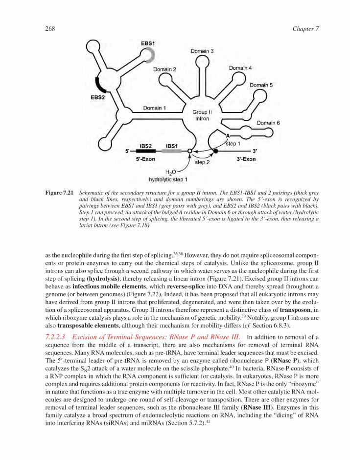
268 Chapter 7
Figure 7.21 Schematic of the secondary structure for a group II intron. The EBS1-IBS1 and 2 pairings (thick greyand black lines, respectively) and domain numberings are shown. The 5�-exon is recognized by pairings between EBS1 and IBS1 (grey pairs with grey), and EBS2 and IBS2 (black pairs with black).Step 1 can proceed via attack of the bulged A residue in Domain 6 or through attack of water (hydrolyticstep 1). In the second step of splicing, the liberated 5�-exon is ligated to the 3�-exon, thus releasing alariat intron (see Figure 7.18)
as the nucleophile during the first step of splicing.36,38 However, they do not require spliceosomal compon-ents or protein enzymes to carry out the chemical steps of catalysis. Unlike the spliceosome, group IIintrons can also splice through a second pathway in which water serves as the nucleophile during the firststep of splicing (hydrolysis), thereby releasing a linear intron (Figure 7.21). Excised group II introns canbehave as infectious mobile elements, which reverse-splice into DNA and thereby spread throughout agenome (or between genomes) (Figure 7.22). Indeed, it has been proposed that all eukaryotic introns mayhave derived from group II introns that proliferated, degenerated, and were then taken over by the evolu-tion of a spliceosomal apparatus. Group II introns therefore represent a distinctive class of transposon, inwhich ribozyme catalysis plays a role in the mechanism of genetic mobility.39 Notably, group I introns arealso transposable elements, although their mechanism for mobility differs (cf. Section 6.8.3).
7.2.2.3 Excision of Terminal Sequences: RNase P and RNase III. In addition to removal of asequence from the middle of a transcript, there are also mechanisms for removal of terminal RNAsequences. Many RNA molecules, such as pre-tRNA, have terminal leader sequences that must be excised.The 5�-terminal leader of pre-tRNA is removed by an enzyme called ribonuclease P (RNase P), whichcatalyzes the SN2 attack of a water molecule on the scissile phosphate.40 In bacteria, RNase P consists ofa RNP complex in which the RNA component is sufficient for catalysis. In eukaryotes, RNase P is morecomplex and requires additional protein components for reactivity. In fact, RNase P is the only “ribozyme”in nature that functions as a true enzyme with multiple turnover in the cell. Most other catalytic RNA mol-ecules are designed to undergo one round of self-cleavage or transposition. There are other enzymes forremoval of terminal leader sequences, such as the ribonuclease III family (RNase III). Enzymes in thisfamily catalyze a broad spectrum of endonucleolytic reactions on RNA, including the “dicing” of RNAinto interfering RNAs (siRNAs) and miRNAs (Section 5.7.2).41
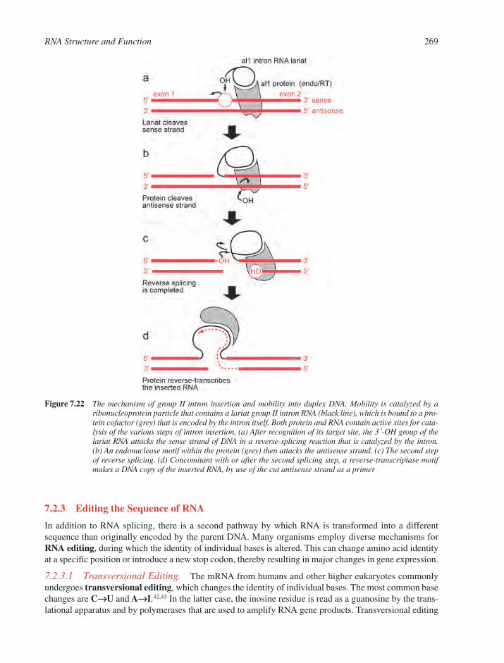
7.2.3 Editing the Sequence of RNA
In addition to RNA splicing, there is a second pathway by which RNA is transformed into a differentsequence than originally encoded by the parent DNA. Many organisms employ diverse mechanisms forRNA editing, during which the identity of individual bases is altered. This can change amino acid identityat a specific position or introduce a new stop codon, thereby resulting in major changes in gene expression.
7.2.3.1 Transversional Editing. The mRNA from humans and other higher eukaryotes commonlyundergoes transversional editing, which changes the identity of individual bases. The most common basechanges are C→→U and A→→I.42,43 In the latter case, the inosine residue is read as a guanosine by the trans-lational apparatus and by polymerases that are used to amplify RNA gene products. Transversional editing
RNA Structure and Function 269
Figure 7.22 The mechanism of group II intron insertion and mobility into duplex DNA. Mobility is catalyzed by aribonucleoprotein particle that contains a lariat group II intron RNA (black line), which is bound to a pro-tein cofactor (grey) that is encoded by the intron itself. Both protein and RNA contain active sites for cata-lysis of the various steps of intron insertion. (a) After recognition of its target site, the 3�-OH group of thelariat RNA attacks the sense strand of DNA in a reverse-splicing reaction that is catalyzed by the intron.(b) An endonuclease motif within the protein (grey) then attacks the antisense strand. (c) The second stepof reverse splicing. (d) Concomitant with or after the second splicing step, a reverse-transcriptase motifmakes a DNA copy of the inserted RNA, by use of the cut antisense strand as a primer

is performed by specialized families of deaminase enzymes that catalyze hydrolysis of amino groups oncytidine and adenosine (Figure 7.23). On a much slower timescale, these same reactions occur spontan-eously and nonspecifically at A and C, which is one reason why it is challenging to determine the originalsequence of DNA or RNA samples that have been extracted from old biological material (�100 years old).However, the deaminases involved in RNA editing have evolved high specificity for their target sequencesand, together with additional proteins, they form efficient editing complexes that modify only discreteregions of certain RNA messages.
One example of transversional editing gives rise to variants of the protein apolipoprotein B. One form ofapolipoprotein B (ApoB-48) is half as long as another common variant (ApoB-100), and the balance betweenthese proteins in different tissues plays a major role in human cardiovascular health. The ApoB-48 variantresults from a stop codon in the ApoB-mRNA. However, this stop codon is not DNA-encoded, but resultsfrom an RNA editing event, whereby a single cytidine is converted to a uracil by a specialized deaminaseprotein that has been named Apobec-1.43 The conversion of a glutamine CAA codon into the UAA stopcodon results in ApoB-48, while unedited transcripts produce ApoB-100. As in subsequent discoveries ofRNA editing, alteration in amino acid identity was only discerned when protein sequences (or cDNAsequences, since they are derived from edited RNAs) were compared to the genomic sequence of the organ-ism. Since the discovery of ApoB editing, numerous other examples of C→U editing have been reported.There is also evidence that Apobec-like enzymes edit the DNA of genes involved in the immune system.43
A second class of transversional editing is catalyzed by the ADAR family of adenosine deaminases.42
This activity was first noted during biochemical studies on an unusual enzyme that was found to bind toduplex RNA in vitro and to convert certain adenosines into inosines. A biological function for this type ofenzyme was identified during studies of the human glutamate receptor gene. In neurons, it was noted that
270 Chapter 7
N
N
NH2
O
R
HN
NO
R
H3N OH
HN
N
O
O
R
H2O
H
N
N N
N
R
NH2
HN
N N
N
R
HN
N N
N
R
OH3N OHH2O
H
C U
A I
Apobec
ADAR
Figure 7.23 The deaminase reactions that are catalyzed by RNA editing enzymes Apobec and ADAR. The Apobecenzymes catalyze C→U transversions at specific sites and the ADAR enzymes catalyze A→I (inosine)transversions
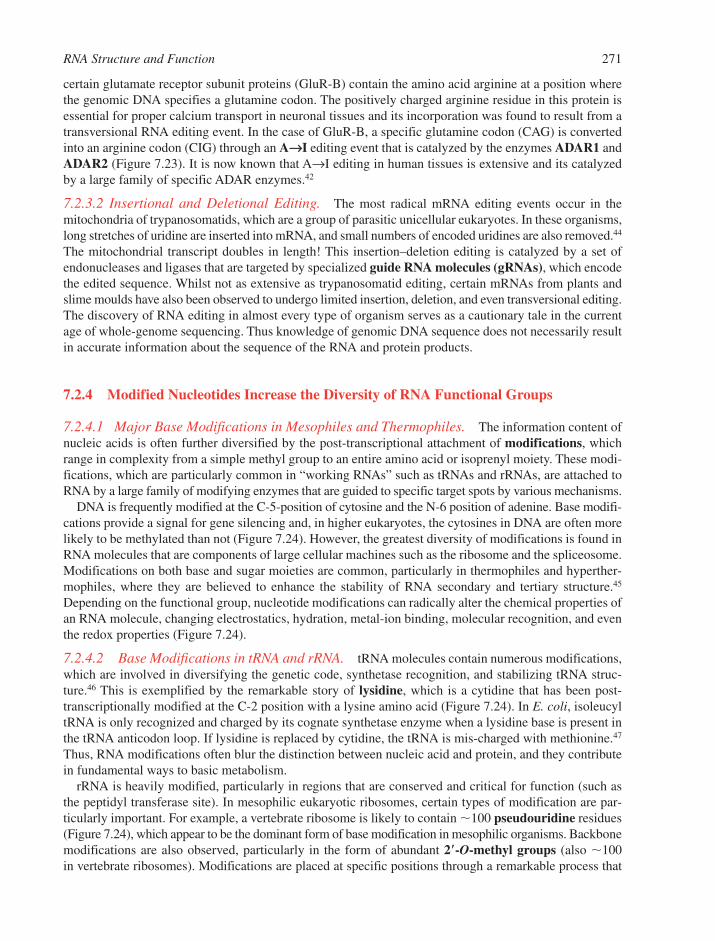
certain glutamate receptor subunit proteins (GluR-B) contain the amino acid arginine at a position wherethe genomic DNA specifies a glutamine codon. The positively charged arginine residue in this protein isessential for proper calcium transport in neuronal tissues and its incorporation was found to result from atransversional RNA editing event. In the case of GluR-B, a specific glutamine codon (CAG) is convertedinto an arginine codon (CIG) through an A→→I editing event that is catalyzed by the enzymes ADAR1 andADAR2 (Figure 7.23). It is now known that A→I editing in human tissues is extensive and its catalyzedby a large family of specific ADAR enzymes.42
7.2.3.2 Insertional and Deletional Editing. The most radical mRNA editing events occur in themitochondria of trypanosomatids, which are a group of parasitic unicellular eukaryotes. In these organisms,long stretches of uridine are inserted into mRNA, and small numbers of encoded uridines are also removed.44
The mitochondrial transcript doubles in length! This insertion–deletion editing is catalyzed by a set ofendonucleases and ligases that are targeted by specialized guide RNA molecules (gRNAs), which encodethe edited sequence. Whilst not as extensive as trypanosomatid editing, certain mRNAs from plants andslime moulds have also been observed to undergo limited insertion, deletion, and even transversional editing.The discovery of RNA editing in almost every type of organism serves as a cautionary tale in the currentage of whole-genome sequencing. Thus knowledge of genomic DNA sequence does not necessarily resultin accurate information about the sequence of the RNA and protein products.
7.2.4 Modified Nucleotides Increase the Diversity of RNA Functional Groups
7.2.4.1 Major Base Modifications in Mesophiles and Thermophiles. The information content ofnucleic acids is often further diversified by the post-transcriptional attachment of modifications, whichrange in complexity from a simple methyl group to an entire amino acid or isoprenyl moiety. These modi-fications, which are particularly common in “working RNAs” such as tRNAs and rRNAs, are attached toRNA by a large family of modifying enzymes that are guided to specific target spots by various mechanisms.
DNA is frequently modified at the C-5-position of cytosine and the N-6 position of adenine. Base modifi-cations provide a signal for gene silencing and, in higher eukaryotes, the cytosines in DNA are often morelikely to be methylated than not (Figure 7.24). However, the greatest diversity of modifications is found inRNA molecules that are components of large cellular machines such as the ribosome and the spliceosome.Modifications on both base and sugar moieties are common, particularly in thermophiles and hyperther-mophiles, where they are believed to enhance the stability of RNA secondary and tertiary structure.45
Depending on the functional group, nucleotide modifications can radically alter the chemical properties ofan RNA molecule, changing electrostatics, hydration, metal-ion binding, molecular recognition, and eventhe redox properties (Figure 7.24).
7.2.4.2 Base Modifications in tRNA and rRNA. tRNA molecules contain numerous modifications,which are involved in diversifying the genetic code, synthetase recognition, and stabilizing tRNA struc-ture.46 This is exemplified by the remarkable story of lysidine, which is a cytidine that has been post-transcriptionally modified at the C-2 position with a lysine amino acid (Figure 7.24). In E. coli, isoleucyltRNA is only recognized and charged by its cognate synthetase enzyme when a lysidine base is present inthe tRNA anticodon loop. If lysidine is replaced by cytidine, the tRNA is mis-charged with methionine.47
Thus, RNA modifications often blur the distinction between nucleic acid and protein, and they contributein fundamental ways to basic metabolism.
rRNA is heavily modified, particularly in regions that are conserved and critical for function (such asthe peptidyl transferase site). In mesophilic eukaryotic ribosomes, certain types of modification are par-ticularly important. For example, a vertebrate ribosome is likely to contain �100 pseudouridine residues(Figure 7.24), which appear to be the dominant form of base modification in mesophilic organisms. Backbonemodifications are also observed, particularly in the form of abundant 2�-O-methyl groups (also �100in vertebrate ribosomes). Modifications are placed at specific positions through a remarkable process that
RNA Structure and Function 271
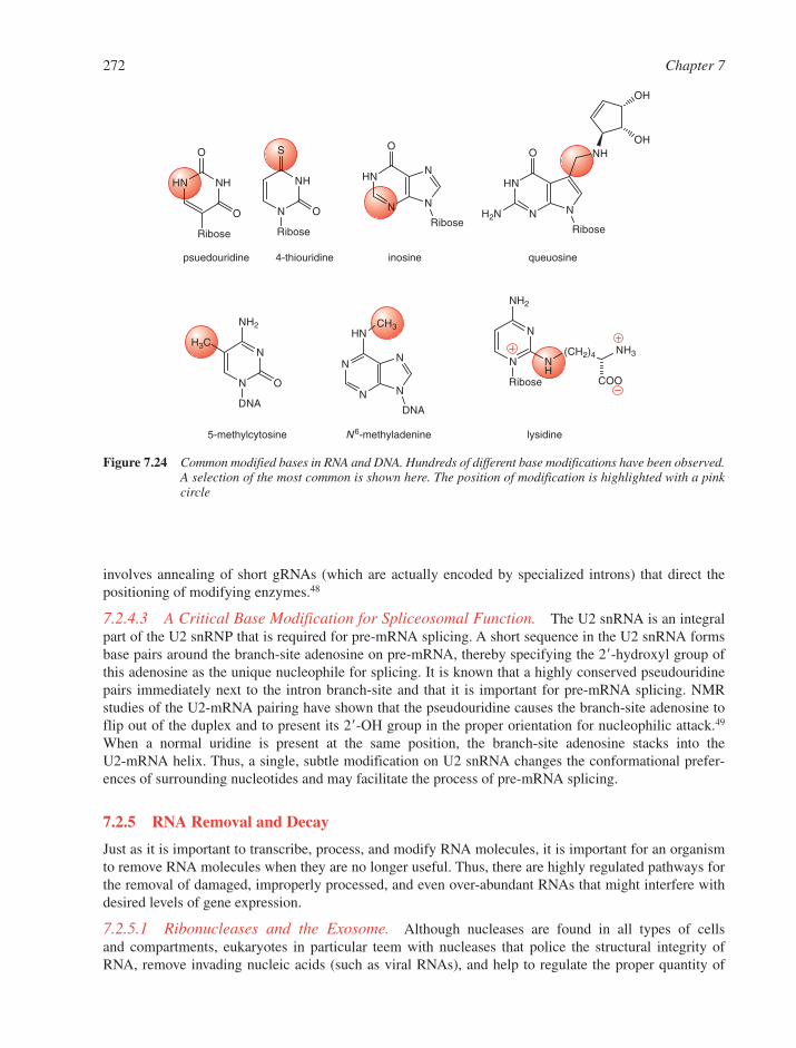
involves annealing of short gRNAs (which are actually encoded by specialized introns) that direct thepositioning of modifying enzymes.48
7.2.4.3 A Critical Base Modification for Spliceosomal Function. The U2 snRNA is an integralpart of the U2 snRNP that is required for pre-mRNA splicing. A short sequence in the U2 snRNA formsbase pairs around the branch-site adenosine on pre-mRNA, thereby specifying the 2�-hydroxyl group ofthis adenosine as the unique nucleophile for splicing. It is known that a highly conserved pseudouridinepairs immediately next to the intron branch-site and that it is important for pre-mRNA splicing. NMRstudies of the U2-mRNA pairing have shown that the pseudouridine causes the branch-site adenosine toflip out of the duplex and to present its 2�-OH group in the proper orientation for nucleophilic attack.49
When a normal uridine is present at the same position, the branch-site adenosine stacks into the U2-mRNA helix. Thus, a single, subtle modification on U2 snRNA changes the conformational prefer-ences of surrounding nucleotides and may facilitate the process of pre-mRNA splicing.
7.2.5 RNA Removal and Decay
Just as it is important to transcribe, process, and modify RNA molecules, it is important for an organismto remove RNA molecules when they are no longer useful. Thus, there are highly regulated pathways forthe removal of damaged, improperly processed, and even over-abundant RNAs that might interfere withdesired levels of gene expression.
7.2.5.1 Ribonucleases and the Exosome. Although nucleases are found in all types of cells and compartments, eukaryotes in particular teem with nucleases that police the structural integrity ofRNA, remove invading nucleic acids (such as viral RNAs), and help to regulate the proper quantity of
272 Chapter 7
HN NH
O
Ribose
O N
NH
S
O
Ribose
HN
N N
N
O
Ribose
HN
N N
O
Ribose
H2N
NH
OH
OH
N
N
Ribose
NH2
NH
(CH2)4 NH3
COON
N
DNA
O
NH2
H3C
CH3
N
N N
N
DNA
HN
lysidine
psuedouridine 4-thiouridine inosine queuosine
5-methylcytosine N6-methyladenine
Figure 7.24 Common modified bases in RNA and DNA. Hundreds of different base modifications have been observed.A selection of the most common is shown here. The position of modification is highlighted with a pinkcircle
RNA molecules for appropriate gene expression (Section 5.3).50 Typical nucleases include the exonucle-ases, which bind to unprotected RNA termini and degrade RNA in either the 3�- or 5�-direction (5� and 3�-exonucleases). There are also endonucleases, which cut in the center of an RNA molecule, often leaving anexposed tail for further degradation by the exonucleases. After splicing, eukaryotic introns are released as lar-iat molecules, which would be resistant to degradation and recycling were it not for a debranching enzyme,which is a specialized nuclease that specifically cuts the 2�–5� linkage that joins the branch-site to the firstnucleotide of an intron. The resultant “linearized” intron can then be degraded by standard nucleases.
While some nucleases diffuse freely throughout a cellular compartment, many of them act in a highlyregulated manner, as part of macromolecular machines such as the exosome. Processed mRNA moleculesare specifically degraded through a carefully orchestrated pathway involving decapping of the 5�-end andloss of the 3�-poly A tail that normally protect mRNA molecules from degradation. Within the exosome, acomplex of 3�→5� exonucleases degrade these unprotected mRNAs. This and other exonucleolyticmachinery also degrade mRNAs that have been improperly capped, adenylated or exported, thus provid-ing a form of mRNA quality control.50
7.2.5.2 Nonsense-Mediated Decay and RNA Quality Control. Transcription, splicing, and otherprocesses involved in RNA synthesis are highly imperfect. Mistakes in these pathways, together with aber-rant transcripts from mutated genes and invading viruses lead to deleterious RNA molecules, which, ifunchecked, would result in aberrant protein expression in the cell. A major mechanism for destroyingthese unwanted messages is nonsense-mediated decay (NMD).51This pathway is based upon the fact thataberrant transcripts commonly contain stop codons (or “nonsense codons”) at inappropriate positions withinthe RNA sequence. During the process of NMD, RNAs containing these premature stop codons are iden-tified and targeted for rapid degradation by exonucleases. Defects in the complex process of NMD havenow been linked to numerous important human disorders.
A second important pathway for RNA quality control is enforced by the ADAR family of enzymes, whichrecognize and target duplex RNA molecules that are either produced endogenously or result from viral infec-tion. ADAR enzymes recognize long RNA duplexes and, as in transversional editing, the paired adenosinesare converted into inosines. When employed as a form of quality control, this base transversion can result intranscript destabilization and susceptibility to degradation. In a similar (or perhaps related) process, the RNAinterference (RNAi) machinery also targets RNA duplexes and marks them for destruction (Section 7.5.3).42
7.3 RNAs IN THE PROTEIN FACTORY: TRANSLATION
In modern organisms, proteins are the dominant macromolecular building materials for cellular function.However, all proteins are the product of a factory that must receive the encoded instructions, gather theamino acid starting materials, and stitch them together in the proper order. But the ribosome is not a mereassembly line for protein synthesis. It is sensitively regulated to produce the correct quantity of protein, todetect problems that arise during synthesis, and to rapidly dispose of defective products. Despite the diver-sity of life, all organisms utilize similar ribosomal factories to build proteins and modulate the requiredlevels of gene expression.
7.3.1 Messenger RNA and the Genetic Code
For protein synthesis, the encoded instructions are contained in messenger RNA (mRNA), which is readlike a tape by ribosomes. An mRNA sequence is translated into protein through the genetic code, whichhas the same basic format for all forms of life on earth (Figure 7.25). The code consists of nucleotide triplets(codons) that specify the identity and sequential position of amino acids in a protein.46,52 For example, thesequence 5�-AAA-3� codes for the amino acid lysine. Each amino acid (and the tRNA to which it isappended) is specified by several different codons (synonyms), which differ primarily in the identity of thethird position. For example, CCU, CCC, CCA, and CCG all encode the amino acid proline. The ribosome
RNA Structure and Function 273
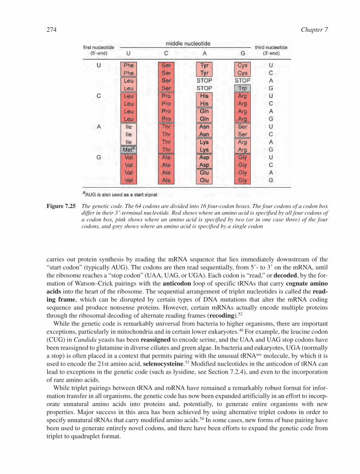
carries out protein synthesis by reading the mRNA sequence that lies immediately downstream of the“start codon” (typically AUG). The codons are then read sequentially, from 5�- to 3� on the mRNA, untilthe ribosome reaches a “stop codon” (UAA, UAG, or UGA). Each codon is “read,” or decoded, by the for-mation of Watson–Crick pairings with the anticodon loop of specific tRNAs that carry cognate aminoacids into the heart of the ribosome. The sequential arrangement of triplet nucleotides is called the read-ing frame, which can be disrupted by certain types of DNA mutations that alter the mRNA codingsequence and produce nonsense proteins. However, certain mRNAs actually encode multiple proteinsthrough the ribosomal decoding of alternate reading frames (recoding).52
While the genetic code is remarkably universal from bacteria to higher organisms, there are importantexceptions, particularly in mitochondria and in certain lower eukaryotes.46 For example, the leucine codon(CUG) in Candida yeasts has been reassigned to encode serine, and the UAA and UAG stop codons havebeen reassigned to glutamine in diverse ciliates and green algae. In bacteria and eukaryotes, UGA (normallya stop) is often placed in a context that permits pairing with the unusual tRNAsec molecule, by which it isused to encode the 21st amino acid, selenocysteine.53 Modified nucleotides in the anticodon of tRNA canlead to exceptions in the genetic code (such as lysidine, see Section 7.2.4), and even to the incorporationof rare amino acids.
While triplet pairings between tRNA and mRNA have remained a remarkably robust format for infor-mation transfer in all organisms, the genetic code has now been expanded artificially in an effort to incorp-orate unnatural amino acids into proteins and, potentially, to generate entire organisms with newproperties. Major success in this area has been achieved by using alternative triplet codons in order tospecify unnatural tRNAs that carry modified amino acids.54 In some cases, new forms of base pairing havebeen used to generate entirely novel codons, and there have been efforts to expand the genetic code fromtriplet to quadruplet format.
274 Chapter 7
Figure 7.25 The genetic code. The 64 codons are divided into 16 four-codon boxes. The four codons of a codon boxdiffer in their 3�-terminal nucleotide. Red shows where an amino acid is specified by all four codons ofa codon box, pink shows where an amino acid is specified by two (or in one case three) of the fourcodons, and grey shows where an amino acid is specified by a single codon
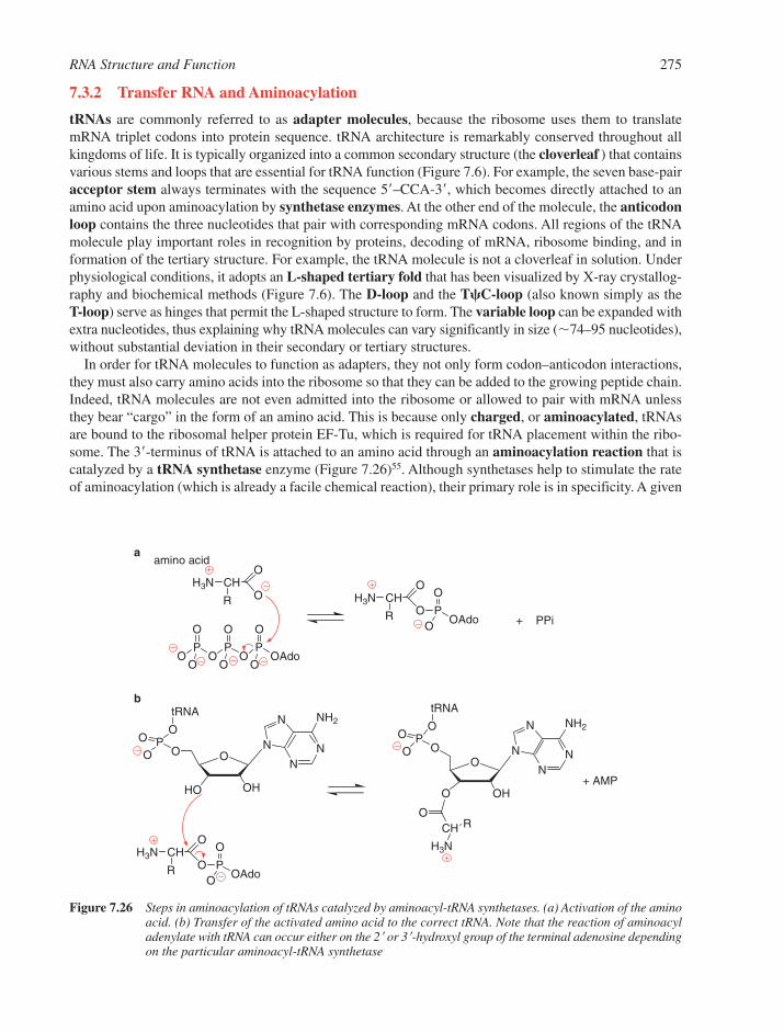
RNA Structure and Function 275
7.3.2 Transfer RNA and Aminoacylation
tRNAs are commonly referred to as adapter molecules, because the ribosome uses them to translatemRNA triplet codons into protein sequence. tRNA architecture is remarkably conserved throughout allkingdoms of life. It is typically organized into a common secondary structure (the cloverleaf ) that containsvarious stems and loops that are essential for tRNA function (Figure 7.6). For example, the seven base-pairacceptor stem always terminates with the sequence 5�–CCA-3�, which becomes directly attached to anamino acid upon aminoacylation by synthetase enzymes. At the other end of the molecule, the anticodonloop contains the three nucleotides that pair with corresponding mRNA codons. All regions of the tRNAmolecule play important roles in recognition by proteins, decoding of mRNA, ribosome binding, and information of the tertiary structure. For example, the tRNA molecule is not a cloverleaf in solution. Underphysiological conditions, it adopts an L-shaped tertiary fold that has been visualized by X-ray crystallog-raphy and biochemical methods (Figure 7.6). The D-loop and the T�C-loop (also known simply as the T-loop) serve as hinges that permit the L-shaped structure to form. The variable loop can be expanded withextra nucleotides, thus explaining why tRNA molecules can vary significantly in size (�74–95 nucleotides),without substantial deviation in their secondary or tertiary structures.
In order for tRNA molecules to function as adapters, they not only form codon–anticodon interactions,they must also carry amino acids into the ribosome so that they can be added to the growing peptide chain.Indeed, tRNA molecules are not even admitted into the ribosome or allowed to pair with mRNA unlessthey bear “cargo” in the form of an amino acid. This is because only charged, or aminoacylated, tRNAsare bound to the ribosomal helper protein EF-Tu, which is required for tRNA placement within the ribo-some. The 3�-terminus of tRNA is attached to an amino acid through an aminoacylation reaction that iscatalyzed by a tRNA synthetase enzyme (Figure 7.26)55. Although synthetases help to stimulate the rateof aminoacylation (which is already a facile chemical reaction), their primary role is in specificity. A given
O
OHHO
O NN
N
N NH2
PO
OO
tRNA
NN
N
N NH2
O
OHO
OP
O
OO
tRNA
OCH R
H3N
OP
O
OP
O
OP
O
OAdoOOO
H3N CH
R
O
O H3N CH
R
O
O P
O
OAdoO
CH
R
O
O P
O
OAdoO
+ PPi
+ AMP
aamino acid
b
H3N
Figure 7.26 Steps in aminoacylation of tRNAs catalyzed by aminoacyl-tRNA synthetases. (a) Activation of the aminoacid. (b) Transfer of the activated amino acid to the correct tRNA. Note that the reaction of aminoacyladenylate with tRNA can occur either on the 2� or 3�-hydroxyl group of the terminal adenosine dependingon the particular aminoacyl-tRNA synthetase

type of tRNA (e.g., tRNAphe) is recognized only by a cognate synthetase enzyme (i.e., phenylalaninetRNA synthetase), which allows only the proper amino acid to be covalently attached to the 3�-terminus(i.e., phenylalanine). There are two major classes of synthetases, with differing architectures and strategiesfor tRNA recognition.55 They are often distinguished by the fact that Class 1 synthetases aminoacylate the 2�-OH of the tRNA acceptor stem, while Class 2 synthetases aminoacylate the 3�-OH. The specificitydeterminants that govern tRNA–synthethase interactions is a major subject of research, as proper tRNA-synthetase recognition is the foundation of a functional genetic code.
7.3.3 Ribosomal RNAs and the Ribosome
The longest, most highly conserved, and most abundant RNA molecules in a cell are the rRNAs. Thesegigantic transcripts, together with a defined set of ribosomal proteins, assemble to form the two ribosomalsubunits (30S and 50S in bacteria; 40S and 60S in eukaryotes) that represent the functional machinery forprokaryotic and eukaryotic ribosomes (Figure 7.27).
The overwhelming majority of ribosomal mass is represented by rRNA, which provides the scaffold formRNA and tRNA binding, helps translocate them through the ribosomal core, and also catalyzes peptidebond formation. Intrinsic ribosomal proteins (designated S1, S2, etc. for proteins of the small subunit andL1, L2, etc. for proteins of the large subunit) are particularly important in early stages of subunit assem-bly, and they contribute subsequently to translation. Additional translation initiation factors (i.e., IF1-IF3),elongation factors (i.e., EF-Tu and EF-G), and release factors (RF) help to facilitate the dynamic processof protein synthesis by the ribosome. Intriguingly, many of these factors (such as EF-Tu, EF-G, and RF)mimic the size, shape, and chemical properties of tRNA molecules, thereby providing important examplesof RNA–protein mimicry that is seen in many aspects of RNA biology (Figure 7.28).56
The two ribosomal subunits are extremely stable and this has contributed to the recent success in obtain-ing high-resolution crystal structures of the prokaryotic 30S and 50S RNA–protein particles (RNPs)(Figure 7.29, see also the Front Cover). Building on earlier cryo-electron microscopy and biochemical studies
276 Chapter 7
Figure 7.27 Two similar designs for the ribosomal factory. Comparison of the prokaryotic and eukaryotic ribosomes and their respective translation cofactors. The structures (above) were obtained by cryo-electron microscopy(Reprinted from Ref. 75. © (2001), with permission from Elsevier)

RNA Structure and Function 277
Figure 7.28 Proteins mimic the structure and interactions of tRNA. A domain of EF-G (purple) mimics tRNA and binds to the ribosomal A site. The complex between tRNA (red) and EF-Tu (yellow) is shown for comparison. Release factor (RF) closely mimics the L-shape and electrostatic properties of tRNA, even binding the ribosome through an anticodon mimic at the tip of its alpha-helical bundle (blue loops)(Reprinted from Ref. 76. © (1999), with permission from AAAS)
Figure 7.29 The structures of the ribosomal subunits. Interface views of the 50S (left) and 30S (right) subunits,along with bound tRNAs (yellow, orange, and red, in the A, P, and E site halves of each subunit). Thesubunits clamp together like the two parts of a shell, enclosing the tRNAs inside (one can visualize theclosed form by superimposing the corresponding tRNA molecules)(Reprinted from Ref. 28. © (2001), with permission from AAAS)
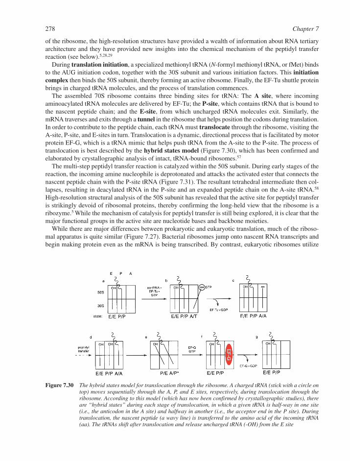
278 Chapter 7
of the ribosome, the high-resolution structures have provided a wealth of information about RNA tertiaryarchitecture and they have provided new insights into the chemical mechanism of the peptidyl transferreaction (see below).5,28,29
During translation initiation, a specialized methionyl tRNA (N-formyl methionyl tRNA, or fMet) bindsto the AUG initiation codon, together with the 30S subunit and various initiation factors. This initiationcomplex then binds the 50S subunit, thereby forming an active ribosome. Finally, the EF-Tu shuttle proteinbrings in charged tRNA molecules, and the process of translation commences.
The assembled 70S ribosome contains three binding sites for tRNA: The A site, where incomingaminoacylated tRNA molecules are delivered by EF-Tu; the P-site, which contains tRNA that is bound tothe nascent peptide chain; and the E-site, from which uncharged tRNA molecules exit. Similarly, themRNA traverses and exits through a tunnel in the ribosome that helps position the codons during translation.In order to contribute to the peptide chain, each tRNA must translocate through the ribosome, visiting theA-site, P-site, and E-sites in turn. Translocation is a dynamic, directional process that is facilitated by motorprotein EF-G, which is a tRNA mimic that helps push tRNA from the A-site to the P-site. The process oftranslocation is best described by the hybrid states model (Figure 7.30), which has been confirmed andelaborated by crystallographic analysis of intact, tRNA-bound ribosomes.57
The multi-step peptidyl transfer reaction is catalyzed within the 50S subunit. During early stages of thereaction, the incoming amine nucleophile is deprotonated and attacks the activated ester that connects thenascent peptide chain with the P-site tRNA (Figure 7.31). The resultant tetrahedral intermediate then col-lapses, resulting in deacylated tRNA in the P-site and an expanded peptide chain on the A-site tRNA.58
High-resolution structural analysis of the 50S subunit has revealed that the active site for peptidyl transferis strikingly devoid of ribosomal proteins, thereby confirming the long-held view that the ribosome is aribozyme.5 While the mechanism of catalysis for peptidyl transfer is still being explored, it is clear that themajor functional groups in the active site are nucleotide bases and backbone moieties.
While there are major differences between prokaryotic and eukaryotic translation, much of the riboso-mal apparatus is quite similar (Figure 7.27). Bacterial ribosomes jump onto nascent RNA transcripts andbegin making protein even as the mRNA is being transcribed. By contrast, eukaryotic ribosomes utilize
Figure 7.30 The hybrid states model for translocation through the ribosome. A charged tRNA (stick with a circle ontop) moves sequentially through the A, P, and E sites, respectively, during translocation through theribosome. According to this model (which has now been confirmed by crystallographic studies), thereare “hybrid states” during each stage of translocation, in which a given tRNA is half-way in one site(i.e., the anticodon in the A site) and halfway in another (i.e., the acceptor end in the P site). Duringtranslocation, the nascent peptide (a wavy line) is transferred to the amino acid of the incoming tRNA(aa). The tRNAs shift after translocation and release uncharged tRNA (-OH) from the E site
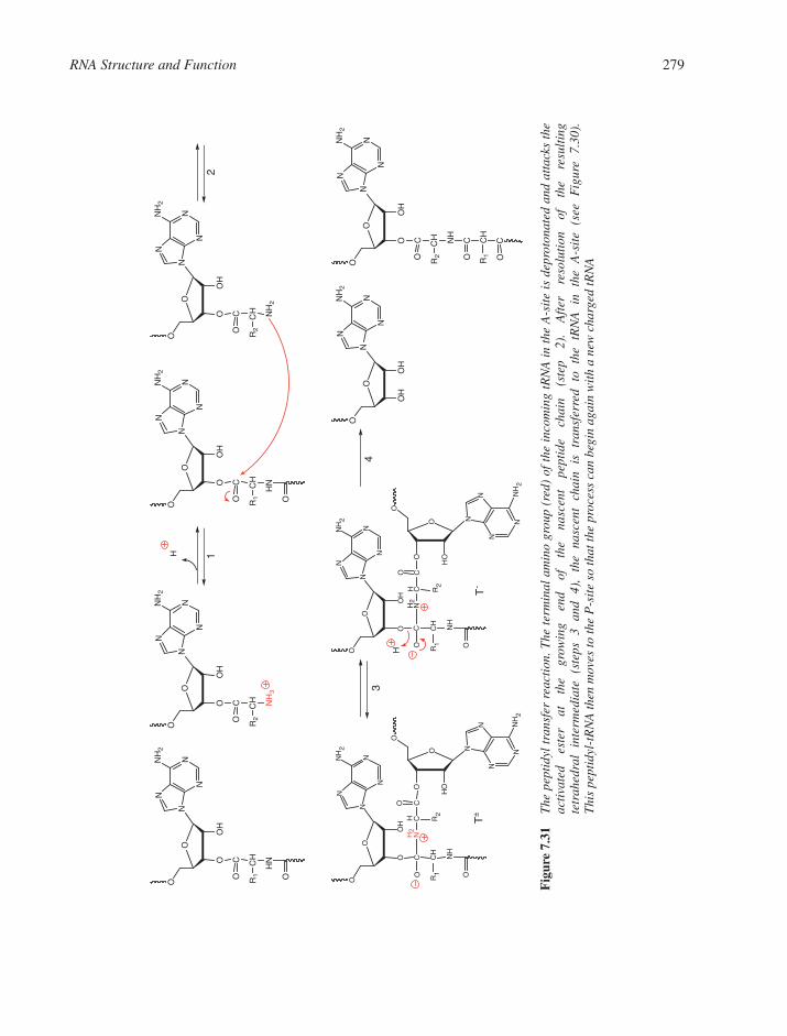
RNA Structure and Function 279
N
N
N
NN
H2
O
O
OH
O CO
CH
R1
HN
O
N
N
N
NN
H2
O
O
OH
O CO
CH
R2
NH
3
N
N
N
NN
H2
O
O
OH
O CO
CH
R1
HN
O
N
N
N
NN
H2
O
O
OH
O CO
CH
R2
NH
2
N
N
N
NN
H2
NH
2
NH
2
O
O
OH
O CO
CH
R1
R1
R2
R2
NH
O
N
N
N
N
O
O
HOO
COH C
H2
N
N
N
N
N
O
O
OH
O CO
CH
NH
O
N
N
N
N NH
2
O
O
HOO
COH C
H2
N
N
N
N
NN
H2
O
O
OH
O CO
CH
R2
NH
CO
CH
R1
CO
N
N
N
NN
H2
O
O
OH
OH
H
H
T-
12
T±
34
Fig
ure
7.31
The
pep
tidy
l tr
ansf
er r
eact
ion.
The
ter
min
al a
min
o gr
oup
(red
) of
the
inc
omin
g tR
NA
in
the
A-s
ite
is d
epro
tona
ted
and
atta
cks
the
acti
vate
d es
ter
at
the
grow
ing
end
of
the
nasc
ent
pept
ide
chai
n (s
tep
2).
Aft
er
reso
luti
on
of
the
resu
ltin
g te
trah
edra
l in
term
edia
te
(ste
ps
3 an
d 4)
, th
e na
scen
t ch
ain
is
tran
sfer
red
to
the
tRN
A
in
the
A-s
ite
(see
F
igur
e 7.
30).
T
his
pept
idyl
-tR
NA
then
mov
es to
the
P-s
ite
so th
at th
e pr
oces
s ca
n be
gin
agai
n w
ith
a ne
w c
harg
ed tR
NA
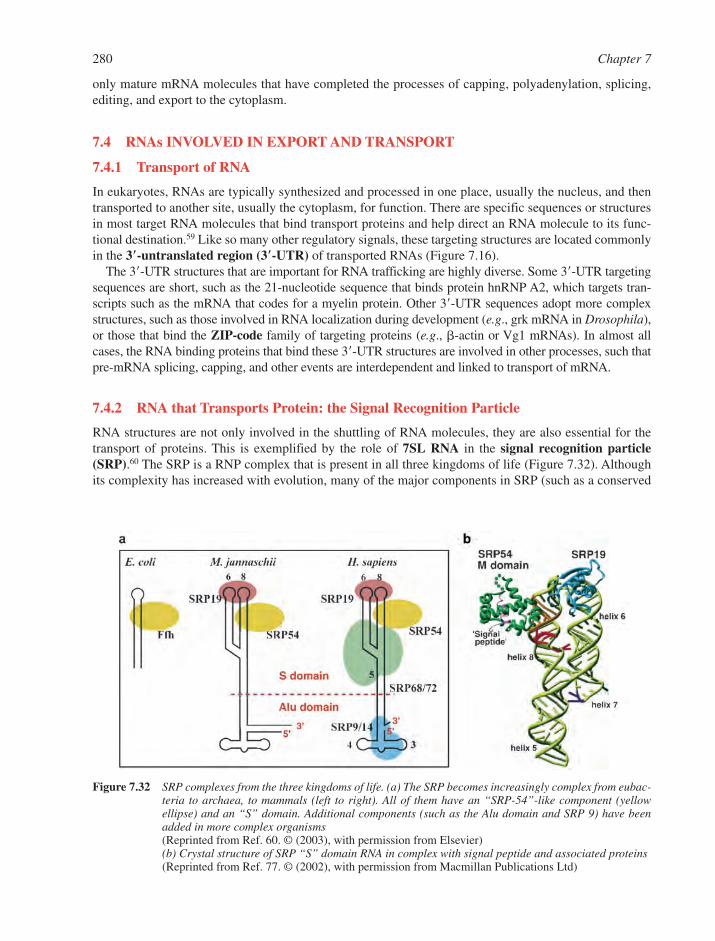
only mature mRNA molecules that have completed the processes of capping, polyadenylation, splicing,editing, and export to the cytoplasm.
7.4 RNAs INVOLVED IN EXPORT AND TRANSPORT
7.4.1 Transport of RNA
In eukaryotes, RNAs are typically synthesized and processed in one place, usually the nucleus, and thentransported to another site, usually the cytoplasm, for function. There are specific sequences or structuresin most target RNA molecules that bind transport proteins and help direct an RNA molecule to its func-tional destination.59 Like so many other regulatory signals, these targeting structures are located commonlyin the 3�-untranslated region (3�-UTR) of transported RNAs (Figure 7.16).
The 3�-UTR structures that are important for RNA trafficking are highly diverse. Some 3�-UTR targetingsequences are short, such as the 21-nucleotide sequence that binds protein hnRNP A2, which targets tran-scripts such as the mRNA that codes for a myelin protein. Other 3�-UTR sequences adopt more complexstructures, such as those involved in RNA localization during development (e.g., grk mRNA in Drosophila),or those that bind the ZIP-code family of targeting proteins (e.g., �-actin or Vg1 mRNAs). In almost allcases, the RNA binding proteins that bind these 3�-UTR structures are involved in other processes, such thatpre-mRNA splicing, capping, and other events are interdependent and linked to transport of mRNA.
7.4.2 RNA that Transports Protein: the Signal Recognition Particle
RNA structures are not only involved in the shuttling of RNA molecules, they are also essential for thetransport of proteins. This is exemplified by the role of 7SL RNA in the signal recognition particle(SRP).60 The SRP is a RNP complex that is present in all three kingdoms of life (Figure 7.32). Althoughits complexity has increased with evolution, many of the major components in SRP (such as a conserved
280 Chapter 7
Figure 7.32 SRP complexes from the three kingdoms of life. (a) The SRP becomes increasingly complex from eubac-teria to archaea, to mammals (left to right). All of them have an “SRP-54”-like component (yellowellipse) and an “S” domain. Additional components (such as the Alu domain and SRP 9) have beenadded in more complex organisms(Reprinted from Ref. 60. © (2003), with permission from Elsevier)(b) Crystal structure of SRP “S” domain RNA in complex with signal peptide and associated proteins(Reprinted from Ref. 77. © (2002), with permission from Macmillan Publications Ltd)
RNA core) have remained the same. The role of SRP in the cell is to bind signal peptides that are locatedat the terminus of nascent membrane and secretory proteins.61 After forming a complex with the signalsequence, SRP guides the entire ribosome/peptide complex to a receptor site on the endoplasmic reticu-lum (ER), where the remainder of the protein is synthesized, while it is transported simultaneouslythrough (or into) the ER membrane.
SRP RNA contains an “S” domain that is highly conserved in all kingdoms (Figure 7.32). In Archaeaand Eukaryotes, this has been appended to an “Alu domain” RNA. Intriguingly, this Alu domain is the samesequence that is encoded by the eukaryotic mobile genetic element of the same name (Section 7.5.1), andthis connection between SRP and genomic plasticity remains a subject of great interest. The SRP RNA isbound by conserved proteins (such as the GTPases SRP54 and SR�) that are involved in assembly andfunction of the particle.
7.5 RNAs AND EPIGENETIC PHENOMENA
Genomes and gene expression are constantly being altered by processes that are “outside” the normalprocesses of DNA replication, RNA metabolism, and protein expression. These “epigenetic phenomena”often involve specialized RNA molecules that play a major role in the evolution and metabolism of diverseorganisms.
7.5.1 RNA Mobile Elements
Genomes are not static environments. Nor do genomes necessarily evolve through small changes thataccumulate slowly over time. Rather, genomes often undergo massive changes, the most potent effectors ofwhich are mobile genetic elements, or transposons (Section 6.8.3). While the zoology of mobile elementsis diverse,62 the retrotransposons represent a subset of RNA molecules that are of particular interestbecause of their remarkable mobility mechanisms and their profound influence on eukaryotic genomes.These RNAs assemble with cofactor proteins to form RNPs that encode, or depend upon, reverse tran-scriptases (RT) to produce stable DNA copies of their progeny.
7.5.1.1 Mammalian L1 and Alu Elements. A stunning 25% of human DNA, by weight, encodestwo RNA molecules (L1, 15%; and Alu, 10%) in millions of copies that have radically altered the organ-ization of mammalian genomes. Although only a fraction of these copies are functional, the mobilizationof L1 and Alu elements causes significant genomic rearrangement, which can lead to cancer and other diseases.63
The L1 gene encodes a large RNA that is �6000 nucleotides in length. This polyadenylated transcriptcontains a 5�-UTR and two reading frames (ORF1 and ORF2) that encode proteins essential for L1 trans-position. The proteins ORF1p and ORF2p form an RNP by binding to substructures in the L1 RNA.Functional L1 RNPs are then imported into the nucleus, where they attack the host genome through aprocess that involves reverse transcription of L1 RNA by the ORF2p. RTs such as ORF2p are polymerasesthat can synthesize DNA from an RNA template. They are important for the replication and genomic inte-gration of many mobile elements and for retroviruses, such as HIV (Section 6.4.6).
The Alu element is a remarkably small RNA (�300 nucleotides) that originated from the 7SL RNA of SRP (Figure 7.32, Section 7.4). Perhaps due to a similarity in the RNA binding properties of SRP andL1 proteins, 7SL RNA is believed to have recruited the L1 transposition apparatus, which allowed it toreplicate and proliferate as the Alu mobile element. Alu elements lack ORFs for proteins that stimulatemobility, and therefore they continue to depend on L1 for mobilization and proliferation in the humangenome.
7.5.1.2 Group II Intron Retrotransposons. A second family of retrotransposon mobilizes a catalyticintron that is commonly found in the organellar genes of plants, fungi, yeast, and also in bacteria. Group IIintrons are self-splicing RNAs (Section 7.2), which contain an ORF that encodes a multifunctional
RNA Structure and Function 281
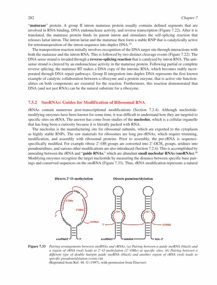
282 Chapter 7
Figure 7.33 Pairing arrangements between snoRNAs and rRNAs. (a) Pairing between a guide snoRNA (black) anda region of rRNA (red) leads to 2�-O methylation (2�-OMe) at specific sites. (b) Pairing between a different type of double hairpin guide snoRNA (black) and another region of rRNA (red) leads to specific pseudouridylation events (�)(Reprinted from Ref. 48. © (1997), with permission from Elsevier)
“maturase” protein. A group II intron maturase protein usually contains defined segments that areinvolved in RNA binding, DNA endonuclease activity, and reverse transcription (Figure 7.22). After it istranslated, the maturase protein binds its parent intron and stimulates the self-splicing reaction thatreleases lariat intron. The intron lariat and the maturase then form a stable RNP that is catalytically activefor retrotransposition of the intron sequence into duplex DNA.39
The transposition reaction initially involves recognition of the DNA target site through interactions withboth the maturase and the intron RNA. This is followed by two distinct cleavage events (Figure 7.22). TheDNA sense strand is invaded through a reverse-splicing reaction that is catalyzed by intron RNA. The anti-sense strand is cleaved by an endonuclease activity in the maturase protein. Following partial or completereverse splicing, the maturase RT makes a DNA copy of the intronic RNA, which becomes stably incor-porated through DNA repair pathways. Group II integration into duplex DNA represents the first knownexample of catalytic collaboration between a ribozyme and a protein enzyme, that is active-site function-alities on both components are essential for the reaction. Furthermore, this reaction demonstrated thatDNA (and not just RNA) can be the natural substrate for a ribozyme.
7.5.2 SnoRNAs: Guides for Modification of Ribosomal RNA
rRNAs contain numerous post-transcriptional modifications (Section 7.2.4). Although nucleotide-modifying enzymes have been known for some time, it was difficult to understand how they are targeted tospecific sites on rRNA. The answer has come from studies of the nucleolus, which is a cellular organellethat has long been a curiosity because it is literally packed with RNA.
The nucleolus is the manufacturing site for ribosomal subunits, which are exported to the cytoplasm as highly stable RNPs. The raw materials for ribosomes are long pre-rRNAs, which require trimming,modification, and assembly with ribosomal proteins. Prior to assembly, the pre-rRNA is sequence-specifically modified. For example ribose 2�-OH groups are converted into 2�-OCH3 groups, uridines intopseudouridines, and various other modifications are also introduced (Section 7.2.4). This is accomplished byannealing between the rRNA and “guide RNAs,” which are abundant small nucleolar RNAs (snoRNAs).48
Modifying enzymes recognize the target nucleotide by measuring the distance between specific base pair-ings and conserved sequences on the snoRNA (Figure 7.33). Thus, rRNA modification represents a natural
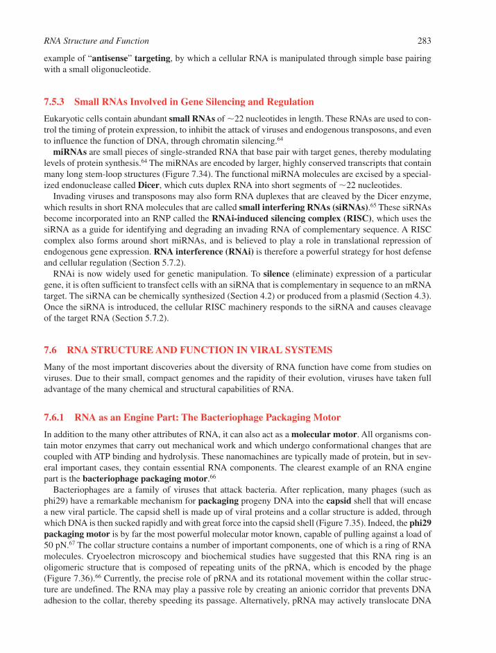
RNA Structure and Function 283
example of “antisense” targeting, by which a cellular RNA is manipulated through simple base pairingwith a small oligonucleotide.
7.5.3 Small RNAs Involved in Gene Silencing and Regulation
Eukaryotic cells contain abundant small RNAs of �22 nucleotides in length. These RNAs are used to con-trol the timing of protein expression, to inhibit the attack of viruses and endogenous transposons, and evento influence the function of DNA, through chromatin silencing.64
miRNAs are small pieces of single-stranded RNA that base pair with target genes, thereby modulatinglevels of protein synthesis.64 The miRNAs are encoded by larger, highly conserved transcripts that containmany long stem-loop structures (Figure 7.34). The functional miRNA molecules are excised by a special-ized endonuclease called Dicer, which cuts duplex RNA into short segments of �22 nucleotides.
Invading viruses and transposons may also form RNA duplexes that are cleaved by the Dicer enzyme,which results in short RNA molecules that are called small interfering RNAs (siRNAs).65 These siRNAsbecome incorporated into an RNP called the RNAi-induced silencing complex (RISC), which uses thesiRNA as a guide for identifying and degrading an invading RNA of complementary sequence. A RISCcomplex also forms around short miRNAs, and is believed to play a role in translational repression ofendogenous gene expression. RNA interference (RNAi) is therefore a powerful strategy for host defenseand cellular regulation (Section 5.7.2).
RNAi is now widely used for genetic manipulation. To silence (eliminate) expression of a particulargene, it is often sufficient to transfect cells with an siRNA that is complementary in sequence to an mRNAtarget. The siRNA can be chemically synthesized (Section 4.2) or produced from a plasmid (Section 4.3).Once the siRNA is introduced, the cellular RISC machinery responds to the siRNA and causes cleavageof the target RNA (Section 5.7.2).
7.6 RNA STRUCTURE AND FUNCTION IN VIRAL SYSTEMS
Many of the most important discoveries about the diversity of RNA function have come from studies onviruses. Due to their small, compact genomes and the rapidity of their evolution, viruses have taken fulladvantage of the many chemical and structural capabilities of RNA.
7.6.1 RNA as an Engine Part: The Bacteriophage Packaging Motor
In addition to the many other attributes of RNA, it can also act as a molecular motor. All organisms con-tain motor enzymes that carry out mechanical work and which undergo conformational changes that arecoupled with ATP binding and hydrolysis. These nanomachines are typically made of protein, but in sev-eral important cases, they contain essential RNA components. The clearest example of an RNA enginepart is the bacteriophage packaging motor.66
Bacteriophages are a family of viruses that attack bacteria. After replication, many phages (such asphi29) have a remarkable mechanism for packaging progeny DNA into the capsid shell that will encasea new viral particle. The capsid shell is made up of viral proteins and a collar structure is added, throughwhich DNA is then sucked rapidly and with great force into the capsid shell (Figure 7.35). Indeed, the phi29packaging motor is by far the most powerful molecular motor known, capable of pulling against a load of50 pN.67 The collar structure contains a number of important components, one of which is a ring of RNAmolecules. Cryoelectron microscopy and biochemical studies have suggested that this RNA ring is anoligomeric structure that is composed of repeating units of the pRNA, which is encoded by the phage(Figure 7.36).66 Currently, the precise role of pRNA and its rotational movement within the collar struc-ture are undefined. The RNA may play a passive role by creating an anionic corridor that prevents DNAadhesion to the collar, thereby speeding its passage. Alternatively, pRNA may actively translocate DNA
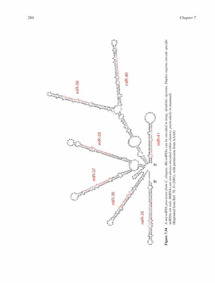
284 Chapter 7
Fig
ure
7.34
A m
icro
RN
A p
recu
rsor
fro
m C
. ele
gans
. Mic
roR
NA
s ca
n be
enc
oded
in
larg
e, d
endr
itic
ope
rons
. Dup
lex
regi
ons
enco
de s
peci
ficm
iRN
As
(in
red)
. MiR
NA
s ar
e no
t alw
ays
enco
ded
wit
hin
clus
ters
, par
ticu
larl
y in
mam
mal
s (R
epri
nted
fro
m R
ef. 7
8. ©
(20
01),
with
per
mis
sion
fro
m A
AA
S)
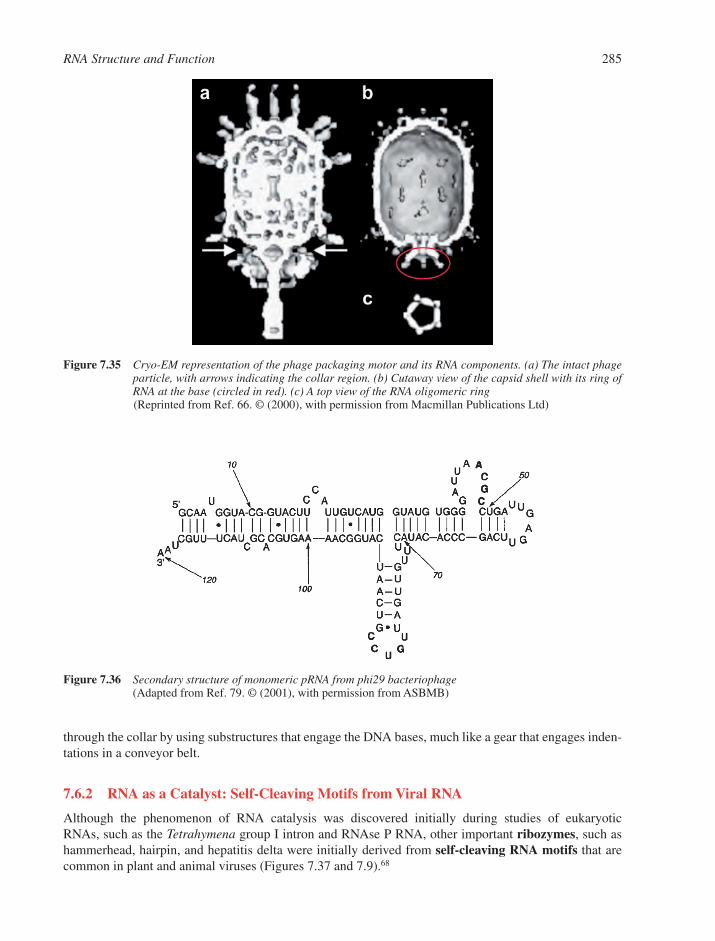
through the collar by using substructures that engage the DNA bases, much like a gear that engages inden-tations in a conveyor belt.
7.6.2 RNA as a Catalyst: Self-Cleaving Motifs from Viral RNA
Although the phenomenon of RNA catalysis was discovered initially during studies of eukaryotic RNAs, such as the Tetrahymena group I intron and RNAse P RNA, other important ribozymes, such ashammerhead, hairpin, and hepatitis delta were initially derived from self-cleaving RNA motifs that arecommon in plant and animal viruses (Figures 7.37 and 7.9).68
RNA Structure and Function 285
Figure 7.35 Cryo-EM representation of the phage packaging motor and its RNA components. (a) The intact phageparticle, with arrows indicating the collar region. (b) Cutaway view of the capsid shell with its ring ofRNA at the base (circled in red). (c) A top view of the RNA oligomeric ring(Reprinted from Ref. 66. © (2000), with permission from Macmillan Publications Ltd)
Figure 7.36 Secondary structure of monomeric pRNA from phi29 bacteriophage(Adapted from Ref. 79. © (2001), with permission from ASBMB)
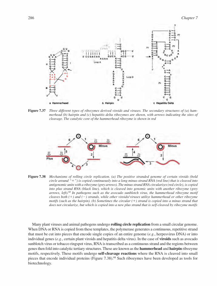
Many plant viruses and animal pathogens undergo rolling circle replication from a small circular genome.When DNA or RNA is copied from these templates, the polymerase generates a continuous, repetitive strandthat must be cut into pieces that encode single copies of an entire genome (e.g., herpesvirus DNA) or intoindividual genes (e.g., certain plant viroids and hepatitis delta virus). In the case of viroids such as avocadosunblotch virus or tobacco ringspot virus, RNA is transcribed as a continuous strand and the regions betweengenes then fold into catalytic tertiary structures. These are known as the hammerhead and hairpin ribozymemotifs, respectively. These motifs undergo self-cleavage reactions where the RNA is cleaved into smallpieces that encode individual proteins (Figure 7.38).68 Such ribozymes have been developed as tools forbiotechnology.
286 Chapter 7
Figure 7.37 Three different types of ribozymes derived viroids and viruses. The secondary structures of (a) ham-merhead (b) hairpin and (c) hepatitis delta ribozymes are shown, with arrows indicating the sites ofcleavage. The catalytic core of the hammerhead ribozyme is shown in red
Figure 7.38 Mechanisms of rolling circle replication. (a) The positive stranded genome of certain viroids (bold circle around “�”) is copied continuously into a long minus-strand RNA (red line) that is cleaved intoantigenomic units with a ribozyme (grey arrows). The minus strand RNA circularizes (red circle), is copiedinto plus strand RNA (black line), which is cleaved into genomic units with another ribozyme (greyarrows, left).68 In pathogens such as the avocado sunblotch virus, the hammerhead ribozyme motifcleaves both (�) and (�) strands, while other viroids/ viruses utilize hammerhead or other ribozymemotifs (such as the hairpin). (b) Sometimes the circular (�) strand is copied into a minus strand thatdoes not circularize, but which is copied into a new plus strand that is self-cleaved by ribozyme motifs
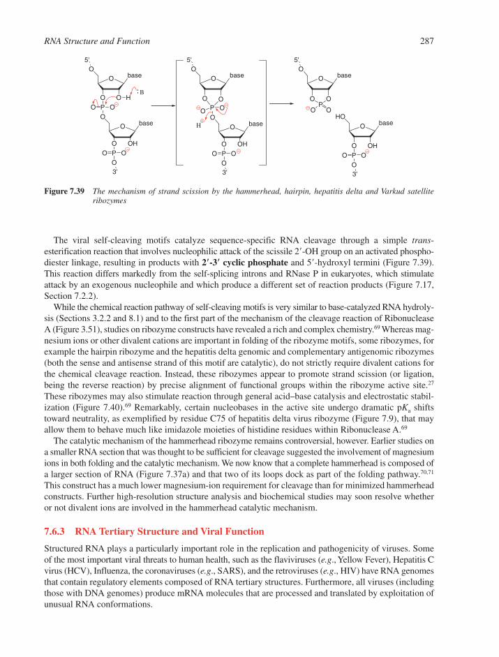
The viral self-cleaving motifs catalyze sequence-specific RNA cleavage through a simple trans-esterification reaction that involves nucleophilic attack of the scissile 2�-OH group on an activated phospho-diester linkage, resulting in products with 2�-3� cyclic phosphate and 5�-hydroxyl termini (Figure 7.39).This reaction differs markedly from the self-splicing introns and RNase P in eukaryotes, which stimulateattack by an exogenous nucleophile and which produce a different set of reaction products (Figure 7.17,Section 7.2.2).
While the chemical reaction pathway of self-cleaving motifs is very similar to base-catalyzed RNA hydroly-sis (Sections 3.2.2 and 8.1) and to the first part of the mechanism of the cleavage reaction of RibonucleaseA (Figure 3.51), studies on ribozyme constructs have revealed a rich and complex chemistry.69 Whereas mag-nesium ions or other divalent cations are important in folding of the ribozyme motifs, some ribozymes, forexample the hairpin ribozyme and the hepatitis delta genomic and complementary antigenomic ribozymes(both the sense and antisense strand of this motif are catalytic), do not strictly require divalent cations forthe chemical cleavage reaction. Instead, these ribozymes appear to promote strand scission (or ligation,being the reverse reaction) by precise alignment of functional groups within the ribozyme active site.27
These ribozymes may also stimulate reaction through general acid–base catalysis and electrostatic stabil-ization (Figure 7.40).69 Remarkably, certain nucleobases in the active site undergo dramatic pKa shiftstoward neutrality, as exemplified by residue C75 of hepatitis delta virus ribozyme (Figure 7.9), that mayallow them to behave much like imidazole moieties of histidine residues within Ribonuclease A.69
The catalytic mechanism of the hammerhead ribozyme remains controversial, however. Earlier studies ona smaller RNA section that was thought to be sufficient for cleavage suggested the involvement of magnesiumions in both folding and the catalytic mechanism. We now know that a complete hammerhead is composed ofa larger section of RNA (Figure 7.37a) and that two of its loops dock as part of the folding pathway.70,71
This construct has a much lower magnesium-ion requirement for cleavage than for minimized hammerheadconstructs. Further high-resolution structure analysis and biochemical studies may soon resolve whetheror not divalent ions are involved in the hammerhead catalytic mechanism.
7.6.3 RNA Tertiary Structure and Viral Function
Structured RNA plays a particularly important role in the replication and pathogenicity of viruses. Someof the most important viral threats to human health, such as the flaviviruses (e.g., Yellow Fever), Hepatitis Cvirus (HCV), Influenza, the coronaviruses (e.g., SARS), and the retroviruses (e.g., HIV) have RNA genomesthat contain regulatory elements composed of RNA tertiary structures. Furthermore, all viruses (includingthose with DNA genomes) produce mRNA molecules that are processed and translated by exploitation ofunusual RNA conformations.
RNA Structure and Function 287
baseO
OO
PO
O
O
O
baseO
OHO
PO
O
O
H
5'
3'
baseO
OOP
O
O
O
baseO
OHO
PO
O
O
5'
3'
B
H
O
baseO
HO
O
baseO
OHO
PO
O
O
5'
3'
O OP
O O
Figure 7.39 The mechanism of strand scission by the hammerhead, hairpin, hepatitis delta and Varkud satelliteribozymes
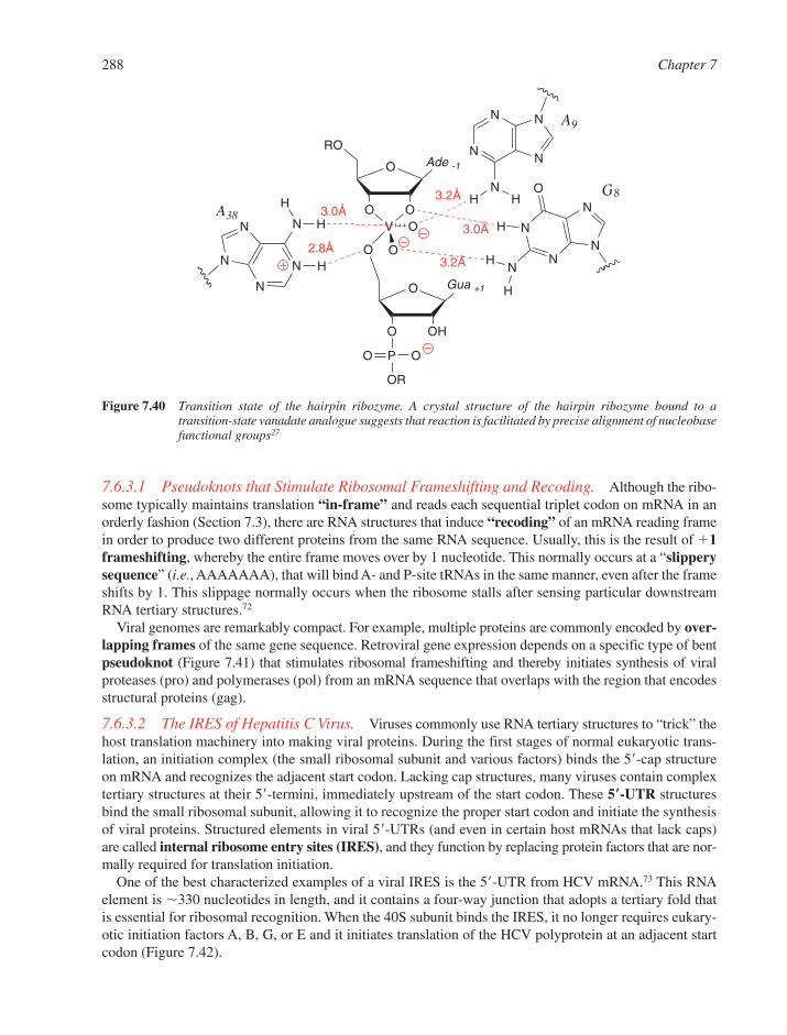
288 Chapter 7
Ade -1O
OOV
RO
Gua +1O
OHO
PO
OR
O
O
O
O
N
NN
N
O
N
N
N N
N
NHH
N
N
N
N N
H
H
H
A9
G8
A38
3.2Å
3.0Å
3.2Å2.8Å
3.0ÅH
H
H
Figure 7.40 Transition state of the hairpin ribozyme. A crystal structure of the hairpin ribozyme bound to atransition-state vanadate analogue suggests that reaction is facilitated by precise alignment of nucleobasefunctional groups27
7.6.3.1 Pseudoknots that Stimulate Ribosomal Frameshifting and Recoding. Although the ribo-some typically maintains translation “in-frame” and reads each sequential triplet codon on mRNA in anorderly fashion (Section 7.3), there are RNA structures that induce “recoding” of an mRNA reading framein order to produce two different proteins from the same RNA sequence. Usually, this is the result of �1frameshifting, whereby the entire frame moves over by 1 nucleotide. This normally occurs at a “slipperysequence” (i.e., AAAAAAA), that will bind A- and P-site tRNAs in the same manner, even after the frameshifts by 1. This slippage normally occurs when the ribosome stalls after sensing particular downstreamRNA tertiary structures.72
Viral genomes are remarkably compact. For example, multiple proteins are commonly encoded by over-lapping frames of the same gene sequence. Retroviral gene expression depends on a specific type of bentpseudoknot (Figure 7.41) that stimulates ribosomal frameshifting and thereby initiates synthesis of viralproteases (pro) and polymerases (pol) from an mRNA sequence that overlaps with the region that encodesstructural proteins (gag).
7.6.3.2 The IRES of Hepatitis C Virus. Viruses commonly use RNA tertiary structures to “trick” thehost translation machinery into making viral proteins. During the first stages of normal eukaryotic trans-lation, an initiation complex (the small ribosomal subunit and various factors) binds the 5�-cap structureon mRNA and recognizes the adjacent start codon. Lacking cap structures, many viruses contain complextertiary structures at their 5�-termini, immediately upstream of the start codon. These 5�-UTR structuresbind the small ribosomal subunit, allowing it to recognize the proper start codon and initiate the synthesisof viral proteins. Structured elements in viral 5�-UTRs (and even in certain host mRNAs that lack caps)are called internal ribosome entry sites (IRES), and they function by replacing protein factors that are nor-mally required for translation initiation.
One of the best characterized examples of a viral IRES is the 5�-UTR from HCV mRNA.73 This RNAelement is �330 nucleotides in length, and it contains a four-way junction that adopts a tertiary fold thatis essential for ribosomal recognition. When the 40S subunit binds the IRES, it no longer requires eukary-otic initiation factors A, B, G, or E and it initiates translation of the HCV polyprotein at an adjacent startcodon (Figure 7.42).
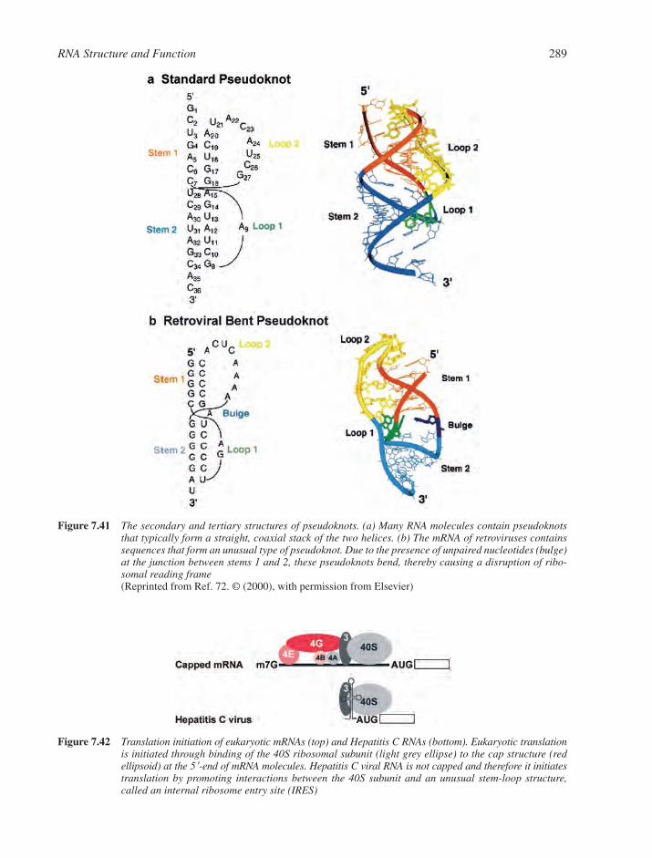
RNA Structure and Function 289
Figure 7.41 The secondary and tertiary structures of pseudoknots. (a) Many RNA molecules contain pseudoknotsthat typically form a straight, coaxial stack of the two helices. (b) The mRNA of retroviruses containssequences that form an unusual type of pseudoknot. Due to the presence of unpaired nucleotides (bulge)at the junction between stems 1 and 2, these pseudoknots bend, thereby causing a disruption of ribo-somal reading frame(Reprinted from Ref. 72. © (2000), with permission from Elsevier)
Figure 7.42 Translation initiation of eukaryotic mRNAs (top) and Hepatitis C RNAs (bottom). Eukaryotic translationis initiated through binding of the 40S ribosomal subunit (light grey ellipse) to the cap structure (redellipsoid) at the 5�-end of mRNA molecules. Hepatitis C viral RNA is not capped and therefore it initiatestranslation by promoting interactions between the 40S subunit and an unusual stem-loop structure,called an internal ribosome entry site (IRES)
REFERENCES
1. K. Chin, K. Sharp, B. Honig and A.M. Pyle, Calculating the electrostatic properties of RNA providesnew insights into molecular interactions and function, Nat. Struct. Biol., 1999, 6, 1055–1061.
2. M. Egli, S. Portmann and N. Usman, RNA hydration: a detailed look, Biochemistry, 1996, 35,8489–8494.
3. N.B. Leontis, J. Stombaugh and E. Westhof, The non-Watson–Crick pairs and their isostericity matri-ces, Nucleic Acids Res., 2002, 30, 3497–3531.
4. V. Biou, A. Yaremchuck, M. Tukalo and S. Cusack, The 2.9 Å crystal structure of T. thermophilusseryl-tRNA synthetase complexed with tRNA(Ser), Science, 1994, 263, 1404–1410.
5. N. Ban, P. Nissen, J. Hansen, P.B. Moore and T.A. Steitz, The complete atomic structure of the largeribosomal subunit at 2.4 Å resolution, Science, 2000, 289, 905–920.
6. L. Su, L. Chen, M. Egli, J.M. Berger and A. Rich, RNA triplex in the structure of ribosomalframeshifting viral pseudoknot, Nat. Struct. Biol., 1999, 3, 285–292.
7. C. Baugh, D. Grate and C. Wilson, 2.8 Å crystal structure of the malachite green aptamer, J. Mol.Biol., 2000, 301, 117–128.
8. L. Gold, B. Polisky, O.C. Uhlenbeck and M. Yarus, Diversity of oligonucleotide functions, Ann. Rev.Biochem., 1995, 64, 763–797.
9. J.H. Cate, A.R. Gooding, E. Podell, K. Zhou, B.L. Golden, C.E. Kundrot, T.R. Cech and J.A. Doudna,Crystal structure of a group I ribozyme domain reveals principles of higher order RNA folding,Science, 1996, 273, 1678–1685.
10. P. Nissen, J.A. Ippolito, N. Ban, P.B. Moore and T.A. Steitz, RNA tertiary interactions in the largeribosomal subunit: the A-minor motif, Proc. Natl. Acad. Sci. USA, 2001, 98, 4899–4903.
11. F.L. Murphy, Y.-H. Wang, J.D. Griffith and T.R. Cech, Coaxially stacked RNA helices in thecatalytic center of the Tetrahymena ribozyme, Science, 1994, 265, 1709–1712.
12. E.A. Doherty, R.T. Batey, B. Masquida and J.A. Doudna, A universal mode of helix packing in RNA,Nat. Struct. Biol., 2001, 8, 339–343.
13. A.R. Ferre-D’amare, K. Zhou and J.A. Doudna, Crystal structure of a hepatitis delta virus ribozyme,Nature, 1998, 395, 567–574.
14. V.K. Misra and D.E. Draper, On the role of magnesium ions in RNA stability, Biopolymers, 1998, 48,113–135.
15. A.M. Pyle, The role of metal ions in ribozymes. In Metal Ions in Biological Systems, H. Sigel andA. Sigel (eds). Marcel Dekker, Inc., New York, 1996, 479–519.
16. R. Shiman and D. Draper, Stabilization of RNA tertiary structure by monovalent cations, J. Mol. Biol.,2000, 302, 79–91.
17. R.K.O. Sigel, A. Vaidya and A.M. Pyle, Metal ion binding sites in a group II intron core, Nat. Struct.Biol., 2000, 7, 1111–1116.
18. S. Basu, R.P. Rambo, J. Strauss-Soukup, J.H. Cate, A. Ferre-D’Amare, S.A. Strobel and J.A. Doudna,A specific monovalent metal ion integral to the AA platform of the RNA tetraloop receptor, Nat.Struct. Biol., 1998, 5, 986–992.
19. J.H. Cate, A.R. Gooding, E. Podell, K. Zhou, B.L. Golden, A.A. Szewczak, C.E. Kundrot, T.R. Cechand J.A. Doudna, RNA tertiary structure mediation by adenosine platforms, Science, 1996, 273,1696–1699.
20. T.R. Sosnick and T. Pan, RNA folding: models and perspectives, Curr. Opin. Struct. Biol., 2003,13, 309–316.
21. R. Schroeder, R. Grossberger, A. Pichler and C. Waldsich, RNA folding in vivo, Curr. Opin. Struct.Biol., 2002, 12, 296–300.
22. S.H. Kim, G. Quigley, F.L. Suddath, A. McPherson, D. Sneden, J.J. Kim, J. Weinzierl, P. Blattman andA. Rich, The three-dimensional structure of yeast transfer RNA: shape of the molecule at 5.5 Å reso-lution, Proc. Natl. Acad. Sci. USA, 1972, 69, 3746–3750.
290 Chapter 7

23. G.J. Quigley, F.L. Suddath, A. McPherson, J.J. Kim, D. Sneden and A. Rich, The molecular structureof yeast phenylalanine transfer RNA in monoclinic crystals, Proc. Natl. Acad. Sci. USA, 1974, 71,2146–2150.
24. H.W. Pley, K.M. Flaherty and D.B. McKay, Three-dimensional structure of a hammerhead ribozyme,Nature, 1994, 372, 68–74.
25. P.L. Adams, M.R. Stahley, A.B. Kosek, J. Wang and S.A. Strobel, Crystal structure of an intact self-splicing group I intron with both exons, Nature, 2004, 430, 45–50.
26. A.S. Krasilnikov, X. Yang, T. Pang and A. Mondragon, Crystal structure of the specificity domain ofRibonuclease P, Nature, 2003, 421, 760–764.
27. P.B. Rupert, A.P. Massey, S.T. Siggurdson and A.R. Ferré-D’amare, Transition-state stabilization by acatalytic RNA, Science, 2002, 298, 1421–1424.
28. M.M. Yusupov, G.Z. Yusupova, A. Baucom, K. Lieberman, T.N. Earnest, J.H. Cate and H.F. Noller,Crystal structure of the ribosome at 5.5 Å resolution, Science, 2001, 292, 883–896.
29. B.T. Wimberley, D.W. Broderson, W.M. Clemons, R.J. Morgan-Warren, A.P. Carter, C. Vonrhein,T. Hartsch and V. Ramakrishnan, Structure of the 30S ribosomal subunit, Nature, 2000, 407, 327–339.
30. N.J. Proudfoot, A. Furger and M.J. Dye, Integrating mRNA processing with transcription, Cell, 2002,108, 501–512.
31. A. Shatkin and J.L. Manley, The ends of the affair: capping and polyadenylation, Nat. Struct. Biol.,2000, 7, 838–842.
32. A.B. Sachs, P. Sarnow and M.W. Hentze, Starting at the beginning, middle, and end: translation initiation in eukaryotes, Cell, 1997, 89, 831–838.
33. H. Madhani and C. Guthrie, Dynamic RNA–RNA interactions in the spliceosome, Ann. Rev. Genet.,1994, 28, 1–26.
34. C.W.J. Smith and J. Valcarcel, Alternative pre-mRNA splicing: the logic of combinatorial control,Trends Biochem. Sci., 2000, 25, 381–388.
35. T.R. Cech, Structure and mechanism of the large catalytic RNAs: group I and group II introns andribonuclease P. In The RNA World, R.F. Gesteland and J.F. Atkins (eds). Cold Spring Harbor Press,Cold Spring Harbor, 1993, 239–270.
36. P.Z. Qin and A.M. Pyle, The architectural organization and mechanistic function of group II intronstructural elements, Curr. Opin. Struct. Biol., 1998, 8, 301–308.
37. K. Kruger, P.J. Grabowski, A.J. Zaug, J. Sands, D.E. Gottschling and T.R. Cech, Self-splicing RNA:autoexcision and autocyclization of the ribosomal RNA intervening sequence of Tetrahymena, Cell,1982, 31, 147–157.
38. C.L. Peebles, P.S. Perlman, K.L. Mecklenburg, M.L. Petrillo, J.H. Tabor, K.A. Jarrell and H.-L.Cheng, A self-splicing RNA excises an intron lariat, Cell, 1986, 44, 213–223.
39. M. Belfort, V. Derbyshire, M.M. Parker, B. Cousineau and A.M. Lambowitz, Mobile introns: pathwaysand proteins. In Mobile DNA II, N.L. Craig, R. Craigie, M. Gellert and A.M. Lambowitz (eds). ASMPress, Washington, DC, 2002, 761–783.
40. J.C. Kurz and C.A. Fierke, Ribonuclease P: a ribonucleoprotein enzyme, Curr. Opin. Chem. Biol.,2000, 4, 553–558.
41. M.A. Carmell and G.J. Hannon, RNase III enzymes and the initiation of gene silencing, Nat. Struct.Mol. Biol., 2004, 11, 214–218.
42. B.L. Bass, RNA editing by adenosine deaminases that act on RNA, Ann. Rev. Biochem., 2002, 71,817–846.
43. J.E. Wedekind, G.S.C. Dance, M.P. Sowden and H.C. Smith, Messenger RNA editing in mammals: newmembers of the APOBEC family seeking roles in the family business, Trends Genet., 2003, 19, 207–216.
44. K. Stuart and A.K. Panigrahi, RNA editing: complexity and complications, Mol. Microbiol., 2002, 45,591–596.
45. K.R. Noon, R. Guymon, P.F. Crain, J.A. McCloskey, M. Thomm, J. Lim and R. Cavicchioli, Influenceof temperature on tRNA modification in Archaea, J. Bacteriol., 2003, 185, 5483–5490.
RNA Structure and Function 291

46. P.F. Agris, Decoding the genome: a modified view, Nucleic Acids Res., 2004, 32, 223–238.47. T. Muramatsu, K. Nishikawa, F. Nemoto, Y. Kuchino, S. Nishimura, T. Miyazawa and S. Yokoyama,
Codon and amino-acid specificities of a transfer RNA are both converted by a single post-transcriptionalmodification, Nature, 1988, 336, 179–181.
48. C.M. Smith and J.A. Steitz, Sno storm in the nucleolus: a new role for myriad small RNPs, Cell, 1997,89, 669–672.
49. M.I. Newby and N.L. Greenbaum, Sculpting of the spliceosomal branch site recognition motif by aconserved pseudouridine, Nature Struct. Biol., 2002, 9, 958–965.
50. R.P. Parker and H. Song, The enzymes and control of eukaryotic mRNA turnover, Nature Struct. Mol.Biol., 2004, 11, 121–127.
51. L.E. Maquat and G.G. Carmichael, Quality control of mRNA function, Cell, 2001, 104, 173–176.52. R.F. Gesteland and J.F. Atkins, Recoding: dynamic reprogramming of translation, Ann. Rev.
Biochem., 1996, 65, 741–768.53. D.L. Hatfield and V.N. Gladyshev, How selenium has altered our understanding of the genetic code,
Mol. Cell. Biol., 2002, 22, 3565–3576.54. J.W. Chin, T.A. Cropp, J.C. Anderson, M. Mukherji, Z. Zhang and P.G. Schultz, An expanded eukary-
otic genetic code, Science, 2003, 301, 964–967.55. P.J. Beuning and K. Musier-Forsyth, Transfer RNA recognition by aminoacyl tRNA synthetases,
Biopolymers, 2000, 52, 1–28.56. Y. Nakamura, M. Uno, T. Toyoda, T. Fujiwara and K. Ito, Protein tRNA mimicry in translation
termination, Cold Spring Harbor Symp. Quant. Biol., 2001, 66, 469–475.57. H.F. Noller, M.M. Yusupov, G.Z. Yusupova, A. Baucom, K. Lieberman, L. Lancaster, A. Dallas,
K. Fredrick, T.N. Earnest and J.H. Cate, Structure of the ribosome at 5.5 Å resolution and itsinteractions with functional ligands, Cold Spring Harbor Symp. Quant. Biol., 2001, 66, 57–66.
58. M.V. Rodnina and W. Wintermeyer, Peptide bond formation on the ribosome: structure and mechanism,Curr. Opin. Struct. Biol., 2003, 13, 334–340.
59. K.L. Farina and R.H. Singer, The nuclear connection in RNA transport and localization, TrendsBiochem. Sci., 2002, 12, 466–472.
60. K. Nagai, C. Oubridge, A. Kuglstatter, E. Menichelli, C. Isel and L. Jovine, Structure, function andevolution of the signal recognition particle, EMBO J., 2003, 22, 3479–3485.
61. R.J. Keenan, D.M. Freymann, R.M. Stroud and P. Walter, The signal recognition particle, Ann. Rev.Biochem., 2001, 70, 755–775.
62. N.L. Craig, R. Craigie, M. Gellert and A.M. Lambowtiz, Mobile DNA II, ASM Press, Washington,DC, 2002.
63. J.V. Moran and N. Gilbert, Mammalian LINE-1 retrotransposons and related elements. In MobileDNA II, N.L. Craig, R. Craigie, M. Gellert and A.M. Lambowtiz (eds). ASM Press, Washington, DC,2002, 836–869.
64. P. Nelson, M. Kiriakidou, A. Sharma, E. Maniataki and Z. Mourelatos, The microRNA world: smallis mighty, Trends Biochem. Sci., 2003, 28, 534–540.
65. G.J. Hannon, RNA interference, Nature, 2002, 418, 244–251.66. A.A. Simpson, Y. Tao, P.G. Leiman, M.O. Badasso, Y. He, P.J. Jardine, N.H. Olson, M.C. Morais,
S. Grimes, D.L. Anderson, T.S. Baker and M.G. Rossman, Structure of the bacteriophage phi29 DNApackaging motor, Nature, 2000, 408, 745–750.
67. D.E. Smith, S.J. Tans, S.B. Smith, S. Grimes, D.L. Anderson and C. Bustamante, The bacteriophagephi29 portal motor can package DNA against a large internal force, Nature, 2001, 413, 748–752.
68. R.H. Symons, Plant pathogenic RNAs and RNA catalysis, Nucleic Acids Res., 1997, 25, 2683–2689.69. P.C. Bevilacqua, T.S. Brown, S. Nakano and R. Yajima, Catalytic roles for proton transfer and
protonation in ribozymes, Biopolymers, 2004, 73, 90–109.70. A. Khvorova, A. Lescoute, E. Westhof and S.D. Jayasena, Sequence elements outside of the hammerhead
ribozyme catalytic core enable intracellular activity, Nat. Struct. Biol., 2003, 10, 708–712.
292 Chapter 7
71. J.C. Penedo, T.J. Wilson, S.D. Hayasena, A. Khvorova and D.M.J. Lilley, Folding of the natural hammerhead ribozyme is enhanced by interaction of auxiliary elements, RNA, 2004, 10, 880–888.
72. D.P. Giedroc, C.A. Theimer and P.L. Nixon, Structure, stability and function of RNA pseudoknotsinvolved in stimulating ribosomal frameshifting, J. Mol. Biol., 2000, 298, 167–185.
73. T.V. Pestova, I.N. Shatsky, S.P. Fletcher, R.J. Jackson and C.U.T. Hellen, A prokaryotic-like mode ofcytoplasmic eukaryotic ribosome binding to the initiation codon during internal translation initiationof HCV and classical swine fever virus RNAs, Genes Dev., 1998, 12, 67–83.
74. A.J. Lee and D.M. Crothers, The solution structure of an RNA loop–loop complex: the ColE1 invertedloop sequence, Struct. Fold Des., 1998, 15, 993–1005.
75. C.M.T. Spahn, R. Beckmann, N. Eswar, P.A. Penczek, A. Sali, G. Blobel and J. Frank, Structure of the80S ribosome from Saccharomyces cerevisiae. Cell, 2001, 107, 373–386.
76. M. Selmer, S. Al-Karadaghi, G. Hirokawa, A. Kaji and A. Liljas, Crystal structure of Thermatogamaritima ribosome recycling factor: a tRNA mimic. Science, 1999, 286, 2349–2352.
77. A. Kuglstatter, C. Oubridge and K. Nagai, Induced structural changes of 7SL RNA during the assem-bly of human signal recognition particle, Nat. Struct. Biol., 2002, 9, 740–744.
78. N.C. Lau, L.P. Lim, E.G. Weinstein and D.P. Bartel, An abundant class of tiny RNAs with probableregulatory roles in C. elegans, Science, 2001, 294, 858–862.
79. Y. Mat-Arip, K. Garver, C. Chen, S. Sheng, Z. Shao and P. Guo, Three-dimensional interaction ofphi29 pRNA dimer probed by chemical modification interference, cryo-AFM, and cross-linking,J. Biol. Chem., 2001, 276, 32575–32584.
RNA Structure and Function 293

CHAPTER 8
Covalent Interactions of Nucleic Acids withSmall Molecules and Their Repair
CONTENTS
8.1 Hydrolysis of Nucleosides, Nucleotides and Nucleic Acids 2968.2 Reduction of Nucleosides 2968.3 Oxidation of Nucleosides, Nucleotides and Nucleic Acids 2978.4 Reactions with Nucleophiles 2988.5 Reactions with Electrophiles 298
8.5.1 Halogenation of Nucleic Acid Residues 2988.5.2 Reactions with Nitrogen Electrophiles 3008.5.3 Reactions with Carbon Electrophiles 3008.5.4 Metallation Reactions 302
8.6 Reactions with Metabolically Activated Carcinogens 3038.6.1 Aromatic Nitrogen Compounds 3048.6.2 N-Nitroso Compounds 3068.6.3 Polycyclic Aromatic Hydrocarbons 307
8.7 Reactions with Anti-Cancer Drugs 3088.7.1 Aziridine Antibiotics 3108.7.2 Pyrrolo[1,4]Benzodiazepines, P[1,4]Bs 3118.7.3 Enediyne Antibiotics 3138.7.4 Antibiotics Generating Superoxide 316
8.8 Photochemical Modification of Nucleic Acids 3168.8.1 Pyrimidine Photoproducts 3168.8.2 Psoralen–DNA Photoproducts 3198.8.3 Purine Photoproducts 3208.8.4 DNA and the Ozone Barrier 321
8.9 Effects of Ionizing Radiation on Nucleic Acids 3228.9.1 Deoxyribose Products in Aerobic Solution 3228.9.2 Pyrimidine Base Products in Solution 3228.9.3 Purine Base Products 322
8.10 Biological Consequences of DNA Alkylation 3238.10.1 N-Alkylated Bases 3238.10.2 O-Alkylated Lesions 325

8.11 DNA Repair 3258.11.1 Direct Reversal of Damage 3268.11.2 Base Excision Repair of Altered Residues 3288.11.3 Mechanisms and Inhibitors of DNA Glycohydrolases 3298.11.4 Nucleotide Excision Repair 3298.11.5 Crosslink Repair 3308.11.6 Base Mismatch Repair 3308.11.7 Preferential Repair of Transcriptionally Active DNA 3318.11.8 Post-Replication Repair 3328.11.9 Bypass Mutagenesis 332References 334
296 Chapter 8
The simple purpose of this chapter is to provide an outline of the more important examples of covalentinteractions of small molecules with nucleic acids. Topics have been chosen as they bear on the modifica-tions of intact nucleic acids, and especially as they relate to mutagenic and carcinogenic effects. Whilemuch of the early information has come from studies on nucleosides, more recent work has shown that thenet effect of a reagent on an intact nucleic acid in many cases may be quite different from either the sumor the average of its interactions with separate components. Above all, we have to recognise that studieson the more subtle effects of DNA and RNA secondary and tertiary structures on covalent interactions arestill in their infancy.
8.1 HYDROLYSIS OF NUCLEOSIDES, NUCLEOTIDES AND NUCLEIC ACIDS
Nucleic acids are easily denatured in aqueous solution at extremes of pH or on heating. While the phos-phate ester bonds are only slowly hydrolysed (Section 3.2.2), the N-glycosylic bonds are relatively labile.Purine nucleosides are cleaved faster than pyrimidines while deoxyribonucleosides are less stable thanribonucleosides.1 Thus, dA and dG are hydrolysed in boiling 0.1 M hydrochloric acid in 30 min; rA and rGrequire 1 h with 1 M hydrochloric acid at 100°C, while rC and rU have to be heated at 100°C with 12 Mperchloric acid (Figure 8.1). It follows that the glycosylic bonds of carbocyclic nucleoside analogues(Section 3.1.2), which cannot donate electrons from the furanose 4�-oxygen, are much more stable toacidic (and also enzymatic) hydrolysis and this property has been used to advantage in many applications.
Formic acid has been used to prepare apurinic acid, which has regions of polypentose phosphatediesters linking pyrimidine oligonucleotides. Such phosphate diesters are relatively labile since the pen-tose undergoes a �-elimination in the presence of secondary amines such as diphenylamine. This givestracts of pyrimidine oligomers with phosphate monoesters at both 3�- and 5�-ends. Total acidic hydrolysiswith minimum degradation of the four bases is best achieved with formic acid at 170°C.
DNA is resistant to alkaline hydrolysis but RNA is easily cleaved because of the involvement of its 2�-hydroxyl groups (Section 3.2.2).
8.2 REDUCTION OF NUCLEOSIDES
Purine and pyrimidine bases are sufficiently aromatic to resist reduction under the mild conditions used,as for example in the hydrogenolysis of benzyl or phenyl phosphate esters. However, hydrogenation witha rhodium catalyst converts uridine or thymidine into 5,6-dihydropyrimidines.2 Alternatively, sodiumborohydride in conjunction with ultraviolet irradiation gives the same products, which can lead on by furtherreduction in the dark to cleavage of the heterocyclic ring. Dihydrouridine and 4-thiouridine are easily andselectively reduced in tRNA with sodium borohydride in the dark.3
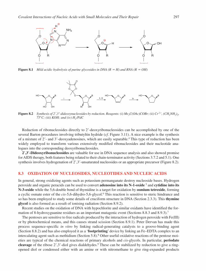
Reduction of ribonucleosides directly to 2�-deoxyribonucleosides can be accomplished by one of theseveral Barton procedures involving tributyltin hydride (cf. Figure 3.11). A nice example is the synthesisof a mixture of 2�- and 3�-deoxyadenosines, which are easily separable.4 This type of reduction has beenwidely employed to transform various extensively modified ribonucleosides and their nucleotide ana-logues into the corresponding deoxyribonucleosides.
2�,3�-Dideoxyribonucleosides are valuable for use in DNA sequence analysis and also showed promisefor AIDS therapy, both features being related to their chain-terminator activity (Sections 3.7.2 and 5.1). Onesynthesis involves hydrogenation of 2�,3�-unsaturated nucleosides or an appropriate precursor (Figure 8.2).
8.3 OXIDATION OF NUCLEOSIDES, NUCLEOTIDES AND NUCLEIC ACIDS
In general, strong oxidizing agents such as potassium permanganate destroy nucleoside bases. Hydrogenperoxide and organic peracids can be used to convert adenosine into its N-1-oxide 5 and cytidine into its N-3-oxide while the 5,6-double bond of thymidine is a target for oxidation by osmium tetroxide, forminga cyclic osmate ester of the cis-5,6-dihydro-5,6-glycol.6 This reaction is sensitive to steric hindrance andso has been employed to study some details of cruciform structure in DNA (Section 2.3.3). This thymineglycol is also formed as a result of ionising radiation (Section 8.9.2).
Recent studies on the oxidation of DNA with hypochlorite and similar oxidants have identified the for-mation of 8-hydroxyguanine residues as an important mutagenic event (Sections 8.8.3 and 8.9.3).7
The pentoses are sensitive to free radicals produced by the interaction of hydrogen peroxide with Fe(III)or by photochemical means, and this causes strand scission (Section 8.9.1). Peter Dervan has made thisprocess sequence-specific in vitro by linking radical-generating catalysts to a groove-binding agent(Section 8.8.2) and has also employed it as a ‘footprinting’ device by linking an Fe–EDTA complex to anintercalating agent such as methidium (Section 5.8).8 Other useful oxidative reactions of the pentose moi-eties are typical of the chemical reactions of primary alcohols and cis-glycols. In particular, periodatecleavage of the ribose 2�,3�-diol gives dialdehydes.9 These can be stabilized by reduction to give a ring-opened diol or condensed either with an amine or with nitromethane to give ring-expanded products
Covalent Interactions of Nucleic Acids with Small Molecules and Their Repair 297
O
O
N
N
N
N
NH2
R
O
H
O
O R
O
H O
O R
O
OH
Figure 8.1 Mild acidic hydrolysis of purine glycosides in DNA (R � H) and RNA (R � OH)
O B
HO OH
HO O B
OH
O
O O
O
Br
O BHO O BHOi ii, iii iv
Figure 8.2 Synthesis of 2�,3�-dideoxynucleosides by reduction. Reagents: (i) Me2C(OAc)COBr; (ii) Cr 2�, (CH2NH2)2,75°C; (iii) KOH; and (iv) H2/PdC
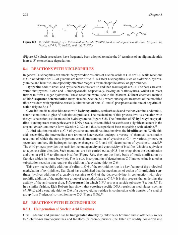
(Figure 8.3). Such procedures have frequently been adopted to make the 3�-terminus of an oligonucleotideinert to 3�-exonuclease degradation.
8.4 REACTIONS WITH NUCLEOPHILES
In general, nucleophiles can attack the pyrimidine residues of nucleic acids at C-6 or C-4, while reactionsat C-6 of adenine or C-2 of guanine are more difficult. �-Effect nucleophiles, such as hydrazine, hydrox-ylamine and bisulfite, are especially effective reagents for nucleophilic attack on pyrimidines.
Hydrazine adds to uracil and cytosine bases first at C-6 and then reacts again at C-4. The bases are con-verted into pyrazol-2-one and 3-aminopyrazole, respectively, leaving an N-ribosylurea, which can reactfurther to form a sugar hydrazone. These reactions were used in the Maxam–Gilbert chemical method of DNA sequence determination (now obsolete, Section 5.1), where subsequent treatment of the modifiedribose residues with piperidine causes �-elimination of both 3�- and 5�-phosphates at the site of depyrimidi-nation (Figure 8.4).10
Cytosine and its nucleosides react with hydroxylamine, semicarbazide and methoxylamine under mild,neutral conditions to give N4-substituted products. The mechanism of this process involves reaction withthe cytosine cation, as illustrated for hydroxylamine (Figure 8.5). The formation of N4-hydroxydeoxycyti-dine is an important mutagenic event in DNA because this modified base exists to a significant extent in theunusual imino-tautomeric form (Section 2.1.2) and thus is capable of base-mispairing with adenine.11
A third addition reaction at C-6 of cytosine and uracil residues involves the bisulfite anion. While thisadds reversibly, the intermediate non-aromatic heterocycles undergo a variety of chemical substitutionreactions of which the most important are: (i) transamination of cytosine at C-4 by various primary or secondary amines, (ii) hydrogen isotope exchange at C-5, and (iii) deamination of cytosine to uracil.12
The third process provides the basis for the mutagenicity and cytotoxicity of bisulfite (which is equivalentto aqueous sulfur dioxide). Such mutations are best carried out at pH 5–6 to bring about the deaminationand then at pH 8–9 to eliminate bisulfite (Figure 8.6a, they are the likely basis of bottle-sterilisation byCamden tablets in home-brewing). The in vitro incorporation of deuterium at C-5 into cytosine is anothersubstitution reaction that requires the addition of a cysteine-thiol to C-6.
This easy nucleophilic addition of sulfur to C-6 of the pyrimidine ring is a key feature of the biologicalmethylation of pyrimidines. Dan Santi has established that the mechanism of action of thymidylate syn-thase involves addition of a catalytic cysteine to C-6 of the deoxyuridylate in conjunction with elec-trophilic addition of the methylene group of tetrahydrofolate to C-5.13 It is this process that underpins theactivity of the anti-cancer drug, 5-fluorouracil in which 5-FU acts as a suicide substrate (Section 3.7.1).In a similar fashion, Rich Roberts has shown that cytosine-specific DNA restriction methylases, such asM. HhaI, add a catalytic thiol to C-6 of a deoxycytidine residue in conjunction with transfer of a methylgroup from S-adenosyl-L–methionine to C-5 (Figure 8.6b).14
8.5 REACTIONS WITH ELECTROPHILES
8.5.1 Halogenation of Nucleic Acid Residues
Uracil, adenine and guanine can be halogenated directly by chlorine or bromine and so offer easy routesto 5-chloro-(or bromo-)uridines and 8-chloro-(or bromo-)purines (the latter are readily converted into
298 Chapter 8
O B
HO OH
RO O B
O
RO
O
O BRO
HO OH
BRO
NR'
O
OHHO
i
ii
iii
Figure 8.3 Periodate cleavage of a 3�-terminal nucleotide (R�RNA) and its subsequent modification. Reagents: (i)NaIO4, pH 4.5; (ii) NaBH4; and (iii) (R�NH2)
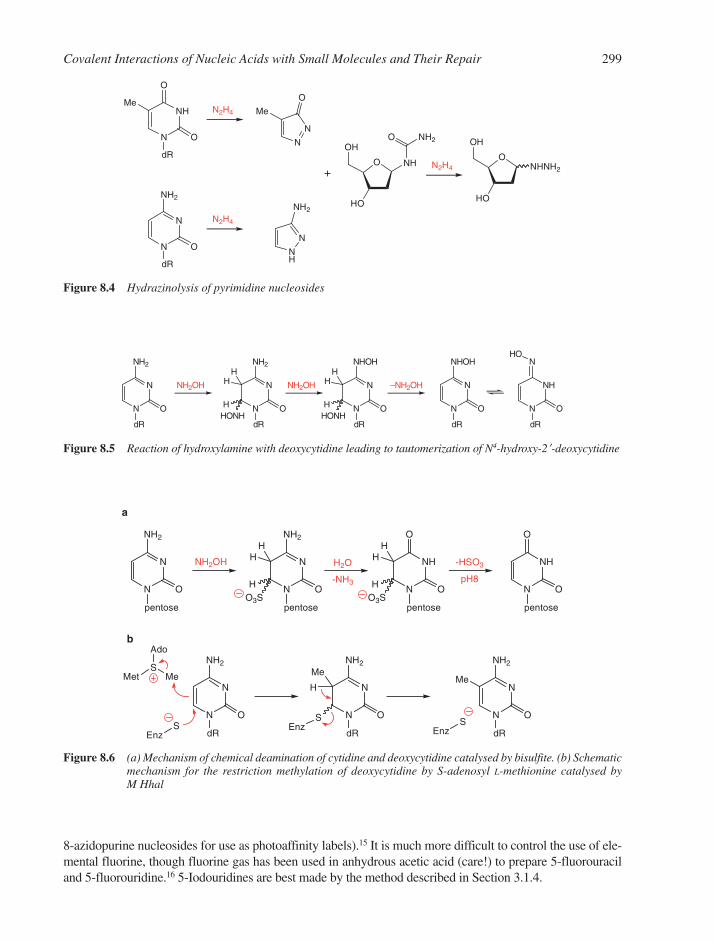
8-azidopurine nucleosides for use as photoaffinity labels).15 It is much more difficult to control the use of ele-mental fluorine, though fluorine gas has been used in anhydrous acetic acid (care!) to prepare 5-fluorouraciland 5-fluorouridine.16 5-Iodouridines are best made by the method described in Section 3.1.4.
Covalent Interactions of Nucleic Acids with Small Molecules and Their Repair 299
N
N
pentose
O
NH2
NH2OH
N
N
pentose
O
NH2
O3SH N
NH
pentose
O
O
O3SH
H2O
-NH3N
NH
pentose
O
-HSO3
pH8O
a
N
N
dR
O
NH2
N
N
dR
O
NH2
b
SEnz
MetS
Me
Ado
SEnz
H
Me
N
N
dR
O
NH2
SEnz
Me
HH
HH
Figure 8.6 (a) Mechanism of chemical deamination of cytidine and deoxycytidine catalysed by bisulfite. (b) Schematicmechanism for the restriction methylation of deoxycytidine by S-adenosyl L-methionine catalysed byM Hhal
N
N
dR
O
NH2
NH2OH
N
N
dR
O
NH2
HONHH N
N
dR
O
NHOH
HONHH
NH2OH
N
N
dR
O
NHOH
–NH2OH
N
NH
dR
O
NHO
HH
HH
Figure 8.5 Reaction of hydroxylamine with deoxycytidine leading to tautomerization of N4-hydroxy-2�-deoxycytidine
N
NHMe
dR
O
O
N
N
dR
O
NH2
N
N
Me
O
NH
N
NH2
O
HO
OH
NH
NH2O
O
HO
OH
NHNH2
N2H4
N2H4
N2H4+
Figure 8.4 Hydrazinolysis of pyrimidine nucleosides

8.5.2 Reactions with Nitrogen Electrophiles
The standard reaction of nitrous acid (as NO�) in the deamination of primary amines converts deoxyadeno-sine into deoxyinosine, deoxycytidine into deoxyuridine and deoxyguanosine into deoxyxanthosine.17 In eachcase, the reaction leads to a base with the opposite base-pairing characteristic. The transitions dA�dT→dI�dC and dC�dG→dU�dA are characteristic of the mutagenic action of nitrous acid (Figure 8.7).
Aromatic nitrogen cations are the second important class of nitrogen electrophiles. These species arederived either from aromatic nitro-compounds by metabolic reduction or from aromatic amines by meta-bolic oxidation. In both cases, an intermediate hydroxylamine species interacts with purine residues inDNA or RNA either at C-8 or N-7 (Section 8.6.1).
8.5.3 Reactions with Carbon Electrophiles
A very large number of reagents form bonds from carbon to nucleic acids. The simplest are species likeformaldehyde and dimethyl sulfate. Among the most complex are carcinogens such as benzo[a]pyrene,which requires transformation by three consecutive metabolic processes before it can become bound topurine bases in DNA or RNA. Not surprisingly, there is a wide range in selectivity for the sites of attackof these reactive species, some of which have been rationalized in terms of Pearson’s HSAB theory(HSAB: hard and soft acids and bases). Frontier orbital analysis can provide a more rigorous picture of theproblem, but requires a deeper insight into theoretical chemistry. Other relevant factors may relate to thedegree of steric access of the electrophile to exposed bases or to intercalation of reagents prior to bondingto nucleotide residues.
8.5.3.1 Formaldehyde. Covalent interactions of formaldehyde with RNA and its constituent nucle-osides take place in a specific reaction of the amino bases. Formaldehyde first adds to the N6-amino groupof adenylate residues to give a 6-(hydroxymethylamino)purine and with guanylate residues to give a 2-(hydroxymethylamino)-6-hydroxypurine. These labile intermediates can react slowly with a secondamino group to give cross-linked products. These have a stable methylene bridge joining the amino groupsof two bases. All three possible species, pAdo-CH2-pAdo, pAdo-CH2-pGuo and pGuo-CH2-pGuo, have beenisolated from RNA that has been treated with formaldehyde and then hydrolysed with alkali (Figure 8.8).The detailed mechanism of formaldehyde mutagenicity is not yet clear.18
300 Chapter 8
N
N
N
N
N
PdR
H CH2OH
dAMP
+
HCHON
N
N
N
N
PdR
H
NN
N
N
NdRP
H
CH2
Figure 8.8 Formaldehyde mutagenesis of adenine residues
N
N
dR
O
NH2
N
NH
dR
O
O
N
N
NH2
HNO2N
N
dR
N
NH
O
N
N
dR
HNO2
Figure 8.7 Pro-mutagenic deamination of dC → dU and of dA → dI by nitrous acid
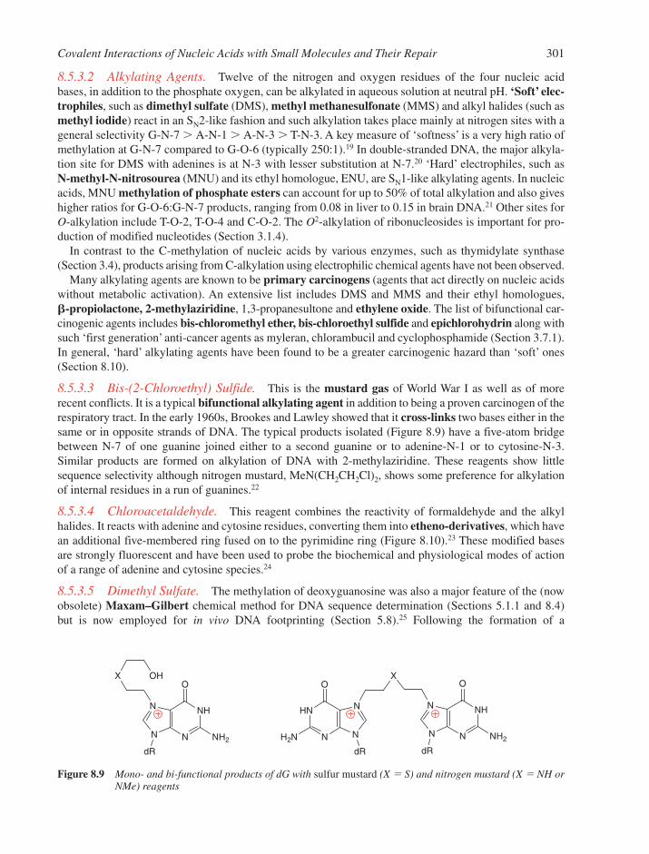
8.5.3.2 Alkylating Agents. Twelve of the nitrogen and oxygen residues of the four nucleic acidbases, in addition to the phosphate oxygen, can be alkylated in aqueous solution at neutral pH. ‘Soft’ elec-trophiles, such as dimethyl sulfate (DMS), methyl methanesulfonate (MMS) and alkyl halides (such asmethyl iodide) react in an SN2-like fashion and such alkylation takes place mainly at nitrogen sites with ageneral selectivity G-N-7 � A-N-1 � A-N-3 � T-N-3. A key measure of ‘softness’ is a very high ratio ofmethylation at G-N-7 compared to G-O-6 (typically 250:1).19 In double-stranded DNA, the major alkyla-tion site for DMS with adenines is at N-3 with lesser substitution at N-7.20 ‘Hard’ electrophiles, such as N-methyl-N-nitrosourea (MNU) and its ethyl homologue, ENU, are SN1-like alkylating agents. In nucleicacids, MNU methylation of phosphate esters can account for up to 50% of total alkylation and also giveshigher ratios for G-O-6:G-N-7 products, ranging from 0.08 in liver to 0.15 in brain DNA.21 Other sites forO-alkylation include T-O-2, T-O-4 and C-O-2. The O2-alkylation of ribonucleosides is important for pro-duction of modified nucleotides (Section 3.1.4).
In contrast to the C-methylation of nucleic acids by various enzymes, such as thymidylate synthase(Section 3.4), products arising from C-alkylation using electrophilic chemical agents have not been observed.
Many alkylating agents are known to be primary carcinogens (agents that act directly on nucleic acidswithout metabolic activation). An extensive list includes DMS and MMS and their ethyl homologues, �-propiolactone, 2-methylaziridine, 1,3-propanesultone and ethylene oxide. The list of bifunctional car-cinogenic agents includes bis-chloromethyl ether, bis-chloroethyl sulfide and epichlorohydrin along withsuch ‘first generation’ anti-cancer agents as myleran, chlorambucil and cyclophosphamide (Section 3.7.1).In general, ‘hard’ alkylating agents have been found to be a greater carcinogenic hazard than ‘soft’ ones(Section 8.10).
8.5.3.3 Bis-(2-Chloroethyl) Sulfide. This is the mustard gas of World War I as well as of morerecent conflicts. It is a typical bifunctional alkylating agent in addition to being a proven carcinogen of therespiratory tract. In the early 1960s, Brookes and Lawley showed that it cross-links two bases either in thesame or in opposite strands of DNA. The typical products isolated (Figure 8.9) have a five-atom bridgebetween N-7 of one guanine joined either to a second guanine or to adenine-N-1 or to cytosine-N-3.Similar products are formed on alkylation of DNA with 2-methylaziridine. These reagents show littlesequence selectivity although nitrogen mustard, MeN(CH2CH2Cl)2, shows some preference for alkylationof internal residues in a run of guanines.22
8.5.3.4 Chloroacetaldehyde. This reagent combines the reactivity of formaldehyde and the alkylhalides. It reacts with adenine and cytosine residues, converting them into etheno-derivatives, which havean additional five-membered ring fused on to the pyrimidine ring (Figure 8.10).23 These modified basesare strongly fluorescent and have been used to probe the biochemical and physiological modes of actionof a range of adenine and cytosine species.24
8.5.3.5 Dimethyl Sulfate. The methylation of deoxyguanosine was also a major feature of the (nowobsolete) Maxam–Gilbert chemical method for DNA sequence determination (Sections 5.1.1 and 8.4) but is now employed for in vivo DNA footprinting (Section 5.8).25 Following the formation of a
Covalent Interactions of Nucleic Acids with Small Molecules and Their Repair 301
N
NH
O
N
N
dR
NH2
X OH
N
HN
O
N
N
dR
H2N
X
N
NH
O
N
N
dR
NH2
Figure 8.9 Mono- and bi-functional products of dG with sulfur mustard (X � S) and nitrogen mustard (X � NH orNMe) reagents
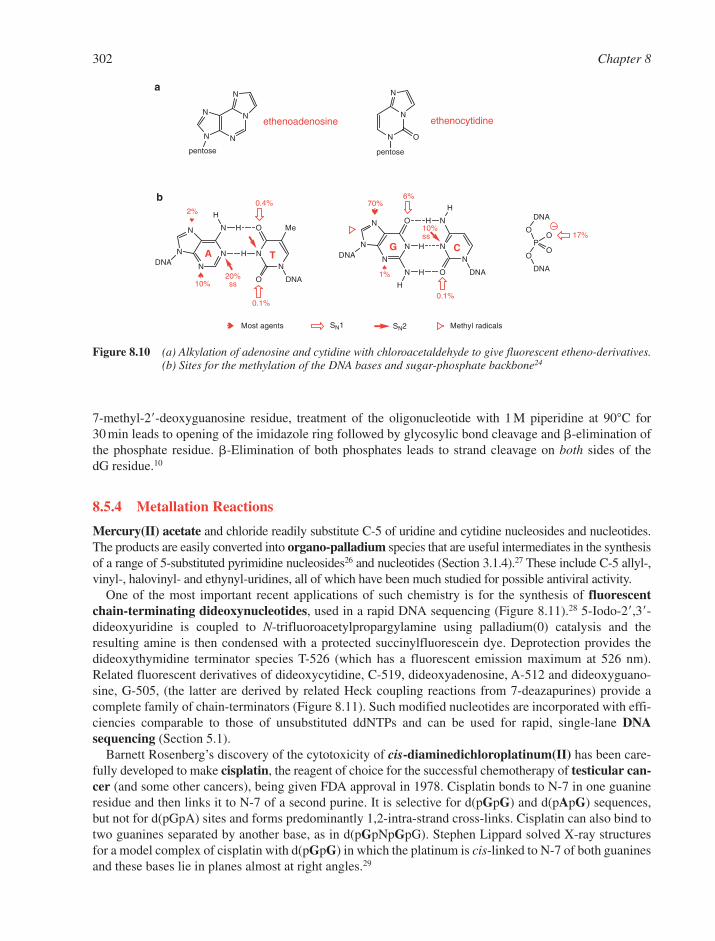
7-methyl-2�-deoxyguanosine residue, treatment of the oligonucleotide with 1 M piperidine at 90°C for30 min leads to opening of the imidazole ring followed by glycosylic bond cleavage and �-elimination of the phosphate residue. �-Elimination of both phosphates leads to strand cleavage on both sides of the dG residue.10
8.5.4 Metallation Reactions
Mercury(II) acetate and chloride readily substitute C-5 of uridine and cytidine nucleosides and nucleotides.The products are easily converted into organo-palladium species that are useful intermediates in the synthesisof a range of 5-substituted pyrimidine nucleosides26 and nucleotides (Section 3.1.4).27 These include C-5 allyl-,vinyl-, halovinyl- and ethynyl-uridines, all of which have been much studied for possible antiviral activity.
One of the most important recent applications of such chemistry is for the synthesis of fluorescentchain-terminating dideoxynucleotides, used in a rapid DNA sequencing (Figure 8.11).28 5-Iodo-2�,3�-dideoxyuridine is coupled to N-trifluoroacetylpropargylamine using palladium(0) catalysis and the resulting amine is then condensed with a protected succinylfluorescein dye. Deprotection provides thedideoxythymidine terminator species T-526 (which has a fluorescent emission maximum at 526 nm).Related fluorescent derivatives of dideoxycytidine, C-519, dideoxyadenosine, A-512 and dideoxyguano-sine, G-505, (the latter are derived by related Heck coupling reactions from 7-deazapurines) provide acomplete family of chain-terminators (Figure 8.11). Such modified nucleotides are incorporated with effi-ciencies comparable to those of unsubstituted ddNTPs and can be used for rapid, single-lane DNAsequencing (Section 5.1).
Barnett Rosenberg’s discovery of the cytotoxicity of cis-diaminedichloroplatinum(II) has been care-fully developed to make cisplatin, the reagent of choice for the successful chemotherapy of testicular can-cer (and some other cancers), being given FDA approval in 1978. Cisplatin bonds to N-7 in one guanineresidue and then links it to N-7 of a second purine. It is selective for d(pGpG) and d(pApG) sequences,but not for d(pGpA) sites and forms predominantly 1,2-intra-strand cross-links. Cisplatin can also bind totwo guanines separated by another base, as in d(pGpNpGpG). Stephen Lippard solved X-ray structuresfor a model complex of cisplatin with d(pGpG) in which the platinum is cis-linked to N-7 of both guaninesand these bases lie in planes almost at right angles.29
302 Chapter 8
N
NN
N
pentose
N
ethenoadenosine
N
N
N
pentose
O
ethenocytidine
N
N
N
NDNA
N H
H
O
H N
N
DNAO
Me
N
N
N
NDNA
O H N
H N
N
DNAON H
H
H
A TG C
O
P
O
O
O
DNA
DNA
17%
0.1%
6%0.4%
0.1%
SN1
♠
♠
♠
20%ss
10%ss
♠
2%
10%
SN2
♠70%
1%
Most agents Methyl radicals
a
b
Figure 8.10 (a) Alkylation of adenosine and cytidine with chloroacetaldehyde to give fluorescent etheno-derivatives.(b) Sites for the methylation of the DNA bases and sugar-phosphate backbone24
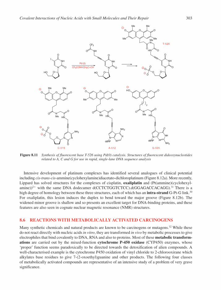
Intensive development of platinum complexes has identified several analogues of clinical potentialincluding cis-trans-cis-ammine(cyclohexylamine)diacetato-dichloroplatinum (Figure 8.12a). More recently,Lippard has solved structures for the complexes of cisplatin, oxaliplatin and (Pt(ammine)(cyclohexyl-amine))2� with the same DNA dodecamer d(CCTCTGGTCTCC).d(GGAGACCACAGG).31 There is ahigh degree of homology between these three structures, each of which has an intra-strand G-Pt-G link.30
For oxaliplatin, this lesion induces the duplex to bend toward the major groove (Figure 8.12b). Thewidened minor groove is shallow and so presents an excellent target for DNA-binding proteins, and thesefeatures are also seen in cognate nuclear magnetic resonance (NMR) structures.
8.6 REACTIONS WITH METABOLICALLY ACTIVATED CARCINOGENS
Many synthetic chemicals and natural products are known to be carcinogens or mutagens.32 While thesedo not react directly with nucleic acids in vitro, they are transformed in vivo by metabolic processes to giveelectrophiles that bind covalently to DNA, RNA and also to proteins. Most of these metabolic transform-ations are carried out by the mixed-function cytochrome P-450 oxidase (CYP450) enzymes, whose‘proper’ function seems paradoxically to be directed towards the detoxification of alien compounds. Awell-characterised example is the cytochrome P450 oxidation of vinyl chloride to 2-chlorooxirane whichalkylates base residues to give 7-(2-oxoethyl)guanine and other products. The following four classes of metabolically activated compounds are representative of an intensive study of a problem of very gravesignificance.
Covalent Interactions of Nucleic Acids with Small Molecules and Their Repair 303
N
NHI
O
O
OHO
N
NH
O
O
OHO
NHO
CF3
Pd (0)
CF3CONHCH2C≡CH N
NH
O
O
OO
HN O
O
Me
O
Me
Me
O
Me
T-526
MeN
O
N N
O
NH2
O
O
HN
O
O
Me
O
Me
O
C-519
NMe
O
N
N
NH2
HN
O
OO O
NMe
O
N
ddRP N
NH
O
HN
O
OHO O
NMe
O
N
ddRP
Me Me
NH2
G-505
2
A-512
P
O
O
O
P
O
O
O
Figure 8.11 Synthesis of fluorescent base T-526 using Pd(0) catalysis. Structures of fluorescent dideoxynucleotidesrelated to A, C and G for use in rapid, single-lane DNA sequence analysis

8.6.1 Aromatic Nitrogen Compounds
Investigations of the binding of N-aryl carcinogens to nucleic acids began in the 1890s with an investiga-tion of the epidemiology of bladder cancer among workers in a Basel dye factory. The list of chemicalsnow banned is extensive, but by no means definitive: some examples of proscribed aromatic amines, nitrocompounds and azo dyes are illustrated (Figure 8.13).33
Aromatic amines of this sort are substrates for oxidation by cytochrome P-450 isozymes, which giveeither phenols, that are inactive and safely excreted, or hydroxylamines. Conjugation of the latter by sul-fotransferase or acetate transferase enzymes converts these proximate carcinogens into ultimate carcino-gens that can bind covalently to nucleic acid bases, especially to guanine.
The competition between such alternative ‘safe’ and ‘hazardous’ metabolic processes is illustrated for2-acetylaminofluorene (Figure 8.14a). The corresponding guanine nucleoside adducts have been isolated
304 Chapter 8
Pt
Figure 8.12 (a) Structures of cisplatin and some more recent analogues under clinical development. (b) A 2.4 Åmolecular structure of oxaliplatin intrastrand cross-link formed with a duplex dodecamer d(CCTCTG-GTCTCC).d(GGAGACCACAGG) showing the G*G* step. Similar intrastrand d(GpG) cross-linkstructures occur for {cis-Pt(NH3)2}
2� and {cis-Pt(NH3)(CyNH2)}2� with the same dodecamer. The
minor groove is widened and shallow, presenting an excellent target for DNA-binding proteins(Adapted from Ref. 31. © (2001), with permission from the American Chemical Society)

Covalent Interactions of Nucleic Acids with Small Molecules and Their Repair 305
NHAc N
Ac
OH N OC6H11O6
Ac
NHAc N
Ac
OSO3 N
Ac
OAc
HO
N Ac
N Ac
N Ac
N
N
N N
dR
NH2
O
H
NH
Ac
N
N
HN
N
dR
HN
O
S
N
Me
NH2
R
S
N
Me
NH
F NH3Cl
O NH3Cl
CYP450
10%
inactive glucuronate
CYP45090%
inactive 3-, 5-, 7- and9-hydroxy metabolites
iii
proximate carcinogen
iii
nitrenium intermediate
two identified productsfrom DNA binding
4
Phortress (NSC 71305)DF 203 (NSC 674495); R = H5F 203 (NSC 703786); R = F
b
a
Figure 8.14 (a) Metabolic activation of 2-acetylaminofluorene (AAF) and its binding to dG via a hypotheticalnitrenium intermediate. (b) Examples of benzothiazolyl anticancer agents. Processes: (i) sulfotransferase;(ii) acetyl transferase; and (iii) binding to DNA in vitro or in vivo
NH2NR"2R
N
NO2
O
R
NHAc
N
N
NHR
Me2N NMe2
NH
2-naphthylamine
R = R' = H; 4-aminobiphenylR = H, R' = O; 4-nitrobiphenylR = NH2, R' = H; benzidine
4-nitroquinoline N-oxide
2-acetylaminofluorene R = H; 4-aminoazobenzeneR = Me; 4-methylaminoazobenzene
auramine
Figure 8.13 Examples of N-aryl carcinogens
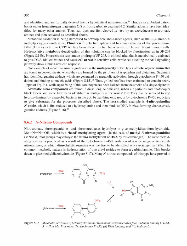
and identified and are formally derived from a hypothetical nitrenium ion.34 This, as an ambident cation,bonds either from nitrogen to guanine C-8 or from carbon to guanine N-2. Similar adducts have been iden-tified for many other amines. Thus, azo dyes are first cleaved in vivo by an azoreductase to aromaticamines and then activated as described above.
Metabolic oxidation is being harnessed to develop new anti-cancer agents, such as the 2-(4-amino-3-methylphenyl)-benzothiazole, Phortress.35 Selective uptake and biotransformation of the parent amineDF-203 by cytochrome CYP1A1 has been shown to be characteristic of human breast tumour cells.Hydroxylative metabolic deactivation of this toluidine can be blocked by fluorination, as in 5F-203(Figure 8.14b). Phortress is a lysylamide prodrug of 5F-203, in clinical trial, that is metabolically activatedto give DNA adducts in vivo and cause cell arrest in sensitive cells, while cells lacking the ArH signallingpathway show a much-reduced response.
One example of more than usual significance is the mutagenicity of two types of heterocyclic amine thatare found in cooked meats, where they are formed by the pyrolysis of tryptophan and glutamine. Sugimurahas identified guanine adducts which are generated by metabolic activation through cytochrome P-450 oxi-dation and binding to nucleic acids (Figure 8.15).36 Thus, grilled beef has been estimated to contain nearly1 ppm of Trp-P-1, while up to 80 ng of this carcinogen has been isolated from the smoke of a single cigarette!
Aromatic nitro compounds are found in diesel engine emission, urban air particles and photocopierblack toners and some have been identified as mutagens in the Ames’ test. They can be reduced to arylhydroxylamines by anaerobic bacteria in the gut, by xanthine oxidase, or by cytochrome P-450 reductaseto give substrates for the processes described above. The best-studied example is 4-nitroquinoline N-oxide, which is first reduced to a hydroxylamine and then binds to DNA in vivo, forming characteristicguanine adducts (Figure 8.16).37
8.6.2 N-Nitroso Compounds
Nitrosoureas, nitrosoguanidines and nitrosourethanes hydrolyse to give methyldiazonium hydroxide,Me�N��N�OH, which is a ‘hard’ methylating agent. (In the case of methyl N-nitrosoguanidine(MNNG), thiol groups may catalyse the in vivo methylation of DNA by this carcinogen). The same methyl-ating species is produced as a result of the cytochrome P-450 oxidation of a wide range of N-methyl-nitrosamines, of which dimethylnitrosamine was the first to be identified as a carcinogen in 1956. Thecommon metabolic pattern is hydroxylation of one alkyl residue to form a carbinolamine. This breaksdown to give methyldiazohydroxide (Figure 8.17). Many N-nitroso compounds of this type have proved to
306 Chapter 8
HN
N
N
N
NdR
NH2
H
NH
Me
NH2
R
tryptophanheat
N
N
N
glutamineheat
R
NH2
i
iO
Ar
Figure 8.15 Metabolic activation of heterocyclic amines from amino acids in cooked food and their binding to DNA;R � H or Me. Processes: (i) cytochrome P-450; (ii) DNA binding; and (iii) hydrolysis
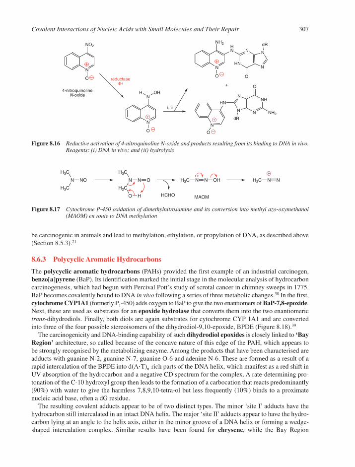
be carcinogenic in animals and lead to methylation, ethylation, or propylation of DNA, as described above(Section 8.5.3).21
8.6.3 Polycyclic Aromatic Hydrocarbons
The polycyclic aromatic hydrocarbons (PAHs) provided the first example of an industrial carcinogen,benzo[a]pyrene (BaP). Its identification marked the initial stage in the molecular analysis of hydrocarboncarcinogenesis, which had begun with Percival Pott’s study of scrotal cancer in chimney sweeps in 1775.BaP becomes covalently bound to DNA in vivo following a series of three metabolic changes.38 In the first,cytochrome CYP1A1 (formerly P1-450) adds oxygen to BaP to give the two enantiomers of BaP-7,8-epoxide.Next, these are used as substrates for an epoxide hydrolase that converts them into the two enantiomerictrans-dihydrodiols. Finally, both diols are again substrates for cytochrome CYP 1A1 and are convertedinto three of the four possible stereoisomers of the dihydrodiol-9,10-epoxide, BPDE (Figure 8.18).39
The carcinogenicity and DNA-binding capability of such dihydrodiol epoxides is closely linked to ‘BayRegion’ architecture, so called because of the concave nature of this edge of the PAH, which appears tobe strongly recognised by the metabolizing enzyme. Among the products that have been characterised areadducts with guanine N-2, guanine N-7, guanine O-6 and adenine N-6. These are formed as a result of arapid intercalation of the BPDE into d(A�T)n-rich parts of the DNA helix, which manifest as a red shift inUV absorption of the hydrocarbon and a negative CD spectrum for the complex. A rate-determining pro-tonation of the C-10 hydroxyl group then leads to the formation of a carbocation that reacts predominantly(90%) with water to give the harmless 7,8,9,10-tetra-ol but less frequently (10%) binds to a proximatenucleic acid base, often a dG residue.
The resulting covalent adducts appear to be of two distinct types. The minor ‘site I’ adducts have thehydrocarbon still intercalated in an intact DNA helix. The major ‘site II’ adducts appear to have the hydro-carbon lying at an angle to the helix axis, either in the minor groove of a DNA helix or forming a wedge-shaped intercalation complex. Similar results have been found for chrysene, while the Bay Region
Covalent Interactions of Nucleic Acids with Small Molecules and Their Repair 307
N
N
HN
NdRH
N
O
N
NH2
ON
NO2
O
N
N
O
H OHN
N
NH
NdR
O
N
HN
O
NH2
4-nitroquinoline N-oxide
reductase4H
i, ii
Figure 8.16 Reductive activation of 4-nitroquinoline N-oxide and products resulting from its binding to DNA in vivo.Reagents: (i) DNA in vivo; and (ii) hydrolysis
H3C
N
H3C
NO
H3C
N
H2C
N
O H
O H3C N N OH
HCHO
H3C N N
MAOM
Figure 8.17 Cytochrome P-450 oxidation of dimethylnitrosamine and its conversion into methyl azo-oxymethanol(MAOM) en route to DNA methylation
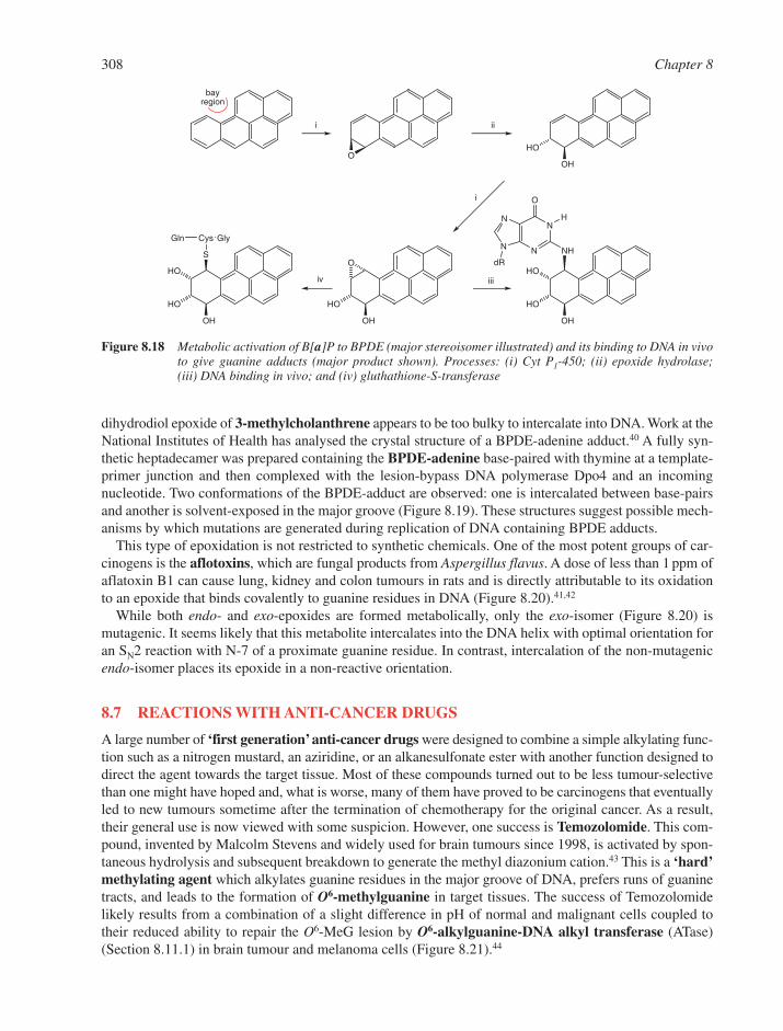
dihydrodiol epoxide of 3-methylcholanthrene appears to be too bulky to intercalate into DNA. Work at theNational Institutes of Health has analysed the crystal structure of a BPDE-adenine adduct.40 A fully syn-thetic heptadecamer was prepared containing the BPDE-adenine base-paired with thymine at a template-primer junction and then complexed with the lesion-bypass DNA polymerase Dpo4 and an incomingnucleotide. Two conformations of the BPDE-adduct are observed: one is intercalated between base-pairsand another is solvent-exposed in the major groove (Figure 8.19). These structures suggest possible mech-anisms by which mutations are generated during replication of DNA containing BPDE adducts.
This type of epoxidation is not restricted to synthetic chemicals. One of the most potent groups of car-cinogens is the aflotoxins, which are fungal products from Aspergillus flavus. A dose of less than 1 ppm ofaflatoxin B1 can cause lung, kidney and colon tumours in rats and is directly attributable to its oxidationto an epoxide that binds covalently to guanine residues in DNA (Figure 8.20).41,42
While both endo- and exo-epoxides are formed metabolically, only the exo-isomer (Figure 8.20) ismutagenic. It seems likely that this metabolite intercalates into the DNA helix with optimal orientation foran SN2 reaction with N-7 of a proximate guanine residue. In contrast, intercalation of the non-mutagenicendo-isomer places its epoxide in a non-reactive orientation.
8.7 REACTIONS WITH ANTI-CANCER DRUGS
A large number of ‘first generation’ anti-cancer drugs were designed to combine a simple alkylating func-tion such as a nitrogen mustard, an aziridine, or an alkanesulfonate ester with another function designed todirect the agent towards the target tissue. Most of these compounds turned out to be less tumour-selectivethan one might have hoped and, what is worse, many of them have proved to be carcinogens that eventuallyled to new tumours sometime after the termination of chemotherapy for the original cancer. As a result,their general use is now viewed with some suspicion. However, one success is Temozolomide. This com-pound, invented by Malcolm Stevens and widely used for brain tumours since 1998, is activated by spon-taneous hydrolysis and subsequent breakdown to generate the methyl diazonium cation.43 This is a ‘hard’methylating agent which alkylates guanine residues in the major groove of DNA, prefers runs of guaninetracts, and leads to the formation of O6-methylguanine in target tissues. The success of Temozolomidelikely results from a combination of a slight difference in pH of normal and malignant cells coupled totheir reduced ability to repair the O6-MeG lesion by O6-alkylguanine-DNA alkyl transferase (ATase)(Section 8.11.1) in brain tumour and melanoma cells (Figure 8.21).44
308 Chapter 8
OOH
HO
OH
HO
O
OH
HO
S
HO
Cys GlyGln
OH
HO
HO
N
N
N
NdR
NH
H
O
bayregion
i
i
ii
iv iii
Figure 8.18 Metabolic activation of B[a]P to BPDE (major stereoisomer illustrated) and its binding to DNA in vivoto give guanine adducts (major product shown). Processes: (i) Cyt P1-450; (ii) epoxide hydrolase;(iii) DNA binding in vivo; and (iv) gluthathione-S-transferase
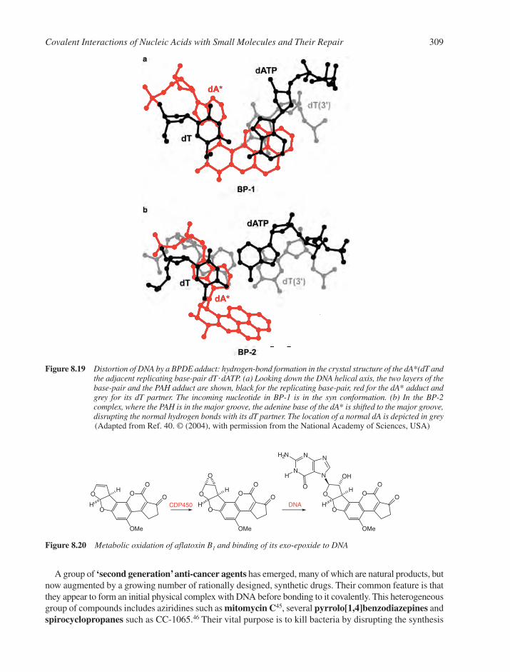
A group of ‘second generation’anti-cancer agents has emerged, many of which are natural products, butnow augmented by a growing number of rationally designed, synthetic drugs. Their common feature is thatthey appear to form an initial physical complex with DNA before bonding to it covalently. This heterogeneousgroup of compounds includes aziridines such as mitomycin C45, several pyrrolo[1,4]benzodiazepines andspirocyclopropanes such as CC-1065.46 Their vital purpose is to kill bacteria by disrupting the synthesis
Covalent Interactions of Nucleic Acids with Small Molecules and Their Repair 309
Figure 8.19 Distortion of DNA by a BPDE adduct: hydrogen-bond formation in the crystal structure of the dA*(dT andthe adjacent replicating base-pair dT� dATP. (a) Looking down the DNA helical axis, the two layers of thebase-pair and the PAH adduct are shown, black for the replicating base-pair, red for the dA* adduct andgrey for its dT partner. The incoming nucleotide in BP-1 is in the syn conformation. (b) In the BP-2 complex, where the PAH is in the major groove, the adenine base of the dA* is shifted to the major groove,disrupting the normal hydrogen bonds with its dT partner. The location of a normal dA is depicted in grey (Adapted from Ref. 40. © (2004), with permission from the National Academy of Sciences, USA)
N
N
N
NH2N
H
OO
O
O
O
O
OMe
O
O
O
O
O
OMe
O
O
O
O
O
OMe
O
H
H
H
H H
H
OH
DNACDP450
Figure 8.20 Metabolic oxidation of aflatoxin B1 and binding of its exo-epoxide to DNA

of DNA and RNA, but many of them have also shown useful anti-tumour activity, which must arise fromselective toxicity. This can be attributed to DNA-binding specificity or to preferential metabolic activationby tumour cells.
8.7.1 Aziridine Antibiotics
An assortment of naturally occurring antibiotics, each having an aziridine ring, has been isolated fromStreptomyces caespitonis. The most interesting of them in clinical terms is mitomycin C. This compoundrequires enzymatic reduction of its quinone function to initiate the processes that cause it to alkylate DNA.It seems likely that the second step is elimination of methanol that potentiates either monofunctional orbifunctional alkylation (Figure 8.22). The antibiotic has been shown to interact with DNA at G-O-6 �A-N-6 � G-N-2 and forms one cross-link for about every ten monocovalent links. The primary process isbonding of the 2-amino group of a guanine residue to C-1 of the reductively activated mitomycin. This reac-tion shows selectivity for 5�-CG sequences. Cross-linking is completed by alkylation of the 2-amino groupof the second guanine to C-10 of the mitomycin (Figure 8.22). This has been accurately analysed by DinshawPatel in NMR studies on the adduct of mitomycin C to the hexamer (TACGTA).d(TACGTA) in which thetwo guanines are crosslinked with the mitomycin molecule positioned in the minor groove of the duplex.47
Many drugs that act on DNA exhibit a requirement for reductive activation, including adriamycin,daunomycin, actinomycin, streptonigrin, saframycin, bleomycin (Section 9.7) and tallysomycin in add-ition to mitomycin C. While there is no common factor uniting the chemistry of DNA modification bythese agents, the fact that tumour tissues seem to have a higher reducing potential than normal tissue hasled to the concept of bioreductive drug activation.
Carzinophilin A is also a DNA-alkylating aziridine antibiotic, though it does not appear to need reductiveactivation.48 It has been identified as the antibiotic azinomycin B, isolated from Streptomyces griseofuscus.Azinomycin B operates in the major groove of DNA, causing cross-linking between a guanine residue anda purine residue that is two bases removed in the duplex, as in the sequence d(G.Py.Py)�d(C.Pu.Pu).
310 Chapter 8
N NH
H2N
Me
O
O
OCONH2
OMe
Mitomycin C
N NH
H2N
Me
OH
OH
OCONH2
OMe
N NH
H2N
Me
OH
OH
OCONH2
– MeOH
N
NH3
H2N
Me
O
OH
OCONH2
N
NH3
H2N
Me
O
O
OCONH2
guanine DNA
oxidn.N
NH3
H2N
Me
O
O
guanine
guanine DNA
Figure 8.22 Activation of mitomycin C by metabolic reduction and bifunctional alkylation of DNA at the 2-aminogroup of adjacent inter-strand deoxyguanines
N N
NN
N
H2NOC
O
CH3
MTIC
NH N
N
HN
N
H2NOC
CH3
AICAR
NH
NH2
N
H2NOC
N
N
H3C N
N
N
NH2N
OH3C
dR
Temozolomide
i, ii iii iv
Figure 8.21 Temozolomide activation and DNA alkylation

Robert Coleman has shown that initial alkylation is at N-7 of a dG residue involving the aziridine ring C-10(Figure 8.23a). It is followed by a slower alkylation of the sub-adjacent purine by the epoxide C-21 as thesecond alkylating function (Figure 8.23b).49 The selectivity for the target sequence appears to be determinedby the relative nucleophilicity of purines in the major groove while the naphthalene moiety provideshydrophobic binding to DNA without intercalation.
8.7.2 Pyrrolo[1,4]Benzodiazepines, P[1,4]Bs
Anthramycin and tomaymycin, along with sibiromycin and neothramycins A and B, are members of thepotent P[1,4]B anti-tumour antibiotic group produced by various actinomycetes (Figure 8.24). The firstthree of these compounds bind physically in the minor groove of DNA where they then form covalentbonds to G-N-2, showing a DNA sequence specificity for 5�-PuGPu sequences.
These P[1,4]Bs appear to interact with DNA in a biphasic process. Initially there is a rapid, non-covalentassociation that results from a close interaction of the antibiotic with the ‘floor’of the minor groove of DNA(Section 10.3.5). Subsequent loss of water or methanol and covalent addition of G-N-2 to C-11 then formsan aminal linkage that is well stabilized by favourable steric and electrostatic interactions.50
Lawrence Hurley has established the existence of two distinct tomaymycin-d(ATGCAT)2 species in solutionfrom NMR studies. These have the antibiotic orientated in opposite directions in the minor groove accordingto its (R)- or (S)-configuration at C-11. The resulting lesions appear neither to impede Watson–Crick base-pairing nor to distort the B-DNA helix structure so that the two lesions probably pose difficult recognition
Covalent Interactions of Nucleic Acids with Small Molecules and Their Repair 311
N
HNMe
OH
OCONH2
OH
anthramycin
N
HNHO
O
OMe
MeO
tomaymycin
– MeOH
N
NHO
O
MeO
N
HN
N
NH2N
O
pentose
H
N
HNHO
O
MeO
N
NH
N
NN
O
pentose
H
Figure 8.24 Binding of P[1,4]B antibiotics to the N2-amino group of guanine
Me
MeOO
HN
NH
O O
O
OH
Me
OMe
ON
HO
AcO
second alkylation
first alkylation
azinomycin B
(carzinophilin)5' –G–C–C– 3'
3' –C–G–G– 5'
epoxide C-21
aziridine C-1010
21a b
Figure 8.23 (a) Structure of azinomycin B (identical with carzinophilin) with sites for nucleophilic attack by N-7 ofguanine (purine) residues in opposite strands of DNA. (b) Preferred duplex sequence for its cross-linkingto sub-adjacent dG residues49
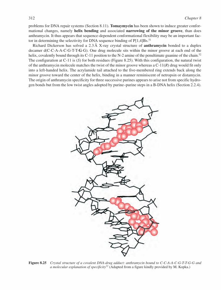
problems for DNA repair systems (Section 8.11). Tomaymycin has been shown to induce greater confor-mational changes, namely helix bending and associated narrowing of the minor groove, than doesanthramycin. It thus appears that sequence-dependent conformational flexibility may be an important fac-tor in determining the selectivity for DNA sequence binding of P[1,4]Bs.51
Richard Dickerson has solved a 2.3 Å X-ray crystal structure of anthramycin bonded to a duplexdecamer d(C-C-A-A-C-G-T-T-G-G). One drug molecule sits within the minor groove at each end of thehelix, covalently bound through its C-11 position to the N-2 amine of the penultimate guanine of the chain.52
The configuration at C-11 is (S) for both residues (Figure 8.25). With this configuration, the natural twistof the anthramycin molecule matches the twist of the minor groove whereas a C-11(R) drug would fit onlyinto a left-handed helix. The acrylamide tail attached to the five-membered ring extends back along theminor groove toward the center of the helix, binding in a manner reminiscent of netropsin or distamycin.The origin of anthramycin specificity for three successive purines appears to arise not from specific hydro-gen bonds but from the low twist angles adopted by purine–purine steps in a B-DNA helix (Section 2.2.4).
312 Chapter 8
Figure 8.25 Crystal structure of a covalent DNA-drug adduct: anthramycin bound to C-C-A-A-C-G-T-T-G-G anda molecular explanation of specificity52 (Adapted from a figure kindly provided by M. Kopka.)
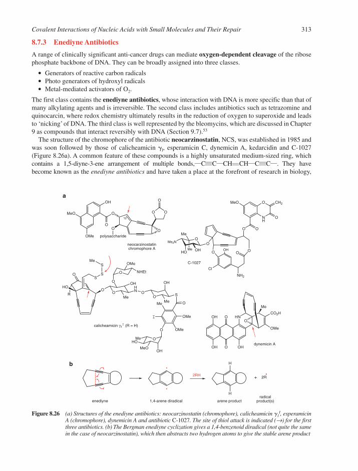
8.7.3 Enediyne Antibiotics
A range of clinically significant anti-cancer drugs can mediate oxygen-dependent cleavage of the ribosephosphate backbone of DNA. They can be broadly assigned into three classes.
� Generators of reactive carbon radicals� Photo generators of hydroxyl radicals� Metal-mediated activators of O2.
The first class contains the enediyne antibiotics, whose interaction with DNA is more specific than that ofmany alkylating agents and is irreversible. The second class includes antibiotics such as tetrazomine andquinocarcin, where redox chemistry ultimately results in the reduction of oxygen to superoxide and leadsto ‘nicking’ of DNA. The third class is well represented by the bleomycins, which are discussed in Chapter9 as compounds that interact reversibly with DNA (Section 9.7).53
The structure of the chromophore of the antibiotic neocarzinostatin, NCS, was established in 1985 andwas soon followed by those of calicheamicin l, esperamicin C, dynemicin A, kedarcidin and C-1027(Figure 8.26a). A common feature of these compounds is a highly unsaturated medium-sized ring, whichcontains a 1,5-diyne-3-ene arrangement of multiple bonds,�C���C�CH��CH�C���C�. They havebecome known as the enediyne antibiotics and have taken a place at the forefront of research in biology,
Covalent Interactions of Nucleic Acids with Small Molecules and Their Repair 313
OH
MeO
OMe
O
OO O
OO
O
polysaccharide
O
O
HN
OHOH
OHO
CO2H
OMe
Me
O
Cl
NH2
OOOH
OO
Me
Me N
OHHOMe
O O
NH
O CH2
O
MeO
H
H
R
HO
OS
S
SMe
OO
O
O
HN O
O S
OMe
OMe
I
Me
O
O
OHMeO
MeHO
OH
Me
OH
Me
OMe
NHEt
O
12
neocarzinostatinchromophore A
dynemicin A
a
b
enediyne 1,4-arene diradical arene productradical
product(s)
calicheamicin γ1I (R = H)
C-1027
2R2RH
Figure 8.26 (a) Structures of the enediyne antibiotics: neocarzinostatin (chromophore), calicheamicin g1I, esperamicin
A (chromophore), dynemicin A and antibiotic C-1027. The site of thiol attack is indicated (→) for the firstthree antibiotics. (b) The Bergman enediyne cyclization gives a 1,4-benzenoid diradical (not quite the samein the case of neocarzinostatin), which then abstracts two hydrogen atoms to give the stable arene product

chemistry and medicine since this group of compounds contains some of the most potent anti-tumourantibiotics known. They are about a thousand times more toxic than the clinically used adriamycin andanthracycline antibiotics and generally induce apoptosis through a caspase-mediated mitochondrialamplification loop.54 In particular, the high cytotoxicity of calicheamicin has been harnessed by means ofantibody directed drug delivery55 to target human myeloid leukaemia tumour cells in a Food and DrugAdministration (FDA) approved product, Mylotarg™.
The mode of action of the enediyne antibiotics involves single or double strand scission of DNA anddepends on the formation of carbon radicals followed by hydrogen atom abstraction from specificnucleotides. The key enediyne reaction is an electrocyclisation, as reported by Bergman in 1972, whichgenerates a 1,4-benzenoid diradical (Figure 8.26b). In the case of the very unstable antibiotic C-1027, thisprocess takes place merely on warming the antibiotic to 50°C in solution in ethanol. For the other com-pounds, a distortion of the enediyne ring inhibits the Bergman cyclisation.56 The rearrangement is ‘triggered’by a chemical process that relaxes the ring and so facilitates a change in conformation of the enediyne toallow the electrocyclisation to take place spontaneously. In the cases of NCS, calicheamicin and esperam-icin, this trigger is the attack of a thiol, possibly glutathione, at the site indicated (arrows in Figure 8.26a).In the case of dynemicin, opening of the epoxide follows initial biological reduction to a quinol by nucleo-philic attack at the position indicated.
While the enediyne provides the warhead for strand breaking, sequence selectivity and orientation ofthe enediyne to the DNA target is delivered by minor groove binding and/or intercalation of peripheralcomponents of these antibiotics.
The neocarzinostatin chromophore acts primarily by single-strand cleavage of DNA, and this requiresoxygen. NCS first intercalates its naphthoate residue into the DNA duplex, which positions the remainderof the molecule in the minor groove (Section 8.7.1). Following activation of the molecule by thiol additionat C-12, Bergman cyclisation generates a benzenoid diradical, which abstracts a 5�-hydrogen atom from aresidue in the DNA recognition sequence.57 Such action takes place preferentially at dA and dT sites withat least 80% of the DNA cleavage resulting in the formation of 5�-aldehydes of A and T residues. Less than20% of strand breaks result from pathways initiated by a second hydrogen abstraction in the alternatestrand from a deoxyribose at C-4� or C-1�.
The NMR structure in solution of a complex formed between calicheamicin and a DNA hairpin con-taining the preferred recognition sequence d(T4-C5-C6-T7)�(A17-G18-G19-A20) has been determined byPatel (Figure 8.27).58 Sequence-specific binding of calicheamicin �1
I to the (T-C-C-T) containing DNAhairpin duplex is favoured by the complementarity of fit through hydrophobic and hydrogen-bondinginteractions between the antibiotic and the floor and walls of the minor groove of a minimally perturbedDNA helix (Section 9.7). Calicheamicin 1
I binds with its arene-tetrasaccharide segment in an extendedconformation spanning the (T-C-C-T)�(A-G-G-A) segment of the duplex minor groove. Its thiol-sugar andthiobenzoate rings are inserted in an edgewise manner deep into the minor groove with their faces sand-wiched between its walls, where hydrophobic and hydrogen-bonding interactions account for the (TCCT)sequence recognition in the complex (Figure 8.27a).
This positioning of the arene-tetrasaccharide moiety orientates the enediyne ring deep in the minorgroove, spanning both strands, such that its pro-radical carbon centers, C-3 and C-6, are proximal to theanticipated H-5� and H-4� sites for hydrogen atom abstraction. When a thiolate nucleophile adds inMichael fashion to the proximate �,�-unsaturated ketone, the resulting change in geometry of theenediyne triggers the Bergman cyclisation to generate a benzenoid 3,6-diradical. This is suitably orien-tated to abstract one 5�-hydrogen atom from the first deoxycytidine residue in the recognition sequenced(TCCT) and a second 4�-hydrogen atom from the opposite strand and this leads oxidatively to a doublestrand cleavage process (Figure 8.28). It is worth noting that the affinity of calicheamicin for DNA hasbeen increased 1000-fold through synthesis of head-to-head and head-to-tail dimers with significant bene-fit to sequence-selectivity for TCCT and ACCT sequences.59
Esperamicin A1 works in a similar fashion but with low sequence selectivity and favours cleavage at T � C � A � G. Its binding to DNA involves a combination of upstream intercalation of an
314 Chapter 8
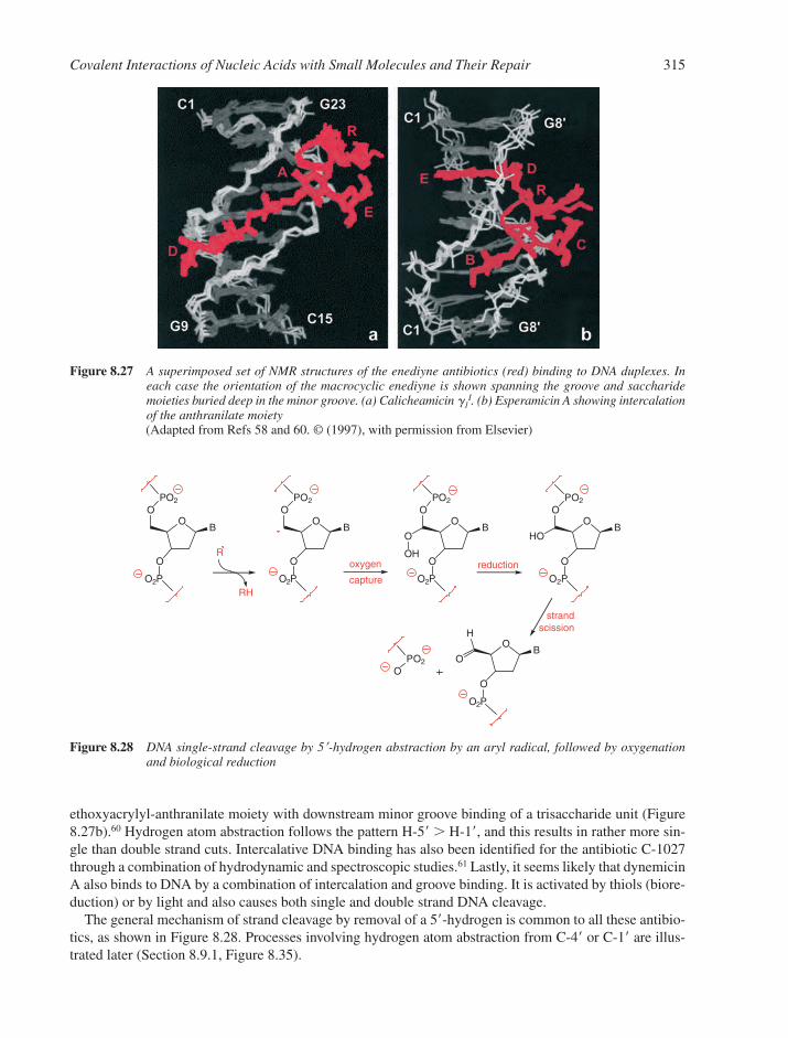
ethoxyacrylyl-anthranilate moiety with downstream minor groove binding of a trisaccharide unit (Figure8.27b).60 Hydrogen atom abstraction follows the pattern H-5� � H-1�, and this results in rather more sin-gle than double strand cuts. Intercalative DNA binding has also been identified for the antibiotic C-1027through a combination of hydrodynamic and spectroscopic studies.61 Lastly, it seems likely that dynemicinA also binds to DNA by a combination of intercalation and groove binding. It is activated by thiols (biore-duction) or by light and also causes both single and double strand DNA cleavage.
The general mechanism of strand cleavage by removal of a 5�-hydrogen is common to all these antibio-tics, as shown in Figure 8.28. Processes involving hydrogen atom abstraction from C-4� or C-1� are illus-trated later (Section 8.9.1, Figure 8.35).
Covalent Interactions of Nucleic Acids with Small Molecules and Their Repair 315
OO
PO2
O
B
O2P
oxygen
capture
reduction
OO
PO2
O
B
O2P
OO
PO2
O
B
O2P
OO
PO2
O
B
O2P
O
OH
HO
O
O
B
O2P
OO
PO2
H
strandscission
R
RH
Figure 8.28 DNA single-strand cleavage by 5�-hydrogen abstraction by an aryl radical, followed by oxygenationand biological reduction
Figure 8.27 A superimposed set of NMR structures of the enediyne antibiotics (red) binding to DNA duplexes. Ineach case the orientation of the macrocyclic enediyne is shown spanning the groove and saccharidemoieties buried deep in the minor groove. (a) Calicheamicin 1
I. (b) Esperamicin A showing intercalationof the anthranilate moiety(Adapted from Refs 58 and 60. © (1997), with permission from Elsevier)
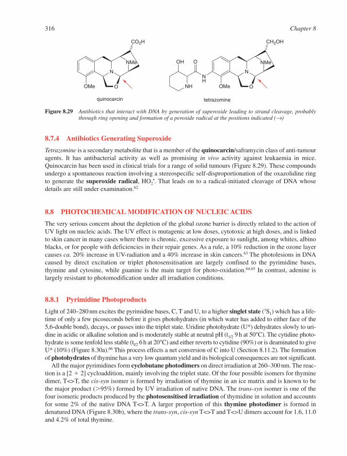
8.7.4 Antibiotics Generating Superoxide
Tetrazomine is a secondary metabolite that is a member of the quinocarcin/saframycin class of anti-tumouragents. It has antibacterial activity as well as promising in vivo activity against leukaemia in mice.Quinocarcin has been used in clinical trials for a range of solid tumours (Figure 8.29). These compoundsundergo a spontaneous reaction involving a stereospecific self-disproportionation of the oxazolidine ringto generate the superoxide radical, HO2
•. That leads on to a radical-initiated cleavage of DNA whosedetails are still under examination.62
8.8 PHOTOCHEMICAL MODIFICATION OF NUCLEIC ACIDS
The very serious concern about the depletion of the global ozone barrier is directly related to the action ofUV light on nucleic acids. The UV effect is mutagenic at low doses, cytotoxic at high doses, and is linkedto skin cancer in many cases where there is chronic, excessive exposure to sunlight, among whites, albinoblacks, or for people with deficiencies in their repair genes. As a rule, a 10% reduction in the ozone layercauses ca. 20% increase in UV-radiation and a 40% increase in skin cancers.63 The photolesions in DNAcaused by direct excitation or triplet photosensitisation are largely confined to the pyrimidine bases,thymine and cytosine, while guanine is the main target for photo-oxidation.64,65 In contrast, adenine islargely resistant to photomodification under all irradiation conditions.
8.8.1 Pyrimidine Photoproducts
Light of 240–280 nm excites the pyrimidine bases, C, T and U, to a higher singlet state (1S1) which has a life-time of only a few picoseconds before it gives photohydrates (in which water has added to either face of the5,6-double bond), decays, or passes into the triplet state. Uridine photohydrate (U*) dehydrates slowly to uri-dine in acidic or alkaline solution and is moderately stable at neutral pH (t1/2 9 h at 50°C). The cytidine photo-hydrate is some tenfold less stable (tl/2 6 h at 20°C) and either reverts to cytidine (90%) or is deaminated to giveU* (10%) (Figure 8.30a).66 This process effects a net conversion of C into U (Section 8.11.2). The formationof photohydrates of thymine has a very low quantum yield and its biological consequences are not significant.
All the major pyrimidines form cyclobutane photodimers on direct irradiation at 260–300 nm. The reac-tion is a [2 � 2] cycloaddition, mainly involving the triplet state. Of the four possible isomers for thyminedimer, T<>T, the cis-syn isomer is formed by irradiation of thymine in an ice matrix and is known to bethe major product (�95%) formed by UV irradiation of native DNA. The trans-syn isomer is one of thefour isomeric products produced by the photosensitised irradiation of thymidine in solution and accountsfor some 2% of the native DNA T<>T. A larger proportion of this thymine photodimer is formed in denatured DNA (Figure 8.30b), where the trans-syn, cis-syn T<>T and T<>U dimers account for 1.6, 11.0and 4.2% of total thymine.
316 Chapter 8
N
NMe
O
CO2H
OMe
quinocarcin
N
NMe
O
CH2OH
OMe
NH
NH
OOH
tetrazomine
Figure 8.29 Antibiotics that interact with DNA by generation of superoxide leading to strand cleavage, probablythrough ring opening and formation of a peroxide radical at the positions indicated (→)
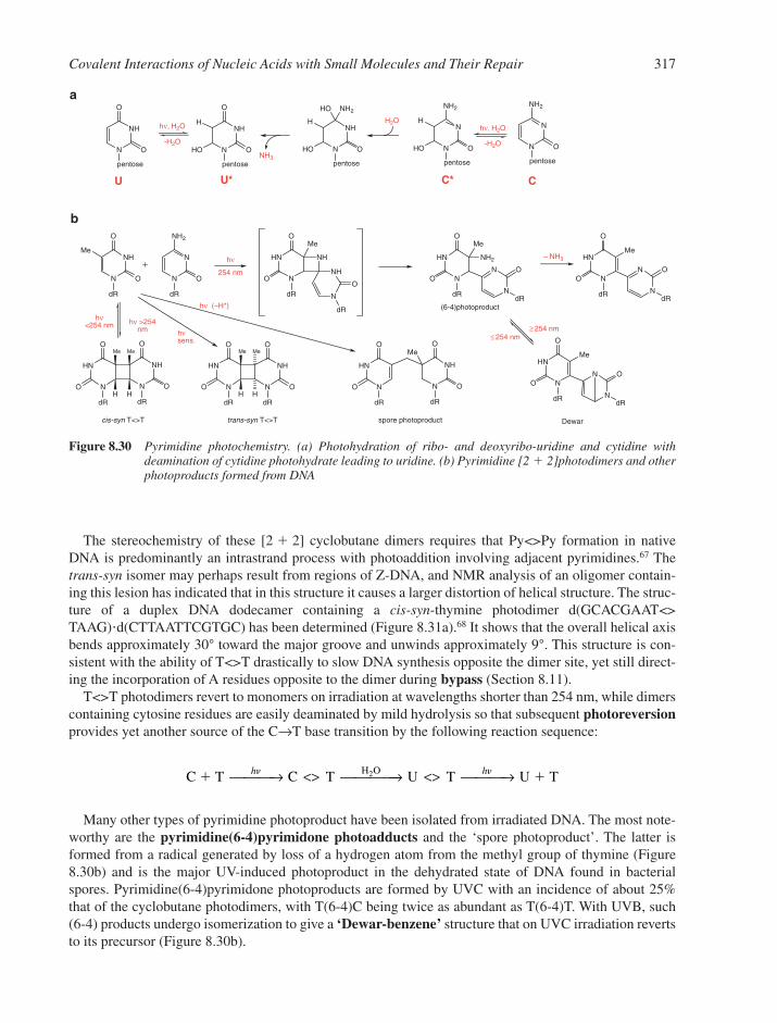
The stereochemistry of these [2 � 2] cyclobutane dimers requires that Py<>Py formation in nativeDNA is predominantly an intrastrand process with photoaddition involving adjacent pyrimidines.67 Thetrans-syn isomer may perhaps result from regions of Z-DNA, and NMR analysis of an oligomer contain-ing this lesion has indicated that in this structure it causes a larger distortion of helical structure. The struc-ture of a duplex DNA dodecamer containing a cis-syn-thymine photodimer d(GCACGAAT<>TAAG)�d(CTTAATTCGTGC) has been determined (Figure 8.31a).68 It shows that the overall helical axisbends approximately 30° toward the major groove and unwinds approximately 9°. This structure is con-sistent with the ability of T<>T drastically to slow DNA synthesis opposite the dimer site, yet still direct-ing the incorporation of A residues opposite to the dimer during bypass (Section 8.11).
T<>T photodimers revert to monomers on irradiation at wavelengths shorter than 254 nm, while dimerscontaining cytosine residues are easily deaminated by mild hydrolysis so that subsequent photoreversionprovides yet another source of the C→T base transition by the following reaction sequence:
Many other types of pyrimidine photoproduct have been isolated from irradiated DNA. The most note-worthy are the pyrimidine(6-4)pyrimidone photoadducts and the ‘spore photoproduct’. The latter isformed from a radical generated by loss of a hydrogen atom from the methyl group of thymine (Figure8.30b) and is the major UV-induced photoproduct in the dehydrated state of DNA found in bacterialspores. Pyrimidine(6-4)pyrimidone photoproducts are formed by UVC with an incidence of about 25%that of the cyclobutane photodimers, with T(6-4)C being twice as abundant as T(6-4)T. With UVB, such(6-4) products undergo isomerization to give a ‘Dewar-benzene’ structure that on UVC irradiation revertsto its precursor (Figure 8.30b).
C T C T U T U TH O� �
h h → <> → <> →2
Covalent Interactions of Nucleic Acids with Small Molecules and Their Repair 317
N
NH
O
O
dR
Me
N
N
NH2
O
dR
N
HN
O
O
dR
Me
N
NH
NH
O
dR
N
HN
O
O
dR
Me
N
N
NH2
O
dR
N
HN
O
O
dR
Me
N
N O
dR
N
HN
O
O
dR
Me
N
N O
dR
N
NH
O
O
dR
Me
N
HN
O
O
dR
N
NH
O
O
dR
N
HN
O
O
dR
Me Me
H HN
NH
O
O
dR
N
HN
O
O
dR
Me Me
H H
hν
254 nm
– NH3
Dewar
≥ 254 nm≤ 254 nm
spore photoproduct
hν >254 nm
hν <254 nm
hνsens.
hν (–H*)
cis-syn T<>T trans-syn T<>T
(6-4)photoproduct
a
N
NH
O
O
pentose
N
NH
O
O
pentose
HO
H
N
N
NH2
O
pentose
N
N
NH2
O
pentose
HO
H
NH3
U C
hν, H2O
-H2O
hν, H2O
-H2O
U* C*
N
NH
NH2
O
pentose
HO
H
HO
H2O
b
Figure 8.30 Pyrimidine photochemistry. (a) Photohydration of ribo- and deoxyribo-uridine and cytidine withdeamination of cytidine photohydrate leading to uridine. (b) Pyrimidine [2 � 2]photodimers and otherphotoproducts formed from DNA
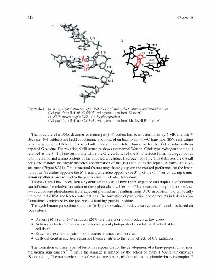
The structure of a DNA decamer containing a (6-4) adduct has been determined by NMR analysis.69
Because (6-4) adducts are highly mutagenic and most often lead to a 3�-T→C transition (85% replicatingerror frequency), a DNA duplex was built having a mismatched base-pair for the 3�-T residue with anopposed G residue. The resulting NMR structure shows that normal Watson–Crick-type hydrogen bonding isretained at the 5�-T of the lesion site while the O-2-carbonyl of the 3�-T residue forms hydrogen bondswith the imino and amino protons of the opposed G residue. Hydrogen bonding thus stabilizes the overallhelix and restores the highly distorted conformation of the (6-4) adduct to the typical B-form-like DNAstructure (Figure 8.31b). This structural feature may thereby explain the marked preference for the inser-tion of an A residue opposite the 5�-T and a G residue opposite the 3�-T of the (6-4) lesion during trans-lesion synthesis, and so lead to the predominant 3�-T → C transition.
Thomas Carell has undertaken a systematic analysis of how DNA sequence and duplex conformationcan influence the relative formation of these photochemical lesions.70 It appears that the production of cis-syn cyclobutane photodimers from adjacent pyrimidines resulting from UVC irradiation is dramaticallyinhibited in A-DNA and RNA conformations. The formation of pyrimidine photoproducts in B-DNA con-formations is inhibited by the presence of flanking guanine residues.
The cyclobutane photodimers and the (6-4) photoproducts products can cause cell death, as based onfour criteria.
� Dimers (80%) and (6-4) products (20%) are the major photoproducts at low doses.� Action spectra for the formation of both types of photoproduct correlate well with that for
cell death.� Enzymatic excision repair of both lesions enhances cell survival.� Cells deficient in excision repair are hypersensitive to the lethal effects of UV radiation.
The formation of these types of lesion is responsible for the development of a large proportion of non-melanoma skin cancers,71,72 while the damage is limited by the action of many DNA repair enzymes(Section 8.11). The mutagenic nature of cyclobutane dimers, (6-4) products and photohydrates is complex.73
318 Chapter 8
Figure 8.31 (a) X-ray crystal structure of a DNA T<>T photoproduct within a duplex dodecamer(Adapted from Ref. 68. © (2002), with permission from Elsevier)(b) NMR structure of a DNA (4-6)Py photoproduct(Adapted from Ref. 69. © (1995), with permission from Blackwell Publishing)
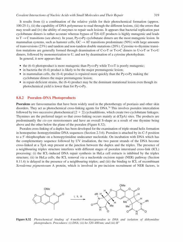
It results from (i) a combination of the relative yields for their photochemical formation (approx.100:25:1), (ii) the capability of DNA polymerase to read through the different lesions, (iii) the errors thatmay result and (iv) the ability of enzymes to repair such lesions. It appears that bacterial replication pastcyclobutane dimers is rather accurate whereas bypass of T(6-4)T products is highly mutagenic and leadsto T → C transitions (see above). In yeast, Py<>Py cyclobutane dimers are the most mutagenic lesion. Inmammalian systems, such as hamster cells, GC → AT transitions predominate (50%) with large amountsof transversions (23%) and tandem and non-tandem double mutations (20%). Cytosine-to-thymine transi-tion mutations are generally formed through deamination of C<>T or T<>C dimers to U<>T or T<>Udimers, followed by monomerization to U, and not by deamination of a cytosine photohydrate.
In general, it now appears that:
� the (6-4) photoproduct is more mutagenic than Py<>Py while T<>T is poorly mutagenic;� in bacteria the (6-4) product is likely to be the major premutagenic lesion;� in mammalian cells, the (6-4) product is repaired more quickly than the Py<>Py making the
cyclobutane dimers the major premutagenic lesion;� in repair-deficient strains, the (6-4) product may be the dominant mutational lesion even though its
photochemical yield is lower than for Py<>Py.
8.8.2 Psoralen–DNA Photoproducts
Psoralens are furocoumarins that have been widely used in the phototherapy of psoriasis and other skindisorders. They act as photochemical cross-linking agents for DNA.74 This involves psoralen intercalationfollowed by two successive photochemical [2 � 2] cycloadditions, which create two cyclobutane linkages.Thymines are the preferred target so that cross-linking occurs mainly at d(TpA) sites. The products arepredominantly the cis-syn stereoisomers and have an overall S-shape as a result of one thymine beingabove and the other below the plane of the psoralen (Figure 8.32).
Psoralen cross-linking of a duplex has been developed for the examination of triple-strand helix formationin homopurine–homopyrimidine DNA sequences (Section 2.3.6). Psoralen is attached by its C-5 positionto a 5�-thiophosphate on a homopyrimidine undecamer nucleotide. On incubation with DNA which hasthe complementary sequence followed by UV irradiation, the two parent strands of the DNA becomecross-linked at a TpA step present at the junction between the duplex and the triplex. The presence of a neighbouring triplex structure interferes with different stages of psoralen interstrand cross-link (ICL)processing: (i) the ICL-induced DNA repair synthesis in HeLa cell extracts is inhibited by the triplex structure; (ii) in HeLa cells, the ICL removal via a nucleotide excision repair (NER) pathway (Section8.11.4) is delayed in the presence of a neighbouring triplex; and (iii) the binding to ICL of recombinantXeroderma pigmentosum A protein, which is involved in pre-incision recruitment of NER factors, is
Covalent Interactions of Nucleic Acids with Small Molecules and Their Repair 319
OO O
OMe OO O
NH
N
N
NH
O OO
O
dR
dRMe
Me
H
H
MeH
HH
i, ii, iii
OMe
Figure 8.32 Photochemical binding of 4-methyl-8-methoxypsoralen to DNA and isolation of dithymidinephotoproducts. Procedures: (i) DNA; (ii) hn 320–400 nm; and (iii) H�

impaired by the presence of the third DNA strand.74 Such psoralen-oligonucleotide conjugates are probesfor sequence-specific helix formation and may have application in site-directed mutagenesis or control of gene expression.
8.8.3 Purine Photoproducts
Adenine and guanine are intrinsically more photostable than the pyrimidines and tend to transfer photo-chemical excitation energy to neighbouring pyrimidines in DNA duplexes. However, UVB can lead to theformation of adenine photoproducts with an adjacent adenine or thymine base (Figure 8.33a).75 While thequantum yields for these processes are low and product formation is easily quenched by base-pairing, the A–Tadduct has been shown to be mutagenic.
Of greater significance is the photo-oxidation of cellular DNA resulting from UVA irradiation involv-ing an unidentified endogenous photosensitiser. The reactions result from one-electron oxidation or hydro-gen abstraction (type I) or from singlet oxygen (type II) processes. The four bases are the preferredsubstrates for type I reactions and reactivity depends on (low) ionisation potential in the orderG � A � T � C. Guanine products predominate because of ‘hole-transfer’ in dsDNA, by which radicalcation migration targets guanine, acting as a sink. Hydration of the initial guanine radical cation leads to8-oxoguanine in an oxidative process or to FapyGua by reduction. Alternatively, deprotonation of the gua-nine cation radical leads to imidazolone and oxazolone products (Figure 8.33b). Lipscomb and Rich havedetermined the X-ray structure of a DNA fragment containing 7,8-dihydro-8-oxoguanine (GO). The struc-ture of the duplex form of d(CCAGOCGCTGG) has been determined to 1.6Å resolution. The 8-oxoGuais in the anti-conformation and forms Watson–Crick base-pairs with the opposite C. There is no stericclash of the 8-oxygen of G with backbone atoms.76
320 Chapter 8
N
N N
N
NH2
dR
N
N
dR
HN
N
CN
N
N
N
HN
NH
NH
NN
NH2N
dR
OH
dRHN
NN
NO
O
dR N N dR
NH2Me
aH
P
P
H
HN
N N
N
O
H2NdR
– e- HN
N N
N
O
H2NdR
N
N N
N
O
H2NdR
N
NNH2
O
HN N
ONH2
O
HNNH2
HN
N N
N
O
H2NdR
OH
H
HN
N N
HN
O
H2NdR
O
HN
N NHCHO
HN
O
H2NdR
O2
H2O
- H+
O2
H2O
H2O
Imidazolone Oxazolone
8-OxoGuo
FapyGuo
b
Figure 8.33 (a) Structure of adenine-containing dimeric photoproducts. (b) Structure and mechanism of formationof the type I photochemistry oxidation products of deoxyguanosine in DNA

8.8.4 DNA and the Ozone Barrier
Life is shielded from DNA-damaging solar radiation by the ozone layer.77,78 The amount of ozone involvedis very small indeed. It is formed photochemically in the stratosphere by UVC radiation and is distributedthroughout the atmosphere. Its maximum density is at some 20–25 km above the earth’s surface, where itspressure is about 130 nbar. Were all the ozone to be concentrated at sea level it would form a layer only3 mm thick!
The absorption spectrum of ozone is very similar to that of DNA (Figure 8.34), so it generally serves toprevent short-wavelength UVC radiation from reaching the earth’s surface and causing DNA damage. Toput that in perspective, it has been estimated that a 1-h exposure of DNA to ozone-filtered sunlight at sealevel would generate about seven cis-syn thymine dimers per 1000 reactive sites. Were the ozone barrierlevel to be halved, the time would be reduced to 10 min. In the complete absence of ozone, the same DNAdamage would occur in 10 s. Other calculations have estimated that for every 1% decrease in the ozonecolumn there could be a 4% increase in the incidence of skin cancer.
One of the major causes for concern is the depletion of ozone resulting from the photochemical behav-iour of chlorofluorocarbons (CFCs) in the upper atmosphere.58,78 This interferes with the formation ofozone through the capture of oxygen atoms by chlorine atoms, as shown by the following equations.
Ozone formation O2 � h → O• � O•
O• � O2 � N2 → O3 � N2
Ozone depletion Cl• � O2 → ClOClO � O3 → Cl• � O2 � O2
A more cautious view is that the two dominant variables that determine the UV incidence at groundlevel integrated over 24 h are (i) the elevation of the sun above the horizon and (ii) the duration of daylight.These two factors, which ultimately relate to the earth’s solar orbit, lead to variations that far exceed thechanges predicted to occur at any middle latitude location as a consequence of any decline in the columnof ozone since 1970. The situation over the Antarctic is, however, a special case. The large depletion inozone over that area in springtime has resulted in UVB irradiances that are substantially larger than existedin that part of the world before the 1980s.
In 2003, the Director General of WHO Lee-Jong Wook said: “As ozone depletion becomes more markedand as people around the world engage more in sun-seeking behaviour, the risk of health complicationsfrom overexposure to ultraviolet radiation is becoming a substantial health concern.”79 The International
Covalent Interactions of Nucleic Acids with Small Molecules and Their Repair 321
240 280
Wavelength (nm)
320 360UVC UVB UVA
Rel
ativ
e ab
sorp
tion
or in
tens
ity
O3DNA
solarsea-level
(6-4)
Dewar
T<>Tcys-syn
Figure 8.34 Absorption spectra for DNA and ozone (red) and the various photoproducts (broken lines) compared tothe incidence of sunlight at sea level and above the atmosphere

Agency for Research on Cancer has estimated that every year, between two and three million new cases ofnon-malignant melanomas and more than 130,000 new cases of melanoma skin cancer are seen world-wide. Furthermore, as reported by the WHO, melanomas and other skin cancers claimed the lives of66,000 people in the world (2002).
International response to this threat has been strong. Some 165 countries have agreed to the MontrealProtocol of 1989, revised in London in 1990, to accelerate the phasing-out of ozone-depleting substances(ODS): initially chlorofluorocarbons (CFCs), halons and carbon tetrachloride by 2000 and methyl chloro-form by 2005.80 As of 2000, industrial production of chlorofluorocarbons has been arrested and atmos-pheric levels appear to show signs of coming under control.
8.9 EFFECTS OF IONIZING RADIATION ON NUCLEIC ACIDS
X-rays, �-radiation and high-energy electrons all interact indirectly with nucleic acids in solution as aresult of the formation of hydroxyl radicals, of solvated electrons, or hydrogen atoms from water. In aero-bic conditions, the most important processes result from HO• radicals, which abstract a hydrogen atomforming an organic radical which then captures O2. Measurements of the efficiency of these processesindicate that for every 1000 eV of energy absorbed, about 27 HO• radicals are formed of which 6 react withthe pentoses and 21 react almost randomly with the four bases.81
8.9.1 Deoxyribose Products in Aerobic Solution
The hydroxyl radical is able to abstract a hydrogen atom from C-4� or from any of the other four carbons inthe sugar. The resultant pentose radicals capture oxygen to give a hydroperoxide radical at C-4� or C-5� andthis leads directly to cleavage of a phosphate ester bond. Similar reactions at C-1�or C-2� lead to alkali-labile phosphate esters that are cleaved on incubation with 0.1 M sodium hydroxide at room temperaturein 10 min. In both cases, the bases are released intact (Figure 8.35). The use of Fenton’s reagent to gener-ate hydroxyl radicals is a key component of a DNA footprinting method (Section 5.5.7).
8.9.2 Pyrimidine Base Products in Solution
At least 24 different products have been isolated from -irradiation of thymidine in dilute, aerated solutionand many more are formed in anoxic conditions or in the solid state. Cytosine and the purines show a simi-lar diversity. The situation is simplified by limiting the study to oxygenated solutions when the principalsite for reaction with the pyrimidines is the 5,6-double bond.82 Under these conditions, hydroxyhydroper-oxides are formed that are semi-stable for thymidine, but break down rapidly for deoxycytidine to give arange of products, of which the major ones are illustrated (Figure 8.36). About 10% of thymine modifica-tion occurs at the methyl group with the formation of 5-hydroxymethyldeoxyuridine. Thymine-glycol isalso a significant lesion.
In anaerobic solution, -radiolysis gives 5,6-dihydrothymidine as the major product with a preferencefor formation of the 5(R)-stereoisomer.
8.9.3 Purine Base Products
The radiation chemistry of the purines is less well understood than that of the pyrimidines. Deoxyadenosinecan add the HO• radical at C-8 to give either 8-hydroxydeoxyadenosine or, with cleavage of the imidazolering, a 5-formamido-4-aminopyrimidine derivative of deoxyribose (Figure 8.37). Guanine has been evenless well studied. However, this situation may change as a result of reports of the formation of 8-hydroxy-guanine from anaerobic irradiation associated, since there is considerable interest in this modified base asa strongly mutagenic lesion (Section 8.11.2).
322 Chapter 8

8.10 BIOLOGICAL CONSEQUENCES OF DNA ALKYLATION
DNA damage is unavoidable and can be divided broadly into two categories. Some forms of damage, suchas mismatches, deamination of bases, base-loss and oxidative damage, arise spontaneously. Others, such asionizing radiation, UV radiation and chemical agents including ubiquitous alkylating agents (Section 8.5.3)arise through environmental influences. Many of these changes can result in errors during the steps of theDNA replication, recombination and repair, and so lead to modification of the structure of genetic material.Happily, many of these modifications can be removed successfully by DNA repair machinery.83,84
8.10.1 N-Alkylated Bases
7-Alkylguanine is the predominant product of DNA alkylation. However, methylation at this site appearsnot to change the base-pairing of G with C and so is an apparently innocuous lesion. Following N-7 alkylation,
Covalent Interactions of Nucleic Acids with Small Molecules and Their Repair 323
O BO
O
O O
P
P
OO
O
P
P
CO2
OHHO
HO
P
P
O HO–1'
O BO
O
P
P
HO
O
O
P
P
O
P
B
HO–
4'OO CO2 heat
B
O BO
O
P
P
OCHOO
O
P
P
O
HO
P
O HO–
O
O
5'
Figure 8.35 Breaks in DNA resulting from hydrogen atom abstraction and peroxide radical formation at C-1� (upper),C-4� (centre) and C-5� (lower) in deoxynucleotides followed by mild alkaline treatment (0.1 M NaOH,10 min, 20°C)
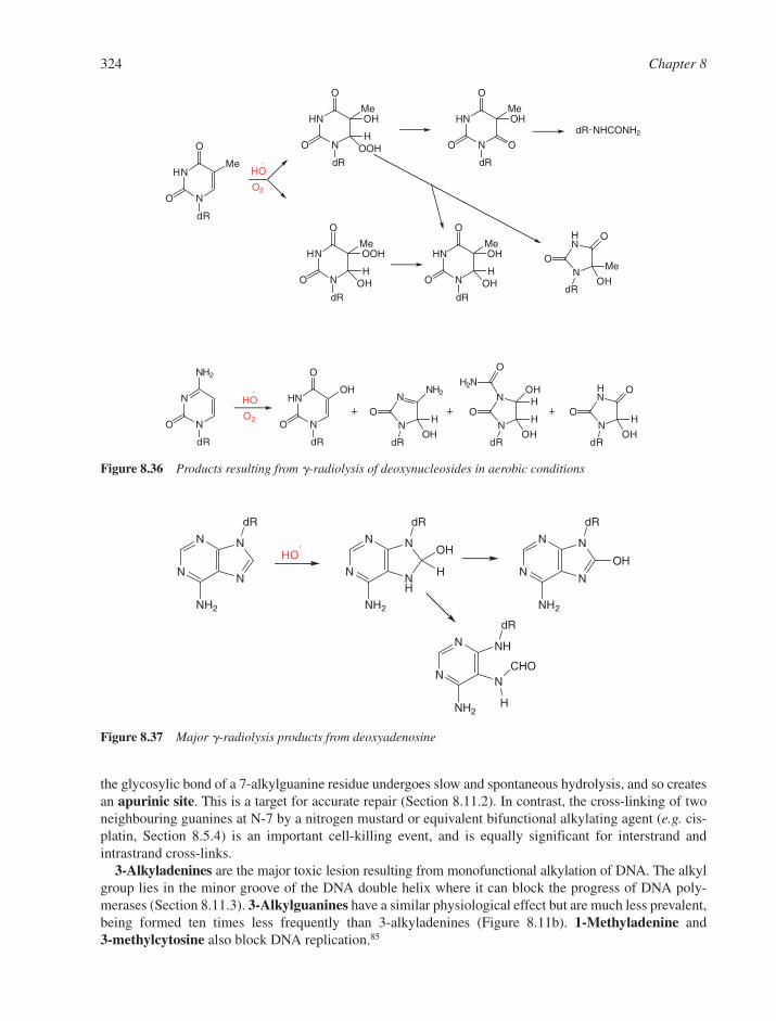
the glycosylic bond of a 7-alkylguanine residue undergoes slow and spontaneous hydrolysis, and so createsan apurinic site. This is a target for accurate repair (Section 8.11.2). In contrast, the cross-linking of twoneighbouring guanines at N-7 by a nitrogen mustard or equivalent bifunctional alkylating agent (e.g. cis-platin, Section 8.5.4) is an important cell-killing event, and is equally significant for interstrand andintrastrand cross-links.
3-Alkyladenines are the major toxic lesion resulting from monofunctional alkylation of DNA. The alkylgroup lies in the minor groove of the DNA double helix where it can block the progress of DNA poly-merases (Section 8.11.3). 3-Alkylguanines have a similar physiological effect but are much less prevalent,being formed ten times less frequently than 3-alkyladenines (Figure 8.11b). 1-Methyladenine and 3-methylcytosine also block DNA replication.85
324 Chapter 8
HN
N
Me
O
dR
HN
N
Me
O
O
dR
HN
N
Me
O
O
dR
OH
HOOH
OOH
HOH
HN
N
Me
O
O
dR
OH
O
dR NHCONH2
HN
N
Me
O
O
dR
OH
HOH
HN
NO
dR
O
Me
OH
HO
O2
N
N
NH2
O
dR
HN
N
O
O
dR
N
NO
dR
NH2
H
OH
N
NO
dR
OH
H
OH
HN
NO
dR
O
H
OH
O
OHH2N
O
HHO
O2
Figure 8.36 Products resulting from -radiolysis of deoxynucleosides in aerobic conditions
N
N
N
N
NH2
dR
N
N
NH
N
NH2
dR
N
N
N
N
NH2
dR
HO
H
OHOH
N
N
N
NH
NH2
dR
CHO
H
Figure 8.37 Major -radiolysis products from deoxyadenosine
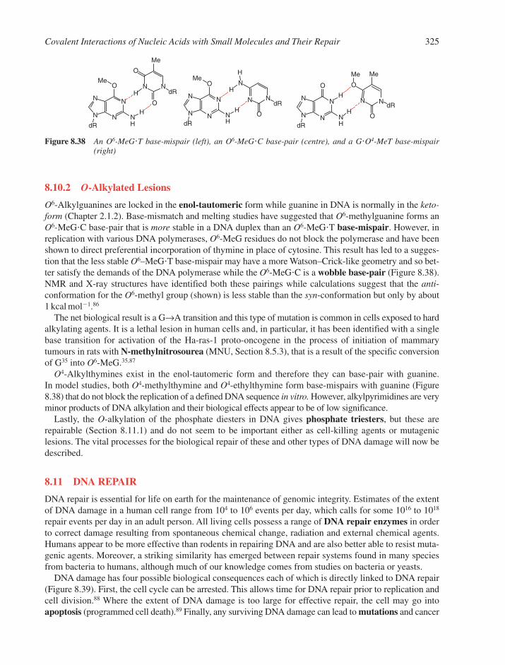
8.10.2 O-Alkylated Lesions
O6-Alkylguanines are locked in the enol-tautomeric form while guanine in DNA is normally in the keto-form (Chapter 2.1.2). Base-mismatch and melting studies have suggested that O6-methylguanine forms anO6-MeG�C base-pair that is more stable in a DNA duplex than an O6-MeG�T base-mispair. However, inreplication with various DNA polymerases, O6-MeG residues do not block the polymerase and have beenshown to direct preferential incorporation of thymine in place of cytosine. This result has led to a sugges-tion that the less stable O6–MeG�T base-mispair may have a more Watson–Crick-like geometry and so bet-ter satisfy the demands of the DNA polymerase while the O6-MeG�C is a wobble base-pair (Figure 8.38).NMR and X-ray structures have identified both these pairings while calculations suggest that the anti-conformation for the O6-methyl group (shown) is less stable than the syn-conformation but only by about1 kcal mol�1.86
The net biological result is a G→A transition and this type of mutation is common in cells exposed to hardalkylating agents. It is a lethal lesion in human cells and, in particular, it has been identified with a singlebase transition for activation of the Ha-ras-1 proto-oncogene in the process of initiation of mammarytumours in rats with N-methylnitrosourea (MNU, Section 8.5.3), that is a result of the specific conversionof G35 into O6-MeG.35,87
O4-Alkylthymines exist in the enol-tautomeric form and therefore they can base-pair with guanine. In model studies, both O4-methylthymine and O4-ethylthymine form base-mispairs with guanine (Figure8.38) that do not block the replication of a defined DNA sequence in vitro. However, alkylpyrimidines are veryminor products of DNA alkylation and their biological effects appear to be of low significance.
Lastly, the O-alkylation of the phosphate diesters in DNA gives phosphate triesters, but these arerepairable (Section 8.11.1) and do not seem to be important either as cell-killing agents or mutageniclesions. The vital processes for the biological repair of these and other types of DNA damage will now bedescribed.
8.11 DNA REPAIR
DNA repair is essential for life on earth for the maintenance of genomic integrity. Estimates of the extentof DNA damage in a human cell range from 104 to 106 events per day, which calls for some 1016 to 1018
repair events per day in an adult person. All living cells possess a range of DNA repair enzymes in orderto correct damage resulting from spontaneous chemical change, radiation and external chemical agents.Humans appear to be more effective than rodents in repairing DNA and are also better able to resist muta-genic agents. Moreover, a striking similarity has emerged between repair systems found in many speciesfrom bacteria to humans, although much of our knowledge comes from studies on bacteria or yeasts.
DNA damage has four possible biological consequences each of which is directly linked to DNA repair(Figure 8.39). First, the cell cycle can be arrested. This allows time for DNA repair prior to replication andcell division.88 Where the extent of DNA damage is too large for effective repair, the cell may go intoapoptosis (programmed cell death).89 Finally, any surviving DNA damage can lead to mutations and cancer
Covalent Interactions of Nucleic Acids with Small Molecules and Their Repair 325
N
N
N
N
O
dR
N N
NN N
O
dR
dR
O
ONH
Me
Me
H
HN
N
N
N
O
dRNH
Me
H
H
N N
O
dR
O
N
N
N
N
O
dRNH
H
H
Me MeH
Figure 8.38 An O6-MeG�T base-mispair (left), an O6-MeG�C base-pair (centre), and a G�O4-MeT base-mispair(right)
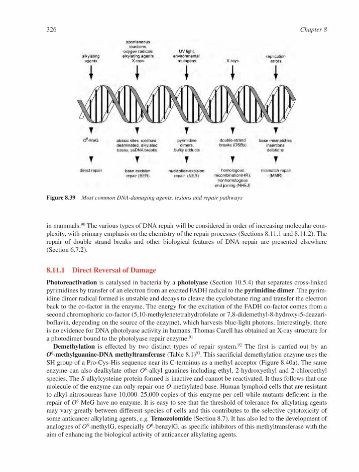
in mammals.90 The various types of DNA repair will be considered in order of increasing molecular com-plexity, with primary emphasis on the chemistry of the repair processes (Sections 8.11.1 and 8.11.2). Therepair of double strand breaks and other biological features of DNA repair are presented elsewhere(Section 6.7.2).
8.11.1 Direct Reversal of Damage
Photoreactivation is catalysed in bacteria by a photolyase (Section 10.5.4) that separates cross-linkedpyrimidines by transfer of an electron from an excited FADH radical to the pyrimidine dimer. The pyrim-idine dimer radical formed is unstable and decays to cleave the cyclobutane ring and transfer the electronback to the co-factor in the enzyme. The energy for the excitation of the FADH co-factor comes from asecond chromophoric co-factor (5,10-methylenetetrahydrofolate or 7,8-didemethyl-8-hydroxy-5-deazari-boflavin, depending on the source of the enzyme), which harvests blue-light photons. Interestingly, thereis no evidence for DNA photolyase activity in humans. Thomas Carell has obtained an X-ray structure fora photodimer bound to the photolyase repair enzyme.91
Demethylation is effected by two distinct types of repair system.92 The first is carried out by an O6-methylguanine-DNA methyltransferase (Table 8.1)93. This sacrificial demethylation enzyme uses theSH group of a Pro-Cys-His sequence near its C-terminus as a methyl acceptor (Figure 8.40a). The sameenzyme can also dealkylate other O6-alkyl guanines including ethyl, 2-hydroxyethyl and 2-chloroethylspecies. The S-alkylcysteine protein formed is inactive and cannot be reactivated. It thus follows that onemolecule of the enzyme can only repair one O-methylated base. Human lymphoid cells that are resistantto alkyl-nitrosoureas have 10,000–25,000 copies of this enzyme per cell while mutants deficient in therepair of O6-MeG have no enzyme. It is easy to see that the threshold of tolerance for alkylating agentsmay vary greatly between different species of cells and this contributes to the selective cytotoxicity ofsome anticancer alkylating agents, e.g. Temozolomide (Section 8.7). It has also led to the development ofanalogues of O6-methylG, especially O6-benzylG, as specific inhibitors of this methyltransferase with theaim of enhancing the biological activity of anticancer alkylating agents.
326 Chapter 8
Figure 8.39 Most common DNA-damaging agents, lesions and repair pathways
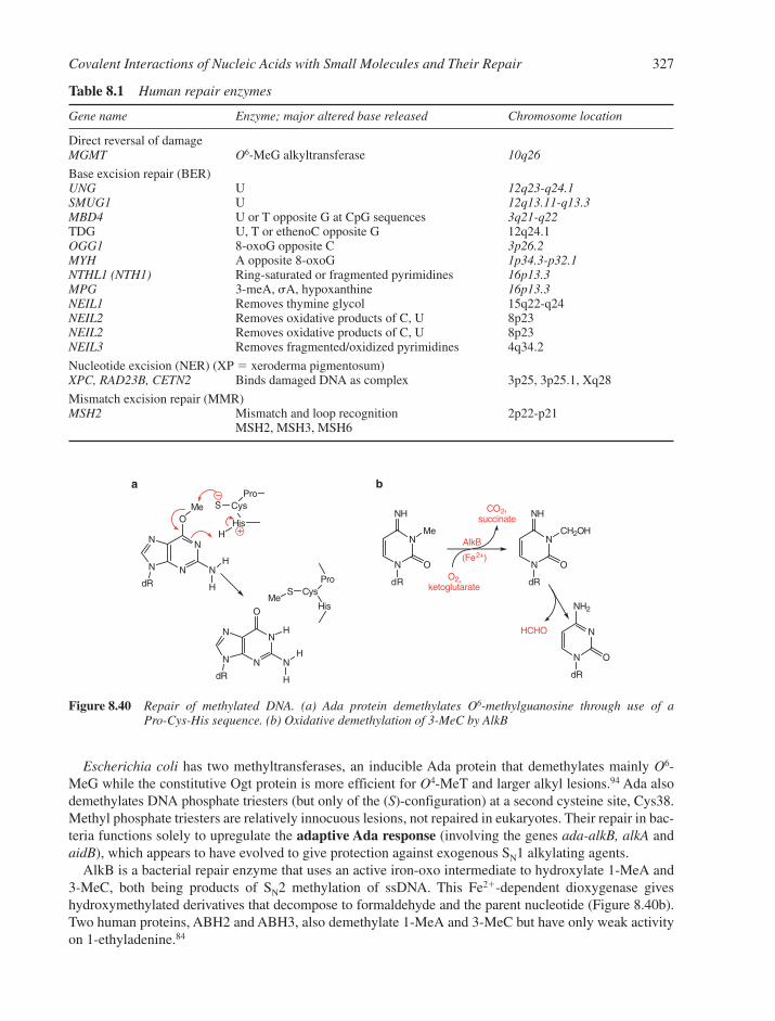
Escherichia coli has two methyltransferases, an inducible Ada protein that demethylates mainly O6-MeG while the constitutive Ogt protein is more efficient for O4-MeT and larger alkyl lesions.94 Ada alsodemethylates DNA phosphate triesters (but only of the (S)-configuration) at a second cysteine site, Cys38.Methyl phosphate triesters are relatively innocuous lesions, not repaired in eukaryotes. Their repair in bac-teria functions solely to upregulate the adaptive Ada response (involving the genes ada-alkB, alkA andaidB), which appears to have evolved to give protection against exogenous SN1 alkylating agents.
AlkB is a bacterial repair enzyme that uses an active iron-oxo intermediate to hydroxylate 1-MeA and3-MeC, both being products of SN2 methylation of ssDNA. This Fe2�-dependent dioxygenase giveshydroxymethylated derivatives that decompose to formaldehyde and the parent nucleotide (Figure 8.40b).Two human proteins, ABH2 and ABH3, also demethylate 1-MeA and 3-MeC but have only weak activityon 1-ethyladenine.84
Covalent Interactions of Nucleic Acids with Small Molecules and Their Repair 327
N
N
N
N
O
dRN
H
Me
H
ProCys
His
S
H
N
N
N
N
O
dRN
H
Me
H
ProCys
His
S
H
a b
N
N
dR
NH
O
Me
N
N
dR
NH
O
CH2OH
N
N
dR
NH2
O
O2,ketoglutarate
AlkB
(Fe2+)
CO2,succinate
HCHO
Figure 8.40 Repair of methylated DNA. (a) Ada protein demethylates O6-methylguanosine through use of a Pro-Cys-His sequence. (b) Oxidative demethylation of 3-MeC by AlkB
Table 8.1 Human repair enzymes
Gene name Enzyme; major altered base released Chromosome location
Direct reversal of damageMGMT O6-MeG alkyltransferase 10q26
Base excision repair (BER)UNG U 12q23-q24.1SMUG1 U 12q13.11-q13.3MBD4 U or T opposite G at CpG sequences 3q21-q22TDG U, T or ethenoC opposite G 12q24.1OGG1 8-oxoG opposite C 3p26.2MYH A opposite 8-oxoG 1p34.3-p32.1NTHL1 (NTH1) Ring-saturated or fragmented pyrimidines 16p13.3MPG 3-meA, �A, hypoxanthine 16p13.3NEIL1 Removes thymine glycol 15q22-q24NEIL2 Removes oxidative products of C, U 8p23NEIL2 Removes oxidative products of C, U 8p23NEIL3 Removes fragmented/oxidized pyrimidines 4q34.2
Nucleotide excision (NER) (XP � xeroderma pigmentosum)XPC, RAD23B, CETN2 Binds damaged DNA as complex 3p25, 3p25.1, Xq28
Mismatch excision repair (MMR)MSH2 Mismatch and loop recognition 2p22-p21
MSH2, MSH3, MSH6

8.11.2 Base Excision Repair of Altered Residues
In mammalian cells, excision repair is the most important mechanism and involves enzyme recognition ofthe modified base, usually resulting form oxidation, deamination, or alkylation (Table 8.1). There are two dis-tinct types of excision repair process and both appear to be error-free: base excision and nucleotide excision.
Repair by base excision, BER, employs a DNA glycosylase that cleaves the glycosylic bonds of modi-fied purine or pyrimidine nucleosides to generate an abasic site.95 The next step is incision of the phosphatebackbone by an AP endonuclease (Section 10.5.2), which operates close to apurinic and apyrimidinicsites. Subsequent exonuclease action then excises nucleotides to create a gap at least 30 residues long.Multiple human glycosylases have been identified, each with its own base-specificity. Four of theseenzymes operate on 2�-deoxyuridine, which arises through misincorporation of dUMP (giving a U�Abase-pair) or from deamination of dC (giving a U�G base-pair). It has been estimated that humans repairsome 500 uracil residues per cell per day. Uracil DNA glycohydrolase, UDG, is highly conserved fromarchaebacteria to humans and is the product of the UNG gene. It is the most proficient of the uracil repairenzymes, having a reaction acceleration close to 1012, while its efficiency of discrimination againstthymine and cytosine in favour of uracil exceeds 1:107.
3-Methyladenine is the principal target for two 3-methyladenine-DNA glycosylases from E. coli, one ofwhich also acts on 3-MeG, O2-MeC and O2-MeT residues. The human enzyme 3-methyladenine DNA gly-cosylase, the MPG gene product, removes a remarkably diverse group of damaged bases from DNA, includ-ing cytotoxic and mutagenic alkylation adducts of purines such as 3-MeA, 7-MeG and hypoxanthine. Thestructure of the human 3-methyladenine DNA glycosylase complexed to a mechanism-based pyrrolidineinhibitor (Figure 8.41a) shows the enzyme intercalated into the minor groove of DNA, causing the abasicpyrrolidine nucleotide to flip into the enzyme active site, where a bound water is poised for nucleophilic attack.
8-Oxoguanine is the major mutagenic base lesion in DNA caused by exposure to reactive oxygenspecies. It is excised by OGG1, a DNA glycosylase with an associated lyase activity for chain cleavage. Itreleases free 8-hydroxyguanine specifically from 8-OH-G�C pairs and also introduces a chain break in a
328 Chapter 8
a
N N
O
O
H
O
OH
HO
H
N
N
O
O
H
O
OH
HO
H
F
H H
NH
N
HN
N
O
NH
H
O
H2C
OH
HO
pseudodeoxyuridine 2'-difluorouridine4'-carba-8-oxoguanosine
b
N
N
O
O
H
O
OPO2
O
PO2
N
N
O
O
H
O
OPO2
O
PO2
OH
H
O
OAsp145
NH
N
O
O
H
O
OPO2
O
PO2
OH
HO
OAsp88
SN1
ionisationsolvation
H
F
HN
OPO2
O
PO2H
iminium abasic analogue
Figure 8.41 (a) Structures of some nucleoside analogues used to inhibit glycosylic bond cleavage. (b) 2-Step SN1mechanism of glycosylic bond cleavage by UDG

double-stranded oligonucleotide (Table 8.1). The human enzyme MUTYH appears to be homologous tothe bacterial repair glycosylase MutY, and removes adenines mis-paired with 8-oxoguanine. The thirdcomponent of the resistance to 8-oxoG is an 8-oxoGTP hydrolase, which removes it from the nucleotidepool to circumvent misincorporation of 8-OH-G opposite A residues.
Recent studies have confirmed the role of reactive oxygen species in the pathogenesis of Alzheimer’sdisease (AD) and the accumulation of 8-oxo-2�-deoxyguanosine in AD brain has been discussed. It seemsthat reduced expression of 8-oxoguanine DNA glycosylase (hOGG1-2a), an enzyme that repairs 8-oxo-2�-deoxyguanosine, may be involved in the pathomechanism of AD.96
8.11.3 Mechanisms and Inhibitors of DNA Glycohydrolases
All DNA glycosylases employ a nucleotide-flipping mechanism. This is a general strategy for enzymesoperating on natural or modified DNA bases to gain access to target loci that are buried in the DNA stackor inaccessible from a groove. Such glycosylases fall into two distinct mechanistic classes: (i) monofunc-tional glycosylases and (ii) bifunctional glycosylase/AP lyases.
Considerable detail has been achieved in understanding the mechanism of monofunctional BER glyco-sylases through the use of inhibitory nucleotide analogues resistant to glycosylic bond cleavage.95 Threegeneral strategies have been employed (Figure 8.41a).
� C-Glycosides as in the case of pseudodeoxyuridine for UDG97 and 2�-deoxyformycin A for MutY,� Carbocyclic sugar nucleosides, as in the case of 8-oxocarbadG for OGG198 and� 2-Fluoropentoses, as in the case of 2�-fluoroadenosine for MutY99 and 2�-difluorodU for human
TDG glycosylase.100
UDG is an example of an enzyme that ‘flips-out’ the aberrant nucleotide, as seen in an elegant sequenceof X-ray structures by John Tainer, one including a transition state analogue.97 Both QM-MM computationand kinetic isotope studies have identified the glycosylic bond cleavage as a dissociative process, much ofthe energy for which derives from stabilisation of an oxocarbenium ion at C-1 of the deoxyribose by thenegative charges on the proximate phosphate residues in the same strand. The water that is captured by theoxocarbenium ion to give the deoxyribose AP-product is activated by an essential aspartic acid in an SN1mechanism (Figure 8.41b). The uracil anion is an adequate leaving group not needing general acid activa-tion. Thus, the UDG enzyme appears to provide no chemical catalysis but serves simply to orientate theDNA substrate to maximise uracil recognition, stereoelectronic effects and the enabling contribution ofthe phosphate negative charges to C–N bond ionisation. In general, repair enzymes that involve oxocar-benium ion intermediates are strongly inhibited by iminium abasic sugar analogues (Figure 8.41a), whichhave proven to constitute a general strategy for studying glycohydrolases.101,102
The enzymes that catalyse depurination show greatly reduced base-specificities and also appear to usegeneral acid catalysis at the purine to effect glycosyl bond cleavage. Less mechanistic detail is yet avail-able to establish whether they employ SN1 or SN2 reaction pathways.
The bifunctional BER enzymes that cause chain cleavage as well as base-excision generally operate via asugar-enzyme intermediate. These include OGG1, NEI1 and NTH1 (Table 8.1). Their mechanism is wellexemplified for the bacteriophage T4 Endonuclease V, which cleaves the glycosylic bond of the 5�-residuein a thymine photodimer.103 The N-terminal amino group of T4 Endo V attacks C-1�, displaces the baseand forms an aldimine. This is followed by �-elimination of the 3�-phosphate to give the single-strandbreak (Figure 8.42).
8.11.4 Nucleotide Excision Repair
Nucleotide excision, NER, is the repair pathway that deals with bulky lesions, including those formed byUV radiation, various environmental mutagens, and some chemotherapeutic agents, and shows broad sub-strate acceptability.104 There seems to be a general correlation between the efficiency of repair and the
Covalent Interactions of Nucleic Acids with Small Molecules and Their Repair 329
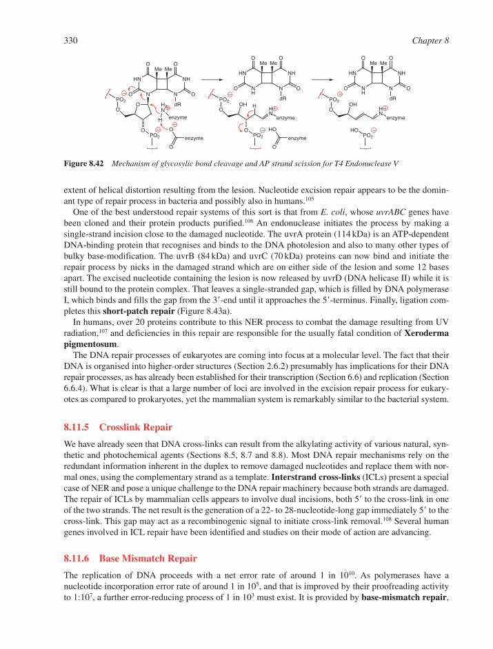
extent of helical distortion resulting from the lesion. Nucleotide excision repair appears to be the domin-ant type of repair process in bacteria and possibly also in humans.105
One of the best understood repair systems of this sort is that from E. coli, whose uvrABC genes havebeen cloned and their protein products purified.106 An endonuclease initiates the process by making a single-strand incision close to the damaged nucleotide. The uvrA protein (114 kDa) is an ATP-dependentDNA-binding protein that recognises and binds to the DNA photolesion and also to many other types ofbulky base-modification. The uvrB (84 kDa) and uvrC (70 kDa) proteins can now bind and initiate therepair process by nicks in the damaged strand which are on either side of the lesion and some 12 basesapart. The excised nucleotide containing the lesion is now released by uvrD (DNA helicase II) while it isstill bound to the protein complex. That leaves a single-stranded gap, which is filled by DNA polymeraseI, which binds and fills the gap from the 3�-end until it approaches the 5�-terminus. Finally, ligation com-pletes this short-patch repair (Figure 8.43a).
In humans, over 20 proteins contribute to this NER process to combat the damage resulting from UVradiation,107 and deficiencies in this repair are responsible for the usually fatal condition of Xerodermapigmentosum.
The DNA repair processes of eukaryotes are coming into focus at a molecular level. The fact that theirDNA is organised into higher-order structures (Section 2.6.2) presumably has implications for their DNArepair processes, as has already been established for their transcription (Section 6.6) and replication (Section6.6.4). What is clear is that a large number of loci are involved in the excision repair process for eukary-otes as compared to prokaryotes, yet the mammalian system is remarkably similar to the bacterial system.
8.11.5 Crosslink Repair
We have already seen that DNA cross-links can result from the alkylating activity of various natural, syn-thetic and photochemical agents (Sections 8.5, 8.7 and 8.8). Most DNA repair mechanisms rely on theredundant information inherent in the duplex to remove damaged nucleotides and replace them with nor-mal ones, using the complementary strand as a template. Interstrand cross-links (ICLs) present a specialcase of NER and pose a unique challenge to the DNA repair machinery because both strands are damaged.The repair of ICLs by mammalian cells appears to involve dual incisions, both 5� to the cross-link in oneof the two strands. The net result is the generation of a 22- to 28-nucleotide-long gap immediately 5� to thecross-link. This gap may act as a recombinogenic signal to initiate cross-link removal.108 Several humangenes involved in ICL repair have been identified and studies on their mode of action are advancing.
8.11.6 Base Mismatch Repair
The replication of DNA proceeds with a net error rate of around 1 in 1010. As polymerases have anucleotide incorporation error rate of around 1 in 105, and that is improved by their proofreading activityto 1:107, a further error-reducing process of 1 in 103 must exist. It is provided by base-mismatch repair,
330 Chapter 8
HN
N N
NH
O
O
O
O
dRO
Me Me
O
PO2
OPO2
H2N
H enzyme
O
O
enzyme
HN
NH
N
NH
O
O
O
O
dR
Me Me
OHO
PO2
OPO2
HN
enzyme
H
HO
O
enzyme
HN
NH
N
NH
O
O
O
O
dR
Me Me
OHO
PO2
HN
enzyme
HOPO2
Figure 8.42 Mechanism of glycosylic bond cleavage and AP strand scission for T4 Endonuclease V
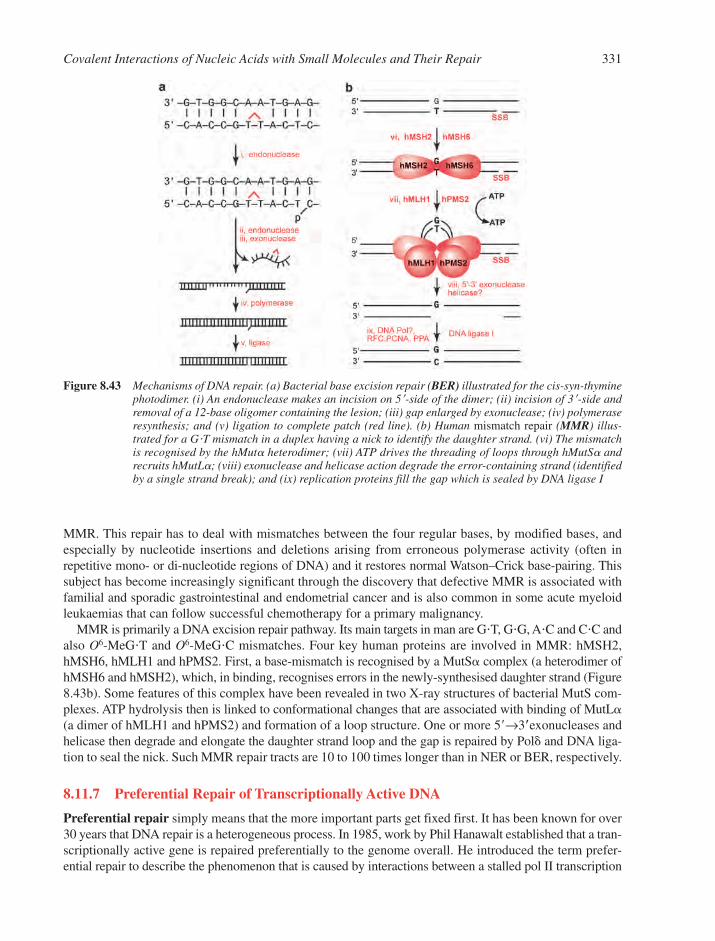
MMR. This repair has to deal with mismatches between the four regular bases, by modified bases, andespecially by nucleotide insertions and deletions arising from erroneous polymerase activity (often inrepetitive mono- or di-nucleotide regions of DNA) and it restores normal Watson–Crick base-pairing. Thissubject has become increasingly significant through the discovery that defective MMR is associated withfamilial and sporadic gastrointestinal and endometrial cancer and is also common in some acute myeloidleukaemias that can follow successful chemotherapy for a primary malignancy.
MMR is primarily a DNA excision repair pathway. Its main targets in man are G�T, G�G, A�C and C�C andalso O6-MeG�T and O6-MeG�C mismatches. Four key human proteins are involved in MMR: hMSH2,hMSH6, hMLH1 and hPMS2. First, a base-mismatch is recognised by a MutS� complex (a heterodimer ofhMSH6 and hMSH2), which, in binding, recognises errors in the newly-synthesised daughter strand (Figure8.43b). Some features of this complex have been revealed in two X-ray structures of bacterial MutS com-plexes. ATP hydrolysis then is linked to conformational changes that are associated with binding of MutL�(a dimer of hMLH1 and hPMS2) and formation of a loop structure. One or more 5�→3�exonucleases andhelicase then degrade and elongate the daughter strand loop and the gap is repaired by Pol and DNA liga-tion to seal the nick. Such MMR repair tracts are 10 to 100 times longer than in NER or BER, respectively.
8.11.7 Preferential Repair of Transcriptionally Active DNA
Preferential repair simply means that the more important parts get fixed first. It has been known for over30 years that DNA repair is a heterogeneous process. In 1985, work by Phil Hanawalt established that a tran-scriptionally active gene is repaired preferentially to the genome overall. He introduced the term prefer-ential repair to describe the phenomenon that is caused by interactions between a stalled pol II transcription
Covalent Interactions of Nucleic Acids with Small Molecules and Their Repair 331
Figure 8.43 Mechanisms of DNA repair. (a) Bacterial base excision repair (BER) illustrated for the cis-syn-thyminephotodimer. (i) An endonuclease makes an incision on 5�-side of the dimer; (ii) incision of 3�-side andremoval of a 12-base oligomer containing the lesion; (iii) gap enlarged by exonuclease; (iv) polymeraseresynthesis; and (v) ligation to complete patch (red line). (b) Human mismatch repair (MMR) illus-trated for a G�T mismatch in a duplex having a nick to identify the daughter strand. (vi) The mismatchis recognised by the hMut� heterodimer; (vii) ATP drives the threading of loops through hMutS� andrecruits hMutL�; (viii) exonuclease and helicase action degrade the error-containing strand (identifiedby a single strand break); and (ix) replication proteins fill the gap which is sealed by DNA ligase I
complex and the excision repair enzymes.109 For example, 70% of photodimers in an active DHFR gene inChinese hamster ovary cells are excised in a 24-h period while only 10–20% of photodimers are removedfrom the genome overall. Later studies established that this phenomenon is confined to the transcribedDNA strand while virtually no repair occurs in the non-transcribed strand.
Selective repair is not confined to cyclobutane dimers; (6-4) photoproducts are also removed preferen-tially from active genes. On the other hand, it appears that N-methylpurine repair is not coupled to tran-scription while the situation regarding covalent modification by carcinogens, such as N-AAF (Section 8.6.1),remains to be clarified.
8.11.8 Post-Replication Repair
The repair systems described in the preceding sections maintain the integrity of the genetic message in thecell as long as repair is rapid and is accurately completed before DNA polymerase attempts to copy thedamaged DNA. What happens if repair is slow or deficient? It appears that when DNA lesions block pro-gression of the replication fork, a major response is the activation of ‘error-prone repair’, without removingthe lesion. Xeroderma pigmentosum is likely caused by a defect in post-replication repair.
SOS repair is the name given to such enhanced repair (ER), which is induced by a wide variety of typesof DNA damage.110 This is how it seems to work in bacteria. When the advancing DNA polymerasereaches a lesion that blocks its further progress, for example T<>T or 3-MeA, a very sizeable gap is leftopposite the lesion before replication starts again. This gap can either be made good by recombinationprocesses, known as recombination-repair (Section 6.7.2), or by the induction of SOS repair.
The SOS process has been most thoroughly studied in E. coli and is one of the functions of the recAgene protein.111 Several recA proteins identify and bind to any long, single-stranded stretch of DNA, whichhas been formed by the recBCD proteins. RecBCD produces ssDNA using its helicase/nuclease activities.Through interaction with ATP, recA now becomes activated as a protease towards the lexA protein, whichis known to be a multifunctional repressor in control of several DNA repair enzymes. Among the manyconsequences of hydrolysis of lexA are the de-repression of ruvAB and recA genes and a reduction in the3�→5� exonuclease proofreading activity of DNA polymerase III. The net result is that pol III operateswith decreased fidelity, not only in its ability to read through regions of damage such as T<>T but alsoelsewhere in the cell, and this situation persists until the activation of recA as a protease ends. At this pointSOS repair is rapidly switched off.
The mechanism of repairing these large gaps, which can be over 1000 bases long, is highly variable. Inparticular, the specificity of elongation varies with several factors: the type of polymerase, the activity of the3�–5� editing nuclease, and the particular type of DNA lesion. For example, in cases where the base modi-fication behaves as a non-instructional site, there is a strong tendency for the incorporation of an adenineresidue.112 This explains why transversion mutations are seen when guanines are modified by aflatoxin orby acetylaminofluorene derivatives. Even then, further elongation in the gap is dependent on the base-sequence in the template strand on the 5�-side of the lesion. The overall result is that ‘long-patch’ repairseems likely to be idiosyncratic for each type of lesion in each mutable site and, above all, is error-prone.
8.11.9 Bypass Mutagenesis
A single mutation that arises from damage to DNA can be described by a two-step mechanism.Misincorporation is the inclusion in the daughter strand of a nucleotide different from the Watson–Crickcomplement to the original residue(s) in the primer strand. Bypass is the process of continued chain elongation beyond the mis-inserted base at the site of the lesion.113 This process has been carefully exam-ined in E. coli through the insertion of synthetic cyclobutane photodimers into oligomers and their use astemplates for pol I in primer extension reactions in vitro and in phage and bacterial replication.
When pol I arrives at a lesion in the template, chain extension of the primer strand can either be terminatedor retarded. Three types of result have now been observed for bypass replication beyond a cyclobutane
332 Chapter 8
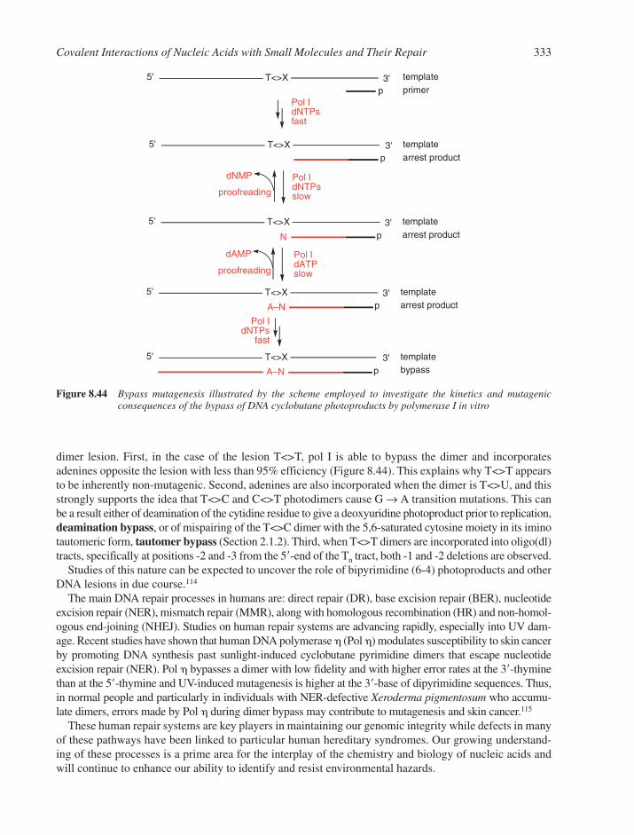
dimer lesion. First, in the case of the lesion T<>T, pol I is able to bypass the dimer and incorporatesadenines opposite the lesion with less than 95% efficiency (Figure 8.44). This explains why T<>T appearsto be inherently non-mutagenic. Second, adenines are also incorporated when the dimer is T<>U, and thisstrongly supports the idea that T<>C and C<>T photodimers cause G → A transition mutations. This canbe a result either of deamination of the cytidine residue to give a deoxyuridine photoproduct prior to replication,deamination bypass, or of mispairing of the T<>C dimer with the 5,6-saturated cytosine moiety in its iminotautomeric form, tautomer bypass (Section 2.1.2). Third, when T<>T dimers are incorporated into oligo(dl)tracts, specifically at positions -2 and -3 from the 5�-end of the Tn tract, both -1 and -2 deletions are observed.
Studies of this nature can be expected to uncover the role of bipyrimidine (6-4) photoproducts and otherDNA lesions in due course.114
The main DNA repair processes in humans are: direct repair (DR), base excision repair (BER), nucleotideexcision repair (NER), mismatch repair (MMR), along with homologous recombination (HR) and non-homol-ogous end-joining (NHEJ). Studies on human repair systems are advancing rapidly, especially into UV dam-age. Recent studies have shown that human DNA polymerase � (Pol �) modulates susceptibility to skin cancerby promoting DNA synthesis past sunlight-induced cyclobutane pyrimidine dimers that escape nucleotideexcision repair (NER). Pol � bypasses a dimer with low fidelity and with higher error rates at the 3�-thyminethan at the 5�-thymine and UV-induced mutagenesis is higher at the 3�-base of dipyrimidine sequences. Thus,in normal people and particularly in individuals with NER-defective Xeroderma pigmentosum who accumu-late dimers, errors made by Pol � during dimer bypass may contribute to mutagenesis and skin cancer.115
These human repair systems are key players in maintaining our genomic integrity while defects in manyof these pathways have been linked to particular human hereditary syndromes. Our growing understand-ing of these processes is a prime area for the interplay of the chemistry and biology of nucleic acids andwill continue to enhance our ability to identify and resist environmental hazards.
Covalent Interactions of Nucleic Acids with Small Molecules and Their Repair 333
5' T<>Xp
templateprimer
Pol IdNTPsfast
5' 3'T<>Xp
dNMP
proofreading
Pol IdNTPsslow
templatearrest product
5' 3'T<>X templatearrest productN
5' 3'T<>X templatearrest product
dAMP
proofreading
Pol IdATPslow
A–N
5' 3'T<>X templatebypassA–N
Pol IdNTPs
fast
p
p
p
3'
Figure 8.44 Bypass mutagenesis illustrated by the scheme employed to investigate the kinetics and mutagenic consequences of the bypass of DNA cyclobutane photoproducts by polymerase I in vitro

REFERENCES
1. W. Szybalski, Effects of elevated temperatures on DNA and on some polynucleotides. Denaturation,renaturation, and cleavage of glycosidic and phosphate ester bonds. in Thermobiology, A.H. Rose(ed). 1967, 73–122.
2. M. Green and S.S. Cohen, Biosynthesis of bacterial and viral pyrimidines. II. Dihydrouracil anddihydrothymine nucleosides. J. Biol. Chem., 1957, 225, 397–407.
3. P. Cerutti, Sodium borohydride reduction of transfer RNA. Methods Enzymol., 1971, 20, 135–143.4. D.H.R. Barton and R. Subramanian, Reactions of relevance to the chemistry of aminoglycoside
antibiotics. Part 7. Conversion of thiocarbonates into deoxy-sugars. J. Chem. Soc. Perkin Trans. 1,1977, 1718–1723.
5. C.A. Krauth, A.T. Shortnacy, J.A. Montgomery and J.A. Secrist III, Synthesis and antiviral evaluationof adenosine-N1-oxide and 1-(benzyloxy)adenosines. Nucleos. Nucleot., 1989, 8, 915–917.
6. Y. Vaishnav, E. Holwitt, C. Swenberg, H.C. Lee and L.S. Kan, Synthesis and characterization ofstereoisomers of 5,6-dihydro-5,6-dihydroxythymidine. J. Biomol. Struct. Dyn., 1991, 8, 935–951.
7. S. Nampalli and S. Kumar, Efficient synthesis of 8-Oxo-dGTP: A mutagenic nucleotide. Bioorg. Med.Chem. Letts., 2000, 10, 1677–1679.
8. J.W. Trauger and P.B. Dervan, Footprinting methods for analysis of pyrrole-imidazole polyamide/DNAcomplexes. Methods Enzymol., 2001, 340, 450–466.
9. B.S. Ermolinsky and S.N. Mikhailov, Periodate oxidation in chemistry of nucleic acids: Dialdehydederivatives of nucleosides, nucleotides, and oligonucleotides. Russ. J. Bioorg. Chem., 2000, 26, 429–449.
10. E. Pichersky, DNA sequencing by the chemical method. Methods Mol. Biol., 1993, 23, 255–259.11. J.H. Phillips, J.H. and D.M. Brown, Mutagenic action of hydroxylamine. Prog. Nucleic Acid Res.
Mol. Biol., 1967, 7, 349–368.12. R. Shapiro, V. DeFate and M. Welcher, Deamination cytosine derivatives by bisulfite. Mechanism of
the reaction. J. Am. Chem. Soc., 1974, 96, 906–912.13. C.W. Carreras and D.V. Santi, The catalytic mechanism and structure of thymidylate synthase. Ann.
Rev. Biochem., 1995, 64, 721–762.14. X. Cheng and R.J. Roberts, AdoMet-dependent methylation, DNA methyltransferases and base flipping.
Nucleic Acids Res., 2001, 29, 3784–3795.15. J.A. Houghton and P.J. Houghton, 5-Halogenated pyrimidines and their nucleosides. Handbook Exp.
Pharmacol., 1984, 72, 515–549.16. M.J. Robins and S.R. Naik, Nucleic acid related compounds. III. Facile synthesis of 5-fluorouracil
bases and nucleosides by direct fluorination. J. Am. Chem. Soc., 1971, 93, 5277–5278.17. R. Shapiro and H. Yamaguchi, Nucleic acid reactivity and conformation. I. Deamination of cytosine
by nitrous acid. Biochim. Biophys. Acta, 1972, 281, 501–506.18. G. Cheng, Y. Shi, S.J. Sturla, J.R. Jalas, E.J. McIntee, P.W. Villalta, M. Wang and S.S. Hecht,
Reactions of formaldehyde plus acetaldehyde with deoxyguanosine and DNA: Formation of cyclicdeoxyguanosine adducts and formaldehyde cross-links. Chem. Res. Toxicol., 2003, 16, 145–152.
19. R.F. Newbold, W. Warren, A.S.C. Medcalf and J. Amos, Mutagenicity of carcinogenic methylatingagents is associated with a specific DNA modification. Nature, 1980, 283, 596–599.
20. P. Blans and J.C. Fishbein, Determinants of selectivity in alkylation of nucleosides and DNA bysecondary diazonium ions: Evidence for, and consequences of, a preassociation mechanism. Chem.Res. Toxicol., 2004, 17, 1531–1539.
21. R.L. Wurdeman, K.M. Church and B. Gold, DNA methylation by N-methyl-N-nitrosourea, N-methyl-N�-nitro-N-nitrosoguanidine, N-nitroso(1-acetoxyethyl)methylamine, and diazomethane. The mech-anism for the formation of N7-methylguanine in sequence-characterized 5�-[32P]-end-labeled DNA.J. Am. Chem. Soc., 1989, 111, 6408–6412.
22. A. Fidder, G.W.H. Moes, A.G. Scheffer, G.P. van der Schans, R.A. Baan, L.P.A. de Jong andH.P. Benschop, Synthesis, characterization, and quantitation of the major adducts formed
334 Chapter 8

between sulfur mustard and DNA of calf thymus and human blood. Chem. Res. Toxicol., 1994, 7,199–204.
23. F.P. Guengerich, S. Langouet, A.N. Mican, S. Akasaka and M.P. Muller, Formation of etheno adductsand their effects on DNA polymerases. IARC Sci. Publ. (Exocyclic DNA Adducts in Mutagenesis andCarcinogenesis), 1999, 150, 137–145.
24. B. Singer and D. Grunberger, Molecular Biology of Mutagens and Carcinogens, Plenum Press, New York, 1983, 45–96.
25. C.S. Madsen, S.C. Ghivizzani, C.V. Ammini, M.R. Nelen and W.W. Hauswirth, Genomic footprintingof mitochondrial DNA: I. In organello analysis of protein-mitochondrial DNA interactions in bovinemitochondria. Methods Enzymol., 1996, 264, 12–22.
26. D.E. Bergstrom and X. Lin, Recent advances in palladium-mediated reactions of nucleosides.Nucleos. Nucleot., 1991, 10, 689–691.
27. P.R. Langer, A.A. Waldrop and D.C. Ward, Enzymic synthesis of biotin-labeled polynucleotides:Novel nucleic acid affinity probes. Proc. Natl. Acad. Sci. USA, 1981, 78, 6633–6637.
28. J.M. Prober, G.L. Trainor, R.J. Dam, F.W. Hobbs, C.W. Robertson, R.J. Zagursky, A.J. Cocuzza,M.A. Jensen and K. Baumeister, A system for rapid DNA sequencing with fluorescent chain-terminatingdideoxynucleotides. Science, 1987, 238, 336–341.
29. E.R. Jamieson and S.J. Lippard, Structure, recognition, and processing of cisplatin-DNA adducts.Chem. Rev., 1999, 99, 2467–2498.
30. A.P. Silverman, W. Bu, S.M. Cohen and S.J. Lippard, Crystal structure of the asymmetric platinumcomplex {Pt(ammine)(cyclohexylamine)}2� bound to a dodecamer DNA duplex. J. Biol. Chem., 2002,277, 49743–49749.
31. B. Spingler, D.A. Whittington and S.J. Lippard, 2.4 angstrom crystal structure of an oxaliplatin 1,2-d(GpG) intrastrand cross-link in a DNA dodecamer duplex. Inorg. Chem., 2001, 40, 5596–5602.
32. G.M. Blackburn and B. Kellard, Chemical carcinogens. Chem. Ind. (Lond.), 1986, 607–613,687–695, 770–779.
33. J.H. Weisburger, A perspective on the history and significance of carcinogenic and mutagenicN-substituted aryl compounds in human health. Mutat. Res., 1997, 376, 261–266.
34. B. Mao, A. Gorin, Z. Gu, B.E. Hingerty, S. Broyde and D.J. Patel, Solution structure of the amino-fluorene-intercalated conformer of the syn [AF]-C8-dG adduct opposite �-2 deletion site in the NarIhot spot sequence context. Biochemistry, 1997, 36, 14479–14490.
35. T.D. Bradshaw and A.D. Westwell, The development of the antitumour benzothiazole prodrug,phortress, as a clinical candidate. Curr. Med. Chem., 2004, 11, 1009–1021.
36. H. Ohgaki, S. Takayama and T. Sugimura, Carcinogenicities of heterocyclic amines in cooked food.Mutat. Res., 1991, 259, 399–410.
37. K. Kohda, K. Y, Y. Minoura and M. Tada, Separation and identification of N4-(guanosin-7-yl)-4-aminoquinoline 1-oxide, a novel nucleic acid adduct of carcinogen 4-nitroquinoline 1-oxide.Carcinogenesis, 1991, 12, 1523–1525.
38. R.G. Harvey and N.E. Geacintov, Intercalation and binding of carcinogenic hydrocarbon metabolitesto nucleic acids. Acc. Chem. Res., 1988, 21, 66–73.
39. W.M. Baird, L.A. Hooven and B. Mahadevan, Carcinogenic polycyclic aromatic hydrocarbon-DNAadducts and mechanism of action. Environ. Mol. Mutagen., 2005, 45, 106–114.
40. H. Ling, J.M. Sayer, B.S. Plosky, H. Yagi, F. Boudsocq, R. Woodgate, D.M. Jerina and W. Yang,Crystal structure of a benzo[a]pyrene diol epoxide adduct in a ternary complex with a DNA polymerase. Proc. Natl. Acad. Sci. USA, 2004, 101, 2265–2269.
41. R.S. Iyer, B.F. Coles, K.D. Raney, R. Thier, F.P. Guengerich and T.M. Harris, DNA adductionby the potent carcinogen aflatoxin B j: Mechanistic studies. J. Am. Chem. Soc., 1994, 116,1603–1609.
42. R.S. Iyer, M.W. Voehler and T.M. Harris, Adenine adduct of aflatoxin B1 epoxide. J. Am. Chem. Soc.,1994, 116, 8863–8869.
Covalent Interactions of Nucleic Acids with Small Molecules and Their Repair 335
43. E.S. Newlands, M.F.G. Stevens, S.R. Wedge, R.T. Wheelhouse and C. Brock, Temozolomide: areview of its discovery, chemical properties, pre-clinical development and clinical trials. CancerTreat. Rev., 1997, 23, 35–61.
44. J. Arrowsmith, S.A. Jennings, A.S. Clark and M.F.G. Stevens, Antitumor imidazotetrazines. 41.Conjugation of the antitumor agents mitozolomide and temozolomide to peptides and lexitropsinsbearing DNA major and minor groove-binding structural motifs. J. Med. Chem., 2002, 45,5458–5470.
45. M. Tomasz, Mitomycin C: small, fast and deadly (but very selective). Chem. Biol., 1995, 2, 575–599.46. L.H. Hurley and D.R. Needham-Vandevanter, Covalent binding of antitumour antibiotics in the
minor groove of DNA, mechanism of action of CC1065 and the pyrrolo[1,4]benzodiazepines. Acc.Chem. Res., 1986, 19, 230–237.
47. G. Subramaniam, M.M. Paz, G.S. Kumar, A. Das, Y. Palom, C.C. Clement, D.J. Patel andM. Tomasz, Solution structure of a guanine-N7-linked complex of the mitomycin C metabolite 2,7-diaminomitosene and DNA. Basis of sequence selectivity. Biochemistry, 2001, 40, 10473–10484.
48. R.W. Armstrong, M.E. Salvati and M. Nguyen, Novel interstrand crosslinks induced by the antitumour antibiotic carzinophilin/azinomycin B. J. Am. Chem. Soc., 1992, 114, 3144–3145.
49. R.S. Coleman, R.J. Perez, C.H. Burk and A. Navarro, Studies on the mechanism of action of azinomycin B: definition of regioselectivity and sequence selectivity of DNA cross-link formationand clarification of the role of the naphthoate. J. Am. Chem. Soc., 2002, 124, 13008–13017.
50. L.H. Hurley, T. Reck, D.E. Thurston, D.R. Langley, K.G. Holden, R.P. Hertzberg, J.R.E. Hoover, G. Gallagher, L.F. Faucette, S.M. Mong and R.K. Johnson, Pyrrolo[1,4]benzodiazepine antitumorantibiotics: relationship of DNA alkylation and sequence specificity to the biological activity of natural and synthetic compounds. Chem. Res. Toxicol., 1988, 1, 258–268.
51. W.A. Remers, M.D. Barkley and L. Hurley, Pyrrolo(1,4)benzodiazepines. Unraveling the complexity ofthe structures of the tomaymycin-DNA adducts in various sequences using fluorescence, proton-NMR,and molecular modeling. in Nucleic Acid Targeted Drug Design, C.L. Propst and T.J. Perun (eds) QCMarcel Dekker, New York, 1992, 375–422.
52. M.L. Kopka, D.S. Goodsell, I. Baikalov, K. Grzeskowiak, D. Cascio and R.E. Dickerson, Crystalstructure of a covalent DNA-drug adduct: anthramycin bound to C-C-A-A-C-G-T-T-G-G and amolecular explanation of specificity. Biochemistry, 1994, 33, 13593–13610.
53. U. Galm, M.H. Hager, S.G. Van Lanen, J. Ju, J.S. Thorson and B. Shen, Antitumor antibiotics:bleomycin, enediynes, and mitomycin. Chem. Rev., 2005, 105, 739–758.
54. A. Prokop, W. Wrasidlo, H. Lode, R. Herold, F. Lang, G. Henze, B. Doerken, T. Wieder and P.T.Daniel, Induction of apoptosis by enediyne antibiotic calicheamicin theta II proceeds through a caspase-mediated mitochondrial amplification loop in an entirely Bax-dependent manner.Oncogene, 2003, 22, 9107–9120.
55. D.L. Meyer and P.D. Senter, Recent advances in antibody drug conjugates for cancer therapy. Ann. Rep. Med. Chem., 2003, 38, 229–237.
56. M. Yus and F. Foubelo, Enediynes. Recent developments. Recent Res. Dev. Org. Chem., 2002, 6,205–280.
57. I.H. Goldberg, Mechanism of neocarzinostatin action: role of DNA microstructure in determinationof chemistry of bistranded oxidative damage. Acc. Chem. Res., 1991, 24, 191–198.
58. R.A. Kumar, N. Ikemoto and D.J. Patel, Solution structure of the calicheamicin gamma-1I-DNAcomplex. J. Mol. Biol., 1997, 265, 187–201.
59. K.C. Nicolaou, B.M. Smith, K. Ajito, H. Komatsu, L. Gomez-Paloma and Y. Tor, DNA-carbohydrateinteractions. Specific binding of head-to-head and head-to-tail dimers of the calicheamicinoligosaccharide to duplex DNA. J. Am. Chem. Soc., 1996, 118, 2303–2304.
60. R.A. Kumar, N. Ikemoto and D.J. Patel, Solution structure of the esperamicin-A1-DNA complex. J. Mol. Biol., 1997, 265, 173–186.
61. L. Yu, S. Mah, T. Otani and P. Dedon, The benzoxazolinate of C-1027 confers intercalative DNAbinding. J. Am. Chem. Soc., 1995, 117, 8877–8878.
336 Chapter 8
62. J.D. Scott and R.M. Williams, Chemistry and biology of the tetrahydroisoquinoline antitumor antibi-otics. Chem. Rev., 2002, 102, 1669–1730.
63. A. Oikarinen and A. Raitio, Melanoma and other skin cancers in circumpolar areas. Int. J.Circumpolar Health, 2000, 59, 52–56.
64. J. Cadet, S. Courdavault, J.-L. Ravanat and T. Douki, UVB and UVA radiation-mediated damage toisolated and cellular DNA. Pure Appl. Chem., 2005, 77, 947–961.
65. J. Cadet and R. Vigny, The photochemistry of nucleic acids, in Bioorganic Photochemistry, H. Morrison (ed). Wiley, New York, 1990, 1–272.
66. G.J. Fisher and H.E. Johns, Pyrimidine photohydrates. Photochem. Photobiol. Nucleic Acids, 1976, 1,169–224.
67. G.M. Blackburn and R.J.H. Davies, Photochemistry of nucleic acids. III. Structure of DNA-derivedthymine photodimer. J. Am. Chem. Soc., 1967, 89, 5941–5945.
68. H. Park, K. Zhang, Y. Ren, S. Nadji, Sinha, J.-S. Taylor and C. Kang, Crystal structure of a DNAdecamer containing a cis-syn thymine dimer. Proc. Natl. Acad. Sci. USA, 2002, 99, 15965–15970.
69. J.-K. Kim and B.-S. Choi, The solution structure of DNA duplex-decamer containing the (6-4) photoproduct of thymidylyl(3�-5�)thymidine by NMR and relaxation matrix refinement. Eur. J.Biochem., 1995, 228, 849–854.
70. L.M. Kundu, U. Linne, M. Marahiel and T. Carell, RNA is more UV resistant than DNA: the formation of UV-induced DNA lesions is strongly sequence and conformation dependent. Chem. Eur.J., 2004, 10, 5697–5705.
71. J.S. Taylor, Unraveling the molecular pathway from sunlight to skin cancer. Acc. Chem. Res., 1994,27, 76–82.
72. Y.-H. You, P.E. Szabó and G.P. Pfeifer, Cyclobutane pyrimidine dimers form preferentially at the majorp53 mutational hotspot in UVB-induced mouse skin tumors. Carcinogenesis, 2000, 21, 2113–2118.
73. T. Douki and J. Cadet, Individual determination of the yield of the main UV-induced dimeric pyrimidine photoproducts in DNA suggest a high mutagenicity of CC photolesions. Biochemistry,2001, 40, 2495–2501.
74. F.-X. Barre, L.L. Pritchard and A. Harel-Bellan, Psoralen-coupled oligonucleotides: in vivo binding andrepair, in Triple Helix Forming Oligonucleotides, C. Malvy, A. Harel-Bellan and L.L. Pritchard (eds).Kluwer, Boston, MA, 1999, 181–192.
75. N.J. Duker and P.E. Gallagher, Purine photoproducts. Photochem. Photobiol., 1988, 48, 35–39.76. L.A. Lipscomb, M.E. Peek, M.L. Morningstar, S.M. Verghis, E.M. Miller, A. Rich, J.M. Essigmann
and L.D. Williams, X-Ray structure of a DNA decamer containing 7,8-dihydro-8-oxoguanine. Proc.Natl. Acad. Sci. USA, 1995, 92, 71923.
77. J.E. Frederick, Ultraviolet sunlight reaching the earth’s surface. Photochem. Photobiol., 1993, 57, 1758.78. M. McFarland and J. Kaye, Chlorofluorocarbons and ozone. Photochem. Photobiol. Nucleic Acids,
1992, 55, 911–929.79. J.-W. Lee, Children suffer most from the effects of ozone depletion, 2003, available at:
www.who.int/mediacentre/news/releases/2003/pr66/en/.80. I.H. Rowlands, The fourth meeting of the parties to the Montreal Protocol: Report and reflection.
Environ. Mol. Mutagen., 1993, 35, 25–34.81. C. von Sonntag, The Chemical Basis of Radiation Biology. Taylor & Francis, London, 1986.82. R.J. Boorstein, J. Cadet, T. Hilbert, M. Lustig, R. O’Donnell, S. Zuo and G. Teebor, Formation and
stabilities of pyrimidine modification in DNA. Journal de Chimie Physique et de Physico-ChimieBiologique, 1993, 90, 837–852.
83. M.D. Evans and M.S. Cooke, Factors contributing to the outcome of oxidative damage to nucleicacids. BioEssays, 2004, 26, 533–542.
84. A.B. Guliaev and B. Singer, DNA damage: alkylation. Encyclopedia Biol. Chem., 2004, 1, 609–613.85. J.C. Delaney and J.M. Essigmann, Mutagenesis, genotoxicity, and repair of 1-methyladenine,
3-alkylcytosines, 1-methylguanine, and 3-methylthymine in alkB Escherichia coli. Proc. Natl.Acad. Sci. USA, 2004, 101, 14051–14056.
Covalent Interactions of Nucleic Acids with Small Molecules and Their Repair 337
86. T.E. Spratt and D.E. Levy, Structure of the hydrogen bonding complex of O6-methylguanine withcytosine and thymine during DNA replication. Nucleic Acids Res., 1997, 25, 3354–3361.
87. G.P. Margison, K.M.F. Santibanez and A.C. Povey, Mechanisms of carcinogenicity/chemotherapyby O6-methylguanine. Mutagenesis, 2002, 17, 483–487.
88. Y. Luo and J.D. Leverson, New opportunities in chemosensitization and radiosensitization: modulating the DNA-damage response. Expert Rev. Anticancer Ther., 2005, 5, 333–342.
89. B. Kaina, DNA damage-triggered apoptosis: critical role of DNA repair, double-strand breaks, cellproliferation and signaling. Biochem. Pharmacol., 2003, 66, 1547–1554.
90. J. Rouse and S.P. Jackson, Interfaces between the detection, signaling, and repair of DNA damage.Science, 2002, 297, 547–551.
91. A. Mees, T. Klar, P. Gnau, U. Hennecke, A.P.M. Eker, T. Carell and L.-O. Essen, Crystal structureof a photolyase bound to a CPD-like DNA lesion after in situ repair. Science, 2004, 306, 1789–1793.
92. L.D. Samson, The repair of DNA alkylation damage by methyltransferases and glycosylases. EssaysBiochem., 1992, 27, 69–78.
93. R.D. Wood, M. Mitchell, J. Sgouros and T. Lindahl, Human DNA repair genes. Science, 2001, 291,1284–1289.
94. M.H. Moore, J.M. Gulbis, E.J. Dodson, B. Demple and P.C. Moody, Crystal structure of a suicidalDNA repair protein: the Ada O6-methylguanine-DNA methyltransferase from E. coli. EMBO J.,1994, 13, 1495–1501.
95. J.C. Fromme and G.L. Verdine, Base excision repair. Adv. Protein Chem., 2004, 69, 1–41.96. M.A. Lovell, C. Xie and W.R. Markesbery, Decreased base excision repair and increased helicase
activity in Alzheimer’s disease brain. Brain Res., 2000, 855, 116–123.97. S.S. Parikh, G. Walcher, G.D. Jones, G. Slupphaug, H.E. Krokan, G.M. Blackburn and J.A. Tainer,
Uracil-DNA glycosylase-DNA substrate and product structures: conformational strain promotes cat-alytic efficiency by coupled stereoelectronic effects. Proc. Natl. Acad. Sci. USA, 2000, 97, 5083–5088.
98. D.O. Zharkov, T.A. Rosenquist, S.E. Gerchman and A.P. Grollman, Substrate specificity and reactionmechanism of murine 8-oxoguanine-DNA glycosylase. J. Biol. Chem., 2000, 275, 28607–28617.
99. C.L. Chepanoske, S.L. Porello, T. Fujiwara, H. Sugiyama and S.S. David, Substrate recognition byEscherichia coli MutY using substrate analogs. Nucleic Acids Res., 1999, 27, 3197–3204.
100. O.D. Schärer, T. Kawate, P. Gallinari, J. Jiricny and G.L. Verdine, Investigation of the mechanismsof DNA binding of the human G/T glycosylase using designed inhibitors. Proc. Natl. Acad. Sci.USA, 1997, 94, 4878–4883.
101. J.C. Fromme, A. Banerjee and G.L. Verdine, DNA glycosylase recognition and catalysis. Curr. Opin.Struct. Biol., 2004, 14, 43–49.
102. J.T. Stivers and Y.L. Jiang, A mechanistic perspective on the chemistry of DNA repair glycosylases.Chem. Rev., 2003, 103, 2729–2759.
103. K. Morikawa and M. Shirakawa, Three-dimensional structural views of damaged-DNA recognition:T4 endonuclease V, E. coli Vsr protein, and human nucleotide excision repair factor XPA. Mutat.Res., 2000, 460, 257–275.
104. J.E. Cleaver, Mending human genes: a job for a lifetime. DNA Repair, 2005, 4, 635–638.105. A. Sancar and J.T. Reardon, Nucleotide excision repair in E. coli and man. Adv. Protein Chem., 2004,
69, 43–71.106. B. Van Houten and L. Grossman, Nucleotide excision repair, bacterial: the UvrABCD system.
Encyclopedia Biol. Chem., 2004, 3, 134–142.107. R. Dip, U. Camenisch and H. Naegeli, Mechanisms of DNA damage recognition and strand
discrimination in human nucleotide excision repair. DNA Repair, 2004, 3, 1409–1423.108. G.-M. Li and S.R. Presnell, DNA mismatch repair and the DNA damage response. in Encyclopedia
of Biological Chemistry, W.J. Lennarz and M.D. Lane (eds). 2004, 671–674.109. P.C. Hanawalt, Genomic instability: Environmental invasion and the enemies within. Mutat. Res.,
1998, 400, 117–125.
338 Chapter 8
110. G.C. Walker, SOS-regulated proteins in translesion DNA synthesis and mutagenesis. TrendsBiochem. Sci., 1995, 20, 416–420.
111. M.F. Goodman, Coping with replication ‘train wrecks’ in Escherichia coli using Pol V, Pol II andRecA proteins. Trends Biochem. Sci., 2000, 25, 189–195.
112. J.-S. Taylor, New structural and mechanistic insight into the A-rule and the instructional andnon-instructional behavior of DNA photoproducts and other lesions. Mutat. Res., 2002, 510, 55–70.
113. Z. Livneh, UmuC, D lesion bypass DNA polymerase V. Encyclopedia of Biological Chemistry,W.J. Lennarz and M.D. Lane (eds). 2004, 308–312.
114. S.G. Kozmin, Y.I. Pavlov, T.A. Kunkel and E. Sage, Roles of Saccharomyces cerevisiae DNA polymerases Pol eta and Pol zeta in response to irradiation by simulated sunlight. Nucleic Acids Res.,2003, 31, 4541–4552.
115. S.D. McCulloch, R.J. Kokoska, C. Masutani, S. Iwai, F. Hanaoka and T.A. Kunkel, Preferential cis-syn thymine dimer bypass by DNA polymerase eta occurs with biased fidelity. Nature, 2004, 428,97–100.
Covalent Interactions of Nucleic Acids with Small Molecules and Their Repair 339

CHAPTER 9
Reversible Small Molecule–Nucleic AcidInteractions
CONTENTS
9.1 Introduction 3429.2 Binding Modes and Sites of Interaction 3429.3 Counter-Ion Condensation and Polyelectrolyte Theory 343
9.3.1 Intercalation and Polyelectrolyte Theory 3459.4 Non-specific Outside-Edge Interactions 3459.5 Hydration Effects and Water–DNA Interactions 346
9.5.1 Cation Binding in the Minor Groove 3479.6 DNA Intercalation 347
9.6.1 The Classical Model 3479.6.2 The Anthracycline Antibiotic Daunomycin 3509.6.3 The Neighbour Exclusion Principle 3539.6.4 Apportioning the Free Energy for DNA Intercalation Reactions 3549.6.5 Bisintercalation 3559.6.6 Nonclassical Intercalation: The Threading Mode 358
9.7 Interactions in the Minor Groove 3619.7.1 General Characteristics of Groove Binding 3619.7.2 Netropsin and Distamycin 3619.7.3 Lexitropsins 3649.7.4 Hairpin Polyamides 3659.7.5 Hoechst 33258 366
9.8 Intercalation Versus Minor Groove Binding 3709.9 Co-Operativity in Ligand–DNA Interactions 372
9.10 Small Molecule Interactions with Higher-Order DNA 3729.10.1 Triplex DNA and its Interactions with Small Molecules 3729.10.2 Quadruplex DNA and its Interactions with Small Molecules 375References 379
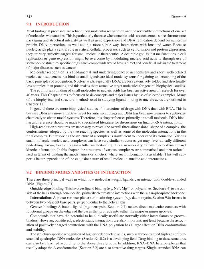
9.1 INTRODUCTION
Most biological processes are reliant upon molecular recognition and the reversible interactions of one setof molecules with another. This is particularly the case where nucleic acids are concerned, since chromosomepackaging and structural integrity as well as gene expression and DNA replication depend on numerousprotein–DNA interactions as well as, in a more subtle way, interactions with ions and water. Becausenucleic acids play a central role in critical cellular processes, such as cell division and protein expression,they are very attractive targets for small molecule therapeutics. A desirable goal is that malfunctions in cellreplication or gene expression might be overcome by modulating nucleic acid activity through use ofsequence- or structure-specific drugs. Such compounds would have a direct and beneficial role in the treatmentof major diseases such as cancer.
Molecular recognition is a fundamental and underlying concept in chemistry and short, well-definednucleic acid sequences that bind to small ligands are ideal model systems for gaining understanding of thebasic principles of recognition. Nucleic acids, especially DNA, are less extensively folded and structurallyless complex than proteins, and this makes them attractive target molecules for general biophysical studies.
The equilibrium binding of small molecules to nucleic acids has been an active area of research for over40 years. This Chapter aims to focus on basic concepts and major issues by use of selected examples. Manyof the biophysical and structural methods used in studying ligand binding to nucleic acids are outlined inChapter 11.
In general there are more biophysical studies of interactions of drugs with DNA than with RNA. This isbecause DNA is a more attractive target for anticancer drugs and DNA has been much easier to synthesizechemically to obtain model systems. Therefore, this chapter focuses primarily on small molecule–DNA bind-ing and reference should be made to specialized literature for discussions on ligand–RNA interactions.
High-resolution structures are necessary to reveal the overall three-dimensional shape of a complex, theconformations adopted by the two reacting species, as well as some of the molecular interactions in thefinal complex. But resolving the structure of a complex is insufficient to understand its formation. Varioussmall molecule–nucleic acid complexes can have very similar structures, yet may have radically differentunderlying driving forces. To gain a fuller understanding, it is also necessary to have thermodynamic andkinetic information. In this chapter, the structures of various complexes are summarised and then rational-ized in terms of binding thermodynamics or kinetics, where such information is available. This will sup-port a better appreciation of the exquisite nature of small molecule–nucleic acid interactions.
9.2 BINDING MODES AND SITES OF INTERACTION
There are three principal ways in which low molecular weight ligands can interact with double-strandedDNA (Figure 9.1).
Outside-edge binding: This involves ligand binding (e.g. Na�, Mg2� or polyamines, Section 9.4) to the out-side of the helix through non-specific, primarily electrostatic interactions with the sugar–phosphate backbone.
Intercalation: A planar (or near planar) aromatic ring system (e.g. daunomycin, Section 9.6) inserts inbetween two adjacent base pairs, perpendicular to the helical axis.
Groove binding: A bound ligand (e.g. netropsin, Section 9.7) makes direct molecular contacts withfunctional groups on the edges of the bases that protrude into either the major or minor grooves.
Compounds that have the potential to be clinically useful are normally either intercalators or groove-binders. However, outside-edge, electrostatic interactions are also important, not least because the associ-ation of positively charged counterions with the DNA polyanion has a large effect on DNA conformationand stability.
The structure-specific recognition of higher-order nucleic acids, such as three-stranded triplexes or four-stranded quadruplex DNA molecules (Section 9.10.2) is a developing field. Drug binding to these structurescan also be classified according to the above three groups. In addition, RNA–DNA heteroduplexes thatusually adopt the A-conformation (Section 2.2) are also attractive drug targets. Single-stranded RNA can
342 Chapter 9

form intramolecular folds with regions of duplex, internal loops, bulges and hairpins, for example, in ribo-somes, tRNA, or genomic viral RNA (Chapter 7). Such non-standard duplex folds in RNA offer a varied arrayof molecular architectures that can be exploited as drug targets, for example, those found in pathogenic RNAviruses such as HIV-1.
9.3 COUNTER-ION CONDENSATION AND POLYELECTROLYTE THEORY
Nucleic acids are highly charged polyanions, and thus polyelectrolyte effects have a major impact on theirbiological behaviour. For example, the polyanionic nature of nucleic acids directly affects the binding ofcharged drugs and proteins and the denaturation and folding of nucleic acids as well as the condensationand packaging of DNA and RNA in cells.
A nucleoside monophosphate has two ionizable hydroxyl groups of pKa � 1 and ca. 6.8 (Figure 2.5).These pKa values are only slightly dependent on the location of the phosphate on the nucleoside or whichof the bases is involved. At pH 7.0 and above, a terminal phosphate group has two negative charges whilean internucleoside phosphate diester has one.
Monovalent cations bind to nucleic acids in a somewhat unusual manner that is unique to highlycharged linear polyelectrolytes. In the early 1970s Gerald Manning described such binding as counter-ioncondensation:1–3 territorial binding of ions that are constrained to stay within a few angstroms of the DNAsurface, yet are free to move along the helix. Hence cations associate with nucleic acids largely as a functionof polymer charge density. To be stable, DNA must associate with counter-ions from solution. Additionalcounter-ions are associated with remaining charges on the polyanion through Debye–Hückel interactions.
The initial interaction of counter-ions with nucleic acids is referred to as condensation because the cationsgenerally form a cloud around the charge density of the nucleic acid and are not bound at specific sites.The ions retain their inner sphere water of hydration and they move up and down the phosphate backbone.Secondary hydration layers of both the ion and the nucleic acid are affected by this interaction. With B-typeduplex DNA, counter-ion condensation theory predicts an average of 0.76 monovalent counter-ions condensedper phosphate group, rising to a total of 0.88 counter-ions per phosphate group including Debye–Hückel typeinteractions.
The effect of counter-ion condensation is to reduce the effective charge on the nucleic acid. This in turnhas a profound effect on solution properties, binding interactions, and stability of nucleic acids. A proportionof the overall free energy for binding intercalators and groove-binders can be derived from the entropicallyfavourable release of condensed counter-ions upon binding. At constant temperature, the entropic effectresulting from counter-ion release can also lead to nucleic acid denaturation as well as to an apparent
Reversible Small Molecule–Nucleic Acid Interactions 343
Figure 9.1 Schematic representations of the three primary binding modes for ligand–duplex DNA binding. The ligand is shown in red and the DNA chains are in black: (a) Outside-edge binding. (b) Intercalation. (c) Groove binding

increase in binding affinity for cationic ligands as bulk salt concentration is decreased. Hence both the melt-ing temperature (Tm, Section 2.5.1) and the observed equilibrium association constant (Kobs) are stronglydependent on salt concentration. When values for these thermodynamic parameters are measured, it is import-ant to take account of the solution conditions, especially salt concentration.
In Manning’s original formulation, condensation is described by a dimensionless structural parameter �
(9.1)
where e is the electronic charge magnitude, � is the bulk solution dielectric constant, k Boltzmann’s con-stant, T the temperature in K, and b the average phosphate group spacing. If B-DNA is modelled as a cylin-der with two negatively charged phosphates spaced 3.4 Å apart, then b � 1.7 Å and � � 4.2. The Manningtheory states that the average fraction of monovalent cation associated with each DNA phosphate as aresult of condensation (�c) is defined as
(9.2)
For B-DNA �c � 0.76. However, as noted by Thomas Record, cations not only associate via condensa-tion but can also bind through Debye–Hückel interactions. This fraction of associated counterions is definedas �s. Therefore, the overall fraction of monovalent cations associated with each phosphate can be definedas � where
(9.3)
For B-DNA the value of � is 0.88, i.e. the double helix retains a net charge equal to 12% of the totalnumber of phosphates. Record has developed a particularly useful formulation that describes the effect ofsalt concentration on nucleic acid equilibria.4 He applied Wyman’s concept of linkage to quantify salt effectson ligand binding. The magnitude of the apparent ligand binding constant Kobs for the interaction of a chargedligand with DNA is found to be strongly dependent on monovalent salt concentration [MX], as embodiedin the expression
(9.4)
where Z is the charge on the ligand and SK is the slope of a plot of � ln Kobs versus � ln[MX]. The negativesign of Z� in Equation 9.4 indicates that Kobs decreases with increasing concentrations of MX. The magnitudeof the decrease depends on Z, the charge on the ligand. In this analysis, any possible contribution fromchanges in anion binding or hydration is omitted. For a cationic ligand L binding to a nucleic acid site D toform a ligand–nucleic acid complex C in the presence of monovalent cation M�, the thermodynamic equi-librium can be written as
(9.5)
This equilibrium describes the release of bound counter-ions concomitant with the binding of a cationicligand. The equilibrium constant for the above reaction is
(9.6)
Hence for the association of a positively charged ligand with DNA in the presence of excess monovalentcation,
KC M
D L
Z
�� �[ ] [ ]
[ ] [ ]
⋅⋅
D L C Z M� � � �� [ ]
�
��
ln
ln[ ]
K
MXZ SKobs � �
� � � �� � � c s [ ( ) ]1 2 1
� �c � ( )1 1
� �e
kTb
2
ε
344 Chapter 9

(9.7)
and
(9.8)
where ln KT and the corresponding free energy term GT refer to a standard state of [MX] of 1 M � Gpe isthe polyelectrolyte contribution to the overall free energy
(9.9)
Gpe is a favourable free energy contribution that arises from the entropically favoured release ofcounter-ions into bulk solvent on binding of a cationic ligand. By experimental determination of the valueof � ln Kobs/� ln[MX], Gpe can be calculated at any given salt concentration through use of Equation 9.9. Fromexperiments and simulations, it is clear that Gpe can be quite large, even at modest salt concentrations,and it can contribute several kJ to the overall binding free energy. The difference between Gpe and theexperimental Gobs (determined at the same salt concentration) is equal to GT, which is the nonpolyelec-trolyte contribution to free energy. GT is independent of salt concentration and represents the free energyarising from all types of interactions other than coupled polyelectrolyte effects, such as van der Waalsinteractions, hydrogen bonding, and hydrophobic effects. The use of polyelectrolyte theory to probe theenergetics of drug–DNA interactions has been discussed in a useful review article.5
9.3.1 Intercalation and Polyelectrolyte Theory
To accommodate an intercalating ligand, the nucleic acid base pairs at the intercalation site must be sep-arated by an additional 3.4 Å (Section 9.6). This lengthens the helix and increases the inter-phosphate spa-cing, parameter b in Equation 9.1. It then leads to a decrease in � and a decrease in the fraction ofmonovalent cations associated per phosphate group. For intercalation, there are two distinct factors thatcontribute to the polyelectrolyte effect: (1) release of condensed counter-ions following binding of a posi-tively charged ligand (as discussed above), and (2) a contribution that arises from increased phosphate spa-cing that results from intercalation-induced conformational changes in the DNA. For a neutral intercalator, theobserved binding constant is still dependent on salt concentration, since the intercalator induces a structuraltransition upon binding.
David Wilson has described a modification of Record’s theory that accounts for ion release arising from aDNA structural transition.6 He predicted values for � ln Kobs/� ln[MX] of 1.06 and 1.89 for the binding ofmono- and dicationic intercalators, respectively. Later Friedman and Manning used the same general conceptbut with a different theoretical model to predict values for � ln Kobs/� ln[MX] of 0.24 and 1.24 for the bind-ing of uncharged and monocationic intercalators, respectively.7
9.4 NON-SPECIFIC OUTSIDE-EDGE INTERACTIONS
There are several types of molecule that can interact non-specifically with the nucleic acid phosphate backbone through mainly electrostatic interactions. The archetypal outside-edge binding drugs are thepolyamines, spermine and spermidine (Figure 9.2).
The nonspecific nature of the interaction between DNA and polyamines means that it is difficult toobtain high-resolution NMR or crystallographic structural data on these complexes. In fact these ligandsare often used to reduce charge–charge effects in crystallographic studies, since they can rarely be seen athigh-density sites in a resolved structure.
Polyamines such as spermine are ubiquitous in eukaryotic cells and these ligands are thought to playmultiple roles in cellular function. One important role is in DNA packaging into chromatin, but the exact
� �G Z RT MXpe ln[ ]
� � G G Gobs T pe
ln ln ln[ ]K K Z MXobs T� �
Reversible Small Molecule–Nucleic Acid Interactions 345
mechanism of action is unknown. However, it is likely that polyamines play an analogous role to histonesin that they may neutralise some of the negative charges on the DNA backbone and hence promote DNApackaging. Direct binding of polyamines to DNA and the resulting modulation of protein–DNA inter-actions appears to play an important role in cell proliferation.9
9.5 HYDRATION EFFECTS AND WATER–DNA INTERACTIONS
Hydration of DNA is essential for it to maintain its folded conformation.9 Indeed the hydration state andinteractions with ions can exert large effects on nucleic acid conformation and hence influence the natureof interactions with other ligands such as proteins (Section 10.4.2). Early fibre diffraction studies (Section 1.4)showed that relative humidity has an effect on DNA conformation, such that B-type DNA is found at highhumidity values (above 85%) whereas A-form DNA occurs with lower humidity (between 75–80%) andDNA becomes disordered if the relative humidity is lowered to between 55 and 75%. In addition, base-composition and salt concentration affect the values of relative humidity under which one or other con-formation is favoured.
In practice, it is very challenging to study water–DNA interactions experimentally. Useful data havebeen obtained from gravimetric studies and infrared (IR) spectra of DNA films as a function of relativehumidity. Visualization of specific water molecules from X-ray diffraction analysis of single DNA crys-tals has also proved difficult in the past. This is because a crystal is derived typically from aqueous solu-tion that also contains cations such as Mg2� or spermine, and alcohols such as 2-methyl-2,4-pentanediol(MPD7). Thus at resolutions of 2 Å or above, it is difficult to distinguish whether a particular electron den-sity corresponds to water, a cation, or a spermine molecule.
Much of our present understanding of DNA hydration (as well as other biophysical characteristics of DNA)has come from studies of the Dickerson–Drew dodecamer (Section 2.2.4, Figure 2.17). This oligonucleotideduplex, with the sequence d(CGCGAATTCGCG)2, provided the first published B-DNA X-ray crystal struc-ture. It is the best studied DNA sequence with over 68 isomorphous members of the d(CGCXAATTYGCG)2
(X � G or A; Y � C or T) dodecamer family, and numerous biophysical and binding studies have beencarried out with it in solution. In the early 1980s, Dickerson and co-workers obtained several structures ofthis sequence, for example, at room temperature, at 16 K or at 7°C for the 9-bromo derivative in MPD7.10
Hydration was not readily visible in the room temperature structure due to high B values. However the16 K and MPD7 structures were extensively hydrated with 65 solvent peaks and an average of three watersper phosphate. In the major groove, 19 solvent molecules formed a first hydration layer around nitrogenand oxygen atoms of the bases in that groove. A second hydration layer contained a further 36 solvent mol-ecules that formed clusters spanning the major groove. In the minor groove, a zigzag pattern of water mol-ecules formed a “spine of hydration” in all the structures (Section 2.2.4, Figure 2.18). This concept of a spineof hydration has become pervasive in DNA chemistry and is often invoked to explain a multitude of effectsrelated to DNA stability and ligand binding.
346 Chapter 9
Figure 9.2 Structures of two outside-edge electrostatic DNA binding ligands, spermine and spermidine
Crystallographic studies have also shown that minor groove width is instrumental in dictating hydration pat-terns. The duplex d(CCAACGTTGG)2 has a narrow minor groove that is about 4.2 Å wide and it has a spineof hydration similar to that originally observed in the Dickerson–Drew sequence. The helix d(CCAA-GATTGG)2 sequence has a wider, 7.2 Å minor groove within which two side-by-side ribbons of water mol-ecules are found. Interestingly, the sequence d(CCAGGCCTGG)2 has a minor groove of intermediate width,which is occupied only very sparsely by ordered water molecules and which does not possess either of the twoother hydration patterns. It has been suggested that the A�T minor groove spine of hydration is important instabilization of B-DNA against the B→A conformational transition. High A�T content DNA is associatedwith narrow minor grooves and resistance to the B→A transition, which has led to the assertion that the spineof hydration is more effective at stabilization of B-DNA than the two parallel ribbons of waters (Section 2.2.4).
9.5.1 Cation Binding in the Minor Groove
The seminal work of Dickerson and Drew has been central to the current view that DNA is conformationallypolymorphic. While many DNA structures have been solved to a resolution of 2.0–3.0 Å, Loren Williams hasachieved a resolution of 1.4 Å, thus allowing a more accurate analysis of the dodecamer structure and reveal-ing some new insights into DNA hydration.11–13 His principal conclusion is that the primary spine of hydra-tion is composed partly of sodium ions. In other words, there is partial occupancy of primary sites of solvationin the minor groove by both water and sodium cations. There is also a secondary, regular and highlyordered spine of water molecules on top of the primary spine that does not interact directly with the DNA.This DNA structure is seen to be more conformationally ordered and not as heterogeneous as thought earlier.
However, in practice, partial occupancy (known as a sodium–water hybrid) is difficult to distinguish froma water molecule alone, especially if sodium occupancy is less than 50%. This is because sodium ions andwater molecules have the same number of electrons and therefore yield electron density peaks of similar size.In addition, both water and sodium ions have a wide variation in their coordination geometries. To confirm thehybrid–solvent model proposed for the high-resolution Dickerson–Drew dodecamer, Williams carried outpotassium substitution experiments12 to take advantage of the fact that a potassium ion can readily be distin-guished from water because of its larger number of electrons. These studies have confirmed that monovalentcations penetrate the primary layer of the spine of hydration and are also present in the secondary hydrationlayer. The spine of hydration forms only the bottom two layers of a four-layer arrangement of solvent and cations in the A�T minor groove, which are organised to form a repeating motif of fused hexagons(Figure 9.3).
This work has implications for binding of other small molecules to DNA minor grooves. A substantialfavourable entropic contribution to binding free energy may arise from positioning positively charged,minor groove-binding ligands or cationic ancillary functional groups of intercalators (e.g. the amino sugarof daunomycin) into A�T minor grooves that contain sodium or potassium ions bound in specific sites.Favourable entropic effects will arise when monovalent metal cations are expelled from the minor grooveby cationic ligands.
9.6 DNA INTERCALATION
9.6.1 The Classical Model
Leonard Lerman first proposed the intercalation model for ligand binding to DNA in the early 1960s.14,15
Working with aminoacridines and proflavin, he postulated that DNA unwinds slightly to open a space intowhich the flat polycyclic aromatic chromophores can be inserted between adjacent base pairs. This mode ofbinding has now been established for a large number of planar heteroaromatic or polycyclic aromatic ringstructures (Figure 9.4).
A common structural feature of all intercalators is that they possess an extended, electron-deficient,planar aromatic ring system that is often referred to as a chromophore. Such compounds typically have
Reversible Small Molecule–Nucleic Acid Interactions 347
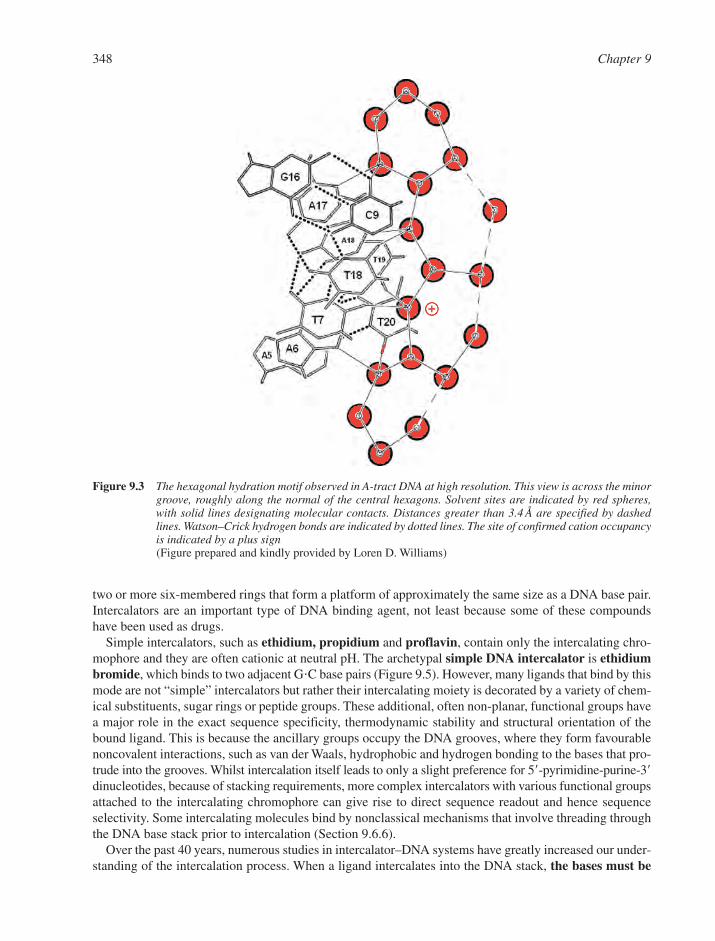
two or more six-membered rings that form a platform of approximately the same size as a DNA base pair.Intercalators are an important type of DNA binding agent, not least because some of these compoundshave been used as drugs.
Simple intercalators, such as ethidium, propidium and proflavin, contain only the intercalating chro-mophore and they are often cationic at neutral pH. The archetypal simple DNA intercalator is ethidiumbromide, which binds to two adjacent G�C base pairs (Figure 9.5). However, many ligands that bind by thismode are not “simple” intercalators but rather their intercalating moiety is decorated by a variety of chem-ical substituents, sugar rings or peptide groups. These additional, often non-planar, functional groups havea major role in the exact sequence specificity, thermodynamic stability and structural orientation of thebound ligand. This is because the ancillary groups occupy the DNA grooves, where they form favourablenoncovalent interactions, such as van der Waals, hydrophobic and hydrogen bonding to the bases that pro-trude into the grooves. Whilst intercalation itself leads to only a slight preference for 5�-pyrimidine-purine-3�dinucleotides, because of stacking requirements, more complex intercalators with various functional groupsattached to the intercalating chromophore can give rise to direct sequence readout and hence sequenceselectivity. Some intercalating molecules bind by nonclassical mechanisms that involve threading throughthe DNA base stack prior to intercalation (Section 9.6.6).
Over the past 40 years, numerous studies in intercalator–DNA systems have greatly increased our under-standing of the intercalation process. When a ligand intercalates into the DNA stack, the bases must be
348 Chapter 9
Figure 9.3 The hexagonal hydration motif observed in A-tract DNA at high resolution. This view is across the minorgroove, roughly along the normal of the central hexagons. Solvent sites are indicated by red spheres,with solid lines designating molecular contacts. Distances greater than 3.4 Å are specified by dashedlines. Watson–Crick hydrogen bonds are indicated by dotted lines. The site of confirmed cation occupancyis indicated by a plus sign(Figure prepared and kindly provided by Loren D. Williams)
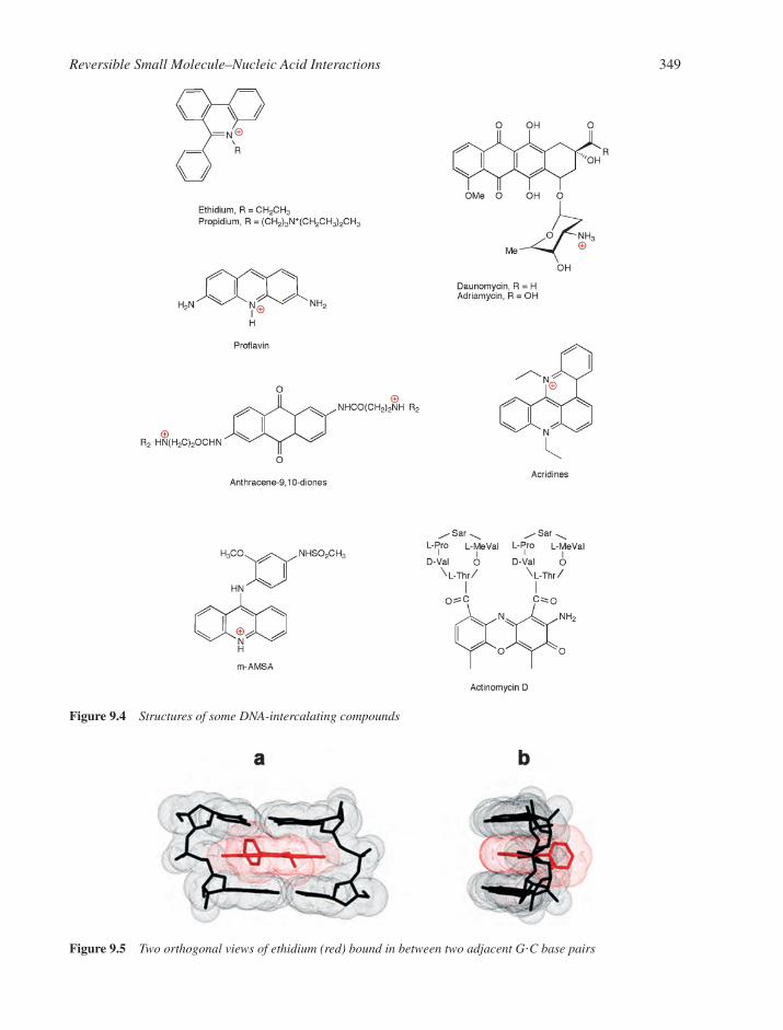
Reversible Small Molecule–Nucleic Acid Interactions 349
Figure 9.4 Structures of some DNA-intercalating compounds
Figure 9.5 Two orthogonal views of ethidium (red) bound in between two adjacent G�C base pairs

separated by approximately 3.4 Å (the van der Waals thickness of a phenyl ring) to accommodate the lig-and. This base pair separation can only occur if there is rotation about torsional bonds in the phosphodi-ester backbone, such that unwinding around the helical axis occurs.
Intercalation of multiple ligands into a stretch of short rod-like DNA and concomitant helix unwindingleads to an overall lengthening of the duplex. This change in DNA length is reflected as a change inhydrodynamic character, and hence the intrinsic viscosity and sedimentation coefficient of the DNAchange upon intercalation. The extent of helix unwinding produced by intercalation is dependent on thenature of the bound drug, especially its geometry and physico–chemical properties. The amount of helixunwinding can be determined experimentally by use of covalently closed circular DNA, with referenceto a standard value of 26° per bound ethidium bromide drug. For canonical B-DNA, the normal backbonetwist angle is 36°. Binding of ethidium reduces this twist angle to 10° and hence the DNA unwinds by 26°.Binding of proflavin or other related acridines causes unwinding of DNA by about 17°, whereas bindingof anthracycline antibiotics results in only 11° unwinding of DNA. The effects of localised unwinding atthe intercalation site are propagated along the DNA helix and result in structural perturbations over longdistances from the bound drug. The helical twist angle for the two base pairs immediately surrounding thebound drug may be little changed from 36°. In such cases, there is usually a significant helical unwindingat adjacent base pairs. Hence the unwinding angle is the sum of cumulative changes in helical twist acrossall affected base pairs.
The process of base-pair separation that occurs during intercalation also produces a number of otherchanges in backbone conformation and base-pair and base-step geometry. In the X-ray-derived molecularstructure of ethidium intercalated between a G�C dinucleotide (Figure 9.5), ethidium is stacked with itslong axis parallel to the long axis of the adjacent base pair. The exocyclic amino groups point towards thediester oxygen atoms of the DNA phosphate groups and provide additional electrostatic and hydrogen-bondstabilization of the complex. The base pairs are slightly kinked, since the out-of-plane phenyl group limitsfull intercalation of the cationic phenanthridinium ring (Figure 9.5a). The phenyl and ethyl substituents ofethidium lie in the minor groove of the complex.
Various molecular events contribute to the overall free energy observed for intercalation reactions. Someof these are enthalpic in nature and others entropic. One of the most important energetic driving forces fordrug binding is the favourable hydrophobic transfer of the drug from bulk solvent into the DNA bindingsite. The non-polar ligand is removed from the aqueous environment and this disrupts the hydration layeraround the ligand, resulting in an entropically favourable release of site-specific water molecules. The DNAbinding site must undergo an energetically unfavourable conformational transition to form a cavity into whichthe drug intercalates. Whilst this deformation is unfavourable from a point-of-view of configuration, it hasadditional features that give rise to a favourable entropy change. Localized unwinding of the helix at the inter-calation site results in an increase in the distance between adjacent phosphates on the backbone. This givesrise to a reduction of localized charge density and hence the release of condensed counter-ions (Section9.3.1). Additional counter-ions are released during the process of intercalation of the cationic ligand owingto the polyelectrolyte effect (Section 9.3).
Ligand intercalation is also associated with favourable enthalpic contributions to free energy arisingfrom the formation of noncovalent interactions between the drug and base pairs. These noncovalent inter-actions involve several different forces, such as the hydrophobic effect, reduction of coulombic repulsion,van der Waals interactions, �-stacking, and hydrogen bonding. Quantitative apportionment of the overallbinding free energy for intercalators and groove binders16–18 is discussed later (Sections 9.6.4 and 9.7.5).
9.6.2 The Anthracycline Antibiotic Daunomycin
The anthracycline antibiotics, daunomycin (daunorubicin) and adriamycin (doxorubicin) as well as some derivatives have been frontline anticancer drugs for many years. Daunomycin is perhaps the best-studied intercalating drug to date with over 20 high-resolution structures known and numerous stud-ies on the kinetics and thermodynamics of its interaction with DNA.19
350 Chapter 9
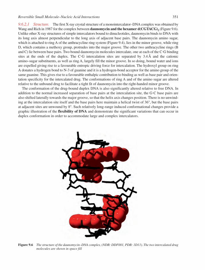
9.6.2.1 Structure. The first X-ray crystal structure of a monointercalator–DNA complex was obtained byWang and Rich in 1987 for the complex between daunomycin and the hexamer d(CGTACG)2 (Figure 9.6).Unlike other X-ray structures of simple intercalators bound to dinucleotides, daunomycin binds to DNA withits long axis almost perpendicular to the long axis of adjacent base pairs. The daunomycin amino sugar,which is attached to ring A of the anthracycline ring system (Figure 9.4), lies in the minor groove, while ringD, which contains a methoxy group, protrudes into the major groove. The other two anthracycline rings (Band C) lie between base pairs. Two bound daunomycin molecules intercalate, one at each of the C�G bindingsites at the ends of the duplex. The C�G intercalation sites are separated by 3.4 Å and the cationicamino–sugar substituents, as well as ring A, largely fill the minor groove. In so doing, bound water and ionsare expelled giving rise to a favourable entropic driving force for intercalation. The hydroxyl group on ringA donates a hydrogen bond to N-3 of guanine and it is a hydrogen-bond acceptor for the amino group of thesame guanine. This gives rise to a favourable enthalpic contribution to binding as well as base pair and orien-tation specificity for the intercalated drug. The conformations of ring A and of the amino sugar are alteredrelative to the unbound drug to facilitate a tight fit of daunomycin into the right-handed minor groove.
The conformation of the drug-bound duplex DNA is also significantly altered relative to free DNA. Inaddition to the normal increased separation of base pairs at the intercalation site, the G�C base pairs arealso shifted laterally towards the major groove, so that the helix axis changes position. There is no unwind-ing at the intercalation site itself and the base pairs here maintain a helical twist of 36°, but the base pairsat adjacent sites are unwound by 8°. Such relatively long-range induced conformational changes provide agraphic illustration of the flexibility of DNA and demonstrate the significant variations that can occur induplex conformation in order to accommodate large and complex intercalators.
Reversible Small Molecule–Nucleic Acid Interactions 351
Figure 9.6 The structure of the daunomycin–DNA complex, (NDB: DDF001, PDB: 1D11). The two intercalated drugmolecules are shown in space fill
9.6.2.2 Thermodynamics of Daunomycin: DNA Interactions. The details of the energetics ofanthracycline–DNA interactions have been studied by several groups.19 In most thermodynamic charac-terizations, the initial step involves determination of the binding free energy. This is readily achieved bymeasurement of the equilibrium association (or binding) constant and use of the standard Gibbs relationship,
(9.10)
where Kobs is the observed equilibrium constant, R the gas constant and T the temperature. Brad Chaires hasconducted equilibrium binding experiments and constructed binding isotherms that span a 100-fold rangein free ligand concentration. Such essentially complete binding curves allow the free energy to be estimatedin a model-free manner by use of Wyman’s concept of median ligand activity. The free energy of ligation(GX) is defined as the free energy to go from a state where no ligand is bound to a degree of saturation X
–.
For the binding of daunomycin to calf-thymus DNA, the value of GX was found to be 32.6 kJ mol1 forthe full ligation of a daunomycin binding site.
The daunomycin–DNA binding affinity is dependent on bulk salt concentration, as is explained by thepolyelectrolyte theories of Record and Manning (Section 9.3). Daunomycin carries one positive charge atneutral pH, which arises from protonation of the amine (pKa � 8.2) on the sugar moiety. Binding of cationicligands such as daunomycin to DNA results in the entropically favourable release of condensed counter-ionsfrom the DNA sugar–phosphate backbone. One practical consequence of polyelectrolyte theory is that itallows the observed binding free energy to be partitioned into two components, Gpe and GT, the polyelectrolyte and nonpolyelectrolyte contributions, respectively. The former is the free energy arising fromcounter-ion release. For daunomycin binding to DNA, Gpe is 4.2 kJ mol1 at a reference salt concen-tration of 0.2 M NaCl. This is about 13% of the total binding free energy.
Once the free energy for a binding interaction has been determined, the next step in a detailed thermo-dynamic characterization is to determine the enthalpic and entropic components of the free energy. Thesecontributions are defined using the standard thermodynamic relationship,
(9.11)
where H and S are the enthalpy and entropy changes accompanying binding. At 20°C, H for thedaunomycin–DNA interaction is 37.6 kJ mol1. By use of Equation 9.11, TS is then calculated as5.0 kJ mol1, and hence at 20°C the value of S is 17.1 J mol1 K1. The negative sign for S indicatesthat entropy decreases upon binding. This unfavourable entropy term is overcome by a large favourable(negative) enthalpy that drives the binding. The binding enthalpy for daunomycin is strongly dependent ontemperature. This is a consequence of a binding-induced change in heat capacity (Cp) that accompaniesthe interaction:
(9.12)
By measurement of the enthalpy as a function of temperature and calculation of the slope of the resultinglinear graph, the Cp can be determined. For daunomycin binding to DNA, this value is 669 J mol1 K1.Determination of heat capacity change is useful, since this parameter can yield information on changes inhydrophobic interactions and removal of nonpolar solvent accessible surface area. It is an important, experi-mentally measurable quantity that is now used to carry out detailed partitioning of free energy terms(Section 9.6.4).
Studies of daunomycin and adriamycin binding to DNA have revealed important differences betweenthe two drugs and served to illustrate the value of thermodynamic characterisation in the context of rationaldrug design. The structures of the two ligands bound to DNA are virtually superimposable and molecular
��
�C
H
Tp
� G H T S
� G RT Kln obs
352 Chapter 9
interactions are equivalent, with only subtle differences in hydration and ion binding. However, adri-amycin has a binding affinity for DNA that is 10-fold greater than that for daunomycin. In addition, bindingof adriamycin to DNA is accompanied by a large favourable entropy term, which is the exact opposite of thesituation for daunomycin. As a consequence, the binding enthalpy of adriamycin is significantly less thanthat measured for daunomycin, but the free energy is greater for adriamycin. These clear and unambigu-ous differences in the binding thermodynamics of two very similar drugs are not predicted from examin-ation of the structures of the two drug–DNA complexes. Thus isostructural does not mean isoenergetic.
9.6.3 The Neighbour Exclusion Principle
Simple intercalators might be expected to occupy every potential intercalation site, i.e. in between everybase pair in a DNA helix. Therefore, at saturation this would give rise to a binding stoichiometry of onebase pair per bound intercalator. Molecular modelling studies where a drug molecule is bound at everypossible intercalation site have led to energy-minimized structures with reasonable backbone torsionangles. However, solution studies have generated a different picture. In 1962 Cairns was the first toobserve that even simple intercalators, such as proflavin, show saturation with DNA at a stoichiometry ofone drug molecule per 2 base pairs. More complex intercalators such as daunomycin occupy 1 in 3 sites.Such empirical observations are a reflection of the neighbour exclusion principle, which states that inter-calators can, at most, only bind at alternate possible base pair sites on DNA. Hence there is only ever amaximum of one intercalator between every second potential binding site. In the first instance, prior toaddition of any ligand, all inter-base pair spaces have an equal potential to be intercalation sites for a non-specific compound (ignoring sequence preference). However, when an intercalator binds to a certain site,the neighbour exclusion principle states that binding of additional ligand molecules at sites immediatelyadjacent is inhibited.
Many attempts have been made to account for the molecular basis of neighbour exclusion. One possi-bility is that intercalator binding induces conformational changes at adjacent sites in the DNA and the newconformation is structurally or sterically unable to accommodate another mono-intercalator. Friedman andManning have advanced an explanation based on counter-ion release and electrostatic effects. Briefly,binding of cationic intercalators has the effect of neutralization of some of the charge on the DNA back-bone. In other words, binding of a cationic intercalator lengthens the DNA and hence increases the aver-age charge spacing, and therefore decreases the linear charge density. In consequence, the binding-induced,energetically favourable release of condensed counter-ions is reduced and thus the observed equilibriumbinding constant is also diminished. This effect leads to curvature in equilibrium binding isotherms that issimilar to that predicted by neighbour exclusion theory. Since the local release of ions at the intercalationsite would be greater than ion release at some distance from the binding site, then the local release of counter-ions could also lead to a lower free energy of binding at sites next to an intercalation site. In practice, it is likelythat both conformational and electrostatic factors play a role in the neighbour exclusion phenomena. Inaddition, other factors may also be responsible for neighbour exclusion, for example, electrostatic repulsionbetween proximally bound drugs.
Various mathematical solutions have been developed that allow drug binding data to be described by theneighbour exclusion model.20,21 In one of these approaches, the DNA is assumed to be a lattice sufficientlylong to allow end effects to be ignored while all potential binding sites are assumed to be equivalent.McGhee and Von Hippel first wrote the neighbour exclusion model in this form in 1974:
(9.13)
where r is the binding ratio (defined as the ratio of the concentration of bound drug (Cb) over the total con-centration of DNA binding sites (S), Cf the concentration of free drug, K the equilibrium binding constant for
r
CK nr
nr
n r
n
f
�
( )( )
11
1 1
1
Reversible Small Molecule–Nucleic Acid Interactions 353
all sites, i.e. any sequence dependence for drug binding is ignored, and n the binding site size, i.e. the numberof base pairs occupied by a bound drug. The neighbour exclusion model predicts that binding isothermsplotted in the form r/Cf versus r should be initially linear but should curve asymptotically as values of r/Cf
approach zero. This is exactly what is observed in experiments, and the neighbour exclusion model candescribe accurately the binding of intercalators to DNA.22
However, it is common for non-integral numbers for the parameter n to emerge from this analysis, whichimplies that the neighbour exclusion model is not adequate to describe the experimental data. Obvious rea-sons for this are that certain assumptions are not appropriate for the particular system under study. In suchcases, more complex versions of the model that account for sequence specificity or the influence of co-operativity are required (Section 9.9).
9.6.4 Apportioning the Free Energy for DNA Intercalation Reactions
The likelihood of success in rational DNA-binding drug design programmes would be greatly enhanced if,in addition to structural studies, thermodynamic analyses were to be carried out for a wide range of ligand–DNA complexes. Determination of the free energy change for binding is useful but insufficient. As a min-imum, the overall observed free energy should be examined in terms of component enthalpic and entropicterms. Modern advances in titration microcalorimetry have given experimentalists the ability to under-take novel and detailed thermodynamic investigations into ligand–DNA interactions. These tools allowthe drug–DNA binding free energy to be apportioned in great detail.
One approach involves evaluation of a minimum set of free energy terms, which are assumed to be addi-tive that can account for the experimentally determined value of Gobs.
16,18 Based on current concepts ofligand–DNA interactions, it is reasonable to describe Gobs as being composed of at least five componentfree energy terms. Thus,
(9.14)
where Gconf is the unfavourable free energy term arising from conformational changes in the DNA andligand, Gr�t is an unfavourable term that results from losses in rotational and translational degrees of free-dom upon complex formation, Ghyd is the free energy for the hydrophobic transfer of ligand from bulksolvent into the DNA binding site, Gpe (Section 9.3) is the electrostatic (polyelectrolyte) contribution tothe observed free energy that arises from coupled polyelectrolyte effects such as counter-ion release (entrop-ically favourable) and Gmol is the free energy term arising from weak non-covalent interactions, such asvan der Waals, hydrogen bonding, dipole–dipole interactions, etc.
Each of the terms in Equation 9.14 can be estimated using semi-empirical or theoretical methods. Atleast two of the terms, Gconf and Gr�t, make unfavourable contributions to binding free energy. Kineticstudies have revealed that the conformational transition in DNA that accompanies binding of a simple inter-calator such as ethidium has a free energy cost of �16.7 kJ mol1. Therefore, this value can be set as the ener-getic cost of structurally perturbing DNA to form an intercalation site. When a bimolecular complex isformed, there is an associated loss of rotational and translational degrees of freedom. This gives rise to a largeentropic cost for binding, which is reflected in the free energy term Gr�t. There is some debate as to theprecise value for this parameter.16 However, a value of �62.8 kJ mol1 for Gr�t for a rigid body interactionis not unreasonable. Hence the two unfavourable free energy terms are set as constants for all intercalator bind-ing reactions.
For a stable ligand–DNA complex to form, the remaining free energy terms must be large enough andfavourable to overcome these two unfavourable contributions. It has been experimentally demonstrated forintercalators (and groove binders) that the major favourable free energy term that drives binding is thehydrophobic transfer of drug from the aqueous environment to the DNA binding site. It is possible to quantify
� � � � � �G G G G G Gobs conf r t hyd pe mol
354 Chapter 9
Ghyd by using algorithms derived from semi-empirical relationships based on heats of solvent transfer ofsmall molecules and protein folding–unfolding equilibria. Record has shown that
(9.15)
From this equation, it is clear that Ghyd can be estimated in a simple and direct manner by measurementof the heat capacity change associated with a binding interaction. A near-complete thermodynamic analysis,including a detailed partitioning of free energy, has been presented recently for a range of DNA intercalators23
and reveals some interesting thermodynamic features of DNA intercalation by small molecules. The bindingof most intercalators is enthalpically driven. For ethidium and daunomycin this is opposed by an unfavourableentropy term while for propidium and adriamycin, there is a small favourable entropy term. These differencesin TS are likely to be due to differences in hydration of the ligands involved. Heat capacity changes forethidium, propidium, daunomycin, and adriamycin fall within a narrow range of about 630 J mol1 K1.
In general, heat capacity changes are smaller for intercalators than they are for groove binders. In add-ition, the heat capacity change for actinomycin D binding is much larger. Both these observations make sensewhen one considers that heat capacity change correlates with changes in nonpolar and polar solvent access-ible surface area. For drug–DNA interactions, the following empirical relationship has been established:
(9.16)
where Anp and Ap are binding-induced changes in nonpolar and polar surface areas, respectively. Thechange in nonpolar surface area for ethidium is 407 Å2, whereas for actinomycin D it is 1046 Å2. Intercalationof positively charged ligands gives rise to a small favourable polyelectrolyte term. For actinomycin D,which is uncharged, the value of Gpe is negligible.
For intercalation, the overwhelming energetic driving forces are hydrophobic interactions and other weak,non-covalent molecular interactions, such as van der Waals and hydrogen bonding. These free energy termsare large enough to overcome the �79.5 kJ mol1 energetic cost that arises from conformational changesand losses in degrees of rotational and translational freedom that accompanies binding. In the case of acti-nomycin D, molecular interactions make a small unfavourable contribution to binding free energy. This isalso the case for the groove binder, Hoechst 33258 (Section 9.7.5). Actinomycin D contains cyclic peptidemoieties that, like Hoechst 33258, occupy the DNA minor groove. Binding of a ligand in the minor groove hasthe effect of expulsion of site-specifically bound water or cation molecules. Hence water-base pair hydro-gen bonds are broken to form ligand-base pair hydrogen bonds. Therefore, from a thermodynamic point-of-view, a favourable energetic contribution arising from ligand hydrogen bonding may be limited.
9.6.5 Bisintercalation
Bisintercalators are bifunctional molecules that possess two planar intercalating aromatic ring systemscovalently linked by chains of varying length. It is also possible to link three or more ring systems togetherusing linkers. A good reason for designing and synthesising a bisintercalator is that it should have a sig-nificantly higher affinity and much slower dissociation kinetics than the monointercalator equivalents. Thebinding constant for a bisintercalator should be approximately the square of the monomer binding constant.Since biological activity is often closely correlated with binding affinity, bisintercalators should also haveenhanced medicinal application.
Bisintercalators have a larger site size than their monointercalator counterparts, which can lead to increasedsequence selectivity. Many simple monointercalators have a binding site size of 1 in 3 base pairs. Therefore,to avoid violation of the neighbour exclusion principle, a bisintercalator made from such monomers can beexpected to span a site of at least 6 base pairs, which is the same size as the recognition sites of many sequence-specific restriction endonuclease enzymes (Section 5.3.1).
Bisintercalators are also ideal model systems for probing and evaluating neighbour exclusion effects,because they can be prepared with variable linker lengths. Ligands with short linkers may be induced to
� C A Ap np p0 382 0 026 0 121 0 077. ( . ) . ( . )
� G Chyd p( )80 10
Reversible Small Molecule–Nucleic Acid Interactions 355
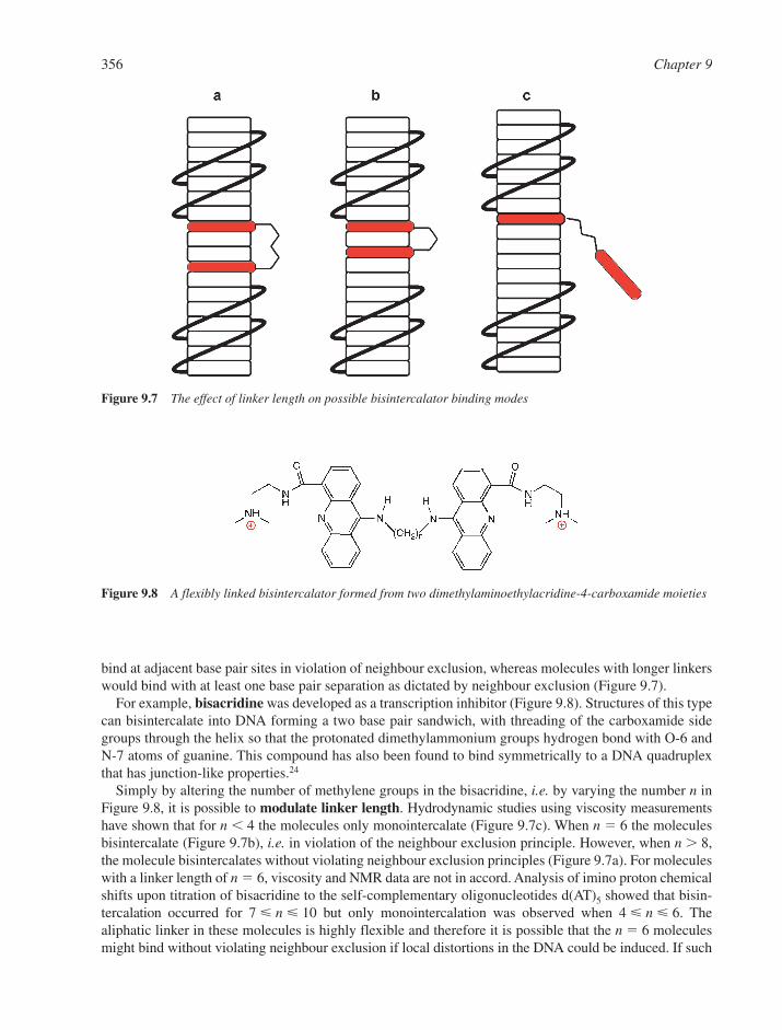
bind at adjacent base pair sites in violation of neighbour exclusion, whereas molecules with longer linkerswould bind with at least one base pair separation as dictated by neighbour exclusion (Figure 9.7).
For example, bisacridine was developed as a transcription inhibitor (Figure 9.8). Structures of this typecan bisintercalate into DNA forming a two base pair sandwich, with threading of the carboxamide sidegroups through the helix so that the protonated dimethylammonium groups hydrogen bond with O-6 andN-7 atoms of guanine. This compound has also been found to bind symmetrically to a DNA quadruplexthat has junction-like properties.24
Simply by altering the number of methylene groups in the bisacridine, i.e. by varying the number n inFigure 9.8, it is possible to modulate linker length. Hydrodynamic studies using viscosity measurementshave shown that for n � 4 the molecules only monointercalate (Figure 9.7c). When n � 6 the moleculesbisintercalate (Figure 9.7b), i.e. in violation of the neighbour exclusion principle. However, when n � 8,the molecule bisintercalates without violating neighbour exclusion principles (Figure 9.7a). For moleculeswith a linker length of n � 6, viscosity and NMR data are not in accord. Analysis of imino proton chemicalshifts upon titration of bisacridine to the self-complementary oligonucleotides d(AT)5 showed that bisin-tercalation occurred for 7 � n � 10 but only monointercalation was observed when 4 � n � 6. Thealiphatic linker in these molecules is highly flexible and therefore it is possible that the n � 6 moleculesmight bind without violating neighbour exclusion if local distortions in the DNA could be induced. If such
356 Chapter 9
Figure 9.8 A flexibly linked bisintercalator formed from two dimethylaminoethylacridine-4-carboxamide moieties
Figure 9.7 The effect of linker length on possible bisintercalator binding modes
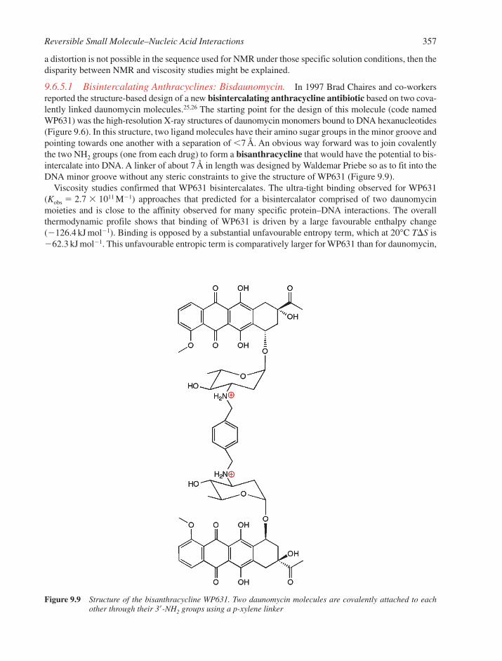
a distortion is not possible in the sequence used for NMR under those specific solution conditions, then thedisparity between NMR and viscosity studies might be explained.
9.6.5.1 Bisintercalating Anthracyclines: Bisdaunomycin. In 1997 Brad Chaires and co-workersreported the structure-based design of a new bisintercalating anthracycline antibiotic based on two cova-lently linked daunomycin molecules.25,26 The starting point for the design of this molecule (code namedWP631) was the high-resolution X-ray structures of daunomycin monomers bound to DNA hexanucleotides(Figure 9.6). In this structure, two ligand molecules have their amino sugar groups in the minor groove andpointing towards one another with a separation of �7 Å. An obvious way forward was to join covalentlythe two NH2 groups (one from each drug) to form a bisanthracycline that would have the potential to bis-intercalate into DNA. A linker of about 7 Å in length was designed by Waldemar Priebe so as to fit into theDNA minor groove without any steric constraints to give the structure of WP631 (Figure 9.9).
Viscosity studies confirmed that WP631 bisintercalates. The ultra-tight binding observed for WP631(Kobs � 2.7 � 1011 M1) approaches that predicted for a bisintercalator comprised of two daunomycinmoieties and is close to the affinity observed for many specific protein–DNA interactions. The overallthermodynamic profile shows that binding of WP631 is driven by a large favourable enthalpy change(126.4 kJ mol1). Binding is opposed by a substantial unfavourable entropy term, which at 20°C TS is62.3 kJ mol1. This unfavourable entropic term is comparatively larger for WP631 than for daunomycin,
Reversible Small Molecule–Nucleic Acid Interactions 357
Figure 9.9 Structure of the bisanthracycline WP631. Two daunomycin molecules are covalently attached to eachother through their 3�-NH2 groups using a p-xylene linker
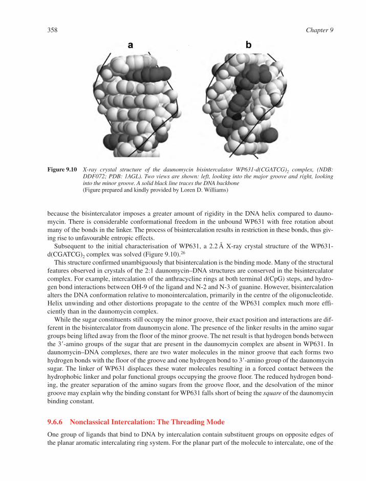
because the bisintercalator imposes a greater amount of rigidity in the DNA helix compared to dauno-mycin. There is considerable conformational freedom in the unbound WP631 with free rotation aboutmany of the bonds in the linker. The process of bisintercalation results in restriction in these bonds, thus giv-ing rise to unfavourable entropic effects.
Subsequent to the initial characterisation of WP631, a 2.2 Å X-ray crystal structure of the WP631-d(CGATCG)2 complex was solved (Figure 9.10).26
This structure confirmed unambiguously that bisintercalation is the binding mode. Many of the structuralfeatures observed in crystals of the 2:1 daunomycin–DNA structures are conserved in the bisintercalatorcomplex. For example, intercalation of the anthracycline rings at both terminal d(CpG) steps, and hydro-gen bond interactions between OH-9 of the ligand and N-2 and N-3 of guanine. However, bisintercalationalters the DNA conformation relative to monointercalation, primarily in the centre of the oligonucleotide.Helix unwinding and other distortions propagate to the centre of the WP631 complex much more effi-ciently than in the daunomycin complex.
While the sugar constituents still occupy the minor groove, their exact position and interactions are dif-ferent in the bisintercalator from daunomycin alone. The presence of the linker results in the amino sugargroups being lifted away from the floor of the minor groove. The net result is that hydrogen bonds betweenthe 3�-amino groups of the sugar that are present in the daunomycin complex are absent in WP631. Indaunomycin–DNA complexes, there are two water molecules in the minor groove that each forms twohydrogen bonds with the floor of the groove and one hydrogen bond to 3�-amino group of the daunomycinsugar. The linker of WP631 displaces these water molecules resulting in a forced contact between thehydrophobic linker and polar functional groups occupying the groove floor. The reduced hydrogen bond-ing, the greater separation of the amino sugars from the groove floor, and the desolvation of the minorgroove may explain why the binding constant for WP631 falls short of being the square of the daunomycinbinding constant.
9.6.6 Nonclassical Intercalation: The Threading Mode
One group of ligands that bind to DNA by intercalation contain substituent groups on opposite edges ofthe planar aromatic intercalating ring system. For the planar part of the molecule to intercalate, one of the
358 Chapter 9
Figure 9.10 X-ray crystal structure of the daunomycin bisintercalator WP631-d(CGATCG)2 complex, (NDB:DDF072; PDB: 1AGL). Two views are shown: left, looking into the major groove and right, lookinginto the minor groove. A solid black line traces the DNA backbone(Figure prepared and kindly provided by Loren D. Williams)

substituent groups must be threaded through the base pair stack to the opposite side of the helix. This morecomplicated binding mechanism is reflected in the kinetics of association and dissociation. The sub-stituents that are threaded through the base pair at the intercalation site are often bulky and/or polar andhence this may represent a kinetically unfavourable step in the binding mechanism. Examples of interca-lators that bind to DNA via a threading mechanism include naphthalene–bisamides, cationic porphyrins,and the anticancer antibiotic nogalamycin (Figure 9.11).
The binding mechanism for simple intercalators such as ethidium and proflavin involves just two steps.First, the cationic intercalator forms a complex with the negatively charged sugar–phosphate backbone ofDNA through electrostatic interactions. Then the intercalator diffuses up and down the helix in the anionicpotential along the surface of the DNA until it comes across gaps in between adjacent base pairs that haveformed due to the normal thermal or “breathing” motions of the DNA. This results in separated base pairsthat form a cavity into which the simple intercalator can bind.
By contrast, threading intercalators, with their large and polar distal-substituted side chains or rings, neces-sarily involve much larger openings in the DNA base pair stack (i.e. a bubble-type structure), and these mayonly form after significant distortions and/or breaking of inter-base hydrogen bonds. These larger and moredrastic dynamic motions in the helix occur with low frequency and hence the association kinetics for threadingintercalators is much slower than for simple intercalators. Thus both association and dissociation rate constantsshould decrease relative to classical intercalation. In addition, threading of cationic side chains through a DNAduplex requires sequential formation and breakage of electrostatic interactions in the threading complex.Hence the effect of salt concentration on observed rate constants can be used to probe for the threading mode.
Although threading intercalators have more complex binding mechanisms with potentially unfavourablesteps, they can still bind to DNA with high affinity. Once the side chain manages to traverse the intercalationsite, the DNA can adopt a conformation that gives a large and favourable free energy for complex formation.The side chains present a kinetic barrier to binding but favourable interactions, both in the intercalation siteand in the grooves, give rise to a stable final complex. The nature and extent of the kinetic barrier dependsupon the size of the side chain, its orientation and polarity.
The nogalamycin chromophore is intercalated, with the uncharged nogalose sugar (the aglycone group)in the minor groove and the positively charged amino sugar in the major groove (Figure 9.11). In thedrug–DNA complex, hydrogen bonds are observed between the drug and N-2 and O-6 atoms of a guanine.Hence there is simultaneous direct readout of sequence information in both the major and minor grooves.Nogalamycin has a binding preference for N�G or C�N sequences, where N can be any base, but the drugintercalates preferentially at the 5�-side of guanine or at the 3�-side of cytosine. The two sugars point in the
Reversible Small Molecule–Nucleic Acid Interactions 359
Figure 9.11 Structures of three intercalators that bind to DNA by a threading mode. (a) Naphthalene–bisimide. (b) Nogalamycin. (c) Cationic porphyrin
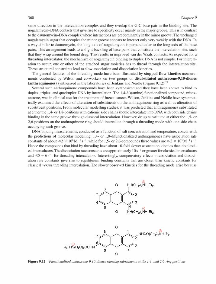
same direction in the intercalation complex and they overlap the G�C base pair in the binding site. Thenogalamycin–DNA contacts that give rise to specificity occur mainly in the major groove. This is in contrastto the daunomycin–DNA complex where interactions are predominantly in the minor groove. The unchargednogalamycin sugar that occupies the minor groove appears to interact only very weakly with the DNA. Ina way similar to daunomycin, the long axis of nogalamycin is perpendicular to the long axis of the basepairs. This arrangement leads to a slight buckling of base pairs that constitute the intercalation site, suchthat they wrap around the bound drug. This results in improved van der Waals contacts. As expected for athreading intercalator, the mechanism of nogalamycin binding to duplex DNA is not simple. For intercal-ation to occur, one or other of the attached sugar moieties has to thread through the intercalation site.These structural constraints lead to slow association and dissociation kinetics.
The general features of the threading mode have been illustrated by stopped-flow kinetics measure-ments conducted by Wilson and co-workers on two groups of disubstituted anthracene-9,10-diones(anthraquinones) synthesised in the laboratories of Jenkins and Neidle (Figure 9.12).27
Several such anthraquinone compounds have been synthesized and they have been shown to bind toduplex, triplex, and quadruplex DNA by intercalation. The 1,4-bis(amino) functionalized compound, mitox-antrone, was in clinical use for the treatment of breast cancer. Wilson, Jenkins and Neidle have systemat-ically examined the effects of alteration of substituents on the anthraquinone ring as well as alteration ofsubstituent positions. From molecular modelling studies, it was predicted that anthraquinones substitutedat either the 1,4- or 1,8-positions with cationic side chains should intercalate into DNA with both side chainsbinding in the same groove through classical intercalation. However, drugs substituted at either the 1,5- or2,6-positions on the anthraquinone ring should intercalate through a threading mode with one side chainoccupying each groove.
DNA binding measurements, conducted as a function of salt concentration and temperature, concur withthe predictions of molecular modelling. 1,4- or 1,8-difunctionalized anthraquinones have association rateconstants of about �2 � 106 M1 s1, while for 1,5- or 2,6-compounds these values are �2 � 105 M1 s1.Hence the compounds that bind by threading have about 10-fold slower association kinetics than do classi-cal intercalators. The dissociation rate constants are approximately 10 s1 or greater for classical intercalatorsand �5 6 s1 for threading intercalators. Interestingly, compensatory effects in association and dissoci-ation rate constants give rise to equilibrium binding constants that are closer than kinetic constants for classical versus threading intercalation. The slower observed kinetics for the threading mode arise because
360 Chapter 9
Figure 9.12 Functionalised anthracene-9,10-diones showing substituents at the 1,4- and 2,6-ring positions
(1) the polar side chain must go from aqueous solvent through the hydrophobic region in between two adja-cent base pairs, (2) the size and rigidity of the side chains may impede this passage, and (3) the rate of DNAthermal motion will result in the formation of a bubble-like structure that can accommodate the side chain.
9.7 INTERACTIONS IN THE MINOR GROOVE
9.7.1 General Characteristics of Groove Binding
A larger number of functional groups of the DNA bases are accessible in the wide major groove comparedto the narrow minor groove of the B-helix. Therefore, most proteins have evolved to make sequence-specific interactions with DNA in the major groove. However, some proteins and many small moleculesinteract with DNA in the minor groove. The major and minor grooves differ in electrostatic potential,hydrogen-bonding characteristics, steric effects, and hydration. Furthermore, DNA structure and conform-ation is highly dependent on sequence context. For example, runs of successive adenosine residues giverise to a very narrow minor groove, where the base pairs have a higher propeller twist and the DNAbecomes slightly bent from the helical axis. In general, small groove-binding molecules exhibit a prefer-ence for the minor groove, not least because this site of interaction provides better van der Waals contacts.In addition, many minor groove-binding ligands prefer A�T sites (compared to intercalators, which gener-ally exhibit a G�C preference).
Minor groove-binding ligands often contain several simple aromatic rings, such as pyrrole, furan, ben-zene or imidazole. These are connected by bonds with torsional freedom to enable such ligands to twistand become isohelical with the DNA minor groove. They thus provide optimal shape complementarity withthe DNA receptor and in most cases a typical crescent shape is adopted by these ligands (Figure 9.13).
The DNA minor groove is narrower in A�T-rich sequences compared to G�C-rich sequences and thus cor-rectly twisted aromatic rings in minor groove-binding ligands fit better at into the A�T minor groove tomake optimal van der Waals contacts with the helical chains that define the walls of the groove. Additionalspecificity (and to some extent stability) is derived from molecular contacts between the bound ligand andthe edges of the base pairs on the floor of the groove. Hydrogen bonds can be accepted by A�T base pairsfrom the bound molecule to the C-2 carbonyl oxygen of thymine or the N-3 nitrogen adenine. Even thoughsimilar functional groups are available in G�C base pairs, the amino group of guanine presents a stericblock to hydrogen bond formation at N-3 of guanine and at the O-2 carbonyl of cytosine. The inter-basehydrogen bond between the guanine amino group and the cytosine carbonyl oxygen lies in the minor grooveand this interaction sterically inhibits penetration of small molecules into the minor groove at G�C sites.Hence the aromatic rings of many minor groove-binding ligands form close contacts with A-H-2 protons inthe minor groove, but there is no room for the added steric bulk of the guanine amino group in G�C base pairs.Bernard Pullman has shown that negative electrostatic potential is greater in A��T minor grooves than it isin G�C minor grooves. This provides an additional important source for the observed A�T specificity of manyminor groove-binding ligands, which often are positively charged. Synthesis of ligands capable of acceptinghydrogen bonds from the guanine amino group is one way of enhancing minor groove-binding at G�C sites.
Unlike most intercalators, groove-binding molecules can extend to span many base pairs along thegroove and hence they can exhibit very high levels of DNA sequence-specific recognition. A long estab-lished goal has been to produce molecules that can specifically recognise DNA sequences long enough tobe unique in a biological context, for example, a eukaryotic regulatory sequence. Minor groove-bindingdrugs28–30 provide some hope of achieving this goal (Section 9.7.4) and some archetypal minor groove lig-ands are described below.
9.7.2 Netropsin and Distamycin
Netropsin was first isolated from Streptomyces netropsis by Finlay in 1951 while Arcamone discovereddistamycin A as a by-product of Streptomyces distallicus fermentation in 1958. Both drugs are
Reversible Small Molecule–Nucleic Acid Interactions 361

pyrrole–amidine antibiotics (Figure 9.13) that have antibacterial and antiviral activity, presumably as aresult of their interaction with DNA. Both compounds have been the focus of much study as DNA interactive ligands. These drugs have a binding preference for A��T-rich minor grooves over G�C-containing sequences. The preferred binding site is 5�-AATT and to a lesser extent 5�-ATAT. The rathersimilar sites 5�-TTAA and 5�-TATA are disfavoured sites of interaction. These differences in binding sitepreference have been interpreted in terms of sequence-dependent variations in minor groove width aswell as due to the disruption induced by the unusually flexible TpA base pair step. In general, these drugsbind in a narrowed minor groove and the complex is stabilised by a combination of electrostatic inter-actions between the DNA and the cationic end groups, as well as by hydrogen bonds to N-3 of adenines andO-2 of thymines. In addition, non-bonded hydrophobic interactions are made with the sugar–phosphatebackbone that defines the walls of the groove. The binding site size of netropsin is about four consecutiveA�T base pairs. Since distamycin is a longer molecule it requires at least five or six contiguous A�T base pairs.
9.7.2.1 Structures of Netropsin– and Distamycin–DNA Complexes. Dickerson and co-workerssolved the first X-ray crystal structure of a netropsin–DNA complex in 1985.31 The DNA target was theDickerson–Drew dodecamer: d(CGCGAATTCGCG)2, referred to here as A2T2. As expected, netropsinbinds in the central AATT of the duplex and it displaces several water molecules that are present in thedrug-free structure. The three-ligand amide groups point inwards and form bifurcated or three-centredhydrogen bonds with N-3 of adenine and O-2 of thymine. The netropsin fits tightly into the narrow minor
362 Chapter 9
Figure 9.13 Structures of some DNA minor groove binding ligands
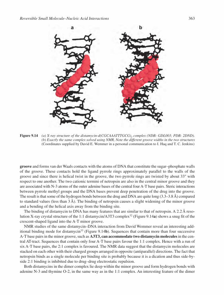
groove and forms van der Waals contacts with the atoms of DNA that constitute the sugar–phosphate wallsof the groove. These contacts hold the ligand pyrrole rings approximately parallel to the walls of thegroove and since there is helical twist in the groove, the two pyrrole rings are twisted by about 33° withrespect to one another. The two cationic termini of netropsin are also in the central minor groove and theyare associated with N-3 atoms of the outer adenine bases of the central four A�T base pairs. Steric interactionsbetween pyrrole methyl groups and the DNA bases prevent deep penetration of the drug into the groove.The result is that some of the hydrogen bonds between the drug and DNA are quite long (3.3–3.8 Å) comparedto standard values (less than 3 Å). The binding of netropsin causes a slight widening of the minor grooveand a bending of the helical axis away from the binding site.
The binding of distamycin to DNA has many features that are similar to that of netropsin. A 2.2 Å reso-lution X-ray crystal structure of the 1:1 distamycin/A3T3 complex32 (Figure 9.14a) shows a snug fit of thecrescent-shaped ligand into the A�T minor groove.
NMR studies of the same distamycin–DNA interaction from David Wemmer reveal an interesting add-itional binding mode for distamycin33 (Figure 9.14b). Sequences that contain more than four successiveA�T base pairs in the minor groove, such as A3T3, can accommodate two distamycin molecules in the cen-tral AT-tract. Sequences that contain only four A�T base pairs favour the 1:1 complex. Hence with a run ofsix A�T base pairs, the 2:1 complex is favoured. The NMR data suggest that the distamycin molecules arestacked on each other with their charged groups arranged in opposite (antiparallel) directions. The fact thatnetropsin binds as a single molecule per binding site is probably because it is a dication and thus side-by-side 2:1 binding is inhibited due to drug–drug electrostatic repulsion.
Both distamycins in the dimer complex lie deep within the minor groove and form hydrogen bonds withadenine N-3 and thymine O-2, in the same way as in the 1:1 complex. An interesting feature of the dimer
Reversible Small Molecule–Nucleic Acid Interactions 363
Figure 9.14 (a) X-ray structure of the distamycin-d(CGCAAATTTGCG)2 complex (NDB: GDL003; PDB: 2DND).(b) Exactly the same complex solved using NMR, Note the different groove widths in the two structures(Coordinates supplied by David E. Wemmer in a personal communication to I. Haq and T. C. Jenkins)
complex is that it requires the minor groove to widen two-fold compared to drug-free DNA or to the 1:1 com-plex (Figure 9.14). This structural rearrangement gives rise to a positive free energy contribution to bind-ing, in a similar way to structural deformations that accompany intercalation. However, this unfavourablefree energy is more than compensated by additional favourable free energy that arises from the greaterhydrophobic interactions derived from binding of two drugs within one minor groove. This shows anotherexample of the conformational mobility of DNA that is crucial to the interaction of proteins and ultimatelyto biological function.
9.7.2.2 Energetics of Netropsin– and Distamycin–DNA Interactions. Recently, there has been anupsurge in detailed thermodynamic studies of minor groove binding that go beyond simple determinations ofbinding constants. For netropsin, distamycin, and a number of other minor-groove specific ligands, it is nowpossible to apportion the overall binding free energy into enthalpic and entropic terms. In addition, recentdevelopments in thermodynamic analysis allows a much more detailed parsing of free energy terms (Section9.6.4 and Equation 9.14), which provides insight into the energetics of drug–DNA interactions.
The relative thermodynamic contributions of two distamycin molecules that bind to the duplexd(GCCAAATTGGC)�d(GCCAATTTGGC), referred to as A3T2, have been examined in the context of thethermodynamics of the 1:1 netropsin interaction with A3T2.34 In all cases, drug binding is associated with anexothermic binding enthalpy, which for netropsin is 31.4 kJ mol1 and for distamycin first and second bind-ing events are 51.5 and 78.7 kJ mol1, respectively. The drugs bind to DNA moderately tightly with Kobs
of 4.3 � 107 M1 for netropsin and 3.1 � 107 M1 for the first distamycin binding. The second distamycinmolecule binds with a lower affinity of 3.3 � 106 M1 (20°C, pH 7.0 and Na� concentration of about 20 mM).
Thus netropsin and the first distamycin have very similar overall binding affinities and both interactionsare enthalpically driven, with the first distamycin having a larger favourable enthalpy. One possibleinterpretation for these observations is that hydrogen bonding contributions are similar for the two com-plexes. The smaller number of favourable electrostatic interactions in the monocationic distamycin, com-pared to dicationic netropsin, is compensated by an increase in hydrophobic interactions in distamycinarising from its additional N-methylpyrrole ring. The magnitudes of the negative enthalpy values aredependent upon relative contributions from hydrogen bonding, van der Waals interactions, and overallhydration changes. The entropy contributions are of opposite sign (favourable for netropsin andunfavourable for distamycin), probably due to differences in release of counter-ions and uptake or releaseof water molecules. The second distamycin (in the 2:1 complex) binds with an eight-fold reduction inaffinity and a 27.2 kJ mol1 more favourable enthalpy. This finding is consistent with observations fromthe NMR structure, since there is an increase in van der Waals contacts due to side-by-side interactions inthe 2:1 distamycin–DNA complex.
Variations in binding affinity with salt concentration are also revealing. The slopes of lnKobs versusln[Na�] plots were found to be 0.8 for netropsin–A3T2 binding and 0.56 for distamycin–A3T3 bind-ing. These values are lower than previous estimates for 1:1 binding of netropsin and distamycin to A2T2and also lower than the theoretical value of 1.76 (for the binding of one dicationic ligand or two mono-cationic ligands) predicted from the polyelectrolyte theories of Record and Manning. Structural data pro-vides a possible explanation for these results. The minor groove becomes widened upon the binding of thesecond distamycin, which results in a lower local charge density. Also, structured water around exposedhydrophobic groups of the bound drug may give rise to a different type of dielectric screening. For bothligands, calorimetrically measured binding-induced changes in heat capacity Cp are significant in mag-nitude and negative in sign, consistent with theoretical estimates based on changes in solvent accessiblesurface area upon binding.35
9.7.3 Lexitropsins
Lexitropsins are a group of minor groove-specific ligands that are comprised of a range of different aromaticrings connected by peptide bonds. Netropsin and distamycin are examples of lexitropsins, where the sequence
364 Chapter 9

of aromatic rings is pyrrole–pyrrole (PyPy) for netropsin and pyrrole–pyrrole–pyrrole (PyPyPy) for dis-tamycin. Dickerson and Lown proposed that replacement of Py moieties in netropsin or distamycin by otherheterocycles, such as 1-methylimidazole (Im), could yield compounds capable of recognising G�C base pairsas well as A�T sequences. In netropsin, pyrrole methyl groups are orientated towards the minor groove suchthat they make close contact with A�T base pairs. In lexitropsins, one or more of the pyrrole rings is replaced by,for example, an imidazole (Im) ring, which is capable of accepting a hydrogen bond. This alleviates the stericclash of a pyrrole with a guanine amino group in the minor groove and instead allows a specific hydrogenbond to form between the guanine amino group and the imidazole nitrogen atom, which facilitatesincreased minor groove interactions between the lexitropsin and G�C base pairs. DNA interactions withnetropsin (PyPy) as well as an imidazole-containing lexitropsin (ImPyPy) are illustrated in Figure 9.15.
NMR and footprinting studies have confirmed the enhanced interactions between suitably modified lex-itropsins and G�C-containing minor grooves, although the presence of an A�T base pair does not interferewith binding. Other examples of ligands in this group include bis-furan lexitropsins, thiazole compounds,and analogues of Hoechst 33258, where benzoxazole or pyridoimidazole groups have replaced the benz-imidazole rings in Hoechst 33258. Binding studies indicate that whilst most of these lexitropsins lose theirA�T specificity, they fail to become properly G�C-specific.
9.7.4 Hairpin Polyamides
Hairpin polyamide ligands provide important opportunities for development of high affinity, sequence-specific DNA recognition agents for the control of gene function and as potential anticancer therapeutics.36–43
In the early 1990s, Dervan synthesised a polyamide containing an imidazole ring followed by two pyrrolemoieties (ImPyPy) with the expectation that it would form a 1:1 complex with sequences of the type 5�-XGXXX, in which X can be either A or T. Unexpectedly, ImPyPy was found to bind to a five base pair
Reversible Small Molecule–Nucleic Acid Interactions 365
Figure 9.15 Schematic representation of the specific molecular interactions that occur between (a) netropsin and(b) a lexitropsin ImPyPy. Dotted lines indicate hydrogen bonds. Double-headed arrows represent nonbonded van der Waals contacts. The curved line is the floor of the minor groove
sequence of the type 5�-XGXCX, (again X can be either A or T). An NMR structure of the complex showedthat ImPyPy binds as an antiparallel 2:1 dimer in a similar manner to distamycin binding to A�T-tracts inthe minor groove. In this arrangement, an imidazole ring is stacked against a pyrrole ring and thus theantiparallel pair can distinguish a G�C base pair from a C�G, i.e. the pair Im/Py targets G�C and Py/Im rec-ognizes C�G. Each G�C base pair has an imidazole on the G strand and a pyrrole on the C strand. A hydro-gen bond is formed between the N-3 atom of the imidazole ring and the amino group of guanine as well as aseries of hydrogen bonds between polyamide NH groups and the edge of the G�C base pair.
Thermodynamic studies reveal that the sequence selectivity of the Im/Py pair is mostly driven by a largefavourable enthalpy term. Imidazole nitrogen atoms do not hydrogen bond to the cytosine side of the basepair due to an unfavourable bond angle for this interaction. In addition, a Py/Py pair is degenerate and canrecognize A�T and T�A base pairs equally well in preference to G�C or C�G base pairs.
These discoveries suggested a new design strategy: unsymmetrical polyamide ring pairs can be used forspecific recognition of the DNA minor groove where one pair of rings in the ligand targets one base pairof DNA. An Im is used for a guanine and a Py for a cytosine in G�C or C�G base pairs, whereas two Pyrings are used to target either A�T or T�A.
To test these pairing rules, Dervan changed an Im in one of the polyamides of a homodimer to Py to givethe heterodimer ImPyPy/PyPyPy. As predicted, this pair of ligands binds specifically to the sequence 5�-XGXXX. Further, ImPyImPy binds to the minor groove of the sequence 5�-GCGC. By analogy withdistamycin, binding of an alkyl chain is always to an A�T site, so that the exact recognition sequence isXGCGCX. The specificity is extremely high, such that even a single mismatch with a recognition motif, e.g.G�C in place of C�G, leads to a 10-fold drop in binding affinity.
Since polyamide dimers do not distinguish A�T from T�A base pairs in the minor groove, a third type ofmonomer was designed as a thymine-selective moiety, N-methyl-3-hydroxypyrrole (Hp). An Hp/Py pairrecognizes a T�A base pair whereas a Py/Hp combination recognizes A�T base pairs. Both orientations dis-favour binding to G�C and C�G base pairs. X-ray structures of two Hp-containing polyamide–DNA complexesconfirm that the hydroxyl group of Hp fits snugly into the asymmetric cleft between adenine C-2 andthymine O-2 atoms. In addition, there is a specific hydrogen bond between the hydroxyl group of Hp andthymine O-2. Hp-containing polyamides bind with lower affinity than the equivalent Py-containing ones.The reason for this is not clear but may be related to factors such as partial melting of the T�A base pair rec-ognized by Hp/Py or a lengthening of the amide–DNA hydrogen bond on the C-terminal of the Hp residue.Nevertheless, with the addition of this third type of monomer, it has become possible to create polyamidescontaining different combinations of Im, Py and Hp rings. Using the Dervan Pairing Rules, one can dis-tinguish all four types of Watson–Crick base pairs and thus design polyamide ligands to target any specificDNA sequence (Figure 9.16).
Linked polyamides are a further synthetic development where two separate molecules are covalentlyjoined together to reduce unfavourable entropy loss upon binding. It also results in an increase in specificityby ensuring that the two molecules bind at the same site. An additional problem is that once the number ofrings in the polyamide ligand increases above six, affinity is decreased. This is because the ligand losessynchronization in either length or curvature with respect to the DNA groove. The problem is addressedby introduction of linkers with torsional flexibility, such as a �-alanine subunit, which allows the naturalcurvature of the polyamide to be relaxed. Molecules have been designed that can bind as overlappinghomodimers to recognize up to 11 base pairs with subnanomolar dissociation constants. The eventual aimis to target a sequence of 16–18 base pairs, since these would be unique in the human genome.
9.7.5 Hoechst 33258
Hoechst 33258 (Figure 9.13) is a fluorescent dye often used as a chromosome stain. Like other minorgroove binders, Hoechst 33258 binds to A�T rich sequences preferentially. In fact it has a 50-fold greateraffinity for AATT than for TATA and higher binding to AATT than to AAAA, which is in contrast to otherA�T-specific minor groove drugs such as distamycin, netropsin and berenil.44
366 Chapter 9

9.7.5.1 Structures of Hoechst 33258–DNA Complexes. Hoechst 33258–DNA structures areextremely well studied. NMR analysis has revealed a number of locations and conformations of the drug in5�-AATT sites. The phenolic ring is able to undergo rapid ring-flips, which involve 180° rotations about thephenol–benzimidazole bond, even though the phenolic ring is tightly accommodated between the walls ofthe minor groove. Dynamic breathing motions must occur in the DNA that results in transient widening ofthe minor groove to allow the ring to flip. The piperazine ring also adopts several different conformations.These two rings at the termini of the molecule are generally more mobile than the bis-benzimidazole core.
In one Hoechst 33258–A2T2 X-ray crystal structure, the ligand is located in the central 5�-AATT tractin a similar manner to netropsin. Hydrogen bonds are observed from the benzimidazole amide groups toO-2 atoms on thymine and N-3 atoms on adenine. Hoechst 33258 is isohelical with the curvature of theDNA minor groove and numerous molecular contacts are made with the walls of the groove. The phenolicring of the ligand is twisted 8° with respect to the adjacent benzimidazole ring and the two benzimidazolerings are twisted 32° with respect to each other. The piperazine ring is only slightly puckered and is almost
Reversible Small Molecule–Nucleic Acid Interactions 367
+ Pyβ Py Py Im
Im Py Py Py
Figure 9.16 (a) The Dervan Pairing Rules, which describe how different combinations of imidazole (Im), pyrrole(Py) and hydroxypyrrole (Hp) rings placed opposite each other can be used to distinguish all four typesof base pairs. (b) and (c) These pairing rules can be applied to target region of DNA with the sequence 5�-AGTACT (� � dimethylaminopropylamine residue)
planar to the benzimidazole ring to which it is attached. The O-4� atoms of the deoxyribose sugar of theDNA backbone are in the correct position to make favourable interactions with the �-electron system ofthe ligand as it sits within van der Waals contact distance of the groove walls. The differences betweenobserved structures in NMR and X-ray experiments arise probably because of the different low-energyconformations that this ligand can adopt when bound to various DNA sequences.
9.7.5.2 Thermodynamics of Hoechst 33258–DNA Interactions. The fluorescent properties ofHoechst 33258 can be exploited to study the DNA binding character of this dye. Equilibrium experi-ments show that Hoechst 33258 binds tightly to calf-thymus DNA, poly(dA-dT)�poly(dA-dT), and to theoligonucleotides A2T2 and d(CCGGAATTCCGG)2 with Kobs values in the range 1–3 � 109 M1.45,46
However, this tight binding occurs only once every 100 base pairs of calf-thymus DNA. In fact Hoechst33258 binds to calf-thymus DNA with a spectrum of different binding modes each characterized by a dif-ferent affinity, with some values of Kobs in the �106 M1 range. Similarly with poly(dA-dT)�poly(dA-dT),Job Plots reveal a multiplicity of binding stoichiometries with between 1 and 6 Hoechst 33258 moleculesbound per 5 A�T base pairs as well as stoichiometries that suggest 1 or 2 ligand molecules are bound perDNA phosphate.
Thus Hoechst 33258 binding to DNA is complex and depends strongly on sequence and DNA length.At least four distinct types of interaction between Hoechst 33258 and DNA are seen:
� Sequence-mediated: specific, high affinity, binding observed in X-ray structures and footprintingstudies;
� Charge-mediated: non-specific interaction that involves the electrostatic binding of monocationicHoechst 33258 with the DNA phosphate backbone;
� Ligand-mediated: non-specific type of binding that occurs when a molecule of free Hoechst 33258in solution binds to other dye molecules that are already associated with the DNA helix, either inthe minor groove or the backbone; and
� Structure-mediated: either specific or non-specific binding – a mode that confines Hoechst 33258to a specific structural region, such as the minor groove, because of some geometric requirement.
The differences between these four modes of binding are subtle. They are not totally separate entities becausemore than one type of interaction contributes to the overall affinity of each bound ligand.
Sequence mediated (specific) binding of Hoechst 33258 to DNA has been examined in some detailusing d(CGCAAATTTGCG)2 (A3T3) as the receptor.47–49 A detailed partitioning of the overall observedfree energy for the Hoechst 33258–A3T3 interaction (Section 9.6.4) has been carried out by measuring, asdirectly as possible, a near complete thermodynamic profile for the interaction. This used a combinationof ITC to measure H and Cp and fluorescence titrations to measure Kobs and hence Gobs. From thesemeasurements TS and Gpe were calculated using standard thermodynamic relationships. The measuredbinding constant of 3.2 0.6 � 108 M(duplex)1 at salt concentration of ca. 12 mM is similar to the Hoechst33258 binding affinity (Kobs � 3.15 � 108 M(duplex)1) determined by Clegg under similar conditions forthe less extended A�T-tract dodecamer d(CGCGAATTCGCG)2. Quantification of the salt dependenceyielded a value for SK of 0.99, which is consistent with the monocationic charge of Hoechst 33258. Thisallows the overall observed binding free energy (Gobs) to be broken down into its component polyelectrolyteand nonpolyelectrolyte terms (Section 9.3). The analysis showed that from a Gobs of 48.9 kJ mol1,some 7.5 kJ mol1 (about 15%) is Gpe, i.e. from coupled polyelectrolyte effects such as the release ofcondensed counter-ions from the DNA upon ligand binding.
X-ray crystal structures of this complex show a network of hydrogen bonds and other minor grooveinteractions involving van der Waals contacts and hydrophobic interactions. The binding free energyshould thus reflect a large nonpolyelectrolyte component. This is borne out by experimental data showing that out of a total free energy of 48.9 kJ mol1, some 41.4 kJ mol1 is due to non-polyelectrolyteeffects. At each temperature studies, binding enthalpies for the interaction of A3T3 with Hoechst 33258are positive and their magnitudes decrease with increasing temperature (H � �42.7 kJ mol1 at 9.3°C
368 Chapter 9
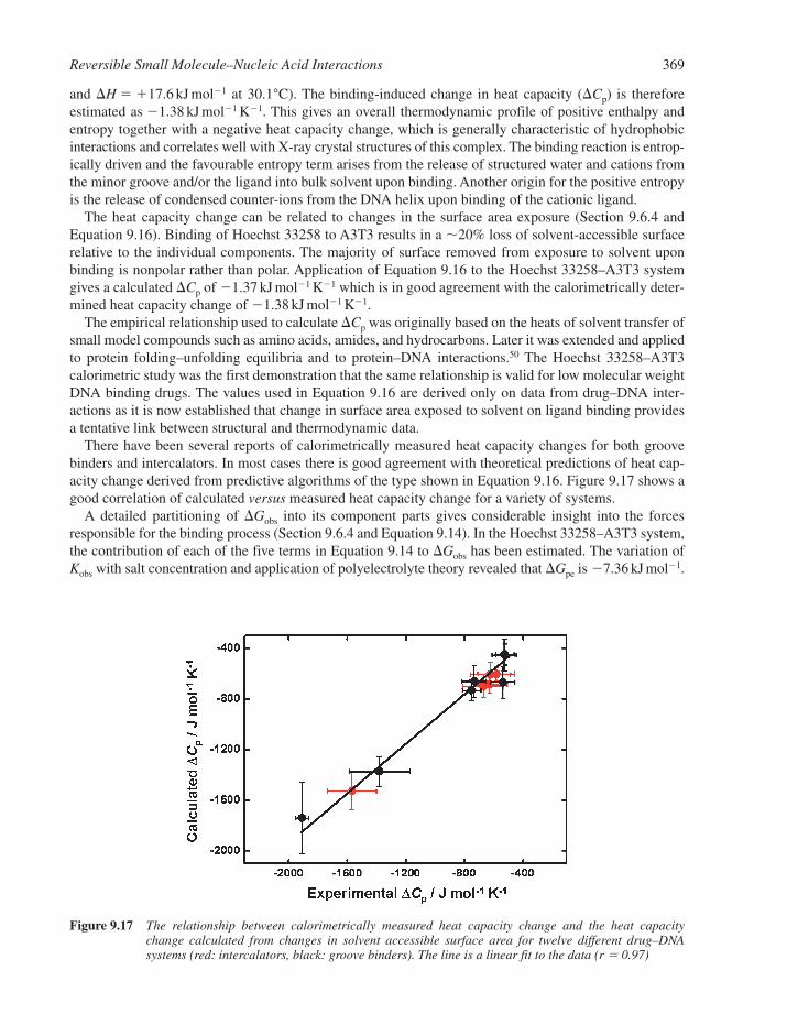
and H � �17.6 kJ mol1 at 30.1°C). The binding-induced change in heat capacity (Cp) is thereforeestimated as 1.38 kJ mol1 K1. This gives an overall thermodynamic profile of positive enthalpy andentropy together with a negative heat capacity change, which is generally characteristic of hydrophobicinteractions and correlates well with X-ray crystal structures of this complex. The binding reaction is entrop-ically driven and the favourable entropy term arises from the release of structured water and cations fromthe minor groove and/or the ligand into bulk solvent upon binding. Another origin for the positive entropyis the release of condensed counter-ions from the DNA helix upon binding of the cationic ligand.
The heat capacity change can be related to changes in the surface area exposure (Section 9.6.4 andEquation 9.16). Binding of Hoechst 33258 to A3T3 results in a �20% loss of solvent-accessible surfacerelative to the individual components. The majority of surface removed from exposure to solvent uponbinding is nonpolar rather than polar. Application of Equation 9.16 to the Hoechst 33258–A3T3 systemgives a calculated Cp of 1.37 kJ mol1 K1 which is in good agreement with the calorimetrically deter-mined heat capacity change of 1.38 kJ mol1 K1.
The empirical relationship used to calculate Cp was originally based on the heats of solvent transfer ofsmall model compounds such as amino acids, amides, and hydrocarbons. Later it was extended and appliedto protein folding–unfolding equilibria and to protein–DNA interactions.50 The Hoechst 33258–A3T3calorimetric study was the first demonstration that the same relationship is valid for low molecular weightDNA binding drugs. The values used in Equation 9.16 are derived only on data from drug–DNA inter-actions as it is now established that change in surface area exposed to solvent on ligand binding providesa tentative link between structural and thermodynamic data.
There have been several reports of calorimetrically measured heat capacity changes for both groovebinders and intercalators. In most cases there is good agreement with theoretical predictions of heat cap-acity change derived from predictive algorithms of the type shown in Equation 9.16. Figure 9.17 shows agood correlation of calculated versus measured heat capacity change for a variety of systems.
A detailed partitioning of Gobs into its component parts gives considerable insight into the forcesresponsible for the binding process (Section 9.6.4 and Equation 9.14). In the Hoechst 33258–A3T3 system,the contribution of each of the five terms in Equation 9.14 to Gobs has been estimated. The variation ofKobs with salt concentration and application of polyelectrolyte theory revealed that Gpe is 7.36 kJ mol1.
Reversible Small Molecule–Nucleic Acid Interactions 369
Figure 9.17 The relationship between calorimetrically measured heat capacity change and the heat capacitychange calculated from changes in solvent accessible surface area for twelve different drug–DNA systems (red: intercalators, black: groove binders). The line is a linear fit to the data (r � 0.97)

From evaluation of Cp one can calculate the possible contribution to binding free energy that arises from thehydrophobic transfer of the drug from bulk solvent into the DNA minor groove (Ghyd) as 110.4 kJ mol1.These data, as well as crystallographic studies, indicate that binding of dye to A3T3 induces negligibleconformational changes in either the DNA or the drug. In effect it is a rigid body interaction. Therefore thevalue of Gconf can be set to zero. The value of Gr�t can be set at �62.8 kJ mol1 by use of argumentsdiscussed by Spolar and Record.50 Thus the calculated free energy change for the Hoechst 33258–A3T3interaction is 55.0 kJ mol1, very close to the experimental value of 48.9 kJ mol1 for Gobs. Therefore,by difference it is possible to estimate Gmol (the contribution to free energy from weak noncovalent inter-actions between the drug and DNA), which is �6.1 kJ mol1.
This analysis suggests that for this particular ligand–DNA complex, noncovalent molecular interactionssuch as hydrogen bonds make a net unfavourable contribution to binding free energy. Although this result isapparently at odds with NMR and crystallographic studies, the two positions can be reconciled when oneconsiders the hydration state of the minor groove. Hydrogen-bond formation between the ligand and DNAis at the expense of hydrogen-bond breakage between the DNA and site-specific waters located in the minorgroove. The net result is an iso-energetic exchange reaction involving Hoechst 33258 and water, such thata near zero free energy change results. Instead, the major driving force for Hoechst 33258 binding to A3T3is the hydrophobic transfer of the drug from bulk solvent into the duplex minor groove binding site. Thisis reflected in the observed negative Cp and the positive binding enthalpy. The large energetic cost resultingfrom losses in rotational and translational freedom that occurs upon complex formation is more than compen-sated for by the favourable contributions from hydrophobic transfer and polyelectrolyte effects. Thus affinitymight be best modulated by changes in drug hydrophobicity. However, hydrogen bond formation andother noncovalent interactions are key modulators of specificity in drug–DNA interactions and they are alsoimportant in fine-tuning the free energy of binding in response to the interactions available in a given DNAbinding site.
9.8 INTERCALATION VERSUS MINOR GROOVE BINDING
There are some interesting compounds that possess structural features that could, in principle, involvebinding by either intercalation or groove binding. In reality, one is favoured over the other. What are thefactors that dictate choice of binding mode? Specific examples are the ligand SN 6999 and the relatedcompound chloroquine (Figure 9.18).
Both molecules contain a fused bicyclic quinoline ring system, yet biophysical and structural studieshave shown that while chloroquine can intercalate, SN 6999 binds in the minor groove. SN 6999 has acurved shape that fits in the minor groove and it forms hydrogen bonds in a similar manner to other groovebinding drugs. Modelling studies have shown that SN 6999 could form favourable contacts within a basepair stack as well as possible electrostatic interactions with the phosphate backbone in an intercalationcomplex. However, if such an intercalation complex formed instead of the groove-bound complex, itwould be at the expense of favourable free energy from hydrogen bonding, non-bonded contacts with thegroove walls, and solvent release from the minor groove. Hence for SN 6999, binding in the minor grooveis much more favourable energetically than intercalation, even though the latter is possible. Chloroquinehowever does not have the correct hydrogen bond donor/acceptor groups or any optimal structure for appro-priate minor groove interactions. Thus it binds to DNA by intercalation but only with low affinity.
SN 6999 is an example of a minor groove binder having a benzene-fused heterocycle. Conversely, thereare examples of unfused aromatic cations that bind by intercalation rather than groove binding. Netropsin,distamycin, and Hoechst 33258 are all examples of ligands that have torsional freedom and this propertyallows these compounds to adopt an optimal twist for minor groove binding. Strekowski and Wilson havedesigned and synthesised a new group of diphenylpyridine compounds with unfused aromatic rings pos-sessing torsional freedom. So one might expect them to be good minor groove-binding ligands (Figure 9.18).Molecular modelling and X-ray crystallography indicate that the molecule has a twist of between 20° and25° between the phenyl and pyrimidine planes. The existence of twisted rings along with cationic end
370 Chapter 9
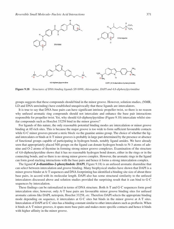
groups suggests that these compounds should bind in the minor groove. However, solution studies, (NMR,LD and DNA unwinding) have established unequivocally that these ligands are intercalators.
It is true to say that DNA base pairs can have significant intrinsic propeller twist, so there is no reasonwhy unfused aromatic ring compounds should not intercalate and enhance the base pair interactionsresponsible for propeller twist. Yet, why should 4,6-diphenylpyridine (Figure 9.18) intercalate whilst sim-ilar compounds such as Hoechst 33258 bind in the minor groove?
For ligands of this nature, the only reasonable potential binding modes are intercalation or minor groovebinding at AT-rich sites. This is because the major groove is too wide to form sufficient favourable contactswhile G�C minor grooves present a steric block via the guanine amino group. The choice of whether the lig-and intercalates or binds at A�T minor grooves is probably in large part determined by the presence or absenceof functional groups capable of participating in hydrogen bonds, notably ligand amides. We have alreadyseen that appropriately placed NH groups on the ligand can donate hydrogen bonds to N-3 atoms of ade-nine and O-2 atoms of thymine in forming strong minor groove complexes. Examination of the structureof 4,6-diphenylpyridine shows that it has no reasonable hydrogen bond donors, either in the rings or in theconnecting bonds, and so there is no strong minor groove complex. However, the aromatic rings in the ligandcan form good stacking interactions with the base pairs and hence it forms a strong intercalation complex.
The ligand 4��,6-diamidino-2-phenylindole (DAPI, Figure 9.18) is an unfused aromatic diamidine thatcan select between intercalation and groove binding. Many biophysical studies have shown that DAPI is aminor groove binder at A�T sequences and DNA footprinting has identified a binding site size of about threebase pairs, in accord with its molecular length. DAPI also has some structural similarity to the unfusedintercalators discussed above and solution studies provided the surprising result that it can bind to G�Csequences by intercalation.
These findings can be rationalized in terms of DNA structure. Both A�T and G�C sequences form goodintercalation sites; however, only A�T base pairs are favourable minor groove binding sites for unfusedaromatic cations like DAPI, netropsin, Hoechst 33258, etc. Therefore DAPI selects the appropriate bindingmode depending on sequence, it intercalates at G�C sites but binds in the minor groove at A�T sites.Intercalation of DAPI at G�C sites has a binding constant similar to other intercalators such as proflavin. Whenit binds at A�T minor grooves, it spans more base pairs and makes more specific contacts and hence it bindswith higher affinity in the minor groove.
Reversible Small Molecule–Nucleic Acid Interactions 371
Figure 9.18 Structures of DNA binding ligands SN 6999, chloroquine, DAPI and 4,6-diphenylpyrimidine
The conclusion is that for all drug–DNA interactions, intercalation and groove binding represent twopotential low-energy wells in a continuous free energy surface. The binding mode with lower energy willdepend on DNA sequence and conformation as well as the molecular features of the bound ligand.
9.9 CO-OPERATIVITY IN LIGAND–DNA INTERACTIONS
Genomic DNA is somewhat different from model duplexes used in structural and biophysical studies,since it possesses a continuous array of potential binding sites that are incorporated into a large nucleicacid–protein complex. Thus the biological activity of a DNA binding drug will be affected by co-operativity.
Co-operativity is an important and pervasive concept in biology. The phenomenon of allostery is wellknown in protein–ligand interactions. A type of negative co-operativity in drug–DNA interactions is neigh-bour exclusion (Section 9.6.3). Here each bound drug excludes one or more adjacent base pairs from beingpotential drug binding sites. This is often referred to as site-exclusion co-operativity. However, it is also pos-sible for there to be co-operative effects between adjacent binding sites (composed of more than one basepair) and in such cases the data can be described by a modified version of the McGhee and von Hippel rela-tionship.20 (Section 9.6.3 and Equation 9.13). The modified form of this equation is
(9.17)
where r is the ratio of bound drug to total DNA sites (in base pairs), Cf the concentration of free drug, Kthe binding constant for the interaction of a drug with an isolated site, and n the neighbour exclusionparameter. The parameter � is the co-operativity function; which in effect is the equilibrium constant formoving a bound drug molecule from a totally isolated binding site to a site contiguous to another boundligand. If � is greater than 1, this indicates positive co-operativity. Conversely, if � is less than 1, there isnegative co-operativity. When � � 1 there is no co-operativity and then Equation 9.17 can be simplifiedto Equation 9.13. This method of analysis is very useful for interactions involving long polynucleotides ornatural DNA samples. Equation 9.17 can be incorporated into nonlinear least squares fitting routines to fitequilibrium binding isotherms and the parameter � can thus be evaluated readily. However, binding datashould primarily be fitted to the simplest model, therefore Equation 9.17 should only be used if it yields asignificantly better statistical fit to the data compared to Equation 9.13.
9.10 SMALL MOLECULE INTERACTIONS WITH HIGHER-ORDER DNA
The vast majority of genomic DNA exists as a double-stranded, antiparallel structure in the B-conformation.However in the past fifteen years, it has become clear that DNA can adopt a number of higher-order conformations. These involve the self-assembly of three or four DNA strands or the intramolecular fold-ing of a single strand such that it adopts a triplex or quadruplex (tetraplex) conformation (Section 2.3.6 and2.3.7). Further, it has become apparent that, far from being simple biophysical curiosities, these multi-stranded DNA structures have considerable biological significance. This has led to triplex and quadruplexDNA becoming major drug targets. Below are summarised the key features of these higher-order struc-tures and a discussion of how small molecules have been used to target them and effect their stabilization.
9.10.1 Triplex DNA and its Interactions with Small Molecules
Triplex DNA is formed when a single-stranded oligonucleotide specifically recognizes the major grooveof duplex DNA and forms hydrogen bonds with the Watson–Crick base pairs (Section 2.3.6). Due to the
r
CK nr
nr r R
nr
n r Rn
f
� �
� �
( )( )( )
( )( )
( )1
2 1 1
2 1 1
1 11
�
�
22 1
1 1 4 1
2
2
( )
{[ ( ) ] ( )}
� � �
nr
R n r r nr
�
372 Chapter 9
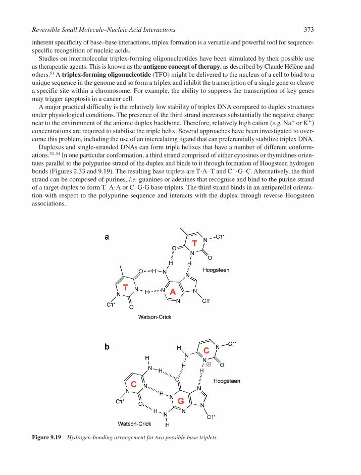
inherent specificity of base–base interactions, triplex formation is a versatile and powerful tool for sequence-specific recognition of nucleic acids.
Studies on intermolecular triplex-forming oligonucleotides have been stimulated by their possible useas therapeutic agents. This is known as the antigene concept of therapy, as described by Claude Hélène andothers.51 A triplex-forming oligonucleotide (TFO) might be delivered to the nucleus of a cell to bind to aunique sequence in the genome and so form a triplex and inhibit the transcription of a single gene or cleavea specific site within a chromosome. For example, the ability to suppress the transcription of key genesmay trigger apoptosis in a cancer cell.
A major practical difficulty is the relatively low stability of triplex DNA compared to duplex structuresunder physiological conditions. The presence of the third strand increases substantially the negative chargenear to the environment of the anionic duplex backbone. Therefore, relatively high cation (e.g. Na� or K�)concentrations are required to stabilise the triple helix. Several approaches have been investigated to over-come this problem, including the use of an intercalating ligand that can preferentially stabilize triplex DNA.
Duplexes and single-stranded DNAs can form triple helixes that have a number of different conform-ations.52–54 In one particular conformation, a third strand comprised of either cytosines or thymidines orien-tates parallel to the polypurine strand of the duplex and binds to it through formation of Hoogsteen hydrogenbonds (Figures 2.33 and 9.19). The resulting base triplets are T�A–T and C��G–C. Alternatively, the thirdstrand can be composed of purines, i.e. guanines or adenines that recognise and bind to the purine strandof a target duplex to form T–A�A or C–G�G base triplets. The third strand binds in an antiparellel orienta-tion with respect to the polypurine sequence and interacts with the duplex through reverse Hoogsteenassociations.
Reversible Small Molecule–Nucleic Acid Interactions 373
Figure 9.19 Hydrogen-bonding arrangement for two possible base triplets

In recent years a number of triple helix-specific ligands have been characterized (Figure 9.20). The func-tion of such ligands is to overcome the inherent instability of triplex DNA and effect triplex stabilization.
The addition of a third strand into the major groove of a DNA duplex leads to the creation of two newgrooves on either side of the third strand in what was previously the major groove, and the minor grooveis left largely unchanged. One of the new grooves on either side of the third strand is essentially feature-less. However, the other groove resembles the minor groove, except that its walls have hydrophobicresidues from pyrimidine C-5 methyl groups while the polar C-4 carbonyl/amino groups are buried.
This unique groove in triplex DNA with its bifurcated hydrogen-bonded centres on the floor, as well as thehydrophobic nature of the groove walls, has led to the development of a new group of ligands designed to tar-get this previously unexploited binding site in triplex DNA.55 Thus, Terry Jenkins has synthesized a series ofpara- and meta-carbonyl substituted 1,3-diaryltriazenes (Figure 9.20) intended to target one of the two newgrooves created during triplex formation. The concave inner surface of the ligand formed by triazene andphenyl rings has no protrusions and so in principle the ligand can penetrate deeply into the narrow triplexgroove. The ligand possesses hydrogen bond acceptor groups, which can interact with hydrogen bond donorson the groove floor. In addition, the �-conjugated core residues make close van der Waals contacts with thehydrophobic groove walls by alignment of phenyl rings in both ligand and DNA. Solution studies confirmthat these ligands bind preferentially to triplex DNA and stabilize this structure more than they stabilize a cor-responding duplex. For example, sub-saturating amounts of the ligand produce a Tm of �2°C in duplexDNA compared to triplex melting, where the presence of ligand shifts Tm 20–25°C higher relative to ligand-free DNA triplex. Binding of the triazene compounds is enthalpically driven, which is consistent with hydro-gen bonding in the groove. It appears that these ligands bind through good surface matching, and the complexis stabilized by favourable groove wall–ligand and hydrogen bond base–ligand interactions.
374 Chapter 9
Figure 9.20 The structures of a range of triplex-selective ligands
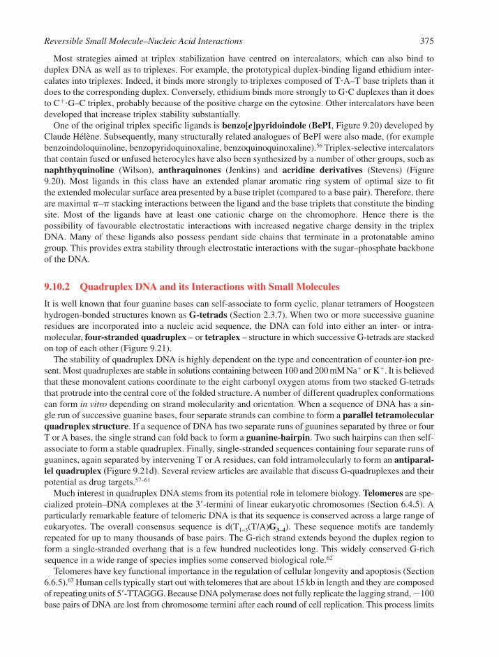
Most strategies aimed at triplex stabilization have centred on intercalators, which can also bind toduplex DNA as well as to triplexes. For example, the prototypical duplex-binding ligand ethidium inter-calates into triplexes. Indeed, it binds more strongly to triplexes composed of T�A–T base triplets than itdoes to the corresponding duplex. Conversely, ethidium binds more strongly to G�C duplexes than it doesto C��G–C triplex, probably because of the positive charge on the cytosine. Other intercalators have beendeveloped that increase triplex stability substantially.
One of the original triplex specific ligands is benzo[e]pyridoindole (BePI, Figure 9.20) developed byClaude Hélène. Subsequently, many structurally related analogues of BePI were also made, (for examplebenzoindoloquinoline, benzopyridoquinoxaline, benzoquinoquinoxaline).56 Triplex-selective intercalatorsthat contain fused or unfused heterocyles have also been synthesized by a number of other groups, such asnaphthyquinoline (Wilson), anthraquinones (Jenkins) and acridine derivatives (Stevens) (Figure9.20). Most ligands in this class have an extended planar aromatic ring system of optimal size to fit the extended molecular surface area presented by a base triplet (compared to a base pair). Therefore, thereare maximal �–� stacking interactions between the ligand and the base triplets that constitute the bindingsite. Most of the ligands have at least one cationic charge on the chromophore. Hence there is the possibility of favourable electrostatic interactions with increased negative charge density in the triplexDNA. Many of these ligands also possess pendant side chains that terminate in a protonatable aminogroup. This provides extra stability through electrostatic interactions with the sugar–phosphate backboneof the DNA.
9.10.2 Quadruplex DNA and its Interactions with Small Molecules
It is well known that four guanine bases can self-associate to form cyclic, planar tetramers of Hoogsteenhydrogen-bonded structures known as G-tetrads (Section 2.3.7). When two or more successive guanineresidues are incorporated into a nucleic acid sequence, the DNA can fold into either an inter- or intra-molecular, four-stranded quadruplex – or tetraplex – structure in which successive G-tetrads are stackedon top of each other (Figure 9.21).
The stability of quadruplex DNA is highly dependent on the type and concentration of counter-ion pre-sent. Most quadruplexes are stable in solutions containing between 100 and 200 mM Na� or K�. It is believedthat these monovalent cations coordinate to the eight carbonyl oxygen atoms from two stacked G-tetradsthat protrude into the central core of the folded structure. A number of different quadruplex conformationscan form in vitro depending on strand molecularity and orientation. When a sequence of DNA has a sin-gle run of successive guanine bases, four separate strands can combine to form a parallel tetramolecularquadruplex structure. If a sequence of DNA has two separate runs of guanines separated by three or fourT or A bases, the single strand can fold back to form a guanine-hairpin. Two such hairpins can then self-associate to form a stable quadruplex. Finally, single-stranded sequences containing four separate runs ofguanines, again separated by intervening T or A residues, can fold intramolecularly to form an antiparal-lel quadruplex (Figure 9.21d). Several review articles are available that discuss G-quadruplexes and theirpotential as drug targets.57–61
Much interest in quadruplex DNA stems from its potential role in telomere biology. Telomeres are spe-cialized protein–DNA complexes at the 3�-termini of linear eukaryotic chromosomes (Section 6.4.5). Aparticularly remarkable feature of telomeric DNA is that its sequence is conserved across a large range ofeukaryotes. The overall consensus sequence is d(T1–3(T/A)G3–4). These sequence motifs are tandemlyrepeated for up to many thousands of base pairs. The G-rich strand extends beyond the duplex region toform a single-stranded overhang that is a few hundred nucleotides long. This widely conserved G-richsequence in a wide range of species implies some conserved biological role.62
Telomeres have key functional importance in the regulation of cellular longevity and apoptosis (Section6.6.5).63 Human cells typically start out with telomeres that are about 15 kb in length and they are composedof repeating units of 5�-TTAGGG. Because DNA polymerase does not fully replicate the lagging strand, �100base pairs of DNA are lost from chromosome termini after each round of cell replication. This process limits
Reversible Small Molecule–Nucleic Acid Interactions 375
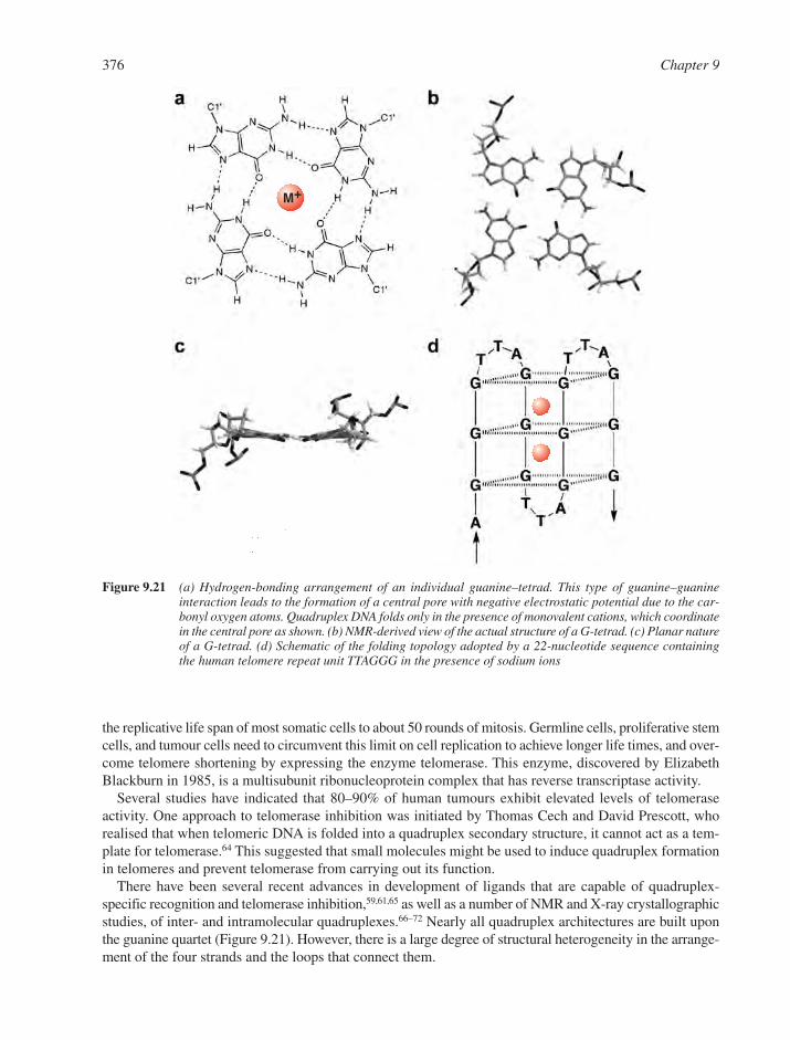
the replicative life span of most somatic cells to about 50 rounds of mitosis. Germline cells, proliferative stemcells, and tumour cells need to circumvent this limit on cell replication to achieve longer life times, and over-come telomere shortening by expressing the enzyme telomerase. This enzyme, discovered by ElizabethBlackburn in 1985, is a multisubunit ribonucleoprotein complex that has reverse transcriptase activity.
Several studies have indicated that 80–90% of human tumours exhibit elevated levels of telomeraseactivity. One approach to telomerase inhibition was initiated by Thomas Cech and David Prescott, whorealised that when telomeric DNA is folded into a quadruplex secondary structure, it cannot act as a tem-plate for telomerase.64 This suggested that small molecules might be used to induce quadruplex formationin telomeres and prevent telomerase from carrying out its function.
There have been several recent advances in development of ligands that are capable of quadruplex-specific recognition and telomerase inhibition,59,61,65 as well as a number of NMR and X-ray crystallographicstudies, of inter- and intramolecular quadruplexes.66–72 Nearly all quadruplex architectures are built uponthe guanine quartet (Figure 9.21). However, there is a large degree of structural heterogeneity in the arrange-ment of the four strands and the loops that connect them.
376 Chapter 9
M+
Figure 9.21 (a) Hydrogen-bonding arrangement of an individual guanine–tetrad. This type of guanine–guanineinteraction leads to the formation of a central pore with negative electrostatic potential due to the car-bonyl oxygen atoms. Quadruplex DNA folds only in the presence of monovalent cations, which coordinatein the central pore as shown. (b) NMR-derived view of the actual structure of a G-tetrad. (c) Planar natureof a G-tetrad. (d) Schematic of the folding topology adopted by a 22-nucleotide sequence containingthe human telomere repeat unit TTAGGG in the presence of sodium ions

One of the most important factors that influences folding topology is the type of monovalent cationpresent during folding. For example, in the fold of the human telomeric sequence (Figure 9.21), which isbased on an NMR study in the presence of Na� (Figure 9.22)67 the TTA loops are arranged in a lateral anddiagonal conformation, and the strands alternate between parallel and antiparallel. By contrast, a recent X-ray structure in the presence of K� ions shows that all four strands are parallel and the TTA loops areextended out from the sides of the G-quartets.
It has been suggested that the antiparallel fold observed in an NMR study is solely a consequence of thepresence of sodium ions, and the presence of potassium ions induces a transition to the parallel arrange-ment. The different conformations adopted by quadruplex structures in different cationic environmentshave a direct impact on the binding of drug molecules. In fact recent biophysical studies reveal consider-able complexity in quadruplex unfolding and show that multiple species coexist in equilibrium across thetemperature range. Thus there is no single static structure for the folded quadruplex and hence targetingquadruplex DNA with small molecules is not straightforward.
Despite these difficulties, several different classes of ligand have shown promise as structure-specific,quadruplex-binding agents and as telomerase inhibitors (Figure 9.23). Most of these compounds havetelomerase inhibitory effects with IC50 values in the low micromolar range but specific binding to quadru-plex DNA over duplex is, at best, only modest.
Laurence Hurley has developed some cationic porphyrins for targeting quadruplex DNA and to effecttelomerase inhibition. The structure of an archetypal porphyrin, [tetra-(N-methyl-4-pyridyl)-porphine]TMPyP4 (Figure 9.23) illustrates the planar arrangement of the aromatic rings, which give rise to a ligand thathas similar molecular dimensions to a G-tetrad intercalation site. This has led to the proposal that compoundsof this type will bind to quadruplex DNA through intercalative stacking. UV spectroscopy, circular dichroism,
Reversible Small Molecule–Nucleic Acid Interactions 377
Figure 9.22 NMR solution structure of the human telomeric sequence 5�-AGGGTTAGGGTTAGGGTTAGGG in thepresence of Na� (PDB: 143D). Guanine residues are in red, thymines in grey, adenines in black, and agrey ribbon highlights the DNA backbone
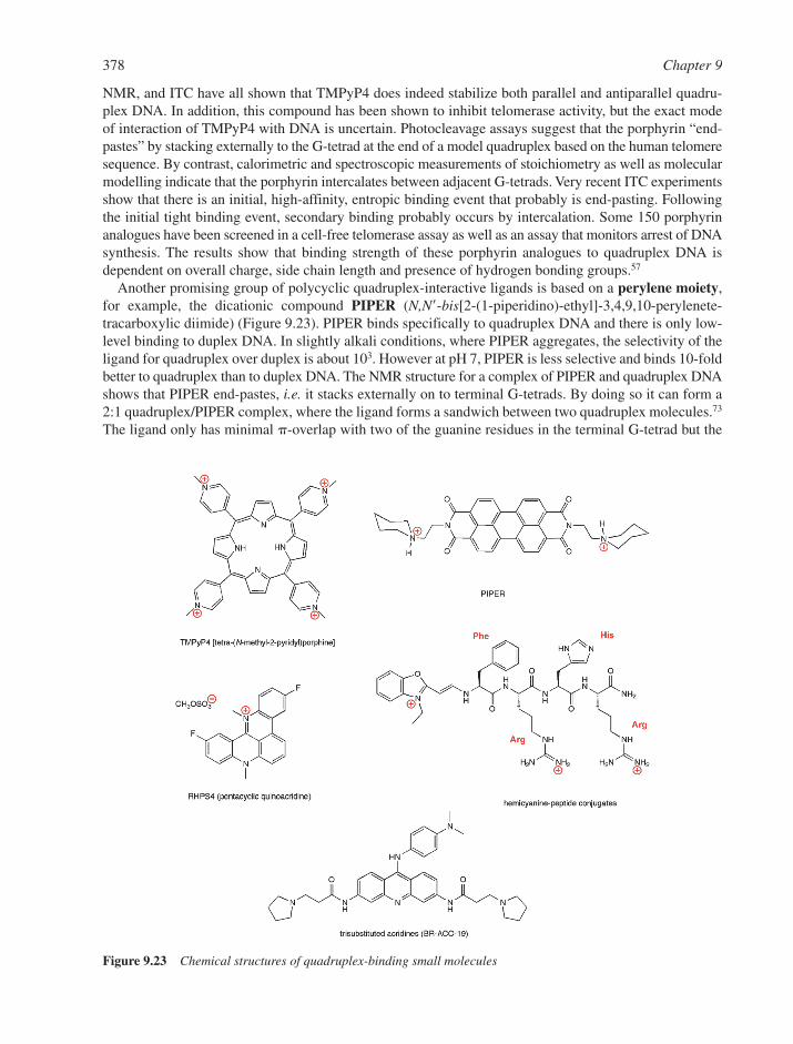
NMR, and ITC have all shown that TMPyP4 does indeed stabilize both parallel and antiparallel quadru-plex DNA. In addition, this compound has been shown to inhibit telomerase activity, but the exact modeof interaction of TMPyP4 with DNA is uncertain. Photocleavage assays suggest that the porphyrin “end-pastes” by stacking externally to the G-tetrad at the end of a model quadruplex based on the human telomeresequence. By contrast, calorimetric and spectroscopic measurements of stoichiometry as well as molecularmodelling indicate that the porphyrin intercalates between adjacent G-tetrads. Very recent ITC experimentsshow that there is an initial, high-affinity, entropic binding event that probably is end-pasting. Followingthe initial tight binding event, secondary binding probably occurs by intercalation. Some 150 porphyrinanalogues have been screened in a cell-free telomerase assay as well as an assay that monitors arrest of DNAsynthesis. The results show that binding strength of these porphyrin analogues to quadruplex DNA isdependent on overall charge, side chain length and presence of hydrogen bonding groups.57
Another promising group of polycyclic quadruplex-interactive ligands is based on a perylene moiety,for example, the dicationic compound PIPER (N,N�-bis[2-(1-piperidino)-ethyl]-3,4,9,10-perylenete-tracarboxylic diimide) (Figure 9.23). PIPER binds specifically to quadruplex DNA and there is only low-level binding to duplex DNA. In slightly alkali conditions, where PIPER aggregates, the selectivity of theligand for quadruplex over duplex is about 103. However at pH 7, PIPER is less selective and binds 10-foldbetter to quadruplex than to duplex DNA. The NMR structure for a complex of PIPER and quadruplex DNAshows that PIPER end-pastes, i.e. it stacks externally on to terminal G-tetrads. By doing so it can form a2:1 quadruplex/PIPER complex, where the ligand forms a sandwich between two quadruplex molecules.73
The ligand only has minimal �-overlap with two of the guanine residues in the terminal G-tetrad but the
378 Chapter 9
Figure 9.23 Chemical structures of quadruplex-binding small molecules
presence of two positive charges on the ligand undoubtedly enhances quadruplex binding. PIPER alsoexhibits significant antitelomerase activity and behaves as a chaperone-like molecule, since it enhancesthe folding of quadruplexes. The ligand alters the kinetics of the association reaction for dimeric quadru-plex formation from second- to first-order and it increases the initial rate for quadruplex formation byabout 100-fold. Thus as well as binding to preformed quadruplex structures, ligands such as PIPER mayalso be able to induce quadruplex formation in vivo at appropriate sequences.
Trisubstituted acridines of the type shown in Figure 9.23 have been synthesized and exhibit very promis-ing quadruplex affinity, selectivity and high levels of telomerase inhibition.74 Based on an NMR structure ofthe quadruplex formed from a sequence model of the human telomere, the expectation was that these mol-ecules would end-paste in a similar manner to PIPER and 2,6-disubstituted amidoanthraquinones. Disubsti-tuted compounds have similar affinity for duplex and quadruplex DNA (�1 � 106 M1) in that there is noappreciable structure selectivity. By contrast, trisubstituted analogues bind quadruplex DNA 30 to 40 timesmore strongly than to duplex DNA and telomerase inhibition correlates with quadruplex binding strength.Thus, activity for disubstituted compounds is either very modest or nonexistent, whereas trisubstituted com-pounds are about 100-fold more potent against telomerase activity, but without increased cellular toxicity.
Quadruplex formation may play many important biological roles. For example, promoter regions of certaingenes such as �-globin genes, rat preproinsulin II gene, adenovirus serotype 2, retinoblastoma suscepti-bility genes, the c-Myc gene, and various other oncogenes, all contain guanine-rich regions that have thepotential to form quadruplex structures. Thus G-quadruplexes may play a role in regulating gene expres-sion, for example, inhibition of transcription. Hurley has shown that ligand-induced quadruplex formationin c-Myc promoter region leads to transcriptional down-regulation of that gene.75 Conversely a G to Amutant in the quadruplex forming region leads to �3-fold increase in the basal transcriptional activity ofthe c-Myc promoter. Thus, genomic sequences that have the propensity to form quadruplex DNA willbecome major drug targets in ongoing efforts to develop structure/sequence-specific agents for the treatmentof human diseases.
Improvements in DNA and RNA synthetic techniques as well as design and synthesis of additional DNA-interactive drugs and model compounds continue to make available systems that are providing new infor-mation on the structure and energetics of nucleic acid complexes with small molecules and are dramaticallyadvancing the field of reversible nucleic acid interactions. In addition, it is clear that both DNA and RNAare prime targets as receptors for drug development. While DNA has been the prime target for such studies,it can be expected that RNA will play an increasing role in the next period of time.
REFERENCES
1. G.S. Manning, Polyelectrolytes. Ann. Rev. Phys. Chem., 1972, 23, 117–140.2. G.S. Manning, The molecular theory of polyelectrolyte solutions with applications to the electrostatic
properties of polynucleotides. Quart. Rev. Biophys., 1978, 11, 179–246.3. G.S. Manning, Counterion binding in polyelectrolyte theory. Acc. Chem. Res., 1979, 12, 443–449.4. M.T. Record Jr., C.F. Anderson and T.M. Lohman, Thermodynamic analysis of ion effects on the
binding and conformational equilibria of proteins and nucleic acids: the roles of ion association orrelease, screening and ion effects on water activity. Quart. Rev. Biophys., 1978, 11, 103–178.
5. J.B. Chaires, Dissecting the free energy of drug binding to DNA. Anti Cancer Drug Design., 1996, 11,569–580.
6. W.D. Wilson and I.G. Lopp, Analysis of cooperativity and ion effects in the interaction of quinacrinewith DNA. Biopolymers, 1979, 18, 3025–3041.
7. R.A.G. Friedman and G.S. Manning, Polyelectrolyte effects on site binding equilibria with applicationto the intercalation of drugs into DNA. Biopolymers, 1984, 23, 2671–2714.
8. T. Thomas and T.J. Thomas, Polyamines in cell growth and cell death: molecular mechanisms andtherapeutic applications. Cell. Mol. Life Sci., 2001, 58, 244–258.
9. E. Westhof and D.L. Beveridge, Hydration of nucleic acids. Water Sci. Rev., 1990, 5, 24–136.
Reversible Small Molecule–Nucleic Acid Interactions 379
10. R.E. Dickerson, H.R. Drew, B.N. Conner, R.M. Wing, A.V. Fratini and M.L. Kopka, The anatomy ofA-, B- and Z-DNA. Science, 1982, 216, 475–485.
11. X. Shui, L. McFail-Isom, G.G. Hu and L.D. Williams, The B-DNA dodecamer at high resolutionreveals a spine of water on sodium. Biochemistry, 1998, 37, 8341–8355.
12. X. Shui, C.C. Sines, L. McFail-Isom, D. VanDerver and L.D. Williams, Structure of the potassium formof the CGCGAATTCGCG: DNA deformation by electrostatic collapse around inorganic cations.Biochemistry, 1998, 37, 16877–16887.
13. L. McFail-Isom, C.C. Sines and L.D. Williams, DNA structure: cations in charge? Curr. Opin. Struct.Biol., 1999, 9, 298–304.
14. L.S. Lerman, Structural considerations in the interaction of DNA and acridines. J. Mol. Biol., 1961, 3,18–30.
15. L.S. Lerman, The structure of the DNA–acridine complex. Proc. Natl. Acad. Sci. USA, 1963, 49, 94–102.16. J.B. Chaires, Energetics of DNA–drug interactions. Biopolymers, 1998, 44, 201–215.17. J.B. Chaires, Drug–DNA interactions. Curr. Opin. Struct. Biol., 1998, 8, 314–320.18. I. Haq, Thermodynamics of drug–DNA interactions. Arch. Biochem. Biophys., 2002, 403, 1–15.19. J.B. Chaires, Energetics of Anthracycline–DNA interactions, in Small Molecule DNA and RNA
Binders: From Synthesis to Nucleic Acid Complexes, M. Demeunynck, C. Bailly and W.D. Wilson (eds).Wiley–VCH Verlag GmbH, Weinheim, 2003, 461–481.
20. J.D. McGhee and P.H. von Hippel, Theoretical aspects of DNA–protein interactions: cooperative andnoncooperativity binding of large ligands to a one-dimensional homogeneous lattice. J. Mol. Biol.,1974, 86, 469–489.
21. D.M. Crothers, Calculation of binding isotherms for heterogeneous polymers. Biopolymers, 1968, 6, 575–584.
22. J.B. Chaires, Analysis and interpretation of ligand–DNA binding isotherms. Methods Enzymol., 2001,340, 1–22.
23. J. Ren, T.C. Jenkins and J.B. Chaires, Energetics of DNA intercalation reactions. Biochemistry, 2000,39, 8439–8447.
24. S.C.M. Teixeira, J.H. Thorpe, A.K. Todd, H.R. Powell, A. Adams, L.P.G. Wakelin, W.A. Denny andC.J. Cardin, Structural characterisation of Bisintercalation in higher-order DNA at a junction likequadruplex. J. Mol. Biol., 2002, 323, 167–171.
25. J.B. Chaires, F. Leng, T. Przewloka, I. Fokt, Y.-H. Ling, R. Perez–Solar and W. Priebe, Structure-based design of a new bisintercalating anthracycline antibiotic. J. Med. Chem., 1997, 40, 261–266.
26. G.G. Hu, X. Shui, F. Leng, W. Priebe, J.B. Chaires and L.D. Williams, Structure of a DNA–bisdaunomycin complex. Biochemistry, 1997, 36, 5940–5946.
27. F.A. Tanious, T.C. Jenkins, S. Neidle and W.D. Wilson, Substituent position dictates the intercalativeDNA-binding mode for anthracene-9,10-dione antitumor drugs. Biochemistry, 1992, 31, 11632–11640.
28. C. Zimmer and U. Wähnert, Nonintercalating DNA-binding ligands: specificity of their interactionand their use a stools in biophysical, biochemical and biological investigations of the genetic material.Prog. Biophys. Molec. Biol., 1986, 47, 31–112.
29. B.H. Geierstanger and D.E. Wemmer, Complexes of the minor groove of DNA. Ann. Rev. Biophys.Biomol. Struct., 1995, 24, 463–493.
30. P.R. Turner and W.A. Denny, The genome as a drug target: sequence specific minor groove bindingligands. Curr. Drug Targets, 2000, 1, 1–14.
31. M.L. Kopka, C. Yoon, D. Goodsell, P. Pjura and R.E. Dickerson, The molecular origin of drug–DNAspecificity in netropsin and distamycin. Proc. Natl. Acad. Sci. USA, 1985, 82, 1376–1380.
32. M. Coll, C.A. Frederick, A.H.-J. Wang and A. Rich, A bifurcated hydrogen bonded conformation inthe d(AT) base pairs of the DNA dodecamer d(CGCAAATTTGCG) and its complex with distamycin.Proc. Natl. Acad. Sci. USA, 1987, 84, 8385–8389.
33. J.G. Pelton and D.E. Wemmer, Binding modes of distamycin A with d(CGCAAATTTGCG)2
determined by two-dimensional NMR. J. Am. Chem. Soc., 1990, 112, 1393–1399.
380 Chapter 9
34. D. Rentzeperis and L.A. Marky, Interaction of minor groove ligands to an AAATT/AATTT site: cor-relation of thermodynamic characterisation and solution structure. Biochemistry, 1995, 34, 2937–2945.
35. A.N. Lane and T.C. Jenkins, Thermodynamics of nucleic acids and their interactions with ligands.Quart. Rev. Biophys., 2000, 33, 255–306.
36. P.B. Dervan, Design of sequence specific DNA-binding molecules. Science, 1986, 232, 464–471.37. J.W. Trauger, E.E. Bairde and P.B. Dervan, Recognition of DNA by designed ligands at subnanomolar
concentrations. Nature, 1996, 382, 559–561.38. J.M. Gottesfeld, L. Neely, J.W. Trauger, E.E. Baird and P.B. Dervan, Regulation of gene expression
by small molecules. Nature, 1997, 387, 202–205.39. D.E. Wemmer and P.B. Dervan, Targeting the minor groove of DNA. Curr. Opin. Struct. Biol., 1997,
7, 355–361.40. S. White, J.W. Szewczyk, J.M. Turner, E.E. Baird and P.B. Dervan, Recognition of the four
Watson–Crick base pairs in the DNA minor groove by synthetic ligands. Nature, 1998, 391, 468–470.41. D.S. Pilch, N. Poklar, E.E. Baird, P.B. Dervan and K.J. Breslauer, The thermodynamics of
polyamide–DNA recognition: hairpin polyamide binding in the minor groove of duplex DNA.Biochemistry, 1999, 38, 2143–2151.
42. P.B. Dervan and R.W. Bürli, Sequence-specific DNA recognition by polyamides. Curr. Opin. Chem.Biol., 1999, 3, 688–693.
43. P.B. Dervan and B.S. Edelson, Recognition of the DNA minor groove by pyrrole–imidazolepolyamides. Curr. Opin. Struct. Biol., 2003, 13, 284–299.
44. A. Abu-Daya, P.M. Brown and K.R. Fox, DNA sequence preference of several AT-selective minorgroove binding ligands. Nucleic Acids Res., 1995, 23, 3385–3392.
45. F.G. Loontiens, P. Regenfuss, A. Zechel, L. Dumortier and R.M. Clegg, Binding characteristics ofHoechst 33258 with calf-thymus DNA, poly[d(A-T)] and d(CCGGAATTCCGG): multiple stoichiome-tries and determination of tight binding with a wide spectrum of site affinities. Biochemistry, 1990,29, 9029–9039.
46. F.G. Loontiens, L.W. McLaughlin, S. Diekmann and R.M. Clegg, Binding of Hoechst 33258 and 4�,6-diamidino-2-phenylindole to self-complementary decadeoxynucleotides with modified exocyclicbase substituents. Biochemistry, 1991, 30, 182–189.
47. M.C. Vega, I. García–Sáez, J. Aymamí, T. Eritja, G.A. van der Marel, J.H. van Boom, A. Rich andM. Coll, Three-dimensional crystal structure of the A-tract DNA dodecamer d(CGCAAATTTGCG)2
complexed with the minor groove binding drug Hoechst 33258. Eur. J. Biochem., 1994, 222, 721–726.48. N. Spink, D.G. Brown, J.V. Skelly and S. Neidle, Sequence-dependent effects in drug–DNA
interaction: the crystal structure of the Hoechst 33258 bound to the d(CGCAAATTTGCG)2
duplex. Nucleic Acids Res., 1994, 22, 1607–1612.49. I. Haq, J.E. Ladbury, B.Z. Chowdhry, T.C. Jenkins and J.B. Chaires, Specific binding of Hoechst
33258 to the d(CGCAAATTTGCG)2 duplex: calorimetric and spectroscopic studies. J. Mol. Biol.,1997, 271, 244–257.
50. R.S. Spolar and M.T. Record Jr., Coupling of local folding to site-specific binding of proteins to DNA.Science, 1994, 263, 777–784.
51. D. Praseuth, A.L. Guieysse and C. Hélène, Triple helix formation and the antigene strategy for thesequence-specific control of gene expression. Biochim. Biophys. Acta, 1999, 1489, 181–206.
52. J.-S. Sun and C. Hélène, Oligonucleotide-directed triple-helix formation. Curr. Opin. Struct. Biol.,1993, 3, 345–356.
53. M.D. Frank-Kamenetskii and S.M. Mirkin, Triplex DNA structures. Annu. Rev. Biochem., 1995, 64,65–95.
54. K.R. Fox, Targeting DNA with triplexes. Curr. Med. Chem., 2000, 7, 17–37.55. T.C. Jenkins, Targeting multistranded DNA structures. Curr. Med. Chem., 2000, 7, 99–115.56. J.L. Mergny, G. Duval–Valentin, C.H. Nguyen, L. Perrouault, B. Faucon, M. Rougée,T. Montenay-
Garestier, E. Bisagni and C. Hélène, Triple helix-specific ligands. Science, 1992,256, 1681–1684.
Reversible Small Molecule–Nucleic Acid Interactions 381
57. H. Han and L.H. Hurley, G-quadruplex DNA: a potential target for anticancer drug design. TrendsPharm. Sci., 2000, 21, 136–142.
58. J.R. Williamson, G-quartet structures in telomeric DNA. Annu. Rev. Biophys. Biomol. Struct., 1994,23, 703–730.
59. J.L. Mergny and C. Hélène, G-quadruplex DNA: a target for drug design. Nature Med., 1998, 4,1366–1367.
60. D. Sen and W. Gilbert, Guanine quartet structures. Methods Enzymol., 1992, 211, 191–199.61. P.J. Perry and T.C. Jenkins, Recent advances in the development of telomerase inhibitors for the treat-
ment of cancer. Exp. Opin. Invest. Drugs, 1999, 8, 1981–2008.62. D. Rhodes and R. Giraldo, Telomere structure and function. Curr. Opin. Struct. Biol., 1995, 5,
311–322.63. E.H. Blackburn, Structure and function of telomeres. Nature, 1991, 350, 569–573.64. A.M. Zahler, J.R. Williamson, T.R. Cech and D.M. Prescott, Inhibition of telomerase by G-quartet
DNA structures. Nature, 1991, 350, 718–720.65. S. Neidle and G.N. Parkinson, Telomere maintenance as a target for anticancer drug discovery. Nature
Rev. Drug Disc., 2002, 1, 383–393.66. C. Kang, X. Zhang, R. Ratliff, R. Moyzis and A. Rich, Crystal structure of four-stranded Oxytricha
telomeric DNA. Nature, 1992, 356, 126–131.67. Y. Wang and D.J. Patel, Solution structure of the human telomeric repeat d[AG3(T2AG3)3]
G-tetraplex. Structure, 1993, 1, 263–282.68. K. Phillips, Z. Dauter, A.I.H. Murchie, D.M. Lilley and B. Luisi, The crystal structure of a parallel-
stranded quanine tetraplex at 0.95 Å resolution. J. Mol. Biol., 1997, 273, 171–182.69. A. Kettani, S. Bouaziz, A. Gorin, H. Zhao, R.A. Jones and D.J. Patel, Solution structure of a Na�
cation stabilized DNA quadruplex containing G�G�G�G and G�C�G�C tetrads formed by G-G-G-Crepeats observed in adeno-associated viral DNA. J. Mol. Biol., 1998, 282, 619–636.
70. S. Haider, G.N. Parkinson and S. Neidle, Crystal structure of the potassium form of an Oxytricha novaG-quadruplex. J. Mol. Biol., 2002, 320, 189–200.
71. G.N. Parkinson, M.P.H. Lee and S. Neidel, Crystal structure of parallel quadruplexes from humantelomeric DNA. Nature, 2002, 417, 876–880.
72. S. Neidle and G.N. Parkinson, The structure of telomeric DNA. Curr. Opin. Struct. Biol., 2003, 13,275–283.
73. O.Y. Fedoroff, M. Salazar, H. Han, V.V. Chemeris, S.M. Kerwin and L.H. Hurley, NMR-based modelof a telomerase-inhibiting compound bound to G-quadruplex DNA. Biochemistry, 1998, 37,12367–12374.
74. M. Read, R.J. Harrison, B. Romagnoli, F.A. Tanious, S.H. Gowan, A.P. Reszka, W.D. Wilson, L.R. Kelland and S. Neidle, Structure-based design of selective and potent G-quadruplex-mediatedtelomerase inhibitors. Proc. Natl. Acad. Sci. USA, 2001, 98, 4844–4849.
75. A. Siddiqui-Jain, C.L. Grand, D.J. Bearss and L.H. Hurley, Direct evidence for a G-quadruplex in apromoter region and its targeting with a small molecule to repress c-MYC transcription. Proc. Natl.Acad. Sci. USA, 2002, 99, 11593–115.
382 Chapter 9
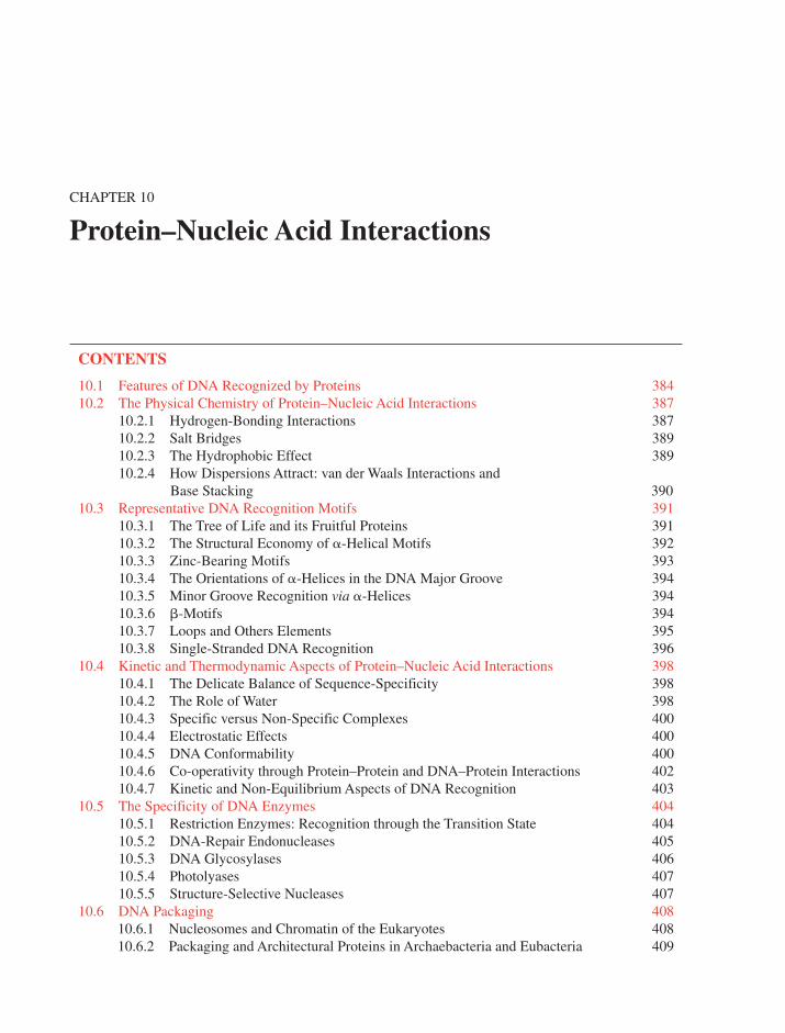
CHAPTER 10
Protein–Nucleic Acid Interactions
CONTENTS
10.1 Features of DNA Recognized by Proteins 38410.2 The Physical Chemistry of Protein–Nucleic Acid Interactions 387
10.2.1 Hydrogen-Bonding Interactions 38710.2.2 Salt Bridges 38910.2.3 The Hydrophobic Effect 38910.2.4 How Dispersions Attract: van der Waals Interactions and
Base Stacking 39010.3 Representative DNA Recognition Motifs 391
10.3.1 The Tree of Life and its Fruitful Proteins 39110.3.2 The Structural Economy of �-Helical Motifs 39210.3.3 Zinc-Bearing Motifs 39310.3.4 The Orientations of �-Helices in the DNA Major Groove 39410.3.5 Minor Groove Recognition via �-Helices 39410.3.6 �-Motifs 39410.3.7 Loops and Others Elements 39510.3.8 Single-Stranded DNA Recognition 396
10.4 Kinetic and Thermodynamic Aspects of Protein–Nucleic Acid Interactions 39810.4.1 The Delicate Balance of Sequence-Specificity 39810.4.2 The Role of Water 39810.4.3 Specific versus Non-Specific Complexes 40010.4.4 Electrostatic Effects 40010.4.5 DNA Conformability 40010.4.6 Co-operativity through Protein–Protein and DNA–Protein Interactions 40210.4.7 Kinetic and Non-Equilibrium Aspects of DNA Recognition 403
10.5 The Specificity of DNA Enzymes 40410.5.1 Restriction Enzymes: Recognition through the Transition State 40410.5.2 DNA-Repair Endonucleases 40510.5.3 DNA Glycosylases 40610.5.4 Photolyases 40710.5.5 Structure-Selective Nucleases 407
10.6 DNA Packaging 40810.6.1 Nucleosomes and Chromatin of the Eukaryotes 40810.6.2 Packaging and Architectural Proteins in Archaebacteria and Eubacteria 409
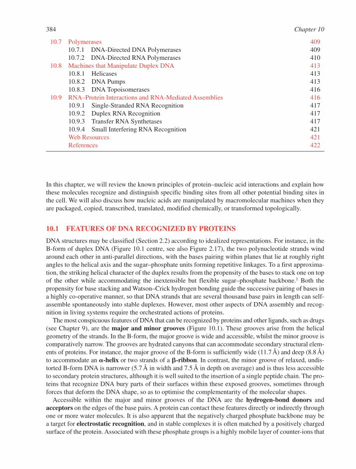
10.7 Polymerases 40910.7.1 DNA-Directed DNA Polymerases 40910.7.2 DNA-Directed RNA Polymerases 410
10.8 Machines that Manipulate Duplex DNA 41310.8.1 Helicases 41310.8.2 DNA Pumps 41310.8.3 DNA Topoisomerases 416
10.9 RNA–Protein Interactions and RNA-Mediated Assemblies 41610.9.1 Single-Stranded RNA Recognition 41710.9.2 Duplex RNA Recognition 41710.9.3 Transfer RNA Synthetases 41710.9.4 Small Interfering RNA Recognition 421Web Resources 421References 422
384 Chapter 10
In this chapter, we will review the known principles of protein–nucleic acid interactions and explain howthese molecules recognize and distinguish specific binding sites from all other potential binding sites inthe cell. We will also discuss how nucleic acids are manipulated by macromolecular machines when theyare packaged, copied, transcribed, translated, modified chemically, or transformed topologically.
10.1 FEATURES OF DNA RECOGNIZED BY PROTEINS
DNA structures may be classified (Section 2.2) according to idealized representations. For instance, in theB-form of duplex DNA (Figure 10.1 centre, see also Figure 2.17), the two polynucleotide strands windaround each other in anti-parallel directions, with the bases pairing within planes that lie at roughly rightangles to the helical axis and the sugar–phosphate units forming repetitive linkages. To a first approxima-tion, the striking helical character of the duplex results from the propensity of the bases to stack one on topof the other while accommodating the inextensible but flexible sugar–phosphate backbone.1 Both thepropensity for base stacking and Watson–Crick hydrogen bonding guide the successive pairing of bases ina highly co-operative manner, so that DNA strands that are several thousand base pairs in length can self-assemble spontaneously into stable duplexes. However, most other aspects of DNA assembly and recog-nition in living systems require the orchestrated actions of proteins.
The most conspicuous features of DNA that can be recognized by proteins and other ligands, such as drugs(see Chapter 9), are the major and minor grooves (Figure 10.1). These grooves arise from the helicalgeometry of the strands. In the B-form, the major groove is wide and accessible, whilst the minor groove iscomparatively narrow. The grooves are hydrated canyons that can accommodate secondary structural elem-ents of proteins. For instance, the major groove of the B-form is sufficiently wide (11.7 Å) and deep (8.8 Å)to accommodate an �-helix or two strands of a �-ribbon. In contrast, the minor groove of relaxed, undis-torted B-form DNA is narrower (5.7 Å in width and 7.5 Å in depth on average) and is thus less accessibleto secondary protein structures, although it is well suited to the insertion of a single peptide chain. The pro-teins that recognize DNA bury parts of their surfaces within these exposed grooves, sometimes throughforces that deform the DNA shape, so as to optimise the complementarity of the molecular shapes.
Accessible within the major and minor grooves of the DNA are the hydrogen-bond donors andacceptors on the edges of the base pairs. A protein can contact these features directly or indirectly throughone or more water molecules. It is also apparent that the negatively charged phosphate backbone may bea target for electrostatic recognition, and in stable complexes it is often matched by a positively chargedsurface of the protein. Associated with these phosphate groups is a highly mobile layer of counter-ions that
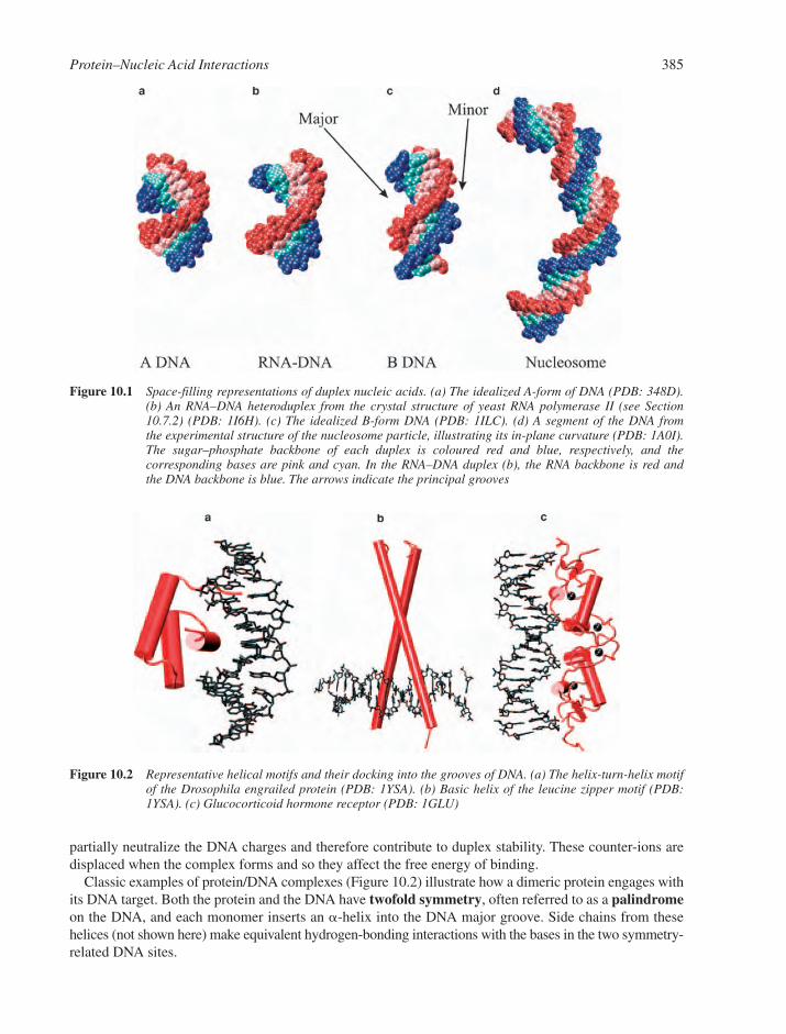
partially neutralize the DNA charges and therefore contribute to duplex stability. These counter-ions aredisplaced when the complex forms and so they affect the free energy of binding.
Classic examples of protein/DNA complexes (Figure 10.2) illustrate how a dimeric protein engages withits DNA target. Both the protein and the DNA have twofold symmetry, often referred to as a palindromeon the DNA, and each monomer inserts an �-helix into the DNA major groove. Side chains from thesehelices (not shown here) make equivalent hydrogen-bonding interactions with the bases in the two symmetry-related DNA sites.
Protein–Nucleic Acid Interactions 385
Figure 10.2 Representative helical motifs and their docking into the grooves of DNA. (a) The helix-turn-helix motifof the Drosophila engrailed protein (PDB: 1YSA). (b) Basic helix of the leucine zipper motif (PDB:1YSA). (c) Glucocorticoid hormone receptor (PDB: 1GLU)
Figure 10.1 Space-filling representations of duplex nucleic acids. (a) The idealized A-form of DNA (PDB: 348D).(b) An RNA–DNA heteroduplex from the crystal structure of yeast RNA polymerase II (see Section10.7.2) (PDB: 1I6H). (c) The idealized B-form DNA (PDB: 1ILC). (d) A segment of the DNA from the experimental structure of the nucleosome particle, illustrating its in-plane curvature (PDB: 1A0I).The sugar–phosphate backbone of each duplex is coloured red and blue, respectively, and the corresponding bases are pink and cyan. In the RNA–DNA duplex (b), the RNA backbone is red and the DNA backbone is blue. The arrows indicate the principal grooves
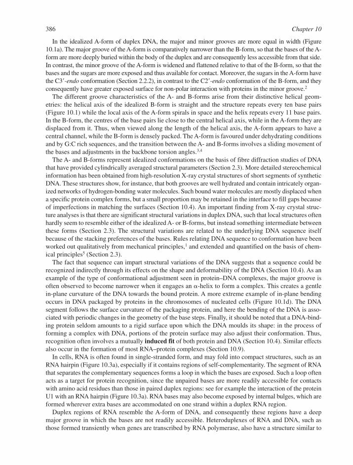
In the idealized A-form of duplex DNA, the major and minor grooves are more equal in width (Figure10.1a). The major groove of the A-form is comparatively narrower than the B-form, so that the bases of the A-form are more deeply buried within the body of the duplex and are consequently less accessible from that side.In contrast, the minor groove of the A-form is widened and flattened relative to that of the B-form, so that thebases and the sugars are more exposed and thus available for contact. Moreover, the sugars in the A-form havethe C3�-endo conformation (Section 2.2.2), in contrast to the C2�-endo conformation of the B-form, and theyconsequently have greater exposed surface for non-polar interaction with proteins in the minor groove.2
The different groove characteristics of the A- and B-forms arise from their distinctive helical geom-etries: the helical axis of the idealized B-form is straight and the structure repeats every ten base pairs(Figure 10.1) while the local axis of the A-form spirals in space and the helix repeats every 11 base pairs.In the B-form, the centres of the base pairs lie close to the central helical axis, while in the A-form they aredisplaced from it. Thus, when viewed along the length of the helical axis, the A-form appears to have acentral channel, while the B-form is densely packed. The A-form is favoured under dehydrating conditionsand by G:C rich sequences, and the transition between the A- and B-forms involves a sliding movement ofthe bases and adjustments in the backbone torsion angles.3,4
The A- and B-forms represent idealized conformations on the basis of fibre diffraction studies of DNAthat have provided cylindrically averaged structural parameters (Section 2.3). More detailed stereochemicalinformation has been obtained from high-resolution X-ray crystal structures of short segments of syntheticDNA. These structures show, for instance, that both grooves are well hydrated and contain intricately organ-ized networks of hydrogen-bonding water molecules. Such bound water molecules are mostly displaced whena specific protein complex forms, but a small proportion may be retained in the interface to fill gaps becauseof imperfections in matching the surfaces (Section 10.4). An important finding from X-ray crystal struc-ture analyses is that there are significant structural variations in duplex DNA, such that local structures oftenhardly seem to resemble either of the idealized A- or B-forms, but instead something intermediate betweenthese forms (Section 2.3). The structural variations are related to the underlying DNA sequence itselfbecause of the stacking preferences of the bases. Rules relating DNA sequence to conformation have beenworked out qualitatively from mechanical principles,1 and extended and quantified on the basis of chem-ical principles5 (Section 2.3).
The fact that sequence can impart structural variations of the DNA suggests that a sequence could berecognized indirectly through its effects on the shape and deformability of the DNA (Section 10.4). As anexample of the type of conformational adjustment seen in protein–DNA complexes, the major groove isoften observed to become narrower when it engages an �-helix to form a complex. This creates a gentlein-plane curvature of the DNA towards the bound protein. A more extreme example of in-plane bendingoccurs in DNA packaged by proteins in the chromosomes of nucleated cells (Figure 10.1d). The DNAsegment follows the surface curvature of the packaging protein, and here the bending of the DNA is asso-ciated with periodic changes in the geometry of the base steps. Finally, it should be noted that a DNA-bind-ing protein seldom amounts to a rigid surface upon which the DNA moulds its shape: in the process offorming a complex with DNA, portions of the protein surface may also adjust their conformation. Thus,recognition often involves a mutually induced fit of both protein and DNA (Section 10.4). Similar effectsalso occur in the formation of most RNA–protein complexes (Section 10.9).
In cells, RNA is often found in single-stranded form, and may fold into compact structures, such as anRNA hairpin (Figure 10.3a), especially if it contains regions of self-complementarity. The segment of RNAthat separates the complementary sequences forms a loop in which the bases are exposed. Such a loop oftenacts as a target for protein recognition, since the unpaired bases are more readily accessible for contactswith amino acid residues than those in paired duplex regions: see for example the interaction of the proteinU1 with an RNA hairpin (Figure 10.3a). RNA bases may also become exposed by internal bulges, which areformed wherever extra bases are accommodated on one strand within a duplex RNA region.
Duplex regions of RNA resemble the A-form of DNA, and consequently these regions have a deepmajor groove in which the bases are not readily accessible. Heteroduplexes of RNA and DNA, such asthose formed transiently when genes are transcribed by RNA polymerase, also have a structure similar to
386 Chapter 10

A-form DNA (Figure 10.1a). These duplexes may be recognized more by shape and groove width than byspecific hydrogen-bonding contacts. An example from the bacterial ribosome shows how a �-strand liesin the groove of a duplex RNA, while the bases in a single-stranded region are contacted by an �-helicalregion of the protein (Figure 10.3b). The intricate three-dimensional folds of RNA add to its ability tomake an induced fit in recognition of protein, for example, during the charging of transfer RNA withamino acids by tRNA synthetases (Sections 7.3.2 and 10.9.3).
10.2 THE PHYSICAL CHEMISTRY OF PROTEIN–NUCLEIC ACIDINTERACTIONS
Since the 1990s, many hundreds of crystal and NMR structures have been solved for protein–DNA and protein–RNA complexes. These structures illustrate the diversity of nucleic acid–protein recognition pat-terns. To understand such complicated molecular architectures, we need to explore the forces that hold thecomponents together.
10.2.1 Hydrogen Bonding Interactions
Although energetically rather weak, hydrogen bonding interactions are a key component of protein–nucleicacid recognition, since collectively they confer sequence-specificity in the majority of cases. In solution, allexposed polar groups form hydrogen bonds with water molecules. When the solvent is removed from thesegroups, as happens in the formation of a protein/nucleic acid complex, they must make compensating inter-actions with other polar groups. The energy of the hydrogen-bonding interaction is optimal when the atomsof the donor and acceptor groups (X, H and Y) and the lone electron pair are all arranged linearly, as indi-cated in the scheme
X���H���Y���R��
Protein–Nucleic Acid Interactions 387
Figure 10.3 Representative complexes of protein with RNA. (a) An RNA hairpin, in complex with the RNP proteinU1 from the eukaryotic intron-splicing machinery (PDB: 1URN). The bases of the single-stranded loopare recognized by contacts with the protein side chains. (b) A view of a section of the ribosome, show-ing an even more complicated RNA/protein interaction (PDB: 1IBM). A b-strand lies in the groove of theduplex RNA and the bases in a single-stranded region are contacted by an a-helical region
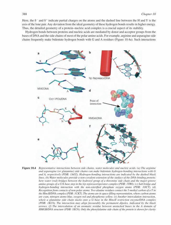
Here, the �� and �� indicate partial charges on the atoms and the dashed line between the H and Y is theaxis of the lone pair. Any deviation from the ideal geometry of these hydrogen bonds results in higher energy.Thus, the detailed geometry of a protein–nucleic acid complex is a crucial aspect of its stability.
Hydrogen bonds between proteins and nucleic acids are mediated by donor and acceptor groups from thebases of DNA and the side chains of most of the polar amino acids. For example, arginine and asparagine sidechains frequently make bidentate hydrogen bonds with G and A residues (Figure 10.4a). Such interactions
388 Chapter 10
Figure 10.4 Representative interactions between side chains, water molecules and nucleic acids. (a) The arginineand asparagine (or glutamine) side chains can make bidentate hydrogen-bonding interactions with Gand A, respectively (PDB: 1AO2). Hydrogen-bonding interactions are indicated by the dashed blacklines. (b) Water molecules provide a non-covalent extension of the surface of the DNA-binding protein;here water (red) bridges between the hydroxyl group of a threonine side chain and the major-grooveamino groups of a C/A base step in the trp repressor/operator complex (PDB: 1TR0). (c) Salt bridge andhydrogen-bonding interaction with the non-esterified phosphate oxygen atoms (PDB: 1OCT). (d)Recognition from contacts of non-polar atoms. Two alanine residues contact the 5-methyl carbon of a T inthe HincII/DNA complex (PDB: 1CKT). The atoms are in space-filling representation, where carbon atomsare cyan, nitrogen atoms blue, oxygen red and phosphorus yellow. (e) Another intercalation interaction,where a glutamine side chain stacks onto a G base in the HincII restriction enzyme/DNA complex(PDB: 1KC6). The interaction may align favourably the permanent dipoles, indicated by the blackarrows. (f) The intercalation of an aromatic residue between unstacked bases in the A domain ofHMGB/DNA structure (PDB: 1KC6). Only the phenylalanine side chain of the protein is shown for clarity
may be partially conjugated and can contribute to stability by ‘stacking’ like plates, akin to the stackingseen for the bases in the nucleic acid. Much less frequently, weaker hydrogen bonds can form between theDNA bases and the aromatic rings of Phe, Tyr and Trp and these are called ‘�-hydrogen bonds’ (notshown). Water molecules can extend the surface of the DNA and bridge to an amino acid, for example themediation of interaction between a Thr hydroxyl group and the major groove exocyclic amino groups at aC/A base step (Figure 10.4b). In addition, the amide backbone of a protein may participate in hydrogen-bond interactions with the nucleic acid, as either a donor (N�H) or acceptor (C��O) (not shown).
From the nucleic acid bases, the carbonyl oxygen atoms of G, C and T (U in RNA) are commonly accept-ors, as are also the heterocyclic N-7 and N-3 atoms of purines. Potential donors are the exocyclic aminogroups of A, G and C and the amido N�H functions of T (U in RNA), G and C. In the case of unpairedbases, the N-1 of purines and the N-3 of pyrimidines often form hydrogen bonds with protein. It might seemsurprising that the most common site of hydrogen-bonding interaction in the recognition of a nucleic acidis not with the bases, but with the non-bridging oxygen atoms of phosphates. An example of this frequentlyoccurring interaction is shown in Figure 10.4c. The bridging oxygen atoms of the phosphate backbone andthe oxygen atom within the ribose sugar ring may act as acceptors of hydrogen bonds, but these are com-paratively weaker interactions.
10.2.2 Salt Bridges
Salt bridges are electrostatic interactions between groups of opposite charge. In protein–nucleic acidcomplexes, salt bridges may be formed between the ionized, non-bridging oxygen atoms of the phosphatebackbone and the protonated, positively charged guanidinium moiety of Arg, the imidazole ring of His, the�-amino group of Lys, or the terminal �-amino group in a protein (e.g. Figure 10.4c). The importance ofsalt bridges in protein stability has been inferred by their frequent occurrence in the heat-stable proteinsisolated from extreme thermophilic organisms, where the salt bridges are found in co-operative networks.Isolated salt bridges are entropically disfavoured because they place constraints on the side-chain conform-ations and cause desolvation of the interacting side chains. However, networks of mutually supporting saltbridges are favourable, since the co-operativity of their interaction compensates for the unfavourableentropy change. This principle may also be true for salt bridges formed between amino acid side chainsand nucleic acid phosphate groups, which are often organized as intricate networks of mutually support-ing interactions. Another type of salt bridge that is also observed in protein–nucleic acid complexes is anelectrostatic interaction mediated by bound counter-ions, such as chloride ions.
Unlike hydrogen bonds, which require alignment of the interacting groups for optimal strength, saltbridges and counter-ion interactions are not directional. Salt bridges vary in strength in inverse proportionto the square of the distance between the individual charges and to the dielectric constant, so that they aregreatly weakened as ionic strength increases and strengthened as the dielectric constant decreases. If saltbridges or bound counter-ions become buried and isolated from bulk solvent, their interaction energy isenhanced, especially if they are sequestered into a hydrophobic environment. Thus, the strength of saltbridges is highly context-dependent, but a typical value in proteins corresponds to about 40 kJ mol�1. Salt-bridging interactions play an important role in making a general electrostatic match of the polyanionic surfaceof the DNA or RNA with the polycationic surface of the recognizing protein. Thus, a charged surface ofthe protein may be used as a rough gauge of the groove width or to discriminate single-stranded nucleicacids from double-stranded. However, electrostatic complementarity is unlikely to provide specificity byitself. Indeed, specificity must originate from other effects, such as the pattern of hydrogen bonds describedabove and complementary surface shapes (Sections 10.2.3 and 10.2.4).
10.2.3 The Hydrophobic Effect
The greatest contribution to the Gibbs free energy of complex formation is that of the hydrophobic effect.This short-range effect is sometimes referred to as a force. However, it is not a real force in the classical
Protein–Nucleic Acid Interactions 389
sense, with an associated scalar field, such as found for the electric forces in which two charged particlesattract or repel each other. Instead, the hydrophobic effect is a consequence of the behaviour of bulk waterat a non-polar surface.6 In its optimal orientation, a water molecule can form four hydrogen bonds in atetrahedral geometry: two through its lone pairs of electrons (the acceptors) and two through its protons(the donors). In bulk solvent at physiological temperatures and pressures, the water molecules are in continual exchange of hydrogen-bonding partners. When the water molecules encounter a non-polar molecule that cannot accept or donate, these hydrogen bonding interactions are broken. If two such non-polar molecules are in proximity, they will associate so as to minimise the disruption to the dynamicwater interactions. In general, macromolecules such as proteins and nucleic acids tend to associate in sucha way as to expose a minimum number of non-polar features on the external surface to the aqueous solv-ent. The non-polar parts of nucleic acid include the 5-methyl group of thymine, the heterocyclic carbonatoms within the purine and pyrimidine rings, and the ribose carbon atoms. These atoms are often contacted by the aliphatic portions of amino acid side chains in protein–nucleic acid complexes (e.g.Figure 10.4d).
The association of hydrophobic molecules in aqueous solvent is favoured entropically. This mightseem surprising given that a hydrogen-bonding pattern is being affected and that bond breaking is associ-ated mostly with enthalpic change. However, it is thought that water molecules become immobilized onnon-polar surfaces because of restricted interactions with partner water molecules. Thus, by burying theexposed non-polar surface, water molecules are liberated and their translational and rotational entropyincreases, with a concomitant lowering of the overall free energy. In this way, shape complementarity canhelp to bury non-polar surfaces optimally and to contribute to the affinity of a macromolecular interaction.Shape complementarity also favours optimal van der Waals interactions (Section 10.2.4).
10.2.4 How Dispersions Attract: van der Waals Interactions and Base Stacking
Van der Waals interactions arise from the dispersive forces originating from transient dipoles inherent in theelectronic structure of atoms and molecules, and even molecules without a net permanent dipole can formthese interactions. Dispersive forces are proportional to the inverse sixth power of the distance between thedipoles. Although the interactions are very weak and sensitive to structural fluctuations, their cumulative con-tribution becomes significant over an extensive intermolecular interface. In a specific protein–DNA complex,van der Waals contacts account for roughly two-thirds of the interfacial surface area, whilst the remaining one-third involves direct or water-mediated hydrogen-bonding interactions.7 Thus, the contacting protein andnucleic acid surfaces in specific complexes are not only characterized by the stereochemical match of donorsand acceptors, but also by the shape complementarity that produces a snug fit so as to benefit from the r�6 law.
Dispersive effects also contribute to the stability of base-stacking interactions and thus are an import-ant aspect of the sequence-dependent conformational effects mentioned earlier (Section 2.3.1). As a con-sequence of stacking and base-pair hydrogen bonding, the packing density of atoms within duplex DNAis greater than for equivalent atoms in related small molecules packing in crystalline lattices.8 As a conse-quence of the hydrophobic effect, the bases tend to propeller-twist in the horizontal plane so as to opti-mize the buried surface area between neighbouring base steps. However, this feature is not sufficient initself to account fully for the observed sequence-dependent conformational variations in duplex DNA.Aside from the general effect of hydrophobicity on the general shape of the molecule, electrostatic effectsare important components of DNA conformation, and they tune the detailed geometry of the base stack-ing. Each of the aromatic atoms of the bases can be treated as sandwiches of charges, from which the con-formational propensities of the heterocyclic bases can be calculated based on the optimized electrostaticinteractions (Section 2.3.1). When combined with a simulated ‘backbone link’, these calculations canaccount for many of the experimentally observed conformations of different base steps.5
Dipolar effects also contribute to DNA conformation and protein–DNA interactions, but the importanceof this effect is difficult to quantify since it depends greatly on the organization of fixed charges and on thedielectric constant. For example, a permanent dipole is associated with the G bases because of the
390 Chapter 10
concentration of electronegative atoms on the major groove side and electropositive atoms on the minorgroove side. The dipole of an asparagine or glutamine side chain can match the local dipole of the nucleotide(Figure 10.4e). Dipoles also affect the stacking of the bases in G/C rich sequences. To a first approxima-tion, the GC base pair has a large dipole along its long axis resulting from the cluster of partial negativecharge on the G and partial positive charge on the C. Dipoles align to maximize attraction and minimizerepulsion, and it can be envisaged how the sandwiched GC dipoles in a sequence like GGGG/CCCCfavour high slide, whilst low slide is preferred for an alternating sequence such as GCGC/CGCG. In con-trast, the AT base pair contains only small patches of isolated positive and negative electrical charge alongits long axis, which are relatively dispersed over the entire pair, and therefore do not amount to a sub-stantial dipole.
Stacking interactions can also occur between aromatic residues in a protein and unstacked bases, forexample in the intercalation of a Phe side chain between two unstacked nucleotides (Figure 10.4f).
10.3 REPRESENTATIVE DNA RECOGNITION MOTIFS
10.3.1 The Tree of Life and its Fruitful Proteins
Protein folds that are used in recognition of DNA (and RNA) may be grouped according to their structuraland evolutionary relationships. But before grouping such folds into classes, it is useful to be aware of howorganisms are grouped. All cellular life on Earth can be organized into three domains: the eukaryotes, thebacteria and the archaea. Eukaryotes and archaebacteria may have shared a common ancestor during aperiod of evolutionary history following the divergence from bacteria.9 During the subsequent evolutionof the archaea, there was a split into the sub-domains known as the crenarchaeota and the euryarchaeota.While eukaryotes and bacteria have many distinguishing features, the archaea share certain molecular simi-larities with both these groups. For instance, the DNA of eukaryotes is packed tightly into cellular nuclei,while bacteria and archaea both lack nuclei. On the other hand, the archaea control the expression of theirgenes by mechanisms that are more similar to those of the eukaryotes, and they use similar proteins in theinitiation of transcription. In all three taxonomic domains, the core of the RNA polymerase is conservedin structure and function (Section 10.7.2). The packaging of DNA is important for all cells (Sections 6.4and 10.6), and the euryarchae contain histone proteins that are similar to those that compact DNA in thenuclei of eukaryotes. In contrast, the crenarchae lack histones but have two DNA-binding proteins thatcompact relaxed or supercoiled DNA.10
In all organisms, gene expression is highly coordinated and regulated. In bacteria and archaea, relatedgenes are clustered together in the genome, and a single DNA-binding protein is often used to control thetranscription of individual clusters (Section 6.1). Representative examples in bacteria are the lac, met and trprepressors. By contrast, in the eukaryotes several ‘transcription factors’ may act together in different com-binations, so that specificity for individual genes is achieved through protein–protein interactions. Anotherdistinguishing aspect of eukaryotic genes is that their activities are generally repressed by chromatin struc-ture. Thus, covalent modification of histones by acetylation and methylation plays an important role in geneexpression by affecting the access of the chromatin to transcription factors.
DNA-binding domains in proteins from all of these taxonomic domains of life can be classified into theorder of dozens of distinct groups according to recurring structural motifs. The structural element employedmost frequently is the �-helix. Less frequently found are �-ribbons, �-sheets and loops. The majorgroove of DNA is most often the target for recognition. But in some cases recognition is mediated by bind-ing in the minor groove. An example of this is the DNA complex of the TATA-binding protein (TBP)(Section 6.6.3). Some of the motifs are very ancient and can be found in archaea, bacteria and eukaryotesalike. Others are clearly more recent, for instance the homeodomain transcription factor which occurs onlyin higher eukaryotes (Figure 10.2a). Here, we describe a representative selection of these various folds.Interactive websites provide more details of many of the known motifs (http://www.biochem.ucl.ac.uk/bsm/prot_dna/prot_ dna_cover.html).
Protein–Nucleic Acid Interactions 391
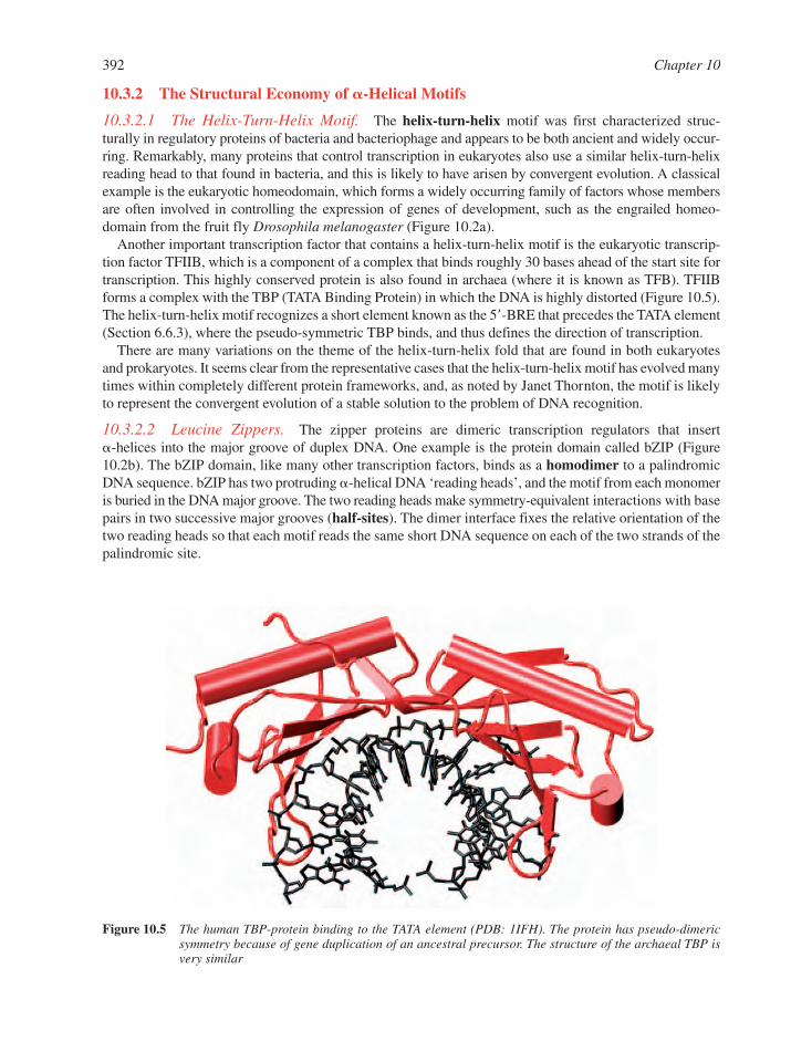
10.3.2 The Structural Economy of �-Helical Motifs
10.3.2.1 The Helix-Turn-Helix Motif. The helix-turn-helix motif was first characterized struc-turally in regulatory proteins of bacteria and bacteriophage and appears to be both ancient and widely occur-ring. Remarkably, many proteins that control transcription in eukaryotes also use a similar helix-turn-helixreading head to that found in bacteria, and this is likely to have arisen by convergent evolution. A classicalexample is the eukaryotic homeodomain, which forms a widely occurring family of factors whose membersare often involved in controlling the expression of genes of development, such as the engrailed homeo-domain from the fruit fly Drosophila melanogaster (Figure 10.2a).
Another important transcription factor that contains a helix-turn-helix motif is the eukaryotic transcrip-tion factor TFIIB, which is a component of a complex that binds roughly 30 bases ahead of the start site fortranscription. This highly conserved protein is also found in archaea (where it is known as TFB). TFIIBforms a complex with the TBP (TATA Binding Protein) in which the DNA is highly distorted (Figure 10.5).The helix-turn-helix motif recognizes a short element known as the 5�-BRE that precedes the TATA element(Section 6.6.3), where the pseudo-symmetric TBP binds, and thus defines the direction of transcription.
There are many variations on the theme of the helix-turn-helix fold that are found in both eukaryotesand prokaryotes. It seems clear from the representative cases that the helix-turn-helix motif has evolved manytimes within completely different protein frameworks, and, as noted by Janet Thornton, the motif is likelyto represent the convergent evolution of a stable solution to the problem of DNA recognition.
10.3.2.2 Leucine Zippers. The zipper proteins are dimeric transcription regulators that insert �-helices into the major groove of duplex DNA. One example is the protein domain called bZIP (Figure10.2b). The bZIP domain, like many other transcription factors, binds as a homodimer to a palindromicDNA sequence. bZIP has two protruding �-helical DNA ‘reading heads’, and the motif from each monomeris buried in the DNA major groove. The two reading heads make symmetry-equivalent interactions with basepairs in two successive major grooves (half-sites). The dimer interface fixes the relative orientation of thetwo reading heads so that each motif reads the same short DNA sequence on each of the two strands of thepalindromic site.
392 Chapter 10
Figure 10.5 The human TBP-protein binding to the TATA element (PDB: 1IFH). The protein has pseudo-dimericsymmetry because of gene duplication of an ancestral precursor. The structure of the archaeal TBP isvery similar
The bZIP domain has a long pair of identical helices, which coil gently around one another in a left-handed sense. This creates a dimer interface known as a leucine zipper because leucines from each of the�-helices intermesh along a line of contact that is co-axial with the long axis of the helical pair. Theseamino acids are like protruding knobs on each �-helix that fit into complementary hydrophobic pockets onthe partner helix. Such favourable ‘knobs-into-holes’ hydrophobic interactions hold the two parts togetherlike a zipper. Because the �-helix has 3.6 amino acids in a helical turn, the leucines form a left-handed spiral on the surface of the �-helix. Consequently, the hydrophobic regions of those two helices match andmaintain perfect register if the helices coil around one another in a gentle left-handed supercoil. This pack-ing was deduced by Francis Crick from geometric principles more than 50 years ago.
There are many structural variations of the zipper. For instance, the zipper architecture is elaborated inthe basic helix-loop-helix proteins, as represented by the developmental controlling factor Max. The dimer-ization interface of the zipper proteins can support homodimers and heterodimers and thus tune their bind-ing affinity and specificity, and hence their repertoire for genetic regulation. Zipper transcription factorsappear to occur exclusively in eukaryotes, and they are widely distributed within this taxonomic domain andare found in yeast, plants, insects and vertebrates. In multi-cellular organisms they often play roles in tissue-specific gene regulation.
10.3.3 Zinc-Bearing Motifs
Collectively, the zinc-bearing transcription factors form the largest group of DNA-binding proteins in theeukaryotes. Their abundance may be due to their simple, structurally economic folds, which are supportedby metal binding. Further, their modularity may have allowed them to be shuffled and combined readilyin the course of evolution to generate new proteins with different specificity and function.
10.3.3.1 The b–b–a Zinc Fingers. The first of the zinc-bearing DNA-binding motifs to be dis-covered were the ‘zinc fingers’. Transcription factors containing the zinc finger motif are found in a widerange of eukaryotic organisms, where they play a number of different genetic regulatory roles. In thehuman genome, roughly 3% of the genes that encode proteins specify zinc fingers.
The canonical structure of the zinc finger is composed of a peptide loop containing a two-stranded �-hairpin and an �-helix. The fold is stabilized by a hydrophobic core and coordination of Zn2� viathe sulfur atoms of two cysteines and the imidazole nitrogen atoms of two histidines.11 The inclination of the helices of the fingers in the transcription factor known as ‘Zif268’ allows stacking of side chains of thehelix and loop regions, which are aligned to interact with the bases in the DNA major groove.12 The modular finger structure permits the protein to track along the groove – mainly to one side of it – and to follow the helical trajectory.
10.3.3.2 The Steroid and Nuclear Receptors. Organisms as diverse as insects and vertebrates havetranscription factors that are activated by small, hydrophobic signalling ligands, such as hormones. Theseproteins, known as the steroid and nuclear receptors, use one structural domain to recognize the signalling ligand and a second domain to recognize specific DNA sequences in the vicinity of the targetgene. Representative examples are the receptors for estrogens, mineralocorticoids, progesterone, gluco-corticoids and vitamin D (Figures 10.2c). Also in this class are the ‘orphan’ receptors which have noknown ligand.
In these receptor proteins, the DNA-binding domain contains eight cysteines that form two peptide loopsthat each cap amphipathic �-helices. There is an imperfect structural repeat of the zinc-loop-�-helix, butthese repeating units do not form independent finger-like structures. Instead, they associate through hydropho-bic interaction of the two amphipathic �-helices to form a globular domain. The �-helix exposed by theamino-terminal module rests in the major groove of the DNA target, for example in the glucocorticoidreceptor, where it directs base contacts. These receptors often bind to DNA as homo- or hetero-dimers andrecognise the spacing between adjacent sites through protein–protein interactions (Section 10.4.6).
Protein–Nucleic Acid Interactions 393
10.3.4 The Orientations of �-Helices in the DNA Major Groove
In Sections 10.3.2 and 10.3.3, a handful of representative examples of protein–DNA complexes weredescribed in which an �-helix is docked snugly into the major groove. While it might seem that a snug fitimplies restricted orientation, there is in fact a wide range of orientations observed for the �-helix withinthe major groove. Indeed, there may be great variation in the orientation of the recognition �-helix in the DNAeven within a domain class whose members have the same fold and very similar amino acid sequences.Pabo and Nekludova13 found a way to explain this observed variation by grouping complexes into twoprincipal families according to how the residues along a ‘ridge’ of the �-helix align with respect to the spiralling trajectory of the DNA. In one family, every fourth residue is aligned (i.e. the ridge formed byresidues along the i, i�4 line) to maintain register with the contacting bases, whilst a second family usesthe line connecting every third residue (i, i�3). For instance, the zinc fingers tend to use the i, i�3 geometry.Because the helical repeat of the relaxed �-helix is 3.6 residues in one complete turn, the i, i�3 line willform a left-handed spiral on the helix surface, while the i, i�4 line will form a right-handed spiral. Thus, thetwo families use different geometries to match the spiralling pattern of amino acids with the curving path ofthe major groove. These matches are only local, and typically correspond to contact with three to five bases.
There are interesting outliers to this grouping, such as the trp repressor and the GAL4 protein, where the�-helices tend to enter the major groove at a steep angle because of additional contacts made to the DNAby other structural elements of the protein. While these family groupings indicate the importance ofmatching the geometry of the �-helix with the surface curvature of the DNA, they do not provide detailedrules for the interaction, since the precise helix orientation is context-dependent.
10.3.5 Minor Groove Recognition via �-Helices
The docking of an �-helix into the major groove of DNA causes the groove to become slightly compressed,on account of the tendency of the phosphate backbones of the DNA to clamp down on the �-helix. Theresulting compression of the major groove tends to bend the DNA towards the protein. By contrast, the accom-modation of an �-helix into the narrow minor groove of B-form DNA tends to expand the groove tremen-dously, with the result that the DNA bends away from the body of the protein. Representative examples ofminor groove recognition are provided by the widely occurring and well-conserved high-mobility group(HMG) proteins, which play structural roles in the dynamic organization of chromatin (Section 10.6.1).
10.3.6 ��-Motifs
The specific recognition of DNA by the Escherichia coli met repressor protein resembles closely �-helix-DNA recognition, except that the amino acids, which directly contact the base pairs are from a �-sheet (which may be made up from a number of �-strands) that make a snug fit into the major groove(Figure 10.6a). With this mode of interaction, the met repressor protein recognises a conserved eight-base-pairsequence of DNA (5�-AGACGTCT), although only four of the eight individual bases are contacted (indicatedin bold). The adjacent G and A bases form hydrogen bonds with lysine and threonine residues from different strands of the �-sheet, while the outer two bases A and C in AGAC are not contacted directly bythe protein. Several phosphates on the DNA interact with various amino acids from the protein.
The met repressor protein has twofold rotational symmetry and it recognizes a palindromic DNA sequence,just as in the DNA complexes of the 434 repressor and bZIP proteins (Figure 10.2b). But in addition, themet-repressor dimers can bind to adjacent sites on the DNA, and the neighbouring dimers interact favourablythrough protein–protein interactions. This gives a beneficial co-operativity of binding (Section 10.4.6).
Another protein that uses a �-ribbon to contact the DNA is the integration host factor (IHF), which is a20-kDa heterodimeric protein from E. coli that binds DNA with sequence-specificity and induces a sharpbend in the DNA, of greater than 160°.14,15 IHF functions as an architectural protein that assists assembly ofreplication complexes and affects long-distance transcriptional regulation. The subunits of the heterodimer
394 Chapter 10

are intertwined in a compact body from which two �-ribbons extend and interact with the DNA (Figure10.6b). Unlike those of the met repressor, the �-ribbons of IHF interact with the DNA in the minor groove.Conserved proline residues from the tips of the �-ribbon intercalate between base steps to support theDNA trajectory. The protein binds a 35 base pair target, and although there is little apparent consensusbetween the sequences of the binding sites, IHF prefers to intercalate at pyrimidine–purine steps. IHF bindspreferentially to these sites 103–104 times more strongly than to random DNA sites. Pyrimidine–purinesteps have the greatest flexibility of the dinucleotide steps (Section 10.4.5), and it seems that IHF may rec-ognize these sites indirectly through their ability to become conformationally distorted. The IHF protein inter-acts extensively with the phosphate backbone and the minor groove of the DNA, where only three aminoacid side chains form hydrogen bonds with the DNA bases. However, these hydrogen-bonding interactionscontacts can be made with an arbitrary sequence. Thus, IHF recognises DNA sites through sequence-dependent structural deformation but not through base-specific contacts.
The TBP binds to the TATA element that is 27–30 bases upstream of the start site of many eukaryoticand archaea genes. This protein also uses a �-sheet as the means of recognition of DNA, like the metrepressor and IHF, except that the sheet is fitted into a greatly widened minor groove, rather than the majorgroove (Figure 10.9, Section 10.4.6). While IHF lies on the concave side of the bent DNA, TBP lies on theconvex side of its deformed target. In binding its target site, TBP unwinds and sharply bends the TATA elem-ent. The conserved �-sheet of TBP forms a saddle-shaped surface that matches the curvature of the exposedbases in the deformed minor groove. A similar distortion is seen in the crystal structure of the HMG proteinbound to a TATA-like element, although an �-helix is used in that complex.16
The TBP/DNA interaction is highly conserved, as can be seen in the similar structures of the TBP/DNAcomplexes from plants, animals and archaea. The TBP protein is used as a scaffold for the recruitment ofother general transcription factors, such as TFIIB (Figure 10.9b, Section 10.4.6). It is curious that suchextreme distortion of the DNA by TBP and its associated proteins occurs and is so highly maintained inevolution. The distortion appears to be required to orient the DNA so that it enters the active site of theRNA polymerase, and it might facilitate unwinding of the DNA at the transcription start site (Section 10.7.2).
10.3.7 Loops and Others Elements
A polypeptide does not necessarily need a defined secondary structure to interact with DNA. For example,loops from certain DNA-binding proteins are used in the recognition of defined DNA sequences. Often
Protein–Nucleic Acid Interactions 395
Figure 10.6 Representative b-motifs and their docking into the grooves of DNA. (a) The E. coli met repressor (PDB:1TGH). Only a single dimer complex is shown in complex with a ‘met box’, but the repressor binds asa co-operative oligomer of dimers to adjacent met boxes (PDB: 1CMA). (b) The integration host factor(IHF) from bacteriophage. Here, two b-ribbons engage the minor groove
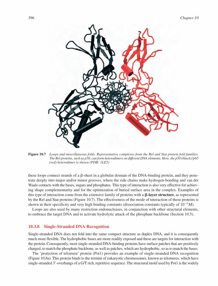
these loops connect strands of a �-sheet in a globular domain of the DNA-binding protein, and they pene-trate deeply into major and/or minor grooves, where the side chains make hydrogen-bonding and van derWaals contacts with the bases, sugars and phosphates. This type of interaction is also very effective for achiev-ing shape complementarity and for the optimization of buried surface area in the complex. Examples ofthis type of interaction come from the extensive family of proteins with a �-layer structure, as representedby the Rel and Stat proteins (Figure 10.7). The effectiveness of the mode of interaction of these proteins isshown in their specificity and very high binding constants (dissociation constants typically of 10�9 M).
Loops are also used by many restriction endonucleases, in conjunction with other structural elements,to embrace the target DNA and to activate hydrolytic attack of the phosphate backbone (Section 10.5).
10.3.8 Single-Stranded DNA Recognition
Single-stranded DNA does not fold into the same compact structure as duplex DNA, and it is consequentlymuch more flexible. The hydrophobic bases are more readily exposed and these are targets for interaction withthe protein. Consequently, most single-stranded DNA-binding proteins have surface patches that are positivelycharged, to match the phosphate backbone, as well as patches, which are hydrophobic, so as to match the bases.
The ‘protection of telomere’ protein (Pot1) provides an example of single-stranded DNA recognition(Figure 10.8a). This protein binds to the termini of eukaryotic chromosomes, known as telomeres, which havesingle-stranded 3�-overhangs of a G/T rich, repetitive sequence. The structural motif used by Pot1 is the widely
396 Chapter 10
Figure 10.7 Loops and miscellaneous folds. Representative complexes from the Rel and Stat protein fold families.The Rel proteins, such as p50, can form heterodimers on different DNA elements. Here, the p50 (black) /p65(red) heterodimer is shown (PDB: 1LE5)
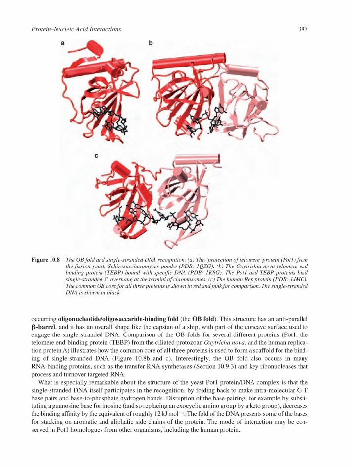
occurring oligonucleotide/oligosaccaride-binding fold (the OB fold). This structure has an anti-parallel �-barrel, and it has an overall shape like the capstan of a ship, with part of the concave surface used toengage the single-stranded DNA. Comparison of the OB folds for several different proteins (Pot1, thetelomere end-binding protein (TEBP) from the ciliated protozoan Oxytricha nova, and the human replica-tion protein A) illustrates how the common core of all three proteins is used to form a scaffold for the bind-ing of single-stranded DNA (Figure 10.8b and c). Interestingly, the OB fold also occurs in manyRNA-binding proteins, such as the transfer RNA synthetases (Section 10.9.3) and key ribonucleases thatprocess and turnover targeted RNA.
What is especially remarkable about the structure of the yeast Pot1 protein/DNA complex is that the single-stranded DNA itself participates in the recognition, by folding back to make intra-molecular GTbase pairs and base-to-phosphate hydrogen bonds. Disruption of the base pairing, for example by substi-tuting a guanosine base for inosine (and so replacing an exocyclic amino group by a keto group), decreasesthe binding affinity by the equivalent of roughly 12 kJ mol�1. The fold of the DNA presents some of the basesfor stacking on aromatic and aliphatic side chains of the protein. The mode of interaction may be con-served in Pot1 homologues from other organisms, including the human protein.
Protein–Nucleic Acid Interactions 397
Figure 10.8 The OB fold and single-stranded DNA recognition. (a) The ‘protection of telomere’ protein (Pot1) fromthe fission yeast, Schizosaccharomyces pombe (PDB: 1QZG). (b) The Oxytrichia nova telomere endbinding protein (TEBP) bound with specific DNA (PDB: 1K8G). The Pot1 and TEBP proteins bind single-stranded 3� overhang at the termini of chromosomes. (c) The human Rep protein (PDB: 1JMC).The common OB core for all three proteins is shown in red and pink for comparison. The single-strandedDNA is shown in black

10.4 KINETIC AND THERMODYNAMIC ASPECTS OF PROTEIN–NUCLEICACID INTERACTIONS
10.4.1 The Delicate Balance of Sequence-Specificity
The most common way of achieving sequence-specific recognition is direct readout of the DNA sequenceby hydrogen bonding between the protein and the DNA. This type of readout commonly includes the par-ticipation of water molecules. A typical DNA–protein complex in the cell might have a binding energy ofa few kJ mol�1, so it seems that the binding energy and sequence discrimination of most protein–DNAcomplexes can be accounted for easily just by a few hydrogen-bonding contacts to the bases and the phos-phate backbone. However, bases that are not contacted by the protein may also significantly affect the stability of certain complexes. This results in indirect readout whereby the sequence is recognizedthrough its conformational effects.
The energy of indirect readout is likely to be around 5–20 kJ mol�1 and is thus important for fine-tuningspecificity. This has been demonstrated experimentally in studies of DNA-binding by the 434 repressor,where it is found that the binding affinity is affected by base substitutions in the central portion of the palin-dromic site, where there are no contacts with the repressor.17 The bases there must influence the mouldingof the DNA to the protein.
The formation of specific protein–DNA complexes may represent a delicate balance between favourableinteractions on the one hand and energy penalties, such as those associated with distorting the DNA, on theother hand. In specific complexes, the free energy contributions are clearly all favourable and derive frombase contacts, phosphate contacts, bulk solvent and counter-ion displacement through the optimal buryingof surface area, and by stabilizing electrostatic interactions. The net free energy change (G) is offset bythe entropy penalties of conformational restriction and induced-folding and the energy penalty for deform-ing the DNA, which involves both conformational restriction and altered base stacking. In a non-specificcomplex, there are fewer direct interactions with the bases, and the buried surface area is often smaller(Section 10.4.3). Consequently, a non-specific complex has smaller favourable contributions from theseeffects, while the entropic penalties associated with conformational effects are also smaller. The relativeenergy contributions are illustrated for specific and non-specific complexes of the restriction enzyme EcoRIwith DNA (Figure 10.9). In this example, specific binding (left) is a fine balance of favourable effects (down-ward arrows) against unfavourable ones (upward arrows), to leave a small energy gain. In the energeticdecomposition of the non-specific binding event (right), the free energy change is comparatively smaller.This decomposition is only illustrative because the changes are highly context-dependent.
Complexes in which the DNA is highly distorted tend to be entropy-driven, whilst those with a more‘relaxed’ DNA conformation tend to be enthalpy-driven (Figure 10.9, right). There are several reasons forthis, but the principal cause is the entropy cost associated with protein folding upon binding of the DNAin the relaxed case and the enthalpy cost of bending the DNA in the ‘distorted’ DNA case.
10.4.2 The Role of Water
There are broadly two effects to be considered for the role of water in protein–nucleic acid interactions:(1) the thermodynamics of assembly, and (2) the detailed optimization of surface interactions. In specificcomplexes, buried surface area is optimised, and the matching surfaces fit together well. The mobilizationof water from the hydrated surfaces of the protein and of the oligonucleotide grooves and phosphate back-bones of the nucleic acid provides an entropically favourable contribution to complex formation (Figure 10.9,right). These bulk solvent effects account in part for the noted changes in heat capacity associated with theformation of complexes.18
Crystallographic structures of nucleic acid–protein complexes frequently reveal ordered water moleculesthat lie at the molecular interfaces (where their diffraction limit is better than about 3 Å). These water mol-ecules mediate the macromolecular interactions and also fill the occasional gap arising from interfacialimperfections. Water molecules participate in hydrogen-bonding networks that link side- and main-chain
398 Chapter 10
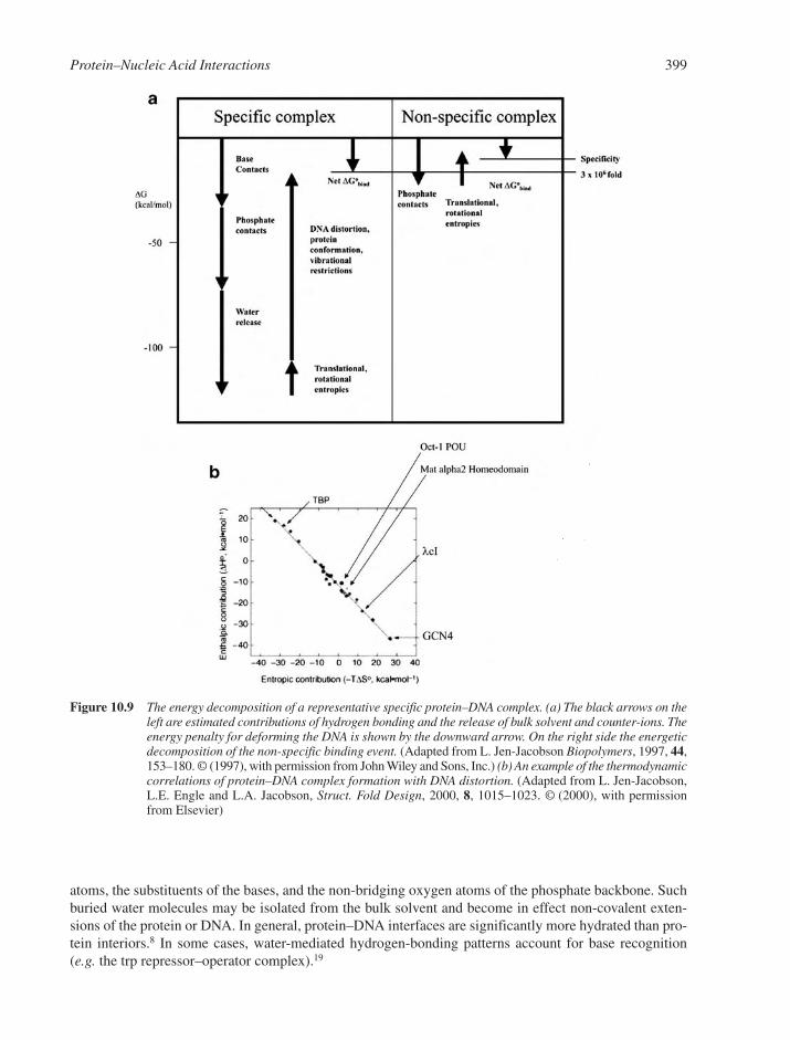
atoms, the substituents of the bases, and the non-bridging oxygen atoms of the phosphate backbone. Suchburied water molecules may be isolated from the bulk solvent and become in effect non-covalent exten-sions of the protein or DNA. In general, protein–DNA interfaces are significantly more hydrated than pro-tein interiors.8 In some cases, water-mediated hydrogen-bonding patterns account for base recognition(e.g. the trp repressor–operator complex).19
Protein–Nucleic Acid Interactions 399
Figure 10.9 The energy decomposition of a representative specific protein–DNA complex. (a) The black arrows on theleft are estimated contributions of hydrogen bonding and the release of bulk solvent and counter-ions. Theenergy penalty for deforming the DNA is shown by the downward arrow. On the right side the energeticdecomposition of the non-specific binding event. (Adapted from L. Jen-Jacobson Biopolymers, 1997, 44,153–180. © (1997), with permission from John Wiley and Sons, Inc.) (b) An example of the thermodynamiccorrelations of protein–DNA complex formation with DNA distortion. (Adapted from L. Jen-Jacobson,L.E. Engle and L.A. Jacobson, Struct. Fold Design, 2000, 8, 1015–1023. © (2000), with permissionfrom Elsevier)
10.4.3 Specific versus Non-Specific Complexes
We have discussed the energetic differences between specific and non-specific complexes (Figure 10.9),but what structural features distinguish them? One example of a non-specific complex is between the glucocorticoid receptor DNA-binding domain and non-target DNA. The natural target of this protein is apalindrome in which the hexameric half-sites are separated by three bases, and the protein binds this as ahomodimer with a self-complementary protein–protein interface (Section 10.3.3). In the non-specific com-plex, the separation of the half-sites is increased to four base pairs. The protein still forms a dimer on thisDNA, and each monomer interacts with its half-site, but the second monomer is pulled out of alignmentwith its recognition sequence. There are fewer direct hydrogen bonds to the bases, but just as many inter-actions with the phosphate backbone. The surface match is not so good for the non-specific complex, sothat the buried surface area is only about half that the specific interaction. In another non-specific complex,a layer of bound water molecules at the protein–nucleic acid interface is observed that must contribute anentropic penalty to the binding and disfavours the non-specific complex.20
The non-specific complex of the BamHI restriction enzyme illustrates the role of both surface electro-static and shape complementarity (Figure 10.10). Thus, target recognition is here associated with struc-tural changes in the protein that alter both the surface charge distribution and the shape.
10.4.4 Electrostatic Effects
For most RNA- and DNA-binding proteins, the affinity for both specific and non-specific nucleic aciddecreases sharply as the ionic strength increases. This arises from the polyelectrolyte character of DNAand RNA, which affect the binding of polycationic proteins and ligands. The salt concentration also affectsthe ability of proteins to slide along the longer target.
One curious exception to this salt-dependence rule occurs in the case of the transcription factor TBPfrom the hyperthermophilic archaea Pyrococcus,21 where the binding affinity increases with ionic strength.This may be explained by a putative cation-binding site that bridges the surfaces of the protein and theDNA. The protein surface that is in proximity to the DNA is electronegative, rather than the more usualelectropositive (e.g. the surface of BamHI; Section 10.4.3), and the counter-ion binding helps to overcomethe charge repulsion.
Subtle electrostatic switching may also explain how the met repressor (Figure 10.6a) is activated by itsligand, S-adenosylmethionine, to gain a 1000-fold increase in its affinity for the ‘met box’ target DNA. Themet repressor undergoes little apparent structural change upon binding of ligand. But because the ligandis charged, it changes the surface potential of the repressor, which now makes a more favourable match tothe surface of the DNA. Perhaps this effectively neutralizes the charges on the DNA, but does so in an asym-metrical fashion, to favour its conformational adjustment, not only to the bound protein, but also to its neigh-bour at an adjacent met box. Attractive protein–protein interactions also favour the binding of adjacentrepressors, with the net result that they bind co-operatively to the DNA element (Section 10.4.6).
10.4.5 DNA Conformability
Duplex DNA can be treated globally as a moderately flexible rod, with two principal degrees of freedom(Section 2.3.1). One mode involves twisting along the local helical axis, but structural restraints to thetwisting impart a torsional stiffness to the molecule. The energy of twisting is proportional to the squareof the change of twist angle per unit length. The other mode is bending, and stereochemical restraints tothis mode are associated with axial stiffness. The energy for bending is roughly proportional to the squareof the curvature, where curvature is a bend-angle per unit length, which is the same as the reciprocal of thelocal radius of curvature. Bending is usually achieved by changing the roll angle at the base steps. Theenergy required for bending may be anisotropic, with a preferred azimuthal angle, according to the abilityof the base steps to roll, and the projection of the roll with respect to the plane of curvature of the bend.When the stacking is maintained, the bending is smooth, as shown for the nucleosomal DNA (Figure10.1d), but the DNA becomes kinked if the stacking is disrupted (e.g. the base step in Figure 10.4f).
400 Chapter 10
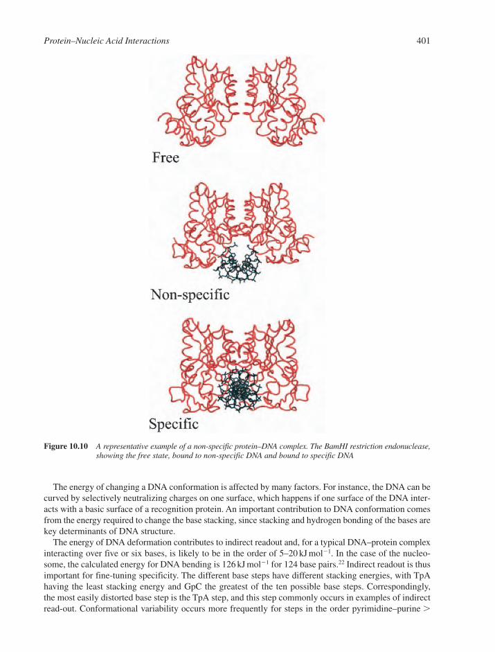
The energy of changing a DNA conformation is affected by many factors. For instance, the DNA can becurved by selectively neutralizing charges on one surface, which happens if one surface of the DNA inter-acts with a basic surface of a recognition protein. An important contribution to DNA conformation comesfrom the energy required to change the base stacking, since stacking and hydrogen bonding of the bases arekey determinants of DNA structure.
The energy of DNA deformation contributes to indirect readout and, for a typical DNA–protein complexinteracting over five or six bases, is likely to be in the order of 5–20 kJ mol�1. In the case of the nucleo-some, the calculated energy for DNA bending is 126 kJ mol�1 for 124 base pairs.22 Indirect readout is thusimportant for fine-tuning specificity. The different base steps have different stacking energies, with TpAhaving the least stacking energy and GpC the greatest of the ten possible base steps. Correspondingly, the most easily distorted base step is the TpA step, and this step commonly occurs in examples of indirectread-out. Conformational variability occurs more frequently for steps in the order pyrimidine–purine �
Protein–Nucleic Acid Interactions 401
Figure 10.10 A representative example of a non-specific protein–DNA complex. The BamHI restriction endonuclease,showing the free state, bound to non-specific DNA and bound to specific DNA
purine–purine � pyrimidine–pyrimidine � purine–pyrimidine. The propensity for DNA deformations tooccur at pyrimidine–purine steps is seen in a number of protein–DNA complexes, including those of theIHF (Figure 10.6b) and TBP (Figure 10.5). The pyrimidine–purine step may adopt large roll or high twistangles in protein–DNA complexes. Severe distortion of the DNA occurs in restriction enzyme/DNA com-plexes, and here indirect readout may account for nearly half of the energy associated with cleavage speci-ficity, corresponding to 25–30 kJ mol�1.23 The detailed energy components of base stacking in the contextof protein–DNA complexes depend on the dielectric constant of the environment and the need to bury protein–DNA interfaces away from the environment of the bulk solvent. Currently, it is hard to quantifythe energy associated with deformation of DNA or RNA within a protein–nucleic acid complex becausein such a complex the environment of the nucleic acid is different from bulk solvent. For instance, thenucleic acid experiences a large change in dielectric constant as its protein partner surrounds it. This has alarge effect on the energies of any conformational adjustments in the nucleic acid.
10.4.6 Co-operativity through Protein–Protein and DNA–Protein Interactions
Many of the proteins, already described, bind to DNA elements as homo- or heterooligomers, and thus makehomotypic or heterotypic protein–protein interactions, respectively. In such complexes, each subunit maypresent a reading head that interacts with the DNA target. If properly arranged, the organization of bindingsites on the DNA can provide co-operative benefits to binding, by simply helping to pre-organize the suc-cessive binding of the other subunits. Even if the proteins pre-exist in solution, there is still an implicit co-operative benefit because the matching of one subunit to its site pre-organizes the second site. Thisdecreases the entropic cost of binding, and is known as the chelate effect.24
A typical control region for a eukaryotic gene is very complex and contains a myriad of binding sitesfor different proteins. In many cases, such proteins form homotypic or heterotypic protein–protein inter-actions, so that they mutually influence their DNA-binding affinity. Such co-operative effects might com-pensate for the comparatively weaker binding and lower sequence discrimination of eukaryotic transcriptionfactors compared with prokaryotic repressors and activators. A complex organization that involves multipleinteracting proteins may also have kinetic benefits for gene regulation. For example, multi-componentassembly is involved in the pre-initiation transcription complexes of eukaryotes and archaea. Pre-initiationcomplexes assist RNA polymerases to bind promoter sites, and are often composed of the TATA-elementbinding protein (TBP) and the transcription factor TFIIB (Figure 10.11). Other components may be assem-bled onto the exposed surface of the TBP, depending on the promoter. Because the complex is asymmet-ric, it can specify the direction of transcription. How does this asymmetry arise?
The TBP protein has approximate twofold symmetry, so there are in principle two possible orientationsfor TBP to bind with respect to the start site of transcription. TBP has a saddle-like shape that engages awidened DNA minor groove, but clearly, TBP binding alone cannot specify the direction of transcription.Transcription factor TFIIB binds asymmetrically to the TBP by recognition of one exposed surface andmakes a contact in the major groove on the 5� side of the TATA element (at the ‘BRE’ recognition site)through a helix-turn-helix motif (Figure 10.11). Thus, it is the combination of protein–protein interactionsbetween the TFIIB and the TBP and the protein–DNA interactions of the TFIIB that breaks the symmetryof the complex and defines the direction of transcription.
Another example of protein–protein interactions is provided by the hormone and nuclear receptors(Figure 10.2c). The DNA elements of steroid receptors usually comprise two hexameric elements that arearranged as an inverted repeat with a three-nucleotide separation of half-sites (e.g. the idealized glucocor-ticoid response element is 5�-AGAACAxxxTGTTCT-3�). The pseudo-dyad symmetry of the half-sitesaligns the proteins, which interact in a ‘head-to-head’orientation. The DNA-binding domains of these proteinsbind through favourable protein–protein interactions, which give rise to co-operativity. The two monomersinteract with the DNA through a dimerization interface, the proper alignment of which requires exact spacing between the two monomer-binding sites. The two monomers would clash sterically if they were tobind to a DNA sequence in which the half-site separation is either two or four bases.
402 Chapter 10

Several different nuclear receptor monomers recognize the same consensus DNA sequence: ACTGGA.However, they bind to different DNA sequences, which are distinguished by the spacing between the half-sites. These elements are bound by a pair of receptors, the relative orientation of which changes with the half-site separation. This brings different surfaces into register and thus explains how the proteins discriminateDNA targets.
Some proteins can undergo secondary, tertiary or quaternary conformational changes upon binding toDNA, and such changes can result in co-operativity. It has been proposed that the DNA element itself canact as an allosteric ligand that induces structural changes in the bound protein, and thereby influences thecomposition or conformation of multi-component regulatory complexes.25 In this way, subtle changes insequence can increase the combinatorial repertoire of a limited number of genetic regulatory proteins.
10.4.7 Kinetic and Non-Equilibrium Aspects of DNA Recognition
The high degree of specificity in protein–DNA recognition does not require high-affinity binding. Forinstance, most specific protein–DNA complexes are stabilized by the energy corresponding to only a fewhydrogen bonds or the liberation of just a handful of bound water molecules – interactions that could belacking in their non-specific complexes. Such weak binding may provide kinetic benefits by permittingprotein–DNA complexes to associate and dissociate on timescales that match other processes in the cell.In many cases, the association rates may be close to maximal for facilitated diffusion.
Some dimeric DNA-binding proteins, such as the leucine zipper proteins (Section 10.3.2), dissociateinto monomers at a significant rate. Dissociable dimeric and oligomeric proteins might have special kineticproperties for DNA association. Consider, for example, two alternative pathways that may describe theassembly of a complex. In one pathway, the dimerization occurs prior to binding the DNA, whilst in the sec-ond, the dimer forms on the DNA only after sequential binding of the subunits. Since in both cases there is a
Protein–Nucleic Acid Interactions 403
Figure 10.11 A representative example of a ternary complex with a specific DNA target. The TBP–TFIIB–DNAcomplex (PDB: 1AKH). The TBP (red) docks into the distorted minor groove. This perspective showsthe recognition helix of TFIIB (pink) docked in the major groove at the recognition site. This bindinghelps to define the polarity of transcription initiation. The perspective is into the distorted minorgroove engaged by the TBP (and is perpendicular to the view seen in Figure 10.5 in two axes)

closed thermodynamic cycle, the particular pathway chosen does not affect the equilibrium affinity. However,the second (monomer) pathway has been noted to have faster association rates for certain leucine-zipper proteins.26
Some proteins find their DNA target sites at rates approaching 103-fold greater than those expected ifsite location were diffusion-controlled.27 Three experimentally determined mechanisms account in part forthis apparent acceleration (Figure 10.12).
1. sliding – involves diffusion in one dimension,2. hopping/jumping – involves limited sliding diffusion, followed by dissociation and re-association
at a different point on the DNA chain,28 and3. inter-segmental transfer – allows the direct transfer from one segment of DNA to another so as to
slide or hop to different regions.29
In all cases, the rate enhancement is expected to be a factor of only sixfold over diffusion-controlled rates,since the diffusion rates are proportional to twice the dimensions of the search space. Most of the apparentrate acceleration may be due to electrostatic attraction, because of the polyelectrolyte character of the DNA.
In the cell, DNA is hardly ever in a free form. Thus, sliding-type diffusion is extremely limited, for examplebecause of obstacles such as nucleosomes (Section 10.6.1). However, three-dimensional diffusion mech-anisms such as hopping provide a means to maintain the search process even with such obstacles present.30
10.5 THE SPECIFICITY OF DNA ENZYMES
10.5.1 Restriction Enzymes: Recognition through the Transition State
Restriction enzymes are bacterial proteins that provide defence against foreign DNA. They recognize andcleave specific DNA sequences on both strands. Many, but not all, have twofold symmetry and so recog-nize palindromic sites. The enzymes recognize typically a sequence of four to eight base pairs and, not sur-prisingly, have extremely high specificity for well-defined target sites on DNA. Any relaxed specificitywould have dire consequences for a bacterium, since its own DNA would be at risk of cleavage at sites notprotected by its own methylase enzyme (Section 5.3.1).
The mechanism of restriction enzyme cleavage of DNA involves nucleophilic attack of a hydroxide ionon the scissile phosphorus atom to generate a 5�-phosphate group (e.g. for the enzyme HincII). The hydroxylion attack causes an in-line displacement of the 3�-hydroxyl group via a penta-coordinate transition state.Restriction enzymes require divalent cations for activity, which are used to activate the water molecule forattack and to stabilize the negative charge that develops transiently on the transition state.31
It might seem paradoxical that many restriction enzymes bind target and non-target DNA with similaraffinities. Therefore, specificity must arise at the catalytic step, probably because of the distortionsinduced in the transition state. Such distortions are seen in the crystal structure of the EcoRI restriction
404 Chapter 10
Figure 10.12 Possible modes for search mechanisms of DNA-binding proteins for their target sites
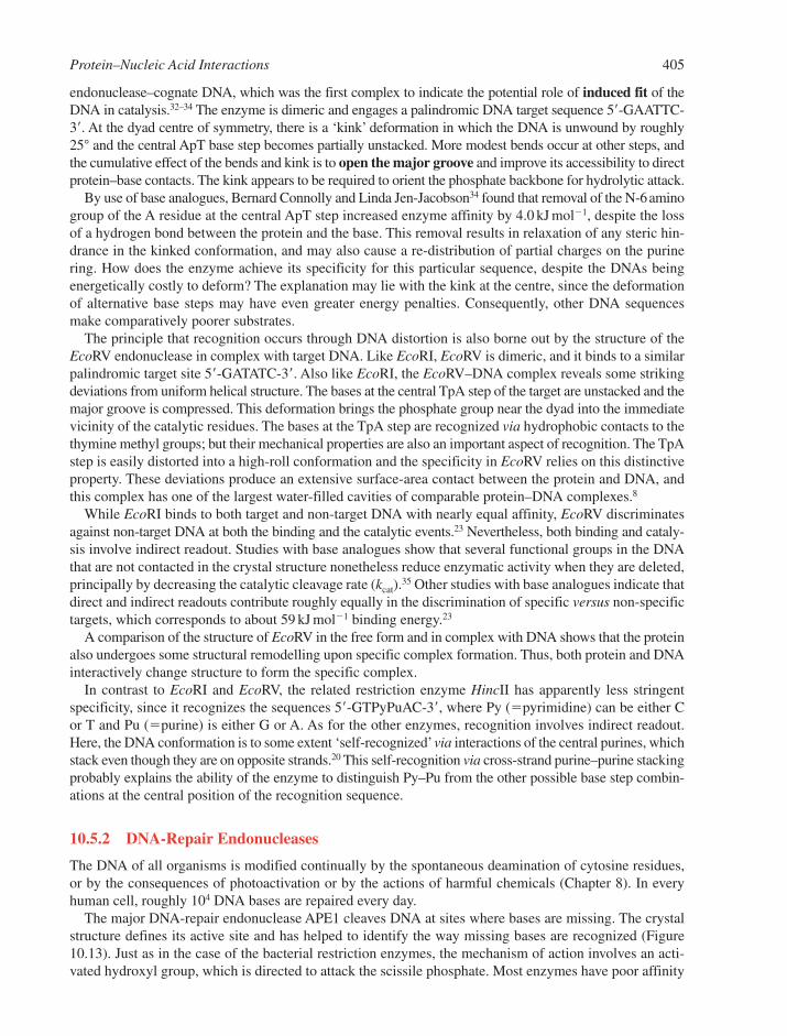
endonuclease–cognate DNA, which was the first complex to indicate the potential role of induced fit of theDNA in catalysis.32–34 The enzyme is dimeric and engages a palindromic DNA target sequence 5�-GAATTC-3�. At the dyad centre of symmetry, there is a ‘kink’ deformation in which the DNA is unwound by roughly25° and the central ApT base step becomes partially unstacked. More modest bends occur at other steps, andthe cumulative effect of the bends and kink is to open the major groove and improve its accessibility to direct protein–base contacts. The kink appears to be required to orient the phosphate backbone for hydrolytic attack.
By use of base analogues, Bernard Connolly and Linda Jen-Jacobson34 found that removal of the N-6aminogroup of the A residue at the central ApT step increased enzyme affinity by 4.0 kJ mol�1, despite the lossof a hydrogen bond between the protein and the base. This removal results in relaxation of any steric hin-drance in the kinked conformation, and may also cause a re-distribution of partial charges on the purinering. How does the enzyme achieve its specificity for this particular sequence, despite the DNAs beingenergetically costly to deform? The explanation may lie with the kink at the centre, since the deformationof alternative base steps may have even greater energy penalties. Consequently, other DNA sequencesmake comparatively poorer substrates.
The principle that recognition occurs through DNA distortion is also borne out by the structure of theEcoRV endonuclease in complex with target DNA. Like EcoRI, EcoRV is dimeric, and it binds to a similarpalindromic target site 5�-GATATC-3�. Also like EcoRI, the EcoRV–DNA complex reveals some strikingdeviations from uniform helical structure. The bases at the central TpA step of the target are unstacked and themajor groove is compressed. This deformation brings the phosphate group near the dyad into the immediatevicinity of the catalytic residues. The bases at the TpA step are recognized via hydrophobic contacts to thethymine methyl groups; but their mechanical properties are also an important aspect of recognition. The TpAstep is easily distorted into a high-roll conformation and the specificity in EcoRV relies on this distinctiveproperty. These deviations produce an extensive surface-area contact between the protein and DNA, andthis complex has one of the largest water-filled cavities of comparable protein–DNA complexes.8
While EcoRI binds to both target and non-target DNA with nearly equal affinity, EcoRV discriminatesagainst non-target DNA at both the binding and the catalytic events.23 Nevertheless, both binding and cataly-sis involve indirect readout. Studies with base analogues show that several functional groups in the DNAthat are not contacted in the crystal structure nonetheless reduce enzymatic activity when they are deleted,principally by decreasing the catalytic cleavage rate (kcat).
35 Other studies with base analogues indicate thatdirect and indirect readouts contribute roughly equally in the discrimination of specific versus non-specifictargets, which corresponds to about 59 kJ mol�1 binding energy.23
A comparison of the structure of EcoRV in the free form and in complex with DNA shows that the proteinalso undergoes some structural remodelling upon specific complex formation. Thus, both protein and DNAinteractively change structure to form the specific complex.
In contrast to EcoRI and EcoRV, the related restriction enzyme HincII has apparently less stringentspecificity, since it recognizes the sequences 5�-GTPyPuAC-3�, where Py (�pyrimidine) can be either Cor T and Pu (�purine) is either G or A. As for the other enzymes, recognition involves indirect readout.Here, the DNA conformation is to some extent ‘self-recognized’via interactions of the central purines, whichstack even though they are on opposite strands.20 This self-recognition via cross-strand purine–purine stackingprobably explains the ability of the enzyme to distinguish Py–Pu from the other possible base step combin-ations at the central position of the recognition sequence.
10.5.2 DNA-Repair Endonucleases
The DNA of all organisms is modified continually by the spontaneous deamination of cytosine residues,or by the consequences of photoactivation or by the actions of harmful chemicals (Chapter 8). In everyhuman cell, roughly 104 DNA bases are repaired every day.
The major DNA-repair endonuclease APE1 cleaves DNA at sites where bases are missing. The crystalstructure defines its active site and has helped to identify the way missing bases are recognized (Figure10.13). Just as in the case of the bacterial restriction enzymes, the mechanism of action involves an acti-vated hydroxyl group, which is directed to attack the scissile phosphate. Most enzymes have poor affinity
Protein–Nucleic Acid Interactions 405

for their products. However, APE1 appears to form very stable complexes with its product.36 Thus, theenzyme holds onto the product after cleavage, since severed DNA poses great risk to the cell, and thendelivers it to the next component of the repair machinery.
Certain repair enzymes cause the damaged nucleotide to be flipped out from the DNA double helix. Insuch enzymes, the nucleotide is accommodated in a specific binding pocket within the enzyme.
10.5.3 DNA Glycosylases
Glycosylases are enzymes that excise damaged DNA, as the first step towards repair. The enzyme uracil–DNAglycosylase functions to remove the base wherever uracil bases occur in DNA as a result of either
406 Chapter 10
Figure 10.13 The structure of a representative repair enzyme, the human APE1 protein bound to DNA (PDB: 1DE8)
misincorporation or deamination of cytosine. The structure of the human uracil DNA glycoslyase showsthat the glycosylase completely encompasses the substrate. Another example is the enzyme 3-methyl-adenine DNA glycosylase, which excises that base from damaged DNA by intercalation into the minor grooveusing two �-strands, and by flipping the damaged base into the active site for base excision.37
10.5.4 Photolyases
Light is the primary source of energy for most life on earth, but ultraviolet light induces spontaneous dam-age to DNA in the way of covalent photo-adducts. One such example is the dimer formed from pyrimidines,cyclobutadipyrimidine. Organisms in all three domains of life have evolved enzymes that repair the light-induced damage by using the energy of light itself. These enzymes are carbon–carbon photolyases.Photolyases share a common fold and use a catalytic mechanism that involves conjugated co-factors forphoto-induced electron transfer. One co-factor that is used directly in the catalytic process is the reducedform of flavin-adenine dinucleotide (FAD-H2). The second chromophore acts as an antenna to transferlight energy to the FAD-H2, which in turn supplies the electron to rupture the carbon–carbon bond. In mostenzymes the antenna co-factor is 5,10-methenyl-tetrahydrofolate, but certain photolyases instead use 5-deazariboflavin. The active site engulfs the pyrimidine substrate, which is in van der Waals contact withthe FAD. The FAD is sandwiched by aromatic stacking with side chains of two conserved tryptophanresidues, which are thought to become radicals transiently in the transfer process.38 There is little roomto accommodate a duplex DNA. So these enzymes probably flip out the pyrimidine dimer from the duplex,in loose analogy with the flipping mechanism used by other DNA-repair enzymes (Sections 10.5.2 and 10.5.3).
10.5.5 Structure-Selective Nucleases
In DNA recombination, an intermediate is formed in which four strands join together (Figure 10.14a andSection 2.3.7) These structures are resolved back into two duplex DNA molecules by the activity of structure-specific endonucleases, which are found in all three domains of life. Site-specific recombinases (e.g. FLPprotein, Figure 10.14b) use the hydroxyl group of either serine or tyrosine as the nucleophile for cutting andrejoining DNA. Topoisomerases also use a tyrosine hydroxyl group for a similar purpose (Section 10.8.3).
Protein–Nucleic Acid Interactions 407
Figure 10.14 (a) The structure of a four-way DNA junction. (b) Its complex with the recombinase FLP protein(PDB: 1P4E)
10.6 DNA PACKAGING
10.6.1 Nucleosomes and Chromatin of the Eukaryotes
A typical eukaryotic cell contains roughly 2 metres of DNA, which must be compacted by a factor of 106
so as to fit within the nucleus. The DNA is assembled with proteins into complexes known as chromatin(Section 6.4). The fundamental packaging unit of the eukaryotic DNA is the nucleosome, an octamericcomplex of histone proteins and DNA (Figure 10.15). The histone octamer engages roughly one- and three-quarter turns of duplex DNA in a left-handed toroidal superhelix. It has been proposed that the efficiencyof the histone-based system of packaging DNA was a key development in the expansion of the eukaryoticgenome, and the divergence of this branch of life.39
The accessibility of DNA is determined primarily by the arrays of nucleosomes, which affect transcrip-tion activity, mostly through repression. The cell has elaborate machinery to ‘re-model’ those arrays dur-ing gene expression. Such machines transduce the free energy of ATP binding and hydrolysis to displacenucleosomes. The machines are transiently fixed with respect to a target nucleosome, and it has been pro-posed that they displace the DNA so that it twists up to form a loop that can move around the nucleosome,causing the nucleosome to slide.40 Modifications of the histones by acetylation and methylation providesignals for recruitment of the remodelling machinery. Recently, it has also been discovered that varianthistones are used to mark boundaries for the repair of damaged DNA by recombination.
The crystal structure of the nucleosome has been solved at high resolution, from which one can observein detail the protein–protein and protein–DNA contacts that are made for a particular sequence of DNA(Figure 10.15a). The histone octamer of the nucleosome is composed of two copies each of the four histoneproteins H2A, H2B, H3 and H4. These proteins have similar sequences, and have undoubtedly evolved bygene duplication. Histones H3 and H4 are among the most highly conserved proteins in multi-cellulareukaryotes, while H2A and H2B are comparatively more variable, especially at their termini. Histones H3and H4 form a heterotypic dimer that self-pairs into a tetramer through a four-helix bundle, in which thehydrophobic interfaces between helices intermesh in a ‘knobs-into-holes’ manner, just like the two-stranded coiled-coil described for leucine zippers (Section 10.3.2).
408 Chapter 10
Figure 10.15 The nucleosome core particle (PDB: 1AOI). The histone amino termini protrude from the core particle; these are sites of covalent modification that affect chromatin structure and gene regulation.The histone dimers have been colour coded. (a) The dyad axis is vertical. (b) A side view: the dyad isnow in the plane of the page
The nucleosomal histones contact the DNA through �-helices and loops. Some of the long �-helices areoriented so that their helix dipoles form favourable interactions with the electronegative phosphate back-bone. Water molecules and counter-ions are also involved in fitting together the protein and DNA surfaces.The DNA follows a highly curved trajectory on the surface of the protein (Figure 10.15b). This requiresin-plane bending and a small accommodating change in twist. The bending is achieved by compression ofthe minor groove where it contacts the surface of the protein, and by widening of the minor groove on theouter surface of the curved DNA. The DNA on the nucleosome is overwound at about 10.2 base pairs/turn(in comparison with 10.6 base pairs/turn for relaxed DNA in solution). This overwinding aligns the minorgrooves of the DNA, so that there are little gaps through which the terminal tails of the histone proteinsprotrude. Although the nucleosome has no sequence preference per se, there are notable preferences for peri-odic composition patterns to occupy the regions where the grooves compress or expand. There is a periodicvariation in twist and roll at base steps, and these angles on average are larger in magnitude than for free DNA.
A string of nucleosomes can assemble into a helical structure known as the solenoid, in which there aresix nucleosomes in a superhelical turn. The termini of the histones, which protrude from the surface asmentioned above, are thought to be important for mediating the contacts within the solenoid, and hencemodifications such as methylation or acetylation of lysine amino groups on the histones can affect theassembly or the recruitment of other regulatory proteins. The solenoid assembly is consolidated by a fifthclass of histone protein, known as the H1 (or H5) histones, which are not structurally related to the octamercore histones H2A, H2B, H3 and H4. These H1 and H5 histones have a DNA-binding motif that is remark-ably similar to that found in the bacterial CAP gene activator protein and the SAP-1 protein. The H1/H5proteins are probably located near the dyad of the nucleosome.
Binding sites within nucleosomes are occasionally exposed, but at sufficiently rapid rates to allow siteaccess under physiological conditions.41,42 The sequential binding of a number of factors independently toDNA sites exerts co-operativity through their mutual effects on the chromatin. The binding of one proteinat its recognition site on the DNA may help to alter the nucleosome position and thus expose the bindingsite of a second protein, favouring its binding.
10.6.2 Packaging and Architectural Proteins in Archaebacteria and Eubacteria
Archaea in the euryarchaeota sub-branch also have histone-core-like proteins. There is a remarkablestructural similarity of the archaeal histone dimer made by the HMfB and HMfb proteins with the eukary-otic H3–H4 dimer (Figure 10.16a). This similarity suggests that the H3–H4 tetramer may represent anearlier organization of chromatin during the early stages of the evolution of the eukaryotic cell.10
The crenarchaeota sub-branch appears not to have histones, but instead a variety of abundant non-sequence-specific DNA-binding proteins. For instance, the Sul7d protein binds non-specifically in the DNAminor groove by means of a three-stranded �-sheet, with intercalation of hydrophobic side chains betweenthe bases (Figure 10.16b). Another protein used in packaging the DNA is the homodimeric Alba, which pro-motes negative supercoiling in DNA. The protein has two highly conserved loops that may contact the DNA.
The genomes of eubacterial species are also compacted by proteins. These organisms use small, basicproteins such as HU and FIS. The HU protein can supercoil DNA to form beaded structures.
10.7 POLYMERASES
10.7.1 DNA-Directed DNA Polymerases
DNA-directed DNA polymerases (E.C. 2.7.7.7) catalyse the sequential, DNA template-directed extensionof the 3�-end of a DNA strand by addition of single nucleotides. The enzymes replicate genomic DNA withhigh fidelity and processivity. Crystal structures of DNA polymerases reveal the role of two metal ions in theactive site. A mechanism has been proposed to be common for polynucleotide polymerases.43 This involvesa metal ion that activates the deprotonation of the 3�-hydroxyl group at the terminus of the growing chain,
Protein–Nucleic Acid Interactions 409
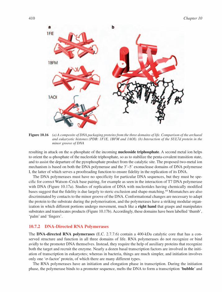
410 Chapter 10
Figure 10.16 (a) A composite of DNA packaging proteins from the three domains of life. Comparison of the archaealand eukaryotic histones (PDB: 1F1E, 1BFM and 1AOI). (b) Interaction of the SUL7d protein in theminor groove of DNA
resulting in attack on the �-phosphate of the incoming nucleoside triphosphate. A second metal ion helpsto orient the �-phosphate of the nucleotide triphosphate, so as to stabilize the penta-covalent transition state,and to assist the departure of the pyrophosphate product from the catalytic site. The proposed two-metal ionmechanism is based on both the DNA polymerase and the 3�–5� exonuclease domains of DNA polymeraseI, the latter of which serves a proofreading function to ensure fidelity in the replication of its DNA.
The DNA polymerases must have no specificity for particular DNA sequences, but they must be spe-cific for correct Watson–Crick base pairing, for example as seen in the interaction of T7 DNA polymerasewith DNA (Figure 10.17a). Studies of replication of DNA with nucleotides having chemically modifiedbases suggest that the fidelity is due largely to steric exclusion and shape-matching.44 Mismatches are alsodiscriminated by contacts to the minor groove of the DNA. Conformational changes are necessary to adaptthe protein to the substrate during the polymerisation, and the polymerases have a striking modular organ-ization in which different portions undergo movement, much like a right hand that grasps and manipulatessubstrates and translocates products (Figure 10.17b). Accordingly, these domains have been labelled ‘thumb’,‘palm’ and ‘fingers’.
10.7.2 DNA-Directed RNA Polymerases
The DNA-directed RNA polymerases (E.C. 2.7.7.6) contain a 400-kDa catalytic core that has a con-served structure and function in all three domains of life. RNA polymerases do not recognize or bindavidly to the promoter DNA themselves. Instead, they require the help of auxiliary proteins that recognizeboth the target and recruit the enzyme. Nearly a dozen basal transcription factors are involved in the initi-ation of transcription in eukaryotes; whereas in bacteria, things are much simpler, and initiation involvesonly one ‘ -factor’ protein, of which there are many different types.
The RNA polymerases have an initiation and elongation phase in transcription. During the initiationphase, the polymerase binds to a promoter sequence, melts the DNA to form a transcription ‘bubble’ and
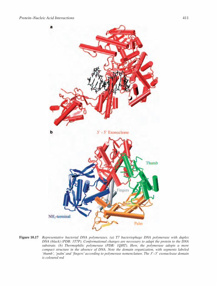
Protein–Nucleic Acid Interactions 411
Figure 10.17 Representative bacterial DNA polymerases. (a) T7 bacteriophage DNA polymerase with duplex DNA (black) (PDB: 1T7P). Conformational changes are necessary to adapt the protein to the DNAsubstrate. (b) Thermophilic polymerase (PDB: 1QHT). Here, the polymerase adopts a more compact structure in the absence of DNA. Note the domain organization, with segments labeled‘thumb’, ‘palm’ and ‘fingers’ according to polymerase nomenclature. The 3�–5� exonuclease domainis coloured red
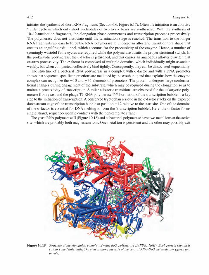
initiates the synthesis of short RNA fragments (Section 6.6, Figure 6.17). Often the initiation is an abortive‘futile’ cycle in which only short nucleotides of two to six bases are synthesized. With the synthesis of10–12-nucleotide fragments, the elongation phase commences and transcription proceeds processively.The polymerase does not dissociate until the termination stage is reached. The transition to the longerRNA fragments appears to force the RNA polymerase to undergo an allosteric transition to a shape thatcreates an engulfing exit tunnel, which accounts for the processivity of the enzyme. Hence, a number ofseemingly wasteful futile cycles are required while the polymerase awaits the proper structural switch. Inthe prokaryotic polymerase, the -factor is jettisoned, and this causes an analogous allosteric switch thatensures processivity. The -factor is composed of multiple domains, which individually might associateweakly, but when compacted, collectively bind tightly. Consequently, they can be dissociated sequentially.
The structure of a bacterial RNA polymerase in a complex with -factor and with a DNA promotershows that sequence-specific interactions are mediated by the subunit; and that explains how the enzymecomplex can recognize the �10 and �35 elements of promoters. The protein undergoes large conforma-tional changes during engagement of the substrate, which may be required during the elongation so as tomaintain processivity of transcription. Similar allosteric transitions are observed for the eukaryotic poly-merase from yeast and the phage T7 RNA polymerase.45,46 Formation of the transcription bubble is a keystep to the initiation of transcription. A conserved tryptophan residue in the -factor stacks on the exposeddownstream edge of the transcription bubble at position �12 relative to the start site. One of the domainsof the -factor is essential for DNA melting to form the ‘transcription bubble’. Here, the -factor formssingle strand, sequence-specific contacts with the non-template strand.
The yeast RNA polymerase II (Figure 10.18) and eubacterial polymerase have two metal ions at the activesite, which are probably both magnesium ions. One metal ion is persistent and the other may possibly exit
412 Chapter 10
Figure 10.18 Structure of the elongation complex of yeast RNA polymerase II (PDB: 1I6H). Each protein subunit iscolour coded differently. The view is along the axis of the central RNA–DNA heteroduplex (green andpurple)

Protein–Nucleic Acid Interactions 413
with pyrophosphate that is released during the extension reaction. Thus, the RNA polymerase may sharea common two-metal mechanism with the DNA polymerases.
10.8 MACHINES THAT MANIPULATE DUPLEX DNA
10.8.1 Helicases
Helicases unwind DNA and RNA duplexes, and they participate in nearly every cellular process that involvesnucleic acids. In E. coli, more than a dozen different helicases have been identified, and these carry out avariety of tasks, ranging from strand separation during DNA replication to more complex processes inDNA repair and recombination and, in the case of RNA helicases, the degradation of messenger RNA.
Whereas certain repair enzymes might use the energy of DNA binding to flip a single base outside theduplex, a helicase must not only locally melt the DNA target, but must also move itself along the DNA to pre-vent re-annealing of the strands. This requires mechanical work, which is provided by the free energy of ATPbinding and hydrolysis. Since helicases must displace proteins from the DNA, they are capable of generat-ing tremendous force. For instance, helicases can produce sufficient force to displace the protein strepta-vidin from 5�-biotinylated DNA,47 which binds with a dissociation constant on the order of 10�14 M. There areseveral families of DNA helicase in archaea, prokaryotes and eukaryotes, but mechanistically these enzymescan be divided into two classes according to their direction of translocation. One class moves in the 5�–3�direction along single-stranded DNA and the second class moves in the opposite direction, from 3�–5�.
The first crystal structure of a 3�–5� helicase was that of PcrA from Bacillis stearothermophilus.48 Theenzyme consists of several domains, including two that are structurally similar to the ATPase domain ofthe recombination protein RecA. Subsequent structures of PcrA helicase in complexes with a DNA sub-strate have provided details of the unwinding mechanism. There are two separable steps: (1) duplex desta-bilization, and (2) DNA translocation.39 Duplex DNA is engaged on the surface of the protein, and one ofthe displaced single strands binds to a surface channel. The operation involves allosteric transitions inwhich protein sub-domains move relative to each other, so that there are changes in both the surface andthe channel that engage the duplex DNA.
An example of a 5�–3� helicase is the bacteriophage T7 gene 4 helicase, which is involved in strand sep-aration during phage DNA replication (Figure 10.19). The protein forms a hexameric ring but, surprisingly,this ring has twofold rotational symmetry rather than the expected sixfold. ATP hydrolysis around the ringis associated with a rotation of each subunit about axes perpendicular to the ring. The nucleotide-bindingsites (at the interfaces between the subunits) are therefore non-equivalent, and have differing affinities for theNTPs. As nucleotide hydrolysis occurs, the states of the three sites may interconvert from NTP-bound toNDP � phosphate binding to empty. This would allow a ‘wave’ of NTP hydrolysis to run around the ring.
This model shows some similarities with the ‘binding-change’ model associated with the F1-subunit ofthe mitochondrial ATPase, although the asymmetries arise in different manners (Figure 10.19b).
10.8.2 DNA Pumps
The duplex DNA of bacteriophages is packaged within protein-rich capsid shells at densities approachingthose of crystals. Electrostatic repulsion of the compacted nucleic acid generates pressure, and this force isused to discharge the DNA into the host cell during the infection process. But the packaging of nucleic acid tosuch an extent in the first place requires tremendous work which is provided by motors that are fuelled by ATPbinding and hydrolysis. The force generated by DNA pumps of bacteriophages during the initial stages oftranslocation is estimated to be in the order of 100 pico-Newtons, and the force grows as packaging proceeds.49
The crystal structure of the central component of the motor from B. subtilis bacteriophage �29 illus-trates the principle of operation of the DNA pumps (Figure 10.20a). In essence, the translocation machineryis a rotary motor comprising a system of two rings of mismatched symmetry that interact with the DNA.50,51
The central component contains 12 copies of a 36-kDa subunit. This forms a central channel with a diameter
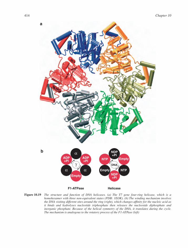
414 Chapter 10
αα
αADP+Pi
ADP+Pi
Empty
γ
F1-ATPase
NDP+Pi
NDP+Pi
NTP
NTP
Empty
Empty
Helicase
DNA
a
b
Figure 10.19 The structure and function of DNA helicases. (a) The T7 gene four-ring helicase, which is a homohexamer with three non-equivalent states (PDB: 1EOK). (b) The winding mechanism involvesthe DNA visiting different sites around the ring (right), which changes affinity for the nucleic acid asit binds and hydrolyses nucleotide triphosphate then releases the nucleoside diphosphate and inorganic phosphate. Because of the helical symmetry of the DNA, it translates during the cycle. The mechanism is analogous to the rotatory process of the F1-ATPase (left)
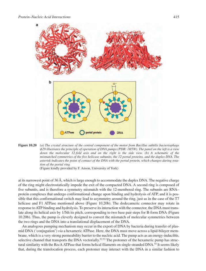
at its narrowest point of 36 Å, which is large enough to accommodate the duplex DNA. The negative chargeof the ring might electrostatically impede the exit of the compacted DNA. A second ring is composed offive subunits, and is therefore a symmetry mismatch with the 12-membered ring. The subunits are RNA–protein complexes that undergo conformational change upon binding and hydrolysis of ATP, and it is pos-sible that this conformational switch may lead to asymmetry around the ring, just as in the case of the T7helicase and F1 ATPase mentioned above (Figure 10.20b). The dodecameric connector may rotate inresponse to ATP binding and hydrolysis. To preserve its interaction with the connector, the DNA must trans-late along its helical axis by 1/5th its pitch, corresponding to two base pair steps for B-form DNA (Figure10.20b). Thus, the pump is cleverly designed to convert the mismatch of molecular symmetries betweenthe two rings and the DNA into a translational displacement of the DNA.
An analogous pumping mechanism may occur in the export of DNA by bacteria during transfer of plas-mid DNA (‘conjugation’) via a hexameric ATPase. Here, the DNA must move across a lipid-bilayer mem-brane, which is a very strong permeability barrier to the nucleic acid. The pump acts as an energy-inducible,selective channel that transports the DNA vectorially.52,53 The protomer of the hexameric pump has struc-tural similarity with the RecA ATPase that forms helical filaments on single-stranded DNA.54 It seems likelythat, during the translocation process, each protomer may interact with the DNA in a similar fashion to
Protein–Nucleic Acid Interactions 415
Figure 10.20 (a) The crystal structure of the central component of the motor from Bacillus subtilis bacteriophagef29 illustrates the principle of operation of DNA pumps (PDB: 1H5W). The panel on the left is a viewdown the molecular 12-fold axis and on the right is the side view. (b) A schematic of the mismatched symmetries of the five helicase subunits, the 12 portal proteins, and the duplex DNA. Theasterisk indicates the point of contact of the DNA with the portal protein, which changes during rota-tion of the portal ring(Figure kindly provided by F. Anson, University of York)

RecA. Perhaps, like the T7 ring helicase, the DNA conjugation pump might undergo asymmetric changeswithin the ring, so that two non-equivalent states of the subunits cause the displacement of the DNAduplex.
10.8.3 DNA Topoisomerases
Genomic DNA is packed densely and continuously undergoes superhelical strain during transcription,replication and (occasionally) recombination. For example, in E. coli, replication involves a rate of unwind-ing of 160 turns of duplex DNA per second: it can be envisaged that unwinding must be compensated atcomparable rates to avoid tangling. Organisms in all taxonomic domains have evolved enzymes that manipu-late DNA topology, either by relaxing superhelical tension generated from transcription, replication andrecombination (a spontaneous process) or by introducing supercoiling (which requires an input of energy)(Section 2.3.5). There are two principal classes of topoisomerases for these purposes: type I and type II.The former break one strand of duplex DNA transiently and allow rotation about the unbroken strand beforethe broken strand is re-ligated, so as to alter the linking number of the two strands. Type II enzymes break bothstrands and pass one region of DNA through the gap, resulting in stepwise changes in linking number of twounits. The structure of topoisomerase II reveals a toroidal shape, and it is possible to imagine the DNA pass-ing through the ring during strand exchange. For both type I and type II topoisomerases, strand cleavageinvolves nucleophilic attack of the phosphodiester backbone by a tyrosine hydroxyl group, resulting in a cova-lent bond between the enzyme and the 5� end of the broken strand. Rotation of the 3� end is followed byre-ligation.
A unique sub-group of the type II topoisomerases are the gyrases of prokaryotes, which catalyse theATP-dependent negative supercoiling of double-stranded closed circular DNA, and they thus maintain thebacterial genome under negative superhelical tension. In eukaryotes, there is little superhelical tension,which may explain the requirement for DNA distortion at the promoter by TBP and the requirement forhelicase activity in one of the transcription factors (Section 10.3.6).
10.9 RNA–PROTEIN INTERACTIONS AND RNA-MEDIATED ASSEMBLIES
Just as in the recognition of DNA, many different protein structural motifs are used to recognize RNA.RNA–protein complexes that have been characterized so far have tended to be more complicated than protein–DNA complexes, since RNA structures are themselves more complex than double-stranded DNA, and include duplex regions, pseudo-knots and single-stranded regions, such as bulges and hairpinloops (Section 7.1).55 Loops can be sub-divided into nearly a dozen different known classes of internalloops that occur between adjoining duplex regions and external loops in hairpins.
RNA–protein complexes are divided roughly into two main classes, depending on the mode of RNAbinding: the groove-binding mode and �-sheet binding, which uses the sheet to bind unpaired RNAbases.56 For groove binding, the �-helix is a predominant secondary structural element for recognition. Inmany RNA–protein complexes, backbone and base contacts are used with roughly equal frequency, in con-trast to protein–DNA interactions where backbone contacts are dominant. Because of the intricate tertiarystructure of the RNA, the packing density of the RNA–protein interface is often not as great as in complexesof protein with single- and double-stranded DNA. However, the recognition of RNA generally involvesmore van der Waals contacts than hydrogen bonds.57
Sequence-specific complexes of RNA and protein achieve their specificity through tight packing of com-paratively non-polar surfaces. Thus, aromatic residues play key roles, probably by stacking on the unpairedbases. The 2�-hydroxyl group often protrudes from these RNA targets; it is more solvent-exposed than theother sugar oxygen atoms,57 and is therefore a target for hydrogen-bonding interactions. Induced fit occursfrequently in protein–RNA recognition. For instance, assembly of almost all ribosomal protein–RNAcomplexes involve induced fit for either the protein, or the RNA or for both.58
416 Chapter 10

Protein–Nucleic Acid Interactions 417
10.9.1 Single-Stranded RNA Recognition
Here, we describe only a few, representative RNA/protein complexes that illustrate how recognition ofsingle-stranded RNA occurs.
Genes in the tryptophan biosynthetic pathway in E. coli are tightly controlled in response to cellularconcentrations of the amino acid Trp, and the dimeric protein known as the trp repressor modulates tran-scription rates of the trp operon. In many species of bacteria, the expression of these genes is also controlledpost-transcriptionally through the binding of the messenger RNA by the trp RNA-binding attenuation protein.59 This protein forms a molecular ring with 11 subunits. Each subunit binds the triplet GAG on theperiphery of the ring (Figure 10.21a). The RNA is recognized by its unstacked bases, which are inter-digitated with aliphatic and aromatic amino acid side chains.
An analogous mode of recognition is used by proteins that contain an RNP-binding motif, also knownas an RNA recognition motif (RRM), which is found widely in all life forms. An example is the U1A spliceo-some protein from the RNA-splicing machinery, which binds part of the of U1 snRNA (Figure 10.3a). Arelated mode of recognition of an RNA hairpin is seen in the structure of the MS2 bacteriophage capsidbound to the initiation site of assembly.60
Other components of the RNA spliceosome are the Sm proteins. In eukaryotes, the Sm proteins formheteroheptameric rings that are the main components of the spliceosomal small nuclear ribonucleoproteins(snRNPs).61 Surprisingly, bacteria and archaea also contain an Sm-like protein that forms a hexamer andmay serve as a carrier of small regulatory RNA and other RNA molecules (Figure 10.21b).62–65 Thus, theSm fold represents a common modular binding unit for oligomeric RNA-binding proteins.
The S1 and KH folds are other classes of small RNA-binding motifs that occur widely in nature. The S1domain was originally identified in the ribosomal protein S1, and it belongs to the wider OB fold such asin the Pot protein mentioned earlier (Figure 10.8a). This motif occurs in the principal ribonucleases of mes-senger RNA decay in E. coli. In these enzymes, the conserved ligand-binding cleft has a groove of posi-tive charge that contains exposed aromatic residues.66 The K homology (KH) domain, first identified in thehnRNP K protein, is found in all taxonomic domains of life and often has a role in sequence-specific bind-ing of RNA. They often are found as tandem copies that may be linked flexibly.67
10.9.2 Duplex RNA Recognition
The modular zinc-finger motif plays an important role in DNA recognition (Section 10.3.3), but it has alsoa second function in the cell in the recognition of RNA. For example, transcription factor TFIIIA containsnine zinc fingers, three of which can interact with a segment of the ribosomal 5S RNA (Figure 10.22). Thefold of the 5S RNA involves both duplex regions and two loops. The helices are oriented so that the posi-tive end of the helix dipole points towards the phosphate backbone, in a similar way to that seen in the histone–DNA complex in the nucleosome (Figure 10.15).
A completely different mode of recognition of duplex RNA is used by the p19 protein from tombavirus(a plant virus), which has no apparent sequence-specificity, but is specific for an optimal length of duplexRNA. Here, a �-sheet serves as a platform for interactions with the RNA, which are mostly with the phosphatebackbone and sugar 2�-hydroxyl groups.68 The p19 protein suppresses the plant’s anti-viral defence bysequestering 19–20 long duplex RNAs that guide destruction of the viral mRNA by cellular ribonucleases.
10.9.3 Transfer RNA Synthetases
To achieve high fidelity in the translation of genetic information, the cell must ensure that each and everytRNA is charged correctly with its corresponding amino acid. This essential task is carried out by theaminoacyl-transfer RNA synthetases (Section 7.3.2). All synthetases catalyse a two-step process. Thefirst step involves the binding of the substrates – ATP and amino acid by the tRNA synthetase and the for-mation of a covalent link between them. In the second step, the amino acid is transferred onto the tRNA.
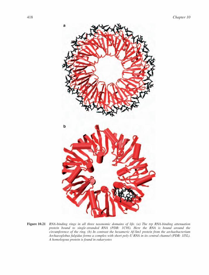
418 Chapter 10
Figure 10.21 RNA-binding rings in all three taxonomic domains of life. (a) The trp RNA-binding attenuationprotein bound to single-stranded RNA (PDB: 1C9S). Here the RNA is bound around the circumference of the ring. (b) In contrast the hexameric Af-Sm1 protein from the archaebacteriumArchaeoglobus fulgidus forms a complex with short poly-U RNA in its central channel (PDB: 1I5L).A homologous protein is found in eukaryotes
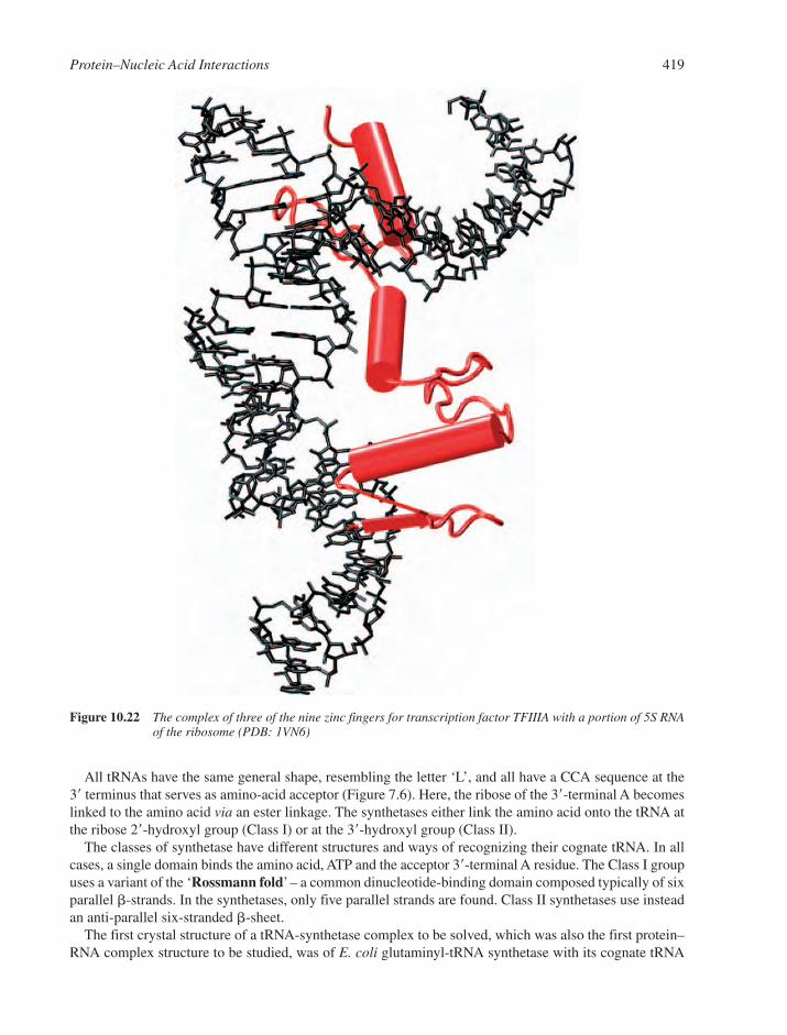
All tRNAs have the same general shape, resembling the letter ‘L’, and all have a CCA sequence at the3� terminus that serves as amino-acid acceptor (Figure 7.6). Here, the ribose of the 3�-terminal A becomeslinked to the amino acid via an ester linkage. The synthetases either link the amino acid onto the tRNA atthe ribose 2�-hydroxyl group (Class I) or at the 3�-hydroxyl group (Class II).
The classes of synthetase have different structures and ways of recognizing their cognate tRNA. In allcases, a single domain binds the amino acid, ATP and the acceptor 3�-terminal A residue. The Class I groupuses a variant of the ‘Rossmann fold’ – a common dinucleotide-binding domain composed typically of sixparallel �-strands. In the synthetases, only five parallel strands are found. Class II synthetases use insteadan anti-parallel six-stranded �-sheet.
The first crystal structure of a tRNA-synthetase complex to be solved, which was also the first protein–RNA complex structure to be studied, was of E. coli glutaminyl-tRNA synthetase with its cognate tRNA
Protein–Nucleic Acid Interactions 419
Figure 10.22 The complex of three of the nine zinc fingers for transcription factor TFIIIA with a portion of 5S RNAof the ribosome (PDB: 1VN6)

(Figure 10.23a). This Class I synthetase binds on one side of the tRNA and contacts the sugar–phosphatebackbone and the minor groove side of the acceptor stem. The acceptor stem is recognized by ‘indirect read-out’ determined by the conformation of the CCA end. By contrast, the structure of the complex betweenthe Class II yeast aspartyl-tRNA synthetase and its cognate tRNA shows that this enzyme binds tRNAfrom the major groove side (Figure 10.23b).
It may seem surprising that the determinants of recognition do not in all cases involve directly the anti-codon triplet, but instead are often distributed throughout the tRNA. For example, the structure of seryl-tRNA synthetase complexed with tRNA (Figure 10.23c) shows a distinctive mode of recognition where an
420 Chapter 10
Figure 10.23 Representative amino acyl tRNA synthetases showing different modes of RNA–protein recognition.(a) Glutamyl-tRNA synthetase (PDB: 1N77). (b) Aspartyl-tRNA syntetase (PDB: 1JGO). (c) Seryl-tRNA synthetase (PDB: 1SER)
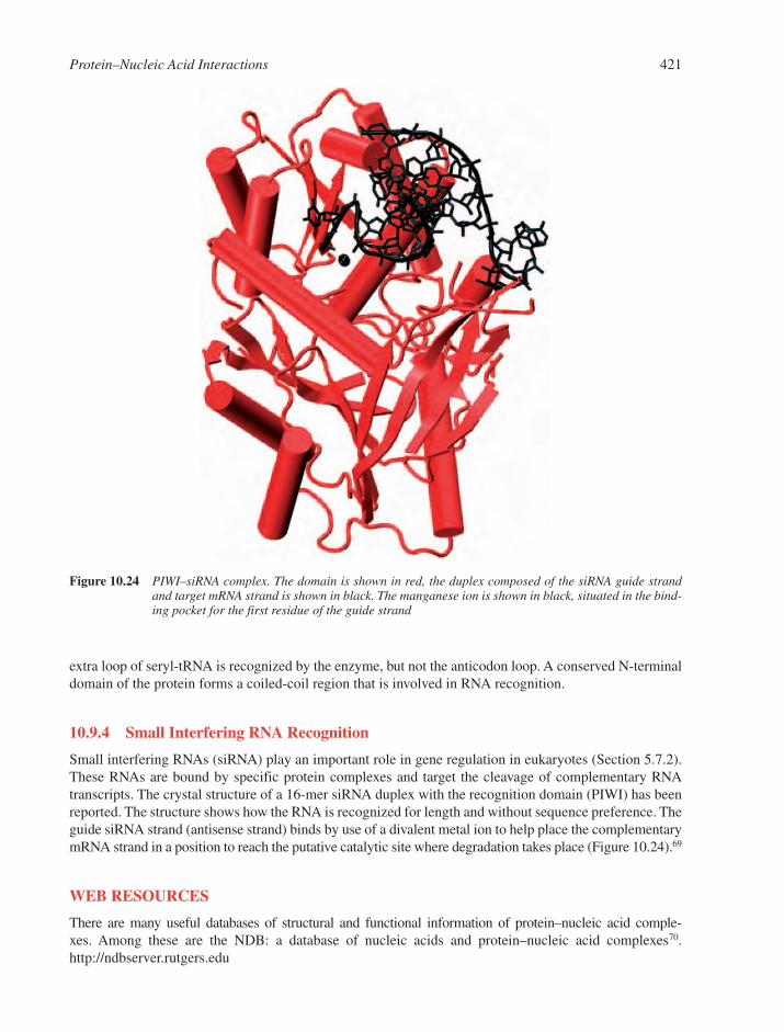
extra loop of seryl-tRNA is recognized by the enzyme, but not the anticodon loop. A conserved N-terminaldomain of the protein forms a coiled-coil region that is involved in RNA recognition.
10.9.4 Small Interfering RNA Recognition
Small interfering RNAs (siRNA) play an important role in gene regulation in eukaryotes (Section 5.7.2).These RNAs are bound by specific protein complexes and target the cleavage of complementary RNAtranscripts. The crystal structure of a 16-mer siRNA duplex with the recognition domain (PIWI) has beenreported. The structure shows how the RNA is recognized for length and without sequence preference. Theguide siRNA strand (antisense strand) binds by use of a divalent metal ion to help place the complementarymRNA strand in a position to reach the putative catalytic site where degradation takes place (Figure 10.24).69
WEB RESOURCES
There are many useful databases of structural and functional information of protein–nucleic acid comple-xes. Among these are the NDB: a database of nucleic acids and protein–nucleic acid complexes70.http://ndbserver.rutgers.edu
Protein–Nucleic Acid Interactions 421
Figure 10.24 PIWI–siRNA complex. The domain is shown in red, the duplex composed of the siRNA guide strandand target mRNA strand is shown in black. The manganese ion is shown in black, situated in the bind-ing pocket for the first residue of the guide strand
An interactive website with details of the classification taxonomy for DNA-binding motifs can be foundat the address:
http://www.biochem.ucl.ac.uk/bsm/prot_dna/prot_dna_cover.htmlEnzyme structural database with enzyme classifications:
http://www.biochem.ucl.ac.uk/bsm/enzymes/ index.htmlMolecular graphics movies are particularly useful for illustrating allosteric transitions and proposed catalytic mechanisms. For instance, the allosteric changes in DNA helicase are shown in an idealized continuum of the structural states in the movie at:
http://sci.cancerresearchuk.org/labs/wigley/projects/helicase/morph1.html andhttp://sci.cancerresearchuk.org/labs/wigley/projects/helicase/morph2.html
A graphics movie showing the catalytic mechanism of AP endonuclease is provided at:http://www. scripps.edu/�jat/
A molecular graphics movie of the bacteriophage DNA pump may be viewed at:http://bilbo.bio.purdue.edu/�viruswww/Rossmann_home/movies.shtml
ProNIT, a database for the thermodynamics of protein–nucleic acid interactions is available at:http://gibk26.bse.kyutech.ac.jp/jouhou/pronit/pronit.html
REFERENCES
1. C.R. Calladine, H.R. Drew, B.F. Luisi and A.A. Travers, Understanding DNA, 3rd edn, Elsevier,Amsterdam, 2004.
2. M.Y. Tolstorukov, R.L. Jernigan and V.B. Zhurkin, Protein–DNA hydrophobic recognition in theminor groove is facilitated by sugar switching. J. Mol. Biol., 2004, 337, 65–76.
3. J.M. Vargason, K. Henderson and P.S. Ho, A crystallographic map of the transition from B-DNA toA-DNA. Proc. Natl. Acad. Sci. USA, 2001, 98, 7265–7270.
4. C.R. Calladine and H.R. Drew, A base-centred explanation of the B-to-A transition in DNA. J. Mol.Biol., 1984, 178, 773–782.
5. E.J. Gardiner, C.A. Hunter, M.J. Packer, D.S. Palmer and P. Willett, Sequence-dependent DNA struc-ture: a database of octamer structural parameters. J. Mol. Biol., 2003, 332, 1025–1035.
6. A. Fersht Enzyme Structure and Mechanism. Freedman, New York, 1998.7. N.M. Luscombe, R.A. Laskowski and J.M. Thornton, Amino acid–base interactions: a three-dimensional
analysis of protein–DNA interactions at an atomic level. Nucleic Acids Res., 2001, 29, 2860–2874.8. K. Nadassy, S.J. Wodak and J. Janin, Structural features of protein–nucleic acid recognition sites.
Biochemistry, 1999, 38, 1999–2017.9. C.R. Woese, Interpreting the universal phylogenetic tree. Proc. Natl. Acad. Sci. USA, 2000, 97, 8392–8396.
10. M.F. White and S.D. Bell, Holding it together: chromatin in the Archaea. Trends Genet., 2002, 18,621–626.
11. J. Miller, A.D. McLachlan and A. Klug, Repetitive zinc-binding domains in the protein transcriptionfactor IIIA from Xenopus oocytes. EMBO J., 1985, 4, 1609–1614.
12. Y. Choo and A. Klug, Physical basis of a protein–DNA recognition code. Curr. Opin. Struct. Biol.,1997, 7, 117–125.
13. C.O. Pabo and L. Nekludova, Geometric analysis and comparison of protein–DNA interfaces: Why isthere no simple code for recognition? J. Mol. Biol., 2000, 301, 597–624.
14. P.A. Rice, S. Yang, K. Mizuuchi and H.A. Nash, Crystal structure of an IHF-DNA complex: a protein-induced DNA U-turn. Cell, 1996, 87, 1295–1306.
15. T.W. Lynch, E.K. Read, A.N. Mattis, J.F. Gardner and P.A. Rice, Integration host factor: putting atwist on protein–DNA recognition. J. Mol. Biol., 2003, 330, 493–502.
16. F.V.T. Murphy, R.M. Sweet and M.E. Churchill, The structure of a chromosomal high mobility groupprotein–DNA complex reveals sequence-neutral mechanisms important for non-sequence-specificDNA recognition. EMBO J., 1999, 18, 6610–6618.
422 Chapter 10
17. G.B. Koudelka, S.C. Harrison and M. Ptashne, Effect of non-contacted bases on the affinity of 434operator for 434 repressor and Cro. Nature, 1987, 326, 886–888.
18. S. Bergqvist, M.A. Williams, R. O’Brien and J.E. Ladbury, Heat capacity effects of water moleculesand ions at a protein–DNA interface. J. Mol. Biol., 2004, 336, 829–842.
19. Z. Otwinowski, R.W. Schevitz, R.G. Zhang, C.L. Lawson, A. Joachimiak, R.Q. Marmorstein,B.F. Luisi and P.B. Sigler, Crystal structure of trp repressor/operator complex at atomic resolution.Nature, 1988, 335, 321–329.
20. N.C. Horton, L.F. Dorner and J.J. Perona, Sequence selectivity and degeneracy of a restrictionendonuclease mediated by DNA intercalation. Nat. Struct. Biol., 2002, 9, 42–47.
21. S. Bergqvist, M.A. Williams, R. O’Brien and J.E. Ladbury, Halophilic adaptation of protein–DNAinteractions. Biochem. Soc. Trans., 2003, 31, 677–680.
22. A.V. Sivolob and S.N. Khrapunov, Translational positioning of nucleosomes on DNA: the role ofsequence-dependent isotropic DNA bending stiffness. J. Mol. Biol., 1995, 247, 918–931.
23. A.M. Martin, M.D. Sam, N.O. Reich and J.J. Perona, Structural and energetic origins of indirect readout in site-specific DNA cleavage by a restriction endonuclease. Nat. Struct. Biol., 1999, 6, 269–277.
24. M.F. Perutz, Mechanisms of Cooperativity and Allosteric Regulation in Proteins. Cambridge UniversityPress, Cambridge, 1990.
25. J.A. Lefstin and K.R. Yamamoto, Allosteric effects of DNA on transcriptional regulators. Nature,1998, 392, 885–888.
26. J.J. Kohler, S.J. Metallo, T.L. Schneider and A. Schepartz, DNA specificity enhanced by sequentialbinding of protein monomers. Proc. Natl. Acad. Sci. USA, 1999, 96, 11735–11739.
27. A.D. Riggs, S. Bourgeois and M. Cohn, The lac repressor–operator interaction. 3. Kinetic studies.J. Mol. Biol., 1970, 53, 401–417.
28. S.E. Halford and J.F. Marko, How do site-specific DNA-binding proteins find their targets? NucleicAcids Res., 2004, 32, 3040–3052.
29. B.A. Lieberman and S.K. Nordeen, DNA intersegment transfer, how steroid receptors search for a targetsite. J. Biol. Chem., 1997, 272, 1061–1068.
30. M. Kampmann, Obstacle bypass in protein motion along DNA by two-dimensional rather thanone-dimensional sliding. J. Biol. Chem., 2004, 279, 38715–38720.
31. N.C. Horton, K.J. Newberry and J.J. Perona, Metal ion-mediated substrate-assisted catalysis in typeII restriction endonucleases. Proc. Natl. Acad. Sci. USA, 1998, 95, 13489–13494.
32. J.A. McClarin, C.A. Frederick, B.C. Wang, P. Greene, H.W. Boyer, J. Grable and J.M. Rosenberg,Structure of the DNA–Eco RI endonuclease recognition complex at 3 A resolution. Science, 1986,234, 1526–1541.
33. D.E. Draper, Protein–DNA complexes: the cost of recognition. Proc. Natl. Acad. Sci. USA, 1993, 90,7429–7430.
34. D.R. Lesser, M.R. Kurpiewski, T. Waters, B.A. Connolly and L. Jen-Jacobson, Facilitated distortionof the DNA site enhances EcoRI endonuclease-DNA recognition. Proc. Natl. Acad. Sci. USA, 1993,90, 7548–7552.
35. I.B. Vipond and S.E. Halford, Structure-function correlation for the EcoRV restriction enzyme: fromnon-specific binding to specific DNA cleavage. Mol. Microbiol., 1993, 9, 225–231.
36. C.D. Mol, T. Izumi, S. Mitra and J.A. Tainer, DNA-bound structures and mutants reveal abasic DNAbinding by APE1 and DNA repair coordination [corrected]. Nature, 2000, 403, 451–456.
37. A.Y. Lau, O.D. Scharer, L. Samson, G.L. Verdine and T. Ellenberger, Crystal structure of a humanalkylbase-DNA repair enzyme complexed to DNA: mechanisms for nucleotide flipping and base exci-sion. Cell, 1998, 95, 249–258.
38. D.M. Popovic, A. Zmiric, S.D. Zaric and E.W. Knapp, Energetics of radical transfer in DNAphotolyase. J. Am. Chem. Soc., 2002, 124, 3775–3782.
39. K. Sandman, S.L. Pereira and J.N. Reeve, Diversity of prokaryotic chromosomal proteins and the ori-gin of the nucleosome. Cell Mol. Life Sci., 1998, 54, 1350–1364.
Protein–Nucleic Acid Interactions 423
40. M.R. Singleton and D.B. Wigley, Multiple roles for ATP hydrolysis in nucleic acid modifyingenzymes. EMBO J., 2003, 22, 4579–4583.
41. K.J. Polach and J. Widom, A model for the cooperative binding of eukaryotic regulatory proteins tonucleosomal target sites. J. Mol. Biol., 1996, 258, 800–812.
42. J.D. Anderson and J. Widom, Sequence and position-dependence of the equilibrium accessibility ofnucleosomal DNA target sites. J. Mol. Biol., 2000, 296, 979–987.
43. T.A. Steitz, A mechanism for all polymerases. Nature, 1998, 391, 231–232.44. J.C. Delaney, P.T. Henderson, S.A. Helquist, J.C. Morales, J.M. Essigmann and E.T. Kool, High-
fidelity in vivo replication of DNA base shape mimics without Watson–Crick hydrogen bonds. Proc.Natl. Acad. Sci. USA, 2003, 100, 4469–4473.
45. T.H. Tahirov, D. Temiakov, M. Anikin, V. Patlan, W.T. McAllister, D.G. Vassylyev and S. Yokoyama,Structure of a T7 RNA polymerase elongation complex at 2.9 A resolution. Nature, 2002, 420, 43–50.
46. Y.W. Yin and T.A. Steitz, Structural basis for the transition from initiation to elongation transcriptionin T7 RNA polymerase. Science, 2002, 298, 1387–1395.
47. P.D. Morris and K.D. Raney, DNA helicases displace streptavidin from biotin-labeled oligonucleotides.Biochemistry, 1999, 38, 5164–5171.
48. S.S. Velankar, P. Soultanas, M.S. Dillingham, H.S. Subramanya and D.B. Wigley, Crystal structuresof complexes of PcrA DNA helicase with a DNA substrate indicate an inchworm mechanism. Cell,1999, 97, 75–84.
49. D.E. Smith, S.J. Tans, S.B. Smith, S. Grimes, D.L. Anderson and C. Bustamante, The bacteriophagestraight phi29 portal motor can package DNA against a large internal force. Nature, 2001, 413, 748–752.
50. A. Guasch, J. Pous, B. Ibarra, F.X. Gomis-Ruth, J.M. Valpuesta, N. Sousa, J.L. Carrascosa andM. Coll, Detailed architecture of a DNA translocating machine: the high-resolution structure of thebacteriophage phi29 connector particle. J. Mol. Biol., 2002, 315, 663–676.
51. A.A. Simpson, Y. Tao, P.G. Leiman, M.O. Badasso, Y. He, P.J. Jardine, N.H. Olson, M.C. Morais, S. Grimes, D.L. Anderson, T.S. Baker and M.G. Rossmann, Structure of the bacteriophage phi29DNA packaging motor. Nature, 2000, 408, 745–750.
52. S.N. Savvides, H.J. Yeo, M.R. Beck, F. Blaesing, R. Lurz, E. Lanka, R. Buhrdorf, W. Fischer,R. Haas and G. Waksman, VirB11 ATPases are dynamic hexameric assemblies: new insights into bac-terial type IV secretion. EMBO J., 2003, 22, 1969–1980.
53. F.X. Gomis-Ruth, G. Moncalian, R. Perez-Luque, A. Gonzalez, E. Cabezon, F. de la Cruz andM. Coll, The bacterial conjugation protein TrwB resembles ring helicases and F1-ATPase. Nature,2001, 409, 637–641.
54. R.M. Story and T.A. Steitz, Structure of the recA protein–ADP complex. Nature, 1992, 355, 374–376.55. K. Nagai, RNA–protein complexes. Curr. Opin. Struct. Biol., 1996, 6, 53–61.56. D.E. Draper, Themes in RNA–protein recognition. J. Mol. Biol., 1999, 293, 255–270.57. S. Jones, D.T. Daley, N.M. Luscombe, H.M. Berman and J.M. Thornton, Protein–RNA interactions:
a structural analysis. Nucleic Acids Res., 2001, 29, 943–954.58. J.R. Williamson, Induced fit in RNA–protein recognition. Nat. Struct. Biol., 2000, 7, 834–837.59. N.H. Hopcroft, A. Manfredo, A.L. Wendt, A.M. Brzozowski, P. Gollnick and A.A. Antson, The inter-
action of RNA with TRAP: the role of triplet repeats and separating spacer nucleotides. J. Mol. Biol.,2004, 338, 43–53.
60. K. Valegard, J.B. Murray, N.J. Stonehouse, S. van den Worm, P.G. Stockley and L. Liljas, The three-dimensional structures of two complexes between recombinant MS2 capsids and RNA operator frag-ments reveal sequence-specific protein–RNA interactions. J. Mol. Biol., 1997, 270, 724–738.
61. C. Kambach, S. Walke, R. Young, J.M. Avis, E. de la Fortelle, V.A. Raker, R. Luhrmann, J. Li and K. Nagai, Crystal structures of two Sm protein complexes and their implications for the assembly ofthe spliceosomal snRNPs. Cell, 1999, 96, 375–387.
424 Chapter 10
62. I. Toro, S. Thore, C. Mayer, J. Basquin, B. Seraphin and D. Suck, RNA binding in an Sm core domain:X-ray structure and functional analysis of an archaeal Sm protein complex. EMBO J., 2001, 20,2293–2303.
63. C. Sauter, J. Basquin and D. Suck, Sm-like proteins in Eubacteria: the crystal structure of the Hfq pro-tein from Escherichia coli. Nucleic Acids Res., 2003, 31, 4091–4098.
64. S. Thore, C. Mayer, C. Sauter, S. Weeks and D. Suck, Crystal structures of the Pyrococcus abyssi Smcore and its complex with RNA. Common features of RNA binding in archaea and eukarya.J. Biol. Chem., 2003, 278, 1239–1247.
65. M.A. Schumacher, R.F. Pearson, T. Moller, P. Valentin-Hansen and R.G. Brennan, Structures of thepleiotropic translational regulator Hfq and an Hfq-RNA complex: a bacterial Sm-like protein. EMBOJ., 2002, 21, 3546–3556.
66. M. Schubert, R.E. Edge, P. Lario, M.A. Cook, N.C. Strynadka, G.A. Mackie and L.P. McIntosh,Structural characterization of the RNase E S1 domain and identification of its oligonucleotide-binding and dimerization interfaces. J. Mol. Biol., 2004, 341, 37–54.
67. H.A. Lewis, K. Musunuru, K.B. Jensen, C. Edo, H. Chen, R.B. Darnell and S.K. Burley, Sequence-specific RNA binding by a Nova KH domain: implications for paraneoplastic disease and the fragileX syndrome. Cell, 2000, 100, 323–332.
68. K. Ye, L. Malinina and D.J. Patel, Recognition of small interfering RNA by a viral suppressor of RNAsilencing. Nature, 2003, 426, 874–878.
69. J.S. Parker, S.M. Roe and D. Barford, Structural insights into mRNA recognition from a PIWIdomain-siRNA guide complex. Nature, 2005, 434, 663–666.
70. H.M. Berman, W.K. Olson, D.L. Beveridge, J. Westbrook, A. Gelbin, T. Demeny, S.-H. Hsieh, A.R. Srinivasan and B. Schneider, The Nucleic Acid Database: a comprehensive relational databaseof three-dimensional structures of nucleic acids. Biophys. J., 1992, 63, 751–759.
Protein–Nucleic Acid Interactions 425

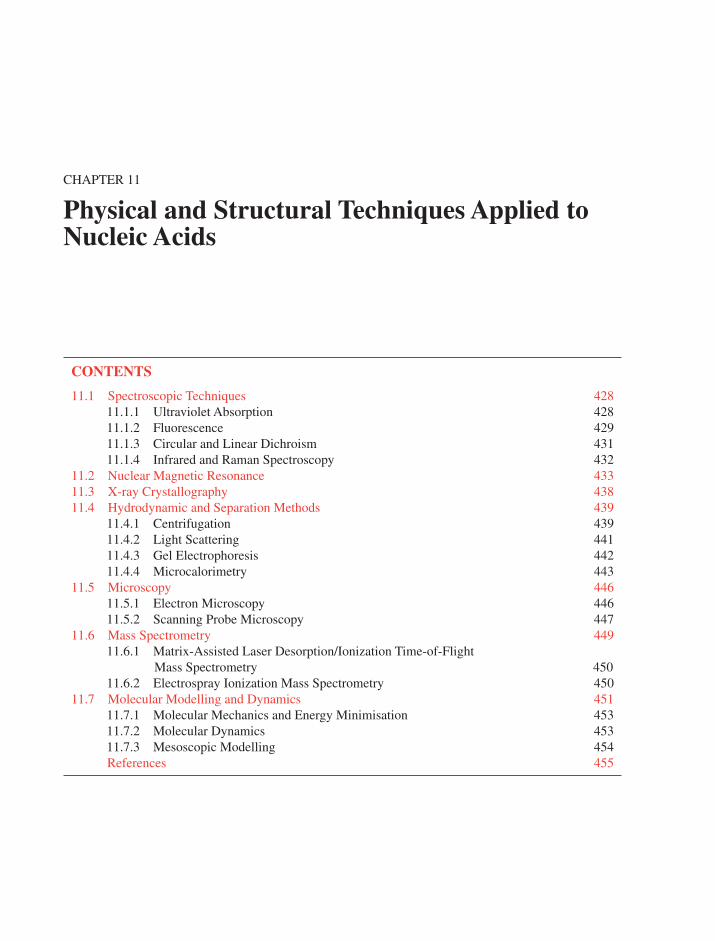
CHAPTER 11
Physical and Structural Techniques Applied toNucleic Acids
CONTENTS
11.1 Spectroscopic Techniques 42811.1.1 Ultraviolet Absorption 42811.1.2 Fluorescence 42911.1.3 Circular and Linear Dichroism 43111.1.4 Infrared and Raman Spectroscopy 432
11.2 Nuclear Magnetic Resonance 43311.3 X-ray Crystallography 43811.4 Hydrodynamic and Separation Methods 439
11.4.1 Centrifugation 43911.4.2 Light Scattering 44111.4.3 Gel Electrophoresis 44211.4.4 Microcalorimetry 443
11.5 Microscopy 44611.5.1 Electron Microscopy 44611.5.2 Scanning Probe Microscopy 447
11.6 Mass Spectrometry 44911.6.1 Matrix-Assisted Laser Desorption/Ionization Time-of-Flight
Mass Spectrometry 45011.6.2 Electrospray Ionization Mass Spectrometry 450
11.7 Molecular Modelling and Dynamics 45111.7.1 Molecular Mechanics and Energy Minimisation 45311.7.2 Molecular Dynamics 45311.7.3 Mesoscopic Modelling 454References 455
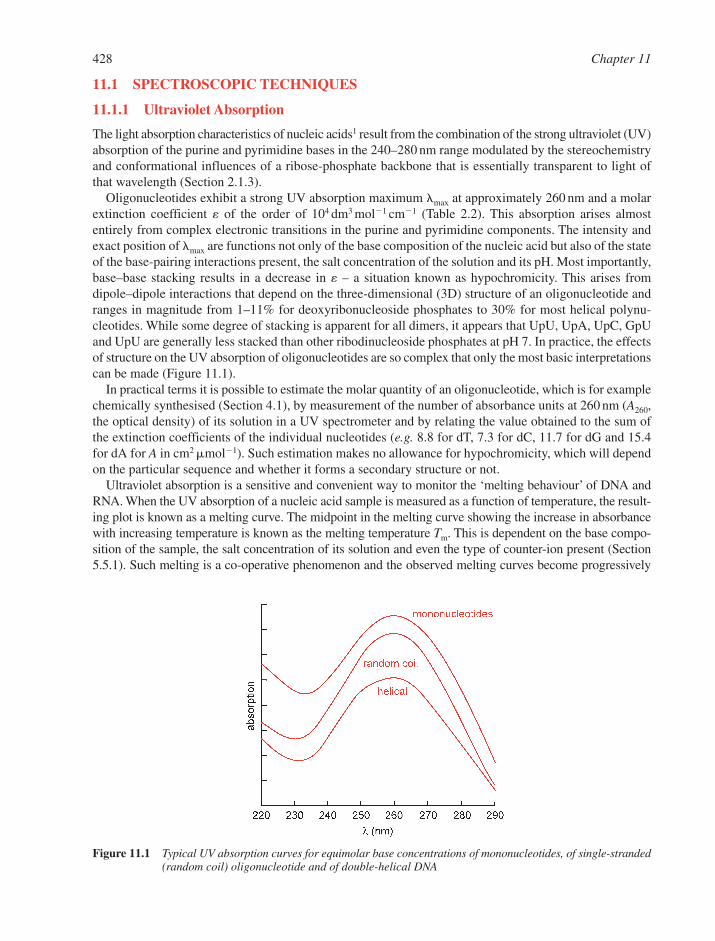
11.1 SPECTROSCOPIC TECHNIQUES
11.1.1 Ultraviolet Absorption
The light absorption characteristics of nucleic acids1 result from the combination of the strong ultraviolet (UV)absorption of the purine and pyrimidine bases in the 240–280 nm range modulated by the stereochemistryand conformational influences of a ribose-phosphate backbone that is essentially transparent to light ofthat wavelength (Section 2.1.3).
Oligonucleotides exhibit a strong UV absorption maximum �max at approximately 260 nm and a molarextinction coefficient � of the order of 104 dm3 mol�1 cm�1 (Table 2.2). This absorption arises almostentirely from complex electronic transitions in the purine and pyrimidine components. The intensity andexact position of �max are functions not only of the base composition of the nucleic acid but also of the stateof the base-pairing interactions present, the salt concentration of the solution and its pH. Most importantly,base–base stacking results in a decrease in � – a situation known as hypochromicity. This arises fromdipole–dipole interactions that depend on the three-dimensional (3D) structure of an oligonucleotide andranges in magnitude from 1–11% for deoxyribonucleoside phosphates to 30% for most helical polynu-cleotides. While some degree of stacking is apparent for all dimers, it appears that UpU, UpA, UpC, GpUand UpU are generally less stacked than other ribodinucleoside phosphates at pH 7. In practice, the effectsof structure on the UV absorption of oligonucleotides are so complex that only the most basic interpretationscan be made (Figure 11.1).
In practical terms it is possible to estimate the molar quantity of an oligonucleotide, which is for examplechemically synthesised (Section 4.1), by measurement of the number of absorbance units at 260 nm (A260,the optical density) of its solution in a UV spectrometer and by relating the value obtained to the sum ofthe extinction coefficients of the individual nucleotides (e.g. 8.8 for dT, 7.3 for dC, 11.7 for dG and 15.4for dA for A in cm2 �mol�1). Such estimation makes no allowance for hypochromicity, which will dependon the particular sequence and whether it forms a secondary structure or not.
Ultraviolet absorption is a sensitive and convenient way to monitor the ‘melting behaviour’ of DNA andRNA. When the UV absorption of a nucleic acid sample is measured as a function of temperature, the result-ing plot is known as a melting curve. The midpoint in the melting curve showing the increase in absorbancewith increasing temperature is known as the melting temperature Tm. This is dependent on the base compo-sition of the sample, the salt concentration of its solution and even the type of counter-ion present (Section5.5.1). Such melting is a co-operative phenomenon and the observed melting curves become progressively
428 Chapter 11
Figure 11.1 Typical UV absorption curves for equimolar base concentrations of mononucleotides, of single-stranded(random coil) oligonucleotide and of double-helical DNA
steeper with increasing length of the oligonucleotide. Simple sigmoid melting curves are observed for manyDNA samples, but it is also possible to observe more complex, multi-phase melting in some cases (Figure11.2). The deconvolution of such multi-phase melting curves makes it possible to examine the effects of basemodification on the stability and nature of nucleic acid secondary structure in more detail.
11.1.2 Fluorescence
Fluorescence is defined as the emission of radiation as a molecule returns to its ground state from anexcited electronic state.2 In order to characterise the photoexcited emission from a molecule, it is necessaryto determine the spectral distribution, photon yield, excited-state lifetime and polarisation of the emission,all as functions of excitation wavelength. The precision with which fluorescence intensity can be measuredis very high, since photon-counting techniques eliminate several sources of uncertainty and error. Excitationand emission occur in timescales of �10�15 s, but the lifetime of an excited state has a duration of �10� 9 s.Thus any physical process that takes place on a similar timescale as the fluorescence lifetime can be analysedby examining changes in the emission.
The fluorescence emission from nucleotides and dinucleoside phosphates is very weak at room temperatureand can only be examined in frozen samples at �80 K. Fluorescence spectroscopy is nevertheless invaluablefor examining nucleic acid–ligand interactions. Many DNA-binding ligands have little or no fluorescencein aqueous solution. However, upon binding to a nucleic acid the ligand is in a hydrophobic environmentand the solvent can no longer quench the intrinsic ligand fluorescence. Therefore fluorescence emission isa direct probe of the concentration of bound ligand.
Fluorescence resonance energy transfer (FRET) is a phenomenon in which the energy of an excited-statefluorescent donor molecules is transferred to an unexcited acceptor molecule via a dipole–dipole coupling.Importantly, the rate of energy transfer is dependent on the distance between donor and acceptor molecules,the spectral characteristics of the pair and the relative orientations of the donor and acceptor transitiondipoles. FRET experiments have been used widely to determine proximity relationships in protein and nucleicacid systems since the 1940s when Förster derived the quantitative analysis for FRET. A key parameter inFRET is the efficiency of depopulation (ET) and this is related to fluorescent lifetimes in the presence (�T)and absence (�) of resonance energy transfer [Equation (11.1)]:
(11.1)ETT� �1
�
�
Physical and Structural Techniques Applied to Nucleic Acids 429
Figure 11.2 An oligonucleotide melting curve (red), the derivative of the curve (black) and the deconvolution of the derivative into its composite components (red)
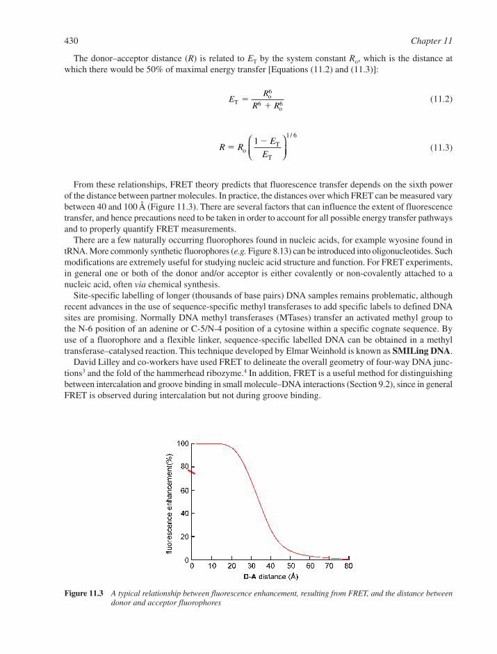
The donor–acceptor distance (R) is related to ET by the system constant Ro, which is the distance atwhich there would be 50% of maximal energy transfer [Equations (11.2) and (11.3)]:
(11.2)
(11.3)
From these relationships, FRET theory predicts that fluorescence transfer depends on the sixth powerof the distance between partner molecules. In practice, the distances over which FRET can be measured varybetween 40 and 100 Å (Figure 11.3). There are several factors that can influence the extent of fluorescencetransfer, and hence precautions need to be taken in order to account for all possible energy transfer pathwaysand to properly quantify FRET measurements.
There are a few naturally occurring fluorophores found in nucleic acids, for example wyosine found intRNA. More commonly synthetic fluorophores (e.g. Figure 8.13) can be introduced into oligonucleotides. Suchmodifications are extremely useful for studying nucleic acid structure and function. For FRET experiments,in general one or both of the donor and/or acceptor is either covalently or non-covalently attached to anucleic acid, often via chemical synthesis.
Site-specific labelling of longer (thousands of base pairs) DNA samples remains problematic, althoughrecent advances in the use of sequence-specific methyl transferases to add specific labels to defined DNAsites are promising. Normally DNA methyl transferases (MTases) transfer an activated methyl group tothe N-6 position of an adenine or C-5/N-4 position of a cytosine within a specific cognate sequence. Byuse of a fluorophore and a flexible linker, sequence-specific labelled DNA can be obtained in a methyltransferase–catalysed reaction. This technique developed by Elmar Weinhold is known as SMILing DNA.
David Lilley and co-workers have used FRET to delineate the overall geometry of four-way DNA junc-tions3 and the fold of the hammerhead ribozyme.4 In addition, FRET is a useful method for distinguishingbetween intercalation and groove binding in small molecule–DNA interactions (Section 9.2), since in generalFRET is observed during intercalation but not during groove binding.
R RE
E�
�o
T
T
11 6
/
ER
R RTo
o
��
6
6 6
430 Chapter 11
Figure 11.3 A typical relationship between fluorescence enhancement, resulting from FRET, and the distance betweendonor and acceptor fluorophores

11.1.3 Circular and Linear Dichroism
Circular dichroism (CD) is a widely used form of chiral spectroscopy which has been applied to the studyof nucleic acids.5,6 A CD signal results from the differential absorption of left and right circularly polarisedlight. Circularly polarised light has chirality and therefore it will exhibit chiral discrimination. A chiralmolecule will absorb left and right circularly polarised light to differing extents and this difference givesrise to the phenomenon of CD. Typically, data are presented as plots of �� versus wavelength. Here �� isthe difference in molar extinction coefficient for left and right circularly polarised light (�L – �R).
Isolated purine and pyrimidine bases are planar, intrinsically optically inactive and hence do not exhibit aCD signal. However when incorporated into nucleosides and nucleotides, the glycosylic bond from the C-1atom of the sugar to either the N-9 of purines or N-1 of pyrimidines gives rise to a chiral perturbation of theUV absorption of the base. The CD signal of a nucleic acid increases with length due to the co-operativityof chiral interactions between contiguous bases. This effect occurs both as a result of sequence effects arisingfrom nearest-neighbour interactions as well as from overall gross secondary structure.
The information derived from CD spectra is complementary to other types of optical spectroscopy, suchas UV, infrared (IR) and linear dichroism (LD), and it provides a quick, convenient and accurate picture ofthe overall conformation and secondary structure of a particular nucleic acid solution. Reference CD spectrafor a B-DNA duplex, single strands and a G3 quadruplex show that each type of secondary structureexhibits its own signature spectra (Figure 11.4). A- and Z-type DNA (Section 2.2) also have unique CD spec-tra, as do many nucleic acid–ligand complexes. CD spectroscopy also allows inter-conversions betweendifferent secondary structures to be monitored. For example titration of NaCl into a solution of poly(dG-dC) to salt concentrations above 4 M induces a structural transition in the polynucleotide from a standardright-handed B-helix to the left-handed Z-form. Increasing the temperature of a nucleic acid solution whilstmeasuring the CD signal allows DNA melting to be monitored directly and it is possible to observe the struc-tural transition from folded duplex to random coil single strands.
Circular dichroism spectroscopy is also a useful tool for studying nucleic acid–ligand interactions. The CDspectrum of each component in solution is directly proportional to its concentration (Beer’s law), and thetotal spectrum arises from the sum of all component spectra. If ligand binding induces extrinsic opticalactivity in the chromophores of the bound ligand, an induced CD signal is observed, which is directly pro-portional to the amount of nucleic acid–ligand complex formed, and hence it can be used to construct abinding isotherm. Alternatively, ligand binding may result in a conformational change in the nucleic acid,and the resultant change in the intrinsic CD signal of the macromolecule allows the binding to be quantified.
Physical and Structural Techniques Applied to Nucleic Acids 431
Figure 11.4 Circular dichroism spectra for three different conformations of DNA, a poly(dA)poly(dT) B-type duplex,a guanine quadruplex (tetraplex) and a single-stranded random coil
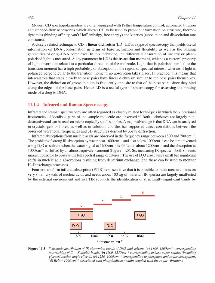
Modern CD spectrapolarimeters are often equipped with Peltier temperature control, automated titrationand stopped-flow accessories which allows CD to be used to provide information on structure, thermo-dynamics (binding affinity, van’t Hoff enthalpy, free energy) and kinetics (association and dissociation rateconstants).
A closely related technique to CD is linear dichroism (LD). LD is a type of spectroscopy that yields usefulinformation on DNA conformation in terms of base inclination and flexibility as well as the bindinggeometries of drug–DNA complexes. In this technique, the differential absorption of linearly or plane-polarised light is measured. A key parameter in LD is the transition moment, which is a vectoral propertyof light absorption related to a particular direction of the molecule. Light that is polarised parallel to thetransition moment has a high probability of absorption in the region of spectral interest, whereas if light ispolarised perpendicular to the transition moment, no absorption takes place. In practice, this means thatintercalators that stack closely to base pairs have linear dichroism similar to the base pairs themselves.However, the dichroism of groove binders is frequently opposite to that of the base pairs, since they bindalong the edges of the base pairs. Hence LD is a useful type of spectroscopy for assessing the bindingmode of a drug to DNA.
11.1.4 Infrared and Raman Spectroscopy
Infrared and Raman spectroscopy are often regarded as closely related techniques in which the vibrationalfrequencies of localised parts of the sample molecule are observed.7,8 Both techniques are largely non-destructive and can be used on microscopically small samples. A major advantage is that DNA can be analysedin crystals, gels or fibres, as well as in solution, and this has supported direct correlations between theobserved vibrational frequencies and 3D structures derived by X-ray diffraction.
Infrared absorptions from nucleic acids are observed in the frequency range between 1800 and 700 cm�1.The problem of strong IR absorption by water near 1600 cm�1 and also below 1000 cm�1 can be circumventedusing D2O as solvent when the water signal at 1600 cm�1 is shifted to about 1200 cm�1 and the absorption at1000 cm�1 is shifted by an almost equivalent amount (Figure 11.5). So, measuring IR spectra in both solventsmakes it possible to observe the full spectral range of interest. The use of D2O also causes small but significantshifts in nucleic acid absorptions resulting from deuterium exchange, and these can be used to monitorH–D exchange processes.
Fourier transform infrared absorption (FTIR) is so sensitive that it is possible to make measurements onvery small crystals of nucleic acids and needs about 100 �g of material. IR spectra are largely unaffectedby the external environment and so FTIR supports the identification of structurally significant bands by
432 Chapter 11
Figure 11.5 Schematic distribution of IR absorption bands of DNA and solvent. (a) 1800–1500 cm�1 correspondingto stretching of C � X double bonds. (b) 1500–1250 cm�1 corresponding to base-sugar entities (includingglycosyl torsion angle effects). (c) 1250–1000 cm�1 corresponding to phosphate and sugar absorptions.(d) Below 1000 cm�1 associated with phosphodiester chain coupled with the sugar vibrations
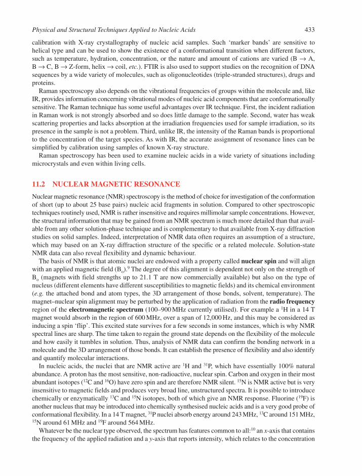
calibration with X-ray crystallography of nucleic acid samples. Such ‘marker bands’ are sensitive to helical type and can be used to show the existence of a conformational transition when different factors,such as temperature, hydration, concentration, or the nature and amount of cations are varied (B → A,B → C, B → Z-form, helix → coil, etc.). FTIR is also used to support studies on the recognition of DNAsequences by a wide variety of molecules, such as oligonucleotides (triple-stranded structures), drugs andproteins.
Raman spectroscopy also depends on the vibrational frequencies of groups within the molecule and, likeIR, provides information concerning vibrational modes of nucleic acid components that are conformationallysensitive. The Raman technique has some useful advantages over IR technique. First, the incident radiationin Raman work is not strongly absorbed and so does little damage to the sample. Second, water has weakscattering properties and lacks absorption at the irradiation frequencies used for sample irradiation, so itspresence in the sample is not a problem. Third, unlike IR, the intensity of the Raman bands is proportionalto the concentration of the target species. As with IR, the accurate assignment of resonance lines can besimplified by calibration using samples of known X-ray structure.
Raman spectroscopy has been used to examine nucleic acids in a wide variety of situations includingmicrocrystals and even within living cells.
11.2 NUCLEAR MAGNETIC RESONANCE
Nuclear magnetic resonance (NMR) spectroscopy is the method of choice for investigation of the conformationof short (up to about 25 base pairs) nucleic acid fragments in solution. Compared to other spectroscopictechniques routinely used, NMR is rather insensitive and requires millimolar sample concentrations. However,the structural information that may be gained from an NMR spectrum is much more detailed than that avail-able from any other solution-phase technique and is complementary to that available from X-ray diffractionstudies on solid samples. Indeed, interpretation of NMR data often requires an assumption of a structure,which may based on an X-ray diffraction structure of the specific or a related molecule. Solution-stateNMR data can also reveal flexibility and dynamic behaviour.
The basis of NMR is that atomic nuclei are endowed with a property called nuclear spin and will alignwith an applied magnetic field (Bo).
9 The degree of this alignment is dependent not only on the strength ofBo (magnets with field strengths up to 21.1 T are now commercially available) but also on the type ofnucleus (different elements have different susceptibilities to magnetic fields) and its chemical environment(e.g. the attached bond and atom types, the 3D arrangement of those bonds, solvent, temperature). Themagnet–nuclear spin alignment may be perturbed by the application of radiation from the radio frequencyregion of the electromagnetic spectrum (100–900 MHz currently utilised). For example a 1H in a 14 Tmagnet would absorb in the region of 600 MHz, over a span of 12,000 Hz, and this may be considered asinducing a spin ‘flip’. This excited state survives for a few seconds in some instances, which is why NMRspectral lines are sharp. The time taken to regain the ground state depends on the flexibility of the moleculeand how easily it tumbles in solution. Thus, analysis of NMR data can confirm the bonding network in amolecule and the 3D arrangement of those bonds. It can establish the presence of flexibility and also identifyand quantify molecular interactions.
In nucleic acids, the nuclei that are NMR active are 1H and 31P, which have essentially 100% naturalabundance. A proton has the most sensitive, non-radioactive, nuclear spin. Carbon and oxygen in their mostabundant isotopes (12C and 16O) have zero spin and are therefore NMR silent. 15N is NMR active but is veryinsensitive to magnetic fields and produces very broad line, unstructured spectra. It is possible to introducechemically or enzymatically 13C and 15N isotopes, both of which give an NMR response. Fluorine (19F) isanother nucleus that may be introduced into chemically synthesised nucleic acids and is a very good probe ofconformational flexibility. In a 14 T magnet, 31P nuclei absorb energy around 243 MHz, 13C around 151 MHz,15N around 61 MHz and 19F around 564 MHz.
Whatever be the nuclear type observed, the spectrum has features common to all:10 an x-axis that containsthe frequency of the applied radiation and a y-axis that reports intensity, which relates to the concentration
Physical and Structural Techniques Applied to Nucleic Acids 433
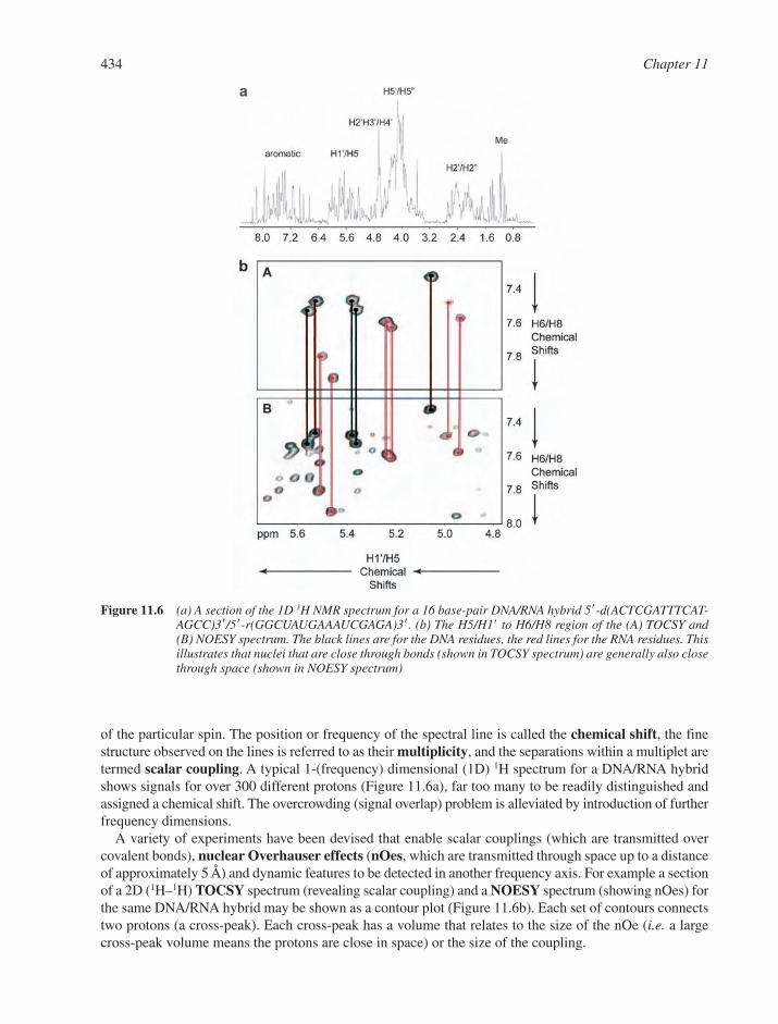
of the particular spin. The position or frequency of the spectral line is called the chemical shift, the finestructure observed on the lines is referred to as their multiplicity, and the separations within a multiplet aretermed scalar coupling. A typical 1-(frequency) dimensional (1D) 1H spectrum for a DNA/RNA hybridshows signals for over 300 different protons (Figure 11.6a), far too many to be readily distinguished andassigned a chemical shift. The overcrowding (signal overlap) problem is alleviated by introduction of furtherfrequency dimensions.
A variety of experiments have been devised that enable scalar couplings (which are transmitted overcovalent bonds), nuclear Overhauser effects (nOes, which are transmitted through space up to a distanceof approximately 5 Å) and dynamic features to be detected in another frequency axis. For example a sectionof a 2D (1H–1H) TOCSY spectrum (revealing scalar coupling) and a NOESY spectrum (showing nOes) forthe same DNA/RNA hybrid may be shown as a contour plot (Figure 11.6b). Each set of contours connectstwo protons (a cross-peak). Each cross-peak has a volume that relates to the size of the nOe (i.e. a largecross-peak volume means the protons are close in space) or the size of the coupling.
434 Chapter 11
Figure 11.6 (a) A section of the 1D 1H NMR spectrum for a 16 base-pair DNA/RNA hybrid 5-d(ACTCGATTTCAT-AGCC)3/5-r(GGCUAUGAAAUCGAGA)3. (b) The H5/H1 to H6/H8 region of the (A) TOCSY and(B) NOESY spectrum. The black lines are for the DNA residues, the red lines for the RNA residues. Thisillustrates that nuclei that are close through bonds (shown in TOCSY spectrum) are generally also closethrough space (shown in NOESY spectrum)
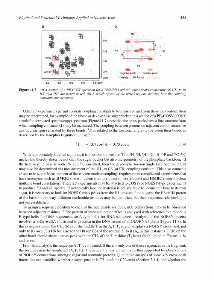
Other 2D experiments permit accurate coupling constants to be measured and from these the conformationmay be determined, for example of the ribose or dexoyribose sugar pucker. In a section of a PE-COSY (COSYstands for correlated spectroscopy) spectrum (Figure 11.7), note that the cross-peaks have a fine structure fromwhich coupling constants (J) may be measured. The coupling between protons on adjacent carbon atoms (orany nuclear spin separated by three bonds, 3J) is related to the torsional angle (�) between their bonds asdescribed by the Karplus Equation (11.4):9
(11.4)
With appropriately labelled samples, it is possible to measure 3J for 1H–1H, 1H–13C, 1H–31P and 13C–13Cnuclei and thereby describe not only the sugar pucker but also the geometry of the phosphate backbone. Ifthe heterocyclic base is both 15N and 13C enriched, then the glycosylic torsion angle (see Section 2.1.4)may also be determined via measurement of the H1 to C6 (or C8) coupling constant. This also connectsa base to its sugar. Measurement of these heteronuclear couplings requires more complicated experiments thathave acronyms such as HMQC (heteronuclear multiple quantum correlation) and HMBC (heteronuclearmultiple bond correlation). These 2D experiments may be attached to COSY- or NOESY-type experimentsto produce 3D and 4D spectra. If isotopically labelled material is not available to ‘connect’ a base to its ownsugar, it is necessary to look for NOESY cross-peaks from the H1 proton of the sugar to the H6 or H8 protonof the base. In this way, different nucleotide residues may be identified, but their sequence relationship isnot yet established.
To assign a sequence position to each of the nucleoside residues, nOe connections have to be observedbetween adjacent residues.11 The pattern of inter-nucleotide nOes is analysed with reference to a model: aB-type helix for DNA sequences, an A-type helix for RNA sequences. Analysis of the NOESY spectrainvolves a ‘nOe-walk’, illustrated in practice in the DNA strand of a DNA/RNA hybrid (Figure 11.8). Inthe example shown, the CH3 (Me) of the middle T in the A6T7T8 stretch displays a NOESY cross-peak notonly to its own (T7) H6 but also to the H8 (or H6) of the residue 5 to it (A6 in this instance). T7H6 on theother hand should share a cross-peak with the CH3 of the 3 residue (T8 here) (highlighted in Figure 11.8)and so on.
From this analysis, the sequence ATT is confirmed. If there is only one of these sequences in the fragment,the residues may be numbered (A6T7T8). The sequential assignment is further supported by observationsof NOESY connections amongst sugar and aromatic protons. Qualitative analysis of some key cross-peakintensities can establish whether a sugar pucker is C3-endo or C2-endo (Section 2.1.4) and whether the
3 213 7 0 73JHH � �. cos . cos� �
Physical and Structural Techniques Applied to Nucleic Acids 435
Figure 11.7 (a) A section of a PE-COSY spectrum for a DNA/RNA hybrid– cross-peaks connecting A6 H1 to its H2 and H2� are boxed in red. (b) A sketch of one of the boxed regions showing how the coupling constants are measured

conformation about an N-glycosylic bond is syn or anti (Figure 11.9). This information lends support tothe quantitative analysis of three-bond (or vicinal) scalar coupling constants, 3J.
Inter-strand hydrogen bonding may be detected indirectly by the observation of nOes among imino,amino and adenine H2 protons. If 15N-labelled bases are present, then the hydrogen bond can be detecteddirectly through measurement of one-bond and two-bond inter-residue 1H–15N couplings.12
436 Chapter 11
Figure 11.9 An illustration of the through-space connections sought to confirm: (a) the sugar pucker of the furanosering or (b) the arrangement of the N-glycosylic bond. The numbers shown on the arrows are in Å.A short distance leads to a large NOESY cross-peak
Figure 11.8 An example of a ‘nOe-walk’ for the sequential assignment of 1H resonances. (a) The red arrows indicatekey nOe connections. (b) The CH3(Me) to H6/H8 region of the (A) TOCSY and (B) NOESY spectrum forthe 16 base pair DNA/RNA hybrid (Figure 11.6). The ‘walk’ A6H8 to T7Me to T7H6 to T8Me to T8H6 toT9Me is shown
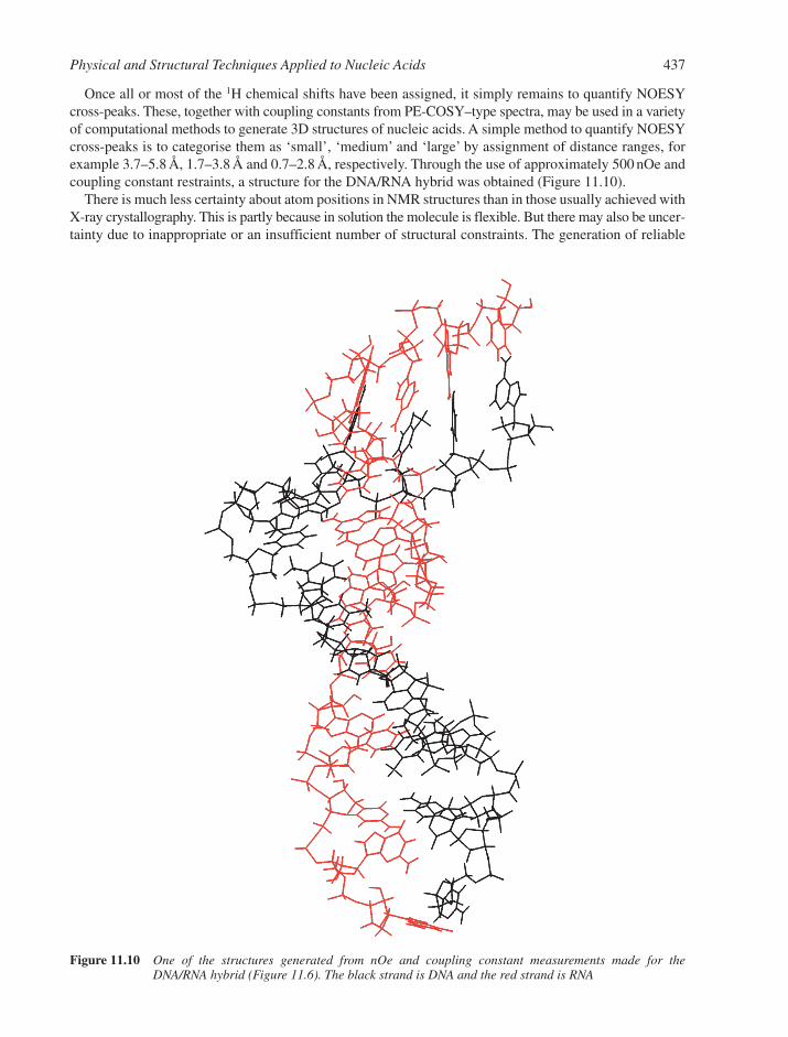
Once all or most of the 1H chemical shifts have been assigned, it simply remains to quantify NOESYcross-peaks. These, together with coupling constants from PE-COSY–type spectra, may be used in a varietyof computational methods to generate 3D structures of nucleic acids. A simple method to quantify NOESYcross-peaks is to categorise them as ‘small’, ‘medium’ and ‘large’ by assignment of distance ranges, forexample 3.7–5.8 Å, 1.7–3.8 Å and 0.7–2.8 Å, respectively. Through the use of approximately 500 nOe andcoupling constant restraints, a structure for the DNA/RNA hybrid was obtained (Figure 11.10).
There is much less certainty about atom positions in NMR structures than in those usually achieved withX-ray crystallography. This is partly because in solution the molecule is flexible. But there may also be uncer-tainty due to inappropriate or an insufficient number of structural constraints. The generation of reliable
Physical and Structural Techniques Applied to Nucleic Acids 437
Figure 11.10 One of the structures generated from nOe and coupling constant measurements made for theDNA/RNA hybrid (Figure 11.6). The black strand is DNA and the red strand is RNA

structures from NMR data has been an important issue until recently.12,13 Approaches are now available toenable the measurement of residual dipolar couplings, which occur via a through-space process but overa longer range than nOes. To measure such couplings, it is necessary to partially align the nucleic acidmolecule (not just the nuclear spin) with Bo, in a manner akin to a crystal lattice. To achieve molecularalignment, it is generally necessary for an agent to be introduced that has a preferred orientation, such asphospholipid bicelles or filamentous phage Pf1, to induce partial alignment of nucleic acid sequences.Such approaches are now aiding the conformational analysis of nucleic acids. NMR structures are nowbeing generated of structural motifs, such as stem-loops, G-tetraplexes, I-motifs, pseudoknots andtriplexes (Chapter 2),14 as well as complexes of nucleic acids with drugs (Chapter 9) and proteins (Chapter 10).
Although the upper molecular mass limit for NMR structure determination for nucleic acids is around25 kDa, a 100 kDa RNA has been studied by NMR.15 Here only part of the RNA sequence was 13C and 15Nenriched, thus simplifying the assignment procedure and allowing the RNA fragment to be studied withina larger molecule.
11.3 X-RAY CRYSTALLOGRAPHY
X-ray crystallography is the key method for determination of the 3D structure of biological macromoleculesat atomic resolution, and therefore has had a major impact on our understanding of the structure and functionof the nucleic acids.
In three decades of crystallographic structure determination, hundreds of structures of DNA, RNA andprotein–nucleic acid complexes have accumulated, as witnessed by the 2334 structures deposited in theNucleic Acid Database (NDB) as of March 16, 2004 (http://ndbserver.rutgers.edu),16 and the 24,785 structuresin the Research Collaboratory for Structural Biology/Protein Data Bank (RCSB/PDB) as of March 23, 2004(http://www.rcsb.org/pdb).17 Dramatic advances have been made in virtually all areas of X-ray crystallography,including crystallization (sparse-matrix crystallization screens) and crystal handling (cryo-protection), datacollection and resolution (synchrotron radiation and CCD detectors), phasing (multi-wavelength anomalousdispersion, MAD), electron density map interpretation and model building (computer graphics and automaticchain tracing) and structure refinement (more computer power, simulated annealing and full-matrix least-squares refinement) (Figure 11.11a–e).18–22
The first step is the production of well-diffracting crystals. While oligonucleotides are relatively easy tocrystallize, crystals with good diffraction qualities are less common. In order to obtain crystals with thenecessary diffraction characteristics, it has proven desirable to consider the specific properties of a DNA andRNA molecule, to determine the physicochemical parameters that favour 3D assembly, and to analyse theinter-molecular contacts of the packing. In contrast to proteins, the packing environment of nucleic acids canstabilize or induce structural transitions. In general, it is necessary to vary the particular construct (lengths,sequence, blunt ends vs. dangling ends, etc.) and to test a large number of crystallization conditions.
Single-crystal diffraction data contain all the information needed to reconstruct the 3D structure of the unitcell and molecules in that structure except for the phase information, which can still present a formidable hur-dle to solving some structures (Figure 11.11). The basic approaches to determining phase information aremultiple isomorphous replacement (MIR), multi- and single-wavelength anomalous dispersion (MADand SAD, respectively), and molecular replacement (MR). The first two techniques require the incorporationof ‘heavy’ atoms or heavy atom–containing derivatives into specific positions within the molecular lattice.Such incorporation can be achieved by either soaking the crystals in a solution containing the heavy atom, bycrystallizing the compound in the presence of the heavy atom, or by incorporation of a heavy atom into thecompound by chemical synthesis or modification, for example 5-bromo-2-deoxyuridine. For MIR to be suc-cessful, it is an absolute requirement that crystals containing heavy atoms are of the same form as the original,so that their X-ray diffraction patterns can be used together with the diffraction pattern from the non-derivatisedcrystal to identify the positions of heavy atoms. The consequent phase and amplitude information can be used to calculate the phases of all other reflections. In many oligonucleotide structure determinations, thephase problem can be circumvented by use of MR, where a related structure or a model structure is used to
438 Chapter 11
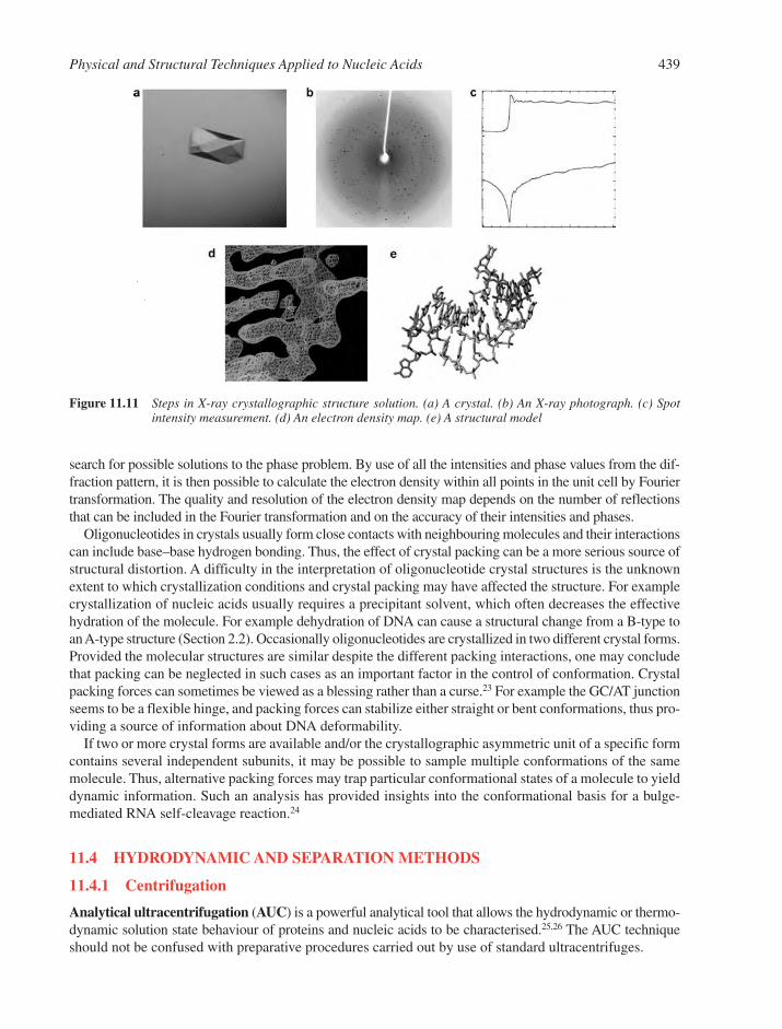
search for possible solutions to the phase problem. By use of all the intensities and phase values from the dif-fraction pattern, it is then possible to calculate the electron density within all points in the unit cell by Fouriertransformation. The quality and resolution of the electron density map depends on the number of reflectionsthat can be included in the Fourier transformation and on the accuracy of their intensities and phases.
Oligonucleotides in crystals usually form close contacts with neighbouring molecules and their interactionscan include base–base hydrogen bonding. Thus, the effect of crystal packing can be a more serious source ofstructural distortion. A difficulty in the interpretation of oligonucleotide crystal structures is the unknownextent to which crystallization conditions and crystal packing may have affected the structure. For examplecrystallization of nucleic acids usually requires a precipitant solvent, which often decreases the effectivehydration of the molecule. For example dehydration of DNA can cause a structural change from a B-type toan A-type structure (Section 2.2). Occasionally oligonucleotides are crystallized in two different crystal forms.Provided the molecular structures are similar despite the different packing interactions, one may concludethat packing can be neglected in such cases as an important factor in the control of conformation. Crystalpacking forces can sometimes be viewed as a blessing rather than a curse.23 For example the GC/AT junctionseems to be a flexible hinge, and packing forces can stabilize either straight or bent conformations, thus pro-viding a source of information about DNA deformability.
If two or more crystal forms are available and/or the crystallographic asymmetric unit of a specific formcontains several independent subunits, it may be possible to sample multiple conformations of the samemolecule. Thus, alternative packing forces may trap particular conformational states of a molecule to yielddynamic information. Such an analysis has provided insights into the conformational basis for a bulge-mediated RNA self-cleavage reaction.24
11.4 HYDRODYNAMIC AND SEPARATION METHODS
11.4.1 Centrifugation
Analytical ultracentrifugation (AUC) is a powerful analytical tool that allows the hydrodynamic or thermo-dynamic solution state behaviour of proteins and nucleic acids to be characterised.25,26 The AUC techniqueshould not be confused with preparative procedures carried out by use of standard ultracentrifuges.
Physical and Structural Techniques Applied to Nucleic Acids 439
Figure 11.11 Steps in X-ray crystallographic structure solution. (a) A crystal. (b) An X-ray photograph. (c) Spotintensity measurement. (d) An electron density map. (e) A structural model
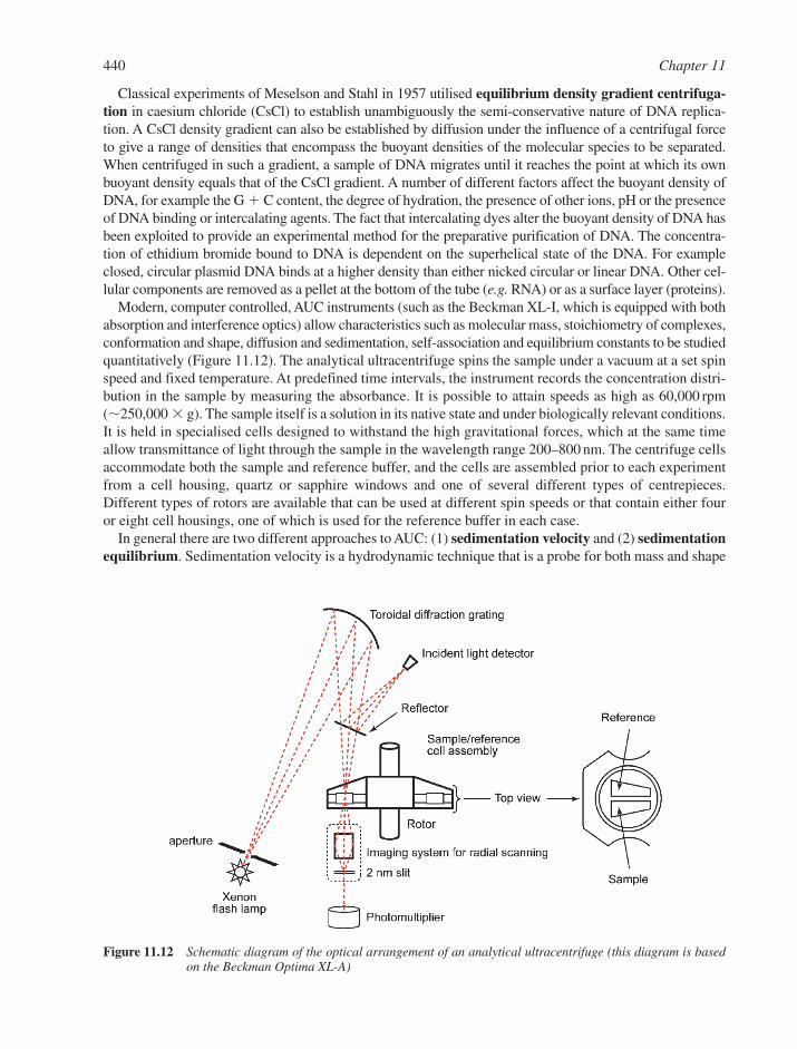
Classical experiments of Meselson and Stahl in 1957 utilised equilibrium density gradient centrifuga-tion in caesium chloride (CsCl) to establish unambiguously the semi-conservative nature of DNA replica-tion. A CsCl density gradient can also be established by diffusion under the influence of a centrifugal forceto give a range of densities that encompass the buoyant densities of the molecular species to be separated.When centrifuged in such a gradient, a sample of DNA migrates until it reaches the point at which its ownbuoyant density equals that of the CsCl gradient. A number of different factors affect the buoyant density ofDNA, for example the G � C content, the degree of hydration, the presence of other ions, pH or the presenceof DNA binding or intercalating agents. The fact that intercalating dyes alter the buoyant density of DNA hasbeen exploited to provide an experimental method for the preparative purification of DNA. The concentra-tion of ethidium bromide bound to DNA is dependent on the superhelical state of the DNA. For exampleclosed, circular plasmid DNA binds at a higher density than either nicked circular or linear DNA. Other cel-lular components are removed as a pellet at the bottom of the tube (e.g. RNA) or as a surface layer (proteins).
Modern, computer controlled, AUC instruments (such as the Beckman XL-I, which is equipped with bothabsorption and interference optics) allow characteristics such as molecular mass, stoichiometry of complexes,conformation and shape, diffusion and sedimentation, self-association and equilibrium constants to be studiedquantitatively (Figure 11.12). The analytical ultracentrifuge spins the sample under a vacuum at a set spinspeed and fixed temperature. At predefined time intervals, the instrument records the concentration distri-bution in the sample by measuring the absorbance. It is possible to attain speeds as high as 60,000 rpm(�250,000 g). The sample itself is a solution in its native state and under biologically relevant conditions.It is held in specialised cells designed to withstand the high gravitational forces, which at the same timeallow transmittance of light through the sample in the wavelength range 200–800 nm. The centrifuge cellsaccommodate both the sample and reference buffer, and the cells are assembled prior to each experimentfrom a cell housing, quartz or sapphire windows and one of several different types of centrepieces.Different types of rotors are available that can be used at different spin speeds or that contain either fouror eight cell housings, one of which is used for the reference buffer in each case.
In general there are two different approaches to AUC: (1) sedimentation velocity and (2) sedimentationequilibrium. Sedimentation velocity is a hydrodynamic technique that is a probe for both mass and shape
440 Chapter 11
Figure 11.12 Schematic diagram of the optical arrangement of an analytical ultracentrifuge (this diagram is basedon the Beckman Optima XL-A)
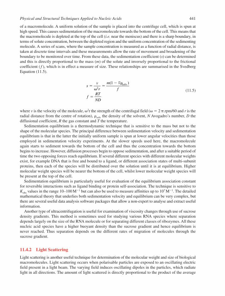
of a macromolecule. A uniform solution of the sample is placed into the centrifuge cell, which is spun athigh speed. This causes sedimentation of the macromolecule towards the bottom of the cell. This means thatthe macromolecule is depleted at the top of the cell (i.e. near the meniscus) and there is a sharp boundary, interms of solute concentration, between the depleted region and the uniform concentration of the sedimentingmolecule. A series of scans, where the sample concentration is measured as a function of radial distance, istaken at discrete time intervals and these measurements allow the rate of movement and broadening of theboundary to be monitored over time. From these data, the sedimentation coefficient (s) can be determinedand this is directly proportional to the mass (m) of the solute and inversely proportional to the frictionalcoefficient ( f ), which is in effect a measure of size. These relationships are summarised in the SvedbergEquation (11.5).
(11.5)
where v is the velocity of the molecule, �2r the strength of the centrifugal field (� � 2 �.rpm/60 and r is theradial distance from the centre of rotation), �solv the density of the solvent, N Avogadro’s number, D thediffusional coefficient, R the gas constant and T the temperature.
Sedimentation equilibrium is a thermodynamic technique that is sensitive to the mass but not to theshape of the molecular species. The principal difference between sedimentation velocity and sedimentationequilibrium is that in the latter the initially uniform sample is spun at lower angular velocities than thoseemployed in sedimentation velocity experiments. At the slower speeds used here, the macromoleculeagain starts to sediment towards the bottom of the cell and thus the concentration towards the bottombegins to increase. However, diffusion processes begin to oppose sedimentation, and after a suitable period oftime the two opposing forces reach equilibrium. If several different species with different molecular weightsexist, for example DNA that is free and bound to a ligand, or different association states of multi-subunitproteins, then each of the species will be distributed over the solution until it is at equilibrium. Highermolecular weight species will be nearer the bottom of the cell, whilst lower molecular weight species willbe present at the top of the cell.
Sedimentation equilibrium is particularly useful for evaluation of the equilibrium association constantfor reversible interactions such as ligand binding or protein self-association. The technique is sensitive toKobs values in the range 10–100 M�1 but can also be used to measure affinities up to 107 M�1. The detailedmathematical theory that underlies both sedimentation velocity and equilibrium can be very complex, butthere are several useful data analysis software packages that allow a non-expert to analyse and extract usefulinformation.
Another type of ultracentrifugation is useful for examination of viscosity changes through use of sucrosedensity gradients. This method is sometimes used for studying various RNA species where separationdepends largely on the size of the RNA molecule or for separating different classes of ribozymes. All thesenucleic acid species have a higher buoyant density than the sucrose gradient and hence equilibrium isnever reached. Thus separation depends on the different rates of migration of molecules through thesucrose gradient.
11.4.2 Light Scattering
Light scattering is another useful technique for determination of the molecular weight and size of biologicalmacromolecules. Light scattering occurs when polarisable particles are exposed to an oscillating electricfield present in a light beam. The varying field induces oscillating dipoles in the particles, which radiatelight in all directions. The amount of light scattered is directly proportional to the product of the average
sv
r
m v
f
fRT
ND
� ��
�
�
�2
1( )solv
Physical and Structural Techniques Applied to Nucleic Acids 441
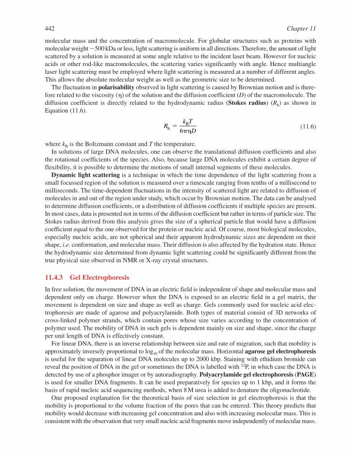
molecular mass and the concentration of macromolecule. For globular structures such as proteins withmolecular weight �500 kDa or less, light scattering is uniform in all directions. Therefore, the amount of lightscattered by a solution is measured at some angle relative to the incident laser beam. However for nucleicacids or other rod-like macromolecules, the scattering varies significantly with angle. Hence multianglelaser light scattering must be employed where light scattering is measured at a number of different angles.This allows the absolute molecular weight as well as the geometric size to be determined.
The fluctuation in polarisability observed in light scattering is caused by Brownian motion and is there-fore related to the viscosity (�) of the solution and the diffusion coefficient (D) of the macromolecule. Thediffusion coefficient is directly related to the hydrodynamic radius (Stokes radius) (Rh) as shown inEquation (11.6).
(11.6)
where kB is the Boltzmann constant and T the temperature.In solutions of large DNA molecules, one can observe the translational diffusion coefficients and also
the rotational coefficients of the species. Also, because large DNA molecules exhibit a certain degree offlexibility, it is possible to determine the motions of small internal segments of these molecules.
Dynamic light scattering is a technique in which the time dependence of the light scattering from asmall focussed region of the solution is measured over a timescale ranging from tenths of a millisecond tomilliseconds. The time-dependent fluctuations in the intensity of scattered light are related to diffusion ofmolecules in and out of the region under study, which occur by Brownian motion. The data can be analysedto determine diffusion coefficients, or a distribution of diffusion coefficients if multiple species are present.In most cases, data is presented not in terms of the diffusion coefficient but rather in terms of particle size. TheStokes radius derived from this analysis gives the size of a spherical particle that would have a diffusioncoefficient equal to the one observed for the protein or nucleic acid. Of course, most biological molecules,especially nucleic acids, are not spherical and their apparent hydrodynamic sizes are dependent on theirshape, i.e. conformation, and molecular mass. Their diffusion is also affected by the hydration state. Hencethe hydrodynamic size determined from dynamic light scattering could be significantly different from thetrue physical size observed in NMR or X-ray crystal structures.
11.4.3 Gel Electrophoresis
In free solution, the movement of DNA in an electric field is independent of shape and molecular mass anddependent only on charge. However when the DNA is exposed to an electric field in a gel matrix, themovement is dependent on size and shape as well as charge. Gels commonly used for nucleic acid elec-trophoresis are made of agarose and polyacrylamide. Both types of material consist of 3D networks ofcross-linked polymer strands, which contain pores whose size varies according to the concentration ofpolymer used. The mobility of DNA in such gels is dependent mainly on size and shape, since the chargeper unit length of DNA is effectively constant.
For linear DNA, there is an inverse relationship between size and rate of migration, such that mobility isapproximately inversely proportional to log10 of the molecular mass. Horizontal agarose gel electrophoresisis useful for the separation of linear DNA molecules up to 2000 kbp. Staining with ethidium bromide canreveal the position of DNA in the gel or sometimes the DNA is labelled with 32P, in which case the DNA isdetected by use of a phosphor imager or by autoradiography. Polyacrylamide gel electrophoresis (PAGE)is used for smaller DNA fragments. It can be used preparatively for species up to 1 kbp, and it forms thebasis of rapid nucleic acid sequencing methods, when 8 M urea is added to denature the oligonucleotide.
One proposed explanation for the theoretical basis of size selection in gel electrophoresis is that themobility is proportional to the volume fraction of the pores that can be entered. This theory predicts thatmobility would decrease with increasing gel concentration and also with increasing molecular mass. This isconsistent with the observation that very small nucleic acid fragments move independently of molecular mass.
Rk T
DhB�
6��
442 Chapter 11
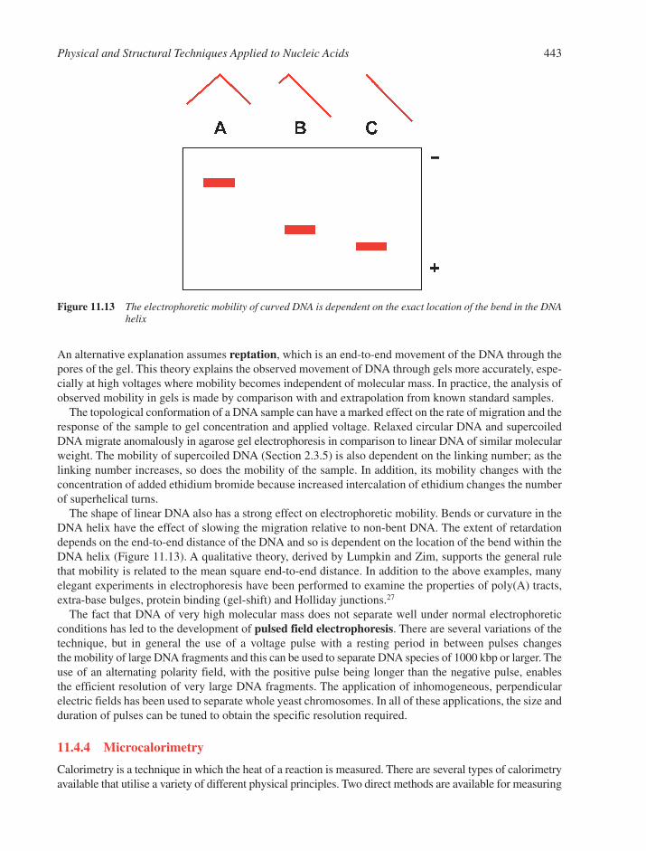
An alternative explanation assumes reptation, which is an end-to-end movement of the DNA through thepores of the gel. This theory explains the observed movement of DNA through gels more accurately, espe-cially at high voltages where mobility becomes independent of molecular mass. In practice, the analysis ofobserved mobility in gels is made by comparison with and extrapolation from known standard samples.
The topological conformation of a DNA sample can have a marked effect on the rate of migration and theresponse of the sample to gel concentration and applied voltage. Relaxed circular DNA and supercoiledDNA migrate anomalously in agarose gel electrophoresis in comparison to linear DNA of similar molecularweight. The mobility of supercoiled DNA (Section 2.3.5) is also dependent on the linking number; as thelinking number increases, so does the mobility of the sample. In addition, its mobility changes with theconcentration of added ethidium bromide because increased intercalation of ethidium changes the numberof superhelical turns.
The shape of linear DNA also has a strong effect on electrophoretic mobility. Bends or curvature in theDNA helix have the effect of slowing the migration relative to non-bent DNA. The extent of retardationdepends on the end-to-end distance of the DNA and so is dependent on the location of the bend within theDNA helix (Figure 11.13). A qualitative theory, derived by Lumpkin and Zim, supports the general rulethat mobility is related to the mean square end-to-end distance. In addition to the above examples, manyelegant experiments in electrophoresis have been performed to examine the properties of poly(A) tracts,extra-base bulges, protein binding (gel-shift) and Holliday junctions.27
The fact that DNA of very high molecular mass does not separate well under normal electrophoreticconditions has led to the development of pulsed field electrophoresis. There are several variations of thetechnique, but in general the use of a voltage pulse with a resting period in between pulses changes the mobility of large DNA fragments and this can be used to separate DNA species of 1000 kbp or larger. Theuse of an alternating polarity field, with the positive pulse being longer than the negative pulse, enables the efficient resolution of very large DNA fragments. The application of inhomogeneous, perpendicularelectric fields has been used to separate whole yeast chromosomes. In all of these applications, the size andduration of pulses can be tuned to obtain the specific resolution required.
11.4.4 Microcalorimetry
Calorimetry is a technique in which the heat of a reaction is measured. There are several types of calorimetryavailable that utilise a variety of different physical principles. Two direct methods are available for measuring
Physical and Structural Techniques Applied to Nucleic Acids 443
Figure 11.13 The electrophoretic mobility of curved DNA is dependent on the exact location of the bend in the DNAhelix
the enthalpy change associated with either a binding interaction or a heat-induced conformational transitionfor nucleic acids and proteins. These are isothermal titration calorimetry (ITC) and differential scanningcalorimetry (DSC).28–33 Many ultra-high sensitivity microcalorimeters can now be obtained.
11.4.4.1 Isothermal Titration Calorimetry. Isothermal titration calorimetry is a powerful and flex-ible technique that is used to determine directly and independently the change in enthalpy (�H) for almostany bimolecular binding interaction at a fixed and constant temperature. Depending on the magnitude of the binding constant and solubility of reactants, ITC can also be used to obtain binding isotherms, fromwhich the equilibrium-binding constant (Kb � Ka) and binding stoichiometry (n) can be determined. Thiscan therefore yield an almost complete thermodynamic profile for a binding interaction in a single experiment.
The calorimeter assembly consists typically of two identical cells (one reference and one sample) con-structed of a metal alloy that has appropriate thermal properties, which is held within an adiabatic jacket.Detection of heat effects within the cells is achieved by placement of semiconductor thermopiles inbetween the cells and the jacket. Following injection of a ligand into the sample cell, the amount of thermalpower that must be applied to actively balance the heat of reaction is measured. Thus the calorimeter moni-tors the heat effects in the sample cell and then applies appropriate power to maintain a zero temperaturedifference between sample and reference cells. The amount of heat applied to the cells is monitored con-tinuously (cell feedback or CFB), and the amount of power applied is adjusted so as to drive the measuredthermal power amplitude towards a stable baseline value. This is achieved by interfacing a computer to thesystem that continually monitors the output of a nanovoltmeter connected to record the output of the thermo-piles. The experimentally measured quantity is the applied thermal power as a function of time required toreturn CFB to the stable baseline value subsequent to an injection of ligand (or system component), and isdirectly proportional to the reaction enthalpy. Modern ultrasensitive ITC instruments, such as theMicroCal VP-ITC, have baseline stability in the range �5 ncal s�1 and heat signals in the nanocalorie rangecan be detected.
In example data from an ITC experiment (Figure 11.14), Panel A shows primary data, where CFB ismeasured in �cal sec�1 as a function of time. At equilibrium, CFB is held at a predefined value. Each peakrepresents the injection of a defined volume of ligand into a continually stirred sample cell. Addition ofligand leads to a binding interaction and concomitant heat effects; in this case an exothermic heat is observed.Each peak is integrated, with respect to time, to yield the binding isotherm in Panel B. After correction ofbackground heats, due to dilution effects, and correction to a per mole basis the enthalpy is obtaineddirectly and Kobs and the stoichiometry (n) are obtained by fitting to an appropriate binding model, suchthat n is the midpoint of the titration curve and Kobs comes from the lineshape of the binding curve.
11.4.4.2 Differential Scanning Calorimetry. Differential scanning calorimetry involves the con-tinuous measurement of the apparent specific heat of a system as a function of temperature. Hence, DSCcan be used to examine physicochemical processes initiated by increases or decreases in temperature, forexample phase transitions or conformational changes.
In general terms, a DSC instrument contains two cells, which are suspended in an adiabatic shield andconnected via a multifunctional thermopile. In a typical experiment, the reference cell is filled with bufferand the sample cell is filled with buffer containing macromolecule (cell volume 0.5–1.5 mL). The temperatureis increased to the 0.1–110°C range by use of electrical heaters that are in good thermal contact with thecells. During a heat-induced endothermic transition, the temperature of the sample cell falls behind that ofthe reference cell, since some of the energy required is used to induce the transition rather than to heat thesolution. This lag is detected by the thermopile, so that extra electrical power is supplied to the sample cellto maintain it at a temperature identical to that of the reference cell. This additional energy is proportionalto the energy associated with the thermally induced transition. Knowledge of the solute concentrationenables a transformation of the observed electrical energy versus temperature profile into a curve that cor-responds to an excess heat capacity versus temperature plot.
444 Chapter 11
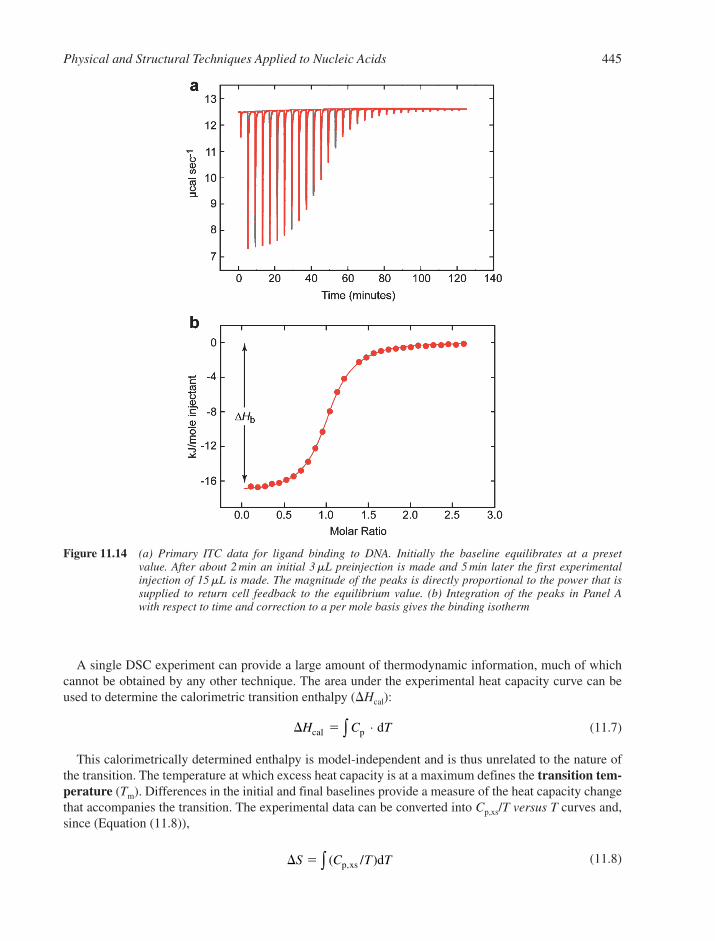
A single DSC experiment can provide a large amount of thermodynamic information, much of whichcannot be obtained by any other technique. The area under the experimental heat capacity curve can beused to determine the calorimetric transition enthalpy (�Hcal):
(11.7)
This calorimetrically determined enthalpy is model-independent and is thus unrelated to the nature ofthe transition. The temperature at which excess heat capacity is at a maximum defines the transition tem-perature (Tm). Differences in the initial and final baselines provide a measure of the heat capacity changethat accompanies the transition. The experimental data can be converted into Cp,xs/T versus T curves and,since (Equation (11.8)),
(11.8)� �S C T T( / ),p xs d∫
� �H C Tcal p d⋅∫
Physical and Structural Techniques Applied to Nucleic Acids 445
Figure 11.14 (a) Primary ITC data for ligand binding to DNA. Initially the baseline equilibrates at a preset value. After about 2 min an initial 3mL preinjection is made and 5 min later the first experimentalinjection of 15mL is made. The magnitude of the peaks is directly proportional to the power that issupplied to return cell feedback to the equilibrium value. (b) Integration of the peaks in Panel A with respect to time and correction to a per mole basis gives the binding isotherm
the area under such a curve represents the entropy change for the transition. From this information thevalue of �G can be evaluated at any temperature.
Differential scanning calorimetry data can also provide important information concerning the co-operativityof a thermal transition. This can be achieved by comparison of the magnitudes of the model-dependentvan’t Hoff enthalpy (obtained by shape analysis of the calorimetric data) and the calorimetric enthalpy. If�HvH � �Hcal and there are no chemical or instrumental kinetic limitations, such that Tm is independent ofboth scan rate and concentration, then the transition proceeds in a two-state manner and meaningful thermo-dynamic data can be obtained by examination of the temperature dependence of an equilibrium property. If�HvH � �Hcal, then the transition must involve a significant number of intermediate states. Conversely, if�HvH � �Hcal, then aggregation is implicated. The �HvH/�Hcal ratio provides quantitative insight into thenature of the transition. Specifically, it provides a measure of the fraction of the structure that melts as asingle thermodynamic entity (i.e. it defines the size of the co-operative unit). This is a unique advantage ofDSC in the study of biological molecules and when combined with ITC, thermodynamic studies over awide temperature range and under a variety of solution conditions can be carried out. These techniquesoffer a powerful tool to probe the energetics of macromolecule stability and ligand association reactions.
11.5 MICROSCOPY
11.5.1 Electron Microscopy
Electron microscopy (EM) is an invaluable tool for gaining structural information on systems that are toolarge for the atomic resolution methods of X-ray diffraction or NMR, and yet too small for optical micro-scopy.34,35 Electron microscopy uses beams of electrons with wavelengths between 0.001 and 0.01 nm. Theresolving power of EM can be below 2 nm but is largely restricted by factors such as radiation damage andthe process of sample preparation.
The lenses in an electron microscope consist of shaped electric or magnetic fields. Electrons from a heatedtungsten filament are accelerated across a voltage difference of up to 100 kV and the sample under study isexamined in a vacuum. In transmission EM the electron beam is passed through the sample, then through asuitable lens system and is detected on a fluorescent plate or photographic film. In scanning EM, the electronbeam is focused down to a point and scanned across the specimen. Secondary electrons are produced whenelectrons interact with the target specimen and these secondary electrons are detected and used to build upa raster image of the sample.
Both transmission and scanning EM require samples to be dehydrated and often coated by depositionof a film of either carbon or a metal such as tungsten. For nucleic acid analysis, one of the oldest means ofsample preparation involves the combination of the nucleic acid with a thin film of denatured protein floatingon water. The nucleic acid and protein film can be lifted off the surface onto a grid and then, after drying,coated with a fine layer of palladium or uranium. The process of coating, or shadow casting, involves deposit-ing the metal from a heated filament that is oriented at an acute angle to the supporting grid. The effect ofthe metal deposition is to highlight regions that are raised above the grid. In order to achieve good contrast,either the samples can be shadowed from several angles or can be rotated during the coating procedure.The protein film has two benefits: one is that it supports and protects the DNA during preparation; the otheris it causes thickening of the DNA strands. Nucleic acids will also bind directly to carbon supports that havebeen physically and chemically pre-treated in a suitable fashion. They can then be dried and shadowed inthe same manner as in film deposition.
The technique of cryogenic EM (cryo EM)36 uses samples that have been flash frozen into a thin film ofvitreous water. The sample is prepared as an aqueous solution and applied to a carbon grid with holes ofabout 3 �m. Blotting of the grid followed by quick freezing in ethane at near its freezing temperature producesvitrified samples which can be directly imaged by EM.
While the contrast in cryo EM is relatively poor, it has the great advantage that a biological specimen ispreserved in its solution conformation without many of the possible artefacts that can result from the
446 Chapter 11

dehydration and coating processes. Several images can then be recorded on the same sample, which istilted between recordings, and used to reconstruct the full 3D shape of the sample (with resolution ca.10 Å). Although the ultimate resolution of cryo EM is limited, crucial information about the interaction ofnucleic acids with other macromolecules can be gained by the marriage of this tool with other structuralmethods. Cryo EM has also been used recently to investigate more complex nucleic acid incorporatedprocesses, including those involved in transcription.
11.5.2 Scanning Probe Microscopy
The field of scanning probe microscopy (SPM) refers to a class of techniques, devised originally duringthe 1980s, which provide images of samples through use of a tiny probe (tip) to ‘feel’ the outermost prop-erties of a flat surface. The members include the scanning tunnelling microscope (STM), the atomicforce microscope (AFM), the scanning near-field optical microscope and the scanning capacitance micro-scope, but it is the first two of these which have contributed most to the structural analysis of nucleic acids.
11.5.2.1 Scanning Tunnelling Microscopy. Scanning tunnelling microscopy was developed in ca.1982. This instrument exploits the ‘electron tunnelling’ effect: electrons are able to pass through a potentialbarrier when the distance of travel is small. The STM uses an atomically sharp conducting probe that scansclosely (typically angstrom) across the sample immobilized on a conducting substrate. The movement ofthe probe or sample in x, y and z directions is realised by a piezoelectric crystal, most often in the form of atube scanner. The scanner is capable of sub-angstrom resolution, with the z-axis conventionally perpendicularto the sample in all scanning probe miscroscopes.
By monitoring the tunnelling current as the probe is scanned over the sample, any spatial variation in theelectronic topography of the surface is recorded. Due to the exponential dependence of the tunnelling current onthe probe-sample separation, the recorded image contrast invariably arises from variations in the surface topog-raphy of the sample. Any variations in the electronic nature of the substrate under study, however, also con-tribute to the recorded signal. Although STM is capable of achieving atomic resolution, in analysis of biologicalmolecules, image resolution is at best several nanometres.37 Thus, whilst STM has been used for the imagingof some nucleic acid samples, the interpretation of the images has always been somewhat open to question.
Various sample preparation approaches have been used for STM analysis. The most straightforwardinvolves deposition from a dilute solution, with the subsequent drying of solvent. The requisite conductiveproperties of the sample are normally acquired through deposition on conducting substrates (atomically flatgold and highly oriented pyrolytic graphite surfaces are frequently used), coating molecules deposited onmica with thin films of platinum–iridium–carbon or through variation in the relative humidity of the imagingenvironment. However, in reality the small number of suitable conducting surfaces, and the restrictionsimposed by the working environment (in air or vacuum) have limited the analysis of nucleic acids by STM.
The limitations of STM subsequently led to the development of the AFM or scanning force microscope(SFM) (ca. 1986). The AFM utilises a similar instrumental setup to the STM, but replaces the conductingmetal probe with a sharp probe (typically silicon or silicon nitride), which is situated at the apex of a can-tilever spring of known stiffness (spring constant). As the probe is scanned over the surface of the sample,the cantilever bends or twists in response to forces acting between the probe and the sample. By reflectionof a laser beam off the back surface of the cantilever onto a position sensitive photodiode, variation in thebending or twisting of the cantilever can be monitored to produce an image (Figure 11.15).
11.5.2.2 Atomic Force Microscopy. The AFM can be used in three different imaging formats,namely contact, tapping and non-contact mode, describing the way in which the probe interacts with thesurface during imaging. For the imaging of biological samples, tapping mode is more commonly used. In thismode the probe intermittently comes into and out of contact with (i.e. lightly taps) the sample as is it scannedover the surface, avoiding the strong lateral forces associated with contact mode, which can denature or sweepaway soft or poorly immobilized samples.
Physical and Structural Techniques Applied to Nucleic Acids 447

One of the big advantages of AFM for the structural analysis of biological samples is that it can imagenon-conducting surfaces in both air and liquid environments. Unsurprisingly, soon after its invention theAFM was applied to the analysis of many biological systems, including membrane proteins, DNA andRNA, protein–nucleic acid complexes and even more complex structures such as chromosomes.
To date, many investigations of nucleic acids have been performed using AFM.38,39 By recording a seriesof images over a certain period of time, AFM is also able to provide dynamic information on processesinvolving nucleic acids, as well as time-lapse AFM movies of processes such as DNA condensation and theprocessing and/or degradation of DNA by enzymes such as the endonuclease EcoKI. The level of attainableimage speed in conventional AFM imaging (normally 30–60 s per image) has been a major problem, however,for the imaging of such dynamic events. To a certain extent, this problem has been overcome recently by using
448 Chapter 11
Figure 11.15 (a) Schematic of the principle features of an AFM instrument. The sizes of the AFM probe andcantilever chip are exaggerated for the purposes of illustration. (b) A representative 2mm 2mm AFMimage (obtained in air) displaying several pBR322 DNA plasmids deposited onto a mica substrate
purpose-developed small cantilevers, which are able to image faster (�2 s per image). Smaller cantilevershave high-resonant frequencies, which allow for high-scanning frequencies, but also low-spring constants,which enable any potential sample damage to be further minimised.
There are two main factors that can influence the level of resolution attainable in AFM images of nucleicacids. One is the sample preparation method employed for the immobilization of DNA to the underlyingsurface, and the other is the sharpness of the AFM probe. To this end, two immobilization methods haveproved to be particularly useful for the analysis of DNA in aqueous environments. One method utilizes anunmodified mica surface with a buffer containing divalent metal ions (such as Ni2�, Co2�, Zn2�, Mn2� orMg2�) or mica presoaked/pretreated with solutions of such ions (in this case a more concentrated divalentmetal ion solution is needed). The other approach uses mica or alternative flat substrates (e.g. silicon) ontowhich positive charges have been introduced, for example through aminosilanation. This latter type of sur-face, however, tends to be more problematic and is more suitable for experiments in which divalent metalions cannot be used without perturbing the DNA structure and/or the biomolecular functions under investigation.
One of the notable drawbacks for AFM is the probe convolution/broadening effect, which manifests asa size difference between the observed dimensions of the image feature and the actual molecular dimensions(normally the former is bigger). This effect arises when the radius of curvature of the imaging probe (typically10–20 nm) is greater than the size of the feature to be imaged. Consequently, the apparent width of thedouble-stranded DNA helix in AFM images is typically 12–15 nm, rather than the 2 nm predicted from thecrystal structure. Recent attempts to improve the sharpness of the probe (and thus the image resolution)have utilised single-walled carbon nanotubes attached to the apexes of AFM probes. Such probes havedecreased the apparent DNA width to around 6–8 nm. Commercially available sharpened silicon oxide AFMprobes (radius of curvature ca. 2 nm) are also available and are helpful in improving the level of attainableimage resolution.
In addition to its imaging function, AFM can also be used to measure forces acting between the probe andsurface. An approach termed single molecule force spectroscopy has been developed consequently to inves-tigate the mechanical properties of single biopolymeric molecules, including DNA.40 Here for example theopposite ends of a linear double-stranded DNA molecule are tethered between an opposing AFM probe andsubstrate, and then the molecule extended/stretched as the probe-substrate separation is increased. Pullingon single linear DNA molecules (with random sequence) produces force-extension curves (Figure 11.16).The plateau region at around 65 pN corresponds to the overstretching transition of DNA in which DNA isstretched from its B-DNA state to an overstretched state (around 1.7 times its B-DNA contour length). Uponfurther extension, a second transition occurs, during which the DNA can be melted into two single strands.Researchers have investigated the influence of different base sequences on this ‘mechanical fingerprint’,and the approach applied to the investigation of more complex molecular architectures including DNA/RNAhairpins. Recently, the effects of DNA-binding drugs on the mechanical properties of DNA have also beeninvestigated (Figure 11.16). The observed changes were found to be dependent on the concentration of theapplied drug and its mode of binding.
Although AFM is still a relatively new approach, it overcomes many of the limitations of its STM pre-decessor. While some improvements are still required before it will be utilised as a routine tool for the inves-tigation of nucleic acid structure, its advantages over other microscopy approaches have meant that it isquickly becoming more accepted. The combination of AFM with other rapidly developing experimentalapproaches, such as single molecule fluorescence, also holds considerable promise for the future towardsdeepening our understanding of the basic processes in which nucleic acids are involved.
11.6 MASS SPECTROMETRY
The determination of intrinsic molecular masses of nucleic acids using mass spectrometry is widely acceptedas one of the most accurate methods to detect nucleic acids.41 The key technologies are the second-generation‘soft ionization’ methods: matrix-assisted laser desorption/ionization time-of-flight (MALDI-TOF) and
Physical and Structural Techniques Applied to Nucleic Acids 449
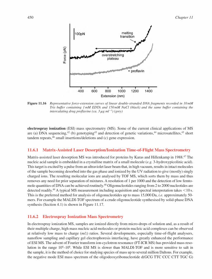
electrospray ionization (ESI) mass spectrometry (MS). Some of the current clinical applications of MSare (a) DNA sequencing,42 (b) genotyping43 and detection of genetic variations,44 microsatellites,45 shorttandem repeats,46 small insertions/deletions and (c) gene expression.
11.6.1 Matrix-Assisted Laser Desorption/Ionization Time-of-Flight Mass Spectrometry
Matrix-assisted laser desorption MS was introduced for proteins by Karas and Hillenkamp in 1988.47 Thenucleic acid sample is embedded in a crystalline matrix of a small molecule (e.g. 3-hydroxypicolinic acid).This target is excited by a pulse from an ultraviolet laser beam that, in high vacuum, results in intact moleculesof the sample becoming desorbed into the gas phase and ionized by the UV radiation to give (mostly) singlycharged ions. The resulting molecular ions are analysed by TOF MS, which sorts them by mass and thusremoves any need for prior separation of mixtures. A resolution of 1 per 1000 and the detection of low femto-mole quantities of DNA can be achieved routinely.48 Oligonucleotides ranging from 2 to 2000 nucleotides aredetected readily.49 A typical MS measurement including acquisition and spectral interpretation takes �10 s.This is the preferred method for analysis of oligonucleotides up to mass 15,000 Da, i.e. approximately 50-mers. For example the MALDI-TOF spectrum of a crude oligonucleotide synthesised by solid-phase DNAsynthesis (Section 4.1) is shown in Figure 11.17.
11.6.2 Electrospray Ionization Mass Spectrometry
In electrospray ionization MS, samples are ionized directly from micro-drops of solution and, as a result oftheir multiple charge, high mass nucleic acid molecules or protein-nucleic acid complexes can be observedat relatively low mass to charge (m/z) ratios. Several developments, especially time-of-flight analysers,nanoflow sampling and capillary gel electrophoresis interfacing, have greatly enhanced the performanceof ESI MS. The advent of Fourier transform ion-cyclotron resonance (FT-ICR MS) has provided mass reso-lution in the range 104–106. While ESI MS is slower than MALDI-TOF and is more sensitive to salt in the sample, it is the method of choice for studying species of mass up to several million Daltons. For example,the negative mode ESI mass spectrum of the oligodeoxyribonucleotide d(GCG TTC CCC CTT TGC G)
450 Chapter 11
Figure 11.16 Representative force-extension curves of linear double-stranded DNA fragments recorded in 10 mMTris buffer containing 1 mM EDTA and 150 mM NaCl (black) and the same buffer containing the intercalating drug proflavine (ca. 3mg ml�1) (grey)

Physical and Structural Techniques Applied to Nucleic Acids 451
Figure 11.17 Matrix-assisted laser desorption/ionization time-of-flightmass spectrum of the crude products of achemical synthesis of 19-mer oligodeoxynucleotide d(GGATTACAGGTATGAGCCA) showing a majorcomponent at 5879.8 (calculated 5876.8). This routine spectrum was obtained on an AppliedBiosystems Voyager DE workstation (linear, negative ion mode) with a standard 3-hydroxypicolinicacid/ammonium acetate matrix. No internal mass calibration was used in this case, which would berequired for more accurate mass measurement
(Figure 11.18) appears as multiply charged peaks. The observed masses correspond to the oligonucleotide inwhich some of the phosphates are protonated. In some cases the replacement of a proton by sodium or potas-sium ions can be observed. The actual mass can be calculated from the accurate mass of any of the multiplycharged species. For example [M-4H]4 � (1198) has an accurate mass of 1198.4396, and since the observedmasses correspond to m/z, the moleculer mass of the oligonucleotide is 4 1198.4396 � 4793.7584 �4H(4 1.008) � 4797.7904. The theoretically calculated mass is 4797.8091.
Electrospray ionization has emerged as a tool for studying non-covalent complexes and has enabled themeasurement of spectra of intact viruses and whole ribosomes (Figure 11.19).50,51 It is possible to measuredirectly the mass of a macromolecular complex as long as the binding interactions that support complexformation are maintained during its transit from solution micro-droplets into the gas phase. Nanoflow ESI52
is a miniaturized version of ESI that facilitates this desolvation process because of the reduced size of dropletsit generates compared with conventional ESI.
11.7 MOLECULAR MODELLING AND DYNAMICS
Despite the remarkable developments in experimental techniques to determine the molecular structure ofnucleic acids, there remain many instances in which such information is not available. Certain nucleic acidstructures (e.g. the triple helix) have proved remarkably difficult to crystallize, while the homogenous andrepetitive nature of other structures makes NMR structure determination difficult or ambiguous. Molecularmodelling can provide a method to predict the structure of such nucleic acids and also to study transient

452 Chapter 11
Figure 11.18 Electrospray ionization mass spectrum (negative mode) of oligodeoxyribonucleotide d(GCGTTCCCC-CTTTGCG) dissolved in methanol containing 0.5% diethylamine. The determined molecular mass is4797.8091. The spectrum shows masses of the multiply charged species [M-3H]3� (1599), [M-3H-Na] 4� (1204.4), [M-4H]4� (1198.9), [M-5H]5� (958.8), [M-6H]6� (798.9) and [M-7H]7� (684.7)(Figure kindly provided by J. Brazier and R. Cosstick)
Figure 11.19 Mass spectra of E. coli ribosomes recorded from solution at pH 7.0 (a) and pH 4.5 (b). Peaks arelabelled with the protein bound, and the numbers of positive charges are given in parenthesis. At pH7.0, four proteins located in the mobile stalk region of the ribosome dominate the spectra. At pH 4.5,eight proteins are identified. Inset, MS/MS spectrum of the peak labelled *. Acceleration in thecollision cell yields a mass consistent with that calculated for the 5S RNA. Individual charge states ofthe L7/L12 proteins reveal their metal-binding properties and post-translational modifications(Reprinted from Ref. 50. © (2003), with permission from ASBMB)

or unstable structures. Molecular modelling also provides a method to relate the structural, dynamic andthermodynamic properties of nucleic acids to underlying theories of the physical processes that drivestructure, bonding and recognition.
11.7.1 Molecular Mechanics and Energy Minimisation
Crick and Watson’s model of the DNA duplex was constructed of wired-up physical shapes, but modernmodelling is carried out almost exclusively through computational techniques. In theory, the most accuratepredictions of molecular structure can come from quantum mechanical calculations, which treat the electronicstructure of molecules in detail and from first principles. But despite the continuing rapid increase in computerpower, the size of nucleic structures that are most often of interest precludes their day-to-day use. Instead,the most commonly used method is molecular mechanics.
Individual atoms are considered as spheres, and the bonds that connect them as springs. A set of equations,called a force field, determine how the energy of the system varies as bonds stretch or compress, bond anglesvary or torsions rotate. Other terms in the equations describe the energetic consequences of nonbonded(van der Waals and electrostatic) interactions between atoms. A computer can then calculate the energy of thesystem as a function of the atomic coordinates. This gives rise to the idea of an energy surface (Figure 11.20a)and the simplest application of molecular mechanics, which is energy minimisation.
The ‘true’ structure of the molecule corresponds to a situation in which there is no net force acting onany atom, i.e. to a point on this surface with zero slope. Starting from an arbitrary initial guess for this truestructure, the process of energy minimisation involves calculation of the forces and moving ‘downhill’ onthis surface until a minimum is reached. However, the figure illustrates the important point that for mostrather complex biomolecular structures, the energy surface has many minima. The real molecule isexpected to adopt the global minimum energy conformation, but energy minimisation may only lead to alocal minimum and thus predict an incorrect structure for the molecule. Because of the complexity of realenergy surfaces, the process of searching for the true global energy minimum conformation can be verydifficult and time-consuming, since there is no way of being absolutely sure that the global minimum hasbeen found until the entire energy surface is examined.
11.7.2 Molecular Dynamics
One of the most widely used methods to try and overcome the problem of obtaining the global energy min-imum of a structure is molecular dynamics. In this computer simulation method, masses and velocities,representative of a particular temperature, are assigned to each atom in some starting conformation. Using thesolution of Newton’s equations of motion, the conformation of the system at some later time-point is pre-dicted. The forces acting on each atom, and thus the velocities, are then recalculated at this new conformation,
Physical and Structural Techniques Applied to Nucleic Acids 453
Figure 11.20 Each configuration of a molecule has an associated energy. In energy minimisation (a), different initialguesses (•) for the true structure may be optimised to local (�) rather than the global (*) energyminimum. Using molecular dynamics (b) the global energy minimum may be more reliably identified

the motion over the next time step determined and so on. This process allows ‘uphill’ motion over the energysurface, and barriers between minima may be overcome, depending on their height relative to the simula-tion temperature (Figure 11.20b).
As well as a method for helping to locate the global energy minimum, molecular dynamics simulationscan be regarded as a better representation of the physical state of the system – as a dynamic molecule that, atnormal temperatures, does not have a single ‘frozen’ conformation. Rather the method samples a range ofconfigurations, not just oscillating about the global minimum, but perhaps also significantly occupying otherlocal energy minimum conformations. The flexibility of DNA can be considerable (Figure 11.21), and this isan important aspect of its function and recognition properties that often cannot be fully appreciated fromcrystallographic and (to a lesser extent) NMR data. The main limitation of molecular dynamics simulationsis the timescale that is accessible. In order for the calculations predicting the trajectory of the molecule to beaccurate, the time-step between which the forces acting on the system are recalculated must be very small –of the order of a femtosecond. Each calculation is computationally expensive and so current computerpower typically limits such simulations to the order of a few tens of nanoseconds at best, which may takemonths to run. On this timescale, only quite modest motions of DNA can be observed directly: oscillationsof small groups of atoms and localised bending and twisting motions. Many more biologically interestingconformational motions of DNA, for example winding and unwinding of the double helix and base-pairopening, only happen on the microsecond timescale or more slowly.
The basic output from a molecular dynamics simulation is a trajectory, i.e. the predicted behaviour of asingle molecule as a function of time. However, the individual ‘snapshots’ from a molecular dynamicssimulation can alternatively be regarded as a Boltzmann-weighted ensemble of structures from whichthermodynamic quantities may be calculated. Modelling methods based on this approach are particularlyvaluable in that they provide a link between the microscopic behaviour of individual molecules – easy tosimulate but difficult to observe experimentally – and the macroscopic properties of the system, which aremuch easier to measure, but can be difficult to interpret in terms of atomic-scale features. Moleculardynamics simulations of DNA have become increasingly sophisticated over the last few years53 and areapplied to a rapidly increasing volume of DNA structures and sequences.54
11.7.3 Mesoscopic Modelling
As indicated above, atomic-scale modelling, where each atom is represented by a sphere and each bond by aspring, does have some serious limitations. Current computer power limits the size of the molecules that maybe studied this way to the order of a few thousand atoms (e.g. up to perhaps 200 base pairs) and, if one isinterested in dynamics, to timescales of the order of a few tens of nanoseconds. To probe other sizes andtimescales, modellers must adopt mesoscopic modelling methods, in which, for example a length of DNAis treated as a uniform elastic rod, or as a set of disks (each representing a base or base pair) connected toits neighbours by springs. These approaches are particularly used at present to study DNA supercoilingand packaging.
454 Chapter 11
Figure 11.21 Snapshots taken from a short (120 ps) molecular dynamics simulation of a 126 base-pair DNAmicro-circle, illustrating its conformational flexibility
REFERENCES
1. V.A. Bloomfield, D. Crothers and I. Tinoco, Physical Chemistry of the Nucleic Acids. Harper and Row,New York, 1974.
2. L. Brand and M.L. Johnson (eds), Meth. Enzymol., 1997, 278, 1–628.3. D.M.J. Lilley, in Nucleic Acids and Molecular Biology, vol 4. D.M.J. Lilley and F. Eckstein (eds).
Springer, Berlin, 1990, 55–77.4. J.C. Penedo, T.J. Wilson, S.D. Jayasena, A. Khvorova and D.M.J. Lilley, Folding of the natural
hammerhead ribozyme is enhanced by interaction of auxiliary elements. RNA, 2004, 10, 880–888.5. M. Eriksson and B. Nordén, Linear and circular dichroism of drug-nucleic acid complexes. Meth.
Enzymol., 2001, 340, 68–99.6. B. Nordén and T. Kurucsev, Analysing DNA complexes by circular and linear dichroism. J. Mol.
Recog., 1994, 7, 141–156.7. E. Taillandier and J. Liquier, Infrared spectroscopy of DNA. Meth. Enzymol., 1992, 211, 307–335.8. G.J. Thomas and A.H.-J. Wang, in Nucleic Acids and Molecular Biology, vol 2. D.M.J. Lilley and
F. Eckstein (eds), Springer, Berlin, 1988, 1–30.9. H. Gunther, NMR Spectroscopy – Basic Principles, Concepts, and Applications in Chemistry. Wiley,
Chichester, 1995.10. T.L. James (ed), Meth. Enzymol., Academic Press, San Deigo, 1995, 261, 3–664.11. K. Wuthrich, NMR of Proteins and Nucleic Acids. Wiley, New York, 1986.12. L. Zidek, R. Stefl and V. Sklenar, NMR methodology for the study of nucleic acids. Curr. Opin. Struct.
Biol., 11, 275–281.13. E.T. Mollova and A. Pardi, NMR solution structure determination of RNAs. Curr. Opin. Struct. Biol.,
2000, 10, 298–302.14. D.E. Gilbert and J. Feigon, Multistrand DNA structures. Curr. Opin. Struct. Biol., 1999, 9, 305–314.15. I. Kim, P.J. Lukavsky and J.D. Puglisi, NMR study of 100 kDa HCV IRES RNA using segmental iso-
tope labeling. J. Am. Chem. Soc., 2002, 124, 9338–9339.16. H.M. Berman, W.K. Olson, D.L. Beveridge, J. Westbrook, A. Gelbin, T. Demeny, S.-H. Hsieh,
A.R. Srinivasan and B. Schneider, The nucleic acid database: a comprehensive relational database ofthree-dimensional structures of nucleic acids. Biophys. J., 1992, 63, 751–759.
17. H.M. Berman, J. Westbrook, Z. Feng, G. Gilliland, T.N. Bhat, H. Weissig, I.N. Shindyalov and B.P. E.,The protein data bank. Nucleic Acids Res., 2000, 28, 235–242.
18. W.A. Hendrickson, Synchrotron crystallography. Trends Biochem. Sci., 2000, 25, 637–643.19. C.W. Carter Jr. and R.M. Sweet (eds), Meth. Enzymol. Macromolecular Crystallography, Part A.
Academic Press, New York, 1997.20. C.W. Carter Jr. and R.M. Sweet (eds), Meth. Enzymol. Macromolecular Crystallography, Part B.
Academic Press, New York, 1997.21. C.W. Carter Jr. (ed), Meth. Enzymol. Macromolecular Crystallography, Part C. Academic Press, New
York, 2003.22. C.W. Carter Jr. (ed), Meth. Enzymol. Macromolecular Crystallography, Part D. Academic Press, New
York, 2003.23. R.E. Dickerson, D.S. Goodsell and S. Neidle, “ … The tyranny of the lattice …” Proc. Natl. Acad. Sci.
USA, 1994, 91, 3579–3583.24. V. Tereshko, S.T. Wallace, N. Usman, F. Wincott and M. Egli, X-ray crystallographic observation of
“in-line” and “adjacent” conformations in a bulged self-cleaving RNA/DNA hybrid. RNA, 2001, 7,405–420.
25. P. Hensley, Defining the structure and stability of macromolecular assemblies in solution: there-emergence of analytical ultracentrifugation as a practical tool. Structure, 1996, 4, 367–373.
26. S.E. Harding and B.Z. Chowdhry (eds), Protein Ligand Interactions: Hydrodynamics andCalorimetry. Oxford University Press, Oxford, 2001.
Physical and Structural Techniques Applied to Nucleic Acids 455
27. V.A. Bloomfield, D.M. Crothers and J.I. Tinoco (eds), Nucleic Acids Structures, Properties, andFunctions. University Science Books, Sausalito, CA, 2000.
28. I. Haq, C.B.Z., J.T.C. and C.J.B., Parsing the free energy of drug-DNA Interactions. Meth. Enzymol., 2000, 323, 373–405.
29. I. Haq, C.B.Z. and J.T.C., The interaction of drugs with higher order DNA structures. Meth. Enzymol.,2001, 340, 109–149.
30. I. Jelesarov and H.R. Bosshard, Isothermal titration calorimetry and differential scanning calorimetryas complementary tools to investigate the energetics of biomolecular recognition. J. Mol. Recog.,1999, 12, 3–18.
31. A. Cooper, Thermodynamic analysis of biomolecular interactions. Curr. Opin. Chem. Biol., 1999, 3,557–563.
32. T. Wiseman, S. Williston, J.F. Brandts and L.-N. Lin, Rapid measurement of binding constants andheats of binding using a new titration calorimeter. Anal. Biochem., 1989, 179, 131–137.
33. J.M. Sturtevant, Biochemical applications of differential scanning calorimetry. Ann. Rev. Phys. Chem.,1987, 38, 463–488.
34. H.W. Fisher and R.C. Williams, Electron microscopic visualization of nucleic acids and of their com-plexes with proteins. Ann. Rev. Biochem., 1979, 48, 649–679.
35. R. Thresher and J. Griffith, Electron microscopic visualization of DNA and DNA-protein complexesas adjunct to biochemical studies. Meth. Enzymol., 1992, 211, 481–489.
36. J. Frank, Single particle imaging of macromolecules by cryo-electron microscopy. Ann. Rev. Biophys.Biomol. Struct., 2002, 31, 303–319.
37. P.G. Arscott and V.A. Blomfield, Scanning tunneling microscopy of nucleic acids. Meth. Enzymol.,1992, 211, 490–506.
38. H.G. Hansma, Surface biology of DNA by atomic force microscopy. Ann. Rev. Phys. Chem., 2001, 52,71–92.
39. H.G. Abdelhady, S. Allen, M.C. Davies, C.J. Roberts, S.J.B. Tendler and W. P.M., Direct real-timemolecular scale visualisation of the degradation of condensed DNA complexes exposed to DNase I.Nucleic Acids Res., 2003, 31, 4001–4005.
40. R. Krautbauer, L.H. Pope, T.E. Schrader, S. Allen and H.E. Gaub, Discriminating drug-DNA bindingmodes by single molecule force spectroscopy. FEBS Lett., 2002, 510, 154–158.
41. P.F. Crain and J.A. McCloskey, Applications of mass spectrometry to the characterization ofoligonucleotides and nucleic acids. Curr. Opin. Biotechnol., 1998, 9, 25–34.
42. D.J. Fu, K. Tang, A. Braun, D. Reuter, B. Darnhofer-Demar, D.P. Little, M.J. O’Donnell, C.R. Cantor and H. Köster, Sequencing exons 5 to 8 of the p53 gene by MALDI-TOF massspectrometry, Nat. Biotech., 1998, 16, 381–384.
43. P. Ross, L. Hall, I. Smirnov and L. Haff, High level multiplex genotyping by MALDI-TOF mass spec-trometry. Nat. Biotech., 1998, 16, 1347–1351.
44. A. Braun, D.P. Little and H. Köster, Detecting CFTR gene mutations by using primer oligo base exten-sion and mass spectrometry. Clin. Chem., 1997, 43, 1151–1158.
45. A. Braun, D.P. Little, D. Reuter, B. Muller-Mysok and H. Köster, Improved analysis of microsatellitesusing mass spectrometry. Genomics, 1997, 46, 18–23.
46. P. Ross and P. Belgrader, Analysis of short tandem repeat polymorphisms in human DNA by matrix-assisted laser desorption/ionization mass spectrometry. Anal. Chem., 1997, 69, 3966–3972.
47. M. Karas and F. Hillenkamp, Laser desorption ionization of proteins with molecular masses exceeding10000 Daltons. Anal. Chem., 1988, 60, 2299–2302.
48. D.J. Little, T.J. Cornish, M.J. O’Donnell, A. Braun, R.J. Cotter and H. Köster, MALDI on a chip:analysis of arrays of low-femtomole to subfemtomole quantities of synthetic oligonucleotides andDNA diagnostic products dispensed by a piezoelectric pipet. Anal. Chem., 1997, 69, 4540–4546.
456 Chapter 11
49. S. Berkenkamp, F. Kirpekar and F. Hillenkamp, Infrared MALDI mass spectrometry of large nucleicacids. Science, 1998, 281, 260–262.
50. C.L. Hanson, P. Fucini, L.L. Ilag, K.H. Nierhaus and C.V. Robinson, Evidence for conformationalchange in a ribosome elongation factor G complex. J. Biol. Chem., 2003, 278, 1259–1267.
51. C.L. Hanson and C.V. Robinson, Protein-nucleic acid interactions and the expanding role of massspectrometry. J. Biol. Chem., 2004, 279, 24907–24910.
52. M. Wilm and M. Mann, Analytical properties of the nanoelectrospray ion source. Anal. Chem., 1996,68, 1–8.
53. T.E. Cheatham and P.A. Kollman, Molecular dynamics simulations of nucleic acids. Ann. Rev. Phys.Chem., 2000, 52, 435–471.
54. D.L. Beveridge, G.K. Barreiro, S. Byun, D.A. Case, T.E. Cheatham, S.B. Dixit, E. Giudice, F. Lankas,R. Lavery, J.H. Maddocks et al., Molecular dynamics simulations of the 136 unique tetranucleotidesequences of DNA oligonucleotides.1. Research design and results on d(C(p)G) steps. Biophys. J.,2004, 87, 3799–3813.
Physical and Structural Techniques Applied to Nucleic Acids 457


A:T base pairminor groove electrostatic
potential 361structure 7
abacavir 130acceptor stem
tRNA structure 275, 4202-acetylaminofluorene
structure 304, 305transversion mutations 332
acridinesintercalation into DNA 349
actinomycin D 125acyclonucleosides
synthesis 90adenine
nucleosides & nucleotides 15–16structure 14
adenine, 3-alkylDNA alkylation 324
adenosine deaminaseADAR family 270, 271
A-DNAcomparison with DNA-RNA
hybrid 385secondary structure 25, 27, 30
ADP, adenosine 5�-diphosphatesynthesis 113
alkaline phosphatase 174allele 222alternative splicing
mRNA biosynthesis 266Altman, R.
nucleic acid research 2Alu family
DNA 247, 280Alzheimer’s disease
8-oxoguanine DNA glycosylase 329AMP, adenosine 5�-monophosphate
biosynthesis 118structure 15
aminoacyl tRNAprotein synthesis 275–276
2-aminopurine nucleosidessynthesis 96
analytical ultracentrifugation,AUC 439–441
circular DNA 440sedimentation equilibrium 440–441sedimentation velocity 440sucrose gradientRNA analysis 441
anthracyclineDNA binding 350, 357
antibioticsenediyneBergman cyclisation 312, 313calicheamicin �1 312, 315neocarzinostatin 312
anticancer agentbrain tumour &
Temozolomide 308, 310, 326first generation 308mitomycin C 309, 310Mylotarg™ 314second generation 309sequence-specific DNA binding 314Phortress 306
anticancer chemotherapy 125, 326anticancer drugs
reactions with nucleic acids 308–316anticodon loop
tRNA structure 258antigene therapy
TFO, triplex forming oligonucleotide373
antimetabolites 126antisense DNA 59, 60antisense oligonucleotides
mechanism of action 193, 195antisense RNA
DNA•RNA heteroduplex structure 59natural targetting 283
antiviral agentsphosphonoformic acid 136ribavirin 135
antiviral chemotherapy 129–135acyclovir 133adefovir dipivoxil prodrug 134agents in clinical use 130combination therapy 131drug design 133famciclovir 134host-cell receptor-based 132penciclovir 134protease inhibitors 132tenofir disoproxil prodrug 134
Ap4AP1,P4-diadenosyl
tetraphosphate 111, 116apoptosis
by DNA damage 325drug induced 314telomere 375
aptamer 198, 257apurinic acid 296arabinonucleosides
synthesis 98aristeromycin
structure, synthesis 89, 90A-RNA
duplex 254secondary structure 57–58
A�-RNA 57ATP, adenosine 5�-triphosphate
chemical synthesis 114oxygen isotope enriched 115structure 112
Avery, O.bacterial transformation 1
aziridine antibiotics 310AZT
3�-azido-2�,3�-dideoxythymidine 82, 130
synthesis 98, 99
� globin gene 211, 214bacteriophage 170bacteriophage phi29
packaging motor 283, 285pRNA 283, 285
Baker’s 1,2-trans rule 85base
misincorporation 332base ratio 6base tautomer 5, 299, 325base mispairingO4-methylthymine 325
base pair mismatch 36–38insertion-deletion 38repair 330transition 37, 300, 319transversion 37, 319
base pairing 6, 232, 256quadruple interaction 257triple interaction 256
base pair‘wobble’ pair 18, 255, 325A�:C pair 255alternative in RNA 255bowing 33clashing 34G:U ‘wobble’ pair 255Hoogsteen 18, 255, 373mismatch 36, 331
Subject Index
460 Subject Index
base pair (contd)separation 349Watson-Crick 17
basesamino-imino forms 299electrostatic interactions 34keto-enol forms 6, 18mutagenic, 8-oxoguanine 328non-instructional modification 332�–� interactions 20, 31
base stacking 18, 350stability matrix 65, 428
base triplesHoogsteen 50reversed Hoogsteen 50
B-DNA 25, 26, 30, 385circular dichroism 431
Beaucage, S. 160bent DNA 36, 40, 312bifunctional alkylating agent
mustard gas 301bioinformatics
DNA sequence 220–223bisacridine 356bisanthracycline 357bisdaunomycin complex
structure 358linker length modulation 356
–35 box 224Brenner, S.
triplet code 7B-Z junction
B-Z transition 45, 47circular dichroism 431energetics 67
C. elegansmiRNA precursor 284
C:G base pair 7, 260, 361cadeguomycin
synthesis 96Calladine’s rules 35, 386cancer
bladder & aromatic amines 303, 305colon & aflatoxins 308, 309scrotal & benzo[a]pyrene 307, 308skin cancer and photolesions 318, 322testicular & cisplatin 302
cap structure 263carbocyclic nucleosides
synthesis 88carbonyl bisimidazole
condensing reagent 114carcinogen
chrysene 307dimethylnitrosamine 306, 307embryonal 192metabolic activationreaction with nucleic acids 303–308
4-nitroquinoline N-oxide 306, 307
polycyclic aromatic hydrocarbons,PAHs 307
proximate & ultimate 304carcinoma cells 149, 153, 186Caruthers, M. 226catabolite activator protein, CAP 186cDNA 222cDNA library 306, 325cell cycle arrest
2�,3�-dideoxynucleotides 297chain termination
Chargaff’s rules 6, 7Chargaff, E. 192chimera 218chloroplast
DNA intercalation 370chloroquine
chromosome component 216, 218chromatin 69, 216–219
‘active’ 230packing 230, 345, 408structure & gene expression 230, 379structure 69, 70
chromosome 431–432artificial – bacterial (BACs) 171, 222artificial – yeast (YACs) 170, 222centromere 216–9ends 235eukaryotic 217–218jumping 173metacentric 219microdissection 172segregation 217telocentric 219walking 170, 173
circular dichroism 431–432circular DNA
AFM analysis 448supercoiling 46, 47, 440
closed promoter complex 224C-nucleosides
synthesis 86codon 273, 274
reassignment 274synonym 273
codon-anticodon interaction 255co-enzyme A
structure, synthesis 112, 115comparative genomic
hybridisation, CGH 188control region
lac operator 226co-operativity
DNA-protein bindingDNA allostery 402the chelate effect 402
in DNA ligand binding 372cosmid 170CpG-rich island 230Crick, F.
DNA structure 6curved DNA 38, 4092-cyanoethyl phosphates
in synthesis 103cyclic ADP ribose 112cyclonucleosides
synthesis 98cyclophosphamide
anti-cancer agent 126, 128cytidine, 2�-deoxy
tautomerism 18cytidine
nucleosides & nucleotides 14–16synthesis of 2�-modified 97
cytidine, 5-methylRNA minor nucleoside 272
cytosinestructure 14
daunomycin ADNA intercalation 350–352, 358
Debye-Hückel interactions 343deoxyadenosine analogues
cladribine 126fludarabine 126
2�-deoxyformycin AMutY inhibition 329
2-deoxy-D-ribosehydroxyl radical action 322structure 3, 14
deoxyribonucleotide biosynthesisribonucleotide reductase 121
Dervan pairing rules 366, 367Dervan, P.
footprinting 297, 366didanosine, ddI
antiviral agent 130, 1312�,3�-didehydro-2�,3�-dideoxy-
nucleosidessynthesis 99
2�,3�-dideoxynucleosidessynthesis 99
2�,3�-dideoxynucleoside 5�-triphosphatesin sequencing 168
5,6-dihydropyrimidine nucleosidessynthesis 296
dihydrouridinesynthesis 296tRNA structure 258
4,4�-dimethoxytrityl, DMTprotecting group 145
dimethyl sulfateDNA alkylation 204
DNA 24–52aptamers 197bent 332blunt ends 174bypass replication 332catalytic 200, 201catenated 49

Subject Index 461
DNA (contd)chain terminators 168chips 184circular 46–49copying 223–235cross-links 301, 330double strand cleavage 314drug intercalation 314, 350duplex stability 195elongation 234expression chips 187fingerprinting 181footprinting 203–204, 297helixmelting curve 428
helix-coil transitioncircular dichroism 431
helix distortion byBPDE-adduct 309
hybridisation 172inserts 171intergenic 215intrastrand linking 303isomorphous geometry 18knotted 49meltingmulti-phase 429
melting curve 428methylation & gene
expression 230microarrays 185nicked 47, 440palindromic sequence 174, 175primary structure 24profiles 182recombinant 170repetitious 216replication 223, 231replication initiation 234replication priming 233satellite 216secondary structure 24semi-conservative replication 232sequence determination 168–170sequencing 168, 169single moleculesingle molecule forcespectroscopy 449
sticky ends 174supercoiling 26superhelical 46–47, 440topology 232transcription 223triple-stranded 49–52vector 170zig-zag structure 31
DNA alkylationbiological consequences 323–325chloroacetaldehyde,
etheno-derivatives 302
demethylation 326‘hard’ alkylating agents, (SN1 agents),
primary carcinogens 301, 306,308, 325, 327
phosphate triester 325‘soft’ alkylating agents,
(SN2 agents) 301, 327tolerance of 326
DNA annealing 66DNA breathing 66, 359DNA cation binding
counter-ion condensation 343entropic release of counter-ionsnon-polyelectrolyte contribution 343polyelectrolyte contribution 343
DNA cellular contentby species 68
DNA cleavagechain breaks by radicals 323hydrogen atom abstraction 314, 323superoxide radical 316
DNA conformation 24–32average rise (Dz) 26helix parameters 29helix rotation (�) 26tilt angle (�) 26
DNA cross-linkintrastrand 303, 317interstrand cross-links, ICLs 330
DNA cross-linkingazinomycin B 310, 311
DNA damagechlorofluorocarbons, CFCs 321, 322ozone barrier 316, 321
DNA enzymestopoisomerase I 48, 416topoisomerase II 49, 416
DNA footprintingFe-EDTA complex 297
DNA glycohydrolaseinhibition by nucleoside analogues 329mechanisms 329
DNA helixaverage parameters 29–30energy of bending 401energy of twisting 401sequence-dependent modulation 33–36
DNA hydration 30, 346DNA intercalation 347–360
bisintercalators 355decrease in linear charge density 353ethidium, propidium, proflavin 348free energy of binding 354neighbour exclusion principle 353nonclassical threading 358–360polycyclic aromatic hydrocarbons 347solvent accessible surface 355thermodynamic parameters 359
DNA intercalatorsstructures 349
DNA ligasegene manipulation 176
DNA methylationin vivo 306phosphate triesters 301
DNA microarrayanalysis 187
DNA mutation 236–237DNA packaging
eukaryotic 217, 408–409in archaea and eubacteria 409–410
DNA photodimerintrastrand 317photoreversion 317, 326
DNA photoproductpsoralen 319
DNA polymeraseerroneous activity 331nucleotide polymerisation 124, 220proofreading, DNA pol III 237, 330,
332, 410DNA polymerase I 168, 235, 330, 332DNA polymerase III 234, 332DNA promoter 263DNA properties
intrinsic viscosity 350DNA protein recognition
representative motifs 391–397DNA quadruplex 52–55DNA recognition motifs 390–397DNA recombination 238–248DNA renaturation 65DNA repair 325–333
accurate repair 324adaptive Ada response 327base pair mismatch 330cross-link repair 330endonucleasesAPE1 405–406
error-prone 332excision repair 328, 329human DNA glycosylases 327, 328mechanisms 331phosphate triesters 327photodimer repair 330, 332post-replication repair 332preferential repair 331recombination repair 238, 239replication bypass 317, 319SOS repair 332translesion synthesis 318
DNA structureanisomorphic 40A-tract 44cruciforms 40, 41, 42Dickerson dodecamer 27, 346, 347, 362anthramycin complex 312
G-tract 44hairpin loops 40helical twist 33
462 Subject Index
DNA structure (contd)I-motif 54linear polynucleotide 4non-canonical 52palindromes 40, 41, 385, 392sequence-dependence 28slipped structures 40twofold symmetry 6, 385unusual 38
DNA-RNA hybrid 59–61, 385NMR 434
DNAzyme 200, 201Donohue, J.
keto-enol forms 7D-ribose
structure 14
Eckstein, F.phosphorothioates 160
elongationprokaryotic transcription 224
enzymesDNABamHI restriction endonuclease,binding to DNA 400
3�-exonucleases 195, 331gyrase 218, 416helicase 234, 331, 413human DNA glycosylases 327integrase 220Klenow fragment DNA pol I 168ligase 176, 331methyl transferases, Mtases,SMILing DNA 430
polymerase 124, 220polymerase III 234, 332polynucleotide kinase 174repair 325restriction endonuclease 173, 176telomerase 235, 376thermostable DNA polymerase 168topoisomerase 218, 232, 234topoisomerase I 48topoisomerase II 49
DNA repairAP endonuclease 328bacterial Ada protein 327human MUTYH 329O6-alkylguanine-DNAalkyltransferase 129, 308
O6-methylguanine-DNAmethyltransferase 326
OGG1 3298-oxoguanine DNA glycosylase 329photolyase 326, 407recBCD 332uracil DNA glycohydrolase,UDG 328, 406
metabolicacetate transferase 304
cytochrome CYP1A1 307cytochrome P-450 oxidase 303epoxide hydrolase 307sulfotransferase 304
nucleosideadenosine deaminase 270thymidine kinase 135
nucleotide8-oxoGTP hydrolase 329thymidylate synthase 122, 123, 298
phosphatesalkaline phosphatase 174
ribonucleotideribonucleotide reductase 121
RNADicer, RNA endonuclease 198, 283eukaryotic RNA polymerases 226poly-A polymerase 264reverse transcriptase 124, 170, 220ribonuclease A 106RNA debranching 273RNA exonuclease 273RNA polymerase 220, 263RNAse H 194, 233RNAse III 268RNase P 268RNA polymerase – viral 224RNA polymerase I 227, 231RNA polymerase II 227RNA polymerase III 231RNA primase 233
tRNAaminoacyl tRNA synthetases 275aminoacyl tRNA synthetases,class I 275–276, 419
aminoacyl tRNA synthetases,class II 276, 419
ethidium bromideDNA intercalation 348, 349, 440
excision repairBER, base excision, enzymes andmechanisms 329, 331human mismatch repair, MMR 331NER, nucleotide excision, enzymes
and mechanisms 330short-patch repair 330
exon/intron boundarysplice sites 265
exons 210, 220, 264exosome 273export
RNA 280
fingerprintingin biotechnology 181
flavine adenine dinucleotide(FAD) 112, 407
Flemming, W.chromatin 2
fluorescence 429–430
fluorescence resonance energytransfer, FRET 159, 429–430
5-fluoro-2�-deoxyuridineerror-prone PCR 191
5-fluorouracilstructure 93, 127thymidylate synthase inhibitor 298
Fodor, S. 185footprinting 203–204
dimethyl sulfate 204Fenton’s reagent 204
four-stranded DNAstructure 52–55, 375
Franklin, R.DNA structure 6
furanose ringnucleoside structure 14NMR analysis 436
G tetradcircular dichroism 431
gel electrophoresishorizontal agaroseDNA separation 443
pulsed-field electrophoresislarge DNA separation 443
gene – single copy 213gene assembly 177gene cloning 170gene duplication 215gene expression
control 224–231, 391G-tetrad regulation 379
gene families 213gene manipulation 173gene mutagenesis 188–192gene sequence
overlapping frame 288small RNA 283
gene structure 210–212complex 211eukaryotic 211
gene synthesis 9, 147, 177gene therapy 192genetic code 273, 274genetic map 223genome
size by species 216glycosylic bond 78
anomeric control 84torsion angleNMR analysis 436syn-, anti- 21–22
glycosylic bond cleavageDNA repair 329
GMPbiosynthesis 118
G-quartet, G-tetrad 256, 375–377gRNA
guide RNA 271, 282
Subject Index 463
G-tetradG-quartet 53
guaninebase structure 14nucleoside & nucleotide 15–16
guanine, 7-alkylapurinic site formation 324
Gulland, M.nucleic acid research 4
hairpin loops 61, 62free energy increments 63
hairpinsDNA 40guanine-hairpin 375kissing hairpin loops 257, 258polyamide ligands 365ribozyme 286RNA 61–63, 198
H-DNA 52Heck reaction 95, 302helix lengthening
intercalation 345, 350helix unwinding
duplex lengthening 349helix-coil transitions 64, 431helper cells 192heterochromatin 216, 217higher-order DNA structure 68histone 217, 391, 408
acetylation 230HIV 124Hoechst 33258
DNA binding 366–368Holley, R.
tRNA structure 7Holliday junction 43homologous recombination 215, 333Hoogsteen duplex 55human genome 221hybridisation 172, 188
base composition 179detection of DNA sequences 178detection of NA sequences 178–188temperature dependence 179
hydrationDNA 346–347
hydrogen bondacceptors 17, 384–387donors 17, 384–387
hydrogen bondingG:U recognition in RNA
minor groove 255G-tetrad 375lexitropsin in DNA minor groove 365neighbouring molecule contactscrystal structures 439
structural aspects 17, 61, 67water molecules in DNA
grooves 386
Watson-Crick for thyminephotodimer 318
8-hydroxyguaninehydroxyl radical product 322lyase repair 328
5-hydroxymethyl-2�-deoxyuridine�-irradiation product 322
hypochromicity 19, 64, 428
ifosphamideanti-cancer agent 128
infrared spectroscopy 432–433Fourier transform, FTIRnucleic acid crystals 432
inosine, 2�-deoxy 191inosine 5�-monophosphate
biosynthesis 118insertions
oligonucleotide 189integrase
RNA viruses 220interactions
protein-nucleic acidskinetics & thermodynamics 398–404
RNA-protein 416–421intercalation 347–360
ethidiumdependence on DNAconformation 440
helix lengthening 345, 350phosphate spacing increase 345
interference RNA, RNAi 197inter-phosphate spacing
increase on intercalation 345intron
gene structure 210, 264group I 266, 267group II 266, 268, 281group II reverse-splice 268, 269self-splicing 266
5-iodo-2�-deoxyuridinesynthesis 93
ionizing radiation andnucleic acids 322–323
isotopic labellingbases
15N 436
Jeffreys, A.DNA fingerprinting 181
Khorana, G.gene synthesis 9, 147trinucleotide synthesis 8
Klenow fragmentDNA polymerase 168
lac operonE. coli transcription 226
lac repressor
E. coli transcription regulation 226lagging strand
DNA strand polarity 232leading strand
DNA strand polarity 232lexitropsins
DNA binding 365ligand binding
adriamycin 350anthracycline antibiotics 350anthraquinones 360change in heat capacity 352co-operativity 372daunomycin 350–352energetics; Gibbs relationship 352, 364enthalpic & entropic
components 352, 364, 369favourable hydrophobic
transfer 349, 354fluoresence analysis 429free energy parsing 364hydrophobic interactions 352intercalation vs grooveFRET analysis 430
intercalation vs minor groove 370isothermal titration calorimetry 444kinetic & equilibrium constants 360lexitropsins 365thermodynamic analysis 354, 368triplex selective ligands 374, 375
ligand interactionsco-operativity of 3724�,6-diamidino-2-phenylindole,
DAPI 371DNA groove binding 342DNA intercalation 342, 345DNA outside-edge binding 342dsDNA 342minor groove 361–369quadruplex DNA 375–378salt effects 344spermine, spermidine,
outside-edge 345triplex DNA 372–374
light scattering 441–442dynamic light scatteringhydrodynamic size of DNA 442
linear dichroismstudy of DNA 432transition moment 432
locked nucleic acids, LNAbackbone modification 163
L-RNAtherapeutic oligonucleotide 202
major grooveagents 310�-sheet bindingintegration host factor, IHF 394, 395met repressor 394, 395

464 Subject Index
major groove (contd)agents (contd)TATA-binding protein, TBP 395
electrostatic potential 254protein binding�-helix orientation 394
structure DNA & RNA 26, 255Temozolomide alkylation in 308tRNA acceptor stem
class II synthetase binding 420width 29, 384, 405
manipulative proteinsDNA helicases 413DNA pumps 413–415DNA topoisomerases 416
Markiewicz reagentsynthetic use 85
mass spectrometry 449–451electrospray ionisationribosome analysis 451–452
electrospray ionisation, ESMSlarge oligonucleotideanalysis 450–452
matrix-assisted laser desorptiontime-of-flight, MALDI-TOF
small oligonucleotide massdetermination 450–451
maturase proteingroup II intron product 282
McGhee von Hippel relationshipDNA ligand binding 372
melting temperature, Tm
deconvolution of complex curve 428DNA denaturation 64, 65, 179
6-mercaptopurineanti-cancer agent 126
Meselson, M.restriction enzymes 8
metaphosphatehypothetical phosphorylationspecies 106
methotrexateanti-cancer agent 127
2�-O-methylnucleosidesin rRNA 271sequence-specific modification
of RNA 282structure 98, 99
microarraysdetection of DNA sequences 184, 187
microcalorimetry 443–446differential scanning, DSCDNA phase transitions 444–446
isothermal titration, ITCDNA-ligand binding 444–445
transition temperature, Tm 445microRNA, miRNA
RNA interference &processing 197, 263, 283
microsatellite
molecular markers 222microscopy 446–449
atomic force microscopycontact, tapping, non-contactmodes 447–448
electron microscopycryogenic EM, cryo EM 446scanning EM 446transmission EM 446
scanning probe microscopy, SPMscanning force microscope, SFM 447scanning tunneling microscope,STM 447
Miescher, F.nuclein 2
minisatellitesDNA fingerprinting 181
minor groovecation binding 347�-helix binding expansion 394ligand binding features 361mitomycin binding 310nucleosomal DNAalignment 409
3-MeAde DNA glycosylase repair 328rRNA packing 260sequence-dependent width 362structure 26, 255tRNA acceptor stemclass I synthetase binding 420
width 29, 347, 384minor groove agents
ligand binding 311–313minor groove binding
distamycin A 361–364electrostatic potential &
base composition 361Hoechst 33258 366–370isohelical ligands 361, 362netropsin 361, 362, 364polyamide dimers 366pyrrole, furan, imidazole,
benzene rings 361sequence-dependent drugs 362, 368
minor groove narrowingtomaymycin 312
mitochondriaeukaryotic plastids 218
Mitsunobu reactionnucleoside synthesis 91
modified baselysidine 271, 272O6-methylguanine 308pseudouridine 271, 272queuosine 272RNA 271, 272
modified nucleoside2�-O-methyl groups 2715-methylcytidine 272N4-hydroxy-2�-deoxycytidine 298
modified oligonucleotidebackbone modification 160biotinylation 159boranophosphates 162dithioate linkages 1612�-5� linkage 162linkers 159locked nucleic acids, LNA 163methylphosphonate linkages 1612�-O-methyl ribose 162peptide nucleic acids, PNA 163phosphoramidates 162phosphorothioates 160Rp/Sp phosphorothioate linkage 160SELEX 202streptavidine 159synthesis with modified bases 158terminal modifications 159
modified pyrimidine 2�-deoxyribo-nucleoside BVDU 134
idoxuridine 1345-substituted 134
molecular markerDNA mapping 222restriction fragment length
polymorphisms, RFLPs 222molecular modelling 451–454
mesoscopic modelling 454molecular dynamicsDNA flexibility 454DNA microcircles 453–454
molecular mechanics 453mRNA
capping 263‘cap’ structure 111, 115–116eukaryotic pre-mRNA 264, 265recoding 274, 288RNA viral 220RNA processing 263RNA translation 273trimethylG cap 263
mutagenesisbisulfite deamination 298, 299bypass mutagenesis 332deamination bypass 333formaldehyde 300hydroxylamine 298, 299nitrous acid deamination 300photochemical deamination 317random 191site-specific in vitro 188tautomer bypass 333
mutationDNA damage 325plus 1 frameshifting 288point mutation 222
neplanocin Asynthesis 89, 90
netropsin
Subject Index 465
DNA binding 361, 362, 364nicotinamide adenine dinucleotide
(NAD+)structure 112
Nirenberg, M.molecular biology 7
nitrogen mustardslomustine 129
NMR, nuclear magneticresonance 433–438
heteronuclear multiple bondcorrelation, HMBC 435
heteronuclear multiple quantumcorrelation, HMQC 435
Karplus equation 435nuclear Overhauser effectnOe-walk 435–436
nucleic acidsactive nuclei 433
oligonucleotidenuclear Overhauser effect 434
oligonucleotideTOCSY & NOESY 434
nogalamycinthreading intercalation 359
non-nucleoside reverse transcriptaseinhibitors
NNRTIs 131Northern blot
RNA analysis 181nuclear matrix
in gene expression 230nuclear scaffold
in gene expression 230nuclease hypersensitive site
DNAse I susceptible sites 230nucleic acid
alkylation, dimethyl sulfate 301alkylation, methylnitrosourea 301, 325�-irradiation damage 322halogenation reactions 298higher-order structures 68–71ionising radiation 322photochemistry 316structural dynamics 64–68x-ray damage 322
nucleic acid components 14–16nucleoid
chromatin structure 69nucleolus
RNA-packed organelle 282nucleophilic reactions of nucleosides &
nucleotides 298–303carbon electrophiles 300–302halogenation 298–299metallation 302–303nitrogen electrophiles 300
nucleosideanomeric effect (shape) 22�-anomer 79
base halogenation 92bisphosphate 15chemical synthesis 77–79diphosphate 15enzymatic synthesis 84fusion synthesis 80, 81hydrolysis 296modified 78modified sugars – uses 98NMR 19oxathiolane 90, 91rotamers 22silyl base synthesis 80, 83spectroscopy 19structure 14sugar anomerisation 82syn-anti conformation 21synthesis of modified bases 87synthesis of modified sugars 92synthesis using base anions 85, 86tautomerisation 16, 325transglycosylation synthesis 82, 84triphosphate 15Vorbrüggen synthesis 82
nucleoside analoguedP 192therapeutic applications 125–135
nucleoside �-boranotriphosphatesnucleotide analogues 114, 115
nucleoside diphosphatesbiosynthesis 121
nucleoside oxidationadenosine N-1-oxide 297osmium tetroxide, thymine glycol 297
nucleoside 5�-phosphoromorpholidatessynthesis of nucleotides 114
nucleoside Q 96nucleoside reduction
Barton reduction 297catalytic hydrogenation 296
nucleoside reverse transcriptaseinhibitors NRTIs 129nucleoside �-thiotriphosphates
synthesis 114, 115nucleoside triphosphates
biosynthesis 121synthesis 114
nucleosidesdioxolane 90
nucleosomeDNA packaging 217, 408–409structure 68DNA structure in 385
nucleotidestructure 14analogues 191HPMPA 134
biosynthesis 116–122catabolismpurines 122, 123
pyrimidines 124deletion 331enzymatic polymerisation 124–125flipping DNA glycosylase 329,
406, 407fluorescent, chain terminators 302hydrolysis 296insertion 331ionisation 16metallationmercury derivatives 302
palladium derivatives 302physical properties 16pKa data 16polymerisation 124preferred conformation 23prodrug 131shapes (conformations) 20spectroscopy 19UV extinction coefficients 428virtual bond concept (shape) 22
nucleotide biosynthesisfeedback inhibition 118
O6-benzylguaninerepair enzyme inhibitor 129, 326
O6-methyl-2�-deoxyguanosinesynthesis 96DNA alkylation product 325
oligodeoxyribonucleotideantisense 193chemical synthesis 143–152NMRTOCSY & NOESY 434
synthesisattachment to support 150chain assembly 151deprotection 152H-phosphonate route 109, 147, 149HPLC 1535�-hydroxyl protection 145light directed 185nucleobase protection 145phosphate diester route 108, 147phosphate triester route 108, 146, 147phosphite triester route 109, 147, 149polyacrylamide gelelectrophoresis 153
2�-protection 144purification of oligomers 153solid phase synthesis 150strategy 144
synthesis enzymatic 156oligonucleotide
aptamer 198chain convention 16enzymatic synthesis 156–158flexibilitymolecular dynamic analysis 454NMR analysis 439

466 Subject Index
oligonucleotide (contd)gapmer 196immune response 196insertions 189mixmer 196reagents & therapeutics 193–202sequencingNMR 435
steric block 193, 194structurecrystal-packing effects 439NMR analysis 437
synthesis of modified 158–165therapeutic 193uptake with cationic lipids 195
oligonucleotide alignmentfilamentous phase Pf1NMR analysis 438
phospholipid bicellesNMR analysis 438
oligonucleotide probes 172oligoribonucleotide
chemical synthesis 153–155synthesisbase protection 154chain assembly 155deprotection 155, 156enzymatic 157hydroxyl protection – ACE 155hydroxyl protection – TBDMS 154hydroxyl protection – TOM 155
enzymatic synthesisligation 157transcription 157
oncogeneHa-ras-1 proto-oncogene 325
open reading frame, ORFepigenetic phenomena 281
operongene cluster 210
oriCreplication origin 234
8-oxo-2�-deoxyguanosinemutational nucleoside analogue 191
packagingDNA 408–409
palindromemet operator 394
palladium-catalysed couplingsmodified nucleoside synthesis 91,
94, 95peptide nucleic acids, PNA
modified backbone oligomers 163, 164phosphate
esters & anhydrides 100–110phosphate diester
2�,3�-cyclic phosphate 286cyclic phosphate hydrolysis 105hydrolysis 104
‘in-line’ displacement 106phosphate ester
diesters 100formal relationships 107hydrolysis 101hydroperoxide radical cleavage 322monoesters 101rates of hydrolysis 105structure 14triesters 100, 327
phosphate monoesterstructure 106synthesis 110
phosphate triesteralkyl triester hydrolysis 103aryl triester hydrolysis 104DNA alkylation 325
phosphonoacetyl aspartate, PALApyrimidine biosynthesis inhibitor 120
phosphoramiditesynthesis 147
5-phosphoribosylaminepurine biosynthesis 117
phosphoric acid anhydridesstructure 100
phosphorodiamidate modifiedoligonucleotides
backbone analogues 164phosphorothioate
antisense application 196backbone analogues 160protection of mutagenised
DNA 189–1905�-phosphorylation
in gene synthesis 177phosphoryl transfer
mechanisms 102photochemistry of nucleic
acids 316–322photolithography
DNA synthesis on chip 185photo-SELEX
oligonucleotide selection 202physical properties
nucleoside & nucleotide 16–19plasmids
DNA insertion vector 170plastid
eukaryotic organelle 218polyacrylamide gel electrophoresis
DNA sequencing 168oligodeoxyribonucleotide
synthesis 153oligonucleotide analysis 443reptationDNA movement 443
Polymerasefidelity 191
polymerase chain reaction,PCR 170–174
error-prone 191gene synthesis 178
polymerasesDNA 409–410
polymerasesRNA 410–412
polymorphismanalysis 188
polynucleotide kinaseoligonucleotide 5�-phosphorylation 176
polyphosphate estermonoalkyl ester synthesis 112nucleoside 113P1,P2-dialkyl ester synthesis 112structure 111
Pribnow boxprokaryotic transcription 224, 225
promoteralternative 211region 224RNA polymerase II 228transcription initiation 210
propeller twisthelical structure 33, 371, 390
proteinnucleic acid interactions 384–390transportsignal recognition particle, SRP 280
protein engineeringmodified protein production 190
proteins�-helix motifs 384, 392–394‘ridge’ 394
aminoacyl tRNAGlu synthetaseclass I, structure 420
binding RNAdsRNA recognition 417
binding single-stranded DNA 396–397binding single-stranded RNA 417, 418�-ribbon 384, 394–395DNA bindingco-operative features 401
DNA duplex manipulation 413–416DNA gyrase 416DNA helicases 413relationship to F1ATPase 414
DNA photodimer repairphotolyase 407
DNA pumpsbacterial infection 413, 415plasmid-transfer in conjugation 415
DNA recognition specificitydirect readout 398indirect readout 398, 401
DNA target selectivitysliding, hopping, inter-segmentaltransfer 404
DNA topoisomerase Isingle strand break 416
DNA topoisomerase II

Subject Index 467
double strand break 416DNA-binding by protein loopsRel & Stat proteins 396
DNA-protein bindingkinetic aspects 402–404
helix-turn-helix motif 385, 392homeodomain, sequence-specific
induced fit 386, 404interaction with nucleic acids 384–422leucine zipper 392–393, 403met repressor protein 394nucleasesstructure-selective 407
nucleic acid bindingwater 398–399
nucleic acid bindingforces 387–391
nucleic acid complexesbase stacking 390dipolar forces 390dispersion forces 390–391electrostatic forces 384–386entropic forces 390hydrogen bonds 387–389hydrophobic effect 389–390kinetics & thermodynamics 398–404non-specific interactions 398–402salt bridges 389, 398–400salt effects 400–401specificity 404–407structure & specificity 399–400oligonucleotide/oligosaccharidebinding fold, OB 397
polymerases 409–412polymerasesDNA-directed DNA 409–411
polymerasesDNA-directed RNA 410–412
RNA interactions andassemblies 416–421
RNA polymerase‘futile’ cycle 412two metal mechanism 413
synthesismitochondria 274mRNA 273
TATA-binding 391–392transporttRNA aminoacyl synthetases 417, 419zinc-bearing motifssteroid & nuclear receptors 393zinc fingers 393
pseudodeoxyuridineUDG inhibition 329
pseudogenesdefective mammalian genes 215
pseudoknotsecondary & tertiary structure 289
pseudorotation at phosphorustrigonal bipyramid 102, 106
pseudorotation cyclesugar pucker 20
pseudouridine, synthesis 88UDGase inhibitor 271
purine basestructure 14
purine nucleosidechemical synthesis 79
purine nucleoside phosphorylase,PNP 84
purine nucleotide biosynthesis 116–118committed step 117de novo pathways 116salvage pathways 119
purine photoproductadenine photoproducts 3208-oxoguanine 320, 328
purine-pyrimidine tractsunusual DNA structure 40
pyrimidinebase 14
pyrimidine analogue, dPmutational base 192
pyrimidine nucleosidesynthesis 82
pyrimidine nucleotide biosynthesisde novo pathways 119salvage pathways 120
pyrimidine oligomersdegradation of apurinic acid 296
pyrimidine photoproductcell death 318cis-syn-thymine photodimer 237,
316, 317pyrimidine(6–4)pyrimidone
photoproduct 317thymine photohydrate 316
quadruplex DNAligand binding 375selective ligands 378
Raman spectroscopy 433recA protein
single strand DNA binding 332recombinant DNA
hybrid DNA for cloning 8, 170recursive PCR
gene synthesis & detection 178, 179repair
damaged DNA 325–333replication
bypass 332DNA 231fork 232origin 233, 234pre-priming complex 234rolling circle 286
repressor protein
lexA, multifunctional 332restriction endonuclease
sequence-specific DNAcleavage 173, 176
restriction enzymeDNA specificity 402–403palindromic sites 175, 402
restriction enzyme cleavagemarkers 222
retrotransposonrepetitious DNA 216, 281
retrovirusinhibitor 129(+)-strand RNA virus 220
reverse transcriptaseintron insertion 124, 281RNA sequencing 170
reverse-splicing reactionintron insertion 282
ribonuclease Amechanism 106
ribonucleosideperiodate cleavage 297
ribonucleotide reductasemechanism 121–122
ribosomal factoryeukaryotic/prokaryotic comparison 276
ribosome30S subunit 276, 27850S subunit 276, 27870S, A-site 27870S, E-site 27870S, P-site 278internal re-entry sites, IRES 288mRNA tunnel 278RNA-protein recognition 387
riboswitchesnative aptamer role 203
ribothymidinestructure 14
ribozymecatalytic mechanism 287hairpin 286hammerhead 286RNA self-splicing 266, 285–286
RISC, RNAi–induced silencingcomplex gene silencing 283
RNA2�-O-methylation 282base modification 271bulges 61–62, 386capping 263catalytic 266catalytic 285coaxial stacking 198cross-linking 300duplex 263, 282, 288duplexes 193editingdeaminase Apobec-1 270
RNA (contd)2�-O-methylation (contd)
processing 257–262, 267translation 273–279
epigenetic phenomena 281epigenetic phenomena 281–283formaldehyde reaction 300guide strand 198helixstructure & function 283–289
infectious mobile element 268interference, RNAi 197nonsense-mediated decay 273passenger strand 198poly-A tail 264polyadenylation 263, 264primary structure 56, 57processing 224, 257quality controlhydration 255
removal 272short hairpin, shRNAsribose zipper 258, 261
‘slippery sequence’, An 198small nuclear, snRNAapolipoprotein B 270
small nucleolar RNA, snoRNAdeaminase reactions 270
tertiary structure 269tertiary structure – metal ionsmisfolding 261
translation 259transversional editingthermodynamics 63
untranslated regions, UTRs 263virus 273
RNA analysis in situSouthern blotting 188
RNA chaperone proteinsRNA folding 261
RNA hairpinprotein recognition 387
RNA helixaverage parameters 29–30
RNA mobile elementAlu 281L1 281retrotransposons 216, 281
RNA motifA-minor 257kissing hairpin complex 257, 258self-cleaving 285, 286tetraloop receptor 257, 259
RNA polymerasetranscription inhibition 125yeast pol II structure 411–412
RNA processing 263–272RNA splicing 264–269
exon omission 211–212pre-mRNA splicing 265
spliceosome 265–266RNA structure 55
A-platforms 259, 262bulges 61general features 254hairpin loop 61, 62, 386internal loop 61junctions 62motifs 253–263P456 261primary structure 56–57pseudoknot 63, 288, 289R17 virus 55-mer 62secondary structure 57–59tertiary architecture 261viral function 287
RNA transcriptRNA processing 263
RNA transportfrom nucleus to cytoplasm 280
RNA-DNA duplexesstructure 59–61
RNAigenetic manipulation by silencing 283RNA interference 59, 273, 283
RNA-induced silencer complex, RISCgene silencing 198
RNA-protein particle, RNPsribosome 276–277, (front cover)
RNAse H inductionantisense activity 194
Rosenberg, B.cisplatin 302, 304
Rp/Sp-ATP�Sstructure & enzymatic use 115
rRNAgene clusters 213ribosomal RNA 263, 276
Sanger, F.DNA sequencing 8
sangivamycinnucleoside analogue, synthesis 96
satellite DNArepetitious “junk” DNA 216
selection in vitro, SELEXoligonucleotide selection 198, 199,200, 202
selenocysteineUGA code for 21st amino acid 274
semi-conservative replicationdescription 232–233Meselson-Stahl experiment 440
sequencingautomated fluorescent 168, 302, 303DNA 168–170DNA ladder 168genome 170Maxam Gilbert 168, 169, 203dimethyl sulfate reaction 301
hydrazine reaction 298Sanger 168, 169
shapes of componentsnucleoside & nucleotide 20–23
short tandem repeats, STRsDNA fingrprinting 183
showdomycinnucleoside analogue 88
sigma factorprokaryotic transcription 224–226
Signer, R.nucleic acid research 3
single copy gene 213single locus probes, SLPs
DNA fingerprinting 182single nucleotide polymorphisms,
SNPsDNA fingerprinting 188, 222
single strand binding proteins, SSBsDNA replication 234
siRNA, small interferingRNA 283, 421
skin cancerozone barrier 316, 321
SN 6999DNA groove binding 370
snRNAsplicesome component 263
solenoidchromosome structure 69, 409
Sonogashira reactionmodified base synthesis 93
Southern blotDNA fractionation 181, 222
Southern, E.Southern blot 181
specificityDNA enzymes 404–407
spectroscopic propertiesnucleoside & nucleotide 19–20
spine of hydrationDNA structure 30, 346, 347
splicingoptional exon usage 212
stavudine, d4Tantiviral nucleotide analogue 130
steric block oligonucleotidecDNA block RNA
recognition 193–194stop codon
premature 273structural dynamics
DNA motion 64rapid motions 68
sugar pucker 20C2�-endo 29, 386
NMR analysis 435–436C3�-endo 20, 29, 386NMR analysis 435–436
NMR 435
468 Subject Index

supercoilingenergetics 47linkage 46linking number Lk 46super-helical density 46topological winding number TW 46writhing number Wr 46
tandem repeathuman RNA gene clusters 213
TATA binding protein, TBPeukaryotic transcription 227
TATA boxA-DNA/B-DNA continuum 36
telomerasechromosome end lengthening 235inhibition 376, 377
telomereeukaryotic chromosome 218–219protection of telomere protein,
Pot1 396–397quadruplex structure 375
temozolomideanti-cancer drug 129
terminationprokaryotic transcription 224
tetranucleotide hypothesisDNA history 3
therapeutic oligonucleotidesantisense 193–196
thermostable DNA polymeraseDNA sequencing 168
6-thio-2�-deoxyguanosinenucleoside analogue, synthesis 96, 97
6-thioguanineanti-cancer agent 126
thymidine, 2�-deoxynucleoside & nucleotides 15–16
thymidineribothymidine in tRNA 14, 258
thymidine kinasedrug activation 135
thymidylate synthasedrug target 122, 123
thyminebase structure 14
Todd, A.nucleic acid research 4
toyocamycinnucleoside analogue 96
transcriptionDNA-RNA heteroduplex
formation 225enhancers 228–9eukaryotic 226eukaryotic initiation 227heat inducible 227initiation 224, 225, 228insulators 228–9prokaryotic 224
proofreading 224, 237, 332RNA polymerase “bubble” 410silencers 228template strand 225, 332
transcription factorTFIID 227
transformationgene cloning 170
transgenic animalsDNA injection into egg 192, 193
transient protection 145translation
elongation factor 276‘in-frame’ 288initiation 278initiation complex 278initiation eukaryotic vs viral 289initiation factor 276peptidyl transfer reaction 279release factor 276
transportRNA 280
transposable elementsrepetitious DNA 216
transposon tagginggene cloning 172
triple-helical DNAligand binding 372psoralen probe 319structure 50, 51synthetic oligonucleotide for geneexpression block 317
triple-stranded RNAstructure 64
triplet codeprotein biosynthesis 7, 273
tRNAacceptor stem 275adapter molecule 275aminoacylation 275anticodon loop 274anticodon loop 275charged 275cloverleaf structure 275cognate amino acid 274cognate synthetase recognition 276D-loop 275L-shape tertiary fold 275protein mimics 277ribosomal translocation 278secondary structure 258TC loop 275variable loop 275
tRNA translocationhybrid states model 278
tubercidinnucleoside analogue 96
UDP-hexosessynthesis 112, 113
ultraviolet absorption 428–429extinction coefficients of bases 19hypochromicity 428purines & pyrimidines 428
uracil 14uridine
nucleoside & nucleotides 15–16uridine, 2�-deoxy
DNA damage & repair 317, 328uridine, 2�-deoxy-5-fluoro-
error-prone PCR 191thymidylate synthase
inhibition 122–123uridine, 2�-deoxy-2�-difluoro
TDG glycosylase inhibition 329
van der Waals3.4Å base pair separation 349DNA models 25, 26, 32minor groove contacts 363, 365, 368
van der Waals forcesin protein DNA interactions 390
viral RNAstructure 283
viral RNA functionbacteriophage packaging motor 283
viroidsvirus replication 286
viruseukaryotic 219genome 219life cycle 220RNA 220
virus capsid shellviral protein envelope 283, 285
Vorbrüggen, H.glycosylation 82
Wallace rulecalculation of Tm 180
Watson, J.DNA structure 6
Watson-Crick base pairing 7, 17adenines with thymine
photodimer 318codon-anticodon recognition 274DNA polymerase fidelity 410human mismatch repair 331in RNA tertiary structure 257in triplex DNA 372O6-MeG:T base mispair 325
W-DNAstructure 45
Wilkins, M.DNA structure 6
wobble base pairing 16G:U wobble in RNA 255O6-MeG:C and DNA polymerase 325
writhing DNAunusual DNA structure 39
Subject Index 469

Wyman’s conceptmedian ligand activity 352
wyosinefluorescence 430minor tRNA base 87
xanthine oxidaseallopurinol therapy for gout
123–124Xeroderma pigmentosum
nucleotide excision 327
repair deficiency 330, 332, 333X-ray crystallography 438–439
molecular replacement 438multiple isomorphous replacement,
MIR5-bromodeoxyuridine 438
single-wavelength anomalousdispersion, SAD 438
Yoshikawa reactionnucleotide synthesis 111
Zachau, H.tRNA structure 8
zalcitabine, ddCanti-viral nucleoside
analogue 130, 131Z-DNA
left-handed DNAstructure 25, 30–32
zidovudine, AZTAIDS therapy nucleoside
analogue 130
470 Subject Index