new pharmacological properties of medicago sativa and saponaria officinalis saponin-rich fractions...
TRANSCRIPT
Downloaded from www.microbiologyresearch.org by
IP: 54.158.174.215
On: Sat, 09 Jul 2016 07:03:43
New pharmacological properties of Medicagosativa and Saponaria officinalis saponin-richfractions addressed to Candida albicans
Beata Sadowska,1 Aleksandra Budzynska,1
Marzena Wieckowska-Szakiel,1 Małgorzata Paszkiewicz,1 Anna Stochmal,2
Barbara Moniuszko-Szajwaj,2 Mariusz Kowalczyk2 and Barbara Rozalska1
Correspondence
Barbara Rozalska
Received 5 March 2014
Accepted 20 May 2014
1Department of Infectious Biology, Faculty of Biology and Environmental Protection, University ofLodz, Banacha 12/16, 90-237 Lodz, Poland
2Department of Biochemistry, Institute of Soil Science and Plant Cultivation, State ResearchInstitute, Czartoryskich 8, 24-100 Pulawy, Poland
The antifungal activity of the saponin-rich fractions (SFs) from Medicago sativa (aerial parts and
roots) and Saponaria officinalis (used as a well-known source of plant saponins) against Candida
albicans reference and clinical strains, their yeast-to-hyphal conversion, adhesion, and biofilm
formation was investigated. Direct fungicidal/fungistatic properties of the tested phytochemicals
used alone, as well as their synergy with azoles (probably resulting from yeast cell wall instability)
were demonstrated. Here, to the best of our knowledge, we report for the first time the ability of
saponin-rich extracts of M. sativa and S. officinalis to inhibit C. albicans germ tube formation, limit
hyphal growth, reduce yeast adherence and biofilm formation, and eradicate mature (24 h)
Candida biofilm. Moreover, M. sativa SFs (mainly obtained from aerial parts), in the range of
concentrations which were active modulators of Candida virulence factors, exhibited low
cytotoxicity against the mouse fibroblast line L929. These properties seem to be very promising in
the context of using plant-derived SFs as potential novel antifungal therapeutics supporting
classic drugs or as ingredients of disinfectants.
INTRODUCTION
Candida albicans is probably the best known and at thesame time the most effective opportunistic fungal pathogenof humans and animals. It constitutes a commensalmicroflora in the gastrointestinal and genitourinary tractsof .70 % of humans. However, in certain circumstances C.albicans could be pathogenic to critically ill, immunocom-promised patients or even to healthy persons. It causes adiverse range of pathologies – from local cutaneous andmucosal infections (e.g. candidiasis of nail shafts and nails,oropharyngeal candidiasis, intestinal candidiasis) to life-threatening systemic infections, such as candidaemia, fungalpneumonia, meningitidis or endocarditis (Cannon et al.,2009; Kabir et al., 2012; Kabir & Ahmad, 2013; Sardi et al.,2013; Tlamcani & Er-rami, 2013). Endogenous candidaemia
and the carrier state are the main mechanisms of Candidaspp. spread and transmission, although exogenous infec-tions are also possible (Mathe & van Dijck, 2013; Sardi et al.,2013). The pathogenicity and high invasiveness of C.albicans arise from the broad range of its virulence factors.These include tissue-damaging hydrolytic enzymes: pro-teases (mainly secreted aspartic proteinases), phospholipasesand haemolysins, but first of all the agents/abilities con-tributing to its strong adherence (e.g. two main C. albicansadhesions: agglutinin-like sequence and hyphal wall protein-1), biofilm formation and the transformation of morpho-logical forms (yeast-to-hyphal). Also, the ability of C.albicans to survive in various anatomical body sites and toevade host defences seems to be essential (Chai et al., 2009;Chandra et al., 2001; Gropp et al., 2009; Sardi et al., 2013).
Invasive candidiasis usually results from tissue colonizationby yeast and formation of biofilm – the structured multi-cellular microbial communities embedded in a self-producedextracellular polymeric substance (EPS) matrix containingb-glucan as the main component of the fungal EPS matrix.The reversible C. albicans conversion from yeast cells toeither pseudohyphal or hyphal growth seems to be one of themost prominent factors contributing to tissue invasion and
Abbreviations: EPS, extracellular polymeric substance; GTF, germ tubeform; Ks, saponin-rich fraction alone as a negative control; Km, mediumalone as a negative control; M, saponin-rich fraction of Medicago sativaaerial parts; Mr, saponin-rich fraction of Medicago sativa roots; MTT, 3-(4,5-dimethylthiazole-2-yl)-2,5-diphenyltetrazolium bromide; S, saponin-rich fraction of Saponaria officinalis; SF, saponin-rich fraction; XTT, 2,3-bis(2-methoxy-4-nitro-5-sulfophenyl)-2H-tetrazolium-5-carboxanilidesodium salt.
Journal of Medical Microbiology (2014), 63, 1076–1086 DOI 10.1099/jmm.0.075291-0
1076 075291 G 2014 The Authors Printed in Great Britain
Downloaded from www.microbiologyresearch.org by
IP: 54.158.174.215
On: Sat, 09 Jul 2016 07:03:43
resistance to phagocytosis. These forms also play a uniquerole in the process of C. albicans biofilm development. Theirpresence provides stability to the biofilm structure, and, inaddition, the EPS matrix is bound to both yeast and hyphalcells (Kabir et al., 2012; Mathe & van Dijck, 2013; Sardi et al.,2013). In vitro research shows that yeast cells usually consti-tute the basal layer of the biofilm from which filamentouscells arise. However, biofilm morphology depends on manyenvironmental factors (temperature, pH, serum availabil-ity), as well as on the concentration of quorum-sensingregulatory molecules such as farnesol, which exhibits aneffect on mycelial growth (Sardi et al., 2013). Additionally,the possibility of more complex polymicrobial biofilmformation (consisting of both fungi and bacteria) shouldbe assumed. C. albicans has been found to grow togetherwith Staphylococcus aureus, Staphylococcus epidermidis andEnterococcus spp. in the course of systemic infections, withGardnerella vaginalis during vaginal infections or with otherfungal pathogens in skin infections (Dongari-Bagtzoglou,2008; Mathe & van Dijck, 2013; Sardi et al., 2013). A numberof factors contribute to the higher resistance of microbialbiofilm to environmental stress conditions, including chemo-therapeutics and host immune response, thus making biofilman important aetiological agent of human disease. Thereasons for this increased resistance must be sought primarilyin the specific structure and unique physiology of thiscommunity, and the case of C. albicans biofilm in particularinvolves various morphological yeast forms and mixedbacterial–fungal biofilms, which are much more difficult todiagnose and treat (Bhattacharyya et al., 2013; Bink et al.,2011; Kabir et al., 2012; Mathe & van Dijck, 2013; Sardi et al.,2013).
Relatively few antimycotic groups have been identified,including polyenes, azoles, echinocandins, allylamines and5-flucytosine (Kabir & Ahmad, 2013; Peman et al., 2009;Rajeshkumar & Sundararaman, 2012). Furthermore, thegrowing resistance of C. albicans and other Candida speciesto antifungal compounds and the participation of theirbiofilms in the development of pathological lesions have ledto an urgent need to develop alternative therapy. Scientistsand physicians have focused on naturally occurring sub-stances, such as plant-derived compounds (e.g. polyphenols,essential oils, saponins) possessing direct biocidal/biostaticproperties against fungi or inhibiting/impairing their viru-lence factor expression, adhesion and biofilm formation(Abid Ali Khan et al., 2012; Bhattacharyya et al., 2013;Naicker & Patel, 2013; Rajeshkumar & Sundararaman, 2012;Tsuzuki et al., 2007). In this respect, one of the lesser knownbut promising agents seem to be saponins – surface-activephytochemicals present in most vegetables, beans and herbs.The best-known sources of saponins are peas, soybeans andsome herbs with names indicating foaming properties, suchas soapwort, soaproot, soapbark and soapberry. For manyyears saponins have been described as compounds influ-encing animals and humans via immunostimulatory, anti-inflammatory, hypocholesterolaemic, antioxidative andanticancerogenic activity. They also reduce the growth of
some insects, protozoans and micro-organisms (Colemanet al., 2010; Francis et al., 2002; Sparg et al., 2004; Timbekovaet al., 1996; Udgirkar et al., 2013; Weng et al., 2010).Commercially available saponins are mainly extracted fromYucca schidigera and Quillaja saponaria. In the present study,saponin-rich fractions (SFs) extracted from the aerial partsand roots of Medicago sativa var. Radius and the roots ofSaponaria officinalis L. were tested.
M. sativa, also called alfalfa or lucerne, is a plant fromthe pea family Fabaceae cultivated in many countries asan important forage crop. Alfalfa produces characteristicsecondary metabolites, such as cumarins, isoflavones,naphthoquinones, alkaloids and saponins. Biological activ-ities of Medicago spp. saponins include haemolytic activity(dependent on a type of aglycone), cytotoxic properties(including anticancer effect), nematocidal and insecticidalactivity, and an antimicrobial effect mainly against Gram-positive bacteria (e.g. Staphylococcus aureus, Enterococcusfaecalis), as well as against some fungi, including yeast fromSaccharomyces spp., plant pathogens like Trichodermaviride, and some human pathogens from Blastomyces spp.and Candida spp. (Avato et al., 2006; Balestrazzi et al.,2011; Carelli et al., 2011; D’Addabbo et al., 2011; Tava &Avato, 2006). For comparison, S. officinalis, commonlynamed soapwort, as a well-known source of phytocom-pounds was included in our study. The biological activityof saponins obtained from S. officinalis (including anti-inflammatory, cytotoxic, haemolytic, anticancer, antifungalor hypocholesterolemic effects) has been widely describedin the literature, which explains their use in traditionalmedicine, mainly as an expectorant during the course ofupper respiratory tract infections or for the treatment ofskin and rheumatic lesions (Bottger & Melzig, 2011;Czaban et al., 2013; Sparg et al., 2004; Weng et al., 2010).
Despite the broadly described biological activity of thesaponins obtained from M. sativa and S. officinalis, theirantifungal properties against C. albicans with regard toanti-biofilm activity and their synergistic effect with classicantimycotics have not been studied so far.
METHODS
Plant material. Seeds of M. sativa var. Radius were provided by thePlant Breeding and Acclimatization Institute, Radzikow/Błonie,Poland. The seeds were planted in an experimental field of theInstitute of Soil Science and Plant Cultivation, Pulawy, Poland. Theaerial parts of the plants were harvested at the beginning of floweringand the roots were collected in the autumn, lyophilized and finelypowdered. Dry, ground roots of S. officinalis L. were purchased from acommercial source (Herbapol). A voucher sample has been depositedat the Department of Biochemistry and Crop Quality, Institute of SoilScience and Plant Cultivation, State Research Institute, Pulawy,Poland.
Extraction and purification of plant-derived SFs. Powdered plantmaterial was deffated with chloroform (M. sativa) or chloroform :hexane (1 : 1 v/v; S. officinalis) in a Soxhlet apparatus for 48 h.Deffated and dried material was extracted with 80 % aqueousmethanol twice under reflux for 1 h for M. sativa or three times at
Activity of saponins towards Candida albicans
http://jmm.sgmjournals.org 1077
Downloaded from www.microbiologyresearch.org by
IP: 54.158.174.215
On: Sat, 09 Jul 2016 07:03:43
room temperature for 24 h for S. officinalis. The combined meth-anolic extracts were concentrated under reduced pressure and the
residues were resuspended in water. The suspensions obtained were
applied onto a 100660 mm LiChroprep RP-18, 40–63 mm vacuumliquid chromatography column (Merck) preconditioned with water.
Sugars and phenolics were removed from the column with water and40 % aqueous methanol, respectively. Saponins were then eluted with
85 % (M. sativa) or 90 % (S. officinalis) aqueous methanol. Thesolvent was evaporated in vacuo and the residues were lyophilized.
The saponin mixture obtained from S. officinalis was suspended inwater and extracted with ethyl acetate saturated with water. This
extraction separated non-polar compounds, which went into the ethylacetate layer, and yielded saponins.
Before testing, crude M. sativa [aerial parts (M) and roots (Mr)] and S.officinalis (S) SFs were dissolved, respectively, in 50 % ethanol (POCh)
and Tissue Culture Water (Sigma) to make stock solutions, and thendiluted to various SFs concentrations in RPMI 1640 medium
(Cytogen) so as not to exceed 1.25 % ethanol at the final concentration.
Chemical analysis of SFs. Quantitative analysis of the SFs was doneusing a Surveyor HPLC system equipped with a PAD detector and
coupled to an LCQ Advantage Max (Thermo Fisher Scientific) ion trapmass spectrometer for M. sativa SFs or an Acquity Ultra Perfor-
mance Liquid Chromatography system equipped with a triple quadruplemass spectrometer (Waters) for S. officinalis SFs.
A reverse-phase Waters Xbridge BEH C18 column was used in HPLC.Samples were separated using a linear 50 min gradient from 20 to
50 % acetonitrile in 0.1 % formic acid with 0.3 ml min21 flow and thecolumn temperature was held at 50 uC. The chromatograms were
examined with PAD detector set at 210 nm. The mass spectrometerwas operated in the negative electrospray mode with the following ion
source parameters: spray voltage 3.9 kV, capillary voltage 47 V, tube
lens offset 60 V, capillary temperature 250 uC. Full-scan spectra wereacquired in the m/z range 150–2000. The automated MS/MS function
was utilized at 35 % normalized collision energy by molecular ionisolation with width of m/z 1.0 and maximum acquisition time 250 ms.
Data acquisition was conducted using the Xcalibur data system(version 1.3 SR1) (Thermo Fisher Scientific). The total saponins
content in these fractions was measured using a Gilson GX-281 HPLCsystem equipped with an PREPELS II Detector (Gilson). Fractions
were separated using a reverse-phase column and chromatographicparameters that were identical to those mentioned above. Peaks
corresponding to saponins were integrated and expressed as anappropriate percentage of all peaks presented on the chromatograms.
Separation of S. officinalis SF was performed on a 10061.0 mminternal diameter, 1.8 mm Waters HSS C18 column using a linear
16 min gradient from 20 to 55 % of solvent B (acetonitrile containing
0.1 % formic acid) in solvent A (doubly distilled water containing0.1 % formic acid) with a flow of 0.14 ml min21. The column was
held at 50 uC during separations and re-equilibration. The 2.5 mlsample was injected in the ‘partial loop needle overfill’ mode of a
Waters Acquity autosampler. Column effluent was introduced intothe ion source of the mass spectrometer, which operated in the
negative ion mode with the following parameters of the ion source:cone voltage 20 V, capillary voltage 3 kV, extractor 3 V, RF lens 100
mV, source temperature 140 uC, desolvation temperature 400 uC,desolvation gas flow 1000 l h21, cone gas flow 40 l h21. Collision cell
entrance and exit were set to 50, collision energy was set to 2.Parameters of quadruples 1 and 3 were set to achieve unit-mass
resolution: both LM and HM resolutions were set to 15 and ion
energies were set to 0.8.
Tested micro-organisms. The reference strain C. albicans ATCC
10231 and five clinical C. albicans strains were used. Suspensions ofthe yeasts, at a cell density of 1–26106 cells ml21 in RPMI 1640
medium, were prepared from fresh (24 h) cultures grown at 35 uC onSabouraud Dextrose Agar (SDA; Difco).
Evaluation of subMICs of SFs. The MICs of the SFs against C.albicans strains were determined using an SDA dilution assay, withthe tested products in the final concentration range of 125–1000 mgml21 for M. sativa SFs and 250–3000 mg ml21 for S. officinalis SFs.Each yeast suspension was spread on the surface of the SDA-SFs platesand the end point of the test system was defined after 48 h incubationat 35 uC. The highest concentration of the given SFs resulting in thelack of inhibition of yeast growth, compared with the untreatedcontrol, was considered as its subMIC and was used for furtherexperiments. All experiments were conducted in duplicate.
Determination of antimycotic agent synergy with SFs. Preparedinocula of C. albicans reference and clinical strains (OD535 0.5,nephelometer type Densilameter II) were spread with a sterile cottonswab on (i) control SDA or (ii) SDA plates containing SFs at a finalconcentration of 250 mg ml21 (subMIC) in duplicate. A standard discdiffusion test was performed (CLSI, 2009), using the followingantimycotic agent set (per disc): amphotericin B (20 mg), miconazole(10 mg), clotrimazole (10 mg), ketoconazole (10 mg), nystatin (100units), natamycin (10 mg), econazole (10 mg) and flucytosine (1 mg)(MASTRING-S; Mast Diagnostics). The plates were incubated at37 uC for 48 h and the growth inhibition zones were measured,including two inhibition zones for azoles (the complete growthinhibition zone and the zone with single small colonies resulting fromthe fungistatic activity of the azoles). The end points were determinedaccording to the manufacturer’s instructions.
Germ tube formation under the influence of SFs. To determineserum-induced filamentation in liquid media and mycelium-likegrowth of C. albicans ATCC 10231, blastoconidia (46106 cells ml21)were incubated in RPMI 1640 medium containing 10 % (v/v) FBS(RPMI-FBS) without (control) or with the addition of SFs atsubMICs (final concentrations 125 and 250 mg ml21). At 1, 2, 3, 4, 24and 48 h, the proportion of germ tube forms (GTFs), hyphae or otherforms of cell morphology was evaluated in each culture by amicroscopic examination of an aliquot of a culture using a lightmicroscope (Zeiss Primo Star). Cells were considered as germinated ifthey had a germ tube at least twice the length of the cell. The resultswere expressed as GTFs per 100 cells (in five replications; total 500cells counted)±SD, and the percentage of GTFs after SF treatment incomparison with the control, i.e. GTFs formed by untreated C.albicans (considered as 100 %), was calculated.
Spider agar-invasive hyphal growth. The test medium consisted ofnutrient broth 1 % (w/v), mannitol 1 % (w/v), K2HPO4 0.2 % (w/v)and agar 1.35 % (w/v) with the addition of SFs at subMICs (finalconcentrations 125, 250 and 500 mg ml21). Spider medium withoutSFs, containing ethanol (solvent) at appropriate concentrations(0.3–1.25 %) served as the control. Aliquots (2 ml) of C. albicansATCC 10231 suspension (56106 cells ml21) were spotted ontoSpider agar plates in triplicates and the morphology of growingcolonies (mycelium formation) was monitored daily for up to 7 daysof incubation at 30 uC. The presence of hyphal growth at the colonyedges was determined using a stereomicroscope (PZO) under 612magnification and photographed using a digital camera.
Candida oxidative stress tolerance under the influence of SFs.To test the oxidative stress tolerance, C. albicans ATCC 10231 wasexposed (24 h) or not (control) to M. sativa SFs used at theconcentration of 500 mg ml21 in agar medium. Then, 1 ml preparedCandida cell suspensions (16105 cells ml21) pre-exposed to SFs orcontrol were transferred to Eppendorf tubes and treated for 1 h at35 uC with hydrogen peroxide at concentrations of 10, 25 or 50 mM(chosen after the preliminary studies). Finally, fungal suspensions
B. Sadowska and others
1078 Journal of Medical Microbiology 63
Downloaded from www.microbiologyresearch.org by
IP: 54.158.174.215
On: Sat, 09 Jul 2016 07:03:43
were centrifuged (1200 r.p.m., 10 min), diluted (from 105 to 103 cells
ml21) and spotted (5 ml) onto YPG (yeast peptone glucose) agar
plates. The morphology of Candida spot growth (diameter and
number of microcolonies) was monitored for 48 h at 30 uC andcompared with the growth of the control yeasts not treated with SFs
and then with hydrogen peroxide.
C. albicans adhesion, biofilm formation on abiotic surfaces
and biofilm eradication under the influence of SFs. A suspension
of C. albicans ATCC 10231 (OD535 0.3, nephelometer typeDensilameter II) in RPMI medium supplemented with 2 % (v/v)
glucose (RPMI/Glc), prepared from a fresh overnight culture on SDA,
was added (100 ml) to the wells of a 96-well tissue culture polystyrene
microplate (Nunc) to estimate fungal adhesion and biofilm forma-
tion. Then, 100 ml SFs alone at a final concentration of 500 mg ml21
or SFs in combination with caspofungin at final concentrations of 500
and 0.25 mg ml21, respectively, were added. C. albicans suspension
was cultured in the absence (control) or constant presence of SFs/SFswith caspofungin (five replications for each) for 2 h to estimate
adhesion or 24 h to assess biofilm formation, at 37 uC. Then, non-
attached fungal cells were removed and metabolic activity of the
remaining cells was tested based on an XTT [2,3-bis(2-methoxy-4-
nitro-5-sulfophenyl)-2H-tetrazolium-5-carboxanilide sodium salt;
Sigma] reduction assay. XTT solution (0.5 mg ml21), containing
10 mM menadione (Sigma), was put into wells (100 ml). The
microplates were incubated for 2 h at 37 uC in the dark and the
absorbance of the wells (490 nm) was measured. The results arepresented as the percentages of adherent cells or biofilm biomass
calculated from the mean absorbance values±SD of the control
(considered as 100 %) and test wells.
The same starting yeast suspension was diluted twofold and added(200 ml) to the wells of a 96-well tissue culture polystyrene microplate
for 24 h at 37 uC to investigate the influence of SFs on preformed
(24 h) C. albicans biofilm (biofilm eradication). Then, non-attached
fungal cells with the medium were removed and SFs alone (at a final
concentration of 500 mg ml21) or SFs together with caspofungin (at
final concentrations of 500 and 2 mg ml21, respectively) were added
to fungal biofilm to a total volume of 200 ml. After 24 h incubation of
C. albicans biofilm at 37 uC with or without (control) SFs/SFs withcaspofungin (five replications for each), the degree of biofilm survival
(%) was assessed as described above using an XTT reduction assay.
Cytotoxic activity of SFs. L929 cells (ATCC CCL-1, NCTC clone
929) were grown in RPMI 1640 medium with L-glutamine and
sodium bicarbonate (Sigma) supplemented with 10 % (v/v) heat-
inactivated FBS (Cytogen) and 1 % (v/v) penicillin/streptomycin
(Sigma) at 37 uC in a humidified incubator with 5 % CO2 atmospherefor 3 days to obtain a confluent cell layer. Cells were detached with
0.25 % trypsin, and 100 ml cell suspension at a density of 16106 cells
ml21 was seeded into a 96-well tissue culture plate (Nunc) for 24 h at
37 uC and 5 % CO2. Then, the culture medium was replaced with
200 ml medium containing the SFs in the concentration range 3.9–
500 mg ml21 and exposed for 0.5 or 24 h. At the same time, the
following controls were set up: cells with culture medium as growth
positive control, cells with 1.25 % of ethanol as a solvent control, the
SFs alone as a negative control for the samples (Ks) and mediumalone as a negative control for the cells growth (Km). The cytotoxicity
of the saponins was evaluated by the MTT [3-(4,5-dimethylthiazole-2-
yl)-2,5-diphenyltetrazolium bromide] assay. At the end of each test
period, the cells were centrifuged (1400 r.p.m., 10 min) and washed
with culture medium. Finally, the supernatants were removed, and
100 ml fresh culture medium and 50 ml MTT in PBS at 1.5 mg ml21
were added to the cells followed by 2 h incubation in the dark.
Subsequently, the medium and MTT were aspirated from the wells,and 75 ml 20 % (w/v) SDS (Sigma) in a mixture of dimethylforma-
mide with water (1 : 1 ratio) was added to the wells for 18 h at room
temperature in the dark to dissolve the insoluble purple formazan
product into a coloured solution. The absorbance was read at a
wavelength of 550 nm using a microplate reader (Victor2; Wallac).
The results obtained for the samples and the controls were used to
calculate the percentage of cell viability as: cell viability (%)5[(A550
sample–A550 Ks)/(A550 growth positive control–A550 Km)6100]. A
dose–response curve was derived from eight concentrations in the test
range 3.9–500 mg ml21 using four wells per concentration to determine
the mean of each point.
Haemolytic activity of SFs. To determine haemolytic activity,
Tryptic Soy Agar (TSA; BTL) containing 5 % human erythrocytes was
used. Aliquots of 5 ml of previously prepared SFs at concentrations of
125, 250 and 500 mg ml21 were applied to TSA as the spots to yield a
circular inoculation site of ~5 mm in diameter and then incubated at
37 uC for 24 h. After incubation, a transparent zone around the spots
was considered as positive haemolytic activity. Purified saponin from
Quillaja saponaria (Quil A; Superfos Specialty Chemicals) prepared in
the same concentration range was used as a standard. Triton X-100
(1 % v/v in PBS; Sigma) was used as a positive control.
Statistical analysis. Most of the results are expressed as mean±SD.
When applicable, statistical differences were evaluated using the
program STATISTICA 6.0 (Stat Soft). P,0.05 was considered
significant.
RESULTS
Content of the saponins in SFs
SFs of M. sativa var. Radius aerial parts (M) and roots (Mr)contained 97.29 and 99.70 % of the saponins, respectively.The saponin content of S. officinalis root extract (S) was862.97 mg g21 (86.29 %; total amount of dry matter).
Antifungal activity of SFs and their synergy withantimycotics
M. sativa (M and Mr) and S. officinalis (S) SFs were assessedfor antifungal activity in vitro. Initially, the effect of theseproducts alone was evaluated against C. albicans ATCC10231, and then the same reference strain and five C.albicans clinical strains were used to test synergistic activityof SFs with known antimycotics. The MICs of M and Mragainst C. albicans ATCC 10231 were set to 1000 mg ml21.The growth of C. albicans was not inhibited by the S SF atrelevant concentrations (the MIC exceeded its highest valueused, i.e. 3000 mg ml21; data not shown).
The synergy of antifungal activity of classic antimycoticswith plant-derived SFs was observed only for azoles, whilstit was weak or had no effect for amphotericin B, nystatin,natamycin and flucytosine. M. sativa root extracts seem tobe the most promising to potentiate azole activity againstC. albicans ATCC 10231. The best effect was described forketoconazole, for which in the presence of Mr the completegrowth inhibition zone reached 23.0±0.8 mm, whereas inthe control (activity of ketoconazole alone) such zones didnot exist and there was only a 20.0±0.0 mm zone withsingle small colonies, indicating weak fungistatic activity ofketoconazole alone. The synergy of Mr with clotrimazole
Activity of saponins towards Candida albicans
http://jmm.sgmjournals.org 1079
Downloaded from www.microbiologyresearch.org by
IP: 54.158.174.215
On: Sat, 09 Jul 2016 07:03:43
was manifested by the enlargement of the complete growthinhibition zone from 20.0±0.0 mm in the control to26.5±0.6 mm in the presence of the SF. In the case ofmiconazole and econazole, extension of the completegrowth inhibition zone was observed from 15.0±0.0 mmin the control to 22.0±0.0 mm and from 11.0±0.0 mm inthe control to 21.5±0.6 mm, respectively. However, it isworth noting that 20.0±0.0 mm zones with single smallcolonies were also seen on the control plates for these twoazoles.
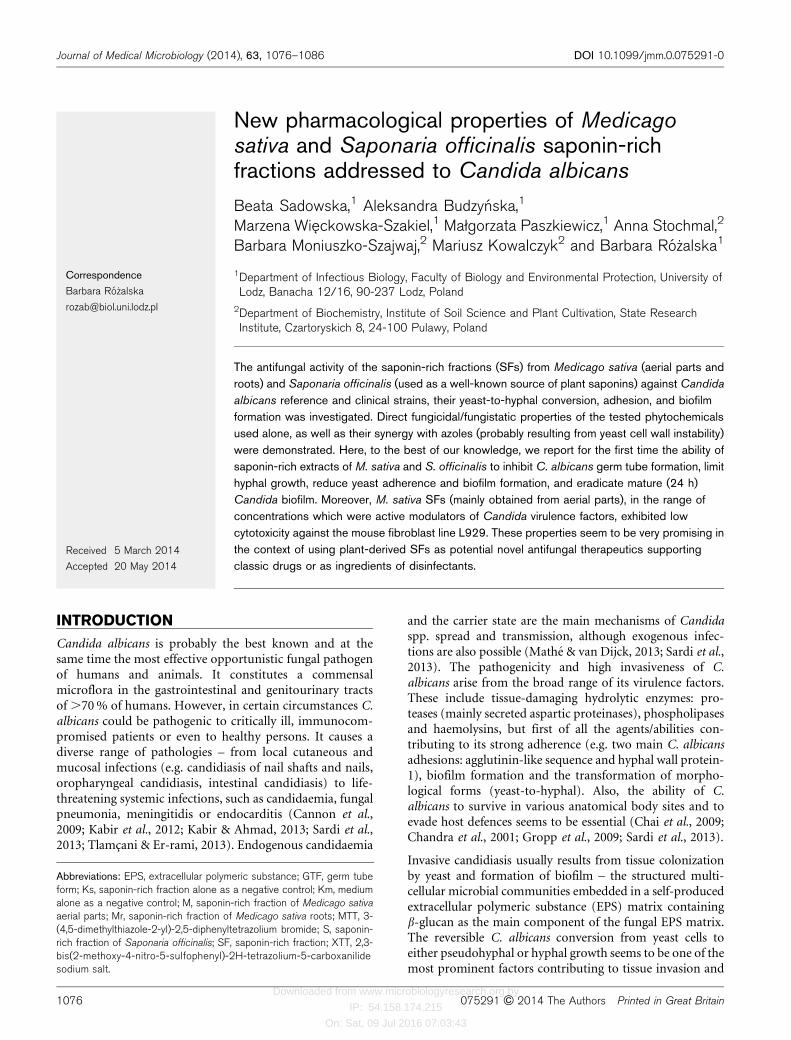
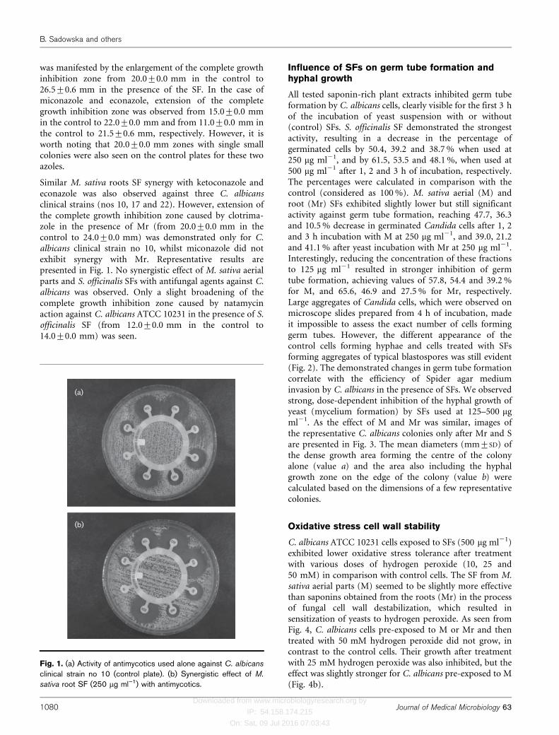
Similar M. sativa roots SF synergy with ketoconazole andeconazole was also observed against three C. albicansclinical strains (nos 10, 17 and 22). However, extension ofthe complete growth inhibition zone caused by clotrima-zole in the presence of Mr (from 20.0±0.0 mm in thecontrol to 24.0±0.0 mm) was demonstrated only for C.albicans clinical strain no 10, whilst miconazole did notexhibit synergy with Mr. Representative results arepresented in Fig. 1. No synergistic effect of M. sativa aerialparts and S. officinalis SFs with antifungal agents against C.albicans was observed. Only a slight broadening of thecomplete growth inhibition zone caused by natamycinaction against C. albicans ATCC 10231 in the presence of S.officinalis SF (from 12.0±0.0 mm in the control to14.0±0.0 mm) was seen.
Influence of SFs on germ tube formation andhyphal growth
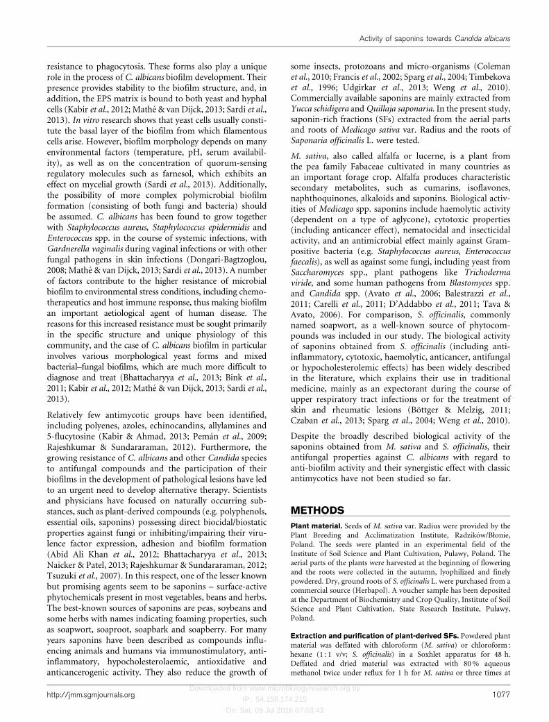
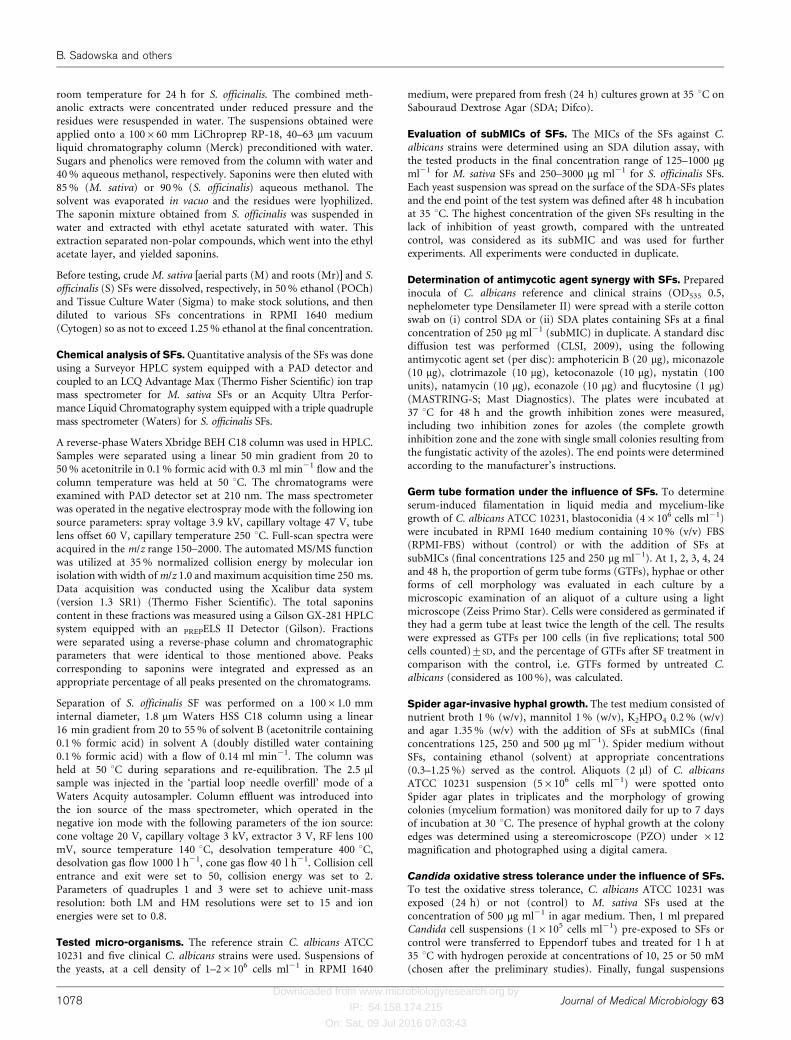
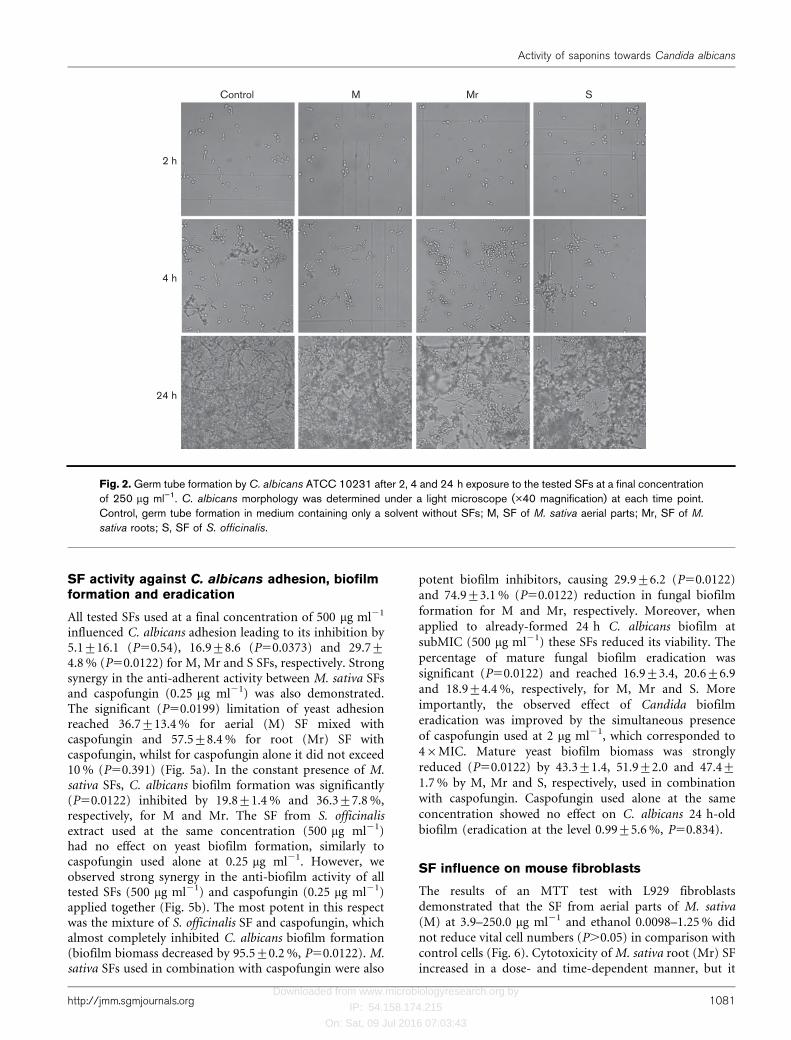
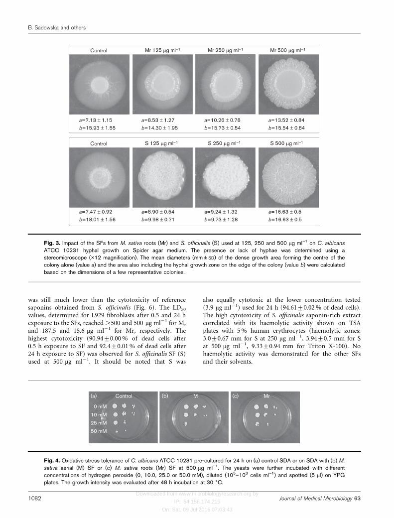
All tested saponin-rich plant extracts inhibited germ tubeformation by C. albicans cells, clearly visible for the first 3 hof the incubation of yeast suspension with or without(control) SFs. S. officinalis SF demonstrated the strongestactivity, resulting in a decrease in the percentage ofgerminated cells by 50.4, 39.2 and 38.7 % when used at250 mg ml21, and by 61.5, 53.5 and 48.1 %, when used at500 mg ml21 after 1, 2 and 3 h of incubation, respectively.The percentages were calculated in comparison with thecontrol (considered as 100 %). M. sativa aerial (M) androot (Mr) SFs exhibited slightly lower but still significantactivity against germ tube formation, reaching 47.7, 36.3and 10.5 % decrease in germinated Candida cells after 1, 2and 3 h incubation with M at 250 mg ml21, and 39.0, 21.2and 41.1 % after yeast incubation with Mr at 250 mg ml21.Interestingly, reducing the concentration of these fractionsto 125 mg ml21 resulted in stronger inhibition of germtube formation, achieving values of 57.8, 54.4 and 39.2 %for M, and 65.6, 46.9 and 27.5 % for Mr, respectively.Large aggregates of Candida cells, which were observed onmicroscope slides prepared from 4 h of incubation, madeit impossible to assess the exact number of cells forminggerm tubes. However, the different appearance of thecontrol cells forming hyphae and cells treated with SFsforming aggregates of typical blastospores was still evident(Fig. 2). The demonstrated changes in germ tube formationcorrelate with the efficiency of Spider agar mediuminvasion by C. albicans in the presence of SFs. We observedstrong, dose-dependent inhibition of the hyphal growth ofyeast (mycelium formation) by SFs used at 125–500 mgml21. As the effect of M and Mr was similar, images ofthe representative C. albicans colonies only after Mr and Sare presented in Fig. 3. The mean diameters (mm±SD) ofthe dense growth area forming the centre of the colonyalone (value a) and the area also including the hyphalgrowth zone on the edge of the colony (value b) werecalculated based on the dimensions of a few representativecolonies.
Oxidative stress cell wall stability
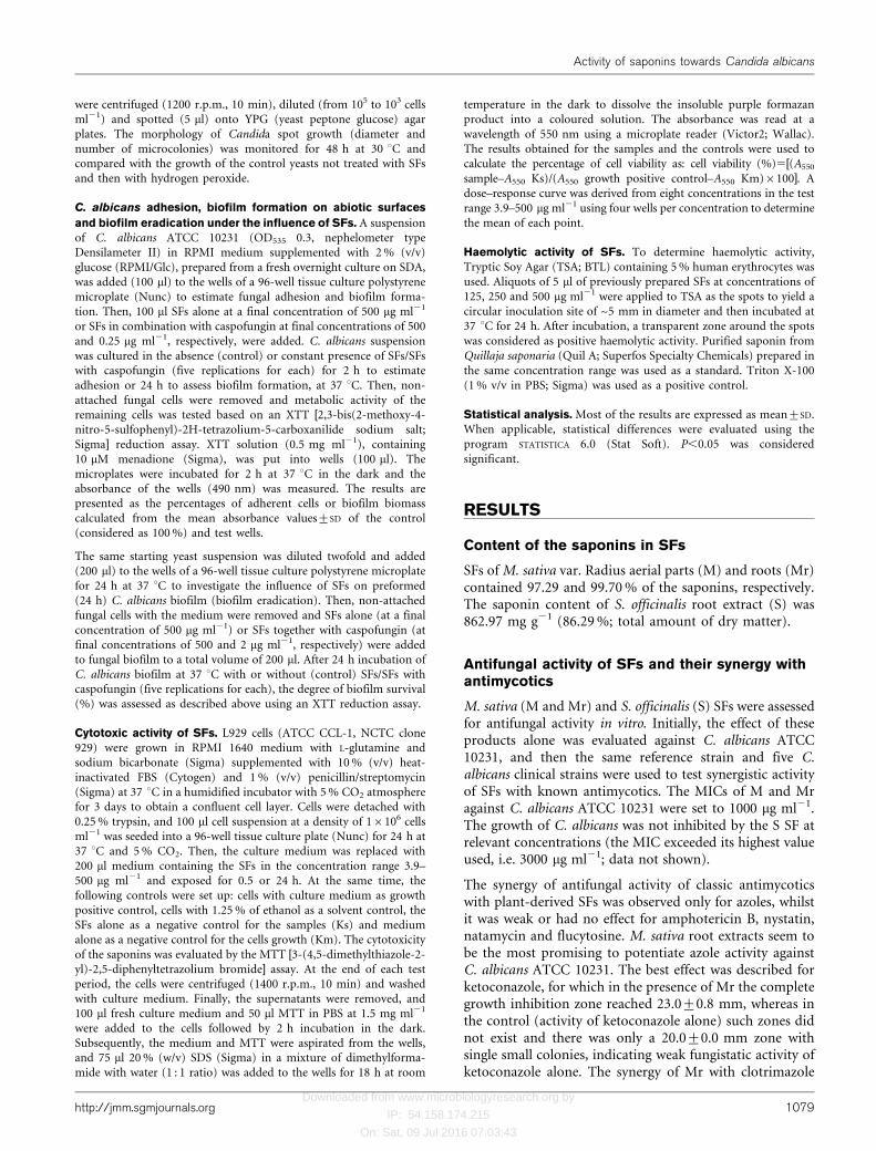
C. albicans ATCC 10231 cells exposed to SFs (500 mg ml21)exhibited lower oxidative stress tolerance after treatmentwith various doses of hydrogen peroxide (10, 25 and50 mM) in comparison with control cells. The SF from M.sativa aerial parts (M) seemed to be slightly more effectivethan saponins obtained from the roots (Mr) in the processof fungal cell wall destabilization, which resulted insensitization of yeasts to hydrogen peroxide. As seen fromFig. 4, C. albicans cells pre-exposed to M or Mr and thentreated with 50 mM hydrogen peroxide did not grow, incontrast to the control cells. Their growth after treatmentwith 25 mM hydrogen peroxide was also inhibited, but theeffect was slightly stronger for C. albicans pre-exposed to M(Fig. 4b).
(a)
(b)
Fig. 1. (a) Activity of antimycotics used alone against C. albicans
clinical strain no 10 (control plate). (b) Synergistic effect of M.
sativa root SF (250 mg ml”1) with antimycotics.
B. Sadowska and others
1080 Journal of Medical Microbiology 63
Downloaded from www.microbiologyresearch.org by
IP: 54.158.174.215
On: Sat, 09 Jul 2016 07:03:43
SF activity against C. albicans adhesion, biofilmformation and eradication
All tested SFs used at a final concentration of 500 mg ml21
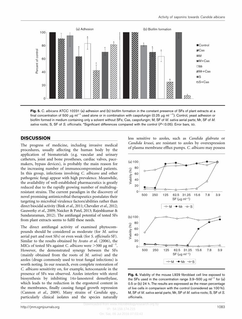
influenced C. albicans adhesion leading to its inhibition by5.1±16.1 (P50.54), 16.9±8.6 (P50.0373) and 29.7±4.8 % (P50.0122) for M, Mr and S SFs, respectively. Strongsynergy in the anti-adherent activity between M. sativa SFsand caspofungin (0.25 mg ml21) was also demonstrated.The significant (P50.0199) limitation of yeast adhesionreached 36.7±13.4 % for aerial (M) SF mixed withcaspofungin and 57.5±8.4 % for root (Mr) SF withcaspofungin, whilst for caspofungin alone it did not exceed10 % (P50.391) (Fig. 5a). In the constant presence of M.sativa SFs, C. albicans biofilm formation was significantly(P50.0122) inhibited by 19.8±1.4 % and 36.3±7.8 %,respectively, for M and Mr. The SF from S. officinalisextract used at the same concentration (500 mg ml21)had no effect on yeast biofilm formation, similarly tocaspofungin used alone at 0.25 mg ml21. However, weobserved strong synergy in the anti-biofilm activity of alltested SFs (500 mg ml21) and caspofungin (0.25 mg ml21)applied together (Fig. 5b). The most potent in this respectwas the mixture of S. officinalis SF and caspofungin, whichalmost completely inhibited C. albicans biofilm formation(biofilm biomass decreased by 95.5±0.2 %, P50.0122). M.sativa SFs used in combination with caspofungin were also
potent biofilm inhibitors, causing 29.9±6.2 (P50.0122)and 74.9±3.1 % (P50.0122) reduction in fungal biofilmformation for M and Mr, respectively. Moreover, whenapplied to already-formed 24 h C. albicans biofilm atsubMIC (500 mg ml21) these SFs reduced its viability. Thepercentage of mature fungal biofilm eradication wassignificant (P50.0122) and reached 16.9±3.4, 20.6±6.9and 18.9±4.4 %, respectively, for M, Mr and S. Moreimportantly, the observed effect of Candida biofilmeradication was improved by the simultaneous presenceof caspofungin used at 2 mg ml21, which corresponded to46MIC. Mature yeast biofilm biomass was stronglyreduced (P50.0122) by 43.3±1.4, 51.9±2.0 and 47.4±1.7 % by M, Mr and S, respectively, used in combinationwith caspofungin. Caspofungin used alone at the sameconcentration showed no effect on C. albicans 24 h-oldbiofilm (eradication at the level 0.99±5.6 %, P50.834).
SF influence on mouse fibroblasts
The results of an MTT test with L929 fibroblastsdemonstrated that the SF from aerial parts of M. sativa(M) at 3.9–250.0 mg ml21 and ethanol 0.0098–1.25 % didnot reduce vital cell numbers (P.0.05) in comparison withcontrol cells (Fig. 6). Cytotoxicity of M. sativa root (Mr) SFincreased in a dose- and time-dependent manner, but it
Control
2 h
4 h
24 h
M Mr S
Fig. 2. Germ tube formation by C. albicans ATCC 10231 after 2, 4 and 24 h exposure to the tested SFs at a final concentrationof 250 mg ml”1. C. albicans morphology was determined under a light microscope (�40 magnification) at each time point.Control, germ tube formation in medium containing only a solvent without SFs; M, SF of M. sativa aerial parts; Mr, SF of M.
sativa roots; S, SF of S. officinalis.
Activity of saponins towards Candida albicans
http://jmm.sgmjournals.org 1081
Downloaded from www.microbiologyresearch.org by
IP: 54.158.174.215
On: Sat, 09 Jul 2016 07:03:43
was still much lower than the cytotoxicity of referencesaponins obtained from S. officinalis (Fig. 6). The LD50
values, determined for L929 fibroblasts after 0.5 and 24 hexposure to the SFs, reached .500 and 500 mg ml21 for M,and 187.5 and 15.6 mg ml21 for Mr, respectively. Thehighest cytotoxicity (90.94±0.00 % of dead cells after0.5 h exposure to SF and 92.4±0.01 % of dead cells after24 h exposure to SF) was observed for S. officinalis SF (S)used at 500 mg ml21. It should be noted that S was
also equally cytotoxic at the lower concentration tested(3.9 mg ml21) used for 24 h (94.61±0.02 % of dead cells).The high cytotoxicity of S. officinalis saponin-rich extractcorrelated with its haemolytic activity shown on TSAplates with 5 % human erythrocytes (haemolytic zones:3.0±0.67 mm for S at 250 mg ml21, 3.94±0.5 mm for Sat 500 mg ml21, 9.33±0.94 mm for Triton X-100). Nohaemolytic activity was demonstrated for the other SFsand their solvents.
Control
a=7.13 ± 1.15b=15.93 ± 1.55
a=8.53 ± 1.27b=14.30 ± 1.95
a=10.26 ± 0.78b=15.73 ± 0.54
a=13.52 ± 0.84b=15.54 ± 0.84
a=7.47 ± 0.92b=18.01 ± 1.56
a=8.90 ± 0.54b=9.98 ± 0.71
a=9.24 ± 1.32b=9.73 ± 1.28
a=16.63 ± 0.5b=16.63 ± 0.5
Mr 125 mg ml–1 Mr 250 mg ml–1 Mr 500 mg ml–1
Control S 125 mg ml–1 S 250 mg ml–1 S 500 mg ml–1
Fig. 3. Impact of the SFs from M. sativa roots (Mr) and S. officinalis (S) used at 125, 250 and 500 mg ml”1 on C. albicans
ATCC 10231 hyphal growth on Spider agar medium. The presence or lack of hyphae was determined using astereomicroscope (�12 magnification). The mean diameters (mm±SD) of the dense growth area forming the centre of thecolony alone (value a) and the area also including the hyphal growth zone on the edge of the colony (value b) were calculatedbased on the dimensions of a few representative colonies.
(a) (b) (c)Control M Mr
0 mM
10 mM
25 mM
50 mM
Fig. 4. Oxidative stress tolerance of C. albicans ATCC 10231 pre-cultured for 24 h on (a) control SDA or on SDA with (b) M.
sativa aerial (M) SF or (c) M. sativa roots (Mr) SF at 500 mg ml”1. The yeasts were further incubated with differentconcentrations of hydrogen peroxide (0, 10.0, 25.0 or 50.0 mM), diluted (105–103 cells ml”1) and spotted (5 ml) on YPGplates. The growth intensity was evaluated after 48 h incubation at 30 6C.
B. Sadowska and others
1082 Journal of Medical Microbiology 63
Downloaded from www.microbiologyresearch.org by
IP: 54.158.174.215
On: Sat, 09 Jul 2016 07:03:43
DISCUSSION
The progress of medicine, including invasive medicalprocedures, usually affecting the human body by theapplication of biomaterials (e.g. vascular and urinarycatheters, joint and bone prostheses, cardiac valves, pace-makers, bypass devices), is probably the main reason forthe increasing number of immunocompromised patients.In this group, infections involving C. albicans and otherpathogenic fungi appear with high prevalence. Meanwhile,the availability of well-established pharmaceutics is greatlyreduced due to the rapidly growing number of multidrug-resistant strains. The current paradigm in the discovery ofnovel promising antimicrobial therapeutics postulates theirtargeting to microbial virulence factors/abilities rather thandirect biocidal activity (Bink et al., 2011; Chevalier et al., 2012;Gauwerky et al., 2009; Naicker & Patel, 2013; Rajeshkumar &Sundararaman, 2012). The antifungal potential of tested SFsfrom plant extracts seems to fulfil these needs.
The direct antifungal activity of examined phytocom-pounds should be considered as moderate (for M. sativaaerial part and root SFs) or even weak (for S. officinalis SF).Similar to the results obtained by Avato et al. (2006), theMICs of tested SFs against C. albicans were .500 mg ml21.However, the demonstrated synergy between the SFs(mainly obtained from the roots of M. sativa) and theazoles (drugs commonly used to treat fungal infections) isworth noting. In our research, even complete restoration ofC. albicans sensitivity on, for example, ketoconazole in thepresence of SFs was observed. Azoles interfere with sterolbiosynthesis by inhibiting 14a-lanosterol demethylase,which leads to the reduction in the ergosterol content inthe membranes, finally causing fungal growth repression(Cannon et al., 2009). Many strains of Candida spp.,particularly clinical isolates and the species naturally
less sensitive to azoles, such as Candida glabrata orCandida krusei, are resistant to azoles by overexpressionof plasma membrane efflux pumps. C. albicans may possess
100(a) Adhesion (b) Biofilm formation
Control
Cas
M
M+Cas
Mr
Mr+Cas
S
S+Cas
*
*
*
*
**
**
*
*
80
60
Per
cent
of c
ontr
ol
40
20
0
Fig. 5. C. albicans ATCC 10231 (a) adhesion and (b) biofilm formation in the constant presence of SFs of plant extracts at afinal concentration of 500 mg ml”1 used alone or in combination with caspofungin (0.25 mg ml ”1). Control, yeast adhesion orbiofilm formed in medium containing only a solvent without SFs; Cas, caspofungin; M, SF of M. sativa aerial parts; Mr, SF of M.
sativa roots; S, SF of S. officinalis. *Significant differences compared with the control (P,0.05). Error bars, SD.
(a) 100
Via
bilit
y (%
) 80
60
40
20
0
(b) 100
Via
bilit
y (%
) 80
60
40
20
0
500 7.815.631.2562.5SF (µg ml–1)
M Mr S
125250 3.9
500 7.815.631.2562.5SF (µg ml–1)
M Mr S
125250 3.9
Fig. 6. Viability of the mouse L929 fibroblast cell line exposed tothe SFs used in the concentration range 3.9–500 mg ml”1 for (a)0.5 or (b) 24 h. The results are expressed as the mean percentageof live cells in comparison with the control (considered as 100 %).M, SF of M. sativa aerial parts; Mr, SF of M. sativa roots; S, SF of S.
officinalis.
Activity of saponins towards Candida albicans
http://jmm.sgmjournals.org 1083
Downloaded from www.microbiologyresearch.org by
IP: 54.158.174.215
On: Sat, 09 Jul 2016 07:03:43
CaCdr1/2p belonging to ATP-binding cassette pumps witha broad spectrum of substrates and CaMdr1p belonging tothe major facilitator superfamily transporters (Cannonet al., 2009). Therefore, we could consider the possibilityof blocking or modifying these pumps by saponins as apotential mechanism of their synergistic effect. However,at the same time we observed slight synergism betweenSFs and natamycin. The complete C. albicans ATCC 10231growth inhibition zone caused by natamycin in the pre-sence of S. officinalis SF was extended from 12.0±0.0 mmin the control to 14.0±0.0 mm. It points to other, pro-bably simpler mechanisms of saponin synergy with anti-mycotics. Natamycin belongs to the polyenes – antifungalswith the rare occurrence of resistance caused mainly by areduction in the amount of ergosterol in the plasmamembrane (te Welscher et al., 2008). Therefore, we believethat the mechanism of demonstrated synergy is based onunspecific yeast cell membrane damage, which leads to itsincreased permeability (also for antifungal drugs), ratherthan blocking or modification of the efflux pumps. Theseassumptions were confirmed by the results of our researchconcerning the influence of the SFs on the effect ofoxidative stress. Tested SFs decreased C. albicans toleranceto hydrogen peroxide, as we had previously demonstratedfor essential oils (Budzynska et al., 2013). C. albicans showsgreat natural resistance to oxidative stress resulting mainlyfrom the activity of numerous anti-oxidative enzymes(e.g. catalase, superoxide dismutases, glutathione oxidase)(Missall et al., 2004). Based on our results, it can besuggested that the SFs had a direct effect on Candida cellwall stability rather than on the activity of the enzymesmentioned above. Such physical alteration might influencethe correct positioning and anchoring of cell-wall-localizedproteins responsible for counteracting the oxidative stress(e.g. superoxide dismutases). Importantly, C. albicansactivates an oxidative stress response, among others, afterexposure to human blood or phagocytosis, which promotespathogen invasion and survival inside the host cells,including phagocytes (Enjalbert et al., 2007; Missall et al.,2004). In summary, the parallel use of antifungals andsaponin-rich plant extracts (SFs) in the course of yeastinfections can support classic therapy, decrease the risk ofinvasive mycosis, reduce the applied doses of antimycoticsand limit the possibility of resistant strain generation.However, it should also be taken into consideration thatthe presence of subinhibitory concentrations of antimyco-tics can lead to overexpression of other fungal virulencefactors, as described by Wu et al. (2000) for C. albicansunder fluconazole subMIC concentrations.
Interestingly, the tested SFs seem to be very promising inthe context of their effect targeted against fungal invasionand biofilm formation. All the more so as only a smallnumber of drugs can be used to treat invasive fungalinfections and biofilm-associated infections of implantedbiomaterials. As described above, C. albicans hyphalgrowth and the possibility of morphological form trans-formation (yeast-to-hyphal) play a pivotal role in tissue
invasion and biofilm stability. Therefore, the ability of testSFs to inhibit Candida germ tube formation, correspond-ing to the limitation of hyphal growth on Spider agarmedium demonstrated here, is extremely important. Thisactivity could prevent the development of invasive mycosisand fungal biofilm formation in the early stages of infec-tion. Moreover, we showed the significant influence of SFson the reduction of C. albicans adherence and biofilmformation, as well as on the eradication of mature (24 h)Candida biofilm. It is important to note that bactericidaland fungicidal (mainly against some plant fungi and alimited number of human pathogens) activity of M. sativasaponins, based on an assessment of MIC, have beenreported previously (Avato et al., 2006; Balestrazzi et al.,2011; Carelli et al., 2011; D’Addabbo et al., 2011; Sung &Lee, 2008; Tava & Avato, 2006). However, data onantifungal activity of M. sativa and S. officinalis SFs inthe context of yeast adhesion and biofilm formation, to thebest of our knowledge, are reported here for the first time.In our research, we also demonstrated strong synergybetween SFs and caspofungin in terms of their ability toinhibit C. albicans adhesion and biofilm development, andeven to eliminate mature fungal biofilm. The effect ofcaspofungin used alone at MIC/2 (against yeast adhesionand biofilm formation) or 46MIC (against biofilmelimination) was very limited. We noticed only insignific-ant inhibition of C. albicans adhesion (P50.3913) and noeffect of caspofungin alone on fungal biofilm. However,caspofungin and amphotericin B have been shown topossess efficacy against yeast biofilm, but only when usedin much higher concentrations. It was confirmed fordifferent Candida spp. that the drug concentrationsrequired for reducing fungal metabolic activity by halfhad to be five to eight times higher in biofilms comparedwith planktonic cells, leading to at least a 30-fold increasein MIC (Mathe & van Dijck, 2013). Soustre et al. (2004)showed that the same subMIC of caspofungin as that usedin our research (MIC/2) decreased C. albicans adhesion,although mainly in the case of strains susceptible tofluconazole. The authors explained this phenomenon by anoriginal mechanism of caspofungin action. Caspofunginbelongs to echinocandins – cyclic lipopeptides interferingwith cell wall biosynthesis by the inhibition of 1,3-b-glucansynthase (Cannon et al., 2009; Rajeshkumar & Sundararaman,2012; Soustre et al., 2004). Therefore, its influence on theyeast cell wall may lead to a disturbance in the expressionof some cellular adhesins.
Considering the biological activity of saponins, thecomposition of a crude saponin mixture seems to beimportant, but not fully understood. Even the extractionmethod can lead to different constitutions and thus variousbiological properties. According to the research performedby Timbekova et al. (1996), the medicagenic acid gluco-pyranoside as well as hederagenin glycosides and theirderivatives (containing medicagenic acid or hederagenin asaglycones) are the main components of M. sativa extracts.However, the antimicrobial activity of crude saponin
B. Sadowska and others
1084 Journal of Medical Microbiology 63
Downloaded from www.microbiologyresearch.org by
IP: 54.158.174.215
On: Sat, 09 Jul 2016 07:03:43
mixtures obtained from M. sativa aerial parts, roots andtheir individual components was still very diverse, anddepended on both the type of extract/compound andthe tested micro-organism. For example, the growth ofC. albicans, Candida utilis, Bacillus mesenterius andPseudomonas lachrymans was not inhibited by the testedphytocompounds, whereas whole-root extract and theglucopyranoside of medicagenic acid limited the growth ofCandida tropicalis. All tested preparations also fullysuppressed the development of Agrobacterium tumefaciensand Corynebacterium michiganense (Timbekova et al.,1996). In other studies, Saccharomyces cerevisiae appearedto be the most susceptible among all tested fungi(including C. albicans, C. tropicalis and Candida laurentii)to saponins, mainly to sapogenin mixtures and medica-genic acid (Avato et al., 2006). Moreover, in many cases thewhole-plant-derived extract (especially enriched in inter-esting phytocompounds) exhibits higher biological activitythan its individual components, which may result fromtheir synergistic effect (Liu, 2003; Seeram et al., 2005). Itcan be assumed that a similar phenomenon occurred inour M. sativa and S. officinalis SFs, in which the number ofindividual saponins reached 24.
In the light of the literature data on the negative effectsof some components of plant extracts on eukaryoticcell viability and functions, cytotoxicity tests on mousefibroblast line L929 were performed for all saponin-richextracts. L929 fibroblast cells are ISO approved (accordingto EN ISO 10993-5) and the most common cell type thatcould be the target of the chemicals released from materials(e.g. dressings, drugs) having contact with damaged tissue.L929 fibroblasts were investigated with an MTT assay – agood indicator of cell viability. Our results showed that theSF from aerial parts of M. sativa was the least cytotoxic,even when used at 250 mg ml21, which was a potentinhibitor of C. albicans germ tube formation and hyphalgrowth. This is encouraging for the future application ofthis kind of preparation directly to eukaryotic tissues(e.g. in the form of surface-active ointments, lotions ordressings). The strong cytotoxicity of S. officinalis saponin-rich extract (even at the lowest concentration tested of3.9 mg ml21), correlating with its haemolytic activity,excludes it from potential direct therapeutic use in humans.However, its ability (alone or in combination withcaspofungin) to eradicate C. albicans biofilm, as well as toinhibit germ tube, hyphal growth and fungal biofilmformation, seems attractive in the context of the use of theSF from S. officinalis as an ingredient of antifungaldisinfectants.
In conclusion, our results indicate an important antifungalpotential of the saponins obtained from aerial parts androots of M. sativa and roots of S. officinalis, used alone orin synergy with antimycotics. The ability of saponin-richextracts to adversely affect such virulence factors of C.albicans as germ tube formation, hyphal growth, adhesionand biofilm formation/eradication was demonstrated.These attributes of Candida cells play a role as new
potential drug targets and thus the properties of saponinsseem to be very promising in the context of their possiblemedical applications. We suggest the use of plant-derivedsaponins as novel therapeutics supporting classic drugs inthe course of fungal infections or as an ingredient ofantifungal disinfectants. Further studies, in particularclinical investigations, are needed.
ACKNOWLEDGEMENTS
The study was partially supported by funds of the National ScienceCentre (grant 2012/05/N/NZ7/01216).
REFERENCES
Abid Ali Khan, M. M., Naqvi, T. S. & Naqvi, M. S. (2012). Identificationof phytosaponis as novel biodynamic agents: an updated overview.Asian J Exp Biol Sci 3, 459–467.
Avato, P., Bucci, R., Tava, A., Vitali, C., Rosato, A., Bialy, Z. & Jurzysta,M. (2006). Antimicrobial activity of saponins from Medicago sp.:structure–activity relationship. Phytother Res 20, 454–457.
Balestrazzi, A., Agoni, V., Tava, A., Avato, P., Biazzi, E., Raimondi, E.,Macovei, A. & Carbonera, D. (2011). Cell death induction and nitricoxide biosynthesis in white poplar (Populus alba) suspension culturesexposed to alfalfa saponins. Physiol Plant 141, 227–238.
Bhattacharyya, S., Gupta, P., Banerjee, G., Jain, A. & Singh, M.(2013). In-vitro inhibition of biofilm formation in Candida albicansand Candida tropicalis by heat stable compounds in culture filtrate ofAspergillus flavus. J Clin Diagn Res 7, 2167–2169.
Bink, A., Pellens, K., Cammue, B. P. A. & Thevissen, K. (2011). Anti-biofilm strategies: how to eradicate Candida biofilms? Open Mycol J 5,29–38.
Bottger, S. & Melzig, M. F. (2011). Triterpenoid saponins of theCaryophyllaceae and Illecebraceae family. Phytochem Lett 4, 59–68.
Budzynska, A., Sadowska, B., Lipowczan, G., Maciag, A., Kalemba,D. & Rozalska, B. (2013). Activity of selected essential oils againstCandida spp. strains. Evaluation of new aspects of their specificpharmacological properties, with special reference to Lemon Balm.Adv Microb 3, 317–325.
Cannon, R. D., Lamping, E., Holmes, A. R., Niimi, K., Baret, P. V.,Keniya, M. V., Tanabe, K., Niimi, M., Goffeau, A. & Monk, B. C. (2009).Efflux-mediated antifungal drug resistance. Clin Microbiol Rev 22,291–321.
Carelli, M., Biazzi, E., Panara, F., Tava, A., Scaramelli, L., Porceddu, A.,Graham, N., Odoardi, M., Piano, E. & other authors (2011). Medicagotruncatula CYP716A12 is a multifunctional oxidase involved in thebiosynthesis of hemolytic saponins. Plant Cell 23, 3070–3081.
Chai, L. Y. A., Netea, M. G., Vonk, A. G. & Kullberg, B. J. (2009).Fungal strategies for overcoming host innate immune response. MedMycol 47, 227–236.
Chandra, J., Kuhn, D. M., Mukherjee, P. K., Hoyer, L. L., McCormick,T. & Ghannoum, M. A. (2001). Biofilm formation by the fungalpathogen Candida albicans: development, architecture, and drugresistance. J Bacteriol 183, 5385–5394.
Chevalier, M., Medioni, E. & Precheur, I. (2012). Inhibition ofCandida albicans yeast-hyphal transition and biofilm formation bySolidago virgaurea water extracts. J Med Microbiol 61, 1016–1022.
CLSI (2009). Method for Antifungal Disk Diffusion SusceptibilityTesting of Yeasts; Approved Guideline, 2nd edn, M44-A2. Wayne, PA:Clinical and Laboratory Standards Institute.
Activity of saponins towards Candida albicans
http://jmm.sgmjournals.org 1085
Downloaded from www.microbiologyresearch.org by
IP: 54.158.174.215
On: Sat, 09 Jul 2016 07:03:43
Coleman, J. J., Okoli, I., Tegos, G. P., Holson, E. B., Wagner, F. F.,Hamblin, M. R. & Mylonakis, E. (2010). Characterization of plant-derived saponin natural products against Candida albicans. ACSChem Biol 5, 321–332.
Czaban, J., Modloch, J., Wroblewska, B., Szumacher-Strabel, M.,Cieslak, A., Oleszek, W. & Stochmal, A. (2013). Effects of triterpenoidsaponins of field scabious (Knautia arvensis L. Coult.) alfalfa, redclover and common soapwort on growth of Gaeumannomycesgraminis var. tritici and Fusarium culmorum. Allelopathy J 32, 79–90.
D’Addabbo, T., Carbonara, T., Leonetti, P., Radicci, V., Tava, A. &Avato, P. (2011). Control of plant parasitic nematodes with activesaponins and biomass from Medicago sativa. Phytochem Rev 10, 503–519.
Dongari-Bagtzoglou, A. (2008). Mucosal biofilms: challenges andfuture directions. Expert Rev Anti Infect Ther 6, 141–144.
Enjalbert, B., MacCallum, D. M., Odds, F. C. & Brown, A. J. P. (2007).Niche-specific activation of the oxidative stress response by thepathogenic fungus Candida albicans. Infect Immun 75, 2143–2151.
Francis, G., Kerem, Z., Makkar, H. P. S. & Becker, K. (2002). Thebiological action of saponins in animal systems: a review. Br J Nutr 88,587–605.
Gauwerky, K., Borelli, C. & Korting, H. C. (2009). Targeting virulence:a new paradigm for antifungals. Drug Discov Today 14, 214–222.
Gropp, K., Schild, L., Schindler, S., Hube, B., Zipfel, P. F. & Skerka,Ch. (2009). The yeast Candida albicans evades human complementattack by secretion of aspartic proteases. Mol Immunol 47, 465–475.
Kabir, M. A. & Ahmad, Z. (2013). Candida infections and theirprevention. ISRN Prev Med 2013, 763628.
Kabir, M. A., Hussain, M. A. & Ahmad, Z. (2012). Candida albicans: amodel organisms for studying fungal pathogens. ISRN Microbiol2012, 538694.
Liu, R. H. (2003). Health benefits of fruit and vegetables are fromadditive and synergistic combinations of phytochemicals. Am J ClinNutr 78 (Suppl), 517S–520S.
Mathe, L. & Van Dijck, P. (2013). Recent insights into Candidaalbicans biofilm resistance mechanisms. Curr Genet 59, 251–264.
Missall, T. A., Lodge, J. K. & McEwen, J. E. (2004). Mechanisms ofresistance to oxidative and nitrosative stress: implications for fungalsurvival in mammalian hosts. Eukaryot Cell 3, 835–846.
Naicker, S. D. & Patel, M. (2013). Dodonaea viscosa var. angustifoliainhibits germ tube and biofilm formation by C. albicans. Evid BasedComplement Alternat Med 2013, 261978.
Peman, J., Canton, E. & Espinel-Ingroff, A. (2009). Antifungal drugresistance mechanisms. Expert Rev Anti Infect Ther 7, 453–460.
Rajeshkumar, R. & Sundararaman, M. (2012). Emergence of Candidaspp. and exploration of natural bioactive molecules for anticandidaltherapy – status quo. Mycoses 55, e60–e73.
Sardi, J. C. O., Scorzoni, L., Bernardi, T., Fusco-Almeida, A. M. &Mendes Giannini, M. J. S. (2013). Candida species: current
epidemiology, pathogenicity, biofilm formation, natural antifungal
products and new therapeutic options. J Med Microbiol 62, 10–24.
Seeram, N. P., Adams, L. S., Henning, S. M., Niu, Y., Zhang, Y., Nair,M. G. & Heber, D. (2005). In vitro antiproliferative, apoptotic and
antioxidant activities of punicalagin, ellagic acid and a total
pomegranate tannin extract are enhanced in combination with other
polyphenols as found in pomegranate juice. J Nutr Biochem 16, 360–
367.
Soustre, J., Rodier, M.-H., Imbert-Bouyer, S., Daniault, G. & Imbert,C. (2004). Caspofungin modulates in vitro adherence of Candida
albicans to plastic coated with extracellular matrix proteins.
J Antimicrob Chemother 53, 522–525.
Sparg, S. G., Light, M. E. & van Staden, J. (2004). Biological activities
and distribution of plant saponins. J Ethnopharmacol 94, 219–243.
Sung, W. S. & Lee, D. G. (2008). In vitro candidacidal action of
Korean red ginseng saponins against Candida albicans. Biol Pharm
Bull 31, 139–142.
Tava, A. & Avato, P. (2006). Chemical and biological activity of
triterpene saponins from Medicago species. Nat Prod Commun 1,
1159–1180.
te Welscher, Y. M., ten Napel, H. H., Balague, M. M., Souza, C. M.,Riezman, H., de Kruijff, B. & Breukink, E. (2008). Natamycin blocks
fungal growth by binding specifically to ergosterol without permea-
bilizing the membrane. J Biol Chem 283, 6393–6401.
Timbekova, A. E., Isaev, M. I. & Abubakirov, N. K. (1996). Chemistry
and biological activity of triterpenoid glycosides from Medicago
sativa. Adv Exp Med Biol 405, 171–182.
Tlamcani, Z. & Er-rami, M. (2013). Fungal opportunist infection:
common and emerging fungi in immunocompromised patients.
J Immunol Tech Infect Dis 2, 2.
Tsuzuki, J. K., Svidzinski, T. I. E., Shinobu, C. S., Silva, L. F. A.,Rodrigues-Filho, E., Cortez, D. A. G. & Ferreira, I. C. P. (2007).Antifungal activity of the extracts and saponins from Sapindus
saponaria L. An Acad Bras Cienc 79, 577–583.
Udgirkar, R. F., Kadam, P. & Kale, N. (2013). Pharmacological
importance of saponin glycoside: a review. Int J Med Pharm Sci Res
Rev 1, 19–31.
Weng, A., Jenett-Siems, K., Schmieder, P., Bachran, D., Bachran, C.,Gorick, C., Thakur, M., Fuchs, H. & Melzig, M. F. (2010). A convenient
method for saponin isolation in tumour therapy. J Chromatogr B
Analyt Technol Biomed Life Sci 878, 713–718.
Wu, T., Wright, K., Hurst, S. F. & Morrison, C. J. (2000). Enhanced
extracellular production of aspartyl proteinase, a virulence factor, by
Candida albicans isolates following growth in subinhibitory concen-
trations of fluconazole. Antimicrob Agents Chemother 44, 1200–1208.
B. Sadowska and others
1086 Journal of Medical Microbiology 63