hair growth promoting effect of radish crude saponin extract
TRANSCRIPT
International Journal of Advanced Smart Convergence Vol.8 No.1 184-195 (2019)http://dx.doi.org/10.7236/IJASC.2019.8.1.184
Hair Growth Promoting Effect of Radish Crude Saponin Extract on Athymic
Nude Mice
Hyun-Kyoung Kim†
†Department of Food Science and Engineering, Seowon University, Cheongju, Korea
Abstract
This study investigates the hair restoration efficacy of selected radish saponin extracts on nude mice. Nude
mice genetically predisposed to pattern balding were used in this study. Our study revealed the underlying
mechanism of stimulating hair growth in athymic nude mice by repair the nu/nu follicular keratin
differentiation defect. Thus, the topical application of radish saponin may represent a novel strategy for the
management and therapy of certain forms of alopecia. The term of hair density of PEE treated nude mice were
significantly increase as compared with of control nude mice. Histological observation of skin sample showed
no hair follicle or only distorted hair follicles were observed in the control samples, in contrast, by the PEE
treatment groups showed a fully formed and increased the number of hair follicles up to three times higher
than that of control group in terms of the number of hair follicles in nude mouse skin.PEE treated mice the
number of BrdU-labeled keratinocytes per anagen follicle increased significantly, especially in the follicular
bulbs and outer root sheath compared with the control mice. Moreover, PEE-treated nude mice also exhibited
a significant increase in the number of BrdU-labeled epidermal keratinocyte proliferation.
Keywords: Anagen , Radish crude saponin, Hair follicle, Athymic nude mice, BrdU
1. Introduction
The hair follicle (HF) is the most prominent mini organ of the skin that remarkable for its dynamic structure
[1-5]. The fundamental feature of hair biology is consisted with the production of a hair shaft (anagen),
apoptosis-driven by regression (catagen), and relative resting (telogen). It undergoes repeated cycles of
regression and regeneration throughout the lifetime of an organism. Every phase in the hair cycle is
characterized by distinctive, strictly co-coordinated progression of tissue proliferation, differentiation, and
apoptosis, thus maintaining hairy phenotype of an organism [6, 7]. The nude mouse mutation has a
pleotropic effect that causes the abnormal development of the skin, hair follicles, lack of fur coat and thymus [8,
9]. The most striking feature of nude mice (nu/nu) mutation has proven to be a complete lack of fur
development after birth, has established a valuable and widely used biomedical tool since its discovery in 1966
IJASC 19-1-23
Manuscript Received: Feb. 28, 2019 / Revised: Mar. 7, 2019 / Accepted: Mar. 13, 2019Corresponding Author: [email protected] Tel: +82-43-299-8474, Fax: +82-43-299-8470Department of Food Science and Engineering, Seowon University, Cheongju, Korea
International Journal of Advanced Smart Convergence Vol.8 No.1 184-195 (2019) 185
[8]. The transcription factor Foxn1 (Forkhead box formerly called Whn and Hfh11) was identified as the
product of the nude gene on mouse chromosome 11 [9-11] which is now referred to as Foxn1nu [8] and also
been detected in humans located on chromosome 17, exhibiting nude mouse-like phenotype [9, 13-15].
Although the nude mouse phenotype appears hairless at the skin but its dermis contains a considerable
number of active hair follicles. However, follicles are aberrant and underdeveloped [8, 16] due to absence of
functional Foxn1 and other inherited deformity related to keratin gene expression [17]. The impaired
differentiation of nude follicle exhibit structural imperfections of the cortex, hair cuticle, and inner root
sheath [18]. Resulting, hair shafts bend and coil at the stage of sebaceous gland and failed to penetrate the
epidermis, thus makes the nude mice lack of external fur coat [19]. Various research groups has taken into
account nude mouse as model for hair biology and widely reported their evaluation of synthetic compounds.
Like as CsA [16, 20-24], KGF [25], AS101 [26], and PKC inhibitors, calcitriols and their related compounds
used as potential therapeutic tools but most having adverse side effects on human health. Natural products
either from pure compounds or standardized extract provide enormous opportunities to discover new
therapeutic against the synthetic drugs. Chemically synthesized drugs are known as common for adverse side
effects, so research turned on the ethnopharmacognosy. Course of investigation was carried out on nude mice
skin to know the effect of natural extract of Radish crude saponin and its response on the distorted hair
follicle. Nude mouse as widely used model in different areas of research like as immunological,
dermatological, cosmetic, oncological, and transplantation research, particularly because of their defect in
allo-or xenotransplant rejection [27,28]. The responsible nude gene ‘nu’ was shown to be of recessive,
autosomal character [23]. This nude gene mutation may provide an ideal model for the study of various
forms of human hair loss disorders involving alopecia, sebaceous gland hyperplasia, hyperkeratosis and
defective differentiation of the epidermis [24].
Thefore the Radish saponin is well known to have a numerous number of therapeutic effects analgesic,
anti-inflammatory, anti hepatitis C virus, antitumor and even reported to posses immunomodulatory
activities. In oder to this study investigates the hair restoration efficacy of selected radish saponin extracts on
nude mice.
2. Experiment Materials and Methods2.1 Heat Treatment of Radish and Preparation of Extract
Radish was purchased from an agricultural and marine products wholesale market in Korea and the whole
plant including leaves and peels were used after washing. A heat-treatment device (Jisco, Seoul, Korea) was
used which was designed and constructed such that it could resist a pressure of 10 kg/cm2 or higher. The whole
radish sample was placed in an inner container which was then placed in an outer container containing a
predetermined amount of water. Then, the radish sample was heated at a predetermined temperature for a
predetermined time so as to prevent the sample from being carbonized by direct heat transfer. During the
heat-treatment process the radish sample could be treated with steam. The heat-treatment temperature was set
at 110, 120, 130, 140 and 150°C and the heat-treatment time was set at 6 hours.
2.2 Extraction and Fractionation of Radish saponin
The heat-treated radish sample was cooled and then ground using a grinder and a 10-fold volume (v/v) of
distilled water was added there to followed by extraction for 14 to 16 hours. The extract was filtered and then
freeze-dried and used. The sample was ground into powder (800g) and extracted three times with petroleum
ether at 40°C for 4 hours under reflux then filtered and evaporated under vacuum to dryness to give the
corresponding PEE. The resulting residue was then extracted three times with MeOH at 70 °C for 4 hours
186 Hair Growth Promoting Effect of Radish Crude Saponin Extract on Athymic Nude Mice
then filtered and evaporated. The dried MeOH extract residue was suspended in distilled water and the
resulting aqueous suspension was fractionated sequentially with hexane fraction (HeF) and n-butanol
fraction (BuF) in a 1:1(v/v) ratio three times at room temperature. The resulting two fractions and remaining
water fraction (WaF) were evaporated under vacuum to dryness.
2.3 Experimental animals
Athymic male nude (nu/nu) mice Balb/c origin at 7 weeks of age were purchased from Dae-Han Biolink
(Eumsung, Korea). They were kept in autoclaved cages with filter bonnets in a laminar flow unit under
12-hour light and 12-hour dark periods at 24 ± 2°C in humidified atmosphere and were fed sterilized food
and distilled water. Experiments were performed in Animal Center under aseptic conditions in accordance
with approved institutional protocols by the Institutional Animal Care and Use Committee (IACUC).
2.4 Administration of PEE and fractions of Radish crude saponin
Mice were divided in to six groups; five males were allocated to each of six groups. While animal in
group-1 received 0.4 ml of vehicle mixture containing propylene glycol (67% v/v), Ethanol (30%, v/v) and
dimethyl sulfoxide (DMSO-3%, v/v) (Sigma, Mo, USA), animals in groups 2 received 5% Minoxidil 0.2ml).
Group-3, group-4, group-5 and group-6 received 5% solution of PEE, HeF, BuF and WaF of radish crude
saponin respectively. Topical application was performed once per day on the back of nude mice skin for 20
consecutive days.
2.5 Evaluation of hair coverage area
Mice were evaluated for hair coverage area by giving them a score of 0 to 8 as described in Table 1 (Suppl.
Figure 1). Hair scores were taken on Day 0, 5, 7, 10, 16 and 20 by the three independent observers blinded to
the treatment groups.
2.6 Evaluation of hair density
After daily treatment digital image were randomly taken on experimental Day 8 and 16 from each mouse
in 35mm2 area of interscapular region of the skin. The change of hair density were evaluated by analyzing
the image (×200 magnification, actual area 35mm2) using KONG, Bom-Viewer Plus (Seoul, Korea). Two
people not familiar with the study quantified the hair density.
2.7 Histologic assessment of hair growth
Skin samples were fixed in 10% neutral buffered formalin for histological analysis. Paraffin-embedded
4µm sections were stained with hematoxylin and eosin (H&E). The nude mice HF morphology and the
structure were evaluated microscopically in the H&E-stained sections of dorsal skin at a magnification
×1000. Five fields per section (×100 magnification) were considered for counting the number of dermal and
subcutaneous hair follicle with respect to the total number of hair follicle. Histopathological processing and
digital photomicrographs were taken using Leica application suite, Version 4.0.0, Leica Microystems
(Switzerland) Limited.
2.8 Assessment of Keratinocyte Proliferation with Anti-BrdU
Keratinocyte proliferation was measured by intraperitoneal injection of BrdU(50 mg/kg body weight;
Sigma Chemical Co., St. Louis, MO) at 1 hour before the mice were sacrificed. Dorsal skin from both
treatment and control animals were collected on experimental day 16 and fixed with 4% paraformaldehyde,
International Journal of Advanced Smart Convergence Vol.8 No.1 184-195 (2019) 187
dehydrated, and embedded in paraffin. After sectioning, the slides were dewaxed and denatured in 1.5 mol/L
of HCl for 30 minutes and neutralized with phosphate-buffered saline for 1 hour. BrdU incorporation was
detected by IHC staining of paraffin embedded sections with mouse anti-BrdU primary antibody (1:200) in a
moist chamber, at room temperature for 3 hour. (Santa cruz Biotechnology, Santa cruz, CA, USA). After
washing three times, incubated with secondary antibodies for 15 min. Skin sections that were not incubated
with primary antibodies were used as a negative control. Assessment of follicular and epidermal keratinocyte
and sebaceous gland epithelial cell BrdU labeling was done by an observer blinded to the treatment groups
using the original magnification, x400.
2.9 Statistical analysis
The experimental data were expressed as mean ± standard error mean (SEM). Student’s t-test or One-way
ANOVA was used for assessment of significance between treatments. Statistical analysis was performed
using SAS 9.2. A value of P < 0.05 was regarded as statistically significant.
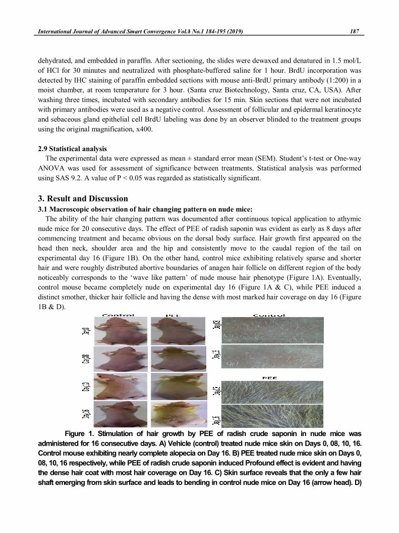
3. Result and Discussion3.1 Macroscopic observation of hair changing pattern on nude mice:
The ability of the hair changing pattern was documented after continuous topical application to athymic
nude mice for 20 consecutive days. The effect of PEE of radish saponin was evident as early as 8 days after
commencing treatment and became obvious on the dorsal body surface. Hair growth first appeared on the
head then neck, shoulder area and the hip and consistently move to the caudal region of the tail on
experimental day 16 (Figure 1B). On the other hand, control mice exhibiting relatively sparse and shorter
hair and were roughly distributed abortive boundaries of anagen hair follicle on different region of the body
noticeably corresponds to the ‘wave like pattern’ of nude mouse hair phenotype (Figure 1A). Eventually,
control mouse became completely nude on experimental day 16 (Figure 1A & C), while PEE induced a
distinct smother, thicker hair follicle and having the dense with most marked hair coverage on day 16 (Figure
1B & D).
Figure 1. Stimulation of hair growth by PEE of radish crude saponin in nude mice was
administered for 16 consecutive days. A) Vehicle (control) treated nude mice skin on Days 0, 08, 10, 16.
Control mouse exhibiting nearly complete alopecia on Day 16. B) PEE treated nude mice skin on Days 0,
08, 10, 16 respectively, while PEE of radish crude saponin induced Profound effect is evident and having
the dense hair coat with most hair coverage on Day 16. C) Skin surface reveals that the only a few hair
shaft emerging from skin surface and leads to bending in control nude mice on Day 16 (arrow head). D)
188 Hair Growth Promoting Effect of Radish Crude Saponin Extract on Athymic Nude Mice
PEE fraction has a normal dense hair coat on Day 16. Four mice /group were evaluated for each treatment
group. Digital image were taken from skin surface of vehicle (C) and PEE (D) treated nude mice on Day 8
and 16 by KONG, Bom-Viewer Plus, 80X magnification lens.
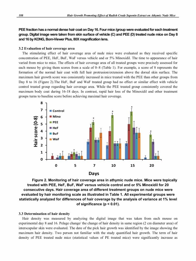
3.2 Evaluation of hair coverage area
The stimulating effect of hair coverage area of nude mice were evaluated as they received specific
concentration of PEE, HeF, BuF, WaF versus vehicle and or 5% Minoxidil. The time to appearance of hair
varied from mice to mice. The effects of hair coverage area of all treated groups were precisely assessed for
each mouse by giving them scores from a scale of 0–8 (Table 1). For example, a score of 8 represents the
formation of the normal hair coat with full hair protrusion/extension above the dorsal skin surface. The
maximum hair growth score was consistently increased in mice treated with the PEE than other groups from
Day 8 to 16 (Figure 2).The HeF, BuF and WaF treated group had no effect or similar effect with vehicle
control treated group regarding hair coverage area. While the PEE treated group consistently covered the
maximum body coat during 16-18 days. In contrast, rapid hair loss of the Minoxidil and other treatment
groups turns to baseline score before achieving maximal hair coverage.
Figure 2. Monitoring of hair coverage area in athymic nude mice. Mice were topically
treated with PEE, HeF, BuF, WaF versus vehicle control and or 5% Minoxidil for 20
consecutive days. Hair coverage area of different treatment groups on nude mice were
evaluated by hair monitoring scale as illustrated in Table 1. All experimental groups were
statistically analyzed for differences of hair coverage by the analysis of variance at 1% level
of significance (p < 0.01).
3.3 Determination of hair density
Hair density was measured by analyzing the digital image that was taken from each mouse on
experimental day 8 and 16. Pelage change/ the change of hair density in same region (2 cm diameter area) of
interscapular skin were evaluated. The date of the pick hair growth was identified by the image showing the
maximum hair density. Two person not familiar with the study quantified hair growth. The term of hair
density of PEE treated nude mice (statistical values of PE treated mice) were significantly increase as
Days
Hai
r sc
ore
(0-8
)
0
1
2
3
4
5
6
7
8
0 5 7 10 15 20
Control
Mino
PEE
HeF
BuF
WaF
*
*
*
*
International Journal of Advanced Smart Convergence Vol.8 No.1 184-195 (2019) 189
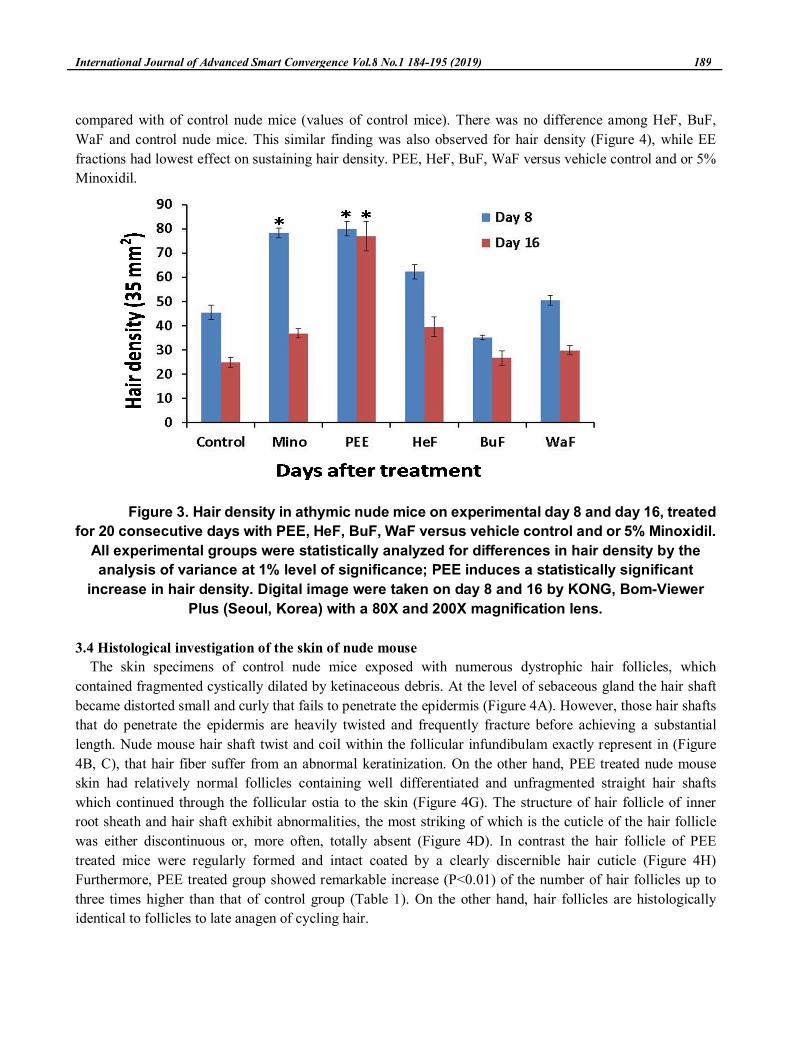
compared with of control nude mice (values of control mice). There was no difference among HeF, BuF,
WaF and control nude mice. This similar finding was also observed for hair density (Figure 4), while EE
fractions had lowest effect on sustaining hair density. PEE, HeF, BuF, WaF versus vehicle control and or 5%
Minoxidil.
Figure 3. Hair density in athymic nude mice on experimental day 8 and day 16, treated
for 20 consecutive days with PEE, HeF, BuF, WaF versus vehicle control and or 5% Minoxidil.
All experimental groups were statistically analyzed for differences in hair density by the
analysis of variance at 1% level of significance; PEE induces a statistically significant
increase in hair density. Digital image were taken on day 8 and 16 by KONG, Bom-Viewer
Plus (Seoul, Korea) with a 80X and 200X magnification lens.
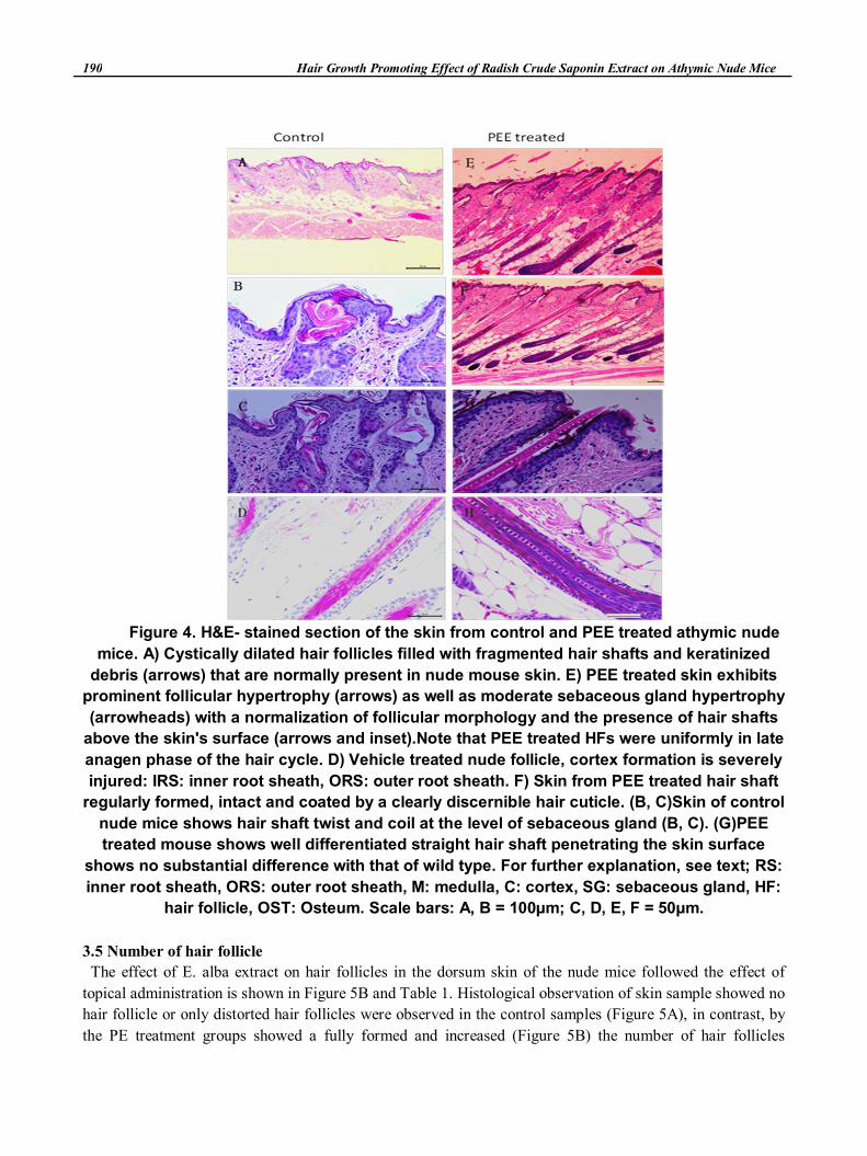
3.4 Histological investigation of the skin of nude mouse
The skin specimens of control nude mice exposed with numerous dystrophic hair follicles, which
contained fragmented cystically dilated by ketinaceous debris. At the level of sebaceous gland the hair shaft
became distorted small and curly that fails to penetrate the epidermis (Figure 4A). However, those hair shafts
that do penetrate the epidermis are heavily twisted and frequently fracture before achieving a substantial
length. Nude mouse hair shaft twist and coil within the follicular infundibulam exactly represent in (Figure
4B, C), that hair fiber suffer from an abnormal keratinization. On the other hand, PEE treated nude mouse
skin had relatively normal follicles containing well differentiated and unfragmented straight hair shafts
which continued through the follicular ostia to the skin (Figure 4G). The structure of hair follicle of inner
root sheath and hair shaft exhibit abnormalities, the most striking of which is the cuticle of the hair follicle
was either discontinuous or, more often, totally absent (Figure 4D). In contrast the hair follicle of PEE
treated mice were regularly formed and intact coated by a clearly discernible hair cuticle (Figure 4H)
Furthermore, PEE treated group showed remarkable increase (P<0.01) of the number of hair follicles up to
three times higher than that of control group (Table 1). On the other hand, hair follicles are histologically
identical to follicles to late anagen of cycling hair.
190 Hair Growth Promoting Effect of Radish Crude Saponin Extract on Athymic Nude Mice
Figure 4. H&E- stained section of the skin from control and PEE treated athymic nude
mice. A) Cystically dilated hair follicles filled with fragmented hair shafts and keratinized
debris (arrows) that are normally present in nude mouse skin. E) PEE treated skin exhibits
prominent follicular hypertrophy (arrows) as well as moderate sebaceous gland hypertrophy
(arrowheads) with a normalization of follicular morphology and the presence of hair shafts
above the skin's surface (arrows and inset).Note that PEE treated HFs were uniformly in late
anagen phase of the hair cycle. D) Vehicle treated nude follicle, cortex formation is severely
injured: IRS: inner root sheath, ORS: outer root sheath. F) Skin from PEE treated hair shaft
regularly formed, intact and coated by a clearly discernible hair cuticle. (B, C)Skin of control
nude mice shows hair shaft twist and coil at the level of sebaceous gland (B, C). (G)PEE
treated mouse shows well differentiated straight hair shaft penetrating the skin surface
shows no substantial difference with that of wild type. For further explanation, see text; RS:
inner root sheath, ORS: outer root sheath, M: medulla, C: cortex, SG: sebaceous gland, HF:
hair follicle, OST: Osteum. Scale bars: A, B = 100µm; C, D, E, F = 50µm.
3.5 Number of hair follicle
The effect of E. alba extract on hair follicles in the dorsum skin of the nude mice followed the effect of
topical administration is shown in Figure 5B and Table 1. Histological observation of skin sample showed no
hair follicle or only distorted hair follicles were observed in the control samples (Figure 5A), in contrast, by
the PE treatment groups showed a fully formed and increased (Figure 5B) the number of hair follicles
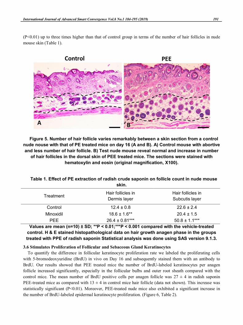
International Journal of Advanced Smart Convergence Vol.8 No.1 184-195 (2019) 191
(P<0.01) up to three times higher than that of control group in terms of the number of hair follicles in nude
mouse skin (Table 1).
Figure 5. Number of hair follicle varies remarkably between a skin section from a control
nude nouse with that of PE treated mice on day 16 (A and B). A) Control mouse with abortive
and less number of hair follicle. B) Test nude mouse reveal normal and increase in number
of hair follicles in the dorsal skin of PEE treated mice. The sections were stained with
hematoxylin and eosin (original magnification, X100).
Table 1. Effect of PE extraction of radish crude saponin on follicle count in nude mouse
skin.
TreatmentHair follicles in
Dermis layer
Hair follicles in
Subcutis layer
Control 12.4 ± 0.8 22.6 ± 2.4
Minoxidil 18.6 ± 1.6** 20.4 ± 1.5
PEE 26.4 ± 0.81*** 50.8 ± 1.1***
Values are mean (n=10) ± SD; **P < 0.01;***P < 0.001 compared with the vehicle-treated
control. H & E stained histopathological data on hair growth anagen phase in the groups
treated with PPE of radish saponin Statistical analysis was done using SAS version 9.1.3.
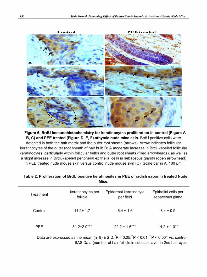
3.6 Stimulates Proliferation of Follicular and Sebaceous Gland Keratinocytes
To quantify the difference in follicular keratinocyte proliferation rate we labeled the proliferating cells
with 5-bromodeoxyuridine (BrdU) in vivo on Day 16 and subsequently stained them with an antibody to
BrdU. Our results showed that PEE treated mice the number of BrdU-labeled keratinocytes per anagen
follicle increased significantly, especially in the follicular bulbs and outer root sheath compared with the
control mice. The mean number of BrdU positive cells per anagen follicle was 27 ± 4 in radish saponin
PEE-treated mice as compared with 13 ± 4 in control mice hair follicle (data not shown). This increase was
statistically significant (P<0.01). Moreover, PEE-treated nude mice also exhibited a significant increase in
the number of BrdU-labeled epidermal keratinocyte proliferation. (Figure 6, Table 2).
192 Hair Growth Promoting Effect of Radish Crude Saponin Extract on Athymic Nude Mice
Figure 6. BrdU Immunohistochemistry for keratinocytes proliferation in control (Figure A,
B, C) and PEE treated (Figure D, E, F) athymic nude mice skin. BrdU positive cells were
detected in both the hair matrix and the outer root sheath (arrows). Arrow indicates follicular
keratinocytes of the outer root sheath of hair bulb D: A moderate increase in BrdU-labeled follicular
keratinocytes, particularly within follicular bulbs and outer root sheats (filled arrowheads), as well as
a slight increase in BrdU-labeled peripheral epithelial cells in sebaceous glands (open arrowhead)
in PEE treated nude mouse skin versus control nude mouse skin (C). Scale bar in A, 100 μm.
Table 2. Proliferation of BrdU positive keratinosites in PEE of radish saponin treated Nude
Mice.
Treatment keratinocytes per
follicle
Epidermal keratinocyte
per field
Epithelial cells per
sebaceous gland
Control 14.9± 1.7 6.4 ± 1.6 8.4 ± 0.9
PEE 31.2±2.0*** 22.2 ± 1.6*** 14.2 ± 1.0**
Data are expressed as the mean (n=9) ± S.D. *P < 0.05;**P < 0.01;***P < 0.001 vs. control.
SAS Data (number of hair follicle in subcutis layer in 2nd hair cycle
International Journal of Advanced Smart Convergence Vol.8 No.1 184-195 (2019) 193
4. Conclusion
This study was conducted to investigate the effect of radish saponin extract on the Radish saponin is well
known to have a numerous number of therapeutic effects analgesic, anti-inflammatory, anti hepatitis C virus,
antitumor and even reported to posses immunomodulatory activities. Taken together this information careful
investigation was carried out on nude mouse skin driven by inherited hair follicular abnormalities and
applied trustworthy hair natural stimuli agent Radish saponin. The unique findings reported here might be
useful for better control of regeneration of human hair loss as well as could be used as alternative to the
synthetic drugs. This study investigates the hair restoration efficacy of selected radish saponin extracts on
nude mice. Learning about the normal development and synchronized cycling of HF between the
developmental stages (anagen, catagen and telogen) is one of the key challenges for hair research. Most of our
current knowledge of the substances which modulate hair growth in humans is derived from clinical
observation and studies of mice have also been used to identify events associated with hair-follicle cycling. In
genetically predisposed nude mice sparsely distributed HFs are barely visible on the nude skin. Moreover,
progressive shortening of successive anagen cycles also leads to excessive shedding of HFs. These changes in
hair growth and development stages are crucial because active HFs are still present and cycling, even in the
skin of bald scalps or mutant nude skin. In this study, we investigated the hair-growth-promoting effect of
topical application of radish crude saponin on nude-mouse skin. Plants producing numerous bioactive
compounds and their fruitful application may promote hair regrowth act by up-regulating hair development. In
this context, natural products are being paid more attention by researchers because there is a thriving clinical
therapy that has already been proved even in the form of only a crude preparation. The mouse mutant “nude” is
hairless, as described by Flanagan (1966) and athymic (Pantelouris, 1968). The hair defect of the nu/nu
mutant is reported to be caused by imperfect keratinization in the hair shaft which causes the hair to break off
at the skin level. The macroscopic appearance of hair growth of the control nude mice was documented as a
few short, crippled, bent hair shafts emerging from the HFs. Very few follicles, of locally-variable density,
occur in the dorsal skin ; and only during a short anagen growth phase. Here, we were able to observe an
incomparable hair growth pattern in radish crude saponin treated mice with other treated groups. This
consequence has raised the possibility that radish crude saponin might induce some signals that regulate the
follicle to continue the growth phase of the anagen stage. Skin specimens of the radish crude saponin-treated
mice were also found to have abundant BrdU-positive keratinocytes in their hair bulbs and outer root sheaths.
One hypothesis is that temporary keratinization in the HF gives strength to hair shafts that rise above the skin
surface. Moreover, in this study we have closely observed two generations of hair growth, as well as
macroscopic analysis of hair growth parameters that included changing patterns of nude-mouse hair growth
area of hair coverage and length of hair. The hair of radish crude saponin-treated mice achieved a substantial
increase in length and became both thicker and smoother, whereas control hair shafts were transient, aberrant,
short and irregular in shape, all suggesting a deeply impaired keratinization process.
Histological study of the different treatment groups showed that theradish crude saponin-treated group had
an increased number of HFs in the deep subcutis, and had completely-developed HFs that corresponded to
the anagen phase of the hair-growth cycle. Further detailed clinical trials and screening by chemical analysis
will be necessary to identify the bioactive components in the extract. This knowledge will be used to prove
the biological activity of the Radish crude saponin extract, and to show whether the whole extrac rather than
individual components acts against alopecia.
194 Hair Growth Promoting Effect of Radish Crude Saponin Extract on Athymic Nude Mice
References
[1] S. C. Kim, “A Low-Complexity antenna selection algorithm for quadrature spatial modulation systems,”
International Journal of Internet, Broadcasting and Communication (IJIBC), Vol.9, No.1, pp.72-80, January 2017
DOI: https://doi.org/10.7236/IJIBC.2017.9.1.72.
[2] J. S. Jung, J. Kwon, S. H. Jung, M. W. Lee, V. Mariappan, and J. S. Cha, “Impact of SV40 T antigen on two
multiple fission microalgae species Scenedesmus quadricauda and chlorella vulgaris,” International Journal of
Advanced Smart Convergence(IJASC), Vol.6, No.1, pp.82-88, March 2017.
DOI: https://doi.org/10.7236/IJASC.2017.6.1.82.
[3] E. G. Ahmed, and H. Y. Seung, “ Impact of SV40 T antigen on two multiple fission microalgae species
scenedesmus quadricauda and chlorella vulgaris,” International Journal of advanced smart convergence(IJASC),
Vol. 7, No. 1, pp. 48-63, June 2018.
DOI: http://dx.doi.org/10.7236/IJASC.2018.7.1.7.
[4] H. J. Jeon, J. Hafeez, A. Hamacher, S. Lee, and S. C. Kwon, “ A study on the quality of photometric scanning
under variable illumination conditions,” International Journal of advanced smart convergence(IJASC), Vol. 6, No.
4, pp. 88-95, June 2017.
DOI: http://dx.doi.org/10.7236/IJASC.2017.6.4.13.
[5] M. H. Hardy, “The secret life of the hair follicle,” Trends in Genetics, Vol.8, No.2, pp. 55-61, February1992.
DOI: http://doi.org/10.1016/0168-9525(92)90350-D.
[6] R. Paus, “Control of the hair cycle and hair diseases as cycling disorders,” Curr Opin Dermatol , Vol.3, pp.
248-258, 1996.
[7] R. Paus, “Principles of hair cycle control,” J Dermatol, Vol.25, No.(12), pp.793-802, December 1998.
DOI: https://doi.org/10.1111/j.1346-8138.1998.tb02507.x.
[8] S. P. Flanagan, ‘Nude’ a new hairless gene with pleiotropic effects in the mouse,” Genet Res, Vol. 8, pp.295–309,
December 1966.
DOI: https://doi.org/10.1017/s0016672300010168.
[9] M. Nehls, D. Pfeifer, M. Schorpp, H. Hedrich, T. Boehm, “New member of the winged-helix protein family
disrupted in mouse and rat nude mutations,” Nature, Vol.372, No.6501, pp.103–107, November 1994.
DOI: https:doi.org/10.1038/37210390.
[10] Y. Takahashi, A. Shimizu, T. Sakai, Y. Endo, N. Osawa, H. Shisa, T. Honjo, “Mapping of the nu gene using
congenic nude strains and in situ hybridization,” J Exp Med, Vol.175, No.3, pp.873–876, March 1992.
[11] L. G. Byrd, “Regional localization of the nu mutation on mouse chromosome 11,” Immunogenetics, Vol.37, No.2,
pp. 157–159, January 1993.
[12] J. A. Segre, J. L. Nemhauser, B. A. Taylor, J. H. Nadea, E. S. Lander, “Positional cloning of the nude locus:
Genetic, physical, and transcriptional maps of the region and mutations in the mouse and rat,” Genomics, Vol.28,
No.(3), pp. 549–559, 1995.
DOI: https:doi.org/10.1006/geno.1995.1187.
[13] K. Schüddekopf, M. Schorpp, T. Boehm, “The whn transcription factor encoded by the nude locus contains an
evolutionarily conserved and functionally indispensible activation domain,” Proc Natl Acad Sci, Vol. 93, No.18,
pp.9661–9664, September 1996.
[14] J. Frank, C. Pignata, A. A. Panteleyev, D. M. Prowse, H. Baden, L. Weiner, L. Gaetanjello, W. Ahmad, P. B.
Csehaimi-Friedman, V. M. Aita, H. Vyttendaele, D. Gordon, J. Ott, J. L. Brissette, A. M. Christian, “Exposing the
human nude phenotype,” Nature, Vol. 398, No.6727, pp.473–474, April 1999.
DOI: https:doi.org//10.1038/18997.
[15] E. M. Pantelouris, “Athymic development in the mouse,” Differentiation, Vol. 1, No.6, pp. 437-450, December
1973.
DOI: https://doi.org/10.1111/j.1432-0436.1973.tb00143.x
International Journal of Advanced Smart Convergence Vol.8 No.1 184-195 (2019) 195
[16] T. Schlake, M. Schorpp, A. Maul-Pavicic, A. M. Malashenko, T. Boehm, “Forkhead/winged-helix transcription
factor Whn regulates hair keratin gene expression: molecular analysis of the nude skin phenotype,” Dev Dyn,
Vol.217, No.4, pp. 368–376, April 2000.
DOI: https://10.1002(SICI)1097-0177(2004)217:4<368:AID- DVDY4> 3.0.C0.2-Z.
[17] P. Köpf-Maier, V. F. Mboneko, H. J. Merker, “Nude Mice Are Not Hairless. A Morphological Study,” Acta Ana,t
Vol.139, No.2, pp. 178–190, February 1990.
[18] R. Paus, S. Muller-Rover, C. Van Der Veen, M. Maurer, S. Eichmüller, Ling G, et al.: “A comprehensive guide for
the recognition and classification of distinct stages of hair follicle morphogenesis,” J Invest Dermatol, Vol.113,
No.4, pp. 523–532, October 1999.
DOI: https://doi.org.//10.1046/j.1523-1747.1999.00740.x.
[19] Gafter-Gvili A, Sredni B, Gal R, Gafter U, Kalechman Y: Cyclosporin A-induced hair growth in mice is associated
with inhibition of calcineurin-dependent activation of NFAT in follicular keratinocytes. Am J Physiol Cell Physiol,
Vol.284, pp.1593–1603, 2003.
[20] S. Watanabe, A. Mochizuki, K. Wagatsuma, M. Kobayashi, Y. Kawa, H. Takahashi, “Hair growth on nude mice due
to cyclosporin A,” J Dermatol, Vol. 18, No.12, pp.714–719, December 1991.
DOI: https://doi.org/10.1111/j.1346- 8138.1991. tb03162.x
[21] M. Sawada, N. Terada, H. Taniguchi, R. Tateishi, Y. Mori, “Cyclosporin A stimulates hair growth in nude mice,”
Lab Invest, Vol.56, No.6, pp.684–686, July 1987.
[22] Y. Hozumi, T. Imaizumi, S. Kondo, “Effect of cyclosporin on hair-existing area of nude mice,” J Dermatol Sci,
Vol.7, No.1, pp. S33–S38, July 1994.
DOI: https://doi.org/10.1016/0923-1811(94)90033-7.
[23] A. E. Buhl, D. J. Waldon, B.F. Miller, M. N. Brunden, “Differences in activity of minoxidil and cyclosporin A on
hair growth in nude and normal mice. Comparisons of in vivo and in vitro studies,” Lab Invest, Vol.62, No.1,
pp.104–107, February 1990.
[24] D. M, Danilenko, B. D. Ring, D. Yanagihara, W. Benson, B. Wiemann, CO. Starnes, G. F. Piere, “Keratinocyte
growth factor is an important endogenous mediator of hair follicle growth, development, and differentiation.
Normalization of the nu/nu follicular differentiation defect and amelioration of chemotherapy induced alopecia,”
Am J Pathol, Vol.147, No.1, pp. 145–154, July 1995.
[25] B. Sredni, R. Gal, I. J. Cohen, J. E. Dazard, D. Givol, U. Gafter, B. Motro, S. Eliyahu, M. Albeck, H. M. Lander, Y.
Kalechman, “Hair growth induction by the Tellurium immunomodulator AS101: association with delayed terminal
differentiation of follicular keratinocytes and ras-dependent up-regulation of KGF expression,” FASEB J, Vol.18:
pp.400–402, February 2004.
DOI: https://doi.org/10.1096/fj.03-0552fje.
[26] G. G. Krueger, D. A. Chambers, J. Shelby, “Epidermal proliferation of nude mouse skin, pig skin and pig skin
grafts,” J Exp Med, Vol.152, No.5, pp.1329–1339, November 1980.
DOI: https://doi.org/10.1084/jem.152.5.1329.
[27] M. Sawada, K. Hayakawa, H. Nishiura, Y. Matsui, S. Tanabe, “Human yolk sac tumor of the ovary serially
heterotransplanted in nude mice,” Gynecol Oncol, Vol.11, No.1, pp.29–43, February 1981.
DOI: https://doi.org/ 10.1016 /0090 - 8258(81)9005-6.
[28] L. Shultz, “Immunological mutants of the mouse,” Am J Anat, Vol. 191, No.3, pp. 303–311, July 1991.
DOI: https: //doi.org/10.1002/aja.1001910310.
[29] F. Benavides, M. F. Starost, M. Flores, I. B. Gimenez-Conti, J. L. Guénet, C. J. Conti, “Impaired hair follicle
morphogenesis and cycling with abnormal epidermal differentiation in nackt mice, a cathepsin L-deficient
mutation,” Am J Pathol, Vol. 161, No.2, pp. 693-703, August 2002.
DOI: https:doi.org/10.1016/S0002- 94401(10)64225-3.
[30] V. D. Thakur, S. A. Mengi, “Neuropharmacological profile of Eclipta alba (Linn.) Hassk,” J Ethnopharmacol,
Vol.102, NO.1, pp.23-31, October, 2005.
DOI: https:doi.org./10.1016/j.jep.2005.05.037.