new biomedical devices with selective peptide recognition properties. part 1: characterization and...
TRANSCRIPT

Introduction
The construction of biological substitutes containingliving functioning cells, capable of restoring, maintaining
or improving specific tissue functions is the final goalof tissue engineering [1, 2]. The transplanted cells are incorporated into a supporting matrix, the scaffold, which in turn is implanted into thepatient’s body. Interest in tissue engineering is cur-rently focused on the development of scaffold structures capable of guiding the growth and the
New biomedical devices with selective peptide recognition
properties. Part 1: Characterization and cytotoxicity of
molecularly imprinted polymers
A. Rechichi a, C. Cristallini
b, U.Vitale
c, *, G. Ciardelli a, N. Barbani
d, G.Vozzi
d, P. Giusti
b, d
a Department of Mechanical Engineering, Politecnico di Torino, Torino, Italyb CNR, Institute of Composite and Biocompatible Materials, Pisa, Italy
c Department of Bioengineering, Politecnico di Milano, Milano, Italyd Department of Chemical Engineering, Università di Pisa, Pisa, Italy
Received: May 11, 2007; Accepted: Jul 26, 2007
Abstract
Molecular imprinting is a technique for the synthesis of polymers capable to bind target molecules selective-ly. The imprinting of large proteins, such as cell adhesion proteins or cell receptors, opens the way to impor-tant and innovative biomedical applications. However, such molecules can incur into important conformation-al changes during the preparation of the imprinted polymer impairing the specificity of the recognition cavi-ties. The “epitope approach” can overcome this limit by adopting, as template, a short peptide sequence rep-resentative of an accessible fragment of a larger protein. The resulting imprinted polymer can recognize boththe template and the whole molecule thanks to the specific cavities for the epitope. In this work two molecu-larly imprinted polymer formulations (a macroporous monolith and nanospheres) were obtained using theprotected peptide Z-Thr-Ala-Ala-OMe, as template, and Z-Thr-Ile-Leu-OMe, as analogue for the selectivityevaluation, methacrylic acid, as functional monomer, and trimethylolpropane trimethacrylate and pentaery-thritol triacrylate (PETRA), as cross-linkers. Polymers were synthesized by precipitation polymerization andcharacterized by standard techniques. Polymerization and rebinding solutions were analyzed by high per-formance liquid chromatography. The highly cross-linked polymers retained about 70% of the total templateamount, against (20% for the less cross-linked ones). The extracted template amount and the rebindingcapacity decreased with the cross-linking degree, while the selectivity showed the opposite behaviour. ThePETRA cross-linked polymers showed the best recognition (MIP 2-, � = 1.71) and selectivity (MIP 2+, �' =5.58) capabilities. The cytotoxicity tests showed normal adhesion and proliferation of fibroblasts cultured inthe medium that was put in contact with the imprinted polymers.
Keywords: molecular imprinting • biomaterials • tissue engineering • cell adhesion
J. Cell. Mol. Med. Vol 11, No 6, 2007 pp. 1367-1376
*Correspondence to: U. VITALE,Department of Bioengineering, Politecnico di Milano, PiazzaLeonardo da Vinci 32, 20133, Milano, Italy.Email: [email protected]
© 2007 The AuthorsJournal compilation © 2007 Foundation for Cellular and Molecular Medicine/Blackwell Publishing Ltd
doi:10.1111/j.1582-4934.2007.00102.x

1368 © 2007 The AuthorsJournal compilation © 2007 Foundation for Cellular and Molecular Medicine/Blackwell Publishing Ltd
organization of transplanted cells [3]. The knowledgeof how cells organize themselves within the body isimportant in designing the properties of the scaffold.
Cellular processes such as adhesion, migration,growth and gene expression are controlled by inter-actions with the surrounding environment. Themolecular mechanism through which the cells recog-nize substrates suitable for the adhesion is widelydescribed in literature [4]. Cell adhesion occursthrough specific membrane receptors. In particular,the integrins, a family of trans-membrane proteinslinked to the cytoskeleton on the cytoplasmatic sideof the membrane, recognize only particular peptidesequences present in some proteins (collagen,laminin, fibronectin, etc.), which are the componentsof the extra-cellular matrix.
The inclusion of these proteins on artificial sub-strates showed them to be poorly effective, becauseof the degradation phenomena following conforma-tional changes in the protein structure. On the otherhand, it is possible to improve cellular adhesion byincluding on the artificial surface short chains ofoligopeptides or oligosaccarides. An approach for thecreation of advanced scaffolds for cellular adhesionand proliferation is represented by molecular imprint-ing technology. This technology permits the introduc-tion, into a polymeric material, of recognition sites forspecific molecular species (templates) through thepolymerization of a monomer in the presence of atemplate [5, 6], or through the dissolving of the pre-formed polymer in a solution containing the moleculeto be recognized [7]. In both cases, the spatialarrangement is maintained by the polymer, after thetemplate extraction, and confers a selective “memo-ry” towards this molecule.
The molecular imprinting technology is a validalternative to molecular recognition systems presentin biological systems, such as those activated byantibodies [8]. The macromolecular matrices pre-pared with this procedure, in fact, can be stable evenin critical chemical and physical conditions [9], canhave a life of several years without any apparentreduction in performance, and can be used repeat-edly without any alteration to the “memory.” The mostextensively studied applications of the molecularlyimprinted polymers (MIPs) are mainly in the separa-tion by affinity sector [10–12], and also in determin-ing the level of drugs in human serum in substitutionof natural antibodies. Other applications are in thefield of the biomimetic sensors [13, 14] and the intel-
ligent polymers [15]. Interesting developments in thebiomaterial field in general [16, 17] and in tissueengineering in particular can be envisaged.
The present work represents a first step towardsthe realization of polymeric materials for applicationsin tissue engineering, which will operate as “smart”systems for recognizing specific peptide sequencesin extra-cellular matrix proteins, in order to guide andcontrol the adhesion process.
The use of entire proteins as template moleculesin molecular imprinting, showed to be, often unsuc-cessful because of the large molecular dimensionsand to the flexibility of the chains, which limit thepolymer molecular recognition capacity and selectiv-ity. To overcome these limitations, stable short pep-tide sequences, representative of an accessible frag-ment of a larger protein of interest (i.e. collagen,fibronectin, laminin, vitronectin, etc.), can be used,these often being located in the receptor domains orin other parts directly involved in the molecularrecognition process (epitopes). Therefore, if thematerial can recognize a peptide that represents theexposed part of a protein structure, it will also be ableto bind the entire protein [18]. In this way, it is possi-ble to increase the adhesion and growth characteris-tics of the cells on the MIP scaffold, thanks to thepresence of sites complementary and selectivetowards specific sequences within the extra-cellularproteins and with which the integrin receptorialdomains will interact.
Here we report the study of different MIPs capableof recognizing a tripeptide segment of an exposedpart of a fibronectin functional domain [19]. Thefibronectin is the main extra-cellular matrix adhesiveglycoprotein and plays a fundamental role in the celladhesion, migration and repair processes.
The tripeptide Z-Thr-Ala-Ala-OMe was used astemplate molecule maintaining the terminal function-al groups protection, in order to avoid undesiredinter- and intra-molecular interactions and toenhance the affinity of the peptide hydroxyl group tothe polymer functional groups. The polymers wereprepared by polymerization of methacrylic acid(MAA) as the functional monomer in the presence ofthe template molecule and of an appropriate cross-linking agent, trimethylolpropane trimethacrylate(TRIM) or pentaerythritol triacrylate (PETRA), to pro-vide a sufficient rigidity to the recognition sites in thepolymer structure.Two different forms of the polymer-ic materials were obtained by varying the cross-link-

J. Cell. Mol. Med. Vol 11, No 6, 2007
1369© 2007 The AuthorsJournal compilation © 2007 Foundation for Cellular and Molecular Medicine/Blackwell Publishing Ltd
ing agent concentration: a macroporous monolith atthe higher cross-linking degree and nanospheres atthe lower cross-linking degree [20]. The obtainedpolymeric materials were characterized for their ther-mal, spectroscopic and morphological properties.The template molecule was removed from the poly-mers by solvent extraction, and the performances ofthe obtained polymers in selectively rebinding thetemplate molecule were verified. Moreover, theselectivity test using the analogue peptide Z-Thr-Ile-Leu-OMe was carried out.
Cytotoxicity tests were also performed by seedingmouse fibroblasts in a culture medium previously putin contact, for a fixed time, with the polymer.
In tissue engineering applications, the loading ofnanoparticles on appropriate biodegradable scaf-folds, the biocompatibility tests and the functionalverification for improving cell adhesion are needed.These important investigations are currently inprogress and will be reported separately.
Materials and methods
Materials
MAA (>99%), from Aldrich, was distilled prior to use toremove the inhibitor. TRIM and PETRA, from Aldrich, andazobisisobutyronitrile (>98%), from Fluka, were used assupplied. The protected peptides TAA and TIL (Z-Thr-Ala-Ala-OMe and Z-Thr-Ile-Leu-OMe peptide sequences) weresynthesized by the group of Prof. Peggion, University ofPadua according to the standard protocols for peptide syn-thesis in solution using 1-hydroxybenzotriazole/1-ethyl-3(3-dimethylaminopropyl)carbodiimmide as coupling reagentsin the presence of diisopropylethylamine and characterizedby high performance liquid chromatography (HPLC),nuclear magnetic resonance and electrospray-ionizationmass spectroscopy. Acetonitrile (>99.9%), methanol(>99.9%), bidistilled water and acetic acid glacial (>99.9%),from Carlo Erba Reagenti, were of HPLC grade purity.Trifluoroacetic acid (>99%) was from Aldrich. Dulbecco’smodification of Eagle’s medium was from Cambrex.Formaldehyde and phosphate buffered saline were fromSigma. Coomassie blue solution was from Fluka.
Preparation of MIPs
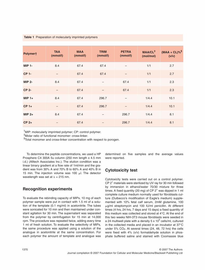
The MIPs were synthesized via precipitation polymeriza-tion at the compositions shown in Table 1. The reactors
were borosilicate 20 ml glass tubes, sealed with screwcaps. The template molecule was dissolved in acetonitrile,then the functional monomer MAA, the cross-linker and theinitiator were added. In the end, acetonitrile was added tothe final volume and the tubes were sealed under dry nitro-gen. The polymerization was thermally initiated at 60°Cand carried out for 20 hrs. A constant agitation of the tubeswas provided during all the process. Depending on thepolymerization conditions, in particular on the level of poro-gen coupled with the level of cross-linker, MIPs in the formof nanospheres or a macroporous monolith were obtained.The nanospheres were collected by centrifugation at14,000 rpm for 15 min (MIKRO 200 centrifuge, HettichGmbH & Co., Tuttlingen, Germany). The macroporousmonolith was crushed and washed with acetonitrile toremove the residual monomer and cross-linker and the notimprinted peptide. The template was extracted by washingthe polymers in Soxhlet apparatus with a MeOH/acetic acidmixture 70/70 (v/v) for 10 hrs. The controls were preparedwith the same procedure of the imprinted polymers, but inthe absence of the template molecule.
Physico-chemical characterization
The morphology of the MIPs nanospheres was examinedwith a JSM 5600 scanning electron microscopy (SEM)(Jeol Ltd., Peabody, MA, USA). The dimensions of the par-ticles were also estimated. Fourier transformed infraredspectroscopy (FT-IR) spectra were recorded on aSpectrum-One instrument, Perkin Elmer Inc., Beaconsfield,England. KBr pellets of the material samples were exam-ined. The thermal characterization was performed with aTGA 6 instrument, Perkin Elmer Inc., Sittard, Netherlands(scan from 30°C to 800°C at 10°C/min).
Chromatographic analysis
The monomer and the cross-linker conversion, and theamount of template entrapped by the polymer, were deter-mined measuring the corresponding residual amount in thepolymerization solution by HPLC (200 Series HPLC sys-tem, Perkin Elmer Inc., Shelton, CT, USA, with a UV/VISdetector). To determine the monomer and cross-linker con-centrations, we used a Alltima C18 5u column (250 mmlength x 4.5 mm i.d.) (Alltech Associates Inc., Deerfield, IL,USA). The mobile phase was: A = 0.085% trifluoroaceticacid (w/v) in acetonitrile; B = 0.1% trifluoroacetic acid (w/v)in water. The elution condition was an isocratic elution for15 min with a mobile phase composition of 54% A and 46% Bat a flow rate of 1 ml/min. The injection volume was 100 �l.The detector wavelength was set at � = 215 nm.
1370 © 2007 The AuthorsJournal compilation © 2007 Foundation for Cellular and Molecular Medicine/Blackwell Publishing Ltd
To determine the peptide concentrations, we used a HPProsphere C4 300A 5u column (250 mm length x 4.5 mmi.d.) (Alltech Associates Inc.). The elution condition was alinear binary gradient at a flow rate of 1ml/min and the gra-dient was from 30% A and 70% B to 60% A and 40% B in15 min. The injection volume was 100 �l. The detectorwavelength was set at � = 215 nm.
Recognition experiments
To evaluate the rebinding capacity of MIPs, 10 mg of eachpolymer sample were put in contact with 1.5 ml of a solu-tion of the template (0.1 mg/ml) in acetonitrile. The tubeswere sonicated for 10 min and then maintained under con-stant agitation for 30 min. The supernatant was separatedfrom the polymer by centrifugation for 15 min at 14,000rpm. The procedure was repeated twice, adding every time1 ml of fresh solution. To evaluate the selectivity of MIPs,the same procedure was applied using a solution of theanalogue in acetonitrile at the same concentration. Foreach polymer the amount of template and analogue was
determined on five samples and the average values were reported.
Cytotoxicity test
Cytotoxicity tests were carried out on a control polymer.CP 2+ materials were sterilized by UV ray for 30 min followedby immersion in ethanol/water 70/30 mixture for threetimes. A fixed quantity (20 mg) of CP 2+ was dipped in 1 mlcomplete culture medium normally used for fibroblasts cul-ture (Dulbecco’s modification of Eagle’s medium) supple-mented with 10% fetal calf serum, 2mM glutamine, 100�g/ml streptomycin and 100 IU/ml penicillin. At differenttimes (4 hrs, 24 hrs, 7 days and 15 days) a fixed quantity ofthis medium was collected and stored at 4°C. At the end ofthis two weeks NIH-3T3 mouse fibroblasts were seeded ina 24 multiwell plate with a density 5 x 10⁵ cells/ml, culturedin the collected media and placed in an incubator at 37°Cunder 5% CO2. At several times (24, 48, 72 hrs) the cellswere fixed with 4% (v/v) formaldehyde solution in phos-phate buffered saline and stained with Coomassie blue
Table 1 Preparation of molecularly imprinted polymers
†MIP: molecularly imprinted polymer; CP: control polymer.‡Molar ratio of functional monomer: cross-linker.§Total monomer and cross-linker concentration with respect to porogen.
Polymer†TAA
(mmol/l)
MAA
(mmol/l)
TRIM
(mmol/l)
PETRA
(mmol/l)MAA/CL
‡
(mol/mol)
(MAA + CL)%§
(v/v)
MIP 1- 8.4 67.4 67.4 – 1/1 2.7
CP 1- – 67.4 67.4 – 1/1 2.7
MIP 2- 8.4 67.4 – 67.4 1/1 2.3
CP 2- – 67.4 – 67.4 1/1 2.3
MIP 1+ 8.4 67.4 296.7 – 1/4.4 10.1
CP 1+ – 67.4 296.7 – 1/4.4 10.1
MIP 2+ 8.4 67.4 – 296.7 1/4.4 8.1
CP 2+ – 67.4 – 296.7 1/4.4 8.1

J. Cell. Mol. Med. Vol 11, No 6, 2007
1371© 2007 The AuthorsJournal compilation © 2007 Foundation for Cellular and Molecular Medicine/Blackwell Publishing Ltd
solution. Fibroblasts cultured in pure culture medium wereused as control. The stained cells were analyzed under anoptical microscope (AX70, Olympus Optical, London, UK)and the ratio between the number of cells cultured in thecollected media and those cultured in control media wastaken as a cytotoxicity index. The cytotoxicity tests wererepeated three times and for each experiment three sam-ples of CP 2+ were used.
Results
Physico-chemical characterization
MIPs and CPs were obtained by precipitation poly-merization under the conditions indicated in Table 1.After 20 hrs of reaction a high monomer conversionwas observed (Table 2). The conversion rate reachedalmost 100% for the more cross-linked resins and itis slightly lower in the case of MIP 2- and the relatedcontrol. An incomplete conversion of the monomersto MIP 1- and CP 1- occurred at 20 hrs was also con-firmed by calorimetric data.
In the case of loading capacity, a remarkably larg-er amount of template was retained by the morehighly cross-linked polymers (Table 3).
SEM micrographs showed large fused aggregatesof microgel particles interconnecting to form alabyrinth of macropores in the case of the morecross-linked resins as reported in Figure 1a. Theimages of MIP 1- showed a powder of nano-sizedparticles (Fig. 1c). Further investigation on samplesobtained by suspension of the MIP 1- in methanolconfirmed the spherical shape and the monodis-perse size distribution of the nanospheres (Fig. 1e).Also for MIP 2- (Fig. 1f) a mass of particles wasobserved, but the particles seemed to form smallaggregates, these were more clearly revealed by theanalysis of the methanol suspension (Fig. 1h).
FT-IR spectra showed the characteristic absorp-tion peaks of the expected chemical structure (� O H= 3450 cm-1; � C-H = 2970-1cm; � C = O = 1730 cm-1;� C(= O)-O = 1160 cm-1). When a lower cross-linkeramount was used (for example CP 1- with respect toCP 1+) a more intense band at 3450/cm due to theO-H stretching of the MAA carboxyl group wasobserved (Fig. 2). When PETRA was used instead ofTRIM as cross-linker agent (Fig. 3) a stronger � O-Hband, due to the addition of the side chain C-OHgroup absorption, and a peak at 1065 cm-1, due to the� C-O, were noted. The comparison between MIPs
Table 2 Monomer and cross-linkers conversion
†Monomer M conversion = ([M initial mole – M final mole]/M initial mole) x 100.
PolymerConversion
†(%)
MAA TRIM PETRA
MIP 1- 86.9 65.5 –
CP 1- 81.6 64.2 –
MIP 2- 99.2 – 93.2
CP 2- 98.2 – 91.5
MIP 1+ 98.7 100 –
CP 1+ 97.7 100 –
MIP 2+ 97.9 – 100
CP 2+ 98.1 – 100
Table 3 Template fraction retained and extracted
†TAA fraction retained = ([TAA initial mole – TAA finalmole]/TAA initial mole) x 100.‡TAA fraction extracted = ([TAA retained mole – TAAextracted mole]/TAA retained mole) x 100.
Polymer TAA fraction
retained †/%
TAA fraction
extracted‡
(%)
MIP 1- 20 40.1
CP 1- – –
MIP 2- 26.7 40.4
CP 2- – –
MIP 1+ 71.1 25.0
CP 1+ – –
MIP 2+ 70.2 33.0
CP 2+ – –

1372 © 2007 The AuthorsJournal compilation © 2007 Foundation for Cellular and Molecular Medicine/Blackwell Publishing Ltd
and CPs did not highlight the presence of the targetmolecule, because of the small amount (<3%) of TAAentrapped by the polymers. The spectra of CP 1- andMIP 1-, in Figure 4, do not show any difference in theregion around (1550 cm-1) where the target moleculepresents an intense absorption peak.
The thermal stability of the prepared polymerswas investigated by thermogravimetric analysis. Thederivative thermogram of PETRA containing resinsexhibits a peak centred at about 460°C in the lesscross-linked polymers, but it is shifted to higher tem-peratures by increasing the cross-linking degree. The
typical derivative thermograms are presented in Fig. 5.No remarkable differences in the thermal degrada-tion of TRIM containing polymers were noted, but ahigher thermal stability (shift of the onset tempera-ture from 450 to 460°C) was obtained enhancingcross-linking rate.
Recognition experiments
The template molecule was removed from the poly-mers by methanol/acetic acid extraction. The poly-
Fig. 1 (a) SEM of MIP 1+ resin showingthe fused microgel particles of the macro-porous monolith. (b) SEM of CP 1+ resinshowing the fused microgel particles ofthe macroporous monolith. (c) SEM ofMIP 1- resin showing aggregates ofnanospheres. (d) SEM of CP 1- resinshowing aggregates of nanospheres. (e)SEM of MIP 1- resin after suspension inmethanol showing discrete and monodis-persed nanospheres. (f) SEM of MIP 2-
resin showing aggregates of nanos-pheres. (g) SEM of CP 2- resin showingaggregates of nanospheres. (h) SEM ofMIP 2- resin after suspension in methanolshowing small aggregates of nanos-pheres.

J. Cell. Mol. Med. Vol 11, No 6, 2007
1373© 2007 The AuthorsJournal compilation © 2007 Foundation for Cellular and Molecular Medicine/Blackwell Publishing Ltd
mer monoliths were crashed and ground in a mortarbefore this step. As expected the amount of templateremoved from the nanospheres was higher than fromthe macroporous resins (Table 3).
The recognition properties of the prepared MIPswere investigated by HPLC analysis. The chromato-graphic results concerning the binding capacity, thatis the amount of template bound per mass of poly-mer, are shown in Table 4. In all cases the MIPs weremore efficient in template recognition than the relat-ed controls, indicating the formation of specific bind-ing sites in the polymer network. The binding factor,expressed as the ratio of template bound by themolecular imprinted material compared to the controlpolymer, indicated that the best performances interms of template recognition were obtained by theMIP 2- nanospheres. The ability of a prepared MIP todifferentiate between the template and a peptide ofsimilar structure was obtained by calculating theselectivity factor, that is the ratio of template com-pared to analogue bound following the same rebind-ing procedure. As indicated in Table 4, in all caseshigher selectivity for the target molecule than theanalogue was measured.
Cytotoxicity test
Figure 6 presents the effects of CP 2+ nanoparticlesincubated with media culture at several times (4 hrs,24 hrs, 7 days and 15 days) time on the cell adhesionat proliferation as a function of incubation time (4, 24,
Fig. 3 FT-IR spectra of CP 1+ and CP 2+ showing anincrease of the band at 3450 cm-1 and the peak at 1065cm-1 for CP 2+, containing additional OH groups due tothe cross-linker PETRA.
Fig. 4 FT-IR superimposed spectra of TAA peptide (dot-ted line) and a couple of imprinted and related controlpolymers. CP 1- (continuous line) and MIP 1- (dashedline) spectra are identical in spite of the presence of thepeptide in MIP 1-.
Fig. 5 Derivative thermograms: shift of the maximumdegradation rate of the more highly cross-linked CP 2+
with respect to CP 2- to higher temperature.
Fig. 2 FT-IR spectra of CP 1+ and CP 1- showing anincrease in the intensity of the band at 3450 cm-1 for CP 1-
, containing a higher concentration of MAA hydroxyl groups.

48 hrs). The ratio between the number of cells cul-tured in conditioned media and those in pure mediawas considered as an index of cytotoxicity.
Discussion
The polymerization method involved an initiallyhomogeneous solution polymerization which, ulti-
mately produced insoluble resins. The amount ofpeptide entrapped inside the resins strongly depend-ed on the matrix rigidity. An enhanced cross-linkingrate was found to produce materials that blocked thetemplate inside the resin more efficiently. MIPs withdifferent morphology were obtained by changing thereaction conditions (as e.g. amount of porogenamount of cross-linker). For lower porogen levels andhigher cross-linker concentration (MIP+ and CP+),the materials obtained were macroporous resins. In amore dilute solution and at lower cross-linker con-centration (MIP- and CP-) the microgel particles didnot fully coalesce and appeared as a microgel pow-der dispersion in accordance to what described bySherrington [21]. The presence of aggregates of par-ticles observed for MIP 2- explained, on the basis ofthe higher monomer conversion, to a further poly-merization in the porogen phase after phase separa-tion of the discrete nanospheres, creating additionalpolymer which helped fuse the particles together.FTIR-ATR spectra were performed in order to studythe interactions of the template molecules with theresins, but the analysis was unsuccessful because ofthe too low amount of peptide in the mixture to pro-duce detectable signals. No remarkable differencesin the thermal degradation of the imprinted polymers
1374 © 2007 The AuthorsJournal compilation © 2007 Foundation for Cellular and Molecular Medicine/Blackwell Publishing Ltd
Polymer
Bound TAA Bound TIL Binding factor †
Selectivity factor ‡
[µmol/g polymer] [µmol/g polymer] � MIP/CP �' template/analogue
MIP 1- 4.42 ± 0.09 3.58 ± 0.08 1.09 1.23
CP 1- 4.06 ± 0.11 2.51 ± 0.14 – –
MIP 2- 4.75 ± 0.06 2.18 ± 0.09 1.71 2.18
CP 2- 2.78 ± 0.04 3.48 ± 0.07 – –
MIP 1+ 1.58 ± 0.05 0.91 ± 0.08 1.07 1.73
CP 1+ 1.48 ± 0.09 2.28 ± 0.10 – –
MIP 2+ 1.74 ± 0.04 0.31 ± 0.04 1.12 5.58
CP 2+ 1.55 ± 0.08 2.22 ± 0.05 – –
Table 4 Binding and selectivity tests for TAA imprinted polymers and related controls
†� = µmol TAA bound by MIP/µmol TAA bound by CP.
‡�' = µmol TAA bound by MIP/µmol TIL bound by MIP.
Fig. 6 Cytotoxicity index of CP 2+ using NIH-3T3 cul-tured in conditioned media culture as function of cell cul-ture time after incubation (at 24, 48 and 72 hrs). Numberof cells cultured in medium not incubated with nanopar-ticles was used as control.

were noted, but a higher thermal stability wasobtained enhancing the cross-linking rate.
The polymers were washed in a Soxhlet appara-tus to remove the template molecule before function-al characterization. The template extraction wouldhave been expected to be difficult because of thelarge dimension of the peptide; it has been indicated[22] that bulky templates cannot easily slip out of apolymer network. The smaller dimension of the parti-cles promote the extraction because the area to vol-ume ratio is increased improving the efficiency ofsolid-liquid contact, even though the physical config-uration of macroporous MIPs allow the solvent tohave access to an essential fraction of the polymermass. Also the enhanced hydrophilicity of the mate-rial, obtained with the PETRA cross-linker, promotespolymer wettability with polar solvents favouring theextraction process.
Template and analogue recognition was studiedby rebinding experiments. The nanoparticles exhibit-ed better template recognition properties than themacroporous systems. The binding sites on thesediscrete particles are supposed to be more accessi-ble; on the contrary large templates cannot easily dif-fuse through the highly cross-linked polymer matrixdue to the steric hindrance. Diffusion of a bulky tem-plate into the imprinted cavities should be made eas-ier by the low cross-linking rate and the high area tovolume ratio, but the comparison between MIP 1-
with MIP 2- clearly suggested that the nature of thecross-linker has a more remarkable effect on tem-plate affinity. On the other hand, MIP 2- did not exhib-it the highest selectivity for the target with respect tothe analogue TIL, a peptide with a very similar chem-ical structure. MIP 2+ exhibited the �’ maximum valueof 5.58 and for MIP 2-
�' was equal to 2.18. Even if ahigher molar amount of functional monomer wasused for MIP 2- with respect to MIP 2+ in order toincrease complexation between functional monomerand the template molecule [18], the resultingincreased non-specific binding leads to a loss ofselectivity. An increased cross-linking content inhibitsthe diffusion of the template reducing the efficiencyof the extraction process and rebinding; on the otherhand, it rigidly fixes the arrangement of the function-al monomer around the template, favouring the for-mation of highly selective sites.
In vitro cell culture tests were performed to inves-tigate the cytotoxicity of the particles. CP 2+ particles
after 4 hrs of incubation in medium displayed a cyto-toxic effect that was a function of time culture. In par-ticular, the cell number decreased of more than 40%as compared to the untreated cells. On the contrary,CP 2+ after 24 hrs of incubation in medium had only anegligible effect on the cell number and an increasedtrend of cell proliferation was detected for all condi-tioning times. Actually, the low cell proliferation valueat 4 hrs was attributed to the presence of residual sol-vent used in the preparation phase – that after 72 hrsseemed to be completely removed or metabolizedunder the experimental conditions. Moreover, thebest result of cell viability was obtained after 72 hrs ofincubation time for all samples. These results sug-gested that the developed material did not presentedany detectable cytotoxicity.
Conclusions
Two formulations of MIPs, specific to TAA peptidesequence, were synthesized with TRIM and PETRAas cross-linker agents. For each formulation, poly-mers in the form of a macroporous monolith andnanospheres were prepared, depending on the levelof porogen coupled with the level of cross-linker. Theanalogue used in the selectivity studies was the TILpeptide sequence. It was observed that the templateis better retained by the higher cross-linked MIPs andit is better extracted from the lower cross-linked ones.The recognition capacity and factor are superior forthe nanosphere formulations, while the selectivityfactor is increased in the macroporous monoliths.These results are in accordance with the expectedincreasing rigidity of the polymer network at highercross-linking degree. The rigidity of the recognitioncavities decreases the rate of diffusion phenomenaand increases the selectivity of the binding sites. TheMIPs synthesized with PETRA showed the best per-formance in terms of recognition capacity and factor(MIP 2-, microspheres) and in terms of selectivity(MIP 2+, monolith), compared to TRIM polymers. Ourresults are in accordance with the molecular imprint-ing theory and with the conclusions of the referenceworks. In addition, screening biological tests suggestthe absence of toxic effects.
Specific tests on cell adhesion and proliferation onporous scaffold surface modified with imprinted
J. Cell. Mol. Med. Vol 11, No 6, 2007
1375© 2007 The AuthorsJournal compilation © 2007 Foundation for Cellular and Molecular Medicine/Blackwell Publishing Ltd

nanoparticles will be also essential for the evaluationof the functional properties of the developed materi-al in view of a perspective application in tissue engi-neering. The rebinding in aqueous solution, in condi-tions close to the biological environment will bereported in a separate contribution. The efficient andselective recognition of the TAA epitope is an impor-tant step forward in the synthesis of MIPs specific tocell adhesion proteins, cell receptors, extra-cellularmatrix molecules, enzymes, growth factors, DNAfragments and complex drugs. Therefore, the tech-nique adopted will lead in the next future to importantapplication in the fields of tissue engineering, syn-thetic receptors or drug delivery.
Acknowledgements
Co-financing by the Italian Ministry of research through thePRIN project 2005091572_002, year 2005 is acknowledged.
References
1. Fournier RL. Tissue engineering. In: Basic transportphenomena in biomedical engineering. Philadelphia:Taylor and Francis; 1998. pp. 201–36.
2. Seal BL, Otero TC, Panitch A. Polymeric biomateri-als for tissue and organ regeneration. Mater Sci Eng.2001; 34: 147–230.
3. Hutmacher DW. Scaffold design and fabricationtechnologies for engineering tissues. State of the artand future perspectives. Biomater Sci Polym Edn.2001; 12: 107–24.
4. Folk A, Toner M. Microengineering of cellular inter-action. Ann Rev Biomed Eng. 2000; 2: 227–56.
5. Mosbach K. Molecular imprinting. Trends Biochem.Sci. 1994; 19: 9–14.
6. Wulff G, Karsten K. Stoichiometric noncovalentinteraction in molecular imprinting. Bioseparation.2002; 10: 257–76.
7. Wang HI, Kobayashi T, Fujii N. Molecular imprintmembranes prepared by the phase inversion precip-itation technique. Langmuir. 1996; 12: 4850–6.
8. Wulff G. Molecular Imprinting in cross-linked materi-als with the aid of molecular templates: a way
towards artificial antibodies. Angew Chem Int EdEngl. 1995; 34: 1812–32.
9. Nicholls IA, Svenson J. On the thermal and chemi-cal stability of molecularly imprinted polymers.Analytica Chimica Acta. 2001; 435: 19–24.
10. Sellergren B, Shea KJ. Chiral ion exchange chro-matography: correlation between solute retention anda theoretical ion-exchange model using imprintedpolymers. J Chromatogr A. 1993; 654: 17–28.
11. Mosbach K, Kempe M. Separation of amino acids,peptides and proteins on molecularly imprinted sta-tionary phases. J Chromatogr A. 1995; 691: 317–23.
12. Hart BR, Shea KJ. Synthetic peptide receptors: mol-ecularly imprinted polymers for the recognition ofpeptides using peptide-metal interactions. J AmChem Soc. 2001; 123: 2072–3.
13. Mosbach K, Ramstrom O. The emerging techniqueof molecular imprinting and its future impact onbiotechnology. Biotechnology. 1996; 14: 163–70.
14. Shea KJ. Molecular imprinting of synthetic networkpolymers: de novo synthesis of macromolecular bind-ing and catalytic sites. Trends Polym Sci. 1994; 2:166–73.
15. Cormack PAG, Mosbach K. Molecular imprinting:recent developments and the road ahead. ReactFunct Polym. 1999; 41: 115—24.
16. Cristallini C, Ciardelli G, Giusti P, Barbani N.
Acrylonitrile-acrylic acid copolymer membraneimprinted with uric acid for clinical uses.Macromolecular Bioscience. 2004; 4: 31—8.
17. Silvestri D, Borrelli C, Giusti P, Cristallini C,
Ciardelli G. Polymeric devices containing imprintednanospheres: a novel approach to improve recogni-tion in water for clinical uses. Analytica Chimica Acta.2005; 542: 3–13.
18. Rachkov A, Minoura N. Towards molecularlyimprinted polymers selective to peptides and pro-teins. the epitope approach. Biochimica et BiophysicaActa. 2001; 1544: 255—66.
19. Dickinson CD, Veerapandian B, Dai XP, Hamlin
RC, Xuong NH, Ruoslahti E, Ely KR. Crystal structure of the tenth type III cell adhesion module of human fibronectin. J Mol Biol. 1994; 236:1079–92.
20. Ye L, Weiss R, Mosbach K. Synthesis and charac-terization of molecularly imprinted microspheres.Macromolecules. 2000; 33: 8239–45.
21. Sherrington DC. Preparation, structure and mor-phology of polymer supports. Chem Commun. 1998;2275–86.
22. Flam F. Molecular imprints make a mark. Science.1994; 263: 1221–2.
1376 © 2007 The AuthorsJournal compilation © 2007 Foundation for Cellular and Molecular Medicine/Blackwell Publishing Ltd