quantum dot nanocrystals having guanosine imprinted nanoshell for dna recognition
TRANSCRIPT
A
a(pugaomaD©
K
1
dtstfi[[
Uf
0d
Available online at www.sciencedirect.com
Talanta 75 (2008) 890–896
Quantum dot nanocrystals having guanosineimprinted nanoshell for DNA recognition
Sibel Emir Diltemiz a, Rıdvan Say a,b, Sibel Buyuktiryaki b,Deniz Hur a, Adil Denizli c, Arzu Ersoz a,∗
a Department of Chemistry, Anadolu University,Eskisehir, Turkey
b BIBAM (Plant, Drug and Scientific Researches Center), Anadolu University, Eskisehir, Turkeyc Department of Chemistry, Hacettepe University, Ankara, Turkey
Received 16 July 2007; received in revised form 10 December 2007; accepted 18 December 2007Available online 4 January 2008
bstract
Molecular imprinted polymers (MIPs) as a recognition element for sensors are increasingly of interest and MIP nanoparticles have started toppear in the literature. In this study, we have proposed a novel thiol ligand-capping method with polymerizable methacryloylamido-cysteineMAC) attached to CdS quantum dots (QDs), reminiscent of a self-assembled monolayer and have reconstructed surface shell by synthetic hostolymers based on molecular imprinting method for DNA recognition. In this method, methacryloylamidohistidine-platinium (MAH-Pt(II)) issed as a new metal-chelating monomer via metal coordination-chelation interactions and guanosine templates of DNA. Nanoshell sensors withuanosine templates give a cavity that is selective for guanosine and its analogues. The guanosine can simultaneously chelate to Pt(II) metal ionnd fit into the shape-selective cavity. Thus, the interaction between Pt(II) ion and free coordination spheres has an effect on the binding abilityf the CdS QD nanosensor. The binding affinity of the guanosine imprinted nanocrystals has investigated by using the Langmuir and Scatchard
ethods, and experiments have shown the shape-selective cavity formation with O6 and N7 of a guanosine nucleotide (Ka = 4.841 × 10⁶ mol L−1)nd a free guanine base (Ka = 0.894 × 10⁶ mol L−1). Additionally, the guanosine template of the nanocrystals is more favored for single strandedNA compared to double stranded DNA.2007 Elsevier B.V. All rights reserved.
Deox
ahtas
(
eywords: Quantum dots; Nanoshell sensor; Molecularly imprinted polymers;
. Introduction
Molecular imprinting provides a promising technique foresigning synthetic receptors that possess the molecular recogni-ion properties of biological systems and is a method for makingelective binding sites in synthetic polymers using molecularemplate [1]. The selectivity of the polymer depends on variousactors like the size and the shape of the cavity and rebinding
nteractions. Covalent interactions [2], non-covalent interactions3–5], electrostatic interactions [6] and metal ion coordination7,8] can be exploited to organize the functional monomers∗ Corresponding author at: Fen Fakultesi, Yunus Emre Kampusu, Anadoluniversitesi, 26470 Eskisehir, Turkey. Tel.: +90 222 335 0580;
ax: +90 222 320 4910.E-mail address: [email protected] (A. Ersoz).
tn
iobos[
039-9140/$ – see front matter © 2007 Elsevier B.V. All rights reserved.oi:10.1016/j.talanta.2007.12.036
tribo nucleic acid (DNA) recognition; Photoluminescence
round the template. The molecular imprinted polymers (MIPs)ave been applied to the realization of recognition and specificityo biomaterials for bioseparations, diagnostic assays, biosensors,nd biocatalysis. MIPs have also been employed in efforts toynthesize materials for biosensor application.
On the other hand, semiconductor nanocrystal quantum dotsQDs) have been explored as fluorescent biological labels, dueo their photostable, size-tunable, narrow bandwidth photolumi-escence and chemically functionalizable surfaces [9–12].
Organic fluorescent dyes have been used for analyte detectionn molecular imprinting process [13,14]. But better results can bebtained from inorganic semiconductor nanoparticles. Due to its
road excitation spectra, which is effective to whole spectrumf colors, emission without red tailing and photodegradationtability of QDs make them more effective as fluorescent labels15].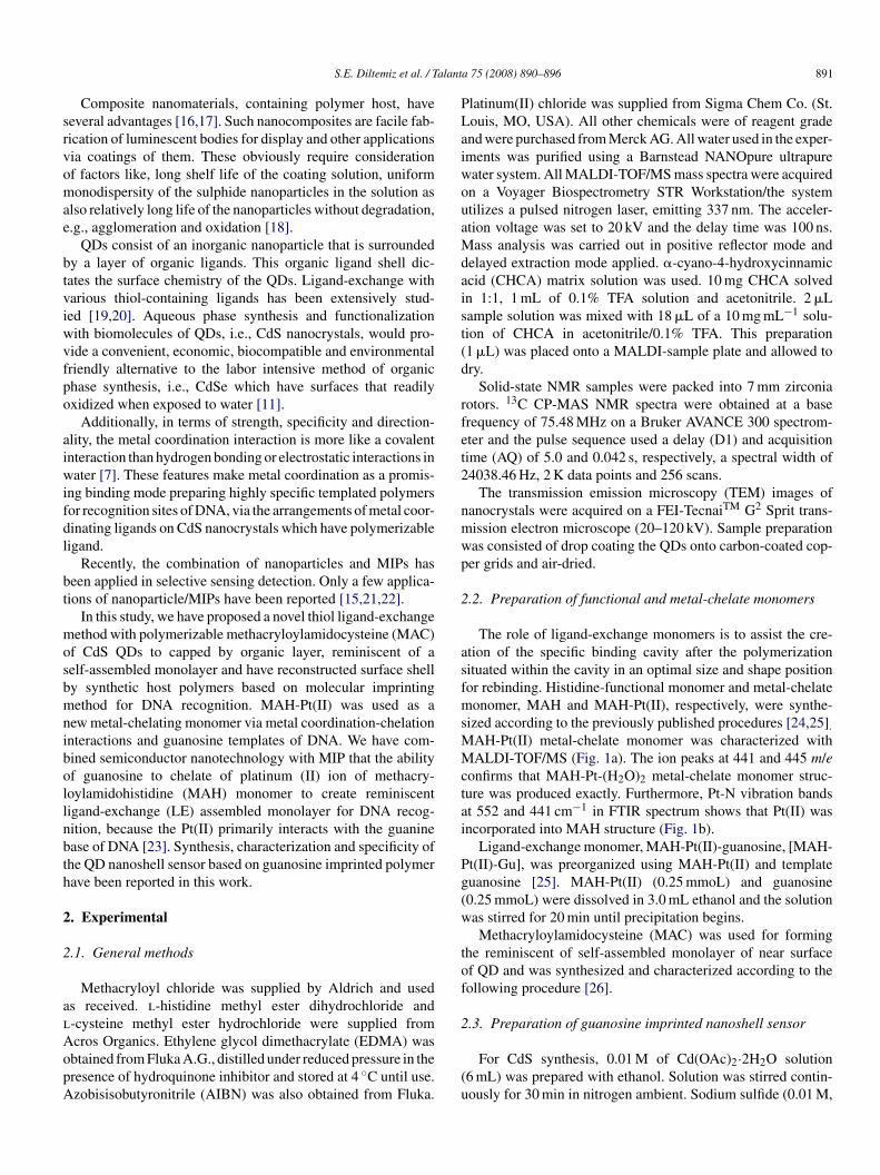
alant
srvomae
btviwvfpo
aiwifdl
bt
mosbmnibollnbth
2
2
alAopA
PLaiwouaMdaist(d
rfet2
nmwp
2
asfmsMMctai
Pg(w
tof
2
S.E. Diltemiz et al. / T
Composite nanomaterials, containing polymer host, haveeveral advantages [16,17]. Such nanocomposites are facile fab-ication of luminescent bodies for display and other applicationsia coatings of them. These obviously require considerationf factors like, long shelf life of the coating solution, uniformonodispersity of the sulphide nanoparticles in the solution as
lso relatively long life of the nanoparticles without degradation,.g., agglomeration and oxidation [18].
QDs consist of an inorganic nanoparticle that is surroundedy a layer of organic ligands. This organic ligand shell dic-ates the surface chemistry of the QDs. Ligand-exchange witharious thiol-containing ligands has been extensively stud-ed [19,20]. Aqueous phase synthesis and functionalizationith biomolecules of QDs, i.e., CdS nanocrystals, would pro-ide a convenient, economic, biocompatible and environmentalriendly alternative to the labor intensive method of organichase synthesis, i.e., CdSe which have surfaces that readilyxidized when exposed to water [11].
Additionally, in terms of strength, specificity and direction-lity, the metal coordination interaction is more like a covalentnteraction than hydrogen bonding or electrostatic interactions inater [7]. These features make metal coordination as a promis-
ng binding mode preparing highly specific templated polymersor recognition sites of DNA, via the arrangements of metal coor-inating ligands on CdS nanocrystals which have polymerizableigand.
Recently, the combination of nanoparticles and MIPs haseen applied in selective sensing detection. Only a few applica-ions of nanoparticle/MIPs have been reported [15,21,22].
In this study, we have proposed a novel thiol ligand-exchangeethod with polymerizable methacryloylamidocysteine (MAC)
f CdS QDs to capped by organic layer, reminiscent of aelf-assembled monolayer and have reconstructed surface shelly synthetic host polymers based on molecular imprintingethod for DNA recognition. MAH-Pt(II) was used as a
ew metal-chelating monomer via metal coordination-chelationnteractions and guanosine templates of DNA. We have com-ined semiconductor nanotechnology with MIP that the abilityf guanosine to chelate of platinum (II) ion of methacry-oylamidohistidine (MAH) monomer to create reminiscentigand-exchange (LE) assembled monolayer for DNA recog-ition, because the Pt(II) primarily interacts with the guaninease of DNA [23]. Synthesis, characterization and specificity ofhe QD nanoshell sensor based on guanosine imprinted polymerave been reported in this work.
. Experimental
.1. General methods
Methacryloyl chloride was supplied by Aldrich and useds received. l-histidine methyl ester dihydrochloride and-cysteine methyl ester hydrochloride were supplied from
cros Organics. Ethylene glycol dimethacrylate (EDMA) wasbtained from Fluka A.G., distilled under reduced pressure in theresence of hydroquinone inhibitor and stored at 4 ◦C until use.zobisisobutyronitrile (AIBN) was also obtained from Fluka.(u
a 75 (2008) 890–896 891
latinum(II) chloride was supplied from Sigma Chem Co. (St.ouis, MO, USA). All other chemicals were of reagent gradend were purchased from Merck AG. All water used in the exper-ments was purified using a Barnstead NANOpure ultrapureater system. All MALDI-TOF/MS mass spectra were acquiredn a Voyager Biospectrometry STR Workstation/the systemtilizes a pulsed nitrogen laser, emitting 337 nm. The acceler-tion voltage was set to 20 kV and the delay time was 100 ns.ass analysis was carried out in positive reflector mode and
elayed extraction mode applied. �-cyano-4-hydroxycinnamiccid (CHCA) matrix solution was used. 10 mg CHCA solvedn 1:1, 1 mL of 0.1% TFA solution and acetonitrile. 2 �Lample solution was mixed with 18 �L of a 10 mg mL−1 solu-ion of CHCA in acetonitrile/0.1% TFA. This preparation1 �L) was placed onto a MALDI-sample plate and allowed tory.
Solid-state NMR samples were packed into 7 mm zirconiaotors. 13C CP-MAS NMR spectra were obtained at a baserequency of 75.48 MHz on a Bruker AVANCE 300 spectrom-ter and the pulse sequence used a delay (D1) and acquisitionime (AQ) of 5.0 and 0.042 s, respectively, a spectral width of4038.46 Hz, 2 K data points and 256 scans.
The transmission emission microscopy (TEM) images ofanocrystals were acquired on a FEI-TecnaiTM G2 Sprit trans-ission electron microscope (20–120 kV). Sample preparationas consisted of drop coating the QDs onto carbon-coated cop-er grids and air-dried.
.2. Preparation of functional and metal-chelate monomers
The role of ligand-exchange monomers is to assist the cre-tion of the specific binding cavity after the polymerizationituated within the cavity in an optimal size and shape positionor rebinding. Histidine-functional monomer and metal-chelateonomer, MAH and MAH-Pt(II), respectively, were synthe-
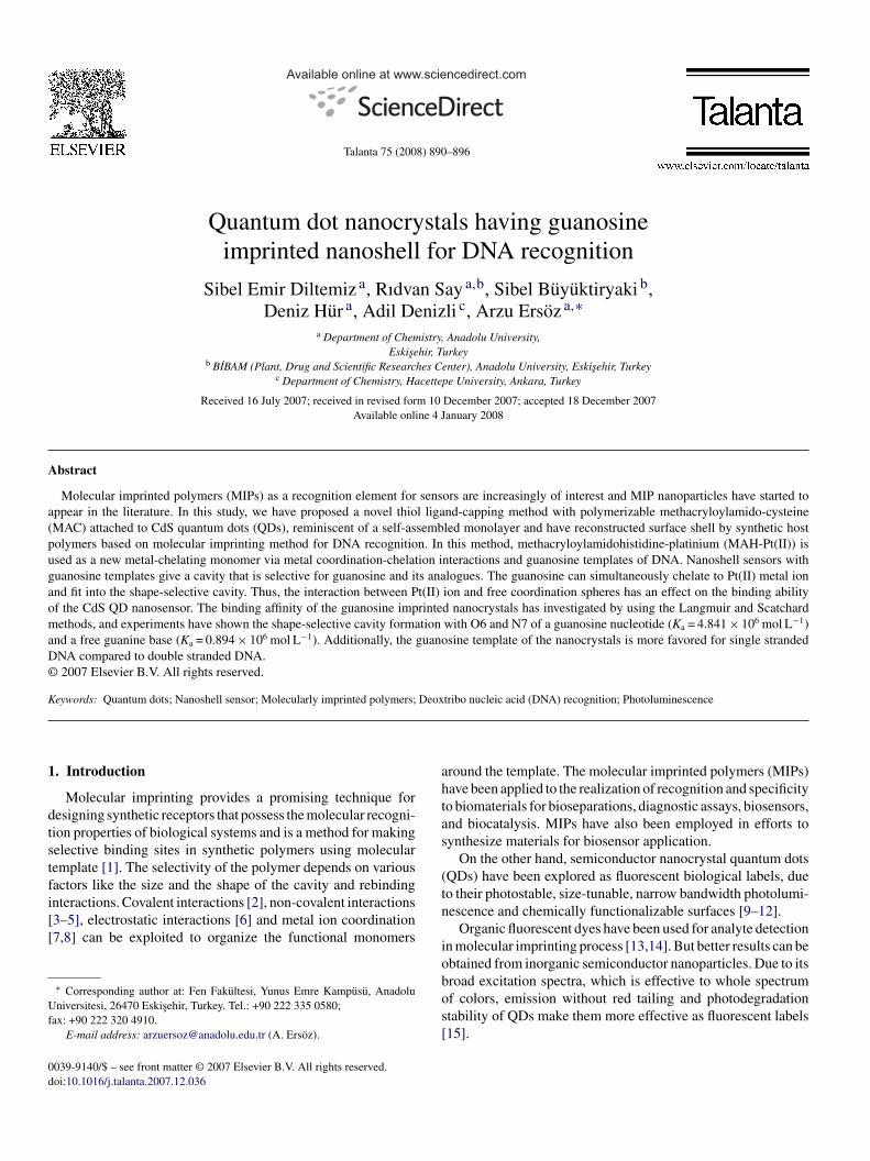
ized according to the previously published procedures [24,25].AH-Pt(II) metal-chelate monomer was characterized withALDI-TOF/MS (Fig. 1a). The ion peaks at 441 and 445 m/e
onfirms that MAH-Pt-(H2O)2 metal-chelate monomer struc-ure was produced exactly. Furthermore, Pt-N vibration bandst 552 and 441 cm−1 in FTIR spectrum shows that Pt(II) wasncorporated into MAH structure (Fig. 1b).
Ligand-exchange monomer, MAH-Pt(II)-guanosine, [MAH-t(II)-Gu], was preorganized using MAH-Pt(II) and templateuanosine [25]. MAH-Pt(II) (0.25 mmoL) and guanosine0.25 mmoL) were dissolved in 3.0 mL ethanol and the solutionas stirred for 20 min until precipitation begins.Methacryloylamidocysteine (MAC) was used for forming
he reminiscent of self-assembled monolayer of near surfacef QD and was synthesized and characterized according to theollowing procedure [26].
.3. Preparation of guanosine imprinted nanoshell sensor
For CdS synthesis, 0.01 M of Cd(OAc)2·2H2O solution6 mL) was prepared with ethanol. Solution was stirred contin-ously for 30 min in nitrogen ambient. Sodium sulfide (0.01 M,
892 S.E. Diltemiz et al. / Talanta 75 (2008) 890–896
Fig. 1. (a) MALDI-TOF/MS spectrum of metal-chelate monomer (2 �L MAH-Pt(II) solution was mixed with 18 �L of a 10 mg mL−1 solution of CHCA inat
63iw
(owtos
ni(EtTp6wntm
Fr
Kp
cplmpor
2
mbasTseparation between the excitation and emission wavelengths,
cetonitrile/0.1% TFA. The acceleration voltage was set to 20 kV and the delayime was 100 ns) and (b) FTIR spectrum of MAH-Pt(II).
mL) was slowly added, stirred under nitrogen ambient for0 min and then centrifuged to collect precipitate. It was washedn double distilled water and dried in air. The entire synthesisas carried out at room temperature [27].CdS nanocrystals were immersed in ethanol containing MAC
0.2 mg) for 30 min in order to introduce methacryloyl groupsnto the surface of CdS nanocrystals. The nanocrystals were thenashed with ethanol and deionized water for 10 min to remove
he excess of thiol groups. A stable self-assembled monolayerf MAC was formed onto the nanocrystal surfaces after all theseteps.
For the synthesis of guanosine recognized nanoshell/polymeranocrystals, methacryloyl-activated nanocrystals weremmersed in the reaction mixture containing the metal-chelateMAH-Pt(II)/Gu) monomer (0.05 mmol) in methoxyethanol,DMA cross-linking monomer (0.4 mmol) and 1 mol% of
he initiator (AIBN) of the radical polymerization in ethanol.his solution was transferred into the dispersion mediumlaced in a magnetically stirred (at a constant stirring rate of00 rpm) glass polymerization reactor. The reactor temperatureas kept at 60 ◦C for 48 h. After polymerization, guanosine
anoshells having CdS nanocrystals were separated fromhe polymerization medium. The residuals (e.g., unconvertedonomer, initiator, etc.) were removed by cleaning procedure.
smS
ig. 2. Schematic representation of nanoshell based on guanosine templateeconstruction on CdS/QD.
The resulting nanocrystals were treated with 15 mL 1.0 M ofOH solution in ethanol for 24 h to remove the templates. Thisrocedure was repeated two times.
The functional metal-chelate monomer, MAH-Pt(II), washose to interact guanosine and to make metal-complexingolymeric receptors for selective binding of guanosine and ana-ogues. Metal-chelate monomer and guanosine molecule wasixed through preorganization and this preorganization com-
lex defines the size and direction of the chemical interactionsf the guanosine imprinted cavity to prepare synthetic guanosineeceptor of CdS/QD (Fig. 2).
.4. Evaluation of nanoshell QDs luminescence
The sensing capability and specificity of the guanosineemory having CdS nanoshell sensor were further explored
y introducing guanosine as a template molecule. The inter-ctions with guanine, ss-DNA, ds-DNA and adenosine weretudied using fluorescence (measurements) spectrophotometer.he guanosine imprinted nanoshell CdS/QDs showed a high
implifying fluorescence measurements, recorded photolu-inescence spectra using spectrofluorometer (RF-5301 PC,himadzu Japan). Guanine, guanosine, ss-DNA, ds-DNA sam-
S.E. Diltemiz et al. / Talant
Fa7
pe
3
3n
n
gFttIobacrc
ptCnpp
nr
spwatMl
lsMi3tmsmcn
3i
nsrabno
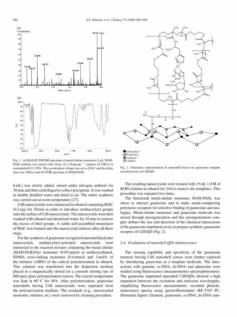
ig. 3. The 13C CP-MAS NMR spectra of (a) CdS-MAC; (b) before and (c)fter the removal of template of the guanosine imprinted nanoshell (7.04 T,5.43 MHz and 256 scans).
les and the CdS/QD nanosensors were excited at 300 nm, andmission was recorded at 600 nm.
. Results and discussion
.1. 13C solid-state NMR and TEM characterization of
anoshell sensorsThe 13C CP-MAS NMR spectra of MAC capped CdSanoparticles and CdS-MIP core-shell nanoparticles (with
Cap
a 75 (2008) 890–896 893
uanosine template and without template) are shown inig. 3a–c. We have compared the 13C CP-MAS NMR spec-
ra of CdS-MAC (Fig. 3a) before (Fig. 3b) and after (Fig. 3c)he removal of template of the guanosine imprinted nanoshell.n Fig. 3a, the peak at 194.4 ppm is due to the carbonyl groupf cysteine and resonance at 198 ppm signal is due to the car-onyl of methacryloyl group. On the other hand, signals at 60.9nd 73.8 ppm in Fig. 3a are due to the methine and methylenearbons of methacryloyl groups, respectively. In addition, theesonance at 50 ppm CA and the resonance at 38.2 ppm CB ofysteine were observed.
As far as the guanosine template polymer is concerned, theeaks at 60.9 and 73.8 ppm were disappeared as the incorpora-ion of MAH (Fig. 3b). The appearance of histidine resonancesA and CB in the region of 66.3–79.4 ppm, guanosine reso-ances C4′, C1′ and C3′ in the region of 88.0–82.1 ppm andarticularly increase of the signal around 200 ppm shows incor-oration of cross-linker EDMA into polymer at high ratio.
After guanosine (template) was removed from CdS/QDanoshell, disappearance of peaks in the range of 88.0–82.0 ppmevealed to the guanosine memory of nanoshell (Fig. 3c).
The incorporation of MAC into CdS/QD from the bindingite of thiolate groups was supported by the observation ofeak at 41.7 ppm, the conservation of MAH-Pt groups whichere binded to them observed in the region of 81.0–66.0 ppm
nd the formation of cross-linked MIP based core/shell QD byhe existence of peak around 199 ppm. This is the first 13CP-
AS NMR description of these types of the structure in theiterature.
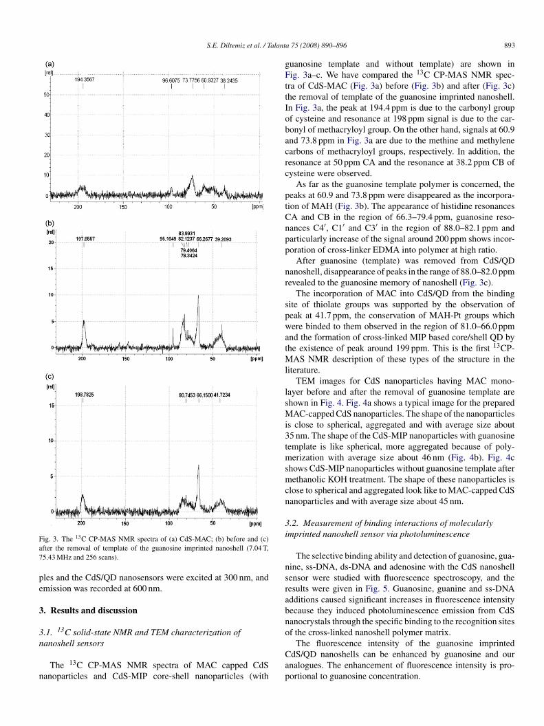
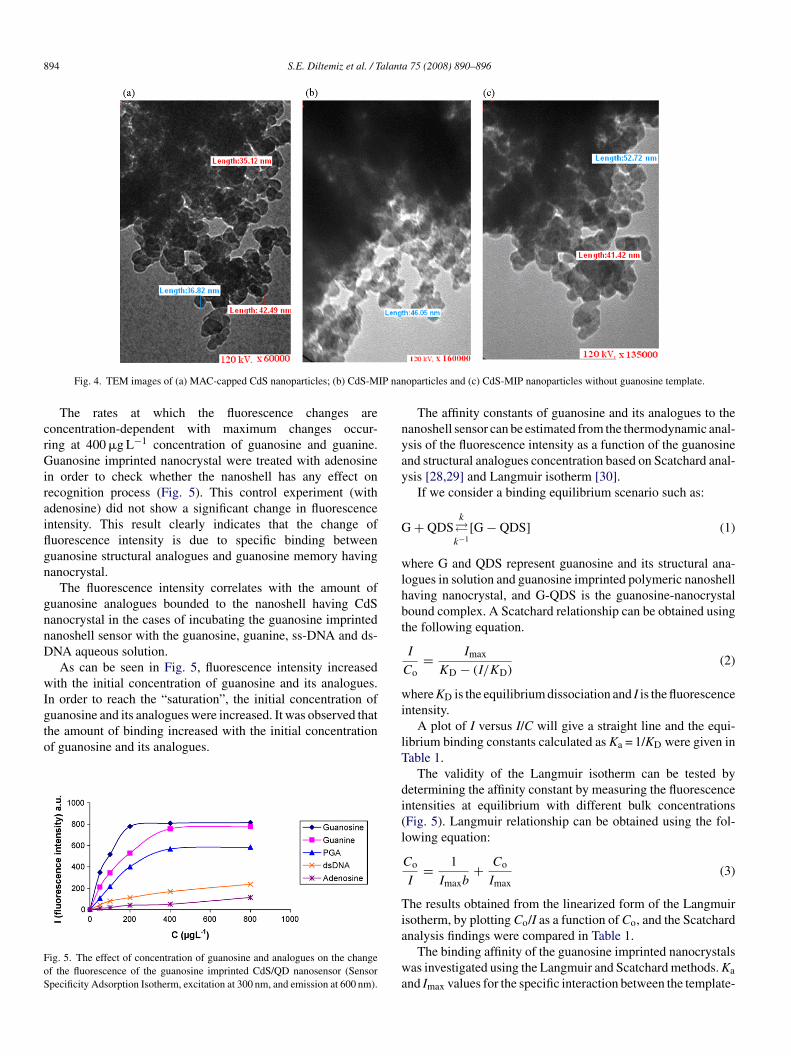
TEM images for CdS nanoparticles having MAC mono-ayer before and after the removal of guanosine template arehown in Fig. 4. Fig. 4a shows a typical image for the prepared
AC-capped CdS nanoparticles. The shape of the nanoparticless close to spherical, aggregated and with average size about5 nm. The shape of the CdS-MIP nanoparticles with guanosineemplate is like spherical, more aggregated because of poly-
erization with average size about 46 nm (Fig. 4b). Fig. 4chows CdS-MIP nanoparticles without guanosine template afterethanolic KOH treatment. The shape of these nanoparticles is
lose to spherical and aggregated look like to MAC-capped CdSanoparticles and with average size about 45 nm.
.2. Measurement of binding interactions of molecularlymprinted nanoshell sensor via photoluminescence
The selective binding ability and detection of guanosine, gua-ine, ss-DNA, ds-DNA and adenosine with the CdS nanoshellensor were studied with fluorescence spectroscopy, and theesults were given in Fig. 5. Guanosine, guanine and ss-DNAdditions caused significant increases in fluorescence intensityecause they induced photoluminescence emission from CdSanocrystals through the specific binding to the recognition sitesf the cross-linked nanoshell polymer matrix.
The fluorescence intensity of the guanosine imprinteddS/QD nanoshells can be enhanced by guanosine and ournalogues. The enhancement of fluorescence intensity is pro-ortional to guanosine concentration.
894 S.E. Diltemiz et al. / Talanta 75 (2008) 890–896
IP nan
crGiraiflgn
gnnD
wIgto
FoS
nyay
G
wlhbt
wi
Fig. 4. TEM images of (a) MAC-capped CdS nanoparticles; (b) CdS-M
The rates at which the fluorescence changes areoncentration-dependent with maximum changes occur-ing at 400 �g L−1 concentration of guanosine and guanine.uanosine imprinted nanocrystal were treated with adenosine
n order to check whether the nanoshell has any effect onecognition process (Fig. 5). This control experiment (withdenosine) did not show a significant change in fluorescencentensity. This result clearly indicates that the change ofuorescence intensity is due to specific binding betweenuanosine structural analogues and guanosine memory havinganocrystal.
The fluorescence intensity correlates with the amount ofuanosine analogues bounded to the nanoshell having CdSanocrystal in the cases of incubating the guanosine imprintedanoshell sensor with the guanosine, guanine, ss-DNA and ds-NA aqueous solution.As can be seen in Fig. 5, fluorescence intensity increased
ith the initial concentration of guanosine and its analogues.n order to reach the “saturation”, the initial concentration of
uanosine and its analogues were increased. It was observed thathe amount of binding increased with the initial concentrationf guanosine and its analogues.ig. 5. The effect of concentration of guanosine and analogues on the changef the fluorescence of the guanosine imprinted CdS/QD nanosensor (Sensorpecificity Adsorption Isotherm, excitation at 300 nm, and emission at 600 nm).
lT
di(l
Tia
wa
oparticles and (c) CdS-MIP nanoparticles without guanosine template.
The affinity constants of guanosine and its analogues to theanoshell sensor can be estimated from the thermodynamic anal-sis of the fluorescence intensity as a function of the guanosinend structural analogues concentration based on Scatchard anal-sis [28,29] and Langmuir isotherm [30].
If we consider a binding equilibrium scenario such as:
+ QDSk
�k−1
[G − QDS] (1)
here G and QDS represent guanosine and its structural ana-ogues in solution and guanosine imprinted polymeric nanoshellaving nanocrystal, and G-QDS is the guanosine-nanocrystalound complex. A Scatchard relationship can be obtained usinghe following equation.
I
Co= Imax
KD − (I/KD)(2)
here KD is the equilibrium dissociation and I is the fluorescencentensity.
A plot of I versus I/C will give a straight line and the equi-ibrium binding constants calculated as Ka = 1/KD were given inable 1.
The validity of the Langmuir isotherm can be tested byetermining the affinity constant by measuring the fluorescencentensities at equilibrium with different bulk concentrationsFig. 5). Langmuir relationship can be obtained using the fol-owing equation:
Co
I= 1
Imaxb+ Co
Imax(3)
he results obtained from the linearized form of the Langmuirsotherm, by plotting Co/I as a function of Co, and the Scatchard
nalysis findings were compared in Table 1.The binding affinity of the guanosine imprinted nanocrystalsas investigated using the Langmuir and Scatchard methods. Ka
nd Imax values for the specific interaction between the template-
S.E. Diltemiz et al. / Talanta 75 (2008) 890–896 895
Table 1Comparison of Langmuir and Scatchard analysis for guanosine imprinted nanocrystals
Rebound molecules Langmuir Ka (L �g−1) Imax (a.u.) Scatchard, Ka (L �g−1) Imax (a.u.)
Guanosine 1.71 × 10⁻² 909.1 1.06 × 10⁻² 1011Guanine 5.92 × 10⁻³ 1000 5.0 × 10⁻³ 1053P −3 83 −3
d 33A 25
iw
awafmriRagcrs3ytin
atsSMti1b
3n
stigiiasn(b
Khtmg
4
fuCEaocbCnaiotbste
atnLco
maaoPeug
olyguanylic acid 3.81 × 10s-DNA 2.86 × 10⁻³
denosine 8.82 × 10⁻⁴
mprinted polymer of the nanocrystal and the template itselfere determined by Langmuir isotherms and Scatchard’s plots.The Langmuir adsorption model assumes that the molecules
re adsorbed at a fixed number of well-defined sites, each ofhich is capable of holding only one molecule. These sites
re also assumed to be energetically equivalent, and distantrom each other so that there are no interactions betweenolecules adsorbed on adjacent sites. According to the cor-
elation coefficients of isotherms, Langmuir adsorption models most favorable for guanosine and guanine (R2: 0.995 and2: 0.989, respectively) and Langmuir model was found to bepplicable in interpreting guanosine and analogues adsorptionuanosine imprinted CdS/QD. The comparison of the asso-iation constants, Ka, and the apparent maximum number ofecognition sites, Imax values, are presented in Table 1. Aseen from Table 1, in general magnitude of Ka (4.841 × 10⁶,.0 × 106 mol L−1, based on Langmuir and Scatchard anal-sis, respectively) is due to the accessibility of guanosineemplate molecules. Some template cavities formed duringmprinting process were stayed inside the polymer matrix of theanoshell.
The Ka and Imax values estimated from Scatchard analysisre very close to the Langmuir analysis data and guanosineemplated sites of nanocrystals are highly selective to the guano-ine recognition sites. So the association constants based oncatchard analysis for the binding of guanosine and guanine toIP nanosensor are 3.0 × 10⁶ and 0.76 × 10⁶ mol L−1, respec-
ively. Maximum fluorescence intensity of ligand-exchangenteraction sites for guanosine and guanine, Imax, are 1011 and053, respectively. The value of Ka suggests that affinity of theinding sites is very strong.
.3. Recognition selectivity of guanosine imprintedanoshell sensor
Molecular imprinting with guanosine gives a cavity that iselective for guanosine and its analogues. For various nucleotidehe oligomers serving as DNA models, the highest binding affin-ty was found between N7 of guanine and platinum [23]. Theuanosine can simultaneously chelate to Pt(II) metal ion and fitnto the shape-selective cavity. So, this interaction between Pt(II)on and free coordination spheres has an effect on the bindingbility of the CdS/QD nanosensor. Experiments have shown that
hape-selective cavity formation with O6 and N7 of a guanosineucleotide (Ka = 4.84 × 10⁶ mol L−1) and a free guanine baseKa = 0.894 × 10⁶ mol L−1) can contribute with natural anti-odies; however, single stranded DNA (poly(guanidylic acid),cTtm
3.3 1.6 × 10 15253.3 3.2 × 10⁻³ 313.80.0 0.9 × 10⁻³ 250.7
a = 3.81 × 10⁻³ L �g−1) is more favored than DNA doubleelix (2.86 × 10⁻³ L �g−1) for shape-selective cavity forma-ion. This can be explained that most of the retained MAH-Pt(II)
emory binding sites bonds two neighboring N7-coordinateduanosine nucleotides on the same DNA strand.
. Conclusion
We have developed a novel chemical preparation methodor methacryloyl based self-assembled monolayer and to makep imprinting polymer via ligand-exchange of guanosine ondS/QD nanoshell. The guanosine imprinted MAH-Pt(II)-DMA copolymer is expected to bind guanosine and itsnalogues for DNA sensing. The results showed that the flu-rescence enhancement effect could be attributed to the highomplexation geometric shape affinity (or guanosine memory)etween guanosine molecules and guanosine cavities on todS/QD nanoshells. In conclusion, the guanosine imprintedanoshell sensor has been gaining widespread recognition assensor for guanosine and its analogues because the imprint-
ng methods create a nanoenvironment based on the shapef the cavity memorial, the size and the positions of func-ional groups that recognizes the imprinting molecule guanosineased on ligand-exchange imprinting methods. Nanoshell sen-or having guanosine templates responses to the concentration ofhe guanosine and its analogues through fluorescence intensitynhancement.
Our results not only demonstrate that guanosine detection ischieved using CdS/QD nanoshell, but that they can also be usedo assay target DNA sequencing. Guanosine imprinted CdS/QDanoshells can recognize and bind the guanosine molecules.ikewise, CdS/QD nanocrystals containing MAH-Pt-guanosinean also be used in the mutagenic studies and for the detectionf DNA defects.
Moreover, as a result of the reaction between guanineolecule and methylazonium cation or ethylmethyl sulphanate,
lkylies of these compounds are bound from O6 site of guaninend usually guanine–thymine pair interaction is formed insteadf guanine–cytosine interaction [31]. In the same way, in MAH-t-guanine monomer which has presented in this study, in thend of the interaction between Pt(II) and O6 site of guanine andsually guanine–thymine pair interaction is formed instead ofuanine–cytosine interaction and this metal-chelate monomer
an be used in both solid phase systems and nanoparticles.hus, ODs can also be used for both molecular recogni-ion based on the hybridization of nucleic acid strands andutagenesis.
8 alant
A
ma
R
[[
[
[
[
[
[[[
[
[[[
[
[
[[
[
[[29] M. Yang, P.Y. Tsoi, C.W. Li, J. Zhao, Sens. Actuators B 115 (2006) 428.
96 S.E. Diltemiz et al. / T
cknowledgments
This work has been supported by Anadolu University, Com-ission of Scientific Research Projects (Project No.: 04 10 06)
nd dedicated to the 50th anniversary of Anadolu University.
eferences
[1] B. Sellergren, Anal. Chem. 66 (1994) 1578.[2] M.J. Whitcombe, M.E. Rodriquez, P. Villar, E.N. Vulfson, J. Am. Chem.
Soc. 117 (1995) 7105.[3] O. Ramstrom, L.I. Andersson, K. Mosbach, J. Org. Chem. 58 (1993) 7562.[4] D.A. Spivak, K.J. Shea, Macromolecules 31 (1998) 2160.[5] R. Say, Anal. Chim. Acta 579 (2006) 74.[6] B. Sellergren, K.J. Shea, J. Chromatogr. 654 (1993) 31.[7] P.K. Dhal, F.H. Arnhold, Macromolecules 25 (1992) 7051.[8] M. Andac, R. Say, A. Denizli, J. Chromatogr. B 811 (2004) 119.[9] M. Bruchez Jr., M. Maronne, P. Gin, S. Weiss, A.P. Alivisatos, Science 281
(1998) 2013.10] W.C.W. Chan, S. Nie, Science 281 (1998) 2016.11] J.O. Winter, T.Y. Liu, B.A. Korgel, C.E. Schmidt, Adv. Mater. 13 (2001)
1673.12] J.O. Winter, N. Gomez, S. Gatzert, C.E. Schmidt, B.A. Korgel, Colloid
Surf. A 254 (2005) 147.13] P. Turkewitsch, B. Wandelt, G.D. Darling, W.S. Powell, Anal. Chem. 70
(1998) 2025.14] C.I. Lin, A.K. Joseph, C.K. Chang, Y.D. Lee, Biosens. Bioelect. 20 (2004)
127.
[
[
a 75 (2008) 890–896
15] C.I. Lin, A.K. Joseph, C.K. Chang, Y.D. Lee, J. Chromatogr. A 1027 (2004)259.
16] J. Yang, H. Lin, Q. He, L. Ling, C. Zhu, F. Bai, Langmuir 17 (2001) 5978.17] L. Qi, H. Colfen, M. Antionetti, Nanoletters 1 (2001) 61.18] B. Bhhattacharjee, D. Ganguli, S.J. Chadhuri, Nanoparticle Res. 4 (2002)
225.19] C. Bruchez, H. Mattoussi, J.M. Mouro, E.R. Goldman, G.P. Anderson,
V.C. Sundar, F.V. Mikulec, M.G. Bawendi, J. Am. Chem. Soc. 122 (2000)12142.
20] K. Sungjee, G.B. Moungi, J. Am. Chem. Soc. 125 (2003) 14652.21] A.B. Kharitonov, A.N. Shipway, I. Willner, Anal. Chem. 71 (1999) 1441.22] J. Matsui, K. Akamatsu, S. Nishiguchi, D. Miyoshi, H. Nawafune, K.
Tamaki, N. Sugimato, Anal. Chem. 76 (2004) 1310.23] W. Kaim, B. Schwederski, Bioinorganic Chemistry: Inorganic Elements in
the Chemistry of Life, John Wiley, 1994, pp. 369.24] R. Say, E. Birlik, A. Ersoz, F. Yılmaz, T. Gedikbey, A. Denizli, Anal. Chim.
Acta 480 (2003) 251.25] A. Ersoz, A. Denizli, A. Ozcan, R. Say, Biosens. Bioelect. 20 (2005) 2197.26] A. Denizli, R. Say, B. Garipcan, S. Emir, A. Karabakan, S. Patır, Sep. Purif.
Technol. 30 (2003) 3.27] S.K. Kulkarni, A.S. Ethiraj, S. Kharrazi, D.N. Deobagkar, D.D. Deobagkar,
Biosens. Bioelect. 21 (1) (2005) 95.28] P.Y. Tsoi, J. Yang, Y.T. Sun, S.F. Sui, M.S. Yang, Langmuir 16 (2000) 6590.
30] B. Persson, K. Stenhag, P. Nilsson, A. Larsson, M. Uhlen, P.A. Nygren,Anal. Biochem. 246 (1997) 34.
31] F. Marchesi, M. Turriziani, G. Tortorelli, G. Avvisati, F. Torino, L. DeVecchis, Pharmacol. Res. 56 (2007) 275.