neoplasia_general considerations
TRANSCRIPT
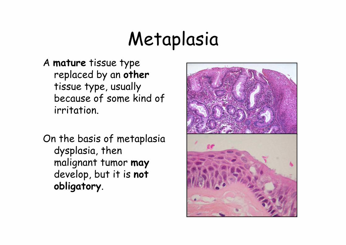
MetaplasiaA mature tissue type
replaced by an other tissue type, usually because of some kind of irritation.
On the basis of metaplasia dysplasia, then malignant tumor maydevelop, but it is not obligatory.
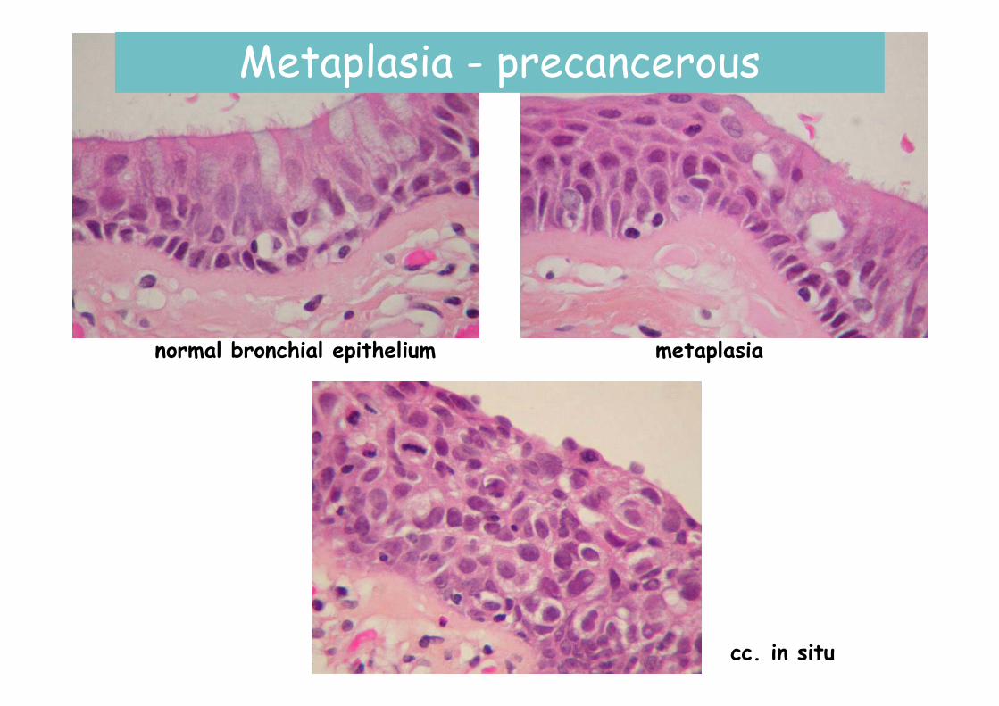
normal bronchial epithelium metaplasia
cc. in situ
Metaplasia - precancerous
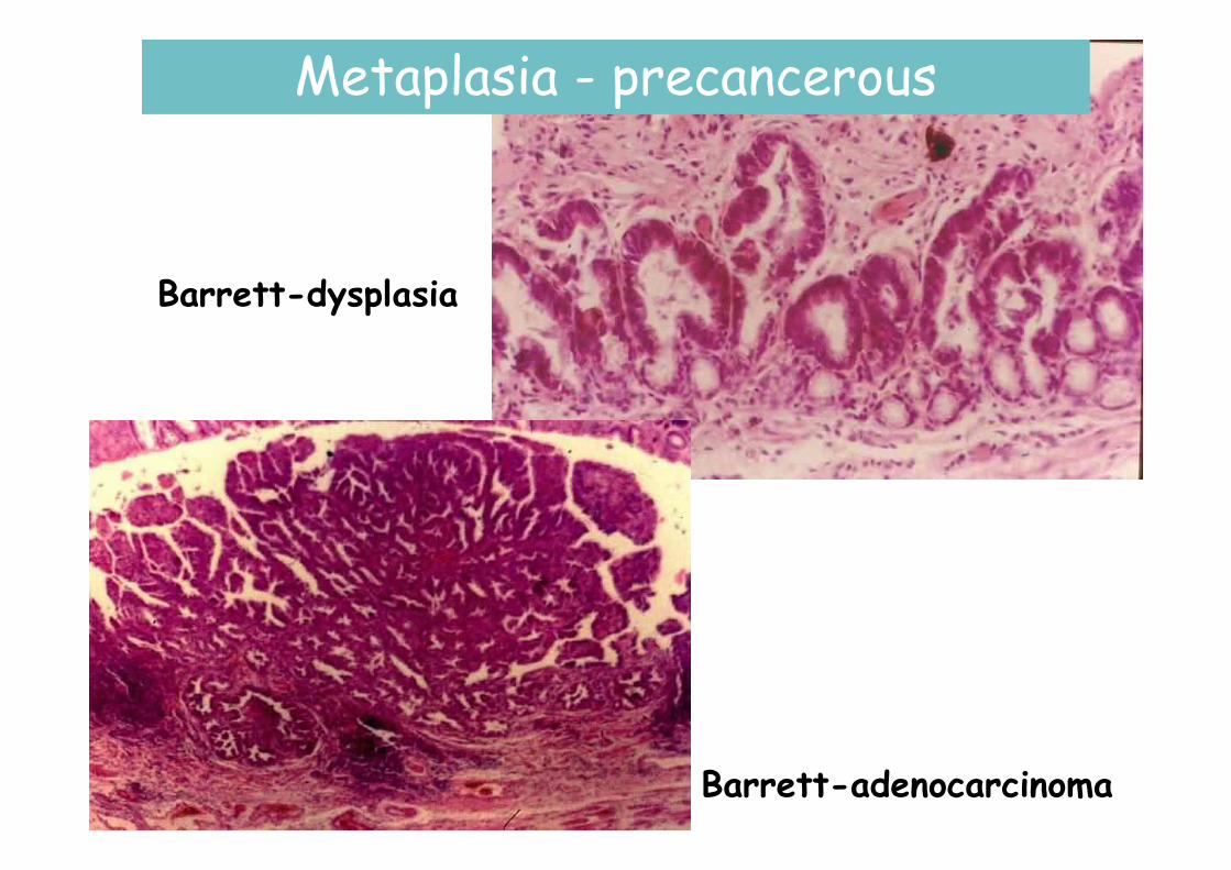
Barrett-dysplasia
Barrett-adenocarcinoma
Metaplasia - precancerous
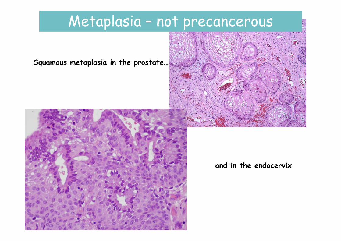
Squamous metaplasia in the prostate…
and in the endocervix
Metaplasia – not precancerous

Neoplasia
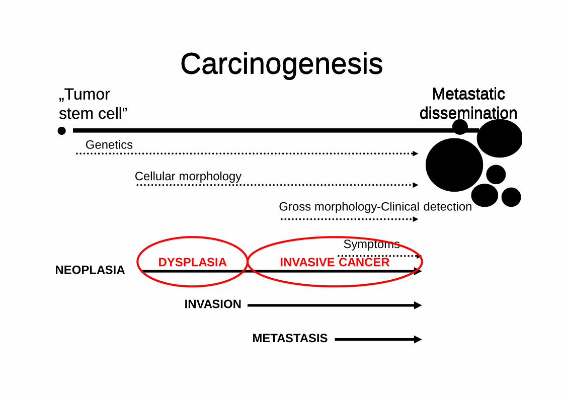
„Tumorstem cell”
Metastaticdissemination
Carcinogenesis
Genetics
Cellular morphology
Gross morphology-Clinical detection
Symptoms
INVASION
METASTASIS
NEOPLASIADYSPLASIA INVASIVE CANCER
Metastaticdissemination
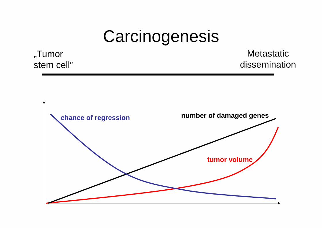
CarcinogenesisMetastatic
dissemination„Tumorstem cell”
CarcinogenesisMetastatic
dissemination
„Tumorstem cell”
CarcinogenesisMetastatic
dissemination
tumor volume
chance of regression number of damaged genes
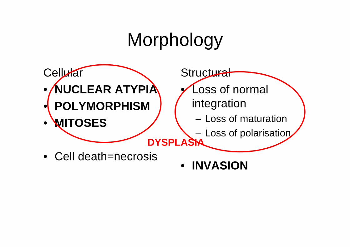
Morphology
Cellular• NUCLEAR ATYPIA• POLYMORPHISM• MITOSES
• Cell death=necrosis
Structural• Loss of normal
integration– Loss of maturation– Loss of polarisation
• INVASION
DYSPLASIA
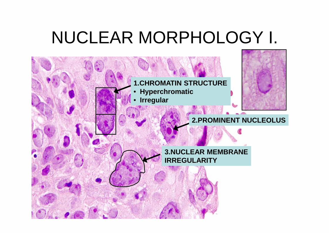
NUCLEAR MORPHOLOGY I.
1.CHROMATIN STRUCTURE• Hyperchromatic• Irregular
2.PROMINENT NUCLEOLUS
3.NUCLEAR MEMBRANEIRREGULARITY
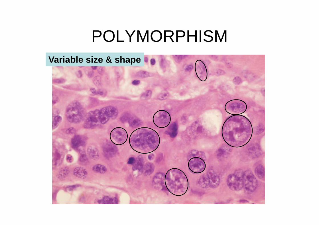
POLYMORPHISMVariable size & shape
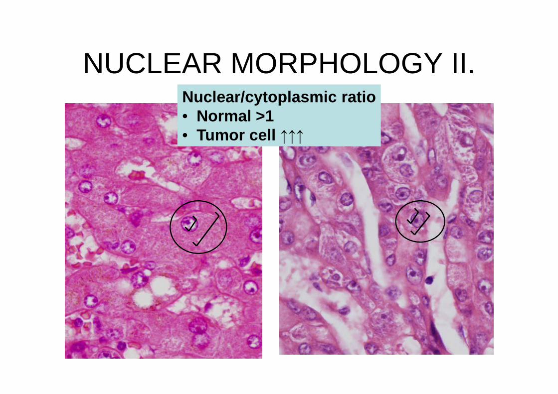
NUCLEAR MORPHOLOGY II.Nuclear/cytoplasmic ratio• Normal >1• Tumor cell ↑↑↑
MITOSIS
NUMBER• Proliferating tissues
– Abnormal location
• „Stable tissues”– Number ↑
MORPHOLOGY– Atypical mitoses – absolute sign of malignancy!!
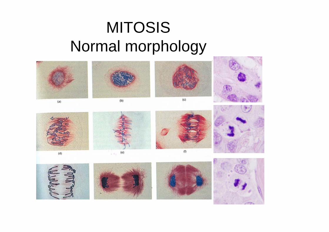
MITOSISNormal morphology
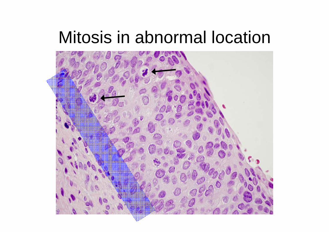
Mitosis in abnormal location
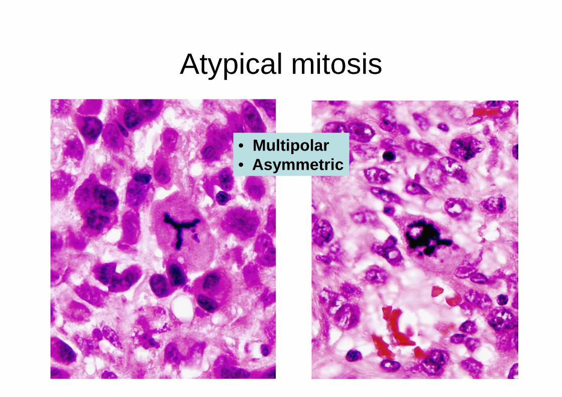
Atypical mitosis
• Multipolar• Asymmetric
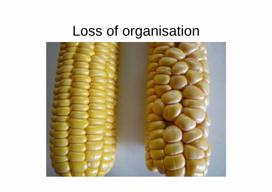
Loss of organisation
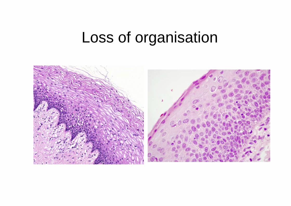
Loss of organisation
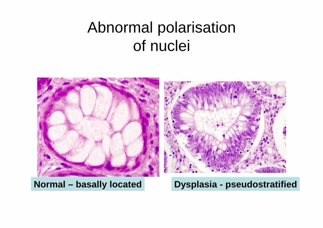
Abnormal polarisationof nuclei
Normal – basally located Dysplasia - pseudostratified
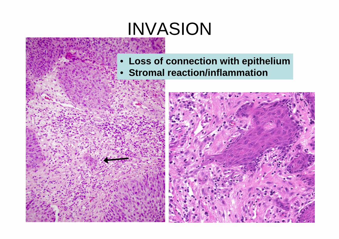
INVASION
• Loss of connection with epithelium• Stromal reaction/inflammation
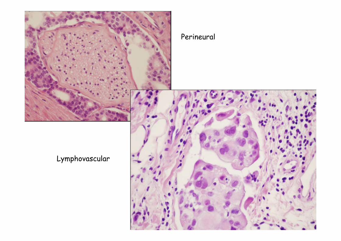
Perineural
Lymphovascular
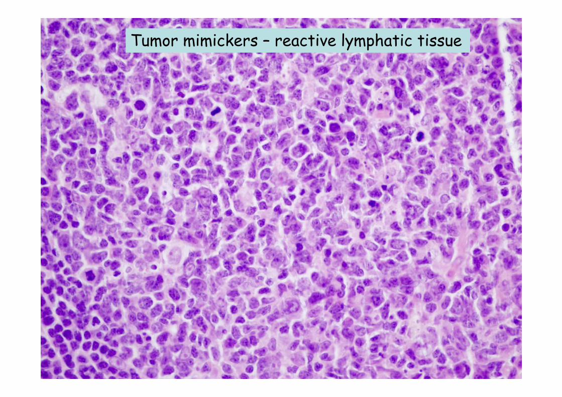
Tumor mimickers – reactive lymphatic tissue
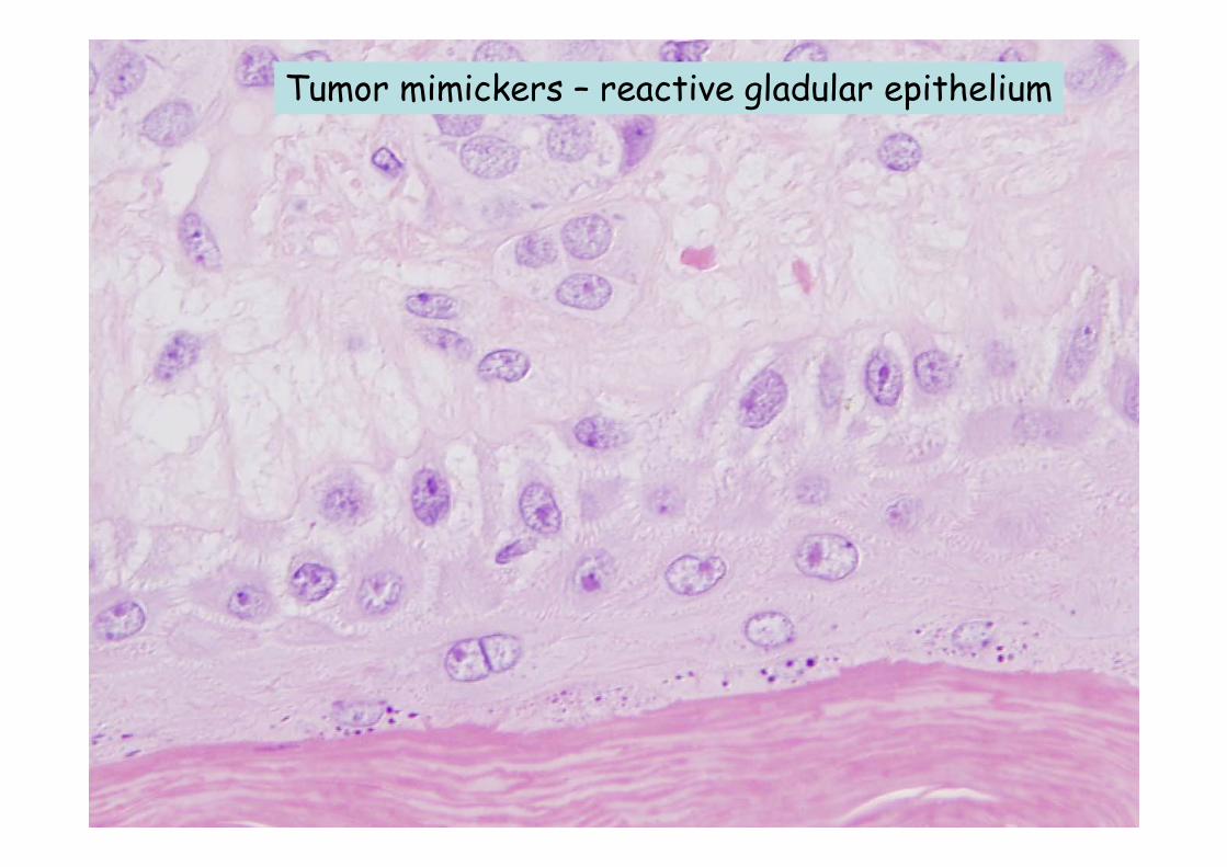
Tumor mimickers – reactive gladular epithelium
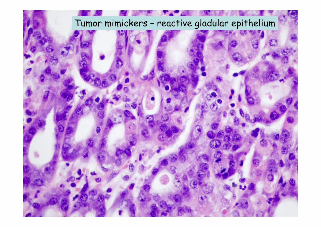
Tumor mimickers – reactive gladular epithelium
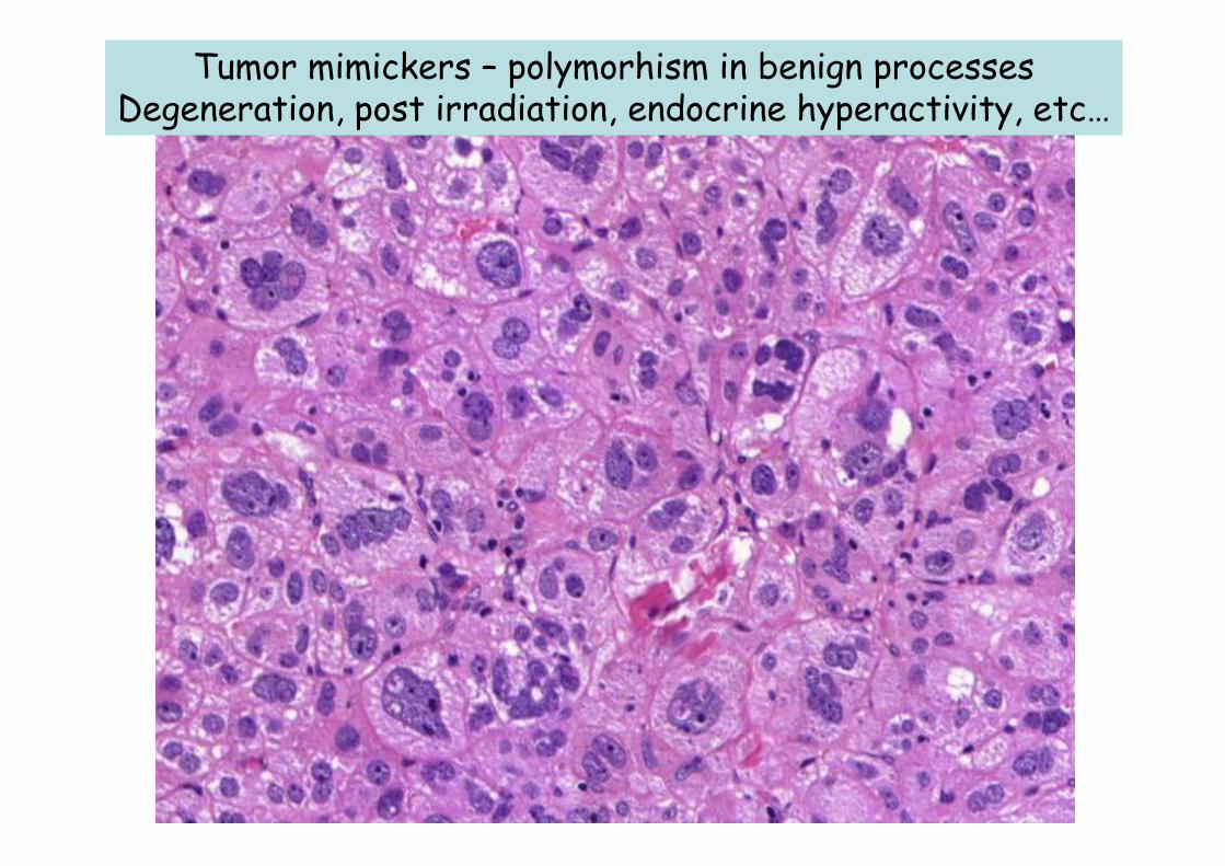
Tumor mimickers – polymorhism in benign processesDegeneration, post irradiation, endocrine hyperactivity, etc…
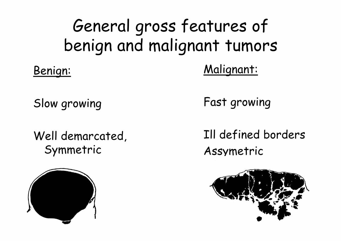
General gross features of benign and malignant tumors
Benign:
Slow growing
Well demarcated, Symmetric
Malignant:
Fast growing
Ill defined borders
Assymetric
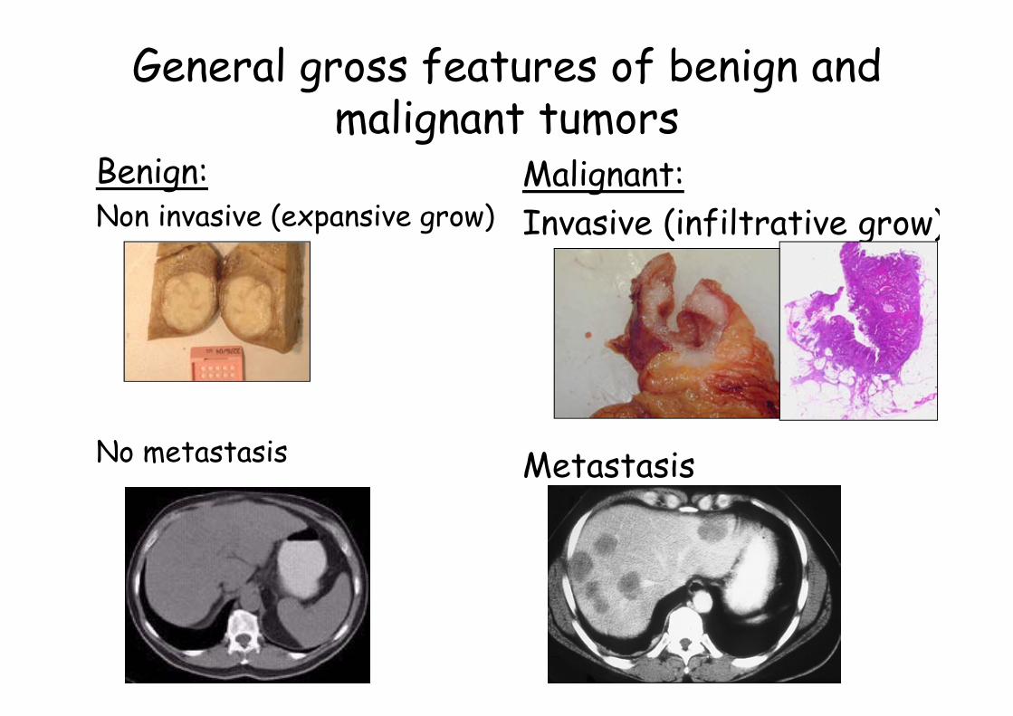
General gross features of benign and malignant tumors
Benign:Non invasive (expansive grow)
No metastasis
Malignant:
Invasive (infiltrative grow)
Metastasis
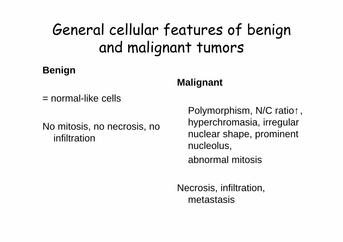
General cellular features of benign and malignant tumors
Benign
= normal-like cells
No mitosis, no necrosis, no infiltration
Malignant
Polymorphism, N/C ratio↑, hyperchromasia, irregular nuclear shape, prominent nucleolus, abnormal mitosis
Necrosis, infiltration, metastasis
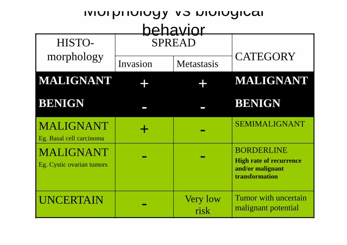
Morphology vs biological behavior
HISTO-morphology
SPREADCATEGORY
Invasion Metastasis
MALIGNANT + + MALIGNANT
BENIGN - - BENIGN
MALIGNANTEg. Basal cell carcinoma
+ - SEMIMALIGNANT
MALIGNANTEg. Cystic ovarian tumors
- - BORDERLINEHigh rate of recurrence and/or malignant transformation
UNCERTAIN - Very lowrisk
Tumor with uncertainmalignant potential
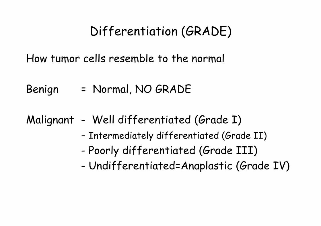
Differentiation (GRADE)
How tumor cells resemble to the normal
Benign = Normal, NO GRADE
Malignant - Well differentiated (Grade I)
- Intermediately differentiated (Grade II)
- Poorly differentiated (Grade III)
- Undifferentiated=Anaplastic (Grade IV)
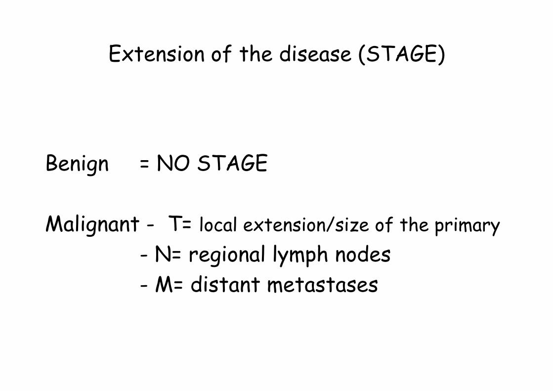
Extension of the disease (STAGE)
Benign = NO STAGE
Malignant - T= local extension/size of the primary
- N= regional lymph nodes
- M= distant metastases
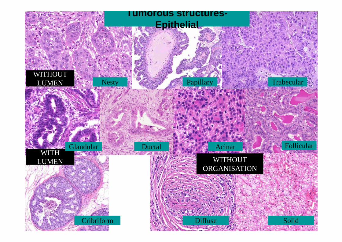
Tumorous structures -Epithelial
Nesty Papillary TrabecularWITHOUT
LUMEN
WITH LUMEN WITHOUT
ORGANISATION
Glandular AcinarDuctal Follicular
Cribriform Diffuse Solid
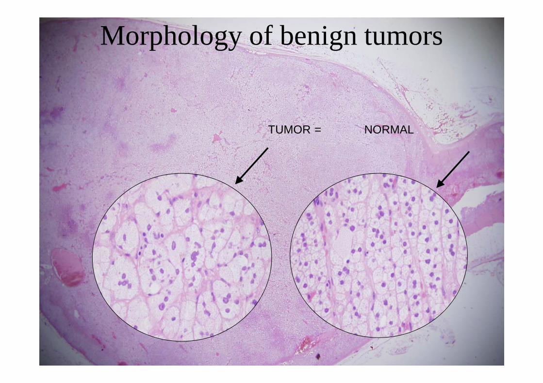
Morphology of benign tumors
TUMOR = NORMAL
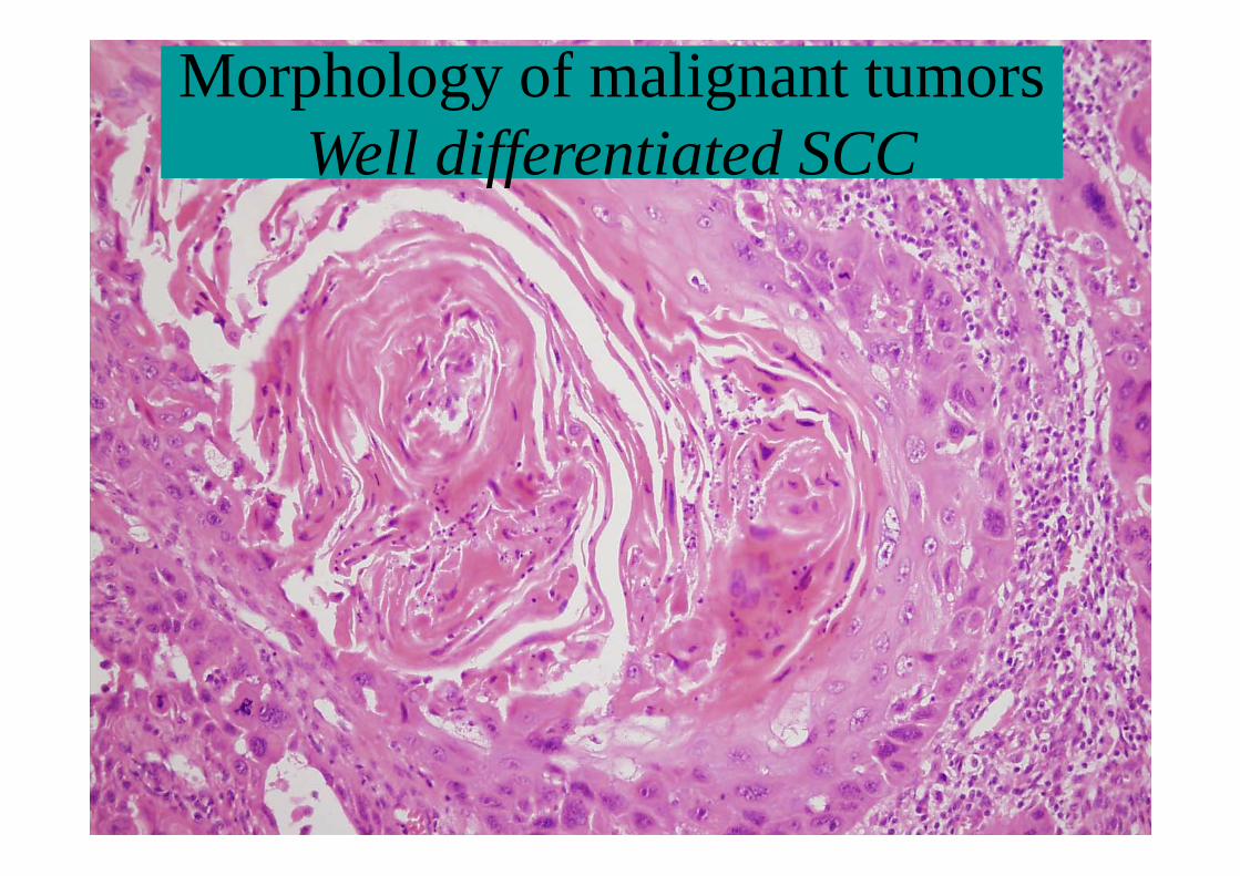
Morphology of malignant tumorsWell differentiated SCC
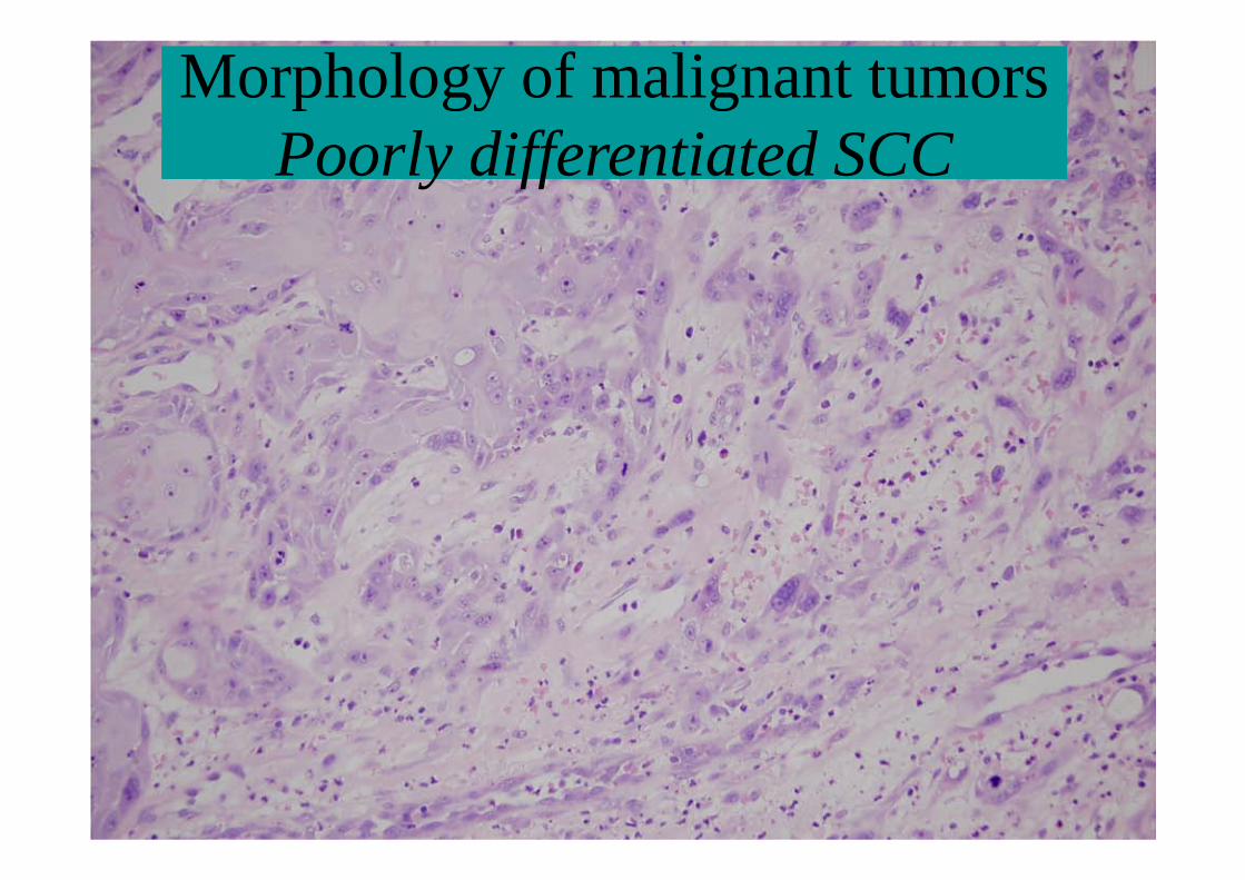
Morphology of malignant tumorsPoorly differentiated SCC
Colon
Kp. Diff.
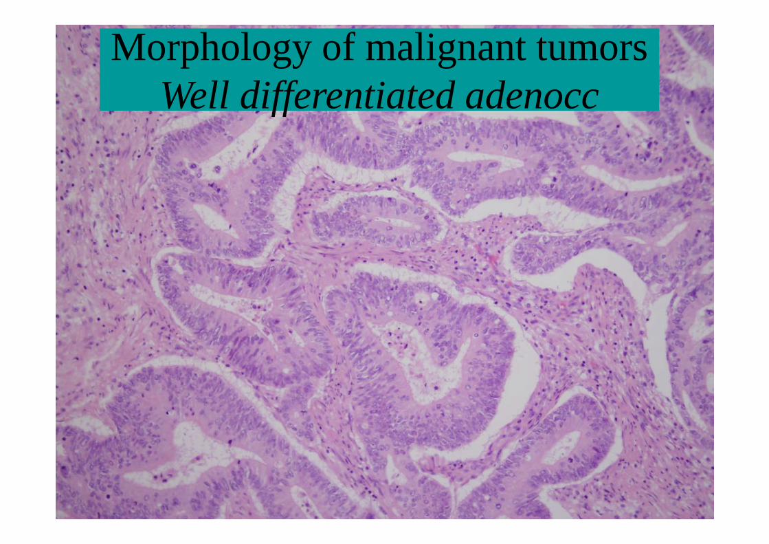
Morphology of malignant tumorsWell differentiated adenocc
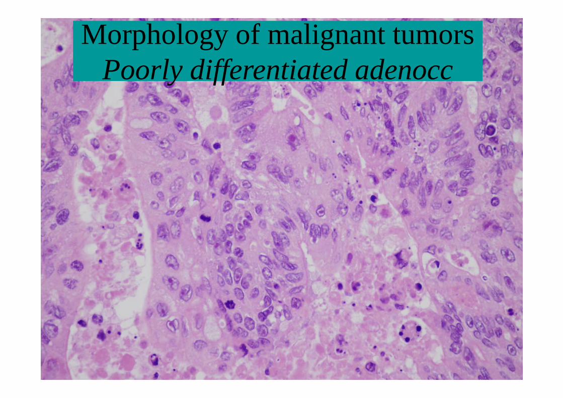
Morphology of malignant tumorsPoorly differentiated adenocc
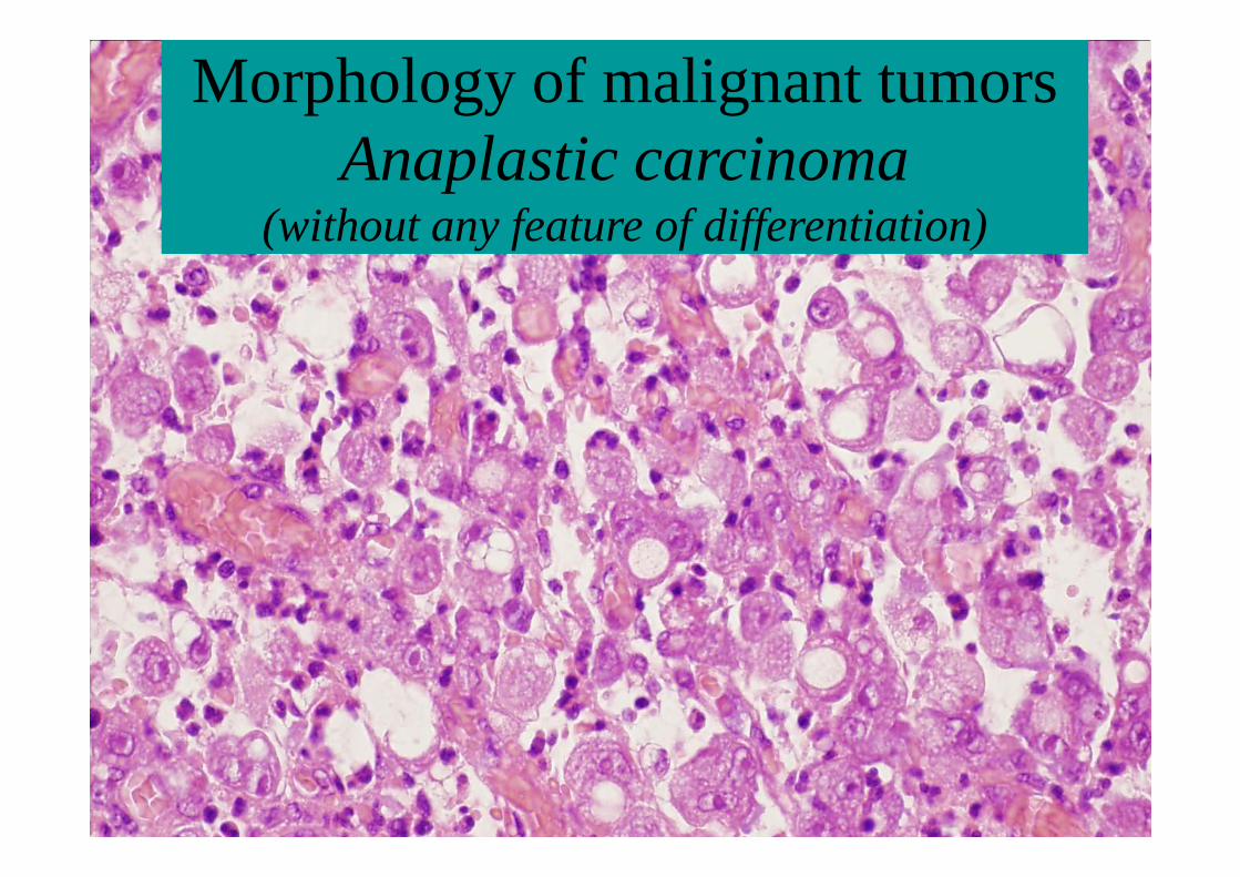
Morphology of malignant tumorsAnaplastic carcinoma
(without any feature of differentiation)
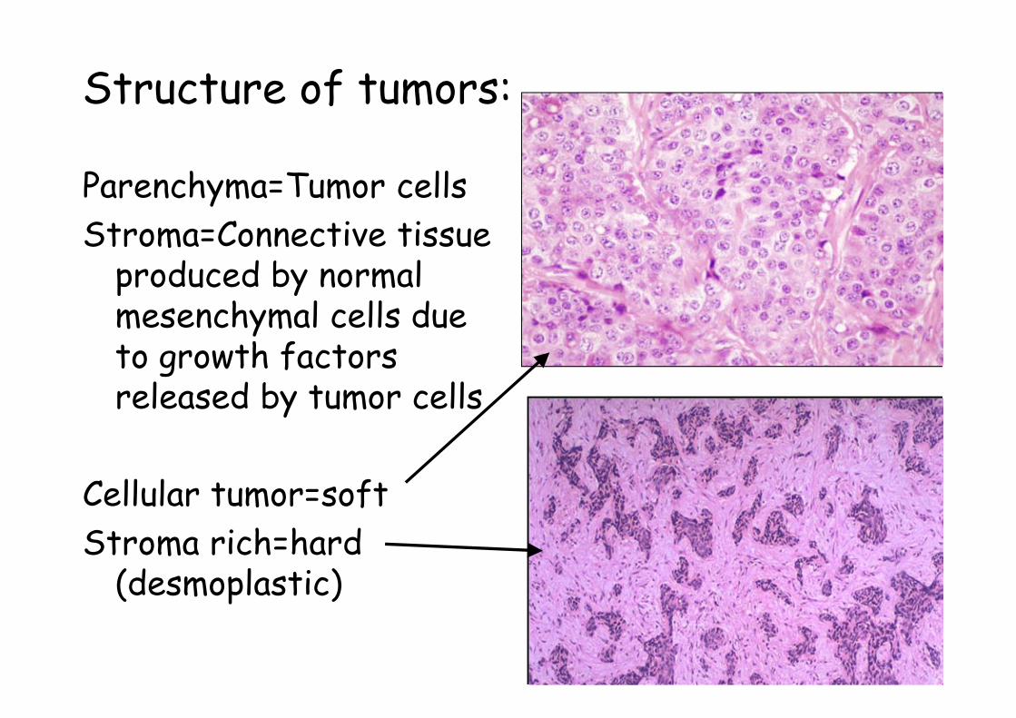
Structure of tumors:
Parenchyma=Tumor cells
Stroma=Connective tissue produced by normal mesenchymal cells due to growth factors released by tumor cells
Cellular tumor=soft
Stroma rich=hard (desmoplastic)
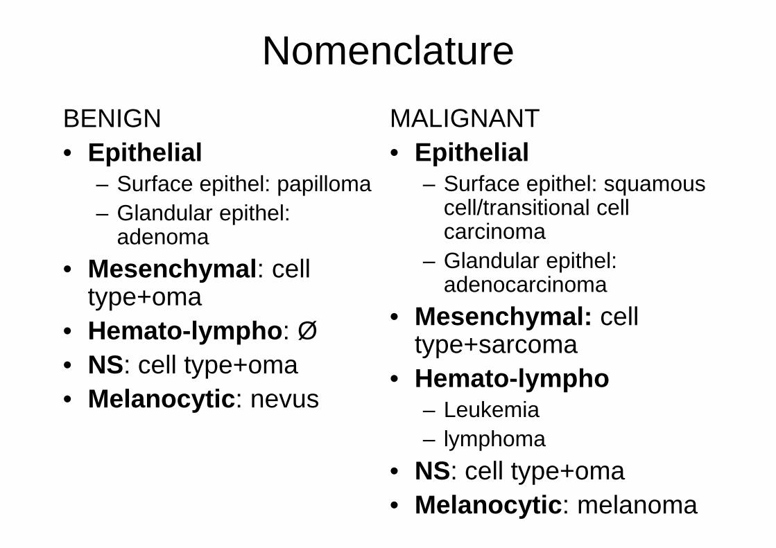
Nomenclature
BENIGN• Epithelial
– Surface epithel: papilloma– Glandular epithel:
adenoma
• Mesenchymal : cell type+oma
• Hemato-lympho : Ø• NS: cell type+oma• Melanocytic : nevus
MALIGNANT• Epithelial
– Surface epithel: squamous cell/transitional cell carcinoma
– Glandular epithel: adenocarcinoma
• Mesenchymal: cell type+sarcoma
• Hemato-lympho– Leukemia– lymphoma
• NS: cell type+oma• Melanocytic : melanoma