navigating 3d electron microscopy maps with em-surfer
TRANSCRIPT

Esquivel-Rodríguez et al. BMC Bioinformatics (2015) 16:181 DOI 10.1186/s12859-015-0580-6
SOFTWARE Open Access
Navigating 3D electron microscopy maps withEM-SURFERJuan Esquivel-Rodríguez1†, Yi Xiong2†, Xusi Han2, Shuomeng Guang2, Charles Christoffer1,3 and Daisuke Kihara1,2*
Abstract
Background: The Electron Microscopy DataBank (EMDB) is growing rapidly, accumulating biological structural dataobtained mainly by electron microscopy and tomography, which are emerging techniques for determining largebiomolecular complex and subcellular structures. Together with the Protein Data Bank (PDB), EMDB is becoming afundamental resource of the tertiary structures of biological macromolecules. To take full advantage of this indispensableresource, the ability to search the database by structural similarity is essential. However, unlike high-resolution structuresstored in PDB, methods for comparing low-resolution electron microscopy (EM) density maps in EMDB are not wellestablished.
Results: We developed a computational method for efficiently searching low-resolution EM maps. The method uses acompact fingerprint representation of EM maps based on the 3D Zernike descriptor, which is derived from a mathematicalseries expansion for EM maps that are considered as 3D functions. The method is implemented in a web server namedEM-SURFER, which allows users to search against the entire EMDB in real-time. EM-SURFER compares the global shapesof EM maps. Examples of search results from different types of query structures are discussed.
Conclusions: We developed EM-SURFER, which retrieves structurally relevant matches for query EM maps from EMDBwithin seconds. The unique capability of EM-SURFER to detect 3D shape similarity of low-resolution EM maps shouldprove invaluable in structural biology.
Keywords: Electron microscopy, Electron density maps, EM Data Bank, EMDB, 3D Zernike Descriptors, Proteins,Macromolecular structure, Low-resolution structure data, Database search
BackgroundThe three dimensional (3D) structure of proteins and otherbiomolecules provides the molecular basis for understand-ing mechanisms of biological functions, interactions, path-ways, and serves as foundation for numerous areas inbiotechnology. In addition to the exponential growth ofsolved 3D protein structures and complexes in the ProteinData Bank (PDB) [1,2], which are mostly determined by X-ray crystallography or NMR, low-resolution biomolecularstructural data determined by cryo-electron microscopy(cryo-EM) and electron tomography are notably being rap-idly accumulated in the Electron Microscopy Data Bank(EMDB, http://www.emdatabank.org/) [3]. Cryo-EM is an
* Correspondence: [email protected]†Equal contributors1Department of Computer Science, Purdue University, West Lafayette, IN47907, USA2Department of Biological Sciences, Purdue University, West Lafayette, IN47907, USAFull list of author information is available at the end of the article
© 2015 Esquivel-Rodriguez et al.; licensee BioMCreative Commons Attribution License (http:/distribution, and reproduction in any mediumDomain Dedication waiver (http://creativecomarticle, unless otherwise stated.
important technique in structural biology used to solvelarge protein complex and subcellular structures. Currently,EMDB holds over 2600 entries, and the number of entriesis growing rapidly. The mean resolution of the EM maps iscurrently about 15 Å, but recent papers [4–6] report high-resolution structures at around 3.5 Å. There is no doubtthat EMDB will become increasingly important not only instructural biology, but also in various areas including mo-lecular biology and bioinformatics.To take full advantage of these valuable resources of 3D
biomolecular structures, it is necessary for one to be ableto efficiently perform a structure-based search againstthe entire structure databases in real-time. Similarity searchis the most essential operation that needs to be providedwith a database. However, compared to biological se-quence databases that are usually equipped with real-time database search methods, structure databases arebehind with respect to efficient search methods, particu-larly for low-resolution structural data.
ed Central. This is an Open Access article distributed under the terms of the/creativecommons.org/licenses/by/4.0), which permits unrestricted use,, provided the original work is properly credited. The Creative Commons Publicmons.org/publicdomain/zero/1.0/) applies to the data made available in this

Figure 1 3DZD computation pipeline. Every map in EMDB yields several 3D Zernike descriptor fingerprints. The raw map is used to generate fourvoxelizations: one from the author-recommended density value, one at one standard deviation, which is lower than the author-recommendedcontour level, and two additional thresholds that reveal core features. Each surface is represented by 121 descriptors, which are concatenated togenerate various fingerprints.
Esquivel-Rodríguez et al. BMC Bioinformatics (2015) 16:181 Page 2 of 9
To this end, we have developed EM-SURFER for real-time searching of EM density maps from EMDB. Userscan search for similar EM maps in EMDB in terms ofthe global shape and the volume of a query map. Aquery can be either chosen from existing EMDB entriesor uploaded. Unlike atomic detailed structures stored inPDB, EM density maps are at low resolution and thusconventional structure comparison approaches cannotbe directly applied.A fast map comparison is achieved by using a math-
ematical representation of 3D shapes named 3D ZernikeDescriptor (3DZD) [7]. 3DZD is a vector derived from aseries expansion of a 3D function, which describes anEM map in a compact and rotation-invariant fashion.3DZD has been successfully applied to represent variousbiomolecular structure analyses [8], including protein3D shape comparison [9], protein docking [10–12], lig-and binding site comparison [13,14], and fast liganddatabase search [15].In EM-SURFER, each search is performed on-the-fly
and only takes a few seconds. The database of EM mapsis automatically synchronized with EMDB weekly. Inwhat follows, we first describe how 3D EM maps arerepresented in EM-SURFER, and then explain input dataand output search results with examples.
ImplementationThe main operation performed by EM-SURFER involvescomparing two EM maps using an efficient structurerepresentation with 3DZD. The descriptor is derivedfrom a mathematical series expansion of a 3D function
based on the 3D Zernike moments. 3DZD was originallyderived by Canterakis [7] and later applied to 3D objectretrieval [16]. A 3DZD can be viewed as a fingerprintthat consists of a vector of real numbers, where eachnumber is a coefficient of the series expansion. Compari-sons between these fingerprints form the basis of therapid search performed by our server. The similarity be-tween 3DZD vectors is quantified by their Euclideandistance.EM density maps for EM-SURFER are obtained from
EMDB [3], the primary repository of electron micros-copy data, and updated on a weekly basis. For each EMmap, 3DZD vectors are computed. It was shown in pre-vious studies [17,18] that 3DZD can properly representEM maps. An EM map is a 3D grid where an electrondensity value is assigned at each grid point. Using theauthor-recommended density contour level provided inEMDB, grid points with an electron density that is equalor larger than the author-recommended density aremarked with 1 and 0 otherwise. The value-mapped 3Dgrid was considered as a 3D function, f(x). This f(x) is ex-panded into a series in terms of the Zernike-Canterakisbasis defined as follows:
Ωmnl ¼
34π
Zxj j≤1
f xð Þ�Zmnl xð Þdx
where
Zmnl r; ϑ;ϕð Þ ¼ RnlY
ml ϑ;ϕð Þ
The ranges of parameters l and m are defined by theorder n: − l <m < l, 0 ≤ l ≤ n, and n-l even. We used order
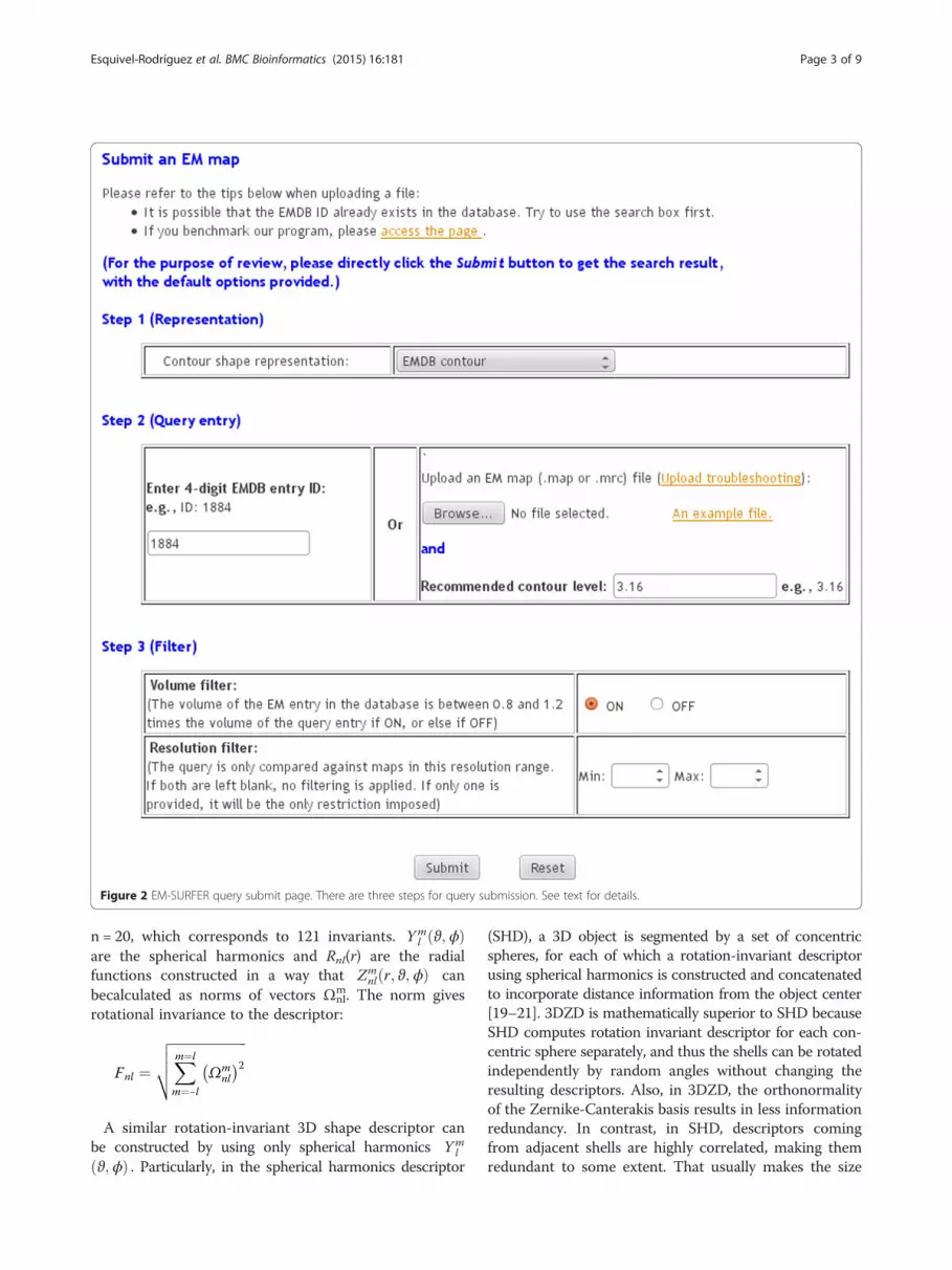
Figure 2 EM-SURFER query submit page. There are three steps for query submission. See text for details.
Esquivel-Rodríguez et al. BMC Bioinformatics (2015) 16:181 Page 3 of 9
n = 20, which corresponds to 121 invariants. Yml ϑ;ϕð Þ
are the spherical harmonics and Rnl(r) are the radialfunctions constructed in a way that Zm
nl r; ϑ;ϕð Þ canbecalculated as norms of vectors Ωm
nl. The norm givesrotational invariance to the descriptor:
Fnl ¼ffiffiffiffiffiffiffiffiffiffiffiffiffiffiffiffiffiffiffiffiffiffiffiXm¼l
m¼−l
Ωmnl
� �2vuut
A similar rotation-invariant 3D shape descriptor canbe constructed by using only spherical harmonics Ym
l
ϑ;ϕð Þ . Particularly, in the spherical harmonics descriptor
(SHD), a 3D object is segmented by a set of concentricspheres, for each of which a rotation-invariant descriptorusing spherical harmonics is constructed and concatenatedto incorporate distance information from the object center[19–21]. 3DZD is mathematically superior to SHD becauseSHD computes rotation invariant descriptor for each con-centric sphere separately, and thus the shells can be rotatedindependently by random angles without changing theresulting descriptors. Also, in 3DZD, the orthonormalityof the Zernike-Canterakis basis results in less informationredundancy. In contrast, in SHD, descriptors comingfrom adjacent shells are highly correlated, making themredundant to some extent. That usually makes the size

Figure 3 EM-SURFER results sections. EMD-1375 was used as a query to explain the different sections returned by EM-SURFER. On the top-leftsection, an image of the original query is shown (or the filename if it was a user-uploaded query). Beside this image, a graphical and text depiction ofthe 3D Zernike descriptors that describe the map are shown. Finally, for each search hit it shows the EMDB code with a short description, an image,and the detailed values for Euclidean distances, volume ratio and resolution.
Esquivel-Rodríguez et al. BMC Bioinformatics (2015) 16:181 Page 4 of 9
(the length of the descriptor) of SHD larger than 3DZD.Moreover, 3DZD was shown to perform better than SHDin shape-based object retrieval [16] and protein globalsurface shape comparison [22]. For more discussionabout 3DZD and spherical harmonics, refer to a reviewpaper [23].The distance between two 3DZDs is quantified as the
Euclidean distance between the vectors. Comparisonsbetween fingerprints form the basis of the rapid searchperformed by our server. A more detailed derivation of
3DZD as well as the mathematical foundation can befound in previous publications [7,16,24].Besides the author-recommended density level, a voxeli-
zation at one standard deviation of electron density, andtwo additional voxelizations at higher density levels, 1/3and 2/3 of the highest density, were computed (Figure 1).The purpose of the additional map descriptions with onelower and two higher densities is to capture shapes at dif-ferent contour levels of the molecules. Each contour levelyields its own vector of 121 3DZD invariants. In total, five
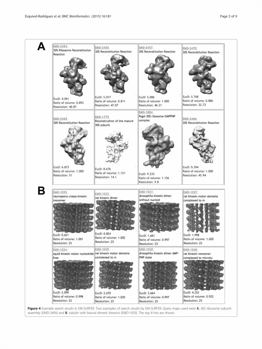
Figure 4 Example search results in EM-SURFER. Two examples of search results by EM-SURFER. Query maps used were A, 30S ribosome subunitassembly (EMD-2456) and B, tubulin with bound dimeric kinesins (EMD-1033). The top 8 hits are shown.
Esquivel-Rodríguez et al. BMC Bioinformatics (2015) 16:181 Page 5 of 9

Table 1 Search results of the two queries by EM-SURFER
Rank EMDB ID Volume Ratio Distance Molecules
EMD-2456 30S ribosomal complex
1 2454 0.893 4.061 30S ribosome
2 2455 0.811 5.017 30S ribosome
3 2457 1.000 5.086 30S ribosome
4 2470 0.866 5.768 30S ribosome
5 2469 1.000 6.873 30S ribosome
6 1775 1.121 8.676 30S subunit
7 1884 1.156 9.233 30S ribosome- GMPPNP
8 2466 1.000 9.394 30S ribosome
EMD-1033 tubulin with bound dimeric kinesins
1 1035 1.001 0.621 Tubulin
2 1032 1.002 0.824 Tubulin
3 1031 0.997 1.661 Tubulin
4 1027 1.020 1.998 Tubulin
5 1034 0.998 2.090 Tubulin
6 1029 1.020 2.670 Tubulin
7 1030 0.997 3.664 Tubulin
8 1040 0.922 4.222 Tubulin
Esquivel-Rodríguez et al. BMC Bioinformatics (2015) 16:181 Page 6 of 9
EM map descriptors were prepared: the 3DZD for 1) theauthor-recommended density level, descriptors that con-catenate the 3DZD of 2) the author-recommended dens-ity level and another 3DZD computed at one standarddeviation, 3) 1/3 maximum density, or 4) 2/3 maximumdensity, and 5) a descriptor that concatenates the author-recommended and 1/3 and 2/3 density level 3DZDs. Thesecond to the fourth descriptors have 242 invariants andthe last one has 363 invariants. The 3DZDs were pre-computed for each EMDB entry. They will be computedon-the-fly for a query if users upload their own EM map.PDBj (Protein Databank Japan, http://pdbj.org/) provides
a list of structurally similar maps for each EM map entryin their EM Navigator. Similar maps are identified by vec-tor quantization and the similarity of all EM maps are
Table 2 Distance between entries of ClpB in various condition
EMDB ID Descriptiona)
2556 ClpB E432A ATPγS. BAP variant bound
2555 ClpB E432A ATPγS. BAP variant bound
2557 ClpB ATPγS. BAP variant bound to ClpP
2558 ClpB ATPγS. BAP variant bound to ClpP
2559 ClpB Y503D mutant with ATPγS. BAP v
2560 ClpB Y503D mutant with ATPγS. BAP v
2561 Hsp104 ATPγS. HAP variant bound to C
2562 ClpB DWB trap mutant with ATPγS. BA
2563 ClpB with ATPγS
a) Description was taken from the sample record of the entries in EMDB.
visualized in a two dimensional map (named the Omokagemap) computed by multidimensional scaling. Although de-tails of the implementation of the method are not providedat the EM Navigator website (http://pdbj.org/emnavi/emnavi_doc.php?doc=omokage), differences between EM-SURFER and EM Navigator include the following: Unlikein the Omokage map, which seems to be pre-computed,similarity search for a query is performed on-the-fly inEM-SURFER. Thus, a search can be performed also for amap that is uploaded by a user.The validity of applying 3DZD for EM map database
search was shown in previous studies [17,18]. These twostudies demonstrated database searches for simulatedand actual EM maps, which achieved high accuracy bydescribing EM maps with 3DZD.
Results and discussionThe main result generated by EM-SURFER is a list ofEM maps, with queries submitted through the Searchpage (Figure 2). To submit a query entry, users shouldgo through the following four steps. In Step 1, the con-tour shape representation should be specified. The de-fault is set to the author-recommended contour level. InStep 2, users need choose the EMDB entry ID or uploadan EM map file. To find an ID from a protein name orother information, use the EMDB text search page athttp://www.ebi.ac.uk/pdbe/emdb/searchForm.html. In Step3, a volume filter is provided, which is enabled by de-fault. When this filter is on, a search only retrieves EMmaps that have a volume similar to the query (the ratiobetween the query and each retrieved map should be be-tween 0.8 to 1.2). Finally, a resolution filter allows usersto restrict the maps returned for the query to be in thespecified resolution range.The results page displays the top 20 entries in the
database that have the most similar global shape to thequery EM map. Figure 3 shows the four most similarEM maps for EMD-1375 as query. In the top panel, itshows the query entry ID and its molecule name, a
s
Distance from 2556
to ClpP 0.00
to ClpP 7.22
22.89
7.11
ariant bound to ClpP 12.32
ariant bound to ClpP 7.58
lpP 17.81
P variant bound to ClpP 21.70
22.75

Figure 5 Similarity of EM maps of ClpB. ClpB (gray) and bound ClpP (green) in the query, EMD-2566, was separately compared against the corre-sponding part of four related entries, EMD-2563, 2557, 2558, 2559, and 2562. The numbers are the Euclidean distances of 3DZDs between them.EMD-2563 contains only ClpB. The Euclidean distance shown for EMD-2563 (21.13) is computed for the complete maps of EMD-2556and EMD-2563.
Esquivel-Rodríguez et al. BMC Bioinformatics (2015) 16:181 Page 7 of 9
figure of the query (which is provided by EMDB), as wellas the 3DZD that characterizes the query entry in textand graphic forms. The query entry ID is a unique 4-digit accession number used in EMDB. Also in the toppanel, the user is given a link to a text file for a list ofthe most similar maps. In the bottom graphic panel, alist of retrieved entries for the query is shown. They areranked by the distance of their 3DZDs to that of thequery entry (quantified by Euclidean distance, EucD, i.e.the square root of the sum of the squares of the differ-ences between corresponding values). The smaller theEucD is, the more similar the shapes of the two EMmaps are. Empirically, entries with a Euclidean distanceof less than 8.0 are biologically related. For each re-trieved entry, it also shows the ratio of the volume of theretrieved entry to the query, which is defined as the vol-ume of the retrieved entry divided by that of the query,as well as the resolution of the map. Clicking on theimage of a retrieved entry will trigger a new search usingthe clicked entry as a query.We show three examples of search results by EM-
SURFER. For these searches, the author-recommendeddensity level was used. Only structures with a resolutionprovided in their meta-data are retrieved in these exam-ples. The volume filter was on. In Figure 4 and Table 1,detailed information of the top eight most similar EMmaps for the first two queries are shown. The first ex-ample is a search from a 30S ribosomal complex struc-ture (EMD-2456). Among the top 10 most similar maps
retrieved from the database, all of them are 30S riboso-mal subunit structures. The second example (Figure 3B)shows search results of tubulin that have cylindrical-shape (EMD-1033). The top thirteen retrieved EM mapsare all from tubulins. Similar to the first example, entriesretrieved with a Euclidean distance of 6.5 or less areall tubulins. The second example demonstrates thatEM-SURFER can retrieve similar EM maps not only forglobular-shape EM maps but also for cylindricalcomplexes.The examples shown above demonstrate that EM-
SURFER successfully retrieves related entries of the samemolecules. However, since EM-SURFER performs globalshape and volume comparison between EM maps, entriesof the same molecule but in different conditions that leadto overall different shape would not be retrieved at a highrank, even if they would be easily retrieved by the textsearch, which is currently available at EMDB. Table 2and Figure 5 provide results that exemplify this type ofsituation. Nine EMDB entries, EMD-2055 to 2563, aremaps under different conditions and mutants of hex-americ AAA+ chaperone ClpB (gray region in Figure 5)bound (or not bound) to protease ClpP (green). Theseentries were reported in the same paper [25]. Six copiesof ClpB assemble into a ring-shape complex (gray re-gion) and work as chaperone, where a misfolded proteinwill go through the pore at the center of the hexamerring and be unfolded. In a study by Carroni et al., mu-tants of ClpB were constructed that lock the complex in

Esquivel-Rodríguez et al. BMC Bioinformatics (2015) 16:181 Page 8 of 9
active or repressed states, which yielded the nine EMstructures [25].As shown in Table 2, when a search was performed
from query EMD-2556, not all the other eight entries wereclose: Three entries, EMD-2555, 2558, and 2560, were re-trieved within a distance of 8.0, but the remaining five en-tries (EMD-2557, 2559, 2561, 2562, and 2563) were moredistant than 10.0 (12.0 to 23.0). To understand why thefive entries have a large distance, we computed the simi-larity of ClpB (gray) and ClpP (green) regions separately(Figure 5). Interestingly, it turned out that actually thoseentries that have a large Euclidean distance have ClpB indifferent shapes reflecting their different functional states.The ClpP region is similar in all the entries (the distanceranges from 4.08 to 6.87). In the case of EMD-2563, itdoes not even have bound ClpP in the map, which makesthe overall shape of the map completely different from theshape of the query. Thus, in this example, EM-SURFERdetected different states of the same complexes, whichwould be very useful for analyzing sub-states of the samemacromolecules.The current EM-SURFER identifies entries with glo-
bally similar shape to the query EM map, but does notdetect local shape similarity between maps. Local mapsimilarity search is left as future work.
ConclusionsWe reported a web application named EM-SURFERfor real-time biomolecular structure search based onelectron microscopy density maps. EM density maps areupdated weekly from EMDB. The unique feature ofEM-SURFER, the ability of searching EM maps by shapesimilarity in a matter of seconds, should prove invalu-able in structural biology. A similar strategy will be alsovaluable for other types of low-resolution biologicalstructure data.
Availability and requirementsProject name: EM-SURFERProject home page: http://kiharalab.org/em-surferOperating system(s):Web application, platform independent
Competing interestsThe authors declare that they have no competing interests.
Authors’ contributionsD.K., S.G. and X.S. developed the original idea. X.H. and S.G. helped developthe initial database structure. J.E.R, Y.X. and C.C. developed the back-end andwebsite software. J.E.R, Y.X. and D.K. wrote the manuscript. All authors readand approved the final manuscript.
AcknowledgementsThis work was partly supported by the National Institute of General MedicalSciences of the National Institutes of Health (R01GM097528) and theNational Science Foundation (IIS0915801, DBI1262189, IOS1127027), andNational Research Foundation of Korea Grant funded by the KoreanGovernment (NRF-2011-220-C00004).
This work was supported by the National Institute of General MedicalSciences of the National Institutes of Health (R01GM075004) and theNational Science Foundation (IIS0915801, DBI1262189, IOS1127027), andNational Research Foundation of Korea (NRF-2011-220-C00004). JER is aFulbright Science and Technology Fellow.
Author details1Department of Computer Science, Purdue University, West Lafayette, IN47907, USA. 2Department of Biological Sciences, Purdue University, WestLafayette, IN 47907, USA. 3Department of Mathematics, Purdue University,West Lafayette, IN 47907, USA.
Received: 24 July 2014 Accepted: 20 April 2015
References1. Berman HM, Westbrook J, Feng Z, Gilliland G, Bhat TN, Weissig H, et al. The
protein data bank. Nucleic Acids Res. 2000;28:235–42.2. Rose PW, Bi C, Bluhm WF, Christie CH, Dimitropoulos D, Dutta S, et al. The
RCSB protein data bank: new resources for research and education. NucleicAcids Res. 2013;41:D475–82.
3. Lawson CL, Baker ML, Best C, Bi C, Dougherty M, Feng P, et al. EMDataBank.org:unified data resource for CryoEM. Nucleic Acids Res. 2011;39:D456–64.
4. Zhang X, Guo H, Jin L, Czornyj E, Hodes A, Hui WH, et al. A new topology ofthe HK97-like fold revealed in Bordetella bacteriophage by cryoEM at 3.5 Aresolution. Elife. 2013;2:e01299.
5. Li X, Mooney P, Zheng S, Booth CR, Braunfeld MB, Gubbens S, et al. Electroncounting and beam-induced motion correction enable near-atomic-resolutionsingle-particle cryo-EM. Nat Methods. 2013;10:584–90.
6. Liao M, Cao E, Julius D, Cheng Y. Structure of the TRPV1 ion channel determinedby electron cryo-microscopy. Nature. 2013;504:107–12.
7. Canterakis N. 3D Zernike Moments and Zernike Affine Invariants for 3DImage Analysis and Recognition. In: 11th Scand. Conf. Image Anal. 1999.
8. Kihara D, Sael L, Chikhi R, Esquivel-Rodríguez J. Molecular surface representationusing 3D zernike descriptors for protein shape comparison and docking. CurrProtein Pept Sci. 2011;12:520–30.
9. La D, Esquivel-Rodríguez J, Venkatraman V, Li B, Sael L, Ueng S, et al. 3D-SURFER:software for high-throughput protein surface comparison and analysis.Bioinformatics. 2009;25:2843–4.
10. Li B, Kihara D. Protein docking prediction using predicted protein-proteininterface. BMC Bioinformatics. 2012;13:7.
11. Venkatraman V, Yang YD, Sael L, Kihara D. Protein-protein docking usingregion-based 3D Zernike descriptors. BMC Bioinformatics. 2009;10:407.
12. Esquivel-Rodríguez J, Yang YD, Kihara D. Multi-LZerD: multiple protein dockingfor asymmetric complexes. Proteins. 2012;7:1818–33.
13. Sael L, Kihara D. Detecting local ligand-binding site similarity in non-homologousproteins by surface patch comparison. Proteins. 2012;80:1177–95.
14. Chikhi R, Sael L, Kihara D. Real-time ligand binding pocket database searchusing local surface descriptors. Proteins. 2010;78:2007–28.
15. Venkatraman V, Chakravarthy PR, Kihara D. Application of 3D Zernike descriptorsto shape-based ligand similarity searching. J Cheminform. 2009;1:19.
16. Novotni M, Klein R. 3D zernike descriptors for content based shape retrieval.In: Proc. Eighth ACM Symp. Solid Model. Appl. - SM’03. New York, USA: ACMPress; 2003. p. 216.
17. Sael L, Kihara D. Improved protein surface comparison and application tolow-resolution protein structure data. BMC Bioinformatics. 2010;11 Suppl 1:S2.
18. Yin S, Dokholyan NV. Fingerprint-based structure retrieval using electrondensity. Proteins. 2011;79:1002–9.
19. Funkhouser T, Min P, Kazhdan M, Chen J, Halderman A, Dobkin D, et al. Asearch engine for 3D models. ACM Trans Graph. 2003;22:83–105.
20. Kazhdan M, Funkhouser T, Rusinkiewicz S. Rotation invariant spherical harmonicrepresentation of 3D shape descriptors. In: Proc. 2003 Eurographics/ACMSIGGRAPH Symp. Geom. Process. Eurographics Association; 2003. 156–164.
21. Xu M, Beck M, Alber F. Template-free detection of macromolecular complexesin cryo electron tomograms. Bioinformatics. 2011;27:i69–76.
22. Sael L, Li B, La D, Fang Y, Ramani K, Rustamov R, et al. Fast protein tertiarystructure retrieval based on global surface shape similarity. Proteins.2008;72:1259–73.
23. Venkatraman V, Sael L, Kihara D. Potential for protein surface shape analysisusing spherical harmonics and 3D zernike descriptors. Cell BiochemBiophys. 2009;54:23–32.

Esquivel-Rodríguez et al. BMC Bioinformatics (2015) 16:181 Page 9 of 9
24. Sael L, Kihara D. Protein surface representation and comparison: Newapproaches in structural proteomics. In: Chen JY, Lonardi S, editors. Biol.Data Min. Boca Raton, FL: Chapman & Hall/CRC; 2009. p. 89–109.
25. Carroni M, Kummer E, Oguchi Y, Wendler P, Clare DK, Sinning I, et al. Head-to-tailinteractions of the coiled-coil domains regulate ClpB activity and cooperationwith Hsp70 in protein disaggregation. Elife. 2014;3:e02481.
Submit your next manuscript to BioMed Centraland take full advantage of:
• Convenient online submission
• Thorough peer review
• No space constraints or color figure charges
• Immediate publication on acceptance
• Inclusion in PubMed, CAS, Scopus and Google Scholar
• Research which is freely available for redistribution
Submit your manuscript at www.biomedcentral.com/submit