nanoparticles for the optical imaging of tumor selectin
TRANSCRIPT
Nanoparticles for the Optical Imaging of Tumor E-selectin1
Martin Funovics*,2, Xavier Montety,2, Fred Reynoldsy, Ralph Weissleder y and Lee Josephson y
*Department of Angiography and Interventional Radiology, Vienna Medical University, Vienna, Austria;yCenter for Molecular Imaging Research, Massachusetts General Hospital and HarvardMedical School, Charlestown, MA, USA
Abstract
We designed a fluorescent peptide–magnetic nano-
particle conjugate that images E-selectin expression
in mouse xenograft models of Lewis lung carcinoma
(LLC) by fluorescence reflectance imaging. It was syn-
thesized by attaching the E-selectin–binding pep-
tide (ESBP; CDSDSDITWDQLWDLMK) to a CLIO(Cy5.5)
nanoparticle to yield ESBP–CLIO(Cy5.5). Internalization
by activated human umbilical vein endothelial cells
(HUVECs) was rapid and mediated by E-selectin, indi-
cated by the lack of uptake of nanoparticles bearing
similar numbers of a scrambled peptide (Scram). To
demonstrate the specificity of E-selectin targeting to
ESBP–CLIO(Cy5.5) in vivo, we coinjected ESBP–CLIO
(Cy5.5) and Scram–CLIO(Cy3.5) and demonstrated a
high Cy5.5/Cy3.5 fluorescence ratio using the LLC. Histol-
ogy showed that ESBP–CLIO was associated with tumor
cells as well as endothelial cells, but fluorescence-
activated cell sorter analysis showed a far less expression
of E-selectin on LLC than on HUVECs. Using immuno-
histochemistry, we demonstrated E-selectin expression
in both endothelial cells and cancer cells in human pros-
tate cancer specimens. We conclude that ESBP–CLIO
(Cy5.5) is a useful probe for imaging E-selectin associ-
ated with the LLC tumor, and that E-selectin is expressed
not only on endothelial cells but also on LLC cells and
human prostate cancer specimens.
Neoplasia (2005) 7, 904–911
Keywords: Nanoparticle, E-selectin, Lewis lung carcinoma, peptide,internalization.
Introduction
Selectins (E-, P-, and L-selectin) are a family of cell surface
glycoproteins that mediate interactions between leuko-
cytes and endothelial cells [1]. E-selectin is a C-type lectin
of 97 kDa that binds to the tetrasaccharide sialyl Lewis X
structure, which appears to be a minimal ligand involved
in cell adhesion [2–4]. E-selectin (CD62E and endothelial
leukocyte adhesion molecule 1) is upregulated in endothelia
by cytokines such as interleukin 1b (IL-1b) or tumor necrosis
factor a, and is an early marker for the detection of inflam-
mation [5,6]. Although less established in inflammation and
leukocyte migration, there is considerable evidence that
an interaction between E-selectin and cancer cells may be in-
volved in adhesion and metastases [7–9].
To develop a nanoparticle binding E-selectin, we conjugated
peptides containing the DITWDQLWDLMK E-selectin–binding
sequence to amino–CLIO(Cy5.5), a crosslinked dextran-
coated iron oxide nanoparticle that is widely used for the at-
tachment of biomolecules for in vitro and in vivo applications
[10–12]. Peptides containing this sequence, termed E-selectin–
binding peptides (ESBPs), bind E-selectin (but not P-selectin
or L-selectin) noncompetitively with sialyl Lewis X with a dis-
sociation constant of 3 to 5 nM and have a similar affinity
for mouse or human E-selectins [13,14]. Mouse and human
E-selectins have a 73% sequence homology and many
common immunologic determinants [15]. Probes using this
sequence lack species specificity and can be used to target
E-selectin in a wide variety of animal models and, potentially,
clinically. In contrast, nanoparticles using a monoclonal anti-
body bind human E-selectin, but not mouse E-selectin [16,17],
which prevents them from being used in animal models like the
Lewis lung carcinoma (LLC) model used here.
We used the fluorescence of the ESBP–CLIO(Cy5.5) nano-
particle to monitor its interaction with LLC cells in vitro and by
fluorescence reflectance imaging in vivo. With the LLC, fluo-
rescence microscopy indicated that ESBP–CLIO(Cy5.5) was
not associated with endothelial cells, as might be expected
based on the function of E-selectin, but was present in both
tumor and endothelial cells. We next examined histochemically
the expression of E-selectin on human prostate cancer speci-
mens and demonstrated that it was a frequently expressed
marker. We conclude that the ESBP–CLIO( Cy5.5) nano-
particle can be used for imaging E-selectin expression by
fluorescence reflectance and that E-selectin is expressed on
both tumor and endothelial cells.
Address all correspondence to: Lee Josephson, PhD, Center for Molecular Imaging Research,
Massachusetts General Hospital, Building 149, 13th Street, No. 5406, Charlestown, MA 02129.
E-mail: [email protected] work was supported by R01-EB00662. X.M. was supported by a fellowship from the
Swiss National Science Foundation.2Martin Funovics and Xavier Montet contributed equally to this work.
Received 10 May 2005; Revised 20 June 2005; Accepted 23 June 2005.
Copyright D 2005 Neoplasia Press, Inc. All rights reserved 1522-8002/05/$25.00
DOI 10.1593/neo.05352
Neoplasia . Vol. 7, No. 10, October 2005, pp. 904 – 911 904
www.neoplasia.com
RESEARCH ARTICLE
Methods
Peptides were synthesized by an Apex 396 peptide syn-
thesizer using a standard solid-phase Fmoc chemistry (Ad-
vanced Chemtech, Lousiville, KY) and were purified by
reverse-phase high-performance liquid chromatography, as
described [12,18]. The peptides used were (FITC)BCDSDS-
DITWDQLWDLMKNH2 (ESBP) and (FITC)BCDSDSK-
MIDWTWLQLDD-NH2 (scrambled peptide or Scram). ‘‘B’’
stands for b-alanine. The identity and purity of peptides were
confirmed by mass spectroscopy. The amino–CLIO nano-
particle (crosslinked iron oxide) was synthesized as de-
scribed and iron-assayed spectrophotometrically [11,12].
The linker chemistry for the E-selectin conjugating pep-
tides to magneto-optical nanoparticles has been described
in detail (see Figure 1 of Ref. [18]). A fluorochrome (Cy3.5 or
Cy5.5; Amersham Biosciences Corp., Piscataway, NJ) was
first attached directly to the crosslinked dextran coating of
the amino–CLIO nanoparticle, consuming one to two of the
available 40 to 60 amino groups per nanoparticle and leaving
many groups for further modification (e.g., attaching a sub-
sequent peptide to the nanoparticle) [18,19].
To synthesize E-selectin–binding peptide–nanoparticle
conjugates, amino–CLIO nanoparticles [10 mg/ml Fe, 2 mg,
in phosphate-buffered saline (PBS)] were placed in a tube,
and approximately 500 mg of Cy3.5 or Cy5.5 was added.
After 2 hours at room temperature, succinimidyl iodoacetic
acid (SIA; 140 mM in 100 ml of DMSO) was added, and the
mixture was allowed to stand for 1 hour. Unreacted dye and
SIA were removed using P-10 spin columns equilibrated in
0.02 M citrate and 0.15 M NaCl (pH 8). The peptides in
DMSO were added at a concentration of 0.5 mg peptide/mg
Fe to yield nanoparticles with approximately 30 peptides
per nanoparticle. The average amount of attached peptides
was determined using the absorbance of fluorescein [12].
The average number of peptides per nanoparticle was cal-
culated by assuming 8000 Fe/CLIO nanoparticle [20].
Human umbilical vein endothelial cells (HUVECs; Clo-
netics, Baltimore, MD) were cultured in an endothelial growth
medium (Clonetics). Cells were stimulated to upregulate
E-selectin by an exposure to recombinant human IL-1b(Fisher Scientific, Fairlawn, NJ) at a final concentration of
2 ng/ml for 4 hours. LLC cells (ATCC, Manassas, VA) were
cultured in Dulbecco’s modified Eagle’s medium, with 4 mM
L-glutamine adjusted to contain 1.5 g/l sodium bicarbonate,
4.5 g/l glucose, 100 U/ml penicillin, 100 mg/ml streptomycin,
and 10% fetal bovine serum (all culture supplements were
from Invitrogen Corp., Paisley, Scotland, UK).
Nanoparticles were incubated with HUVECs (stimulated
or unstimulated) as above (8 mg/ml Fe, 8 hours). Cells were
washed three times in PBS, detached with 0.1% Trypsin–
EDTA (2 minutes at 37jC), suspended in a cell culture me-
dium at 4jC, and centrifuged (900g for 5 minutes). Cells
were resuspended in a cell culture medium and kept on ice,
and flow cytometry was performed (FacsCalibur; Becton
Dickinson, Franklin Lakes, NJ). Relative median fluores-
cence intensity (Cy5.5 channel) was obtained as a measure
of nanoparticle accumulation.
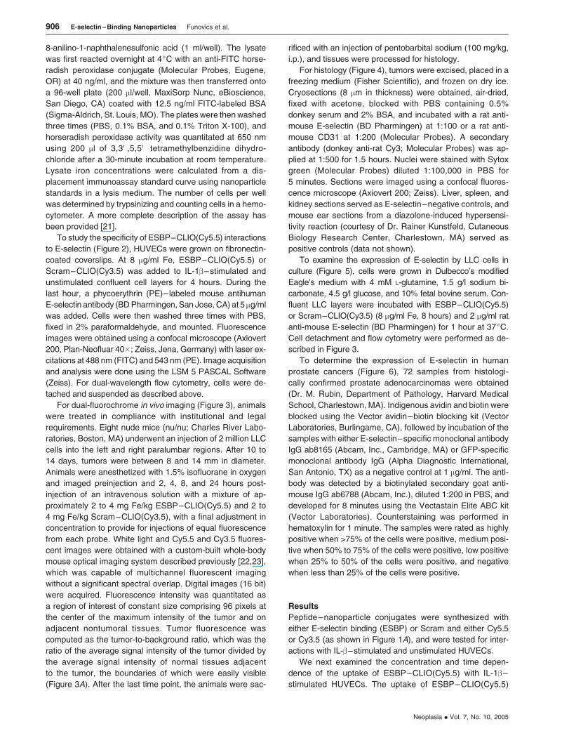
To obtain the concentration and time dependence of the
uptake of ESBP–CLIO(Cy5.5) by HUVECs (Figure 1,CandD),
confluent layers of IL-1b–stimulated cells in 24-well plates
were incubated with ESBP–CLIO(Cy5.5) in a complete cell
culture medium for 8 hours at 37jC. The uptake was mea-
sured using an FITC hapten immunoassay for immunoreactive
FITC attached to the nanoparticle. Briefly, cells were washed
three times with PBS and lysed with PBS containing 0.1%
bovine serum albumin (BSA), 0.1% Triton X-100, and 1 mM
Figure 1. Synthesis and activity of ESBP–nanoparticle conjugates. (A) Peptides containing the ESBP sequence DITWDQLWDLMK or a scrambled sequence
were conjugated to either Cy5.5- or Cy3.5-labeled nanoparticles. (B) The uptake of ESBP–CLIO(Cy5.5) as a function of concentration. (C) The uptake of ESBP–
CLIO(Cy5.5) as a function of time. IL-1� –stimulated HUVECs were used for (B) and (C). The nanoparticle concentration was 8 �g/ml Fe in (C).
E-selectin – Binding Nanoparticles Funovics et al. 905
Neoplasia . Vol. 7, No. 10, 2005
8-anilino-1-naphthalenesulfonic acid (1 ml/well). The lysate
was first reacted overnight at 4jC with an anti-FITC horse-
radish peroxidase conjugate (Molecular Probes, Eugene,
OR) at 40 ng/ml, and the mixture was then transferred onto
a 96-well plate (200 ml/well, MaxiSorp Nunc, eBioscience,
San Diego, CA) coated with 12.5 ng/ml FITC-labeled BSA
(Sigma-Aldrich, St. Louis, MO). The plates were then washed
three times (PBS, 0.1% BSA, and 0.1% Triton X-100), and
horseradish peroxidase activity was quantitated at 650 nm
using 200 ml of 3,3V,5,5V tetramethylbenzidine dihydro-
chloride after a 30-minute incubation at room temperature.
Lysate iron concentrations were calculated from a dis-
placement immunoassay standard curve using nanoparticle
standards in a lysis medium. The number of cells per well
was determined by trypsinizing and counting cells in a hemo-
cytometer. A more complete description of the assay has
been provided [21].
To study the specificity of ESBP–CLIO(Cy5.5) interactions
to E-selectin (Figure 2), HUVECs were grown on fibronectin-
coated coverslips. At 8 mg/ml Fe, ESBP–CLIO(Cy5.5) or
Scram–CLIO(Cy3.5) was added to IL-1b–stimulated and
unstimulated confluent cell layers for 4 hours. During the
last hour, a phycoerythrin (PE)–labeled mouse antihuman
E-selectin antibody (BD Pharmingen, San Jose, CA) at 5 mg/ml
was added. Cells were then washed three times with PBS,
fixed in 2% paraformaldehyde, and mounted. Fluorescence
images were obtained using a confocal microscope (Axiovert
200, Plan-Neofluar 40�; Zeiss, Jena, Germany) with laser ex-
citations at 488 nm (FITC) and 543 nm (PE). Image acquisition
and analysis were done using the LSM 5 PASCAL Software
(Zeiss). For dual-wavelength flow cytometry, cells were de-
tached and suspended as described above.
For dual-fluorochrome in vivo imaging (Figure 3), animals
were treated in compliance with institutional and legal
requirements. Eight nude mice (nu/nu; Charles River Labo-
ratories, Boston, MA) underwent an injection of 2 million LLC
cells into the left and right paralumbar regions. After 10 to
14 days, tumors were between 8 and 14 mm in diameter.
Animals were anesthetized with 1.5% isofluorane in oxygen
and imaged preinjection and 2, 4, 8, and 24 hours post-
injection of an intravenous solution with a mixture of ap-
proximately 2 to 4 mg Fe/kg ESBP–CLIO(Cy5.5) and 2 to
4 mg Fe/kg Scram–CLIO(Cy3.5), with a final adjustment in
concentration to provide for injections of equal fluorescence
from each probe. White light and Cy5.5 and Cy3.5 fluores-
cent images were obtained with a custom-built whole-body
mouse optical imaging system described previously [22,23],
which was capable of multichannel fluorescent imaging
without a significant spectral overlap. Digital images (16 bit)
were acquired. Fluorescence intensity was quantitated as
a region of interest of constant size comprising 96 pixels at
the center of the maximum intensity of the tumor and on
adjacent nontumoral tissues. Tumor fluorescence was
computed as the tumor-to-background ratio, which was the
ratio of the average signal intensity of the tumor divided by
the average signal intensity of normal tissues adjacent
to the tumor, the boundaries of which were easily visible
(Figure 3A). After the last time point, the animals were sac-
rificed with an injection of pentobarbital sodium (100 mg/kg,
i.p.), and tissues were processed for histology.
For histology (Figure 4), tumors were excised, placed in a
freezing medium (Fisher Scientific), and frozen on dry ice.
Cryosections (8 mm in thickness) were obtained, air-dried,
fixed with acetone, blocked with PBS containing 0.5%
donkey serum and 2% BSA, and incubated with a rat anti-
mouse E-selectin (BD Pharmingen) at 1:100 or a rat anti-
mouse CD31 at 1:200 (Molecular Probes). A secondary
antibody (donkey anti-rat Cy3; Molecular Probes) was ap-
plied at 1:500 for 1.5 hours. Nuclei were stained with Sytox
green (Molecular Probes) diluted 1:100,000 in PBS for
5 minutes. Sections were imaged using a confocal fluores-
cence microscope (Axiovert 200; Zeiss). Liver, spleen, and
kidney sections served as E-selectin–negative controls, and
mouse ear sections from a diazolone-induced hypersensi-
tivity reaction (courtesy of Dr. Rainer Kunstfeld, Cutaneous
Biology Research Center, Charlestown, MA) served as
positive controls (data not shown).
To examine the expression of E-selectin by LLC cells in
culture (Figure 5), cells were grown in Dulbecco’s modified
Eagle’s medium with 4 mM L-glutamine, 1.5 g/l sodium bi-
carbonate, 4.5 g/l glucose, and 10% fetal bovine serum. Con-
fluent LLC layers were incubated with ESBP–CLIO(Cy5.5)
or Scram–CLIO(Cy3.5) (8 mg/ml Fe, 8 hours) and 2 mg/ml rat
anti-mouse E-selectin (BD Pharmingen) for 1 hour at 37jC.
Cell detachment and flow cytometry were performed as de-
scribed in Figure 3.
To determine the expression of E-selectin in human
prostate cancers (Figure 6), 72 samples from histologi-
cally confirmed prostate adenocarcinomas were obtained
(Dr. M. Rubin, Department of Pathology, Harvard Medical
School, Charlestown, MA). Indigenous avidin and biotin were
blocked using the Vector avidin–biotin blocking kit (Vector
Laboratories, Burlingame, CA), followed by incubation of the
samples with either E-selectin–specific monoclonal antibody
IgG ab8165 (Abcam, Inc., Cambridge, MA) or GFP-specific
monoclonal antibody IgG (Alpha Diagnostic International,
San Antonio, TX) as a negative control at 1 mg/ml. The anti-
body was detected by a biotinylated secondary goat anti-
mouse IgG ab6788 (Abcam, Inc.), diluted 1:200 in PBS, and
developed for 8 minutes using the Vectastain Elite ABC kit
(Vector Laboratories). Counterstaining was performed in
hematoxylin for 1 minute. The samples were rated as highly
positive when >75% of the cells were positive, medium posi-
tive when 50% to 75% of the cells were positive, low positive
when 25% to 50% of the cells were positive, and negative
when less than 25% of the cells were positive.
Results
Peptide–nanoparticle conjugates were synthesized with
either E-selectin binding (ESBP) or Scram and either Cy5.5
or Cy3.5 (as shown in Figure 1A), and were tested for inter-
actions with IL-b–stimulated and unstimulated HUVECs.
We next examined the concentration and time depen-
dence of the uptake of ESBP–CLIO(Cy5.5) with IL-1b–
stimulated HUVECs. The uptake of ESBP–CLIO(Cy5.5)
906 E-selectin – Binding Nanoparticles Funovics et al.
Neoplasia . Vol. 7, No. 10, 2005
was proportional to nanoparticle concentration (1–30 mg/ml
Fe) and time (0–8 hours), as shown in Figure 1, B and C.
The rate of uptake of ESBP–CLIO(Cy5.5) was 3.47 pg/cell
Fe per hour at 8 mg/ml Fe. Consistent with the observations
of Martens et al. [14], an N-terminal attachment was found
to be essential for preserving the E-selectin–binding activity
of DITWDQLWDLMK-based peptide–nanoparticle conju-
gates (data not shown).
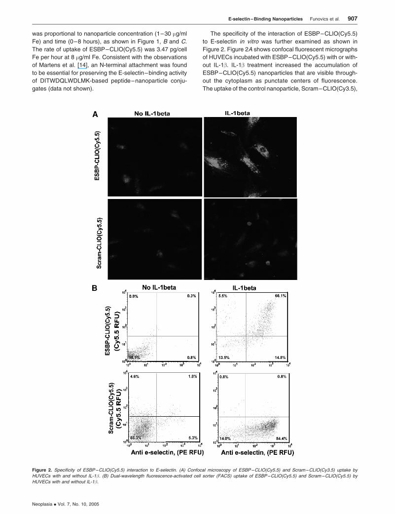
The specificity of the interaction of ESBP–CLIO(Cy5.5)
to E-selectin in vitro was further examined as shown in
Figure 2. Figure 2A shows confocal fluorescent micrographs
of HUVECs incubated with ESBP–CLIO(Cy5.5) with or with-
out IL-1b. IL-1b treatment increased the accumulation of
ESBP–CLIO(Cy5.5) nanoparticles that are visible through-
out the cytoplasm as punctate centers of fluorescence.
The uptake of the control nanoparticle, Scram–CLIO(Cy3.5),
Figure 2. Specificity of ESBP–CLIO(Cy5.5) interaction to E-selectin. (A) Confocal microscopy of ESBP–CLIO(Cy5.5) and Scram–CLIO(Cy3.5) uptake by
HUVECs with and without IL-1�. (B) Dual-wavelength fluorescence-activated cell sorter (FACS) uptake of ESBP–CLIO(Cy5.5) and Scram–CLIO(Cy5.5) by
HUVECs with and without IL-1�.
E-selectin – Binding Nanoparticles Funovics et al. 907
Neoplasia . Vol. 7, No. 10, 2005
was lower and was not stimulated by IL-1b. To further exam-
ine the specificity of the interaction of ESBP–CLIO(Cy5.5) to
E-selectin, HUVECs were examined by the dual-wavelength
flow cytometry (Figure 2B). Here the uptake of ESBP–
CLIO(Cy5.5)– labeled Cy5.5 nanoparticles was compared
with the binding of a PE-labeled anti-human E-selectin. As
expected, IL-1b treatment increased E-selectin expres-
sion, and cells expressing E-selectin internalized ESBP–
CLIO(Cy5.5). However, IL-1b treatment failed to increase the
uptake of Scram–CLIO(Cy5.5).
We next examined the specificity of the interaction of
ESBP–CLIO(Cy5.5) to E-selectin in vivo, as shown in
Figure 3. A mouse xenograft model of LLC was used as a
model of E-selectin–expressing tissue because it is a rap-
idly growing tumor and is an excellent source of small blood
vessels, which express E-selectin [24]. We examined nano-
particle accumulation in LLC using dual-channel intravital
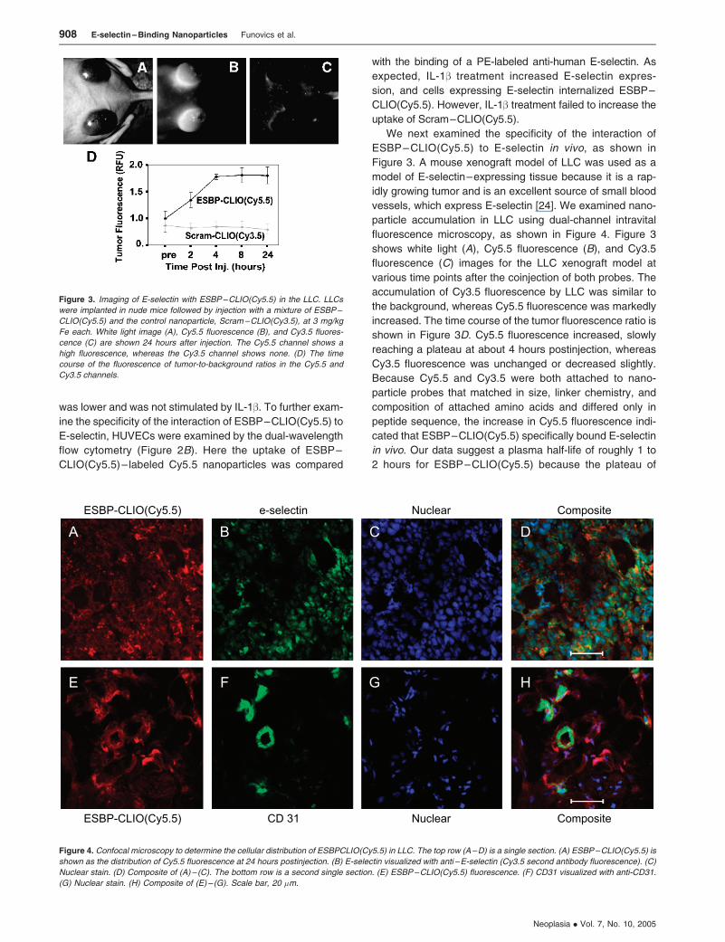
fluorescence microscopy, as shown in Figure 4. Figure 3
shows white light (A), Cy5.5 fluorescence (B), and Cy3.5
fluorescence (C) images for the LLC xenograft model at
various time points after the coinjection of both probes. The
accumulation of Cy3.5 fluorescence by LLC was similar to
the background, whereas Cy5.5 fluorescence was markedly
increased. The time course of the tumor fluorescence ratio is
shown in Figure 3D. Cy5.5 fluorescence increased, slowly
reaching a plateau at about 4 hours postinjection, whereas
Cy3.5 fluorescence was unchanged or decreased slightly.
Because Cy5.5 and Cy3.5 were both attached to nano-
particle probes that matched in size, linker chemistry, and
composition of attached amino acids and differed only in
peptide sequence, the increase in Cy5.5 fluorescence indi-
cated that ESBP–CLIO(Cy5.5) specifically bound E-selectin
in vivo. Our data suggest a plasma half-life of roughly 1 to
2 hours for ESBP–CLIO(Cy5.5) because the plateau of
Figure 3. Imaging of E-selectin with ESBP–CLIO(Cy5.5) in the LLC. LLCs
were implanted in nude mice followed by injection with a mixture of ESBP–
CLIO(Cy5.5) and the control nanoparticle, Scram–CLIO(Cy3.5), at 3 mg/kg
Fe each. White light image (A), Cy5.5 fluorescence (B), and Cy3.5 fluores-
cence (C) are shown 24 hours after injection. The Cy5.5 channel shows a
high fluorescence, whereas the Cy3.5 channel shows none. (D) The time
course of the fluorescence of tumor-to-background ratios in the Cy5.5 and
Cy3.5 channels.
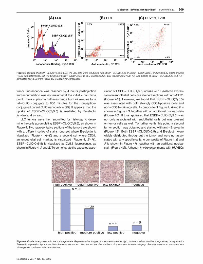
Figure 4. Confocal microscopy to determine the cellular distribution of ESBPCLIO(Cy5.5) in LLC. The top row (A–D) is a single section. (A) ESBP–CLIO(Cy5.5) is
shown as the distribution of Cy5.5 fluorescence at 24 hours postinjection. (B) E-selectin visualized with anti –E-selectin (Cy3.5 second antibody fluorescence). (C)
Nuclear stain. (D) Composite of (A) – (C). The bottom row is a second single section. (E) ESBP–CLIO(Cy5.5) fluorescence. (F) CD31 visualized with anti-CD31.
(G) Nuclear stain. (H) Composite of (E) – (G). Scale bar, 20 �m.
908 E-selectin – Binding Nanoparticles Funovics et al.
Neoplasia . Vol. 7, No. 10, 2005
tumor fluorescence was reached by 4 hours postinjection
and accumulation was not maximal at the initial 2-hour time
point. In mice, plasma half-lives range from 47 minutes for a
tat–CLIO conjugate to 650 minutes for the nonpeptide-
conjugated parent CLIO nanoparticle [25]. It appears that the
uptake of ESBP–CLIO(Cy5.5) is mediated by E-selectin
in vitro and in vivo.
LLC tumors were then submitted for histology to deter-
mine the cells accumulating ESBP–CLIO(Cy5.5), as shown in
Figure 4. Two representative sections of the tumors are shown
with a different series of stains: one set where E-selectin is
visualized (Figure 4, A–D) and a second set where CD31,
an endothelial cell marker, is visualized (Figure 4, E–H ).
ESBP–CLIO(Cy5.5) is visualized as Cy5.5 fluorescence, as
shown in Figure 4,A andE. To demonstrate the expected asso-
ciation of ESBP–CLIO(Cy5.5) uptake with E-selectin expres-
sion on endothelial cells, we stained sections with anti-CD31
(Figure 4F ). However, we found that ESBP–CLIO(Cy5.5)
was associated with both strongly CD31-positive cells and
non–CD31-staining cells. A composite of Figure 4,A andB is
shown in Figure 4D, together with an additional nuclear stain
(Figure 4C). It thus appeared that ESBP–CLIO(Cy5.5) was
not only associated with endothelial cells but was present
on tumor cells as well. To further verify this point, a second
tumor section was obtained and stained with anti–E-selectin
(Figure 4B). Both ESBP–CLIO(Cy5.5) and E-selectin were
widely distributed throughout the tumor and were not asso-
ciated with any specific cells. A composite of Figure 4, E and
F is shown in Figure 4H, together with an additional nuclear
stain (Figure 4G). Although in vitro experiments with HUVECs
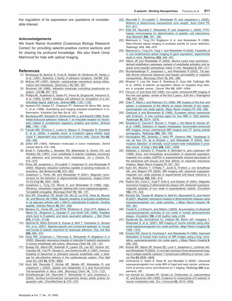
Figure 6. E-selectin expression in the human prostate. Representative images of specimens rated as high positive, medium positive, low positive, or negative for
E-selectin expression by immunohistochemistry are shown. Also shown are the numbers of specimens in each category. Samples were from prostates with
histologically confirmed adenocarcinomas.
Figure 5. Binding of ESBP–CLIO(Cy5.5) to LLC. (A) LLC cells were incubated with ESBP–CLIO(Cy5.5) or Scram–CLIO(Cy3.5), and binding by single-channel
FACS was determined. (B) The binding of ESBP–CLIO(Cy5.5) to LLC is analyzed by dual-wavelength FACS. (C) The binding of ESBP–CLIO(Cy5.5) to IL-1� –
stimulated HUVECs from Figure 2B is shown for comparison.
E-selectin – Binding Nanoparticles Funovics et al. 909
Neoplasia . Vol. 7, No. 10, 2005
indicated that ESBP–CLIO(Cy5.5) was internalized in IL-1b–
activated cells by an interaction with E-selectin, it appeared that
E-selectin expression and accumulation of ESBP–CLIO
(Cy5.5) occurred throughout the tumor.
We next examined whether E-selectin might be ex-
pressed on LLC cells grown in vitro. Figure 5A shows the
uptake of ESBP–CLIO(Cy5.5) and Scram–CLIO(Cy3.5) by
LLC using a single-channel flow cytometry analysis. With
ESBP–CLIO(Cy5.5), both high- and low-binding populations
of cells were clearly discernable, whereas with the Scram–
CLIO(Cy3.5) nanoparticle, only a single low-binding popula-
tion was obtained. The accumulation of ESBP–CLIO(Cy5.5)
was further examined by the dual-wavelength flow cytometry
using PE-labeled anti–E-selectin, as shown in Figure 5B.
As was the case with the single-channel flow cytometry
(Figure 5A), a population of cells labeled with both ESBP–
CLIO(Cy5.5) and anti–E-selectin was present (26.9% of cells).
However, this population appeared to have less ESBP–
CLIO(Cy5.5) uptake or E-selectin expression than HUVECs
(compare Figure 5, B and C). Thus, it appears that E-selectin
is expressed by LLCs both in vivo and in vitro, but this ex-
pression is at a lower level than that seen when IL-1b in-
duces E-selectin expression on HUVECs. The treatment
of LCC with IL-1b had no effect on the binding of ESBP–
CLIO(Cy5.5) to LLC (data not shown).
The expression of E-selectin on endothelial cells was well
established, but we were unaware of reports of its expres-
sion on tumor cells until the recent report [26]. We therefore
obtained an array of 72 human prostate specimens from
prostates with histologically confirmed adenocarcinomas. Of
these, 67 could be evaluated for E-selectin expression by
immunohistochemistry. Samples were rated as high positive,
medium positive, low positive, and negative, representative
specimens of which are shown in Figure 6A. As shown in
Figure 6B, 38 of 67 samples evaluated were rated as
strongly positive for E-selectin expression.
Discussion
Specificity
A key issue for molecularly targeted optical nanoparticles is
the specificity of their molecular targeting in vivo. Determining
molecular specificity in vivo with peptide–nanoparticle con-
jugates (10–200 nm) using a peptide to block interactions
between a peptide–nanoparticle conjugate and its target is
difficult because of limitations of peptide solubility; in addition,
the plasma half-life of peptides is often far shorter than the
half-life of nanoparticles whose binding they are proposed to
inhibit. The problem of demonstrating nanoparticle specificity
in vivo is exacerbated by the ability of nonmolecularly tar-
geted magnetic nanoparticles to accumulate in diverse types
of pathology. Although magnetic nanoparticles accumulate
in the normal liver, spleen, and lymph nodes [27–31], their
accumulation in a variety of pathologies also occurs. Non-
targeted nanoparticle accumulation occurs at the sites of
infection [32, 33], in atherosclerotic vessels [34–36], and in
rheumatoid arthritis [37]. Nanoparticle accumulation has also
been seen in several types of tumors, such as those of the
brain [38,39] and head and neck [40]. To minimize the
possibility of non–E-selectin–mediated uptake contribut-
ing to tissue fluorescence, we used 3 mg Fe/kg ESBP–
CLIO(Cy5.5), which was only slightly higher than the clinical
dose of 2.6 mg/kg Fe [31]. To determine the molecular speci-
ficity of ESBP–CLIO(Cy5.5) in vivo, the nanoparticle and
Scram–CLIO(Cy3.5) were coinjected and fluorescence re-
flectance images were obtained using dual-channel fluo-
rescent imaging methods [22]. The progressive increase
in the Cy5.5/Cy3.5 ratio in the LLC with time provided evi-
dence of the E-selectin–mediated accumulation of ESBP–
CLIO(Cy5.5) in vivo.
Sensitivity, Uptake, and the Presence of E-selectin
on Tumor Cells
It appears that the fluorescence of ESBP–CLIO(Cy5.5)
nanoparticles, when used in conjunction with near-infrared
fluorescent imaging, has the sensitivity to detect low levels
of E-selectin expression in the LLC. The explanation for this
high sensitivity lies in the rapid E-selectin–mediated inter-
nalization of ESBP–CLIO(Cy5.5), even at low concentra-
tions. In addition, the binding of ESBP–CLIO(Cy5.5) triggers
endocytosis and intracellular accumulation (Figure 2A), with
negligible nanoparticle loss in the 24-hour period of our
longest experiments [18,39]. Finally, when E-selectin–binding
ligands are internalized, plasma membrane E-selectin is rap-
idly replaced [41].
Expression on Human Cells
We found that ESBP – CLIO(Cy5.5) binding and
E-selectin expression were not limited to endothelial cells
in the LLC both in vivo (Figure 4) and in vitro (Figure 5).
Bhaskar et al. [26] have recently reported that E-selectin
mRNA and immunoreactive E-selectin were overexpressed
in human prostate cancer specimens even though E-selectin
expression was not detected in human cancer cell lines
of prostatic origin, normal human tissues, or nonprostatic
cancers. We have confirmed the expression of E-selectin
in the human prostate cancer epithelium using immuno-
histochemistry (Figure 6). Our results indicate that E-selectin
expression in the LLC model, a tumor of murine origin, can
be imaged by optical methods and that E-selectin expres-
sion can be detected by immunohistochemistry in human
prostate cancer specimens. The lack of expression of E-
selectin on cultured human cell lines and xenographs [26],
however, means that there is currently no model suitable
for imaging E-selectin expression with cancers of human
origin. The lack of expression of E-selectin on human pros-
tate cancer cell lines and its expression in human prostatic
cancers suggest that unknown tissue-based environmental
factors cause its upregulation or downregulation [26] and
complicate efforts to study E-selectin function in vitro. Thus,
the comparison of E-selectin expression on LLC and endo-
thelial cells (Figure 5) may be between cells expressing basal
levels of E-selectin (LLC) and those where it is highly up-
regulated (IL-1b/endothelial cells). Further determinations of
which tumor cells express E-selectin, its function, and
910 E-selectin – Binding Nanoparticles Funovics et al.
Neoplasia . Vol. 7, No. 10, 2005
the regulation of its expression are questions of consider-
able interest.
Acknowledgements
We thank Rainer Kunstfeld (Cutaneous Biology Research
Center) for providing selectin-positive control sections and
for sharing his profound knowledge. We also thank Umar
Mahmood for help with optical imaging.
References[1] Bevilacqua M, Butcher E, Furie B, Gallatin M, Gimbrone M, Harlan J,
et al. (1991). Selectins: a family of adhesion receptors. Cell 67, 233.
[2] McEver RP (1997). Selectin – carbohydrate interactions during inflam-
mation and metastasis. Glycoconj J 14, 585 – 591.
[3] Stoolman LM (1989). Adhesion molecules controlling lymphocyte mi-
gration. Cell 56, 907 –910.
[4] Phillips ML, Nudelman E, Gaeta FC, Perez M, Singhal AK, Hakomori S,
et al. (1990). ELAM-1 mediates cell adhesion by recognition of a car-
bohydrate ligand, sialyl-Lex. Science 250, 1130 –1132.
[5] Haskard DO, Keelan ET, Chapman PT, Robinson M, Binns RM, Jamar
F, et al. (1998). Quantifying and imaging activated endothelium in in-
flammation. Endothel Cell Res Ser 4, 135 –149.
[6] Bevilacqua MP, Stengelin S, Gimbrone MA Jr, and Seed B (1989). Endo-
thelial leukocyte adhesion molecule 1: an inducible receptor for neutro-
phils related to complement regulatory proteins and lectins. Science
243, 1160 – 1165.
[7] Fukuda MN, Ohvama C, Lowitz K, Matsuo O, Pasqualini R, Ruoslahti
E, et al. (2000). A peptide mimic of E-selectin ligand inhibits sialyl
Lewis X – dependent lung colonization of tumor cells. Cancer Res 60,
450 – 456.
[8] Zetter BR (1993). Adhesion molecules in tumor metastasis. Semin
Cancer Biol 4, 219 – 229.
[9] Brodt P, Fallavollita L, Bresalier RS, Meterissian S, Norton CR, and
Wolitzky BA (1997). Liver endothelial E-selectin mediates carcinoma
cell adhesion and promotes liver metastasis. Int J Cancer 71,
612 – 619.
[10] Perez JM, Josephson L, O’Loughlin T, Hogemann D, and Weissleder R
(2002). Magnetic relaxation switches capable of sensing molecular in-
teractions. Nat Biotechnol 20, 816 –820.
[11] Josephson L, Perez JM, and Weissleder R (2001). Magnetic nano-
sensors for the detection of oligonucleotide sequences. Angew Chem
Int Ed Engl 40, 3204 –3206.
[12] Josephson L, Tung CH, Moore A, and Weissleder R (1999). High-
efficiency intracellular magnetic labeling with novel superparamagnetic–
Tat peptide conjugates. Bioconjug Chem 10, 186 –191.
[13] Zinn KR, Chaudhuri TR, Smyth CA, Wu Q, Liu H-G, Fleck M, Mountz
JD, and Mountz JM (1999). Specific targeting of activated endothelium
in rat adjuvant arthritis with a 99mTc-radiolabeled E-selectin – binding
peptide. Arthritis Rheum 42, 641 –649.
[14] Martens CL, Cwirla SE, Lee RYW, Whitehorn E, Chen EYF, Bakker A,
Martin EL, Wagstrom C, Gopalan P, and Smith CW (1995). Peptides
which bind to E-selectin and block neutrophil adhesion. J Biol Chem
270, 21129 – 21136.
[15] Hammel M, Weitz-Schmidt G, Krause A, Moll T, Vestweber D, Zerwes
HG, et al. (2001). Species-specific and conserved epitopes on mouse
and human E-selectin important for leukocyte adhesion. Exp Cell Res
269, 266 – 274.
[16] Kang HW, Josephson L, Petrovsky A, Weissleder R, Bogdanov A Jr
(2002). Magnetic resonance imaging of inducible E-selectin expression
in human endothelial cell culture. Bioconjug Chem 13, 122 –127.
[17] Spragg DD, Alford DR, Greferath R, Larsen CE, Lee KD, Gurtner GC,
Cybulsky MI, Tosi PF, Nicolau C, and Gimbrone MA Jr (1997). Immuno-
targeting of liposomes to activated vascular endothelial cells: a strat-
egy for site-selective delivery in the cardiovascular system. Proc Natl
Acad Sci USA 94, 8795 – 8800.
[18] Koch AM, Reynolds F, Kircher MF, Merkle HP, Weissleder R, and
Josephson L (2003). Uptake and metabolism of a dual fluorochrome
Tat-nanoparticle in HeLa cells. Bioconjug Chem 14, 1115 –1121.
[19] Schellenberger EA, Reynolds F, Weissleder R, and Josephson L
(2004). Surface-functionalized nanoparticle library yields probes for
apoptotic cells. ChemBioChem 5, 275 – 279.
[20] Reynolds F, O’Loughlin T, Weissleder R, and Josephson L (2005).
Method of determining nanoparticle core weight. Anal Chem 77,
814 – 817.
[21] Kelly KA, Reynolds F, Weissleder R, and Josephson L (2004). FITC-
hapten immunoassay for determination of peptide – cell interactions.
Anal Biochem 330, 181 – 185.
[22] Mahmood U, Tung CH, Bogdanov A Jr, and Weissleder R (1999).
Near-infrared optical imaging of protease activity for tumor detection.
Radiology 213, 866 –870.
[23] Mahmood U, Tung CH, Tang Y, and Weissleder R (2002). Feasibility of
in vivo multichannel optical imaging of gene expression: experimental
study in mice. Radiology 224, 446 –451.
[24] Allport JR and Weissleder R (2003). Murine Lewis lung carcinoma –
derived endothelium expresses markers of endothelial activation and re-
quires tumor-specific extracellular matrix in vitro. Neoplasia 5, 205 –217.
[25] Wunderbaldinger P, Josephson L, and Weissleder R (2002). Tat pep-
tide directs enhanced clearance and hepatic permeability of magnetic
nanoparticles. Bioconjug Chem 13, 264 – 268.
[26] Bhaskar V, Law DA, Ibsen E, Breinberg D, Cass KM, DuBridge RB,
et al. (2003). E-selectin up-regulation allows for targeted drug deliv-
ery in prostate cancer. Cancer Res 63, 6387 –6394.
[27] Ferrucci JT and Stark DD (1990). Iron oxide – enhanced MR imaging of
the liver and spleen: review of the first 5 years. AJR Am J Roentgenol
155, 943 – 950.
[28] Chen F, Ward J, and Robinson PJ (1999). MR imaging of the liver and
spleen: a comparison of the effects on signal intensity of two super-
paramagnetic iron oxide agents. Magn Reson Imaging 17, 549 –556.
[29] Chachuat A and Bonnemain B (1995). European clinical experience
with Endorem. A new contrast agent for liver MRI in 1000 patients.
Radiologie 35, S274 –S276.
[30] Seneterre E, Taourel P, Bouvier Y, Pradel J, Van Beers B, Daures JP,
et al. (1996). Detection of hepatic metastases: ferumoxides-enhanced
MR imaging versus unenhanced MR imaging and CT during arterial
portography. Radiology 200, 785 –792.
[31] Harisinghani MG, Barentsz J, Hahn PF, Deserno WM, Tabatabaei S,
van de Kaa CH, de la Rosette J, and Weissleder R (2003). Non-
invasive detection of clinically occult lymph-node metastases in pros-
tate cancer. N Engl J Med 348, 2491 –2499.
[32] Gellissen J, Axmann C, Prescher A, Bohndorf K, and Lodemann KP
(1999). Extra- and intracellular accumulation of ultrasmall superpara-
magnetic iron oxides (USPIO) in experimentally induced abscesses of
the peripheral soft tissues and their effects on magnetic resonance
imaging. Magn Reson Imaging 17, 557 – 567.
[33] Kaim AH, Wischer T, O’Reilly T, Jundt G, Frohlich J, von Schulthess
GK, and Allegrini PR (2002). MR imaging with ultrasmall superpara-
magnetic iron oxide particles in experimental soft-tissue infections in
rats. Radiology 225, 808 – 814.
[34] Ruehm SG, Corot C, Vogt P, Kolb S, and Debatin JF (2001). Magnetic
resonance imaging of atherosclerotic plaque with ultrasmall superpara-
magnetic particles of iron oxide in hyperlipidemic rabbits. Circulation
103, 415 – 422.
[35] Schmitz SA, Taupitz M, Wagner S, Wolf KJ, Beyersdorff D, and Hamm
B (2001). Magnetic resonance imaging of atherosclerotic plaques using
superparamagnetic iron oxide particles. J Magn Reson Imaging 14,
355 – 361.
[36] Trivedi R, J U-King-Im, and Gillard J (2003). Accumulation of ultrasmall
superparamagnetic particles of iron oxide in human atherosclerotic
plaque. Circulation 108, e140 (author reply e140).
[37] Dardzinski BJ, Schmithorst VJ, Holland SK, Boivin GP, Imagawa T,
Watanabe S, et al. (2001). MR imaging of murine arthritis using ultra-
small superparamagnetic iron oxide particles. Magn Reson Imaging 19,
1209 – 1216.
[38] Enochs WS, Harsh G, Hochberg F, and Weissleder R (1999). Improved
delineation of human brain tumors on MR images using a long- circu-
lating, superparamagnetic iron oxide agent. J Magn Reson Imaging 9,
228 – 232.
[39] Kircher MF, Allport JR, Graves EE, Love V, Josephson L, Lichtman AH,
and Weissleder R (2003). In vivo high resolution three-dimensional imag-
ing of antigen-specific cytotoxic T-lymphocyte trafficking to tumors. Can-
cer Res 63, 6838 – 6846.
[40] Jonkmanns C, Saleh A, Rees M, and Moedder U (2002). Ultrasmall
superparamagnetic iron oxide MIR of head and neck cancer: enhance-
ment of primary tumor and influence on T staging. Radiology 225 (sup-
plement), 281.
[41] von Asmuth EJ, Smeets EF, Ginsel LA, Onderwater JJ, Leeuwenberg
JF, and Buurman WA (1992). Evidence for endocytosis of E-selectin in
human endothelial cells. Eur J Immunol 22, 2519 –2526.
E-selectin – Binding Nanoparticles Funovics et al. 911
Neoplasia . Vol. 7, No. 10, 2005