mycotoxic nephropathy in bulgarian pigs and chickens: complex aetiology and similarity to balkan...
TRANSCRIPT

For Peer Review O
nly
Mycotoxic nephropathy in Bulgarian pigs and chickens:
complex etiology and similarity to Balkan Endemic
Nephropathy
Journal: Food Additives and Contaminants
Manuscript ID: TFAC-2009-100.R1
Manuscript Type: Original Research Paper
Date Submitted by the Author:
28-Jun-2009
Complete List of Authors: Stoev, Stoycho; Trakia University, Faculty of Veterinary medicine,
Dept of General and clinical pathology Dutton, Michael; University of Johannesburg, Food, Environment and Health Research Group, Faculty of Health Science Njobeh, Patrick; University of Johannesburg, Food, Environment and Health Research Group, Faculty of Health Science Mosonik, Joseph; University of Johannesburg, Food, Environment and Health Research Group, Faculty of Health Science Steenkamp, Paul; Council for Scientific and Industrial Research, Biosciences
Methods/Techniques: HPLC, Mycology, Screening assays, Toxicology - animal study
Additives/Contaminants: Fumonisins, Mycotoxins, Mycotoxins - fungi, Mycotoxins –
ochratoxin A
Food Types: Animal feed
http://mc.manuscriptcentral.com/tfac Email: [email protected]
Food Additives and Contaminantspe
er-0
0573
918,
ver
sion
1 -
5 M
ar 2
011
Author manuscript, published in "Food Additives and Contaminants 27, 01 (2009) 72-88" DOI : 10.1080/02652030903207227

For Peer Review O
nly
1
Mycotoxic nephropathy in Bulgarian pigs and chickens:
complex etiology and similarity to Balkan Endemic
Nephropathy
Stoycho D. Stoeva*
Michael F. Duttonb, Patrick B. Njobeh
b, Joseph S. Mosonik
b, Paul A.
Steenkampc
aDepartment of General and clinical pathology, Faculty of Veterinary Medicine, Trakia
University, Students campus, 6000 Stara Zagora, Bulgaria
bFood, Environment and Health Research Group, Faculty of Health Science, University
of Johannesburg, Doornfontein 2028, Gauteng, PO Box 17011, South Africa
cCouncil for Scientific and Industrial Research, Biosciences, Ardeer Road, Private Bag
x2, Modderfontein, 1645, South Africa
Running Title: Complex etiology of Bulgarian nephropathy
Key words: ochratoxin A, penicillic acid, citrinin, fumonisin B1, mycotoxins, mycotoxic
nephropathy
Abbreviations: AFs – aflatoxins, BEN - balkan endemic nephropathy, CIT – citrinin, CA –
cyclopiazonic acid, DAS – diacetoxyscirpenol, DON – deoxynivalenol, ERY – erythroskyrin,
FB1 – fumonisin B1, FB2 - fumonisin B2, GLI – gliotoxin, ISL – islandiotoxin, KA – kojic
acid, LUT – luteoskyirin, MPN - mycotoxic porcine nephropathy, MCN - mycotoxic chicken
nephropathy, MON – moniliformin, UM – unknown metabolite, OTA – ochratoxin A, OTC -
ochratoxin C, OOS – oosporein (isoosporin), PenA – penitrem A, PAT – patulin, PA –
penicillic acid, RUB – rubratoxin B, T-2 – T-2 toxin, XA – xanthomegnin, ZEA –
zearalenone.
* Corresponding author: S. D. Stoev, Department of General and Clinical Pathology, Faculty of Veterinary Medicine, Trakia University, Students campus, 6000 Stara Zagora, Bulgaria, Tel: +35942 670540; E-mail: [email protected]
Page 1 of 38
http://mc.manuscriptcentral.com/tfac Email: [email protected]
Food Additives and Contaminants
123456789101112131415161718192021222324252627282930313233343536373839404142434445464748495051525354555657585960
peer
-005
7391
8, v
ersi
on 1
- 5
Mar
201
1

For Peer Review O
nly
2
ABSTRACT
Spontaneous nephropathy in Bulgaria, which is observed frequently during the meat
inspection and which differs morphologically from the classical description of mycotoxic
porcine/chicken nephropathy as made in Denmark, was found to have multi-mycotoxic
etiology being mainly provoked by combined effect of ochratoxin A, penicillic acid and
fumonisin B1 in addition to a not yet known metabolite. Mean contamination levels of
ochratoxin A were consecutively low (188.8 and 376.4 µg/kg) in contrast to high
contamination levels of fumonisin B1 (5564.1 and 3254.5 µg/kg) and penicillic acid (838.6
and 904.9 µg/kg) for years 2006 and 2007 respectively. Some other mycotoxins with lower
importance such as citrinin, penitrem A, etc, may also influence clinicopathological picture of
this nephropathy. A heavy contamination with Gibberella fujikuroi var. moniliformis
(Fusarium verticillioides) and Penicillium aurantiogriseum complex (mainly Penicillium
polonicum) was observed in almost all examined feed samples coming from pig- and chick
farms with nephropathy problems from Bulgaria. In contrast, low contamination with
Aspergillus ochraceus, Penicillium verrucosum and Penicillium citrinum was observed in the
same feed samples and these species were isolated as very rare components of the mycobiota.
Page 2 of 38
http://mc.manuscriptcentral.com/tfac Email: [email protected]
Food Additives and Contaminants
123456789101112131415161718192021222324252627282930313233343536373839404142434445464748495051525354555657585960
peer
-005
7391
8, v
ersi
on 1
- 5
Mar
201
1
For Peer Review O
nly
3
INTRODUCTION
It is known that mycotoxic nephropathy (MN) is a renal disease caused by alimentary
ingestion of secondary fungal metabolites with nephrotoxic properties, which can be found in
feeds and foods made mainly from cereals or fibrous plants, and kept in storehouse conditions
and increased humidity. The disease has various names: nephrosis provoked by moulds,
chronic interstitial fibrosis of kidneys, chronic interstitial nephritis, etc. However, the terms
“ochratoxicosis” and “mycotoxic nephropathy” are the most frequently used for describing
this nephropathy. It is important to mention that there are some variances with the
clinicomorphological picture of that disease, which in many cases is provoking by combined
nephrotoxic effect of several mycotoxins (Stoev et al., 1998a, 2001, 2002a; Stoev, 2008b) and
is additionally influenced by some secondary bacterial infections as a result of the significant
immunosuppression in the affected animals (Stoev et al., 2000a,b; Oswald et al., 2003, 2005).
Ochratoxin A (OTA), citrinin (CIT) and fumonisin B1 (FB1) are reported to be
nephrotoxic mycotoxins and probably are involved in some cases of nephropathy in various
countries (Voss et al., 2001; Stoev, 2008a,b; Stoev et al., 1998a,b). Fumonisins are
mycotoxins produced by Fusarium verticillioides (F. moniliforme), Fusarium proliferatum
and related Fusarium species found mainly in corn (maize) (Dutton and Kinsey, 1995),
whereas OTA is mainly produced by A. ochraceus and P. viridicatum, which are storage
fungi and often contaminate various animal and human feeds/foods stored for a long period of
time. Penicillic acid (PA) can be produced by P. aurantiogriseum strains and the same
ochratoxinogenic fungi from the Aspergillus ochraceus group, which are the major producers
of OTA in the warmer climatic zones (Stoev et al., 1998a; Stoev, 2008a). CIT is produced
mainly by Penicillium citrinum, which is also a storage fungus. However, only scarce data are
available about a combined exposure to OTA, CIT, FB1 and PA, which might spontaneously
occur under field conditions (Stoev et al., 2002a; Stoev, 2008b). In this context, some
mycological investigations of animal feeds, suspicious to cause porcine nephropathy in
Bulgaria, revealed the presence of a P. aurantiogriseum strains (Stoev et al., 1998a), which
are potent producers of PA.
The established concentrations of OTA in Bulgarian feeds from farms with
nephropathies (100-200 µg/kg) were at least 5-fold less than that presented as the explanation
for Danish mycotoxic porcine (MPN) or chicken (MCN) nephropathy (Stoev et al., 1998a,b,
2002a). Therefore, it can be concluded that the animals' intake of OTA is much lower than
that shown to be necessary (Krogh, 1976), even for less severe symptomatology, as can be
seen from all experimental studies with OTA. It is obvious, that Bulgarian porcine/chicken
nephropathy may have a multitoxic aetiology because it cannot be explained by the
Page 3 of 38
http://mc.manuscriptcentral.com/tfac Email: [email protected]
Food Additives and Contaminants
123456789101112131415161718192021222324252627282930313233343536373839404142434445464748495051525354555657585960
peer
-005
7391
8, v
ersi
on 1
- 5
Mar
201
1

For Peer Review O
nly
4
concentration of OTA alone (Stoev et al., 1998a, 2002a). The low feed- and serum levels of
OTA in MPN-affected farms are also very similar and comparable to those found in food and
blood of humans with Balkan Endemic Nephropathy (BEN), widely ranged in Bulgaria and
other Balkan countries (Petkova-Bocharova et al., 1991; Petkova-Bocharova and Castegnaro,
1991; Stoev and Petkova-Bocharova, 1994). These very similar toxicological findings
strongly suggest a possible common aetiology in both diseases - BEN and MPN (Stoev,
1998).
It is also expected that mixtures of mycotoxins would have at least an additive, if not
synergistic toxic effect. The presence of multiple mycotoxins in various feeds presents new
concerns since toxicological information on the effects of simultaneous exposure is still very
limited. Although, the prediction of the effect of multiple toxins is difficult, certain in vitro or
in vivo studies can help us predict the outcome (Klaric et al., 2007; Kubena et al., 1997; Stoev
et al., 2001, 2004; Stoev, 2008a,b). On the other hand, we have induced porcine- as well as
chicken nephropathies using a mouldy diet containing very low levels of OTA (180-300
µg/kg) in combination with PA (Stoev, 2000; Stoev et al., 1999, 2000a, 2001, 2004). The
more potent toxicity of OTA in these studies might be due to synergistic toxic effects when
OTA is simultaneously administered with PA, as has been reported in mice (Sansing et al.,
1976; Shepherd et al., 1981) and in poultry (Micco et al., 1991). The OTA-producers used in
the mentioned above experiments produced both toxins (OTA and PA) simultaneously. In this
context, some authors have suggested about the important role of the pancreatic enzyme
carboxypeptidase A in the partial detoxification of OTA in the small intestine in rats (Doster
and Sinhuber, 1972; Suzuki et al., 1977). It was found by Parker and collaborators (1982) that
PA inhibits carboxypeptidase activity both “in vitro” and “in vivo”, and such inhibition may
significantly impair the partial detoxification of OTA in the intestinal tract and so be partly
responsible for the enhanced toxicity of OTA, when the same mycotoxin is ingested together
with PA. Moreover, the hepatobiliary excretory dysfunction, which can be provoked by PA
(Chan and Hayes, 1981) may also result in decreasing of hepatobiliary excretion of OTA.
Recently, an opinion was expressed, that such synergism between OTA and other mycotoxins
(as PA, CIT or FB1) under field conditions may be responsible for the spontaneous MPN
(Stoev et al., 1998a,b) and MCN (Stoev et al., 2002a) in Bulgaria, which can be caused by
relatively low contamination levels of OTA in feed (207.10 ± 65.14 µg/kg in 1993, 114.06 ±
35.79 µg/kg in 1994 and 196.2 ± 45.9 µg/kg in 1997). In addition, the more distinctive
features of spontaneous MPN (e.g. enlargement of renal lymph nodes, neoplastic changes,
significantly enlarged and marbled or pale appearance of kidneys as well as cystic or stronger
fibrotic changes in cortex of the kidneys) and MCN (e.g. nervous symptoms, vascular and
Page 4 of 38
http://mc.manuscriptcentral.com/tfac Email: [email protected]
Food Additives and Contaminants
123456789101112131415161718192021222324252627282930313233343536373839404142434445464748495051525354555657585960
peer
-005
7391
8, v
ersi
on 1
- 5
Mar
201
1

For Peer Review O
nly
5
oedematous changes in various internal organs and the brain, and subcutaneous or liver and
kidney haemorrhages in addition to known degenerative changes in the kidneys, liver and
lymphoid organs) in Bulgaria may result largely from the effects of nephrotoxic metabolites
other than OTA (Stoev et al., 1998a, 2002a) or may be attributed to synergistic effects
between OTA and other mycotoxins, and the increase of OTA-toxicity (Stoev, 2008b). These
features were not observed in nephropathy provoked by pure OTA, given at doses and for
periods of exposure similar to those in the field (Krogh et al., 1974; Elling et al., 1985; Stoev
et al., 2002b) or associated with Danish MPN (Krogh, 1976; Elling, 1977). The low
contamination levels of OTA in fed forages as well as the high incidence of MPN (Stoev et
al., 1998a) and MCN (Stoev et al., 2002a) in Bulgaria suppose a possible synergistic
interaction between OTA and other mycotoxins (produced by the same ochratoxinogenic- or
other fungi) enhancing the nephrotoxicity of OTA and/or having additional nephrotoxic
effect, which needs to be proved.
Therefore, the objective of this study is to carry out more field toxicological
investigations in order to assess the involvement probability of all possible mycotoxins
(especially nephrotoxic mycotoxins as OTA, CIT and FB1 as well as mycotoxins having
synergistic effects on their action as PA, etc) in etiology of MPN and MCN, encountered
largely in some regions in Bulgaria. On the other hand, the risk assessment for animals and
humans potentially exposed to multi-mycotoxins suffers from the lack of adequate
information, because synergistic or antagonistic interaction between various mycotoxins are
often present, but not taken into account.
MATERIALS AND METHODS
Sampling
Twenty-five feed samples were taken in years 2006 and 2007 (a total 50 feed samples)
from various pig/chick farms in Bulgaria having nephropathy problems (enlarged and mottled
or pale appearance of kidneys) at slaughter time (Figure 1). Some of the samples came from
affected farms and may represent the actual toxic feed. Other samples came directly from
commercial feed plants or from long-term stores, which were the main source of the feeds in
the same farms and would not be directly representative of the nephrotoxic feed, because
sampling was always retrospective. The content of various ingredients as wheat or barley in
these commercial feed samples was often different, but the maize content was usually high.
The feed samples were then frozen at -20° C until analysis.
Blood and urine samples were also collected in years 2006 and 2007 (a total 20 blood
and 20 urine samples). All blood or urine samples were taken at slaughter time from 40
Page 5 of 38
http://mc.manuscriptcentral.com/tfac Email: [email protected]
Food Additives and Contaminants
123456789101112131415161718192021222324252627282930313233343536373839404142434445464748495051525354555657585960
peer
-005
7391
8, v
ersi
on 1
- 5
Mar
201
1
For Peer Review O
nly
6
different pigs having nephropathy problems and originating from 4 of the same farms with
MPN as 10 serum samples or 10 urine samples were collected from each farm. The serum
was separated from the blood and the samples were then frozen at -20° C until analysis.
Mycotoxin extraction from feed samples and clean-up procedures
A multi-mycotoxin extraction method (multi-mycotoxin screen) similar to that of
Patterson and Roberts (1979) with some modifications was used to analyze the same feed
samples from farms with nephropathy problems. Using the same method two extracts can be
generated via an one step: neutral fraction (containing mainly: aflatoxins - B1, B2, G1, G2 and
M1; trichothecenes - T-2 toxin, diacetoxyscirpenol, deoxynivalenol, fusarenon X, nivalenol and
their derivatives; zearalenone; patulin; sterigmatocystin; rubratoxin B) and acid fraction
(containing mainly: citrinin, ochratoxin A, kojic acid, cyclopiazonic acid, penicillic acid). Most
of the mycotoxins of interest (ochratoxin A, citrinin, penicillic acid, penitrem A,
deoxynivalenol, zearalenone) were extracted using the same method and only extraction and
clean-up of FB1 was performed by a separate procedure, because fumonisins are only soluble in
lower alcohols and acetonitril and therefore required additional extraction step than other
mycotoxins. The fumonisins extraction and clean-up was performed according to Hinojo et al
(2006) using the SAX column.
TLC analysis of feed extracts
TLC (thin layer chromatography) analysis was performed according to Patterson and
Roberts (1979) using two-dimensional thin layer chromatographic technique. Twenty
microlitres of the neutral and acid fractions (dissolved in appropriate solvent – mainly
dichloromethane) obtained from each feed sample were spotted on TLC plates (about 10 mm
from the edge of a silica gel TLC plate) and dried in a warm stream of air. A similar
procedure was followed for the standard mycotoxins. The spotted plates were then developed
in TLC tanks using two solvents (mainly CEI and TEF for most of the mycotoxins;
CMA/CM2 and BWA for fumonisin B1, moniliformin or rubratoxin, CtE and ChE for
zearalenone, etc)† in two-dimensional directions and were dried after each development. The
solvents move the toxins to the solvent front. The plates were visualized under ultraviolet
(UV) light at 254 and 365 nm for the presence of any fluorescent or absorbing spot and were
compared with the standard plates for each analyzed mycotoxin. In order to confirm the
† CEI – Chloroform-Ethyl Acetate-Propan-2-ol (90:5:5 v/v/v) TEF – Toluene-Ethyl Acetate-Formic Acid (6:3:1 v/v/v) CM – Chloroform-Methanol (95:5 v/v) CM2 – Chloroform-Methanol (3:2 v/v) CMA - Chloroform-Methanol-Acetic acid (80:20:2 v/v/v) BWA – Butanol-Water-Acetic acid (12:5:3 v/v/v) CtE – Carbon tetrachloride-Ethanol (98:2 v/v) ChE – Cyclohexane-Ether (3:1 v/v)
Page 6 of 38
http://mc.manuscriptcentral.com/tfac Email: [email protected]
Food Additives and Contaminants
123456789101112131415161718192021222324252627282930313233343536373839404142434445464748495051525354555657585960
peer
-005
7391
8, v
ersi
on 1
- 5
Mar
201
1

For Peer Review O
nly
7
identity of the mycotoxin, some plates were then sprayed with specific reagent for particular
mycotoxin, such as Anisaldehyde for FB1, NH3-vapour for PA, Diazotised Benzidine for
zearalenone (ZEA), Chromotropic acid for deoxynivalenol (DON) and other trichotecenes,
Pauly’s reagent for kojic acid (KA) or patulin (PAT), Ehrlich’s reagent for cyclopiazonic acid
(CA), etc. The RF (ratio of fronts) value for each spot was calculated and compared with the
RF. value of a standard for each mycotoxin.
A new substance (UM – unknown metabolite) with green fluorescence (Fig. 2), which
was found to differ from all known mycotoxins and which was seen in almost all analyzed
feed samples, was purified by repeated one-dimensional preparative TLC with alternating
mobile phases (CEI and CM)* as silica (containing unidentified metabolite with green
fluorescence) was scrapped from the plate into a flask, dissolved in acetone, filtered through
Whatman No. 1 filter paper and dried by a rotory evaporator. The residues were reconstituted
with dichloromethane (chloroform) and dried by passing through a stream of N2 gas until
analyzed. For receiving enough quantity of the same substance, which was found also to be
produced mainly by P. polonicum strain, a cultivation of the same species on solid media (as
shredded wheat) and liquid media (as YES medium), and extraction via the mentioned above
procedure was also performed.
Liquid chromatography/mass spectrophotometry
The mass spectrum of the purified extract of the UM was then determined by liquid
chromatography/mass spectrophotometry (LC/MS) using Synapt HDMS Time-of-Flight mass
spectrometer system equipped with Acquity-UPLCTM Sample Manager, Sample Organizer
and Photodiode Array (PDA) UV detector (Waters, Milford, USA). An UPLC BEH C18
column (2.1 × 100 mm i.d, 1.7 µm particle size) was used in all chromatographic procedures.
The sample compartments of the Sample Manager and Sample Organizer were cooled to 10o
C. The column heater was set at 40o C for all chromatographic experiments. The instrument
was set to run in the positive ion mode with a capillary voltage (3200 V), sample cone voltage
(35 V), resolution mass of 16,000 (W-mode), cone and desolvation gas flows of 50 and 450
L/h, respectively. In addition, source and desolvation temperatures were adjusted to 120 and
450 oC, respectively. The mass spectrometer was calibrated using sodium formate (20
reference masses) and the calibration was checked daily using leucine enkephalin (50 pg/ml).
Leucine enkephalin was also used as LockSpray reagent to ensure mass accuracy throughout
the analysis process. The mass spectrometer was operated in W mode and all calibration data
were collected as continuum data and deconvoluted using a mass range of m/z 50–1500 amu.
Raw data were collected as centroided data using MassLynx version 4.1 Software (Waters,
Milford, USA).
Page 7 of 38
http://mc.manuscriptcentral.com/tfac Email: [email protected]
Food Additives and Contaminants
123456789101112131415161718192021222324252627282930313233343536373839404142434445464748495051525354555657585960
peer
-005
7391
8, v
ersi
on 1
- 5
Mar
201
1
For Peer Review O
nly
8
The purified TLC sample of investigated substance (UM) was dissolved in ultra-high
purity methanol (Burdick & Jackson, Honeywell, USA) and diluted further to obtain a
suitable concentration level for analysis. The initial mobile phase consisting of ultra-high
purity water (Milli-Q Advantage, Millipore USA) and ultra-high purity acetonitrile was
pumped at a flow rate of 0.30 ml/min. All solvents and samples were filtered through 0.22 µm
membrane of syringe filters prior to use. All infusion experiments were done by T-infusing
the sample (in methanol) into the eluent stream consisting of water:methanol (1:1, v/v) at a
flow rate of 5 µl/min. During the first infusion experiment, a complex mass spectrum was
observed with mass ions ranging from m/z 875 to m/z 149 with the base peak observed at m/z
413. MS-MS experiments were done to determine precursor – product ion relationships and to
assist with structure elucidation of the compound. These experiments were set up to produce
approximately a 50% fragmentation of the precursor ion and all masses lower than the
selected precursor mass were monitored.
In an attempt to determine the best chromatographic conditions for the sample, 1 µl
injections were made while altering the eluent composition from 90% water: 10% methanol to
10% water: 90% methanol. This experiment was also repeated by replacing the methanol with
acetonitrile. As the compound of interest ionized in ESI positive mode, the addition of 0.1%
formic acid to the water phase was also evaluated. To assist with the interpretation of the
mass spectral data cationisation of the compound was attempted by T-infusing an aqueous
100 mM potassium hydroxide at 1 µl/min just before the eluent stream entered the ESI probe.
Various experiments were done to determine if this UPLC-MS analysis procedure of 10
min (including the column equilibration step) could be shortened. Various gradient profiles
and flow speeds between 0.3 and 0.6 ml/min were evaluated. The effect of cone gas on the
ionisation process was also determined by varying the cone gas from 50 L/h to 150 L/h.
HPLC analysis of feed extracts
All found mycotoxins of interest (via TLC method) in feed samples were then
quantified using HPLC. HPLC analysis was performed using the system Shimadzu (Kyoto,
Japan), consisting of liquid chromatograph LC 20A fitted to degasser DGU 20A3, auto
sampler (injection) SIL 20A, communications bus module CBM 20A, column oven CTO
20A, photodiode array detector SPD M20A and fluorescence detector RF 10AXL all
connected to gigabyte computer with Intel Core DUO with Microsoft XP. For HPLC analysis
of PA, CIT, OTA, PenA and DON photodiode array (PDA) detector SPD M20A with column
Waters Symetry C18 (250 mm long, 4.6 mm internal diameter) were used accordingly with
appropriate mobile phase, flow rate and running time for each mycotoxin according to
Kokkonen et al. (2005) and Abdulkadar et al. (2004) with little modifications. Reagents and
Page 8 of 38
http://mc.manuscriptcentral.com/tfac Email: [email protected]
Food Additives and Contaminants
123456789101112131415161718192021222324252627282930313233343536373839404142434445464748495051525354555657585960
peer
-005
7391
8, v
ersi
on 1
- 5
Mar
201
1
For Peer Review O
nly
9
HPLC conditions for the same mycotoxins were: mobile phase – acetonitril/water (50/50);
flow rate – 0,8 ml/min; injection volume - 10µl per sample; running time – 30 min; column
temperature – 30° C. The differences in HPLC conditions for DON were: mobile phase –
acetonitril/water (15/85); flow rate – 1 ml/min, running time – 20 min.
For FB1 and ZEA analysis, RF-10AXL fluorescence detector (FD) was used with the
same column and respective wavelength (excitation and emission), mobile phase, flow rate
and running time for each mycotoxin. The settings of FD were: wavelength of 420 nm
excitation and 500 nm emission for FB1, and 274 nm excitation and 418 nm emission for
ZEA. Reagents and HPLC conditions for the same mycotoxins were: mobile phase –
acetonitril/water in the ratio 60/40 for FB1 and 45/55 for ZEA; flow rate – 1 ml/min; injection
volume - 10µl per sample; running time – 30 min; column temperature – 30° C.
Derivatization reagent used for FB1 analysis was naphthalene dicarboxyaldehyde (NDA) and
HPLC was performed according to Bennett and Richard (1994) – residues (sample and
standards) were dissolved with 1 ml methanol; into 100 µl of sample or mycotoxin standard
dissolved with methanol in HPLC vial, the following reagents were added in sequence and
mixed after each addition: 100µl 0.05 M sodium borate buffer (pH 9.5), 50 µl sodium cyanide
and 50µl NDA. This was followed by incubation in a water bath set at 500C for 20 minutes,
after which the samples were cooled and diluted with 700 µl 0.05 M phosphate buffer (pH 7)-
acetonitrile (40/60).
HPLC analysis of serum and urine samples
Extraction and HPLC analyses of ZEA and DON were performed according to Bily et
al (2004), whereas those of CIT according to Wilkes and Sutherland (1998). PA was extracted
and analysed according to Hanna et al (1981). OTA in serum was extracted and analyzed
according to Boudra and Morgavi (2006), but in urine according to Fazekas et al (2005).
Fungal screening and identification
The Fungal screening and identification of all mycotoxin-analysed feed samples was
also performed by the following mycological analytical procedures: fungal isolation on potato
dextrose agar (PDA) and Ohio agricultural and experimental station agar (OAEA), sub-
culturing on PDA, malt extract agar (MEA) and czapek yeast extract agar (CYA), macro- and
microscopic identification. The final step involved: a) DNA extraction, b) PCR (polymerise
chain reaction) amplification, c) purification of PCR product, d) product quantification and e)
DNA sequence for a confirmation of various species of fungi.
For each feed sample a serial dilution technique was employed via which 1g of each
feed sample was diluted in 10 ml ringer solution and vortexed, and subsequently 1 ml of this
suspension was transferred to 10 ml ringer solution and vortexed, etc. One ml of each
Page 9 of 38
http://mc.manuscriptcentral.com/tfac Email: [email protected]
Food Additives and Contaminants
123456789101112131415161718192021222324252627282930313233343536373839404142434445464748495051525354555657585960
peer
-005
7391
8, v
ersi
on 1
- 5
Mar
201
1
For Peer Review O
nly
10
suspension was then aseptically inoculated on PDA and OAEA in Petri dishes and incubated
at 25oC for 7-14 days. The culture method employed is according to that suggested by Klich
(2002). From the fourth to the 7th day, plates were screened for different types of fungal
colonies, and counted using a colony counter. After incubation, the number of fungal colonies
per gram of sample was calculated and expressed in colony forming units per gram (cfu/g).
For the identification of fungal species, CYA, MEA and PDA were used. The hyphae and
conidia from each colony representing each fungal species were transfered aseptically on
three spots on PDA and incubated at 30oC for 7-14 days for further identification.
Determination of each species of fungi was done using the keys of Klich and Pitt (1988) and
Klich (2002) for Aspergillus spp and Pitt and Hocking (1997) for Penicillium and Nelson et
al. (1983) for Fusarium spp. This was done by observing both macroscopic characteristics of
the colonies on various media used as well as the microscopic morphology under the
microscope and measurements of the conidiophores (after staining mycelia with 0.1% fuchsin
dissolved in lactic acid or with Lactophenol blue solution). The isolates were stored at 4oC for
further uses and PCR analysis.
In a case where the morphological characteristics of individual fungal spp were not
sufficient for clear identification, further analysis was performed. In addition, all isolated
fungi were further identified via PCR analysis. The mycelia for PCR analysis were scrapped
and transferred into a 0.5 ml sterile screw-cap vial containing 200µl of ringer solution, freeze-
dried and stored at -40oC until analyzed. Fresh mycelia of some fungi was also used for PCR
analysis in some cases. The technique involving the comparison of nucleic acid profiles of
individual fungal species was employed using an automated sequencer. DNA extraction, PCR
amplification, purification and quantification of PCR product, DNA sequencing and analysis
were performed using similar technology as these described by Samson et al (2004) and
Geiser et al (2004) with some modifications using Invisorb Spin Plant Mini Kit (Invitek
GmbH, Robert-Rossle-Str. 10, D-13125, Berlin) for DNA extractions from plant material
(fresh, frozen or dried materials) in addition to MSB Spin PCRapace for ultrafast
purification and concentration of PCR-fragments (Invitek GmbH, Robert-Rossle-Str. 10, D-
13125, Berlin). This PCR process is covered by U.S. Patents 4,683,195 and 4,683,202 owned
by Hoffmann-LaRoche AG. Samples were further analyzed on an ABI PRISM 3700 Genetic
analyzer (AB, Applied Biosystems, Nieuwerkerk a/d Yssel, The Netherlands). The forward
and reverse sequences of the PCR products were assembled with a DYEamic ET Terminator
Cycle Sequencing Kit (Amersham, Bioscience, Roosendaal, Netherlands) using the
programmes SeqMan and EditSeq from the LaserGene package (DNAStar Inc. Madison, WI).
Page 10 of 38
http://mc.manuscriptcentral.com/tfac Email: [email protected]
Food Additives and Contaminants
123456789101112131415161718192021222324252627282930313233343536373839404142434445464748495051525354555657585960
peer
-005
7391
8, v
ersi
on 1
- 5
Mar
201
1

For Peer Review O
nly
11
The PCR analysis was performed in Inqaba Biotec in Pretoria (South Africa) as Inqaba Finch
server was used for DNA sequencing and identification of fungi.
Determination of mycotoxinogenic potentials of isolates
All identified fungal species were further analyzed for their capability to produce
various mycotoxins. The isolates were individually cultured on YES agar in Petri dishes and
incubated at 25oC for two to three weeks according to the method of Singh et al. (1991). A
thin layer chromatography technique was then employed whereby 5g of isolate including the
medium was plugged and dissolved in 10ml of dichloromethane. This solution was further
filtered, and the filtrate put in a screw-cap vial and dried under a stream of N2 gas and stored
at 4oC until analyzed. The same two-dimensional thin layer chromatographic technique
(Patterson and Roberts, 1979) was employed for the detection of mycotoxins.
Chemicals and mycotoxins
Mycotoxins: The standard of all analysed mycotoxins were obtained from Sigma
Bioscience (St Louis, USA) or Merck & Co and only the standard for penicillic acid (PA) was
obtained from A.G. Scientific, Inc. (San Diego, CA, U.S.A.).
All solvents for HPLC analysis were HPLC grade and were obtained from Merck,
Darmstadt, Germany. All chemicals for PCR analysis were purchased from Invitek GmbH
(Invitek GmbH, Robert-Rossle-Str. 10, D-13125, Berlin).
Statistical analysis:
The Student's t-test was used to calculate the mean values and standard error of the
mean of various parameters as appropriate.
RESULTS
Our investigations in the farms with porcine/chicken nephropathy showed that the same
farms usually had a history of incorrect feed storage. In a few cases, the problem seemed to
come from certain feed plants whose grains, collected during moist and rainy days, had not
been properly dried. All farms supplied by these plants subsequently produced some
pigs/chickens with nephropathy and growth depression, but after changing the suspected feeds
the problems with poor growth of pigs/chickens disappeared. As a whole, the frequency and
duration of the observed nephropathy in different batches of slaughtered pigs/chickens varied
significantly (from 1-2% up to 80-90% frequency) and have depended on the duration of
feeding of various suspected feeds stored for a long time in poor conditions and at high
humidity. That nephropathy (mottled or enlarged and pale kidneys) was established during the
meat inspection at slaughter time (Figure 1).
Page 11 of 38
http://mc.manuscriptcentral.com/tfac Email: [email protected]
Food Additives and Contaminants
123456789101112131415161718192021222324252627282930313233343536373839404142434445464748495051525354555657585960
peer
-005
7391
8, v
ersi
on 1
- 5
Mar
201
1
For Peer Review O
nly
12
TLC analysis of feed extracts, prepared via multi-mycotoxin extraction procedure from
feed samples originated from farms with porcine/chicken nephropathy, revealed presence of
the following mycotoxins in most of the feed samples: OTA, PA, CIT, FB1, PenA, DON and
ZEA. Therefore, these feed extracts were analyzed for the same mycotoxins via HPLC
method allowing quantification of these mycotoxins in feed samples (Table 1). Contamination
levels of OTA were low (188.8 ± 27.3 µg/kg) in contrast to high contamination levels of FB1
(5564.1 ± 584.4 µg/kg) and PA (838.6 ± 223.9 µg/kg).
HPLC analysis of serum and urine samples from pigs with MPN, originated from MPN-
affected farms, revealed also presence of some of these mycotoxins (Table 2, Table 3).
A new secondary fungal metabolite (UM) with green fluorescence (Fig. 2), which was
seen to differ from all known mycotoxins by its fluorescence and RF values, was found in 23
feed extracts (92% of the feed extracts). The same substance, which was purified as described
in Materials and Methods will be further studied for cytotoxic effect on human lymphocytes
in some other experiments. It was established that this UM produced an UV spectrum which
shows the molecule can be detected at maximum wavelength of 204 nm. The spectrum also
confirmed the purity of the molecule.
Fungal analysis revealed a heavy contamination with Gibberella fujikuroi var.
moniliformis (Fusarium verticillioides) as all isolated species were able to produce FB1.
Similarly, a heavy contamination with Penicillium aurantiogriseum complex (mainly
Penicillium polonicum), which appeared to be a good producer of PA and UM was also
observed in almost all examined feed samples (92%) coming from pig- and chick farms with
nephropathy problems in Bulgaria. In contrast, a light contamination with the most usual
producers of OTA (Aspergillus ochraceus, Penicillium verrucosum) and citrinin (Penicillium
citrinum) was observed in the same feed samples and these species were isolated as very rare
component of the mycobiota. Some of the A. niger, A. wentii, P. commune and A. fumigatus
species were also found to produce small amount of OTA (Table 4a). Some rare isolates were
also seen to produce UM (Table 4b).
As can be seen from the table, the UM was found to be produced by a large range of
species, but mainly by P. polonicum. The initial infusion experiments with this metabolite
produced a complex spectrum with a base peak ion observed at m/z 413. The mass spectrum
of the main compound of interest shows the base peak changed to m/z 803 only when a highly
aqueous mobile phase (90% aqueous) was used. On the other hand, when the aqueous content
of the mobile phase was changed, a shift back to a base peak of m/z 413 was observed and it
remained the base peak in all the water:methanol solvent compositions tested. In the
experiments where the methanol was replaced with acetonitrile, the compound with a base
Page 12 of 38
http://mc.manuscriptcentral.com/tfac Email: [email protected]
Food Additives and Contaminants
123456789101112131415161718192021222324252627282930313233343536373839404142434445464748495051525354555657585960
peer
-005
7391
8, v
ersi
on 1
- 5
Mar
201
1

For Peer Review O
nly
13
peak at m/z 803 was not observed and in all solvent ratio’s evaluated (10% acetonitrile to 90%
acetonitrile) only the compound with the base peak at m/z 413 was observed.
The UM was found to be soluble in methanol, acetone and dichloromethane. Methanol
was selected as the best solvent for the compound. It was seen that the compound ionized the
best with methanol as organic modifier and that the presence of acetonitrile suppressed
ionization when compared with methanol. The addition of formic acid did not contribute to
the ionization process as no significant change in mass ions was observed. Therefore, formic
acid was omitted from the mobile phase and the optimized method utilized only
water:methanol.
The optimized chromatographic and mass spectrometric method was utilized to
elucidate the mass and the possible structure of the UM. The fact that the compound did not
show any increase in ionization with the addition of formic acid to the eluent, suggests that
the compound contains no nitrogen or at least no nitrogen that can be ionized. The addition of
potassium afforded the formation of a new base peak with m/z 429 and a related peak at m/z
819. The previous base peak at m/z 413, observed in the absence of potassium, as well as its
related peak at m/z 803, were also present but in low abundance with a mass difference of 390
between the two sets of associated peaks. By discontinuing the infusion of potassium the mass
spectrum of the compound reverted back to the original base peak and its associated mass ion
(413 and 803). The results found confirmed the presence of cationisation with the mass ions at
m/z 413 and 429 to be [M + Na]+ and [M + K]+, respectively, thus giving the molecular mass
of this novel metabolite as 390. This mass also provides the difference between the base peak
mass ions and their associated higher mass ions, an indicative of dimer formation.
By interpreting the TOF MS and MS MS analysis data and ascertaining if the
compound is forming some adducts it was established the molecular mass of the novel
metabolite (390.27701). The tendency of the compound to undergo cationisation with sodium
or potassium was also confirmed in some further experiments.
DISCUSSION
The m/z of UM does not correspond with those of the penitrems provided by Kyriakidis
et al. (1981) and metabolites of other Penicillium cultures provided by Smedsgaard and
Frisvad (1996), Smedsgaard (1997) and Smedsgaard and Nielsen (2005). It can therefore be
said that the novel mycotoxin cannot be assigned to any known metabolite reported in
literature. The full set of NMR spectra only remain to be performed in order to determine the
structure of UM.
Page 13 of 38
http://mc.manuscriptcentral.com/tfac Email: [email protected]
Food Additives and Contaminants
123456789101112131415161718192021222324252627282930313233343536373839404142434445464748495051525354555657585960
peer
-005
7391
8, v
ersi
on 1
- 5
Mar
201
1
For Peer Review O
nly
14
The absence of PenA in most of the serum or urine samples could be due to the lack of a
direct connection between the feed samples and blood or urine samples, because the feed
sampling is always retrospective and sometimes the feed sources of the affected farms have
been recently changed. On the other hand feed deprivation of pigs before slaughtering could
eliminate PenA and especially FB1 from the blood. It is known that fumonisins have a poor
absorption and a rapid excretion mainly through the faeces (via the liver) rather than the urine
and it is unusual to discover this mycotoxin in pig blood (Taranu et al., 2008), especially after
the feed deprivation of pigs before slaughtering. That was the reason why the study of FB1 in
the blood was omitted.
Also, it is difficult to assess the content of various mycotoxins in the grounded feeds
because the fungi invade only a minor fraction of feed particles with appropriate condition for
a growth of fungi. Thus, the mycotoxin’s contamination of feedstuffs in two nearly situated
places is markedly different. That could explain why some of the mycotoxins can be only
found in small number of blood or urine samples of the pigs from the same farm.
Analyzing the results obtained, it becomes clear, that the overall concentration of OTA
in feed samples were substantially lower than the levels 1-2 mg/kg required to reproduce
MPN/MCN of severity similar to that observed in spontaneous cases (Krogh, 1976; Stoev et
al., 1998a,b, 2002a). It seems, therefore, that the MPN/MCN in Bulgaria may have a multi-
toxin or multi-factor aetiology, because it cannot be explained by the concentration of OTA
alone and the respective OTA producing fungi (P. verrucosum and A. ochraceus) found in the
feeds from farms with nephropathy problems. The multi-mycotoxin aetiology of MPN/MCN
in Bulgaria was clearly confirmed by the present investigations according to which high mean
contamination levels of PA (838.6 - 904.9 µg/kg) and FB1 (3254.5 - 5564.1 µg/kg) in
Bulgarian feed samples from farms with MPN/MCN were found in addition to consecutively
low mean levels of OTA (188.8 - 376.4 µg/kg) in the same feeds. It is also important to
mention that the percentage of feed contamination with PA and FB1 was constantly high –
above 88%. The same multimycotoxin etiology was recently found for South African MPN as
the same mixture of mycotoxins (67.8 ± 39.2 µg/kg OTA - 83.3% positives; 149.2 ± 64.1
µg/kg PA - 41,7% positives, 5046.2 ± 1301 µg/kg FB1 - 80% positives) was established in
South African feed samples from pig farms with nephropathy problems (Stoev, 2008b). Only
the levels of OTA and PA were 2-3 times lower (unpublished data).
Obviously, the best model for Bulgarian nephropathy is the renal disease that was
previously investigated in depth in Denmark and shown conclusively to be caused by
nephrotoxins of fungal origin (mainly OTA and CIT produced by Penicillium verrucosum
growing on poorly stored feed or grain). However, in the Bulgarian nephropathy the incidence
Page 14 of 38
http://mc.manuscriptcentral.com/tfac Email: [email protected]
Food Additives and Contaminants
123456789101112131415161718192021222324252627282930313233343536373839404142434445464748495051525354555657585960
peer
-005
7391
8, v
ersi
on 1
- 5
Mar
201
1

For Peer Review O
nly
15
of OTA measured in the diet, in serum and in kidney tissues was much less than should be
necessary (according to extensive experimentation in Denmark) fully to account for the renal
changes (Stoev et al., 1998a,b,c, 2002a). According to the different macroscopic and
microscopic changes as well according to the different stages of the progress of the observed
nephropathy it was made a classification of the kidneys damages in pigs (Stoev et al., 1998a).
The examinated kidneys were classified in five separated groups: "mottled kidneys",
"enlarged and marbled kidneys", “enlarged pale kidneys", "cystic kidneys" and "fibrotic
kidneys". The pathology in the last two groups of kidneys is not characteristic for
nephropathy ranged in Scandinavian countries. The differences in macroscopical and
microscopical picture of kidney were considered to be due to the differences in the length of
the time of exposure to the mouldy feed (Stoev et al., 1998a) as well as to the differences in
the amount or combination of nephrotoxic mycotoxins ingested by each pig.
It is now clear that all difference between classic Danish porcine/chicken nephropathy
and Bulgarian nephropathy in pigs (e.g. enlargement of renal lymph nodes, neoplastic
changes, significantly enlarged and marbled or pale appearance of kidneys as well as cystic or
stronger fibrotic changes in cortex of the kidneys) as described recently in the Veterinary
record’s paper (Stoev et al., 1998a) and chickens (e.g. nervous symptoms, vascular and
oedematous changes in various internal organs and the brain, and subcutaneous or liver and
kidney haemorrhages in addition to known degenerative changes in the kidneys, liver and
lymphoid organs) as described in Veterinary Research’s paper (Stoev et al., 2002a) are
probably a result of the effects of other nephrotoxic metabolites as PA, FB1 and PenA
(probably responsible for nervous symptoms) in addition to the toxic effect of OTA or may be
attributed to synergistic effects between OTA and other mycotoxins, and the increase of
OTA-toxicity (Stoev, 2008b).
A synergistic effect between OTA, CIT and FB1, has been seen in some “in vivo” or “in
vitro” studies. OTA and FB1 were reported to induce “in vitro” and “in vivo” degenerative
and apoptotic changes in rat kidney (Dragan et al., 2001; Petrik et al., 2003). A synergistic
effect between OTA and FB1, which was proved “in vitro” (Klaric et al., 2007; Creppy et al.,
2004) is in line with some “in vivo” data from the literature (Kubena et al., 1997). Moreover,
the DNA damage provoked by the combined treatment with OTA and FB1, measured either
by the standard comet assay or Fpg-modified comet assay, showed a synergistic increase in
kidney cells “in vivo” as indicated by the tail length, tail intensity and OTM (olive tail
moment), even at doses that correspond to the daily human exposure in Europe (Domijan et
al., 2006). In the same experiment, the tail intensity and OTM of the kidneys cells of rats
receiving combined treatment of OTA and FB1 was much higher than would be a simple sum
Page 15 of 38
http://mc.manuscriptcentral.com/tfac Email: [email protected]
Food Additives and Contaminants
123456789101112131415161718192021222324252627282930313233343536373839404142434445464748495051525354555657585960
peer
-005
7391
8, v
ersi
on 1
- 5
Mar
201
1
For Peer Review O
nly
16
of values caused by the respective doses of either mycotoxin alone. It was also shown that the
oxidative stress is not only the mechanism in DNA damages, provoked by OTA and FB1
alone or together (Domijan et al., 2006).
Another report for a synergistic effect between some of the target mycotoxins is made
by Lillehoj and Ceigler (1975), who give an example where PA and CIT were innocuous
when administered alone, but were 100% lethal when given in combination.
A synergistic effect has been also seen between OTA and CIT in the suppression of
Concavalin A-induced proliferation of porcine lymphocyte (Bernhoft et al., 2004). A similar
synergistic effect between these mycotoxins has been reported also in “in vivo” studies with
poultry, rodent and dogs (Koshinsky and Khachatourians, 1994) as well as simultaneously in
both “in vivo” and “in vitro” studies as the co-treatment with OTA and CIT has been observed
to increase the major adduct formed by OTA (Pfohl-Leszkowicz et al., 2008). Simultaneous
administration of OTA and CIT also enhanced the incidence of renal cell tumours in mice
(Kanisawa, 1984).
Because of the potent toxic and synergistic effects between OTA and PA or CIT
(Sansing et al., 1976; Stoev et al., 2001; Bernhoft et al., 2004) as well as between OTA and
FB1 (Klaric et al., 2007; Creppy et al., 2004), simultaneous exposure to those mycotoxins
might be of significant importance and could be crucial for development of chronic renal
failure observed in MPN or BEN, especially after long-term ingestion of the same
mycotoxins. An important question then arises, whether such a combination of mycotoxins is
occurring in food and feed and if yes what the ranges are. It should be emphasized that there
is only scarce information about the contamination levels of PA and FB1 in foods or feeds
from MPN- and BEN-endemic areas, because extensive studies have not been performed. It
has only been reported that FB1 and OTA co-occurred in maize in Croatia (Jurjevic et al.,
1999, 2002; Domijan et al., 2005) as the mean levels of FB1 (459.8 µg/kg) found in the last
study (Domijan et al., 2005) are not of little significance. High contamination levels of OTA
and FB1 (up to 40 mg/kg) have been also found in some pig feeds (Diaz et al., 2001) and
were reported to provoke the death of the same pigs as pathological picture revealed
pathological signs of both toxins, e.g. pulmonary oedema, liver and kidney lesions.
On the other hand, OTA and CIT (Vrabcheva et al., 2000) in addition to OTA and PA
(Stoev et al., 2002a) are often present in human or animal foods/feeds in Bulgaria originated
from BEN-endemic areas or from farms with mycotoxic porcine/avian nephropathy as can be
seen in the present study. Moreover, the feed levels of the same mycotoxins are not so low in
order to be neglected and the amount of CIT has been often ten times higher in bean or maize
from BEN-affected families compared to the nonaffected ones (Vrabcheva et al., 2000). Some
Page 16 of 38
http://mc.manuscriptcentral.com/tfac Email: [email protected]
Food Additives and Contaminants
123456789101112131415161718192021222324252627282930313233343536373839404142434445464748495051525354555657585960
peer
-005
7391
8, v
ersi
on 1
- 5
Mar
201
1

For Peer Review O
nly
17
recent investigations also confirmed the implication of OTA and CIT in BEN, because DNA
adducts related to OTA (Castegnaro et al., 2006) and CIT (Pfohl-Leszkowicz et al., 2007)
were found in human kidney tissues from BEN-endemic areas, and both mycotoxins were
present in high percent of investigated food/wheat samples as well as in the urine of families
with BEN (Castegnaro et al., 2006; Pfohl-Leszkowicz et al., 2007). In addition, the
comparative ten-year follow-up study of OTA exposure in an BEN endemic village in Croatia
clearly showed higher frequency of OTA-possitive food and serum samples than in control
village (Fuchs and Peraica, 2005). OTA was also more frequent and at higher levels in blood
samples from patients with BEN and urinary tract tumours compared to non-affected ones
(Fuchs and Peraica, 2005).
The synergism in cytotoxicity of FB1 and OTA could be related to the ability of both
toxins to impair protein synthesis and to increase lipid peroxidation producing reactive
oxygen species (Creppy et al., 1984; Rahimtula et al., 1988; Abado-Becognee et al., 1998).
Due to the fact that these toxins are increasing reactive oxygen species production, the feeding
animals with diet containing antioxidants may provide a good prevention means (Creppy et
al., 2004; Stoev et al., 2002c).
On the other hand some rare and slightly nephrotoxic mycotoxins as XA, CA, ERY and
RUB can also have an additional synergistic or additive nephrotoxic effects in addition to
already mentioned mycotoxins as the same are found in some of the feed samples from farms
with MPN (see Tables 4a,b).
Our recent investigations on the cytotoxic effect of different combinations of OTA, PA,
CIT and FB1 on human peripheral blood mononuclear cells measured by MTT assay revealed
additive or synergistic effects of the following mycotoxins: OTA, CIT and FB1 as compared
to any single mycotoxin (Stoev et al., 2009). In the same experiment, there was not observed
“in vitro” synergistic effect between OTA and PA, which can be explained by the specific
mechanism of “in vivo” synergistic effect of both mycotoxins and MTT assay being
inappropriate for cytotoxic evaluation of PA as the same mycotoxin increases metabolic
activity of cells. It is important to recognize that “in vitro” experiments can mainly
demonstrate direct synergistic effects of mycotoxins, but only “in vivo” studies can clearly
show the real interaction between mycotoxins in addition to their absorbtion, distribution,
bioavailability, metabolism and excretion. Therefore, it is already of great importance to
determine whether feeding such mycotoxin contaminated mouldy feed to pigs/chicks could
reproduce the same functional and morphological changes in kidneys observed in
spontaneous porcine/chicken nephropathy at contamination levels and periods corresponding
to the levels and periods of exposure to feed naturally contaminated with the same
Page 17 of 38
http://mc.manuscriptcentral.com/tfac Email: [email protected]
Food Additives and Contaminants
123456789101112131415161718192021222324252627282930313233343536373839404142434445464748495051525354555657585960
peer
-005
7391
8, v
ersi
on 1
- 5
Mar
201
1

For Peer Review O
nly
18
combination of mycotoxins. In addition, it will be helpful to establish, whether the same
combination of mycotoxins has a real synergistic effect as this one between PA and OTA
(Stoev et al., 2001, 2004).
It has to be emphasized that pathomorphological changes in kidneys in spontaneous
MPN in Bulgaria, including fibrotic changes and contraction of kidneys in later stages of
MPN, resemble much more to those in BEN in humans, than in Danish MPN (Stoev, 1998),
Moreover, there are many other striking similarities between BEN in humans and Bulgarian
MPN as the low food/feed concentrations of OTA, various kind of tumours in kidneys (pigs)
or urinary tract (humans), retention tubular cyst formations, vascular damages, electron dense
formations or myelin-like figures in mitochondria of epithelial cells. The same damages have
not beeen seen in classical MPN as described in Scandinavian countries or elsewhere. All
these discrepancies between Bulgarian MPN and classical MPN, in addition to the similarities
between Bulgarian MPN and BEN, could be due to the interaction between OTA and other
mycotoxins, which needs to be further proved. Our arguments can be also supported by some
recent experiments, in which we found electron-dense formations and myelin-like figures in
kidneys of pigs exposed to very low contamination levels of OTA together with PA. The
same damages resemble much more to those in spontaneous MPN and are unusual for
classical Danish MPN (Stoev et al., 2001).
On the other hand, FB1 (Gelderblom et al., 1992; Howard et al., 2001) and PA (Dickens
and Jones, 1961; Palmgren and Ciegler, 1983) were found to be carcinogenic mycotoxins and
may interact in this dimension with OTA, which is also proven carcinogen. Moreover, FB1
was found to have a pronounced nephrotoxic effect on animal kidneys (Voss et al., 2001;
Bucci et al., 1998), which can be additive to nephrotoxic effect of OTA.
Some recent experiments clearly showed that the toxicity of various strains of the same
Aspergillus ochraceus group is completely different, depending on their capacity to produce
both mycotoxins: OTA and PA. A stong synergistic effect was reported for the both
mycotoxins: OTA and PA, when the same were given simultaneously to pigs or chicks
(Micco et al., 1991; Stoev et al., 1999, 2000a, 2001, 2004). It was shown that such low
contamination levels of OTA as 180 µg/kg, together with PA can induce macroscopic kidney
damages similar to spontaneous MPN only after 3 months of exposure (Stoev et al., 2001).
Degenerative and weight changes in kidneys, liver and lymphoid organs similar to
spontaneous MCN as well as immunosuppression were also observed in chickens at only 0.2
or 0.3 mg/kg OTA in combination with PA (Stoev, 2000; Stoev et al., 1999, 2000a, 2004).
Similar changes in chickens can be only provoked by ingestion of significantly higher
contamination levels of pure OTA (about 4 mg/kg) in feed (Dwivedi and Burns, 1984;
Page 18 of 38
http://mc.manuscriptcentral.com/tfac Email: [email protected]
Food Additives and Contaminants
123456789101112131415161718192021222324252627282930313233343536373839404142434445464748495051525354555657585960
peer
-005
7391
8, v
ersi
on 1
- 5
Mar
201
1

For Peer Review O
nly
19
Manning and Wyatt, 1984). The low experimental levels of OTA used in the mentioned above
experiments are very similar to the feed levels of OTA found in spontaneous cases of
porcine/chicken nephropathy in Bulgaria (90-310 µg/kg), which also supports the multicausal
nature of animal nephropathy in Bulgaria (Stoev et al., 1998a; 2002a). The increase in OTA-
toxicity in these cases is shown to be due to the partially impaired detoxification of OTA by
PA, when both mycotoxins are ingested by animals (Micco et al., 1991; Stoev et al., 1999,
2000a, 2001, 2004). Such a multiple mycotoxin production by a single fungus, such as
Aspergillus ochraceus (which produces OTA and PA simultaneously), or by several fungi,
appeared to be a significant problem that has not been sufficiently investigated. Such mixtures
of mycotoxins may have synergistic or at least additive effects in farm animals, which could
explain why the low levels of OTA in the feeds for pigs (Stoev et al., 1998a,b), chickens
(Stoev et al., 2002a) or humans (Stoev, 1998) may have such a potent toxic effect on kidneys,
when received simultaneously with other mycotoxins via spontaneous mouldy feeds.
It is important to mention that FB1 may also contribute to the immunosuppressive effect
of OTA and the increase in secondary bacterial infections observed in pigs with spontaneous
MPN (Stoev et al., 2000a,b), because an increase of intestinal colonization by pathogenic E.
coli has been found in FB1-treated pigs (Oswald et al., 2003).
The previous mycological investigations of OTA-contaminated feeds in Bulgaria
showed the common presence of P. aurantiogriseum complex (Stoev et al., 1998a), which can
be a potent producer of PA (Rubiales et al., 1998). The same mycotoxin is usually produced
by numerous species of Penicillium (mainly P. aurantiogriseum) and Aspergillus at
temperature between 4°C and 30°C, with the maximum production at about 25°C (Le Bars,
1980). The production of PA usually decreases significantly with decreasing of oxygen
concentrations in contrast to the fungal growth, which is slightly influenced (Northolt, 1979).
PA can form progressively complexes with compounds containing -SH radicals (Lieu and
Bullerman, 1977, 1978), which significantly increases with pH as well as in high temperature
(Lieu and Bullerman, 1978). These complexes are less toxic than the uncoupled molecules,
which results in actual detoxification. That is why, PA usually accumulates at relatively low
temperatures during the winter, when actual detoxification is more restricted than toxin
production (Le Bars, 1980). That could be a good explanation for the time of observing this
nephropathy in Bulgaria - during the spring or summer periods (after the winter).
It is important to mention, that our results are in good agreement with those earlier
reported by Miljkovic et al. (2003), who found that administration of P. polonicum extract
(not containing OTA or other known mycotoxins) to rats can provoke significant and
persistent pathomorphological changes in the nuclei of tubular epithelium in kidneys of rats
Page 19 of 38
http://mc.manuscriptcentral.com/tfac Email: [email protected]
Food Additives and Contaminants
123456789101112131415161718192021222324252627282930313233343536373839404142434445464748495051525354555657585960
peer
-005
7391
8, v
ersi
on 1
- 5
Mar
201
1
For Peer Review O
nly
20
such as apoptosis and karyomegalic or mitotic changes, including DNA-adducts formation.
This P. polonicum strain, which is a common food/feed spoilage mould in warm temperate
areas, was found to be a frequent contaminant in Bulgarian feeds, suspected of causing
spontaneous MPN (Mantle and McHugh, 1993; Stoev et al., 1998a). That P. polonicum
extract given to rats by Miljkovic et al. (2003) could also contain PA, because the strains from
P. aurantiogriseum group (including P. polonicum) are potent producers of PA. Moreover,
PA can also provoke DNA breaks in mammalian cell lines as has been previously reported
(Umeda et al., 1972). Therefore, it could appear that the main source of PA in Bulgarian feeds
can be different from the source of OTA. The same changes (apoptosis and karyomegaly in
tubular epithelium), provoked by P. polonicum extract, could be also induced by the same
UM found in the present study, which need to be further proven. It seems, that UM could be
partly responsible for the nephrotoxic damages described in Bulgarian animal or human
nephropathy, which etiology remains not thoroughly clear. Our recent experiment (Njobeh et
al., 2009) suggests a potent cytotoxicity of the purified UM. Moreover, the quiet apoptosis,
induced by the same UM of P. polonicum extract, could be also partly responsible for the
cryptic and clinically-silent onset of renal atrophy in the idiopathic BEN in humans (Mantle et
al., 1998). The provoked by UM apoptic changes could couple with apoptic changes, which
can be provoked by OTA. It is known that OTA can also provoke apoptic changes as well as
DNA-adducts “in vitro” (Obrecht-Pflumio and Dirheimer, 2000, 2001) and “in vivo” (Atroshi
et al., 2000; Faucet et al., 2004), and that mycotoxin was considered to be responsible for the
DNA-adducts in the urinary tract tumors of patients with BEN (Pfohl-Leszkowicz et al, 1993,
2007). Therefore, the interaction between OTA and other co-contaminants in commercial
chicken/pig rations or human food would be very important and could explain the
significance of the relatively lower doses of OTA that commercial chickens (Stoev et al.,
2000a, 2002a), pigs (Stoev et al., 1998a,b) or humans (Stoev, 1998) may ingest via the
feed/food. Particular attention have to be paid to the high incidences of these Penicillium spp.
(especially P. polonicum) responsible for the high contamination levels of this UM in animal
and human feed/food. It is essential to study other potential biological effects of the UM on
mammalian cells in further “in vivo” or “in vitro” studies. In addition, further investigations,
including nuclear magnetic resonance, must be also undertaken in order to clarify the
chemical structure of UM.
In addition to P. pollonicum, some other Penicillium fungi as P. aurantiogriseum and P.
commune were also found to be nephrotoxic to rats (Macgeorge and Mantle, 1990) or to
kidney tubule cells in tissue culture (Yeulet et al., 1988). These fungi were isolated from
maize collected from BEN-endemic areas of former Yugoslavia and athors conclude that the
Page 20 of 38
http://mc.manuscriptcentral.com/tfac Email: [email protected]
Food Additives and Contaminants
123456789101112131415161718192021222324252627282930313233343536373839404142434445464748495051525354555657585960
peer
-005
7391
8, v
ersi
on 1
- 5
Mar
201
1
For Peer Review O
nly
21
same fungi produce biologically active fraction or secondary metabolite (unknown
mycotoxin), which could be a possible factor in the aetiology of BEN. The toxicity of this
compound is a little similar to that of OTA and the target place was concluded to be the P3
segment of proximal tubules of kidneys. Such fungi were also isolated from MPN-endemic
areas in Bulgaria 10 years ago (Stoev et al., 1998) as well as in our last study. As can be seen
in this study, we managed to isolate such a substance from the same Penicillium fungi,
isolated from MPN-endemic areas in Bulgaria. The same substance was recently purified and
studied for possible cytotoxic effect on human lymphocytes in comparison to other
mycotoxins as OTA and T-2 toxin and its toxicity at low concentrations (0.15, 0.31 and 0.63
µg/ml) was found to be lower than toxicity of OTA, but a little similar to that of T-2 toxin
(Njobeh et al., 2009).
In the present study it became clear, that the synergism between OTA and various other
mycotoxins such a PA, CIT, FB1 and not yet chemically identified P. polonicum nephrotoxin
in field conditions may be responsible for an enchanged toxicity of OTA. Due to the potent
toxic and synergistic effects of OTA and PA or CIT (Stoev et al., 2001; Bernhoft et al., 2004)
as well as between OTA and FB1 (Klaric et al., 2007; Creppy et al., 2004), simultaneous
exposure to those mycotoxins might be an important factor for development of chronic renal
diseases in animals and humans as this one in Bulgaria, especially after long-term exposure. It
also seems that in a diverse human diet, exposure to multiple mycotoxins at a low
concentration on an intermittent rate over long period of time may also cause toxic damages
in kidneys. It is therefore of great importance to investigate the real toxic effect of combined
administration of OTA and other mycotoxins, as it is occurred in the real field conditions.
In addition to various synergistic effects between target mycotoxins, we must have in
mind the specific characteristics of each mycotoxin as its transfer to the milk or through the
placenta to the foetus as well as its bioavailability and degree of binding with plasma proteins
(Pfohl-Leszkowicz and Manderville, 2007) in order to establish its potential risk for animals
or humans.
On the other hand, only integrated approach to food safety, which includes systematic
identification and assessment of hazards in foods/feeds and various means to control them,
could resolve the existing problems in this field. Effective enforcement of food safety laws
and regulations in addition to surveillance control is also required to reduce the number of
food-borne diseases as well as to enhance foods/feeds security. That’s why a harmonization of
various national standards in regards to various mycotoxins and their combinations is also
necessary in order to protect the consumer and to ensure a global safety of various kinds of
foods/feeds produced in various countries.
Page 21 of 38
http://mc.manuscriptcentral.com/tfac Email: [email protected]
Food Additives and Contaminants
123456789101112131415161718192021222324252627282930313233343536373839404142434445464748495051525354555657585960
peer
-005
7391
8, v
ersi
on 1
- 5
Mar
201
1

For Peer Review O
nly
22
Acknowledgements:
This research has been financially supported in part by Marie Curie Outgoing
International Felowship within 6th European Community Framework Programme, Department
of Science and Technology in South Africa, UK Royal Society Joint Project with Central and
Eastern Europe and Foundation of Ministry of science and education of Bulgaria via 5
Research projects.
REFERENCES
Abado-Becognee K, Mobio TK, Ennamany R, Fleurat-Lessard F, Shier WT, Badria F,
Creppy EE. 1998. Cytotoxicity of fumonisin B1: implication of lipid peroxidation and
inhibition of protein and DNA syntheses. Arch Toxicol. 72: 233–236.
Abdulkadar AHW, Al-Ali AA, Al-Kildi M, Al-Jedah JH. 2004. Mycotoxins in food products
available in Qatar. Food Control. 15: 543-548.
Atroshi F, Biese I, Saloniemi H, Ali-Vehmas T, Saari S, Rizzo A, Veijalainen P. 2000.
Significance of apoptosis and its relationship to antioxidants after ochratoxin A
administration in mice. J Pharm Pharmaceut Sci. 3: 281-291.
Bennett GA, Richard JL. 1994. Liquid Chromatographic method for analysis of the
naphthalene dicarboxaldehyde derivative of fumonisins. J Assoc Off Anal Chem Int. 77 (2):
501-506.
Bernhoft A, Keblys M, Morrison E, Larsen HJS, Flåøyen A. 2004. Combined effects of
selected Penicillium mycotoxins on “in vitro” proliferation of porcine lymphocytes.
Mycopathologia. 158: 441–450.
Bily AC, Reid LM, Savard ME, Reddy R, Blackwell BA, Campbell CM, Krantis A, Durst T,
Philogene BJR, Arnason JT, Regnault-Roger C. 2004. Analysis of Fusarium
graminearum mycotoxins in different biological matrices by LC/MS. Mycopathologia.
157: 117–126.
Boudra H, Morgavi DP. 2006. Development and validation of a HPLC method for the
quantitation of ochratoxins in plasma and raw milk. J Chromatogr B. 843: 295–301.
Bucci TJ, Howard PC, Tolleson WH, Laborde JB, Hansen DK. 1998. Renal effects of
fumonisin mycotoxins in animals. Toxicol Pathol. 26: 190-194.
Castegnaro M, Canadas D, Vrabcheva T, Petkova-Bocharova T, Chernozemsky IN, Pfohl-
Leszkowicz A. 2006. Balkan endemic nephropathy: Role of ochratoxin A through
biomarkers. Mol Nutr Food Res. 50: 519-529.
Page 22 of 38
http://mc.manuscriptcentral.com/tfac Email: [email protected]
Food Additives and Contaminants
123456789101112131415161718192021222324252627282930313233343536373839404142434445464748495051525354555657585960
peer
-005
7391
8, v
ersi
on 1
- 5
Mar
201
1

For Peer Review O
nly
23
Chan PK, Hayes AW. 1981. Effect of penicillic acid on biliary excretion of indocyanine
green in the mouse and rat. J Toxicol Environ Health. 7: 169-179.
Creppy EE, Röschenthaler R, Dirheimer G. 1984. Inhibition of protein synthesis in mice by
ochratoxin A and its prevention by phenylalanine. Food Chem Toxicol. 22: 883–886.
Creppy EE, Chiarappa P, Baudrimont I, Borracci P, Moukha S, Carratù MR. 2004.
Synergistic effects of fumonisin B1 and ochratoxin A: are “in vitro” cytotoxicity data
predictive of “in vivo” acute toxicity. Toxicology. 201: 115-123.
Diaz CT, Sogbe E, Ascanio E, Hernandez M. 2001. Ochratoxin A and fumonisin B1 natural
interaction in pigs. Clinical and pathological studies. Rev Cient Fac Cien. V: 314–321.
Dickens F, Jones HEH. 1961. Carcinogenic activity of a series of reactive lactones and
related substances. Br J Cancer. 15: 85-100.
Domijan A, Peraica M, Jurjevic Z, Ivic D, Cvjetkovic B. 2005, Fumonisin B1, fumonisin B2,
zearalenone and ochratoxin A contamination of maize in Croatia. Food Add Contam. 22
(7): 677-680.
Domijan A, Zeljezic D, Kopjar N, Peraica M. 2006. Standard and Fpg-modifed comet assay
in kidney cells of ochratoxin A-and fumonisin B1-treated rats. Toxicology 222: 53-59.
Doster RC, Sinhuber RO. 1972. Compаrative rates of hydrolysis of ochratoxin A and B “in
vitro”. Food Cosmet Toxicol. 10: 389-394.
Dragan YP, Bidlack WR, Cohen SM, Goldsworthy TL, Hard GC, Howard PC, Riley RT,
Voss KA. 2001. Implications of apoptosis for toxicity, carcinogenicity, and risk
assessment: fumonisin B1 as an example. Toxicol Sci. 61: 6–17.
Dutton MF, Kinsey A. 1995. Incidence of mycotoxins and fungi in feedstuffs in Natal in
1995. Mycopathologia. 131: 31-36.
Dwivedi P, Burns RB. 1984. Pathology of ochratoxicosis A in young broiler chicks. Res Vet
Sci. 36 (1): 92–103.
Elling F. 1977. Demonstration of ochratoxin A in kidneys of pigs and rats by
immunofluorescence microscopy. Acta Path Microbiol Scand Sect A. 85: 151-156.
Elling F, Nielsen JP, Lillehoj EB, Thomassen MS, Stormer FC. 1985. Ochratoxin A -
induced porcine nephropathy: enzyme and ultrastructural changes after short-term
exposure. Toxicon. 23: 247 – 254.
Faucet V, Pfohl-Leszkowicz A, Dai J, Castegnaro M, Manderville RA. 2004. Evidence for
Covalent DNA Adduction by Ochratoxin A Following Chronic Exposure to Rat and
Subacute Exposure to Pig. Chem Res Toxicol. 17: 1289-1296.
Fazekas B, Tar A, Kovacs M. 2005. Ochratoxin A content of urine samples of healthy humans
in Hungary. Acta Vet Hung. 53: 35-44.
Page 23 of 38
http://mc.manuscriptcentral.com/tfac Email: [email protected]
Food Additives and Contaminants
123456789101112131415161718192021222324252627282930313233343536373839404142434445464748495051525354555657585960
peer
-005
7391
8, v
ersi
on 1
- 5
Mar
201
1
For Peer Review O
nly
24
Fuchs R, Peraica M. 2005. Ochratoxin A in human kidney diseases. Food Addit Contam.
Suppl 1: 53-57.
Geiser DM, Jimenez-Gasco M, Kang S, Makalowska I, Veerrarghavan N, Ward TJ, Zhang
N, Kuldau GA, O’Donnell K. 2004. Fusarium-ID v. 1.0: A DNA sequence database for
identifying Fusarium. Eur J Plant Pathol. 110: 473-479.
Gelderblom WCA, Marasas WFO, Farber E. 1992. The cancer initiating potential of the
fumonisin B mycotoxins. Carcinogenesis. 13: 433-437.
Hanna GD, Phillips TD, Kubena LF, Cysewski SJ, Ivie GW, Heidelbaugh ND, Witzel DA,
Hayes AW. 1981. High pressure liquid chromatographic determination of penicillic acid
in chicken tissues. Poultry Sci. 60 (10): 2246-2252.
Hinojo MJ, Medina A, Valle-Algarra FM, Gimeno-Adelantado JV, Jimenez M, Mateo R.
2006. Fumonisin production in rice cultures of Fusarium verticillioides under different
incubation conditions using an optimized analytical method. Food Microbiol. 23: 119–
127.
Howard PC, Warbritton A, Voss KA, Lorenzen RJ, Thurman JD, Kovach RM, Bucci TJ.
2001. Compensatory regeneration as a mechanism for renal tubule carcinogenesis of
fumonisin B1 in F344/N/Nctr BR rat. Environ Health Persp. 109: 309-314.
Jurjevic Z, Solfrizzo M, Cvjetkovic B, Avantaggiato G, A, Visconti A. 1999. Ochratoxin A
and fumonisins (B1 and B2) in maize from Balkan nephropathy endemic and non endemic
areas of Croatia. Mycotoxin Research. 15: 67-80.
Jurjevic L, Solfrizzo M, Cvjetkovic B, De Girolamo A, Visconti A. 2002. Occurrence of
beauvericin in corn from Croatia. Food Technol Biotechnol. 40: 91–94.
Kanisawa M. 1984. Synergistic effect of citrinin on hepatorenal carcinogenesis of ochratoxin
A in mice. Dev Food Sci. 7: 245-254.
Klaric MS, Rumora L, Ljubanovic D, Pepeljnjak S. 2007. Cytotoxicity and apoptosis induced
by fumonisin B1, beauvericin and ochratoxin A in porcine kidney PK15 cells: effects of
individual and combined treatment. Arch Toxicol. doi: 10.1007/s00204-007-0245-y.
Klich MA, Pitt JI. 1988. A Laboratory Guide to Common Aspergillus Species and Their
Teleomorphs. North Ryde NSW: CSIRO Division of Food Research. pp 1-107.
Klich MA. 2002. Introduction; economic and medical importance of Aspergillus. In:
Identification of common Aspergillus species. Centraalbureau voor Schimmelculture.
Utrecht: The Netherlands (Publishers). pp. 1-16.
Kokkonen M, Jestoi M, Rizzo A. 2005. The effect of substrate on mycotoxin production of
selected Penicillium strains. Int J Food Microbiol. 99: 207–214.
Page 24 of 38
http://mc.manuscriptcentral.com/tfac Email: [email protected]
Food Additives and Contaminants
123456789101112131415161718192021222324252627282930313233343536373839404142434445464748495051525354555657585960
peer
-005
7391
8, v
ersi
on 1
- 5
Mar
201
1
For Peer Review O
nly
25
Koshinsky HA, Khachatourians GG. 1994. Mycotoxicoses: The effects of mycotoxin
combinations. In: Hui YH, Gorham JR, Murrell KD, Cliver DO. (Eds) Foodborne Disease
Handbook. Diseases Caused by Viruses, Parasites, and Fungi. Vol. 2. Canada: University
of Saskatchewan. Saskatoon. 463–520.
Krogh P, Axelsen NH, Elling F, Gyrd-Hansen N, Hald B, Hyldgaard-Jensen J, Larsen AE,
Madsen A, Mortensen HP, Moller T, Peterson OK, Ravnskov U, Rostgaard M, Aalund O.
1974. Experimental porcine nephropathy: Changes of renal function and structure induced
by ochratoxin A-contaminated feed. Acta Pathol Microb Scand Sect A. Suppl 246: 1-21.
Krogh P. 1976. Mycotoxic nephropathy. In: Advances in Veterinary Science and
Comparative Medicine. vol. 20: Academic Press New York. 147-170.
Kubena LF, Edrington TS, Harvey RB, Phillips TD, Sarr AB, Rottinghaus GE. 1997.
Individual and combined effects of fumonisin B1 present in fusarium moniliforme culture
material and diacetoxyscirpenol or ochratoxin A in turkey poults. Poultry Sci. 76: 256–
264.
Kyriakidis N, Waight ES, Day JB, Mantle PG. 1981. Novel metabolites from Penicillium
crustosum, including Penitrem E, a tremorgenic Mycotoxin. Appl Environ Microbiol. 42:
(1) 61-62.
Le Bars J. 1980. Facteurs favorisant la production d'acide penicillique par le Penicillium
verrucosum var. cyclopium dans les denrees alimentaires. Ann Rech Vet. 11: 321-326.
Lieu FY, Bullerman LB. 1977. Production and stability of aflatoxins, penicillic acid and
patulin in several substrates. J. Food Sci. 42: 1222-1224.
Lieu FY, Bullerman LB. 1978. Binding of patulin and penicillic acid to glutathione and
cysteine and toxicity of the resulting adducts. Milchwissensch. 33: 16-20.
Lillehoj EB, Ciegler A. 1975. Mycotoxin synergism. In: D. Schlessinger (Ed) Microbiology,
American Society of Microbiology. Washington. DC. pp. 344-358.
Macgeorge KM, Mantle PG. 1990. Nephrotoxicity of Penicillium aurantiogriseum and P.
commune from an endemic nephropathy areas of Yugoslavia. Mycopathologia. 112: 139-
145.
Manning RO, Wyatt D. 1984. Toxicity of Aspergillus ochraceus contaminated wheat and
different chemical forms of ochratoxin A in broiler chicks. Poultry Sci. 63 (3): 458 – 465.
Mantle PG, McHugh KM. 1993. Nephrotoxic fungi in foods from nephropathy households in
Bulgaria. Mycological Res. 97: 205 – 212.
Mantle P, Miljkovic A, Udupa V, Dobrota M. 1998. Does apoptosis cause renal atrophy in
Balkan endemic nephropathy. The Lancet 352 (No9134): 1118-1119.
Page 25 of 38
http://mc.manuscriptcentral.com/tfac Email: [email protected]
Food Additives and Contaminants
123456789101112131415161718192021222324252627282930313233343536373839404142434445464748495051525354555657585960
peer
-005
7391
8, v
ersi
on 1
- 5
Mar
201
1
For Peer Review O
nly
26
Micco C, Miraglia M, Onori R, Libanori A, Brera C, Mantovani A, Macri C. 1991. Effect of
combined exposure to ochratoxin A and penicillic acid on residues and toxicity in broilers.
La Ravista della Societa Italiana di Scienza dell'Allimentazione. 20: 101-108.
Miljkovic A, Pfohl-Leszkowicz A, Dobrota M, Mantle PG. 2003. Comparative responses to
mode of oral administration and dose of ochratoxin A or nephrotoxic extract of
Penicillium polonicum in rats. Exp Toxicol Pathol. 54: 305-312.
Nelson PE, Toussoum TA, Marassas WFO. 1983. Fusarium Species: An illustrated manual
for identification: The Pensylvania State Park. Pensylvanya. pp. 193.
Njobeh PB, Dutton MF, Chuturgoon AA, Koch SH, Steenkamp PA, Stoev SD. 2009.
Identification of novel metabolite and its cytotoxic effect on human lymphocyte cells in
comparison to other mycotoxins. Int J Biol Chem Sci. (in press).
Northolt MD, van Egmond HP, Paulsch WE. 1979. Ochratoxin A production by some fungal
species in relation to water activity and temperature. J Food Protect. 42: 485-490.
Obrecht-Pflumio S, Dirheimer G. 2000. In vitro DNA and dGMP adducts formation caused
by ochratoxin A. Chem. Biol Interact. 127: 29-44.
Obrecht-Pflumio S, Dirheimer G. 2001. Horseradish peroxidase mediates DNA and
deoxyguanosine 3’–monophosphate adduct formation in the presence of ochratoxin A
Arch Toxicol. 75: 583-590.
Oswald IP, Desautels C, Laffitte J, Fournout S, Peres SY, Odin M, Le Bars P, Le Bars J,
Fairbrother JM. 2003. Mycotoxin fumonisin B-1 increases intestinal colonization by
pathogenic Escherichia coli in pigs, Appl Environ Microbiol. 69 (10): 5870-5874.
Oswald IP, Bouhet S, Marin DE, Pinton P, Taranu I. 2005. Mycotoxin effects on the pig
immune system. Proceedings of the European mycotoxin seminar series “Evaluating the
Impact of Mycotoxins in Europe”: European Lecture Tour, 22 February 2005, Sofia. pp
78-93.
Palmgren MS, Ciegler A. 1983. Toxicity and carcinogenicity of fungal lactones: patulin and
penicillic acid. In: Keeler R.F. and Tu A.T (Eds) Handbook of natural toxins. Vol. 1. Plant
and fungal toxins. New York. USA: Marcel Dekker Inc. pp 325-341.
Parker R, Phillips T, Kubena L. Russell LH, Heidelbaugh ND. 1982. Inhibition of pancreatic
carboxypeptidase A: a possible mechanism of interaction between penicillic acid and
ochratoxin A. J Environ Sci Hlth B17: 77-91.
Patterson DSP, Roberts BA. 1979. Mycotoxins in animal feedstuffs: Sensitive thin layer
chromatographic detection of aflatoxin, ochratoxin A, sterigmatocystin, zearalenone and
T2 toxin. J Assoc Off Anal Chem. 62: 1265-1267.
Page 26 of 38
http://mc.manuscriptcentral.com/tfac Email: [email protected]
Food Additives and Contaminants
123456789101112131415161718192021222324252627282930313233343536373839404142434445464748495051525354555657585960
peer
-005
7391
8, v
ersi
on 1
- 5
Mar
201
1
For Peer Review O
nly
27
Petkova-Bocharova T, Castegnaro M. 1991. Ochratoxin A in human blood in relation to
Balkan endemic nephropathy and urinary tract tumours in Bulgaria. In: Mycotoxins,
Endemic Nephropathy and Urinary Tract Tumours (Ed: M. Castegnaro, R. Plestina, G.
Dirheimer, I. N. Chernozemsky, B. Bartsch). IARC. Lyon. pp. 135-137.
Petkova-Bocharova T, Castegnaro M, Michelon J, Maru V. 1991. Ochratoxin A and other
mycotoxins in cereals from an area of Balkan endemic nephropathy and urinary tract
tumours in Balgaria. In: Mycotoxins, Endemic Nephropathy and Urinary Tract Tumours
(Ed: M. Castegnaro, R. Plestina, G. Dirheimer, I. N. Chernozemsky, B. Bartsch). Lyon.
IARC. pp 83 – 87.
Petrik J, Zanic´-Grubišic T, Barišic K, Pepeljnjak S, Radic B, Fereničic Z, Cepelak I. 2003.
Apoptosis and oxidative stress induced by ochratoxin A in rat kidney. Arch Toxicol. 77:
685–693.
Pfohl-Leszkowicz A, Grosse Y, Castegnaro M, Nicolov IG, Chernozemsky IN, Bartsch H,
Betbeder AM, Creppy EE and Dirheimer G. 1993. Ochratoxin A related DNA adducts in
urinary tract tumours of Bulgarian subjects. IARC Scientific Public 124. pp 141-148.
Pfohl-Leszkowicz, Manderville R. 2007. Review on Ochratoxin A: an overview on toxicity
and carcinogenicity in animals and humans. Mol Nutr Food Res. 51: 61-99.
Pfohl-Leszkowicz A, Tozlovanu M, Manderville R, Peraica M, Castegnaro M, Stefanovic V.
2007. New molecular and field evidences for the implication of mycotoxins but not
aristolochic acid in Human Nephropathy and Urinary tract tumor. Mol Nutr Food Res. 51:
1131-1146.
Pfohl-Leszkowicz A, Molinié A, Tozlovanu M, Manderville RA. 2008. Combined toxic
effects of ochratoxin A and citrinin, in vitro and in vivo. In: Food contaminants,
mycotoxins and food allergen, DP Siantar, MW Trucksess, PM Scott & EM Herman
(eds), ACS Symposium series 1001, pp 56-80.
Pitt JI, Hocking AD. 1997. Primary keys and miscellaneous fungi. In: Fungi and Food
Spoilage. 2nd Edition. Blackie Academic and Professional. London, Weinheim, New
York, Tokyo, Melbourne, Madras. pp 59-171.
Rahimtula AD, Bereziat JC, Bussachini-Griot V, Bartsch H. 1988. Lipid peroxidation as
possible cause of Ochratoxin A toxicity. Biochem. Pharmacol. 37: 4469–4477.
Rubiales MV, Bragulat MR, Cabanes FJ. 1998. Mycotoxin production by Penicillium species
in liquid medium. Revue Med. Vet. 149 (6): 526.
Samson RA, Seifert KA, Kuijpers AFA, Houbraken JAP, Frisvad JC. 2004. Phylogentic
analysis of Penicillium Subgenus Penicillium using partial β-tubulin sequences. Studies in
Mycology. 49: 175-200.
Page 27 of 38
http://mc.manuscriptcentral.com/tfac Email: [email protected]
Food Additives and Contaminants
123456789101112131415161718192021222324252627282930313233343536373839404142434445464748495051525354555657585960
peer
-005
7391
8, v
ersi
on 1
- 5
Mar
201
1

For Peer Review O
nly
28
Sansing GA, Lillehof EB, Detroy RW, Muller MA. 1976. Synergistic toxic effects of citrinin,
ochratoxin A and penicillic acid in mice. Toxicon. 14: 213-220.
Shepherd EC, Philips TD, Joiner GN, Kubena LF, Heidelbaugh ND. 1981. Ochratoxin A and
penicillic acid interaction in mice. J Environ Sci Health. B16: 557-573.
Singh K, Frisvad JC, Thrane U, Mathur SB. 1991. An illustrated manual on identification of
some seed-borne Aspergilli, Fusaria, Penicillia and their mycotoxins. Danish
government. Institute of Seed Pathology for Developing Countries. Hellerup. pp 8-12.
Smedsgaard J. 1997. Terverticillate penicillia studied by direct electrospray mass
spectrometric profiling of crude extracts. II. Database and identification. Biochem Syst
Ecol 25: 65–71.
Smedsgaard J, Frisvad JC. 1996. Using direct electrospray mass spectrometry in taxonomy
and secondary metabolite profiling of crude fungal extracts. J Microbiol Methods 25: 5–
17.
Smedsgaard J, Nielsen J. 2005. Metabolite profiling of fungi and yeast: from phenotype to
metabolome by MS and informatics. J Exp Botany 56: 273-286
Stoev SD, Petkova-Bocharova TK. 1994. A possible role of ochratoxin A in a disease
causation in connection with Balkan endemic nephropathy: Proceedings of the 8th
International Congress on Animal Hygiene. September 12-16. St. Paul. Minnesota. USA.
po 61-64.
Stoev SD. 1998. The Role of Ochratoxin A as a Possible Cause of Balkan Endemic
Nephropathy and its Risk Evaluation. Vet Hum Toxicol. 40 (6): 352-360.
Stoev SD, Hald B, Mantle P. 1998a. Porcine nephropathy in Bulgaria: a progressive
syndrome of complex of uncertain (mycotoxin) etiology. Vet Rec. 142: 190-194.
Stoev SD, Stoeva J, Anguelov G, Hald B, Creppy EE, Radic B. 1998b. Haematological,
biochemical and toxicological investigations in spontaneous cases with different
frequency of porcine nephropathy in Bulgaria, J Vet Med Series A. 45: 229-236.
Stoev SD, Grozeva N, Hald B. 1998c. Ultrastructural and toxicological investigations in
spontaneous cases of porcine nephropathy in Bulgaria. Vet Arhiv 68 (2): 39-49.
Stoev SD, Anguelov G, Pavlov D, Pirovski L. 1999. Some antidotes and paraclinical
investigations in experimental intoxication with ochratoxin A and penicillic acid in chicks,
Vet Arhiv. 69: 179-189.
Stoev SD. 2000. Ultrastructural and antidote investigations into the experimental intoxication
of chickens with ochratoxin A and penicillic acid. Folia Vet. 44 (2): 85-90.
Page 28 of 38
http://mc.manuscriptcentral.com/tfac Email: [email protected]
Food Additives and Contaminants
123456789101112131415161718192021222324252627282930313233343536373839404142434445464748495051525354555657585960
peer
-005
7391
8, v
ersi
on 1
- 5
Mar
201
1
For Peer Review O
nly
29
Stoev SD, Anguelov G, Ivanov I, Pavlov D. 2000a. Influence of ochratoxin A and an extract
of artichoke on the vaccinal immunity and health in broiler chicks. Exp Toxicol Pathol.
52: 43-55.
Stoev SD, Goundasheva D, Mirtcheva T, Mantle P. 2000b. Susceptibility to secondary
bacterial infections in growing pigs as an early response in ochratoxicosis. Exp Toxicol
Pathol. 52: 287-296.
Stoev, S.D., Vitanov, S., Anguelov, G., Petkova-Bocharova, T., Creppy, E.E., 2001.
Experimental mycotoxic nephropathy in pigs provoked by a mouldy diet containing
ochratoxin A and penicillic acid. Vet. Res. Commun. 25, 205-223.
Stoev SD, Daskalov H, Radic B, Domijan A, Peraica M. 2002a. Spontaneous mycotoxic
nephropathy in Bulgarian chickens with unclarified mycotoxin aetiology. Vet Res. 33 (1):
83-94.
Stoev SD, Paskalev M, MacDonald S, Mantle, PG. 2002b. Experimental one year ochratoxin
A toxicosis in pigs. Exp Toxicol Pathol. 53: 481-487.
Stoev SD, Djuvinov D, Mirtcheva T, Pavlov D, Mantle P. 2002c. Studies on some feed
additives giving partial protection against ochratoxin A toxicity in chicks. Toxicol Lett.
135: 33-50.
Stoev SD, Stefanov M, Denev S, Radic B, Domijan A, Peraica M. 2004. Experimental
mycotoxicosis in chickens induced by ochratoxin A and penicillic acid and intervention
by natural plant extracts. Vet Res Commun. 28 (8): 727-746.
Stoev SD. 2008a. Mycotoxic nephropathies in farm animals – diagnostics, risk assessment
and prevеntive measures. In: Oswald, I, Taranu I, Pandalai SG (Eds). Effect of
Mycotoxins in Farm Animals. Chapter 8. Transworld Research Network 37/661 (2), Fort
PO. Trivandrum-695 023, Kerala. India. pp. 155-195.
Stoev SD. 2008b. Complex Etiology, Prophylaxis and Hygiene Control in Mycotoxic
Nephropathies in Farm Animals and Humans. Int J Mol Sci. 9: 578-605.
Stoev SD, Dutton MF, Nkosi B. 2009. Cytotoxic effect of mycotoxins Ochratoxin A,
Citrinin, Penicillic acid, Fumonisin B1 and their combinations on human peripheral blood
mononuclear cells as measured by MTT assay. The Open Toxinol J. 2: 1-8.
Suzuki S, Satoh T, Yamazaki M. 1977. The pharmacokinetics of ochratoxin A in rats. Jpn J
Pharmacol. 27: 735-744.
Taranu I, Marin DE, Bouhet S, Oswald IP. 2008. Effect of fumonisin on the pig. In: Oswald,
I, Taranu I, Pandalai SG (Eds). Mycotoxins in Farm Animals. Chapter 5. Transworld
Research Network 37/661 (2), Fort PO. Trivandrum-695 023, Kerala. India. pp. 91-111.
Page 29 of 38
http://mc.manuscriptcentral.com/tfac Email: [email protected]
Food Additives and Contaminants
123456789101112131415161718192021222324252627282930313233343536373839404142434445464748495051525354555657585960
peer
-005
7391
8, v
ersi
on 1
- 5
Mar
201
1
For Peer Review O
nly
30
Umeda M, Yamamoto T, Saito M. 1972. DNA-strand breakage of HeLa cells induced by
several mycotoxins. Jpn J Exp Med. 42: 527-539.
Voss KA, Riley RT, Norred WP, Bacon CW, Meredith FI, Howard PC. 2001. An overview
of rodent toxicities: liver and kidney effects of fumonisins and Fusarium moniliforme.
Environ Health Persp. 109: 259-266.
Vrabcheva T, Usleber E, Dietrich R, Martlbauer E. 2000. Co-occurrence of ochratoxin A and
citrinin in cereals from Bulgarian villages with a history of Balkan endemic nephropathy J
Agric Food Chem. 48 (6): 2483–2488.
Wilkes JG, Sutherland JB. 1998. Sample preparation and high-resolution separation of
mycotoxins possessing carboxyl groups J Chromatogr B. 717: 135–156.
Yeulet SE, Mantle PG, Rudge MS, Greig JB. 1988. Nephrotoxiciy of Penicillium
aurantiogriseum, a possible factor in the aetiology of Balkan Endemic Nephropathy.
Mycopathologia. 102: 21-30..
Page 30 of 38
http://mc.manuscriptcentral.com/tfac Email: [email protected]
Food Additives and Contaminants
123456789101112131415161718192021222324252627282930313233343536373839404142434445464748495051525354555657585960
peer
-005
7391
8, v
ersi
on 1
- 5
Mar
201
1
For Peer Review O
nly
31
Figures caption:
Fig. 1. Macroscopic appearance of kidneys with mycotoxic porcine nephropathy (MPN). Different degree of enlargement and mottled or enlarged and pale appearance of kidneys in pigs of 6-8 month-age, taken at slaughter time.
Fig. 2. Green fluorescence of the new secondary fungal metabolite produced by P.
polonicum, which was seen in most of the feed extracts and was found to differ from all known mycotoxins by its fluorescence, RF values, UV spectrum and molecular mass.
Page 31 of 38
http://mc.manuscriptcentral.com/tfac Email: [email protected]
Food Additives and Contaminants
123456789101112131415161718192021222324252627282930313233343536373839404142434445464748495051525354555657585960
peer
-005
7391
8, v
ersi
on 1
- 5
Mar
201
1
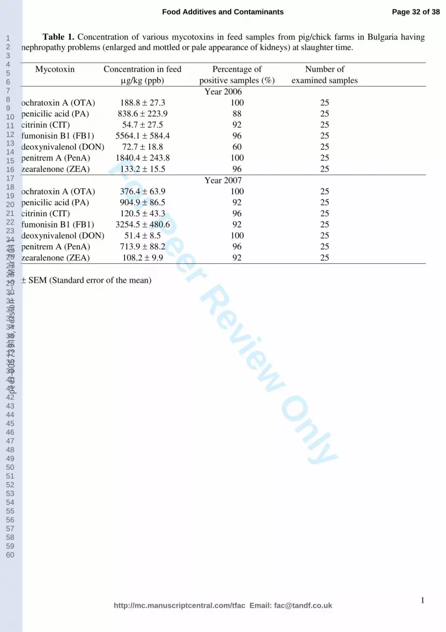
For Peer Review O
nly
1
Table 1. Concentration of various mycotoxins in feed samples from pig/chick farms in Bulgaria having
nephropathy problems (enlarged and mottled or pale appearance of kidneys) at slaughter time.
Mycotoxin Concentration in feed Percentage of Number of
µg/kg (ppb) positive samples (%) examined samples
Year 2006
ochratoxin A (OTA) 188.8 ± 27.3 100 25
penicilic acid (PA) 838.6 ± 223.9 88 25
citrinin (CIT) 54.7 ± 27.5 92 25
fumonisin B1 (FB1) 5564.1 ± 584.4 96 25
deoxynivalenol (DON) 72.7 ± 18.8 60 25
penitrem A (PenA) 1840.4 ± 243.8 100 25
zearalenone (ZEA) 133.2 ± 15.5 96 25
Year 2007
ochratoxin A (OTA) 376.4 ± 63.9 100 25
penicilic acid (PA) 904.9 ± 86.5 92 25
citrinin (CIT) 120.5 ± 43.3 96 25
fumonisin B1 (FB1) 3254.5 ± 480.6 92 25
deoxynivalenol (DON) 51.4 ± 8.5 100 25
penitrem A (PenA) 713.9 ± 88.2 96 25
zearalenone (ZEA) 108.2 ± 9.9 92 25
± SEM (Standard error of the mean)
Page 32 of 38
http://mc.manuscriptcentral.com/tfac Email: [email protected]
Food Additives and Contaminants
123456789101112131415161718192021222324252627282930313233343536373839404142434445464748495051525354555657585960
peer
-005
7391
8, v
ersi
on 1
- 5
Mar
201
1

For Peer Review O
nly
2
Table 2. Concentration of various mycotoxins in serum samples from pig farms in Bulgaria having
nephropathy problems (enlarged and mottled or pale appearance of kidneys) at slaughter time.
Mycotoxin Concentration in serum Number of Number of
µg/l positive samples examined samples
Year 2006
ochratoxin A (OTA) 28.8 ± 25.1 8 10
penicilic acid (PA) 23.3 ± 22.1 8 10
citrinin (CIT) 1.3 ± 0.6 7 10
deoxynivalenol (DON) 7.6 ± 7.6 1 10
penitrem A (PenA) 64.0 ± 55.9 3 10
zearalenone (ZEA) 0.24 ± 0.12 5 10
Year 2007
ochratoxin A (OTA) 6.3 ± 4.9 9 10
penicilic acid (PA) 22.9 ± 12.8 9 10
citrinin (CIT) 1.6 ± 0.6 8 10
deoxynivalenol (DON) 0 0 10
penitrem A (PenA) 45.6 ± 40.3 3 10
zearalenone (ZEA) 0.33 ± 0.17 5 10
± SEM (Standard error of the mean)
Page 33 of 38
http://mc.manuscriptcentral.com/tfac Email: [email protected]
Food Additives and Contaminants
123456789101112131415161718192021222324252627282930313233343536373839404142434445464748495051525354555657585960
peer
-005
7391
8, v
ersi
on 1
- 5
Mar
201
1

For Peer Review O
nly
3
Table 3. Concentration of various mycotoxins in urine samples from pig farms in Bulgaria having
nephropathy problems (enlarged and mottled or pale appearance of kidneys) at slaughter time.
Mycotoxin Concentration in urine Number of Number of
µg/l positive samples (%) examined samples
Year 2006
ochratoxin A (OTA) 3.5 ± 1.1 10 10
penicilic acid (PA) 1.6 ± 0.5 6 10
citrinin (CIT) 1.7 ± 0.4 10 10
deoxynivalenol (DON) 5.1 ± 5.1 1 10
penitrem A (PenA) 0 0 10
zearalenone (ZEA) 9.4 ± 3.9 10 10
Year 2007
ochratoxin A (OTA) 6.2 ± 2.3 10 10
penicilic acid (PA) 1.7 ± 0.6 6 10
citrinin (CIT) 1.8 ± 0.4 10 10
deoxynivalenol (DON) 0 0 10
penitrem A (PenA) 0 0 10
zearalenone (ZEA) 13.1 ± 5.6 10 10
± SEM (Standard error of the mean)
Page 34 of 38
http://mc.manuscriptcentral.com/tfac Email: [email protected]
Food Additives and Contaminants
123456789101112131415161718192021222324252627282930313233343536373839404142434445464748495051525354555657585960
peer
-005
7391
8, v
ersi
on 1
- 5
Mar
201
1
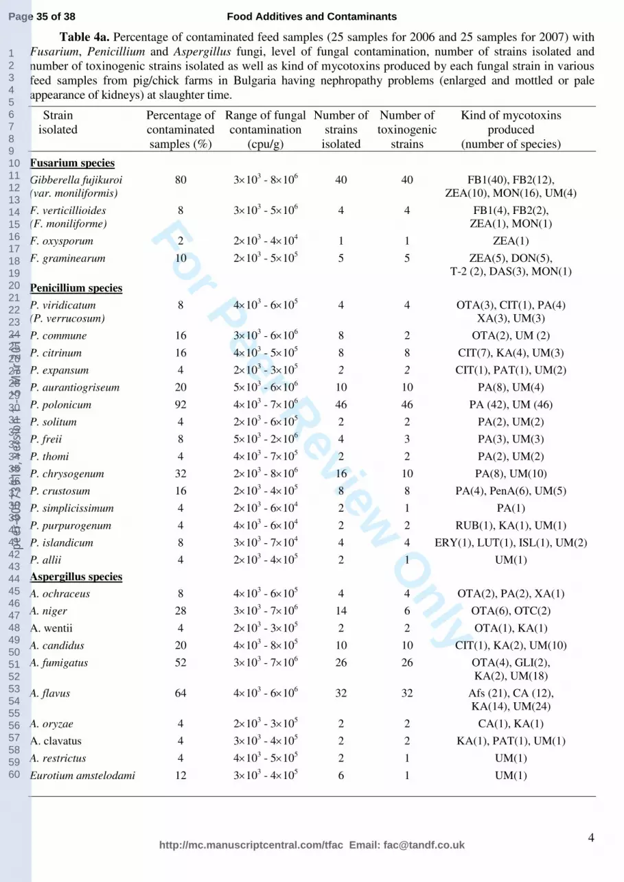
For Peer Review O
nly
4
Table 4a. Percentage of contaminated feed samples (25 samples for 2006 and 25 samples for 2007) with
Fusarium, Penicillium and Aspergillus fungi, level of fungal contamination, number of strains isolated and
number of toxinogenic strains isolated as well as kind of mycotoxins produced by each fungal strain in various
feed samples from pig/chick farms in Bulgaria having nephropathy problems (enlarged and mottled or pale
appearance of kidneys) at slaughter time.
Strain Percentage of Range of fungal Number of Number of Kind of mycotoxins
isolated contaminated contamination strains toxinogenic produced
samples (%) (cpu/g) isolated strains (number of species)
Fusarium species Gibberella fujikuroi 80 3×10
3 - 8×10
6 40 40 FB1(40), FB2(12),
(var. moniliformis) ZEA(10), MON(16), UM(4) F. verticillioides 8 3×10
3 - 5×10
6 4 4 FB1(4), FB2(2),
(F. moniliforme) ZEA(1), MON(1) F. oxysporum 2 2×10
3 - 4×10
4 1 1 ZEA(1)
F. graminearum 10 2×10
3 - 5×10
5 5 5 ZEA(5), DON(5),
T-2 (2), DAS(3), MON(1) Penicillium species P. viridicatum 8 4×10
3 - 6×10
5 4 4 OTA(3), CIT(1), PA(4)
(P. verrucosum) XA(3), UM(3) P. commune 16 3×10
3 - 6×10
6 8 2 OTA(2), UM (2)
P. citrinum 16 4×10
3 - 5×10
5 8 8 CIT(7), KA(4), UM(3)
P. expansum 4 2×10
3 - 3×10
5 2 2 CIT(1), PAT(1), UM(2)
P. aurantiogriseum 20 5×10
3 - 6×10
6 10 10 PA(8), UM(4)
P. polonicum 92 4×10
3 - 7×10
6 46 46 PA (42), UM (46)
P. solitum 4 2×10
3 - 6×10
5 2 2 PA(2), UM(2)
P. freii 8 5×10
3 - 2×10
6 4 3 PA(3), UM(3)
P. thomi 4 4×10
3 - 7×10
5 2 2 PA(2), UM(2)
P. chrysogenum 32 2×10
3 - 8×10
6 16 10 PA(8), UM(10)
P. crustosum 16 2×10
3 - 4×10
5 8 8 PA(4), PenA(6), UM(5)
P. simplicissimum 4 2×10
3 - 6×10
4 2 1 PA(1)
P. purpurogenum 4 4×10
3 - 6×10
4 2 2 RUB(1), KA(1), UM(1)
P. islandicum 8 3×10
3 - 7×10
4 4 4 ERY(1), LUT(1), ISL(1), UM(2)
P. allii 4 2×10
3 - 4×10
5 2 1 UM(1)
Aspergillus species A. ochraceus 8 4×10
3 - 6×10
5 4 4 OTA(2), PA(2), XA(1)
A. niger 28 3×10
3 - 7×10
6 14 6 OTA(6), OTC(2)
A. wentii 4 2×10
3 - 3×10
5 2 2 OTA(1), KA(1)
A. candidus 20 4×10
3 - 8×10
5 10 10 CIT(1), KA(2), UM(10)
A. fumigatus 52 3×10
3 - 7×10
6 26 26 OTA(4), GLI(2),
KA(2), UM(18) A. flavus 64 4×10
3 - 6×10
6 32 32 Afs (21), CA (12),
KA(14), UM(24) A. oryzae 4 2×10
3 - 3×10
5 2 2 CA(1), KA(1)
A. clavatus 4 3×10
3 - 4×10
5 2 2 KA(1), PAT(1), UM(1)
A. restrictus 4 4×10
3 - 5×10
5 2 1 UM(1)
Eurotium amstelodami 12 3×10
3 - 4×10
5 6 1 UM(1)
Page 35 of 38
http://mc.manuscriptcentral.com/tfac Email: [email protected]
Food Additives and Contaminants
123456789101112131415161718192021222324252627282930313233343536373839404142434445464748495051525354555657585960
peer
-005
7391
8, v
ersi
on 1
- 5
Mar
201
1

For Peer Review O
nly
5
Table 4b. Percentage of contaminated feed samples (25 samples for 2006 and 25 samples for 2007) with
various other fungi, level of fungal contamination, number of strains isolated and number of toxinogenic strains
isolated as well as kind of mycotoxins produced by each fungal strain in various feed samples from pig/chick
farms in Bulgaria having nephropathy problems (enlarged and mottled or pale appearance of kidneys) at slaughter
time.
Strain Percentage of Range of fungal Number of Number of Kind of mycotoxins
isolated contaminated contamination strains toxinogenic produced
samples (%) (cpu/g) isolated strains (number of species)
Acremonium strictum 16 4×10³ - 5×10
5 8 2 OOS(2)
Pichia guilliermondii 4 3×10³ - 5×10
5 2 - -
Pichia caribbica 28 4×10³ - 6×10
4 14 - -
Pichia fermentans 4 3×10³ - 2×10
5 2 - -
Paecilomyces variotii 12 2×10³ - 6×10
5 6 - -
Paecilomyces lilacinus 4 2×10³ - 7×10
4 2 - -
Paecilomyces sinensis 4 5×10³ - 6×10
5 2 1 UM(1)
Absidia corymbifera 4 2×10³ - 8×10
4 2 - -
Cladosp. cladosporioides 8 4×10³ - 6×10
4 4 - -
Issatchenkia orientalis 4 3×10³ - 3×10
5 2 - -
Talaromyces emersonii 4 2×10³ - 3×10
5 2 - -
Cordiceps bassiana 4 2×10³ - 2×10
5 2 - -
Mucor racemosus 4 4×10³ - 6×10
5 2 1 UM(1)
Mucor circinelloides 16 4×10³ - 7×10
5 8 3 UM(3)
Candida albicans 4 2×10³ - 4×10
4 2 - -
Candida tropicalis 8 3×10³ - 2×10
5 4 - -
Geomithia pallida 4 2×10³ - 2×10
5 2 1 UM(1)
Sacharomyces cerevisiae 4 3×10³ - 6×10
4 2 - -
Galatomyces geotrichum 4 2×10³ - 4×10
4 2 - -
Rhodotorula mucilaginosa 4 4×10³ - 3×10
4 2 - -
Page 36 of 38
http://mc.manuscriptcentral.com/tfac Email: [email protected]
Food Additives and Contaminants
123456789101112131415161718192021222324252627282930313233343536373839404142434445464748495051525354555657585960
peer
-005
7391
8, v
ersi
on 1
- 5
Mar
201
1
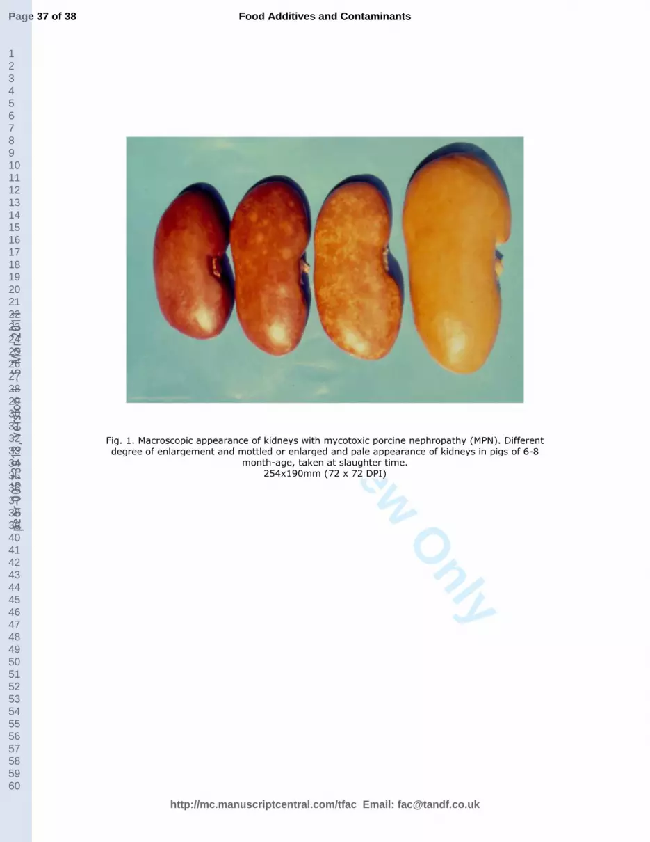
For Peer Review O
nly
Fig. 1. Macroscopic appearance of kidneys with mycotoxic porcine nephropathy (MPN). Different degree of enlargement and mottled or enlarged and pale appearance of kidneys in pigs of 6-8
month-age, taken at slaughter time. 254x190mm (72 x 72 DPI)
Page 37 of 38
http://mc.manuscriptcentral.com/tfac Email: [email protected]
Food Additives and Contaminants
123456789101112131415161718192021222324252627282930313233343536373839404142434445464748495051525354555657585960
peer
-005
7391
8, v
ersi
on 1
- 5
Mar
201
1
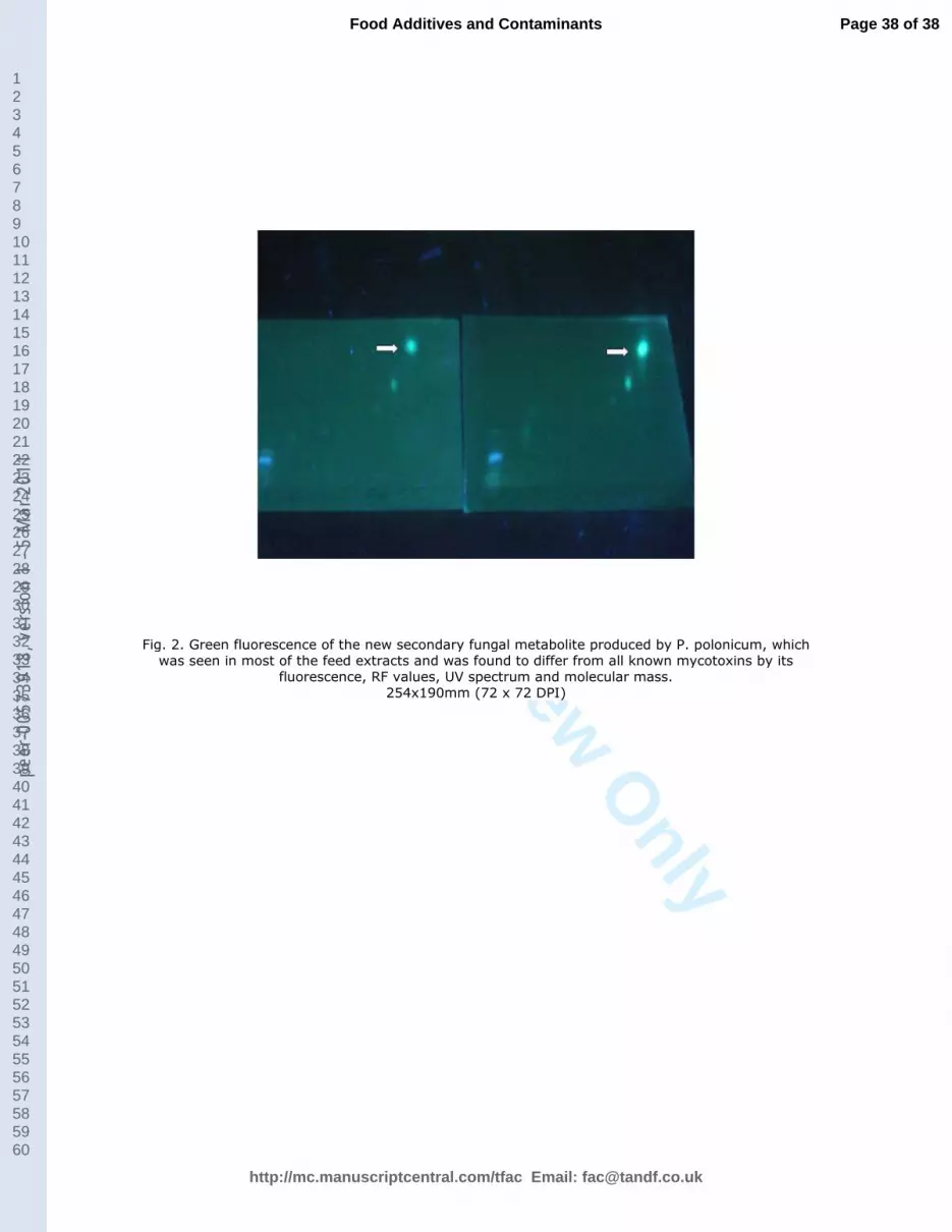
For Peer Review O
nly
Fig. 2. Green fluorescence of the new secondary fungal metabolite produced by P. polonicum, which was seen in most of the feed extracts and was found to differ from all known mycotoxins by its
fluorescence, RF values, UV spectrum and molecular mass. 254x190mm (72 x 72 DPI)
Page 38 of 38
http://mc.manuscriptcentral.com/tfac Email: [email protected]
Food Additives and Contaminants
123456789101112131415161718192021222324252627282930313233343536373839404142434445464748495051525354555657585960
peer
-005
7391
8, v
ersi
on 1
- 5
Mar
201
1