muscle gene electrotransfer is increased by the antioxidant tempol in mice
TRANSCRIPT
ORIGINAL ARTICLE
Muscle gene electrotransfer is increased by the antioxidanttempol in mice
B Markelc1, G Tevz1, M Cemazar1, S Kranjc1, J Lavrencak2, B Zegura3, J Teissie4,5 and G Sersa1
Electropermeabilization (EP) is an effective method of gene transfer into different tissues. During EP, reactive oxygen species(ROS) are formed, which could affect transfection efficiency. The role of generated ROS and the role of antioxidants inelectrotransfer in myoblasts in vitro and in Musculus tibialis cranialis in mice were, therefore, investigated. We demonstratein the study that during EP of C2C12 myoblasts, ROS are generated on the surface of the cells, which do not induce long-termgenomic DNA damage. Plasmid DNA for transfection (pEGFP-N1), which is present outside the cells during EP, neutralizesthe generated ROS. The ROS generation is proportional to the amplitude of the electric pulses and can be scavenged byantioxidants, such as vitamin C or tempol. When antioxidants were used during gene electrotransfer, the transfection efficiencyof C2C12 myoblasts was statistically significantly increased 1.6-fold with tempol. Also in vivo, the transfection efficiency ofM. tibialis cranialis in mice was statistically significantly increased 1.4-fold by tempol. The study indicates that ROS aregenerated on cells during EP and can be scavenged by antioxidants. Specifically, tempol can be used to improve geneelectrotransfer into the muscle and possibly also to other tissues.Gene Therapy (2012) 19, 312–320; doi:10.1038/gt.2011.97; published online 30 June 2011
Keywords: electropermeabilization; gene electrotransfer; muscle; tempol; reactive oxygen species
INTRODUCTION
Electropermeabilization (EP) is a physical method that enables theintroduction of molecules such as dyes, drugs or genetic material intocells. By applying external electric field pulses to living cells, a localincrease in transmembrane potential difference occurs. Under well-defined conditions, the membrane becomes transiently permeabilized,which enables various molecules to enter the cell.1,2
EP is currently in clinical use for the delivery of cancer chemother-apeutic drugs such as bleomycin or cisplatin (electrochemotherapy) inthe treatment of skin metastases or deep-seated tumors.3–5 Completeresponses of skin metastases, predominantly melanoma, are in therange of 70–80%.6
EP was originally discovered for the delivery of plasmid DNA intocells in vitro by Neumann et al. in 1982.7 Since the first report ofsuccessful gene electrotransfection of the skin,8 the method has beenincreasingly used for the delivery of different types of DNA/RNAmolecules into cells in vitro and different tissues, including muscle andtumors in vivo.9–19 Furthermore, gene electrotransfer has also provedto be safe and effective in the treatment of cancer. A report on the firstclinical study of gene electrotransfer, which used therapeutic genecoding for interleukin-12 for the treatment of metastatic melanoma,has already been published by Daud et al.20 There are also otherclinical studies in progress on muscle tissue for DNA vaccination,systemic delivery of therapeutic proteins and correction of gene defectsin muscles; therefore, the studies on muscle cell cultures and muscletissue are of special importance. Namely, good correlation between thein vitro studies on C2C12 cells and in vivo studies on muscle tissue has
been demonstrated.21,22 In addition, gene therapy studies are on-goingin veterinary medicine, showing that the field is rapidly expanding.23–26
Although EP has many advantages compared with viral transfectionmethods, such as the lack of immunogenicity, ease of preparationof large quantities of endotoxin-free plasmid DNA, control andreproducibility of the method and the availability of electropulsatorsapproved for clinical use, there are some problems that must beresolved before the method can become widely used.9,23 The mainproblem is still the low expression efficiency of gene electrotransfer indifferent tissues. Several attempts have, therefore, been made toimprove transfection efficiency, determination of appropriate pulseparameters for specific tissue, the interval between plasmid injectionand the application of electric pulses for a specific tumor type anddegradation of tumor extracellular matrix.15,27–32 In addition, numericalmodeling was used to further increase the efficacy of electrogenetransfer in muscle tissue.33
Previous reports have indicated that EP induces the generation ofreactive oxygen species (ROS), mainly �O2
�, which are generated onthe permeabilized part of the cell membrane during EP34 only whenthe cells are reversibly permeabilized. These ROS can damage lipids,proteins and DNA and, consequently, effect long-term cell survival. Inaddition to that, there have been several studies reporting the induc-tion of genes for radical scavenging molecules after EP.35–37 Thegeneration of ROS and their effect on cell survival after EP can beavoided by adding antioxidant ascorbic acid,38,39 but the toxicity ofthe compound alone is too high for safe usage in vivo. The impact ofgenerated ROS on gene electrotransfer is still not clear.
Received 21 February 2011; revised 13 May 2011; accepted 16 May 2011; published online 30 June 2011
1Department of Experimental Oncology, Institute of Oncology Ljubljana, Ljubljana, Slovenia; 2Department of Cytopathology, Institute of Oncology Ljubljana, Ljubljana, Slovenia;3Department of Genetic Toxicology and Cancer Biology, National Institute of Biology, Ljubljana, Slovenia; 4IPBS CNRS, UMR 5089, Toulouse Cedex, France and 5Universite deToulouse; UPS, IPBS, Toulouse, FranceCorrespondence: Dr J Teissie, IPBS CNRS, UMR 5089, 205 Route de Narbonne, F-31077 Toulouse Cedex, France and Dr G Sersa, Department of Experimental Oncology,Institute of Oncology Ljubljana, Zaloska cesta 2, Ljubljana SI-1000, Slovenia.E-mails: [email protected] and [email protected]
Gene Therapy (2012) 19, 312–320& 2012 Macmillan Publishers Limited All rights reserved 0969-7128/12
www.nature.com/gt
The aim of our study was, therefore, to explore the generation ofROS by the exposure of cells to electric pulses used for electrogenetherapy. In relation to this, the effect of ROS scavengers on thetransfection efficiency of electrotransfer of plasmid DNA encodinggreen fluorescent protein (GFP) to C2C12 myoblasts in vitro andMusculus tibialis cranialis in mice was tested, as a model system. Theresults may be indicative for other tissues that could benefit fromincreased transfection efficiency; predominantly tumors.
RESULTS
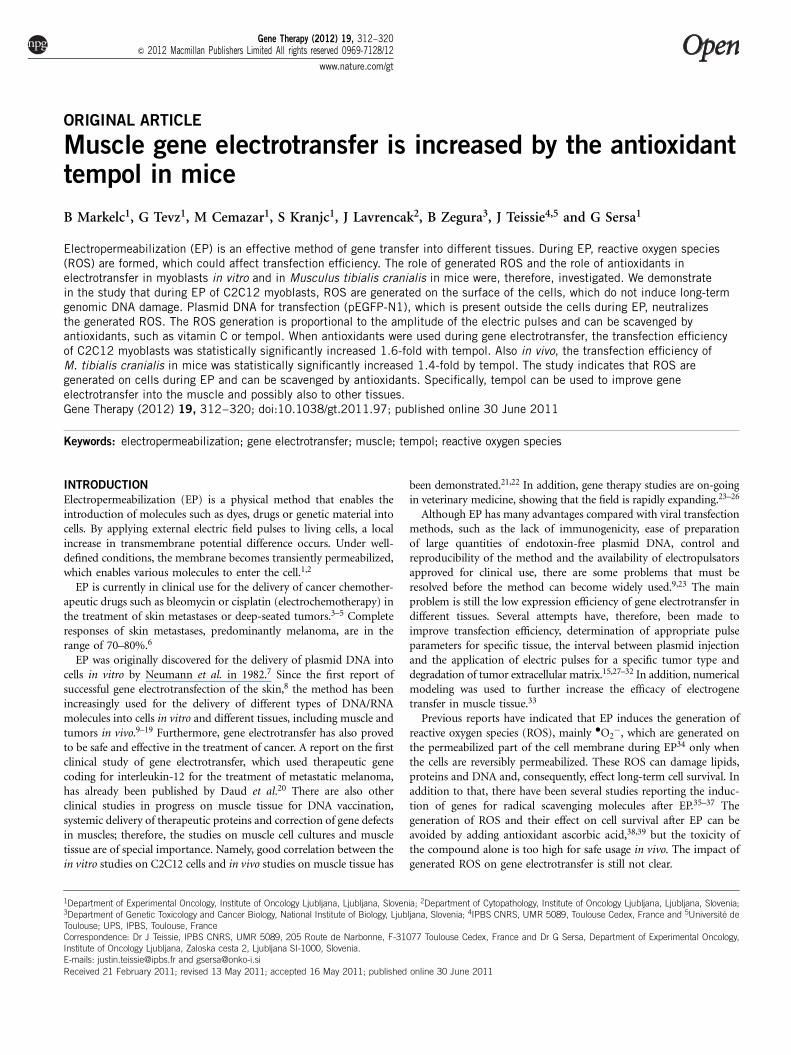
EP and survival of C2C12 mouse myoblastsCell survival and permeabilization of C2C12 cells in vitro weredetermined for different electric pulse parameters: ranging from4 to 8 electric pulses of 5 ms duration of various amplitudes(Figure 1). The results indicated that better cell survival of cells wasobtained with 4 or 6 electric pulses than with 8 electric pulses.Furthermore, there was no difference in cell survival between 4 and6 electric pulses at 500 Vcm�1, whereby cell permeabilization wasB70%, which is the amplitude of electric pulses that was selected forfurther experiments.
EP-induced ROS generation on cells and neutralizing effect ofplasmid DNAThe generation of ROS was determined by lucigenin chemilumines-cence, which is indicative of ROS generated outside of cells.34
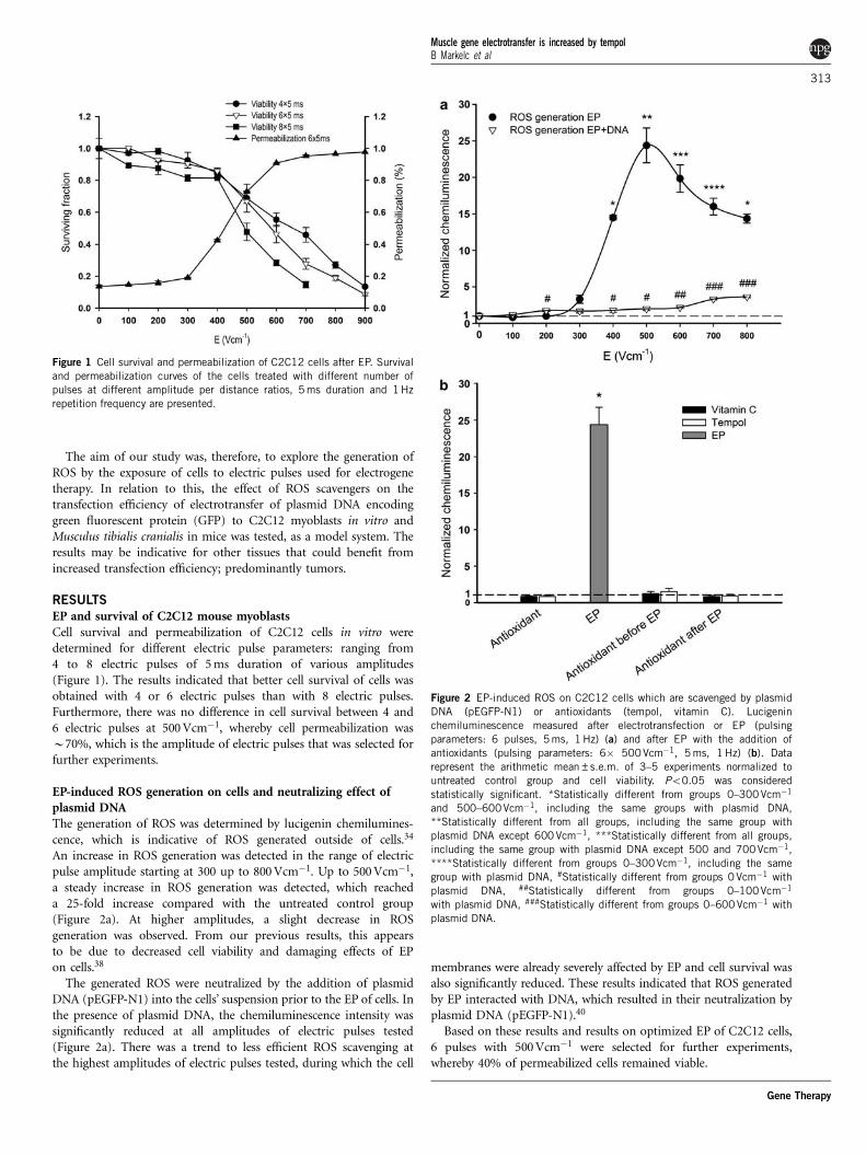
An increase in ROS generation was detected in the range of electricpulse amplitude starting at 300 up to 800 Vcm�1. Up to 500 Vcm�1,a steady increase in ROS generation was detected, which reacheda 25-fold increase compared with the untreated control group(Figure 2a). At higher amplitudes, a slight decrease in ROSgeneration was observed. From our previous results, this appearsto be due to decreased cell viability and damaging effects of EPon cells.38
The generated ROS were neutralized by the addition of plasmidDNA (pEGFP-N1) into the cells’ suspension prior to the EP of cells. Inthe presence of plasmid DNA, the chemiluminescence intensity wassignificantly reduced at all amplitudes of electric pulses tested(Figure 2a). There was a trend to less efficient ROS scavenging atthe highest amplitudes of electric pulses tested, during which the cell
membranes were already severely affected by EP and cell survival wasalso significantly reduced. These results indicated that ROS generatedby EP interacted with DNA, which resulted in their neutralization byplasmid DNA (pEGFP-N1).40
Based on these results and results on optimized EP of C2C12 cells,6 pulses with 500 Vcm�1 were selected for further experiments,whereby 40% of permeabilized cells remained viable.
Figure 1 Cell survival and permeabilization of C2C12 cells after EP. Survival
and permeabilization curves of the cells treated with different number of
pulses at different amplitude per distance ratios, 5 ms duration and 1 Hz
repetition frequency are presented.
Figure 2 EP-induced ROS on C2C12 cells which are scavenged by plasmid
DNA (pEGFP-N1) or antioxidants (tempol, vitamin C). Lucigenin
chemiluminescence measured after electrotransfection or EP (pulsing
parameters: 6 pulses, 5 ms, 1 Hz) (a) and after EP with the addition of
antioxidants (pulsing parameters: 6� 500Vcm�1, 5 ms, 1 Hz) (b). Data
represent the arithmetic mean±s.e.m. of 3–5 experiments normalized to
untreated control group and cell viability. Po0.05 was considered
statistically significant. *Statistically different from groups 0–300Vcm�1
and 500–600Vcm�1, including the same groups with plasmid DNA,**Statistically different from all groups, including the same group with
plasmid DNA except 600 Vcm�1, ***Statistically different from all groups,
including the same group with plasmid DNA except 500 and 700Vcm�1,
****Statistically different from groups 0–300Vcm�1, including the same
group with plasmid DNA, #Statistically different from groups 0 Vcm�1 with
plasmid DNA, ##Statistically different from groups 0–100Vcm�1
with plasmid DNA, ###Statistically different from groups 0–600 Vcm�1 with
plasmid DNA.
Muscle gene electrotransfer is increased by tempolB Markelc et al
313
Gene Therapy
Antioxidants scavenge the ROS generated by EPExposure of C2C12 myoblasts to EP of 500 Vcm�1 induced asignificant increase in ROS generation on the cell membrane, up to25-fold compared with untreated cells (Figures 2a and b). Theaddition of antioxidants before or after EP (10 s) of cells significantlydecreased the amount of ROS detected. The change of pulsing bufferconductivity due to the addition of scavengers did not affect the shapeof the electric pulses delivered on the sample. The ROS scavengingeffect of either vitamin C (4 mM) or tempol (6 mM) was equallyeffective; the values detected were in the range of untreated controlcells, as well as in the range of cells exposed to antioxidants only(Figure 2b).
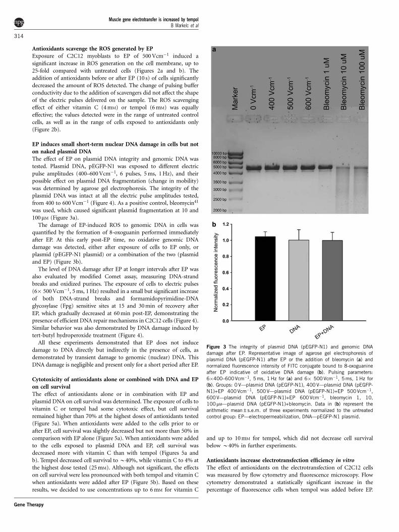
EP induces small short-term nuclear DNA damage in cells but noton naked plasmid DNAThe effect of EP on plasmid DNA integrity and genomic DNA wastested. Plasmid DNA, pEGFP-N1 was exposed to different electricpulse amplitudes (400–600 Vcm�1, 6 pulses, 5 ms, 1 Hz), and theirpossible effect on plasmid DNA fragmentation (change in mobility)was determined by agarose gel electrophoresis. The integrity of theplasmid DNA was intact at all the electric pulse amplitudes tested,from 400 to 600 Vcm�1 (Figure 4). As a positive control, bleomycin41
was used, which caused significant plasmid fragmentation at 10 and100mM (Figure 3a).
The damage of EP-induced ROS to genomic DNA in cells wasquantified by the formation of 8-oxoguanin performed immediatelyafter EP. At this early post-EP time, no oxidative genomic DNAdamage was detected, either after exposure of cells to EP only, orplasmid (pEGFP-N1 plasmid) or a combination of the two (plasmidand EP) (Figure 3b).
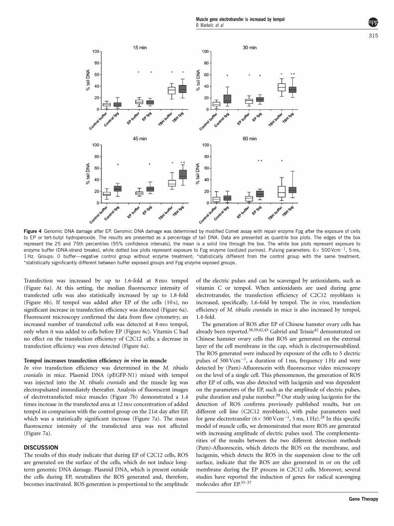
The level of DNA damage after EP at longer intervals after EP wasalso evaluated by modified Comet assay, measuring DNA-strandbreaks and oxidized purines. The exposure of cells to electric pulses(6� 500 Vcm�1, 5 ms, 1 Hz) resulted in a small but significant increaseof both DNA-strand breaks and formamidopyrimidine-DNAglycosylase (Fpg) sensitive sites at 15 and 30 min of recovery afterEP, which gradually decreased at 60 min post-EP, demonstrating thepresence of efficient DNA repair mechanisms in C2C12 cells (Figure 4).Similar behavior was also demonstrated by DNA damage induced bytert-butyl hydroperoxide treatment (Figure 4).
All these experiments demonstrated that EP does not inducedamage to DNA directly but indirectly in the presence of cells, asdemonstrated by transient damage to genomic (nuclear) DNA. ThisDNA damage is negligible and present only for a short period after EP.
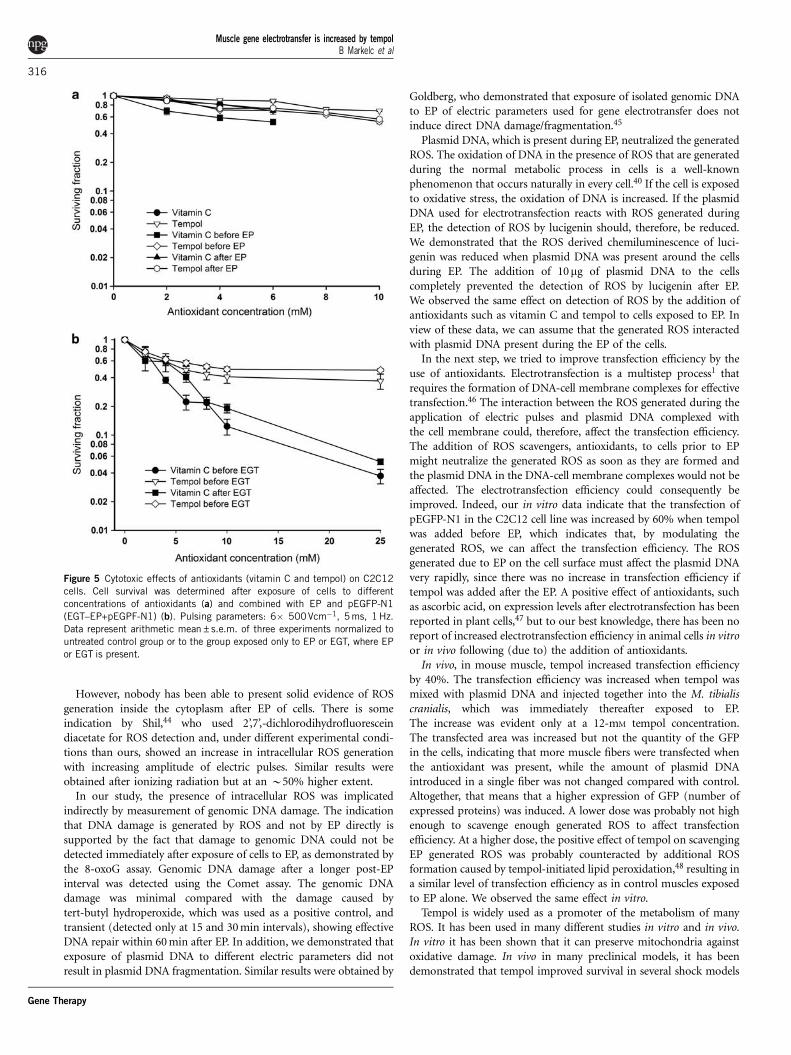
Cytotoxicity of antioxidants alone or combined with DNA and EPon cell survivalThe effect of antioxidants alone or in combination with EP andplasmid DNA on cell survival was determined. The exposure of cells tovitamin C or tempol had some cytotoxic effect, but cell survivalremained higher than 70% at the highest doses of antioxidants tested(Figure 5a). When antioxidants were added to the cells prior to orafter EP, cell survival was slightly decreased but not more than 50% incomparison with EP alone (Figure 5a). When antioxidants were addedto the cells exposed to plasmid DNA and EP, cell survival wasdecreased more with vitamin C than with tempol (Figures 5a andb). Tempol decreased cell survival to B40%, while vitamin C to 4% atthe highest dose tested (25 mM). Although not significant, the effectson cell survival were less pronounced with both tempol and vitamin Cwhen antioxidants were added after EP (Figure 5b). Based on theseresults, we decided to use concentrations up to 6 mM for vitamin C
and up to 10 mM for tempol, which did not decrease cell survivalbelow B40% in further experiments.
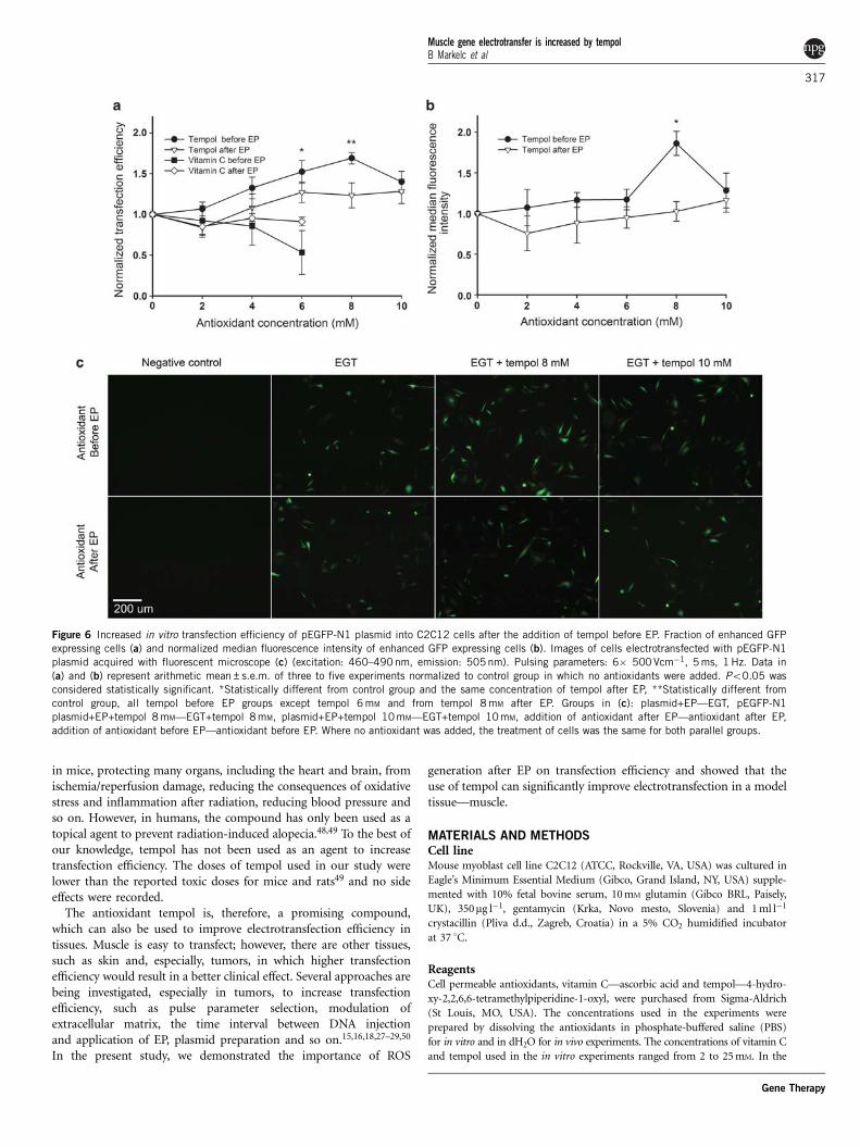
Antioxidants increase electrotransfection efficiency in vitroThe effect of antioxidants on the electrotransfection of C2C12 cellswas measured by flow cytometry and fluorescence microscopy. Flowcytometry demonstrated a statistically significant increase in thepercentage of fluorescence cells when tempol was added before EP.
Figure 3 The integrity of plasmid DNA (pEGFP-N1) and genomic DNA
damage after EP. Representative image of agarose gel electrophoresis of
plasmid DNA (pEGFP-N1) after EP or the addition of bleomycin (a) and
normalized fluorescence intensity of FITC conjugate bound to 8-oxoguanine
after EP indicative of oxidative DNA damage (b). Pulsing parameters:
6�400–600Vcm�1, 5 ms, 1 Hz for (a) and 6� 500 Vcm�1, 5 ms, 1 Hz for
(b). Groups: 0 V—plasmid DNA (pEGFP-N1), 400 V—plasmid DNA (pEGFP-
N1)+EP 400Vcm�1, 500V—plasmid DNA (pEGFP-N1)+EP 500Vcm�1,
600V—plasmid DNA (pEGFP-N1)+EP 600 Vcm�1, bleomycin 1, 10,
100mM—plasmid DNA (pEGFP-N1)+bleomycin. Data in (b) represent the
arithmetic mean±s.e.m. of three experiments normalized to the untreatedcontrol group: EP—electropermeabilization, DNA—pEGFP-N1 plasmid.
Muscle gene electrotransfer is increased by tempolB Markelc et al
314
Gene Therapy
Transfection was increased by up to 1.6-fold at 8 mM tempol(Figure 6a). At this setting, the median fluorescence intensity oftransfected cells was also statistically increased by up to 1.8-fold(Figure 6b). If tempol was added after EP of the cells (10 s), nosignificant increase in transfection efficiency was detected (Figure 6a).Fluorescent microscopy confirmed the data from flow cytometry; anincreased number of transfected cells was detected at 8 mM tempol,only when it was added to cells before EP (Figure 6c). Vitamin C hadno effect on the transfection efficiency of C2C12 cells; a decrease intransfection efficiency was even detected (Figure 6a).
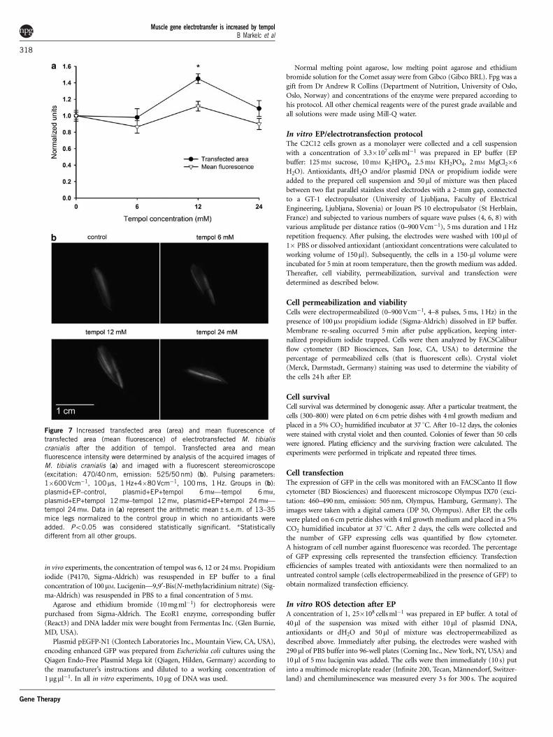
Tempol increases transfection efficiency in vivo in muscleIn vivo transfection efficiency was determined in the M. tibialiscranialis in mice. Plasmid DNA (pEGFP-N1) mixed with tempolwas injected into the M. tibialis cranialis and the muscle leg waselectropulsated immediately thereafter. Analysis of fluorescent imagesof electrotransfected mice muscles (Figure 7b) demonstrated a 1.4times increase in the transfected area at 12 mM concentration of addedtempol in comparison with the control group on the 21st day after EP,which was a statistically significant increase (Figure 7a). The meanfluorescence intensity of the transfected area was not affected(Figure 7a).
DISCUSSION
The results of this study indicate that during EP of C2C12 cells, ROSare generated on the surface of the cells, which do not induce long-term genomic DNA damage. Plasmid DNA, which is present outsidethe cells during EP, neutralizes the ROS generated and, therefore,becomes inactivated. ROS generation is proportional to the amplitude
of the electric pulses and can be scavenged by antioxidants, such asvitamin C or tempol. When antioxidants are used during geneelectrotransfer, the transfection efficiency of C2C12 myoblasts isincreased, specifically, 1.6-fold by tempol. The in vivo, transfectionefficiency of M. tibialis cranialis in mice is also increased by tempol,1.4-fold.
The generation of ROS after EP of Chinese hamster ovary cells hasalready been reported.38,39,42,43 Gabriel and Teissie42 demonstrated onChinese hamster ovary cells that ROS are generated on the externallayer of the cell membrane in the cap, which is electropermeabilized.The ROS generated were induced by exposure of the cells to 5 electricpulses of 500 Vcm�1, a duration of 1 ms, frequency 1 Hz and weredetected by (Pam)-Afluorescein with fluorescence video microscopyon the level of a single cell. This phenomenon, the generation of ROSafter EP of cells, was also detected with lucigenin and was dependenton the parameters of the EP, such as the amplitude of electric pulses,pulse duration and pulse number.39 Our study using lucigenin for thedetection of ROS confirms previously published results, but ondifferent cell line (C2C12 myoblasts), with pulse parameters usedfor gene electrotransfer (6� 500 Vcm�1, 5 ms, 1 Hz).28 In this specificmodel of muscle cells, we demonstrated that more ROS are generatedwith increasing amplitude of electric pulses used. The complementa-rities of the results between the two different detection methods(Pam)-Afluorescein, which detects the ROS on the membrane, andlucigenin, which detects the ROS in the suspension close to the cellsurface, indicate that the ROS are also generated in or on the cellmembrane during the EP process in C2C12 cells. Moreover, severalstudies have reported the induction of genes for radical scavengingmolecules after EP.35–37
Figure 4 Genomic DNA damage after EP. Genomic DNA damage was determined by modified Comet assay with repair enzyme Fpg after the exposure of cells
to EP or tert-butyl hydroperoxide. The results are presented as a percentage of tail DNA. Data are presented as quantile box plots. The edges of the box
represent the 25 and 75th percentiles (95% confidence intervals), the mean is a solid line through the box. The white box plots represent exposure to
enzyme buffer (DNA-strand breaks), while dotted box plots represent exposure to Fpg enzyme (oxidized purines). Pulsing parameters: 6� 500Vcm�1, 5 ms,
1 Hz. Groups: 0 buffer—negative control group without enzyme treatment; *statistically different from the control group with the same treatment,+statistically significantly different between buffer exposed groups and Fpg enzyme exposed groups.
Muscle gene electrotransfer is increased by tempolB Markelc et al
315
Gene Therapy
However, nobody has been able to present solid evidence of ROSgeneration inside the cytoplasm after EP of cells. There is someindication by Shil,44 who used 2’,7’,-dichlorodihydrofluoresceindiacetate for ROS detection and, under different experimental condi-tions than ours, showed an increase in intracellular ROS generationwith increasing amplitude of electric pulses. Similar results wereobtained after ionizing radiation but at an B50% higher extent.
In our study, the presence of intracellular ROS was implicatedindirectly by measurement of genomic DNA damage. The indicationthat DNA damage is generated by ROS and not by EP directly issupported by the fact that damage to genomic DNA could not bedetected immediately after exposure of cells to EP, as demonstrated bythe 8-oxoG assay. Genomic DNA damage after a longer post-EPinterval was detected using the Comet assay. The genomic DNAdamage was minimal compared with the damage caused bytert-butyl hydroperoxide, which was used as a positive control, andtransient (detected only at 15 and 30 min intervals), showing effectiveDNA repair within 60 min after EP. In addition, we demonstrated thatexposure of plasmid DNA to different electric parameters did notresult in plasmid DNA fragmentation. Similar results were obtained by
Goldberg, who demonstrated that exposure of isolated genomic DNAto EP of electric parameters used for gene electrotransfer does notinduce direct DNA damage/fragmentation.45
Plasmid DNA, which is present during EP, neutralized the generatedROS. The oxidation of DNA in the presence of ROS that are generatedduring the normal metabolic process in cells is a well-knownphenomenon that occurs naturally in every cell.40 If the cell is exposedto oxidative stress, the oxidation of DNA is increased. If the plasmidDNA used for electrotransfection reacts with ROS generated duringEP, the detection of ROS by lucigenin should, therefore, be reduced.We demonstrated that the ROS derived chemiluminescence of luci-genin was reduced when plasmid DNA was present around the cellsduring EP. The addition of 10mg of plasmid DNA to the cellscompletely prevented the detection of ROS by lucigenin after EP.We observed the same effect on detection of ROS by the addition ofantioxidants such as vitamin C and tempol to cells exposed to EP. Inview of these data, we can assume that the generated ROS interactedwith plasmid DNA present during the EP of the cells.
In the next step, we tried to improve transfection efficiency by theuse of antioxidants. Electrotransfection is a multistep process1 thatrequires the formation of DNA-cell membrane complexes for effectivetransfection.46 The interaction between the ROS generated during theapplication of electric pulses and plasmid DNA complexed withthe cell membrane could, therefore, affect the transfection efficiency.The addition of ROS scavengers, antioxidants, to cells prior to EPmight neutralize the generated ROS as soon as they are formed andthe plasmid DNA in the DNA-cell membrane complexes would not beaffected. The electrotransfection efficiency could consequently beimproved. Indeed, our in vitro data indicate that the transfection ofpEGFP-N1 in the C2C12 cell line was increased by 60% when tempolwas added before EP, which indicates that, by modulating thegenerated ROS, we can affect the transfection efficiency. The ROSgenerated due to EP on the cell surface must affect the plasmid DNAvery rapidly, since there was no increase in transfection efficiency iftempol was added after the EP. A positive effect of antioxidants, suchas ascorbic acid, on expression levels after electrotransfection has beenreported in plant cells,47 but to our best knowledge, there has been noreport of increased electrotransfection efficiency in animal cells in vitroor in vivo following (due to) the addition of antioxidants.
In vivo, in mouse muscle, tempol increased transfection efficiencyby 40%. The transfection efficiency was increased when tempol wasmixed with plasmid DNA and injected together into the M. tibialiscranialis, which was immediately thereafter exposed to EP.The increase was evident only at a 12-mM tempol concentration.The transfected area was increased but not the quantity of the GFPin the cells, indicating that more muscle fibers were transfected whenthe antioxidant was present, while the amount of plasmid DNAintroduced in a single fiber was not changed compared with control.Altogether, that means that a higher expression of GFP (number ofexpressed proteins) was induced. A lower dose was probably not highenough to scavenge enough generated ROS to affect transfectionefficiency. At a higher dose, the positive effect of tempol on scavengingEP generated ROS was probably counteracted by additional ROSformation caused by tempol-initiated lipid peroxidation,48 resulting ina similar level of transfection efficiency as in control muscles exposedto EP alone. We observed the same effect in vitro.
Tempol is widely used as a promoter of the metabolism of manyROS. It has been used in many different studies in vitro and in vivo.In vitro it has been shown that it can preserve mitochondria againstoxidative damage. In vivo in many preclinical models, it has beendemonstrated that tempol improved survival in several shock models
Figure 5 Cytotoxic effects of antioxidants (vitamin C and tempol) on C2C12
cells. Cell survival was determined after exposure of cells to different
concentrations of antioxidants (a) and combined with EP and pEGFP-N1
(EGT–EP+pEGPF-N1) (b). Pulsing parameters: 6� 500Vcm�1, 5 ms, 1 Hz.
Data represent arithmetic mean±s.e.m. of three experiments normalized to
untreated control group or to the group exposed only to EP or EGT, where EP
or EGT is present.
Muscle gene electrotransfer is increased by tempolB Markelc et al
316
Gene Therapy
in mice, protecting many organs, including the heart and brain, fromischemia/reperfusion damage, reducing the consequences of oxidativestress and inflammation after radiation, reducing blood pressure andso on. However, in humans, the compound has only been used as atopical agent to prevent radiation-induced alopecia.48,49 To the best ofour knowledge, tempol has not been used as an agent to increasetransfection efficiency. The doses of tempol used in our study werelower than the reported toxic doses for mice and rats49 and no sideeffects were recorded.
The antioxidant tempol is, therefore, a promising compound,which can also be used to improve electrotransfection efficiency intissues. Muscle is easy to transfect; however, there are other tissues,such as skin and, especially, tumors, in which higher transfectionefficiency would result in a better clinical effect. Several approaches arebeing investigated, especially in tumors, to increase transfectionefficiency, such as pulse parameter selection, modulation ofextracellular matrix, the time interval between DNA injectionand application of EP, plasmid preparation and so on.15,16,18,27–29,50
In the present study, we demonstrated the importance of ROS
generation after EP on transfection efficiency and showed that theuse of tempol can significantly improve electrotransfection in a modeltissue—muscle.
MATERIALS AND METHODSCell lineMouse myoblast cell line C2C12 (ATCC, Rockville, VA, USA) was cultured in
Eagle’s Minimum Essential Medium (Gibco, Grand Island, NY, USA) supple-
mented with 10% fetal bovine serum, 10 mM glutamin (Gibco BRL, Paisely,
UK), 350mg l�1, gentamycin (Krka, Novo mesto, Slovenia) and 1 ml l�1
crystacillin (Pliva d.d., Zagreb, Croatia) in a 5% CO2 humidified incubator
at 37 1C.
ReagentsCell permeable antioxidants, vitamin C—ascorbic acid and tempol—4-hydro-
xy-2,2,6,6-tetramethylpiperidine-1-oxyl, were purchased from Sigma-Aldrich
(St Louis, MO, USA). The concentrations used in the experiments were
prepared by dissolving the antioxidants in phosphate-buffered saline (PBS)
for in vitro and in dH2O for in vivo experiments. The concentrations of vitamin C
and tempol used in the in vitro experiments ranged from 2 to 25 mM. In the
Figure 6 Increased in vitro transfection efficiency of pEGFP-N1 plasmid into C2C12 cells after the addition of tempol before EP. Fraction of enhanced GFP
expressing cells (a) and normalized median fluorescence intensity of enhanced GFP expressing cells (b). Images of cells electrotransfected with pEGFP-N1
plasmid acquired with fluorescent microscope (c) (excitation: 460–490 nm, emission: 505nm). Pulsing parameters: 6� 500 Vcm�1, 5 ms, 1 Hz. Data in
(a) and (b) represent arithmetic mean±s.e.m. of three to five experiments normalized to control group in which no antioxidants were added. Po0.05 was
considered statistically significant. *Statistically different from control group and the same concentration of tempol after EP, **Statistically different from
control group, all tempol before EP groups except tempol 6 mM and from tempol 8 mM after EP. Groups in (c): plasmid+EP—EGT, pEGFP-N1
plasmid+EP+tempol 8 mM—EGT+tempol 8 mM, plasmid+EP+tempol 10mM—EGT+tempol 10mM, addition of antioxidant after EP—antioxidant after EP,
addition of antioxidant before EP—antioxidant before EP. Where no antioxidant was added, the treatment of cells was the same for both parallel groups.
Muscle gene electrotransfer is increased by tempolB Markelc et al
317
Gene Therapy
in vivo experiments, the concentration of tempol was 6, 12 or 24 mM. Propidium
iodide (P4170, Sigma-Aldrich) was resuspended in EP buffer to a final
concentration of 100mM. Lucigenin—9,9¢-Bis(N-methylacridinium nitrate) (Sig-
ma-Aldrich) was resuspended in PBS to a final concentration of 5 mM.
Agarose and ethidium bromide (10 mg ml�1) for electrophoresis were
purchased from Sigma-Aldrich. The EcoR1 enzyme, corresponding buffer
(React3) and DNA ladder mix were bought from Fermentas Inc. (Glen Burnie,
MD, USA).
Plasmid pEGFP-N1 (Clontech Laboratories Inc., Mountain View, CA, USA),
encoding enhanced GFP was prepared from Escherichia coli cultures using the
Qiagen Endo-Free Plasmid Mega kit (Qiagen, Hilden, Germany) according to
the manufacturer’s instructions and diluted to a working concentration of
1mgml�1. In all in vitro experiments, 10mg of DNA was used.
Normal melting point agarose, low melting point agarose and ethidium
bromide solution for the Comet assay were from Gibco (Gibco BRL). Fpg was a
gift from Dr Andrew R Collins (Department of Nutrition, University of Oslo,
Oslo, Norway) and concentrations of the enzyme were prepared according to
his protocol. All other chemical reagents were of the purest grade available and
all solutions were made using Mill-Q water.
In vitro EP/electrotransfection protocolThe C2C12 cells grown as a monolayer were collected and a cell suspension
with a concentration of 3.3�107 cells ml�1 was prepared in EP buffer (EP
buffer: 125 mM sucrose, 10 mM K2HPO4, 2.5 mM KH2PO4, 2 mM MgCl2�6
H2O). Antioxidants, dH2O and/or plasmid DNA or propidium iodide were
added to the prepared cell suspension and 50ml of mixture was then placed
between two flat parallel stainless steel electrodes with a 2-mm gap, connected
to a GT-1 electropulsator (University of Ljubljana, Faculty of Electrical
Engineering, Ljubljana, Slovenia) or Jouan PS 10 electropulsator (St Herblain,
France) and subjected to various numbers of square wave pulses (4, 6, 8) with
various amplitude per distance ratios (0–900 Vcm�1), 5 ms duration and 1 Hz
repetition frequency. After pulsing, the electrodes were washed with 100ml of
1� PBS or dissolved antioxidant (antioxidant concentrations were calculated to
working volume of 150ml). Subsequently, the cells in a 150-ml volume were
incubated for 5 min at room temperature, then the growth medium was added.
Thereafter, cell viability, permeabilization, survival and transfection were
determined as described below.
Cell permeabilization and viabilityCells were electropermeabilized (0–900 Vcm�1, 4–8 pulses, 5 ms, 1 Hz) in the
presence of 100mM propidium iodide (Sigma-Aldrich) dissolved in EP buffer.
Membrane re-sealing occurred 5 min after pulse application, keeping inter-
nalized propidium iodide trapped. Cells were then analyzed by FACSCalibur
flow cytometer (BD Biosciences, San Jose, CA, USA) to determine the
percentage of permeabilized cells (that is fluorescent cells). Crystal violet
(Merck, Darmstadt, Germany) staining was used to determine the viability of
the cells 24 h after EP.
Cell survivalCell survival was determined by clonogenic assay. After a particular treatment, the
cells (300–800) were plated on 6 cm petrie dishes with 4 ml growth medium and
placed in a 5% CO2 humidified incubator at 37 1C. After 10–12 days, the colonies
were stained with crystal violet and then counted. Colonies of fewer than 50 cells
were ignored. Plating efficiency and the surviving fraction were calculated. The
experiments were performed in triplicate and repeated three times.
Cell transfectionThe expression of GFP in the cells was monitored with an FACSCanto II flow
cytometer (BD Biosciences) and fluorescent microscope Olympus IX70 (exci-
tation: 460–490 nm, emission: 505 nm, Olympus, Hamburg, Germany). The
images were taken with a digital camera (DP 50, Olympus). After EP, the cells
were plated on 6 cm petrie dishes with 4 ml growth medium and placed in a 5%
CO2 humidified incubator at 37 1C. After 2 days, the cells were collected and
the number of GFP expressing cells was quantified by flow cytometer.
A histogram of cell number against fluorescence was recorded. The percentage
of GFP expressing cells represented the transfection efficiency. Transfection
efficiencies of samples treated with antioxidants were then normalized to an
untreated control sample (cells electropermeabilized in the presence of GFP) to
obtain normalized transfection efficiency.
In vitro ROS detection after EPA concentration of 1, 25�108 cells ml�1 was prepared in EP buffer. A total of
40ml of the suspension was mixed with either 10ml of plasmid DNA,
antioxidants or dH2O and 50ml of mixture was electropermeabilized as
described above. Immediately after pulsing, the electrodes were washed with
290ml of PBS buffer into 96-well plates (Corning Inc., New York, NY, USA) and
10ml of 5 mM lucigenin was added. The cells were then immediately (10 s) put
into a multimode microplate reader (Infinite 200, Tecan, Mannendorf, Switzer-
land) and chemiluminescence was measured every 3 s for 300 s. The acquired
Figure 7 Increased transfected area (area) and mean fluorescence of
transfected area (mean fluorescence) of electrotransfected M. tibialis
cranialis after the addition of tempol. Transfected area and mean
fluorescence intensity were determined by analysis of the acquired images ofM. tibialis cranialis (a) and imaged with a fluorescent stereomicroscope
(excitation: 470/40nm, emission: 525/50 nm) (b). Pulsing parameters:
1�600 Vcm�1, 100ms, 1 Hz+4�80 Vcm�1, 100 ms, 1 Hz. Groups in (b):
plasmid+EP–control, plasmid+EP+tempol 6 mM—tempol 6 mM,
plasmid+EP+tempol 12 mM–tempol 12 mM, plasmid+EP+tempol 24 mM—
tempol 24mM. Data in (a) represent the arithmetic mean±s.e.m. of 13–35
mice legs normalized to the control group in which no antioxidants were
added. Po0.05 was considered statistically significant. *Statistically
different from all other groups.
Muscle gene electrotransfer is increased by tempolB Markelc et al
318
Gene Therapy
data were plotted and smoothed to create a curve. The local maximum of the
curve represented the increased chemiluminescence signal due to ROS generation.
To determine the effect of antioxidants on in vitro ROS production, the
antioxidants were added in two different ways. First, the antioxidants were
added to the cell suspension after EP (290ml of antioxidant dissolved in 1� PBS
buffer) instead of PBS buffer and second, 10ml of antioxidants were added to
the cell suspension just before EP (o5 s). The concentrations were calculated to
a working volume of 350ml. The data were normalized to the control group, in
which the cells were not subjected to EP and cell viability.
Determination of plasmid DNA integrityIn order to determine the effect of electric pulses on plasmid DNA integrity,
1ml of pEGFP-N1 plasmid was resuspended in 49ml of EP buffer. The mixture
was then exposed to electric pulses (6� 500 Vcm�1, 5 ms, 1 Hz) or to different
concentrations of bleomycin (1, 10, 100mM) for positive control and then
incubated for 4 h in a water bath at 37 1C, with shaking. After incubation, 7ml
of the sample was mixed with 1ml of EcoR1 enzyme and 2ml of corresponding
buffer (React3) and then incubated for 1 h in a water bath at 37 1C, with
shaking. The restricted samples, containing 140 ng of pDNA, were then
transferred to 1% agarose gel stained with ethidium bromide and electrophor-
esis run for 2 h at 50 Vcm�1. An image of the gel was taken with the GelDoc-it
imaging system (UVP, Upland, CA, USA) under ultraviolet light.
Determination of oxidative DNA damage of genomic (nuclear)DNA after EPIn order to determine whether EP causes any oxidative DNA damage to cells,
8-oxoguanine, which is formed from guanine after interaction of genomic DNA
with ROS, was quantified by OxyDNA test kit (8-oxoG assay; Merck KGaA,
Darmstadt, Germany). The cells were subjected to 6 square wave electric pulses
of 500 Vcm�1 amplitude per distance ratio, 5 ms duration time and 1 Hz
repetition frequency. Immediately (o5 s) after EP, the cells were fixed in 4%
formaldehyde and then permeabilized with 0.1% Triton and washed several
times with wash solution supplied with the kit. After washing, FITC conjugate,
which binds to 8-oxoguanin, was added to the cell pellet and the cells were
incubated for 60 min at room temperature. The cells were again washed with
1� PBS and the cells were then analyzed by FACSCanto II flow cytometer (BD
Biosciences). Histograms of cell number against fluorescence were recorded.
The mean fluorescence of the cell sample was then normalized to the untreated
control group.
Determination of nuclear DNA damage after EPSingle cell gel electrophoresis, also called the Comet assay, is a very sensitive
method for detecting DNA double-strand breaks and single-strand breaks,
alkali labile sites such as apurinic/apyrimidinic sites, DNA–DNA and DNA–
protein cross-links and single-strand breaks associated with incomplete excision
repair.51 The genomic oxidative DNA damage induced by EP was measured
with the modified Comet assay, as described by Collins et al.52 with minor
modifications,53 using lesion-specific DNA repair enzyme Fpg, which converts
oxidized purines to apyrimidinic sites and strand breaks.54,55 C2C12 cells were
electropermeabilized as described above and left in an incubator at 37 1C in 5%
CO2. For positive control of DNA damage, the cells were incubated for 15 min
on ice with 100mM tBOOH (tert-butyl hydroperoxide, Sigma-Aldrich). After
15, 30, 45 and 60 min of cell recovery at 37 1C, 30ml of cell suspension was
mixed with 70ml of 1% low melting point agarose and added to fully frosted
slides that had been covered with a layer of 1% normal melting point agarose.
The slides were subsequently lysed (2.5 M NaOH, 0.1 M ethylenediaminetetraa-
cetic, 0.01 M Tris, pH 10 and 1% Triton X-100) for 1 h at 4 1C. After the lysis,
the slides were washed three times for 5 min in endonuclease buffer solution
(40 mM HEPES-KOH, 0.1 M KCl, 0.5 mM EDTA, 0.2 mg ml–1 bovine serum
albumin, pH 8.0). A total of 50ml aliquots of Fpg solution or enzyme buffer
without Fpg were added to the gel, covered with a cover glass and incubated at
37 1C for 30 min as described by the manufacturer. After the incubation, the
slides were placed in the electrophoresis solution (300 mM NaOH, 1 mM EDTA,
pH 13) for 20 min to allow DNA unwinding, and electrophoresed for 20 min at
25 V (300 mA). Finally, the slides were neutralized with 0.4 M Tris buffer (pH
7.5), stained with ethidium bromide (5mg ml�1) and analyzed using a
fluorescence microscope (Nikon Eclipse 800, Tokyo, Japan). Images of 50
randomly selected nuclei per experimental point were analyzed with image
analysis software Comet Assay IV (Perceptive Instruments, Haverhill, UK). The
results from two independent experiments performed each time in two parallels
are expressed as a percentage of tail DNA and are shown as box plots.
Experimental animalsFemale C57Bl/6 mice were used in the experiments (Institute of Pathology,
Faculty of Medicine, University of Ljubljana, Slovenia). Experimental protocols
were in accordance with the guidelines for animal experiments of European
Union directives and a permit from the Ministry of Agriculture, Forestry and
Food of the Republic of Slovenia. (Permit no.: 34401-11/2009/6). The age of the
mice that were included in the experiments was between 12 and –16 weeks.
Mice were kept under specific pathogen-free conditions at a constant room
temperature (21 1C) and a 12-h light/dark cycle. Food and water were provided
ad libitum. Animals were subjected to an adaptation period of 1 week before
experiments. The experiments were performed three times. Experimental
groups consisted of 6–18 animals.
In vivo electrotransfection of M. tibialis cranialisBoth mouse hind legs were shaved and then depilated with hair removal cream
(Vitaskin, Krka d.d). To increase the randomization, each leg was assigned to
different experimental group. Before EP, the mice were anesthetized with
Isofluran (Torrex Chiesi Pharma GmbH, Vienna, Austria). Muscle electro-
transfection was performed according to our previously described protocol.29
Briefly, 20mg of DNA in 30ml of dH2O for the control group or 20mg of DNA
in 20ml of dH2O mixed with 10ml of dissolved antioxidants was injected into
the muscle o5 s before EP; EP was then performed with CLINIPORATOR
(IGEA s.r.l., Carpi, Italy). Transfection efficiency in vivo was monitored with a
fluorescent stereomicroscope (excitation: 470/40 nm, emission: 525/50 nm,
SteREOLumar V.12, Carl Zeiss, Jena, Germany), equipped with an AxioCam
MRc5 digital camera (Carl Zeiss), on the 7th and 21st day after EP. The
transfected area was separated from the non-transfected area by determining a
threshold value for pixel intensity on every image. Pixels with greater intensity
than the threshold value gave the transfected area. Values were normalized to
the group in which no antioxidants were used. Transfection efficiency was
determined as the normalized fluorescent area and the normalized mean
fluorescence intensity of the fluorescent area on images of M. tibialis cranialis.
ImageJ software (National Institute of Mental Health, Bethesda, MD, USA) was
used for image analysis.
Statistical analysisAll data were tested for normality of distribution using the Kolmogorov–
Smirnov test. Differences between experimental groups were statistically
evaluated by one-way analysis of variance followed by the Holm–Sidak test
for multiple comparisons. A P-value o0.05 was considered to be statistically
significant. SigmaPlot Software (Systat Software, London, UK) was used for
statistical analysis and graphical presentation. For the Comet assay, one-way
analysis of variance (Kruskal–Wallis) was used to analyze differences between
treatments within each experiment. Dunnett’s test was used for comparison
versus the control; Po0.05 was considered to be statistically significant.
CONFLICT OF INTERESTThe authors declare no conflict of interest.
ACKNOWLEDGEMENTSWe acknowledge financial support from the state budget through the Slovenian
Research Agency (program no. P3-0003) and French-Slovenian Scientific
Cooperation (PROTEUS and PICS).
1 Teissie J, Golzio M, Rols MP. Mechanisms of cell membrane electropermeabilization:a minireview of our present (lack of ?) knowledge. Biochim Biophys Acta 2005; 1724:270–280.
Muscle gene electrotransfer is increased by tempolB Markelc et al
319
Gene Therapy
2 Miklavcic D, Towhidi L. Numerical study of the electroporation pulse shape effect onmolecular uptake of biological cells. Radiol Oncol 2010; 44: 34–41.
3 Miklavcic D, Snoj M, Zupanic A, Kos B, Cemazar M, Kropivnik M et al. Towardstreatment planning and treatment of deep-seated solid tumors by electrochemotherapy.Biomed Eng Online 2010; 9: 10.
4 Sersa G, Miklavcic D, Cemazar M, Rudolf Z, Pucihar G, Snoj M. Electrochemotherapy intreatment of tumours. Eur J Surg Oncol 2008; 34: 232–240.
5 Moller MG, Salwa S, Soden DM, O’Sullivan GC. Electrochemotherapy as an adjunct oralternative to other treatments for unresectable or in-transit melanoma. Expert RevAnticancer Ther 2009; 9: 1611–1630.
6 Marty M, Sersa G, Garbay JR, Gehl J, Collins CG, Snoj M et al. Electrochemotherapy—an easy, highly effective and safe treatment of cutaneous and subcutaneous metas-tases: results of ESOPE (European Standard Operating Procedures of Electroche-motherapy) study. Eur J Cancer Suppl 2006; 4: 3–13.
7 Neumann E, Schaefer-Ridder M, Wang Y, Hofschneider PH. Gene transfer into mouselyoma cells by electroporation in high electric fields. EMBO J 1982; 1: 841–845.
8 Titomirov AV, Sukharev S, Kistanova E. In vivo electroporation and stable transformationof skin cells of newborn mice by plasmid DNA. Biochim Biophys Acta 1991; 1088:131–134.
9 Cemazar M, Golzio M, Sersa G, Rols MP, Teissie J. Electrically-assisted nucleicacids delivery to tissues in vivo: where do we stand? Curr Pharm Des 2006; 12:3817–3825.
10 Mir LM. Nucleic acids electrotransfer-based gene therapy (electrogenetherapy): past,current, and future. Mol Biotechnol 2009; 43: 167–176.
11 Vidic S, Markelc B, Sersa G, Coer A, Kamensek U, Tevz G et al. MicroRNAs targetingmutant K-ras by electrotransfer inhibit human colorectal adenocarcinoma cell growthin vitro and in vivo. Cancer Gene Ther 2010; 17: 409–419.
12 Marshall Jr WG, Boone BA, Burgos JD, Gografe SI, Baldwin MK, Danielson ML et al.Electroporation-mediated delivery of a naked DNA plasmid expressing VEGF to theporcine heart enhances protein expression. Gene Therapy 2010; 17: 419–423.
13 Aung W, Hasegawa S, Koshikawa-Yano M, Obata T, Ikehira H, Furukawa T et al.Visualization of in vivo electroporation-mediated transgene expression in experimentaltumors by optical and magnetic resonance imaging. Gene Therapy 2009; 16: 830–839.
14 Aihara H, Miyazaki J. Gene transfer into muscle by electroporation in vivo. NatBiotechnol 1998; 16: 867–870.
15 Gehl J, Mir LM. Determination of optimal parameters for in vivo gene transfer byelectroporation, using a rapid in vivo test for cell permeabilization. Biochem BiophysRes Commun 1999; 261: 377–380.
16 Gehl J, Sorensen TH, Nielsen K, Raskmark P, Nielsen SL, Skovsgaard T et al. In vivoelectroporation of skeletal muscle: threshold, efficacy and relation to electric fielddistribution. Biochim Biophys Acta 1999; 1428: 233–240.
17 Lucas ML, Heller R. Immunomodulation by electrically enhanced delivery of plasmidDNA encoding IL-12 to murine skeletal muscle. Mol Ther 2001; 3: 47–53.
18 Mir LM, Bureau MF, Gehl J, Rangara R, Rouy D, Caillaud JM et al. High-efficiency genetransfer into skeletal muscle mediated by electric pulses. Proc Natl Acad Sci USA1999; 96: 4262–4267.
19 Mir LM, Bureau MF, Rangara R, Schwartz B, Scherman D. Long-term, high level in vivogene expression after electric pulse-mediated gene transfer into skeletal muscle. C RAcad Sci III 1998; 321: 893–899.
20 Daud AI, DeConti RC, Andrews S, Urbas P, Riker AI, Sondak VK et al. Phase I trial ofinterleukin-12 plasmid electroporation in patients with metastatic melanoma. J ClinOncol 2008; 26: 5896–5903.
21 Hojman P. Basic principles and clinical advancements of muscle electrotransfer. CurrGene Ther 2010; 10: 128–138.
22 Burattini S, Ferri P, Battistelli M, Curci R, Luchetti F, Falcieri E. C2C12 murinemyoblasts as a model of skeletal muscle development: morpho-functional character-ization. Eur J Histochem 2004; 48: 223–233.
23 Cemazar M, Jarm T, Sersa G. Cancer electrogene therapy with interleukin-12. CurrGene Ther 2010; 10: 300–311.
24 Heller LC, Heller R. Electroporation gene therapy preclinical and clinical trials formelanoma. Curr Gene Ther 2010; 10: 312–317.
25 Pavlin D, Cemazar M, Cor A, Sersa G, Pogacnik A, Tozon N. Electrogene therapy withinterleukin-12 in canine mast cell tumors. Radiol Oncol 2011; 45: 30–39.
26 Bodles-Brakhop AM, Heller R, Draghia-Akli R. Electroporation for the delivery of DNA-based vaccines and immunotherapeutics: current clinical developments. Mol Ther2009; 17: 585–592.
27 Andre F, Gehl J, Sersa G, Preat V, Hojman P, Eriksen J et al. Efficiency of high and lowvoltage pulse combinations for gene electrotransfer in muscle, liver, tumor and skin.Hum Gene Ther 2008; 19: 1261–1271.
28 Cemazar M, Golzio M, Sersa G, Hojman P, Kranjc S, Mesojednik S et al. Control bypulse parameters of DNA electrotransfer into solid tumors in mice. Gene Therapy 2009;16: 635–644.
29 Tevz G, Pavlin D, Kamensek U, Kranjc S, Mesojednik S, Coer A et al. Gene electro-transfer into murine skeletal muscle: a systematic analysis of parameters for long-termgene expression. Technol Cancer Res Treat 2008; 7: 91–101.
30 Roos AK, Eriksson F, Walters DC, Pisa P, King AD. Optimization of skin electroporationin mice to increase tolerability of DNA vaccine delivery to patients. Mol Ther 2009; 17:1637–1642.
31 Vry JD, Martinez-Martinez P, Losen M, Bode GH, Temel Y, Steckler T et al. Low current-driven micro-electroporation allows efficient in vivo delivery of nonviral DNA into theadult mouse brain. Mol Ther 2010; 18: 1183–1191.
32 Hojman P, Gissel H, Andre FM, Cournil-Henrionnet C, Eriksen J, Gehl J et al.Physiological effects of high- and low-voltage pulse combinations for gene electro-transfer in muscle. Hum Gene Ther 2008; 19: 1249–1260.
33 Zupanic A, Corovic S, Miklavcic D, Pavlin M. Numerical optimization of gene electro-transfer into muscle tissue. Biomed Eng Online 2010; 9: 66.
34 Gabriel B, Teissie J. Control by electrical parameters of short- and long-term cell deathresulting from electropermeabilization of Chinese hamster ovary cells. Biochim BiophysActa 1995; 1266: 171–178.
35 Heller LC, Cruz YL, Ferraro B, Yang H, Heller R. Plasmid injection and application ofelectric pulses alter endogenous mRNA and protein expression in B16.F10 mousemelanomas. Cancer Gene Ther 2010; 17: 864–871.
36 Hojman P, Zibert JR, Gissel H, Eriksen J, Gehl J. Gene expression profiles in skeletalmuscle after gene electrotransfer. BMC Mol Biol 2007; 8: 56.
37 Rubenstrunk A, Mahfoudi A, Scherman D. Delivery of electric pulses for DNA electro-transfer to mouse muscle does not induce the expression of stress related genes.Cell Biol Toxicol 2004; 20: 25–31.
38 Bonnafous P, Vernhes M, Teissie J, Gabriel B. The generation of reactive-oxygen speciesassociated with long-lasting pulse-induced electropermeabilisation of mammalian cellsis based on a non-destructive alteration of the plasma membrane. Biochim BiophysActa 1999; 1461: 123–134.
39 Gabriel B, Teissie J. Generation of reactive-oxygen species induced by electropermea-bilization of Chinese hamster ovary cells and their consequence on cell viability.Eur J Biochem 1994; 223: 25–33.
40 Cooke MS, Evans MD, Dizdaroglu M, Lunec J. Oxidative DNA damage: mechanisms,mutation, and disease. FASEB J 2003; 17: 1195–1214.
41 Keller TJ, Oppenheimer NJ. Enhanced bleomycin-mediated damage of DNA oppositecharged nicks. A model for bleomycin-directed double strand scission of DNA.J Biol Chem 1987; 262: 15144–15150.
42 Gabriel B, Teissie J. Spatial compartmentation and time resolution of photooxidation ofa cell membrane probe in electropermeabilized Chinese hamster ovary cells.Eur J Biochem 1995; 228: 710–718.
43 Sabri N, Pelissier B, Teissie J. Electropermeabilization of intact maize cells induces anoxidative stress. Eur J Biochem 1996; 238: 737–743.
44 Shil P, Sanghvi SH, Vidyasagar PB, Mishra KP. Enhancement of radiation cytotoxicity inmurine cancer cells by electroporation: in vitro and in vivo studies. J Environ PatholToxicol Oncol 2005; 24: 291–298.
45 Goldberg A, Rubinsky B. The effect of electroporation type pulsed electric fields onDNA in aqueous solution. Technol Cancer Res Treat 2010; 9: 423–430.
46 Faurie C, Rebersek M, Golzio M, Kanduser M, Escoffre JM, Pavlin M et al. Electro-mediated gene transfer and expression are controlled by the life-time of DNA/mem-brane complex formation. J Gene Med 2010; 12: 117–125.
47 Sabri N, Pelissier B, Teissie J. Ascorbate increases electrotransformation efficiency ofintact maize cells. Anal Biochem 1998; 264: 284–286.
48 Wilcox CS, Pearlman A. Chemistry and antihypertensive effects of tempol and othernitroxides. Pharmacol Rev 2008; 60: 418–469.
49 Wilcox CS. Effects of tempol and redox-cycling nitroxides in models of oxidative stress.Pharmacol Ther 2010; 126: 119–145.
50 Mesojednik S, Pavlin D, Sersa G, Coer A, Kranjc S, Grosel A et al. The effect of thehistological properties of tumors on transfection efficiency of electrically assisted genedelivery to solid tumors in mice. Gene Therapy 2007; 14: 1261–1269.
51 Tice RR, Agurell E, Anderson D, Burlinson B, Hartmann A, Kobayashi H et al. Singlecell gel/comet assay: guidelines for in vitro and in vivo genetic toxicology testing.Environ Mol Mutagen 2000; 35: 206–221.
52 Collins AR, Duthie SJ, Dobson VL. Direct enzymic detection of endogenous oxidativebase damage in human lymphocyte DNA. Carcinogenesis 1993; 14: 1733–1735.
53 Zegura B, Lah TT, Filipic M. The role of reactive oxygen species in microcystin-LR-induced DNA damage. Toxicology 2004; 200: 59–68.
54 Collins AR, Dusinska M, Gedik CM, Stetina R. Oxidative damage to DNA: do we have areliable biomarker? Environ Health Perspect 1996; 104 (Suppl 3): 465–469.
55 Miklos M, Gajski G, Garaj-Vrhovac V. Usage of the standard and modified comet assayin assessment of DNA damage in human lymphocytes after exposure to ionizingradiation. Radiol Oncol 2009; 43: 97–107.
This work is licensed under the Creative CommonsAttribution-NonCommercial-No Derivative Works 3.0
Unported License. To view a copy of this license, visit http://creativecommons.org/licenses/by-nc-nd/3.0/
Muscle gene electrotransfer is increased by tempolB Markelc et al
320
Gene Therapy