natural bioactive products with antioxidant properties useful
TRANSCRIPT
Oxidative Medicine and Cellular Longevity
Natural Bioactive Products with Antioxidant Properties Useful in Neurodegenerative Diseases
Lead Guest Editor: Francisco J.B. Mendonça JúniorGuest Editors: Luciana Scotti, Marcus T. Scotti, Anuraj Nayarisseri, and Ernestine N. Zondegoumba
Natural Bioactive Products with AntioxidantProperties Useful in Neurodegenerative Diseases

Oxidative Medicine and Cellular Longevity
Natural Bioactive Products with AntioxidantProperties Useful in Neurodegenerative Diseases
Lead Guest Editor: Francisco J.B. Mendonça JúniorGuest Editors: Luciana Scotti, Marcus T. Scotti, Anuraj Nayarisseri,and Ernestine N. Zondegoumba

Copyright © 2019 Hindawi. All rights reserved.
This is a special issue published in “OxidativeMedicine and Cellular Longevity.” All articles are open access articles distributed under theCreative Commons Attribution License, which permits unrestricted use, distribution, and reproduction in any medium, provided theoriginal work is properly cited.

Editorial Board
Fabio Altieri, ItalyFernanda Amicarelli, ItalyJosé P. Andrade, PortugalCristina Angeloni, ItalyAntonio Ayala, SpainElena Azzini, ItalyPeter Backx, CanadaDamian Bailey, UKSander Bekeschus, GermanyJi C. Bihl, USAConsuelo Borrás, SpainNady Braidy, AustraliaRalf Braun, GermanyLaura Bravo, SpainAmadou Camara, USAGianluca Carnevale, ItalyRoberto Carnevale, ItalyAngel Catalá, ArgentinaGiulio Ceolotto, ItalyShao-Yu Chen, USAFerdinando Chiaradonna, ItalyZhao Zhong Chong, USAAlin Ciobica, RomaniaAna Cipak Gasparovic, CroatiaGiuseppe Cirillo, ItalyMaria R. Ciriolo, ItalyMassimo Collino, ItalyGraziamaria Corbi, ItalyManuela Corte-Real, PortugalMark Crabtree, UKManuela Curcio, ItalyAndreas Daiber, GermanyFelipe Dal Pizzol, BrazilFrancesca Danesi, ItalyDomenico D’Arca, ItalySergio Davinelli, USAClaudio De Lucia, ItalyYolanda de Pablo, SwedenSonia de Pascual-Teresa, SpainCinzia Domenicotti, ItalyJoël R. Drevet, FranceGrégory Durand, FranceJavier Egea, SpainErsin Fadillioglu, Turkey
Ioannis G. Fatouros, GreeceQingping Feng, CanadaGianna Ferretti, ItalyGiuseppe Filomeni, ItalySwaran J. S. Flora, IndiaTeresa I. Fortoul, MexicoJeferson L. Franco, BrazilRodrigo Franco, USAJoaquin Gadea, SpainJuan Gambini, SpainJosé Luís García-Giménez, SpainGerardo García-Rivas, MexicoJanusz Gebicki, AustraliaAlexandros Georgakilas, GreeceHusam Ghanim, USARajeshwary Ghosh, USAEloisa Gitto, ItalyDaniela Giustarini, ItalySaeid Golbidi, CanadaAldrin V. Gomes, USATilman Grune, GermanyNicoletta Guaragnella, ItalySolomon Habtemariam, UKE.-M. Hanschmann, GermanyTim Hofer, NorwayJohn D. Horowitz, AustraliaSilvana Hrelia, ItalyStephan Immenschuh, GermanyMaria Isaguliants, LatviaLuigi Iuliano, ItalyVladimir Jakovljevic, SerbiaMarianna Jung, USAPeeter Karihtala, FinlandEric E. Kelley, USAKum Kum Khanna, AustraliaNeelam Khaper, CanadaThomas Kietzmann, FinlandDemetrios Kouretas, GreeceAndrey V. Kozlov, AustriaJean-Claude Lavoie, CanadaSimon Lees, CanadaCh. Horst Lillig, GermanyPaloma B. Liton, USAAna Lloret, Spain
Lorenzo Loffredo, ItalyDaniel Lopez-Malo, SpainAntonello Lorenzini, ItalyNageswara Madamanchi, USAKenneth Maiese, USAMarco Malaguti, ItalyTullia Maraldi, ItalyReiko Matsui, USAJuan C. Mayo, SpainSteven McAnulty, USAAntonio Desmond McCarthy, ArgentinaBruno Meloni, AustraliaPedro Mena, ItalyVíctor M. Mendoza-Núñez, MexicoMaria U. Moreno, SpainTrevor A. Mori, AustraliaRyuichi Morishita, JapanFabiana Morroni, ItalyLuciana Mosca, ItalyAnge Mouithys-Mickalad, BelgiumIordanis Mourouzis, GreeceDanina Muntean, RomaniaColin Murdoch, UKPablo Muriel, MexicoRyoji Nagai, JapanDavid Nieman, USAHassan Obied, AustraliaJulio J. Ochoa, SpainPál Pacher, USAPasquale Pagliaro, ItalyValentina Pallottini, ItalyRosalba Parenti, ItalyVassilis Paschalis, GreeceVisweswara Rao Pasupuleti, MalaysiaDaniela Pellegrino, ItalyIlaria Peluso, ItalyClaudia Penna, ItalySerafina Perrone, ItalyTiziana Persichini, ItalyShazib Pervaiz, SingaporeVincent Pialoux, FranceAda Popolo, ItalyJosé L. Quiles, SpainWalid Rachidi, France
Zsolt Radak, HungaryN. Soorappan Rajasekaran, USAKota V. Ramana, USASid D. Ray, USAHamid Reza Rezvani, FranceAlessandra Ricelli, ItalyPaola Rizzo, ItalyFrancisco J. Romero, SpainJoan Roselló-Catafau, SpainH. P. Vasantha Rupasinghe, CanadaGabriele Saretzki, UKLuciano Saso, ItalyNadja Schroder, Brazil
Sebastiano Sciarretta, ItalyRatanesh K. Seth, USAHonglian Shi, USACinzia Signorini, ItalyMithun Sinha, USACarla Tatone, ItalyFrank Thévenod, GermanyShaneThomas, AustraliaCarlo G. Tocchetti, ItalyAngela Trovato Salinaro, JamaicaPaolo Tucci, ItalyRosa Tundis, ItalyGiuseppe Valacchi, Italy
Jeannette Vasquez-Vivar, USADaniele Vergara, ItalyVictor M. Victor, SpainLászló Virág, HungaryNatalie Ward, AustraliaPhilip Wenzel, GermanyAnthony R. White, AustraliaGeorg T. Wondrak, USAMichal Wozniak, PolandSho-ichi Yamagishi, JapanLiang-Jun Yan, USAGuillermo Zalba, SpainMario Zoratti, Italy

Contents
Natural Bioactive Products with Antioxidant Properties Useful in Neurodegenerative DiseasesFrancisco J. B. Mendonça-Junior , Marcus T. Scotti , Anuraj Nayarisseri ,Ernestine N. T. Zondegoumba , and Luciana ScottiEditorial (2 pages), Article ID 7151780, Volume 2019 (2019)
Morinda citrifolia and Its Active Principle Scopoletin Mitigate Protein Aggregation and NeuronalApoptosis through Augmenting the DJ-1/Nrf2/ARE Signaling PathwayKishore Kumar S. Narasimhan, Deepthy Jayakumar, Prema Velusamy, Ashokkumar Srinivasan,Thangarajeswari Mohan, Divya Bhavani Ravi, Saraswathi Uthamaraman, Yogesh Kanna Sathyamoorthy,Namakkal Soorappan Rajasekaran , and Kalaiselvi PeriandavanResearch Article (13 pages), Article ID 2761041, Volume 2019 (2019)
Pasteurized Orange Juice Rich in Carotenoids Protects Caenorhabditis elegans against Oxidative Stressand 𝛽-Amyloid Toxicity through Direct and Indirect MechanismsRicardo Basílio de Oliveira Caland, Cesar Orlando Muñoz Cadavid, Lourdes Carmona, Leandro Peña,and Riva de Paula OliveiraResearch Article (13 pages), Article ID 5046280, Volume 2019 (2019)
Ethanol Extract of Centipeda minima Exerts Antioxidant and Neuroprotective Effects via Activation ofthe Nrf2 Signaling PathwayYi-Jie Wang, Xin-YueWang, Xu-Yi Hao, Yong-Ming Yan, Ming Hong, Su-Fen Wei, Yi-Le Zhou, Qi Wang ,Yong-Xian Cheng , and Yong-Qiang LiuResearch Article (16 pages), Article ID 9421037, Volume 2019 (2019)
Sulforaphane-Enriched Broccoli Sprouts Pretreated by Pulsed Electric Fields ReducesNeuroinflammation and Ameliorates Scopolamine-Induced Amnesia in Mouse Brain through ItsAntioxidant Ability via Nrf2-HO-1 ActivationLalita Subedi , KyoHee Cho, Yong Un Park, Hyuk Joon Choi, and Sun Yeou KimResearch Article (19 pages), Article ID 3549274, Volume 2019 (2019)
Inhibition of Oxidative Neurotoxicity and Scopolamine-Induced Memory Impairment by 𝛾-Mangostin:In Vitro and In Vivo EvidenceYoungmun Lee, Sunyoung Kim, Yeonsoo Oh, Young-Mi Kim, Young-Won Chin , and Jungsook ChoResearch Article (14 pages), Article ID 3640753, Volume 2019 (2019)
Eleutherococcus Species Cultivated in Europe: A New Source of Compounds withAntiacetylcholinesterase, Antihyaluronidase, Anti-DPPH, and Cytotoxic ActivitiesKuba Adamczyk, Marta Olech , Jagoda Abramek, Wioleta Pietrzak, Rafał Kuźniewski ,Anna Bogucka-Kocka , Renata Nowak , Aneta A. Ptaszyńska, Alina Rapacka-Gackowska,Tomasz Skalski, Maciej Strzemski , Ireneusz Sowa , Magdalena Wójciak-Kosior, Marcin Feldo,and Daniel ZałuskiResearch Article (10 pages), Article ID 8673521, Volume 2019 (2019)
Honokiol-Mediated Mitophagy Ameliorates Postoperative Cognitive Impairment Induced bySurgery/Sevoflurane via Inhibiting the Activation of NLRP3 Inflammasome in the HippocampusJi-Shi Ye, Lei Chen, Ya-Yuan Lu, Shao-Qing Lei , Mian Peng , and Zhong-Yuan XiaResearch Article (13 pages), Article ID 8639618, Volume 2019 (2019)
Benefits of Vitamins in the Treatment of Parkinson’s DiseaseXiuzhen Zhao, Ming Zhang, Chunxiao Li, Xue Jiang, Yana Su, and Ying ZhangReview Article (14 pages), Article ID 9426867, Volume 2019 (2019)
Antioxidant Properties of Amazonian Fruits: AMini Review of In Vivo and In Vitro StudiesRaúl Avila-Sosa , Andrés Felipe Montero-Rodríguez, Patricia Aguilar-Alonso , Obdulia Vera-López,Martin Lazcano-Hernández, Julio César Morales-Medina, and Addí Rhode Navarro-CruzReview Article (11 pages), Article ID 8204129, Volume 2019 (2019)
Borneol for Regulating the Permeability of the Blood-Brain Barrier in Experimental Ischemic Stroke:Preclinical Evidence and Possible MechanismZi-xian Chen, Qing-qing Xu , Chun-shuo Shan , Yi-hua Shi , Yong Wang,Raymond Chuen-Chung Chang , and Guo-qing ZhengReview Article (15 pages), Article ID 2936737, Volume 2019 (2019)
Computational Studies Applied to Flavonoids against Alzheimer’s and Parkinson’s DiseasesAlex France M. Monteiro, Jéssika De O. Viana, Anuraj Nayarisseri, Ernestine N. Zondegoumba ,Francisco Jaime B. Mendonça Junior , Marcus Tullius Scotti , and Luciana ScottiResearch Article (21 pages), Article ID 7912765, Volume 2018 (2019)
Effect of Alkaloid Extract from African Jointfir (Gnetum africanum) Leaves on Manganese-InducedToxicity inDrosophila melanogasterGaniyu Oboh , Opeyemi Babatunde Ogunsuyi , Olatunde Isaac Awonyemi ,and Victor Ayomide AtokiResearch Article (10 pages), Article ID 8952646, Volume 2018 (2019)
Anacardiummicrocarpum Promotes Neuroprotection Dependently of AKT and ERK Phosphorylationbut Does Not Prevent Mitochondrial Damage by 6-OHDAIllana Kemmerich Martins, Nélson Rodrigues de Carvalho, Giulianna Echeverria Macedo,Nathane Rosa Rodrigues, Cynthia Camila Ziech, Lúcia Vinadé, Valter Menezes Barbosa Filho,Irwin Alencar Menezes , Jeferson Franco, and Thaís PosserResearch Article (12 pages), Article ID 2131895, Volume 2018 (2019)
Formulated Chinese Medicine Shaoyao Gancao Tang Reduces Tau Aggregation and ExertsNeuroprotection through Anti-Oxidation and Anti-InflammationI-Cheng Chen , Te-Hsien Lin , Yu-Hsuan Hsieh, Chih-Ying Chao, Yih-Ru Wu ,Kuo-Hsuan Chang , Ming-Chung Lee, Guey-Jen Lee-Chen , and Chiung-Mei ChenResearch Article (16 pages), Article ID 9595741, Volume 2018 (2019)
Effects of the Aphanizomenonflos-aquae Extract (Klamin®) on a Neurodegeneration Cellular ModelD. Nuzzo , G. Presti, P. Picone, G. Galizzi, E. Gulotta, S. Giuliano, C. Mannino, V. Gambino, S. Scoglio,and M. Di CarloResearch Article (14 pages), Article ID 9089016, Volume 2018 (2019)

EditorialNatural Bioactive Products with Antioxidant Properties Useful inNeurodegenerative Diseases
Francisco J. B. Mendonça-Junior ,1,2 Marcus T. Scotti ,1 Anuraj Nayarisseri ,3,4
Ernestine N. T. Zondegoumba ,5 and Luciana Scotti 1,6
1Postgraduate Program in Natural and Synthetic Bioactive Products, Federal University of Paraíba, João Pessoa, PB, Brazil2Laboratory of Synthesis and Drug Delivery, Department of Biological Science, State University of Paraiba, João Pessoa, PB, Brazil3In Silico Research Laboratory, Eminent Biosciences, Indore, 452010 Madhya Pradesh, India4Bioinformatics Research Laboratory, LeGene Biosciences Pvt. Ltd., Indore, 452010 Madhya Pradesh, India5Department of Organic Chemistry, Faculty of Science, University of Yaoundé I, PO Box 812, Yaoundé, Cameroon6Teaching and Research Management-University Hospital, Federal University of Paraíba, João Pessoa, PB, Brazil
Correspondence should be addressed to Francisco J. B. Mendonça-Junior; [email protected]
Received 12 March 2019; Accepted 13 March 2019; Published 9 May 2019
Copyright © 2019 Francisco J. B. Mendonça-Junior et al. This is an open access article distributed under the Creative CommonsAttribution License, which permits unrestricted use, distribution, and reproduction in any medium, provided the original workis properly cited.
Neurodegenerative diseases (NDs) constitute a large group ofpathological conditions, characterized by a progressive lossof neuronal cells, which compromise motor and/or cognitivefunctions. The most common NDs are Alzheimer’s disease(AD), amyotrophic lateral sclerosis (ALS), Parkinson’s dis-ease (PD), and Huntington’s disease (HD). The causes ofthese pathologies are multifactorial and not fully understood,but it is well known that factors related to aging and to theoverproduction of free radical and reactive oxygen specieslead to oxidative stress and to cell death, which are extremelyrelated. Whereas oxidative stress plays an unquestionableand central role in NDs, the control of free radicals and reac-tive oxygen species levels represents an interesting and prom-ising strategy to delay neurodegeneration and attenuate theassociated symptoms.
In this context, several natural bioactive compoundsisolated from plants, fungi, and algae, among others, and alsosynthetic compounds inspired by natural scaffolds, whichpresent antioxidant properties, including vitamins C and E,anthocyanins, and phenolic compounds, are extensivelydescribed as potential palliative agents of neurodegenerativesymptoms. In vitro and in vivo studies, performed withextracts and fractions of plants and with isolated naturalbioactive compounds, provide evidence of the role of these
substances in the modulation of the cellular redox balanceand in the reduction of the formation of reactive oxygen spe-cies originating from oxidative stress, thereby demonstratingtheir great value as antioxidant agents and cellular protectors.
In this special issue, articles were selected that addressnew therapeutic alternatives on the antioxidant and anti-inflammatory role and the consequent neuroprotetor ofnatural (or inspired) bioactive compounds in the prevention/-treatment or improvement of neurodegenerative diseases.This special issue compiles fifteen (15) manuscripts includingthree (3) reviews and twelve (12) research papers, which showrecent research about the discovery of plant-derived antioxi-dants with application in neurodegenerative diseases.
The review by R. Avila-Sosa et al. describes the antioxi-dant effects of main bioactive components isolated fromAmazonian fruits. Among other activities, the authors high-light antioxidants, immunomodulatory, anticancer, anti-inflammatory, and antidepressant properties of phenoliccompounds, unsaturated fatty acids, carotenoids, phytos-terols, and tocopherols.
The review by X. Zhao et al. highlights the benefits ofvitamin supplementation in the treatment or improvementof the clinical symptoms of Parkinson’s disease. The authorssummarized the biological correlations between vitamins and
HindawiOxidative Medicine and Cellular LongevityVolume 2019, Article ID 7151780, 2 pageshttps://doi.org/10.1155/2019/7151780

PD as well as the underlying pathophysiological mecha-nisms, demonstrating that the antioxidant properties andthe regulatory gene expression promoted by vitamins arebeneficial for the treatment/prevention of PD.
Due to the fact that many diseases that affect the centralnervous system also promote blood-brain barrier (BBB)destruction, consequently increasing BBB permeability, inthe third review, Z. Chen et al. carry out a systematic reviewof about the evidence of possible neuroprotective borneol(terpenoid) effects for ischemic stroke. The authors havefound much evidence that borneol exerted a significantdecrease of BBB permeability, thus acting as a neuroprotector.
Ten of the eleven research articles deal with the proof ofantioxidant, anti-inflammatory, and neuroprotective activi-ties in in vitro and/or in vivo models, of plant and/or cya-nobacteria extracts, and natural products isolated orchemically modified. The only article that eludes thistheme is the work of A. F. M. Monteiro et al. which carriedout in silico studies aimed at the identification of potentiallyuseful flavonoids for in vitro/in vivo screening in Parkinsonand Alzheimer models.
G. Oboh et al.’s group found that the alkaloid extractfrom the African Jointfir (Gnetum africanum) is capable tocounteract the Mn-induced elevation in AChE activity, NO,and ROS levels. I. K. Martins et al. observed the neuroprotec-tive effect of the methanolic fraction of Anacardium micro-carpum (from Brazil). This fraction was able to preventneurodegeneration through the chelating properties towardROS species, which is dependent on ERK1/2 and AKT phos-phorylation; however, it does not prevent mitochondrialdamage by 6-OHDA.
K. Adamczyk et al. evaluated the antihyaluronidase, anti-acetylcholinestarase, and anti-DPPH activities of severalEleutherococcus species cultivated in Poland. The methanolicextract was shown to be rich in polyphenols and promoted areduction in DPPH in a time-dependent mode. E. gracilisty-lus and E. sessiliflorus showed the highest inhibition of AChE,and E. henryi was the best hyaluronidase inhibitor. R. B. deOliveira Caland et al. observed the neuroprotective and anti-oxidative effect of pasteurized orange juice (Citrus sinensis L.)rich in carotenoids. The authors observed reduction in ROSproduction and upregulation of the expression of antioxidantand chaperonin genes, generating greater resistance tooxidative stress.
I.-C. Chen et al. evaluated the neuroprotective effects offormulated Chinese herbal medicines in a cell model oftauopathy. Shaoyao Gancao Tang (P. lactiflora and G.uralensis in a 1 : 1 ratio) presented the best antioxidativeand anti-inflammatory results, reducing the tau misfoldingand the production of the reactive oxygen species (ROS)level, especially nitric oxide (NO). In the research article byD. Nuzzo et al.’s group, the authors observed the neuro-protective effect of the cyanobacteria extract (Klamin®).Klamin® interferes with Aβ aggregation kinetics, exerts aprotective role against beta amyloid (Aβ), and promotesactivation of IL-6 and IL-1β inflammatory cytokines.
Y.-J. Wang et al. observed the antioxidant and neuropro-tective activities of the extract of Centipeda minima and fourisolated sesquiterpenoids. They found that the extract
reduces glutamate and tert-butyl hydroperoxide-inducedneuronal death, ROS production, and mitochondria dysfunc-tion. Among the isolated sesquiterpenoids, 6-O-Angeloylple-nolin and arnicolide D were the most active and responsiblefor the activation of the Nrf2 pathway and inhibition of ROSproduction. The study conducted by K. K. S. Narasimhanet al. has demonstrated that scopoletin (one of the main com-ponents from Morinda citrifolia) prevents oxidative injuryand mitigates protein aggregation by the markedly upregu-lated DJ-1/Nrf2/ARE pathway.
L. Subedi et al. observed the antioxidant and anti-inflammatory effects of sulforaphane-enriched broccolisprouts (SEBS) which lead to their neuroprotective effects.SEBS has protective effects of neuroinflammatory conditionsby inhibition of the LPS-induced activation of the NF-κB sig-naling pathway, by the secretion of inflammatory proteins(inhibition of inflammatory cascade), and least by the upreg-ulation of the expression of Nrf2 and HO-1, improving thescopolamine-induced memory impairment in mice. Y. Leeet al. verified that γ-mangostin (one of the major constituentsfrom Garcinia mangostana fruits) reduces the oxidativeneurotoxicity through the inhibition of H2O2-induced DNAfragmentation, ROS generation, lipid peroxidation, andDPPH radical formation, which is associated with the protec-tion against H2O2-induced oxidative neuronal death. Orally,in vivo, γ-mangostin also improved scopolamine-inducedmemory impairment in mice. And finally, J.-S. Ye et al.observe the neuroprotective effect of Honokiol (a lignan iso-lated from the Magnolia genus) in postoperative cognitivechange. Honokiol-mediated mitophagy inhibits the activa-tion of the NLRP3 inflammasome and neuroinflammationin the hippocampus by increasing the expression of LC3-II,Beclin-1, Parkin, and PINK-1 at protein levels and throughattenuation of mitochondrial structure damage and reduc-tion of mtROS and MDA generation.
This compilation of articles gives us an up-to-date sampleof the therapeutic potential of natural products in providingpotential drugs and/or plant candidates to treat, prevent, orameliorate the oxidative stress associated with neurodegener-ative diseases including, but not limited to, Parkinson’s andAlzheimer’s diseases. We are sure that the information avail-able in this issue will be very useful and will contribute to thefuture successofnew therapies forneurodegenerativediseases.
Conflicts of Interest
The authors declare that there is no conflict of interestregarding the publication of this article.
Acknowledgments
Wewould like to thank all the authors, reviewers, and editorialstaff who contributed to the organization of this special issue.
Francisco J. B. Mendonça-JuniorMarcus T. Scotti
Anuraj NayarisseriErnestine N. T. Zondegoumba
Luciana Scotti
2 Oxidative Medicine and Cellular Longevity
Research ArticleMorinda citrifolia and Its Active Principle Scopoletin MitigateProtein Aggregation and Neuronal Apoptosis throughAugmenting the DJ-1/Nrf2/ARE Signaling Pathway
Kishore Kumar S. Narasimhan,1,2 Deepthy Jayakumar,1 Prema Velusamy,1
Ashokkumar Srinivasan,1 Thangarajeswari Mohan,1 Divya Bhavani Ravi,1
Saraswathi Uthamaraman,1 Yogesh Kanna Sathyamoorthy,3
Namakkal Soorappan Rajasekaran ,2 and Kalaiselvi Periandavan 1
1Department of Medical Biochemistry, Dr. ALM Post Graduate Institute for Basic Medical Sciences, University of Madras,Chennai, India2Cardiac Aging & Redox Signaling Laboratory, Division of Molecular & Cellular Pathology, Department of Pathology,University of Alabama, Birmingham, AL 35294, USA3Department of Anatomy, Dr. ALM Post Graduate Institute for Basic Medical Sciences, University of Madras, Chennai, India
Correspondence should be addressed to Namakkal Soorappan Rajasekaran; [email protected] Kalaiselvi Periandavan; [email protected]
Received 1 October 2018; Revised 16 January 2019; Accepted 12 February 2019; Published 2 May 2019
Guest Editor: Luciana Scotti
Copyright © 2019 Kishore Kumar S. Narasimhan et al. This is an open access article distributed under the Creative CommonsAttribution License, which permits unrestricted use, distribution, and reproduction in any medium, provided the original workis properly cited.
Given the role of oxidative stress in PD pathogenesis and off-target side effects of currently available drugs, several naturalphytochemicals seem to be promising in the management of PD. Here, we tested the hypothesis that scopoletin, an activeprinciple obtained from Morinda citrifolia (MC), efficiently quenches oxidative stress through DJ-1/Nrf2 signaling andameliorates rotenone-induced PD. Despite reducing oxidative stress, the administration of MC extract (MCE) has lessenedprotein aggregation as evident from decreased levels of nitrotyrosine and α-synuclein. In vitro studies revealed that scopoletinlessened rotenone-induced apoptosis in SH-SY5Y cells through preventing oxidative injury. Particularly, scopoletin markedlyupregulated DJ-1, which then promoted the nuclear translocation of Nrf2 and transactivation of antioxidant genes.Furthermore, we found that scopoletin prevents the nuclear exportation of Nrf2 by reducing the levels of Keap1 and therebyenhancing the neuronal defense system. Overall, our findings suggest that scopoletin acts through DJ-1-mediated Nrf2 signalingto protect the brain from rotenone-induced oxidative stress and PD. Thus, we postulate that scopoletin could be a potentialdrug to treat PD.
1. Introduction
Although the causal mechanisms of Parkinson’s disease(PD) remain elusive, excess production of reactive oxygenspecies (ROS), mitochondrial dysfunction, neuroinflamma-tion, and environmental toxins are reported to promotethe loss of dopaminergic neurons in PD [1]. Oxidativestress has been shown to induce misfolding, aggregation,and accumulation of such aggregates leading to the
pathogenesis of many neurodegenerative diseases includingPD [2]. Intracellular inclusions known as Lewy bodies(LBs) are regarded as a hallmark of common pathologicalmanifestation in both familial and sporadic PD patientswith α-synuclein (α-Syn) serving as the main componentof LB [3]. α-Syn is natively unfolded and is prone to formfibrils during oxidative stress [4], indicating that redoxsignaling may play a significant role in the aggregation ofα-Syn.
HindawiOxidative Medicine and Cellular LongevityVolume 2019, Article ID 2761041, 13 pageshttps://doi.org/10.1155/2019/2761041

Previous studies have reported that the loss of antioxi-dant defense aggravates PD progression [5, 6]. A keyexample includes DJ-1/PARK7, a molecular chaperoneknown to regulate Keap1-Nrf2 signaling, which is the pri-mary sensor for reactive electrophiles activating Nrf2nuclear translocation and transactivation of the antioxidantresponse element (ARE) in a battery of cytoprotective genesfacilitating protection from oxidative stress pathogenesis [7]including experimental models of PD [8]. Thus, pharmaco-logical activation of the Nrf2 in the brain is likely to preserveneuronal health. Therefore, exploration for therapeuticcompounds with lesser neurotoxic effects that activatesNrf2 signaling would be promising to treat PD.
In this context, identifying potential principles frommedicinal plants would be ideal as plant extracts have beenreported to have several therapeutic benefits, due to thesynergistic effect of various natural ingredients [9]. However,such plant sources have not been in clinical practice or inglobal market due to the lack of scientific validation. Mor-inda citrifolia fruit extract (MCE) has potential antioxidantproperties due to the presence of several active constituents(including scopoletin and quercetin) and protects skeletalmuscle from apoptosis [10] and also prevents striatal degener-ation [11] in experimental Parkinsonian rats. Here, wehypothesized that MCE and scopoletin prevent rotenone-induced oxidative stress and apoptosis through the activationof DJ-1/Nrf2 signaling and investigated its neuroprotectiveeffects using in vivo (Sprague-Dawley rats) and in vitro (SH-SY5Y cells) models of PD.
2. Materials and Methods
2.1. Animals, Intranigral Rotenone Infusion, and Treatment.Adult male Sprague-Dawley rats were used in this study.All experiments were performed in accordance with theguidelines approved by the Institutional Animal EthicalCommittee (IAEC No. 01/09/12). Rats were divided into fivegroups (n = 10/gp). Group I served as control, while groups IIto V were subjected to stereotaxic surgery. Group II served assham controls and groups III-V were stereotaxically infusedwith rotenone to induce Parkinsonism. Briefly, rats wereanaesthetized with ketamine hydrochloride and xylazine(80mg/kg and 10mg/kg; i.p.) and placed on a small animalstereotaxic frame (Stoelting, IL, USA). Rotenone dissolved inDMSO (1μg/1μl) was infused into the right ventral tegmentalarea (VTA, anterior-posterior (AP): 5.0mm; laterally (L):1.0mm; dorso-ventral (DV): 7.8mm) and into the right sub-stantia nigra pars compacta (SNPc, AP: 5.0mm; L: 2.0mm;DV: 8.0mm) at a flow rate of 0.2μl/min using a Hamilton26-gauge needle [10, 11]. The infusion needle was left in placefor additional five minutes for complete diffusion of the drug.Sham controls were infused with DMSO and polyethylene gly-col in the ratio of 1 : 1 during stereotaxic surgery. Two weekspostsurgery, rats in groups IV and V were treated with levo-dopa (LD, 100mg/kg with 25mg/kg benserazide [12]) andethyl acetate extract of Morinda citrifolia fruit (MCE,150mg/kg body weight), respectively, for the next 30 days.To determine the efficiency of intranigral infusion of rotenone,animals were subjected to behavioural analysis [11].
2.2. In Vitro Studies Using SH-SY5Y Cells. SH-SY5Y cellswere initially grown in 1 : 1 mixture of DMEM and F12KMedium supplemented with 10% fetal bovine serum (v/v),100U/ml penicillin, and 100μg/ml streptomycin in a25 cm2 and 75 cm2 vented culture flasks. The cultures wereincubated at 37°C in 5% CO2/95% humidified air. Whencells had reached 80–90% confluence in the flask, they weretrypsinized and seeded onto 96-well plates or 6-well plates.Rotenone and scopoletin were dissolved in dimethyl sulfox-ide (DMSO, final concentration of DMSO was 0.01%). Rote-none (500 nM) was used for 24 h to induce cell damage [13].Preliminary studies were carried out with different concen-trations (1μM, 10μM, 30μM, 50μM, and 100μM) and dif-ferent time intervals (0 h, 1 h, 3 h, and 6h) of scopoletin tofix the time of exposure and dosage using MTT assay andthe optimal concentration was found to be 30μM pretreatedfor 3 h (Supplementary Figure 2). To investigate whetherscopoletin protects cells from rotenone-induced cell death,cells were divided into three groups: control group,rotenone group (treated with rotenone for 24 h), andscopoletin group (pretreated with 30μM scopoletin for 3 hfollowed by exposure to rotenone).
2.3. Analysis of Apoptotic Cells by Flow Cytometry. The differ-ent stages of apoptosis in control and treated SH-SY5Y cellswere analyzed by flow cytometry using annexin V-FITC/PIdouble staining kit (Cayman Chemicals, Carlsbad, USA).After the incubation period, cells were washed with coldphosphate buffered saline (PBS), centrifuged twice at1500 rpm for 5min, and suspended in 500μl of bindingbuffer. FITC-labeled annexin V (5μl) and propidium iodide(PI, 5μl) were added and incubated with the cells at roomtemperature for 15min. Apoptotic cells were measuredusing a FACSCalibur flow cytometer (Becton Dickinson,NJ, USA). Annexin V-positive, PI-negative cells were scoredas early apoptotic cells. Cells that were positive for bothannexin V and PI were considered as late apoptotic cells.
2.4. mRNA Expression Studies by Reverse Transcription-Polymerase Chain Reaction (RT-PCR). Total RNA was iso-lated from the striatal tissue using total RNA isolation reagent(TRIZOL, Invitrogen, Carlsbad, CA, USA). Oligonucleotideprimer sequences (Supplementary Table 1) of the selectedgenes for reverse transcription-polymerase chain reaction(RT-PCR) were synthesized by Sigma-Aldrich (St. Louis,MO, USA) and Eurofins Genomics (India). The amplifiedproducts were separated by electrophoresis on 1-2% agarosegel. Specificity was confirmed by the size of the amplifiedproducts with reference to 100bp DNA ladder (BioVision,USA) and the band intensities were quantified by QuantityOne Software (Bio-Rad, USA).
2.5. Western Blotting. Striatal tissue lysates (50μg protein)and SH-SY5Y cell lysates were separated by SDS-PAGE on10–12% polyacrylamide gels and transferred to polyvinyli-dene difluoride membranes. The membranes were incubatedwith specific primary antibodies and the antibodies usedwere DJ-1, Nrf2, phosphoS40-Nrf2 (Abcam 1 : 1000 dilu-tion), Keap1, NQO1, Cullin3, PKC-δ (Pierce Antibodies,
2 Oxidative Medicine and Cellular Longevity

1 : 1000 dilution), γGCLC, HO-1, nitrotyrosine (Santa CruzBiotech, 1 : 1000 dilution), α-synuclein, and iNOS (Cell Sig-naling Technology, 1 : 1000 dilution). To verify the unifor-mity of protein load and transfer efficiency across the testsamples, membranes were reprobed with β-actin and LaminB (Cell Signaling Technology, 1 : 1000 dilution). Immunore-active bands were developed with Immobilon WesternChemiluminescent HRP substrate (Millipore Corporation,Billerica, USA) and visualized by using an enhanced chemi-luminescence system (ChemiDoc, Bio-Rad, USA) and pre-sented in comparison to β-actin/Lamin B expression.
2.6. Statistical Analysis. Data are presented as mean ±standard error of mean (SEM) of the results obtained fromthe average of at least three to six independent experiments.Results were analyzed by one-way analysis of variance(ANOVA) using the SPSS software package for Windows(Version 20.0; SPSS Inc., Chicago, IL, USA) and p values weredetermined using the Student-Newman-Keuls and leastsignificant difference post hoc test. Differences among meanswere considered statistically significant when the p value wasless than 0.05.
3. Results
3.1. Impact of MCE on Nigrostriatal Tyrosine Hydroxylase(TH) Immunoreactivity. Immunohistochemical localizationof TH-positive neurons in the striatum as well as the substan-tia nigra pars compacta (SNPc) of control and experimentalrats is presented in Figure 1(a). Histochemical analysis ofSNPc along with the striatum makes it convenient to under-stand the efficacy of the MCE treatment. The striatum ipsilat-eral to the side of infusion showed significant loss of THimmunostaining. TH immunoreactivity of the ipsilateral stria-tum shows a remarkable refurbishment of dopaminergic neu-rons seen in MCE-administered rats. Rotenone-infused SNPcshowed less number of TH-positive cells as compared to con-trol animals. MCE treatment in these animals restored, to agreat extent, the loss of these cells. While there was a 43%decrease in TH-positive neurons in response to rotenoneadministration, MCE treatment curtailed this to 30%(Figure 1(b)).
3.2. MCE Counteracts Rotenone-Induced Oxidative Stress inExperimental PD Rats. Analyses of various oxidative stressmarkers indicated that MCE was an efficacious treatmentto reduce oxidative stress in rotenone-induced PD rats.We first determined the levels of nitric oxide (NO) whichwas significantly increased (p < 0 05) in the striatum ofrotenone-infused PD rats and this was blunted in responseto MCE treatment. Next, quantification of lipid peroxida-tion (LPO) and protein carbonyls (PC) revealed that rote-none infusion increased the levels of these oxidative by-products to 22 and 41%, respectively, in relation to shamcontrols (Figure 1(c)). However, upon treating with MCE,LPO and PC content was significantly decreased.
3.3. MCE Protects from Rotenone-Induced ProteinAggregation. Considering the interrelationships between nitricoxide, oxidative stress, and protein aggregation [14], we
further determined the impact of MCE on the levels of iNOSand nitrotyrosine as the former is involved in the synthesisof NO and the latter is a marker for protein aggregation.Rotenone-infused Parkinsonian rats showed a significant(p < 0 05) increase in the protein levels of iNOS and nitrotyr-osine when compared with controls (Figure 1(d)). Treatmentwith MCE significantly ameliorated rotenone-mediated NOproduction through diminishing the levels of iNOS. As a result,we also found that MCE was particularly efficient in blockingthe formation of nitrotyrosine adducts (i.e. α-synuclein), whichare the primary events in the process of protein aggregationthat occurs in response to oxidative insults, such as those trig-gered by rotenone.
To further confirm the suppression of protein aggrega-tion by MCE, we analyzed both the protein levels and theimmunostaining for α-synuclein (α-Syn), a pathologicalprotein that is aggregated in PD. While immunostainingshowed a significant increase in α-Syn aggregation(Figures 2(a) and 2(b)) in the striatum of rotenone-inducedrats which correlates with the increase in the nitrotyrosinelevels, MCE treatment abolished these changes and signifi-cantly reduced the aggregation ofα-Syn suggesting that oxida-tive stress promotes the aggregation of α-Syn. Moreover,rotenone-infused Parkinsonian rats exhibited a significant(p < 0 05) increment in the protein levels of α-Syn by 79%(Figure 2(c)), which further aggravated the aggregation of α-Syn in these rats.
3.4. MCE Prevents Rotenone-Induced Oxidative Stress byAugmenting the Antioxidant Defense. We postulated thatthe antioxidative potential of MCE might be associated withdiminished oxidative stress. A significant decline (p < 0 05)in the activities of antioxidant enzymes, namely, superoxidedismutase (SOD), catalase, glutathione peroxidase (GPx),glutathione reductase (GR), and glutathione-S-Transferase(GST), was observed (Table 1(a)) in the rotenone-inducedParkinsonian rats when compared with controls. However,upon supplementing with MCE, a significant augmentationin the activities of SOD, CAT, and GST was observed witha maximum improvement in the SOD activity (36%). How-ever, there were no significant changes in the activities ofother enzymatic antioxidants GPx, GR. In addition, thelevels of reduced glutathione (GSH), a vital ubiquitousantioxidant thiol, decreased in response to rotenone-induced oxidative stress. MCE supplementation significantlyincreased the levels of GSH back to near normal when com-pared with the rotenone-infused rats. Because the Nrf2 path-way transcriptionally activates glutathione-biosynthesizingenzymes, we next assessed whether GSH changes weremechanistically linked to Nrf2 signaling in the MCE-treated animals.
3.5. MCE Induces Nrf2/ARE Pathway and SuppressesRotenone-Induced Oxidative Stress. The downregulation ofthe Nrf2/ARE pathway exacerbates oxidative stress whichpotentiates dopaminergic degeneration and pathogenesisof PD [15]. As we noticed a significant alteration in cellularredox status, elevated α-Syn expression, and aggregation inthe current study, we further analyzed the levels of Nrf2
3Oxidative Medicine and Cellular Longevity

TH – striatum TH – SNPC 4x
Cont
rol
Sham
Rote
none
LDM
CE
(a)
0
20
40
60
80
100⁎
⁎⁎⁎⁎
TH-positive n
eurons
ControlRotenone
LDMCE
Sham
(b)
0.0
0.5
1.0
1.5
2.0 ⁎ ⁎⁎⁎⁎
LPO
(µm
oles
of
MD
A /m
g pt
n)
0.0
0.5
1.0
1.5
2.0 ⁎ ⁎⁎⁎⁎
PC (n
mol
es/m
g pt
n)
0.0
0.5
1.0
1.5
2.0
2.5 ⁎ ⁎⁎⁎⁎
NO
(µm
oles
/mg
ptn)
(c)
Nitrotyrosine
�훽-Actin
iNOS
L1 L2 L3 L4 L5
L1: Control; L2: Sham; L3: Rot Ind; L4: Rot+LD; L5: Rot+MCE
0
500
1000
1500
2000
iNOS Nitrotyrosine
⁎⁎⁎⁎
⁎⁎
⁎⁎⁎⁎
Den
sity
units
/�훽-a
ctin
(d)
Figure 1: (a) Photographic representation of the TH-positive neurons in the striatum and in SNPc. (b) Quantification of relative intensity ofTH staining in the striatum using the densitometry protocol through ImageJ was performed to substantiate the potentials of salvaging activityof MCE. (c) Impact of MCE on rotenone-induced oxidative stress: values are expressed as for six animals in each group. (d) Immunoblotanalysis of iNOS and nitrotyrosine (i.e. α-synuclein) and representative densitometry quantification. Statistical significance (p < 0 05) wascalculated by Student-Newman-Keuls and least significant difference post hoc test, where ∗ represents control vs. other groups, ∗∗
represents rotenone vs. LD, MCE.
4 Oxidative Medicine and Cellular Longevity

and its interacting proteins to test the hypothesis that theMCE-mediated augmentation of antioxidative systemoccurs through the activation of Nrf2/ARE signaling.
Immunoblotting analysis revealed that rotenoneinfusion significantly decreased Nrf2 protein levels. In com-pounding fashion, Keap1 and Cullin3, scaffold and adaptorproteins responsible for cytosolic sequestration and protea-somal degradation of Nrf2, respectively, were significantlyincreased in rotenone-infused rats. Interestingly, MCEtreatment significantly rescued the levels of Nrf2 andreversed the rotenone-induced increase in Keap1 and Cul-lin3 (Figure 3(a)). Recent reports indicate that nucleartranslocation of Nrf2 is not only mediated by phosphoryla-tion by PKC-δ but also by intact DJ-1, which binds and sta-bilizes the Nrf2 and favours its translocation to the nucleus[16]. Next, we extended our immunoblotting analyses toDJ-1; similar to that of Nrf2, the DJ-1 is also downregulatedin rotenone-infused rats and this was reversed upon MCEadministration (Figure 3(a)). To delineate the difference in
total protein expression, we performed the immunoblottingof Nrf2 and Keap1 in nuclear and cytosolic fractions(Figure 3(b)). Our results indeed confirmed that rotenoneinfusion significantly repressed the translocation of Nrf2from the cytosol to the nucleus as evident from decreasedlevels of nuclear Nrf2 in rotenone versus control rats.Hence, rotenone infusion not only decreases total Nrf2 pro-tein levels but also impairs its nuclear translocation bydownregulating DJ-1 and augmenting cytosolic Keap1levels. As such, supplementation with MCE significantlyaugmented the nuclear translocation of Nrf2 as evidentfrom increased levels of nuclear Nrf2 (83%) by augmentingthe DJ-1 and decreasing the nuclear Keap1 levels whencompared with rotenone-infused rats.
3.6. MCE Augments Nrf2/ARE Downstream Genes. Nrf2, amember of Cap'n'Collar family of basic region-leucine zip-per (bZIP) transcription factors, plays an important role inARE-mediated gene expression through the transcriptional
�훼-Synuclein DAPI Merged
Cont
rol
Sham
Rote
none
LDM
CE
(a)
800⁎ ⁎⁎
⁎⁎
500
400
200
0
Fluo
resc
ence
inte
nsity
(AU
)
ControlRotenone
LDMCE
Sham
(b)
L1: Control L2: Sham L3: Rot Ind L4: Rot+LD L5: Rot+MCE
�훼-SYN
�훽-Actin
L1 L2 L3 L4 L5
⁎ ⁎⁎⁎⁎
2000
1500
1000
500
0
�훼-S
YN/�훽
-act
in
ControlRotenone
LDMCE
Sham
(c)
Figure 2: Rotenone-induced aggregation of α-synuclein (α-Syn) measured by immunofluorescence analysis. (a) The cells were visualizedusing fluorescence microscopy and images captured using 20x magnification. The control and sham groups show very low level ofexpression; however, the expression is punctate and high in the rotenone-induced group; the expression is meager in levodopa; on thecontrary, expression in the MCE groups is comparable with that in the control group. (b) Relative fluorescence intensity was calculated.(c) Immunoblot analysis of α-Syn and representative densitometry quantification. Statistical significance (p < 0 05) was calculated byStudent-Newman-Keuls and least significant difference post hoc test, where ∗ represents control vs. other groups, ∗∗ represents rotenonevs. LD, MCE.
5Oxidative Medicine and Cellular Longevity

activation of antioxidant genes such as heme oxygenase-1(Ho-1), gamma-glutamyl cysteine ligase (γGclc), andNAD(P)H:quinone oxidoreductase 1 (Nqo1). As weobserved a significant decline in Nrf2 protein levels, we fur-ther assayed transcript and protein levels for Nrf2 gene tar-gets. Consistent with our observation of oxidative insultand Nrf2 pathway antagonism, rotenone treatment wasassociated with a prominent decline in the mRNA(Figure 4(a)) and protein (Figure 4(b)) levels of severalARE targets with a maximum decrease being observed inHo-1 (45%). However, supplementation with MCE signifi-cantly enhanced the levels of these proteins both at the tran-scriptional and translational levels by an average of 40%. Theimproved protein levels are reflected in the activities of theseenzymes (Table 1(b)).
After characterizing the neuroprotective effect of MCEin an in vivo model of PD and confirming its ability to aug-ment Nrf2 signaling and reverse oxidative insult, we furtherexamined the neuroprotective efficacy of scopoletin, amajor and active principle of MCE using SH-SY5Y cells.To choose an optimal concentration of scopoletin for thisstudy, we pretreated SH-SY5Y neuroblastoma cells with dif-ferent doses of scopoletin ranging from 1 to 100μM at differ-ent time intervals (from 0 hours to 6 hours). Until 50μMscopoletin, there was no toxicity observed in SH-SY5Y cells.Maximum viability was achieved at the concentration of30μM scopoletin (Supplementary Figure 2). Hence, furtherstudies were carried out using 30μM scopoletin.
3.7. Scopoletin Prevented Rotenone-Induced Cell Death. SH-SY5Y cells undergoing various stages of apoptosis (early,
midstage, and late stage) were analyzed by flow cytometryusing annexin V and propidium iodide (PI) dual staining.Treatment of SH-SY5Y cells with rotenone (500 nM for24 h) resulted in 40% cell death, which was attenuated bypretreating the cells with 30μM scopoletin for 3 h(Figures 5(a) and 5(b)). These data indicate that scopoletinprotects SH-SY5Y from rotenone-induced cell death.
3.8. Scopoletin-Mediated Neuroprotective Effects WereAssociated with the Translocation of Nuclear p40Nrf2 andUpregulation of DJ-1. The activation of the Nrf2/ARE path-way is known to confer resistance to oxidative stress-inducedcell death [17]. As scopoletin prevented rotenone-induced celldeath, we further assessed Nrf2 pathway constituents. Alongthese lines, both the unphosphorylated (total/cytosolic form)and the serine 40-phosphorylated Nrf2 (pNrf2S40) levels weremeasured by immunoblotting. Consistent with animal studiesdemonstrating that MCE improves nuclear levels of Nrf2,scopoletin also increased the nuclear translocation of Nrf2as evident from the increased nuclear levels of pNrf2 (S40)(Figure 5(d)). This increase in the nuclear levels of Nrf2may be attributed to the phosphorylation of Nrf2 by PKC-δwhich is also augmented upon pretreating with scopoletin(Figure 5(c)). Concomitant with the animal studies, boththe total and nuclear levels of Keap1 were significantly ele-vated in rotenone-treated SH-SY5Y cells when comparedwith untreated control cells. Furthermore, rotenone treat-ment also increased the levels of E3 ubiquitin ligase Cullin3(Figure 5(c)) by 46%.
In order to determine whether DJ-1 plays an importantrole in scopoletin-mediated Nrf2 translocation, we further
Table 1: (a) Impact of MCE on antioxidant defense system. Values are expressed as mean ± SEM for six animals in each group. SOD:units/mg protein. One unit is equal to the amount of enzyme that inhibits the pyrogallol autoxidation by 50%; CAT: μmoles of H2O2consumed/min/mg protein; GPx: μg of GSH consumed/min/mg protein; GR: nmoles of NADPH oxidized/min/mg protein; GST: μmolesof CDNB conjugate formed/minute/mg protein; GSH: nmoles/mg protein. (b) Impact of MCE on the activities of Nrf2/ARE downstreamenzymes. Values are expressed as mean ± SEM for six animals in each group. γGCLC: millimoles of NADH oxidized/min/mg protein;NQO1: nmoles DCPIP utilized/min/mg protein; HO-1: nmoles of bilirubin/h/mg protein. Statistical significance (p < 0 05) was calculatedby Student-Newman-Keuls and least significant difference post hoc test, where ∗ represents control vs. other groups, ∗∗ representsrotenone vs. LD, MCE.
(a) Impact of MCE on antioxidant defense system
Parameter Control Sham control Rotenone induced LD MCE
SOD 0 58 ± 0 018 0 52 ± 0 017 0 33 ± 0 015∗ 0 41±0 016∗∗ 0 45±0 014∗∗
CAD 17 43 ± 0 60 16 36 ± 0 36 12 13 ± 0 38∗ 14 21±0 32∗∗ 15 22±0 41∗∗
GPx 8 25 ± 0 37 8 22 ± 0 32 5 66 ± 0 23∗ 6 59±0 31∗∗ 6 64±0 20∗∗
GR 5 16 ± 0 25 5 12 ± 0 22 3 90 ± 0 15∗ 4 47±0 19∗∗ 4 48±0 15∗∗
GST 9 08 ± 0 394 9 01 ± 0 468 6 38 ± 0 271∗ 7 75±0 298∗∗ 7 86±0 241∗∗
GSH 1 93 ± 0 055 1 76 ± 0 048 1 28 ± 0 058∗ 1 61±0 059∗∗ 1 65±0 062∗∗
(b) Impact of MCE on the activities of Nrf2/ARE downstream enzymes
Parameter Control Sham control Rotenone induced LD MCE
γGCL 2 54 ± 0 084 2 46 ± 0 083 1 35 ± 0 051∗ 1 90±0 071∗∗ 2 01±0 068∗∗
NQO1 5 88 ± 0 193 5 85 ± 0 193 3 15 ± 0 111∗ 4 57±0 151∗∗ 4 82±0 153∗∗
HO-1 0 81 ± 0 026 0 80 ± 0 026 0 48 ± 0 016∗ 0 63±0 021∗∗ 0 65±0 018∗∗
6 Oxidative Medicine and Cellular Longevity

analyzed the levels of DJ-1 in rotenone-induced SH-SY5Ycells pretreated with or without scopoletin. Rotenone-treated cells exhibited a relative decrease in the protein
levels of DJ-1 (Figure 5(c)) which may be attributed tothe impaired Nrf2 nuclear translocation in these cells. Con-versely, scopoletin-pretreated cells showed an augmented
NRF2
β-Actin
KEAP1
CUL3
L1 L2 L3 L4 L5TOTAL
0
1000
2000
3000
4000 ⁎⁎⁎
⁎⁎
NRF
2/β-
actin
0
1000
2000
3000
4000⁎
⁎⁎⁎⁎
KEA
P1/β
-act
in
0
500
1000
1500
2000
2500 ⁎⁎⁎
⁎⁎
CUL3
/β-a
ctin
0
500
1000
1500
2000⁎⁎
⁎⁎⁎
⁎⁎
DJ-
1/β-
actin
DJ-1
ControlRotenone
LDMCE
Sham
(a)
L1: Control L2: Sham L3: Rot Ind L4: Rot+LD L5: Rot+MCE
Lamin B
L1 L2 L3 L4 L5Cytosol
NRF2
KEAP1
β-Actin
Nuclear
NRF2
KEAP1
0
500
1000
1500
2000
2500
Cytosol
⁎⁎⁎⁎
⁎⁎
⁎⁎⁎⁎
Nuclear
Den
sit u
nits
(NRF
2)
0
1000
200025003000
Cytosol
⁎
⁎
⁎⁎
⁎⁎⁎⁎
⁎⁎
Nuclear
Den
sit u
nits
(KEA
P1)
ControlRotenone
LDMCE
Sham
(b)
Figure 3: Immunoblot analysis of Nrf2 and its negative regulators Keap1 and Cullin3 in the striatum of rotenone-induced PD rats. (a) Thetotal protein levels of Nrf2, Keap1, Cullin3, and DJ-1. (b) The protein levels of Nrf2 and Keap1 in cytosolic and nuclear compartment ofstriatal neuronal cells. Statistical significance (p < 0 05) was calculated by Student-Newman-Keuls and least significant difference post hoctest, where ∗ represents control vs. other groups, ∗∗ represents rotenone vs. LD, MCE.
7Oxidative Medicine and Cellular Longevity

expression of DJ-1 (46%), suggesting that DJ-1 may be vitalfor scopoletin-mediated neuroprotective effects. From theseobservations, it is clear that scopoletin augments theNrf2/ARE pathway by increasing levels of DJ-1 and con-comitantly prevents the cytosolic degradation of Nrf2 byreducing the levels of its negative regulators Keap1 andCullin3.
4. Discussion
Increasing interest has been focused on identifying dietarysupplements and phytoconstituents that can inhibit ROS-mediated protein aggregation and neuronal cell death andthereby reverse the multifaceted pathophysiological eventsunderlying PD. Here, we investigated whether Morinda
β-Actin
GCLC
NQO1
HO-1
L1 L2 L3 L4 L5
M- DNA Ladder L1: Control L2: ShamL3: Rotenone L4: Rot+LD L5: Rot+MCE
0
50
100
150
200
250 ⁎⁎⁎
⁎⁎
0
50
100
150
200 ⁎⁎⁎
⁎⁎
0
500
1000
1500
2000⁎
⁎⁎⁎⁎
0
500
1000
1500
2000 ⁎⁎⁎⁎⁎
0
500
1000
1500
2000
2500 ⁎⁎⁎⁎⁎
GCL
C/β-
actin
NQ
O1/β-
actin
HO
-1/β
-act
in
β-Actin(375 bp) Gclc(180 bp)
M L1 L2 L3 L4 L5
β-ActinNqo1(204 bp)
β-Actin
Ho-1(167 bp)
0
50
100
150
200 ⁎⁎⁎ ⁎⁎
Gclc/β-Ac
tinNqo1/β-Ac
tinHo1/β-Actin
ControlRotenone
LDMCE
Sham
A
BL1 L2 L3 L4 L5
Figure 4: Increased Nrf2 regulated antioxidant genes in response to MCE supplementation in rotenone-induced PD rats. (a) The mRNAlevels of Nrf2 downstream genes γGCLC, NQO1, and HO-1. (b) The protein levels of γGCLC, NQO1, and HO-1. Statistical significance(p < 0 05) was calculated by Student-Newman-Keuls and least significant difference post hoc test, where ∗ represents control vs. othergroups, ∗∗ represents rotenone vs. LD, MCE.
8 Oxidative Medicine and Cellular Longevity

Control FL
-2H
FL-1HFL
-2H
Rotenone
FL-1H
Rotenone+scopoletin104
103
102
101
100
100 101 102 103 104
104
103
102
101
100100 101 102 103 104
FL-2
H
FL-1H
104
103
102
101
100100 101 102 103 104
FL-2
H
104
103
102
101
100
FL-2
H
104
103
102
101
100
(a)
0
10
20
30
40
50 ⁎ ⁎⁎
Control Rot Rot+sco
Apo
ptos
is (%
)
(b)
DJ1 CUL3 PKC-δ0
500
1000
1500
Den
sity
units
/β-a
ctin
DJ1
NRF2
β-Actin
PKC-δ
KEAP1
CUL3
RotenoneScopoletin
- + +- - +
⁎ ⁎
⁎⁎⁎⁎⁎⁎
(c)
0
500
1000
1500
Cytosol Nuclear
KEAP1
Den
sity
units
0
500
1000
1500⁎ ⁎⁎
⁎ ⁎⁎
NRF2 NRF2pS40
Den
sity
units
0.0
0.5
1.0
1.5
2.0
2.5ns ns
NRF
2/N
RF2-
pS40
NRF2-pS40
Lamin-B
KEAP1(N)
RotenoneScopoletin
- + +- - + ⁎ ⁎⁎
⁎ ⁎⁎
ControlRotenoneRot+scopoletin
(d)
Figure 5: (a) Rotenone-induced apoptosis is counteracted by scopoletin in SH-SY5Y cells. At the end of the experiment, cells were stainedwith annexin V/FITC and read immediately by flow cytometry to measure the extent of apoptosis; x-axis FL-1H denotes the FITC and y-axis FL-2H denotes the PI. (b) Quantification of percentage dead cells. (c) Immunoblot analysis of several proteins involved in Nrf2/AREpathway is performed in SH-SY5Y cells treated with/without rotenone and scopoletin. (d) Immunoblot analysis of phospho (S40) Nrf2and nuclear Keap1 in SH-SY5Y cells. Statistical significance (p < 0 05) was calculated by Student-Newman-Keuls and least significantdifference post hoc test, where ∗ represents control vs. other groups, ∗∗ represents rotenone vs. scopoletin.
9Oxidative Medicine and Cellular Longevity

citrifolia fruit extract attenuates rotenone-induced oxidativestress by activating the Nrf2-dependent antioxidantresponse and tested whether this mechanism may preventthe loss of dopaminergic neurons. After confirming itsneuroprotective effect in vivo, we elucidated the mechanismof action for scopoletin, a major compound present in theMCE, in vitro using SH-SY5Y cells and identified that thetherapeutic effect of scopoletin is facilitated through theactivation of the DJ-1/Nrf2/ARE signaling cascade.
While the pathogenic mechanism of PD is poorlyknown, it is believed that oxidative stress involving theimbalance of nitric oxide (NO) signaling is a major playerin the prognosis of PD [14]. A significant increase in thelevels of NO in the striatum of rotenone-infused Parkinso-nian rats is in line with previous reports [18]. However, sup-plementation of MCE reduced the levels of NO in thestriatum of PD-induced rats. Recent reports documentedthat the ethyl acetate extract of noni fruit is shown to reduceoxidative and nitrosative stress in the brain [19]. As NOhomeostasis is significantly impaired in response to rote-none infusion, our further analysis revealed an increase inthe levels of iNOS in the striatal tissues in these rats. Thus,the increased iNOS might have been responsible forincreased nitric oxide levels. The administration of MCEsafeguarded the striatum from the deleterious effect of nitricoxide by attenuating the iNOS expression induced by rotenone(Figure 1).
As protein aggregation is a common event underlyingneurodegenerative diseases including PD, wherein α-synu-clein (α-Syn) comprises the bulk of Lewy bodies [20, 21],further investigations were directed to assess the rate of α-Syn aggregation. α-Syn expression and its aggregation wereincreased in the striatum of rotenone-infused rats whencompared with the control rats (Figure 2). This increasedaggregation of α-Syn might also be due to NO-mediatedoxidative stress in PD rats (Table 1(a)), as it has been pre-viously shown that fibrillary α-Syn aggregates with perinuc-lear localization were formed in cells exposed to NO [22].Interestingly, treatment with MCE reduced the α-Synaggregation which might be due to the reduction in thelevels of NO and the consequent tyrosine nitration inMCE-treated rats.
Intracellular defense is maintained through a variety ofantioxidant enzymes and low molecular weight antioxidantssuch as glutathione to combat the deleterious effects of ROSoverproduction and oxidative damage [23]. Elevated ROSproduction, in the absence of increased antioxidant defenses,will exacerbate oxidative damage and oxidative stress [24].In the current study, an overall decline in the activities ofantioxidant enzymes such as superoxide dismutase (SOD),catalase (CAT), glutathione peroxidase (GPx), glutathionereductase (GR), and glutathione-s-transferase (GST) wasnoticed along with significantly decreased glutathione(GSH) content in the striatum of intranigrally rotenone-infused rats when compared with control rats. Notably,MCE supplementation restored the overall antioxidantstatus, rescuing glutathione levels in the striatum ofrotenone-infused PD rats. These results support recent workdemonstrating that the ethyl acetate extract of Morinda
citrifolia fruit boosts SOD, GPx, and GR enzymatic activityin β-amyloid-induced cognitive dysfunction in mice [25]and in the skeletal muscle of rotenone-infused hemi-Parkinsonian rats [10].
In the present investigation, the total protein level of Nrf2was significantly reduced in rotenone-induced rats whileKeap1 and Cullin3, negative regulators of Nrf2, were mark-edly elevated. High levels of oxidative stress may reduce theactivity of Nrf2, although the molecular mechanism for thisdefect is uncertain [26]. Interestingly, the administration ofMCE reversed the rotenone-mediated increase in Keap1and Cullin3, thereby preventing the degradation of Nrf2in vivo. Keap1 is capable of restraining Nrf2 activity not onlyvia its capacity to target Nrf2 to a cytoplasmic Cullin3-basedE3 ligase [27] but also by transiently entering into thenucleus and targeting Nrf2 for ubiquitylation in this com-partment under stressed conditions [28]. This instigated usto analyze the nuclear levels of Nrf2 and Keap1 which givesus an unblemished picture of the nuclear translocation andactivated form of Nrf2.
The nuclear level of Nrf2 was reduced in the striatum ofrotenone-infused Parkinsonian rats when compared withthe control rats and the opposite trend was observed forKeap1. From these observations, it may be inferred thatnot only is a lower concentration of Nrf2 protein presentin the nucleus but that it also may be bound by Keap1,thereby preventing Nrf2 from binding to AREs. Indeed, thisnotion is consistent with our observation of a lower antiox-idant status in these rats. However, the redox milieu insidethe striatal nucleus of PD rats treated with MCE is different,where MCE with its rich phytoconstituents favours thetranslocation of Nrf2 into the nucleus and detains Keap1in the cytosol, which is further reflected in the resultsobtained for antioxidant status in these rats. From theseobservations, it is clear that MCE increases the nucleartranslocation of Nrf2.
Since we observed a significant decline in the transcrip-tional activity of Nrf2, we further assessed its downstreameffectors (γGclc, Nqo1 and Ho-1) by analyzing the levels ofmRNA and protein. Overall, both the mRNA and proteinlevels of γGclc, Nqo1 and Ho-1 were significantly decreasedin the striatum of rats stereotaxically infused with rotenonewhen compared with the control rats. The consequence ofimpaired transcription and translation of these proteins wasalso reflected in the diminished activities of these enzymeson rotenone infusion. However, upon treating with MCE,the activities and mRNA and protein levels of these proteinswere significantly increased, likely a downstream effect ofenhanced Nrf2 stability and activation.
Overall, from the in vivo studies, it is clear that MCE, withits antioxidant property, scavenges the free radicals, and italso reduces the expression of iNOS and prevents rotenone-induced aggregation of α-synuclein. MCE also augmentsthe total levels of Nrf2 and subsequently translocates Nrf2to the nucleus by preventing its degradation mediated byKeap1/Cullin3 complex which in turn leads to transcriptionand enhanced activities of its downstream effectors γGclc,Ho-1 and Nqo1. This neuroprotective effect of MCE mightbe attributed to the presence of identified phytoconstituents
10 Oxidative Medicine and Cellular Longevity

quercetin, rutin, and scopoletin and also other unidentifiedconstituents [10]. However, as scopoletin is the biomarkerfor Morinda citrifolia [29] while other compounds such asrutin and quercetin are commonly present in most of theplants [30, 31], further emphasis was given to scopoletin,and its mechanism of action in boosting DJ-1/Nrf2/AREpathway was studied in in vitro cell culture using SH-SY5Ydopaminergic cells, in order to delineate the ability of thephytochemical derived from Morinda citrifolia on dopami-nergic neuronal cell survival.
We observed a significant reduction in cell viability onrotenone-exposed SH-SY5Y cells (data not shown), whichwas attenuated by pretreatment with scopoletin. Hence, wefurther analyzed the status of cellular apoptosis usingannexin V/propidium iodide staining. Rotenone exposureof SH-SY5Y cells shows a significant increase in the propor-tion of early apoptotic cells compared with the controls. Ourobservation is in coherence with the previous reports by Janget al. [32], who have stated that rotenone at a concentrationof 200 nM induces apoptosis in SH-SY5Y cells by generatingROS. However, this shift is reversed when the cells are pre-treated with scopoletin, and the possible explanation for thiseffect would be scopoletin with its antioxidant potential [33]should have ameliorated rotenone-induced apoptosis byquenching free radicals. To the best of our knowledge, thisis the first study to show that scopoletin prevents apoptosisin an in vitro rotenone exposure unless otherwise like litera-ture which poses it as a potent proapoptotic agent in variouscancer cell lines [34–36].
In lightofourobservationsonMCE-mediatedNrf2activa-tion, we examined the capacity of scopoletin to influence theNrf2 signaling by aiding the nuclear translocation of Nrf2.Weanalyzed the levels of phospho-Nrf2 andPKC-δ that phos-phorylate serine 40 ofNrf2, thereby aiding in its nuclear trans-location, in SH-SY5Y cells exposed to rotenone. Pretreatmentwith scopoletin augmented thenuclear levels of phospho-Nrf2which may be associated with our observation of increasedPKC-δ expression. Indeed, Nam and Kim [37] have shownthat scopoletin influences the expression of reprogramminggenes and exerts antiaging effects by regulating the transcrip-tion factor Nrf2 (Figure 5). Previous reports have shown thatDJ-1 stabilizes Nrf2 and promotes its nuclear translocation[38]. Intriguingly, mutations in DJ-1 are associated with therisk of developing PD [39]. Hence, we further evaluated theeffect of scopoletin onDJ-1 levels in SH-SY5Y cells.We foundthat rotenone exposure of cells resulted in diminished levels ofDJ-1. Angeline et al. [40] have reported that chronic exposureto rotenone reduced the cytoprotective proteins Parkin,Hsp70, and DJ-1. However, on pretreating with scopoletin,DJ-1 protein levels were augmented, subsequently conferringprotection against rotenone-induced oxidative stress, as over-expression of DJ-1 rescued MN9D cells exposed to rotenone,indicating that DJ-1 protected nigral DA neurons fromrotenone-induced cell death [41].
5. Conclusions
This study has provided evidence that MCE prevented α-synuclein aggregation through augmenting Nrf2 antioxidant
signaling, leading to the suppression of oxidative stress.Notably, scopoletin, the active component from MCE, seemsto be responsible for stabilizing Nrf2/ARE pathway by aug-menting the phosphorylation of Nrf2 and its nuclear translo-cation, in a DJ-1-dependent manner. Thus, we propose thatDJ-1 might be a potential target for scopoletin-based thera-peutic strategy against neurodegenerative diseases.
Abbreviations
DMSO: Dimethyl sulfoxideF-12K: Kaighn’s modification of Ham’s F-12 mediumFBS: Fetal bovine serumGSH: Reduced glutathioneHO-1: Heme oxygenase-1Keap1: Kelch-like erythroid cell-derived protein with
CNC (Cap “n” Collar) homology (ECH) protein1LD: LevodopaLPO: Lipid peroxidationMCE: Ethyl acetate extract of Morinda citrifolia fruitNO: Nitric oxideNQO1: NAD(P)H:quinone oxidoreductase 1Nrf2: Nuclear factor erythroid 2-related factor 2PC: Protein carbonylPD: Parkinson’s diseasePKC-δ: Protein kinase C deltaROS: Reactive oxygen speciesRot Ind: Rotenone inducedSH-SY5Y: Neuroblast-like subclone of SK-N-SHSNPc: Substantia nigra pars compactaVTA: Ventral tegmental areaγGCLC: Catalytic subunit of gamma glutamate cysteine
ligase.
Data Availability
The data supporting the findings of this study are availablewithin the article [and/or] its supplementary materials.
Ethical Approval
All experiments were performed in accordance with theguidelines approved by the Institutional Animal EthicalCommittee (IAEC No. 01/09/12, 01/09/12 Extension).
Conflicts of Interest
The authors declare that they have no potential conflict ofinterest including any financial and personal relationshipswith other people or organizations.
Authors’ Contributions
Kishore Kumar S. Narasimhan, the first author, designed andexecuted the experimental work; collected, analyzed, andinterpreted the data; and drafted the manuscript. DeepthyJayakumar and Saraswathi Uthamaraman assisted with keyexperiments and were involved in data collection. KishoreKumar S. Narasimhan, Prema Velusamy, and AshokkumarSrinivasan were responsible for the in vitro and flow
11Oxidative Medicine and Cellular Longevity

cytometry experiments. Thangarajeswari Mohan, Divya Bha-vani Ravi, and Yogesh Kanna Sathyamoorthy were responsi-ble for immunofluorescence analysis and quantification andlanguage editing. Namakkal Soorappan Rajasekaran revisedthe article and provided important directions for interpretingthe results and discussion. Kalaiselvi Periandavan conceivedthe idea, provided directions for designing the experiments,and supervised the entire work.
Acknowledgments
The financial assistance to Dr. Kishore Kumar S Narasimhan,Department of Medical Biochemistry, from the IndianCouncil of Medical Research (ICMR) in the form of ICMRSenior Research Fellow (SRF), New Delhi, Governmentof India, is gratefully acknowledged. Authors greatlyappreciate Dr. Gobinath Shanmugam for assisting withartwork (figures) and Mr. Justin Quiles for his editorialassistance.
Supplementary Materials
The supplementary materials (SM) provided along withthis manuscript details about the methods we used todetermine the levels of oxidative stress markers, activitiesof antioxidant defense systems/downstream enzymes ofNrf2/ARE signaling pathway, immunofluorescence of α-synuclein, and sequence of the primers used in the PCRprotocol. In addition, the SM also provides a data forbehavioural analysis (Supplementary Figure 1) and dosedetermination of scopoletin using MTT assay (Supplemen-tary Figure 2). (Supplementary Materials)
References
[1] B. J. Ryan, S. Hoek, E. A. Fon, and R. Wade-Martins, “Mito-chondrial dysfunction and mitophagy in Parkinson’s: fromfamilial to sporadic disease,” Trends in Biochemical Sciences,vol. 40, no. 4, pp. 200–210, 2015.
[2] X. Chen, C. Guo, and J. Kong, “Oxidative stress in Neurode-generative diseases,” Neural Regeneration Research, vol. 7,no. 5, pp. 376–385, 2012.
[3] T. Tamura, M. Yoshida, Y. Hashizume, and G. Sobue, “Lewybody-related α-synucleinopathy in the spinal cord of caseswith incidental Lewy body disease,” Neuropathology, vol. 32,no. 1, pp. 13–22, 2012.
[4] S. Shendelman, A. Jonason, C. Martinat, T. Leete, andA. Abeliovich, “DJ-1 is a redox-dependent molecular chaper-one that inhibits α-synuclein aggregate formation,” PLoS Biol-ogy, vol. 2, no. 11, article e362, 2004.
[5] L. Gan and J. A. Johnson, “Oxidative damage and the Nrf2-ARE pathway in neurodegenerative diseases,” Biochimica etBiophysica Acta (BBA) - Molecular Basis of Disease,vol. 1842, no. 8, pp. 1208–1218, 2014.
[6] F. Jin, Q. Wu, Y. F. Lu, Q. H. Gong, and J. S. Shi, “Neuropro-tective effect of resveratrol on 6-OHDA-induced Parkinson’sdisease in rats,” European Journal of Pharmacology, vol. 600,no. 1-3, pp. 78–82, 2008.
[7] J. A. Johnson, D. A. Johnson, A. D. Kraft et al., “The Nrf2–AREpathway,” Annals of the New York Academy of Sciences,vol. 1147, no. 1, pp. 61–69, 2008.
[8] Y. Masaki, Y. Izumi, A. Matsumura, A. Akaike, and T. Kume,“Protective effect of Nrf2–ARE activator isolated from greenperilla leaves on dopaminergic neuronal loss in a Parkinson’sdisease model,” European Journal of Pharmacology, vol. 798,pp. 26–34, 2017.
[9] V. Sharma and A. Agarwal, “Physicochemical and antioxidantassays of methanol and hydromethanol extract of ariel parts ofIndigofera tinctoria Linn,” Indian Journal of PharmaceuticalSciences, vol. 77, no. 6, pp. 729–734, 2015.
[10] K. K. S. Narasimhan, L. Paul, Y. K. Sathyamoorthy et al.,“Amelioration of apoptotic events in the skeletal muscle ofintra-nigrally rotenone-infused Parkinsonian rats by Mor-inda citrifolia – up-regulation of Bcl-2 and blockage of cyto-chrome c release,” Food & Function, vol. 7, no. 2, pp. 922–937, 2016.
[11] S. N. Kishore Kumar, J. Deepthy, U. Saraswathi et al., “Mor-inda citrifolia mitigates rotenone-induced striatal neuronalloss in male Sprague-Dawley rats by preventing mitochondrialpathway of intrinsic apoptosis,” Redox Report, vol. 22, no. 6,pp. 418–429, 2017.
[12] C. E. Adams, A. F. Hoffman, J. L. Hudson, B. J. Hoffer, and S. J.Boyson, “Chronic treatment with levodopa and/or selegilinedoes not affect behavioral recovery induced by fetal ventralmesencephalic grafts in unilaterally 6-hydroxydopamine-lesioned rats,” Experimental Neurology, vol. 130, no. 2,pp. 261–268, 1994.
[13] Y. N. Deng, J. Shi, J. Liu, and Q. M. Qu, “Celastrol protectshuman neuroblastoma SH-SY5Y cells from rotenone-induced injury through induction of autophagy,” Neurochem-istry International, vol. 63, no. 1, pp. 1–9, 2013.
[14] K. J. Barnham, C. L. Masters, and A. I. Bush, “Neurodegener-ative diseases and oxidative stress,” Nature Reviews Drug Dis-covery, vol. 3, no. 3, pp. 205–214, 2004.
[15] A. Jazwa, A. I. Rojo, N. G. Innamorato, M. Hesse, J. Fernández-Ruiz, and A. Cuadrado, “Pharmacological targeting of thetranscription factor Nrf2 at the basal ganglia provides diseasemodifying therapy for experimental Parkinsonism,” Antioxi-dants & Redox Signaling, vol. 14, no. 12, pp. 2347–2360, 2011.
[16] M. S. Bitar, C. Liu, A. Ziaei, Y. Chen, T. Schmedt, and U. V.Jurkunas, “Decline in DJ-1 and decreased nuclear transloca-tion of Nrf2 in Fuchs endothelial corneal dystrophy,” Investi-gative Ophthalmology & Visual Science, vol. 53, no. 9,pp. 5806–5813, 2012.
[17] J. W. Yao, J. Liu, X. Z. Kong et al., “Induction of activation ofthe antioxidant response element and stabilization of Nrf2 by3-(3-pyridylmethylidene)-2-indolinone (PMID) confers pro-tection against oxidative stress-induced cell death,” Toxicologyand Applied Pharmacology, vol. 259, no. 2, pp. 227–235, 2012.
[18] Y. He, S. Z. Imam, Z. Dong et al., “Role of nitric oxide inrotenone-induced nigro-striatal injury,” Journal of Neuro-chemistry, vol. 86, no. 6, pp. 1338–1345, 2003.
[19] S. D. Pachauri, P. R. P. Verma, A. K. Dwivedi et al., “Amelio-rative effect of Noni fruit extract on streptozotocin-inducedmemory impairment in mice,” Behavioural Pharmacology,vol. 24, no. 4, pp. 307–319, 2013.
[20] L. Stefanis, “α-Synuclein in Parkinson’s disease,” Cold SpringHarbor Perspectives in Medicine, vol. 2, no. 2, articlea009399, 2011.
12 Oxidative Medicine and Cellular Longevity

[21] A. Oczkowska, W. Kozubski, M. Lianeri, and J. Dorszewska,“Mutations in PRKN and SNCA genes important for the prog-ress of Parkinson’s disease,” Current Genomics, vol. 14, no. 8,pp. 502–517, 2013.
[22] E. Paxinou, Q. Chen, M. Weisse et al., “Induction of α-synu-clein aggregation by intracellular nitrative insult,” Journal ofNeuroscience, vol. 21, no. 20, pp. 8053–8061, 2001.
[23] R. I. López-Cruz, T. Zenteno-Savín, and F. Galván-Magaña,“Superoxide production, oxidative damage and enzymaticantioxidant defenses in shark skeletal muscle,” ComparativeBiochemistry and Physiology Part A: Molecular & IntegrativePhysiology, vol. 156, no. 1, pp. 50–56, 2010.
[24] B. Poljsak, D. Šuput, and I. Milisav, “Achieving the balancebetween ROS and antioxidants: when to use the synthetic anti-oxidants,” Oxidative Medicine and Cellular Longevity,vol. 2013, Article ID 956792, 11 pages, 2013.
[25] P. Muralidharan, V. R. Kumar, and G. Balamurugan, “Protec-tive effect of Morinda citrifoliafruits on β-amyloid (25–35)induced cognitive dysfunction in mice: an experimental andbiochemical study,” Phytotherapy Research, vol. 24, no. 2,pp. 252–258, 2010.
[26] N. Mercado, R. Thimmulappa, C. M. R. Thomas et al.,“Decreased histone deacetylase 2 impairs Nrf2 activation byoxidative stress,” Biochemical and Biophysical Research Com-munications, vol. 406, no. 2, pp. 292–298, 2011.
[27] S. B. Cullinan and J. A. Diehl, “PERK-dependent activation ofNrf2 contributes to redox homeostasis and cell survival follow-ing endoplasmic reticulum stress,” Journal of Biological Chem-istry, vol. 279, no. 19, pp. 20108–20117, 2004.
[28] T. Nguyen, P. J. Sherratt, P. Nioi, C. S. Yang, and C. B. Pickett,“Nrf2 controls constitutive and inducible expression of ARE-driven genes through a dynamic pathway involving nucleocy-toplasmic shuttling by Keap1,” Journal of Biological Chemistry,vol. 280, no. 37, pp. 32485–32492, 2005.
[29] S. Mahattanadul, W. Ridtitid, S. Nima, N. Phdoongsombut,P. Ratanasuwon, and S. Kasiwong, “Effects of Morinda citrifo-lia aqueous fruit extract and its biomarker scopoletin on refluxesophagitis and gastric ulcer in rats,” Journal of Ethnopharma-cology, vol. 134, no. 2, pp. 243–250, 2011.
[30] A. Wach, K. Pyrzynska, and M. Biesaga, “Quercetin content insome food and herbal samples,” Food Chemistry, vol. 100,no. 2, pp. 699–704, 2007.
[31] J. Kalinova and E. Dadakova, “Rutin and total quercetin con-tent in amaranth (Amaranthus spp.),” Plant Foods for HumanNutrition, vol. 64, no. 1, pp. 68–74, 2009.
[32] W. Jang, H. J. Kim, H. Li et al., “1,25-Dyhydroxyvitamin D3attenuates rotenone-induced neurotoxicity in SH-SY5Y cellsthrough induction of autophagy,” Biochemical and BiophysicalResearch Communications, vol. 451, no. 1, pp. 142–147, 2014.
[33] R. Mogana, K. Teng-Jin, and C. Wiart, “Anti-inflammatory,anticholinesterase, and antioxidant potential of scopoletin iso-lated from Canarium patentinervium Miq. (BurseraceaeKunth),” Evidence-Based Complementary and AlternativeMedicine, vol. 2013, Article ID 734824, 7 pages, 2013.
[34] E. K. Kim, K. B. Kwon, B. C. Shin et al., “Scopoletin inducesapoptosis in human promyeloleukemic cells, accompanied byactivations of nuclear factor κB and caspase-3,” Life Sciences,vol. 77, no. 7, pp. 824–836, 2005.
[35] P. Zhao, Y. Dou, L. Chen et al., “SC-III3, a novel scopoletinderivative, induces autophagy of human hepatoma HepG2cells through AMPK/mTOR signaling pathway by acting onmitochondria,” Fitoterapia, vol. 104, pp. 31–40, 2015.
[36] Y. M. Tabana, L. E. A. Hassan, M. B. K. Ahamed et al., “Scopo-letin, an active principle of tree tobacco (Nicotiana glauca)inhibits human tumor vascularization in xenograft modelsand modulates ERK1, VEGF-A, and FGF-2 in computermodel,” Microvascular Research, vol. 107, pp. 17–33, 2016.
[37] H. Nam andM.M. Kim, “Scopoletin has a potential activity foranti-aging via autophagy in human lung fibroblasts,” Phytome-dicine, vol. 22, no. 3, pp. 362–368, 2015.
[38] C. M. Clements, R. S. McNally, B. J. Conti, T. W. Mak, and J. P.Y. Ting, “DJ-1, a cancer-and Parkinson’s disease-associatedprotein, stabilizes the antioxidant transcriptional master regu-lator Nrf2,” Proceedings of the National Academy of Sciences,vol. 103, no. 41, pp. 15091–15096, 2006.
[39] B. Björkblom, A. Adilbayeva, J. Maple-Grødem et al., “Parkin-son disease protein DJ-1 binds metals and protects againstmetal-induced cytotoxicity,” Journal of Biological Chemistry,vol. 288, no. 31, pp. 22809–22820, 2013.
[40] M. S. Angeline, P. Chaterjee, K. Anand, R. K. Ambasta, andP. Kumar, “Rotenone-induced parkinsonism elicits behavioralimpairments and differential expression of parkin, heat shockproteins and caspases in the rat,” Neuroscience, vol. 220,pp. 291–301, 2012.
[41] H. Gao, W. Yang, Z. Qi et al., “DJ-1 protects dopaminergicneurons against rotenone-induced apoptosis by enhancingERK-dependent mitophagy,” Journal of Molecular Biology,vol. 423, no. 2, pp. 232–248, 2012.
13Oxidative Medicine and Cellular Longevity
Research ArticlePasteurized Orange Juice Rich in Carotenoids ProtectsCaenorhabditis elegans against Oxidative Stress and β-AmyloidToxicity through Direct and Indirect Mechanisms
Ricardo Basílio de Oliveira Caland,1,2 Cesar Orlando Muñoz Cadavid,1 Lourdes Carmona,3
Leandro Peña,3,4 and Riva de Paula Oliveira 1,5
1Rede Nordeste de Biotecnologia (RENORBIO), Universidade Federal do Rio Grande do Norte, Natal, RN, Brazil2Instituto Federal de Educação, Ciência e Tecnologia do Piauí-IFPI, Brazil3Fundo de Defesa da Citricultura (Fundecitrus), Araraquara, SP, Brazil4Instituto de Biología Molecular y Celular de Plantas, Valencia, Spain5Departamento de Biologia Celular e Genética, Universidade Federal do Rio Grande do Norte, Natal, RN, Brazil
Correspondence should be addressed to Riva de Paula Oliveira; [email protected]
Received 4 October 2018; Revised 28 December 2018; Accepted 17 January 2019; Published 18 April 2019
Guest Editor: Luciana Scotti
Copyright © 2019 Ricardo Basílio de Oliveira Caland et al. This is an open access article distributed under the Creative CommonsAttribution License, which permits unrestricted use, distribution, and reproduction in any medium, provided the original work isproperly cited.
‘Cara Cara’ is a red orange (Citrus sinensis (L.) Osbeck) variety originally fromVenezuela characterized by a significantly higher anddiversified carotenoid content including higher-concentration lycopene, all-E-β-carotene, phytoene, and other carotenoids whencompared with the carotenoid profile of its isogenic blond counterpart ‘Bahia’, also known as Washington navel. Theexceptionally high carotenoid content of ‘Cara Cara’ is of special interest due to its neuroprotective potential. Here, we used thenematode Caenorhabditis elegans to analyze the antioxidant effect and the protection against β-amyloid-induced toxicity ofpasteurized orange juice (POJ) obtained from ‘Cara Cara’ and compare to that from ‘Bahia’. POJ treatment reduced theendogenous ROS levels and increased the worm’s survival rate under normal and oxidative stress conditions. POJ treatment alsoupregulated the expression of antioxidant (gcs-1, gst-4, and sod-3) and chaperonin (hsp-16.2) genes. Remarkably, ROS reduction,gene expression activation, oxidative stress resistance, and longevity extension were significantly increased in the animals treatedwith ‘Cara Cara’ orange juice compared to animals treated with ‘Bahia’ orange juice. Furthermore, the body paralysis induced byβ-amyloid peptide was delayed by both POJs but the mean paralysis time for the worms treated with ‘Cara Cara’ orange juicewas significantly higher compared to ‘Bahia’ orange juice. Our mechanistic studies indicated that POJ-reduced ROS levels areprimarily a result of the direct scavenging action of natural compounds available in the orange juice. Moreover, POJ-inducedgst-4::GFP expression and –increased stress resistance was dependent of the SKN-1/Nrf2 transcription factor. Finally, thetranscription factors SKN-1, DAF-16, and HSF-1 were required for the POJ-mediated protective effect against Aβ toxicity.Collectively, these results suggest that orange juice from ‘Cara Cara’ induced a stronger response against oxidative stress andβ-amyloid toxicity compared to orange juice from ‘Bahia’ possibly due to its higher carotenoid content.
1. Introduction
Citrus sinensis L. Osbeck orange juice is considered anexcellent dietary source of several bioactive compoundswith beneficial properties for human health due to its highcontent of flavonoids, carotenoids, sugars, minerals, andfiber. Numerous epidemiological and intervention studies
have provided substantial evidence to support an inverse corre-lation between orange juice intake and the occurrence of car-diovascular disease, cancer, and aging-related disorders [1, 2].
Among sweet oranges, there is increasing interest in C.sinensis Osbeck cv. ‘Cara Cara’, a bud mutation originatedfrom ‘Bahia’ navel orange, also known as ‘Washington’navel orange. ‘Cara Cara’ orange pulp is characterized by
HindawiOxidative Medicine and Cellular LongevityVolume 2019, Article ID 5046280, 13 pageshttps://doi.org/10.1155/2019/5046280
a bright red coloration due to a significantly higher anddiversified carotenoid content compared to ‘Bahia’ juice,with a mixture of (Z)-isomers of lycopene, all-E-β-carotene,phytoene, and phytofluene isomers [3]. Total flavanonecontent as well as hesperidin levels are usually comparablein ‘Cara Cara’ and ‘Bahia’ pasteurized juice [1]. On theother hand, ‘Bahia’ presents a higher ascorbic acid contentcompared to ‘Cara Cara’ [1]. The exceptionally high carot-enoid content of ‘Cara Cara’ may be of special interest dueto recent nutritional studies that have demonstrated itsassociation with the prevention and treatment of variousdiseases, including neurodegenerative diseases (ND) [4].
Carotenoids are the main pigments responsible for thecolor of the peel and pulp of citrus fruits. They have beenindicated as important dietary nutrients having antioxidant,anti-inflammatory, antimutagenic, anticarcinogenic, andautophagy-modulatory activities [4, 5]. In addition to theirdirect antioxidant activities, carotenoids can protect cellsfrom oxidative stress by activating the antioxidant networkenzymes, including superoxide dismutase (SOD) andcatalase (CAT) [4].
Given that oxidative damage and increased neuroin-flammation are critically related with the pathogenesis ofand neuronal loss in neurodegenerative diseases, the neu-roprotective effect of carotenoids has been of specific inter-est in the search for effective treatments for these diseases.The beneficial effects of dietary carotenoids such as lyco-pene, astaxanthin, crocin, crocetin, and fucoxanthin onneurodegenerative diseases have been recently studied inanimal and cell culture models for Alzheimer’s disease(AD) [4]. In a recent paper, Hwang et al. [6] showed thatlycopene significantly inhibited intracellular β-amyloidaccumulation and reduced mitochondrial ROS levels andapoptotic cell death in human neuronal SH-SY5Y cells.In the rodent models, administration of certain types ofcarotenoids, including lycopene, successfully attenuatednot only cellular-level phenotypes such as mitochondrialoxidative damage and increased neuroinflammation butalso organism-level phenotypes such as memory impair-ment and locomotive defects [7]. Despite these studies,there is a substantial lack of AD animal models to showthe therapeutic potential of carotenoids against neurode-generative disease [8–11].
The nematode worm Caenorhabditis elegans is an estab-lished model organism to study aging and age-related disor-ders [12–14] and an attractive platform for rapidly screeningdrug safety and efficacy [15]. It has been demonstrated thatlutein supplementation in C. elegans is able to suppress theROS generation induced by the hepatotoxin microcystin-LR(MIC-LR) and restore the levels of the antioxidant enzymeCAT, as well as their survival rate [16]. Pons et al. [17]showed that transgenic β-carotene-enriched orangeincreases C. elegans survival under oxidative stress inducedby hydrogen peroxide. Furthermore, a delay of paralysis ina β-amyloid peptide transgenic C. elegans strain was inducedby lycopene [18], while fucoxanthin, but not β-carotene, pro-vided positive effects on the C. elegans lifespan [19].
A number of genes and pathways have been identifiedto modulate lifespan, stress resistance, and proteostasis in
C. elegans [20, 21]. These highly conserved pathwaysinclude the transcription factors heat shock factor 1 homo-logue HSF-1, the FOXO homologue DAF-16, and theNrf-1/2 homologue SKN-1. HSF-1 controls the inducibletranscription of a family of genes encoding heat shock pro-teins (HSPs), many of which are molecular chaperones. TheDAF-16/FOXO target genes include specific hsps and otherstress response/longevity genes. SKN-1 mediates the expres-sion of genes involved in a wide range of detoxificationprocesses, as well as in immunity, proteostasis, and metab-olism. Collectively, these transcription factors orchestratethe expression of genes that contribute to longevity.
Up to date, there are no data demonstrating the antiox-idant and neuroprotective capacity of pasteurized orangejuice (POJ). Here in this work, we assessed and comparedthe antioxidant capacity and the protective effect againstthe β-amyloid peptide of pasteurized orange juice from‘Cara Cara’ with respect to that from the ‘Bahia’ counter-part using the nematode C. elegans.
2. Materials and Methods
2.1. Chemicals, Reagents, and Strains. tert-Butyl hydroperox-ide (TBHP), fluorodeoxyuridine (FUdR), and 2,7-dichloro-dihydrofluorescein diacetate (H2DCFDA) were purchasedfrom Sigma-Aldrich (St. Louis, MO, USA).
The following Caenorhabditis elegans strains were used: N2(wild-type strain), EU40 (skn-1(zu129) IV/nT1 [unc-?(n754)let-?]) (IV;V), CF1038 (daf-16(mu86) I), PS3551 (hsf-1(sy441)I), CL2006 (dvIs2[pCL12(unc-54/human Abeta peptide 1-42minigene) + pRF4]), CF1553 (muIs84 [pAD76(sod-3::GFP)]),CL2166 (dvls19[pAF15(gst-4::GFP::NLS)]), LD1171 (ldIs3[gcs-1p::GFP + rol-6(su1006)]), and TJ375 (gpls[hsp-16.2::GFP]).
2.2. Pasteurized Orange Juice (POJ) Preparation. Pasteurizedorange juices obtained from C. sinensis L. Osbeck cv. ‘CaraCara’ and cv. ‘Bahia’ were provided by Citrosuco (Matão,São Paulo, Brazil). Oranges from both cultivars were collectedon June 7th 2016 in Araraquara (São Paulo, Brazil) located insoutheastern São Paulo at 23°23′19′′ S and 48°43′22′′ W withan elevation of 600m above sea level. Orange juices wereobtained by cutting and squeezing, using an industrial extrac-tor (model 391, JBT). The pasteurization process was achievedin an industrial pasteurizer (UHT/HTST Lab-25-DH, Micro-Thermics) at 92−94°C for 17 s (750mL/min). After process-ing, the juices were filled into 1L plastic flasks and cooled to1°C. All juices were immediately stored at −20°C.
2.3. POJ Characterization. Determination of °Brix/acidityratio: juice acidity was determined by titration with phe-nolphthalein and 0.1N NaOH and was expressed as mgcitric acid per 100 g. The soluble solid content (°Brix)was estimated by refractometry, using an Atago® refrac-tometer. The maturity index is expressed as the ratio of°Brix/acidity.
Determination of limonin and vitamin C: limoninextraction was performed by centrifugation of orange juice(50mL) for 15min at 5000 rpm. Subsequently, a volume of2mL of juice sample was passed through a C18 Sep-Pak
2 Oxidative Medicine and Cellular Longevity
cartridge (Waters), and water was added to the concen-trated sample and eluted with vacuum using acetonitrile.Finally, the elution was filtered and prepared for HPLCanalysis [22]. Vitamin C content was evaluated by the titra-tion method described by Stevens [23].
Carotenoids were extracted as is described in Alquezaret al. [24]. UPLC analysis of individual carotenoids: thecarotenoid composition of each sample was analyzed byUPLC with a Nexera X2 Shimadzu liquid chromatographysystem equipped with a LC-30AD pump and aSPD-M20A photodiode array detector, as well as LabSolu-tions software (version 5.57 SP1). An Acquity BEH C18carotenoid column (100mm × 2 1mm, 1.8μm) coupled toa C18 guard column (20mm × 2 1mm) (Waters, USA)was used. Samples were prepared for UPLC by dissolvingthe dried carotenoid extracts in CHCl3 :MeOH : acetone(3 : 2 : 1, v v v). For carotenoid separation, a binary gradi-ent elution was adapted from the ternary described byAlquezar et al. [24] by using the Gradient Method Calcula-tor (Thermo Scientific). The initial solvent compositionconsisted of 100% MeOH :water (90 : 5, v/v) and 5% methyltert-butyl ether (MTBE). The solvent composition changedin a linear fashion to 95% MeOH :water and 5% MTBE at0.91min. During the next minute, the solvent compositionwas changed to 86% MeOH :water and 14% MTBE. Afterreaching this concentration, the solvent was gradually chan-ged to 75% MeOH :water and 25% MTBE at 2.28min, 50%MeOH :water and 50% MTBE 50% at 3.8min, and 25%MeOH :water and 75% MTBE at 5.32min. The initial con-ditions were reached at 6min, and the column was equili-brated for 2min before the next injection. The flow ratewas 0.5mLmin−1, column temperature was set to 27°C,and the injection volume was 3μL. The photodiode arraydetector was set to scan from 250 to 800nm, and for eachelution a MaxPlot chromatogram was obtained, which plotseach carotenoid peak at its corresponding maximum absor-bance wavelength. Carotenoids were identified by theirretention time, absorption, and fine spectra [25–29]. Thecarotenoid peaks were integrated at their individual max-ima wavelength, and their contents were calculated usingcalibration curves of β-carotene (Sigma) for α- and β-caro-tene; β-cryptoxanthin (Extrasynthese) for β-cryptoxanthin;zeaxanthin (Extrasynthese) for zeaxanthin; lutein (Sigma)for violaxanthin isomers, mutatoxanthin and antherax-anthin; lycopene (Sigma) for lycopene; and phytoene(Sigma) for phytoene and phytofluene.
2.4. C. elegans Culture Conditions. For all the experimentalprocedures, C. elegans were cultured at 20°C on NematodeGrowth Medium (NGM) plates [30] seeded with Escheri-chia coli OP50. For treatment, worms were cultivated onNGM plates containing 1, 2, 5, or 10% POJ of either‘Bahia’ or ‘Cara Cara’. The control group was preparedin the same manner but without POJ. Tests were also per-formed with heat-killed E. coli OP50 (OP50-HK).Heat-killed bacteria were prepared by incubating liquidcultures of E. coli OP50 to 75°C for 1 hour. Synchronizedpopulations were obtained by either bleach treatment oregg-laying.
RNA interference (RNAi) was carried out using thefeeding method described previously [31]. Briefly, RNAiclones were grown with 12.5μg/mL tetracycline and100μg/mL ampicillin. On the following day, cultures werediluted in LB supplemented with 60μg/mL ampicillin andgrown to an OD600 of 1. This culture was used to seedplates containing ampicillin and 1mM IPTG and left todry for 2 days at room temperature. Synchronized L1 lar-vae were then placed at 20°C on E. coli HT115 thatexpressed target gene RNAi or control RNAi (empty vec-tor pL4440) for 48 h, until they reached the L4 stage.skn-1 RNAi efficiency was verified by the absence of F1larvae. For daf-16, RNAi efficiency was confirmed by thesuppression of GFP emission on DAF-16::GFP transgenicline (TJ356). For hsf-1, RNAi efficiency was confirmed bythe worms reduced survival at 35°C.
2.5. Quantification of Intracellular ROS. N2 wild-type ani-mals synchronized at first-stage larvae (L1) were cultivatedon NGM with different concentrations of POJ for 48 h. Theexperiments were performed under standard and stressconditions as described previously by de Freitas Bonomoet al. [32] with modification. For the stress condition, theanimals were exposed to 10mM tert-butyl hydroperoxide(TBHP) in M9 medium for 1 hour. Subsequently, 20 to40 worms per group were collected in PBS+1% Tween-20,washed twice, and transferred to a 96-well microtiter plate,to which 50μM H2DCF-DA was added. Measurementswere performed in triplicate in a multilabel microplatereader GloMax®-Multi Detection System (Promega, Wis-consin, USA), with excitation at 490nm and emission at510-570 nm, and the mean values were calculated. Readingswere performed every 30min for 4 h.
2.6. Reporter Gene Analysis. Transgenic lines expressinggcs-1::GFP, gst-4::GFP, sod-3::GFP, and hsp-16.2::GFP weretreated with 2% POJ for 48 h at 20°C since L1 until theL4 stage. The experiments were performed under standardand stress conditions. For gcs-1::GFP, gst-4::GFP, andsod-3::GFP animals, the stress condition was induced byincubating in 10mM TBHP in M9 medium for 1 hour.For hsp-16.2::GFP, the stress condition was induced for1 h at 35°C. Images of 25 worms from each group wereacquired using the optic microscope Olympus BX51(Tokyo, Japan) and fluorescent signals were measured withNIH ImageJ software.
2.7. Lifespan and Stress Resistance Assays. The lifespan assaywas performed with synchronized N2 wild-type animalstreated with 2% POJ starting at the L4 stage. We analyzedapproximately 90 animals per group divided into 3 NGMplates containing FUdR to prevent progeny growth. Thesurvival analysis consisted of scoring dead/alive animalsevery day beginning at the first day of adulthood(t0 = day 1) at 25°C. The animals were determined to bedead if no movement was shown with or without stimula-tion, and those animals with hatched eggs internally,extruded parts, or those who went missing, were excludedfrom analysis.
3Oxidative Medicine and Cellular Longevity
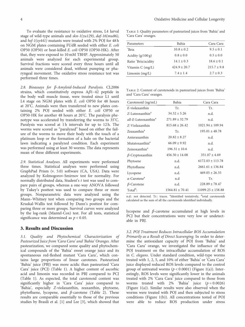
To evaluate the resistance to oxidative stress, L4 larvalstage of wild-type animals and skn-1(zu129), daf-16(mu86),and hsf-1(sy441) mutants were treated with 2% POJ for 48 hon NGM plates containing FUdR seeded with either E. coliOP50 (OP50) or heat-killed E. coli OP50 (OP50-HK). Afterthat, they were exposed to 10mM TBHP. Approximately 50animals were analyzed for each experimental group.Survival fractions were scored every three hours until allanimals were considered dead, without pumping or pha-ryngeal movement. The oxidative stress resistance test wasperformed three times.
2.8. Bioassays for β-Amyloid-Induced Paralysis. CL2006strains, which constitutively express Aβ1–42 peptide inthe body wall muscle tissue, were treated since L1 untilL4 stage on NGM plates with E. coli OP50 for 48 hoursat 20°C. Animals were then transferred to new plates con-taining 2% POJ seeded with either E. coli OP50 orOP50-HK for another 48 hours at 20°C. The paralysis phe-notype was accelerated by transferring the worms to 35°C.Paralysis was scored at 1 h intervals for up to 12 h. Theworms were scored as “paralyzed” based on either the fail-ure of the worms to move their body with the touch of aplatinum loop or the formation of a halo on the bacteriallawn indicating a paralyzed condition. Each experimentwas performed using at least 30 worms. The data representsmean of three different experiments.
2.9. Statistical Analyses. All experiments were performedthree times. Statistical analyses were performed usingGraphPad Prism (v. 5.0) software (CA, USA). Data wereanalyzed by Kolmogorov-Smirnov test for normality. Fornormally distributed data, Student’s t test was used to com-pare pairs of groups, whereas a one-way ANOVA followedby Tukey’s posttest was used to compare three or moregroups. Nonparametric data were analyzed using theMann–Whitney test when comparing two groups and theKruskal-Wallis test followed by Dunn’s posttest for com-paring three or more groups. Survival curves were analyzedby the log-rank (Mantel-Cox) test. For all tests, statisticalsignificance was determined as p < 0 05.
3. Results and Discussion
3.1. Quality and Phytochemical Characterization ofPasteurized Juice from ‘Cara Cara’ and ‘Bahia’Oranges. Afterpasteurization, we compared some quality and phytochem-ical compounds of the ‘Bahia’ sweet orange juice and itsspontaneous red-fleshed mutant ‘Cara Cara’, which con-tains large proportions of linear carotenes. Pasteurized‘Bahia’ juice (PBJ) was more acidic than pasteurized ‘CaraCara’ juice (PCJ) (Table 1). A higher content of ascorbicacid and limonin was recorded in PBJ compared to PCJ(Table 1). As expected, the total carotenoid content wassignificantly higher in ‘Cara Cara’ juice compared to‘Bahia’, especially Z-violaxanthin, zeaxanthin, phytoene,phytofluene, lycopene, and β-carotene (Table 2). Theseresults are comparable essentially to those of the previousstudies by Brasili et al. [1] and Lee [3], which showed that
lycopene and β-carotene accumulated at high levels inPCJ but their concentrations were very low or undetect-able in PBJ.
3.2. POJ Treatment Reduces Intracellular ROS AccumulationPrimarily as a Result of Direct Scavenging. In order to deter-mine the antioxidant capacity of POJ from ‘Bahia’ and‘Cara Cara’ orange, we investigated the influence of thePOJ treatment on the intracellular accumulation of ROSin C. elegans. Under standard condition, wild-type wormstreated with 1, 2, 5, and 10% of either ‘Bahia’ or ‘Cara Cara’juice displayed reduced ROS levels compared to the controlgroup of untreated worms (p < 0 0001) (Figure 1(a)). Inter-estingly, ROS levels were significantly lower in the animalstreated with 2% ‘Cara Cara’ juice compared to those fromworms treated with 2% ‘Bahia’ juice (p = 0 0026)(Figure 1(a)). Similar results were also observed when theworms were treated with POJ and then subjected to stressconditions (Figure 1(b)). All concentrations tested of POJwere able to reduce ROS production under stress
Table 1: Quality parameters of pasteurized juices from ‘Bahia’ and‘Cara Cara’ oranges.
Parameters Bahia Cara Cara°Brix 10 8 ± 0 2 9 5 ± 0 1Acidity (g/100 g) 0 8 ± 0 0 0 5 ± 0 0Ratio °Brix/acidity 14 1 ± 0 3 18 6 ± 0 1Vitamin C (mg/L) 424 9 ± 20 7 215 7 ± 9 8Limonin (mg/L) 7 4 ± 1 4 2 7 ± 0 3
Table 2: Content of carotenoids in pasteurized juices from ‘Bahia’and ‘Cara Cara’ oranges.
Carotenoid (ng/mL) Bahia Cara Cara
E-violaxanthin Tr. Tr.
Z-Luteoxanthina 34 52 ± 5 26 n.d.
all-E-Luteoxanthina 271 89 ± 32 79 n.d.
Z-Violaxanthin 815 68 ± 26 42 1021 94 ± 109 94Zeaxanthina Tr. 195 01 ± 48 78Anteraxanthin 20 82 ± 8 27 n.d.
Mutatoxanthina 66 09 ± 9 92 n.d.
Zeinoxanthina 196 31 ± 10 6 n.d.
β-Cryptoxanthin 456 50 ± 14 08 351 07 ± 4 69Phytoene n.d. 6172 03 ± 113 78Phytofluene n.d. 2661 61 ± 136 84Lycopene n.d. 469 05 ± 26 55α-Carotenea n.d Tr.
β-Carotene n.d. 228 89 ± 78 47Totalb: 1564 81 ± 70 41 11099 25 ± 158 88n.d.: not detected. Tr.: traces. aIdentified tentatively, dtotal carotenoidscalculated as the sum of all the carotenoids identified individually.
4 Oxidative Medicine and Cellular Longevity
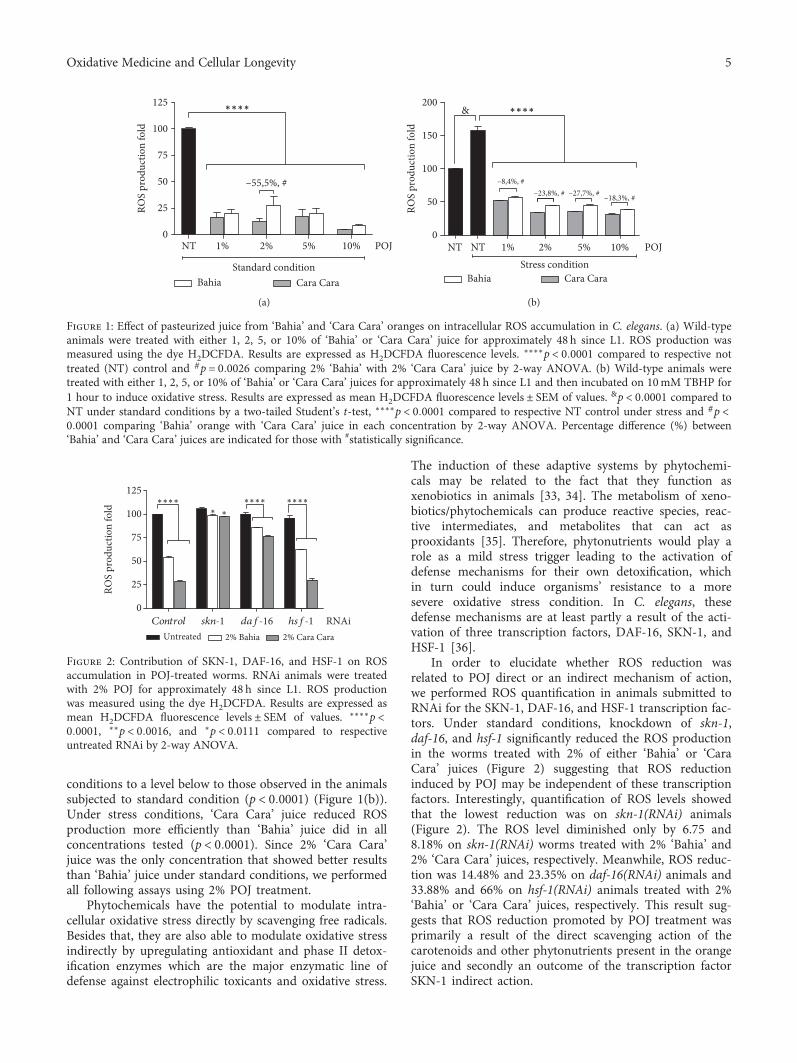
conditions to a level below to those observed in the animalssubjected to standard condition (p < 0 0001) (Figure 1(b)).Under stress conditions, ‘Cara Cara’ juice reduced ROSproduction more efficiently than ‘Bahia’ juice did in allconcentrations tested (p < 0 0001). Since 2% ‘Cara Cara’juice was the only concentration that showed better resultsthan ‘Bahia’ juice under standard conditions, we performedall following assays using 2% POJ treatment.
Phytochemicals have the potential to modulate intra-cellular oxidative stress directly by scavenging free radicals.Besides that, they are also able to modulate oxidative stressindirectly by upregulating antioxidant and phase II detox-ification enzymes which are the major enzymatic line ofdefense against electrophilic toxicants and oxidative stress.
The induction of these adaptive systems by phytochemi-cals may be related to the fact that they function asxenobiotics in animals [33, 34]. The metabolism of xeno-biotics/phytochemicals can produce reactive species, reac-tive intermediates, and metabolites that can act asprooxidants [35]. Therefore, phytonutrients would play arole as a mild stress trigger leading to the activation ofdefense mechanisms for their own detoxification, whichin turn could induce organisms’ resistance to a moresevere oxidative stress condition. In C. elegans, thesedefense mechanisms are at least partly a result of the acti-vation of three transcription factors, DAF-16, SKN-1, andHSF-1 [36].
In order to elucidate whether ROS reduction wasrelated to POJ direct or an indirect mechanism of action,we performed ROS quantification in animals submitted toRNAi for the SKN-1, DAF-16, and HSF-1 transcription fac-tors. Under standard conditions, knockdown of skn-1,daf-16, and hsf-1 significantly reduced the ROS productionin the worms treated with 2% of either ‘Bahia’ or ‘CaraCara’ juices (Figure 2) suggesting that ROS reductioninduced by POJ may be independent of these transcriptionfactors. Interestingly, quantification of ROS levels showedthat the lowest reduction was on skn-1(RNAi) animals(Figure 2). The ROS level diminished only by 6.75 and8.18% on skn-1(RNAi) worms treated with 2% ‘Bahia’ and2% ‘Cara Cara’ juices, respectively. Meanwhile, ROS reduc-tion was 14.48% and 23.35% on daf-16(RNAi) animals and33.88% and 66% on hsf-1(RNAi) animals treated with 2%‘Bahia’ or ‘Cara Cara’ juices, respectively. This result sug-gests that ROS reduction promoted by POJ treatment wasprimarily a result of the direct scavenging action of thecarotenoids and other phytonutrients present in the orangejuice and secondly an outcome of the transcription factorSKN-1 indirect action.
NT 1% 2% 5% 10%0
25
50
75
100
125
POJ
Standard conditionBahia Cara Cara
ROS
prod
uctio
n fo
ld
∗∗∗∗
–55,5%, #
(a)
0
50
100
150
200&
NT NT 1% 2% 5% 10% POJStress condition
Bahia Cara Cara
ROS
prod
uctio
n fo
ld
∗∗∗∗
–8,4%, #–23,8%, # –27,7%, # –18,3%, #
(b)
Figure 1: Effect of pasteurized juice from ‘Bahia’ and ‘Cara Cara’ oranges on intracellular ROS accumulation in C. elegans. (a) Wild-typeanimals were treated with either 1, 2, 5, or 10% of ‘Bahia’ or ‘Cara Cara’ juice for approximately 48 h since L1. ROS production wasmeasured using the dye H2DCFDA. Results are expressed as H2DCFDA fluorescence levels. ∗∗∗∗p < 0 0001 compared to respective nottreated (NT) control and #p = 0 0026 comparing 2% ‘Bahia’ with 2% ‘Cara Cara’ juice by 2-way ANOVA. (b) Wild-type animals weretreated with either 1, 2, 5, or 10% of ‘Bahia’ or ‘Cara Cara’ juices for approximately 48 h since L1 and then incubated on 10mM TBHP for1 hour to induce oxidative stress. Results are expressed as mean H2DCFDA fluorescence levels ± SEM of values. &p < 0 0001 compared toNT under standard conditions by a two-tailed Student’s t-test, ∗∗∗∗p < 0 0001 compared to respective NT control under stress and #p <0 0001 comparing ‘Bahia’ orange with ‘Cara Cara’ juice in each concentration by 2-way ANOVA. Percentage difference (%) between‘Bahia’ and ‘Cara Cara’ juices are indicated for those with #statistically significance.
Control skn-1 daf-16 hsf-1
0
25
50
75
100
125
Untreated 2% Bahia 2% Cara Cara
∗∗∗∗∗ ∗
∗∗∗∗ ∗∗∗∗
RNAi
ROS
prod
uctio
n fo
ld
Figure 2: Contribution of SKN-1, DAF-16, and HSF-1 on ROSaccumulation in POJ-treated worms. RNAi animals were treatedwith 2% POJ for approximately 48 h since L1. ROS productionwas measured using the dye H2DCFDA. Results are expressed asmean H2DCFDA fluorescence levels ± SEM of values. ∗∗∗∗p <0 0001, ∗∗p < 0 0016, and ∗p < 0 0111 compared to respectiveuntreated RNAi by 2-way ANOVA.
5Oxidative Medicine and Cellular Longevity

3.3. POJ Treatment Induces the Expression of Antioxidant,Detoxification, and Chaperonin Genes. To better character-ize the molecular responses associated with the directand indirect antioxidant effects of POJ in C. elegans, weanalyzed the gene expression of four reporter genes relatedto stress resistance, detoxification, and longevity. Weselected γ-glutamyl cysteine synthetase (gcs-1) and glu-tathione-s-transferase-4 (gst-4), two SKN-1 target genes[37, 38], superoxide dismutase 3 (sod-3), a well-knownDAF-16 target gene [39], and the chaperone hsp-16.2::GFPwhose expression is regulated by DAF-16 and HSF-1 [40].The fluorescence signals of gcs-1::GFP, gst-4::GFP,sod-3::GFP, and hsp-16.2::GFP animals were significantlyincreased after 2% POJ treatment with either ‘Bahia’ or‘Cara Cara’ juices compared to untreated worms, both atstandard and stress conditions (Figures 3(a)–3(d)). Inter-estingly, the expression signal of gcs-1::GFP (Figure 3(a)),gst-4::GFP (Figure 3(b)), and hsf-16.2::GFP (Figure 3(d))from the animals treated with 2% ‘Cara Cara’ juice weresignificantly higher than that of the animals treated with2% ‘Bahia’ juice under standard conditions. Under stressconditions, the treatment with 2% ‘Cara Cara’ juice signif-icantly increased the expression of gcs-1::GFP andsod-3::GFP.
0
1
2
3
4
Rela
tive l
evel
s of fl
uore
scen
ce/a
rea
2% Cara cara2% BahiaUntreated
#
#
Standard condition Stress condition
∗∗∗∗
∗∗∗∗gcs-1::GFP
(a)
Rela
tive l
evel
s of fl
uore
scen
ce/a
rea
2% Cara cara2% BahiaUntreated
0
5
10
15
20
#
Standard condition Stress condition
∗∗∗∗
∗∗∗∗
gst-4::GFP
(b)
Rela
tive l
evel
s of fl
uore
scen
ce/a
rea
2% Cara cara2% BahiaUntreated
0
2
4
6
#
Standard condition Stress condition
∗∗∗∗
∗∗∗∗
sod-3::GFP
(c)
Rela
tive l
evel
s of fl
uore
scen
ce/a
rea
2% Cara cara2% BahiaUntreated
0
2
4
6
8
#
hsp-16.2::GFP
∗∗∗∗
∗∗∗∗
Standard condition Stress condition
(d)
Figure 3: Effect of pasteurized juice from ‘Bahia’ and ‘Cara Cara’ oranges on the expression of antioxidant and detoxification genes. Analysisof gcs-1::GFP (a), gst-4::GFP (b), sod-3::GFP (c), and hsp-16.2::GFP (d) fluorescent expression levels. Transgenic worms were treated or notwith 2% ‘Bahia’ or 2% ‘Cara Cara’ juice for 48 h. After this period, animals were submitted to stress conditions. For gcs-1::GFP (a),gst-4::GFP (b), and sod-3::GFP (c); the stress condition was incubation for 1 h on 10mM TBHP. For hsp-16.2::GFP, the stress conditionwas incubation for 1 h at 35°C. Photographs were taken on a fluorescence microscope, and GFP fluorescence signals were measured usingNIH ImageJ software. The results represent mean GFP levels ± SEM of values. ∗∗∗∗p < 0 0008 by a two-tailed Student’s t-test compared tothe untreated group, #p < 0 0003 by a two-tailed Student’s t-test for 2% ‘Bahia’ with 2% ‘Cara Cara’ juice.
1
0
2
3
4 ∗∗∗∗
∗∗∗∗
&&
gst-4::GFP
Control
Control
skn-1
daf-16
hsf
-1
Control
skn-1
daf-16
hsf
-1
NT 2% Bahia 2% Cara Cara
RNAi
Rela
tive l
evel
s of fl
uore
scen
ce/a
rea
Figure 4: Contribution of SKN-1, DAF-16, and HSF-1 ongst-4::GFP expression induced by POJ treatment. RNAi animalswere treated with 2% POJ for approximately 48 h since L1.Photographs were taken on a fluorescence microscope, and GFPfluorescence signals were measured using NIH ImageJ software.The results represent mean GFP levels ± SEM of values. &p <0 0001 compared to untreated (NT) control(RNAi) under standardconditions and ∗∗∗∗p < 0 0001, compared to respectivePOJ-treated control(RNAi) by 1-way ANOVA.
6 Oxidative Medicine and Cellular Longevity

The POJ induction, rather than an inhibition, of the anti-oxidant and detoxification genes indicates that the phytonu-trients present in the POJ were not only directly scavengingROS but also acting as prooxidants and inducing a mild cel-lular stress response [41, 42]. This phenomenon is alsoreferred as hormesis since the phytochemicals are toxic andprotect plants against insects and other harmful organismsand stresses. However, at the subtoxic doses, the phytochem-icals involve kinases and transcription factors in order toinduce the expression of genes that encode antioxidantenzymes, protein chaperones, phase-2 enzymes, neuro-trophic factors, and other cytoprotective proteins [41, 42].
Since gst-4::GFP showed the strongest expression afterPOJ treatments, we assessed whether it was dependent onSKN-1, DAF-16, and HSF-1. RNAi of all three transcrip-tion factors reduced significantly gst-4::GFP expressioninduced by the POJ treatments suggesting that they all con-tribute to its activation (Figure 4). Moreover, knockingdown of skn-1 caused the greatest reduction on gst-4::GFPexpression induced by the POJ treatments. The fluorescent
levels of gst-4::GFP in the worms treated with ‘Bahia’ juicewere 3.26 times lower on skn-1(RNAi) animals comparedto control(RNAi) animals while the reduction was 2.54and 1.34 times lower on daf-16(RNAi) and hsf-1(RNAi) ani-mals, respectively. This result suggests that SKN-1 seemedto play a more important role on gst-4 expression com-pared to DAF-16 or HSF-1.
3.4. POJ Treatment Increases Longevity and Oxidative StressResistance. To explore whether POJ antioxidant propertieshave a protective effect in vivo, we tested whether POJcould affect the lifespan of C. elegans under standard labo-ratory conditions. We determined the lifespan of N2 wormswith and without 2% POJ treatment. The maximum andmean lifespan of N2 worms fed with either 2% pasteurized‘Bahia’ or ‘Cara Cara’ juice was significantly increased com-pared to untreated worms (Figure 5(a), Table 3). Remark-ably, animals treated with 2% ‘Cara Cara’ juice showed anincreased lifespan compared to animals treated with 2%‘Bahia’ juice (p = 0 0003) (Table 3).
0 2 4 6 8 10 12 14 16 18 20 220.00
0.25
0.50
0.75
1.00
Surv
ival
frac
tion
Time (days)Untreated2% Cara Cara∗∗∗, #
2% Bahia∗∗∗
(a)
0.00
0.25
0.50
0.75
1.00
Surv
ival
frac
tion
Untreated2% Cara Cara∗∗∗, #
2% Bahia∗∗∗
0 4 8 12 16 20 24 28 32 36Time (hours)
(b)
Figure 5: Effect of pasteurized juice from ‘Bahia’ and ‘Cara Cara’ oranges on C. elegans stress resistance and longevity. (a) Survival curves ofwild-type (N2) animals under standard laboratory conditions. Worms were treated with either 2% ‘Bahia’ or 2% ‘Cara Cara’ juice beginning atL4. Survival was verified every day at 25°C. ∗∗∗∗p < 0 0001 compared to respective controls and #p = 0 0003 comparing animals treated with2% ‘Bahia’ with 2% ‘Cara Cara’ juice by log-rank (Mantel-Cox) test. (b) Survival curves of wild-type (N2) animals under oxidative stressconditions. Worms were treated with either 2% ‘Bahia’ or 2% ‘Cara Cara’ juice beginning at L4 and transferred to plates with 10mMTBPH. Survival was verified every 3 hours at 20°C. ∗∗∗∗p < 0 0001 compared to the untreated control group and #p = 0 0134 comparinganimals treated with 2% ‘Bahia’ with 2% ‘Cara Cara’ juice by log-rank (Mantel-Cox) test.
Table 3: Effect of pasteurized juice from ‘Bahia’ and ‘Cara Cara’ oranges on C. elegans lifespan.
ConditionMaximum survival
(days ± SEM)Mean survival(days ± SEM)
% mean survival timevariation vs. untreated
p value (log rank) POJa
vs. untreatedbp value (log rank)PBJc vs. PCJb,d
Ne
Untreated 17 33 ± 2 31 12 11 ± 0 13 243 (3)
2% Bahia 19 00 ± 1 73 14 75 ± 0 17 22.0 <0.0001 255 (3)
2% Cara Cara 19 33 ± 2 31 15 64 ± 0 18 29.5 <0.0001 0.0003 234 (3)aPOJ, pasteurized orange juice, bcomparisons were performed using log-rank (Mantel-Cox) test. cPBJ, pasteurized ‘Bahia’ orange juice, dPCJ, pasteurized ‘CaraCara’ orange juice, etotal number of animals analyzed. The number in parentheses indicates the number of independent trials.
7Oxidative Medicine and Cellular Longevity
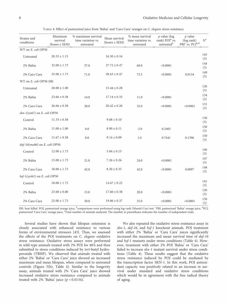
Several studies have shown that lifespan extension isclosely associated with enhanced resistance to variousforms of environmental stressors [43]. Thus, we assessedthe effects of the POJ treatments on C. elegans oxidativestress resistance. Oxidative stress assays were performedin wild-type animals treated with 2% POJ for 48h and thensubmitted to stress conditions induced by tert-butyl hydro-peroxide (TBHP). We observed that animals treated witheither 2% ‘Bahia’ or ‘Cara Cara’ juice showed an increasedmaximum and mean lifespan, when compared to untreatedcontrols (Figure 5(b), Table 4). Similar to the longevityassay, animals treated with 2% ‘Cara Cara’ juice showedincreased oxidative stress resistance compared to animalstreated with 2% ‘Bahia’ juice (p = 0 0134).
We also repeated the oxidative stress resistance assay inskn-1, daf-16, and hsf-1 knockout animals. POJ treatmentwith either 2% ‘Bahia’ or ‘Cara Cara’ juices significantlyincreased the maximum and mean survival time of daf-16and hsf-1 mutants under stress conditions (Table 4). How-ever, treatment with either 2% POJ ‘Bahia’ or ‘Cara Cara’failed to increase skn-1 mutant survival under stress condi-tions (Table 4). These results suggest that the oxidativestress resistance induced by POJ could be mediated bythe transcription factor SKN-1. In this work, POJ antioxi-dant capacity was positively related to an increase in sur-vival under standard and oxidative stress conditionswhich would be in agreement with the free radical theoryof aging.
Table 4: Effect of pasteurized juice from ‘Bahia’ and ‘Cara Cara’ oranges on C. elegans stress resistance.
Strains andconditions
Maximumsurvival
(hours ± SEM)
% maximum survivaltime variation vs.
untreated
Mean survival(hours ± SEM)
% mean survivaltime variation vs.
untreated
p value (logrank) POJa vs.untreatedb
p value(log rank)
PBJc vs. PCJb,dNe
WT on E. coli OP50
Untreated 20 33 ± 1 15 16 50 ± 0 16 145(3)
2% Bahia 32 00 ± 1 73 57.0 27 72 ± 0 47 68.0 <0.0001 144(3)
2% Cara Cara 35 00 ± 1 73 71.0 28 63 ± 0 47 73.5 <0.0001 0.0134149(3)
WT on E. coli OP50-HK
Untreated 20 00 ± 1 00 15 44 ± 0 28 126(3)
2% Bahia 23 66 ± 0 58 14.0 17 14 ± 0 33 11.0 <0.0001 134(3)
2% Cara Cara 26 66 ± 0 58 28.0 20 42 ± 0 26 32.0 <0.0001 <0.0001 131(3)
skn-1(zu67) on E. coli OP50
Control 11 33 ± 0 58 9 08 ± 0 10 150(3)
2% Bahia 11 00 ± 1 00 0.0 8 90 ± 0 11 -2.0 0.2405150(3)
2% Cara Cara 11 67 ± 0 58 0.0 9 14 ± 0 09 1.0 0.7441 0.1396150(3)
daf-16(mu86) on E. coli OP50
Control 12 00 ± 1 73 5 84 ± 0 15 146(3)
2% Bahia 15 00 ± 1 73 21.0 7 26 ± 0 26 24.0 <0.0001 147(3)
2% Cara Cara 18 00 ± 1 73 42.0 8 30 ± 0 35 42.0 <0.0001 0.0097148(3)
hsf-1(sy441) on E. coli OP50
Control 18 00 ± 1 73 14 67 ± 0 22 141(3)
2% Bahia 23 00 ± 0 00 15.0 17 60 ± 0 38 20.0 <0.0001 150(3)
2% Cara Cara 25 00 ± 1 73 30.0 19 86 ± 0 37 35.0 <0.0001 <0.0001 150(3)
HK: heat killed. POJ, pasteurized orange juice, bcomparisons were performed using log-rank (Mantel-Cox) test. cPBJ, pasteurized ‘Bahia’ orange juice, dPCJ,pasteurized ‘Cara Cara’ orange juice, eTotal number of animals analyzed. The number in parentheses indicates the number of independent trials.
8 Oxidative Medicine and Cellular Longevity

Together, these results indicate that pasteurized juicetreatment improves C. elegans antioxidant capacity, whichis associated with the upregulation of antioxidant and cha-peronin genes and increased lifespan and stress resistance.It is noteworthy that the treatment with ‘Cara Cara’orange juice provided better results on all parametersanalyzed here compared to ‘Bahia’ orange juice with theonly exception for the expression of sod-3::GFP(Figure 3(c)) under standard conditions and for gst-4::GFP(Figure 3(b)) and hsp-16.2 (Figure 3(d)) under stress con-ditions. The beneficial effects of ‘Cara Cara’ orange juicecould be related to its higher carotenoid content, especiallyZ-violaxanthin, phytoene, phytofluene, lycopene, andβ-carotene, even though ‘Bahia’ juice has higher levels ofvitamin C and limonin (Table 2). However, the additionaleffects of ‘Cara Cara’ orange juice compared to ‘Bahia’orange juice are either moderate or small to be explainedsolely based on the total carotenoid difference betweenthese two varieties.
3.5. POJ Treatment Delays Paralysis Induced by β-Amyloid(Aβ) Expression. The oxidative stress state has an importantrole in the development of neurodegenerative diseases suchas Alzheimer’s disease (AD). Accumulation of ROS anddeposition of toxic amyloid species have been proposed toexacerbate the symptoms observed in AD patients [44,45]. The observation that POJ has antioxidant propertiesin vivo led us to ask whether POJ could protect againstβ-amyloid-induced toxicity in a C. elegans model for Alz-heimer’s disease. The expression of human Aβ1–42 in themuscle of transgenic CL2006 strain promotes a paralysisthat can be monitored over time. We observed that theonset of paralysis was significantly delayed in POJ-treated
animals (Figure 6). The mean paralysis time for wormstreated with 2% ‘Bahia’ and 2% ‘Cara Cara’ juices wasincreased by 25.0% and 46.0%, respectively, compared tountreated worms (Figure 6, Table 5).
Various studies have shown that the protective effect offlavonoids and carotenoids against amyloid-induced neuro-toxicity is due to their antioxidant properties. Other possi-ble mechanisms could be attenuation of Aβ aggregationin vivo by downregulating the expression level of β-amy-loid precursor protein (APP) [18] and/or activation of pro-tein degradation pathways [46]. In the present study, thePOJ treatment delayed Aβ1–42-induced paralysis inworms suggesting that POJ may be able to attenuate thedevelopment of AD by altering Aβ aggregate statesin vivo. Most importantly, our results indicate that ‘CaraCara’ juice, with higher carotenoid contents, provides asuperior protection against Aβ1–42-induced paralysis overthat provided by Bahia.
We also evaluated the role of SKN-1, DAF-16, andHSF-1 in POJ-mediated protection against Aβ1-42 toxicity.To this end, we examined the effect of POJ on CL2006worms using RNAi to knock down skn-1, daf-16, and hsf-1expression. POJ treatment significantly delayed the paralysisrate of CL2006 worms with control(RNAi) (Table 5). In con-trast, RNAi of skn-1 and daf-16 in CL2006 wormscompletely abolished POJ-mediated beneficial effects ondelaying the progression of paralysis (Table 5). RNAi ofhsf-1 in CL2006 worms only prevented the beneficial effectagainst β-amyloid paralysis for those animals treated with2% ‘Cara Cara’ juice (Table 5). Together, these findings sug-gest that the transcription factors SKN-1, DAF-16, andHSF-1 are required for POJ-mediated protective effectagainst Aβ toxicity.
Taking these data together, our mechanistic studiesindicated that POJ improves the antioxidant status of awhole organism by direct and indirect mechanisms.POJ-reduced ROS levels were primarily a result of thedirect scavenging action of natural compounds availablein the orange juice and secondly an outcome of the tran-scription factor SKN-1 indirect action. POJ promotesgst-4::GFP expression and oxidative stress resistance mainlythrough SKN-1 although DAF-16 and HSF-1 also contrib-ute to a less extent to these effects. Finally, POJ delayedAβ-induced onset paralysis on a SKN-1-, DAF-16-, andHSF-1-dependent manner. Previous studies in the nema-tode reported that DAF-16, SKN-1, and HSF-1 play pivotalroles in regulating longevity and ameliorating Aβ [12].Since these transcription factors are key regulators of manyimportant biological processes, including lifespan, stressresponses, and proteostasis, we reasoned that POJ treat-ments might protect worms against Aβ1–42 toxicity byincreasing antioxidant capacity and proteostasis.
3.6. POJ Protection against Oxidative Stress and Aβ ToxicityIs Partially Related to Antimicrobial Effect. Since E. coli hasa pathogenic effect on C. elegans, which may alter its longev-ity, resistance to stress, and Aβ1–42-induced paralysis [47,48], we investigated whether the POJ protective effects couldbe a secondary response of a possible POJ antimicrobial
Frac
tion
not p
aral
yzed
Time (hours)0 1 2 3 4 5 6 7 8 9 10
0.00
0.25
0.50
0.75
1.00
Untreated2% Cara Cara∗∗∗, #
2% Bahia∗∗∗
Figure 6: Effect of pasteurized juice from oranges cv. ‘Bahia’ and cv.‘Cara Cara’ treatment on the β-amyloid induced paralysis in C.elegans transgenic model of Alzheimer’s disease. Paralysis curvesfor C. elegans strain CL2006 which expresses β-amyloid peptideconstitutively in the muscle. Worms were treated beginning at L4with 2% POJ for 48 h at L4 stage. Paralysis was verified at 1 hintervals at 35°C. ∗∗∗∗p < 0 0001 compared to untreated controland #p < 0 0001 comparing 2% ‘Bahia’ with 2% ‘Cara Cara’ juiceby log-rank (Mantel-Cox) test.
9Oxidative Medicine and Cellular Longevity

property. First, we repeated the oxidative stress resistanceassay in animals treated with POJ on dead bacteria. Weobserved that wild-type animals treated with 2% POJ stillshowed increased survival on 10mM TBHP compared tountreated controls (p < 0 0001) (Figure 7(a), Table 4). How-ever, the mean survival variation observed for thePOJ-treated animals on dead bacteria was considerablylower compared to the, respectively, POJ-treated animals
on live bacteria (Table 4). The mean survival variation was11 and 68% for the animals treated with 2% ‘Bahia’ juiceon dead and live bacteria, respectively. Likewise, the meansurvival variation was 32 and 73.5% for the animals treatedwith 2% ‘Cara Cara’ juice on dead and living bacteria,respectively (Table 4).
The role of bacterial in affecting longevity of aging C.elegans is well known [49]. Much experimental evidence
Table 5: Effect of pasteurized juice from ‘Bahia’ and ‘Cara Cara’ oranges on C. elegans paralysis induced by β-amyloid expression.
Strain and conditionsMean paralysis time
(hours ± SEM)% mean paralysis timevariation vs. control
p value (log rank)POJa vs. untreatedb
p value (log rank)PBJc vs. PCJb,d
Ne
CL2006 on E. coli OP50
Untreated 4 59 ± 0 11 84(3)
2% Bahia 5 75 ± 0 12 25.0 <0.0001 85(3)
2% Cara Cara 6 70 ± 0 13 46.0 <0.0001 <0.0001 84(3)
CL2006 on E. coli OP50-HKa
Untreated 5 29 ± 0 16 86(3)
2% Bahia 6 50 ± 0 18 23.0 <0.0001 80(3)
2% Cara Cara 7 82 ± 0 22 48.0 <0.0001 <0.0001 82(3)
CL2006 on control(RNAi)
Untreated 4 80 ± 0 10 88(3)
2% Bahia 5 48 ± 0 12 14.0 <0.0001 89(3)
2% Cara Cara 5 54 ± 0 16 15.0 <0.0001 0.556289(3)
CL2006 on skn-1(RNAi)
Untreated 4 20 ± 0 07 87(3)
2% Bahia 3 95 ± 0 07 -6.0 0.019188(3)
2% Cara Cara 3 94 ± 0 07 -6.1 0.0151 0.855589(3)
CL2006 on daf-16(RNAi)
Untreated 4 09 ± 0 07 89(3)
2% Bahia 4 13 ± 0 08 0.9 0.601688(3)
2% Cara Cara 4 17 ± 0 08 1.9 0.4024 0.858289(3)
CL2006 on hsf-1(RNAi)
Untreated 4 26 ± 0 08 88(3)
2% Bahia 4 57 ± 0 08 7.2 0.006989(3)
2% Cara Cara 3 84 ± 0 07 -9.8 <0.0001 <0.0001 89(3)
HK: heat killed, aPOJ, pasteurized orange juice, bcomparisons were performed using log-rank (Mantel-Cox) test. cPBJ, pasteurized ‘Bahia’ orange juice, dPCJ,pasteurized ‘Cara Cara’ orange juice, etotal number of animals analyzed. The number in parentheses indicates the number of independent trials.
10 Oxidative Medicine and Cellular Longevity

supports bacterial infection and proliferation within theintestine of adults as a potential pathogen that triggersinnate immune and stress host responses which may con-tribute as a double-edged effect to tissue damage and aging[47]. The conserved PMK1-p38 mitogen-activated proteinkinase (MAPK) pathway is the major mediator of innateimmunity in C. elegans. Interestingly, the PMK-1 pathwayis also required for the activation of the transcription factorSKN-1 under stress conditions [50]. Thus, any interventionthat attenuates bacterial pathogenicity and toxicity extendslifespan and stress resistance. The reduction of the variationsurvival time on stress when the POJ-treated animals werefed on bacterial previously killed by heat indicates that partof the survival extension observed on POJ-treated animalsfed on live bacteria is due to its antimicrobial effect. It isalso reasonable to suppose that this reduction of the varia-tion survival time on stress could be a result of the animalsacquiring less antioxidant buffer from their living bacterialfood source [49]. Another possibility could be related tothe fact that in the absence of pathogenic bacteria, thePMK-1-SKN-1 signaling pathway is also diminished andtherefore the defense mechanisms activation is attenuated.
Next, we performed the Aβ1-42-induced paralysis assayalso using bacteria killed by heat. Interestingly, the meanvariation observed for the Aβ1-42-induced paralysis wasnot particularly different between the POJ treatments ondead or living bacteria (Figure 7(b), Table 5). The meanparalysis time variation for the animals treated with 2%‘Bahia’ juice on dead bacteria was 23% compared tountreated animals while the variation for the same treat-ment on living bacteria was 25% (Table 5). For ‘Cara Cara’juice treatment, the mean paralysis time variation was 48%compared to untreated animals on dead bacteria and 46%compared to untreated animals on living bacteria (Table 5).
Although the onset paralysis in the CL2006 animalsgrown on live OP50 was faster compared to those grownon dead OP50 as expected, we did not observe any signifi-cant difference of the paralysis time variation on thePOJ-treated animals fed on dead bacteria compared tothose fed on live bacteria (Table 5). This result suggests thatthe beneficial effect of POJ against Aβ toxicity is indepen-dent of the bacterial pathogenicity. This observation seemscontradictory since many studies have shown a linkbetween food source and proteotoxicity [48, 51]. Steinkrauset al. [51] showed that bacterial food deprivation suppressesproteotoxicity in Aβ worms through an hsf-1-dependentmechanism. It would be interesting for further studies toshed light on the possible relationship between POJprotection against Aβ in the absence of bacteria dependson HSF-1.
4. Conclusions
In this work, we investigated the in vivo effects of POJ fromthe ‘Bahia’ and ‘Cara Cara’ varieties using the C. elegansmodel. Treatment with POJ reduced the endogenous levelsof ROS and increased the rate of survival of the wormsunder normal and stress conditions. Our mechanistic stud-ies indicated that POJ promotes resistance to oxidativestress by acting through the transcription factor SKN-1.We observed that POJ treatments increased the expressionof the antioxidant genes (gcs-1, gst-4, and sod-3) and cha-peronin (hsp-16.2), which are known to be regulated bythe transcription of SKN-1, DAF-16, and HSF-1 factors.In addition, POJ treatments delayed β-amyloid-inducedparalysis in the transgenic model C. elegans in a mannerrequiring SKN-1, DAF-16, and HSF-1. Noteworthy, thetreatment with the two juice types produced excellent
Surv
ival
frac
tion
Time (hours)0 3 6 9 12 15 18 21 24 27 30 33
0.00
0.25
0.50
0.75
1.00
Untreated2% Cara Cara∗∗∗, #
2% Bahia∗∗∗
(a)
Time (hours)
0.00
0.25
0.50
0.75
1.00
Frac
tion
not p
aral
yzed
0 1 2 3 4 5 6 7 8 9 1110 12
Untreated2% Cara Cara∗∗∗, #
2% Bahia∗∗∗
(b)
Figure 7: Effect of POJ on oxidative stress resistance and Aβ1-42-induced paralysis on C. elegans grown on dead bacteria. (a) Stress resistanceassay on bacteria dead bacteria. Worms were treated with 2% POJ on E. coli OP50 heat-killed beginning at L4 and transferred to plates with10mM TBPH. Survival was verified every 3 hours at 20°C. ∗∗∗∗p < 0 0001 compared to the untreated control group, log-rank (Mantel-Cox)test. (b) Paralysis profile of CL2006 transgenic animals fed with 2% POJ mixed with OP50 heat-killed. Paralysis was verified at 1 h intervals at35°C. ∗∗∗∗p < 0 0001 compared to untreated control by log-rank (Mantel-Cox) test.
11Oxidative Medicine and Cellular Longevity

results; however, ‘Cara Cara’ juice induced significantly bet-ter responses than ‘Bahia’ juice did in almost all experi-ments, possibly due to its higher content of carotenoidssuch as Z-violaxanthin, zeaxanthin, phytoene, phytofluene,lycopene, and β-carotene.
Data Availability
All data used to support the findings of this study areavailable from the corresponding author upon request.
Conflicts of Interest
The authors declare that there is no conflict of interestsregarding the publication of this paper.
Acknowledgments
This study was supported by Fundecitrus, São Paulo, Brazil,and Universidade Federal do Rio Grande do Norte (UFRN).Research fellowships were sponsored by CNPq (Oliveira, R.P.). We thank the Caenorhabditis Genetics Center (CGC),which is funded by the NIH National Center for ResearchResources (NCRR), and Dr. T. Keith Blackwell for supplyingthe C. elegans strains. We thank Citrosuco (Matão, São Paulo,Brazil) for providing access to their farms and processing theorange juices used in this study.
References
[1] E. Brasili, D. F. Seixas Chaves, A. A. O. Xavier, A. Z. Merca-dante, N. M. A. Hassimotto, and F. M. Lajolo, “Effect of pas-teurization on flavonoids and carotenoids in Citrus sinensis(L.) Osbeck cv. ‘Cara Cara’ and ‘Bahia’ juices,” Journal of Agri-cultural and Food Chemistry, vol. 65, no. 7, pp. 1371–1377,2017.
[2] A. Milani, M. Basirnejad, S. Shahbazi, and A. Bolhassani,“Carotenoids: biochemistry, pharmacology and treatment,”British Journal of Pharmacology, vol. 174, no. 11, pp. 1290–1324, 2017.
[3] H. S. Lee, “Characterization of carotenoids in juice of red navelorange (Cara Cara),” Journal of Agricultural and Food Chemis-try, vol. 49, no. 5, pp. 2563–2568, 2001.
[4] K. S. Cho, M. Shin, S. Kim, and S. B. Lee, “Recent advances instudies on the therapeutic potential of dietary carotenoids inneurodegenerative diseases,” Oxidative Medicine and CellularLongevity, vol. 2018, Article ID 4120458, 13 pages, 2018.
[5] M. Obulesu, M. R. Dowlathabad, and P. V. Bramhachari,“Carotenoids and Alzheimer’s disease: an insight into thera-peutic role of retinoids in animal models,” NeurochemistryInternational, vol. 59, no. 5, pp. 535–541, 2011.
[6] S. Hwang, J. Lim, and H. Kim, “Inhibitory effect of lycopene onamyloid-β-induced apoptosis in neuronal cells,” Nutrients,vol. 9, no. 8, p. 883, 2017.
[7] M. Qu, L. Li, C. Chen et al., “Protective effects of lycopeneagainst amyloid β-induced neurotoxicity in cultured rat corti-cal neurons,”Neuroscience Letters, vol. 505, no. 3, pp. 286–290,2011.
[8] Y. Ding, A. Qiao, Z. Wang et al., “Retinoic acid attenuatesbeta-amyloid deposition and rescues memory deficits in an
Alzheimer’s disease transgenic mouse model,” The Journal ofNeuroscience, vol. 28, no. 45, pp. 11622–11634, 2008.
[9] A. B. Goodman, “Retinoid receptors, transporters, and meta-bolizers as therapeutic targets in late onset Alzheimer disease,”Journal of Cellular Physiology, vol. 209, no. 3, pp. 598–603,2006.
[10] A. B. Goodman and A. B. Pardee, “Evidence for defective ret-inoid transport and function in late onset Alzheimer’s disease,”Proceedings of the National Academy of Sciences, vol. 100,no. 5, pp. 2901–2905, 2003.
[11] M. Maden, “Retinoic acid in the development, regenerationand maintenance of the nervous system,”Nature Reviews Neu-roscience, vol. 8, no. 10, pp. 755–765, 2007.
[12] A. Antebi, “Genetics of aging in Caenorhabditis elegans,” PLoSGenetics, vol. 3, no. 9, pp. 1565–1571, 2007.
[13] Y. Luo, “Long-lived worms and aging,” Redox Report, vol. 9,no. 2, pp. 65–69, 2004.
[14] C. A. Wolkow, K. D. Kimura, M. S. Lee, and G. Ruvkun, “Reg-ulation of C. elegans life-span by insulin-like signaling in thenervous system,” Science, vol. 290, no. 5489, pp. 147–150,2000.
[15] M. Artal-Sanz, L. de Jong, and N. Tavernarakis, “Caenorhabdi-tis elegans: a versatile platform for drug discovery,” Biotechnol-ogy Journal, vol. 1, no. 12, pp. 1405–1418, 2006.
[16] P. R. Augusti, A. V. S. Brasil, C. Souto et al., “Microcystin-LRexposure induces oxidative damage in Caenorhabditis elegans:protective effect of lutein extracted from marigold flowers,”Food and Chemical Toxicology, vol. 109, Part 1, pp. 60–67,2017.
[17] E. Pons, B. Alquezar, A. Rodriguez et al., “Metabolic engineer-ing of β-carotene in orange fruit increases its in vivo antioxi-dant properties,” Plant Biotechnology Journal, vol. 12, no. 1,pp. 17–27, 2014.
[18] W. Chen, L. Mao, H. Xing et al., “Lycopene attenuates Aβ1–42secretion and its toxicity in human cell and Caenorhabditiselegans models of Alzheimer disease,” Neuroscience Letters,vol. 608, pp. 28–33, 2015.
[19] E. Lashmanova, E. Proshkina, S. Zhikrivetskaya et al., “Fuco-xanthin increases lifespan of Drosophila melanogaster andCaenorhabditis elegans,” Pharmacological Research, vol. 100,pp. 228–241, 2015.
[20] M. Dimitriadi and A. C. Hart, “Neurodegenerative disorders:insights from the nematode Caenorhabditis elegans,” Neurobi-ology of Disease, vol. 40, no. 1, pp. 4–11, 2010.
[21] J. Li and W. Le, “Modeling neurodegenerative diseases in Cae-norhabditis elegans,” Experimental Neurology, vol. 250,pp. 94–103, 2013.
[22] Anon, FMC Food Tech. The Laboratory Manual. Proceduresfor Analysis of Citrus Products. Manual No. 054R11990, JohnBean Technologies Corporation, Inc, Lakeland, FL, USA, 2000.
[23] J. W. Stevens, “Estimation of Ascorbic Acid in Citrus Juices,”Industrial & Engineering Chemistry Analytical Edition,vol. 10, no. 5, pp. 269–271, 1938.
[24] B. Alquezar, M. J. Rodrigo, and L. Zacarías, “Regulation ofcarotenoid biosynthesis during fruit maturation in thered-fleshed orange mutant Cara Cara,” Phytochemistry,vol. 69, no. 10, pp. 1997–2007, 2008.
[25] G. Britton, “Overview of carotenoid biosynthesis,” in Biosyn-thesis and Metabolism, G. Britton, S. Liaaen-Jensen, and H.Pfander, Eds., pp. 13–148, Birkhäuser Verlag, Basel, 1998.
12 Oxidative Medicine and Cellular Longevity

[26] L. Carmona, L. Zacarías, and M. J. Rodrigo, “Stimulation ofcoloration and carotenoid biosynthesis during postharveststorage of ‘Navelina’ orange fruit at 12 °C,” Postharvest Biologyand Technology, vol. 74, pp. 108–117, 2012.
[27] M. J. Rodrigo, J. F. Marcos, F. Alferez, M. D. Mallent, andL. Zacarias, “Characterization of Pinalate, a novel Citrus sinen-sis mutant with a fruit-specific alteration that results in yellowpigmentation and decreased ABA content,” Journal of Experi-mental Botany, vol. 54, no. 383, pp. 727–738, 2003.
[28] M. J. Rodrigo, J. F. Marcos, and L. Zacarias, “Biochemical andmolecular analysis of carotenoid biosynthesis in flavedo oforange (Citrus sinensis L.) during fruit development and mat-uration,” Journal of Agricultural and Food Chemistry, vol. 52,no. 22, pp. 6724–6731, 2004.
[29] R. L. Rouseff, L. Raley, and H. J. Hofsommer, “Application ofdiode array detection with a C-30 reversed phase column forthe separation and identification of saponified orange juicecarotenoids,” Journal of Agricultural and Food Chemistry,vol. 44, no. 8, pp. 2176–2181, 1996.
[30] S. Brenner, “The genetics of Caenorhabditis elegans,” Genetics,vol. 77, no. 1, pp. 71–94, 1974.
[31] F. A. Paiva, L. de Freitas Bonomo, P. F. Boasquivis et al., “Car-queja (Baccharis trimera) protects against oxidative stress andbeta-amyloid-induced toxicity in Caenorhabditis elegans,”Oxidative Medicine and Cellular Longevity, vol. 2015, ArticleID 740162, 15 pages, 2015.
[32] L. de Freitas Bonomo, D. N. Silva, P. F. Boasquivis et al., “Açaí(Euterpe oleracea Mart.) modulates oxidative stress resistancein Caenorhabditis elegans by direct and indirect mechanisms,”PLoS One, vol. 9, no. 3, article e89933, 2014.
[33] A. M. Gonzalez-Paramas, B. Ayuda-Duran, S. Martinez,S. Gonzalez-Manzano, and C. Santos-Buelga, “The mecha-nisms behind the biological activity of flavonoids,” CurrentMedicinal Chemistry, vol. 25, 2018.
[34] A. Murakami, “Non-specific protein modifications may benovel mechanism underlying bioactive phytochemicals,” Jour-nal of Clinical Biochemistry and Nutrition, vol. 62, no. 2,pp. 115–123, 2018.
[35] S. Y. Tang and B. Halliwell, “Medicinal plants and antioxi-dants: what do we learn from cell culture and Caenorhabditiselegans studies?,” Biochemical and Biophysical Research Com-munications, vol. 394, no. 1, pp. 1–5, 2010.
[36] K. Koch, S. Havermann, C. Buchter, and W. Watjen, “Caenor-habditis elegansas model system in pharmacology and toxicol-ogy: effects of flavonoids on redox-sensitive signallingpathways and ageing,” Scientific World Journal, vol. 2014, arti-cle 920398, 15 pages, 2014.
[37] J. H. An and T. K. Blackwell, “SKN-1 links C. elegans mesen-dodermal specification to a conserved oxidative stressresponse,” Genes & Development, vol. 17, no. 15, pp. 1882–1893, 2003.
[38] L. A. Khan, T. Yamanaka, and N. Nukina, “Genetic impair-ment of autophagy intensifies expanded polyglutamine tox-icity in Caenorhabditis elegans,” Biochemical andBiophysical Research Communications, vol. 368, no. 3,pp. 729–735, 2008.
[39] C.T.Murphy, S.A.McCarroll, C. I. Bargmannet al., “Genes thatact downstream of DAF-16 to influence the lifespan of Caenor-habditis elegans,”Nature, vol. 424, no. 6946, pp. 277–283, 2003.
[40] E. Guisbert, D. M. Czyz, K. Richter, P. D. McMullen, and R. I.Morimoto, “Identification of a tissue-selective heat shock
response regulatory network,” PLoS Genetics, vol. 9, no. 4, arti-cle e1003466, 2013.
[41] V. Murugaiyah and M. P. Mattson, “Neurohormetic phyto-chemicals: an evolutionary-bioenergetic perspective,” Neuro-chemistry International, vol. 89, pp. 271–280, 2015.
[42] T. G. Son, S. Camandola, and M. P. Mattson, “Hormetic die-tary phytochemicals,” Neuromolecular Medicine, vol. 10,no. 4, pp. 236–246, 2008.
[43] K. I. Zhou, Z. Pincus, and F. J. Slack, “Longevity and stress inCaenorhabditis elegans,” Aging, vol. 3, no. 8, pp. 733–753,2011.
[44] S. J. Choi, J. K. Kim, S. H. Suh et al., “Ligularia fischeri extractprotects against oxidative-stress-induced neurotoxicity inmice and pc12 cells,” Journal of Medicinal Food, vol. 17,no. 11, pp. 1222–1231, 2014.
[45] H. R. Jeong, Y. N. Jo, J. H. Jeong, H. J. Kim,M.-J. Kim, and H. J.Heo, “Blueberry (Vaccinium virgatum) leaf extracts protectagainst Aβ-induced cytotoxicity and cognitive impairment,”Journal of Medicinal Food, vol. 16, no. 11, pp. 968–976, 2013.
[46] P. F. Boasquivis, G. M. M. Silva, F. A. Paiva, R. M. Cavalcanti,C. V. Nunez, and R. de Paula Oliveira, “Guarana (Paulliniacupana) extract protects Caenorhabditis elegans models forAlzheimer disease and Huntington disease through activationof antioxidant and protein degradation pathways,” OxidativeMedicine and Cellular Longevity, vol. 2018, Article ID9241308, 16 pages, 2018.
[47] D. H. Kim, “Bacteria and the aging and longevity of Caenor-habditis elegans,” Annual Review of Genetics, vol. 47, no. 1,pp. 233–246, 2013.
[48] F. Munoz-Lobato, M. J. Rodriguez-Palero, F. J. Naranjo-Ga-lindo et al., “Protective role of DNJ-27/ERdj5 in Caenorhabdi-tis elegans models of human neurodegenerative diseases,”Antioxidants & Redox Signaling, vol. 20, no. 2, pp. 217–235,2014.
[49] D. Garigan, A.-L. Hsu, A. G. Fraser, R. S. Kamath, J. Ahringer,and C. Kenyon, “Genetic analysis of tissue aging in Caenor-habditis elegans: a role for heat-shock factor and bacterial pro-liferation,” Genetics, vol. 161, no. 3, pp. 1101–1112, 2002.
[50] H. Inoue, N. Hisamoto, J. H. An et al., “The C. elegans p38MAPK pathway regulates nuclear localization of the transcrip-tion factor SKN-1 in oxidative stress response,”Genes &Devel-opment, vol. 19, no. 19, pp. 2278–2283, 2005.
[51] K. A. Steinkraus, E. D. Smith, C. Davis et al., “Dietary restric-tion suppresses proteotoxicity and enhances longevity by anhsf-1-dependent mechanism in Caenorhabditis elegans,”Aging Cell, vol. 7, no. 3, pp. 394–404, 2008.
13Oxidative Medicine and Cellular Longevity

Research ArticleEthanol Extract of Centipeda minima Exerts Antioxidant andNeuroprotective Effects via Activation of the Nrf2Signaling Pathway
Yi-Jie Wang,1 Xin-Yue Wang,2 Xu-Yi Hao,1 Yong-Ming Yan,3 Ming Hong,1 Su-Fen Wei,1
Yi-Le Zhou,1 Qi Wang ,1 Yong-Xian Cheng ,3 and Yong-Qiang Liu 1
1Institute of Clinical Pharmacology, Guangzhou University of Chinese Medicine, Guangzhou 510405, China2Sun Yat-sen University Cancer Center, Guangzhou 510060, China3School of Pharmaceutical Sciences, Shenzhen University Health Science Center, Shenzhen 518060, China
Correspondence should be addressed to Qi Wang; [email protected], Yong-Xian Cheng; [email protected],and Yong-Qiang Liu; [email protected]
Received 5 October 2018; Revised 3 January 2019; Accepted 20 January 2019; Published 3 April 2019
Guest Editor: Francisco Jaime B. Mendonça Júnior
Copyright © 2019 Yi-Jie Wang et al. This is an open access article distributed under the Creative Commons Attribution License,which permits unrestricted use, distribution, and reproduction in any medium, provided the original work is properly cited.
Oxidative stress is implicated in the pathogenesis of neurodegeneration and other aging-related diseases. Previous studies havefound that the whole herb of Centipeda minima has remarkable antioxidant activities. However, there have been no reports onthe neuroprotective effects of C. minima, and the underlying mechanism of its antioxidant properties is unclear. Here, weexamined the underlying mechanism of the antioxidant activities of the ethanol extract of C. minima (ECM) both in vivo and invitro and found that ECM treatment attenuated glutamate and tert-butyl hydroperoxide (tBHP)-induced neuronal death,reactive oxygen species (ROS) production, and mitochondria dysfunction. tBHP-induced phosphorylation of p38 mitogen-activated protein kinase (p38 MAPK) and c-Jun N-terminal kinases (JNK) was reduced by ECM, and ECM sustainedphosphorylation level of extracellular signal regulated kinase (ERK) in SH-SY5Y and PC12 cells. Moreover, ECM induced theactivation of nuclear factor erythroid 2-related factor 2 (Nrf2) and the upregulation of phase II detoxification enzymes,including heme oxygenase-1 (HO-1), superoxide dismutase-2 (SOD2), and NAD(P)H quinone oxidoreductase-1 (NQO-1) inboth two cell types. In a D-galactose (D-gal) and aluminum muriate (AlCl3)-induced neurodegenerative mouse model,administration of ECM improved the learning and memory of mice in the Morris water maze test and ameliorated the effects ofneurodegenerative disorders. ECM sustained the expression level of postsynaptic density 95 (PSD95) and synaptophysin (SYN),activated the Nrf2 signaling pathway, and restored the levels of cellular antioxidants in the hippocampus of mice. In addition,four sesquiterpenoids were isolated from C. minima to identify the bioactive components responsible for theantioxidant activity of C. minima; 6-O-angeloylplenolin and arnicolide D were found to be the active compoundsresponsible for the activation of the Nrf2 signaling pathway and inhibition of ROS production. Our study examined themechanism of C. minima and its active components in the amelioration of oxidative stress, which holds the promise for thetreatment of neurodegenerative disease.
1. Introduction
Aging is a complex molecular process that is associated withmany life-threatening diseases, such as neurodegenerativedisease, diabetes, and cardiovascular disease [1, 2]. The hip-pocampus is the most vulnerable region in the central ner-vous system (CNS), as it can be severely affected by the
aging process [3]; neurodegeneration can be induced in thehippocampus and can result in cognitive dysfunction, whichhas a close relationship with the pathological progression ofAlzheimer’s disease and markedly decreases quality of life[4–6]. Various drugs have been developed to ameliorateneurodegenerative diseases. Donepezil is one of the mostcommonly used drugs approved for dementia; however,
HindawiOxidative Medicine and Cellular LongevityVolume 2019, Article ID 9421037, 16 pageshttps://doi.org/10.1155/2019/9421037

adverse side effects could be induced by long-term and high-dose treatment [7–9]. Development of novel drugs for theprevention and treatment of neurodegenerative diseases isurgently needed.
Oxidative damage, mitochondrial dysfunction, andcarbonyl toxification have been widely accepted as the pri-mary causes for the development of aging processes, espe-cially the pathogenesis of most neurodegenerative disorders[10, 11]. Previous research has shown that high levels ofROS and abnormal redox changes can markedly induce neu-ronal death and potentiate the pathogenesis of neurodegen-erative disease [12]. Excessive free radicals can attackbiological macromolecules, including nucleic acids, lipids,and proteins through peroxidation, which induces malonyldialdehyde (MDA) production, nucleic acid crosslinking,and antioxidative enzyme overconsumption, consequentlyleading to neurological senescence in the hippocampus andcortex [13–15]. Several signaling pathways have beenreported to protect normal tissues from oxidative damage,and compelling evidence has demonstrated that the phaseII detoxification systems exert neuroprotective effects againstcarcinogens and oxidants via the Nrf2 signaling pathways[16, 17]. Nrf2 is a pivotal regulator that can activate genesin the CNS, such as HO-1, NQO-1, SOD1, glutathione per-oxidase, thioredoxins, and glutathione S-transferase (GST)[18–20]. Several studies have shown that HO-1 and NQO-1exert neuroprotective effects by directly reducing oxidativestress and maintaining the integrity of the mitochondria [21,22].Meanwhile, altered levels ofHO-1 andNQO-1 expressionhave been found in the temporal cortex and hippocampus ofpatients with dementia [23, 24]. Furthermore, Nrf2 overex-pression has been shown to protect against neurotoxicitycaused by amyloid fibrils [25, 26], indicating that phase IIdetoxification enzymes have an indispensable role in alleviat-ing the pathogenesis of neurodegenerative disease. Therefore,stimulation of the Nrf2 signaling pathway could be a valuabletool for amelioration of oxidative stress and treatment of neu-rodegenerative diseases.
Traditional Chinese medicine (TCM) has been widelyused for the treatment of aging diseases based on its antioxi-dant properties. The crude extracts and active components inTCM exert antioxidant activities either by directly scaveng-ing free radical or enhancing the function of the antioxidantenzymes [27]. C. minima is widely distributed over the areasof East and Southeast Asia, is well known as a medicinal herbthat has antibacterial and antiprotozoal activities, and is usedfor the treatment of nasal allergy, headache, cough, malaria,and asthma in China and Korea [28–31]. Both the aqueousand hydroalcoholic extracts of C. minima have been reportedto attenuate oxidative stress by increasing the activities ofantioxidant enzymes, which suggests that C. minima maycontain bioactive components that have antioxidant proper-ties [32]. Several compounds, including sesquiterpenelactones and terpenoids, have been isolated from C. minimaand tested for antiproliferation activity in cancer cells[33–37]. However, the role of C. minima in treating neurode-generative diseases remains unknown, and the bioactivecomponents of C. minima and their antioxidant activitieshave never been reported. Here, we examined the
neuroprotective effects of ECM against oxidative stress andexplored its underlying mechanism both in vivo andin vitro. We also tested the antioxidant activity of the sesqui-terpene lactones isolated from ECM. Our results suggest thatC. minima and its isolated bioactive compounds hold thepromise as antioxidant agents for the treatment of aging-related neurodegenerative diseases.
2. Materials and Methods
2.1. Materials. tBHP, D-gal, AlCl3, and glutamate were pur-chased from Sigma-Aldrich (St. Louis, MO). Kits used forMDA, SOD, glutathione (GSH), the bicinchoninic acid(BCA) protein assay, the ROS assay, and mitochondrialmembrane potential detection were purchased from Beyo-time (Shanghai, China). Antibodies against Nrf2, HO-1,SOD2, NQO-1, and Lamin B1 were obtained from Abcam(Cambridge, MA). Antibodies against β-actin, phospho-p38, p38, phospho-ERK, ERK, phospho-JNK, GAPDH,SYN, PSD95, Bcl-2, and Bax were purchased from Cell Sig-naling Technology (Beverly, MA). All secondary antibodies(horseradish peroxidase-conjugated anti-rabbit IgG andanti-mouse IgG)werepurchased fromCell SignalingTechnol-ogy (Beverly, MA). All reagents used were of the highest gradeavailable commercially.
2.2. Centipeda minima Extract Preparation. The C. minimapowders were extracted by 95% EtOH with ultrasound (twicein 6-fold solvent for 1 h each) followed by concentration invacuo and lyophilization to produce a 10.3% yield driedextract. The ECM was stored at 4°C until use. For the celltreatment, the ECM sample was dissolved in DMSO to makea stock concentration of 20mg/ml. For the animal experi-ments, the ECM sample was dissolved in a solution of 10%ethanol in normal saline to the concentrations of 20, 40,and 80mg/ml, which corresponded to the low-, middle-,and high-dose groups of ECM, respectively. The compound6-O-angeloylplenolin, referred to as EBSC-26A, was purifiedfrom the ethanol extract of C. minima as previouslydescribed [37]. We separated and enriched the EBSC-26Aby high-performance liquid chromatography (HPLC) andalso got the other three compounds EBSC-26B–EBSC-26D(see Supplementary Information). The structures of EBSC-26A to EBSC-26D with HPLC grade purity were identifiedas 6-O-angeloylplenolin, arnicolide D, arnicolide C, andmicrohelenin C by spectroscopic methods as well ascomparison with literature data [28, 38] (SupplementaryFigures S2–S9). The samples were dissolved in DMSO at astock solution of 20mmol/l.
2.3. Cell Culture and Treatment. The human neuroblastomacell line SH-SY5Y was purchased from the cell bank InterlabCell Line Collection (Genova, Italy). The highly differenti-ated mouse pheochromocytoma line PC12 was obtainedfrom American Type Culture Collection (Rockville, MD).Cells were cultured in DMEM medium (Gibco, CA) con-tained with 10% fetal bovine serum (Gibco, CA), penicillin(100U/ml, Gibco, CA), and streptomycin (100mg/ml,Gibco, CA) in a humidified incubator containing 95% air
2 Oxidative Medicine and Cellular Longevity

and 5% CO2 at 37°C. For the experiments, the cells were pre-
treated with the indicated concentrations of ECM (0.5, 1, or2μg/ml) or the compounds EBSC-26A−EBSC-26D (0.5, 1,or 2μM) for 2 h prior to the addition of tBHP (300μM)and glutamate (10mM), and vitamin E was used as a positivecontrol for investigation of antioxidant activity of ECM.
2.4. Animals. All experiments in this study were approved byandperformedinaccordancewith theguidelinesof theAnimalEthics Committee of GuangzhouUniversity of ChineseMedi-cine. Six-week-old male Kunming mice were purchased fromShanghai Laboratory Animal Research Center (Shanghai,China). The mice were kept for a minimum of one week priorto theexperiments at a temperatureof23 ± 1°Cona12h light/-dark cycle with access to water and food ad libitum. The micewere randomly divided into six groups (n = 10 per group): (1)vehicle control (solvent: 10% ethanol in normal saline); (2)D-gal (120mg/kg)/AlCl3 (20mg/kg); (3) D − gal/AlCl3 + ECM (100, 200, or 400mg/kg); (4) D-gal/AlCl3 + vitamin E(80mg/kg); Vitamin E (16mg/ml, dissolved with 10% ethanolin normal saline) was used as apositive control. The experi-mental groupswere treatedwithD-gal (hypodermic injection,120mg/kg/d, Sigma-Aldrich, St. Louis,MO) andAlCl3 (intra-gastric administration, 20mg/kg/d, Sigma-Aldrich, St. Louis,MO) for 90 days to establish a subacute aging model. Aftertreatment with D-gal and AlCl3 for an uninterrupted 60 days,ECMandvitaminEwereadministrated intragastrically toeachof the treatment group every day for 30 d. At the end of thetreatment period, behavioral tests were performed at regularintervals to assess learning andmemory of the mice.
2.5. Cell Viability. MTT assay was used to detect the viabilityof SH-SY5Y and PC12 cells in 96-well plates. 1 × 10⁴ cells perwell were pretreated with ECM or EBSC-26A−EBSC-26D atthe indicated concentrations for 2 h and further incubatedwith tBHP (300μM) for 6 h or glutamate (10mM) for 24h.The cell morphology was observed using a Leica opticalmicroscope at the end of treatment. 10μl of an MTT workingsolution containing 5mg/ml MTT was added into each welland incubated for 3 h at 37°C. The supernatant was thenreplaced with 100μl of DMSO, and the absorbance wasdetected at 490nm using a microplate reader (ThermoFisher, Waltham, MA). The results were expressed as themean percentage of absorbance (treated versus control cells).
2.6. Determination of ROS Production. The intracellular ROSlevels were measured using a 2′,7′-dichlorofluorescin diace-tate (DCFH-DA) fluorescent probe as previously described[39]. Briefly, SH-SY5Y and PC12 cells were washed withice-cold PBS and incubated with 5μM DCFH-DA in phenolred-free DMEM medium for 30min at 37°C in the dark. Thecells were then washed with PBS and stained with Hoechst33342 for 5min. DCFH-DA will be cleaved by intracellularesterases and be oxidized into the highly fluorescentdichlorofluorescein (DCF) by ROS. ROS-positive cells weremonitored using a fluorescence microscope (Spectra MAX,Gemini EM, Molecular Devices, Sunnyvale, CA).
2.7. Measurement of MDA, GSH, and SOD Activities. Thecontents of the MDA and GSH, and the activities of SOD
were determined as described previously [40]. Briefly, thecells were harvested and lysed in 0.2ml of lysis buffer (1%Triton X-100 in PBS, pH7.0) with sonication on ice. Thehomogenate was centrifuged at 13,200×g for 10min at 4°C,and the supernatant was collected to determine the activityof SOD and the contents of MDA and GSH using assay kits(Beyotime, Shanghai). For the in vivo tests, the hippocampusand cortex were homogenized in ice-cold saline. The homog-enate was centrifuged at 13,200×g for 10min at 4°C. Thesupernatant was collected for the measurement of the activityof SOD and the contents of MDA and GSH according tothe manufacturer’s instructions. The total protein concen-tration of the supernatant was determined using a BCAProtein Assay.
2.8. Determination of Mitochondrial Membrane Potential(MMP). The fluorescent probe JC-1 exists as a green fluores-cent monomer in cells at lowMMP and forms red fluorescentaggregates at high MMP and can be used to measure MMP aspreviously described [41]. In brief, the cell culture mediumwas removed and the cells were further incubated with500μl of Hank’s solution containing 10mg/ml JC-1 for20min at 37°C. After removal of the Hank’s solution, the cellswere washed with PBS. The green fluorescence of the JC-1monomer and the red fluorescence of the JC-1 oligomer wereobserved using fluorescence microscopy.
2.9. Extracting Cytoplasmic and Nuclear Proteins. The cul-tured cells were harvested and washed twice with cold PBS.The cytoplasmic and nuclear protein fractions were extractedusing a Nuclear and Cytoplasmic Extraction Kit (ThermoFisher Scientific, Rockford, IL) according to the manufac-turer’s protocol.
2.10. Morris Water Maze Test. The Morris water maze testwas performed according to the method described by Morris[42]. The water maze equipment (Guangzhou Feidi BiologyTechnology Co. Ltd., Guangzhou, China) consists of a blackcircular pool, black platform, and recording system. The poolwas divided into four imaginary quadrants (target, opposite,left, and right) by a computerized tracking/image analysissystem. A circular, transparent escape platform (10 cm diam-eter) was placed 2 cm below the surface of the water in thetarget quadrant of the pool. The learning and memory abili-ties of the mice were assessed using the Morris water mazetest in a dark room. The mice were given a place navigationtest on five consecutive days. Each daily trial consisted of foursequential training trials that began with placing the animalin the water facing the wall of the pool. The drop locationchanged for each trial at random. The recording systemstarted to record the time upon placement of the animal inthe water. The escape latency was recorded at the timerequired for the mice to find the platform. If the mice failedto find the platform within 90 s, it would be guided tothe platform by the trainer and was allowed to remainthere for 10 s; in this instance, the escape latency wasrecorded as 90 s. On the sixth day, the mice were allowedto swim freely in the pool for 90 s without the platform,
3Oxidative Medicine and Cellular Longevity

and the number of crossing through the original platformposition was recorded.
2.11. Western Blotting. At the end of treatment, the cells andtissues were harvested and lysed using RIPA lysis buffer.Equal amounts of protein per sample were loaded in eachlane and separated by 12% SDS-PAGE and then transferredto PVDF membranes. The membranes were blocked with5% skimmed milk for 2 h at room temperature and incubatedwith the indicated antibodies overnight at 4°C. The PVDFmembranes were further incubated with HRP-conjugatedsecondary antibodies for 2 h at room temperature. Theprotein bands were visualized using a Super Signal West PicoChemiluminescent Substrate Trial Kit (Pierce, Rockford, IL).Images were obtained using a ChemiDoc XRS systemwith Quantity One software (Bio-Rad, Richmond, CA).The results were obtained from a minimum of threeindependent experiments.
2.12. Statistical Analyses. The data are presented as the mean ± SD unless noted otherwise. Unpaired t-tests or one-wayanalysis of variance (ANOVA) followed by Dunnett’s posthoc tests were performed to determine the statistical signifi-cance of the differences. P < 0 05 was considered statisticallysignificant (error bars, SEM). Data handling and statisticalprocessing were performed using GraphPad Prism 7.0(GraphPad Software, San Diego, CA). All experiments wereperformed at least three times.
3. Results
3.1. ECM Attenuates Oxidative Stress-Induced Cell Death. Todetermine the neuroprotective effects of ECM againstoxidative stress, the well-known ROS inducers tBHP andglutamate were used to generate excessive ROS in neuronalSH-SY5Y and PC12 cells. First, a cell viability assay was per-formed to examine the toxic effects of ECM on neuronal cells.No obvious cytotoxicity of ECM was observed at the indi-cated concentrations (Figure 1(a)), suggesting that ECM issafe for neuronal cells. We then tested the neuroprotectiveeffects of ECM. tBHP and glutamate treatment reduced thecell viability of SH-SY5Y and PC12 cells to 30-50% at theindicated time points. However, ECM pretreatment pro-tected the cells against oxidative damage, as the ratio of viablecells of SH-SY5Y and PC12 increased to nearly 90%(Figures 1(b) and 1(c)). Cell morphological changes wereexamined to detect the neuroprotective effects of ECM.SH-SY5Y and PC12 cells shrank and became round aftertreatment with tBHP and glutamate, suggesting that massivecytotoxicity had been induced by excessive oxidative stress.ECM pretreatment reversed the morphological changes, withthe cells treated with ECM at a high dose almost returning tonormal in comparison with the control cells (Figures 1(d)and 1(e)). These results indicated that ECM exerted neuro-protective effects through the attenuation of oxidative stress.
3.2. ECM Inhibits ROS Production and MitochondriaDysfunction in Neuronal Cells. To confirm the neuroprotec-tive effect of ECM via the amelioration of oxidative stress,we detected the ROS scavenging activity of ECM in
SH-SY5Y and PC12 cells using the oxidation-sensitive fluo-rescent probe DCFH-DA. The results showed that tBHP sig-nificantly increased intracellular ROS generation in SH-SY5Y and PC12 cells relative to the control cells that theeffect could be reversed by ECM and vitamin E treatment,and that ECM treatment at a high dose had a ROS scavengingactivity comparable to vitamin E (Figures 2(a) and 2(b)).Moreover, MDA production was increased, and the levelsof GSH and SOD activities were decreased after tBHP treat-ment (Figures 2(c) and 2(e)), indicating that intracellularantioxidative capacity had been reduced. ECM and vitaminE pretreatment significantly decreased the level of MDAand increased the level of GSH and SOD activities in SH-SY5Y and PC12 cells (Figures 2(c) and 2(e)).
Excessive ROS generation can induce depolarization ofmitochondria and changes in the MMP, which accelerateROS-induced neuronal damage. Moreover, Bcl-2 family pro-teins are major regulators of MMP, Bcl-2 possesses antiapop-totic activity, whereas Bax exerts proapoptotic effect, and theBcl-2/Bax ratio is of particular interest in assessingmitochondria-mediated cell death. To determine the role ofECM in mitochondrial protection, we detected MMP byJC-1 staining and further examined the ratio of Bcl-2/Baxexpression by Western blot. The control cells with normalMMP exhibited red fluorescence after JC1 staining; however,tBHP treatment for 6 h significantly increased the ratio ofgreen/red fluorescence (Figure 2(f)), representing a declinein MMP in the SH-SY5Y cells. Meanwhile, ECM and vitaminE treatment markedly attenuated the tBHP-induced collapseof MMP, as indicated by an increased red/green florescenceratio (Figure 2(f)). Consistently, exposure to tBHP decreasedthe ratio of Bcl-2/Bax expression in comparison with controlcells, whereas ECM treatment increased the ratio of Bcl-2/Bax (Figures 2(g) and 2(h)), suggesting that ECM sustainedMMP and protected mitochondrial function from oxidant-induced damage, thus exerting neuroprotective effects.
3.3. ECM Attenuates tBHP-Induced Oxidative Stress throughModulation of the MAPK and Nrf2 Signaling Pathways. It haspreviously been demonstrated that oxidative stress can acti-vate members of MAPK family such as p38 MAPK andJNK, which are critical in mediating intracellular stress. Wefurther assessed whether ECM exerts antioxidant effects viaregulation of the MAPK kinase pathway. We found thattBHP exposure for 1 h caused a significant increase in thephosphorylation levels of p38 MAPK and JNK in SH-SY5Yand PC12 cells. However, preincubation with ECMmarkedlyattenuated the tBHP-induced p38 MAPK and JNK phos-phorylation (Figures 3(a) and 3(b)). In contrast, the phos-phorylation level of ERK was significantly decreased in cellsexposed to tBHP, while ECM pretreatment increased thephosphorylation level of ERK (Figures 3(a) and 3(b)). Theseresults suggest that alteration of the phosphorylation level ofMAPK kinase can mediate the antioxidant activity of ECM,and that p38 MAPK and ERK may play different role inmediating the antioxidant effects of ECM.
ERK has been reported to regulate Nrf2 nuclear translo-cation and the antioxidative response, and numerous studieshave demonstrated that natural products can improve the
4 Oxidative Medicine and Cellular Longevity
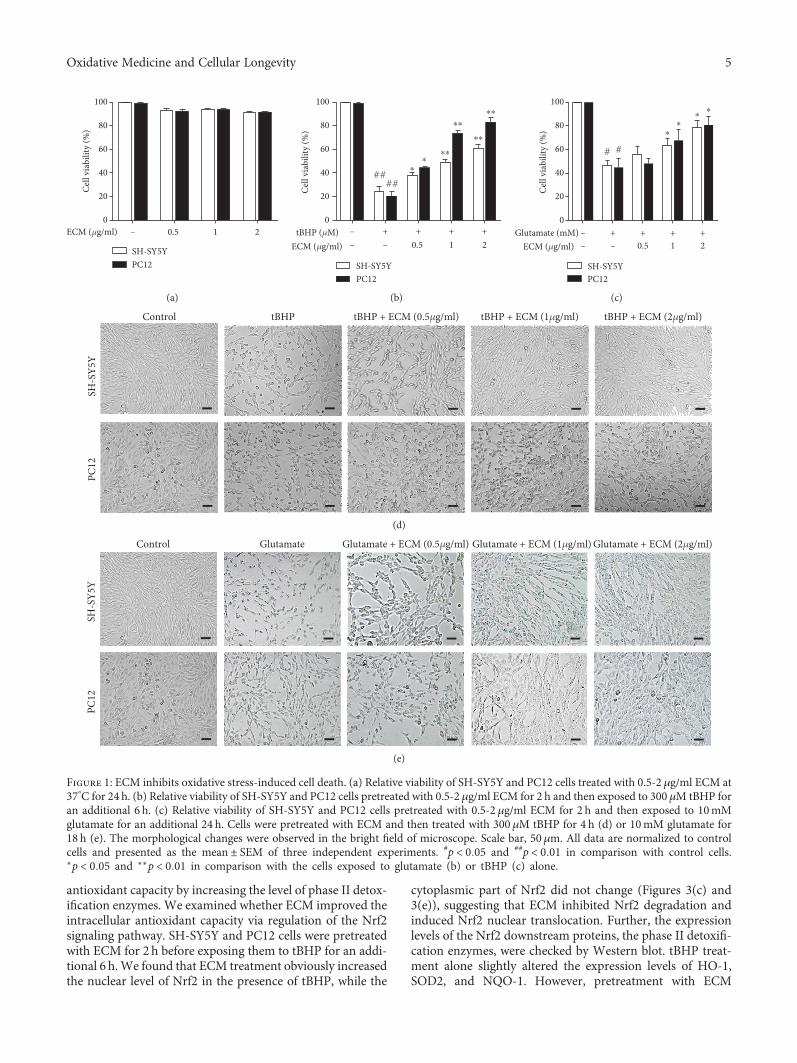
antioxidant capacity by increasing the level of phase II detox-ification enzymes. We examined whether ECM improved theintracellular antioxidant capacity via regulation of the Nrf2signaling pathway. SH-SY5Y and PC12 cells were pretreatedwith ECM for 2 h before exposing them to tBHP for an addi-tional 6 h.We found that ECM treatment obviously increasedthe nuclear level of Nrf2 in the presence of tBHP, while the
cytoplasmic part of Nrf2 did not change (Figures 3(c) and3(e)), suggesting that ECM inhibited Nrf2 degradation andinduced Nrf2 nuclear translocation. Further, the expressionlevels of the Nrf2 downstream proteins, the phase II detoxifi-cation enzymes, were checked by Western blot. tBHP treat-ment alone slightly altered the expression levels of HO-1,SOD2, and NQO-1. However, pretreatment with ECM
0
20
40
60
80
100
Cell
viab
ility
(%)
ECM (�휇g/ml) 0.5 1 2
SH-SY5YPC12
–
(a)
0
20
40
60
80
100
Cell
viab
ility
(%)
ECM (�휇g/ml)tBHP (�휇M)
0.5 1
SH-SY5YPC12
–
– –+ + + +
2
⁎⁎
⁎⁎
⁎⁎
⁎⁎
⁎⁎
####
(b)
0
20
40
60
80
100
Cell
viab
ility
(%)
Glutamate (mM)ECM (�휇g/ml) 0.5
–– –
+ + + +1 2
SH-SY5YPC12
⁎⁎
⁎⁎
# #
(c)
PC12
SH-S
Y5Y
tBHP tBHP + ECM (0.5�휇g/ml) tBHP + ECM (1�휇g/ml) tBHP + ECM (2�휇g/ml)Control
(d)
Glutamate + ECM (2�휇g/ml)
PC12
SH
-SY5
Y
GlutamateControl Glutamate + ECM (1�휇g/ml)Glutamate + ECM (0.5�휇g/ml)
(e)
Figure 1: ECM inhibits oxidative stress-induced cell death. (a) Relative viability of SH-SY5Y and PC12 cells treated with 0.5-2 μg/ml ECM at37°C for 24 h. (b) Relative viability of SH-SY5Y and PC12 cells pretreated with 0.5-2μg/ml ECM for 2 h and then exposed to 300 μM tBHP foran additional 6 h. (c) Relative viability of SH-SY5Y and PC12 cells pretreated with 0.5-2 μg/ml ECM for 2 h and then exposed to 10mMglutamate for an additional 24 h. Cells were pretreated with ECM and then treated with 300 μM tBHP for 4 h (d) or 10mM glutamate for18 h (e). The morphological changes were observed in the bright field of microscope. Scale bar, 50μm. All data are normalized to controlcells and presented as the mean ± SEM of three independent experiments. #p < 0 05 and ##p < 0 01 in comparison with control cells.∗p < 0 05 and ∗∗p < 0 01 in comparison with the cells exposed to glutamate (b) or tBHP (c) alone.
5Oxidative Medicine and Cellular Longevity

Vitamin E 50�휇M
tBHPSH
-Y5Y
SP2
1C
Control tBHP ECM (0.5�휇g/ml) ECM (1�휇g/ml) ECM (2�휇g/ml)
(a)
ROS-
posit
ive c
ells
(%) 60
SH-SY5Y
tBHP (�휇M)ECM (�휇g/ml)
Vitamin E (�휇M)
PC12
– + + + + +––
–– – – –
–
40
20
0.5 1 250
0
⁎
⁎⁎ ⁎⁎⁎⁎
⁎⁎⁎⁎⁎⁎
##
##
(b)
16
MD
A (µ
mol
/mgp
rot)
12
8
4
0tBHP (�휇M)
ECM (�휇g/ml)Vitamin E (�휇M)
– + + + + +––
–– – – –
–0.5 1 250
SH-SY5YPC12
## ##⁎ ⁎
⁎⁎⁎⁎⁎⁎⁎⁎
(c)
SOD
(U/m
gpro
t)
tBHP (�휇M)ECM (�휇g/ml)
Vitamin E (�휇M)
60
40
20
0
80
–
–– – – – –
––
+ + + + +
0.5 1 250
SH-SY5YPC12
####
⁎⁎
⁎⁎⁎⁎
⁎⁎
(d)
GSH
(�휇m
ol/m
gpro
t)
tBHP (�휇M)ECM (�휇g/ml)
Vitamin E (�휇M)
300
240
180
120
60
0––– – – – –
––+ + + + +
0.5 1 250
SH-SY5YPC12
####⁎ ⁎
⁎⁎⁎⁎
⁎⁎⁎⁎
(e)
Figure 2: Continued.
6 Oxidative Medicine and Cellular Longevity

significantly upregulated the expression levels of HO-1,SOD2, and NQO-1 in the presence of tBHP (Figures 3(d)and 3(e)). Our results indicated that ECM has a role in atten-uating ROS production via the enhancement of intracellularantioxidant capacity.
3.4. Active Compounds in ECM Alleviate tBHP-InducedOxidative Stress through the Nrf2 Signaling Pathway.Recently, several sesquiterpene lactones have been isolatedfrom C. minima. However, the active compounds that con-tribute to the antioxidative properties of C. minima havenever been reported. In order to identify the active compo-nents that exert neuroprotective effects, we isolated a seriesof sesquiterpenoids from the ethanol extracts of C. minima,including 6-O-angeloylplenolin, arnicolide D, arnicolide C,and microhelenin C, referred to as EBSC-26A−EBSC-26D,respectively. We first tested the antioxidative activities ofthese compounds. None of the compounds showed obviouscytotoxicity towards SH-SY5Y or PC12 cells at indicatedconcentrations (Figure 4(a), upper panel). Intriguingly, thecompounds protected the neuronal cells from the oxidativedamage induced by tBHP (Figure 4(a), lower panel). In partic-ular, EBSC-26A and EBSC-26B significantly reverse the cyto-toxic effects induced by tBHP at lower-dose treatment.
Meanwhile, the tBHP-inducedROSproductionwasalsoatten-uatedbyEBSC-26AandEBSC-26B(Figures4(b)and4(c)).Wethen examined whether EBSC-26A and EBSC-26B exertedantioxidant effects via the Nrf2 signaling pathway. SH-SY5Ycells were pretreated with EBSC-26A and EBSC-26B for 2 hbefore tBHP treatment for an additional 6 h. We found thatboth EBSC-26A and EBSC-26B markedly increased thenuclear levels of Nrf2 and subsequently upregulated theexpression levels of HO-1, NQO-1, and SOD2 (Figures 4(d)and 4(e)), suggesting that the sesquiterpenoids in C. minimaact as the active compounds that attenuate oxidative stress,and that 6-O-angeloylplenolin and arnicolide D are thepotent antioxidant compounds in C. minima.
3.5. ECM Improves Learning and Memory Ability in aD-gal/AlCl3-Induced Mouse Model. D-gal can produce oxi-dative stress and induce neurodegeneration and memoryimpairment. AlCl3 is also frequently used to reduce neuronalviability in the hippocampus and promote memory impair-ment. Thus, we evaluated the therapeutic potential of ECMin a D-gal/AlCl3-induced neurodegenerative mouse model.The mice were treated with D-gal/AlCl3 for 3 months toinduce neurodegeneration. ECM was administrated duringthe final month of D-gal/AlCl3 treatment, and the Morris
Control tBHPtBHP
ECM (0.5�휇g/ml) ECM (1�휇g/ml) ECM (2�휇g/ml) Vitamin E 50�휇M
(f)
Bcl-2
Bax
�훽-Actin
tBHP (�휇M)ECM (�휇g/ml)
Vitamin E (�휇M)
– ++ + + +––
–– ––
––
0.5 1 250
(g)Bc
l-2/B
ax (%
)
10
8
6
4
2
0
Vitamin E (�휇M) – – –– – 50
tBHP (�휇M)ECM (�휇g/ml)
– ++ + + +– – –0.5 1 2
###
⁎⁎
⁎⁎
⁎⁎⁎
(h)
Figure 2: ECM attenuates oxidative stress-induced mitochondrial dysfunction. The cells were pretreated with 0.5-2μg/ml ECM and 50μMvitamin E for 2 h and then exposed to 300 μM tBHP for an additional 6 h. (a) SH-SY5Y (upper panel) or PC12 cells (lower panel) were treatedwith DCFH-DA for 30min; Hoechst 33342 was used to counterstain cell nuclei. Scale bar, 50 μm. (b) The percentage of ROS-positive cellsamong cultured SH-SY5Y or PC12 cells was quantified and was shown as histogram. Intracellular MDA content (c), SOD activity (d), andGSH levels (e) were detected using a kit assay and are presented as a histogram. (f) SH-SY5Y cells were pretreated with 0.5-2μg/ml ECMfor 2 h and then exposed to 300μM tBHP for an additional 6 h. The mitochondrial membrane potential was determined using the JC-1fluorescence probe, and representative pictures have been shown for comparison. (g) Western blot analysis was performed usingantibodies against Bax and Bcl-2, and β-actin was used as a loading control. (h) The ratio of Bcl-2 to Bax was quantified by densitometryand is shown as a histogram. The results are shown as the mean ± SEM of three independent experiments. ##p < 0 01 in comparison withcontrol cells. ∗p < 0 05 and ∗∗p < 0 01 in comparison with the cells exposed to tBHP alone.
7Oxidative Medicine and Cellular Longevity

SH-SY5Y PC12p-p38
p38
p-ERK
ERK
p-JNK
ECM (�휇g/ml)tBHP (�휇M)
Vitamin E (�휇M)
++0.5 1 2++ ++ ++––
–– ––++––
–– –– –––– –– 50
++0.5 1 2++ ++ ++––
–– ––++––
–– –– –––– –– 50
�훽-Actin
(a)
0
0.5
1.0
1.5
0
0.4
0.8
1.2
ECM (�휇g/ml)tBHP (�휇M)
Vitamin E (�휇M)
++0.5 1 2++ ++ ++––
–– ––++––
–– –– –––– –– 50
SH–SY5YPC12
ECM (�휇g/ml)tBHP (�휇M)
Vitamin E (�휇M)
++0.5 1 2++ ++ ++––
–– ––++––
–– –– –––– –– 50
SH–SY5YPC12
ECM (�휇g/ml)tBHP (�휇M)
Vitamin E (�휇M)
++0.5 1 2++ ++ ++––
–– ––++––
–– –– –––– –– 50
SH–SY5YPC12
0
0.5
1.0
1.5
p-p3
8/p3
8(fo
ld ch
ange
)
p-ER
K/ER
K(fo
ld ch
ange
)
p-JN
K/�훽
-act
in(fo
ld ch
ange
)
###
##
⁎⁎⁎⁎
⁎⁎⁎⁎⁎ ⁎⁎⁎
⁎⁎ ⁎⁎⁎⁎
⁎⁎⁎
⁎⁎⁎
⁎⁎⁎⁎⁎⁎
##
#
###
## ⁎⁎⁎⁎⁎
⁎⁎⁎ ⁎⁎⁎⁎⁎⁎⁎⁎⁎⁎⁎⁎
(b)
SH-SY5Y PC12
Nuclear Nrf2
Lamin B1
Cytoplasmic Nrf2
GAPDH
++0.5 1 2++ ++ ++––
–– ––
++
–––– –– –––– –– 50
++0.5 1 2++ ++ ++––
–– ––
++
–––– –– –––– –– 50
ECM (�휇g/ml)tBHP (�휇M)
Vitamin E (�휇M)
(c)
SH-SY5Y PC12
HO–1
NQO1
SOD2
GAPDH
++0.5 1 2++ ++ ++––
–– ––
++
–––– –– –––– –– 50
++0.5 1 2++ ++ ++––
–– ––
++
–––– –– –––– –– 50
ECM (�휇g/ml)tBHP (�휇M)
Vitamin E (�휇M)
(d)
Figure 3: Continued.
8 Oxidative Medicine and Cellular Longevity

water maze test was performed to assess learning andmemory. During the five days of spatial acquisition training,we found that the time required for the mice to find thehidden platform decreased progressively (Figure 5(a)). TheD-gal/AlCl3 group exhibited significantly longer escapelatency than the vehicle control group. ECM or vitamin Etreatment decreased the escape latency (Figure 5(a)). TheECM or vitamin E treatment group had a shorter swimmingpath to the platform than the D-gal/AlCl3 group(Figure 5(b)). Subsequently, the platform was removed onday 6, the spatial probe test was performed, and the numberof crossings over the position of the removed platform wasrecorded (Figure 5(c)). The ECM or vitamin E treatmentgroup had significantly more platform crossings than themice in the D-gal/AlCl3 group (Figure 5(c)), indicating thatECM could improve learning and memory.
The synaptic proteins, PSD95 and SYN, play critical rolesin synaptic plasticity and cognitive function [43, 44]. Oxida-tive stress can induce a decrease of the expression of PSD95and SYN in the hippocampus, leading to cognitive im-pairment and development of neurodegenerative diseases[45, 46]. Thus, we examined the neuroprotective effects ofECM on the hippocampus. In D-gal/AlCl3-treated mice, wefound that the expression levels of PSD95 and SYN weredecreased in the hippocampal tissues (Figure 5(d)); mean-while, the neurons in the hippocampus were remarkablyshrunken and irregularly arranged, and the pyknotic nuclei
were also increased based on H&E staining (SupplementaryFigure 1), indicating that oxidative stress contributes toneuronal damage in the hippocampus. However, ECM andvitamin E treatment groups exhibited tightly packed andregularly arranged neurons (Supplementary Figure 1). Inline with this, ECM or vitamin E treatment sustained theexpression levels of PSD95 and SYN (Figures 5(d) and 5(e)).The results indicate that ECM could protect neurons againstoxidative stress in a D-gal/AlCl3-induced neurodegenerativemouse model.
3.6. ECM Protects Hippocampus Neurons from OxidativeStress via the Nrf2 Signaling Pathway. To determine the anti-oxidant effects of ECM, we first detected the levels of MDA,GSH, and SOD activities in the brain. In the hippocampusand cortex of D-gal/AlCl3-treated mice, the level of MDAwas significantly higher, and the SOD activity and GSH levelwere lower than those of the control group (Figure 6(a));these levels were markedly reversed by ECM treatment. Inparticular, the levels in the high-dose ECM group were com-parable to those in the vitamin E group (Figure 6(a)). Theseresults indicate that ECM exerts neuroprotective effects viaalleviation of oxidative stress in a D-gal/AlCl3 mouse model.
To determine whether ECM ameliorates oxidative stressvia the MAPK and Nrf2 signaling pathways in D-gal/AlCl3-challgened mice, the levels of phosphorylated p38 MAPKand ERK and phase II detoxification enzymes in the
++0.5 1 2++ ++ ++––
–– ––++––
–– –– –––– –– 50
SH-SY5YPC12
0
0.4
0.8
1.2
0
1.0
2.0
3.0
0
0.6
1.2
1.8
0
0.6
1.2
1.8
ECM (�휇g/ml)tBHP (�휇M)
Vitamin E (�휇M)
++0.5 1 2++ ++ ++––
–– ––++––
–– –– –––– –– 50ECM (�휇g/ml)
tBHP (�휇M)
Vitamin E (�휇M)
++0.5 1 2++ ++ ++––
–– ––++––
–– –– –––– –– 50ECM (�휇g/ml)
tBHP (�휇M)
Vitamin E (�휇M)
++0.5 1 2++ ++ ++––
–– ––++––
–– –– –––– –– 50ECM (�휇g/ml)
tBHP (�휇M)
Vitamin E (�휇M)
Nuc
lear
Nrf2
/Lam
in B
1(fo
ld ch
ange
)
HO
-1/G
APD
H(fo
ld ch
ange
)
NQ
O1/
GA
PDH
(fold
chan
ge)
SOD
2/G
APD
H(fo
ld ch
ange
)
##
###
#####
###
###
###
⁎⁎⁎
⁎⁎⁎
⁎⁎⁎ ⁎⁎⁎⁎⁎⁎
⁎⁎⁎ ⁎⁎⁎⁎⁎⁎
⁎
⁎⁎
⁎⁎⁎⁎⁎⁎
⁎⁎⁎⁎⁎⁎⁎⁎⁎ ⁎⁎⁎
⁎⁎⁎⁎⁎⁎⁎⁎⁎
⁎⁎⁎⁎⁎⁎
⁎⁎⁎
⁎⁎⁎
⁎⁎⁎⁎
⁎⁎⁎
⁎⁎⁎⁎⁎⁎⁎⁎⁎
⁎⁎⁎
⁎⁎⁎⁎⁎
(e)
Figure 3: ECM inhibits oxidative stress via the regulation of the MAPK and Nrf2 signaling pathways. The cells were pretreated with0.5-2μg/ml ECM and 50 μM vitamin E for 2 h and then exposed to 300 μM tBHP for an additional 1 h. Western blot analysis wasperformed using antibodies against p-p38, p38, p-ERK, ERK, p-JNK, and GAPDH; β-actin was used as a loading control in SH-SY5Y (a)and PC12 cells (b). Nuclear and cytoplasmic proteins were extracted after treatment, and Nrf2 was detected using Western blot analysis inSH-SY5Y (c) and PC12 cells (d); GAPDH and Lamin B1 were used as loading controls. Western blot analysis was performed usingantibodies against HO-1, NQO1, and SOD2 in SH-SY5Y (e) and PC12 cells (f); GAPDH was used as a loading control.
9Oxidative Medicine and Cellular Longevity

EBSC-26A (�휇M) − 0.5 1 2 EBSC-26B (�휇M) − 0.5 1 2 EBSC-26C (�휇M) − 0.5 1 2 EBSC-26D (�휇M) − 0.5 1 2
+
# # # # # ## #⁎
⁎ ⁎⁎
tBHP (�휇M) + + +−EBSC-26A (�휇M) − 0.5 1 2−
+tBHP (�휇M) + + +−EBSC-26B (�휇M) − 0.5 1 2−
+tBHP (�휇M) + + +−EBSC-26C (�휇M) − 0.5 1 2−
+tBHP (�휇M) + + +−EBSC-26D (�휇M) − 0.5 1 2−
020406080
100120
020406080
100120
020406080
100120
020406080
100120
020406080
100120
020406080
100120
020406080
100120
020406080
100120
Cell
viab
ility
(%)
Cell
viab
ility
(%)
Cell
viab
ility
(%)
Cell
viab
ility
(%)
Cell
viab
ility
(%)
Cell
viab
ility
(%)
Cell
viab
ility
(%)
Cell
viab
ility
(%)
⁎⁎⁎⁎⁎⁎⁎⁎⁎⁎⁎⁎⁎⁎
⁎⁎ ⁎
⁎ ⁎⁎⁎⁎⁎
SH-SY5YPC12
(a)
Control tBHP EBSC-26A 1 �휇M EBSC-26A 2 �휇M EBSC-26B 1 �휇M EBSC-26B 2 �휇MtBHP
(b)
EBSC-26A (�휇M)tBHP (�휇M) +
2+ + +−
− −+−
EBSC-26B (�휇M) 1 2− − −1−
−
0
12
24
###36
ROS-
posit
ive c
ells
(%)
⁎⁎⁎⁎⁎⁎
⁎⁎⁎⁎⁎⁎
(c)
Nuclear Nrf2
Lamin B1
HO-1
NQO1
SOD2
�훽-Actin
50
EBSC-26A (�휇M)tBHP (�휇M)
Vitamin E (�휇M)
+2
+ + +−− −
+−
− − −− −EBSC-26B (�휇M) 1 2− − −
1−
−+
−
−−
(d)
50
EBSC-26A (�휇M)tBHP (�휇M)
Vitamin E (�휇M)
+
#####
###
⁎⁎⁎⁎⁎⁎
⁎⁎⁎⁎⁎⁎
⁎⁎⁎
2+ + +−
− −+−
− − −− −EBSC-26B (�휇M) 1 2− − −
1−
−+
−
−−
50
EBSC-26A (�휇M)tBHP (�휇M)
Vitamin E (�휇M)
+2
+ + +−− −
+−
− − −− −EBSC-26B (�휇M) 1 2− − −
1−
−+
−
−−
50
EBSC-26A (�휇M)tBHP (�휇M)
Vitamin E (�휇M)
+2
+ + +−− −
+−
− − −− −EBSC-26B (�휇M) 1 2− − −
1−
−+
−
−−
50
EBSC-26A (�휇M)tBHP (�휇M)
Vitamin E (�휇M)
+2
+ + +−− −
+−
− − −− −EBSC-26B (�휇M) 1 2− − −
1−
−+
−
−−
0
0.7
1.4
2.1
0
0.5
1.0
1.5
0
0.5
1.0
1.5
0
0.6
1.2
1.8
Nuc
lear
N
rf2/L
amin
B1(fo
ld ch
ange
)
HO
-1/�훽
-act
in(fo
ld ch
ange
)
NQ
O1/�훽
-act
in(fo
ld ch
ange
)
SOD
2/�훽
-act
in(fo
ld ch
ange
)
⁎⁎⁎
⁎⁎⁎
⁎⁎⁎
⁎⁎⁎
⁎⁎
⁎⁎⁎ ⁎⁎⁎
⁎⁎⁎⁎⁎⁎
⁎⁎⁎ ⁎⁎⁎⁎⁎⁎
⁎⁎⁎
(e)
Figure 4: The active compounds EBSC-26A−EBSC-26D ameliorate oxidative stress. (a) SH-SY5Y and PC12 cells were treated with 0.5-2μMEBSC-26A−EBSC-26D at 37°C for 24 h (upper panel) or pretreated with 0.5-2μMEBSC-26A−EBSC-26D for 2 h and then exposed to 300μMtBHP for an additional 6 h (lower panel); the relative viability of cells was measured by MTT assay. (b, c) SH-SY5Y cells were probed withDCFH-DA (b) and the percentage of ROS-positive cells among culture cells was quantified (c). Scale bar, 50μm. (d) After pretreatment with1-2 μM EBSC-26A, EBSC-26B, and vitamin E for 2 h, SH-SY5Y cells were exposed to 300μM tBHP for an additional 6 h, and then nuclearNrf2, HO-1, NQO-1, and SOD2 levels were detected by Western blot; β-actin was used as a loading control. (e) Relative protein levels werequantified by densitometry and normalized to Lamin B or β-actin. The results are shown as the mean ± SEM of three independentexperiments. #p < 0 05 in comparison with control cells. ∗p < 0 05 and ∗∗p < 0 01 in comparison with the cells exposed to tBHP alone.
10 Oxidative Medicine and Cellular Longevity

ControlModel #ECM-L ⁎⁎
ECM-M ⁎⁎⁎ECM-H ⁎⁎⁎Vitamin E ⁎⁎
60
40
20
0
Esca
pe la
tenc
y (s
ec)
1 2 3 4 5
(a)
Control Model ECM-L Vitamin EECM-M ECM-H
Day 1
IV
III II
I IV
III II
I IV
III II
I IV
III II
I IV
III II
I IV
III II
I
IV
III II
I IV
III II
I IV
III II
I IV
III II
I IV
III II
I IV
III II
IDay 5
(b)
Cros
sing
times
5
3
2
0
1
4
Control Model
#
⁎⁎
⁎⁎⁎
⁎⁎⁎
⁎⁎
L M H Vitamin E
ECM
(c)
Control Model L M H Vitamin E
ECM
PSD95
SYN
�훽-Actin
Hippocampus
(d)
PSD
95/�훽
-act
in
2.5
1.5
1.0
0
0.5
2.0
##
##
Cont
rol
Mod
el L M H
Vita
min
E
ECM
SYN
/�훽-a
ctin
3
1
0
2
Cont
rol
Mod
el L M H
Vita
min
E
ECM
⁎⁎⁎ ⁎⁎⁎
⁎⁎⁎⁎⁎⁎
⁎⁎⁎
⁎⁎⁎
⁎⁎
(e)
Figure 5: ECM improves learning andmemory in a neurodegenerativemousemodel. (a) Escape latency on five consecutive days of testing. (b)The swimming paths of the respective groups on the first and fifth days. (c) The crossing times in the probe trial. (d) Western blot analysis ofPSD95 and SYN; β-actin was used as a loading control in the hippocampus. (e) Relative protein levels were quantified by densitometry andnormalized to β-actin. Control: vehicle control; ECM-L: D − gal/AlCl3 + ECM (100mg/kg/d); ECM-M: D − gal/AlCl3 + ECM (200mg/kg/d);ECM-H: D − gal/AlCl3 + ECM (400mg/kg/d); vitamin E: D − gal/AlCl3 + vitamin E (80mg/kg/d). Data represent mean ± SEM (n = 10 pergroup). #p < 0 05 and ##p < 0 01 in comparisonwith control group; ∗p < 0 05, ∗∗p < 0 01, and ∗∗∗p < 0 001 in comparisonwith themodel group.
11Oxidative Medicine and Cellular Longevity

⁎
⁎
Hip
poca
mpu
sCo
rtex
Cont
rol
Mod
el L M H
Vita
min
E
ECM
#
#
100
60
40
0
20
80
0
10
20
30
40 #
⁎
MD
A (n
mol
/mgp
rot)
0
10
20#
30
40
SOD
(U/m
gpro
t)
204060
#
80100
GSH
(�휇m
ol/g
prot
)
0
MD
A (n
mol
/mgp
rot)
0
20
40
80
SOD
(U/m
gpro
t)0
200
400
600
#
800
GSH
(�휇m
ol/g
prot
)
Cont
rol
Mod
el L M H
Vita
min
E
ECM
Cont
rol
Mod
el L M H
Vita
min
E
ECM
Cont
rol
Mod
el L M H
Vita
min
E
ECMCo
ntro
l
Mod
el L M H
Vita
min
E
ECM
Cont
rol
Mod
el L M H
Vita
min
E
ECM
⁎⁎ ⁎⁎⁎⁎ ⁎⁎⁎
⁎⁎⁎⁎⁎
⁎⁎⁎⁎
⁎
⁎⁎⁎ ⁎⁎⁎ ⁎⁎⁎
⁎⁎ ⁎⁎⁎⁎⁎⁎
⁎⁎
(a)
Hippocampus
p-p38
p38
p-ERK
ERK
�훽-Actin
Nrf2
HO-1
SOD2
NQO1
Control Model L M H Vitamin EECM
(b)
⁎⁎
⁎⁎
⁎
#
⁎⁎⁎
⁎⁎⁎
#
⁎⁎⁎
⁎⁎⁎
⁎⁎
⁎⁎⁎
*
⁎⁎⁎
# #
⁎⁎ ⁎⁎⁎
⁎⁎
p-p3
8/p3
8
p-ER
K/ER
K
Nrf2
/�훽-a
ctin
HO
-1/�훽
-act
in
NQ
O1/�훽
-act
in
SOD
2/�훽
-act
in
1.0
0.60.4
00.2
0.82.5
1.51.0
00.5
2.0
1.5
#1.0
0
0.5
2.0
2.5
1.51.0
00.5
2.0
5
3#2
01
4
4
2
0
6
8
Cont
rol
Mod
el L M H
Vita
min
E
ECM
Cont
rol
Mod
el L M H
Vita
min
E
ECM
Cont
rol
Mod
el L M H
Vita
min
E
ECM
Cont
rol
Mod
el L M H
Vita
min
EECM
Cont
rol
Mod
el L M H
Vita
min
E
ECM
Cont
rol
Mod
el L M H
Vita
min
E
ECM
⁎⁎⁎
⁎⁎⁎⁎⁎⁎ ⁎⁎⁎
⁎⁎
⁎⁎⁎ ⁎⁎⁎
⁎
(c)
Figure 6: ECM inhibits oxidative stress via the MAPK and Nrf2 signaling pathways in the CNS. The homogenate of the hippocampus andcortex was used for the assay, and the lysate of the hippocampus was used for Western blot. (a) The content of MDA and the activities of SODand GSH in the hippocampus (upper panel) and cortex (lower panel) were measured using a kit assay. (b)Western blot analysis of p-p38, p38,p-ERK, ERK, Nrf2, HO-1, NQO-1, and SOD2; β-actin was used as a loading control in the hippocampus. (c) Relative protein levels werequantified by densitometry and normalized to β-actin. Control: vehicle control; ECM-L: D − gal/AlCl3 + ECM (100mg/kg/d); ECM-M:D − gal/AlCl3 + ECM (200mg/kg/d); ECM −H D − gal/AlCl3 + ECM (400mg/kg/d); vitamin E: D − gal/AlCl3 + vitamin E (80mg/kg/d).Data are represented as the mean ± SEM (n = 10 per group). #p < 0 05 and ##p < 0 01 in comparison with the control group; ∗p < 0 05,∗∗p < 0 01, and ∗∗∗p < 0 001 in comparison with the model group.
12 Oxidative Medicine and Cellular Longevity

hippocampus were examined by Western blot. In line withthe in vitro results, phosphor-p38 MAPK was increased,while phosphor-ERK was decreased, in the hippocampus ofD-gal/AlCl3-treated mice (Figures 6(b) and 6(c)). However,ECM and vitamin E treatment reduced the phosphorylationof p38 MAPK and increased the phosphorylation of ERK,especially in the high-dose ECM and vitamin E treatmentgroups (Figures 6(b) and 6(c)). We then detected the expres-sion levels of Nrf2 and its downstream proteins. Nrf2, HO-1,SOD2, and NQO-1 had relatively lower expression levels inthe D-gal/AlCl3 model group than those in the control group,whereas ECM or vitamin E treatment significantly increasedthe levels of Nrf2, HO-1, SOD2, and NQO-1 (Figures 6(b)and 6(c)), which was consistent with the in vitro results.These data suggest that ECM treatment can activate theNrf2 signaling pathway and induce the expression of phaseII detoxification enzymes, which are responsible for ameliorat-ing oxidative stress and improving neurodegenerative diseases.
4. Discussion
Numerous reports have indicated that natural products har-bor great chemical diversity, in turn exhibiting multiple bio-logical activities. Increasing evidence has demonstrated thatsome natural products contain antioxidants that protectagainst oxidative stress in chronic disease, especially aging-related diseases. Identification of safe therapeutic regimensfor oxidative stress-related diseases has attracted great atten-tion. C. minima has been used as a medicinal herb to treat anumber of diseases. Both aqueous and hydroalcoholicextracts of C. minima have been reported to scavenge freeradicals and ameliorate oxidative stress, and the hydroalco-holic extracts express higher antioxidant activity [32].However, the underlying mechanism of C. minima as anantioxidant agent was previously unknown. In this study,we examined the therapeutic potential and underlying mech-anism of ECM in the treatment of oxidative stress-inducedneurodegenerative disease and found that the ethanolextracts of C. minima exhibited significant antioxidantactivity both in vitro and in vivo, mainly through upregulat-ing nonenzymatic and enzymatic antioxidants. In this study,we also found that ECM protected mitochondrial functionthrough the reduction of oxidative stress and by sustainingthe Bcl-2/Bax ratio. Therefore, ECM could protect neuronsin the brain from oxidative stress, which in turn attenuatesthe process of neurodegeneration and improves learningandmemory capacity in aD-gal/AlCl3-inducedmousemodel.
Although polyphenol and flavonoids are thought to beresponsible for the antioxidant activity of C. minima, theactive compounds had not been identified. A series ofsesquiterpene lactones have been isolated from C. minima,with 6-O-angeloylplenolin found to be one of the most abun-dant compounds in ECM; 6-O-angeloylplenolin has beenreported to exhibit antiallergy, antibacterial, and antiprolifer-ative effects [34–37]. Arnicolide D and arnicolide C have alsobeen shown to have antibacterial and antiproliferative effects[28, 36]. However, the antioxidant effects of these sesquiter-penoids in C. minima have never been reported. Therefore,we examined whether the sesquiterpenoids mediated the
antioxidant activity of ECM. Intriguingly, we found that allfour sesquiterpenoids had antioxidant activity, as they inhib-ited the neuronal death induced by tBHP, with 6-O-angeloyl-plenolin and arnicolide D exhibiting antioxidant activityeven at low concentrations. Meanwhile, 6-O-angeloylpleno-lin and arnicolide D markedly induced the expression ofphase II detoxification enzymes responsible for alleviatingoxidant stress. Moreover, we analyzed the concentration ofthe compounds in ECM and found that both 6-O-angeloyl-plenolin and arnicolide D were highly concentrated in theextract of C. minima (see Supplementary Material), furtherindicating that 6-O-angeloylplenolin and arnicolide D actas active compounds that exhibit antioxidant properties.Our results suggest that sesquiterpenoids could be consid-ered the active compounds in C. minima responsible forameliorating oxidative stress; therefore, the therapeuticpotential of sesquiterpenoids in treating neurodegenerativediseases warrants further investigation. The compound 6-O-angeloylplenolin and arnicolide D have been reported toexhibit antiproliferative activities against cancer cells; how-ever, no obvious cytotoxicity was observed in our study,which may be due to that the compounds were used at lowerconcentrations than that used for antiproliferation studies,indicating that the compounds exert different activities at dif-ferent concentrations. We will further examine the toxiceffects of 6-O-angeloylplenolin and arnicolide D in vivo.
Increasing evidence has demonstrated that the transcrip-tion factor Nrf2 plays a key role in antagonizing oxidativestress, with Nrf2 exerting cytoprotective effects via the upreg-ulation of a series of phase II detoxification enzymes.Recently, certain Nrf2 inducers have been studied clinically,and Nrf2 has been considered an emerging therapeutic target[47]. Nrf2 activation is under strict regulation; usually, Nrf2is retained in the cytoplasm by Kelch-like ECH-associatedprotein 1 (Keap1) and tends to be degraded. Certain kinases,including ERK, protein kinase C, and AKT, regulate Nrf2activation, and multiple natural products have been reportedto regulate Nrf2 activation and nuclear translocation.Vitamin E and sulforaphane have been extensively examinedfor the ability to attenuate oxidative stress via the regulationof the Nrf2 signaling pathway. In this study, we used vitaminE as a positive agent for attenuating oxidative stress andfound that ECM ameliorated oxidative stress both in vitroand in vivo, mainly through the activation of ERK andinduction of Nrf2 nuclear translocation. The active com-pounds 6-O-angeloylplenolin and arnicolide D also mark-edly induced activation of Nrf2. Our results suggest that theECM-induced activation of Nrf2 may occur, at least in part,through the induction of ERK activation. It is also possiblethat the active compounds in ECM directly disrupt the bind-ing interface between Nrf2 and Keaps. Therefore, the role ofthe active compounds of C. minima in Nrf2 activationrequires further investigation.
5. Conclusion
In summary, we found that the ethanol extract of C. minimais able to protect neuronal cells against oxidative stress-induced neurodegeneration through activation of the Nrf2
13Oxidative Medicine and Cellular Longevity

signaling pathway. We also found that 6-O-angeloylplenolinand arnicolide D are the potential active compounds in C.minima, which provides strong evidence that C. minimahas a therapeutic potential in ameliorating neurodegenera-tive diseases. C. minima could be used as a source of antiox-idant and neuroprotective compounds.
Abbreviations
ECM: Ethanol extract of C. minimatBHP: tert-Butyl hydroperoxideD-gal: D-GalactoseAlCl3: Aluminum chlorideROS: Reactive oxygen speciesJNK: c-Jun N-terminal kinasesERK: Extracellular signal-regulated kinasesMDA: Malonyl dialdehydeGST: Glutathione S-transferaseMAPK: Mitogen-activated protein kinaseNrf2: Nuclear factor erythroid 2-related factor 2HO-1: Heme oxygenase-1NQO-1: NAD(P)H: quinone oxidoreductase-1SOD: Superoxide dismutaseGSH: GlutathionePSD95: Postsynaptic density protein 95SYN: SynaptophysinHPLC: High-performance liquid chromatographyDCFH-DA: 2′,7′-Dichlorofluorescin diacetate.
Data Availability
All data used to support the findings of this study areincluded within the article.
Conflicts of Interest
The authors declare no conflicts of interest.
Authors’ Contributions
Yi-Jie Wang and Xin-Yue Wang contributed equally tothis work.
Acknowledgments
This work was supported by the Guangzhou Science Tech-nology and Innovation Commission Technology ResearchProjects (201805010005), the National Natural ScienceFoundation of China (81473740 and 81802776), the NationalScience Fund for Distinguished Young Scholars (81525026),and the National Key Research and Development Programof China “Research and Development of ComprehensiveTechnologies on Chemical Fertilizer and Pesticide Reductionand Synergism” (2017YFD0201402).
Supplementary Materials
Material and Method for extraction and isolation of EBSC-26A–EBSC-26D and 9 figures. Supplementary Figure 1:ECM exerts neuroprotective effects in the hippocampus.
Supplementary Figure 2–9: 1H NMR and 13C NMR spectraof EBSC-26A–EBSC-26D. (Supplementary Materials)
References
[1] J. R. Hodges, “Decade in review-dementia: a decade of discov-ery and disappointment in dementia research,” NatureReviews Neurology, vol. 11, no. 11, pp. 613-614, 2015.
[2] B. Maloney and D. K. Lahiri, “Epigenetics of dementia: under-standing the disease as a transformation rather than a state,”The Lancet Neurology, vol. 15, no. 7, pp. 760–774, 2016.
[3] S. J. Lupien, R. P. Juster, C. Raymond, and M. F. Marin, “Theeffects of chronic stress on the human brain: from neurotoxic-ity, to vulnerability, to opportunity,” Frontiers in Neuroendo-crinology, vol. 49, pp. 91–105, 2018.
[4] D. M. Lipnicki, J. D. Crawford, R. Dutta et al., “Age-relatedcognitive decline and associations with sex, education andapolipoprotein E genotype across ethnocultural groups andgeographic regions: a collaborative cohort study,” PLoS Medi-cine, vol. 14, no. 3, article e1002261, 2017.
[5] J. C. Jurado-Coronel, R. Cabezas, M. F. Avila Rodriguez,V. Echeverria, L. M. Garcia-Segura, and G. E. Barreto, “Sex dif-ferences in Parkinson’s disease: features on clinical symptoms,treatment outcome, sexual hormones and genetics,” Frontiersin Neuroendocrinology, vol. 50, pp. 18–30, 2018.
[6] B. A. Kent and R. E. Mistlberger, “Sleep and hippocampal neu-rogenesis: implications for Alzheimer’s disease,” Frontiers inNeuroendocrinology, vol. 45, pp. 35–52, 2017.
[7] A. Adlimoghaddam, M. Neuendorff, B. Roy, and B. C. Albensi,“A review of clinical treatment considerations of donepezil insevere Alzheimer’s disease,” CNSNeuroscience & Therapeutics,vol. 24, no. 10, pp. 876–888, 2018.
[8] B. T. Winslow, M. K. Onysko, C. M. Stob, and K. A. Hazle-wood, “Treatment of Alzheimer disease,” American FamilyPhysician, vol. 83, pp. 1403–1412, 2011.
[9] L. Letra, T. Rodrigues, P. Matafome, I. Santana, and R. Seica,“Adiponectin and sporadic Alzheimer’s disease: clinical andmolecular links,” Frontiers in Neuroendocrinology, vol. 52,pp. 1–11, 2019.
[10] D. Yin and K. Chen, “The essential mechanisms of aging:irreparable damage accumulation of biochemical side-reac-tions,” Experimental Gerontology, vol. 40, no. 6, pp. 455–465,2005.
[11] J. Lv, S. Jiang, Z. Yang et al., “PGC-1α sparks the fire of neuro-protection against neurodegenerative disorders,” AgeingResearch Reviews, vol. 44, pp. 8–21, 2018.
[12] V. Calabrese, R. Sultana, G. Scapagnini et al., “Nitrosativestress, cellular stress response, and thiol homeostasis inpatients with Alzheimer’s disease,” Antioxidants & Redox Sig-naling, vol. 8, no. 11-12, pp. 1975–1986, 2006.
[13] E. E. Devore, F. Grodstein, F. J. A. van Rooij et al., “Dietaryantioxidants and long-term risk of dementia,” Archives of Neu-rology, vol. 67, no. 7, pp. 819–825, 2010.
[14] N. Dragicevic, N. Copes, G. O’Neal-Moffitt et al., “Melatonintreatment restores mitochondrial function in Alzheimer’smice: a mitochondrial protective role of melatonin membranereceptor signaling,” Journal of Pineal Research, vol. 51, no. 1,pp. 75–86, 2011.
[15] C. A. Massaad, R. G. Pautler, and E. Klann, “Mitochondrialsuperoxide: a key player in Alzheimer’s disease,” Aging,vol. 1, no. 9, pp. 758–761, 2009.
14 Oxidative Medicine and Cellular Longevity

[16] M. Zhang, C. An, Y. Gao, R. K. Leak, J. Chen, and F. Zhang,“Emerging roles of Nrf2 and phase II antioxidant enzymes inneuroprotection,” Progress in Neurobiology, vol. 100, pp. 30–47, 2013.
[17] K. Dasuri, L. Zhang, and J. N. Keller, “Oxidative stress, neuro-degeneration, and the balance of protein degradation and pro-tein synthesis,” Free Radical Biology & Medicine, vol. 62,pp. 170–185, 2013.
[18] R. K. Thimmulappa, K. H. Mai, S. Srisuma, T. W. Kensler,M. Yamamoto, and S. Biswal, “Identification of Nrf2-regulatedgenes induced by the chemopreventive agent sulforaphane byoligonucleotide microarray,” Cancer Research, vol. 62, no. 18,pp. 5196–5203, 2002.
[19] J. A. Rubiolo,G.Mithieux, and F. V.Vega, “Resveratrol protectsprimary rat hepatocytes against oxidative stress damage: activa-tion of theNrf2 transcription factor and augmented activities ofantioxidant enzymes,” European Journal of Pharmacology,vol. 591, no. 1-3, pp. 66–72, 2008.
[20] A. Loboda,M.Damulewicz, E. Pyza, A. Jozkowicz, and J. Dulak,“Role of Nrf2/HO-1 system in development, oxidative stressresponseanddiseases:anevolutionarilyconservedmechanism,”Cellular and Molecular Life Sciences, vol. 73, no. 17, pp. 3221–3247, 2016.
[21] J. Kwon, E. Han, C. B. Bui et al., “Assurance of mitochondrialintegrity and mammalian longevity by the p62–Keap1–Nrf2–Nqo1 cascade,”EMBOReports, vol. 13, no. 2, pp. 150–156, 2012.
[22] Y.Yang,S.Jiang,J.Yanetal.,“Anoverviewofthemolecularmech-anisms andnovel roles ofNrf2 in neurodegenerative disorders,”Cytokine&GrowthFactorReviews,vol.26,no.1,pp.47–57,2015.
[23] H. Schipper, D. Bennett, A. Liberman et al., “Glial hemeoxygenase-1 expression in Alzheimer disease and mild cogni-tive impairment,” Neurobiology of Aging, vol. 27, no. 2,pp. 252–261, 2006.
[24] Y. Wang, K. Santa–Cruz, C. DeCarli, and J. A. Johnson,“NAD(P)H:quinone oxidoreductase activity is increased inhippocampal pyramidal neurons of patients with Alzheimer’sdisease,” Neurobiology of Aging, vol. 21, no. 4, pp. 525–531,2000.
[25] I. Buendia, P. Michalska, E. Navarro, I. Gameiro, J. Egea, andR. Leon, “Nrf2–ARE pathway: an emerging target againstoxidative stress and neuroinflammation in neurodegenerativediseases,” Pharmacology & Therapeutics, vol. 157, pp. 84–104,2016.
[26] K. Kanninen, T. M. Malm, H. K. Jyrkkanen et al., “Nuclear fac-tor erythroid 2-related factor 2 protects against beta amyloid,”Molecular and Cellular Neurosciences, vol. 39, no. 3, pp. 302–313, 2008.
[27] C. Y. Shen, J. G. Jiang, L. Yang, D. W. Wang, and W. Zhu,“Anti-ageing active ingredients from herbs and nutraceuticalsused in traditional Chinese medicine: pharmacological mecha-nisms and implications for drug discovery,” British Journal ofPharmacology, vol. 174, no. 11, pp. 1395–1425, 2017.
[28] R. S. L. Taylor and G. H. N. Towers, “Antibacterial constitu-ents of the Nepalese medicinal herb, Centipeda minima,” Phy-tochemistry, vol. 47, no. 4, pp. 631–634, 1998.
[29] H. Liang, F. Bao, X. Dong et al., “Antibacterial thymol deriva-tives isolated from Centipeda minima,” Molecules, vol. 12,no. 8, pp. 1606–1613, 2007.
[30] S. Schwikkard and F. R. van Heerden, “Antimalarial activity ofplant metabolites,” Natural Product Reports, vol. 19, no. 6,pp. 675–692, 2002.
[31] G. François, C. Passreiter, H. Woerdenbag, and M. van Loove-ren, “Antiplasmodial activities and cytotoxic effects of aqueousextracts and sesquiterpene lactones from Neurolaena lobata,”Planta Medica, vol. 62, no. 2, pp. 126–129, 1996.
[32] S. S. Huang, C. S. Chiu, T. H. Lin et al., “Antioxidant and anti-inflammatory activities of aqueous extract of Centipeda min-ima,” Journal of Ethnopharmacology, vol. 147, no. 2, pp. 395–405, 2013.
[33] H. M. On, B. M. Kwon, N. I. Baek et al., “Inhibitory activity of6-O-angeloylprenolin from Centipeda minima on farnesylprotein transferase,” Archives of Pharmacal Research, vol. 29,no. 1, pp. 64–66, 2006.
[34] Y. Q. Liu, X. L. Wang, X. Cheng et al., “Skp1 in lung cancer:clinical significance and therapeutic efficacy of its small mole-cule inhibitors,” Oncotarget, vol. 6, no. 33, pp. 34953–34967,2015.
[35] Y. Liu, X. Q. Chen, H. X. Liang et al., “Small compound 6-O-angeloylplenolin induces mitotic arrest and exhibits therapeu-tic potentials in multiple myeloma,” PLoS One, vol. 6, no. 7,article e21930, 2011.
[36] P. Wu, M. X. Su, Y. Wang et al., “Supercritical fluid extractionassisted isolation of sesquiterpene lactones with antiprolifera-tive effects from Centipeda minima,” Phytochemistry, vol. 76,pp. 133–140, 2012.
[37] L. F. Ding, Y. Liu, H. X. Liang, D. P. Liu, G. B. Zhou, and Y. X.Cheng, “Two new terpene glucosides and antitumor agentsfrom Centipeda minima,” Journal of Asian Natural ProductsResearch, vol. 11, no. 8, pp. 732–736, 2009.
[38] Y. Imakura, K. H. Lee, D. Sims et al., “Antitumor agentsXXXVI: structural elucidation of sesquiterpene lactonesmicrohelenins-A, B, and C, microlenin acetate, and plenolinfrom Helenium microcephalum,” Journal of PharmaceuticalSciences, vol. 69, no. 9, pp. 1044–1049, 1980.
[39] L. Xiao, H. Li, J. Zhang et al., “Salidroside protects Caenorhab-ditis elegans neurons from polyglutamine-mediated toxicity byreducing oxidative stress,” Molecules, vol. 19, no. 6, pp. 7757–7769, 2014.
[40] Q. Wang, Y. Huang, C. Qin et al., “Bioactive peptides fromAngelica sinensis protein hydrolyzate delay senescence in Cae-norhabditis elegans through antioxidant activities,” OxidativeMedicine and Cellular Longevity, vol. 2016, Article ID8956981, 10 pages, 2016.
[41] S. T. Smiley, M. Reers, C. Mottola-Hartshorn et al., “Intracel-lular heterogeneity in mitochondrial membrane potentialsrevealed by a J-aggregate-forming lipophilic cation JC-1,” Pro-ceedings of the National Academy of Sciences of the UnitedStates of America, vol. 88, no. 9, pp. 3671–3675, 1991.
[42] E. Himeno, Y. Ohyagi, L. Ma et al., “Apomorphine treatmentin Alzheimer mice promoting amyloid-β degradation,” Annalsof Neurology, vol. 69, no. 2, pp. 248–256, 2011.
[43] M. Audrain, R. Fol, P. Dutar et al., “Alzheimer’s disease-likeAPP processing in wild-type mice identifies synaptic defectsas initial steps of disease progression,”Molecular Neurodegen-eration, vol. 11, no. 1, p. 5, 2016.
[44] D. T. Proctor, E. J. Coulson, and P. R. Dodd, “Reduction inpost-synaptic scaffolding PSD-95 and SAP-102 protein levelsin the Alzheimer inferior temporal cortex is correlated withdisease pathology,” Journal of Alzheimer's Disease, vol. 21,no. 3, pp. 795–811, 2010.
[45] M. A. Ansari, K. N. Roberts, and S. W. Scheff, “Oxidative stressand modification of synaptic proteins in hippocampus after
15Oxidative Medicine and Cellular Longevity

traumatic brain injury,” Free Radical Biology & Medicine,vol. 45, no. 4, pp. 443–452, 2008.
[46] S. W. Scheff, M. A. Ansari, and E. J. Mufson, “Oxidative stressand hippocampal synaptic protein levels in elderly cognitivelyintact individuals with Alzheimer’s disease pathology,” Neuro-biology of Aging, vol. 42, pp. 1–12, 2016.
[47] I. M. Copple, A. T. Dinkova-Kostova, T. W. Kensler, K. T.Liby, and W. C. Wigley, “NRF2 as an emerging therapeutictarget,” Oxidative Medicine and Cellular Longevity, vol. 2017,Article ID 8165458, 2 pages, 2017.
16 Oxidative Medicine and Cellular Longevity

Research ArticleSulforaphane-Enriched Broccoli Sprouts Pretreated by PulsedElectric Fields Reduces Neuroinflammation and AmelioratesScopolamine-Induced Amnesia in Mouse Brain through ItsAntioxidant Ability via Nrf2-HO-1 Activation
Lalita Subedi ,1 KyoHee Cho,1 Yong Un Park,2 Hyuk Joon Choi,2 and Sun Yeou Kim 1
1Laboratory of Pharmacognosy, College of Pharmacy, Gachon University, #191, Hambakmoero, Yeonsu-gu,Incheon 21936, Republic of Korea2BK Bio Co. Ltd., 2706-38, Iljudong-ro, Gujwa-eup, Jeju-si, Jeju-do, Republic of Korea
Correspondence should be addressed to Sun Yeou Kim; [email protected]
Received 19 September 2018; Revised 12 November 2018; Accepted 21 November 2018; Published 27 March 2019
Guest Editor: Francisco J.B. Mendonça Júnior
Copyright © 2019 Lalita Subedi et al. This is an open access article distributed under the Creative Commons AttributionLicense, which permits unrestricted use, distribution, and reproduction in any medium, provided the original work isproperly cited.
Activated microglia-mediated neuroinflammation plays a key pathogenic role in neurodegenerative diseases, such as Alzheimer’sdisease, Parkinson’s disease, multiple sclerosis, and ischemia. Sulforaphane is an active compound produced after conversion ofglucoraphanin by the myrosinase enzyme in broccoli (Brassica oleracea var) sprouts. Dietary broccoli extract as well assulforaphane has previously known to mitigate inflammatory conditions in aged models involving microglial activation. Here,we produced sulforaphane-enriched broccoli sprouts through the pretreatment of pulsed electric fields in order to trigger thebiological role of normal broccoli against lipopolysaccharide-activated microglia. The sulforaphane-enriched broccoli sproutsshowed excellent potency against neuroinflammation conditions, as evidenced by its protective effects in both 6 and 24 h ofmicroglial activation in vitro. We further postulated the underlying mechanism of action of sulforaphane in broccoli sprouts,which was the inhibition of an inflammatory cascade via the downregulation of mitogen-activated protein kinase (MAPK)signaling. Simultaneously, sulforaphane-enriched broccoli sprouts inhibited the LPS-induced activation of the NF-κB signalingpathway and the secretions of inflammatory proteins (iNOS, COX-2, TNF-α, IL-6, IL-1β, PGE2, etc.), which are responsible forthe inflammatory cascades in both acute and chronic inflammation. It also upregulated the expression of Nrf2 and HO-1 innormal and activated microglia followed by the lowered neuronal apoptosis induced by activated microglia. Based on theseresults, it may exhibit anti-inflammatory effects via the NF-κB and Nrf2 pathways. Interestingly, sulforaphane-enriched broccolisprouts improved the scopolamine-induced memory impairment in mice through Nrf2 activation, inhibiting neuronal apoptosisparticularly through inhibition of caspase-3 activation which could lead to the neuroprotection against neurodegenerativedisorders. The present study suggests that sulforaphane-enriched broccoli sprouts might be a potential nutraceutical withantineuroinflammatory and neuroprotective activities.
1. Introduction
Neuroinflammation plays a key role in the regulation ofaging, Alzheimer’s disease (AD), Parkinson’s disease (PD),Huntington’s disease (HD), multiple sclerosis (MS), stroke,depression, dementia, and metabolic disorders such ashypertension and diabetes [1]. Neuroinflammation is alsothe pathogenic hallmark of aging-related neurodegenerative
conditions [1, 2]. Therefore, anti-inflammatory strategiescould be efficient prophylactic and therapeutic managementstrategies for a number of central nervous system (CNS) dis-orders [3, 4]. CNS disorders may develop owing to chronicmicroglial activation. Glial cells, particularly microglia, areimmune cells in the CNS that are responsible for the mainte-nance of normal homeostasis as well as repair after injury inthe brain [5]. Although activated microglia are required for
HindawiOxidative Medicine and Cellular LongevityVolume 2019, Article ID 3549274, 19 pageshttps://doi.org/10.1155/2019/3549274
host defense and debris clearance in the brain, chronicmicroglial activation is toxic to the CNS [6]. Conversion ofnormal microglia to the toxic microglial phenotype (knownas the M1 phenotype) is responsible for the initiation ofinflammatory cascades in the CNS, particularly the produc-tion of reactive oxygen species (ROS), nitric oxide (NO),proteases, arachidonic acids, excitatory amino acids, andcytokines [7]. These neurotoxic substances trigger the oxida-tive stress and are responsible for the disruption of the archi-tecture and functions of neurons, consequently leading tosynaptic degeneration and neurodegeneration [8]. Theseneuroinflammatory and neurotoxic cascades are responsiblefor the hippocampal neuronal damage leading to cognitivedysfunction [9]. Inhibition of the neuroinflammation or acti-vation of the endogenous antioxidant system might be abetter alternative for the repairment of this damage. Particu-larly, activation of the Nrf2 and its mediated antioxidantenzyme, HO-1, can not only inhibit the inflammatorycascades but also increase the neuronal survival and hippo-campal neurogenesis [10]. Extensive neuropharmacologyresearch has succeeded in finding novel drug candidates forthe treatment of CNS disorders. However, they have failedto prove their efficacy in human biological systems duringclinical trials. Potential reasons for these failures were sug-gested to be differences in tissue physiology of the CNS,unstable pharmacokinetics, or difficulty in crossing theblood-brain barrier (BBB) [11, 12]. Given the lack of properallopathic medications to treat neuroinflammatory disorders,there is a growing interest in complementary and alternativemedication, including nutraceuticals. Consumption of die-tary nutraceuticals with neuroprotection could prevent CNSdiseases, overcoming the limitations of allopathic drug deliv-ery to the CNS.
Previous reports highlight the beneficial effects of broccoliand its active compound, sulforaphane, in neurodegenerativedisorders. Although broccoli has a mild effect againstage-related neuroinflammation, it does not show a significanteffect against lipopolysaccharide- (LPS-) induced inflamma-tory conditions [13]. Another independent study has revealedthat sulforaphane can reverse the hyperammonemia-inducedglial activation, neuroinflammation, and disturbances in neu-rotransmitter receptors in the hippocampus that impairedspatial learning [14]. The anti-inflammatory effect of sulfo-raphane has also been previously reported in rat primarymicroglia [15]. These previous studies suggest that biocon-version of glucoraphanin in the broccoli may increase theyield of sulforaphane. It could lead to better health benefitsfor patients with neuroinflammatory disorders. Either theinduction of myrosin production or the increase in myrosin-ase activity can increase the bioconversion of glucoraphanininto sulforaphane [16]. The presence of some other proteins,such as epithiospecifier protein (ESP), can also convert gluco-raphanin to sulforaphane nitrile, without affecting sulfora-phane’s anti-inflammatory effects [17]. An increase inmyrosinase activity with reduced ESP activity can selectivelyenhance sulforaphane yield in the broccoli sprout, making ita better candidate for the treatment of several pathologicalconditions, including cancer, inflammation, neurodegenera-tion, and aging [18]. Pharmacokinetic studies have revealedthat, in mice, sulforaphane has good absorption and
distribution patterns, in various tissues of the body, includingthe brain, lungs, heart, liver, kidneys, and muscles, after oraladministration [19]. Previous reports suggest that sulforaph-ane and its metabolites do not readily cross the BBB [20].However, the disruption of the BBB during neuroinflamma-tion and neurodegenerative conditions allows sulforaphaneto permeate the BBB and enhances its anti-inflammatoryeffects along the brain axis [20].
Former reports have revealed that activation of myrosin-ase in steamed broccoli sprouts can enhance the yield ofsulforaphane [20]. Similarly, pulsed electric field (PEF) pre-treatment enhances the production of glucosinolate in thebroccoli flower and stalk. It also increased anthocyanin’s pro-duction in red cabbage [21, 22]. Thus, we postulated that PEFmight also be effective in enhancing the production of sulfo-raphane in broccoli sprouts.
In the current study, we are to reveal whether PEFtreatment can increase the sulforaphane yield in broccolisprouts or not. We also try to figure out the difference inthe biological activity of broccoli sprout before and after PEFtreatment.Todetermine themolecularmechanismunderlyingthe sulforaphane-enriched broccoli sprout-mediated neuro-protective effect, we assessed the effect of sulforaphane-enriched broccoli sprouts on lipopolysaccharide- (LPS-)inducedproinflammatory responses inbothacute andchronicmicroglial activation invitro. Invivostudywillbedonewhethersulforaphane-enriched broccoli sprouts improve the scopolamine-inducedmemory impairment inmice or not.
2. Materials and Methods
2.1. Reagents. The pulsed electric field (PEF) was purchasedfrom HVP 5 (Elea, DIL, Quakenbrueck, Germany). Lipopoly-saccharide (LPS) and sulforaphane were purchased fromSigma Chemical (St. Louis, MO). Dulbecco’s modified Eaglemedium (DMEM), Fetal bovine serum (FBS), andpenicillin-streptomycin were purchased from Invitrogen(Carlsbad, CA, USA). Antibodies for cyclooxygenase 2(COX-2), β-actin, GAPDH, and histone-3 were obtained fromSanta Cruz Biotechnology (Dallas, Texas, United States). Theantibody of inducible nitric oxide synthase (iNOS) was pur-chased from Abcam (Cambridge, UK). The primary antibodyfor α-tubulin, C-Jun N-terminal kinase (JNK), p38, extracellu-lar signal-regulated kinases (ERK), NF-κB, I-κB, pI-κB, C-Jun,pC-Jun, C-Fos, and pC-Fos were purchased from Cell Signal-ing (Beverly, MA, USA). Various ELISA kits like interleukin-6(IL-6), prostaglandin E2 (PGE2), tumor necrosis factor alpha(TNF-α), and interleukin-β (IL-1β) were acquired fromR&D Systems (Minneapolis, MN, USA).
2.2. Animals. Male ICR mice (6 weeks; 25-30 g) were pur-chased from Orient Bio, Seoul, Korea. Four or five mice wereplaced in each cage and acclimatized for one week in labora-tory conditions with food and water ad libitum. They weremaintained under temperature (23 ± 1°C) and relativehumidity (60 ± 10%) conditions and in a 12/12h light anddark cycle. Animal handling and experimental procedureswere conducted in accordance with the Principles of Labora-tory Animal Care (GIACUC-R2017016) and the Animal
2 Oxidative Medicine and Cellular Longevity

Care and Use Guidelines of Gachon University, Korea. Theexperimental protocol is shown in Scheme 1.
2.3. PEF-Broccoli Preparation and Extraction. Broccoli sproutextraction was performed according to a previous study withslight modifications [23]. Each whole broccoli sprout (SB) aswell as the hypocotyl (H), cotyledon (C), and radicle (R) weretreated with PEF, at 0–7 kJ, for 3 s each, followed byfreeze-drying and grinding to make fine powder ready formyrosinase activity induction. The powdered broccoli or itsparts were incubated with 10x volume of distilled water,and the suspension was kept at 37°C for a total of 2 h forthe induction of myrosinase activity, yielding a greateramount of glucoraphanin to sulforaphane as a final product.During this conversion, sulforaphane nitrile could also beformed with the help of epithiospecifier protein (ESP). Thisconversion was prevented by placing/heating the mixture at80°C for 10min. The mixture was cooled down by keepingit in the ice. As a final step, the suspension was mixed withmethanol making the solvent as 70% methanol in total andthe extraction was performed. Extraction was performedwith continuous stirring and sonication for about 3 h in total.We used each sample as the following; broccoli sproutcotyledons (C) or sprout cotyledons with both PEF andmyrosinase activity (C-P) and hypocotyls from broccolisprouts (H) or hypocotyls from broccoli sprouts treated withboth PEF and myrosinase activity (H-P). The supernatantwas filtered (HYUNDAI Micro No. 20 filter paper, Korea)and evaporated using a rotary evaporator to remove themethanol. The extract was free-dried, and the lyophilizedpowder was used for subsequent experiments. Preparationof PEF-broccoli samples is shown in Scheme 2.
2.4. Broccoli Extract HPLC Standardization. High-perfor-mance liquid chromatography (HPLC) analysis was per-formed to measure the sulforaphane content in the broccolisprout extract. The HPLC analysis was performed using aWaters system (Waters Corp., Milford, MA), consisting of aseparation module (e2695) with an integrated column heater,autosampler, and photodiode array detector (2998). The UVabsorbance was monitored at 200–400nm. Quantificationwas conducted by integrating the peak areas at 235 nm. Theinjection volume was 10μL. A column (250 × 4 6mm; parti-cle size, 5μm; Phenomenex, USA) was installed in a columnoven and maintained at 25°C. The mobile phase was com-posed of water containing 1% acetic acid (solvent A) and ace-tonitrile (solvent B). The flow rate was 0.5mL/min. Thegradient was 0.0min, 10% B; 5min, 20% B; 15.0min, 40%B; 25.0min, 60% B; 35.0min, 90% B; and 40.0min, 10% B.The re-equilibration time between runs was 20min.
2.5. Cell Culture. The murine microglia cell (BV2) was usedas a representative cell of brain microglia. The neuroblastomacell (N2a) was used as a representative cell line of the neuron[24]. BV2 cells were kindly obtained by Dr. E. Choi at KoreaUniversity (Seoul, Korea), while N2a cells were obtainedfrom the Korean Cell Line Bank (Seoul, Korea). BV2 & N2acells were maintained in high-glucose DMEM supplementedwith 10% heat-inactivated FBS and 1% penicillin (1 × 10⁵ U/L)and streptomycin (100mg/L), in a humidified incubator with5% CO2 at 37
°C.
2.6. Cell Treatment and Cytotoxicity Assay.Cytotoxicity of thesamples was evaluated through 3-(4,5-dimethylthiazo-l-2-yl)-2,5-diphenyltetrazolium bromide (MTT) assay also
Sacrifice &brain
extraction
0 7 8 9
Day
Scopolamine (1.2 mg/kg) injection (i.p)
Male 5-week old ICR Y-mazetest
NORT1 day
NORT2 days
Experiment
8 group (n = 8)(i) Normal (saline)
(ii) Scopolamine (1.2 mg/kg)(iii) Donepezil (2 mg/kg)(iv) Broccoli sprout (200 mg/kg)(v) Broccoli enzyme (200 mg/kg)
(vi) Broccoli PEF (200 mg/kg)(vii) Broccoli PEF + enzyme (200 mg/kg)
PAT1 day
PAT2 days
PAT1 day
PAT2 days
A�er 3months
2-week treatment(7 times/week)
Scheme 1: Experimental design for the in vivo experiment.
10x DW/ 37°Cwaterbath-2 h
powder
Stop ESPactivity by heating at80°C and cooldown
Myrosinaseactivity
induction
Extraction in 80 % MeOH
PEFtreat
ment
Filtration, evaporation,and freeze-drying of extract yield
Freeze-dried
Scheme 2: Preparation of PEF-treated broccoli sprout powder.
3Oxidative Medicine and Cellular Longevity

called the cell viability assay [25]. BV2 cells were seeded in96-well plates overnight, and they were treated (pre/post)with different concentrations of broccoli extract with orwithout LPS. LPS (100ng/mL) was added 30min after sam-ple treatment in case of pretreatment of prophylactic treat-ment condition. LPS activation in the seeded cells occurred30min before the sample treatment in case of posttreat-ment/therapeutic treatment strategy. Cells were incubatedfor 24h after LPS activation, for the nitrite and cell viabilityassays. In case of neuronal cell viability, conditioned mediafrom 24h treated BV2 cells were transferred to the seededN2a cells and incubated for another 24h as described pre-viously [26]. Treated cells were incubated for an hour withthe MTT solution of 0.5mg/mL concentration that willstain the viable cells into a blue color. The MTT solutionwas suctioned out, and 200μL of dimethyl sulfoxide wasadded to each well that converts the blue-stained cells toa purple-colored solution. The optical density (OD) wasmeasured using a plate reader at 570nm. The results wereexpressed as a percentage of the LPS-treated cells (LPS-treated group).
2.7. Nitric Oxide (NO) Measurement. Nitric oxide produc-tion inhibition by the treatment of broccoli extract andsulforaphane on LPS-stimulated BV2 cells was performedthrough Griess assay as described previously with slightmodification [27]. BV2 cells were seeded in a 96-well plate(4 × 10⁴ cells/well) and activated with 100ng/mL LPS, inthe presence or absence of different concentrations ofbroccoli extract or sulforaphane, for 24 h. The nitrite levelin the culture media was measured using Griess reagent(1% sulfanilamide and 0.1% N-1-napthylethylenediaminedihydrochloride in 5% phosphoric acid). A total of 50μLof supernatant was mixed with an equal volume of Griessreagent, and OD was measured at 570nm. NG-Mono--methyl-L-arginine (L-NMMA), a well-known nitric oxidesynthase (NOS) inhibitor, was used as a positive control.The pretreatment and posttreatment conditions were per-formed to evaluate its effect for prophylactic and therapeu-tic purposes.
2.8. Measurement of TNF-α, IL-1β, IL-6, and PGE2Production. Cells were plated in a 6-well plate at a densityof 1 5 × 10⁶ cells/well in DMEM and incubated for 24 h, tomeasure TNF-α, IL-1β, PGE2, and IL-6 production. Thecultures were prepared and stimulated with LPS in thepresence or absence of sample. After a 24h incubation,the supernatant from the culture medium was harvested.The levels of PGE2, TNF-α, IL-1β, and IL-6 were mea-sured. PGE2 was measured using a competitive enzymeimmunoassay kit (Cayman Chemical, Ann Arbor, MI,USA), and TNF-α, IL-1β, and IL-6 were measured usingELISA development kits (R&D Systems, Minneapolis,MN, USA). The % of CV was set below 10 for everyELISA assay.
2.9. NF-κB Assay. Nuclear and cytosolic extracts from treatedmicroglial cells were prepared using a Nuclear/CytosolicExtraction Kit (Active Motif, Carlsbad, CA) according to the
manufacturer’s protocol. Protein levels of NF-κB, histone-3,I-κB, and pI-κB were determined using western blot analysis.Expression of nucleolic and cytosolic NF-κB was measuredusing histone-3 and β-actin as loading controls, respectively.The expressions of I-κB and pI-κB in the cytosolic fractionwere observed. The absence of β-actin expression in the neu-cleolic fraction suggested the clear separation of the neucleolicand cytosolic fractions during fractionation, without any con-tamination. Densitometry analysis of the bands was per-formed using ImageMaster™ 2D Elite software (version 3.1,Amersham Pharmacia Biotech, Buckinghamshire, UK).
2.10. Western Blot Analysis. Western blot analysis was con-ducted as previously described [26], with slight modification.Proteins obtained from BV2 cells (6 × 10⁵ cells/well), whichwere seeded in a 6-well plate, were used for western blot anal-ysis. Total proteins (30μg) from each group were separatedby 10% SDS-PAGE gel electrophoresis, transferred to nitro-cellulose membranes, and incubated with primary antibodiesagainst tubulin, iNOS, COX-2, ERK, pERK, JNK, pJNK, p38,pp38, NF-κB, histone-3, β-actin, I-κB, pI-κB, C-Fos, pC-Fos,C-Jun, pC-Jun, and α-tubulin. Membranes were incubatedwith horseradish peroxidase-conjugated secondary antibod-ies, and protein bands were visualized using ECL WesternBlotting Detection Reagents (Amersham Pharmacia Bio-tech). Densitometry analysis of the bands was performedusing ImageMaster™ 2D Elite software (version 3.1, Amer-sham Pharmacia Biotech).
2.11. Y-Maze Test. The Y-maze test was performed to evalu-ate the spatial memory or perception of the mice afterscopolamine-induced neuronal injury. The Y-shaped mazehaving 5 : 20 : 10 of width : length : height was prepared, andthe three arms of Y shape were allocated as A, B, and C. Micewere trained in the Y-maze before the start of the experiment.Mice were randomly divided into groups of saline, donepezil(2mg/kg, p.o.), and different extracts of the broccoli sprout(200mg/kg, p.o.). Mice for the donepezil and extract-administered groups were challenged with scopolamine(1.2mg/kg, i.p.) 30min after drug administration. Donepezilis a well-known acetylcholine esterase inhibitor and is widelyused as a positive control for the scopolamine-induced mem-ory impairment model [28, 29]. Scopolamine-administeredand sample treated mice were kept in the center of themaze, and the mice were allowed to enter into the armsof the maze. Mice were let to habituate for 2min, and theirentry in the arms for 8min was evaluated. Mouse entry isset when the mouse body (from nose to tail) was fullyentered into the arms. Only the entry of the mouse in allthe 3 different arms consecutively was assigned with point1 for each arm entered. Alternation behavior was definedas 3 consecutive entries into 3 different arms of the maze.Spatial perception ability was calculated according to theformula below [30].
Voluntary alternation behavior rate % = NalterationNentries − 2 × 100,
1
4 Oxidative Medicine and Cellular Longevity

where Nalterations is the number of times alternation behav-ior was observed (scored by points), and Nentries is the totalnumber of arm entries.
2.12. Novel-Object Recognition Test (NORT). In order todetermine the role of the broccoli sprout extract enrichedwith sulforaphane in memory boost, a novel object recogni-tion test (NORT) was performed. The mouse was placed intoa 45 cm × 45 cm × 50 cm box containing the novel object forabout 5 minutes to habituate in the testing environment. Fol-lowing the habituation, response of the mouse (time) forobject recognition was recorded for 3 minutes. On the thirdday of testing, 1 of the objects was replaced with a new object,and the response time for the new object recognition wasrecorded. The concentration of mouse to recognize the newobject was evaluated by the recognition index (%) as follows:
Recognition index % = time novel/time novel + time familiar × 100
2
Here, time novel is the time spent exploring the novelobject, and time familiar is the time spent exploring thefamiliar object.
2.13. Passive Avoidance Task. A passive avoidance test wasperformed using identical boxes which are illuminated ornonilluminated of the size (20 × 20 × 20 cm), separated by aguillotine door (5 × 5 cm) as described elsewhere [31]. Theilluminated compartment contained a 50W bulb, and thefloor of the nonilluminated compartment (20 × 20 × 20 cm)was composed of 2mm stainless steel rods spaced 1 cm apart.For an acquisition trial, mice were placed in the illuminatedcompartment and, after 10 s, the door between the two com-partments was opened. When mice entered the dark com-partment, the door automatically closed and an electric footshock (0.25mA) was delivered for 3 s through the stainlesssteel rods. Twenty-four hours after the acquisition trial, theretrieval trial was conducted by placing the mice in the illu-minated compartment. The latency to enter the dark com-partment after opening the door was recorded. Cut-offlatency was set at 600 s to avoid ceiling effects.
2.14. Acetylcholinesterase Activity Assay. Colorimetric assaywas performed to determine the acetylcholinesterase activityusing acetylthiocholine enzyme and acetylthiocholine iodidesubstrate as described previously [32], with slight modifica-tions. The mouse brains were quickly harvested after CO2euthanasia and well homogenized in a homogenizationbuffer (12.5mM sodium phosphate buffer, pH7.0, 400mMNaCl) using a glass Teflon homogenizer (EYELA, Japan),and the supernatant was used for the acetylthiocholine activ-ity assay as described previously [32]. In brief, 0.02%Tanshinone congeners were prepared in dimethyl sulfoxideand mixed with acetylthiocholine iodide solution (75mM),buffered Ellman’s reagent (10mM), 5,5′-dithio-bis(2-nitro-benzoic acid), and 15mM sodium bicarbonate. The totalmixture was incubated for 30min for reaction at room tem-perature. Absorbance was measured at 410 nm immediatelyafter adding the enzyme source to the reaction mixtures,
and readings were taken at 30 s intervals for 5min. Donepezilwas used as a positive control.
2.15. Data and Statistical Analysis. All results are expressedas mean ± standard error of the mean (SEM). Statistical sig-nificance between experimental groups was determined byusing one-way analysis of variance (ANOVA) followed bythe Tukey post hoc test using GraphPad Prism 5 (Graph-Pad Software Inc., La Jolla, CA, USA). Statistical signifi-cance was set at P < 0 05. Each experiment was performedin triplicate.
3. Results
3.1. PEF Exposure Enriched Sulforaphane Content in BroccoliSprouts. We confirmed the content of sulforaphane as a sur-rogate marker, using HPLC, for the quality control of broc-coli sprouts. Figure 1 shows the chromatogram of broccolisprouts compared to that of sulforaphane. The linearity ofthe compound was calculated using five concentrations.The sulforaphane content of adult broccoli is found to bealmost 4.5-fold lesser than that of the broccoli sprouts. Enzy-matic activation of broccoli sprouts increased the sulforaph-ane content almost 1.2-fold more than that of the normalbroccoli sprout. Sulforaphane content was increased by2.5-fold in a broccoli sample treated with PEF only. Whenthe enzymatically activated broccoli sprout was treated withPEF, sulforaphane yield was 4.2-fold, in particular in the cot-yledon, in comparison to the broccoli sprouts. Our experi-ment revealed that the sulforaphane content of broccolisprouts, particularly in the hypocotyls and cotyledons, wasincreased to the highest extent with the combined treatmentof PEF and enhanced enzymatic activity. The radicle of thebroccoli had the lowest amount of sulforaphane, which wasnot altered, even with exposure to PEF and enzymatic activ-ity. Compared to broccoli sprouts, adult broccoli containsless amount of sulforaphane, which was increased with acti-vation of enzymatic activity, PEF exposure, and the combina-tion of both conditions as shown in Figure 1. Overall,increased enzymatic activity and PEF exposure showed asynergistic effect in upregulating the amount of sulforaphanein broccoli sprouts.
3.2. Broccoli Sprouts Exposed to PEF Inhibited NitriteProduction in LPS-Activated Microglia. The ability of youngand adult broccoli to inhibit NO was more pronounced whenthe broccoli sprouts were exposed to PEF and enzymeactivity. Broccoli plants exposed to this combination wereapproximately 2-fold more effective at inhibiting nitrite pro-duction, compared to those exposed to either PEF or enzy-matic activity only. Compared to the hypocotyls, broccoliplant cotyledons exposed to enzyme and PEF showed thehighest potency. After confirming the effect of PEF andenzyme activity on broccoli, we evaluated differences in itsactivity, during pre-treatment and post-treatment, onLPS-activated microglial cells. The pattern of activity, as wellas the extent of potency, was similar to its effects onnitrite pro-duction during pre- and post-treatment. Broccoli sprout coty-ledons (C) exposed with PEF (C-P) showed better potency in
5Oxidative Medicine and Cellular Longevity

AU
0.00
0.02
0.04
0.06
0.08
0.10
0.12
0.14
0.16
0.18
0.20
Minutes
0.00 1.00 2.00 3.00 4.00 5.00 6.00 7.00 8.00 9.00 10.00 11.00 12.00 13.00 14.00 15.00 16.00 17.00 18.00 19.00 20.00 21.00 22.00 23.00 24.00 25.00 26.00 27.00 28.00 29.00 30.00 31.00 32.00 33.00 34.00 35.00 36.00 37.00 38.00 39.00 40.00
0.00
0.05
0.10
0.15
0.20
0.30
Minutes0.00 1.00 2.00 3.00 4.00 5.00 6.00 7.00 8.00 9.00 10.00 11.00 12.00 13.00 14.00 15.00 16.00 17.00 18.00 19.00 20.00 21.00 22.00 23.00 24.00 25.00 26.00 27.00 28.00 29.00 30.00 31.00 32.00 33.00 34.00 35.00 36.00 37.00 38.00 39.00 40.00
-0.05
0.05
0.10
0.15
0.20
Minutes0.00 1.00 2.00 3.00 4.00 5.00 6.00 7.00 8.00 9.00 10.00 11.00 12.00 13.00 14.00 15.00 16.00 17.00 18.00 19.00 20.00 21.00 22.00 23.00 24.00 25.00 26.00 27.00 28.00 29.00 30.00 31.00 32.00 33.00 34.00 35.00 36.00 37.00 38.00 39.00 40.00
AU
-0.20
0.00
0.20
0.40
0.60
0.80
1.00
1.20
1.40
1.60
Minutes0.00 1.00 2.00 3.00 4.00 5.00 6.00 7.00 8.00 9.00 10.00 11.00 12.00 13.00 14.00 15.00 16.00 17.00 18.00 19.00 20.00 21.00 22.00 23.00 24.00 25.00 26.00 27.00 28.00 29.00 30.00 31.00 32.00 33.00 34.00 35.00 36.00 37.00 38.00 39.00 40.00
AU
0.00
0.02
0.04
0.06
0.08
0.10
0.12
Minutes0.00 1.00 2.00 3.00 4.00 5.00 6.00 7.00 8.00 9.00 10.00 11.00 12.00 13.00 14.00 15.00 16.00 17.00 18.00 19.00 20.00 21.00 22.00 23.00 24.00 25.00 26.00 27.00 28.00 29.00 30.00 31.00 32.00 33.00 34.00 35.00 36.00 37.00 38.00 39.00 40.00
0.00
0.20
0.40
0.60
0.80
1.00
1.20
Minutes0.00 1.00 2.00 3.00 4.00 5.00 6.00 7.00 8.00 9.00 10.00 11.00 12.00 13.00 14.00 15.00 16.00 17.00 18.00 19.00 20.00 21.00 22.00 23.00 24.00 25.00 26.00 27.00 28.00 29.00 30.00 31.00 32.00 33.00 34.00 35.00 36.00 37.00 38.00 39.00 40.00
SB
SB-Enz
SB-PEF
SB-PEF + Enz
Sulforaphane
AB
(a)
1.30
6.047.07
15.59
25.40
0.00
5.00
10.00
15.00
20.00
25.00
30.00
AB SB SB-Enz SB-PEF SB-PEF-EnzA
mou
nt o
f sul
phof
uran
e in
mg/
g of
pow
dere
d pl
ant
(b)
0.0010.0020.00
30.0040.0050.00
60.00
PEF0
PEF2
PEF4
PEF7
PEF0
PEF2
PEF4
PEF7
PEF0
PEF2
PEF4
PEF7
Am
ount
of s
ulfo
raph
ane i
n m
g/g
ofpo
wde
red
plan
t
Sprout broccoliWithout enzyme activityWith enzyme activity
Cotyledon (C) Radicle (R)Hypocotyls (H)
(c)
SB-withenzymeactivity
Hypocotyls(H)
PEF amount Sulforaphane amountPEF 0 14.07 ± 4.11 PEF 2 32.23 ± 0.28PEF 4 43.33 ± 2.37PEF 7 49.24 ± 3.47
Cotyledon(C)
PEF 0 37.72 ± 1.92PEF 2 46.31 ± 0.64PEF 4 47.71 ± 2.21PEF 7 56.28 ± 2.51
Radicle (R)
PEF 0 9.43 ± 0.99PEF 2 13.30 ± 3.60PEF 4 6.19 ± 6.18PEF 7 10.73 ± 2.47
SB-withoutenzymeactivity
Hypocotyls(H)
PEF 0 3.92 ± 0.20PEF 2 6.31 ± 0.71PEF 4 7.26 ± 0.76PEF 7 7.01 ± 1.55
Cotyledon(C)
PEF 0 9.59 ± 1.14PEF 2 13.81 ± 0.98PEF 4 12.56 ± 2.64PEF 7 11.88 ± 4.54
Radicle (R)
PEF 0 3.18 ± 0.53PEF 2 4.10 ± 0.65PEF 4 5.23 ± 1.56PEF 7 4.65 ± 0.39
(d)
Figure 1: Pulsed electric field (PEF) and enzyme activity increased sulforaphane content in broccoli sprouts. Broccoli sprout was treated withPEF and extracted followed by the treatment of myrosinase for increased enzyme activity to convert glucoraphane to sulforaphane.High-performance liquid chromatography (HPLC) evaluation of different broccoli extracts was performed. (a, b) Quantitative evaluations ofsulforaphane in adult broccoli (AB), broccoli sprouts (SB), broccoli and induced enzyme activity (SB-Enz), sprout broccoli with PEF(SB-PEF), and sprout broccoli with PEF and induced enzyme activity (SB-PEF +Enz), and pure sulforaphane compound samples, usingHPLC. (c, d) Amount of sulforaphane in broccoli plants, particularly in the hypocotyls, cotyledons, or radicle, following different PEFconcentrations, with orwithout enzyme activity. All data are presented asmean ± standard error of themean of three independent experiments.
6 Oxidative Medicine and Cellular Longevity
comparison to normal hypocotyls (H) or PEF-exposed hypo-cotyls (H-P) as shown in Figure 2. From this screening, weselect 100μg/mL of the concentration of C, C-P, H, and H-Pfor further experiment and mechanism study.
3.3. Broccoli Sprouts Exposed to PEF Inhibited the Expressionof iNOS and COX-2, during 6 and 24 h LPS Activation.LPS-mediated inflammation is mainly characterized byincreased nitrite production and the expression of iNOSand COX-2 [27]. The expression of iNOS and COX-2 signif-icantly increased in BV2 cells treated with LPS [26]. ThePEF-enzyme activity increased the inhibitory effect of thecotyledons and hypocotyls of broccoli sprouts on the expres-sion of these inflammatory proteins. Further, the expressionof these proteins in the BV2 cells returned to almost normallevels with cotyledon and hypocotyl treatment, even in thepresence of LPS in microglial cells. Sprout broccoli cotyle-dons (C) and cotyledon-P (C-P) were less effective atinhibiting the expression of COX-2 than that of iNOS. How-ever, C-P and H-P significantly inhibited COX-2 expressionafter LPS activation as shown in Figure 3. Interestingly,normal broccoli H showed a higher potency to inhibitCOX-2 expression 24 h following microglial activation.Taken together, untreated broccoli plants as well as thoseexposed to PEF effectively inhibited LPS-induced iNOSand COX-2 expression.
3.4. Broccoli Sprout Cotyledons/Hypocotyls Exposed to PEFModulated Effector Signaling, Specifically ERK and p38Phosphorylation. MAPK effector signaling pathways controlthe production of inflammatory mediators and proinflam-matory cytokines. The activation (phosphorylation) ofMAPK proteins (i.e., p38, JNK, and ERK) is responsible forcontrolling transcription factors and ultimately the produc-tion of inflammatory mediators [33]. LPS significantlyincreased JNK, ERK, and p38, and this effect was inhibitedby C-P. C-P inhibited the phosphorylation of ERK and p38to an unexpectedly high extent as shown in Figure 4.
3.5. Broccoli Sprout Cotyledons/Hypocotyls Exposed to PEFModulated Effector Signaling Short-Term and Chronic LPSActivation in BV2 Microglial Cells. Both 6h and 24h LPSactivation sufficiently activated MAPK phosphorylation,which was slightly or significantly altered by C, C-P, H, andH-P treatment. Although both C-P and H-P extensivelyinhibited ERK phosphorylation during the 6 h (short-term)and 24h (chronic) LPS activation, the inhibitory effect of cot-yledons was weak. H samples, however, increased ERK phos-phorylation during chronic activation. The inhibitory effectof C and C-P treatment on p38 was very promising duringthe 6 h short-term activation and 24 h chronic activation asshown in Figure 5. Tubulin was used as a loading controlfor these experiments.
3.6. Broccoli Sprout Cotyledons Exposed to PEF InhibitedNF-κB- and AP-1-Mediated Transcription of InflammatoryProteins.NF-κB and AP-1 are the major transcription factorsthat are responsible for altering the production of inflamma-tory proteins and proinflammatory cytokines in LPS-activated BV2 cells. Both the cotyledons and hypocotyls of
broccoli sprouts can inhibit NF-κB activity; however, thiseffect was more prominent following exposure to PEF. LPStreatment significantly upregulated nuclear NF-κB anddecreased cytosolic NF-κB. This effective translocation wasnecessary for further transcription. However, this cascadewas reversed by PEF-treated broccoli sprout treatment. Inaddition to inhibiting NF-κB translocation, broccoli C-Pand H-P also inhibited the phosphorylation of I-κB andincreased its inactive form. Histone-3 was used as a loadingcontrol for nuclear proteins, while B-actin was used for cyto-solic proteins as shown in Figure 6. No significant changeswere measured in AP-1 signaling after broccoli treatment.
3.7. Broccoli Sprout Cotyledons Exposed to PEF InhibitedProduction of Proinflammatory Cytokines and InhibitedNeuronal Death Induced by Activated Microglia. Next, weevaluated the effects of broccoli cotyledons and hypocotylson the production of inflammatory cytokines. The cotyle-dons and hypocotyls of normal broccoli sprouts, as well asthose exposed to PEF and enzyme, significantly inhibitedthe production of proinflammatory cytokines, such asTNF-α, IL-6, IL-1β, and PGE2. We demonstrated that broc-coli plants showed the highest effect inhibiting IL-6 andIL-1β production and the lowest efficacy in inhibiting TNF-αand PGE2 production. The low potency by that in inhibitingCOX-2 production is due to weak potency in inhibitingPGE2 production as shown in Figure 7. After confirmingthe anti-inflammatory potential of C and C-P, we checkedits role in the neuronal survival against activated microglia-induced toxicity to neurons. LPS-induced activation of themicroglia resulted in the production of various inflammatorymediators which are lethal to neuronal cells, but treatment ofC and C-P to the activated microglia lowered the inflamma-tory cascades and hence made it possible to inhibit neuronaldeath by lowering the Bax/Bcl2 ration and expression ofcleaved caspase-3. In both of the cases, C-P showed aslightly higher potential to that of C alone. This result pro-vides a strong cue that C and C-P might prevent neuronaldeath, and hence, they can prevent neurodegenerationinduced either by neuroinflammation/activated microgliaor by other toxicities.
3.8. Broccoli Samples with or without PEF Increased theNuclear Factor Erythroid 2–Related Factor 2 (Nrf2) andHeme Oxygenase- (HO-) 1 Expression in the Normal BV2Cells and against Scopolamine-Induced Amnesia in MouseBrain Tissue Samples. Sulforaphane is known as an Nrf2 acti-vator; in our study, we also observed that treatment of C andC-P significantly increased the expression of Nrf2 and anti-oxidant protein HO-1 in normal as well as LPS-activatedmicroglia as shown in Figure 8. The increased HO-1 andNrf2 are higher in the case of the PEF-exposed sample, andit must be because of the increased sulforaphane content init. This suggests that the better potency of C-P to induceNrf2/HO-1 activation in direct as well as LPS-activatedmicroglia not only can show its antioxidant effect directlyin the brain cells but also can lower the activated microglia-induced inflammatory cascades. In addition to this, SB-PEFor SB-Enz-PEF has the highest amount of sulforaphane,
7Oxidative Medicine and Cellular Longevity

which is responsible for the inhibition of the neuroinflamma-tion and neurodegeneration against scopolamine toxicity. Inthe animal brain, we clearly noticed that SB-Enz-PEF showeda sharp increase in Nrf2 expression (Figure 8) suggesting thatincreased Nrf2 was responsible for the improved memory,cognition, and increased latency time in the passive avoid-ance test.
3.9. Sprout Broccoli Samples Repair the Scopolamine-InducedMemory and Cognitive Impairment In Vivo.Oral administra-tion of the sprout broccoli sample 30min before scopolamineadministration significantly inhibited the spontaneous alter-ation almost 3-fold in comparison to the untreated controlgroup (Figure 9). The sprout broccoli treated with PEF andsprout broccoli with enzyme activation and PEF treatment
Ctl LPS 10 50 100 10 50 100 10 50 100 10 50 100 10 50 1000
50
100
150
###
SB AB SB-Enz SB-PEF SB-PEF + Enz
Sample (𝜇g/ml) + LPS
Cell
viab
ility
(% o
f LPS
)
(a)
Ctl LPS 10 50 100 10 50 100 10 50 100 10 50 100 10 50 1000
10
20
30
40
50
###
SB AB SB-Enz SB-PEF SB-PEF + Enz
⁎⁎⁎⁎⁎
⁎
⁎⁎⁎
⁎⁎⁎
⁎⁎⁎
⁎⁎⁎
⁎⁎⁎⁎⁎⁎
⁎⁎⁎⁎⁎⁎
⁎ ⁎⁎
Sample (𝜇g/ml) + LPS
Nitr
ite (𝜇
M)
(b)
Ctl LPS 10 50 100 10 50 100 10 50 100 10 50 1000
20
40
60
80
###
⁎⁎⁎⁎⁎⁎
⁎⁎⁎
⁎⁎⁎
⁎⁎⁎⁎⁎⁎
⁎⁎⁎
⁎⁎⁎
⁎⁎⁎
⁎⁎⁎
⁎⁎⁎⁎⁎⁎
C C-P H H-PSample (𝜇g/ml) + LPS
Nitr
ite (𝜇
M)
(c)
CtlLPS 10 50100 10 50100 10 50100 10 501000
50
100
150
#
C C-P H H-PSample (𝜇g/ml) + LPS
Cel v
iabi
lity
(% o
f LPS
)
(d)
C C-P H H-P0
10
20
30
40
50
Nitr
ite in
hibi
tion
IC50
(𝜇M
)
(e)
Ctl LPS C C-P H H-P0
10
20
30
40
50###
⁎⁎⁎⁎⁎⁎
Sample (𝜇g/ml) + LPS
⁎⁎⁎
⁎⁎⁎
Nitr
ite (𝜇
M)
(f)
Ctl LPS C C-P H H-P0
50
100
150
###⁎⁎⁎⁎
⁎⁎⁎
Sample (𝜇g/ml) + LPS
Cell
viab
ility
(% o
f LPS
)
(g)
Ctl LPS C C-P H H-P0
10
20
30
40
50###
⁎⁎⁎
⁎⁎⁎⁎⁎⁎
⁎⁎⁎
LPS + sample (𝜇g/ml)
Nitr
ite (𝜇
M)
(h)
Ctl LPS C C-P
LPS + Sample (𝜇g/ml)
H H-P0
50
100
150
###
⁎⁎⁎
Cell
viab
ility
(% o
f LPS
)
(i)
Figure 2: Pulsed electric field (PEF) and enzyme activity-treated broccoli sprouts inhibited nitrite production in lipopolysaccharide- (LPS-)activated BV2 microglial cells. BV2 microglial cells were pretreated with broccoli extracts, and LPS (100 ng/mL) stimulation was performedafter 30min. (a, b) Nitrite production and cell viability after different types of broccoli sample treatment. (c, d) Nitrite production and cellviability of LPS-activated microglia following pretreatment of broccoli samples. (e) IC50 value for the nitrite inhibition by C, C-P, H, andH-P treatment in LPS-activated microglia. (f-i) Nitrite inhibition and cell viability after C, C-P, H, and H-P pre- and post-treatment inLPS-activated microglia. All data are presented as mean ± standard error of the mean of three independent experiments. ∗P < 0 05, ∗∗P <0 01, and ∗∗∗P < 0 001 indicate significant differences compared with treatment with LPS alone, while #P < 0 05 and ###P < 0 001 indicatethe significant differences compared with an untreated control group. Ctl: control; LPS: lipopolysaccharide; SB: sprout broccoli; AB: adultbroccoli; SB-Enz: broccoli sprouts with induced enzyme activity; SB-PEF: broccoli sprouts with PEF treatment; SB-PEF +Enz: broccolisprouts with PEF treatment and induced enzyme activity; C: cotyledons; C-P: cotyledons exposed with PEF + enzyme activity; H:hypocotyls; H-P: hypocotyls exposed with PEF + enzyme activity.
8 Oxidative Medicine and Cellular Longevity

showed the highest and significant ability to repair thememory impairment. SB-PEF and SB-Enz-PEF bring thevalue of spontaneous alteration almost similar to that ofuntreated control mice. The role of SB-PEF and SB-Enz-PEF on memory impairment was reaffirmed through theNORT assay. The broccoli samples showed a significantlyimproved ability to recognize the novel object compared tothat of scopolamine-administered mice. Interestingly, theeffects of SB-Enz and SB-Enz-PEF for the novel object recog-nition were higher than that of the well-known positive con-trol, donepezil. Additionally, acetylcholine esterase activity inthe brain samples revealed that only the sprout broccoli withenhanced enzyme activity and SB-Enz-PEF significantlyinhibited the level of AchE in the brain which might beresponsible for the memory improvement against scopol-amine toxicity. Beside these changes, normal/PEF broccolifurther improved the cognitive ability of mice againstscopolamine-induced memory impairment as evidenced bythe passive avoidance test in scopolamine-induced acuteand chronic models of memory impairment. Scopolaminedramatically reduced the latency time while the sprout broc-coli with PEF and SB-Enz-PEF treatment remarkably recov-ered the latency time in the acute model. In case of chronicimpairment, the broccoli samples increased the latency timesuggesting their capacity to inhibit scopolamine toxicity. Inboth cases, SB-PEF and SB-Enz-PEF showed better potency
than did the positive control donepezil (Figure 9). Done-pezil and all the broccoli samples lowered the Bax/Bcl2ration while only the broccoli samples lowered the expres-sion of cleaved caspase-3 suggesting that the broccoli sam-ple, especially SB-Enz-PEF, possesses higher potency toincrease cell survival and decrease neuronal death in thedementia model like scopolamine treatment. Inhibition ofthe apoptosis-related proteins in SB-ENZ, SB-PEF, andespecially SB-Enz-PEF further clarified that inhibition ofneuronal death induced survival which could also takepart in the higher potency to improve the cognitive func-tion against scopolamine-induced amentia mice.
4. Discussion
Sulforaphane is an active compound of broccoli, Brassicaoleracea var. italic, which is produced after conversion of glu-coraphanin in the presence of the myrosinase enzyme [34].Increased amounts of glucoraphanin or myrosinase activitycan enhance sulforaphane production in broccoli or broccolisprouts. We selected broccoli sprout for the current studysince it contains more glucoraphanin than does the adultbroccoli [16]. Previous studies suggested that steamingbroccoli sprouts increases the enzymatic conversion ofglucoraphanin to sulforaphane by lowering sulforaphanenitrile [35]. PEF pretreatment promotes the production of
Ctl LPS C C-P H H-PSample (𝜇g/ml) + LPS (6 h)
iNOS
COX-2
Tubulin
(a)
Ctl LPS C C-P H H-P0
50
100
150
###
⁎⁎⁎⁎⁎⁎
⁎⁎⁎
⁎⁎⁎
(Sample (𝜇g/ml) + LPS
iNO
S O
D (%
of L
PS)
(b)
Ctl LPS C C-P H H-P0
50
100
150
###⁎⁎
⁎⁎⁎
⁎⁎⁎⁎⁎⁎
Sample (𝜇g/ml) + LPS
COX-
2 O
D (%
of L
PS)
(c)
iNOS
COX-2
TubulinCtl LPS C C-P H H-P
Sample (𝜇g/ml) + LPS (24h)
(d)
Ctl LPS C C-P H H-P0
50
100
150
###⁎⁎⁎
⁎⁎⁎ ⁎⁎⁎
⁎⁎⁎
Sample (𝜇g/ml) + LPS
iNO
S O
D (%
of L
PS)
(e)
Ctl LPS C C-P H H-P0
50
100
150
### ⁎
⁎⁎⁎ ⁎⁎⁎
Sample (𝜇g/ml) + LPS
COX-
2 O
D (%
of L
PS)
(f)
Figure 3: Pulsed electricfield (PEF) and enzyme activity-treated broccoli sprouts inhibited expression of inducible nitric oxide synthase (iNOS)and cyclooxygenase 2 (COX-2) in lipopolysaccharide- (LPS-) activated BV2microglial cells. BV2microglial cells were pretreated with broccoliextracts; LPS (100 ng/mL) stimulation was performed after 30min. (a-c) iNOS and COX-2 expression and band intensity observed inLPS-activated BV2 microglial cells after a 6 h sample treatment and LPS activation. (d-f) iNOS and COX-2 expression and band intensityobserved in a 24 h sample and LPS activation in BV2 cells. All data are presented asmean ± standard error of the mean of three independentexperiments. ∗P < 0 05, ∗∗P < 0 01, and ∗∗∗P < 0 001 indicate significant differences compared with treatment with LPS alone, while ###P <0 001 indicates the significant differences compared with an untreated control group. Ctl: control; LPS: lipopolysaccharide; C: cotyledons;C-P: cotyledons exposed with PEF+ enzyme activity; H: hypocotyls; H-P: hypocotyls exposed with PEF + enzyme activity.
9Oxidative Medicine and Cellular Longevity
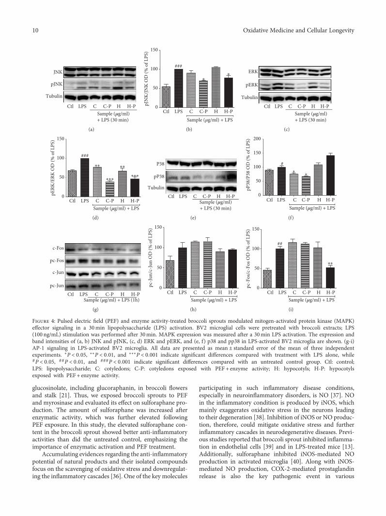
glucosinolate, including glucoraphanin, in broccoli flowersand stalk [21]. Thus, we exposed broccoli sprouts to PEFand myrosinase and evaluated its effect on sulforaphane pro-duction. The amount of sulforaphane was increased afterenzymatic activity, which was further elevated followingPEF exposure. In this study, the elevated sulforaphane con-tent in the broccoli sprout showed better anti-inflammatoryactivities than did the untreated control, emphasizing theimportance of enzymatic activation and PEF treatment.
Accumulating evidences regarding the anti-inflammatorypotential of natural products and their isolated compoundsfocus on the scavenging of oxidative stress and downregulat-ing the inflammatory cascades [36]. One of the key molecules
participating in such inflammatory disease conditions,especially in neuroinflammatory disorders, is NO [37]. NOin the inflammatory condition is produced by iNOS, whichmainly exaggerates oxidative stress in the neurons leadingto their degeneration [38]. Inhibition of iNOS or NO produc-tion, therefore, could mitigate oxidative stress and furtherinflammatory cascades in neurodegenerative diseases. Previ-ous studies reported that broccoli sprout inhibited inflamma-tion in endothelial cells [39] and in LPS-treated mice [13].Additionally, sulforaphane inhibited iNOS-mediated NOproduction in activated microglia [40]. Along with iNOS-mediated NO production, COX-2-mediated prostaglandinrelease is also the key pathogenic event in various
JNK
pJNK
Tubulin
Ctl LPS C C-P H H-PSample (𝜇g/ml)+ LPS (30 min)
(a)
Ctl LPS C C-P H H-P0
50
100
150
###
⁎⁎
Sample (𝜇g/ml) + LPS
pJN
K/JN
K O
D (%
of L
PS)
(b)
ERK
pERK
Tubulin
Ctl LPS C C-P H H-PSample (𝜇g/ml)+ LPS (30 min)
(c)
Ctl LPS C C-P H H-P0
50
100
150
###⁎⁎
⁎⁎⁎
⁎⁎
⁎⁎⁎
Sample (𝜇g/ml) + LPS
pERK
/ERK
OD
(% o
f LPS
)
(d)
P38
pP38
TubulinCtl LPS C C-P H H-P
Sample (𝜇g/ml)+ LPS (30 min)
(e)
Ctl LPS C C-P H H-P0
50
100
150
200
#⁎
⁎
Sample (𝜇g/ml) + LPS
pP38
/P38
OD
(% o
f LPS
)
(f)
c-Fos
pc-Fos
c-Jun
pc-Jun
Ctl LPS C C-P H H-PSample (𝜇g/ml) + LPS (1h)
(g)
Ctl LPS C C-P H H-P0
50
100
150
Sample (𝜇g/ml) + LPS
pc-J
un/c
-Jun
OD
(% o
f LPS
)
(h)
Ctl LPS C C-P H H-P0
50
100
150
##
⁎⁎
Sample (𝜇g/ml) + LPS
pc-F
os/c
-Fos
OD
(% o
f LPS
)
(i)
Figure 4: Pulsed electric field (PEF) and enzyme activity-treated broccoli sprouts modulated mitogen-activated protein kinase (MAPK)effector signaling in a 30min lipopolysaccharide (LPS) activation. BV2 microglial cells were pretreated with broccoli extracts; LPS(100 ng/mL) stimulation was performed after 30min. MAPK expression was measured after a 30min LPS activation. The expression andband intensities of (a, b) JNK and pJNK, (c, d) ERK and pERK, and (e, f) p38 and pp38 in LPS-activated BV2 microglia are shown. (g-i)AP-1 signaling in LPS-activated BV2 microglia. All data are presented as mean ± standard error of the mean of three independentexperiments. ∗P < 0 05, ∗∗P < 0 01, and ∗∗∗P < 0 001 indicate significant differences compared with treatment with LPS alone, while#P < 0 05, ##P < 0 01, and ###P < 0 001 indicate significant differences compared with an untreated control group. Ctl: control;LPS: lipopolysaccharide; C: cotyledons; C-P: cotyledons exposed with PEF + enzyme activity; H: hypocotyls; H-P: hypocotylsexposed with PEF+ enzyme activity.
10 Oxidative Medicine and Cellular Longevity

inflammatory conditions [41]. Elevated expression of iNOSandCOX-2canoccurwith toxinexposure and inneuroinflam-matory diseases. The chronic overexpression of iNOS andCOX-2 is typical in AD, PD, neuropathic pain, etc., in whichoverexpression of these proteins mainly occurs in microglia[42, 43]. Therefore, minimizing the activation of COX in neu-roinflammatoryconditionsisadesirableapproachtoachievingthe neuroprotective effects. In the current study, enhancedexpression of sulforaphane in broccoli sprout dramaticallydownregulatedtheCOX-2-mediatedPGE2production inacti-vated microglia. Although C-P did not significantly alter theexpression of COX-2 in long-term microglial activation, itspotency in inhibiting iNOSwasmuch better in long-term LPSexposure than that in short-termmicroglial activation. Previ-ously, sulforaphane is reported to downregulate COX-2
expression[44].Ourstudynotonlyconfirmstheseexistingevi-dences but also provides noble insights exploring that PEFtreatment further increased the NO inhibiting potential ofbroccoli sprout through elevated sulforaphane content.
MAPK effector signaling is responsible for altering theexpression and secretion of inflammatory mediators as wellas proinflammatory cytokines [45]. MAPK proteins such asERK, JNK, and p38 play essential roles in neuroinflamma-tion [46]. P38, in particular, is responsible for the inductionof apoptosis, differentiation, and regulation of inflammatoryresponses. LPS activates p38 downstream from TLR4 activa-tion, promoting proinflammatory cytokines, such as TNF-α,IFN-γ, IL-1β, IL-12, IL-6, and IL-23, which are responsiblefor neuroinflammation [47, 48]. Similarly, JNK pathwayscan further phosphorylate c-Jun and respond to cytokines,
pJNK
JNK
pERK
ERK
pp38
p38
Tubulin
Ctl LPS C C-P H H-PSample (𝜇g/ml)+ LPS (6h)
Ctl LPS C C-P H H-PSample (𝜇g/ml) + LPS (24h)
(a)
Ctl LPS C C-P H H-P0
50
100
150
###
Sample (𝜇g/ml) + LPS (6h)
pJN
K-JN
K O
D (%
of L
PS)
(b)
Ctl LPS C C-P H H-P0
50
100
150
##
⁎⁎⁎⁎
Sample (𝜇g/ml) + LPS (6h)
pERK
-ERK
OD
(% o
f LPS
)
(c)
Ctl LPS C C-P H H-P0
50
100
150
200
Sample (𝜇g/ml) + LPS (6h)
pP38
-P38
OD
(% o
f LPS
)
(d)
Ctl LPS C C-P H H-P0
50
100
150
Sample (𝜇g/ml) + LPS (24 h)
pJN
K-JN
K O
D (%
of L
PS)
(e)
Ctl LPS C C-P H H-P0
50
100
150
###
Sample (𝜇g/ml) + LPS (24 h)
pERK
-ERK
OD
(% o
f LPS
)
(f)
Ctl LPS C C-P H H-P0
50
100
150
Sample (𝜇g/ml) + LPS (24 h)
pP38
-P38
OD
(% o
f LPS
)
(g)
Figure 5: Pulsed electric field (PEF) and enzyme activity-treated broccoli sprouts modulated mitogen-activated protein kinase (MAPK)effector signaling after 6 and 24 h of lipopolysaccharide (LPS) activation. BV2 microglial cells were pretreated with broccoli extracts; LPS(100 ng/mL) stimulation was performed after 30min. MAPK modulation was observed after 6 and 24 h of LPS activation. (a) MAPKexpression in 6 h and 24 h LPS activation. (b-d) JNK and pJNK, ERK and pERK, and p38 and pp38 band intensity in 6 h LPS-activatedBV2 microglia. (e-g) JNK and pJNK, ERK and pERK, and p38/pp38 band intensity in 24 h LPS-activated BV2 microglia. All data arepresented as mean ± standard error of the mean of three independent experiments. ∗P < 0 05, ∗∗P < 0 01, and ∗∗∗P < 0 001 indicatesignificant differences compared with treatment with LPS alone, while #P < 0 05, ##P < 0 01, and ###P < 0 001 indicate significantdifferences compared with an untreated control group. Ctl: control; LPS: lipopolysaccharide; C: cotyledons; C-P: cotyledons exposed withPEF+ enzyme activity; H: hypocotyls; H-P: hypocotyls exposed with PEF+ enzyme activity.
11Oxidative Medicine and Cellular Longevity
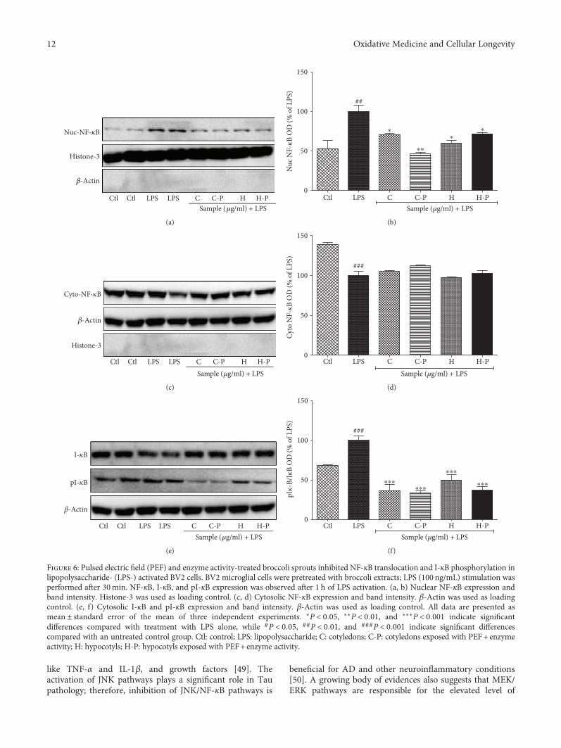
like TNF-α and IL-1β, and growth factors [49]. Theactivation of JNK pathways plays a significant role in Taupathology; therefore, inhibition of JNK/NF-κB pathways is
beneficial for AD and other neuroinflammatory conditions[50]. A growing body of evidences also suggests that MEK/ERK pathways are responsible for the elevated level of
Nuc-NF-𝜅B
Histone-3
𝛽-Actin
Ctl Ctl LPS LPS C C-P H H-PSample (𝜇g/ml) + LPS
(a)
Ctl LPS C C-P H H-P0
50
100
150
##
⁎⁎
⁎ ⁎⁎
Sample (𝜇g/ml) + LPS
Nuc
NF-𝜅
B O
D (%
of L
PS)
(b)
Cyto-NF-𝜅B
𝛽-Actin
Histone-3
Ctl Ctl LPS LPS C C-P H H-PSample (𝜇g/ml) + LPS
(c)
Ctl LPS C C-P H H-P0
50
100
150
###
Sample (𝜇g/ml) + LPS
Cyto
NF-𝜅
B O
D (%
of L
PS)
(d)
I-𝜅B
pI-𝜅B
𝛽-Actin
Ctl Ctl LPS LPS C C-P H H-PSample (𝜇g/ml) + LPS
(e)
Ctl LPS C C-P H H-P0
50
100
150
###
⁎⁎⁎⁎⁎⁎
⁎⁎⁎
⁎⁎⁎
Sample (𝜇g/ml) + LPS
pI𝜅
-B/I𝜅
B O
D (%
of L
PS)
(f)
Figure 6: Pulsed electric field (PEF) and enzyme activity-treated broccoli sprouts inhibited NF-κB translocation and I-κB phosphorylation inlipopolysaccharide- (LPS-) activated BV2 cells. BV2 microglial cells were pretreated with broccoli extracts; LPS (100 ng/mL) stimulation wasperformed after 30min. NF-κB, I-κB, and pI-κB expression was observed after 1 h of LPS activation. (a, b) Nuclear NF-κB expression andband intensity. Histone-3 was used as loading control. (c, d) Cytosolic NF-κB expression and band intensity. β-Actin was used as loadingcontrol. (e, f) Cytosolic I-κB and pI-κB expression and band intensity. β-Actin was used as loading control. All data are presented asmean ± standard error of the mean of three independent experiments. ∗P < 0 05, ∗∗P < 0 01, and ∗∗∗P < 0 001 indicate significantdifferences compared with treatment with LPS alone, while #P < 0 05, ##P < 0 01, and ###P < 0 001 indicate significant differencescompared with an untreated control group. Ctl: control; LPS: lipopolysaccharide; C: cotyledons; C-P: cotyledons exposed with PEF+ enzymeactivity; H: hypocotyls; H-P: hypocotyls exposed with PEF+ enzyme activity.
12 Oxidative Medicine and Cellular Longevity

TNF-α, IL-1β, IL-6, and iNOS following ischemia, whichfurther highlights their neuroinflammatory role [50]. Thus,modulation of p38, JNK, and ERK could be the potentialtherapeutic strategy in various inflammatory and neurode-generative conditions in the brain. In the present study, thebroccoli sprouts exposed to PEF enzyme significantly inhib-ited the phosphorylation of ERK, JNK, and p38, during the30min and 6h LPS activation; p38 activation alone wasmaintained for 24h of LPS activation. The sulforaphane-enriched broccoli did not change in the activation (phos-phorylation) of c-Jun. However, H-P showed the significantinhibition of C-Fos in LPS-activated microglia. This resultsuggests that PEF-exposed broccoli hypocotyls might have
a role in inhibiting pC-Fos-mediated signaling for inflam-matory cascades.
MAPK signaling controls NF-κB-mediated transcriptionof inflammatory mediators such as cytokines and chemo-kines. NF-κB/AP-1 and MAPK pathways play a key role inthe production of cytokines such as TNF-α, IL-6, and IL-8in BV2 microglial cells [51, 52]. In our study, C-P signifi-cantly inhibited the phosphorylation of ERK, JNK, and P38nonspecifically, during the 30min, 6 h, and 24 h LPS-induced microglial activation. This alteration might inducethe significant inhibition of nuclear NF-κB while increasingcytosolic NF-κB. The inhibition of NF-κB-mediated tran-scription of inflammatory proteins was further confirmed
Ctl LPS 10 100 10 100 10 100 10 1000
500
1000
1500
###⁎⁎⁎
⁎⁎⁎
⁎⁎⁎
⁎⁎⁎
⁎⁎⁎
⁎⁎⁎
⁎⁎⁎
⁎⁎⁎
C C-P H H-PSample (𝜇g/ml) + LPS
IL-6
(pg/
ml)
(a)
Ctl LPS 10 100 10 100 10 100 10 1000
500
1000
1500
2000
2500###
⁎⁎⁎
⁎⁎⁎⁎⁎⁎
⁎⁎⁎
⁎
⁎⁎⁎
⁎⁎⁎
⁎⁎⁎
C C-P H H-PSample (𝜇g/ml) + LPS
TNF-𝛼
(pg/
ml)
(b)
CtlLPS 10 100 10 100 10 100 10 1000
200
400
600
800 ###⁎⁎⁎
⁎⁎⁎⁎⁎⁎
⁎⁎⁎
⁎⁎⁎⁎⁎⁎
⁎⁎⁎
⁎⁎⁎
C C-P H H-PSample (𝜇g/ml) + LPS
PGE2
(pg/
ml)
(c)
Ctl LPS 10 100 10 100 10 100 10 1000
200
400
600
800###
⁎⁎⁎ ⁎⁎⁎
⁎⁎⁎⁎
⁎⁎⁎
C C-P H H-PSample (𝜇g/ml) + LPS
IL-1𝛽
(pg/
ml)
(d)
NC Ctl LPS C C-P0
50
100
150
200
250
Sample (𝜇g/ml)+ LPS
###⁎⁎⁎
⁎⁎⁎Ce
ll vi
abili
ty (%
of L
PS)
(e)
Bax
Bcl2
Cleaved-caspase-3
Tubulin
NC Ctl LPS C C-PSample (𝜇g/ml)
(f)
NC Ctl LPS C C-P0
50
100
150
Sample (𝜇g/ml) + LPS
#⁎ ⁎⁎
Bax/
Bcl2
OD
(% o
f LPS
)
(g)
NC Ctl LPS C C-P0
50
100
150
Sample (𝜇g/ml) + LPS
##
⁎⁎ ⁎⁎
Clea
ved
casp
ase-
3 O
D(%
of L
PS)
(h)
Figure 7: Pulsed electric field (PEF) and enzyme activity-treated broccoli sprouts inhibited proinflammatory cytokine production inLPS-activated microglia and also reduced the neuronal death caused by activated microglia. BV2 microglial cells were pretreated withbroccoli extracts; LPS (100 ng/mL) stimulation was performed after 30min. Proinflammatory cytokines were measured in the conditionedmedium of the treated cells using enzyme-linked immunosorbent assays after 24 h of LPS activation. (a) Interleukin- (IL-) 6 production.(b) Tumor necrosis factor- (TNF-) α secretion. (c) Prostaglandin E2 (PGE2) secretion. (d) IL-1β secretion. Similarly treated BV2 cells inthe conditioned medium were transferred to seeded N2a cells in the 6-well plate. Cell viability and proteins expression in the N2a cellswere evaluated after 24 h of CM treatment. (f) N2a cell viability after activated microglial CM treatment. (f-h) Apoptosis-related proteinexpression in BV2 CM-treated N2a cells and their quantification. All data are presented as mean ± standard error of the mean of threeindependent experiments. ∗P < 0 05, ∗∗P < 0 01, and ∗∗∗P < 0 001 indicate significant differences compared with treatment with LPSalone, while #P < 0 05, ##P < 0 01, and ###P < 0 001 indicate significant differences compared with the untreated control group. NC:normal control; Ctl: control; LPS: lipopolysaccharide; C: cotyledons; C-P: cotyledons exposed with PEF+ enzyme activity; H: hypocotyls;H-P: hypocotyls exposed with PEF + enzyme activity.
13Oxidative Medicine and Cellular Longevity

by the increased and decreased expression of I-κB and pI-κB,respectively. The inhibition of MAPK-NF-κB was furthercharacterized by the significant inhibition of TNF-α andIL-6 production in LPS-activated BV2 microglial cells. C-Pshowed the highest potency for all these events, and thismight be due to the higher amount of sulforaphane.
Previously, broccoli sprout extract and sulforaphane arereported as activators of well-known antioxidant moleculesNrf2/HO-1 [53]. Treatment of C and C-P activated the
expression of Nrf2 and HO-1 as did by the sulforaphanetreatment in normal and LPS-activated microglial cells. Thiseffect of broccoli extract and sulforaphane can further inhibitthe inflammatory cascades either through inhibiting TLR4-mediated inflammatory cascades or by their antioxidantpotentials. Therefore, it is thought to be a potential mecha-nism that indicates the antineuroinflammatory effect of sul-foraphane. Nrf2 inhibits the neuroinflammation as well asneurodegeneration against various models of brain disorders
Nrf2
Histone-3
Ctl C C-PSample (100 𝜇g/ml)
HO-1
GAPDH
(a)
0
50
100
150 ⁎⁎⁎
⁎⁎
Sample (100𝜇g/ml)
Nrf2
OD
(% o
f Ctl)
Ctl C C-P
(b)
Ctl C C-P0
50
100
150
200 ⁎⁎⁎
⁎⁎
Sample (100𝜇g/ml)
HO
-1 O
D (%
of C
tl)
(c)
C C-P
Sample (100 𝜇g/ml) + LPS
LPSCtl
Nrf2
Histone-3
HO-1
GAPDH
(d)
Ctl LPS C C-P0
50100150200
Sample (𝜇g/ml) + LPS
#⁎⁎⁎
⁎⁎⁎
Nrf2
OD
(% o
f LPS
)
(e)
Ctl LPS C C-P0
50100150200
Sample (𝜇g/ml) + LPS
#⁎⁎⁎
⁎⁎⁎
HO
-1 O
D (%
of L
PS)
(f)
Nrf2
GAPDH
Ctl _ Donepazil SB SB-Enz SB-PEF SB-Enz-PEFScopolamine (1.2 mg/kg)
(g)
Ctl − Donepazil SB SB-Enz SB-PEF SB-Enz-PEF0
50
100
150
200
250
###
⁎⁎⁎
⁎⁎⁎
Scopolamine (1.2 mg/kg)
Nrf2
OD
(% o
f sco
pola
min
e onl
y)
(h)
Figure 8: Broccoli sample treatment increased the Nrf2-HO-1 expression showing antioxidant effect in microglial cells (normal andLPS-activated conditions) as well as against a scopolamine-induced amnesia model in mice. BV2 microglial cells were treated with thebroccoli sample itself. (a-c) Nrf2-HO-1 expression in normal microglia after broccoli cotyledon treatment and their quantifications. (d-f)Nrf2-HO-1 expression in LPS-activated microglia after broccoli cotyledon treatment and their quantifications. Mice were continuouslyexposed with scopolamine and broccoli samples for two weeks’ period. Animal were sacrifices, and the brain samples were homogenizedand tissue lysates were separated using the western blot technique. (g, h) Nrf2 expression and its quantification in the mouse whole brainsample. Histone-3 and GAPDH were used as a loading control for respective proteins. All data are presented as mean ± standard error ofthe mean of three independent experiments. ∗P < 0 05, ∗∗P < 0 01, and ∗∗∗P < 0 001 indicate significant differences compared with theuntreated control group in (c, d) and only scopolamine-treated group in (f) while #P < 0 05 and ###P < 0 001 indicate significantdifferences compared with the untreated control group. C: cotyledons; C-P: cotyledons exposed with PEF and enzyme activity; SB: sproutbroccoli; SB-Enz: sprout broccoli with induced enzyme activity; SB-PEF: sprout broccoli with PEF treatment; SB-Enz-PEF: sprout broccoliwith activated enzyme activity and PEF treatment.
14 Oxidative Medicine and Cellular Longevity

Ctl − Donepazil SB SB-Enz SB-PEFSB-Enz-PEF0
20
40
60
80
Scopolamine (1.2 mg/kg)
Tota
l ent
ry (n
o.)
(a)
Ctl − Donepazil SB SB-Enz SB-PEF SB-Enz-PEF40
50
60
70
80
Scopolamine (1.2 mg/kg)
###
⁎⁎⁎⁎⁎⁎
⁎⁎⁎⁎
⁎⁎
Reco
gniti
on (%
)
(b)
Ctl − Donepazil SB SB-Enz SB-PEF SB-Enz-PEF0
20
40
60
80
Scopolamine (1.2 mg/kg)
##
⁎⁎⁎⁎⁎⁎
Spon
tane
ous a
ltern
atio
n (%
)
(c)
Ctl − Donepazil SB SB-Enz SB-PEF SB-Enz-PEF0
50
100
150
Scopolamine (1.2 mg/kg)
⁎⁎A
ChE
activ
ity (%
of c
ontr
ol)
(d)
908070605040
Late
ncy
Tim
eIn
acut
e mod
el (s
ec)
302010
0Ctl −
###
Donepazil SB SB-Enz SB-PEF SB-Enz-PEFScopolamine (1.2 mg/kg)
⁎ ⁎
Test trialTraining trial
(e)
Late
ncy
Tim
eIn
chro
mic
mod
el (s
ec)
Ctl0
50100150200250
−
##
Donepazil SB SB-Enz SB-PEFSB-Enz-PEFScopolamine (1.2 mg/kg)
⁎⁎
⁎⁎⁎⁎
⁎⁎
⁎
Test trialTraining trial
(f)
Bax
Bcl2
Cleaved-caspase-3
Tubulin
Ctl − Done SB SB-Enz SB-PEF SB-Enz-PEF
Scopolamine (1.2 mg/kg)
(g)
Figure 9: Continued.
15Oxidative Medicine and Cellular Longevity

0
50
100
150
###
⁎⁎⁎
Bax/
Bcl2
OD
(% o
f Sco
pola
min
e)
(h)
0
50
100
150
###
⁎⁎⁎
⁎⁎⁎⁎
Clea
ved
casp
ase-
3 O
D(%
of s
copo
lam
ine)
(i)
Figure 9: Broccoli sprout extract repaired the memory impairment by inhibiting neuronal apoptosis against scopolamine induced toxicityin vivo. Experimental animals were exposed with 1.2mg/kg scopolamine and 2mg/kg of donepezil for the donepezil group and 200mg/kgbroccoli samples. The treated animal’s spatial memory was evaluated by the (a, b) Y-maze test, (c) novel object recognition test (NORT),(d) acetylcholine esterase activity assay in animal brain, (e, f) passive avoidance test in acute condition (one time per-oral treatment andtoxicity induction) and chronic condition (two weeks’ per-oral treatment and toxicity induction). After sacrifice, mouse whole brains werecollected and homogenized with tissue lysis buffer and western blot analysis was performed. (g-i) Bax, Bcl2, and cleaved caspase-3expression and their quantification. All data are presented as mean ± standard error of the mean of three independent experiments. ∗P <0 05, ∗∗P < 0 01, and ∗∗∗P < 0 001 indicate significant differences compared with treatment with scopolamine alone while ##P < 0 01 and###P < 0 001 indicate the significant differences compared with an untreated control group. Ctl: control; SB: sprout broccoli; SB-Enz:sprout broccoli with induced enzyme activity; SB-PEF: sprout broccoli with PEF treatment; SB-Enz-PEF: sprout broccoli with activatedenzyme activity and PEF treatment.
TLR4
LPS
TRAF6
MAPKIKK𝛼
ERK1/2 JNK P38
NF-𝜅BAP1
IKB𝛼
NF-𝜅B
NF-𝜅Btranscription
TNF-𝛼, IL-6, IL-1𝛽, iNOS,COX-2, PGE2, NO
TNF-𝛼, IL-6, IL-1𝛽, NO, PGE2
Neuronal death/neurodegeneration
Sulforaphane-enrichedbroccoli
Sulforaphane-enrichedbroccoli
Nrf2
Nrf2
HO-1
Scopolamine
Figure 10: Schematic diagram for the antineuroinflammatory and antiamnesic effects of broccoli sprout extract treated with PEF.
16 Oxidative Medicine and Cellular Longevity

in both in vitro and in vivo experimental models [54, 55].Previous reports suggested that sulforaphane protected neu-rons against rotenone-induced toxicity in an in vivo modelwhich is mediated through the activation of the Nrf2 path-way [56]. In this study, SB-Enz-PEF significantly improvedthe scopolamine-induced spontaneous alteration as deter-mined by the Y-maze test, novel object recognition test, andAchE activity inhibition test indicating that sulforaphanecould improve memory impairment in different neurologicaldisorders. SB-Enz-PEF also increased latency time in PATassay. Moreover, through in vitro analysis, we found thatthe SB-Enz-PEF-treated conditioned medium from LPS-stimulated BV2 cells not only increased the neuronal cell sur-vival but also attenuated the apoptotic proteins in neurons.Our data demonstrated that the protective effects of SB-ENZ-PEF are mediated through the Nrf2 signaling pathway;however, the overall neuroprotection of broccoli and sulfo-raphane might be mediated through the antiapoptoticeffects, in particular downregulating caspase-3 activation.Sulforaphane-enriched broccoli extract, therefore, couldmediate the neuroprotection in various neurodegenerativemodels, and the plausible pathway for the neuroprotectioncould be due to the combination of Nrf2 activation and itsantiapoptotic effects.
Taken together, in this study, we provided the newinsight of increased-sulforaphane-mediated protective effectsin neuroinflammatory models. Being multifunctional, identi-fication of a particular therapeutic target for phytochem-icals/nutraceuticals to show desired biological activity is akey issue for the new drug development [57, 58]. However,the technique that increased the particular bioactive com-pounds, as PEF-induced sulforaphane in this study, couldenhance the therapeutic benefit of phytochemicals. Addition-ally, a time point study of iNOS/COX-2 and MAPK signalingin this study could be the magnificent cues for the furtherin vivo experiment including both acute (such as ischemia,traumatic brain injury) and chronic (such as AD, PD)inflammatory conditions. Our findings demonstrated thatsulforaphane-enriched broccoli sprout showed antineuroin-flammatory and neuroprotective effects in vitro and showedthe protective effects in mice against scopolamine-inducedamnesia in vivo through Nrf2 activation. Finally, in moststudies, the biological activity of sulforaphane, specificallyof (−)L-isothiocyanato-4R-(methyl-sulfinyl)-butane, is dem-onstrated using the racemic mixture, despite the fact thathumans are exposed only to the R-enantiomer through theirdiet. Therefore, it would be tempting to determine the role ofR- and S-sulforaphane as future independent studies. Theoverall findings of our current study are summarized inFigure 10.
Data Availability
All the data have been included in the manuscript and it canalso be provided by the corresponding author on request.
Conflicts of Interest
No competing financial interests exist.
Authors’ Contributions
Hyuk Joon Choi, Sun Yeou Kim, and Lalita Subedi hypothe-sized and designed the experiment; Lalita Subedi, KyoHeeCho, and Young Un Park performed the experiment; andLalita Subedi wrote the manuscript. Hyuk Joon Choi andSun Yeou Kim revised and finalized the manuscript.
Acknowledgments
This work was supported by the National Research Founda-tion of Korea (NRF) grant funded by the Korea government(MSIT) (NRF-2014M3A9B6069338) and also by a grantfrom BK bio (R0005491), Republic of Korea.
References
[1] W. W. Chen, X. Zhang, and W. J. Huang, “Role of neuro-inflammation in neurodegenerative diseases (review),”Molecular Medicine Reports, vol. 13, no. 4, pp. 3391–3396,2016.
[2] H. Hong, B. S. Kim, and H. I. Im, “Pathophysiological role ofneuroinflammation in neurodegenerative diseases and psychi-atric disorders,” International Neurourology Journal, vol. 20,Supplement 1, pp. S2–S7, 2016.
[3] D. L. Krause and N. Muller, “Neuroinflammation, microgliaand implications for anti-inflammatory treatment in Alzhei-mer’s disease,” International Journal of Alzheimer's Disease,vol. 2010, article 732806, 9 pages, 2010.
[4] M. W. Salter and B. Stevens, “Microglia emerge as centralplayers in brain disease,” Nature Medicine, vol. 23, no. 9,pp. 1018–1027, 2017.
[5] A. D. Kraft and G. J. Harry, “Features of microglia and neuro-inflammation relevant to environmental exposure and neuro-toxicity,” International Journal of Environmental Research andPublic Health, vol. 8, no. 7, pp. 2980–3018, 2011.
[6] L. Alvarez-Erviti, Y. Couch, J. Richardson, J. M. Cooper, andM. J. A. Wood, “Alpha-synuclein release by neurons activatesthe inflammatory response in a microglial cell line,” Neurosci-ence Research, vol. 69, no. 4, pp. 337–342, 2011.
[7] J. D. Cherry, J. A. Olschowka, and M. O’Banion, “Neuroin-flammation and M2 microglia: the good, the bad, and theinflamed,” Journal of Neuroinflammation, vol. 11, no. 1,p. 98, 2014.
[8] A. I. Ramirez, R. de Hoz, E. Salobrar-Garcia et al., “The role ofmicroglia in retinal neurodegeneration: Alzheimer’s disease,Parkinson, and glaucoma,” Frontiers in Aging Neuroscience,vol. 9, p. 214, 2017.
[9] D. B. McKim, A. Niraula, A. J. Tarr, E. S. Wohleb, J. F.Sheridan, and J. P. Godbout, “Neuroinflammatory dynamicsunderlie memory impairments after repeated social defeat,”The Journal of Neuroscience, vol. 36, no. 9, pp. 2590–2604,2016.
[10] J. Seo, B. Kim, J. Oh, and J.-S. Kim, “Soybean-derived phyto-alexins improve cognitive function through activation of Nrf2/HO-1 signaling pathway,” International Journal of Molecu-lar Sciences, vol. 19, no. 1, 2018.
[11] S. J. Enna andM.Williams, “Challenges in the search for drugsto treat central nervous system disorders,” The Journal ofPharmacology and Experimental Therapeutics, vol. 329, no. 2,pp. 404–411, 2009.
17Oxidative Medicine and Cellular Longevity

[12] T. M. Barchet and M. M. Amiji, “Challenges and opportunitiesin CNS delivery of therapeutics for neurodegenerative dis-eases,” Expert Opinion on Drug Delivery, vol. 6, no. 3,pp. 211–225, 2009.
[13] B. E. Townsend, Y. J. Chen, E. H. Jeffery, and R. W. Johnson,“Dietary broccoli mildly improves neuroinflammation in agedmice but does not reduce lipopolysaccharide-induced sicknessbehavior,” Nutrition Research, vol. 34, no. 11, pp. 990–999,2014.
[14] V. Hernandez-Rabaza, A. Cabrera-Pastor, L. Taoro-Gonzalezet al., “Hyperammonemia induces glial activation, neuroin-flammation and alters neurotransmitter receptors inhippocampus, impairing spatial learning: reversal by sulfo-raphane,” Journal of Neuroinflammation, vol. 13, no. 1, p. 41,2016.
[15] L. O. Brandenburg, M. Kipp, R. Lucius, T. Pufe, and C. J.Wruck, “Sulforaphane suppresses LPS-induced inflammationin primary rat microglia,” Inflammation Research, vol. 59,no. 6, pp. 443–450, 2010.
[16] J. W. Fahey, Y. Zhang, and P. Talalay, “Broccoli sprouts: anexceptionally rich source of inducers of enzymes that protectagainst chemical carcinogens,” Proceedings of the NationalAcademy of Sciences of the United States of America, vol. 94,no. 19, pp. 10367–10372, 1997.
[17] N. V. Matusheski, R. Swarup, J. A. Juvik, R. Mithen,M. Bennett, and E. H. Jeffery, “Epithiospecifier protein frombroccoli (Brassica oleracea L. ssp. italica) inhibits formationof the anticancer agent sulforaphane,” Journal of Agriculturaland Food Chemistry, vol. 54, no. 6, pp. 2069–2076, 2006.
[18] T. A. Shapiro, J. W. Fahey, A. T. Dinkova-Kostova et al.,“Safety, tolerance, and metabolism of broccoli sprout glucosin-olates and isothiocyanates: a clinical phase I study,” Nutritionand Cancer, vol. 55, no. 1, pp. 53–62, 2006.
[19] Y. Li, T. Zhang, X. Li, P. Zou, S. J. Schwartz, and D. Sun,“Kinetics of sulforaphane in mice after consumption ofsulforaphane-enriched broccoli sprout preparation,” Molecu-lar Nutrition & Food Research, vol. 57, no. 12, pp. 2128–2136, 2013.
[20] J. D. Clarke, A. Hsu, D. E. Williams et al., “Metabolism and tis-sue distribution of sulforaphane in Nrf 2 knockout andwild-type mice,” Pharmaceutical Research, vol. 28, no. 12,pp. 3171–3179, 2011.
[21] I. Aguilo-Aguayo, M. Suarez, L. Plaza et al., “Optimization ofpulsed electric field pre-treatments to enhance health-promoting glucosinolates in broccoli flowers and stalk,” Jour-nal of the Science of Food and Agriculture, vol. 95, no. 9,pp. 1868–1875, 2015.
[22] T. Gachovska, D. Cassada, J. Subbiah, M. Hanna,H. Thippareddi, and D. Snow, “Enhanced anthocyanin extrac-tion from red cabbage using pulsed electric field processing,”Journal of Food Science, vol. 75, no. 6, pp. E323–E329, 2010.
[23] D. J. Kliebenstein, J. Kroymann, P. Brown et al., “Geneticcontrol of natural variation in Arabidopsis glucosinolateaccumulation,” Plant Physiology, vol. 126, no. 2, pp. 811–825, 2001.
[24] C. S. Kim, L. Subedi, S. Y. Kim, S. U. Choi, K. H. Kim, andK. R. Lee, “Diterpenes from the trunk of Abies holophyllaand their potential neuroprotective and anti-inflammatoryactivities,” Journal of Natural Products, vol. 79, no. 2,pp. 387–394, 2016.
[25] S. Y. Lim, L. Subedi, D. Shin, C. S. Kim, K. R. Lee, and S. Y.Kim, “A new neolignan derivative, balanophonin isolated from
Firmiana simplex delays the progress of neuronal cell death byinhibiting microglial activation,” Biomolecules & Therapeutics,vol. 25, no. 5, pp. 519–527, 2017.
[26] L. Subedi, B. P. Gaire, M. H. Do, T. H. Lee, and S. Y. Kim,“Anti-neuroinflammatory and neuroprotective effects of theLindera neesiana fruit in vitro,” Phytomedicine, vol. 23, no. 8,pp. 872–881, 2016.
[27] L. Subedi, E. Ji, D. Shin, J. Jin, J. Yeo, and S. Kim, “Equol, a die-tary daidzein gut metabolite attenuates microglial activationand potentiates neuroprotection in vitro,” Nutrients, vol. 9,no. 3, 2017.
[28] A. T. Mugwagwa, L. L. Gadaga, W. Pote, and D. Tagwireyi,“Antiamnesic effects of a hydroethanolic extract of Crinummacowaniion scopolamine-induced memory impairment inmice,” Journal of Neurodegenerative Diseases, vol. 2015, ArticleID 242505, 9 pages, 2015.
[29] S. A. A. El-Marasy, R. M. Abd-Elsalam, and O. A. Ahmed--Farid, “Ameliorative effect of silymarin on scopolamine-induced dementia in rats,” Open Access Macedonian Journalof Medical Sciences, vol. 6, no. 7, pp. 1215–1224, 2018.
[30] H. E. Lee, Y. W. Lee, S. J. Park et al., “4-Hydroxybenzyl methylether improves learning andmemory in mice via the activationof dopamine D1 receptor signaling,” Neurobiology of Learningand Memory, vol. 121, pp. 30–38, 2015.
[31] E. Kim, H. J. Ko, S. J. Jeon et al., “The memory-enhancingeffect of erucic acid on scopolamine-induced cognitive impair-ment in mice,” Pharmacology, Biochemistry, and Behavior,vol. 142, pp. 85–90, 2016.
[32] G. L. Ellman, K. D. Courtney, V. Andres Jr, and R. M. Feather-stone, “A new and rapid colorimetric determination of acetyl-cholinesterase activity,” Biochemical Pharmacology, vol. 7,no. 2, pp. 88–95, 1961.
[33] L. Subedi, T. H. Lee, H. M. Wahedi, S. H. Baek, and S. Y. Kim,“Resveratrol-enriched rice attenuates UVB-ROS-induced skinaging via downregulation of inflammatory cascades,” Oxida-tive Medicine and Cellular Longevity, vol. 2017, Article ID8379539, 15 pages, 2017.
[34] D. Angelino, E. B. Dosz, J. Sun et al., “Myrosinase-dependentand -independent formation and control of isothiocyanateproducts of glucosinolate hydrolysis,” Frontiers in Plant Sci-ence, vol. 6, 2015.
[35] G. C.Wang, M. Farnham, and E. H. Jeffery, “Impact of thermalprocessing on sulforaphane yield from broccoli (Brassica oler-acea L. ssp. italica),” Journal of Agricultural and Food Chemis-try, vol. 60, no. 27, pp. 6743–6748, 2012.
[36] P. Arulselvan, M. T. Fard, W. S. Tan et al., “Role of antioxi-dants and natural products in inflammation,” Oxidative Med-icine and Cellular Longevity, vol. 2016, Article ID 5276130, 15pages, 2016.
[37] G. Gelders, V. Baekelandt, and A. Van der Perren, “Linkingneuroinflammation and neurodegeneration in Parkinson’sdisease,” Journal of Immunology Research, vol. 2018, ArticleID 4784268, 12 pages, 2018.
[38] J. E. Yuste, E. Tarragon, C. M. Campuzano, andF. Ros-Bernal, “Implications of glial nitric oxide in neurode-generative diseases,” Frontiers in Cellular Neuroscience,vol. 9, 2015.
[39] A. Sotokawauchi, Y. Ishibashi, T. Matsui, and S. I. Yamagishi,“Aqueous extract of glucoraphanin-rich broccoli sproutsinhibits formation of advanced glycation end products andattenuates inflammatory reactions in endothelial cells,”
18 Oxidative Medicine and Cellular Longevity

Evidence-based Complementary and Alternative Medicine,vol. 2018, Article ID 9823141, 6 pages, 2018.
[40] E. Eren, K. U. Tufekci, K. B. Isci, B. Tastan, K. Genc, andS. Genc, “Sulforaphane inhibits lipopolysaccharide-inducedinflammation, cytotoxicity, oxidative stress, and miR-155expression and switches to Mox phenotype through activatingextracellular signal-regulated kinase 1/2–nuclear factor ery-throid 2-related factor 2/antioxidant response element path-way in murine microglial cells,” Frontiers in Immunology,vol. 9, 2018.
[41] L. Minghetti, “Role of COX-2 in inflammatory and degenera-tive brain diseases,” Sub-Cellular Biochemistry, vol. 42,pp. 127–141, 2007.
[42] A. R. Patel, R. Ritzel, L. D. McCullough, and F. Liu, “Microgliaand ischemic stroke: a double-edged sword,” InternationalJournal of Physiology, Pathophysiology and Pharmacology,vol. 5, no. 2, pp. 73–90, 2013.
[43] E. Badoer, “Microglia: activation in acute and chronic inflam-matory states and in response to cardiovascular dysfunction,”The International Journal of Biochemistry & Cell Biology,vol. 42, no. 10, pp. 1580–1585, 2010.
[44] W. J. Choi, S. K. Kim, H. K. Park, U. D. Sohn, and W. Kim,“Anti-inflammatory and anti-superbacterial properties ofsulforaphane from Shepherd’s purse,” The Korean Journalof Physiology & Pharmacology, vol. 18, no. 1, pp. 33–39,2014.
[45] B. Kaminska, A. Gozdz, M. Zawadzka, A. Ellert-Miklaszewska,and M. Lipko, “MAPK signal transduction underlying braininflammation and gliosis as therapeutic target,” The Anatomi-cal Record: Advances in Integrative Anatomy and EvolutionaryBiology, vol. 292, no. 12, pp. 1902–1913, 2009.
[46] G. Ramesh, “Novel therapeutic targets in neuroinflammationand neuropathic pain,” Inflammation and Cell Signaling,vol. 1, no. 3, 2014.
[47] D. N. Krementsov, T. M. Thornton, C. Teuscher, andM. Rincon, “The emerging role of p 38 mitogen-activatedprotein kinase in multiple sclerosis and its models,” Molec-ular and Cellular Biology, vol. 33, no. 19, pp. 3728–3734,2013.
[48] H. K. Koul, M. Pal, and S. Koul, “Role of p 38 MAP kinase sig-nal transduction in solid tumors,” Genes & Cancer, vol. 4,no. 9-10, pp. 342–359, 2013.
[49] P. K. Roy, F. Rashid, J. Bragg, J. A. Ibdah, and Division of Gas-troenterology and Hepatology, University of Missouri Schoolof Medicine, Columbia, Missouri, United States, “Role of theJNK signal transduction pathway in inflammatory bowel dis-ease,” World Journal of Gastroenterology, vol. 14, no. 2,pp. 200–202, 2008.
[50] F. Zhang and L. Jiang, “Neuroinflammation in Alzheimer’sdisease,” Neuropsychiatric Disease and Treatment, vol. 11,pp. 243–256, 2015.
[51] L. Neff, M. Zeisel, J. Sibilia, M. Scholler-Guinard, J. P. Klein,and D. Wachsmann, “NF-kappaB and the MAP kinases/AP-1pathways are both involved in interleukin-6 and interleukin-8expression in fibroblast-like synoviocytes stimulated by pro-tein I/II, a modulin from oral streptococci,” Cellular Microbiol-ogy, vol. 3, no. 10, pp. 703–712, 2001.
[52] J. Zhu, L. Jiang, Y. Liu et al., “MAPK and NF-κB pathways areinvolved in bisphenol A-induced TNF-α and IL-6 productionin BV2 microglial cells,” Inflammation, vol. 38, no. 2,pp. 637–648, 2015.
[53] Z. Xu, S. Wang, H. Ji et al., “Broccoli sprout extract preventsdiabetic cardiomyopathy via Nrf 2 activation in db/db T2DMmice,” Scientific Reports, vol. 6, no. 1, article 30252, 2016.
[54] S. A. Onasanwo, R. Velagapudi, A. El-Bakoush, and O. A. Ola-jide, “Inhibition of neuroinflammation in BV2 microglia bythe biflavonoid kolaviron is dependent on the Nrf2/ARE anti-oxidant protective mechanism,” Molecular and Cellular Bio-chemistry, vol. 414, no. 1-2, pp. 23–36, 2016.
[55] P. Deshmukh, S. Unni, G. Krishnappa, and B. Padmanabhan,“The Keap1-Nrf2 pathway: promising therapeutic target tocounteract ROS-mediated damage in cancers and neurodegen-erative diseases,” Biophysical Reviews, vol. 9, no. 1, pp. 41–56,2017.
[56] Q. Zhou, B. Chen, X. Wang et al., “Sulforaphane protectsagainst rotenone-induced neurotoxicity in vivo: involvementof the mTOR, Nrf2, and autophagy pathways,” ScientificReports, vol. 6, no. 1, article 32206, 2016.
[57] L.-X. Liao, X.-M. Song, L.-C. Wang et al., “Highly selectiveinhibition of IMPDH2 provides the basis of antineuroinflam-mation therapy,” Proceedings of the National Academy of Sci-ences of the United States of America, vol. 114, no. 29,pp. E5986–E5994, 2017.
[58] J. A. Beutler, “Natural products as a foundation for drug dis-covery,” Current Protocols in Pharmacology, vol. 46, p. 9, 2009.
19Oxidative Medicine and Cellular Longevity
Research ArticleInhibition of Oxidative Neurotoxicity and Scopolamine-Induced Memory Impairment by γ-Mangostin: In Vitro and InVivo Evidence
Youngmun Lee, Sunyoung Kim, Yeonsoo Oh, Young-Mi Kim, Young-Won Chin ,and Jungsook Cho
College of Pharmacy, Dongguk University-Seoul, Goyang, Gyeonggi-do 10326, Republic of Korea
Correspondence should be addressed to Jungsook Cho; [email protected]
Youngmun Lee and Sunyoung Kim contributed equally to this work.
Received 4 October 2018; Revised 14 December 2018; Accepted 9 January 2019; Published 24 March 2019
Guest Editor: Luciana Scotti
Copyright © 2019 Youngmun Lee et al. This is an open access article distributed under the Creative Commons Attribution License,which permits unrestricted use, distribution, and reproduction in any medium, provided the original work is properly cited.
Among a series of xanthones identified from mangosteen, the fruit of Garcinia mangostana L. (Guttifereae), α- and γ-mangostinsare known to be major constituents exhibiting diverse biological activities. However, the effects of γ-mangostin on oxidativeneurotoxicity and impaired memory are yet to be elucidated. In the present study, the protective effect of γ-mangostin onoxidative stress-induced neuronal cell death and its underlying action mechanism(s) were investigated and compared to that ofα-mangostin using primary cultured rat cortical cells. In addition, the effect of orally administered γ-mangostin onscopolamine-induced memory impairment was evaluated in mice. We found that γ-mangostin exhibited prominent protectionagainst H2O2- or xanthine/xanthine oxidase-induced oxidative neuronal death and inhibited reactive oxygen species (ROS)generation triggered by these oxidative insults. In contrast, α-mangostin had no effects on the oxidative neuronal damage orassociated ROS production. We also found that γ-mangostin, not α-mangostin, significantly inhibited H2O2-induced DNAfragmentation and activation of caspases 3 and 9, demonstrating its antiapoptotic action. In addition, only γ-mangostin wasfound to effectively inhibit lipid peroxidation and DPPH radical formation, while both mangostins inhibited β-secretase activity.Furthermore, we observed that the oral administration of γ-mangostin at dosages of 10 and 30mg/kg markedly improvedscopolamine-induced memory impairment in mice. Collectively, these results provide both in vitro and in vivo evidences for theneuroprotective and memory enhancing effects of γ-mangostin. Multiple mechanisms underlying this neuroprotective actionwere suggested in this study. Based on our findings, γ-mangostin could serve as a potentially preferable candidate overα-mangostin in combatting oxidative stress-associated neurodegenerative diseases including Alzheimer’s disease.
1. Introduction
The generation of reactive oxygen species (ROS) includingsuperoxide anion (⋅O2
-), hydroxyl radical (⋅OH), and hydrogenperoxide is recognised as a key factor in oxidative stress to thecells [1]. The accumulation of ROS in neuronal cells causeslipid peroxidation as well as damage to the structure and func-tion of proteins and DNAmolecules, ultimately leading to celldeath [2]. The brain is known to be particularly susceptible tooxidative stress, due to its relative deficiency of endogenousantioxidant defence mechanisms, enriched levels of transition
metals and unsaturated lipids, and high utilisation of oxygen[3]. Consequently, oxidative stress-induced neuronal damagehas been recognised as an important mechanism involved inmany neurodegenerative diseases, including Alzheimer’s dis-ease (AD) and Parkinson’s disease [4, 5].
AD is characterised by the progressive impairment of cog-nition which is strongly correlated with neuronal degenerationand death. One of the hallmarks of AD is the appearance ofsenile plaques generated through the extracellular depositionof β-amyloid (Aβ) peptide, which is derived from amyloid pre-cursor protein (APP) upon enzymatic cleavage by β- and
HindawiOxidative Medicine and Cellular LongevityVolume 2019, Article ID 3640753, 14 pageshttps://doi.org/10.1155/2019/3640753

γ-secretases [6, 7]. Aβ peptide can induce oxidative damagethrough the production of ROS, potentially triggering neuro-toxic events. Furthermore, it has been suggested that an aug-mentation in the cellular level of ROS and free radicalsproduces more Aβ peptide, which in turn exerts further oxida-tive stress and toxic insults on neurons [8]. With the accumu-lative evidence for oxidative stress as an important factor inAD, antioxidants that reduce ROS and prevent oxidativestress-induced neuronal death have been intriguing potentialcandidates to prevent or treat AD [4, 5]. However, the resultsfrom many clinical studies have been rather disappointing sofar. Nonetheless, various approaches considering heteroge-neous and multifactorial characteristics of AD are attemptedto explore favorable efficacy of antioxidant therapy in AD [9].
Mangosteen, Garcinia mangostana L. (G. mangostana,Guttifereae), is a tree cultivated in Southeast Asia includingIndonesia, Philippines, and India. Its fruit is edible and alsoknown to have medicinal benefits. The pericarp of the fruithas been traditionally used in these countries to treat infec-tion, wounds, inflammation, and diarrhea [10]. In addition,mangosteen products in the form of juice or tablets accountfor some of the best-selling dietary supplements in the U.S.market [11]. The major bioactive secondary metabolites ofmangosteen have been found to be xanthone derivatives,among which α-mangostin is the most studied xanthone[12, 13]. Using cell-free in vitro assays, α-mangostin wasreported to scavenge singlet oxygen and superoxide anion,while it was shown to be unable to scavenge hydroxyl radicalsand hydrogen peroxide [14]. Additionally, using primarycultures of cerebellar granule neurons, α-mangostin was foundto exhibit ROS scavenging and neuroprotective effects againstthe mitochondrial toxin 3-nitropropionic acid or iodoacetate,an inhibitor of the glycolytic enzyme glyceraldehyde-3-phosphate dehydrogenase inducing metabolic inhibition inneurons [14, 15]. Moreover, α-mangostin has been demon-strated to attenuate β-amyloid oligomers-induced neurotox-icity by inhibiting amyloid aggregation and also to decreaseAβ production via modulation of the amyloidogenic pathway[16, 17]. Together, these findings suggest that α-mangostinmay serve as a multifunctional therapeutic intervention tocombat the multiple pathological processes of AD [18, 19].
Unlike α-mangostin, however, there have only been lim-ited findings supporting the neuroprotective effects of γ-man-gostin. It has been reported that γ-mangostin, along with otherxanthones from G. mangostana, inhibits glutamate-inducedcell death in the HT22 hippocampal neuronal cell line andself-induced Aβ42 aggregation in vitro [18]. These findings alsosuggest that, in addition to α-mangostin, γ-mangostin couldbe a promising compound for AD therapy [18].
In order to confirm and further characterise the neuropro-tective actions of these mangostins, the present study evalu-ated the effect of γ-mangostin on the oxidative insults usingprimary cultured rat cortical cells as a model and comparedit to that of α-mangostin. To elucidate the probable actionmechanism(s) underlying the neuroprotective effect, we nextinvestigated the effects on the H2O2-induced DNA fragmenta-tion and activation of caspases. Their antioxidant propertiesand effects on β-secretase activity were further examinedusing cell-free in vitro assays. In order to provide in vivo
evidence of its therapeutic potential in AD, we finally eval-uated the memory enhancing effect of orally administeredγ-mangostin in amousemodel of scopolamine-inducedmem-ory impairment using the passive avoidance test.
2. Materials and Methods
2.1. Materials. G. mangostana fruits were collected from Indo-nesia, and their pericarps were extracted with methanol. Fur-ther separation and identification of α- and γ-mangostinswere performed according to the method described previously[20]. Spectroscopic data for α- and γ-mangostins are depictedin Supplementary Materials (available here), and their chemi-cal structures are shown in Figure 1. The purification yields ofα- and γ-mangostins were 0.57% (w/w) and 0.06% (w/w),respectively, based on the total weight of mangosteen pericarp.The purity of these compounds as determined by HPLC-UVanalysis was >95%. Minimum essential medium (MEM, sup-plemented with Earle’s salt), fetal bovine serum (FBS), horseserum (HS), and antibiotic-antimycotic agent were procuredfrom Invitrogen (Carlsbad, CA, USA). Laminin, poly-L-lysine,glucose, L-glutamine, 3-(4,5-dimethylthiazol-2-yl)-2,5-diphe-nyltetrazolium bromide (MTT), 2′,7′-dichlorofluorescin diace-tate (DCFH-DA), H2O2, xanthine (X), xanthine oxidase (XO),cytosine arabinoside, 2-thiobarbituric acid (TBA), 1,1-diphe-nyl-2-picrylhydrazyl (DPPH), and anti-β-actin antibody(monoclonal) were obtained from Sigma-Aldrich (St. Louis,MO, USA). Polyethylene glycol (PEG) was from YakuriPure Chemicals Co. Ltd. (Kyoto, Japan). Anti-caspase 3(8G10), anti-caspase 9 (Asp353), and horseradish peroxi-dase- (HRP-) linked anti-rabbit or anti-mouse immuno-globulin G (IgG) antibodies were purchased from CellSignaling Technology (Danvers, MA, USA).
2.2. Animals. Timed-pregnant Sprague-Dawley (SD) rats andICR mice were procured from Daehan Biolink (Chungbuk,Korea). Animals were maintained under conditions ofcontrolled temperature (22 ± 2°C) and relative humidity(40-60%) with a 12 h light-dark cycle. They were given accessto a standard chow diet and water ad libitum. All experi-mental procedures including the use, care, and handling of
H3COO
O
OH
OHHO
(a)
O
O
OH
OH
HO
HO
(b)
Figure 1: Chemical structures of α-mangostin (a) and γ-mangostin(b) isolated from G. mangostana.
2 Oxidative Medicine and Cellular Longevity

animals were conducted following the international guide-lines (Guide for the Care and Use of Laboratory Animals,Institute of Laboratory Animal Resources, Commission onLife Sciences, National Research Council; National AcademyPress: Washington D.C., 1996). Prior to the study, the ratio-nale, design, and protocols of the experiments were approvedby the Institutional Animal Ethical Committee of DonggukUniversity (approval numbers: IACUC-2013-0005 andIACUC-2016-035-2).
2.3. Cell Culture. Primary culture of rat cerebrocortical cellscontaining neuronal and nonneuronal cells was carried outas previously described [21, 22]. Briefly, pregnant SD ratson the 17th day of gestation were sacrificed using anesthesia,and their uteri were promptly removed. Embryos wereharvested, and their cerebral cortices were excised andmechanically dissociated into single cells by triturating withfire-polished Pasteur pipettes. The isolated cells were thenseeded on either 35mm culture dishes (6 × 10⁶ cells/dish)or 24-well culture plates (6 × 10⁵ cells/well) precoated withthe mixture of poly-L-lysine and laminin inMEM (containingEarle’s salt) supplemented with 2mM glutamine, 25mM glu-cose, 5% FBS, 5% HS, and 1% antibiotic-antimycotic agent.The cultures plated on 35mm dishes were employed forWestern blotting analysis, and those on 24-well plates werefor the remaining experiments. The cells were maintainedin the same medium in an incubator at 37°C with a humidi-fied atmosphere of 95% air/5% CO2. On day 7 of plating, thecultures were treated with 10μM cytosine arabinoside inorder to arrest the proliferation of nonneuronal cells. Finally,the neuronal cells were used for experimentation on days10-11 of culturing.
2.4. Treatment of Cells and Assessment of Cell Viability.Before starting any treatment, the cultured cortical cellswere washed with HEPES-buffered control salt solution(HCSS, 20mM HEPES, pH 7.4; 120mM NaCl; 5.4mMKCl; 1.6mM MgCl2·6H2O; 2.3mM CaCl2·2H2O; 15mMglucose; 10mM NaOH). To assess the potential cytotoxiceffects of α- or γ-mangostin, the HCSS-washed cultured cellswere treated with these compounds at the concentrations of0.3~10μM in MEM supplemented with 25mM glucose(MEMG) for 24 h. To induce oxidative damage, the culturedcells were exposed to 100μM H2O2 for 5min or 0.5mM Xand 10mU/ml XO for 10min in MEMG, washed with HCSS,and then incubated in MEMG for 18-20 h [23]. For eachexperiment, the control cells were exposed to the vehicle(MEMG) without any agent.
In order to evaluate the protective effects of α- orγ-mangostin on the oxidative neuronal damage elicited bythe above-mentioned inducers, the cultured cells weresimultaneously treated with mangostin compounds at theconcentrations of 0.3~10μM with the respective insults.
Following the termination of desired treatments, the via-bility of cells was determined by the MTT reduction assay, aspreviously described [22, 23]. In brief, MTT was added to thetreated cells at a final concentration of 1.0mg/ml and incu-bated for 3 h at 37°C. Upon completion of the MTT reaction,the culture media were carefully removed, and 500μl DMSO
was added. Following the incubation of cells for 15min todissolve the formazan crystal products, the absorbance wasmeasured at 550nm using a microplate reader (Spectra-Max M2e, Molecular Devices, Sunnyvale, CA, USA). Theviability of control cells in terms of absorbance was expressedas 100%.
2.5. Determination of Intracellular ROS. The effect of α- orγ-mangostin on the generation of intracellular ROS was mea-sured spectrofluorometrically using DCFH-DA fluorogenicdye as a probe [22]. Briefly, after washing with HCSS, the cul-tured cells were treated with DCFH-DA at a final concentra-tion of 10μM in MEMG for 30min at 37°C, washed withHCSS, and then treated with the corresponding insults(100μM H2O2 or 0.5mM X in combination with 10mU/mlXO) in MEMG for 2 h in the absence or presence of α- orγ-mangostin at the concentrations of 0.3~10μM. Intracellu-lar ROS generation was determined by the fluorescencedetection of 2′,7′-dichlorofluorescein on a microplate reader(SpectraMax M2e, Molecular Devices) with excitation andemission wavelengths at 490nm and 520nm, respectively,and expressed as % control.
2.6. Fluorescence Microscopy. The effect of α- or γ-mangostinon the H2O2-induced ROS generation in the cultured cellswas further confirmed by fluorescence microscopy usingDCFH-DA fluorogenic dye as a probe. Briefly, followingDCFH-DA probing and subsequent treatment with 100 μMH2O2 in the absence or presence of either α- or γ-mangostin(10 μM) as described above, the intracellular ROS wasvisualised under a Nikon Eclipse Ti-U inverted microscope(Nikon, Tokyo, Japan) with excitation and emission wave-lengths of 495 and 530nm, respectively [24].
2.7. Terminal Deoxynucleotidyl Transferase-MediatedDeoxyuridine Triphosphate (dUTP) Nick End-Labeling(TUNEL) Assay. The effect of α- or γ-mangostin at 10μMon H2O2-treated apoptotic cells was evaluated by detectionof fragmented DNA using a TUNEL assay kit (DeadEnd™Colorimetric TUNEL System, Promega, Madison, WI,USA), performed according to the manufacturer’s instruc-tions [22]. Briefly, the cells were treated, washed with PBS,and fixed for 25min in 4% paraformaldehyde. The cells werethen washed with PBS and incubated for 5min with 0.2%Triton X-100 for permeabilisation. Following washing andincubation in equilibration buffer for 10min at room temper-ature, the cells were transferred into terminal deoxynucleoti-dyl transferase reaction mixture containing biotinylatednucleotide mix and then incubated for 60min at 37°C to per-mit the nick end-labeling reaction. After terminating the reac-tion, the cells were immersed in saline sodium citrate solutionand subsequently incubated with streptavidin-conjugatedHRP. After washing with PBS, the cells were finally stainedwith diaminobenzidine. The cells were washed twice withPBS, and the TUNEL-positive cells stained as dark browncolor were detected using a TS-100 inverted microscope(Nikon, Tokyo, Japan).
2.8. Western Blotting. The immunodetection of the cleavedcaspases 3 and 9 was performed by Western blotting as
3Oxidative Medicine and Cellular Longevity
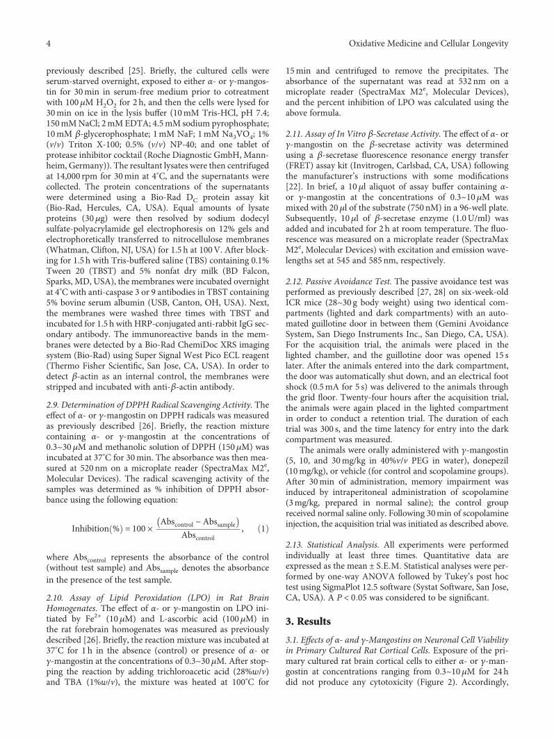
previously described [25]. Briefly, the cultured cells wereserum-starved overnight, exposed to either α- or γ-mangos-tin for 30min in serum-free medium prior to cotreatmentwith 100μM H2O2 for 2 h, and then the cells were lysed for30min on ice in the lysis buffer (10mM Tris-HCl, pH 7.4;150mMNaCl; 2mM EDTA; 4.5mM sodium pyrophosphate;10mM β-glycerophosphate; 1mM NaF; 1mM Na3VO4; 1%(v/v) Triton X-100; 0.5% (v/v) NP-40; and one tablet ofprotease inhibitor cocktail (Roche Diagnostic GmbH, Mann-heim, Germany)). The resultant lysates were then centrifugedat 14,000 rpm for 30min at 4°C, and the supernatants werecollected. The protein concentrations of the supernatantswere determined using a Bio-Rad DC protein assay kit(Bio-Rad, Hercules, CA, USA). Equal amounts of lysateproteins (30μg) were then resolved by sodium dodecylsulfate-polyacrylamide gel electrophoresis on 12% gels andelectrophoretically transferred to nitrocellulose membranes(Whatman, Clifton, NJ, USA) for 1.5 h at 100V. After block-ing for 1.5 h with Tris-buffered saline (TBS) containing 0.1%Tween 20 (TBST) and 5% nonfat dry milk (BD Falcon,Sparks, MD, USA), the membranes were incubated overnightat 4°C with anti-caspase 3 or 9 antibodies in TBST containing5% bovine serum albumin (USB, Canton, OH, USA). Next,the membranes were washed three times with TBST andincubated for 1.5 h with HRP-conjugated anti-rabbit IgG sec-ondary antibody. The immunoreactive bands in the mem-branes were detected by a Bio-Rad ChemiDoc XRS imagingsystem (Bio-Rad) using Super Signal West Pico ECL reagent(Thermo Fisher Scientific, San Jose, CA, USA). In order todetect β-actin as an internal control, the membranes werestripped and incubated with anti-β-actin antibody.
2.9. Determination of DPPH Radical Scavenging Activity. Theeffect of α- or γ-mangostin on DPPH radicals was measuredas previously described [26]. Briefly, the reaction mixturecontaining α- or γ-mangostin at the concentrations of0.3~30μM and methanolic solution of DPPH (150μM) wasincubated at 37°C for 30min. The absorbance was then mea-sured at 520nm on a microplate reader (SpectraMax M2e,Molecular Devices). The radical scavenging activity of thesamples was determined as % inhibition of DPPH absor-bance using the following equation:
Inhibition % = 100 ×Abscontrol −Abssample
Abscontrol, 1
where Abscontrol represents the absorbance of the control(without test sample) and Abssample denotes the absorbancein the presence of the test sample.
2.10. Assay of Lipid Peroxidation (LPO) in Rat BrainHomogenates. The effect of α- or γ-mangostin on LPO ini-tiated by Fe2+ (10μM) and L-ascorbic acid (100μM) inthe rat forebrain homogenates was measured as previouslydescribed [26]. Briefly, the reaction mixture was incubated at37°C for 1 h in the absence (control) or presence of α- orγ-mangostin at the concentrations of 0.3~30μM. After stop-ping the reaction by adding trichloroacetic acid (28%w/v)and TBA (1%w/v), the mixture was heated at 100°C for
15min and centrifuged to remove the precipitates. Theabsorbance of the supernatant was read at 532nm on amicroplate reader (SpectraMax M2e, Molecular Devices),and the percent inhibition of LPO was calculated using theabove formula.
2.11. Assay of In Vitro β-Secretase Activity. The effect of α- orγ-mangostin on the β-secretase activity was determinedusing a β-secretase fluorescence resonance energy transfer(FRET) assay kit (Invitrogen, Carlsbad, CA, USA) followingthe manufacturer’s instructions with some modifications[22]. In brief, a 10μl aliquot of assay buffer containing α-or γ-mangostin at the concentrations of 0.3~10μM wasmixed with 20μl of the substrate (750 nM) in a 96-well plate.Subsequently, 10μl of β-secretase enzyme (1.0U/ml) wasadded and incubated for 2 h at room temperature. The fluo-rescence was measured on a microplate reader (SpectraMaxM2e, Molecular Devices) with excitation and emission wave-lengths set at 545 and 585nm, respectively.
2.12. Passive Avoidance Test. The passive avoidance test wasperformed as previously described [27, 28] on six-week-oldICR mice (28~30 g body weight) using two identical com-partments (lighted and dark compartments) with an auto-mated guillotine door in between them (Gemini AvoidanceSystem, San Diego Instruments Inc., San Diego, CA, USA).For the acquisition trial, the animals were placed in thelighted chamber, and the guillotine door was opened 15 slater. After the animals entered into the dark compartment,the door was automatically shut down, and an electrical footshock (0.5mA for 5 s) was delivered to the animals throughthe grid floor. Twenty-four hours after the acquisition trial,the animals were again placed in the lighted compartmentin order to conduct a retention trial. The duration of eachtrial was 300 s, and the time latency for entry into the darkcompartment was measured.
The animals were orally administered with γ-mangostin(5, 10, and 30mg/kg in 40%v/v PEG in water), donepezil(10mg/kg), or vehicle (for control and scopolamine groups).After 30min of administration, memory impairment wasinduced by intraperitoneal administration of scopolamine(3mg/kg, prepared in normal saline); the control groupreceived normal saline only. Following 30min of scopolamineinjection, the acquisition trial was initiated as described above.
2.13. Statistical Analysis. All experiments were performedindividually at least three times. Quantitative data areexpressed as themean ± S E M Statistical analyses were per-formed by one-way ANOVA followed by Tukey’s post hoctest using SigmaPlot 12.5 software (Systat Software, San Jose,CA, USA). A P < 0 05 was considered to be significant.
3. Results
3.1. Effects of α- and γ-Mangostins on Neuronal Cell Viabilityin Primary Cultured Rat Cortical Cells. Exposure of the pri-mary cultured rat brain cortical cells to either α- or γ-man-gostin at concentrations ranging from 0.3~10μM for 24hdid not produce any cytotoxicity (Figure 2). Accordingly,
4 Oxidative Medicine and Cellular Longevity

upcoming experiments with α- or γ-mangostin were per-formed using this concentration range.
3.2. Effects of α- and γ-Mangostins on the H2O2- orX/XO-Induced Oxidative Neuronal Damage and ROSGeneration in Primary Cultured Rat Cortical Cells. Inagreement with the previous reports [22, 26], treatment ofthe cultured cells with 100μM of H2O2 for 5min causedapproximately 80% or more cell death and approximately200% increase in intracellular ROS production (Figures 3(a)and 3(b), respectively (#, P < 0 05 vs. vehicle-treated controlcells without H2O2, α-mangostin, or γ-mangostin treat-ment)). The reduced viability of the H2O2-treated cells wascompletely reversed by γ-mangostin at the concentration of10μM (Figure 3(a) (∗, P < 0 05 vs. H2O2-treated cells withoutα- or γ-mangostin)). Since its protective effect against theH2O2-induced oxidative damage was so dramatic, we fur-ther tested the effect of γ-mangostin at the concentrationsbetween 3 and 10μM. As shown in Figure 3(a) (inset), 5and 7μM of γ-mangostin also exhibited dramatic increasesin the viability of H2O2-treated cells. In addition, the H2O2-in-duced ROS production was significantly inhibited by γ-man-gostin at 3~10μM concentrations (Figure 3(b) (∗, P < 0 05vs. H2O2-treated cells without α- or γ-mangostin)), whichwas confirmed by fluorescence microscopy (Figure 3(c)). Incontrast, α-mangostin showed no significant effects onH2O2-induced oxidative damage or ROS production at theconcentrations tested in this study (Figures 3(a)–3(c)).
Treatment of the cultured cells with 0.5mM X and10mU/ml XO caused approximately 80% or more cell death
and a 300% increase in ROS production (Figures 3(d) and3(e), respectively (#, P < 0 05 vs. vehicle-treated control cellswithout X/XO, α-mangostin, or γ-mangostin treatment)).The decreased viability of the X/XO-treated cells was totallyreversed by γ-mangostin at the concentration of 10μM(Figure 3(d) (∗, P < 0 05 vs. X/XO-treated cells withoutα- or γ-mangostin)). Similarly, the X/XO-induced ROSproduction was significantly suppressed by γ-mangostin at3 and 10μM concentrations (Figure 3(e) (∗, P < 0 05 vs.X/XO-treated cells without α- or γ-mangostin)). However,α-mangostin showed no significant effects on X/XO-inducedoxidative damage or ROS production at the concentrationstested (Figures 3(d) and 3(e)).
3.3. Effects of α- and γ-Mangostins on the H2O2-InducedApoptosis in Primary Cultured Rat Cortical Cells. In orderto elucidate the probable mechanism(s) underlying the neu-roprotective role of γ-mangostin, we next examined the effectof this compound on H2O2-induced apoptosis and comparedit to that of α-mangostin. In agreement with the previousreport [29], the exposure of cultured cells to H2O2 causedDNA fragmentation, an important hallmark of apoptosis,as reflected by a dramatic increase in the TUNEL-positive cellpopulation (Figures 4(a) (B and F) and Figure 4(b) (#, P <0 05 vs. vehicle-treated control cells without α- or γ-mangos-tin treatment)). The H2O2-induced DNA fragmentation wasremarkably inhibited by γ-mangostin at 10μM (Figure 4(a)(G) and Figure 4(b) (∗, P < 0 05 vs. H2O2-treated cells with-out α- or γ-mangostin treatment)). We also examined theireffects on the H2O2-induced activation of caspases, anotherimportant molecular event during the apoptotic process. Asillustrated in Figures 4(c) and 4(d), γ-mangostin significantlyattenuated the H2O2-induced activation of both caspases 3and 9. In contrast, α-mangostin neither prevented DNA frag-mentation nor inhibited caspase activities in the H2O2-trea-ted cells (Figure 4).
3.4. Effects of α- and γ-Mangostins on DPPH RadicalFormation and Lipid Peroxidation. The antioxidant proper-ties of α- and γ-mangostins were further substantiated byevaluating their radical scavenging activities using stable freeradical DPPH as a probe (Figure 5(a)). In addition, their abil-ity to inhibit LPO initiated by Fe2+ and L-ascorbic acid in ratbrain homogenate was also examined (Figure 5(b)). Ourresults demonstrated that γ-mangostin considerably attenu-ated the formation of DPPH radicals and effectively inhibitedlipid peroxide formation in concentration-dependent man-ners. In contrast, α-mangostin showed no DPPH radicalscavenging activity with only a minimal inhibition of LPOat the concentrations tested (Figures 5(a) and 5(b), respec-tively). Vit. C and BHA were used as reference compoundsto validate the assay procedures for DPPH radical scavengingactivity and inhibition of LPO, respectively (grey bars).
3.5. Effects of α- and γ-Mangostins on In Vitro β-SecretaseEnzyme Activity. Since β-secretase plays a vital role inthe generation of the Aβ peptide from APP [6, 7], we alsoexamined the impact of α- and γ-mangostins on the activityof this enzyme using the in vitro β-secretase FRET assay
Concentration (�휇M)
0 1 3 10
Cell
surv
ival
(% co
ntro
l)
0
20
40
60
80
100
120
�훼-Mangostin �훾-Mangostin
Figure 2: Effects of α- and γ-mangostins on neuronal cell viabilityin primary cultured rat cortical cells. Cells were exposed to theindicated concentrations of α- or γ-mangostin for 24 h. Thecontrol cells were treated with vehicle (DMSO) only. Cell viabilitywas determined by the MTT reduction assay, as described inMaterials and Methods. The viability of control cells treated withvehicle only was considered to be 100%, and the data wereexpressed as percentages of the control. Each point represents themean ± S E M from at least three independent experiments,performed in duplicate.
5Oxidative Medicine and Cellular Longevity

H2O2
0 0.3 1 3 10
Cell
surv
ival
(% co
ntro
l)
0
20
40
60
80
100
120
�훼-Mangostin�훾-Mangostin
# #
(�휇M)
Concentration (�휇M)
Cell
surv
ival
(% co
ntro
l)0
20
40
60
80
100
120
#
⁎ ⁎
#
50 7
⁎
(a)
0 10.3 3 75 10
�훼-Mangostin�훾-Mangostin
(�휇M)
H2O2
Intr
acel
lula
r RO
S (%
cont
rol)
0
50
100
150
200
250#
#
⁎
⁎
⁎⁎
(b)
(A) (B)
(C) (D)
(c)
0 0.3 1 3 10
�훼-Mangostin�훾-Mangostin
(�휇M)
X/XO
Cell
surv
ival
(% co
ntro
l)
0
20
40
60
80
100
120
# #
⁎
(d)
Figure 3: Continued.
6 Oxidative Medicine and Cellular Longevity

kit. We found that both α- and γ-mangostins potentlyinhibited β-secretase activity in concentration-dependentfashion (Figure 6).
3.6. Effect of γ-Mangostin on the Scopolamine-InducedMemory Impairment in Mice. Using cell-based and cell-freein vitro assays, our findings demonstrated that γ-mangostinshowed more potent antioxidant and neuroprotective activi-ties than α-mangostin, although both compounds showedsimilar degrees of inhibition against β-secretase activity.Based on these findings, we selected γ-mangostin to examinewhether it could improve scopolamine-induced memoryimpairment in mice using the passive avoidance test.
As illustrated in Figure 7, the acquisition trials did notshow significant differences in the time latency of all groups(black bars). During the retention trials (white bars), thescopolamine-treated group (without γ-mangostin or done-pezil administration) showed marked reduction of thetime latency than the control group treated with vehicle only(#, P < 0 05 vs. vehicle-treated control group), indicating thatsignificant memory impairment was induced by scopol-amine injection. The groups administered with γ-mangostinat the dosages of 10 and 30mg/kg significantly restored the
scopolamine-induced decrease in time latency (∗, P < 0 05vs. scopolamine-treated amnesia group). Donepezil, a well-known cholinesterase inhibitor clinically used for the treat-ment of AD, was employed as a reference drug to validateour experimental procedures and compare its effect withthat of γ-mangostin. The reduced time latency by scopol-amine injection was also recovered by the oral administra-tion of donepezil at the dosage of 10mg/kg (∗, P < 0 05 vs.scopolamine-treated amnesia group). The inhibition ofscopolamine-induced memory impairment by γ-mangostinadministration (10 and 30mg/kg of dosage) was nearly com-parable to that of the donepezil-treated group (Figure 7).However, administration of γ-mangostin at the dosage of5mg/kg did not show a significant effect on reversingscopolamine-induced memory impairment in mice.
4. Discussion
Oxidative stress-induced cell damage is known to be involvedin a number of neurodegenerative diseases such as AD,Parkinson’s disease, and stroke [4, 5], where ROS such assuperoxide anion, hydroxyl radical, and hydrogen peroxideplay pivotal roles. It has been demonstrated that H2O2 can
0 0.3 1 3 10
�훼-Mangostin�훾-Mangostin
(�휇M)
X/XO
Intr
acel
lula
r RO
S (%
cont
rol)
0
100
200
300
400
##
⁎
⁎
(e)
Figure 3: Effects of α- and γ-mangostins on H2O2- or X/XO-induced oxidative neurotoxicity and ROS generation in primary cultured ratcortical cells. (a and d) The cells were exposed to 100μM H2O2 for 5min (a) or 0.5mM X in combination with 10mU/ml XO for 10min(d) in the absence or presence of either α- or γ-mangostin at various concentrations as indicated. Cell viability was determined by theMTT reduction assay at 18-20 h after exposure, as described in Materials and Methods. The cell survival was expressed as percentages ofthe control treated with vehicle only. (b and e) The cells were preincubated with 10 μM DCFH-DA for 30min at 37°C in the dark, thentreated with 100μM H2O2 for 2 h (b) or 0.5mM X in combination with 10mU/ml XO for 2 h (e) in the absence or presence ofeither α- or γ-mangostin at various concentrations as indicated. The generation of intracellular ROS was measured as described inMaterials and Methods. The ROS levels were expressed as percentages of the control treated with vehicle only. Each data point representsthe mean ± S E M from at least three independent experiments, performed in duplicate (#P < 0 05 vs. vehicle-treated control cells withoutα- or γ-mangostin treatment; ∗P < 0 05 vs. H2O2- or X/XO-treated cells). (c) Fluorescence microscopic images showing the inhibition ofH2O2-induced ROS generation by γ-mangostin in primary cultured rat cortical cells. The cells were preincubated with 10μM DCFH-DAfor 30min at 37°C in the dark and treated with 100 μM H2O2 in the absence (B) or presence of 10μM α-mangostin (C) or γ-mangostin(D) for 2 h. The control cells were treated with vehicle only without α- or γ-mangostin (A). Following the desired treatment, ROS levelswere imaged using epifluorescence microscopy as described in Materials and Methods. Representative photomicrographs from threeindependent experiments are shown.
7Oxidative Medicine and Cellular Longevity

(A) (B) (C) (D)
(E) (F) (G) (H)
(a)
TUN
EL-p
ositi
ve ce
lls /
tota
l cel
ls (%
)
0
5
10
15
20
25
30
35
H2O2
##
10 100 0
⁎
�훼-Mangostin�훾-Mangostin
(�휇M)
(b)
Cleaved caspase 3
Cleaved caspase 9
�훽-Actin
�훽-Actin
�훼-Mangostin (�휇M)
�훾-Mangostin (�휇M)
�훼-Mangostin (�휇M)
�훾-Mangostin (�휇M)
H2O2 (100 �휇M)
H2O2 (100 �휇M)
− −
−
−
− − −
− −
+ + + + +
3 10
3 10
− −
−
−
− − −
− −
+ + + + +
3 10
3 10
(c)
Figure 4: Continued.
8 Oxidative Medicine and Cellular Longevity

readily cross the cell membrane and cause injuries to the tis-sues through a number of different mechanisms, includingthe production of hydroxyl radicals and destabilisation ofthe oxidant/antioxidant pathway, ultimately leading to apo-ptotic and/or necrotic cell death [30]. The oxidation of X byXO also serves as an important source of ROS, generating
H2O2 and superoxide anions that are known to contributeto the onset of neuronal damage in many neurodegenerativediseases [31, 32]. Taking critical roles of these ROS intoaccount, we treated the primary cultured rat cortical cellswith either H2O2 or X and XO in this study to induce oxida-tive neuronal damage as well as ROS generation and tested
Casp
ase 3
/�훽-a
ctin
(% C
ontro
l)
0
100
200
300
400
500
600
700
H2O2
#
Casp
ase 9
/�훽-a
ctin
(% co
ntro
l)
0
100
200
300
400
500
600
700
H2O2
#
0 0 3 10
0 0 3 10
⁎
⁎
⁎
⁎
�훼-Mangostin�훾-Mangostin
�훼-Mangostin�훾-Mangostin
(�휇M)
(�휇M)
(d)
Figure 4: Effects of α- and γ-mangostins on H2O2-induced apoptosis in primary cultured rat cortical cells. (a and b) Inhibition ofH2O2-induced DNA fragmentation by γ-mangostin. Cells were treated with 100 μM H2O2 for 2 h with or without α- or γ-mangostin atthe concentration of 10μM, and the TUNEL assay was carried out as described in Materials and Methods. Representative microscopicimages from at least three individual experiments are shown (a). (A and E) Control cells were treated with vehicle only; (B and F) cellswere treated with 100μM H2O2 for 2 h; (C and G) cells were treated for 2 h with either 10μM α-mangostin (C) or γ-mangostin (G) incombination with 100μM H2O2; (D and H) cells were treated with 10 μM α-mangostin (D) or γ-mangostin for 2 h without H2O2 (H).Scale bar = 10 μm. Quantitative analyses of the TUNEL-positive cells from at least three independent experiments are shown (b)(#P < 0 05 vs. vehicle-treated control cells without α- or γ-mangostin treatment; ∗P < 0 05 vs. H2O2-treated cells without α- orγ-mangostin). (c and d) Inhibition of the H2O2-induced activation of caspases 3 and 9 by γ-mangostin. Cells were treated with 100μMH2O2 for 2 h in the absence or presence of either α- or γ-mangostin at 3 and 10μM. The expression of cleaved caspases 3 and 9 wasassessed by Western blotting as described in Materials and Methods. Representative blots from at least three individual experiments areshown (c). The intensities of the bands from at least three independent experiments were quantified by densitometric analyses andnormalised to β-actin (d) (#P < 0 05 vs. vehicle-treated control cells without α- or γ-mangostin treatment; ∗P < 0 05 vs. H2O2-treated cellswithout α- or γ-mangostin).
9Oxidative Medicine and Cellular Longevity

the effects of α- and γ-mangostins, the two xanthones iso-lated from the fruit hull of mangosteen, on this oxidativedamage and ROS.
Our results demonstrated that the exposure of culturedcells to these oxidative insults produced approximately80% cell death and marked increases in ROS production(Figures 3(a)–3(e)). We found in this study that only γ-man-gostin, not α-mangostin, completely reversed the H2O2- orX/XO-induced oxidative neuronal damage and significantlyattenuated ROS production. These results are in line withthose of an earlier report, demonstrating that, among thesixteen xanthones including α- and γ-mangostins, onlyγ-mangostin exhibited HO. scavenging activity in an in vitrocell-free assay [33].
It has been previously reported that α-mangostin pro-vides ROS scavenging activity and neuroprotective actionagainst 3-nitropropionic acid- or iodoacetic acid-treatedprimary cultures of cerebellar granule neurons [14, 15].However, in our study, α-mangostin neither exhibited neu-roprotective activity against the H2O2- or X/XO-inducedoxidative damage nor inhibited associated ROS productionat any concentration tested (Figures 3(a)–3(e)). The discrep-ancies between the earlier reports [14, 15] and our resultsmay be due to the different cell types used in these studies.Another plausible reason for these contradictory resultsmay be due to the different species of radicals generated bythe different oxidative inducers used in these studies. Accord-ing to the report by Pedraza-Chaverrí et al. [14], α-mangostinwas able to scavenge superoxide anion and peroxynitriteanion, whereas it was unable to scavenge hydroxyl radicalsand hydrogen peroxide. Since α-mangostin was found toinhibit 3-nitropropionic acid-induced neurotoxicity and
ROS production, Pedraza-Chaverrí et al. suggested thatsuperoxide radical and peroxynitrite anion may be involvedin 3-nitropropionic acid-induced toxicity in cerebellar gran-ule neurons [14]. In our study, however, hydroxyl radicalsand hydrogen peroxide were produced in cortical neuronsby the treatment with H2O2 and X/XO, and α-mangostindid not inhibit the neurotoxicity and ROS productioncaused by these inducers. Thus, our findings on α-mangos-tin are entirely in agreement with the observation byPedraza-Chaverrí et al. It would be interesting to clarifypreferable species of ROS, if any, to be scavenged by γ-man-gostin under the same experimental conditions employed forα-mangostin. Further study will be required to elucidateunderlying mechanisms by which these xanthones distin-guish different species of radicals to scavenge. We furtherconfirmed that γ-mangostin exhibits more potent radicalscavenging activity and antioxidant effect than α-mangostinusing in vitro assays for DPPH radical scavenging activityand LPO. Again, unlike γ-mangostin, α-mangostin showedno DPPH radical scavenging activity and only minimal inhi-bition of LPO (Figure 5).
The action of H2O2 as an inducer of apoptosis andDNA damage has been well documented [34, 35]. It hasbeen found that in PC12 cells and SH-SY5Y neuroblatomacells, H2O2 can induce ROS generation and the activationof caspase cascades and ultimately triggers apoptosis [29]. Inagreement with these previous reports [29, 34, 35], the expo-sure of our cultured cells to H2O2 also caused DNA fragmen-tation (Figure 4(a), B and F), as evidenced by the increasednumber of TUNEL-positive cells (Figure 4(b)). The H2O2-induced DNA fragmentation was markedly inhibited byγ-mangostin but not by α-mangostin (Figures 4(a) and
DPP
H ra
dica
l sca
veng
ing
activ
ity(%
cont
rol)
0
20
40
60
80
100
Vit. C
�훼- or �훾-mangostin Vit. C
⁎
⁎
⁎
⁎
0.3 1 3 10 30 100
�훼-Mangostin�훾-Mangostin
(�휇M)
(a)
BHA
�훼- or �훾-mangostin
Inhi
bitio
n of
lipi
d pe
roxi
datio
n(%
cont
rol)
-20
0
20
40
60
80
100
BHA
⁎
⁎
⁎ ⁎ ⁎
�훼-Mangostin�훾-Mangostin
0.3 1 3 10 30 100 (�휇M)
(b)
Figure 5: Effects of α- and γ-mangostins on DPPH radical formation and lipid peroxidation (LPO). Inhibition of DPPH radical (a) and LPOinduced by Fe2+ (10μM) and L-ascorbic acid (100 μM) in rat forebrain homogenate (b) by α- or γ-mangostin at the indicated concentrationswere measured as described in Materials and Methods. Each data point represents the mean ± S E M from at least three independentexperiments, performed in duplicate (∗P < 0 05 vs. vehicle-treated control without α- or γ-mangostin treatment). Vit. C and BHA wereused as references to validate the assay procedures for DPPH radical scavenging activity and inhibition of LPO, respectively (grey bars).Vit. C: vitamin C; BHA: butylhydroxyanisole.
10 Oxidative Medicine and Cellular Longevity

4(b)). Furthermore, the H2O2 treatment of the cultured cellstriggered the activation of caspases 3 and 9, the major molec-ular events in the apoptotic process. The activated caspases 3and 9 were significantly suppressed only by γ-mangostin(Figures 4(c) and 4(d)). Recently, γ-mangostin was foundto inhibit caspase 3 activity in 6-hydroxy dopamine-treatedSH-SY5Y cells [36], which is consistent with our finding.Taken together, it is conceivable that the underlying mecha-nism(s) for the neuroprotective effect of γ-mangostin againstH2O2-induced oxidative damage may involve the inhibitionof ROS production, as well as its antiapoptotic action inhibit-ing DNA fragmentation and activation of caspases 3 and 9.Furthermore, the antioxidant activities of γ-mangostin, notα-mangostin, scavenging DPPH radical and inhibiting LPOmay also contribute to its neuroprotective action (Figure 5).
Among the diverse pathophysiological factors involved inthe onset and development of AD, Aβ peptide plays a key rolein neuronal cell death [37]. In this study, we found that bothα- and γ-mangostins inhibited β-secretase activity, anenzyme involved in the generation of the Aβ peptides fromAPP in the amyloidogenic pathway [6, 7]. Previously, a seriesof xanthones isolated from G. mangostana were reportedto modestly inhibit BACE1 activity, exhibiting 60.3 and42.1% inhibition by α- and γ-mangostins at the concentra-tion of 100μM, respectively [17]. Our study expanded theirfindings, illustrating the concentration-dependent inhibitionof β-secretase activity by both α- and γ-mangostins at theconcentration range of 0.3~10μM (Figure 6). For some rea-son, however, the inhibition by α- or γ-mangostin wasobserved to be much more potent in our study than that
shown by a previous report [18], as approximately 60-70%inhibition of β-secretase activity was achieved at the concen-tration as low as 3μM of α- or γ-mangostin (Figure 6). Oneof the possible explanations for this potency discrepancymay be due to the differences in the bioactivities of β-secre-tase in the assay kits employed in our study and in the previ-ous report. In order to test this possibility, the β-secretaseactivity has to be reevaluated in the presence of the testingxanthones under the same experimental conditions, usingassay kits manufactured by the same company. In any event,our and previous findings strongly support that both α- andγ-mangostins may reduce Aβ formation from APP throughthe inhibition of β-secretase activity.
It has been previously reported that α-mangostin concen-tration dependently attenuated the neurotoxicity induced byAβ-(1-40) or Aβ-(1-42) oligomers in primary rat cerebral corticalneurons [16]. Based on molecular docking simulations,
Concentration (�휇M)
�훽-S
ecre
tase
activ
ity (%
cont
rol)
0
20
40
60
80
100
120
�훼-Mangostin�훾-Mangostin
0 0.3 1 3 10
⁎⁎
⁎⁎
Figure 6: Effects of α- and γ-mangostins on β-secretase activity.The inhibitory effects of α- and γ-mangostins on the enzymaticactivity of β-secretase were determined by the β-secretase FRETassay as described in Materials and Methods. The data wereexpressed as percentages of the control treated without α- orγ-mangostin. Each data point represents the mean ± S E M fromat least three independent experiments, performed in duplicate(∗P < 0 05 vs. vehicle-treated control without α- or γ-mangostintreatment).
Tim
e lat
ency
(s)
0
50
100
150
200
250
300
Acquisition trial Retention trial
#
Scopolamine
5 3010
Donepezil (mg/kg) 10
�훾-Mangostin(mg/kg)
⁎ ⁎ ⁎
−
−
−
−
− − − −
−
+ + + + +
Figure 7: Effect of γ-mangostin on the scopolamine-inducedmemory impairment in mice. Animals were randomly dividedinto 6 groups with 6-7 mice in each group. To the 3 groups ofanimals, γ-mangostin was orally administered at the respectivedosages of 5, 10, or 30mg/kg as indicated. For the referencedrug-treated group, donepezil was administered at the dosage of10mg/kg. For the control group (the vehicle-treated groupwithout scopolamine, γ-mangostin, or donepezil treatment) andthe scopolamine group (the group treated with scopolamineinjection, not with γ-mangostin or donepezil treatment), vehiclewas only administered. After 30min of each administration,memory impairment was induced by intraperitoneal injection ofscopolamine (3mg/kg in normal saline) in 5 groups as indicatedabove in the figure; for the control group, normal saline withoutscopolamine was injected. Following 30min of scopolamine orsaline injection, the acquisition trial was initiated by delivering afoot shock to the animals. Twenty-four hours after the acquisitiontrials, the retention trials were performed. The detailed experimentalprocedures are described in Materials and Methods. The timelatency was calculated from three independent experiments. Eachdata point represents the mean ± S E M (#P < 0 05 vs. vehicle-treated control group without scopolamine, γ-mangostin, ordonepezil treatment; ∗P < 0 05 vs. scopolamine group treated withscopolamine only without γ-mangostin or donepezil).
11Oxidative Medicine and Cellular Longevity

the thioflavin T fluorescence assay and electron microscopyimaging, α-mangostin was found to inhibit and dissociateamyloid aggregation, which could contribute to its effect ofattenuating Aβ oligomers-induced neurotoxicity [16]. Wealso attempted to evaluate the effects of α- and γ-mangostinson the neurotoxicity induced in our culture model byAβ-(25-35), the active fragment of Aβ, as previously described[22]. We treated the cultured cells with 40μM of Aβ-(25-35)for 24h in the absence or presence of α- or γ-mangostin atthe concentration range of 0.3~10μM. Both mangostinsappeared to exhibit weak protective effect on Aβ-(25-35)-in-duced neurotoxicity (data not shown). However, the appar-ent effects were not statistically significant by one-wayANOVA analysis. Further extensive studies under the sameexperimental conditions with the same Aβ peptides may benecessary to evaluate and compare their effects on the Aβ-in-duced neurotoxicity.
It has long been believed that acetylcholine is one of themost important neurotransmitters in learning and memoryprocesses [38], and thus, cholinergic dysfunction is closelyassociated with AD pathology [39, 40]. Based on this cholin-ergic hypothesis, AChE inhibitors such as donepezil are cur-rently used to alleviate the symptoms of AD in clinicalsituations. Recently, several prenylated xanthones fromman-gosteen, including α- and γ-mangostins, have been reportedto inhibit AChE activity with IC50 values of lower than20.5μM, as determined by Ellman’s colorimetric method[41, 42]. The protein-ligand interactions between AChEand xanthones were confirmed by molecular docking studies[41]. In our study, we also tested the effects of α- and γ-man-gostins on AChE activity in vitro and verified the previousfindings (data not shown). Collectively, based on our resultsand previous findings [18, 41, 42], both α- and γ-mangostinsmay reduce Aβ formation and improve cholinergic trans-mission through the inhibition of β-secretase and AChEactivities, respectively.
The α- and γ-mangostins are the most extensively stud-ied xanthone derivatives of mangosteen [12]. Although theyshare a common chemical backbone, their chemical struc-tures differ with respect to the numbers of hydroxyl andmethoxy groups (Figure 1). While α-mangostin possessesthree hydroxyl groups (at positions 1, 3, and 6) and a meth-oxy group (at position 7), γ-mangostin has four hydroxylgroups (at positions 1, 3, 6, and 7) without any methoxygroup. We observed that only γ-mangostin, not α-mangos-tin, exhibited marked and potent neuroprotective andantioxidant effects. The exact mechanisms by which thestructural discrepancy between these two xanthones canaccount for such a decisive difference in their neuroprotec-tive and antioxidant effects are not yet understood. Thecatechol moiety of γ-mangostin or the hydroxyl group itselfat position 7 may play crucial role in its neuroprotectiveand radical scavenging activities. Interestingly, however, thestructural difference between α- and γ-mangostins was notreflected in their inhibitory effects on β-secretase and AChEenzyme activities, as both mangostins exhibited similardegrees of inhibition (Figure 6) [41, 42]. It is assumed thatthe hydroxyl groups at positions 1, 3, and 6 may play impor-tant roles in the inhibition of these enzyme activities. The
methoxy group at position 7 of α-mangostin may notbe directly involved in the interactions with these enzymes.Further studies are needed to explain the basis of such dif-ferential or similar pharmacological actions by the twomangostin compounds.
Even though both mangostins exhibited similar inhibi-tion against β-secretase and AChE activities as measured incell-free in vitro assays, γ-mangostin appeared to be a sub-stantially more potent antioxidant and neuroprotective agentthan α-mangostin, based on our findings in cell-based as wellas cell-free in vitro studies. These beneficial pharmacologicalprofiles of γ-mangostin strongly suggest its therapeuticpotential for AD and other neurodegenerative diseases asso-ciated with oxidative stress.
In order to test this possibility, we next conducted a pas-sive avoidance test using a scopolamine-induced amnesiamodel in mice to investigate the memory-improving effectof γ-mangostin in vivo. It has been previously revealed thatmemory deficit induced by scopolamine, a nonselective mus-carinic receptor antagonist, is associated with oxidative stress[43], an event known to play a vital role in neurodegenerativedisorders such as AD [4, 5]. As shown in Figure 7, the mem-ory impairment induced by scopolamine was significantlyimproved by a single oral administration of γ-mangostinat dosages of 10 and 30mg/kg. The memory-improvingeffects of γ-mangostin at these dosages were quite compa-rable to that of donepezil, a reference drug. Our results arein alignment with those of a previous report, demonstrat-ing the protective effect of the mangosteen extract onscopolamine-induced amnesia [28]. In addition, the abilityof γ-mangostin to penetrate the blood-brain barrier andreach CNS targets was predicted in vitro using a parallel arti-ficial membrane penetration assay [18]. Taken together, ourresult and the previous findings suggest that γ-mangostincould be a promising candidate for therapeutic interven-tions of AD. To the best of our knowledge, our presentstudy is the first report revealing the neuroprotective effectof γ-mangostin against oxidative neurotoxicity, as well asits memory-enhancing effect in mice.
5. Conclusions
The present study demonstrated that γ-mangostin, a xan-thone derivative isolated from the fruit hull of mangosteen,exhibited a potent neuroprotective effect against H2O2- orX/XO-induced oxidative neuronal damage. The underlyingmechanisms for this neuroprotective action may involve theinhibition of ROS generation triggered by these oxidativeinsults, antioxidant activities as evident by inhibition ofDPPH radical formation and LPO, and its antiapoptoticproperties as demonstrated by the inhibition of H2O2-in-duced DNA fragmentation and activation of caspases. Unlikeγ-mangostin, however, α-mangostin neither exhibited neu-roprotective activity nor demonstrated antioxidant proper-ties in our study. Moreover, γ-mangostin was shown toexhibit a potent inhibitory effect on β-secretase activity andstrongly improved scopolamine-induced memory deficits inmice. Based on our study providing in vitro and in vivo evi-dences, γ-mangostin may be considered to be a promising
12 Oxidative Medicine and Cellular Longevity
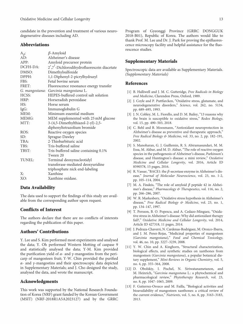
candidate in the prevention and treatment of various neuro-degenerative diseases including AD.
Abbreviations
Aβ: β-AmyloidAD: Alzheimer’s diseaseAPP: Amyloid precursor proteinDCFH-DA: 2′,7′-Dichlorodihydrofluorescein diacetateDMSO: DimethylsulfoxideDPPH: 1,1-Diphenyl-2-picrylhydrazylFBS: Fetal bovine serumFRET: Fluorescence resonance energy transferG. mangostana: Garcinia mangostana L.HCSS: HEPES-buffered control salt solutionHRP: Horseradish peroxidaseHS: Horse serumIgG: Immunoglobulin GMEM: Minimum essential mediumMEMG: MEM supplemented with 25mM glucoseMTT: 3-(4,5-Dimethylthiazol-2-yl)-2,5-
diphenyltetrazolium bromideROS: Reactive oxygen speciesSD: Sprague-DawleyTBA: 2-Thiobarbituric acidTBS: Tris-buffered salineTBST: Tris-buffered saline containing 0.1%
Tween 20TUNEL: Terminal deoxynucleotidyl
transferase-mediated deoxyuridinetriphosphate nick end-labeling
X: XanthineXO: Xanthine oxidase.
Data Availability
The data used to support the findings of this study are avail-able from the corresponding author upon request.
Conflicts of Interest
The authors declare that there are no conflicts of interestsregarding the publication of this paper.
Authors’ Contributions
Y. Lee and S. Kim performed most experiments and analysedthe data; Y. Oh performed Western blotting of caspase 9and statistically analysed the data; Y-M. Kim providedthe purification yield of α- and γ-mangostins from the peri-carp of mangosteen fruit; Y-W. Chin provided the purifiedα- and γ-mangostins and their spectroscopic data depictedin Supplementary Materials; and J. Cho designed the study,analysed the data, and wrote the manuscript.
Acknowledgments
This work was supported by the National Research Founda-tion of Korea (NRF) grant funded by the Korean Government(MSIT) (NRF-2018R1A5A2023127) and by the GRRC
Program of Gyeonggi Province (GRRC DONGGUK2018-B01), Republic of Korea. The authors would like tothank Prof. M. Lee and Dr. J. Park for proving the epifluores-cence microscopy facility and helpful assistance for the fluo-rescence studies.
Supplementary Materials
Spectroscopic data are available as Supplementary Materials.(Supplementary Materials)
References
[1] B. Halliwell and J. M. C. Gutteridge, Free Radicals in Biologyand Medicine, Clarendon Press, Oxford, 1989.
[2] J. Coyle and P. Puttfarcken, “Oxidative stress, glutamate, andneurodegenerative disorders,” Science, vol. 262, no. 5134,pp. 689–695, 1993.
[3] J. N. Cobley, M. L. Fiorello, and D. M. Bailey, “13 reasons whythe brain is susceptible to oxidative stress,” Redox Biology,vol. 15, pp. 490–503, 2018.
[4] C. Behl and B. Moosmann, “Antioxidant neuroprotection inAlzheimer’s disease as preventive and therapeutic approach,”Free Radical Biology & Medicine, vol. 33, no. 2, pp. 182–191,2002.
[5] S. Manoharan, G. J. Guillemin, R. S. Abiramasundari, M. M.Essa, M. Akbar, and M. D. Akbar, “The role of reactive oxygenspecies in the pathogenesis of Alzheimer’s disease, Parkinson’sdisease, and Huntington’s disease: a mini review,” OxidativeMedicine and Cellular Longevity, vol. 2016, Article ID8590578, 15 pages, 2016.
[6] R. Vassar, “BACE1: the β-secretase enzyme in Alzheimer’s dis-ease,” Journal of Molecular Neuroscience, vol. 23, no. 1-2,pp. 105–114, 2004.
[7] M. A. Findeis, “The role of amyloid β peptide 42 in Alzhei-mer’s disease,” Pharmacology & Therapeutics, vol. 116, no. 2,pp. 266–286, 2007.
[8] W. R. Markesbery, “Oxidative stress hypothesis in Alzheimer’sdisease,” Free Radical Biology & Medicine, vol. 23, no. 1,pp. 134–147, 1997.
[9] T. Persson, B. O. Popescu, and A. Cedazo-Minguez, “Oxida-tive stress in Alzheimer’s disease: Why did antioxidant therapyfail?,” Oxidative Medicine and Cellular Longevity, vol. 2014,Article ID 427318, 11 pages, 2014.
[10] J. Pedraza-Chaverri, N. Cardenas-Rodriguez, M. Orozco-Ibarra,and J. M. Perez-Rojas, “Medicinal properties of mangosteen(Garcinia mangostana),” Food and Chemical Toxicology,vol. 46, no. 10, pp. 3227–3239, 2008.
[11] Y. W. Chin and A. Kinghorn, “Structural characterization,biological effects, and synthetic studies on xanthones frommangosteen (Garcinia mangostana), a popular botanical die-tary supplement,” Mini-Reviews in Organic Chemistry, vol. 5,no. 4, pp. 355–364, 2008.
[12] D. Obolskiy, I. Pischel, N. Siriwatanametanon, andM. Heinrich, “Garcinia mangostana L.: a phytochemical andpharmacological review,” Phytotherapy Research, vol. 23,no. 8, pp. 1047–1065, 2009.
[13] F. Gutierrez-Orozco and M. Failla, “Biological activities andbioavailability of mangosteen xanthones: a critical review ofthe current evidence,” Nutrients, vol. 5, no. 8, pp. 3163–3183,2013.
13Oxidative Medicine and Cellular Longevity

[14] J. Pedraza-Chaverrí, L. M. Reyes-Fermín, E. G. Nolasco-A-maya et al., “ROS scavenging capacity and neuroprotectiveeffect of α-mangostin against 3-nitropropionic acid in cerebel-lar granule neurons,” Experimental and Toxicologic Pathology,vol. 61, no. 5, pp. 491–501, 2009.
[15] L. M. Reyes-Fermín, S. González-Reyes, N. G. Tarco-Álvarez,M. Hernández-Nava, M. Orozco-Ibarra, andJ. Pedraza-Chaverri, “Neuroprotective effect of α-mangostinand curcumin against iodoacetate-induced cell death,”Nutritional Neuroscience, vol. 15, no. 5, pp. 34–41, 2013.
[16] Y. Wang, Z. Xia, J. R. Xu et al., “α-Mangostin, a polyphenolicxanthone derivative from mangosteen, attenuates β-amyloidoligomers-induced neurotoxicity by inhibiting amyloid aggre-gation,” Neuropharmacology, vol. 62, no. 2, pp. 871–881, 2012.
[17] L. X. Zhao, Y. Wang, T. Liu et al., “α-Mangostin decreasesβ-amyloid peptides production via modulation of amyloido-genic pathway,” CNS Neuroscience & Therapeutics, vol. 23,no. 6, pp. 526–534, 2017.
[18] S. N. Wang, Q. Li, M. H. Jing et al., “Natural xanthones fromGarcinia mangostana with multifunctional activities for thetherapy of Alzheimer’s disease,” Neurochemical Research,vol. 41, no. 7, pp. 1806–1817, 2016.
[19] M. H. Wang, K. J. Zhang, Q. L. Gu, X. L. Bi, and J. X. Wang,“Pharmacology of mangostins and their derivatives: a compre-hensive review,” Chinese Journal of Natural Medicines, vol. 15,no. 2, pp. 81–93, 2017.
[20] G. H. Quan, S. R. Oh, J. H. Kim, H. K. Lee, A. D. Kinghorn, andY. W. Chin, “Xanthone constituents of the fruits of Garciniamangostana with anticomplement activity,” PhytotherapyResearch, vol. 24, no. 10, pp. 1575–1577, 2010.
[21] J. Cho, J.-Y. Kong, D.-Y. Jeong, K. D. Lee, D. U. Lee, and B. S.Kang, “NMDA recepter-mediated neuroprotection by essen-tial oils from the rhizomes of Acorus gramineus,” Life Sciences,vol. 68, no. 13, pp. 1567–1573, 2001.
[22] S. Kim, Y. Lee, and J. Cho, “Korean red ginseng extract exhibitsneuroprotective effects through inhibition of apoptotic celldeath,” Biological & Pharmaceutical Bulletin, vol. 37, no. 6,pp. 938–946, 2014.
[23] J. Cho, H. M. Kim, J. H. Ryu, Y. S. Jeong, Y. S. Lee, and C. Jin,“Neuroprotective and antioxidant effects of the ethyl acetatefraction prepared from Tussilago farfara L,” Biological & Phar-maceutical Bulletin, vol. 28, no. 3, pp. 455–460, 2005.
[24] K. Lee, C. Park, Y. Oh, H. Lee, and J. Cho, “Antioxidant andneuroprotective effects of N-((3,4-dihydro-2H-benzo[h]chro-men-2-yl)methyl)-4-methoxyaniline in primary cultured ratcortical cells: involvement of ERK-CREB signaling,”Molecules,vol. 23, no. 3, 2018.
[25] J. Cho and D. L. Gruol, “The chemokine CCL2 activates p38mitogen-activated protein kinase pathway in cultured rat hip-pocampal cells,” Journal of Neuroimmunology, vol. 199,no. 1-2, pp. 94–103, 2008.
[26] J. Cho and H. K. Lee, “Wogonin inhibits excitotoxic and oxida-tive neuronal damage in primary cultured rat cortical cells,”European Journal of Pharmacology, vol. 485, no. 1-3,pp. 105–110, 2004.
[27] J. Sattayasai, P. Chaonapan, T. Arkaravichie et al., “Protectiveeffects of mangosteen extract on H2O2-induced cytotoxicityin SK-N-SH cells and scopolamine-induced memory impair-ment in mice,” PLoS One, vol. 8, no. 12, article e85053, 2013.
[28] M. Moniruzzaman, Y. W. Chin, and J. Cho, “HO-1 dependentantioxidant effects of ethyl acetate fraction from Physalis
alkekengi fruit ameliorates scopolamine-induced cognitiveimpairments,” Cell Stress & Chaperones, vol. 23, no. 4,pp. 763–772, 2018.
[29] L. Chen, L. Liu, J. Yin, Y. Luo, and S. Huang, “Hydrogenperoxide-induced neuronal apoptosis is associated with inhibi-tion of protein phosphatase 2A and 5, leading to activation ofMAPK pathway,” The International Journal of Biochemistry &Cell Biology, vol. 41, no. 6, pp. 1284–1295, 2009.
[30] S. Okuda, N. Nishiyama, H. Saito, and H. Katsuki, “Hydrogenperoxide-mediated neuronal cell death induced by an endoge-nous neurotoxin, 3-hydroxykynurenine,” Proceedings of theNational Academy of Sciences of the United States of America,vol. 93, no. 22, pp. 12553–12558, 1996.
[31] R. Harrison, “Structure and function of xanthine oxidoreduc-tase: where are we now?,” Free Radical Biology & Medicine,vol. 33, no. 6, pp. 774–797, 2002.
[32] C. E. Berry and J. M. Hare, “Xanthine oxidoreductase and car-diovascular disease: molecular mechanisms and pathophysio-logical implications,” The Journal of Physiology, vol. 555,no. 3, pp. 589–606, 2004.
[33] Y. W. Chin, H. A. Jung, H. Chai, W. J. Keller, and A. D.Kinghorn, “Xanthones with quinone reductase-inducingactivity from the fruits of Garcinia mangostana (mango-steen),” Phytochemistry, vol. 69, no. 3, pp. 754–758, 2008.
[34] J. Chandra, A. Samali, and S. Orrenius, “Triggering and mod-ulation of apoptosis by oxidative stress,” Free Radical Biology& Medicine, vol. 29, no. 3-4, pp. 323–333, 2000.
[35] A. Barbouti, P. T. Doulias, L. Nousis, M. Tenopoulou, andD. Galaris, “DNA damage and apoptosis in hydrogenperoxide-exposed Jurkat cells: bolus addition versus continu-ous generation of H2O2,” Free Radical Biology & Medicine,vol. 33, no. 5, pp. 691–702, 2002.
[36] Y. Jaisin, P. Ratanachamnong, C. Kuanpradit, W. Khumpum,and S. Suksamrarn, “Protective effects of γ-mangostin on6-OHDA-induced toxicity in SH-SY5Y cells,” NeuroscienceLetters, vol. 665, pp. 229–235, 2018.
[37] M. P. Mattson, “Pathways towards and away from Alzheimer’sdisease,” Nature, vol. 430, no. 7000, pp. 631–639, 2004.
[38] M. E. Hasselmo, “The role of acetylcholine in learning andmemory,” Current Opinion in Neurobiology, vol. 16, no. 6,pp. 710–715, 2006.
[39] R. Mayeux, “Therapeutic strategies in Alzheimer’s disease,”Neurology, vol. 40, no. 1, pp. 175–180, 1990.
[40] M. Weinstock, “The pharmacotherapy of Alzheimer’s diseasebased on the cholinergic hypothesis: an update,” Neurodegen-eration, vol. 4, no. 4, pp. 349–356, 1995.
[41] K. Y. Khaw, S. B. Choi, S. C. Tan, H. A. Wahab, K. L. Chan, andV. Murugaiyah, “Prenylated xanthones from mangosteen aspromising cholinesterase inhibitors and theirmolecular dockingstudies,” Phytomedicine, vol. 21, no. 11, pp. 1303–1309, 2014.
[42] H. W. Ryu, S. R. Oh, M. J. Curtis-Long, J. H. Lee, H. H. Song,and K. H. Park, “Rapid identification of cholinesterase inhibi-tors from the seedcases of mangosteen using an enzyme affin-ity assay,” Journal of Agricultural and Food Chemistry, vol. 62,no. 6, pp. 1338–1343, 2014.
[43] Y. Fan, J. Hu, J. Li et al., “Effect of acidic oligosaccharide sugarchain on scopolamine-induced memory impairment in ratsand its related mechanisms,” Neuroscience Letters, vol. 374,no. 3, pp. 222–226, 2005.
14 Oxidative Medicine and Cellular Longevity

Research ArticleEleutherococcus Species Cultivated in Europe: A New Source ofCompounds with Antiacetylcholinesterase, Antihyaluronidase,Anti-DPPH, and Cytotoxic Activities
Kuba Adamczyk,1 Marta Olech ,2 Jagoda Abramek,3 Wioleta Pietrzak,2
Rafał Kuźniewski ,1 Anna Bogucka-Kocka ,3 Renata Nowak ,2 Aneta A. Ptaszyńska,4
Alina Rapacka-Gackowska,1 Tomasz Skalski,1 Maciej Strzemski ,5 Ireneusz Sowa ,5
Magdalena Wójciak-Kosior,5 Marcin Feldo,6 and Daniel Załuski 1
1Department of Pharmacognosy, Ludwik Rydygier Collegium Medicum, Nicolaus Copernicus University, 9 MarieCurie-Skłodowska Street, 85-094 Bydgoszcz, Poland2Department of Pharmaceutical Botany, Medical University of Lublin, 1 Chodźki Street, 20-093 Lublin, Poland3Department of Biology and Genetics, Medical University of Lublin, 4a Chodźki Street, 20-093 Lublin, Poland4Department of Botany and Mycology, Institute of Biology and Biochemistry, Faculty of Biology and Biotechnology,Maria Curie-Skłodowska University, Lublin, Poland5Department of Analytical Chemistry, Medical University of Lublin, Chodźki 4a, 20-093 Lublin, Poland6Department of Vascular Surgery and Angiology, Medical University of Lublin, Lublin, Poland
Correspondence should be addressed to Daniel Załuski; [email protected]
Received 5 October 2018; Revised 10 December 2018; Accepted 18 February 2019; Published 10 March 2019
Guest Editor: Luciana Scotti
Copyright © 2019 Kuba Adamczyk et al. This is an open access article distributed under the Creative Commons AttributionLicense, which permits unrestricted use, distribution, and reproduction in any medium, provided the original work isproperly cited.
Secondary metabolites of the roots of Eleutherococcus spp. cultivated in Poland, or the bioactivity, are not fully known. The 75%methanol extracts of five Eleutherococcus spp. (E. senticosus, E. divaricatus, E. sessiliflorus, E. gracilistylus, and E. henryi) wereexamined for the content of polyphenols and phenolic acids as well as for antiacetylcholinesterase, antihyaluronidase,anti-DPPH∗, and cytotoxic activities. The richest in polyphenols were the roots of E. henryi (10.4mg/g DW), while in flavonoidsthe roots of E. divaricatus (6.5mg/g DW). The richest in phenolic acids occurred the roots of E. henryi [protocatechuic acid(1865 μg/g DE), caffeic acid (244μg/g DE), and p-coumaric and ferulic acids (55 μg/g DE)]. The highest inhibition of AChE wasobserved for E. gracilistylus and E. sessiliflorus (32%), at the concentration of 100μg/0.19mL of the reaction mixture, while thatof Hyal for the roots of E. henryi (40.7%), at the concentration of 100 μg/0.16mL of the reaction mixture. Among five speciestested, the E. henryi extract exhibited the strongest HL-60 cell line growth’s inhibition (IC50 270μg/mL). The extracts reducedDPPH∗ in a time-dependent mode, at the concentration of 0.8mg/mL. After 90min from 14.7 to 26.2%, DPPH∗was reduced. Aphytochemical composition and activity of the Eleutherococcus species, cultivated in Poland, are still under research; however,on the basis of the results obtained, it may be concluded that they may become a source of phytochemicals and be useful forEurope’s citizens.
1. Introduction
There are at least 250,000 species of higher plants thatexist on the planet, but merely 5-10% of these have beeninvestigated so far. Because plants represent an unlimitedsource of novel chemical entities (NCE) with potential as
drug leads, they are still used by herbalists to treat variousailments [1]. The Eleutherococcus species are mostly foundin eastern Asia and the far eastern areas of the Russiantaiga. The species are a source of plant-based chemicals,such as eleutherosides (derivatives of lignans, coumarins,and phenylpropanoids). These compounds are said to be
HindawiOxidative Medicine and Cellular LongevityVolume 2019, Article ID 8673521, 10 pageshttps://doi.org/10.1155/2019/8673521

responsible for antioxidative, immunomodulating, hepato-protective, antirheumatic, or anti-inflammatory activities[2–8]. One of the best-known worldwide species from thisgenus is Eleutherococcus senticosus (Rupr. et Maxim.)Maxim. that has been used in TCM (Traditional ChineseMedicine) for years to treat combined neurosis, coronaryheart disease, inflammation, angina pectoris, stress-inducedpathophysiologic changes, and menopausal syndrome. Thatspecies has been recognized as an adaptogenic plant. Theadaptogen increases the state of nonspecific resistance andis safe in long-term use in the appropriate dose level. In addi-tion to this, the adaptogen should reduce stress reactions inthe alarm phase. Some researchers think that eleutherosidesare responsible for that activity, whereas the others do not.Nevertheless, there has been a lack of studies on isolatedeleutherosides and their adaptogenic activity [3, 5, 9–11].
In the literature, there is no information on an antihya-luronidase (anti-Hyal) activity of Eleutherococcus spp., apartfrom Załuski et al.’s reports [6, 12]. Hyaluronidase is theenzyme that takes part in tumor invasiveness and the devel-opment of inflammation. The overexpression of Hyal hasan impact, among others, on the development of varicoseveins [13, 14]. The Hyal inhibitors used in the treatment havea low inhibitory potency (aescin isolated from Hippocastanisemen; Aesculus hippocastanum L.), and new inhibitors arerequired in the clinic.
In the TCM, Eleutherococcus spp. are used to improve amental process; however, the biochemical mechanisms oftheir action are not yet known. In a majority of studies, aninhibitory effect on amyloid β (Aβ) peptide formation isreported, in in vitro and in vivo models [15, 16]. There arenot a lot of reports on the inhibitory activity of Eleutherococ-cus spp. towards acetylcholinesterase (AChE).
Taking into account the above information, the phyto-chemical and ethnopharmacological knowledge of Eleuthero-coccus spp. should be now confirmed with using newapproaches, in in vitro and in vivomodels. A chemical meta-bolomics strategy is used to identify the potential biomarkersfor assessing their action mechanism and searching for thestructure-activity relationship. The knowledge about theirvaluable medicinal properties is based, mainly, on the Tradi-tional Chinese Medicine and scientific investigations done inAsia. The European species have not been investigated indetails; therefore, the results of these studies may becomean alternative for the species imported from Asia, very often,with poor quality and substituted with Periploca sepium.
As part of a program to search for bioactive constitu-ents from Eleutherococcus species, the aim of this studywas to evaluate whether the 75% methanol extracts fromthe roots of five Eleutherococcus species contain phyto-chemicals inhibiting the activity of Hyal, AChE, DPPH∗,and the HL-60 cell line’s growth. The phenolic acid profilehas been also determined.
2. Experimental
2.1. Standards and Reagents. Standards of caffeic, ferulic, gal-lic, protocatechuic, 4-OH-benzoic, salicylic, rosmarinic,vanillic, syringic,m-coumaric, p-coumaric, and veratric acids
and LC grade acetonitrile were provided by Sigma-AldrichFine Chemicals (St. Louis, MO, USA). Gentisic and sinapicacid were provided by ChromaDex (Irvine, USA). Folin-Ciocalteu reagent, 1,1-diphenyl-2-picryl-hydrazyl (DPPH),ascorbic acid, DMSO, bovine albumin, hyaluronidase frombovine testes type I-S, Streptococcus equi hyaluronic acid,DTNB (5,5′-dithiobis(2-nitrobenzoic acid)), acetylcholines-terase (AChE), ACTI (acetylthiocholine iodide), and sodiumphosphate buffer pH7.0 were obtained from Sigma-Aldrich.FeCl3 and methanol were obtained from POCH (Lublin,Poland). The acetate buffer, pH4.5, was purchased fromJ.T.Baker, USA. Liquid chromatography- (LC-) grade metha-nol (MeOH) and acetonitrile (ACN) were purchased fromMerck (Darmstadt, Germany). LC grade water was preparedusing a Millipore Direct-Q3 purification system (Bedford,MA, USA). All reagents were of analytical grade.
2.2. Plant Materials. The roots of E. senticosus (Rupr. etMaxim.) Maxim., E. divaricatus (Siebold et Zucc.) S.Y. Hu,E. sessiliflorus (Rupr. et Maxim.) S.Y. Hu, E. gracilistylus(W.W. Smith) S.Y. Hu, and E. henryi Oliv. were obtainedfrom the arboretum in Rogów (Poland) in October 2017.Voucher specimen was deposited at the Department of Phar-macognosy, Collegium Medicum in Bydgoszcz, Poland (Nr.:ES01/17, ED02/17, ESes04/17, EG05/17, EH06/17). Thefollowing are the growth’s conditions: geographic data 51°
49′N and 19° 53′E; the average, long-term tempera-ture—20.1°C, the 6bth subclimate (according to USDA FrostHardiness Zones), and the second zone according to the Kór-nik’s category. These plants are grown on the acidic, luvic,and sandy soils.
2.3. Dried Material Extraction with 75% Methanol. The air--dried, grinded roots (10 g each) were soaked in 100mL of75% methanol for 24 h. Subsequently, the samples were soni-ficated three times (100, 2 × 50mL of 75% methanol) in thefollowing conditions: room temperature and time 15minfor each cycle. Finally, 200mL of each extract was obtained.The solvents were evaporated under vacuum conditions at45°C and subjected to lyophilisation. The extraction yieldwas calculated based on the dry weight of the extract (%).
2.4. Total Phenolic Content (TPC). The total phenolic contentof extracts was determined using the method of Singletonand Rossi [17]. Gallic acid was used to calculate the calibra-tion curve (20-100μg/mL; y = 0 0026x + 0 044; r2 = 0 999),and TPC was expressed as gallic acid equivalents (GAE/mL).The experiments were done in triplicate.
2.5. Total Flavonoid Content (TFC). The TFC in investigatedsamples was determined using the FeCl3 method [18]. TFCwere expressed as themean (±S.E.)mg of quercetin equivalent(QEs/mL for FeCl3 method; 20-100μg/mL; y = 0 0041x +0 236; r2 = 0 999). The experiments were done in triplicate.
2.6. LC-ESI-MS/MS Conditions of Analysis of Phenolic Acids.Phenolic acid profile was determined according to the mod-ified methods of Nowacka et al., [19], Pietrzak et al., [20],and Załuski et al., [12]. In this case, the Agilent 1200 SeriesHPLC system (Agilent Technologies, USA) connected to a
2 Oxidative Medicine and Cellular Longevity

3200 QTRAPMS/MS (AB Sciex, USA) was used. The separa-tion’s conditions were as follows: temperature 25°C, a ZorbaxSB-C18 column (2 1 × 50mm, 1.8μm particle size; AgilentTechnologies. USA), and injection volume 3μL. Gradientelution was applied using water containing 0.1% HCOOH(A) and methanol (B) with the flow rate of 500μL/min. Thegradient was as follows: 0-1min 5% B; 2-3min 20% B;5-8min 100% B; and 9-12min 5% B.
To quantify phenolic acids, the parameters of analysiswere optimized. The 3200 QTRAP ESI-MS/MS system inthe negative mode was used. The optimal mass spectrometerparameters were as follows: curtain gas 25 psi; capillary tem-perature 500°C; nebulizer gas 60 psi; and negative ionizationmode source voltage -4500V. Nitrogen was used as a curtainand collision gas. For the quantitative analysis of compounds,the data were processed using Analyst 1.5 software from ABSciex, USA, and in a multiple reaction monitoring system(MRM). The identification of analytes was done by compar-ing the retention times and m/z values obtained by MS andMS2 with the mass spectra of the corresponding standardstested under the same conditions. The calibration curvesobtained in the MRM mode were used for the quantificationof all analytes. The identified compounds were quantifiedbased on their peak areas and comparison with a calibrationcurve for the corresponding standards. Linearity ranges forcalibration curves were specified. The limits of detection(LOD) and quantification (LOQ) for phenolic acids weredetermined at a signal-to-noise ratio of 3 : 1 and 10 : 1,respectively, by injecting a series of dilute solutions ofknown concentrations.
2.7. Antihyaluronidase Studies. The modified spectrophoto-metric method was used to determine the antihyaluronidase(Hyal) activity of the extracts [21]. The extracts (4.6mg/mL)were dissolved in 10% water ethanol solution, the final con-centration in the reaction’s mixture was 100μg/0.16mL.50μL of enzyme (30U/mg) in acetate buffer pH4.5, 50μLof sodium phosphate buffer (50mM, pH7.0; with 77mMNaCl and 1mg/mL of albumin), and 22μL of the analyzedsamples were combined and next incubated at 37°C for10min. After that time, 50μL of HA (0.3mg/mL of acetatebuffer pH4.5) was added and incubated at 37°C for 45min.The unhydrolyzed HA was precipitated with 1mL acid albu-min solution (0.1% bovine serum albumin in 24mM sodiumacetate and 85mM acetic acid). After 10min incubation ofthe mixture at room temperature, the absorbance of the mix-ture was measured at 600 nm (Multi-Detection MicroplateReader Synergy™ HT, BioTek). Aescin was used as the posi-tive control at the following concentration 0.05, 0.1, 0.2, 0.4,0.6, and 0.8mg/0.16mL, and the absorbance in the absenceof enzyme was used as the blind control. All assays weredone in triplicate. The percentage of inhibition was calcu-lated as follows:
% inhibition = AB −AEAS −AE × 100, 1
where AB is the absorbance of the enzyme+substrate+extract, AE is the absorbance of the enzyme+substrate
sample, and AS is the absorbance of the enzyme+sub-stance sample.
2.8. Antiacetylcholinesterase Studies. To determine the abilityof the extracts to inhibit AChE, the modified spectrophoto-metric method of Ellman et al. [22] was applied. The extracts(1.0mg/mL) were dissolved in 10% water ethanol solution.The final concentration in the reaction’s mixture was100μg/0.19mL. Physostigmine was used as the positive con-trol at the following concentrations: 2, 3, 4, 15, 30, and40μg/0.19mL. Every assay was done in triplicate.
2.9. Cytotoxic Activity. Leukemic cells (HL-60-human Cauca-sian promyelocytic leukemia from American Type CultureCollection (ATCC CCL-240™)) were incubated at the con-centration of 5 × 10⁵ cells/mL in 5% CO2 atmosphere for24 h at 37°C, in a growing medium [RPMI 1640 medium(Sigma-Aldrich, St. Luis, USA), with 15% fetal bovine serum(Sigma-Aldrich), 2mM L-glutamine, and antibiotics (100U/mL penicillin, 100μM/mL streptomycin, and 2.5μg/mLamphotericin B; Gibco, Carlsbad, USA)].
The trypan blue test was used to assay the extracts’ cyto-toxicity. The cell lines were treated with different concentra-tions of the extracts (from 10 to 500μg/mL of cell culture)and incubated for 24 h at 37°C in air atmosphere humidifiedby 5% CO2. After that, the medium was removed from eachplate by aspiration, the cells were washed with PBS and cen-trifuged at 800 rpm for 10min, and PBS was removed byaspiration. Then, 10μL suspension cells were incubated for5min with the 10μL 0.4% trypan blue solution (Bio-RadLaboratories Inc., Hercules, CA) and analyzed using anOlympus BX41 light microscope. The extracts were dissolvedin DMSO. As a positive control, podophyllotoxin was used.The experiments were done in triplicate.
2.10. DPPH Assay. The DPPH∗ scavenging activity of theextracts was measured by using the modified method ofBrand-Williams et al. [23]. The extracts’ concentration was0.8mg/mL. As a positive control, ascorbic acid was used (1,10, 20, 40, and 80μg/mL). A multidetection BioTek spectro-photometer was applied to measurement of absorbance. Theexperiments were done in triplicate.
2.11. Statistical Analysis. Determinations were performed bytriplicate. The Statistica 7.0. programme (StatSoft, Cracow)was used for the statistical analysis. The evaluations wereanalyzed for one-factor variance analysis. Statistical differ-ences between the treatment groups were estimated by Spear-man’s (R) and Person’s (r) tests. All statistical tests werecarried out at significance level of α = 0 05.
3. Results and Discussion
3.1. Extraction Yield, TPC, and TFC Contents. The extractionof the roots yielded 5.7 and 10.7%. The extracts were a yel-low-brown, odorless powder. The 75% methanol rootextracts contained 4.1 to 10.4mg of polyphenols (mg GAE/gDW) and 1.8 to 6.5mg of flavonoids (mg QEs/g DW),(Table 1). The richest in polyphenols were the roots of E. hen-ryi (10.4mg/g DW), while in flavonoids the roots of E.
3Oxidative Medicine and Cellular Longevity

divaricatus (6.5mg/g DW). The previous studies of Załuskiet al. [24] have shown that the 75% ethanol root extracts con-tained 6.9 to 10.6mg/g DW of polyphenols. Those results arein an agreement with those obtained in another study thatmay suggest that both 75% ethanol and 75% methanol arethe good solvents for polyphenol extraction. There is notmany literature reports on polyphenols in Eleutherococcusspp. growing, e.g., in Asia. Ondrejovič et al. [25] studied theTPC content in the roots of E. senticosus, the roots of whichcontained 6.2 to 21.8mg/g plant material of polyphenols.The amount was dependent on the ethanol concentrationand extraction’s temperature. The other studies have revealedthat the roots of E. senticosus and E. koreanum, growing inKorea, contained 44 and 34mg GAE/g DW of TPC [5]. Asmay be seen, the TPC is dependent on species, the type ofraw material, extraction type, and solvent used. In this case,it is hard to make a reliable comparison; however, this isimportant that Eleutherococcus spp. growing in Poland con-tain polyphenols. When comparing the Eleutherococcusspp. with other medicinal plants, it should be also reportedthat the content of polyphenols depends on a morphologicalpart of plants. The underground and woody tissues containless polyphenols than the aerial parts.
3.2. LC-ESI-MS/MS Analysis of Phenolic Acids. To identifyphenolic acids, a triple quadrupole tandemmass spectrometer(MS/MS) with multiple-reaction monitoring mode (MRM)spectra was employed (Tables 2 and 3). Among fourteen phe-nolic acids, just five were qualitatively and quantitativelydetermined in the roots (Table 4, Figure 1). The content ofphenolic acids ranged between 11 and 1865μg/g DE. Amongfive identified acids, the richest occurred in the roots of E. hen-ryi (protocatechuic acid occurred in the largest amount,1865μg/g DE; followed by caffeic acid, 244μg/g DE; andp-coumaric and ferulic acids, 55μg/gDE, respectively). Proto-catechuic acid has been detected in all species as a predomi-nant constituent. The previous studies of Załuski et al. [6]provided that protocatechuic acid has been detected in the75% ethanol inflorescence extracts fromE. senticosus,E. graci-listylus, and E. giraldii, in the amounts of 614.7, 833.4 and855.6μg/g DE, respectively. In turn, the 75% ethanol extractsfrom the fruits of E. divaricatus and E. sessiliflorus contained893 and 818μg/100 g DE of protocatechuic acid. A lower con-tent was quantified in the fruit infusion of these species (270and 267μg/100 g DE, respectively) [26].
There are not a lot of reports on phenolic acid content inEleutherococcus species growing in Asia or Russia and intheir native habitat. Li et al. [4] studied the content of proto-catechuic acid in ten commercial E. senticosus samples, pur-chased from TCM shops of different places in China. Thecontent varied from 3.1 to 23.1μg/g.
Protocatechuic acid has been determined in many otherplants, however, mostly in the aerial parts, which makes itdifficult to interpret when compared to underground parts.The aerial parts, usually, contain more phenolic acids.
3.3. Antienzymatic Activity. Acetylcholinesterase is theenzyme located in the nervous system and muscles, whichregulates the AChE concentration. It decreases the acetylcho-line level which implies the gradual loss of memory andlearning ability. By far, the AChE inhibitors galantamine, riv-astigmine, and donepezil are commonly used. Because oftheir unfavorable side effects, bioprospecting studies insearching for AChEIs are still ongoing [27–29].
The Eleutherococcus spp. are known for their improve-ment of the mental ability; therefore, the impact of theextracts, at the concentration of 100μg/0.19mL of the reac-tion mixture, on AChE (5U/mg) activity was measured(Table 5). The five extracts have been tested, for which theinhibition was established to be between 19.6 and 32%, withthe highest inhibitory level for E. gracilistylus and E. sessili-florus (32%). Physostigmine, used as a control, inhibitedAChE at the level of 30% at concentration 2μg.
The previous studies of Załuski et al. [30] on the anti-AChE activity of the 75% ethanol extracts from the roots ofEleutherococcus spp. have shown 50% inhibition for theextract concentration in the range of 300-1750μg/mL. Thehighest activity was observed for E. sessiliflorus and E. setch-uenensis (IC50 300μg/mL), the lowest for E. henryi (IC501750μg/mL). It is very interesting that in the case of twotypes of solvents used, 75% EtOH and 75% MeOH extracts,the same species (E. sessiliflorus) has shown the highestinhibitory activity.
Nino et al. [31] studied 27 methanol extracts from differ-ent species collected in Colombia (AChE, 0.3U/mL). Sola-num leucocarpum Dunal and Witheringia coccoloboides(Damm) have shown the highest inhibition (IC50 204.5 and220.6mg/L). Nwidu et al. [32] showed that the methanol,ethyl acetate, and aqueous root fractions from Carpolobialutea G. Don inhibited AChE activity with the IC50 value0.3-3μg/mL. Kostelnik and Pohanka [33] found out thatboldine inhibited AChE (100U/mg) with an IC50 value of372μmol/L. A new promising group of AChE inhibitors arecompounds found in fungi and bacteria. Thabthimsuk et al.[28] studied the anti-AChE activity of polysaccharide-peptide complexes extracted from 3 types of edible fungi,including white shimeji (Hypsizygus marmoreus), brown shi-meji (Hypsizygus marmoreus), and enokitake (Flammulinavelutipes). The highest activity to inhibit AChE (0.28U/mL)was showed by brown shimeji (71.3%). In turn, Tan et al.[27] evaluated the anti-AChE (0.22U) activity of 55 ethylacetate extracts from 55 bacterial strains belonging to ninebacterial families (Vibrionaceae, Bacillaceae, Microbacteria-ceae, Aerococcaceae, Brevibacteriaceae, Staphylococcaceae,
Table 1: Yield, TPC, and TFC in 75% methanol extracts from theroots of Eleutherococcus spp. (g GAE/g and QEs/g DW∗).
Sample Yield (%) TPC TFC
E. gracilistylus 10.7 4 1 ± 1 4 1 8 ± 0 02E. divaricatus 5.7 9 4 ± 0 9 6 5 ± 1 1E. senticosus 8.3 7 9 ± 0 3 4 6 ± 0 9E. henryi 6.4 10 4 ± 1 3 4 8 ± 0 3E. sessiliflorus 10.7 9 7 ± 0 5 6 2 ± 0 7∗Results are the means ± standard deviation of triplicates.
4 Oxidative Medicine and Cellular Longevity
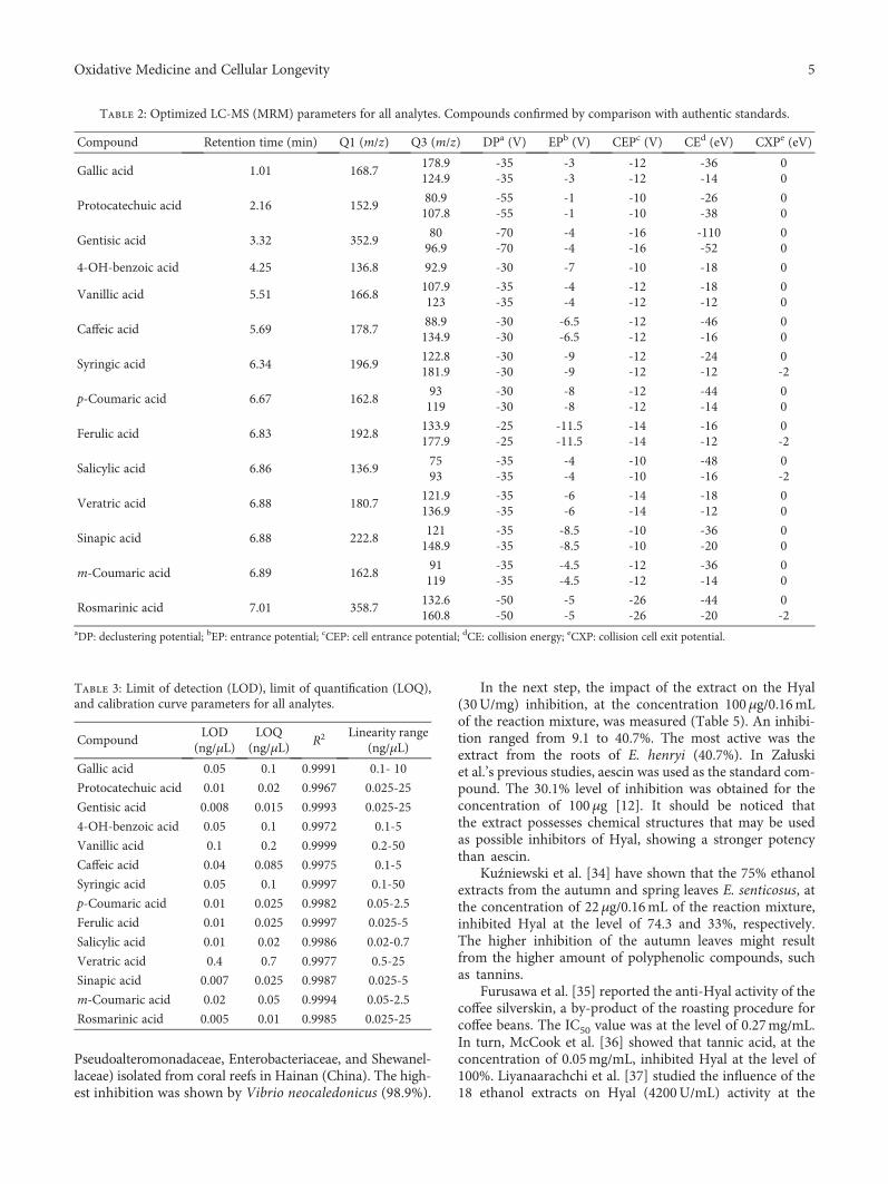
Pseudoalteromonadaceae, Enterobacteriaceae, and Shewanel-laceae) isolated from coral reefs in Hainan (China). The high-est inhibition was shown by Vibrio neocaledonicus (98.9%).
In the next step, the impact of the extract on the Hyal(30U/mg) inhibition, at the concentration 100μg/0.16mLof the reaction mixture, was measured (Table 5). An inhibi-tion ranged from 9.1 to 40.7%. The most active was theextract from the roots of E. henryi (40.7%). In Załuskiet al.’s previous studies, aescin was used as the standard com-pound. The 30.1% level of inhibition was obtained for theconcentration of 100μg [12]. It should be noticed thatthe extract possesses chemical structures that may be usedas possible inhibitors of Hyal, showing a stronger potencythan aescin.
Kuźniewski et al. [34] have shown that the 75% ethanolextracts from the autumn and spring leaves E. senticosus, atthe concentration of 22μg/0.16mL of the reaction mixture,inhibited Hyal at the level of 74.3 and 33%, respectively.The higher inhibition of the autumn leaves might resultfrom the higher amount of polyphenolic compounds, suchas tannins.
Furusawa et al. [35] reported the anti-Hyal activity of thecoffee silverskin, a by-product of the roasting procedure forcoffee beans. The IC50 value was at the level of 0.27mg/mL.In turn, McCook et al. [36] showed that tannic acid, at theconcentration of 0.05mg/mL, inhibited Hyal at the level of100%. Liyanaarachchi et al. [37] studied the influence of the18 ethanol extracts on Hyal (4200U/mL) activity at the
Table 2: Optimized LC-MS (MRM) parameters for all analytes. Compounds confirmed by comparison with authentic standards.
Compound Retention time (min) Q1 (m/z) Q3 (m/z) DPa (V) EPb (V) CEPc (V) CEd (eV) CXPe (eV)
Gallic acid 1.01 168.7178.9124.9
-35-35
-3-3
-12-12
-36-14
00
Protocatechuic acid 2.16 152.980.9107.8
-55-55
-1-1
-10-10
-26-38
00
Gentisic acid 3.32 352.98096.9
-70-70
-4-4
-16-16
-110-52
00
4-OH-benzoic acid 4.25 136.8 92.9 -30 -7 -10 -18 0
Vanillic acid 5.51 166.8107.9123
-35-35
-4-4
-12-12
-18-12
00
Caffeic acid 5.69 178.788.9134.9
-30-30
-6.5-6.5
-12-12
-46-16
00
Syringic acid 6.34 196.9122.8181.9
-30-30
-9-9
-12-12
-24-12
0-2
p-Coumaric acid 6.67 162.893119
-30-30
-8-8
-12-12
-44-14
00
Ferulic acid 6.83 192.8133.9177.9
-25-25
-11.5-11.5
-14-14
-16-12
0-2
Salicylic acid 6.86 136.97593
-35-35
-4-4
-10-10
-48-16
0-2
Veratric acid 6.88 180.7121.9136.9
-35-35
-6-6
-14-14
-18-12
00
Sinapic acid 6.88 222.8121148.9
-35-35
-8.5-8.5
-10-10
-36-20
00
m-Coumaric acid 6.89 162.891119
-35-35
-4.5-4.5
-12-12
-36-14
00
Rosmarinic acid 7.01 358.7132.6160.8
-50-50
-5-5
-26-26
-44-20
0-2
aDP: declustering potential; bEP: entrance potential; cCEP: cell entrance potential; dCE: collision energy; eCXP: collision cell exit potential.
Table 3: Limit of detection (LOD), limit of quantification (LOQ),and calibration curve parameters for all analytes.
CompoundLOD
(ng/μL)LOQ
(ng/μL)R2 Linearity range
(ng/μL)
Gallic acid 0.05 0.1 0.9991 0.1- 10
Protocatechuic acid 0.01 0.02 0.9967 0.025-25
Gentisic acid 0.008 0.015 0.9993 0.025-25
4-OH-benzoic acid 0.05 0.1 0.9972 0.1-5
Vanillic acid 0.1 0.2 0.9999 0.2-50
Caffeic acid 0.04 0.085 0.9975 0.1-5
Syringic acid 0.05 0.1 0.9997 0.1-50
p-Coumaric acid 0.01 0.025 0.9982 0.05-2.5
Ferulic acid 0.01 0.025 0.9997 0.025-5
Salicylic acid 0.01 0.02 0.9986 0.02-0.7
Veratric acid 0.4 0.7 0.9977 0.5-25
Sinapic acid 0.007 0.025 0.9987 0.025-5
m-Coumaric acid 0.02 0.05 0.9994 0.05-2.5
Rosmarinic acid 0.005 0.01 0.9985 0.025-25
5Oxidative Medicine and Cellular Longevity
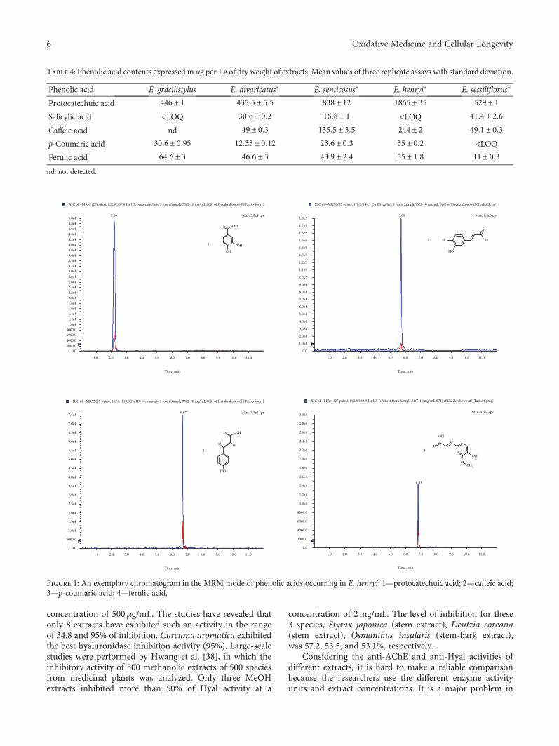
concentration of 500μg/mL. The studies have revealed thatonly 8 extracts have exhibited such an activity in the rangeof 34.8 and 95% of inhibition. Curcuma aromatica exhibitedthe best hyaluronidase inhibition activity (95%). Large-scalestudies were performed by Hwang et al. [38], in which theinhibitory activity of 500 methanolic extracts of 500 speciesfrom medicinal plants was analyzed. Only three MeOHextracts inhibited more than 50% of Hyal activity at a
concentration of 2mg/mL. The level of inhibition for these3 species, Styrax japonica (stem extract), Deutzia coreana(stem extract), Osmanthus insularis (stem-bark extract),was 57.2, 53.5, and 53.1%, respectively.
Considering the anti-AChE and anti-Hyal activities ofdifferent extracts, it is hard to make a reliable comparisonbecause the researchers use the different enzyme activityunits and extract concentrations. It is a major problem in
Table 4: Phenolic acid contents expressed in μg per 1 g of dry weight of extracts. Mean values of three replicate assays with standard deviation.
Phenolic acid E. gracilistylus E. divaricatus∗ E. senticosus∗ E. henryi∗ E. sessiliflorus∗
Protocatechuic acid 446 ± 1 435 5 ± 5 5 838 ± 12 1865 ± 35 529 ± 1Salicylic acid <LOQ 30 6 ± 0 2 16 8 ± 1 <LOQ 41 4 ± 2 6Caffeic acid nd 49 ± 0 3 135 5 ± 3 5 244 ± 2 49 1 ± 0 3p-Coumaric acid 30 6 ± 0 95 12 35 ± 0 12 23 6 ± 0 3 55 ± 0 2 <LOQFerulic acid 64 6 ± 3 46 6 ± 3 43 9 ± 2 4 55 ± 1 8 11 ± 0 3nd: not detected.
O OH
OHOH
1
1.0 2.0 3.0 4.0 5.0 6.0 7.0 8.0 9.0 10.0 11.0
0.02000.04000.06000.08000.0
1.0e41.2e41.4e41.6e41.8e42.0e42.2e42.4e42.6e42.8e43.0e43.2e43.4e43.6e43.8e44.0e44.2e44.4e44.6e44.8e4
2.165.0e4
Time, min
XIC of −MRM (27 pairs): 152.9/107.8 Da ID: protocatechuic 1 from Sample 75(2-10 mg/mL 066) of Datakrakow.wiff (Turbo Spray)
1.0 2.0 3.0 4.0 5.0 6.0 7.0 8.0 9.0 10.0 11.0
0.0
1.0e4
2.0e4
3.0e4
4.0e4
5.0e4
6.0e4
7.0e4
8.0e4
9.0e4
1.0e5
1.1e5
1.2e5
1.3e5
1.4e5
1.5e5
1.6e5
1.7e5
1.8e55.69
2 HO
HO
OH
O
Max. 1.8e5 cps
Time, min
XIC of −MRM (27 pairs): 178.7/134.9 Da ID: caffeic 1 from Sample 75(2-10 mg/mL 066) of Datakrakow.wiff (Turbo Spray)
1.0 2.0 3.0 4.0 5.0 6.0 7.0 8.0 9.0 10.0 11.0
0.0
2000.0
4000.0
6000.0
8000.0
1.0e4
1.2e4
1.4e4
1.6e4
1.8e4
2.0e4
2.2e4
2.4e4
2.6e4
2.8e4
3.0e4
6.83
O
OH
OH
OCH3
4
Max. 3.0e4 cps
Time, min
XIC of −MRM (27 pairs): 192.8/133.9 Da ID: ferulic 1 from Sample 81(5-10 mg/mL 072) of Datakrakow.wiff (Turbo Spray)
Max. 5.0e4 cps
1.0 2.0 3.0 4.0 5.0 6.0 7.0 8.0 9.0 10.0 11.00.0
5000.0
1.0e4
1.5e4
2.0e4
2.5e4
3.0e4
3.5e4
4.0e4
4.5e4
5.0e4
5.5e4
6.0e4
6.5e4
7.0e4
7.5e4 6.67
HO
HH
O
3
OH
Time, min
XIC of −MRM (27 pairs): 162.8 /119.0 Da ID: p-coumaric 1 from Sample 75(2-10 mg/mL 066) of Datakrakow.wiff (Turbo Spray)
Max. 7.5e4 cps
Figure 1: An exemplary chromatogram in the MRM mode of phenolic acids occurring in E. henryi: 1—protocatechuic acid; 2—caffeic acid;3—p-coumaric acid; 4—ferulic acid.
6 Oxidative Medicine and Cellular Longevity

antienzymatic activity assays for which the general protocolsfor the assay should be developed. When the enzyme activityunit was given, we included it in the manuscript. This prob-lem has been resolved, e.g., in case of cytotoxic activity, whereaccording to the National Cancer Institute (United States)plant screening program, a crude extract is generally consid-ered to have in vitro cytotoxic activity if the IC50 is<20 μg/mL. For compounds, the limit has been establishedas 4 μg/mL [39].
3.4. Cytotoxic Activity. About 6 million of people die of can-cer each year, and this trend is about to grow in the nextdecades. Leukemia is a haematological disease caused by per-sistence of immature white blood cells in different compart-ments of the organism mainly the bone marrow, lymphnode, spleen, and circulating blood. Plant-based chemicalsare a promising group of antileukemic agents, especially thepolyphenol group [40, 41]. Because Eleutherococcus spp.contain polyphenols, we have studied an antileukemic activ-ity of the extracts. The extracts inhibited a HL-60 cell linegrowth with an IC50 value in the range of 270-2000μg/mL(Table 6). As a positive control, podophyllotoxin was used,with the IC50 value of 0.0084μg/mL. The previous studiesof Załuski et al. [42], on the cytotoxic activity of the 75% eth-anol extracts from the roots of Eleutherococcus spp., haverevealed that the IC50 values ranged between 49 and522μg/mL, with the highest for E. divaricatus (49μg/mL).Comparing the 75% EtOH extracts to the 75% MeOHextracts, it is seen that both types exhibited a weak inhib-itory activity.
The higher IC50 values have been obtained for the iso-lated compounds and their derivatives. Hu et al. [43] studiedthe cytotoxic activities of five stilbenoids isolated from thestem bark of Acanthopanax leucorrhizus and six derivativesagainst HL-60. Among 17 compounds, only two derivatives(E)-3,5-dimethoxy-3′,4′-methylenedioxystilbene and 2-(3′-hydroxy-4′-isopentenyl-5′-methoxyphenyl)-4-hydroxyben-zofuran exhibited moderate cytotoxicities against HL-60cell lines with the lower IC50 values of 30 51 ± 5 37 and40 23 ± 6 12 μM, respectively. In turn, isolates showed aweak inhibitory activity against HL-60, in the range of98.6-200μM.
The cytotoxic activity, against HL-60, was also exhibitedby the different extracts [petroleum ether, Ch2Cl2, EtOAc,
EtOH, and EtOH/H2O (1 : 1 v/v)] prepared from the aerialparts of Artemisia biennis Willd. Nevertheless, high IC50values have been obtained, in the range of 54.3-200μg/mL[44]. The n-hexane, Ch2Cl2, EtOAc, EtOH, and EtOH/H2O(1 : 1 v/v) extracts from another Artemisia species, Artemisiaturanica Krasch., have also shown a weak cytotoxic effectwith the IC50 values of 68.8-450μg/mL [45]. It should bementioned that petroleum ether, dichloromethane, ethyl ace-tate, ethanol, and 50% aqueous ethanol extracts of the aerialparts of Artemisia ciniformis inhibited HL60 cell growth withthe IC50 value of 31.3-200μg/mL [46].
Teixeira et al. [47] reported on the cytotoxic activity offour flavonoids isolated from the roots of Tephrosia egregiaSandwith. Three compounds (pongaflavone, praecansoneB, and 12a-hydroxyrotenone) exhibited a high activityagainst HL-60 cell line with the IC50 values of 1.4, 8.1, and1.9μg/mL.
Taking into account the antileukemic activity of poly-phenols, we noticed that E. henryi was the most activetowards HL-60 and contains the highest amount of proto-catechuic, p-coumaric, and caffeic acids. Another observa-tion resulted from this study is that nonpolar extractsare more cytotoxic than polar ones. A moderate/low cyto-toxicity in our study may result from a high polarity ofextract constituents, for which a cell wall penetration islimited. Further studies are needed to confirm this activityfor the isolated compounds.
3.5. The Capacity of DPPH∗ Reduction. It is known that freeradicals are responsible for the development of many dis-eases, such as Parkinson’s disease, Alzheimer’s disease, oramyotrophic latera sclerosis. Free radicals are constantlyproduced in the brain in vivo, contributing to oxidativedamage of proteins and lipids. For this reason, in searchingfor new antioxidants, a special attention should be paid tocompounds that act as an antioxidant and inhibitor ofAChE or BuChE. Nature is a significant source of health-promoting compounds that may be divided into two groups:fruits and vegetables and medicinal plants [48]. Therefore,in the next step, an antioxidative activity of the extractswas examined. The extracts reduced DPPH∗ in a time-dependent mode, at the concentration of 0.8mg/mL(Figure 2). After 90min from 14.7 to 26.2%, DPPH∗wasreduced. As a positive control, ascorbic acid was used, withan EC50 value of 38μg/mL (after 5min of incubation). Aslow change of antioxidant activity in the time is in agree-ment with the adaptogen’s definition, according to which,adaptogen should act slowly in a long-term mode. It shouldbe noticed that E. gracilistylus expressed the highest anti-AChE activity and anti-DPPH∗ scavenging. Usually, freeradicals are associated with the neurodegenerative processdevelopment; in this case, we can suppose that E. gracilisty-lus contain both compounds with anti-AChE activity andanti-DPPH∗.
Since a few years, the main attention has been paid tofungi as a new source of antioxidants. By far, they were onlyof culinary interest; however, around 650 species containtherapeutic properties, e.g., Lentinus edodes, Boletus edulis,or Grifola frondosa. They are rich in phenolic acids, such as
Table 5: Antihyaluronidase and antiacetylcholinesterase activitiesof extracts.
SampleAnti-AChE∗ Anti-Hyal∗
(% inhibition)
E. gracilistylus 32 ± 0 8 14 9 ± 0 3E. divaricatus 23 2 ± 0 9 11 ± 0 2E. senticosus 26 1 ± 0 05 10 4 ± 0 6E. henryi 19 6 ± 0 4 40 7 ± 1 1E. sessiliflorus 32 ± 0 6 9 1 ± 0 05∗Results are the means ± standard deviation of triplicates.
7Oxidative Medicine and Cellular Longevity

cinnamic, protocatechuic, p-hydroxybenzoic, p-coumaric,and gallic [48].
The acetonic extracts from Boletus edulis and Boletus aes-tivalis have showed a powerful antioxidant activity with theIC50 value of 4.72 and 8.63μg/mL which were similar orgreater than those of the standard antioxidants, ascorbic acid(EC50 4.22μg/mL), BHA (EC50 6.42μg/mL), and α-tocoph-erol (EC50 62.43μg/mL) [49]. Higher values have beenobtained for Agaricus brasiliensis and Agaricus bisporus.The 60% ethanol extracts from those fungi have shown a rad-ical scavenging capability for EC50 1.6 and 2.7mg/mL. Inturn, the aqueous extracts have had the EC50 value at the levelof 3.8 and 4.5mg/mL [50].
4. Conclusions
The ethnopharmacological uses of Eleutherococcus spp. arenow confirmed with an application of new approaches andtools, in in vitro and in vivo models. Considering the inhibi-tion of an AChE activity and DPPH∗ reduction, new inhibi-tors should be searched for in the roots of E. gracilistylus,while inhibitors with an anti-Hyal or cytotoxic activityshould be searched for in the roots of E. henryi. New Hyalinhibitors may have a possible use as ingredients of plant-based products, used, e.g., topically as cosmetics or intreatment of skin diseases. Taking into account the resultsobtained in this work, a further research should focus onthe isolation of single phytochemicals being responsible forthe anti-AChE, anti-Hyal, and cytotoxic effects. Furtherresearches are needed to determine the antienzymatic activ-ity with the use of modern approaches, such as the isothermalcalorimetric titration (ITC).
Abbreviations
TPC: Total phenolic contentTFC: Total flavonoid contentDNPH: 2,4-DinitrophenylhydrazineFeCl3: Iron (III) chlorideGA: Gallic acidHE: Hesperetin equivalentQE: Quercetin equivalentDPPH∗: 2,2-Diphenyl-1-picrylhydrazylHL-60: Human Caucasian promyelocytic leukemia from
American Type Culture Collection (ATCC CCL-240™)
Hyal: HyaluronidaseAChE: Acetylcholinesterase.
Data Availability
The data used to support the findings of this study areavailable from the corresponding author upon request.
Consent
Informed consent was obtained from all the participantsincluded in the work.
Conflicts of Interest
The authors declare no competing financial interest.
Acknowledgments
This paper was developed using the equipment purchasedwithin the project “The equipment of innovative laboratoriesdoing research on new medicines used in the therapy of civ-ilization and neoplastic diseases” within the Operational Pro-gram Development of Eastern Poland 2007-2013, PriorityAxis I Modern Economy, Operations I.3 Innovation Pro-motion. This work was funded by the financial supportof academic research in the Ludwik Rydygier CollegiumMedicum in Bydgoszcz, Nicolaus Copernicus Universityin Toruń.
References
[1] W. Rungsung, K. K. Ratha, S. Dutta, A. K. Dixit, and J. Hazra,“Secondary metabolites of plants in drugs discovery,” WorldJournal of Pharmaceutical Research, vol. 4, pp. 604–613, 2015.
[2] Ł. Ciesla, M. Waksmundzka-Hajnos, D. Zaluski, H. D. Smo-larz, and M. Hajnos, “HPTLC-densitometric method fordetermination of eleutherosides B, E, and E1 in differentEleutherococcus species,” Journal of Chromatographic Science,vol. 49, no. 3, pp. 182–188, 2011.
DPPH reduction (%)
1 min 5 min 10 min 60 min 90 min
25 23.4 ± 0.44
19.4 ± 0.19
18.9 ± 0.62 16.4 ± 0.4213.7 ± 0.3
13.3 ± 0.43
8.1 ± 0.23 9.7 ± 0.29
13.6 ± 0.7614.7 ± 0.57
14.7 ± 0.9413.7 ± 0.48
17.9 ± 0.92 18.1 ± 0.8919.7 ±17.8 ± 0.43
18.9 ± 0.49 23 ± 0.7920.3 ± 0.7
21 ± 0.63 21.5 ± 0.6222.5 ± 0.8
24.1 ± 0.11 24.7 ± 0.8326.2 ± 0.9
20
0
5
10
15
E. gracilistylusE. divaricatus
E. henryiE. sessiliflorusE. senticosus
Figure 2: A time-dependent DPPH∗ reduction (%).
Table 6: The cytotoxic impact of the extracts on HL-60 cell line (IC50 μg/mL).
E. gracilistylus E. divaricatus∗ E. senticosus∗ E. henryi∗ E. sessiliflorus∗
890 ± 3 7 650 ± 2 9 450 ± 2 5 270 ± 1 1 2000 ± 3 4
8 Oxidative Medicine and Cellular Longevity

[3] A. Panossian, “Understanding adaptogenic activity: specificityof the pharmacological action of adaptogens and other phyto-chemicals,” Annals of the New York Academy of Sciences,vol. 1401, pp. 49–64, 2017.
[4] Q. Li, Y. Jia, L. Xu et al., “Simultaneous determination of pro-tocatechuic acid, syringin, chlorogenic acid, caffeic acid, lirio-dendrin and isofraxidin in Acanthopanax senticosus Harmsby HPLC-DAD,” Biological & Pharmaceutical Bulletin,vol. 29, pp. 532–534, 2006.
[5] A. Panossian, E. J. Seo, and T. Efferth, “Novel molecular mech-anisms for the adaptogenic effects of herbal extracts on isolatedbrain cells using systems biology,” Phytomedicine, vol. 50,pp. 257–284, 2018.
[6] D. Załuski, M. Olech, R. Kuźniewski, R. Verpoorte, R. Nowak,and H. D. Smolarz, “LC-ESI-MS/MS profiling of phenolicsfrom Eleutherococcus spp. inflorescences, structure-activityrelationship as antioxidants, inhibitors of hyaluronidase andacetylcholinesterase,” Saudi Pharmaceutical Journal, vol. 25,pp. 734–743, 2017.
[7] D. Załuski, E. Mendyk, and H. D. Smolarz, “Identification ofMMP-1 andMMP-9 inhibitors from the roots of Eleutherococ-cus divaricatus, and the PAMPA test,” Natural ProductResearch, vol. 30, pp. 595–599, 2016.
[8] D. Załuski and Z. Janeczko, “Variation in phytochemicals andbioactivity of the fruits of Eleutherococcus species cultivated inPoland,” Natural Product Research, vol. 29, no. 23, pp. 2207–2211, 2015.
[9] Y. Zhou, C. Cheng, D. Baranenko, J. Wang, Y. Li, andW. Lu, “Effects of Acanthopanax senticosus on brain injuryinduced by simulated spatial radiation in mouse modelbased on pharmacokinetics and comparative proteomics,”International Journal of Molecular Sciences, vol. 19, no. 1,p. 159, 2018.
[10] M. X. Lu, Y. Yang, Q. P. Zou et al., “Anti-diabetic effects ofAcankoreagenin from the leaves of Acanthopanax gracilistylusherb in RIN-m5F cells via suppression of NF-κB activation,”Molecules, vol. 23, no. 4, p. 958, 2018.
[11] W. Dimpfel, L. Schombert, and A. G. Panossian, “Assessingthe quality and potential efficacy of commercial extracts ofRhodiola rosea L. by analyzing the salidroside and rosavin con-tent and the electrophysiological activity in hippocampallong-term potentiation, a synaptic model of memory,” Fron-tiers in Pharmacology, vol. 9, p. 425, 2018.
[12] D. Załuski, M. Olech, A. Galanty et al., “Phytochemical con-tent and pharma-nutrition study on Eleutherococcus senticosusfruits intractum,” Oxidative Medicine and Cellular Longevity,vol. 2016, Article ID 9270691, 10 pages, 2016.
[13] K. Madan and S. Nanda, “In-vitro evaluation of antioxidant,anti-elastase, anti-collagenase, anti-hyaluronidase activities ofsafranal and determination of its sun protection factor in skinphotoaging,” Bioorganic Chemistry, vol. 77, pp. 159–167, 2018.
[14] D. Załuski, Ł. Cieśla, and Z. Janeczko, “The structure-activityrelationships of plant secondary metabolites with antimicro-bial, free radical scavenging and inhibitory activity towardsselected enzymes,” in Studies in Natural Product Chemistry(Bioactive Natural Products), A.-u. Rahman, Ed., vol. 45,pp. 217–249, Elsevier science publishers, Amsterdam, theNetherlands, 2015.
[15] C. Tohda, M. Ichimura, and Y. Bai, “Inhibitory effects ofEleutherococcus senticosus extracts on amyloid β(25-35)–induced neuritic atrophy and synaptic loss,” Journal of Phar-macological Sciences, vol. 107, pp. 329–339, 2008.
[16] D. Lee, “Neuroprotective effects of Eleutherococcus senticosusbark on transient global cerebral ischemia in rats,” Journal ofEthnopharmacology, vol. 139, pp. 6–11, 2012.
[17] V. L. Singleton and J. A. Rossi, “Colometry of total pheno-lics with phosphomolybdic- phospotungstic acid reagents,”American Journal of Enology Viticulture, vol. 16, pp. 144–158, 1965.
[18] C. C. Chang, M. H. Yang, H. M. Wen, and J. C. Chern, “Esti-mation of total flavonoid content in propolis by two comple-mentary colorimetric methods,” Journal of Food and DrugAnalysis, vol. 10, pp. 178–182, 2002.
[19] N. Nowacka, R. Nowak, M. Drozd, M. Olech, R. Los, andA. Malm, “Analysis of phenolic constituents, antiradical andantimicrobial activity of edible mushrooms growing wild inPoland,” LWT - Food Science and Technology, vol. 59,pp. 689–694, 2014.
[20] W. Pietrzak, R. Nowak, U. Gawlik-Dziki, M. K. Lemieszek, andW. Rzeski, “LC-ESI-MS/MS identification of biologicallyactive phenolic compounds in mistletoe berry extracts fromdifferent host trees,” Molecules, vol. 22, no. 4, p. 624, 2017.
[21] Y. A. Yahaya and M. M. Don, “Evaluation of Trametes lactineaextracts on the inhibition of hyaluronidase, lipoxygenase andxanthine oxidase activities in vitro,” Journal of Physical Science,vol. 23, no. 2, pp. 1–15, 2012.
[22] G. L. Ellman, K. D. Courtney, V. Andres jr., and R. M. Feath-erstone, “A new and rapid colorimetric determination of ace-tylcholinesterase activity,” Biochemical Pharmacology, vol. 7,no. 2, pp. 88–95, 1961.
[23] W. Brand-Williams, M. E. Cuvelier, and C. Berset, “Use of afree radical method to evaluate antioxidant activity,” LWT -Food Science and Technology, vol. 28, pp. 25–30, 1995.
[24] D. Załuski, H. D. Smolarz, and U. Gawlik-Dziki, “Bioactivecompounds and antioxidative, antileukemic and anti-MMPsactivity of Eleutherococcus species cultivated in Poland,” Nat-ural Product Communications, vol. 7, no. 11, pp. 1483–1486,2012.
[25] M. Ondrejovič, D. Chmelová, and T. Maliar, “Optimization ofthe antioxidant extraction from Eleutherococcus senticosusroots by response surface methodology,” Nova Biotechnologicaet Chimica, vol. 12, no. 1, pp. 30–38, 2013.
[26] D. Załuski, M. Olech, R. Verpoorte, I. Khan, R. Kuźniewski,and R. Nowak, “Phytoconstituents and nutritional propertiesof the fruits of Eleutherococcus divaricatus and Eleutherococcussessiliflorus: a study of non-European species cultivated inPoland,” Oxidative Medicine and Cellular Longevity,vol. 2017, Article ID 8374295, 7 pages, 2017.
[27] L. Tan, S. Guo, F. Ma, C. Chang, and I. Gómez-Betancur, “Invitro inhibition of acetylcholinesterase, alphaglucosidase, andxanthine oxidase by bacteria extracts from coral reef in Hai-nan, South China Sea,” Journal of Marine Science and Engi-neering, vol. 6, no. 2, p. 33, 2018.
[28] S. Thabthimsuk, N. Panya, A. Owatworakit, andW. Choksawangkarn, “Acetylcholinesterase inhibition andantioxidant activities of polysaccharide-peptide complexesfrom edible mushrooms,” in The 6th International Conferenceon Biochemistry andMolecular Biology, Thailand, 2018.
[29] M. T. N. S. Lima, L. B. d. Santos, R. W. Bastos, J. R. Nicoli, andJ. A. Takahashi, “Antimicrobial activity and acetylcholinester-ase inhibition by extracts from chromatin modulated fungi,”Brazilian Journal of Microbiology, vol. 49, no. 1, pp. 169–176,2018.
9Oxidative Medicine and Cellular Longevity

[30] D. Załuski and R. Kuźniewski, “In vitro anti-AChE, anti--BuChE, and antioxidant activity of 12 extracts of Eleutherococ-cus species,” Oxidative Medicine and Cellular Longevity,vol. 2016, Article ID 4135135, 7 pages, 2016.
[31] J. Niño, J. A. Hernández, Y. M. Correa, and O. M. Mosquera,“In vitro inhibition of acetylcholinesterase by crude plantextracts from Colombian flora,” Memórias do InstitutoOswaldo Cruz, vol. 101, no. 7, pp. 783–785, 2006.
[32] L. L. Nwidu, E. Elmorsy, J. Thornton et al., “Anti-acetylcholi-nesterase activity and antioxidant properties of extracts andfractions of Carpolobia lutea,” Pharmaceutical Biology,vol. 55, pp. 1875–1883, 2017.
[33] A. Kostelnik andM. Pohanka, “Inhibition of acetylcholinester-ase and butyrylcholinesterase by a plant secondary metaboliteboldine,” BioMed Research International, vol. 2018, Article ID9634349, 5 pages, 2018.
[34] R. Kuźniewski, D. Załuski, M. Olech, P. Banaszczak, andR. Nowak, “LC-ESI-MS/MS profiling of phenolics in the leavesof Eleutherococcus senticosus cultivated in the West Europeand anti-hyaluronidase and anti-acetylcholinestarase activi-ties,” Natural Product Research, vol. 32, pp. 448–452, 2018.
[35] M. Furusawa, Y. Narita, K. Iwai, T. Fukunaga, and O. Nakagiri,“Inhibitory effect of a hot water extract of coffee “silverskin” onhyaluronidase,” Bioscience, Biotechnology, and Biochemistry,vol. 75, pp. 1205–1207, 2011.
[36] J. McCook, P. Dorogi, D. Vasily, and D. Cefalo, “In vitro inhi-bition of hyaluronidase by sodium copper chlorophyllin com-plex and chlorophyllin analogs,” Clinical, Cosmetic andInvestigational Dermatology, vol. 8, pp. 443–448, 2015.
[37] G. D. Liyanaarachchi, J. K. R. R. Samarasekera, K. R. R. Maha-nama, and K. D. P. Hemalal, “Tyrosinase, elastase, hyaluroni-dase, inhibitory and antioxidant activity of Sri Lankanmedicinal plants for novel cosmeceuticals,” Industrial Cropsand Products, vol. 111, pp. 597–605, 2018.
[38] S. G. Hwang, A. Yang, S. J. Kim et al., “Screening of hyaluron-idase inhibitor in Korean medicinal plants,” Journal of Life Sci-ence, vol. 24, pp. 498–504, 2014.
[39] V. Kuete, B. Wiench, M. E. F. Hegazy et al., “Antibacterialactivity and cytotoxicity of selected Egyptian medicinalplants,” Planta Medica, vol. 78, pp. 193–199, 2012.
[40] S. Taverna and C. Corrado, “Natural compounds: molecularweapons against Leukemia’s,” Journal of Leukemia, vol. 5,no. 1, p. 226, 2017.
[41] Y. Han, A. H. Zhang, Y. Z. Zhang, H. Sun, X. C. Meng, and X. J.Wang, “Chemical metabolomics for investigating the protec-tive effectiveness of Acanthopanax senticosus Harms leafagainst acute promyelocytic leukemia,” RSC Advances, vol. 8,no. 22, pp. 11983–11990, 2018.
[42] D. Załuski, H. D. Smolarz, and A. Bogucka-Kocka, “Cyto-toxic activity of ethanolic extracts of Eleutherococcus spe-cies cultivated in Poland on HL60 leukemia cell line,”Current Issues in Pharmacy and Medical Sciences, vol. 27,pp. 41–45, 2014.
[43] H. B. Hu, H. P. Liang, H. M. Li et al., “Isolation, modificationand cytotoxic evaluation of stilbenoids from Acanthopanaxleucorrhizus,” Fitoterapia, vol. 124, pp. 167–176, 2018.
[44] Z. Tayarani-Najaran, F. S. Makki, N. S. Alamolhodaei,M.Mojarrab, and S.A. Emami, “Cytotoxic and apoptotic effectsof different extracts of Artemisia biennis Willd. on K562 andHL-60 cell lines,” Iranian Journal of Basic Medical Sciences,vol. 20, no. 2, pp. 166–171, 2017.
[45] Z. Tayarani-Najaran, M. Sareban, A. Gholami, S. A. Emami,and M. Mojarrab, “Cytotoxic and apoptotic effects of differentextracts of Artemisia turanica Krasch. on K562 and HL-60 celllines,” The Scientific World Journal, vol. 2013, Article ID628073, 6 pages, 2013.
[46] Z. Tayarani-Najaran, Z. Hajian, M. Mojarrab, and S. A.Emami, “Cytotoxic and apoptotic effects of extracts of Artemi-sia ciniformis Krasch. & Popov ex Poljakov on K562 andHL-60 cell lines,” Asian Pacific Journal of Cancer Prevention,vol. 15, pp. 7055–7059, 2014.
[47] M. V. S. Teixeira, J. Q. Lima, A. T. A. Pimenta et al., “New fla-vone and other compounds from Tephrosia egregia: assessingthe cytotoxic effect on human tumor cell lines,” Revista Brasi-leira de Farmacognosia, vol. 28, no. 3, pp. 333–338, 2018.
[48] A. Haseeb, H. Ghulam, and M. Imtiaz, “Antioxidants fromnatural sources,” in Chapter 1 in Book: Antioxidants in Foodand Its Application, pp. 1–27, IntechOpen Publisher, 2018.
[49] M. Kosanić, B. Ranković, and M. Dašić, “Mushrooms as possi-ble antioxidant and antimicrobial agents,” Iranian Journal ofPharmaceutical Research, vol. 11, no. 4, pp. 1095–1102, 2012.
[50] C. H. Gan, B. Nurul Amira, and R. Asmah, “Antioxidant anal-ysis of different types of edible mushrooms (Agaricus bisporousand Agaricus brasiliensis),” International Food Research Jour-nal, vol. 20, pp. 1095–1102, 2013.
10 Oxidative Medicine and Cellular Longevity

Research ArticleHonokiol-Mediated Mitophagy AmelioratesPostoperative Cognitive Impairment Induced bySurgery/Sevoflurane via Inhibiting the Activation ofNLRP3 Inflammasome in the Hippocampus
Ji-Shi Ye,1 Lei Chen,2 Ya-Yuan Lu,2 Shao-Qing Lei ,1 Mian Peng ,2
and Zhong-Yuan Xia 1
1Department of Anesthesiology, Renmin Hospital of Wuhan University, Wuhan, 430060 Hubei, China2Department of Anesthesiology, Zhongnan Hospital of Wuhan University, 169, Donghu Road, Wuhan, 430071 Hubei, China
Correspondence should be addressed to Zhong-Yuan Xia; [email protected]
Received 5 August 2018; Revised 16 October 2018; Accepted 10 December 2018; Published 24 February 2019
Guest Editor: Luciana Scotti
Copyright © 2019 Ji-Shi Ye et al. This is an open access article distributed under the Creative Commons Attribution License, whichpermits unrestricted use, distribution, and reproduction in any medium, provided the original work is properly cited.
Background. The potential mechanism of postoperative cognitive impairment is still largely unclear. The activation of NLRP3inflammasome had been reported to be involved in neurodegenerative diseases, including postoperative cognitive change, and isclosely related to mitochondrial ROS and mitophagy. Honokiol (HNK) owns multiple organic protective effects. This study isaimed at observing the neuroprotective effect of HNK in postoperative cognitive change and examining the role of HNK in theregulation of mitophagy and the relationship between these effects and NLRP3 inflammasome activation in mice induced bysurgery/anesthesia. Methods. In this study, mice were divided into several groups: control group, surgery group, surgery+HNKgroup, and surgery+HNK+3-methyladenine (3-MA) group. Hippocampal tissue samples were harvested and used forproinflammatory cytokines, mitochondrial ROS, and malondialdehyde (MDA) assay. The process of mitophagy and theactivation of NLRP3 inflammasome were observed by Western blot, immunohistochemistry, and transmission electronmicroscopy. Results. The results showed that HNK treatment obviously recovered the postoperative decline and enhanced theexpressions of LC3-II, Beclin-1, Parkin, and PINK1 at protein levels after surgery/sevoflurane treatment, which are both anautophagy marker and a mitophagy marker. In addition, HNK attenuated mitochondrial structure damage and reducedmtROS and MDA generation, which are closely associated with NLRP3 inflammasome activation. Honokiol-mediatedmitophagy inhibited the activation of NLRP3 inflammasome and neuroinflammation in the hippocampus. Using 3-MA, anautophagy inhibitor, the neuroprotective effects of HNK on mitophagy and NLRP3 inflammasome activation were eliminated.Conclusion. These results indicated that HNK-mediated mitophagy ameliorates postoperative cognitive impairment induced bysurgery/sevoflurane. This neuroprotective effect may be involved in inhibiting the activation of NLRP3 inflammasome andsuppressing inflammatory responses in the hippocampus.
1. Introduction
Surgery/anesthesia is often an inevitable medical interven-tion in many patients during hospitalization. Postoperativecognitive decline (POCD) describes a cluster of cognitivebehavior abnormalities including a relative drop in learn-ing and memory performance on a set of neuropsycholog-ical tests from before to after surgery [1]. Dissecting themechanisms of POCD becomes important, not only
because it is a pathophysiological problem that we donot yet illuminate completely but also because it is acommon postoperative complication that affects the qual-ity of the patients’ daily life and long-term outcome [2].Like Alzheimer’s disease (AD) and other neurodegenera-tive diseases, the potential pathophysiological mechanismof POCD may also be involved in neuroinflammation,oxidative stress, blood-brain barrier dysfunction, andapoptosis [2–6].
HindawiOxidative Medicine and Cellular LongevityVolume 2019, Article ID 8639618, 13 pageshttps://doi.org/10.1155/2019/8639618

In recent years, several lines of studies have focused theirattention on inflammasomes, which are essential compo-nents of the innate immune system and play a pivotal rolein pro- or anti-inflammatory homeostasis [6–8]. Inflamma-somes are intracellular multiprotein complexes that drivethe activation of inflammatory responses. Among all typesof inflammasomes, such as NLRP1, NLRP3, NLRC4, andAIM2, NLRP3 is the most studied one, especially in thecentral neural system [9, 10]. NLRP3 inflammasome activa-tion could recruit and activate Caspase-1, leading to thesecretion of mature IL-1β and IL-18 and the initiation of anovel form of cell death named pyroptosis [11]. Emergingevidence showed that NLRP3 inflammasomes could be iden-tified in microglia, astrocytes and neurons, which inducedneuroinflammation in a series of neurodegenerative diseases[7, 8, 12, 13]. So, in the surgery/sevoflurane model, we canobserve whether the NLRP3 inflammasome was activatedand could influence the neurological outcome. Moreover, aset of researches have also uncovered that the high levels ofreactive oxygen species (ROS) are a common step that isessential for the formation of NLRP3 inflammasome [14].Mitophagy, an autophagic process that specifically autop-hagically degrades damaged and free radical-generatingmitochondria, regulates the mitochondrial homeostasis andcellular survival [15]. As mitophagy is impaired, the overac-cumulation of mitochondrial ROS from the damaged mito-chondria could induce NLRP3 inflammasome activationand lead to the inflammatory cascade [16]. Therefore, recentstudies have demonstrated that regulation of autophagy/mitophagy may be a novel target for NLRP3-dependentproinflammatory responses in CNS disorders and metabolicinflammation.
Honokiol (HNK) (2-(4-hydroxy-3-prop-2-enyl-phenyl)-4-prop-2-enyl-phenol) is a bioactive compound obtainedfrom Magnolia grandiflora, a species of magnolia commonin Japan, which possesses multiple properties including anti-tumor, antiarrhythmic, antithrombocytic, anti-inflammatory,antiangiogenesis, and antioxidative activities in vivo andin vitro [17–21]. In previous studies, we found that honokiolhave the protective effect on amyloid β oligomer-inducedAlzheimer’s disease in mice via attenuating mitochondrialapoptosis [22]. And in our preliminary studies (unpublished),honokiol could also ameliorate the oxidative stress andneuroinflammation in mice induced by surgery/anesthesia.However, the influence of HNK on mitophagy and itsrelationship with the NLRP3 inflammasome in surgery/sevoflurane models are still unknown.
In the present study, to improve the understanding ofthe neuroprotective effect of HNK in POCD, we observedthe role of HNK in the regulation of mitophagy and therelationship between these effects and NLRP3 inflammasomeactivation in mice induced by surgery/anesthesia.
2. Materials and Methods
2.1. Animals. The animal use and care protocols wereapproved by the Animal Ethics Committee of ZhongnanHospital of Wuhan University, Hubei, China. 4-month-oldadult female C57BL/6J mice weighing 20–25 g were
purchased from the Beijing Vital River Laboratory. All ani-mals were acclimatized to the laboratory condition for atleast 7 days prior to use. The environment of animalhousing was under a 12-h/12-h light/dark cycle at 25°Cand 50%-65% humidity with free access to food and water.
2.2. Experimental Group and Treatment. Animals weredivided into six groups: (1) control group; (2) surgery+vehicle group (mice treated with vehicle for 1 week andunderwent surgical operation); (3) surgery+HNK group(mice treated with HNK and underwent surgical operation);(4) surgery+HNK+3-MA group (mice treated with HNK and3-MA and underwent surgical operation); (5) control+HNKgroup (control mice pretreated with HNK); and (6) control+HNK+3-MA group (control mice pretreated with HNK and3-MA). Both honokiol and 3-MA were obtained fromSigma–Aldrich (St. Louis, MO, USA). They were dissolvedin 0.5% dimethyl sulfoxide (DMSO). The animals receiveddaily intraperitoneal injections of HNK at a dose of 10mg/kg,3-MA at a dose of 2mg/kg, or DMSO for 7 days before thesurgery. The control group mice received daily intraperito-neal injections of equal volume (0.5mL) 0.5% DMSO for 7days. Drug dosages were selected based on data from previ-ous studies [23, 24] and preliminary experiments.
After fear conditioning training for 1 day, all animalsreceived surgery/sevoflurane exposure. The open-field test(OFT) was achieved 20min before each test phase of fearconditioning at postoperative 1, 3, and 7 days. And then themice performed the fear conditioning test (FCT). After eachtest, the OFT and the FCT were washed with 75% ethanol toeliminate olfactory cues. The mice were sacrificed for bio-chemistry detection 1h after all behavioral evaluations. Thedetailed study plan is graphically described in Figure 1.
2.3. Anesthesia and Surgery. The mice were subjected toabdominal exploratory surgery under general anesthesia viainhaling sevoflurane. Briefly, animals were anesthetized with5% sevoflurane and maintained with 3% sevoflurane carriedby 5L/min oxygen. To avoid carbon dioxide retention anddeep anesthesia, the concentration of sevoflurane and carbondioxide was continuously monitored by an anesthesia gasmonitor (Dräger Medical GmbH, Lübeck, Germany). Underspontaneous breathing, a 3 cm midline abdominal incisionwas made in the midline and the abdominal organs wereexplored gently with sterilized gauze. Using 9/0 Prolenesutures (Ethicon, USA), the incision was closed neatly. Then,a 0.2% lidocaine solution was administered subcutaneouslyfor postoperative analgesia. All processes lasted approxi-mately 50 mins. And a heat pad was utilized to keep the rats’body temperature at ~37°C during the surgery. At the end ofthe operation, 0.5mL saline was administered by intraperito-neal injection for fluid supplementation. The mice of thecontrol group did not receive the surgical operation.
2.4. Open-Field Test. To assess the effect of surgery/sevoflurane on locomotor activity of the mice, we performedthe open-field test in an open-field chamber (60 × 60 × 40 cm)under dim light. The floor of a plastic transparent box wasdivided in 16 equal-sized squares. The center zone was the
2 Oxidative Medicine and Cellular Longevity

four squares in the center and the periphery was the 12squares along the walls. Each animal was put into the centerof the box and was permitted to travel for 5min. The totaltravelled distance and the amount of time spent in the centerzone were recorded via the ANY-maze animal trackingsystem software (Xinruan, Shanghai, China).
2.5. Fear Conditioning Test. Fear conditioning tests (FCT)were performed to assess memory in animals. The freezingbehavior times of the mice reflect the capacity of learningand memory. Based on previously published studies, FCTincludes two parts: a training phase at 1 day before surgicaloperation and a test phase at postoperative 1 day and 3 days.
In the training phase, the mice were trained for fearconditioning to establish the long-term memory. All animalswere in the same training session and allowed to adapt tothe environments (context) for 120 seconds, followed bysix cycles of conditional-unconditional stimuli. A cycle ofconditional-unconditional stimuli was then applied as a20 s, 80 dB tone (conditional stimuli)—30 s delay—5 s,0.75mA electrical foot shock (unconditional stimuli). Thecycles of conditional-unconditional stimuli were separatedby random intervals from 45 to 60 seconds.
The context test, which reflects hippocampal-dependentmemory, is the part of FCT. 1, 3, and 7 days after surgicaloperation, all the mice were placed back into the originalconditioning box for 5min, where no tone and no shockwere produced. The percentage of time spent not moving(percentage freezing time) was captured and recorded bya video camera mounted above the center of the pool.
2.6. Apoptosis Detection in the Hippocampus. After behav-ioral tests, the mice were deeply anesthetized with pentobar-bital sodium (50mg/kg). To clear the blood in the circulatorysystem, the mice underwent a thoracotomy for transcardialperfusion with 0.9% NaCl, followed by 4% paraformalde-hyde. Then, the fixed brain was removed and postfixed in4% paraformaldehyde overnight at 4°C, then embedded inparaffin. Terminal deoxynucleotidyl transferase-mediateddUTP nick-end labeling (TUNEL) staining was performedaccording to the manufacturer’s instruction (Roche, SouthSan Francisco, CA, USA). Neuronal apoptosis was analyzedin the hippocampus sections. The quantification of TUNEL-
positiveneurons in the hippocampal CA1 and DG was per-formed by a pathologist in 3 fields per slide randomly at200× magnification.
2.7. Immunohistochemistry. The mice were deeply anesthe-tized with pentobarbital sodium (50mg/kg) and perfusedintracardially with saline followed by 4% paraformaldehyde.Then, the fixed cerebral tissues were postfixed in 4% parafor-maldehyde at 4°C for 24 h and embedded in paraffin. Thebrain sections (4μm thickness) were incubated overnight at4°C with a primary antibody against NLRP3. After washingcarefully in PBS for about 15min, the sections were thenincubated with a second antibody (1 : 200) and AB work solu-tion (Vector Laboratories, Burlingame, CA, USA) for 30minat room temperature. DAB solution was used to visualize thestaining of NLRP3.
2.8. Transmission ElectronMicroscopy. 1mm3 tissue from thebrain tissues from each group of mice was cut and fixed witha solution containing 2% (v/v) glutaraldehyde and 4% form-aldehyde in 0.1M sodium cacodylate, pH 7.4, for 15min atroom temperature. The fixed samples were then treatedat the Wuhan Institute of Virology, Chinese Academy ofSciences, for further steps as previously described. Imageswere acquired using a Tecnai G2 20 TWIN transmissionelectron microscope.
2.9. Western Blot. After behavior tests, the mice were killedand their hippocampal tissues were harvested. Total proteinswere separated by SDS-PAGE after denaturation and trans-ferred onto polyvinylidene difluoride (PVDF) membranes.After blocking with 5% skim milk, the membranes wereincubated with rabbit anti-mouse monoclonal antibodiesagainst NLRP3 (1 : 200, Novus, USA); Caspase-1 and ASC(1 : 200, Santa Cruz, CA, USA); PINK1, Parkin, andBeclin-1 (1 : 500, 1 : 1000, and 1 : 500, respectively, Abcam,Cambridge, UK); LC-3 (1 : 10000; Cell Signaling, USA); andGAPDH (1 : 1000, Abcam, Cambridge, UK) overnight at4°C with shaking. Then, the membranes were washed inTBST. After that, the membranes were washed and incubatedwith secondary antibody anti-rabbit IgG (1 : 2000, SantaCruz, CA, USA) for 1.5 h at room temperature. All blots were
3-MA (2 mg/kg, i.p.)treatment daily for 1 week
HNK (10 mg/kg, i.p.)treatment daily for 1 week
OFT, FCT
HC tissuecollectionand assay
OFT, FCT
HC tissuecollectionand assay
OFT, FCT10−1
Surgery/sevoflurane
FCT training
3 7 (days)
HC tissuecollectionand assay
Figure 1: The study plan and schematic diagram of the experimental timeline. HNK: honokiol; FCT: fear conditioning test; OFT: open-fieldtest; HC: hippocampus.
3Oxidative Medicine and Cellular Longevity

scanned and analyzed using the Odyssey Infrared ImagingSystem (LI-COR Biosciences).
2.10. Isolation of Mitochondria from the Hippocampus. Theintact mitochondria of the hippocampal region were isolatedfrom the fresh hippocampus by using a tissue mitochondriaisolation kit (Beyotime, China). Briefly, the hippocampustissues were homogenized in an ice-cold buffer as previouslydescribed [25]. And then, the homogenate was centrifuged at6000×g at 4°C for 5min. After that, the collected supernatantwas then centrifuged at 11000×g at 4°C for 10min toobtain a mitochondrial pellet. Then, the pellet was storedas mitochondria and suspended in the mitochondrial storagefluid provided in the kit.
2.11. Enzyme-Linked Immunosorbent Assay (ELISA). Wedetected the hippocampal levels of IL-1β and TNF-α at 24 hafter isoflurane exposure by ELISA kits (R&D Systems,DY401) following the protocols provided by the manufac-turer (Abcam). Readings were normalized to the amount ofa standard protein.
2.12. Statistical Analysis. All statistical analyses were per-formed with SPSS12 (SPSS Inc., Chicago, IL) and GraphPadPrism 5 (GraphPad, San Diego, CA). All data are presentedasmean ± standard error of the mean (SEM). Statistical anal-ysis of differences between groups was performed by using aone-way ANOVA followed by a SNK test for post hoc com-parisons; p < 0 05 was considered statistically significant.
3. Results
3.1. HNK Enhanced Cognitive Recovery in Surgery/Sevoflurane-Treated Mice. Previous and our preliminarystudies showed that surgery/anesthesia (sevoflurane or iso-flurane) induced behavioral and cognitive impairment inmice.
To evaluate the postoperative cognitive decline in miceinduced by surgery/sevoflurane and the protective effect ofHNK, we assessed locomotor activity, learning, and memoryby using the open-field test and the contextual fear condi-tioning tests, respectively.
In the open-field test, there are no significant differencesin the total distance among the groups at postoperative 1, 3,and 7 days. These data suggested that surgery/sevoflurane,HNK, and 3-MA had no effect on the locomotor activity ofthe mice.
In the contextual fear conditioning test, there are signifi-cant differences among the groups at postoperative 1, 3, and 7days. Meanwhile, we found that HNK or 3-MA pretreatmentalone did not change the cognitive function of the controlmice (p > 0 05). Besides, we also observed that surgery undersevoflurane anesthesia could reduce the freezing time of thecontextual fear response compared with the control groupup to postoperative 7 days, indicating the hippocampalimpairments with postoperative cognitive decline (p < 0 05,Figures 2(d)–2(f)). Notably, HNK prevented the memoryimpairment from surgical stress and restored freezingbehavior, the index for memory retention in mice (p < 0 05,Figures 2(d)–2(f)). However, 3-MA, an autophagy and
mitophagy inhibitor, abolished the protective effect ofHNK (p < 0 05, Figures 2(d)–2(f)). These data indicated thatHNK may regulate autophagy or mitophagy to provide neu-roprotection in postoperative cognitive impairment.
3.2. HNK Improved Mitophagy and Reduced the Levels ofMitochondrial ROS in the Hippocampus of Mice Induced bySurgery/Sevoflurane. To evaluate the levels of autophagyand mitophagy in mice under surgery operation, we exam-ined the expressions of autophagy andmitophagy biomarkersby Western blotting and immunofluorescence. And ultra-structural morphological changes of the mitochondria wereobserved by transmission electron microscopy (TEM). Sur-gery under sevoflurane could upregulate the expressionlevels of autophagy-related proteins, LC3-II, and Beclin-1,compared with the control group up to postoperative 7days (p < 0 05, Figure 3). Intriguingly, pretreatment withHNK could further enhance the expression of autophagybiomarkers compared with the surgery/sevoflurane group(p < 0 05, Figure 3). And 3-MA markedly decreased theHNK-induced autophagy enhancement in mice undersurgery/sevoflurane (p < 0 05, Figure 3).
Double immunofluorescence suggested that Beclin-1was colocalized with positive staining of Iba-1 and NeuN(microglia and neuron marker, respectively) in the hippo-campus at 1 day after surgery/sevoflurane. These resultsdemonstrated that autophagy activation could occur inthe two most common types of neuronal cells of miceinduced by surgery/sevoflurane. Meanwhile, the expressionof microglial marker Iba-1 was upregulated in the hippo-campus of the mice in the surgery group on postoperative1 day compared to the control group (Figure 3, p < 0 05).For autophagy, the expression of Beclin-1 was upregulatedin the hippocampus of mice at 1 day after surgery/anesthesiacompared to the control group, while HNK pretreatmentcould further augment the Beclin-1 expression in the hippo-campus and 3-MA inhibited these results (Figure 3, p < 0 05).However, different treatments had no effect on the NeuNexpression (p > 0 05).
Mitophagy, a process of selective autophagy, removeddamaged and superfluous mitochondria from the cell bythe Parkin/PINK1 pathway. The mice underwent surgery/sevoflurane and promoted the PINK1 and Parkin proteinexpression compared with the control group up to postoper-ative 7 days, whereas HNK pretreatment further improvedthese expressions (p < 0 05, Figures 4(a)–4(c)). Moreover,3-MA intervention could ameliorate the upregulation ofthese mitophagy-related proteins (p < 0 05, Figures 4(a)–4(c)). Mitochondrial ROS (mtROS) and malondialdehyde(MDA) can also reflect mitochondria function and the degreeof oxidative stress. There are significant differences amongthe groups at postoperative 1, 3, and 7 days (Figures 4(d)and 4(e)). The levels of MDA and mtROS were significantlyincreased in the surgery/sevoflurane mice compared withthe control group, which was consistent with mitochondrialstructure damage. And HNK treatment made a markedreduction on mtROS and MDA compared with the controlgroup (p < 0 05, Figures 4(d) and 4(e)). Inhibition of mito-phagy by 3-MA exaggerated the content of mtROS and
4 Oxidative Medicine and Cellular Longevity
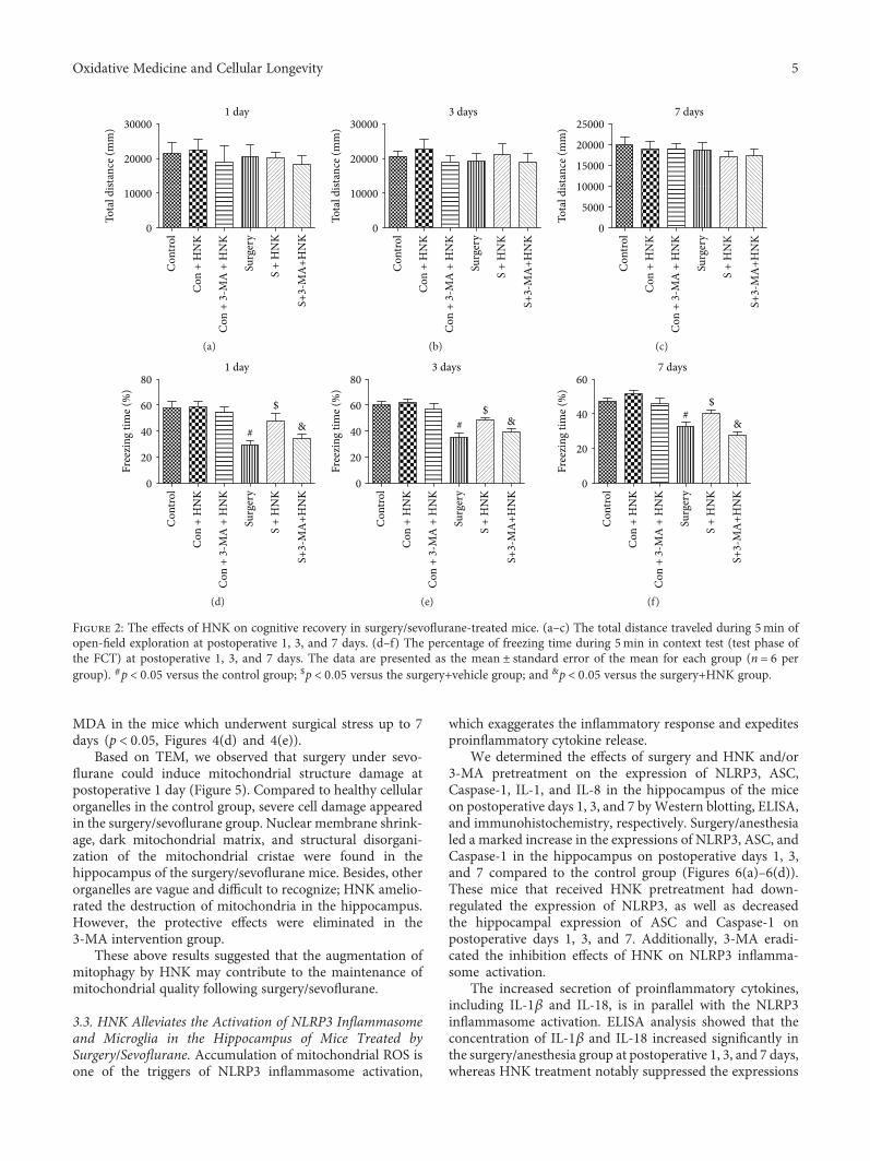
MDA in the mice which underwent surgical stress up to 7days (p < 0 05, Figures 4(d) and 4(e)).
Based on TEM, we observed that surgery under sevo-flurane could induce mitochondrial structure damage atpostoperative 1 day (Figure 5). Compared to healthy cellularorganelles in the control group, severe cell damage appearedin the surgery/sevoflurane group. Nuclear membrane shrink-age, dark mitochondrial matrix, and structural disorgani-zation of the mitochondrial cristae were found in thehippocampus of the surgery/sevoflurane mice. Besides, otherorganelles are vague and difficult to recognize; HNK amelio-rated the destruction of mitochondria in the hippocampus.However, the protective effects were eliminated in the3-MA intervention group.
These above results suggested that the augmentation ofmitophagy by HNK may contribute to the maintenance ofmitochondrial quality following surgery/sevoflurane.
3.3. HNK Alleviates the Activation of NLRP3 Inflammasomeand Microglia in the Hippocampus of Mice Treated bySurgery/Sevoflurane. Accumulation of mitochondrial ROS isone of the triggers of NLRP3 inflammasome activation,
which exaggerates the inflammatory response and expeditesproinflammatory cytokine release.
We determined the effects of surgery and HNK and/or3-MA pretreatment on the expression of NLRP3, ASC,Caspase-1, IL-1, and IL-8 in the hippocampus of the miceon postoperative days 1, 3, and 7 byWestern blotting, ELISA,and immunohistochemistry, respectively. Surgery/anesthesialed a marked increase in the expressions of NLRP3, ASC, andCaspase-1 in the hippocampus on postoperative days 1, 3,and 7 compared to the control group (Figures 6(a)–6(d)).These mice that received HNK pretreatment had down-regulated the expression of NLRP3, as well as decreasedthe hippocampal expression of ASC and Caspase-1 onpostoperative days 1, 3, and 7. Additionally, 3-MA eradi-cated the inhibition effects of HNK on NLRP3 inflamma-some activation.
The increased secretion of proinflammatory cytokines,including IL-1β and IL-18, is in parallel with the NLRP3inflammasome activation. ELISA analysis showed that theconcentration of IL-1β and IL-18 increased significantly inthe surgery/anesthesia group at postoperative 1, 3, and 7 days,whereas HNK treatment notably suppressed the expressions
20000
Tota
l dist
ance
(mm
)1 day
10000
S +
HN
K
Surg
ery
Con
+ 3
-MA
+ H
NK
Con
+ H
NK
Con
trol
S+3-
MA
+HN
K
0
30000
(a)
3 days
S +
HN
K
Surg
ery
Con
+ 3
-MA
+ H
NK
Con
+ H
NK
Con
trol
S+3-
MA
+HN
K
20000
10000
0
30000
Tota
l dist
ance
(mm
)(b)
7 days
S +
HN
K
Surg
ery
Con
+ 3
-MA
+ H
NK
Con
+ H
NK
Con
trol
S+3-
MA
+HN
K
20000
15000
10000
5000
0
25000
Tota
l dist
ance
(mm
)
(c)
1 day
S +
HN
K
Surg
ery
Con
+ 3
-MA
+ H
NK
Con
+ H
NK
Con
trol
S+3-
MA
+HN
K
60
80
40
20
0
Free
zing
tim
e (%
)
&
$
#
(d)
3 days
S +
HN
K
Surg
ery
Con
+ 3
-MA
+ H
NK
Con
+ H
NK
Con
trol
S+3-
MA
+HN
K
60
40
20
0
80
Free
zing
tim
e (%
)
&$
#
(e)
7 days
S +
HN
K
Surg
ery
Con
+ 3
-MA
+ H
NK
Con
+ H
NK
Con
trol
S+3-
MA
+HN
K
40
20
0
60
Free
zing
tim
e (%
)
&
$#
(f)
Figure 2: The effects of HNK on cognitive recovery in surgery/sevoflurane-treated mice. (a–c) The total distance traveled during 5min ofopen-field exploration at postoperative 1, 3, and 7 days. (d–f) The percentage of freezing time during 5min in context test (test phase ofthe FCT) at postoperative 1, 3, and 7 days. The data are presented as the mean ± standard error of the mean for each group (n = 6 pergroup). #p < 0 05 versus the control group; $p < 0 05 versus the surgery+vehicle group; and &p < 0 05 versus the surgery+HNK group.
5Oxidative Medicine and Cellular Longevity

of IL-1β and IL-18 compared with the surgery/anesthesia-induced mice (Figures 6(e)–6(g)). Microglia activationalso reflects neuroinflammation. Pretreatment with HNKresulted in a decreased Iba-1 expression on postoperative1 day, while 3-MA treatment could result in an increasedIba-1 expression compared to the surgery group, whichindicated the different activation of microglia (Figure 3(f)).
3.4. HNK Suppresses Neuronal Apoptosis in the HippocampalCA1 and DG Regions of Mice Induced by Surgery/Sevoflurane.To reveal whether mitophagy was associated with the
neuronal apoptosis, 3-MA was utilized as described abovein the surgery/sevoflurane-induced mice. As shown inFigure 7, TUNEL-positive cells were rarely detected in thecontrol group, whereas there were more TUNEL-positivecells in the surgery/sevoflurane group compared to the con-trol group, suggesting that surgery/sevoflurane led to neuro-nal damage. HNK pretreatment dramatically deceased theneuronal apoptosis in both the hippocampal CA1 and DGregions compared with the surgery/sevoflurane group.However, 3-MA inhibited the neuroprotection of HNK onneuronal apoptosis.
1 day
LC-3-ILC-3-II
GAPDH
16 KDa14 KDa
37 KDa
3 days 7 days
(a)
0.80.6
Ratio
LC3-
II/L
C3-I
1 day$
# &
$#
&
$# &
3 days 7 days
S+3-
MA
+HN
K
S +
HN
K
Surg
ery
Con
trol
0.40.20.0
1.00.60.40.20.0
0.8
S+3-
MA
+HN
K
S +
HN
K
Surg
ery
Con
trol
Ratio
LC3-
II/L
C3-I
0.60.8
0.40.20.0
S+3-
MA
+HN
K
S +
HN
K
Surg
ery
Con
trol
Ratio
LC3-
II/L
C3-I
(b)
1 day
Beclin-1
GAPDH
3 days 7 days
37 KDa
60 KDa
(c)
$
# &
$
# &
$
# &
1 day 3 days 7 days
S +
3-M
A +
NH
K
S +
HN
K
Surg
ery
Con
trol
0.80.60.40.20.0
1.0
Rela
tive e
xpre
ssio
nBe
clin-
1/G
APD
H
S +
3-M
A +
NH
K
S +
HN
K
Surg
ery
Con
trol
0.80.60.40.20.0
1.0
Rela
tive e
xpre
ssio
nBe
clin-
1/G
APD
H
S +
3-M
A +
NH
K
S +
HN
K
Surg
ery
Con
trol
0.80.60.40.20.0
1.0
Rela
tive e
xpre
ssio
nBe
clin-
1/G
APD
H
(d)
DAPI
Control
Surgery
Surgery + HNK
Surgery + HNK + 3-MA
NeuN Beclin-1 Merge
(e)
DAPI
Control
Surgery
Surgery + HNK
Surgery + HNK + 3-MA
Iba-1 Beclin-1 Merge
(f)
Figure 3: The effects of HNK on autophagy in the hippocampus of mice induced by surgery/sevoflurane. (a–d) The Western blotrepresentative blots of autophagy-related proteins, LC3-II, and Beclin-1 (n = 6 per group). (e, f) Representative doubleimmunofluorescence labeling of Beclin-1 with NeuN and Iba-1 in the CA1 at 24 h after surgery/sevoflurane. Scale bar = 50μm. The dataare presented as the mean ± standard error of the mean for each group (n = 6 per group). #p < 0 05 versus the control group; $p < 0 05versus the surgery+vehicle group; and &p < 0 05 versus the surgery+HNK group.
6 Oxidative Medicine and Cellular Longevity
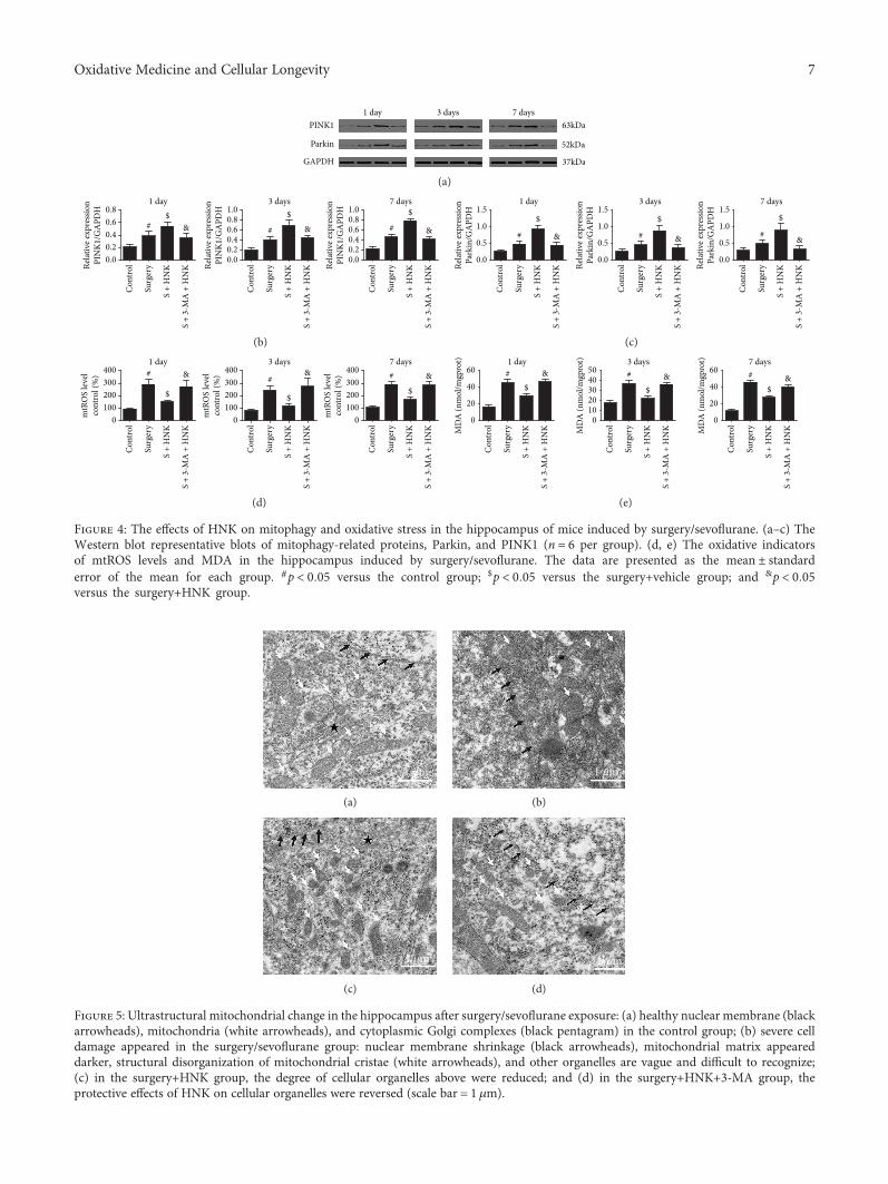
1 dayPINK1
Parkin
GAPDH
3 days 7 days63kDa
52kDa
37kDa
(a)
0.0
S +
HN
K
Surg
ery
Con
trol
S +
3-M
A +
HN
K
S +
HN
K
Surg
ery
Con
trol
S +
3-M
A +
HN
K
S +
HN
K
Surg
ery
Con
trol
S +
3-M
A +
HN
K
0.6
Relat
ive e
xpre
ssio
nPI
NK1
/GA
PDH
Relat
ive e
xpre
ssio
nPI
NK1
/GA
PDH
Relat
ive e
xpre
ssio
nPI
NK1
/GA
PDH
0.40.2
0.8
0.0
0.8
0.0
0.80.6 0.60.4 0.40.2 0.2
1.0 1.01 day 3 days 7 days
#$
& #
$& #
$
&
(b)
S +
HN
K
Surg
ery
Con
trol
S +
3-M
A +
HN
K
S +
HN
K
Surg
ery
Con
trol
S +
3-M
A +
HN
K
S +
HN
K
Surg
ery
Con
trol
S +
3-M
A +
HN
KRelat
ive e
xpre
ssio
nPa
rkin
/GA
PDH
Relat
ive e
xpre
ssio
nPa
rkin
/GA
PDH
Relat
ive e
xpre
ssio
nPa
rkin
/GA
PDH
0.0
1.0
0.5
1.5
0.0
1.0
0.5
1.5
0.0
1.0
0.5
1.51 day 3 days 7 days
#
$
& #
$
& #
$
&
(c)
S +
HN
K
Surg
ery
Con
trol
S +
3-M
A +
HN
K
S +
HN
K
Surg
ery
Con
trol
S +
3-M
A +
HN
K
S +
HN
K
Surg
ery
Con
trol
S +
3-M
A +
HN
K
mtR
OS
leve
lco
ntro
l (%
)
mtR
OS
leve
lco
ntro
l (%
)
mtR
OS
leve
lco
ntro
l (%
)
0
300200100
400
0
300200100
400
0
300200100
4001 day 3 days 7 days
#
$
& #
$
& #
$&
(d)
S +
HN
K
Surg
ery
Con
trol
S +
3-M
A +
HN
K
S +
HN
K
Surg
ery
Con
trol
S +
3-M
A +
HN
K
S +
HN
K
Surg
ery
Con
trol
S +
3-M
A +
HN
KMD
A (n
mol
/mgp
rot)
MD
A (n
mol
/mgp
rot)
MD
A (n
mol
/mgp
rot)
0
40
20
60
0
40302010
50
0
40
20
601 day 3 days 7 days
#$
& #
$& #
$&
(e)
Figure 4: The effects of HNK on mitophagy and oxidative stress in the hippocampus of mice induced by surgery/sevoflurane. (a–c) TheWestern blot representative blots of mitophagy-related proteins, Parkin, and PINK1 (n = 6 per group). (d, e) The oxidative indicatorsof mtROS levels and MDA in the hippocampus induced by surgery/sevoflurane. The data are presented as the mean ± standarderror of the mean for each group. #p < 0 05 versus the control group; $p < 0 05 versus the surgery+vehicle group; and &p < 0 05versus the surgery+HNK group.
1 �휇m
(a)
1 �휇m
(b)
1 �휇m
(c)
1 �휇m
(d)
Figure 5: Ultrastructural mitochondrial change in the hippocampus after surgery/sevoflurane exposure: (a) healthy nuclear membrane (blackarrowheads), mitochondria (white arrowheads), and cytoplasmic Golgi complexes (black pentagram) in the control group; (b) severe celldamage appeared in the surgery/sevoflurane group: nuclear membrane shrinkage (black arrowheads), mitochondrial matrix appeareddarker, structural disorganization of mitochondrial cristae (white arrowheads), and other organelles are vague and difficult to recognize;(c) in the surgery+HNK group, the degree of cellular organelles above were reduced; and (d) in the surgery+HNK+3-MA group, theprotective effects of HNK on cellular organelles were reversed (scale bar = 1 μm).
7Oxidative Medicine and Cellular Longevity

1.5
NLRP3
1 day
Caspase-1
ASC
GAPDH
1.0
1.5
1.0
Rela
tive e
xpre
ssio
nCa
spas
e-1/
GA
PDH
Rela
tive e
xpre
ssio
nA
SC/G
APD
HRe
lativ
e exp
ress
ion
NLR
P3/G
APD
H
0.5
0.5
0.0
0.0
S +
HN
K
Surg
ery
Con
trol
S +
3-M
A +
HN
K
S +
HN
K
Surg
ery
Con
trol
S +
3-M
A +
HN
K
S +
HN
K
Surg
ery
Con
trol
S +
3-M
A +
HN
K
1.0
0.8
0.6
0.4
0.2
0.0
# &
$
# &
$
#&
$
(a)
3 days
NLRP3
Caspase-1
ASC
GAPDH
Rela
tive e
xpre
ssio
nCa
spas
e-1/
GA
PDH
Rela
tive e
xpre
ssio
nA
SC/G
APD
HRe
lativ
e exp
ress
ion
NLR
P3/G
APD
H
S +
HN
K
Surg
ery
Con
trol
S +
3-M
A +
HN
K
S +
HN
K
Surg
ery
Con
trol
S +
3-M
A +
HN
K
S +
HN
K
Surg
ery
Con
trol
S +
3-M
A +
HN
K
1.5
1.0
1.5
1.0
0.5
0.5
0.0
0.0
1.0
0.5
1.5
0.0
# &$
#&
$
# &
$
(b)
7 days
NLRP3
Caspase-1
ASC
GAPDH
110 kDa
45 kDa
25 kDa
37 kDa
Rela
tive e
xpre
ssio
nCa
spas
e-1/
GA
PDH
Rela
tive e
xpre
ssio
nA
SC/G
APD
HRe
lativ
e exp
ress
ion
NLR
P3/G
APD
H
S +
HN
K
Surg
ery
Con
trol
S +
3-M
A +
HN
K
S +
HN
K
Surg
ery
Con
trol
S +
3-M
A +
HN
K
S +
HN
K
Surg
ery
Con
trol
S +
3-M
A +
HN
K
1.5
1.0
1.5
1.0
0.5
0.5
0.0
0.0
0.6
0.2
0.2
0.8
0.0
# &
$
# &
$
# &
$
(c)
Figure 6: Continued.
8 Oxidative Medicine and Cellular Longevity

4. Discussion
POCD can deteriorate surgery and lead to an induction ofmorbidity and mortality in patients [26]. Revealing thepathogeny and treatments for POCD are beneficial for the
increase of hospitalization comfort and improvement of thelong-term outcome. In the current study, we first providedevidence that mitophagy or autophagy was involved inpostoperative cognitive impairment induced by surgery/sevoflurane in mice. Honokiol could further induce the
1 day
3 days
7 days
S + HNKSurgeryControl S + 3-MA + HNK
(d)
1 day
1 day
150
100
IL-1�훽
(pg/
mg
prot
einl
)IL
-18
(pg/
mg
prot
einl
)
50
0
S +
HN
K
Surg
ery
Con
trol
S +
3-M
A +
HN
K
S +
HN
K
Surg
ery
Con
trol
S +
3-M
A +
HN
K
250
200
150
100
50
0
# &
$
# &
$
(e)
3 days
3 days
IL-1�훽
(pg/
mg
prot
einl
)IL
-18
(pg/
mg
prot
einl
)
S +
HN
K
Surg
ery
Con
trol
S +
3-M
A +
HN
K
S +
HN
K
Surg
ery
Con
trol
S +
3-M
A +
HN
K
200
150
100
50
0
200
100
150
50
250
0
#
&
$
#&
$
(f)
7 days
7 days
IL-1�훽
(pg/
mg
prot
einl
)IL
-18
(pg/
mg
prot
einl
)
S +
HN
K
Surg
ery
Con
trol
S +
3-M
A +
HN
K
S +
HN
K
Surg
ery
Con
trol
S +
3-M
A +
HN
K
100
60
80
40
20
0
150
100
50
250
200
0
# &
$
#
&
$
(g)
Figure 6: The effects of HNK on NLRP3 inflammasome activation in the hippocampus induced by surgery/sevoflurane. (a–c) The Westernblot representative blots of NLRP3, ASC, and Caspase-1 (n = 6 per group). (d) The immunohistochemistry of NLRP3 in the hippocampus at24 h after surgery/sevoflurane. (e–g) The concentration of IL-1β and IL-18 in the hippocampus at postoperative 1, 3, and 7 days. The data arepresented as the mean ± standard error of the mean for each group. #p < 0 05 versus the control group; $p < 0 05 versus the surgery+vehiclegroup; and &p < 0 05 versus the surgery+HNK group.
9Oxidative Medicine and Cellular Longevity

mitophagy in surgery/sevoflurane-induced mice. Besides,honokiol inhibited the activation of NLRP3 inflammasomeafter surgery/sevoflurane treatment and alleviated the neu-roinflammation in mice. Pretreatment with honokiol couldalso preserve neuronal apoptosis and alleviate cognitivedecline following surgery/sevoflurane treatment. In addition,3-MA, an autophagy and mitophagy inhibitor, reversed theneuroprotective effect of HNK on the inhibition of NLRP3inflammasome activation. These results demonstrated thatHNK-mediated mitophagy ameliorates postoperative cogni-tive impairment induced by surgery/sevoflurane in mice viainhibiting the activation of the NLRP3 inflammasome.
Surgical stress and long duration of inhaled anestheticscan then prompt the activation of immune cells whichcontributes to neuroinflammation and cognitive change[27, 28]. Without effective intervention, the transformationfrom acute neuroinflammation to chronic neuroinflamma-tion, which was accompanied by neutrophil infiltration andmicroglia/macrophage activation, may result in neuronaldeath, disruption of the blood-brain barrier, and brainedema [26, 29–32]. All the time, anti-neuroinflammatory
strategies exhibit potential therapeutic effects against surgery/anesthesia exposure and improve cognitive functions aftersurgery/anesthesia stress. In recent years, the NLRP3inflammasome became a research focus in a series of dis-eases, such as diabetes, neurodegenerative disorders, andbrain trauma. Previous studies showed that the MPTPmouse model of Parkinson’s disease, traumatic brain injury,cerebral ischemia-reperfusion injury, and isoflurane-inducedhippocampal inflammation can induce the NLRP3inflammasome activation [6, 8, 33]. Moreover, the inhibi-tion of the NLRP3 inflammasome provided robust neuro-protective response in these pathophysiological processes,which was associated with NLRP3-mediated inflamma-tion and reduction of proinflammatory cytokines. Ourfindings also suggested that HNK could effectively suppressNLRP3 inflammasome activation and subsequent inflam-matory cytokine expression. Immunofluorescence results alsoshowed that trends of microglia activation are correlated withthe NLRP3 inflammasome expression in surgery/sevoflur-ane-induced mice, which is consistent with previous stud-ies. Emerging evidences demonstrated that NLRP3 seems
Control S + HNKSurgery
DAPI
Merge
TUNEL
S + HNK + 3-MA
(a)
Control S + HNKSurgery
DAPI
Merge
TUNEL
S + HNK + 3-MA
(b)
20
15
10
5
0
TUN
EL-p
ositi
ve ce
lls (%
)
Con
trol
S +
HN
K
Surg
ery
S +
HN
K +
3-M
A
Con
trol
S +
HN
K
Surg
ery
S +
HN
K +
3-M
A
CA1 regionDG region
$
#&
$
# &
(c)
Figure 7: The effects of HNK on neuronal apoptosis in the hippocampal CA1 and DG regions of mice induced by surgery/sevoflurane.(a, b) The representative images showing TUNEL staining in the hippocampal CA1 and DG regions at 24 h after surgery/sevoflurane.The apoptotic cells were detected by TUNEL (green), and the nuclei were detected by DAPI (blue). Scale bar = 50 μm. (c) The percentage ofTUNEL-positive cells. The data are presented as themean ± standard error of the mean for each group (n = 3 per group), #p < 0 05 versus thecontrol group; $p < 0 05 versus the surgery+vehicle group; and &p < 0 05 versus the surgery+HNK group.
10 Oxidative Medicine and Cellular Longevity

sensitive to the imbalance of cellular homeostasis, so thereare a great many of NLRP3 activators, such as low intracel-lular K+ concentration, lysosomal lysis, mitochondrial ROS,or mitochondrial DNA released from damaged mitochon-dria and Ca2 flux [11]. In our studies, we further detectedthe levels of mitochondrial ROS and MDA, which couldreflect the relationships between oxidative stress and theNLRP3 inflammasome. Our results showed that HNKpossessed strongly antioxidant capacity which decreasedthe levels of mtROS and was critical for the reductionof the NLRP3 inflammasome. This study may be the firstto exhibit the bridge between oxidative stress andNLRP3-mediated neuroinflammation in postoperativecognitive decline induced by surgery/sevoflurane.
Mitochondria are the primary platforms for energy pro-duction and are hypersensitive to the surgical stress. Whenmitochondrial function is damaged, the overplus of ROS,especially for mitochondrial ROS, is deleterious for thenormal physiological state and could cause oxidative stressand inflammation, which are the basis of a wealth of dis-eases, such as diabetes and cerebral/myocardial ischemia-reperfusion injury [34, 35]. In the view of mitochondrialmorphology and biochemical results, we found that HNKeffectively blunted the mitochondrial injury (decrease ofswelling mitochondria) and the degrees of oxidative stress.These results provided a novel sight for NLRP3 inflamma-some activation in postoperative cognitive decline inducedby surgery/sevoflurane.
Mitophagy, as a selective autophagy, is a crucial media-tor of the degradation of injured mitochondria. Larger num-bers of evidence have indicated that autophagy/mitophagyemerges in response to various conditions, such as nutrientdepletion, mitochondrial dysfunction, or red blood cell mat-uration, and has a significant impact on a series of diseases[36]. Lin et al. showed that inhibition of mitophagy couldaggravate the neuroinflammation induced by traumaticbrain injury. And melatonin enhanced mitophagy throughthe mTOR signaling pathway, then ameliorated the TBI-triggered neuroinflammation [37]. And not all mitophagyis processed by the Parkin/PINK1 pathway. Studies of theBnip3/Nix pathway and the Parkin/PINK1 signaling path-way all have a direct connection between defective mitochon-dria and mitophagy [36]. The difference is that the Parkin/PINK1 pathway depends on the voltage-dependent inhibi-tion in the autophagosome clearance process [38, 39]. Andthe dysfunction of the Parkin/PINK1 mechanism may leadto defects in mitochondrial morphology, dynamics, andfunction and result in an imbalance in mitochondrial fusionand fission. However, mitophagy involved in the Bnip3/Nixpathway may be crucial for maintaining the number andthe function of mitochondria during cell differentiation anddedifferentiation [40]. In our studies, we found that HNKincreased the mitophagy-related proteins, Parkin/PINK1,and protected the abnormal mitochondrial structure (e.g.,fragmented cristae and swollen, distorted mitochondrialmorphology) in the surgery/sevoflurane model, suggestingthat increased mitophagy may eliminate more damagedand dysfunctional mitochondria and may be helpful inreducing the overproduction of ROS. These signaling
pathways may be the upstream of NLRP3 inflammasomeactivation. And different disease models have demonstratedthe assumption above. For example, Kim et al. demonstratedthat SESN2/sestrin2, an autophagic inducer, prevents sepsisby inducing mitophagy and inhibiting NLRP3 via an increaseof unc-51-like kinase 1 protein activity and levels [41].Consistent with these outcomes, Zhong et al. observed thatthe NF-κB signaling pathway can translocate the increasedcargo receptor p62 to damaged mitochondria, which arerecognized by Parkin-dependent ubiquitin and inducemitophagic clearance. The intrinsic regulatory loop “NF-κB-p62-mitophagy” in the macrophage restrains NLRP3 inflam-masome activation and maintains homeostasis [42]. Ourfindings and these studies indicate that mitophagy is one ofthe self-limiting systems to protect tissues and organs fromhyperinflammation and favor tissue repair.
However, it is noted that there are some limitations in ourcurrent study. Firstly, in view of behavior tests, we justperformed three timepoints (postoperative 1, 3, and 7 days).Due to lack of behavioral data at postoperative 28 days andcontinuous monitoring within postoperative 24 hours, wefailed to understand the fluctuations of cognitive impair-ment. Further investigation would observe the long-termpostoperative cognitive change. Second, in our studies, wejust measured the related indicator only in the hippocampus.Several studies also have reported that other brain regions,such as the prefrontal cortex and amygdaloid nucleus,could participate in cognitive function. So, these regionswould be investigated in the future. Third, considering theantianxiety property of HNK and our previous study, wecannot exclude the possibility that the change in anxietylevels by HNK hindered the freezing behavior in the fearconditioning test in mice. Finally, due to the expensiveprice and limited number, we only used 8-week-old femalemice but not the aged mice. However, several studies haveshown that surgical stress could also induce cognitivechange in female mice. And in the future investigation,we will add the observation of the effects of surgery/sevo-flurane on postoperative cognitive decline in mice of differ-ent sexes and ages.
5. Conclusions
Taken together, our study deepened the understanding of theneuroprotective effects of HNK on surgery/sevoflurane-induced postoperative cognitive impairment and detected anovel therapeutic target. Our results indicated that honokiol-mediatedmitophagy ameliorates postoperative cognitiveimpairment induced by surgery/sevoflurane. This neuropro-tective effect may be involved in inhibiting the activation ofthe NLRP3 inflammasome and suppressing inflammatoryresponses in the hippocampus.
Data Availability
The datasets analyzed during the current study are availablefrom the corresponding author on reasonable request.
11Oxidative Medicine and Cellular Longevity

Ethical Approval
The experimental protocol was approved by the AnimalEthics Committee of ZhongnanHospital ofWuhanUniversity,Hubei, China, and all experiments were performed in accor-dance with the National Institutes of Health Guidelines forthe Care and Use of Laboratory Animals.
Conflicts of Interest
The authors declare that they have no competing interests.
Authors’ Contributions
JSY designed and performed the experiments, analyzed andinterpreted the data, and drafted the manuscript. LC partici-pated in designing the study and interpreting the results. YYLcontributed to behavioral testing and biochemical analysis.SQL and MP participated in preparing the animal models.ZYX conceived the study, participated in its design and coor-dination, secured funding for the project, helped to draft themanuscript, and critically revised the manuscript. All authorsread and approved the final manuscript.
Acknowledgments
We thank the Central Laboratory (Renmin Hospital ofWuhan University) for the equipment and excellent techni-cal assistance. This study was supported by the NationalNatural Science Foundation of China (Grant Nos. 81471844and 81671891).
References
[1] M. J. Needham, C. E. Webb, and D. C. Bryden, “Postoperativecognitive dysfunction and dementia: what we need to knowand do,” British Journal of Anaesthesia, vol. 119, Supplement1, pp. i115–i125, 2017.
[2] S. S. Makhani, F. Y. Kim, Y. Liu et al., “Cognitive impairmentand overall survival in frail surgical patients,” Journal of theAmerican College of Surgeons, vol. 225, no. 5, pp. 590–600.e1, 2017.
[3] Z. Cristina, B. Michela, B. Agostino et al., “Post-operativecognitive decline (POCD) after gynaecologic surgery: currentopinions and future applications,” Archives of Gynecologyand Obstetrics, vol. 297, no. 3, pp. 551–554, 2018.
[4] X. Feng, M. Valdearcos, Y. Uchida, D. Lutrin, M. Maze, andS. K. Koliwad, “Microglia mediate postoperative hippocam-pal inflammation and cognitive decline in mice,” JCI insight,vol. 2, no. 7, article e91229, 2017.
[5] W. Liu, J. Guo, J. Mu, L. Tian, and D. Zhou, “Rapamycinprotects sepsis-induced cognitive impairment in mouse hippo-campus by enhancing autophagy,” Cellular and MolecularNeurobiology, vol. 37, no. 7, pp. 1195–1205, 2017.
[6] Z. Wang, S. Meng, L. Cao, Y. Chen, Z. Zuo, and S. Peng,“Critical role of NLRP3-caspase-1 pathway in age-dependentisoflurane-induced microglial inflammatory response and cog-nitive impairment,” Journal of Neuroinflammation, vol. 15,no. 1, p. 109, 2018.
[7] T. Wang, D. Nowrangi, L. Yu et al., “Activation of dopamineD1 receptor decreased NLRP3-mediated inflammation in
intracerebral hemorrhage mice,” Journal of Neuroinflamma-tion, vol. 15, no. 1, p. 2, 2018.
[8] X. Xu, D. Yin, H. Ren et al., “Selective NLRP3 inflammasomeinhibitor reduces neuroinflammation and improves long-term neurological outcomes in a murine model of traumaticbrain injury,” Neurobiology of Disease, vol. 117, pp. 15–27,2018.
[9] Q. Guo, Y. Wu, Y. Hou et al., “Cytokine secretion andpyroptosis of thyroid follicular cells mediated by enhancedNLRP3, NLRP1, NLRC4, and AIM2 inflammasomes areassociated with autoimmune thyroiditis,” Frontiers in Immu-nology, vol. 9, p. 1197, 2018.
[10] K. Vats, D. Sarmah, H. Kaur et al., “Inflammasomes in stroke:a triggering role for acid-sensing ion channels,” Annals of theNew York Academy of Sciences, vol. 1431, no. 1, pp. 14–24,2018.
[11] T. Tang, X. Lang, C. Xu et al., “CLICs-dependent chlorideefflux is an essential and proximal upstream event for NLRP3inflammasome activation,” Nature Communications, vol. 8,no. 1, p. 202, 2017.
[12] Y. L. Wang, Q. Q. Han, W. Q. Gong et al., “Microglial activa-tion mediates chronic mild stress-induced depressive- andanxiety-like behavior in adult rats,” Journal of Neuroinflam-mation, vol. 15, no. 1, p. 21, 2018.
[13] J. Zheng, L. Shi, F. Liang et al., “Sirt3 ameliorates oxidativestress and mitochondrial dysfunction after intracerebral hem-orrhage in diabetic rats,” Frontiers in Neuroscience, vol. 12,p. 414, 2018.
[14] Y. He, M. Y. Zeng, D. Yang, B. Motro, and G. Núñez,“NEK7 is an essential mediator of NLRP3 activation down-stream of potassium efflux,” Nature, vol. 530, no. 7590,pp. 354–357, 2016.
[15] S. Cao, S. Shrestha, J. Li et al., “Melatonin-mediated mitophagyprotects against early brain injury after subarachnoid hemor-rhage through inhibition of NLRP3 inflammasome activa-tion,” Scientific Reports, vol. 7, no. 1, p. 2417, 2017.
[16] M. J. Kim, J. H. Yoon, and J. H. Ryu, “Mitophagy: a balanceregulator of NLRP3 inflammasome activation,” BMB Reports,vol. 49, no. 10, pp. 529–535, 2016.
[17] V. B. Pillai, S. Samant, N. R. Sundaresan et al., “Honokiolblocks and reverses cardiac hypertrophy in mice by activatingmitochondrial Sirt3,” Nature Communications, vol. 6, no. 1,p. 6656, 2015.
[18] T. Hong, H. Min, Z. Hui, L. Yuejian, Y. Lixing, and X. Z. Liang,“Oral administration of honokiol attenuates airway inflamma-tion in asthmatic mouse model,” Pakistan Journal of Pharma-ceutical Sciences, vol. 31, no. 4, pp. 1279–1284, 2018.
[19] P. Tang, J. M. Gu, Z. A. Xie et al., “Honokiol alleviates thedegeneration of intervertebral disc via suppressing the activa-tion of TXNIP-NLRP3 inflammasome signal pathway,” FreeRadical Biology and Medicine, vol. 120, pp. 368–379, 2018.
[20] D. Wang, X. Dong, and C. Wang, “Honokiol amelioratesamyloidosis and neuroinflammation and improves cognitiveimpairment in Alzheimer’s disease transgenic mice,” The Jour-nal of Pharmacology and Experimental Therapeutics, vol. 366,no. 3, pp. 470–478, 2018.
[21] B. Zhang, M. Zhai, B. Li et al., “Honokiol ameliorates myocar-dial ischemia/reperfusion injury in type 1 diabetic rats byreducing oxidative stress and apoptosis through activatingthe SIRT1-Nrf2 signaling pathway,” Oxidative medicine andcellular longevity, vol. 2018, Article ID 3159801, 16 pages, 2018.
12 Oxidative Medicine and Cellular Longevity

[22] M. Wang, Y. Li, C. Ni, and G. Song, “Honokiol attenuatesoligomeric amyloid β1-42-induced Alzheimer’s disease in micethrough attenuating mitochondrial apoptosis and inhibitingthe nuclear factor kappa-B signaling pathway,” Cellular Physi-ology and Biochemistry, vol. 43, no. 1, pp. 69–81, 2017.
[23] Y. F. Xian, S. P. Ip, Q. Q. Mao et al., “Honokiol improveslearning and memory impairments induced by scopolaminein mice,” European Journal of Pharmacology, vol. 760,pp. 88–95, 2015.
[24] A. Jangra, S. Dwivedi, C. S. Sriram et al., “Honokiol abrogateschronic restraint stress-induced cognitive impairment anddepressive-like behaviour by blocking endoplasmic reticulumstress in the hippocampus of mice,” European Journal of Phar-macology, vol. 770, pp. 25–32, 2016.
[25] J. S. Ye, L. Chen, Y. Y. Lu, S. Q. Lei, M. Peng, and Z. Y. Xia,“SIRT3 activator honokiol ameliorates surgery/anesthesia-induced cognitive decline in mice through anti-oxidative stressand anti-inflammatory in hippocampus,” CNS Neuroscience &Therapeutics, 2018.
[26] J. Bi, W. Shan, A. Luo, and Z. Zuo, “Critical role of matrixmetallopeptidase 9 in postoperative cognitive dysfunctionand age-dependent cognitive decline,” Oncotarget, vol. 8,no. 31, pp. 51817–51829, 2017.
[27] A. A. Zurek, J. Yu, D. S. Wang et al., “Sustained increase inα5GABAA receptor function impairs memory after anesthe-sia,” The Journal of Clinical Investigation, vol. 124, no. 12,pp. 5437–5441, 2014.
[28] M. Plas, E. Rotteveel, G. J. Izaks et al., “Cognitive decline aftermajor oncological surgery in the elderly,” European journal ofcancer, vol. 86, pp. 394–402, 2017.
[29] Z. Li, N. Mo, L. Li et al., “Surgery-induced hippocampalangiotensin II elevation causes blood-brain barrier disruptionvia MMP/TIMP in aged rats,” Frontiers in Cellular Neurosci-ence, vol. 10, p. 105, 2016.
[30] D. Ikawa, M. Makinodan, K. Iwata et al., “Microglia-derivedneuregulin expression in psychiatric disorders,” Brain, Behav-ior, and Immunity, vol. 61, pp. 375–385, 2017.
[31] B. Zheng, R. Lai, J. Li, and Z. Zuo, “Critical role of P2X7 recep-tors in the neuroinflammation and cognitive dysfunction aftersurgery,” Brain, Behavior, and Immunity, vol. 61, pp. 365–374, 2017.
[32] J. Wang, L. Li, Z. Wang et al., “Supplementation of lycopeneattenuates lipopolysaccharide-induced amyloidogenesis andcognitive impairments via mediating neuroinflammation andoxidative stress,” The Journal of Nutritional Biochemistry,vol. 56, pp. 16–25, 2018.
[33] C. Qiao, L. X. Zhang, X. Y. Sun, J. H. Ding, M. Lu, and G. Hu,“Caspase-1 deficiency alleviates dopaminergic neuronal deathvia inhibiting caspase-7/AIF pathway in MPTP/p mousemodel of Parkinson’s disease,” Molecular Neurobiology,vol. 54, no. 6, pp. 4292–4302, 2017.
[34] L. Tilokani, S. Nagashima, V. Paupe, and J. Prudent,“Mitochondrial dynamics: overview of molecular mecha-nisms,” Essays in Biochemistry, vol. 62, no. 3, pp. 341–360,2018.
[35] O. E. Aparicio-Trejo, E. Tapia, L. G. Sánchez-Lozada, andJ. Pedraza-Chaverri, “Mitochondrial bioenergetics, redox state,dynamics and turnover alterations in renal mass reductionmodels of chronic kidney diseases and their possible implica-tions in the progression of this illness,” PharmacologicalResearch, vol. 135, pp. 1–11, 2018.
[36] J. Harris, N. Deen, S. Zamani, and M. A. Hasnat, “Mitophagyand the release of inflammatory cytokines,” Mitochondrion,vol. 41, pp. 2–8, 2018.
[37] C. Lin, H. Chao, Z. Li et al., “Melatonin attenuates trau-matic brain injury-induced inflammation: a possible role formitophagy,” Journal of Pineal Research, vol. 61, no. 2,pp. 177–186, 2016.
[38] C. T. Chu, “Mechanisms of selective autophagy and mito-phagy: implications for neurodegenerative diseases,” Neurobi-ology of Disease, vol. 122, 2019.
[39] R. Lan, J. T. Wu, T. Wu et al., “Mitophagy is activated in braindamage induced by cerebral ischemia and reperfusion viathe PINK1/Parkin/p62 signalling pathway,” Brain ResearchBulletin, vol. 142, pp. 63–77, 2018.
[40] M. Šprung, I. Dikic, and I. Novak, “Flow cytometer monitoringof Bnip3- and Bnip3L/Nix-dependent mitophagy,” in Mito-phagy, N. Hattori and S. Saiki, Eds., vol. 1759 of Methods inMolecular Biology, Humana Press, New York, NY, USA, 2017.
[41] M. J. Kim, S. H. Bae, J. C. Ryu et al., “SESN2/sestrin2suppresses sepsis by inducing mitophagy and inhibitingNLRP3 activation in macrophages,” Autophagy, vol. 12,no. 8, pp. 1272–1291, 2016.
[42] Z. Zhong, A. Umemura, E. Sanchez-Lopez et al., “NF-κBrestricts inflammasome activation via elimination of damagedmitochondria,” Cell, vol. 164, no. 5, pp. 896–910, 2016.
13Oxidative Medicine and Cellular Longevity
Review ArticleBenefits of Vitamins in the Treatment of Parkinson’s Disease
Xiuzhen Zhao,1 Ming Zhang,2 Chunxiao Li,3 Xue Jiang,1 Yana Su,1 and Ying Zhang 1
1Department of Neurology and Neuroscience Center, First Hospital of Jilin University, Xinmin Street No. 71,Changchun 130000, China2Department of Pharmacology, College of Basic Medical Sciences, Jilin University, 126 Xin Min Street, Changchun,Jilin 130021, China3Department of Neurology and Neuroscience Center, The Affiliated Hospital of Qingdao University, Qingdao,Shandong 266000, China
Correspondence should be addressed to Ying Zhang; [email protected]
Received 4 October 2018; Accepted 4 February 2019; Published 20 February 2019
Guest Editor: Francisco Jaime B. Mendonça Júnior
Copyright © 2019 Xiuzhen Zhao et al. This is an open access article distributed under the Creative Commons Attribution License,which permits unrestricted use, distribution, and reproduction in any medium, provided the original work is properly cited.
Parkinson’s disease (PD) is the second most common neurodegenerative disease in the elderly, which is clinically characterized bybradykinesia, resting tremor, abnormal posture balance, and hypermyotonia. Currently, the pathogenic mechanism of PD remainsunclear. Numerous clinical studies as well as animal and cell experiments have found a certain relationship between the vitaminfamily and PD. The antioxidant properties of vitamins and their biological functions of regulating gene expression may bebeneficial for the treatment of PD. Current clinical evidence indicates that proper supplementation of various vitamins canreduce the incidence of PD in the general population and improve the clinical symptoms of patients with PD; nevertheless, thesafety of regular vitamin supplements still needs to be highlighted. Vitamin supplementation may be an effective adjuvanttreatment for PD. In this review, we summarized the biological correlations between vitamins and PD as well as the underlyingpathophysiological mechanisms. Additionally, we elaborated the therapeutic potentials of vitamins for PD.
1. Introduction
Parkinson’s disease (PD) is the second most common neuro-degenerative disorder following Alzheimer’s disease. Clini-cally, PD is characterized by resting tremor, hypermyotonia,postural instability, and bradykinesia [1]. Additionally,patients with PD can alsomanifest with nonmotor symptoms,such as cognitive decline, olfactory dysfunction, constipation,sleep disorders, and autonomic symptoms [2], and these non-motor symptoms usually occur prior to the onset of motorsymptoms [3]. PD severely affects the quality of life of theindividual with the disease and also creates a great burdenon the caregivers. The typical pathological hallmark of PDis degeneration of dopaminergic neurons in the substantianigra pars compacta (SNpc) and eosinophilic inclusion bod-ies (Lewy bodies) in the remaining neurons, which is themajor contributor to the deficiency of dopamine in the basalganglia [4, 5]. The exact pathogenetic mechanisms of PD is
not yet fully understood. Current theories regard PD as amultifactorial disease, involving various genetic and envi-ronmental factors, among which mitochondrial dysfunctionand oxidative stress play an important role in the pathogen-esis and development of PD [6, 7]. The treatment for PD ischallenging, and the existing therapeutic strategies can onlyrelieve clinical symptoms but fail to control the progressionof PD.
Vitamins are natural bioactive products with antioxidantproperties, which are necessities for maintaining the normalfunctions of human organisms. Essential vitamins cannotbe endogenously synthesized in the organism and thereforemust be obtained through the diet. Clinically, vitamin defi-ciency is quite common, especially in infants and elderly.Vitamins are generally divided into fat-soluble variants(vitaminsA,D, E, andK) andwater-soluble variants (vitaminsB andC). The formermainly bind to cellular nuclear receptorsand affect the expression of specific genes [8]. The latter
HindawiOxidative Medicine and Cellular LongevityVolume 2019, Article ID 9426867, 14 pageshttps://doi.org/10.1155/2019/9426867

mainly constitute a cofactor for the enzyme, affecting theenzymatic activity [9].
Numerous clinical studies as well as animal and cellexperiments have found a certain relationship between thevitamin family and PD [10]. The antioxidant properties ofvitamins and their biological functions of regulating geneexpression may be beneficial for the treatment of PD. Cur-rent clinical evidence indicates that proper supplementationof various vitamins can reduce the incidence of PD in thegeneral population and improve the clinical symptoms ofpatients with PD; nevertheless, the safety of regular vitaminsupplements still needs to be highlighted. Vitamin supple-mentation may represent an effective adjuvant treatmentfor PD. In this review, we summarized the biological correla-tions between vitamins and PD as well as the underlyingpathophysiological mechanisms. Additionally, we elaboratedthe therapeutic potentials of vitamins for PD.
2. The Pathogenesis of Oxidative Stress in PD
Oxidative stress refers to the imbalance between the oxida-tion system and antioxidant system, resulting in excessiveaccumulation of oxidative substances, such as reactive oxy-gen species (ROS) and reactive nitrogen species (RNS) [11].ROS include superoxide anion radical (O2
-), hydroxyl radical(OH-), and hydrogen peroxide (H2O2); RNS include nitricoxide (NO), nitrogen dioxide (NO2), and peroxynitrite(ONOO-). The antioxidant system mainly consists of twosubtypes: (1) enzymatic antioxidant system, including super-oxide dismutase (SOD), catalase (CAT), and glutathioneperoxidase (GSH-Px), and (2) nonenzymatic antioxidant sys-tem, including vitamin C, vitamin E, glutathione, melatonin,alpha-lipoic acid, carotenoids, and trace elements copper,zinc, and selenium.
Oxidative stress plays an important physiological role inthe organism. For example, phagocytic cells kill pathogenicmicroorganisms, participate in detoxification and enzymaticreactions, and synthesize some essential biologically activesubstances. Meanwhile, it can as well cause damage to thebody, such as cell membrane destruction, protein denatur-ation, and nucleic acid changes.
There is increasing evidence that oxidative stress repre-sents a pathophysiological characteristic of PD, and the pro-duction of reactive oxygen species can result in neuronaldeath [12, 13]. The mitochondrial respiratory chain isregarded as the major source of ROS [14]. Additionally, pre-vious studies have found that mitochondrial dysfunctionexists in the substantia nigra of patients with PD [15].Reduced glutathione (GSH) can enhance the production ofROS and RNS [16], and oxidation of dopamine and dopa-mine metabolites such as 3,4-dihydroxyphenylacetic acid(DOPAC) can inhibit the activity of complex I [17]. Thesefindings indicate that the downstream metabolites ofdopamine may make dopamine neurons more susceptible.Moreover, iron accumulation in the substantia nigra is com-mon in patients with PD, leading to overproduction ofhydrogen peroxide and molecular oxygen in the Fenton reac-tion from Fe2+ to Fe3+; hydrogen peroxide generates a highlytoxic hydroxyl radical through the Haber-Weiss reaction in
the presence of Fe2+, which causes severe oxidative damageto the cellular components [18]. From the above, the oxidantstress is closely associated with the pathogenesis of PD.Oxidative stress can cause neuronal loss through someunderlying intracellular damage, such as protein aggregation,mitochondrial dysfunction, and DNA rupture. Therefore,antioxidant damage has become a potential target for thetreatment of PD.
3. Vitamin B and PD
The B family of vitamins is water-soluble, which includes thi-amine (vitamin B1), riboflavin (vitamin B2), niacin (vitaminB3), pantothenic acid (vitamin B5), pyridoxine (vitamin B6),biotin (vitamin B7), folate (vitamin B9), and cobalamin(vitamin B12) [19]. These vitamins play an important roleas enzyme cofactors in multiple biochemical pathways in alltissues, such as regulating metabolism, improving the func-tion of the immune system and nervous system, and promot-ing cell growth and division [20].
Almost all of these B family vitamins are essential var-iants dependent on diet supply, except niacin which canalso be synthesized from tryptophan. Vitamin B deficien-cies are frequent in the children, elderly, vegetarians, preg-nant women, and patients with gastrointestinal diseases.Recently, the association with vitamin B and PD is gettingmore and more attention. Herein, we use vitamin B3 as arepresentative to discuss the relationship between vitaminB and PD.
3.1. Vitamin B3. Nicotinamide is the active form of niacin,and it is the precursor of coenzymes NADH and NADPH,which are essential for over 200 enzymatic reactions in theorganism, especially the production of adenosine triphos-phate (ATP). Meat, fish, and wheat are generally rich in nic-otinamide, while vegetables have a low nicotinamide content[21]. Deficiency of nicotinamide/niacin can lead to pellagra,causing dermatitis, diarrhea, and depression [22]. Nicotin-amide has neuroprotective and antioxidant functions at lowdoses but exhibits neurotoxicity, especially dopaminergictoxicity, at high doses [23]. Fukushima also suggests thatexcessive nicotinamide is related to the development of PD[24]; excessive nicotinamide can induce overproduction of1-methylnicotinamide (MNA), which is increased in patientswith PD [25]. In an in vitro study, Griffin et al. found thatlow-dose nicotinamide (10mM) has a significant effect oninducing differentiation from embryonic stem cells into neu-rons; however, higher doses (>20mM) of nicotinamideinduce cytotoxicity and cell death [26]. The definitive protec-tive dose of vitamin B3 still needs further researches.
3.2. Possible Neuroprotective Mechanisms of Vitamin B3 inPD. Firstly, numerous studies have demonstrated that mito-chondrial dysfunction and cellular energy failure are patho-physiological features of PD. Nicotinamide participates inthe biosynthesis of nicotinamide adenine dinucleotide(NAD; oxidized form: NAD+; reduced form: NADH) via var-ious metabolic pathways [27]. NADH is an essential cofactorassisting the tetrahydrobiopterin functioning in tyrosine
2 Oxidative Medicine and Cellular Longevity

hydroxylase, which can hydroxylate tyrosine and producedopamine; NADH deficiency is common in PD [28].
Secondly, NADH is indispensable for the physiologicalfunction of mitochondrial complex I in ATP synthesis, andthe corresponding dysfunction is involved in PD patientsand animal models [15, 29, 30]. Nicotinamide mononucleo-tide (NMN) constitutes one of the key precursors of NAD+.In previous in vitro studies, the scholars have established acellular model of PD using rotenone-treated PC12 cells,and they found the NMN (0.1mM or 1mM) treatment wasassociated with a significantly higher survival rate in therotenone-treated (0.5 μm) PC12 cells. NMN is assumed toenhance the intracellular levels of NAD+ and ATP in thecellular model of PD [31].
In addition, nicotinamide can act a neuroprotectiverole by inhibiting the oxidative stress. In 1-methyl-4--phenyl-1,2,3,6-tetrahydropyridine- (MPTP-) induced mousemodels of PD, nicotinamide (500mg/kg) was injected beforesubacute (30mg/kg/d for 5 days) MPTP administration. Thisstudy showed that cotreatment withMPTP and nicotinamidesignificantly improved the locomotor activity comparedto single-agent treatment with MPTP. Nicotinamideadministration significantly attenuated the MPTP-induceddopamine depletion (47 11 ± 21 06 vs. 12 77 ± 8 06).Meanwhile, nicotinamide pretreatment markedly inhibitedMPTP-induced lactate dehydrogenase (LDH) and NOSactivities, which prevented the oxidative stress and alleviatedthe oxidative damage.
Sirtuins (SIRTs) are NAD+-dependent protein deacety-lases involved in vital biological processes [32]. Recently, sir-tuin 5 (SIRT5) has received considerable attention. Liu et al.investigated the role of SIRT5 in MPTP-induced mouse PDmodels [33]. They found that deletion of SIRT5 exacerbatedmotor deficits, nigrostriatal dopaminergic degeneration inthe compact part of substantia nigra (SNc), and mitochon-drial antioxidant activities in the PD models. These findingsprovide new insight into the therapeutic strategies for PD.However, the protective effects of nicotinamide are stillcontroversial, and further researches are needed to clarifythe biological function of vitamin B3 in PD.
3.3. Clinical Studies regarding Vitamin B3 in PD. Currentexisting clinical studies have shown that a high-niacin dietcan reduce the risk of PD [34, 35]. A previous case report alsodemonstrated that oral niacin (500mg twice daily for threemonths) significantly improved rigidity and bradykinesia ina patient with idiopathic PD, though the original purposewas to treat hypertriglyceridemia; after the cessation of oralniacin due to obvious adverse effects (unacceptable night-mares and skin rash), the symptoms of rigidity and bradyki-nesia relapsed [36]. However, other studies failed to noticethe remarkable clinical efficacy [37, 38]. Therefore, moreclinical observations are warranted to verify the efficacy aswell as side effects of niacin in PD.
4. Vitamin C and PD
Vitamin C (ascorbic acid) is another water-soluble, essentialvitamin, which is widely distributed in various tissues. This
nutriment is abundant in vegetables, fresh fruits, and animallivers. Vitamin C contains two molecular subforms in organ-isms: the reduced form (ascorbic acid (AA)) and the oxidizedform (dehydroascorbic acid (DHA)). Deficiency of vitamin Cis common, especially in children and elderly. A long-termlack of vitamin C can cause scurvy. Vitamin C is very impor-tant for the physiological function of the nervous system andthe antioxidant function by inhibiting oxidative stress,reducing lipid peroxidation, and scavenging free radicals[39]. Moreover, it is also involved in many nonoxidativestress processes, such as synthesis of collagen, cholesterol,carnitine, catecholamines, amino acids, and some peptidehormones [40, 41].
Dopamine metabolism can produce oxidative stressproducts, which in return induce accumulation of abnormalproteins in PD [42]. Vitamin C has potentials for thetreatment of PD considering the following reasons. Firstly,vitamin C is mainly distributed in areas that are rich inneurons [43, 44]. Secondly, vitamin C can be transportedto the brain by SVCT2 (vitamin C transporter type 2)[45], and DHA can be transported to the brain by GLUT1(glucose transporter type 1) and GLUT3 (glucose transportertype 3) [46].
4.1. Possible Neuroprotective Mechanisms of Vitamin C inPD. There is evidence that ascorbic acid can protect againstboth levodopa toxicity and the MPTP neurotoxicity[47, 48]. Vitamin C can increase the production of dihy-droxyphenylalanine (DOPA). Seitz et al. noted overpro-duction of DOPA in a dose-dependent manner afterincubation of the human neuroblastoma cell line SK-N-SHwith ascorbic acid (100-500mM) for 2 hours. Additionally,the gene expression of tyrosine hydroxylase increased three-fold after incubation with ascorbic acid (200mM) for 5days.The scholars speculated that ascorbic acid may be effective inthe treatment of early-stage PD [49].
Vitamin C can improve the absorption of levodopa inelderly PD patients with a poor levodopa bioavailability[50]. Previous studies showed that ascorbic acid can reducethe levodopa dosage under the premise of equal efficacy[51]. Combination of anti-PD drugs and vitamin C may bemore effective for alleviating the symptoms of PD.
Vitamin C is essential for the brain development. A studyshowed that ascorbic acid treatment can promote a 10-foldincrease of dopaminergic differentiation in CNS precursorcells derived from the E12 rat mesencephalon [52]. Soonafter, another in vitro study also reported that AA canstimulate the CNS precursor cells differentiating intoCNS neurons and glia [53]. Recently, He et al. proposedthat vitamin C can greatly enhance the embryonic mid-brain neural stem cells differentiating into midbrain dopa-minergic neurons in vitro. Vitamin C induces the gain of5-hydroxymethylcytosine (5HMC) and loss of H3K27m3in dopaminergic phenotype gene promoters, which are cata-lysed by ten-eleven translocation 1 methylcytosine dioxygen-ase 1 (TET1) and histone H3K27 demethylase (JMJD3),respectively. However, subsequent TET1 and JMJD3knockdown/inhibition experiments did not show thiseffect of vitamin C, and the epigenetic role of vitamin C
3Oxidative Medicine and Cellular Longevity

may be associated with the midbrain dopaminergic neurondevelopment [54, 55].
4.2. Clinical Studies regarding Vitamin C in PD. Althoughvitamin C has many potential positive effects on PD, theserum level of vitamin C in patients with PD remains contro-versial [56, 57]. Noteworthily, the vitamin C level in lympho-cytes has been found significantly lower in patients withsevere PD [58]. Theoretically, vitamin C supplementationmay be beneficial for the treatment of PD. A cohort studyinvolving 1036 patients with PD supported this hypothesis,which found that dietary vitamin C intake significantlyreduced the risk of PD, but this effect is invalid for a4-year-lag analysis [59]. Controversially, many studies didnot support that vitamin C supplementation can reduce therisk of PD [10, 60, 61]. We speculate this contradiction maybe related to the timing of vitamin application.
5. Vitamin E and PD
Vitamin E is a fat-soluble vitamin with high antioxidantproperties. Natural vitamin E includes two subgroups:tocopherols and tocotrienols; and they can further be dividedinto four lipophilic molecules, respectively: α-, β-, γ-, andδ-tocopherol (αT, βT, γT, and δT) and α-, β-, γ-, andδ-tocotrienol (αTE, βTE, γTE, and δTE). The major differ-ence between tocopherols and tocotrienols is the sidechain. Tocopherols have a saturated phytyl tail, whiletocotrienols possess an unsaturated isoprenoid side chain[62]. Because of this unsaturated side chain, the tocotrienolis superior to the tocopherol as an antioxidant by increasingthe molecular mobility through lipid membranes and byaccepting electrons readily. Overt vitamin E deficiency isrelatively rare, mainly in infants and premature babies.
In addition to its potent antioxidant capacity, vitamin E isinvolved in many physiological processes such as immunefunction [63], cognitive function, physical performance[64, 65], and regulation of gene expression. In humans,deficiency of vitamin E is clinically characterized byperipheral neuropathy, ataxia, and anemia [66, 67].
5.1. Possible Neuroprotective Mechanisms of Vitamin E in PD.Unilateral 6-hydroxydopamine (6-OHDA) injections intothe striatum can cause circling behaviours and biochemicalabnormalities in rats. Cadet et al. found that pretreatmentwith either D-alpha-tocopherol or all-racemic-alpha--tocopherol significantly attenuated these pathologicalchanges [68]. Roghani and Behzadi [69] and Sharma andNehru [70] also demonstrated the similar phenomenon in6-OHDA-induced PD models and in rotenone-induced PDmodels, respectively. However, some studies have shown thatvitamin E did not completely protect dopaminergic neuronsfrom MPTP-mediated damage in PD models [71, 72]. Theprotective effects of vitamin E may be achieved throughpreventing oxidative stress in cells and inhibiting apopto-sis. Moreover, one study has found that tocotrienol partic-ipates not only in antioxidant stress but also in estrogenreceptor beta (ERβ) signal transduction [73]. Then, Nakaso’steam demonstrated a protective effect of vitamin E via
this signaling pathway. Firstly, they reported that γ-toco-trienol/δ-tocotrienol exerts neuroprotective effects throughthe ERβ-PI3K/Akt signaling pathways in SH-SY5Y cellsby resisting 1-methyl-4-phenylpyridiniumion- (MPP+-)induced toxicity [74]. Secondly, they verified this mecha-nism in a mouse model of PD. Meanwhile, they foundδ-tocotrienol administration can reduce the loss of dopa-minergic neurons in the substantia nigra and ER inhibi-tors can attenuate this neuroprotective effect [75]. Thesefindings indicate vitamin E may be potential therapeuticagents for PD.
5.2. Clinical Studies regarding Vitamin E in PD. The DATA-TOP (Deprenyl and Tocopherol Antioxidative Therapy ofParkinsonism) experiment is a multicentre-controlled clini-cal trial to investigate the long-term efficacy of treatmentwith deprenyl and/or copherol (vitamin E) and to explorewhether it is possible to extend the time before the applica-tion of levodopa treatment. At 28 US and Canadian sites,800 eligible patients with untreated early-stage PD wereenrolled in DATATOP and randomized to four groups: (1)deprenyl 10mg/d, (2) copherol 2000 IU/d, (3) placebo-con-trolled, and (4) deprenyl 10mg/d and copherol 2000 IU/d.Deprenyl can delay the development of functional disorders,delay the application of levodopa, and improve motor symp-toms, but vitamin E is disappointing [76]. Similarly, anothertwo population-based studies also did not find the associa-tion between vitamin E intake and risk of PD [10, 77].
However, a large community-based study showed thathigh intake of dietary vitamin E (10mg/day) may reducethe occurrence of PD [78]. Another pilot trail suggests thatlong-term treatment with vitamin E may delay the use oflevodopa in patients with PD [79]. Further research is neededto verify these results.
Although there seems to be no difference in the level ofalpha-tocopherol (vitamin E) in serum, cerebrospinal fluid,and brains between PD and normal controls [80–82], thereis evidence showing that high-dose vitamin E (2000 IU/day)can significantly elevate the vitamin E level in cerebrospinalfluid [83]. At present, the protective mechanism of vitaminE in PD is still unclear and may be related to the strong anti-oxidant effect of vitamin E. Further research is needed todetermine whether vitamin E can be used as a potential treat-ment for PD.
6. Vitamin D and PD
Vitamin D, a steroid hormone, is crucial for calcium homeo-stasis and skeletal health. This nutriment mainly includestwo forms: vitamin D2 and vitamin D3; the latter is endoge-nously produced when skin is exposed to UV-B rays fromthe sun. Both of the above forms are inactive, and they aretransformed into the active form 1,25-dihydroxy vitaminD3 (1,25-(OH)2-D3) after being hydroxylated twice [84, 85].1,25-(OH)2-D3 were secreted into the blood system by thekidney, having a direct effect on gene regulation by bindingto the nuclear vitamin D receptor (VDR) [86, 87]. VitaminD deficiency is prevalent at all ages, especially in elderly.Vitamin D not only regulates the calcium homeostasis
4 Oxidative Medicine and Cellular Longevity

Table 1: The other clinical study of vitamins and Parkinson’s disease.
Vitamins Authors Type of study Patients/controls Conclusions
Vitamin B3 Abbott et al. [37]A Honolulu-Asia Aging
Study inJapanese-American
Total 8006 and observed137 PD
Niacin has no obviousrelationship with clinical PD
Johnson et al. [38] A case-control study in US 126/432Niacin has no relationship with
PD
Fall et al. [34]A case-control study in
Sweden113/263
High-niacin diet can reduce therisk of PD
Hellenbrand et al. [35]A case-control study in
German342/342
PD patients with lower intake ofniacin than controls
Vitamin C Yang et al. [61]A prospective study in
SwedenTotal 84,774 and observed
1329 PD cases
Intake of vitamin C has a negativecorrelation with PD risk in
women at borderline significance(P = 0 04)
Hughes et al. [59]A prospective study in
AmericanTotal 129,422 and observed
1036 PD casesIntake of vitamin C has norelationship with PD risk
Ide et al. [58] A case-case study in Japan 62 PDThe severe PD patients withsignificantly lower lymphocytevitamin C levels (P < 0 01)
Miyake et al. [60]A case-control study in
Japan249/368
Intake of vitamin C has norelationship with PD risk
Zhang et al. [10] A prospective study in USTotal 124,221 and observed
371 PD casesIntake of vitamin C has norelationship with PD risk
Férnandez-Calle et al. [56]A case-control study in
Spain63/63
Vitamin C has no relationshipwith PD
King et al. [127]A case-control study in
United States27/16
Vitamin C was higher in PDgroups
Vitamin E Yang et al. [61]A prospective study in
SwedenTotal 84,774 and observed
1329 PD cases
Dietary intake of vitamin E hasnegative correlation with theincidence of PD in women
(P = 0 02)
Hughes et al. [59]A prospective study in
AmericanTotal 129,422 and observed
1036 PD casesVitamin E has no relationship
with PD risk
Miyake et al. [60]A case-control study in
Japan249/368
Vitamin E significantly reducedthe risk of PD
Zhang et al. [10] A prospective study in USTotal 124,221 and observed
371 PD cases
Intaking foods containing morevitamin E can reduce the risk of
Parkinson’s disease
Molina et al. [82]A case-control study in
Spain34/47
CSF and serum vitamin E levelshave no difference between two
groups
de Rijk et al. [78]A cross-sectional study in
Netherlands5342 individuals including
31 PD casesIntaking 10mg dietary vitamin Edaily may reduce the risk of PD
Logroscino et al. [77]A case-control study in
USA110/287
Vitamins A, C, and E were notassociated with PD
Férnandez-Calle et al. [80]A case-control study in
Spain42/42
Serum levels of alpha-tocopherol(vitamin E) have no difference
between two groups
Vitamin D Kim et al. [128]A prospective,
observational study inKorea
39 PD casesThe level of vitamin D might
impact the olfactory dysfunctionin PD
Sleeman et al. [112]A prospective observational
study in England145/94
Serum 25(OH)D concentrationsare often lower in PD patientsthan controls and relate to theseverity of motor symptoms
5Oxidative Medicine and Cellular Longevity

and skeletal health but also regulates the physiological andpathological processes, such as cell proliferation and differen-tiation, immunomodulatory, and antioxidative stress [88–90].Children with a lack of vitamin D may suffer from rickets,and adults may develop osteomalacia. Additionally, vita-min D deficiency is also associated with cardiovasculardiseases, muscle weakness, diabetes mellitus, cancers, andmultiple sclerosis [91]. The relationship between vitamin Dand PD has gradually attracted attention [92].
6.1. Possible Neuroprotective Mechanisms of Vitamin D inPD. VDR belongs to the intranuclear receptor superfamily,composing of eight coding exons and three alternative5′ noncoding exons, spanning over 105 kb, on chromosome12 [93]. The most widely studied biallelic polymorphic sitesare BsmI, TaqI, ApaI, and FokI. Substantial researches havebeen carried out to explore the relationship between theseallelic variations and PD. Kim et al. detected VDR gene BsmIpolymorphisms in over 300 Korean individuals (85 PD and
Table 1: Continued.
Vitamins Authors Type of study Patients/controls Conclusions
Wang et al. [110]A case-control study in
China201/199
The serum 25(OH)D andsunlight exposure inversely
correlated with PD occurrence
Shrestha et al. [129] A prospective study in USATotal 12,762 participantsand observed 67 PD cases
This study did not suggest thatthe vitamin D can reduce the risk
of PD
Liu and Zhang [111]A case-control study in
China229/120
The 25(OH) D levels may beinversely associated with the PD
severity
Lin et al. [130]A case-control study in
China700/792
They have not found theassociations between the genetic
variants of VDR and PDoccurrence
Zhu et al. [131]A case-control study in
China209/210
Outdoor activity and totalvitamin D intake may reduce the
risk of PD
Petersen et al. [132]A case-control study in
Denmark121/235
They have not found theassociation between PD and
vitamin D polymorphisms and/or25(OH)D levels
Török et al. [133]A case-control study in
Hungary100/109
It showed the association betweenthe FokI C allele and PD
Lv et al. [134]A case-control study in
China483/498
The study did not support therelationship between VDR gene
and PD
Peterson et al. [135]A cross-sectional,
observational study in USA40 PD
Serum vitamin D levels areinversely related to the severity ofParkinson’s disease and play animportant role in balance of PD
Suzuki et al. [96]A prospective cohort study
in Japan137 PD
The 25-hydroxyvitamin D levelsand the vitamin D receptor FokICC genotype may be associatedwith the severity of the PD
Kenborg et al. [108]A case-control study in
Denmark3819/19,282
This study supports that workingoutdoors can reduce the risk of
PD
Evatt et al. [136] A survey study in USA 199 PD (from DATATOP)Vitamin D insufficiency is verycommon in early PD patients
Miyake et al. [137]A case-control study in
Japan249/368
The study showed that vitamin Dwas not related to the PD
Knekt et al. [106]The Mini-Finland Health
Survey in FinlandTotal 3173 and observed 50
PD cases
The serum vitamin Dconcentrations were inverselycorrelated with the risk of PD
Kim et al. [94]A case-control study in
Korea85/231
Vitamin D receptor genepolymorphism was associated
with the PD
6 Oxidative Medicine and Cellular Longevity

231 controls). The frequency of VDR genotype bb was signif-icantly increased in the PD patients (84.7%) than that in thecontrols (72.7%). The bb genotype was more common in PDpatients with postural instability and gait difficulty than inthe PD patients with tremor (94.3% vs. 75.6%) [94]. Ameta-analysis showed that VDR BsmI and FokI polymor-phisms were associated with the risk of PD [95], and VDRFokI genotype was associated with the severity and cognitivedecline of PD [96, 97]. Muscular and motor impairments,which can seriously affect the motor behaviour, were foundin the VDR-knockout mice [98], indicating that vitamin Dmay be involved in the pathogenesis of PD.
Glial cell line-derived neurotrophic factor (GDNF) isa protein that is essential for the maintenance and sur-vival of dopaminergic neurons and can inhibit microglialactivation [99]. Many animal studies showed that1,25-(OH)2-D3 could enhance the endogenous GDNFexpression in vitro and in vivo and inhibit the glial cellactivation to protect dopaminergic neurons from immuneinflammation [100–102].
Vitamin D3 can protect dopaminergic neurons against6-hydroxydopamine-mediated neurotoxicity and improvethe motor performance in the 6-hydroxydopamine-inducedPD rat [103]. It may be related to vitamin D’s properties ofinhibiting oxidative stress and decreasing the production ofreactive oxygen species and free radicals [104]. In addition,endothelial dysfunction may be associated with low vitaminD levels in patients with PD [105]. The definitive correlationsbetween vitamin D and PD require more researches.
6.2. Clinical Studies regarding Vitamin D in PD. Substan-tial epidemiological and clinical studies suggest that vita-min D has a positive effect on PD. In a cohort study,over 7000 Finnish’s serum samples were collected formeasuring the 25-hydroxy vitamin D level, and mean-while, the occurrence of PD was instigated over a30-year follow-up period. The results showed that indi-viduals with higher serum vitamin D concentrations hada lower risk of PD [106]. Evatt et al. also noted consistentfindings [107].
As mentioned above, vitamin D3 can be endogenouslysynthesized upon sunlight exposure in the skin. In a largecase-control study of Danish men, involving 3819 PDpatients and 19,282 controls, the scholars proposed thatmen working outdoors have a lower risk of PD [108].Another nationwide ecologic study in France also suggeststhat vitamin D levels are negatively correlated with therisk of PD, but this result needs taking ages into account[109]. Wang et al. not only demonstrated a positive corre-lation between serum 25-hydroxy vitamin D and sunlightexposure but also noted that lower serum levels of25-hydroxy vitamin D and sunlight exposure can increasethe risk of PD [110].
Furthermore, PD patients with lower 25-hydroxy vitaminD levels may exhibit more severe symptoms compared withnormal controls [111, 112]. Unsurprisingly, a randomized,double-blind, placebo-controlled trial found that vitaminD3 supplementation (1200 IU/day for 12 months) signifi-cantly prevented the deterioration of PD [113].
Table 2: The clinical intervention trial of vitamins and Parkinson’s disease.
Vitamins Authors Patients Treatment Conclusions
Vitamin C Nagayama et al. [50] 67 elderly PD patients 200mg ascorbic acidAscorbic acid can improve
levodopa absorption in elderlyPD patients
Vitamin EParkinson Study Group(DATATOP study) [76]
800 untreated and early PDpatients
Deprenyl 10mg/d and/ortocopherol (vitamin E)
2000 IU/d
There was no effect of tocopherolon PD
Parkinson Study Group(DATATOP study) [124]
800 untreated and early PDpatients
Deprenyl 10mg/d and/ortocopherol (vitamin E)
2000 IU/d
Alpha-tocopherol did notimprove clinical features in
patients with Parkinson’s disease
Vatassery et al. (DATATOPstudy) [83]
n = 18 (vitamin Egroup)/n = 5 (placebo
group)
Tocopherol (vitamin E)2000 IU/d
Treatment with vitamin Esignificantly increased the
alpha-tocopherol concentrationsin cerebrospinal fluid
Taghizadeh et al. [125]n = 30 (vitamin E
group)/n = 30 (placebogroup)
1000mg omega-3 fattyacids plus 400 IU vitamin or
placebo
Omega-3 and vitamin Ecosupplementation in PDpatients improved UPDRScompared with the placebo
Vitamin D Suzuki et al. [113]n = 56 (vitamin D3
group)/n = 58 placebogroup)
Vitamin D3 1200 IU/d orplacebo for 12 months
Vitamin D3 prevented thedeterioration of the PD andespecially patients with FokI
TT genes
Sato et al. [126]n = 43 (vitamin D
group)/n = 43 (placebogroup)
1α(OH)D3 1 μg/d orplacebo for 12 months
1alpha-hydroxyvitamin D3supplements can reduce the riskof hip and other nonvertebral
fractures in PD patients
7Oxidative Medicine and Cellular Longevity

Table 3: The basic study of vitamins in PD.
Vitamin Authors Object of study Treatment Conclusions
Vitamin B3(nicotinamide)
Lu et al. [31] Rotenone-PC12 cellsNMN (0.1mM, 1.0mM,5mM, and 10mM)
coculture
Attenuated apoptosis andimproved energy metabolism
Xu et al. [114] MPTP-C57BL/6 mice500mg/kg/day for 5
days i.p.
Nicotinamide can alleviateMPTP-induced damage to
dopaminergic neurons throughantioxidant stress
Jia et al. [115]
MPP(+)-SK-N-MC humanneuroblastoma cells andalpha-synuclein transgenicDrosophila PD model
Nicotinamideconcentration (21, 51, 101,301, and 501mg/L and 3,15, 30, and 60mg/100 g)
High doses of nicotinamide canreduce oxidative stress and
improve mitochondrial function
Anderson et al. [116]MPTP-adult male C57Bl/6
miceNicotinamide (125, 250, or
500mg/kg i.p.)
Recovered the striatal DA levelsand SNc neurons after accepting
nicotinamide
Vitamin C(ascorbic acid)
Khan et al. [117] PD Drosophila model
L-Ascorbic acid (AA, 11 35× 10−5 M, 22 71 × 10−5 M,45 42 × 10−5 M, and 68 13× 10−5 M for 21 days)
Except 11 35 × 10−5 M, otherconcentrations of AA attenuatedthe loss of climbing ability of PDmodel flies in a dose-dependent
manner
Yan et al. [52]Mesencephalic precursors
from the E12 rat
Ascorbic acid (0.1 μM,1 μM, 10 μM, 100 μM, and
1mM)
Ascorbic acid promoted thedopaminergic differentiation
Seitz et al. [49]Human neuroblastoma cell
line SK-N-SH
Short-term incubation(100–500 μM for 2 h) andlong-term incubation(200 μM for 5 days)
Ascorbic acid increased theDOPA production and tyrosinehydroxylase gene expression
Pardo et al. [47]Human neuroblastoma cell
NB69
10-3 M ascorbic acid or 23and 115 × 10-3 Malpha-tocopherol
Ascorbic acid prevents thelevodopa toxicity and quinoneformation, but alpha-tocopherol
did not
Sershen et al. [48] MPTP-BALB/cBy mice Ascorbic acid 100mg/kg i.p.Ascorbic acid may protect against
the MPTP neurotoxicity
Vitamin E Nakaso et al. [75] MPTP-C57BL/6 miceδ-Tocotrienol (100 μg/kg
for 4 days, p.o.)
δ-Tocotrienol administrationinhibited the loss of
dopaminergic neurons andimproved the motor performance
Sharma and Nehru[70]
Rotenone-Sprague-Dawleyrats
Vitamin E (100 IU/kg/dayfor 35 days i.m.)
Vitamin E administrationsignificantly improved locomotor
activity and increased thedopamine level, GSH, and SOD
Ortiz et al. [118] MPTP-C57BL/6 miceVitamin E (50mg/kg/day
p.o.)
Vitamin E administration hasdecreased the COX-2 activity,LPO, and nitrite/nitrate level
Pasbakhsh et al.[119]
6-OHDA-ratAlpha-tocopherol acid
succinate (24 IU/kg, i.m.)
Vitamin E treatment can protectlocus coeruleus neurons in the
PD model
Roghani andBehzadi [69]
6-OHDA –Sprague-Dawley rats
D-α-Tocopheryl acidsuccinate (24 I.U./kg, i.m.)
Vitamin E treatment improvedthe rotational behaviour andprevented the reduction of
tyrosinehydroxylase-immunoreactive
cells
8 Oxidative Medicine and Cellular Longevity

7. Conclusion
In summary, vitamins may play a protective role in PD.Among the fat-soluble vitamins, we briefly summarized theeffects of vitamin E and vitamin D. At present, althoughmany studies have shown that vitamin E supplementationcan reduce the risk of Parkinson’s disease (Table 1), theDATATOP study has showed that vitamin E supplementa-tion is ineffective in Parkinson’s disease (Table 2). Many non-interventional studies found that the high levels of serumvitamin D can reduce the risk of PD (Table 1), and severalclinical intervention trials also proposed that vitamin D sup-plementation can attenuate the deterioration of the Parkin-son’s disease and reduce the occurrence of fractures inpatients with PD (Table 2). Among the water-soluble vita-mins, we elaborated the functions of vitamin B3 and vitaminC. There is still a paucity of clinical evidence for determiningthe pros and cons of vitamin B3 in PD (Table 2). Vitamin C isvital to the human organism, and it can improve levodopaabsorption in elderly PD patients (Table 2); current epidemi-ological evidence is still insufficient to establish a correlationbetween the serum level or dietary intakes of vitamin C andthe risk of PD (Table 1). Although there have been manyresearches on the relationship between vitamins and PD(Tables 1–3), there is still lack of a clinical intervention trialexplicitly confirming that vitamin supplementation canreduce the incidence of PD and prevent the progression ofthe disease. Moreover, the individual physical and chemical
properties, absorption rate, and bioavailability of vitaminsmay affect the efficacy. Further studies are still needed toclarify the potentials of vitamins for the treatment of PD.
Conflicts of Interest
The authors have no conflicts of interest to declare.
Authors’ Contributions
Xiuzhen Zhao and Ming Zhang equally contributed to thisstudy.
Acknowledgments
This work was supported by the crosswise project from theMinistry of Science and Technology of Jilin Province (GrantNo. 3R2168713428) and the Natural Science Foundation ofJilin Province (Grant No. 20180101154JC).
References
[1] W. R. Gibb and A. J. Lees, “The relevance of the Lewy body tothe pathogenesis of idiopathic Parkinson’s disease,” Journalof Neurology, Neurosurgery, and Psychiatry, vol. 51, no. 6,pp. 745–752, 1988.
[2] T. K. Khoo, A. J. Yarnall, G. W. Duncan et al., “The spectrumof nonmotor symptoms in early Parkinson disease,” Neurol-ogy, vol. 80, no. 3, pp. 276–281, 2013.
Table 3: Continued.
Vitamin Authors Object of study Treatment Conclusions
Vitamin D Lima et al. [120] 6-OHDA-Wistar rats1,25-(OH)2D3 (1 μg/kg for7 days or for 14 days, p.o.)
Vitamin D can protect thedopaminergic neurons by its
anti-inflammatory andantioxidant properties
Calvello et al. [121]MPTP-male C57BL/6 N
miceVitamin D (1 μg/kg for 10
days, i.g.)
Vitamin D administrationattenuates neuroinflammation
and dopaminergicneurodegeneration
Li et al. [122] MPTP-C57BL/6 miceCalcitriol (0.2, 1, and
5 μg/kg/day for 7 days p.o.)
Calcitriol can significantlyattenuate the neurotoxicity
induced by MPTP
Jang et al. [104] Rotenone-SH-SY5Y cellsCalcitriol (0.0 μM, 0.63 μM,1.25 μM, 2.5 μM, 5 μM, and
10 μM)
1,25-Dyhydroxyvitamin D3 caninduce the autophagy to protectagainst the rotenone-induced
neurotoxicity
Cass et al. [123]6-OHDA-male Fischer-344
rats
Calcitriol (0.3 or1.0 μg/kg/day for 8 days,
i.h.)
Calcitriol can promote functionalrecovery of dopaminergic
neurons and release of dopamine
Sanchez et al. [100]6-OHDA-Sprague-Dawley
rats
1,25(OH)(2)D(3)(1 μg/mL/kg/day for 7 days
i.p.)
1,25(OH)(2)D(3) treatmentincreased the GDNF protein
expression and partially restoredTH expression
Kim et al. [102]6-OHDA Sprague-Dawleyrats and MPTP-C57BL/6
mice
1,25-(OH)2D3 (1 μg/mL at1mL/kg/day for 7 days, i.p.)
1,25-(OH)2D3 can inhibit themicroglial activation and protectagainst nigrostriatal degeneration
LPO: lipid peroxides; COX-2: ciclooxigenase-2; TH: tyrosine hydroxylase; i.p.: intraperitoneal; i.m.: intramuscular; i.g.: intragastrical; i.h.: hypodermic injection;p.o.: peros.
9Oxidative Medicine and Cellular Longevity

[3] R. B. Postuma, D. Aarsland, P. Barone et al., “Identifyingprodromal Parkinson’s disease: pre-motor disorders inParkinson’s disease,” Movement Disorders, vol. 27, no. 5,pp. 617–626, 2012.
[4] P. Goswami, N. Joshi, and S. Singh, “Neurodegenerative sig-naling factors and mechanisms in Parkinson’s pathology,”Toxicology In Vitro, vol. 43, pp. 104–112, 2017.
[5] M. G. Spillantini, M. L. Schmidt, V. M. Y. Lee, J. Q.Trojanowski, R. Jakes, and M. Goedert, “α-Synuclein inLewy bodies,” Nature, vol. 388, no. 6645, pp. 839-840,1997.
[6] E. Hattingen, J. Magerkurth, U. Pilatus et al., “Phosphorusand proton magnetic resonance spectroscopy demonstratesmitochondrial dysfunction in early and advanced Parkinson’sdisease,” Brain, vol. 132, no. 12, pp. 3285–3297, 2009.
[7] W. D. Parker Jr, J. K. Parks, and R. H. Swerdlow, “Complex Ideficiency in Parkinson’s disease frontal cortex,” BrainResearch, vol. 1189, pp. 215–218, 2008.
[8] D. Sánchez-Hernández, G. H. Anderson, A. N. Poon et al.,“Maternal fat-soluble vitamins, brain development, andregulation of feeding behavior: an overview of research,”Nutrition Research, vol. 36, no. 10, pp. 1045–1054, 2016.
[9] J. Chawla and D. Kvarnberg, “Hydrosoluble vitamins,”Handbook of Clinical Neurology, vol. 120, pp. 891–914, 2014.
[10] S. M. Zhang, M. A. Hernan, H. Chen, D. Spiegelman, W. C.Willett, and A. Ascherio, “Intakes of vitamins E and C, carot-enoids, vitamin supplements, and PD risk,” Neurology,vol. 59, no. 8, pp. 1161–1169, 2002.
[11] H. Sies, C. Berndt, and D. P. Jones, “Oxidative stress,” AnnualReview of Biochemistry, vol. 86, no. 1, pp. 715–748, 2017.
[12] L. V. Kalia and A. E. Lang, “Parkinson’s disease,” The Lancet,vol. 386, no. 9996, pp. 896–912, 2015.
[13] J. Blesa, I. Trigo-Damas, A. Quiroga-Varela, and V. R.Jackson-Lewis, “Oxidative stress and Parkinson’s disease,”Frontiers in Neuroanatomy, vol. 9, p. 91, 2015.
[14] M. P. Murphy, “How mitochondria produce reactive oxygenspecies,” The Biochemical Journal, vol. 417, no. 1, pp. 1–13,2009.
[15] A. H. Schapira, J. M. Cooper, D. Dexter, P. Jenner, J. B. Clark,and C. D. Marsden, “Mitochondrial complex I deficiency inParkinson’s disease,” The Lancet, vol. 333, no. 8649, p. 1269,1989.
[16] J. Sian, D. T. Dexter, A. J. Lees et al., “Alterations in glutathi-one levels in Parkinson’s disease and other neurodegenerativedisorders affecting basal ganglia,” Annals of Neurology,vol. 36, no. 3, pp. 348–355, 1994.
[17] M. R. Gluck and G. D. Zeevalk, “Inhibition of brain mito-chondrial respiration by dopamine and its metabolites: impli-cations for Parkinson’s disease and catecholamine-associateddiseases,” Journal of Neurochemistry, vol. 91, no. 4,pp. 788–795, 2004.
[18] D. J. Hare and K. L. Double, “Iron and dopamine: a toxiccouple,” Brain, vol. 139, no. 4, pp. 1026–1035, 2016.
[19] H. E. Sauberlich, “Implications of nutritional status onhuman biochemistry, physiology, and health,” ClinicalBiochemistry, vol. 17, no. 2, pp. 132–142, 1984.
[20] K. Mikkelsen, L. Stojanovska, K. Tangalakis, M. Bosevski, andV. Apostolopoulos, “Cognitive decline: a vitamin B perspec-tive,” Maturitas, vol. 93, pp. 108–113, 2016.
[21] W. Gehring, “Nicotinic acid/niacinamide and the skin,” Jour-nal of Cosmetic Dermatology, vol. 3, no. 2, pp. 88–93, 2004.
[22] D. Surjana and D. L. Damian, “Nicotinamide in dermatologyand photoprotection,” Skinmed, vol. 9, no. 6, pp. 360–365,2011.
[23] A. Williams and D. Ramsden, “Nicotinamide: a double edgedsword,” Parkinsonism & Related Disorders, vol. 11, no. 7,pp. 413–420, 2005.
[24] T. Fukushima, “Niacin metabolism and Parkinson’s disease,”Environmental Health and Preventive Medicine, vol. 10, no. 1,pp. 3–8, 2005.
[25] K. Aoyama, K. Matsubara, M. Kondo et al., “Nicotinami-de-N-methyltransferase is higher in the lumbar cerebrospinalfluid of patients with Parkinson’s disease,” NeuroscienceLetters, vol. 298, no. 1, pp. 78–80, 2001.
[26] S. M. Griffin, M. R. Pickard, R. P. Orme, C. P. Hawkins, andR. A. Fricker, “Nicotinamide promotes neuronal differentia-tion of mouse embryonic stem cells in vitro,” Neuroreport,vol. 24, no. 18, pp. 1041–1046, 2013.
[27] S. Imai, “Nicotinamide phosphoribosyltransferase (Nampt):a link between NAD biology, metabolism, and diseases,” Cur-rent Pharmaceutical Design, vol. 15, no. 1, pp. 20–28, 2009.
[28] S. M. Pearl, M. D. Antion, G. D. Stanwood, J. D. Jaumotte,G. Kapatos, and M. J. Zigmond, “Effects of NADH ondopamine release in rat striatum,” Synapse, vol. 36, no. 2,pp. 95–101, 2000.
[29] Y. Mizuno, S. Ohta, M. Tanaka et al., “Deficiencies incomplex I subunits of the respiratory chain in Parkinson’sdisease,” Biochemical and Biophysical Research Communica-tions, vol. 163, no. 3, pp. 1450–1455, 1989.
[30] W. J. Nicklas, I. Vyas, and R. E. Heikkila, “Inhibition ofNADH-linked oxidation in brain mitochondria by1-methyl-4-phenyl-pyridine, a metabolite of the neurotoxin,1-methyl-4-phenyl-1, 2, 5, 6-tetrahydropyridine,” LifeSciences, vol. 36, no. 26, pp. 2503–2508, 1985.
[31] L. Lu, L. Tang, W. Wei et al., “Nicotinamide mononucleotideimproves energy activity and survival rate in an in vitromodel of Parkinson’s disease,” Experimental and TherapeuticMedicine, vol. 8, no. 3, pp. 943–950, 2014.
[32] L. Yang, X. Ma, Y. He et al., “Sirtuin 5: a review of structure,known inhibitors and clues for developing new inhibitors,”Science China Life Sciences, vol. 60, no. 3, pp. 249–256, 2017.
[33] L. Liu, C. Peritore, J. Ginsberg, J. Shih, S. Arun, andG. Donmez, “Protective role of SIRT5 against motor deficitand dopaminergic degeneration in MPTP-induced micemodel of Parkinson’s disease,” Behavioural Brain Research,vol. 281, pp. 215–221, 2015.
[34] P. A. Fall, M. Fredrikson, O. Axelson, and A. K. Granérus,“Nutritional and occupational factors influencing the risk ofParkinson’s disease: a case-control study in southeasternSweden,” Movement Disorders, vol. 14, no. 1, pp. 28–37,1999.
[35] W. Hellenbrand, H. Boeing, B. P. Robra et al., “Diet andParkinson’s disease. II: a possible role for the past intakeof specific nutrients. Results from a self-administeredfood-frequency questionnaire in a case-control study,”Neurology, vol. 47, no. 3, pp. 644–650, 1996.
[36] J. M. Alisky, “Niacin improved rigidity and bradykinesia in aParkinson’s disease patient but also caused unacceptablenightmares and skin rash—a case report,” Nutritional Neuro-science, vol. 8, no. 5-6, pp. 327–329, 2005.
[37] R. D. Abbott, G. Webster Ross, L. R. White et al., “Envi-ronmental, life-style, and physical precursors of clinical
10 Oxidative Medicine and Cellular Longevity

Parkinson’s disease: recent findings from the Honolulu-AsiaAging Study,” Journal of Neurology, vol. 250, Supplement 3,pp. iii30–iii39, 2003.
[38] C. C. Johnson, J. M. Gorell, B. A. Rybicki, K. Sanders, andE. L. Peterson, “Adult nutrient intake as a risk factor forParkinson’s disease,” International Journal of Epidemiology,vol. 28, no. 6, pp. 1102–1109, 1999.
[39] H. M. Oudemans-van Straaten, A. M. E. Spoelstra-de Man,and M. C. de Waard, “Vitamin C revisited,” Critical Care,vol. 18, no. 4, p. 460, 2014.
[40] I. B. Chatterjee, A. K. Majumder, B. K. Nandi, andN. Subramanian, “Synthesis and some major functions ofvitamin C in animals,” Annals of the New York Academy ofSciences, vol. 258, pp. 24–47, 1975.
[41] G. Grosso, R. Bei, A. Mistretta et al., “Effects of vitamin C onhealth: a review of evidence,” Frontiers in Bioscience, vol. 18,pp. 1017–1029, 2013.
[42] E. Belluzzi, M. Bisaglia, E. Lazzarini, L. C. Tabares,M. Beltramini, and L. Bubacco, “Human SOD2 modificationby dopamine quinones affects enzymatic activity by promot-ing its aggregation: possible implications for Parkinson’sdisease,” PLoS One, vol. 7, no. 6, article e38026, 2012.
[43] I. N. Mefford, A. F. Oke, and R. N. Adams, “Regional distri-bution of ascorbate in human brain,” Brain Research,vol. 212, no. 1, pp. 223–226, 1981.
[44] K. Milby, A. Oke, and R. N. Adams, “Detailed mapping ofascorbate distribution in rat brain,” Neuroscience Letters,vol. 28, no. 1, pp. 15–20, 1982.
[45] S. N. Hansen, P. Tveden-Nyborg, and J. Lykkesfeldt, “Doesvitamin C deficiency affect cognitive development and func-tion?,” Nutrients, vol. 6, no. 9, pp. 3818–3846, 2014.
[46] K. Hosoya, G. Nakamura, S. I. Akanuma, M. Tomi, andM. Tachikawa, “Dehydroascorbic acid uptake and intracellu-lar ascorbic acid accumulation in cultured Müller glial cells(TR-MUL),” Neurochemistry International, vol. 52, no. 7,pp. 1351–1357, 2008.
[47] B. Pardo, M. A. Mena, S. Fahn, and J. G. de Yébenes, “Ascor-bic acid protects against levodopa-induced neurotoxicity on acatecholamine-rich human neuroblastoma cell line,” Move-ment Disorders, vol. 8, no. 3, pp. 278–284, 1993.
[48] H. Sershen,M. E. A. Reith, A. Hashim, and A. Lajtha, “Protec-tion against 1-methyl-4-phenyl-1, 2, 3, 6-tetrahydropyridineneurotoxicity by the antioxidant ascorbic acid,” Neurophar-macology, vol. 24, no. 12, pp. 1257–1259, 1985.
[49] G. Seitz, S. Gebhardt, J. F. Beck et al., “Ascorbic acidstimulates DOPA synthesis and tyrosine hydroxylase geneexpression in the human neuroblastoma cell line SK-N-SH,”Neuroscience Letters, vol. 244, no. 1, pp. 33–36, 1998.
[50] H. Nagayama, M. Hamamoto, M. Ueda, C. Nito,H. Yamaguchi, and Y. Katayama, “The effect of ascorbic acidon the pharmacokinetics of levodopa in elderly patients withParkinson disease,” Clinical Neuropharmacology, vol. 27,no. 6, pp. 270–273, 2004.
[51] W. Sacks and G. M. Simpson, “Letter: ascorbic acid inlevodopa therapy,” Lancet, vol. 1, no. 7905, p. 527, 1975.
[52] J. Yan, L. Studer, and R. D. G. McKay, “Ascorbic acidincreases the yield of dopaminergic neurons derived frombasic fibroblast growth factor expanded mesencephalicprecursors,” Journal of Neurochemistry, vol. 76, no. 1,pp. 307–311, 2001.
[53] J. Y. Lee, M. Y. Chang, C. H. Park et al., “Ascorbate-induceddifferentiation of embryonic cortical precursors into neuronsand astrocytes,” Journal of Neuroscience Research, vol. 73,no. 2, pp. 156–165, 2003.
[54] X. B. He, M. Kim, S. Y. Kim et al., “Vitamin C facilitatesdopamine neuron differentiation in fetal midbrain throughTET1- and JMJD3-dependent epigenetic control manner,”Stem Cells, vol. 33, no. 4, pp. 1320–1332, 2015.
[55] N. Wulansari, E. H. Kim, Y. A. Sulistio, Y. H. Rhee, J. J. Song,and S. H. Lee, “Vitamin C-induced epigenetic modificationsin donor NSCs establish midbrain marker expressions criticalfor cell-based therapy in Parkinson’s disease,” Stem CellReports, vol. 9, no. 4, pp. 1192–1206, 2017.
[56] P. Férnandez-Calle, F. J. Jiménez-Jiménez, J. A. Molina et al.,“Serum levels of ascorbic acid (vitamin C) in patients withParkinson’s disease,” Journal of the Neurological Sciences,vol. 118, no. 1, pp. 25–28, 1993.
[57] K. Sudha, A. V. Rao, S. Rao, and A. Rao, “Free radical toxicityand antioxidants in Parkinson’s disease,” Neurology India,vol. 51, no. 1, pp. 60–62, 2003.
[58] K. Ide, H. Yamada, K. Umegaki et al., “Lymphocytevitamin C levels as potential biomarker for progression ofParkinson’s disease,” Nutrition, vol. 31, no. 2, pp. 406–408,2015.
[59] K. C. Hughes, X. Gao, I. Y. Kim et al., “Intake of antioxidantvitamins and risk of Parkinson’s disease,” Movement Disor-ders, vol. 31, no. 12, pp. 1909–1914, 2016.
[60] Y. Miyake, W. Fukushima, K. Tanaka et al., “Dietary intake ofantioxidant vitamins and risk of Parkinson’s disease: acase-control study in Japan,” European Journal of Neurology,vol. 18, no. 1, pp. 106–113, 2011.
[61] F. Yang, A. Wolk, N. Håkansson, N. L. Pedersen, andK. Wirdefeldt, “Dietary antioxidants and risk of Parkinson’sdisease in two population-based cohorts,” Movement Disor-ders, vol. 32, no. 11, pp. 1631–1636, 2017.
[62] M. L. Colombo, “An update on vitamin E, tocopheroland tocotrienol-perspectives,” Molecules, vol. 15, no. 4,pp. 2103–2113, 2010.
[63] A. Beharka, S. Redican, L. Leka, and S. N. Meydani, “[22]Vitamin E status and immune function,” Methods in Enzy-mology, vol. 282, pp. 247–263, 1997.
[64] M. Cesari, M. Pahor, B. Bartali et al., “Antioxidants andphysical performance in elderly persons: the Invecchiare inChianti (InCHIANTI) study,” The American Journal ofClinical Nutrition, vol. 79, no. 2, pp. 289–294, 2004.
[65] A. Cherubini, A. Martin, C. Andres-Lacueva et al., “VitaminE levels, cognitive impairment and dementia in older persons:the InCHIANTI study,” Neurobiology of Aging, vol. 26, no. 7,pp. 987–994, 2005.
[66] M. W. Clarke, J. R. Burnett, and K. D. Croft, “Vitamin E inhuman health and disease,” Critical Reviews in Clinical Labo-ratory Sciences, vol. 45, no. 5, pp. 417–450, 2008.
[67] J. M. Aparicio, A. Bélanger-Quintana, L. Suárez et al., “Ataxiawith isolated vitamin E deficiency: case report and review ofthe literature,” Journal of Pediatric Gastroenterology andNutrition, vol. 33, no. 2, pp. 206–210, 2001.
[68] J. L. Cadet, M. Katz, V. Jackson-Lewis, and S. Fahn, “VitaminE attenuates the toxic effects of intrastriatal injection of6-hydroxydopamine (6-OHDA) in rats: behavioral andbiochemical evidence,” Brain Research, vol. 476, no. 1,pp. 10–15, 1989.
11Oxidative Medicine and Cellular Longevity

[69] M. Roghani and G. Behzadi, “Neuroprotective effect ofvitamin E on the early model of Parkinson’s disease in rat:behavioral and histochemical evidence,” Brain Research,vol. 892, no. 1, pp. 211–217, 2001.
[70] N. Sharma and B. Nehru, “Beneficial effect of vitamin E inrotenone induced model of PD: behavioural, neurochemicaland biochemical study,” Experimental Neurobiology, vol. 22,no. 3, pp. 214–223, 2013.
[71] T. L. Perry, V. W. Yong, S. Hansen et al., “α-Tocopherol andβ-carotene do not protect marmosets against the dopaminer-gic neurotoxicity of N-methyl-4-phenyl-1, 2, 3, 6-tetrahydro-pyridine,” Journal of the Neurological Sciences, vol. 81,no. 2-3, pp. 321–331, 1987.
[72] C. Heim, W. Kolasiewicz, T. Kurz, and K. H. Sontag, “Behav-ioral alterations after unilateral 6-hydroxydopamine lesionsof the striatum. Effect of alpha-tocopherol,” Polish Journalof Pharmacology, vol. 53, no. 5, pp. 435–448, 2001.
[73] R. Comitato, K. Nesaretnam, G. Leoni et al., “A novel mech-anism of natural vitamin E tocotrienol activity: involvementof ERβ signal transduction,” American Journal of Physiology.Endocrinology and Metabolism, vol. 297, no. 2, pp. E427–E437, 2009.
[74] K. Nakaso, N. Tajima, Y. Horikoshi et al., “The estrogenreceptor β-PI3K/Akt pathway mediates the cytoprotectiveeffects of tocotrienol in a cellular Parkinson’s disease model,”Biochimica et Biophysica Acta (BBA) - Molecular Basis ofDisease, vol. 1842, no. 9, pp. 1303–1312, 2014.
[75] K. Nakaso, Y. Horikoshi, T. Takahashi et al., “Estrogenreceptor-mediated effect of δ-tocotrienol prevents neurotox-icity and motor deficit in the MPTP mouse model of Parkin-son’s disease,” Neuroscience Letters, vol. 610, pp. 117–122,2016.
[76] “DATATOP: a multicenter controlled clinical trial in earlyParkinson’s disease: Parkinson Study Group,” Archives ofNeurology, vol. 46, no. 10, pp. 1052–1060, 1989.
[77] G. Logroscino, K. Marder, L. Cote, M. X. Tang, S. Shea, andR. Mayeux, “Dietary lipids and antioxidants in Parkinson’sdisease: a population-based, case-control study,” Annals ofNeurology, vol. 39, no. 1, pp. 89–94, 1996.
[78] M. C. de Rijk, M. M. Breteler, J. den Breeijen et al., “Die-tary antioxidants and Parkinson disease. The RotterdamStudy,” Archives of Neurology, vol. 54, no. 6, pp. 762–765,1997.
[79] S. Fahn, “A pilot trial of high-dose alpha-tocopherol andascorbate in early Parkinson’s disease,” Annals of Neurology,vol. 32, no. S1, pp. S128–S132, 1992.
[80] P. Fernandez-Calle, J. A. Molina, F. J. Jimenez-Jimenez et al.,“Serum levels of alpha-tocopherol (vitamin E) in Parkinson’sdisease,” Neurology, vol. 42, no. 5, pp. 1064–1066, 1992.
[81] D. T. Dexter, P. Jenner, R. J. Ward et al., “α-Tocopherol levelsin brain are not altered in Parkinson’s disease,” Annals ofNeurology, vol. 32, no. 4, pp. 591–593, 1992.
[82] J. A. Molina, F. de Bustos, F. J. Jiménez-Jiménez et al., “Cere-brospinal fluid levels of alpha-tocopherol (vitamin E) in Par-kinson’s disease,” Journal of Neural Transmission, vol. 104,no. 11-12, pp. 1287–1293, 1997.
[83] G. T. Vatassery, S. Fahn, and M. A. Kuskowski, “Alphatocopherol in CSF of subjects taking high-dose vitamin E inthe DATATOP study: Parkinson Study Group,” Neurology,vol. 50, no. 6, pp. 1900–1902, 1998.
[84] V. Kulda, “Vitamin Dmetabolism,” Vnitřní Lékařství, vol. 58,no. 5, pp. 400–404, 2012.
[85] S. Christakos, P. Dhawan, A. Verstuyf, L. Verlinden, andG. Carmeliet, “Vitamin D: metabolism, molecular mecha-nism of action, and pleiotropic effects,” Physiological Reviews,vol. 96, no. 1, pp. 365–408, 2016.
[86] M. R. Haussler, G. K. Whitfield, I. Kaneko et al., “Molecularmechanisms of vitamin D action,” Calcified Tissue Interna-tional, vol. 92, no. 2, pp. 77–98, 2013.
[87] C. Carlberg and F. Molnar, “Vitamin D receptor signalingand its therapeutic implications: genome-wide and structuralview,” Canadian Journal of Physiology and Pharmacology,vol. 93, no. 5, pp. 311–318, 2015.
[88] S. Samuel and M. D. Sitrin, “Vitamin D’s role in cell prolifer-ation and differentiation,” Nutrition Reviews, vol. 66, no. 10,Supplement 2, pp. S116–S124, 2008.
[89] M. Myszka and M. Klinger, “The immunomodulatory role ofvitamin D,” Postȩpy Higieny i Medycyny Doświadczalnej,vol. 68, pp. 865–878, 2014.
[90] K. Kono, H. Fujii, K. Nakai et al., “Anti-oxidative effect ofvitamin D analog on incipient vascular lesion in non-obesetype 2 diabetic rats,” American Journal of Nephrology,vol. 37, no. 2, pp. 167–174, 2013.
[91] O. Sahota, “Understanding vitamin D deficiency,” Age andAgeing, vol. 43, no. 5, pp. 589–591, 2014.
[92] H. L. Newmark and J. Newmark, “Vitamin D and Parkinson’sdisease—a hypothesis,” Movement Disorders, vol. 22, no. 4,pp. 461–468, 2007.
[93] K. Köstner, N. Denzer, C. S. Müller, R. Klein, W. Tilgen, andJ. Reichrath, “The relevance of vitamin D receptor (VDR)gene polymorphisms for cancer: a review of the literature,”Anticancer Research, vol. 29, no. 9, pp. 3511–3536, 2009.
[94] J. S. Kim, Y. I. Kim, C. Song et al., “Association of vitamin Dreceptor gene polymorphism and Parkinson’s disease inKoreans,” Journal of Korean Medical Science, vol. 20, no. 3,pp. 495–498, 2005.
[95] C. Li, H. Qi, S. Wei et al., “Vitamin D receptor gene polymor-phisms and the risk of Parkinson’s disease,” Neurological Sci-ences, vol. 36, no. 2, pp. 247–255, 2015.
[96] M. Suzuki, M. Yoshioka, M. Hashimoto et al., “25-hydroxyvi-tamin D, vitamin D receptor gene polymorphisms, and sever-ity of Parkinson’s disease,” Movement Disorders, vol. 27,no. 2, pp. 264–271, 2012.
[97] N. M. Gatto, K. C. Paul, J. S. Sinsheimer et al., “Vitamin Dreceptor gene polymorphisms and cognitive decline in Par-kinson’s disease,” Journal of the Neurological Sciences,vol. 370, pp. 100–106, 2016.
[98] T. H. J. Burne, J. J. McGrath, D. W. Eyles, andA. Mackay-Sim, “Behavioural characterization of vitamin Dreceptor knockout mice,” Behavioural Brain Research,vol. 157, no. 2, pp. 299–308, 2005.
[99] F. L. Campos, A. C. Cristovão, S. M. Rocha, C. P. Fonseca,and G. Baltazar, “GDNF contributes to oestrogen-mediatedprotection of midbrain dopaminergic neurones,” Journal ofNeuroendocrinology, vol. 24, no. 11, pp. 1386–1397, 2012.
[100] B. Sanchez, J. L. Relova, R. Gallego, I. Ben-Batalla, andR. Perez-Fernandez, “1, 25-Dihydroxyvitamin D3 adminis-tration to 6-hydroxydopamine-lesioned rats increases glialcell line-derived neurotrophic factor and partially restorestyrosine hydroxylase expression in substantia nigra and
12 Oxidative Medicine and Cellular Longevity

striatum,” Journal of Neuroscience Research, vol. 87, no. 3,pp. 723–732, 2009.
[101] B. Sanchez, E. Lopez-Martin, C. Segura, J. L. Labandeira--Garcia, and R. Perez-Fernandez, “1, 25-DihydroxyvitaminD3 increases striatal GDNF mRNA and protein expressionin adult rats,” Molecular Brain Research, vol. 108, no. 1-2,pp. 143–146, 2002.
[102] J. S. Kim, S. Y. Ryu, I. Yun et al., “1α,25-DihydroxyvitaminD3 protects dopaminergic neurons in rodent models of Par-kinson’s disease through inhibition of microglial activation,”Journal of Clinical Neurology, vol. 2, no. 4, pp. 252–257, 2006.
[103] J. Y. Wang, J. N. Wu, T. L. Cherng et al., “Vitamin D3 atten-uates 6-hydroxydopamine-induced neurotoxicity in rats,”Brain Research, vol. 904, no. 1, pp. 67–75, 2001.
[104] W. Jang, H. J. Kim, H. Li et al., “1,25-Dyhydroxyvitamin D3attenuates rotenone-induced neurotoxicity in SH-SY5Y cellsthrough induction of autophagy,” Biochemical and Biophysi-cal Research Communications, vol. 451, no. 1, pp. 142–147,2014.
[105] J. H. Yoon, D. K. Park, S. W. Yong, and J. M. Hong, “VitaminD deficiency and its relationship with endothelial dysfunctionin patients with early Parkinson’s disease,” Journal of NeuralTransmission (Vienna), vol. 122, no. 12, pp. 1685–1691, 2015.
[106] P. Knekt, A. Kilkkinen, H. Rissanen, J. Marniemi,K. Sääksjärvi, and M. Heliövaara, “Serum vitamin D andthe risk of Parkinson disease,” Archives of Neurology,vol. 67, no. 7, pp. 808–811, 2010.
[107] M. L. Evatt, M. R. Delong, N. Khazai, A. Rosen, S. Triche, andV. Tangpricha, “Prevalence of vitamin d insufficiency inpatients with Parkinson disease and Alzheimer disease,”Archives of Neurology, vol. 65, no. 10, pp. 1348–1352, 2008.
[108] L. Kenborg, C. F. Lassen, B. Ritz et al., “Outdoor work andrisk for Parkinson’s disease: a population-based case-controlstudy,” Occupational and Environmental Medicine, vol. 68,no. 4, pp. 273–278, 2011.
[109] A. Kravietz, S. Kab, L. Wald et al., “Association of UV radia-tion with Parkinson disease incidence: a nationwide Frenchecologic study,” Environmental Research, vol. 154, pp. 50–56, 2017.
[110] J. Wang, D. Yang, Y. Yu, G. Shao, and Q. Wang, “Vitamin Dand sunlight exposure in newly-diagnosed Parkinson’s dis-ease,” Nutrients, vol. 8, no. 3, p. 142, 2016.
[111] Y. Liu and B. S. Zhang, “Serum 25-hydroxyvitamin D pre-dicts severity in Parkinson’s disease patients,” NeurologicalSciences, vol. 35, no. 1, pp. 67–71, 2014.
[112] I. Sleeman, T. Aspray, R. Lawson et al., “The role of vitamin Din disease progression in early Parkinson’s disease,” Journalof Parkinson's Disease, vol. 7, no. 4, pp. 669–675, 2017.
[113] M. Suzuki, M. Yoshioka, M. Hashimoto et al., “Randomized,double-blind, placebo-controlled trial of vitamin D supple-mentation in Parkinson disease,” The American Journal ofClinical Nutrition, vol. 97, no. 5, pp. 1004–1013, 2013.
[114] J. Xu, S. Q. Xu, J. Liang, Y. Lu, J. H. Luo, and J. H. Jin, “Pro-tective effect of nicotinamide in a mouse Parkinson’s diseasemodel,” Zhejiang Da Xue Xue Bao. Yi Xue Ban, vol. 41, no. 2,pp. 146–152, 2012.
[115] H. Jia, X. Li, H. Gao et al., “High doses of nicotinamide pre-vent oxidative mitochondrial dysfunction in a cellular modeland improve motor deficit in a Drosophila model of Parkin-son’s disease,” Journal of Neuroscience Research, vol. 86, no. 9,pp. 2083–2090, 2008.
[116] D. W. Anderson, K. A. Bradbury, and J. S. Schneider, “Broadneuroprotective profile of nicotinamide in different mousemodels of MPTP-induced parkinsonism,” The EuropeanJournal of Neuroscience, vol. 28, no. 3, pp. 610–617, 2008.
[117] S. Khan, S. Jyoti, F. Naz et al., “Effect of L-ascorbic acid on theclimbing ability and protein levels in the brain of Drosophilamodel of Parkinson’s disease,” The International Journal ofNeuroscience, vol. 122, no. 12, pp. 704–709, 2012.
[118] G. G. Ortiz, F. P. Pacheco-Moisés, V. M. Gómez-Rodríguez,E. D. González-Renovato, E. D. Torres-Sánchez, and A. C.Ramírez-Anguiano, “Fish oil, melatonin and vitamin E atten-uates midbrain cyclooxygenase-2 activity and oxidative stressafter the administration of 1-methyl-4-phenyl-1, 2, 3, 6- tet-rahydropyridine,” Metabolic Brain Disease, vol. 28, no. 4,pp. 705–709, 2013.
[119] P. Pasbakhsh, N. Omidi, K. Mehrannia et al., “The protectiveeffect of vitamin E on locus coeruleus in early model of Par-kinson’s disease in rat: immunoreactivity evidence,” IranianBiomedical Journal, vol. 12, no. 4, pp. 217–222, 2008.
[120] L. A. R. Lima, M. J. P. Lopes, R. O. Costa et al., “Vitamin Dprotects dopaminergic neurons against neuroinflammationand oxidative stress in hemiparkinsonian rats,” Journal ofNeuroinflammation, vol. 15, no. 1, p. 249, 2018.
[121] R. Calvello, A. Cianciulli, G. Nicolardi et al., “Vitamin Dtreatment attenuates neuroinflammation and dopaminergicneurodegeneration in an animal model of Parkinson’s dis-ease, shifting M1 to M2 microglia responses,” Journal of Neu-roimmune Pharmacology, vol. 12, no. 2, pp. 327–339, 2017.
[122] H. Li, W. Jang, H. J. Kim et al., “Biochemical protective effectof 1,25-dihydroxyvitamin D3 through autophagy inductionin the MPTP mouse model of Parkinson’s disease,” Neurore-port, vol. 26, no. 12, pp. 669–674, 2015.
[123] W. A. Cass, L. E. Peters, A. M. Fletcher, and D. M. Yurek,“Calcitriol promotes augmented dopamine release in thelesioned striatum of 6-hydroxydopamine treated rats,” Neu-rochemical Research, vol. 39, no. 8, pp. 1467–1476, 2014.
[124] The Parkinson Study Group, “Effects of tocopherol and dep-renyl on the progression of disability in early Parkinson’s dis-ease,” The New England Journal of Medicine, vol. 328, no. 3,pp. 176–183, 1993.
[125] M. Taghizadeh, O. R. Tamtaji, E. Dadgostar et al., “The effectsof omega-3 fatty acids and vitamin E co-supplementation onclinical and metabolic status in patients with Parkinson’s dis-ease: a randomized, double-blind, placebo-controlled trial,”Neurochemistry International, vol. 108, pp. 183–189, 2017.
[126] Y. Sato, S. Manabe, H. Kuno, and K. Oizumi, “Ameliorationof osteopenia and hypovitaminosis D by 1α-hydroxyvitaminD3 in elderly patients with Parkinson’s disease,” Journal ofNeurology, Neurosurgery, and Psychiatry, vol. 66, no. 1,pp. 64–68, 1999.
[127] D. King, J. R. Playfer, and N. B. Roberts, “Concentrations ofvitamins A, C and E in elderly patients with Parkinson’s dis-ease,” Postgraduate Medical Journal, vol. 68, no. 802, pp. 634–637, 1992.
[128] J. E. Kim, E. Oh, J. Park, J. Youn, J. S. Kim, and W. Jang,“Serum 25-hydroxyvitamin D3 level may be associated witholfactory dysfunction in de novo Parkinson’s disease,” Journalof Clinical Neuroscience, vol. 57, pp. 131–135, 2018.
[129] S. Shrestha, P. L. Lutsey, A. Alonso, X. Huang, T. H. MosleyJr, and H. Chen, “Serum 25-hydroxyvitamin D concentra-tions in mid-adulthood and Parkinson’s disease risk,” Move-ment Disorders, vol. 31, no. 7, pp. 972–978, 2016.
13Oxidative Medicine and Cellular Longevity

[130] C. H. Lin, K. H. Chen, M. L. Chen, H. I. Lin, and R. M. Wu,“Vitamin D receptor genetic variants and Parkinson’s diseasein a Taiwanese population,” Neurobiology of Aging, vol. 35,no. 5, pp. 1212.e11–1212.e13, 2014.
[131] D. Zhu, G. Y. Liu, Z. Lv, S. R. Wen, S. Bi, and W. Z. Wang,“Inverse associations of outdoor activity and vitamin Dintake with the risk of Parkinson’s disease,” Journal of Zhe-jiang University Science B, vol. 15, no. 10, pp. 923–927, 2014.
[132] M. S. Petersen, S. Bech, D. H. Christiansen, A. V. Schmedes,and J. Halling, “The role of vitamin D levels and vitamin Dreceptor polymorphism on Parkinson’s disease in the FaroeIslands,” Neuroscience Letters, vol. 561, pp. 74–79, 2014.
[133] R. Török, N. Török, L. Szalardy et al., “Association of vitaminD receptor gene polymorphisms and Parkinson’s disease inHungarians,” Neuroscience Letters, vol. 551, pp. 70–74, 2013.
[134] Z. Lv, B. Tang, Q. Sun, X. Yan, and J. Guo, “Association studybetween vitamin d receptor gene polymorphisms andpatients with Parkinson disease in Chinese Han population,”The International Journal of Neuroscience, vol. 123, no. 1,pp. 60–64, 2013.
[135] A. L. Peterson, M. Mancini, and F. B. Horak, “The relation-ship between balance control and vitamin D in Parkinson’sdisease-a pilot study,” Movement Disorders, vol. 28, no. 8,pp. 1133–1137, 2013.
[136] M. L. Evatt, M. DeLong, M. Kumari et al., “High prevalence ofhypovitaminosis D status in patients with early Parkinsondisease,” Archives of Neurology, vol. 68, no. 3, pp. 314–319,2011.
[137] Y. Miyake, K. Tanaka, W. Fukushima et al., “Lack of associa-tion of dairy food, calcium, and vitamin D intake with the riskof Parkinson’s disease: a case-control study in Japan,” Parkin-sonism & Related Disorders, vol. 17, no. 2, pp. 112–116, 2011.
14 Oxidative Medicine and Cellular Longevity

Review ArticleAntioxidant Properties of Amazonian Fruits: A Mini Review of InVivo and In Vitro Studies
Raúl Avila-Sosa ,1 Andrés Felipe Montero-Rodríguez,1 Patricia Aguilar-Alonso ,1
Obdulia Vera-López,1 Martin Lazcano-Hernández,1 Julio César Morales-Medina,2
and Addí Rhode Navarro-Cruz 1
1Departamento de Bioquímica-Alimentos, Facultad de Ciencias Químicas, Benemérita Universidad Autónoma de Puebla,Puebla, Mexico2Research Center for Animal Reproduction, CINVESTAV-UAT, Tlaxcala, Mexico
Correspondence should be addressed to Addí Rhode Navarro-Cruz; [email protected]
Received 26 September 2018; Revised 27 November 2018; Accepted 6 December 2018; Published 17 February 2019
Guest Editor: Francisco Jaime B. Mendonça Júnior
Copyright © 2019 Raúl Avila-Sosa et al. This is an open access article distributed under the Creative Commons Attribution License,which permits unrestricted use, distribution, and reproduction in any medium, provided the original work is properly cited.
Brazil, Colombia, Ecuador, Peru, Bolivia, Venezuela, Suriname, Guyana, and French Guiana share an area of 7,295,710 km2 of theAmazon region. It is estimated that the Amazonian forest offers the greatest flora and fauna biodiversity on the planet and on itssurface could cohabit 50% of the total existing living species; according to some botanists, it would contain about 16-20% of thespecies that exist today. This region has native fruit trees in which functional properties are reported as antioxidant andantiproliferative characteristics. Amazon plants offer a great therapeutic potential attributed to the content of bioactivephytochemicals. The aim of this mini review is to examine the state of the art of the main bioactive components of the moststudied Amazonian plants. Among the main functional compounds reported were phenolic compounds, unsaturated fatty acids,carotenoids, phytosterols, and tocopherols, with flavonoids and carotenoids being the groups of greatest interest. The mainbeneficial effect reported has been the antioxidant effect, evaluated in most of the fruits investigated; other reported functionalproperties were antimicrobial, antimutagenic, antigenotoxic, analgesic, immunomodulatory, anticancer, bronchodilator,antiproliferative, and anti-inflammatory, including hypercholesterolemic effects, leishmanicidal activity, induction of apoptosis,protective action against diabetes, gastroprotective activity, and antidepressant effects.
1. Introduction
In the vicinity of the Amazon River, a large number of plantsgrow. Many of them are slightly known by a large part of thepopulation living in this region. Most of these plant fruits notonly are edible but are potentially functional with a variety ofbeneficial compounds to health. Basically, the problem withnative Amazonian fruits is summarized in poor processingtechnologies and ignorance of their functional compoundsand that outside the region, we have very little knowledgeof it. The aim of this mini review is to examine the state ofthe art of the main bioactive components of the most studiedAmazonian plants.
2. Methodology
Studies with original data related to the presence offunctional activity compounds in Amazonian plants (pub-lished between 1999 and 2018) were identified by searchingelectronic databases and reviewing citations. Among thedatabases were Elsevier, ScIELO, Dialnet, and Redalyc,including publications in English, Spanish, and Portuguese.Eligible studies for this review included randomized con-trolled trials in humans, experimental animals, or cell cul-tures, with prospective, parallel, or crossed designs, with fulltext, and whose results showed a protective effect against oxi-dative stress and/or favorable effects on some pathological
HindawiOxidative Medicine and Cellular LongevityVolume 2019, Article ID 8204129, 11 pageshttps://doi.org/10.1155/2019/8204129

conditions. There was no restriction on the type of publica-tion or sample size. Documents whose main informationwas related to technological processing or could not verifythe functional effects of Amazonian plants were excluded.Table 1 shows the main compounds and the biological activ-ities reported for the plants reviewed.
3. Monographs
3.1. Eugenia stipitata (McVaugh, 1956). Also known asquince, Amazonic guava, arazá, or araςa in Brazil, it is a cli-macteric fruit of the Myrtaceae family from the Ecuadorian
Amazon region. It grows in deep, fertile, and well-drainedsoils. It is harvested from 38 days of the transition fromflower to fruit with a frequency of three crops per year. Ithas an oval shape (Figure 1), with a longitudinal diameterand transverse diameter of 5-10 cm and 7-8 cm, respec-tively, with a yellow pulp and skin and an average weightof 150 g (per fresh fruit). Its epicarp is thin, with fine pubes-cence and light-green color that turns yellowish or orangeat maturity [1, 2].
It has a moisture content of 82-83% and an acidic taste(pH~2.5). It is a delicate and easily decomposable fruit; thepostharvest shelf life is shortened as a result of anthracnose
Table 1: Biological activity and main responsible compounds of some Amazonian plants.
Botanical name Functional compounds Functional properties Reference
Eugenia stipitataPhenolic compounds (chlorogenic, gallic, and caffeicacids), carotenoids (xanthophylls and carotenes)
Antioxidant, antimutagenic, andantigenotoxic
[5, 7, 15]
Euterpe oleracea Phenolic compounds (flavonoids) and carotenoidsAntioxidant, leishmanicide,
antimicrobial, immunomodulatory,and antigenotoxic
[14, 16, 18, 19],
Myrciaria dubiaPhenolic compounds (flavonoids), carotenoids, and
vitamin CAntioxidant, antimicrobial, and
antigenotoxic[26–29, 31, 32]
Solanumsessiliflorum
Ascorbic acid, p-coumaric acid, p-hydroxydihydro coumaric acid, naringenin, methyl
salicylate, long chain hydrocarbons, fatty acids,and their methyl and ethyl esters
Antioxidant, hypocholesterolemic,and antigenotoxic
[39, 40]
Theobromagrandiflorum
Theobromine, volatile compounds (aldehydes,ketones and alcohols, ethyl butanoate, ethyl hexanoate,and linalool), unsaturated fatty acids, and flavonoids
Antioxidant, probiotic, and reductionof hypertriglyceridemia
[42, 43, 45, 47]
Mauritia flexuosaPhenolic compounds (phenolic acids and flavonoids)
and carotenoidsAntioxidant and antimicrobial [46, 48, 49]
Plukenetia volubilisPolyunsaturated fatty acids, tocopherols, phytosterols,
and phenolic compoundsAntioxidant [52, 53]
Bactris gasipaesUnsaturated fatty acids (oleic, linoleic, and linolenic),
carotenoids (β-carotene, lutein, zeaxanthin,β-cryptoxanthin, and α-carotene), and dietary fiber
Antioxidant, precursor ofvitamin A
[54–60]
Paullinia cupanaPhenolic compounds: catechin, epicatechin, and
proanthocyanidins; also dietary fiber, theobromine,theophylline, and caffeine
Antioxidant, stimulant,antimicrobial, antihyperglycemic,
and cytoprotective effect[62–64]
Chlorogenic acidGallic acid
Caffeic acidGermacrene D
Eugenia stipitata (Mc Vaugh, 1956)HO
HO
HO
CO2H
HO
HO
OHO
O
OO
OH
OH
OH
OH
OH
OH
Figure 1: Eugenia stipitata McVaugh and its main compounds with functional activity.
2 Oxidative Medicine and Cellular Longevity

and other decay problems [3]. Protein content and mineralsare 11 9 ± 0 5 and 4± 0.1, respectively (dry base); solublesugar content represents around 50% of the dry weight beingits main constituent fructose. Total dietary fiber content ishigh compared to other tropical fruits, reaching about 35%(d/w) [2]. It is rich in volatile terpenes, fiber, and mainly vita-min C. Some studies have shown antioxidant activity and ahigh phenolic content that differ between different arazágenotypes. Arazá fruit revealed a total phenolic quantity of184 05 ± 8 25mg of gallic acid/100 g of extract with antioxi-dant activity. No cytostatic effects have been demonstrated;however, antimutagenic and antigenotoxic activities wereobserved at doses of 300mg of extract/kg of body weight;so, E. stipitata could contribute to antimutagenic and antige-notoxic activities [4, 5].
A greater antioxidant activity is showed in the green state.As maturity degree advances, especially in the epicarp,chlorogenic, gallic, and caffeic acids are the major phenolsresponsible for antioxidant activity [6, 7]. Among the identi-fied carotenoids, lutein and esters with palmitic and myristicacids were identified: lutein dipalmitate, lutein palmitate--myristate, β-cryptoxanthin palmitate, and zeaxanthin palmi-tate [6]. Essential oils present in tree leaves showed a complexpattern of monoterpenes and sesquiterpenes (69.5%), ofwhich approximately 52% of them being oxygenated. Oneof these molecules, Germacrene D, could be responsible forthe cytotoxic activity on the HCT116 human colon carci-noma cell line [8, 9], as well as its antimicrobial capacity [10].
3.2. Euterpe oleracea (Mart, 1824). It is a widespread palmtree, with an incidence and economic importance in theAmazonian delta flood plains, known by the names of palmof asaí, azaí, huasaí, palma murrapo, naidí, or generally acai.The fruit is produced in clusters from a third-year growth.Each fruit (Figure 2) is a sessile stone fruit with a woodyendocarp, round shape, 1-2 cm diameter, and mass that var-ies from 0.8 to 2.3 g. Its fruits are constituted by a slightlyhard seed, surrounded by a greyish and oily pulp, covered
by a dark-purple epidermis [11, 12]. Fruits and roots are tra-ditionally being used against diarrhea, jaundice, skin compli-cations (acne, eczema), and parasitic infections (helminths)and as a remedy against influenza, fever, and pain [13].
The polyphenolic profile and antioxidant activity ofColombian acai are different from the one carried out withseveral Brazilian acai studies. Colombian acai has higher pro-portions of delphinidin, cyanidin (cyanidin-3-glucoside),and ferulic acid with high antioxidant activity [14, 15].Proanthocyanidins were detected from acai seed aqueousextract, as well as their bioactivity (antioxidant and cytotoxicactivities) depending strongly on their phenolic profile. How-ever, other nonphenolic compounds may be involved in theirantioxidant activity [16]. Moreover, in healthy women, it hasbeen observed that the consumption of acai pulp improvesthe concentration of antioxidant cellular enzymes and serumbiomarkers increasing catalase activity, total antioxidantcapacity, and the reduction of reactive oxygen species andcarbonyl protein concentration [17].
Among other studies, acai showed antiparasitic activityagainst L. infantum and L. amazonensis without cytotoxiceffects to the host cell [18], reduction of early carcinogenesisin the colon of mice, mitigation of DNA damage induced byazoxymethane [19], antitumorigenic potential in the MCF-7cell line [20], reduction in selected markers of metabolic dis-ease risk in overweight adults [21], protection against renaldamage in diabetic rats [22], inhibition of urinary bladdercarcinogenesis in mice [23], improvement of cardiac dys-function and exercise intolerance in rats subjected to myo-cardial infarction [24], and prevention of oxidative damagein the brain of rats [25].
3.3. Myrciaria dubia (HBK) (McVaugh). Its common namesare camu-camu, caçari, arazá de agua, guayabo, guayabito,or guapuro blanco. It grows near the river and lake margins(Figure 3). Its high phenolic and vitamin C concentrationcontributes to a high antioxidant capacity and the conse-quent health benefits [26, 27].
Euterpe oleracea (Mart, 1824)
Cyanidin-3-glucoside Ferulic acid
Cyanidin-3-rutinoside
OH
OH
OH
OHOH
O⊕
O
O
OHOH
+
O O
HO
OCH3
HO
O
OH
HOHO
HO
OHH3C
HO
HO
OO
OH
OH
OHO
HO
Figure 2: Euterpe oleracea Mart and its main compounds with functional activity.
3Oxidative Medicine and Cellular Longevity
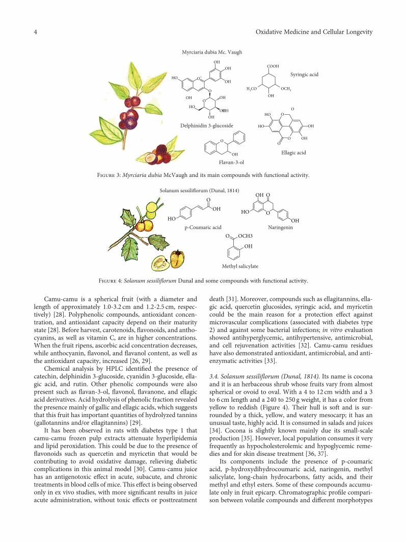
Camu-camu is a spherical fruit (with a diameter andlength of approximately 1.0-3.2 cm and 1.2-2.5 cm, respec-tively) [28]. Polyphenolic compounds, antioxidant concen-tration, and antioxidant capacity depend on their maturitystate [28]. Before harvest, carotenoids, flavonoids, and antho-cyanins, as well as vitamin C, are in higher concentrations.When the fruit ripens, ascorbic acid concentration decreases,while anthocyanin, flavonol, and flavanol content, as well asthe antioxidant capacity, increased [26, 29].
Chemical analysis by HPLC identified the presence ofcatechin, delphinidin 3-glucoside, cyanidin 3-glucoside, ella-gic acid, and rutin. Other phenolic compounds were alsopresent such as flavan-3-ol, flavonol, flavanone, and ellagicacid derivatives. Acid hydrolysis of phenolic fraction revealedthe presence mainly of gallic and ellagic acids, which suggeststhat this fruit has important quantities of hydrolyzed tannins(gallotannins and/or ellagitannins) [29].
It has been observed in rats with diabetes type 1 thatcamu-camu frozen pulp extracts attenuate hyperlipidemiaand lipid peroxidation. This could be due to the presence offlavonoids such as quercetin and myricetin that would becontributing to avoid oxidative damage, relieving diabeticcomplications in this animal model [30]. Camu-camu juicehas an antigenotoxic effect in acute, subacute, and chronictreatments in blood cells of mice. This effect is being observedonly in ex vivo studies, with more significant results in juiceacute administration, without toxic effects or posttreatment
death [31]. Moreover, compounds such as ellagitannins, ella-gic acid, quercetin glucosides, syringic acid, and myricetincould be the main reason for a protection effect againstmicrovascular complications (associated with diabetes type2) and against some bacterial infections; in vitro evaluationshowed antihyperglycemic, antihypertensive, antimicrobial,and cell rejuvenation activities [32]. Camu-camu residueshave also demonstrated antioxidant, antimicrobial, and anti-enzymatic activities [33].
3.4. Solanum sessiliflorum (Dunal, 1814). Its name is coconaand it is an herbaceous shrub whose fruits vary from almostspherical or ovoid to oval. With a 4 to 12 cm width and a 3to 6 cm length and a 240 to 250 g weight, it has a color fromyellow to reddish (Figure 4). Their hull is soft and is sur-rounded by a thick, yellow, and watery mesocarp; it has anunusual taste, highly acid. It is consumed in salads and juices[34]. Cocona is slightly known mainly due its small-scaleproduction [35]. However, local population consumes it veryfrequently as hypocholesterolemic and hypoglycemic reme-dies and for skin disease treatment [36, 37].
Its components include the presence of p-coumaricacid, p-hydroxydihydrocoumaric acid, naringenin, methylsalicylate, long-chain hydrocarbons, fatty acids, and theirmethyl and ethyl esters. Some of these compounds accumu-late only in fruit epicarp. Chromatographic profile compari-son between volatile compounds and different morphotypes
Myrciaria dubia Mc. Vaugh
Delphinidin 3-glucoside
Ellagic acid
Flavan-3-ol
Syringic acidHO
HO
HO
HO
H3CO
COOH
O+
O
OO
O OO
OOH
OHOHOH
OH
OH
OH
OH
OH
OH
OH
OCH3
OH
Figure 3: Myrciaria dubia McVaugh and its main compounds with functional activity.
Solanum sessiliflorum (Dunal, 1814)
p-Coumaric acid Naringenin
Methyl salicylate
HOHO
O
OH
OCH3
OH
OO
O
OH
OH
Figure 4: Solanum sessiliflorum Dunal and some compounds with functional activity.
4 Oxidative Medicine and Cellular Longevity

(oval, small round, and large round) showed chemical differ-ences; the oval morphotype exhibits greater chemical com-plexity in terms of volatile and nonvolatile metabolites.Furthermore, cytotoxic, genotoxic, and antigenotoxic poten-tial was evaluated in vivo, observing a noncytotoxic effecton bone marrow cells and a nongenotoxic effect on Wistarrats. Cocona antioxidant capacity may contribute to the anti-genotoxic effects [38, 39]. Finally, cocona flour administra-tion showed a reduction of total cholesterol concentration,low-density lipoprotein (LDL-c), and liver cholesterol andincreasing cholesterol and high-density lipoprotein (HDL-c)fecal excretion in hypercholesterolemic rats [40].
3.5. Theobroma grandiflorum (Willd. ex Spreng.) (K. Schum,1886). It is a tree that reaches 15-20m high; it belongs tothe Sterculiaceae family [41]. Its fruits have different shapes(oblong, round oval), weighing between 200 g and 4000 g(Figure 5) [42]. It is known as copoazú and belongs to theTheobroma genus, like cocoa, and is considered as one ofthe most popular fruits in the Amazonian market [43].
From copoazú almonds, it is obtained as a cocoa-likeliquor, with improved characteristics on unsaturated fattyacid percentages and a smooth and pleasant flavor. It hasactive antioxidant substances, low percentage of theobro-mine, and high content of linoleic and oleic unsaturated fattyacids. It is considered a suitable product for cosmetic, choco-late, beverage, liquor, and food industries [44]. Its main com-ponents in pulp were detected such as volatile compounds:24 esters, 13 terpenes, 8 alcohols, 4 carbonyls, 4 acids, 2lactones and phenol, ethyl butanoate, ethyl hexanoate,and linalool [41].
Polyphenols derived from copoazú were studied evaluat-ing the distribution and metabolism in the gastrointestinaltract of mice and the microbial metabolic conversion of aunique combination of flavonoids (flavan-3-ols, procyani-dins, and flavones). These compounds are accumulatedmainly in the stomach and small intestine where they couldexert local effects. Procyanidin microbial metabolism was dif-ferent from cocoa that contains procyanidin too [43]. Fur-ther, copoazú and cocoa liquors were chronically provided
to diabetic rats with streptokin. Copoazú liquor improvestheir lipid profile and antioxidant status, which could suggesta superior effect of the cocoa liquor [45].
3.6. Mauritia flexuosa L.f. (1782). It is commonly known ascanangucha, buriti, or moriche palm. It is considered themost abundant native palm that grows naturally in the Bra-zilian Amazon biome. Its fruit is highly nutritious with ayellow-orange pulp (Figure 6) and bittersweet taste. Its endo-carp is surrounded by a spongy material made of starch andoil, with a hard skin, and contains a small reddish-brownscale-like fruit [46]. It is possible to extract oil from its pulp,whose main components are palmitic (18.7%), stearic (1.5%),oleic (76.7%), linoleic (1.5%), linolenic (0.7%), and arachidicacid (0.5%) [47].
The moriche plant has phenolic compounds mainly fla-vonoids and glycosylated anthocyanins like the following:catechin, caffeic acid, chlorogenic acid, quercetin, narin-genin, myricetin, vitexin, scoparin, rutin, cyanidin-3-rutino-side, cyanidin-3-glucoside, epicatechin, and kaempferol[48]. On the other hand, the fruits show a reasonable amountof phenolic compounds, carotenoids (with predominance ofβ-carotenes), and antioxidant activity, which confirms thefunctional potential of moriche [49]. Fruit pulp extractsshowed six phenolic acids: p-coumaric, ferulic, caffeic, proto-catechuic, chlorogenic, and quinic. Quinic acid is much moreabundant than other phenolic acids in pulp; extracts alsoshow seven kinds of flavonoids such as catechin, epicatechin,apigenin, luteolin, myricetin, kaempferol, and quercetin [50].In leaves, tricine-7-O-rutinoside, apigenin-6-C-arabinoside,8-C-glucoside (isoschaftoside), kaempferol-3-O-rutinoside(nicotiflorine), quercetin-3-O-rutinoside (rutin), luteolin-8-C-glucoside (orientin), and luteolin-6-C-glucoside (isoorien-tin) were identified [51]. Leaf extract revealed its great abilityto inhibit food pathogens, like Pseudomonas aeruginosa, anda moderate antimicrobial activity when applied in fruits [48].
3.7. Plukenetia volubilis L. It is a domesticated grapevineknown also as sacha inchi, sacha yuchi, sacha yuchiqui, moun-tain peanuts, wild peanuts, or inca peanuts among others. It
�eobroma grandiflorum (Willd. ex Spreng.) K.Schum. 1886
�eobromineLinalool
Oleic acid
Linoleic acid
O
N
O OOH
CH3
N
NOH
HO
HN
CH2(CH2)5CH2
O
Figure 5: Theobroma grandiflorum and its main compounds with functional activity.
5Oxidative Medicine and Cellular Longevity

grows in warm climates, at high altitude in the Andean rain-forest to the Peruvian Amazon lowlands (Figure 7). Due toits oil content, it is used as food supplement, in skin care,and for wound treatment, insect bites, and skin infections [52].
The most studied and interesting fraction of this fruit isits oil. Fruit seeds are a suitable oil source (35-60%) rich inomega 3 and 6, whose composition varies according to seedvarieties. Found were significant contents of α-linolenic acidand a low linolenic acid/linoleic acid ratio, as well as consid-erable amounts of tocopherols (γ- and δ-tocopherols), phy-tosterols (β-sitosterol and stigmasterol), and phenoliccompounds like ferulic acid. However, no correlations havebeen found between hydrophilic and lipophilic bioactivecompounds and antioxidant capacity. It suggests a complexinteraction of different antioxidant compounds with differ-ent action modes. Although there are few studies on thesacha inchi oil effects on health, there are evidences that itcould act by improving the lipid profile [52]. Regarding itsuse for skin care, sacha inchi oil was very active as a nonstick(preventive) in keratinocytes and in the detachment ofStaphylococcus aureus on the adherence to in vitro humanskin explants [53].
3.8. Bactris gasipaes H. B. Kunth. It is an Amazonian palmgrown mainly for fruit production (Figure 8), known aschontaduro, pejiballe, acana, or pupunha [54]. The chonta-duro fruit has considerable concentration of proteins andoil [55], with an important content of linoleic and linolenicpolyunsaturated fatty acids [56], as well as β-carotenes [57].
Chontaduro flour residues contain different types ofcarotenoids: violaxanthin, lutein, zeaxanthin, 15-cis β-caro-tene, 13-cis β-carotene, all-trans β-carotene, 9-cis β-caro-tene, and α-carotene, as the main carotenoid pigment.Retinol equivalent values found for chontaduro cookedfruit (traditional consumption form) and flour are higherthan those reported for popular products such as tomatoand papaya [58, 59]. Chontaduro flour carbohydrates arepredominantly composed of insoluble fiber, highly esteri-fied homogalacturonan (70% of esterification). It containslinear methyl and minor portions of xylogalacturonanand rhamnogalacturonan that may promote health benefits.Although not very well documented in the literature, prob-ably refer to their antioxidant capacity and their nutritionalvalue since their protein contains eight essential aminoacids [60].
COOH
H
H
H
H
O
H
H
H
H
HO
HO
HO
�훼-Linolenic Acid
�훽-Sitosterol
Stigmasterol
�훾-Tocopherol
Plukenetia volubilis Hnneo
Figure 7: Plukenetia volubilis L. and its main compounds with functional activity.
OHHO
HOHO
OHO
O
OH
OOH
HO
OH
O
OOHOH
OH
O
O
OH
OH
OH
HO
HO
OH
OO
O
OH
OH
OH OHHO
Mauritia flexuosa L.f.
VitexinMyricetin
Luteolin
ApigeninProtocatechuic acid
Figure 6: Mauritia flexuosa L.f. and some compounds with functional activity.
6 Oxidative Medicine and Cellular Longevity

3.9. Paullinia cupana Kunth (1823). This climbing shrub,better known as guarana, is rich in vitamins and stimu-lants such as caffeine; so, it is used mainly for consump-tion as beverage (Figure 9). It is produced mainly in theBrazilian states of Amazonas and Bahia, and approxi-mately 70% of its production is used in soft and energydrink industries [61]. Its seeds are used to produce guaranapowder, which is consumed mainly due to its stimulatingactivity [62]. The main reason so far to study guarana isits caffeine content, and this probably will continue dueto the high demand of this alkaloid in the pharmaceuticaland cosmetic industries. Semipurified guarana extract shows
antidepressant and panicolytic effects [63]. Guarana seedextracts present antimicrobial activity against Escherichiacoli, Pseudomonas fluorescens, Bacillus cereus, and spoilagefungi such as Aspergillus niger, Trichoderma viride, andPenicillium cyclopium [64].
All guarana seed extracts have antioxidant activitywith high amounts of total phenolic compounds like cate-chins, such as epicatechin, catechin, and epicatechin gallate.Due to their high antioxidant, antibacterial, and antifungalactivities, guarana extracts have a promising potential as nat-ural antioxidants in food, cosmetic, and pharmaceuticalindustries [64].
Bactris gasipaes H. B. Kunth
Violaxanthin
Galacturonan
Zeaxanthin
HO
HO O
OO
O
O
O
O
O
O
O
O
O
O
OO
O
OO
O
OO
O
HO
HO
OH
OH
OH
COO−
COO−
COO−
COO−
HO
HO
HO
HO
HO
HO
OH
COO−
COO−
CH2CO
CH3CO
CH3CO
COOCH3
COOCH3
COOCH1
OHO
O
OH
HH
H
HH
H
H
H
H
HH
H
HH
OH
Figure 8: Bactris gasipaes H. B. Kunth and its main functional compounds.
Paullinia cupana Kunth 1823
Caffeine
Epicatechin gallate
Rhamnogalacturonan
N
N
N
N
O
O
O
O
O
HO
OH
OH
OH
OH
OHOH
O O
O
O
O
O
OO
CC
CH3
CH3CH3
CH3OH
CH3OH
CHOH
O
O
COOH
COOH
COOH
COOH
COOH
COOH
COOHCOOH
O
O
OO
OO
O
O
O
O
O
O OO
O
O
O
OOOO
O
O
O
OO
O
O
O
O
O
O
OO
O
O
OO
O
O
O
O
OO
O O
O
O
O
O
O
O
O
O
O
OH
OH
OH
OH
OH
OH
OHOH
OH
OH
OH
OH
OH
OH
OH
OH
OH
OH
OH
OH
OH
HO
HO
HO
HO
HOHO
HO
HO
HOHO
HOHO
HOHO
HOHO
HO
HO
COOH
COOH
COOHCOOH
COOH
COOH
HO
HOHOHOHO
HO
HOHOOC
HO
HO
HOH3C
H3C H3C H3C
H3C
HOH3C
H3C
H3C
H3CH3C
HOH3C
HOH3C
Figure 9: Paullinia cupana Kunth, 1823, and some functional compounds.
7Oxidative Medicine and Cellular Longevity

Table2:Antioxidant
capacity
ofnative
Amazon
ianfruits.
Nam
eDPPH
ORAC
ABTS
FRAP
Reference
Eugeniastipitata
IC50069
±023
μg/mL
37198
±11
50μmol
TE/100
gDW
N/R
N/R
[5]
Euterpeoleracea
21,049
±3,0710μ
mol
TE/100
gDW;
12,420
μmolTE
±465/100g
DW;
6091μ
molTE
/gDW
IC50
88±027
μg/mL
101,336.1μmol
TE/100
gDW;
686.0μmol
TE/100
gDW
247±10
6μmolTE
/100
gDW;
40,330
±19,656
μmolTE
/100
gDW
3,834±56
mgascorbic
acid/100
gDW
[6,14,16]
Myrciaria
dubia
185±11
μmolTE
/gFW
;1,679±75
μmolTE
/gDW
;2,1387μ
molTE
/gDW
IC50
1,11687
±0064μ
g/mL
1,002±27
(μmolTE/gDW;
3,060.8μmol
TE/g
DW
N/R
N/R
[26,30,31]
Solanu
msessiliflorum
N/R
N/R
N/R
N/R
Mau
ritiaflexuosa
IC50
1958
±0064m
g/mL
N/R
33.02μmol
TE/g
FW28080
±37
99μmol
FeSO
4·7H
2Oequiv/100g
[48,49]
Theobromagran
diflorum
1,913±228μ
molTE
/100
gDW
13,628
±184μ
mol
TE/100
gDW
N/R
N/R
[51]
Plukenetia
volubilis
N/R
6.5–9.8μmol
TE/g
N/R
N/R
[29]
Bactrisgasipaes
N/R
N/R
N/R
N/R
Paullin
iacupana
IC50
=85μ
g/mL
(app
roximatevaluetakenfrom
thegraph)
N/R
N/R
N/R
[63]
TE:T
roloxequivalent,V
CE:vitam
inCequivalent,D
W:dry
weight;FW
:fresh
weight;NR:n
otrepo
rted
8 Oxidative Medicine and Cellular Longevity

The presence of dietary fiber, including pectic and hemi-cellulose polysaccharides has been reported, and a homoga-lacturonan with rhamnogalacturonan and xylans has alsobeen isolated and characterized. Pectic polysaccharides andmethanolic extract exhibited antioxidant activity, and partof the possible antioxidant effects of guarana could be attrib-uted to their pectic component [62].
4. Antioxidant Capacity of NativeAmazonian Fruits
In summary, most of the compounds with functional activitycorrespond to compounds with antioxidant activity; Table 2shows the different methods used in the references examinedin the present mini review. However, the comparisons areextremely complicated so it would be more appropriate toreview clinical studies performed on animals, but unfortu-nately, to date, there are very few of them. The antioxidantcapacity methods were DPPH, FRAP, TEAC, ABTS, andORAC. Comparison of antioxidant capacity between fruitsshould be made only when the conditions (method, solvent,sampling, expression of results, etc.) analyzed are the same;therefore, results are not comparable with a great disadvan-tage that presents to compare the antioxidant capacities ofvarious fruits.
5. Conclusion
According to numerous authors, many Amazonian fruits arean adequate source of multiple compounds with potentialhealth benefits, mainly antioxidant effects, which has alsobeen proven through numerous studies such as those detailedin this mini review. However, among its differences in com-position, quality, and insufficient in vivo tests, scientific evi-dence offers challenges and great opportunities in differentareas of research (toxicology, food safety, food technology,and processing). Therefore, new trends in functional foodsshould be conducted considering the enormous potential ofthese Amazonian fruits in human health.
Conflicts of Interest
The authors declare no conflict of interest.
References
[1] L. C. Neves, J. M. Tosin, R. M. Benedette, andL. Cisneros-Zevallos, “Post-harvest nutraceutical behaviourduring ripening and senescence of 8 highly perishable fruitspecies from the Northern Brazilian Amazon region,” FoodChemistry, vol. 174, pp. 188–196, 2015.
[2] H. Rogez, R. Buxant, E. Mignolet, J. N. S. Souza, E. M. Silva,and Y. Larondelle, “Chemical composition of the pulp of threetypical Amazonian fruits: araça-boi (Eugenia stipitata), bacuri(Platonia insignis) and cupuaçu (Theobroma grandiflorum),”European Food Research and Technology, vol. 218, no. 4,pp. 380–384, 2004.
[3] R. Rodríguez Sandoval and P. Bastidas Garzón, “Evaluatingthe cooking process for obtaining hard candy from araza
(Eugenia stipitata) pulp,” Ingeniería e Investigación, vol. 29,pp. 35–41, 2009.
[4] J. P. L. Aguiar, “Araçá-boi (eugenia stiptata, mcvaug) - aspec-tos e dados preliminares sobre a sua composição química,”Acta Amazonica, vol. 13, no. 5-6, pp. 953-954, 1983.
[5] I. A. Neri-Numa, L. B. Carvalho-Silva, J. P. Morales et al.,“Evaluation of the antioxidant, antiproliferative and antimuta-genic potential of araçá-boi fruit (Eugenia stipitata Mc Vaugh—Myrtaceae) of the Brazilian Amazon Forest,” Food ResearchInternational, vol. 50, no. 1, pp. 70–76, 2013.
[6] G. A. Garzón, C.-E. Narváez-Cuenca, R. E. Kopec, A. M.Barry, K. M. Riedl, and S. J. Schwartz, “Determination ofcarotenoids, total phenolic content, and antioxidant activityof Arazá (Eugenia stipitata McVaugh), an Amazonian fruit,”Journal of Agricultural and Food Chemistry, vol. 60, no. 18,pp. 4709–4717, 2012.
[7] F. A. Cuellar, E. Ariza, C. Anzola, and P. Restrepo, “Researchof antioxidant capacity of araza (Eugenia stipitata Mc Vaugh)during the ripening,” Revista Colombiana de Química, vol. 42,pp. 21–28, 2013.
[8] A. Guerrini, G. Sacchetti, A. Grandini, A. Spagnoletti,M. Asanza, and L. Scalvenzi, “Cytotoxic effect and TLCbioautography-guided approach to detect health propertiesof Amazonian Hedyosmum sprucei essential oil,” Evidence--Based Complementary and Alternative Medicine, vol. 2016,Article ID 1638342, 8 pages, 2016.
[9] S. Casiglia, M. Bruno, M. Bramucci et al., “Kundmannia sicula(L.) DC: a rich source of germacrene D,” Journal of EssentialOil Research, vol. 29, no. 6, pp. 437–442, 2017.
[10] I. Peluso, T. Magrone, D. Villaño Valencia, C. Y. O. Chen,and M. Palmery, “Antioxidant, anti-inflammatory, andmicrobial-modulating activities of nutraceuticals and func-tional foods,” Oxidative Medicine and Cellular Longevity,vol. 2017, Article ID 7658617, 2 pages, 2017.
[11] F. Aguiar, V. Menezes, and H. Rogez, “Spontaneous posthar-vest fermentation of açai (Euterpe oleracea) fruit,” PostharvestBiology and Technology, vol. 86, pp. 294–299, 2013.
[12] R. G. Costa, K. Andreola, R. de Andrade Mattietto, L. J. G. deFaria, and O. P. Taranto, “Effect of operating conditions onthe yield and quality of açai (Euterpe oleracea Mart.) powderproduced in spouted bed,” LWT - Food Science and Technol-ogy, vol. 64, no. 2, pp. 1196–1203, 2015.
[13] M. Amsellem-Laufer, “Euterpe oleracea Martius (Areca-ceae):Açaï,” Phytothérapie, vol. 13, no. 2, pp. 135–140, 2015.
[14] B. A. Rojano, I. C. Z. Vahos, A. F. A. Arbeláez, A. J. M.Martínez,F. B. C. Correa, and L. G. Carvajal, “Polifenoles y actividadantioxidante del fruto liofilizado de palma naidi (Açai Colom-biano) (Euterpe oleracea Mart),” Revista Facultad Nacional deAgronomía Medellín, vol. 64, pp. 6213–6220, 2011.
[15] G. A. Garzón, C. E. Narváez-Cuenca, J. P. Vincken, andH. Gruppen, “Polyphenolic composition and antioxidantactivity of açai (Euterpe oleraceaMart.) from Colombia,” FoodChemistry, vol. 217, pp. 364–372, 2017.
[16] L. Barros, R. C. Calhelha, M. J. R. P. Queiroz et al., “The pow-erful in vitro bioactivity of Euterpe oleracea Mart. seeds andrelated phenolic compounds,” Industrial Crops and Products,vol. 76, pp. 318–322, 2015.
[17] P. O. Barbosa, D. Pala, C. T. Silva et al., “Açai (Euterpe oleraceaMart.) pulp dietary intake improves cellular antioxidantenzymes and biomarkers of serum in healthy women,” Nutri-tion, vol. 32, no. 6, pp. 674–680, 2016.
9Oxidative Medicine and Cellular Longevity
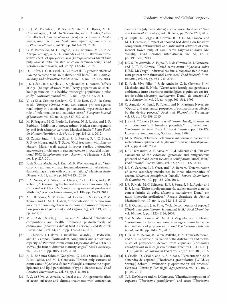
[18] B. J. M. Da Silva, J. R. Souza-Monteiro, H. Rogez, M. E.Crespo-López, J. L. M. Do Nascimento, and E. O. Silva, “Selec-tive effects of Euterpe oleracea (açai) on Leishmania (Leish-mania) amazonensis and Leishmania infantum,” Biomedicine& Pharmacotherapy, vol. 97, pp. 1613–1621, 2018.
[19] G. R. Romualdo, M. F. Fragoso, R. G. Borguini, M. C. P. deAraújo Santiago, A. A. H. Fernandes, and L. F. Barbisan, “Pro-tective effects of spray-dried açaí (Euterpe oleracea Mart) fruitpulp against initiation step of colon carcinogenesis,” FoodResearch International, vol. 77, pp. 432–440, 2015.
[20] D. F. Silva, F. C. B. Vidal, D. Santos et al., “Cytotoxic effects ofEuterpe oleracea Mart. in malignant cell lines,” BMC Comple-mentary and Alternative Medicine, vol. 14, no. 1, p. 175, 2014.
[21] J. K. Udani, B. B. Singh, V. J. Singh, and M. L. Barrett, “Effectsof Açai (Euterpe oleracea Mart.) berry preparation on meta-bolic parameters in a healthy overweight population: a pilotstudy,” Nutrition Journal, vol. 10, no. 1, pp. 45–51, 2011.
[22] V. da Silva Cristino Cordeiro, G. F. de Bem, C. A. da Costaet al., “Euterpe oleracea Mart. seed extract protects againstrenal injury in diabetic and spontaneously hypertensive rats:role of inflammation and oxidative stress,” European Journalof Nutrition, vol. 57, no. 2, pp. 817–832, 2018.
[23] M. F. Fragoso, M. G. Prado, L. Barbosa, N. S. Rocha, and L. F.Barbisan, “Inhibition of mouse urinary bladder carcinogenesisby açai fruit (Euterpe oleraceae Martius) intake,” Plant Foodsfor Human Nutrition, vol. 67, no. 3, pp. 235–241, 2012.
[24] G. Zapata-Sudo, J. S. da Silva, S. L. Pereira, P. J. C. Souza,R. S. de Moura, and R. T. Sudo, “Oral treatment with Euterpeoleracea Mart. (açaí) extract improves cardiac dysfunctionand exercise intolerance in rats subjected to myocardial infarc-tion,” BMC Complementary and Alternative Medicine, vol. 14,no. 1, p. 227, 2014.
[25] F. de Souza Machado, J. Kuo, M. F. Wohlenberg et al., “Sub-chronic treatment with acai frozen pulp prevents the brain oxi-dative damage in rats with acute liver failure,”Metabolic BrainDisease, vol. 31, no. 6, pp. 1427–1434, 2016.
[26] L. C. Neves, V. X. Silva, E. A. Chagas, C. G. B. Lima, and S. R.Roberto, “Determining the harvest time of camu-camu [Myr-ciaria dubia (H.B.K.) McVaugh] using measured pre-harvestattributes,” Scientia Horticulturae, vol. 186, pp. 15–23, 2015.
[27] A. L. R. Souza, M. M. Pagani, M. Dornier, F. S. Gomes, R. V.Tonon, and L. M. C. Cabral, “Concentration of camu-camujuice by the coupling of reverse osmosis and osmotic evapora-tion processes,” Journal of Food Engineering, vol. 119, no. 1,pp. 7–12, 2013.
[28] M. S. Akter, S. Oh, J. B. Eun, and M. Ahmed, “Nutritionalcompositions and health promoting phytochemicals ofcamu-camu (Myrciaria dubia) fruit: a review,” Food ResearchInternational, vol. 44, no. 7, pp. 1728–1732, 2011.
[29] R. Chirinos, J. Galarza, I. Betalleluz-Pallardel, R. Pedreschi,and D. Campos, “Antioxidant compounds and antioxidantcapacity of Peruvian camu camu (Myrciaria dubia (H.B.K.)McVaugh) fruit at different maturity stages,” Food Chemistry,vol. 120, no. 4, pp. 1019–1024, 2010.
[30] A. E. de Souza Schmidt Gonçalves, C. Lellis-Santos, R. Curi,F. M. Lajolo, and M. I. Genovese, “Frozen pulp extracts ofcamu-camu (Myrciaria dubia McVaugh) attenuate the hyper-lipidemia and lipid peroxidation of type 1 diabetic rats,” FoodResearch International, vol. 64, pp. 1–8, 2014.
[31] F. C. da Silva, A. Arruda, A. Ledel et al., “Antigenotoxic effectof acute, subacute and chronic treatments with Amazonian
camu-camu (Myrciaria dubia) juice on mice blood cells,” Foodand Chemical Toxicology, vol. 50, no. 7, pp. 2275–2281, 2012.
[32] A. Fujita, K. Borges, R. Correia, B. D. G. M. Franco, andM. I. Genovese, “Impact of spouted bed drying on bioactivecompounds, antimicrobial and antioxidant activities of com-mercial frozen pulp of camu-camu (Myrciaria dubia Mc.Vaugh),” Food Research International, vol. 54, no. 1,pp. 495–500, 2013.
[33] J. C. S. De Azevêdo, A. Fujita, E. L. de Oliveira, M. I. Genovese,and R. T. P. Correia, “Dried camu-camu (Myrciaria dubiaH.B.K. McVaugh) industrial residue: a bioactive-rich Amazo-nian powder with functional attributes,” Food Research Inter-national, vol. 62, pp. 934–940, 2014.
[34] D. F. da Silva Filho, J. S. de Andrade, C. R. Clement, F. M.Machado, and H. Noda, “Correlações fenotípicas, genéticas eambientais entre descritores morfológicos e químicos em fru-tos de cubiu (Solanum sessiliflorum Dunal) da amazônia1,”Acta Amazonica, vol. 29, no. 4, pp. 503–511, 1999.
[35] C. Agudelo, M. Igual, P. Talens, and N. Martínez-Navarrete,“Optical and mechanical properties of cocona chips as affectedby the drying process,” Food and Bioproducts Processing,vol. 95, pp. 192–199, 2015.
[36] J. Salick, “Cocona (Solanum sessiliflorum Dunal), an overviewof productions and breeding potentials,” in InternationalSymposium on New Crops for Food Industry, pp. 125–129,University Southampton, Southampton, 1989.
[37] M. A. Pardo, “Efecto de Solanum sessiliflorum dunal sobre elmetabolismo lipídico y de la glucosa,” Ciencia e Investigación,vol. 7, pp. 43–48, 2004.
[38] L. C. Hernandes, A. F. Aissa, M. R. d. Almeida et al., “In vivoassessment of the cytotoxic, genotoxic and antigenotoxicpotential of maná-cubiu (Solanum sessiliflorum Dunal) fruit,”Food Research International, vol. 62, pp. 121–127, 2014.
[39] J. E. C. Cardona, L. E. Cuca, and J. A. Barrera, “Determinationof some secondary metabolites in three ethnovarieties ofcocona (Solanum sessiliflorum Dunal),” Revista Colombianade Química, vol. 40, pp. 185–200, 2011.
[40] J. R. P. Maia, M. C. Schwertz, R. F. S. Sousa, J. P. L. Aguiar, andE. S. Lima, “Efeito hipolipemiante da suplementação dietéticacom a farinha do cubiu (Solanum sessiliflorum Dunal) emratos hipercolesterolêmicos,” Revista Brasileira de PlantasMedicinais, vol. 17, no. 1, pp. 112–119, 2015.
[41] C. E. Quijano and J. A. Pino, “Volatile compounds of copoazú(Theobroma grandiflorum Schumann) fruit,” Food Chemistry,vol. 104, no. 3, pp. 1123–1126, 2007.
[42] S. d. N. Melo Ramos, W. Danzl, G. Ziegleder, and P. Efraim,“Formation of volatile compounds during cupuassu fermenta-tion: influence of pulp concentration,” Food Research Interna-tional, vol. 87, pp. 161–167, 2016.
[43] H. R. d. M. Barros, R. García-Villalba, F. A. Tomás-Barberán,andM. I. Genovese, “Evaluation of the distribution andmetab-olism of polyphenols derived from cupuassu (Theobromagrandiflorum) in mice gastrointestinal tract by UPLC-ESI-Q-TOF,” Journal of Functional Foods, vol. 22, pp. 477–489, 2016.
[44] J. Criollo, D. Criollo, and A. S. Aldana, “Fermentación de laalmendra de copoazú (Theobroma grandiflorum [Willd. exSpreng.] Schum.): evaluación y optimización del proceso,”Corpoica Ciencia y Tecnología Agropecuaria, vol. 11, no. 2,p. 107, 2010.
[45] T. B. De Oliveira andM. I. Genovese, “Chemical composition ofcupuassu (Theobroma grandiflorum) and cocoa (Theobroma
10 Oxidative Medicine and Cellular Longevity

cacao) liquors and their effects on streptozotocin-induceddiabetic rats,” Food Research International, vol. 51, no. 2,pp. 929–935, 2013.
[46] J. T. Milanez, L. C. Neves, R. C. Colombo, M. Shahab, and S. R.Roberto, “Bioactive compounds and antioxidant activity ofburiti fruits, during the postharvest, harvested at different rip-ening stages,” Scientia Horticulturae, vol. 227, pp. 10–21, 2018.
[47] R. Pereira Lima, P. T. Souza da Luz, M. Braga et al., “Mur-umuru (Astrocaryum murumuru Mart.) butter and oils ofburiti (Mauritia flexuosa Mart.) and pracaxi (Pentaclethramacroloba (Willd.) Kuntze) can be used for biodiesel produc-tion: physico-chemical properties and thermal and kineticstudies,” Industrial Crops and Products, vol. 97, pp. 536–544,2017.
[48] H. H. F. Koolen, F. M. A. da Silva, F. C. Gozzo, A. Q. L. deSouza, and A. D. L. de Souza, “Antioxidant, antimicrobialactivities and characterization of phenolic compounds fromburiti (Mauritia flexuosa L. f.) by UPLC-ESI-MS/MS,” FoodResearch International, vol. 51, no. 2, pp. 467–473, 2013.
[49] T. L. N. Candido, M. R. Silva, and T. S. Agostini-Costa, “Bioac-tive compounds and antioxidant capacity of Buriti (Mauritiaflexuosa L.f.) from the Cerrado and Amazon biomes,” FoodChemistry, vol. 177, pp. 313–319, 2015.
[50] G. A. Bataglion, F. M. A. da Silva, M. N. Eberlin, and H. H. F.Koolen, “Simultaneous quantification of phenolic compoundsin buriti fruit (Mauritia flexuosa L.f.) by ultra-high perfor-mance liquid chromatography coupled to tandem mass spec-trometry,” Food Research International, vol. 66, pp. 396–400,2014.
[51] D. M. De Oliveira, E. P. Siqueira, Y. R. F. Nunes, and B. B.Cota, “Flavonoids from leaves of Mauritia flexuosa,” RevistaBrasileira de Farmacognosia, vol. 23, no. 4, pp. 614–620,2013.
[52] F. Garmendia, R. Pando, and G. Ronceros, “Efecto del aceite desacha inchi (Plukenetia volúbilis L) sobre el perfil lipídico enpacientes con hiperlipoproteinemia,” Revista Peruana de Med-icina Experimental y Salud Pública, vol. 28, no. 4, pp. 628–632,2012.
[53] G. Gonzalez-Aspajo, H. Belkhelfa, L. Haddioui-Hbabi,G. Bourdy, and E. Deharo, “Sacha inchi oil (Plukenetia volubi-lis L.), effect on adherence of Staphylococus aureus to humanskin explant and keratinocytes in vitro,” Journal of Ethnophar-macology, vol. 171, pp. 330–334, 2015.
[54] A. S. Heringer, D. A. Steinmacher, H. P. F. Fraga et al.,“Improved high-efficiency protocol for somatic embryogenesisin peach palm (Bactris gasipaes Kunth) using RITA® tempo-rary immersion system,” Scientia Horticulturae, vol. 179,pp. 284–292, 2014.
[55] E. E. Haro, J. A. Szpunar, and A. G. Odeshi, “Dynamic andballistic impact behavior of biocomposite armors made ofHDPE reinforced with chonta palm wood (Bactris gasipaes)microparticles,” Defence Technology, vol. 14, no. 3, pp. 238–249, 2018.
[56] J. Restrepo, J. A. Estupiñán, and A. J. Colmenares, “Estudiocomparativo de las fracciones lipídicas de Bactris gasipaesKunth (chontaduro) obtenidas por extracción soxhlet y porextracción con CO2 supercrítico,” Revista Colombiana de Quí-mica, vol. 45, no. 1, pp. 5–9, 2016.
[57] F. A. Espinosa-Pardo, J. Martinez, and H. A. Martinez-Correa,“Extraction of bioactive compounds from peach palm pulp(Bactris gasipaes) using supercritical CO2,” The Journal ofSupercritical Fluids, vol. 93, pp. 2–6, 2014.
[58] C. Rojas-Garbanzo, A. M. Pérez, J. Bustos-Carmona, andF. Vaillant, “Identification and quantification of carotenoidsby HPLC-DAD during the process of peach palm (Bactris gasi-paesH.B.K.) flour,” Food Research International, vol. 44, no. 7,pp. 2377–2384, 2011.
[59] G. J. Basto, C. W. P. Carvalho, A. G. Soares et al., “Physico-chemical properties and carotenoid content of extruded andnon-extruded corn and peach palm (Bactris gasipaes, Kunth),”LWT - Food Science and Technology, vol. 69, pp. 312–318,2016.
[60] B. C. Bolanho, E. D. G. Danesi, and A. D. P. Beleia, “Carbohy-drate composition of peach palm (Bactris gasipaes Kunth)by-products flours,” Carbohydrate Polymers, vol. 124, pp. 196–200, 2015.
[61] F. C. Schimpl, J. F. da Silva, J. F. C. Gonçalves, andP. Mazzafera, “Guarana: revisiting a highly caffeinated plantfrom the Amazon,” Journal of Ethnopharmacology, vol. 150,no. 1, pp. 14–31, 2013.
[62] N. Dalonso and C. L. d. O. Petkowicz, “Guarana powder poly-saccharides: characterisation and evaluation of the antioxidantactivity of a pectic fraction,” Food Chemistry, vol. 134, no. 4,pp. 1804–1812, 2012.
[63] T. Klein, R. Longhini, M. L. Bruschi, and J. C. P. de Mello,“Microparticles containing Guaraná extract obtained byspray-drying technique: development and characterization,”Revista Brasileira de Farmacognosia, vol. 25, no. 3, pp. 292–300, 2015.
[64] L. Majhenič, M. Škerget, and Ž. Knez, “Antioxidant and anti-microbial activity of guarana seed extracts,” Food Chemistry,vol. 104, no. 3, pp. 1258–1268, 2007.
11Oxidative Medicine and Cellular Longevity

Review ArticleBorneol for Regulating the Permeability of the Blood-BrainBarrier in Experimental Ischemic Stroke: Preclinical Evidence andPossible Mechanism
Zi-xian Chen,1 Qing-qing Xu ,1 Chun-shuo Shan ,1 Yi-hua Shi ,1 Yong Wang,1
Raymond Chuen-Chung Chang ,2,3 and Guo-qing Zheng 1
1Department of Neurology, The Second Affiliated Hospital and Yuying Children’s Hospital of Wenzhou Medical University,Wenzhou, China2Laboratory of Neurodegenerative Diseases, School of Biomedical Sciences, LKS Faculty of Medicine,The University of Hong Kong, Hong Kong3State Key Laboratory of Brain and Cognitive Sciences, The University of Hong Kong, Hong Kong
Correspondence should be addressed to Raymond Chuen-Chung Chang; [email protected] Guo-qing Zheng; [email protected]
Received 8 August 2018; Accepted 2 December 2018; Published 4 February 2019
Guest Editor: Luciana Scotti
Copyright © 2019 Zi-xian Chen et al. This is an open access article distributed under the Creative Commons Attribution License,which permits unrestricted use, distribution, and reproduction in any medium, provided the original work is properly cited.
Borneol, a natural product in the Asteraceae family, is widely used as an upper ushering drug for various brain diseases in manyChinese herbal formulae. The blood-brain barrier (BBB) plays an essential role in maintaining a stable homeostaticenvironment, while BBB destruction and the increasing BBB permeability are common pathological processes in many seriouscentral nervous system (CNS) diseases, which is especially an essential pathological basis of cerebral ischemic injury. Here, weaimed to conduct a systematic review to assess preclinical evidence of borneol for experimental ischemic stroke as well asinvestigate in the possible neuroprotective mechanisms, which mainly focused on regulating the permeability of BBB. Sevendatabases were searched from their inception to July 2018. The studies of borneol for ischemic stroke in animal models wereincluded. RevMan 5.3 was applied for data analysis. Fifteen studies investigated the effects of borneol in experimental ischemicstroke involving 308 animals were ultimately identified. The present study showed that the administration of borneol exerted asignificant decrease of BBB permeability during cerebral ischemic injury according to brain Evans blue content and brain watercontent compared with controls (P < 0 01). In addition, borneol could improve neurological function scores (NFS) and cerebralinfarction area. Thus, borneol may be a promising neuroprotective agent for cerebral ischemic injury, largely through alleviatingthe BBB disruption, reducing oxidative reactions, inhibiting the occurrence of inflammation, inhibiting apoptosis, andimproving the activity of lactate dehydrogenase (LDH) as well as P-glycoprotein (P-GP) and NO signaling pathway.
1. Introduction
The blood-brain barrier (BBB) is an anatomical and biochem-ical barrier, consisting of endothelial cells, a basal lamina, andastrocytic end feet [1]. The BBB integrity is of great signifi-cance for brain homeostasis, while the dysfunction of BBBcan lead to complications of neurological diseases, such asstroke, chronic neurodegenerative disorders, neuroinflamma-tory disorders, and brain tumor [1–3].Moreover, a disruptionof BBB integrity, characterized by increased permeability, is
associated with several neurological pathologies, such asischemia/hypoxia, hemorrhage, multiple sclerosis, and amyo-trophic lateral sclerosis [4]. During the cerebral ischemia/re-perfusion injury, changes in BBB structures result in theincrease of its permeability and the loss of its protective func-tion, which may deteriorate the tissue injury [5, 6]. Thus, thedisruption of BBB is an essential pathological basis of cerebralischemic injury. The long-lasting BBB disruption can directlycontribute to cerebral edema and the influx of immunecell and inflammatory materials, ultimately resulting in the
HindawiOxidative Medicine and Cellular LongevityVolume 2019, Article ID 2936737, 15 pageshttps://doi.org/10.1155/2019/2936737
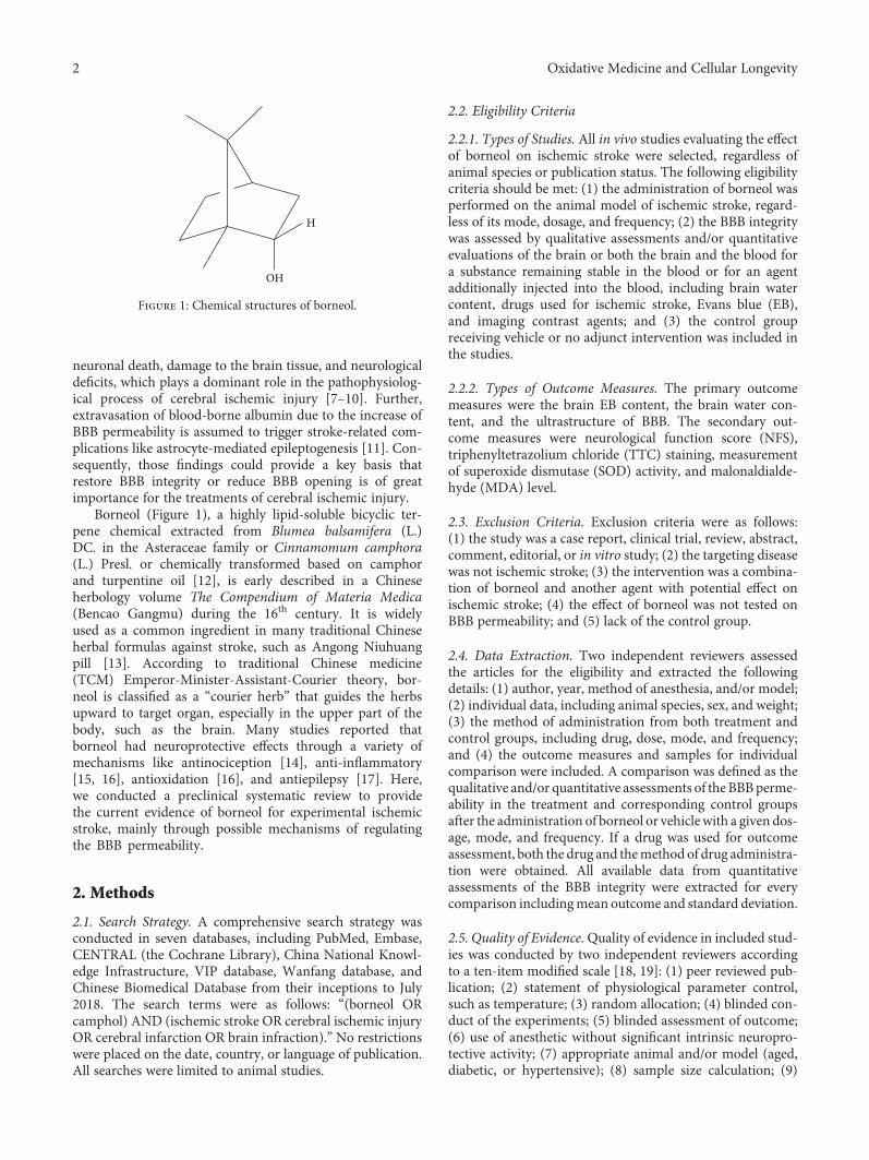
neuronal death, damage to the brain tissue, and neurologicaldeficits, which plays a dominant role in the pathophysiolog-ical process of cerebral ischemic injury [7–10]. Further,extravasation of blood-borne albumin due to the increase ofBBB permeability is assumed to trigger stroke-related com-plications like astrocyte-mediated epileptogenesis [11]. Con-sequently, those findings could provide a key basis thatrestore BBB integrity or reduce BBB opening is of greatimportance for the treatments of cerebral ischemic injury.
Borneol (Figure 1), a highly lipid-soluble bicyclic ter-pene chemical extracted from Blumea balsamifera (L.)DC. in the Asteraceae family or Cinnamomum camphora(L.) Presl. or chemically transformed based on camphorand turpentine oil [12], is early described in a Chineseherbology volume The Compendium of Materia Medica(Bencao Gangmu) during the 16th century. It is widelyused as a common ingredient in many traditional Chineseherbal formulas against stroke, such as Angong Niuhuangpill [13]. According to traditional Chinese medicine(TCM) Emperor-Minister-Assistant-Courier theory, bor-neol is classified as a “courier herb” that guides the herbsupward to target organ, especially in the upper part of thebody, such as the brain. Many studies reported thatborneol had neuroprotective effects through a variety ofmechanisms like antinociception [14], anti-inflammatory[15, 16], antioxidation [16], and antiepilepsy [17]. Here,we conducted a preclinical systematic review to providethe current evidence of borneol for experimental ischemicstroke, mainly through possible mechanisms of regulatingthe BBB permeability.
2. Methods
2.1. Search Strategy. A comprehensive search strategy wasconducted in seven databases, including PubMed, Embase,CENTRAL (the Cochrane Library), China National Knowl-edge Infrastructure, VIP database, Wanfang database, andChinese Biomedical Database from their inceptions to July2018. The search terms were as follows: “(borneol ORcamphol) AND (ischemic stroke OR cerebral ischemic injuryOR cerebral infarction OR brain infraction).” No restrictionswere placed on the date, country, or language of publication.All searches were limited to animal studies.
2.2. Eligibility Criteria
2.2.1. Types of Studies. All in vivo studies evaluating the effectof borneol on ischemic stroke were selected, regardless ofanimal species or publication status. The following eligibilitycriteria should be met: (1) the administration of borneol wasperformed on the animal model of ischemic stroke, regard-less of its mode, dosage, and frequency; (2) the BBB integritywas assessed by qualitative assessments and/or quantitativeevaluations of the brain or both the brain and the blood fora substance remaining stable in the blood or for an agentadditionally injected into the blood, including brain watercontent, drugs used for ischemic stroke, Evans blue (EB),and imaging contrast agents; and (3) the control groupreceiving vehicle or no adjunct intervention was included inthe studies.
2.2.2. Types of Outcome Measures. The primary outcomemeasures were the brain EB content, the brain water con-tent, and the ultrastructure of BBB. The secondary out-come measures were neurological function score (NFS),triphenyltetrazolium chloride (TTC) staining, measurementof superoxide dismutase (SOD) activity, and malonaldialde-hyde (MDA) level.
2.3. Exclusion Criteria. Exclusion criteria were as follows:(1) the study was a case report, clinical trial, review, abstract,comment, editorial, or in vitro study; (2) the targeting diseasewas not ischemic stroke; (3) the intervention was a combina-tion of borneol and another agent with potential effect onischemic stroke; (4) the effect of borneol was not tested onBBB permeability; and (5) lack of the control group.
2.4. Data Extraction. Two independent reviewers assessedthe articles for the eligibility and extracted the followingdetails: (1) author, year, method of anesthesia, and/or model;(2) individual data, including animal species, sex, and weight;(3) the method of administration from both treatment andcontrol groups, including drug, dose, mode, and frequency;and (4) the outcome measures and samples for individualcomparison were included. A comparison was defined as thequalitative and/or quantitative assessments of the BBBperme-ability in the treatment and corresponding control groupsafter the administration of borneol or vehiclewith a given dos-age, mode, and frequency. If a drug was used for outcomeassessment, both the drug and themethod of drug administra-tion were obtained. All available data from quantitativeassessments of the BBB integrity were extracted for everycomparison includingmean outcome and standard deviation.
2.5. Quality of Evidence.Quality of evidence in included stud-ies was conducted by two independent reviewers accordingto a ten-item modified scale [18, 19]: (1) peer reviewed pub-lication; (2) statement of physiological parameter control,such as temperature; (3) random allocation; (4) blinded con-duct of the experiments; (5) blinded assessment of outcome;(6) use of anesthetic without significant intrinsic neuropro-tective activity; (7) appropriate animal and/or model (aged,diabetic, or hypertensive); (8) sample size calculation; (9)
H
OH
Figure 1: Chemical structures of borneol.
2 Oxidative Medicine and Cellular Longevity

compliance with animal welfare regulations; (10) statementof potential conflict of interests.
2.6. Statistical Analysis. The statistical analysis was con-ducted via RevMan version 5.3. To estimate the effect ofborneol on ischemic stroke, a model of random effect (RE)was applied to estimate pooled effect with 95% confidenceintervals (CI) if statistical heterogeneity was found (P < 0 1,I2 > 50%), while a model of fixed effect (FE) was set with95% CI if no statistical evidence of heterogeneity existed(p ≥ 0 1, I2 ≤ 50%). The weighted mean difference (WMD)was calculated as a summary statistic if the outcomes wereapplied the same scale, while standardized mean differences(SMD) was used if the same outcome measurements weremeasured in a variety of ways. Heterogeneity was assessedvia a standard chi-square test and I2 statistic. A probabilityvalue less than 0.05 was considered statistically significant.
3. Results
3.1. Study Selection. After primary search from seven data-bases, a total of 511 potentially relevant studies were included.By reviewing titles and abstracts, 393 studies that werecase reports, clinical trials, reviews, abstracts, comments, or
editorials were excluded. After reading the remaining 118full-text articles for eligibility, 103 studies were removed forat least one of following reasons: (1) not an in vivo study;(2) the targeting disease was not ischemic stroke; (3) thestudy did not access the effects of borneol on the animalmodel of ischemic stroke; (4) the intervention was a combi-nation of borneol and another agent with potential effect onischemic stroke; (5) lack of outcome assessments for BBBintegrity; and (6) lack of the control group. Ultimately, 15studies involving 308 animals were selected for quantitativeanalysis (Figure 2).
3.2. Study Characteristics. Fifteen studies [20–34] involving308 animals were included. Five species were referred,including SD rats (n = 225) [21–23, 25, 27–30, 33, 34], Wistarrats (n = 24) [24], ICR mice (n = 21) [33], Kunming mice(n = 14) [31], and C57 BL/6J mice (n = 24) [20]. The weightof rats ranged from 180 g to 400 g, and the weight of miceranged from 18 g to 25 g. Cerebral ischemic injury in theincluded studies was induced by temporary middle cere-bral artery occlusion (MCAO) for two hours [25–27, 30],permanent MCAO [21, 32], temporary occlusion of bilat-eral common carotid arteries for a range of 20-60min[20, 22, 23, 33], transient occlusion of bilateral hemisphere
Records identified through databasesearching (n = 511)
Scre
enin
gIn
clud
edEl
igib
ility
Records after duplicates and irrelevant articlesremoved (n = 511)
Records screened titleand abstract (n = 118)
Full-text articlesassessed for eligibility
(n = 15)
Full-text articles excluded (n = 103)
Not in vivo studies (n = 19)The targeting disease was not ischemicstroke (n = 27)
(ii)
Not test the effect of borneol onexperimental ischemic stroke (n = 18)The intervention was a combination ofborneol and another agent with potentialeffect on ischemic stroke (n = 5)
(iv)
Studies included inquantitative synthesis
(meta-analysis) (n =15)
Additional records identifiedthrough other sources (n = 0)
Full-text articles excluded (n = 0)
Iden
tifica
tion
(i)
(iii)
Lackof outcome assessment for BBBpermeability (n = 32)
(v)
Lack of control group (n = 2)(vi)
Case reports; clinical trials; reviews;abstracts; comments and editorials
(i)
Records excluded (n = 393)
No available data(i)
Figure 2: Flow diagram of the search process.
3Oxidative Medicine and Cellular Longevity

for 20min by Pulsinelli-4VO method [24], or permanentligation of bilateral common carotid arteries [28, 29, 31, 34].Chloral hydrate was used in twelve studies, while the anes-thetic in three study [21, 26, 28] cannot be confirmed fromthe primary data.
Among all the included studies, one study [21] assessedthe effects of L-borneol, D-borneol, and synthetic borneol,one study [23] assessed the effects of synthetic borneol as wellas L-borneol, three studies [23, 29, 31] used synthetic bor-neol, one study [24] used D-borneol, two studies declaredthe administration of natural borneol [30] but withoutreporting the type of borneol, and the remaining studies usedthe borneol without further information provided. The modeof borneol application consisted of oral gavage (14 studies)and intravenous injection (1 study) [34]. The frequency ofthe treatment varied from once daily for the duration of 3days [20, 21, 24, 26, 27, 29–31] to 8 days [33]. Two studiesapplied the borneol to the animals before as well as afterthe model establishment [20, 25], while the other thirteenstudies applied it before the model establishment.
Four studies [23, 26–28] reported the ultrastructure ofBBB when six studies performed the quantitative assessmentsof brain for EB [20, 24, 25, 30, 31], and ten studies performedthe quantitative assessments of brain for water content[20, 22–25, 27, 29, 32–34]. More details about the character-istics of the included studies were shown in Table 1.
3.3. Quality of Included Study. The quality scores varied from2/10 to 5/10 with the average of 3.13. Twelve studies werepeer-reviewed publications, while three studies were unpub-lished master’s thesis or PhD thesis. Four studies describedthe control of temperature. Fourteen studies declared therandom allocation. Twelve studies described the use of anes-thetic without significant intrinsic neuroprotective activity.Four studies stated the compliance with animal welfare regu-lations. One study had a statement of potential conflict ofinterests. None of the included studies reported the maskedconduct of experiments, the blinded assessments of outcome,and the application of animal or model with relevant comor-bidities or a sample size calculation. The quality scores for theincluded studies shown in Table 2.
3.4. Effectiveness Assessment
3.4.1. The Brain EB Content. The assessments of the brain EBcontent were performed in six studies [20, 24, 25, 28, 30, 31]at a time point ranged from 20min to 72 hours after theinduction of the model. Combining available data in ameta-analysis from the above six studies showed the signifi-cantly protective effect of borneol on the BBB during cerebralischemic injury according to the brain EB content[nTreatment/nControl nT/nC = 40/38, WMD -4.16, 95% CI:-4.68~-3.64, P < 0 00001; heterogeneity χ2 = 12 34, df = 5,I2 = 59%]. After sequentially omitting each study, one outlierstudy [24] reporting Pulsinelli four-vessel method was con-sidered as the potential sources of the heterogeneity.Meta-analysis of remaining four studies showed a morehomogeneous result (nT/nC = 34/32, WMD -3.81, 95% CI:-4.39~-3.23, P < 0 00001; heterogeneity χ2 = 4 94, df = 4,
I2 = 19%, Figure 3). It was indicated that the method ofmodel induction may be one possible explanation forthe heterogeneity.
3.4.2. The Brain Water Content. Ten studies [20, 22–25, 27,29, 32–34] with eleven comparisons evaluated the brainwater content at a time point ranged from 10min to 72 hoursafter the induction of the model. Among them, nine studiesassessed brain water content by using dry-wet weight methodand showed the significant decreasing of BBB permeability inthe treatment of cerebral ischemic injury (nT/nC = 72/72,WMD -1.28, 95% CI: -1.93~-0.63, P = 0 0001; heterogeneityχ2 = 61 26, df = 8, I2 = 87%). After sequential removal ofeach study, the outlier study [24] with model induced by Pul-sinelli four-vessel method was removed. The meta-analysis ofeight studies showed a homogeneous result (nT/nC = 68/68,WMD -0.92, 95% CI: -1.10~-0.75, P < 0 00001; heterogeneityχ2 = 3 96, df = 7, I2 = 0%, Figure 4), which also indicated apossible explanation for the heterogeneity. The remainingstudy [27] with two comparisons reported the rate of cerebraledema based on wet weight and meta-analysis of themshowed significant effects of borneol for alleviating BBB per-meability during cerebral ischemic injury (nT/nC = 16/16,WMD -9.09, 95% CI: -12.11~-6.07, P < 0 00001; heterogene-ity χ2 = 0 02, df = 1, I2 = 0%, Figure 4).
3.4.3. The Ultrastructure of BBB. For cerebral ischemic injury,four studies [21, 23, 26, 27] with seven comparisons assessedthe impacts of borneol on the ultrastructure of BBB, involv-ing six comparisons reporting significantly neuroprotectiveeffects and one comparison reporting no difference.
3.4.4. NFS.NFS was reported in two studies [21, 32] with fourcomparisons, examined according to the five-point scaledescribed previously by Longa et al. (Longa et al., 1989).Meta-analysis showed a significant difference in improvingNFS but with substantial heterogeneity (nT/nC = 45/45, MD-0.42, 95% CI: -0.65 to -0.20, P < 0 00001, heterogeneityχ2 = 300 05, df = 3, I2 = 99%). Zhang et al. [32] comparedadministration of borneol (0.4 g per animal, ig, qd) with thesame volume of 1% Tween (ig, qd) for 7 days before occlu-sion and reported no significant difference in NFS betweenthe two groups (nT/nC = 9/9). Comparing L-borneol(0.2 g/kg, ig, qd), D-borneol (0.2 g/kg, ig, qd), or syntheticborneol (0.6 g/kg, ig, qd) with the same volume of 5% Tween80 solution (ig, qd) for 3 days before occlusion, respectively,Dong et al. [21] found that these three comparisonshad a significant difference in improving NFS (L-borneol:nT/nC = 12/12; D-borneol: nT/nC = 12/12; and syntheticborneol: nT/nC = 12/12).
3.4.5. TTC Staining. Two studies [21, 32] with four compari-sons applied TTC staining to evaluate cerebral infarction area,showing a significant difference but with substantial heteroge-neity (nT/nC = 24/24, MD -6.64, 95% CI: -12.53 to -0.75,P < 0 00001, heterogeneity χ2 = 252 43, df = 3, I2 = 99%).Using borneol (0.4 g per animal, ig, qd) for 7 days beforeocclusion as an experimental administration (nT/nC = 9/9),Zhang et al. [32] found that the infarction area was
4 Oxidative Medicine and Cellular Longevity

Table1:Summaryof
theeffi
cacy
ofborneolfor
ischem
icstroke.
Stud
y(autho
r,years)
Species
Weight
Anesthetic
Con
dition
sor
mod
elindu
ction
Metho
dof
administration
Outcomemeasures
(sam
ple)
Intergroup
differences
Treatmentgrou
pCon
trol
grou
p
Chenetal.
2010
[20]
Male,C57
BL/6J
mice
18–20g
Chloralhydrate
(300
mg/kg,ip)
Tem
porary
occlusion
ofbilateralcom
mon
carotidarteries
for
25min
Borneol,0.01g/kg,ig,
at30
min
before
mod
elandon
cedaily
for2days
after
occlusion
Samevolumeof
10%
ethano
l,ig,at30
min
before
mod
eland
once
daily
for2days
afterocclusion
(1)Brain
EBcontent,
48h(6/6)
(2)Brain
water
content,24
h(6/6)
(3)Morriswater
maze
test,4
d(12/12)
(4)MDA,SOD,4
d(6/6)
(5)GFA
P,4
d(3/3)
(1)P<005
(2)P<005
(3)P>005
(4)P>005
Don
g2018
[21]
aMale,SD
rats
230–270g
NS
Permanentmiddle
cerebralartery
occlusion
L-borneol,0.2g/kg,ig,
once
daily
for3days
before
occlusion
Samevolumeof
5%Tween80
solution
,ig,
once
daily
for3days
before
occlusion
(1)Neurological
function
score,
24h(12/12)
(2)Brain
water
content,24
h(8/8)
(3)C
erebralinfarction
ratewithTTC
staining,24h(5/5)
(4)The
ultrastructure
ofBBB,N
S(5)VEGFlevelsin
the
serum,24h(7/7)
(6)T
NF-αlevelsinthe
serum,24h(7/7)
(7)Bax
mRNA,24h
(3/3)
(8)Bcl-2
mRNA,24h
(3/3)
(9)Claud
in-5
mRNA,
24h(3/3)
(1)P<005
(2)P<001
(3)P<001
(4)NS
(5)P<001
(6)P<001
(7)P>005
(8)P>005
(9)P>005
Don
g2018
[21]
bMale,SD
rats
230–270g
NS
Permanentmiddle
cerebralartery
occlusion
D-borneol,0.2g/kg,
ig,oncedaily
for3
days
before
occlusion
Samevolumeof
5%Tween80
solution
,ig,
once
daily
for3days
before
occlusion
(1)Neurological
function
score,
24h(12/12)
(2)Brain
water
content,24
h(8/8)
(3)C
erebralinfarction
ratewithTTC
staining,24h(5/5)
(4)The
ultrastructure
ofBBB,N
S(5)VEGFlevelsin
the
serum,24h(7/7)
(1)P>005
(2)P>005
(3)P>005
(4)NS
(5)P<001
(6)P<001
(7)P>005
(8)P>005
(9)P<005
5Oxidative Medicine and Cellular Longevity
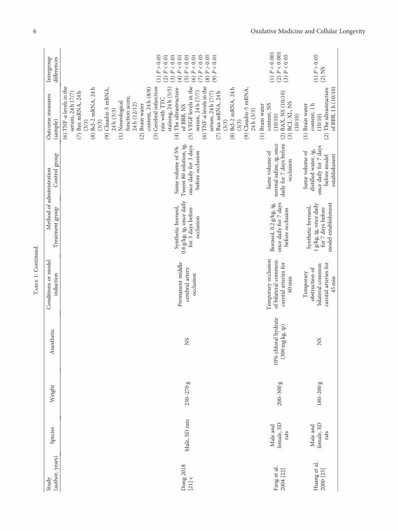
Table1:Con
tinu
ed.
Stud
y(autho
r,years)
Species
Weight
Anesthetic
Con
dition
sor
mod
elindu
ction
Metho
dof
administration
Outcomemeasures
(sam
ple)
Intergroup
differences
Treatmentgrou
pCon
trol
grou
p
(6)T
NF-αlevelsinthe
serum,24h(7/7)
(7)Bax
mRNA,24h
(3/3)
(8)Bcl-2
mRNA,24h
(3/3)
(9)Claud
in-5
mRNA,
24h(3/3)
Don
g2018
[21]
cMale,SD
rats
230–270g
NS
Permanentmiddle
cerebralartery
occlusion
Syntheticborneol,
0.6g/kg,ig,on
cedaily
for3days
before
occlusion
Samevolumeof
5%Tween80
solution
,ig,
once
daily
for3days
before
occlusion
(1)Neurological
function
score,
24h(12/12)
(2)Brain
water
content,24
h(8/8)
(3)C
erebralinfarction
ratewithTTC
staining,24h(5/5)
(4)The
ultrastructure
ofBBB,N
S(5)VEGFlevelsin
the
serum,24h(7/7)
(6)T
NF-αlevelsinthe
serum,24h(7/7)
(7)Bax
mRNA,24h
(3/3)
(8)Bcl-2
mRNA,24h
(3/3)
(9)Claud
in-5
mRNA,
24h(3/3)
(1)P>005
(2)P<001
(3)P<001
(4)P<001
(5)P<001
(6)P<001
(7)P<005
(8)P>005
(9)P<001
Fang
etal.
2004
[22]
Maleand
female,SD
rats
200–300g
10%
chloralh
ydrate
(300
mgkg,ip)
Tem
porary
occlusion
ofbilateralcom
mon
carotidarteries
for
60min
Borneol,0.2g/kg,ig,
once
daily
for7days
before
occlusion
Samevolumeof
norm
alsalin
e,ig,once
daily
for7days
before
occlusion
(1)Brain
water
content,NS
(10/10)
(2)BAX,N
S(10/10)
(3)BCL-XL,
NS
(10/10)
(1)P<0001
(2)P<0001
(3)P<005
Huang
etal.
2000
[23]
Maleand
female,SD
rats
180–200g
NS
Tem
porary
obstructionof
bilateralcom
mon
carotidarteries
for
45min
Syntheticborneol,
1g/kg,ig,on
cedaily
for7days
before
mod
elestablishm
ent
Samevolumeof
distilled
water,ig,
once
daily
for7days
before
mod
elestablishm
ent
(1)Brain
water
content,1h
(10/10)
(2)The
ultrastructure
ofBBB,1
h(10/10)
(1)P>005
(2)NS
6 Oxidative Medicine and Cellular Longevity
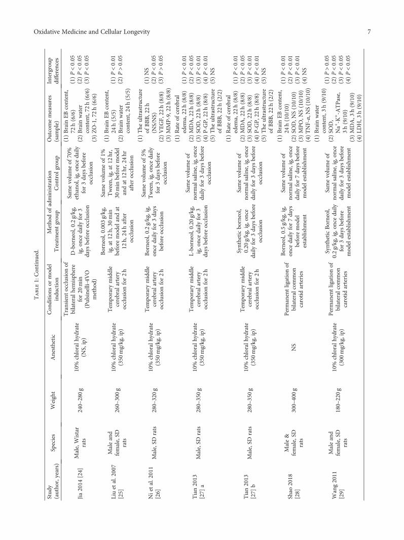
Table1:Con
tinu
ed.
Stud
y(autho
r,years)
Species
Weight
Anesthetic
Con
dition
sor
mod
elindu
ction
Metho
dof
administration
Outcomemeasures
(sam
ple)
Intergroup
differences
Treatmentgrou
pCon
trol
grou
p
Jia2014
[24]
Male,Wistar
rats
240–280g
10%
chloralh
ydrate
(NS,ip)
Transient
occlusionof
bilateralh
emisph
ere
for20
min
(Pulsinelli-4VO
metho
d)
D-borneol,0.2g/kg,
ig,oncedaily
for3
days
before
occlusion
Samevolumeof
70%
ethano
l,ig,oncedaily
for3days
before
occlusion
(1)Brain
EBcontent,
72h(6/6)
(2)Brain
water
content,72
h(6/6)
(3)ZO-1,72h(6/6)
(1)P<005
(2)P<005
(3)P<005
Liuetal.2007
[25]
Maleand
female,SD
rats
260–300g
10%
chloralh
ydrate
(350
mg/kg,ip)
Tem
porary
middle
cerebralartery
occlusionfor2h
Borneol,0.003
g/kg,
ig,at12
h,30
min
before
mod
elandat
12h,
24hafter
occlusion
Samevolumeof
1%Tween,
ig,at12
hr,
30min
before
mod
elandat
12hr,24hr
afterocclusion
(1)Brain
EBcontent,
24h(5/5)
(2)Brain
water
content,24
h(5/5)
(1)P<001
(2)P>005
Nietal.2011
[26]
Male,SD
rats
280–320g
10%
chloralh
ydrate
(350
mg/kg,ip)
Tem
porary
middle
cerebralartery
occlusionfor2h
Borneol,0.2g/kg,ig,
once
daily
for3days
before
occlusion
Samevolumeof
5%Tween,
ig,oncedaily
for3days
before
occlusion
(1)The
ultrastructure
ofBBB,22h
(NS/NS)
(2)VEGF,
22h(8/8)
(3)MMP-9,22h(8/8)
(1)NS
(2)P<005
(3)P>005
Tian2013
[27]
aMale,SD
rats
280–350g
10%
chloralh
ydrate
(350
mg/kg,ip)
Tem
porary
middle
cerebralartery
occlusionfor2h
L-borneol,0.20
g/kg,
ig,oncedaily
for3
days
before
occlusion
Samevolumeof
norm
alsalin
e,ig,once
daily
for3days
before
occlusion
(1)Rateof
cerebral
edem
a,22
h(8/8)
(2)MDA,22h(8/8)
(3)SO
D,22h(8/8)
(4)P-G
P,22h(8/8)
(5)The
ultrastructure
ofBBB,22h(2/2)
(1)P<001
(2)P<005
(3)P<001
(4)P<001
(5)NS
Tian2013
[27]
bMale,SD
rats
280–350g
10%
chloralh
ydrate
(350
mg/kg,ip)
Tem
porary
middle
cerebralartery
occlusionfor2h
Syntheticborneol,
0.20
g/kg,ig,on
cedaily
for3days
before
occlusion
Samevolumeof
norm
alsalin
e,ig,once
daily
for3days
before
occlusion
(1)Rateof
cerebral
edem
a,22
h(8/8)
(2)MDA,22h(8/8)
(3)SO
D,22h(8/8)
(4)P-G
P,22h(8/8)
(5)The
ultrastructure
ofBBB,22h(2/2)
(1)P<001
(2)P<005
(3)P<001
(4)P<001
(5)NS
Shao
2018
[28]
Male&
female,SD
rats
300–400g
NS
Permanentligationof
bilateralcom
mon
carotidarteries
Borneol,0.5g/kg,ig,
once
daily
for7days
before
mod
elestablishm
ent
Samevolumeof
norm
alsalin
e,ig,once
daily
for7days
before
mod
elestablishm
ent
(1)Brain
EBcontent,
24h(10/10)
(2)SO
D,N
S(10/10)
(3)MPO,N
S(10/10)
(4)T
NF-α,N
S(10/10)
(1)P<001
(2)P<001
(3)P<001
(4)NS
Wang2011
[29]
Maleand
female,SD
rats
180–220g
10%
chloralh
ydrate
(300
mg/kg,ip)
Permanentligationof
bilateralcom
mon
carotidarteries
SyntheticBorneol,
0.2g/kg,ig,on
cedaily
for3days
before
mod
elestablishm
ent
Samevolumeof
norm
alsalin
e,ig,once
daily
for3days
before
mod
elestablishm
ent
(1)Brain
water
content,3h(9/10)
(2)SO
D,
Na+-K
+-A
TPase,
3h(9/10)
(3)MDA,3
h(9/10)
(4)LD
H,3
h(9/10)
(1)P>005
(2)P<005
(3)P<005
(4)P<005
7Oxidative Medicine and Cellular Longevity

Table1:Con
tinu
ed.
Stud
y(autho
r,years)
Species
Weight
Anesthetic
Con
dition
sor
mod
elindu
ction
Metho
dof
administration
Outcomemeasures
(sam
ple)
Intergroup
differences
Treatmentgrou
pCon
trol
grou
p
XuandZhang
2015
[30]
Male,SD
rats
250±20
g10%
chloralh
ydrate
(NS,NS)
Tem
porary
middle
cerebralartery
occlusionfor2h
Naturalborneol,
0.028g/kg,ig,on
cedaily
for3days
before
occlusion
Samevolumeof
norm
alsalin
e,ig,once
daily
for3days
before
occlusion
(1)Brain
EBcontent,
24h(5/5)
(2)ZO-1,24h(5/5)
(3)Claud
in-5,24h
(5/5)
(1)P>005
(2P>005
(3)P>005
Yao
etal.2011
[31]
Maleand
female,
Kun
ming
mice
22±3g
4%chloralh
ydrate
(400
mg/kg,ip)
Permanentligationof
bilateralcom
mon
carotidarteries
Syntheticborneol,
0.0666
g/kg,ig,on
cedaily
for3days
before
occlusion
Samevolumeof
norm
alsalin
e,ig,once
daily
for3days
before
occlusion
Brain
EBcontent,
20min
(8/6)
P>005
Zhang
etal.
2011
[32]
Male,SD
rats
250–280g
10%
chloralh
ydrate
(350
mg/kg,ip)
Permanentmiddle
cerebralartery
occlusion
Borneol,0.4gper
anim
al,ig,on
cedaily
for7days
before
occlusion
Samevolumeof
1%Tween,
ig,oncedaily
for7days
before
occlusion
(1)Brain
water
content,24
h(9/9)
(2)Neurological
function
score,NS
(9/9)
(3)Infarction
volume,
NS(9/9)
(1)P>005
(2)P>005
(3)P>005
Zhang
etal.
2011
[33]
Maleand
female,ICR
mice
18–22g
4%chloralh
ydrate
(0.4mg/kg,N
S)
Tem
porary
occlusion
ofbilateralcom
mon
carotidarteries
for
20min
Borneol,0.5gper
anim
al,ig,on
cedaily
for8days
before
occlusion
Samevolumeof
1%Tween,
ig,oncedaily
for8days
before
occlusion
(1)Brain
water
content,10
min
(11/10)
(2)SO
D,10min
(11/10)
(3)MDA,10min
(11/10)
(1)P>005
(2)P>005
(3)P>005
Zhu
etal.2007
[34]
Male,SD
rats
200–250g
10%
chloralh
ydrate
(400
mg/kg,ip)
Permanentligationof
bilateralcom
mon
carotidarteries
Borneol,0.020
g/kg,
iv,oncedaily
for4
days
before
mod
elestablishm
ent
Samevolumeof
norm
alsalin
e,iv,once
daily
for4days
before
mod
elestablishm
ent
Brain
water
content,
3h(8/8)
P>005
Note:BBB:the
blood-brainbarrier;increased:asignificantlyincreasing
blood-brainbarrierperm
eabilityaftertheadministrationof
borneol;decreased:asignificantlydecreasing
blood-brainbarrierperm
eability
aftertheadministrationof
borneol;ND:n
ostatisticald
ifference
betweentreatm
entandcontrolgroup
s;Increased?
ordecreased?:the
efficacy
resultwas
repo
rted
asincreasing
ordecreasing
bloodbrainbarrier
perm
eabilitywiththeabsenceof
statisticalanalysis
oravailableoriginal
data;ig:intragastric
administration;
ip:intraperiton
ealadministration;
iv:intravenou
sinjection;
NS:
notstated;SO
D:superoxide
dism
utase;MDA:m
alon
dialdehyde;E
T:end
othelin
;NO:n
itricoxide;LD
H:lactatedhydrogenase;EB:E
vans
blue;Z
O-1:zon
ulaocclud
ens-1;GFA
P:glio
fibrillar
acid
protein;
BAX:B
cl-2
associated
Xprotein;
BCL:
B-celllym
phom
a;VEGF:
vascular
endo
thelialgrowth
factor;M
MP-9:m
atrixmetalloproteinase-9;P
-GP:P
-glycoprotein.
8 Oxidative Medicine and Cellular Longevity

statistically similar between the borneol group and the con-trol group (same volume of 1% Tween, ig, qd). ComparingL-borneol (0.2 g/kg, ig, qd), D-borneol (0.2 g/kg, ig, qd), orsynthetic borneol (0.6 g/kg, ig, qd) with the same volume of5% Tween 80 solution (ig, qd) for 3 days before occlusion,respectively, Dong et al. [21] found that these three compar-isons had a significant difference in alleviating the infarctionarea (L-borneol: nT/nC = 5/5; D-borneol: nT/nC = 5/5; andsynthetic borneol: nT/nC = 5/5).
3.4.6. SOD. Meta-analysis of four studies [20, 28, 29, 33]showed that animals in the borneol group had statisti-cally significant higher SOD activity than the controlgroup (nT/nC = 34/35, MD 17.22, 95% CI: -10.00 to 24.44,P < 0 00001, heterogeneity χ2 = 20 56, df = 3, I2 = 85%).Sensitivity analyses were conducted to explore potentialsources of heterogeneity after the omissions of each individ-ual study from the original analysis. Sensitivity analysespointed to one study [28] as a likely source of heterogeneity.After removal of the study, the SOD activity between thetwo groups had significant difference and the heterogene-ity was reduced (nT/nC = 24/25, MD 14.51, 95% CI: 10.93 to18.09, P < 0 00001, heterogeneity χ2 = 3 65, df = 2, I2 = 45%,Figure 5).
3.4.7. MDA. Three studies [20, 29, 33] based on the measure-ment of MDA level showed no significant difference betweenthe borneol group and the control group but with substantialheterogeneity (nT/nC = 24/24, WMD -0.39, CI: -6.25~1.63,P = 0 86, I2 = 66%). Sensitivity analyses were conducted toexplore potential sources of heterogeneity after the omissions
of each individual study from the original analysis. Sensitivityanalyses pointed to one study [20] as a likely source ofheterogeneity. After removal of the study, the MDA out-comes between the two groups remained similar and theheterogeneity was reduced (nT/nC = 18/18, WMD -0.32,CI: -0.80~0.16, P = 0 19, I2 = 0%, Figure 6).
3.4.8. Possible Neuroprotective Mechanisms of Borneol.According to the included studies, the possible neuroprotec-tive mechanisms of borneol for ischemic stroke lie in thefollowing aspects: (1) borneol could help alleviate the patho-logical BBB disruption [21, 23, 24, 26, 27, 30]. (2) Borneolcould effectively reduce oxidative reactions through increas-ing the activity of SOD and decreasing the concentration ofMDA [28, 29, 33]. (3) Borneol could inhibit the occurrenceof inflammation by decreasing the expression of proinflam-matory cytokines such as TNF-α [21, 28]. (4) Borneol couldexert antiapoptotic effects, resulting in neuroprotection[21, 22]. (5) Borneol could improve the activity of lactatedehydrogenase (LDH) in brain tissue to inhibit the increaseof lactic acid, which reduces the accumulation of lactic acidand exerts neuroprotective effect [29]. (6) Borneol couldimprove the energy metabolism disorder by upregulatingthe activity of Na+-K+-ATPase [27, 29], Ca2+-Mg2+-ATPase[27], and T-ATPase [27]. (7) Borneol could exert the neuro-protective effect via P-glycoprotein (P-GP) signaling pathway[27]. (8) Borneol exerts the neuroprotective effect via NOsignaling pathway [27]. More details were shown in Table 3.
4. Discussion
4.1. Summary of Results.As far as we know, this is the first pre-clinical systematic review to determine the effects of borneolfor experimental ischemic stroke, mainly through possiblemechanisms of regulating the BBB permeability. In the pres-ent study, thirteen studies with 230 animals were selectedfor analysis. Borneol were identified to have a decreasedimpact on pathological BBB permeability from animals withcerebral ischemic injury. The pooled data suggested thatborneol exerts a significant protection on the experimentalmodel of ischemic stroke.
4.2. Limitations. First, all the databases we searched were inEnglish or Chinese, which may cause selective bias as studiespublished in other languages may be left out. Second, thepresent study found that only one animal species (rodent)was used, which potentially posed a threat to the promotionof the findings. Third, the methodological quality of mostincluded studies was moderate, which was an inherent draw-back in the primary study. Methodological flaws in mostincluded studies lie in blinding, sample size calculation, lack-ing animals with relevant comorbidities, and lacking state-ment of potential conflict of interests. Thus, the conclusionsin the present study should be partially treated with caution.
4.3. Implications. Currently, it is increasingly recognized thatthe regulation of BBB permeability is a complicated process,which involves multiple components, such as endothelialcells, tight junction, basal lamina, and pericytes [35–38],and a variety of genes. BBB can effectively prevent the entry
Table 2: Quality assessment of included studies.
Study A B C D E F G H I J Total
Chen et al. [20] + + + + + 5
Dong et al. [21] + + + + + 5
Fang et al. [22] + + + 3
Huang et al.[23] + + 2
Jia [24] + + + 3
Liu et al. [25] + + + 3
Ni et al. [26] + + + 3
Tian [27] + + + 3
Shao et al. [28] + + 2
Wang [29] + + 2
Xu and Zhang [30] + + + 3
Yao et al. [31] + + + 3
Zhang et al. [32] + + + + 4
Zhang et al. [33] + + + 3
Zhu et al. [34] + + + 3
A: peer-reviewed publication; B: monitoring of physiological parameterssuch as temperature; C: random allocation; D: blinded conduct of theexperiments; E: blinded assessment of outcome; F: use of anestheticwithout significant intrinsic neuroprotective activity (e.g., ketamine); G:animal and/or model (aged, diabetic, or hypertensive); H: sample sizecalculation; I: compliance with animal welfare regulations; J: statement ofpotential conflict of interests.
9Oxidative Medicine and Cellular Longevity

of lipophilic potential neurotoxins, protect the brain frommost pathogens, and selectively transport essential molecules,which is of great importance in the homeostatic regulation of
the brain microenvironment. As common pathological pro-cesses in many serious CNS diseases, the BBB destructionand the increasing BBB permeability are especially essential
Study or subgroup
Chen et al. 2010 [20]Jia 2014 [24]Liu et al. 2007 [25]Shao et al. 2018 [28]Xu and Zhang 2015 [30]Yao et al. 2011 [31]
Total (95% CI)Heterogeneity: Chi2 = 4.94, df = 4 (P = 0.29); I2 = 19%Test for overall effect: Z = 12.94 (P < 0.00001)
34
Mean4.57 0.88 6 8.84 1.79 6
65
1056
32 100.0%
13.1%
2.2%82.0%2.5%0.2%
1.33.990.133.5
11.995
77.8310.659.13
27.3839.659
65
1058
0.751.831.022.24
13.449
72.174.875.36
26.7930.578
Borneol Control Mean difference
SD Total Mean SD TotalWeight
IV, fixed, 95% CI
Mean difference
IV, fixed, 95% CI−4.27 [−5.87, −2.67]
−5.78 [−9.63, −1.93]−3.77 [−4.41, −3.13]−0.59 [−4.23, 3.05]
−9.08 [−22.46, 4.30]
−3.81 [−4.39, −3.23]−20
Favours [borneol] Favours [control]−10 0 10 20
Not estimable
Figure 3: The forest plots: the borneol group versus the control group on brain Evans blue content under pathological conditions.
BorneolMean SD Total SD TotalMean
WeightIV, fixed, 95% CI IV, fixed, 95% CI
Study or subgroup
Chen et al. 2010 [20]Fang et al 2004 [22]Huang et al. 2000 [23]Liu et al. 2007 [25]Wang 2011 [29]Zhang et al. 2011 [32]Zhang et al. 2011 [33]Zhu et al. 2007 [34]Subtotal [95% CI]Heterogeneity: Chi2 = 3.96; df = 7 (P = 0.78); I2 = 0%Test for overall effect: Z = 10.21 (P < 0.00001)
Heterogeneity: Chi2 = 0.02; df = 1 (P = 0.88); I2 = 0%Test for overall effect: Z = 5.90 (P < 0.00001)
Heterogeneity: Chi2 = 31.97, df = 9 (P = 0.0002); I2 = 72%Test for overall effect: Z = 4.25 (P < 0.00001)Test for subgroup difference: Chi2 =27.99. df = 1 (P < 0.00001), I2 = 96.4%
Total (95% CI)
1.2.2 cerebral ischemic injury - brain water content 2Tian 2013 [27] aTian 2013 [27] bSubtotal (95% CI)
7.918.36
3.013.77
88
16
84 84 100.0%
17.2117.21
5.155.15
88
16
0.2%0.2%0.3%
79.3377.3
82.2280.7978.5582.2280.0879.19
0.180.271.840.930.191.242.021.13
61010599
118
68 68
80.4378.2382.1481.5179.4583.0581.1379.79
0.40.520.950.530.412.061.630.91
610105
109
108
18.6% −1.10 [−1.45, −0.75]−0.93 [−1.29, −0.57]0.08 [−1.20, −1.36]−0.72 [−1.66, −0.22]−0.90 [−1.18, −0.62]−0.83 [−2.40, −0.74]−1.05 [−2.61, −0.51]−0.60 [−1.61, −0.41]−0.92 [−1.10, −0.75]
−10 −5 0 5 10
−9.30 [−13.43, −5.17]−8.85 [−13.27, −4.43]−9.09 [−12.11, −6.07]
−0.95 [−1.13, −0.78]
18.4%7.9%
11.1%19.3%6.0%6.1%
10.4%99.7%
1.2.1 celebral ischemic injury - brain water sontent 1
Control Mean difference Mean difference
Favours [borneol] Favours [control]
Figure 4: The forest plots: the borneol group versus the control group on brain water content under pathological conditions.
Study or subgroup
Total (95% CI)Heterogeneity: Chi2 = 3.65, df = 2 (P = 0.16); I2 = 45%Test for overall effect: Z = 7.95 (P < 0.00001)
Chen et al. 2010 [20]Wang 2011 [29]Zhang et al. 2011 [33]
Mean375.31
105.45122.89
30.11
14.582.34
6
11
24
7344.04
98.68107.63
17.83
9.844.96
6
11
25 100.0% 14.51 [10.93, 18.09]
−100 −50
Favours [control] Favours [borneol]0 50 100
81.6%
11.9%86.5%
31.27 [3.27, 59,27]
6.77 [−3.62, 17.16]15.26 [11,41, 19.11]
Borneol Control Mean differenceSD Total Mean SD Total
WeightIV, fixed, 95% CI
Mean differenceIV, fixed, 95% CI
Figure 5: The forest plots: the borneol group versus the control group on SOD activity under pathological conditions.
10 Oxidative Medicine and Cellular Longevity

pathological bases of ischemic stroke. The present study dem-onstrated that the borneol could alleviate the increased BBBpermeability by protecting the functions of endothelial cellsand maintaining the integrity of the tight junction and basallamina during cerebral ischemic injury, which exerts potentialneuroprotective effect.
The possible neuroprotective mechanisms of borneol forischemic stroke are summarized as follows: (1) BBB: borneolcan alleviate the pathological BBB disruption through pro-tecting the function of endothelial cells, maintaining theintegrity of the basal lamina, and reducing the damage oftight junction integrity [23, 24, 26, 27, 30]. (2) Oxidativereactions: borneol can reduce oxidative reactions and theneurotoxicity of free radicals through increasing the activityof SOD and decreasing the concentration of MDA [29, 33].SOD is a potent natural antioxidant enzyme that plays abioprotective role by alternately catalyzing the dismutationor partitioning of the superoxide (O2-) radical into either ordi-nary molecular oxygen (O2) or hydrogen peroxide (H2O2),which is a prominent antioxidant defense in nearly all livingcells exposed to oxygen [39]. As part of the brain ischemicantioxidant defense, an increase of SOD activities can amelio-rate the oxidative stress damages [40].MDA is amain productof lipid peroxidation of polyunsaturated fatty acids [41].MDAcan be overproduced, resulting from an increase in free radi-cals [42]. Thus, MDA is commonly used as a biomarker tomeasure the level of oxidative stress [43, 44]. Oxidative stress,including lipid peroxidation, plays an important role in thepathogenesis of acute brain injury and the breakdown ofBBB [45]. Lipid peroxidation, mediated by superoxide, couldcause alterations of membrane permeability as well as struc-tural and functional impairment of cellular components. Freeradicals are generally generated in the ischemic areas duringischemic stroke, leading to neuronal damage by promotinglipid peroxidation, protein breakdown, and DNA damage,which in turn results in cellular apoptosis and BBB permeabil-ity [46]. The pooled data showed that borneol significantlyincreases the activity of SOD and decreases the concentrationof MDA which reduced the neurotoxicity of free radicals. (3)Anti-inflammation: borneol can exert anti-inflammationeffects by decreasing the expression of proinflammatory cyto-kine TNF-α [21, 28]. Inflammation has been well recognizedas a predominate contributor in ischemic stroke, playing animportant role in all stages of the ischemic cascade [47, 48].After ischemia, proinflammatory cytokines, such as TNF-α,were released,which resulted inBBBbreakdown andneuronaldeath [21]. TNF-α, as a key proinflammatorymediator, plays a
critical role in inflammatory responses. The present studyindicated that borneol may be a potential anti-inflammatorydrug, particularly in respect to cytokine suppression, resultingfrom the decreased expression of TNF-α induced by ischemicstroke. (4) Antiapoptosis: apoptosis is essential in the patho-genesis of acute and chronic neurodegenerative diseases, suchas ischemic stroke, which can cause neuronal death and irre-versible cerebral dysfunction [49]. It was found that inhibitionof apoptosis could alleviate ischemic injury [50, 51]. Theexpression of Bcl-2 family plays a predominate role in apopto-sis, which could either promote (Bax, Bak, Bad, Bim, and Bid)or prevent (Bcl-2, Bcl-XL, and Bcl-w) apoptosis [52, 53]. Thepresent study revealed that borneol had antiapoptosis effectsby decreasing the mRNA expression of Bax [22], increasingthe mRNA expression of Bcl-XL [22] and modulating theBax/Bcl-2 expression at both the mRNA and protein levels[21]. (5) LDH: LDH is an enzyme that is widely distributedin the cytoplasm of neurons and glial cells. LDH is abundantin the brain under physiological condition, which wouldrelease into the blood and consequently increase the accumu-lation of lactic acid during cerebral ischemic injury. The accu-mulation of lactic acid can cause acidosis in ischemic injury,which increases free radicals and decreases the use of glucosein the brain, ultimately resulting in the oxidative stress andthedecrease inATP levels [29]. Borneol can improve the activ-ity of LDH to inhibit an increase in lactic acid [29]. (6) Energymetabolism: ischemic stroke easily causes the energy metabo-lism disorder including mitochondria functional impairmentand imbalance of ion homeostasis [54]. Borneol can improvethe energy metabolism disorder through upregulating theactivity of Na+-K+-ATPase [27, 29], Ca2+-Mg2+-ATPase[27], and T-ATPase [27]. (7) P-glycoprotein (P-GP): P-GP isan important protein of the cell membrane and more accu-rately an ATP-dependent efflux pump with broad substratespecificity. P-GP expressed in the capillary endothelial cellscomposes the BBB [55–57]. Borneol could decrease theexpression of P-GP. (8) NO: excessive nitric oxide (NO) gen-erated during the cerebral ischemic stroke may be combinedwith free radicals, which causes damage to lipid membranes,nucleic acids, and cell protein. Borneol exerts the neuropro-tective effect via NO signaling pathway [27]. Borneol coulddecrease the expression of NO [27]. During cerebral ischemiaand reperfusion, signaling cascades can be triggered by crosstalk. As mentioned above, BBB disruption, oxidative stress,acidosis, the energy metabolism disorder, P-GP, and NO canaffect each other and interact as both cause and effect. Thepresent study showed the neuroprotective mechanism of
Study or subgroupMean
Borneol Control Mean differenceSD Total Mean SD Total
WeightIV, fixed, 95% CI
Mean differenceIV, fixed, 95% CI
Chen et al. 2010 [20]Wang 2011 [29]Zhang et al. 2011 [33]
202.86
27.4964.684
27.94
4.960.61
8
117
201.97
29.774.976
14.49
4.240.25
8
108
0.0%
1.5%
0.89 [−20.92, 22.70]−0.29 [−0.78, 0.19]−2.27 [−6.21, 1.66]
98.5%
Total (95% CI) 26 26 100.0% −0.32 [−0.80, 0.16]
−20 −10 10 20
Favours [borneol] Favours [control]0
Heterogeneity: Chi2 = 0.97, df = 2 (P = 0.62); I2 = 0%Test for overall effect: Z = 1.31 (P < 0.19)
Figure 6: The forest plots: the borneol group versus the control group on MDA level under pathological conditions.
11Oxidative Medicine and Cellular Longevity

Table 3: Characteristics of mechanism studies of borneol on experimental ischemic stroke.
Study ModelMethod of administration (experimentalgroup versus control group)
Possible mechanism
Dong et al. [21] pMCAO in SD rats(1) L-borneol versus 5% Tween 80(2) D-borneol versus 5% Tween 80(3) Synthetic borneol versus 5% Tween 80
(1) Alleviate the pathological BBBdisruption by upregulating tightjunction proteins Claudin-5
(2) Accelerate the proliferation of vascularendothelial cells and by initiatingangiogenesis.
(3) Anti-inflammation by decreasing theexpression of TNF-α
(4) Antiapoptosis by modulating theBax/Bcl-2 expression at both themRNA and protein levels
Fang et al. [22] BCO/1 h in SD rats Borneol versus normal salineAntiapoptosis by decreasing the mRNAexpression of Bax and increasing themRNA expression of Bcl-XL
Huang et al. [23] BCO/45min in SD rats Synthetic borneol versus distilled water
Alleviate the pathological BBB disruptionby protecting the function of endothelialcells and maintaining the integrity of thebasal lamina
Jia [24]
Occlusion of bilateralhemisphere (Pulsinelli-4VOmethod)/20min in Wistar
rats
D-borneol versus 70% ethanolAlleviate the pathological BBB disruptionby upregulating tight junction proteinsZO-1
Ni et al. [26] MCAO/2 h in SD rats Borneol versus 5% TweenAlleviate the pathological BBB disruptionby downregulating VEGF and MMP-9
Tian [27] MCAO/2 h in SD rats(1) L-borneol versus normal saline(2) Synthetic borneol versus normal saline
(1) Alleviate the pathological BBBdisruption by alleviating the damageof the BBB tight junction integrity
(2) Reduce oxidative reactions byincreasing the activity of SOD anddecreasing the concentration of MDA
(3) Improve the energy metabolismdisorder by upregulating the activity ofNa+-K+-ATPase, Ca2+-Mg2+-ATPase,and T-ATPase
(4) Neuroprotection via P-GP signalingpathway
(5) Neuroprotection via NO signalingpathway
Shao et al. [28] Permanent BCO in SD rats Borneol versus normal saline
(1) Reduce oxidative reactions byincreasing the activity of SOD
(2) Anti-inflammation by decreasing theexpression of TNF-α
Wang [29] Permanent BCO in SD rats Synthetic borneol versus 1% Tween
(1) Reduce oxidative reactions byincreasing the activity of SOD anddecreasing the concentration of MDA
(2) Improve the activity of LDH(3) Improve the energy metabolism
disorder by upregulating the activity ofNa+-K+-ATPase
12 Oxidative Medicine and Cellular Longevity

borneol in the treatment of cerebral ischemic injury wasmainly attributed to the decrease of BBB permeability.However, cellular and molecular alteration mechanisms ofborneol for ischemic stroke have not been clearly elucidatedyet, which presented an exciting investigative direction offurther research in this field. In addition, the administrationof borneol could expand to other diseases with similarpathologies, namely, the increased BBB permeability, whichwas associated with the development and progression ofmany CNS diseases, such as multiple sclerosis, Alzheimer’sdisease, and HIV-associated dementia.
Infarct volume (IV) is a common index for assessing theextent of cerebral ischemic injury. Infarct on imaging is oneof the common identified predictors of clinical outcome forischemic stroke [58]. In addition, IVmeasurement is clinicallyhelpful in the accurate selection of patients for decompressivesurgery and the determination of the time of surgery [59].Therefore, it is essential for further experimental trails of bor-neol for ischemic stroke to choose IV as outcome measures.
The methodological quality of the included studies wasmoderate. In particular, no study estimated the sample size,since inadequate sample size can miss the real interventioneffect in an experiment or excessive sample size can resultin wasting animals and raising animal ethical issues [60].No study blindingly assessed outcome, which could attributeto a 27% overestimation of the mean reported effect size [61].No study used animals with relevant comorbidities, whichwas rarely like human pathology under the clinical condi-tions [18]. Thus, it is necessary for further research of borneolfor ischemic stroke to take a rigor experimental design intoconsideration. We recommended that the Animal Research:Reporting of In Vivo Experiments (ARRIVE) [62], a report-ing guideline consisting of a 20-item checklist for the intro-duction, methods, results, and discussion, should be used asguidelines for the design and reporting of further experimen-tal research examining borneol for ischemic stroke, whichcan extremely help improve the methodological quality.
5. Conclusion
Borneol can alleviate the BBB disruption and plays a protec-tive role on cerebral ischemic injury through multiple signal-ing pathways. Further relevant molecular mechanismsdeserve adequate research.
Conflicts of Interest
The authors declare that there is no conflict of interestsregarding the publication of this paper.
Authors’ Contributions
ZXC, QQX, CSS, YHS, YW, RCCC, and GQZ designed thestudy. ZXC, QQX, CSS, YHS, and YW collected the data.ZXC, QQX, CSS, YHS, and YW performed all analyses.ZXC, QQX, and GQZ wrote the manuscript. All authors con-tributed to writing of this manuscript. Zi-xian Chen andQing-qing Xu contributed equally to this work.
Acknowledgments
This work was supported by the grant of the NationalNatural Science Foundation of China (81573750, 81473491,81173395); the Young and Middle-Aged University Disci-pline Leaders of Zhejiang Province, China (2013277); andthe Zhejiang Provincial Program for the Cultivation ofHigh-level Health Talents (2015).
References
[1] R. Khatri, A. M. McKinney, B. Swenson, and V. Janardhan,“Blood-brain barrier, reperfusion injury, and hemorrhagictransformation in acute ischemic stroke,” Neurology, vol. 79,13, Supplement 1, pp. S52–S57, 2012.
[2] R. Daneman and A. Prat, “The blood-brain barrier,” ColdSpring Harbor Perspectives in Biology, vol. 7, no. 1, articlea020412, 2015.
[3] Z. Zhao, A. R. Nelson, C. Betsholtz, and B. V. Zlokovic, “Estab-lishment and dysfunction of the blood-brain barrier,” Cell,vol. 163, no. 5, pp. 1064–1078, 2015.
[4] G. A. Rosenberg, “Neurological diseases in relation to theblood-brain barrier,” Journal of Cerebral Blood Flow andMetabolism, vol. 32, no. 7, pp. 1139–1151, 2012.
[5] Y. Shi, R. K. Leak, R. F. Keep, and J. Chen, “Translationalstroke research on blood-brain barrier damage: challenges,perspectives, and goals,” Translational Stroke Research,vol. 7, no. 2, pp. 89–92, 2016.
[6] X. Jiang, A. V. Andjelkovic, L. Zhu et al., “Blood-brain barrierdysfunction and recovery after ischemic stroke,” Progress inNeurobiology, vol. 163-164, pp. 144–171, 2018.
Table 3: Continued.
Study ModelMethod of administration (experimentalgroup versus control group)
Possible mechanism
Xu and Zhang [30] MCAO/2 h in SD rats Borneol versus normal salineAlleviate the pathological BBB disruptionby upregulating tight junction proteinsZO-1 and claudin-5
Zhang et al. [33] BCO/20min in ICR mice Borneol versus 1% TweenReduce oxidative reactions by increasingthe activity of SOD and decreasing theconcentration of MDA
Note: BCO, bilateral carotid occlusion; MCAO, middle cerebral artery occlusion; pMCAO, permanent middle cerebral artery occlusion; LDH, lactatedehydrogenase; ZO-1, Zonula occludens-1; VEGF, vascular endothelial cell growth factor; MMP-9matrix metalloproteinase-9; P-GP, P-glycoprotein.
13Oxidative Medicine and Cellular Longevity

[7] F. Ju, Y. Ran, L. Zhu et al., “Increased BBB permeabilityenhances activation of microglia and exacerbates loss of den-dritic spines after transient global cerebral ischemia,” Frontiersin Cellular Neuroscience, vol. 12, p. 236, 2018.
[8] Y. Yang and G. A. Rosenberg, “Blood–brain barrier break-down in acute and chronic cerebrovascular disease,” Stroke,vol. 42, no. 11, pp. 3323–3328, 2011.
[9] M. Krueger, I. Bechmann, K. Immig, A. Reichenbach,W. Härtig, and D. Michalski, “Blood—brain barrier break-down involves four distinct stages of vascular damage in vari-ous models of experimental focal cerebral ischemia,” Journal ofCerebral Blood Flow and Metabolism, vol. 35, no. 2, pp. 292–303, 2015.
[10] R. Prakash and S. T. Carmichael, “Blood-brain barrierbreakdown and neovascularization processes after stroke andtraumatic brain injury,” Current Opinion in Neurology,vol. 28, no. 6, pp. 556–564, 2015.
[11] M. Krueger, W. Härtig, C. Frydrychowicz et al., “Stroke-induced blood-brain barrier breakdown along the vasculartree - no preferential affection of arteries in different animalmodels and in humans,” Journal of Cerebral Blood Flow andMetabolism, vol. 37, no. 7, pp. 2539–2554, 2017.
[12] State Pharmacopoeia Committee, The Pharmacopoeia of Peo-ple's Republic of China, Chemical Industry Press, Beijing, 2010.
[13] Y. Guo, S. Yan, L. Xu, G. Zhu, X. Yu, and X. Tong, “Use ofAngong Niuhuang in treating central nervous system diseasesand related research,” Evidence-based Complementary andAlternative Medicine, vol. 2014, Article ID 346918, 9 pages,2014.
[14] M. Takaishi, K. Uchida, F. Fujita, and M. Tominaga, “Inhibi-tory effects of monoterpenes on human TRPA1 and the struc-tural basis of their activity,” The Journal of PhysiologicalSciences, vol. 64, no. 1, pp. 47–57, 2014.
[15] H. Y. Wu, Y. Tang, L. Y. Gao et al., “The synergetic effect ofedaravone and borneol in the rat model of ischemic stroke,”European Journal of Pharmacology, vol. 740, pp. 522–531,2014.
[16] R. Liu, L. Zhang, X. Lan et al., “Protection by borneol on cor-tical neurons against oxygen-glucose deprivation/reperfusion:involvement of anti-oxidation and anti-inflammation throughnuclear transcription factor κappaB signaling pathway,”Neuroscience, vol. 176, pp. 408–419, 2011.
[17] R. Tambe, P. Jain, S. Patil, P. Ghumatkar, and S. Sathaye,“Antiepileptogenic effects of borneol in pentylenetetrazole-induced kindling in mice,” Naunyn-Schmiedeberg's Archivesof Pharmacology, vol. 389, no. 5, pp. 467–475, 2016.
[18] S. C. Landis, S. G. Amara, K. Asadullah et al., “A call for trans-parent reporting to optimize the predictive value of preclinicalresearch,” Nature, vol. 490, no. 7419, pp. 187–191, 2012.
[19] M. R. Macleod, T. O’Collins, D. W. Howells, and G. A.Donnan, “Pooling of animal experimental data reveals influ-ence of study design and publication bias,” Stroke, vol. 35,no. 5, pp. 1203–1208, 2004.
[20] X. H. Chen, Z. Z. Lin, A. M. Liu et al., “The orally combinedneuroprotective effects of sodium ferulate and borneol againsttransient global ischaemia in C57 BL/6J mice,” Journal ofPharmacy and Pharmacology, vol. 62, no. 7, pp. 915–923, 2010.
[21] T. Dong, N. Chen, X. Ma et al., “The protective roles ofL-borneolum, D-borneolum and synthetic borneol in cerebralischaemia via modulation of the neurovascular unit,” Biomed-icine & Pharmacotherapy, vol. 102, pp. 874–883, 2018.
[22] Y. Q. Fang, L. Li, Q. D. Wu, Y. Y. Zhou, G. Wei, and S. F. Lin,“Protective effects of β-asarone and borneol in rat model ofcerebral ischemia with reperfusion,” Zhongguo Zhong Yi YaoKe Ji, vol. 11, no. 6, pp. 353-354, 2004.
[23] P. Huang, Q. Wu, X. Rong et al., “Protective effects of Borneo-lum combined with rhizoma chuanxiong on cerebral ischemiawith reperfusion injury,” Journal of Guangzhou UniversityTraditional Chinese Medicine, vol. 17, no. 4, pp. 323–326,2000.
[24] Y. L. Jia, “Effects of borneol on rat's blood brain barrier andzonula occludens-1 after global cerebral ischemia-reperfusioninjury, MD dissertation,” Xinxiang Medical University, 2014.
[25] Y. M. Liu, X. H. Xia, G. F. Zhao, S. Q. Peng, Q. Y. Xu, andQ. Shen, “Effect of Moschus combined with borneol onbrain water content and blood-brain barrier permeabilityin rat model of cerebral focal ischemia with reperfusion,”Journal of GuangzhouUniversity Traditional ChineseMedicine,vol. 24, no. 6, pp. 498–501, 2007.
[26] C. Ni, N. Zeng, F. Xu et al., “Effects of aromatic resuscitationdrugs on blood brain barrier in cerebral ischemia-reperfusioninjury model rats,” Zhongguo Zhong Yao Za Zhi, vol. 36,no. 18, pp. 2562–2566, 2011.
[27] H. Tian, “Comparative research on mechanism of neuropro-tective effect and influence of blood-brain barrier betweenL-borneolum and borneolum syntheticum, PhD dissertation,”Chengdu University of TCM, Sichuan (Chengdu), 2013.
[28] X. R. Shao, K. R. Cai, R. Jia et al., “Effects of borneol on inflam-matory reaction and permeability of blood-brain barrier in ratswith cerebral ischemic injury,” The Chinese Journal of ClinicalPharmacology, vol. 34, no. 13, pp. 1558–1560, 2018.
[29] Y. Wang, “The effect of musk, borneol and compatibility ofthem on the animal models of cerebral isehemia, MD disserta-tion,” Chengdu University of Traditional Chinese Medicine,Sichuan (Chengdu), 2011.
[30] L. Xu and T. J. Zhang, “Effects of borneol combined with bre-viscapine on blood-brain barrier permeability after ischemiareperfusion injury in rats,” Pharmacology & Clinics of ChineseMateria Medica, vol. 31, no. 1, pp. 74–76, 2015.
[31] H. W. Yao, J. Wang, Y. Liu, and D. D. Wang, “Effect ofMoschus and Borneo camphor in different proportion onblood brain barrier permeability of acute cerebral ischemicmodel mice,” Journal of Chengdu University of TraditionalChinese Medicine, vol. 34, no. 4, pp. 62–64, 2011.
[32] X. S. Zhang, E. H. Zhang, X. Shen, and J. R. Chen, “The protec-tive effects of compatibility of dioscin and borneol on the focalcerebral ischemia in rats,” Chinese Journal of ExperimentalTraditional Medical Formulae, vol. 17, no. 24, pp. 151–153,2011.
[33] X. S. Zhang, E. H. Zhang, G. K. Gao, and Y. Q. Ren, “The effectof the compatibility of dioscin and camphol on cerebralischemia-reperfusion injury in mice,” Western Journal of Tra-ditional Chinese Medicine, vol. 24, no. 12, pp. 11–13, 2011.
[34] J. R. Zhu, Y. M. Li, W. R. Fang, and S. X. Liu, “Protective effectsof scutellarin and borneolum injection on experimental cere-bral ischemia in animals,” Journal of China PharmaceuticalUniversity, vol. 38, no. 35, pp. 456–460, 2007.
[35] A. Armulik, G. Genové, M. Mäe et al., “Pericytes regulate theblood-brain barrier,” Nature, vol. 468, no. 7323, pp. 557–561,2010.
[36] R. Daneman, “The blood-brain barrier in health and disease,”Annals of Neurology, vol. 72, no. 5, pp. 648–672, 2012.
14 Oxidative Medicine and Cellular Longevity
[37] J. Sargento-Freitas, S. Aday, C. Nunes et al., “Endothelialprogenitor cells enhance blood-brain barrier permeability insubacute stroke,” Neurology, vol. 90, no. 2, pp. e127–e134,2018.
[38] F. Arba, R. Leigh, D. Inzitari et al., “Blood–brain barrier leak-age increases with small vessel disease in acute ischemicstroke,” Neurology, vol. 89, no. 21, pp. 2143–2150, 2017.
[39] J. Azadmanesh and G. E. O. Borgstahl, “A review of the cata-lytic mechanism of human manganese superoxide dismutase,”Antioxidants, vol. 7, no. 2, p. 25, 2018.
[40] S. Manzanero, T. Santro, and T. V. Arumugam, “Neuronaloxidative stress in acute ischemic stroke: sources and contribu-tion to cell injury,” Neurochemistry International, vol. 62,no. 5, pp. 712–718, 2013.
[41] A. Ayala, M. F. Muñoz, and S. Argüelles, “Lipid peroxidation:production, metabolism, and signaling mechanisms of malon-dialdehyde and 4-hydroxy-2-nonenal,” Oxidative Medicineand Cellular Longevity, vol. 2014, Article ID 360438, 31 pages,2014.
[42] I. M. Cojocaru, M. Cojocaru, V. Sapira, and A. Ionescu, “Eval-uation of oxidative stress in patients with acute ischemicstroke,” Romanian Journal of Internal Medicine, vol. 51,no. 2, pp. 97–106, 2013.
[43] D. Tsikas, “Assessment of lipid peroxidation by measuringmalondialdehyde (MDA) and relatives in biological samples:analytical and biological challenges,” Analytical Biochemistry,vol. 524, pp. 13–30, 2017.
[44] S. Jiménez-Fernández, M. Gurpegui, F. Díaz-Atienza,L. Pérez-Costillas, M. Gerstenberg, and C. U. Correll, “Oxida-tive stress and antioxidant parameters in patients with majordepressive disorder compared to healthy controls before andafter antidepressant treatment: results from a meta-analysis,”Journal of Clinical Psychiatry, vol. 76, no. 12, pp. 1658–1667,2015.
[45] P. T. Ronaldson and T. P. Davis, “Targeting transporters: pro-moting blood-brain barrier repair in response to oxidativestress injury,” Brain Research, vol. 1623, pp. 39–52, 2015.
[46] S. Milanizadeh, K. N. N. Zuwarali, A. Aliaghaei, and M. R.Bigdeli, “Therapeutic potential of pretreatment with allograftSertoli cells transplantation in brain ischemia by improvingoxidative defenses,” Journal of Molecular Neuroscience, vol. 64,no. 4, pp. 533–542, 2018.
[47] M. Gelderblom, M. Gallizioli, P. Ludewig et al., “IL-23 (inter-leukin-23)-producing conventional dendritic cells control thedetrimental IL-17 (interleukin-17) response in stroke,” Stroke,vol. 49, no. 1, pp. 155–164, 2018.
[48] C. Iadecola and J. Anrather, “The immunology of stroke: frommechanisms to translation,” Nature Medicine, vol. 17, no. 7,pp. 796–808, 2011.
[49] S. E. Khoshnam, W. Winlow, M. Farzaneh, Y. Farbood, andH. F. Moghaddam, “Pathogenic mechanisms following ische-mic stroke,” Neurological Sciences, vol. 38, no. 7, pp. 1167–1186, 2017.
[50] D. Radak, N. Katsiki, I. Resanovic et al., “Apoptosis and acutebrain ischemia in ischemic stroke,” Current Vascular Pharma-cology, vol. 15, no. 2, pp. 115–122, 2017.
[51] N. Wei, L. Xiao, R. Xue et al., “MicroRNA-9 mediates the cellapoptosis by targeting Bcl2l11 in ischemic stroke,” MolecularNeurobiology, vol. 53, no. 10, pp. 6809–6817, 2016.
[52] P. E. Czabotar, G. Lessene, A. Strasser, and J. M. Adams, “Con-trol of apoptosis by the BCL-2 protein family: implications for
physiology and therapy,” Nature Reviews Molecular Cell Biol-ogy, vol. 15, no. 1, pp. 49–63, 2014.
[53] X. Chen, M. Lu, X. He, L. Ma, L. Birnbaumer, and Y. Liao,“TRPC3/6/7 knockdown protects the brain from cerebralischemia injury via astrocyte apoptosis inhibition and effectson NF-кB translocation,” Molecular Neurobiology, vol. 54,no. 10, pp. 7555–7566, 2017.
[54] J. P. Zöllner, E. Hattingen, O. C. Singer, and U. Pilatus,“Changes of pH and energy state in subacute human ischemiaassessed by multinuclear magnetic resonance spectroscopy,”Stroke, vol. 46, no. 2, pp. 441–446, 2015.
[55] W. Fang, P. Lv, X. Geng et al., “Penetration of verapamil acrossblood brain barrier following cerebral ischemia depending onboth paracellular pathway and P-glycoprotein transportation,”Neurochemistry International, vol. 62, no. 1, pp. 23–30, 2013.
[56] W. Abdullahi, T. P. Davis, and P. T. Ronaldson, “Functionalexpression of P-glycoprotein and organic anion transportingpolypeptides at the blood-brain barrier: understanding trans-port mechanisms for improved CNS drug delivery?,” TheAAPS Journal, vol. 19, no. 4, pp. 931–939, 2017.
[57] Y. Hoshi, Y. Uchida, M. Tachikawa, S. Ohtsuki, andT. Terasaki, “Actin filament-associated protein 1 (AFAP-1) isa key mediator in inflammatory signaling-induced rapid atten-uation of intrinsic P-gp function in human brain capillaryendothelial cells,” Journal of Neurochemistry, vol. 141, no. 2,pp. 247–262, 2017.
[58] W. J. Powers, A. A. Rabinstein, T. Ackerson et al., “2018 guide-lines for the early management of patients with acute ischemicstroke: a guideline for healthcare professionals from the Amer-ican Heart Association/American Stroke Association,” Stroke,vol. 49, no. 3, pp. e46–e110, 2018.
[59] S. Kamran, N. Akhtar, A. Alboudi et al., “Prediction of infarc-tion volume and infarction growth rate in acute ischemicstroke,” Scientific Reports, vol. 7, no. 1, article 7565, 2017.
[60] W. N. Arifin and W. M. Zahiruddin, “Sample size calculationin animal studies using resource equation approach,” Malay-sian Journal of Medical Sciences, vol. 24, no. 5, pp. 101–105,2017.
[61] L. Holman, M. L. Head, R. Lanfear, and M. D. Jennions, “Evi-dence of experimental bias in the life sciences: why we needblind data recording,” PLoS Biology, vol. 13, no. 7, articlee1002190, 2015.
[62] C. Kilkenny, W. J. Browne, I. C. Cuthill, M. Emerson, andD. G. Altman, “Improving bioscience research reporting: theARRIVE guidelines for reporting animal research,” Osteoar-thritis and Cartilage, vol. 20, no. 4, pp. 256–260, 2012.
15Oxidative Medicine and Cellular Longevity
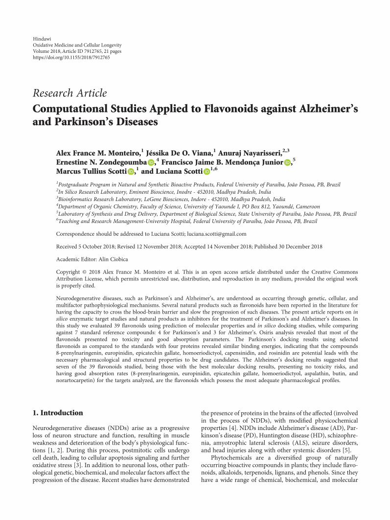
Research ArticleComputational Studies Applied to Flavonoids against Alzheimer’sand Parkinson’s Diseases
Alex France M. Monteiro,1 Jéssika De O. Viana,1 Anuraj Nayarisseri,2,3
Ernestine N. Zondegoumba ,4 Francisco Jaime B. Mendonça Junior ,5
Marcus Tullius Scotti ,1 and Luciana Scotti 1,6
1Postgraduate Program in Natural and Synthetic Bioactive Products, Federal University of Paraíba, João Pessoa, PB, Brazil2In Silico Research Laboratory, Eminent Bioscience, Inodre - 452010, Madhya Pradesh, India3Bioinformatics Research Laboratory, LeGene Biosciences, Indore - 452010, Madhya Pradesh, India4Department of Organic Chemistry, Faculty of Science, University of Yaounde I, PO Box 812, Yaoundé, Cameroon5Laboratory of Synthesis and Drug Delivery, Department of Biological Science, State University of Paraiba, João Pessoa, PB, Brazil6Teaching and Research Management-University Hospital, Federal University of Paraíba, João Pessoa, PB, Brazil
Correspondence should be addressed to Luciana Scotti; [email protected]
Received 5 October 2018; Revised 12 November 2018; Accepted 14 November 2018; Published 30 December 2018
Academic Editor: Alin Ciobica
Copyright © 2018 Alex France M. Monteiro et al. This is an open access article distributed under the Creative CommonsAttribution License, which permits unrestricted use, distribution, and reproduction in any medium, provided the original workis properly cited.
Neurodegenerative diseases, such as Parkinson’s and Alzheimer’s, are understood as occurring through genetic, cellular, andmultifactor pathophysiological mechanisms. Several natural products such as flavonoids have been reported in the literature forhaving the capacity to cross the blood-brain barrier and slow the progression of such diseases. The present article reports on insilico enzymatic target studies and natural products as inhibitors for the treatment of Parkinson’s and Alzheimer’s diseases. Inthis study we evaluated 39 flavonoids using prediction of molecular properties and in silico docking studies, while comparingagainst 7 standard reference compounds: 4 for Parkinson’s and 3 for Alzheimer’s. Osiris analysis revealed that most of theflavonoids presented no toxicity and good absorption parameters. The Parkinson’s docking results using selectedflavonoids as compared to the standards with four proteins revealed similar binding energies, indicating that the compounds8-prenylnaringenin, europinidin, epicatechin gallate, homoeriodictyol, capensinidin, and rosinidin are potential leads with thenecessary pharmacological and structural properties to be drug candidates. The Alzheimer’s docking results suggested thatseven of the 39 flavonoids studied, being those with the best molecular docking results, presenting no toxicity risks, andhaving good absorption rates (8-prenylnaringenin, europinidin, epicatechin gallate, homoeriodictyol, aspalathin, butin, andnorartocarpetin) for the targets analyzed, are the flavonoids which possess the most adequate pharmacological profiles.
1. Introduction
Neurodegenerative diseases (NDDs) arise as a progressiveloss of neuron structure and function, resulting in muscleweakness and deterioration of the body’s physiological func-tions [1, 2]. During this process, postmitotic cells undergocell death, leading to cellular apoptosis signaling and furtheroxidative stress [3]. In addition to neuronal loss, other path-ological genetic, biochemical, and molecular factors affect theprogression of the disease. Recent studies have demonstrated
the presence of proteins in the brains of the affected (involvedin the process of NDDs), with modified physicochemicalproperties [4]. NDDs include Alzheimer’s disease (AD), Par-kinson’s disease (PD), Huntington disease (HD), schizophre-nia, amyotrophic lateral sclerosis (ALS), seizure disorders,and head injuries along with other systemic disorders [5].
Phytochemicals are a diversified group of naturallyoccurring bioactive compounds in plants; they include flavo-noids, alkaloids, terpenoids, lignans, and phenols. Since theyhave a wide range of chemical, biochemical, and molecular
HindawiOxidative Medicine and Cellular LongevityVolume 2018, Article ID 7912765, 21 pageshttps://doi.org/10.1155/2018/7912765

characteristics, phytochemicals are of considerable interestfor treating NDDs. Phytochemicals are promising candidatesfor various pathological conditions involving modulation ofmultiple signal pathways and serving as antioxidant andanti-inflammatory agents [6], agents against cancer and neu-rodegenerative diseases [7–9], or as antifungal agents [10].Several studies have addressed the protective activity of nat-ural derivatives such as alkaloids when applied to neurode-generative diseases such as Alzheimer’s and Parkinson’s[11]; genistein brings neuroprotective effects [12, 13]; hesper-etin presents potent antioxidant and neuroprotective effects[14]; quercetin [15] and xanthones present multifunctionalactivities against Alzheimer’s disease [16].
Flavonoids fit the NDDs profile, and in a process depen-dent on the suppression of lipid peroxidation, inhibition ofinflammatory mediators, modulation of gene expression,and activation of antioxidant enzymes, flavonoids help main-tain the endogenous antioxidant status of neurons, protect-ing them from neurodegeneration [17, 18]. Based on theirchemical structure, they are classified into several categoriesincluding flavanols, flavonols, flavones, flavanones, isofla-vones, anthocyanidins, and chalcones [19].
This article focuses on flavonoids found in the litera-ture for anti-Parkinson and anti-Alzheimer activity,including targets involved in the degenerative process ofeach disease. Molecular docking studies detail the struc-tural parameters involved that best contribute to the activ-ity of such compounds. This study facilitates knowledge asapplied to two NDDs concerning flavonoid structuralenhancements and the pharmacophores involved in thereceptor-protein complex.
2. Parkinson’s Disease
Parkinson’s disease (PD) is the second most common neuro-degenerative disease globally and has been increasing consid-erably without evidence of cure [20, 21]. PD is reported as aloss of dopaminergic neurons located in the substantia nigra(SN) and affects 1-2% of people over the age of 60 [22]. Esti-mates of the disease range from 5 to 35 new cases per 100,000individuals [23]; this increases with age [24]. The prevalenceof PD is increasing considerably, corroborating a doubling bythe year 2030 [25].
To characterize PD, progressive degeneration of dopami-nergic (DA) neurons causing depletion of striatal dopamineand formation of Lewy bodies in the substantia nigra (SN)are the principal neuropathological correlations of motordamage in PD. The symptoms include resting tremor, rigid-ity, bradykinesia, gait difficulty, postural instability, andbehavioral problems [26]; nonmotor symptoms includedepression, anxiety, emotional changes, cognitive impair-ment, sleep difficulty, and olfactory dysfunction [27]. Thereare several studies that report neurodegenerative factors suchas neuroinflammation [28] and cytotoxic factors such as IL1,NO, ROS, and TNF [29].
The treatment of PD focuses on carbidopa to replacedopamine, levodopa drugs, monoamine oxidase B inhibitors,dopamine agonists, catechol-o-methyltransferase inhibitors,anticholinergics, and amantadine [30]. Levodopa is the single
most used drug to treat Parkinson’s disease [31]. However,these drugs cause many side effects [32], and they usuallylead to other complications, yet without curing or stoppingdisease progression. The search for new therapeutic agentswith few side effects is essential.
The use of natural products against PD has intensified inrecent years, chiefly compounds derived from plants, sincethey are known to have fewer side effects than synthetic com-pounds [33, 34]. These advances in the treatment of PD givethe disease a chance to be administered effectively, leading tosymptom control and improvement of patient quality of life,often for decades after onset of the disease.
2.1. Molecular Docking Applied to Natural Products forParkinson’s Disease. Molecular docking studies are based onjoining a particular ligand to a receptor region, providinginformation about conformation, orientation, and organiza-tion at the receptor site [35]. Studies using computationalchemistry to predict potential inhibitors for neurodegenera-tive diseases have been reported in the literature [36–38],and studies involving molecular docking have been reportedin the literature for Parkinson’s disease and flavonoidderivatives [39].
Desideri et al. [40] reported the in vitro and in silico activityof a series of homo-isoflavonoids as potent inhibitors ofhuman monoamine oxidase-B. Presenting better in vitroresults than the standard drug, selegiline, (E)-3-(4-(Dimethy-lamino)benzylidene)chroman-4-one and (E)-5,7-dihydroxy-3-(4-hydroxybenzylidene)chroman-4-one also demonstratedselectivity and high potency during the in silico studies, inter-actingwithhydrogen andhydrophobicbonds at the active site.
Our research group applied ligand-based-virtual screeningtogether with structure based-virtual screening (docking) for469 alkaloids of the Apocynaceae family in a study of humanAChE inhibitory activity [41]. As a result, 9 alkaloids presentingbetter inhibition profiles for both Parkinson’s and Alzheimer’s(dihydro-cylindrocarpine, 14,19-dihydro-11-methoxycondylo-carpine, Di (demethoxycarbonyl) tetrahydrosecamine, tetra-hydrosecamine, 16-demethoxycarbonyltetrahydrosecamine,16-hydroxytetrahydrosecamine, usambarensine, 4′,5′,6′,17-tetrahydro-usambarensine-N-oxide, and 6,7-seco-angustilo-bine) were selected for future studies.
Baul and Rajiniraja [42] performed a molecular dockingstudy using flavonoids such as quercetin, epigallocatechingallate (EGCG), and acacetin to predict inhibitory activitiesand their ability to inhibit the enzyme α-synuclein. Theresults showed that the flavonoids present low energy valueinteractions with residues Lys45, Lys43, Lys32, and Val40,being essential for activity in this protein.
In silico studies involving Parkinson’s disease anti-inflammatory activity have also been targeted for novel bio-active compounds. As a general rule for anti-inflammatoryactivity, both hydrogen and π-π hydrophobic interactionsbetween the active site of the macromolecule and the com-pounds are essential. Madeswaran et al. [43] reported theinhibition activity of nine flavonoids (morin, naringenin,taxifolin, esculatin, daidzein, genistein, scopoletin, galangin,and silbinin) against human lipoxygenase enzyme. The flavo-noid interactions especially those of morin were similar to
2 Oxidative Medicine and Cellular Longevity

Azelastine, a flavonoid already reported in the literaturefor lipoxygenase inhibition activity, thus defining aminoacids Tyr359, Gln358, and Gln539 as critical to the activityof these compounds.
2.2. Targets in Parkinson’s Disease
2.2.1. Adenosine A2A Receptors. Adenosine receptors aremembers of the G protein-coupled receptor superfamilyand considered potential targets for treatment of numerousdiseases. Adenosine binds four types of G-protein receptorsknown as A1, A2A, A2B, and A3 all with distribution in thebrain. A2A has a more specific and abundant distribution inthe basal ganglia. This selective distribution for receptorscan help guarantee fewer adverse effects and make nondopa-minergic antagonists more promising for the treatment ofPD [44].
The A2A adenosine receptor (A2AAR) is highly expressedin the basal ganglia and depends on Gs and other proteininteractions for signal interpretation [45]. In mammals, highexpression of this protein is found in the striatum in the basalganglia, with an important route for the regulation of dopami-nergic transmission [46]. The A2A receptor subtype presentssignaling involving activation of serine/threonine kinase[47, 48], which modulates phosphorylation of ionotropicglutamate receptors [49, 50]. The A2A receptor may pro-vide improvement in motor abnormalities for patientswith PD, by controlling hyperphosphorylation of theglutamatergic receptor.
Indeed, five A2A receptor antagonists are now in clinicaltrials (phases I to III) for Parkinson’s disease, and otherantagonists have been reported in the literature [51]. Theuse of these receptors is due to various preclinical studieswhich have shown that adenosinergic neuromodulationantagonizes dopaminergic neurotransmission in aspects rele-vant to motor control. The adenosine A2A receptor activatesadenylyl cyclase and certain voltage-sensitive Ca2+ channels[52]. These receptors are expressed in the GABAergic neu-rons and in glutamatergic neuronal terminals [53].
Schwarzschild et al. [54] proposed an anti-Parkinsonactivity reactive mechanism for the A2A receptor. In thenormal state, the dopamine of the neurons is found in thesubstantia nigra and acts on two receptors: D1 receptors(direct stimulatory pathway) and D2 receptors (indirectinhibitory pathway). Adenosine, which is released by A2Areceptors, stimulates neurons at the D2 receptor pathway.In degenerative processes, as is the case in PD, the centralnervous system (CNS) degeneration blocks the entry ofstriatum dopamine, which increases GABA’s inhibitoryinfluence, consequently mitigating PD motor deficits.
The restriction of striatum region expression contributesto fewer side effects in PD patients [55–57]. Several studieshave reported the activity of nondopaminergic A2A receptorantagonists [58, 59], a good target for the development ofanti-Parkinson drugs.
2.2.2. α-Synuclein. A 140 amino acid protein, α-synuclein iscommonly located in presynaptic terminals [60, 61]. Alpha-synuclein represents the most abundant protein in Lewy
bodies (LB), cytoplasmic inclusions found in PD and inLB dementia (LBD), which have a little understood physiol-ogy. The synuclein family has three members, α-synuclein,β-synuclein, and γ-synuclein, ranging from 127 to 140 aminoacids, with about 55 to 62% of homologous sequences, andwhere α and β have an identical carboxy-terminal domain.These proteins are commonly found in nerve terminals, closeto synaptic vesicles; β-synucleins are present in almost allnerve cells [62].
Among the factors that influence α-synuclein abnormal-ities, genetic factors (protein gene, PARK3, and PARK4 locusmutations) and environmental factors (oxidative damages)often lead to errors in the ordering and conformation ofα-synuclein filaments [63].
Recent studies report a mutation of alanine to threonineat position 53 of the protein gene causing a rare and familialform of PD in four families [64]. The identification ofthis mutation in autosomal dominant families of inher-ited Parkinson’s led to the discovery of a new target forPD pathology.
Olanow and Brundin [65] provided evidence ofα-synuclein activity in prion-like proteins acting in PD, thussuggesting new studies for the development of inhibitors.Recent studies have reported that a doubling or tripling ofthe α-synuclein gene leads to a similar type of PD [66, 67].Mutagenic studies involved in the α-synuclein responseassociate and reinforce the hypothesis that mutations areinvolved in the pathogenesis of PD.
2.2.3. Catechol-O-Methyltransferase. The enzyme catechol-O-methyltransferase, also known as COMT, is as an impor-tant enzyme involved in biochemistry, pharmacology, andgenetic mechanisms. Methylation of endogenous catechol-amines, as well as other catechols, is catalyzed by the enzymecatechol-O-methyltransferase (COMT). COMT transfersthe methyl group of S-adenosylmethionine (SAM) to themeta- or para-hydroxyl group present in catechols [68, 69];COMT is considered a SAM-dependent methyltransferase[70]. COMT substrates involve both endogenous andexogenous catechols, such as dopamine, norepinephrine,and epinephrine. In the brain, COMT is involved in mentalprocesses, as studies have reported for Parkinson’s disease[71]. COMT is considered a target for study and develop-ment of new anti-Parkinson drugs using coadministrationwith levodopa [72, 73]. The enzyme has two forms: a solubleform, known as S-COMT, presenting 221 residues; and asecond form, known as membrane based (MB-COMT),exhibiting 50 residues at the N-terminus [74]. The COMTactive site has a SAM binding site and an S-COMT catalyticsite. In addition, the presence of Mg2+ in the catalytic siteis responsible for converting catechol hydroxyl groups tosubstrates [68].
The COMT enzyme has the single domain structure con-taining α and βmoieties, where 8 helices are disposed arounda central β sheet. The active site of the enzyme is composedof an S-adenosyl-L-methionine-(AdoMet-) binding domain,similar to a Rossmann fold, and present in numerous proteinsthat interact with nucleotides [68].
3Oxidative Medicine and Cellular Longevity

The catechol-O-methyltransferase (COMT) gene encodesan enzyme that performs catecholamine (such as dopamine,epinephrine, and norepinephrine) degradation [75]; this pro-cess is depressed in patients with PD. The COMT gene islocated on chromosome 22q11, which has been reported asone of the major loci related to schizophrenia [76]. Recentstudies have shown a polymorphism at codon 158 (Val158-Met, called rs4680) that influences the COMT enzyme, bydecreasing its activity [77], and which interferes with execu-tive cognitive performance [78, 79].
2.2.4. Monoamine Oxidase B. The enzyme monoamine oxi-dase B (MAO-B) has been reported as a therapeutic targetfor the treatment of Parkinson’s disease [80, 81] and is alsoa brain glial biomarker [82]. Studies have shown that MAOis located in the outer mitochondrial membrane, in the liver,and in the brain [83] and presents FAD as a cofactor in itsactive site, where irreversible MAO inhibitors bind, suchas rasagiline.
MAO’s mechanism of reaction involves oxidativedeamination of primary, secondary, and tertiary amines,to the corresponding aldehyde, and free amine withthe generation of hydrogen peroxide. As for the aldehyde,this is metabolized by the enzyme aldehydedehydrogenase,producing acids such as 5-hydroxyindole acetic acid(5-HIAA)ordihydroxy-phenyl-acetic acid (DOPAC),metab-olites used as MAO activity drugs. MAO also produceshydrogen peroxide, leading to oxidative stress and neuronalcell death [84, 85].
MAO can be found in two isoforms, known as isoform Aand isoform B, with differences that are of great pharmaco-logical importance [86]. Isoform A is located next to cate-cholaminergic neurons, whereas the B isoform is located inneurotransmitters. Among the two subtypes, MAO-B is oneof the enzymes that oxidize the neurotransmitter dopaminein addition to metabolizing other amines. This enzyme isfound in large numbers in astrocytes but is also present inserotonin neuron cell bodies, whereas MAO-A is located inneurons in the brain [87]. Isoform A is inhibited by low con-centrations of clorgiline, while MAO-B is inhibited by selegi-line and rasagiline [88–90], drugs used to elevate braindopamine by inhibiting its breakdown and promoting bene-ficial symptomatic effects for the patient.
Studies have reported the expression of MAO-B inhuman brains or more precisely in the substantia nigra ofpatients affected by PD [91, 92]. Human MAO-B presentstwo cavities in its structure, and the FAD coenzyme is pres-ent in the active site. The N5 atom is present in the externalregion, and the residues Tyr398 and Tyr435 play importantroles in hMAO-B catalytic activity [93]. The inhibition ofMAO-B using rasagiline may promote increased dopaminer-gic activity of the striatum, leading to symptomatic benefitsdue to interference in dopamine degradation. Improvementsalso result from decreased free radicals as generated fromdopamine oxidation. The development of selective andreversible MAO-B inhibitors may reduce undesirable adverseeffects and present long-term efficacy in neurodegenerativedisease treatment.
3. Alzheimer’s Disease
Alzheimer’s disease (AD) is a progressive neurodegenerativedisease common in older people (from 60 years of age andupwards). It consists in memory loss and gradual impair-ment of cognitive function due to mainly cholinergic neurondeath, which makes accomplishment of daily activities diffi-cult, leading the patient to dependence for the basic activitiesof their daily routine. Because the neurological impairmentcompromises the autonomic nervous system (ANS), it even-tually leads to death. [94–98].
One of the symptoms of AD is dementia, and accordingto the World Health Organization (WHO) Bulletin, AD isthe main pathology responsible for up to 70% of individualswith dementia. WHO estimates that more than 47 millionpeople suffer from dementia, and more than half are fromunderdeveloped countries. Alzheimer’s has no cure and itstreatment consists of trying to slow the progression of thedisease and offer symptomatic relief [99, 100].
Alzheimer’s is clinically explained by neuronal decreaseslinked to deficient synthesis of acetylcholine (ACh) involvedin memory, learning, and SNA. Thus, studies commonly aimat inhibiting acetylcholinesterase (AChE) to prevent AChbreakdown and consequent loss of memory and cognitivefunctions [101–104].
3.1. Molecular Docking Applied to Natural Products forAlzheimer’s Disease. Bioactive beta-secretase-1 (BACE1)inhibitors are currently being studied as therapeutic targets.BACE1 inhibition prevents the amyloid β-amyloid peptide(Aβ) from increasing, preventing cleavage of localized amy-loid precursor protein (APP), and thus portion C99 entersthe membrane while the (sAPPβ) portion enters the extracel-lular environment. Inhibition of BACE1 is a therapeuticalternative that inhibits the evolution of AD. This hypothesishas been known since the 1990s as “amyloid cascade”because it consists of a set of neuropathological events thatoccur in chain, initiated by the accumulation of Aβ,followed by the dysfunction of Tau proteins (which nor-mally stabilize neuronal microtubules), which results in celldeath through the agglomeration of Tau proteins in the cell;this compromises both dendrite and the neuronal cell bodyfunctions [105–109].
In a molecular docking study [110] to identify moleculesthat potentiate Alzheimer’s inhibition in the BACE1 target,docking of 14 molecules using Molex Virtual Docker wasperformed with PDB ID 2XFJ and presented interactionswith amino acid residues Thr292, Asp93, Asp289, Thr293,Gln134, Asn294, and Thr133. For the compounds studied,hydrogen bonds and hydrophobic interactions with theseresidues favored inhibitory activity.
Barai et al. [111] using the GOLD suite v.5 programanalyzed molecular docking interactions of bergenin(Figure 1(a)) 2 with the objective of highlighting its neuro-protective effects against AD. The docking data in thisstudy were obtained from interactions of the natural productwith acetylcholinesterase (PDB ID 1B41), butyrylcholinester-ase (PDB ID 1P0I), Tau protein kinase 1 (PDB ID 1J1B), andBACE-1 (PDB ID 1FKN). The docking results were compared
4 Oxidative Medicine and Cellular Longevity
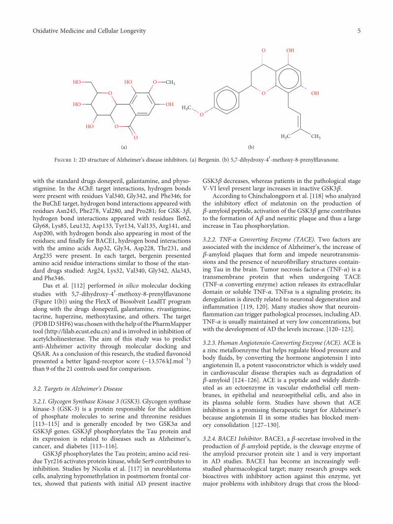
with the standard drugs donepezil, galantamine, and physo-stigmine. In the AChE target interactions, hydrogen bondswere present with residues Val340, Gly342, and Phe346; forthe BuChE target, hydrogen bond interactions appeared withresidues Asn245, Phe278, Val280, and Pro281; for GSK-3β,hydrogen bond interactions appeared with residues Ile62,Gly68, Lys85, Leu132, Asp133, Tyr134, Val135, Arg141, andAsp200, with hydrogen bonds also appearing in most of theresidues; and finally for BACE1, hydrogen bond interactionswith the amino acids Asp32, Gly34, Asp228, Thr231, andArg235 were present. In each target, bergenin presentedamino acid residue interactions similar to those of the stan-dard drugs studied: Arg24, Lys32, Val340, Gly342, Ala343,and Phe346.
Das et al. [112] performed in silico molecular dockingstudies with 5,7-dihydroxy-4′-methoxy-8-prenylflavanone(Figure 1(b)) using the FlexX of Biosolveit LeadIT programalong with the drugs donepezil, galantamine, rivastigmine,tacrine, huperzine, methoxytaxine, and others. The target(PDBID5HF6)was chosenwith thehelpof thePharmMappertool (http://lilab.ecust.edu.cn) and is involved in inhibition ofacetylcholinesterase. The aim of this study was to predictanti-Alzheimer activity through molecular docking andQSAR. As a conclusion of this research, the studied flavonoidpresented a better ligand-receptor score (−13.576 kJ.mol−1)than 9 of the 21 controls used for comparison.
3.2. Targets in Alzheimer’s Disease
3.2.1. Glycogen Synthase Kinase 3 (GSK3). Glycogen synthasekinase-3 (GSK-3) is a protein responsible for the additionof phosphate molecules to serine and threonine residues[113–115] and is generally encoded by two GSK3α andGSK3β genes. GSK3β phosphorylates the Tau protein andits expression is related to diseases such as Alzheimer’s,cancer, and diabetes [113–116].
GSK3β phosphorylates the Tau protein; amino acid resi-due Tyr216 activates protein kinase, while Ser9 contributes toinhibition. Studies by Nicolia et al. [117] in neuroblastomacells, analyzing hypomethylation in postmortem frontal cor-tex, showed that patients with initial AD present inactive
GSK3β decreases, whereas patients in the pathological stageV-VI level present large increases in inactive GSK3β.
According to Chinchalongporn et al. [118] who analyzedthe inhibitory effect of melatonin on the production ofβ-amyloid peptide, activation of the GSK3β gene contributesto the formation of Aβ and neuritic plaque and thus a largeincrease in Tau phosphorylation.
3.2.2. TNF-α Converting Enzyme (TACE). Two factors areassociated with the incidence of Alzheimer’s, the increase ofβ-amyloid plaques that form and impede neurotransmis-sions and the presence of neurofibrillary structures contain-ing Tau in the brain. Tumor necrosis factor-α (TNF-α) is atransmembrane protein that when undergoing TACE(TNF-α converting enzyme) action releases its extracellulardomain or soluble TNF-α. TNFsα is a signaling protein; itsderegulation is directly related to neuronal degeneration andinflammation [119, 120]. Many studies show that neuroin-flammation can trigger pathological processes, including AD.TNF-α is usually maintained at very low concentrations, butwith the development of AD the levels increase. [120–123].
3.2.3. Human Angiotensin-Converting Enzyme (ACE).ACE isa zinc metalloenzyme that helps regulate blood pressure andbody fluids, by converting the hormone angiotensin I intoangiotensin II, a potent vasoconstrictor which is widely usedin cardiovascular disease therapies such as degradation ofβ-amyloid [124–126]. ACE is a peptide and widely distrib-uted as an ectoenzyme in vascular endothelial cell mem-branes, in epithelial and neuroepithelial cells, and also inits plasma soluble form. Studies have shown that ACEinhibition is a promising therapeutic target for Alzheimer’sbecause angiotensin II in some studies has blocked mem-ory consolidation [127–130].
3.2.4. BACE1 Inhibitor. BACE1, a β-secretase involved in theproduction of β-amyloid peptide, is the cleavage enzyme ofthe amyloid precursor protein site 1 and is very importantin AD studies. BACE1 has become an increasingly well-studied pharmacological target; many research groups seekbioactives with inhibitory action against this enzyme, yetmajor problems with inhibitory drugs that cross the blood-
HO
HO
HO O
O
O
HO O CH3
OH
(a)
O
O
O OH
OH
H3C CH3
H3C
(b)
Figure 1: 2D structure of Alzheimer’s disease inhibitors. (a) Bergenin. (b) 5,7-dihydroxy-4′-methoxy-8-prenylflavanone.
5Oxidative Medicine and Cellular Longevity

Table 1: Structure, name, structural formula, and molar mass of the flavonoids present in the study.
No. Structure Molecular name Molecular formula Mass
1 O O
O
O
O
OO
3-O-Methylquercetin C16H12O7 316.058
2 O
O
OO
O 8-Prenylnaringenin C20H20O5 340.131
3
O
O
O
OO
Afzelechin C15H14O5 274.084
4O
O
O
O
O
OO
O Ampelopsin C15H12O8 320.053
5O
O O
O
O
O
Aromadendrin C15H12O6 288.063
6
OO
O O
OOO
O
O
O
O
Aspalathin C21H24O11 452.131
7O+
O
O
O
O
O
Aurantinidin C15H11O6 287.055
8
O
OO
O
O
Butin C15H12O5 272.068
9O O
OO
Capensinidin C18H17O7 345.097
10O
OO
O
Chrysin C15H10O4 254.057
11
O
O+
O
O
O
O
O Delphinidin C15H11O7 303.050
6 Oxidative Medicine and Cellular Longevity

Table 1: Continued.
No. Structure Molecular name Molecular formula Mass
12
O
O
O
O
O
O O
O Di-hydrogossypetin C15H12O8 320.053
13
O O
O
O
OO
O
Di-hydromorin C15H12O7 304.058
14 O
O
O
O
O
O
Epicatechin C15H14O6 290.07
15 O
OO
O
O
O
Eriodictyol C15H12O6 288.063
16 O+O
OO
O
OO
Europinidin C17H15O7 331.081
17
O
O
O
O
O
O
Fisetin C15H10O6 286.047
18
O
O
O
O
O Fisetinidol C15H14O5 274.084
19
O
OO
O
O
O
Fustin C15H12O6 288.063
20
O
O
O
OO
O
O
O
O
O
Epicatechin gallate C22H18O10 442.090
21
O
OO
O
O
Genistein C15H10O5 270.052
22 O
O
O O
O
O
O
O
Gossypetin C15H10O8 318.037
7Oxidative Medicine and Cellular Longevity

Table 1: Continued.
No. Structure Molecular name Molecular formula Mass
23
OO
O
O
OO
O
O O
O
O O
O
O
O
Hesperidin C28H34O15 610.189
24
O
O
O
OO
O
O
O
O Hibiscetin C15H10O9 334.032
25O
O
O
O O
O
Homoeriodictyol C16H14O6 302.079
26OO
OO
O
Isosakuranetin C16H14O5 286.084
27
O
O
O
O+
O
Luteolinidin C15H11O5 271.060
28
O
OO
O
O
O
Meciadanol C16H16O6 304.094
29 O
O
O
O
O
O
Mesquitol C15H14O6 290.079
30O
O
OO
O
O
O
Morin C15H10O7 302.042
31
O O
O
OO
O
Norartocarpetin C15H10O6 286.047
32
O
O
O
O
Pinocembrin C15H12O4 256.073
8 Oxidative Medicine and Cellular Longevity
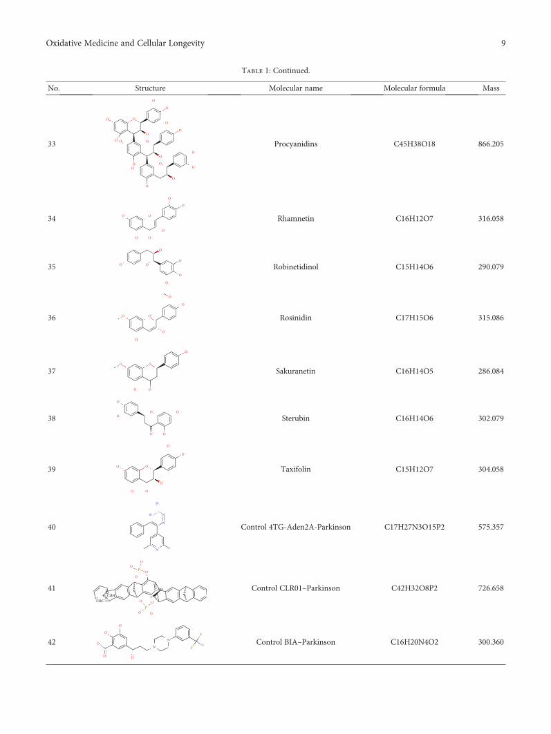
Table 1: Continued.
No. Structure Molecular name Molecular formula Mass
33
O
O
OO
O O
O
O
O
O
O
O
O
O
O
OOO
Procyanidins C45H38O18 866.205
34
O
OO
OO
O
O
Rhamnetin C16H12O7 316.058
35 O
O
O
O
O
O Robinetidinol C15H14O6 290.079
36
O
O
O
O
O
O+
Rosinidin C17H15O6 315.086
37O
O
OO
O
Sakuranetin C16H14O5 286.084
38
O
OO O
OO
Sterubin C16H14O6 302.079
39 O O
O
O
O
OO
Taxifolin C15H12O7 304.058
40
N
NN
N
N
Control 4TG-Aden2A-Parkinson C17H27N3O15P2 575.357
41
P
OO
OO
O O
OOP
CBC
CBH
CAH Control CLR01–Parkinson C42H32O8P2 726.658
42
O
O
O O
ON N
NF
FF
Control BIA–Parkinson C16H20N4O2 300.360
9Oxidative Medicine and Cellular Longevity

brain barrier remain [131–134]. Studies with mice show thatBACE1 inhibitors are efficient in combating new Aβ plaquesbut inefficient against growth of existing plaques, suggestingearly treatment with the aim of preventing initial plaqueformation [135, 136].
4. Materials and Methods
4.1. Data Set. From the literature, we selected the set of 39flavonoid structure, known for their antioxidant action. Thecompounds were submitted to molecular modeling andmolecular docking tools to provide their important structuralinformation and activity as multitarget compounds. Data forthe physicochemical characteristics of the compounds hasbeen reported (Table 1).
4.2. Molecular Modeling. All of the structures were drawn inHyperChem for Windows v. 8.0.5 (HyperChem, 2009) [137],and their molecular geometries were minimized using themolecular mechanics MM+ force field, without restrictionsfor aromatic form conversions, and clean molecular graphingin three dimensions. The optimized structures were subjectedto conformational analysis using a random search methodwith 1000 interactions, 100 cycles of optimization, and the10 lowest minimum energy conformers. The compoundswere saved in the MOL format.
4.3. Quantitative Structure-Activity Relationship: OSIRIS.The cytotoxicity risk study was performed using OSIRISDataWarrior 4.7.3 [138]. The cytotoxic effects were mutage-nicity, carcinogenicity, and irritability to the skin and repro-ductive system. The TPSA (topological polar surface area)values were used to calculate the rate of absorption (%) offlavonoids and control as drugs by the formula
%ABS = 109 − 0 345 × TPSA 1
4.4. Molecular Docking. For Parkinson’s disease, the struc-tures of human adenosine receptor A2A (PDB ID 3UZA, at
a resolution of 3.2Å), α-synuclein (PDB ID 1XQ8), COMT(PDB ID 1H1D, at a resolution of 2Å), and MAO-B(PDB ID 2C65, at a resolution of 1.7Å) were downloadedfrom the Protein Data Bank (PDB) [139]. The choice ofthese proteins relied on protein validations reported inthe literature, with anti-Parkinson activity as a prerequisite.The adenosine receptor A2A, COMT, and MAO-B proteins,respectively, contained 6-(2,6-dimethylpyridin-4-yl)-5-phenyl-1,2,4-triazin-3-amine (T4G) (an inhibitory drug),1-(3,4,dihydroxy-5-nitrophenyl)-3-{4-[3-(trifluoromethyl)phenyl] piperazin-1-yl}propan-1-one (BIA), and ladostigilwhich served as bases for active site labeling and as controlcompounds for comparing energy values with the flavonoids.As for the α-synuclein protein, the option was chosen todetect 10 possible cavities, admitted as possible active siteson which to run the molecular docking. In order to comparethe results of the 39 flavonoids, the docking was also runwith the compound CLR01, an α-synuclein inhibitor fromthe literature.
For Alzheimer’s, 4 targets with respect to pathologywere analyzed, PDB ID 160K (resolution of 1.94Å) thecrystal structure of glycogen synthase kinase 3 (GSK-3)with a complexed inhibitor [114], PDB ID 2FV5 (2.1Åresolution) for the TACE crystal structure complexedwith IK682 [140], PDB ID 3BKL (resolution 2.18Å) forthe ACE cocrystal structure with kAW inhibitor [141],and PDB ID 4DJU (resolution 1.8Å) for the crystallinestructure of BACE bound to 2-imino-3-methyl-5,5-diphe-nylimidazolidin-4-one [142]. The targets were selectedbased on scientific papers on in silico studies of moleculeswith anti-Alzheimer activity. The inhibitor for GSK-3complexed together with the crystal structure was N-(4-methoxybenzyl)-N′-(5-nitro-1,3-thiazol-2-yl) urea (TMU),for TACE it was (2R)-N-hydroxy-2-[(3S)-3-methyl-3-{4-[(2-methylquinolin-4-yl)methoxy] phenyl}-2-oxopyrroli-din-1-yl] propanamide (541), and for ACE it wasN-{(5S)-4,4-dihydroxy-6-phenyl-5-[(phenylcarbonyl)amino]hexanoyl}-L-tryptophan (kAW).
Table 1: Continued.
No. Structure Molecular name Molecular formula Mass
43O
O
N N+
Control ladostigil-Parkinson C16H20N2O2 272.340
44 NNO
O
Control rivastigmine-Alzheimer C14H22N2O2 250.337
45
OO
O
N
Control galantamine-Alzheimer C17H21NO3 287.340
46N
O
O
O Control donepezil-Alzheimer C24H29NO3 379.480
10 Oxidative Medicine and Cellular Longevity

All 39 flavonoid structures (in MOL format) were sub-mitted to molecular docking using the Molegro VirtualDocker v. 6.0.1 (MVD) [143]. All of the water compoundswere deleted from the enzyme structure. For the moleculardocking simulation, the bonds for all the compounds andthe protein residues in the binding site were set as flexible,with a tolerance of 1.0, strength of 0.80, and with the tor-sional degrees of freedom for the flexible residues and ligandsat 2000 steps of energy minimization. The enzyme and com-pound structures were prepared using the same defaultparameter settings in the same software package (score func-tions: MolDock score; ligand evaluation: internal ES, internalHBond, were all verified; number of runs: 10; algorithm:MolDock SE; maximum interactions: 1500; max. populationsize: 50; max. steps: 300; neighbor distance factor: 1.00; max.number of poses returned: 5). The docking procedure wasperformed using a 15Å radius GRID and 0.30 of resolutionto cover the ligand-binding site of the protein. For pose orga-nizer, the MolDock score (GRID) algorithm was used as thescore function and the Moldock search algorithm was used.
5. Results and Discussion
5.1. Quantitative Structure-Activity Relationship. Studies instructure-based design have become routine in drug discov-ery, searching for the best profiles against a disease. Thus, itis possible to analyze and discover various pharmacophoricgroups and predict possible activities against a certain target.This study was performed through analysis of the physico-chemical properties of drugs, such as TPSA and drug absorp-tion, and using studies related to structure-based proteindrug design. Toxicity risks and TPSA data, calculated inOsiris software, are presented in Table 2.
Mutagenicity studies can be used to quantify the roleplayed by various organics in promoting or interfering withthe way a drug can associate with DNA. According to thedata from the Osiris program, flavonoids present low tenden-cies to be toxic. There were only six compounds that pre-sented mutagenic toxicity (fisetin, genistein, gossypetin,hibiscetin, morin, and rhamnetin); two presented reproduc-tive toxicity (genistein and procyanidin) and one presentedtumor activity (genistein). These compounds present highrisk and do not possess good drug profiles.
5.2. Molecular Docking in Parkinson’s Disease. The moleculardocking studies for the flavonoids and the control drugs withthe PD targets are presented in Table 3.
For the enzyme Aden2A, it was observed that the threeflavonoids (epicatechin gallate, hesperidin, and procyanidin)with respective energy values of −113.727 kcal/mol,−101.446 kcal/mol, and −98.216 kcal/mol presented higheraffinities when compared to the PDB ligand (4TG).
The flavonoids pre PDB ligand; hydrogen bonds presentin hydroxyl groups with residues Asn253, Ala63, His250,His278, and steric interactions were observed for Asn253,Phe168, Trp246, and Leu249 for the flavonoids which pre-sented higher score values. Key interactions were detectedat His278, Leu249, and Asn253, being present in all of the
flavonoids studied, principally at residue Asn253, because itis also present for the ligand PDB (Figure 2(a)).
For the enzyme α-synuclein, the observed value of PDB(CLR01=−147.800 kcal/mol) presented better energy valuesas compared to flavonoids in the study. However, three ofthe compounds presented energy values close to that of thePDB ligand; these were procyanidin (−130.002 kcal/mol),
Table 2: Toxicity data, TPSA, and %ABS calculated on the Osiristool for flavonoids.
Flavonoids Toxicity risks TPSA %ABS
3-O-methylquercetin No 116.450 68.824
8-prenylnaringenin No 86.990 78.9884
Afzelechin No 90.150 77.8982
Ampelopsin No 147.680 58.050
Aromadendrin No 107.220 72.009
Aspalathin No 208.370 37.112
Aurantinidin No 101.150 74.103
Butin No 86.990 78.988
Capensinidin No 88.380 78.508
Chrysin No 66.760 85.967
Delphinidin No 121.380 67.123
Di-hydrogossypetin No 147.680 58.050
Di-hydromorin No 127.450 65.029
Epicatechin No 110.380 70.918
Eriodictyol No 107.220 72.009
Europinidin No 99.380 74.713
Fisetin Mutagenic 107.220 72.009
Fisetinidol No 90.150 77.898
Fustin No 107.220 72.009
Epicatechin gallate No 177.140 47.886
GenisteinMutagenic/tumor/
reproductive86.990 78.988
Gossypetin Mutagenic 147.680 58.050
Hesperidin No 234.290 28.169
Hibiscetin Mutagenic 167.910 51.071
Homoeriodictyol No 96.220 75.804
Isosakuranetin No 75.990 82.783
Luteolinidin No 80.920 81.082
Meciadanol No 99.380 74.713
Mesquitol No 110.380 70.918
Morin Mutagenic 127.450 65.029
Norartocarpetin No 107.220 72.009
Pinocembrin No 66.760 85.967
Procyanidin Reproductive 331.140 −5.243Rhamnetin Mutagenic 116.450 68.824
Robinetinidol No 110.380 70.918
Rosinidin No 92.290 77.159
Sakuranetin No 75.990 82.783
Sterubin No 96.220 75.804
Taxifolin No 127.450 65.029
11Oxidative Medicine and Cellular Longevity

epicatechin gallate (−98.330 kcal/mol), and rosinidin(−95.587 kcal/mol). For the flavonoids, hydrogen bonds werepresent for Lys43, Leu38, and Glu35. Key interactions werealso observed for flavonoid activity in hydroxyl group stericinteractions with residues Lys32, Lys43, and Glu35 consid-ered key interactions for complex formation. These residuesalso appeared for the PDB ligand (Figure 2(b)).
Most COMT inhibitors have a catechol ring in theirstructure, such as entacapone and tolcapone, the mostfamous COMT inhibitor drugs. In our studies the enzyme
COMT also presented flavonoid compound activity, beingepicatechin gallate (−96.205 kcal/mol) a stronger interactionthan the PDB ligand (BIA=−80.800 kcal/mol). For flavonoidactivity, interactions with the active site presented eight resi-dues, such as Asp141, Asn170, Lys144, Met40, and Glu199,forming hydrogen interactions with the catechol portions ofthe flavonoids. Residues Asn170, Glu199, Trp38, Leu198,Asp141, and Trp143 presented hydrophobic interactionswith the hydroxyl portions of the flavonoids (Figure 2(c)).Similar results have been presented by Lee and Kim [144]
Table 3: Description of energy scores of flavonoids and control compounds on PD target proteins.
Flavonoids Aden A2A α-Synuclein COMT MAO-B
3-O-methylquercetin −71.095 −74.901 −53.659 −140.7638-prenylnaringenin −83.692 −83.012 −67.998 −145.425Afzelechin −61.973 −70.911 −51.278 −107.22Ampelopsin −60.848 −74.188 −53.806 −134.626Aromadendrin −53.880 −66.701 −45.951 −123.726Aspalathin −55.009 −86.361 −56.396 −150.386Aurantinidin −67.749 −75.414 −56.591 −117.977Butin −68.355 −77.949 −60.034 −124.25Capensinidin −84.669 −87.321 −71.529 −140.926Chrysin −59.594 −70.872 −52.576 −120.287Delphinidin −70.457 −82.877 −68.376 −126.481Di-hydrogossypetin −56.359 −73.612 −48.949 −135.483Di-hydromorin −61.416 −66.071 −54.329 −131.088Epicatechin −66.996 −74.661 −53.054 −122.78Eriodictyol −66.790 −74.167 −55.545 −119.801Europinidin −75.421 −79.694 −74.993 −140.585Fisetin −67.182 −79.763 −64.252 −130.773Fisetinidol −64.279 −72.271 −59.406 −118.506Fustin −59.854 −76.510 −56.851 −135.63Epicatechin gallate −113.727 −98.330 −96.205 −174.333Genistein −68.316 −73.585 −58.867 −119.162Gossypetin −63.019 −75.620 −58.446 −139.059Hesperidin −101.446 −89.698 −65.656 −181.222Hibiscetin −71.879 −75.302 −60.718 −137.019Homoeriodictyol −75.599 −82.786 −62.698 −141.639Isosakuranetin −65.924 −71.351 −49.177 −131.514Luteolinidin −65.240 −80.031 −57.149 −122.481Meciadanol −73.596 −77.668 −55.342 −126.337Mesquitol −60.219 −74.776 −51.753 −128.058Morin −70.744 −84.587 −59.595 −139.778Norartocarpetin −67.527 −77.898 −60.514 −137.774Pinocembrin −56.707 −66.573 −46.254 −113.423Procyanidin −98.216 −130.002 −85.226 −88.460Rhamnetin −69.702 −83.582 −49.586 −142.785Robinetinidol −62.594 −78.967 −51.172 −125.203Rosinidin −83.735 −95.587 −63.376 −149.196Sakuranetin −70.695 −74.984 −51.408 −129.56Sterubin −69.560 −77.022 −56.015 −141.623Taxifolin −56.665 −69.743 −52.804 −126.612
12 Oxidative Medicine and Cellular Longevity

and Tervo et al. [145], using molecular docking applied tocompounds containing catechol and revealing potent COMTinhibition, which highlight the presence of these same inter-actions previously reported. The residues Asp141 andAsn170 were present for all of the flavonoids in our study,including the PDB ligand, making them key residues for theactivity of these compounds.
MAO-B enzyme docking was performed at the two activePDB ligand sites. At the active site we saw that all of the flavo-noids in the study were bound to the enzyme at both sites, withthe same prevalence of compounds and presenting very closevalues at both sites. We also observed that the B subunit pre-sents greater interaction with the compounds than subunit A(Table 3). Comparing the subunit B values, we found thatthe 10 flavonoid interactions were even more active thanthe PDB binder (4CR=−140kcal/mol): 3-O-methylquercetin(−140.763kcal/mol), 8-prenylnaringenin (−145.425kcal/mol),
aspalathin (−150.386kcal/mol), capensinidin (−140.926kcal/mol), europinidin (−140.585 kcal/mol), epicatechin gallate(−174.333kcal/mol), hesperidin (−181.222kcal/mol), homoer-iodictyol (−141.639kcal/mol), rosinidin (−149.196kcal/mol),and sterubin (−141.623 kcal/mol). All these compounds pre-sented steric interactions with residues Cys172, Tyr435,Leu171, Tyr435, Tyr326, Tyr60, and Gln206. Hydrogenbonds at Tyr398 and Met436 and Cys397 were presentedwith the hydroxyl portions of the flavonoids (Figure 2(d)).Similar results were also reported by Turkmenoglu et al.[39], using differing flavonoid derivatives in moleculardocking (from a Sideritis species) for human monoamineoxidase (hMAO) isoform A and B, and by Shireen et al.[146] using flavonones from Boesenbergia rotunda for mono-amine oxidase B, both of which presented interactionssimilar to our studied flavonoids, with docking resultsthat presented significant hMAO-B inhibitory activity.
(a) (b)
(c) (d)
Figure 2: Molecular docking of flavonoids at the active site of Aden A2A (PDB: 3UZA), α-synuclein (PDB: 1XQ8), COMT (PDB: 1H1D),and MAO-B (PDB: 2C65). (a) Docking of flavonoids at the active site of Aden2A (green to epicatechin gallate, yellow to procyanidin, andblue to hesperidin). (b) Docking of flavonoids at the active site of α-synuclein (green to procyanidin, yellow to epicatechin gallate, andblue to rosinidin). (c) Docking of flavonoids at the COMT active site (green to epicatechin gallate and yellow to procyanidin and blue toeuropinidin) (ligand PDB). (d) Docking of flavonoids at the active site of MAO-B (green to hesperidin, yellow to epicatechin, and blueto aspalathin).
13Oxidative Medicine and Cellular Longevity

Such activity is recommended for first-line drugs to treatParkinson’s disease.
We observed that the interactions between flavonoidsand the study proteins occurred close to the hydroxyl groupspresent in the ligand structure and a strong interaction with
the catechol ring. It was also observed that molecules withgreater molecular mass, and electron-donating hydrophilichydroxyl groups in ring position B, were more reactivewith the enzyme, this, given the greater number of stericand electrostatic interactions with the catalytic site. The
Table 4: Energy scores of flavonoids and control compounds against Alzheimer’s disease.
Name 1Q5K 2FV5 3BKL 4DJU
3-O-Methylquercetin −77.844 −137.815 −89.583 −81.9598-Prenylnaringenin −97.365 −132.520 −96.493 −85.052Afzelechin −69.480 −120.893 −79.982 −65.259Ampelopsin −71.079 −119.645 −83.823 −68.341Aromadendrin −65.678 −115.123 −81.374 −145.179Aspalathin −91.374 −153.001 −125.583 −92.594Aurantinidin −77.482 −113.425 −84.517 −60.915Butin −80.350 −132.235 −89.736 −110.684Capensinidin −77.262 −134.112 −108.407 −118.415Chrysin −77.346 −117.834 −88.051 −85.052Delphinidin −86.937 −132.828 −98.687 −73.381Di-hydrogossypetin −66.795 −120.679 −80.429 −72.832Di-hydromorin −67.026 −121.489 −87.870 −71.631Donepezil∗ −112.609 −154.722 −119.399 −83.404Epicatechin −72.393 −127.619 −83.552 −78.328Eriodictyol −74.681 −124.042 −87.631 −90.944Europinidin −85.511 −140.803 −108.977 −89.075Fisetin −81.627 −139.645 −95.587 −73.317Fisetinidol −74.131 −116.368 −83.084 −65.914Fustin −74.571 −116.130 −80.078 −74.650Galantamine∗ −84.430 −156.068 −93.838 −115.428Epicatechin gallate −105.952 −187.352 −114.841 −83.154Genistin −78.990 −127.356 −89.509 −90.625Gossypetin −69.944 −142.715 −84.131 −79.410Hesperidin −85.551 −145.093 −97.557 −80.780Hibiscetin −66.573 −144.530 −103.117 −81.446Homoeriodictyol −85.345 −134.677 −93.198 −82.368Isosakuranetin −76.492 −124.546 −81.779 −70.443Luteolinidin −76.499 −121.014 −84.251 −87.799Meciadanol −73.882 −127.300 −84.290 −74.119Mesquitol −81.114 −130.982 −92.321 −80.051Morin −79.444 −130.332 −97.326 −80.051Norartocarpetin −79.739 −128.750 −99.216 −106.335Pinocembrin −67.298 −113.647 −81.535 −56.405Procyanidin −115.164 −154.184 −113.990 −81.313Rhamnetin −81.950 −127.432 −89.885 130.736
Rivastigmine∗ −76.582 −121.774 −85.559 186.829
Robinetidinol −86.339 −124.910 −95.178 −136.143Rosinidin −96.375 −134.734 −111.602 266.611
Sakuranetin −74.645 −118.156 −88.698 −89.075Sterubin −85.628 −124.397 −91.209 −145.179Taxifolin −69.263 −120.177 −77.806 −82.368∗Drugs used as a control for Alzheimer’s molecular docking.
14 Oxidative Medicine and Cellular Longevity

observations led to the hypothesis that such clusters canbe viewed as possible pharmacophores for the developmentof anti-PD drugs.
Our screening results (yielding the best values againstthe four studied proteins) indicated that 8-prenylnarin-genin, europinidin, epicatechin gallate, homoeriodictyol,capensinidin, and rosinidin present structural characteristicswhich guarantee their potential pharmacological activityagainst PD.
5.3. Molecular Docking in Alzheimer’s Disease. Moleculardocking of the 39 flavonoids was performed to analyzeligand-receptor integration for AD targets; the total interac-tion energy values are presented in Table 4.
For the GSK-3 target, two flavonoids (procyanidin andepicatechin gallate) presented better receptor interactionresults with respective energy values of −115.164 kJ/mol
and− 105.952 kJ/mol. However, procyanidin presents toxic-ity risks to the reproductive system. Analyzing interactionswith the amino acid residues, we perceived hydrogen bondsof hydroxyls at residue Val135, as well as Asp133, anddiscretely at Arg141, Pro136, and Try134 for most of thestudied flavonoids. Comparing the common amino acidresidues of the interaction of the complexed ligand withthe crystalline target, we noticed the common contributionof two residues with hydrogen bonds, 2 interactions withresidue Val135, and 1 interaction with Pro136, leading tothe hypothesis that these residues contribute to GSK-3inhibitory activity.
For the TACE target, three flavonoids presentedinteraction energies below 150.0000 kJ/mol (epicatechingallate, procyanidin, and aspalathin) with respectiveinteraction energies of −187.352 kJ/mol, −154.184 kJ/mol,and− 153.001 kJ/mol. In addition to the abovementioned
(a) (b)
(c) (d)
Figure 3: Molecular docking of flavonoids in the active site of GSK3 (PDB: 1Q5K), TACE (PDB: EFV5), ACE (PDB: 3BKL), and BACE1(PDB: 4DJU). (a) Docking of flavonoids in the active site of GSK3 (green to procyanidin and yellow to epicatechin gallate). (b) Docking offlavonoids in the active site of TACE (green to epicatechin gallate, yellow to procyanidin, and blue to aspalathin). (c) Docking offlavonoids in the active site of ACE (green to aspalathin, yellow to epicatechin gallate, and blue to procyanidin). (d) Docking of flavonoidsin the active site of BACE1 (green to sterubin, yellow to aromadendrin, and blue to robinetidinol).
15Oxidative Medicine and Cellular Longevity

toxicity of procyanidin, there is little possibility for oralabsorption since the %ABS=−5.241. For this target themolecules showed an interaction tendency for hydrogenbonding with Try433, Try436, and Pro437. For most ofthe compounds studied, the ligand when complexed withthe PDB presented hydrogen-bonding interactions withresidues Gly349, His409, His405, Glu406, Leu348, Gly349,and Asn447.
For the ACE target, thirteen compounds presented betterinteractions (below the median dock energy for each targetstudied) and hydrogen bond interactions with at least one ofthe amino acid residues: Tyr520, His513, Lys511, Tyr523,His353,Glu411,Glu384, andAla356.Of these,five hadmolec-ular docking energies below −100.000 kJ/mol, aspalathin,epicatechin gallate, rosinidin, europinidin, and capensinidin.
Finally, for the BACE1 inhibition study, seventeen mole-cules presented satisfactory molecular docking energies, ofwhich six (aromadendrin, sterubin, robinetidinol, capensi-din, butin, and norartocarpetin) presented energies between−106.335 kJ/mol and −145.179 kJ/mol. The amino acid resi-dues involved in the ligand-receptor interaction, with hydro-gen bonds in important residues, Ile187, Glu95, Thr292,Asp289, Phe169, Thy132, Asn98, Trp137, Ser97, andArg189, appeared with a high number of molecular bonds.In Figure 3, the docking of the 3 flavonoid enhancementsfor each target is presented.
By cross-checking the virtual screening data of the 39 fla-vonoids with the best interactions for each chosen PDB tar-get, 7 flavonoids with the best results were obtained and arepresented in this research: 8-prenylnaringenin, europinidin,epicatechin gallate, homoeriodictyol, aspalathin, butin, andnorartocarpetin.
6. Conclusions
We conclude that the flavonoids of the study demonstratepotential neuroprotective activity by virtue of bindingto certain key targets for Parkinson’s and Alzheimer’s.Based on our molecular docking studies, the flavonoids8-prenylnaringenin, europinidin, epicatechin gallate, homo-eriodictyol, capensinidin, and rosinidin present the bestresults for Parkinson’s, whereas for Alzheimer’s, the flavo-noids 8-prenylnaringenin, europinidin, epicatechin gallate,homoeriodictyol, aspalathin, butin, and norartocarpetin pres-ent the best results.With lower and comparable binding ener-gies (compared to crystallized binders), four flavonoids wereobserved in common for both diseases, presenting interac-tions and similarities consistent to those reported in the liter-ature. For these flavonoid derivatives, it was observed thathaving greater flexibility together with hydrophobic hydroxylgroups facilitates interactionswith hydrophobic regions of thetarget protein-binding sites.
Data Availability
The data used to support the findings of this study areavailable from the corresponding author upon request.
Conflicts of Interest
The authors declare that there is no conflict of interestregarding the publication of this paper.
Acknowledgments
The authors wish to acknowledge the Conselho Nacional deDesenvolvimento Científico e Tecnológico (CNPq). Thisstudy was financed in part by the Coordenação de Aperfei-çoamento de Pessoal de Nível Superior—Brasil (CAPES)—Finance Code 001.
References
[1] K. J. Barnham, C. L. Masters, and A. I. Bush, “Neurodegener-ative diseases and oxidative stress,” Nature Reviews DrugDiscovery, vol. 3, no. 3, pp. 205–214, 2004.
[2] M. Fakhoury, “Role of immunity and inflammation in thepathophysiology of neurodegenerative diseases,” Neurode-generative Diseases, vol. 15, no. 2, pp. 63–69, 2015.
[3] M. T. Lin and M. F. Beal, “Mitochondrial dysfunction andoxidative stress in neurodegenerative diseases,” Nature,vol. 443, no. 7113, pp. 787–795, 2006.
[4] G. G. Kovacs, “Current concepts of neurodegenerative dis-eases,” European Medical Journal of Neurology, vol. 1,pp. 78–86, 2014.
[5] M. Hamer and Y. Chida, “Physical activity and risk of neuro-degenerative disease: a systematic review of prospective evi-dence,” Psychological Medicine, vol. 39, no. 1, pp. 3–11, 2009.
[6] P. I. Uriarte and M. I. Calvo, “Phytochemical study and eval-uation of antioxidant, neuroprotective and acetylcholinester-ase inhibitor activities of Galeopsis ladanum L. extracts,”Pharmacognosy Magazine, vol. 5, no. 20, p. 287, 2009.
[7] A. Hosseini and A. Ghorbani, “Cancer therapy with phyto-chemicals: evidence from clinical studies,” Avicenna Journalof Phytomedicine, vol. 5, no. 2, pp. 84–97, 2015.
[8] P. R. Dandawate, D. Subramaniam, R. A. Jensen, andS. Anant, “Targeting cancer stem cells and signaling pathwaysby phytochemicals: novel approach for breast cancer ther-apy,” in Seminars in Cancer Biology, pp. 192–208, AcademicPress, 2016.
[9] I. Sarfraz, A. Rasul, F. Jabeen et al., “Fraxinus: a plant withversatile pharmacological and biological activities,” Evi-dence-Based Complementary and Alternative Medicine,vol. 2017, Article ID 4269868, 12 pages, 2017.
[10] N. Windayani, Y. Rukayadi, E. H. Hakim, K. Ruslan, andY. M. Syah, “Antifungal activity of lignans isolated fromPhyllanthus myrtifolius Moon. against Fusarium oxy-sporum,” Phytochemistry, vol. 12, pp. 33–39, 2014.
[11] M. Mohamed Essa, M. Akbar, and G. Guillemin, The benefitsof Natural Products for Neurodegenerative Diseases, SpringerInternational Publishing, Switzerland, 2016.
[12] M. Sonee, T. Sum, C. Wang, and S. K. Mukherjee, “The soyisoflavone, genistein, protects human cortical neuronal cellsfrom oxidative stress,” Neurotoxicology, vol. 25, no. 5,pp. 885–891, 2004.
[13] W. Liao, G. Jin, M. Zhao, and H. Yang, “The effect ofgenistein on the content and activity of α- and β-secretaseand protein kinase C in Aβ-injured hippocampal neurons,”
16 Oxidative Medicine and Cellular Longevity

Basic & Clinical Pharmacology & Toxicology, vol. 112, no. 3,pp. 182–185, 2013.
[14] J. Cho, “Antioxidant and neuroprotective effects of hesperi-din and its aglycone hesperetin,” Archives of PharmacalResearch, vol. 29, no. 8, pp. 699–706, 2006.
[15] A. M. Sabogal-Guáqueta, J. I. Muñoz-Manco, J. R. Ramírez-Pineda, M. Lamprea-Rodriguez, E. Osorio, and G. P.Cardona-Gómez, “The flavonoid quercetin ameliorates Alz-heimer’s disease pathology and protects cognitive and emo-tional function in aged triple transgenic Alzheimer’s diseasemodel mice,”Neuropharmacology, vol. 93, pp. 134–145, 2015.
[16] S. N. Wang, Q. Li, M. H. Jing et al., “Natural xanthones fromGarcinia mangostana with multifunctional activities for thetherapy of Alzheimer’s disease,” Neurochemical Research,vol. 41, no. 7, pp. 1806–1817, 2016.
[17] I. Solanki, P. Parihar, M. L. Mansuri, and M. S. Parihar, “Fla-vonoid-based therapies in the early management of neurode-generative diseases,” Advances in Nutrition, vol. 6, no. 1,pp. 64–72, 2015.
[18] H. Parhiz, A. Roohbakhsh, F. Soltani, R. Rezaee, andM. Iranshahi, “Antioxidant and anti-inflammatory proper-ties of the citrus flavonoids hesperidin and hesperetin: anupdated review of their molecular mechanisms and experi-mental models,” Phytotherapy Research, vol. 29, no. 3,pp. 323–331, 2015.
[19] J. B. Harborne, The Flavonoids: Advances in Research since1980, Springer, 2013.
[20] I. I. Abubakar, T. Tillmann, and A. Banerjee, “Mortality andcauses of death collaborators. Global, regional, and nationalage-sex specific all-cause and cause-specific mortality for240 causes of death, 1990-2013: a systematic analysis for theGlobal Burden of Disease Study 2013,” The Lancet, vol. 385,no. 9963, pp. 117–171, 2015.
[21] A. J. Noyce, A. J. Lees, and A. E. Schrag, “The prediagnosticphase of Parkinson’s disease,” Journal of Neurology, Neuro-surgery, and Psychiatry, vol. 87, no. 8, pp. 871–878, 2016.
[22] C. J. H. M. Klemann, G. J. M. Martens, M. Sharma et al.,“Integrated molecular landscape of Parkinson’s disease,”NPJ Parkinson's Disease, vol. 3, no. 1, p. 14, 2017.
[23] D. Twelves, K. S. M. Perkins, and C. Counsell, “Systematicreview of incidence studies of Parkinson’s disease,” Move-ment Disorders, vol. 18, no. 1, pp. 19–31, 2003.
[24] T. Pringsheim, N. Jette, A. Frolkis, and T. D. L. Steeves, “Theprevalence of Parkinson’s disease: a systematic review andmeta-analysis,” Movement Disorders, vol. 29, no. 13,pp. 1583–1590, 2014.
[25] E. R. Dorsey, R. Constantinescu, J. P. Thompson et al., “Pro-jected number of people with Parkinson disease in the mostpopulous nations, 2005 through 2030,” Neurology, vol. 68,no. 5, pp. 384–386, 2007.
[26] S. Sveinbjornsdottir, “The clinical symptoms of Parkinson’sdisease,” Journal of Neurochemistry, vol. 1, pp. 318–324, 2016.
[27] L. V. Kalia and A. E. Lang, “Parkinson’s disease,” Lancet,vol. 386, no. 9996, pp. 896–912, 2015.
[28] J. M. Shulman, P. L. de Jager, and M. B. Feany, “Parkinson’sdisease: genetics and pathogenesis,” Annual Review of Pathol-ogy, vol. 6, no. 1, pp. 193–222, 2011.
[29] H. M. Gao, D. Tu, and Y. Gao, “Roles of microglia ininflammation-mediated neurodegeneration: models, mecha-nisms, and therapeutic interventions for Parkinson’s disease,”Advances in Neurotoxicology, vol. 1, pp. 185–209, 2017.
[30] N. L. Diaz and C. H. Waters, “Current strategies in the treat-ment of Parkinson’s disease and a personalized approach tomanagement,” Expert Review of Neurotherapeutics, vol. 9,no. 12, pp. 1781–1789, 2014.
[31] T. Müller, H. Hefter, R. Hueber et al., “Is levodopa toxic?,”Journal of Neurology, vol. 251, pp. 44–46, 2004.
[32] C. A. Tamminga, “Partial dopamine agonists in the treatmentof psychosis,” Journal of Neural Transmission, vol. 109, no. 3,pp. 411–420, 2002.
[33] Z. Shahpiri, R. Bahramsoltani, M. Hosein Farzaei, F. Farzaei,and R. Rahimi, “Phytochemicals as future drugs for Parkin-son’s disease: a comprehensive review,” Reviews in the Neuro-sciences, vol. 27, no. 6, pp. 651–668, 2016.
[34] K. B. Magalingam, A. K. Radhakrishnan, andN. Haleagrahara, “Protective mechanisms of flavonoids inParkinson’s disease,” Oxidative Medicine and CellularLongevity, vol. 2015, Article ID 314560, 14 pages, 2015.
[35] L. Ferreira, R. dos Santos, G. Oliva, and A. Andricopulo,“Molecular docking and structure-based drug design strate-gies,” Molecules, vol. 20, no. 7, pp. 13384–13421, 2015.
[36] F. F. Ribeiro, F. J. B. Mendonca Junior, J. B. Ghasemi, H. M.Ishiki, M. T. Scotti, and L. Scotti, “Docking of natural prod-ucts against neurodegenerative diseases: general concepts,”Combinatorial Chemistry & High Throughput Screening,vol. 21, no. 3, pp. 152–160, 2018.
[37] V. P. Lorenzo, M. F. Alves, L. Scotti, S. G. dos Santos, M. D. F.F. M. Diniz, and M. T. Scotti, “Computational chemistrystudy of natural alkaloids and homemade databank to predictinhibitory potential against key enzymes in neurodegenera-tive diseases,” Current Topics in Medicinal Chemistry,vol. 17, no. 26, pp. 2926–2934, 2017.
[38] H. M. Ishiki, J. M. B. Filho, M. S. da Silva, M. T. Scotti, andL. Scotti, “Computer-aided drug design applied to Parkinsontargets,” Current Neuropharmacology, vol. 16, no. 6, pp. 865–880, 2018.
[39] F. Turkmenoglu, İ. Baysal, S. Ciftci-Yabanoglu et al., “Flavo-noids from Sideritis species: human monoamine oxidase(hMAO) inhibitory activities, molecular docking studiesand crystal structure of xanthomicrol,” Molecules, vol. 20,no. 5, pp. 7454–7473, 2015.
[40] N. Desideri, A. Bolasco, R. Fioravanti et al., “Homoisoflavo-noids: natural scaffolds with potent and selective monoamineoxidase-B inhibition properties,” Journal of Medicinal Chem-istry, vol. 54, no. 7, pp. 2155–2164, 2011.
[41] L. Scotti and M. Scotti, “Computer aided drug design studiesin the discovery of secondary metabolites targeted againstage-related neurodegenerative diseases,” Current Topics inMedicinal Chemistry, vol. 15, no. 21, pp. 2239–2252, 2015.
[42] H. S. Baul and M. Rajiniraja, “Favorable binding of quercetinto α-synuclein as potential target in Parkinson disease: aninsilico approach,” Research Journal of Pharmacy and Tech-nology, vol. 11, no. 1, p. 203, 2018.
[43] A. Madeswaran, M. Umamaheswari, K. Asokkumar,T. Sivashanmugam, V. Subhadradevi, and P. Jagannath,“Docking studies: in silico lipoxygenase inhibitory activityof some commercially available flavonoids,” BangladeshJournal of Pharmacology, vol. 6, no. 2, 2011.
[44] C. N.Wilson and S. J. Mustafa, Adenosine Receptors in Healthand Disease, Springer, Berlin, Heidelberg, 2009.
[45] M. T. Armentero, A. Pinna, S. Ferré, J. L. Lanciego, C. E.Müller, and R. Franco, “Past, present and future of A2A
17Oxidative Medicine and Cellular Longevity

adenosine receptor antagonists in the therapy of Parkinson’sdisease,” Pharmacology & Therapeutics, vol. 132, no. 3,pp. 280–299, 2011.
[46] M. Morelli, A. R. Carta, and P. Jenner, “Adenosine A2Areceptors and Parkinson’s disease,” Handbook of Experimen-tal Pharmacology, vol. 193, pp. 589–615, 2009.
[47] R. A. Cunha and J. A. Ribeiro, “Adenosine A2A receptorfacilitation of synaptic transmission in the CA1 area of therat hippocampus requires protein kinase C but not proteinkinase A activation,” Neuroscience Letters, vol. 289, no. 2,pp. 127–130, 2000.
[48] T. Shindou, H. Nonaka, P. J. Richardson, A. Mori, H. Kase,and M. Ichimura, “Presynaptic adenosine A2A receptorsenhance GABAergic synaptic transmission via a cyclic AMPdependent mechanism in the rat globus pallidus,” BritishJournal of Pharmacology, vol. 136, no. 2, pp. 296–302, 2002.
[49] S. L. Swope, S. J. Moss, L. A. Raymond, and R. L. Huganir, “3Regulation of ligand-gated ion channels by protein phos-phorylation,” Advances in Second Messenger and Phospho-protein Research, vol. 33, pp. 49–78, 1999.
[50] A. L. Carvalho, C. B. Duarte, and A. P. Carvalho, “Regulationof AMPA receptors by phosphorylation,” NeurochemicalResearch, vol. 25, no. 9/10, pp. 1245–1255, 2000.
[51] A. Pinna, “Adenosine A2A receptor antagonists in Parkin-son’s disease: progress in clinical trials from the newlyapproved istradefylline to drugs in early development andthose already discontinued,” CNS Drugs, vol. 28, no. 5,pp. 455–474, 2014.
[52] B. B. Fredholm, M. P. Abbracchio, G. Burnstock et al.,“Nomenclature and classification of purinoceptors,” Pharma-cological Reviews, vol. 46, no. 2, pp. 143–156, 1994.
[53] P. Svenningsson, R. Nergardh, and B. B. Fredholm, “Regionaldifferences in the ability of caffeine to affect haloperidol-induced striatal c-fos mRNA expression in the rat,” Neuro-pharmacology, vol. 37, no. 3, pp. 331–337, 1998.
[54] M. A. Schwarzschild, L. Agnati, K. Fuxe, J. F. Chen, andM. Morelli, “Targeting adenosine A2A receptors in Parkin-son’s disease,” Trends in Neurosciences, vol. 29, no. 11,pp. 647–654, 2006.
[55] W. Bara-Jimenez, A. Sherzai, T. Dimitrova et al., “AdenosineA2Areceptor antagonist treatment of Parkinson’s disease,”Neurology, vol. 61, no. 3, pp. 293–296, 2003.
[56] R. A. Hauser, J. P. Hubble, D. D. Truong, and the Istradefyl-line US-001 Study Group, “Randomized trial of the adeno-sine A(2A) receptor antagonist istradefylline in advancedPD,” Neurology, vol. 61, no. 3, pp. 297–303, 2003.
[57] T. Kondo, Y. Mizuno, and Japanese Istradefylline StudyGroup, “A long-term study of istradefylline safety and effi-cacy in patients with Parkinson disease,” Clinical Neurophar-macology, vol. 38, no. 2, pp. 41–46, 2015.
[58] P. Jenner, “A2A antagonists as novel non-dopaminergictherapy for motor dysfunction in PD,” Neurology, vol. 61,no. 11, Supplement 6, pp. S32–S38, 2003.
[59] M. Morelli, N. Simola, and J. Wardas, The AdenosinergicSystem: A Non-Dopaminergic Target in Parkinson’s Disease,vol. 10, Springer, 2015.
[60] R. Jakes, M. G. Spillantini, and M. Goedert, “Identification oftwo distinct synucleins from human brain,” FEBS Letters,vol. 345, no. 1, pp. 27–32, 1994.
[61] A. Iwai, E. Masliah, M. Yoshimoto et al., “The precursor pro-tein of non-Aβ component of Alzheimer’s disease amyloid is
a presynaptic protein of the central nervous system,” Neuron,vol. 14, no. 2, pp. 467–475, 1995.
[62] D. F. Clayton and J. M. George, “Synucleins in synapticplasticity and neurodegenerative disorders,” Journal ofNeuroscience Research, vol. 58, no. 1, pp. 120–129, 1999.
[63] M. Farrer, K. Gwinn-Hardy, M. Muenter et al., “A chromo-some 4P haplotype segregating with Parkinson’s disease andpostural tremor,” Human Molecular Genetics, vol. 8, no. 1,pp. 81–85, 1999.
[64] M. H. Polymeropoulos, C. Lavedan, E. Leroy et al., “Mutationin the α-synuclein gene identified in families with Parkinson’sdisease,” Science, vol. 276, no. 5321, pp. 2045–2047, 1997.
[65] C. W. Olanow and P. Brundin, “Parkinson’s disease andalpha synuclein: is Parkinson’s disease a prion-like disor-der?,” Movement Disorders, vol. 28, no. 1, pp. 31–40, 2013.
[66] M.-C. Chartier-Harlin, J. Kachergus, C. Roumier et al.,“α-Synuclein locus duplication as a cause of familial Parkin-son’s disease,” The Lancet, vol. 364, no. 9440, pp. 1167–1169,2004.
[67] P. Ibáñez, A.-M. Bonnet, B. Débarges et al., “Causal relationbetween α-synuclein locus duplication as a cause of familialParkinson’s disease,” The Lancet, vol. 364, no. 9440,pp. 1169–1171, 2004.
[68] P. T. Männisto and S. Kaakkola, “Catechol-O-methyltrans-ferase (COMT): biochemistry, molecular biology, pharma-cology, and clinical efficacy of the new selective COMTinhibitors,” Pharmacological Reviews, vol. 51, no. 4,pp. 593–628, 1999.
[69] B. Zhu, “Catechol-O-methyltransferase (COMT)-mediatedmethylation metabolism of endogenous bioactive catecholsand modulation by endobiotics and xenobiotics: importancein pathophysiology and pathogenesis,” Current Drug Metab-olism, vol. 3, no. 3, pp. 321–349, 2002.
[70] J. L. Martin and F. M. McMillan, “SAM (dependent) I AM:the S-adenosylmethionine-dependent methyltransferasefold,” Current Opinion in Structural Biology, vol. 12, no. 6,pp. 783–793, 2002.
[71] S. Kaakkola, “Clinical pharmacology, therapeutic use andpotential of COMT inhibitors in Parkinson’s disease,” Drugs,vol. 59, no. 6, pp. 1233–1250, 2000.
[72] E. Nissinen, P. Kaheinen, K. E. Penttila, J. Kaivola, and I. B.Linden, “Entacapone, a novel catechol-O-methyltransferaseinhibitor for Parkinson’s disease, does not impair mitochon-drial energy production,” European Journal of Pharmacology,vol. 340, no. 2-3, pp. 287–294, 1997.
[73] D. Offen, H. Panet, R. Galili-Mosberg, and E. Melamed,“Catechol-O-methyltransferase decreases levodopa toxicityin vitro,” Clinical Neuropharmacology, vol. 24, no. 1,pp. 27–30, 2001.
[74] I. Reenilä and P. T. Männistö, “Catecholamine metabolism inthe brain by membrane-bound and soluble catechol-o-methyltransferase (COMT) estimated by enzyme kineticvalues,” Medical Hypotheses, vol. 57, no. 5, pp. 628–632,2001.
[75] M. Martínez-Jauand, C. Sitges, V. Rodríguez et al., “Pain sen-sitivity in fibromyalgia is associated with catechol-O-methyltransferase (COMT) gene,” European Journal of Pain,vol. 17, no. 1, pp. 16–27, 2013.
[76] J. A. Badner and E. S. Gershon, “Meta-analysis of whole-genome linkage scans of bipolar disorder and schizophrenia,”Molecular Psychiatry, vol. 7, no. 4, pp. 405–411, 2002.
18 Oxidative Medicine and Cellular Longevity
[77] J. Chen, B. K. Lipska, N. Halim et al., “Functional analysis ofgenetic variation in catechol-O-methyltransferase (COMT):effects on mRNA, protein, and enzyme activity in postmor-tem human brain,” The American Journal of Human Genet-ics, vol. 75, no. 5, pp. 807–821, 2004.
[78] J. B. Rowe, L. Hughes, C. H. Williams-Gray et al., “Theval158met COMT polymorphism’s effect on atrophy inhealthy aging and Parkinson’s disease,” Neurobiology ofAging, vol. 31, no. 6, pp. 1064–1068, 2010.
[79] C. H. Williams-Gray, A. Hampshire, T. W. Robbins, A. M.Owen, and R. A. Barker, “Catechol O-methyltransferaseVal158Met genotype influences frontoparietal activity duringplanning in patients with Parkinson’s disease,” Journal ofNeuroscience, vol. 27, no. 18, pp. 4832–4838, 2007.
[80] M. B. H. Youdim and Y. S. Bakhle, “Monoamine oxidase: iso-forms and inhibitors in Parkinson’s disease and depressiveillness,” British Journal of Pharmacology, vol. 147, no. S1,pp. S287–S296, 2006.
[81] J. P. M. Finberg, “Update on the pharmacology of selectiveinhibitors of MAO-A and MAO-B: focus on modulation ofCNS monoamine neurotransmitter release,” Pharmacology& Therapeutics, vol. 143, no. 2, pp. 133–152, 2014.
[82] E. Rodriguez-Vieitez, L. Saint-Aubert, S. F. Carter et al.,“Diverging longitudinal changes in astrocytosis and amyloidPET in autosomal dominant Alzheimer’s disease,” Brain,vol. 139, no. 3, pp. 922–936, 2016.
[83] J. Reis, F. Cagide, D. Chavarria et al., “Discovery of newchemical entities for old targets: insights on the lead optimi-zation of chromone-based monoamine oxidase B (MAO-B)inhibitors,” Journal of Medicinal Chemistry, vol. 59, no. 12,pp. 5879–5893, 2016.
[84] J. C. Shih, K. Chen, and M. J. Ridd, “Monoamine oxidase:from genes to behavior,” Annual Review of Neuroscience,vol. 22, no. 1, pp. 197–217, 1999.
[85] R. B. Silverman, “Radical ideas about monoamine oxidase,”Accounts of Chemical Research, vol. 28, no. 8, pp. 335–342,1995.
[86] E. Grünblatt, S. Mandel, J. Jacob-Hirsch et al., “Gene expres-sion profiling of parkinson substantia nigra pars compacta;alterations in ubiquitin-proteasome, heat shock protein, ironand oxidative stress regulated proteins, cell adhesion/cellularmatrix and vesicle trafficking genes,” Journal of Neural Trans-mission, vol. 111, no. 12, pp. 1543–1573, 2004.
[87] S. Harris, S. Johnson, J. W. Duncan et al., “Evidencerevealing deregulation of the KLF11-MAO a pathway inassociation with chronic stress and depressive disorders,”Neuropsychopharmacology, vol. 40, no. 6, pp. 1373–1382,2015.
[88] O. Rascol, S. Perez-Lloret, and J. J. Ferreira, “New treatmentsfor levodopa-induced motor complications,” MovementDisorders, vol. 30, no. 11, pp. 1451–1460, 2015.
[89] D. Robakis and S. Fahn, “Defining the role of the monoamineoxidase-B inhibitors for Parkinson’s disease,” CNS Drugs,vol. 29, no. 6, pp. 433–441, 2015.
[90] H. H. Fernandez and J. J. Chen, “Monoamine oxidase-Binhibition in the treatment of Parkinson’s disease,”Pharmaco-therapy, vol. 27, 12 Part 2, pp. 174S–185S, 2007.
[91] K. Teo and S.-L. Ho, “Monoamine oxidase-B (MAO-B)inhibitors: implications for disease-modification in Parkin-son’s disease,” Translational Neurodegeneration, vol. 2,no. 1, p. 19, 2013.
[92] J. W. Choi, B. K. Jang, N. Cho et al., “Synthesis of a series ofunsaturated ketone derivatives as selective and reversiblemonoamine oxidase inhibitors,” Bioorganic & MedicinalChemistry, vol. 23, no. 19, pp. 6486–6496, 2015.
[93] C. Binda, P. Newton-Vinson, F. Hubalek, D. E. Edmondson,and A. Mattevi, “Structure of humanmonoamine oxidase B, adrug target for the treatment of neurological disorders,”Nature Structural and Molecular Biology, vol. 9, no. 1,pp. 22–26, 2001.
[94] G. C. Brown and P. H. St George-Hyslop, “Decipheringmicroglial diversity in Alzheimer’s disease,” Science, vol. 356,no. 6343, pp. 1123-1124, 2017.
[95] R. M. Ransohoff, “Specks of insight into Alzheimer’s disease,”Nature, vol. 552, no. 7685, pp. 342-343, 2017.
[96] A. Fitzpatrick, B. Falcon, S. He et al., “Cryo-Em structures ofTau filaments from Alzheimer’s disease brain,” Alzheimer's &Dementia, vol. 13, no. 7, p. P892, 2017.
[97] H. Y. Cai, J. T. Yang, Z. J. Wang et al., “Lixisenatide reducesamyloid plaques, neurofibrillary tangles and neuroinflamma-tion in an APP/PS1/taumousemodel of Alzheimer’s disease,”Biochemical and Biophysical Research Communications,vol. 495, no. 1, pp. 1034–1040, 2018.
[98] P. C. Donaghy, J.-P. Taylor, J. T. O’Brien et al., “Neuropsychi-atric symptoms and cognitive profile in mild cognitiveimpairment with Lewy bodies,” Psychological Medicine,vol. 48, no. 14, pp. 2384–2390, 2018.
[99] K. G. Yiannopoulou and S. G. Papageorgiou, “Current andfuture treatments for Alzheimer’s disease,” TherapeuticAdvances in Neurological Disorders, vol. 6, no. 1, pp. 19–33,2012.
[100] S. M. Neuner, L. A.Wilmott, B. R. Hoffmann, K. Mozhui, andC. C. Kaczorowski, “Hippocampal proteomics definespathways associated with memory decline and resilience innormal aging andAlzheimer’s diseasemousemodels,” Behav-ioural Brain Research, vol. 322, no. Part B, pp. 288–298, 2017.
[101] E. Andrade-Jorge, L. A. Sánchez-Labastida, M. A. Soriano-Ursúa, J. A. Guevara-Salazar, and J. G. Trujillo-Ferrara, “Iso-indolines/isoindoline-1, 3-diones as AChE inhibitors againstAlzheimer’s disease, evaluated by an improved ultra-microassay,” Medicinal Chemistry Research, vol. 27, no. 9,pp. 2187–2198, 2018.
[102] A. Simchovitz, M. T. Heneka, and H. Soreq, “Personalizedgenetics of the cholinergic blockade of neuroinflammation,”Journal of Neurochemistry, vol. 142, pp. 178–187, 2017.
[103] J. Yan, J. Hu, A. Liu, L. He, X. Li, and H. Wei, “Design, syn-thesis, and evaluation of multitarget-directed ligands againstAlzheimer’s disease based on the fusion of donepezil and cur-cumin,” Bioorganic & Medicinal Chemistry, vol. 25, no. 12,pp. 2946–2955, 2017.
[104] L. Scotti, F. J. B. M. Junior, M. S. da Silva, I. R. Pitta, andM. T.Scotti, “Biochemical changes evidenced in Alzheimer’s dis-ease: a mini-review,” Letters in Drug Design & Discovery,vol. 11, no. 2, pp. 240–248, 2014.
[105] E. F. Silva-Junior, P. H. Barcellos Franca, L. J. Quintans-Junior et al., “Dynamic simulation, docking and DFT studiesapplied to a set of anti-acetylcholinesterase inhibitors in theenzyme β-secretase (BACE-1): an important therapeutic tar-get in Alzheimer’s disease,” Current Computer-Aided DrugDesign, vol. 13, no. 4, pp. 266–274, 2017.
[106] T. D. Mai, D. Ferraro, N. Aboud et al., “Single-step immuno-assays and microfluidic droplet operation: towards a versatile
19Oxidative Medicine and Cellular Longevity

approach for detection of amyloid-β peptide-based bio-markers of Alzheimer’s disease,” Sensors and Actuators B:Chemical, vol. 255, pp. 2126–2135, 2018.
[107] E. F. Silva-Junior, P. H. Barcellos Franca, L. J. Quintans-Junior et al., “A key role for MAM in mediating mitochon-drial dysfunction in Alzheimer disease,” Cell Death & Dis-ease, vol. 9, no. 3, p. 335, 2018.
[108] D. Alcolea, E. Vilaplana, M. Suárez-Calvet et al., “CSF sAPPβ,YKL-40, and neurofilament light in frontotemporal lobardegeneration,” Neurology, vol. 89, no. 2, pp. 178–188,2017.
[109] G. Botteri, L. Salvadó, A. Gumà et al., “The BACE1 productsAPPβ induces ER stress and inflammation and impairs insu-lin signaling,” Metabolism, vol. 85, pp. 59–75, 2018.
[110] F. Atlam, M. Awad, and R. Salama, “Factors influencing thepotency of Alzheimer inhibitors: computational and dockingstudies,” American Journal of Alzheimer's Disease & OtherDementias®, vol. 33, no. 3, pp. 166–175, 2018.
[111] P. Barai, N. Raval, S. Acharya, A. Borisa, H. Bhatt, andN. Acharya, “Neuroprotective effects of bergenin in Alzhei-mer’s disease: investigation through molecular docking,in vitro and in vivo studies,” Behavioural Brain Research,vol. 356, pp. 18–40, 2019.
[112] S. Das, M. A. Laskar, S. D. Sarker et al., “Prediction ofanti-Alzheimer’s activity of flavonoids targeting acetylcholi-nesterasein silico,” Phytochemical Analysis, vol. 28, no. 4,pp. 324–331, 2017.
[113] H. Eldar-Finkelman and A.Martinez, “GSK-3 inhibitors: pre-clinical and clinical focus on CNS,” Frontiers in MolecularNeuroscience, vol. 4, 2011.
[114] R. Bhat, Y. Xue, S. Berg et al., “Structural insights and biolog-ical effects of glycogen synthase kinase 3-specific inhibitorAR-A014418,” Journal of Biological Chemistry, vol. 278,no. 46, pp. 45937–45945, 2003.
[115] W. Wang, M. Li, Y. Wang et al., “GSK-3β inhibitor TWS119attenuates rtPA-induced hemorrhagic transformation andactivates the Wnt/β-catenin signaling pathway after acuteischemic stroke in rats,” Molecular Neurobiology, vol. 53,no. 10, pp. 7028–7036, 2016.
[116] A. Kremer, “GSK3 and Alzheimer’s disease: facts andfiction…,” Frontiers in Molecular Neuroscience, vol. 4, 2011.
[117] V. Nicolia, V. Ciraci, R. A. Cavallaro, I. Ferrer, S. Scarpa, andA. Fuso, “GSK3β 5′-flanking DNA methylation and expres-sion in Alzheimer’s disease patients,” Current AlzheimerResearch, vol. 14, no. 7, pp. 753–759, 2017.
[118] V. Chinchalongporn, M. Shukla, and P. Govitrapong,“Melatonin ameliorates Aβ42-induced alteration of βAPP-processing secretases via the melatonin receptor throughthe Pin1/GSK3β/NF-κB pathway in SH-SY5Y cells,” Jour-nal of Pineal Research, vol. 64, no. 4, article e12470,2018.
[119] S. J. Tyler, D. Dawbarn, G. K. Wilcock, and S. J. Allen, “α-andβ-secretase: profound changes in Alzheimer’s disease,” Bio-chemical and Biophysical Research Communications,vol. 299, no. 3, pp. 373–376, 2002.
[120] R. Chang, K. L. Yee, and R. K. Sumbria, “Tumor necrosis fac-tor α inhibition for Alzheimer’s disease,” Journal of CentralNervous System Disease, vol. 9, 2017.
[121] D. W. Dickson, “The pathogenesis of senile plaques,” Journalof Neuropathology & Experimental Neurology, vol. 56, no. 4,pp. 321–339, 1997.
[122] X. Cheng, L. Yang, P. He, R. Li, and Y. Shen, “Differentialactivation of tumor necrosis factor receptors distinguishesbetween brains from Alzheimer’s disease and non-demented patients,” Journal of Alzheimer's Disease, vol. 19,no. 2, pp. 621–630, 2010.
[123] R. Zhou and P. Bickler, “Interaction of isoflurane, tumornecrosis factor-α and β-amyloid on long-term potentiationin rat hippocampal slices,” Anesthesia & Analgesia, vol. 124,no. 2, pp. 582–587, 2017.
[124] P. G. Kehoe, S. Wong, N. A. L. Mulhim, L. E. Palmer, andJ. Scott Miners, “Angiotensin-converting enzyme 2 is reducedin Alzheimer’s disease in association with increasingamyloid-β and tau pathology,” Alzheimer's Research &Therapy, vol. 8, no. 1, p. 50, 2016.
[125] T. Ohrui, N. Tomita, T. Sato-Nakagawa et al., “Effects ofbrain-penetrating ACE inhibitors on Alzheimer diseaseprogression,” Neurology, vol. 63, no. 7, pp. 1324-1325,2004.
[126] V. Mehta, N. Desai, A. Perwez, D. Nemade, S. Dawoodi, andS. B. Zaman, “ACE Alzheimer’s: the role of vitamin A, C andE (ACE) in oxidative stress induced Alzheimer’s disease,”Journal of Medical Research and Innovation, vol. 2, no. 1,article e000086, 2018.
[127] X. Li and J. N. Buxbaum, “Transthyretin and the brainre-visited: is neuronal synthesis of transthyretin protectivein Alzheimer’s disease?,”Molecular Neurodegeneration, vol. 6,no. 1, p. 79, 2011.
[128] H. Kölsch, F. Jessen, N. Freymann et al., “ACE I/D polymor-phism is a risk factor of Alzheimer’s disease but not of vascu-lar dementia,”Neuroscience Letters, vol. 377, no. 1, pp. 37–39,2005.
[129] R. Monastero, R. Caldarella, M. Mannino et al., “Lack of asso-ciation between angiotensin converting enzyme polymor-phism and sporadic Alzheimer’s disease,” NeuroscienceLetters, vol. 335, no. 2, pp. 147–149, 2002.
[130] C. Fridman, S. P. Gregório, E. D. Neto, and É. P. BenquiqueOjopi, “Alterações genéticas na doença de Alzheimer,”Archives of Clinical Psychiatry (São Paulo), vol. 31, no. 1,pp. 19–25, 2004.
[131] R. Vassar, “BACE1 inhibitor drugs in clinical trials for Alz-heimer’s disease,” Alzheimer's Research & Therapy, vol. 6,no. 9, pp. 89–89, 2014.
[132] G. Koelsch, “BACE1 function and inhibition: implications ofintervention in the amyloid pathway of Alzheimer’s diseasepathology,” Molecules, vol. 22, no. 10, p. 1723, 2017.
[133] C. Ridler, “Alzheimer disease: BACE1 inhibitors block newAβ plaque formation,” Nature Reviews Neurology, vol. 14,no. 3, p. 126, 2018.
[134] J. Bao, M. Qin, Y. A. R. Mahaman et al., “BACE1 SUMOyla-tion increases its stability and escalates the protease activity inAlzheimer’s disease,” Proceedings of the National Academy ofSciences, vol. 115, no. 15, pp. 3954–3959, 2018.
[135] X. Hu, B. Das, H. Hou, W. He, and R. Yan, “BACE1 deletionin the adult mouse reverses preformed amyloid depositionand improves cognitive functions,” Journal of ExperimentalMedicine, vol. 215, no. 3, pp. 927–940, 2018.
[136] A. V. Savonenko, T. Melnikova, F. M. Laird, K. A. Stewart,D. L. Price, and P. C. Wong, “Alteration of BACE1-dependent NRG1/ErbB4 signaling and schizophrenia-likephenotypes in BACE1-null mice,” Proceedings of the NationalAcademy of Sciences, vol. 105, no. 14, pp. 5585–5590, 2008.
20 Oxidative Medicine and Cellular Longevity

[137] HyperChem, Computational Chemistry, Hypercube Inc,2009.
[138] OSIRIS, Property Explorer, Organic Chemistry Portal,Idorsia pharmaceuticals ltd, Switzerland, http://www.organicchemistry.org/prog/peo/.
[139] H. M. Berman, J. Westbrook, Z. Feng et al., “The Protein DataBank,” Nucleic Acids Research, vol. 28, no. 1, pp. 235–242,2000.
[140] X. Niu, S. Umland, R. Ingram et al., “IK682, a tight bindinginhibitor of TACE,” Archives of Biochemistry and Biophysics,vol. 451, no. 1, pp. 43–50, 2006.
[141] J. M. Watermeyer, W. L. Kröger, H. G. O’Neill, B. T. Sewell,and E. D. Sturrock, “Probing the basis of domain-dependent inhibition using novel ketone inhibitors ofangiotensin-converting enzyme,” Biochemistry, vol. 47,no. 22, pp. 5942–5950, 2008.
[142] J. N. Cumming, E. M. Smith, L. Wang et al., “Structure baseddesign of iminohydantoin BACE1 inhibitors: identification ofan orally available, centrally active BACE1 inhibitor,” Bioor-ganic & Medicinal Chemistry Letters, vol. 22, no. 7,pp. 2444–2449, 2012.
[143] Molegro Virtual Docker 2.3 Molegro ApS, Aarhus, Denmark,2007.
[144] J. Y. Lee and Y. M. Kim, “Comparative homology modelingand ligand docking study of human catechol-O-methyltransferase for antiparkinson drug design,” Bulletinof the Korean Chemical Society, vol. 26, no. 11, pp. 1695–1700, 2005.
[145] A. J. Tervo, T. H. Nyrönen, T. Rönkkö, and A. Poso, “Astructure-activity relationship study of catechol-O-methyltransferase inhibitors combining molecular dockingand 3D QSAR methods,” Journal of Computer-Aided Molec-ular Design, vol. 17, no. 12, pp. 797–810, 2003.
[146] P. Ajmala Shireen, K. Muraleedharan, and V. M. AbdulMujeeb, “Identification of flavanones from Boesenbergiarotunda as potential antioxidants and monoamine oxidaseB inhibitors,” Chemical Papers, vol. 71, no. 12, pp. 2473–2483, 2017.
21Oxidative Medicine and Cellular Longevity

Research ArticleEffect of Alkaloid Extract from African Jointfir(Gnetum africanum) Leaves on Manganese-InducedToxicity in Drosophila melanogaster
Ganiyu Oboh ,1 Opeyemi Babatunde Ogunsuyi ,1,2 Olatunde Isaac Awonyemi ,3
and Victor Ayomide Atoki1
1Department of Biochemistry, Federal University of Technology, P.M.B. 704 Akure, Nigeria2Department of Biomedical Technology, Federal University of Technology, P.M.B. 704 Akure, Nigeria3Central Research Laboratory, Federal University of Technology, P.M.B. 704 Akure, Nigeria
Correspondence should be addressed to Ganiyu Oboh; [email protected]
Received 2 May 2018; Accepted 1 October 2018; Published 30 December 2018
Guest Editor: Francisco J. B. Mendonça Júnior
Copyright © 2018 Ganiyu Oboh et al. This is an open access article distributed under the Creative Commons Attribution License,which permits unrestricted use, distribution, and reproduction in any medium, provided the original work is properly cited.
Metal-induced toxicity in fruit fly (Drosophila melanogaster) is one of the established models for studying neurotoxicity andneurodegenerative diseases. Phytochemicals, especially alkaloids, have been reported to exhibit neuroprotection. Here, weassessed the protective effect of alkaloid extract from African Jointfir (Gnetum africanum) leaf on manganese- (Mn-) inducedtoxicity in wild type fruit fly. Flies were exposed to 10mM Mn, the alkaloid extract and cotreatment of Mn plus extract,respectively. The survival rate and locomotor performance of the flies were assessed 5 days posttreatment, at which point theflies were homogenized and assayed for acetylcholinesterase (AChE) activity, nitric oxide (NO), and reactive oxygen species(ROS) levels. Results showed that the extract significantly reverted Mn-induced reduction in the survival rate and locomotorperformance of the flies. Furthermore, the extract counteracted the Mn-induced elevation in AChE activity, NO, and ROS levels.The alkaloid extract of the African Jointfir leaf may hence be a source of useful phytochemicals for the development of noveltherapies for the management of neurodegeneration.
1. Introduction
Manganese (Mn) is essential for a wide array of biochemicalprocesses in the body [1]. However, exposure to excessivelevel of Mn through, for example, occupational means caninduce neurodegenerative diseases with pathophysiologicalfeatures similar to Parkinson’s disease [2]. Neurodegenera-tive diseases are pathologies of multiple causative factors;examples include Alzheimer’s disease (AD) and Parkinson’sdisease (PD). Studies reveal that these diseases are character-ized by decline in neurotransmitters associated with thebrain, such as acetylcholine and neuroactive amines, as wellas oxidative stress caused by excessive levels of metal ionsin the brain [3]. It has therefore become essential to developholistic therapeutic approach for preventing and managingthese diseases by focusing attention on the different risk fac-tors. Cholinergic neurons make use of the neurotransmitter
acetylcholine which is metabolized by cholinesteraseenzymes [4–6]. Therefore, cholinesterase inhibitors such asgalantamine and donepezil have been used as therapeuticintervention for AD [6, 7]. Over the years, cholinesteraseand monoamine oxidase inhibitors have been used astherapeutic approach for managing these diseases. Theseinterventions have, however, been shown to pose seriousadverse effects. Therefore, the importance of complemen-tary/alternative dietary/medicinal interventions as a manage-rial and preventive approach cannot be overemphasized.
The fruit fly (Drosophila melanogaster) has gained a lot ofuse as an alternative animal model for biomedical research,especially for unraveling the molecular mechanisms behindseveral human diseases. Furthermore, studies have shownthat up to 75% of the human disease-causing genes areconserved in Drosophila [8]. The similarity between humanand Drosophila genomes is not only limited to genetic
HindawiOxidative Medicine and Cellular LongevityVolume 2018, Article ID 8952646, 10 pageshttps://doi.org/10.1155/2018/8952646

elements but also includes the relationship between them,with numerous examples of conserved biological mecha-nisms [9]. Metal-induced toxicity in D. melanogaster is anestablished model for studying neurotoxicity and neurode-generative diseases. Previous studies have used metals suchas Fe, Al, Cu, and Mn to induce neurotoxicity as models ofneurodegeneration in D. melanogaster; Fe-, Cu-, and Mn-induced toxicity have been linked with PD and Parkinsonism[10], while Al is used to model Alzheimer-like pathology inD. melanogaster [11]. Specifically, the hallmark of Mn-induced neurotoxicity in D. melanogaster includes reductionin life span and locomotor performance, increased ROSgeneration, and impaired cholinergic and doperminergicsystems [12–14].
Plant-based bioactive phytochemicals such as polyphe-nols and alkaloids have shown promising neuroprotectivepotentials in both in vitro [15] and in vivo rat [16, 17] anddrosophila models [18, 19]. Green leafy vegetables form amajor constituent of local diets and are the major sourcesof plant bioactive phytochemicals. They are desired not onlyfor their nutritional benefits but also for their medicinalproperties as reported in folklore. Notable among them isAfrican Jointfir (Gnetum africanum). African Jointfir (AFJ)is a leafy vegetable desired in different African countriesand especially south-eastern Nigeria (where it is commonlyreferred to as “Okazi”) for its nourishment and medicinalproperties [14, 20, 21]. The leaves are reported to be abun-dant in alkaloid phytochemicals [22] and has been usedtraditionally for treatment of several diseases such as fever,ulcer, and diabetes [23]. The leaves are consumed as spiceand in preparation of soups and stews [24]. Furthermore, thehypolipidermic, hyperglycemic, anti-inflammatory, and anti-oxidant properties of this vegetable has been previouslyreported [25, 26]. This study, therefore, evaluated some ofthe biochemical mechanisms behind the protective proper-ties of alkaloid extract of AFJ against manganese-inducedneurotoxicity model in D. melanogaster.
2. Materials and Method
2.1. Sample Collection and Extraction of Crude AlkaloidsCompounds. Fresh sample of African Jointfir (Gnetum africa-num) leaves was sourced from local market in Akure, (SouthWest) Nigeria, during the raining season (May) of 2017. Thesample was identified and authenticated at the Departmentof Biology, Federal University of Technology, Akure, Nigeria(identification number: FUTA/BIO/404). The leaves werecarefully separated, rinsed under running tap water, anddried for twenty days at room temperature (under shade) toconstant weight. The pulverized sample was kept in an air-tight container prior the extraction of the alkaloids.
2.2. Reagents. Chemical reagents such as acetylthiocholineiodide, thiobarbituric acid, sulphanilamide, reduced glutathi-one, DEPPD, DMSO, DPPH, trichloroacetic acid, andsodium acetate were sourced from Sigma-Aldrich (nowMerck KGaA, Darmstadt, Germany). Hydrogen peroxide,methanol, acetic acid, hydrochloric acid, aluminium chlo-ride, potassium acetate, sodium dodecyl sulphate, iron (II)
sulphate, manganese chloride, potassium ferrycyanide, andferric chloride were sourced from BDH Chemicals Ltd.,(Poole, England). Except stated otherwise, all other chemicalsand reagents were of analytical grades and the water wasglass distilled.
2.3. Fruit Fly (Drosophila melanogaster) Culturing.Wild typefruit fly (Harwich strain) stock culture (originally from theNational Species Stock Centre (Bowling Green, OH, USA))was obtained from the Drosophila Laboratory, Departmentof Biochemistry, University of Ibadan, Oyo State. The flieswere maintained and reared on a normal diet made up ofcorn meal medium containing 1% w/v brewer’s yeast and0.08% v/w nipagin at room temperature under 12 h dark/light cycle conditions in the Drosophila Research Laboratory,Functional Foods and Nutraceutical Unit, Federal Universityof Technology, Akure, Nigeria. All the experiments werecarried out with the same D. melanogaster strain.
2.4. Alkaloid Extract Preparation. Alkaloid extract of thesample was prepared according to the method of Harborne[27], with slight modifications [28]. This involved extracting10 g of the samples in 100mL of 10% alcoholic-acetic acid for24 hours. The mixture was subsequently filtered to obtainclear filtrate. The filtrate was concentrated under a vacuumat 45°C in a rotary evaporator (Laborota 4000 Efficient,Heidolph, Germany), which was followed by NH3OH pre-cipitation. The precipitate was collected as the crude alkaloidextract, dried thoroughly at 45°C and stored in the refrig-erator at 4°C for all subsequent analysis. The yield of theextract obtained was 95mg/g of dried leaf sample ofAfrican Jointfir. The extracts were dissolved in 1% DMSOfor all subsequent analysis.
2.5. In Vitro Analysis
2.5.1. Cholinesterase Activity Assay. The effect of the alkaloidextract on AChE activity was assessed by themodified Ellmancolorimetric method [29]. The reaction consisted of 135μL ofdistilled water, 20μL of 100mM sodium phosphate buffer(pH 8.0), 20μL of 10mM DTNB, fly homogenate in 0.1Mphosphate buffer (pH 8.0) (see below for details of tissuehomogenate preparation), appropriate dilutions of extract,and 20μL of 8mM acetylthiocholine iodide as initiator. Thereaction was monitored for 5min (15 s intervals) at 412nmusing a spectrophotometer. A negative control assay was con-ducted to include the extract and homogenate, without thesubstrate. The AChE activity was thereafter expressed as per-centage inhibition of the reference (homogenate AChE activ-ity in the absence of the extract).
2.5.2. Free Radical Scavenging Ability. The ability of theextract to scavenge free radicals using the DPPH model wasassessed by the method of Gyamfi et al. [30] as previouslyreported [31] in a reaction mixture consisting of the extract(0-1mL) and 1mL of 0.4mM methanolic-DPPH solution.This was followed by incubation in the dark for 30min, andthe absorbance was measured at 516nm. The radical scav-enging ability was expressed as percentage of the reference(reaction mixture excluding the extract).
2 Oxidative Medicine and Cellular Longevity

2.5.3. Iron Chelation Assay. The chelating ability of theextract against iron was determined using the method ofPuntel et al. [32]. An aliquot of 150μL of 500μM FeSO4which serve as the iron source was reacted with 168μL0.1M Tris–HCl (pH 7.4), 218μL saline, and the extract(0–25μL). The reaction mixture was incubated for 5min,before addition of 13μL 0.25% 1, 10-phenanthroline (w/v).The absorbance was subsequently measured at 510nm in aspectrophotometer. The Fe2+ chelating ability was expressedas percentage of the reference (reaction mixture excludingthe extract).
2.6. In Vivo Analysis
2.6.1. Experimental Design. Harwich strain of D. melanoga-ster (both gender, 3–5 days old) was divided into 5 groupscontaining 50 flies each (n = 3). Group 1 was placed on a nor-mal diet (without alkaloid), while groups 2–4 were placed ona diet containing 10mM Mn (sourced as MnCl2), Mn+AFJ
alkaloid extract (final concentration of 2.5mg/g of diet),and AFJ alkaloid extract alone, respectively. The flies wereexposed to these treatments for 20 days and maintained atambient temperature before being used for different assays.The choice of dose for Mn was based on preliminary survivalstudy (data not shown) in which flies were exposed to varyingconcentrations of Mn (2–10mM) supported by previouslypublished data [14]. A preliminary study was conducted toascertain that the dose of AFJ extract chosen and the vehicle(1% DMSO) showed no significant mortality to the flies (datanot shown).
2.6.2. Lethality Response. The flies were observed daily for theincidence of mortality, and the survival rate was determinedby counting the number of dead flies, while the survivorswere transferred to a freshly prepared diet weekly. Thedata were subsequently analysed and plotted as cumulativemortality and percentage of live flies after the treatmentperiod [13].
100
80
60
40
20
00 5 10 15 20
Days
Surv
ival
(%)
ControlMn
Mn + AFJAFJ
(a)
Groups
Day 5
100
80
60
40
20
0
Surv
ival
(%)
Control Mn Mn + AFJ AFJ
⁎
# #
(b)
Day 20
60
40
20
0
Surv
ival
(%)
GroupsControl Mn Mn + AFJ AFJ
###
##
⁎⁎
(c)
Figure 1: Effect of African Jointfir (AFJ) leaf alkaloid extract on: (a) survival, (b) day 5 survival rate, and (c) day 20 survival rate in Mn-induced toxicity in Drosophila melanogaster. Bars represent mean± SEM. Mean values are significantly different at P < 0 05∗; P < 0 01∗∗compared to control. Mean values are significantly different at P < 0 05#; P < 0 01##; P < 0 001### compared to Mn-treated group.
3Oxidative Medicine and Cellular Longevity

2.6.3. Measurement of Locomotor Performance by NegativeGeotaxis Assay. The negative geotaxis assay was used toevaluate the locomotor performance of flies [33]. In brief,after the treatment period of five days, the flies from eachgroup were briefly immobilized in ice and transferred intoa clean tube (11 cm in length and 3.5 cm in diameter)labelled accordingly. The flies were initially allowed torecover from immobilization for 10min and thereafterwere tapped at the bottom of the tubes. Observations thatwere made for the total number of flies that crossed the6 cm line within a period of 6 s were recorded. The resultsare expressed as percentage of flies that escaped beyond aminimum distance of 6 cm in 6 s during three independentexperiments.
2.6.4. Preparation of Tissue Homogenate. The flies wereimmobilized in ice and homogenized in 0.1M phosphatebuffer, pH 7.4. The resulting homogenates were centrifugedat 10,000 × g at 4°C for 10 minutes in a Kenxin refrigeratedcentrifuge Model KX3400C (KENXIN Intl. Co., Hong Kong).Subsequently, the supernatant was separated from the pelletinto labelled Eppendorf microtubes and used for various bio-chemical assays.
2.7. Bioassays
2.7.1. Acetylcholinesterase (AChE) Activity Assay. AChEactivity was assayed according to the method of Ellman[34], with slight modifications. The reaction mixture wasmade up of 195μL of distilled water, 20μL of 100mMsodium phosphate buffer (pH 8.0), 20μL of 10mM DTNB,5μL of homogenate, and 20μL of 8mM acetylthiocholine(as initiator). Thereafter, reaction was monitored for 5minutes (15-second intervals) at 412nm. The AChE activitywas thereafter calculated and expressed as mmolAcSch/h/mg protein.
2.7.2. Total Reactive Oxygen Species (ROS) Level. Total ROSlevel in the whole fly tissue homogenates was estimated asH2O2 equivalent according to a previously reported method[35], with slight modifications. The reaction mixture consistof 50μL of tissue homogenate, 1400μL of 0.1M sodium ace-tate buffer (pH 4.8), and 1000μL of reagent mixture of 6mg/mL DEPPD and 4.37μM of FeSO4 dissolve in the sodiumacetate buffer (1 : 25). The reaction was incubated at 37°Cfor 5min, followed by absorbance measurement at 505nmin a spectrophotometer. ROS levels were estimated from anH2O2 standard calibration curve and expressed as unit/mgprotein, where 1 unit = 1mg H2O2/L.
2.7.3. Measurement of Nitric Oxide (NO).NO content in thewhole fly tissue homogenate was estimated using theGreiss reagent spectrophotometric based method [36] withslight modifications. Briefly, the reaction mixture consistof 150μL of tissue homogenate, 50μL of distilled water,and 600μL of Greiss reagent (0.1% N-(1-naphthyl)-ethyle-nediamine dihydrochloride, 1% sulfanilamide, and 2.5%phosphoric acid). This was followed by 10min incubationat room temperature in the dark and absorbance measure-ment at 540 nm. The concentration of nitrite and nitrateas a measure of NO level was determined from thesodium nitrate standard curve and expressed as μmol ofNO/mg protein.
2.7.4. Determination of Total Protein. The Bradford method[37], with bovine serum albumin (BSA) as standard, was usedto quantify the total protein content of fly homogenates.
2.8. GC-MS Characterization. A qualitative characterizationanalysis of possible compounds present in AFJ was carriedout using GC-MS (using scan mode) as previouslyreported [28] with slight modifications. Briefly, an aliquotof samples (500mg) was dissolved in 10mL of methanol.Thereafter, the analysis was performed using 7890A gas chro-matograph coupled to 5975C inert mass spectrometer withelectron-impact source (Agilent Technologies). The station-ary phase of separation of the compounds was HP-5MS cap-illary column coated with 5% phenyl methyl siloxane (30mlength× 0.32mm diameter× 0.25μm film thickness) (AgilentTechnologies). The carrier gas was helium used at a constantflow of 1.0mL/min at an initial nominal pressure of 19.6MPaand average velocity of 33.425 cm/sec. An aliquot of the sam-ples (1μL) was injected in splitless mode at an injection tem-perature of 110°C. Purge flow was 3mL/min with a total flowof 11.762mL/min; gas saver mode was switched on. Oven wasinitially programmed at 110°C (2min) then ramped at 10°C/min to 200°C (2min) then 5°C/min to 280°C (9min). Runtime was 38min with a 3min solvent delay. The mass spec-trometer was operated in electron ionization mode with ion-ization energy of 70 eV with ion source temperature of230°C, quadrupole temperature of 150°C, and transfer linetemperature of 280°C.
Prior to analysis, the MS was autotuned to perfluorotri-butylamine (PFTBA) using already established criteria tocheck the abundance of m/z 69, 219, 502, and other instru-ment optimal and sensitivity conditions.
100
80
60
40
20
0
Clim
bing
(%)
GroupsControl Mn Mn + AFJ AFJ
### ##
⁎⁎
Figure 2: Effect of African Jointfir (AFJ) leaf alkaloid extract on:locomotor performance (climbing ability) in Mn-induced toxicityin Drosophila melanogaster. Bars represent mean± SEM. Meanvalues are significantly different at P < 0 01∗∗ compared to control.Mean values are significantly different at P < 0 01##; P < 0 001###compared to Mn-treated group.
4 Oxidative Medicine and Cellular Longevity

Analysis validation was conducted by running replicatesamples in order to see the consistency of the constituentcompound name, respective retention time, and molecularweight. These abundances were outputs from the NIST 11library search report of the extracts constituents. Each com-pound identified via the NIST library search report has a cor-responding mass spectrum showing the abundance of thepossible numerous m/z peaks per compound.
2.9. Data Analysis. Data obtained were reported as mean±standard error of mean (SEM) and appropriately analysedusing one-way analysis of variance (ANOVA) with a subse-quent Tukey’s post hoc test (levels of significance wereaccepted at p < 0 05, p < 0 01, and p < 0 001). All statisticalanalysis was carried out using the software GraphPad PRISM(V.5.0).
3. Results and Discussion
In this present study, we assessed the mechanisms behind theprotective ability of alkaloid extract of African Jointfir (AFJ)against manganese-induced neurotoxicity model in D. mela-nogaster. This study reveals that exposure of D. melanogasterto 10mM of Mn for 20 days significantly (P < 0 05) reducedflies’ survival rate (Figures 1(a)–1(c)) and locomotor perfor-mance (Figure 2). However, cotreatment with AFJ alkaloidextract significantly ameliorated the reduction in the survivalrate and locomotor performance of Mn-induced flies. Previ-ous studies have also reported that 10mM Mn significantlyreduced the survival rate and locomotor performance of D.melanogaster, and this could be associated with the cytotoxiceffect of Mn [13]. Our findings also agree with earlier findingson the ability of plant alkaloid extracts to ameliorate Mn-
200
150
100
50
0
AChE
activ
ity(m
mol
AcSc
h/h/
mg
prot
ein)
GroupsControl Mn Mn + AFJ AFJ
⁎
##
(a)
AChE
(% in
hibi
tion)
Samples
100
80
60
40
20
00.5 0.7 1.0 1.5
(b)
Figure 3: (a) Effect of African Jointfir (AFJ) leaf alkaloid extract on acetylcholinesterase (AChE) activity in Mn-induced toxicity inDrosophilamelanogaster. (b) In vitro AChE inhibitory effect of AFJ leaf alkaloid extract. Bars represent mean± SEM. Mean values are significantlydifferent at P < 0 05∗ compared to control. Mean values are significantly different at P < 0 01## compared to Mn-treated group.
6
4
2
0
ROS
(uni
t/mg
prot
ein)
GroupsControl Mn Mn + AFJ AFJ
⁎⁎
##
##
(a)
50
40
30
20
10
01.1 0.3 0.5 0.8 1.0 1.32.1 3.1 4.2
Concentration (mg/ml)
% In
hibi
tion
DPPH Fe chelation
(b)
Figure 4: (a) Effect of African Jointfir (AFJ) leaf alkaloid extract on reactive oxygen species (ROS) level in Mn-induced toxicity in Drosophilamelanogaster. (b) In vitroDPPH radical scavenging and Fe2+ chelating abilities of AFJ leaf alkaloid extract. Bars represent mean± SEM. Meanvalues are significantly different at P < 0 01∗∗ compared to control. Mean values are significantly different at P < 0 01## compared to Mn-treated group.
5Oxidative Medicine and Cellular Longevity

induced impairment in the locomotor performance and sur-vival rate ofD. melanogaster [14, 38]. Furthermore, it has alsobeen reported that plant alkaloid extract ameliorated Mn-induced neurotoxicity in rats [39, 40].
Our findings showed that AFJ exhibit AChE inhibitoryeffect (in vitro) concentration dependently (Figure 3(b))and also significantly ameliorate the elevation in the activityof AChE induced by Mn in vivo in Drosophila(Figure 3(a)). AChE catalyses the hydrolysis of acetylcholineto acetate and choline, thus regulating the cholinergic neuro-nal function [7]. Acetylcholine as a neurotransmitter isessential to regulate cognitive function, learning/memory,motor function, and locomotion [41]. Neurotoxic levels ofMn have been reported to impair the cholinergic systemwhich is associated with Mn-induced impairments of motorfunction, locomotion, and cognitive dysfunction [41]. There-fore, as demonstrated in this present study, the increase inAChE activity after five days of exposure to Mn correlatesto the significant decrease in their climbing abilities. Theimpairment observed in the climbing ability of the flies couldbe associated with a decrease in bioavailability of acetylcho-line available for cholinergic neurotransmission [13, 42].This phenomenon has been reported as a major risk factorin the development and progression of dementia [7]. Also,our findings are in agreement with earlier reports thatshort-term administration of Mn brought about a significantelevation in rat brain AChE activity [43, 44]. Another studyrevealed that exposure of rat to Mn caused an elevation inAChE activity in their serum and brain [45]. Consequently,the ability of AFJ alkaloid extract to ameliorate Mn-inducedelevated AChE activity in flies, as well as increase theclimbing ability of Mn-treated flies could suggest one of themechanisms behind the protective effect of the extractagainst Mn-induced toxicity. In agreement with previousfindings, anticholinesterase properties of plant extract havebeen substantially linked to their constituent phytochemicalsincluding alkaloids; alkaloid extracts exhibit cholinesterase
inhibitory properties in vitro [46, 47]. Konrath et al. reportedthat alkaloid extracts from Lycopodiaceae species from SouthAmerica exhibited anticholinesterase activities in vitro andin vivo in the cerebral cortex, striatum, and hippocampus ofrats following acute administration [48]. In addition, plant-derived alkaloids such as berberine, caffeine, evodiamine,isorhynchophylline, tetramethylpyrazine, and trigonellinehave also been reported to show neuroprotective propertiesin various experimental models and are often associated withtheir anticholinesterase, antioxidant, and anti-inflammatoryproperties [49–54]. Therefore, AFJ alkaloid extracts alsopresent a source of potent neuroprotective alkaloids. Gener-ally, plant alkaloids are being promoted as drug leads forthe management of neurodegenerative diseases owing to anumber of their therapeutic mechanisms including anticho-linesterase properties [55].
Earlier findings have implicated oxidative stress in Mn-induced neurotoxicity [13, 56, 57]. Oxidative stress, a con-sequence of ROS overload which cannot be effectivelycounteracted by the antioxidant defence mechanisms andhence, initiating cellular damage [58, 59]. In physiologicalsystems, antioxidant enzymes including catalase, superoxidedismutase, and glutathione peroxidase are involved in pre-venting free radical overload and consequent oxidative stress.Results from this study showed that flies exposed to Mnexhibited a significant elevation in ROS level (Figure 4(a))which is an indicator of oxidative stress. This is in agreementwith earlier report on elevation of ROS level following Mn-induced toxicity in Drosophila [13, 60, 61]. However, cotreat-ment with AFJ alkaloid extract significantly ameliorated ROSlevel in Mn-intoxicate flies. This observation could be associ-ated with the antioxidant properties of AFJ extract which canbe further substantiated by their DPPH free radical scaveng-ing and Fe2+ chelating abilities (Figure 4(b)). DPPH scaveng-ing ability and Fe2+ chelation are two common assays carriedout to assess the antioxidant potential of a test compound.Previous studies have shown that plant alkaloid extractsexhibit antioxidant properties which have been associatedwith their neuroprotective potentials [62]. The Fe2+ chelating
4
3
2
1
0
GroupsControl Mn Mn + AFJ AFJ
Nitr
ite le
vel
(�휇m
ol/m
g pr
otei
n)⁎⁎⁎
###
###
Figure 5: Effect of African Jointfir (AFJ) leaf alkaloid extract on:nitric oxide (NO) level in Mn-induced toxicity in Drosophilamelanogaster. Bars represent mean± SEM. Mean values aresignificantly different at P < 0 001∗∗∗ compared to control. Meanvalues are significantly different at P < 0 001### compared to Mn-treated group.
5.00 10.00 15.00 20.00 25.00 30.00 35.000
50000
100000
150000
200000
250000
300000
350000
400000
Time-->
Abu
ndan
ce
Figure 6: Total ion chromatogram (TIC) from GC-MScharacterization of African Jointfir (Gnetum africanum) leafalkaloid extract.
6 Oxidative Medicine and Cellular Longevity

ability of the extract in this study could be of significant ther-apeutic importance; first, excessive accumulation of Fe2+
could trigger chain redox reactions especially the Fentonreaction which can initiate the generation of cytotoxic ROS.Secondly, the chemical similarities between Mn and Fe havesuggested that the neurotoxic effect of Mn is mediated bycompeting with Fe for “nonredox” domains in proteins[63]. Therefore, therapeutic agents with Fe chelating abilitiesare shown to be promising in ameliorating Mn-inducedtoxicity [41].
This study also shows that Mn was able to significantlyelevate NO level in flies (Figure 5). This is similar to previousstudy which reported that Mn-induced toxicity caused eleva-tion in NO level in rat brain cerebral cortex [2]. Nitric oxideis a diffusible signalling molecule in the nervous system ofboth vertebrates and insects [64]. In D. melanogaster, NOhas been shown to mediate in cell proliferation and differen-tiation during fly developmental stages [65, 66]. NO alsomediates in immune responses of the flies to pathogens andparasites [67, 68]. Generally, NO is reported to act as aproinflammatory mediator in which there is NO synthesisby inducible nitric oxide synthase (iNOS) that is significantlyelevated [69]. Consequently, the ability of AFJ alkaloidextract to significantly ameliorate Mn-induced elevationin NO level in flies could suggest its potential anti-inflammatory property. In Figure 6 and Table 1, the resultof the GC-MS characterization was presented, and themolecular structure of possible compounds in the extractwere determined; this however demands further studiesto fully elucidate the precise structural composition ofconstituent alkaloids in the extract for further pharmaco-logical investigations.
Conclusion can therefore be drawn from this presentstudy that alkaloid extract of AFJ was able to protect againstmanganese-induced toxicity by mechanisms includingreducing oxidative stress, repressing AChE activity and pro-inflammatory mediator NO, and a consequent increase inclimbing and locomotion activities in Drosophila melanoga-ster. Therefore, AFJ alkaloid extract could represent a novelsource of neuroprotective compounds.
Abbreviations
AChE: AcetylcholinesteraseAFJ: African JointfirAD: Alzheimer’s diseaseDEPPD: N,N-diethyl-para-phenylenediamine
DPPH: 1,1-diphenyl–2 picrylhydrazylDMSO: Dimethyl sulfoxideNO: Nitric oxidePD: Parkinson’s diseaseROS: Reactive oxygen species.
Data Availability
The data used to support the findings of this study are avail-able from the corresponding author upon request.
Conflicts of Interest
The authors declare that they have no conflicts of interest.
Acknowledgments
This research was funded by The World Academyof Sciences (TWAS) Grant No: 16-500 RG/CHE/AF/AC_G – FR3240293300.
References
[1] R. B. Hernández, M. Farina, B. P. Espósito, N. C. Souza-Pinto,F. Barbosa Jr., and C. Suñol, “Mechanisms of manganese-induced neurotoxicity in primary neuronal cultures: the roleof manganese speciation and cell type,” Toxicological Sciences,vol. 124, no. 2, pp. 414–423, 2011.
[2] Y. Chtourou, H. Fetoui, M. Sefi et al., “Silymarin, a naturalantioxidant, protects cerebral cortex against manganese-induced neurotoxicity in adult rats,” BioMetals, vol. 23, no. 6,pp. 985–996, 2010.
[3] O. Bar-Am, T. Amit, L. Kupershmidt et al., “Neuroprotectiveand neurorestorative activities of a novel iron chelator-brainselective monoamine oxidase-A/monoamine oxidase-B inhib-itor in animal models of Parkinson’s disease and aging,” Neu-robiology of Aging, vol. 36, no. 3, pp. 1529–1542, 2015.
[4] J. L. Cummings, “Alzheimer’s disease,” The New EnglandJournal of Medicine, vol. 351, no. 1, pp. 56–67, 2004.
[5] C. Humpel, “Chronic mild cerebrovascular dysfunction as acause for Alzheimer’s disease?,” Experimental Gerontology,vol. 46, no. 4, pp. 225–232, 2011.
[6] E. E. Nwanna, S. I. Oyeleye, O. B. Ogunsuyi, G. Oboh, A. A.Boligon, and M. L. Athayde, “In vitro neuroprotective proper-ties of some commonly consumed green leafy vegetables inSouthern Nigeria,” NFS Journal, vol. 2, pp. 19–24, 2016.
[7] H. W. Klafki, M. Staufenbiel, J. Kornhuber, and J. Wiltfang,“Therapeutic approaches to Alzheimer’s disease,” Brain,vol. 129, no. 11, pp. 2840–2855, 2006.
Table 1: Possible compounds from GC-MS characterization of African Jointfir (Gnetum africanum) leaf alkaloid extract.
S/N Possible structure RT (min)∗ Molecular formula MW∗∗ Exact mass (g/mol) CAS number EN∗∗∗
1S
NH2N
9.158 [C9H10N2S] 178 178.056469 29927-08-0 291167
2
HO
13.438 [C20H40O] 296 296.307917 150-86-7 375015
∗RT (min) = retention time (min); ∗∗MW=molecular weight (g/mol); ∗∗∗entry number in NIST 11 library.
7Oxidative Medicine and Cellular Longevity

[8] M. D. Adams, S. E. Celniker, R. A. Holt et al., “The genomesequence of Drosophila melanogaster,” Science, vol. 287,no. 5461, pp. 2185–2195, 2000.
[9] L. T. Reiter, L. Potocki, S. Chien, M. Gribskov, and E. Bier, “Asystematic analysis of human disease-associated genesequences in Drosophila melanogaster,” Genome Research,vol. 11, no. 6, pp. 1114–1125, 2001.
[10] L. Bonilla-Ramirez, M. Jimenez-Del-Rio, and C. Velez-Pardo,“Acute and chronic metal exposure impairs locomotion activ-ity in Drosophila melanogaster: a model to study Parkinson-ism,” BioMetals, vol. 24, no. 6, pp. 1045–1057, 2011.
[11] K. Prüßing, A. Voigt, and J. B. Schulz, “Drosophila melanoga-ster as a model organism for Alzheimer’s disease,” MolecularNeurodegeneration, vol. 8, no. 1, p. 35, 2013.
[12] A. Benedetto, C. Au, and M. Aschner, “Manganese-induceddopaminergic neurodegeneration: insights into mechanismsand genetics shared with Parkinson’s disease,” ChemicalReviews, vol. 109, no. 10, pp. 4862–4884, 2009.
[13] I. A. Adedara, A. O. Abolaji, J. B. T. Rocha, and E. O. Farombi,“Diphenyl diselenide protects against mortality, locomotordeficits and oxidative stress in Drosophila melanogastermodel of manganese-induced neurotoxicity,” NeurochemicalResearch, vol. 41, no. 6, pp. 1430–1438, 2016.
[14] M. C. Bianchini, C. O. A. Gularte, D. F. Escoto et al., “Peumusboldus (Boldo) aqueous extract present better protectiveeffect than boldine against manganese-induced toxicity in D.melanogaster,” Neurochemical Research, vol. 41, no. 10,pp. 2699–2707, 2016.
[15] S. F. Akomolafe, G. Oboh, A. A. Akindahunsi, A. J. Akinyemi,and O. G. Tade, “Inhibitory effect of aqueous extract of stembark of Cissus populnea on ferrous sulphate- and sodiumnitroprusside-induced oxidative stress in rat’s testes in vitro,”ISRN Pharmacology, vol. 2013, Article ID 130989, 7 pages,2013.
[16] T. P. Stringer, D. Guerrieri, C. Vivar, and H. van Praag, “Plant-derived flavanol (−)epicatechin mitigates anxiety in associa-tion with elevated hippocampal monoamine and BDNF levels,but does not influence pattern separation in mice,” Transla-tional Psychiatry, vol. 5, no. 1, article e493, 2015.
[17] G. Oboh, O. B. Ogunsuyi, and O. E. Olonisola, “Does caf-feine influence the anticholinesterase and antioxidant prop-erties of donepezil? Evidence from in vitro and in vivostudies,” Metabolic Brain Disease, vol. 32, no. 2, pp. 629–639,2017.
[18] A. J. Akinyemi, G. Oboh, O. Ogunsuyi, A. O. Abolaji, andA. Udofia, “Curcumin-supplemented diets improve antioxi-dant enzymes and alter acetylcholinesterase genes expressionlevel in Drosophila melanogaster model,” Metabolic BrainDisease, vol. 33, no. 2, pp. 369–375, 2018.
[19] A. O. Abolaji, O. V. Babalola, A. K. Adegoke, and E. O.Farombi, “Hesperidin, a citrus bioflavonoid, alleviatestrichloroethylene-induced oxidative stress in Drosophila mel-anogaster,” Environmental Toxicology and Pharmacology,vol. 55, pp. 202–207, 2017.
[20] E. E. J. Iweala and A. O. Osundiya, “Biochemical, haematolog-ical and histological effects of dietary supplementation withleaves of Gnetum africanum Welw. on paracetamol-inducedhepatotoxicity in rats,” International Journal of Pharmacology,vol. 6, no. 6, pp. 872–879, 2010.
[21] R. U. Ebana, A. I. Essien, and O. D. Ekpa, “Nutritional andpotential medicinal value of the leaves of Lasianthera africana
(Beauv),” Global Journal of Pure and Applied Sciences, vol. 1,pp. 1–7, 1995.
[22] E. U. Onyeka and I. O. Nwambekwe, “Phytochemical profile ofsome green leafy vegetables in South East, Nigeria,” NigerianFood Journal, vol. 25, no. 1, pp. 67–76, 2007.
[23] J. E. Okokon, B. S. Antia, and G. A. Essiet, “Evaluation ofin vivo antiplasmodial activity of ethanolic leaf extract ofLasianthera africana,” Research Journal of Pharmacology,vol. 1, no. 2, pp. 30–33, 2007.
[24] A. F. Eleyinmi, “Chemical composition and antibacterial activ-ity of Gongronema latifolium,” Journal of Zhejiang UniversitySCIENCE B, vol. 8, no. 5, pp. 352–358, 2007.
[25] O. Morebise, M. A. Fafunso, J. M. Makinde, O. A. Olajide, andE. O. Awe, “Antiinflammatory property of the leaves ofGongronema latifolium,” Phytotherapy Research, vol. 16,no. S1, pp. 75–77, 2002.
[26] O. O. Ogundipe, J. O. Moody, T. O. Akinyemi, and A. Raman,“Hypoglycemic potentials of methanolic extracts of selectedplant foods in alloxanized mice,” Plant Foods for HumanNutrition, vol. 58, no. 3, pp. 1–7, 2003.
[27] A. J. Harborne, “Phytochemical methods a guide to moderntechniques of plant analysis,” Springer, 1998.
[28] A. O. Ademiluyi, O. B. Ogunsuyi, G. Oboh, and O. J. Agbebi,“Jimson weed (Datura stramonium L.) alkaloid extractsmodulate cholinesterase and monoamine oxidase activitiesin vitro: possible modulatory effect on neuronal function,”Comparative Clinical Pathology, vol. 25, no. 4, pp. 733–741,2016.
[29] A. J. Akinyemi, G. Oboh, S. I. Oyeleye, andO. Ogunsuyi, “Anti-amnestic effect of curcumin in combination with donepezil, ananticholinesterase drug: involvement of cholinergic system,”Neurotoxicity Research, vol. 31, no. 4, pp. 560–569, 2017.
[30] M. A. Gyamfi, M. Yonamine, and Y. Aniya, “Free-radical scav-enging action of medicinal herbs from Ghana: Thonningiasanguinea on experimentally-induced liver injuries,” GeneralPharmacology: The Vascular System, vol. 32, no. 6, pp. 661–667, 1999.
[31] S. F. Akomolafe, A. J. Akinyemi, O. B. Ogunsuyi et al., “Effectof caffeine, caffeic acid and their various combinations onenzymes of cholinergic, monoaminergic and purinergic sys-tems critical to neurodegeneration in rat brain—in vitro,”Neu-roToxicology, vol. 62, pp. 6–13, 2017.
[32] R. L. Puntel, C. W. Nogueira, and J. B. T. Rocha, “Krebs cycleintermediates modulate thiobarbituric acid reactive species(TBARS) production in rat brain in vitro,” NeurochemicalResearch, vol. 30, no. 2, pp. 225–235, 2005.
[33] E. Le Bourg and F. A. Lints, “Hypergravity and aging in Dro-sophila melanogaster. 4. Climbing activity,” Gerontology,vol. 38, no. 1-2, pp. 59–64, 1992.
[34] G. L. Ellman, “Tissue sulfhydryl groups,” Archives of Biochem-istry and Biophysics, vol. 82, no. 1, pp. 70–77, 1959.
[35] I. Hayashi, Y. Morishita, K. Imai, M. Nakamura, K. Nakachi,and T. Hayashi, “High-throughput spectrophotometric assayof reactive oxygen species in serum,” Mutation Research,vol. 631, no. 1, pp. 55–61, 2007.
[36] K. Soni and M. Parle, “Trachyspermum ammi seeds supple-mentation helps reverse scopolamine, alprazolam and electro-shock induced amnesia,” Neurochemical Research, vol. 42,no. 5, pp. 1333–1344, 2017.
[37] M. M. Bradford, “A rapid and sensitive method for the quan-titation of microgram quantities of protein utilizing the
8 Oxidative Medicine and Cellular Longevity

principle of protein-dye binding,” Analytical Biochemistry,vol. 72, no. 1-2, pp. 248–254, 1976.
[38] G. F. Kwakye, R. A. McMinimy, and M. Aschner, “Disease-toxicant interactions in Parkinson’s disease neuropathology,”Neurochemical Research, vol. 42, no. 6, pp. 1772–1786, 2017.
[39] R. I. Nadeem, H. I. Ahmed, and B. M. El-Sayeh, “Protectiveeffect of vinpocetine against neurotoxicity of manganese inadult male rats,” Naunyn-Schmiedeberg's Archives of Pharma-cology, vol. 391, no. 7, pp. 729–742, 2018.
[40] M. A. Rao, M. N. Palaksha, K. N. Sirisha, V. L. Bhargavi, andP. Manikandhar, “Effect of Aerva lanata on cisplatin inducedneurotoxicity in rats,” World Journal of Pharmacy andPharmaceutical Sciences, vol. 3, no. 2, pp. 2431–2451, 2014.
[41] T. V. Peres, M. R. C. Schettinger, P. Chen et al., “Manganese-induced neurotoxicity: a review of its behavioral consequencesand neuroprotective strategies,” BMC Pharmacology andToxicology, vol. 17, no. 1, p. 57, 2016.
[42] P. S. Oliveira, V. C. Chaves, N. P. Bona et al., “Eugenia uniflorafruit (red type) standardized extract: a potential pharmacolog-ical tool to diet-induced metabolic syndrome damage manage-ment,” Biomedicine & Pharmacotherapy, vol. 92, pp. 935–941,2017.
[43] C. Liapi, A. Zarros, P. Galanopoulou et al., “Effects of short-term exposure to manganese on the adult rat brain antioxidantstatus and the activities of acetylcholinesterase, (Na+,K+)-ATPase and Mg2+-ATPase: modulation by L-cysteine,” Basic& Clinical Pharmacology & Toxicology, vol. 103, no. 2,pp. 171–175, 2008.
[44] Y. Chtourou, H. Fetoui, E. M. Garoui, T. Boudawara, andN. Zeghal, “Improvement of cerebellum redox states andcholinergic functions contribute to the beneficial effects ofsilymarin against manganese-induced neurotoxicity,” Neuro-chemical Research, vol. 37, no. 3, pp. 469–479, 2012.
[45] M. A. Lebda, M. S. El-Neweshy, and Y. S. El-Sayed, “Neurohe-patic toxicity of subacute manganese chloride exposure andpotential chemoprotective effects of lycopene,” NeuroToxicol-ogy, vol. 33, no. 1, pp. 98–104, 2012.
[46] N. Cortes, R. Alvarez, E. H. Osorio, F. Alzate, S. Berkov,and E. Osorio, “Alkaloid metabolite profiles by GC/MSand acetylcholinesterase inhibitory activities with binding-mode predictions of five Amaryllidaceae plants,” Journal ofPharmaceutical and Biomedical Analysis, vol. 102, pp. 222–228, 2015.
[47] C. L. Cespedes, C. Balbontin, J. G. Avila et al., “Inhibition oncholinesterase and tyrosinase by alkaloids and phenolics fromAristotelia chilensis leaves,” Food and Chemical Toxicology,vol. 109, Part 2, pp. 984–995, 2017.
[48] E. L. Konrath, B. M. Neves, P. S. Lunardi et al., “Investigationof the in vitro and ex vivo acetylcholinesterase and antioxidantactivities of traditionally used Lycopodium species from SouthAmerica on alkaloid extracts,” Journal of Ethnopharmacology,vol. 139, no. 1, pp. 58–67, 2012.
[49] S. K. Kulkarni and A. Dhir, “Berberine: a plant alkaloid withtherapeutic potential for central nervous system disorders,”Phytotherapy Research, vol. 24, no. 3, pp. 317–324, 2010.
[50] J. Y. Zhou and S. W. Zhou, “Isorhynchophylline: a plant alka-loid with therapeutic potential for cardiovascular and centralnervous system diseases,” Fitoterapia, vol. 83, no. 4, pp. 617–626, 2012.
[51] J. Zhou, L. Chan, and S. Zhou, “Trigonelline: a plant alkaloidwith therapeutic potential for diabetes and central nervous
system disease,” Current Medicinal Chemistry, vol. 19, no. 21,pp. 3523–3531, 2012.
[52] G. Huang, B. Kling, F. H. Darras, J. Heilmann, and M. Decker,“Identification of a neuroprotective and selective butyrylcho-linesterase inhibitor derived from the natural alkaloid evodia-mine,” European Journal of Medicinal Chemistry, vol. 81,pp. 15–21, 2014.
[53] M. Kim, S. O. Kim, M. Lee et al., “Tetramethylpyrazine, anatural alkaloid, attenuates pro-inflammatory mediatorsinduced by amyloid β and interferon-γ in rat brain microglia,”European Journal of Pharmacology, vol. 740, pp. 504–511,2014.
[54] G. Oboh, A. J. Akinyemi, A. O. Ademiluyi, and F. O. Bello,“Inhibitory effect of some tropical green leafy vegetableson key enzymes linked to Alzheimer’s disease and somepro-oxidant induced lipid peroxidation in rats’ brain,” Jour-nal of Food Science and Technology, vol. 51, no. 5, pp. 884–891, 2014.
[55] Y. P. Ng, T. C. T. Or, and N. Y. Ip, “Plant alkaloids as drugleads for Alzheimer’s disease,” Neurochemistry International,vol. 89, pp. 260–270, 2015.
[56] D. S. Ávila, D. Colle, P. Gubert et al., “A possible neuropro-tective action of a vinylic telluride against Mn-induced neuro-toxicity,” Toxicological Sciences, vol. 115, no. 1, pp. 194–201,2010.
[57] E. N. Martins, N. T. C. Pessano, L. Leal et al., “Protective effectof Melissa officinalis aqueous extract against Mn-inducedoxidative stress in chronically exposed mice,” Brain ResearchBulletin, vol. 87, no. 1, pp. 74–79, 2012.
[58] M. Valko, D. Leibfritz, J. Moncol, M. T. D. Cronin, M. Mazur,and J. Telser, “Free radicals and antioxidants in normal phys-iological functions and human disease,” The InternationalJournal of Biochemistry & Cell Biology, vol. 39, no. 1, pp. 44–84, 2007.
[59] E. O. Farombi, I. A. Adedara, A. B. Oyenihi, E. Ekakitie, andS. Kehinde, “Hepatic, testicular and spermatozoa antioxidantstatus in rats chronically treated with Garcinia kolaseed,”Journal of Ethnopharmacology, vol. 146, no. 2, pp. 536–542,2013.
[60] M. Mora, E. Bonilla, S. Medina-Leendertz, Y. Bravo, and J. L.Arcaya, “Minocycline increases the activity of superoxidedismutase and reduces the concentration of nitric oxide,hydrogen peroxide and mitochondrial malondialdehyde inmanganese treated Drosophila melanogaster,” NeurochemicalResearch, vol. 39, no. 7, pp. 1270–1278, 2014.
[61] G. Mohandas, S. V. Rao, Muralidhara, and P. S. Rajini, “Wheyprotein isolate enrichment attenuates manganese-induced oxi-dative stress and neurotoxicity in Drosophila melanogaster.Relevance to Parkinson’s disease,” Biomedicine & Pharmaco-therapy, vol. 95, pp. 1596–1606, 2017.
[62] E. E. Nwanna, A. A. Adebayo, G. Oboh, O. B. Ogunsuyi, andA. O. Ademosun, “Modulatory effects of alkaloid extract fromGongronema latifolium (Utazi) and Lasianthera africana (Edi-tan) on activities of enzymes relevant to neurodegeneration,”Journal of dietary supplements, pp. 1–13, 2018.
[63] M. Farina, D. S. Avila, J. B. T. da Rocha, and M. Aschner,“Metals, oxidative stress and neurodegeneration: a focus oniron, manganese and mercury,” Neurochemistry International,vol. 62, no. 5, pp. 575–594, 2013.
[64] U. Müller, “The nitric oxide system in insects,” Progress inNeurobiology, vol. 51, no. 3, pp. 363–381, 1997.
9Oxidative Medicine and Cellular Longevity

[65] G. Enikolopov, J. Banerji, and B. Kuzin, “Nitric oxide andDrosophila development,” Cell Death & Differentiation,vol. 6, no. 10, pp. 956–963, 1999.
[66] J. S. Jaszczak, J. B. Wolpe, A. Q. Dao, and A. Halme, “Nitricoxide synthase regulates growth coordination during Dro-sophila melanogaster imaginal disc regeneration,” Genetics,vol. 200, no. 4, pp. 1219–1228, 2015.
[67] A. J. Nappi, E. Vass, F. Frey, and Y. Carton, “Nitric oxideinvolvement in Drosophila immunity,” Nitric Oxide, vol. 4,no. 4, pp. 423–430, 2000.
[68] I. Eleftherianos, K. More, S. Spivack, E. Paulin, A. Khojandi,and S. Shukla, “Nitric oxide levels regulate the immuneresponse of Drosophila melanogaster reference laboratorystrains to bacterial infections,” Infection and Immunity,vol. 82, no. 10, pp. 4169–4181, 2014.
[69] E. O. Farombi, A. O. Abolaji, T. H. Farombi, A. S. Oropo, O. A.Owoje, and M. T. Awunah, “Garcinia kola seed biflavonoidfraction (Kolaviron), increases longevity and attenuatesrotenone-induced toxicity in Drosophila melanogaster,” Pesti-cide Biochemistry and Physiology, vol. 145, pp. 39–45, 2018.
10 Oxidative Medicine and Cellular Longevity
Research ArticleAnacardium microcarpum Promotes NeuroprotectionDependently of AKT and ERK Phosphorylation but Does NotPrevent Mitochondrial Damage by 6-OHDA
Illana Kemmerich Martins,1 Nélson Rodrigues de Carvalho,1,2
Giulianna Echeverria Macedo,1 Nathane Rosa Rodrigues,1,3 Cynthia Camila Ziech,1
Lúcia Vinadé,1 Valter Menezes Barbosa Filho,4 Irwin Alencar Menezes ,4 Jeferson Franco,1
and Thaís Posser 1
1Oxidative Stress and Cell Signaling Research Group, Centro Interdisciplinar de Pesquisa em Biotecnologia, Universidade Federaldo Pampa, Campus São Gabriel, 97300-000 São Gabriel, RS, Brazil2Instituto Federal Farroupilha, Campus Santo Ângelo, 98806-700 Santo Ângelo, RS, Brazil3Departamento de Química, Programa de Pós-Graduação em Bioquímica Toxicológica, Universidade Federal de Santa Maria,97105-900 Santa Maria, RS, Brazil4Departamento de Química Biológica, Universidade Regional do Cariri, 63100-000 Crato, CE, Brazil
Correspondence should be addressed to Thaís Posser; [email protected]
Received 12 June 2018; Revised 11 August 2018; Accepted 18 August 2018; Published 29 October 2018
Academic Editor: Francisco Jaime Bezerra Mendonça Júnior
Copyright © 2018 Illana Kemmerich Martins et al. This is an open access article distributed under the Creative CommonsAttribution License, which permits unrestricted use, distribution, and reproduction in any medium, provided the original workis properly cited.
Parkinson’s disease is a degenerative and progressive illness characterized by the degeneration of dopaminergic neurons.6-hydroxydopamine (6-OHDA) is a widespread model for induction of molecular and behavioral alterations similar to Parkinsonand has contributed for testing of compounds with neuroprotective potential. The Brazilian plantAnacardiummicrocarpum is usedin folk medicine for treatment of several illnesses; however, the knowledge about toxicology and biological effects for this plant isvery rare. The neuroprotective effect from hydroalcoholic extract and methanolic and acetate fraction of A. microcarpum on6-OHDA-induced damage on chicken brain slices was investigated in this study. 6-OHDA decreased cellular viabilitymeasured by MTT reduction assay, induced lipid peroxidation by HPLC, stimulated Glutathione-S-Transferase andThioredoxin Reductase activity, and decreased Glutathione Peroxidase activity and the total content of thiols containingcompounds. The methanolic fraction of A. microcarpum presented the better neuroprotective effects in 6-OHDA-induceddamage in relation with hydroalcoholic and acetate fraction. The presence of AKT and ERK1/2 pharmacological inhibitorsblocked the protective effect of methanolic fraction suggesting the involvement of survival pathways in the neuroprotectionby the plant. The plant did not prevent 6-OHDA autoxidation or 6-OHDA-induced mitochondrial dysfunction. Thus, theneuroprotective effect of the methanolic fraction of A. microcarpum appears to be attributed in part to chelating propertiesof extract toward reactive species and is dependent on ERK1/2 and AKT phosphorylation. This study contributes to theunderstanding of biochemical mechanisms implied in neuroprotective effects of the vegetal species A. microcarpum.
1. Introduction
The Brazilian plant Anacardium microcarpum, popularlyknown as “cajui,” belongs to the Anacardiaceae family. It isfound in the Northeast Region of Brazil and is used in
traditional folk medicine for treatment of infectious dis-eases, inflammation, rheumatism, and tumor. Phytochemi-cal constitution of A. microcarpum stem bark crude extractand fractions demonstrating the presence of different flavo-noids such as gallic acid, caffeic acid, and quercetin [1].
HindawiOxidative Medicine and Cellular LongevityVolume 2018, Article ID 2131895, 12 pageshttps://doi.org/10.1155/2018/2131895

Although a limited number of studies on this plant is avail-able, our group demonstrated in vitro antioxidant potentialand antibacterial effect of this plant [2].
Parkinson’s disease (PD), which was described by JamesParkinson in 1817, is the second most frequent neurodegen-erative disease after Alzheimer’s disease and is characterizedby a progressive nigrostriatal neurodegeneration. This illnessreaches all ethnic groups and socioeconomic classes and ispresent in approximately 1% of the world population overthe 60s [3]. PD symptoms include resting tremor, stiffness,bradykinesia, and gait impairment [4]. The symptoms onsetindicates an advanced stage of disease, with a substantial lossof dopaminergic cells in the substantia nigra and an 80%depletion of striatum dopamine [5]. It is known that genetic,environmental, and aging factors contribute to the pro-gression of disease [6]. Moreover, biochemical factors suchas oxidative stress, mitochondrial dysfunction, inflamma-tion, and apoptotic cell death play important roles in thepathogenesis of PD [7, 8].
Substances able to damage selectively dopaminergic neu-rons are useful tools to study molecular mechanisms impliedin neurodegeneration in PD and for screening of neuropro-tective potential of chemicals. Among those substances areMPTP, paraquat, rotenone, and 6-hydroxydopamine [9].
6-hydroxydopamine (6-OHDA) is a toxic dopaminemetabolite which is rapidly and nonenzymatically oxidizedby molecular oxygen to form p-quinone and hydrogen per-oxide [10] and is proposed as a putative neurotoxic factorcontributing for PD pathogenesis. The induction of reactiveoxygen species (ROS) formation is a major mechanismimplied in neurotoxicity of 6-OHDA. Some characteristicsof the brain tissue make it very susceptible to oxidative stresssuch as the elevated consumption of oxygen, high content ofunsaturated fatty acids, and iron levels [11].
The most effective drug in the treatment of PD isL-DOPA; however, its frequent use is associated withneurotoxicity once L-DOPA gives rise to 6-OHDA vianonenzymatic reactions [12]. It is important to considerthat the therapies available for PD delay the progressionof degeneration and symptoms instead of providing aneffective treatment for the disease. Thus, the search foralternative therapies such as natural antioxidants hasgrown greatly over the years; besides, there are evidencethat plant extracts have beneficial potential, attenuatingthe progression of PD, through antioxidant compoundspresent in extracts [13–15]. The model of brain sliceshas provided an important contribution for detailing ofbrain circuits and neurochemical mechanisms and testingof the neuroprotective potential of compounds. The mainfactor why this model is considered appropriate for studyingbiochemical events in the brain is the maintenance of extra-cellular matrix, neuronal connectivity, and neuronal-glialinteractions [16].
This study is aimed at evaluating the neuroprotectivepotential and the mechanisms that mediate the neuroprotec-tion of A. microcarpum hydroalcoholic extract (AMHE),methanolic (AMMF), and ethyl acetate (AMEAF) fractionsagainst 6-hydroxydopamine- (6-OHDA-) induced damageon cortical slices.
2. Materials and Methods
2.1. Chemicals. Dimetil sulfoxide (DMSO), Folin-Ciocalteu,2,2′-azino-bis(3-ethylbenzothiazoline-6-sulfonic acid diammo-nium salt, sodium acetate, HEPES minimum 99.5% titration,albumin from bovine serum (BSA), reduced glutathione (GSH),oxidized glutathione, tetramethylethylenediamine (TEMED),3-(4,5-dimethylthiazol-2-yl)-2,5-diphenyltetrazolium bromide(MTT), D-Manitol, K2HPO4, KH2PO4, Triton X-100, β-mer-captoethanol, anti-rabbit immunoglobulin (HRP peroxidase-linked antibody), and carbonyl cyanide 4-(trifluoromethoxy)phenylhydrate (FCCP) were obtained from Sigma-Aldrich(São Paulo, SP, Brazil). SDS, acrylamide, bis-acrylamide,and hybond nitrocellulose were obtained from GE Health-care Life Division (Uppsala, Sweden). Anti-phospho-p38(Thr180/Tyr182) and total form, anti phospho-AKT(Thr308) and total form, anti-phospho PTEN, anti-phospho and total JNK1/2 (Thr183/Tyr185), anti-phosphoERK1/2 (Thr202/Tyr204) and anti-total-ERK1/2, andβ-actin antibodies were purchased from Cell SignalingTechnology (Danvers, MA). Poly (ADP)-ribose polymerase(PARP) antibody was obtained from Santa Cruz Biotech-nology (Santa Cruz, CA). Kit Caspase-Glo 3/7 was obtainedfrom Promega (Madison, WI). All other reagents were com-mercial products of the highest purity grade available.
2.2. Animals. For this study, it used chicks of Gallus gallusspecies from both genders with age among 5–15 days. Theanimals were maintained in the animal facility at controlledconditions of light and temperature with food and water adlibitum. All procedures were performed in accordance withthe approval, under protocol no. 011/2012, of the CEUA/UNIPAMPA (Animal Ethics Committee from UniversidadeFederal do Pampa).
2.3. Plant Collection and Extractions. The stem barks ofA. microcarpum were collected from Barreiro Grande,Crato-Ceará (7°22_S; 39°28_W; 892m sea level), Brazil, inNovember 2011. The plant material was identified by Dr.Maria Arlene Pessoa da Silva of the herbarium CaririenseDárdano de Andrade-Lima (HCDAL) of the RegionalUniversity of Cariri (URCA), and a voucher specimen wasdeposited (n° 6702). The fresh barks of A. microcarpum weremacerated with 99.9% of ethanol and water (1 : 1, v/v) for3 days. The suspension was filtered, and the solvent evapo-rated and lyophilized under reduced pressure to obtain490 g of hydroalcoholic extract. One hundred and fifty grams(150 g) of this was partitioned with ethyl acetate and metha-nol to obtain 12.5 g of ethyl acetate fraction and 105.23 g ofthe methanolic fraction. All fractions were stored in thefreezer and resuspended in water prior to experiments.
2.4. Identification and Quantification of Phenolic Compoundsand Flavonoids of Anacardium microcarpum by HPLC-DAD.The chemical composition of the A. microcarpum hydro-alcoholic extract (AMHE), A. microcarpum methanolicfraction (AMMF), and A. microcarpum ethyl acetate frac-tion (AMEAF) was previously determined by our group[1] as shown in Table 1. The complete study can befound in literature where differences were verified between
2 Oxidative Medicine and Cellular Longevity
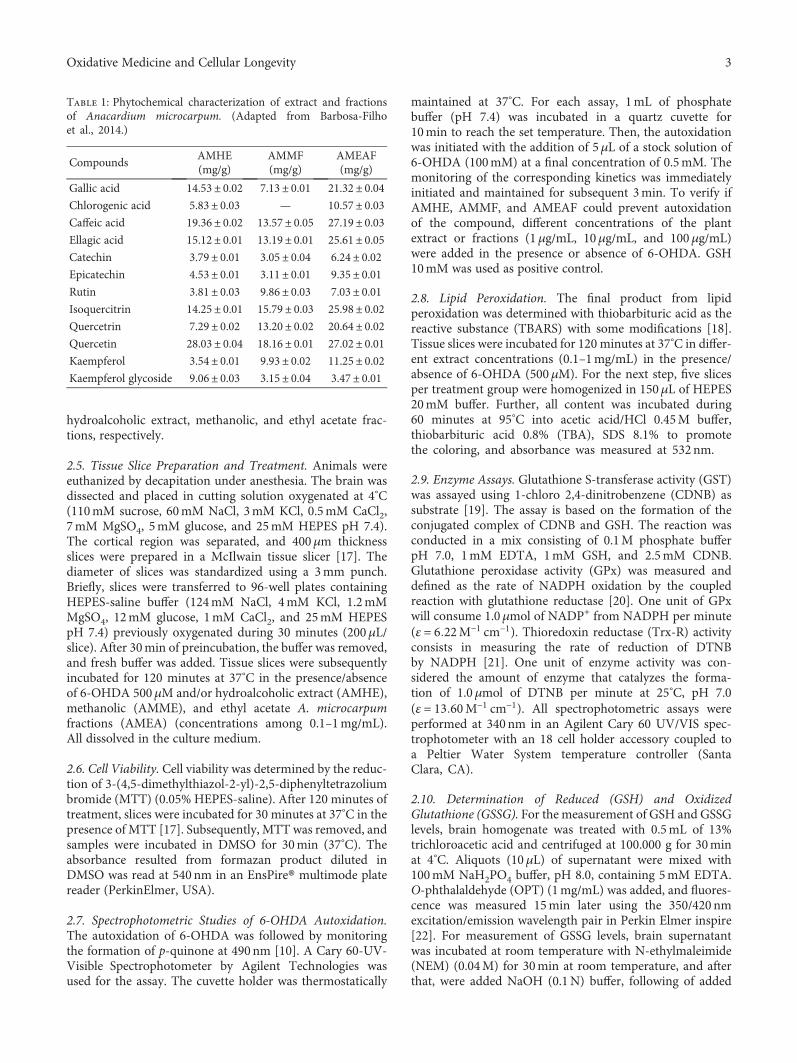
hydroalcoholic extract, methanolic, and ethyl acetate frac-tions, respectively.
2.5. Tissue Slice Preparation and Treatment. Animals wereeuthanized by decapitation under anesthesia. The brain wasdissected and placed in cutting solution oxygenated at 4°C(110mM sucrose, 60mM NaCl, 3mM KCl, 0.5mM CaCl2,7mM MgSO4, 5mM glucose, and 25mM HEPES pH 7.4).The cortical region was separated, and 400μm thicknessslices were prepared in a McIlwain tissue slicer [17]. Thediameter of slices was standardized using a 3mm punch.Briefly, slices were transferred to 96-well plates containingHEPES-saline buffer (124mM NaCl, 4mM KCl, 1.2mMMgSO4, 12mM glucose, 1mM CaCl2, and 25mM HEPESpH 7.4) previously oxygenated during 30 minutes (200μL/slice). After 30min of preincubation, the buffer was removed,and fresh buffer was added. Tissue slices were subsequentlyincubated for 120 minutes at 37°C in the presence/absenceof 6-OHDA 500μM and/or hydroalcoholic extract (AMHE),methanolic (AMME), and ethyl acetate A. microcarpumfractions (AMEA) (concentrations among 0.1–1mg/mL).All dissolved in the culture medium.
2.6. Cell Viability. Cell viability was determined by the reduc-tion of 3-(4,5-dimethylthiazol-2-yl)-2,5-diphenyltetrazoliumbromide (MTT) (0.05% HEPES-saline). After 120 minutes oftreatment, slices were incubated for 30 minutes at 37°C in thepresence of MTT [17]. Subsequently, MTT was removed, andsamples were incubated in DMSO for 30min (37°C). Theabsorbance resulted from formazan product diluted inDMSO was read at 540 nm in an EnsPire® multimode platereader (PerkinElmer, USA).
2.7. Spectrophotometric Studies of 6-OHDA Autoxidation.The autoxidation of 6-OHDA was followed by monitoringthe formation of p-quinone at 490nm [10]. A Cary 60-UV-Visible Spectrophotometer by Agilent Technologies wasused for the assay. The cuvette holder was thermostatically
maintained at 37°C. For each assay, 1mL of phosphatebuffer (pH 7.4) was incubated in a quartz cuvette for10min to reach the set temperature. Then, the autoxidationwas initiated with the addition of 5μL of a stock solution of6-OHDA (100mM) at a final concentration of 0.5mM. Themonitoring of the corresponding kinetics was immediatelyinitiated and maintained for subsequent 3min. To verify ifAMHE, AMMF, and AMEAF could prevent autoxidationof the compound, different concentrations of the plantextract or fractions (1μg/mL, 10μg/mL, and 100μg/mL)were added in the presence or absence of 6-OHDA. GSH10mM was used as positive control.
2.8. Lipid Peroxidation. The final product from lipidperoxidation was determined with thiobarbituric acid as thereactive substance (TBARS) with some modifications [18].Tissue slices were incubated for 120 minutes at 37°C in differ-ent extract concentrations (0.1–1mg/mL) in the presence/absence of 6-OHDA (500μM). For the next step, five slicesper treatment group were homogenized in 150μL of HEPES20mM buffer. Further, all content was incubated during60 minutes at 95°C into acetic acid/HCl 0.45M buffer,thiobarbituric acid 0.8% (TBA), SDS 8.1% to promotethe coloring, and absorbance was measured at 532nm.
2.9. Enzyme Assays. Glutathione S-transferase activity (GST)was assayed using 1-chloro 2,4-dinitrobenzene (CDNB) assubstrate [19]. The assay is based on the formation of theconjugated complex of CDNB and GSH. The reaction wasconducted in a mix consisting of 0.1M phosphate bufferpH 7.0, 1mM EDTA, 1mM GSH, and 2.5mM CDNB.Glutathione peroxidase activity (GPx) was measured anddefined as the rate of NADPH oxidation by the coupledreaction with glutathione reductase [20]. One unit of GPxwill consume 1.0μmol of NADP+ from NADPH per minute(ε = 6 22M−1 cm−1). Thioredoxin reductase (Trx-R) activityconsists in measuring the rate of reduction of DTNBby NADPH [21]. One unit of enzyme activity was con-sidered the amount of enzyme that catalyzes the forma-tion of 1.0μmol of DTNB per minute at 25°C, pH 7.0(ε = 13 60M−1 cm−1). All spectrophotometric assays wereperformed at 340nm in an Agilent Cary 60 UV/VIS spec-trophotometer with an 18 cell holder accessory coupled toa Peltier Water System temperature controller (SantaClara, CA).
2.10. Determination of Reduced (GSH) and OxidizedGlutathione (GSSG). For the measurement of GSH and GSSGlevels, brain homogenate was treated with 0.5mL of 13%trichloroacetic acid and centrifuged at 100.000 g for 30minat 4°C. Aliquots (10μL) of supernatant were mixed with100mM NaH2PO4 buffer, pH 8.0, containing 5mM EDTA.O-phthalaldehyde (OPT) (1mg/mL) was added, and fluores-cence was measured 15min later using the 350/420 nmexcitation/emission wavelength pair in Perkin Elmer inspire[22]. For measurement of GSSG levels, brain supernatantwas incubated at room temperature with N-ethylmaleimide(NEM) (0.04M) for 30min at room temperature, and afterthat, were added NaOH (0.1N) buffer, following of added
Table 1: Phytochemical characterization of extract and fractionsof Anacardium microcarpum. (Adapted from Barbosa-Filhoet al., 2014.)
CompoundsAMHE(mg/g)
AMMF(mg/g)
AMEAF(mg/g)
Gallic acid 14.53± 0.02 7.13± 0.01 21.32± 0.04Chlorogenic acid 5.83± 0.03 — 10.57± 0.03Caffeic acid 19.36± 0.02 13.57± 0.05 27.19± 0.03Ellagic acid 15.12± 0.01 13.19± 0.01 25.61± 0.05Catechin 3.79± 0.01 3.05± 0.04 6.24± 0.02Epicatechin 4.53± 0.01 3.11± 0.01 9.35± 0.01Rutin 3.81± 0.03 9.86± 0.03 7.03± 0.01Isoquercitrin 14.25± 0.01 15.79± 0.03 25.98± 0.02Quercetrin 7.29± 0.02 13.20± 0.02 20.64± 0.02Quercetin 28.03± 0.04 18.16± 0.01 27.02± 0.01Kaempferol 3.54± 0.01 9.93± 0.02 11.25± 0.02Kaempferol glycoside 9.06± 0.03 3.15± 0.04 3.47± 0.01
3Oxidative Medicine and Cellular Longevity

OPT and incubated for 15min in the dark, using the proce-dure outlined above for GSH assay. Results were presentedas the GSH/GSSG ratio.
2.11. High-Resolution Respirometry (HRR) In Vitro. Forrespirometry determination, chick brain (400mg) wasweighed and transferred to 1mL of cold homogenizationbuffer containing 5mM Tris-HCl, 250mM sucrose, and2mM EGTA (pH 7.4), and brain homogenate was used tothe HRR. Oxygraph-2k (O2k, OROBOROS Instruments,Innsbruck, Austria) was employed for all respiration mea-surements. Experiments were performed in 2mL of MiR05buffer (110mM sucrose, 60mMK-lactobionate, 0.5mMEGTA, 3mM MgCl2, 20mM taurine, 10mM KH2PO4,20mM HEPES pH7.4, and 0.1% BSA) [23]. All experimentswere performed at 37°C using DatLab 4.0 software (OroborosInc., Austria), with continuous stirring at 750RPM, and allexperiments started by registering the endogenous substratesupported respiration, following protocols established in theliterature [24].
All experiments of mitochondrial bioenergetics analysisin brain homogenate were performed following [25] withminor modifications at the O2k-chamber. All concentrationsof compounds (control group without treatment, 100μg/mLof a methanolic fraction, 6-OHDA 500μM in the absenceor presence of methanolic fraction) were added at theO2k-chamber after signal stabilization of the basal respira-tion supported by endogenous substrates. Four individualpreparations of the brain homogenate were performedper group.
2.12. Mitochondrial Respiration Assays. Titration protocols ofmultiple substrates and inhibitors were used to assess mito-chondrial function in terms of different respiration states.The routine of electron transport system activities in brainhomogenate was carried out according to literature [26].Malate, glutamate, and succinate were used as oxidizable sub-strates in all experiments. Complex I- (CI-) mediated Leak(LEAK) respiration was determined using 2mM malate and10mM glutamate. CI-mediated OXPHOS (OXPHOS) wasdetermined using ADP (2.5mM). Respiratory control ratios(RCR=CIOXPHOS/CILEAK) were used as a quality controlof isolated mitochondria. The convergent electron flowduring the maximal OXPHOS respiration (CI+CIIOXPHOS)was determined with substrates of CI and CII (10mMSuccinate). CI +CII-mediated ETS (electron transfer system)(CI +CIIETS) was determined using Carbonyl cyanide-4-(tri-fluoromethoxy) phenylhydrazone (FCCP) (optimum con-centration reached between 0.5 and 1.5μM). CII-mediatedETS respiration (CIIETS) was determined with 0.5μM rote-none. Addition of 2.5μM antimycin A inhibited complexIII, resulting in nonmitochondrial respiration (ROX) withsmall contributions from electron leak in the uncoupled state.
2.13. Western Blotting Analysis. Analysis of protein phos-phorylation in cortical slices was performed using westernblotting with slight modification [27]. Four slices werehomogenized in 100μL of 4% SDS stop solution (4% SDS,50mM Tris, 100mM EDTA, pH 6.8), and 10μL of sample
was taken out for protein analysis. In the remaining samplewas added 25% Glycerol sample and 8% β-mercaptoethanol.The proteins were separated by SDS–PAGE using 10% gelsand then electrotransferred to nitrocellulose membranes.The membranes were washed in Tris-buffered saline withTween-20 (100mM Tris–HCl, pH 7.5, 0.9% NaCl, and0.1% Tween-20) and incubated overnight at 4°C with pri-mary antibodies anti-phospho p38, anti-total and phospho-ERK1/2, anti-total and phospho-JNK1/2, anti-phospho andtotal-AKT, anti-phospho PTEN. Subsequently, membraneswere washed in Tris-buffered saline with Tween-20 and incu-bated for 1 hour at 25°C with horseradish peroxidase-linkedanti-IgG secondary specific antibodies. The blottings werevisualized on the IS4000MM Pro Bruker imaging systemusing ECL-detection reagent, and the band density was quan-tified using the Scion Image® software. The density of thebands was measured and expressed as a rate (%) of increasein relation to control (slices treated only with media).
2.14. PI3K/AKT and MEK Inhibitors. To determine theimplication of signaling pathway involved in cell survival inthe neuroprotective effect of A. microcarpum methanolicfraction, PI3K/AKT inhibitor LY294002 at final concentra-tion of 20μM and MEK/ERK inhibitor PD98059 at finalconcentration of 50μM. PD98059 acts on inhibition ofMEK1 in a reversible, allosteric, and noncompetitive mannerwith respect to ATP and ERK1/2 binding whereas LY294002acts at an ATP-binding site of PI3K enzyme, thus selectivelyinhibiting the PI3K-Akt interaction. The inhibitors wereadded to the medium 30min prior to the addition of 6-OHDA plus AMMF. After 2 hours of treatment, MTT assaywas conducted to verify the slices viability as described above.Inhibitors were diluted in DMSO; the final concentration ofDMSO in the wells was 0.5%.
2.15. Protein Quantification. The protein concentration insamples was estimated using BSA as standard [28, 29].
2.16. Statistical Analysis. All data were tested for normaldistribution by Kolmogorov-Smirnov. Statistical analysiswas performed using one-way analysis of variance (ANOVA)followed by Newman-Keuls post hoc analysis. Results wereconsidered statistically significant when p < 0 05.
3. Results
3.1. Analysis of 6-Hydroxydopamine Autoxidation in thePresence of Extract and Fractions of A. microcarpum.6-OHDA undergoes spontaneous autoxidation in the pres-ence of oxygen under physiological conditions forminghydrogen peroxide (H2O2) and the corresponding p-quinonewhose formation can be monitored spectrophotometricallyat 490nm [10]. The absorbance of 6-OHDA at the end of3min of incubation with phosphate buffer is visualized inthe graph (Figures 1(a)–1(c)). The presence of fractions ofA. microcarpum did not alter the 6-OHDA absorbance,whereas hydroalcoholic fraction decreased it partially. Theantioxidant glutathione (GSH) was used as a positive controlpreventing 6-OHDA autoxidation due to the ability of
4 Oxidative Medicine and Cellular Longevity

sulfhydryl compounds to remove the H2O2 formed duringthe autoxidation reaction of 6-OHDA.
3.2. Evaluation of Toxicity and Neuroprotective Potential ofA. microcarpum Hydroalcoholic Extract and Fractions. Inorder to investigate a possible neurotoxic effect of A. micro-carpum, cortical slices were incubated for 2 hours with differ-ent concentrations of hydroalcoholic extract and fractions: 0,1μg/mL, 10μg/mL, 100μg/mL, and 1000μg/mL. At the endof incubation period, cell viability assay was performed byMTT test. Our data showed that A. microcarpum per se wasunable to affect the viability of slices (Figures 2(a)–2(c)).
To investigate the neuroprotective potential of A. micro-carpum, slices were incubated with neurotoxin 6-OHDA500μM for 2 hours in the presence or absence of differentextracts or fraction concentration (1–100μg/mL). 6-OHDAconcentration was defined in a dose-response curve, andthe concentration able to decrease in approximately 30%the cell viability was chosen for further studies. The hydroal-coholic extract was unable to protect against damage causedby 6-OHDA (Figure 2(d)); however, AMMF and AMEAFreverted the drop in cell viability promoted by 6-OHDA ata concentration of 100μg/mL (p < 0 0001) (Figures 2(e) and2(f), respectively).
3.3. Lipid Peroxidation in Response to the Treatment withMethanolic and Acetate Fractions of A. microcarpum and6-OHDA. Oxidative stress is implied in dopaminergic cell
death induced by 6-OHDA [11]. AMMF and AMEAF butnot AMHE presented neuroprotective potential in the MTTassay; thus, it was investigated a possible antioxidant poten-tial of these fraction on slices exposed to 6-OHDA by itsability to prevent lipid peroxidation. 6-OHDA induced lipidperoxidation in 25% (p < 0 0001) when compared to controlgroup. Only AMMF prevented this effect (Figures 3(a) and3(b)); thus, further studies were conducted in the presenceof methanolic fraction.
3.4. Analysis of ERK, AKT, PTEN Phosphorylation, and PARPCleavage in Response to the Treatment with 6-OHDA andMethanolic Fraction of A. microcarpum. In this study, theeffect of 6-OHDA on phosphorylation of proteins p38,JNK1/2, ERK1/2, AKT, cleavage of PARP protein, andphosphatase PTEN was analyzed by western blotting tech-nique. No alterations in phosphorylation and total levels ofp38 and JNK1/2 were detected (data not shown). Thephosphorylation of ERK was increased in 30% only in thepresence of extract and 6-OHDA (Figure 4(b)); no alterationswere observed in the other groups. AKT phosphorylation(Figure 4(c)) was inhibited in approximately 25% by 6-OHDA treatment and remained at control level whenfraction was present. The cleavage of PARP protein in an89 kDa fragment was evaluated as an indicator of apoptoticcell death. No alteration in PARP cleavage was visualizedby treatments as observed in the blotting. PTEN phosphory-lation was unchanged by the treatments (Figure 4(d)).
0.00
0.02
0.04
0.06
0.08
0.10
6-OHDA − + + + + (500 �휇M)
GSH + − − − − (10 mM)AMHE − − 1 10 100 (�휇g/mL)
Auto
xida
tion
6-O
HD
A(4
90 n
m ab
sorb
ance
)
⁎⁎⁎
⁎⁎
(a)
6-OHDA − + + + + (500 �휇M)
GSH + − − − − (10 mM)AMHE − − 1 10 100 (�휇g/mL)
Auto
xida
tion
6-O
HD
A(4
90 n
m ab
sorb
ance
)
0.00
0.05
0.10
0.15
⁎⁎
(b)
6-OHDA − + + + + (500 �휇M)
GSH + − − − − (10 mM)AMHE − − 1 10 100 (�휇g/mL)
Auto
xida
tion
6-O
HD
A(4
90 n
m ab
sorb
ance
)
0.00
0.05
0.10
0.15
0.20
⁎
(c)
Figure 1: 6-OHDA autoxidation in the presence of A. microcarpum and GSH. The autoxidation of 6-OHDA (500 μM) wasfollowed spectrophotometrically by monitoring the formation of p-quinone at 490 nm in the presence or absence of extract orfractions (a) 6-OHDA+AMHE, (b) 6-OHDA+AMMF, and (c) 6-OHDA+AMEAF. GSH 10mM was used as positive control preventing6-OHDA autoxidation. Data are expressed as percentage of the untreated control± SE (n = 3). ∗∗∗p < 0 0001 as compared to GSH control.##p < 0 001 and ###p < 0 0001 as compared to 6-OHDA group.
5Oxidative Medicine and Cellular Longevity

Ctl 1 10 100 10000
50
100
150
AMHE(�휇g/mL)
Slic
es v
iabi
lity
(% o
f con
trol)
(a)
Ctl 1 10 100 1000 (�휇g/mL)0
50
100
150
Slic
es v
iabi
lity
(% o
f con
trol)
AMMF
(b)
Ctl 1 10 100 1000 (�휇g/mL)0
50
100
150
Slic
es v
iabi
lity
(% o
f con
trol)
AMEAF
(c)
0
50
100
150
Slic
es v
iabi
lity
(% o
f con
trol)
6-OHDA − + + + + (500 �휇M)AMHE − − 1 10 100 (�휇g/mL)
⁎⁎⁎
(d)
Slic
es v
iabi
lity
(% o
f con
trol)
0
50
100
150
6-OHDA − + + + + (500 �휇M)AMMF − − 1 10 100 (�휇g/mL)
⁎⁎⁎
(e)
Slic
es v
iabi
lity
(% o
f con
trol)
0
50
100
150
6-OHDA − + + + + (500 �휇M)AMEAF − − 1 10 100 (�휇g/mL)
⁎⁎⁎⁎⁎ ⁎
(f)
Figure 2: Effects of A. microcarpum and 6-OHDA on the viability of cortical slices. Cortical slices were incubated for 2 h in differentconcentrations (1–1000 μg/mL) of (a) AMHE, (b) AMMF, and (c) AMEAF and in the presence or absence of 6-OHDA (500 μM) during2 h, (d) AMHE, (e) AMMF, and (f) AMEAF. Cell viability was measured by MTT test. Data are expressed as percentage of the untreatedcontrol± SEM (n = 3). ∗p < 0 05, ∗∗p < 0 001, and ∗∗∗p < 0 0001 different from control group.
0
50
100
150
6-OHDA − + − + (500 �휇M)AMMF − − + + (100 �휇g/mL)
⁎⁎⁎
##
Lipi
d pe
roxi
datio
n(%
of c
ontro
l)
(a)
0
50
100
150
6-OHDA − + − + (500 �휇M)AMEAF − − + + (100 �휇g/mL)
⁎⁎⁎⁎⁎⁎
Lipi
d pe
roxi
datio
n(%
of c
ontro
l)
(b)
Figure 3: Effects of A. microcarpum on 6-OHDA (500 μM) induced lipid peroxidation (LPO) in cortical slices. Cortical slices were incubatedwith (a) AMMF and (b) AMEAF in the presence/absence of 6-OHDA for two hours, and lipid peroxidation was evaluated by formation ofTBARS at 532 nm. Data are expressed as percentage of the untreated control± SEM (n = 3). ∗∗∗p < 0 0001 as compared to control.##p < 0 001 as compared to 6-OHDA group.
6 Oxidative Medicine and Cellular Longevity
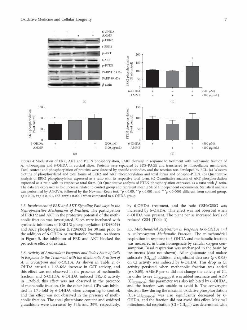
3.5. Involvement of ERK and AKT Signaling Pathways in theNeuroprotective Mechanisms of Fraction. The participationof ERK1/2 and AKT in the protective potential of the meth-anolic fraction was investigated. Slices were incubated withsynthetic inhibitors of ERK1/2 phosphorylation (PD98059)and AKT phosphorylation (LY294002) for 30min prior tothe addition of 6-OHDA or methanolic fraction. As shownin Figure 5, the inhibition of ERK and AKT blocked theprotective effects of extract.
3.6. Activity of Antioxidant Enzymes and Redox State of Cellsin Response to the Treatment with the Methanolic Fraction ofA. microcarpum and 6-OHDA. As shown in Table 2, 6-OHDA caused a 1.68-fold increase in GST activity, andthis effect was not observed in the presence of methanolicfraction and 6-OHDA. 6-OHDA induced TRx-R activityin 1.9-fold; this effect was not observed in the presenceof methanolic fraction. On the other hand, GPx was inhib-ited in 1.71-fold by 6-OHDA when comparing to control,and this effect was not observed in the presence of meth-anolic fraction. The total glutathione content and oxidizedglutathione were decreased by 34% and 39%, respectively,
by 6-OHDA treatment, and the ratio GSH/GSSG wasincreased by 6-OHDA. This effect was not observed when6-OHDA was present. The plant per se increased levels ofreduced GSH (Table 3).
3.7. Mitochondrial Respiration in Response to 6-OHDA andA. microcarpum Methanolic Fraction. The mitochondrialrespiration in response to 6-OHDA and methanolic fractionwas measured in brain homogenate by cellular oxygen con-sumption. Basal respiration was unchanged in the brain bytreatments (data not shown). After glutamate and malatesubstrate (CILeak) addition, a significant decrease (p < 0 05)on CI activity was induced by 6-OHDA. This drop in CIactivity persisted when methanolic fraction was added(p < 0 05). AMMF per se did not change the activity of CI.In order to see CIOXPHOS, it was added succinate and ADP(CIOXPHOS); this parameter was also inhibited by 6-OHDA,and the fraction was unable to avoid it. The convergentelectron flow during the maximal oxidative phosphorylation(CI +CIIOXPHOS) was also significantly decreased by 6-OHDA, and the fraction did not avoid this effect. Maximalmitochondrial respiration (CI +CIIETS) was determined with
6-OHDA++ −−++−−
p-ERK2
t-ERK2
p-AKT
t-AKTp-PTEN
PARP 116 kDa
PARP 89 kDa
�훽-actin
AMMF
(a)
0
50
100
150
200
ERK2
pho
spho
ryla
tion
(% o
f con
trol)
⁎
6-OHDA + − + (500 �휇M)AMMF − + + (100 �휇g/mL)
(b)
6-OHDA + − + (500 �휇M)AMMF − + + (100 �휇g/mL)
⁎
AKT
pho
spho
ryla
tion
(% o
f con
trol)
0
50
100
150
(c)
PTEN
pho
spho
ryla
tion
(% o
f con
trol)
6-OHDA + − + (500 �휇M)AMMF − + + (100 �휇g/mL)
0
50
100
150
(d)
Figure 4: Modulation of ERK, AKT and PTEN phosphorylation, PARP cleavage in response to treatment with methanolic fraction ofA. microcarpum and 6-OHDA in cortical slices. Proteins were separated by SDS–PAGE and transferred to nitrocellulose membrane.Total content and phosphorylation of proteins were detected by specific antibodies, and the reaction was developed by ECL. (a) Westernblotting of phosphorylated and total forms of ERK2 and AKT phosphorylation and total forms and phospho-PTEN. (b) Quantitativeanalysis of ERK2 phosphorylation expressed as a ratio with its respective total form. (c) Quantitative analysis of AKT phosphorylationexpressed as a ratio with its respective total form. (d) Quantitative analysis of PTEN phosphorylation expressed as a ratio with β-actin.The data are expressed as fold increase related to control group and represent mean± SE of 4 independent experiments. Statistical analysiswas performed by ANOVA, followed by the Newman-Keuls test. ∗p < 0 05, ∗∗p < 0 001, and ∗∗∗p < 0 0001 different from control group.#p < 0 05, ##p < 0 001, and ###p < 0 0001 when compared to 6-OHDA group.
7Oxidative Medicine and Cellular Longevity

0
50
100
150
### ###
6-OHDA − − + − + + (500 �휇M)AMMF − − − + + + (100 �휇g/mL)
LY 294002 − + − − − + (50 �휇M)
Slic
es v
iabi
lity
(% o
f con
trol
) ++
⁎⁎⁎ ⁎⁎⁎
(a)
0
50
100
150
6-OHDA − − + − + + (500 �휇M)AMMF − − − + + + (100 �휇g/mL)
PD 98059 − + − − − + (50 �휇M)
Slic
es v
iabi
lity
(% o
f con
trol
)
###⁎⁎⁎
#++
⁎⁎⁎
(b)
Figure 5: Effects of A. microcarpum and 6-OHDA on the viability of cortical slices in the presence of LY (294002) inhibitors and PD (98059).Slices were incubated with AMMF in the presence or absence of 6-OHDA (500 μM) and inhibitors for 2 h. The inhibitors were added 30minprior to the addition of fraction and 6-OHDA and remained during all period of the treatment. Data are expressed as percentage of theuntreated control± SE (n = 3). ∗∗∗p < 0 0001 as compared to control. ###p < 0 0001 as compared to only inhibitor treated group;++p < 0 001 as compared to cotreated group 6-OHDA+AMMF.
Table 2: Activity of antioxidant enzymes in cortical slices submitted to treatment with the neurotoxin 6-OHDA and A. microcarpummethanolic fraction.
GST(mg/mU protein)
GPx(mg/mU protein)
TrxR(mg/mU protein)
Control 241.2± 8.49 41.37± 5.01 2.375± 0.3426-OHDA 500μM 406.4± 75.89∗ 23.82± 2.07∗ 4.675± 0.608∗
AMMF 100μg/mL 186.2± 42.13# 51.01± 4.04 2.459± 0.137##
AMMF 100μg/mL+ 6-OHDA 500 μM 251.5± 36.48# 46.29± 3.20## 3.164± 0.496#
Data are expressed as percentage of the untreated control± SEM. ∗p < 0 05 in relation to control group, #p < 0 05 in relation to control group, ##p < 0 001 inrelation to 6-OHDA group.
Table 3: Effect of treatment with 6-OHDA and A. microcarpum on GSH and GSSG levels and ratio GSH/GSSG.
GSH(% of control)
GSSG(% of control)
Total glutathione(% of control)
GSH/GSSG(% of control)
Control 88.05± 6.73 90.60± 5.48 100.0± 10.66 100.0± 2.516-OHDA 500μM 87.49± 5.41 61.61± 12.87∗ 66.56± 11.34∗ 168.1± 19.64∗∗
AMMF 100μg/mL 136.7± 6.54∗∗ 117.2± 10.04## 129.7± 4.18## 120.7± 9.21##
AMMF 100μg/mL+ 6-OHDA 500 μM 111.4± 13.28 111.9± 3.17## 111.8± 4.96# 99.61± 8.99##
Data are expressed as percentage of the untreated control ± SEM. ∗p < 0 05; ∗∗p < 0 001 in relation to control group. ##p < 0 001 when compared to6-OHDA group.
8 Oxidative Medicine and Cellular Longevity

the addition of the uncoupler FCCP. This parameter wasinhibited by 6-OHDA and not avoided by the fraction(p < 0 001). CIIETS activity was analyzed after inhibitionof CI by rotenone, and no significant changes occurredby treatments. AMMF per se but not in the presence of6-OHDA induced CILEAK, CIOXPHOS, CI +CIIOXPHOS, andCI+ IIETS (Figure 6).
4. Discussion
Oxidative stress, mitochondrial dysfunction, genetic, andenvironmental factors are mechanisms associated with neu-ronal damage observed in PD [30–32]. A number of studieshave proposed antioxidant therapies to attenuate PD symp-toms [33, 34]. In this study, methanolic and acetate fractionbut not hydroalcoholic extract protected against neurotoxic-ity induced by 6-OHDA in brain slices, but methanolicfraction was more effective in protecting against inductionin lipid peroxidation by 6-OHDA.
The cytotoxic effect of 6-OHDA is attributed to theformation of reactive species such as superoxide radical,para-quinone, and hydrogen peroxide from enzymatic andautoxidation reactions [35]. It this study, plant extract orfractions did not block effectively the autoxidation of 6-OHDA in vitro. With the base in this finding, it can beinferred that the mechanism implied in the protective effectof the plant is the neutralization of reactive species secondaryto the autoxidation reaction and not a direct interaction with6-OHDA molecule. In previous studies, 6-OHDA inducedmitochondrial dysfunction by inhibition of I and IV com-plexes, disrupting the mitochondrial function, and producingsuperoxide anion, which in turn may form hydroxyl radi-cals which reacts with nitric oxide generating peroxynitrite[36–39]. In the present study, the 6-OHDA inhibited in55% complex I activity in basal respiration around 75%
the ATP production CI-dependent and compromise themitochondrial electron transport system in 50%. This isin accordance with a similar work that showed a 20% ofinhibition of complex I at a concentration of 100μM,approximately [40]. This phenomenon was not blocked byA. microcarpum. Considering the inability of the plant tohamper the mitochondrial damage caused by 6-OHDA, itcould be supposed that the neuroprotective effect by the plantis due to the neutralization of reactive species resulted frommitochondrial dysfunction.
Reactive oxygen species production, detoxification, andsignaling pathways have been considered interesting targetsfor intervention in neurodegenerative diseases [41, 42].Endogenous enzymatic and nonenzymatic antioxidants, suchas GSH, glutathione S-transferase, glutathione peroxidase,and thioredoxin reductase (TRx-R), delay or prevent oxida-tive damage to proteins, lipids, and DNA [43, 44]. Glutathi-one peroxidase (GPx) is an intracellular antioxidant thatreduces hydrogen peroxide to water at expenses of GSHand limits its harmful effect. In this study, 6-OHDA causeda substantial inhibition in GPx activity that was not observedin presence of methanolic fraction. Similar data were demon-strated in neuroblastoma cells treated with 6-OHDA [45]. InPD patients, the degree of symptom severity correlates withintracellular GSH loss in substantia nigra [46]. In our study,GSH content was not altered, and GSSG was decreased by6-OHDA; this effect may be related to GPx inhibition andconsequently a lower oxidation of GSH by this system. Onthe other hand, the TRx-R activity was stimulated by 6-OHDA, and the same data were observed in neuroblastomacell line SH-SY5Y [47]. Our results suggest the participationof peroxiredoxins catalyzing peroxide reduction as a com-pensatory mechanism to replace the inhibited activity ofGPx. Herein, 6-OHDA increased the activity of GST, whichis implied in neuronal detoxification of quinones resulted
CILeak CIOxphos CI + CIIOxphos CI + CIIETS CIIETS
0
1000
2000
3000
4000
Control
6-OHDA 500 �휇M
AMMF 100 �휇g/mL
AMMF 100 �휇g/mL + 6-OHDA 500 �휇M
###
###
⁎
###
⁎
##
Oxy
gen
flux
(pm
ol O
2s−
1 mg−
1 )
⁎⁎
⁎⁎⁎⁎⁎⁎
⁎⁎
⁎⁎⁎⁎
⁎⁎
⁎⁎⁎⁎
Figure 6: O2 flux measured in the mitochondria of cortex homogenate exposed to 6-OHDA and/or A. microcarpum.Mitochondrial functionis presented with the abbreviation(s) of the complex(es) involved followed by the state of respiration measured in the presence ofglutamate +malate (CILEAK), +ADP (CIOXPHOS), +succinate (CIIOXPHOS), +FCCP (CI + IIETS), +rotenone (CIIETS). Antimycin A was usedto correct for residual O2 consumption. Results are means± SEM for 4 different preparations. ∗p < 0 05, ∗∗p < 0 001, and ∗∗∗p < 0 0001different from control group. ##p < 0 001 and ###p < 0 0001 when compared to 6-OHDA group.
9Oxidative Medicine and Cellular Longevity
from catecholamine oxidation and free radicals [48], and thiseffect was not observed when plant was present. All theseeffects were prevented by the methanolic fraction of plant,showing a protective mechanism against oxidative stressinduced by the 6-OHDA. Due to methodological issues, wewere not able to detect quantifiable levels of the enzymesSuperoxide Dismutase and Catalase, but their analysis willbe considered in further studies.
Extracellular signal-regulated kinases (ERKs) have beenimplicated in the cellular response to reactive oxygen species[49–52]. Growth factors and other extracellular stimuliactivate the kinase MEK1/2 by Ras/Raf pathway; MEK1/2then phosphorylates and activates ERK1/2 [53]. ERK1/2activates transcription factors such as cAMP response ele-ment–binding protein (CREB) and Elk, thereby increasingtranscription of neurotrophic factors and prosurvival genessuch as Bcl-2 [54]. In this study, the use of MEK1/2 inhibitorweakened the protective potential of methanolic fractionagainst the 6-OHDA, suggesting that the antioxidant poten-tial of fraction per se is not enough to protect the brainsslices, but the activation of prosurvival factors plays animportant role in this effect. Experiments of western blottingshowed that 6-OHDA did not alter ERK phosphorylation inbrain slices after two hours of incubation with 6-OHDA.Previous studies reported an ERK1/2 phosphorylation peakafter 10–15min of exposure of dopaminergic cells to 6-OHDA; the phosphorylation of prosurvival protein CREBfollowed this temporal profile as well [54]. Authors showedthat the inhibition of early phosphorylation of ERK1/2 abol-ished CREB activation and increased 6-OHDA toxicity.Thus, the possibility of an early activation of ERK1/2 in slicessubmitted to the treatment and the contribution of thisactivation for a self-protective response of cells that wasprevented with use of inhibitors could not be discarded.
AKT is a serine/threonine kinase and its signalingpathway plays an important role in fundamental cellularfunctions, such as cell proliferation and survival, by phos-phorylating a variety of enzymes, including proapoptoticregulators, antioxidant proteins, and transcription factors[55]. It is reported that AKT phosphorylation is reduced inthe striatum of patients with PD, suggesting that its inactiva-tion has an important role in PD [56]; being so, it is a sub-stantial therapeutic target for treating neurodegenerativediseases, beyond other pathologies [57]. Herein, 6-OHDAinhibited phosphorylation level of AKT, and the plantprevented this effect. There was no alteration in phosphory-lation level of PTEN (Phosphatase and tensin homologdeleted on chromosome ten), a negative regulator of AKT.Similar results were reported in SH-SY5Y and dopaminergiccell lines [58, 59]. In this study, AKT phosphorylation seemsto display a role in neuroprotective effect of fraction against6-OHDA, once the use of PI3K/AKT inhibitor blocked theprotective effect of fraction. This result proposes that survivalsignaling pathways ERK and AKT contribute for neuropro-tection by methanolic fraction of A. microcarpum.
Recent studies have suggested that several signaltransduction pathways, including phosphatidylinositol 3kinase (PI3K) pathways and MAPKs, are involved in releas-ing transcription factor Nrf2 from the complex Keap1-Nrf2
promoting Nfr2 translocation to the nucleus [60]. Nrf2promotes transcriptional activation of a variety of antioxi-dant genes [61]. AMMF-mediated cytoprotection against 6-OHDA was abolished by ERK and AKT pathway inhibitors;these data support a possible involvement of Nrf2 activationleading to expression of downstream antioxidant genesthrough the modulation of AKT and ERK1/2 pathways bythe fraction. According to another study, inhibition of AKTand ERK has also abolished the neuroprotective effect of atriterpenoid isolated from plant [62].
5. Conclusion
The present work shows for the first time the potential ofthe Brazilian plant A. microcarpum in protecting against6-OHDA-induced damage in brain slices. Inhibition ofmitochondrial complexes by 6-OHDA was not avoided bythe extract. ERK and AKT phosphorylation display a rolein the neuroprotective effect by the plant which wasdecreased by the presence of pharmacological inhibitors ofthose pathways.
Data Availability
The data used to support the findings of this study areavailable from the corresponding author upon request.
Conflicts of Interest
The authors declare that they have no conflicts of interest.
Acknowledgments
This study was supported by CNPq process #456207/2014-7,FAPERGS process # 2380-2551/14-8, and FAPERGS/PRONEX #16/25510000499-4. JLF is a recipient of CNPqfellowship. IKM, GEM and NRR are recipients of CAPESdoctoral scholarship.
References
[1] V. M. B. Filho, E. P. Waczuk, J. P. Kamdem et al., “Phytochem-ical constituents, antioxidant activity, cytotoxicity and osmoticfragility effects of Caju (Anacardium microcarpum),” Indus-trial Crops and Products, vol. 55, pp. 280–288, 2014.
[2] H. Coutinho, V. Barbosa-Filho, E. Waczuk et al., “Phytocom-pounds and modulatory effects of Anacardium microcarpum(cajui) on antibiotic drugs used in clinical infections,” DrugDesign, Development and Therapy, vol. 9, pp. 5965–5972, 2015.
[3] T. Fritsch, K. A. Smyth, M. S. Wallendal, T. Hyde, G. Leo, andD. S. Geldmacher, “Parkinson disease: research update andclinical management,” Southern Medical Journal, vol. 105,no. 12, pp. 650–656, 2012.
[4] R. Balestrino and P. Martinez-Martin, “Neuropsychiatricsymptoms, behavioural disorders, and quality of life inParkinson’s disease,” Journal of the Neurological Sciences,vol. 373, pp. 173–178, 2017.
[5] P. P. Michel, E. C. Hirsch, and S. Hunot, “Understandingdopaminergic cell death pathways in Parkinson disease,”Neuron, vol. 90, no. 4, pp. 675–691, 2016.
10 Oxidative Medicine and Cellular Longevity

[6] S. Fahn and D. Sulzer, “Neurodegeneration and neuroprotec-tion in Parkinson disease,” NeuroRx, vol. 1, no. 1, pp. 139–154, 2004.
[7] P. Bhattarai, A. K. Thomas, Y. Zhang, and C. Kizil, “The effectsof aging on amyloid-β42-induced neurodegeneration andregeneration in adult zebrafish brain,” Neurogenesis, vol. 4,no. 1, article e1322666, 2017.
[8] D. Kempuraj, R. Thangavel, P. A. Natteru et al., “Neuroinflam-mation induces neurodegeneration,” Journal of Neurology,Neurosurgery and Spine, vol. 1, no. 1, 2016.
[9] J. Bové and C. Perier, “Neurotoxin-based models of Parkin-son’s disease,” Neuroscience, vol. 211, pp. 51–76, 2012.
[10] R. Soto-Otero, E. Méndez-Álvarez, Á. Hermida-Ameijeiras,A. M. Muñoz-Patiño, and J. L. Labandeira-Garcia, “Autoxida-tion and neurotoxicity of 6-hydroxydopamine in the presenceof some antioxidants: potential implication in relation tothe pathogenesis of Parkinson’s disease,” Journal of Neuro-chemistry, vol. 74, no. 4, pp. 1605–1612, 2000.
[11] M. Shichiri, “The role of lipid peroxidation in neurologicaldisorders,” Journal of Clinical Biochemistry and Nutrition,vol. 54, no. 3, pp. 151–160, 2014.
[12] D. Blum, S. Torch, N. Lambeng et al., “Molecular pathwaysinvolved in the neurotoxicity of 6-OHDA, dopamine andMPTP: contribution to the apoptotic theory in Parkinson’sdisease,” Progress in Neurobiology, vol. 65, no. 2, pp. 135–172, 2001.
[13] K. Komolafe, T. M. Olaleye, R. L. Seeger et al., “Parkiabiglobosa improves mitochondrial functioning and protectsagainst neurotoxic agents in rat brain hippocampal slices,”BioMed Research International, vol. 2014, Article ID 326290,15 pages, 2014.
[14] K. Gao, M. Liu, J. Cao et al., “Protective effects of Lyciumbarbarum polysaccharide on 6-OHDA-induced apoptosis inPC12 cells through the ROS-NO pathway,” Molecules,vol. 20, no. 1, pp. 293–308, 2015.
[15] A. I. Abushouk, A. Negida, H. Ahmed, and M. M. Abdel-Daim, “Neuroprotective mechanisms of plant extracts againstMPTP induced neurotoxicity: future applications in Parkin-son’s disease,” Biomedicine & Pharmacotherapy, vol. 85,pp. 635–645, 2017.
[16] R. Rodnight, C. A. Gonçalves, R. Leal, E. Rocha, C. G. Salbego,and S. T. Wofchuk, “Chapter 11: regional distribution andproperties of an enzyme system in rat brain that phosphory-lates ppH-47, an insoluble protein highly labelled in tissueslices from the hippocampus,” Progress in Brain Research,vol. 89, pp. 157–167, 1991.
[17] T. Posser, J. L. Franco, D. A. dos Santos et al., “Diphenyldiselenide confers neuroprotection against hydrogen peroxidetoxicity in hippocampal slices,” Brain Research, vol. 1199,pp. 138–147, 2008.
[18] H. Ohkawa, N. Ohishi, and K. Yagi, “Assay for lipid peroxidesin animal tissues by thiobarbituric acid reaction,” AnalyticalBiochemistry, vol. 95, no. 2, pp. 351–358, 1979.
[19] W. H. Habig andW. B. Jakoby, “[51] Assays for differentiationof glutathione S-transferases,”Methods in Enzymology, vol. 77,pp. 398–405, 1981.
[20] A. Wendel, “[44] Glutathione peroxidase,” Methods inEnzymology, vol. 77, pp. 325–333, 1981.
[21] A. Holmgren and M. Björnstedt, “Thioredoxin and thiore-doxin reductase,” Methods in Enzymology, vol. 252, pp. 199–208, 1995.
[22] P. J. Hissin and R. Hilf, “A fluorometric method for determina-tion of oxidized and reduced glutathione in tissues,” AnalyticalBiochemistry, vol. 74, no. 1, pp. 214–226, 1976.
[23] E. Gnaiger, “Capacity of oxidative phosphorylation in humanskeletal muscle: new perspectives of mitochondrial physiol-ogy,” The International Journal of Biochemistry & Cell Biology,vol. 41, no. 10, pp. 1837–1845, 2009.
[24] A. V. Kuznetsov, S. Schneeberger, R. Seiler et al., “Mitochon-drial defects and heterogeneous cytochrome c release aftercardiac cold ischemia and reperfusion,” American Journal ofPhysiology-Heart and Circulatory Physiology, vol. 286, no. 5,pp. H1633–H1641, 2004.
[25] N. Pichaud, M. Messmer, C. C. Correa, and J. W. O. Ballard,“Diet influences the intake target and mitochondrial functionsof Drosophila melanogaster males,” Mitochondrion, vol. 13,no. 6, pp. 817–822, 2013.
[26] D. Pesta and E. Gnaiger, “High-resolution respirometry:OXPHOS protocols for human cells and permeabilized fibersfrom small biopsies of human muscle,” Methods in MolecularBiology, vol. 810, pp. 25–58, 2012.
[27] T. Posser, M. T. De Paula, J. L. Franco, R. B. Leal, and J. B. T.Da Rocha, “Diphenyl diselenide induces apoptotic cell deathand modulates ERK1/2 phosphorylation in human neuroblas-toma SH-SY5Y cells,” Archives of Toxicology, vol. 85, no. 6,pp. 645–651, 2011.
[28] M. M. Bradford, “A rapid and sensitive method for thequantitation of microgram quantities of protein utilizing theprinciple of protein-dye binding,” Analytical Biochemistry,vol. 72, no. 1-2, pp. 248–254, 1976.
[29] G. L. Peterson, “A simplification of the protein assay methodof Lowry et al. which is more generally applicable,” AnalyticalBiochemistry, vol. 83, no. 2, pp. 346–356, 1977.
[30] V. Bellou, L. Belbasis, I. Tzoulaki, E. Evangelou, and J. P. A.Ioannidis, “Environmental risk factors and Parkinson’s dis-ease: an umbrella review of meta-analyses,” Parkinsonism &Related Disorders, vol. 23, pp. 1–9, 2016.
[31] S. D. Ugarte, E. Lin, E. Klann, M. J. Zigmond, and R. G. Perez,“Effects of GDNF on 6-OHDA-induced death in a dopami-nergic cell line: modulation by inhibitors of PI3 kinase andMEK,” Journal of Neuroscience Research, vol. 73, no. 1,pp. 105–112, 2003.
[32] B. Uttara, A. Singh, P. Zamboni, and R. Mahajan, “Oxidativestress and neurodegenerative diseases: a review of upstreamand downstream antioxidant therapeutic options,” CurrentNeuropharmacology, vol. 7, no. 1, pp. 65–74, 2009.
[33] M. Solayman, M. Islam, F. Alam, M. Khalil, M. Kamal, andS. Gan, “Natural products combating neurodegeneration:Parkinson’s disease,” Current Drug Metabolism, vol. 18,no. 1, pp. 50–61, 2017.
[34] Q. Zhao, J. Ye, N. Wei, C. Fong, and X. Dong, “Protectionagainst MPP+-induced neurotoxicity in SH-SY5Y cells by tor-mentic acid via the activation of PI3-K/Akt/GSK3β pathway,”Neurochemistry International, vol. 97, pp. 117–123, 2016.
[35] Y. Glinka, M. Gassen, and M. B. H. Youdim, “Mechanismof 6-hydroxydopamine neurotoxicity,” Journal of NeuralTransmission. Supplementum, vol. 50, pp. 55–66, 1997.
[36] Y. Y. Glinka and M. B. H. Youdim, “Inhibition of mitochon-drial complexes I and IV by 6-hydroxydopamine,” EuropeanJournal of Pharmacology, vol. 292, no. 3-4, pp. 329–332, 1995.
[37] G. C. Brown and V. Borutaite, “Inhibition of mitochondrialrespiratory complex I by nitric oxide, peroxynitrite and
11Oxidative Medicine and Cellular Longevity

S-nitrosothiols,” Biochimica et Biophysica Acta (BBA) -Bioenergetics, vol. 1658, no. 1-2, pp. 44–49, 2004.
[38] R. K. Chaturvedi and M. Flint Beal, “Mitochondrial diseasesof the brain,” Free Radical Biology & Medicine, vol. 63,pp. 1–29, 2013.
[39] M. Golpich, E. Amini, Z. Mohamed, R. Azman Ali,N. Mohamed Ibrahim, and A. Ahmadiani, “Mitochondrialdysfunction and biogenesis in neurodegenerative diseases:pathogenesis and treatment,” CNS Neuroscience & Therapeu-tics, vol. 23, no. 1, pp. 5–22, 2017.
[40] J. Iglesias-González, S. Sánchez-Iglesias, E. Méndez-Álvarezet al., “Differential toxicity of 6-hydroxydopamine in SH-SY5Y human neuroblastoma cells and rat brain mitochondria:protective role of catalase and superoxide dismutase,” Neuro-chemical Research, vol. 37, no. 10, pp. 2150–2160, 2012.
[41] E. K. Kim and E.-J. Choi, “Compromised MAPK signaling inhuman diseases: an update,” Archives of Toxicology, vol. 89,no. 6, pp. 867–882, 2015.
[42] J. M. C. Gutteridge and B. Halliwell, “Antioxidants: molecules,medicines, and myths,” Biochemical and Biophysical ResearchCommunications, vol. 393, no. 4, pp. 561–564, 2010.
[43] J. Robaczewska, K. Kedziora-Kornatowska, M. Kozakiewiczet al., “Role of glutathione metabolism and glutathione-related antioxidant defense systems in hypertension,” Journalof Physiology and Pharmacology, vol. 67, no. 3, pp. 331–337, 2016.
[44] H.-C. Kuo, H.-C. Chang, W.-C. Lan, F.-H. Tsai, J.-C. Liao,and C.-R. Wu, “Protective effects of Drynaria fortuneiagainst 6-hydroxydopamine-induced oxidative damage inB35 cells via the PI3K/AKT pathway,” Food & Function,vol. 5, no. 8, pp. 1956–1965, 2014.
[45] Y. Chandrasekhar, G. Phani Kumar, E. M. Ramya, and K. R.Anilakumar, “Gallic acid protects 6-OHDA induced neurotox-icity by attenuating oxidative stress in human dopaminergiccell line,” Neurochemical Research, vol. 43, no. 6, pp. 1150–1160, 2018.
[46] P. Riederer, E. Sofic, W. D. Rausch et al., “Transition metals,ferritin, glutathione, and ascorbic acid in parkinsonian brains,”Journal of Neurochemistry, vol. 52, no. 2, pp. 515–520, 1989.
[47] L. Arodin, A. Miranda-Vizuete, P. Swoboda, and A. P.Fernandes, “Protective effects of the thioredoxin and glutare-doxin systems in dopamine-induced cell death,” Free RadicalBiology & Medicine, vol. 73, pp. 328–336, 2014.
[48] A. P. Mazzetti, M. C. Fiorile, A. Primavera, and M. Lo Bello,“Glutathione transferases and neurodegenerative diseases,”Neurochemistry International, vol. 82, pp. 10–18, 2015.
[49] K. Z. Guyton, Y. Liu, M. Gorospe, Q. Xu, and N. J. Holbrook,“Activation of mitogen-activated protein kinase by H2O2. Rolein cell survival following oxidant injury,” The Journal ofBiological Chemistry, vol. 271, no. 8, pp. 4138–4142, 1996.
[50] A. Bonni, A. Brunet, A. E. West, S. R. Datta, M. A. Takasu,and M. E. Greenberg, “Cell survival promoted by the Ras-MAPK signaling pathway by transcription-dependent and-independent mechanisms,” Science, vol. 286, no. 5443,pp. 1358–1362, 1999.
[51] H. Dudek, S. R. Datta, T. F. Franke et al., “Regulation ofneuronal survival by the serine-threonine protein kinaseAkt,” Science, vol. 275, no. 5300, pp. 661–665, 1997.
[52] H. Harada, B. Quearry, A. Ruiz-Vela, and S. J. Korsmeyer,“Survival factor-induced extracellular signal-regulated kinasephosphorylates BIM, inhibiting its association with BAX and
proapoptotic activity,” Proceedings of the National Academyof Sciences, vol. 101, no. 43, pp. 15313–15317, 2004.
[53] R. Seger and E. G. Krebs, “The MAPK signaling cascade,” TheFASEB Journal, vol. 9, no. 9, pp. 726–735, 1995.
[54] E. Lin, J. E. Cavanaugh, R. K. Leak, R. G. Perez, and M. J.Zigmond, “Rapid activation of ERK by 6-hydroxydopaminepromotes survival of dopaminergic cells,” Journal of Neuro-science Research, vol. 86, no. 1, pp. 108–117, 2008.
[55] B. D. Manning and A. Toker, “AKT/PKB signaling: navigatingthe network,” Cell, vol. 169, no. 3, pp. 381–405, 2017.
[56] L. A. Greene, O. Levy, and C. Malagelada, “Akt as a victim,villain and potential hero in Parkinson’s disease pathophysiol-ogy and treatment,” Cellular and Molecular Neurobiology,vol. 31, no. 7, pp. 969–978, 2011.
[57] M. P. Cunha, M. D. Martín-de-Saavedra, A. Romero et al.,“Protective effect of creatine against 6-hydroxydopamine-induced cell death in human neuroblastoma SH-SY5Y cells:involvement of intracellular signaling pathways,” Neurosci-ence, vol. 238, pp. 185–194, 2013.
[58] E. Amiri, R. Ghasemi, and M. Moosavi, “Agmatine protectsagainst 6-OHDA-induced apoptosis, and ERK and Akt/GSKdisruption in SH-SY5Y cells,” Cellular and Molecular Neurobi-ology, vol. 36, no. 6, pp. 829–838, 2016.
[59] C. X. Tiong, M. Lu, and J.-S. Bian, “Protective effect of hydro-gen sulphide against 6-OHDA-induced cell injury in SH-SY5Ycells involves PKC/PI3K/Akt pathway,” British Journal ofPharmacology, vol. 161, no. 2, pp. 467–480, 2010.
[60] J. Kim, Y.-N. Cha, and Y.-J. Surh, “A protective role of nuclearfactor-erythroid 2-related factor-2 (Nrf2) in inflammatorydisorders,” Mutation Research/Fundamental and MolecularMechanisms of Mutagenesis, vol. 690, no. 1-2, pp. 12–23, 2010.
[61] S. E. Lacher, J. S. Lee, X. Wang, M. R. Campbell, D. A. Bell, andM. Slattery, “Beyond antioxidant genes in the ancient Nrf2regulatory network,” Free Radical Biology & Medicine,vol. 88, pp. 452–465, 2015.
[62] Z. Qi, X. Ci, J. Huang et al., “Asiatic acid enhances Nrf2 signal-ing to protect HepG2 cells from oxidative damage through Aktand ERK activation,” Biomedicine & Pharmacotherapy, vol. 88,pp. 252–259, 2017.
12 Oxidative Medicine and Cellular Longevity
Research ArticleFormulated Chinese Medicine Shaoyao Gancao TangReduces Tau Aggregation and Exerts Neuroprotection throughAnti-Oxidation and Anti-Inflammation
I-Cheng Chen ,1 Te-Hsien Lin ,2 Yu-Hsuan Hsieh,2 Chih-Ying Chao,1 Yih-Ru Wu ,1
Kuo-Hsuan Chang ,1 Ming-Chung Lee,3 Guey-Jen Lee-Chen ,2 and Chiung-Mei Chen 1
1Department of Neurology, Chang Gung Memorial Hospital, Chang Gung University College of Medicine, Taipei 10507, Taiwan2Department of Life Science, National Taiwan Normal University, Taipei 11677, Taiwan3Sun Ten Pharmaceutical Co. Ltd., New Taipei City 23143, Taiwan
Correspondence should be addressed to Guey-Jen Lee-Chen; [email protected] Chiung-Mei Chen; [email protected]
Received 11 May 2018; Accepted 29 July 2018; Published 28 October 2018
Academic Editor: Francisco Jaime Bezerra Mendonça Júnior
Copyright © 2018 I-Cheng Chen et al. This is an open access article distributed under the Creative Commons Attribution License,which permits unrestricted use, distribution, and reproduction in any medium, provided the original work is properly cited.
Misfolded tau proteins induce accumulation of free radicals and promote neuroinflammation by activating microglia-releasingproinflammatory cytokines, leading to neuronal cell death. Traditional Chinese herbal medicines (CHMs) have been widely usedin clinical practice to treat neurodegenerative diseases associated with oxidative stress and neuroinflammation. This studyexamined the neuroprotection effects of formulated CHMs Bai-Shao (made of Paeonia lactiflora), Gan-Cao (made ofGlycyrrhiza uralensis), and Shaoyao Gancao Tang (SG-Tang, made of P. lactiflora and G. uralensis at 1 : 1 ratio) in cell modelof tauopathy. Our results showed that SG-Tang displayed a greater antioxidative and antiaggregation effect than Bai-Shao andGan-Cao and a stronger anti-inflammatory activity than Bai-Shao but similar to Gan-Cao. In inducible 293/SH-SY5Y cellsexpressing proaggregant human tau repeat domain (ΔK280 tauRD), SG-Tang reduced tau misfolding and reactive oxygenspecies (ROS) level in ΔK280 tauRD 293 cells and promoted neurite outgrowth in ΔK280 tauRD SH-SY5Y cells. Furthermore,SG-Tang displayed anti-inflammatory effects by reducing nitric oxide (NO) production in mouse BV-2 microglia andincreased cell viability of ΔK280 tauRD-expressing SH-SY5Y cells inflamed by BV-2 conditioned medium. To uncover theneuroprotective mechanisms of SG-Tang, apoptosis protein array analysis of inflamed tau expressing SH-SY5Y cells wasconducted and the suppression of proapoptotic proteins was confirmed. In conclusion, SG-Tang displays neuroprotection byexerting antioxidative and anti-inflammatory activities to suppress neuronal apoptosis in human tau cell models. The studyresults lay the base for future applications of SG-Tang on tau animal models to validate its effect of reducing tau misfoldingand potential disease modification.
1. Introduction
Neurodegenerative diseases including Alzheimer’s disease(AD) and tauopathy are characterized by the presence ofhyperphosphorylated, insoluble, and filamentous tau protein,which leads to neuronal dysfunction and loss [1]. Tau is anubiquitously distributed microtubule-associated protein thatpromotes and stabilizes microtubule assembly. Aside fromhelping microtubule assembly, tau also interacts with othercytoskeleton components to play a role in axonal transport
[2]. Tau is encoded by MAPT (microtubule-associated pro-tein tau) gene located on chromosome 17q21, containing16 exons [3]. By alternative splicing, tau proteins exist assix different isoforms in human central nervous system(CNS). Exons 9–12 encode four C-terminal microtubulebinding motifs which are imperfect copies of an 18-amino-acid tau repeat domain (tauRD). Different point mutationsfound in tauRD reduced the ability of tau to promote micro-tubule assembly [4] and accelerated aggregation of tau intofilaments [5]. In addition, a single amino acid deletion
HindawiOxidative Medicine and Cellular LongevityVolume 2018, Article ID 9595741, 16 pageshttps://doi.org/10.1155/2018/9595741

(ΔK280) was found in patients with frontotemporal demen-tia and AD [6–8]. ΔK280 is extremely fibrillogenic and fre-quently used to model tau aggregation [9–11].
Emerging evidence has shown protein aggregation as atrigger for inflammation and neurodegeneration [12]. Acti-vated microglia are found in the postmortem brain tissuesof human tauopathy, and microglial burden correlated withtau burden in most of the pathologically afflicted areas[13, 14]. Chronic activation of microglia may enhance thehyperphosphorylation of tau and the subsequent develop-ment of neurofibrillary tangles [15]. Activated microglia con-tribute to neurofibrillary pathology inAD throughproductionof interleukin (IL)-1 and activation of neuronal p38-MAPK(mitogen-activated protein kinase 1) in vitro [16] andin vivo [17]. In transgenic mice that develop both tau andamyloid pathologies (3×Tg-AD line), lipopolysaccharide-(LPS-) induced activation of glia exacerbates tau pathology[18]. Tau oligomers colocalize with astrocytes and microgliato induce inflammation, leading to neuronal damage andeventual cell death [19]. Being a critical component in path-ogenesis, neuroinflammation provides an attractive thera-peutic target in the treatment and prevention of AD andother tauopathy [20, 21].
Traditional Chinese herbal medicines (CHMs) haveaccumulated several lines of beneficial evidence in the treat-ment of AD [22–24]. However, treatment approachesaddressing inflammatory processes in tauopathy have notbeen well investigated. Bai-Shao and Gan-Cao are formu-lated CHMs prepared from herbs Paeonia lactiflora (P. lacti-flora) and Glycyrrhiza uralensis (G. uralensis), respectively.Total glucosides of paeony extracted from P. lactiflora mayexert anti-inflammatory activities that contribute to its anal-gesic effect through modulating production of proinflamma-tory cytokines from macrophage-like synoviocytes [25]. Inaddition, ethanol extracts of G. uralensis possess inhibitoryeffects against NF-κB-mediated inflammatory response andstrong activation of the Nrf2-ARE-antioxidative stress sig-naling pathways [26]. In this study, Bai-Shao, Gan-Cao,and Shaoyao Gancao Tang (SG-Tang), a formulated CHMmade of P. lactiflora and G. uralensis at 1 : 1 ratio, were testedin a tau aggregation model [27] to reveal underlying patho-genesis and develop therapeutic strategy targeting neuroin-flammation in tauopathy.
2. Materials and Methods
2.1. Preparation of Formulated CHMs. Bai-Shao (Code:5722), Gan-Cao (Code: 5536), and SG-Tang (Code: 0703H)were provided by Sun Ten Pharmaceutical Co. Ltd. (NewTaipei City, Taiwan). To prepare the CHM stock solution,5 g powder was dissolved in 10ml ddH2O, vortexed to mixwell, and then centrifuged at 4000 rpm for 10min at roomtemperature. The supernatant was collected and used forfurther experiments.
2.2. HPLC Analysis. High-performance liquid chromatogra-phy (HPLC) was performed using a LaChrom Elite HPLCsystem (Hitachi, Tokyo, Japan) equipped with photodiodearray detector. The chromatographic separation of Bai-Shao,
Gan-Cao, and SG-Tang (500mg/ml) was achieved using aHypersil ODS (C18) column (250× 4.6mm, 5μm). Themobile phase consisted of 0.1% phosphoric acid in water(A) and acetonitrile (B). The linear gradient elution was usedas follows: 10~50% B (0~40min), 50~90% B (40~45min),90% B (45~55min), 90~10% B (55~60min), and 10% B(60~70min). The flow rate was 0.8ml/min. The columnand autosampler were maintained at 30°C and 20°C, respec-tively. Reference compounds were paeoniflorin and ammo-nium glycyrrhizinate (Sigma-Aldrich, St. Louis, MO, USA)and absorbance was monitored at 230nm and 250 nm,respectively. The scan range for photo diode array was190~600 nm. 3-(4,5-Dimethylthiazol-2-yl)-2,5-diphenyltet-razolium bromide (MTT), 1,1-diphenyl-2-picrylhydrazyl(DPPH), LPS, and Congo red were purchased from Sigma-Aldrich. Interferon- (IFN-) γ was obtained from Santa Cruz.
2.3. Cell Culture. Two mouse cell lines, RAW 264.7 macro-phage (BCRC 60001, Food Industry Research and Devel-opment Institute, Taiwan) and BV-2 microglia (kind giftfrom Dr. Han-Min Chen, Catholic Fu-Jen University, NewTaipei City, Taiwan), were used in this study. The murineRAW 264.7 and microglial BV-2 cells were routinely main-tained in DMEM supplemented with 10% FBS (Invitrogen,Waltham, MA, USA) at 37°C under 5% CO2 and 95% rela-tive humidity.
Four human cell lines, HEK-293 cells (ATCC no. CRL-1573), SH-SY5Y neuronal cells (ATCC no. CRL-2266) andTet-on ΔK280 tauRD-DsRed 293/SH-SY5Y cells [27] wereused. HEK-293 cells were grown in DMEM with 10% FBS,and SH-SY5Y cells were maintained in DMEM-F12 with10% FBS. In addition to the basal media for HEK-293 andSH-SY5Y, 5μg/ml blasticidin and 100μg/ml hygromycin(InvivoGen, San Diego, CA, USA) were applied for Tet-OnΔK280 tauRD-DsRed cells.
2.4. MTT Assay. To evaluate cell viability, 5× 10⁴ HEK-293/SH-SY5Y cells were plated into 48-well dishes, grown for20 h, and treated with tested Chinese medicine formulas(0.1~1000μg/ml Bai-Shao, Gan-Cao, or SG-Tang). After 1day, 20μl of 5mg/ml MTT was added onto each 48-well con-taining cells with 200μl of cultured medium at 37°C for 3 h.200μl of lysis buffer (10% Triton X-100, 0.1N HCl, 18% iso-propanol) was then added onto 48-well and the absorbanceat OD 570nm was read by a microplate reader (FLx800 fluo-rescence microplate reader, Bio-Tek, Winooski, VT, USA).The half maximal inhibitory concentration (IC50) were calcu-lated using the interpolation method.
2.5. DPPH Assay. The DPPH radical-scavenging activity wasmeasured in a reaction mixture containing 0.1ml of 0.2mMDPPH radical solution and 0.1ml of each tested formulas(100~1000μg/ml). The solution was rapidly mixed andincubated for 30min at 25°C. The scavenging capacitywas measured by monitoring the absorbance at 517nmwith a microplate reader (Multiskan GO, Thermo Scientific,Waltham, MA, USA). The half maximal effective concentra-tions (EC50) were calculated using the interpolation method.
2 Oxidative Medicine and Cellular Longevity
2.6. Detection of Inflammatory Mediators. Murine RAW264.7 macrophage cells were seeded in DMEM containing1% FBS and pretreated with tested formulas (0.5~2mg/ml)or celecoxib (50μM) for 8 h followed by LPS (1μg/ml)stimulation. The release of NO was evaluated by Griessassay according to the manufacturer’s protocol (Sigma-Aldrich). The levels of tumor necrosis factor- (TNF-) α,IL-1β, and IL-6 were determined using a mouse enzyme-linked immunosorbent assay (ELISA) system (R&D Sys-tems, Minneapolis, MN, USA) following the manufacturer’sprotocol. The optical density at 450 nm was detected usinga microplate reader (ELISA Reader: SpectraMAX340PC;Molecular Devices, Sunnyvale, CA, USA). In addition, theimmortalized murine microglial BV-2 cells, an alternativemodel system for primary microglia, were used. BV-2 cellswere seeded in DMEM containing 1% FBS. Next day,cells were pretreated with SG-Tang for 8~24 h, stimulatedwith LPS (1μg/ml) for 20h, and released of NO in themedia determined.
2.7. ΔK280 tauRD-DsRed Fluorescence Assay. DsRed fluores-cence was evaluated to reflect tau aggregation. On the firstday, ΔK280 tauRD-DsRed 293 cells were seeded into the 96-well dish in a density of 0.8× 10⁴ cells/well and one day afterseeding, 5~20μM Congo red or 50~200μg/ml Bai-Shao,Gan-Cao, and SG-Tang were added. After 8 h of culture,doxycycline (1μg/ml; Sigma-Aldrich) was added to inducemisfolded tau expression. On the fifth day, cells were stainedwith Hoechst 33342 (0.1μg/ml) for 30min, and fluorescenceintensities (543 nm excitation and 593nm emission forDsRed; 377nm excitation and 447nm emission for Hoechst33342) were measured using a high content analysis (HCA)system (ImageXpressMICRO, Molecular Devices). Allimages were analyzed by MetaXpress Image Acquisitionand Analysis Software (Molecular Devices).
2.8. ROS Assay. Cellular ROS of the above Tet-On ΔK280tauRD-DsRed 293 cells was measured by fluorogenic reagent(CellROX™ Deep Red, Molecular Probes, Eugene, OR,USA) with final concentration of 5μM and incubated at37°C for 30min. Then, cells were washed with PBS and ana-lyzed by flow cytometer (Becton-Dickinson, Franklin Lakes,NJ, USA) with excitation/emission wavelengths at 640/665nm. For each sample, 5× 10⁴ cells are analyzed.
2.9. Neurite Outgrowth Analysis. 3× 10⁴ of ΔK280 tauRD-DsRed SH-SY5Y cells/well were seeded in a 24-well plate,and 10μM retinoic acid (Sigma-Aldrich) was added to initi-ate neuronal differentiation. On the second day, cells weretreated with SG-Tang (200μg/ml) or Congo red (20μM)for 8 h before tau expression induction by adding doxycycline(1μg/ml). On day 9, cells were fixed in 4% paraformaldehydein PBS for 15min, permeabilized in 0.1% Triton X-100 inPBS for 10min, and blocked in 3% bovine serum albumin(BSA) in PBS for 20min. Primary TUBB3 antibody(1 : 1000 dilution in PBS with 1% BSA, 0.05% Tween 20, and0.02% NaN3; Covance, Princeton, NJ, USA) was used to stainneuronal cells, followed by secondary goat anti-rabbit AlexaFluor ®555 antibody (1 : 1000 dilution; Molecular probes)
at room temperature. After nuclei staining by 4′-6-diami-dino-2-phenylindole (DAPI), images of cells were takenvia the HCA system and analyzed as described.
2.10. Cell Viability/Cytotoxicity Assays of Inflamed SH-SY5YCells. Previously, cell-free media obtained from LPS/IFN-γ-exposed microglia-like cells resulted in the highest toxicityon cell viability of SH-SY5Y cells [28]. To prepare condi-tioned medium (CM) with inflammatory factors, BV-2 cellswere stimulated with a combination of LPS (1μg/ml) andIFN-γ (100 ng/ml) for 24 h. After morphology examination,the BV-2 CM were collected, pooled, and centrifuged toremove cell debris. The induced inflammation was confirmedby release of NO, TNF-α, IL-1β, and IL-6 in the media andincreased Iba1 expression in the cell lysate.
For SH-SY5Y cell viability assay, DMEM-F12 was thenmixed with two times volume of BV-2 CM (a final FBSconcentration at 10%) and added to undifferentiated ΔK280tauRD-DsRed SH-SY5Y cells for 2 days to induce inflam-mation. Cell viability was determined by MTT assay asdescribed. For SH-SY5Y cytotoxicity assay, neuronal-differentiated ΔK280 tauRD-DsRed SH-SY5Y cells weretreated with BV-2 CM for 5 days as described and mediawere collected. 100μl of supernatant from each samplewas transferred to 96-well plate to examine the release oflactate dehydrogenase (LDH) by using LDH cytotoxicityassay kit (Cayman, Ann Arbor, MI, USA). The absorbancewas read at 490nm with a microplate reader (MultiskanGO, Thermo Scientific).
2.11. Human Apoptosis Antibody Array. Protein samplesfrom ΔK280 tauRD-DsRed SH-SY5Y cells with differenttreatments (Dox uninduced/induced, CM unstimulated/stimulated, and SG-Tang unpretreated/pretreated) were pre-pared and incubated with apoptosis antibody array mem-branes (RayBiotech, Norcross, GA, USA). The relativelevels of 43 apoptosis-related proteins in human cell lysateswere measured with the array. The detected changes in pro-tein levels were confirmed byWestern blot or caspase 3 activ-ity assay.
2.12. Western Blot Analysis. Cells were lysed in hypotonicbuffer (20mM HEPES pH7.4, 1mM MgCl2, 10mM KCl,1mM DTT, 1mM EDTA pH8.0) containing the proteaseinhibitor mixture (Sigma-Aldrich). After sonication andsitting on ice for 20min, the lysates were centrifuged at14000× g for 30min at 4°C. Protein concentrations weredetermined using the Bio-Rad protein assay kit (Bio-Rad,Hercules, CA, USA), with albumin as standards. Totalproteins (25μg) were electrophoresed on 10% or 12% SDS-polyacrylamide gel and transferred onto nitrocellulosemembrane (Bio-Rad) by reverse electrophoresis. After beingblocked, the membrane is stained with Iba1 (1 : 500; Wako,Osaka, Japan), Tau (1 : 200; Dako, Santa Clara, CA, USA),p-Tau Ser202 (1 : 200; Fremont, CA, USA), p-Tau Thr231(1 : 500, Invitrogen), p-Tau Ser396 (1 : 500; Invitrogen), BID(1 : 1000; Cell Signaling, Danvers, MA, USA), BAD (1 : 500;Santa Cruz, Dallas, TX, USA), CYCS (1 : 500; Biovision, Mil-pitas, CA, USA), CASP8 (1 : 1000; Cell Signaling), DsRed
3Oxidative Medicine and Cellular Longevity

(1 : 500; Santa Cruz), tubulin (1 : 1000; Sigma-Aldrich), orGAPDH (1 : 1000, MDBio) primary antibodies. The immunecomplexes are detected using horseradish peroxidase-conjugated goat anti-mouse (Jackson ImmunoResearch,WestGrove, PA, USA) or goat anti-rabbit (Rockland, Pottstown,PA, USA) IgG antibody (1 : 10000 dilution) and Immobi-lonTMWesternChemiluminescentHRPsubstrate (Millipore,Billerica, MA, USA).
2.13. Caspase 3 Activity Measurement. Cells were lysed in1× lysis buffer by repeated cycles of freezing and thawing.Caspase 3 activity was measured with the caspase 3 assaykit according to the manufacturer’s instructions (Sigma-Aldrich).
2.14. Statistical Analysis. For each set of values, data are rep-resented as mean± SD of three independent experiments.Differences between groups were evaluated by two-tailed Stu-dent’s t-test or ANOVA (one-way and two-way) with posthoc LSD test where appropriate. p values < 0.05 were consid-ered significant.
3. Results
3.1. Formulated CHMs and Cytotoxicity. Three formulatedCHMs, Bai-Shao, Gan-Cao, and SG-Tang were studied. Toexamine the cytotoxicity of these CHM formulas, MTTassay was performed on HEK-293 or SH-SY5Y cells aftertreatment with the tested formulas for 24 h. As shown inFigure 1(a), Bai-Shao, Gan-Cao, and SG-Tang exhibitedvery low cytotoxicity in HEK-293 and SH-SY5Y cells.
Next, the amounts of active constituents, paeoniflorinand ammonium glycyrrhizinate, in these CHM formulaswere analyzed by full-spectrum analytic HPLC. As shownin Figure 1(b), chromatographic patterns showed peaks at230 and 250nm corresponding to the retention time compat-iblewithpaeoniflorin andammoniumglycyrrhizinate, respec-tively. The amounts of active constituents in these CHMformulas (0.5 g/ml) were 4.06% (42.25mM) for paeoniflorinin Bai-Shao, 5.78% (34.41mM) for ammonium glycyrrhizi-nate in Gan-Cao, and 2.81% (29.33mM) for paeoniflorinand 2.43% (14.52mM) for ammonium glycyrrhizinate inSG-Tang.
3.2. Radical-Scavenging Activity and Anti-InflammatoryActivity of the Tested Formulas. To evaluate the radical-scavenging activity of these CHM formulas, DPPH scaveng-ing assay was conducted. As shown in Figure 2(a), Bai-Shao,Gan-Cao, and SG-Tang displayed free radical-scavengingactivities with EC50 at 305μg/ml, 794μg/ml, and 292μg/ml,respectively, indicating SG-Tang has a greater radical-scavenging activity than Bai-Shao or Gan-Cao. The anti-inflammatory responses of formulated CHMs were examinedusing RAW 264.7 cells, as LPS induced NO, TNF-α, and IL-6production in murine macrophages [29, 30]. As shown inFigure 2(b), the exposure of RAW 264.7 cells to LPS resultedin a significant increase of NO, TNF-α, IL-1β, and IL-6 after24 h of incubation (100% vs. 1~12%, p < 0 001). The eleva-tions in NO, TNF-α, IL-1β, and IL-6 were reduced signifi-cantly in the presence of the nonsteroidal anti-inflammatory
drug (NSAID) celecoxib (a selective cyclooxygenase (COX)inhibitor as a positive control) (NO: 39%, p < 0 001; TNF-α: 23%, p = 0 003; IL-1β: 20%, p = 0 001; IL-6: 29%, p =0 002). Similar inhibitory phenomena were observed in thecells treated with Gan-Cao and SG-Tang (NO: 72~16%,p = 0 023~<0.001; TNF-α: 66~42%, p = 0 044~0.001; IL-1β:44~26%, p = 0 004~<0.001; IL-6: 51~20%, p = 0 003~<0.001). Our results demonstrated that formulated CHMsGan-Cao and SG-Tang possess anti-inflammatory effectsby reducing production of inflammatory mediators.
3.3. Reduction of Tau Misfolding and Promotion of NeuriteOutgrowth of the Tested Formulas. Previously, we generateda proaggregant (ΔK280) tauRD cell model targeting tau mis-folding [27]. Inhibition of tau aggregation may improveDsRed misfolding, leading to increased fluorescence intauRD-DsRed expressing cells. Utilizing the established Tet-on ΔK280 tauRD-DsRed 293 cells, Bai-Shao, Gan-Cao, andSG-Tang were tested for effects of reducing tau misfoldingand antioxidation (Figure 3(a)). Fluorescent images of thecells were automatically recorded by a HCA system. As a pos-itive control, Congo red (5~20μM) significantly increasedthe ΔK280 tauRD-DsRed fluorescence compared to no treat-ment (113~127% vs. 100%, p = 0 023~0.004). Significantlyincreased DsRed fluorescence was observed with Bai-Shao(109~117% for 100~200μg/ml treatment, p = 0 028~0.023),Gan-Cao (109~123% for 50~200μg/ml treatment, p = 0 017~0.003), and SG-Tang (108~130% for 50~200μg/mltreatment, p = 0 003~< 0.001) compared to no treatment(Figure 3(b)). Representative fluorescent images of ΔK280tauRD-DsRed cells untreated or treated with Congo red(20μM) or SG-Tang (200μg/ml) are shown in Figure 3(c).The results indicated that Bai-Shao, Gan-Cao, and SG-Tangreduced tau misfolding in our tauopathy 293 cell modeland SG-Tang demonstrated a better antiaggregation functionthan Bai-Shao or Gan-Cao.
Misfolded tau may increase the production of reactiveoxygen species (ROS) [31]. To examine whether theseCHM formulas display antioxidative effects, ROS level wasevaluated in Tet-On ΔK280 tauRD-DsRed 293 cells. AsFigure 3(d) shows, pretreatment with Congo red (10μM, apositive control) or formulas (100μg/ml) significantlyreversed the ROS level elevated by misfolded tau productioncompared to no treatment (88~95% vs. 100%, p = 0 045~<0.001). These data showed the anti-oxidative effects ofBai-Shao, Gan-Cao, and SG-Tang, and SG-Tang possesses agreater anti-oxidative effect than Bai-Shao or Gan-Cao.
Since our study showed that SG-Tang has greater effectsin free radical scavenging, antioxidation, and antiaggregationthan Bai-Shao or Gan-Cao, we focused on SG-Tang treat-ment in subsequent experiments. The neuroprotective poten-tial of SG-Tang was examined (Figure 4(a)). As Figure 4(b)shows, misfolded tau induction significantly reduced thelength of neurites as compared to the absence of induction(93% vs. 100%, p = 0 030) and 20μM Congo red (positivecontrol) or 200μg/ml SG-Tang pretreatment amelioratedthis negative effect (103% vs. 93%, p = 0 041 for Congored; 101% vs. 93%, p = 0 039 for SG-Tang). Representativeneurite outgrowth images uninduced (− Dox), untreated
4 Oxidative Medicine and Cellular Longevity

(+ Dox), and after treatment with Congo red and SG-Tangare shown in Figure 4(c). Thus, SG-Tang exerts neuropro-tective effect by rescuing the reduction of neurite outgrowthinduced by tau misfolding.
3.4. Anti-Inflammatory Effects of SG-Tang in LPS-StimulatedBV-2 Microglia. In the brain, activated microglia release
proinflammatory mediators such as NO and cytokines as aresponse to inflammation [32]. Thus, the anti-inflammatoryeffects of SG-Tang were determined using LPS-stimulatedBV-2 microglia (Figure 5(a)). Figure 5(b) demonstrates thatNO production of BV-2 cells significantly increased by LPSstimulation (33.9μM vs. 4.8μM, p < 0 001) and pretreatmentof 100~500μg/ml SG-Tang for 8~24 h significantly reduced
50%
50%
100
60
20
100
Bai-Shao Gan-Cao
HEK-293
Cel
l via
bilit
y (%
)C
ell v
iabi
lity
(%)
SG-Tang
0.1 �휇g/ml1 �휇g/ml10 �휇g/ml
100 �휇g/ml1000 �휇g/ml
Bai-Shao Gan-Cao
SH-SY5Y
SG-Tang
60
20
(a)
Ammoniumglycyrrhizinate
Paeoniflorin
Inte
nsity
(AU
)
230 nm
SG-Tang
Bai-Shao
SG-Tang
Gan-Cao
250 nm2.5
2.0
1.5
1.0
0.5
0.0
Inte
nsity
(AU
)2.5
2.0
1.5
1.0
0.5
0.00
OHO
HOHO
HO
H3C
H3C
H3C
CH3
CH3CH3
•NH3
CH3
HO
HO O
Bai-Shao
Content in 0.5 g/ml
Paeoniflorin Ammoniumglycyrrhizinate
Gan-Cao
SG-Tang
4.06%42.25 mM
2.81%29.33 mM
O
H
HH
OH
O OOH OH
OH
OH
OH
OHO
O OO
O
OO
O
O
PaeoniflorinAmmonium
glycyrrhizinate
10 20 30 40Retention time (min)
50 60 70
a
b
a
b
0 10 20 30 40Retention time (min)
50 60
−
−
5.78%34.41 mM
2.43%14.52 mM
(b)
Figure 1: Cytotoxicity and chemical profiles of Bai-Shao, Gan-Cao, and SG-Tang. (a) MTT cell viability assay of HEK-293 and SH-SY5Y cellsafter treatment with Bai-Shao, Gan-Cao, and SG-Tang (0.1~1000μg/ml) for 24 h. To normalize, the relative viability of untreated cells was setas 100%. The red line represents 50% viability. (b) HPLC analysis of Bai-Shao, Gan-Cao, and SG-Tang. Chromatographic patterns (230 and250 nm) show peaks compatible with paeoniflorin and ammonium glycyrrhizinate. Also shown below are chemical structures of paeoniflorinand ammonium glycyrrhizinate and the relative amounts (in % and mM) of these molecules in Bai-Shao, Gan-Cao, and SG-Tang (0.5 g/ml).
5Oxidative Medicine and Cellular Longevity

NO production (100μg/ml for 8 h: 23.7μM, p = 0 008;500μg/ml for 8 h: 19.4μM, p = 0 002; 100μg/ml for 24h:17.5μM, p = 0 001; 500μg/ml for 24h: 12.2μM, p = 0 002).
The results indicate that SG-Tang displayed anti-inflammatory effects by reducing NO production in microg-lia. We then applied LPS and IFN-γ to BV-2 cells for 24h for
80
60
20
40
Bai-Shao
100 200 400 800 1000 100 200 400 800 1000 100 200 400 800 1000
Gan-Cao
DPPH scavenging activity
Rel.
abso
rptio
nin
hibi
tion
(%)
SG-TangEC50: 305 �휇g/ml 794 �휇g/ml 292 �휇g/ml
�휇g/ml
(a)
Seeding(RAW 264.7)
+Celec./formula
Griess reagent (NO)ELISA (TNF-�훼, IL-1�훽, IL-6)LPS (1 �휇g/ml)
0 h 20 h 28 h 48 h
Bai-ShaoCelec. Gan-Cao SG-Tang Bai-ShaoCelec. Gan-Cao SG-Tang
Bai-ShaoCelec. Gan-Cao SG-Tang Bai-ShaoCelec. Gan-Cao SG-Tang
50 �휇M0.5 mg/ml2 mg/ml
120
80
NO
(%)
40
120
80TN
F-�훼
(%)
40
120
80
IL-1�훽
(%)
40
120
80
IL-6
(%)
40
LPS: ++ + + + + + +− LPS: ++ + + + + + +−
LPS: ++ + + + + + +− LPS: ++ + + + + + +−
⁎⁎⁎
⁎
⁎⁎
⁎⁎⁎
⁎
⁎⁎⁎⁎
⁎⁎⁎⁎
⁎
⁎⁎
⁎⁎
⁎⁎⁎⁎ ⁎⁎
⁎⁎
⁎⁎
⁎⁎⁎ ⁎⁎
⁎⁎
⁎⁎⁎⁎⁎
⁎⁎⁎
⁎⁎
⁎⁎
+
(b)
Figure 2: Antioxidative and anti-inflammatory activities of Bai-Shao, Gan-Cao, and SG-Tang. (a) DPPH radical-scavenging activities of thetested CHM formulas (100~1000 μg/ml). The EC50 of each formula is shown under the columns. (b) Anti-inflammatory activities of the testedformulas on RAW 264.7 macrophages. Cells (106) were pretreated with formulas (0.5~2mg/ml) or compound celecoxib (Celec., 50 μM) as apositive control for 8 h, and LPS (1 μg/ml) was applied to induce inflammation. After 20 h, the levels of NO (assessed by Griess reagent),TNF-α, IL-1β, and IL-6 (assessed by ELISA) released into cultured media were determined (n = 3). For normalization, the relative NO,TNF-α, IL-1β, and IL-6 levels of LPS-treated cells were set as 100%. ∗ p < 0 05, ∗∗ p < 0 01, and ∗∗∗ p < 0 001, celecoxib/formulas treatedvs. untreated cells.
6 Oxidative Medicine and Cellular Longevity
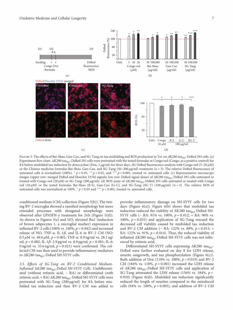
conditioned medium (CM) collection (Figure 5(b)). The rest-ing BV-2 microglia showed a ramified morphology but moreextended processes with elongated morphology wereobserved after LPS/IFN-γ treatment for 24 h (Figure 5(d)).As shown in Figures 5(e) and 5(f), elevated Iba1 (inductionof brown adipocytes 1, a microglial marker) expression ininflamed BV-2 cells (100% vs. 240%, p = 0 042) and increasedrelease of NO, TNF-α, IL-1β, and IL-6 in BV-2 CM (NO:0.5μM vs. 49.6μM, p = 0 001; TNF-α: 0.9 ng/ml vs. 28.1 ng/ml, p = 0 002; IL-1β: 2.9 pg/ml vs. 8.9 pg/ml, p < 0 001; IL-6:0 ng/ml vs. 33.6 ng/ml, p = 0 021) were confirmed. The col-lected CM was then used to provide inflammatory mediatorsto ΔK280 tauRD-DsRed SH-SY5Y cells.
3.5. Effects of SG-Tang on BV-2 Conditioned Medium-Inflamed ΔK280 tauRD-DsRed SH-SY5Y Cells. Undifferenti-ated (without retinoic acid, − RA) or differentiated (withretinoic acid, + RA) ΔK280 tauRD-DsRed SH-SY5Y cells werepretreated with SG-Tang (200μg/ml) for 8 h before mis-folded tau induction and then BV-2 CM was added to
provoke inflammatory damage on SH-SY5Y cells for twodays (Figure 6(a)). Figure 6(b) shows that misfolded tauinduction reduced the viability of ΔK280 tauRD-DsRed SH-SY5Y cells (− RA: 91% vs. 100%, p = 0 012; + RA: 90% vs.100%, p = 0 035) and application of SG-Tang rescued thedecreased cell viability caused by misfolded tau inductionand BV-2 CM addition (− RA: 122% vs. 88%, p = 0 015; +RA: 122% vs. 91%, p = 0 014). Thus, the reduced viability ofinflamed ΔK280 tauRD-DsRed SH-SY5Y cells was not influ-enced by retinoic acid.
Differentiated SH-SY5Y cells expressing ΔK280 tauRD-DsRed were further evaluated on day 8 for LDH release,neurite outgrowth, and tau phosphorylation (Figure 6(c)).Both addition of Dox (118% vs. 100%, p = 0 019) and BV-2CM (184% vs. 118%, p < 0 001) increased the LDH releaseof ΔK280 tauRD-DsRed SH-SY5Y cells and application ofSG-Tang attenuated the LDH release (156% vs. 184%, p =0 010) (Figure 6(d)). Misfolded tau induction significantlyreduced the length of neurites compared to the uninducedcells (94% vs. 100%, p = 0 005), and addition of BV-2 CM
Congo Dox/formula
DsRedflourescence
/ROS
Seeding + +
D1 D2 D58 h
(a)
⁎⁎ ⁎⁎
⁎⁎
⁎ ⁎⁎⁎⁎
⁎⁎⁎⁎⁎
⁎⁎⁎
DsR
edflo
ures
cenc
e (%
)
140
100
60
20
Untr. 5 10Congo red
(�휇M)Bai-Shao(�휇g/ml)
Gan-Cao(�휇g/ml)
SG-Tang(�휇g/ml)
20 50 100200 50 100200 50 100200
(b)
DsRed/Hoechst 33342 merged
DsRed alone
Untreated Congo red SG-Tang
50 �휇m
(c)
100
60
20
Untr.RO
S (%
)B-S SG-TCongo
10 �휇MG-C
100 �휇g/ml
⁎ ⁎ ⁎ ⁎⁎⁎
(d)
Figure 3: The effects of Bai-Shao, Gan-Cao, and SG-Tang on tau misfolding and ROS production in Tet-on ΔK280 tauRD-DsRed 293 cells. (a)Experiment flow chart. ΔK280 tauRD-DsRed 293 cells were pretreated with the tested formulas or Congo red (Congo, as a positive control) for8 h before misfolded tau induction by doxycycline (Dox, 1 μg/ml) for three days. (b) DsRed fluorescence analysis with Congo red (5~20 μM)or the Chinese medicine formulas Bai-Shao, Gan-Cao, and SG-Tang (50~200μg/ml) treatment (n = 3). The relative DsRed fluorescence ofuntreated cells is normalized (100%). ∗ p < 0 05, ∗∗ p < 0 01, and ∗∗∗ p < 0 001, treated vs. untreated cells. (c) Representative microscopyimages (upper row: merged DsRed and Hoechst 33342 signals; low row: DsRed signal alone) of ΔK280 tauRD-DsRed 293 cells untreated ortreated with Congo red (20 μM) or SG-Tang (200 μg/ml). (d) ROS assay of ΔK280 tauRD-DsRed 293 cells untreated or treated with Congored (10 μM) or the tested formulas Bai-Shao (B-S), Gan-Cao (G-C), and SG-Tang (SG-T) (100 μg/ml) (n = 3). The relative ROS ofuntreated cells was normalized as 100%. ∗ p < 0 05 and ∗∗∗ p < 0 001, treated vs. untreated cells.
7Oxidative Medicine and Cellular Longevity
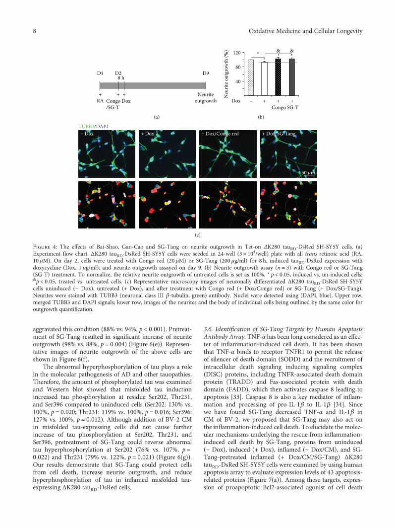
aggravated this condition (88% vs. 94%, p < 0 001). Pretreat-ment of SG-Tang resulted in significant increase of neuriteoutgrowth (98% vs. 88%, p = 0 004) (Figure 6(e)). Represen-tative images of neurite outgrowth of the above cells areshown in Figure 6(f).
The abnormal hyperphosphorylation of tau plays a rolein the molecular pathogenesis of AD and other tauopathies.Therefore, the amount of phosphorylated tau was examinedand Western blot showed that misfolded tau inductionincreased tau phosphorylation at residue Ser202, Thr231,and Ser396 compared to uninduced cells (Ser202: 130% vs.100%, p = 0 020; Thr231: 119% vs. 100%, p = 0 016; Ser396:127% vs. 100%, p = 0 012). Although addition of BV-2 CMin misfolded tau-expressing cells did not cause furtherincrease of tau phosphorylation at Ser202, Thr231, andSer396, pretreatment of SG-Tang could reverse abnormaltau hyperphosphorylation at Ser202 (76% vs. 107%, p =0 022) and Thr231 (79% vs. 122%, p = 0 021) (Figure 6(g)).Our results demonstrate that SG-Tang could protect cellsfrom cell death, increase neurite outgrowth, and reducehyperphosphorylation of tau in inflamed misfolded tau-expressing ΔK280 tauRD-DsRed cells.
3.6. Identification of SG-Tang Targets by Human ApoptosisAntibody Array. TNF-α has been long considered as an effec-ter of inflammation-induced cell death. It has been shownthat TNF-α binds to receptor TNFR1 to permit the releaseof silencer of death domain (SODD) and the recruitment ofintracellular death signaling inducing signaling complex(DISC) proteins, including TNFR-associated death domainprotein (TRADD) and Fas-associated protein with deathdomain (FADD), which then activates caspase 8 leading toapoptosis [33]. Caspase 8 is also a key mediator of inflam-mation and processing of pro-IL-1β to IL-1β [34]. Sincewe have found SG-Tang decreased TNF-α and IL-1β inCM of BV-2, we proposed that SG-Tang may also act onthe inflammation-induced cell death. To elucidate the molec-ular mechanisms underlying the rescue from inflammation-induced cell death by SG-Tang, proteins from uninduced(− Dox), induced (+ Dox), inflamed (+ Dox/CM), and SG-Tang-pretreated inflamed (+ Dox/CM/SG-Tang) ΔK280tauRD-DsRed SH-SY5Y cells were examined by using humanapoptosis array to evaluate expression levels of 43 apoptosis-related proteins (Figure 7(a)). Among these targets, expres-sion of proapoptotic Bcl2-associated agonist of cell death
NeuriteoutgrowthCongo Dox
/SG-T
+RA
+
D1 D28 h
D9
+
(a)
⁎120 & &
80
40
Dox
Neu
rite o
utgr
owth
(%)
+ + +Congo SG-T
−
(b)
TUBB3/DAPI− Dox + Dox + Dox/Congo red + Dox/SG-Tang
50 �휇m
(c)
Figure 4: The effects of Bai-Shao, Gan-Cao and SG-Tang on neurite outgrowth in Tet-on ΔK280 tauRD-DsRed SH-SY5Y cells. (a)Experiment flow chart. ΔK280 tauRD-DsRed SH-SY5Y cells were seeded in 24-well (3× 10⁴/well) plate with all trans retinoic acid (RA,10μM). On day 2, cells were treated with Congo red (20 μM) or SG-Tang (200 μg/ml) for 8 h, induced tauRD-DsRed expression withdoxycycline (Dox, 1μg/ml), and neurite outgrowth assayed on day 9. (b) Neurite outgrowth assay (n = 3) with Congo red or SG-Tang(SG-T) treatment. To normalize, the relative neurite outgrowth of untreated cells is set as 100%. ∗ p < 0 05, induced vs. un-induced cells;&p < 0 05, treated vs. untreated cells. (c) Representative microscopy images of neuronally differentiated ΔK280 tauRD-DsRed SH-SY5Ycells uninduced (− Dox), untreated (+ Dox), and after treatment with Congo red (+ Dox/Congo red) or SG-Tang (+ Dox/SG-Tang).Neurites were stained with TUBB3 (neuronal class III β-tubulin, green) antibody. Nuclei were detected using (DAPI, blue). Upper row,merged TUBB3 and DAPI signals; lower row, images of the neurites and the body of individual cells being outlined by the same color foroutgrowth quantification.
8 Oxidative Medicine and Cellular Longevity

(BAD), BH3-interacting domain death agonist (BID), cas-pase 3 (CASP3), caspase 8 (CASP8) and cytochrome c, andsomatic (CYCS) were apparently reduced by SG-Tang treat-ment (Table 1). Western blot analysis of BAD, BID, CASP8,and CYCS expression changes and caspase 3 activity assayfurther confirmed that pretreatment of SG-Tang couldsignificantly decrease these identified targets (BAD: from228% to 157%, p = 0 023; BID: from 139% to 110%, p =0 038; CASP8: from 118% to 104%, p = 0 024; CYCS: from163% to 96%, p = 0 040; caspase 3 activity: from 165% to103%, p = 0 005). Moreover, addition of SG-Tang improvedΔK280 tauRD-DsRed misfolding and enhanced solubletauRD-DsRed protein level in inflamed ΔK280 tauRD-DsRedSH-SY5Y cells (from 90% to 115%, p = 0 004) (Figure 7(b)).Our results indicated that SG-Tang may protect inflamed
ΔK280 tauRD-DsRed SH-SY5Y cells by inhibiting productionof proapoptotic proteins.
4. Discussion
In this study, we demonstrated neuroprotection, antioxida-tive and anti-inflammatory effects of formulated CHM SG-Tang. Our results showed that SG-Tang displayed a greaterantioxidative and antiaggregation effect than Bai-Shao andGan-Cao and a stronger anti-inflammatory activity thanBai-Shao but similar to Gan-Cao (Figures 2 and 3). More-over, SG-Tang showed neuroprotective effect of promotingneurite outgrowth probably by ameliorating tau misfoldingand oxidative stress in our tauopathy model (Figures 3 and4). The anti-inflammatory effects of SG-Tang were further
0 h
Seeding(BV-2 in 1% FBS)
+Celec./SG-T
Griess reagent (NO)+LPS
(1 �휇g/ml)
20 h 28/44 h 48/64 h
(a)
50 �휇M100 �휇g/ml500 �휇g/ml
40
30
20
10
LPS:SG-T Celec. SG-T
(8 h)SG-T(24 h)
NO
(�휇M
)
− − − + + + + ++
⁎⁎⁎
⁎⁎⁎⁎ ⁎⁎
⁎⁎
(b)
48 h24 h0 h
Seeding(BV-2 in 1% FBS)
+LPS (1 �휇g/ml)
IFN-�훾 (100 ng/ml)
MorphologyIba 1/NO/TNF-�훼/IL-1�훽/IL-6
(collect CM)
(c)
Untreated
50 �휇m
+ LPS & IFN-�훾
(d)
Iba1
�훽-Actin
300
100
Iba1
(%)
− LPS/IFN-�훾
− LPS/IFN-�훾
⁎
(e)
60
40
20
LPS/IFN-�훾
10
6
2
35
25
15
5
45
35
25
15
5
NO
(�휇M
)
TNF-�훼
(ng/
ml)
IL-1�훽
(pg/
ml)
IL-6
(ng/
ml)
⁎⁎⁎⁎⁎⁎⁎⁎⁎
− LPS/IFN-�훾− LPS/IFN-�훾− LPS/IFN-�훾−
(f)
Figure 5: Anti-inflammatory effects of SG-Tang and BV-2 conditioned medium preparation. (a) Experiment flow chart for LPS stimulation.BV-2 cells were seeded in 1% fetal bovine serum (FBS) and pretreated with 50μM celecoxib (Celec.) for 8 h or 100~500 μg/ml SG-Tang(SG-T) 8~24 h followed by 1μg/ml LPS stimulation 20 h. NO level was evaluated with Griess reagent. (b) Anti-inflammatory effect ofcelecoxib (Celec.) and SG-Tang (SG-T) on BV-2 cells (n = 3). ∗∗ p < 0 01 and ∗∗∗ p < 0 001, treated vs. untreated cells. (c) Experimentflow chart for LPS/IFN-γ stimulation. For preparation of BV-2 conditioned medium (CM), BV-2 cells were seeded in Dulbecco’smodified Eagle’s medium (DMEM) with 1% FBS medium. Next day, cells were stimulated with a combination of LPS (1 μg/ml) andIFN-γ (100 ng/ml). After 24 h stimulation, the BV-2 CM was collected and examined for inflammation by morphology, Iba1 Westernblotting and NO/TNF-α/IL-1β/IL-6 determination. (d) Morphology of BV-2 cells. (e) Western blot analysis of Iba1 expression ininflamed BV-2 cells (n = 3). To normalize, Iba1 expression level in uninflamed cells was set as 100%. ∗ p < 0 05, stimulated vs.unstimulated cells. (f) Secretion of NO, TNF-α, IL-1β, and IL-6 in BV-2 CM. ∗∗ p < 0 01 and ∗∗∗ p < 0 001, stimulated vs. unstimulated cells.
9Oxidative Medicine and Cellular Longevity

D5D3D2D1
± RA
SG-TangDox CM
8 h
Cell viability(MTT)
+ + +
(a)
RADOX
CM
−−−
−+−
−++
−++
SG
+−−
++−
+++
+++
SG
& &
⁎ ⁎
20
60
100
140
Cel
l via
bilit
y (%
)
(b)
D3 D8D2D1
RA+
SG-TangDox CM
+8 h
LDH releaseNeurite outgrowth
Tau phosphorylation+ +
(c)
DOXCM
−−
+−
++
++
SG
⁎
###&
40
80
120
160
200
LDH
rele
ase (
%)
(d)
DOXCM
−−
+−
++
++
SG
⁎⁎###
&&
40
80
120
Neu
rite o
utgr
owth
(%)
(e)
TUBB3/DAPI− Dox + Dox + Dox/CM + Dox/CM/SG-Tang
50 �휇m
(f)
Figure 6: Continued.
10 Oxidative Medicine and Cellular Longevity

demonstrated by using LPS-stimulated BV-2 microglia(Figure 5). Targets identified from human apoptosis pro-tein array indicate SG-Tang may suppress the expressionlevels of proapoptotic proteins in inflamed ΔK280 tauRD-DsRed SH-SY5Y cells and thus elevate the cell viability(Figures 6 and 7).
In human tauopathy, substantial activated microglia arefound in regions of phosphorylated tau accumulation [35].In tau P301S transgenic mice, prominent glial activation pre-cedes tangle formation and the pattern of activated glia corre-lates closely with the distribution and density of NFTs [36].As neuroinflammation is linked to the progression of tauopa-thy, anti-inflammatory strategy may be effective at reducingtau-related pathology. Indeed, FK506 attenuates tau pathol-ogy and increased lifespan in tau P301S mouse model [36].Treatment of 3xTg-AD mice with anti-inflammatory drugibuprofen reduces tau phosphorylation and memory impair-ment [37]. Administration of potent anti-inflammatory min-ocycline reduces the development of disease-associated tauspecies in the htau mouse model [38] by reducing severalinflammatory factors [39]. In the present study, we appliedBV-2 conditioned medium to proaggregant ΔK280 tauRD-DsRed 293/SH-SY5Y cells to mimic neuroinflammation.The study results reveal that CHM formula SG-Tang displaysneuroprotection by exerting anti-inflammatory and antia-poptotic activities.
Inflammation is a double-edged sword. Inflammatoryresponse could lead to activation of immune system andelimination of pathogens thereby reducing further cell loss.
Although inflammation might be protective and beneficialto cells, prolonged or dysregulated inflammatory processcould also result in production of neurotoxic factors thatexacerbate neurodegenerative pathology and cause cell death[12]. Thus, a potential strategy for treating tauopathies is tointervene in microglial activation and neuroinflammation.NSAID has been commonly used as treatment of inflamma-tion and known to be neuroprotective [40]. The mechanismof NSAID has been shown to inhibit the synthesis or activityof inflammatory mediators such as prostaglandin and COXisoforms 1 and 2. Although NSAID could effectively suppressthe inflammatory symptoms, these agents may also inducesignificant side effects such as increased risk of thromboticcardiovascular and cerebrovascular events [41]. Therefore,more safely, anti-inflammatory drugs need to be exploredand developed.
There is a growing interest in natural compounds/prod-ucts with anti-inflammatory activities which have long beenused for treating inflammation-related diseases. In this study,SG-Tang used was formulated with Bai-Shao (P. lactiflora)and Gan-Cao (G. uralensis) and analyzed by HPLC usingtwo main active constituents, paeoniflorin and ammoniumglycyrrhizinate (Figure 1). Both paeoniflorin and glycyrrhizi-nic acid were demonstrated to be able to cross the blood-brain barrier (BBB) in middle cerebral artery occlusion rats[42]. However, multiplicity of the components in P. lactifloraand G. uralensis contributes to the effects of antioxidationand anti-inflammation. In the root of P. lactiflora, a totalof 40 components including 29 monoterpene glycosides,
& &
⁎⁎ ⁎140
−Dox Dox CM SG −Dox Dox CM SG −Dox Dox CM SG −Dox Dox CM SGTotal
p-Ta
u/Ta
u (%
)
Tau
GAPDH
p-Tau (Ser202) p-Tau (Thr231) p-Tau (Ser396)
100
60
20
(g)
Figure 6: Neuroprotection of SG-Tang on ΔK280 tauRD-DsRed SH-SY5Y cells from BV-2 conditioned medium-induced cell death. (a)Experiment flow chart for cell viability assay. ΔK280 tauRD-DsRed SH-SY5Y cells were plated in media with/without retinoic acid (± RA,10μM) on day 1 and pretreated with SG-Tang the next day for 8 h, followed by doxycycline addition (Dox, 1 μg/ml) to induce misfoldedtau expression. On day 3, DMEM-F12 media was mixed with BV-2 CM and cell viability was assessed by MTT assay on day 5. (b) Cellviability assay (∗ p < 0 05, − Dox vs. + Dox; &p < 0 05, + Dox/CM vs. + Dox/CM/SG-Tang-treated cells) (n = 3). (c) Experiment flow chartfor LDH release, neurite outgrowth, and tau phosphorylation assays. RA (10 μM, present in cultures throughout) differentiated ΔK280tauRD-DsRed SH-SY5Y cells were pretreated with SG-Tang (200 μg/ml) on day 2 for 8 h, followed by inducing ΔK280 tauRD-DsRedexpression (+ Dox, 1μg/ml). On day 3, DMEM-F12 was mixed with BV-2 CM and added to the cells. After five days, media werecollected for LDH release examination. In addition, cells were examined for neurite outgrowth and tau phosphorylation. (d) LDH assay(∗p < 0 05, − Dox vs. + Dox; ###p < 0 001, + Dox vs. +Dox/CM; &p < 0 05, + Dox/CM vs. + Dox/CM/SG-Tang treated cells) (n = 3).(e) Neurite outgrowth assay (n = 3). To normalize, the relative neurite outgrowth of uninduced cells is set as 100%. ∗∗ p < 0 01, − Doxvs. + Dox; ###p < 0 001, + Dox vs. + Dox/CM; &&p < 0 01, + Dox/CM vs. + Dox/CM/SG-Tang-treated cells. (f) Representative microscopyimages of differentiated ΔK280 tauRD-DsRed SH-SY5Y cells uninduced (− Dox), induced (+ Dox), inflamed (+ Dox/CM), or treated withSG-Tang (+ Dox/CM/SG-Tang). Neurites were stained with TUBB3 (green) antibody. Nuclei were detected using DAPI (blue). Upperrow, merged TUBB3 and DAPI signals; lower row, images of the neurites and the body of a cell being outlined by the same color foroutgrowth quantification. (g) Western blot analysis of total and phosphorylated (Ser202, Thr231, and Ser396) tau (normalized to GAPDHinternal control, n = 3). ∗ p < 0 05, − Dox vs. + Dox; &p < 0 05, + Dox/CM vs. + Dox/CM/SG-Tang-treated cells.
11Oxidative Medicine and Cellular Longevity

8 galloyl glucoses, and 3 phenolic compounds were identi-fied [43]. Among them, paeoniflorin, a monoterpene gly-coside, is known to possess anti-inflammatory effect andhas been applied to cerebral ischemic injury [44]. Paeoni-florin also exhibits neuroprotective effects via inhibiting
neuroinflammation in APP/PS1 and in PS2 mutant mice[45, 46]. Paeoniflorin and the isomer albiflorin attenuatedneuropathic pain by inhibiting the activation of p38 MAPKpathway in spinal microglia and subsequent upregulatedIL-1β and TNF-α [47]. Benzoylpaeoniflorin, another
+ Dox/CM/SG-Tang
+ Dox
+ Dox/CM
– Dox
CYCS
BAD
BID
CASP
3CA
SP8
CYCS
BAD
BID
CASP
3CA
SP8
(a)
SG-TangSG-TangSG-TangCMDox
BAD(23 kDa)
321
CYCS(15 kDa)
Tubulin(55 kDa)
GAPDH(37 kDa)
GAPDH(37 kDa)
2
1
BID(22 kDa)
2
1
tauRD-DsRed(40 kDa)
1.51.00.5
CASP8(55/53 kDa)
1.51.00.5
1.52.0
1.00.5CA
SP3
activ
ity
++
−+
++
−−
CMDox
++
−+
++
−−
CMDox
++
−+
++
−−
#&
# &
#
&&⁎⁎⁎
&⁎ # &
&&
(b)
Figure 7: Apoptosis-related protein targets of SG-Tang in BV-2 conditioned medium-stimulated ΔK280 tauRD-DsRed SH-SY5Y cells.(a) Representative images of apoptosis antibody array of proteins collected from Figure 6(c). (b) Western blot analysis of BAD, CYCS,CASP8, BID, and tauRD-DsRed protein levels (normalized to tubulin or GAPDH internal control, n = 3) and caspase 3 activity assayfrom each sample. ∗ p < 0 05 and ∗∗ p < 0 01, − Dox vs. + Dox; #p < 0 05, + Dox vs. + Dox/CM; &p < 0 05 and &&p < 0 01, + Dox/CMvs. + Dox/CM/SG-Tang-treated cells.
Table 1: Proteins identified by human apoptosis antibody array.
Gene symbolUniProt
accession numberProtein
Fold change(+ Dox/CM vs. − Dox)
Fold change(+ Dox/CM/SG-T vs. + Dox/CM)
BAD Q92934 Bcl2-associated agonist of cell death 1.16 0.63
BID P55957 BH3-interacting domain death agonist 1.33 0.41
CASP3 P42574 Caspase 3 1.49 0.71
CASP8 Q14790 Caspase 8 1.34 0.62
CYCS P99999 Cytochrome c, somatic 0.92 0.62
12 Oxidative Medicine and Cellular Longevity

paeoniflorin-related glycoside in P. lactiflora root, protectedprimary rat cortical cells against H2O2-induced oxidativestress [48]. In addition to monoterpene glycosides, gallicacid, a phenolic compound in P. lactiflora root, displayedantioxidative effect by scavenging free radicals, inhibitinglipid peroxidation, and protecting against oxidative DNAdamage [49]. Paeonol, another phenolic compound in P.lactiflora root, exerted neuroprotective effect in the modelof ischemia through reducing proinflammatory receptors/mediators [50].
The main bioactive components of G. uralensis are tri-terpene saponins and various types of flavonoids, includingglycyrrhetinic acid, glycyrrhizic acid, liquiritigenin, isoliquir-itigenin, liquiritin, and licochalcone A [51]. Glycyrrhizin andrelated compounds were found to show anti-inflammatoryactivity in vitro [52] and in vivo [53]. Although diammo-nium glycyrrhizinate rescues neurotoxicity in Aβ1–42-induced mice [54], its effect in tauopathy models is notknown. Isoliquiritigenin, isoliquiritin, and liquiritigenin sig-nificantly suppressed iNOS, TNF-α, and IL-6 expression inIL-1β-treated rat hepatocytes [55]. Interestingly, the purifiedglycyrrhiza polysaccharides increased the pinocytic activity,the production of NO, IL-1, IL-6, and IL-12 in macrophagesof mice [56]. Glycyrrhetinic acid, liquiritigenin, isoliquiriti-genin, and liquiritin were also found to be all potent NRF2inducers [57]. Moreover, Calzia et al. has shown that poly-phenolic phytochemicals displayed a potent antioxidantaction by modulating the ectopic F0F1-ATP synthase activityof the rod outer segments of the retina and prevented theinduction of apoptosis [58]. Therefore, polyphenolic com-pounds from Bai-Shao and Gan-Cao may also exert antioxi-dative activities not only in but also outside of mitochondria.Given that multiple different compounds in both Bai-Shaoand Gan-Cao are exerting effects on different pathways, thecombination of Bai-Shao and Gan-Cao may thus have addi-tive protection effects than each alone, which is supported byour study results.
The anti-inflammatory effect of Jakyakgamcho-tang, aformulated P. lactiflora and G. uralensis in Korea, has beenshown by inhibiting the NF-κB signaling pathway in kerati-nocytes [59]. Aberrant activation of NF-κB signaling maylead to apoptosis and cell death [60]. We found that severalproapoptotic proteins including BAD, BID, CASP3, CASP8,and CYCS were induced by misfolded tau expression and/or caused by LPS/IFN-γ-stimulated BV2 microglia. BADprotein is a proapoptotic member of the Bcl-2 gene familyinvolved in initiating apoptosis [61]. BID is also aproapoptoticprotein which plays a role as a sentinel for protease-mediateddeath signals [62]. Caspases are well-studied importantmediators of apoptosis. CYCS is known to be released frommitochondria into cytosol to stimulate cell apoptosis [63].Administration of SG-Tang decreased the production ofthese proapoptotic proteins, indicating that SG-Tang maytarget on inhibiting proapoptotic proteins to protect neuroncells from inflammatory damage.
Finally, pretreatment of SG-Tang reversed abnormalhyperphosphorylation at tau Ser202 and Thr231 in inflamedmisfolded tau-expressing SH-SY5Y cells (Figure 6). Taufunction is regulated by phosphorylation at specific sites,
and tau phosphorylation plays both physiological and patho-logical roles in the cells. Tau Ser199/202 and Thr205 werefound to be locally phosphorylated along the nascent axonduring axonogenesis [64]. Phosphorylation of tau Thr231inhibited tau to bind and stabilize microtubules [65]. BothSer202 and Thr231 are hyperphosphorylated in degeneratingAD brain [66]. Among kinases that regulate tau Ser202 andThr231 phosphorylation, cyclic AMP-dependent proteinkinase (PKA) and cyclin-dependent kinase 2 (CDC2) mightbe the potential targets of SG-Tang, and SG-Tang treat-ment may result in activity suppression of these two kinases[66, 67]. The exact mechanism for PKA or CDC2 regulationby SG-Tang remains to be determined in our future work.
5. Conclusions
Plant-derived natural medications have been used for centu-ries and becoming more popular because of their low sideeffects. Despite the fact that natural compounds are rela-tively safe, the complexity of natural products makes nutra-ceutical preparations difficult to be appropriately designed. Inthis study, we showed antioxidative and anti-inflammatoryeffects of SG-Tang as a potential agent for treatment orprevention of neuroinflammation-associated tauopathy. Infuture, studies of main active compounds paeoniflorin andammonium glycyrrhizinate in SG-Tang, separately or incombination, in tauopathy cell model are warranted to pro-vide a novel avenue for protection against tauopathy.
Abbreviations
AD: Alzheimer’s diseaseBAD: Bcl2-associated agonist of cell deathBBB: Blood-brain barrierBID: BH3-interacting domain death agonistBSA: Bovine serum albuminCASP3: Caspase 3CASP8: Caspase 8CDC2: Cyclin-dependent kinase 2CHM: Chinese herbal medicineCM: Conditioned mediumCNS: Central nervous systemCOX: CyclooxygenaseCYCS: Cytochrome c, somaticDAPI: 4′-6-Diamidino-2-phenylindoleDox: DoxycyclineDMEM: Dulbecco’s modified Eagle’s mediumDPPH: 1,1-Diphenyl-2-picrylhydrazylEC50: Half maximal effective concentrationELISA: Enzyme-linked immunosorbent assayFBS: Fetal bovine serumHCA: High-content analysisHPLC: High-performance liquid chromatographyIba1: Induction of brown adipocytes 1IC50: Half maximal inhibitory concentrationIFN: InterferonIL: InterleukinLDH: Lactate dehydrogenaseLPS: Lipopolysaccharide
13Oxidative Medicine and Cellular Longevity

MTT: 3-(4,5-Dimethylthiazol-2-yl)-2,5-diphenyltetrazo-lium bromide
NO: Nitric oxideNSAID: Nonsteroidal anti-inflammatory drugPKA: Cyclic AMP-dependent protein kinaseROS: Reactive oxygen speciesSG-Tang: Shaoyao Gancao TangtauRD: Tau repeat domainTNF: Tumor necrosis factor.
Data Availability
The data used to support the findings of this study areavailable from the corresponding author upon request.
Conflicts of Interest
The authors declare no conflicts of interest.
Authors’ Contributions
Guey-Jen Lee-Chen and Chiung-Mei Chen designed theresearch and revised the paper. I-Cheng Chen performedthe experiments, analyzed the data, and wrote the manu-script. Te-Hsien Lin conducted experiments and analyzedthe data. Yu-Hsuan Hsieh and Chih-Ying Chao performedexperiments and assisted in the technical work. Yih-Ru Wuand Kuo-Hsuan Chang commented on the experimentdesign. Ming-Chung Lee provided CHM materials for thisstudy. All authors approved the final version of the manu-script. I-Cheng Chen and Te-Hsien Lin contributed equallyto this work.
Acknowledgments
This work was supported by the grants from the Ministry ofScience and Technology (105-2325-B-003-001) and ChangGung Memorial Hospital (CMRPG3F136, CMRPG3F1611-2, and CMRPG3G052). We thank the Molecular ImagingCore Facility of National Taiwan Normal University for thetechnical assistance.
References
[1] D. R. Williams, “Tauopathies: classification and clinical updateon neurodegenerative diseases associated with microtubule-associated protein tau,” Internal Medicine Journal, vol. 36,no. 10, pp. 652–660, 2006.
[2] G. Lee and C. J. Leugers, “Tau and tauopathies,” Progressin Molecular Biology and Translational Science, vol. 107,pp. 263–293, 2012.
[3] R. L. Neve, P. Harris, K. S. Kosik, D. M. Kurnit, and T. A.Donlon, “Identification of cDNA clones for the humanmicrotubule-associated protein tau and chromosomal locali-zation of the genes for tau and microtubule-associated pro-tein 2,” Brain Research, vol. 387, no. 3, pp. 271–280, 1986.
[4] M. Hasegawa, M. J. Smith, and M. Goedert, “Tau proteinswith FTDP-17 mutations have a reduced ability to promotemicrotubule assembly,” FEBS Letters, vol. 437, no. 3, pp. 207–210, 1998.
[5] P. Nacharaju, J. Lewis, C. Easson et al., “Accelerated filamentformation from tau protein with specific FTDP-17 missensemutations,” FEBS Letters, vol. 447, no. 2-3, pp. 195–199, 1999.
[6] I. D'Souza, P. Poorkaj, M. Hong et al., “Missense and silenttau gene mutations cause frontotemporal dementia withparkinsonism-chromosome 17 type, by affecting multiplealternative RNA splicing regulatory elements,” Proceedingsof the National Academy of Sciences of the United States ofAmerica, vol. 96, no. 10, pp. 5598–5603, 1999.
[7] P. Rizzu, J. C. van Swieten, M. Joosse et al., “High prevalence ofmutations in the microtubule-associated protein tau in a pop-ulation study of frontotemporal dementia in the Netherlands,”The American Journal of Human Genetics, vol. 64, no. 2,pp. 414–421, 1999.
[8] P. Momeni, A. Pittman, T. Lashley et al., “Clinical and patho-logical features of an Alzheimer’s disease patient with theMAPT ΔK280 mutation,” Neurobiology of Aging, vol. 30,no. 3, pp. 388–393, 2009.
[9] S. Barghorn, Q. Zheng-Fischhöfer, M. Ackmann et al., “Struc-ture, microtubule interactions, and paired helical filamentaggregation by tau mutants of frontotemporal dementias,”Biochemistry, vol. 39, no. 38, pp. 11714–11721, 2000.
[10] V. Vogelsberg-Ragaglia, J. Bruce, C. Richter-Landsberg et al.,“Distinct FTDP-17 missense mutations in tau produce tauaggregates and other pathological phenotypes in transfectedCHO cells,” Molecular Biology of the Cell, vol. 11, no. 12,pp. 4093–4104, 2000.
[11] M. von Bergen, S. Barghorn, L. Li et al., “Mutations of tauprotein in frontotemporal dementia promote aggregation ofpaired helical filaments by enhancing local β-structure,” TheJournal of Biological Chemistry, vol. 276, no. 51, pp. 48165–48174, 2001.
[12] P. J. Khandelwal, A. M. Herman, and C. E. H. Moussa,“Inflammation in the early stages of neurodegenerative pathol-ogy,” Journal of Neuroimmunology, vol. 238, no. 1-2, pp. 1–11,2011.
[13] P. L. DiPatre and B. B. Gelman, “Microglial cell activationin aging and Alzheimer disease: partial linkage with neuro-fibrillary tangle burden in the hippocampus,” Journal of Neu-ropathology and Experimental Neurology, vol. 56, no. 2,pp. 143–149, 1997.
[14] K. Ishizawa and D. W. Dickson, “Microglial activation paral-lels system degeneration in progressive supranuclear palsyand corticobasal degeneration,” Journal of Neuropathologyand Experimental Neurology, vol. 60, no. 6, pp. 647–657,2001.
[15] M. Kitazawa, T. R. Yamasaki, and F. M. Laferla, “Microglia as apotential bridge between the amyloid β-peptide and tau,”Annals of the New York Academy of Sciences, vol. 1035, no. 1,pp. 85–103, 2004.
[16] Y. Li, L. Liu, S. W. Barger, and W. S. T. Griffin, “Interleukin-1mediates pathological effects of microglia on tau phosphoryla-tion and on synaptophysin synthesis in cortical neuronsthrough a p38-MAPK pathway,” The Journal of Neuroscience,vol. 23, no. 5, pp. 1605–1611, 2003.
[17] K. Bhaskar, M. Konerth, O. N. Kokiko-Cochran, A. Cardona,R. M. Ransohoff, and B. T. Lamb, “Regulation of tau pathologyby the microglial fractalkine receptor,” Neuron, vol. 68, no. 1,pp. 19–31, 2010.
[18] M. Kitazawa, S. Oddo, T. R. Yamasaki, K. N. Green, andF. LaFerla, “Lipopolysaccharide-induced inflammation exacer-bates tau pathology by a cyclin-dependent kinase 5-mediated
14 Oxidative Medicine and Cellular Longevity

pathway in a transgenic model of Alzheimer’s disease,” Journalof Neuroscience, vol. 25, no. 39, pp. 8843–8853, 2005.
[19] A. N. Nilson, K. C. English, J. E. Gerson et al., “Tau oligo-mers associate with inflammation in the brain and retinaof tauopathy mice and in neurodegenerative diseases,” Jour-nal of Alzheimer's Disease, vol. 55, no. 3, pp. 1083–1099,2017.
[20] P. D.Wes, F. A. Sayed, F. Bard, and L. Gan, “Targeting microg-lia for the treatment of Alzheimer’s disease,” Glia, vol. 64,no. 10, pp. 1710–1732, 2016.
[21] A. Ardura-Fabregat, E. W. G. M. Boddeke, A. Boza-Serranoet al., “Targeting neuroinflammation to treat Alzheimer's dis-ease,” CNS Drugs, vol. 31, no. 12, pp. 1057–1082, 2017.
[22] J. Gao, Y. Inagaki, X. Li, N. Kokudo, and W. Tang, “Researchprogress on natural products from traditional Chinese medi-cine in treatment of Alzheimer’s disease,” Drug Discoveries &Therapeutics, vol. 7, no. 2, pp. 46–57, 2013.
[23] Z. Y. Wang, J. G. Liu, H. Li, and H. M. Yang, “Pharmacologicaleffects of active components of Chinese herbal medicine in thetreatment of Alzheimer’s disease: a review,” The AmericanJournal of Chinese Medicine, vol. 44, no. 08, pp. 1525–1541,2016.
[24] T. Y. Wu, C. P. Chen, and T. R. Jinn, “Traditional Chinesemedicines and Alzheimer’s disease,” Taiwanese Journal ofObstetrics & Gynecology, vol. 50, no. 2, pp. 131–135, 2011.
[25] Y.Q.ZhengandW.Wei, “Total glucosidesofpaeony suppressesadjuvant arthritis in rats and intervenes cytokine-signalingbetween different types of synoviocytes,” International Immu-nopharmacology, vol. 5, no. 10, pp. 1560–1573, 2005.
[26] T.-Y. Wu, T. O. Khor, C. L. L. Saw et al., “Anti-inflammatory/anti-oxidative stress activities and differential regulation ofNrf2-mediated genes by non-polar fractions of tea Chrysan-themum zawadskii and licorice Glycyrrhiza uralensis,” TheAAPS Journal, vol. 13, no. 1, pp. 1–13, 2011.
[27] K.-H. Chang, I.-C. Chen, H.-Y. Lin et al., “The aqueous extractof Glycyrrhiza inflata can upregulate unfolded proteinresponse-mediated chaperones to reduce tau misfolding in cellmodels of Alzheimer’s disease,”Drug Design, Development andTherapy, vol. 10, pp. 885–896, 2016.
[28] D. Brown, A. Tamas, D. Reglodi, and Y. Tizabi, “PACAP pro-tects against inflammatory-mediated toxicity in dopaminergicSH-SY5Y cells: implication for Parkinson’s disease,”Neurotox-icity Research, vol. 26, no. 3, pp. 230–239, 2014.
[29] Y. C. Park, C. H. Lee, H. S. Kang, H. T. Chung, and H. D. Kim,“Wortmannin, a specific inhibitor of phosphatidylinositol-3-kinase, enhances LPS-induced NO production from murineperitoneal macrophages,” Biochemical and Biophysical ResearchCommunications, vol. 240, no. 3, pp. 692–696, 1997.
[30] H. Fang, R. A. Pengal, X. Cao et al., “Lipopolysaccharide-induced macrophage inflammatory response is regulated bySHIP,” Journal of Immunology, vol. 173, no. 1, pp. 360–366,2004.
[31] M. Cente, P. Filipcik, M. Pevalova, and M. Novak, “Expressionof a truncated tau protein induces oxidative stress in a rodentmodel of tauopathy,” The European Journal of Neuroscience,vol. 24, no. 4, pp. 1085–1090, 2006.
[32] P. L. McGeer, T. Kawamata, D. G. Walker, H. Akiyama,I. Tooyama, and E. G. McGeer, “Microglia in degenerativeneurological disease,” Glia, vol. 7, no. 1, pp. 84–92, 1993.
[33] L. M. Sedger and M. F. McDermott, “TNF and TNF-receptors:from mediators of cell death and inflammation to therapeutic
giants - past, present and future,” Cytokine & Growth FactorReviews, vol. 25, no. 4, pp. 453–472, 2014.
[34] T. P. Monie and C. E. Bryant, “Caspase-8 functions as a keymediator of inflammation and pro-IL-1β processing via bothcanonical and non-canonical pathways,” ImmunologicalReviews, vol. 265, no. 1, pp. 181–193, 2015.
[35] A. Sasaki, T. Kawarabayashi, T. Murakami et al., “Microglialactivation in brain lesions with tau deposits: comparison ofhuman tauopathies and tau transgenic mice TgTauP301L,”Brain Research, vol. 1214, pp. 159–168, 2008.
[36] Y. Yoshiyama, M. Higuchi, B. Zhang et al., “Synapse loss andmicroglial activation precede tangles in a P301S tauopathymouse model,” Neuron, vol. 53, no. 3, pp. 337–351, 2007.
[37] A. C. McKee, I. Carreras, L. Hossain et al., “Ibuprofen reducesAβ, hyperphosphorylated tau and memory deficits in Alzhei-mer mice,” Brain Research, vol. 1207, pp. 225–236, 2008.
[38] W. Noble, C. Garwood, J. Stephenson, A. M. Kinsey, D. P.Hanger, and B. H. Anderton, “Minocycline reduces the devel-opment of abnormal tau species in models of Alzheimer’sdisease,” The FASEB Journal, vol. 23, no. 3, pp. 739–750,2009.
[39] C. J. Garwood, J. D. Cooper, D. P. Hanger, and W. Noble,“Anti-inflammatory impact of minocycline in a mouse modelof tauopathy,” Frontiers in Psychiatry, vol. 1, p. 136, 2010.
[40] M. A. Ajmone-Cat, A. Bernardo, A. Greco, and L. Minghetti,“Non-steroidal anti-inflammatory drugs and brain inflamma-tion: effects on microglial functions,” Pharmaceuticals (Basel),vol. 3, no. 6, pp. 1949–1965, 2010.
[41] J. C. Maroon, J. W. Bost, and A. Maroon, “Natural anti-inflammatory agents for pain relief,” Surgical Neurology Inter-national, vol. 1, no. 1, p. 80, 2010.
[42] H. Li, M. Ye, Y. Zhang et al., “Blood-brain barrier permeabilityof Gualou Guizhi granules and neuroprotective effects inischemia/reperfusion injury,” Molecular Medicine Reports,vol. 12, no. 1, pp. 1272–1278, 2015.
[43] S. L. Li, J. Z. Song, F. F. K. Choi et al., “Chemical profilingof Radix Paeoniae evaluated by ultra-performance liquidchromatography/photo-diode-array/quadrupole time-of-flightmass spectrometry,” Journal of Pharmaceutical and BiomedicalAnalysis, vol. 49, no. 2, pp. 253–266, 2009.
[44] Y. Zhang, H. Li, M. Huang et al., “Paeoniflorin, a monoterpeneglycoside, protects the brain from cerebral ischemic injury viainhibition of apoptosis,” The American Journal of ChineseMedicine, vol. 43, no. 03, pp. 543–557, 2015.
[45] H. R. Zhang, J. H. Peng, X. B. Cheng, B. Z. Shi, M. Y. Zhang,and R. X. Xu, “Paeoniflorin atttenuates amyloidogenesis andthe inflammatory responses in a transgenic mouse model ofAlzheimer’s disease,” Neurochemical Research, vol. 40, no. 8,pp. 1583–1592, 2015.
[46] X. Gu, Z. Cai, M. Cai et al., “Protective effect of paeoniflorin oninflammation and apoptosis in the cerebral cortex of a trans-genic mouse model of Alzheimer’s disease,” Molecular Medi-cine Reports, vol. 13, no. 3, pp. 2247–2252, 2016.
[47] J. Zhou, L. Wang, J. Wang et al., “Paeoniflorin and albiflorinattenuate neuropathic pain via MAPK pathway in chronicconstriction injury rats,” Evidence-Based Complementary andAlternative Medicine, vol. 2016, Article ID 8082753, 11 pages,2016.
[48] S. H. Kim, M. K. Lee, K. Y. Lee, S. H. Sung, J. Kim, and Y. C.Kim, “Chemical constituents isolated from Paeonia lactifloraroots and their neuroprotective activity against oxidative stress
15Oxidative Medicine and Cellular Longevity

in vitro,” Journal of Enzyme Inhibition and Medicinal Chemis-try, vol. 24, no. 5, pp. 1138–1140, 2009.
[49] S. C. Lee, Y. S. Kwon, K. H. Son, H. P. Kim, and M. Y. Heo,“Antioxidative constituents from Paeonia lactiflora,” Archivesof Pharmacal Research, vol. 28, no. 7, pp. 775–783, 2005.
[50] W. Y. Liao, T. H. Tsai, T. Y. Ho, Y. W. Lin, C. Y. Cheng, andC. L. Hsieh, “Neuroprotective effect of paeonol mediatesanti-inflammation via suppressing toll-like receptor 2 andtoll-like receptor 4 signaling pathways in cerebral ischemia-reperfusion injured rats,” Evidence-Based Complementaryand Alternative Medicine, vol. 2016, Article ID 3704647, 12pages, 2016.
[51] Q. Zhang and M. Ye, “Chemical analysis of the Chinese herbalmedicine Gan-Cao (licorice),” Journal of Chromatography. A,vol. 1216, no. 11, pp. 1954–1969, 2009.
[52] S. Matsui, H. Matsumoto, Y. Sonoda et al., “Glycyrrhizin andrelated compounds down-regulate production of inflamma-tory chemokines IL-8 and eotaxin 1 in a human lung fibroblastcell line,” International Immunopharmacology, vol. 4, no. 13,pp. 1633–1644, 2004.
[53] T. Genovese, M. Menegazzi, E. Mazzon et al., “Glycyrrhizinreduces secondary inflammatory process after spinal cordcompression injury in mice,” Shock, vol. 31, no. 4, pp. 367–375, 2009.
[54] H. Zhao, S. L. Wang, L. Qian et al., “Diammonium glycyrrhi-zinate attenuates Aβ1-42-induced neuroinflammation and reg-ulates MAPK and NF-κB pathways in vitro and in vivo,” CNSNeuroscience & Therapeutics, vol. 19, no. 2, pp. 117–124, 2013.
[55] R. Tanemoto, T. Okuyama, H. Matsuo, T. Okumura, Y. Ikeya,and M. Nishizawa, “The constituents of licorice (Glycyrrhizauralensis) differentially suppress nitric oxide production ininterleukin-1β-treated hepatocytes,” Biochemistry and Bio-physics Reports, vol. 2, pp. 153–159, 2015.
[56] A. Cheng, F. Wan, J. Wang, Z. Jin, and X. Xu, “Macrophageimmunomodulatory activity of polysaccharides isolated fromGlycyrrhiza uralensis fish,” International Immunopharmacol-ogy, vol. 8, no. 1, pp. 43–50, 2008.
[57] H. Gong, B. K. Zhang, M. Yan et al., “A protective mechanismof licorice (Glycyrrhiza uralensis): isoliquiritigenin stimulatesdetoxification system via Nrf2 activation,” Journal of Ethno-pharmacology, vol. 162, pp. 134–139, 2015.
[58] D. Calzia, M. Oneto, F. Caicci et al., “Effect of polyphenolicphytochemicals on ectopic oxidative phosphorylation in rodouter segments of bovine retina,” British Journal of Pharmacol-ogy, vol. 172, no. 15, pp. 3890–3903, 2015.
[59] S. J. Jeong, H. S. Lim, C. S. Seo et al., “Traditional herbal for-mula Jakyakgamcho-tang (Paeonia lactiflora and Glycyrrhizauralensis) impairs inflammatory chemokine production byinhibiting activation of STAT1 and NF-κB in HaCaT cells,”Phytomedicine, vol. 22, no. 2, pp. 326–332, 2015.
[60] B. Kaltschmidt, C. Kaltschmidt, T. G. Hofmann, S. P. Hehner,W. Droge, and M. L. Schmitz, “The pro- or anti-apoptoticfunction of NF-κB is determined by the nature of the apoptoticstimulus,” European Journal of Biochemistry, vol. 267, no. 12,pp. 3828–3835, 2000.
[61] E. Yang, J. Zha, J. Jockel, L. H. Boise, C. B. Thompson, andS. J. Korsmeyer, “Bad, a heterodimeric partner for Bcl-XLand Bcl-2, displaces Bax and promotes cell death,” Cell,vol. 80, no. 2, pp. 285–291, 1995.
[62] K. Wang, X. M. Yin, D. T. Chao, C. L. Milliman, and S. J.Korsmeyer, “BID: a novel BH3 domain-only death agonist,”Genes & Development, vol. 10, no. 22, pp. 2859–2869, 1996.
[63] X. Liu, C. N. Kim, J. Yang, R. Jemmerson, and X. Wang,“Induction of apoptotic program in cell-free extracts: require-ment for dATP and cytochrome c,” Cell, vol. 86, no. 1, pp. 147–157, 1996.
[64] J. W. Mandell and G. A. Banker, “A spatial gradient of tauprotein phosphorylation in nascent axons,” The Journal ofNeuroscience, vol. 16, no. 18, pp. 5727–5740, 1996.
[65] J.-H. Cho and G. V.W. Johnson, “Glycogen synthase kinase 3βphosphorylates tau at both primed and unprimed sites: differ-ential impact on microtubule binding,” Journal of BiologicalChemistry, vol. 278, no. 1, pp. 187–193, 2003.
[66] J. Z. Wang and F. Liu, “Microtubule-associated protein tau indevelopment, degeneration and protection of neurons,” Prog-ress in Neurobiology, vol. 85, no. 2, pp. 148–175, 2008.
[67] D. P. Hanger, B. H. Anderton, and W. Noble, “Tau phos-phorylation: the therapeutic challenge for neurodegenerativedisease,” Trends in Molecular Medicine, vol. 15, no. 3,pp. 112–119, 2009.
16 Oxidative Medicine and Cellular Longevity

Research ArticleEffects of the Aphanizomenon flos-aquae Extract (Klamin®) ona Neurodegeneration Cellular Model
D. Nuzzo ,1 G. Presti,2 P. Picone,1 G. Galizzi,1 E. Gulotta,2 S. Giuliano,2 C. Mannino,2
V. Gambino,2 S. Scoglio,3 and M. Di Carlo 1
1Istituto di Biomedicina ed Immunologia Molecolare (IBIM) “Alberto Monroy”, CNR, Via Ugo La Malfa 153, 90146 Palermo, Italy2Chemical Laboratory of Palermo, Italian Agency of Customs and Monopolies, Via Crispi, 143, 90133 Palermo, Italy3Nutritherapy Research Center, 61029 Urbino, Italy
Correspondence should be addressed to M. Di Carlo; [email protected]
Received 19 June 2018; Accepted 30 July 2018; Published 17 September 2018
Academic Editor: Francisco Jaime Bezerra Mendonça Júnior
Copyright © 2018 D. Nuzzo et al. This is an open access article distributed under the Creative Commons Attribution License, whichpermits unrestricted use, distribution, and reproduction in any medium, provided the original work is properly cited.
Cyanobacteria have been recognized as a source of bioactive molecules to be employed in nutraceuticals, pharmaceuticals, andfunctional foods. An extract of Aphanizomenon flos-aquae (AFA), commercialized as Klamin®, was subjected to chemicalanalysis to determine its compounds. The AFA extract Klamin® resulted to be nontoxic, also at high doses, when administeredonto LAN5 neuronal cells. Its scavenging properties against ROS generation were evaluated by using DCFH-DA assay, and itsmitochondrial protective role was determined by JC-1 and MitoSOX assays. Klamin® exerts a protective role against betaamyloid- (Aβ-) induced toxicity and against oxidative stress. Anti-inflammatory properties were demonstrated by NFβB nuclearlocalization and activation of IL-6 and IL-1β inflammatory cytokines through ELISA. Finally, by using thioflavin T (ThT) andfluorimetric measures, we found that Klamin® interferes with Aβ aggregation kinetics, supporting the formation of smaller andnontoxic structures compared to toxic Aβ aggregates alone. Altogether, these data indicate that the AFA extract may play aprotective role against mechanisms leading to neurodegeneration.
1. Introduction
Blue-green algae, including Aphanizomenon flos-aquae(AFA), are unicellular prokaryotic microorganism belongingto the Cyanobacteria (Cyanophyta) phylum. These bacteriaare among the oldest life forms, are the only microorganismsto achieve oxygenic photosynthesis, and probably havebeen the main biotic source of oxygen on early Earth[1]. These microorganisms are widely diffused, colonizingboth salt- and freshwater around the world, alone or ina symbiotic relationship with plants or fungi [2]. Cyano-bacteria are a source of bioactive compounds such aspolyunsaturated fatty acids, proteins, pigments, and mineralsand are rich in substances with anti-inflammatory andantioxidant properties [3–8].
The Aphanizomenon genus includes several speciesincluding the flos-aquae. AFA is a cyanobacterial unicellularorganism endowed with several health-enhancing properties
that, unlike other commercial “microalgae,” spontaneouslygrows in Upper Klamath Lake (southern Oregon, USA)where it also inhibits, when in bloom, the growth of othercyanobacterial species. Upper Klamath Lake is an ideal natu-ral ecosystem, in which the AFA microalga finds the perfectconditions that allow its proliferation, especially between latesummer and early fall, while going quiescent in the winter.The 300 days of sunshine that characterize the Klamath Basinfavor its intense photosynthetic activity, managed by themicroalga through various pigments, including its uniquetype of phycocyanins. Being placed at 1300 meters of height,in the winter, the lake freezes over, a condition whichstimulates the production of fatty acids, including omega 3,by the AFA microalgae. AFA contains a high concentrationof vitamins, especially of the B group. Vitamin B12 is essen-tial for the synthesis of nucleic acids, of erythrocytes, and formyelin formation. B12 deficiency can cause a series of moreor less severe nervous system-associated symptoms [9, 10].
HindawiOxidative Medicine and Cellular LongevityVolume 2018, Article ID 9089016, 14 pageshttps://doi.org/10.1155/2018/9089016

Due to the volcanic origin of the lake, AFA contains a wideand complete spectrum of minerals and trace minerals [11].It is also rich in pigments such as carotene, beta-carotene,and chlorophylls [12]. Of particular relevance are its phycocy-anins, which have a particular structure [13] and have provento have significant antioxidant [14], anti-inflammatory [15],and antiproliferative [16] properties.
Furthermore, AFA contains significant amounts ofphenylethylamine (PEA), an endogenous molecule that isconsidered a general neuromodulator [17] and which islacking in certain forms of depression and affective distur-bances [18, 19]. Klamin® is an AFA extract which concen-trates phenylethylamine, as well as other molecules, such asAFA-phycocyanins and the mycosporine-like amino acids(MAAs) porphyra and shinorine, which act as powerfulMAO-B inhibitors [20]. Klamin® has proven to be effectivein countering depression, anxiety, and other pathologies[21–23]. This is why Klamin® is emerging as a nutritionalsupplement supporting the correct functioning of the neuro-logical system, and its role in neurodegenerative diseases hasbeen reported [24]. The PEA contained in Klamin® has alsobeen shown to play a role in modulating the immune systemresponse [25]. All these properties make Klamin® a potentialtherapeutic agent, especially for those pathologies in whichoxidative stress, inflammation, and mitochondrial and neu-rological dysfunction play a relevant role.
With the increase of the life expectancy, the number ofpeople affected by neurodegenerative diseases is growing.Among these diseases, Alzheimer’s disease (AD) is the mostdiffuse form of dementia [26–29]. Currently, no effectivetreatment is available. Hence, there is a great interest instudying natural bioactive compounds to use as neuroprotec-tive and neuroregenerative agents. One of the histopatholog-ical hallmarks of AD is the amyloid plaque formation in thebrain. These plaques are mainly constituted by aggregatesof the Aβ peptide, a peptide generated by proteolyticcleavage of the amyloid precursor protein (APP) [30]. Atthe molecular level, Aβ induces oxidative stress, inflamma-tion, mitochondrial dysfunction associated with specificsignaling impairment, and apoptosis [31–36]. Antioxidantmolecules, such as ferulic acid, have been successfullyemployed to inhibit oxidative stress, mitochondrial dys-function, and apoptosis in an in vitro model of Alzheimer’s[37–39]. An attractive approach for preventing or reducingneurodegeneration could consist in introducing dietary sup-plements with antioxidant, anti-inflammatory, and neuro-modulating properties. Antioxidant and anti-inflammatorysubstances from natural, food-derived sources displayelevated bioavailability and a higher efficacy than syntheticantioxidants that do produce scarce if any effect [40].Thus, a proper intake of natural, food-derived antioxidantand anti-inflammatory substances can play an importantrole in preventing and controlling diseases. Some studieshave shown that the AFA extract Klamin® can improvemood, reduce anxiety, and enhance attention and learning,suggesting that it could have a role in clinical areas includingmood disorders and neurodegenerative diseases [20, 21].
In the present study, we investigate some biologicalproperties of Klamin®’s compounds, thus evaluating the
possibility to use it as a dietary supplement playing aprotective role against neurodegenerative disease. A solublepreparation of the AFA extract Klamin® was used to identifythe antioxidant properties in a neuronal LAN5 cell modeland its protective effect on neurodegeneration by biochemi-cal assay and inhibition of Aβ fibrillogenesis kinetics.
2. Materials and Methods
2.1. Solubilization of Klamin®. The Klamin® extract waskindly provided by Nutrigea Research s.r.l. (Republic of SanMarino). Klamin® tablets were pulverized using a mortar andpestle and 10mg of powder was dissolved in 10ml of PBS(pH=7.4; 137mM NaCl, 2.7mM KCl, 8mM Na3PO4). Thesolution was sonicated (70% of the maximum power, twicefor 30 seconds) andmagnetically stirred for an hour. The insol-uble faction was removed by centrifugation at 14,000 rpmfor 30min at 4°C. The supernatant was collected, filteredby using a 0.45μm Sartorius filter, aliquoted (1ml/vial), andstored at −20°C. We will call this fraction “AFA extract.”
2.2. Chemical Analysis. The solubilized Klamin® (1 : 100) wasloaded on a cuvette and measured by using a spectrophotom-eter in the range between 310 and 360nm for mycosporine-like amino acid (MAA) identification and in the spectrumrange of 400–600nm for carotenoid, phycoerythrin, andphycocyanin identification. Phyco-complexes were visual-ized by using a fluorescence microscope (ZEISS) and specificexcitation filters for UV (blue emission), FITC (green emis-sion), and Texas Red (red emission). Phenolic compoundswere determined according to Ignat et al. [41] and Naczkand Shahidi [42] and by employing reverse-phase high-performance liquid chromatography (HPLC) combined witha photodiode array detector (DAD) (Accela Thermo FisherScientific) and UV detection at 280nm wavelength (Zanget al., 2013) and by using 3-hydroxytyrosol (Sigma), caffeicacid (Fluka), and vanillic acid (Fluka) as standard. Thecolumn was a C18 THERMO Accucore (2.1mm× 100mm,particle size 2.6μm) thermostated at 30°C. The mobile phasewas obtained by a gradients composed by 50% methanol/50% acetonitrile (solution A) and H2O/0.2% H3PO4 (solu-tion B). The flow rate is 450μl/min. The identity of thephenolic compounds was ascertained by comparing theirretention times and UV–Vis spectra with that of authenticstandards. Carbohydrates were analyzed according to Rojas-Escudero et al. [43] and by using a gas chromatography-mass spectrometry (GC-MS) (GC-MS ISQ LT 300 ThermoFisher Scientific) instrument and D-Pinitol (Sigma) andinositol (Sigma) as standard. The solubilized Klamin® sample(1ml) was submitted to the derivatization method to increaseits volatility by using SYLON HTP kit HDMS+TMCS+PYR-IDINE 3 : 1 : 9 Supelco at 80°C for 1 hour. Then, the samplewas centrifuged at 5000 rpm for 30 minutes and the superna-tant was freeze-dried in nitrogen. After the addition ofhexane (1ml), the sample was analyzed by GC-MS. Mineralcompounds were evaluated by inductively coupled plasma-mass spectrometry (ICP-MS) (ICP X-SERIES 2 ThermoScientific Instrument). The AFA extract (5 gr) was ionizedby microwave digestion by using a MARS Xpress instrument
2 Oxidative Medicine and Cellular Longevity

(CEM) and in the presence of nitric acid (10ml) at 400Wfor 15 minutes and at 800W for additional 15 minutes.After digestion, 500ml of H2O was added to the samplefor qualitative and quantitative mineral determinations bymass spectrometry.
2.3. Folin-Ciocalteu Colorimetric Assay. Hydrosoluble bio-phenolic content was determined by using Folin-Ciocalteucolorimetric assay.
An aliquot (0.2ml) of the AFA hydrosoluble extract wasdiluted with distilled water to a total volume of 5ml, and0.5ml of the Folin-Ciocalteu reagent was added. After3min, 1ml of Na2CO3 (20% w/v) was added to the reactionmixture that was mixed and diluted with water to 10ml.The samples were stored for 2 hours at room temperature,and the absorbance of the solution was measured at 765 nmby using a spectrophotometer (Shimadzu) and quantifiedby using a gallic acid standard curve.
2.4. Oxygen Radical Absorbance Capacity (ORAC) Assay ofKlamin®’s Phenolic Extract. The ORAC assay was per-formed according to Ninfali et al. [44] and Cao et al.[45], slightly modified. The reaction was carried out usinga 96-well plate: 160μl of 0.04μM fluorescein in 0.075MNa-K phosphate buffer (pH7.0), 20μl of the diluted phe-nolic extract, or 20μl of 100μM Trolox. The mixturewas incubated for 10min at 37°C in the dark; after thisincubation, 20μl of 40mM 2,2′-Azobis-(2-methylpropio-namidine) dihydrochloride (AAPH) solution was added.The microplate was immediately placed in a microplatereader (Thermo Scientific Fluoroskan Ascent F2 Microplate)and the fluorescence recorded (excitation and emissionwavelengths of 485 and 527nm, respectively) every fiveminutes for 60min. The ORAC value refers to the net areaunder the curve of fluorescein decay in the presence of theKlamin® phenolic extract or Trolox, minus the blank area.The activity of the sample was expressed by μmol of Troloxequivalents (TE)/g of Klamin®, with the following equation:
ORAC μmol TE/g = kahSsample − SblankSTrolox − Sblank
, 1
where k is the final dilution of the water soluble extract, a isthe ratio between the volume (liters) of the water solubleextract and grams of Klamin®, h is the final concentrationof Trolox expressed as μmol/l, and S is the area under thecurve of fluorescein in the presence of sample, Trolox, orbuffer solution. All the reaction mixtures were preparedin triplicate, and at least three independent assays wereperformed for each sample.
2.5. Cell Cultures and Treatment. Cells were cultured withRPMI 1640 medium (Celbio srl, Milan, Italy) supplementedwith 10% fetal bovine serum (Gibco-Invitrogen, Milan, Italy)and 1% antibiotics (50mgml−1 penicillin and 50mgml−1
streptomycin). Cells were maintained in a humidified5% CO2 atmosphere at 37± 0.1°C. For dose- and time-dependent assay, LAN5 cells were treated with 50, 100,200, 400, and 800ng/μl of extract for 3, 24, 48, and 72 hours.
For the DCFH-DA, MitoRed, and JC-1 assays and immuno-fluorescence experiment, the AFA extract was utilized at800 ng/μl for 24 h. The treated and control cells wereanalyzed by using microscopy (Axio Scope 2 microscope;Zeiss, Germany). In the experiments in which the Aβ pep-tide effect was analyzed, 40μM of a recombinant peptideproduced according to Carrotta et al. [46] was utilized.
2.6. Determination of Cell Viability. Cell viability wasmeasured by MTS assay (Promega Italia S.r.l., Milan, Italy).MTS was utilized according to the manufacturer’s instruc-tions. After cell treatments, the incubation was continuedfor 3 hours at 37°C in 5% CO2. The absorbance was read at490 nm on the Microplate reader WallacVictor 2 1420Multilabel Counter (Perkin Elmer Inc., Monza, Italy). Resultswere expressed as the percentage MTS reduction withreference to the control cells.
2.7. Oxidation Kinetics. LAN5 cells were treated with 1mMTBH or 40μM Aβ to induce oxidative stress and cotreatedwith five different concentrations of the AFA extract (50,100, 200, 400, and 800ng/μl). The production of reactiveoxygen species (ROS) was evaluated using 2′,7′-dichloro-diidrofluorescineacetate (DCFH-DA) (Molecular Probes,Eugene, OR). The oxidation kinetics was evaluated usingthe GloMax® Discover System (Promega) in a 96-multiwellplate incubated for 2 hours at 37°C at the excitation wave-length of 475 nm and emission wavelength 555nm. Aftertreatment, cells were analyzed with the microscope LeicaMicrosystems (Leica, Heidelberg, Germany). Results werecompared with untreated cells, used as control.
2.8. ROS Generation and Mitochondrial TransmembranePotential Modification. To assess ROS generation, the cellswere incubated as mentioned above. Afterwards, cells wereincubated with 1mM DCFH-DA in PBS for 10min at roomtemperature in the dark. After washing with PBS, the cellswere analyzed by a fluorescence microscope (Axio Scope 2microscope; Zeiss, Oberkochen, Germany) and a fluorimeter(Microplate reader WallacVictor 2 1420 Multilabel Counter;PerkinElmer Inc.) for fluorescence intensity. Then, cells weretreated with 800ng/μl of the AFA extract and incubated for30min at 37°C with 2mM JC-1 using the MitoProbe JC-1assay kit (Molecular Probes, Eugene, OR, USA) fluorescentdye. CCCP (carbonyl cyanide 3-chlorophenylhydrazone)(50μm), a mitochondrial membrane potential disrupter,was used as positive control. Fluorescence emission shift ofJC-1 from red (590 nm) to green (529 nm) was evaluated byfluorimeter (Microplate reader WallacVictor 2 1420 Multila-bel Counter; PerkinElmer, Inc.) and fluorescence microscope(Axio Scope 2 microscope; Zeiss) equipped with 488nmexcitation laser. The mitochondrial production of superox-ides was analyzed by fluorescence using the MitoSOX Redreagent. After the treatment, the cells were washed withPBS and incubated with 5μM MitoSOX reagent workingsolution for 10min at 37°C in the dark. At the end of theincubation, cells were washed in PBS and analyzed byfluorimeter. MitoSOX fluorescence was measured usingsupercontinuum white laser (Leica Microsystems CMS,
3Oxidative Medicine and Cellular Longevity

350400 500 600 700
450 550 650 750
0
1
2
3
200 300 400 500 600 700 800
ABS
Wavelength (nm)
MAAs
PEAPhycocyanin
Carotenoids
Phycoerythrin
(a)
Brightfield RedBlue Grean
PhycocyaninPhycoerythrinCarotenoidsPhytocomplexes
20 �휇m
(b)
Elemets AFA (ppm)
Ti 0.4
Mo 0.5
W 0.2
B 0.6
Al 1.5
Mn 2.2
Co 0.1
Ni 0.3
Zn 0.15
Cu 0.2
As 0.4
Se 0.06
Rb 0.25
Sr 4.5
Ba 0.5
(c)
Time (minutes)
1 2 3 4 5 6 7 8 9 10
160140120100
80604020
0
mA
U
Hydroxytyrosol Caffeic acid Vanillic acid
AreaRetention time
2.27
9772
222
2
3.34
9626
325
7
1818
4906
524
5
(d)
10080604020
Rela
tive a
bund
ance
s Phosphate group Fatty acids Disaccharidesugar
Inositol
Pinitol
3.05 4.35 8.10
8.35
9.4310.19
14.1612.74 16.19 18.41 22.18 25.38 26.66
28.85 32.48
37.48
33.2336.22 39.37
40.4142.0844.96
45.83 49.35 53.20 57.70
48.06
36.50
14.67
(e)
Figure 1: Hydrosoluble Klamin® extract compounds. (a) Spectrophotometric measure of the extract presenting different peaks of absorptionboth in the spectrum range between 310 and 360 nm (MAAs) and in the spectrum range of 400–600 nm. (b) Fluorescence analysis ofphyco-complexes. (c) Spectrum of minerals obtained by ICP-MS analysis. (d) Phenolic components separated by HPLC. (e) Spectrum ofcarbohydrate obtained by GC-mass analysis.
4 Oxidative Medicine and Cellular Longevity

Mannheim, Germany) at the excitation wavelength of514nm and recording the emission spectrum in the range540–640nm.
2.9. Immunofluorescence. 106/ml LAN5 cells were culturedon Lab-Tek II Chambered Coverglass (Nunc) and treatedas described above. After washing in PBS, the cells were fixedin 4% paraformaldehyde for 30min and stored at 4°C. Afterincubation with 3% BSA/PBS for 1 h, the cells were immuno-stained with anti-phosphorylated-NFκB (1 : 100; Cell Signal-ing) antibody at 4°C overnight. After washing in PBS, thesamples were incubated with anti-rabbit Cy3-conjugatesecondary antibody (1 : 500; Sigma). For nuclear staining,the cells were incubated with Hoechst 33258 (5μg/ml) for20 minutes. After washing, the cells were visualized by usinga Leica DM5000 upright microscope (Leica Microsystems,Heidelberg, Germany) at 20x magnification.
2.10. ELISA. Supernatant of LAN5 cultured cells wascentrifuged at 14,000 rpm for 30min at 4°C. 100μl of thesupernatant were used to measure interleukin-1β andinterleukin-6 (Cloud-Clone Corp) according to manufac-turer’s instructions. The samples were red on iMark™Micro-plate Absorbance Reader at 450 nm.
2.11. Kinetics of Aβ Aggregation. Samples containing Aβ(40μM) alone and Aβ with AFA extract (Aβ-AFA)(800 ng/μl) were placed in a 96 black multiwell plate at whichthioflavin T (8μM) was added. The kinetics was followedby using a fluorimeter at the wavelength of 485nm at37°C for 8 hours until the samples arrived to the plateau.The formation and mean size of Aβ aggregates was evaluatedby using a fluorescence microscope (Leica Microsystems,Heidelberg, Germany).
2.12. Statistical Analysis. All experiments were repeated atleast three times and each experiment was performed in trip-licate. The results are presented as mean+SD. A one-wayANOVA was performed, followed by Bonferroni post hoctest for analysis of significance. Results with a p value< 0.05were considered statistically significant.
3. Results
3.1. Characterization of the Water-Soluble Klamin® Extract.With the aim to test the biological effect of AFA compoundson in vitro cell culture, the extract was dissolved in PBS andsubmitted to different chemical analyses. Spectrophotometerabsorption measures in the range between 310 and 360nmindicated the presence of mycosporine-like amino acids(MAAs), whereas absorption in the spectrum range between400 and 600nm indicated the presence of carotenoids, phy-coerythrins, and phycocyanins (Figure 1(a)). The presenceof phyco-complexes was confirmed by using fluorescencemicroscope at different wavelengths, by using appropriateexcitation and emission filters (Figure 1(b)).
By ICP-MS, a spectrum of minerals was identified and anunusual presence of molybdenum and tungsten was revealed(Figure 1(d)). The polyphenolic component was separated byHPLC, and peaks corresponding to hydroxytyrosol, vanillic
acid, and caffeic acid were detected (Figure 1(d)). Presenceof inositol and pinitol was revealed by GC-Mass spectrum(Figure 1(e)). Thus, the hydrosoluble Klamin® extract con-tains molecules with potential bioactive activity.
AFA extract antioxidant activity expressed as μmol ofTrolox equivalents (TE) per gr of extract for ORAC assaysand as mg of gallic acid equivalents per gr of extract for theFolin-Ciocalteu (F-C) assay are reported in Table 1.
3.2. Klamin® Is Not Cytotoxic. To evaluate the possibletoxicity of the AFA extract, different concentrations wereadded to LAN5 cells, and after incubation at different times,an MTS assay was performed. Figure 2(a) shows that no tox-icity was detected at all concentrations and times comparedwith the control. The result was confirmed by the morpho-logical observation of the cells treated with the highest AFAextract concentration and incubated at different times(Figure 2(b)). Correct cell shape was observed and measuresof the cell body confirmed the absence of any cell damage(Figure 2(c)).
3.3. Klamin® Prevents Oxidative Stress. The AFA extractprotective property was evaluated by treating LAN5 cellswith TBH alone or in combination with increasing concen-trations of the AFA extract and after 3 hours of incubation.As detected by MTS assay, the AFA extract is able to inhibitTBH-induced toxicity at 50 ng/μl in a dose-dependentmanner (Figure 3(a)); the results were also confirmed bymicroscopic observation in which a significant recovery ofthe altered cell morphology was observed (Figure 3(b)).Furthermore, Klamin® antioxidant ability was analyzed byusing DCFH-DA assay. Presence of Klamin® decreasesTBH-induced ROS generation at 800ng/μl (Figure 3(c)).The result was also confirmed by fluorescence microscopeinspection (Figure 3(d)), in which untreated, or AFA extracttreated cells, did not show any fluorescence; in contrast,cells treated with TBH showed green fluorescence due toROS generation. The result suggests that the componentsof AFA extract, such as carotenoids, phycoerythrins, phy-cocyanins, MAAs, and polyphenols, play a significant roleas antioxidant agents.
The possible protective effect of the AFA extract on mito-chondrial dysfunction was also evaluated. LAN5 cells treatedwith TBH and CCCP, as positive control, show an intensegreen fluorescence indicating that a high depolarization ofthe mitochondrial membrane is occurred. Cells cotreatedwith TBH and AFA extract, or alone with AFA extract,showed instead a higher red fluorescence emission similarto the untreated control (Figure 3(e)). The red/green fluores-cence ratio is represented in the histogram in Figure 3(f).
Table 1: Antioxidant capacity of the hydrosoluble Klamin® extractassayed by ORAC and F–C reducing capacity.
Test Value
ORAC 86.54± 15.6 μmol TE/g
FOLIN 86± 11.4mg/l
5Oxidative Medicine and Cellular Longevity

The ability of the AFA extract in counteracting the mito-chondrial superoxide ions, generated as byproducts of oxida-tive phosphorylation, was investigated through MitoSOXassay. LAN5 cells treated with TBH exhibited a high fluores-cence intensity. On the contrary, the cells treated with theAFA extract alone, or with TBH and Klamin®, do not showany fluorescent signal (Figure 3(g)). The fluorescence wasquantified by fluorimeter analysis and illustrated in the rela-tive histogram (Figure 3(h)). This data is in agreement withthe presence of biomolecules such as hydroxytyrosol, vanillicacid, and caffeic acid in the AFA extract, whose antioxidantproperties are well known, plus carotenoids and the specificcyanobacterial antioxidant molecules such as phycocyaninsand phycoerythrins.
3.4. Klamin® Protects LAN5 Cells from Toxicity andOxidative Stress Induced by Aβ. The potential neuroprotec-tive effect of the AFA extract was evaluated by treatingLAN5 cells with Aβ peptide alone and with two AFA extractconcentrations and submitted to MTS assay. The Aβ-induced toxicity was inhibited by the coadministrationof the AFA extract between 100 ng/μl and 800ng/μl(Figure 4(a)). Observation of cell morphology confirmedthe viability assay results (Figure 4(b)). The antioxidantcapacity of AFA extract against the Aβ peptide-induced oxi-dative stress was evaluated by DCFH-DA assay. Fluorescenceanalysis indicated that cells treated with Aβ alone exhibithigh levels of ROS generation, whereas cotreatment of Aβand AFA extract (Aβ-AFA) did not produce any fluorescent
CTR
140120100
8060Vi
abili
ty(%
resp
ect t
o th
e con
trol)
4020
03 h 24 h
Time (hours)48 h 72 h
AFA 50 ng/�휇lAFA 100 ng/�휇l
AFA 200 ng/�휇lAFA 400 ng/�휇lAFA 800 ng/�휇l
(a)
CTR
24 h
48 h
72 h
AFA 800 ng/�휇l
(b)
CTR
24 h
48 h
27 h
Tim
e (ho
urs)
0 5 10 15% size cells (mm)
20 25 30
AFA 800 ng/�휇l
(c)
Figure 2: Cytotoxicity of the AFA extract on LAN5 cells. (a) MTS cell viability assay. (b) Representative morphological images of untreatedcells (CTR) and after 24, 48, and 72 hours from the addition of 800 ng/μl of the extract. (c) Analysis of the body size of untreated cells (CTR)and cells treated with the AFA extract at different concentrations and times. Bar: 50 μm.
6 Oxidative Medicine and Cellular Longevity

MTS 3 h
CTR
TBH
1 m
M
TBH
+ A
FA 5
0 ng
/�휇l
TBH
+ A
FA 1
00 n
g/�휇
l
TBH
+ A
FA 2
00 n
g/�휇
l
TBH
+ A
FA 4
00 n
g/�휇
TBH
+ A
FA 8
00 n
g/�휇
l
⁎
⁎
020406080
100120
Viab
ility
(% re
spec
t to
the c
ontro
l)
(a)
CTR TBH 1 mM
TBH + AFA800 ng/�휇l AFA 800 ng/�휇l
(b)
CTRTBH 1 mM
TBH + AFA 800 ng/�휇lAFA 800 ng/�휇l
0
2000
4000
6000
8000
10000
DCF
H-D
Aflu
ores
cenc
e int
ensit
y
1000 2000 3000 4000 5000 60000Time (sec)
(c)
CTR TBH 1 mM
TBH + AFA800 ng/�휇l AFA 800 ng/�휇l
(d)
CTR TBH 1 mMTBH + AFA 800
ng/�휇l AFA 800 ng/�휇l CCCP 50 �휇M
(e)
CTR
TBH
1 m
M
TBH
+A
FA 8
00 n
g/�휇
l
AFA
800
ng/�휇
l
CCCP
50 �휇
M
⁎
⁎
0
0.5
1
1.5
2
2.5
3
Red/
gree
nflu
ores
cenc
e int
ensis
ty
(f)
CTR TBH 1 mM TBH + AFA 800 ng/�휇l AFA 800 ng/�휇l
(g)
CTR
TBH
1 mM
TBH
+ AFA
800 n
g/�휇l
AFA
800 n
g/�휇l
⁎
050
100150200250
Red i
nten
sity
(% re
spec
t to t
he co
ntro
l)
(h)
Figure 3: AFA extract protects LAN5 cells from oxidative insult. (a) Viability assay on untreated cells (CTR) or cells treated with TBH orcotreated with TBH and AFA extracts at increasing concentrations for 3 hours. (b) Representative morphological images of untreated cells(CTR) or cells treated with TBH or cotreated with TBH and the AFA extract and treated with the AFA extract. (c) Oxidation kinetics ofLAN5 cells alone (CTR) or in the presence of TBH or TBH and AFA at two concentrations measured by the DCFH-DA assay. (d)Fluorescence microscopy images of untreated cells (CTR) and cells treated with TBH or cotreated with TBH and the AFA extract. Bar:50μm. AFA extract protects against mitochondrial damage. (e) Fluorescence microscope images of cells untreated (CTR) or treated withTBH alone or with the AFA extract or with CCCP and submitted to JC-1. (f) Values of the ratio between the red and green fluorescenceintensities as compared with the control. (g) MitoSOX assay on control cells (CTR) and cells treated with TBH, with TBH and the AFAextract, with AFA alone, and with CCCP. (h) Histogram representing the red fluorescence intensity. Bar: 50μm.
7Oxidative Medicine and Cellular Longevity

signal (Figure 4(d)). The same samples were observed at fluo-rescent microscopy (Figure 4(d)).
3.5. Neuroinflammation. NFκB is a transcription factor thatunder stimulation is phosphorylated and translocates fromthe cytoplasm to the nucleus where several genes includingthose triggering inflammation responses are activated. Weassessed the ability of AFA to inhibit Aβ-induced inflamma-tion by analyzing activated NFκB (p-NFκB) localization.Immunofluorescence analysis showed that after LAN5 Aβstimulation, p-NFκB was localized in the nucleus, whereasafter AFA or Aβ-AFA treatment, no presence of p-NFκB inthe nuclei of the cells (Figure 5(a)) was detected. In addition,to take another evidence that Klamin® protects the cellsagainst Aβ inflammatory response activation, the expressionof cytokines1β and 6 (IL1β and IL6) was measured. The data
shown in Figures 5(b) and 5(c) indicate that, in agreementwith NFκB activation, the AFA extract significantly reducesthe expression of proinflammatory cytokines.
3.6. Klamin® Affects Aβ Peptide Aggregation. The effect ofthe AFA extract on the aggregation kinetics of Aβ peptidewas measured in presence of thioflavin T (ThT). Results,showed in Figure 6, indicated that the AFA extract interfereswith the Aβ aggregation kinetics. Fluorescence micros-copy analysis on samples containing Aβ alone, or Aβplus AFA extract, was done at time O (t=0 and after 8 hours(t=8). At t=0, no signal was detected in any samples(Figure 6(a)). At t=8 in Aβ sample, a diffuse fluorescenceis detectable and objects with an average size of about90μm were present (Figure 6(b)). On the contrary, a slightfluorescence was visible in Aβ+AFA extract sample in which
140
120
100
80
60
40
20
0CTR AFA 100
ng/�휇lAFA 800
ng/�휇lA�훽 40 �휇M A�훽 + AFA
100 ng/�휇lA�훽 + AFA800 ng/�휇l
Viab
ility
(% re
spec
t to
the c
ontro
l)
MTS 24 h
⁎
(a)
CTR A�훽 40 �휇M
A�훽 + AFA 800 ng/�휇l AFA 800 ng/�휇l
(b)
300
250
200
150
DCF
H-D
Aflu
ores
cenc
e int
ensit
y
100
50
0CTR A�훽 40 �휇M A�훽 40 �휇M + AFA
800 ng/�휇lAFA
800 ng/�휇l
⁎
(c)
CTR A�훽 40 �휇M
A�훽 + AFA 800 ng/�휇l AFA 800 ng/�휇l
(d)
Figure 4: AFA protects against Aβ-induced toxicity. (a) MTS of untreated LAN5 cells (CTR) or cells treated with the AFA extract at twoconcentrations, with Aβ alone, or with Aβ-AFA at two concentrations for 24 hours. (b) Morphological representative images of samplesindicated in (a). (c) DCFH-DA assay of untreated LAN5 cells (CTR) or cells treated with Aβ alone or with Aβ-AFA at twoconcentrations. (d) Fluorescence representative images of samples indicated in (c). Bar: 50 μm.
8 Oxidative Medicine and Cellular Longevity

objects having mean size of 50μm was present (Figure 6(c)).These results reveal that the AFA extract affects Aβ aggrega-tion inducing formation of smaller aggregates.
Furthermore, the toxicity and antioxidant ability of theAβ aggregates formed in the absence or in the presence ofthe AFA extracts on LAN5 cells was evaluated throughMTS (Figure 7(a)) and DCFH-DA assay (Figure 7(b)).Aggregates of Aβ peptide (Aβag) caused a reduction of about40% of cell viability when compared with the control. On thecontrary, the addition of Aβ+Klamin® aggregates (Aβ-AFAag) did not significantly affect cell viability. Microscopefluorescence images performed after DCFH-DA assayshowed that a diffuse green fluorescent signal was mainlypresent in cells treated with Aβag while no signal wasrevealed after treatments with Aβ+AFAag. Furthermore,no significant morphological differences were observed fromcells incubated with Aβ+AFAag or control cells or cellstreated with the AFA extract (Figure 7(b)).
4. Discussion
Cyanobacteria are a source of structurally differentbioactive compounds with potential nutraceutical, pharma-ceutical, and cosmeceutical employment [47]. The AFAextract Klamin® contains secondary metabolites such asMMAs, polyphenols, sugars, and different minerals. By
spectrophotometer analysis, MMAs were detected in theUV absorbing range of 310–360 nm and carotenoids,phycoerythrins, and phycocyanins in the visible spectrumof light between 400 and 650 nm. MMAs are synthesizedand accumulated by the algae as defense against expositionto environmental UV [48]. Use of high-resolution nuclearmagnetic resonance (NMR) has evidenced in Klamin® a highconcentration of MAAs and in particular of porphyra-334(P334) and shinorine (Shi), two monoamine oxidase(MAO) inhibitors [49]. These molecules can cross theblood-brain barrier (BBB) and express their MAO-B inhibi-tory potential in the brain [20]. Most of the mycosporinesexhibit high antioxidant activity by scavenging large amountsof reactive oxygen such as superoxide anions and hydroxylradicals [50]. Thus, their antioxidant properties, born tocontrast environmental stress, can be exploited to enhancehuman health. Molybdenum and tungsten, two transitionmetals, also known as enzymes cofactors [51, 52] were foundin high concentration in Klamin® and, even though addi-tional studies would be required, we can plausibly expect thatthey can activate biochemical pathways with a beneficialeffect. Presence of polyphenols such as hydroxytyrosol,vanillic acid, caffeic acid; of phytosynthetic pigments suchas carotenoids and phycoerythrins, endowed with well-known antioxidant properties: and most of all of the AFA-phycocyanins, which have shown to be the most powerful
NucleiCT
RA�훽
40 �휇
MA�훽
+ A
FA80
0 ng
/�휇l
AFA
800
ng/�휇
lp-NF�휅B Merge
(a)
250
200
150
100
50
0CTR A�훽 40 �휇M A�훽 40 �휇M + AFA
800 ng/�휇lAFA
800 ng/�휇l
II1�훽
(pg/
ml)
⁎
(b)
250
200
150
100
50
0CTR A�훽 40 �휇M A�훽 40 �휇M + AFA
800 ng/�휇lAFA
800 ng/�휇l
IL6
(pg/
ml)
⁎
(c)
Figure 5: AFA extract inhibits Aβ-induced NFκB nuclear translocation. (a) Immunofluorescence of untreated LAN5 cells (CTR) or cellstreated with Aβ, with Aβ-AFA, or AFA alone and incubated with an antibody against p-NFκB. (b–c) Measurement of the level ofexpression of IL-1β and IL-6 in untreated LAN5 cells (CTR) or cells treated with Aβ, with Aβ-AFA, or AFA alone, by the ELISA test.
9Oxidative Medicine and Cellular Longevity

antioxidants among all purified molecules [14], suggeststhat the AFA extract can be regarded as a valuable resourcefor human nutrition and health.
Klamin® does not show any toxicological risks on theLAN5 cell line even at high doses, indicating that it doesnot contain any unhealthy by-products for this test. Theradical scavenging activity of the AFA extract was con-firmed by in vitro experiments in which an oxidant agent
was used. The antioxidant activity found with the AFAextract is probably particularly strong thanks to the syner-gic effect of all its various components. TBH-inducedmitochondrial dysfunction was also prevented by theadministration of Klamin®, indicating that its scavengingeffect is also extended to mitochondrial ROS. TBH-induced mitochondrial membrane damage is inhibited byKlamin®, indicating that it exerts a protective role in the
Abeta
40
35
30
25
20
ThT
fluor
esce
nce i
nter
sity
15
10
5
00 2 4
Time (hours)6 8 10
AFA 800 ng/�휇l + AbetaAFA 800 ng/�휇l
(a)
T0
T8h
A�훽
A�훽 A�훽 + AFA
(b)
250
200
150
100Agg
rega
tes (�휇
m)
50
0A�훽 A�훽 + AFA
(c)
Figure 6: Effects of the AFA extract on Aβ aggregation. (a) Kinetics of Aβ aggregation alone or with AFA incubated with ThT. (b)Fluorescence microscopic images representing size and morphology of Aβ aggregates at t= 0 and t= 8 in the absence or presence of AFA.(c) Histogram of medium size of Aβ aggregates formed in the absence or presence of AFA at t= 8.
10 Oxidative Medicine and Cellular Longevity
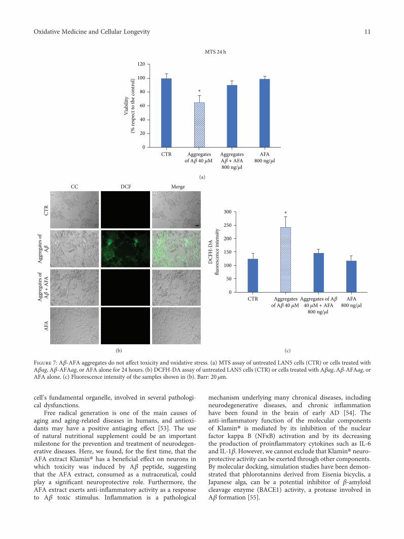
cell’s fundamental organelle, involved in several pathologi-cal dysfunctions.
Free radical generation is one of the main causes ofaging and aging-related diseases in humans, and antioxi-dants may have a positive antiaging effect [53]. The useof natural nutritional supplement could be an importantmilestone for the prevention and treatment of neurodegen-erative diseases. Here, we found, for the first time, that theAFA extract Klamin® has a beneficial effect on neurons inwhich toxicity was induced by Aβ peptide, suggestingthat the AFA extract, consumed as a nutraceutical, couldplay a significant neuroprotective role. Furthermore, theAFA extract exerts anti-inflammatory activity as a responseto Aβ toxic stimulus. Inflammation is a pathological
mechanism underlying many chronical diseases, includingneurodegenerative diseases, and chronic inflammationhave been found in the brain of early AD [54]. Theanti-inflammatory function of the molecular componentsof Klamin® is mediated by its inhibition of the nuclearfactor kappa B (NFκB) activation and by its decreasingthe production of proinflammatory cytokines such as IL-6and IL-1β. However, we cannot exclude that Klamin® neuro-protective activity can be exerted through other components.By molecular docking, simulation studies have been demon-strated that phlorotannins derived from Eisenia bicyclis, aJapanese alga, can be a potential inhibitor of β-amyloidcleavage enzyme (BACE1) activity, a protease involved inAβ formation [55].
120
MTS 24 h
80
100
60
40
20
0CTR
Viab
ility
(% re
spec
t to
the c
ontro
l)
Aggregatesof A�훽 40 �휇M
AggregatesA�훽 + AFA800 ng/�휇l
AFA800 ng/�휇l
⁎
(a)
CC
CTR
AFA
Agg
rega
tes o
fA�훽
+ A
FAA
ggre
gate
s of
A�훽
DCF Merge
(b)
⁎300
200
250
150
100
50
0
DCF
H-D
Aflu
ores
cenc
e int
ensit
y
CTR Aggregatesof A�훽 40 �휇M
Aggregates of A�훽40 �휇M + AFA
800 ng/�휇l
AFA800 ng/�휇l
(c)
Figure 7: Aβ-AFA aggregates do not affect toxicity and oxidative stress. (a) MTS assay of untreated LAN5 cells (CTR) or cells treated withAβag, Aβ-AFAag, or AFA alone for 24 hours. (b) DCFH-DA assay of untreated LAN5 cells (CTR) or cells treated with Aβag, Aβ-AFAag, orAFA alone. (c) Fluorescence intensity of the samples shown in (b). Barr: 20μm.
11Oxidative Medicine and Cellular Longevity

Fibrillogenesis of Aβ is a relevant event, leading todeposition of amyloid plaques in AD brain. Use of inhibitorscan prevent this process [56]. Klamin® interferes on Aβaggregation stabilizing Aβ aggregates in a protective way toreduce oxidative stress. The kinetics of amyloid formationcan be described in a sigmoid curve subdivided in threestages: (a) the slow lag nucleation phase, (b) the fast-exponential elongation phase, and (c) the saturation phase.Klamin® blocks the elongation phase, producing aggregatesof shorter dimensions. We can suppose that the various anti-oxidant components of AFA extract, from its polyphenols toits phycocyanins, interact with Aβ, inhibiting its extensionand destabilizing the preformed fibrils. It is known that anti-oxidants, containing one or more phenolic rings, are able tointeract with the aromatic residues of the amyloid peptides,destabilizing their well-ordered self-assembly process [37].Thus, Klamin® could have a disaggregating activity on Aβ.
In conclusion, Klamin® is a reservoir of effectivemolecules with numerous health benefits. The intrinsicantioxidant, anti-inflammatory, and antifibrillogenesis prop-erties of its compounds suggest that it could be used as aninnovative approach for the treatment and/or prevention ofneurodegenerative disease.
Data Availability
The data used to support the findings of this study areavailable from the corresponding author upon request.
Disclosure
Neither the funding agency nor any outside organizationhas participated in the study design or have any competingof interest.
Conflicts of Interest
The authors declare that there is no conflict of interestsregarding the publication of this paper. Dr. Stefano Scogliois the owner and manager of the Nutrigea company that pro-vided us the Klamath. The research was funded by Nutrigea.Nutrigea had the final approval of the manuscript.
Acknowledgments
The authors wish to thank Dr. Alessia Provenzano andMister Luca Caruana for their useful technical support.
References
[1] T. L. Hamilton, D. A. Bryant, and J. L. Macalady, “The role ofbiology in planetary evolution: cyanobacterial primary pro-duction in low-oxygen Proterozoic oceans,” EnvironmentalMicrobiology, vol. 18, no. 2, pp. 325–340, 2016.
[2] W. K. Dodds, D. A. Gudder, and D.Mollenhauer, “The ecologyof Nostoc,” Journal of Phycology, vol. 31, no. 1, pp. 2–18, 1995.
[3] H. Choi, S. J. Mascuch, F. A. Villa et al., “Honaucins A−C,potent inhibitors of inflammation and bacterial quorum sens-ing: synthetic derivatives and structure-activity relationships,”Chemistry & Biology, vol. 19, no. 5, pp. 589–598, 2012.
[4] E. Christaki, P. Florou-Paneri, and E. Bonos, “Microalgae:a novel ingredient in nutrition,” International Journal ofFood Sciences and Nutrition, vol. 62, no. 8, pp. 794–799,2011.
[5] C. S. Ku, Y. Yang, Y. Park, and J. Lee, “Health benefits ofblue-green algae: prevention of cardiovascular disease andnonalcoholic fatty liver disease,” Journal of Medicinal Food,vol. 16, no. 2, pp. 103–111, 2013.
[6] C. M. Reddy, V. B. Bhat, G. Kiranmai, M. N. Reddy,P. Reddanna, and K. M. Madyastha, “Selective inhibitionof cyclooxygenase-2 by C-phycocyanin, a biliprotein fromSpirulina platensis,” Biochemical and Biophysical ResearchCommunications, vol. 277, no. 3, pp. 599–603, 2000.
[7] C. Romay, J. Armesto, D. Remirez, R. González, N. Ledon,and I. García, “Antioxidant and anti-inflammatory proper-ties of C-phycocyanin from blue-green algae,” InflammationResearch, vol. 47, no. 1, pp. 36–41, 1998.
[8] C. Romay, N. Ledón, and R. González, “Further studies onanti-inflammatory activity of phycocyanin in some animalmodels of inflammation,” Inflammation Research, vol. 47,no. 8, pp. 334–338, 1998.
[9] L. Baroni, S. Scoglio, S. Benedetti et al., “Effect of a Klamathalgae product (“AFA-B12”) on blood levels of vitamin B12and homocysteine in vegan subjects: a pilot study,” Interna-tional Journal for Vitamin and Nutrition Research, vol. 79,no. 2, pp. 117–123, 2009.
[10] R. Pawlak, S. E. Lester, and T. Babatunde, “The prevalence ofcobalamin deficiency among vegetarians assessed by serumvitamin B12: a review of literature,” European Journal ofClinical Nutrition, vol. 68, no. 5, pp. 541–548, 2014.
[11] R. I. Kushak, C. Drapeau, and H. D. Winter, “The effect ofblue-green algae Aphanizomenon Flos Aquae on nutrientassimilation in rats,” JANA, vol. 3, pp. 35–39, 2001.
[12] J. P. Kamat, K. K. Boloor, and T. P. A. Devasagayam,“Chlorophyllin as an effective antioxidant against membranedamage in vitro and ex vivo,” Biochimica et Biophysica Acta(BBA) - Molecular and Cell Biology of Lipids, vol. 1487,no. 2-3, pp. 113–127, 2000.
[13] S. Rinalducci, P. Roepstorff, and L. Zolla, “De novo sequenceanalysis and intact mass measurements for characterizationof phycocyanins subunit isoforms from the blue-green algaAphanizomenon flos-aquae,” Journal of Mass Spectrometry,vol. 44, no. 4, pp. 503–515, 2009.
[14] S. Benedetti, F. Benvenuti, S. Scoglio, and F. Canestrari,“Oxygen radical absorbance capacity of phycocyanin andphycocyanobilin from the food supplement Aphanizomenonflos-aquae,” Journal of Medicinal Food, vol. 13, no. 1,pp. 223–227, 2010.
[15] A. Cavalchini and S. Scoglio, “Complementary treatment ofpsoriasis with an AFA-phyocyanins product: a preliminary10-cases study,” International Medical Journal, vol. 16,pp. 221–224, 2009.
[16] S. Scoglio, V. Lo Curcio, S. Catalani, F. Palma, S. Battistelli, andS. Benedetti, “Inhibitory effects of Aphanizomenon flos-aquaeconstituents on human UDP-glucose dehydrogenase activity,”Journal of Enzyme Inhibition andMedicinal Chemistry, vol. 31,no. 6, pp. 1492–1497, 2016.
[17] M. Federici, R. Geracitano, A. Tozzi et al., “Trace aminesdepress GABAB response in dopaminergic neurons by inhibit-ing G-βγ-gated inwardly rectifying potassium channels,”Molecular Pharmacology, vol. 67, no. 4, pp. 1283–1290, 2005.
12 Oxidative Medicine and Cellular Longevity

[18] H. C. Sabelli and A. D. Mosnaim, “Phenylethylamine hypoth-esis of affective behavior,” American Journal of Psychiatry,vol. 131, no. 6, pp. 695–699, 1974.
[19] H. Sabelli, P. Fink, J. Fawcett, and C. Tom, “Sustainedantidepressant effect of PEA replacement,” The Journal ofNeuropsychiatry and Clinical Neurosciences, vol. 8, no. 2,pp. 168–171, 1996.
[20] S. Scoglio, Y. Benedetti, F. Benvenuti, S. Battistelli,F. Canestrari, and S. Benedetti, “Selective monoamine oxi-dase B inhibition by an Aphanizomenon flos-aquae extractand by its constitutive active principles phycocyanin andmycosporine-like amino acids,” Phytomedicine, vol. 21,no. 7, pp. 992–997, 2014.
[21] A. D. Genazzani, E. Chierchia, C. Lanzoni et al., “Effects ofKlamath algae extract on psychological disorders anddepression in menopausal women: a pilot study,” MinervaGinecologica, vol. 62, no. 5, pp. 381–388, 2010.
[22] P. Bellingeri, M. Bonucci, and S. Scoglio, “Complementarytreatment of mood disturbances in terminally ill oncologicalpatients with the Aphanizomenon flos aquae extract Kla-min®,” Advances in Complementary and Alternative Medicine,vol. 1, pp. 1–5, 2018.
[23] M. Cremonte, D. Sisti, I. Maraucci et al., “Complementary andalternative supplementation with the Klamath algae extractKlamin® on attention deficit/hyperactivity disorder,” Journalof Medicinal Food, vol. 20, pp. 1233–1239, 2017.
[24] S. Sedriep, X. Xia, F. Marotta et al., “Beneficial nutraceuticalmodulation of cerebral erythropoietin expression and oxida-tive stress: an experimental study,” Journal of BiologicalRegulators and Homeostatic Agents, vol. 25, no. 2, pp. 187–194, 2011.
[25] A. Babusyte, M. Kotthoff, J. Fiedler, and D. Krautwurst,“Biogenic amines activate blood leukocytes via trace amine-associated receptors TAAR1 and TAAR2,” Journal of Leuko-cyte Biology, vol. 93, no. 3, pp. 387–394, 2013.
[26] M. Robinson, B. Y. Lee, and F. T. Hane, “Recent progress inAlzheimer's disease research, part 2: genetics and epidemiol-ogy,” Journal of Alzheimer's Disease, vol. 57, no. 2, pp. 317–330, 2017.
[27] M. D. Hurd, P. Martorell, A. Delavande, K. J. Mullen, andK. M. Langa, “Monetary costs of dementia in the UnitedStates,” The New England Journal of Medicine, vol. 368,no. 14, pp. 1326–1334, 2013.
[28] T. Rapp, B. H. Apouey, C. Senik, and REAL.FR/DSA group,“The impact of institution use on the wellbeing of Alzheimer'sdisease patients and their caregivers,” Social Science &Medicine, vol. 207, pp. 1–10, 2018.
[29] J. M. Zissimopoulos, B. C. Tysinger, P. A. St.Clair, andE. M. Crimmins, “The impact of changes in population healthand mortality on future prevalence of Alzheimer's disease andother dementias in the United States,” The Journals of Geron-tology: Series B, vol. 73, Supplement 1, pp. S38–S47, 2018.
[30] W. L. Klein, W. B. Stine Jr., and D. B. Teplow, “Smallassemblies of unmodified amyloid β-protein are the proximateneurotoxin in Alzheimer’s disease,” Neurobiology of Aging,vol. 25, no. 5, pp. 569–580, 2004.
[31] R. Castellani, K. Hirai, G. Aliev et al., “Role of mitochondrialdysfunction in Alzheimer’s disease,” Journal of NeuroscienceResearch, vol. 70, no. 3, pp. 357–360, 2002.
[32] P. Picone, R. Carrotta, G. Montana, M. R. Nobile, P. L. SanBiagio, and M. di Carlo, “Aβ oligomers and fibrillar
aggregates induce different apoptotic pathways in LAN5neuroblastoma cell cultures,” Biophysical Journal, vol. 96,no. 10, pp. 4200–4211, 2009.
[33] P. Picone, D. Giacomazza, V. Vetri et al., “Insulin-activatedAkt rescues Aβ oxidative stress-induced cell death by orches-trating molecular trafficking,” Aging Cell, vol. 10, no. 5,pp. 832–843, 2011.
[34] P. Picone, D. Nuzzo, L. Caruana, V. Scafidi, and M. di Carlo,“Mitochondrial dysfunction: different routes to Alzheimer'sdisease therapy,” Oxidative Medicine and Cellular Longevity,vol. 2014, Article ID 780179, 11 pages, 2014.
[35] D. Nuzzo, L. Inguglia, J. Walters, P. Picone, and M. Di Carlo,“A shotgun proteomics approach reveals a new toxic role forAlzheimer’s disease Aβ peptide: spliceosome impairment,”Journal of Proteome Research, vol. 16, no. 4, pp. 1526–1541,2017.
[36] D. Nuzzo, P. Picone, L. Caruana et al., “Inflammatorymediators as biomarkers in brain disorders,” Inflammation,vol. 37, no. 3, pp. 639–648, 2014.
[37] A. Sgarbossa, D. Giacomazza, and M. Di Carlo, “Ferulic acid: ahope for Alzheimer’s disease therapy from plants,” Nutrients,vol. 7, no. 7, pp. 5764–5782, 2015.
[38] P. Picone, M. L. Bondi, P. Picone et al., “Ferulic acid inhibitsoxidative stress and cell death induced by Ab oligomers:improved delivery by solid lipid nanoparticles,” Free RadicalResearch, vol. 43, no. 11, pp. 1133–1145, 2009.
[39] P. Picone, D. Nuzzo, and M. Di Carlo, “Ferulic acid: a naturalantioxidant against oxidative stress induced by oligomericA-beta on sea urchin embryo,” The Biological Bulletin,vol. 224, no. 1, pp. 18–28, 2013.
[40] R. Kahl and H. Kappus, “Toxicology of the synthetic antioxi-dants BHA and BHT in comparison with the natural antioxi-dant vitamin E,” Zeitschrift für Lebensmittel-Untersuchungund -Forschung, vol. 196, no. 4, pp. 329–338, 1993.
[41] I. Ignat, I. Volf, and V. I. Popa, “A critical review of methodsfor characterisation of polyphenolic compounds in fruits andvegetables,” Food Chemistry, vol. 126, no. 4, pp. 1821–1835,2011.
[42] M. Naczk and F. Shahidi, “Extraction and analysis of phenolicsin food,” Journal of Chromatography A, vol. 1054, no. 1-2,pp. 95–111, 2004.
[43] E. Rojas-Escudero, A. L. Alarcón-Jiménez, P. Elizalde-Galván,and F. Rojo-Callejas, “Optimization of carbohydrate silylationfor gas chromatography,” Journal of Chromatography A,vol. 1027, no. 1-2, pp. 117–120, 2004.
[44] P. Ninfali, A. Chiarabini, and D. Angelino, “The ORAC/kcalratio qualifies nutritional and functional properties of fruitjuices, nectars, and fruit drinks,” International Journal of FoodSciences and Nutrition, vol. 65, no. 6, pp. 708–712, 2014.
[45] G. Cao, H. M. Alessio, and R. G. Cutler, “Oxygen-radicalabsorbance capacity assay for antioxidants,” Free RadicalBiology & Medicine, vol. 14, no. 3, pp. 303–311, 1993.
[46] R. Carrotta, M. Di Carlo, M. Manno et al., “Toxicity of recom-binant β-amyloid prefibrillar oligomers on the morphogenesisof the sea urchin Paracentrotus lividus,” The FASEB Journal,vol. 20, no. 11, pp. 1916-1917, 2006.
[47] J. T. Hafting, J. S. Craigie, D. B. Stengel et al., “Prospects andchallenges for industrial production of seaweed bioactives,”Journal of Phycology, vol. 51, no. 5, pp. 821–837, 2015.
[48] J. M. Shick and W. C. Dunlap, “Mycosporine-like aminoacids and related gadusols: biosynthesis, accumulation, and
13Oxidative Medicine and Cellular Longevity

UV-protective functions in aquatic organisms,” AnnualReview of Physiology, vol. 64, no. 1, pp. 223–262, 2002.
[49] S. Scoglio, F. Canestrari, S. Benedetti, Y. Benedetti, andM. Delgado-Esteban, “Alphanizomenon flos aquae prepara-tion, extracts and purified components thereof for thetreatment of neurological, neurodegenerative and mooddisorders,” US Patent WO2008000430, 2008.
[50] N. Wada, T. Sakamoto, and S. Matsugo, “Multiple roles ofphotosynthetic and sunscreen pigments in cyanobacteriafocusing on the oxidative stress,” Metabolites, vol. 3, no. 2,pp. 463–483, 2013.
[51] R. Hille, “Molybdenum and tungsten in biology,” Trends inBiochemical Sciences, vol. 27, no. 7, pp. 360–367, 2002.
[52] M. J. Pushie and G. N. George, “Spectroscopic studies ofmolybdenum and tungsten enzymes,” Coordination ChemistryReviews, vol. 255, no. 9-10, pp. 1055–1084, 2011.
[53] D. Harman, “Aging: a theory based on free radical andradiation chemistry,” Journal of Gerontology, vol. 11, no. 3,pp. 298–300, 1956.
[54] I. Blasko, M. Stampfer-Kountchev, P. Robatscher, R. Veerhuis,P. Eikelenboom, and B. Grubeck-Loebenstein, “How chronicinflammation can affect the brain and support the develop-ment of Alzheimer's disease in old age: the role of microgliaand astrocytes,” Aging Cell, vol. 3, no. 4, pp. 169–176, 2004.
[55] H. A. Jung, S. H. Oh, and J. S. Choi, “Molecular dockingstudies of phlorotannins from Eisenia bicyclis with BACE1inhibitory activity,” Bioorganic & Medicinal Chemistry Letters,vol. 20, no. 11, pp. 3211–3215, 2010.
[56] D. M.Walsh, A. Lomakin, G. B. Benedek, M. M. Condron, andD. B. Teplow, “Amyloid β-protein fibrillogenesis. Detection ofa protofibrillar intermediate,” Journal of Biological Chemistry,vol. 272, no. 35, pp. 22364–22372, 1997.
14 Oxidative Medicine and Cellular Longevity