multivariate analysis of a personal series of 247 consecutive patients with liver metastases from...
TRANSCRIPT
Multivariate Analysis of a Personal Series of 247 Patients withLiver Metastases from Colorectal Cancer
11. Treatment by Intrahepatic Chemotherapy
JOSEPH G. FORTNER, M.D., JOHN S. SILVA, MAJ., M.C., U.S.A.F., EDWIN B. COX, M.D.,ROBERT B. GOLBEY, M.D., HELEN GALLOWITZ, R.N., BARBARA J. MACLEAN, B.A.
One hundred and seventeen patients with colorectal hepatic me-tastases had insertion of catheters for infusional chemotherapy.The two-year survival estimate of patients with less than 50%hepatic replacement and no other adverse factors was 37%. Nineof 39 patients in this group are alive at 24 months. The catheterswere placed into the hepatic artery (HA), 23; into the portalvenous system (PV), 18; into both HA and PV, 64; or into anaccessory HA following ligation, 12. Fifty-nine patients hadligation of the common HA also. The 30-day postoperative mor-tality rate was 1.7% (2/117) and morbidity was 37.6%. Themajority of complications were related to fever (61%, 27/44).Over the past 2 years, 87% of patients have been dischargedwithin 10 days following surgery. Preoperative CEA ranged from0.5-12,150 ng/ml (median 165 ng/ml); 93% (78/84) had plasmaCEA levels exceeding 5 ng/ml. All patients had careful intra-operative staging: per cent hepatic replacement (PHR) rangedfrom 5-95% (median 60%); portal, celiac, or periaortic lymphnode metastases were observed in 31% (36/117). Initial intra-hepatic chemotherapy programs consisted of either CAMF (9patients), MAFL (60 patients), BFS (22 patients), continuousinfusion FUDR (14 patients), or miscellaneous drugs (4 patients).Median survival time of 109 evaluable patients was 11.5 months.The effect of 20 variables on the observed survival time wasanalyzed using a multivariate proportional hazard model. Threevariables were found to have influenced survival: PHR emergedas the most significant, p = 0.000001. Increased PHR was as-sociated with decreased survival time. Lymph node metastasesand prior chemotherapy were prognostic factors also, p = 0.0006and p = 0.03, respectively. No patient with PHR greater than80% lived more than 8 months. Utilization of these variableswould appear to be necessary for accurate stratification andevaluation of future chemotherapy trials in patients with col-rectal hepatic metastases.
EPATIC METASTASES from colorectal cancer havebeen treated most commonly with systemic che-
motherapy. Fluorouracil (5FU) does not appreciably im-
From the Departments of Surgery and Medicine, MemorialSloan-Kettering Cancer Center, New York, New York, and
the Department of Medicine, Duke University MedicalCenter, Durham, North Carolina
prove survival but does have an objective response rateof about 20%.' Polychemotherapy appears to improvethe response rate, although survival of patients with he-patic metastases may be only slightly improved.2 Mediansurvival of untreated hepatic metastases has been reportedto range from 3.4 to 24 months depending upon theextent of liver replacement.3~
Infusion of 5FU or 5-fluoro-2-deoxyuridine (FUDR)via the hepatic artery results in a 5- to 10-fold increasein tumor drug concentration compared with systemicadministration.8 A number of studies have demonstrateda response rate of 50-70% using regional intrahepaticadministration of 5 FU or FUDR even in patients pre-viously treated with systemic 5 FU.9-" More recently,Ensminger et al.'2 reported an 83% overall response ratewith intra-arterial FUDR using an implantable pumpdrug delivery system. Median survival of highly selectedpatients with metastases only in the liver was approxi-mately 21 months.
In none ofthe reports using either systemic or infusionalchemotherapy does there appear to have been a systematicattempt to identify patients with surgically resectable dis-ease. Great reliance has been placed on indirect assessmentofthe extent and location of metastatic disease using liverscans, computerized axial tomography, and physical ex-amination. Although indirect measurements includingangiography are quite accurate in detecting recurrent dis-ease, they are highly inaccurate in assessing the locationand extent of liver involvement by cancer.'3 It wouldappear that patients with potentially surgically curable
317
Reprint requests: Joseph G. Fortner, MD, Department of Surgery,Memorial Sloan-Kettering Cancer Center, 1275 York Avenue, NewYork, NY 10021.
Submitted for publication: June 6, 1983.
318 FORTNER AN
TABLE 1. Site of Catheter Placement
Common Hepatic Artery
Catheter Site Ligation No Ligation Total
Hepatic artery (HA) only 19 4 23Portal vein (PV) only 16 2 18HA and PV 14 50 64Accessory hepatic artery(AHA)* 6 6
AHA* and PV 4 2 6
Total 59 58 117
* In all cases the accessory hepatic arteries (12 patients) were ligatedproximal to the catheter insertion site.
metastatic coloretal cancer in the liver may have beenincluded in some series of patients treated only by che-motherapy.
Factors affecting survival of treated patients with co-lorectal metastases have been investigated by few authors.Almersjo'4 found that survival time varied inversely withper cent hepatic replacement. Cady"5 found the per centofliver replacement to be the most significant determinantof survival. Others6"16 have used clinical findings with orwithout laboratory data to stage patients. However, noneof these studies have evaluated the interdependence of
TABLE 2. Thirty-day Postoperative MorbidityFollowing Intrahepatic Catheters
Complication Number*
Fever 27Atelectasis 8Bleeding 5Pneumonia 2Pleural effusion IHepatorenal syndrome I
Total 44
* Excludes complications in two 30-day postoperative deaths and onedeath at 42 days.
TABLE 3. Sites ofExtrahepatic Metastatic Disease
Intraperitoneal Metastases
Lymph Node PeritonealMetastases None Surface Pelvic Omental
None 67 7 5Portal (P) 3P and Celiac (C) 17 3 2 1P and C and Aortic 3 3 3 1
Total* 90 13 10 2
* Excludes two patients in whom nodal and peritoneal status couldnot be determined.
ID OTHERS Ann. Surg. * March 1984
clinical, intraoperative, or biochemical variables. In thepresent study, 1 17 consecutive patients, staged at surgeryby the senior author and shown to have nonresectabledisease, have had catheters placed in their hepatic arteryand/or portal vein for regional chemotherapy. In an at-tempt to define independent prognostic factors, a mul-tivariate proportional hazard model was used to evaluatethe effects of 20 variables on the patients' survival. Theyare a subset of 582 patients with primary or secondaryliver disease treated by the senior author at MemorialSloan-Kettering Cancer Center during the past 11 years.
Materials and Methods
Patient Population
The charts of 247 patients with a histologic diagnosisof colorectal cancer metastatic to the liver were reviewed.The 1 7 patients (47%) who had insertion ofhepatic and/or portal vein catheters for intrahepatic chemotherapyare the basis ofthis report.17-20 Seventy-five patients (30%)who had surgical removal oftheir metastatic liver diseaseare the subject of a companion report2' and are not con-sidered here. Three patients who had hepatic artery li-gation without catheter insertion, two who had isolationchemotherapy perfusion,22 and 50 who had a biopsy onlyare not included in this analysis.
Preoperative evaluation of the patients included liverfunction tests and, since 1974, plasma levels of CEA.Computerized tomography scans, as well as selected celiacand superior mesenteric angiography were done beforesurgery.2' At laparotomy, the extent of liver replacementby tumor was estimated by inspection and palpation anda search made for extrahepatic metastatic disease. Fol-lowing biopsy of hepatic metastases and other suspectedintraabdominal disease or lymph node metastases, a cath-eter (since 1976, a Raimondi (American Heyer-Schulte,Goleta, California) anti-reflux catheter) has been placedin the hepatic artery via the gastroduodenal artery and/or into the portal venous system via the inferior mesenteric
24vein.
Surgical Procedure
The site of catheter insertion and operative procedureare shown in Table 1. Twenty-three patients had insertionof a catheter into the hepatic artery only; 18 patients hada catheter placed in the portal venous system only; and64 patients had catheters inserted into both the hepaticartery and portal vein. Fifty-nine of the 1 17 patients hadligation oftheir common hepatic artery also. Twelve otherpatients had hepatic arterial anatomy necessitating cath-eter placement into an aberrant vessel with concomitant
INTRAHEPATIC CHEMOTHERAPY OF LIVER METASTASES
proximal ligation. Seven patients had small accessory he-patic arteries which were ligated in addition to the aboveprocedures. Ninety per cent (79/88) of portal vein cath-eters were placed via the inferior mesenteric vein (25catheters) or a branch of the middle colic vein (54 cath-eters).The 30-day postoperative mortality rate was a gratifying
1.7% (2/117). Both deaths occurred in patients with morethan 75% liver replacement. One additional patient diedof hepatic failure on the forty second day after hepaticartery ligation and cannulation. The postoperative mor-
bidity was 37.6% (Table 2). The majority ofcomplications(61%) were related to unexplained fever (temperature> 38.5 C) which usually occurred on the second throughfourth postoperative day without elevation of neutrophilcount or chest x-ray changes. This was considered likelydue to tumor or tumor necrosis. The median postoperativehospital stay was 11 days. In the past 2 years, 87% ofpatients have been discharged within 10 days followingsurgery.The results of intraoperative staging are shown in Table
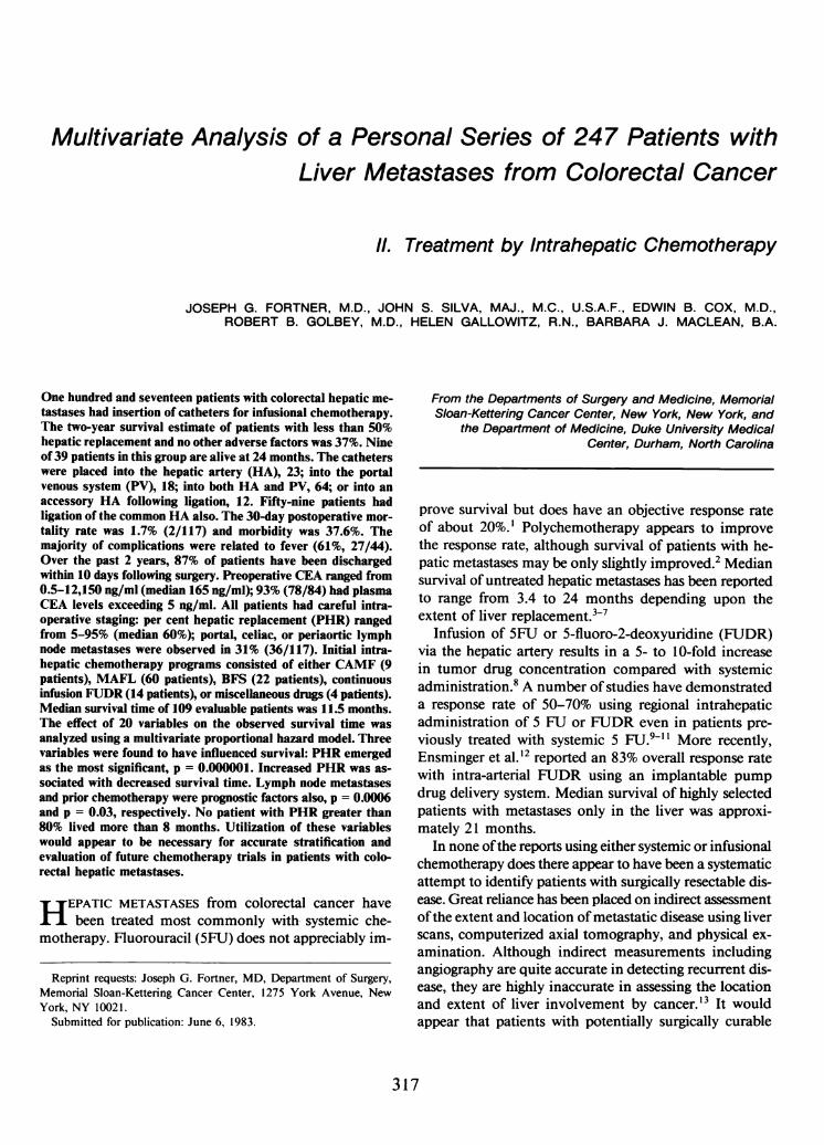
3. Sixty-seven patients (58%) had liver involvement only.In addition to liver metastases, 23 patients had regionalor periaortic lymph node metastases and 12 had minimalperitoneal metastases. An additional 13 patients had bothlymph node and peritoneal metastases. Eighteen patientshad a small amount of ascites. Per cent hepatic replace-ment (PHR) in 114 patients ranged from 5-95% with a
median of60% (Figure 1). PHR was not recorded in threepatients.
Chemotherapy
One of the 4 intrahepatic drug combinations, CAMF,MAFL, BFS, or FUDR, was used in the majority of pa-
tients (Table 4). CAMF was used from 1973 to 1975 andconsisted of Cytoxan (1 mg/kg) orally each day. Acti-nomycin D (1 mg), Methotrexate (10 mg) and 5FU (10mg/kg) were given together once weekly via intrahepaticcatheter. MAFL was used from 1976 to 1979. It consistedof a 3-week cycle of bolus intrahepatic chemotherapy:Actinomycin D (1 mg) + 5FU (10 mg/kg) week 1, Ac-tinomycin D (1 mg) week 2, and Methotrexate (20 mg)+ Levamisole (150 mg/day p.o. for 3 days) week 3. Be-ginning in 1979, a modification of the MOF regimenoriginally described by Moertel25 was used. BFS consistedofa 70-day cycle ofbolus intrahepatic injections ofBCNU(30 mg/M2, daily X 5 days week 1); 5FU (300 mg/M2,daily X 5 days week 1 and 6) and Streptozotocin (500mg/mi, once weekly for 11 weeks). Continuous intra-hepatic infusion of FUDR (0.3 mg/kg/day for 14 daysfollowed by 14 days saline infusion) was used in the most
25 r
af)cn
CSa1)4-6-
0
a)
4-
E
z3
20
25
10 [
5
0 20 40 60 80 100
Percent hepatic replacementFIG. 1. Per cent hepatic replacement in patients with colorectal hepaticmetastases determined at laparotomy.
recent patients. When patients had evidence of local fail-ure (liver progression) a second regimen of intrahepaticchemotherapy was usually given. All patients in this seriesreceived at least 2 weeks of intrahepatic chemotherapy.When patients had distant metastases or catheter failure,systemic chemotherapy was instituted.
Statistical Analyses
All data were stored in CLINFO (a data analysis system,Bolt, Beranek and Newman, Boston, MA). Data wereanalyzed for significance using statistical programs resi-dent in CLINFO and included descriptive statistics, two-tailed t-test, the product-limit life table analysis, and gen-eralized Wilcoxon test for life tables. Evaluation of the
TABLE 4. Initial Intrahepatic Chemotherapy
Chemotherapy Regimen* Number of Patients
MAFL 60BFS 22FUDR 14CAMF 9Miscellaneous 4
Total 109t
t Excludes eight patients: two 30-day postoperative deaths, one 42-day death, and five patients receiving less than 2 weeks of intrahepaticchemotherapy.
* See Chemotherapy section in Materials and Methods for explanationof the chemotherapy regimens MAFL, BFS, FUDR, and CAMF.
VoL. 199 . No. 3 319
FORTNER AND OTHERS
50 r
(U)
'4--
c
Q)
a)
0
EL
z6-
40
30
20 p
10
30 40 50 60 70
YearsFIG. 2. Age distribution of patients with colorectal hepatic metastasestreated with intrahepatic chemotherapy.
effect of multiple variables on the survival of patients wascarried out using a step-wise proportional hazard analysis.The association ofputative prognostic factors with survivalduration were estimated using Cox's proportional hazardsurvival regression model.26 In this model:
Xi(t) = X0(t) exp (2;#jX,j),j
where Xi and Xo are the hazard functions for the individualand overall group; /j is the regression coefficient for thejth covariate, and Xij is the value of the jth covariate inthe ith patient. The fj are estimated using the maximumlikelihood techniques. The criterion for inclusion of a
1.0
Estimohtd survival95% confidence limits
0.8 of estmted swMvival
0.6
0
OAo0
a.0.2
0 6 12 18 24 30
Months
FIG. 3. Survival of 109 patients with colorectal hepatic metastases treatedwith intrahepatic chemotherapy. Vertical marks: patients alive at lastexamination.
variable in the model was significance level less than 0.1for its step-wise inclusion. Significant regression coeffi-cients are interpreted as having an adverse prognosticimplication when the coefficient is positive and a favorableimplication when negative.
Results
Patient Population
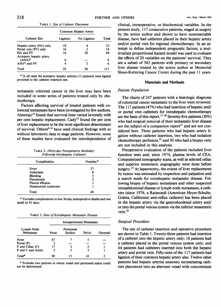
There were 68 men and 49 women with a median ageof56 years (range 26-79 years, Fig. 2). The age distributionof male patients did not differ from female patients. Kar-nofsky performance status ranged from 50% to 90% witha median of 80%. The sigmoid colon (41/117) or rightcolon (27/117) represented the most common sites ofprimary disease. Most patients (83/117) had Dukes' Cprimary lesions, 28 had Dukes' B, and 6 were unclassified.Fifty-five per cent (64/117) of the population had livermetastases at the time of colon resection. Since 1977,71% of patients with metachronous liver metastases havehad the diagnosis of liver metastasis first suspected onthe basis of an elevated post-colon resection CEA level.Preoperative CEA levels ranged from 0.5-12,150 ng/ml,with a median value of 165 ng/ml. Ninety-three per cent(78/84) of patients had values above 5 ng/ml; 63% (53/84) had CEA levels above 100 ng/ml, and 24% (20/84)had CEA levels exceeding 1,000 ng/ml.
Survival Analysis
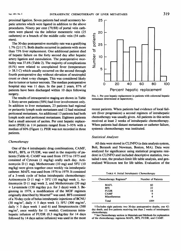
Median survival of 109 evaluable patients was 11.5months from time of catheter placement (Fig. 3). Eightpatients were excluded for the following reasons: threepatients died postoperatively and five patients receivedlessthan 2 weeks ofintrahepatic chemotherapy. The initialintrahepatic chemotherapy regimens administered duringthe period of this study are shown in Table 4.The effect of 20 variables on patient survival was an-
alyzed using a step-wise proportional hazard model (Table5). Per cent hepatic replacement emerged as the mostsignificant determinant of survival, p = 10-6. IncreasingPHR was associated with shorter survival times (positivecorrelation coefficient). The extent of lymph node me-tastases was also a significant variable, p = 0.006. Moreextensive nodal involvement was associated with de-creased survival. Prior chemotherapy was a significantprognostic factor, p = 0.03; previously treated patientshad decreased survival time compared with untreatedpatients. Among the remaining 17 variables, only SGOThad borderline prognostic significance, p = 0.08. Site ofcatheter placement (hepatic artery, portal vein, or both),ligation of common hepatic artery, or intrahepatic che-motherapy regimen were not predictors of survival in this
320 Ann. Surg. * March 1984
INTRAHEPATIC CHEMOTHERAPY OF LIVER METASTASES
model. Dukes' classification, site ofcolon primary, disease-free interval, interval from colon resection to catheterinsertion, preoperative laboratory tests (bilirubin, alkalinephosphatase, lactic dehydrogenase, 5' nucleotidase, andCEA) did not add further information to the multivariatesurvival model (Table 5).The three significant variables were combined in an
attempt to delineate prognostic groups. Evaluable patientswere stratified into three groups based on per cent hepaticreplacement: group I, PHR < 50%; group II, PHR from55% to 80%; and group III, PHR > 80%. Each group was
divided into two subgroups based on the presence or
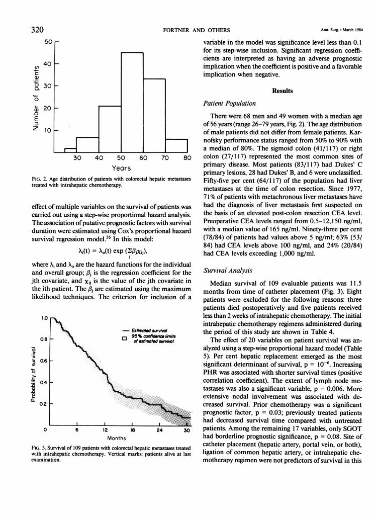
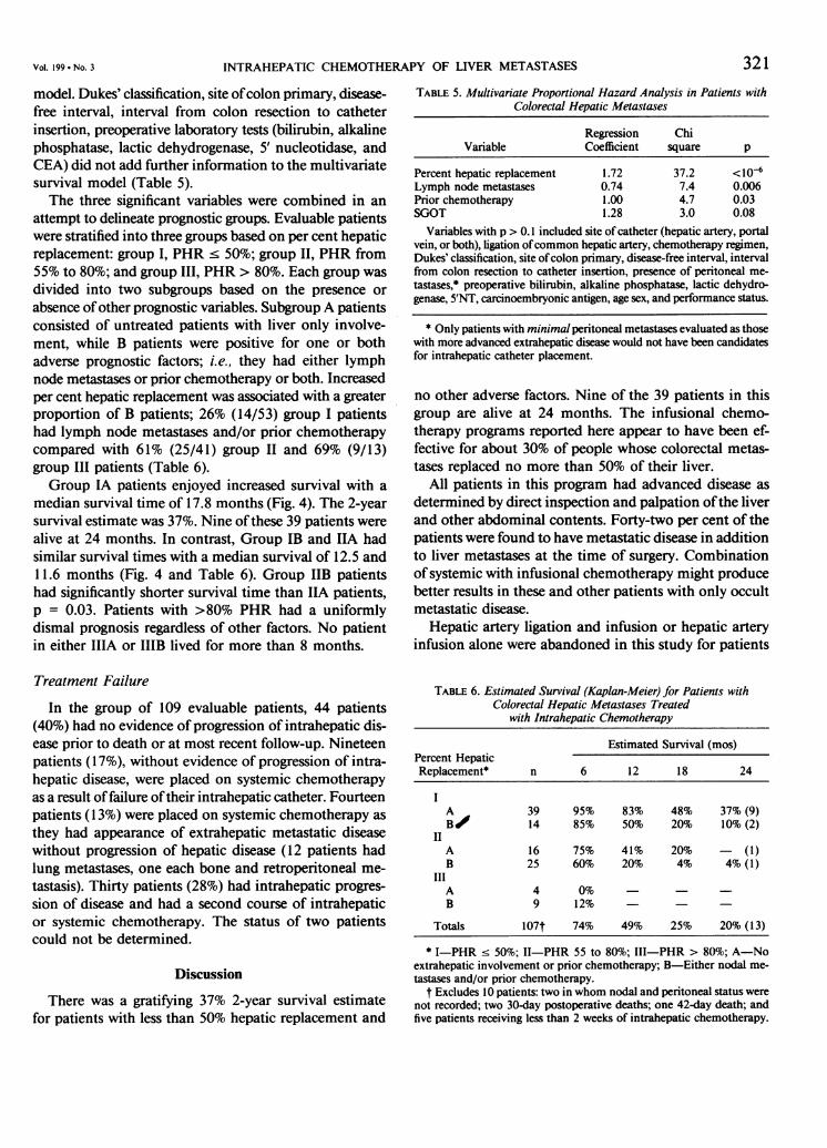
absence ofother prognostic variables. Subgroup A patientsconsisted of untreated patients with liver only involve-ment, while B patients were positive for one or bothadverse prognostic factors; i.e., they had either lymphnode metastases or prior chemotherapy or both. Increasedper cent hepatic replacement was associated with a greaterproportion of B patients; 26% (14/53) group I patientshad lymph node metastases and/or prior chemotherapycompared with 61% (25/41) group II and 69% (9/13)group III patients (Table 6).Group IA patients enjoyed increased survival with a
median survival time of 17.8 months (Fig. 4). The 2-yearsurvival estimate was 37%. Nine ofthese 39 patients werealive at 24 months. In contrast, Group IB and IIA hadsimilar survival times with a median survival of 12.5 and11.6 months (Fig. 4 and Table 6). Group IIB patientshad significantly shorter survival time than IIA patients,p = 0.03. Patients with >80% PHR had a uniformlydismal prognosis regardless of other factors. No patientin either IIIA or IIIB lived for more than 8 months.
Treatment Failure
In the group of 109 evaluable patients, 44 patients(40%) had no evidence of progression of intrahepatic dis-ease prior to death or at most recent follow-up. Nineteenpatients (17%), without evidence of progression of intra-hepatic disease, were placed on systemic chemotherapyas a result offailure oftheir intrahepatic catheter. Fourteenpatients (13%) were placed on systemic chemotherapy as
they had appearance of extrahepatic metastatic diseasewithout progression of hepatic disease (12 patients hadlung metastases, one each bone and retroperitoneal me-
tastasis). Thirty patients (28%) had intrahepatic progres-sion of disease and had a second course of intrahepaticor systemic chemotherapy. The status of two patientscould not be determined.
Discussion
There was a gratifying 37% 2-year survival estimatefor patients with less than 50% hepatic replacement and
TABLE 5. Multivariate Proportional Hazard Analysis in Patients withColorectal Hepatic Metastases
Regression ChiVariable Coefficient square p
Percent hepatic replacement 1.72 37.2 <10-6Lymph node metastases 0.74 7.4 0.006Prior chemotherapy 1.00 4.7 0.03SGOT 1.28 3.0 0.08
Variables with p > 0.1 included site of catheter (hepatic artery, portalvein, or both), ligation ofcommon hepatic artery, chemotherapy regimen,Dukes' classification, site ofcolon primary, disease-free interval, intervalfrom colon resection to catheter insertion, presence of peritoneal me-tastases,* preoperative bilirubin, alkaline phosphatase, lactic dehydro-genase, 5'NT, carcinoembryonic antigen, age sex, and performance status.
* Only patients with minimal peritoneal metastases evaluated as thosewith more advanced extrahepatic disease would not have been candidatesfor intrahepatic catheter placement.
no other adverse factors. Nine of the 39 patients in thisgroup are alive at 24 months. The infusional chemo-therapy programs reported here appear to have been ef-fective for about 30% of people whose colorectal metas-tases replaced no more than 50% of their liver.
All patients in this program had advanced disease as
determined by direct inspection and palpation ofthe liverand other abdominal contents. Forty-two per cent of thepatients were found to have metastatic disease in additionto liver metastases at the time of surgery. Combinationofsystemic with infusional chemotherapy might producebetter results in these and other patients with only occultmetastatic disease.
Hepatic artery ligation and infusion or hepatic arteryinfusion alone were abandoned in this study for patients
TABLE 6. Estimated Survival (Kaplan-Meier) for Patients withColorectal Hepatic Metastases Treated
with Intrahepatic Chemotherapy
Estimated Survival (mos)Percent HepaticReplacement* n 6 12 18 24
A 39 95% 83% 48% 37% (9)B/ 14 85% 50%O 20% 10% (2)
IIA 16 75% 41% 20% - (1)B 25 60% 20% 4% 4% (1)
IIIA 4 0%B 9 12%
Totals 107t 74% 49% 25% 20% (13)
* I-PHR < 50%; II-PHR 55 to 80%; III-PHR > 80%; A-Noextrahepatic involvement or prior chemotherapy; B-Either nodal me-tastases and/or prior chemotherapy.
t Excludes 10 patients: two in whom nodal and peritoneal status werenot recorded; two 30-day postoperative deaths; one 42-day death; andfive patients receiving less than 2 weeks of intrahepatic chemotherapy.
Vol. 199 No. 3 321
322 FORTNER AND OTHERS Ann. Surg. March 1984
1.0 L -- i -- Potients with no odverse foctors with avascular metastases on angiography. Only about
*L A,
'' *_ Potients with lymph node 10%27 ofpatients with metastatic colon cancer in the liver0.8 -1tmetastoses and/or prior chemo- have increased vascularity on hepatic arterial injection.
..I >', Their route to the liver had been portal venous so thatI..> I 1it appears that the predominant blood supply for 90% of
0.6 _ these patients was also the portal venous route. Subsequento N ' -_,, experiences proved that there was no difference in patient
04 L L Vsurvival as related to hepatic artery or portal vein routes.0 | . ,ofdrug administration.The portal venous route was cho-
L L. 1 6;sen since it has been free of the gastrointestinal compli-0.2 cations seen with the arterial route; the catheter is easier
X to place, and free oftroublesome complications. Problemsposed by hepatic arterial anomalies are avoided.
0 6 12 18 24 30 Comparison of these results with those of others isMonths difficult.28'29 The patients may not be comparable since
it seems likely that our patients with limited disease were
1.0 1 -- Patients surgically resected2' but such patients are likely to havePotients with no adverse factors been included in other reports dealing with the results of
| - Potients with lymph node systemic or infusional chemotherapy. Accurate intra-0.8 nmetostases ond/or prior chemo- operative staging of the patient's disease is essential to
o -,>V_ therapy evaluating resectability and this has seldom been done.Response rates rather than survival times are most com-
0.6 monly used. Response ofliver metastases to chemotherapyo is particularly difficult to assess. The healing surgical- 04 S- --. wound can make accurate liver measurements impossible;
\| often, different observers will obtain quite different liversizes at a given examination. This is further compounded
0.2 when a different person sees the patient at sequentialexaminations. Technetium scans are unreliable in deter-mining the extent of disease. Comparison of sequential
0 6 12 18 24 30 CAT scans of the liver gives only a rough estimate unlessMonths very dramatic changes have occurred. Accurate assess-
ment otherwise would require the same tomographic cut1.0 on each scan and this is not done ordinarily.
- - Patients with no adverse factors Despite these difficulties, certain comparisons can bePatndtor prior cyemotheropy made tentatively. Patients with untreated colorectal he-
0.8 and/or prior chemotherapy patic metastases have a median survival time which ranges
from 3.4 to 24 months.6'7'30 Systemic chemotherapy with
X 0.6 L 9 5FU does not improve the survival appreciably. Variouspolychemotherapy programs appear to improve responserate, although survival ofpatients with hepatic metastases
n 0.4 - may be only slightly improved.
2 Kemeny et al.3 initiated a pilot study of MOF-StrepX1L_, in 1976 in patients with advanced measurable colorectal
0.2I cancer. Seventy-four patients received an adequate trialof therapy with a complete or partial response rate of
l_____l_____I ____l _____l____ l_____ ,___ 32% (24/74). Forty-five per cent (26/58) ofliver metastases0 6 12 18 24 30 responded to therapy compared with a 17% response rate
Months reported in previous protocols using MOF alone. A sub-FIG. 4. Survival of 107 patients with colorectal hepatic metastases treated sequent prospective randomized study was carried outwith intrahepatic chemotherapy grouped by prognostic variables. (Top) using somewhat higher doses ofmCCNU and 5FU.32 TheSurvival ofpatients with less than 55% PHR; (Middle) Survival ofpatientswith PHR 55 to 80%; and (Bottom) Survival of patients with PHR complete or partial response rates were 34% (12/35) forgreater than 80%. Vertical tic marks: patients alive at last examination. MOF-Strep and 6% (2/34) for MOF alone.
Vol. 199 - No. 3 INTRAHEPATIC CHEMOTHERAPY OF LIVER METASTASES 323
The best reported results to date for infusional che-motherapy appear to be those of Ensminger et al., whoreported an 83% overall response rate with intra-arterialFUDR using an implantable pump drug delivery system. 12Median survival of patients with presumed hepatic onlymetastases was about 21 months. More time must elapsebefore these results can be fully evaluated. The study hasthe limitations described above, including the likely pres-ence of localized, surgically resectable disease in the mostfavorable group where the metastases were confined tothe liver. In the most favorable group ofthe present report(IA) the median survival was 17.8 months. This is unlikelyto be significantly different from that reported by En-sminger.The effectiveness of infusional chemotherapy vs. sys-
temic chemotherapy can only be shown by a randomizedstudy ofcarefully staged patients. This has not been carriedout to date and was not done in this study due to limi-tations in available clinical material. The present analysishas identified three prognostic variables which should beconsidered in the conduct of future studies.
It would appear desirable to use survival time as theend-point in evaluating results. One possibility is to usesurvival time until activity of the patient declines to lessthan 60% on the Karnofsky performance scale. Sixty percent is defined as when patients are unable to work butare able to live at home and care for most of their needsbut require occasional assistance.33
Staging of liver metastases as in this series has beenused previously only to a limited extent. Almserjo et al.'4attempted this in a small series of patients in whom themedian survival was only 4 months. Cady and Oberfieldobserved among patients receiving intra-arterial FUDRinfusion that median survival time decreased as percentageliver involvement by tumor increased: median survivaltime was 16 months for those with less than 25% liverreplacement, 13 months for those with 25-50% involve-ment, and 8 months for those with more extensive hepaticdisease.5The data ofthis report indicated little benefit from any
therapy after more than 50% of the liver was replaced bytumor. In this multivariate analysis, per cent hepatic re-placement proved to be the most important of the 20factors examined. Other significant variables related tothe amount of metastatic disease in and outside the liver,i.e., extent of lymph node involvement. Prior chemo-therapy was important, possibly since there would appearto be a natural selection of patients with resistant cancersto this program. Other factors were not influential: thedisease-free interval, Dukes' classification and site ofcolonprimary, age, sex, and liver function tests other thanSGOT. The preoperative CEA level was not a determinantfactor.
The low morbidity and mortality rates with relativelyshort hospitalization periods are gratifying. This is espe-cially so when it is realized that the median amount ofliver replacement in this series was 60%. These rates aredistinctly better than those experienced earlier, primarilydue to better patient selection. Patients with a massiveliver replaced by cancer, with jaundice, ascites, portalvenous thrombosis, or a Karnofsky performance status<60% are at excessive risk and will not benefit. Infusionalchemotherapy with presently available agents is best re-served for those who have nonresectable disease but with50% or less of the liver involved.
References
1. Moertel CG. Clinical management of advanced gastrointestinalcancer. Cancer 1975; 36:675-682.
2. Davis HL. Chemotherapy of large bowel cancer. Cancer 1982;50:2638-2646.
3. Wood CB, Gillis CR, Blumgart LH. A retrospective study of thenatural history of patients with liver metastases from colorectalcancer. Clin Oncol 1976; 2:285-288.
4. Jaffe BM, Donegan WL, Watson F, Spratt JS. Factors influencingsurvival in patients with untreated hepatic metastases. Surg Gy-necol Obstet 1968; 127: 1 -11.
5. Cady B, Oberfield RA. Regional infusion chemotherapy of hepaticmetastases from carcinoma of the colon. Am J Surg 1974;127:220-227.
6. Goslin R, Steele G, Zamchek N, et al. Factors influencing survivalin patients with hepatic metastases from adenocarcinoma of thecolon or rectum. Dis Colon Rectum 1982; 25:749-754.
7. Bengtsson G, Varlsson G, Hafstrom L, Jonsson P-E. Natural historyof patients with untreated liver metastases from colorectal cancer.Am J Surg 1981; 141:586-589.
8. Ensminger WD, Rosowsky A, Raso V, et al. A clinical-pharma-cological evaluation of hepatic arterial infusions of 5-fluoro-2'-deoxyuridine and 5-fluorouracil. Cancer Res 1978; 38:3784-3792.
9. Oberfield RA, McCaffrey JA, Polio J, et al. Prolonged and continuouspercutaneous intra-arterial hepatic infusion chemotherapy in ad-vanced metastatic liver adenocarcinoma from colorectal primary.Cancer 1979; 44:414-423.
10. Ansfield FJ, Ramirez G. The clinical results of 5-fluorouracil in-trahepatic arterial infusion in 528 patients with metastatic cancerto the liver. Prog Clin Cancer 1978; 7:217-233.
11. Reed ML, Vaitkevicius VK, Al-Sarraf M, et al. The practicality ofchronic hepatic artery infusion therapy ofprimary and metastatichepatic malignancies. Cancer 1981; 47:402-409.
12. Ensminger W, Niederhuber J, Gyves J, et al. Effective control ofliver metastases from colon cancer with an implanted systemfor hepatic arterial chemotherapy. Proc Am Soc Clin Oncol 1982;1:94.
13. Kim DK, McSweeney J, Yeh SDJ, Fortner JG. Tumors of the liveras demonstrated by angiography, scan and laparotomy. SurgGynecol Obstet 1975; 141:409-410.
14. Almersjo 0, Bengmark S. Rudenstam CM, et al. Evaluation ofhepatic dearterialization of primary and secondary cancer of theliver. Am J Surg 1972; 124:5-8.
15. Cady B, Oberfield RA. Regional infusion chemotherapy of hepaticmetastases from carcinoma of the colon. Am J Surg 1974;127:220-227.
16. Pettavel J, Morgenthaler F. Protracted arterial chemotherapy ofliver tumors: an experience of 107 cases over a 12-year period.Prog Clin Cancer 1978; 7:217-233.
17. Fortner JG. Infusion chemotherapy. In Shah J, ed. Current Conceptsin Surgical Oncology. New York: Memorial Sloan-KetteringCancer Center, 1980; 265-269.
324 FORTNER AND OTHERS Ann. Surg. March 1984
18. Fortner JG, Kim DK, Barrett MK, et al. Eight years' experiencewith the surgical management of 321 patients with liver tumors.In Fox BW, ed. Advances in Medical Oncology, Research, andEducation, Vol 5, Basis for Cancer Therapy 1. Oxford: PergamonPress, 1979; 257-261.
19. Fortner JG, Mulcare RJ, Solis A, et al. Treatment of primary andsecondary liver cancer by hepatic artery ligation and infusionalchemotherapy. Ann Surg 1973; 178:162-172.
20. Kim DK, Penneman R, Kallum BO, et al. Acute renal failure afterligation ofthe hepatic artery. Surg Gynecol Obstet 1976; 143:391-394.
21. Fortner JG, Silva JS, Golbey RB, et al. Multivariate analysis of apersonal series of247 patients with liver metastases from colorectalcancer. I. Treatment by hepatic resection. 1984; 199(3):306-316.
22. Fortner JG, Penneman R, Krakoff IH. Actinomycin D perfusionof the isolated liver for cancer. Bull Soc Int Chir 1975; 5:399-403.
23. Fortner JG, Beattie EJ Jr, Shiu MH, et al. Surgery in liver tumors.In Current Problems in Surgery. Chicago: Year Book MedicalPublishers, 1972.
24. Fortner JG, Pahnke LD. A new method for long term intrahepaticchemotherapy. Surg Gynecol Obstet 1976; 143:979-980.
25. Moertel CG, Schutt AJ, Hahn RG, Reitemeier RJ. Therapy ofadvanced colorectal cancer with a combination of 5-fluorouracil,methyl-1,3-cis(2 chloroethyl)-l-nitrosurea + vincristine. J NatICancer Inst 1975; 54:69-71.
26. Cox DR. Regression models and life tables. J Royal Statis Soc 1972;B34: 187-220.
27. Kim DK, Watson RC, Pahnke LD, Fortner JG. Tumor vascularityas a prognostic factor for hepatic tumors. Ann Surg 1977; 185:31-34.
28. Sundqvist K, Hafstrom LO, Jonnson PE, et al. Treatment of livercancer with regional intraarterial 5-FU infusion. Am J Surg 1978;136:328-331.
28. Almersjo 0, Bengmark S, Hafstrom L, Leissner K-H. Results ofliver dearterialization combined with regional infusion of 5-flu-orouracil for liver cancer. Acta Chir Scand 1976; 142:131-138.
30. Bengmark S, Hafstrom L. The natural history of primary and sec-ondary malignant tumors ofthe liver. I. The prognosis for patientswith hepatic metastases from colonic and rectal carcinoma bylaparotomy. Cancer 1969; 23:198-202.
31. Kemeny N, Yagoda A, Braun D, Golbey R. Therapy for metastaticcolorectal carcinoma with a combination of methyl-CCNU, 5-fluorouracil, vincristine and streptozotocin (MOF-Strep). Cancer1980; 45:876-881.
32. Kemeny NE. Chemotherapy of colorectal carcinoma. In CurrentConcepts in Medical Oncology. New York: Memorial Sloan-Kettering Cancer Center, 1980; 243-246.
33. Karnofsky DA, Burchenal JH. The clinical evaluation of chemo-therapeutic agents in cancer. In MacLeod CM, ed. Evaluationof Chemotherapeutic Agents. New York: Columbia UniversityPress, 1949; 191-205.