multiscale investigation of the depth-dependent mechanical anisotropy of the human corneal stroma
TRANSCRIPT

Cornea
Multiscale Investigation of the Depth-DependentMechanical Anisotropy of the Human Corneal Stroma
Cristina Labate,1 Marco Lombardo,2 Maria P. De Santo,1 Janice Dias,3 Noel M. Ziebarth,3
and Giuseppe Lombardo4,5
1Department of Physics, University of Calabria, Rende, Italy2Fondazione G.B. Bietti IRCCS, Rome, Italy3Biomedical Atomic Force Microscopy Laboratory, Department of Biomedical Engineering, University of Miami College ofEngineering, Coral Gables, Florida, United States4Consiglio Nazionale delle Ricerche, Istituto per i Processi Chimico-Fisici (CNR-IPCF), Messina, Italy5Vision Engineering Italy srl, Rome, Italy
Correspondence: Giuseppe Lombar-do, Consiglio Nazionale delle Ri-cerche, Istituto per i ProcessiChimico-Fisici (CNR-IPCF), VialeFerdinando Stagno d’Alcontres 37,98158 Messina, Italy;[email protected] Lombardo, Fondazione G.B.Bietti IRCCS, Via Livenza 3, 00198Rome, Italy;[email protected].
Submitted: March 15, 2015Accepted: April 27, 2015
Citation: Labate C, Lombardo M, DeSanto MP, Dias J, Ziebarth NM, Lom-bardo G. Multiscale investigation ofthe depth-dependent mechanical an-isotropy of the human corneal stroma.Invest Ophthalmol Vis Sci.2015;56:4053–4060. DOI:10.1167/iovs.15-16875
PURPOSE. To investigate the depth-dependent mechanical anisotropy of the human cornealstroma at the tissue (stroma) and molecular (collagen) level by using atomic force microscopy(AFM).
METHODS. Eleven human donor corneas were dissected at different stromal depths by using amicrokeratome. Mechanical measurements were performed in 15% dextran on the surface ofthe exposed stroma of each sample by using a custom-built AFM in force spectroscopy modeusing both microspherical (38-lm diameter) and nanoconical (10-nm radius of curvature)indenters at 2-lm/s and 15-lm/s indentation rates. Young’s modulus was determined byfitting force curve data using the Hertz and Hertz-Sneddon models for a spherical and aconical indenter, respectively. The depth-dependent anisotropy of stromal elasticity wascorrelated with images of the corneal stroma acquired by two-photon microscopy.
RESULTS. The force curves were obtained at stromal depths ranging from 59 to 218 lm. At thetissue level, Young’s modulus (ES) showed a steep decrease at approximately 140-lm stromaldepth (from 0.8 MPa to 0.3 MPa; P ¼ 0.03) and then was stable in the posterior stroma. At themolecular level, Young’s modulus (EC) was significantly greater than at the tissue level; EC
decreased nonlinearly with increasing stromal depth from 3.9 to 2.6 MPa (P ¼ 0.04). Thevariation of microstructure through the thickness correlated highly with a nonconstantprofile of the mechanical properties in the stroma.
CONCLUSIONS. The corneal stroma exhibits unique anisotropic elastic behavior at the tissue andmolecular levels. This knowledge may benefit modeling of corneal behavior and help in thedevelopment of biomimetic materials.
Keywords: atomic force microscopy, elasticity, anisotropy, microscopy
The histology and, accordingly, biomechanics of the humancorneal stroma are highly heterogeneous. There is exten-
sive knowledge on the existence of regional differences in thecollagen fibril bundles’ packing arrangement and lamellarorientation across and throughout the thickness of the humancorneal stroma.1–5 The anterior stroma consists of shortbundles of collagen fibrils that show dense intertwining andalso insert vertically into Bowman’s layer, thus contributing tomaintain corneal shape. The deeper stroma contains collagenfibril bundles that are arranged in wide lamellae orientedpredominantly along the superior/inferior and nasal/temporalmeridians and with reduced connections between adjacentlayers.
Several laboratory techniques have been used to elucidate
the depth-dependent mechanical properties of the human
corneal stroma.6–17 All of these studies have shown that the
elastic modulus within the stroma decreases from anterior to
posterior, regardless of the mechanical testing used. It is
therefore well accepted that the depth-dependent changes of
stromal mechanical elasticity are linked to the depth-dependent
change of stromal microstructure.
Atomic force microscopy (AFM) enables localized mechan-
ical sample testing and has been used to investigate the local
elastic modulus of the human corneal stroma at different
depths. Although the results from previous studies6–11 could
not be directly compared, because of the use of AFM tips with
variable geometry and dimension as well as different study
protocols, the elastic modulus is shown to decrease 40% to 80%
with depth from the anterior stroma to the posterior part of the
stroma.
The aim of this study was to provide information on the
depth-dependent compressive elastic modulus of the human
corneal stroma both at the tissue (stroma) and molecular
(collagen) level by using AFM. In addition, we correlated the
depth-dependent anisotropy of stromal elasticity with images of
the human corneal stroma acquired by second harmonic
generation (SHG) microscopy.
Copyright 2015 The Association for Research in Vision and Ophthalmology, Inc.
iovs.arvojournals.org j ISSN: 1552-5783 4053
Downloaded From: http://iovs.arvojournals.org/pdfaccess.ashx?url=/data/Journals/IOVS/934118/ on 06/29/2015

MATERIALS AND METHODS
Corneal Tissues
Donor human eye globes (n ¼ 11; age range, 19–92 years;postmortem interval range, 2.5–12.1 hours) were obtainedfrom the Florida Lions Eye Bank (Miami, FL, USA) in sealed vialswith gauze soaked in balanced salt solution. The whole globeswere in these vials from the time of extraction until the time ofreceipt (within 5 days) at the Biomedical Atomic ForceMicroscopy Laboratory of the University of Miami. Donorsdid not have history of corneal pathologies or eye surgery. Allhuman eyes were obtained and used in compliance with theguidelines of the Declaration of Helsinki for research involvingthe use of human tissue. Immediately upon arrival in thelaboratory, the corneal epithelium was removed by using acotton-tipped applicator, and pachymetry measurements weretaken to determine extent of corneal swelling. The wholeglobes were submerged in 20% dextran solution, cornea sidedown, to restore corneal thickness to physiological levels. Thewhole globes remained in the 20% dextran solution for 24hours in the refrigerator at 48C. The corneas were excised fromthe whole globe after this pretreatment with 20% dextran.Before AFM mechanical testing, pachymetry measurementswere performed by using an ultrasound pachymeter (DGH 55Pachmate; DGH Technology, Inc., Exton, PA, USA) to ensurethat the central corneal thickness was within the physiologicalrange of 400 to 600 lm. Each sample was mounted onto anartificial chamber and a microkeratome (CB, Moria, France)with two different microkeratome heads of 50- and 90-lmdepth was used to dissect the cornea at different stromaldepths. The pressure inside the artificial anterior chamber wasnot monitored during the microkeratome cut in order toachieve different cutting depths in each corneal sample. Theanterior corneal flap created by the microkeratome wasdiscarded and the biomechanical properties of each specimenwere measured by indenting the surface of the exposed stromaof the posterior lenticule.
Three additional sclerocorneal tissues, from differentdonors, not suitable for transplantation, were obtained fromthe Veneto Eye Bank Foundation (Venezia Zelarino, Italy) andused for SHG microscopy imaging at the Department of Physicsof the University of Calabria. Tissues were used in compliancewith the guidelines of the Declaration of Helsinki for researchinvolving the use of human tissue and the experimentalprotocol was approved by the National Research Councilresearch ethics and bioethics advisory committee. Donors didnot have history of corneal pathologies or eye surgery.
Atomic Force Microscopy Data Acquisition
The mechanical properties of the corneal stroma wereinvestigated by using a custom-built AFM in force spectroscopymode.7,10,18 Each posterior stromal lenticule was placed, facingupward, in a sample holder and maintained in place withoutthe use of glue.7,10 Measurements were performed with thesamples immersed in 15% dextran solution, which has beenfound in a previous study to be the most effective solution inmaintaining corneal hydration and Young’s modulus overtime.19
Both microsized and nanosized AFM tips were used tomeasure the depth-dependent mechanical anisotropy of thestroma at the tissue and individual tissue component (ie,collagen) levels, respectively. Tipless AFM cantilevers (nominalspring constant: 4.5 N/m, NSC12 series; Mikromasch, San Jose,CA, USA) were modified with glass microspheres (30–50 lmnominal diameter; Polysciences, Inc., Warrington, PA, USA) byusing epoxy adhesive.7,10,19 The cantilevers were allowed to
dry overnight and then rinsed in ethanol to remove the excessepoxy. Each cantilever was then calibrated to determine itsspring constant by using a reference force calibrationcantilever (nominal spring constant: 10.4 N/m, CLFC-NOBO;Bruker, Camarillo, CA, USA) manufactured specifically for thecalibration of other probes.7,10,20 This method was based onthe ‘‘beam on beam’’ approach developed by Gibson et al.21
and Tortonese and Kirk,22 in which a cantilever of unknownspring constant is brought into contact with a calibratedstandard cantilever beam. Twenty force curves were recordedon different locations at the center of the stromal surface of theposterior lenticule at two different approach speeds, 2 lm/s (n¼ 5) and 15 lm/s (n¼ 11) in order to investigate the differentbehavior of the corneal stroma under ‘‘slow’’ and ‘‘fast’’indentation rates.9
Commercially available phosphorus-doped rectangular sili-con cantilever of nominal elastic constant between 20 and 80N/m (TESPA; Bruker) with 10-nm radius of curvature tips wasused in four specimens. The cantilever elastic constant wascalculated by using its geometric dimensions, as described inprevious work23:
k ¼ 3EkI
L3; ð1Þ
where L is the length of the cantilever, Ek is the lever Young’smodulus, and I is the moment of inertia of a trapezoidal sectioncantilever. Twenty force curves were recorded at 2-lm/s (n ¼4) and 15-lm/s (n¼ 4) indentation rates on different locationsat the center of the stromal surface of each posterior lenticulecollected from different donors.
Atomic Force Microscopy Data Analysis
The optical detector sensitivity was measured as the slope ofthe force curve in 15% dextran, when the tip was in contactwith a rigid surface, in this case a Petri dish; this value was usedto convert the cantilever deflection, in volts, to deflection inmicrometers by using a specifically written routine in Matlab(version 2013; The Mathworks, Inc., Natick, MA, USA). Wemade sure that the same conditions were kept during theexperiment. At the end of every set of force measurements, weacquired an additional reference force measurement on thehard surface to verify that the calibration was not changedduring the experiment.9 The program was further customizedto automatically detect the contact point between the tip andthe stromal surface. The search for contact point wasperformed on both the approaching and retracting curves. Asan example, the first derivative of the approach curve wascalculated and the contact point was chosen in the rangewhere the derivative started to be different from zero.Thereafter, the approach and retract curves were shifted withrespect to the calculated contact point before estimation of theYoung’s modulus.
Young’s modulus of elasticity was calculated for each corneaindividually by using the data obtained by either microsphericalor nanoconical AFM indenters. The automatic detection of thecontact point provided highly reproducible values of modulus ofelasticity.9 For microspherical tips, the Young’s modulus wasdetermined by fitting force curve data (ie, the fit was done, fromthe contact point, on the entire approach curve) to the Hertzmodel for a spherical indenter7,10:
F ¼ F0 þ4ES
ffiffiffi
Rp
3ð1� v2Þ ðd� d0Þ32; ð2Þ
where F0 and d0 are the loading force at baseline and theindentation depth at the contact point, v is Poisson’s ratio (0.49to indicate near incompressibility for the corneal tissue),23 d is
Multiscale Stromal Elasticity Probed With AFM IOVS j June 2015 j Vol. 56 j No. 6 j 4054
Downloaded From: http://iovs.arvojournals.org/pdfaccess.ashx?url=/data/Journals/IOVS/934118/ on 06/29/2015

the indentation depth, and ES is the Young’s modulus of thestroma (in Pascal) and R is the radius of the spherical tip.
For nanoconical tips, the Young’s modulus was calculatedby using the Hertz-Sneddon model (the fit was done, from thecontact point, on the entire approach curve)9,24:
F ¼ F0 þ2
pEC
1� v2ðd� d0Þ2tana; ð3Þ
where a is the semi-opening angle of the conical tip (17.58) andEC is the Young’s modulus of the stromal collagen. For bothtypes of AFM indenters, a nonlinear least square curve fittingmethod was applied to the approach part of the force curve.
Second Harmonic Generation Microscopy
Three eye bank donor sclerocorneal tissues were imaged byusing two-photon microscopy (Leica DM6000CS; Leica Micro-systems GmbH, Wetzlar, Germany). The samples were placedon a glass slide under an upright microscope and illuminatedwith a pulse width of 140 fs (measured at the sample plane) at80 MHz of repetition rate generated by a Ti:Sapphire laser(Vision II; Coherent, Santa Clara, CA, USA) tuned to 810 nm.The laser power was attenuated by an electro-optical-modulator (EOM) and then coupled into the Leica SP8-Spectral Scan-Head (Leica Microsystems GmbH) where itpasses through the x–y scanning mechanism before beingfocused by a Leica HCX IRAPO 25x/0.95 NA IRAPO waterimmersion objective (2.5-mm working distance). Secondharmonic generation signal was collected in forward directionby a nondescanned detector. Forward scatter signals thatpassed through the sample were collected with the use of ashort pass filter (k < 680 nm, SP680) and a 10-nm full width athalf maximum (FWHM) band pass filter centered at 405 nm(FF01-405/10-25; Semrock, Inc., Rochester, NY, USA) posi-tioned in front of the transmission light detector.
The samples were mounted with the corneal surfaceparallel to the scanning plane and were scanned with a 2- and5-lm step size in the z-axis, extending from the surface ofBowman’s layer to the endothelium. Image recording wasperformed on multiple locations in the central region of eachtissue. Image processing, analysis, and visualization werecarried out by using proprietary Leica software and an imageprocessing package (Rasband WS, ImageJ; http://imagej.nih.gov/ij/; provided in the public domain by the NationalInstitutes of Health, Bethesda, MD, USA) using custom-writtenmacros. The stacks of SHG images were used to reconstructcross-sectional images of the stroma.
Statistics
A commercial software program (KyPlot; KyensLab, Inc., Tokyo,Japan) was used for statistical testing. Data were given as mean6 standard deviation. The Wilcoxon test was used tostatistically compare either ES or EC values between theanterior stroma (defined as the anterior 140 lm in this studybased on the results of SHG imaging of the stroma) and theposterior stroma (corresponding to the remaining depth), ES orEC values obtained at the two different approach speeds, and ES
and EC values obtained at the two different scales. Thedifferences with a P value of 0.05 or less were consideredstatistically significant.
RESULTS
The central corneal thickness of corneal specimens rangedbetween 514 and 596 lm (average, 554 6 27 lm). The forcecurves were obtained at stromal depths ranging from 59 to TA
BLE
1.
Hu
man
Co
rnea
Sam
ple
Info
rmat
ion
Sam
ple
Co
rnea
1_O
D
Co
rnea
1_O
S
Co
rnea
2
Co
rnea
3_O
D
Co
rnea
4_O
D
Co
rnea
5
Co
rnea
4_O
S
Co
rnea
6
Co
rnea
3_O
S
Co
rnea
7
Co
rnea
8
Do
no
rag
e,
y9
29
28
11
96
27
36
22
21
98
12
2
Stro
mal
dep
th,l
m5
97
38
21
27
13
61
37
14
71
74
20
22
05
21
8
Yo
un
g’s
mo
du
lus
at1
5-l
m/s
app
roac
hsp
eed
,M
Pa
0.9
96
0.1
20
.90
60
.03
0.9
16
0.0
51
.03
60
.21
0.2
06
0.0
10
.71
60
.05
0.2
26
0.0
20
.20
60
.03
0.1
76
0.0
10
.65
60
.08
0.4
16
0.0
4
Yo
un
g’s
mo
du
lus
at2
-lm
/s
app
roac
hsp
eed
,M
Pa
0.5
56
0.0
40
.56
60
.04
0.3
56
0.0
4-
-0
.35
60
.04
--
-0
.20
60
.03
-
Th
ed
on
or
age,
stro
mal
dep
th,
and
Yo
un
g’s
mo
du
lus
of
ela
stic
ity
(MPa)
eval
uat
ed
by
usi
ng
am
icro
sph
eri
cal
AFM
ind
en
ter
(38
-lm
dia
mete
r)at
two
dif
fere
nt
app
roac
hsp
eed
s.V
alu
es
for
the
mo
du
lus
of
ela
stic
ity
are
avera
ge6
stan
dar
dd
evia
tio
n.
OD
,o
cu
lus
dex
ter;
OS,
ocu
lus
sin
iste
r.
Multiscale Stromal Elasticity Probed With AFM IOVS j June 2015 j Vol. 56 j No. 6 j 4055
Downloaded From: http://iovs.arvojournals.org/pdfaccess.ashx?url=/data/Journals/IOVS/934118/ on 06/29/2015
218 lm (Table 1). Three pairs of samples, that is, samples n.1,n.3, and n.4, were collected from the same donors.
Corneal Stroma Elasticity at the Tissue Level
An AFM cantilever modified with a 38-lm-diameter glassmicrosphere was used to probe the mechanical properties of11 samples at the tissue level. The elastic constant of the leverwas 15.1 N/m. No adhesion was detected between themicrospherical tip and stromal surface.
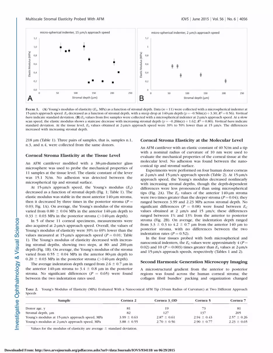
At 15-lm/s approach speed, the Young’s modulus (ES)decreased as a function of stromal depth (Fig. 1; Table 1). Theelastic modulus was stable in the most anterior 140-lm stroma,then it decreased by three times in the posterior stroma (P ¼0.03; Fig. 1A). On average, the Young’s modulus of the stromavaried from 0.80 6 0.04 MPa in the anterior 140-lm depth to0.33 6 0.03 MPa in the posterior stroma (>140-lm depth).
In 5 of these 11 corneal specimens, measurements werealso acquired at 2-lm/s approach speed. Overall, the values ofYoung’s modulus of elasticity were 39% to 69% lower than thevalues measured at 15-lm/s approach speed (P ¼ 0.01; Table1). The Young’s modulus of elasticity decreased with increas-ing stromal depths, showing two steps, at 80- and 200-lmdepth (Fig. 1B). On average, the Young’s modulus of the stromavaried from 0.55 6 0.04 MPa in the anterior 80-lm depth to0.20 6 0.03 MPa in the posterior stroma (>140-lm depth).
The average indentation depth ranged from 2.6 6 0.7 lm inthe anterior 140-lm stroma to 3.4 6 0.8 lm in the posteriorstroma. No significant differences (P ¼ 0.65) were foundbetween the two indentation rates used.
Corneal Stroma Elasticity at the Molecular Level
An AFM cantilever with an elastic constant of 40 N/m and a tipwith a nominal radius of curvature of 10 nm were used toevaluate the mechanical properties of the corneal tissue at themolecular level. No adhesion was found between the nano-conical tip and stromal surface.
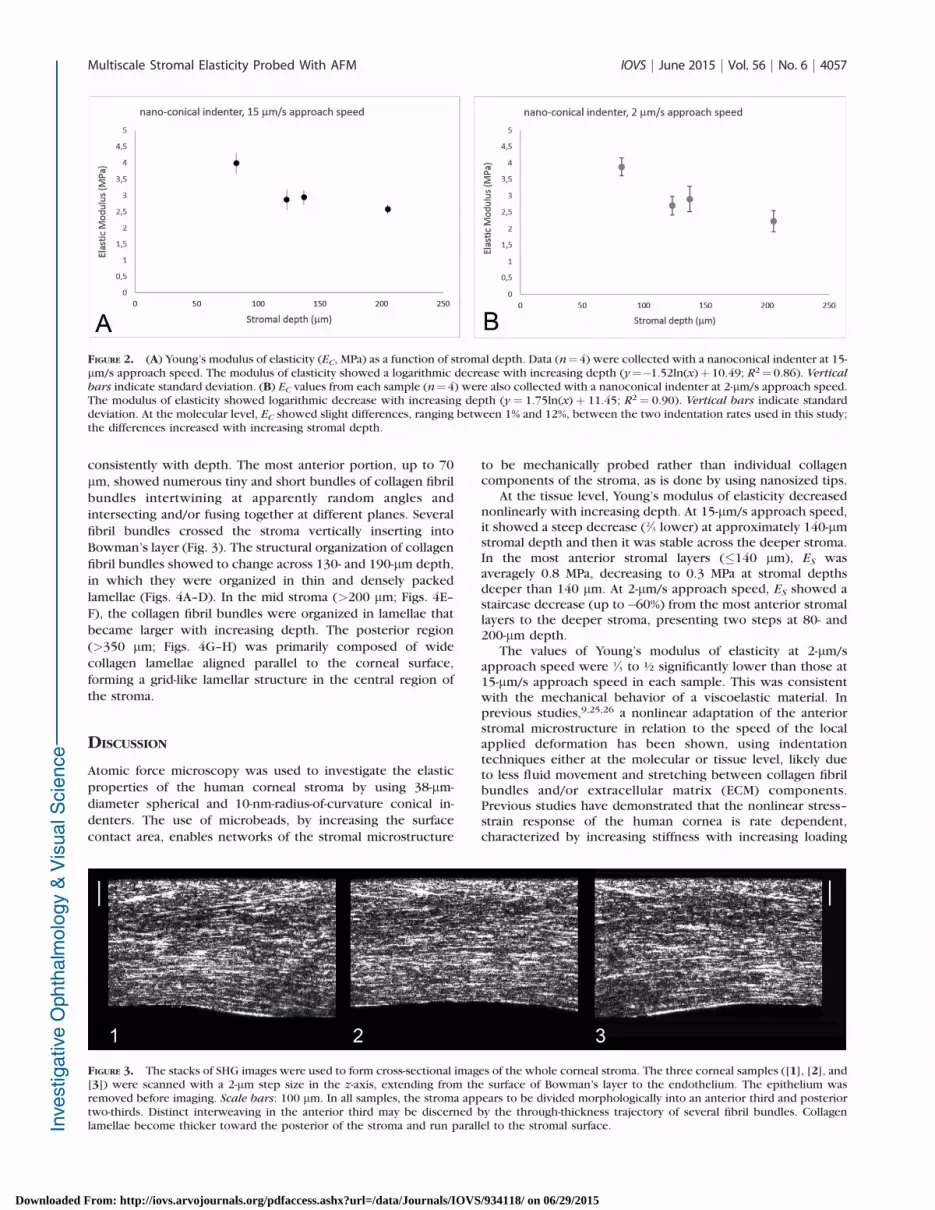
Experiments were performed on four human donor corneasat 2-lm/s and 15-lm/s approach speeds (Table 2). At 15-lm/sapproach speed, the Young’s modulus decreased nonlinearlywith increasing stromal depths, though the depth-dependentdifferences were less pronounced than using microsphericaltips (Fig. 2A). The EC values of the anterior 140-lm stromawere two times greater than the deeper stroma (P¼0.04); theyranged between 3.99 and 2.23 MPa across stromal depth. Nosignificant differences (P ¼ 0.08) were found between EC
values obtained at 2 lm/s and 15 lm/s; these differencesranged between 1% and 13% from the anterior to posteriorstroma (Fig. 2B). On average, the indentation depth rangedfrom 3.3 6 0.3 to 4.2 6 0.7 lm from the anterior 140 lm toposterior stroma, with no differences between the twoindentation rates (P ¼ 0.52).
In the four tissues probed with both microspherical andnanoconical indenters, the EC values were approximately 4 (P¼0.02) and 10 (P¼ 0.003) times greater than ES values at 2-lm/sand 15-lm/s approach speeds, respectively (Tables 1 and 2).
Second Harmonic Generation Microscopy Imaging
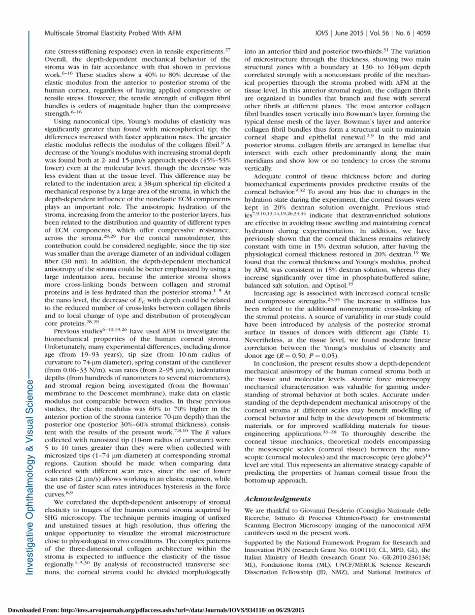
A microstructural gradient from the anterior to posteriorregions was found across the human corneal stroma; thecollagen fibril bundles’ packing and organization changed
FIGURE 1. (A) Young’s modulus of elasticity (ES, MPa) as a function of stromal depth. Data (n¼11) were collected with a microspherical indenter at15-lm/s approach speed. ES decreased as a function of stromal depth, with a steep drop at 140-lm depth (y¼�0.56ln(x)þ3.30; R2¼0.56). Vertical
bars indicate standard deviation. (B) ES values from five samples were collected with a microspherical indenter at 2-lm/s approach speed. At a slowscan speed, the elastic modulus shows a staircase decrease with increasing stromal depth (y¼�0.26ln(x)þ 1.62; R2¼ 0.80). Vertical bars indicatestandard deviation. At the tissue level, ES values obtained at 2-lm/s approach speed were 39% to 59% lower than at 15 lm/s. The differencesincreased with increasing stromal depth.
TABLE 2. Young’s Modulus of Elasticity (MPa) Evaluated With a Nanoconical AFM Tip (10-nm Radius of Curvature) at Two Different ApproachSpeeds
Sample Cornea 2 Cornea 3_OD Cornea 5 Cornea 7
Donor age, y 81 19 73 81
Stromal depth, lm 82 127 137 205
Young’s modulus at 15-lm/s approach speed, MPa 3.99 6 0.63 2.87 6 0.61 2.94 6 0.43 2.57 6 0.26
Young’s modulus at 2-lm/s approach speed, MPa 3.88 6 0.55 2.70 6 0.56 2.90 6 0.77 2.23 6 0.65
Values for the modulus of elasticity are average 6 standard deviation.
Multiscale Stromal Elasticity Probed With AFM IOVS j June 2015 j Vol. 56 j No. 6 j 4056
Downloaded From: http://iovs.arvojournals.org/pdfaccess.ashx?url=/data/Journals/IOVS/934118/ on 06/29/2015
consistently with depth. The most anterior portion, up to 70
lm, showed numerous tiny and short bundles of collagen fibril
bundles intertwining at apparently random angles and
intersecting and/or fusing together at different planes. Several
fibril bundles crossed the stroma vertically inserting into
Bowman’s layer (Fig. 3). The structural organization of collagen
fibril bundles showed to change across 130- and 190-lm depth,
in which they were organized in thin and densely packed
lamellae (Figs. 4A–D). In the mid stroma (>200 lm; Figs. 4E–
F), the collagen fibril bundles were organized in lamellae that
became larger with increasing depth. The posterior region
(>350 lm; Figs. 4G–H) was primarily composed of wide
collagen lamellae aligned parallel to the corneal surface,
forming a grid-like lamellar structure in the central region of
the stroma.
DISCUSSION
Atomic force microscopy was used to investigate the elastic
properties of the human corneal stroma by using 38-lm-
diameter spherical and 10-nm-radius-of-curvature conical in-
denters. The use of microbeads, by increasing the surface
contact area, enables networks of the stromal microstructure
to be mechanically probed rather than individual collagencomponents of the stroma, as is done by using nanosized tips.
At the tissue level, Young’s modulus of elasticity decreasednonlinearly with increasing depth. At 15-lm/s approach speed,it showed a steep decrease (2 =
3 lower) at approximately 140-lmstromal depth and then it was stable across the deeper stroma.In the most anterior stromal layers (�140 lm), ES wasaveragely 0.8 MPa, decreasing to 0.3 MPa at stromal depthsdeeper than 140 lm. At 2-lm/s approach speed, ES showed astaircase decrease (up to�60%) from the most anterior stromallayers to the deeper stroma, presenting two steps at 80- and200-lm depth.
The values of Young’s modulus of elasticity at 2-lm/sapproach speed were 1 =
3 to ½ significantly lower than those at15-lm/s approach speed in each sample. This was consistentwith the mechanical behavior of a viscoelastic material. Inprevious studies,9,25,26 a nonlinear adaptation of the anteriorstromal microstructure in relation to the speed of the localapplied deformation has been shown, using indentationtechniques either at the molecular or tissue level, likely dueto less fluid movement and stretching between collagen fibrilbundles and/or extracellular matrix (ECM) components.Previous studies have demonstrated that the nonlinear stress–strain response of the human cornea is rate dependent,characterized by increasing stiffness with increasing loading
FIGURE 2. (A) Young’s modulus of elasticity (EC, MPa) as a function of stromal depth. Data (n¼4) were collected with a nanoconical indenter at 15-lm/s approach speed. The modulus of elasticity showed a logarithmic decrease with increasing depth (y¼�1.52ln(x)þ 10.49; R2¼ 0.86). Vertical
bars indicate standard deviation. (B) EC values from each sample (n¼4) were also collected with a nanoconical indenter at 2-lm/s approach speed.The modulus of elasticity showed logarithmic decrease with increasing depth (y ¼ 1.75ln(x) þ 11.45; R2 ¼ 0.90). Vertical bars indicate standarddeviation. At the molecular level, EC showed slight differences, ranging between 1% and 12%, between the two indentation rates used in this study;the differences increased with increasing stromal depth.
FIGURE 3. The stacks of SHG images were used to form cross-sectional images of the whole corneal stroma. The three corneal samples ([1], [2], and[3]) were scanned with a 2-lm step size in the z-axis, extending from the surface of Bowman’s layer to the endothelium. The epithelium wasremoved before imaging. Scale bars: 100 lm. In all samples, the stroma appears to be divided morphologically into an anterior third and posteriortwo-thirds. Distinct interweaving in the anterior third may be discerned by the through-thickness trajectory of several fibril bundles. Collagenlamellae become thicker toward the posterior of the stroma and run parallel to the stromal surface.
Multiscale Stromal Elasticity Probed With AFM IOVS j June 2015 j Vol. 56 j No. 6 j 4057
Downloaded From: http://iovs.arvojournals.org/pdfaccess.ashx?url=/data/Journals/IOVS/934118/ on 06/29/2015

FIGURE 4. Second harmonic generation images of different layers of the human corneal stroma. Scale bars: 100 lm. (A) Second harmonic generationimage at 20-lm depth, showing the collagen fibrils arranging in tiny and short bundles densely interweaved. (B) At 60-lm depth, collagen fibril bundlesare organized in wider bundles that intersect with each other at different planes. (C, D) The structural organization of collagen fibril bundles across130- and 190-lm depth changes in comparison with the most anterior stroma, showing thin and densely packed lamellae. (E, F) In the mid stroma(250- and 330-lm depth), the collagen lamellae become increasingly wider and thicker. (G, H) In the posterior stroma (410- and 500-lm depth), thecollagen lamellae show a grid-like structure, crossing each other at almost vertical angles; the most posterior lamellae (white arrows), lying anteriorly tothe Descemet’s membrane, are thinner than overlying lamellae.
Multiscale Stromal Elasticity Probed With AFM IOVS j June 2015 j Vol. 56 j No. 6 j 4058
Downloaded From: http://iovs.arvojournals.org/pdfaccess.ashx?url=/data/Journals/IOVS/934118/ on 06/29/2015
rate (stress-stiffening response) even in tensile experiments.27
Overall, the depth-dependent mechanical behavior of thestroma was in fair accordance with that shown in previouswork.6–16 These studies show a 40% to 80% decrease of theelastic modulus from the anterior to posterior stroma of thehuman cornea, regardless of having applied compressive ortensile stress. However, the tensile strength of collagen fibrilbundles is orders of magnitude higher than the compressivestrength.6–16
Using nanoconical tips, Young’s modulus of elasticity wassignificantly greater than found with microspherical tip; thedifferences increased with faster application rates. The greaterelastic modulus reflects the modulus of the collagen fibril.9 Adecrease of the Young’s modulus with increasing stromal depthwas found both at 2- and 15-lm/s approach speeds (45%–53%lower) even at the molecular level, though the decrease wasless evident than at the tissue level. This difference may berelated to the indentation area; a 38-lm spherical tip elicited amechanical response by a large area of the stroma, in which thedepth-dependent influence of the nonelastic ECM componentsplays an important role. The anisotropic hydration of thestroma, increasing from the anterior to the posterior layers, hasbeen related to the distribution and quantity of different typesof ECM components, which offer compressive resistance,across the stroma.28,29 For the conical nanoindenter, thiscontribution could be considered negligible, since the tip sizewas smaller than the average diameter of an individual collagenfiber (30 nm). In addition, the depth-dependent mechanicalanisotropy of the stroma could be better emphasized by using alarge indentation area, because the anterior stroma showsmore cross-linking bonds between collagen and stromalproteins and is less hydrated than the posterior stroma.1–5 Atthe nano level, the decrease of EC with depth could be relatedto the reduced number of cross-links between collagen fibrilsand to local change of type and distribution of proteoglycancore proteins.28,29
Previous studies6–10,19,26 have used AFM to investigate thebiomechanical properties of the human corneal stroma.Unfortunately, many experimental differences, including donorage (from 19–93 years), tip size (from 10-nm radius ofcurvature to 74-lm diameter), spring constant of the cantilever(from 0.06–33 N/m), scan rates (from 2–95 lm/s), indentationdepths (from hundreds of nanometers to several micrometers),and stromal region being investigated (from the Bowman’membrane to the Descemet membrane), make data on elasticmodulus not comparable between studies. In these previousstudies, the elastic modulus was 60% to 70% higher in theanterior portion of the stroma (anterior 70-lm depth) than theposterior one (posterior 30%–60% stromal thickness), consis-tent with the results of the present work.7,8,10 The E valuescollected with nanosized tip (10-nm radius of curvature) were5 to 10 times greater than they were when collected withmicrosized tips (1–74 lm diameter) at corresponding stromalregions. Caution should be made when comparing datacollected with different scan rates, since the use of lowerscan rates (2 lm/s) allows working in an elastic regimen, whilethe use of faster scan rates introduces hysteresis in the forcecurves.8,9
We correlated the depth-dependent anisotropy of stromalelasticity to images of the human corneal stroma acquired bySHG microscopy. The technique permits imaging of unfixedand unstained tissues at high resolution, thus offering theunique opportunity to visualize the stromal microstructureclose to physiological in vivo conditions. The complex patternsof the three-dimensional collagen architecture within thestroma is expected to influence the elasticity of the tissueregionally.1–5,30 By analysis of reconstructed transverse sec-tions, the corneal stroma could be divided morphologically
into an anterior third and posterior two-thirds.31 The variationof microstructure through the thickness, showing two mainstructural zones with a boundary at 130- to 160-lm depthcorrelated strongly with a nonconstant profile of the mechan-ical properties through the stroma probed with AFM at thetissue level. In this anterior stromal region, the collagen fibrilsare organized in bundles that branch and fuse with severalother fibrils at different planes. The most anterior collagenfibril bundles insert vertically into Bowman’s layer, forming thetypical dense mesh of the layer. Bowman’s layer and anteriorcollagen fibril bundles thus form a structural unit to maintaincorneal shape and epithelial renewal.2,9 In the mid andposterior stroma, collagen fibrils are arranged in lamellae thatintersect with each other predominantly along the mainmeridians and show low or no tendency to cross the stromavertically.
Adequate control of tissue thickness before and duringbiomechanical experiments provides predictive results of thecorneal behavior.9,32 To avoid any bias due to changes in thehydration state during the experiment, the corneal tissues werekept in 20% dextran solution overnight. Previous stud-ies7,9,10,13,14,19,26,33,34 indicate that dextran-enriched solutionsare effective in avoiding tissue swelling and maintaining cornealhydration during experimentation. In addition, we havepreviously shown that the corneal thickness remains relativelyconstant with time in 15% dextran solution, after having thephysiological corneal thickness restored in 20% dextran.19 Wefound that the corneal thickness and Young’s modulus, probedby AFM, was consistent in 15% dextran solution, whereas theyincrease significantly over time in phosphate-buffered saline,balanced salt solution, and Optisol.19
Increasing age is associated with increased corneal tensileand compressive strengths.23,35 The increase in stiffness hasbeen related to the additional nonenzymatic cross-linking ofthe stromal proteins. A source of variability in our study couldhave been introduced by analysis of the posterior stromalsurface in tissues of donors with different age (Table 1).Nevertheless, at the tissue level, we found moderate linearcorrelation between the Young’s modulus of elasticity anddonor age (R¼ 0.50; P ¼ 0.05).
In conclusion, the present results show a depth-dependentmechanical anisotropy of the human corneal stroma both atthe tissue and molecular levels. Atomic force microscopymechanical characterization was valuable for gaining under-standing of stromal behavior at both scales. Accurate under-standing of the depth-dependent mechanical anisotropy of thecorneal stroma at different scales may benefit modelling ofcorneal behavior and help in the development of biomimeticmaterials, or for improved scaffolding materials for tissue-engineering applications.36–38 To thoroughly describe thecorneal tissue mechanics, theoretical models encompassingthe mesoscopic scales (corneal tissue) between the nano-scopic (corneal molecules) and the macroscopic (eye globe)14
level are vital. This represents an alternative strategy capable ofpredicting the properties of human corneal tissue from thebottom-up approach.
Acknowledgments
We are thankful to Giovanni Desiderio (Consiglio Nazionale delleRicerche, Istituto di Processi Chimico-Fisici) for enviromentalScanning Electron Microscopy imaging of the nanoconical AFMcantilevers used in the present work.
Supported by the National Framework Program for Research andInnovation PON (research Grant No. 0100110; CL, MPD, GL), theItalian Ministry of Health (research Grant No. GR-2010-236138;ML), Fondazione Roma (ML), UNCF/MERCK Science ResearchDissertation Fellowship (JD, NMZ), and National Institutes of
Multiscale Stromal Elasticity Probed With AFM IOVS j June 2015 j Vol. 56 j No. 6 j 4059
Downloaded From: http://iovs.arvojournals.org/pdfaccess.ashx?url=/data/Journals/IOVS/934118/ on 06/29/2015
Health (NIH) National Research Service Award Individual Predoc-toral Fellowship (1F31EY021714-01; JD, NMZ).
Disclosure: C. Labate, None; M. Lombardo, None; M.P. DeSanto, None; J. Dias, None; N.M. Ziebarth, None; G. Lombar-do, None
References
1. Winkler M, Shoa G, Xie Y, et al. Three-dimensional distributionof transverse collagen fibers in the anterior human cornealstroma. Invest Ophthalmol Vis Sci. 2013;54:7293–7301.
2. Morishige N, Takagi Y, Chikama T, Takahara A, Nishida T.Three-dimensional analysis of collagen lamellae in the anteriorstroma of the human cornea visualized by second harmonicgeneration imaging microscopy. Invest Ophthalmol Vis Sci.2011;52:911–915.
3. Meek KM, Blamires T, Elliot GF, Gyi TJ, Nave C. Theorganisation of collagen fibrils in the human corneal stroma:a synchroton x-ray diffraction study. Curr Eye Res. 1987;6:841–846.
4. Komai Y, Ushiki T. The three-dimensional organization ofcollagen fibrils in the human cornea and sclera. Invest
Ophthalmol Vis Sci. 1991;32:2244–2258.
5. Muller LJ, Pels E, Vrensen GF. The specific architecture of theanterior stroma accounts for maintenance of corneal curva-ture. Br J Ophthalmol. 2001;85:437–443.
6. Last JA, Liliensiek SJ, Nealey PF, Murphy CJ. Determining themechanical properties of human corneal basement membraneswith atomic force microscopy. J Struct Biol. 2009;167:19–24.
7. Dias JM, Ziebarth NM. Anterior and posterior corneal stromaelasticity assessed using nanoindentation. Exp Eye Res. 2013;115:41–45.
8. Last JA, Thomasy SM, Croasdale CR, Russell P, Murphy CJ.Compliance profile of the human cornea as measured byatomic force microscopy. Micron. 2012;43:1293–1298.
9. Lombardo M, Lombardo G, Carbone G, De Santo MP, Barberi R,Serrao S. Biomechanics of the anterior human corneal tissueinvestigated with atomic force microscopy. Invest Ophthalmol
Vis Sci. 2012;53:1050–1057.
10. Dias J, Diakonis VF, Kankariya VP, Yoo SH, Ziebarth NM.Anterior and posterior corneal stroma elasticity after cornealcollagen crosslinking treatment. Exp Eye Res. 2013;116:58–62.
11. Seifert J, Hammer CM, Rheinlaender J, et al. Distribution ofYoung’s modulus in porcine corneas after riboflavin/UVAinduced collagen cross-linking as measured by atomic forcemicroscopy. PLoS One. 2014;9:e88186.
12. Kohlhaas M, Spoerl E, Schilde T, Unger G, Wittig C, Pillunat LE.Biomechanical evidence of the distribution of cross-links incorneas treated with riboflavin and ultraviolet A light. J
Cataract Refract Surg. 2006;32:279–283.
13. Lombardo M, Serrao S, Rosati M, Ducoli P, Lombardo G.Biomechanical changes of the human cornea followingtransepithelial corneal cross-linking using iontophoresis. J
Cataract Surg. 2014;40:1706–1715.
14. Lombardo M, Serrao S, Rosati M, Lombardo G. Analysis of theviscoelastic properties of the human cornea using Scheimp-flug imaging in inflation experiment of eye globes. PLoS One.2014;9:e112169.
15. Dupps WJ, Netto MV, Herekar S, Krueger RR. Surface waveelastometry of the cornea in porcine and human donor eyes. J
Refract Surg. 2007;23:66–75.
16. Scancelli G, Pineda R, Yun SH. Brillouin optical microscopy forcorneal biomechanics. Invest Ophthalmol Vis Sci. 2013;53:185–190.
17. Petsche SJ, Chernyak D, Martiz J, Levenston ME, Pinsky PM.Depth-dependent transverse shear properties of the humancorneal stroma. Invest Ophthalmol Vis Sci. 2012;53:873–880.
18. Butt HJ, Cappella B, Kappl M. Force measurements with theatomic force microscope: technique, interpretation andapplications. Surf Sci Rep. 2005;59:1–152.
19. Dias J, Ziebarth NM. Impact of hydration media on ex vivocorneal elasticity measurements [published online ahead ofprint January 19, 2015]. Eye Contact Lens.
20. Ebenstein DM, Pruitt LA. Nanoindentation of biologicalmaterials. NanoToday. 2006;1:26–33.
21. Gibson CT, Watson GS, Myhra S. Determination of the springconstants of probes for force microscopy/spectroscopy.Nanotechnology. 1996;7:259–262.
22. Tortonese M, Kirk M. Characterization of application specificprobes for SPMs. Proc SPIE. 1997;3009:53–60.
23. Knox Cartwright NE, Tyrer JR, Marshall J. Age-relateddifferences in the elasticity of the human cornea. Invest
Ophthalmol Vis Sci. 2011;52:4324–4329.
24. Poggi MA. A method for calculating the spring constant ofatomic force microscopy cantilevers with a nonrectangularcross section. Anal Chem. 2005;77:1192–1195.
25. Ahearne M, Yang Y, Then KY, Liu KK. An indentationtechnique to characterize the mechanical and viscoelasticproperties of human and porcine corneas. Ann Biomed Eng.2007;35:1608–1616.
26. Labate C, De Santo MP, Lombardo G, Lombardo M. Under-standing of the viscoelastic response of the human cornealstroma induced by riboflavin/UV-A cross-linking at the nanolevel. PLoS One. 2015;10:e0122868.
27. Elsheikh A, Wang D, Pye D. Determination of the modulus ofelasticity of the human cornea. J Refract Surg. 2007;23:808–818.
28. Muller LJ, Pels E, Schurmans L, Vrensen G. A new three-dimensional model of the organization of proteoglycans andcollagen fibrils in the human corneal stroma. Exp Eye Res.2004;78:493–501.
29. Wilson G, O’Leary DJ, Vaughan W. Differential swelling incompartments of the corneal stroma. Invest Ophthalmol Vis
Sci. 1984;25:1105–1111.
30. Winkler M, Chai D, Kriling S, et al. Nonlinear opticalmacroscopic assessment of 3-D corneal collagen organizationand axial biomechanics. Invest Ophthalmol Vis Sci. 2011;2:8818–8827.
31. Ruberti JW, Roy AS, Roberts CJ. Corneal structure andfunction. Annu Rev Biomed Eng. 2011;13:269–295.
32. Hatami-Marbini H, Etebu E. Hydration dependent biomechan-ical properties of the corneal stroma. Exp Eye Res. 2013;116:47–54.
33. Hamaoui M, Tahi H, Chapon P, et al. Corneal preparation ofeye bank eyes for experimental surgery. Cornea. 2001;20:317–320.
34. Borja D, Manns F, Lamar P, Rosen A, Fernandez V, Parel JM.Preparation and hydration control of corneal tissue strips forexperimental use. Cornea. 2004;23:61–66.
35. Elsheikh A, Wang D, Brown M, Rama P, Campanelli M, Pye D.Assessment of corneal biomechanical properties and theirvariation with age. Curr Eye Res. 2007;32:11–19.
36. Studer H, Larrea X, Riedwyl H, Buchler P. Biomechanicalmodel of human cornea based on stromal microstructure. J
Biomech. 2010;43:836–842.
37. Thomasy SM, Raghunathan VK, Winkler M, et al. Elasticmodulus and collagen organization of the rabbit cornea:epithelium to endothelium. Acta Biomater. 2014;10:785–791.
38. Ruberti JW, Zieske JD. Prelude to corneal tissue engineering:gaining control of collagen organization. Prog Ret Eye Res.2008;27:549–577.
Multiscale Stromal Elasticity Probed With AFM IOVS j June 2015 j Vol. 56 j No. 6 j 4060
Downloaded From: http://iovs.arvojournals.org/pdfaccess.ashx?url=/data/Journals/IOVS/934118/ on 06/29/2015