analysis of corneal stroma organization with wavefront optimized nonlinear microscopy
TRANSCRIPT

BASIC INVESTIGATION
Analysis of Corneal Stroma Organization With WavefrontOptimized Nonlinear Microscopy
Juan M. Bueno, PhD, Emilio J. Gualda, PhD, and Pablo Artal, PhD
Purpose: To investigate the organization of stromal collagen in
healthy ex vivo corneas of different species from second harmonic
generation (SHG) microscopy images.
Methods: A custom backscattered nonlinear microscope has been
used to study the corneal structure of different species: porcine, bovine,
rabbit, rat, chicken, and humans. The instrument uses a femtosecond
laser for illumination, a scanning unit, and a photon-counting detection
device. It also includes a wavefront aberration control module. SHG
signals produced by collagen within the cornea were acquired. A
motorized stage allowed optical sectioning across the entire corneal
thickness. Samples were neither fixed nor stained, and they were fully
scanned.
Results: SHG images revealed the microscopic organization of the
lamellae of collagen fibers. Despite absorption, for all corneal depths,
images could be analyzed. The anterior stroma was similar in all
samples, showing interwoven short bands of collagen randomly
distributed. The lamellae at the central and posterior stroma were
densely packed and often presented longer bundles lying pre-
dominantly parallel to the corneal surface with characteristic spatial
distributions for each species. In particular, collagen bundles in
bovine and porcine corneas were interweaved. In the chick cornea,
the stromal arrangement had an orientation changing regularly with
depth. In human corneas, lamellae were longer and had similar
orientation than their neighbors.
Conclusions: Using a unique wavefront aberration–controlled
backscattered nonlinear microscope, changes in corneal morphology
as a function of depth were characterized for different species
(including humans). This allowed a direct comparison among species,
which might help to establish the basis of collagen distribution in
animal models or to understand diseased corneas.
Key Words: nonlinear microscopy, corneal stroma, collagen, second
harmonic generation
(Cornea 2011;30:692–701)
Multiphoton microscopy,1 and in particular secondharmonic generation (SHG) microscopy,2,3 is a powerful
tool to explore the nonlinear properties of biological tissuescontaining collagen, as is the particular case of the cornea.4–6
Because of the natural noncentrosymmetric organization witha triple helix complex structure, collagen type I generateslarge SHG signals, allowing the microscopic analysis of thecorneal structure in both normal and pathological eyes. Thisis important because critical corneal properties, such as itstransparency, may depend on an ordered arrangement of thecollagen fibers within the corneal stroma.7
Similarly to two-photon excitation fluorescence (TPEF)imaging,1 SHG microscopy provides intrinsic optical sectioningand allows for 3-dimensional (3D) analysis and volumerendering. However, SHG and TPEF are based on differentphenomena. SHG is a coherent process based on nonlinearscattering, whereas TPEF relies on nonlinear absorption followedby fluorescence emission. SHG provides minimum sampledisturbance because no energy is absorbed by the specimen.
Yeh et al8 obtained high-resolution SHG images ofnormal rabbit corneas without exogenous dyes using aninverted microscope. Forward and backward SHG signalsfrom fixed porcine corneas have been analyzed, showing aregular packaging of the collagen fibrils.9 Similar results wereobtained by Teng et al10 in nonfixed porcine corneas imaged inthe backward direction. SHG microscopy of mouse, rabbit,and human corneas has been also reported11 to assess theircollagen organization. These samples were fixed with para-formaldehyde, and both backward and forward SHG signalswere recorded. The stromal collagen bundles have beenstudied with SHG microscopy in normal and keratoconushuman corneas.12,13 Noticeable differences in the lamellarinterweaving at the Bowman layer were found. Structuralalterations of the cornea because of scars have been detectedusing multiphoton microscopy.14 The effect of polarization andwavelength15,16 on the forward and backward SHG signalsfrom porcine corneas have also been evaluated. Moreover, thecombination of SHG and third harmonic generation signal hasbeen applied to resolve the porcine corneal layers.17 A recentwork investigates the 3D structure of the corneal collagen inhuman corneas with keratoconus and after intra-laser in situkeratomileusis surgery.18
In this study, we further advance the application of SHGimaging techniques to characterize the properties of the stromaacross different relevant species (including humans). We useda custom backscattered nonlinear microscope with wavefrontaberration (WA) optimization19,20 to study the structure of thehealthy corneal stroma. This instrument provides images of
Received for publication April 29, 2010; revision received July 26, 2010;accepted September 13, 2010.
From the Laboratorio de Optica, Centro de Investigacion en Optica y Nanofısica(CiOyN), Universidad de Murcia, Campus de Espinardo, Murcia, Spain.
Supported by ‘‘Ministerio de Educacion y Ciencia,’’ Spain (grants no.FIS2007-64765 and Consolider SAUUL CSD2007-00033) and ‘‘Funda-cion Seneca,’’ (Region de Murcia, Spain; grant no. 4524/GERM/06).
Reprints: Juan M. Bueno, Laboratorio de Optica, Centro de Investigacion enOptica y Nanofısica (CiOyN), Universidad de Murcia, Campus deEspinardo, 30100 Murcia, Spain (e-mail: [email protected]).
Copyright � 2011 by Lippincott Williams & Wilkins
692 | www.corneajrnl.com Cornea � Volume 30, Number 6, June 2011

exceptional quality with even smaller illumination doses to thecornea. All images were obtained with the same experimentalsetup under similar conditions, to allow the direct comparisonof structural collagen organization between the differentspecies. The SHG signal was always detected in the backwarddirection because this will be the most likely configuration forthe possible future in vivo implementations of the technique.Because SHG is an intrinsic process, all specimens wereimaged without applying any staining or fixation procedure.The lamellar arrangement will be evaluated at different depthswithin the cornea, and the changes in size and organization ofcollagen bundles for the different samples will be comparedand discussed.
MATERIALS AND METHODS
Experimental SetupA schematic diagram of the system used is shown in
Figure 1. A mode-locked femtosecond laser (Mira900f; Coherent,St Clara, CA) providing the excitation source has been coupledinto an inverted microscope (TE2000-U; Nikon Corp., Tokyo,Japan). This source is a tunable (700–980 nm) Ti:sapphire ultrafasthigh-power laser providing pulses of about 110 fs at 76 MHz, withan average power of 0.8 W and peak powers of up to 130 kW.Measurements were carried out at 760 nm. The laser beam reachedthe sample through a nonimmersion long working distancemicroscope objective (Nikon ELWD Series, 320, numericalaperture [NA] = 0.5). SHG signal was detected in the backwarddirection via the same microscope objective. This signal wastransmitted through a dichroic mirror (DIM), passed a narrow-band (380 6 10 nm) spectral filter (FB380-10; Thorlabs, Inc,Newton, NJ), and reached the photomultiplier tube (R7205-01;Hamamatsu, Shizouka, Japan). Images were recorded in the XYplane using a pair of nonresonant galvanometric mirrors(VM1000; GSI, Billerica, MA) acting as optical scanning unit.Moreover, a Z-axis step motor (PI C-136; Karlsruhe, Germany)was used to collect stacks of XY images at successive depths. TheSHG filter was used to isolate the nonlinear image from the cornealstroma (at half of the excitation wavelength), so that the epitheliumand the endothelium remained invisible. This allowed us toestablish the ‘‘zero distance’’ for depth imaging within the stromaas the first position where SHG signal appeared. For most samples,we were able to scan the whole corneal thickness with good imagecontrast. The system was completely automated and controlledthrough a custom LabView (National Instruments, Austin, TX)interface. The optical power at the sample’s plane ranged between100 and 130 mW (depending on the specimen), and it was
controlled by means of a continuously variable neutral densityfilter placed in front of the laser source, which was manuallyadjusted. Although the maximum field of view was 700 3 700mm2, all the images shown below correspond to an area of 210 3210 mm2. Every final image was the average of 6 individual framesof 250 3 250 pixels. The recording time of every individual framewas set to 1 image per second. Image processing was performedwith MatLab (The MathWorks, Inc., Natick, MA).
In the illumination pathway, the system includes a WAcontrol composed of a real-time Hartmann–Shack wavefrontsensor.20 By minimizing the effects of the laser beamaberrations through the laser cavity realignment and WAcontrolling, nonlinear microscopy images are significantlyimproved (see Results).
Preparation of Corneal TissueCorneas of porcine, bovine, rabbits, rats, adult chickens,
and humans were analyzed in this study. The samples wereneither fixed in paraformaldehyde nor stained. Porcine andbovine ocular globes were obtained from a local slaughter-house and moved to the laboratory immediately after death.These intact eyes were placed upside down on a glass bottomdish (thickness: 170 mm) and filled with a solution composedof Medium 199 (Hanks salts and sodium) with L-glutamineadded (Sigma-Aldrich). Corneas from rabbits, rats, and adultchickens were excised with a trephine right after death,immersed in the same solution, and imaged a few hours later.Within every species, all specimens had similar age. Humancorneas from healthy donors (aged between 29 and 75 years)were provided by the eye bank of the ‘‘Servicio deOftalmologıa,’’ Hospital Universitario Virgen de la Arrixaca,Murcia, Spain. This study was approved by the Ethical ReviewBoard of the hospital. The human samples were treatedfollowing the instructions of the World Medical Association’sDeclaration of Helsinki. Only normal corneas not suitable fortransplantation were selected. After trephination (7.5 mm) wasperformed, the human corneas were stored in Optisol solution(Bausch and Lomb) overnight and sent to the laboratory foranalysis. All corneas appeared clear when viewed througha white-light microscope during the entire experiment.Excision procedures and manipulation were always carriedout by experienced personnel to minimize the risk of folding.For all specimens, imaged areas corresponded to the centralcornea.
Excised corneal tissues are prone to swelling, whichproduces edema. Corneal edema modifies the normal structureof the stroma and may lead to the wrong conclusions on themorphological study. The 2 solutions used in this work have
FIGURE 1. Schematic representationof the experimental setup. DIM, di-chroic mirror; F, filter; GM1 and GM2,scanning mirrors; HS, Hartmann–Shack wavefront sensor; L1–L6,achromatic doublets; O, objective;PMT, photomultiplier tube.
q 2011 Lippincott Williams & Wilkins www.corneajrnl.com | 693
Cornea � Volume 30, Number 6, June 2011 Corneal Stroma Organization
been reported to enhance corneal dehydration during storageand then to minimize the effects of swelling.21,22
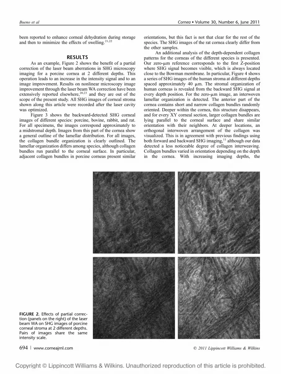
RESULTSAs an example, Figure 2 shows the benefit of a partial
correction of the laser beam aberrations in SHG microscopyimaging for a porcine cornea at 2 different depths. Thisoperation leads to an increase in the intensity signal and to animage improvement. Results on nonlinear microscopy imageimprovement through the laser beam WA correction have beenextensively reported elsewhere,20,23 and they are out of thescope of the present study. All SHG images of corneal stromashown along this article were recorded after the laser cavitywas optimized.
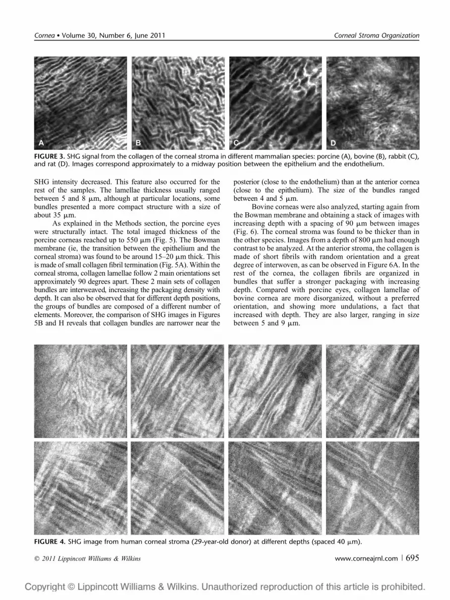
Figure 3 shows the backward-detected SHG cornealimages of different species: porcine, bovine, rabbit, and rat.For all specimens, the images correspond approximately toa midstromal depth. Images from this part of the cornea showa general outline of the lamellar distribution. For all images,the collagen bundle organization is clearly outlined. Thelamellar organization differs among species, although collagenbundles run parallel to the corneal surface. In particular,adjacent collagen bundles in porcine corneas present similar
orientations, but this fact is not that clear for the rest of thespecies. The SHG images of the rat cornea clearly differ fromthe other samples.
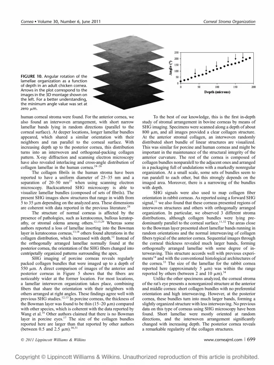
An additional analysis of the depth-dependent collagenpatterns for the corneas of the different species is presented.Our zero-mm reference corresponds to the first Z-positionwhere SHG signal becomes visible, which is always locatedclose to the Bowman membrane. In particular, Figure 4 showsa series of SHG images of the human stroma at different depthsspaced approximately 40 mm. The stromal organization ofhuman corneas is revealed from the backward SHG signal atevery depth position. For the zero-mm image, an interwovenlamellar organization is detected. The anterior part of thecornea contains short and narrow collagen bundles randomlyoriented. Deeper within the cornea, this structure disappears,and for every XY corneal section, larger collagen bundles arelying parallel to the corneal surface and share similarorientation with their neighbors. At deeper locations, anorthogonal interwoven arrangement of the collagen wasvisualized. This is in agreement with previous findings usingboth forward and backward SHG imaging,11 although our datadetected a less noticeable degree of collagen interweaving.Collagen bundles varied in orientation depending on the depthin the cornea. With increasing imaging depths, the
FIGURE 2. Effects of partial correc-tion (panels on the right) of the laserbeam WA on SHG images of porcinecorneal stroma at 2 different depths.Pairs of images share the sameintensity scale.
694 | www.corneajrnl.com q 2011 Lippincott Williams & Wilkins
Bueno et al Cornea � Volume 30, Number 6, June 2011
SHG intensity decreased. This feature also occurred for therest of the samples. The lamellae thickness usually rangedbetween 5 and 8 mm, although at particular locations, somebundles presented a more compact structure with a size ofabout 35 mm.
As explained in the Methods section, the porcine eyeswere structurally intact. The total imaged thickness of theporcine corneas reached up to 550 mm (Fig. 5). The Bowmanmembrane (ie, the transition between the epithelium and thecorneal stroma) was found to be around 15–20 mm thick. Thisis made of small collagen fibril termination (Fig. 5A). Within thecorneal stroma, collagen lamellae follow 2 main orientations setapproximately 90 degrees apart. These 2 main sets of collagenbundles are interweaved, increasing the packaging density withdepth. It can also be observed that for different depth positions,the groups of bundles are composed of a different number ofelements. Moreover, the comparison of SHG images in Figures5B and H reveals that collagen bundles are narrower near the
posterior (close to the endothelium) than at the anterior cornea(close to the epithelium). The size of the bundles rangedbetween 4 and 5 mm.
Bovine corneas were also analyzed, starting again fromthe Bowman membrane and obtaining a stack of images withincreasing depth with a spacing of 90 mm between images(Fig. 6). The corneal stroma was found to be thicker than inthe other species. Images from a depth of 800 mm had enoughcontrast to be analyzed. At the anterior stroma, the collagen ismade of short fibrils with random orientation and a greatdegree of interwoven, as can be observed in Figure 6A. In therest of the cornea, the collagen fibrils are organized inbundles that suffer a stronger packaging with increasingdepth. Compared with porcine eyes, collagen lamellae ofbovine cornea are more disorganized, without a preferredorientation, and showing more undulations, a fact thatincreased with depth. They are also larger, ranging in sizebetween 5 and 9 mm.
FIGURE 3. SHG signal from the collagen of the corneal stroma in different mammalian species: porcine (A), bovine (B), rabbit (C),and rat (D). Images correspond approximately to a midway position between the epithelium and the endothelium.
FIGURE 4. SHG image from human corneal stroma (29-year-old donor) at different depths (spaced 40 mm).
q 2011 Lippincott Williams & Wilkins www.corneajrnl.com | 695
Cornea � Volume 30, Number 6, June 2011 Corneal Stroma Organization

In rabbit corneas, the total thickness of the measuredcorneas was around 450–500 mm (depending on the sample).Regions of interwoven lamellae and others with orthogonallyarranged distribution are found within the stroma, as depictedin Figure 7. The anterior cornea (up to approximately 90 mmbelow the Bowman membrane) showed short collagenlamellae at random orientations and a strong interwoven
organization (Figs. 7A, B). At deeper locations, the stroma isorganized in groups of orthogonally and interwoven arrangedbundles (composed of 5 or 6 lamellae) running parallel to thecorneal surface (Figs. 7C–G). We observe a small reduction inthe bundle thickness and spacing with increasing depth. At theposterior cornea (Fig. 7H), the stroma is made of much shorterlamellae. Here, the interweaving observed in other regions also
FIGURE 5. SHG image of corneal collagen in a porcine eye (imaged planes are spaced about 75 mm).
FIGURE 6. SHG signal from bovine corneal stroma at different depths (images correspond to planes 90 mm apart).
696 | www.corneajrnl.com q 2011 Lippincott Williams & Wilkins
Bueno et al Cornea � Volume 30, Number 6, June 2011

exist, but the orthogonal distribution disappears and thelamellae run along different orientations. The average size ofthe lamellae was found to be about 5 mm.
In the rat samples, the corneal thickness was around 250mm. Compared with the other species, the stromal collagenseemed to be much less organized with a lack of clearstructure, for most corneal thickness. Some ‘‘wrinkles’’ in thestroma were also found. Collagen fibrils did not show anypreferential orientation; lamellar bands were short andpresented a large degree of interwoven all over the anteriorcornea (Figs. 8B–F). However, in the last stromal segment orposterior cornea (Figs. 8G, H), larger collagen fibers appearedwith a moderate organization.
The corneal structure of a nonmammalian animal, theadult chicken, was also analyzed. For these corneas, the totalthickness was around 300 mm. By simple visual examination(Fig. 9), it is apparent that the structure of the collagen clearlydiffers from the samples shown in previous figures. At theanterior cornea, an interwoven structure of short collagenlamellae mostly lying parallel to the corneal surface is seen.Sequential images corresponding to deeper locations showedlonger collagen bundles running parallel to each other withsome orthogonal interweaving. However, this pattern presentsan angular shift for every Z-position (see the orientationrepresented by arrows). Figure 10 shows the evolution of thisangle as a function of depth. The change in the angle is almostlinear all over the corneal thickness with a constant rate ofapproximately 1 degree per micrometer. This distribution wassimilar in a number of corneas of different specimens andseveral positions across each sample. The lamellae sizereached up to 7 mm with an average distance between con-secutive bundles of about 5 mm (with a maximum of 7.5 mm).
DISCUSSIONA WA optimized nonlinear microscope has been de-
veloped to study the corneal stroma organization in differentspecies. As reported in a very recent work,20 the technicalimprovements in the instrument allowed obtaining high-qualitynonlinear microscopy images with comparatively lower incidentenergy. We have used the backscattered SHG signal from thecornea to analyze the collagen packing as a function of depth.SHG microscopy provides a noninvasive high-resolutionimaging technique without the need of fixation procedures orexogenous dyes. Moreover, its autoconfocality allows theoptical sectioning of the sample, the correlation with Z-position,and the reconstruction of the collagen 3D architecture.
Nonlinear microscopy over the entire cornea providesmorphological information of epithelial and endothelial cells,keratocytes, and global collagen orientation. However, becausea SHG filter was used here, epithelium, keratocytes, andendothelium always remained invisible, and only the signal fromthe stroma was registered. In most samples, we were able toimage the total stromal thickness with enough contrast andresolution to observe the bundles of collagen. In particular,bovine corneas were imaged up to a depth of 800 mm, withcontrast good enough to be processed. This represents a sig-nificant advantage of our instrument compared with existingsystems, where the deeper corneal layers could not be imaged.
SHG imaging of human corneas is scarce in theliterature, and they deserve special attention. To ourknowledge, only Morishige et al11,18 have reported studieson the entire healthy human cornea using backscattered andforward-directed SHG configurations. They compared theinformation obtained from both experimental configurationsclaiming that in the backscattered direction, individual
FIGURE 7. Corneal stroma SHG images in a rabbit eye imaged at planes spaced 60 mm.
q 2011 Lippincott Williams & Wilkins www.corneajrnl.com | 697
Cornea � Volume 30, Number 6, June 2011 Corneal Stroma Organization

collagen fibers were not fully resolved as clearly as in theforward direction. Moreover, in the backward configuration,the patterns of the posterior cornea were difficult to identify.Unlike them, our images of that part of the cornea had enoughcontrast to clearly distinguish the stromal organization. Apart
from the difference in the wavelength used (800 vs. 760 nm),our system incorporates, as mentioned above, the optimizationof the laser WA.
Our results from human corneas are in agreement withthose previously reported11,13 because 2 distinct patterns in the
FIGURE 8. SHG signal from the corneal stroma in a rat eye at planes spaced 30 mm.
FIGURE 9. SHG image of corneal stroma in an adult chicken at different depths (30 mm). Arrows indicate the directions of collagenlamellae.
698 | www.corneajrnl.com q 2011 Lippincott Williams & Wilkins
Bueno et al Cornea � Volume 30, Number 6, June 2011
human corneal stroma were found. For the anterior cornea, wealso found an interwoven arrangement, with short narrowlamellar bands lying in random directions (parallel to thecorneal surface). At deeper locations, longer lamellar bundlesappeared, which shared a similar orientation with theirneighbors and ran parallel to the corneal surface. Withincreasing depth up to the posterior cornea, this distributionturns into an interwoven and orthogonal-packing collagenpattern. X-ray diffraction and scanning electron microscopyhave also revealed interlacing and cross-angle distribution ofcollagen lamellae in the human cornea.24–26
The collagen fibrils in the human stroma have beenreported to have a uniform diameter of 25–35 nm and aseparation of 20–50 nm27 when using scanning electronmicroscopy. Backscattered SHG microscopy is able tovisualize lamellar bundles (composed of sets of fibrils). Thepresent SHG images show structures that range in width from5 to 35 mm depending on the analyzed area. These dimensionsare coherent with data reported in the previous literature.28
The structure of normal corneas is affected by thepresence of pathologies, such as keratoconus, bullous keratop-athy, or stromal edema among others.12,13,29 Whereas someauthors reported a loss of lamellae inserting into the Bowmanlayer in keratoconus corneas,13,18 others found alterations in thecollagen distribution, which increased with depth.12 Instead ofthe orthogonally arranged lamellae normally found at theposterior cornea, the orientation of the SHG fibers changed intocentripetally organized patterns surrounding the apex.
SHG imaging of porcine corneas reveals regularlypacked collagen bundles that were imaged up to a depth of550 mm. A direct comparison of images of the anterior andposterior corneas in Figure 5 shows that the fibers arenoticeably wider at the former location. For most locations,a lamellar interwoven organization takes place, combiningfibers that share the orientation with their neighbors withothers arranged at right angles. These findings agree well withprevious SHG studies.12,13 In porcine corneas, the thickness ofthe Bowman layer was found to be thin (15–20 mm) comparedwith other species, which is coherent with the data reported byWang et al.16 Other authors claimed that there is no Bowmanlayer in porcine eyes.17 The size of the collagen bundlesreported here are larger than that reported by other authors(between 0.5 and 2.5 mm).30,31
To the best of our knowledge, this is the first in-depthstudy of stromal arrangement in bovine corneas by means ofSHG imaging. Specimens were scanned along a depth of about800 mm, and all images provided a clear collagen structure.At the anterior stromal collagen, an interwoven randomlydistributed short bundle of linear structures are visualized.This was similar for porcine and human corneas and might beimportant in the maintenance of the structural integrity of theanterior curvature. The rest of the cornea is composed ofcollagen bundles nonparalell to the adjacent ones and arrangedin a packaging full of undulations with a markedly nonregularorganization. At a small scale, some sets of bundles seem torun paralell to each other, but this strongly depends on theimaged area. Moreover, there is a narrowing of the bundleswith depth.
SHG signals were also used to map collagen fiberorientation in rabbit corneas. As reported using a forward SHGsignal,11 we also found that these corneas presented regions ofinterwoven structures and others with orthogonally arrangedorganization. In particular, we observed 3 different stromadistributions, although collagen bundles were lying pre-dominantly parallel to the corneal surface.11,32 The areas closeto the Bowman layer presented short lamellar bands running inrandom orientations and the normal interweaving of collagenfibers typical of the anterior cornea. Sequential images throughthe corneal thickness revealed much larger bands, formingorthogonally arranged lamellae with some degree of in-terweaving. This structure accords well with previous experi-ments14 and with the conventional histological architectures ofthe cornea.33 The size of the lamellae for the rabbit corneareported here (approximately 5 mm) was within the rangereported by others (between 2 and 10 mm).34
Unlike the other specimens analyzed, the corneal stromaof the rat’s eye presents a nonorganized structure at the anteriorand middle cornea: short collagen bundles with no preferentialorientation and high interweaving. However, at the posteriorcornea, these bundles turn into much larger bands, forming aslightly organized structure with less interweaving. No previousdata on this type of corneas using SHG microscopy have beenfound. Short lamellae were mostly oriented at randomdirections, and the interwoven arrangement significantlychanged with increasing depth. The posterior cornea revealsa remarkable regularity of the collagen structures.
FIGURE 10. Angular rotation of thelamellae organization as a functionof depth in an adult chicken cornea.Arrows in the plot correspond to theimages in the 3D montage shown onthe left. For a better understanding,the minimum angle value was set atzero mm.
q 2011 Lippincott Williams & Wilkins www.corneajrnl.com | 699
Cornea � Volume 30, Number 6, June 2011 Corneal Stroma Organization
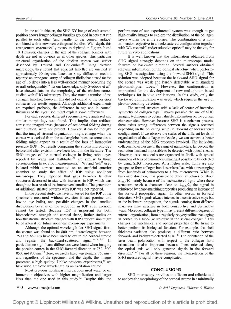
In the adult chicken, the SHG XY image of each stromalposition shows longer collagen bundles grouped in sets that runparallel to each other (and parallel to the corneal surface)combined with interwoven orthogonal bundles. With depth, thisarrangement systematically rotates as depicted in Figures 9 and10. However, changes in the size of the collagen bundles withdepth are not as obvious as in other species. This particularstructural organization of the chicken cornea was earlierdescribed by Trelstad and Coulombre.35 Using electronmicroscopy, they found that adjacent lamellae are oriented atapproximately 90 degrees. Later, an x-ray diffraction methodreported an orthogonal array of collagen fibrils that turned (at theage of 16 days) into a less well-oriented manner obscuring theoverall orthogonality.36 To our knowledge, only Svoboba et al37
have showed data on the morphology of the chicken corneastudied with SHG microscopy. They also noted a rotation of thecollagen lamellae; however, this did not extend to the posteriorcornea as our results suggest. Although additional experimentsare required, probably, the difference in age and in cornealthickness of the eyes used might explain some differences.
For each species, different specimens were analyzed andsimilar morphology was found. This implies that artifactsacross the imaged areas (because of accidental folding duringmanipulation) were not present. However, it can be thoughtthat the imaged stromal organization might change when thecornea was excised from the ocular globe, because some tissuefolding might appear as a result of the loss of intraocularpressure (IOP). No results comparing the stroma morphologybefore and after excision have been found in the literature. TheSHG images of the corneal collagen in living rabbit eyesreported by Wang and Halbhuber38 are similar to thosecorresponding to ex vivo measurements.11 Wu and Yeh32 usedisolated rabbit corneas mounted on an artificial anteriorchamber to study the effect of IOP using nonlinearmicroscopy. They reported that gaps between lamellarstructures decreased in size with increases in IOP, which wasthought to be a result of the interwoven lamellae. The generationof additional striated patterns with IOP was not reported.
In the present study, we have not tested for the IOP of thespecimens measured without excision (intact porcine andbovine eye balls), and possible changes in the lamellaedistribution because of the reduction in IOP after excisioncannot be tested. Because IOP is important for bothbiomechanical strength and corneal shape, further studies onhow the stromal structure changes with IOP after excision mightbe of interest for future nonlinear microscopy experiments.
Although the optimal wavelength for SHG signal fromthe cornea was found to be 800 nm,11 wavelengths between750 and 880 nm have been used to excite the corneal stromaand register the backward-scattered signal.8–10,12,16 Inparticular, no significant differences were found when imagingthe porcine cornea in the SHG-forward direction at 750, 800,850, and 900 nm.15 Here, we used a fixed wavelength (760 nm),and regardless of the specimen and the depth, the imagespresented a high quality. Unlike previous experiments,39 wehave used a unique wavelength as an excitation source.
Most previous nonlinear microscopes used water or oilimmersion objectives with higher magnification and largerNAs than the one used in this study.8,9 Despite this, the
performance of our experimental system was enough to gethigh-quality images to explore the distribution of the collagenlayers within the entire cornea. The combination of a non-immersion objective in a backscattered configuration togetherwith WA control20 and/or adaptive optics23 may be the key forfuture in vivo applications.
It is well known that the information obtained fromSHG signal strongly depends on the microscope mode:forward or backward direction. Several authors obtainedrelevant information on the corneal structure when perform-ing SHG investigations using the forward SHG signal. Thissolution was adopted because the backward SHG signal forthe cornea was weak and hardly detectable with standardphotomultiplier tubes.5,7 However, this configuration isimpractical for the development of new multiphoton-basedtechniques for in vivo applications. In this study, only thebackward configuration was used, which requires the use ofphoton-counting detectors.
The natural structure with a lack of center of inversionsymmetry of collagen type I makes possible the use of SHGimaging techniques to obtain valuable information on the cornealcharacteristics. However, because SHG is a coherent process,there exists strong differences between the signals obtaineddepending on the collecting setup (ie, forward or backscatteredconfiguration). If we observe the scales of the different levels oforganization of the collagen molecules, we can achieve a betterunderstanding of the SHG processes involved. The individualcollagen molecules are in the range of nanometers, far beyond theresolution limit and impossible to be detected with optical means.However, these molecules are organized in fibrils with typicaldiameters of tens of nanometers, making it possible to be detectedby using SHG microscopy. At a higher scale, fibrils are alsogrouped to form collagen bundles or lamellae, with a size rangingfrom hundreds of nanometers to a few micrometers. While inbackward direction, it is possible to detect structures of aboutlSHG/10 mainly because of the backscattered light, when thesestructures reach a diameter close to lSHG/2, the signal isreinforced by phase-matching properties producing an increase ofthe forward propagated signal. In other words, in forwarddetection, SHG signals always interact in a constructive way; butin the backward propagation, the signals coming from differentstructures may interfere in both constructive and destructiveways. Moreover, collagen type I may present different degrees ofinternal organization, from a regularly polycrystalline packagingin cornea, to a tube-like structure in the scleral collagen.5 Thischanges the mechanical and optical properties of the tissue tobetter perform its biological function. For example, the shellthickness variation also produces a different ratio betweenforward- and backward-detected SHG.40 The orientation of thelaser beam polarization with respect to the collagen fibrilorientation is also important because fibers oriented alongthe optical axis will only generate signals in the forwarddirection.41,42 For all of these reasons, the interpretation of theSHG measured signal maybe complicated.
CONCLUSIONSSHG microscopy provides an efficient and reliable tool
to analyze the morphology of the corneal stroma in a minimally
700 | www.corneajrnl.com q 2011 Lippincott Williams & Wilkins
Bueno et al Cornea � Volume 30, Number 6, June 2011
invasive manner. The improvements in the instrument whenusing wavefront control permitted to record SHG images withhigh quality even from deeper corneal layers in a backwardconfiguration. The high contrast of collagen images even atdeeper locations allows the analysis of the lamellar distributionacross the entire stroma. The collagen distribution foundacross the different species might be used as a basis toestablish models of stromal arrangement very useful inunderstanding the effects of pathologies, surgery, or thermaldamage. In the future, the clinical use of this technique inliving eyes would be helpful in the diagnosis and follow-up ofcorneal pathologies, to explore the effects of different cornealsurgeries, and in monitoring wound healing processes, amongothers.
ACKNOWLEDGMENTSThe authors thank Dr. J. M. Marın and C. Molero,
Hospital Universitario ‘‘Virgen de la Arrixaca,’’ Murcia,Spain, for their help with the donor’s corneal samples.
REFERENCES1. Denk W, Strickler JH, Webb WW. Two-photon laser scanning fluorescence
microscopy. Science. 1990;248:73–76.2. Guo Y, Ho PP, Savage H, et al. Second-harmonic tomography of tissues.
Opt Lett. 1997;22:1323–1325.3. Campagnola PJ, Clark HA, Mohler WA, et al. Second-harmonic imaging
microscopy of living cells. J Biomed Opt. 2001;6:277–286.4. Fine S, Hansen WP. Optical second harmonic generation in biological
systems. Appl Opt. 1971;10:2350–2353.5. Hochheimer BF. Second harmonic light generation in the rabbit cornea.
Appl Opt. 1982;21:1516–1518.6. Zaidi Q, Pokorny J. Appearance of pulsed infrared light: second harmonic
generation in the eye. Appl Opt. 1988;27:1064–1068.7. Maurice DM. The structure and transparency of the cornea. J Physiol.
1957;136:263–286.8. Yeh AT, Nassif N, Zoumi A, et al. Selective corneal imaging using
combined second-harmonic generation and two-photon excited fluores-cence. Opt Lett. 2002;27:2082–2084.
9. Han M, Giese G, Bille J. Second harmonic generation imaging of collagenfibrils in cornea and sclera. Opt Express. 2005;13:5791–5797.
10. Teng SW, Tan HY, Peng JL, et al. Multiphoton autofluorescence andsecond-harmonic generation imaging of the ex vivo porcine eye. InvestOphthalmol Vis Sci. 2006;47:1216–1224.
11. Morishige N, Petroll WM, Nishida T, et al. Noninvasive corneal stromalcollagen imaging using two-photon-generated second-harmonic signals.J Cataract Refract Surg. 2006;32:1784–1791.
12. Tan HY, Sun Y, Lo W, et al. Multiphoton fluorescence and secondharmonic generation imaging of the structural alterations in keratoconusex vivo. Invest Ophthalmol Vis Sci. 2006;47:5251–5259.
13. Morishige N, Wahlert AJ, Kenney MC, et al. Second-harmonic imagingmicroscopy of normal human and keratoconus cornea. Invest OphthalmolVis Sci. 2007;48:1087–1094.
14. Teng SW, Tan HY, Sun Y, et al. Multiphoton fluorescence and second-harmonic-generation microscopy for imaging structural alterations incorneal scar tissue in penetrating full-thickness wound. Arch Ophthalmol.2007;125:977–978.
15. Vohnsen B, Artal P. Second-harmonic microscopy of ex vivo porcinecorneas. J Microsc. 2008;232:158–163.
16. Wang BG, Eitner A, Lindenau J, et al. High-resolution two-photonexcitation microscopy of ocular tissues in porcine eye. Lasers Surg Med.2008;40:247–256.
17. Jay L, Brocas A, Singh K, et al. Determination of porcine corneal layerswith high spatial resolution by simultaneous second and third harmonicgeneration microscopy. Opt Express. 2008;16:16284–16293.
18. Morishige N, Nishida T, Jester J. Second harmonic generation forvisualizing 3-dimensional structure of corneal collagen lamellae. Cornea.2009;28:S46–S53.
19. Bueno JM, Vohnsen B, Roso L, et al. Temporal wavefront stability of anultrafast high-power laser beam. Appl Opt. 2009;48:770–777.
20. Gualda EJ, Bueno JM, Artal P. Wavefront optimized non-linearmicroscopy of ex-vivo human retinas. J Biomed Opt. 2010;15:026007.
21. Kaufman HE, Beuerman RW, Steinemann TL, et al. Optisol cornealstorage medium. Arch Ophthalmol. 1991;109:864–868.
22. Wilson SE, Bourne WM. Corneal preservation. Surv Ophthalmol. 1982;33:237–259.
23. Bueno JM, Gualda EJ, Artal P. Adaptive optics multiphoton microscopy tostudy ex-vivo ocular tissues. J Biom Opt 2010;15:066004.
24. Radner W, Zehetmayer M, Aufreiter R, et al. Interlacing and cross-angledistribution of collagen lamellae in the human cornea. Cornea. 1998;17:537–543.
25. Radner W, Mallinger R. Interlacing of collagen lamellae in the midstromaof the human cornea. Cornea. 2002;21:598–601.
26. Abahussin M, Hayes S, Knox Cartwright NE, et al. 3D Collagen orientationstudy of the human cornea using x-ray diffraction and femtosecond lasertechnology. Invest Ophthalmol Vis Sci. 2009;50:5159–5164.
27. Komai Y, Ushiki T. The three-dimensional organization of collagen fibrilsin the human cornea and sclera. Invest Ophthalmol Vis Sci. 1991;32:244–258.
28. Hogan MJ, Alvarado JA, Weddell JE. Histology of the Human Eye: an Atlas
and Textbook. Philadelphia, PA: W.B. Saunders Company; 1971:93–101.29. Morishige N, Yamada N, Teranishi S, et al. Detection of subepithelial
fibrosis associated with corneal stromal edema by second harmonicgeneration imaging microscopy. Invest Ophthalmol Vis Sci. 2009;50:3145–3150.
30. Matteini P, Ratto F, Rossi F, et al. Photothermally-induced disorderedpatterns of corneal collagen revealed by SHG imaging. Opt Express.2009;17:4868–4878.
31. Aptel F, Olivier N, Deniset-Besseau A, et al. Multimodal nonlinearimaging of the human cornea. Invest Ophthalmol Vis Sci. 2010;51:2459–2465.
32. Wu Q, Yeh AT. Rabbit cornea microstructure response to changes inintraocular pressure visualized by using nonlinear optical microscopy.Cornea. 2008;27:202–208.
33. Freeman IL. Collagen polymorphism in mature rabbit cornea. InvestOphthalmol Vis Sci. 1978;17:171–177.
34. Cox JL, Farrell RA, Hart RW, Langham ME. The transparency of themammalian cornea. J Physiol. 1970;210:601–616.
35. Trelstad RL, Coulombre AJ. Morphogenesis of the collagenous stroma inthe chick cornea. J Cell Biol. 1971;50:840–858.
36. Quantock AJ, Boote C, Siegler V, et al. Collagen organization in thesecondary chick cornea during development. Invest Ophthalmol Vis Sci.2003;44:130–136.
37. Svoboba KKH, Petroll MW, Jester JV. Second harmonic signal analysisof whole embryonic avian corneas. Microsc Microanal. 2007;13:1550–1551.
38. Wang BG, Halbhuber KJ. Corneal multiphoton microscopy andintratissue optical nanosurgery by nanojoule femtosecond near-infraredpulsed lasers. Ann Anat. 2006;188:395–409.
39. Wang BG, Koenig K, Riemann I, et al. Intraocular multiphotonmicroscopy by infrared femtosecond lasers. Histochem Cell Biol.2006;126:507–515.
40. Williams RM, Zipfel WR, Webb WW. Interpreting second harmonicgeneration images of collagen I fibrils. Biophys J. 2005;88:1377–1386.
41. Cox GC, Xu P, Sheppard CJR, et al. Characterization of the secondharmonic signal from collagen. Proc SPIE. 2003;4963:32–40.
42. Chu S-W, Tai S-P, Sun C-K, et al. Selective imaging in second-harmonic-generation microscopy by polarization manipulation. Appl Phys Lett.2007;91:103903.
q 2011 Lippincott Williams & Wilkins www.corneajrnl.com | 701
Cornea � Volume 30, Number 6, June 2011 Corneal Stroma Organization