mrap2 regulates ghrelin receptor signaling and hunger sensing
TRANSCRIPT
ARTICLE
MRAP2 regulates ghrelin receptor signaling andhunger sensingDollada Srisai1,2,3, Terry C. Yin1,2,3, Abigail A. Lee1,2,3, Alix A.J. Rouault1,2,3, Nicole A. Pearson2,4,
Justin L. Grobe2,4 & Julien A. Sebag 1,2,3
Ghrelin is the only known circulating orexigenic hormone. It is primarily secreted by the
stomach and acts at its receptor, the growth hormone secretagogue receptor 1a (GHSR1a), in
the hypothalamus to signal hunger and promote food intake. The melanocortin receptor
accessory protein 2 (MRAP2) was previously shown to regulate energy homeostasis through
the modulation of the activity of the melanocortin-4 receptor and prokineticin receptors. In
this study we identify MRAP2 as a partner of ghrelin-GHSR1a signaling. We show that
MRAP2 interacts with GHSR1a and potentiates ghrelin-stimulated signaling both in vitro and
in vivo. We demonstrate that in the absence of MRAP2, fasting fails to activate agouti-related
protein neurons. In addition, we show that the orexigenic effect of ghrelin is lost in mice
lacking MRAP2. Our results suggest that MRAP2 is an important modulator of the energy
homeostasis machinery that operates through the regulation of multiple GPCRs throughout
the hypothalamus.
DOI: 10.1038/s41467-017-00747-6 OPEN
1 Department of Molecular Physiology and Biophysics, University of Iowa, Carver College of Medicine, Iowa City, IA 52242, USA. 2 F.O.E.D.R.C, Iowa City, IA52242, USA. 3 Pappajohn Biomedical Institute, Iowa City, IA 52242, USA. 4 Department of Pharmacology, University of Iowa, Carver College of Medicine,Iowa City, IA 52242, USA. Dollada Srisai and Terry C. Yin contributed equally to this work.Correspondence and requests for materials should be addressed toJ.A.S. (email: [email protected])
NATURE COMMUNICATIONS |8: 713 |DOI: 10.1038/s41467-017-00747-6 |www.nature.com/naturecommunications 1
Food intake and energy expenditure are tightly regulated bynumerous hormones and neuropeptides secreted either byperipheral organs or centrally1, 2. These molecules act on the
hypothalamus, through their respective receptors, to inform thebrain on the energy state of the organism. Ghrelin is a peptidehormone secreted by the stomach3 during fasting to signalhunger to the brain4, 5. Ghrelin acts through its receptor, thegrowth hormone secretagogue receptor 1a (GHSR1a),a G-protein-coupled receptor (GPCR) expressed in the hypo-thalamus, in particular in orexigenic agouti-related protein(AGRP) neurons5–8. Ghrelin function has been shown to belargely mediated through and to require AGRP neurons5, 9.Whereas several GPCRs have been shown to be regulated by or torequire the presence of accessory proteins, the interaction ofGHSR1a with accessory proteins has not been investigated.
The melanocortin receptor accessory protein 2 (MRAP2) is asingle transmembrane protein that plays an important role in thecontrol of energy homeostasis since the deletion of MRAP2 inmice causes severe obesity10. Mutations in MRAP2 have also beenidentified in obese humans10. MRAP2 is highly expressed in thehypothalamus where it is known to regulate the activity of themelanocortin-4 receptor10, 11 and the prokineticin receptors12,GPCRs, that signal satiety and are required for the proper controlof food intake and energy expenditure12–16. In this study weinvestigated if MRAP2 modulates ghrelin signaling in vitro andin vivo, and conclude that MRAP2 interacts with GHSR1a and isan important component of the ghrelin receptor signaling com-plex, thus establishing that in addition to being a regulator ofsatiety sensing through the modulation of MC4R and PKR1activity, MRAP2 promotes hunger sensing by potentiating ghrelinsignaling.
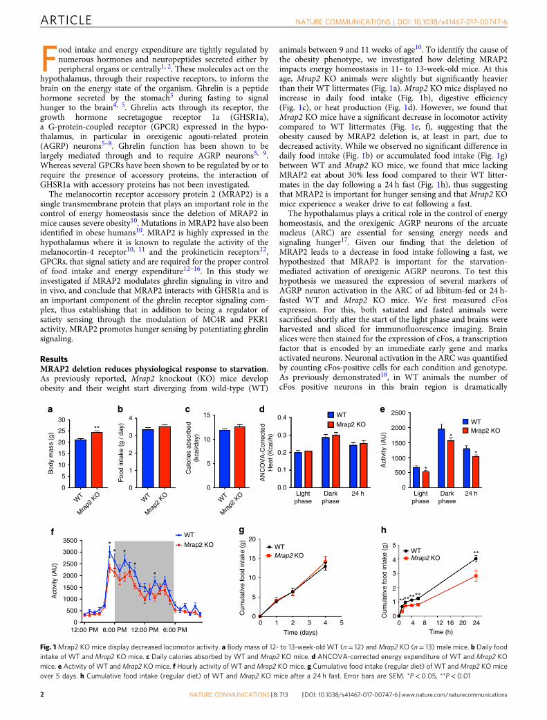
ResultsMRAP2 deletion reduces physiological response to starvation.As previously reported, Mrap2 knockout (KO) mice developobesity and their weight start diverging from wild-type (WT)
animals between 9 and 11 weeks of age10. To identify the cause ofthe obesity phenotype, we investigated how deleting MRAP2impacts energy homeostasis in 11- to 13-week-old mice. At thisage, Mrap2 KO animals were slightly but significantly heavierthan their WT littermates (Fig. 1a). Mrap2 KO mice displayed noincrease in daily food intake (Fig. 1b), digestive efficiency(Fig. 1c), or heat production (Fig. 1d). However, we found thatMrap2 KO mice have a significant decrease in locomotor activitycompared to WT littermates (Fig. 1e, f), suggesting that theobesity caused by MRAP2 deletion is, at least in part, due todecreased activity. While we observed no significant difference indaily food intake (Fig. 1b) or accumulated food intake (Fig. 1g)between WT and Mrap2 KO mice, we found that mice lackingMRAP2 eat about 30% less food compared to their WT litter-mates in the day following a 24 h fast (Fig. 1h), thus suggestingthat MRAP2 is important for hunger sensing and that Mrap2 KOmice experience a weaker drive to eat following a fast.
The hypothalamus plays a critical role in the control of energyhomeostasis, and the orexigenic AGRP neurons of the arcuatenucleus (ARC) are essential for sensing energy needs andsignaling hunger17. Given our finding that the deletion ofMRAP2 leads to a decrease in food intake following a fast, wehypothesized that MRAP2 is important for the starvation-mediated activation of orexigenic AGRP neurons. To test thishypothesis we measured the expression of several markers ofAGRP neuron activation in the ARC of ad libitum-fed or 24 h-fasted WT and Mrap2 KO mice. We first measured cFosexpression. For this, both satiated and fasted animals weresacrificed shortly after the start of the light phase and brains wereharvested and sliced for immunofluorescence imaging. Brainslices were then stained for the expression of cFos, a transcriptionfactor that is encoded by an immediate early gene and marksactivated neurons. Neuronal activation in the ARC was quantifiedby counting cFos-positive cells for each condition and genotype.As previously demonstrated18, in WT animals the number ofcFos positive neurons in this brain region is dramatically
e 2500
2000
1500
1000
500
0
*
*
*
Lightphase
Darkphase
24 h
Mrap2 KOWT
f
**
*** Mrap2 KO
WT
0
Act
ivity
(A
U)
500
1000
1500
2000
2500
3000
3500
12:00 PM 12:00 PM6:00 PM 6:00 PM
g
Mrap2 KOWT
20
15
10
5
00
Cum
ulat
ive
food
inta
ke (
g)
Time (days)
54321
d0.4
0.3
0.2
0.1
0.0Light
phaseDark
phase24 h
Mrap2 KO
WT
AN
CO
VA
-Cor
rect
edH
eat (
Kca
l/h)
Act
ivity
(A
U)
h
**
********
5
4
3
2
00 4 8 12 16 20
Time (h)24
1
Mrap2 KOWT
Cum
ulat
ive
food
inta
ke (
g)
b4
Foo
d in
take
(g
/ day
)
2
1
0
3
WT
Mra
p2 K
O
c
10
15
5
0
Cal
orie
s ab
sorb
ed(k
cal/d
ay)
WT
Mra
p2 K
O
a
**25
30
20
15
10
5
0
WT
Mra
p2 K
O
Bod
y m
ass
(g)
Fig. 1Mrap2 KO mice display decreased locomotor activity. a Body mass of 12- to 13-week-old WT (n= 12) andMrap2 KO (n= 13) male mice. b Daily foodintake of WT and Mrap2 KO mice. c Daily calories absorbed by WT and Mrap2 KO mice. d ANCOVA-corrected energy expenditure of WT and Mrap2 KOmice. e Activity of WT andMrap2 KO mice. f Hourly activity of WT andMrap2 KO mice. g Cumulative food intake (regular diet) of WT andMrap2 KO miceover 5 days. h Cumulative food intake (regular diet) of WT and Mrap2 KO mice after a 24 h fast. Error bars are SEM. *P< 0.05, **P< 0.01
ARTICLE NATURE COMMUNICATIONS | DOI: 10.1038/s41467-017-00747-6
2 NATURE COMMUNICATIONS |8: 713 |DOI: 10.1038/s41467-017-00747-6 |www.nature.com/naturecommunications
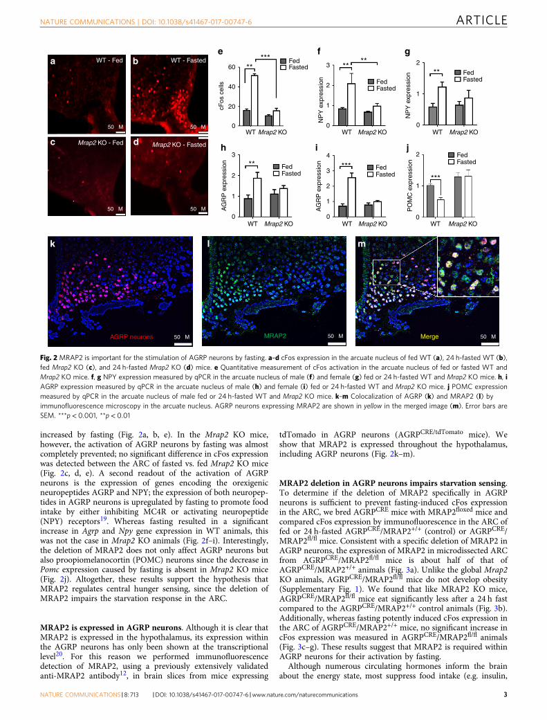
increased by fasting (Fig. 2a, b, e). In the Mrap2 KO mice,however, the activation of AGRP neurons by fasting was almostcompletely prevented; no significant difference in cFos expressionwas detected between the ARC of fasted vs. fed Mrap2 KO mice(Fig. 2c, d, e). A second readout of the activation of AGRPneurons is the expression of genes encoding the orexigenicneuropeptides AGRP and NPY; the expression of both neuropep-tides in AGRP neurons is upregulated by fasting to promote foodintake by either inhibiting MC4R or activating neuropeptide(NPY) receptors19. Whereas fasting resulted in a significantincrease in Agrp and Npy gene expression in WT animals, thiswas not the case in Mrap2 KO animals (Fig. 2f–i). Interestingly,the deletion of MRAP2 does not only affect AGRP neurons butalso proopiomelanocortin (POMC) neurons since the decrease inPomc expression caused by fasting is absent in Mrap2 KO mice(Fig. 2j). Altogether, these results support the hypothesis thatMRAP2 regulates central hunger sensing, since the deletion ofMRAP2 impairs the starvation response in the ARC.
MRAP2 is expressed in AGRP neurons. Although it is clear thatMRAP2 is expressed in the hypothalamus, its expression withinthe AGRP neurons has only been shown at the transcriptionallevel20. For this reason we performed immunofluorescencedetection of MRAP2, using a previously extensively validatedanti-MRAP2 antibody12, in brain slices from mice expressing
tdTomado in AGRP neurons (AGRPCRE/tdTomato mice). Weshow that MRAP2 is expressed throughout the hypothalamus,including AGRP neurons (Fig. 2k–m).
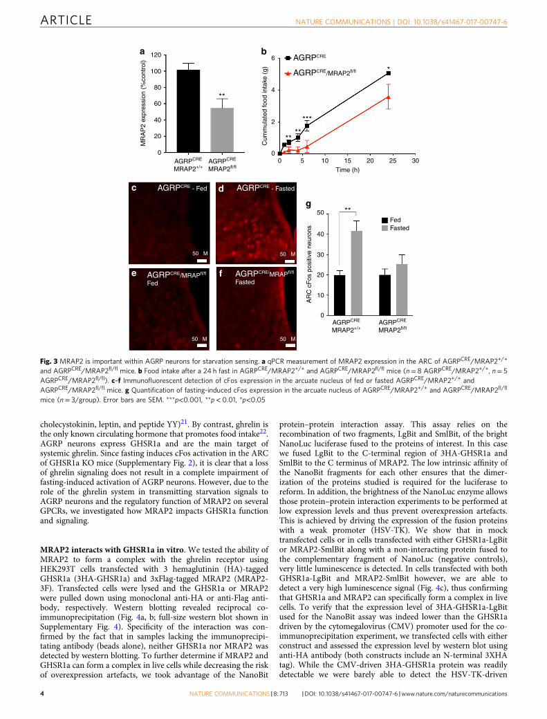
MRAP2 deletion in AGRP neurons impairs starvation sensing.To determine if the deletion of MRAP2 specifically in AGRPneurons is sufficient to prevent fasting-induced cFos expressionin the ARC, we bred AGRPCRE mice with MRAP2floxed mice andcompared cFos expression by immunofluorescence in the ARC offed or 24 h-fasted AGRPCRE/MRAP2+/+ (control) or AGRPCRE/MRAP2fl/fl mice. Consistent with a specific deletion of MRAP2 inAGRP neurons, the expression of MRAP2 in microdissected ARCfrom AGRPCRE/MRAP2fl/fl mice is about half of that ofAGRPCRE/MRAP2+/+ animals (Fig. 3a). Unlike the global Mrap2KO animals, AGRPCRE/MRAP2fl/fl mice do not develop obesity(Supplementary Fig. 1). We found that like MRAP2 KO mice,AGRPCRE/MRAP2fl/fl mice eat significantly less after a 24 h fastcompared to the AGRPCRE/MRAP2+/+ control animals (Fig. 3b).Additionally, whereas fasting potently induced cFos expression inthe ARC of AGRPCRE/MRAP2+/+ mice, no significant increase incFos expression was measured in AGRPCRE/MRAP2fl/fl animals(Fig. 3c–g). These results suggest that MRAP2 is required withinAGRP neurons for their activation by fasting.
Although numerous circulating hormones inform the brainabout the energy state, most suppress food intake (e.g. insulin,
Mrap2 KO - Fed Mrap2 KO - Fasted
AGRP neurons MRAP2 Merge
WT - FastedWT - Feda b
c d
k l m
Mrap2 KOWT
60
e
Fasted***
**
40
20
0
cFos
cel
ls
Fed
50 µM
50 µM50 µM
50 µM50 µM 50 µM
50 µM
Mrap2 KOWT
i
Fasted***
4
3
2
1
0
AG
RP
exp
ress
ion
Fed
Mrap2 KOWT
f
NP
Y e
xpre
ssio
n
Fasted
****3
2
1
0
Fed
Mrap2 KOWT
g
NP
Y e
xpre
ssio
n
Fasted**
2
1
0
Fed
Mrap2 KOWT
h
Fasted**
3
2
1
0A
GR
P e
xpre
ssio
n
Fed
Mrap2 KOWT
jFasted
***
2
1
0
PO
MC
exp
ress
ion
Fed
Fig. 2 MRAP2 is important for the stimulation of AGRP neurons by fasting. a–d cFos expression in the arcuate nucleus of fed WT (a), 24 h-fasted WT (b),fed Mrap2 KO (c), and 24 h-fasted Mrap2 KO (d) mice. e Quantitative measurement of cFos activation in the arcuate nucleus of fed or fasted WT andMrap2 KO mice. f, g NPY expression measured by qPCR in the arcuate nucleus of male (f) and female (g) fed or 24 h-fasted WT and Mrap2 KO mice. h, iAGRP expression measured by qPCR in the arcuate nucleus of male (h) and female (i) fed or 24 h-fasted WT and Mrap2 KO mice. j POMC expressionmeasured by qPCR in the arcuate nucleus of male fed or 24 h-fasted WT and Mrap2 KO mice. k–m Colocalization of AGRP (k) and MRAP2 (l) byimmunofluorescence microscopy in the arcuate nucleus. AGRP neurons expressing MRAP2 are shown in yellow in the merged image (m). Error bars areSEM. ***p< 0.001, **p< 0.01
NATURE COMMUNICATIONS | DOI: 10.1038/s41467-017-00747-6 ARTICLE
NATURE COMMUNICATIONS |8: 713 |DOI: 10.1038/s41467-017-00747-6 |www.nature.com/naturecommunications 3
cholecystokinin, leptin, and peptide YY)21. By contrast, ghrelin isthe only known circulating hormone that promotes food intake22.AGRP neurons express GHSR1a and are the main target ofsystemic ghrelin. Since fasting induces cFos activation in the ARCof GHSR1a KO mice (Supplementary Fig. 2), it is clear that a lossof ghrelin signaling does not result in a complete impairment offasting-induced activation of AGRP neurons. However, due to therole of the ghrelin system in transmitting starvation signals toAGRP neurons and the regulatory function of MRAP2 on severalGPCRs, we investigated how MRAP2 impacts GHSR1a functionand signaling.
MRAP2 interacts with GHSR1a in vitro. We tested the ability ofMRAP2 to form a complex with the ghrelin receptor usingHEK293T cells transfected with 3 hemaglutinin (HA)-taggedGHSR1a (3HA-GHSR1a) and 3xFlag-tagged MRAP2 (MRAP2-3F). Transfected cells were lysed and the GHSR1a or MRAP2were pulled down using monoclonal anti-HA or anti-Flag anti-body, respectively. Western blotting revealed reciprocal co-immunoprecipitation (Fig. 4a, b; full-size western blot shown inSupplementary Fig. 4). Specificity of the interaction was con-firmed by the fact that in samples lacking the immunoprecipi-tating antibody (beads alone), neither GHSR1a nor MRAP2 wasdetected by western blotting. To further determine if MRAP2 andGHSR1a can form a complex in live cells while decreasing the riskof overexpression artefacts, we took advantage of the NanoBit
protein–protein interaction assay. This assay relies on therecombination of two fragments, LgBit and SmlBit, of the brightNanoLuc luciferase fused to the proteins of interest. In this casewe fused LgBit to the C-terminal region of 3HA-GHSR1a andSmlBit to the C terminus of MRAP2. The low intrinsic affinity ofthe NanoBit fragments for each other ensures that the dimer-ization of the proteins studied is required for the luciferase toreform. In addition, the brightness of the NanoLuc enzyme allowsthose protein–protein interaction experiments to be performed atlow expression levels and thus prevent overexpression artefacts.This is achieved by driving the expression of the fusion proteinswith a weak promoter (HSV-TK). We show that in mocktransfected cells or in cells transfected with either GHSR1a-LgBitor MRAP2-SmlBit along with a non-interacting protein fused tothe complementary fragment of NanoLuc (negative controls),very little luminescence is detected. In cells transfected with bothGHSR1a-LgBit and MRAP2-SmlBit however, we are able todetect a very high luminescence signal (Fig. 4c), thus confirmingthat GHSR1a and MRAP2 can specifically form a complex in livecells. To verify that the expression level of 3HA-GHSR1a-LgBitused for the NanoBit assay was indeed lower than the GHSR1adriven by the cytomegalovirus (CMV) promoter used for the co-immunoprecipitation experiment, we transfected cells with eitherconstruct and assessed the expression level by western blot usinganti-HA antibody (both constructs include an N-terminal 3XHAtag). While the CMV-driven 3HA-GHSR1a protein was readilydetectable we were barely able to detect the HSV-TK-driven
50 µM50 µM
c d
e f
50 µM 50 µM
AGRPCRE - Fed AGRPCRE - Fasted
AGRPCRE/MRAPfl/fl
FastedAGRPCRE/MRAPfl/fl
Fed
0105 15
Time (h)20 25
*
300
2
Cum
mul
ated
food
inta
ke (
g)
4
6
***
AGRPCRE
AGRPCRE/MRAP2fl/fl
****
120
100
60
80
a b
40
20
0
**
AGRPCRE
MRAP2+/+AGRPCRE
MRAP2fl/fl
MR
AP
2 ex
pres
sion
(%
cont
rol)
g50
Fed**
Fasted40
30
20
10
AR
C c
Fos
pos
itive
neu
rons
0AGRPCRE
MRAP2+/+AGRPCRE
MRAP2fl/fl
Fig. 3 MRAP2 is important within AGRP neurons for starvation sensing. a qPCR measurement of MRAP2 expression in the ARC of AGRPCRE/MRAP2+/+
and AGRPCRE/MRAP2fl/fl mice. b Food intake after a 24 h fast in AGRPCRE/MRAP2+/+ and AGRPCRE/MRAP2fl/fl mice (n= 8 AGRPCRE/MRAP2+/+, n= 5AGRPCRE/MRAP2fl/fl). c–f Immunofluorescent detection of cFos expression in the arcuate nucleus of fed or fasted AGRPCRE/MRAP2+/+ andAGRPCRE/MRAP2fl/fl mice. g Quantification of fasting-induced cFos expression in the arcuate nucleus of AGRPCRE/MRAP2+/+ and AGRPCRE/MRAP2fl/fl
mice (n= 3/group). Error bars are SEM. ***p<0.001, **p< 0.01, *p<0.05
ARTICLE NATURE COMMUNICATIONS | DOI: 10.1038/s41467-017-00747-6
4 NATURE COMMUNICATIONS |8: 713 |DOI: 10.1038/s41467-017-00747-6 |www.nature.com/naturecommunications
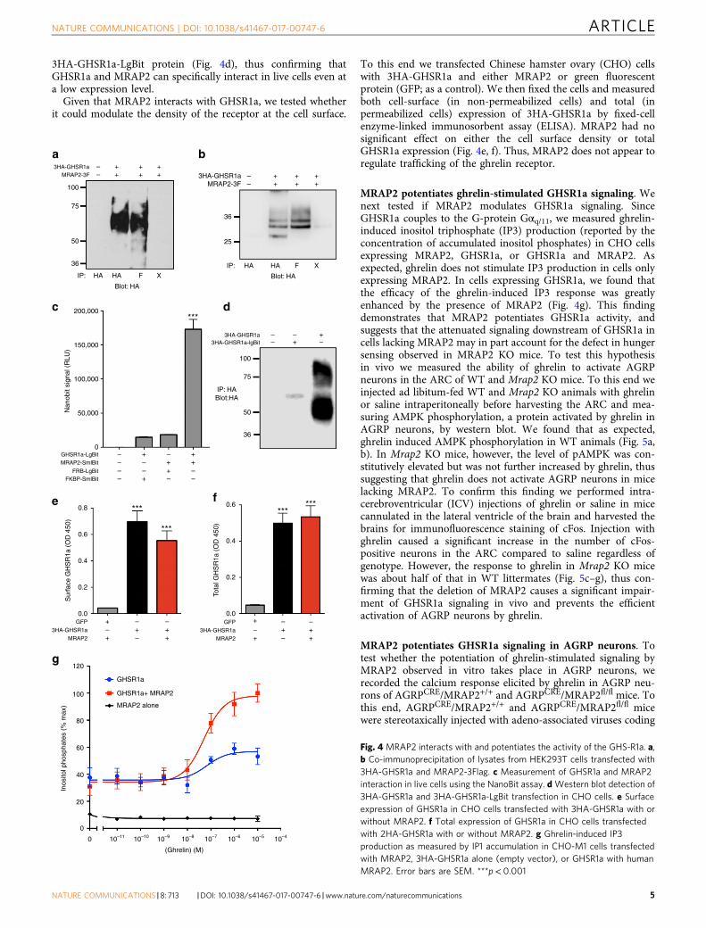
3HA-GHSR1a-LgBit protein (Fig. 4d), thus confirming thatGHSR1a and MRAP2 can specifically interact in live cells even ata low expression level.
Given that MRAP2 interacts with GHSR1a, we tested whetherit could modulate the density of the receptor at the cell surface.
To this end we transfected Chinese hamster ovary (CHO) cellswith 3HA-GHSR1a and either MRAP2 or green fluorescentprotein (GFP; as a control). We then fixed the cells and measuredboth cell-surface (in non-permeabilized cells) and total (inpermeabilized cells) expression of 3HA-GHSR1a by fixed-cellenzyme-linked immunosorbent assay (ELISA). MRAP2 had nosignificant effect on either the cell surface density or totalGHSR1a expression (Fig. 4e, f). Thus, MRAP2 does not appear toregulate trafficking of the ghrelin receptor.
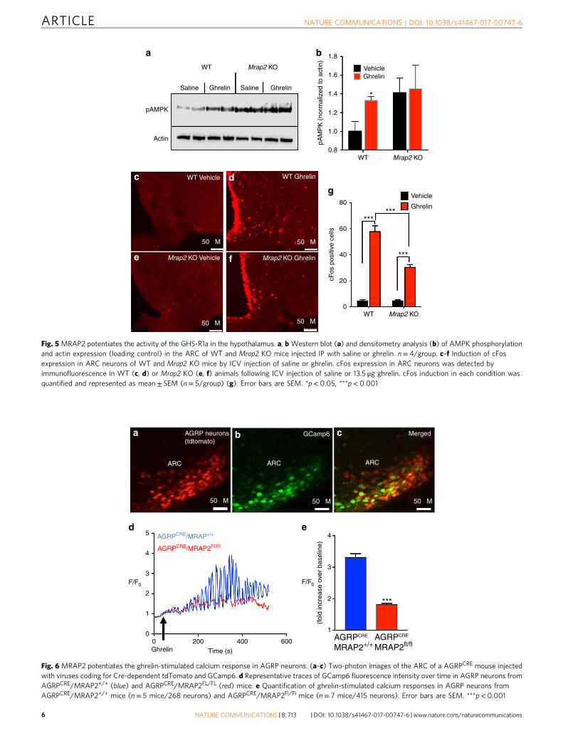
MRAP2 potentiates ghrelin-stimulated GHSR1a signaling. Wenext tested if MRAP2 modulates GHSR1a signaling. SinceGHSR1a couples to the G-protein Gαq/11, we measured ghrelin-induced inositol triphosphate (IP3) production (reported by theconcentration of accumulated inositol phosphates) in CHO cellsexpressing MRAP2, GHSR1a, or GHSR1a and MRAP2. Asexpected, ghrelin does not stimulate IP3 production in cells onlyexpressing MRAP2. In cells expressing GHSR1a, we found thatthe efficacy of the ghrelin-induced IP3 response was greatlyenhanced by the presence of MRAP2 (Fig. 4g). This findingdemonstrates that MRAP2 potentiates GHSR1a activity, andsuggests that the attenuated signaling downstream of GHSR1a incells lacking MRAP2 may in part account for the defect in hungersensing observed in MRAP2 KO mice. To test this hypothesisin vivo we measured the ability of ghrelin to activate AGRPneurons in the ARC of WT and Mrap2 KO mice. To this end weinjected ad libitum-fed WT and Mrap2 KO animals with ghrelinor saline intraperitoneally before harvesting the ARC and mea-suring AMPK phosphorylation, a protein activated by ghrelin inAGRP neurons, by western blot. We found that as expected,ghrelin induced AMPK phosphorylation in WT animals (Fig. 5a,b). In Mrap2 KO mice, however, the level of pAMPK was con-stitutively elevated but was not further increased by ghrelin, thussuggesting that ghrelin does not activate AGRP neurons in micelacking MRAP2. To confirm this finding we performed intra-cerebroventricular (ICV) injections of ghrelin or saline in micecannulated in the lateral ventricle of the brain and harvested thebrains for immunofluorescence staining of cFos. Injection withghrelin caused a significant increase in the number of cFos-positive neurons in the ARC compared to saline regardless ofgenotype. However, the response to ghrelin in Mrap2 KO micewas about half of that in WT littermates (Fig. 5c–g), thus con-firming that the deletion of MRAP2 causes a significant impair-ment of GHSR1a signaling in vivo and prevents the efficientactivation of AGRP neurons by ghrelin.
MRAP2 potentiates GHSR1a signaling in AGRP neurons. Totest whether the potentiation of ghrelin-stimulated signaling byMRAP2 observed in vitro takes place in AGRP neurons, werecorded the calcium response elicited by ghrelin in AGRP neu-rons of AGRPCRE/MRAP2+/+ and AGRPCRE/MRAP2fl/fl mice. Tothis end, AGRPCRE/MRAP2+/+ and AGRPCRE/MRAP2fl/fl micewere stereotaxically injected with adeno-associated viruses coding
a
d
3HA-GHSR1a3HA-GHSR1a-lgBit
100
75
50
36
IP: HA
– –––+
+
Blot:HA
b
36
25
3HA-GHSR1aMRAP2-3F
+––
+++
++
IP: HA HA
Blot: HA
F X
MRAP2 ++ –MRAP2
0.8 *** ******
e f
***
++
++
– –– +
–
GFP
3HA-GHSR1a
++– –
– +GFP
3HA-GHSR1a
0.6
0.4
0.2
0.0
Sur
face
GH
SR
1a (
OD
450
)
0.6
0.4
0.2
0.0
Tota
l GH
SR
1a (
OD
450
)
g120
100
60
Inos
itol p
hosp
hate
s (%
max
)
80
40
20
0
0
(Ghrelin) (M)
10–11 10–10 10–9 10–8 10–7 10–6 10–5 10–4
GHSR1a
GHSR1a+ MRAP2
MRAP2 alone
3HA-GHSR1aMRAP2-3F
100
75
50
36
IP: HA
+––
+++
++
HA
200,000
150,000
100,000
50,000
+0
–+
+–
––
––
–
– ––
++
+
***
Nan
obit
sign
al (
RLU
)
c
GHSR1a-LgBitMRAP2-SmlBit
FKBP-SmlBitFRB-LgBit
Blot: HA
F X
Fig. 4 MRAP2 interacts with and potentiates the activity of the GHS-R1a. a,b Co-immunoprecipitation of lysates from HEK293T cells transfected with3HA-GHSR1a and MRAP2-3Flag. c Measurement of GHSR1a and MRAP2interaction in live cells using the NanoBit assay. dWestern blot detection of3HA-GHSR1a and 3HA-GHSR1a-LgBit transfection in CHO cells. e Surfaceexpression of GHSR1a in CHO cells transfected with 3HA-GHSR1a with orwithout MRAP2. f Total expression of GHSR1a in CHO cells transfectedwith 2HA-GHSR1a with or without MRAP2. g Ghrelin-induced IP3production as measured by IP1 accumulation in CHO-M1 cells transfectedwith MRAP2, 3HA-GHSR1a alone (empty vector), or GHSR1a with humanMRAP2. Error bars are SEM. ***p< 0.001
NATURE COMMUNICATIONS | DOI: 10.1038/s41467-017-00747-6 ARTICLE
NATURE COMMUNICATIONS |8: 713 |DOI: 10.1038/s41467-017-00747-6 |www.nature.com/naturecommunications 5
WT
Saline
pAMPK
Actin
SalineGhrelin Ghrelin
Mrap2 KO
Mrap2 KO GhrelinMrap2 KO Vehicle
WT Vehiclec d
e f
WT Ghrelin
50 µM 50 µM
50 µM50 µM
a b 1.8
1.6
1.4
1.2
Ghrelin
WT
Vehicle
pAM
PK
(no
rmal
ized
to a
ctin
)
1.0
0.8
*
Mrap2 KO
Ghrelin
Vehicle
******
***
cFos
pos
itive
cel
ls
WT
80
g
60
40
20
0Mrap2 KO
Fig. 5 MRAP2 potentiates the activity of the GHS-R1a in the hypothalamus. a, b Western blot (a) and densitometry analysis (b) of AMPK phosphorylationand actin expression (loading control) in the ARC of WT and Mrap2 KO mice injected IP with saline or ghrelin. n= 4/group. c–f Induction of cFosexpression in ARC neurons of WT and Mrap2 KO mice by ICV injection of saline or ghrelin. cFos expression in ARC neurons was detected byimmunofluorescence in WT (c, d) or Mrap2 KO (e, f) animals following ICV injection of saline or 13.5 μg ghrelin. cFos induction in each condition wasquantified and represented as mean± SEM (n= 5/group) (g). Error bars are SEM. *p< 0.05, ***p< 0.001
ARC
a b c
ARC ARC
GCamp6 Merged
50 µM 50 µM 50 µM
AGRP neurons(tdtomato)
00
1
2
3
4
d e5
F/F0 F/F0
200
AGRPCRE/MRAP2FI/FI
AGRPCRE/MRAP+/+
Ghrelin Time (s)400 600
4
3
2
(fol
d in
crea
se o
ver
base
line)
1
***
AGRPCRE
MRAP2+/+AGRPCRE
MRAP2fl/fl
Fig. 6 MRAP2 potentiates the ghrelin-stimulated calcium response in AGRP neurons. (a–c) Two-photon images of the ARC of a AGRPCRE mouse injectedwith viruses coding for Cre-dependent tdTomato and GCamp6. d Representative traces of GCamp6 fluorescence intensity over time in AGRP neurons fromAGRPCRE/MRAP2+/+ (blue) and AGRPCRE/MRAP2FL/FL (red) mice. e Quantification of ghrelin-stimulated calcium responses in AGRP neurons fromAGRPCRE/MRAP2+/+ mice (n= 5 mice/268 neurons) and AGRPCRE/MRAP2Fl/Fl mice (n= 7 mice/415 neurons). Error bars are SEM. ***p< 0.001
ARTICLE NATURE COMMUNICATIONS | DOI: 10.1038/s41467-017-00747-6
6 NATURE COMMUNICATIONS |8: 713 |DOI: 10.1038/s41467-017-00747-6 |www.nature.com/naturecommunications
for CRE-dependent tdTomato and the GCamp6 calcium reporterunilaterally in the ARC. About 2 weeks after viral injection, brainslices containing the ARC were placed in the perfusion chamberof a two-photon microscope for fluorescence imaging (Fig. 6a–c).Slices were perfused with artificial cerebrospinal fluid (aCSF)followed by aCSF supplemented with 10 nM ghrelin, andGCamp6 fluorescence intensity was recorded over time in allAGRP neurons (tdTomato-positive) present in the field. At theend of the recording, slices were perfused with KCl to inducedepolarization and allow the identification of all responsiveneurons. We found that ghrelin induces calcium oscillations inAGRP neurons (Fig. 6d) with an average amplitude greater thanthreefold over baseline (Fig. 6e). Deletion of MRAP2 specificallyin AGRP neurons significantly decreased the efficacy of ghrelinsince the amplitude of the calcium response in AGRP neurons ofAGRPCRE/MRAP2fl/fl mice was below twofold over baseline(Fig. 6d, e). No difference in the overall number of AGRP neu-rons was measured in the ARC of AGRPCRE/MRAP2+/+ andAGRPCRE/MRAP2fl/fl mice (Supplementary Fig. 3). This resultdemonstrates that, in agreement with our in vitro data, MRAP2potentiates GHSR1a signaling in AGRP neurons.
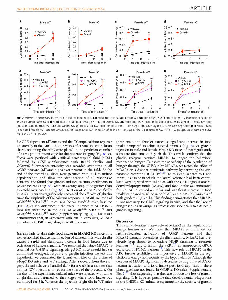
Ghrelin fails to stimulate food intake in MRAP2 KO mice. It iswell established that central injection of satiated mice with ghrelincauses a rapid and significant increase in food intake due toactivation of hunger signaling. We reasoned that since MRAP2 isessential for GHSR1a signaling, Mrap2 KO mice should have adiminished orexigenic response to ghrelin injection. To test thishypothesis, we cannulated the lateral ventricles of the brains ofMrap2 KO mice and WT siblings. After recovery from the sur-gery, the animals were handled daily for a week in a manner thatmimics ICV injections, to reduce the stress of the procedure. Onthe day of the experiment, satiated mice were injected with salineor ghrelin, and returned to their cages where food intake wasmonitored for 3 h. Whereas the injection of ghrelin in WT mice
(both male and female) caused a significant increase in foodintake compared to saline-injected animals (Fig. 7a, c), ghrelininjection in male and femaleMrap2 KO mice did not significantlystimulate food intake (Fig. 7b, d). This result confirms that theghrelin receptor requires MRAP2 to trigger the behavioralresponse to hunger. To assess the specificity of the regulation ofhunger through the GHSR1a by MRAP2, we tested the effect ofMRAP2 on a distinct orexigenic pathway by activating the can-nabinoid receptor 1 (CB1R)23, 24. To this end, satiated WT andMrap2 KO mice in which the lateral ventricle had been cannu-lated were injected with saline or with the CB1R agonist arachi-donylcyclopropylamide (ACPA), and food intake was monitoredfor 3 h. ACPA caused a similar and significant increase in foodintake compared to saline in both WT and Mrap2 KO animals ofboth genders (Fig. 7e–h). This finding demonstrates that MRAP2is not necessary for CB1R signaling in vivo, and that the lack ofhunger sensing inMrap2 KO mice is due specifically to a defect inghrelin signaling.
DiscussionThis study identifies a new role of MRAP2 in the regulation ofenergy homeostasis. We show that MRAP2 is important forfasting-mediated activation of AGRP neurons and thatMRAP2 strongly potentiates ghrelin signaling. MRAP2 has pre-viously been shown to potentiate MC4R signaling to promoteleanness10, 11 and to inhibit the PKR112, an anorexigenic GPCRexpressed in POMC neurons20. This new role of MRAP2 in theARC further establishes the importance of MRAP2 in the reg-ulation of energy homeostasis by the hypothalamus. Although thedeletion of MRAP2 significantly decreases fasting-induced AGRPneuron activation and food intake post food deprivation, thosephenotypes are not found in GHSR1a KO mice (SupplementaryFig. 2)25, thus suggesting that they are not due to a loss of ghrelinsignaling. It is however possible that developmental adaptationsin the GHSR1a KO animal compensate for the absence of ghrelin
4
Female WT
VehicleGhrelin
**
0.5
0.4
0.3
0.2
0.1
0.00 1
Time after injection (h)
Cum
ulat
ive
food
inta
ke (
g)
*
Male WT
VehicleGhrelin
**0.6
0.5
0.4
0.3
0.2
0.1
0.00
a
e f g h
b c d
1Time after injection (h)
Cum
ulat
ive
food
inta
ke (
g)
2 3 4
*
32
Male KO
VehicleGhrelin
0.6
0.5
0.4
0.3
0.2
0.1
0.00 1
Time after injection (h)
Cum
ulat
ive
food
inta
ke (
g)
2 3 4 4
Female KO
VehicleGhrelin
0.5
0.4
0.3
0.2
0.1
0.00 1
Time after injection (h)
Cum
ulat
ive
food
inta
ke (
g)
32
Cum
ulat
ive
food
inta
ke (
g)
4
Male KO
Vehicle1 µg ACPA5 µg ACPA
***
0.6
0.4
0.2
0.00 1
Time after injection (h)
2 3 4
Female KO
Vehicle1 µg ACPA5 µg ACPA
***
*** **
**
0.4
0.3
0.2
0.1
0.00 1
Time after injection (h)
Cum
ulat
ive
food
inta
ke (
g)
2 34
Female WT
Vehicle1 µg ACPA5 µg ACPA
***
**
**
0.4
0.3
0.2
0.1
0.00 1
Time after injection (h)
Cum
ulat
ive
food
inta
ke (
g)
2 3
Male WT
Vehicle1 µg ACPA5 µg ACPA
**0.5
0.4
0.3
0.2
0.1
0.00 1
Time after injection (h)
Cum
ulat
ive
food
inta
ke (
g)
2 3 4
Fig. 7MRAP2 is necessary for ghrelin to induce food intake. a, b Food intake in satiated male WT (a) andMrap2 KO (b) mice after ICV injection of saline or13.25 µg ghrelin (n= 6). c, d Food intake in satiated female WT (c) andMrap2 KO (d) mice after ICV injection of saline or 13.25 µg ghrelin (n= 6). e, f Foodintake in satiated male WT (e) and Mrap2 KO (f) mice after ICV injection of saline or 1 or 5 μg of the CB1R agonist ACPA (n= 5/group). g, h Food intakein satiated female WT (g) and Mrap2 KO (h) mice after ICV injection of saline or 1 or 5 μg of the CB1R agonist ACPA (n= 5/group). Error bars are SEM.**p< 0.01, ***p< 0.001
NATURE COMMUNICATIONS | DOI: 10.1038/s41467-017-00747-6 ARTICLE
NATURE COMMUNICATIONS |8: 713 |DOI: 10.1038/s41467-017-00747-6 |www.nature.com/naturecommunications 7
signaling. Since we have shown that MRAP2 is not expressed inthe embryo and only reaches normal expression levels around thetime of weaning11, it is less likely that developmental adaptationtake place in the Mrap2 KO mice. However, because thehomeostatic neuronal network can remodel and compensate forthe ablation of AGRP neurons in neonates8, compensation in theMrap2 KO model cannot be ruled out.
We show that the deletion of MRAP2 does not cause a com-plete loss of ghrelin responsiveness both in vitro and in vivo sincecells transfected with GHSR1a alone can elicit a small amount ofIP3 production when stimulated with ghrelin, and direct ICVinjection of ghrelin can induce some level of cFos expression inneurons of the Mrap2 KO mice.
While this study focuses on the role of MRAP2 in AGRPneurons, we show that the fasting-induced decrease in POMCexpression is also diminished in Mrap2 KO mice. This may bedue to the decreased inhibitory input from AGRP neurons ontoPOMC neurons or an indication that MRAP2 also plays animportant role in POMC neurons where it is also expressed20.
Mrap2 KO mice develop severe obesity over time10, 12, aphenotype that is in apparent contradiction with the conclusionof this study since one would predict that a decrease in GHSR1asignaling, due to the deletion of MRAP2, would promote lean-ness. It is however important to remember that, as stated earlier,MRAP2 is not a specific regulator of GHSR1a but also modulatethe activity of MC4R10, 11, PKR1,12 and possibly other yet uni-dentified receptors involved in the regulation of energy home-ostasis. Consequently, the phenotype of Mrap2 KO mice is theresult of a complex deregulation of the hypothalamic energyhomeostasis machinery and does not reflect specifically the effectof Mrap2 deletion on GHSR1a action. Later studies focusing onthe manipulation of MRAP2 expression specifically in AGRPneurons will be necessary to determine the role of MRAP2 on theregulation of physiological actions of ghrelin.
This study also illustrates the promiscuity of MRAP2, which atfirst was thought to be a specific regulator of melanocortinreceptors, and thus suggests that MRAP2 likely regulates severaladditional GPCRs. Identifying such GPCRs will improve ourunderstanding of the global physiological roles of MRAP2.MRAP2 displays a high level of selectivity for the receptors itinteracts with. For example, MRAP2 does not significantlymodulate the activity of the β2-adrenergic receptor, themelanocortin-3 receptor, or NPY receptors11. In this study, thenormal orexigenic response to ACPA suggests that MRAP2 is notrequired for CB1R function. The mechanisms involved in theselectivity of MRAP2 for a subset of GPCRs and its modulatoryeffects still remain to be elucidated.
Mice with a global or neuron-specific deletion of GHSR1a areresistant to diet-induced obesity and display improved insulinsensitivity26, 27, suggesting that inhibiting ghrelin actions couldpromote weight loss and decrease insulin resistance. Additionally,the stimulation of the ghrelin pathway is a promising approachfor the treatment of cachexia28 and diabetic gastroparesis29, 30.Understanding how MRAP2 enhances the activity of GHSR1aactivity may uncover new strategies to modulate ghrelin actionand inform drug discovery efforts targeting GHSR1a.
MethodsAnimals. C57BL/6N Mrap2 KO and Mrap2 floxed mice were generated by theSanger Mouse Genetics Project. AGRPCRE/tdTomato animals were generated bybreeding AGRPCRE (Agrptm1(cre)Lowl from the Jackson Laboratory) with R26-LSL-tdTomato reporter mice from the Jackson Laboratory. GHSR1a KO mice werekindly provided by Dr. Jeffrey Zigman (University of Texas Southwestern, Dallas,TX, USA). All animals were maintained in the University of Iowa temperaturecontrolled animal facility with 12 h light/dark cycles (6 a.m./6 p.m.). Animals werefed with standard rodent diet (NIH-31) and allowed free access to water. All
experiments using mice were approved by the Animal Care and Use Committee atthe University of Iowa.
Intracerebroventricular cannulation. Seven-week-old animals were anesthetizedwith intraperitoneal injection of a combination of xylazine and ketamine at dosage10:100 mg/kg before being placed on a stereotaxic apparatus (David Kopf Instru-ments, Tujunga, CA, USA). After standard disinfection on surgical site, an incisionwas made to expose the skull and a small hole was drilled. A 26-gauge stainless steelcannula (PlasticOne) was placed into the left lateral ventricle (anteroposterior − 0.3mm; medial lateral + 1.0 mm; dorsal ventral − 2.1 mm) and fixed in place withdental cement. Mice were allowed to recover for 7 days post surgery beforeexperiments. At the end of the study, the localization of the injection site wasconfirmed by injecting 2 μl blue ink into the cannula.
cFos immunofluorescence. Animals were deeply anesthetized with saturatedisoflurane in a closed chamber and perfused with ice cold 0.1 M phosphate buffersolution (PBS; pH 7.4), followed by ice-cold 4% paraformaldehyde (PFA) in PBS.Whole brains were dissected out and post fixed in fresh 4% PFA for12 h at 4 °C.Brains were then immersed in 30% sucrose in PBS at 4 °C until they sunk. Sampleswere embedded in Tissue-Tek OCT compound (Sakura Finetechical Co., Ltd.,Tokyo) and sectioned at 35 µm thickness using a cryostat (Leica Biosystems,Buffalo Grove, IL, USA). Free-floating sections were washed in PBS two times andincubated with blocking buffer containing 5% normal goat serum in PBS with 0.5%Triton X-100 for 1 h followed by rabbit polyclonal anti-cFos antibody (Catalog(Cat.) No. sc 52, Santa Cruz Biotechnology, TX, USA) in blocking solution (1:1000)overnight at 4 °C. After washed with PBS containing 0.1% Tween-20 (PBST), thesections were incubated in 1:500 Alexa Fluor 594 goat anti-rabbit IgG (R37117,Thermo Fisher Scientific) for 1 h at room temperature. The sections were washedin PBST five times and mounted onto superFrost slides (Fisher Scientific, Pitts-burgh, PA, USA), air-dried at room temperature in a dark room and coverslippedwith Prolong diamond antifade mountant with 4,6-diamidino-2-phenylindole(Molecular Probes by Life Technologies, Carlsbad, CA, USA). For cell counting,three different sections of the ARC of hypothalamus in each mouse were examinedusing × 20 magnification. cFos-positive cells were manually counted from entireARC from five mice per group using the Olympus IX3 microscope system andOlympus Cellsens Dimension software.
Cell culture and transfections. CHO-K1, HEK293T, and CHO-M1 cells werecultured in Dulbecco's Modified Eagle's medium (DMEM) /F-12 supplementedwith 5% v/v fetal bovine serum and 1% penicillin–streptomycin solution. Cultureswere incubated at 37 °C in a humidified atmosphere containing 5% CO2. Cells weretransfected using LipoD293 in vitro transfection reagent (Signagen, Rockville, MD,USA). The total amount of DNA transfected for each condition within anexperiment was kept identical.
Co-immunoprecipitation and western blot. HEK293T cells were transfected withhuman MRAP2-3Flag and 3HA-GHSR1a using LipoD293 according to the man-ufacturer’s protocol. At 24 h after transfection, the cells were lysed with ice-coldnon-denaturing lysis buffer containing 0.1% n-dodecyl-β-maltoside in PBS withproteinase inhibitor and incubated at 4 °C for 10 min on an orbital shaker, followedby centrifugation at 15,000 × g for 10 min. The protein lysates were transferred intonew centrifuge tubes and following antibodies, Purified mouse anti-HA.11 epitopetag antibody (Biolegend, USA) or M2 mouse anti-Flag (Sigma-Aldrich, St. Louis,MO, USA), were added into the cell lysates at 1/5000 dilution. The protein lysateswere then incubated on a shaker for overnight at 4 °C. 20 µl of protein G dynabeads(Life Technologies) were added into each sample and incubated at 4 °C for 1 h. Thebeads were washed four times with wash buffer and sample buffer containing LDSloading buffer with 5% β-mercaptoethanol was added on the beads to elude thetarget protein and antibodies and boiled 5 min. The same amount of protein fromeach sample was separated by SDS-polyacrylamide gel electrophoresis on 10% geland transferred to polyvinylidene fluoride membranes. The proteins were detectedby western blot using mouse anti-HA for GHSR1a co-immunoprecipitationexperiments and mouse anti-Flag for detecting MRAP2. After 1 h in blockingsolution containing 5% milk in PBS with 0.1 % Tween-20, the membranes wereincubated with the primary antibody diluted 1/5000 in blocking solution overnightat 4 °C. The membranes were washed with PBST for 5 min three times and thenincubated with secondary goat anti-mouse horseradish peroxidase (HRP) antibody(Cat. No. 170-6516, Bio-Rad Laboratories Inc, USA) at 1/5000 dilution for 45 minat room temperature followed by four washes with PBST. The WesternSure ECLsubstrate was applied on the membrane to detect the HRP-labeled secondaryantibodies on the membrane and imaged on an cDigit scanner (LI-COR, NE,USA).
NanoBit protein–protein interaction assay. CHO cells were transfected withGFP, 3HA-GHSR1a-LgBit + FKBP-SmlBit, FRB-LgBit + MRAP2-SmlBit, or 3HA-GHSR1a-LgBit +MRAP2-SmlBit. FKBP and FRB do not interact with GHSR1aand MRAP2, respectively, and were used to measure nonspecific NanoLucrecombination (Promega). Transfected cells were plated in a white opaque 96-wellplate and allowed to attach overnight. Live-cell NanoBit substrate was added to the
ARTICLE NATURE COMMUNICATIONS | DOI: 10.1038/s41467-017-00747-6
8 NATURE COMMUNICATIONS |8: 713 |DOI: 10.1038/s41467-017-00747-6 |www.nature.com/naturecommunications
cells following the manufacturer’s direction and luminescence was measured 5 minlater using a Spectramax i3 plate reader (Molecular Devices).
Western blot for MRAP2-V5 or pAMPK from ARC. Ad libitum-fed or 24 h-fasted mice were deeply anesthetized with isoflurane before removing the brain.ARC was isolated by microdissection and solubilized in lysis buffer (38.5 mM Tris-HCl, 1 mM EGTA, 1 mM EDTA, 1% Triton-X 100, 1 mM sodium orthovanadate,50 mM sodium fluoride, 5 mM sodium pyrophosphate, and 250 mM sucrose) withprotease inhibitor on ice. Protein concentration was determined by bicinchoninicacid assay and the same amount of protein was loaded on polyacrylamide gels forSDS separation. Western blot was performed using either anti-V5 (Cell SignalingTechnology, Rabbit mAb #13202) or anti-pAMPK (Cell Signaling TechnologyRabbit mAb #2535).
Quantitative PCR. After 24 h fasting, 11-week-old animals were deeply euthanizedwith saturated isoflurane in a closed chamber. Fresh whole hypothalami werecollected into an ice-chilled tube and immediately frozen in dry ice and kept at−80 °C. Well-fed animals were used as control groups. Total RNA was extractedfrom hypothalamus using RNeasy lipid tissue mini kit (Qiagen, Germany)according to the manufacturer’s instruction and treated with RQ1 RNase-freeDNase enzyme (Promega, USA). A unit of 1 µg cDNA was synthesized usingiScript reverse transcription kit for reverse transcription (RT)-qPCR according tothe manufacturer’s instruction (Bio-Rad, USA). Gene expression was quantifiedusing mouse-specific predesigned probe-based quantitative PCR (qPCR) assays(IDT, USA) in 20 µl of PCR mixture containing 1 × KAPA Probe Fast qPCR mastermix (Kapa Biosystems, MA, USA), 50 ng of cDNA (Bio-Rad), 200 nM of eachprimer, and FAM/BHQ-1 hydrolysis probe. Quantitative real-time PCR was donein optical 96-well plates on an iCycler (Bio-Rad) using a fast cycling protocol; 95 °Cfor 3 min followed by 40 cycles of 3 s at 95 °C and 20 s at 60 °C. The beta actinmRNA level was quantified as an endogenous control. The amount of cDNA inunknown samples was determined from a standard curve method. The targetcDNA was compared to the amount of beta actin.
Primer sequences. Primer sequences were as follows. Mouse Mrap2: primer 1,AGTCTGACACAAAGCTGTTCA; primer 2, GATTGGATTCTGGGTTGGTCT;probe, /56-FAM/TGTCTTCGT/ZEN/CAGCAAAGTCAGCACA/3IABkFQ/.
Mouse Pomc: primer 1, CTGTTCATCTCCGTTGCCA; primer 2,CATAGATGTGTGGAGCTGGTG; probe, /56-FAM/AGCGAGAGG/ZEN/TCGAGTTTGCAAGC/3IABkFQ/.
Mouse Agrp: primer 1, CAACAGCAGAACACAACTCAG; primer 2,GCACAAGAGACCAGGACATC; probe, /56-FAM/AGATCAGCA/ZEN/AGCAAAGGCCATGC/3IABkFQ/.
Mouse Npy: primer 1, ACAAGTTTCATTTCCCATCACC; primer 2,ACATCAATCTCATCACCAGACAG; probe, /56-FAM/CCCAGAACA/ZEN/AGGCTTGAAGACCCT/3IABkFQ/.
Mouse β-actin: primer 1, AGGTCTTTACGGATGTCAACG; primer 2,ATTGGCAACGAGCGGTT; probe, /56-FAM/ATTCCATAC/ZEN/CCAAGAAGGAAGGCTGG/3IABkFQ/.
Immunofluorescent staining for MRAP2. We bred AgRPCRE mice (#012899,Jackson Laboratories) with R26-LSL-tdTomato reporter mice to study theexpression of Mrap2 protein in the AgRP neuron in the ARC of the hypothalamus.AgRPCRE/tdTomato male mice were perfused with 4% PFA in 0.1 M phosphatebuffer, pH 7.4, whole brains were dissected out and post-fixed in fresh 4% PFAovernight at 4 °C. The brains were cryopreserved by incubating in 30% sucrose in0.1 M phosphate buffer. Cryosections were cut at 40 µm thickness and incubated in10 mM sodium citrate at 80 °C for 30 min. The sections were then blocked in 5%normal goat serum/0.4% Triton X-100 in PBS for 1 h and subsequently incubatedwith rabbit anti-MRAP2 antibody (Novus Biologicals) 1:1000 in blocking bufferovernight at 4 °C. The sections were washed with 0.1% Tween-20 in PBS threetimes and incubated with goat anti-rabbit Alexa Fluor 488 antibody 1:300 for 1 h inblocking buffer and washed four times with 0.1% Tween-20 in PBS beforemounting on a glass slide. Slides were visualized with an Olympus IX83 invertedfluorescence microscope.
Inositol phosphate assay. Total inositol 1 phosphate (IP1) level was measured48 h after transfection in adherent cells plated in a 384-well white opaque plate at adensity of 14,000 cells/well. CHO-M1 cells (CHO cells stably expressing themuscarinic receptor) were transfected with MRAP2 alone, GHSR1a and emptyvector, or GHSR1a and MRAP2 at a 1:5 ratio and plated in a 384-well plate thenext day. IP1 accumulation was measured using the IP-One Tb HTRF kit (CisbioBioassays) according to the manufacturer’s instructions. Signals at 665 and 620 nmwere detected using Spectramax i3 (Molecular Devices) equipped with a CisbioHTRF module. Results were normalized to the signal obtained in response to10 µM Carbachol for each condition.
Food intake and energy expenditure measurement. For measurements ofcumulative food intake, mice were individually housed in standard cages and, after
7 days of acclimation, food consumption was measured daily at 10.00 a.m. Forenergy expenditure, food intake, and activity measurements, 7-week-old male micewere individually housed in the home cage for 1 week before housed in CLAMScenter feeder cages for 5 days. Data such as food intake, oxygen consumption, heat,and locomotion were collected automatically over a 5-day period and then ana-lyzed with the Oxymax/CLAMS software. Daily food intake and activity data overthe experimental period were collapsed to generate average daily food consumptionand activity. Heat production was analyzed using analysis of covariance(ANCOVA).
Fixed-cell ELISA. The CHO-K1 cells were cultured in a 24-well plate and trans-fected transiently with 3HA-GHSR1a and MRAP2, 3HA-GHSR1a and emptyvector, or empty vector only. The fixed-cell ELISA was performed at 24 h after thetransfection as described previously (eLife paper). After washing with PBS, the cellswere fixed with 4% PFA in PBS for 10 min and washed with PBS twice, blockedwith either 5% milk in PBS for surface-expression measurement or 5% milk inRIPA lysis buffer containing 150 mM NaCl, 50 mM Tris, 1 mM EDTA, 1% TritonX-100, 0.1% SDS, and 0.5% sodium deoxycholate, pH 8.0, for total receptorexpression measurement for 45 min. Cells were subsequently incubated with 1/5000 mouse anti-HA monoclonal antibody in the blocking buffer for 2 h at roomtemperature and washed for 5 min three times with PBS followed by incubationwith 1/5000 anti-mouse-HRP antibody for 1 h at room temperature before the cellswere washed with PBS for 5 min three times. A volume of 200 µl of ELISA substrate(3,3ʹ,5,5ʹ-Tetramethylbenzidine, Sigma-Aldrich) was added into each well andreaction was stopped with 10% sulfuric acid when blue color was visible. Absor-bance of ELISA reaction was measured at 450 nm using a Spectramax I3 platereader.
Stereotaxic injection. Mice were anesthetized in accordance with University ofIowa Carver College of Medicine guidelines. Mice were placed into stereotaxicframe and injection of 5 µl AAV1-GCamp6s virus and 5 µl AAV1-flox-tdTomato(UPenn vector core) were injected unilaterally into the ARC of the hypothalamus(bregma −1.6, M/L + 0.2, and D/V −5.4) with a flow rate of 1 µl/5 min. At com-pletion of injection, needle was kept in injection site for an additional 15 min. Micerecovered from surgery for 7 days and tissue was processed for analysis 7–14 daysafter injection.
Multiphoton imaging. Coronal slices (400 µm) from AAV1-GCamp6s/AAV1-flox-tdTomato-injected mice were prepared, in accordance with University of IowaCarver College of Medicine guidelines. Slices were cut using a Vibratome 1000 Plus(Vibratome, St. Louis, MO, USA) in ice-cold slicing buffer (127 mM NaCl, 26 mMNaHCO3, 1.2 mM KH2PO4, 1.9 mM KCl, 1.1 mM CaCl2, 2 mM MgSO4, and 10mM D-glucose) bubbled with 95% O2 and 5% CO2. Slices were then transferred toa holding chamber containing oxygenated aCSF (127 mM NaCl, 26 mM NaHCO3,1.2 mM KH2PO4, 1.9 mM KCl, 2.2 mM CaCl2, 1 mM MgSO4, and 10 mM D-glucose) for 30 min at 34 °C and then for another 30 min at 22 °C for recovery, andsubsequently transferred to a submersion recording chamber continually perfusedwith 32 °C oxygenated aCSF (rate: 2 ml/min). Slices were then placed in theOlympus FVMPE-RS multiphoton scanning microscope and equilibrated for atleast 15 min before each recording. Baseline GCamp6 signal was recorded for thefirst 5 min and then slice was perfused with 10 nM ghrelin for 15 min. Peakfluorescence during the 15 min ghrelin perfusion was normalized to baselinemeasurements for each neuron recorded. At the end of the experiment, slices wereperfused with high-potassium solution (127 mM KCl, 26 mM NaHCO3, 1.2 mMKH2PO4, 1.9 mM KCl, 2.2 mM CaCl2, 1 mM MgSO4, and 10 mM D-glucose) toinduce depolarization and ensure cell viability.
Effect of ghrelin and ACPA on food intake. Ghrelin-induced spontaneous foodintake was observed in ad libitum Mrap2 KO mice and WO littermates during thelight phase from 10 a.m. to 1 p.m. WT and Mrap2 KO mice were cannulated in thelateral ventricle and individually housed. After recovering from the surgery, micewere acclimated to handling for 7 days before the behavioral test was started.Animals received a slow single ICV injection of either 3 μl of sterile normal saline,13.5 μg rat ghrelin (Tocris Bioscience, Cat. No. 1465), 1 μg ACPA, or 5 μg ACPA(Tocris Bioscience, Cat. No. 1318) over a 1 min period using a 25 μl Hamiltonsyringe coupled to an injection cannula by a polyethylene tubing. After injectionthe injector was kept in place for 3 min to ensure full diffusion from the injector tip.Food intake was monitored at 0, 1, and 3 h after injection.
Statistical analysis. Statistical difference was analyzed using GraphPad Prism 6program (Graph Pad software, San Diego, CA, USA). The data were shown asmean± SEM. One-way analysis of variance (ANOVA) followed by post hoc Tukeytest or unpaired t-test or regular two-way ANOVA was used to determine statis-tically significant differences between the groups tested time points and differentsamples. Energy expenditure data were analyzed by ANCOVA. p< 0.05 was takenas the level of significance in all statistical analyses.
Data availability. All data included in this study are available upon request.
NATURE COMMUNICATIONS | DOI: 10.1038/s41467-017-00747-6 ARTICLE
NATURE COMMUNICATIONS |8: 713 |DOI: 10.1038/s41467-017-00747-6 |www.nature.com/naturecommunications 9
Received: 7 December 2016 Accepted: 25 July 2017
References1. Cone, R. D. et al. The arcuate nucleus as a conduit for diverse signals relevant to
energy homeostasis. Int. J. Obes. Relat. Metab. Disord. 25(Suppl 5): S63–S67(2001).
2. Schwartz, M. W., Woods, S. C., Porte, D. Jr., Seeley, R. J. & Baskin, D. G.Central nervous system control of food intake. Nature 404, 661–671 (2000).
3. Kojima, M. et al. Ghrelin is a growth-hormone-releasing acylated peptide fromstomach. Nature 402, 656–660 (1999).
4. Tschop, M., Smiley, D. L. & Heiman, M. L. Ghrelin induces adiposity inrodents. Nature 407, 908–913 (2000).
5. Chen, H. Y. et al. Orexigenic action of peripheral ghrelin is mediated byneuropeptide Y and agouti-related protein. Endocrinology 145, 2607–2612(2004).
6. Kamegai, J. et al. Chronic central infusion of ghrelin increases hypothalamicneuropeptide Y and Agouti-related protein mRNA levels and body weight inrats. Diabetes 50, 2438–2443 (2001).
7. Nakazato, M. et al. A role for ghrelin in the central regulation of feeding. Nature409, 194–198 (2001).
8. Luquet, S., Perez, F. A., Hnasko, T. S. & Palmiter, R. D. NPY/AgRP neurons areessential for feeding in adult mice but can be ablated in neonates. Science 310,683–685 (2005).
9. Wang, Q. et al. Arcuate AgRP neurons mediate orexigenic and glucoregulatoryactions of ghrelin. Mol. Metab. 3, 64–72 (2014).
10. Asai, M. et al. Loss of function of the melanocortin 2 receptor accessory protein2 is associated with mammalian obesity. Science 341, 275–278 (2013).
11. Sebag, J. A., Zhang, C., Hinkle, P. M., Bradshaw, A. M. & Cone, R. D.Developmental control of the melanocortin-4 receptor by MRAP2 proteins inzebrafish. Science 341, 278–281 (2013).
12. Chaly, A. L., Srisai, D., Gardner, E. E. & Sebag, J. A. The Melanocortin ReceptorAccessory Protein 2 promotes food intake through inhibition of theProkineticin Receptor-1. Elife 5, e12397 (2016).
13. Anderson, E. J. et al. 60 YEARS OF POMC: regulation of feeding and energyhomeostasis by alpha-MSH. J. Mol. Endocrinol. 56, T157–T174 (2016).
14. Fan, W., Boston, B. A., Kesterson, R. A., Hruby, V. J. & Cone, R. D. Role ofmelanocortinergic neurons in feeding and the agouti obesity syndrome. Nature385, 165–168 (1997).
15. Beale, K. et al. Peripheral administration of prokineticin 2 potently reduces foodintake and body weight in mice via the brainstem. Br. J. Pharmacol. 168,403–410 (2013).
16. Szatkowski, C. et al. Prokineticin receptor 1 as a novel suppressor ofpreadipocyte proliferation and differentiation to control obesity. PLoS ONE 8,e81175 (2013).
17. Betley, J. N. et al. Neurons for hunger and thirst transmit a negative-valenceteaching signal. Nature 521, 180–185 (2015).
18. Wu, Q. et al. The temporal pattern of cfos activation in hypothalamic, cortical,and brainstem nuclei in response to fasting and refeeding in male mice.Endocrinology 155, 840–853 (2014).
19. Hahn, T. M., Breininger, J. F., Baskin, D. G. & Schwartz, M. W. Coexpression ofAgrp and NPY in fasting-activated hypothalamic neurons. Nat. Neurosci. 1,271–272 (1998).
20. Henry, F. E., Sugino, K., Tozer, A., Branco, T. & Sternson, S. M. Cell type-specific transcriptomics of hypothalamic energy-sensing neuron responses toweight-loss. Elife 4, doi:10.7554/eLife.09800 (2015).
21. Havel, P. J. Peripheral signals conveying metabolic information to the brain:short-term and long-term regulation of food intake and energy homeostasis.Exp. Biol. Med. (Maywood) 226, 963–977 (2001).
22. Kirchner, H., Tong, J., Tschop, M. H. & Pfluger, P. T. Ghrelin and PYY in theregulation of energy balance and metabolism: lessons from mouse mutants.Am. J. Physiol. Endocrinol. Metab. 298, E909–E919 (2010).
23. Williams, C. M., Rogers, P. J. & Kirkham, T. C. Hyperphagia in pre-fed ratsfollowing oral delta9-THC. Physiol. Behav. 65, 343–346 (1998).
24. Koch, J. E. Delta(9)-THC stimulates food intake in Lewis rats: effects on chow,high-fat and sweet high-fat diets. Pharmacol. Biochem. Behav. 68, 539–543(2001).
25. Sun, Y., Butte, N. F., Garcia, J. M. & Smith, R. G. Characterization of adultghrelin and ghrelin receptor knockout mice under positive and negative energybalance. Endocrinology 149, 843–850 (2008).
26. Zigman, J. M. et al. Mice lacking ghrelin receptors resist the development ofdiet-induced obesity. J. Clin. Invest. 115, 3564–3572 (2005).
27. Lee, J. H. et al. Neuronal deletion of ghrelin receptor almost completelyprevents diet-induced obesity. Diabetes 65, 2169–2178 (2016).
28. Muller, T. D., Perez-Tilve, D., Tong, J., Pfluger, P. T. & Tschop, M. H. Ghrelinand its potential in the treatment of eating/wasting disorders and cachexia. J.Cachexia Sarcopenia Muscle 1, 159–167 (2010).
29. Murray, C. D. et al. Ghrelin enhances gastric emptying in diabetic gastroparesis:a double blind, placebo controlled, crossover study. Gut 54, 1693–1698 (2005).
30. Shin, A. & Wo, J. M. Therapeutic applications of ghrelin agonists in thetreatment of gastroparesis. Curr. Gastroenterol. Rep. 17, 430 (2015).
AcknowledgementsThis work was supported by J.A.S. startup funds provided by the Department ofMolecular Physiology and Biophysics and the F.O.E.D.R.C at the University of Iowa.
Author contributionsD.S., T.C.Y., and J.A.S. designed experiments, performed experiments, analyzed the data,and wrote the manuscript. A.A.L. and A.A.J.R. performed experiments, analyzed thedata, and edited the manuscript. N.A.P. performed experiments and J.L.G. designedexperiments and analyzed the metabolic data.
Additional informationSupplementary Information accompanies this paper at doi:10.1038/s41467-017-00747-6.
Competing interests: The authors declare no competing financial interests.
Reprints and permission information is available online at http://npg.nature.com/reprintsandpermissions/
Publisher's note: Springer Nature remains neutral with regard to jurisdictional claims inpublished maps and institutional affiliations.
Open Access This article is licensed under a Creative CommonsAttribution 4.0 International License, which permits use, sharing,
adaptation, distribution and reproduction in any medium or format, as long as you giveappropriate credit to the original author(s) and the source, provide a link to the CreativeCommons license, and indicate if changes were made. The images or other third partymaterial in this article are included in the article’s Creative Commons license, unlessindicated otherwise in a credit line to the material. If material is not included in thearticle’s Creative Commons license and your intended use is not permitted by statutoryregulation or exceeds the permitted use, you will need to obtain permission directly fromthe copyright holder. To view a copy of this license, visit http://creativecommons.org/licenses/by/4.0/.
© The Author(s) 2017
ARTICLE NATURE COMMUNICATIONS | DOI: 10.1038/s41467-017-00747-6
10 NATURE COMMUNICATIONS |8: 713 |DOI: 10.1038/s41467-017-00747-6 |www.nature.com/naturecommunications