molecular analysis of smfes, a tyrosine kinase of schistosoma mansoni orthologous to the members of...
TRANSCRIPT

ARTICLE IN PRESS
www.elsevier.com/locate/ybbrc
Biochemical and Biophysical Research Communications xxx (2007) xxx–xxx
Molecular analysis of SmFes, a tyrosine kinase of Schistosomamansoni orthologous to the members of the Fes/Fps/Fer family q
Fernanda Ludolf a,c, Diana Bahia a,*, Luiza F. Andrade a,c, Alexandre Cousin b,Monique Capron b, Colette Dissous b, Raymond J. Pierce b, Guilherme Oliveira a,c
a Centro de Pesquisas Rene Rachou (CPqRR)—FIOCRUZ, Av. Augusto de Lima 1715, Belo Horizonte, MG 30190-002, Brazilb Inserm, U 547, Universite Lille 2, Institut Pasteur de Lille, IFR 42, 1 rue du Professeur A. Calmette, 59019 Lille, France
c Programa de Pos-Graduacao e Pesquisa, Santa Casa de Belo Horizonte, Av. Francisco Sales 1111 9� andar, Ala D, Belo Horizonte, MG 30150-221, Brazil
Received 1 June 2007
Abstract
A novel protein tyrosine kinase (PTK) was identified in Schistosoma mansoni and designated SmFes. SmFes exhibits the characteristicfeatures of Fes/Fps/Fer (fes, feline sarcoma; fps, Fujinami poultry sarcoma; fer, fes related) PTKs, containing three coiled-coil regions,an SH2 (Src-homology-2) and a TK (tyrosine kinase catalytic) domain signature. SmFes is the first gene from the Fes/Fps/Fer familyidentified in S. mansoni, and is a single copy gene. Phylogenetic analyses revealed that SmFes is most closely related to its invertebrateorthologues. The assembly of the SmFes cDNA and genomic sequences indicated the presence of 18 introns in SmFes. Comparison of itsgenomic structure with those of human Fps/Fes and Drosophila Fps indicates that intron positions are conserved within the regionencoding the kinase domain. Analysis of partial cDNA clones showed the presence of a 9 bp insertion at the 3 0 end of exon 10, producingtwo different cDNA populations, pointed as an alternative splicing event. In addition, an allele of SmFes containing a 15 bp insertion wasobserved in the genomic sequence. Quantitative RT-PCR indicated that the overall transcription level of SmFes is rather low in all par-asite developmental stages. Moreover, SmFes mRNA levels decrease progressively after cercarial transformation, consistent with a rolefor the corresponding protein in the early stages of infection.� 2007 Elsevier Inc. All rights reserved.
Keywords: Fes/Fps/Fer; fps proto-oncogene protein; Protein tyrosine kinase; Schistosoma mansoni; Signal transduction; Alternative splicing
Schistosoma mansoni is one of three main species ofplatyhelminth parasite that cause schistosomiasis, a diseasethat affects over 200 million people, placing over 600 mil-lion individuals at risk [1]. The main control strategyinvolves the use of the only drug available for mass chemo-therapy, Praziquantel, an effective, safe and single-dosedrug [2]. However, despite advances, especially in genomics[3], the goals of developing new drugs and vaccines have
0006-291X/$ - see front matter � 2007 Elsevier Inc. All rights reserved.
doi:10.1016/j.bbrc.2007.06.018
q The authors wish it to be known that, in their opinion, the first twoauthors should be regarded as joint first authors.
* Corresponding author. Address: Rua Botucatu, 862, 60 andar, SaoPaulo, SP 04023-062, Brazil. Fax: +55 11 5571 1095.
E-mail address: [email protected] (D. Bahia).
Please cite this article in press as: F. Ludolf et al., Molecular analysphys. Res. Commun. (2007), doi:10.1016/j.bbrc.2007.06.018
not yet been reached and the eradication of transmissionremains a long-term objective.
Schistosoma mansoni has a complex life-cycle includingtwo hosts and six morphologically distinct forms and ischaracterized by its interaction with the environment,which stimulates physiological, morphological, and bio-chemical changes. For example, female worm maturationdepends on pairing with the male, which induces mitoticactivity in female reproductive organs [4,5]. The parasiteis highly adapted and can survive in the human host formany decades [6]. Molecular factors produced by the par-asitized organism (for a review see [7]) can be sensed andtransduced into signals that mediate physiological adapta-tions of the parasite. In common with other developmentalprocesses, schistosome infection of the host and subsequent
is of SmFes, a tyrosine kinase of Schistosoma ..., Biochem. Bio-

Table 1Oligonucleotide primers used in the study
Primer Sequence (50–30)
For cloning SmFes cDNA
SmFesRev CACGAAGACGACATTGATATGTACGTGSmFesRevnested CATAGATTGTCGTACACTTTGCCAAAGACGSmFesFwd CAGGAGCATGTCTTGTGACACCAATTTC
2 F. Ludolf et al. / Biochemical and Biophysical Research Communications xxx (2007) xxx–xxx
ARTICLE IN PRESS
maturation require permanent sensing of the environment,communication between cells of the organism and betweensexes, all of which involve signal transduction mechanisms.However, although a variety of signaling proteins havebeen identified in schistosomes [7], including cell surfacereceptor tyrosine kinases and cytosolic protein kinases[8], many components of the corresponding signaling cas-cades have yet to be characterized.
Protein tyrosine kinases (PTKs) are key molecules in alarge variety of signal transduction pathways, beinginvolved in developmental, differentiation and communica-tion processes of cells [9]. Cellular PTKs are involved ineukaryotic pathways, controlling diverse cellular processessuch as adhesion, migration, proliferation, differentiation,cytoskeletal alteration, and survival [10–12]. As well as pro-viding insights into the mechanisms involved in these pro-cesses, the study of PTKs may provide new strategies forthe development of new drugs, an approach intensivelypursued in cancer research [13]. Three cytosolic TKs haveso far been described in S. mansoni: TK5 is an orthologueof Fyn that is a member of the Src family [14]; TK4 is anorthologue of the Syk protein [15] and TK3 is an ortho-logue of the Src protein [16].
In the context of the EST program of Schistosome Gen-ome Project [3], we identified a new PTK in S. mansoni,SmFes (GenBank Accession No.: AF515706), a putativemember of the Fes/Fps/Fer family. Although the cellularfunctions of the Fes/Fps/Fer kinases are still largelyunknown, they have been shown to be implicated in theregulation of cell–cell and cell–matrix interactions andcytoskeletal rearrangement (for review see [17]). In a paral-lel study [18], we determined that the SmFes protein islocalized at the surface of infective larval stages, suggestingthat it may play a role in signal transduction pathwaysinvolved in larval transformation after penetration intointermediate and definitive hosts.
Here, we report the overall structural conservation ofSmFes compared to its orthologues and show that the genestructure is also partially conserved. We further describethe identification of both splicing and allelic isoforms andthe level of SmFes mRNA expression throughout the life-cycle and following cercarial transformation.
SmFesFwdnested CTTGTCGTGTTGATATGACTGCATCGGFor generating probes for Southern blots
SmFesTK4 TCATATTCTACTGATCCTCCSmFesPK1 GACAGTTGGATTATGTAAAGGSmGOTK1 ATGCCAAAAGACAGATAAAACCATACGSmGOTK2 ATAACCCTTTACATAATCCAACTGTCAATGE AATCGTAAAATTATTGACAGP2rev GATTAGCAAACACAGCACGCCATTGAGATCharacterization of the SmFes variant form
SmFesTK2 AATAAGAATAGCCTAATAAGCCTTAATGGASmFesTK6 CCTTTACATAATCCAACTGTCSmFesTK7 CAAGTTCTATACCAAATACACGSmFesTK1 ATGCCAAAAGACAGATAAAACCGTACCSmFesP4 CGAAATTTTGACATAGATTGTCGTACACReal-time quantitative PCR
SmRTF TCAATACAAAACGATATACTGATTCTGTGASmRTR TGGACAGTTTTTTCAGCTTTGACT
Materials and methods
Parasites. A Puerto-Rican strain of S. mansoni was maintained inBiomphalaria glabrata snails and golden hamsters (Mesocricetus auratus).Cercariae were released from infected snails and harvested on ice. Theywere then washed three times by resuspension in 30 ml of Hank’s BalancedSalt Solution (Gibco-BRL) in a corex tube (Corning) and centrifugationfor 10 min. at 1500g. Schistosomula were obtained in vitro [19] and weremaintained in culture for up to 8 days in 6-well plates (25,000 schisto-somula per well) under the conditions previously described [20]. Adultworms were obtained by whole-body perfusion of 6-week infected ham-sters [21]. Eggs were obtained from the livers of infected hamsters andhatched under light to obtain miracidia [22]. Primary sporocysts wereobtained after overnight axenic culture of miracidia as described [22].Parasite DNA was extracted from the free-living cercariae using standardmethods [23]. Total RNA was extracted from all life-cycle stages using the
Please cite this article in press as: F. Ludolf et al., Molecular analyphys. Res. Commun. (2007), doi:10.1016/j.bbrc.2007.06.018
guanidine thiocyanate/caesium chloride method [24], except in the case ofcultured schistosomula from which total RNA was obtained using theRiboPure kit (Ambion).
Cloning of the SmFes cDNA. A cDNA clone containing a 1891 bpinsert encoding part of a tyrosine kinase catalytic domain was originallyidentified by random sequencing of clones from an S. mansoni egg stagelibrary (G.O., unpublished data). The full-length cDNA sequence ofSmFes was obtained by RACE-PCR (5 0 and 3 0 rapid amplification ofcDNA ends). Complementary DNA was synthesized from adult wormRNA using the SMART RACE cDNA amplification kit (Clontech),according to the manufacturer’s instructions. 5 0-RACE was carried outusing SmFesRev followed by SmFesRevnested and 3 0-RACE withSmFesFwd followed by SmFesFwdnested (oligonucleotide primers used inthe study are listed in Table 1) along with adaptor primers to amplifycDNA fragments of approximately 2200 and 1800 bp, respectively.Sequencing reactions were performed using the dye terminator cyclesequencing kit and analyzed on an ABI Prism 377 DNA sequencer (Per-kin-Elmer Biosystems). Sequence analysis was performed using DNAStar(DNAstar Inc.). The sequence of SmFes was deposited with the GenBankAccession No. AF515706.
Computational sequence analysis and determination of intron–exon
boundaries. Analyses of the SmFes amino acid sequence domains weremade with RPS-BLAST [25]. The probability of SmFes to fold into acoiled-coil conformation was tested using the program Coils version 2.1 bycomparison against a database of well-known parallel two-strandedcoiled-coils using the MTIDK matrix [26]. The subdomains of the catalyticdomain described by Hanks et al. [27] were manually inspected in SmFesafter alignment with a set of Fps/Fes/Fer protein members using ClustalW[28] and displayed graphically with the use of the BioEdit v7.0.1 package(http://www.mbio.ncsu.edu/BioEdit/bioedit.html). The hydrophobic pro-file of SmFes was determined as described by Kyte and Doolittle [29] witha window size of 19 residues.
The SmFes cDNA sequence was compared with the genomic sequencesavailable at the Wellcome Trust Sanger Institute S. mansoni Blast server(http://www.sanger.ac.uk/cgi-bin/blast/submitblast/s_mansoni). Intron–exon junctions were detected by eye (5 0-GT and 3 0-AG) on sequencealignments obtained using Megalign (DNAStar Inc.) and contigs wereassembled using Seqman (DNAStar Inc.). Some introns were confirmedby PCR followed by restriction digestion (not shown).
sis of SmFes, a tyrosine kinase of Schistosoma ..., Biochem. Bio-

F. Ludolf et al. / Biochemical and Biophysical Research Communications xxx (2007) xxx–xxx 3
ARTICLE IN PRESS
Data mining and phylogenetic analysis. For phylogenetic analysis, a setof Fps/Fes/Fer protein members were selected from GenBank and alignedwith ClustalW [28]. The sequences from each organism were selected basedon the presence of the domains characteristic of the Fps/Fes/Fer family.The sequences used and their GenBank accession numbers were: FBS ofFujinami sarcoma virus (NP_955606); Fert2 of Mus musculus,(AAH58100); Fps/Fes of Mus musculus (AAN33122); Fer of Mus mus-
culus (AAB18988); PTK of Ephydatia fluvialis (BAA81721); Fer of Canis
familiaris (AAF00543); Fer of Homo sapiens (NP_005237); Fps/Fes of H.
sapiens (P07332); Fps85D(dFer) of Drosophila melanogaster (P18106);Fer-frk-1 of Caenorhabditis elegans (NP_501818); Fes/Fps Felis catus
(TVCTFF) and PTK of Sycon raphanus (CAA7660). Phylogenetic analysiswas conducted by PHYLIP 3.6 (http://evolution.genetics.washington.edu/phylip.html). The complete sequences or the SH2-PK domains wereanalyzed separately using parsimony and bootstrap with 1000 replicates.The tree was constructed using TreeView 1.6.6 [30].
Southern blot. DNA from adult worms was obtained using the Wizardgenomic DNA purification kit (Promega). For the Southern blot analyses,genomic S. mansoni DNA (10 lg) was digested with HindIII and EcoRIrestriction enzymes and separated on 1% agarose gels. The genomic DNAwas transferred onto a nylon membrane (Roche). Probes were obtained byincorporation of digoxigenin-labeled nucleotide during PCR amplificationof the cDNA of S. mansoni, using as primers (Table 1) SmFesTK4(position 2065–2084) and SmFesPK1 (reverse 2398–2418) (TK4PK1probe), SmGOTK1 (reverse 2785–2811) and SmGOTK2 (forward 2393–2422) (TK1TK2 probe), or E (forward 991–1010) and P2rev (reverse1503–1531) (P2revE probe). Blots were hybridized at 37 �C in the presenceof 50% formamide, then washed twice in both SSC 2· 0.1% SDS for15 min at 68 �C and in SSC 0.5· 0.1% SDS for 15 min at 37 �C. 1.5 ll ofthe anti-digoxigenin antibody (750 U/ml, Roche) was added to a 2.5%blocking solution (Roche) for 30 min. CSPD ready to use (Roche) wasadded for detection and the membrane exposed to Kodak X-Omat film.
Characterization of the SmFes variant form. In order to detect thepresence of a 9 bp insert in transcripts, RNA was extracted from indi-vidual male or female worms, pooled adult males and females, eggs andcercariae, using the Rneasy Kit (Qiagen). RNA was treated with DNase I(deoxyribonuclease) (Invitrogen) to eliminate genomic DNA contamina-tion and cDNA synthesis was done with Superscript II reverse-trans-criptase (Invitrogen), according to the manufacturer’s instructions.Standard PCR amplification proceeded for 35 cycles for 1 min at 95 �C,1 min at 52 �C and 1 min at 72 �C, using TaqGold (Applied Biosystems) inan ABI 9700 thermocycler. For the reaction three sense oligonucleotideprimers were used: SmFesTK2, (forward 2392–2422), SmFesTK6 (for-ward 2398–2418), SmFesTK7 (forward 2273–2294). All reactions wereperformed with one anti-sense primer SmFesTK1 (reverse 2798–2826), inindividual reactions. Restriction digestion was carried out with 8 ll of thePCR product with 1 U of SfcI (New England Biolabs) at 25 �C for 16 h.
For sequencing of the genomic fragments containing the 15 bp inser-tion, DNA was amplified with primers SmFesTK4 (forward 2065–2084)and SmFesP4 (reverse 2207–2234) and directly sequenced using theDYEnamic ET Dye terminator sequence kit (GE Healthsciences) in aMegabace 500 automated sequencer (GE Healthsciences).
Real-time quantitative PCR. Analysis of the SmFes transcript levels atdifferent developmental stages was performed by quantitative RT-PCR.Total RNA from the different life-cycle stages was reverse-transcribedusing the ThermoscriptTM RT-PCR System (Invitrogen). cDNAs wereused as templates for PCR amplification using the SYBR� Green PCRMaster Mix in an ABI PRISM 7000 sequence detection system (AppliedBiosystems). Primers specific for S. mansoni tubulin (M80214, positions851–873 and 925–904) and SmFes (AF515706, SmRTF, positions 356–389, and SmRTR, 486–509, Table 1) were designed by the Primer ExpressProgram (Applied Biosystems). Amplification reactions were carried outin triplicate. PCR cycling conditions were of 40 cycles at 95 �C for 15 s andat 60 �C for 1 min. In order to determine the efficiency of the PCRs witheach primer pair, Ct values were obtained for cercarial cDNA in amountsranging from 40 pg to 100 ng. The standard curves obtained (not shown)showed high linearity (Pearson correlation coefficient r > 0.99). Thereal-time PCR efficiency (E) of one cycle in the exponential phase was
Please cite this article in press as: F. Ludolf et al., Molecular analysphys. Res. Commun. (2007), doi:10.1016/j.bbrc.2007.06.018
calculated according to the equation: E = 10[�1/slope] [31]. The investigatedtranscripts showed very high and comparable efficiency rates; SmFes, 1.94;S. mansoni a-tubulin, 2.00. For graphical representation of quantitativePCR data, DCt values were obtained by deducting the raw cycle threshold(Ct values) obtained for a-tubulin mRNA, the internal standard, from theCt values obtained for SmFes in miracidia, cercariae, schistosomula, andadult male and female worms. The efficiency rates of the PCRs allow theratios of expression to be calculated using the 2�DDCt ratio [32,33] com-pared to adult male worms.
Results and discussion
Sequence analysis and domain characterization of SmFes
The complete SmFes cDNA obtained by 5 0 and 3 0
RACE PCR (4911 bp) contained an open-reading frame(ORF) coding for a protein of 1259 aa (Fig. 1A) with a pre-dicted molecular weight of 143.2 kilodaltons (kDa) and atheoretical isoelectric point of 6.93. The hydrophobic pro-file of SmFes indicated the absence of a transmembraneregion in the protein. Structural analysis by RPS-BLASTindicated that SmFes exhibited characteristic features ofthe Fes/Fps/Fer protein tyrosine kinase family: threecoiled-coil regions, one SH2 (gn1|CDD|16538) and onePTK catalytic domain (gn1|CDD|5392) signatures(Fig. 1A).
Interestingly, no FCH amino terminal domain (Fes/CIP4 homology domain), usually found in proteins of theFes/Fps/Fer family [17], was identifiable in SmFes usingRPS-BLAST and this result was confirmed by InterPro-Scan analysis. However, a closer examination of the N-ter-minal region of SmFes and particularly the first coiled-coildomain (Fig. 1A) showed some similarity to the FCHdomains of Fes and Fer proteins of other species(Fig. 1B). Moreover, this alignment is supported by thepresence of two conserved intron positions immediatelydownstream of the exon encoding this region (see Expres-sion of SmFes during the S. mansoni life-cycle). It is there-fore possible that SmFes contains a degenerate FCHdomain. This domain is found in proteins that are involvedin the regulation of cytoskeletal rearrangement, vesiculartransport and endocytosis, and microfilament association[17]. The FCH domain of Fes has been reported to bindto microtubules and tubulin [34] and in this way to beinvolved in microtubule nucleation. A Fes mutant, lackingthe FCH domain, still co-localized with microtubules, butit could not bind to soluble tubulin [35]. However, not allmetazoan Fes family members possess this domain. InC. elegans, 42 genes of the 52 cytoplasmic PTKs, belongto the Fer subfamily [36]. However, none of these predictedPTKs contain the FCH or coiled-coil domains that charac-terize the Fes/Fps/Fer family. This absence and that of astrongly conserved FCH domain from SmFes probablyrepresent a loss of function during evolution since theFes family member from the marine sponge S. raphanus
contains a conserved FCH domain ([37]; Fig. 1B). SmFesdoes contain two further coiled-coil domains (Fig. 1A).These mediate the formation of oligomers, homopentamers
is of SmFes, a tyrosine kinase of Schistosoma ..., Biochem. Bio-

A
B
C
D
Fig. 1. Domain structure and conservation of SmFes. (A) Schematic diagram of the domain structure of the SmFes protein. The numbers indicate theamino acid sequence limits of each domain. (B) Alignment of the putative FCH domain of SmFes with those of D. melanogaster Fps85D (DmFps;Accession No.: P18106) human Fps/Fes (HsFes; P07332), and S. raphanus PTK (SrPTK; Y17051). (C) Alignment of the SH2 domains of SmFes, HsFes,DmFps, SrPTK and C. elegans Fer-frk-1 (CeFer; NP_501818). The conserved structural motifs (a-helices and b-strands, according to [39]) are shownabove the alignment. Arrows indicate the conserved residues involved in interactions with phosphotyrosines. (D) Alignment of the kinase domains ofSmFes, HsFes, DmFps, SrPTK, and CeFer. Conserved motifs involved in ATP and Mg2+ binding and catalysis are overlined. The arrow indicates theconserved autophosphorylated tyrosine residue. Alignments were carried out using Clustal W and the graphical representation prepared using BioEditv7.0.1 package (http://www.mbio.ncsu.edu/BioEdit/bioedit.html).
4 F. Ludolf et al. / Biochemical and Biophysical Research Communications xxx (2007) xxx–xxx
ARTICLE IN PRESS
Please cite this article in press as: F. Ludolf et al., Molecular analysis of SmFes, a tyrosine kinase of Schistosoma ..., Biochem. Bio-phys. Res. Commun. (2007), doi:10.1016/j.bbrc.2007.06.018
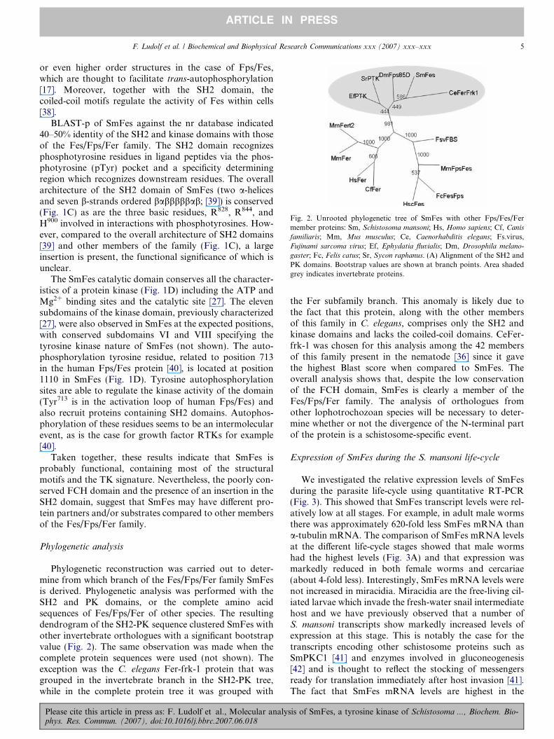
Fig. 2. Unrooted phylogenetic tree of SmFes with other Fps/Fes/Fermember proteins: Sm, Schistosoma mansoni; Hs, Homo sapiens; Cf, Canis
familiaris; Mm, Mus musculus; Ce, Caenorhabditis elegans; Fs.virus,Fujinami sarcoma virus; Ef, Ephydatia fluvialis; Dm, Drosophila melano-
gaster; Fc, Felis catus; Sr, Sycon raphanus. (A) Alignment of the SH2 andPK domains. Bootstrap values are shown at branch points. Area shadedgrey indicates invertebrate proteins.
F. Ludolf et al. / Biochemical and Biophysical Research Communications xxx (2007) xxx–xxx 5
ARTICLE IN PRESS
or even higher order structures in the case of Fps/Fes,which are thought to facilitate trans-autophosphorylation[17]. Moreover, together with the SH2 domain, thecoiled-coil motifs regulate the activity of Fes within cells[38].
BLAST-p of SmFes against the nr database indicated40–50% identity of the SH2 and kinase domains with thoseof the Fes/Fps/Fer family. The SH2 domain recognizesphosphotyrosine residues in ligand peptides via the phos-photyrosine (pTyr) pocket and a specificity determiningregion which recognizes downstream residues. The overallarchitecture of the SH2 domain of SmFes (two a-helicesand seven b-strands ordered babbbbbab; [39]) is conserved(Fig. 1C) as are the three basic residues, R828, R844, andH900 involved in interactions with phosphotyrosines. How-ever, compared to the overall architecture of SH2 domains[39] and other members of the family (Fig. 1C), a largeinsertion is present, the functional significance of which isunclear.
The SmFes catalytic domain conserves all the character-istics of a protein kinase (Fig. 1D) including the ATP andMg2+ binding sites and the catalytic site [27]. The elevensubdomains of the kinase domain, previously characterized[27], were also observed in SmFes at the expected positions,with conserved subdomains VI and VIII specifying thetyrosine kinase nature of SmFes (not shown). The auto-phosphorylation tyrosine residue, related to position 713in the human Fps/Fes protein [40], is located at position1110 in SmFes (Fig. 1D). Tyrosine autophosphorylationsites are able to regulate the kinase activity of the domain(Tyr713 is in the activation loop of human Fps/Fes) andalso recruit proteins containing SH2 domains. Autophos-phorylation of these residues seems to be an intermolecularevent, as is the case for growth factor RTKs for example[40].
Taken together, these results indicate that SmFes isprobably functional, containing most of the structuralmotifs and the TK signature. Nevertheless, the poorly con-served FCH domain and the presence of an insertion in theSH2 domain, suggest that SmFes may have different pro-tein partners and/or substrates compared to other membersof the Fes/Fps/Fer family.
Phylogenetic analysis
Phylogenetic reconstruction was carried out to deter-mine from which branch of the Fes/Fps/Fer family SmFesis derived. Phylogenetic analysis was performed with theSH2 and PK domains, or the complete amino acidsequences of Fes/Fps/Fer of other species. The resultingdendrogram of the SH2-PK sequence clustered SmFes withother invertebrate orthologues with a significant bootstrapvalue (Fig. 2). The same observation was made when thecomplete protein sequences were used (not shown). Theexception was the C. elegans Fer-frk-1 protein that wasgrouped in the invertebrate branch in the SH2-PK tree,while in the complete protein tree it was grouped with
Please cite this article in press as: F. Ludolf et al., Molecular analysphys. Res. Commun. (2007), doi:10.1016/j.bbrc.2007.06.018
the Fer subfamily branch. This anomaly is likely due tothe fact that this protein, along with the other membersof this family in C. elegans, comprises only the SH2 andkinase domains and lacks the coiled-coil domains. CeFer-frk-1 was chosen for this analysis among the 42 membersof this family present in the nematode [36] since it gavethe highest Blast score when compared to SmFes. Theoverall analysis shows that, despite the low conservationof the FCH domain, SmFes is clearly a member of theFes/Fps/Fer family. The analysis of orthologues fromother lophotrochozoan species will be necessary to deter-mine whether or not the divergence of the N-terminal partof the protein is a schistosome-specific event.
Expression of SmFes during the S. mansoni life-cycle
We investigated the relative expression levels of SmFesduring the parasite life-cycle using quantitative RT-PCR(Fig. 3). This showed that SmFes transcript levels were rel-atively low at all stages. For example, in adult male wormsthere was approximately 620-fold less SmFes mRNA thana-tubulin mRNA. The comparison of SmFes mRNA levelsat the different life-cycle stages showed that male wormshad the highest levels (Fig. 3A) and that expression wasmarkedly reduced in both female worms and cercariae(about 4-fold less). Interestingly, SmFes mRNA levels werenot increased in miracidia. Miracidia are the free-living cil-iated larvae which invade the fresh-water snail intermediatehost and we have previously observed that a number ofS. mansoni transcripts show markedly increased levels ofexpression at this stage. This is notably the case for thetranscripts encoding other schistosome proteins such asSmPKC1 [41] and enzymes involved in gluconeogenesis[42] and is thought to reflect the stocking of messengersready for translation immediately after host invasion [41].The fact that SmFes mRNA levels are highest in the
is of SmFes, a tyrosine kinase of Schistosoma ..., Biochem. Bio-

Fig. 3. (A) Transcript levels of SmFes at the different development stagesof S. mansoni determined by quantitative RT-PCR. The relative amountsof SmFes transcripts in adult male worms, adult female worms, miracidia,sporocysts, and cercariae are shown. The 2�DDCt values were calculatedrelative to the expression in adult male worms (see real-time quantitativePCR). (B) Transcript levels in cercariae compared to those in newlytransformed schistosomula (som. 2 h) and schistosomula maintained inculture for 24 h (som. 24 h) or 48 h (som. 48 h). Results shown are from arepresentative experiment, one of three carried out.
Table 2Comparison of the genomic organization of the SmFes and HsFps/Fes
genes
HsFesa SmFesa
Exons Sizeb Introns Size Exons Size Introns Size
1 66 1 493 1 22 1 29632 222 2 154 2 685 2 10573 174 3 1376 3 174 3 >52984 97 4 130 4 97 4 68075 184 5 1936 5 199 5 >46136 138 6 74 6 108 6 >3877 120 7 204 7 129 7 17928 123 8 130 8 294 8 38309 187 9 123 9 331 9 >2882
10 84 10 498 10 421c 10 >791211 210 11 363 11 110 11 >163812 123 12 382 12 65 12 348213 54 13 596 13 273 13 222914 119 14 275 14 136 14 161315 95 15 97 15 211 15 >139516 124 16 240 16 125 16 231717 158 17 125 17 91 17 202018 123 18 1358 18 184 18 >75519 360 19 1280
The SmFes gene structure is detailed in Table 1 in Supplementary materialonline. Numbers in bold and underlined are conserved exons between thehuman and schistosome genes. Numbers in bold are introns conservedbetween the SmFes gene and the D. melanogaster Fps gene (not shown).
a HsFes gene size = 11,297 bp, SmFes > 52,990 bp.b Size given in base pairs.c The genomic sequence contains a 15bp insertion.
6 F. Ludolf et al. / Biochemical and Biophysical Research Communications xxx (2007) xxx–xxx
ARTICLE IN PRESS
differentiated male worms may indicate a specific role forSmFes in these worms.
In a parallel study [18], we showed that the SmFes pro-tein is particularly expressed at the terebratorium of mira-cidia and the tegument of cercaria and skin-stageschistosomula. Moreover, no SmFes protein was found inschistosomula maintained in culture for 7 days. We there-fore carried out a comparison of the expression of SmFesmRNA in cercariae, newly transformed schistosomulaand larvae incubated in culture (Fig. 3B). The results ofthree separate experiments show that SmFes mRNA levelsdecrease steadily after transformation of cercariae intoschistosomula and subsequent culture, consistent with thecorresponding decline in the amounts of protein. After48 h mRNA levels were reduced by more than 60%. Thisfinding is consistent with a putative role for SmFes in sig-naling during the process of host invasion.
The genomic structure of SmFes: comparison with the human
Fps/Fes and Drosophila Fes genes
The SmFes gene structure was determined by screeningthe individual and assembled genomic sequences at theWellcome Trust Sanger Institute S. mansoni Blast server.The results of the survey are shown in detail on Table 1in Supplementary material online. The gene comprises 19exons and 18 introns of which 7 are of indeterminate sizedue to gaps in the contig assembly which we were not ableto fill by chromosome walking with individual shotgunsequences. The overall gene size is >52,990 bp with intron
Please cite this article in press as: F. Ludolf et al., Molecular analyphys. Res. Commun. (2007), doi:10.1016/j.bbrc.2007.06.018
sizes ranging from 1057 to >7912 bp. The sizes of someof the introns was checked by PCR (not shown) and nota-bly intron 18 was found to be about 1100 bp in size(>755 bp from the genomic assembly). There are thereforeno very small introns which are often present at the 5 0 endof schistosome genes such as the three �30 bp introns pres-ent in the SmPKC1 gene [41]. Moreover, it is unlikely thatvery large introns are present such as the >40 kbp intronsin two schistosome Hox genes [43].
The genome survey indicated that only one copy of theSmFes gene was present and this view was supported bySouthern blotting. We used PCR-generated cDNA frag-ments corresponding to exon 10 to probe Southern blotswith genomic DNA digested with HindIII or EcoR1(Fig. 1 in Supplementary material online).
The comparison of the organization of the SmFes genewith those of the Human Fps/Fes (HsFps/Fes) andDrosophila Fps (DmFps) genes showed that some intronpositions were conserved, particularly at the 5 0 end of thegenes and in the regions encoding the SH2 and kinasedomains. The HsFps/Fes gene (on genomic contigNT_010274.16), like SmFes, contains 19 exons includingone non-coding exon, but is more compact; the whole genespans only 11,297 bp. This contrasts with the human Fer
gene (on genomic contig NT_034772) which contains 20exons spanning 439,845 bp (not shown). The direct com-parison of the HsFps/Fes and SmFes gene structures (Table2) and the location of the intron positions on the peptide
sis of SmFes, a tyrosine kinase of Schistosoma ..., Biochem. Bio-

F. Ludolf et al. / Biochemical and Biophysical Research Communications xxx (2007) xxx–xxx 7
ARTICLE IN PRESS
sequence (not shown) demonstrates that several intronpositions are conserved, leading to conserved exons(Table 2). Notably, the positions of exons 3 and 4 in eachgene are perfectly conserved and have identical sizes. Theseexons are immediately downstream of exon 2 which in eachcase encodes the (putative in the case of SmFes) FCHdomain and support the peptide sequence similaritydetected in this region. The central exons are not con-served, either in terms of size or of position. Exon 10 inHsFps/Fes and exon 12 in SmFes both encode the start ofthe SH2 domain and are conserved. Similarly, exons 16in each gene are also conserved and encode part of thekinase domain. Comparison with the DmFps gene (ongenomic contig NT_033777.2) showed two further con-served exons (Table 2) in the region encoding the kinasedomain (exons 15 and 18 in SmFes, exons 10 and 13 inDmFps, not shown). Moreover, exons 3 and 4 of DmFps
are also conserved, further supporting the identity of thisregion in SmFes. The conservation of intron positions inschistosome genes is variable. In general, as in the Hox
genes [43], intron positions are identical to those of orthol-ogous genes within and around highly conserved functionaldomains (the homeodomain for example), but not else-where in the gene. This is the case for SmFes, but, incontrast, in SmPKC1 [41] all the intron positions wereconserved compared to the human PKCb gene.
Whilst, we have detected a degree of polymorphism inthe SmFes gene (see Polymorphism of SmFes), it differsfrom its orthologues in the apparent absence of majoralternatively spliced isoforms. No potentially alternativelymajor spliced forms (in which exons were deleted) weredetected, either during the 5 0 and 3 0 RACE experiments,or during PCR amplification of internal fragments. Thiscontrasts with the DmFps gene for which four differenttranscripts are identified in GenBank [CG8874-RA(NM_079564), CG8874-RB (NM_169274), CG8874-RC(NM_169275), and CG8874-RD (NM_169276)] generatedby alternative splicing or alternative promoter usage(CG8874-RD). Similarly, the human Fer gene, which hasan identical genomic structure to HsFps/Fes, apart fromthe presence of an additional 5 0 non-coding exon, generatesa truncated isoform in testis, FerT, the transcription ofwhich is driven from a testis-specific promoter [17].
Polymorphism of SmFes
Sequencing of multiple SmFes cDNA clones permittedthe identification of clones containing a 9 bp insert justbefore the SH2 domain (nt position 2556) and coding forthe peptide sequence VSE (Fig. 4A). Comparison of thecDNA sequence with the corresponding genomic regionsuggested the existence of an alternative splice site at the5 0end of the intron 10, resulting in the cDNA populationsthat are observed (Fig. 4B). The verification of alternativelyspliced messages was performed by RFLP (restriction-frag-ment length polymorphism) as the presence of the 9 bpinsertion created a restriction site for SfcI. Three different
Please cite this article in press as: F. Ludolf et al., Molecular analysphys. Res. Commun. (2007), doi:10.1016/j.bbrc.2007.06.018
PCR amplifications of the insertion region were conductedwith cDNA obtained from adult worms. All PCR frag-ments were partially digested, indicating that the twocDNA populations were present (Fig. 4C). The sameobservation was made for cDNA obtained from pools ofmale or female worms, cercariae, eggs and from individualmale or female worms, indicating that alternative splicingof SmFes is present throughout the life-cycle of the parasiteand could coexist in a single individual (Fig. 4D). ControlDNA was fully digested (not shown). These alternativetranscripts could determine tissue specific expression, cellu-lar localization or mechanisms of protein regulation. Thefunction of the alternatively spliced SmFes is unknown.It is possible, however, that the three residues could alterthe protein structure and modulate the function of SmFes.
Comparison of the genomic and cDNA sequences indi-cated the possibility of a 15 bp insertion at position 2170 ofthe cDNA (within exon 10) coding for the peptideLQHQQ. None of the 11 cDNAs sequenced had the15 bp insertion (Fig. 4E). However, all three genomicsequences from the Sanger database had the insertion, asdid some of the new genomic sequences generated by us.Taken together, the results indicate that these two typesof sequence may be alleles of the same SmFes gene. Twodifferent populations, containing or not the 15 bp insertion,were identified among several genomic sequences of S.
mansoni, and can be classified as alleles since no splicingelements were observed and since SmFes is a single copygene. Allele-specific polymorphisms were also identified atthe S. mansoni Fyn-like protein kinase [14].
Single nucleotide polymorphisms (SNPs) at position2166 (C-A) and 2169 (G-A) were also observed. The SNPsalways appeared linked to one of the two genomic allelesdescribed above (Fig. 4E). The first SNP results in a substi-tution of a histidine for glutamine in the allele containingthe 15 bp insertion, while the second SNP is silent andmaintains the glutamine residue (Fig. 4E). Analysis ofgenomic and cDNA clones sequences indicated the possibleexistence of single nucleotide polymorphisms (SNP) atposition 2416 (G/A) of the cDNA, just before the PKdomain. The polymorphism results in a substitution ofvaline for isoleucine.
Conclusion
SmFes shows a high degree of structural conservation,particularly in the kinase catalytic domain, but also origi-nal structural aspects compared to other members of theFps/Fes/Fer family. The low level of conservation of theputative FCH domain indicates that it may not interactwith microtubules in the same way as does mammalianFps/Fes [34] and the large insertion in the SH2 domainmay influence the binding specificity of the domain andthe type of protein partner recognized. Moreover, we arecurrently constructing a 3D model of the SH2 domain todetermine the potential effect on the domain structure bothof the large insertion and of the VSE insertion which just
is of SmFes, a tyrosine kinase of Schistosoma ..., Biochem. Bio-

1 2 3 4 5 6
550b
430bp
270bp
160bp
1 2 3 4 5 6
550b
430b
270bp
160bp
550b
430b
270bp
160bp
281-271bp310bp
234bp
234bp
603bp 1 2 3 4 5 6 7
A
B
C D
E
Fig. 4. (A) Alternative splicing of SmFes. (A) Sequences of 8 cDNA clones indicating the 9 bp insertion at position 2556. (B) Genomic sequence of theregion where the 9 bp insertion occurs. The coding sequence is in capital letters, introns in lower case letters. The squares indicate the alternative splicedonor sites. The SfcI restriction site is underlined. (C) Restriction pattern of PCR products obtained from the amplification of adult worm cDNA. Lanes1–2, 3–4, and 5–6 were obtained by amplification with SmFesTK1/SmFesTK2, SmFesTK1/SmFesTK7, and SmFesTK1/SmFesTK6 primer pairs,respectively. Lanes 2, 4, and 6 were digested with SfcI. The undigested cDNA is a band of 428 bp for lane 1, 547 bp for lane 3 and 423 bp for lane 5. Afterdigestion, the fragment is cleaved into 159 and 269 bp for lane 2, 278 and 269 bp for lane 4, 154 and 269 bp for lane 6, shown in the dotted box. Molecularweight markers are shown on the left. (D) Restriction pattern of PCR products obtained from cDNA amplified with the primers, SmFesTK1 andSmFesTK2, and digested with the enzyme SfcI. (1) Molecular weight markers, (2) adult worm pool, (3) male pool, (4) female pool, (5) cercariae pool, (6)individual female worm, (7) individual male worm. (E) SmFes gene allele containing a 15 bp insertion. The 15 bp are inserted after the base 2170 of thecDNA in exon 10. 11 cDNA clones showed no insertion. Several genomic clones, either with or without the 15 bp insertion were observed.
8 F. Ludolf et al. / Biochemical and Biophysical Research Communications xxx (2007) xxx–xxx
ARTICLE IN PRESS
precedes the domain. We have shown that the SmFes pro-tein is expressed in locations in infective larvae (cercarialtegument, miracidial terebretorium) consistent with a rolein host invasion [18]. Indeed, one of the defined roles ofmammalian Fes/Fps/Fer is to regulate cell–cell and cell–matrix interactions [17] and we can speculate that in schis-tosomes SmFes could similarly regulate cytoskeletal rear-rangements associated with host penetration. Futurework will be aimed at identifying protein partners ofSmFes, interacting either with the putative FCH domainor the SH2 domain as well as protein substrates of SmFesin order to determine its biological role. A number oforthologues of partners of Fps/Fes/Fer [17] have beenfound in S. mansoni by in silico screening of the EST andgenomic databases (F.L. and D.B., not shown) includingcortactin, PI3K, and b-catenin, as well as other PKs such
Please cite this article in press as: F. Ludolf et al., Molecular analyphys. Res. Commun. (2007), doi:10.1016/j.bbrc.2007.06.018
as SRC and JAK. We will extend and complete this surveyand select potential partners in order to test the conserva-tion or otherwise of the protein interaction networksinvolving SmFes.
Acknowledgments
The work was supported by the Inserm-Fiocruz collab-orative programme, the Institut National de la Sante et dela Recherche Medicale (U547), the Institut Pasteur de Lille,the Centre National de la Recherche Scientifique and theMicrobiology program of the Ministere de l’EducationNationale, de la Recherche et de la Technologie (MEN-RT). The financial support from FIOCRUZ/PDTIS(GO), PAPES (GO 400315/2006-8) and FAPEMIG (DBCBB-174/02) is also acknowledged. F.L. is supported by
sis of SmFes, a tyrosine kinase of Schistosoma ..., Biochem. Bio-

F. Ludolf et al. / Biochemical and Biophysical Research Communications xxx (2007) xxx–xxx 9
ARTICLE IN PRESS
an NIH-Fogarty grant (TW007012-01). G.O. is a CNPqfellow.
Appendix A. Supplementary data
Supplementary data associated with this article can befound, in the online version, at doi:10.1016/j.bbrc.2007.06.018.
References
[1] D. Engels, L. Chitsulo, A. Montresor, L. Savioli, The globalepidemiological situation of schistosomiasis and new approaches tocontrol and research, Acta Trop. 82 (2002) 139–146.
[2] D. Cioli, L. Pica-Mattoccia, Praziquantel, Parasitol. Res. 90 (Suppl.1) (2003) S3–S9.
[3] G. Oliveira, N.B. Rodrigues, A.J. Romanha, D. Bahia, Genome andgenomics of schistosomes, Can. J. Zool. 82 (2004) 375–390.
[4] W. Kunz, Schistosome male–female interaction: induction of germ-cell differentiation, Trends Parasitol. 17 (2001) 227–231.
[5] P.T. Loverde, L. Chen, Schistosome female reproductive develop-ment, Parasitol. Today 7 (1991) 303–308.
[6] A.R. Harris, R.J. Russell, A.D. Charters, A review of schistosomiasisin immigrants in Western Australia, demonstrating the unusuallongevity of Schistosoma mansoni, Trans. R. Soc. Trop. Med. Hyg. 78(1984) 385–388.
[7] D. Bahia, L.F. Andrade, F. Ludolf, R.A. Mortara, G. Oliveira,Protein tyrosine kinases in Schistosoma mansoni, Mem. Inst. OswaldoCruz 101 (2006) 137–143.
[8] C. Dissous, N. Khayath, J. Vicogne, M. Capron, Growth factorreceptors in helminth parasites: signalling and host–parasite relation-ships, FEBS Lett. 580 (2006) 2968–2975.
[9] K. Neet, T. Hunter, Vertebrate non-receptor protein-tyrosine kinasefamilies, Genes Cells 1 (1996) 147–169.
[10] H. Mano, The Tec family protein-tyrosine kinases: a subset of kinasesfor a subset of signalings, Int. J. Haematol. 69 (1999) 6–12.
[11] T.E. Smithgall, J.A. Rogers, K.L. Peters, J. Li, S.D. Briggs, J.M.Lionberger, H. Cheng, A. Shibata, B. Scholtz, S. Schreiner, N.Dunham, The c-Fes family of protein-tyrosine kinases, Crit. Rev.Oncog. 9 (1998) 43–62.
[12] S.M. Thomas, J.S. Brugge, Cellular functions regulated by Src familykinases, Ann. Rev. Cell Dev. Biol. 13 (1997) 513–609.
[13] P. Traxler, Tyrosine kinases as targets in cancer therapy—successesand failures, Expert Opin. Ther. Targets 7 (2003) 215–234.
[14] K. Kapp, P. Schussler, W. Kunz, C.G. Grevelding, Identification,isolation and characterization of a Fyn-like tyrosine kinase fromSchistosoma mansoni, Parasitology 122 (2001) 317–327.
[15] J. Knobloch, R. Winnen, M. Quack, W. Kunz, C.G. Grevelding, Anovel Syk-family tyrosine kinase from Schistosoma mansoni which ispreferentially transcribed in reproductive organs, Gene 294 (2002) 87–97.
[16] K. Kapp, J. Knobloch, P. Schussler, S. Sroka, R. Lammers, W. Kunz,C.G. Grevelding, The Schistosoma mansoni Src kinase TK3 isexpressed in the gonads and likely involved in cytoskeletal organiza-tion, Mol. Biochem. Parasitol. 138 (2004) 171–182.
[17] P. Greer, Closing in on the biological functions of Fps/Fes and Fer,Nat. Rev. Mol. Cell Biol. 3 (2002) 278–289.
[18] D. Bahia, R.A. Mortara, J.R. Kusel, L.F. Andrade, F. Ludolf, P.R.Kuser, L. Avelar, J. Trolet, C. Dissous, R.J. Pierce, G. Oliveira,Schistosoma mansoni: expression of Fes-like tyrosine kinase SmFes inthe tegument and terebratorium suggests its involvement in hostpenetration, Exp. Parasitol. 116 (2007) 225–232.
[19] F.J. Ramalho-Pinto, G. Gazzinelli, R.E. Howells, T.A. Mota-Santos,E.A. Figueiredo, J. Pellegrino, Schistosoma mansoni: defined systemfor stepwise transformation of cercaria to schistosomule in vitro, Exp.Parasitol. 36 (1974) 360–372.
Please cite this article in press as: F. Ludolf et al., Molecular analysphys. Res. Commun. (2007), doi:10.1016/j.bbrc.2007.06.018
[20] R. Harrop, R.A. Wilson, Protein synthesis and release by culturedschistosomula of Schistosoma mansoni, Parasitology 107 (Pt. 3) (1993)265–274.
[21] S.R. Smithers, R.J. Terry, The infection of laboratory hosts withcercariae of Schistosoma mansoni and the recovery of the adultworms, Parasitology 55 (1965) 695–700.
[22] T.P. Yoshino, J.R. Laursen, Production of Schistosoma mansoni
daughter sporocysts from mother sporocysts maintained in synxenicculture with Biomphalaria glabrata embryonic (Bge) cells, J. Parasitol.81 (1995) 714–722.
[23] J. Sambrook, D.W. Russel, Molecular Cloning: A LaboratoryManual, third ed., Cold Spring Harbour Laboratory Press, NewYork, 2001.
[24] J.M. Chirgwin, A.E. Przybyla, R.J. MacDonald, W.J. Rutter,Isolation of biologically active ribonucleic acid from sources enrichedin ribonuclease, Biochemistry 18 (1979) 5294–5299.
[25] A. Marchler-Bauer, J.B. Anderson, C. DeWeese-Scott, N.D. Fedor-ova, L.Y. Geer, S. He, D.I. Hurwitz, J.D. Jackson, A.R. Jacobs, C.J.Lanczycki, C.A. Liebert, C. Liu, C. Madej, G.H. Marchler, R.Mazumder, A.N. Nikolskaya, A.R. Panchenko, B.S. Rao, B.A.Shoemaker, V. Simonyan, J.S. Song, P.A. Thiessen, S. Vasudevan, Y.Wang, R.A. Yamashita, J.J. Yin, S.H. Bryant, CDD: a curatedEntrez database of conserved domain alignments, Nucleic Acids Res.31 (2003) 383–387.
[26] A. Lupas, M. Van Dyke, J. Stock, Predicting coiled coils from proteinsequences, Science 252 (1991) 1162–1164.
[27] S.K. Hanks, A.M. Quinn, T. Hunter, The protein kinase family:conserved features and deduced phylogeny of the catalytic domains,Science 241 (1988) 42–52.
[28] J.D. Thompson, D.G. Higgins, T.J. Gibson, CLUSTAL W: improv-ing the sensitivity of progressive multiple sequence alignment throughsequence weighting, position-specific gap penalties and weight matrixchoice, Nucleic Acids Res. 22 (1994) 4673–4680.
[29] J. Kyte, R.F. Doolittle, A simple method for displaying thehydropathic character of a protein, J. Mol. Biol. 157 (1982) 105–132.
[30] R.D. Page, TreeView: an application to display phylogenetic trees onpersonal computers, Comput. Appl. Biosci. 12 (1996) 357–358.
[31] R. Rasmussen, Quantification on the Light Cycler, in: S. Meuer, C.Wittwer, K. Nakagawara (Eds.), Rapid Cycle Real-time PCR,Methods and Applications, Springer Press, Heidelberg, 2001, pp.21–34.
[32] M.W. Pfaffl, A new mathematical model for relative quantification inreal-time RT-PCR, Nucleic Acids Res. 29 (2001) e45.
[33] K.J. Livak, T.D. Schmittgen, Analysis of relative gene expressiondata using real-time quantitative PCR and the 2(-Delta Delta C(T))method, Methods 25 (2001) 402–408.
[34] S. Takahashi, R. Inatome, A. Hotta, Q. Qin, R. Hackenmiller, M.C.Simon, H. Yamamura, S. Yanagi, Role for Fes/Fps tyrosine kinase inmicrotubule nucleation through is Fes/CIP4 homology domain, J.Biol. Chem. 278 (2003) 49129–49133.
[35] C.E. Laurent, F.J. Delfino, H.Y. Cheng, T.E. Smithgall, The humanc-Fes tyrosine kinase binds tubulin and microtubules throughseparate domains and promotes microtubule assembly, Mol. Cell.Biol. 24 (2004) 9351–9358.
[36] G.D. Plowman, S. Sudarsanam, J. Bingham, D. Whyte, T. Hunter,The protein kinases of Caenorhabditis elegans: a model for signaltransduction in multicellular organisms, Proc. Natl. Acad. Sci. USA96 (1999) 13603–13610.
[37] H. Cetkovic, I.M. Muller, W.E. Muller, V. Gamulin, Characteriza-tion and phylogenetic analysis of a cDNA encoding the Fes/FERrelated, non-receptor protein–tyrosine kinase in the marine spongeSycon raphanus, Gene 216 (1998) 77–84.
[38] Y. Takashima, F.J. Delfino, J.R. Engen, G. Superti-Furga, T.E.Smithgall, Regulation of c-Fes tyrosine kinase activity by coiled-coiland SH2 domains: analysis with Saccharomyces cerevisiae, Biochem-istry 42 (2003) 3567–3574.
[39] K. Machida, B.J. Mayer, The SH2 domain: versatile signaling moduleand pharmaceutical target, Biochem. Biophys. Acta 1747 (2005) 1–25.
is of SmFes, a tyrosine kinase of Schistosoma ..., Biochem. Bio-

10 F. Ludolf et al. / Biochemical and Biophysical Research Communications xxx (2007) xxx–xxx
ARTICLE IN PRESS
[40] J.A. Rogers, R.D. Read, J. Li, K.L. Peters, T.E. Smithgall,Autophosphorylation of the Fes tyrosine kinase. Evidence for anintermolecular mechanism involving two kinase domain tyrosineresidues, J. Biol. Chem. 271 (1996) 17519–17525.
[41] D. Bahia, L. Avelar, R.A. Mortara, N. Khayath, Y. Yan, C. Noel,M. Capron, C. Dissous, R.J. Pierce, G. Oliveira, SmPKC1, a newprotein kinase C identified in the platyhelminth parasite Schisto-
soma mansoni, Biochem. Biophys. Res. Commun. 345 (2006) 1138–1148.
Please cite this article in press as: F. Ludolf et al., Molecular analyphys. Res. Commun. (2007), doi:10.1016/j.bbrc.2007.06.018
[42] N. Khayath, G. Mithieux, C. Zitoun, C. Coustau, J. Vicogne, A.G.Tielens, C. Dissous, Glyceroneogenesis: an unexpected metabolicpathway for glutamine in Schistosoma mansoni sporocysts, Mol.Biochem. Parasitol. 147 (2006) 145–153.
[43] R.J. Pierce, W. Wu, H. Hirai, A. Ivens, L.D. Murphy, C. Noel, D.A.Johnston, F. Artiguenave, M. Adams, J. Cornette, E. Viscogliosi, M.Capron, G. Balavoine, Evidence for a dispersed Hox gene cluster inthe platyhelminth parasite Schistosoma mansoni, Mol. Biol. Evol. 22(2005) 2491–2503.
sis of SmFes, a tyrosine kinase of Schistosoma ..., Biochem. Bio-