microbial community responses to organophosphate substrate additions in contaminated subsurface...
TRANSCRIPT

Microbial Community Responses to OrganophosphateSubstrate Additions in Contaminated SubsurfaceSedimentsRobert J. Martinez1*, Cindy H. Wu2, Melanie J. Beazley1, Gary L. Andersen2, Mark E. Conrad2,
Terry C. Hazen3, Martial Taillefert4, Patricia A. Sobecky1
1 Department of Biological Sciences, University of Alabama, Tuscaloosa, Alabama, United States of America, 2 Earth Sciences Division, Lawrence Berkeley National
Laboratory, Berkeley, California, United States of America, 3 Department of Civil and Environmental Engineering, University of Tennessee, Knoxville, Tennessee, United
States of America, 4 School of Earth and Atmospheric Sciences, Georgia Institute of Technology, Atlanta, Georgia, United States of America
Abstract
Background: Radionuclide- and heavy metal-contaminated subsurface sediments remain a legacy of Cold War nuclearweapons research and recent nuclear power plant failures. Within such contaminated sediments, remediation activities arenecessary to mitigate groundwater contamination. A promising approach makes use of extant microbial communitiescapable of hydrolyzing organophosphate substrates to promote mineralization of soluble contaminants within deepsubsurface environments.
Methodology/Principal Findings: Uranium-contaminated sediments from the U.S. Department of Energy Oak Ridge FieldResearch Center (ORFRC) Area 2 site were used in slurry experiments to identify microbial communities involved inhydrolysis of 10 mM organophosphate amendments [i.e., glycerol-2-phosphate (G2P) or glycerol-3-phosphate (G3P)] insynthetic groundwater at pH 5.5 and pH 6.8. Following 36 day (G2P) and 20 day (G3P) amended treatments, maximumphosphate (PO4
32) concentrations of 4.8 mM and 8.9 mM were measured, respectively. Use of the PhyloChip 16S rRNAmicroarray identified 2,120 archaeal and bacterial taxa representing 46 phyla, 66 classes, 110 orders, and 186 families amongall treatments. Measures of archaeal and bacterial richness were lowest under G2P (pH 5.5) treatments and greatest withG3P (pH 6.8) treatments. Members of the phyla Crenarchaeota, Euryarchaeota, Bacteroidetes, and Proteobacteriademonstrated the greatest enrichment in response to organophosphate amendments and the OTUs that increased inrelative abundance by 2-fold or greater accounted for 9%–50% and 3%–17% of total detected Archaea and Bacteria,respectively.
Conclusions/Significance: This work provided a characterization of the distinct ORFRC subsurface microbial communitiesthat contributed to increased concentrations of extracellular phosphate via hydrolysis of organophosphate substrateamendments. Within subsurface environments that are not ideal for reductive precipitation of uranium, strategies thatharness microbial phosphate metabolism to promote uranium phosphate precipitation could offer an alternative approachfor in situ sequestration.
Citation: Martinez RJ, Wu CH, Beazley MJ, Andersen GL, Conrad ME, et al. (2014) Microbial Community Responses to Organophosphate Substrate Additions inContaminated Subsurface Sediments. PLoS ONE 9(6): e100383. doi:10.1371/journal.pone.0100383
Editor: Melanie R. Mormile, Missouri University of Science and Technology, United States of America
Received December 20, 2013; Accepted May 27, 2014; Published June 20, 2014
Copyright: � 2014 Martinez et al. This is an open-access article distributed under the terms of the Creative Commons Attribution License, which permitsunrestricted use, distribution, and reproduction in any medium, provided the original author and source are credited.
Funding: This research was supported by the Office of Science (BER), U.S. Department of Energy Grant No. DE-FG02-04ER63906 (University of Alabama),Subcontract No. SC0002530 (Georgia Institute of Technology), and partially by No. DE-AC02-05CH11231 (Lawrence Berkeley National Laboratory). The fundershad no role in study design, data collection and analysis, decision to publish, or preparation of the manuscript.
Competing Interests: The authors have declared that no competing interests exist.
* Email: [email protected]
Introduction
Within sediments, the mobility of phosphate (PO432) is
controlled by pH, coprecipitation reactions with metals and
radionuclides, adsorption/desorption, and ion-exchange reactions
[1]. As a result of this poor mobility in subsurface environments,
microorganisms release organic acids and/or express phosphatase
enzymes (i.e., acid/alkaline phosphohydrolases) to enhance the
solubility and cellular transport of phosphate [2–4]. Harnessing
microbial phosphatases expressed by extant microbial communi-
ties within uranium (U)-contaminated environments represents an
approach to leverage microbial phosphate acquisition phenotypes
to promote in situ sequestration of U as insoluble phosphate
minerals.
Alternative approaches for microbial mediated U immobiliza-
tion have examined bioaccumulation, reductive precipitation,
ligand-generated precipitation (e.g., carbonate and sulfide), and
volatilization reactions to reduce contaminant solubility [5–8].
Microbial reductive precipitation of soluble U(VI) to insoluble
U(IV) has been extensively examined in both laboratory and field
studies where delivery of electron donor substrates, buffered at
circumneutral pH, has proven effective as an immobilization
strategy [9–12]. However, the limitations of U(VI) reduction are
observed in environments that experience dynamic geochemical
PLOS ONE | www.plosone.org 1 June 2014 | Volume 9 | Issue 6 | e100383

conditions where low pH inhibits microbial U(VI) reduction
[11,13] and reoxidation of U(IV) occurs in the presence of oxygen,
nitrate, ferric iron, and humics [14–17].
Within U-contaminated sediments at the United States
Department of Energy Oak Ridge Field Research Center
(ORFRC), three distinct groundwater contaminant flow paths
contribute to pH and co-contaminant heterogeneity (i.e., porewa-
ter pH ranging from 3.4–7.0 and [NO32] ranging from 29 mg
L21 to 2300 mg L21), which inhibit or reverse microbial U(VI)
reduction [12,18–20]. Alternatively, in situ precipitation of U(VI)
as highly insoluble phosphate minerals (e.g., autunite) that remain
stable across a broad pH range (Figure 1) offers an approach for
U(VI) sequestration under both oxidizing and reducing conditions
[21–23]. Autunite minerals have been identified in sediments at
the U.S. Department of Energy (DOE) Fernald site, Hanford site,
and Oak Ridge National Laboratory [24–26], suggesting that
long-term in situ sequestration of U(VI) as phosphate minerals
represents a viable remediation strategy. Unfortunately, direct
injection of phosphate causes blockage of sediment pore spaces at
injection sites due to the rapid precipitation of phosphate with
subsurface sediment cations [27]. Therefore, the use of less
reactive inorganic or organic phosphate compounds must be
employed for effective delivery to deep subsurface contaminated
zones where microbial phosphatase activity can hydrolyze these
substrates and liberate reactive phosphate.
The activity of phosphatases from several bacterial species [e.g.,
Aeromonas, Bacillus, Myxococcus, Pantoea, Pseudomonas, Rahnella, and
Serratia (formerly Citrobacter sp.)] have been shown to increase
extracellular phosphate concentrations that subsequently promote
metal and radionuclide precipitation as highly insoluble mineral
phosphates [22,28–33].
Our previous work has shown that organophosphate substrates
[i.e., glycerol-3-phosphate (G3P) and glycerol-2-phosphate (G2P)]
remain soluble within saturated sediments and in solutions
containing uranium [34,35]. Both G3P and G2P represent
organophosphates that are present within sediments: G3P is
commonly found in prokaryotic and eukaryotic cell walls,
cytoplasm, and lipid membranes [36,37], while G2P is a less
common compound found within bacterial and fungal cell extracts
and from phosphatidyl choline alkaline hydrolysis [38–40].
Furthermore, our recent ORFRC sediment column studies
utilizing both G2P and G3P amendments stimulated the extant
microbial community that hydrolyzed both substrates which
yielded 1–3 mM phosphate within acidic and circumneutral pH
porewater and promoted precipitation of U(VI) [35].
Due to the observed pH and contaminant heterogeneity
observed within the ORFRC subsurface, we hypothesized that
distinct microbial communities capable of organophosphate
hydrolysis would be enriched with G2P or G3P amendments
under acidic or circumneutral pH and that hydrolysis of G3P
would yield the greatest concentrations of extracellular phosphate.
The goal of this study was to utilize the 16S rRNA high-density
microarray (PhyloChip), capable of detecting 8,741 archaeal and
bacterial taxa [41], to characterize the extant prokaryotic
community within ORFRC U-contaminated sediments that
contributed to organophosphate (i.e., G2P and G3P) hydrolysis.
Due to the heterogeneity of geochemical parameters (pH, [U],
[NO32], etc) present within the ORFRC subsurface, character-
ization of extant phosphate solubilizing microbial communities
enriched under specific pH and organophosphate amendments
can aid in development of strategies for in situ phosphate
mineralization of U(VI).
Results
Microbial response to slurry incubationsTotal DNA extractions from sediment slurry treatments were
measured as a proxy for microbial growth in response to the
different incubation conditions. Prior to treatments, ORFRC
subsurface sediment DNA concentrations were 1.160.1 mg g21
(Figure 2). Sediment slurries incubated at either pH 5.5 or pH 6.8
without organophosphate addition exhibited a 1.7-fold increase
(1.860.2 to 1.960.4 mg g21) in DNA concentration after 36 days.
DNA concentrations increased 20-fold (21.666.8 to 23.966.5 mg
g21) after 36 days in G2P-amended treatments and 6-fold
(7.260.9 to 7.361.1 mg g21) after 20 days in G3P-amended
treatments at both pH values (Figure 2).
Figure 1. Thermodynamic modeling of U(VI) in the presence of phosphate as a function of pH. ORFRC Area 2 groundwaterconcentrations of dissolved ions (GW-836 monitoring well), U(VI) = 4.5 mM, Ca2+ = 4.85 mM, (A) PO4
32 = 500 mM and (B) PO432 = 5 mM were used to
model the distribution of U(VI) species. Dashed lines represent soluble species and solid lines represent insoluble species.doi:10.1371/journal.pone.0100383.g001
Microbial Community Responses to Organophosphates
PLOS ONE | www.plosone.org 2 June 2014 | Volume 9 | Issue 6 | e100383

Chemical analyses of sediment slurry incubationsG2P, G3P, PO4
32, NO32, NO2
2, and organic acids were
measured at 96 h intervals over the course of all incubations.
Average NO32 concentrations among all sediment slurry treat-
ments did not decrease throughout the time course (data not
shown) indicating that aerobic conditions were maintained during
these incubations.
Phosphate concentrations in the G2P-amended slurries re-
mained below 140 mM for 576 h then increased to 4.8 mM and
2.2 mM in the pH 5.5 and 6.8 incubations, respectively
(Figures 3A and 3B). Combined concentrations of G2P and
soluble phosphate in the pH 6.8 treatments exhibited that mass
balance of PO432 was respected throughout the entire time course
(Figure 3B). Conversely, at pH 5.5, G2P was completely removed
from solution without a proportional accumulation of soluble
phosphate after 576 h (Figure 3A). In contrast to the G2P
treatments, G3P was completely consumed within 300–400 h at
both pH values (Figures 3A and 3B). Phosphate concentrations in
G3P-amended slurries increased after 96 h (pH 5.5) and prior to
the 96 h time point (pH 6.8), then accumulated over 4.7 mM
phosphate by the 192 h time point. At the 480 h time point,
soluble phosphate concentrations reached 8.9 mM and 8.7 mM
phosphate in G3P (pH 5.5) and G3P (pH 6.8) treatments,
respectively (Figures 3A and 3B).
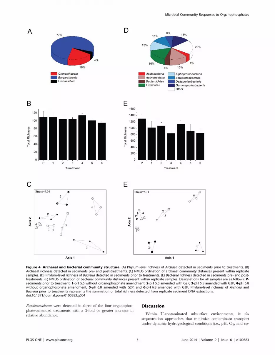
Archaeal community structureA total of 180 archaeal OTUs representing 3 phyla, 10 classes,
16 orders, and 25 families were detected amongst all pre-treatment
and treatment samples (Table S1). The phyla Crenarchaeota and
Euryarchaeota accounted for over 96% of the total archaeal richness
within ORFRC sediments prior to treatments with the remainder
comprised of unclassified Archaea (Figure 4A, Table S1). Following
treatments, archaeal richness did not change significantly (p-
value.0.05) regardless of the amendments (Figure 4B). NMDS
ordination of archaeal community composition clustered the
replicate samples into distinct groups based on treatment
(Figure 4C), and MRPP tests confirmed that archaeal communities
differed significantly (do = 0.1255, de = 0.2798, p-value,0.001,
A = 0.5516) amongst all treatments.
The combined influence of pH and organophosphate addition
shaped the archaeal community by affecting OTU abundance
relative to treatments at the same pH without organophosphate
(Figures 5, Tables S1 and S2). Relative to total richness detected in
sediments prior to treatments (134 OTUs), the richness of OTUs
responding to treatment conditions decreased by 34-fold (4
OTUs), 4-fold (34 OTUs), 2-fold (65 OTUs), and 1.6-fold (85
OTUs) in G2P (pH 5.5), G3P (pH 5.5), G2P (pH 6.8), and G3P
(pH 6.8) treatments, respectively (Figure 5). Archaea that demon-
strated a relative increase in abundance of 2-fold or greater in G2P
(pH 5.5) treatments consisted of two unclassified Crenarchaeota
OTUs. In the G3P (pH 5.5) treatments, one Crenarchaeota OTU
(unclassified at the class level) and two Euryarchaeota OTUs
belonging to the classes Archaeoglobi and Methanobacteria demon-
strated a 2-fold or greater increase in abundance (Figure 6A and
Table S2). Class-level distribution with a 2-fold or greater increase
in abundance in G2P (pH 6.8) treatments contained 13 OTUs
composed of unclassified Crenarchaeota (15%), Methanobacteria (77%),
and unclassified Archaea (8%). In the G3P (pH 6.8) treatments, 14
OTUs that increased in abundance by 2-fold or greater were
composed of Thermoprotei (14%), unclassified Crenarchaeota (7%),
Archaeoglobi (7%), Methanobacteria (7%), Methanomicrobia (14%),
Thermoplasmata (21%), and unclassified Archaea (29%) (Figure 6A
and Table S2).
Observed changes in relative abundance were identified in 4
OTUs [G2P (pH 5.5)], 34 OTUs [G3P (pH 5.5)], 65 OTUs [G2P
(pH 6.8)], and 85 OTUs [G3P (pH 6.8)]. Four archaeal OTUs
belonging to Archaeoglobi, Methanobacteria, and two unclassified
classes of Crenarchaeota (related to deep-sea sediment and landfill
leachate environmental clones) were detected at a 2-fold or greater
increase in relative abundance in multiple treatment conditions
(Figure S1A and Table S3). The Crenarchaeota (landfill leachate
related clone), Archaeoglobi, and Methanobacteria OTUs detected in
multiple treatment conditions [i.e., G3P (pH 5.5), G2P (pH 6.8),
and G3P (pH 6.8)] were most abundant in the G3P (pH 6.8)
treatments, i.e., 29%–81% greater relative abundance when
compared to the other treatments. The second Crenarchaeota
OTU (deep-sea related clone) was 81% more abundant in
treatment conditions with G2P (pH 6.8) relative to G3P (pH 5.5)
treatments.
Bacterial community structureA total of 1,940 bacterial OTUs, representing 43 phyla, 56
classes, 94 orders, and 161 families, were detected amongst all pre-
treatment and treatment samples (Table S1). Prior to incubations,
1540 OTUs representing 42 phyla were identified: 43% belonged
to the phylum Proteobacteria, 20% belonged to 37 unique phyla, and
the remaining 37% consisted of OTUs that belong to the
Acidobacteria, Actinobacteria, Bacteroidetes, and Firmicutes (Figure 4D,
Table S1). G3P treatments at pH 5.5 and pH 6.8 were the only
conditions in which a significant decrease (p-value,0.05) in total
bacterial richness was observed relative to pre-treatment sediments
(Figure 4E). NMDS ordination and MRPP tests confirmed
bacterial communities differed significantly (do = 0.04862,
de = 0.137, p-value,0.001, A = 0.6451) based on pre- and post-
treatment conditions (Figure 4F). Observed changes in relative
abundance were identified in 672 OTUs [G2P (pH 5.5)], 983
OTUs [G3P (pH 5.5)], 788 OTUs [G2P (pH 6.8)], and 1120
OTUs [G3P (pH 6.8)] (Figure 5). Within these treatments, only
3%–17% of detected OTUs increased in relative abundance by 2-
fold or greater.
Within the pH 5.5 treatments, the phylum Proteobacteria
accounted for 94%–99% of the 29 OTUs detected with a 2-fold
or greater increase in relative abundance (Figure 6B and Table
S2). In treatments with G2P (pH 5.5), a-proteobacteria was the
dominant class. The orders Caulobacterales and Rhizobiales account-
Figure 2. DNA extractions from ORFRC subsurface sediments.DNA concentrations pre- and post-incubations (pH 5.5 and pH 6.8)amended with G2P, G3P, and without organophosphate. Error barsindicate standard deviation of replicate treatments (n = 3).doi:10.1371/journal.pone.0100383.g002
Microbial Community Responses to Organophosphates
PLOS ONE | www.plosone.org 3 June 2014 | Volume 9 | Issue 6 | e100383

ed for 14% and 24%, respectively, of all proteobacterial OTUs
that increased in abundance by as much as 7-fold. Only one OTU
from the family Hyphomicrobiaceae was enriched in this treatment
and demonstrated the greatest increase in relative abundance (17-
fold). The remaining b-, d-, and c-proteobacteria classes were
composed of OTUs from the orders Burkholderiales, Rhodocyclales,
Desulfobacterales, Myxococcales, Chromatiales, Enterobacteriales, Pseudomo-
nadales, Thiotrichales, and Xanthomonadales.
Treatments amended with G3P (pH 5.5) contained 164 OTUs
that increased by 2-fold or greater relative to unamended (control)
treatments and were dominated by the class c-proteobacteria
(Figure 6B and Table S2). The orders Enterobacteriales and
Pseudomonadales accounted for 39% and 22%, respectively, of all
proteobacterial OTUs with a 2-fold or greater increase in relative
abundance. Less than 5% of the OTUs from this treatment
increased in relative abundance (increases ranged from 13- to 406-
fold) and belonged to the genera Arsenophonus, Pseudomonas,
Pectobacterium, Rahnella, Photorhabdus, Obesumbacterium, and Brenneria.
The remaining a-, b-, and c-proteobacteria classes were composed of
OTUs (with relative abundance increases between 2- and 4-fold)
from the orders Rhizobiales, Rhodobacterales, Rhodospirillales, Burkhol-
deriales, Hydrogenophilales, Aeromonadales, Alteromonadales, Chromatiales,
Oceanospirillales, SAR86, Thiotrichales, unclassified (c-proteobacteria),
Vibrionales, and Xanthomonadales.
Within the pH 6.8 treatments, a 40%–60% increase in phylum-
level richness was detected for OTUs with a 2-fold or greater
increase in abundance relative to pH 5.5 treatments. The
dominant phyla under growth conditions at pH 6.8 were
Bacteroidetes and Proteobacteria, and accounted for 71%–79% of all
OTUs with a 2-fold or greater increase in relative abundance
(Figure 6B and Table S2). In treatments amended with G2P, the
phylum Bacteroidetes was composed of three orders: Bacteroidales
(38%), Cytophagales (25%), and Sphingobacteriales (38%). The
distribution of Proteobacteria consisted of the orders: Rhizobiales
(78%), Sphingomonadales (11%), and Enterobacteriales (11%). An OTU
from the family Enterobacteriales demonstrated the greatest increase
in relative abundance (16-fold) under these treatment conditions
and members of the Prevotellaceae, unclassified Bacteroidetes, and one
unclassified Bacteria were shown to increase in abundance by as
much as 6-fold.
The G3P (pH 6.8) treatment exhibited the greatest number of
phyla that had a 2-fold or greater increase in relative abundance
(Figure 6B and Table S2). The phylum Bacteroidetes was composed
of the Bacteroidales (10%), Flavobacteriales (20%), Sphingobacteriales
(65%), and unclassified Bacteroidetes (5%). The two dominant
proteobacterial orders were Pseudomonadales (50%) and Enterobacter-
iales (17%). The following orders comprised 11% or less of the
remaining proteobacterial richness: Alteromonadales, Rickettsiales,
Myxococcales, and Vibrionales. Four OTUs from the order Sphingo-
bacteriales (unclassified at the family-level) and one Enterobacteriaceae
OTU demonstrated the greatest increase in relative abundance in
this treatment (11- to 50-fold).
Further analysis of all treatment conditions identified 400
bacterial OTUs that were previously below the limit of detection
in sediments prior to any treatments (Table S1). Under all
treatment conditions, a subset of the previously undetected OTUs
(i.e., 125 OTUs representing 3 phyla, 7 classes, 20 orders, and 22
families) were shown to increase in relative abundance by 2-fold or
greater (Table S2). A total of 36 OTUs were detected in two or
more treatment conditions at a 2-fold or greater increase in
abundance relative to unamended treatments, 17 of the 36 OTUs
were undetected in sediments prior to treatment (Figure S1B and
Table S3). Ten families within the phylum Proteobacteria accounted
for 75% of all OTUs detected in multiple treatment conditions.
The dominant proteobacterial families OTUs detected in multiple
treatment conditions, accounting for over 70% of Proteobacteria,
belonged to Enterobacteriaceae, Phyllobacteriaceae, Pseudomonadaceae, and
Rhizobiaceae. Two OTUs from the families Phyllobacteriaceae and
Figure 3. Organophosphate and phosphate measurements. Sediment slurry incubations conducted at (A) pH 5.5 and (B) pH 6.8. Solid linesconnect time points in G2P treatments and dashed lines connect time points in G3P treatments.doi:10.1371/journal.pone.0100383.g003
Microbial Community Responses to Organophosphates
PLOS ONE | www.plosone.org 4 June 2014 | Volume 9 | Issue 6 | e100383
Pseudomonadaceae were detected in three of the four organophos-
phate-amended treatments with a 2-fold or greater increase in
relative abundance.
Discussion
Within U-contaminated subsurface environments, in situ
sequestration approaches that minimize contaminant transport
under dynamic hydrogeological conditions (i.e., pH, O2, and co-
Figure 4. Archaeal and bacterial community structure. (A) Phylum-level richness of Archaea detected in sediments prior to treatments. (B)Archaeal richness detected in sediments pre- and post-treatments. (C) NMDS ordination of archaeal community distances present within replicatesamples. (D) Phylum-level richness of Bacteria detected in sediments prior to treatments. (E) Bacterial richness detected in sediments pre- and post-treatments. (F) NMDS ordination of bacterial community distances present within replicate samples. Designations for all samples are as follows: P-sediments prior to treatment, 1-pH 5.5 without organophosphate amendment, 2-pH 5.5 amended with G2P, 3-pH 5.5 amended with G3P, 4-pH 6.8without organophosphate amendment, 5-pH 6.8 amended with G2P, and 6-pH 6.8 amended with G3P. Phylum-level richness of Archaea andBacteria prior to treatments represents the summation of total richness detected from replicate sediment DNA extractions.doi:10.1371/journal.pone.0100383.g004
Microbial Community Responses to Organophosphates
PLOS ONE | www.plosone.org 5 June 2014 | Volume 9 | Issue 6 | e100383

Figure 5. Dynamic archaeal and bacterial OTUs within sediment slurry treatments. Total detected archaeal (left column) and bacterial(right column) OTUs compiled from replicate treatments that significantly increased or decreased relative to incubations lacking organophosphate.Treatment conditions and total number of taxa plotted: (A) G2P (pH 5.5), (B) G3P (pH 5.5), (C) G2P (pH 6.8), and (D) G3P (pH 6.8). OTUs with a 2-foldor greater decrease in fluorescence were not detected.doi:10.1371/journal.pone.0100383.g005
Microbial Community Responses to Organophosphates
PLOS ONE | www.plosone.org 6 June 2014 | Volume 9 | Issue 6 | e100383

contaminants) remain a challenge for many of the U.S. DOE
legacy sites. This study examined the extant ORFRC prokaryotic
community that could promote in situ sequestration of U as
geochemically stable autunite-type minerals (Figure 1) through
hydrolysis of organophosphate substrates (i.e., G2P and G3P). Our
previous work has shown that autunite-type minerals, composed of
[U]:[PO432] in a 1:1 ratio, were formed during U-biomineraliza-
tion [34]. Thus, characterization of the prokaryotic community
that contributes to organophosphate hydrolysis with a concomi-
tant increase in extracellular phosphate concentration is essential
in understanding biogeochemical parameters controlling uranium
phosphate precipitation. Within the ORFRC, multiple subsurface
pathways exist that contribute to contaminant and pH heteroge-
neity [20], demonstrating the importance of elucidating the
dynamic prokaryotic communities that contribute to organophos-
phate hydrolysis at both acidic and circumneutral pH.
The change in total extractable DNA following organophos-
phate treatments (Figure 2) was used as a proxy for increased
microbial activity that contributed to increased accumulation of
extracellular phosphate. The rapid hydrolysis of approximately
90% of total G3P by the end of the 20-day treatment versus
hydrolysis of approximately 20%–50% of G2P at the end of the
36-day treatment (Figure 3) likely reflects the predominance of
microbial enzymes that can utilize G3P over G2P as a substrate.
Conversely, the greater concentrations of total extracted DNA
from G2P relative to G3P treatments could reveal enrichment of
prokaryotes adapted to organophosphate assimilation rather than
rapid hydrolysis.
Characterization of the extant subsurface archaeal and bacterial
community as well as the dynamic OTUs responding to growth
treatments was determined via PhyloChip 16S rRNA microarray
hybridization. Although direct measure of population abundance
is not possible with this method, the capability of detecting 107-
1011 16S rRNA gene copies [42] supported our goal in
characterizing OTUs most responsive (i.e., OTUs that increased
2-fold or greater in relative abundance were designated as
responsive) to organophosphate amendments.
Archaeal community characterization within Oak Ridge
National Laboratory U-contaminated sediments is currently
limited to examination of U- and Hg- contaminated river
sediments shown to be dominated by acetate- and hydrogen-
dependent methanogens [43] and the enrichment of hydrogen-
dependent methanogens following Area 2 subsurface injection of
emulsified vegetable oil [44]. While our studies maintained oxic
growth conditions, OTUs related to hydrogen-dependent me-
thanogens increased in relative abundance for all treatments
except G2P pH 5.5 (Figure 5, Table S1 and S2). Similarly, recent
studies have demonstrated metabolic activity of methanogens
within oxic environments and suggest related taxa may have
expanded ecological functions [45–47]. Additionally, OTUs
related to thermophilic Crenarchaeota and Euryarchaeota were
detected in all treatments except G2P (pH 5.5). Earlier studies
have identified metabolically active thermophilic Archaea and
Bacteria within temperate sediments suggests that thermophiles can
occupy an expanded niche but their influence on local geochem-
istry remains unknown [48–50]. The archaeal OTUs that
increased by 2-fold or greater in relative abundance (Figures 5
and 6) represent 9%–50% of total archaeal richness detected in
ORFRC sediment slurry treatments. Observations of dynamic
archaeal taxa within ORFRC sediments highlight the need for
future studies that examine functional contributions under oxic
growth conditions.
Of the total observed bacterial richness detected in ORFRC
sediment slurry treatments, only 3%–17% demonstrated an
increase in relative abundance by 2-fold or greater (Figures 5
and 6). Within the pH 5.5 treatments, the phyla Proteobacteria
represented 94% (G2P) and 98% (G3P) of the enriched OTUs.
Alternatively, Proteobacteria and Bacteroidetes dominated the pH 6.8
treatments, which combined represented 71% (G2P) and 79%
(G3P) of enriched OTUs. From culture-dependent studies, isolates
belonging to the phyla Bacteroidetes, Firmicutes, and Proteobacteria have
been shown to enhance phosphate solubility within the rhizo-
sphere [51,52]. Use of the PhyloChip has provided an expanded
view of bacterial taxa that can contribute to phosphate-cycling
within ORFRC sediments.
The lack of mass balance between organophosphate and
phosphate concentrations was observed in the G2P (pH 5.5)
treatments and the most dynamic OTU (17-fold increase in
relative abundance) was related to the genus Hyphomicrobium.
Members of the family Hyphomicrobiaceae are capable of C1
metabolism, denitrification, and polyphosphate accumulation
[53,54]. Interestingly, previous work examining ORFRC subsur-
face microbial communities capable of denitrification have
identified Hyphomicrobium spp. as an abundant member of Area 2
ORFRC sediments [55], the dominant denitrifying species within
Area 1, 2, and 3 ORFRC groundwater [56], and a readily
culturable species from ethanol amended Area 2 sediment
enrichments [57]. These observations suggest that in addition to
the important role of denitrification within ORFRC sediments,
Hyphomicrobium species could play a role in sequestering extracel-
Figure 6. Class-level distribution of enriched OTUs. CompiledOTUs enriched (i.e., 2-fold or greater increase in relative abundance) inreplicate treatments representing the most responsive (A) archaeal and(B) bacterial classes from treatments amended with organophosphatesat pH 5.5 and 6.8.doi:10.1371/journal.pone.0100383.g006
Microbial Community Responses to Organophosphates
PLOS ONE | www.plosone.org 7 June 2014 | Volume 9 | Issue 6 | e100383

lular phosphate via intracellular polyphosphate accumulation.
Although polyphosphate accumulation could reduce extracellular
phosphate concentrations, this physiological response is essential in
controlling the cytotoxicity of metals and radionuclides which
ultimately can aid in continued denitrification processes.
The c-proteobacteria were shown to be the most dynamic class
within G3P (pH 5.5) treatments where OTUs related to the
genera Arsenophonus, Brenneria, Pseudomonas, Obesumbacterium, Pecto-
bacterium, Rahnella, and Photorhabdus increased from 13-fold to 406-
fold. The enrichment of OTUs related to Pseudomonas and Rahnella
is likely due to the previously described phosphate solubilizing
activities of related genera isolated from rhizosphere and U-
contaminated sediments [22,31,51]. The Obesumbacterium-related
OTU has not been described as a common phosphate solubilizing
isolate but characterization of an encoded phytase in Obesumbacter-
ium proteus suggests related strains may be capable of organophos-
phate hydrolysis [58]. The genera Arsenophonus, Photorhabdus,
Brenneria, and Pectobacterium contain species that have been
described as symbionts or plant pathogens but to date have not
been shown to enhance phosphate solubilization [59–62].
In the G2P (pH 6.8) treatments, the two dominant classes were
Sphingobacteria and a-proteobacteria but an OTU from the family
Enterobacteriaceae demonstrated the greatest increase in relative
abundance (over 16-fold). The Bacteroidetes OTUs that were
enriched in this treatment were most closely related to rumen
and soil isolates capable of phytase activity [63–65]. In addition to
phytate hydrolysis, the phytase enzyme has been shown to
hydrolyze various organophosphate substrates, including G2P
[66]. Thus, enrichment of Bacteroidetes-related OTUs may also
contribute to the hydrolysis of G2P as well as other organophos-
phate substrates within the ORFRC subsurface.
Enrichment of Sphingobacteria and c-proteobacteria.dominated the
treatments at pH 6.8 amended with G3P. The same Enterobacte-
riaceae OTU that exhibited the enrichment in the G2P (pH 6.8)
treatment also increased 16-fold in relative abundance. Sphingo-
bacteriales OTUs that demonstrated the greatest increases in
abundance were related to Bacteroidetes clones from soil, river,
and wastewater samples [67–69]. Within G3P (pH 6.8) treat-
ments, a Sphingobacteriales OTU that exhibited the greatest increase
in relative abundance (50-fold) was related to a clone associated
with polyhydroxyalkanoate (PHA)- and polyphosphate (polyP)-
accumulating communities from a biological phosphorus removal
reactor.
Additional taxa that have not been described as phosphate-
solubilizing bacteria were enriched under all amended treatments
and may suggest additional ecological functions within sediments
that include organophosphate turnover. Within both G2P
treatments, the enrichment of OTUs related to Chloroflexi,
Deferribacteres, Nitrospira, and Planctomycetes were detected. Analysis
of the Candidatus Nitrospira defluvii and Isosphaera pallida genomes
reveal that both encode a putative Class C acid phosphatase that
could, in theory, contribute to G2P hydrolysis by related Nitrospira
and Planctomycetes OTUs. The lack of studies that examine
Chloroflexi and Deferribacteres organophosphate utilization underline
the need for future studies to determine the physiological
capacities of related OTUs. The G3P (pH 6.8) treatments were
shown to enrich Cyanobacteria and Chlamydiae OTUs related to
Euglena chloroplast symbionts and pathogens harbored by and
Acanthamoeba spp., respectively [70,71]. Due to the fact that all
incubations where conducted in the dark, it is unlikely that
photosynthetic algae where enriched but further studies are
required to determine if these OTUs were enriched as a result
of protozoan-association. Within both pH treatments amended
with G3P, six OTUs from the family Vibrionaceae were enriched.
Although this finding has not been reported in previous ORFRC
sediment diversity studies, members of this family have been
detected in other terrestrial and freshwater environments but their
ecological function remains unknown [72,73].
Within environments such as the ORFRC that are defined by
acidic-to-circumneutral subsurface regions, thermodynamic mod-
eling of PO432 species in ORFRC groundwater containing two
different concentrations of P (e.g., 500 mM and 5 mM) demon-
strates the formation of hydroxyapatite across a wide pH range
(Figure S2A and S2B), resulting in a secondary path for
remediation by providing mineral surface sites for the adsorption
of metals and radionuclides. This additional path for phosphate
mineral sequestration of U(VI) has been described in a recent
study examining microbial hydrolysis of G3P in ORFRC Area 2
synthetic groundwater containing U(VI) and a calcium concen-
tration of 4 mM that resulted in U(VI) coprecipitation with
hydroxyapatite [33]. Furthermore, Ca concentrations greater than
1 mM (Figure S2C and S2D) that have been shown to enhance
U(VI) transport as well as decrease U(VI) reduction rates [12,74–
76]. Thus, harnessing P metabolic capabilities within the ORFRC
subsurface that sequester Ca as a mineral phosphate could
augment in situ U reduction processes. Within this study, the
PhyloChip microarray rapidly identified relative abundance
changes of prokaryotes with previously characterized P-solubiliz-
ing phenotypes as well as several archaeal and bacterial taxa that
have yet to be described influences on terrestrial phosphate-
cycling. The rapid assessment of microbial community dynamics
provided by microarray analyses represents an approach that can
provide insight into the diversity of prokaryotes that contribute to
terrestrial phosphate-cycling and the influence these taxa could
have on the cycling of metals and radionuclides within subsurface
environments.
Materials and Methods
Ethics StatementAll sediment samples from the U.S. Department of Energy Oak
Ridge Field Research Center were requested and obtained from
David Watson, Oak Ridge National Laboratory Field Research
Manager. This work did not involve field studies nor did it require
specific permits.
Sampling siteContaminated sediments were collected from the ORFRC
(Area 2) located within the Oak Ridge National Laboratory
Reservation in Oak Ridge, Tennessee. The contaminated
sediments are located adjacent to three former waste ponds (S-3
ponds) used during decades of nuclear weapons production. The
ponds and surrounding sediments received uranium, other
radionuclides, heavy metals, organic solvents, and nitric acid
waste (DOE Subsurface Biogeochemical Research website; http://
esd.lbl.gov/research/projects/ersp/). Sediment cores (5 cm inter-
nal diameter with an average length of 168 cm) were collected
aseptically and preserved under an argon atmosphere. Sediment
samples from borehole FB107-04-00 at a depth of 7 meters below
ground surface were obtained from the saturated zone where
groundwater is approximately 4.5 meters below ground surface
(http://public.ornl.gov/orifc/sitenarrative.cfm#Anchor12). Sedi-
ments from borehole FB107-04-00 were used for all incubations.
Sediment from 7 meters below ground surface was aseptically
subsampled, placed in a sterile plastic bag and homogenized. All
subsequent analyses and slurry treatments utilized subsampled
homogenized FB107-04-00 sediments. Sediment porewater was
pH 6.8, measured with an Orion Dual Star digital meter and
Microbial Community Responses to Organophosphates
PLOS ONE | www.plosone.org 8 June 2014 | Volume 9 | Issue 6 | e100383

calibrated electrode (Thermo Scientific, Beverly, MA). Prior to
treatments, carbon (C) content was measured before and after
acidification with a Leco CNS 2000 analyzer (Leco Corporation,
St. Joseph, MI) at the University of Georgia College of
Agricultural and Environmental Services Laboratories. Total C,
organic C, and inorganic C content was 1077, 83, and 993 ppm,
respectively.
Sediment slurry incubations and DNA extractionsSediment slurry treatments were conducted in acid washed 1 L
glass Erlenmeyer flasks containing 4 g sediment and synthetic
groundwater in a final volume of 250 mL. Synthetic groundwater
consisted of: 2 mM FeSO4, 5 mM MnCl2, 8 mM Na2MoO4,
0.8 mM MgSO4, 7.5 mM NaNO3, 0.4 mM KCl, 7.5 mM KNO3,
and 0.2 mM Ca(NO3)2. Sediment slurry pH 5.5 treatments were
buffered with 50 mM 2-(N-Morpholino) ethanesulfonic acid
(Sigma Aldrich, St. Louis, MO) and pH 6.8 treatments were
unbuffered. Either G2P or G3P (Sigma Aldrich, St. Louis, MO)
were added to sediment slurries as the sole C and P amendment at
a final concentration of 10 mM. Control sediment slurry
treatments were conducted at pH 5.5 and 6.8 without organo-
phosphate additions. The combinations of pH and organophos-
phate amendments yielded six different treatment conditions: (1)
unamended control (pH 5.5), (2) G2P (pH 5.5), (3) G3P (pH 5.5),
(4) unamended control (pH 6.8), (5) G2P (pH 6.8), and (6) G3P
(pH 6.8). To maintain oxic growth conditions, sediment slurries
were constantly mixed in the dark with a magnetic stir bar at
200 rpm on a Variomag Multipoint 15 magnetic stirrer (Thermo
Scientific, Beverly, MA) at 22uC. Aseptic techniques were followed
during assembly and sub-sampling of all treatments. All sediment
slurry treatments were conducted in triplicate and all subsequent
analyses utilized all replicates from each respective treatment.
Once incubations were completed, all replicate sediment slurries
were centrifuged at 10,000 g for 10 min, supernatant decanted.
MP Biomedicals FastDNA spin kit for soils (MP Biomedicals,
Solon, OH) was utilized according to manufacturer’s protocol to
extract genomic DNA from 500 mg of homogenized sediment
prior to treatment (subsampled in triplicate) as well as pelleted
sediment from each replicate sediment slurry treatment. DNA
concentrations for each replicate DNA extraction were measured
via absorption at 260 nm using a NanoDrop ND-1000 (Thermo
Scientific, Beverly, MA).
PCR amplification of 16S rRNA genes and PhyloChipanalysis
Genomic DNA (gDNA) extracted from each replicate sediment
slurry treatment was utilized as template for 16S rRNA gene
polymerase chain reaction (PCR). Universal bacterial (27F and
1492R) and universal archaeal (A340F and A934R) primers were
utilized for PCR amplification of 16S rRNA of gDNA extractions
from all replicate treatments [77–79]. Reagents for all PCR
reactions and thermocycling conditions for bacterial 16S rRNA
genes were performed as previously described [55]. Archaeal PCR
conditions consisted of an initial denaturation at 95uC (5 min), 35
cycles of 95uC (30 sec), 60uC (2 min), 72uC (2 min), and a final
extension at 72uC (10 min). Purification of replicate16S rRNA
gene amplicons were performed as previously described [55].
Archaeal and bacterial 16S rRNA gene diversity present within
each of the replicate sediment slurry treatments was assessed using
the Affymetrix PhyloChip microarray (i.e., a total of 21 archaeal
and 21 bacterial 16S rRNA PCR amplicons obtained from
replicate sediment slurry treatment as well as each replicate
sediment sample prior to treatment were analyzed on 42 separate
microarrays). Microarray sample preparation, hybridization, and
normalization were performed as previously described [41,55].
The threshold for identifying an operational taxonomic unit
(OTU) present in a sample was a positive fraction (pf) $0.9,
indicating that over 90% of perfect match probes from the entire
probe set of a given OTU were positive. Total richness each for
each sample was determined by summation of all OTUs with a pf
$0.9. The fold change in community richness of a treatment at a
given pH was determined by dividing the average richness of the
replicate unamended control treatments by the average richness of
the replicate amended treatments. Student t-test was performed
and p-value of #0.05 was used as cutoff for OTUs with
significantly increasing or decreasing abundance based on
treatment.
Student t-tests (R Development Core Team, 2011 and PASW
Statistics 18 for Microsoft Windows) were performed to determine
significance of treatments on OTU fluorescence and community
richness. Bray-Curtis distance, non-metric multidimensional scal-
ing (NMDS), and multiple response permutation procedure
(MRPP) calculations were performed using the R software
platform. NMDS and MRPP groups were defined by treatment
(i.e., pH and organophosphate substrate), and both utilized Bray-
Curtis distance matrices (1000 permutations). MRPP analysis was
performed to test differences among the archaeal and bacterial
communities based on treatments. Significance of community
differences were calculated from weighted mean within-group
observed distances (do) and expected distances (de). Chance-
corrected within-group agreement (A) was also conducted to assess
group similarities where the maximum value of A = 1 indicates all
members within groups are identical, A.0 indicates homogeneity,
and A,0 indicates heterogeneity [80]. Venn diagrams were
constructed using Venny software [81].
Nutrients, metal and radionuclide measurementsNutrient measurements (nitrate, nitrite, phosphate, G2P, and
G3P) were measured with an ICS-2000 ion chromatography
system with an AS-DV automated sampler (Dionex, Sunnyvale,
CA) equipped with a degasser, a KOH eluent generator with a
continuously regenerating anion trap column, AS11-HC
(46250 mm) anion exchange column, AG11-HC guard column
(46250 mm), ARS 300 4 mm anion regenerating suppressor
(164 mA current setting), and Chromeleon 6.8 software. A 25 ml
sample loop was used for all samples. The KOH eluent was
delivered at a flow rate of 1.25 mL min21 as follows: 0–4 min
isocratic (10 mM); 5–20 min gradient (10 mM to 45 mM); 20–
23 min isocratic (45 mM); 23–24 min gradient (45 mM to
10 mM). The samples were filtered through a 0.2 mm polyether-
sulfone membrane (Millipore, Billerica, MA) before analyses. Prior
to sediment slurry incubations, nitrate was extracted from 2 g
sediment with 2 mL water (18.2 MV) by constant agitation with a
cell mixer (New Brunswick Scientific, Edison, NJ) for 1 h at room
temperature. Nitrate concentration was 39 mg kg21 and nitrite
was not detected.
Total dissolved uranium was measured by inductively-coupled
plasma mass spectrometry (ICP-MS) with an Agilent 7500a Series
system. Blanks, calibration check standards (95–105% recovery),
and River Water Certified Reference Material for Trace Metals
(SLRS-4, National Research Council Canada, Ottawa, Canada)
were analyzed for quality controls. The analytical error on
triplicate samples was ,3% relative standard deviation (RSD).
Sediments (2 g) were digested in 10 mL of 2% nitric acid (trace
metal grade, Fisher Scientific, Pittsburgh, PA) for 1 h at room
temperature under constant agitation with a cell mixer (New
Brunswick Scientific, Edison, NJ). Samples were filtered through a
0.2 mm polyethersulfone membrane (Millipore, Billerica, MA) and
Microbial Community Responses to Organophosphates
PLOS ONE | www.plosone.org 9 June 2014 | Volume 9 | Issue 6 | e100383

diluted in 18.2 MV water (Nanopure; Barnstead International,
Dubuque, IA). Sediment uranium concentration was 19 mg kg21.
Thermodynamic modelingThermodynamic equilibrium modeling of ORFRC groundwa-
ter was conducted using MINEQL+ v. 4.5 [82] updated with the
Nuclear Energy Agency’s thermodynamic database for uranium
[83]. The equilibrium model was developed using the average
concentrations of dissolved ions from GW-836 including calcium
(4.5 mM), uranium (4.21 mM), carbonate (5 mM), and phosphate
(500 mM and 5 mM). GW-836 is the closest groundwater
monitoring well proximal to borehole FB107-04-00 (http://
public.ornl.gov/orifc/history.cfm?Location = ’GW-836’).
Supporting Information
Figure S1 Venn diagram of OTUs enriched in multipletreatments. (A) Archaeal and (B) bacterial OTUs detected in
one or more of the organophosphate-amended treatments. Only
OTUs that had a 2-fold or greater increase in fluorescence for
each respective treatment were used for comparisons.
(PPTX)
Figure S2 Thermodynamic modeling of P and Ca in theabsence of U(VI) as a function of pH. ORFRC Area 2
groundwater concentrations of dissolved ions (GW-836 monitoring
well), U(VI) = 4.5 mM, and Ca2+ = 4.85 mM were used to model
the distribution of PO432 species with (A) PO4
32 = 500 mM, (B)
PO432 = 5 mM as well as the distribution of Ca2+ species with (C)
PO432 = 500 mM and (D) PO4
32 = 5 mM. Dashed lines represent
soluble species and solid lines represent insoluble species.
(PPTX)
Table S1 Total archaeal and bacterial OTUs detected inORFRC sediments prior to treatment and following eachof the six treatment conditions. Positive fraction and
normalized fluorescence values are reported for each of the
triplicate treatments.
(XLS)
Table S2 OTUs with a 2-fold or greater relativeincrease in fluorescence intensity following sedimentslurry treatments.
(DOC)
Table S3 OTUs with a 2-fold or greater relativeincrease in fluorescence intensity detected in two ormore treatments.
(DOC)
Author Contributions
Conceived and designed the experiments: RJM CHW MJB MT PAS.
Performed the experiments: RJM CHW MJB. Analyzed the data: RJM
CHW MJB MT PAS GLA MEC TCH. Contributed reagents/materials/
analysis tools: GLA MEC TCH MT PAS. Wrote the paper: RJM CHW
MJB MT PAS.
References
1. Langmuir D (1997) Aqueous Environmental Geochemistry. Upper Saddle
River: Prentice-Hall. 600 p.
2. Oberson A, Joner EJ (2005) Microbial turnover of phosphorus in soil. In: TurnerBL, Frossard, E, Baldwin, D.S., editor. Organic phosphorus in the environment.
Oxfordshire: CAB International. pp. 133–164.
3. Richardson AE, Barea JM, McNeill AM, Prigent-Combaret C (2009)Acquisition of phosphorus and nitrogen in the rhizosphere and plant growth
promotion by microorganisms. Plant Soil 321: 305–339.
4. Francis I, Holsters M, Vereecke D (2010) The Gram-positive side of plant-
microbe interactions. Environ Microbiol 12: 1–12.
5. White C, Sharman AK, Gadd GM (1998) An integrated microbial process for
the bioremediation of soil contaminated with toxic metal. Nat Biotechnol 16:
572–575.
6. Barkay T, Miller SM, Summers AO (2003) Bacterial mercury resistance fromatoms to ecosystems. FEMS Microbiol Rev 27: 355–384.
7. Lloyd JR (2003) Microbial reduction of metals and radionuclides. FEMS
Microbiol Rev 27: 411–425.
8. Fujita Y, Redden GD, Ingram JC, Cortez MM, Ferris FG, et al. (2004)Strontium incorporation into calcite generated by bacterial ureolysis. Geochim
Cosmochim Acta 68: 3261–3270.
9. Lovley DR, Phillips EJP, Gorby YA, Landa ER (1991) Microbial reduction ofuranium. Nature 350: 413–416.
10. Anderson RT, Vrionis HA, Ortiz-Bernad I, Resch CT, Long PE, et al. (2003)
Stimulating the in situ activity of Geobacter species to remove uranium from the
groundwater of a uranium-contaminated aquifer. Appl Environ Microbiol 69:5884–5891.
11. Istok JD, Senko JM, Krumholz LR, Watson D, Bogle MA, et al. (2004) In situ
bioreduction of technetium and uranium in a nitrate-contaminated aquifer.Environ Sci Technol 38: 468–475.
12. Wan JM, Tokunaga TK, Brodie E, Wang ZM, Zheng ZP, et al. (2005)
Reoxidation of bioreduced uranium under reducing conditions. Environ SciTechnol 39: 6162–6169.
13. Wu WM, Carley J, Gentry T, Ginder-Vogel MA, Fienen M, et al. (2006) Pilot-
scale in situ bioremedation of uranium in a highly contaminated aquifer. 2.Reduction of U(VI) and geochemical control of U(VI) bioavailability. Environ
Sci Technol 40: 3986–3995.
14. Sani RK, Peyton BM, Dohnalkova A, Amonette JE (2005) Reoxidation of
biologically reduced uranium with Fe(III)-(hydr)oxides under sulfate-reducingconditions. Geochim Cosmochim Acta 69: A471–A471.
15. Wu WM, Carley J, Luo J, Ginder-Vogel MA, Cardenas E, et al. (2007) In situ
bioreduction of uranium (VI) to submicromolar levels and reoxidation bydissolved oxygen. Environ Sci Technol 41: 5716–5723.
16. Tokunaga TK, Wan JM, Kim YM, Daly RA, Brodie EL, et al. (2008) Influences
of organic carbon supply rate on uranium bioreduction in initially oxidizing,contaminated sediment. Environ Sci Technol 42: 8901–8907.
17. Singh G, Sengor SS, Bhalla A, Kumar S, De J, et al. (2014) Reoxidation of
Biogenic Reduced Uranium: A Challenge Toward Bioremediation. Crit RevEnviron Sci Technol 44: 391–415.
18. Wu WM, Carley J, Fienen M, Mehlhorn T, Lowe K, et al. (2006) Pilot-scale in
situ bioremediation of uranium in a highly contaminated aquifer. 1.
Conditioning of a treatment zone. Environ Sci Technol 40: 3978–3985.
19. Moon HS, Komlos J, Jaffe PR (2007) Uranium reoxidation in previouslybioreduced sediment by dissolved oxygen and nitrate. Environ Sci Technol 41:
4587–4592.
20. Spain AM, Krumholz LR (2011) Nitrate-reducing bacteria at the nitrate and
radionuclide contaminated Oak Ridge Integrated Field Research Challenge site:A review. Geomicrobiol J 28: 418–429.
21. Jerden JL, Sinha AK (2003) Phosphate based immobilization of uranium in an
oxidizing bedrock aquifer. Appl Geochem 18: 823–843.
22. Beazley MJ, Martinez RJ, Sobecky PA, Webb SM, Taillefert M (2009)
Nonreductive biomineralization of uranium(VI) phosphate via microbialphosphatase activity in anaerobic conditions. Geomicrobiol J 26: 431–441.
23. Sivaswamy V, Boyanov MI, Peyton BM, Viamajala S, Gerlach R, et al. (2011)
Multiple mechanisms of uranium immobilization by Cellulomonas sp. strain ES6.
Biotechnol Bioeng 108: 264–276.
24. Buck EC, Brown NR, Dietz NL (1996) Contaminant uranium phases andleaching at the Fernald site in Ohio. Environ Sci Technol 30: 81–88.
25. Roh Y, Lee SR, Choi SK, Elless MP, Lee SY (2000) Physicochemical and
mineralogical characterization of uranium-contaminated soils. Soil Sediment
Contam 9: 463–486.
26. Stubbs JE, Veblen LA, Elbert DC, Zachara JM, Davis JA, et al. (2009) Newlyrecognized hosts for uranium in the Hanford Site vadose zone. Geochim
Cosmochim Acta 73: 1563–1576.
27. Wellman DM, Icenhower JP, Owen AT (2006) Comparative analysis of soluble
phosphate amendments for the remediation of heavy metal contaminants: Effecton sediment hydraulic conductivity. Environ Chem 3: 219–224.
28. Macaskie LE, Empson RM, Cheetham AK, Grey CP, Skarnulis AJ (1992)
Uranium bioaccumulation by a Citrobacter sp. as a result of enzymaticallymediated growth of polycrystalline HUO2PO4. Science 257: 782–784.
29. Pattanapipitpaisal P, Mabbett AN, Finlay JA, Beswick AJ, Paterson-Beedle M,et al. (2002) Reduction of Cr(VI) and bioaccumulation of chromium by Gram
positive and Gram negative microorganisms not previously exposed to Cr-stress.Environ Technol 23: 731–745.
30. Jroundi F, Merroun ML, Arias JM, Rossberg A, Selenska-Pobell S, et al. (2007)
Spectroscopic and microscopic characterization of uranium biomineralization in
Myxococcus xanthus. Geomicrobiol J 24: 441–449.
31. Martinez RJ, Beazley M, Taillefert M, Arakaki A, Skolnick J, et al. (2007)Aerobic uranium(VI) bioprecipitation by metal resistant bacteria isolated from
radionuclide and metal-contaminated subsurface soils. Environ Microbiol 9:
3122–3133.
Microbial Community Responses to Organophosphates
PLOS ONE | www.plosone.org 10 June 2014 | Volume 9 | Issue 6 | e100383

32. Geissler A, Merroun M, Geipel G, Reuther H, Selenska-Pobell S (2009)
Biogeochemical changes induced in uranium mining waste pile samples byuranyl nitrate treatments under anaerobic conditions. Geobiology 7: 282–294.
33. Shelobolina ES, Konishi H, Xu HF, Roden EE (2009) U(VI) sequestration in
hydroxyapatite produced by microbial glycerol-3-phosphate metabolism. ApplEnviron Microbiol 75: 5773–5778.
34. Beazley MJ, Martinez RJ, Sobecky PA, Webb SM, Taillefert M (2007) Uraniumbiomineralization as a result of bacterial phosphatase activity: Insights from
bacterial isolates from a contaminated subsurface. Environ Sci Technol 41:
5701–5707.35. Beazley MJ, Martinez RJ, Webb SM, Sobecky PA, Taillefert M (2011) The
effect of pH and natural microbial phosphatase activity on the speciation ofuranium in subsurface soils. Geochim Cosmochim Acta 75: 5648–5663.
36. Athenstaedt K, Daum G (1999) Phosphatidic acid, a key intermediate in lipidmetabolism. Eur J Biochem 266: 1–16.
37. Grundling A, Schneewind O (2007) Synthesis of glycerol phosphate lipoteichoic
acid in Staphylococcus aureus. Proc Natl Acad Sci USA 104: 8478–8483.38. Turner BL, Mahieu N, Condron LM (2003) Phosphorus-31 nuclear magnetic
resonance spectral assignments of phosphorus compounds in soil NaOH-EDTAextracts. Soil Sci Soc Am J 67: 497–510.
39. Jones C, Lemercinier X (2005) Full NMR assignment and revised structure for
the capsular polysaccharide from Streptococcus pneumoniae type 15B. CarbohydrRes 340: 403–409.
40. Buenemann EK, Smernik RJ, Doolette AL, Marschner P, Stonor R, et al. (2008)Forms of phosphorus in bacteria and fungi isolated from two Australian soils.
Soil Biol Biochem 40: 1908–1915.41. Brodie EL, DeSantis TZ, Parker JPM, Zubietta IX, Piceno YM, et al. (2007)
Urban aerosols harbor diverse and dynamic bacterial populations. Proc Natl
Acad Sci USA 104: 299–304.42. DeSantis TZ, Brodie EL, Moberg JP, Zubieta IX, Piceno YM, et al. (2007)
High-density universal 16S rRNA microarray analysis reveals broader diversitythan typical clone library when sampling the environment. Microb Ecol 53:
371–383.
43. Porat I, Vishnivetskaya TA, Mosher JJ, Brandt CC, Yang ZMK, et al. (2010)Characterization of archaeal community in contaminated and uncontaminated
surface stream sediments. Microb Ecol 60: 784–795.44. Gihring TM, Zhang GX, Brandt CC, Brooks SC, Campbell JH, et al. (2011) A
limited microbial consortium is responsible for extended bioreduction ofuranium in a contaminated aquifer. Appl Environ Microbiol 77: 5955–5965.
45. Angel R, Claus P, Conrad R (2012) Methanogenic archaea are globally
ubiquitous in aerated soils and become active under wet anoxic conditions.ISME J 6: 847–862.
46. Angel R, Matthies D, Conrad R (2011) Activation of methanogenesis in aridbiological soil crusts despite the presence of oxygen. PLoS One 6: e20453.
47. Grossart H-P, Frindte K, Dziallas C, Eckert W, Tang KW (2011) Microbial
methane production in oxygenated water column of an oligotrophic lake. ProcNatl Acad Sci USA 108: 19657–19661.
48. Marchant R, Banat IM, Rahman TJ, Berzano M (2002) The frequency andcharacteristics of highly thermophilic bacteria in cool soil environments. Environ
Microbiol 4: 595–602.49. Wu XL, Friedrich MW, Conrad R (2006) Diversity and ubiquity of thermophilic
methanogenic archaea in temperate anoxic soils. Environ Microbiol 8: 394–404.
50. Thummes K, Kampfer P, Jackel U (2007) Temporal change of composition andpotential activity of the thermophilic archaeal community during the composting
of organic material. Syst Appl Microbiol 30: 418–429.51. Rodriguez H, Fraga R (1999) Phosphate solubilizing bacteria and their role in
plant growth promotion. Biotechnol Adv 17: 319–339.
52. Hayat R, Ali S, Amara U, Khalid R, Ahmed I (2010) Soil beneficial bacteria andtheir role in plant growth promotion: a review. Ann Microbiol 60: 579–598.
53. Rainey FA, Ward-Rainey N, Gliesche CG, Stackebrandt E (1998) Phylogeneticanalysis and intrageneric structure of the genus Hyphomicrobium and the related
genus Filomicrobium. Int J Syst Bacteriol 48: 635–639.
54. Zhang B, Ji M, Qiu ZG, Liu HN, Wang JF, et al. (2011) Microbial populationdynamics during sludge granulation in an anaerobic-aerobic biological
phosphorus removal system. Bioresour Technol 102: 2474–2480.55. Brodie EL, DeSantis TZ, Joyner DC, Baek SM, Larsen JT, et al. (2006)
Application of a high-density oligonucleotide microarray approach to studybacterial population dynamics during uranium reduction and reoxidation. Appl
Environ Microbiol 72: 6288–6298.
56. Yan TF, Fields MW, Wu LY, Zu YG, Tiedje JM, et al. (2003) Moleculardiversity and characterization of nitrite reductase gene fragments (nirK and nirS)
from nitrate- and uranium-contaminated groundwater. Environ Microbiol 5:13–24.
57. Green SJ, Prakash O, Gihring TM, Akob DM, Jasrotia P, et al. (2010)
Denitrifying bacteria isolated from terrestrial subsurface sediments exposed tomixed-waste contamination. Appl Environ Microbiol 76: 3244–3254.
58. Zinin NV, Serkina AV, Gelfand MS, Shevelev AB, Sineoky SP (2004) Gene
cloning, expression and characterization of novel phytase from Obesumbacterium
proteus. FEMS Microbiol Lett 236: 283–290.
59. Forst S, Dowds B, Boemare N, Stackebrandt E (1997) Xenorhabdus and
Photorhabdus spp.: Bugs that kill bugs. Annu Rev Microbiol 51: 47–72.
60. Maes M, Huvenne H, Messens E (2009) Brenneria salicis, the bacterium causing
watermark disease in willow, resides as an endophyte in wood. EnvironMicrobiol 11: 1453–1462.
61. Novakova E, Hypsa V, Moran NA (2009) Arsenophonus, an emerging clade of
intracellular symbionts with a broad host distribution. BMC Microbiol 9: 143.
62. Ryan PR, Dessaux Y, Thomashow LS, Weller DM (2009) Rhizosphereengineering and management for sustainable agriculture. Plant Soil 321: 363–
383.
63. Yanke LJ, Bae HD, Selinger LB, Cheng KJ (1998) Phytase activity of anaerobic
ruminal bacteria. Microbiology-UK 144: 1565–1573.
64. Tajima K, Aminov RI, Nagamine T, Ogata K, Nakamura M, et al. (1999)Rumen bacterial diversity as determined by sequence analysis of 16S rDNA
libraries. FEMS Microbiol Ecol 29: 159–169.
65. Dunbar J, Barns SM, Ticknor LO, Kuske CR (2002) Empirical and theoretical
bacterial diversity in four Arizona soils. Appl Environ Microbiol 68: 3035–3045.
66. Oh BC, Choi WC, Park S, Kim YO, Oh TK (2004) Biochemical properties andsubstrate specificities of alkaline and histidine acid phytases. Appl Microbiol
Biotechnol 63: 362–372.
67. Liu WT, Nielsen AT, Wu JH, Tsai CS, Matsuo Y, et al. (2001) In situ
identification of polyphosphate- and polyhydroxyalkanoate-accumulating traitsfor microbial populations in a biological phosphorus removal process. Environ
Microbiol 3: 110–122.
68. Simek K, Pernthaler J, Weinbauer MG, Hornak K, Dolan JR, et al. (2001)Changes in bacterial community composition and dynamics and viral mortality
rates associated with enhanced flagellate grazing in a mesoeutrophic reservoir.
Appl Environ Microbiol 67: 2723–2733.
69. Valinsky L, Della Vedova G, Scupham AJ, Alvey S, Figueroa A, et al. (2002)Analysis of bacterial community composition by oligonucleotide fingerprinting
of rRNA genes. Appl Environ Microbiol 68: 3243–3250.
70. Greub G, Raoult D (2004) Microorganisms resistant to free-living amoebae. Clin
Microbiol Rev 17: 413–433.
71. Milanowski R, Zakrys B, Kwiatowski J (2001) Phylogenetic analysis ofchloroplast small-subunit rRNA genes of the genus Euglena Ehrenberg.
Int J Syst Evol Microbiol 51: 773–781.
72. Rastogi G, Osman S, Kukkadapu R, Engelhard M, Vaishampayan PA, et al.
(2010) Microbial and mineralogical characterizations of soils collected from thedeep biosphere of the former Homestake gold mine, South Dakota. Microb Ecol
60: 539–550.
73. Whitehouse CA, Baldwin C, Sampath R, Blyn LB, Melton R, et al. (2010)Identification of pathogenic Vibrio by multilocus PCR-electrospray ionization
mass spectrometry and its application to aquatic environments of the former
Soviet Republic of Georgia. Appl Environ Microbiol 76: 1996–2001.
74. Brooks SC, Fredrickson JK, Carroll SL, Kennedy DW, Zachara JM, et al. (2003)Inhihition of bacterial U(VI) reduction by calcium. Environ Sci Technol 37:
1850–1858.
75. Zheng ZP, Tokunaga TK, Wan JM (2003) Influence of calcium carbonate on
U(VI) sorption to soils. Environ Sci Technol 37: 5603–5608.
76. Stewart BD, Mayes MA, Fendorf S (2010) Impact of uranyl-calcium-carbonatocomplexes on uranium(VI) adsorption to synthetic and natural sediments.
Environ Sci Technol 44: 928–934.
77. Wilson KH, Blitchington RB, Greene RC (1990) Amplification of bacterial 16Sribosomal DNA with polymerase chain reaction. J Clin Microbiol 28: 1942–
1946.
78. Raskin L, Stromley JM, Rittmann BE, Stahl DA (1994) Group specific 16S
ribosomal hybridization probes to describe natural communities of methano-gens. Appl Environ Microbiol 60: 1232–1240.
79. Ovreas L, Forney L, Daae FL, Torsvik V (1997) Distribution of bacterioplankton
in meromictic Lake Saelenvannet, as determined by denaturing gradient gel
electrophoresis of PCR-amplified gene fragments coding for 16S rRNA. ApplEnviron Microbiol 63: 3367–3373.
80. Mielke Jr PW, Berry K (2007) Permutation methods: A distance function
approach. 2nd Edition. New York: Springer Verlag.
81. Oliveros JC (2007) VENNY. An interactive tool for comparing lists with Venn
Diagrams. Available: http://bioinfogp.cnb.csic.es/tools/venny/index.html.
82. Schecher WD, McAvoy DC (2001) MINEQL+: A chemical equilibriummodeling system, version 4.5 for Windows. Environmental Research Software.
83. Guillaumont R, Fanghanel T, Fuger J, Grenthe I, Neck V, et al. (2003)
Chemical Thermodynamics 5: Update on the Chemical Thermodynamics ofUranium, Neptunium, Plutonium, Americium and Technetium; Mompean FJ,
Illemassene M, Domenech-Orti C, Ben-Said K, editors. Amsterdam: Elsevier.
Microbial Community Responses to Organophosphates
PLOS ONE | www.plosone.org 11 June 2014 | Volume 9 | Issue 6 | e100383