microbial cell factories
TRANSCRIPT

Tu et al. Microb Cell Fact (2018) 17:28 https://doi.org/10.1186/s12934-018-0875-1
RESEARCH
Characterization and heterologous expression of the neoabyssomicin/abyssomicin biosynthetic gene cluster from Streptomyces koyangensis SCSIO 5802Jiajia Tu1,2†, Siting Li1,3†, Jiang Chen1,4, Yongxiang Song1, Shaobin Fu2, Jianhua Ju1,4 and Qinglian Li1*
Abstract
Background: The deep-sea-derived microbe Streptomyces koyangensis SCSIO 5802 produces neoabyssomicins A–B (1–2) and abyssomicins 2 (3) and 4 (4). Neoabyssomicin A (1) augments human immunodeficiency virus-1 (HIV-1) replication whereas abyssomicin 2 (3) selectively reactivates latent HIV and is also active against Gram-positive patho-gens including methicillin-resistant Staphylococcus aureus (MRSA). Structurally, neoabyssomicins A–B constitute a new subtype within the abyssomicin family and feature unique structural traits characteristic of extremely interesting biosynthetic transformations.
Results: In this work, the biosynthetic gene cluster (BGC) for the neoabyssomicins and abyssomicins, composed of 28 opening reading frames, was identified in S. koyangensis SCSIO 5802, and its role in neoabyssomicin/abyssomicin biosynthesis was confirmed via gene inactivation and heterologous expression experiments. Bioinformatics and genomics analyses enabled us to propose a biosynthetic pathway for neoabyssomicin/abyssomicin biosynthesis. Similarly, a protective export system by which both types of compounds are secreted from the S. koyangensis pro-ducer was identified, as was a four-component ABC transporter-based import system central to neoabyssomicin/abyssomicin biosynthesis. Furthermore, two regulatory genes, abmI and abmH, were unambiguously shown to be positive regulators of neoabyssomicin/abyssomicin biosynthesis. Consistent with their roles as positive regulatory genes, the overexpression of abmI and abmH (independent of each other) was shown to improve neoabyssomicin/abyssomicin titers.
Conclusions: These studies provide new insight into the biosynthesis of the abyssomicin class of natural products, and highlight important exploitable features of its BGC for future efforts. Elucidation of the neoabyssomicin/abys-somicin BGC now enables combinatorial biosynthetic initiatives aimed at improving both the titers and pharmaceuti-cal properties of these important natural products-based drug leads.
Keywords: Abyssomicin, Tetronate, Biosynthesis, Transporter, Pathway-specific regulator
© The Author(s) 2018. This article is distributed under the terms of the Creative Commons Attribution 4.0 International License (http://creat iveco mmons .org/licen ses/by/4.0/), which permits unrestricted use, distribution, and reproduction in any medium, provided you give appropriate credit to the original author(s) and the source, provide a link to the Creative Commons license, and indicate if changes were made. The Creative Commons Public Domain Dedication waiver (http://creat iveco mmons .org/publi cdoma in/zero/1.0/) applies to the data made available in this article, unless otherwise stated.
Open Access
Microbial Cell Factories
*Correspondence: [email protected] †Jiajia Tu and Siting Li contributed equally to this work1 CAS Key Laboratory of Tropical Marine Bio-resources and Ecology, Guangdong Key Laboratory of Marine Materia Medica, RNAM Center for Marine Microbiology, South China Sea Institute of Oceanology, Chinese Academy of Sciences, 164 West Xingang Road, Guangzhou 510301, ChinaFull list of author information is available at the end of the article
Page 2 of 14Tu et al. Microb Cell Fact (2018) 17:28
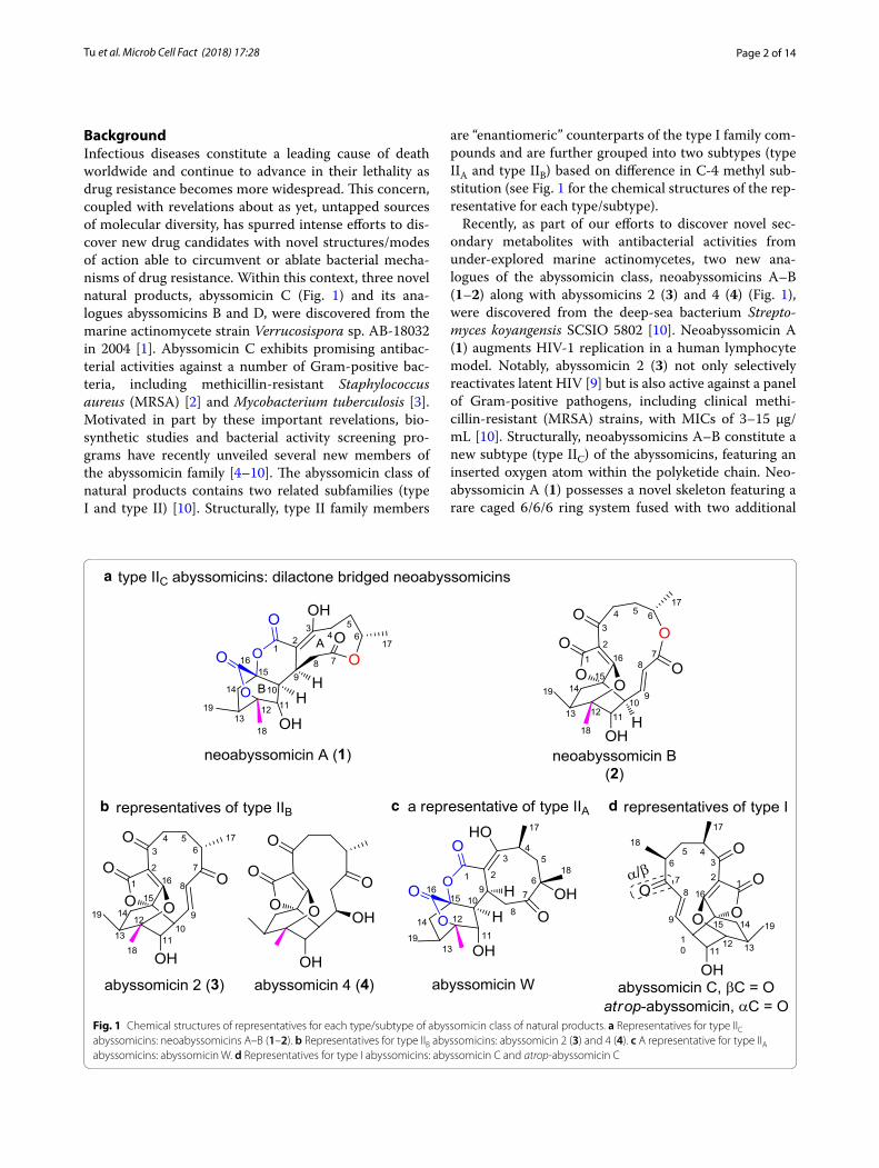
BackgroundInfectious diseases constitute a leading cause of death worldwide and continue to advance in their lethality as drug resistance becomes more widespread. This concern, coupled with revelations about as yet, untapped sources of molecular diversity, has spurred intense efforts to dis-cover new drug candidates with novel structures/modes of action able to circumvent or ablate bacterial mecha-nisms of drug resistance. Within this context, three novel natural products, abyssomicin C (Fig. 1) and its ana-logues abyssomicins B and D, were discovered from the marine actinomycete strain Verrucosispora sp. AB-18032 in 2004 [1]. Abyssomicin C exhibits promising antibac-terial activities against a number of Gram-positive bac-teria, including methicillin-resistant Staphylococcus aureus (MRSA) [2] and Mycobacterium tuberculosis [3]. Motivated in part by these important revelations, bio-synthetic studies and bacterial activity screening pro-grams have recently unveiled several new members of the abyssomicin family [4–10]. The abyssomicin class of natural products contains two related subfamilies (type I and type II) [10]. Structurally, type II family members
are “enantiomeric” counterparts of the type I family com-pounds and are further grouped into two subtypes (type IIA and type IIB) based on difference in C-4 methyl sub-stitution (see Fig. 1 for the chemical structures of the rep-resentative for each type/subtype).
Recently, as part of our efforts to discover novel sec-ondary metabolites with antibacterial activities from under-explored marine actinomycetes, two new ana-logues of the abyssomicin class, neoabyssomicins A–B (1–2) along with abyssomicins 2 (3) and 4 (4) (Fig. 1), were discovered from the deep-sea bacterium Strepto-myces koyangensis SCSIO 5802 [10]. Neoabyssomicin A (1) augments HIV-1 replication in a human lymphocyte model. Notably, abyssomicin 2 (3) not only selectively reactivates latent HIV [9] but is also active against a panel of Gram-positive pathogens, including clinical methi-cillin-resistant (MRSA) strains, with MICs of 3–15 μg/mL [10]. Structurally, neoabyssomicins A–B constitute a new subtype (type IIC) of the abyssomicins, featuring an inserted oxygen atom within the polyketide chain. Neo-abyssomicin A (1) possesses a novel skeleton featuring a rare caged 6/6/6 ring system fused with two additional
a
b c d
Fig. 1 Chemical structures of representatives for each type/subtype of abyssomicin class of natural products. a Representatives for type IIC abyssomicins: neoabyssomicins A–B (1–2). b Representatives for type IIB abyssomicins: abyssomicin 2 (3) and 4 (4). c A representative for type IIA abyssomicins: abyssomicin W. d Representatives for type I abyssomicins: abyssomicin C and atrop-abyssomicin C

Page 3 of 14Tu et al. Microb Cell Fact (2018) 17:28
6/9 lactone rings (Fig. 1). Neoabyssomicin B (2) has a 12-membered lactone ring in place of the 11-membered polyketide ring (Fig. 1). Both the unique structures and biological activities of neoabyssomicins/abyssomicins provide clear inspiration for understanding the biosyn-thetic mechanisms leading to their production as well as possible application of combinatorial biosynthesis to enhance yields and create structural diversity within the class.
A model for the biosynthesis of atrop-abyssomicin C has been proposed based on a combination of feed-ing studies with 13C-labelled biosynthetic precursors and identification of its BGC from Verrucosispora sp. AB-18032 [11]. AbyU has been demonstrated to be a Diels–Alderase that catalyzes [4 + 2] cycloaddition to form a key cyclohexene in atrop-abyssomicin C [12]. Recently, a BGC, identified in Streptomyces sp. LC-6-2 by whole genome sequencing, was proposed to account for abyssomicin M–X biosynthesis [8]. Relative to atrop-abyssomicin C, neoabyssomicins A–B are characterized by a number of biosynthetically interesting structural features. Here, we report the application of genetics experiments and bioinformatics analyses to decipher the biosynthetic pathway leading to the neoabyssomicins/abyssomicins in S. koyangensis SCSIO 5802 as well as their transport systems. Furthermore, rationally designed enhancements to neoabyssomicin/abyssomicin produc-tion were realized via overexpression of two different pathway-specific positive regulators.
MethodsBacterial strains, plasmids and culture conditionsThe bacterial strains and plasmids are listed in Additional file 1: Tables S1 and S2, respectively. The culture condi-tions for S. koyangensis SCSIO 5802 has been previously described [10]. Escherichia coli including XL 1-blue MR, ET12567/pUZ8002, BW25113/pIJ790, as well as, cul-ture conditions have also been previously described [13]. When necessary, antibiotics were supplemented at the following concentrations: apramycin (Apr) 50 μg/mL, chloramphenicol (Chl) 25 μg/mL, and kanamycin (Kan) 50 μg/mL.
Complete genome sequencing and bioinformatics analysisHigh-molecular-weight DNA of S. koyangensis SCSIO 5802 was isolated according to a slightly modified proto-col [14]. Sequencing of the complete genome was accom-plished using a combination of PacBio RSII sequencing (Pacific Biosciences) and Illumina Hiseq 2500 technolo-gies at Biozeron Biotech Co., LTD (Shanghai, China). Genes involved in secondary metabolic pathways were predicted using online antiSMASH software (http://antis mash.secon darym etabo lites .org/). The deduced
ORFs were analyzed using online FramePlot 4.0beta software (http://nocar dia.nih.go.jp/fp4/) and their func-tional predictions were accomplished with an online BLAST program (http://blast .ncbi.nlm.nih.gov/). The PKS architectures were analyzed using an NRPS-PKS online website (http://nrps.igs.umary land.edu/nrps/). The nucleotide sequence of abm BGC was deposited at GenBank under accession number MG243704.
Construction of genomic cosmid library and inactivation of S. koyangensis SCSIO 5802 genesThe genomic cosmid library of S. koyangensis SCSIO 5802 was constructed using SuperCos1 according to the manufacturer’s protocol provided by the vector kit (Agi-lent). About 2000 clones were picked and placed into 96-well plates and stored at − 80 °C. We set out to probe the neoabyssomicin/abyssomicin BGC with three pairs of primers associated with abmB1, orf(−2) and orf(+3) (Additional file 1: Table S3) using PCR methods. These three designed primers were utilized to screen the picked 2000 clones, and 6 positive cosmids (10-8B, 9-7C, 7-6F, 21-9A, 6-7D and 29-8D) were obtained.
The λ-RED-mediated PCR-targeting mutagenesis method was then employed to inactivate targeted neo-abyssomicin/abyssomicin biosynthetic genes [15]. Three cosmids 7-6F, 9-7C and 21-9A covering the whole neo-abyssomicin/abyssomicin gene cluster were used to inactivate the genes in the neoabyssomicin/abyssomicin BGC. Primers designed for gene-specific inactivation are listed in Additional file 1: Table S3. An example for abmB1 is detailed here. Cosmid 9-7C was introduced into E. coli BW25113/pIJ790 to inactivate abmB1. The aac(3)IV-oriT cassette was amplified by PCR from pIJ773 using primers abmB1-Del-F and abmB1-Del-R (Additional file 1: Table S3), and introduced into E. coli BW25113/pIJ790/cosmid 9-7C by electroporation to replace abmB1 via λ-RED-mediated recombination. Cor-rect recombination was established by PCR using prim-ers abmB1-TF and abmB1-TR. The mutated cosmid 9-7C-ΔabmB1 was then introduced into E. coli ET12567/pUZ8002 for further conjugation with S. koyangensis SCSIO 5802. The double crossover mutant was obtained by antibiotic selection (AprRKanS) and confirmed by PCR using primers abmB1-TF and abmB1-TR.
Construction of genomic PAC library and heterologous expression of the abm gene clusterPAC library construction was performed using pESAC13-A by Bio S&T (Montreal, Canada). pESAC13-A was developed from pPAC-S1 [16] by Sosio and Donadio, NAICONS, Milano, Italy and is used by Bio S&T Inc. for PAC library construction. In contrast to previously developed E. coli–Streptomyces

Page 4 of 14Tu et al. Microb Cell Fact (2018) 17:28
Artificial Chromosomes, pESAC13-A contains an oriT site that allows transfer into Streptomyces by conju-gation and confers apramycin resistance in both E. coli and Streptomyces. pESAC13-A vector DNA was digested with BamHI, dephosphorylated and purified using standard procedures. S. koyangensis SCSIO 5802 cells was embedded in 2 mL 2% low-melting-point agarose plugs, which was then treated with proteinase K at 50 °C and stored in 0.5 M EDTA at 4 °C. Plugs were partially digested with BamHI at 37 °C and the reactions were stopped by the addition of 1/10 vol-ume of 0.5 M EDTA (pH 8.0). For each case, partially digested high-molecular-weight DNAs were sepa-rated by two rounds of pulsed field gel electrophore-sis (PFGE) with different ramped pulse times. The DNA fragments 100–300 kb were eluted from the gel by PFGE with a constant pulse time. The eluted DNA fragments were dialyzed against 1× TE (10 mM Tris–HCl, 1 mM EDTA, pH 8.0) prior to ligation. Partially-digested size-selected DNA fragments were ligated to the BamHI-digested and dephosphorylated pESAC13-A vector. The ligation mix was transformed into E. coli DH10β by electroporation. Three sets of primers, including the primers for abmB1, orf(−2), and orf(+3), were designed to screen the library for the desired clone that contains all the biosynthetic genes.
The PAC clone 4-3F, which tested positive for all three PCR probes, was selected for heterologous expression. The DH10β E. coli strain containing 4-3F was used in a triparental mating with the non-meth-ylating E. coli strain ET12567 containing the driver plasmid pUB307 and the heterologous host S. coeli-color M1152. For the E. coli strains DH10β/4-3F and ET12567/pUB307, they were grown to an OD600 of 0.4 in 10 mL LB; the cells were pelleted by centrifugation, washed in LB, pelleted again and finally resuspended in a volume of 200 μL LB. For the S. coelicolor M1152, the required number of spores (108) were added in 400 μL TSB, then incubated at 50 °C for 10 min and continu-ously incubated at 28 °C for 5–6 h on rotary shakers (200 rpm) to activate germination. The germinated spores of S. coelicolor M1152 were mixed with the two previously prepared E. coli strains, and the mixture was spread onto an MS agar plate supplemented with 10 mM MgCl2. After incubation at 28 °C for 18 h, the plate was covered with 950 μL sterile deionized water containing 30 μL trimethoprim (TMP, 50 mg/mL) and 50 μL Apr (50 mg/mL). Finally, the plates were incu-bated for a further 4–5 days at 28 °C until exconjugants appeared. The resulting exconjugants were tested by PCR using two sets of primers, primers for orf(−2) and orf(+3), to confirm that exconjugants contained the entire abm gene cluster.
Overexpression of abmI or abmH in wild‑type producer strainThe coding regions of abmI and abmH were amplified by PCR using the genomic DNA of S. koyangensis SCSIO 5802 wild-type strain and the primers listed in Additional file 1: Table S3. Each of the PCR products was digested with NdeI/SpeI then cloned into the same digested sites of a pSET152-derived expression plasmid, pL646 [17], under the control of a strong constitutive promoter ermE*p. The recombinant plasmids pL646-abmI and pL646-abmH were transformed into E. coli ET12567/pUZ8002, and then individually transferred into wild-type S. koyangensis SCSIO 5802 by conjugation; the exconjugants were selected on the basis of phenotypes showing apramycin resistance and then confirmed by PCR to give the overexpression strains 5802::abmI and 5802::abmH.
Metabolite analyses of wild‑type S. koyangensis SCSIO 5802 and related derivative strainsThe wild-type S. koyangensis SCSIO 5802 and rel-evant gene-inactivated mutants were first grown on A1 medium agar [10] at 28 °C for 5 days to achieve sporu-lation. For the abmA1–A5 mutant strains, a portion of mycelium and spores (1 cm2) for each strain was added to 250 mL flasks containing 50 mL of RA medium [10] with supplemental 1% XAD-16 resin. Fermentations were then carried out at 28 °C on rotary shakers (200 rpm) for 8 days. After fermentation, each fermentation culture was centrifuged to yield the supernatant and pellet. Pel-lets were washed with 30 mL MeOH three times, and, for each sample, the MeOH extracts were combined, and the MeOH solvent was removed under reduced pressure to afford an oily residue. Residues were each dissolved into 1 mL MeOH and centrifuged at 13,000g for 10 min; supernatants were subjected to HPLC–UV analyses, each of which was performed using an Agilent Technologies 1260 Infinity system using a Phenomenex ODS column (150 × 4.6 mm, 5 μm), eluting with a linear gradient of 5 to 65% solvent B (solvent B: CH3CN + 0.1% trifluoro-acetic acid (TFA); solvent A: H2O + 0.1% TFA) over 20 min, followed by 65% to 100% solvent B in 2 min, and then 100% solvent B for 5 min, at a flow rate of 1 mL/min. In all cases chromatograms employed UV detec-tion at 254 nm. To characterize compounds 5 and 6 that accumulated in the ΔabmA4 and ΔabmA5 mutants, the metabolites of these two mutants were further subjected to mass analysis. MS analyses for 5 and 6 were carried out on a Bruker MaXis quardrupole-time-of-flight mass spectrometer equipped with an electrospray iron source (operated in the positive ion mode) fitted with a Phenom-enex ODS column (150 × 4.6 mm, 5 μm) using a gradient of solvent B as described above for HPLC–UV analyses.
Page 5 of 14Tu et al. Microb Cell Fact (2018) 17:28
For other gene-inactivated mutants, the recombinant heterologous abm expression strains and the abmI/abmH overexpression strains, a portion of mycelium and spores (1 cm2) of each strain was inoculated into 250-mL flasks containing 50 mL of RA medium without supplemental of 1% XAD-16 resin. Fermentations were then carried out at 28 °C on rotary shakers (200 rpm) for 8 days. After fermentations, each culture broth was extracted with two volumes of butanone (1 × 100 mL), and solvents were then removed under reduced pressure to afford oily res-idue. Each residue was dissolved into 1 mL MeOH and centrifuged at 13,000g for 10 min; the supernatant was subjected to HPLC–UV analyses according to the meth-ods described above except that the mobile phases did not contain 0.1% TFA.
To quantify the production of abyssomicin 2 (3) from wild-type and abmI/abmH overexpression strains, a standard curve was first generated using an analytically pure sample and HPLC–UV analyses (Additional file 1: Figure S11). Using a detection wavelength of 254 nm and correspondingly integrated signals (relative to control injections) the concentrations of abyssomicin 2 (3) were determined for the wild-type and abmI/abmH overex-pressing strains.
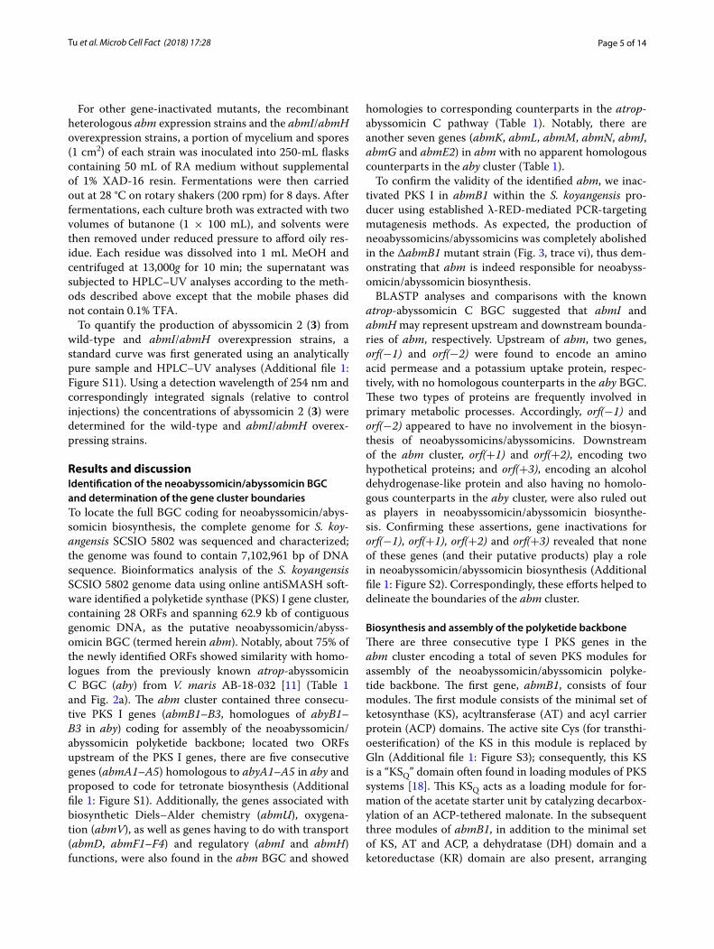
Results and discussionIdentification of the neoabyssomicin/abyssomicin BGC and determination of the gene cluster boundariesTo locate the full BGC coding for neoabyssomicin/abys-somicin biosynthesis, the complete genome for S. koy-angensis SCSIO 5802 was sequenced and characterized; the genome was found to contain 7,102,961 bp of DNA sequence. Bioinformatics analysis of the S. koyangensis SCSIO 5802 genome data using online antiSMASH soft-ware identified a polyketide synthase (PKS) I gene cluster, containing 28 ORFs and spanning 62.9 kb of contiguous genomic DNA, as the putative neoabyssomicin/abyss-omicin BGC (termed herein abm). Notably, about 75% of the newly identified ORFs showed similarity with homo-logues from the previously known atrop-abyssomicin C BGC (aby) from V. maris AB-18-032 [11] (Table 1 and Fig. 2a). The abm cluster contained three consecu-tive PKS I genes (abmB1–B3, homologues of abyB1–B3 in aby) coding for assembly of the neoabyssomicin/abyssomicin polyketide backbone; located two ORFs upstream of the PKS I genes, there are five consecutive genes (abmA1–A5) homologous to abyA1–A5 in aby and proposed to code for tetronate biosynthesis (Additional file 1: Figure S1). Additionally, the genes associated with biosynthetic Diels–Alder chemistry (abmU), oxygena-tion (abmV), as well as genes having to do with transport (abmD, abmF1–F4) and regulatory (abmI and abmH) functions, were also found in the abm BGC and showed
homologies to corresponding counterparts in the atrop-abyssomicin C pathway (Table 1). Notably, there are another seven genes (abmK, abmL, abmM, abmN, abmJ, abmG and abmE2) in abm with no apparent homologous counterparts in the aby cluster (Table 1).
To confirm the validity of the identified abm, we inac-tivated PKS I in abmB1 within the S. koyangensis pro-ducer using established λ-RED-mediated PCR-targeting mutagenesis methods. As expected, the production of neoabyssomicins/abyssomicins was completely abolished in the ΔabmB1 mutant strain (Fig. 3, trace vi), thus dem-onstrating that abm is indeed responsible for neoabyss-omicin/abyssomicin biosynthesis.
BLASTP analyses and comparisons with the known atrop-abyssomicin C BGC suggested that abmI and abmH may represent upstream and downstream bounda-ries of abm, respectively. Upstream of abm, two genes, orf(−1) and orf(−2) were found to encode an amino acid permease and a potassium uptake protein, respec-tively, with no homologous counterparts in the aby BGC. These two types of proteins are frequently involved in primary metabolic processes. Accordingly, orf(−1) and orf(−2) appeared to have no involvement in the biosyn-thesis of neoabyssomicins/abyssomicins. Downstream of the abm cluster, orf(+1) and orf(+2), encoding two hypothetical proteins; and orf(+3), encoding an alcohol dehydrogenase-like protein and also having no homolo-gous counterparts in the aby cluster, were also ruled out as players in neoabyssomicin/abyssomicin biosynthe-sis. Confirming these assertions, gene inactivations for orf(−1), orf(+1), orf(+2) and orf(+3) revealed that none of these genes (and their putative products) play a role in neoabyssomicin/abyssomicin biosynthesis (Additional file 1: Figure S2). Correspondingly, these efforts helped to delineate the boundaries of the abm cluster.
Biosynthesis and assembly of the polyketide backboneThere are three consecutive type I PKS genes in the abm cluster encoding a total of seven PKS modules for assembly of the neoabyssomicin/abyssomicin polyke-tide backbone. The first gene, abmB1, consists of four modules. The first module consists of the minimal set of ketosynthase (KS), acyltransferase (AT) and acyl carrier protein (ACP) domains. The active site Cys (for transthi-oesterification) of the KS in this module is replaced by Gln (Additional file 1: Figure S3); consequently, this KS is a “KSQ” domain often found in loading modules of PKS systems [18]. This KSQ acts as a loading module for for-mation of the acetate starter unit by catalyzing decarbox-ylation of an ACP-tethered malonate. In the subsequent three modules of abmB1, in addition to the minimal set of KS, AT and ACP, a dehydratase (DH) domain and a ketoreductase (KR) domain are also present, arranging
Page 6 of 14Tu et al. Microb Cell Fact (2018) 17:28
Table 1 Deduced functions of ORFs in abm BGC from S. koyangensis SCSIO 5802
a Size in units of amino acids (aa); ID/SI: identity/similarity; aby: the BGC of atrop-abyssomicin C from Verrucosispora sp. AB-18032; abs: the putative BGC of abyssomicins M–X from Streptomyces sp. LC-6
ORF Sizea Proposed function Closest homolog, origin (protein ID); ID/SI (%) aby homolog abs homolog
orf(−2) 223 Potassium uptake protein TrkA, Streptomyces coelicolor A3(2) (Q53949.2); 88/96 – –
orf(−1) 687 Amino acid permease PlaP, Escherichia coli O157:H7 (P0AA48.1); 21/39 – –
abmN 445 rRNA (Uracil-5-)-methyltransferase RlmD, Acinetobacter baumannii AB307-0294 (B7H018.1); 30/48
– –
abmI 268 Transcriptional activator, SARP family DnrI, Streptomyces peucetius (P25047.1); 35/50 abyI –
AbmE2 356 Luciferase-like monooxygenase, α-subunit LuxA, Photorhabdus luminescens (P23146.1); 24/42 – –
abmU 218 Diels–Alderase YD repeat-containing protein, Streptomyces regensis (KMS84434.1); 41/52
abyU absU
abmK 256 4′-Phosphopantetheinyl transferase superfamily (PPTase)
Npt, Nocardia iowensis (A1YCA5.1); 42/54 – –
abmL 281 Metallophosphoesterase GsiA, Salmonella enterica (Q57RB2.2); 46/60 – –
abmF4 560 ABC transporter system ATP-binding protein OppD, Lactococcus lactis (AIS04392.1); 42/73 or OppF, Lactococcus lactis (ABA47382.1); 43/74
abyF4 absF4
abmF3 298 ABC transporter system substrate-binding protein dependent permease
OppC, Lactococcus lactis (ABA47380.1); 27/63 abyF3 absF3
abmM 413 Amidohydrolase Mb2939c, Mycobacterium bovis AF2122/97 (P68916.1); 28/37
– –
abmF2 313 ABC transporter system permease OppB, Lactococcus lactis (ABA47381.1); 27/58 abyF2 absF2
abmF1 546 ABC transport system substrate-binding protein OppA, Lactococcus lactis (AAO63469.1); 20/52 abyF1 absF1
abmJ 331 Aldo/keto reductase OsI_15387, Oryza sativa Indica Group (A2XRZ0.1); 50/68
– absJ
abmG 77 Ferredoxin Fd-1, Streptomyces griseolus (P18324.3); 58/75 – absG1
abmV 405 Cytochrome P450 Vitamin D3 dihydroxylase, Streptomyces griseolus (P18326.2); 55/70
abyV absV
abmC 257 TetR regulatory protein Mce3R, Mycobacterium tuberculosis H37Rv (P95251.2); 33/48
abyC absC2
AbmE1 353 Luciferase-like monooxygenase, β-subunit LuxB, Photorhabdus luminescens (P19840.1); 20/41 abyE –
abmD 487 Major facilitator superfamily of transporter EmrB, Mycobacterium tuberculosis CDC1551 (P9WG88.1); 34/56
abyD absD
abmA1 343 Ketoacyl-S-ACP synthase ChlM, Streptomyces antibioticus (AAZ77702.1); 61/74 abyA1 absA1
abmA2 628 Glyceryl-S-ACP synthase ChlD, Streptomyces antibioticus (AAZ77703.1); 61/70 abyA2 absA2
abmA3 75 Acyl carrier protein ChlD2, Streptomyces antibioticus (AAZ77704.1); 56/73 abyA3 absA3
abmA4 280 2-Oxoacid dehydrogenase multienzymes acyltrans-ferase E2 component
ChlD3, Streptomyces antibioticus (AAZ77705.1); 65/76 abyA4 absA4
abmA5 373 α/β hydrolase fold protein ChlD4, Streptomyces antibioticus (AAZ77706.1); 51/64 abmA5 absA5
abmT 274 Type II thioesterase PikA5, Streptomyces venezuelae (Q9ZGI1.1); 32/45 abyT –
abmZ 178 NADPH-dependent flavin reductase HsaB, Rhodococcus jostii RHA1 (Q0S808.1); 39/57 abyZ absH1
abmB1 6540 PKS I (module 1: KS, ATa, ACP; module 2: KS, ATp, DH, KR, ACP; module 3: KS, ATa, DH, KR, ACP; module 4: KS, ATa, DH, KR, ACP)
PikA1, Streptomyces venezuelae (Q9ZGI5.1); 54/65 abyB1 absB1
abmB2 4054 PKS I (module 5: KS, ATp, DH, KR, ACP; module 6: KS, ATa, DH, ER, KR, ACP)
PikA2, Streptomyces venezuelae (Q9ZGI4.1); 49/59 abyB2 absB2
abmB3 1040 PKS I (module 7: KS, ATa, ACP) PikA1, Streptomyces venezuelae (Q9ZGI5.1); 54/64 abyB3 absB3
abmH 942 LuxR family transcriptional regulator NreC, Staphylococcus carnosus subsp. carnosus TM300 (Q7WZY4.1); 48/67
abyH –
orf(+1) 530 Hypothetical protein Hypothetical protein; Actinospica acidiphila (WP_033273634.1); 49/58
– –
orf(+2) 1939 Hypothetical protein Hypothetical protein, Actinospica acidiphila (WP_033273633.1); 56/67
– –
orf(+3) 331 Alcohol dehydrogenase-like protein TDH, Agrobacterium radiobacter K84 (B9J738.1); 32/53 – –

Page 7 of 14Tu et al. Microb Cell Fact (2018) 17:28
a
b
Fig. 2 Organization of the abm BGC (a) and proposed biosynthetic pathway (b) for neoabyssomicins/abyssomicins in S. koyangensis SCSIO 5802
collinearly with their functions in the biosynthetic assem-bly line. Module 5 of abmB2 has the same arrangement as module 4 although its KR domain is believed to be inactive on the basis of sequence analyses indicating that it lacks both the conserved NADPH binding motif and the Lys and Tyr of the active site triad required of reduc-tase activity [19]; these active site residues are replaced by Glu and Phe, respectively (Additional file 1: Figure S4). Whether the DH domain of module 5 (Additional file 1: Figure S5) is active or not is unclear since the requisite
hydroxy group of a putative DH substrate is absent due to crippling of the KR domain (Additional file 1: Figure S4). In contrast to module 5, module 6 of abmB2 con-tains an additional enoylreductase (ER) domain. Mod-ule 7 of abmB3 consists of a minimal set of KS, AT and ACP that adds an additional acetate unit. AbmB1-AT_2 and AbmB2-AT_5 contain the substrate specificity code (YASH) [20] (Additional file 1: Figure S6), characteris-tic of propionate incorporation. The remainder of the five AT domains contain the substrate specificity code

Page 8 of 14Tu et al. Microb Cell Fact (2018) 17:28
(HAFH) [20] (Additional file 1: Figure S6), indicative of malonyl-CoA specificity; this observation is consistent with the structure of neoabyssomicins/abyssomicins.
Discrete type II TEs have been reported to play a role in editing stalled polyketide chains [21]. The gene abmT encodes a discrete type II thioesterase (TE) and multi-ple sequence alignments of AbmT with the typical type II TEs revealed that it contains the conserved motif (GHSXG) (Additional file 1: Figure S7) [22, 23]. There-fore, AbmT probably plays an editing role in the biosyn-thesis of neoabyssomicins/abyssomicins by hydrolyzing misincorporated monomers. Notably, abmT inactiva-tion was found to decrease neoabyssomicin/abyssomicin titers relative to the wild-type producer consistent with its predicted editing function (Fig. 3, trace vii).
Formation of the tetronate moiety and subsequent post‑tailoring stepsWithin the abm cluster abmA1–A5 are homologous to a set of five highly conserved genes unique to tetronate biosynthesis (Additional file 1: Figure S1). Early bio-chemical studies unveiled a common pathway relevant
to most (if not all) tetronate-containing natural products. The pathway is composed of the following steps: (i) trans-fer of a glycerol moiety from d-1,3-biphosphoglycerate to a discrete acyl carrier protein (ACP) (e.g., Tmn7a in tetronomycin biosynthesis [24], a homologue of AbmA3) as catalyzed by a glyceryl-S-ACP synthase (e.g., RkE in RK-682 biosynthesis [25], a homologue of AbmA2), lead-ing to glyceryl-S-ACP; (ii) binding of the glyceryl-S-ACP to the nascent polyketide chain and detachment of the polyketide from the PKS, generating the linear hydroxy-methyl tetronate ring as catalyzed by a ketoacyl-S-ACP synthase (e.g., RkD in RK-682 biosynthesis [26], a homo-logue of AbmA1); (iii) exomethylene installation via an acylation-elimination process accomplished by an acyl-transferase E2 component of 2-oxoacid dehydrogenase multienzymes (e.g., Agg4 in agglomerin biosynthesis [27] and QmnD3 in quartromicin biosynthesis [28], homo-logues of AbmA4) and an α/β hydrolase fold protein (e.g., Agg5 in agglomerin biosynthesis [27] and QmnD4 in quartromicin biosynthesis [28], homologues of AbmA5). This sequence of steps, executed by the products of abmA1–A5, likely accounts for tetronate generation in the neoabyssomicins/abyssomicins in a fashion highly similar to other tetronates such as agglomerin, quar-tromicin and others (Fig. 2b for a proposed biosynthetic pathway).
To validate our hypotheses regarding tetronate instal-lation and to define the roles of abmA1–A5, each was specifically inactivated to afford double-crossover mutants for each gene. Inactivation of abmA1, abmA2 and abmA3 completely abolished neoabyssomicin/abyssomicin production, confirming their involvement in neoabyssomicin/abyssomicin biosynthesis (Fig. 4a, traces ii–iv). HPLC–UV analyses of the fermentation extract for the ΔabmA4 mutant revealed its failure to produce neoabyssomicins/abyssomicins (1–4) (Fig. 4a, trace v). However, the ΔabmA4 mutant accumulated a new compound whose molecular mass, as determined by HR–ESI–MS ([M + H]+, calculated: 349.1606, found: 349.1649) (Fig. 4b), exactly matched that predicted for linear hydroxy-tetronate intermediate 5 (Fig. 2b). The ΔabmA5 mutant, on HPLC–UV analysis of its fermen-tation extract, also failed to produce 1–4 but accumu-lated minor amounts of 5 (Fig. 4a, trace vi). In addition, the ΔabmA5 mutant, generated a major metabolite with longer HPLC elution time than 5 (Fig. 4a, trace vi) and whose molecular mass, on the basis of HR–ESI–MS ([M + H]+, calculated: 391.1712, found: 391.1759) (Fig. 4c), exactly matched that predicted for the acetyl-tetronate intermediate 6 (Fig. 2b). Consequently, it is clear on the basis of these inactivation studies that abmA1–A5 all play vital roles in 1–4 biosynthesis and that, more precisely, abmA4 and abmA5 are involved in
Fig. 3 HPLC analyses of fermentation extracts. i–iv: authentic standards of compound 1–4; v: wild-type S. koyangensis SCSIO 5802; vi: ΔabmB1 mutant; vii: ΔabmT mutant; viii: negative control of the host strain S. coelicolor M1152; ix: S. coelicolor M1152 bearing the 4-3F BAC clone, which contains the abm cluster

Page 9 of 14Tu et al. Microb Cell Fact (2018) 17:28
tetronate exomethylene installation leading to 7; abmA4 codes for the OH activating acetyltransferase and the abmA5 product catalyzes elimination to generate the exomethylene moiety.
We hypothesized early on that intermediate 7 may readily undergo an intermolecular [4 + 2] cycloaddition (Diels–Alder) reaction between the newly installed exo-methylene group and the terminal conjugated diene to afford polycyclic species 8 uniquely characterized by its spirotetronate skeleton. AbmU, showing sequence simi-larity (37% identity) to the Diels–Alderase, AbyU [12], in the atrop-abyssomicin C biosynthetic pathway, was proposed to catalyze this intramolecular spirocycliza-tion. The path from polycycle 8 to neoabyssomicins/abyssomicins (1–4) dictates the involvement of several tailoring enzymes. We envision that 8 undergoes epoxi-dation of the cyclohexene C11–C12 double bond afford-ing epoxide 9, which then reacts via intramolecular ring-opening with the sole OH moiety. Epoxide opening via nucleophilic attack by the tetronate OH affords abys-somicin 2 (3). Hydration of 3 via Michael-type addition to the C8–C9 olefin can be readily envisioned as a means of generating abyssomicin 4 (4). Structurally, neoabys-somicins A and B (1, 2) constitute a new subtype (type IIC) of abyssomicins, featuring an inserted oxygen atom within the polyketide chain, and we proposed that a unique biosynthetic Baeyer–Villiger reaction represents access to this new class of abyssomicins. We envision that 3 might also undergo Baeyer–Villiger oxidation to yield
neoabyssomicin B (2). Notably, the new scaffold neo-abyssomicin A (1) may well be derived from 2 through a sequence of: (i) C-16 hydrolysis, (ii) keto-enol tautom-erism, (iii) a retro-aldol reaction to establish the C-16 containing lactone, (iv) establishment of the C-2 to C-9 linkage via an aldol-type Michael reaction, and finally, (v) a simple keto-enol tautomerization (Fig. 2b). Within the abm cluster, abmV, abmM, abmJ, abmG, abmE1 and abmE2, are all predicted to encode enzymes related to oxidation or reduction (Table 1) and are therefore excel-lent tailoring enzyme candidates for the 8 → 1–4 pro-gression. However, although their involvement in these conversions is highly likely, the precise details for how these gene products carry out their relevant chemistries remains to be determined.
Export and import systems for neoabyssomicin/abyssomicin biosynthesisThe abmD orf is predicted to encode a major facilitator superfamily (MFS) protein. MFS family proteins pos-sess 12 or 14 transmembrane segments (TMS) and are capable of transporting small molecules in response to changes in chemiosmotic gradients [29]. Detailed sequence analysis of AbmD revealed that it belongs to the 14-TMS subfamily of the MFS (Additional file 1: Figure S8). In Streptomyces, members of the 14-TMS subfamily are usually involved in antibiotic secretion from the pro-ducer strain thus endowing the microbial producer with a self-defense mechanism [30, 31]. Since neoabyssomicins/
Fig. 4 Analyses of the fermentation extracts of abmA1–A5 inactivated mutants: a HPLC–UV analyses of the fermentation extracts of abmA1–A5 inactivated mutants. b HR–ESI–MS spectra of peak 5 of the fermentation extract of ΔabmA4 mutant. c HR–ESI–MS spectra of peak 6 of the fermentation extract of ΔabmA5 mutant
Page 10 of 14Tu et al. Microb Cell Fact (2018) 17:28
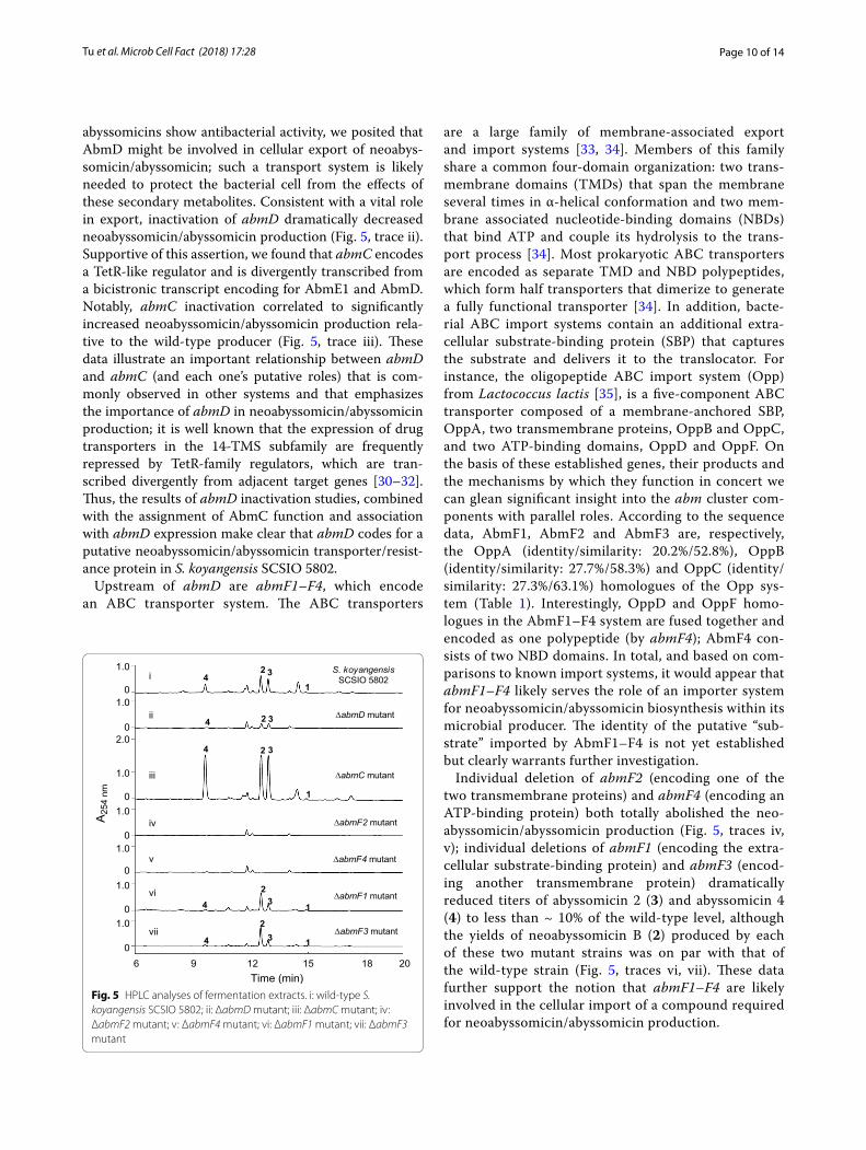
abyssomicins show antibacterial activity, we posited that AbmD might be involved in cellular export of neoabys-somicin/abyssomicin; such a transport system is likely needed to protect the bacterial cell from the effects of these secondary metabolites. Consistent with a vital role in export, inactivation of abmD dramatically decreased neoabyssomicin/abyssomicin production (Fig. 5, trace ii). Supportive of this assertion, we found that abmC encodes a TetR-like regulator and is divergently transcribed from a bicistronic transcript encoding for AbmE1 and AbmD. Notably, abmC inactivation correlated to significantly increased neoabyssomicin/abyssomicin production rela-tive to the wild-type producer (Fig. 5, trace iii). These data illustrate an important relationship between abmD and abmC (and each one’s putative roles) that is com-monly observed in other systems and that emphasizes the importance of abmD in neoabyssomicin/abyssomicin production; it is well known that the expression of drug transporters in the 14-TMS subfamily are frequently repressed by TetR-family regulators, which are tran-scribed divergently from adjacent target genes [30–32]. Thus, the results of abmD inactivation studies, combined with the assignment of AbmC function and association with abmD expression make clear that abmD codes for a putative neoabyssomicin/abyssomicin transporter/resist-ance protein in S. koyangensis SCSIO 5802.
Upstream of abmD are abmF1–F4, which encode an ABC transporter system. The ABC transporters
are a large family of membrane-associated export and import systems [33, 34]. Members of this family share a common four-domain organization: two trans-membrane domains (TMDs) that span the membrane several times in α-helical conformation and two mem-brane associated nucleotide-binding domains (NBDs) that bind ATP and couple its hydrolysis to the trans-port process [34]. Most prokaryotic ABC transporters are encoded as separate TMD and NBD polypeptides, which form half transporters that dimerize to generate a fully functional transporter [34]. In addition, bacte-rial ABC import systems contain an additional extra-cellular substrate-binding protein (SBP) that captures the substrate and delivers it to the translocator. For instance, the oligopeptide ABC import system (Opp) from Lactococcus lactis [35], is a five-component ABC transporter composed of a membrane-anchored SBP, OppA, two transmembrane proteins, OppB and OppC, and two ATP-binding domains, OppD and OppF. On the basis of these established genes, their products and the mechanisms by which they function in concert we can glean significant insight into the abm cluster com-ponents with parallel roles. According to the sequence data, AbmF1, AbmF2 and AbmF3 are, respectively, the OppA (identity/similarity: 20.2%/52.8%), OppB (identity/similarity: 27.7%/58.3%) and OppC (identity/similarity: 27.3%/63.1%) homologues of the Opp sys-tem (Table 1). Interestingly, OppD and OppF homo-logues in the AbmF1–F4 system are fused together and encoded as one polypeptide (by abmF4); AbmF4 con-sists of two NBD domains. In total, and based on com-parisons to known import systems, it would appear that abmF1–F4 likely serves the role of an importer system for neoabyssomicin/abyssomicin biosynthesis within its microbial producer. The identity of the putative “sub-strate” imported by AbmF1–F4 is not yet established but clearly warrants further investigation.
Individual deletion of abmF2 (encoding one of the two transmembrane proteins) and abmF4 (encoding an ATP-binding protein) both totally abolished the neo-abyssomicin/abyssomicin production (Fig. 5, traces iv, v); individual deletions of abmF1 (encoding the extra-cellular substrate-binding protein) and abmF3 (encod-ing another transmembrane protein) dramatically reduced titers of abyssomicin 2 (3) and abyssomicin 4 (4) to less than ~ 10% of the wild-type level, although the yields of neoabyssomicin B (2) produced by each of these two mutant strains was on par with that of the wild-type strain (Fig. 5, traces vi, vii). These data further support the notion that abmF1–F4 are likely involved in the cellular import of a compound required for neoabyssomicin/abyssomicin production.
Fig. 5 HPLC analyses of fermentation extracts. i: wild-type S. koyangensis SCSIO 5802; ii: ΔabmD mutant; iii: ΔabmC mutant; iv: ΔabmF2 mutant; v: ΔabmF4 mutant; vi: ΔabmF1 mutant; vii: ΔabmF3 mutant

Page 11 of 14Tu et al. Microb Cell Fact (2018) 17:28
AbmI and AbmH are two positive regulators governing neoabyssomicin/abyssomicin productionWithin the abm cluster abmI and abmH, encode homo-logues of known regulators of secondary metabolism. The abmI gene, present in the far upstream (left) region of the cluster, encodes a protein with significant similar-ity to pathway specific Streptomyces antibiotic regula-tory protein (SARP) transcriptional activators, such as ActII-ORF4 [36], RedD [37] and DnrI [38]. Sequence alignments of AbmI with these well-characterized SARP regulators revealed that they all contain a con-served N-terminal OmpR-like DNA-binding domain and a C-terminal bacterial transcriptional activation domain (BTAD) (Additional file 1: Figure S9), indicat-ing that AbmI might be a positive regulator of neoabys-somicin/abyssomicin biosynthesis. Located within the other far extreme side (downstream) of the abm cluster was found abmH encoding a transcriptional regulator of the LuxR family. In Gram-negative bacteria, LuxR regu-lators, composed of an N-terminal autoinducer binding domain and a C-terminal helix-turn-helix (HTH) DNA-binding domain, can be activators or repressors, which, acting through a quorum sensing mechanism, induce transcription when a certain cell density is reached [39, 40]. In actinobacteria, LuxR regulators are usually identi-fied as pathway specific transcriptional activators such as AveR [41], PikD [42], and GdmR1 [43]; these systems are characterized by an N-terminal nucleotide triphosphate (NTP)-binding domain represented by Walker A motif (GxxxxGK[T/S]) and Walker B motif (hhhhD) (where h is a hydrophobic residue), as well as a C terminal HTH
LuxR-type DNA-binding domain. Bioinformatics analy-ses revealed that AbmH showed sequence and domain organizations similar to those of the well-studied AveR, PikD, and GdmR1 regulators (Additional file 1: Figure S10). Furthermore, AbmH appears to lack any obvious autoinducer binding domain (Additional file 1: Figure S10) suggesting AbmH as a likely positive regulator of neoabyssomicin/abyssomicin production.
To determine the roles of abmI and abmH in 1–4 bio-synthesis, both genes were independently inactivated according to standard methods to generate the ΔabmI and ΔabmH mutants, respectively. As expected, disrup-tion of abmI and abmH completely abolished neoabys-somicin/abyssomicin production (Fig. 6a, traces ii, iii). These results demonstrated that abmI and abmH are both pivotal positive regulators of neoabyssomicin/abys-somicin biosynthesis.
Heterologous expression of the abm BGC efficiently produces 1–4Based on the above informatics and genetic information, we hypothesized that all genes required for biosynthesis of all four neoabyssomicins/abyssomicins (1–4) are pre-sent in the identified abm cluster, although several puta-tive tailoring genes/enzymes and mechanisms are not yet well-defined. To verify the ability of abm-housed genes to generate 1–4, we sought to heterologously express the abm BGC in S. coelicolor M1152. A P1-derived arti-ficial chromosome (PAC) library for the S. koyangensis SCSIO 5802 genome was constructed using an E. coli–Streptomyces artificial chromosome vector, pESAC-13-A
WT 5802::abmI 5802::abmH0
1
2
abys
som
icin
2 (g
/L)
a b
Fig. 6 Individual overexpression of abmI and abmH enhances neoabyssomicin/abyssomicin production. a HPLC analyses of fermentation extracts. i: wild-type S. koyangensis SCSIO 5802; ii: ΔabmI mutant; iii: ΔabmH mutant; iv: abmI overexpression strain 5802::abmI; v: abmH overexpression strain 5802::abmI. 5 μL fermentation extract was subjected to HPLC analyses for each of the samples. b Abyssomicin 2 (3) production of wild-type and overexpression strains. WT: wild-type S. koyangensis SCSIO 5802. The values are mean ± SD from three different experiments
Page 12 of 14Tu et al. Microb Cell Fact (2018) 17:28
(a derivative of pPAC-S1 [16]), which can be shuttled between E. coli and a suitable Streptomyces host. Three sets of primers, including the primers for abm1, orf(−2), and orf(+3), were used to screen ~ 2000 clones of the library for the one containing all the identified biosyn-thetic genes. A BAC clone, 4-3F, testing positive for all three PCR probes, was isolated and introduced into S. coelicolor M1152 by triparental intergeneric conjugation to generate M1152::pESAC4-3F, with the integrated abm gene cluster in its chromosome. HPLC–UV analyses of the fermentation extract of M1152::pESAC4-3F revealed that, indeed, this heterologous host produced 1–4 in yields rivaling the wild-type producer (Fig. 3, traces viii, ix); BAC clone 4-3F clearly housed the intact abm cluster containing all genes needed for wild-type levels of neo-abyssomicin/abyssomicin production.
Overexpression of abmI or abmH boosts neoabyssomicin/abyssomicin productionSince abmI and abmH both encode pathway-specific regulators, positively regulating the production of neo-abyssomicin/abyssomicin, overexpression of abmH or abmH was envisioned to be a viable means of increas-ing neoabyssomicin/abyssomicin titers in S. koyangen-sis SCSIO 5802. Similarities to related systems [36–43] and the results indicated in Fig. 6 supported this logic. To evaluate this hypothesis each positive regulator abmI and abmH was independently cloned into a pSET152-derived expression plasmid, pL646 [17]; both abmI and abmH were situated under the control of constitutive ermE*p promoter. Each of the resulting plasmids was conjugated into wild-type S. koyangensis SCSIO 5802 to create 5802:abmI and 5802:abmH overexpression strains, respectively. As expected, the yields for 1–4 increased almost uniformly relative to the unmodified wild-type producer (Fig. 6a, traces iv, v). Only abyss-omicin 2 (3), the major metabolite in the fermentation, was quantitated in further experiments. Production of 3 in 5802::abmI (2.1045 g/L) relative to the wild-type producer (0.2764 g/L) improved by 7.6-fold whereas the increase observed with 5802::abmH (0.8292 g/L) was approximately threefold relative to wild-type (Fig. 6b). These data clearly support the positive regulatory roles played by AbmI and AbmH in the synthesis of 1–4 while also providing a glimpse of how data presented herein might be employed in future combinatorial biosynthesis efforts to better exploit the unique structures and activi-ties of the neoabyssomicins and abyssomicins.
ConclusionsWe have identified and characterized the neoabyss-omicin/abyssomicin BGC in S. koyangensis SCSIO 5802 by carrying out whole genome sequencing, systematic
gene disruptions and heterologous expression experi-ments; the abm BGC generates 1–4. Informatics analyses and genetics data have enabled us to propose a plausi-ble biosynthetic pathway leading to 1–4. Central to this proposal is our demonstration that the tetronate moi-eties result from an enzymatically driven acetylation-elimination sequence. Beyond their basic construction, we employed gene inactivations and bioinformatics to unveil an export system by which the microbial producer avoids the detrimental effects of 1–4 intracellular pro-duction; a biosynthetically vital four-component import system was also uncovered. Finally, we have also revealed that abmI and abmH both encode proteins involved in positively regulating neoabyssomicin/abyssomicin bio-synthesis; overexpression of either gene afforded sig-nificant enhancements in titers. Overall, the findings detailed here set the stage for dramatic improvements in the production, study, and potential clinical applica-tions of the neoabyssomicin/abyssomicin scaffold. The working model for biosynthesis (and its regulation) of 1–4 enabled by these efforts is anticipated to significantly advance combinatorial biosynthetic initiatives to better exploit this unique family of natural products.
AbbreviationsMRSA: methicillin-resistant Staphylococcus aureus; BGC: biosynthetic gene cluster; ORFs: opening reading frames; HIV: human immunodeficiency virus; aby: the biosynthetic gene cluster of atrop-abyssomicin C; abs: the puta-tive biosynthetic gene cluster of abyssomicins M–X; abm: the biosynthetic gene cluster of neoabyssomicins/abyssomicins; PKS: polyketide synthase; AT: acyltransferase; ACP: acyl carrier protein; DH: dehydratase; ER: enoylreduc-tase; KR: ketoreductase; TE: thioesterase; MFS: major facilitator superfamily; TMS: transmembrane segments; TMDs: transmembrane domains; NBDs: nucleotide-binding domains; SBP: substrate-binding protein; Opp: oligopep-tide ABC import system; SARP: Streptomyces antibiotic regulatory protein; HTH: helix-turn-helix; BTAD: bacterial transcriptional activation domain.
Authors’ contributionsJT, SL and JC performed the experiments. YS and SF assisted with the experi-ments. QL and JJ analyzed the data. QL supervised the full project and wrote the manuscript. JJ helped with the critical reading and editing of the manu-script. All authors read and approved the final manuscript.
Additional file
Additional file 1: Table S1. Bacteria used in this study. Table S2. Plasmids used in this study. Table S3. Primers used in this study. Figure S1. Chemi-cal structures of tetronate-containing natural products and the unique set of five highly conserved genes responsible for tetronate biosynthesis. Fig‑ure S2. HPLC analyses of fermentation extracts of the inactivated mutants of boundary genes. Figure S3. Alignments of seven KS domains of AbmB1–B3. Figure S4. Alignments of five KR domains of AbmB1–B2. Fig‑ure S5. Alignments of five DH domains of AbmB1–B2. Figure S6. Align-ments of five AT domains of AbmB1–B2. Figure S7. Alignments of AbmT with the typical type II TEs. Figure S8. The 14 transmembrane helices of AbmD. Figure S9. Alignments of AbmI with previously characterized SARP regulators. Figure S10. Alignments of AbmH with previously charac-terized LuxR-regulators. Figure S11. The quantitative HPLC standard curve for abyssomicin 2. Figures S12–S30. Disruption of 19 abm-related genes in wild-type S. koyangensis SCSIO 5802 via PCR-targeting.
Page 13 of 14Tu et al. Microb Cell Fact (2018) 17:28
Author details1 CAS Key Laboratory of Tropical Marine Bio-resources and Ecology, Guang-dong Key Laboratory of Marine Materia Medica, RNAM Center for Marine Microbiology, South China Sea Institute of Oceanology, Chinese Academy of Sciences, 164 West Xingang Road, Guangzhou 510301, China. 2 School of Pharmacy, Zunyi Medical University, 201 Dalian Road, Zunyi 563000, China. 3 College of Bio and Marine Sciences, Shenzhen University, 3688 Nanhai Ave, Shenzhen 518060, China. 4 University of Chinese Academy of Sciences, 19 Yuquan Road, Beijing 110039, China.
AcknowledgementsNo applicable.
Competing interestsThe authors declare that they have no competing interests.
Availability of data and materialsAll data generated or analyzed during this study are included in this manu-script and in its additional file.
Consent for publicationThe authors are consent for publication.
Ethics approval and consent to participateNot applicable.
FundingThis work was supported in part by the National Natural Science Foundation of China (31670087, 81425022 and 41676151), and the National Key R&D Program of China (2017YFD0201400).
Publisher’s NoteSpringer Nature remains neutral with regard to jurisdictional claims in pub-lished maps and institutional affiliations.
Received: 20 November 2017 Accepted: 9 February 2018
References 1. Bister B, Bischoff D, Ströbele M, Riedlinger J, Reicke A, Wolter F, et al.
Abyssomicin C-A polycyclic antibiotic from a marine Verrucosispora strain as an inhibitor of the p-aminobenzoic acid/tetrahydrofolate biosynthesis pathway. Angew Chem Int Ed Engl. 2004;43:2574–6.
2. Riedlinger J, Reicke A, Zähner H, Krismer B, Bull AT, Maldonado LA, et al. Abyssomicins, inhibitors of the para-aminobenzoic acid pathway pro-duced by the marine Verrucosispora strain AB-18-032. J Antibiot (Tokyo). 2004;57:271–9.
3. Freundlich JS, Lalgondar M, Wei JR, Swanson S, Sorensen EJ, Rubin EJ, et al. The abyssomicin C family as in vitro inhibitors of Mycobacterium tuberculosis. Tuberculosis. 2010;90:298–300.
4. Keller S, Nicholson G, Drahl C, Sorensen E, Fiedler HP, Süssmuth RD. Abyss-omicins G and H and atrop-abyssomicin C from the marine Verrucosispora strain AB-18-032. J Antibiot (Tokyo). 2007;60:391–4.
5. Wang Q, Song F, Xiao X, Huang P, Li L, Monte A, et al. Abyssomicins from the South China Sea deep-sea sediment Verrucosispora sp.: natural thioether Michael addition adducts as antitubercular prodrugs. Angew Chem Int Ed Engl. 2013;52:1231–4.
6. Niu XM, Li SH, Görls H, Schollmeyer D, Hilliger M, Grabley S, et al. Abyss-omicin E, a highly functionalized polycyclic metabolite from Streptomyces species. Org Lett. 2007;9:2437–40.
7. Abdalla MA, Yadav PP, Dittrich B, Schüffler A, Laatsch H. ent-Homoabyss-omicins A and B, two new spirotetronate metabolites from Streptomyces sp. Ank 210. Org Lett. 2011;13:2156–9.
8. Wang X, Elshahawi S, Cai W, Zhang Y, Ponomareva LV, Chen X. Bi- and tetracyclic spirotetronates from the coal mine fire isolate Streptomyces sp. LC-6-2. J Nat Prod. 2017;80:1141–9.
9. León B, Navarro G, Dickey BJ, Stepan G, Tsai A, Jones GS, et al. Abys-somicin 2 reactivates latent HIV-1 by a PKC- and HDAC-independent mechanism. Org Lett. 2015;17:262–5.
10. Song Y, Li Q, Qin F, Sun C, Liang H, Wei X, et al. Neoabyssomicins A–C, polycyclic macrolactones from the deep-sea derived Streptomyces koyan-gensis SCSIO 5802. Tetrohedron. 2017;73:5366–72.
11. Gottardi EM, Krawczyk JM, von Suchodoletz H, Schadt S, Mühlenweg A, Uguru GC, et al. Abyssomicin biosynthesis: formation of an unusual pol-yketide, antibiotic-feeding studies and genetic analysis. ChemBioChem. 2011;12:1401–10.
12. Byrne MJ, Lees NR, Han LC, van der Kamp MW, Mulholland AJ, Stach JE, et al. The catalytic mechanism of a natural Diels–Alderase revealed in molecular detail. J Am Chem Soc. 2016;138:6095–8.
13. Li Q, Song Y, Qin X, Zhang X, Sun A, Ju J. Identification of the biosynthetic gene cluster for the anti-infective desotamides and production of a new analogue in a heterologous host. J Nat Prod. 2015;78:944–8.
14. Kieser T, Bibb MJ, Buttner MJ, Chater KF, Hopwood DA. Practical Strepto-myces genetics. Norwich: John Innes Foundation; 2000.
15. Gust B, Challis GL, Fowler K, Kieser T, Chater KF. PCR-targeted Streptomyces gene replacement identifies a protein domain needed for biosynthe-sis of the sesquiterpene soil odor geosmin. Proc Natl Acad Sci USA. 2003;100:1541–6.
16. Sosio M, Giusino F, Cappellano C, Bossi E, Puglia AM, Donadio S. Artificial chromosomes for antibiotic-producing actinomycetes. Nat Biotechnol. 2000;18:343–5.
17. Hong B, Phornphisutthimas S, Tilley E, Baumberg S, McDowall KJ. Streptomycin production by Streptomyces griseus can be modulated by a mechanism not associated with change in the adpA component of the A-factor cascade. Biotechnol Lett. 2007;29:57–64.
18. Keatinge-Clay AT. The structures of type I polyketide synthases. Nat Prod Rep. 2012;29:1050–73.
19. Haydock SF, Aparicio JF, Molnar I, Schwecke T, Khaw LE, Konig A, et al. Divergent sequence motifs correlated with the substrate specificity of (methyl)malonyl-CoA:acyl carrier protein transacylase domains in modu-lar polyketide synthases. FEBS Lett. 1995;374:246–8.
20. Reeves CD, Murli S, Ashley GW, Piagentini M, Hutchinson CR, McDaniel R. Alteration of the substrate specificity of a modular polyketide synthase acyltransferase domain through site-specific mutations. Biochemistry. 2001;40:15464–70.
21. Claxton HB, Akey DL, Silver MK, Admiraal SJ, Smith JL. Structure and functional analysis of RifR, the type II thioesterase from the rifamycin biosynthetic pathway. J Biol Chem. 2009;284:5021–9.
22. Koglin A, Lohr F, Bernhard F, Rogov VV, Frueh DP, Strieter ER, et al. Struc-tural basis for the selectivity of the external thioesterase of the surfactin synthetase. Nature. 2008;454:907–11.
23. Schwarzer D, Mootz HD, Linne U, Marahiel MA. Regeneration of mis-primed nonribosomal peptide synthetases by type II thioesterases. Proc Natl Acad Sci USA. 2002;99:14083–8.
24. Demydchuk Y, Sun Y, Hong H, Staunton J, Spencer JB, Leadlay PF. Analysis of the tetronomycin gene cluster: insights into the biosynthesis of a polyether tetronate antibiotic. ChemBioChem. 2008;9:1136–45.
25. Sun Y, Hong H, Gillies F, Spencer JB, Leadlay PF. Glyceryl-S-acyl carrier protein as an intermediate in the biosynthesis of tetronate antibiotics. ChemBioChem. 2008;9:150–6.
26. Sun Y, Hahn F, Demydchuk Y, Chettle J, Tosin M, Osada H, Leadlay PF. In vitro reconstruction of tetronate RK-682 biosynthesis. Nat Chem Biol. 2010;6:99–101.
27. Kanchanabanca C, Tao W, Hong H, Liu Y, Hahn F, Samborskyy M, et al. Unusual acetylation-elimination in the formation of tetronate antibiotics. Angew Chem Int Ed Engl. 2013;52:5785–8.
28. Wu L, He H, Pan H, Han L, Wang R, Tang G. Characterization of QmnD3/QmnD4 for double bond formation in quartromicin biosynthesis. Org Lett. 2014;16:1578–81.
29. Pao SS, Paulsen IT, Saier MH Jr. Major facilitator superfamily. Microbiol Mol Biol Rev. 1998;62:1–34.
30. Cuthbertson L, Nodwell JR. The TetR family of regulators. Microbiol Mol Biol Rev. 2013;77:440–75.
31. Caballero JL, Malpartida F, Hopwood DA. Transcriptional organization and regulation of an antibiotic export complex in the producing Streptomyces culture. Mol Gen Genet. 1991;228:372–80.

Page 14 of 14Tu et al. Microb Cell Fact (2018) 17:28
• We accept pre-submission inquiries
• Our selector tool helps you to find the most relevant journal
• We provide round the clock customer support
• Convenient online submission
• Thorough peer review
• Inclusion in PubMed and all major indexing services
• Maximum visibility for your research
Submit your manuscript atwww.biomedcentral.com/submit
Submit your next manuscript to BioMed Central and we will help you at every step:
32. Guilfoile PG, Hutchinson CR. The Streptomyces glaucescens TcmR protein represses transcription of the divergently oriented tcmR and tcmA genes by binding to an intergenic operator region. J Bacteriol. 1992;174:3659–66.
33. Hyde SC, Emsley P, Hartshorn MJ, Mimmack MM, Gileadi U, Pearce SR, et al. Structural model of ATP-binding proteins associated with cystic fibrosis, multidrug resistance and bacterial transport. Nature. 1990;346:362–5.
34. Higgins CF. ABC transporters: from microorganisms to man. Annu Rev Cell Biol. 1992;8:67–113.
35. Tynkkynen S, Buist G, Kunji E, Kok J, Poolman B, Venema G, Haandrikman A. Genetic and biochemical characterization of the oligopeptide trans-port system of Lactococcus lactis. J Bacteriol. 1993;175:7523–32.
36. Arias P, Fernandez-Moreno MA, Malpartida F. Characterization of the pathway-specific positive transcriptional regulator for actinorhodin biosynthesis in Streptomyces coelicolor A3(2) as a DNA-binding protein. J Bacteriol. 1999;181:6958–68.
37. Narva KE, Feitelson JS. Nucleotide sequence and transcriptional analysis of the redD locus of Streptomyces coelicolor A3(2). J Bacteriol. 1990;172:326–33.
38. Sheldon PJ, Busarow SB, Hutchinson CR. Mapping the DNA-binding domain and target sequences of the Streptomyces peucetius daunorubicin biosynthesis regulatory protein, DnrI. Mol Microbiol. 2002;44:449–60.
39. Nasser W, Reverchon S. New insights into the regulatory mechanisms of the LuxR family of quorum sensing regulators. Anal Bioanal Chem. 2007;387:381–90.
40. Schaefer AL, Hanzelka BL, Eberhard A, Greenberg EP. Quorum sensing in Vibrio fischeri: probing autoinducer-LuxR interactions with autoinducer analogs. J Bacteriol. 1996;178:2897–901.
41. Kitani S, Ikeda H, Sakamoto T, Noguchi S, Nihira T. Characterization of a regulatory gene, aveR, for the biosynthesis of avermectin in Streptomyces avermitilis. Appl Microbiol Biotechnol. 2009;82:1089–96.
42. Wilson DJ, Xue Y, Reynolds KA, Sherman DH. Characterization and analysis of the PikD regulatory factor in the pikromycin biosynthetic pathway of Streptomyces venezuelae. J Bacteriol. 2001;183:3468–75.
43. He W, Lei J, Liu Y, Wang Y. The LuxR family members GdmRI and GdmRII are positive regulators of geldanamycin biosynthesis in Streptomyces hygroscopicus 17997. Arch Microbiol. 2008;189:501–10.