metabolites from new zealand marine organisms. - core
TRANSCRIPT

THE ISOLATION OF
BIOLOGICALLY ACTIVE SECONDARY
METABOLITES FROM NEW ZEALAND
MARINE ORGANISMS.
by
Robert Alexander Keyzers
VICTORIA UNIVERSITY OF WELLINGTONTb Whare Wdnanga o te tlpoko o te lka a Mfrui
ffrEEl9DAI EIE-fr-.t-
\32
A thesis
submitted to Victoria University of Wellington
in fulfilment ofthe
requirements for the degree ofDoctor of Philosophy
in Chemistrv.
Victoria University of Wellington
2003

Abstract
An improved protocol for the screening of marine sponges using cyclic loading,
PSDVB, and both lD and 2D NMR spectroscopy is described. Using this new
methodology, 5l sponges were screened. Further investigations were carried out on
seven of the 51 organisms, resulting in the isolation of several known and eight novel
compounds. Clathriols A (32) and B (33) are novel sterols isolated from the sponge
Clathria lissosclera. Both 32 and 33 possess the rare 14B stereochemistry, a feature
only naturally occurring in marine sponges. Both are also moderate anti-infla6matory
compounds' Ten spongian diterpenes were isolated from the New Zealand, sponge
Chelonaplysilla violacea, six of which are novel. Cadlinolides C (138) and D (139) are
similar to several previously reported compounds while pourewic acid A (140),
l5-methoxypourewic acid B (l4l), methylpourewate B (142) and pourewanone (143)
have unique structural features and are of biogenetic significance. Pourewanone (143) is
the first example of a formate isolated from the marine environment. Several of the
novel d i terpenes exhi bit moderate anti-infl ammatory activity.
A potent dinoflagellate toxin was partially purified from cultures of the producing
organism, Karenia brevisulcata. K. brevisulcata is a new dinoflagellate species
implicated in a large toxic algal bloom in Wellington Harbour, New Zealand, which
formed during the summer of 1997/1998. Although the toxin could not be identified,
some of the functionality present, and several possible substructures, is proposed. The
biological activity of the toxin is also described.
-ll-

(32)
H
-ru-

Dedieated to nny lVlum,
For ke,eping me s,me throu out nny phDo
whjle you had your ow much mor€ inipor,tant
ftings to deal with. You ffie afrrazing.
. [Y-
Aclmowledgments
" He who overseas everything also created
very rnany poisonous fish, and in this way
he punishes those who seek them."
A comment about the punishment meted out upon those who seek to find bioactive
marine nafural products.'
The most important part my studies at Victoria are without a doubt the people that have
been a part of my life during my time as a student.
First I would like to thank Dr Peter Northcote for his time and patience at guiding me
through my tumultuous time as a post-graduate scientist. Peter, you have been a
fantastic supervisor. More importantly, I have no qualms in saying that you are also a
great friend. Thank you very much.
Next would have to be Dr Penny Truman (ESR) for all her support, enthusiasm and
knowledge, and for performing all the N2A assays for KBT. Thanks also to David
Stirling (ESR) for all the help, support, and advice with KBT.
I would also like to thank our major collaborators on several projects. In no particular
order, Mr Mike Page (NIWA) for all the diving and sponge work, Drs Vicky Webb and
Hoe Chang, and Ms. Sarah Allen, (NIWA), for all the help with KBT. Thanks also to DrPatrick Holland and Ms. Veronica Beuzenberg (Cawtluon Institute) also for help,
advice, and materials, for KBT. Thanks to Dr John Miller and his students (School ofBiological Sciences) as well as Dr Mike Benidge and An Tan (Malaghan Institute ofMedical Research) for bioassays of the various sponge metabolites described. Thank
you to both Dr Brent Copp (University of Auckland) and Professor John Blunt(University of Canterbury) for running various NMR spectra for me throughout the
course of my PhD studies.
A vote of thanks to Dr. Belinda Glasby for the identification of Clathria lissosclera and
to Professor Pat Bergquist for the identification of Chelonaplysilla violacea.
' Scheuer, P. J. Accounts of Chemical Research 1977 , I0,33-39
Halstead, B. W. Poisonous and Venomous Marine Animals of the lilorld:United StatesGovernment Printing Office: Washington D.C., 1965; Vol. 2, p 162.
-v-

Next I have the great pleasure to thank all my fellow post-graduate students for theirfriendship and help. A very special thanks to my partner-in-crime is so many things,Laine Cousins. Thanks to John Ryan for help with editing, NMR trouble-shooting, anddebates about cornmas. Thanks to all my other colleagues, in no particular order, DrsAntony Fake, Mike Richardson, Thomas Borrmann, and Lyndon West, peter Bickers,Steve Mackey, Greta Moraes, Ben Clark, Andy McFarlane, Craig Milestone, KirstenEdgar, and Wendy Popplewell.
Thanks to all the academic and support staff at VUW including professors BrianHalton, John spencer, and Jim Johnston, Dr John Hoberg, Rhys Batchelor, olegZubkov, Rhyl Singleton, Jenny Hall, Dave Stead, Dave Gilmour, Alan Rennie, GrantFranklin, Ying Shang, Bill Leck, sally wisheart, Teresa Gen, and Jackie King.
I would like to especially thank my family and friends. Dad, for all of the experteditorial skills which he supplied without complaint as well as for all the discussions ona wide range of topics which we have had, especially during my time at home whilewriting this tome. Mum for just being there and supplying the important emotional (andgastric!) support. Pete Junior and Amanda for their thoughts from the other side of theworld. Also, thanks to my two second families, the Freemans and the McCabes. I mustalso thank those friends that aren't part of SCPS. Thanks to Marc, Dave, Becky, Steve,sue, Allison, olivia, Bruce, Duncan, sarah, and Amanda. All of you have all helpedkeep me human.
Most important of all, I would like to take this opportunity to thank my partner Lucy forall the love, support, and good times that we've had together throughout the last fewyears' Thank you for everything, especially when times were hard and you helped mekeep in touch with the real world.
Finally, I would like to thank the School of Chemical and Physical Sciences, the CurtisGordon Research Scholarships Committee, the Victoria University Science FacultyLeave and Grants Committee, ESR, NIWA, and the New Zealand Marine SciencesSociety, for financial support, and also the Victoria University Library for help withreference materials.
Thank you one and all.
-vl-

Table of Contents
Abstract:- -.----.....ii
Dedication:_.-._.....----.-- ivAcknowledr-*rr,----------.-----.--.--.------- at
Table of Contents: ._.---.---------.-.vii
List of Figures:._---_--.- xiList of schemes:--
----_-_-_-----.-xv
List of Tables:---.. xviGlossary:-
---.---.-.-.xvlr
Chapterl: Introduction
1. Natural Products Chemistry and History. .-.-...__-_..._.......1
2. New Zealand Geography.....--.----_-..
3. Sponges.-..- A
4. Dinoflagelt""r io*"oirr.*;...........................-.-..... """"""""'--'-1,,
......9
5. Isolation of Marine Natural products.--- -----.1I
6. Identification of lnteresting Organisms-.---__---- ..--_--_----_ l37. Guidance of Crude Extract Fractionati A8. Methods Used in the Isolation of Secondary Metabil--....----.---.-...---...-.-..,,9' cyclic Loading...--
-....-.-..-..._..-..__.rg
Chapter 2: Sponge Screening and Results
l. Initial Sponge Screening Methodology--.-..-.-.-... .._..-.-.-242. Revised Sponge-Screening protocol.-_-,-----
-_------------.----253. Sponge-screening Results.__... 274. conclusionsoftheRevisedr*'*.-r.';;;t";;;;.................. ;
chapter 3: clathriols A and B: Novel polyorygenated l4p sterols isolated
from the sponge Clathria lissosclera
l. Steroid Biosynthesis-....----.-.--. ._-_--_.-....._..-...14
2. sponge sterols .........------_t7
3. l4B Sterols Ao
4. ctathria,,,,,;;;;;;..................... ."""""'."". ''io
...,5 I5. Clathriol A- .............- 55
-vlt-

6. Clathriol B.-----...---..-_ _........_...._...67
7. Biological Activity._... ......._......-.76
8. Derivatisations of Clathriol A (32)-. .-.-___.---.77
9. other secondary Metabolites Isolated from the Genus clathria 7g
chapter 4: spongian Diterpene constituents of the Marine sponge
C helonaplysilla violacea
l. Dictyoceratid and Dendroceratid Sponges .--_._----_._-_._.-g4
2. Diterpenes from Dictyoceratid and Dendroceratid Sponges.-._----.--_.--_-----.--..-.g7
3. Miscellaneous Diterpenes.._-__.-.---_ ..--.----_-----g7
4. Spongian Diterpenes g9
5. Rearranged Spongian Diterpenes . gz
6. Taxonomic Considerations_---.-.----- -----------._-.9S
7. Biogenesis of Spongian Diterpenes and spongian Derived skeletons.----.......99
8. Novel Spongian Diterpenes from chelonaplysilla violacea_--_ .-_ 103
9. Cadlinolide C..-............ 107
1 0. Cadlinolide D..-.--.-
I l. Pourewic Acid A..-.._--.-_-..- ....----- l 19
12. 15-MethoxypourewicAcidB-_-- -....--... 124
13. Methylpourewate B 130
14. Pourewanone-.-.--.--.-- .---.-------------.135
15. Biogenesis of the Isolated Spongian Diterpenes. ....---...140
16. BiologicalActivity. -----_-..-._._..._..147
17. Other Metabolites from the Genus Chelonaplysilla 148
Chapter 5: Isolation of a Marine Toxin produced by the
Dinofl agellate K are n ia brevis ulc ota
l. Dinoflagellate Toxin Classes--..__ ...._---....--...150
2. Paralytic Shellfish poisoning (psp)......._- ..- 150
3. Neurotoxic Shellfish poisoning (NSp).-..____ ,._.----.-..-....151
4. Dianhetic Shellfish poisoning (DSp).-...... -_..---....._-_.._.152
5. Amnesic Shellfish poisoning (ASp)....-... _-.153
6. Other Dinoflagellate Toxins. 154
7. Structural Considerations--..--_---_ - -- lS7
8. Dinoflagellate Toxins and New Zealand... l5g9. Karenia Brevisurcata ---.:::--..-...-- --.---.---.---- 159
- vlll -

10. Bioassays Used to Guide Isolation
I l. Culture Harvesting _._- 165
12. Stability Tests..-._.-.-- ......-._._.-.-..-- 166
13. Isolation 167
14. Spectral Analysis of KBT..-...
15. Biological Activity of KBT...._-
16. Further Work on KBT
Chapter 6: Concluding Remarks
ChapterT: Experimental
l. General Methods.... ___-.--__...._-_....,1g9
2. Revised Sponge-Screening protocol.._.. 190
3. Sponges Selected for Further Investigation--........-.---- .... tnt4. Isolation of 5cr,8cr,-epidioxy-6-ene-24-R-ethylcholesta-3B-ol (7) and a
Sphingolipid..---.......... tgz..'.
5. IsolationofaTribromobisindoleCompound.....-.-._....- .-_--.........._-193
6. Isolation of (llQ- and (l0E)-hymenialdisine (12) and (13) and a Related
compound ......__-....-..1g4
Isolation of Clathriol A (32) and Clathriol B (33)-.. .....196Per-acetylation of Clathriol A (32)........_..
-----._-__..........1g7
Base Induced Epimerisation of Clathriol A (32)--..- ....19gNaBHa Reduction of Clathriol A (32)..-.- ...,19gIsolation of Diterpene Metabolites from chelonaprysiila vioracea.-----_---------.r99
Methylation of Pourewic Acid A (140)..-.- ...--..........-...203Basic Harvesting of Karenia brevisulcata
-.----___--__-----.204
Improved Harvesting of Karenia brevisulcata __--_-_?05
Optimised Isolation of KBT.-._-- ...-.--.----......?05
Stability of KBT to Acids and Bases._ __-.._.-207
Effect of Acid or Base on Elution from pSDVB-..--.._..__.... ..--.._....?07
Use of the Amino Ion Exbhange Column ?oq
163
t72
t82
184
186
7.
8.
9.
10.
11.
t2.
13.
14.
15.
t6.
17.
18.
19. Use of the CBA Ion Exchange Column 209
Appendix 1: NMR spectra of clathriol A (32)_..-. .-_-_..-.__..-..210Appendix 2: NMR spectra of clathriol B (33)...... ................21gAppendix 3: NMR spectra of cadlinolide C (l3S)-.-..-... 222
-ix-

Appmdir4: hfftIRspcctraof oadlinolide D (13$.- .r,-ri---...-r,--r.i. -.;.-"..,oZZgAppendix 5r IrlI![R spectra otpurew,ic acid A, (_140]-".._ ..
Appendlx 6,r llNfB spectra of 1S-me0hoxf; psprcrwie asfld ts (r4l).....-.". .-.-.-*.._-..336Append,ix 7: It$,[[R spGstra of methf,,lpo*rewateB (l+A1..-._.- __*.-.._-AgSAppendix &; N$4R spoetra of pourewenone (!*.S1...-..,....+_,...8-__..q-. "..-pfitAppendix'9:NIERcp'eotraofWehgtonffarbourTorfu.,......-*......*Zt[S
Referencs$:----..----..-.-,--ri.-r.rr-L:-r...t-._;-..i,_,-,_.r-i,r-r d.r_!.--i-..r;.i:!.tjJ'ii.___4!?!, ...,-Zsz
-x-

List of Figures
Chapter I1. Map of New Zealand- ....-...-...----5
2. sEM image of Protoceratium reticulatum showing transverse and
posterior groves.----.-- --------.-_-_-----.10
3. A marine algal bloom in Hawkes Bay, New Zealand-.--- -__-_---------.11
4. Diagrammatic representation of the cyclic loading process.._--_ -------------__-------?l
Chapter 2
l. COSY and HSQC NMR screen of MNpO196...-.----...-..- ..-...........-30
2. COSY and HSQC NMR screen of MNp024l ....-.....-.-.-. 3l3. COSY and HSQC NMR screen of MNp035 2...........___-_ -....r,4. Substructures of a brominated bisindole compound isolated from
zyzzya sp. ---..-.--..-- .-.--......---.....---.34
5. COSY and HSQC NMR screen of MNp0355...._.....__--_- ....--_---......35
6. COSY and HSQC NMR screen of MNp0Z07_--.--.....--_ 37
7. COSY and HSQC NMR screen of MNp0090._-__....-..-.-_ ..-_-......-.-..40
8. COSY and HSQC NMR screen of MNp097g-...._. 4l
Chapter 3
l. Stereochemistry of cholesterol....-- ....._-.---_-_16
2. Hydrographic chart of Three Kinds Islands.--.-. .._.._..._-.52
3. Physical structure of Clathria lissosclera. 54
4. up-field porrion of the rH NMR spectrum of clathriol A (32) indicating
severe spectral overlap at 300 MHz
5. selected cosY and HMBC correlations used to establish
substructure I of clathriol A (32).- ..--.-__-.--._57
6. COSY correlations used to establish substructure II of clathriol A (32).---...-57
7. Selected correlations establishing the -3-(4-methylpentan-2-ol)
side chain (substructure II) of clathriol A (32)... .-........5g8. Selected HMBC correlations connecting cH3-lg and cHr-19 to
substructure I of clathriol A (32)-- -...---.-.-.-..59
9' selected HMBC correlations connecting cH:-21 to substructure I ofclathriol A (32)....---
.........--..--.....5910. selected correlations estabrishing the connectivity of rings A, B and
c of clathriol A (32).-. ..-..-.........61
-xi-
.56

I l. Selected correlations connecting rings A, B and c with substructure
II of clathriol A (32)._. ............-.-.61
12. Establishment of the final connectivity of clathriol A (32)..-..-_. _-_-.-......-__-__..-f2
13. Observed NOEs establishing stereochemistry of ring A of clathriol A (32)-63
14. Observed NOEs establishing stereochemistry of ring B of clathriol A (32)..64
15. observed NoEs establishing stereochemistry of c-I4 and rings c and
D of clathriol A (32).- -...._......--..05
16. w-coupling observed in the cosy spectrum between H-14 and
H-l2B of clathriol A (32)....... ............-.-..._...65
17. Selected cosY and lD-Tocsy conelations establishing spin
system I of clathriol B (33)...-_. -....-.....--...--..fg18. Selected COSY correlations establishing spin system II of clathriol B (33).69
19. selected HMBC correlations connecting spin systems I and II ofclathriol B (33)....--.-
-......__.....--_...70
20. Selected COSY and HMBC correlations establishing the final connectivity
of clathriol B (33).--....- ....._........_71
21. Selected NOE correlations establishing the relative stereochemistry
of the tetracyclic portion of clathriol B (33)_.....- _-_---.__73
22. Selected NOE correlations establishing the relative stereochemistry ofC-20 of clathriol B (33)----.----.-
._......._........-.73
Chapter 4
1. Hydrographic chart of Stephens Island-..._ -....-_-.-........-.1042. Skeleton and appearance of Chelonaplysilla violacea _-----.-_.._---_--.106
3. Establishment of 1,3,3-trimethylcyclohexyl substructure of cadlinolide
C (138)....._ .................108
4. Establishment of the tetrahydrofuran substructure of cadlinolide C (l3S)-.-.10S
5. Establishment of the side chain of cadlinolide C (l3S) ..--._...--...-.- 109
6. Establishment of final connectivity of cadlinolide C (133)....--._---.---....---.....-.1l0
7. NoE conelations and stereochemistry of tetrahydrofi.ran ring ofcadlinolide c (13s)-._.-
--...-...__--...1I I8. selected NoE correlations establishing the stereochemistry of the
1,3,3-trimethylcyclohexyr and lactone rings of cadlinolide c (r3s)._......-_._._.1l l9. Establishment of 1,3,3-trimethylcyclohexyl substructure of
cadlinolide D (139).._.- ................114
10. Establishment of the tetrahydrofuran ring of cadlinolide D (139).-..-.......-.-....1l5
-xii-

I l. Establishment of final connectivity of cadlinoride D (139)........._-.-..-.........._-.1 l6l2' Selected NOE correlations establishing the stereochemistry of the
1,3,3-trimethylcyclohexyl and lactone rings of cadlinolide D (139).-.--- .-..----.117
13. Suggested stereochemistry of tetrahydrofuran ring of the major
epimer of cadlinolide D (139).-.--....- -.-.......-.tt714. Selected HMBC correlations establishing the final connectivity of pourewic
acid A (140).--...--. .....t2115. selected NoE correlations establishing the stereochemistry of
the tetrahydrofuran ring of pourewic acid A (140).---_ -.....-....._..-..122
16' Selected NOE correlations establishing the stereochemistry of the side chain
and the 1,3,3-trimethylcyclohexyl ring of pourewic acid A (140)..--. .--..--.---..r2217. Negative ion HRESIMS spectrum of l5-methoxypourewic acid B (l4l)......124
18. Establishment of y-lactone substructure of l5-methoxypourewic acid B
(t4t)......... .._.._....__.....126
19. Establishment of the final connectivity of lS-methoxypourewic acid B(t4t)..........
_......_.........r27
20. Establishment of the stereochemistry of 1S-methoxypourewic acid B (l4l)l2g21. Establishment of the side chain of methyrpourewate B (r42).....-....----.........-.131
22. selected correlations establishing the final connectivity ofmethylpourewate B (142).....- .....132
23- NoE correlations establishing the stereochemistry ofmethylpourewate B (142).....-
..-..133
24. Establishment of the methylene chain substructure ofpourewanone (143)....136
25. Establishment of the final connectivity of pourewanone (r43)-...........--.....-.-.r37
Chapter 5
I. Hydrographic chart of welringlon Harbour and southern wairarapa
coast-..--_-- ..............._.160
2. SEM image of Karenia brevisulcata 160
3. N2A cells.. -.-. rU,
4' Elution profile of KBT after running the pilot sample on a diol column.-.-.-.1695. 'H-l3C one-bond correlations of common algal toxins 177
6. Selected NMR chemical shifts of palytoxin (164)......___ .....-.,r,7. Proposed NMR chemicar shifts of a 1,4-disubstitutedA2,3-dihydrofuran
ring in KBT..-.-.--.. I7g8. NMR chemical shifts of a l,2-dioxene ring... -.. ..._.r*O
- xlll -
9. SelestcdNMRElemical chifts of GCTX,il (l6@_--.."--.!!,i...-__iii-.-..-_......_-.'lgl
10. Poosibile subsffuctures andNMRchemical shifm ofKllT..-..--,..jij-;.,..j--,---._--".lga
Chapter 7
1. Elutiouprofile ftom the large diol soluoon
2. companfuou oftoxicityrecovered ftom the d,iot Mptc separations. --..-"-.-.."-?a7
3, Grgph oftwo repeat iqiections of IGT under neuhal conditions rrsitrg
P'sDvB.---,-.-.---!-:i...!i.-. .---.r,!i..,:iir.ij.-i.-..E-.--...---.....-..-..-p0s
4. cxaph of the re-idrction of a fractiom frsm eaeh peak of aettvity uodsl
neutal conditions using P$DVB.-.-.
-xlv-

List of Schemes
Chapter 3
l. Biosynthesis of lanosterol (15)... ..----.-..__..-.!4
2. oxidative demethylation of lanosterol (r5) to form cholesterol (16)........-.-..45
3. Alkylation of steroid side chains. ....-.--_-..--.-!6
4. The isolation of clathriol A (32) and clathriol B (33) from clathriaIissosclera-
.---------------53
Chapter 4
l. Phylogenic relationship of Chelonaptysitta violacea--_- ....-----...-....g6
2. Biogenesis of the basic spongian diterpene skeleton gg
3. Proposed biogenesis of shahamins A-E (90-94)----.. -.-... t,
4. Proposed biogenesis of shahamins F-J (97-l0l)_-- -.-.- 100
5. Proposed biogenesis of (102)..-.- ..-_---...._--.-..101
6. Proposed biogenesis of the norrisoride compounds (103-105). ...101
7. Proposed biogenesis of gracilin A (lls) and related compounds...__--...__...-.__ 102
8. Proposed biogenesis of (120) and related compounds....-........_-.........-_--...-..-.103
9. Isolation scheme for spongian diterpenes from Chelonaplysilla violacea sp.
(part l)....-- ..._.............105
10. Isolation scheme for spongian diterpenes from Chelonaplysilla violacea sp.
(part 2)--.... ..---..........-.106
11. Proposed biogenesis of cadrinolides A-D (132-133, l3g-139).-..-...-----.-----.-- 14r
12. Proposed biogenesis of pourewic acid A (140), lS-methoxypourewic
acid B (141), and methylpourewate B (142).....- .-_........14313. Proposed biogenesis of pourewanone (143).-...-.-. t45l4.Altemativebiogenesisofpourewanone(l43).........
15. Proposed biogenesis of muricenones A and B (144,145)-.----..... ..--.-------__--.--147
16. Proposed biogenesis of cyclomegistine (146)..---....- ._..147
Chapter 5
I. Proposed biosynthesis of brevetoxin B (l5f)--._._..- -_-_..15g
2. Phylogenic relationship of Karenia brevisulcata. -_---....1623. Optimised large-scale isolation scheme for KBT.--- l7l
-xv-

List af Tables
Chapter2
l. $ummary of the sponges sereened ftr,ing this study.- _-.--.-...---.....-29
Chapter 3
l. I3c qzs MHz) and rH (300 MIIZ) NI\4R dara (cD3oD) ofclathriol,{ (32)..-f6
2. tlc (75MHz) arrd rH (300 MIIZ) NMR data (CDClj of clart[iol B (33)..-..7s
Ch.apter 4
l. r3c 175 MHz) andrH (300 Mr{z) NMR- Data (cDCfu) of cadtinolide c
It22. ttc (7i MHz) and rH (300 MIIZ)I{MRDara (cDCl3) of cadlinolide D
o39),-........ ----.,!i.j.i.-.,-. ..!..-..,-i,-..;-. -.-.....-....-..1lB
3, t3c 175 MHz) and tH (300 MHz) NI\4R Data (cDCr3) of pourewic acid A
121
4. r3c q75 MHz) and tH 1100MHz) NMR Data (cDCtJ of
l5-methoxypourewic acid B (141)-.,._.-- .!...-.-.r..!,.:,- -.-..12g5, t3c
125 lvflr4 and rH (300 MHz) NMR Data (cDCl3) ofmethylpourewate B (142).-."- 134
6. tt ltoo N{Hz) and rH (300 M}Iz) NMR Data (cDer3) of ponrewanone
7. Anti-inflammatory activity ofseveral isolaied spon$an diterpenes.-.._--.---..-.14g
Chapter 5
l. obsenr'ed rtl and r3c NNm. re-$onaaces of KBT (s00 MI{z, D6-DMso) -,.--..17s
-XVJ -

Glossary
Amberchrom PSDVB stationary support (Tosohaas)
Amino Amine derivatised silica gel (R_Si(CH2)3NH2)
cra octadecyl derivatised silica gel (R-si(cH2)rzcHr) (also oDS)CBA CarBoxylic Acid derivatised silica gel (R-SiCH2COOH)
CDCII Deuterated chloroform
CDrOD Deuterated methanol
CH2CI2 Dichloromethane (Methylene chloride)
COSY 'H-rH eenelation gpectroscopy
d Doublet
DzO Deuterated HzO
Ds-CsHsN Deuterated pyridine
D6-IIMSO Deuterated dimethyl sulfoxide
DEPT Distortionless Enhancement by polarisation lransferDiol Diol derivatised silica gel
(R-Si(CH2)zCHzOCHzCH(OH)CH2OH)
DMSO Ditv[ethyl Sul{exide
DQF-cosY pouble euantum Filtered 'H-lH conelation gpectroscop!
ELISA Enzyme Linked lmmuno-gorbent Assay
EsR lnstitute of Environmental $cience and Research Ltd.EtzO Diethyl ether
EtOAc Ethyl acetate
fMLP N-Eormyl-lv[ethionine-leucine-lhenylalanine
fD-gNOESY gradient enhanced Nuclear Qverhauser Effect gpectroscop!
HrO Water
HMBC Heteronuclear lv[ultiple Bond eonelationHOMO2D"f Homonuclear.,I-resolved2D experiment
HOHAHA HOmonucleargArtmann_HAhn effect
HP-20 PSDVB stationary support (Mitsubishi)
HP-20S PSDVB stationary support (Supelco)
HPLC High lressure (lerformance) Liquid Chromatography
HRESIMS High Resolution Electro-S,pray lonisation lv[ass lpectrometryHSQC Heteronuclear Single euantum Coherence
ICso 50% Inhibitory Concentration
- xvll -
IP lntraleritoneal
IR lnfra&ed
J Scalar coupling constant
I(BT Karenia Brevisulcala Toxin
LC-MS coupled Liquid c,hromatography Mass spectrometry
LDso Slo/oLethal Dose
Me2CO Acetone (propanone)
MeOII Methanol
MNP number lv[arine Natural lroducts number
MPLC Medium lressure Liquid ehromatography
MS lv[ass Spectrometry
mult. or m. Multiplet
m/z Mass to charge ratio
N2A Murine Neuroblastoma Assay
NIWA National lnstitute for water and Atmospheric research
NMR \uclear Magnetic Resonance
NOE \uclear everhauser Effect
ODS OctaQecyl derivatised Silica gel (R-Si(CHz)rzCHr) (also C1s)
PMA lhorbol lV[yristate Acetate
ppm larrs per MillionPSDVB loly(gtyrene-piyinylBenzene)
Quartet
ROESY Rotating frame nuclear everhauser Effect fipectroscop!RP Reversed-lhase
s Singlet
SEM Scanning Electron Microscope
t Triplet
TLC lhin layer ehromatography
TOCSY TOtal Correlation Spectroscopy (HOHAHA)TU loxicity Unit
UV Ultraviolet
VLC Vacuum Liquid ChromatographytH NMR proton Nuclear lv[agnetic Resonancet3C NMR Carbon-13 Nuclear lv[agnetic Resonance
6 Chemical shift (ppm)
- xvnl -

Chapter One
Introduction
l.l Natural Products Chemistrv and Historv
Mankind has a long tradition of utilising natural resources for a variety of applications,
including extracting plants for medicines and dyes, using inorganic minerals for
building and paints, and smelting of ores to obtain pure metals for tools.l'3 The use of
plant extracts as medicines, in particular, has fascinated and intrigued all societies.
Every ancient civilisation used natural resources to treat illness and disease.t'a A, man's
understanding of chemistry has progressed throughout the ages, so too has the desire to
understand the formulation of the active principles of medicinally beneficial plant
extracts' It is the goal of natural products chemists to isolate and elucidate the strucrures
of these active principles.
Use of terrestrial herbs to treat illness has a long-standing history. The marine
environment, however, has played very little role in traditional medicines due to the
difficulty in collecting flora and fauna in all but the most shallow of waters. There is
some evidencen however, that various ethnic $oups did have an understanding of the
biological activities associated with certain marine organisms, even if these compounds
were not used to treat disease [e.g. Hawaii's o'Limu make o Hana" ('oDeadly seaweed of
Hana"), the ancient Egyptians recognition of the poisonous puffer fish
Tetradon stellatus during the Fifth dynasty (-2700 B.C.), or, as most species of toxic
fish do not possess true scales, the Biblical decree not to eat fish without scales,
(Deuteronomy 14:9-10, -1451 B.C.)].4-6 Natural products from marine organisms
remained largely undiscovered until the early 1940s when Jacques Cousteau designed
-l-

the first inexpensive and reliable SCUBA apparatus.r'7 Easier access to shallow sub-
tidal environs allowed the maturing science of terrestrial natural products to diversify
into a totally new area of study. The study of marine natural products blossomed in the
1970s and the number of novel metabolites reported each year has been steadily
increasing until only very recently.8,e
All living organisms produce an enonnous variety of organic molecules for a large array
of uses' Primary metabolites are those molecules produced by all organisms and include
Iipids, proteins, amino acids, and carbohydrates. Such metabolites are primarily the
subject of study of biochemistry.2'lo'tt Secondary metabolites are those molecules that
are produced by individual species for specific end uses. The biological role of a
secondary metabolite can be difficult to identify but often includes defence against
predation or encroachment.2'10'12 It has been suggested that secondary metabolites may
have evolved as a means to consume primary metabolites surplus to the organisms
requirements.13 Secondary metabolites are often very complex chemical entities and are
therefore metabolically expensive to produce, implying that they must provide some
kind of ecological advantage to the producing organism.12 Biologically active secondary
metabolites need to be able to travel throughout a target organism's body in order to
reach the intended sites of action. They therefore need to be able to exist in both
hydrophilic and hydrophobic environs and so are usually of intermediate polarity, in
order to transverse both aqueous and lipid environments.ll
The biological effects of marine derived secondary metabolites, especially those
produced for defensive measures, can be very different in mammals than they are in
other marine organisms; some may have therapeutic effects against mammalian diseases
or illnesses, which is why marine natural products are currently targeted as potential
therapeutic agents.2'10'12'la Although no isolated marine natural product is currently
-2-

available as a pharmaceutical, there is a strong basis for using natural products to treat
disease. Between 1983 and 1994, the United States of America Food and Drug
Administration Agency approved 522 new pharmaceuticals for human use. Although
only 30 (5.7%) of these were true natural products, 127 (24.3%) were semi-synthetic
derivatives of natural products whilst 46 (8.8%) were synthetic but based upon natural
product templates, all of which implies that3gYo of all new pharmaceuticals introduced
between 1983 and 1994 wers natural products based.ls Of the 92 anti-cancer
compounds commercially available up to 1994,57 were derived from natural origins.l'15
Moreover, in 1996 eight of the top twenty selling pharmaceuticals were derived from
natural sources.l6
Currently, there are approximately thirteen marine natural product based
pharmaceuticals in clinical development. Twelve of these are derived from marine
invertebrates.e Several anti-viral and anti-cancer compounds were isolated from the
Caribbean sponge Cryptotheca crypta in the 1950s. Synthetic analogues of these were
developed leading to the anti-viral agent Ara-A (f) and the clinically used anti-cancer
drug Ara-c (2).t)' Bryostatin-l (3), a macrolide isolated from the bryozoan
Bugula neritina, has advanced into phase II clinical trials.l'15'17 The first marine natural
product to enter clinical testing was didemnin B from the tunicate
Trididemnum solidum. Unfortunately, didemnin B was significantly toxic whilst
showing irreproducible activity against a variety of tumours in phase II trials. Several
other metabolites with microtubule stabilising activity similar to taxol are currently
under further investigation. I'l 7
-3-

a)(1)
(3)
1.2 New Zealand Geography
New Zealand is an archipelago (see figure 1.1), foturd in the South Pacific Ocean, that
has long been isolated from all other landmasses. This isolation has allowed New
Zealand to develop a unique flora and fauna that is quite distinct from any other.
Prominent New Zealand palaeobiologist Sir Charles Fleming believed that the terrestrial
forests of New Zealand, bear more resemblance to the Mesozoic forests of
Gondwanaland than to other contemporary wooded ateas.ls-20 This unique ecology
extends to NewZealand's marine environment.
-4-

m.$!|tli!ar.<O-nr
Figure l.l Map of Nerv Zealand. (Courtesy of GeoGraplrX lVlapping, Nen'Zealand)
-5-

During the course of this study, secondary metabolites were isolated from both marine
sponges (multicellular aquatic animals) and dinoflagellates (unicellular aquatic plants).
Examples of the biology of each are described below.
Sponges
Sponges, which constitute the entire phylum Porifer4 are the oldest and simplest
multicellular animals ftingdom Metazoa).2r-23 In fact, sponges are so simplistic that it
was not until the late Eighteenth century that they were commonly accepted as
animals.2l-23 The Porifera are considered to belong to an ancient phylum, as
representatives of all modern groups of sponges were present and widespread in the
early to mid Cambrian era 500 million yea.s ugo.2l'23 Currently the phylum is divided
into three classes, 27 sub-classes, 25 orders, 127 farilies and over 683 genera.23
Currently, there are 7000 species of formally described living sponges worldwide,
although it is conservatively estimated that there at least 15,000 species extarft.22,n
Sponges do not possess many of the normal features associated with other Metazoans.
For example, sponges do not have any truly individual tissues or organs, nor do they
possess circulatory, digestive, or nervous systems.2l-2a Poriferans are sedentary filter
feeders. To feed, sponges use specialised flagellated cells (choanocytes) to pump warer
in the body via ostia, through a series of channels, and out through circular surface
oscules' within these channels are a series of successively finer filters that catch food
particles for the animal to consume.2l-23 Interspersed between the choanocytes and the
outer pinacoderm, which Protects the sponge from the outside environment, is the
connective tissue known as mesohyl. It is within the mesohyl that the skeletal
component of the sponge resides.2l-2a
1.3
-6-

The taxonomy of sponges is a long-standing and difficult problem. Many of the
common markers used to differentiate between species in other phyla, such as colour,
shape, or size, cannot be applied, as sponges grow in indeterminate patterns similar to
many plants. The size, appearance, and morphology of a sponge, is dependent on
external factors such as water current, nutrient levels, the presence of symbionts, as well
as substrate composition and slope.2l-23
Other markers for the identification of sponges have taken precedence over those
mentioned. In particular, the skeletal content of the mesohyl is predominantly used to
identifr different sponge specimens. Sponge skeletons are generally comprised of a
collagen-type fibre known as spongin, which may or may not be combined with
inorganic spines.2l-23 The inorganic spines, which are made of either calcium carbonate
or silica, are known as spicules, the size, shape, and composition of which is often a
diagnostic feature of an individual species and is now commonly used to distinguish
between different sponge specimens.2 I -23
It was once thought that spicules were used for both protection and structure within the
sponge skeleton'2l-23 There is a recent body of evidence, howevero that suggests that
sponges rely less on spicules for defence than might first be imagined. There are many
examples of physical predation upon sponges by more advanced members of the animal
kingdom, including attacks against species possessing the hardest skeletons kno\ m.2l2s-
32 It is now thought that sponges rely more heavily upon biochemical means of defence
against predation and encroachment.2l This is in keeping with the long evolutionary
time-scale within which sponges have been able to develop novel secondary metabolic
means of defence.2l'23

Other methods currently used to differentiate sponges include identification of
individual sponge genomes and also, of more interest to the natural products chemist,
chemotaxonomy. Chemotaxonomy is the use of secondary metabolic composition as a
taxonomic marker for different organisms. It has been used to differentiate between
sponges of different orders, families, and gener4 but is too general to discern between
species.22'23'33
Phylum Porifera is divided into tluee main classes, the Demospongiae, the Calcarea and
the Hexactinellida. The Demospongiae, also known as the siliceous sponges, usually
possess spicules made from silica. The Demospongiae represent 85% of all the
described sponges with approximately 6000 species within 15 orders, 88 families and
around 500 genera. The class also contains all of the sponge orders devoid of any
spicules.2l'2'''o De-orponges are found in many environs worldwide, including the
eight families of sponges that live in freshwater. Most Demosponges, however, are
marine based and are found between tidal (0 m) and hadal (> 6000 m) depths.2r-2+'ra
The Calcarea, or calcareous sponges, possess spicules composed exclusively of calcium
carbonate.2l-23'34 The Calcarea represent 7o/o of all known sponges with approximately
500 described species in five orders, 22 families and 75 genera. Most are inconspicuous
as they are often srnall and lightly coloured. They are usually found in sheltered and
shallow environs (< 1000 m), predominately in tropical waters where they associate
with coral r..6.2l-r'3a
The final class, the Hexactinellida or glass sponges, possess six-rayed siliceous spicules
(hexacts) that are found individually or fused together. The spicules of Hexactinellid
sponges tend to be much longer than those of the other two classes and often make up a
larger proportion of the sponge body than the organic component of the animal tissue.2l-
-8-
t.4
23'34 The morphology of the Hexactinellid sponges is so different from that of the
Demospongiae or the Calcarea that some taxonomists believe that Hexactinellid
sponges should form their own sub-phylum distinct from the other two classes, or
maybe even form a different phylum altogether.2z'23 The Hexactinellida are mostly
found in deeper watsr (> 200 m). There are currently about 500 described species
distributed in five orders, l7 families and I l g genera.2l-23,3a
It should be noted that the taxonomy of sponges at the genus and species level is very
fluid and there is often disagreement between taxonomists as to the phylogenic
placement of various individual sponges and even phyletic, class, ordinal, or familial
relationships are often disputed.23
Dinofl agellates (Dinophyceae)
Dinoflagellates are microscopic, single-celled algae of phylum Chromophycota
(kingdom Plantae). Chromophycota is a diverse phylum of photosynthetic plants
covering nine separate classes, grouped together only because they all contain
chlorophylls a and c but not chlorophyll 0.35 Dinoflagellates have also been regarded as
being part of kingdom Protozoa (phylum Sarcomastigophor4 class Dinoflagellata)
rather than as an aquatic plant.3s-:z This debate has led some to believe that
dinoflagellates are phylogenically on the border between prokaryotes and eukaryotes;
they are therefore at times referred to as ,.mesokaryotes',.38,39
The dinoflagellates themselves are small (usually 5 pm to 2 mm in size), motile, cellular
bodies possessing two whip-like tails or flagella; one is usually found wrapped in a
transverse groove around the body while the second is used for movement and travels
-9-
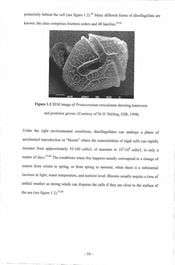
posteriorly behind the cell (see figure l.Z).^o Man), different forms of dinoflagellate are
known; the class comprises fourteen orders and 4g families.3s,al
Figure 1.2 sEM image of Protoceratiumreticulatumshowing ftansverse
and posterior groves. (Courtesy of Dr D. Stirling, ES& l99S)
Under the right environmental conditions, dinoflagellates can undergo a phase of
accelerated reproduction or "bloom'o where the concenftation of algal cetls can rapidly
increase from approximately 10-100 cells/L of seawater to 105-l0s cells/L in only a
matter of days.37'a0 The conditions when this happens usually correspond to a change of
season from winter to spring, or from spring to summer, when there is a substantial
increase in light, water temperature, and nutrient level. Blooms usually require a time of
settled weather as strong winds can disperse the cells if they are close to the surface of
the sea (see figure 1.3).",00
- 10-

Figure 1.3 A marine algal bloom in Hawkes Bay, New Zealand. (Courtesy of MrN.
Watsorq Hawkes Bay District Health Boardq 2003)
Mass mortalities of aquatic life can follow algal blooms for two reaso$r. Firsl the large
changes in cell number can result in a massive change in the concentration of dissolved
Oz in the seawater. Fish may dig often after a bloom has collapsd when a massive
depletion of dissolved O2 (anoxia) causes asphp<iation.al Second although most species
of dinoflagellate are harmless, some species produce toxins that when s€creted into the
zurrounding environment, orintroduced into the food chaiq can cause the death of othq
organisms. In some cases, the toxins are not dangerous to the consumer, but rathen they
are accumulated in the tissues of the conzuming organism and can intoxicate predators
higher in the food chain.37'3e'N'42 Abxn*ium, Dinophysis, Karenia, and Kulodiniwn,
are examples of well known toxin-producing dinofl4gellate genera.tt'n'4t
1.5 Isolation of Merine Netural Products
ffr" goat of marine natural products chemistry is to isolate novel chemical structureg
preferably with interesting and useful biological activities. By their very nature,
- ll -

biologically active secondary metabolites are often present in concentrations of less than
10 mg/kg of the sample organism, therefore isolating these compounds is expensive,
difficult, and time consuming.e To maximise the chances of achieving this goal, three
questions must be answered.
How does one select an organism with which to work?
How does one purifu (fractionate) the desired secondary metabolite to allow
identification?
3. How does one track the isolation of the interesting secondary metabolite from an
extract of the selected organism?
Selection is based upon a screening test performed on an extract of the organism, in
order to determine if there are any interesting secondary metabolites present. If there
are, the extract is subjected to a series of purification steps (chromatography) to separate
the compound from all the other chemicals present in the crude extract. Testing is
carried out at each stage of the isolation process, in order to confirm the presence of the
target metabolite. Once the compound is pure, it is then identified.
These steps are intimately linked. The screening method used to identiff which extracts
contain novel secondary metabolites is often the method used to test fractions generated
tJrroughout an isolation. A screen may require initial fractionation to semi-purify an
extract before testing. After each step in an isolation procedure, fractions generated will
be tested to check that the compound of interest is still present. Screening, testing of
fractions to guide isolations, and methods for fractionation, will each be discussed
below.
l.
2.
t2-
Identification of Interesting Organisms
Extracts of many marine organisms will contain novel biologically active secondary
metabolites, whilst many others will be devoid of any interesting compounds, or they
may only contain previously reported metabolites. In order to decide which organisms
contain novel secondary metabolites, a preliminary screen needs to be performed on an
extract of the organism to assess the presence of any novel metabolites. If possible, it is
also desirable to identi$/ if the interesting metabolites present have been previously
reported (structure dereplication).
There are several strategies available to maximise the likelihood of selecting an
organism that contains novel secondary metabolites. First, a screen targeting a unique
biological activity may be used. This may not be advantageous, however, as known
compounds can be active in many different bioassay systems. Second, unusual
organisms that have not been previously examined could be targeted, as they may
possess a unique biochemical makeup. Finally, chemical testing using various
chromatographic separations prior to screening, or use of different spectroscopic
screens, can select for certain functional groups that often are found in novel structures.
This method can also maximise the chance of identi$ing known compounds at an early
stage.
Historically, screening of organisms for interesting metabolites revolved primarily
around observed thin layer chromatography (TLC) signatures using a variety of running
solvents and visualisation aides (R7 values, W absorbances and fluorescenceso
characteristic chars, etc).46 More recently, high-throughput screening using a variety of
biological assays has become prevalent. Many different in vitro bioassays focussing on
different molecular targets, including anti-cancer, anti-fungal, anti-bacterial and anti-
1.6
-13-

t.7
inflammatory agents, are contmonly used to test crude extracts of marine organisms. An
organism is generally selected for further study if a crude extract from it has reached or
exceeded a certain level of biological activity (usually measured as the concentration
required to achieve a certain percentage kill or inhibition of growth e.g. LD56 or
ICro;.l' 16'1732'+e
Guidance of Crude Extract Fractionation
To utilise bioassay-guided fractionation, an extract of the organism selected by the
screening process is fractionated using standard separation technologies. After each step
in the isolation, all the fractions generated are biologically tested to monitor the activity
through the isolation procedure. A compound isolated in this manner will alwavs be
biologically active.
Bioassay guided fractionation does suffer from several disadvantages. Bioassay
processing time can be significant, meaning that the isolation process is often very slow.
Testing at each stage can be very expensive, especially when many fractions requiring
testing may be generated at any stage of the isolation procedure. Valuable mass of an
active compound is sacrificed at each stage for testing, reducing the amount of material
available for the identification of the purified metabolite.6 Identification of known
compounds is impossible with a purely bioassay guided fractionation. Bioassays
themselves are of necessity focussed very nilrowly and often go through ,.boom and
bust" cycles of popularity. This means that compounds isolated using a particular assay
may be of less therapeutic importance if the assay drops from popularity or usefirlness.
Whole body in vivo assays are inherently more useful from a therapeutic perspective
rhan in vitro assays, as they test biological activity in a whole organism rather than in
one cellular target. This can be misleading as the activity as a whole could be due to a
-t4-

mixture of biologically active metabolites affecting multiple linked cellular rargets,
rather than one single compound. In vivo assays are generally slow, more expensive and
difficult to perform, and require more material than in vitro cell-, enzyme-, or
receptor-based assays. Often, when an extract triggers an in vfvo based screen, a
surrogate in vitro assay is used to guide the isolation. This may guide the researcher to a
compound that is not active in the original in vivo whole organism assay. The
investigator is totally dependant on one assay to monitor and guide them through the
isolation procedure. If there are any inconsistencies or problems with the assay, the
efficiency of the isolation may be compromised.4s'4e Finally, most bioassays are
qualitative and therefore recovery of the active compounds can be difficult to quantiry
at each step. Also, those assays that can be quantified often exhibit large levels of
uncertainty in the final result (up to + 50Yo), making estimates of recovery difficult to
determine.
Two contrasting approaches for the guidance of an isolation were used in this study.
The first was an NMR based screen, followed by the NMR guided isolation of
metabolites from marine sponge extracts, the results of which are detailed in chapters
two, three, ffid four. The bioassay-guided fractionation of an algal toxin was also
performed and is described in chapter hve.
Methods used in the Isolation of secondary Metabolites
Regardless ofthe approach taken to guide an isolation procedure, a series ofsuccessive
separations must be carried out on a crude extract of the organism, in order to purifu the
interesting secondary metabolites present. There are many methods available for the
separation of a target secondary metabolite from the unwanted material in the crude
extract of a marine organism. It is estimated that an extract of I kg of sponge (wet
1.8
- t5 -

weight) can contain more than 40 g of polar salts, proteins and sugars, over 5 g of
non-polar fats and steroids, and often less than 10 mg of the target secondary
metabolites.so Any attempt to modifu the polarity of a crude extract will result in the
precipitation of either the polar or the non-polar components, depending upon how the
polarity is modified.so
The most challenging part of the fractionation of secondary metabolites will often be at
the very start of an isolation procedure, due in large part to the vast array of compounds
of differing polarities found in the crude extract. Methods developed to fractionate
crude extracts include liquid/liquid partitioning, column chromatography using different
stationary phases, and vacuum liquid chromatography (VLC).
Liquid/liquid partitioning is a commonly used procedure where the crude extract is
shaken together with an immiscible solvent; ideally the target metabolite concentrates
preferentially in one of the two layers dependant upon its solubility in each. The layer
containing the interesting metabolite can be partitioned several times using different
solvent systems. Muny systems of immiscible solvents have been developed to separare
a wide range of target molecules.sl-s3 The advantages of liquid/liquid panitioning are
that only cornmon glassware is required, the procedure can be carried out on a large
laboratory scale (0-2 kg wet weight sponge), and only common solvents are normally
needed' Liquid/liquid partitioning is not perfect and there are several problems
associated with its use. First, large volumes of potentially environmentally damaging
halogenated solvents are often used. Second, stable emulsions of the immiscible
solvents are often formed. Third, the target metabolites may be spread over more than
one solvent fraction, reducing the resolution of the separation.sa
t6-
Flash chromatography (pressure assisted chromatography) using silica gel (hydrated
silic4 a normal-phase chromatographic stationary phase) is often used as the first step
of an isolation procedure. Silica gel is best utilised to separate non-polar compounds
and is therefore of limited use when trying to fractionate mid-potarity biologically
active metabolites from crude extracts containing compounds of widely varying
polarity.ss Furthermore, polar molecules can irreversibly bind to the substrate,
potentially reducing recovery of materials that may very well be present in only small
amounts.
VLC was designed to help fractionate crude extracts and is a variation of flash
chromatography using silica gel. The crude extract is loaded onto a small column in a
volatile non-polar solvent, which is then removed by an applied vacuum. The column is
then sequentially batch-eluted under vacuum with solvents of increasing polarity. This
method is designed for normal-phase stationary packing materials only and is therefore
only suitable for fractionating non-polar compounds present in small amounts.
Chromatographic resolution of the technique is also limited due to the size and shape of
the column used.s6
Blunt and Munro developed the usage of octadecyl derivatised silica gel (ODS or C1s)
for the reversed-phase fractionation of crude extracts. The crude extract is added to a
small amount of C1s, ard the solvent removed under vacuum. This loaded stationary
phase is then transferred, either dry or as a slurry, onto a larger column, which is then
eluted with solvents of decreasing polarity.54 The method is a vast improvement upon
traditional techniques as it is faster than multiple liquid/liquid partitioning steps, shows
better chromatographic resolution, uses inexpensive glassware and, by using reversed-
phase substrates, polar molecules will not generally bind to the packing material whilst
allowing better separation of biologically active mid-polarity secondary metabolites.sa
-t7-

The method does suffer, however, from the fact that large amounts of extract can be
difficult to concentrate onto the solid support. Also, the process of eluting the column
can lead to stripping of the bonded-phase from the stationary support, reducing
reusability of the substrate. As well, basic compounds do not separate well on Cra.s7 To
prepare the Crs stationary phase, acidic silanol groups on the surface of the silica gel are
reacted with n-octadecyltrichlorosilane, and then with trimethylchlorosilane.ss If this
reaction does not go totally to completion, any uncapped sites will retain their acidic
character and can therefore ionically associate with organic cations (e.g. protonated
amines), leading to an almost irreversible binding of the metabolite to the substrate.
Very strong non-polar solvents are needed to elute these compounds from the C1s
stationary phase.57
Cyclic Loading
West and Northcote developed cyclic loading in 1996 to help in the fractionation of
crude extracts' Cyclic loading utilises poly(styrene-divinylbenzene) (PSDVB) resin, a
reversed-phased stationary phase that deviates from the use of silica gel, Crs,
liquid/liquid partitionso etc, in the initial fractionation of crude extracts.so It was
originally developed for processing extracts of the sponge Mycole hentscheli in an
ongoing screening programme. M hentscheli produces the biologically active
compounds mycalamide A (4), pateamine (s) and peloruside (6).rn*, All three
metabolites vary in both polarity and functionality, and therefore are difficult to
concentrate into one sample for analysis. Pateamine (5), in particular, is very sensitive
to changes in pH.5o'62
1.9
l8 -

QH9Mea
(4) (s)
PSDVB is a rigid, macroporous, cross-linked polymer support that is inexpensive and
reusable. It lacks any polar sites and so does not sufler from irreversible binding of
polar solutes. It has been found that PSDVB is very useful for the separation of basic
compounds and quaternary arnmonium salts.63 PSDVB is chemically inert in most
organic solvents, can be used at a wide range ofpH values (pH l-13), and can withstand
solutions of high ionic strength. These characteristics allow for a wide range of potential
applications.
Cyclic loading addresses the difficulty of loading crude extracts on reversed-phase
supports. The method involves the sequential dilution of a crude extract with HzO in
order to increase its polarity, thereby forcing less polar molecules to absorb to the
stationary phase (although conceptually this can be extended to diluting polar extracts
with increasing amounts of non-polar solvents to cyclic load onto a normal-phase
substrate)' Initially, the crude extract of an organism is passed through a column of
PSDVB, after which it is diluted with HzO. Under normal circumstances, addition of
(6)
-19-

HzO to a crude extract will result in the precipitation of any non-polar fats, triglycerides,
and steroids present. Addition of HzO will not cause precipitation in this case, as the
non-polar metabolites will have already absorbed to the stationary phase and will no
longer be present in the diluted extract.so The diluted extract is then passed through the
same column again, with the mid-polarity compounds absorbing to the packing
material. The eluent is then be diluted further and is passed back tbrough the same
column again. The process of dilution and passing through the column is repeated until
all contpounds of interest are absorbed onto the column stationary phase. In mosr cases,
the target mid-polarity secondary metabolites will have been absorbed once the extract
has been diluted four fold and been passed through the column (see figure 1.4).to
The major drawback of cyclic loading is that the volume of solvent passed through the
column increases at each step. For example, if a sponge sample is extracted twice with
I L MeOH and is cyclic loaded in two dilution steps to 25% MeOH/FI2O, then a total of
14 L would have been passed rhrough the column [(2 x I L) + (l x 4 L) + (l x s L)].
This is time consuming, and often requires the use of large glassware.
-20 -

+
A
,'-Ga/ Loading \
[[&$ &s2. Eluentdlluted 3. t)lluled aluentl. Extaat pass€d
through P9DVB.
KEY. = Non-polar meGabollie
o = lllld polarlty metrboltba = Polar metabdlb
u,l$ llrO.
Figure 1.4 Diagrammatic representation ofthe cyclic loading process.
The disadvantage of increasing volume is far out-weighed by the advantages of the
method. No concentration of the crude extract is required prior to loading. No
liquid/liquid partition is performed. The solvents used (Meoft Me2co, Irzo) are
environmentally safe and inexpensive to supply in bulk. Finally, the method is easily
scalable from analytical through to industrial scale.so'e
passed backftrough samePSIR B colurn.
'1. Sbpe 2 and 3
tepeabd un0ll alldeslrcd conpoundrtrD|l0almd.
-2t-

Once the column has been loaded, it is eluted using a stepped or gradient system of
increasing organic solvent (MeOH or Me2CO) in H2O.s0 The majority of the unwanted
mass of a crude extract is made up of polar salts, sugars and proteins, or non-polar fats
and steroids, while most of the target biologically active secondary metabolites will be
of mid-polarity, and so witl elute between the polar and non-polar compounds from a
reversed-phase support. This exploits the o'mass windo#', the region of lowest eluted
mass yet with the highest proportion of biologically active secondary metabolites
Conceptually, cyclic loading is the opposite of chromatography as the technique's aim
is to change the polarity of the mobile phase to force target compounds to sequentially
absorb to the stationary support. Fine separation is generally not achieved as the crude
extract is loaded over the entire length of the column in a manner similar to VLC,
reducing chromatographic resolution during elution. The aim of the technique, however,
is not to achieve great chromatographic separation, but is to allow easy fractionation of
a crude extract of a marine organism into a few discrete samples at the first stage of an
isolation scheme, separate from the majority of unwanted compounds and therefore
separate from the majority of the mass. The fractions containing the target metabolites
can then be further purified using standard separation techniques.
Variations of the cyclic loading method have also been developed. Backloading is often
used during an isolation procedure. Fractions eluted from reversed-phase
chrornatographic supports are usually mixtures of HzO and an organic solvent, which
are difficult to concentrate as they often "bump" when placed under vacuum. To
backload, the eluted fraction is cyclic Ioaded onto a smaller PSDVB column which can
then be eluted with a small volume of pure organic solvent. This has the effect of
concentrating the sample into a smaller volume of solvent that is easier to evaporate
without bumping than the original H2O/organic solvent mix.50 Another corlmon
-22-

tecbnigue is to cyclic load a fraction o-nto a small amount of pSDVB which is then be
tnr,rasferred onto a larger ooluimn as a stuqr,y. The loaded PSDVB can then be
chromafiogmaphically separated in a silnillar way to Blunt anfl Munro,rs C* mEthod.s0#
These methods harrc become standard procedures used in Victoria University's Marine
Naflual hoducts. Leboratory for the separation of crude extraets in both the soreening of
organisms, as well as in the fraolionatisn of largor sarrple extacts.
-23 -

Chapter Two
Sponse Screening and Results
2.1 Initial Sponge Screening Methodologr
Rather than relying upon external bioassays to identifu sponges containing interesting
secondary metabolites, West and Northcote instigated an in-house lH NMR protocol for
the screening of semi-purified extracts. Of primary importance to the screening protocol
was structural novelty, with biological activity a secondary consideration. It was
assumed that novel structures often have novel activities that can be determined after
identification, rather than using biological activity to track the isolation of a secondary
metabolite. The protocol was required to be both cheaper and quicker than what was
available through use of out-sourced bioassays.so It has been noted previously that
in-house methods are preferable to external assays for speed, economy, and accuracy.as
West and Northcote's method used cyclic Ioading to help semi-purifu a crude sponge
extract until a sufficient amount of the sample could be dissolved in a single solvent for
analysis by tH NMR. Approximately 100 g of wet weight sponge was extracted with
MeOH. The extract was cyclic loaded onto PSDVB that was washed with H2O to
remove any polar salts and carbohydrates. The column was then eluted with 30%
MezCO/FIzO, 75yo MezCO/FIzO, and finally Me2co. The 75% Mezco fraction was
then backloaded onto a smaller PSDVB column and then eluted with MezCO. This
sample was evaporated to dryness and analysed by rH NMR in cDCl3.s0
It was assumed that the secondary metabolites most likely to be biologically active are
of intermediate polarity and would generally elute from the PSDVB column in the
75%oMe2co/H2o fraction. If analysis of the tH NMR screen of a sponge indicated the-24 -

presence of novel secondary metabolites within the extract, fractionation of a bulk
sample extract of the sponge was initiated using NMR guided fractionation. Once a
metabolite was isolated and its structure elucidated, it was submitted for biological
evaluation in any bioassays available. The range of bioassays available to test pure
compounds is far greater in number than those suitable for screening crude extracts, and
the cost is generally lower due to the smaller number of samples submitted. Using this
strategy, West isolated eight novel metabolites, seven clerodane diterpenes, one of
which exhibited marked anti-inflammatory activity, and a biologically inactive sulfamic
acid indolo 13,2- a]cNbazol e. 5 o
Although West's protocol was successful for the screening and tH NMR guided
fractionation of crude sponge extracts, it suffered from a limited spectral window. Most
of the semi-purified extracts screened still contained large amounts of fats, steroids, and
other primary metabolites, the signals of which obscured most other resonances from
0.5 to 5.5 ppm in the rH NMR spectrum. As a consequence, all the metabolites that
West detected contained olefins, furans, or aromatic rings, and so were readily observed
in the deshielded (> 5.5 ppm) portion of the rH NMR spectrum.so Many biologically
active compounds will not possess these structural features, therefore they would not
have any significant tH NMR signature in the spectral window available in West,s
protocol. The current study expanded upon West's method by utilising 2D NMR
experiments (COSY and HSQC) to better identifu interesting secondary metabolites in
semi-purified sponge extracts.
2.2 Revised Sponge-screening protocol
A standard protocol was established in order to screen extracts of target sponges. The
pre-screen semi-purification remained the same as that used by West.so After
-25 -

semi-purification, the screen sample was analysed using three different NMR
experiments; rH, COSY and HSQC. Initially, all screens were performed using both
CDCI: and CD3OD as NMR solvents. Early results indicated that more resonances
attributable to interesting secondary metabolites were observed in samples dissolved in
cD3oD therefore the use of cDCl: was abandoned in order to save time.
Once a suitable number of screens had been generated, all the results obtained were
collated, and mask sheets showing the positions of correlations from common primary
metabolite resonances were generated for both the COSY and HSeC spectra. For
analysis, a COSY or HSQC spectrum generated from a sponge screen was overlaid on
the relevant mask sheet, and the position of any novel signals identified. The
tH resonances of a secondary metabolite that had the same chemical shift as those of a
common primary metabolite would generally show novel correlations in the COSY
and/or HSQC spectra. These correlations would allow for a unique spectral signature to
be identified for the interesting metabolic product, irrespective of the primary
metabolites present. Once all interesting correlations had been identified in the
2D NMR experiments, the results from the tH, cosy and HSec NMR spectra were
considered. The screen was then assigned a rating based upon the novelty (position) of
any unusual resonances and correlations, the number of novel signals observed, the
amount of material present as estimated by the strength of the novel resonances, and
also by the availability of the raw sponge material both in terms of frozen sponge
available and ease of recollection.
If a sponge extract was deemed worthy of further investigation, the 30yo MezCO/HzO
and MezCO fractions from the screen were also analysed by NMR to identify the
presence of interesting compounds of a more or less polar nature than those highlighted
by the screen. This would also determine if the metabolites already identified had eluted
-26 -

into either of these two fractions, as knowledge of this could help optimise an isolation
of the targeted compounds.
2.3 Sponge-Screening Results
During this study, 51 sponges were screened using the refined NMR based screening
protocol described above (see table 2.1). All of the sponges were screened in
conjunction with the National Institute of Water and Atmospheric research (NIWA). All
organisms, including sponges, collected by NIWA as part of their marine natural
products chemical bio-prospecting programme, are given a Marine Natural products
(vrNP) number as a unique identifier. The sponges screened were collected from around
the North Island of New Zealand, mainly from the Three Kings Islands and Northland,
and also from around D'Urville Island on the northern tip of the South Island (see
figure l.l). Of the 5l sponges screened,43 were not investigated further for a number
of reasons' Many (29) did not appear to contain any compounds of particular interest, or
any novel compounds present were in too low a concentration to allow for efficient
isolation. Fourteen sponges were of moderate interest but were superseded by those of a
higher rating. Eight sponges were selected for large-scale extraction. Unfortunately, the
spectra generated from a large extract of one bore no resemblance to those of the
original screen and so this sample was not further investigated. The remaining seven
sponges are discussed below.
-27 -

69
90
91
95
116
134
161
183
190
201
216219223227
232238
241
246296
304
352
353?ta
375
440
547
548
552704707
729744827
9751013
1015
1 016101 I1019
102'l'to22
1024
1025
1027
1034
1038
Axinella
Chelonaplysilla
,ssosc/era
violacea
Orange, encrusting, ftiablelophon fan, haplosclerid
Haplosclerida pale pink petrosia
Matted black finger brown convul.
Choristid (CrettaJike)
Cream sponge lobate fat finger
Yellow crusting sponge (Vermetid)
Flat tophan
Red/orange fan spongeOrange ball sponge
Mopsella (brown)
Yellow encrusting Adouside-likePink fleshy globular sponge
Orange linger sponge
Tan Dysidea Encrusting Mucus
orange flat sponge
Flat yellow fibrous sponge
Three Kings Dredge
Princes
Princes
Great Banier lsland
NZ North Cape
NZ Dredge
NZ Dredge
NZ Dredge
NZ Dredge
NZ Dredge
NZ Dredge
Great Barrier lsland
Thee Kings DredgeThree Kings Dredge
Stephen's lsland, O'Urvilte
127
1U143
103
230130
150
153
115
147
145'180
283
138
132133
'132
139
110
206
103
124
162143
104
40
104
68
92
93
93
10298
110
58
33
109
92
148
't07
69
84
31
46
29
33
28
42?n
110
54
76
6
0
6
0
0
I0
I0
7
0
I6
7
0
6
0
8
7o
6
4
0
2
5
0
9
7
7
E
0
0
0
7
90
E
0
0
IE
0
8
6
2
Table 2.1 summary of the sponges screened during this study.
28-

MNPOl96
An unidentified cream coloured lobate sponge that was collected by dredging near the
Three Kings Islands. The initial screen indicated the presence of an aromatic compound
from several deshielded HSQC correlations (6c 110-130,6H 6.90-7.80). None of the
aromatic resonances showed fuither COSY correlations to other parts of the spectrum.
Several other weak correlations in the aliphatic region of the HSQC spectrum (6c 30-
40,6s 1.00-2.10) were also identified (see figure 2.1). An extract of a large sample of
the sponge (426 g) was fractionated but unforfunately, the aromatic compound degraded
before it could be purified.
MNPOz4I
An unidentified orange fan sponge collected by dredging from the northern tip of the
North Island. Analysis of the screen indicated the presence of an olefin from two
characteristic deshielded HSQC correlations (6c I l6 and 124, 6H 5.30 and 5.60). Several
oxygenated methines were also evident from other correlations in the HSQC spectrum
(6s 58-82, 6H 3.5(H.20). These resonances showed significant couplings to resonances
in other portions of the COSY spectrum (see figure 2.2). An extract of 779 g of the
sponge was made and purification of the metabolite present attempted, however, the
interesting compound degraded over time before it was pure enough to be characterised.
A lack of more raw sponge material prevented further work to isolate the novel
component observed in the original screen.
-29 -

r:-,."- .ts.,c ,.,!{.!fr J- . L1".,: m_l
,@rlI
"l*---.----:*.---il1,..q.....r'..t'....J'r?....'
-. -.#L--t-$ 'u/# -' -
Ih'3i-cl
t',i, ,
't
,fir--€,''-*='-':-=rr-:*:- ffi.-ff]::-**--.-*JC,;*--q3 :l
#.1 .: ':-.-t..-.o*'l- --,,
"t a
i -'---t 'zt
"tt;l',- ie', . ,s.
0'," O 'i',1,*;,,
bl
I
I
I
It
iItt
;
;
o
1gedo@
. r -t.12.O U0 tor o[ ac .7.0 e 0 s0
fr [Op.l4l 3b zt l!
Flgurc 2,1 COSY ald IISQC NMR screEq of MNPOtg6
(Norrel sorrolstions circled ln red),
- 30-

.9",:,,.o
oo
. ,.1E765
rr (ppD)
F?It pell
r-
I
2a
J
.
3-1
II
I1
a4
J
I
5JilJ
l
6'
I
j,:
'
F'
;
I60
t0roo
o rlr{&,...!,..i0.
'.1
'..
I
b
150 r40 13! t20 lr0 100 90 80 70 60 50 40 30 z0Fr ( ppn,
Figure 2.2 COSY and HSQC NMR screen of MNp0Z4l.
- 3l -

MNPO352
A sample of the sponge Biemna sp. collected fiorn the northern part of the North lsland.
The sponge was described as massive with purple exterior and tan interior colouration.
Analysis of the screen HSQC spectrum revealed interesting correlations from several
oxygenated methines, possibly those of a carbohydrate (6c, 60-78, 6H 3.00-4.00 and
acetalihemi-acetal 6c 100, 6H 4.85) as well as several ring junction methines
(6c35-40,611 1.00-1.50). This was supported by COSY correlations consistent with a
carbohydrate moiety. Several of the carbohydrate resonances also coupled strongly to
resonances in the aliphatic region (611 1.50-2.10) of the COSY spectrum (see
figure 2.3).
Extraction of 979gof MNP0352 yielded the known steroid 5a,8cr-epidioxy-6-ene-24-R-
ethylcholesta-3p-ol (7).ut'uu The presence of a sphingolipid compound (8) was also
suggested on the basis of characteristic deshielded NMR (oxyrnethine and amide)
resonances observed.6T-6e Isolation of this compound was stopped when it was deemed
too difficult to separate the sphingolipid from any other lipid material in the sample.
Compounds of this nature are difficult to follow in an NMR guided isolation as the
methylene tH NMR envelope of the metabolites alkyl chain is indistinguishable from
those of any fatty acids, triglycerides, or other long chain alkane derivatives present.
R1:carbohydrate
R2:alkyl chain
R3:alkyl chain
(7) (8)
-32-

i|n .?-r .*..4- rr J €-d,?{.|>,. .i1*rr€.4.{.^&{& *
Fr (ppn)
r40 130 tz0 ll0 100 90 80 70 50 50 rl0 30 a0
Ft (epn)
Figure 2.3 COSY and HSQC NMR screen ofMNP0352.
tD-to
-o
.-l'iI
I.l.T_'T .--_--1.r'=T-i...'----l
76543?l
o
%o
-33-
MNPO355
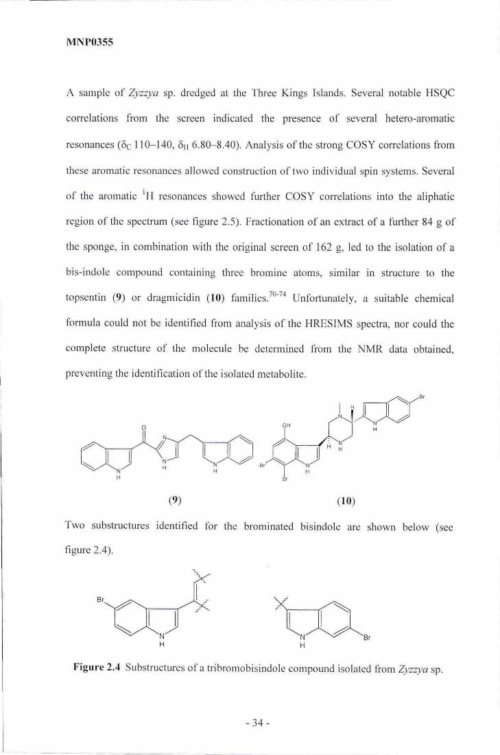
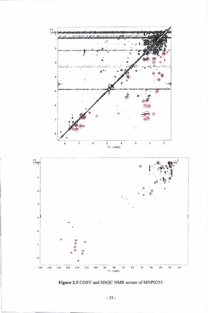
A sample of Zyzzya sp. dredged at the Three Kings Islands. Several notable HSQC
correlations from the screen indicated the presence of several hetero-aromatic
resonances (6c 110-140, 6r"1 6.80-8.40). Analysis of the strong COSY correlations from
these aromatic resonances allowed construction of two individual spin systems. Several
of the aromatic lH ,esonances showed further COSY correlations into the aliphatic
region of the spectrum (see figure 2.5). Fractionation of an extract of a further 84 g of
the sponge, in combination with the original screen of 162 g. led to the isolation of a
bis-indole compound containing three brornine atoms, similar in structure to the
topsentin (9) or dragrnicidin (10) families.T0'74 Unfortunately, a suitable chemical
formula could not be identified from analysis of the HRESIMS spectra, nor could the
complete structure of the molecule be determined from the NMR data obtained,
preventing the identification of the isolated metabolite,
Two substructures
figure 2.4).
r--f\a""\--\2
(e) (10)
identified for the brominated bisindole are shown below (see
Figure 2.4 Strbstructures of a tribromobisindole compound isolated from Zyzzycr sp.
-34-
Fr ( pp0)
og,i!ooOrnoo'
o
Fzl(PT4
1
l.i
I
l'lll
"]l
'Jil
u.l
l
l7.i
l
l61
I'i''
f ':'o,"fi'$i'
'.:ih l'
'o I
@
__l
oa
D
-.'.r...------.1-_....'..-..'.-|tto 140 130 120 rto tto 90 60 lg
Fr (pFr)60 50 40 3! 2n l0
Figurc 2.5 COSY and HSQC NMR screen of MNP0355.
-35-

Bioassays carried out by NIWA on a crude extract of MNP0355 indicated strong
anti-viral RNA activity, prompting further biological testing. A repeat assay on this
crude extract. along with testing of semi-purified fractions of the isolated bisindole, and
on further crude extracts of small samples of MNP0355, all returned negative results.
MNPOTOT
A sarnple of Axinella sp.. collected by dredging from Spirits Bay. at the northern tip of
the North lsland. The initial screen of the sponge (93 g) was one of the most promising
fbund using the revised screening protocol. The HSQC spectrum indicated the presence
of a large variety of deshielded aromatic (6c 105-135,6H 6.20-7.60) and oxymethine
(5q 70-75. 6rr 3.70-4.10) resonances. Substantial numbers of couplings from these
lH resonances were noted in the COSY spectrLlm (see figure 2.6).
Unfortunately, only a small amount of sponge (59 g) was available for extraction. The
extract of this was combined with the original screen sample for further fractionation.
Even when combined, this was less than optirnal for an isolation procedure (ideally
> 0.5 kg of sponge is extracted). Analysis of correlations in both the HSQC and COSY
spectra of semi-purified fractions indicated the presence of a number of compounds
belonging to the oroidin (ll) family of compounds.Ts The (l0Z)- (12) and (l0E)- (13)
geometrical isomers of hymenialdisine were isolated from the sponge and their
structures conltrmed by cornparison with published HRESIMS and NMR data.76,77 The
NMR signatures of both 12 and 13 show a strong pH dependence, as they can exist in
tautomeric fonns depending on the degree of protonation that the metabolite exhibits.Ti
-36-
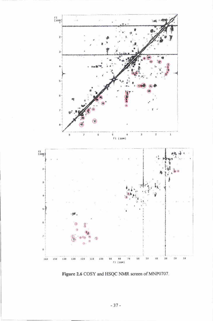
51
lI
I
6i
il
?4IlI
E".j
;3e
160 rtil l,il lla reg !r0 10ll 'El '80 70 t! 80 401
rr ( p-ft
Figure 2.6 COSY and TXSQC NMR scroon sflv&t1P0707
,;I;*:,,n i1!.0'
il:{l
t
II
I
-37 -
?.t r0

(12) (13)
A ftrther rretabolite was also isoiated with severa[ NldR resonanc€s similar to those:
observed for 12 and 13. Also, like 12 and 13, this cornpound suffered from a lack of
COSV and HMBC oorrelations ftorn which to assign its struoture due to ths highly
proton defreient nature of the m.olecule. Cor4pounds 12 and 13 are, espeeially proton
deficient in the conjugated lieterocyc.llc portion o,f the moleoules. As a consequence, the
structure of the third metabolite could r-rot be eluoidated using the spec,ha o-btained.
Several dttempts were made to produee crystals suitable for a single crystal x-ray
'struoture analysis, as the, identities of 12, 13, and other related rnetabolites had been
determined in this way.76'78;7e unfortunately none yielded crlnstals amenable to anarysis
by this rnethod.
-38-

MNPOO9O
A sanrple of the orarlge friable sponge Clathria lissosclera, collected at the Three Kinds
Islands, was screened. Novel resonances observed in the HSQC spectrum included
those of several oxygenated methines (6c 62-80, 611 3.00-4.20) as well as several that
appeared to be fiom the ring junction methines and the ring methylenes of a steroid
(6c45-55. 6r-1 0.90-1.50). Also observed were several weak deshielded conelations
from a series of aromatic resonances (5c 115-135,6H 6.60-8.30) (see figure 2.7). An
extract of 610 g of the sponge was fractionated to yield two novel anti-inflammatory
sterols (32) and (33). This work is detailed further in chapter three.
MNPO979
An extract of a tan encrusting sponge of the genus Chelonaplysilla, collected using
SCUBA at Stephen's Island, Marlborough, gave the most interesting screen
encountered dr"rring this study, with the largest number of novel correlations in both the
HSQC and COSY spectra. Several comelations attributable to acetal or hemi-acetal
resonances were present (6g 100-110, 6tr 4.90-5.20). as were those of multiple
oxymethines (6q 70-75,6H 3.30-4.00). Also of interest were conelations relatively
shielded in the carbon dimension (5c 40-55) yet deshielded in the proton (6H 4.00-4.20).
Strong COSY conelations were found from all of the previously mentioned
'Hresonances to all otherparts of the spectrum (see figure 2.8). An extract of 182 g of
the sponge was fractionated to yield several novel metabolites based upon the
reananged spongian diterpene skeleton (138-143). The isolation and structural
elucidation of these compounds is dealt with in chapter four.
-39 -

'., 'r,'t, €.' 'tG r"'S*. ; .i, l-'$i- ,:."-'l-iei
$r
#'TI
(ppm)
".dJ'g:
..i
*
5
F1
4f'
t'
,l-S;:...-.: ,J ..1
gol
i
I.lI
I
I
I
I
;i
ff!$'';6p+f-*-,ti.4
;3
4O
o
FZ( ro1)l
.tl)
.
it'3..
o
do'oio^or r 'l 'll{ ''lr i '
o- '[D^p6)\vr'
o(O
:'tQ
()
rp
^ r-t
ri)
r50 140 r30 L20 ll0 100 90 80 70
Fr tFpml
I
"_t
1
t,
?l
;
'lrio
Figure 2.7 COSY and HSQC NMR screen of MNP0090.
-40-
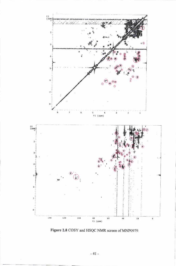
,*" tGi
Fel'*x
lj
21
Il
EJ
l.'l.l1
,l-1rl
l"-J
l,J
11
8-{
{r{0 L?o .r.0! 8r 60
Fr (.tF.l
Figure 2.8 COSY and II$QC- DrI.IvfR screcn ofMNP.0g?,g.
-41 -

2.4 Conclusions of the Revised Sponge-Screening Protocol
'fhe isolation of several metabolites without significant deshielded lH ,esonances
highlights the importance of using NMR experiments that will reveal signals that would
otherwise be masked by the resonances of common primary metabolites. This has been
achieved by using 2D COSY and HSQC spectra, and by developing masks of primary
metabolite signals, which are used to readily identifu the position of any other novel
correlations present. The use of CD:OD, rather than CDCI3, has led to the isolation of
several metabolites of a more polar nature than those West isolated. Whether the
majority of metabolites found using this revised screening method are more likely to be
structurally novel and/or biologically active than those using West's original protocol
remains to be seen.
Subsequent development of the screening protocol described here has included addition
of an internal standard to the NMR sample to help quantify the approximate amounts of
any novel compounds present, and also a refinement of the criteria used to select and
prioritise sponges for large-scale extraction, Further rvork searching spectral databases,
and potential use of other spectroscopic techniclues including MS, will further enhance
the protocol to be able to identify known compounds, and to identify the most
promising sponge extracts for further investigation.
One important aspect of the refined NMR screening method used during this study is
the relationship between a screen. the fractionation of the target compounds, and the
guidance of an isolation procedure. Guidance of an isolation is obviously carried out
using NMR after each step in isolation. Any resonances highlighted in the screen should
be easily identified in subsequent fiactions generated after various stages of purification.
The purification procedures thernselves represent the application of accepted, robust,
-42-

methods for isolating compounds ftom crude extracts a4d semi-purified fractions. \ttlhat
is not immediately obvious with ilre ourrenl method is whish sponges are worthy of
further investigation. The crucial part of the screening process is to rapidly identifl
sponges containing interesting secondary metabo,liteso in order ts seleet which szuuples
are worthy of further detailed investigation.
-43-
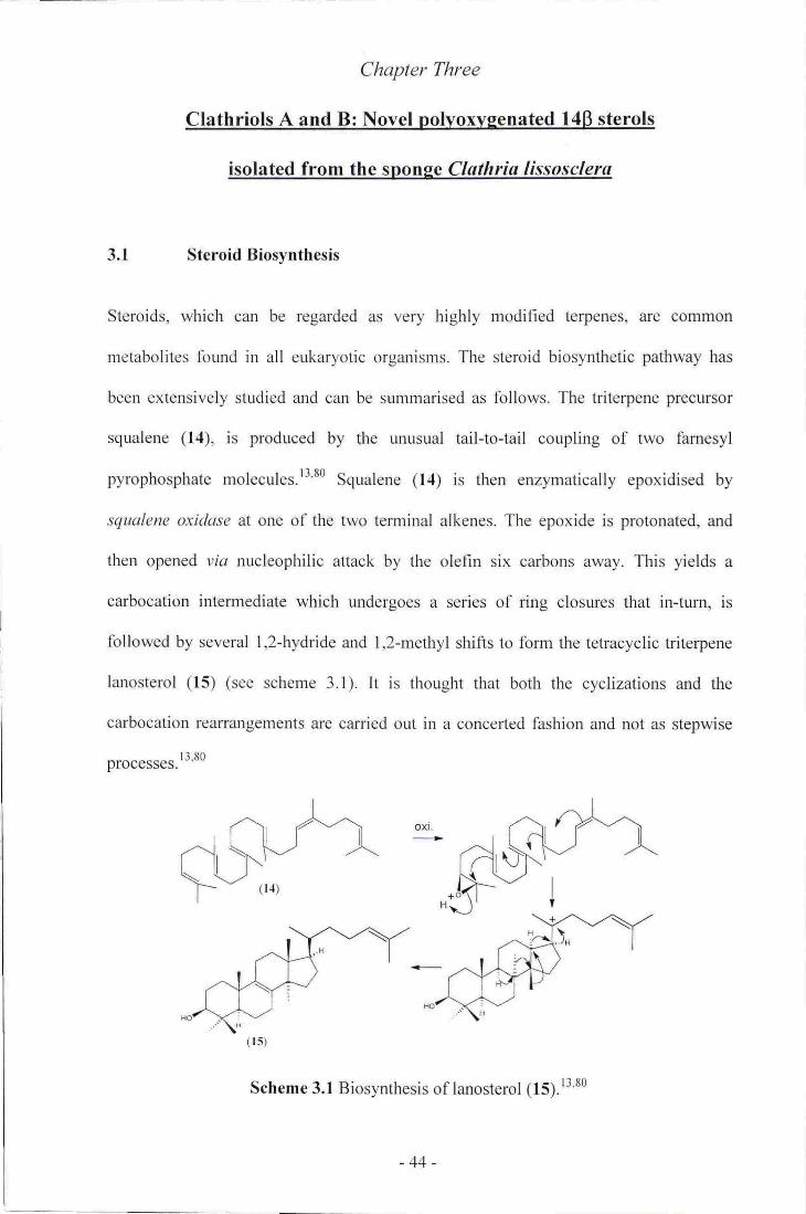
Chapter Three
Clathriols A and B: Novel polvoxvsenated I4B sterols
isolated from the soonge Clathria lissosclera
3.1 Steroid Biosvnthesis
Steroids, which can be regarded as very highly rnodified terpenes, are common
metabolites found in all eukaryotic organisms. The steroid biosynthetic pathway has
been extensively studied and can be summarised as follows. The triterpene precursor
squalene (14), is produced by the unusual tail-to-tail coupling of two famesyl
pyrophosphate molecules.l3'80 Squalene (14) is then enzymatically epoxidised by
.squalene oxidase at one of the two terminal alkenes. The epoxide is protonated, and
then opened via nucleophilic attack by the olefin six carbons away. This yields a
carbocation intermediate which undergoes a series of ring closures that in-turn, is
followed by several 1,2-hydride and 1,2-methyl shifts to form the tetracyclic triterpene
lanosterol (15) (see scheme 3.1). It is thought that both the cyclizations and the
carbocation rearrangen'lents are carried out in a conceded fashion and not as stepwise
proaasses. t''*o
Scheme 3.1 Biosynthesis of lanosterol (15).r3'80
oxr.
-44-

Hydrogenation of the side chain of lanosterol (15) is followed by oxidation of one of
three methyls to a hydroxyrnethyl and then to a formyl group. The formyl group is then
lost as methanoic acid, to form a demethylated product. Similar demethylation occurs
twice more. Isomerization of the A8'e olefin to a As'6 olefin finally forms the archetypical
steroid cholesterol (16) (see scheme 3.2\.t3
Scheme 3.2 Oxidative demethylation of lanosterol (15) to form cholesterol (16).''
Common derivatives of the basic steroid skeleton, exemplified by 16, include oxidation
and alkylation of both the tetracyclic and side-chain portions of the molecule.
Alkylation is believed to occur through stepwise methylation by the biological
methyl-group donor, ,S-adenosylmethionine (17), in a reaction involving nucleophilic
attack of an olefin upon the activated methyl of the sulfonium group of 17, followed by
a 1,2-hydride shifl, before loss of a proton to fonn a new double bond. Methyl and ethyl
l.HcooH
H
-45-

derivatives are commonly found, although examples of up to penlyJ extEllsions are
know^n (see scheme 3.3).tt Blological oxidation is generally earried out by en4rmes of
the eyto-ehrome P45s family, although other e.nzymatic pathwaye cqn also be irsed. t3
,"hfu. ad.nosine
#-->y-4rIt
'{(\,,A=4./+lI /ahl_l\ffi.lt*Al -rl
Schorne 3.3 Alkylation of steroid side chaitls.13'81
Figure }1 Stereochernistry o-,f cholesJerol (16).E0
-46-

3.2 Sponge Sterols
Following the pioneering research of Djerassi in the 1970s, sBonges have proven to be a
rich sor.,lroe of interesting ste,ro-ids. Many rseent examples have exhibited both nov'el
biological activities and structural diversity with unusual side chains and varied
ftrnetionalities.sl'83 Some researcrhe$ believe that spo.nges may provide the widest
steroid divereity inthe whole arimal kingdom..Ps
Aragusterol A (18) was isolated from arr Okinaw.nn $Fonge, Xesta pongla sp.. Sterol 18
exhibited very strong in vttro activity, inhibiting eell protriferatiou with subqranomolar
ffnge aativities against a varieti of nrammalian cancer coll lines. It also exhibited potent
in vivo activity against tumours in mice,8s Aragusterols B (f D ar-d D (20) were later
reported from the same sponge. Bioassays revealed ftat ilg sholrred redueed activity
w-hencompared to l8 whilst 20, surpriis'ingly, showed 4o activity at all.8-6
(1e)
(20)

Two novel cytotoxie sterols were isolated ftom the Tirnisian sponge Dysidea ineruslans.
Incnislastero,ls A (21) and B (22) exhibi'ted nanonrctar range activity against a series of
humao trilrour eell lines.87
(21) Q2)
.dgo:sterol A (23) was isolated fronn a samp-le o,f Spangia sB.. Th,is sterol acetate
possesses activ.ity that reverses Multidn€ Resis ance (lvIDR) in tumow cellso a
desi-ralle e-ffect for improving the effrcacy of eancer chernotherapy agents. Aftet
treatment with 23, KB-C2 and KB-eV60 eancer sell,s showed a eornplete reversal of
drug resistance to colchicine and vincrisfiine respectively; maldng 23 a stong candidate
for furthor clinical investigations.ss'8e
(23)
l4p Sterols
In 1992, Andersen and Burgoyne isolated a uuique sterol ftom the :spong;e Petra.sia
contignala, Cootignasterol Q4) is the ftrst exarnple ,of, a narurally 'occurring sterol where
rings C and D are flrsed in a cis fashion, leading H-14 to be in t},rc "unnatural" p
-48-
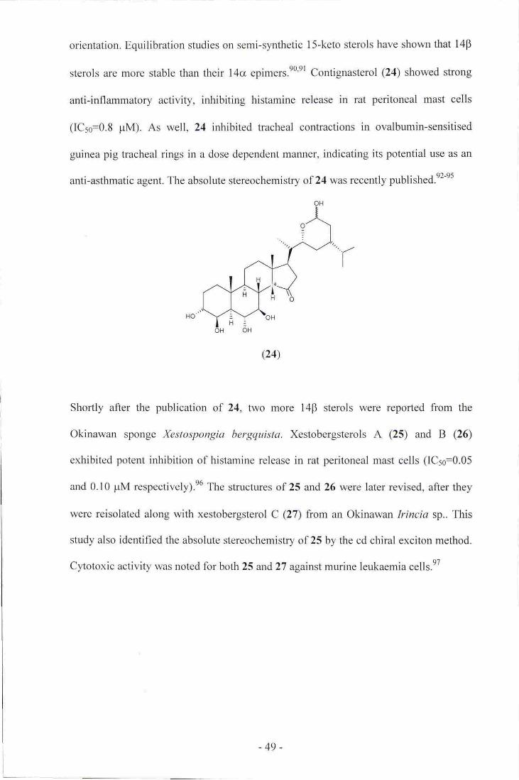
orientation. Equili-bration sfudies on semi-,sy'nthetic l5-keto eterols have shownthat [4p
stErols are nrore stable tharr their 14cr epimers.e0'el eontignasterol @4) show,ed sHong
anti-inflammatory activity, inhibiting histaurine re.leilsCI in rat peritoneal mast cells
([Cso=0.8 pM). As well, 24 inhibited tracheal contractions in ovalbumin-sensitised
guinea pig tracheal rings in a dose dependent mannetr, indicating its potential irse as an
anti-asthmatic agent. The absolute stereochernistry of,?4 was recently published.e:-es
(24)
Shortly after the prblication af 24, fwo more l,4p sterols were ;report€d ftorn the
Okinawan sponge Xestospongia hergg:u,ista. Xestobergsterols A (25) and B (26)
exhibited potelt inhibition of histanrine rele.ase in rat peritoneal mast oe,lls ([C5e:0.05
and 0.10 pM respectively).e6 The structures of 25 and 26 were laterrevissd, afterthey
were reisolated along with xestobergsterol e QT from an Okinawan lrineta sp.. This
study also identified the absolute stereochemishry of 25 by the cd chiral exciton method.
Cytotoxic activity was noted for both 25 and 2? against mruine leukaemia cells.97
-49-

(25) fu=t1, RrOH
(26) R1=R2=!l
(24 Rr:Rz{H
A new 14p sterol, haliclostffronc sulfate (28), was isolated, along with the knovvn
metabolite halistanol sulfate (29), ftona Malaysian Haliclons sp..e8
(2e)
The l4cr (30) and l4B (3D epirners of the same sterol, tamo.sterone sulfate, were
isolatEd from a Microneoian spong€ of a new genus within the baplosclerid family
Oceanapiidae, This. is the first ex-ample of the co-isolatisn of both C-14 epimers sf thE
s&rire sponge sterol.ee
(30) laq
(31) 14p
-50-
(28)
3.4
All six reported naturally occurrirrg l4p steroids have been isolated frorn marrine
sponges although there are many exan:ples of synthetic l4B sterols. All natumlly
occurring 14B sterols have a ketone at C-15, potentially offering an enol-stabilised
earbanion rout€ for the epimerisation of C-14.
Clathria lissosclera
Dwing the course of rotrtine NMR based $p-onge screening; it was noted that an eKffact
of the sponge Ctrathrta ltssaselera (MNP0090), dredged from 100 m at the Three Kings
Islirnds, Northland, New Zealand (see figure 3.2), showed correlations indicative of
several oxygenated nrethines in both the COSY and HSQC NMR specfta (see chapter
two). A subsequent MeOII extraot ,sf 610 g of C ltssosclera, fr-aetionated using
repeated reversed-phase chromatography, yieided the novel sterol clatb,riol A (32)
Q6.2 mg).100 F,rthet analysis of the sid,e fractions generated during the isolation of 3-2
indicated the p.resence of se.veral more related sterols. Fufher purification using both
size-e,xclusion and repeated normal-phase chrornatography- resulted in the isolation of a
second novel sterol, clatbrlol B (1.3 mg) (33) (see sclreme 3.,3).tot
-51 -

t
lq!
I
il
il,
3t
!
t\!
t)3
t\ o'
1
rlI
I
.i('
tq
tl
ll r
n.a [r/
t
'*s
r8
€.iltl
SP.i\'n r-)
.\\b
,, )' )5Y;t i-.,9
I
I
it
rt!d r
I
ai
ai
3
I
e
g
I I
gI
0iI
Figure 3.2 Hydrographic chart of the Three Kings Islands.
(Courtesy of Land and Information New Zealand)
-52-
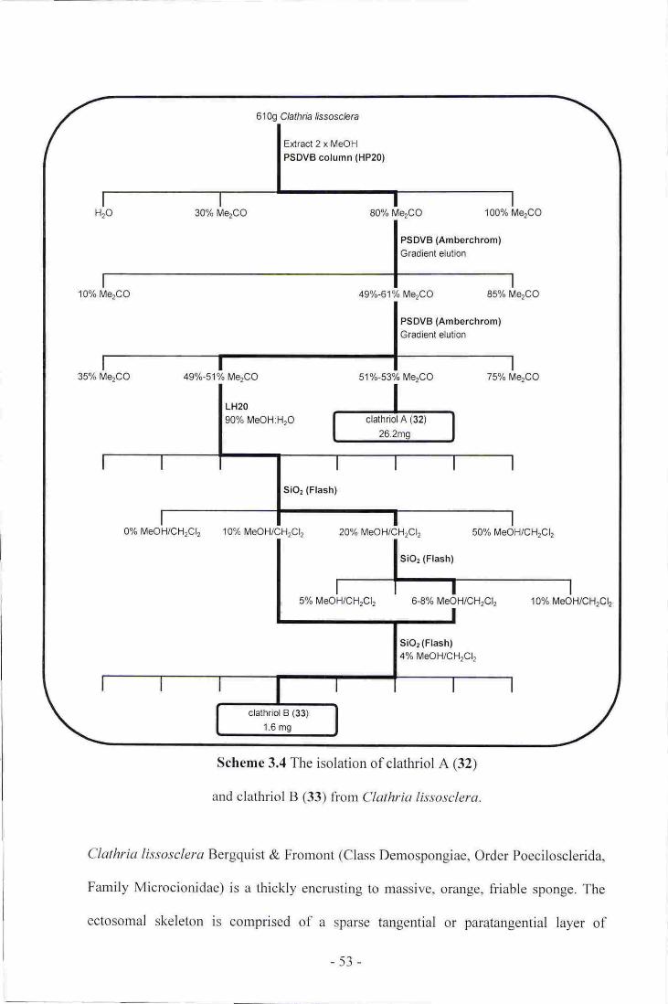
61O9 Clathria /issosc/era
Extract 2 x MeOH
PSDVB column (HP20)
30% Me2CO 80% Me2CO 100% Me2CO
PSDVB (Amberchrom)Gradient elution
10% MerCO 49a/o-61o/o Me2CO 85% MerCO
PSDVB (Amberchrom)Gradient elution
35% Me2CO 49Vo-51% Me.CO 51%-53% MerCO 75oh MerQQ
0% MeOH/CHrCl, 10% MeOH/CHrCl, 20% MeOH/CHrCl, 50% MeOH/CHrCl,
5% MeOH/CHrCl, 6-8% MeOH/CHrCl, 10% MeOH/CHrCl,
Si02 (Flash)4% MeOH/CHrClt
Scheme 3.4 The isolation of clathriol A (32)
and clathriol B (33) from ('lathria Iissosclera.
('lathria li.rsoscleru Bergquist & Fromont (Class Demospongiae. Order Poecilosclerida,
Family Microcionidae) is a thickly encrusting to massive. orange, friable sponge. The
ectosomal skeleton is cornprised of a sparse tangential or paratangential layer of
-53-

subectosomal styles supporting a thin layer of isochelae. Megascleres are slnooth styles
(250 pm long) with microscleres being palmate isochelae (20 pm long) and, less
frequently, toxas (70 pm) (see figure 3.3).t'''ut
Figure 3.3 Physical structure of Clathria lissosclera.
(Courtesy of Professor P. Bergquist)
D: Surface view of Clathria lissosclera surface showing circular oscules.'02
E: lrregular reticulate choanosomal skeleton (x t l3).'02
F: Spicules (x 650).'02
-54-

3.5 Clathriol A
Observation of HRESIMS pseudo-molecular ions in both positive and negative ion
modes enabled the molecular formula of clathriol A (32) (26.2 mg) to be established as
CzsHsoOs V79.3754 [M+H]', L3.7 ppm;477.3582 [M-H]-, A 1.5 ppm), implying five
degrees of unsaturation. Evidence of four exchangeable protons was obtained by
running a negative ion mode HRESIMS of 32, using DzO and CD3OD as the injection
solvent, where a pseudo-molecular ion of 481 .3903 was observed, corresponding to the
exchange of four deuteriums. The four exchangeable protons were assumed to be from
hydroxyl functionalities as a strong OH stretching band (3308 cm-'1 was observed in the
IR spectrum. A C:O stretching band indicative of a saturated ketone was also observed
(1729 cm-l). Further evidence for a saturated ketone was obtained from a '3C NMR
resonance at22l.9 ppm. AII other 28 carbons and 46 hydrogens attached to carbon were
observed in the llc and tH NMR spectra respectively. One-bond carbon-proton
correlations were established by an HSQC experiment. Detailed analyses of the l3C and
DEPT NMR spectra gave evidence of a saturated carbonyl, four oxygenated methines
(6c 75.8, 75.I,71.8,69.6), eight methines (55, 53.4,52,6 49.8,48.5, 47.2,40.4,35.4,
29.4), eight methylenes (6c 39.0,38.2, 38.1,33.5,33.0,31.5,25.8, 22.8), six methyls
(6c 2l .5,20.7,19.8, 19.4" 19.4,13.7) and two quaternary carbons (6c 42.6,36.9). With
no further evidence of double bonds, a tetracyclic structure was required to satisS the
remaining four degrees of unsaturation. It should be noted that severe resonance overlap
in most of the NMR spectra, especially fbr the majority of the methylene 'H resonances
of 32, made the unequivocal assignment of the metabolite's structure a non-trivial
exercise (see figure 3.4).
-55-

H-r8 H-29t23p
f n-stza H-27t22
\\
\
H--26
\
Hp
11-t2o./48 9/lcr
H-20 H-25
lI
l
H-12$123la H-l lpo \H-2p I
\/ \
^Al \,1,41.51.5t.l1.61.9z.l It t.? 0.3 0.t pp.
Figure 3.4 Shielded portion of the 'H NMR spectrum of
clathriol A (32) indicating severe spectral overlap at 300 MHz.
Examination of correlations in both COSY and lD-TOCSY experiments from the four
well resolved oxymethine 'H resonances between 4.5 and 3.0 ppm allowed the
establishment of two separate substructures. A linear fragment (fragment A) was
established frorn the observation of COSY correlations from the lH resonances of a
rnethylene (C-2:6c 31.5,69 1.7I, I.34) to those of an oxymethine
(C-3:6c71.8,6n3.47) and then to another methylene (C-4: 6c 33.0, 6H 2.15, l.16).
Similarly, a linear sequence of five methine carbons was established on the basis of
observed COSY cross peaks from the resonances of their attached protons [methine
(C-5:6q 48.5,6H LIO) to oxymethine (C-6:6c 75.1,6u 3.06), to oxymethine
(C-7: 6c 75.6, 5n 4.30), to a methine (C-8: 6c 40.4, 6H 1.62) and finally to a methine
(C-14: 6c 53.4, 6H 2.84)l to give fragrnent B. Although no direct linkage between
fragments A and B could be determined through COSY correlations, the linkage was
-56-
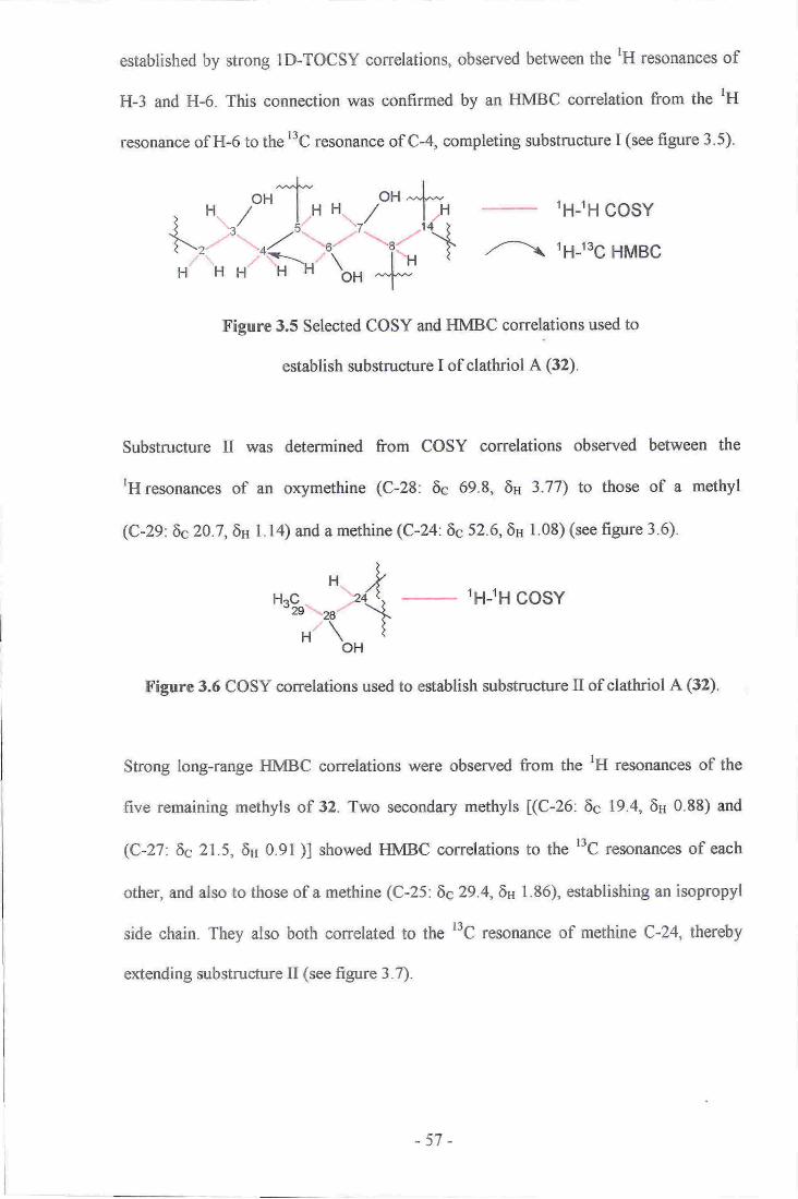
established by strong ID-TOCSY correlations" observed between the tH resonances of
H-3 and H-6. This connection was confirmed by an HMBC correlation from the lH
resonance of H-6 to the r3C resonance of C-4, completing substructure I (see figure 3.5).
L,:j*tlfr
H*Hu%-,.rr,"\
H\o"l
,--\
tH-tH cosY
t6-tsg HMBC
Figure 3.5 Selected COSY and HMBC correlations used to
establish substructure I of clathriol A (32).
Substructure II was determined from COSY correlations observed between the
tH resonances of an oxymethine (C-28: 6c 69.8, 6a 3.77) to those of a methyl
(C-29:6c20.7,6H Ll4) and a methine (C-24:6c 52.6,6H 1.08) (see figure 3.6).
1H-lH cosY
Figure 3.6 COSY correlations used to establish substructure U of clathriol A (32).
Strong long-range HMBC correlations were observed from the tH resonances of the
five remaining methyls of 32. Two secondary methyls [(C-26: 6c 19.4, 6n 0.88) and
(C-27:6c 21.5,6u 0.91 )l showed HMBC correlations to the t'C resonances of each
other, and also to those of a methine (C-25 : 6e 29 .4,6s I . 86), establishing an isopropyl
side chain. They also both correlated to the r3C resonance of methine C-24, thereby
extending substructure II (see figure 3.7).
-57 -

1H-13c HMBC
X'igure 3.7 Se,lected HMBC correlations establ-ishiug the
-3-(4-methylpentan-2-ol) side ohain (subsfructure II) of clathriol A (32).
Detailed analysis of the HI\4BC spectrum revealed strong correlations ftorn the
nH resormnce of a tertiaryangular methyl (C-19.: 5c 13.7, SH 0"S3) ts thE l'lCtesonanc,es
o.f a rnethylene (C-l: Ec 382,6n 1.71,0.93), a methine (e-9: 6c 47.2,6s 0,93), a
qu$ternary oarbon (C-10: 6c 36"9) and mettrine e-5 of substnrcture I. This allowed
methyl CH3-19 to be colueeted to quatemary oenlre C-10 and alss established linkages
frorn C-10 to C-1, C-5 and e-9'of substructure'I (secfigure,3.8).
The rH resonanse o,t the other tertiary angular rnethyt (Gt8: 6c 19.4,6H 1.19) showed
strong HMBC correlations to the l3C re-sonanees of a rnethylene
(C-IZ:6c39.0,8n I.37, l.t7), a quaternar.y cerrtre (C-13: 6q42,.6),, a methine,
(C-tr7:Eg49.8,6n i1.72) and also methiuE C-14 of subs-tnrcture I. Thes.e observationsr
allswed placernent of CHrlS at C-13, estabtished the linkagee between C-13 and C,-12,
e-14, and C-17, and,allowed for epansio.n of substrucfure I to include the two angular
melhyle (see figure 3.8).
-58-

,(rD
-.lJf'---joHl
L-I--l
t\
r6-1sg HMBC
Figure 3.8 Selected HMBC correlations connecting CH3-18
and CH:-19 to substructure I of clathriol A (32).
The rH resonance of a secondary methyl (C-21: 69 19.8, 6H 0.94) showed HMBC
correlations to the ttc resonances of a methylene (C'22:6c 33.5, 6n 1.60, 0.90) and trvo
methines lC-17 and (C-20: 6c 35.4, 6H l.9l)]. A COSY correlation observed between
the rH resonances of H-20 and CH:-21 indicated the attachment of the secondary
methyl on C-20 between C-17 and C-22 (see figure 3.9).
lH-lH cosY
t6-tsg HMBC
Figure 3.9 Selected HMBC correlations connecting
CH3-21 to substructure I of clathriol A (32).
All of the previous observations had established the connectivities of substructure I
(nineteen carbons) and substructure II (six carbons), leaving three methylenes and a
ketone to be assembled into a tetracyclic structure as required by the remaining degrees
of unsaturation. COSY cross-peaks could not be reliably used to identiff correlations
-59-

from the 'H resonances of the remaining methylenes, due to the severe spectral overlap
in the shielded region of the rH NMR spectrum (see figure 3.4). 1D-TOCSY, HMBC,
and HSQC-TOCSY experiments were therefore used to assign the remaining
connections within the molecule.
The presence of a six-membered ring (ring A of a steroid) was identified by selective
excitation of thelH resottance of H-3 in a lD-TOCSY experiment. Correlations to the
'H resonances of both protons attached to methylenes C-2 and C-l were revealed
sequentially as the spin-lock mixing time was increased, thereby establishing the
connection of C-3 to C-2 and C-2 to C-1. This connectivity was confirmed by a series of
HSQC-TOCSY experiments where a rH resonance of C-l (6c 38.2,69 0.93), showed
TOCSY correlations to a 'H resonance of C-2 (611 1.34) and also of C-3 (5H 3.47),
thereby confirming the C-l C-2 bond and reinforcing the establishment of ring A (see
figure 3.10).
The presence of ring B was established from HSQC-TOCSY conelations from the
rHr"sonance of H-7 (6c75.6,611 4.30) to that of H-9 (6H 0.93) via H-8 (6H 1.62), as
observed sequentially through experirnents with increasing spin-lock mixing times. The
connectivity of the final six-rnembered ring (ring C), was determined from further
HSQC-TOCSY correlations between the rH resonance of H-9 (69 47.2,6H 0.93) and
those of two methylenes [(C-ll: 6c 22.8, 611 1.52, 1.25) and C-12], indicating the
placement of C-l I between C-9 and C-12. The placernent of C-l I was confirmed by the
observation of an HMBC correlation tiom the IH resonance of H-8 to the'3C resonance
of C-l I, thereby completing the connectivity of rings A, B and C (see figure 3.10).
-60-

'D)H-/
HSQC-TOCSY
1D-TOCSY
//--\ 16_ts6 HMBC
Figure 3.10 Selected correlations establishing the
connectivity of rings At B and C of clathriol A (32).
Further analysis of HSQC-TOCSY correlations from the rH resonances of a methylene
(C-23:69 25.8, 6H 1.36, l.14) to those of H-20 (6H l.9l) and orymethine H-28 (6H 3.77)
revealed connections from C-23 to both C-22 and C-24. This observation was consistent
with an HMBC correlation observed from the tH resonance of H-28 to the
t'C resonance of C-23, thereby linking substructure tr with rings d B and C (see
flrgure 3.1l).
HSQGTOCSY
t;1trg HMBC
Figure 3.11 Selected correlations connecting rings
A,, B and C with substructure tr of clathriol A (32).
Connections to the remaining methylene (C-16: 6c 38.1,6u 2.36,2.18) and ketone
(C-15: 6c 221.9) were assigned on the basis of observed HMBC correlations. HMBC
correlations from the deshielded tH resonance of H-l6p (6H 2.36) to the t'C resonances
-61 -

of C-13, C-17 and C-20 revealed the attachment of C-16 to C-17. Observation of
FIMBC correlations fronr the 'H resonances of H-8, H-14 and H-l6P to the
'3C resonance of ketone C-15 provided evidence for the last two carbon bonds, C-14
C-15 and C-15 C-16. Further evidence for the placement of C-I5 was provided by the
deshielded chemical shifts of the 'H resonances of H-14 (6H 2.84),H-l6cr (6H 2.18) and
H-l6B (6H 2.36), and also by an HMBC correlation from the resonance of H-l6p to that
of C-14. These correlations established the five-membered ring (ring D) and completed
the steroidal carbon skeleton of clathriol A (32) (see figure 3.12).
/---\ 1H_t3C HMBC
Figure 3.12 Establishment of the final connectivity of clathriol A (32).
The relative stereochemistry of ten of the thirteen stereogenic centres of clathriol A (32)
were determined by a combination of analysis of NOE correlations, observed in both
1D-gNOESY and ROESY experiments. and by measurement of tH-rH coupling
constants, determined from lH NMR, 'H-'H homonuclear decoupling, and double
quantum filtered COSY (DQF-COSY) experiments. Ring A was assigned as being in a
chair conformation, based on the both large and small vicinal coupling constants of the
protons attached to C-I. C-3 and C-4. The hydroxyl attached to C-3 was assigned as p
(equatorial), on the basis of the multiplicity of H-3. Proton H-3 was assigned as a
(axial). as its resonance showed coupling constants of greater than l0 Hz to both H-2a
and H-4cr, while also showing couplings of less than 5 Hz to H-2B and H-48. Strong
-62-
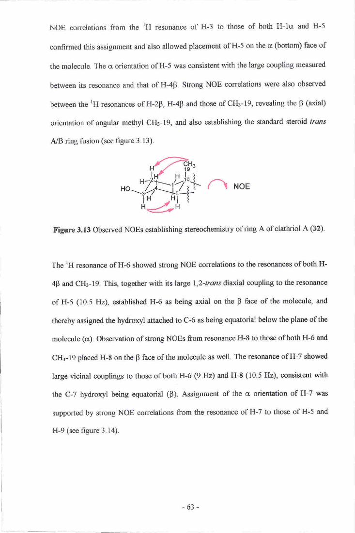
NOE correlations from the lH resonance of H-3 to those of both H-lct and H-5
confirmed this assignment and also allowed placement of H-5 on the cr (bottom) face of
the molecule. The cr orientation of H-5 was consistent with the large coupling measured
between its resonance and that of H-4P. Strong NOE correlations were also observed
between the IH resonances of H-2p, H-4p and those of CHr-19, revealing the p (axial)
orientation of angular methyl CH3-19, and also establishing the standard steroid frams
A/B ring fusion (see figure 3.13).
n NoE
Figure 3.13 Observed NOEs establishing stereochemistry of ring A of clathriol A (32).
The rH resonance of H-6 showed strong NOE correlations to the resonances of both H-
4p and CH3-19. This, together with its large \,2-trmrs diurial coupling to the resonance
of H-5 (10.5 Hz), established H-6 as being axial on the B face of the molecule, and
thereby assigned the hydroxyl attached to C-6 as being equatorial below the plane of the
molecule (a). Observation of strong NOEs from resonance H-8 to those of both H-6 and
CH3-19 placed H-8 on the p face of the molecule as well. The resonance of H-7 showed
large vicinal couplings to those of both H-6 (9 Hz) and H-8 (10.5 FIz), consistent with
the C-7 hydroryl being equatorial (F). Assignment of the a orientation of H-7 was
supported by strong NOE correlations from the resonance of H-7 to those of H-5 and
H-9 (see figure 3.14).
-63-

?T'f,.-\H
'qq-4/-\ NoE
'i'L-t'Figure 3.14 Observed NOEs establishing stereochemistry of ring B of clathriol A (32).
A large coupling observed between the resonances of H-8 and H-9, consistent with a
\,2-trans diaxial relationship, implied a trans B/C ring fusion. Methyl CHs-18 was
assigned on the p side of the molecule on the basis of the strong NOE correlation
observed between its lH resonance and that of H-8. Further NOE correlations between
the rH resonances of methyl CH3-IS and methine C-20 also placed the side-chain of
clathriol A (32) on the F face of the molecule (see figure 3.15).
Almost all steroids possess a trans C/D ring fusion. Detailed analyses of NOE
correlations in both ID-gNOESY and ROESY experiments revealed that the resonance
of H-14 showed strong correlations to the tH resonances of both H-8 and CH3-18 on the
p face of the molecule. This observation was consistent with H-14 being in the rare p
orientation, implying a cis CID ring fusion for the sterol. This was confirmed by the
small vicinal coupling constant observed between the resonances of H-8 and H-14 (3
FIz) and also the weak long-range W coupling (< 2Hz) between the resonance of H-14
and those of H-l2p and H-164, as observed inthe COSY spectrum (see figures 3.15
and 3.16).
-64-

n NoE
Figure 3.15 Observed NOEs establishing stereochemistry
of C-14 and rings C and D of clathriol A (32).
rH-rH cosY
Figure 3.16 W-coupling observed in the COSY
spectrum between H-14 and H-12F of,clathrisl A (32).
Aszuming norrnal steroidal absolute configrrratioq the cyclic portion of the rnolecule
can therefore be assigned as 3,S, 55, 6n, 7R,8R,9,S, 10R, l3R, l4R, 17R.
(32)
-65-

Ta.ble 3.1. r3C 05 MIIZ) and IH (300 MHz) NVIR data (cDoD) of clathriol A (32,)"
PositisnLs,lF
Zs-
eF
3
4a
4p
5
6
7
E
9
l0
Itrc
rrF
l2et
llpl3
14
t5
l6ql6F
17
l8
x9
t0
2t
)iazzq
23s
a924
25
26
27
28
mutt 6
cft:
efl,
COS.Y
zsAF
2s.2F
ro,lFJtcrlFJ
tuaqAd,,4F
3,5,6
3J4s"4:p,6
4g,,S:1\8
6,8
6;l,8,l2rl',Jzg
Itp
. 9,12412p
8'u9
E,lrpJ4l8
I,r2p,r6c
\4.|v
t7
l6€r,l6ppl
t2
4"5,78
16rE 14
I I,15.
7,8
9,Ial3
8,9,lA13"lt tg
13,14,15,1?t0
14t3it4,l7
Il"9.IO
nwn
NOE3
3
lcr/cr"4q5
3
6
3J4p;7,8,18
5f,8,9J4
6,7.1&19
7
7'E'18
l6B
l6q20
6Jtr0
2p"4F,4g,ltp
tl,lBpl?0
3l.t
?lr8r
330
48.5
75,1
1,5.6
40.4
4,'l:2
36.9
?28
39;0
42_6,
53t
ms3&l
49.8
19,.t
13.7
35;4
tQ,8
33.5
?5.8
52.6
29.4
lg,4
1Ii3
69-6
20:7
Ctr{r
0.93 .td (r3,5, 10)
r.7r d (l tlr.?l m
!34 m
3.47 dddd (t3.5, 10,4.5,3)
2.rs br d (l t-s)
l.16 rn
I,rc m
3,06 dd (!0.,5,9)
4.30 dd(10.5, e)
l.6J ddd(t2110' 5,3)
0'93 m
1.52 m
l,zt rn
\.17 El
1.37 m
2.84 dd (3" 2)
CH
gHi2'4
CH
CH
c.H
CH
cflccl,l:
cEH
cGHr
t'l
CH
CI{t
CHj
CH
cllrCHr
28CH
CH
CHr
c[.r:
ctt
r9;
CHz
2.1E m
236 dd(20- t,0)
r:ra rR
l,l9 6
0.E3 s
l.9l m
ore{ d(6.5)
1.60 m
0t90 ln.
1,.36 !n
I"l4 m
1.08 rn
t.86 rn
0,88 d(6,5)
o.9l d (6.5)
?;77 Suio (6)
2425p7
24,2526
B,U#pe
2627
z5
25
Multiplioity deternrined ftsm DF;PT and HgeCoqperirnents
earbon oonn€ctivity deterniinEd ftom an HFQC exporimentt multiplicity determined &om rH,
ID-TOCSY and ho- no2DJ cryr€dnn€nts.
-66-

3.6 Clathriol B
Pseudo-molecular ions were observed for clathriol B (33) (1.3 mg) in both the positive
and negative ion HRESIMS modes, corresponding to the formula CzsFIaoOs (463.3423
[M+H]n, A 1.0 ppm; 461 .3298 [M-H]-, L 4.7 ppm), differing from that of clathriol A
(32) by one carbon and four hydrogens, and implying six degrees of unsaturation. As
with 32, all 28 carbons and 42 protons attached to carbon were observed in the t3C and
'H NMR spectra. One-bond proton-carbon correlations were established in an
HSQC-DEPT experiment. Again, the presence of a saturated ketone was suggested by
an IR C:O stretch (1729 cm-r; and supported by a deshielded r3C NMR resonance
(6c222.3). The presence of a disubstituted carbon-carbon double bond was indicated by
two deshielded IH resonances (6ey 5.63, 5.26) that correlated to two l3C resonances
(6c 134'6, 130.3) in the HSQC-DEPT spectrum. As there was no further evidence of
double bonds, a tetracyclic structure was required for 33 by the remaining degrees of
unsaturation.
Detailed analysis of the COSY and ID-TOCSY spectra allowed construction of two
separate lH spin systems. The two systems were identified and constructed from the
most prominent resonances in the 'H NMR spectrum, those of an oxygenated
methylene, two olefinic methines, and three oxygenated methines. The first spin system
consisted of a linear sequence of carbons from a methylene (c- I : 6s 36.g, 6s 0.g4, 1.69)
and ending with a methine (c-14: 69 51.7, 6s 2.74), as determined by cosy
correlations between the resonances of their attached protons fmethylene C-l to a
methylene (c-2: 6s 30.4, 6H 1.36, L77), oxymethine (c-3: 6c 71.1, 6n 3.54),methylene
(c'4:6s 32.1,611 1.24,2.24), methine (c-5: 5c 47.0,6H l.16), oxymethine
-67 -

(C-6: 6c73.5,6H 3.16), oxymethine (C-7: 6c 74.5, 6n 4.26), methine
(C-8: 6c38.9, 6n 1.58) and finally methine C-l4l (see figure 3.17).
The branch from C-8 to C-12 was difficult to identi$ from COSY correlations due to
the severe spectral overlap of various shielded tH resonances. A series of ID-TOCSY
experiments were therefore carried out to establish this connection. Selective irradiation
of the rH resonance of H-7 (6n4.26), using a variety of spinJock mixing times, revealed
correlations to two methines [H-8 (6n 1.58) and (C-9: 6c 46.0, 6n 0.89)], and two
methylenes [(C-ll: 6c 21.3, 6H 1.20, 1.50) and (C-12: 6c 36.5, 6H 1.19, 1.36)] in
sequence, establishing the C-8 C-9, C-9 C-ll, and C-ll C-12 linkages. A COSY
correlation from the rH resonance of methine H-9 to the deshielded proton H-llp,
which was itself coupled to deshielded proton IiI-LhF, confirmed these connections and
completed spin system I. This spin system was entirely consistent with the structure
established for clathriol A (32) (see figure 3.17).
tH-lH cosy
z^\ 1D-TOCSY
Figure 3.17 Selected COSY and ID-TOCSY correlations
establishing spin system I of clathriol B (33).
The second spin system from a methylene (C-16: 6c 37.9, 6s 2.17,2.40) to an
orymethylene (C-28: 6c64.7,6n 3.45, 3.64) was established from analysis of observed
COSY correlations between adjacent lH resonances [oxymethylene H-28 to a methine
(C-24: 6c 52.3, 6H 1.96), methine (C-25: 6c29.3,6H 1.58) to rwo methyls
-68-

(C-26:6c 19.6, 6H 0.81) and (C-27: 6c 20.8, 6n 0.86); H-24 to olefinic methine
(C-23:6c 130.3, 6n 5.26), olefinic methine (C-22:6c 134.6,6115.63), methine
(C-20:6c34.9,6H2.75) to both a methyl (C-21:6c19.6,6H 0.99) and a methine
(C-17 6c 48.2, 6s 1.76), and finally H-17 to methylene C-161, completing spin
system tr (see figure 3.18).
oH
-
lH-lH coSY
Figurc 3.lE Selectd COSY correlations establishing spin system tr of clathiol B (33).
The final connectivity ofthe A B, and C rings was established by analysis ofthe strong
HMBC correlations observed from the rH resonances of two angular methyls
[(C-18: 6c 18.9, 6H 1.21) and (C-19: 6s 13.5, 6n 0.80)]. Strong correlations ftom CII3-19
to the t3C resonances of a quaternary centre (C-10: 6c 36.0), mettrylene C-1, and
methines C-5 and C-9, connected ring A with ring B. A similar analysis from CHg-18 to
the r3C resonarrces of a quaternary centre (C-13: 6c 41.7), methines C-14 and Cl7, and
methylene C-12, completed ring C and connected spin systems I and tr together (see
figure 3.19).
-69-

tpto6 HMBG
tr'igure 3.19 Selected HMBC correlations connecting
spin systems I and II of clathriol B (33).
The only connections within the molecule remaining to be assigned were those to the
ketone, which were finalised on the basis of observed HMBC correlations. Strong
HMBC correlations were observed to a carbonyl r3C resonance (C-15: 6s222.3) fuom
the rH resonances of H-8, H-14 H-16a and H-168. As with clathriol A (32), further
evidence for the placement of ketone C-15 was given by the deshielded chemical shifts
of the adjacent protons H-14 (6H 2.74), H-l6cr ( 6H 2.40) and H-l6B 6H2.17). Weak
W-coupling between the resonances of H-14 and H-16p, as observed in the COSY
spectrum also helped to confirm this assignment. This completed ring D and finalised
the tetracyclic structure of clathriol B (33) (see figure 3.20).
-70-

lH-lH cosY
t;1-ts6 HMBC
Figure 3.20 Selected COSY and HMBC correlations
establishing the final connectivity of clathriol B (33).
The relative stereochemistry of eleven of the twelve possible stereogenic centres of 33,
as well as the geometry of the olefin, were determined from a combination of tH-tH
vicinal coupling constants and NOE measurements. The magnitudes of the tH-tH
vicinal coupling constants were measured in both the lH NIlm. spectrum and in several
selective tH-tH homonuclear-decoupling experiments. NOE, correlations were detected
in both 1D-gNOESY and ROESY experiments. The normal sterol conformation of rings
A B, and C, was confirmed by NOE correlations observed from the rH resonances of
the two angular methyls CI{3-18 and CHr-19 over the I face of the molecule. In
particular, l,3-diaxial NOE correlations from the lH resonance of CHs-19 to those of
H-zP, H-4B and H-6, established the chair conformations of rings A and B, consistent
with the standard steroid trmrs t'rng fusion. Similarly, strong NOE correlations from the
tH resonance of CH3-18 to those of H-8, H-22 and H-23, confirmed the chair
conformation of ring C and also the placement of the side chain on the F face of the
molecule (see figures 3.21 and3.22\.
-71-

The C-3 hydroxyl can be placed as equatorial on the basis of the observed coupling
constants of the resonance of H-3. The resonance of proton H-3 shows two large
couplings (greater than 10 Hz) to those of H-2B and H-48, whilst showing two small
couplings (less than 5 Hz) to those of H-2a and H-4a. This assignment supports the
stereochemistry suggested by the observed NOE correlations from angular methyl
CHr-19. This proposed assignment is further strengthened by observation of an NOE
correlation from the resonance of H-3 to that of H-5 across the cr face of 33 (see
figure 3.21).
Strong NOE correlations from the resonance of H-6 to those of both H-8 and CH3-19,
combined with the two large coupling constants measured between the resonances of
H-6 and both H-5 and H-7, which are consistent with |,2-trans diaxial relationships,
allow placement of the C-6 hydroxyl as equatorial. The C-7 hydroxyl was also assigned
as equatorial on the basis of the large coupling constants between the resonance of H-7
(> 10 Hz) and those of both H-6 and H-8, and also from the strong NOEs observed from
the resonance of H-7 to those of both H-5 and to H-9 across the a face of the molecule
(see figure 3.21).
The unusual B configuration of H-14 was agun suggested by the strong NOE observed
between the resonances of H-8 and H-14. Weak long-range W-couplings were also
observed between the resonances of H-14 and both H-l2B and H-16B, confirming this
assignment. A strong NOE was observed from the 'H resonance of CH3-18 to a
resonance at 2.74 ppm. This correlation, however, had to be ignored as both H-14 and
allylic proton H-20 resonate at almost the same frequency (H-14: 6u 2.74;
H-20:6H2.75) and both are in close spatial proximity to methyl CH3-18, therefore no
valid inference could be made from this observation (see figure 3.21).
-72-

n NoE
f igure 3.21 Selected NOE correlations establishing the relative
stereochemistry ofthe tetracyclic portion of olatbriol B (33).
Restricted rotation ofthe side chaiq and therefore one dominant conformation, between
C-17 and C-20 was indicated by the weak coupling observed between their attached
protons. This was evidenced by the observation of a COSY cross peak between their
rH resonanoes and yet the absence of a measurable coupling constant. These
observations are indicative of a bond angle of approximately 90o between H-17 and
H-20. NOE correlations from the resonance of H-23 to those of CHr-18 and H-16p,
together with a correlation from the resonanc€ of H-22 also to that of CH3-18 clearly
places the olefin of the side chain above the plane of ring D on the p face of the
molecule in an anti-periplanar relationship to H-17. The R configuration of C-20 is
confirmed by an NOE correlation from the resonance of H-16p to that of CH3-21 (see
figure 3.22).
{J n NoE
KFigurc 3.22 Selected NOE correlations establishing the relative
stereochemistry of C-20 of clathriol B (33).
-73 -

Finally, the geometry of the olefin was assigned as E on the basis of tlre large observed
eorrplihg oonstant measured between H-22 and H.23 (15.8 Hz). NOE correlations wcre
observed f,romthe resonanc€ of822to that of H-20, and frorn H-23w those of H-16p,
CHs2l and H-24, qnhich helped to eonfirm this proposal. Assurning nomal steroid
stereo,chsmistry, these observations allow clarbriol B (33) to be assigned as 3,S, 5S, 6R,
7R, 8.R,g.f, l0n, 13,R, l4n" [7.R,2AR,,22(E).
,r-'\[-[
'--[.!laj.Ji'-/>r.'-o, o
nLl
(33)
21
-74 -

Table 3.2 r3C 95lvfHz) and
tH (300IWHZ) NL4R data (CDCI3) of clatbriol B (33).
l36a tHb HMBCFositjon 6fnpm) rnult t(ppur) multJftid COSY (Hbc) NQE
36.8 CtJr 0.94 mlcr,
lrF
2p'{!
3
4p
d
5
6
1
I9
l0llop
120
p
13
t4l5l6$cl7l8l920
2l22
23
u25
26
27
2.8a
30.4 CI{i
7r".1 GH
32.1 ClIr
47.0 CH
73.5 CH
74.5 C-H
38,9 Clr46.0 CH
36.0 e21.3 CHr
36.5 CIL
4t,7 C
t1.7 cH22L3 C
37.9 CHi
48J. CH
18.9 eHl13.5 Ct{r
34.9 CH
19.6 CH!
134.6 €n130.3 cHs2"3 CH
29.3 CH
19.6 CHr
20,8 CHr
9,.7 CHr
1.69 rR
!1,36 rF
1.77 ni
tlF.2o
lei,2dleu,lF;2F,3,4F
2{&'3
lp,3tc
7%192p'6
tc, 2c, 4u,4p, 5
3i6, 19
315
3,6n7
4F,5,7.8, lg5,6, &9" leo,14
6,7, 14" 18
7
l8tI
7, I, l8
l6s"lL,18,2l,D"2tt6Fr,2l
l?q2'18;l,lF,14,2O2?
2p,4p,6
l6p,18,21,22l6qrl7tZ6,23r6p, r8i20,24
l?ll"r6p,2l24" 46. 284
n.26,21
25
21,25
z+fr'2124-2j,.ZV
3.511 dddd{f44 103,4.3,3;8) 2o,2S,4o"4p3,4pr
3,4e,,$244F,6
5,1
6,87,t4uF
llp9,I1o,.19p
rz9I lcr' l2ot' l'f
I, l?s, l6a
t6$, l?- t4
l6.L l7l6s, 16F,20
t7,x'r.,22
20
20,23
2.?.24
2,3"25,?$a,28b
24,26,2,1
25
25
24,28b
?,4^?Bt
l.I4 nr
Z.M nr
!il6 nl3.16 dd(r0.3,9.t)
42ti dd 00.6 8is)
t.58 dr (l,r.E,42)
A.89 m
1.20 m
1.50 td
1.[,r9 m
1,36 m
2.74 m
z.t1 brd (I9,9)
,2.40 dd(19.9.9,3)
1.76 dd(t0"8,?,7)
l.Zl s
0:.80 s
2,VS rn
0:99 d(7,01
5.63 dd.(rt.8, ?l5)
5,i5 dd(r53,8,0)1.96 m
1.58r ai
0,6!r d (67)
0.86 d (6.t)
3,+5 I (r0i4)
3.64 dd (10i6.4,1)
6
6
6
7,9, ll, 15
E, 12, 13; 15
t3, I5;20
l5;20
ta 13r t4, !'?
1,5r 9, t0t?
17,24,22
?n,u,a20,24-,78
24,?8
24,23,n,
24,2,5,26
8,A2324
lvlultiplicity deternined ftom au TISQC--DEFT (multiplicity edir€d HSQC) erprxirnont
Carb-on connectivlty detennined ftom an HSQC-DEPT el?eriment" rlrultiplieiry determined ftsm'I{,
ID;TO€SY and rH hornonuclear dwollpM elqerirnenr.s.
-v5 -

3.7
All of the NMR spectral conelations observed for the tetracyclic portions of clathriol A
(32) and for clatluiol A (33) were in complete agreement with each other. The
substitution of the tetracyclic portion is identical in both cases, therefore the consistency
of the independently detennined NMR data helped to confirm the proposed structures of
both metabolites. Both assignments were also consistent with those of the B, C and D
rings of contignasterol (2a) which shares the same functionality of these rings.e2 The rH
and l3C NMR chemical shifts of the side chain of 33 are also consistent with that
published previously for sterols with the same side chain.r03'r0a
Biological Activity
As contignasterol (24), and both xestobergsterols A (25) and B (26), had exhibited
potent anti-inflammatory activity, clathriol A (32) was submitted for both cytotoxicity
and anti-inflammatory testing.e0'ea'e6 [t was found that32 was not cytotoxic to any great
degree in the assay used (HL-60). It was, however, found to possess moderate anti-
inflammatory activity as measured by the inhibition of superoxide production by human
peripheral blood neutrophils, stimulated with either fMLP (N-formyl-methionine-
leucine-phenylalanine) or PMA (phorbol myristate acetate) with ICso values of 33 pM
and 140 pM respectively.ros Due to a paucity of material, clathriol B (33) was not
submitted for cytotoxicity screening, although it returned values of 27 1tM and 130 pM
in the superoxide anti-inflammatory assay, showing remarkably similar potency to that
of 32. Superoxide has been implicated as a cause of inflammation. In this assay, human
neutrophils are stimulated with PMA or fMLP to produce superoxide. The cells are then
exposed to the test compounds in order to assess whether they prevent inllammation as
measured by production of superoxide.r0s PMA and fMLP activate neutrophils at
different points of the cellular anti-inflammation process therefore the different
-76-
activities of 32 and 33 against activation by PMA or fMLP imply that they inhibit
inflammation at different points of the cycle.
Literature describing the use of contignasterol (24) as an anti-inflammatory agent,
suggests that hemi-acetal or alcohol functionality is needed at C-29 for full biological
potency. It is also suggested that CH:-21 methyl ot C-24 isopropyl groups are not
crucial for the biological activity of the molecules.l06'107 Both 32 and 33 possess
alcoholic functionality at C-28, which is close to that portion of 24 that is required for
activity. Minor variation of the side chain between 32 and 33 (i.e. presence of an olefin,
methyl or ethyl substitution atC-24) has no noticeable effect on their anti-inflarrmatory
activity. Similarly, the substitution of the B, C and D rings of 32 and 33 is the same as
that of 24. This may suggest that this functionalisation of the rings is required for the
anti-inflammatory activity noted for these three molecules.
Derivatisations of Clathriol A (32)
Several derivatisations of clathriol A (32) were attempted in order to confirm the
structure of the molecule. Contignasterol (24) shares both similar substitution and the
same stereochemistry of the B, C and D rings as 32 and clathriol B (33). Due to the
slow epimerisation of the hemi-acetal side-chain, the structural assignment of 24 was
carried out on its tetra-acetate derivative. In order to compare the structure of 24 with
that of 32, the tetra-acetate derivative of clathriol A was prepared. The l3C and IH
resonances of clathriol tetra-acetate were assigned from correlations observed in the
COSY, HSQC and HMBC spectra. Both the chemical shifts and coupling constants of
H-8, H-9, H-l6a and H-l6B in both tetra-acetates were essentially identical to those
observed for the contignasterol tetra-acetate, confinning the structural assignment of 32.
3.8
I
-77 -

Two further reactions were carried out upon clathriol A (32). First, the base-induced
epimerisation of C-14 via an enolate ion was attempted by reacting 32 with NaOMe.
The reaction generated an intractable mixture of compounds, including some starting
material. It is possible that alkoxide ions were formed with the four hydroxyl
functionalities in competition to with the formation of the enolate ions, forming the
mixture of compounds.
Second, the NaBH+ reduction of C-15 was attempted. Again, this gave a mixture of at
least two compounds. The lH NMR spectrum of the mixture included at least two
additional oxygenated methine resonances. This implies the formation of two C-15
reduction products that are epimers of each other. These would be formed due to a lack
of preference for the attack of the hydride nucleophile upon the two faces of the sp2
ketone centre.
Other Secondary Metabolites Isolated From the Genus Clathria
Metabolites from Clathria appear to be largely under-represented in the marine natural
products literature with very few compounds reported from the genus. The only other
compounds previously reported include four steroids, three carotenoids, two ceramides,
thio-sugars, alkaloids, and polyolefinics.
Three novel 7-keto sterols were isolated from the Meditenanean sponge Clathria
clathrus. Structures (34-36) are the first examples of steroids isolated from a natural
source with the novel dienone oxidation pattem of ring B shown.l08
3.9
-78-

(34) R:H (35) R:Me (36) R:Et
A novel sulfated sterol has recently been isolated from an undescribed Clathria species.
Clathsterol (37) was isolated by bioactivity directed fractionation after the sponge's
crude exfract was shown to contain a strong inhibitor of human immunodeficiency virus
type I revetse transcriptase (HIV-I RT).ton
Tanaka et al have reported a series of related carotenoids (38-a0) from the sponge
Clathriafrondifera in a continuing study on the pigments of Japanese invertebrates.ll0'
il3
-79 -

fia- r1:^*T -'"4(38) (3e) (40)
Capon's goup in Australia have isolated several novel compounds from different
specimens of Clathria including 5-thio-D-mannose (41) from Clathria pyromida,wltjLch
was the first reported isolation of a S-thiosugar from a natural source.ll4 They also
prrblished clathrins A, B, and C (42-44) from an undescribed Clathria species. Although
not biologically active, they are of interest as they are possible linking intermediates
between the mixed terpenoidlshikimate and sesquiterpene/benzoid biosynthetic
pathways.lls Finally, they have also published the structure of the novel anti-bacterial
alkaloid, mirabilin G (45), from an undescribe d Clathria sp..l 16
R:
(41) (42)
-80-

(43) (44)
(4s)
The elathrynamides A, B, and C (46-48) were isolated from an undescribed Clathria
sp.. Amide 46 was shown to strongly inhibit the mitotic cell division of starfish eggs,
and also inhibited the growth of human myeloid K-562 cells ir vilro, whilst 47 and 48
were not partieutarly active in either assay.llT
(46) R:H
(47) R:CH(CH3)CH2CHzCH(OH)CHI
(48) R:CH(CHI)CHzCHzCOCHT
.A. series of novel ceramide homologues (a9) and (50) were recently isolated from
Clathriafasaiulate collected in the South China Sea near Hainan Island.rls
-81 -
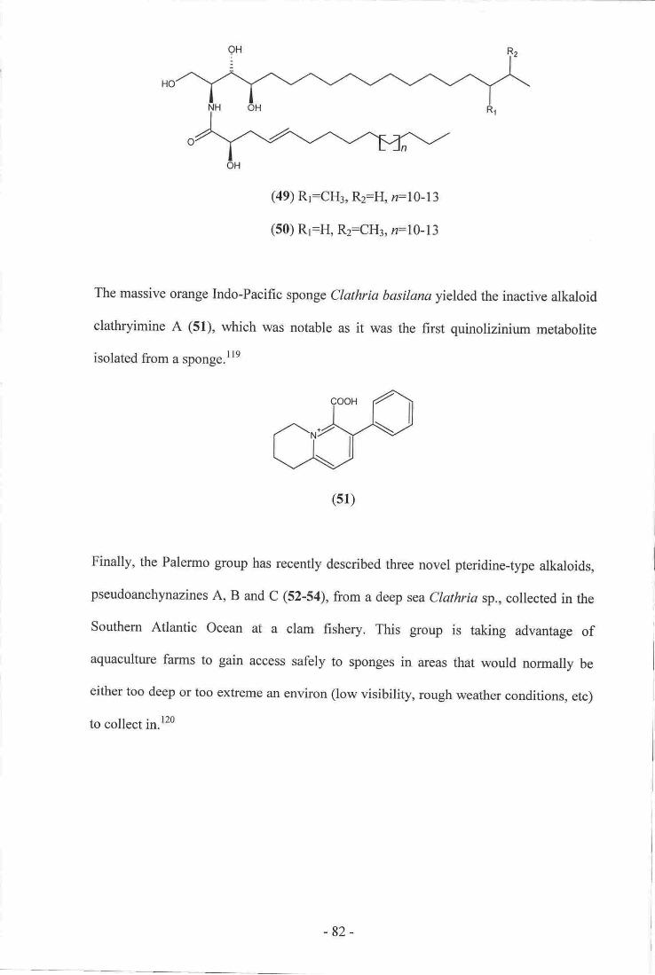
(49) Rr:CI{3, R2:Ho n:10-13
(50) R1:H, Rz:CHr, n:10-I3
The massive orange Indo-Pacific sponge Clathria basilana yielded the inactive alkaloid
clathryimine A (51), which was notable as it was the fust quinolizinium metabolite
isolated from a sponge.lle
Finally, the Palermo group has recently described three novel pteridine-type alkaloids,
pseudoanchynazines A, B and C (52-54), from a deep sea Clathria sp., collected in the
Southem Atlantic Ocean at a clam flrshery. This group is taking advantage of
aquaculture farms to gain access safely to sponges in areas that would norrrally be
either too deep or too extreme an environ (low visibility, rough weather conditions, etc)
to collect in.l2o
(sl)
-82 -
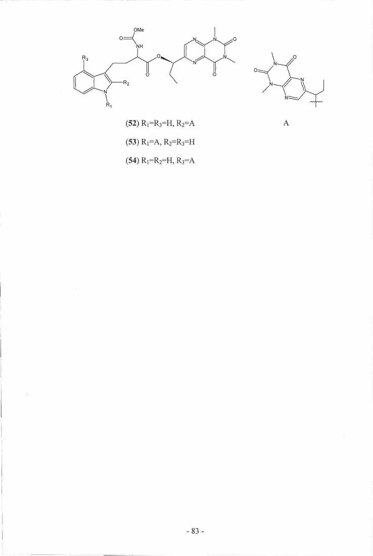
(52) R1=R3:H, R2=A
(53) R1:A, R2:R3:H
(54) R1=R2:H, R3:A
A
-83-

4.1
Chapter Four
Sponsian Diterpene Constituents of the
Marine Soonse C h e Io nap lv silla vio lsc e a
Dictyoceratid and Dendroceratid Sponges
Sponge taxonomists still debate the phylogenic placement of many sponges within
various orders, families, and genera, a situation closely linked to the difficulty in
positively identifying different specimens.2t'23'34In fact, there is even still debate at the
phylum and class level of sponge taxonomy.23 Problems with identification may arise
from either a historical taxonomic misplacement of an archetypical sponge, or from
disagreement on which morphological characteristics (spicule composition, colour,
shape, etc) to use for identification.2t'22'34
Sponges of the Dictyoceratid and Dendroceratid orders exempliff this debate. These
two orders contain sponges totally devoid of mineral spicules, as does the Verongid
order. It is not surprising that sponges devoid of spicules are difficult to identiff, as the
presence of certain spicule types is the most commonly used taxonomic marker.
Historically, sponges lacking inorganic spicules and with an obvious dendritic spongin
skeleton and large sac-shaped choanocytes, were classified as Dendroceratid sponges,
while others with anastomosing (netted) spongin skeletons were placed in the
Dictyoceratid order. Members of the Verongida are quite distinct from those of either
the Dictyoceratid or Dendroceratid orders, as their skeletons are comprised of pith
containing collagen fibres, rather than fibres made from spongin. The histology of
Verongid sponge cells is also quite different to that of the Dictyoceratid or
Dendroceratid orders.2l'33'l2l The difference between dendritic and anastomosing
skeletons can be diffrcult to identify, however, and may have led to sponges currently
-84-

classed as either Dendroceratida or Dictyoceratida, to be artificially separated. As
techniques for the identification of sponges become more sophisticated, problems in
identification may be eliminated and various sponge orders, families, and genera, ffioy
be taxonomically reassigned, created, or removed, on the basis of genetic,
microbiological, or chemotaxonomic considerations. Interestingly, Bergquist has
suggested that, contrary to accepted practice for identification of most other sponge
orders, colour is often a diagnostic feature that can be used to differentiate between
species of the Dictyoceratid and Dendroceratid orders.33
Bergquist has also suggested that sesquiterpene content can be used a chemotaxonomic
marker between members of the two orders.23'tzt Previous work on a New Zealand
Dysidea sp. (Dictyoceratida) carried out within our laboratory has supported this
proposal.l22 During the current study, a series of spongian diterpenes were isolated from
a New Zealand specimen of Chelonaplysilla violacea (Dendroceratida). It was therefore
prudent to evaluate the available literature on any diterpenoids isolated from both the
Dictyoceratid and Dendroceratid orders, to determine if this class of compounds are also
of chemotaxonomic value.
-85-

Kingdom
Phylum
Calcarea
Order Dendroceratida
Metazoa (Animals)
Porifera/[
(Sponges)
Demos-pongiae Hexactinellida
Dictyocbratida Verongida
Family
S\
Danvinellidae Dictyodendrillidae/\sGenera ^rrr( *x,: Darwinella Dendrilla
violacea
Scheme 4.1 Phylogenic relationstnp of Chelonaplysilla violacea. For simplicity, not all
orders or species are shown.2t'23'34;t2r'r23
-86-

4.2 Diterpenes From Dictyoceratid and Dendroceratid Sponges
A survey of the literature to the end of 2002, encompassing all diterpenes produced by
both Dictyoceratid and Dendroceratid sponges, yielded well over 130 individual
structures. The following review summarises the major carbon skeletal groups, with
salient examples, rather than being a comprehensive listing of alt the diterpenes isolated
from the two orders.
4.3 Miscellaneous Diterpenes
Several varied classes of diterpenes have been isolated from Dictyoceratid and
Dendroceratid sponges. Faulkner and Walker published the structures ambliols A (55)
and B (56), ambliofuran (57), ambliolide (58) and dehydroambliol A (59), isolated from
the sponge Dysidea amblia. The structure of 56 was later revised when it was re-
isolated with ambliol C (60).r24'r25
(ss) (s6)
(s7)
-87 -
(s8)

(se) (60)
The sponge Dendrilla sp. collected from a lake in Palau yielded (61), along with several
members of the dendrillolide family of compounds. Several structural revisions of the
dendrillolides were suggested after further isolation of these metabolites ftom the
predatory nudibranch Chromodoris macfarlandi 27'126't27
(61)
Three diterpenes with very different carbon skeletons were isolated from the Red Sea
sponge Chelonaplysilla erecta. Chelodane (62), barekoxide (63) and z,aatfuin (64) were
all biologically inaetive in the assays used, although the initial extract from the sponge
showed significant anti-tumour activity. 128
(62) (63) (64)
Lu and Faulkner isolated three new dolabellane diterpenes (6H7) and a macrolide (68)
from a Dysidea sp. collected in Palau. Neither dolabellane diterpenes nor any
-88-

macrolides had previously been isolated from any species of Dysidea, thus the authors
felt that the four novel compounds were likely to be metabolic products from other
organisms that the sponge had obtained from its environment.l2e
(6s) (66)
AcO
H ""'lAcO ""
(67) (68)
4.4 Spongian Diterpenes
By far, the most common type of diterpene from either the Dictyoceratid or
Dendroceratid orders, are those based upon the spongian carbon skeleton (69). This
group of compounds is so named, as many of the early members of this class were
isolated from the Dictyoceratid genus Spongia.tr0 The biochemistry of the spongian
diterpenes is punctuated with many examples of highly oxygenated and oxidatively
cleaved carbon skeletons. This is a quite different theme to that of many other classes of
compounds found from sponges.
The first metabolite isolated in this large group was isoagatholactone (70), from the
common Mediterranean bath sponge Spongia officinalis. The structure of 70 was
-89-
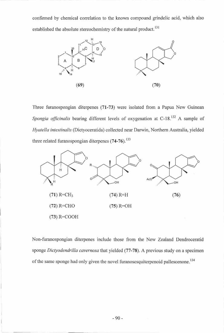
confirmed by chemical correlation to the known compound grindelic acid, which
established the absolute stereochemistry of the natural product.l3l
-'{r-'q.Hc I o)o
\:--,{\f,,lB
l:.u..9
(6e) (70)
Three firanospongian diterpenes (71.-73) were isolated from a Papua New Guinean
Spongta fficinalis bearing different levels of oxygenation at C-18.r32 A sample of
Hyatella intestinalis (Dictyoceratida) collected near Darwin, Northem Australia, yielded
three related frranospongian diterpenes (7 4-7 q.t33
(71) R:CH3
(72) R:CHO
(73) R:COOH
(74) R=H
(75) R=OH
(76)
Non-furanospongian diterpenes include those from the New Zealand Dendroceratid
sponge Dictyodendrilla cavernosathat yielded (77-78), A previous study on a specimen
of the same sponge had only given the novel furanosesquiterpenoid pallescenone.l3a
-90-

Q7) (78)
Six spongian diterpenes (79-Sa) were isolated from the sponge Spongia matamata. All
possess carboxylic acid functionality at C-19 and some oxygenation of ring D.r3s
(82) R:H
(83) R=OMe
(84)
(80) R:a-OMe
(81) R:p-OMe
A common structural feattue of many of the spongian diterpenes is an acetal linkage
between C-15 and C-17. Examples include (35-87) from the Caribbean Dendroceratid
sponge lgernella notables,\36 and both (88) and (89) from an Ausftali an Darwinella sp.
@endroceratida).137
:RR
(85) R=H
(86) R:COCH3
(87) R:COCHzCHzCHT
(88) R:H
(89) R:OAc
-91 -
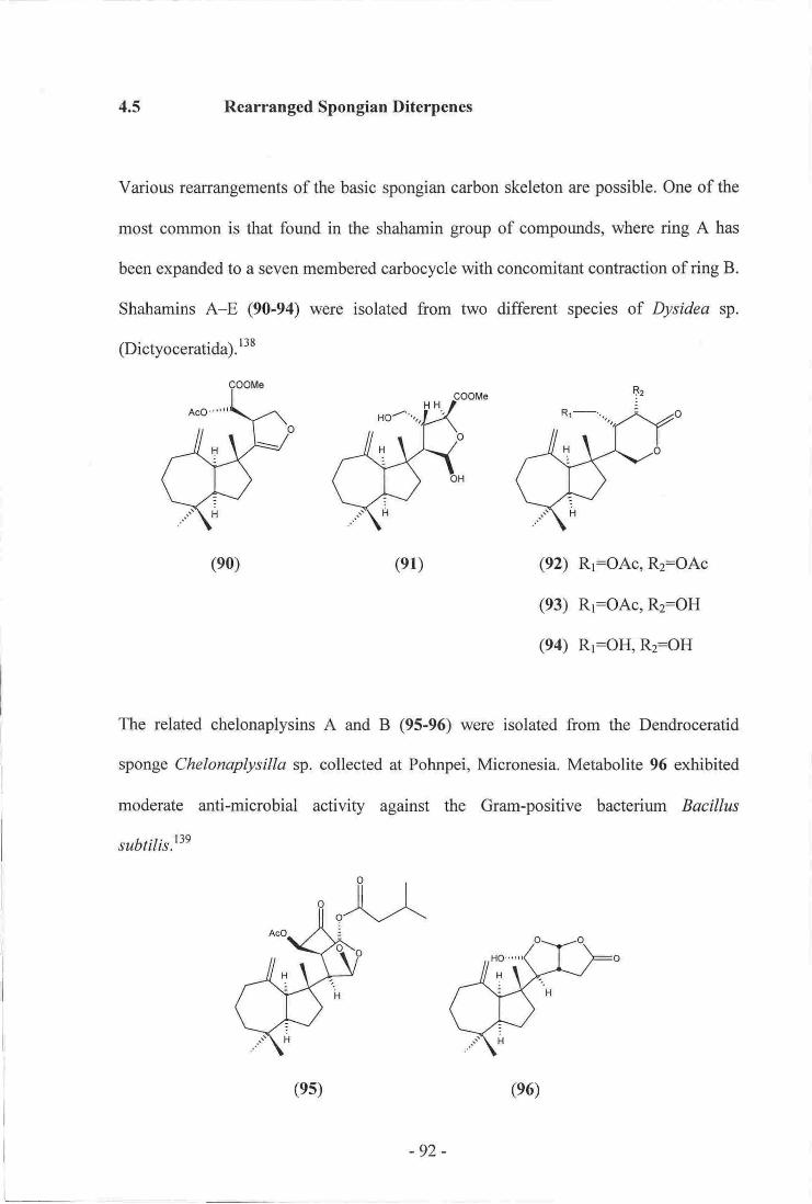
4.5 Rearranged Spongian Diterpenes
Various reuurangements of the basic spongian carbon skeleton are possible. One of the
most common is that found in the shahamin group of compounds, where ring A has
been expanded to a seven membered carbocycle with concomitant confraction of ring B.
Shahamins A-E (90-94) were isolated from two different species of Dysidea sp.
(Dictyoceratida).r38
(e0) (e1) Rr=OAc, Rz:OAc
R1:OAc, R2:OH
R1:OH, Rz:OH
The related chelonaplysins A and B (95-96) were isolated frorn the Dendroceratid
sponge Chelonaplysilla sp. collected at Pohnpei, Micronesia. Metabolite 96 exhibited
moderate anti-microbial activity against the Gram-positive bacterium Bacillus
subtilis.t3e
(e2)
(e3)
(e4)
(es)
-92-
(e6)

Further members of the shatramin group of compounds, shahamins F-J (97-101), were
isolated concurrently with those mentioned above, although they are based upon a
different rearranged carbon skeleton. 138
(97) Rr:H, Rz=H
(98) Rr:H, R2=OH
(99) R1:OH, Rz:H
There is only one example of a compound where
Norditerpene (102) was isolated from a sample
Spongionella gracilis (Dendroceratida). rao
ring A has been contacted.
of the Meditenanean sponge
(1oo)R:H
(101)R:OH
(102)
Rearrangement of ring B gives the norrisolide family of compounds, including the three
novel diterpenes (103-105) isolated from a Red Sea Dysidea sp.. One of the new
compounds, 103, showed some cytotoxic activity.tot In the same class of compounds is
chelonaplysin C (106) isolated from the same collection of the Micronesian
Chelonaplysilla sp. mentioned previously. l3e
-93-

'-cooM8
(103)
(r0s)
(104)
(106)
Baeyer-Villiger type oxidation of ring A also allows for an expansion to occur, fonrdng
a lactone fiurctionality exernplified by spongialactone (107), isolated from the Red Sea
sponge Spongia arabica.taz Collections of a Great Barrier Reef sponge Spongia sp.
yielded a second ring A lactone diterpene (108). Compound 108 exhibited marginal
cytotoxicity against P388 murine leukaemia cells.la3
(108)
Oxidative degradation of ring D is noted in several rearranged spongian diterpenes. A
collection of Darwinella oxeata yielded structures (109-1f0). Both metabolites possess
a lactone linkage between C-l5 and C-I7,and ring D has been lost.raa
(107)
l_
-94-

(109) R:H
(110) R:OH
Similarly, Hambley et al isolated four norditerpenes (111-114) from a sample of
Aplysitta pallida (Dendroceratida).ras In another study, diterpene (115) was reported
from the Dictyoceratid sponge Coscinoderma mathewsi, which was collected at
Pohnpei, Micronesia. Aldehyde 115 is a possible decarbonylation product of (116).146
(111)
(114)
(112)
(11s)
(113)
(116)
The final class of spongian diterpenes are those where methyl C-17 has undergone a
I,2-migration and the C-5 to C-6 bond has been cleaved to open ring B. This common
feature is often found in conjunction with some of the structural motifs noted
previously. Aplysulphurin (117), from a New Zealand Aplysilla sulphurea, is the first
example of an aromatic spongian diterpene.raT Mayol, Piccialli and Sica isolated the
related compounds gracilins A (lf8) and B (ll9), from a Mediterranean Spongionella
-95 -

gracitis (Dicfyoceatida), Gracilin B (119) w,as the fhst exa,uple of a bis-norditqpene
ftonn a madne sponge. The same gro-up aehievedilthe isolation of four more gracilin-t1pe
diterpenes ([2,0-123) ovet subsequent ye41s.i40'148't 5o
(120) (121) R:Ac
(122) R+x
(r23)
A firrther Soul:of tolated mEtabolitEs, (124-nq. wereisolated ftom a sanrple ofNew
Zealand Darwirulla axeata (Dendroceratida), as well as from several individuals of the
rnorphologrcally similar sponge Dendrilla rosea. This collection w,a$ the same as that
uAich osntained 10F110. r44
(ll7') (118)
(12s)
(1re)
(r24)
-96 -
(126)

The anti-bacterial diterpenes 9,ll-dihydrogracilin A (127) and membranolide (128)
were isolated from the Antarctic Dendroceratid sponge Dendrilla membranosa. The
authors suggested that 127 and 128 are defensive metabolites as this particular sponge
had never been observed being predated upon and yet it is devoid of any spicules.lsl
(128)
Three rearranged spongian diterpenes (129-131) were reported from the Dendroceratid
sponge Aplysitla tongo,collected in New South Wales, Australia.l52
(r27)
(130)
Finally, several rearranged diterpenes from a Canadian Aplysilla glacialis were reported
during a study of the bioactive metabolites of nudibranchs, carried out to determine
whether the compounds isolated from nudibranchs were from dietary sources, or were
de novo synthesised by the organism itself. As well as isolating metabolites (132-135)
from,4. glacialis, several related rearranged spongian diterpenes were obtained from the
predatory nudibranch Chromodoris luteomarginata, implying a dietary source for the
isolated natural products. Another diterpene of the marginatane class (136) was also
isolated from the sponge.28
(r2e) (131)
-97 -

(132) (133) (134)
(13s) (136)
There are many other spongian diterpenes reported in the literature. Some have been
isolated from nudibranchs and are considered to be of dietary origin whilst others are
synthetic products. Many of the natural products have not been examined for biological
activity due to a lack of material available for testing. Recent surveys have therefore
focussed on testing both natural and synthetic spongian diterpenes in a variety of assays
to determine uses of these compounds.l53-156
Taxonomic Considerations
The literature survey indicates that spongian diterpenes are found from members of both
the Dictyoceratid and Dendroceratid orders. Diterpenoid content cannot therefore be
reliably used as a taxonomic marker for differentiating between families or genera of
these two orders, as they are currently defined. Also, various researchers have noted that
some species of sponge that cannot be differentiated by classic taxonomic methods may
have very different chemical constituents. Chemotaxonomic criteria may therefore be
difficult to establish for families or genera of the Dendroceratid and Dictyoceratid
orders.l2s'13''t57 Bergquist, however, has suggested that Dendroceratid sponges produce
-98-
4.6

4.7
only diterpenes, therefore the presence of other classes of terpene would clearly identify
members of the Dictvoceratida.zs
Biogenesis of Spongian Diterpenes and Spongian Derived Skeletons
The basic tetracyclic spongian skeleton is generated by the cyclization of the diterpene
isoprenoid precursor, geranylgeraniol (137), which is formed by head-to-tail
condensations of four isoprene units. In this particular biosynthetic pathway, 137 is in
an all-nans folded conformation prior to cyclization as shown in scheme 4.2.ts8
+-H
-
Scheme 4.2 Biogenesis of the basic spongian diterpene skeleton.l5s
The biosynthesis of the shahamin class of compounds follows two linked pathways. The
first forms the seven-member ring shahamins A-E (90-94). Oxidative cleavage of the
C-9 C-11 bond places an electronegative leaving group at C-9. A l,2-hydride shift,
followed by a ring expansion with concomitant loss of the leaving group, gives the final
carbon skeleton. Functionlisation of ring D then forms the individual shahamin
compounds as indicated in scheme 4.3. The formation of shahamins F-J (97-l0l) starts
in a similar way with the oxidative cleavage of the C-9 C-l I bond. Elimination of H-5
(r37)
-99-

foltow,ed by a lp-methyl shift from C-10 to C-9 generates the carbon skeleton,
followed by functionatisation of ring D to, agoiu forrn tlre various colnpounds (see
scheme 4.4).t"
eoOMet{o-. /shahamh.B ''(\"N-
L,bn/9RQR/lir'
shdsmlnG-E | |
*,zoSchemo 4.3 Proposed biogenesis ofshahamins A-E (90-94).ttt
oDddii$r?plalrrsg!
\An",a[+C*.
8hd|enin F-J
Scheme 4.4 Fmrposed biogenesi$ of shahanins F-J (97-101).Fg
Ring coutracted metabolite (102) is proposed to, be generated via the meclxrnism
deseribed in scheme 4.5. The opening of a 5,6-epoxide allows a bond shift from C-l
C-10 ts C-l C-5 to form a oontacted spiro-rno-iety. The C-10 oarboeatio,n then
-100-

undergoes nucleophilic attack. Cleavage of the C-6 C-7 bond, followed by oxidation of
C-6 and hydrogenation at C-7, would generate a molecule that could decarboxylate to
generate !02.t40
Scheme 4.5 Proposed biogenesis of (102).140
Members of the norrisolide class (10!105) are biosynthesised as described in scheme
4.6, which starts with the same initial step as that of the shahamins. Initially, the C-9
C-l1 bond is cleaved, which is followed by the migration of the C-7 C-8 bond to form
the five membered ring B, followed by elimination to yield the norrisolides.r26
Scheme 4.6 Proposed biogenesis of the norrisolide compounds (103-105).126
-BH+
-101 -

There are many different compounds reported with ring B opened; all presumably are
generated vra a similar biosynthetic pathway. Oxidation to form a C-6 C-7 epoxide is
followed by l,2-hydride and l,2-methyl shifts. Oxidative cleavage of the C-5 C-6 bond
forms a carboxylic acid which can undergo decarboxylation to fonn the precursor of
gracilin A (118) and other related compounds, which are generated by various
derivatisations of ring D as in scheme 4.T.Isomerization of the diene precursor to 118
or epoxidation followed by reformation of the diene gives an allylic alcohol that can
oxidatively ring open to form 120 and related compounds, shown in scheme 4.3.150'152
Scheme 4.7 Proposed biogenesis of gracilin A (11S) and related compounds.lso'rs2
ox,......_...........*
oxt.+
+
-102-

oxi.+
oxt,
-_-+
l"',
-
Scheme 4.8 Proposed biogenesis of (120) and related compounds.l50'I52
4.8 Novel Spongian Diterpenes from C helo naplys illa vio lacea
A sample of Chelonaplysilla violacea (MNP0979) was collected using SCUBA at
Stephen's Island, Marlborough Sounds, at the nonthern tip of the South Island of New
Zealand (see figure 4.1). The organism was initially described in the field as a tan,
encrusting sponge of the genus Dysidea (Dictyoceratida). Chelonaplysilla violacea
(Class Demospongiae, Order Dendroceratida, Family Darwinellidae) is described as
having a dendritic skeleton composed of smooth, erect, fibres, rising from a basal plate
of spongin. Fibres are concentrically laminar with a very dense outer bark and a diffrrse
pith interior. The sponge is usually found in shaded areas such as caves and archways,
-103-

encru$ting on boulders in sheets 2-3 mm thick. The colour of the sponge is described as
dark purple throughouf with white surface patterning caused by organised sand
reticulation. 23'r2r
Figure 4.1 Hydrographic chart of Stephens Island.
(Courtery of Land and Information New Zealand)
Initial NMR based screening of ll0 g of the sponge revealed many interesting
resonances, including those of multiple acetal/lremi-acetal functionalities, several
oxygenated methines, and also those of several methines with shielded carbon and
deshielded proton correlations, as indicated in the HSQC spectrum (see figure 2.8). An
extract of a further 182 gwas combined with the material generated from the screen.
This combined extract was fractionated using normal- and reversed-phase solid
supports, size-exclusion gel-permeation chromatography and finally, reversed-phase
HPLC, to yield ten spongian diterpenes. Two of the compounds were the known
metabolites tetrahydroaplyzulphurin-l (124) (1.8 mg) and cadlinolide B (133) (1.4 mg),
based upon comparison with published MS and NMR data.2E'r4'r5e Two of the
6e
rafrnde #36 {
^\ -CL roor.ttt
- 104-

remaining eight metabolites could not be identified on the basis of the NMR and MS
data obtained. The last six metabolites isolated constituted novel structures and are dealt
with below. Two of the six novel compounds share the same carbon skeleton as
cadlinolides A (132) and B (133) and are therefore reported as cadlinolides C and D.28
The remaining four compounds possess different carbon skeletons to those previously
reported and are named based upon the Maori name for Stephen's Island
(Takapourewa).
Scheme 4.9 Isolation scheme for spongian diterpenes
from Chelonaplysilla violacea sp. (part I ).
182 g Ch'lonadysilla violacea
Extract 2 x MeOHPSDVB column (HP20)
20% Me2CO 4070 Me2CO 6090 Me2CO 80% Me2CO
(SEE NEXT PAGE)100% Me2CO
+ Screen Samde
PSDVB (Arnborchrcm)Gradient elution
crs RP
7 5o/o MeOHlZSo/o 0. 1 M CH3COOH
Crr RP
807o MeCN/20olo H2O
cr! RP750/o MeOHlZSo/o 0.1M CH3COOH
Crr RP
600/o MeCN/40% H2O
PSDVB (A6!e1s17e61Gradient elution
- 105 -

II
ib?co80%Mer@
(FROM PREYPIJS PAGE)
Llt2090% lffiH/1096 l-lrO
f'j6 irlaoFutio% cHror
Schcme 4.l0Isolation scheme for spongian ditetrpenes
from Chelonqlysilla violacea sp. (part 2).
Figure 4.2 Skeleton and appearance of ClvlorryIysillaviolacea.
(Courtesy of Professor P. Bergquist)
C: Chetonaplysillaviolacea, section of fibre to show ma*ed vesiorlarpith (x 120).121
E: Chelonap lysi lla violrce a t2 t
- 106-

Cadlinolide C
A single intense pseudo-molecular ion was observed for cadlinolide C (138) (2.4 mg) in
the positive ion HRESIMS mode establishing a molecular formula of C26H3oO3
(319.2260 [M+H]*, L 2.6 ppm) and requiring six degrees of unsaturation. Only one
significant band was identified in the IR spectrum, that of a C:O stretch (1739 cm'r).
All 20 carbon and all 30 proton resonances were observed in the l3C and lH NMR
spectra, and one-bond proton-carbon correlations were detected in an HSQC-DEPT
experiment. Prominent signals in the I3C spectrum included resonances of a carbonyl
(6c172.5), an olefin (69 147.0, 122.9), an acetal (6c 103.5), and an oxygenated
methylene (6c 73.1). The olefin was assigned as tetrasubstituted from the lack of any
HSQC-DEPT conelations to these l3C resonances. With no other evidence of multiple
bonds, the remaining degrees of unsaturation required cadlinolide C (138) to have a
tetracyclic structure.
Initial analysis of the correlations from various proton resonances in both the COSY and
HMBC spectra, allowed construction of several substructures. In particular, a linear
fragment of three methylenes was connected on the basis of COSY correlations between
the resonances of their attached protons [(C-1:69 40.0,6s 1.95, 1.13) to
(C-2: 66 20.6,6H l.5l) to (C-3: 6s 39.7, 6H 1.32, 1.19)1. Strong HMBC correlations
were observed from the rH resonances of two methyl singlets [(C-1s: 6c27.6,6H 0.76),
(C-19: 6c32.9,6H 0.90)l to the r3C resonances of each other, two methylenes [C-3 and
(C-5: 6g 51.2, 6H 1.80, 1.26)l and a quaternary centre (C-4: 66 31.9), establishing the
link of C-3 to gem-dimethyl quatemary centre C-4. Finally, the tH resonance of a third
methyl singlet (C-20: 6c 31.9, 6H l.l3) showed HMBC correlations to two methylenes
-t07-
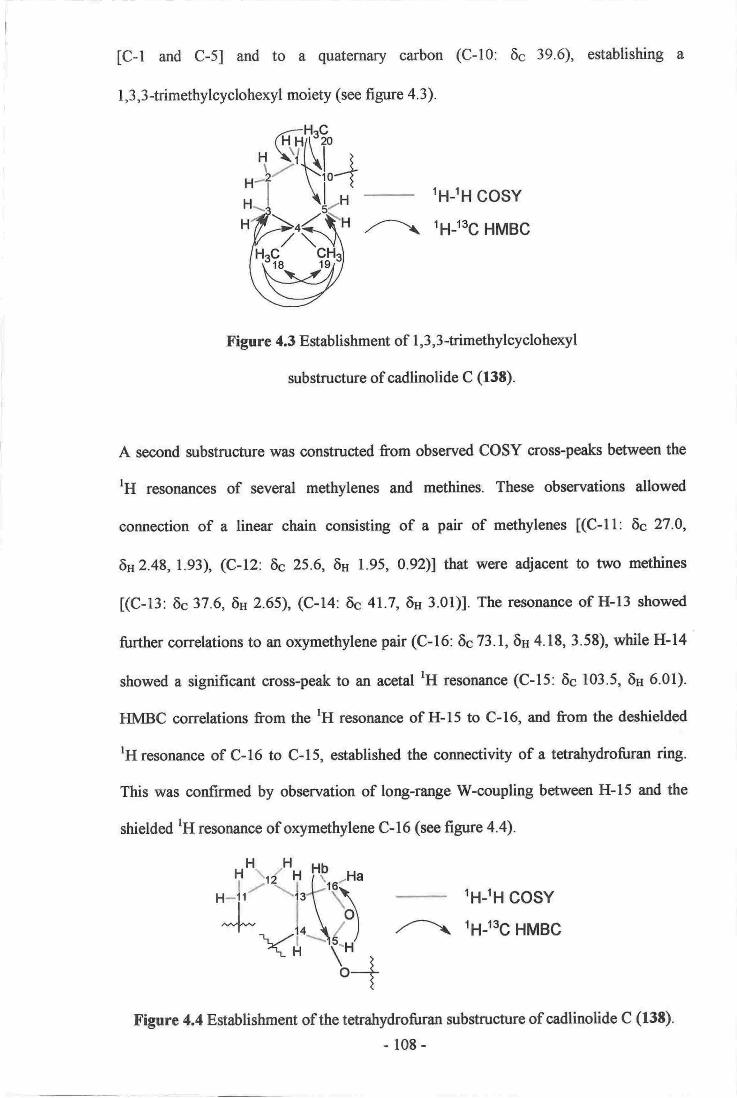
[C-l and C-5] and to a quaternary carbon
1,3,3 -himethylcyclohexyl moiety (see figure 4. 3 ).
(C-10: 6c 39.6), establishing
/'--\
1H-lH cosYt6-ts6 HMBG
Figure 4.3 Establishment of 1,3, 3-trimethylcycloheryl
substructure of cadlinolide C (13E).
A second substructure was oonstructed from observed COSY cross-peaks between the
tH resonances of several methylenes and methines. These observations allowed
connection of a linear chain consisting of a pair of methylenes [(C-l l: 6c 27.0,
6n 2.48, 1.93), (C-12 6c 25.6, 6H 1.95, 092)l that were adjacent to two methines
[(C-13: 6c37.6,6s 2.65), (C-14: 6c41.7,6H 3.01)]. The resonance of H-13 showed
further correlations to an orymethylene pair (C-16: 6c 73.1, 6H 4.18, 3.58), while H-14
showed a significant cross-peak to an acetal tH resonance (C-15: 6c 103.5, 6H 6.01).
HMBC correlations from the tH resonance of H-15 to C-16, and from the deshielded
rH resonance of C-16 to C-15, established the connectivity of a tetrahydrofuran ring.
This was confirmed by observation of long-range W-coupling between H-15 and the
shielded tH resonance of orymethylene C-16 (see figure 4.4).
1H-rH cosYt;-1-tsg HMBC
Figure 4.4 Establishment of the tetrahydrofuran zubstructure of cadlinolide C (138).
108 -
"j',)'t[{ ("^L t\,1, Yt_$(H,
b+

A strong COSY correlation was observed between the lH resonances of a methyl
(C-6: 6c 14.4, 6n 1.a0) and a methine (C-7: 6c 40.7, 6H 4.18). HMBC correlations were
observed from the lH resonances of both these centres to the ttc .esonances of each
other, and also to that of the carbonyl (C-17: 6c 172.5) to establish a third fragment (see
figure 4.5).
,--l*-.---*li.g+f\--*-l?'/ov \o>r.---
1H-1H cosYt6-rsg HMBC
Figure 4.5 Establishment of the side chain of cadlinolide C (f3t).
The previous observations had accounted for 18 of the 20 carbons of 138, leaving a
tetrasubstituted olefin to be assigned. The placement of the olefirq and therefore the
final connectivity of the molecule, was established from analysis of several key COSY
and HMBC correlations. HMBC correlations were observed between the rH re$onanc€s
of methyl C-6 and methine C-7 to the t3C resonanoe of a dizubstituted olefinic carbon
(C-8: 6c 122.9). The resonance of H-7 also showed HMBC correlations to the
r3C resonances of the second disubstituted olefinic carbon (C-9: 6c 147.0') and to C-14,
which established the linkage of the side chain to the olefin at C-8 between C-9 and
C-14 and connected the side chain with the tetrahydrofuran zubstructure. A strong
HMBC correlation was observed from the lH resonance of C-20 to the l3C resonance of
C-9, allowing connection of the 1,3,3-trimethylcyclohexyl ring to the olefin. The
deshielded tH resonance H-l lb showed a correlation to the l3C resonance of quaternary
centre C-10, which helped to confirm the linkage between the olefin and the
1,3,3-ffimethylcycloheryl ring. A homo-allylic coupling was observed between H-lla
and H-14 in the COSY spectrum that confirmed the placement ofthe olefin within a six-
membered ring. Finally, although no HMBC correlations were observed between the-109-

resonances of H-15 and C-17, a lactone linkage could be deduced by the final required
degree of unsaturation, and also by the absence of any alcohol functionality within the
molecule, as evidenced by the lack of an OH stretch in the IR spectrum. The lactone
linkage is supported by the deshielded chemical shift of H-I5, and also the r3C chemical
shift of carbonyl C-17 which is similar to that of tetrahydroaplysulphurin-l (124),
cadlinolide A (132) and cadlinolide B (133) (see figure 4.6).u
/-\
1H-lH cosYt;1-tsg HMBC
Figure 4.6 Establishment of final connectivity of cadlinolide C (138).
The relative stereochemistry of 138 was established from vicinal rH-rH homonuclear
coupling constants, measured in the tH NNm. and lH decoupled NMR experiments, and
from NOE correlations established from a ROESY experiment. A small coupling was
meazured between H-14 and H-l5, zuggesting a small dihedral angle between these two
protons. An NOE correlation was observed between H-13 and H-14, implying that they
are on the same face of the molecule (see figure 4.7). H-15 also showed a strong NOE
correlation to CHr-6, implying that C-6 is on the same face as protons H-14 and H-15.
NOE correlations were observed between CI{3-6 and H-5b, implying that H-5b is also
on the same face as methyl C-6. NOE correlations were observed from H-7 to H-5q H-
5b and also to CH3-20, supporting this assignment (see figure 4.8). These observations
allowed the relative stereochemistry of cadlinolide C (l3S) to be assigned as 7R*, 10^l*,
13,S*, 14S*, l5,S*, which was consistent with the stereochemistry determined for
tetratrydroaplysulphurin- | (124), which had been solved by x-ray diffraction.28'r'r4'l5e
- ll0-

n NoE
X'igUrc 4.7 NOE correlations and stereochemistry of tetratrydrofuran
ring of cadlinolide C (13t).
n NoE
X'igurc 4.t Selected NOE conetations establishing the ster€ochemistry ofthe
1,3,3-timethylcycloheryl and lactone rings of cadlinolide c (Ft).
YffiW
- lll -

Table 4.1 r3c (75 MHz) and rH (300 Mlxz) NIvIR Data (CDClj of oadlinolide C (13s)"
tre ,Hu
cosYHMBCffitoC)Position s(oom) mutt 6 (pun) mult./Glz) NOE
la
b
2
3a
,b
4
5e
b
6
7
IIl0
llab
l2a
b
l3
t4
I5
l6a
b
tltgo
lgo
20
40.0
20.6
39,7
3t.9
st2
t4.4
40.7
t22.9
147.0
39.6
27-0
25.6
34.,6
41.:t
103.t
73.1
na.s
2V.6
32.9
3r"9
Cl{r
CIft
cCH,
cHcllcllCtlr
lJ31,95
r.51
r.1o
t.l?::
126
l.t0r.40
it.l 8
I '93
a48
0:92
1,95
2;65
3!01
6,0t
3.58
4.18
clli Ilm
llt?tttn
m
d(13,CI
dt(r3.7, t.7)
d (?.3)
qo,o
lbl*2
I.b- 34,3
2, l82,5b
5b
5q 3b
1
6
m
ft06.1,4.'0m
m
m
ddd (r3,4 5,6,22)
d (5,e)
dd (10"0,8,8)
r{8.a
c
s
s
I lb, t?q lillla, l2q l2t
lla llb,lzb, 13
llb" lZq 13
124 l2b,14" l6a, l6b
l3n 15, l6b, I la
t4, t6a
13, l6b, 15
13, l6a" 14
3a, 19
l8
3
3,4 l0
9; lO 1E,19,20 7
1,4, f0 ]a,6,71,8,17 5h 15
6,8"9,14,11 ft,5b,20
lrb8,I,10,12, 13 lla
l2b
ll, il|, t4 lbl4
g,g 13
13, l,*, 16 6
13, 14, 15
3,4, 5. 19
3,4,5, t8
l-s_9- t0 1
3b
criz
CLlr
CH
ccc
CH!
c
ClIe
cH:CHr
0:76
0.90
l.t3
a
b
Mutfplicity detennined from HSQC-DEPT (multipticity edited TISQC) experiment.
Carbon oonn€otivity detenrfned ftom an HSQO:DEPT experimenL multiplioity determined from,rH and rH homonuslear decopled expertirents.
lntorchangeable..
038)
-112-

4.10 Cadlinolide D
The molecular formula of cadlinolide D (139) (0.8 mg) was determined as CzJ\zOc
from observation of several pseudo-molecular ions in the positive ion HRESIMS mode
(349.2399 [M+H]n, A 7.5 ppm; 371.2201[M+Na]", A 2.1 ppm;719.4483 [2M+Na]', A
2.5 ppm), requiring six degrees of unsaturation. Due to the small amount of 139
isolated, a l3C NMR spectrum could not be obtained therefore atl 2l observed l3C
chemical shifts referred to below, were observed as correlations in the HSQC-DEPT or
HMBC spectra. All32 tH resonances were observed in the lH NMR spectrum. As with
cadlinolide C (138), only one significant peak was observed in the IR spectrum (C=O
stretch, 1744 cm'r). Notable r3C resonances included those of a carbonyl (6c 171.8), an
olefin (69 145.7, 122.2) and two acetals (6c 109.2, 102.2). Again, the olefin was
assigned as tetrasubstituted as no HSQC-DEPT correlations were observed to these two
t3C resonances. After accounting for the olefin and the carbonyl, the remaining degrees
ofunsaturation of139 required a tetracyclic structure.
A significant feature of the tH NMR spectrum was the 3:1 ratio observed between
several closely related resonances, implying the presence of two similar compounds
(diastereomers). Several unsuccessful attempts were made to separate these compounds;
therefore the following structural elucidation was performed on a mixture of
diastereomers in a 3:l ratio. Cadlinolide B (133) had previously been reported as a
similar inseparable pair of diastereomers. 28
As with cadlinolide C (138), a 1,3,3-trimethylcyclohexyl ring could be established. A
strong COSY correlation was observed between the rH resonances of two methylenes
[(C-l: 6c 39.2, 6u 1.92, l.l3), (C-2: 6c 20.9,8u 1.51)]. Specific correlations between the
tH resonances of methylenes C-2 and C-3 could not be unequivocally distinguished in
- I 13 - vtcroElA ui\rvERGiTY oF WELLINGTON
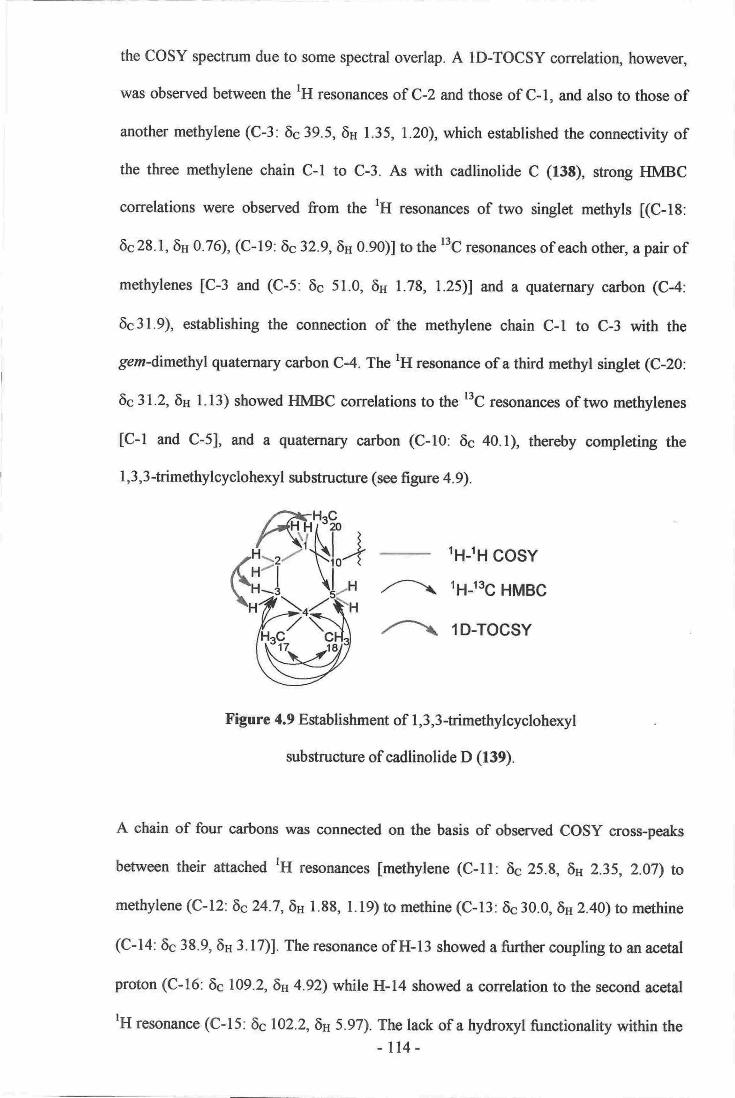
the COSY spectrum due to some spectral overlap. A ID-TOCSY correlatioq however,
was observed between the lH resonances of C-2 and those of C-1, and also to those of
another methylene (C-3: 6q 39.5, 6H 1.35, 1.20), which established the connectivity of
the three methylene chain C-l to C-3. As with cadlinolide C (138), strong HMBC
correlations were observed from the tH resonances of two singlet methyls [(C-18:
6c 28.1, 6s 0.76), (C-19: 6c32.9,6n 0.90)] to the t3C resonances of each other, a pair of
methylenes [c-3 and (c-5: 6c 51.0, 6n 1.78, r.25)] and a quaternary carbon (c-4:
6s31.9), establishing the connection of the methylene chain C-l to C-3 with the
gem-dimahyl quaternary carbon C4. The rH resonance of a third methyl singlet (C-20:
6c 31.2, 6H l.13) showed HMBC correlations to the r3C resonances of two methylenes
[c-l and c-5], and a quaternary carbon (c-10: 6c 40.1), thereby completing the
l, 3, 3 -trimethylcycloheryl substructure (see fi gure 4. 9).
1H-lH cosYt;-q-tog HMBC
1D-TOCSY
Figure 4.9 Establishment of 1,3,3 -trimethylcycloheryl
substructure of cadlinolide D (139).
A chain of four carbons was connected on the basis of observed COSY cross-peaks
between their attached lH resonances [methylene (c-11: 6s 25.8, 6n 2.35, 2.07\ to
methylene (C-12:8c24.7,6H 1.88, 1.19) to methine (C-13: 6c30.0, SH z.eoy to methine
(C-14: 6c 38.9, 6H 3.17)1. The resonance of H-l3 showed a further coupling to an acetal
proton (C-16: 6c 109.2, 6H 4.92) while H-14 showed a correlation to the second acetal
'H resonance (C-15: 6c102.2,6H 5.97). The lack of a hydroryl functionality within the
- l14-

molecule, as evidenced by the lack of an OH stretching band in the IR spectrunq
coupled with the chemical shifts of both methines C-15 and C-16, suggest that C-I5 and
C-16 are connected via an ether linkage, which would form a tetrahydrofuran ring
similar to that found in cadlinolide C (13S). Unfortunately, no HMBC correlations were
observed between methines C-15 and C-16 to help confirm this linkage (see
figure 4.10).
lH-lH cosY
Figure 4.10 Establishment of the tetrahydrofuran ring of cadlinolide D (139).
As with 138, an intense COSY correlation was observed betwe€n the rH resonances of a
methyl (C-6: 6c 14.8, 6H l.a0) and a methine (C-7: 6c41.2, 6H 4.19). The tH resonanc€s
of both showed strong HMBC correlations to the ItC resonance of the other, and also to
that of a carbonyl (C-17:6c 171.8) to complete a third fragment (see figure 4.5).
The analysis detailed above had assigned 18 ofthe 21 carbon centres of 139, leaving an
orygenated methyl and a tetrasubstituted olefin to be assigned. As with cadlinolide C
(l3E), several key HMBC and COSY correlations were used to identify the placement
of these functionalities, and to complete the final structrne of the molecule. The
rH resonances of C-6 and C-7 each showed an HMBC correlation to the l3C resonance
of an olefinic carbon (C-8: 6q 122.2) while H-7 also showed an HMBC correlation to
C-14, which together imply the attachment of the side chain at C-8. Similar to l3t, the
lH resonance of C-20 showed a strong correlation to the l'C resonance of the second
dizubstitr*ed olefinic carbon (C-9: 6c 145.7), establishing the link betwe€n C-9 and
-l15-
hH Hq
H\N'lH \-HH-lt-''\lt-t\
tt9*:--ll-'("'H \ |ot
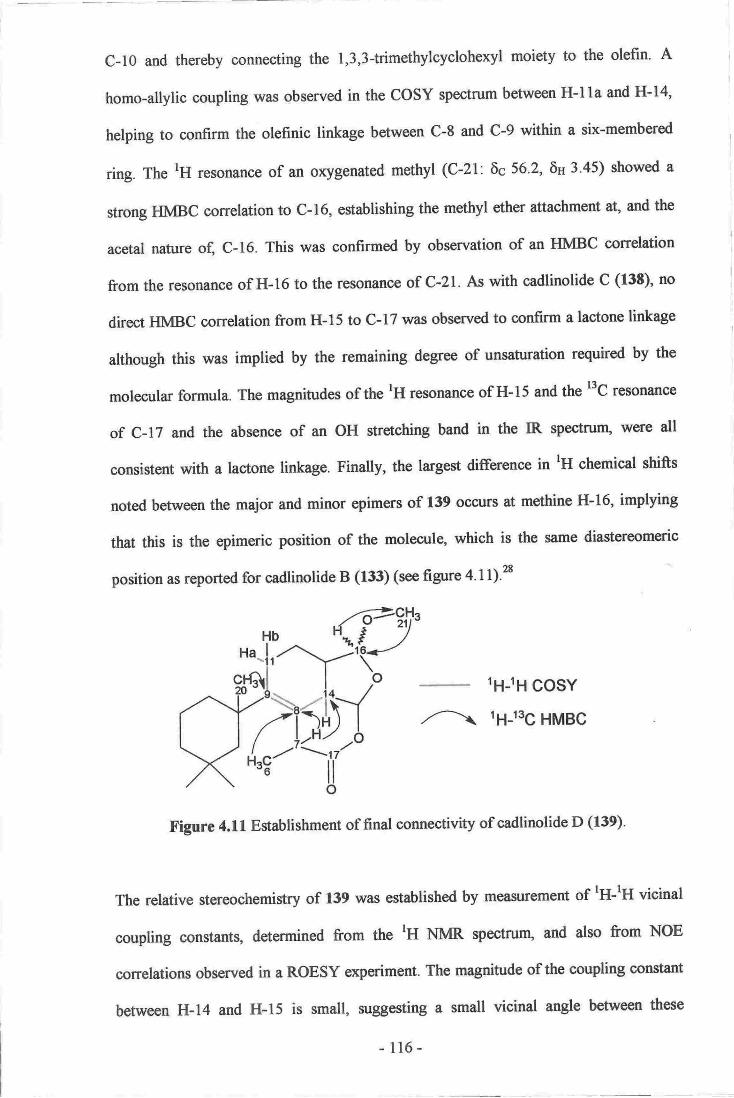
C-10 and thereby connecting the 1,3,3-trimethylcyclohexyl moiety to the olefrn. A
homo-allylic coupling was observed in the COSY spectrum between H-lla and H-14,
helping to confirm the olefinic linkage between C-8 and C-9 within a six-membered
ring. The rH resonance of an oxygenated methyl (C'21: 6c 56.2, 6H 3-45) showed a
strong HMBC correlation to C-16, establishing the methyl ether attachment at, and the
acetal nature of, C-16. This was confirmed by observation of an HMBC correlation
from the resonance of H-16 to the resonance of C-21. As with cadlinolide C (138), no
direct HMBC correlation from H-15 to C-17 was observed to confirm a lactone linkage
although this was implied by the remaining degree of unsaturation required by the
molecular formula. The magnitudes of the tH resonance of H-15 and the t'C resonance
of C-17 and the absence of an OH stretching band in the IR spectrunr, were all
consistent with a lactone linkage. Finally, the largest difference in rH ohemical shifts
noted between the major and minor epimers of 139 ocsurs at methine H-16, implying
that this is the epimeric position of the moleculg which is the same diastereomeric
position as reported for cadlinolide B (133) (see figure 4.1l)'28
,/-\
lH-lH cosYtg-tsg HMBG
Figure 4.ll Establishment of final connectivity of cadlinolide D (f39).
The relative stereochemistry of 139 was estabtished by measurement of tH-rH vicinal
coupling constants, determined from the lH NNm. spectrurg and also from NOE
correlations observed in a ROESY experiment. The magnitude of the coupling constant
between H-14 and H-15 is small, suggesting a small vicinal angle between these
- l16-
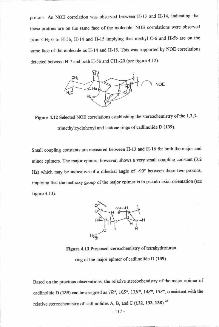
proton$. An NOE correlation was observed between H-13 and H-14, indicating that
these protons a^re on the same face of the molecule. NOE conelations were observed
from CHs-6 to H,5b, H-14 and H-15 implying that methyl C-6 and H-5b are on the
same face of the molecule as H-14 and H-15. This was supported by NOE correlations
detected berween H-7 and both H-5b and CHr-20 (see figure 4.12).
a\ NoE
Figure 4.12 Selected NOE correlations establishing the stereochemistry of the 1,3,3-
trimethylcycloheryl and lactone rings of cadlinolide D (139).
Small coupling constants are measured between H-13 and H-16 for both the major and
minor epimers. The major epimer, however, shows a very small coupling constant (3.2
tlz) which may be indicative of a dihedral angle of -90o between these two protons,
implying that the methoxy group of the major epimer is in pseudo-arial orientation (see
figure 4.13).
o)t'oiiul'o-H"AAi'A'--l{},'-"
H^C/[ " A
'21
f igure 4.13 Proposed stereochemistry of t*ratrydrofuran
ring of the major epimer of cadlinolide D (f39).
Based on the previous observations, the relative stereochemistry of the major epimer of
cadlinolide D (f39) can be assigned as 7R*, l0,S*, l3R{', l4,S*, 15,51 consistent with the
relative stereochemistry of cadlinolides d B, and C (132, 133, 138).2t
-rr7 -
T"" l-tHb\-./ I
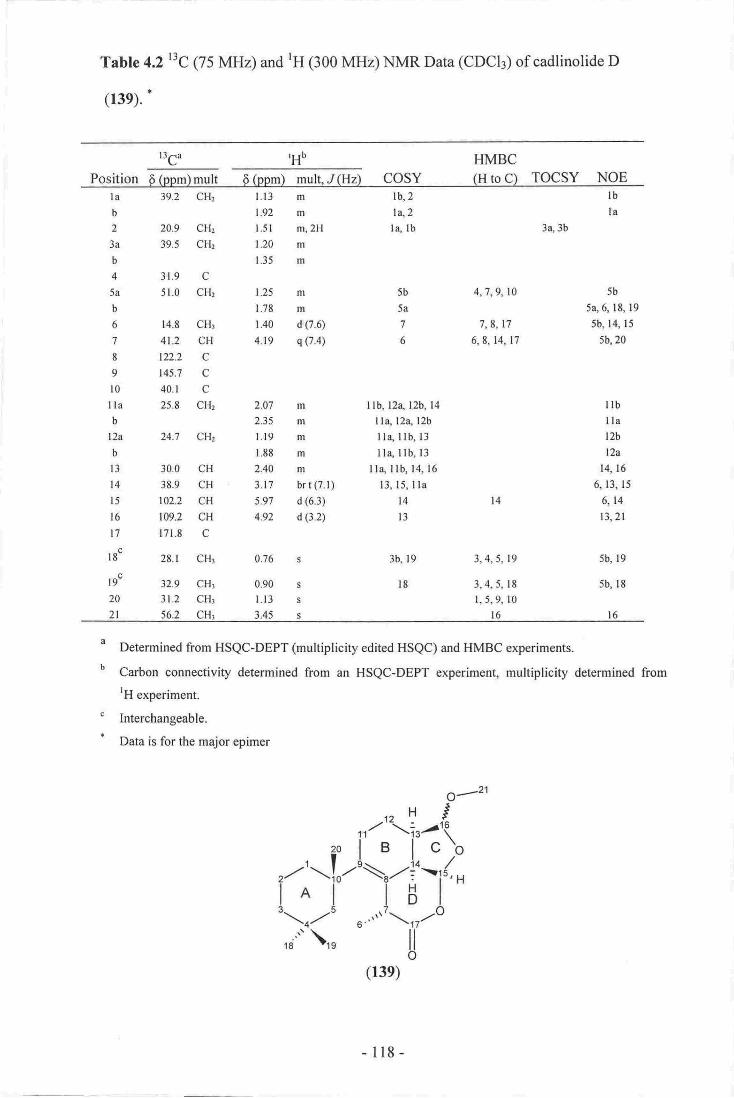
Tabte 4.2t3c g5 MHz) aud rH (300 MIrz) t{MR Data (CDCI3) of cadlinolide D
G39). !
HIVTBC
Posltion 6(ppm)mrrlt 6(ppn) mul{"I(Hz) cosY (I{roC-) TocsY NoE
,HEr3c
lab
2
3a
b
45a
b
6
7
IIt0Ilab
L2a
b
t3I,4
l5l6t7
lgc
r9s
2A
2l
39.2 Cll,
209 CHr
39,5 CFL
3,'[.9 C
5lO CHr
f.4"8 CHr
41.2 CH,
Lnz C
145"7 C
40,1 c25"8 CH{
u.1 ciFI2
3.0,0 cll389 CH
1@2 Cttr09J ellL7L'8 C
28.1 CHr
32.9 CH!
3le cH:562 eHr
1.13 m
li92 rn'
l.3l r4,2H
1.20 m
1.35 m
1.?5
l.rE1.40
419
lbi2lcZla lb
llb; t2o, l2b, 14
ita, [24 UtI Ia" I lb, 13
l1* llb 13
lle llb, l4 16
13,15, rtal4t3
3b, t9
l8
lbla
3q,3b
m
rg
il (7.5)r
s 0,4)
5b
5a
7
6
d 7,9, 10
1,8,1'l6,8,14, l7
3,41 19
3,,4,5, 18
t,5,9, lol6
5b
!a,6,18,195b,14, lJ
5b,20
2.01:
2.35
l,,ll9
r.88
2AA
,.17.
5.n,
4:9
0.76
0;90
l.13
3d5
ml
m
Em
trr
b'r t C7.t)
ir (6.3)
d (3,a)
lrbllat2b
\?A
14 16
6, 13, 15
6,14
t3,2r
5b, 19
5b, lg
l6
[4
s
$
s
s
:a
b
c
a
Deterrrined from HSQC-DEPT (multiplicity ditsd HSQC) and HMBC expeiinents"
Carbon eonneotivity determincd fr-om En HSQC-DEPT expertmont, multipliciS detsrmin€d fronrHexperinent
Inbrchangeable.
Data is for the mqior epimer
-ll8-

4.ll Pourewic Acid A
The molecular formula of pourewic acid A (140) (2.9 mg) was established as CzrHg+O+
by observation of a pseudo-molecular ion in negative ion mode HRESIMS (349.2401
[M-H]-, A 4.8 ppm), implying five degrees of unsaturation. All 21 carbons and 33 of the
34 protons of 140 were observed in the l3C and lH NMR spectra respectively. Two
prominent stretching bands (OH stretch, 3392 cm-l, C:O stretch 1704 crnl) were
observed in the IR spectrum. The presence of a carbonyl was confirmed by a
r3C resonance observed (6c 178.8). A tetrasubstituted olefin was implied by the two
deshielded '3C NNm. resonances observed (69 144.2,127.9), combined with the absence
of any HSQC-DEPT correlations to these resonances. With no other evidence of double
bonds, the remaining degrees of unsaturation required a tricyclic structure for 140-
As with cadlinolides C (138) and D (139), analysis of COSY and HMBC correlations
from various tH resonances of 140 allowed several substructures to be constructed. In
particular, close examination of the spectra allowed the rapid establishment of a
1,3,3-trimethylcylohexyl ring, in a similar manner to that of 138 [(C-l: 6c 39.1,
6n 2.13,!.21),(C-2:6c20.1,6u 1.85, 1'49), (C-3:6c40.0,6H l'33, 1'18)' (C-4:6c31'6)'
(C-5: 6c50.8,6H 1.76,0.96), (C-10: 6641.5), (C-18: 6c26.6,5s 0'87), (C-19: 6c33'1,
6H 0.84) and (C-20: 6c 3l .6, 6H 1.03)l (see table 4.3 and figure 4.3).
A similar series of COSY and HMBC correlations as those observed for 138 allowed a
second substructure to be established. In particular, a sequence of two methylene pairs,
two methines, an oxymethylene and an acetal centre [(C-ll 6c27.6,6u2'22, 1.91),
(C-12: 8c 30.6, 6u 1.63, 1.24), (C-13: 6c 37'8, 6p 2.30), (C-14: 66 48'8, 66 2'7 1), (C-15:
6c I 11.0, 6u 4.75), (C-16: 6c74.7,5n 4.03, 3.76)l was connected from observed COSY
- 119-

correlations between their various 'H resonances. A strong HMBC correlation from
H-I5 to C-16, and a similar correlation from H-l6a to C-15, established the connectivity
of the tehahydrofuran ring, This was confirmed by observation of W-coupling between
H-15 and H-l6b as evidenced in the COSY spectrum (see figure 4.4).
Similarly, a strong COSY correlation between the lH resonances of a methyl (C-6:
6c18.8, 5H 1.21) and of a methine (C-7: 6c 41.9, 6s 4.22) was observed. Both
tH resonances also showed strong HMBC correlations to the l3C resonances of each
other, and to that of a carbonyl (C-17: 6c 178.8) to establish a third fragment (see
figure 4.5).
The final connectivity of pourewic acid A (140) was established by close examination
of its HMBC spectrum. A methyl singlet (C-21: 6c 54.9,6u 3.17) showed a strong
HMBC correlation to C-15, while a similar conelation from H-15 back to C-21,
established a methyl ether substituent on the C-15 acetal centre. The lH resonances of
CH3-6 and H-7 both showed an intense HMBC correlation to an olefinic carbon (C-8:
6c 127.8) while H-7 also showed strong correlations to both the second non-protonated
olefinic carbon (C-9: 6c 144.2) and to methine C-14, indicating the attachment of the
side chain at C-8 between C-9 and C-14. Again, a strong HMBC correlation was
observed from CH3-20 to C-9, confirming the linkage of the 1,3,3-trimethylcyclohexyl
moiety to olefin carbon C-9. Observation of HMBC correlations from the deshielded
resonance H-l1b to C-8, C-9, and C-l0, confirmed the connection of the olefin within a
six-membered ring and established the final connectivity of 140. As all the degrees of
unsaturation for the molecule had been accounted for, carbonyl C-17 must be part of a
carboxylic acid, which was consistent with the OH stretch noted in the IR spectrum (see
figure 4.14). The presence of the carboxylic acid was confirmed by the preparation of
the methyl ester of the natural product by reacting it with CHzNz. The chemical shifts of-t20-

the methyl ester derivative of 140 compare favourably with those of C-\7 methyl ester
metabolite membranolide (l2S).15r Pourewic acid A (140) is the first exarnple of a
rearranged spongian diterpene with a free carboxylic acid at C-17. The biochemical
significance of this will be discussed later.
t^o-.,/t-r 1H-13c HMBC
Y Vy'\,,r-ot
Figure 4.14 Selected HMBC correlations establishing the final
connectivity of pourewic acid A (f40).
The relative stereochemistry of 140 was established by analysis of vicinal homonuclear
coupling constants and by observation of NOE correlations observed in both
ID-gNOESY and ROESY experiments. A small coupling constant was measured
between H-14 and H-l5, consistent with a small dihedral angle between these protons.
NOE correlations were observed from H-14 to H-13 and H-16b, implying they are on
the same face of the molecule. A strong NOE correlation was observed between H-l6a
and H-12b indicating their spatial proximity. A l,3-diaxial NOE was observed between
H-13 and H-lla helping to confirm these assignments. These observations were
consistent with the stereochemistry shown in figure 4.15. NOE correlations were
observed between CH:-6 and H-14, placing methyl C-6 on the same face as H-14. NOE
correlations between CH:-6 and CHr-20, H-7 and H-lb, and from H-5a to H-l1b, were
consistent with this placement and allowed the relative stereochemistry of C-7 and C-l0
to be established. All these observations allowed the relative stereochemistry of
pourewic acid A (140) to be assigned as 7R*, l0,S*, l3,S*, l4,S*, l5-R* (see figure 4.16).
F4,
llo
121

$i:
X'igure 4.15 Selected NOE conelations establishing the
stereochemistry of the tetralrydrofuran ring of pourewic acid A. (140).
Hb
n NoE
Figurc 4.15 Selected NOE correlations establishing the stereochemistry of the side
chain and the 1,3,3-timethylcycloheryl ring of pourewic acid A (140).
- 122-

Table 4.3 r3C (75 MHz) and IH (300 MHz) NMR Data (cDctd of pourewic acid A (f40).
Hbl3Ca HMBCfttro c) NOEPosition 6 (oom) mutt 6 (oom) mdltJfiz) cosY
la
b,
7b
L.
3.t
b
,4,
5a
b
5
v
8
Il0
I la (ax)
bGol2a(m)
b Fq)13
l4l5
l6a (eq)
b(lH)
tIIgc
lgc2A
2L
40.CI
31.6
50.8
l&8
41.9
tz:l9
t44,2
41,5
21'.6
3,6
37.t
48.8
t11.0
't4.1
178.S.
26.6
33.1
30?
54,9
cl{r
cCHr
crdb
EH
ccc
Cf-I!
ctN
CH
cllCHr
cCHI
ClIr
CHI
39.[ CH, l.2l rn
2.13 ul.'[9 dt(132,3J)
I.E5 m
l.t8 r(3,4t.33 m
0.96 .t (l?,9)
1..76 ml.2il d (6.8)
4i.22 Cp.l)
l,9r dd (11.7;3,7)
2,22 r (4.3)
1.74 rn
l.63 dq(l1.5-3,7)
239 n2.1r dd (8.0,2.7)
4.15 d (2.e)
3.76 d (8.5)
4.03 dd (t.3, 5, t)
0.87 s
0.84 s
1.03 s
CHr
lb,2a3a
la,2a2t
l4!b.24,tu'3t
lbJr343b
2b"3b,le'18
TbJa
5,b
5a
7 7r8,9,r17
6 6,8"9,.14"17
| 1b,124:l2b 89'12,13
lla,l2at2b 8,9,10,t2,13
llallb,l2b,l9ltaltb,I2a,t3 9'll,l3,l4i16
l2el2b-I4l6a'15t
I3,l5,l6a ?i8,9,1413,15
l4,l6b 8,13,r4"x6,21
l3,l6b,l4 15
l3,160,l5
3a 7*15,19
3"45,18
L5,qt0
7
l:€.,lb2a3\1130
ltb:
tb,?"!2b,t3,14,18t0
lb'?b,520
ia
t lqtrb,l2s"l3,15,t5a
I la, t2gl2b t4,l6a,l6b
6tl3,I5,l6a,l6b
l1+l2b,l418,2l
nb,r3,14r6b
l3;t4,r6all
Z43b,l5
?s'3a
Ir"247,16a
l5
Ct{l
Cllr t.l1 s 15
4 ltlultiplicity detprmined frorn HSQC-DEPJ (multiplicity edited [{SQC) experiment.
b Carbon Gonnectiviry deternined ftoul an HSQCTDEP-I exB€riment, multipliisity detenmined tom
lH and' lH homonuclear deco-rryled experiments.
u Interohangpable.
(r40)
.123 "
1-'\.!.;'qB lc'ooi-i--{...-*\r/oro-zt
ilo
',,;S \,
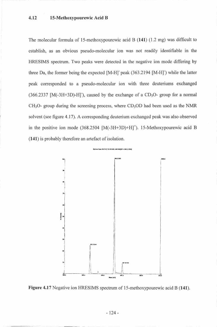
4,12 l5-Methoxypourewic Acid B
The molecular formula of lS-methoxypourewic acid B (141) (1.2 mg) was difficult to
establish, as an obvious pseudo-molecular ion was not readily identifiable in the
HRESIMS spectrum. Two peaks were detected in the negative ion mode differing by
three Da, the former being the expected [M-H]'peak (363.2194 [M-H]-) while the latter
peak corresponded to a pseudo-molecular ion with three deuteriums exchanged
(366.2337 M(-3H+3D)-F{), caused by the exchange of a CD3O- group for a normal
CHIO- group during the screening process, where CDIOD had been used as the NMR
solvent (see figure 4.17). A corresponding deuterium exchanged peak was also observed
in the positive ion mode (368.2504 M(-3H+3D)+HI). l5-Methoxypourewic acid B
(141) is probably therefore an artefact of isolation.
[email protected]& erF.P.gl
Figure 4.17 Negative ion HRESIMS spectrum of l5-methoxypourewic acid B (141).
-124-
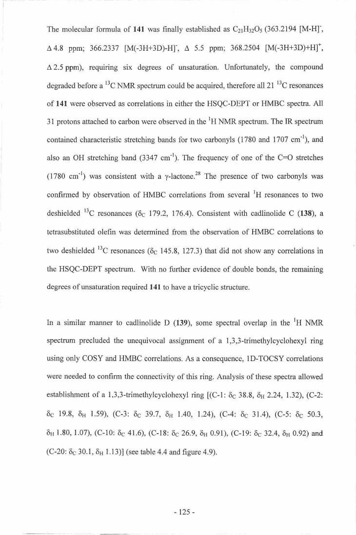
The molecular formula of 141 was finally established as C21H32OsQ63.2194 [M-Hl',
A4.8 ppm; 366.2337 [M(-3H+3D)-H]-, A 5.5 ppm; 368.2504 [M(-3H+3D)+H]*,
A 2.5 ppm), requiring six degrees of unsaturation. Unfortunately, the compound
degraded before a l3C NMR spectrum could be acquired, therefore all 2l t3C resonances
of l4l were observed as correlations in either the HSQC-DEPT or HMBC spectra. All
31 protons attached to carbon were observed in the lH NMR spectrum. The IR spectrum
contained characteristic stretching bands for two carbonyls (1780 and 1707 cm-'1, and
also an OH stretching band (3347 cm-r). The frequency of one of the C=O stretches
(1780 cm-I) was consistent with a y-lactone.28 The presence of two carbonyls was
confirmed by observation of HMBC correlations from several 'H resonances to two
deshielded r3C resonances (66 179.2, 176.4), Consistent with cadlinolide C (138), a
tetrasubstituted olefin was determined from the observation of HMBC correlations to
two deshieldedr3C resonances (6c 145.8, 127.3) that did not show any correlations in
the HSQC-DEPT spectrum. With no further evidence of double bonds, the remaining
degrees of unsaturation required 141 to have a tricyclic structure.
In a similar manner to cadlinolide D (139), some spectral overlap in the 'H NMR
spectrum precluded the unequivocal assignment of a 1,3,3-trimethylcyclohexyl ring
using only COSY and HMBC correlations. As a consequence, ID-TOCSY correlations
were needed to confirm the connectivity of this ring. Analysis of these spectra allowed
establishment of a 1,3,3-trimethylcyclohexyl ring [(C-l: 6c 38.8, 6s 2.24, 1.32), (C-2:
6c 19.8, 6u 1.59), (C-3: 6c 39.7, 6u 1.40, 1.24), (C-4: 6c 31.4), (C-5: 6s 50.3,
6s 1.80, 1.07), (C-10:6s 41.6), (C-18: 6c26.9,6H 0.91), (C-19:6c32.4,6n 0.92) and
(C-20: 6c 30. I , 6H I . 1 3)l (see table 4.4 and figure 4.9).
125 -

COSY correlations between the lH resonances of various methylenes and methines
were used to establish the second substructure of 141. In particular, a four-carbon chain
consisting of two methylenes t(C-ll'. 6c 26.3,6n 2.31, 1.63) and (C'12:6c 26.5,
6n2.02, l.4l)] adjacent to a pair of methines [(C-13: 6c 40.3, 6n 2.91) and (C-14:
6e43.7,6H 3.09)l was determined from observed COSY cross-peaks. Proton H-14 in
turn correlated to an acetal centre (C-15: 6c I10.3, 6H 5.22). The lH resonance of acetal
C-15 showed a strong HMBC correlation to a lactone carbonyl (C-16: 6c 179.2) to
finalise a y-lactone ring. The deshielded chemical shift of H-13, and the absence of
further COSY correlations from it, is consistent with other yJactone rings (see
figure 4. I 8;.ze'trz'tlo
HH9
"+''(l-\*-ri'-'(/ io-J
1H-lH cosYt6-tag HMBC,,/--\
Figure 4.18 Establishment ofy-lactone substruchrre of
1S-methorypourewic acid B (141).
Finally, a strong COSY correlation was observed between a methyl (C-6: 6c 16.3,
6Hl.2l) and a methine (C-7: 6c 41.4, 6H 4.32). Both ClIr-6 and H-7 also showed strong
HMBC correlations to the ttc resonances of each other, and also to that of the
remaining carbonyl (C-17: 6c 176.4) to establish the third substructure (see figure 4.5).
Again, the final connectivity of 141 was established on the basis of several key
observed HMBC correlations to the two olefinic carbons. In particular, CI{3-6 and H-7
both correlated to an olefinic carbon (C-8: 6c 127 .3) while H-7 also correlated to C-14,
again indicating the attachment of the side chain at C-8. The tH resonance of C-20
-126-
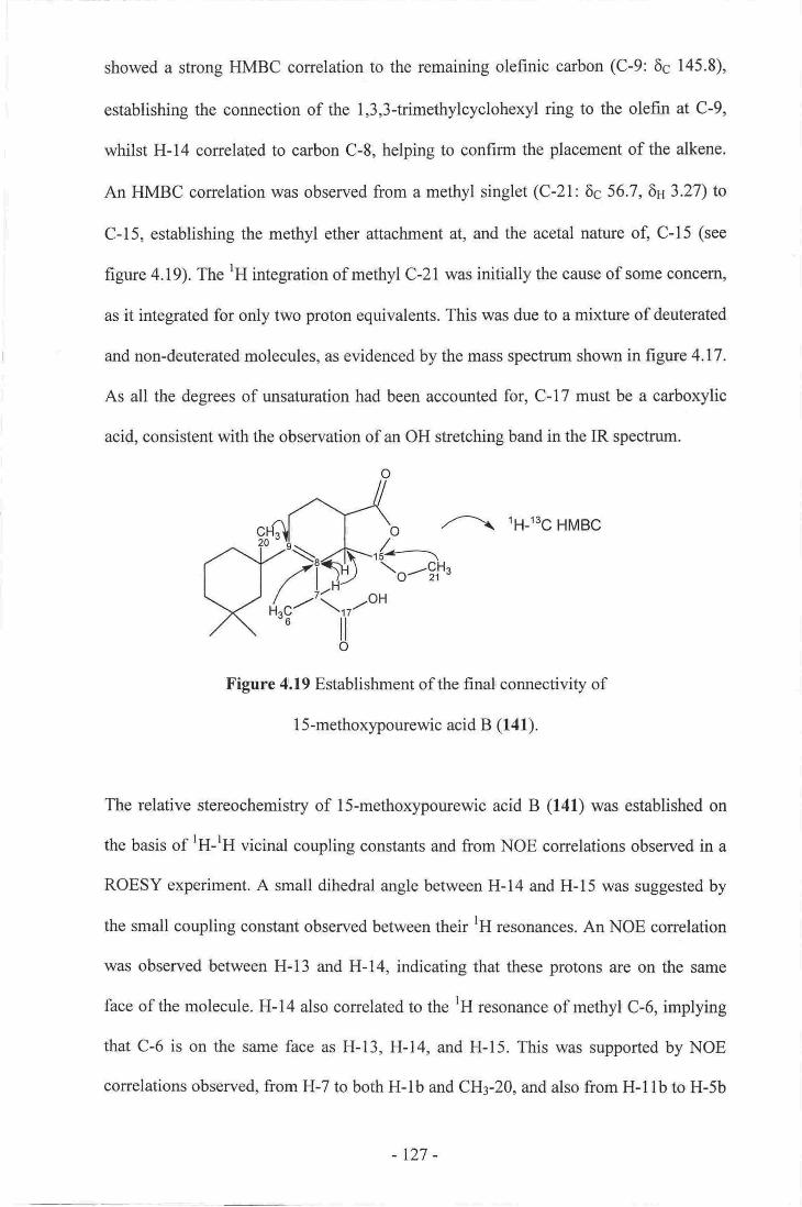
showed a strong HMBC correlation to the remaining olefinic carbon (C-9: 6c 145.8),
establishing the connection of the 1,3,3-trimethylcyclohexyl ring to the olefin at C-9,
whilst H-14 correlated to carbon C-8, helping to confirm the placement of the alkene.
An HMBC correlation was observed from a methyl singlet (C-21: 6c 56.7, 6n3.27) to
C-15, establishing the methyl ether attachment at, and the acetal nature of, C-15 (see
figure 4.19). The rH integration of methyl C-21 was initially the cause of some concem,
as it integrated for only two proton equivalents. This was due to a mixture of deuterated
and non-deuterated molecules, as evidenced by the mass spectrum shown in figure 4.17.
As all the degrees of unsaturationhad been accounted for, C-17 must be acarboxylic
acid, consistent with the observation of an OH stretching band in the IR spectrum.
o{------\
/---\ tg-tsg HMBC
\o--tfra
Figure 4.19 Establishment of the final connectivity of
l5-methoxypourewic acid B (141).
The relative stereochemistry of lS-methoxypourewic acid B (141) was established on
the basis of lH-rH vicinal coupling constants and from NOE correlations observed in a
ROESY experiment. A small dihedral angle between H-14 and H-l5 was suggested by
the small coupling constant observed between their lH resonances. An NOE correlation
was observed between H-13 and H-14, indicating that these protons are on the same
face of the molecule. H-14 also correlated to the tH resonance of methyl C-6, implying
that C-6 is on the same face as H-13, H-14, and H-15. This was supported by NOE
correlations observed, from H-7 to both H-lb and CH3-20, and also from H-llb to H-5b
-r27-

end eHr.ZO (see figpre,4.Z0), thes€ obser,vations atlo'tv-td the rolative storoochoutis-fy
of f s-srghoxyporrfowic,acidB (l{1) tO.be assieltgd ss 7R*, l0,S*, 13ft*, l4n*, JiR*'
n NoE
FlsurG a.?O Establishnent ofthe ohemiw of
I S"mefhoryypornemnie acftd B (1{8-
-128-

Table 4A t3c475, MHa) and rH (300 MHz) NMR Data (CDCls + CsHsN) of l5'-
metho4pourewic acid B (141).
HMBC
Porition;(,o''t 'uo 0b;;;'1r*")- COSY .'no.t TOCSY NOE
l8 38.8 Ctir 1.72 m lb lb
b 2.24 m l42,Jb lc?2 19.8 Clil l-S9 m,2H lb ls" lb'3q3b
3a 39.1 AH? 124 m l8b t.4O m
4 31,4 e5a 503 Cflr 1.07 tn 5b 5b
b 1.80 m 54 lb 5g llb, lE
5 t6;3 CHs 127 d (6.E) 7 7|8,17 7'14
7 4l/' gfd' 4's2 c(t'0) 6 6'&' 14' t7 lb" 6'r' 20
8 lnli g
9 lil5.E C
l0 41.5 C
Ita 26.3 CHr l'63 In llb, t2a' I2b llbb 231 m lla l2a l2b 5b' tla' l2b'20
lhs 26.5 CIlr 1.41 n llq llb, !2b' 13 Ulb' 13
b L02 m I lq, I lb, I24 13 ltb, l.lE
13 &.3 C*t 2.91 m l2q l2b, 14 124 14
' 14 43,.7 €tt 3-09 dd(lo"o}D 13,15 7.8,12 613
r5i u0.3 cI{ 52l-z d(3,?) t4 8, L6 5b'?l
16 179,2 re
r7 176.4 E
l8c 26.9 cHr o:91 s 3419 3i45,I9c19- 32.4 CtL Am s l8 3,4,5,18
20 310,1 eFlr l.l3 s l'5'9, l0 7' tlb?l 56.1 Cfi" 327 s 15 15
I Derermincd from HSQC.DEPT (multi.pllciq, edited HS.QC) ,and HMBC experim€nts.
Carton connoctivity detsnnrined frorn an HSQC'DEPT exper,iment, muhiplicity determined frorn
rH exper,imenf,
Interolmngeab-le.
,Hbl36*a
,.,-'{'-f{30 | e lcb
i-'-.-]rt=,-t--'(3_,,n-o-u "..."i\..r-oH
,es \" I(141),
-r29-
4.13 MethYlPourewate B
The molecular formula of methylpourewate B (142) (3'0 mg) was established as
CzrHrzOs from observation of intense pseudo-molecular ions in both the negative and
positive ion HRESIMS modes (365.2335 [M+H]*, A 3.5 ppm;363.2185 [M-H]-' A 5'3
ppm), implying six degrees of unsaturation. All 21 carbons and 3l protons attached to
carbon were observed in the l3C and tH NMR spectra respectively. Prominent peaks in
the IR spectrum included an OH stretch (3325 cm-t; attd two C:O stretches (1762,1735
cm-r). Significant t3C NMR signals included two carbonyl (6c 178'9' 175'3), one hemi-
acetal (6c 103.8) and two tetrasubstituted olefinic (69 147.2,127.3) resonances' With no
other evidence of multiple bonds, the remaining degrees of unsaturation requiredl42to
have a tricyclic structure.
The presence of a 1,3,3-trimethylcyclohexyl in 142 was established based upon a
similar analysis of the COSY and HMBC spectra as that performed for cadlinolide C
(13S) [(C-1: 6c 39.0,6H 2.18, l.2g), (C-2:6c l9'8,6H 1'83, l'51)' (C-3:6c 39'9'
6s 1.36, I.2l), (C-4: 6s 31.6), (C-5: 6c 50.6, 6H l'77, L00), (C-10: 6s 42'l)' (C-18:
6626.8, 6s 0.85), (C-19: 6c32.9,6n 0.87), (C-20: 6c 30.6, 6H 1.05)l (see table 4.5 and
figure 4.3).
COSY correlations were used to establish a linear chain of three carbons [(C-11:
6c26.8, 6s2,34,1.47) to methylene (C-12: 6c27.5,6H 2.09, 1'30) to methine (C-13:
6c41.2, SH 2.97)1. Initially, the resonance of H-13 overlapped with that of another
methine proton at the same frequency. Addition of a small amount of Ds-CsHsN caused
these two overlapping resonances to spread apart. It was found that the two resonances
coupled to each other, as evidenced by a resolved COSY cross-peak between them,
-130-

revealing the connection between C-l3 and a second methine (C-14: 6c 45.8, 6H2.97).
A COSY cross-peak was also observed between H-14 and a hemi-acetal resonance
(C-15: 6c 103.8, 6H 5.41). Proton H-15 showed HMBC correlations to C-14 and also to
that of a 7-lactone carbonyl [(C-16: 6c 178,9)1. As with 141, the deshielded chemical
shift o{, and the absence of any other couplings from, H-13 supports the proposal of a
y-lactone ring (see figure 4.18).
Finally, a strong COSY correlation was observed between the tH resonances of a
methyl (C-6: 6c 16.6, 6n l.2l) and a methine (C-7: 6c 41.5, 6n 4.30). Both CHr-6 and
H-7 showed strong HMBC correlations to the ttc resonances of each other and also to
that of a carbonyl (C-17: 6c 175.3). A strong HMBC correlation was also observed from
an oxygenated methyl (C-21: 6c 52.1, 6H 3.71) to C-17, implying a methyl-ester
functionality (see figure 4.21). The chemical shifts observed are comparable to those
noted for the methyl ester derivative of pourewic acid A (f40), and for membranolide
(12t).15t
/xf\1'g-i-?v(d' ,/--\
lH-1H cosYtH-r3c HMBC
Figure 4.2l Establishment of the side chain of methylpourewate B (f42).
All of the correlations already mentioned had accounted for all the oarbons in the
molecule except two substituted olefinic centres [(C-8: 6s 127.3), (C-9: 6c 147.2)1. As
before, the placement of the olefin and the final connectivity of the molecule were
established from key observed COSY and HMBC correlations. Strong HMBC
correlations from H-7 to C-8, C-9, and C-14, and from CH3-6 to C-8, established the
connection of the side chain at C-8 between C-9 and C-14. As with cadlinolide C (138),
-131-
CH3-20 also showed a strong correlation to C-9, confirming the attachment of the
1,3,3-trimethylcyclohexyl moiety at C-9, and established the final connectivity of the
molecule. The connection of the olefin within a six-membered ring was confirmed by a
homo-allylic coupling between H-1lb and H-14 as observed in the COSY spectrum (see
figure 4.22).
HaHbl
-111H-lH cosYt6-tsg HMBC
nl | \cFt.\l I ofl -tsr-it- /I rt).F \| / t-n/ r)Hfl, /--\
OH
t{rg-'Yo-""uo
Figure 4.22 Selected correlations establishing the
final connectivity of methylpourewate B (f 42).
The stereochemistry of methylpourewate B (142) was determined from both rH-rH
vicinal coupling constants, measured in the tH l.tl{R. spectrurn, and NOE correlations,
detected in a ROESY experiment. As with l3t, H-15 shows a small coupling to H-14,
consistent with a small dihedral angle between these protons. Due to overlap of their
close chemical shifts, no reliable NOE could be detected between H-13 and H-14. The
coupling constant between these two protons, however, is similar in magnitude to those
measured for 13E, therefore it is aszumed that the stereochemical relationship between
the two protons is the same. Several of the NOE correlations used to determine the
relative stereochemistry of l.3t were absent in 142, precluding a similar stereochemical
analysis. Although none were observed between H-5a and H-l lb, NOE conelations
were observed between CH3-6 and both H-14 and CH3-20, and also between H-7 and
both H-lb and CI*-20, in a similar manner to that observed for l3t, therefore the
stereochemistry for C-7 and C-10 is assumed to be the same. These observations and
-r32-

,a$sumpti s allonr the. rolative stofcochodtl'stry of melhylpourewat€ B (f4A) to bG
tentatively assignd as: 7R*; lffi*, lffi*, 14S*, l5rt* (see figur€ 4.8).
(a NoE
['igulc,4.!3, NOE s-ffila,tiont F,{tublbhing tr€
s'teroo&emilitr,y, of ul@pormrwato B (144
-133-

Table 4.5 r3C 05 MHz) md rH
(30CI I'r{Itu) NMR Dafa (cDc[) of methylpourewate B (142).
HMtsCrHbtsgq
la,b
2b
^3a
b
4
5a,
b
6'1
r8
It0
l!a(a|b (es)
l2a (or)
b (es)
t3
t4t5
t6l?
l8c
190
20
rnult
39O CItr
l9t8 CIIr
39.9 CHI
?1,6 c5q6 C!r,
IR
6m
m
r!n
m
m
d (6.8)
s (al)
m
dt (15,9" 2.9)
m
m
q (816)
dd (10.0,3.el
.r (3.3)
b
l*?a'2blh 2q 3a
1b,2b,34 3b
Za tb2418
5b
ia7
6
I lb, l2a, l2bllq 124 ttb, 14
llq llb, 13
lla, lIb, 13
t4lza, l2b, l,!b t3,15
l'4
7,4,5
1,4, 16
7,&r76,8,9,14,11
8. 13, l514 16
3,4,5" 19
314,S,l8
1, S, I, l0l7
lb,wllbrl2b
llb
5a
3b, t8
14" 20
lh20
llt;lzb2s,2b.l lg12bl0
l2D
2b;I 14l lbrl2a
6
Cub
16,6
4r,5
LZI3:1472.
42.1
26,8
n.s-
4134ts4'o3.8
1789
1753
26,8
CttiCH
cce
CHr
t?e2.18.
1.5.t
1,83
1.36
l.2t
1.00
t.7v
t.2r430
t.47
2,t41.30
2.W
2,91
2,91'
5.4r
t2
s2930r6
52.l
erl?
CH
CH'cH
cc
CI{:
eHiCHI
0.85 e,
0.8,7 s
1.05 s
3b, 18
l8
a
b
3.71 s
Mulfiplicity determined fiorn HSQC-D€PT (multiplicity edited HSQC) experiment
Carbsn conrcstivity detennin€d fro'm an I{-SQC-DEFT experiment, multiplicity deterniirsdrI{ e4porimeRt.
lnterchangeable,
fi'om
o
,,-'\!o-(le lc)''\-;'=('"
..""+\nnzo$'I
(142)
- 134-

4.14 Pourewanone
Strong pseudo-molecular ions were observed for pourewanone (143) (2,2 mg) in both
positive and negative ion HRESIMS experiments, establishing a molecular formula of
C2sH3sO5 (351.2160 JM+H1*, A 1.8 ppm, 718.4527 [2M+NH4]*, A 0.3 ppm, 349.2019
[M-H]-; A 0.5 ppm) and requiring six degrees of unsaturation. All 20 carbons and 29
protons attached to carbon were observed in the l3C and lH NMR spectra. Notable
features of the IR spectrum included an OH stretch (3335 .*-'), and both a broad and a
narrow C:O stretch(1725,1665 cm-r respectively). The band at 1665 cm-r was suitable
for an o, B-unsaturated carbonyl.tuo A single peak was noted in the UV spectrum
(l,ma(:248 nm, tr*:l 4,425), characteristic of a cr,B,B-saturated-enone system.160 Th"
t3C NMR spectrum of 143 was quite different from any of the other diterpenes isolated
during this study. Notable r3C resonances included those of three carbonyls (6s 198.1,
177.2, 161.0), a polarised olefin (6s 167.3, 135.6), and an oxygenated methylene (6s
62.9). The l3C resonance at 161.0 ppm was assigned as that of a carbonyl, and not as
part of a polarised olefin, by the following reasoning. Attachment of a proton at 8.06
ppm was determined from a correlation in the HSQC-DEPT spectrum. The l-16,11
coupling constant between these resonances was found to be 225 Hz when measured in
a rH coupled HSQC-DEPT experiment. A t-ls,H coupling constant of this magnitude is
diagnostic of formate functionality, which was also consistent with both the lH and l3C
chemical shifts.l6l-l6a tnitiaily, observation of an HSQC-DEPT correlation between the
lH and l3C resonances of the formate was diffrcult, as the signal was weak due to the
inappropriate lJg,H value (140 Hz) used when running the experiment. Once this value
had been optimised (rJ6,s=190 llz), the strenglh of the formate HSQC-DEPT
conelation was enhanced, allowing for better detection.
-135-

As with all the previous related metabolites, careful analysis of the COSY and HMBC
spectra allowed the rapid assignment of a l,3,3trimethylcyclohexyl moiety [(C-l:
6c38.3,6n 1.96, 1.29), (C-2: 6c 20.5,6s 1.61), (C-3:6c 39.5,6n 1.28), (C-4: 6c 31.8),
(C-5: 6c 49.6, 6H 1.84, 1.38), (C-10: 6c 41.0), (C-18: 69 29.0, 6n 0.83), (C-19: 6c 31.4,
6n 0.95), (C-20: 6c28.8, 6s 1.28)l for 143 (see table 4.6 and figure 4.3).
A linear chain of three methylenes and a methine was established on the basis of
observed COSY correlations. The protons of a methylene (C-l l: 6c?7 .7, 619-2.72, 2.63)
were strongly coupled to the protons of another methylene (C-12 6c 25.1,
6n2.11,1.74). These in turn showed strong COSY correlations to a methine (C-13:
6c4.7,611 2.56), which was coupled to an orygenated methylene pair (C-15: 6e62.9,
6H 4.49, 4.26) (see figure 4.24).
,_ji),i#,doAL *"1*
1H-lH cosY
Figure 4.24 Establishment of the methylene chain
zubstructure of pourewanone (143).
The final COSY correlations observed were between a methyl doublet (C-6: 6c 15.0,
6n 1.37) and a methine (C-7: 6c 39.0, 6n 3.84). As beforg both CIIr-6 and H-7 showed
HMBC correlations to the "C resonances of eaoh otheg and to that of a carbonyl
(C-17:6c 177.l) (see figure 4.5).
The COSY and HMBC correlations detailed above had accounted for 16 of the 20
carbons of 143, leaving a polarised olefin, a carbonyl, and a formate, to be assigned to
define the final connectivity of the metabolite. The final connectivity of the molecule
- 136-
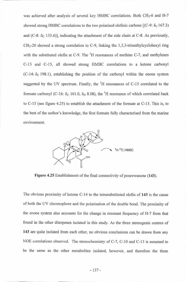
was achieved after analysis of several key HMBC correlations. Both CHg-6 and H-7
showed strong HMBC conelations to the two polarised olefinic carbons [(C-9: 6c 167.3)
and (C-8: 6c 135.6)1, indicating the attachment of the side chain at C-8. As previously,
CH3-20 showed a strong correlation to C-9, linking the 1,3,3-trimethylcyclohexyl ring
with the substituted olefin at C-9. The rH resonances of methine C-7, and methylenes
C-13 and C-15, all showed strong HMBC correlations to a ketone carbonyl
(C-14:6g 198.1), establishing the position of the carbonyl within the enone system
suggested by the UV spectrum. Finally, the lH resonances of C-15 correlated to the
formate carbonyl (C-16: 6c 161.0, 6H 8.08), the rH resonance of which conelated back
to C-15 (see figure 4.25)to establish the attachment of the formate at C-15. This is, to
the best of the author's knowledge, the first formate fully characterised from the marine
environment.
,/--\ 1H-13C HMBC
Y""o
Figure 4.25 Establishment of the final connectivity of pourewanone (143).
The obvious proximity of ketone C-14 to the tetrasubstituted olefin of 143 is the cause
of both the UV chromophore and the polarisation of the double bond. The proximity of
the enone system also accounts for the change in resonant frequency of H-7 from that
found in the other diterpenes isolated in this study. As the three stereogenic centres of
143 are quite isolated from each other, no obvious conclusions can be drawn from any
NOE correlations observed. The stereochemistry of C-7, C-l0 and C-l3 is assumed to
be the same as the other metabolites isolated. however. and therefore the three
laR\fr
-r37-

stereogenic centres of pourewanone (143) are tentatively assigned as 7R*, 10'S*, 13'S*
consistent with all other metabolites isolated during this study.
-138-

Table 4.6 t3c(100IuHz) and rH(300 il4H2)Nh{RData(cDC13) ofpotnewanone (143).
TIMBC'Htt3C.
4264.49
8.0E
cx{lCtlrc
g.Ht
gHt
EH
cce
Olfi
Cllr
CH
cCHr
lab
2
3
4
5.a
b
6
7
I9
l0llab
l2tb
l3l4l5a
b
l6t7
t8C
lg!2g
38.3
20.5
3.531.8
4q6
15.0
39,0
13J.6
r67.3
4[.02t1.7
25,t
M:Ir93.1
679
161.0
177.r
29.'0
31.4
CXlr
colrc
,ctt
CH'
t29r.95
l.6lt28
138
!.84
137
3.84
2:63
2:il2
t.T4
2,ll2,56
m
mm.2Hm,aH
m
md (6'81
q (6CI
dd (10.3,4,6)
t(3.9)nm
n
dd 0 1.5, 7.1)
dd (n2,41)s
cosYlb,2142
lc lt,3?
5b, lE
5a
1
6
!tb, 124 lzb
t[la. l2a' tAU
ftE, I !b- t2b, 13
tla. llb" t2q 13
l2q lzb l5o" l5b
r3, lsb, 16
13, fsa, 16
lSa" 15'b
5E, t9
l8
4, l0l8
10, 18
l07,8,9,17
6" 8,9, 14 l?
I
l3t3t4
12,13,14, 16
la 13; l4 16
l5
3,4,5, 19
3"d5, l8
" n{Uthueity determinod from HSQC-DEPT (rruXliplicity,odited HSQC) experiqent'
Oarbon eonncstiitity deteiminsd frorn an HSQC-DEPT eryXtriuonr" multipliaity dbfemined fion!H er-poriment
hrterchangeable.
\ru-ottlo
(r43)
-t39-
4.15 Biogenesis of the Isolated Spongian Diterpenes
Several of the spongian diterpenes isolated as part of this study are of biogenetic
significance. They are either suggested intermediates in previously proposed
biosynthetic pathways, or they represent a new carbon skeleton that is fomted vla a new
mechanism. Each group of metabolites will be dealt with in turn.
Cadlinolides A-D (132-133, f38-139) are structurally related to the gracilin-class of
compounds (1l&-fB) and are therefore presumably formed vla a similar mechanism to
that shown in scheme 4.7.t40't48'tso K*y steps of the biogenesis are the l,2-hydride and
I,2-methyl shifts, followed by the opening of the epoxide ring to form an alcohol. The
C-5 C-6 bond is then oxidatively cleaved to form a carborylic acid. Instead of
undergoing a decarboxylation as proposed for the gracilins, however, the C-17
carboxylic acid would undergo a lactonisation with a C-15 alcohol. The fotn members
of the class are formed by various oxidations and functionalisations of the
five-membered heterocyclic ring. This mechanism is summarised in scheme 4.11.
-140-

ox.+
Scheme 4.11 Proposed biogenesis of cadlinolides A-D (132-133n 138-139). Based upon
the proposed biogenesis of the gracilins.l50'ls2
Pourewic acid A (140), l5-methoxypourewic acid B (141) and methylpourewate B
(142) are also closely related to the gracilin family of compounds (118-18).140'148-150
The biosynthesis of the gracilin compounds involves the oxidative opening of ring B to
give various carboxylic acids that decarboxylate to give compounds 118-123.150'152 'I-he
isolation of 140-142 helps to confirm this biosynthetic pathway, as they were predicted
intermediates of these mechanisms. It is unlikely that traditional purification method
oxi.+-
-r41 -

using silica gel would have allo-wed isolation of these compounds, as irreversible
binding of the metabolites to the chrromatographic subshate may occur. ln the
biosynthetic pathway proposed for the formation of 1l&-123, a leaving group (X) that
takes part in the decarboxylation reaction has been thought to be present on the original
ring C throughout the mechanism. As none of the compounds isolated during this study
have any functionality on ring C, it would appear more likely that some oxidation of
ring C takes place after the oxidative cleavage of ring B, to help facilitate the
decarboxylation suggested. A suggested biosynthesis of compounds t40-l42 is detailed
in scheme 4.12.
-t42-

I
'.6)+/tl
I
I
ttl"nt{ oxi.
Scheme 4.12 Proposed biogenesis of pourewic acid A (140), lS-methoxypourewic acid
B (141) and methylpourewate B (142). Based upon the proposed biogenesis of the
gracilins. I 50'l 52
Pourewanone (143) is the first representative of the new pourewanane carbon skeleton.
It is proposed that the oxidative opening of ring B occurs as for the cadlinolide and
pourewic acid classes. Loss of HzO leads to the formation of a Al4'15 olefin that then
undergoes an oxidative cleavage to form formate 143. This pathway is described in
scheme 4.13. Alternatively, a furanospongian diterpene could undergo oxidative
cleavage of the C-14 C-l5 bond followed by hydrogenation of the C-13 C-16 bond (or
lon.
f
metrrvration
-t43-

hydrogenation of C-13 C-16 and then cleavage). This is summarised in scheme 4.14.
No furano-diterpenes and at least one C-15 OH diterpene were isolated from
Chelonaplysilla violacea during this study, therefore the mechanism shown in scheme
4.13 would appear more likely. As mentioned, to the best of the author's knowledge,
pourewanone is the first example of a formate from the marine environment.
The oxidative cleavage of the double bond proposed is similar to the synthetic cleavage
of an olefin by O:.EO Such cleavages have been proposed for the biosynthesis of retinal
and vitamin Ar from B-carotene.l3'80 More recently, similar cleavages have been
proposed for the biosynthesis of the degraded pregnanes muricenones A and B (144,
145), and for the aromatic alkaloid cyclomegistine (146) (see schemes 4.15 and
4.16;.t0s'100 Both these biosyntheses have been supported by synthetic studies.l66'16?
i::("""';;l(o""
o
(144) R:H
(145) R=OAc
(r46)
-t44-
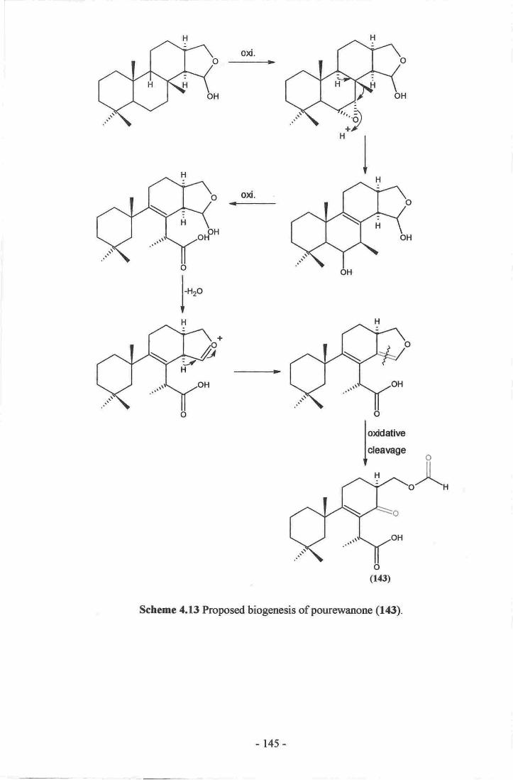
oxi.
-->
l*"
Schemc 4.13 Proposed biogenesis ofpourernanone (f6).
-145-

F
Scheme 4.14 Altennative biogenesis of pourovanone (143).
- 145-

oxi.
-CO2, + C1
+-
scheme 4.15 Proposed biogenesis of muricenones A and B (144, 14$.166
Scheme 4.16 Proposed biogenesis of cyclomegistine (146)'165
4.16 Biological Activity of the Isolated Spongian Diterpenes
Due to the small amounts of the metabolites isolated, only a few could be submitted for
biological evaluation. Cadlinolide C (138), pourewic acid A (140)' and
methylpourewate B (f42) were all submitted for evaluation in both cytotoxicity and
anti-inflammatory assays. None of the three metabolites showed any cytotoxic activity
against the HL-60 leukaemia cell line (maximum concentration 10 pM). The
147 -

compounds did show moderate anti-inflammatory activity as measured by inhibition of
superoxide production by human peripheral blood neutrophils stimulated with either
ft\4LP or PMA.r0s The results are summarised in table 4.7. Finally, 140 was also
submitted for anti-microbial activity and was shown to be inactive.
ICso
PMA fMLPcadlinolide C (138)
pourewic acid A (140)
methylpourewate B (f 42)
13 pM77 p,M
58 uM
13 pM741tM58 uM
Table 4.7 Anti-inIlammatory activity of several isolated spongian diterpenes.
It is unfortunate that a paucity of material prevented more of the diterpenes being
assayed for anti-inflammatory activity, as this prevents a true structure-activity
relationship to be suggested. Cadlinolide C (138), which contains a C-15 C-17 lactone
ring, is the most active of all, whilst pourewic acid A (140) and methylpourewate B
(142), which do not have this lactone linkage, show comparable and reduced activity.
4.17 Other metabolites from the Genus Chelonaplysilla
Other than the diterpenes mentioned during the literatwe survey, only one other
publication regarding metabolites from the genus Chelonaplysilla could be found. The
anti-microbial aromatic alkaloids (147-f 50) were isolated from the polar fractions of an
extract of the same sponge that yielded several diterpenes. It was felt by the authors that
the plethora of diterpenes found in various species of Dendroceratid sponges had meant
that other researchers had overlooked the more polar, and generally more biologically
active, nitrogenous compounds that the sponges produce. It was also suggested that
these compounds may more important components of the animal's defensive
mechanism.l6s
-148-

(147) R=H
(148) R=Br
(149) R1:OMe, R2:H, Rg:OH
(150) Rt:H, R2:R3:OMe
The use of NMR during this study, for the screening of sponges and the guidance of
isolation procedures, coupled with the use of reversed-phase stationary supports' has
resulted in the isolation of several nitogenous compounds (see ohapter two). It is
unlikely that polar nitrogenous compounds were present in the sample of
ahelonaplysilla violacea that yielded diterpenes 138-143. Alkaloids such as these
would have easily been noted during the screening process'
-t49-

5.1
Chapter Five
Isolation of a Marine Toxin Producedbv the Dinoflasellate K are nia br ev is ulc ata.
Dinoflagellate Toxin Classes
The presence of a dinoflagellate toxin in the environment is often not recognised until
cases of human intoxication from eating contaminated seafood, usually shellfish, are
reported. This is because many shellfish species can bioaccumulate toxins in their
various tissues without causing harm to the animal itself. The toxin is only noticed when
higher predators consume the seafood and become sick.l0'3e'42 The toxic symptoms
elicited in humans are therefore often used to classifu the broad categories of
dinoflagellate toxin. The four main classes of dinoflagellate toxin are Paralytic Shellfish
Poisoning (PSP), Neurotoxic Shellfish Poisoning (NSP), Amnesic Shellfish Poisoning
(ASP) and Diarrhetic Shellfish Poisoning (DSP). There is also a collection of other
unique toxins that do not fall under these categories.l0'3e'a2't6e-t77
5.2 Paralytic Shellfish Poisoning (PSP)
PSP is caused by the alkaloids saxitoxin (151), neosaxitoxin (152), and their
homologues. Both the saxitoxins and neosaxitoxins are produced by marine and
freshwater dinoflagellates, including various species of Alexandrium, Karenia and
Pyrodinium. I 7 l'l 72' I 78-l 83
-150-

(151) R:H (152) R:OH
The mode of action of both the saxitoxins and neosaxitoxins is the same; both classes
block the sodium channels of excitable nerve membranes which inhibits the propagation
of action potentials (voltages), thereby causing paralysis of muscles. Other major
symptoms in humans include hypersensitivity of the mouth and skin, ataxia, muscle
weakness, and a sensation of floating. PSP is often fatal due to paralysis of the muscles
controlling the lungs of victims.lEa
5.3 Neurotoxic Shellfish Poisoning (NSP)
NSP is caused by the brevetoxins, a class of polyether toxins produced by Karenia
brevis (syn. Gymnodinium breve) and other members of the same genus. It is blooms of
K. brevis that have led to the commonly used phrase "Red Tide", due to the
characteristic colour of large blooms of the dinoflagellate (see figure l.3). One of the
first records of a Red Tide is found in the Bible in Exodus 7 20-21: "... and the water
changed into blood. The fish died and the river stank...".6 The brevetoxin family
include brevetoxins A (153), B (154) and 82 (155;.tee'tzo'tts
(ls3)
-t5l-

(154)-:4
I NHzll l -
/t9-too"I
(155) R:iAv*
5.4
The mode of action of the brevetoxins is quite different to that of the sarcitoxins, even
though both classes affect the sodium channels of cell membranes. This is because
brevetoxins bind at a different site on the channel than the PSP toxins. The brevetoxins
cause sodium channels to stay open longer, by stabilising both the pre-open and open
states of the channel, allowing greater sodium ion flow into the cell. This increased ion
concentration causes neuronal membrane depolarisation, which stops nerve signal
propagation.a2 NSP can cause a wide variety of symptoms, including a reversal of
thermoreception, loss of coordination, headache, and general malaise.42't8a't86
Diarrhetic Shellfish Poisoning (DSP)
DSP is caused by several different organisms and covers a wide variety of symptoms.
The main causative agent is okadaic acid (156), which has been isolated from many
sources. Okadaic acid (156) was originally isolated from the sponges Halichondria
okadai and H. melandocia. One major producing dinoflagellate is Prorocentrum lima
although other genera, including Dinophysis, have been found to produce 156 and other
congenefs. t7 s'r7 6't 87
-152-

J.J
(1s6)
Okadaic acid (156) is a potent tumour promoter due to its ability to greatly inhibit the
serine/threonine protein phosphatases PPI and PP2A, which allows a build-up of
phosphorylated proteins in cells. This mode of action is similar to that of the well-
known tumorigenic phorbol esters, which activate protein kinases. Increased protein
phosphorylation leads to continuous o'secondary messenger" production, which irrcludes
excess fluid excretion from gut cells causing diarrhoea. Other symptoms of DSP include
gastrointestinal disorders such as nausea and abdominal pain.lsa
Amnesic Shellfish Poisoning (ASP)
ASP is caused by domoic acid (157) which was first isolated from the macro-alga
Chondria armata. Domoic acid (f5f was later identified as also being produced by
members of the diatom genus Pseudonitzschia. Lrke PSP, cases of ASP have been
fatal.tT 3,t7 4,r 88, r 8e
) ..-cooH'',,r-.,,.r
/\\*'/-coon
H
(rs8)
cooH
-1s3-

The mode of action of 157 is similar to that of kainic acid (158). Both metabolites are
excitatory amino acid agonists to glutamate, a neurotransmitter in the central nervous
system.lT3'174'l8e Both are thought to bind to kainate-sensitive glutamate receptors on
nerves. This causes a depolarisation effect, followed by an influx of calcium ions into
the cell that causes eventual cell death. Some domoic acid extracts may be more toxic
than others due to possible synergy between domoate and other excitatory amino acids
such as glutamate. Symptoms of ASP intoxication include vomiting and diarrhoea,
followed by short-term memory loss, confusion, disorientation, and possible coma.lsa
5.6 Other Dinofl agellate Toxins
There are many other toxins produced by dinoflagellates that do not fall into the four
main symptom classes, many of which are still problematic for the seafood industry.
These toxins include ciguatoxin (159), the agent that causes ciguatera seafood poisoning
(CSP), found after the consumption of many species of tropical fish.a2 The earliest
report of ciguatera poisoning is believed to have been recorded in China by Ch'en
T'sang-chi sometime during the T'ang dynasty (618-907 A.D.) who claimed that the
yellowtail is o'...a large poisonous fish fatally toxic to man." leo
(lse)
Ciguatoxin (f59) is produced by the epiphytic dinoflagellate, Gambierdiscus toxicus,
which is ingested by fish and introduced into the food chain.rel-re3 Th. symptoms of
-t54-

ciguatera are similar to that of NSP and include reversal of thermoreception, joint pain,
low blood pressure, nausea, vomiting, and dianhoea. Studies have shown that 159 binds
to the same voltage-sensitive sodium channel as the brevetoxins, hence the similarity in
observed symptoms.a2 Although other congeners contribute, the major toxic component
of ciguatera is 159.42
A second class of toxin is produced by G. toxicus which is also of great interest.
Maitotoxin (f60) is a unique natural product as it is the largest, non-biopolymer, natural
product ever isolated (Cto+HzsoO5sS2Na2, 3422Da), and it may also be the most potent
non-proteinaceous toxin known (LDso - 50 ng/kg intraperitoneal [IP] versus mice).a2'1ea-
In6 Studies to determine the mode of action of 160 are still cunently being carried
out.42,r94-196
Several other toxins have until recently been included in the DSP class, as they are
extracted in a similar manner and are often found in conjunction with okadaic acid
(156), although they do not show any structural similarity to 156 nor do they appear to
possess the same mode of action. These toxins include the pectenotoxins, such as
pectenotoxin-1 (161) from Dinophysisfortii and D. acuminata, and the yessotoxin class,
exemplified by yessotoxin (162), from Protoceratium reticulatum.te7-z00 It is now
recognised that these metabolites are not true DSP toxins.20l
(l6l)
-ls5-
tl
H
'H
H
(160)
- 155,-

3.7
(162)
Structural Considerations
It can be seen from the previous examples that there are two common structural features
of these natural products. First, most are highly oxygenated and they often contain other
rare functionalities (hydrates, imines, spiro centres etc). Second, the fused polycyclic-
ether carbon backbone is present in almost each toxin class and yet the modes of action
and toxic symptoms elicited in humans are all very different.
The basic polyether structure has been proposed by Shimizu to be formed by a cascade
ring closure of a polyepoxide compound. Shimizu proposed this mechanism for the
biosynthesis of brevetoxin B (154).3e The isolation of hemibrevetoxin B (163), the
exocyclic olefin portion of 154, would suggest that the cascade begins at that side of the
molecule.202 The suggested biosynthesis begins with a hydride shift, followed by the
cascade opening of the cis expoxides. The other polycyclic-ether toxins could
presumably be produced via a similar mechanism (see scheme 5.l).3e
(163)
-157-

5.8
Scheme 5.1 Proposed biosynthesis of brevetoxin B (154).3e
Dinoflagellate Toxins and New Zealand
Shellfish harvesting forms an integral part of the seafood industry in New Zealand, with
both economic and cultural significance. In 1998, fisheries exports were worth $NZ
1,236.8 million to the national economy, with shellfish accounting for $NZ 273.9
million of that total.203 Any long-term closure of shellfish harvesting because of a toxic
dinoflagellate bloom could have a serious impact on the New Zealand economy.
In January 1993, the entire coastline of New Zealand was closed to all shellfish
harvesting after cases of human intoxication were reported. The intoxications were
found to be caused by NSP toxins produced during a large algal bloom in late 1992. The
dinoflagellates present during the bloom were Alexandrium minutum and
-158-

Karenia brevis, reaching peak levels of I.l7 x 10' and 4.1 x l0' cells/L
respectively.204'20s The effect of this closure was the loss of several million dollars to
New Zealand's economy and over one thousand jobs during the first three months of
1993. It was fortuitous that the shellfish industry had reserved large stocks of non-
contaminated shellfish prior to Christmas 1992, and that the closure occurred while
most workers were on summer holidays. By the end of the financial year, the industry
had made almost a full recovery and most workers had been reinstated in their jobs.zffi
The results of the 1993 closure were not too serious for the New Zealand shellfish
industry, although that was due more to good fortune than good planning or
management. The New Zealand authorities have since instigated a continuous shellfish-
monitoring programme in order to better detect and respond to such situations, should
they arise again in the future. It is therefore important that new toxins are identified, and
methods to detect and quantifu any new toxins are established, in order to protect the
shellfish industry and its consumers.
Karenia brevisulcata
In the summer of 199711998, a bloom of a previously undescribed toxic dinoflagellate
species formed in the waters off the East Coast of the North Island of New Zealand,
conesponding to an unusually warm and still spring period.aa The bloom spread south
along the eastem coastline, and then across the bottom of the North Island along the
southern Wairarapa Coast and into Wellington Harbour (see figure 5.1). The
dinoflagellate was isolated and named Gymnodinium brevisulcatum (etymology: Latin
brevis : short, sulcatum : groove).43 The concentration of G. brevisulcatum peaked at
3.3 x 10⁷ cells/L in Wellington on March l3tt', l998.aa Several toxic dinoflagellate
genera including Gymnodinium, have recently been taxonomically reclassified and the
dinoflagellate is now known as Karenia brevisulcata, a species closely related to the
5.9
- 159 -

known toxic dfuroflageltates K. brevis and K mikimotoi (see figure 5.2 and
scheme 5 .21.4t4s'za7z6
Figurc 5.1 Hydrographic chart ofWellington llarbour and sotrthsn Wairampa Coast
(Courtesy ofland and InformationNew Zealand)
Figure 5.2 SEM image of l(senia brevisulcatq.
(Courtesy ofDr H. Ch8ng, NIWA 2003).
- 160-

The most notable effects of the bloom were due to the extreme biological activity
exhibited by the dinoflagellate. Almost all the fish, plant, ffid invertebrate species
within Wellington Harbour died. Most of the species not killed by the dinoflagellate
stopped eating and died of starvation instead. The biological activity of the presumed
toxin was quite unlike anything previously reported.a3'44207'208
Over 150 cases of human respiratory distress were noted during the bloom, with
symptoms including a dry cough, severe sore throat, inflamed and pufr eyes,
dermatitis, feverish influenza-like symptoms, headache, nausea, and stomach cramps.
The cases of respiratory distress were thought to be due to aerosol effects from sea
spray containing a toxin and/or live toxic algal cells.a
Samples of K. brevisulcata were acquired, and cultures of the dinoflagellate grown. It
was decided to attempt the isolation of the causative toxin(s) from artificially grown
cultures of K. brevisulcata, in order to determine the identity of the toxin. If the toxin
proved to be a novel compound, a secondary goal was to establish detection methods for
the metabolite that could be integrated into the current national shellfish-monitoring
programme.
-16l-

Kingdom Plantae (Plants)
Phylum Chromophycota
Class Chrysophyceae
Order Dinophysiales Gymnodiniales Peridinials
Entomosigmaceae Gymnodiniaceae Polyrikaceae
Gymnodinium
brevis
5.2 Phylogenic relationship of Karenia brevisulcata. For simplicity, not all
classes, orderso families, genera or species are shown.3s'*As
Species
Scheme
-t62-

s.10 Bioassavs Used to Guide Isolation
One of the key issues relating to the isolation of the Karenia brevisulcata Toxin (KBT)
was the method used to direct isolation. Unlike the NMR based screening method
described in chapter two, no chemical signature appropriate to guide an isolation
procedure was evident for KBT. Rather, as any toxin is obviously biologically active,
bioassays would be more appropriate to guide an isolation of I(BT. After identification,
other methods including ELISA (Enzyme Linked Immuno-Sorbent Assay), LC-MS or
NMR, may be established for quantitatively identifuing toxin levels in a variety of
samples. The major requirements of a bioassay to be used were fast turnaround time,
accuracy of results, and a need to be quantitative. The last requirement was of supreme
importance. If the assay used to detect a toxin is only qualitative, and the assay is very
sensitive or the toxin highly potent, then recovery of only a small amount of toxin may
still trigger a positive response in the assay, even if most of the active material had been
lost. Loss of the active material may occur through irreversible binding to the column
substrate or by degradation of the molecule. A quantitative assay, which can accurately
calculate the total recovery of the toxin, is therefore needed.
The first assay used to monitor the isolation of KBT from cultures of K. brevisulcata
measured the death of another dinoflagellate species following exposure to KBT. The
number of cells killed and the time to death were used to estimate the amount of KBT.
This assay was used to assess the toxicity of various cultures of K. brevisulcata, and
was also initially used to direct the attempted isolation of KBT. One 300 L culture of K.
brevisulcata was semi-purified through two fractionation steps using direction by this
assay. The resulting fractions from this batch were used for various biological trials,
including some stability tests (see below). Unforrunately, the assay was slow and
difficult to use as it relied on observing the time taken for the test organisms to die,
-163-

which could take a considerable time as samples could only be processed in batches of
five. The time taken to analyse many fractions generated from a purification step would
be prohibitive. Determining when the cells died was in itself a subjective matter as it
relied on personal experience and knowledge of the test organism. This assay was
dropped as the primary means of monitoring the isolation of KBT.
The assay used subsequently to direct isolation was a murine neuroblastoma (N2A)
assay. This assay is an MTT (3-(4,5-dimethylthiazol-2-yl)-2,5-diphenyltetrazolium)
based assay, originally developed for detecting the NSP or PSP toxin classer.20e'tto It
has been found, during the course of this study, that KBT has a distinctive and
reproducible activity that varies in a linear fashion with dilution in the N2A assay'
making it a quantitative system for measuring the amount of KBT.
In order to accurately quanti$ the amount of toxicity that a sample contains, a standard
must be used as a control against which the unknown samples are measured. To do this,
a measurement of arbitrary toxicity units (TUs) is used. One TU was defined as the
minimum amount of a specific sample of KBT (the "Gold Standard") required to affect
the viability of the N2A cells in the assay system. A standard aliquot of the gold
standard is added to a control well during each quantification, in order to calibrate the
results obtained from the N2A assay system by checking that the response of the N2A
cells is the same each time. Each sample is measured against the response of the cells to
the gold standard, and is quantified in terms of TUs without knowing any of the
physical characteristics of KBT (in theory, similar to determining molar concentration
without knowledge of molecular mass). It is therefore possible to note how many TUs
are loaded onto a column at any step, and then measure the recovery afterwards (see
figure 5.3).
-t64-

X'igure 5.3 N2A ce[s (Ief) unreatod" (nght)treced with KBT'
(Conrtesy of Dr P. Truman, ESR' 2003)
5.11 Culture Harverfing
Once an appropriate assay for guiding the isolation was in place' the main objective was
to quickly and quantitatively fractionate and puriff KBT. The initial step of extracting
the maximum amount of toxin from the cell culture was of major importanco, as this
would limit the maximum yield from any lrarvest. Prerrious work carricd out in our
laboratory had proven the use ofPSDVB in the foiitial extraction of dinoflagpllate to:rins
from culnrre media.6a It was asstrmed that most dinoflagellates would secrete any toxins
produced into the zurrounding media" in order to deter predation Any secreted toxins
passed through a column of PSDVB should absort to the stationary phase' The PSD\D
could then be eluted to achieve basic fractionation. The algal cells could also be
coltected (by filtration or centrifirgation) and extracted as well. An extract of the oells
cogld be cyclic loaded and eluted, with any toxic fractions combined with those
generated from the column that the media had been passed thongh' Normal
chromatographic methods could then be applied to isolate the toxin itself.n
-165-

In this way, several batches of K. brevisulcata, from 20-320 L culture media, were
harvested throughout the course of this study, each time yielding an extractable toxin
that performed consistently in all the chromatographic separations and assay systems
used. It was noted early on, however, that the amount of toxicity yielded varied greatly
from batch to batch. This could be, in part, due to differences in the timing of harvest in
relation to the life cycle of K. brevisulcatao a facet that has not yet been explored fully'
The time of harvesting, however, was not felt to be a major factor' More likely, the
dinoflagellate itself was not secreting the same amount of toxin in each batch due to
differences in culture conditions. Batches grown with dense cell concentrations,
indicating good conditions for growth, may not yield the most toxin, as the culture may
not be stressed enough to respond by secreting as much toxin.
In order to maximise the yield of KBT from each batctr, the dinoflagellate cells were
lysed to release all toxin into the sugounding media. This was achieved by adding
MezCO until a concentration of 10% MezCOlgO% media was reached' The change in
polarity caused all the dinoflagellate cells to lyse, thereby releasing any KBT contained
within the cell itself. By harvesting in this way, the toxic yield from -320 L
K. brevisulcata was increased from 150 x 106 TU to 2,500 x 106 TU (-16 fold increase
in toxicity). In a subsequent experiment, addition of freshwater to decrease the salinity
of the culture media has successfully lysed the cells of a wild sample of Karenia sp. in a
similar manrer.2llThis may in future be a cheaper and more environmentally safe
method to maximise the yield of various toxins from batches of cultured dinoflagellates.
Stability Tests5.12
With the availability of a quantitative assay, stability tests were carried out on aliquots
of semi-purified KBT. Each sample was exposed to varying amounts of either acid or
-166-
base over time. The activity of each fraction was then quantified by the N2A assay. It
was found that the level of activity was largely unaffected by exposure to acid, but was
highly sensitive to even small amounts of base (exposure to pH 9 for several hours
showed considerable loss of activity).t't This work was of extreme importance, as it
gave clear guidelines on the potential use of acidic or basic buffers as solvents in the
isolation of I(BT.
It was also shown that KBT remained stable in HzO/lvIeOH or HzO/lvIezCO mixtures at
4oC with no noticeable loss of activity.2r2 Finally, initial tests using the dinoflagellate
bioassay indicated that KBT is thermally unstable as most of the activity was lost after
heating to 100"C. Nonetheless, after prolonged exposure to boiling HzO for 30 minutes,
some toxic activity was still noticeable.2oT
5.13 Isolation
The isolation of KBT from cultures of K. brevisulcata is problematic. Two cultures of
KBT have been used to supply material for the purification procedure. The first was
without, and the second with, lysing of the algal cells prior to harvest. The first batch,
containing less toxic material, has been used as a pilot sample for experimental trials of
various isolation methods. Any purification steps that were shown to be effective were
performed upon the second sample that contained the larger amount of KBT.
The initial process remained the stepped elution of the HP-20 PSDVB column that the
toxic culture media had been passed through. Initially, the column was washed with
HzO and eluted with five different solvent mixtures (20, 40, 60, 80,
100%MezCO/HzO), with the bulk of the toxicity concentrated in the 60YoMezCO/HzO
fraction, although detectable amounts of toxicity were noted in both the 40% and
-t67-
80% MezCO/HzO elutions. For subsequent batches, the elution profile was changed to
maximise yield of toxin while reducing the number of fractions collected (HzO wash,
30, 70, 100% MezCO/HzO).
The next step was to cyclic load the toxic fraction onto a small amount of HP-20S or
Amberchrom that was then transferred onto a larger column of PSDVB as a slurry for
gradient MPLC elution. The toxin streaked from the column (eg. KBT eluted between
53-83% MezCO/HzO from the HP-20S MPLC column) each time this step was used.
This was assumed to be due to either overloading of the column or, more likely, from a
mixture of closely related toxic compounds eluting separately.
As a trial separation, aliquots of KBT were run on a PRP-I (PSDVB) analytical HPLC
column using a gradient from 40-70yo MezCOtHzO, pH 5, or pH 8 buffer, in order to
test the effect of acidic or basic buffers on the elution profile. Surprisingly, it was found
that under neutral conditions, two peaks of activity were detected while under both
acidic and basic conditions, only one peak was observed. Two repeat injections under
neutral conditions confirmed the original response. A fraction collected from each peak
of activity was re-injected under neutral conditions. In both cases, only one peak eluted
with the same retention time as previously, indicating that the toxicity was not from a
mixture of tautomers in equilibrium with each other. This was the first evidence
suggesting the presence of more than one toxic compound from cultures of K.
brevisulcata.
Many other chromatographic separations were attempted with the pilot KBT sample.
Column substrates used included further PSDVB (Amberchrom), LH-20 size-exclusion
gel, silica gel, and diol. Streaking of the activity was observed with most of the
substrates used. Diol was the only substrate that showed any particular promise as a
-168-

suitable column packing material. Initial trials using silica gel had shown that KBT
almost ineversibly retained on the column and would only elute in very polar solvent
(10-20%MeOH/EtOAc), which was too polar for adequate separation. Furthermore,
only 60% of the toxicity was recovered from the silica column. Diol is much more
stable to polar solvents such as MeOH and EtOAc, and polar molecules will generally
retain less to the substrate, therefore generally eluting earlier than as compared to silica
gel. Results from an initial trial using an aliquot of KBT on a small diol column showed
that all the toxicity loaded eluted from the column in the 2S%MeOHns%ErOAc
fraction, which although no earlier than with silica, was still well within the
chromatographic range of the packing material. On the basis of the small-scale diol trial,
a larger separation using the pilot sample was performed under flash chromatographic
conditions. The sample was loaded in 1%oEtOAc/99Yo CHzClz and eluted with mixtures
of EtOAc/MeOH. Bioassay testing of the fractions generated revealed that four peaks of
activity had eluted from the column, the first being the largest in terms of total activity
eluted (see figure 5.4). Each step in an isolation procedure generates a large number of
samples. In each diol separation, either every fraction or every second fraction had to be
quantified. As a result, a substantial number of assays were perfonned to identiff the
elution profile of KBT at each stage.
160
'140,120
J 100
SsoF6040
20
0
20 40 60 80
Fraction number
100 120
fl
A irJt\ n
Figure 5.4 Elution profile of KBT after running the pilot sample on a diol column.
-169-

As the pilot KBT sample had been successfully chromatographed using diol stationary
support, the major sample of KBT was processed in a similar manner. It had been noted
at each stage of the isolation procedure that KBT was only sparingly soluble in all
solvents tried, except for MeOH in which it was partially soluble. Even with MeOH,
large volumes were required to dissolve most of the sample. The column was therefore
loaded as a suspension of this sample, rather than as a solution. Again, four major peaks
of activity were eluted from this diol column.
As before, most of the fractions combined from this fractionation step still showed a
large degree of insolubility in most solvents. Unfortunately, the amount of MeOH
required to dissolve the major sample of KBT prohibited the use of HPLC as a method
for final purification. As diol is chemically stable to MeOH, it was decided to dissolve
the KBT fully in MeOH and then cyclic load the solution onto a small amount of diol by
diluting with CHzClz. Once loaded, the diol was transferred as a slurry onto a larger
MPLC diol column and run under a gradient elution profile from
10-50% MeOFVEtOAc. During the flash diol column runs, several of the later peaks of
activity eluted when a change in solvent had occuned. This may imply that the peaks of
activity were not from several different toxins, but were due to the same toxin streaking
off the column, "pulse eluting" with each new solvent front. This theory was
strengthened by the observation of the toxicity streaking from the diol MPLC column,
where a long tail was noted during elution rather than several peaks. This was similar to
what had been observed when earlier MPLC runs using HP-20S had been carried out.
-r70-

320 L Karenia brevisulcata
(CAW 82, innoculated 22112101)
30% MqCO
757o EtOAc
25% MeOH
Other peaks of activity
90% EtOAc10% MeOH
84.5 - 78.5%EtOAc
15.5 - 21.5% MeOH
50% EtOAc
50% MeOH
Passed through HP-20 column
Stripped with MqCOMqCO strip loaded onto small
HP-20 column
Scheme 5.3 Optimised large-scale isolation scheme for KBT.
Several samples were generated by combining fractions from running the major KBT
fraction on the diol MPLC column. Each sample was analysed by NMR. Again, each
sample was largely insoluble in the CD3OD NMR solvent. As a trial, the samples were
concentrated to dryness and re-dissolved in Do-DMSO. Each sample was fully soluble
in this solvent. It is thought that KBT becomes less soluble in most organic solvents as
it becomes more pure, a problem that has been noted previously in dinoflagellate toxin
research.sl'2tr'2ts This lack of solubility may account for the poor separation and
streaking of KBT from the chromatographic substrates used, as any metabolite needs to
be fully dissolved in order to achieve maximum chromatographic performance.
Unfortunately, DMSO is not a particularly good solvent for chromatography as it is
-r7t-

viscous, difficult to remove under reduced pressrue, and its ability to pass through the
skin, possibly carrying with it any dissolved toxic material, makes it a hazardous solvent
to use in the isolation of natural toxins. It has been suggested thal KBT may be a large
molecule as this sometimes correlates with lack of solubility except in DMSO.2I6
Finally, aliquots of KBT were run through amino or CBA (carboxylic acid) ion
exchange columns to test whether they would be useful for the final purification of the
toxin. It was found that KBT irreversibly binds to the amino substrate, suggesting some
kind of acidic functionality (carboxylic or sulfamic acid, phenol etc) in the molecule.
KBT was not retained by CBA, suggesting that no basic functionality is present in the
molecule. The presence of an acidic and absence of basic functionality may be
supported by the observation that KBT is highly sensitive to the presence of bases, and
not affected by acids, as noted during the stability tests.
The use of D6-DMSO to dissolve the KBT sample for NMR analysis has highlighted
several lH resonances, the intensities of which appear to wax and wane in a manner
similar to the biological activity, as measured with the N2A assay. These resonances are
of similar size, as measured by peak integration, and may therefore be from the same
molecule. There are also several other resonances that do not follow the biological
activity noted in the N2A assay, and whose signal intensity is an order of magnitude
larger than that of many of the other resonances noted. This certainly implies that the
sample is not yet pure.
5.14 Spectral Analysis of KBT
Each fraction generated throughout the isolation using both the pilot and large samples
of KBT, was tested using in-house NMR and MS facilities. HRESIMS was attempted
-172-

using both positive and negative ion modes. Many signals were observed in each
sample in both modes, although none appeared to follow the biological activity
measured in the N2A assay. As noted above, KBT shows a marked lack of solubility in
all the solvents trialled except DMSO. This may have hampered opportunities during
the early stages of isolation to identiff possible NMR signals attributable to KBT. It
may also have precluded analysis by HRESIMS, as suffrcient KBT may not have been
dissolved to allow for detection. In addition, DMSO is not useful as a solvent for
analysis by HRESIMS as it can suppress analyte signal.2rT
Extensive analysis of the NMR spectral data of the most pure sample of KBT, collected
at Victoria University, proved to be inadequate for a structural analysis. In particular,
the 300 MHz spectrometer available did not show enough sensitivity or resolution to be
able to generate adequate spectra of KBT. Several NMR experiments were therefore
carried out at the University of Canterbury using a 500 MHz spectrometer. The use of a
higher-field magnet gave better sensitivity and resolution than obtained previously,
allowing for identification of several substructures of KBT. Extensive analysis of the
rH, COSY, HSQC, and TOCSY spectra obtained at 500 MHz revealed at least 97
individual rH resonances possibly attributable to KBT, falling between 0.67 and
6.48ppm. Of these 97 resonances, g0 correlated to 8l l3C resonances between 8.5 and
129.2 ppm in an HSQC experiment. Three exchangeable lH resonances were noted
from COSY correlations, observed when the sample was dissolved in D6-DMSO. These
resonances are assigned to exchangeable protons as there is no correlation to these
resonances in the HSQC spectrum, and the COSY correlations are absent when run
using CD:OD. Finally, it is assumed that all six lH resonances between 5.88 and 6.04
ppm are attached to carbon, even though four show no direct correlations in the HSQC
spectrum (see table 5.1). It should be noted that any analysis of the NMR spectra must
be viewed with some caution, as the sample used for the NMR experiments is of an
-t73-

unlcroum purtty (there is at least ono mafor impruity in the saople). The sample rntly
also cantain several congpner$ or hornologues of a tsxin in vry concentrations, and tbe
toxin may be a sifficantly large moleculeo therefore any correlations observod in the'
TOCSY or COSY specFa may be due o ov,erlapping resonances of differeril spin
sy$ems within the sarne molecule, or even between different molecules altogether.
-LVA-

I3c (ppm) Et (p-pn) r3c (onm) rH fupm)
),
?,
7
?
129.17
t29;10
93.58
87.45
87,0x
83.76
8ai?5
83,63,
&2.t8
8,1.06
79,3E
7E.51
78.fl
7*46
78.30
11,82
11.5d
ltl:44
16.m
76,M
7:5.88
75.83
1535
'14,8)73i4!
73.t3
'12,72
72,69
7t.35
71,19
7t.t8
7An
?0-63
?0.36
?029
?ol3?0.1?
69,16
69.01
6n14
6.04
5"99'
5i95
5:88
5'96
5,89
6,4E
4.63
4.68
s.29
f J,e
53'V
LZ3
3,t0
3o!,
3.33
3.18
325
3,(X
420
E"fr1
4.0r1
3.84
,3,i10
L9.1
2,85
3yr3.59
3"@
4.31
3,34
329
3.4r
3.t3
3.15
33,t
3.83
3.41
l.0e
329
424
3,42
330
3.78
65,89
65163
.6s.35
6!"55
,63,rfll
59i1E
5.6.08
45j645,09
4?.12
4I,TT
40,49
38"28
38"0?
34.86
3135
3l.3rf
31.34
z97t2E.61
'2659
2it_'60
?1..60
21"40i
21.25
2r.03
10"50
20.61
20.54
N,89,
,0.0[
r9J8
18.28
r6.39
l518
l{.t614.31
14.t6.
9.57
&-88
&54,
t.v4
3.79
3-85
*r0-1
3,74
35,2
l5E
l.3s
XlM
129
135
1.52
lA61.54
l',36
067
l,:1:I
1.30
l.3tt.21
1.31
\.29
1,25
r.04
t.07
r,t3
r.0$
r.37
t"31
t25t,14
1..35
l.t3t,08
t"0t
l.[5
IJE117,6
1.58
Lfiz.t6
LL&
2.93
1.?.6
LT?
1.02
414
[.63
0.73 l,@
4el (oH)
4"84 (OH)
4,7? (Otn
Tatle S.tr Observed rII aad ttNMRtresoranoesof KBT (500 MHa D6-DlildSO).
To help tn the identiflcaflon o-f KE-T, a database of over l4S0 indivifual IIil-l3C one-
bond NMR oorelations was €stablished" containing data finm 30 rcported atgal toxins
-175.
including okadaic acid (156), azaspiracid, two pectenotoxins, several brevetoxins, two
ciguatoxins, yessotoxin (162), various saxitoxins, a prymnesin, several amphidinolides
and others, as well as the zooanthid toxin palyoxin (see figure
5.51.sr'rzo'rr8,180,18s,18?.1s5,t97,2t4'2t8'234 The most important observation from analysis of
the database is that KBT is not one of these compounds. For instance, it is unlikely that
KBT is related to the prymnesins or colopsinol A as these structures contain
carbohydrate residues whilst KBT shows no evidence of any anomeric acetal or hemi-
acetal centres.2l4,2t5,227 Similarly, KBT is unlikely to be a saxitoxin derivative as it
obviously contains olefins, a functionality not seen in this class of toxins.lT8'180'22s
Finally, many common dinoflagellate toxins, including yessotoxin (162)' the
brevetoxins, amphidinolides and others, contain exocyclic olehns; there is no evidence
that KBT possesses this structural feafure.le7'22r'222228-230 Th" presence or absence of
several other functional classes precludes KBT as being one of the structures of the
other common algal toxins, the NMR data of whigh are contained within the database.
This analysis does not preclude KBT being a congener or homologue of one of these
compounds or classes.
As well as being used to establish that KBT is not a contmon dinoflagellate toxin, the
database was also used to identify chemical environments that may give rise to the
various observed correlations in the COSY, TOCSY, and HSQC spectra of I(BT. Many
of the correlations identified in the HSQC spectrum of KBT are indicative of
oxygenated methines, the majority of which are consistent with polycyclic-ether
functionality. From a chemotaxonomic perspective, this is entirely consistent with the
many toxins already isolated from dinoflagellates.
-176-
(urdd) HroOF(\CvltlO(0NcO(DF.
o
oro
F88Yt-()
dtF
oK}IF
cto(\l
.l f:I[n1 tar.
la a't*
ffi a
tao. laa- .to
al
t tI a
t:#(-
:i ft a
ia
l?.' l.
tII )r o
&. ,t
H it'.
Figure 5.5 lH-l3C one-bond correlations of common dinoflagellate toxins.
-177-

Detailed analysis of the COSY, TOCSY and HSQC correlations of KBT have allowed
some possible substructures to be constructed. There are six 'H resonances near 6 ppm,
coupled together in three pairs of disubstituted olefins as evidenced by COSY
conelations. The chemical shifts of these lH resonances are indicative of non-polarised
olefins, as each pair of resonances do not differ significantly from each other. The
shifts, however, are de-shielded from what would be expected for an isolated olefin in
an aliphatic chain and are, in fact, more consistent with the resonances of a conjugated
diene system, or possibly an olefin within a small ring system. Conjugated diene
systems are precedented, as both the prymnesins and palytoxin (164) contain this
functionality. In particular, 164 has two pairs of conjugated dienes, one of which has
chemical shifts similar to those observed in KBT (see figure 5.6).t" Palytoxin (16a) is
not a valid candidate structure for KBT, however, as several other prominent NMR
signals from it are absent in KBT. For example, 164 contains 15 olefinic
f H-r3C correlations, including one with a tH tesonance at 6n 7.79 wltrch is significantly
less shielded than any observed in KBT.23l
133.47 130.04
s.37 'l u.oo I*',* i/, '74--zs' 5.78,i.-/-\ i,,^tri\"r:^lk
6s.0 t2s.87 133.88
Figure 5.6 Selected NMR chemical shifts of palytoxin (164).23r
Furthermore, analysis of observed COSY cross-peaks indicates a connection between
two olefinic protons (6H 5.96, d 4.9 Hz;6s 6.04, d, 5.9 Hz), each connected to one of
two oxygenated methines (66 87.45, 5H 4.63; 6c 83.76, 6H 5.29). Each olefinic
resonance shows only one coupling, to the other, in the lH NMR spectrum (evidenced
-178-

by the multiplicity of the tH resonances), implying a dihedral angle of approximately
90o between the olefinic and orygenated methine protons. Both the orygenated methine
tH resonances show a COSY correlation to each other. Based upon these observations,
it appears that this system may be a l,4-disubstituted A43-dihydrofuran ring although
there is no precedence for this functionality in the natural products literature. The
de-shielded chemical shift of the rH resonance at 5.29 ppm may be explained by the
proximity of another orygenated methine (6c 78.46, 6H 3.25) to which it is coupled, as
indicated by cross-peaks in both the COSY and TOCSY spectra. The rH resonance at
4.63 ppm is coupled further to another methine proton (6H 1.29). There is severe
spectral overlap in the rH spectrum at this frequency that precludes extension of this
spin systenq even though there are several other COSY and TOCSY correlations
observed from this resonant frequency (see figure 5.7).
1.29F.f 87
3.25H ;fl*:
n.u, H
rH-1H cosYH 65.29
6.04 5.95
X'igure 5.7 Proposed NMR chemical shifts of a
l,4-disubstituted A23-dihydrofuran ring in KBT.
A similar system that may also be considered for these resonances is a substituted 1,2'
dioxene ring, formed by the Diels-Alder addition of singlet Oz to a diene to form a
peroxide. This type of functionality is precedented in the natural products literature,
although the lH and l3C chemical shifts of the resonances from these molecules do not
compare favourably with those observed for I(BT (see figure 5.8).*t
-179-

128.0 t27.4
Figure 5.8 NMR chemical shifts of a 1,2-dioxene ring.235
The closest match for the chemical shifts of the observed resonances of KBT comes
from the ciguatoxin congeners CTX-3C (165) and C-CTX-I (166).232'236 Both 165 and
166 have the same structure from rings B to J. In particular, ring D consists of an
olefinic seven-membered oxygenated heterocycle. The chemical shifts of the olefin, and
the oxygenated methines adjacent to the olefin, are similar to those observed in KBT
(see figure 5.91.222'zs0It is unlikely, however, that KBT contains a ring similar to this, as
the COSY correlation observed between the two oxygenated methine lH resonances of
KBT is stronger (implying a stronger coupling) than what would be expected for a
purely homo-allylic coupling that would give rise to such a cross-peak in the ciguatoxin
derivatives. Also, the coupling constants of the olefinic protons measured in I(BT (5.9
H2,4.9 Hz) differ substantially from those measured in CTX-3C (165) (both 13 Hz) or
C-CTX-l (166) (13 Hz,7 Hz), indicating a different conformation of these protons from
those of KBT (see figure 5.91.232'zt0
(r6s)
- 180 -

3.71
13r,0 131.0
Figure 5.9 Selected NMR chemical shifts of C-CTX- | (166).232
A second series of correlations was noted in the COSY and TOCSY spectra. Three
exchangeable rH resonances (6n 4.91, 4.84,4.77) all appear to show strong conelations
to a methine proton (6H 4.00). Although an ortho-acid functionality (R-C(OH)3) is not
without precedence in organic chemistry, there is no precedent for such a functional
group in the natural products literature. It is also unlikely that such a functional group
would show such strong 4../H,H correlations to a proton on an adjacent carbon. It is
therefore much more likely that the thnee exchangeable tH resonances each conelate to
a different methine proton, all of which resonate at the same frequency (6s 4.00). This
may be indicative of three repeating structural units within KBT, or may suggest that
the sample is a mixture of three or more congeners. Finally, several other isolated
substructures have been identified through COSY correlations between their attached
tH resonances. All these substructures are detailed below (see figure 5.10).
- l8r

1.294.00 4.91HH 4.00 4.84HH
l-t-r' -"1,*
4.W 4.77HHl--t-r'-L
H0.72,8.54
H3
t2e{--,\Jn'h5.95 5.88
lH-lH cosY
Figure 5.10 Possible substructures and NMR chemical shifts of KBT.
5.15 Biologicel Activity of KBT
I(BT is a toxin and therefore has potent biological activity. Initially, after harvesting of
the oulture media by lysing of the cells and elution of the I{P-20 columr\ the toxic
sample of KBT had an N2A activity of 9.2 x 10⁶ TU/mg. The most pure active fraction
has an N2A actMty of 2.7 x t06 TU/mg. Analysis of a semi-purified sample of KBT
from 100 L of K. brevislcata has shown that the toxin has an LDso -500 TU/mouse
when injected W.'37 The most pure sample at the current level of activity therefore has
as an LDso -2OO nglmouse IP (i.e. LDso -10 pglkEIP, KBT can kill5000 20 g mice per
ms).
The symptoms of KBT in mice are most pronounced at high levels of exposure. At IP
doses of 50,000 TU, toxic effects were noted soon after injection. The mice became
hunched and lethargig and respiration slowed with cyanosis becoming apparent. Death
occurred approximately five hours after dosing. At doses between 5,000 and 20,000 TU,
the mice were unaffected for several hours with symptoms only becoming apparent
later; the animals died between ten and thirteen hours after dosing. When dosed with
between 500 and 2,000 TU, the mice showed no symptoms for 24 hours. After this time,
similar signs of toxicity were noted as before. No recovery was made after the
tj-6
-182-

symptoms became apparent. The mice dosed at these lower levels became progressively
more lethargic and were destroyed on humanitarian gtounds.237
Histological studies were carried out on mice killed by I(BT. Mice injected with
between 400,000-800,000 TU show significant necrosis of various tissues. KBT
appears to be a nephrotoxin (damaging kidney tissue) although the major target appears
to be striated muscle tissue, including the diaphragm and, more importantly, the heart.
Heart muscle, unlike skeletal muscle, is not able to regenerate after injury. KBT could
therefore cause ineparable damage to the heart, with repeated exposure of small doses
of KBT possibly showing cumulative damage to the organ.237 At this stage, it is
unknown whether KBT can be absorbed through the skin or lungs. Absorption of I(BT
through the respiratory tract would be of particular concern, as people were affected by
breathing salt spray'live cells during the original K. brevisulcata bloom.a'207 If KBT
does act in this way then people originally affected may have already suffered
irreparable heart damage. This type of biological activity is unique amongst
dinoflagellate toxins. In the future, another bloom of K brevisulcata could pose a major
health risk to any person exposed. If the toxin is absorbed through the respiratory tract,
then people exposed to sea spray would be at risk of heart damage. Workers in the
shellfish industry would also be exposed.
It should be noted that mice were orally dosed with up to 2,000,000 TU each of KBT
with no obvious sign of toxicity. There is therefore some barrier to absorption of KBT
in the gut. This may or may not imply that there would be barriers to adsorption through
the skin or lungs.237
The harvesting of 320 L K. brevisulcata culttxe yielded 2,500 x 106 TU. The cell
concentration of this culture peaked at 7,060,000 cellsll, (-2,260 x 106 total cells in the
- 183 -

culture) prior to harvesting.23E This implies that the toxic production is -1,1 TU/cell.
The cell concentration of K. brevisulcata peaked in Wellinglon Harbour at 33 x 106
cells/L on March l3th 1998, implying that each L of seawater contained -36.3 x 10⁶ TU
or enough KBT to kill 72,600 mice via IP injection.aa'237
5.16 Further Work on KBT
The final isolation and structural elucidation of KBT remains incomplete. Dinoflagellate
toxins are often large and complex metabolites that require sophisticated instrumental
techniques to identify.te6'23e'243 Several reported dinoflagellate toxins constitute the
largest natural products isolated, and some have suggested that investigation of these
"middle weight" (intermediate between small secondary metabolites and biopolymers
consisting of proteins, lipids and oligosaccharides) metabolites will become a separate
area of research within natural products chemistry.ln6 Molecules of this size pose
substantial challenges in both isolation and structural elucidation, and several have
taken many years to identifu.s'le6'23t'244'246 Unfortunately, New Zealand does not possess
adequate NMR and MS facilities capable of successfully identifying the more complex
dinoflagellate toxins, be they novel or previously reported. It is therefore likely that the
current sample of KBT will be sent to an overseas research group to complete the
isolation and structural elucidation studies. Once this work is completed, and if the toxin
proves to be novel, then methods may be developed to help in the detection of this
metabolite that may be integrated into standard shellfish-monitoring programs. It is also
likely that several congeners of I(BT are produced by K. brevisulcata and once one has
been identified, strategies for the detection, isolation, and elucidation of these related
compounds can be established. The establishment of chemical methods to test for KBT
is of major importance in light of the extremely potent and possibly ineparable damage
that the toxin can cause in mammals. Chemical methods of detection would also be
- 184-

insfirmeffiI in helpine isolata m,ore KBT to car,rJr ont fiutlier snrdles'on the biotogleal
,activity of the,molosule, and in ttetermining the nodo of:aptiol of the obs€rved activity.
Finiailly, otfuerr uses of the toxin may be fwnd For exannple, several dinoflagellate,tol(ins
hame bsen ilwl inr olucidating the meclisnioarg by urhich several biological
procasses w,o1k.a? lt r'nay bE that the novel bislogiaal activity of IiBT rrakes thisr
megbolite useful for researeh,,of ,as a eomponeut in other general teehnologies.
- 185-

Chapter Six
Concludine Remarks
The isolation of novel secondary metabolites from marine organisms relies on natural
products chemists selecting a suitable organism to investigate, using a robust method by
which to guide them through an isolation procedure, and by applying various separation
techniques to purify the target molecules. These three concepts are all intimately linked.
In the course of this study, natwal products from two different phyla were examined,
each requiring the use of contrasting approaches. Various species of marine sponge
were examined using an NMR based screening method, in order to identifr which
sponge extracts were worthy of further investigation. The isolation of metabolites from
selected sponges was then guided by NMR analysis. As a contrast to the isolation of
sponge metabolites, an attempt was made to isolate and elucidate the structure of a
dinoflagellate toxin. The organism to be investigated was pre-selected by way of its
extreme toxicity. In order to fractionate the target metabolite, a suitable bioassay was
required to direct the isolation scheme.
The original NMR based sponge screening protocol developed by West and Northcote
was successfully used for the identification of sponges containing novel secondary
metabolites.5o This method has been extended in the present study by the use of
2D NMR experiments, and by changing the NMR solvent used. These changes have
allowed the identification of interesting metabolites within sponge extracts that do not
contain olefins, furan rings, or aromatic systems; these being the only functionalities
readily observable in the spectral window available in West's original protocol.5o
Several different classes of compounds have been isolated during the course of this
study including alkaloids, bromoindoles, steroids, and diterpenes. The use of 2D NMR
experiments in the screening protocol has been vindicated as the eight novel compounds
- 186 -

isolated during this study do not possess functionality that would be observable in
West's original protocol; the majority of the lH resonances of these molecules are
masked in the lH NITIR spectrum by other resonances from coflrmon primary
metabolites.
Of the eight compounds that West isolated using the original protocol, one exhibited
anti-inflammatory activity whilst the remaining seven were inactive in the assays
used.so Of the novel compounds isolated during this study that were submitted for
bioassay, all exhibited moderate anti-inflammatory activity. The isolation of these
compounds validates the use of reversed-phase stationary supports for the initial pre-
screen fractionation, and also the use of CD3OD, rather than CDCI3, 4s the NMR
solvent of choice in the screening process. Reversed-phase supports are better suited
than silica gel for isolating mid-polarity molecules, which will often be biologically
active, therefore they are more applicable for a pre-screen purification. Although
CDIOD is much more expensive than CDCI3, it is also more polar and therefore its use
as the NMR solvent in the screening process increases the likelihood of dissolving the
various intermediate polarity compounds present in a semi-purified extract.
The use of reversed-phase solid supports has also been instrumental in the isolation of
several spongian diterpenes of a more polar nature than those that have been previously
reported. Several of these compounds represent likely intermediates in proposed
biogenetic sequences, strengthening the suggested pathways. It is doubtful that these
metabolites would have been isolated using naditional silica gel based isolation
techniques.
The attempted isolation of KBT has been a long and diffrcult project. The isolation
process has been complicated at various stages by lack of quantitative assays, the time
- 187 -

taken to receive assay results, and the insolubility of the toxin itself. However, a semi-
purified toxin has been isolated which does not appear to be a common, previously
reported, metabolite. The potent biological activity and possible unique structure of this
molecule make it an important candidate for further investigation.
The biological (bioassay) and chemical (spectral) approaches to organism selection and
isolation guidance have both inherent strengths and weaknesses. Neither is better or
worse than the other. The NMR based system allows for all testing to be carried out in-
house and is also useful for the possible dereplication of known metabolites. It suflers,
however, in being a relatively insensitive technique requiring milligram quantities of
material, and there is no guarantee that molecules isolated using this strategy will be
biologically active. Conversely, bioassays will often need to be performed off-site and
may slow an isolation procedure dramatically. There is also limited possibility of
structure dereplication of known compounds. The advantages of a bioassay system are
that it is much more sensitive than an NMR based method and the molecules targeted
will always be biologically active. Both methods have been utilised successfully in this
study.
-188-

7.1
Chapter Seven
Exnerimental
General Methods
Unless otherwise noted, all NMR spectra were recorded using a Varian Unity-Inova 300
spectrometer operating at 300 MHz for rH and 75 MHz for r3C. The r3C NMR spectra
of clathriol B (33), cadlinolide C (138) and methylpourewate B (142) were recorded
using a Briiker Avance 300 spectrometer operating at75 MHz. The r3C NMR spectrum
of pourewanone (143) was recorded using a Briiker Avance 400 spectrometer operating
at 100 MHz, while the rH, COSY, TOCSY and HSQC spectra of KBT were recorded
using a Varian Unity-lnova 500 spectrometer operating at 500 MHz for rH and
125 MHzfor l3C. All chemical shifts (6) were referenced to the residual solvent peak.toT
HRESIMS were obtained using a PE Biosystem Mariner 5158 TOF mass spectrometer.
Infrared spectra were recorded using a Biorad FTS-7 spectrometer or a Brtiker
Tensor 27 spectrometer to t 2 cm-1. Uv/vis spectra were recorded using a Hewlett-
Packard 8452A diode array spectrometer or a Varian Cary 100 spectrometer to + I nm.
Optical rotations were recorded using a Perkin-Elmer 241polarimeter.
MPLC and HPLC were performed using a Rainin Dynamax SD-200 HPLC system
coupled to a Rainin UV-l detector. All solvents for MPLC either were analytical
reagent grade or glass distilled before use. Solvents for HPLC were all analytical
reagent grade. HzO for MPLC and HPLC was glass distilled and deionised using a
MilliQ system. All solvent mixtures are reported as %o vol/vol. Reversed-phase HPLC
was performed using either a Hamilton PRP-I PSDVB column or a Phenomenex
Prodigy ODS column [analytical (0.46 x 25 cm) or semi-preparative (1.0 x 25 cm)].
189 -
TLC analyses were performed using Merck Kieselgel (Alufoilen) 60 Fzs+
(normal-phase) or Macherey-Nagel (Alugram) RP-18 WAJV25a (reversed-phase). TLC
plates were visualised by (l) fluorescence quenching under W light (X.:254 nm),
(2) spraying with 50% MeOtVHzSO+ and then heating, or (3) spraying with
50% MeOH/FIzSO+, dipping in l% vanillin/EtOH, and then heating. Molecular
size-exclusion chromatography was performed using Sephadex LH-20. Reversed-phase
chromatography on PSDVB was performed using HP-20 (Mitsubishi), HP-20S
(Supelco), or Amberchrom CG-161M (Tosohaas). Kieselgel 60 (230-400 mesh ASTM)
or International Sorbent Technologies Diol was used for normal-phase chromatography.
7.2 Revised Sponge-Screening Protocol
Extraction and Cyclic Loading: The sample of sponge (-100 g) was cut into small
cubes (-2 cm per side) and was extracted twice with MeOH (2 x250 mL) for 12 h. The
second extract was passed through an HP-20 column (2.5 x 13 cm) pre-equilibrated
with 50% MeOFVHzO (300 mL). The first extract was then passed through the same
column and its eluent was combined with that from the second extract. The combined
eluants were diluted with H2O (l L) and were passed back through the same column.
Finally, the eluent was diluted further with HzO (2 L) and was passed again through the
same column.
Elution and Backloading: The column was then washed with HzO (150 mL) and was
eluted with 150 mL fractions of (1) 30% MezCO/FIzO, (2) 75%o MezCO/H2O and
(3) Me2CO. Fraction (2) was diluted with H2O (150 mL) and was passed through an
HP-20 column (1.8 x 15 cm) which had been pre-equilibrated with 35o/o MezCO/HrO
(150 mL). The eluent was further diluted with HzO (200 mL) and was then passed again
-190-

through the sarne column. This column was washed with H2O (150 mL) and then eluted
with MezCO (150 mL).
NMR: The MezCO fraction was evaporated to dryness and was then analysed by rH,
COSY, and HSQC NMR (CD3OD, 750 pL).
rH NIrvfR: Spectrum narrowed -0.5 to 15.0 ppm
32 scans
Time0h02min
COSY NMR: Spectrum narrowed 0.5 to 8.5 ppm
dl relaxation:2 sec
I scan
512 increments per scan
Time0h21 min
HSQC NMR: Spectrum narrowed 0.5 to 8.5 ppm in IH dimension
Spectrum narrowed I to 161 ppm in l3C dimension
dl relaxation:I.5 sec
8 scans
512 increments per scan
Time4h0Smin
7.3 Sponges and Amounts Screened Using the Above Protocol that were
Found to Contain Interesting Secondary Metabolites
MNP0090 Clathria lissosclera (13a g)
MNP0I96 (l15 e)
MNP024I (r32 e)
MNP0352 Biemna sp. (103 g)
MNP0355 Zyzzya sp. (162 g)
-191 -

7.4
MNP0707 Axinella sp. (93 g)
MNP0979 Chelonaplysilla violacea (110 g)
Isolation of 5cr,8o-epidioxy-6-ene-24-R-ethylcholesta-3p-ol (7) and a
Sphingolipid from Biemna sp. (MNP0352)
MNP0352 (971 g) was extracted with MeOH (2 x 2 L). The two extracts were cyclic
loaded to25Yo MeOH/HzO onto a large column of HP-20 (6.3 x 15 cm). The column
was washed with HzO (l L) and then eluted with 1.5 L fractions of
(l) 30% Me2CO/H2O, (2) 50% MezCO/FIzO, (3) 70% MezCO/FIzO,
(4) 80% MezCO/FIzO, and (5) Me2CO. Fractions (1) and (2) were combined and cyclic
loaded onto a small amount of HP-20S (-20 mL) which was transferred onto an HP-20S
MPLC column (2.5 x 45 cm) as a slurry. This column was eluted using a gradient
profile from 0-100% MezCOlHzO.The fractions from 60-80% MezCO/HzO were
combined, on the basis of TLC chars, and then chromatographed on a Sephadex LH-20
column (1.8 x 90 cm) using 90% MeOH/HzO as the eluting solvent. Fractions with
similar TLC chars were combined to yield an impure sample of a sphingolipid (11.8
mg).
Fraction (5) (493 mg) from the initial HP-20 column was loaded onto a silica gel
column (2.5 x 20 cm) using petroleum ether and was then chromatographed using a
stepped elution profile with 150 mL portions of (1) petroleum ether,
(2) 50% CHzClzlpetroleum ether, (3) CHzClz, (4) 5% EtOAc/CHzClz,
(5) 10% EtOAc/CH2Cl2, (6) 25% EtOAc/CHzClz, Q) 50% EtOAc/CHzClz, and
(8) EtOAc. Several fractions eluted with portion (7) were combined (72.I mg) on the
basis of TLC chars. The combined fraction was loaded onto a second silica gel column
(2.5 x l0 cm) in petroleum ether. The column was eluted using a stepped elution profile
-192-

with (1) l0% El2Olpetroleum ether (50 mL), (2) 20% Et2Olpetroleum ether (50 mL),
(3) 50% Et2O/petroleum ether (100 mL), and (a) EtzO (150 mL). The fractions eluted
with EtzO were combined (31.9 mg) and then chromatographed on a Sephadex LH-20
column (1.8 x 90 cm) using 50% MeOH lCHzClz as the eluting solvent. Several fractions
that had similar TLC chars were combined to give 54,8o-epidioxy-6-ene-24-R-
ethylcholesta-38-ol (7) (23.8 mg).
Sphingolipid: Pale yellow solid; NMR data, rH NMR (D6-DMSO, 300 MHz) 6 5.74,
4.59, 4.01, 3.94, 3.91, 3.86, 3.71, 3.17, 2.96, 2.91, 2.57, 2.30, 1.51, 1.23, 0.87, 0.85;
r3c NMR (D6-DMSO , 75 MHz) 6 98.3, 74.1, 73.4, 73.r, 7r.8, 69.0, 69.3, 67.1, 6s.7,
62.2, 5g.g. 55.0, 54.3, 44.8, 36.3, 34.g, 33.5, 31.2, 26.4:} 24.5, 23.2, 22.0, 13.8;
HRESIMS. obsd. m/z 555.8756.
5cr,8a-epidiory-6-ene-24-fi-ethylcholesta-3p-ol (7)t Pale yellow solid; all
spectroscopic data were in agreement with those previously reported.6s'tr
Isolation of a Tribromobisindole Compound from Zyz4ya sp.
(MNP03ss)
MNP0355 (82 g) was extracted with MeOH (3 x 400 mL). The three extracts were
cyclic loaded onto an HP-20 column (2.5 x 13 cm). The column was washed with HzO
(200 mL) and then eluted with 200 mL portions of (1) 15% Me2CO/H2O,
(2)75%MezCO/FIzO, and (3) Me2CO. Fraction (2) was combined with the screen
sample (extracted from 162 g of sponge). This combined sample was cyclic loaded onto
Amberchrom (l mL) which was transferred onto an Amberchrom MPLC column
(1.1x25 cm) as a slurry. This column was eluted using a gradient profile from
7.5
193 -
7.6
l0-100% MezCO/HzO. Fractions collected from 50-63Yo MezCO,/H2O were combined
on the basis of TLC chars to give a tribromobisindole compound (3 1.6 mg).
Tribromobisindole: Pale yetlow solid; NMR data, rH NMR (D6-DMSO + TFA, 300
MHz) 6 11.60 (br s), 10.70 (br s), 8.00 (s),7.70 (d, 1.6 Hz),7.68 (d,2.3 Hz),7.50
(d, 1.8 Hz),7.34 (d,8.5 Hz),7.17 (dd,8.1, 1.4 Hz),6.92 (dd 8.5, 1.8 Hz),6.73 (s),6.59
(d, 8.5 Hz);tsCNMR (D6-DMSO + TFA, 75MHz) 6 153.8, 141.1, 137.4,130.3, 126.2,
122.3, 121..7, 121.4, 120.7, I 15.8, I 13.9, I 1 3.3, I 1 1.0, 108.3, I 04.1 ; HRESIMS, obsd.
nt/z 749.9301:751.9287:753.9327:755.9298 in a 7:4:4:1 ratio, (indicating three Br
atoms).
Isolation of (l0Z)- and (10.8)-hymenialdisine (12) and (13), and a
refated compound from Axinella sp. (MNP0707)
A sample of MNP0707 (93.4 g) was extracted with MeOH (2 x 400 mL). The two
extracts were cyclic loaded onto an HP-20 column (2.5 x 13 cm). The column was
washed with HzO (200 mL) then eluted with 200 mL fractions of (l) 30o/oMezCO/FI2O,
(2) 75% MezCO/FIzO, and (3) MezCO. Fraction (1) (159 mg) was cyclic loaded onto
Ambercbrom (6 mL) which was transferred onto an MPLC Amberchrom column
(1,1 x 25 cm) as a slurry. This column was eluted using a gradient profile from 0-100%
MeOH/HzO. The fractions eluted from 68-78% MeOWH2O were combined (88.0 mg)
on the basis of TLC chars. This sample was chromatographed on an LH-20 column (1.8
x 90 cm) using 90% MeOH19.5%HzOl}.5% TFA as the eluting solvent. Fractions from
this column were combined on the basis of TLC chars for NMR analysis. One such
fraction (62 mg) was dissolved in D6-DMSO. The NMR solvent that was not transferred
to the NMR tube slowly evaporated to leave crystals of (10^O)-hymenialdisine (13) (l l.l
mg). The solvent that was transferred into the NMR tube was removed after analysis
-t94-

and was filtered through a 0.45 pm PTFE filter. The filter was washed with 5070
MeOH/45% HzOlS% AcOH. The filtrate was then left to slowly evaporate to give
(llQ-hymenialdisine (12) (a. I mg).
An extract (2 x 300 mL) of a second sample of MNP0707 (59 g) was cyclic loaded onto
a small column of HP-20 (2.5 x 7 mL). This column was washed with HzO (200 mL)
and then eluted with 200 mL fractions of (l) 20% MezCO/HzO, (2) 40%o MezCO/FIzO,
(3) 60% MezCO/HzO, (4) 80% MezCO/FIzO, and (5) MezCO. Fraction (2) 64.7 me)
was combined on the basis of TLC chars with another fraction (7.a mg) from the
original Amberchrom MPLC separation. This combined sample was chromatographed
on an LH-20 column (1.8 x 90 cm) using 90% MeO[YH2O as the eluting solvent.
Several fractions from this separation were combined (3.8 mg) on the basis of TLC
chars. These fractions were then combined with another sample, with a similar TLC
char, from the Amberchrom separation (19.3 mg). This combined sample was
chromatographed on a further LH-20 column (1.8 x 90 cm) using 90% MeOH/CH2CI2
as the eluting solvent. This yielded a brominated alkaloid compound (8.6 mg), related to
12 and 13, which could not be identified.
(102)-Hymenialdisine (12): Pale yellow solid; all spectroscopic data were in
agreement with those previously reported.T6
(10^E)-Hymenialdisine (13): Pale yellow solid; all spectroscopic data were in
agreement with those previously reported.TT
Related compound: Pale yellow solid; NMR data, rH NMR (D6-DMSO, 300 MHz)
6 11.60, 7.96,7.11, 7.00, 6.99,6.93,6.80,6.71, 5.42,L88, 1.23; T3CNMR (D6-DMSO,
-195-

75 MHz) 6 172.2,156.5, 138.4, 134.6,131.5, 118.6, 110.4, 104.0, 95.5,28.9,21'3;
HRESIMS, obsd. m12278.0028:280.0019 in a l:l ratio, (indicating one Br atom)'
7.7 Isolation of Clathriot A (32) and Clathriol B (33) from Clathtia
lissosclera (MNP0090)
MNP0090 (610 g) was extracted with MeOH (2 x L 5 L). The two extracts were cyclic
loaded onto an HP-20 column (2.5 x 20 cm). The column was washed with HzO
(300 mL) and eluted with 300 mL fractions of (l) 40% MezCO/HzO,
(2) S0%MezCO/HzO, and (3) MezCO. Fraction (2) (452 mg) was cyclic loaded onto
Amberchrom (10 mL) which was transferred onto an Amberchrom MPLC column
(l.l x 25 cm) as a slurry. The column was eluted using a gradient profile from 0-100%
MezCOlHzO. The 4g4l% MezCO/HzO fractions were combined on the basis of TLC
chars, and concentrated to dryness to give a pale yellow solid (l l7 mg) that was
re-chromatogfaphed in a similar manner, using a slower gradient. The 5l'53o/o
MezCO/FIzO fractions were combined on the basis of TLC chars to give clathriol A (32)
(26.2 mg). The 48-50%o MezCO/FIzO fractions were combined on the basis of TLC
chars to give a mixture of sterols (17.1 mg). Repeated normal-phased chromatography
on silica gel (0-10% MeOH/CH2CI2) yielded clathriol B (33) (1'3 mg)'
Clathriot A (32): White solid; [o]too +22.6 (c. l.4,MeOH); IR (KBr) v,n* 3410,2958'
2872,1734,1584, 1055 cm-l; NMR data see Table 3.1; HRESIMS, obsd. rulz 479'4754
[M+H]+, 477.3592 [M-H]-, CzqHsoOs requires 479.4736 A 3.7 ppm, 477.3575
A 1.5 ppm.
-t96-

7.8
Clathriol B (33): White solid; lo]too -2g.4" (c. 0.71, CHzClz); IR (KBr) v'* 3376,
2928,1728,1602,1461,1206,1153, 1056 cm-r; NMR data see Table 3'2; HRESMS'
obsd. m/z 463.3423 [M+H]*, 461.3299 M-Hl-, CzrFIqoOs requires 463.3418 A l'0 ppm,
46t.3273 A 4.7 PPm.
Per-acetylation of Clathriol A (32)
Acetic anhydride (0.5 mL) was added to clathriol A (32) (3'0 mg) dissolved in pyridine
(0.5 mL). The reaction mixture was stirred at room temperature for 17 h. The mixture
was cyclic loaded onto a small glass column prepacked with Amberchrom resin (l mL)'
The column was washed with HzO (20 mL) and then eluted with (1) MeOH (5 mL) and
(2) MezCO (5 mL). Fraction (2) was concentrated to yield clathriol tetra-acetate (2.4
mg).
Clathriol tetra-acetate: White amorphous powder; IR (KBr film) v'o 2938,2875,
1736, 136g, 1247,1031 cm-r; tH NMR data (CDCI:, 300 MHz) 6 5.86 (lH, dd, 10.5, 9
Hz,H-7),4.99 (lH quin, 6 Hz,H-28),4.81 (1H, dd, l0'5, 9 Hz, H-6), 4'61 (lH' dddd'
13.5, 10,4.5, 3 Hz, H-3), 2.32 (lH,dd, 3, 2Hz,H-14),2'30 (lH, dd,20, 10 Hz' H-l6p)'
2.17 (lH,m, H-l6cr),2.02 (3H, s, C:O),2.01 (6H, s,2 x C:O), l'94 (3H, s, C:O), 1'88
(lH, ddd, 12, 10.5, 3 Hz,H-8), 1.85 (lH, m, H-20), 1.84 (1H, br d, I l'5 Hz,H-4u),l'77
(1H, m, H-Zo,),l.l2 (lH, ll Hz, H-lp), 1.69 (1H, m, H-17), 1'51 (1H, m, H-5), 1'50
(1H, m, H-llcr), 1.45 (lH, m, H-4B), 1.40 (1H, m,H'22a). l'39 (1H, m, H-23a), 1'35
(lH, m, H-t2p), 1.30 (1H, m, H-2p), 1.24 (lH, m, H-25), l'20 (1H, m, H-llp)' 1'17
(1H, m, H-24),1.16 (1H, m, H-9), 1.15 (3H, d,6-5 Hz,CHr29), l'14 (1H, m, H-l2a)'
1.12 (3H, s, CHr-l8), L12 (1H, m, H-23b), 1.03 (lH, dd 13.5, 10 Hz, H-lcr,), 0.94 (lH,
m,H-22b),0.90 (3H, s, CH3-19), 0.90 (3H, d, 6.5, CHt-21), 0'87 (3H, d, 6'5 Hz, CHr-
-197 -

26), 0.87 (3H, d, 6.5 Hz, CHfl7);'3C NMR data (CDC\, 75 MHz) 6 217.8 (C-15),
1 7 I .0 (C=OCH:), 170.1 (2 X C:OCHj , 169 .9 (C:OCH3) , 73 .9 (C-7), 73 .9 (C-6), 72.4
(c-3), 71.3 (C-28),51.2 (C-14), 45.6 (C-24),47.6 (C-17),44.6 (C-5), 44.6 (C-9).41.0
(c-13), 37.1 (c-tz),36.5 (C-8),36.5 (C-16), 35.8 (C-l), 33.6 (C-20),31.3 (C-22),29.2
(C-25), 27.8 (C-2),26.4 (C-4),23.1 (2 x CHrC=O),22.7 (CH3:O), 22.4 (CH3:O),21.2
(c-11), 2t.0 (c-27), 18.9 (C-2t), r8.7 (C-26), 18.6 (C-18), 17.0 (C-29), 12.7 (C-19);
HRESIMS, obsd. m/z 645.4006 [M-H]', C37H57Oe requires 645.3997 A 1.4 ppm.
7.9 Base Induced Epimerisation of Clathriol A (32)
A catalytic amount of hexane washed Na metal was added to clathriol A (32) (3.5 mg)
dissolved in MeOH (1 mL). After refluxing for 6:45 h, the reaction was quenched by
addition of HzO (l mL). The reaction mixture was cyclic loaded onto Amberchrom
resin (l mL) that was then washed with HzO (3 mL). The column was stripped with
MezCO (3 mL) which was evaporated to dryness to give a mixture of products (4.6 mg).
7.10 NaBHr Reduction of Clathriol A (32)
NaBH4 (2.5 mg) was dissolved in MeOH (1 mL, 65 nM). This solution was added to
clathriol A (32) (2.0 mg). The solution was stined at room temperature for 5:40 h after
which the reaction was quenched by addition of H2O (l mL). The reaction mixture was
cyclic loaded onto a column of Amberchrom resin (l mL) that was then washed with
H2O (3 mL). The column was then stripped with MezCO (3 mL) which was evaporated
to dryness to give a mixture of products (2.2 mg}
-198-

7.tl Isolation of Diterpene Metabolites
(MNP097e)
from ChelonaPlYsilla violacea
MNP0979 (1S2 g) was extracted with MeoH (2 x 400 mL). Both extracts were cyclic
loaded onto a column of HP-20 (2.5 x 10cm). The column was then washed with HzO
(300 mL) and eluted with 300 mL portions of (1) 20% MezCO/FIzO,
(2) 40%MezCO/HzO, (3) 60% MezCO/HzO, (4) S0% MezCO/HzO and (5) MezCO'
Fraction (2) (176 mg) was cyclic loaded onto HP-20S (5 mL)' The loaded stationary
phase was transferred onto an HP-20S MPLC column (2.5 X 45 cm) as a slurry' The
MpLC column was eluted using a gradient elution profile from 10-100% MeOFVHzO'
The fractions eluting from the column in MeOH were combined and chromatographed
isocratically on a Phenomenex Prodigy ODS semi-preparative HPLC column (75%
MeOHl25% 0.1 mol/L AcOH, 4.75 ml/min flow-rate, L=230 nm)' The first
unidentified diterpene (6.3 mg) eluted with a retention time of 7.16 min' A compound
with a retention time of 17.00 min was reinjected isocratically (67% MeOFV33%
0.1 mol/L ACOH, 4.75 mLlmin flow-rate,L:230 nm) on the same HPLC column. The
second unidentified diterpene Q.a mg eluted with a retention time of 52.90 min.
Fraction (3) from the HP-20 column was combined with the screen sample (197 mg)'
This sample was cyclic loaded onto HP-20S (10 mL) which was transferred onto a large
Hp-20S MPLC column (2.5 x 45 cm) as a slurry. This column was eluted using a
gradient profile from 10-100% MezCO/HzO. The fractions that eluted from the column
in MezCO were combined into two samples, based upon TLC chars. The first (40.7 mg)
was chromatographed isocratically on a Phenomenex Prodigy semi-preparative ODS
column (S0% MeCN/H2O,4.50 ml/min flow-rate, ?':230 nm). Cadlinolide C (138) (2.4
mg) eluted with a retention time of 12.23 min. Two other fractions (retention times of
7.41 (4.2 mg) and 13.02 (1.4 mg) min respectively) were reinjected. The first was
- 199-

chromatographed using the same column (60% MeCN/HzO, 4.50 ml/min flow-rate,
L:230 nm). Cadlinolide B (133) (1.4 mg) eluted with a retention time of 17.42min
while lS-methoxypourewic acid B (141) (1 .2 mg) eluted with a retention time of
19.91 min. The fraction collected with a retention time of 13.02 min from the 80%
MeCN/FIzO separation was reinjected on the same column (70% MeCN/IIzO,
4.50 ml/min flow-rate, l":230 nm). Cadlinolide D (139) (0.8 mg) eluted with a
retention time of 24.62 min.
The second sample from the combination of the MezCO fractions from the HP-20S
MPLC column (27.9 mg) was chromatographed on an LH-20 column (1.8 x 90 cm)
using 90% MeOH/HzO as the eluting solvent. Fractions generated by this separation
were combined on the basis of TLC chars. One such sample (20.9 mg) was
chromatographed on a silica gel column (1.6 x 7.5 cm) using 100 mL portions of
(l) CH2CI2, (2) 2% MeOH/CH2CI2, (3) 3% MeOHlCHzCLz, (4) 4% MeOFVCHzClz,
(5) 5% MeOFVCH2CI2, (6) l0% MeO[VCHzClz, and (7) 50% MeOFVCHzClz. The
fractions eluted using portions (l) and (2) were combined (10.a mg) on the basis of
TLC chars.
Fraction (4) from the original HP-20 column (80% MezCO/FIzO) (227.3 mg) was
chromatogtaphed on a silica gel column (2.5 x 14 cm) using 250 mL portions of
(1) CH2CI2, (2) 2% MeOFVCH2CI2, (3) 4%6 MeOWCH2CI2, (4) 6% MeOH/CHzClz,
(5) l0% MeOH/CH2CI2, and (6) 50% MeOFVCHzClz. All the fractions collected when
eluting with portions (l), (2) and (3) were combined (80.7 mg) on the basis of TLC
chars. This sample was combined with the sample from the silica gel column of the
second MezCO fraction generated from the HP-20S MPLC separation. This combined
sample was chromatographed on a further silica gel column using 200 mL portions of
(l) 50% cHzcl2/petroleum ether, (2) 75% cH2Cl2/petroleum ether, (3) cHzclz,
-200-

(4) l%MeOFVCHzClz, (5) 2% MeOFVCHzClz, (6) 3% MeOFVCHzCIz'
(7) 4%MeOFVCH2CI2, and (8) 10% MeOH/CHzClz. several fractions eluted using
portion (5) were combined (48.4 mg) on the basis of TLC chars and were
chromatographed on a further silica gel column (2.5 x 10 cm) using 400 mL portions of
(l) 75% CH2Cl2/petroleum ether, (2) l0% CHzClz/petroleum ether, (3) CHzClz, g) l%
MeOH/CH2CI2, (5) 2% MeOWCHzClz, 6) 3% MeOFVCHzCIz and 200 mL portions of
(7) %MeOH/CHzClz and (8) l0% MeoFVCHzClz. The fractions generated by eluting
with portions (5) to (8) were combined (24'5 mg) on the basis of TLC chars, and were
chromatogtaphed isocratically on the same Phenomenex semi-preparative column as
before (50% MeCN/HzO, 4.50 ml/min, )":230 nm). Pourewanone (143) (2'2 mg),
methylpourewate B (142) (3.0 mg) and tetrahydroaplysulphurin-l (124) (1'8 mg) eluted
with retention times of 5.61, 8.11, and l0'98 min respectively.
A sample was generated by the combination of fractions, with similar TLC chars, from
one of the previous silica gel column separations. The 80% MezCO/HzO fraction from
the HP-20 column had been combined with the second sample eluted with
100% MezCO from the HP-20S MPLC column. This combined sample had already
been chromatographed twice on silica gel. Those fractions eluted using 3% and
4%MeOH/CHzClz from the second separation were combined (10.0 mg) and were
chromatographed on the same Phenomenex semi-preparative column(7}% MeCN/HzO,
4.50 ml,/min, l.:230 nm). Pourewic acid A (140) (2.9 mg) eluted with a retention time
of 19.69 min.
Tetrahydroaplysulphurin-l (124)z White solid; all spectroscopic data were in
agreement with those previously reported.tt''a' t 5n
-20t -

cadlinolide B (133): white solid; all spectroscopic data were in agreement with those
previously reported.2s
cadlinolide c (138): Pale yellow solid; [o]'oo +27.3" (c. 0.68, CHzClz); IR (film)
v^^2945, 1739, 1455, 1228,1026 cm-r; NMR data see Table 4.1; HRESIMS, obsd'
m/z 319.2260 [M+H]+, CzoHgoOs requires 319'2268, A 2'6 ppm'
Cadlinolide D (139): Off-white solid; lo]too Na. as diastereomeric mixture; IR (film)
v,n 2946, 1744, 1456, 1206, 1028 crn-r; NMR data see Table 4.2; HRESIMS, obsd'
m/2349.2399 [M+H]*, 371.2201 [M+Na]+, 791.4483 [2M+Na]+, CzrH:zO+ requires
349.2373,A 7.5 ppm, 371.2193,A 2.1 ppm, 719.4501, A 2'5 ppm'
pourewic acid A (1a0): white solid; lo]too -10.8' (c. 0.86, CHzClz); IR (film)
v,n*3392,2927, 1704, 1455, 1221, 1096, 1060 cm-r; NMR data see Table 4.3;
HRESIMS, obsd. m/2349.2401 [M-H]-, C21H3aOa requires 349.2384, A 4.8 ppm.
lS-methoxypourewic acid B (l4r): Pale yellow solid; lo]too -368.6' (c. 0.23,CHzClz);
IR (film) v^*3347,2927,1778, 1707,1456,1204 cm-r; NMR data see Table 4.4;
HRESIMS, obsd. m/z 363.2194 [M-H]-, 366.2337 [M(-3H+3D)-HI-, 368.2504
[M(-3H+3D)+H]*, CzrHgzOs requires 363.2177 A4.8 ppm, 366.2365 A7.9 ppm,
368.2504 A 3.7 ppm.
Methytpourewate B (142): Pale yellow solid; lo]too -81.8" (c. 0.70, cHzclz);
IR(film) v* 2947,1762, 1735. 1455, 1206, 1124 cm-r; NMR data see Table 4.5;
HRESIMS, obsd. m/z 365.2335 [M+H]*,363.2185 [M-H]-, CzrHrzOs requires 365.2323
A 3.5 ppm, 363.2166 A 5.3 ppm.
-202 -

pourewanone (143): White solid; [o]'oo +22.6" (c. 0.35, CHzCb); w (cH2cl2)
^.^*248nm (t 14,425);IR (film) vn,* 3335, 2929,1725,1665' 1456' 1366' 1307' 1186
cm'r; NMR data see Table 4.6; HRESIMS, obsd. m/z 351.2160 [M+H]+, 349.2019
[M-H]-, C2sH3eO5 requires 351.2166 A l '8 ppm, 349'2021 A 0'5 ppm'
7.12 Methylation of Pourewic Acid A (140)
Diazald (N-methyl-N-nitroso-p-toluenesulfonamide) (1 g) was reacted with KOH
(5gdissolved in 18 mL44Yo HzO/EIOH) to generate cH2N2, which was dissolved in
EtzO. A solution of pourewic acid A (140) (0.8 mg dissolved in 1 mL CHzClz) was
treated with excess CHzNz for 4 h. The solvent was removed under vacuum to yield a
mixture of compounds. The mixture was dissolved in 50% CH2Clz/IvIeOH (l mL)
which was passed through an amino column (0.5 X 1.5 cm) that had been
pre-equilibrated with 50o/o CHzClz/MeOH (5 mL). The column was washed with
50% CHzClz/lvIeOH (4 mL) which was collected together with the eluent of loading.
The column was then eluted with 5% AIOW47.5YoCHzClzl47.5%MeOH (5 mL). The
sample containing the eluent of loading and the column washings was evaporated to
dryness under reduced pressgre to yield methyl pourewate A (0.7 mg).
Methyt pourewate A: White amorphous powder; IR (film) v^u* 2928, 1732, 1455,
1366, 1096, 909, 732 cm-t;rH NMR data (CDClg, 300 MHz) 6 4.62 (1H, d, 2.4 H4
H-15), 4.18 (1H, q., 4.4 Hz, H-7), 4.02 (lH, dd, 8.5, 6.4 Hz, H-16b), 3'76 (1H' dd,
8.8,3.4 Hz, H-l6a),3.67 (3H, s, methyl esterH-22),3.24 (3H, s, methoxy H'21),2.65
(1H, dd, 8.1,2.4H2,H-14),2.34 (IH, q.,7.6 Hz, H-13), 2'20 (lH, m, H-l lb), 2'18 (lH'
m, H-lb), 1.94 (1H, m, H-2a), 1.86 (lH, ffi,H-lla), 1.84 (lH, m, H-5b), 1'65 (lH, m'
H-12b), 1.49 (1H, m, H-2b), 1.38 (1H, m, H-3b), 1.26 (lH, m, H-la), 1'24 (lH, m'
-203 -
H-12a),1.20(3H,d6.8Hz,H-6),1.18(1H,m,H-3all'02(3H's'H-20)'0'97(1H'm'
H-5a), 0.92 (3H, s, H-18), 0.88 (3H, s, H-I9); t'C NMR data (CDCI3' 75 MHz) 6174'7
(C-17), |43.g (C-9), 128.2 (C-s), 110.8 (C-15),74.9 (Cil6),54.6 (c-21),5|,7 (C-22),
50.9 (C-5),49.0 (c-14), 42.0 (C-7),41.6 (C-10), 40.1 (c-3),39'2 (C-l), 38'0 (c-13)',
33.3 (C-19), 31.6 (C-4), 31.2 (C-12),30.9 (C-20), 27.7 (C-ll),26.2 (C-18),20'0 (c-2)'
16.l (C-6); HRESIMS, obsd. m/z 333.2417 [M-OM']*, C21H33O3 requires 333.2424
A 2.1 ppm.
7.13 Basic Harvesting of Karenia brevisulcata
337 L of K. brevisulcata (Cawthron organism number CAWD 82) was filtered through
glass fibre filters (3 x large pre-filter tubes, 6 x filter pads) to isolate the algal cells. The
culture filtrate was passed through a column of HP-20 (5.2 x 36 cm). This column was
washed with Hzo (1.5 L) and then eluted with Mezco (2.1 L). The filters retaining the
cells of K. brevisulcatawere also extracted (pre-filters: 3 X 3.5 LMeOH, filter pads:
3 x 800 mL). Each sample was analysed by N2A assay. The majority of the activity was
concentrated in the cell extracts. The extracts from both the pre-filters and the filter pads
were cyclic loaded onto individual HP-20 columns (2.5 x 20 cm). Each column was
washed with HzO (300 mL) and then eluted with 300 mL portions of
(t) 20% Mezco/Hzo, (2) 40% Mezco/Hzo, (3) 60% Me2CO/H2O,
(4) S0% MezCO/HzO, and (5) Me2CO. In both cases, fraction (3) showed significant
N2A activity. The MezCO elution of the HP-20 column showed no significant activity.
The two 60Yo MezCO/HzO fractions were combined and gave a total activity of
150 x 106 TU. (Note: This combined 60% MezCO/HzO sample was sub-sampled and
submitted for N2A assay. The sub-sample was serial diluted until no activity was noted.
The addition of I pL of this sub-sample was then defined as the effect of one TU).
-204 -

7.14 Improved Harvesting of Karenia brevisulcata
MezCO (3ZL) was added to a320 L culture of K. brevisulcata (Cawthron organism
number CAWD 82), which was allowed to stand for 12 h. The culture was then filtered
through glass fibre filters as before. The culture filtrate was passed through a column of
HP-20 (5.2 x36 cm), which was washed with H2o (1.5 L), and then eluted with Mezco
(2.1 L). The MezCO elution was cyclic loaded onto a smaller column of HP-20
(2.5 x 17 cm). This column was washed with H2o (250 mL) and eluted with 250 mL
portions of (1) 30% MezCO/HzO (2) 70% MezCO/HzO, and (3) MezCO' A sub-sample
of fraction (2) was taken for N2A assay. Fraction (2) was quantified as containing
2,500 x 106 TU.
7.15 Optimised Isolation of KBT
A culture of K. brevisulcata (Cawthron organism number CAWD 82) was harvested
using the improved protocol, to generate a fraction (273 mg)containing 2,500 x 106 TU
(9.2 x 10⁶ TU/mg) (see above). This fraction was cyclic loaded onto 15 mL of I{P-20S
which was transferred as a slurry onto a large HP-20S MPLC column (2.5 x 45 cm).
The Hp-20S MPLC column was eluted using a gradient profile from 20-100o/oMezCO,
collecting 100 fractions. Every second fraction was sub-sampled and tested using the
N2A assay. On the basis of the N2A assay results forty fractions, from 53'83%
MezCO/FIzO, were combined (175 mg, 77.8x 10⁶ TU, 0.44x l06TU/mg). This sample
was dissolved in 50% EtOAclCHzClz (2 mL) and was chromatographed on a diol
column (2.2 x 20 cm) using 500 mL portions of (1) 5% MeO[VEtOAc,
(2) I0%MeoH/EtOAc, (3) 25%MeOH/EtOAc, (4) 50% MeOFVEtOAc and (5) MeOH.
Each of the l2l fractions was sub-sampled for N2A assay. On the basis of the N2A
assay results, 16 fractions collected when eluting with portion (3) were combined
-205 -

(54.3 mg, cumulative toxicity 41.9 x 10⁶ TU, 0.76x 10⁶ TU/mg) (see figure 7'1)' This
sample was dissolv ed in}SYo MeOH/EtOAc (2 mL). Half was cyclic loaded onto diol
(1 mL) by diluting with cHzclz until a final concentration of 5.5% MeOHll6'6%
EIOAclTT.g% cH2cl2 wrrs achieved. This was transferred onto an MPLC diol column
(1.1 x 25 cm) as a slurry. The MPLC diol column was eluted using a gradient profile
from 10-50% MeOH/EtOAc. During the course of the diol separation, 105 fractions
were collected. Every second tube was sub-sampled for N2A assay' Initial results
indicated adequate separation, so the second half of the sample from the first diol
column was cyclic loaded onto diol and processed in a similar manner (see figure 7'2)'
Twelve fractions from 15.5-21.5% MeOFVEtOAc were combined (12.7 rgo
25 x 106 TU, 2.0 x 10⁶ TU/mg) to generate the most pure sample of KBT' Side fractions
from this separation were used to test the behaviour of KBT on amino and CBA ion
exchange columns.
F1
7
40035030025020015010050
0
Fraction number
a Ia -l .'i a2 r flbl oo ilb
'- II I ) 515] L-.JL --w--.-_
o 25 50 75 100 125150175200225250
Figure 7.1 Elution profile from the large diol column.
-206 -

tto
I160
J
1iO.
't?o
1m
E{t
dt
ia{t
20
0it: m 30 {{t i.! oo ?o cr s lm llo
+lstDiolMPLC run
+2ndDiolMPLC ruN
X'igure 7.2 Comparison of toxicity recovered from the diol MPLC separations'
7.L6 Stebility of KBT to Acids and Bases
Five zub-samples (250 x 103 TU each) were exposed to various acidic and basic
conditions (5% HCl, HCOOTL PH 8 buffer, pH g buffer, and one neutral control) for
l2h. Each sample was then cyclic loaded onto I mL AmberchronU washed with H'zO
(10 mL), and eluted with MezCO (5 mL). Each sample was zubmitted for N2A a$say'
The acidic, neutral, and pH 8 samples were all of comparable activity whilst the pH 9
sample showed substantial loss of activity (125 x 103 TI'I)'
7.17 Effect of Acid or Basc on Elution from PSDVB
Three injections of KBT (15,000 TU) were performed on an analyticat PRP-I PSDVB
column using a gradient elution profile from 40'70o/o Me2CO/aqueous solvent
(1 ml/min) over a period of 15 minutes, collecting 30 fractions p€r nrn. Neutral, acidic
(pH 5 buffer), or basic (pH 8 buffer) were used as the aqueous solvent component ofthe
system. Buffers were prepared by adding AcoH orNH3 to a 0.2 M solution ofNIIroAc
-207 -

until the desiredpH was obtained. Each fraction collected was submitted forN2A assay'
As the neutral injection had resulted in two sepafate peaks of activity, two more
injections were performed under identical conditions which confirmed the original
observation (see figure 7.3). Two samples from the original neutral injection were
concentrated and reinjected under the same conditions (see figure 7'4)'
Figure 7.3 Graph oftwo repeat injections of KBT
under neutral conditions using PSDVB.
Figure 7.4 Graph of the re-injection of a fraction from
each peak of activity under neutral conditions using PSDVB'
300
250
200FlS tsoF
100
50
0
14
12
10
Fl I
\=L 6tr42
0
-2
Fraction nurnber
-208-

7.18 Use of the Amino Ion Exchange Column
An unquantified semi-purified sample of KBT (22.7 mg) (a side fraction combined from
the two diol MPLC separations) was dissolved in DMSO (0.3 mL), to which MeoH
(2.7 mL)was added. The resulting solution was passed through an amino column (0'5 x
2.0 cm) that had been pre-equilibrated with MeOH (5 mL). The column was washed
with MeOH (2 mL) which was collected together with the eluent of loading' The
column was then eluted with 5 mL portions of (1) 2% AcOHllvIeOH, (2) DMSO' (3)
5% ACOH/DMSO, (4) 5% HCOOHIDMSO, and (5) 5% NHg/DMSO. None of the
samples exhibited any significant cytotoxicity in the N2A assay, indicating that the
I(BT had irreversibly bound to the column substrate.
7.19 Use of the CBA Ion Exchange Column
An unquantified semi-purified sample of KBT (7.9 mg) (a side fraction combined from
the two diol MPLC separations) was dissolved in DMSO (0.3 mL), to which MeoH
(Z.i mL) was added. The resulting solution was passed through a CBA column
(0.5 x 2.0 cm) that had been pre-equilibrated with MeOH (5 mL). The column was then
washed with MeOH (2 mL) which was collected together with the eluent of loading.
The column was then eluted with 5 mL portions of (1) 2% NH/lvIeOH, (2) DMSO, and
(3) S% NH:/DMSO. All three samples were tested using the N2A assay with all the
toxicity concentrated in fraction (1) (8.7 mg, 12.5 x 10⁶ TU, 1.44 x t06 tU/mg).
-209,

lH spectr,nar o,,f clathriol A(3?) (300 MHz, CDIOD)
Appendix Orw
t.,I
\
.210 -
IIa
1
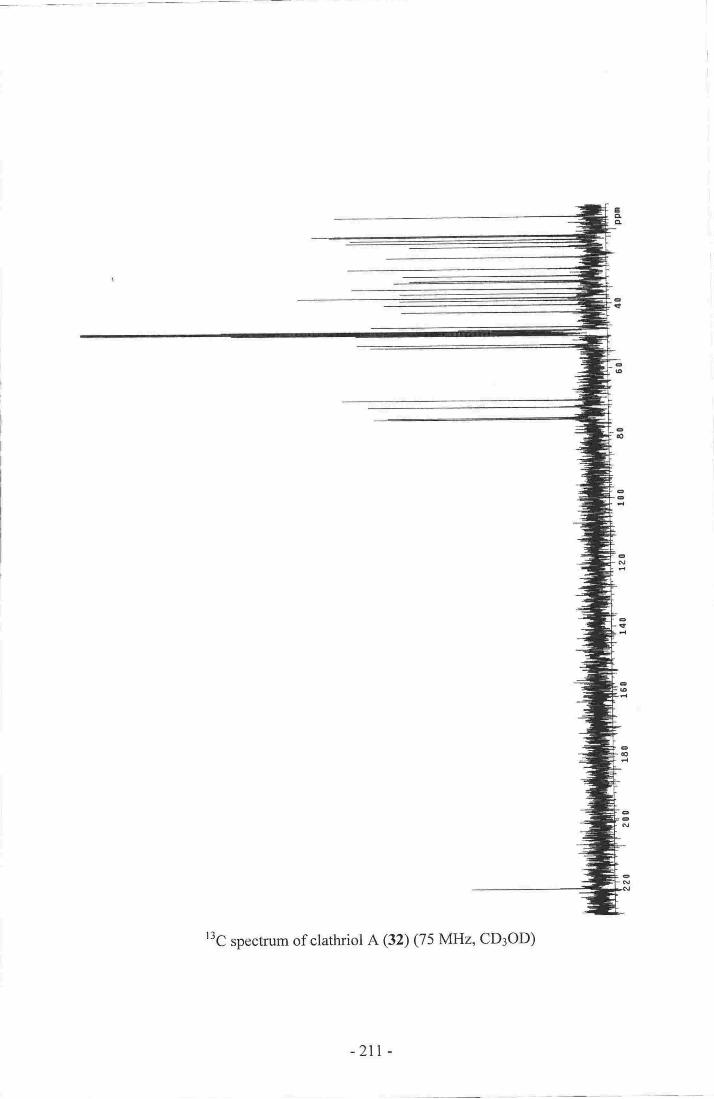
r3C spectrum of clathriol A (32) (75 MlIz, CD3OD)
-211 -
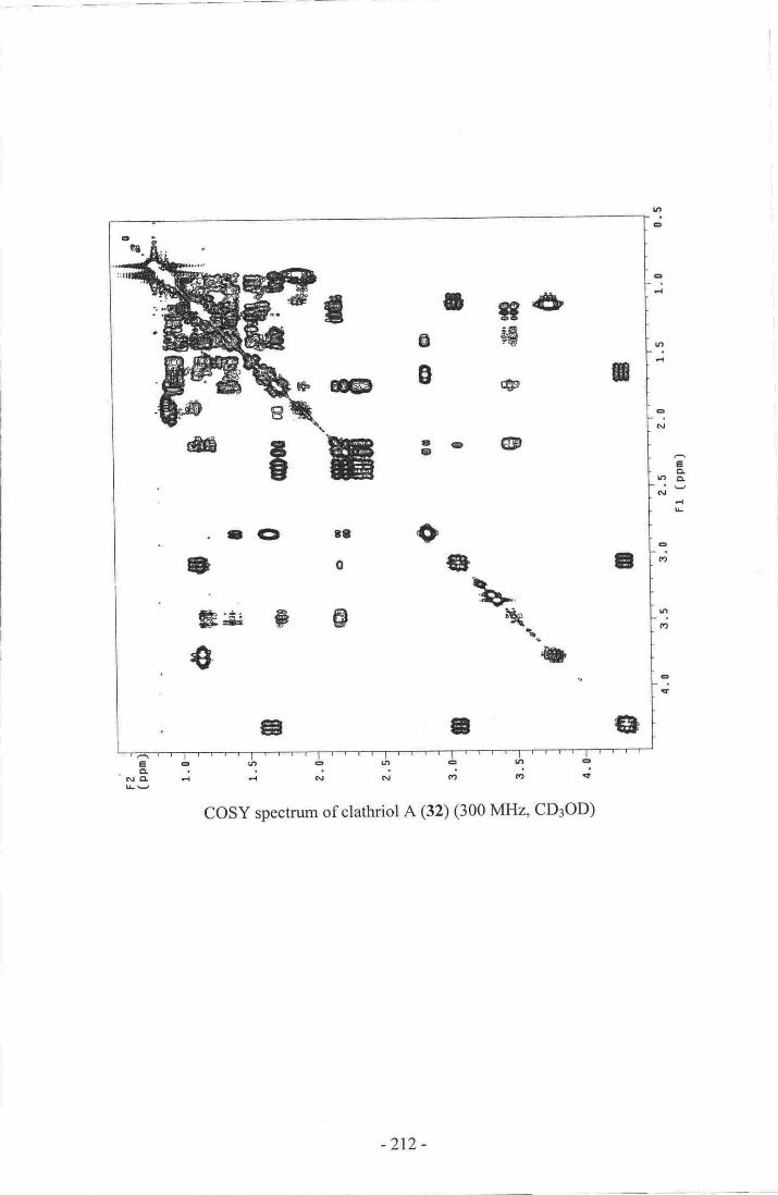
ll|
o *g&s4pF
B
a
3c @
\:$-.qt.
{D
s
a
B
g
+
rts@
CD 38
0
FE
aa
€g
tr.
aaGg
€
o
Lt
Eang
e
N
€o:t
D(',
€T
ryrlr,,r ,,t ,.1 |
ee6eltratD€c!'nrd -l d N Al <') d:t It
cosY spectrum of clathriol A (32) (300 MHz, CD3OD)
-212 -

f,4,-4
-F <I'
+>-!:-
-flD -,*--'
- -'{-
- -
r-lF
-{l)-
--)€
{-
L
€iG|
ir,
a0
tc4
u-
-ut
eIt
-
ry.f|!l''t''"4'.'..|.-g-.Sorrr'; jilg t{ Fl a\l Rr
'rit
HSQC B.pe.stnm of elathri0l A (32) (3CI0 MI-IZ' CDsOrD)
-273 -

IoEL
toNA
o
+*+ 4-,a-E-o= o- n
.gO o e
$ee$-eE 'E
,Q-o oo€
$ae=*g*- $*: -o1---de
al ap e
e e€e
eO
eO lP
e
ID
+ 'ee
+e.a*
€.e
e
-a- a1Q eoee
Narrowed HMBC spectrum of clathriol A (32) (300 MHz, CD3OD)
(note folding of C:O resonance)
-2r4 -

-.ED
a"D .Da
3at.DQr'
€oFt
6ctcl
N
r
Full HMBC spectrum of clathriol A (32) (300 MHz, CD3OD)
-2t5 -

O- @- - 4 >
o
---.:-
-- C!---
€E# - qr-
{E GEO@ . o
.EED CF (D 1D
tQ>o G
o@rc (ED {E> c rD
0D,REDCPo C) rF (D o
€fGe.ED
ffiCF--"t> @'r- --
(D €€DIED + lD O tE
rD {D{9itD€ lD e ' ''.D.GDrDd€@
'a{!FA
c
.€aer {Etr G 1o lGD rttt (D
a€B eg, @
m'1"'t6tit€litEBea
srEL i d N N c, c, It
HSQC-TOCSY (S0 ms mixing time) spectrum of clathriol A (32) (300 MHz' CD3OD)
e
eN
efa
e!t
EE!cl
Em
!ao
e
-2t6 -

";' S.i,o
i:Ffp0lorn
I
" lto1
gl@@
@
t
il
9o
o.fF
@0q
o { e€
0@, ,
00
OG
ROESY spectrum of clathriol A (32) (300 MHz, CD3OD)
fiiF
-T€iCL
dCL
-2t7 -

Appendix Two
I\MR Spectra of Clathriol B (33)
\
\
\
\
rH spectmm of clathriol B (32) (300 MHz, CDCI3)
-218 -
tCIq,
-

r3C spectrum of clathriol B (32) (75MHz,CDCI3, Note Ds-CsHsN added)
-2r9 -
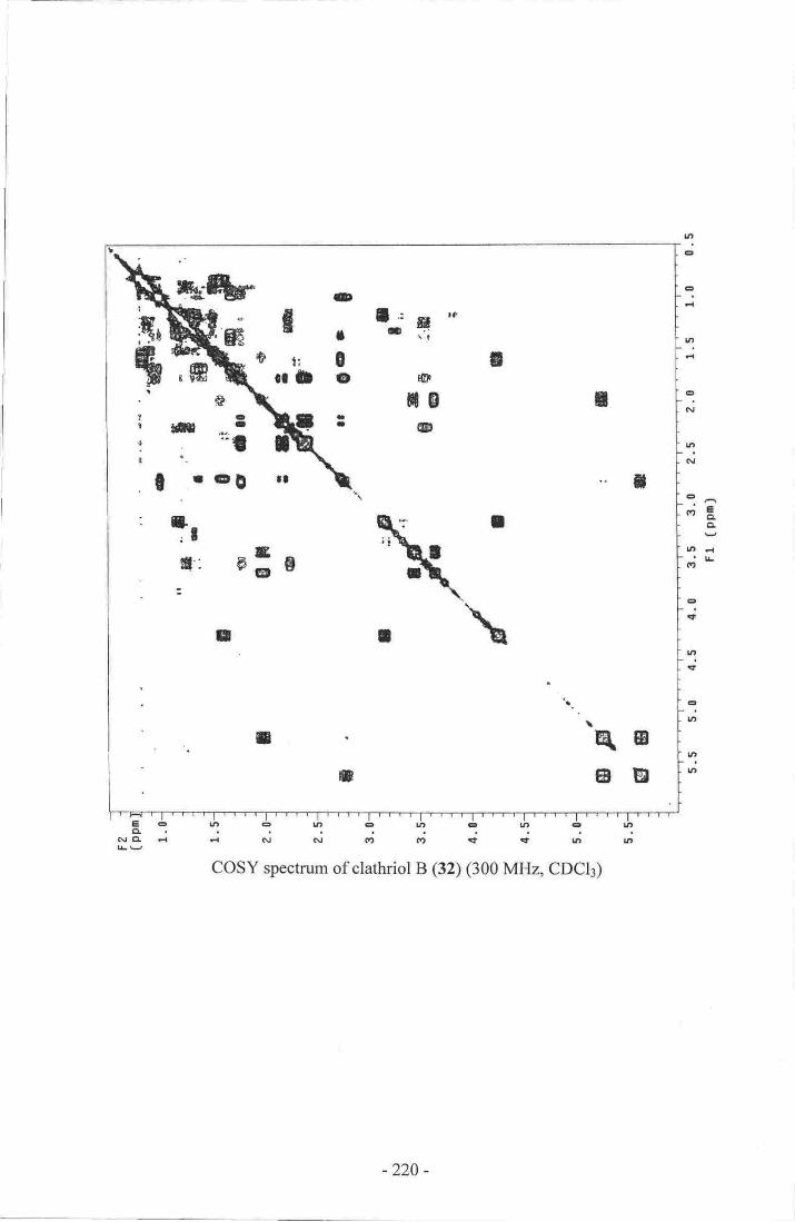
tng
e
ur
I
C\,|
V'N
eIt
t,'rt
ettt
ttU'
-irct
tt d(?
COSY spectrum of clathriol B (32) (300 MHz, CDCI3)
I
..t
qBatr
O
I lG'g '3
0!o {at
TB:.D
I
I
n
;t:
m0
a;B
o
E
k-furEA
ea-'ttl
eD
!
,ffi
rfr.'
t
I.:r4':
:3tslfr
g
I
:
I
-220 -
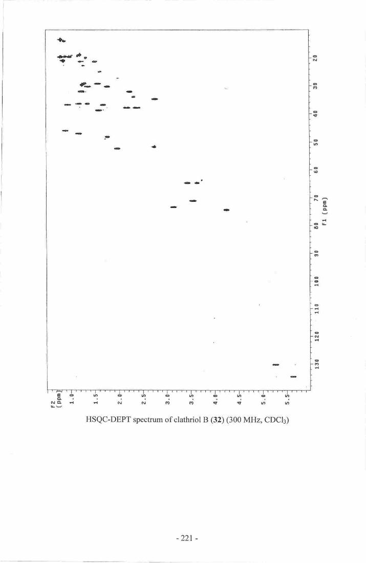
t>
rff,+ )f.4 1-
f--c.r-!-
ai--D
-<FI'
- e-
-l '-
--'
-
a(\|
ct{ttt
art
-lrt
€t\eCIB
-lro
cal
!t-
€
EG'
|=.|.",.|.''"|r'1.-|...'|||l{'||||tECUtOt/DEUtOUt€lltg,..!,r
Atll d rl cl N. Gt Oil l '!t u7 |a
HSQC.EEPT sp-eeEuui of o.latrr-iol B (32) (300 A4{2, CDCb)
-221 -

Appendix Three
I\MR Spectra of Cadlinolide C (138)
lH spectum of cadlinolide C (13S) (300 MHz, CDCIt
-222-
eAcl
E
E
o
I
f''
6
!t
-0
nn
6
o

IloI..@
tI
tI
L=lNtI
I
Il"r3C spectrum of cadlinolide C (l3S) (75 MHz,CDCI3)
-223 -

c
vl
IN
Ir'N
€o'
e,- tl-:eaoe
e.'
rn
a
E
la
nt'|
E
ID
|n6
COSY spectrum of cadlinolide C (138) (300 MHz, CDCI3)
a-oNC -l
-224 -

,e
-.r rt---I
a'd
Sr .-
.la
oa.
a
?6
t
a{
GiEt
F,r|l
aBg
Fa
Pgt
!a
' T'l' "',h" -'A'''''.1"' "g' "',S"''4" "'th" "l' "',i" " "l' "'q | .; . 't i :
a{Ei rr d Nl N et !f rl rt r'' .ui ..
HSQC.DEPT speetnm of eadlinolide e 038) (300 IvIIIz, CDel3)
-22;5 -

,.d
i'- e
O$'rD - a
a. o.
rf f#f eaotta
,.a,
?o*. .
e
*"t'5
rfc'.r.
a
og{
qrt
aIT
Od0
IC!g
.lL,€'e
Itarl
'Dct
.6rO
'.I:'5' 'tb ' 'g '4"'-5
"' 6" "1" ",i'.""1 '-'*'"'-5"c;"iddiJ;d.;
Eg* +r 6
HII{B, e specfrm of aadlinolide C;O38) (300 ivIH4 eDCt);
.2?16 -

:-':';.ilt.!
ECLCI
dL
e
ttt
eN
ulN
edt
ttt{t
elr
ltt(t
en
ttlh
'e
.€
"v,@
-(l
3o lrn
.+
enNN ro
yre
v, ro
ROESY spectrum of cadlinolide C (f3S) (300 MHz, CDCI3)
-227 -

Appendix Four
NMR Snectra of Cadlinolide D (139)
\
rH spectrum of cadlinolide D (139) (300 MHz, CDCIr)
-228 -
aaEI
€

.:,d.
It
:*:-:!'. Fil
:'tl
forFl€Frt'['."rirF
loFdt
Ir!rFJt-r€F;rrr
L',t'I',Fg
LrcI
trfa-!- ro
ct
3
a
q'l
nIt
,
al-ED
ltt-
a
z. fn I4,
t|
,c I.' ra rt--erlfil
afi,a\i
eqg
t-gcrl ll ili
COSY spec,ruul of Eadlinotide D O39) (300 lviHa CDC[)
t?
-;F
-229 -
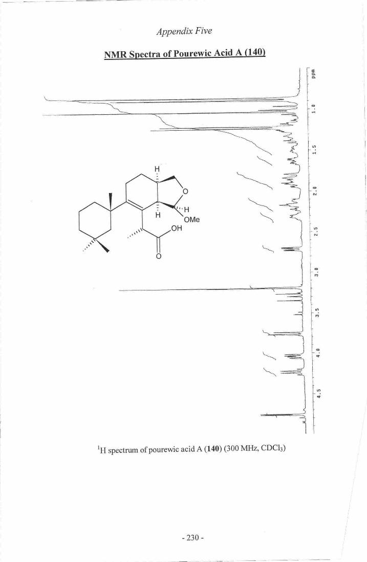
APP'e'rdix Flve
I{,MR Snectra o-f Pourervie Acid A fl401
o
:'HOMe
rH spectnrrr of, ponrewie acid A (f4O) (300 Mttrz, CDCI3)
\
-\
\\
-234 -
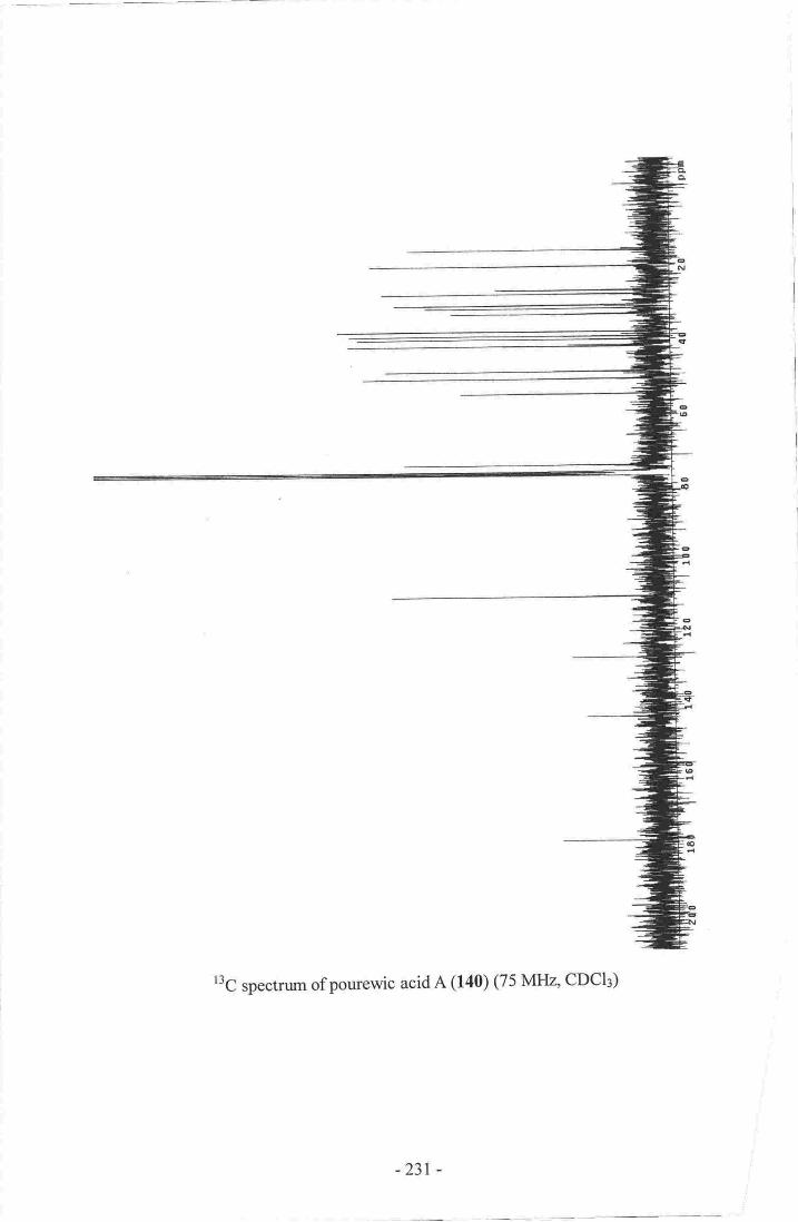
r3C spectrum of pourewic acid A (140) (75N{tIz" CDCI3)
-23t -

E
tat
oC\I
u7
6.'?clCL
Erl
fit
tt(a
€it
I')
(t
€ut
E:'::i:-:NcL ..{ d N N ttt {tt qt € Itt
COSY spectrum of pourewic acid A (140) (300 MHz, CDCI3)
*
slm
!B
'{)
g: r$o
o 0to
0g
&t
6rr
t>'o
-232 -
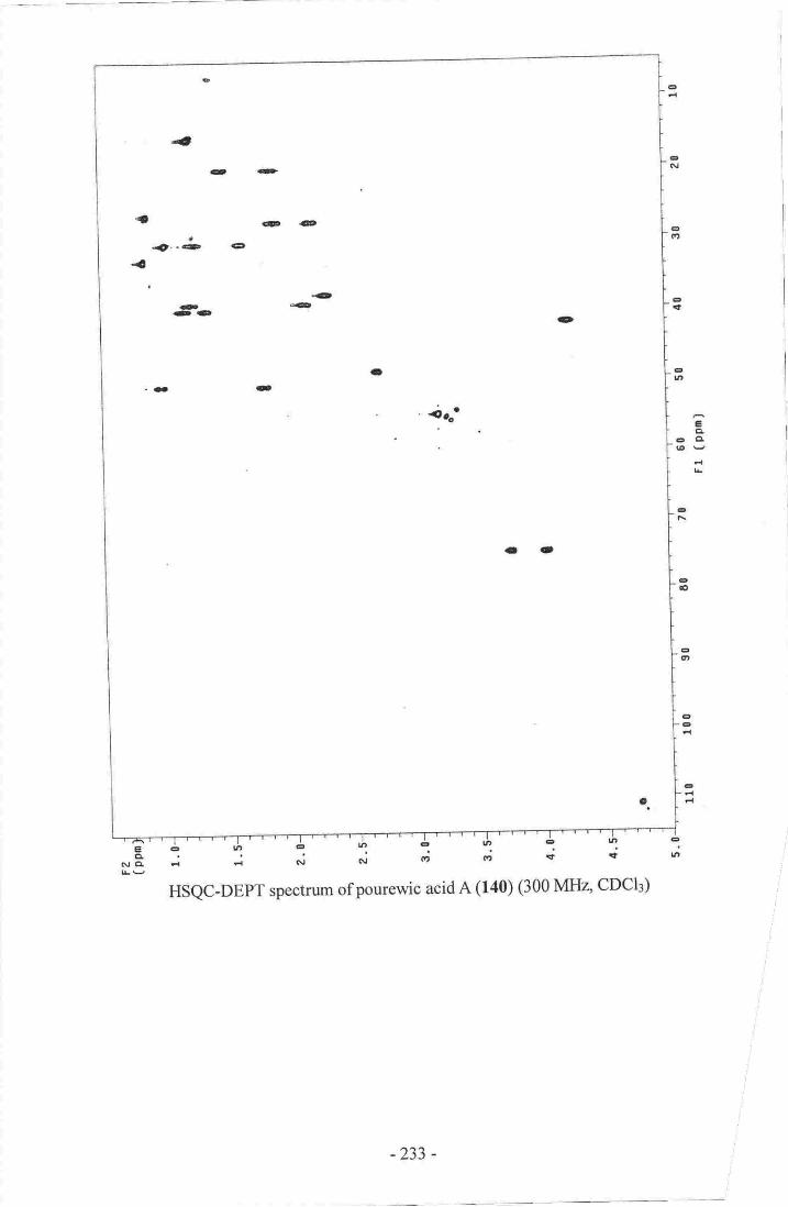
,4
-D-
rt
{
eaa
-"-a €
-a- 'rt<af tt
.i- It-
-.it'
-1t
.a
I
rF|.GI
rDN
.-:lt
;tl|'
arCl
rC!It tt?
Ittt
..€
cE-t
c
ac,a,E:Nq illL,i-,
Fg?r
N Crl
HSQC.DEPT speeffurn of poru.ewic asid A (r4q G00IyIIIa ODCIg)
-233 -

a
a QCDadr. 4
. fuit''' ' 4'a q lF ' a
ta...q
.o.lo'. - lo a
te@'-
i+ .F 'a .2 .t
{|pc
e .p,
tst
at
eto
c€
t,oGl
)4.-!l !{
€hl
gt.{
CI|.;f
co
aEL
NB:-;:1d 61. grl t{ n' FZ + rt
HhdBC speatnrfip of pou(ewic acid A (laQ p00ltffJ4 CDCI')
-234 -

----.G
tt
Eltc
L
t.a J .i
o.it
0!ioa!@:i
.-|la
ROESY spectrum of pourewic acid A (140) (300 MFIZ' CDCI3)
-23s -

ApBer,dtx Six
o
l',H
OMe
rH qpecgirno of l5-rnethoxypo'rewic aoid B (f41) (300 MHz, CDCI3)
-236 -

-
a,N
IBe
u-
lad
stG|
G-
G}
.lrf
a?
!tr!
tttt
-I,'
COSY speaaum of 1,5-mcthpxv'pourewic acid B G41) (300 [{I{4 CDCIg)
-rtI
{
iLeOI 0
tat
!t,
Cl
-237 -

Appedix Ssten
\
\,
tH spectnrm ofmcthylpourewate B (142) (300 MHa CDCI3)
-238 -
al,

r3C spectrum of methylpourewate B (142) (75MHz'CDCI3)
-239 -
o6
ootu
eoGT
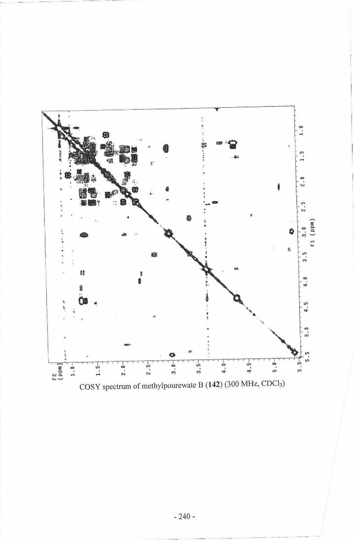
a.a;
:
IFF
!
ao
sia
-fte\F,Egr$ ' :
Br0s!,a&.9E-'q
e-o-.CL
d
ttt(?
o
e
6
tl
I0
eltr
lrtlrt
ala!t cl
.oal'tesaN
COSY spectrum of methylpourewate B (142) (300 MHz' CpClr)
-240 -

APPendtx Eight
NMR Spectra of Fourewanone (143)
tH spunmrro of pourew'anone (143) (300 Nfltu' CDel3)
.241'
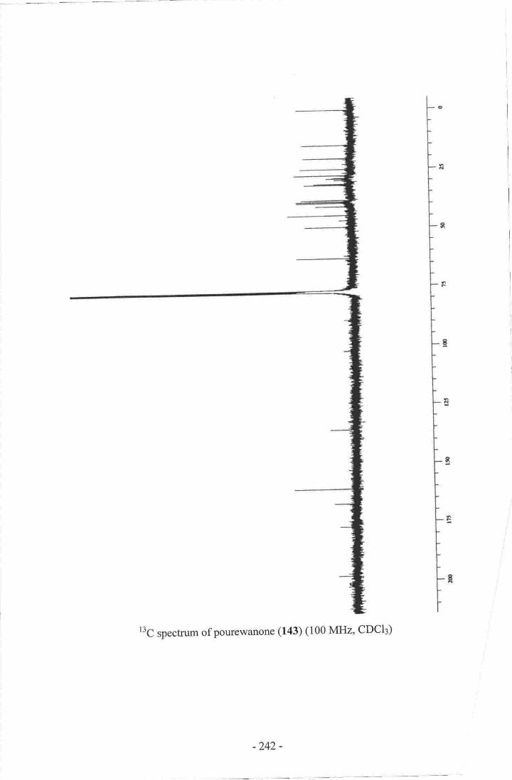
l3C spectrum of pourewanone (f43) (100 MHz, CDCI3)
-242 -

9!gil
t
i
tl
a
:u
gcErJtlL
B.lrt cl
COSY spgctrutrt of pqrrewanoue (fS) G00I\4H2, CDCb)
-243 -

Optimised HSQC-DEPT spectrum of pourewanone (143)' ('Jc.u: 190H2,300 MHz' CDCI3)
-244 -

- ill
t;foI
aI
I
tnArl
[.tEq"-**t peolffum of poureutanone (143) (nole'n-- 140 Hz' 300 MIIzt GDctrs)
-245.

ta .t'l€'-o
rro-. tt
aa. a
eo
e^eEdo,
ct
aN
HMBC spectrum of pourewanone (143) (300 MHz, CDCI3)
-246-
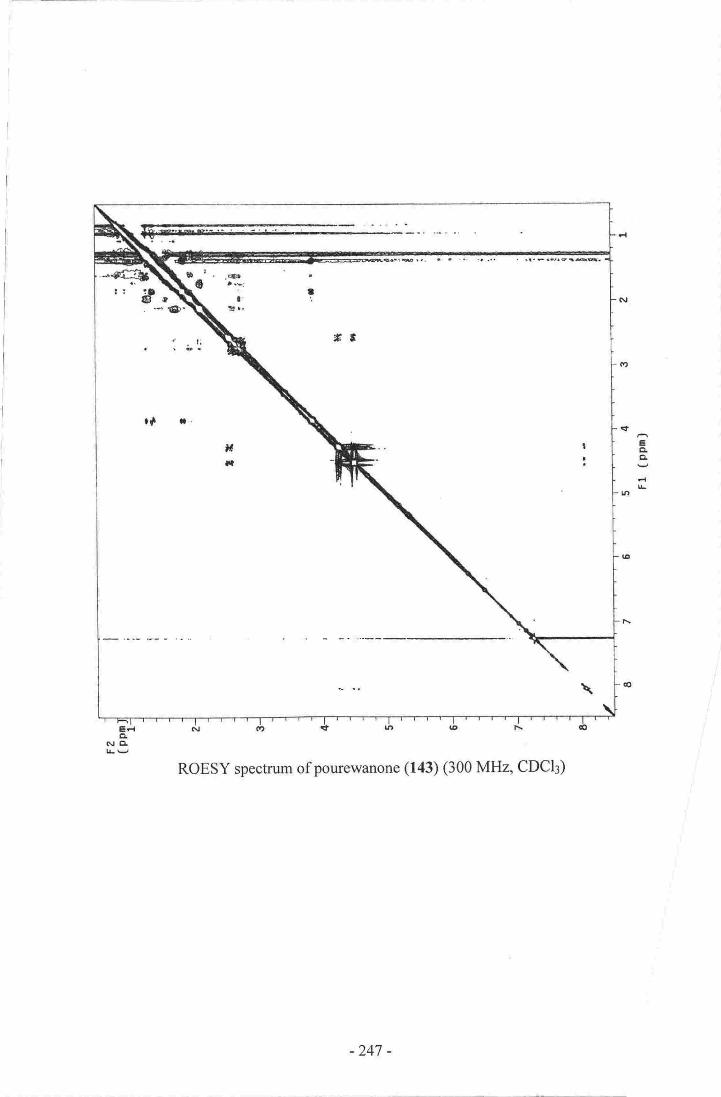
EELCI
ROESY spectrum of pourewanone (143) (300 MHz, CDCI3)
-247 -

NMR Spectra af Karcnia braisulcata Toxin (KBT)
rH speetrum of I(BT (500 MElz, D6-DMSO)
App.endix Nine
-248 -
EF{ctNEI
COSY spectrum of KBT (500 MHz, D6-DMSO)
-249 -
EctEL
FI
HSQC spectrum of KBT (500 MHz, D6-DMSO)
-250 -
rrlx
til.,lohl0r I !
Qt ll o I C
a
EgCI
d|r
rt{d
It
Edct
Nll
TOCSY spectrum of KBT (500 MHz, D6-DMSO)
-251 -

References
l. Cragg, G. M.; Boyd, M. R.; Khanna, R.; Kneller, R.; Mays, T. D'; Mazan
Kate, D.; Newman, D.J.; Sausville, E. A. Pure Appl. Chem. 1999,71,1619and references contained therein.
2. Hendrickson, J. B. The Molecules of Nature. In The Organic ChemistryMonograph Series; W. A. Benjamin,Inc.: New York, 1965.
3. Cobb, C.; Goldthwaite, H. Creations of Fire: Chemistry's Lively History fromAlchemy to the Atomic,4ge.; Plenum Press: New York,1995.
4. Halstead, B. W. Poisonous and Venomous Marine Animals of the World;United States Govemment Printing Office: Washington D.C., 1965; Vol. 1.
5. Moore, R. E.; Scheuer, P. J. Science 1971, 172,495.
6. Scheuer, P. J. Acc. Chem. Res. 1977, 10,33.
7. Martin, L.; htql://www.mtsinai.orgy'pulmonary/books/scuba/sectiona.htm: Asa|26108102.
8. Blunt, J. W.; Copp, B. R.; Munro, M. H. G.; Northcote, P. T.; Prinsep, M. R'Nat. Prod. Rep. 2003, 20, l.
9. Proksch, P.; Ebel, R.; Edrada, R. A.; Schupp, P.; Lin, W' H.; Sudarsono;
Wray, V.; Steube, K. Pure Appl. Chem. 2003, 75,345.
10. Scheuer, P. J. Chemistry of Marine Natural Products. ln Marine NaturalProducts; Academic Press, Inc.: New York and London, 19731. Vol. L
I l. Zubay, G.L. Biochemistry, Fourth ed.; Wm C. Brown: Melbourne, 1998.
12. Pawlik, J.R. Chem. Rev. 1993,93,1911.
13. Mann, J. Chemical Aspects of Biosynthesis. In Oxford Chemistry Primers;Oxford University Press: New York, 1994; Vol. 20.
14. Scheuer, P. J. Marine Natural Products: Chemical and BiologicalPerspectives. [n Marine Natural Products; Academic Press, Inc.: New Yorkand London, 1978; Vol. 2.
15. Cragg, G. M.; Newman, D. J.; Snader, K. M. J. Nat. Prod. 1997, 60,52.
16. Quinn, R. J.; de Almeida Leone, P.; Guymer, G.; Hooper, J. N. A. Pure Appl.Chem. 2002,74,519.
17. Pettit, G.R. Pure Appl. Chem. 1994,66,2271.
18. Fleming, C. A. Tuatara 1949, I 1,72.
-252 -

19.
20.
Fleming, C. A. New Zealand Journal of Forestry 1977,22,249.
Fleming, C. A. The Geological History of New Zealand and its Life.;
Auckland University Press, 1979.
Bergquist, P.R. Sponges; Hutchinson & Co.: London, 1978.
Hooper, J. N. A.; Wiedenmayer, F. Porifera. In Zoological Catalogue ofAustralia; CSIRO: Melbourne,1994; Vol. 12.
Hooper, J. N. A.; van Soest, R. W. M.; (Eds) Systema Porifera: A guide to the
classification of sponges., I ed.; Kluwer Academic/Plenum: New York,2002-
Pritchard, K.; Ward, V. Marine Sponges: Forty-Six Sponges of Northern NewZealand; University of Auckland Marine Laboratory: Auckland, 1984; pp.
r49.
Faulkner, D. J.; Unson, M. D.; Bewley, C. A. Pure Appl. Chem' 1994, 66,
I 983.
Faulkner, D. J.; Molinski, T. F.; Andersen, R. J.; Dumdei, E. J.; de Silva, E. D.
Comp. Biochem. Physiol. 1990, 97C, 233.
Molinski, T.F.; Faulkner, D. J.; Cun-heng, H.; Van Duyne, G. D.; Clardy, J.lOrg. Chem. 1986, 5/,,4564.
Tischler, M.; Andersen, R. J.; Choudhary, M. I.; Clardy, J. J. Org' Chem'
1991,56,42.
Hirota, H.; Okino, T.; Yoshimura, E.; Fusetani, N. Tetrahedron 1998, 54,
13971.
Dumdei, E. J.; Kubanek, J.; Coleman, J. E.; Pika, J.; Andersen, R. J.; Steiner,J. R.; Clardy,J. Can. J. Chem. 1997,75,773.
Simpson, J. S.; Garson, M. J.; Hooper, J. N. A.; Cline, E. I.; Angerhofer, C. K.Aust. J. Chem. 1997,56,1123.
de Jesus, R. P.; Faulkner, D. J. J. Nat. Prod.2003, 66,671.
Bergquist, P. R.; Wells, R. J. Chemotaxonomy of the Porifera: TheDevelopment and Current Status of the Field. In Marine Natural Products:Chemical and Biological Perspectfves; Scheuer, P. J. Ed.; Academic Press:
New York, 1983; Vol. 5.
Hartman, W. D.; Bergquist, P. R. Porifera. In Synopsis and ClassiJication ofLiving Organisms; Parker, S. P. Ed.; McGraw-Hill: New York, 1982; Vol. 1;
pp.640.
Kristiansen, J.; Loeblish III, A. R. Dinophyceae. In Synopsis andClassification of Living Organisms,' Parker, S. P. Ed.; McGraw-Hill: NewYork, 1982; Vol. 1; pp. 640.
2t.
22.
23.
24.
26.
25.
27.
28.
29.
31.
30.
32.
33.
34.
35.
-253 -

36. Corliss, J. O. Protozoa. In Synopsis and Classification of Living Organisms;Parker, S. P. Ed.; McGraw-Hill: New York, 19821' Vol. l; pp. 640.
| 37. Allaby, M.; Allaby, R.; Kent, M.; Sainsbury, D.i Whitmore, T. C. The ConciseOxford Dictionary of Botany; Oxford University Press: Oxford, 1992.
38. Dodge, J.D. Prog. Protozool.1965"264.
41. Personal Communication; Chang, F. H.; NIWA; September;Z0D2.
42. Yasumoto, T.; Murata,M. Chem. Rev. 1993,93, 1897.
43. Chang, F.H. Phycologia 1999, 38,377.
44. Chang, F. H.; Chriswell, S, M.; Uddstrom, M. J. Phycologia 2001, 40,215.
45. Daugjberg, N.; Hansen, G.; Larsen, J.; Moestrup, O. Phycologia 2000, 39,302.
46. Wagner, H.; Bladt, S.; Zgainski, E. M. Plant Drug Analysis; Springer-Verlag:Berlin, 1984.
47. Mestel, R. Discover 1999, March,7\.
48. Moore, R.E. Pure Appl. Chem. 1982, 54,1919.
49. Faulkner, D. J. Academic chemistry and the discovery of bioactive marinenatural products. ln Marine Biotechnologt: Pharmaceutical and BioactiveNatural Products;Attaway, D. H.; Zaborsky, O. R. Eds.; Plenum Press: NewYork, 1993; Vol. l; pp. 500.
50. West, L. M. The Isolation of Secondary Metabolites from New ZealandMarine Sponges., Victoria University of Wellington 2001.
51. Satake, M.; Shoji, M.; Oshimo, Y.i Naoki, H.; Fujita, T.; Yasumoto, T,Tetrahedron Le tt. 2002, 4 3, 5829.
52. Kobayashi, J. i.; Cheng, J.-f.; Ohta, T.; Nozoe, S.; Ohizumi, Y.; Sasaki,T. J.Org. Chem. 1990, 55,3666.
53. Umeyama, A.; Adachi, K.; Ito, S.; Arihara, S. J. Nat. Prod.2000, 63, I175.
54. Blunt, J. W.; Calder, V. L.; Fenwick, G. D.; Lake, R. J.; McCombs, J. D.;Munro, M. H. G.; Perry, N. B.l Nat. Prod. 1987,50,290.
55. Still, W. C.; Kahn, M.; Mitra, A. J. Org. Chem. 1978,43,2923.
56. Coll, J. C.; Bowden, B. F. J. Nat. Prod.1986, 49,934.
-254 -

,. 57. Shaksher, Z.M.; Seitz, W. R. Anal. Chem. 1989, 61,590.
I 58 Evans, M. B.; Dale, A. D.; Little, C. J. Chromatographica 1980, I3, 5.
I 59. West, L. M.; Northcote, P. T.; Battershill, C. N.I Org. Chem. 2000, 65,445.
60. Northcote, P. T.; Blunt, J. W.; Munro, M. H. G. Tetrahedron Lett. 1991, 32,64tt.
61. Perry, N. B.; Blunt, J. W.; Munro, M. H. G. J. Am. Chem. Soc. 1988, I 10,4850.
62. West, L. M. Honours Project Report; Victoria University of Wellington:Wellington, 1996.
63. van Liedekerke, B. M.; Nelis, H. J.; Lambert, W. E.; de Leenheer, A.P. Anal.Chem. 1989, 61,728.
64. Keyzers, R, A. Isolation of Yessotoxin from contaminated green lippedmussels, Perna canaculus, and from cultures of the algae Protoceratiumreticulatum.; Victoria University of Wellington: Wellington, 1998; pp. 43.
65. Anjaneyulu, A. S. R.; Sagar, K. S.l Chem. Research (S) 1995,142.
66. Gauvin, A.; Smadja, J.; Aknin, M.; Faure, R.; Gaydou, E. M. Can. J. Chem.2000, 7g,9g6.
67. Makita, A.; Taniguchi, N. Glycosphingolipids. In Glycoliprds; Wiegandt, H.Ed.; Elsevier: Amsterdam, 1985; Vol. l0; pp. 314.
68. Nagle, D. G.; McClatchey, W. C.; Gerwick, W. H. J. Nat. Prod. lgg?,, 55,1013.
69. Makarieva, T.N.; Denisenko, V.A.; Stonik, V.A.; Milgrom, y.M.; Rashkes,Y. Y. Tetrahedron Lett. 1989, 30, 6581.
70. Bartik, K.; Braekman, J. C.; Daloze, D.; Stoller, C.; Huysecom, J.;Vandevyver, G.; Ottinger, R. Can. J. Chem. 1987,65,2118.
71. Casapullo, A.; Bifulco, G.; Bruno,I.; Riccio, R. J. NaL Prod.2000, 63,447.
72. Cutignano, A.; Bifulco, G.; Bruno, I.; Casapullo, A.; Gomez-Palom4 L.;Riccio, R. Tetrahedron 2000, 5 6, 37 43.
73. Kohmoto, S.; Kashman, Y.; McConnell, O. J.; Rinehart Jnr, K. L.; Wright, A.;Koehn, F . J. Org. Chem. 1988, 5-t, 31 16.
74. Tsuji, S.; Rinehart Jnr, K. L.; Gunasekera, S. P.; Kashman, Y.; Cross, S. C.;Lui, M. S.; Pomponi, S.; Diaz, M. C. J. Org. Chem.1988, 53,5446.
75. Forenza, S.; Minale, L.; Riccio, R. J. Chem. Soc. Chem. Commun. l97l,ll2g.
-255 -

76. Cimino, G.; de Rosa, S.; de Stefano, S;Mazzarella, L.; Puliti, R.; Sodano, G.Tetrahedron Lett. 1982, 23,767.
77. Williams, D. H.; Faulkner, D. J. Nat. Prod. Lett. 1996, 9,57.
78. Walker, R. P.; Faulkner, D. J.; van Engen, D.; Clardy, J.J. Am. Chem. Soc.lggl, 103,6772.
79. Pettit, G. R.; Herald, C. L.; Leet, J. E.; Gupta, R.; Schaufelberger, D. E.;Bates, R. B.; Clewlow, P. J.; Doubek, D. L.; Manfredi, K. p.; Rutzler, K.;Schmidt, J.M.; Tackett, L.P.; Ward, F.B.; Bruck, M.; Camou,F.Can. J.Chem. 1990, 68,1621.
I SO' McMurry, J. Organic Chemistry, 4 ed.; Brooks/Cole: Pacific Grove, 1996.
81. Giner, J.-L. Chem. Rev. 1993, 93,1735.
82. Djerassi, C.; Silva, C. J. Acc. Chem. Res. 1991, 24,371.
83. Faulkner, D. J. Nar. Prod. Rep. 1993, 10, 497 and previous papers in thisseries.
84. Sheikh, Y. M.; Djerassi, C. Tetrahedron 1974,30,4095.
85. Iguchi, K.; Fujita, M.; Nagoka, H.; Mitome, H.; yamad a, y. Tetrahedron Lett.1993,34,6277.
86. Iguchi, K.; Shimura, H.; Taira, S.; yokoo, C.; Keita, M.; yamada,y. J. Org.Chem. 1994,59,7499.
87. casapullo, A.; Minale,L.;zollo,F. Tetrahedron Lett. lggs, 36,2669.
88. Aoki, S.; Yoshioka, y.; Miyamoto, y.; Higuchi, K.; setiawan, A.; Murakami,N.; chen, z.-s.; sumizawa, T.; Akiyama, s.-i.; Kobayashi, M. TetrahedronLert. 1998, 39,6303.
89. Aoki, S.; Setiawan, A.; yoshioka, y.; Higuchi, K.; Fudetani, R.; chen, Z.-s.;sumizawa, T.; Akiyama, s.-i.; Kobayashi, M. Tetrahedron 1999,5s, 13965.
90. Allinger, N.L.; Hermann, R.B.; Djerassi, c. J. org. chem.1960, 25,g22.
91. Kamano, Y.; Kumon, s.; Arai, T.; Komatsu,M. chem. pharm. Bull. 1973,21,1960.
92' Burgoyne, D. L.; Andersen, R. J.; Aleen, T. M. J. org. chem. rggz,57, szs.
93. Takei, M.; Burgoyne, D. L.; Andersen, R. J. J. pharm. sci. 1994, g3,1234.
94. Bramley, A. M.; Langlands, J. M.; Jones, A. K.; Li, y.; Burgoyne, D. L.;Andersen, R. J.; Salari, H. Brit. J. pharma. 1995, I I S , 1433 .
95. Y*g, L.; Andersen, R. J. J. Nat. prod.2002,65,lg}4.
-256 -

96. Shoji, N.; Umeyama, A.i Shin, K.; Takeda, K.; Arihara, S.; Kobayashi, J. i.;Takei, M. J. Org. Chem. 1992,57,2996.
97. Kobayashi, J. i.; Shinonaga, H.; Shigemori, H.; Umeyama, A'; Shoji, N-;Arihara, S. J. Nat. Prod.1995, 58,312.
98. Sperry, S.; Crews, P. J. Nat. Prod. 1997,60,29.
99. Fu, X.; Ferreira, M. L. G.; Schmitz,F. J.; Kelly, M. J. Org. Chem. 1999,64,6706.
100. Keyzers, R. A.; Northcote, P. T.; Webb, Y . J. Nat. Prod. 2002, 65,598.
l0l. Keyzers, R.A.; Northcote, P.T.; Berridge, M. V. Aust. J. Chem.2003,56,279.
102. Bergquist, P. R.; Fromont, P. J. The Marine Fauna of New Zealand: Porifera,Demospongiae, Part 4 (Poecilosclerida). In New Zealand OceanographicInstitute Memoir; New Zealand Oceanographic Institute: Wellinglon, 1988;
Vol.96.
103. Riccio, R.; Finamore, E.; Santaniello, M.; Zollo,F. J. Org. Chem. 1990,5.t,2548.
104. D'Auria, M. V.; Riccio, R.; Uriarte, E.; Minale, L.; Tanaka, J.; Higa, T. J. Org.
Chem.1989, 54,234.
105. Tan, A. S.; Benidge, M. V. J. Immunological Methods 2000,59.
106. Andersen, R. J.; Allen, T. M.; Burgoyne, D. L. Contignasterol, and related 3a-
hydroxy-6a-hydroxy-7b-hydroxy-1S-keto-l4b steroids useful as anti-inflammatory and anti-thrombosis agents. In United States Patent Office;University of British Columbia, University of Alberta: United States ofAmerica, 1996.
107. Andersen, R. J.; Allen, T. M.; Burgoyne, D. L. Contignasterol compounds andpharmaceutical compositions comprising the same. In United States PatentOffice; University of British Columbia, University of Alberta: United States ofAmerica, 1997.
108. Aiello, A.; Ciminiello, P.; Fattorusso, E.; Magno, S. Steroids 1988, 52,533.
109. Rudi, A.; Yosief, T.;Loya, S.; Hizi, A.; Schleyer, M.; Kashman, Y.J. Nat.Prod. 2001, 64, 1451.
ll0. Tanaka, Y.; Katayama, T. Bulletin of the Japanese Society of ScientiJicFisheries. 1976, 42, 801.
lll. Tanaka, Y.; Fujita, Y.; Katayama, T. Bulletin of the Japanese Society ofScientific Fisheries. 1977, 42,767 .
-257 -
ll2. Tanaka, Y.; Soejima, T.; Katayama, T. Bulletin of the Japanese Society ofScientific Fisheries. 1978, 44, 1283.
I13. Tanaka, Y.; Akase, S.-I.; Yamada, S. Fisheries Science200l,67,378.
ll4. Capon, R. J.; Macleod, J. K. J. Chem. Soc. Chem. Commun. 1987, 1200.
ll5. Capon, R. J.;Miller, M.;Rooney, F.J. Nat Prod,2000, 63,821.
I16. Capon, R. J,; Miller, M.; Rooney, F. J. Nat Prod.200l, 64,643.
lI7. Ohta, S.; Okada, H.; Kobayashi, H.; Oclarit, J. M.; Ikegami, S. TetrahedronLett 1993, 34,5935.
118. Xiao, D.-j.; Deng, S.-s.; Zeng, L.-m. Acta Scientiarium NaturaliumUniversitatis Sunyatseni 2002, 4l, lII.
119, Sperry, S.; Crews, P. Tetrahedron Lett 1996,37,2389.
120. Zuleta. I. A.; Vitelli, M. L.; Baggio, R.; Garland, M. T,; Seldes, A. M.;Palermo, J. A. Tetrahedron2002,58, 4481.
l2l. Bergquist, P.R. The Marine Fauna of New Zealand: Porifera, Demospongiae,Part 5 (Dendroceratida and Halisarcida). ln New Zealand OceanographicInstitute Memoir; NIWA: Wellington,1996; Vol. 107.
122. Fake, A. D. Sesquiterpenoids from the New Zealand Marine Sponge Dysideasp., Victoria University of Wellinglon 1999.
123. Dawson, E. W. The Marine Fauna of New Zealand: Index to the Fauna: 2.
Porifera. In New Zealand Oceanographic Institute Memoir; MWA:Wellington, 1993;Vol. 1 00.
124. Walker, R. P.; Faulkner, D. J.l Org. Chem. 1981, 46,1098.
125. Walker, R. P.; Rosser, R. M.; Faulkner, D. J.; Bass, L. S.; Cun-heng, H.iClardy, J. J. Org. Chem. 1984, 49,5164.
126. Sullivan, B.; Faulkner, D. J. J. Org. Chem.1984, 49,3204.
127. Bobzin, S. C.; Faulkner, D. J. J. Org. Chem. 1989, 54,5727.
128. Rudi, A.; Kashman, Y. J. Nat. Prod.1992,55, 1408.
129. Lu, Q.; Faulkner, D. J. J. Nat. Prod.1998, 61,1096.
130. Kazlauskas, R.; Murphy, P. T.; Wells, R. J.; Noack, K.; Oberhansli, W' E.;Schonholzer,P. Aust. J. Chem. 1979, 32,867 .
131. Cimino, G.; de Rosa, D.; de Stefano, S.; Minale, L. Tetrahedron 1974, 30,645.
-258 -

132. Capelle, N.; Braekmffi, J. C.; Daloze, D.; Tursch, B. Bull. Soc. Chim. Belg.
1980, 89,399.
133. Cambie, R. C.; Craw, P. A.; Stone, M.J.; Bergquist, P. R. J Nat. Prod.1988,5 1,293.
134. Kernan, M.R.; Cambie, R. C.; Bergquist, P. R.I Nat. Prod.1990, 53,724.
135. Li, C.-J.; Schmitz, F. J.; Kelly-Borges, M. J. Nat. Prod, 1999, 62,287.
136. Schmitz, F. J.; Chang, J. S.; Hossain, M. B.; van der Helm, D. J. Org. Chem.
1985, 50,2862.
137. Karuso, P.; Taylor, W. C. Aust. J. Chem. 1986, 39,1629.
138. Carmely, S.; Cojocaru, M.; Loya, Y.; Kashmil, Y. J. Org. Chem. 1988, J3,4801.
139. Bobzin, S. C.; Faulkner, D. J. J Nat Prod. 1991,54,225.
140. Mayol, L.; Piccialli, V.; Sica, D. Gazz. Chim. Ital. 1988, I18,559.
l4l. Rudi, A.; Kashmffi, Y. Tetrahedron1990,46,4019.
142. Hirsch, S.; Kashrnan,Y. J. Nat. Prod. 1988, 5l , 1243.
143. Gunasekera, S. P.; Schmitz, F. J. -/. Org. Chem. 1991, 56,1250.
144. Karuso, P.; Bergquist, P. R.; Cambie, R. C.; Buckleton, J. S.; Clark, G. R.;
Rickard, C. E. F. Aust. J. Chem. 1986, 39,1643.
145. Hambley, T. W.; Taylor, W. C.; Toth, S. Aust. J. Chem. 1997,50,903.
146. Hyosu, M.; Kimura,J,J. Nat. Prod.2000, 63,422.
I47. Karuso, P.; Skelton, B. W.; Taylor, W. C.; White, A. H. Aust. J. Chem- 1984,
37, 1081.
148. Mayol, L.; Piccialli, V.; Sica, D. Tetrahedron Lett.1985, 26,1357.
149. Mayol, L.; Piccialli, V.; Sica, D. Tetrahedron Lett. 1985, 26,1253.
150. Mayol, L.; Piccialli, V.; Sica, D. Tetrahedron 1986, 42,5369.
l5l. Molinski, T.F.; Faulkner, D. J. -/. Org. Chem.1987,52,296.
152. Poiner, A.: Taylor, W. C. Aust. J. Chem.1990, 43,1713.
153. Goeller, F.; Heinemann, C.; Demuth, M. Synthesis 2001,8, I I14.
154. Arno, M.; Gonzalez, M. A.; Marin, M. L.; Zatagoza,R. J. Tetrahedron lett'2001, 42,1669.
-259-

i tSS. Abad, A.; Agullo, C.; Arno, M.; Marin, M.L.; Zatagoza, R. J. I Chem' Soc.
i Perkin I1996.2193.
156. Betancur-Galvis, L.; Zuluaga, C.; Amo, M.; Gonzalez, M. A.;Zwagoza R. J.
J. Nat. Prod. 2002, 65, 189.
I57. Zubia, E.; Gavagnin, M.; Scognamiglio, G.; Cimino, G' J' Nat' Prod' 1994,
57,725.
158. Fenical, W. Diterpenoids. In Marine Natural Products: Chemical andBiological Perspectives; Scheuer, P. J. Ed.; Academic Press: New York, 1978;Vol. 2; pp.392.
159. Buckleton, J. S.; Bergquist, P. R.; Cambie, R. C.; Clark, G. R'; Karuso, P.;
Rickard, C. E. F. Acta Cryst. 1987, C43,2430.
160. Silverstein, R. M.; Bassler, G. C.; Morrill, T. C. Spectrometric ldentificationof Organic Molecules,5 ed.; John Wiley and Sons, Inc.: New York, 1991.
16l. Bowden, B. F.; Coll, J. C.; Mitchell, S. J.; Skelton, B. W.; White, A.H. Aust.
J. Chem.1980, 33,2737.
' 162. Cantillo-Ciau,,Z.;Brito-Loeza, W.; Quijano, L. J. Nat. Prod.2001, 64,953.
163. Hadden, C. E.; Angwin, D.T. Magn. Reson. Chem.200l, 39,1.
164. Voelter, W.; Breitmaier, E. Carbon-L3 NMR Spectoscopy,3 ed.; VCH: NewYork, 1987.
165. Fokialakis, N.; Magiatis, P.; Terzis, A.; Tillequin, F.; Skaltsounis, A.-L.Tetrahedron Lett. 2001, 42, 5323.
166. Ortega, M. J.; Zubia, E.; Rodriguez, S.; Carballo, J. L.; Salva, J. Eur. J. Org.Chem.2002,3250.
167. de Avellar, L G. J.; Vierhapper, F. W. Tetrahedron2000,56,9957.
168. Bobzin, S. C.; Faulkner, D. J. I Org. Chem. 1991, 56,4403.
169. Shimizu, Y.; Chou, H.-N.; Bando, H. J. Am. Chem.,Soc. 1986, 108,515.
170. Lin, Y.-Y.; Risk, M.; Ray, S. M.; van Engen, D.; Clardy, J.; Goik, J.; James, J.
C.; Nakanishi, K. J. Am. Chem. Soc. 1981, /,03,6773.
l7l. Wichmann, C.F.; Boyer, G.L.; Divan, C.L.; Schantz, E. J.; Schnoes, H. K.Tetrahedron Lett. 1981, 22, 1941.
172. Schantz, E. J.; Ghazarossian, V.E.; Schnoes, H.K.; Strong, F.M.; Springer, J.
P.;Pezzanite, J. O.; Clardy, L J. Am. Chem. Soc. 1975,97,1238.
173. Bates, S. S.; Bird, C. J.; de Freitas, A. S. W.; Foxall, R.; Gilgan, M.; Hanic, L.A.; Johnson, G. R.; McCulloch, A. W.; Odense, P.; Pocklinglon, R.; Quilliam,
-260 -

M. A.; Sim, P. G.; Smith, J. C.; Subba Rao, D.V.; Todd, E. C. D.; Walter, J.
A.; Wright, J.L. C. Can. J. Fish. Aquat.,Scl. 1989, 46,1203.
174. Subba Rao, D. V.; Quilliam, M.A.; Pocklinglon,R. Can. J. Fish. Aquat. Sci.
1988, 45,2076.
175. Tachibana, K.; Scheuer, P. J.; Tsukitani, Y.; Kikuchi, H.; van Engen, D.;
Clardy, J.; Gopichand, Y.; Schmitz, F. J. J. Am. Chem..Soc, 1981, 103,2469.
176. Hu, T.; Curtis, J. M.; Walter, J. A.; Wright, J. L. C. J. Chem. Soc. Chem-
Commun. 1995,597.
I77. Hu, T.; Curtis, J. M.; Walter, J. A.; Mclachlan, J. L.; Wright, J. L. C'Tetrahedron Lett. 1995, 36, 9273.
178. Arakawa, O.; Noguchi, T.; Shida, Y.; Onoue,Y. Toxicon 1994, 32,175.
179. Koehn, F.; Hall, S.; Wichmann, C. F.; Schnoes, H. K.; Reichardt, P. B.Tetrahedron Lett. 1982, 23,2247.
180. Onodera, H.; Satake, M.; Oshima, Y.; Yasumoto, T.; Carmichael, W. W.Natural Toxins 1997, 5, 146.
l8l. Shimizu, Y.; Hsu, C.-P.; Genenah, A. J. Am. Chem. Soc. 1981, I03, 605.
182. Zaman, L.; Arakawa, O.; Shimosu, A.; Shida, Y.; Onoue,Y. Toxicon 199E,36,627.
183. Wichmann, C. F.; Niemczura, W. P.; Schnoes, H. K.; Hall, S.; Reichardt, P.
B.; Darling, S. D. J. Am. Chem. Soc. 1981, 103,6977.
184. Laverty, R. Modes of Action of Shellfish Toxins. ln Marine Toxins and NewZealand Shel$ish; Royal Society of New Zealand: Wellington,7993; pp. 31.
185. Murata, K.; Satake, M.; Naoki, H.; Kaspar, H. F.; Yasumoto, T. Tenahedron1999,54,735.
186. Bates, M.; Baker, M.; Wilson, N.; Lane, L.; Handford, S. EpidemiologicOverview of the New Zealand Shellfish Toxicity Outbreak. In Marine Toxinsand New Zealand Shellfish,; Royal Society of New Zealand.: Wellington,1993; pp. 35.
187. Norte, M.; Gonzalez, R.; Fernandez, J. J.; Rico,M. Tetrahedron 1991,47,7437,
188. Takemoto, T.; Daigo, K. Chem. Pharm. Bull. 1958,6,578.
189. Quilliam, M.A.; Wright, J. L. C. Anal. Chem. 1989,6/, 1053A.
190. Halstead, B. W. Poisonous and Venomous Marine Animals of the World;United States Govemment Printing Office: Washington D.C., 1965; Vol. 2.
- 26r

l9l. Scheuer, P. J.; Takahashi, W.; Tsutsumi, J.; Yoshida,T. Science 1967,155,
1267.
192. Murata, M.; Legrand, A.-M.; Ishibashi, Y.; Yasumoto, T. J. Am. Chem. Soc.
1989,111,8929.
193. Murata, M.; Legrand, A.-M.; Scheuer, P. J.; Yasumoto, T. Tetrahedron Lett.
1992,33,525.
194. Murata, M.; Naoki, H.; lwashita, T.; Matsunaga, S.; Sasaki, M.; Yokoyama,A.; Yasumoto, T. J. Am. Chem. Soc. 1993, I I5,2060.
195. Murata, M.; Iwashita, T.; Yokoyama, A.; Sasaki, M.; Yasumoto, T. J. Am.
Chem. Soc. 1992, 114,6594.
196. Murata, M.; Yasumoto, T. Nat. Prod. Rep.2000,17,293.
197. Murata, M.; Kumagi, M.; Lee, J.-S.; Yasumoto, T. Tetrahedron Lett. 1987,28,5869.
198. Satake, M.; Terasawa, K.; Kadowaki, Y.; Yasumoto, T. Tetrahedron Lett.1996,37,5955.
199. Takahashi, H.; Kusumi, T.; Kan, Y.; Satake, M.; Yasumoto, T. TetrahedronLett. 1996, 37,7087.
240. Yasumoto, T.; Murata, M.; Oshima, Y.i Sano, M.; Matsumoto, G. K.; Clardy,J. Tetrahedron 1985, 4l,1019.
201. Personal Communication; Truman, P.; ESR; May;2003.
202. Prasad, A. V.; Shimizu, Y. J. Am. Chem.Soc. 1"989, I I 1,6476.
203. Statistics New Zealand;http://www.stats.govt.nzldomino/external/Web/nzstories.nsflO92edeb76ed5aa6bcc256afe008 I d84e/d00d64 I 9b4c27 I dfcc256b I f000 I aa49?OpenDocument:As at l8/09/02.
204. Chang, F. H.; Sharples, J.; Grieve, J. M. Harmful and Toxic Algal Blooms.1996,235.
205. Chang, F.H.; Mackenzie, L.; Till, D.G.; Hannah, D.; Rhodes, L. L. HarmfulMarine Algal Bloom.s.1995, 145.
206. Personal Communication; McFarlane, A.; New Zealand Fisheries Council;May; 1998.
207 . Chang, F. H. Harmful Algae News 1999, l.
208. Chang, F.H. NIWA Aquaculture Update 1998, l.
209. Truman, P.; Lake, R. J.l AOAC Int. 1996,79,1130.
-262-

210. Truman, P.; Stirling, D.J.; Northcote, P.T.; Lake, R. J.; Seamer, C.; Hannah,
D. J. J. AOAC lnt.2002,85, 1057.
2ll. Personal Communication; Hay, B.; Aquabiotox; November;2002.
212. Personal Communication; Truman, P.; ESR; July;2002.
2I3. Igarashi, T.; Satake, M.; Yasumoto, T. J. Am. Chem. Soc. 1996, I i,8,479.
214. Igarashi, T.; Satake, M.; Yasumoto, T. J. Am. Chem. Soc.1999, 121,8499.
215. Igarashi, T.; Oshima, Y.; Murata, M.; Yasumoto, T. Chemical Studies on
Prymnesins Isolated from Prymnesium parvum. ln Harmful Marine AlgalBlooms; Lassus, P.; Arzul, G.; Erard, P.; Gentien, P.; Marcaillou, C. Eds.;
Lavoisier: Paris, 1995; pp. 878.
216. Personal Communication; Stirling, D. J.; ESR; March;2003.
217. Personal Communication; Zubkov, O.; VUW; March; 2003.
218. Seki, T.; Satake, M.; Mackeruie,L.; Kaspar, H. F.; Yasumoto, T. TetrahedronLett. 1995,36,7093.
219. Uemura, D.; Chou, T.; Haino, T.; Nagatsu, A.; Fukuzawa, S.; Zheng, S.-2.;Chen, H.-S. J Am. Chem. Soc. 1995, I 17,1155.
220. Torigoe, K.; Murata, M.; Yasumoto, T.; Iwashita,T. J. Am. Chem. ,Soc. 1988,
I 10,7876.
221. Hr:, T.; Curtis, J.M.; Oshima, Y.; Quilliam, M.A.; Walter, J.A.; Watson-Wright, W.M.; Wright, J. L. C. J. Chem. Soc. Chem. Commun.1995,2159.
222. Morohashi, A.; Satake, M.; Murat4 K.; Naoki, H.; Kaspar, H. F.; Yasumoto,T. Tetrahedron Lett. 1995, 36,8995.
223. Sasaki, K.; Wright, J. L. C.; Yasumoto, T. J. Org. Chem.1998, 63,2475.
224. Macpherson, G. R.; Burton, I.; LeBlanc, P.; Walter, J. A.; Wright, J.L. C. J.
Org. Chem. 2003, 68,1659.
225. Dell'Aversano, C.; Walter, J.A.; Burton, I. W.; Stirling, D. J.; Fattorusso, E.;
Quilliam, M. A. Isolation and structure elucidation of new and unusualsaxitoxin analogues from mussels. In Harmful Algal Blooms;Intergovemmental Oceanographic Commission of TINESCO: Paris, 2003.
226. Kobayashi, J. i.; Yamaguchi, N.; Ishibashi, M. Tetrahedron Lett. 1994, 35,7049.
227. Kobayashi, J. i.; Kubota, T.; Takahashi, M.; Ishibashi, M.; Tsuda, M.; Naoki,H. J. Org. Chem. 1999,64,1478.
228. Ishibashi, M.; Takahashi, M.; Kobayashi, J.i.J. Org. Chem. 1995, 60,6062.
-263 -
22g. Kobayashi, J. i.; Yamaguchi, N.; Ishibashi, M. J. Org. Chem. 1994, 59,4698.
230. Kobayashi, J. i.; Shigemori, H.; Ishibashi, M.; Yamasu, T.; Hirota, H.; Sasaki,
T. J. Org. Chem.1991, 56,5221.
231. Kan, Y.; Uemura, D.; Hirata, Y.; Ishiguro, M.; Iwashita, T. Tetrahedron lett.
2001,42,3197.
232. Lewis, R. J.; Vernoux, J.-P.; Brereton, I. M. J. Am. Chem, Soc' 1998, 120,
s914.
233. Satake, M.; Fukui, M.; Legrand, A.-M.; Cruchet, P.; Yasumoto, T.
Tetrahedron Lett. 1998, 39, 1197 .
234. Satake, M.; Ofuji, K.; Naoki, H.; James, K. J.; Furey, A.; McMahon, T.; Silke,J.; Yasumoto, T. I Am. Chem. Soc. 1998, 120,9967 .
235. Rudi, A.; Talpir, R.; Kashman, Y.; Benayahu, Y.; Schleyer, M. J. Nat. Prod.1993, 56,2179.
236. Satake, M.; Murata, M.; Yasumoto, T. Tetrahedron Lett.1993, 34,1975.
237. Personal Communication; Munday, R.; Ag Research; May; 2003.
238. Personal Communication; Beuzenberg, V.t Cawthron Institute; February;2002.
239. Naoki, H.; Murata, M.; Yasumoto, T. Rapid Commun. Mass Spectrom. 1993,7, l7g.
240. Matsumori, N.; Murata, M.i Tachibana" K. Tetrahedron 1995, 51,12229.
241. Murata, M.; Matsuoka, S.; Matsumori, N.; Paul, G. K.; Tachibana, K. J. Am.Chem. Soc. 1999, 121,870.
242. Matsumori, N.; Kaneno, D.; Murata, M.; Nakamura, H.; Tachibana, K. J. Org,Chem. 1999, 64,866.
243. Riccio, R.; Bifulco, G.; Cimino, P.; Bassarello, C.; Gomez-Paloma, L. PureAppl. Chem. 2003, 75,295.
244. Moore, R. E.; Bartolini, G. J. Am. Chem. Soc. 1981, 103,249I.
245. Moore, R. E.; Bartolini, G.; Barchi, J.; Bothner-By, A. A.; Dadok, J.; Ford, J.J. Am. Chem. Soc.1982, 104,3776.
246. Uemura, D.; Ueda, K.; Hirata, Y. Tetrahedron Lett.1981, 22,2791.
247. Gottlieb, H. E.; Kotlyar, V.; Nudelman, A. J. Org. Chem. 1997,62,7512.
-264 -