membrane trafficking regulates the activity of the human dopamine transporter
TRANSCRIPT

Membrane Trafficking Regulates the Activity of the HumanDopamine Transporter
Haley E. Melikian and Kathleen M. Buckley
Department of Neurobiology, Harvard Medical School, Boston, Massachusetts 02115
The trafficking of synaptic proteins is unquestionably a majordeterminant of the properties of synaptic transmission. Here,we present a detailed analysis of the downregulation and intra-cellular trafficking of the cocaine- and amphetamine-sensitivedopamine transporter (DAT), a presynaptic plasma membraneprotein responsible for the regulation of extracellular DA con-centrations. Using PC12 cells stably transfected with humanDAT cDNA, we observe that phorbol ester activation of proteinkinase C (PKC) results in decreased transporter capacity and aparallel decrease in the amount of DAT on the cell surface that
is attributable to intracellular transporter sequestration. Afterinternalization, DAT diverges to the recycling, as opposed to thedegradative, arm of the endocytic pathway. This study demon-strates, for the first time, DAT endocytosis, establishes thepathways through which DAT traffics both at steady state and inresponse to PKC activation, and suggests that DAT recycling islikely to occur.
Key words: dopamine; endocytosis; trafficking; transporter;regulation; protein kinase C; recycling
The temporal and spatial coordination of dopaminergic neuro-transmission is achieved by striking a balance between the releaseand reuptake of dopamine (DA). Although a large body of worksupports regulation of DA release (Langer, 1997; Nagatsu andStjarne, 1998), far less is known about the modulation of trans-mitter reuptake. In the brain, clearance of extracellular DA ismediated by the high affinity DA transporter (DAT). DAT activ-ity is critical in regulating extracellular DA levels. Indeed, elim-ination of DA transport either pharmacologically (Grace, 1995)or via genetic manipulation (Jones et al., 1998) results in in-creased extracellular DA levels and enhanced synaptic responses.DAT is a member of the gene family of Na1/Cl2-dependentplasma membrane transporters whose members share multiplestructural features, including 12 putative transmembrane do-mains, multiple N-linked glycosylation sites, and putative cyto-plasmic phosphorylation sites (Amara and Kuhar, 1993). In ad-dition to DAT, the monoamine branch of the gene family includestransporters for serotonin (SERT), norepinephrine (NET), andepinephrine, all of which are potently inhibited by tricyclic anti-depressants, as well as drugs of abuse, such as cocaine andamphetamines (Barker and Blakely, 1995; Nelson, 1998).
Although DAT activity is significantly attenuated by exog-enously administered agents, recent reports support the hypoth-esis that the plasma membrane neurotransmitter transporters aresubject to modulation by intrinsic cellular mechanisms. Specifi-cally, a number of laboratories have demonstrated that high-affinity DA (Copeland et al., 1996; Huff et al., 1997; Vaughan etal., 1997; Zhang et al., 1997; Zhu et al., 1997; Pristupa et al.,1998), serotonin (Qian et al., 1997; Sakai et al., 1997; Ramamoor-thy et al., 1998), NE (Apparsundaram et al., 1998a,b), and GABA
(Corey et al., 1994; Sato et al., 1995; Quick et al., 1997; Beckmanet al., 1998) transporters undergo acute downregulation in re-sponse to activation of protein kinase C (PKC). PKC-mediatedtransporter downregulation is reported as a decrease in the Vmax
of substrate transport, although no significant changes in substrateaffinity or transport turnover rates have been reported. Moreover,cell surface labeling studies for SERT (Qian et al., 1997) andNET (Apparsundaram et al., 1998b), as well as whole-cell radio-ligand binding (Zhu et al., 1997) and immunofluorescent studies(Pristupa et al., 1998) for DAT, have demonstrated that mono-amine transporters redistribute from the plasma membrane to anunidentified intracellular destination in response to PKC activa-tion. These findings have caused many to speculate that mem-brane trafficking is the major determinant of transporter down-regulation (Beckman and Quick, 1998; Blakely et al., 1998) andraise questions regarding whether common mechanisms governthe trafficking of Na1/Cl2-dependent transporters.
While all evidence to date supports the hypothesis that in-creased trafficking is the basis for PKC-mediated transporterdownregulation, the molecular mechanisms responsible for puta-tive transporter trafficking have not been elucidated. Addition-ally, the pathways traversed by transporters during trafficking areentirely unknown. Given that distinct molecular mechanismsgovern protein trafficking within different pathways, the first stepin analyzing determinants of transporter trafficking is a detailedknowledge of transporter trafficking pathways. Hence, we soughtto (1) determine whether membrane trafficking was responsiblefor DAT downregulation and (2) define the pathways throughwhich DAT traffics under both steady-state and regulated condi-tions. The results obtained demonstrate that membrane traffick-ing underlies PKC-mediated DAT downregulation, establish thepathway through which DAT traffics, and suggest that DATrecycling back to plasma membrane is highly likely.
MATERIALS AND METHODSHuman DAT cDNA constructHuman DAT (hDAT) cDNA cloned into pcDNA3 vector was the kindgift of Drs. Hyman Niznik and Zdenek Pristupa (University of Toronto,
Received April 16, 1999; revised June 16, 1999; accepted June 28, 1999.This work was supported by National Institutes of Health Grants NS27536
(K.M.B) and T32NS07112 (H.E.M.). We thank Michael Waring and Aimee Powelkafor excellent technical assistance and Chester Provoda and Jennelle Richardson forhelpful discussions and comments.
Correspondence should be addressed to Dr. Kathleen M. Buckley, Department ofNeurobiology, Harvard Medical School, 220 Longwood Avenue, Boston, MA 02115.Copyright © 1999 Society for Neuroscience 0270-6474/99/197699-12$05.00/0
The Journal of Neuroscience, September 15, 1999, 19(18):7699–7710

Toronto, Canada). hDAT was recovered from the pcDNA3 vector byrestriction digest with HindIII /XhoI and was subcloned intopcDNA3.1(1) vector (Invitrogen, San Diego, CA) at the HindIII /XhoIsite. Transfection quality cDNA was prepared using Qiagen (Valencia,CA) Maxiprep kits.
Anitbodies
A rat monoclonal antibody directed against the N terminus of the humandopamine transporter was the generous gift of Dr. Allan Levey (EmoryUniversity School of Medicine, Atlanta, GA). HRP-conjugated sheepanti-rat antibody was obtained from Amersham (Arlington Heights, IL),and anti-synaptophysin (SY38) and HRP-conjugated goat anti-mouseantibodies were from Boehringer Mannheim (Indianapolis, IN). Mousemonoclonal anti-transferrin receptor (TfR) antibody (H68.4) was fromZymed (San Francisco, CA) and rabbit anti-secretogranin II and rab5Aantibodies were from Santa Cruz Biotechnology (Santa Cruz, CA). MouseEEA1 antibody was from Transduction Laboratories (Lexington, KY).
Cell culture and stable cell lines
PC12 cells, acquired from American Type Culture Collection (Manas-sas, VA), were cultured at 37°C, 10% CO2 in high glucose DMEM (LifeTechnologies, Gaithersburg, MD) supplemented with 5% horse serum(catalog #16050–122; Life Technologies), 5% bovine calf serum de-fined–supplemented (catalog #SH30072.03; HyClone, Logan UT), 2 mMglutamine, and 100 U/ml penicillin–streptomycin. PC12 cells were stablytransfected with either hDAT/pcDNA 3.1(1) or pcDNA 3.1(1) vectoralone using the Lipofectamine method (Life Technologies) per themanufacturer’s instructions, with slight modifications. PC12 cells wereplated 2–3 d before transfection in six-well tissue culture plates. At50–70% confluency, cells were transfected by rinsing with OPTIMEMmedium (Life Technologies) and adding DNA–liposome mixture at aratio of 12 mg of DNA/2 ml of lipid per well in a final volume of 1.0 mlof OPTIMEM. Cells were fed with complete PC12 medium 24 hr aftertransfection. At 48 hr after transfection, cells were split 1:2 into 10 cmplates and selected in PC12 medium containing 0.5 mg/ml G418 (LifeTechnologies). Selection continued until large colonies were detectableon visual inspection, at which time individuals colonies were picked andplated out into 24-well plates. Of 55 independent clones screened byimmunoblot, 37 displayed detectable levels of DAT protein and exhibiteddesipramine (DMI)-insensitive dihydroxyphenylethamine 3,4-[ring-2,5,6,- 3H] ([ 3H]DA) (NEN, Boston, MA) uptake, albeit at differentexpression levels. In some instances, colonies were pooled to generatepooled cell lines for comparison with clonal cell lines. All vector-onlytransfected cells were derived using the pooling technique. Once conflu-ent, individual cell lines were replica plated and screened for hDATexpression by immunoblotting. After selection and screening, expandedcell lines were maintained in PC12 medium supplemented with 0.2mg/ml G418.
Uptake assays
DAT-PC12 cells were plated at a density of 4 3 10 4 cells per well onpoly-D-lysine-coated 48-well plates 1 d before performing assays. Cellswere rinsed and preincubated in KRH buffer (120 mM NaCl, 4.7 mMKCl, 2.2 mM CaCl2, 1.2 mM MgSO4, 1.2 mM KH2PO4, 0.18% glucose,and 10 mM HEPES, pH 7.4) at 37°C for 30 min, unless otherwiseindicated. For inhibition and regulation studies, antagonists and otherdrugs were included in the preincubation step. Assays were initiated bythe addition of [ 3H]DA and included 10 25 M pargyline and 10 25 Mascorbic acid (final concentrations). For single point assays, the final DAconcentration was 50 nM. For kinetic studies, increasing amounts of[ 3H]DA were added for each DA concentration point, and nonspecificuptake was determined in parallel for each concentration point in thepresence of 50 nM DMI and 500 nM nomifensine. After uptake for 10 minat 37°C, assays were terminated by rapidly washing plates three timeswith ice-cold KRH buffer. Cells were solubilized in 1% SDS for 15 minat room temperature and shaken, and accumulated radioactivity wasdetermined by liquid scintillation counting. All assays were performed inthe presence of 50 nM DMI to block endogenous NE transporter activity.Nonspecific DA transport was defined in the presence of 50 nM DMI and500 nM nomifensine. Data analysis was performed using Microsoft (Seattle,WA) Excel and KaleidaGraph (Synergy Software, Reading, PA). Statisticalanalysis was performed using Instat (Graph Pad, San Diego, CA).
Immunoblots and cell surface biotinylation
DAT-PC12 cells underwent solubilization, SDS-PAGE, and immunoblotas described by Melikian et al. (1994). For quantitation, bands in thelinear range of the film were scanned (Magiscan; UMAX, Willich-Munchheide, Germany), and band densities were determined usingImagQuant software (Molecular Dynamics, Sunnyvale, CA).
For cell surface biotinylation, cells were rinsed four times with ice-coldPBS containing 0.1 mM CaCl2 and 1.0 mM MgCl2 (PBS 21) and incubatedtwice with 1.0 mg/ml NHS–SS–biotin (Pierce, Rockford, IL) for 20 minat 4°C. Nonreactive biotin was quenched with 2 3 20 min incubations at4°C in ice-cold PBS 21 and 0.1 M glycine. Cells were solubilized inradioimmunoprecipitation assay (RIPA) buffer (10 mM Tris, pH 7.4, 150mM NaCl, 1.0 mM EDTA, 0.1% SDS, 1.0% Triton X 100, and 1.0%sodium deoxycholate) containing protease inhibitors (1.0 mM PMSF and1.0 mg/ml each leupeptin, aprotinin, and pepstatin), and protein concen-trations were determined. Biotinylated and nonbiotinylated proteinswere separated from equal amounts of cellular protein by incubation withImmunopure immobilized streptavidin (Pierce) for 45 min at roomtemperature with constant mixing. Unbound proteins were precipitatedwith TCA (5% final concentration) and resuspended in Laemmli samplebuffer, and pH was adjusted with 1.0 M Tris base. Proteins bound tostreptavidin beads were eluted in Laemmli sample buffer after 4 3 1.0 mlwashes in RIPA buffer. Biotinylated and nonbiotinylated samples wereanalyzed by SDS-PAGE, and immunoblotting and densitometry wereperformed as described above.
Subcellular fractionation
DAT-PC12 cells, grown to confluency on 15 cm diameter tissue cultureplates, were scraped into homogenization buffer A (HBA) (150 mM
NaCl, 10 mM HEPES, pH 7.4, 1.0 mM EDTA, and 0.1 mM MgCl2),pelleted (262 3 g, 5 min, room temperature), and resuspended in 0.5 mlice-cold HBA containing protease inhibitors (as above). Cells werehomogenized by passing 10 times through a ball-bearing homogenizer(Berni-Tech, Palo Alto, CA) with 12 mm clearance. Homogenates werecentrifuged (800 3 g, 10 min, 4°C), and postnuclear supernatants wereassayed for protein content and were fractionated by one of the followingprotocols.
Large dense-core vesicles. DAT-PC12 cells were incubated in [ 3H]DA(1.0 mCi/ml, 100 mM pargyline, and 100 mM ascorbic acid, 37°C, 16 hr) tolabel the large dense-core vesicle (LDCV) pool. Postnuclear superna-tants (1.0 ml) were fractionated in 0.3–1.2 M linear sucrose velocitygradients (11 ml) in an SW41 Ti rotor (Beckman Instruments, Fullerton,CA) at 26,000 rpm (83,472 3 g) for 30 min at 4°C, and 12 3 1.0 mlfractions were taken from the top with an Autodensiflow fractionator(Labconco, Kansas City, MO). Each fraction (200 ml) was reserved forliquid scintillation counting, and an additional 200 ml of each fraction wasTCA-precipitated and analyzed by SDS-PAGE and immunoblotting.Peak fractions containing [ 3H]DA were pooled (1.8 ml total) and under-went a second fractionation step in 0.6–1.6 M linear sucrose equilibriumgradients (10.2 ml) in an SW41 rotor at 30,000 rpm (111,132 3 g) for 16hr at 4°C. Fractions (1.0 ml each) were assessed for [ 3H]DA content,DAT, and secretogranin II.
Synaptic vesicles. Postnuclear supernatant (0.5 ml) was fractionated on5–25% linear glycerol velocity gradient (4.5 ml) on a 200 ml 50% sucrosepad in an SW55 Ti rotor (Beckman Instruments) at 48,000 rpm(218,438 3 g) for 45 min at 4°C. The loading volume was discarded fromthe top of the gradient, and 16 3 300 ml fractions were taken from the top(see above). Proteins were concentrated by 5% TCA precipitation usingdeoxycholate (200 mg/ml) as a carrier. Pellets were resuspended inLaemmli SDS-PAGE sample buffer and were analyzed by SDS-PAGEand immunoblotting as described above.
Endosomes. Postnuclear supernatant (0.5 ml) was fractionated in 10–50% linear sucrose equilibrium gradients (4.5 ml) in an SW55 Ti rotor(Beckman Instruments) at 48,000 rpm (218,438 3 g) for 16 hr at 4°C.Fractions (8 3 0.6 ml) were taken from the top (see above), precipitatedwith TCA, and analyzed by SDS-PAGE and immunoblotting to identifyDAT and the endosomal marker transferrin receptor. Sucrose concen-trations for each fraction were determined by measuring the refractiveindex of each fraction and converting to percentage sucrose using asucrose standard curve.
7700 J. Neurosci., September 15, 1999, 19(18):7699–7710 Melikian and Buckley • Dopamine Transporter Trafficking

Internalization–2-mercaptoethanosulfonic acidprotection experiments
DAT-PC12 cells were biotinylated as described above. Cells were washedfour times in serum-free medium (37°C), and internalization was initi-ated by incubating with phorbol esters, as indicated. Cells were rapidlychilled to 4°C, and remaining cell surface biotin was removed accordingto the method of Schmidt et al. (1997) using 2-mercaptoethanosulfonicacid (MesNa). After MesNa treatment, cells were washed with HBA,homogenized, and fractionated in 10–50% linear sucrose equilibriumgradients as described above. Fractions were solubilized by the additionof a 1/10 volume of 103 solubilization buffer (10% Triton X-100, 1.0 MNaCl, 20 mM EDTA, and 500 mM Tris, pH 7.4) and incubated withstreptavidin beads, as described above, to isolate internalized biotinyl-ated proteins.
Organelle immunoisolation
Postnuclear supernatants from DAT-PC12 cells were fractionated in10–50% sucrose equilibrium gradients. The peak transferrin receptor–DAT-containing fractions (;27–32% sucrose) were pooled, and intactorganelles containing the transferrin receptor were immunoisolated us-ing antibody-coated magnetic beads (M-500; Dynal, Great Neck, NY).Beads were first coated with anti-mouse secondary antibody according tothe manufacturer’s instructions. Approximately 1.0 mg of secondary-coated beads were incubated with 4 mg of mouse anti-transferrin receptorantibody for 30 min at 4°C with mixing. This ratio of antibody to beadswas determined as saturating in optimization experiments. After washing(four times for 5 min with PBS at 4°C), beads were resuspended in PBSand incubated with 250 ml of pooled fractions for 16 hr at 4°C withmixing. Organelles remaining in the supernatant were precipitated withTCA and solubilized in Laemmli sample buffer. Magnetic beads werewashed and pelleted three times with PBS for 15 min at 4°C, andorganelles were eluted in Laemmli sample buffer. Bead eluents andsupernatants were analyzed by SDS-PAGE and immunoblotting withantibodies to the DAT and the transferrin receptor.
RESULTS
Characterization of DAT-PC12 cellsTo study the regulation and trafficking of DAT in a neuroendo-crine cell type, we generated a PC12 cell line in which the humanDAT cDNA was stably integrated and under the transcriptionalcontrol of the cytomegalovirus promoter (DAT-PC12 cells). DATexpression was strictly dependent on transfection with hDATcDNA and was not detected in either nontransfected or vector-transfected PC12 cells (Fig. 1A). DAT immunoreactivity wasdetected as a major 90 kDa band and a lower abundant 56 kDaband (Fig. 1A) that was a biosynthetic intermediate as deter-mined by enzymatic deglycosylation (data not shown).
DAT-PC12 cells exhibited DAT-specific [3H]DA transport insingle point transport assays. DAT-specific transport was defined
Figure 1. Stable DAT expression in DAT-PC12 cells. A, Immunoblot oftotal cell extracts (25 mg/ lane) from PC12 cells alone (PC12) or stablytransfected with either vector (V-PC12) or hDAT (DAT-PC12) cDNAs.Blots were probed with a rat monoclonal antibody to the N terminus of
4
the human dopamine transporter. B, Saturation kinetics of DAT-mediated (i.e., desipramine-insensitive) [ 3H]DA uptake in V-PC12 ( filledcircles) versus DAT-PC12 (open squares) cells. Assays were performedusing increasing concentrations of [ 3H]DA, and nonspecific uptake wasdefined in the presence of 50 nM desipramine (V-PC12) or 50 nM desi-pramine/500 nM nomifensine (DAT-PC12) in parallel for each [ 3H]DAconcentration point. A representative experiment of three performed isshown. C, Dose–response curves of DAT antagonist treatment on DAT-PC12 cells. Cells were treated for 30 min with increasing concentrationsof either GBR12909 ( filled circles), mazindol ( filled triangles), nomi-fensine (open squares), or imipramine (open circles) before initiatingtransport with 50 nM [ 3H]DA. Representative experiments are shown. Alltransport experiments were performed in the presence of 50 nM DMI toeliminate contribution by the endogenous NE transporter, and nonspe-cific transport was defined in the presence of 50 nM DMI and 500 nMnomifensine.
Melikian and Buckley • Dopamine Transporter Trafficking J. Neurosci., September 15, 1999, 19(18):7699–7710 7701
as [3H]DA uptake in the presence of 50 nM DMI, added toeliminate any contribution of [ 3H]DA uptake by endogenouslyexpressed NET. All DAT-PC12 cell lines examined exhibitedDMI-insensitive, nomifensine-sensitive [3H]DA uptake (data notshown). Multiple clonal and pooled cell lines were screened, allwith identical functional and biochemical profiles. Therefore, weselected a single clonal cell line, 4.27.37, for all subsequent ex-periments. DAT-PC12 cells exhibited a high-affinity DMI-insensitive [3H]DA transport component with a Km of 1.28 6 0.56mM (n 5 6) and a Vmax of 6.4 6 2.2 mol z min21 z cell21 (n 5 6).In contrast, vector-transfected PC12 cells (V-PC12) did not dis-play saturable DMI-insensitive [3H]DA uptake (Fig. 1B), dem-onstrating the absence of any endogenous DAT in the PC12parental cell line. DA transport was potently inhibited by severalDAT antagonists, including mazindol (KI of 10.2 6 3.1 nM),GBR12909 (KI of 13.9 6 4.8 nM), and nomifensine (Ki of 16.5 62.4 nM), whereas the SERT-specific antagonist imipramine dem-onstrated low potency (Ki of 23.1 mM) (Fig. 1C) for the inhibitionof DA transport. Together, these results demonstrate that DATexpressed in DAT-PC12 cells exhibits all the biochemical andfunctional hallmarks of DAT as previously defined in both tissuepreparations (Horn, 1990) and heterologous expression systems(Lee et al., 1996) and that DAT-PC12 cells are well suited forstudying DAT trafficking and regulation.
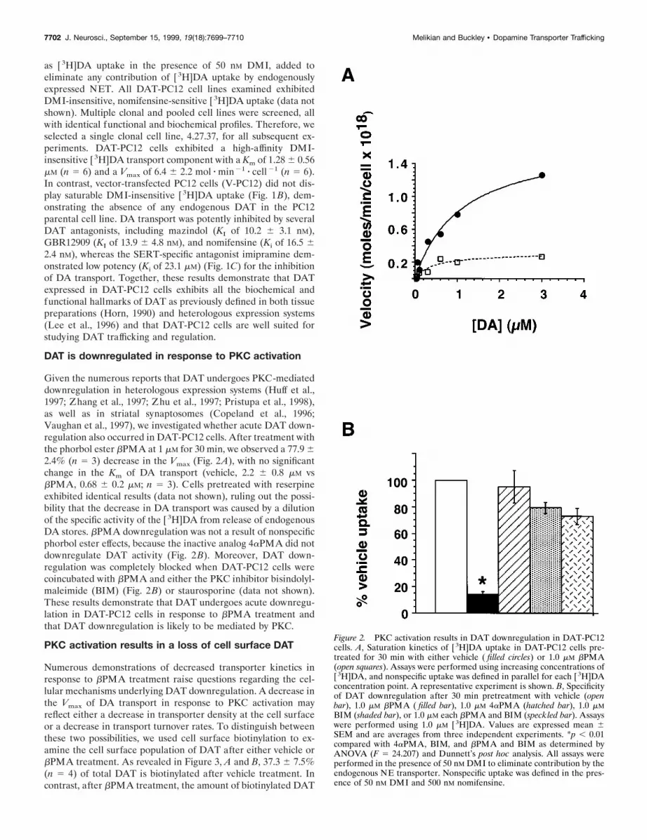
DAT is downregulated in response to PKC activation
Given the numerous reports that DAT undergoes PKC-mediateddownregulation in heterologous expression systems (Huff et al.,1997; Zhang et al., 1997; Zhu et al., 1997; Pristupa et al., 1998),as well as in striatal synaptosomes (Copeland et al., 1996;Vaughan et al., 1997), we investigated whether acute DAT down-regulation also occurred in DAT-PC12 cells. After treatment withthe phorbol ester bPMA at 1 mM for 30 min, we observed a 77.9 62.4% (n 5 3) decrease in the Vmax (Fig. 2A), with no significantchange in the Km of DA transport (vehicle, 2.2 6 0.8 mM vsbPMA, 0.68 6 0.2 mM; n 5 3). Cells pretreated with reserpineexhibited identical results (data not shown), ruling out the possi-bility that the decrease in DA transport was caused by a dilutionof the specific activity of the [3H]DA from release of endogenousDA stores. bPMA downregulation was not a result of nonspecificphorbol ester effects, because the inactive analog 4aPMA did notdownregulate DAT activity (Fig. 2B). Moreover, DAT down-regulation was completely blocked when DAT-PC12 cells werecoincubated with bPMA and either the PKC inhibitor bisindolyl-maleimide (BIM) (Fig. 2B) or staurosporine (data not shown).These results demonstrate that DAT undergoes acute downregu-lation in DAT-PC12 cells in response to bPMA treatment andthat DAT downregulation is likely to be mediated by PKC.
PKC activation results in a loss of cell surface DAT
Numerous demonstrations of decreased transporter kinetics inresponse to bPMA treatment raise questions regarding the cel-lular mechanisms underlying DAT downregulation. A decrease inthe Vmax of DA transport in response to PKC activation mayreflect either a decrease in transporter density at the cell surfaceor a decrease in transport turnover rates. To distinguish betweenthese two possibilities, we used cell surface biotinylation to ex-amine the cell surface population of DAT after either vehicle orbPMA treatment. As revealed in Figure 3, A and B, 37.3 6 7.5%(n 5 4) of total DAT is biotinylated after vehicle treatment. Incontrast, after bPMA treatment, the amount of biotinylated DAT
Figure 2. PKC activation results in DAT downregulation in DAT-PC12cells. A, Saturation kinetics of [ 3H]DA uptake in DAT-PC12 cells pre-treated for 30 min with either vehicle ( filled circles) or 1.0 mM bPMA(open squares). Assays were performed using increasing concentrations of[ 3H]DA, and nonspecific uptake was defined in parallel for each [ 3H]DAconcentration point. A representative experiment is shown. B, Specificityof DAT downregulation after 30 min pretreatment with vehicle (openbar), 1.0 mM bPMA ( filled bar), 1.0 mM 4aPMA (hatched bar), 1.0 mMBIM (shaded bar), or 1.0 mM each bPMA and BIM (speckled bar). Assayswere performed using 1.0 mM [ 3H]DA. Values are expressed mean 6SEM and are averages from three independent experiments. *p , 0.01compared with 4aPMA, BIM, and bPMA and BIM as determined byANOVA (F 5 24.207) and Dunnett’s post hoc analysis. All assays wereperformed in the presence of 50 nM DMI to eliminate contribution by theendogenous NE transporter. Nonspecific uptake was defined in the pres-ence of 50 nM DMI and 500 nM nomifensine.
7702 J. Neurosci., September 15, 1999, 19(18):7699–7710 Melikian and Buckley • Dopamine Transporter Trafficking

is reduced to 8.8 6 2.9% of the total DAT (n 5 4). Thiscorresponds to a 76.3% reduction in the amount of cell surfaceDAT and is consistent with the magnitude of the decrease in Vmax
after bPMA treatment. Total DAT signals were not significantlyreduced after bPMA treatment (110.5 6 10.8% of vehicle; n 5 4),suggesting that reduction in cell surface DAT is not caused bydegradation. Together, these data indicate that activation of PKCresults in a redistribution of DAT from the cell surface to intra-
cellular compartments and that the loss of DAT from the plasmamembrane is responsible for the decrease in Vmax observed afterbPMA treatment.
Intracellular DAT colocalizes with endosomal markers
Examination of the distribution of DAT in DAT-PC12 cellsdemonstrated that, at steady state, a substantial amount (62.7 67.4%) of mature DAT protein is intracellular. Given that differ-ent types of intracellular organelles are subject to distinct traf-ficking mechanisms, we sought to determine the identity of theDAT-containing intracellular compartment. To this end, we per-formed a series of subcellular fractionations on postnuclear su-pernatants (800 3 g) prepared from DAT-PC12 cells and com-pared the fractionation of DAT with known cellular organellemarkers.
First, we examined the population of LDCVs using a two-stagesubcellular fractionation method. DAT-PC12 cells were pre-loaded with [3H]DA to label the LDCVs. Analysis of the firstfractionation (0.3–1.2 M sucrose velocity gradient) revealed that,whereas preloaded [ 3H]DA and the LDCV marker secretograninII (data not shown) colocalized to fractions 5–7, DAT segregatedinto two peaks that were distinct from the secretogranin II–[3H]DA peak (Fig. 4A). Further fractionation of the secretogra-nin II–[3H]DA peak fractions on 0.6–1.6 M sucrose equilibriumgradients also revealed that DAT migrates at a density distinctfrom that of LDCVs (Fig. 4B) and suggests that the majority ofDAT is not localized to this organelle.
We also examined the population of synaptic vesicles (SVs) byfractionation in 5–25% glycerol velocity gradients, a methodknown to identify SVs in PC12 cells (Clift-O’Grady et al., 1990).As illustrated in Figure 5, whereas SVs peak in fractions 4–9, themajority of DAT peaked in fractions 13–16, suggesting that themajority of intracellular DAT is not localized to SVs.
Finally, we examined the distribution of DAT in 10–50%sucrose equilibrium gradients compared with markers of theendocytic pathway. After endocytosis, proteins are targeted tothe early, or sorting, endosome (SE), characterized by the mo-lecular markers EEA1 and rab5 (Clague, 1998). Proteins destinedfor recycling back to the plasma membrane, such as the trans-ferrin receptor, traffic from the SE to the pericentriolar endoso-mal recycling compartment (ERC), characterized by the presenceof the majority of intracellular TfR. Proteins destined for degra-dation [e.g., epidermal growth factor (EGF) receptor] exit theendosomal pathway at the SE and traffic through the late endo-some–lysosome pathway (Sorkin, 1998) (see Fig. 10). DAT im-munoreactivity peaked precisely with that of the TfR at 29 60.6% sucrose (n 5 5), whereas rab5A and EAA1 fractionated ata significantly distinct density (unpaired Student’s t test; p , .005)at a sucrose concentration of 22.4 6 0.4% sucrose (n 5 3) (Fig. 6).Thus, it appears that, at steady state, the majority of intracellularDAT cofractionates with the TfR-positive ERC but not therab5A–EEA1-positive SE.
Comigration of DAT with the TfR suggested that DAT mightreside in the ERC. Alternatively, whereas TfR resides in theERC, DAT may reside in a distinct vesicle of similar density. Todistinguish between these possibilities, we pooled the peakDAT—TfR-immunopositive fractions and isolated intact or-ganelles with antibodies to the TfR, followed by immunoblot forthe presence of DAT. Organelle immunoisolation with anti-TfRantibodies coprecipitated nearly all of the DAT present, whereasa control antibody did not immunoprecipitate TfR or appreciable
Figure 3. PKC activation results in DAT redistribution from the cellsurface to an intracellular pool. A, Steady-state biotinylation of DAT-PC12 cells after 30 min treatment with either vehicle or 1.0 mM PMA.Biotinylated (cell surface) and nonbiotinylated (intracellular) proteinswere separated with streptavidin beads and analyzed by immunoblot witha DAT-specific antibody as described in Materials and Methods. A rep-resentative immunoblot is shown. B, Quantitation of DAT-PC12 cellbiotinylation. Immunoblots of biotinylated (cell surface; open bars) andnonbiotinylated (intracellular; hatched bars) proteins were scanned andquantitated using ImageQuant software. *p , 0.05, significant differencecompared with vehicle-treated cells; unpaired Student’s t test; n 5 4.
Melikian and Buckley • Dopamine Transporter Trafficking J. Neurosci., September 15, 1999, 19(18):7699–7710 7703

amounts of DAT (Fig. 7). Additionally, EEA1 failed to coprecipi-tate with the TfR (data not shown), consistent with the DAT–TfR compartment being distinct from the EEA1-positive SE.These data demonstrate that DAT and TfR are present in thesame intracellular vesicles and indicate that, at steady state, DATis enriched in the ERC.
DAT is targeted to the recycling endosome inresponse to PKC activation
ERC-localized DAT may be involved in DAT trafficking duringdownregulation. Alternatively, endosomal DAT may serve a traf-ficking function distinct from DAT sequestration after PKC ac-tivation. To distinguish between these possibilities, we directlyexamined the internalization pathway through which DAT trafficsduring steady state and after PKC activation. First, DAT-PC12cells were biotinylated at 4°C with NHS–SS–biotin to label thecell surface population of proteins. Cells were rapidly warmed to37°C and were incubated with either vehicle or bPMA for 30 minat 37°C to initiate DAT internalization. Next, cells were rapidlychilled to 4°C to arrest endocytosis, and any remaining cellsurface biotin was removed by treating with the cell-impermeantreducing agent MesNa. Under these conditions, only proteins thatwere present on the cell surface and were subsequently internal-ized would remain biotinylated. Cells were then fractionated on
10–50% sucrose equilibrium gradients, and internalized biotinyl-ated proteins were isolated with streptavidin beads. Using thismethod, only DAT that arose from the cell surface (i.e., biotin-ylated) and accumulated intracellularly (i.e., MesNa protected)would be identified.
As seen in Figure 8, during vehicle treatment, biotinylatedDAT cofractionated with biotinylated transferrin receptor peak,demonstrating basal endocytosis of TfR and DAT. bPMA treat-ment resulted in a robust increase in the amount of internalizedDAT (243 6 15.1% over vehicle treatment; n 5 3). Moreover,internalized DAT accumulated in the TfR-positive ERC and didnot shift to some other compartment. The cofractionation ofinternalized DAT and TfR likely represents DAT trafficking tothe ERC. Alternatively, DAT may internalize and accumulate inan organelle of similar density but physically distinct from theERC. To further characterize the compartment in which DATaccumulates during bPMA treatment, we treated DAT-PC12cells with bPMA (1.0 mM, 30 min), fractionated postnuclearsupernatants in 10–50% sucrose equilibrium gradients, and per-formed organelle immunoisolation on pooled DAT-positive frac-tions. As seen in Figure 9, immunoisolation of organelles with anantibody directed against the TfR isolated the majority of TfR(.81%), as well as the majority of DAT (.88%), consistent withthe accumulation of DAT in the TfR-positive ERC during PMA-
Figure 4. DAT does not colocalize with large dense-core vesicles in DAT-PC12 cells. A, B, Large dense-core vesicles fractionation. [ 3H]DA-labeledDAT-PC12 cells were fractionated in 0.3–1.2 M sucrose velocity gradients, ( A) followed by fractionation of peak [ 3H]DA-containing fractions on 0.6–1.6M sucrose equilibrium gradients (B). Top, Fractions were analyzed for DAT content by immunoblot. Bottom, Fractionation profile of DAT signal ( filledcircles) and [ 3H]DA (in counts per minute; open squares). A representative experiment of two performed is shown.
7704 J. Neurosci., September 15, 1999, 19(18):7699–7710 Melikian and Buckley • Dopamine Transporter Trafficking

induced downregulation. These results demonstrate that DAT (1)undergoes endocytosis under basal conditions and (2) is targetedto the TfR-positive ERC under steady-state conditions, as well asduring PKC-mediated downregulation.
DISCUSSION
Membrane trafficking in neurons is fundamental to a number ofkey regulatory events, including synaptic vesicle recycling, desen-sitization of G-protein-coupled receptors (Cao et al., 1998; Vick-ery and von Zastrow, 1999), and attenuation of signaling throughreceptor tyrosine kinases (Sorkin, 1998). Moreover, recent stud-ies implicate endocytic trafficking in ionotropic glutamate recep-tor downregulation (Lissin et al., 1999) and suggest that traffick-ing may play a potential role in neuronal plasticity. Given recentreports that DAT undergoes PKC-mediated downregulation, wetested the hypothesis that endocytic trafficking underlies DATdownregulation and sought to define the pathway(s) throughwhich endocytic DAT travels.
To study DAT regulation and trafficking in a homogeneousneuroendocrine preparation, we generated a stable PC12 cell lineexpressing human DAT cDNA. DAT-PC12 cells exhibited all ofthe hallmarks of DA transport, with kinetic constants and antag-onist potencies in excellent agreement with those obtained inboth striatal membranes (Horn, 1990) and heterologous expres-sion systems (Lee et al., 1996). Given the notorious heterogeneity
of PC12 cells, we screened numerous clonal and pooled cell linesto confirm that cell lineage or DAT expression levels did notaffect DAT function, biosynthesis, or downregulation. Regardlessof which clonal line was studied, no variation in DAT electro-phoretic mobility, transport activity, or susceptibility to bPMA-induced downregulation was detected.
Figure 5. DAT does not colocalize with synaptic vesicles in DAT-PC12cells. DAT-PC12 cells were homogenized and fractionated in 5–25%glycerol velocity gradients as described in Materials and Methods. Top,Immunoblot of fractions with antibodies to DAT and the synaptic vesiclemarker p38 (synaptophysin). Bottom, Quantitation of DAT ( filled circles)and p38 (open squares) signals detected in each fraction. The peak of DATimmunoreactivity is localized to the bottom of the gradient, whereassynaptic vesicles migrate to fractions 5–8 of the gradient. A representativeexperiment is shown of three performed.
Figure 6. DAT and TfR cofractionate on sucrose equilibrium gradients.DAT-PC12 cells were homogenized and fractionated on 10–50% sucroseequilibrium gradients as described in Materials and Methods. Fractionswere TCA-precipitated and analyzed by SDS-PAGE (10%) and immuno-blot. A, Immunoblot probing with antibodies for the human DAT, TfR,rab5A, and EEA1. B, Quantitation of 10–50% sucrose equilibrium gra-dient probed for DAT ( filled circles), TfR (open triangles), rab5A ( filledtriangles), and EEA1 (open squares). DAT and TfR colocalize at 29 60.6% sucrose (n 5 5), whereas rab5A and EAA1 fractionated at asignificantly distinct density (unpaired Student’s t test; p , 0.005) at asucrose concentration of 22.4 6 0.4% sucrose (n 5 3). Blots were scanned,and band densities were determined using ImageQuant software as de-scribed in Materials and Methods. Experiments were performed three tosix times with essentially identical results. A representative example isshown.
Melikian and Buckley • Dopamine Transporter Trafficking J. Neurosci., September 15, 1999, 19(18):7699–7710 7705

After fully characterizing the DAT-PC12 cell line, we firstinvestigated whether DAT function was modulated in response tobPMA treatment. Thirty minute treatment with 1 mM bPMAresulted in a robust transporter downregulation, observed as a77.9 6 2.4% decrease in transporter Vmax (Fig. 2A). The magni-tude of this decrease is consistent with findings from striatalsynaptosomes (Copeland et al., 1996; Vaughan et al., 1997) andoocytes (Zhu et al., 1997), as well as in C6 glioma (Zhang et al.,1997), LLC-PK1 (Huff et al., 1997), COS, and Sf9 (Pristupa et al.,1998) cells. Moreover, coincubation with bPMA and either stau-rosporine (data not shown) or BIM (Fig. 2B) blocked the effectsof bPMA, suggesting that bPMA -induced DAT downregulationrequires activation of PKC. The effects of PKC activation on DAtransport were not caused by global alterations in membranetrafficking or ion gradients, as Na1-dependent [ 3H]alanine trans-port was not affected by PMA treatment (data not shown).Whereas PKC activation is clearly involved in DAT downregula-tion, the specific role of PKC in DAT downregulation has not yetbeen determined. Phorbol ester-mediated PKC activation alsoresults in phosphorylation of DAT (Huff et al., 1997; Vaughan etal., 1997) and SERT (Ramamoorthy et al., 1998); however, directphosphorylation of DAT or SERT by PKC has not been demon-strated, raising the possibility that PKC may mediate transporterphosphorylation and/or downregulation via recruitment of yetunidentified downstream effectors. Indeed, recent reports suggestthat cAMP-dependent protein kinase may play a role in DAT(Batchelor and Schenk, 1998), as well as SERT and NET (Jay-anthi and DeFelice, Society for Neuroscience, 1998) regulationand also that Ca 21-dependent pathways may also contribute totransporter modulation (Uchida et al., 1998).
A decrease in Vmax of DA transport suggests that DAT down-
regulation may reflect decreased cell surface expression of DAT.Transporter internalization has been demonstrated recently forboth SERT (Qian et al., 1997) and NET (Apparsundaram et al.,1998b). Immunofluorescent examination of DAT expressed inCOS and Sf9 cells suggests that DAT redistributes in response tokinase activation (Pristupa et al., 1998); however, a biochemicalanalysis of DAT redistribution has not been performed to date.Moreover, if DAT is sequestered during downregulation, neitherthe identity of the intracellular compartment harboring DAT northe pathways traversed by DAT are established. To examine theplasma membrane pool during DAT downregulation, we per-formed cell surface biotinylation on DAT-PC12 cells after eithervehicle or bPMA treatment. As seen in Figure 3A, bPMA treat-ment resulted in a significant shift in the distribution of DATfrom the cell surface to an intracellular location. The loss of DATfrom the cell surface could not be explained by degradation,because there was not a significant loss in the total amount ofDAT signal per experiment. Therefore, we conclude that de-creased Vmax of DA transport in response to PKC activation is theresult of sequestration of DAT from the cell surface an intracel-lular compartment(s) whose identity is not known.
DAT-PC12 cell biotinylation also revealed that, even undervehicle-treated conditions, a substantial proportion of DAT isintracellular (62.7 6 7.4%; n 5 4). Ultrastructural studies inves-tigating the subcellular localization of DAT in the rat brain haverevealed an intracellular DAT pool in substantia nigra (Nirenberget al., 1996; Hersch et al., 1997), VTA (Nirenberg et al., 1997),and their projections. To biochemically identify the compartmentcontaining DAT, we used subcellular fractionation to character-ize which intracellular organelles harbor DAT. DAT did notappear to be enriched in large dense-core vesicles (Fig. 4A,B),nor was DAT present in synaptic vesicles (Fig. 5). Interestingly, arecent report from Renick et al. (1999) demonstrated enrichmentof the Na1/Cl2-dependent proline transporter, but not DAT, insynaptic vesicles isolated from rat brain, suggesting that thesubcellular targeting of DAT in DAT-PC12 cells is consistentwith that found in the brain.
Fractionation of DAT-PC12 cells in sucrose equilibrium gradi-ents and intact organelle immunoisolation demonstrated that, atsteady state, DAT colocalizes with the transferrin receptor in theERC but not with rab5A and EAA1 in the SE (Fig. 7). Detectionof TfR in the ERC but not the SE is entirely consistent with itsrapid exit from the SE (Dunn et al., 1989) but slower exit from theERC (McGraw et al., 1987). Localization of DAT to the ERC atsteady state does not necessarily mean that DAT is targeted to theERC during PKC-mediated downregulation. Alternatively,ERC-localized DAT may be involved in some other traffickingevent unrelated to transporter downregulation. To determinewhether PKC activation resulted in DAT internalization to theERC, we examined the destination of cell surface DAT afterbPMA treatment. As seen in Figure 8, bPMA treatment resultedin a striking increase in the amount of biotinylated DAT presentin the TfR-positive ERC, suggesting that the DAT is targeted tothis organelle after internalization and that the DAT-positiveorganelle is, indeed, endocytic. Moreover, after PMA treatment,the majority of DAT colocalizes to TfR-positive ERCs as deter-mined by organelle immunoisolation (Fig. 9). To the best of ourknowledge, this is the first identification of the endocytic pathwayand organelle used by a Na1/Cl2-dependent neurotransmittertransporter. The accumulation of DAT in the recycling endosomein response to PKC activation could be caused by an increase inthe endocytic rate or, conversely, a decrease in the exocytic rate
Figure 7. DAT and TfR are localized to the same organelle at steadystate. Organelle immunoisolation from DAT-PC12 cells. DAT-PC12 cellswere homogenized and fractionated in 10–50% sucrose equilibrium gra-dients. Peak DAT–TfR fractions were pooled, and intact organelles wereimmunoisolated using magnetic beads coated with either a negativecontrol antibody (mouse IgG) or a monoclonal antibody directed againstthe human transferrin receptor (aTfR). Bead eluents and supernatantswere analyzed by 10% SDS-PAGE and were immunoblotted with anti-bodies directed against either DAT or TfR. The majority of DAT and theTfR are precipitated by antibodies to the TfR, while both proteins remainin the supernatant when control beads are used.
7706 J. Neurosci., September 15, 1999, 19(18):7699–7710 Melikian and Buckley • Dopamine Transporter Trafficking

of DAT. Ongoing studies in the laboratory should prove illumi-nating in this regard.
The confirmation of transporter endocytosis raises a number ofquestions. What is the cellular fate of endocytosed transporters?Other plasma membrane proteins subject to endocytosis can meet
with a variety of fates. For example, the receptor tyrosine kinasescan recycle back to the cell surface, traffic to degradative or-ganelles (Mellman, 1996; Sorkin, 1998) such as lysosomes, or, inthe case of the NGF receptor TrkA, form a novel signalingorganelle (Grimes et al., 1996, 1997). A number of receptors, such
Figure 8. DAT internalizes to the endosomal recycling compartment in response to phorbol ester treatment. DAT-PC12 cells were biotinylated at 4°C,warmed to 37°C, and treated with either vehicle ( filled circles) or 1.0 mM bPMA (open squares) for 30 min at 37°C to initiate endocytosis. Cells wererapidly chilled to 4°C, and noninternalized biotin was released from the cell surface by treatment with 10 mM MesNa as described in Materials andMethods. Cells were homogenized and fractionated in 10–50% sucrose equilibrium gradients, and organelles were solubilized. Biotinylated proteins wereisolated from each fraction with streptavidin beads and analyzed by 10% SDS-PAGE and immunoblot. Top, Immunoblot of internalized DAT and TfRafter treatment with vehicle or 1.0 mM bPMA. Bottom, Quantitation of DAT and TfR immunoreactivity after treatment with either vehicle ( filled circles)or 1.0 mM bPMA (open squares). A representative experiment of three independent experiments is shown.
Melikian and Buckley • Dopamine Transporter Trafficking J. Neurosci., September 15, 1999, 19(18):7699–7710 7707

as the transferrin and low-density lipoprotein receptors, undergomultiple rounds of ligand-induced internalization and recyclingback to the plasma membrane (Mellman, 1996; Sorkin, 1998). Todate, it is not understood whether endocytosed DAT recyclesback to the plasma membrane. However, the targeting of DAT tothe ERC suggests that DAT recycling is likely. Moreover, themovement of DAT, presumably through the SE (Fig. 9), to theERC as opposed to the late endosomal–degradatory pathwaysuggests that PKC-mediated DAT internalization does not resultin chronic DAT depletion; rather, DAT internalization is likely toresult in short-term, recoverable downregulation.
What intrinsic–extrinsic molecular factors govern transportertrafficking? Although it is clear that many of the Na1/Cl2-dependent neurotransmitter transporters are subject to down-regulation, it is entirely unknown whether the same cellularmechanisms control membrane trafficking of transporter ho-mologs. Hence, although it is tempting to suggest that the DATtrafficking pathways we have identified will be applicable for all ofthe Na1/Cl2-dependent transporters, it is highly possible thateach homolog may present exciting differences corresponding totheir individual structural divergence. Indeed, recent traffickingstudies examining internalization of DA receptor subtypes (Vick-ery and von Zastrow, 1999) suggest that multiple mechanismsmay differentially govern the internalization of homologousplasma membrane proteins. Moreover, studies performed in po-larized epithelial cells demonstrate that transporter homologs aredifferentially targeted to cellular domains (Ahn et al., 1996; Gu etal., 1996). Finally, are there physiologically relevant mechanisms
involved in transporter trafficking? Preliminary data from ourlaboratory (our unpublished results) and recent observationsfrom Apparsundaram et al. (1998a) and Beckman et al. (1999)suggest that DAT, NET, and GABA transporters, respectively,undergo downregulation in response to activation of muscariniccholinergic receptors. Recent reports support the hypothesis thatmuscarinic downregulation of DATs is also likely to occur in thebrain. For example, elevated extracellular DA concentrations invivo after administration of muscarinic agonists have been de-tected in rat striatum (Smolders et al., 1997) and VTA (West-erink et al., 1998) and could be attributed to DAT downregula-tion. Future studies examining recovery from downregulationshould shed light on possible DAT recycling and the molecularmechanisms underlying DAT trafficking.
How does DAT membrane trafficking fit into our conception ofgeneral membrane trafficking? As illustrated in Figure 10, themajority of proteins that undergo endocytic trafficking are ini-tially internalized to the rab5-positive sorting endosome. Once inthe sorting endosome, proteins can be targeted to the late endo-some–lysosome pathway, as is the EGF receptor, or enter theERC and undergo exocytosis back to the cell surface, as does thetransferrin receptor. Fitting our data into the known model ofendocytic trafficking, we hypothesize that DAT downregulation–trafficking could occur in the following manner (Fig. 10). Activa-tion of PKC, possibly via a G-protein-coupled receptor, such asthe muscarinics, results in an increased accumulation of DAT inthe recycling endosome, consistent with the hypothesis that DATundergoes endocytic trafficking, and possibly recycling, after ac-tivation of PKC. Effectors of PKC that affect DAT traffickinghave not, as yet, been identified and should reveal the molecularevents responsible for DAT sequestration.
In conclusion, we have characterized DAT regulation andtrafficking in stably transfected PC12 cells. We have demon-
Figure 9. After PMA treatment, DAT is localized to the ERC. Or-ganelle immunoisolation from DAT-PC12 cells. After treatment with 1.0mM PMA for 30 min at 37°C, DAT-PC12 cells were homogenized andfractionated in 10–50% sucrose equilibrium gradients. Peak DAT–TfRfractions were pooled, and intact organelles were immunoisolated usingmagnetic beads coated with either a negative control antibody (mouseIgG) or a monoclonal antibody directed against the human transferrinreceptor (aTfR; H68.4). Bead eluents and supernatants were analyzed by10% SDS-PAGE and were immunoblotted with antibodies directedagainst either DAT or TfR. After PMA treatment, the majority of DATand the TfR are precipitated by antibodies to the TfR, while both proteinsremain in the supernatant when control beads are used.
Figure 10. Model for DAT endocytic trafficking. DAT constitutivelyrecycles between the plasma membrane and intracellular endosomalcompartments. Activation of PKC via cell surface receptors (e.g. musca-rinic acetylcholine receptors) results in an accumulation of DAT in theendocytic recycling compartment. Subsequently, DAT may undergo exo-cytosis back to the plasma membrane. For comparison, the constitutiverecycling of the transferrin receptor is also depicted.
7708 J. Neurosci., September 15, 1999, 19(18):7699–7710 Melikian and Buckley • Dopamine Transporter Trafficking

strated that DAT undergoes endocytosis in response to bPMA-induced PKC activation and have identified the endocytic path-way through which DAT traffics in both steady-state andregulated conditions. These results represent the first direct de-scription of the trafficking pathways used by a Na1/Cl2-dependent transporter and identify the specific endocytic com-partment harboring DAT. The initial identification of DATtrafficking and endocytosis in a neuroendocrine cell line formsthe basis for future studies investigating the mechanisms under-lying DAT trafficking.
REFERENCES
Ahn J, Mundig O, Muth TR, Rudnick G, Caplan MJ (1996) Polarizedexpression of GABA transporters in Madin–Darby canine kidney cellsand cultured hippocampal neurons. J Biol Chem 271:6917–6924.
Amara SG, Kuhar MJ (1993) Neurotransmitter transporters: recentprogress. Annu Rev Neurosci 16:73–93.
Apparsundaram S, Galli A, DeFelice LJ, Hartzell HC, Blakely RD(1998a) Acute regulation of norepinephrine transport. I. Protein ki-nase C-linked muscarinic receptors influence transport capacity andtransporter density in SK-N-SH cells. J Pharmacol Exp Ther287:733–743.
Apparsundaram S, Schroeter S, Giovanetti E, Blakely RD (1998b) Acuteregulation of norepinephrine transport. II. PKC-modulated surfaceexpression of human norepinephrine transporter proteins. J PharmacolExp Ther 287:744–751.
Barker EL, Blakely RD (1995) Norepinephrine and serotonin transport-ers: molecular targets of antidepressant drugs. In: Psychopharmacology:the fourth generation of progress (Bloom F, Kupfer D, eds), pp 321–333. New York: Raven.
Batchelor M, Schenk JO (1998) Protein kinase A activity may kineticallyupregulate the striatal transporter for dopamine. J Neurosci18:10304–10309.
Beckman ML, Quick MW (1998) Neurotransmitter transporters: regu-lators of function and functional regulation. J Membr Biol 164:1–10.
Beckman ML, Bernstein EM, Quick MW (1998) Protein kinase C reg-ulates the interaction between a GABA transporter and syntaxin 1A.J Neurosci 18:6103–6112.
Beckman ML, Bernstein EM, Quick MW (1999) Multiple G-protein-coupled receptors inititate protein kinase C redistribution of GABAtransporters in hippocampal neurons. J Neurosci 19:RC9, 1–6.
Blakely RD, Ramamoorthy S, Schroeter S, Qian Y, Apparsundaram S,Galli A, DeFelice LJ (1998) Regulated phosphorylation and traffick-ing of antidepressant-sensitive serotonin transport proteins. Biol Psy-chiatry 44:169–178.
Cao TT, Mays RW, von Zastrow M (1998) Regulated endocytosis ofG-protein-coupled receptors by a biochemically and functionally distinctsubpopulation of clathrin-coated pits. J Biol Chem 273:24592–24602.
Clague MJ (1998) Molecular aspects of the endocytic pathway. BiochemJ 336:271–282.
Clift-O’Grady L, Linstedt AD, Lowe AW, Grote E, Kelly RB (1990)Biogenesis of synaptic vesicle-like structures in a pheochromocytomacell line PC-12. J Cell Biol 110:1693–1703.
Copeland BJ, Vogelsberg V, Neff NH, Hadjiconstantinou M (1996) Pro-tein kinase C activators decrease dopamine uptake into striatal synap-tosomes. J Pharmacol Exp Ther 277:1527–1532.
Corey JL, Davidson N, Lester HA, Brecha N, Quick MW (1994) Proteinkinase C modulates the activity of a cloned g-aminobutyric acid trans-porter expressed in Xenopus oocytes via regulated subcellular redistri-bution of the transporter. J Biol Chem 269:14759–14767.
Dunn KW, McGraw TE, Maxfield FR (1989) Iterative fractionation ofrecycling receptors from lysosomally destined ligands in an early sortingendosome. J Cell Biol 109:3303–3314.
Grace AA (1995) The tonic/phasic model of dopamine system regula-tion: its relevance for understanding how stimulant abuse can alter basalganglia function. Drug Alcohol Depend 37:111–129.
Grimes ML, Zhou J, Beattie EC, Yuen EC, Hall DE, Valletta JS, ToppKS, LaVail JH, Bunnett NW, Mobley WC (1996) Endocytosis of
activated TrkA: evidence that nerve growth factor induces formation ofsignaling endosomes. J Neurosci 16:7950–7964.
Grimes ML, Beattie E, Mobley WC (1997) A signaling organelle con-taining the nerve growth factor-activated receptor tyrosine kinase,TrkA. Proc Natl Acad Sci USA 94:9909–9914.
Gu HH, Ahn J, Caplan MJ, Blakely RD, Levey AI, Rudnick G (1996)Cell-specific sorting of biogenic amine transporters expressed in epi-thelial cells. J Biol Chem 271:18100–18106.
Hersch SM, Yi H, Heilman CJ, Edwards RH, Levey AI (1997) Subcel-lular localization and molecular topology of the dopamine transporterin the striatum and substantia nigra. J Comp Neurol 388:211–227.
Horn AS (1990) Dopamine uptake: a review of progress in the lastdecade. Prog Neurobiol 34:387–400.
Huff RA, Vaughan RA, Kuhar MJ, Uhl GR (1997) Phorbol estersincrease dopamine transporter phosphorylation and decrease transportVmax. J Neurochem 68:225–232.
Jones SR, Gainetdinov RR, Jaber M, Giros B, Wightman RM, Caron MG(1998) Profound neuronal plasticity in response to inactivation of thedopamine transporter. Proc Natl Acad Sci USA 95:4029–4034.
Langer SZ (1997) 25 years since the discovery of presynaptic receptors:present knowledge and future perspectives. Trends Pharmacol Sci18:95–99.
Lee FJS, Pristupa ZB, Ciliax BJ, Levey AI, Niznik HB (1996) The do-pamine transporter carboxyl-terminal tail. J Biol Chem 271:20885–20894.
Lissin DV, Carroll RC, Nicoll RA, Malenka RC, von Zastrow M (1999)Rapid, activation-induced redistribution of ionotropic glutamate recep-tors in cultured hippocampal neurons. J Neurosci 19:1263–1272.
McGraw TE, Greenfield L, Maxfield FR (1987) Functional expression ofthe human transferrin receptor cDNA in Chinese hamster ovary cellsdeficient in endogenous transferrin recpetor. J Cell Biol 105:207–214.
Melikian HE, McDonald JK, Gu H, Rudnick G, Moore KR, Blakely RD(1994) Human norepinephrine transporter: Biosynthetic studies usinga site directed polyclonal antibody. J Biol Chem 269:12290–12297.
Mellman I (1996) Endocytosis and molecular sorting. Annu Rev CellDev Biol 12:575–625.
Nagatsu T, Stjarne L (1998) Catecholamine synthesis and release. Over-view. Adv Pharmacol 42:1–14.
Nelson N (1998) The family of Na 1/Cl 2 neurotransmitter transporters.J Neurochem 71:1785–1803.
Nirenberg MJ, Vaughan RA, Uhl GR, Kuhar MJ, Pickel VM (1996) Thedopamine transporter is localized to dendritic and axonal plasma mem-branes of nigrostriatal dopaminergic neurons. J Neurosci 16:436–447.
Nirenberg MJ, Chan J, Vaughan RA, Uhl GR, Kuhar MJ, Pickel VM(1997) Immunogold localization of the dopamine transporter: an ul-trastructural study of the rat ventral tegmental area. J Neurosci17:5255–5262.
Pristupa ZB, McConkey F, Liu F, Man HY, Lee FJ, Wang YT, Niznik HB(1998) Protein kinase-mediated bidirectional trafficking and functionalregulation of the human dopamine transporter. Synapse 30:79–87.
Qian Y, Galli A, Ramamoorthy S, Risso S, DeFelice LJ, Blakely RD(1997) Protein kinase C activation regulates human serotonin transport-ers in HEK-293 cells via altered cell surface expression. J Neurosci17:45–57.
Quick MW, Corey JL, Davidson N, Lester HA (1997) Second messen-gers, trafficking-related proteins, and amino acid residues that contrib-ute to the functional regulation of the rat brain GABA transporterGAT1. J Neurosci 17:2967–2979.
Ramamoorthy S, Giovanetti E, Qian Y, Blakely RD (1998) Phosphory-lation and regulation of antidepressant-sensitive serotonin transporters.J Biol Chem 273:2458–2466.
Renick SE, Kleven DT, Chan J, Stenius K, Milner TA, Pickel VM,Fremeau Jr RT (1999) The mammalian brain high-affinity L-prolinetransporter is enriched preferentially in synaptic vesicles in a subpopu-lation of excitatory nerve terminal in rat forebrain. J Neurosci 19:21–33.
Sakai N, Sasaki K, Nakashita M, Honda S, Ikegake N, Saito N (1997)Modulation of serotonin transporter activity by a protein kinase Cactivator and an inhibitor of type 1 and 2A serine/threonine phospha-tases. J Neurochem 68:2618–2624.
Sato K, Betz H, Schloss P (1995) The recombinant GABA transporterGAT1 is downregulated upon activation of protein kinase C. FEBSLett 375:99–102.
Schmidt A, Hannah MJ, Huttner WB (1997) Synaptic-like microvesi-clesof neuroendocrine cells originate from a novel compartment that iscontinuous with the plasma membrane and devoid of transferrin recep-tor. J Cell Biol 137:445–458.
Melikian and Buckley • Dopamine Transporter Trafficking J. Neurosci., September 15, 1999, 19(18):7699–7710 7709

Smolders I, Bogaert L, Ebinger G, Michotte Y (1997) Muscarinic mod-ulation of striatal dopamine, glutamate, and GABA release, as mea-sured with in vivo microdialysis. J Neurochem 68:1942–1948.
Sorkin A (1998) Endocytosis and intracellular sorting of receptor ty-rosine kinases. Front Biosci 3:d729–d738.
Uchida J, Kiuchi Y, Yura OM, Oguchi K (1998) Ca(21)-dependentenhancement of [3H]noradrenaline uptake in PC12 cells throughcalmodulin-dependent kinases. Brain Res 809:155–164.
Vaughan RA, Huff RA, Uhl GR, Kuhar MJ (1997) Protein kinaseC-mediated phosphorylation and functional regulation of dopaminetransporters in striatal synaptosomes. J Biol Chem 272:15541–15546.
Vickery RG, von Zastrow M (1999) Distinct dynamin-dependent and
-independent mechanisms target structurally homologous dopaminereceptors to different endocytic membranes. J Cell Biol 144:31–43.
Westerink BH, Enroco P, Feimann J, de Vries JB (1998) The pharma-cology of mesocortical dopamine neurons: a dual-probe microdialysisstudy in the ventral tegmental area and prefrontal cortex of the ratbrain. J Pharmacol Exp Ther 285:143–154.
Zhang L, Coffey LL, Reith ME (1997) Regulation of the functionalactivity of the human dopamine transporter by protein kinase C.Biochem Pharmacol 53:677–688.
Zhu S-J, Kavanaugh MP, Sonders MS, Amara SG, Zahniser NR (1997)Activation of protein kinase C inhibits uptake, currents and bindingassociated with the human dopamine transporter expressed in Xenopusoocytes. J Pharmacol Exp Ther 282:1358–1365.
7710 J. Neurosci., September 15, 1999, 19(18):7699–7710 Melikian and Buckley • Dopamine Transporter Trafficking