a juxtamembrane mutation in the n terminus of the dopamine transporter induces preference for an...
TRANSCRIPT
A Juxtamembrane Mutation in the N Terminus of theDopamine Transporter Induces Preference for anInward-Facing Conformation
Bipasha Guptaroy, Minjia Zhang, Erica Bowton, Francesca Binda, Lei Shi, Harel Weinstein,Aurelio Galli, Jonathan A. Javitch, Richard R. Neubig, and Margaret E. GnegyDepartment of Pharmacology, University of Michigan, Ann Arbor, Michigan (B.G., M.Z., R.R.N., M.E.G.); Department ofPharmacology and Psychiatry, Center for Molecular Recognition, College of Physicians and Surgeons, Columbia University,New York (J.A.J.); Department of Physiology and Biophysics and the HRH Prince Alwaleed Bin Talal Bin Abdulaziz AlsaudInstitute for Computational Biomedicine, Weill Cornell Medical College, Cornell University, New York (L.S., H.W.);and the Department of Molecular Physiology and Biophysics, Center for Molecular Neuroscience and Kennedy Center,Vanderbilt University, Nashville, Tennessee (E.B., F.B., A.G.)
Received May 12, 2008; accepted December 12, 2008
ABSTRACTThe human dopamine transporter (hDAT) regulates synapticdopamine (DA) levels and is the site of action of abused andtherapeutic drugs. Here we study the effect of a threonineresidue (Thr62 in hDAT) that is highly conserved within a ca-nonical phosphorylation site (RETW) in the juxtamembrane N-terminal region of monoamine transporters. In stably trans-fected human embryonic kidney 293T cells, expression ofT62D-hDAT was reduced compared with hDAT or T62A-hDAT.T62D-hDAT displayed dramatically reduced [3H]dopamine up-take but exhibited a higher basal dopamine efflux comparedwith hDAT or T62A-hDAT, as determined by measurements of[3H]dopamine efflux and amperometry. The high constitutiveefflux in T62D-hDAT precluded the measurement of amphet-amine-stimulated [3H]dopamine efflux, but when dopaminewas added internally into voltage-clamped T62D-hDAT cells,amphetamine-induced efflux comparable with hDAT was de-
tected by amperometry. In accordance with findings that Zn2�
can rescue reduced DA uptake in mutant transporters that arepredominantly inward-facing, micromolar concentrations ofZn2� markedly potentiated [3H]dopamine uptake in T62D-hDATand permitted the measurement of amphetamine-stimulateddopamine efflux. These results suggest that T62D-hDAT pre-fers an inward-facing conformation in the transition betweeninward- and outward-facing conformations. For T62A-hDAT,however, the measured 50% reduction in both [3H]dopamineuptake and [3H]dopamine efflux was consistent with a slowedtransition between inward- and outward-facing conformations.The mechanism underlying the important functional role ofThr62 in hDAT activity suggested by these findings is examinedin a structural context using dynamic simulations of a three-dimensional molecular model of DAT.
The strength and duration of dopaminergic neurotransmis-sion is tightly controlled by the dopamine transporter (DAT),which mediates reuptake of synaptic dopamine (DA) (Chenand Reith, 2000) and is a target for therapeutic drugs and
abused psychostimulants (Sulzer et al., 2005). Amphetamine(AMPH), a substrate for DAT, competitively inhibits DA re-uptake and elicits outward transport of DA by reversal of thetransporter (Sulzer et al., 2005), purportedly by an exchangediffusion mechanism (Fischer and Cho, 1979; Sulzer et al.,2005). This model assumes that the transporter transitionsbetween two primary conformational states—an “outward-facing” conformation favoring substrate binding and influx,and an “inward-facing” conformation in which the bindingsites are available to the intracellular milieu and which fa-vors substrate efflux. In the absence of substrate, the trans-porter favors an outward-facing conformation ready to bind
This work was supported by the National Institutes of Health NationalInstitute on Drug Abuse [Grants DA11697, DA12408, DA14684, DA022413,DA023694]. This work used the DNA Sequencing Core of the Michigan Dia-betes Research and Training Center, supported by the National Institutes ofHealth National Institute of Diabetes and Digestive and Kidney Diseases[Grant 5P60-DK20572].
Article, publication date, and citation information can be found athttp://molpharm.aspetjournals.org.
doi:10.1124/mol.108.048744.
ABBREVIATIONS: DAT, dopamine transporter; AMPH, amphetamine; DA, dopamine; HEK, human embryonic kidney; KRH, Krebs-Ringer-HEPES;PKC, protein kinase C; PKA, cAMP-dependent protein kinase; TM, transmembrane domain; ANOVA, analysis of variance; cGPK, cGMP-dependent protein kinase; MD, molecular dynamics; FEP, free energy perturbation; GBR12935, 1-[2-(diphenylmethoxy)ethyl]-4-(3-phenylpropyl)-piperazine; IL, intracellular loop.
0026-895X/09/7503-514–524$20.00MOLECULAR PHARMACOLOGY Vol. 75, No. 3Copyright © 2009 The American Society for Pharmacology and Experimental Therapeutics 48744/3443845Mol Pharmacol 75:514–524, 2009 Printed in U.S.A.
514
extracellular substrate and assume an inward-facing confor-mation (Sulzer et al., 2005). After dissociation of inwardlytransported AMPH, the transporter binds intracellular sub-strate and reverses its conformation to cause efflux. Thispredicts that the influx and efflux rates would be modifiedequivalently. However, recent studies demonstrate that in-flux and efflux of substrates through the transporter can bemodulated independently (Kantor and Gnegy, 1998; Sitteet al., 1998; Pifl et al., 1999; Chen and Justice, 2000;Khoshbouei et al., 2004).
The molecular structure of human DAT (hDAT) comprises12 transmembrane (TM) segments with cytoplasmic N and Ctermini and a large second extracellular loop with severalputative N-glycosylation sites (Giros and Caron, 1993). TheN-terminal region of the transporter and the first transmem-brane domain are important for substrate binding and func-tion of the transporter. Based on the crystal structure of thebacterial leucine transporter (LeuTAa) (Yamashita et al.,2005) and subsequent structure-based alignment for DAT(Beuming et al., 2006), TM1 is predicted to have a “break” inits helical structure in the membrane bilayer that comprisespart of the substrate binding site. The N-terminal cytoplas-mic region of DAT is a site for phosphorylation (Foster et al.,2002) and for the binding of proteins involved in phosphory-lation, such as syntaxin 1A, whose function is regulated byphosphorylation, and receptor for activated C kinase 1, whichfacilitates phosphorylation (Torres, 2006). Phosphorylationof serines within the N-terminal region seems to maintainnormal AMPH-stimulated DA efflux (Khoshbouei et al.,2004). Mutating other serine and threonine residues in theDAT N terminus alters the ability of various kinase-modu-lating drugs to regulate DAT function and expression (Lin etal., 2003).
Considerable information concerning DAT structure andfunction relationships come from site-directed mutagenesisstudies. Most studies have focused on the TM domains thatare highly conserved between the transporters and acrossspecies and probably contribute to the “binding pocket” in-volved in substrate and antagonist recognition and substratetranslocation (Uhl and Lin, 2003). Mutations of residues inregions outside of these domains, such as distal TMs(Kitayama et al., 1992; Wu and Gu, 2003; Sen et al., 2005),extracellular (Uhl and Lin, 2003) and intracellular loops(Loland et al., 2002; Chen et al., 2004; Loland et al., 2004),and the N- and C-terminal domains (Lee et al., 1996; Khosh-bouei et al., 2004), also affect transporter function. The rela-tionship of these sites to the structure of DAT was clarified(Beuming et al., 2006) in the context of three-dimensionalstructural information for the cognate LeuT (Yamashita etal., 2005).
Here we investigate the effect of mutations of a threonineresidue (Thr62) located within a juxtamembrane site (RETW)that is highly conserved in the plasmalemmal transporters forDA, norepinephrine, and serotonin; interestingly, it is within acanonical site for PKC/cGMP-dependent protein kinase (cGPK)/PKA-dependent phosphorylation (Giros and Caron, 1993; Gra-nas et al., 2003). In rat DAT, the mutation of this site to alaninealters the actions of some kinase effectors (Lin et al., 2003). Weinvestigated whether mutation of this highly conserved residueto alanine, a somewhat conservative substitution, or a noncon-servative substitution to aspartate, would alter DAT function.These mutations mimic a nonphosphorylatable state (alanine)
or a phosphorylated state (aspartate), addressing the putativerole of phosphorylation at this site (Thorsness and Koshland,1987). We find a profound change in the balance of influx andefflux in the Thr62 to aspartate mutant with less dramatic, butstill altered, effects upon mutation to alanine. These resultssupport the importance of the TM1-juxtamembrane N-terminalregion in regulating transporter function.
Materials and MethodsMutagenesis and Generation of Stable Cell Lines. A syn-
thetic hDAT cDNA, tagged at the N terminus with a FLAG epitope,was used in these studies. The gene encodes a protein with an aminoacid sequence identical with wild-type human DAT, but the nucleo-tide sequence was altered to increase the number of unique restric-tion sites and optimize codon use (Saunders et al., 2000). The cDNAwas cloned into a bicistronic vector pCIHyg (Saunders et al., 2000)derived from pIREShyg vector from Clontech (Mountain View, CA).Previous studies have shown that the addition of the N-terminalFLAG tag does not alter transporter function (Saunders et al., 2000).Mutations were made by polymerase chain reaction and confirmedby sequencing. In brief, sense and antisense mutagenic oligonucleo-tides were synthesized and used for polymerase chain reaction tosynthesize mutagenized DNA using Vent DNA polymerase (PromegaCorporation, Madison, WI). Methylated parental DNA strands in thesample were digested using DpnI enzyme (Promega, Madison, WI)for 2 h at 37°C, and these were then transformed into XL10-Goldcompetent cells (Stratagene, La Jolla, CA). Clones were analyzed byDNA sequencing. The cDNAs for hDAT and mutants were stablytransfected into HEK 293T cells with Lipofectamine (Invitrogen,Carlsbad, CA), and a stable pool of hygromycin-resistant cells wasselected. Cells were maintained in Dulbecco’s modified Eagle’s me-dium supplemented with 10% fetal bovine serum at 37°C and 5%CO2.
Cell Surface Biotinylation. Surface expression of the wild-typeand mutant transporters in HEK 293T cells was determined byreacting the surface proteins in suspension with sulfosuccinimidyl-2-(biotinamido)ethyl-1, 3-dithiopropionate (sulfo-NHS-SS-biotin)(Pierce, Rockford, IL) at 4°C as described previously (Johnson et al.,2005). The reaction was attenuated by incubating the biotinylatedcells with 100 mM glycine at 4°C. Cells were lysed in solubilizationbuffer (25 mM Tris, 150 mM NaCl, 1 mM EDTA, 5 mM N-ethylmale-meide, phenylmethylsulfonyl fluoride, and 1% Triton X-100) contain-ing protease inhibitors (Roche, Indianapolis, IN) and centrifuged at20,000g to remove cell debris. Lysates containing 700 �g of proteinwere incubated with 50 �l of monomeric Streptavidin beads (Pierce)for 1 h at room temperature. Beads were washed four times withradioimmunoprecipitation assay buffer and then eluted in 2� SDS-polyacrylamide gel electrophoresis sample buffer containing dithio-threitol and resolved by electrophoresis on a 10% Tris-glycine gel.
Immunoblot Analysis. To detect hDAT, proteins were trans-ferred to a nitrocellulose membrane after electrophoresis on SDSpolyacrylamide gel. Membranes were blocked with Tris-buffered sa-line/Tween 20 (10 mM Tris, pH 7.4, 150 mM NaCl, and 0.1% Tween20) containing 5% milk for 1 h followed by overnight incubation withanti-DAT antibody, monoclonal antibody 369 (Millipore BioscienceResearch Reagents, Temecula, CA) at a 1:1000 dilution in 5% milk,followed by three rinses with Tris-buffered saline/Tween 20. Mem-branes were then incubated with goat anti-rat secondary antibody(Santa Cruz Biotechnology, Santa Cruz, CA) at 1:5000 dilution (0.08�g/ml), followed by washes as before. Chemiluminescence was de-tected by enhanced chemiluminescence (GE Healthcare, Chalfont St.Giles, Buckinghamshire, UK). Quantification of bands was done bydensitometry using Scion Image software (Scion Corporation, Fred-erick, MD). We normalized to loading equal protein for each lane,along with total transporter, and in some experiments, actin was
Juxtamembrane N-Terminal Mutations Modulate DAT Function 515
used as an internal control, which remained unchanged among thesamples.
Uptake of [3H]DA. For time course experiments measuring theinitial rate of [3H]DA uptake, cells were plated on 24-well plates at100,000 cells/well. On the following day, cells were rinsed with KRH(25 mM HEPES, pH 7.4, 125 mM NaCl, 4.8 mM KCl, 1.2 mMKH2PO4, 1.3 mM CaCl2, 1.2 mM MgSO4, and 5.6 mM glucose), and[3H]DA uptake was measured in the absence or presence of 10 �MGBR12935 (Sigma, St. Louis, MO). Uptake was initiated with[3H]DA (specific activity, 59.3 Ci/mmol; PerkinElmer Life and Ana-lytical Sciences, Waltham, MA) in concentrations ranging from 30nM to 3 �M in a final volume of 250 �l and allowed to proceed for 0to 2 min at room temperature and was stopped by rapidly washingthe cells three times with cold phosphate-buffered saline. Cells weresolubilized in 1% SDS, and radioactivity was counted using Scinti-verse BD (Thermo Fisher Scientific, Waltham, MA) in a Beckman LS5801 liquid scintillation counter (Beckman Coulter, Fullerton, CA).For assaying [3H]DA uptake in the presence of AMPH (Sigma), cellswere incubated with 30 nM [3H]DA and varying concentrations ofAMPH (10 nM to 3 �M) for 10 min and then assayed as describedabove. Nonspecific uptake was measured in the presence of 10 �MGBR12935. Uptake in the presence of Zn2� was determined using 15nM [3H] DA for 10 min at ambient temperature after preincubationwith Zn2�.
[3H]DA Efflux. For measurement of the initial rate of efflux, cellswere plated on 12-well plates at a density of 150,000 cells/well. Cellswere washed with KRH and incubated with 60 or 120 nM [3H]DAand unlabeled DA at concentrations from 1 to 80 �M for 30 min atroom temperature in KRH. The cells were then quickly washed threetimes with KRH, and 500 �l of KRH was added to the cells andimmediately removed for a 0 time point. At additional times (10–30s), 500-�l aliquots were removed, and KRH was replaced. Finally,cells were lysed in 1% SDS and counted using Scintiverse. Specificefflux was determined in the absence and presence of 100 �M co-caine. For the measurement of basal efflux and AMPH-stimulatedDA efflux, cells were plated on 24-well plates at a density of 100,000to 150,000 cells/well, washed, and loaded with DA for 20 min at roomtemperature in KRH containing between 0.5 and 5 �M DA, 30 nM[3H]DA, 1 mM tropolone (Sigma), and 100 �M pargyline (Sigma).After three quick washes with KRH, 250-�l aliquots were taken forcounting and replaced with fresh KRH every 10 min for 30 min toestablish a baseline. At 30 min, 250 �l of AMPH (1–10 �M) wasadded, and another 250-�l aliquot was taken after 5 min. Finally,cells were lysed in 1% SDS and counted using Scintiverse. Cells fromone well were lysed immediately after DA loading to obtain anestimate of the initial amount of DA (total DA) in the cells.
DA Efflux by Superfusion. Confluent 100-mm plates of cellswere washed twice with KRH and incubated at 37°C with 5 �M DAfor 30 min to load with DA. The hDAT cells were loaded in theabsence of Zn2�, and T62D-hDAT cells were loaded in the presence of30 �M Zn2�. After incubation, cells were washed with KRH, har-vested, and resuspended in KRH with or without Zn2�. DA effluxwas measured in the eluate of superfused cells. Cells were placed ona Whatman GF/B filter (Whatman, Clifton, NJ) in a chamber of aBrandel superfusion apparatus (Brandel SF-12, Gaithersburg, MD)at room temperature and perfused using KRH with or without Zn2�
at 400 �l/min, and 2-min fractions were collected after washing cellsfor 1 h. AMPH (10 �M) was added and perfused for 2 min followed byKRH for 15 min. Fractions were collected into vials containing2-amino phenol (Sigma) as an internal standard. Samples werestored at �70°C, and DA content was measured by high-pressureliquid chromatography with electrochemical detection. DA efflux wasquantified as the peak DA in the eluent.
Electrophysiology. HEK 293T cell lines stably transfected withT62D-hDAT, T62A-hDAT, and hDAT were used for electrophysiolog-ical recordings. Cells were plated 2 days before experiments on35-mm dishes. For nonclamped amperometric experiments, cellswere washed twice with KRH assay buffer (130 mM NaCl, 1.3 mM
KCl, 2.2 mM CaCl2, 1.2 mM MgSO4, 1.2 mM KH2PO4, 10 mMHEPES, pH 7.4, 10 mM D-glucose, 100 �M pargyline, 10 �M tropo-lone, and 100 �M ascorbic acid) and were then incubated with 1 �MDA in assay buffer for 45 min at 37°C to actively load the cells withDA. Plates were then washed twice with bath solution (130 mMNaCl, 10 mM HEPES, 34 mM dextrose, 1.5 mM CaCl2, 0.5 mMMgSO4, and 1.3 mM KH2PO4 adjusted to pH 7.35) at room temper-ature. A carbon-fiber electrode (ProCFE; fiber diameter is 5 �m;Dagan Corporation, Minneapolis, MN) touching the plasma mem-brane of the cell and held at �700 mV with respect to the bathground (a potential greater than the oxidizing potential of DA) wasused to measure DA flux through oxidation reactions. The ampero-metric electrode measures electrical currents (in picoamperes) as aresult of DA molecule oxidation which, after integration, can beconverted when required to the number of DA molecules. The am-perometric electrode measures the number of DA molecules oxidizedand not the concentration of DA. Amperometric currents were thenrecorded using Axopatch 200B with a low-pass Bessel filter set at 100Hz and digitally filtered offline at 10 Hz. The cells were not voltage-clamped, allowing the measurement of basal efflux under restingmembrane potential conditions. The term “DAT-mediated leak” isused to describe DA efflux under basal conditions of the cell (restingmembrane potential) and was defined as amperometric baselinecurrent minus the current present after the addition of 10 �Mcocaine (Sigma), a blocker of DAT function. The dotted horizontallines in Fig. 4 represent the amperometric currents recorded afterthe addition of cocaine when we had achieved the maximum DATblockade. Data were acquired by averaging a 15-s interval of currentdirectly before cocaine application (baseline current) and 20 minafter cocaine application (cocaine current). The stability of our am-perometric signals was confirmed by recording currents with anamperometric electrode positioned in the bath and held at �700 mVfor 20 min. When averaged and subtracted as above, the average ofthe differences was 0.016 � 0.005 pA (n � 6).
For voltage-clamped experiments, DA efflux was elicited using 30mM Na� and 2 mM DA in the recording pipette (pipette solution: 90mM KCl, 30 mM NaCl, 0.1 mM CaCl2, 2 mM MgCl2, 1.1 mM EGTA,10 mM HEPES, 30 mM dextrose, and 2 mM DA, adjusted to pH 7.35)(Khoshbouei et al., 2003). hDAT-, T62D-hDAT-, and T62A-hDAT-mediated effluxes were obtained by subtraction of the efflux valuerecorded in the presence of 20 �M cocaine from the one recorded inthe control condition. Patch electrodes were pulled from quartz pi-pettes on a P-2000 puller with a resistance of 7 M�. Both whole-cellcurrent and amperometric current were recorded from cells. Cur-rent-voltage relationships were generated by stepping membranevoltage from a holding potential of �20 mV to voltages between �100and �100 mV in 20-mV increments for 1 s. Data were recorded andanalyzed using pClamp8 software. Current was calculated as theaverage current during the final 100 ms of the voltage step. Foramperometric analysis, a carbon-fiber electrode was used as de-scribed above.
Protein Determination. Protein assay was performed accordingto the Dc protein assay kit (Bio-Rad, Hercules, CA).
Data Analysis. Kinetic constants, including Km, Vmax, and IC50
values for the hDAT and mutant DAT constructs, were determinedby nonlinear regression analysis of the mean values for each mutantusing Prism version 5 (GraphPad Software Inc., San Diego, CA).Statistical significance was determined using Prism version 4 eitherby comparison with hDAT using ANOVA or a two-tailed Student’s ttest, or with an F test by comparing fits in which selected valueswere constrained to be equal or were allowed to differ. The nullhypothesis was that the best-fit parameter for the value did notdiffer. A conclusion of statistical significance represents a rejection ofthe null hypothesis and indicates a difference between designatedvalues.
Molecular Modeling and Simulations. The molecular modelfor the human DAT was constructed with Modeler 8v2 (Sali andBlundell, 1993) using the LeuT structure as template and the se-
516 Guptaroy et al.

quence alignment detailed in Beuming et al. (2006). As seen from thealignment, the first 59 residues of DAT do not have a correspondingsequence in LeuT, nor do the last 30 residues in the C terminus and26 residues in the middle of extracellular loop 2. These segments aretherefore not included in the DAT model. DAT also has more resi-dues than LeuT in the sixth extracellular loop; these were modeled tocontinue the helix observed in LeuT. The substrate, dopamine, waspositioned in the binding site by aligning the amine group and thehydrophobic portion with those of the substrate in the LeuT struc-ture. The final model also includes two Na� ions positioned equiva-lently to those in LeuT and a Cl� ion coordinated by residues Asn82,Tyr102, Ser321, and Asn353 of DAT based on the chloride bindingsite described in Zomot et al. (2007). Before the molecular dynamics(MD) simulation runs, the structure was equilibrated in a modellipid bilayer of 1-palmitoyl,2-oleoyl-sn-glycero-3-phosphocholine asdescribed in detail in our recent publication (Shi et al., 2008). Inbrief, the simulated system composed of the DAT model immersed inan explicit representation of the water/lipid bilayer/water environ-ment was constructed with visual molecular dynamics (Humphrey etal., 1996) and equilibrated with nanoscale molecular dynamics (Phil-lips et al., 2005) following a procedure modified from Sotomayor andSchulten (2004). During the equilibration of the DAT model in itsenvironment, the backbones were initially fixed and then harmoni-cally constrained, and water was restrained by small forces frompenetrating the protein-lipid interface. The constraints were re-leased gradually in three steps of 300 ps each, changing the forceconstants from 1, to 0.5, and 0.1 kcal/(mol Å2), respectively. Theall-atom CHARMM27 force field was used throughout. Constanttemperature (310 K) was maintained with Langevin dynamics, and1 atm constant pressure was achieved by using the hybrid Nose-Hoover Langevin piston method on a flexible periodic cell (Shi et al.,2008). The simulated system including the transporter embedded ina membrane patch of 196 1-palmitoyl,2-oleoyl-sn-glycero-3-phospho-choline molecules (95 on the periplasmic side and 101 on the cyto-plasmic side) and water layers on each side containing Na� and Cl�
ions corresponding to a concentration of 150 mM NaCl, is composedof nearly 72,000 atoms in a box with final dimensions (at the end of16 ns of free equilibration) of 83 � 89 � 94 Å3.
To model the mutant DAT constructs with T62D and T62A, themutations were introduced using the free energy perturbation (FEP)method implemented in nanoscale molecular dynamics, which usesthe dual-topology paradigm (Henin et al., 2006). Starting from thelast snapshot of the constraint-free equilibration of wild-type DATmodel, the reaction paths for the gradual transformation of threo-nine into asparate (or alanine) at the site was divided into 105 stages(windows) of uneven widths, with narrower intermediates statesdefined toward the end points of the transformation, where thesystem is more sensitive to the perturbations. Each window in thetransformation procedure was run for 40 ps, with the entire trans-formation accomplished in 4.2 ns.
ResultsSurface Expression and Uptake Activity of hDAT and
the Thr62 Mutants. Surface expression of the transporter inhDAT-HEK293T (hDAT cells), T62A-hDAT, and T62D-hDATcells was determined by cell surface biotinylation. A represen-tative blot showing the levels of total and biotinylated surfaceDAT is shown in Fig. 1A. The overall levels of total and surfaceexpression of the T62A-hDAT and hDAT were comparable (Fig.1A). However, the surface expression of the T62D mutant wassignificantly reduced to approximately 50% of hDAT and T62A-hDAT (p � 0.002, one-way ANOVA, post hoc Tukey multiplecomparisons test, n � 9) (Fig. 1B). The percentage of T62D-hDAT delivered to the plasma membrane was not altered be-cause the total expression of the mutant was also decreased in
comparison with hDAT or T62A-hDAT (biotinylated/total:hDAT, 0.73 � 0.05; T62A-hDAT, 0.77 � 0.1; T62D-hDAT,0.55 � 0.08; p � 0.1, one-way ANOVA, n � 9).
The velocity of [3H]DA uptake for these mutants was de-termined by measuring the initial rate of uptake calculatedfrom time course curves for varying concentrations of [3H]DA.The calculated Vmax value for T62D-hDAT was significantlylower than that of hDAT (0.82 � 0.17 pmol/min/105 cells forhDAT, n � 6, and 0.04 � 0.005 pmol/min/105 cells for T62D-hDAT, n � 6). After normalization of the data to the expressionof hDAT, the Vmax value for T62D-hDAT is 0.07 � 0.01 pmol/min, as shown in Fig. 2. The Vmax value for T62A-hDAT, whichhad the same surface expression as hDAT, is 0.38 � 0.03 pmol/min/105 cells, n � 5. The Vmax value for [3H]DA uptake by bothT62D-hDAT and T62A-hDAT differs significantly from those ofhDAT (one-way ANOVA, p � 0.0004; in post hoc Bonferronitesting, hDAT differs from T62D-hDAT at p � 0.001 and fromT62A-hDAT at p � 0.05). Likewise, the Km values for DA aresignificantly different for T62D-hDAT (0.16 � 0.1 �M) andT62A-hDAT (0.82 � 0.1 �M) compared with hDAT (2.1 � 0.3�M) (ANOVA, p � 0.0003; in Bonferroni post hoc testing, hDATdiffers from T62A at p � 0.01 and from T62D-hDAT at p �0.001). The turnover numbers, or kcat, are 0.2/s, 0.07/s, and0.02/s for the hDAT, T62A-hDAT, and T62D-hDAT, respec-tively. However, the kcat/Km value, a second-order rate constant
Fig. 1. Surface expression of hDAT and mutant hDAT in HEK 293T cells.A, cell surface expression of hDAT and mutant hDAT proteins wasmeasured by biotinylation of cell surface proteins with sulfo-NHS-SS-biotin as described under Materials and Methods. Biotinylated proteinswere isolated using avidin beads and analyzed by Western blotting usinga monoclonal anti-DAT antibody. The position of the 100-, 75-, and50-kDa molecular mass markers is shown. B, quantitation of the biotin-ylated surface and total (in lysate) hDAT proteins in hDAT and mutanthDAT cells. The expression of the constructs was measured by ScionImage software. Mean arbitrary density units were plotted (n � 6) for thesurface hDATs (biotinylated) and total hDAT (lysate) for the hDAT,T62A-, and T62D-hDAT constructs. �, one-way ANOVA, p � 0.002; inpost-hoc Tukey-Kramer analysis, p � 0.01 for hDAT versus T62D-hDAT,p � 0.05 for T62A-hDAT versus T62D-hDAT. For lysate totals, one-wayANOVA, p � 0.001; in post-hoc Tukey-Kramer analysis, p � 0.001 forhDAT versus T62D-hDAT; p � 0.05 for T62A-hDAT versus T62D-hDAT.
Juxtamembrane N-Terminal Mutations Modulate DAT Function 517

of the association of DA with the transporter, for hDAT andT62A-hDAT does not differ, and the value of kcat/Km for [3H]DAuptake through T62D-hDAT is only 25 to 30% less than for theother two constructs. The kcat/Km values for the hDAT, T62A-hDAT, and T62D- hDAT mutants are 9.3 � 104, 8.6 � 104, and5.0 � 104 s�1 � M�1, respectively.
Basal Efflux of DA in hDAT and the Thr62 Mutants.Curiously, although the kcat/Km for T62D-hDAT in the initialrate experiments was not dramatically impaired, we ob-served extremely low accumulation of DA over time. Onepossible explanation for this reduction is that basal DA effluxcould be elevated in T62D-hDAT, resulting in reduced accu-mulation of intracellular DA. To assess this possibility, cellswere preloaded with 5 �M [3H]DA for 20 min at room tem-perature. This incubation with 5 �M [3H]DA did not changesurface DAT for any of the three mutants (data not shown).The cells were rapidly washed, and the buffer was removedand replaced at 10-min intervals, as described under Mate-rials and Methods. In Fig. 3, [3H]DA efflux is expressed as apercentage of the total amount of [3H]DA measured in thecells before the collection period (basal fractional efflux).Whereas basal fractional efflux in hDAT and T62A-hDATcells is relatively low, in T62D-hDAT cells, it is significantlyhigher at each time point and highest at the earliest timepoint measured (10 min). The total DA content in the hDAT,T62A-hDAT, and T62D-hDAT cells at the end of the loadingperiod was 32.6 � 7, 23 � 7 and 1.1 � 0.1 pmol DA/105 cells(n � 8), respectively. These results suggest that T62D-hDATis unable to concentrate intracellular DA in response to highextracellular DA because of a high basal efflux.
To test whether DA uptake by T62D-hDAT is overcome bybasal DA efflux, we measured basal DA efflux in a single cellby amperometry after loading the cell with 1 �M DA for 45min at 37°C. Figure 4 shows representative amperometrictraces for hDAT (Fig. 4A), T62A-hDAT (Fig. 4B). and T62D-hDAT (Fig. 4C) under control conditions and after bath ap-plication of cocaine (arrow). The data in Fig. 4D show theDAT-mediated cocaine-sensitive current that is the differ-ence between the current recorded under control conditionsand the current recorded in the presence of cocaine. Thesedata demonstrate the presence of a significantly greater
basal DA leak in T62D-hDAT compared with hDAT andT62A-hDAT. Furthermore, the difference is probably greaterthan shown because the levels of surface DAT in T62D-hDATis approximately half that of hDAT and T62A-hDAT.
The results shown in Figs. 3 and 4 suggest that T62D-hDAT has a greater facility to elicit efflux than either hDATor T62A-hDAT. To assess the catalytic effectiveness of theT62D-hDAT, we measured the kinetic constants for the out-ward transport of [3H]DA through the mutant and comparedthem with the values for hDAT. T62D-hDAT and hDAT cellswere incubated with concentrations of [3H]DA from 1 to 80�M for 30 min at room temperature. After the incubation, thecells were rapidly washed three times with KRH, and basalefflux of [3H]DA was measured at 10-s intervals from 0 to30 s. The intracellular [3H]DA concentration was calculatedby considering the volume of an HEK 293 cell to be approx-imately 1.25 pl (Sitte et al., 2001). Using initial rate valuesfor efflux, and the initial concentration of [3H]DA calculatedusing the initial [3H]DA content and cell volumes, Michaelis-Menten curves for [3H]DA efflux through the two DAT mu-tants were constructed (Fig. 5). Analysis of the absolute sumof squares residuals using Prism 5 demonstrated that theselines better fit a Michaelis-Menten hyperbola than a straightline. The Vmax values for the outward flow of [3H]DA throughT62D-hDAT and hDAT in pmol/105 cells/min are 14.5 � 1and 7.2 � 0.5, respectively. The normalized value for T62D-hDAT compared with surface hDAT was 15.1 pmol/min, andthe normalized values are shown in Fig. 5. However, the Km
value for efflux of [3H]DA is significantly reduced in theT62D-hDAT mutant compared with hDAT (629 versus 42 �Mfor hDAT and T62D-hDAT, respectively; p � 0.0001, F1,34 �25.42). From these values, we calculated the turnover num-bers for outward transport at 25°C for the two mutants,which were 3.3/s for hDAT and 3.2/s for T62D-hDAT. Al-though the turnover numbers are equivalent for hDAT andT62D-hDAT, the second-order rate constant of association ofDA with the transporter (kcat/Km) is 14.6 times greater for
Fig. 2. Uptake kinetics of [3H]DA in cells expressing hDAT and mutanthDAT. [3H]DA uptake was measured at indicated DA concentrationsunder initial rate conditions as described under Materials and Methods inhDAT(f), T62A-hDAT (Œ), and T62D-hDAT (E) cells. Kinetic analysiswas performed by nonlinear regression analysis of the individual exper-iments for each mutant using GraphPad Prism version 4.
Fig. 3. Enhanced basal DA efflux in T62D-hDAT cells. Cells were prein-cubated with 5 �M [3H]DA in KRH for 20 min at room temperature, andbasal efflux was measured in hDAT (f), T62A-hDAT (Œ), and T62D-hDAT (E) cells as described under Materials and Methods. Efflux mea-sured at various times is calculated as the percentage of total [3H]DA inthe cell at the start of the experiment (n � 8). The total DA content in thehDAT, T62A-hDAT, and T62D-hDAT cells at the end of the loading periodwas 32.6 � 7, 23 � 7, and 1.1 � 0.1 pmol DA/105 cells (n � 8). Analysisof the data by two-way ANOVA demonstrates a significant interaction ofthe different mutants and time (p � 0.0026), as well as differences amongthe mutants (p � 0.001) and time (p � 0.001). In Bonferroni post tests, ��,p � 0.001, compared with hDAT and T62A-hDAT; �, p � 0.05 comparedwith hDAT and T62A-hDAT.
518 Guptaroy et al.

T62D-hDAT (7.7 � 104 s�1 � M�1) than that for hDAT(0.053 � 105 s�1 � M�1) due to the lower Km value forT62D-hDAT.
Zn2� Rescues T62D-hDAT-Mediated DA Uptake. Theincreased basal efflux in T62D-hDAT could be due to a con-formational change that increases the partitioning of thetransporter in the membrane toward inward-facing confor-mation. To test this hypothesis, we used the demonstratedability of Zn2� to partially “rescue” the conformational stateof DAT mutants that are predominantly inward-facing (Lo-land et al., 2002, 2004). Occupancy of the endogenous Zn2�
binding site in hDAT (His193, His375, and Glu396) has beensuggested to favor an outward-facing conformation and, to alarge extent, inhibit DA uptake (Norregaard et al., 1998;Loland et al., 1999). We reasoned that the addition of Zn2� toT62D-hDAT should diminish the preference for the inward-facing conformation and enhance substrate uptake. Consis-tent with this hypothesis, in the presence of Zn2�, [3H]DAuptake is significantly increased in T62D-hDAT cells com-pared with hDAT (Fig. 6). As expected, increasing concentra-tions of Zn2� reduce [3H]DA uptake in hDAT cells. It isnoteworthy that the sensitivity of T62A-hDAT to the inhibi-tion of [3H]DA uptake by Zn2� is greatly increased (Fig. 6),although the extent of inhibition is comparable with hDAT.Thus, [3H]DA uptake into T62A-hDAT cells is significantlyinhibited by 10 nM and 30 nM Zn2� concentrations, whichhave no effect on [3H]DA uptake into hDAT cells. This isconsistent with the data showing that T62A-hDAT differsfrom the wild type but does not have as extreme a phenotypeas T62D-hDAT.
Characterization of AMPH-Stimulated DA Efflux inhDAT and the Thr62 Mutants. We have demonstratedpreviously that influx and efflux of DA can be independentlyregulated by mutations in hDAT (Khoshbouei et al., 2004).Because of the high baseline efflux, we predicted that T62D-hDAT might not be responsive to AMPH, but the T62A-hDATmutant may respond to AMPH in a manner different fromhDAT. The hDAT, T62A-hDAT, and T62D-hDAT cells werepreincubated with [3H]DA for 20 min, and [3H]DA efflux inresponse to AMPH was measured. Because the amount of[3H]DA present in the cells during the 20-min incubationdiffered among the mutants (Fig. 7), AMPH-stimulated
[3H]DA efflux was calculated as fractional release, which isthe percentage of [3H]DA in the eluate compared with thetotal cellular [3H]DA before the addition of AMPH. As shownin Fig. 7A, a reduction in AMPH-stimulated DA efflux wasapparent in T62A-hDAT cells compared with hDAT cells.Vmax values for efflux (expressed as the fractional release of[3H]DA per 5 min) were 7.7 and 3.9 (p � 0.005, F1,81 � 8.322,n � 8), and the Km values for AMPH were 1.3 and 0.7 �M forhDAT and T62A-hDAT, respectively (not significant). There-fore, the T62A mutant was compromised in both inward andoutward transport.
As expected, we were unable to detect AMPH-stimulated[3H]DA efflux from T62D-hDAT cells (Fig. 7A), although lowlevels of cytoplasmic DA could be detected in the cells afterloading with 5 �M [3H]DA. If the T62D-hDAT mutant isconstitutively transporting DA outward, a lack of effect ofAMPH would be expected. However, the inability of AMPH tocause DA efflux could also result from an impaired interac-tion of AMPH with this mutant. To assess this possibility, weexamined the ability of AMPH to block the uptake of 30 nM[3H]DA in the mutant cells. As shown in Fig. 7B, T62D-hDATcells had a higher affinity for AMPH than either T62A-hDATor hDAT cells. The IC50 values for the hDAT, T62A-, andT62D-hDAT mutants were 1600, 657 and 104 nM, respec-tively (p � 0.0002, F2,45 � 10.6, n � 3). The IC50 value forAMPH in T62A-hDAT cells did not significantly differ fromthat in hDAT cells.
Thus, our data are consistent with the hypothesis that theconstitutive efflux of DA through T62D-hDAT precludes reg-ulation by AMPH. To circumvent the problem of constitutiveefflux, the T62D-hDAT cells were loaded with DA in thepresence of Zn2�, which permits the accumulation of DA. Inthese experiments, cells were loaded with 5 �M unlabeledDA for 30 min at 37°C, in the presence and absence of 30 �MZn2�, perfused with buffer and AMPH, and DA was mea-sured by high-performance liquid chromatography with elec-trochemical detection. We predicted that preloading and su-perfusion of T62D-hDAT cells in the presence of Zn2� wouldreduce basal efflux and restore AMPH-stimulated efflux. In-cubation with Zn2� permitted the accumulation of intracel-lular DA to levels comparable with those of hDAT T62D-hDAT without Zn2�, 3.8 � 0.9 nmol/mg protein; T62D-hDAT
Fig. 4. T62D mutation confers DA leak. Representativeamperometric current obtained from hDAT (A), T62A-hDAT (B), and T62D-hDAT (C). Cells were preloaded byincubating them in a bath solution containing 1 �M DA.Cells were then washed, and DA leak was estimated bymeasuring the decrease in amperometric current uponcocaine application. D, DA leak from hDAT (n � 8),T62A-hDAT (n � 7), and T62D-hDAT (n � 12) cellsreported as mean � S.E.M. The dotted horizontal linesrepresent the amperometric currents recorded when alldopamine transporters are blocked by cocaine. �, one-way ANOVA, p � 0.001; in Bonferroni post tests, p �0.01 for T62D-hDAT versus T62A-hDAT and hDAT.
Juxtamembrane N-Terminal Mutations Modulate DAT Function 519

with Zn2�, 12.2 � 1.6 nmol/mg protein; hDAT without Zn2�,10.5 � 0.3 nmol/mg protein; hDAT with Zn2� 11.8 � 0.6nmol/mg protein). As shown in Fig. 8, AMPH stimulates DAefflux in T62D-hDAT to levels equivalent to those of hDATwhen Zn2� is present both during the DA loading and duringAMPH exposure. In hDAT, a concentration of Zn2� that
inhibits DA uptake promotes AMPH-stimulated efflux, asobserved previously by others (Scholze et al., 2002). Indeed, ithas been suggested that the ability of Zn2� to stabilize thetransporter in an outward-facing state facilitates transporterreversal (Scholze et al., 2002).
To measure efflux in T62D-hDAT cells without the con-founding effects of DA loading and/or Zn2� treatment, T62D-hDAT cells were voltage-clamped in the whole-cell configu-ration, whereas DA efflux was simultaneously monitored byamperometry. The whole-cell patch pipette was filled with aninternal solution containing 30 mM NaCl and 2 mM DA,which is sufficient to sustain DA efflux (Khoshbouei et al.,2003). The DAT-mediated amperometric currents (DA efflux)are defined by subtracting the current recorded in the pres-ence of cocaine from the current recorded in vehicle-treatedcontrol. The data of Fig. 9A show that under voltage-clampedconditions, T62D-hDAT cells exhibit DA efflux, and at posi-tive voltages, the efflux does not differ from that of hDAT.However, it is noteworthy that, at �20 mV, which resembles
Fig. 5. Kinetic analysis of [3H]DA efflux in hDAT and T62D-hDAT. hDAT(f) or T62D-hDAT (E) cells (1 � 105) were preloaded by incubating themin KRH containing 60 or 120 nM [3H] DA and varying concentrations ofunlabeled DA (1–80 �M) for 30 min at 25°C. After loading, the effluxexperiment was conducted at 25°C. Cells were washed rapidly with KRH,and fresh buffer was added. Specific efflux was determined in the pres-ence and absence of 100 �M cocaine. The intracellular concentrations ofDA were calculated in micromoles using 1.25 pl as the volume of an HEK293 cell (Sitte et al., 2001). Initial rates of efflux were determined from 0to 30 s (every 10 s) and plotted against the intracellular concentration of[3H]DA. Vmax and Km values for efflux were calculated using nonlinearregression analysis in Prism 4. Statistical significance was determinedwith an F test using Prism version 4. The null hypothesis was that thebest-fit parameter for the value did not differ among the two cell types. Aconclusion of statistical significance represents a rejection of the nullhypothesis and indicates a difference among designated values. Km val-ues for the two mutants differed at p � 0.0001.
Fig. 6. Zn2� potentiates DA uptake in T62D-hDAT. The hDAT (f),T62A-hDAT (Œ), and T62D-hDAT (E) cells were incubated with 15 nM[3H]DA, and indicated concentrations of Zn2� and [3H]DA uptake weremeasured as explained under Materials and Methods. Values are thepercentage of control of [3H]DA uptake in cells in the absence of Zn2� (n �3). In two-way ANOVA analysis, p � 0.0001 for Zn2�, mutants, and theinteraction. In post hoc Bonferroni testing, �, p � 0.001 for hDAT versusT62D-hDAT and T62A-hDAT; �, p � 0.01 for hDAT versus T62A-hDAT.All points for T62D-hDAT differed from those for T62A-hDAT at p �0.001. The counts per minute for [3H]DA taken up in the absence of Zn2�
were 4486 � 320 (hDAT), 1770 � 251 (T62A-hDAT), and 1528 � 87(T62D-hDAT).
Fig. 7. AMPH-induced DA efflux in hDAT and the T62A-hDAT cells. A,cells (100,000/well) were incubated with 5 �M DA containing 30 nM[3H]DA for 20 min, washed, and treated with indicated concentrations ofAMPH as described under Materials and Methods. AMPH-mediated DAefflux is expressed as the fractional release of cellular [3H]DA per 5 min,in hDAT (f), T62A- (Œ), and T62D- (E) hDAT cells. Statistical analysiswas performed with an F test using Prism version 4 as described in Fig.2. Total initial [3H]DA in the cells was 32 � 7 pmol/105 cells for hDAT,23 � 6 pmol/105 cells for T62A-hDAT, and 1.1 � 0.1 pmol/105 cells forT62D-hDAT, n � 8. The Vmax for AMPH-stimulated [3H]DA efflux was 7.8pmol/5 min/105 cells in hDAT cells and 3.9 pmol/5 min/105 cells in T62A-hDAT cells (p � 0.002, F2,86 � 6.561, n � 6–10). There was no significantdifference in Km values for AMPH between hDAT cells (1.3 �M) andT62A-hDAT cells (0.7 �M). There was no [3H]DA efflux in response toAMPH in T62D-hDAT cells. B, DA uptake in hDAT(f), T62A-hDAT (Œ),and T62D-hDAT (E) cells in the presence of increasing concentrations ofAMPH. Cells were incubated with 30 nM [3H]DA for 10 min at roomtemperature and processed as described under Materials and Methods.
520 Guptaroy et al.
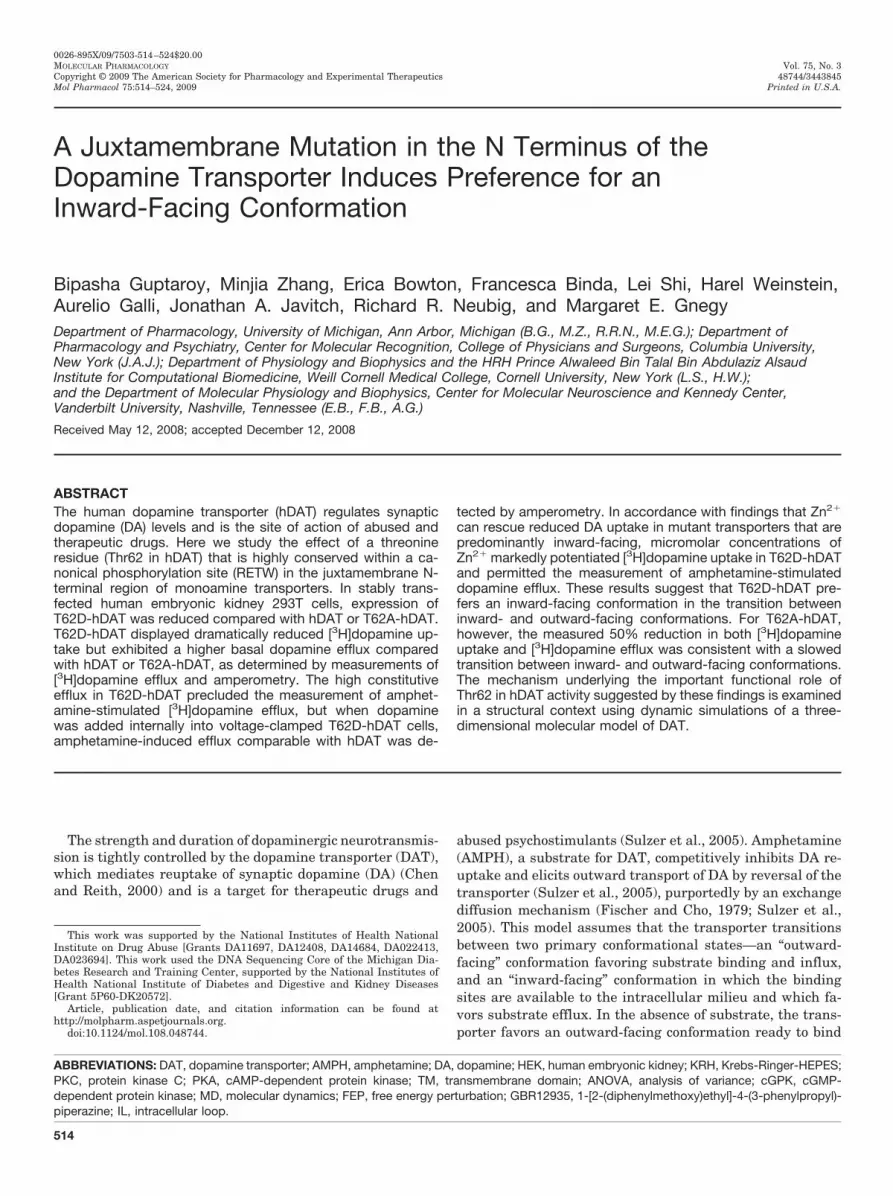
the resting membrane potential of HEK 293 cells, T62D-hDAT exhibited significantly greater DA efflux comparedwith T62A-hDAT and hDAT (one-way ANOVA, p � 0.01),indicating that the T62D mutation confers the ability toefflux at negative resting membrane potentials. The differ-ences between T62D-hDAT and hDAT are probably greaterthan shown in Fig. 9A, however, because the levels of surfaceDAT for T62D-hDAT are approximately half that of hDAT.Therefore, the values for T62D-hDAT are an underestimateof the true values. At depolarizing membrane potentials, DAefflux mediated by T62A-hDAT was significantly less thanthat of hDAT or T62D-hDAT (two-way ANOVA, p � 0.0001for voltage, p � 0.001 for mutant and p � 0.001 for interac-tion; in post hoc Bonferroni testing, efflux through T62A-hDAT differed from both hDAT and T62D-hDAT as indicatedin Fig. 9A).
A good correlation between the ability of substrates toinduce currents and their ability to cause efflux has beenreported previously (Sitte et al., 1998). However, the elicitedwhole-cell currents through DAT exceed the predicted stoi-chiometry (Sitte et al., 1998). We found the whole-cell cur-rents measured through T62D-hDAT to be significantlygreater at positive voltages than those carried through hDATor T62A-hDAT cells (Fig. 9B). When analyzed by two-wayANOVA, the curves differed for mutant (p � 0.001) andvoltage (p � 0.001) but not for the interaction. In post hocBonferroni testing, the whole-cell currents for T62D-hDATand T62A-hDAT differed at 60 mV (p � 0.05). Unlike theamperometric currents, the whole-cell currents for T62A-hDAT were not significantly different from hDAT. It is note-worthy that the apparent reversal potential of the hDAT-mediated current was not affected by mutation of Thr62,suggesting that these mutations do not affect the ionic gra-dient across the plasma membrane.
DiscussionWe show that Thr62 in the juxtamembrane N-terminal
domain of the human DAT plays a critical role in transporterfunction. Analysis of a somewhat conservative and a noncon-servative substitution at this site reveals differential effects.Both substitutions alter function, with a more drastic effectcaused by the nonconservative substitution (threonine to as-partate). The functional significance of Thr62 is underscoredby the fact that the motif containing Thr62 (RETW) is highlyconserved in all monoamine transporters (Vaughan, 2004),and the RXXW sequence is fully conserved throughout thefamily of sodium/chloride-dependent neurotransmitter trans-porters. In addition, RETW is a canonical phosphorylationsite for PKC/cGPK/PKA, suggesting that the threonine mightbe phosphorylated. The crystal structure of LeuTAa, a bacte-rial homolog of the sodium/chloride-dependent transporter,suggests that this sequence is part of a conserved network ofcytoplasmic interactions that may be critical in forming theintracellular gate (Yamashita et al., 2005), although threo-nine in this motif is not in LeuT. The addition of a negativecharge (as in aspartate or phosphothreonine) to the RETWmotif could have a profound effect on the movement of this
Fig. 8. AMPH stimulated DA efflux in hDAT- and T62D-hDAT cells inthe presence and absence of Zn2�. T62D-hDAT cells were loaded with 5�M DA as described under Materials and Methods in the presence of 30�M Zn2�, and hDAT cells were loaded in the absence of Zn2� at 37°C for30 min. AMPH-stimulated DA efflux was measured by superfusion asdescribed under Materials and Methods in either the presence (f, hDAT;F, T62D-hDAT) or absence of Zn2� (�, hDAT; E, T62D-hDAT) of 30 �MZn2� as indicated (n � 3). Baseline values of [3H]DA were collected for 70min until the addition of 10 �M AMPH (arrow). Initial levels of DA in thecells were the following: T62D-hDAT without Zn2�, 3.8 � 0.9 nmol/mgprotein; T62D-hDAT with Zn2�, 12.2 � 1.6 nmol/mg protein; hDAT with-out Zn2�, 10.5 � 0.3 nmol/mg protein; hDAT with Zn2�, 11.8 � 0.6nmol/mg protein. In two-way ANOVA analysis, p � 0.0001 for time(fraction) and the mutant/Zn2� condition; p � 0.0001 for the interaction;�, p � 0.01 for hDAT with Zn2� versus all other groups. T62D-hDAT withno Zn2� differed from all other groups at p � 0.001. There was nostatistical difference between hDAT with no Zn2� and T62D-hDAT withZn2�.
Fig. 9. Current-voltage and amperometric-voltage relationships fromhDAT, T62A-, and T62D-hDAT cells. Amperometric-voltage relationship(A) and current-voltage relationship (B) obtained from hDAT (f), T62A-hDAT (Œ), and T62D-hDAT (E) in the presence of 30 mM Na� in cellsloaded with 2 mM DA, whereas the membrane potential was steppedfrom a holding potential of �20 mV to voltages between �100 to �100 in20-mV increments (means � S.E.M., n � 6). A, in a two-way ANOVA, p �0.001 for mutant, for voltage and interaction. In post hoc Bonferronitesting, ��, p � 0.01; ���, p � 0.001 for T62A-hDAT compared with hDAT;�, p � 0.05 for T62D-hDAT compared with hDAT. In a one-way ANOVAcomparing values at �20 mV, #, p � 0.0037 for T62A-hDAT comparedwith T62D-hDAT. B, in two-way ANOVA, p � 0.001 for mutant and forvoltage. In post hoc Bonferroni testing, �, p � 0.05 for T62D-hDATcompared with T62A-hDAT.
Juxtamembrane N-Terminal Mutations Modulate DAT Function 521
intracellular gate in monoamine transporters, as discussedbelow.
Our results indicate that mutation of Thr62 to aspartatealters the conformational equilibrium of the DAT transportcycle such that the free transporter partitions predominantlyin inward-facing conformation, which can readily mediateconstitutive DA efflux. This inference is suggested by thesignificant reduction in [3H]DA uptake into T62D-hDAT cellsversus hDAT, as would be expected if the mutant constructfavored an inward-facing conformation. Data in the litera-ture support such an inference, because mutations postu-lated to arrest the transporter in inward-facing conforma-tions consistently exhibit a reduced Vmax value of DA uptake(Loland et al., 2002, 2004; Chen et al., 2004; Korkhov et al.,2006). Strikingly, the affinity for uptake of two substrates,DA and AMPH, was increased in the T62D-hDAT mutantcompared with hDAT. Increased sensitivity to substrateswas reported in D345N-hDAT (Chen et al., 2004), whichseemed to be predominantly inward-facing as a result of aninability to reorient to the extracellular side. Impairment ofthe return of an empty transporter from inward- to outward-facing state would reduce both Vmax and Km values. Becauseboth Vmax and Km values for T62D-hDAT were reduced com-pared with hDAT, the overall catalytic effectiveness of theuptake process in T62D-hDAT was only 30% less than inhDAT. The second-order rate constant (Vmax/kcat), however,is much lower than that reported in another study usinghDAT-HEK cells (1.2 � 107 M�1 � s�1) (Earles and Schenk,1999). Lower temperature (25 versus 37°C) could partiallyaccount for this, but other factors could be our construct orthe cells.
Strikingly, efflux of intracellular substrate in T62D-hDATwas increased in the absence of stimulation with eitherAMPH or voltage. In hDAT, the catalytic effectiveness ofuptake was greater than that for efflux, largely because theKm value for DA efflux is �40 times greater than for DAinflux. The decrease in the Km value for DA at the inward-facing side of T62D-hDAT compared with hDAT could ex-plain the constitutive efflux through this mutant. As might
be expected for a transporter in a predominantly inward-facing state, AMPH was ineffective in stimulating efflux. Amajor role of AMPH influx is to move Na� through thetransporter and increase the availability of Na� at the innerface of the transporter (Rutledge, 1978; Khoshbouei et al.,2003); this requirement could be precluded in a transporterthat is predominantly inward-facing.
Binding of Zn2� to the endogenous Zn2� binding site inDAT stabilizes the outward-facing conformation and facili-tates reorientation of the inward-facing transporter (Lolandet al., 2002). The T62D mutation converted the inhibitoryeffect of Zn2� on DA uptake to a stimulatory effect, consistentwith a shift away from an outward-facing conformation. Ac-cordingly, in T62D-hDAT, Zn2� increases DA uptake andpermits wild-type AMPH-stimulated DA efflux. Our experi-ments using amperometry, in which DA is added intracellu-larly, strongly support a shift of T62D-hDAT toward an in-ward-facing conformation. Not only was an enhanced “leak”of DA measured by amperometry from the T62D-hDAT mu-tant, but DA efflux was observed at negative potentials fromT62D-hDAT, as opposed to hDAT or T62A-hDAT. Finally, theincreased current through T62D-hDAT compared with T62A-hDAT in the current-voltage relationship supports the hy-pothesis of an efflux-preferring conformation because there isa positive correlation between transporter-mediated currentsand efflux through the transporter (Sitte et al., 1998). How-ever, the apparent ion flux through the three transporterconstructs was not different because the reversal potentialwas the same for all three.
The fact that the initial rate of influx through T62D-hDATwas severely reduced but not abrogated demonstrates thatT62D-hDAT can transition to the outward-facing conforma-tion and does not have a defect in reorientation of the trans-porter as observed in D345N-hDAT (Chen et al., 2004) andE136A-human serotonin transporter (Korkhov et al., 2006).The Thr62 to alanine substitution generates a transporterwith properties that differ from hDAT but are less severe,demonstrating a reduction in DA uptake and AMPH-stimu-lated efflux. Reduced [3H]DA uptake compared with rDAT
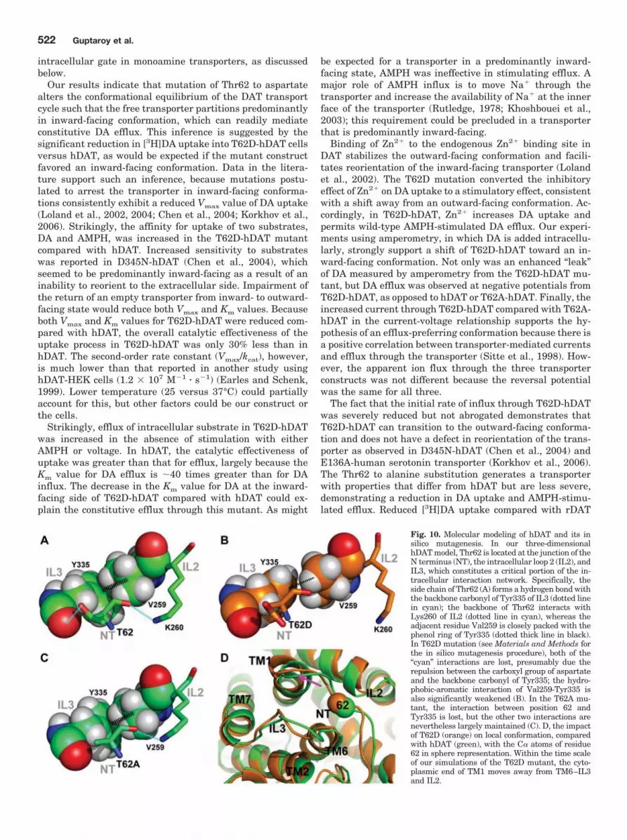
Fig. 10. Molecular modeling of hDAT and its insilico mutagenesis. In our three-dimensionalhDAT model, Thr62 is located at the junction of theN terminus (NT), the intracellular loop 2 (IL2), andIL3, which constitutes a critical portion of the in-tracellular interaction network. Specifically, theside chain of Thr62 (A) forms a hydrogen bond withthe backbone carbonyl of Tyr335 of IL3 (dotted linein cyan); the backbone of Thr62 interacts withLys260 of IL2 (dotted line in cyan), whereas theadjacent residue Val259 is closely packed with thephenol ring of Tyr335 (dotted thick line in black).In T62D mutation (see Materials and Methods forthe in silico mutagenesis procedure), both of the“cyan” interactions are lost, presumably due therepulsion between the carboxyl group of aspartateand the backbone carbonyl of Tyr335; the hydro-phobic-aromatic interaction of Val259-Tyr335 isalso significantly weakened (B). In the T62A mu-tant, the interaction between position 62 andTyr335 is lost, but the other two interactions arenevertheless largely maintained (C). D, the impactof T62D (orange) on local conformation, comparedwith hDAT (green), with the C� atoms of residue62 in sphere representation. Within the time scaleof our simulations of the T62D mutant, the cyto-plasmic end of TM1 moves away from TM6–IL3and IL2.
522 Guptaroy et al.

was demonstrated in T62A-rDAT cells (Lin et al., 2003). Thereduction in DA efflux through T62A-hDAT is more extremewhen efflux is measured by amperometry (Fig. 9A) than by[3H]DA efflux (Fig. 7). Amperometry measurements, how-ever, are performed on a millisecond time scale in voltage-clamped cells as opposed to the longer incubation method,suggesting that the Thr62 to alanine mutation may slow theequilibrium between inward- and outward-facing states. Thegreater sensitivity to inhibition by Zn2� could also be ex-plained by a reduced capacity for T62A-hDAT to transitionbetween inward and outward states. The similar degree ofreduction (�50% of hDAT) of both influx and efflux of DA inT62A-hDAT suggests that both inward-outward and out-ward-inward equilibria are slowed.
In view of the remarkable conservation of threonine at thisposition, it is important to understand the differential impactof T62A and T62D on the pharmacological properties ofhDAT from the structural perspective. Using the molecularmodel of the transporter, we conducted MD and FEP studiesto characterize the effects of the mutations at the molecularlevel. First, MD simulation was used to reveal the localconformational divergence from the LeuT structure (Ya-mashita et al., 2005) because of the presence of a threonine atposition 62 in the DAT model. The -OH group of Thr62engages in important interactions, as evidenced by the con-sistency along the 16-ns MD trajectory of the hydrogen bondwith the backbone carbonyl group of Tyr335 from IL3,whereas the Thr62 backbone interacts with the side-chain ofLys260 from IL2. In this local region, similar to the observa-tion in the LeuT crystal structure, the cation-� interactionbetween Tyr335 and Arg60 is stabilized by the aromatic-hydrophobic interaction of Tyr335 with Val259 (Fig. 10A).This network of interactions is modified in the mutant in amanner described by the in silico mutations that we intro-duced with the FEP procedure (see Materials and Methods).We found that the T62D mutation is associated with a loss ofboth interactions, with Tyr335 and Lys260, and that theinteractions between Tyr335 and Val259 is weakened (Fig.10B). The orientation of Tyr335 toward Arg60 is affectedaccordingly (data not shown), generating a significant con-formational change in which the cytoplasmic end of TM1moves away from the junction point with IL2 and IL3 (Fig.10D, shown with a pink arrow). In the T62A mutant, only theinteraction with Tyr335 is lost, whereas the other interac-tions in this local network mentioned above are largely main-tained (Fig. 10C). Thus, dramatic changes are produced inthe structure of a functionally important microenvironmentafter the mutation of Thr62 to aspartate, which demon-strates the critical role of this residue in facilitating theinteractions between IL2 and IL3, which are probably in-volved in the transition between the inward-facing and out-ward-facing conformations.
There is evidence for phosphothreonine in DAT (Foster etal., 2002). Thr62, contained within a consensus sequence forPKC/cGPK/PKA (RETW), could be phosphorylated in vivo. Itis noteworthy that Thr276 in IL2 of the serotonin transporteris directly adjacent to the network of interactions identifiedhere. This residue is phosphorylated by cGPK (Ramamoorthyet al., 2007), consistent with the possibility that Thr62 maybe phosphorylated as well. The altered conformation of DATas a result of Thr62 mutation could affect function by alteringthe binding of other proteins to DAT, such as syntaxin and
receptor for activated C kinase 1. A T62A mutation of rDATrendered it almost completely insensitive to the effect ofphosphatidylinositol 3 kinase inhibition and mitigated theeffect of protein kinase C activation on DA uptake (Lin et al.,2003).
In conclusion, our data identify mechanisms involvingThr62 as a residue important for DAT function. A noncon-servative substitution at this residue, (as in threonine toaspartate) seems to shift the conformation of DAT from thephysiologically favored substrate influx mode to an effluxpromoting one—a conformational switch promoted by drugsof abuse, such as AMPH, which are also DAT substrates.Mutating Thr62 to alanine seems to produce a transporterthat is slower to transition between inward- and outward-facing conformations. Because Thr62 is a highly conservedresidue in monoamine transporters, its role in regulatinghDAT function is likely to be replicated in other monoaminetransporters as well.
Acknowledgments
We thank Susana Shamban, Charles Lo, Hobart Ng Ng Tsai, andChristina Chi for technical assistance.
ReferencesBeuming T, Shi L, Javitch JA, and Weinstein H (2006) A comprehensive structure-
based alignment of prokaryotic and eukaryotic neurotransmitter/Na� symporters(NSS) aids in the use of the LeuT structure to probe NSS structure and function.Mol Pharmacol 70:1630–1642.
Chen N and Justice JB (2000) Differential effect of structural modification of humandopamine transporter on the inward and outward transport of dopamine. BrainRes Mol Brain Res 75:208–215.
Chen N and Reith ME (2000) Structure and function of the dopamine transporter.Eur J Pharmacol 405:329–339.
Chen N, Rickey J, Berfield JL, and Reith ME (2004) Aspartate 345 of the dopaminetransporter is critical for conformational changes in substrate translocation andcocaine binding. J Biol Chem 279:5508–5519.
Earles C and Schenk JO (1999) Multisubstrate mechanism for the inward transportof dopamine by the human dopamine transporter expressed in HEK cells and itsinhibition by cocaine. Synapse 33:230–238.
Fischer JF and Cho AK (1979) Chemical release of dopamine from striatal homog-enates: evidence for an exchange diffusion model. J Pharmacol Exp Ther 208:203–209.
Foster JD, Pananusorn B, and Vaughan RA (2002) Dopamine transporters arephosphorylated on N-terminal serines in rat striatum. J Biol Chem 277:25178–25186.
Giros B and Caron MG (1993) Molecular characterization of the dopamine trans-porter. Trends Pharmacol Sci 14:43–49.
Granas C, Ferrer J, Loland CJ, Javitch JA, and Gether U (2003) N-terminal trun-cation of the dopamine transporter abolishes phorbol ester- and substance Preceptor-stimulated phosphorylation without impairing transporter internaliza-tion. J Biol Chem 278:4990–5000.
Henin J, Maigret B, Tarek M, Escrieut C, Fourmy D, and Chipot C (2006) Probing amodel of a GPCR/ligand complex in an explicit membrane environment: the hu-man cholecystokinin-1 receptor. Biophys J 90:1232–1240.
Humphrey W, Dalke A, and Schulten K (1996) VMD: visual molecular dynamics. JMol Graph 14:33–38, 27–28.
Johnson LA, Guptaroy B, Lund D, Shamban S, and Gnegy ME (2005) Regulation ofamphetamine-stimulated dopamine efflux by protein kinase C�. J Biol Chem280:10914–10919.
Kantor L and Gnegy ME (1998) Protein kinase C inhibitors block amphetamine-mediated dopamine release in rat striatal slices. J Pharmacol Exp Ther 284:592–598.
Khoshbouei H, Sen N, Guptaroy B, Johnson L, Lund D, Gnegy ME, Galli A, andJavitch JA (2004) N-terminal phosphorylation of the dopamine transporter isrequired for amphetamine-induced efflux. PLoS Biol 2:E78.
Khoshbouei H, Wang H, Lechleiter JD, Javitch JA, and Galli A (2003) Amphetamine-induced dopamine efflux. A voltage-sensitive and intracellular Na�-dependentmechanism. J Biol Chem 278:12070–12077.
Kitayama S, Shimada S, Xu H, Markham L, Donovan DM, and Uhl GR (1992)Dopamine transporter site-directed mutations differentially alter substrate trans-port and cocaine binding. Proc Natl Acad Sci U S A 89:7782–7785.
Korkhov VM, Holy M, Freissmuth M, and Sitte HH (2006) The conserved glutamate(Glu136) in transmembrane domain 2 of the serotonin transporter is required forthe conformational switch in the transport cycle. J Biol Chem 281:13439–13448.
Lee FJ, Pristupa ZB, Ciliax BJ, Levey AI, and Niznik HB (1996) The dopaminetransporter carboxyl-terminal tail. Truncation/substitution mutants selectivelyconfer high affinity dopamine uptake while attenuating recognition of the ligandbinding domain. J Biol Chem 271:20885–20894.
Lin Z, Zhang PW, Zhu X, Melgari JM, Huff R, Spieldoch RL, and Uhl GR (2003)
Juxtamembrane N-Terminal Mutations Modulate DAT Function 523

Phosphatidylinositol 3-kinase, protein kinase C, and MEK1/2 kinase regulation ofdopamine transporters (DAT) require N-terminal DAT phosphoacceptor sites.J Biol Chem 278:20162–20170.
Loland CJ, Grånas C, Javitch JA, and Gether U (2004) Identification of intracellularresidues in the dopamine transporter critical for regulation of transporter confor-mation and cocaine binding. J Biol Chem 279:3228–3238.
Loland CJ, Norregaard L, and Gether U (1999) Defining proximity relationships inthe tertiary structure of the dopamine transporter. Identification of a conservedglutamic acid as a third coordinate in the endogenous Zn2�-binding site. J BiolChem 274:36928–36934.
Loland CJ, Norregaard L, Litman T, and Gether U (2002) Generation of an activat-ing Zn2� switch in the dopamine transporter: mutation of an intracellular tyrosineconstitutively alters the conformational equilibrium of the transport cycle. ProcNatl Acad Sci U S A 99:1683–1688.
Norregaard L, Frederiksen D, Nielsen EO, and Gether U (1998) Delineation of anendogenous zinc-binding site in the human dopamine transporter. EMBO J 17:4266–4273.
Phillips JC, Braun R, Wang W, Gumbart J, Tajkhorshid E, Villa E, Chipot C, SkeelRD, Kale L, and Schulten K (2005) Scalable molecular dynamics with NAMD.J Comput Chem 26:1781–1802.
Pifl C, Agneter E, Drobny H, Sitte HH, and Singer EA (1999) Amphetamine reversesor blocks the operation of the human noradrenaline transporter depending on itsconcentration: superfusion studies on transfected cells. Neuropharmacology 38:157–165.
Ramamoorthy S, Samuvel DJ, Buck ER, Rudnick G, and Jayanthi LD (2007) Phos-phorylation of threonine residue 276 is required for acute regulation of serotonintransporter by cyclic GMP. J Biol Chem 282:11639–11647.
Rutledge CO (1978) Effect of metabolic inhibitors and ouabain on amphetamine- andpotassium-induced release of biogenic amines from isolated brain tissue. BiochemPharmacol 27:511–516.
Sali A and Blundell TL (1993) Comparative protein modelling by satisfaction ofspatial restraints. J Mol Biol 234:779–815.
Saunders C, Ferrer JV, Shi L, Chen J, Merrill G, Lamb ME, Leeb-Lundberg LM,Carvelli L, Javitch JA, and Galli A (2000) Amphetamine-induced loss of humandopamine transporter activity: an internalization-dependent and cocaine-sensitivemechanism. Proc Natl Acad Sci U S A 97:6850–6855.
Scholze P, Nørregaard L, Singer EA, Freissmuth M, Gether U, and Sitte HH (2002)The role of zinc ions in reverse transport mediated by monoamine transporters.J Biol Chem 277:21505–21513.
Sen N, Shi L, Beuming T, Weinstein H, and Javitch JA (2005) A pincer-like config-
uration of TM2 in the human dopamine transporter is responsible for indirecteffects on cocaine binding. Neuropharmacology 49:780–790.
Shi L, Quick M, Zhao Y, Weinstein H, and Javitch JA (2008) The mechanism of aneurotransmitter: sodium symporter–inward release of Na� and substrate istriggered by substrate in a second binding site. Mol Cell 30:667–677.
Sitte HH, Hiptmair B, Zwach J, Pifl C, Singer EA, and Scholze P (2001) Quantitativeanalysis of inward and outward transport rates in cells stably expressing thecloned human serotonin transporter: inconsistencies with the hypothesis of facil-itated exchange diffusion. Mol Pharmacol 59:1129–1137.
Sitte HH, Huck S, Reither H, Boehm S, Singer EA, and Pifl C (1998) Carrier-mediated release, transport rates, and charge transfer induced by amphetamine,tyramine, and dopamine in mammalian cells transfected with the human dopa-mine transporter. J Neurochem 71:1289–1297.
Sotomayor M and Schulten K (2004) Molecular dynamics study of gating in themechanosensitive channel of small conductance MscS. Biophys J 87:3050–3065.
Sulzer D, Sonders MS, Poulsen NW, and Galli A (2005) Mechanisms of neurotrans-mitter release by amphetamines: a review. Prog Neurobiol 75:406–433.
Thorsness PE and Koshland DE Jr (1987) Inactivation of isocitrate dehydrogenaseby phosphorylation is mediated by the negative charge of the phosphate. J BiolChem 262:10422–10425.
Torres GE (2006) The dopamine transporter proteome. J Neurochem 97 (Suppl1):3–10.
Uhl GR and Lin Z (2003) The top 20 dopamine transporter mutants: structure-function relationships and cocaine actions. Eur J Pharmacol 479:71–82.
Vaughan RA (2004) Phosphorylation and regulation of psychostimulant-sensitiveneurotransmitter transporters. J Pharmacol Exp Ther 310:1–7.
Wu X and Gu HH (2003) Cocaine affinity decreased by mutations of aromatic residuephenylalanine 105 in the transmembrane domain 2 of dopamine transporter. MolPharmacol 63:653–658.
Yamashita A, Singh SK, Kawate T, Jin Y, and Gouaux E (2005) Crystal structure ofa bacterial homologue of Na�/Cl-dependent neurotransmitter transporters. Na-ture 437:215–223.
Zomot E, Bendahan A, Quick M, Zhao Y, Javitch JA, and Kanner BI (2007) Mech-anism of chloride interaction with neurotransmitter: sodium symporters. Nature449:726–730.
Address correspondence to: Dr. Margaret E. Gnegy, Department of Phar-macology, 2220E MSRBIII, University of Michigan Medical School, Ann Arbor,MI 48109-0632. E-mail: [email protected]
524 Guptaroy et al.