effect of extended-release naltrexone on striatal dopamine transporter availability, depression and...
TRANSCRIPT

ORIGINAL INVESTIGATION
Effect of extended-release naltrexone on striatal dopaminetransporter availability, depression and anhedoniain heroin-dependent patients
Eline R. Zaaijer & Lonneke van Dijk & Kora de Bruin &
Anna E. Goudriaan & Laureen A. Lammers &
Maarten W. J. Koeter & Wim van den Brink & Jan Booij
Received: 11 December 2014 /Accepted: 14 February 2015 /Published online: 12 March 2015# The Author(s) 2015. This article is published with open access at Springerlink.com
AbstractRationale Extended-release naltrexone (XRNT), an opioid re-ceptor antagonist, is successfully used in the treatment of opi-oid dependence. However, naltrexone treatment of opioid-dependent patients may reduce striatal dopamine transporter(DAT) availability and cause depression and anhedonia.Objectives The aim of this study is to investigate changes instriatal DAT availability and symptoms of depression (BeckDepression Inventory (BDI)) and anhedonia (Snaith HamiltonPleasure Scale (SHAPS)) before and during XRNT treatment.Methods At baseline, ten detoxified heroin-dependent pa-tients and 11 matched healthy controls underwent [123I]FP-CIT single photon emission computed tomography (SPECT)imaging to assess striatal DAT binding. Patients underwent asecond SPECT scan 2 weeks after an intramuscular injectionwith XRNT.
Results At baseline, the mean binding potential (BPND) in theputamen was at a trend level lower and the mean BDI scorewas significantly higher in heroin patients (n=10) than incontrols (n=11) (3.45±0.88 vs. 3.80±0.61, p=0.067, d=−0.48 and 12.75±7.40 vs. 5.20±4.83, p=0.019, d=1.24, re-spectively). Post hoc analyses in subgroups with negativeurine analyses for opioids and cocaine showed significantlylower baseline putamen BPND in heroin patients (n=8) thancontrols (n=10) (3.19±0.43 vs. 3.80±0.64, p=0.049, d=−1.03). XRNT treatment in heroin patients was not signifi-cantly associated with changes in striatal DAT availability(p=0.348, d=0.48), but the mean BDI score after XRNT treat-ment was significantly lower than before treatment (7.75±7.21 vs. 12.75±7.40, p=0.004, d=−0.68).Conclusions The results of this study suggest that XRNTtreatment does not reduce striatal DAT availability and hasno significant effect on anhedonia, but is associated with asignificant reduction of depressive symptoms.
Keywords Dopamine transporter . Abstinence . Addiction .
Brain imaging . In vivo . Opioid receptor
Introduction
The worldwide prevalence of opioid dependence is estimatedto be 0.2 % (Degenhardt et al. 2014) and the prevalence ofillicit opioid use 0.7 % (UNODC 2012). The main illicit opi-oid used in Europe is heroin. Although a downward trend inthe use of heroin was suggested, existing problem users willremain a key issue for many years to come (EMCDDA 2013).In the Netherlands (16.7 million inhabitants), the estimated
Electronic supplementary material The online version of this article(doi:10.1007/s00213-015-3891-4) contains supplementary material,which is available to authorized users.
E. R. Zaaijer : L. van Dijk :A. E. Goudriaan :M. W. J. Koeter :W. van den BrinkAmsterdam Institute for Addiction Research, Department ofPsychiatry, AcademicMedical Center, University of Amsterdam, POBox 22660, 1100 DD Amsterdam, The Netherlands
E. R. Zaaijer (*) :K. de Bruin : J. BooijDepartment of Nuclear Medicine, Academic Medical Center,University of Amsterdam, PO Box 22660, 1100DD Amsterdam, The Netherlandse-mail: [email protected]
L. A. LammersDepartment of Hospital Pharmacy, Academic Medical Center,University of Amsterdam, PO Box 22660, 1100DD Amsterdam, The Netherlands
Psychopharmacology (2015) 232:2597–2607DOI 10.1007/s00213-015-3891-4

number of opioid-dependent people in 2012 was 14,000,which is approximately 1 per 1000 adult inhabitants (Crutset al. 2013). More than 90 % of the opioid-dependent peoplein the Netherlands inhale heroin, and injection of heroin is rare(NDM 2012; Cruts et al. 2013).
In the Netherlands, about 80 % of all heroin-dependentpeople is in treatment, mostly methadone maintenancetreatment (85 %) and heroin-assisted treatment (5 %)(Cruts et al. 2013; Wisselink et al. 2013). The remaining10 % of patients in treatment are in some kind ofabstinence-oriented program, including extended detoxifi-cation programs followed by outpatient psychosocial sup-port and oral naltrexone (Cruts et al. 2013). International-ly, the focus of opioid addiction treatment is shifting to-ward recovery-oriented drug treatment (Neale et al. 2013),resulting in a greater emphasis on abstinence as the finaltreatment goal. However, outpatient treatment with orwithout oral naltrexone was associated with early treat-ment discontinuation and very high relapse rates. As aconsequence, oral naltrexone was probably not more ef-fective than placebo (Minozzi et al. 2011).
Extended-release naltrexone (XRNT), given as injec-tion or implant, may be a more suitable treatment foropioid addiction than oral naltrexone treatment due tobetter compliance. XRNT implants and injections signifi-cantly reduced heroin use (Gastfriend 2011; Lobmaieret al. 2011) and opioid-dependent people receiving XRNTinjections had significantly more opioid-free weeks thanopioid-dependent patients who were given a placebo in-jection (Syed and Keating 2013). Patients receivingXRNT injections stayed in treatment longer than patientsreceiving placebo injections (Lobmaier et al. 2008; Syedand Keating 2013), and XRNT treatment was well toler-ated (Krupitsky and Blokhina 2010; Gastfriend 2011).
Although naltrexone treatment compliance can be im-proved by extended-release formulations, there are con-cerns about possible side effects that may result in treat-ment dropout, i.e., no further injections/implants. For ex-ample, significantly higher 6β-naltrexol levels, the majormetabolite of naltrexone, were found in subjects who ex-perienced one or more side effects (i.e., headache, nausea,anxiety) (King et al. 1997). Moreover, the prevalence ofdepression and anhedonia was found to be high amongheroin addicts (Tiurina et al. 2011). Endogenous opioidsinfluence motivational and stress regulatory processes andmood regulation directly by binding to the μ-opioid re-ceptor, which causes inhibi t ion of the gamma-aminobutyric acid (GABA) neurons and indirectly in-duces dopamine release in the nucleus accumbens (Kooband Le Moal 2008). Naltrexone, which is a μ-opioid re-ceptor antagonist, possibly disturbs normal endogenousopioid binding leading to reduced dopamine release. Thedopamine transporter (DAT) plays an important role in
controlling the synaptic dopamine levels by removing do-pamine from the synapse. So, when the dopamine releaseis changed chronically, this may lead to changes in syn-aptic dopamine levels, and consequently to changes in theDAT expression (Schmitt and Reith 2010; Vaughan andFoster 2013). Interestingly, human studies also showedthat high endogenous striatal DA release was associatedwith anhedonia (Zijlstra et al. 2008) and low availabilityof striatal DATs was associated with symptoms of apathy(David et al. 2008) and depression (Sarchiapone et al.2006; Roselli et al. 2009). Additionally, opioid-dependent patients who were abstinent showed lowerstriatal dopamine D2/3 receptors (Zijlstra et al. 2008) andDAT availability compared to healthy controls, but it wasnot clear whether this effect was reversible (Jia et al.2005; Shi et al. 2008; Yeh et al. 2012; Liu et al. 2013).Finally, there is evidence that chronic naltrexone admin-istration in rats results in decrease of striatal DAT avail-ability (Bhargava and Gudehithlu 1996). Therefore,(chronic) naltrexone treatment may further reduce striatalDAT availability leading to an exacerbation of existingdepressive symptoms and anhedonia in opioid-dependentpatients.
The effects of naltrexone on anhedonia in humans weremainly assessed with self-reports of pleasure ratings. The-se studies showed that oral naltrexone can cause anhedo-nia in healthy controls (Murphy et al. 1990; Daniel et al.1992; Yeomans and Gray 2002). However, althoughXRNT treatment was associated with a reduction of thehedonic properties of addictive substances (O’Brien et al.2010), XRNT treatment did not reduce the ability to ex-perience pleasure during natural rewarding activities inaddicted patients (O’Brien et al. 2010; Tiurina et al.2011). In order to better understand these findings, weconducted the first study looking at the effect of XRNTon both striatal DAT binding and self-reported anhedoniain detoxified heroin addicts.
Based on the literature, we hypothesize that (1) atbaseline, heroin-dependent patients have lower striatalDAT availability and report more anhedonia and depres-sive symptoms than healthy controls; (2) during XRNTtreatment, heroin-dependent patients show a further de-crease in striatal DAT availability compared to baselineand an increase in anhedonia and depression scores; (3)plasma levels of naltrexone and its major metabolite 6β-naltrexol in heroin-dependent patients correlate withchanges in s t r ia ta l DAT avai lab i l i ty and wi thanhedonia/depression before and during XRNT treat-ment; (4) at baseline and at follow-up, striatal DATavailability is negatively correlated with anhedonia anddepression; and (5) the decrease in striatal DAT avail-ability during treatment is associated with an increase inanhedonia and depression.
2598 Psychopharmacology (2015) 232:2597–2607

Methods
Subjects
Subjects were recruited between January 2013 and July 2014.Twelve detoxified heroin-dependent patients (11 male) wererecruited from addiction treatment centers throughout theNetherlands. Inclusion criteria were (1) diagnosis of DSM-IVopioid dependence, (2) heroin as the main substance of abuse,and (3) inhalation as themain route of administration of heroin.Exclusion criteria were (1) estimated IQ <70, (2) prior or cur-rent diagnosis of psychosis or current depression with suicidalideation, (3) use of medication that interferes with binding ofthe DAT radiotracer, (4) use of naltrexone in the past 6 months,(5) history of head trauma or brain surgery, (6) (planned) preg-nancy, breastfeeding, or no acceptable method of contracep-tion, (7) involuntary treatment, (8) medical contradictions forXRNT, and (9) no intention to be opioid-free for a minimum of10–14 days before starting XRNT treatment.
Eleven healthy subjects were included who had no diagno-sis of substance dependence and were matched to the patientgroup for gender, age, body mass index (BMI), and smokingstatus. Healthy controls were recruited through online adver-tisement and flyer postings. Exclusion criteria for controls wereidentical to exclusion criteria for the heroin-dependent subjects.
All subjects provided written informed consent to partici-pate in the study. The study was approved by the EthicalCommittee of the Academic Medical Centre of the Universityof Amsterdam, where the study was conducted, and per-formed in accordance with the ethical standards laid down inthe 1964 Declaration of Helsinki.
Study design
For patients, there was a 2-week heroin- and methadone-freeperiod between the end of detoxification and the first scanningday in order to minimize the risk of opioid withdrawal symp-toms after XRNT injection. To measure DAT availabilityin vivo, the first [123I]FP-CIT single photon emission comput-ed tomography (SPECT) was performed just before theXRNT injection and the second scan was made 2 weeks afterthe XRNT injection. In healthy control subjects, only one(baseline) SPECT scan was performed. All subjects were re-quired to have a negative urine drug screen (UDS) for opioids,cocaine, and amphetamine on the day of the SPECT scan(s).None of the subjects used medication that could interfere with[123I]FP-CIT binding (Booij and Kemp 2008). A breath alco-hol test was performed to assess acute alcohol intoxication.
Clinical assessments
DSM-IV criteria for substance use disorders, psychotic disor-ders, and depressive disorder with suicidal ideation were
assessed with the Dutch translation of the Mini-InternationalNeuropsychiatric Interview (MINI; van Vliet and de Beurs2007). Before each scan, subjects were asked to fill out self-report questionnaires assessing depressive symptoms (BeckDepression Inventory (BDI); Arnou et al. 2001) and anhedonia(Snaith-Hamilton Pleasure Scale (SHAPS); Snaith et al. 1995).On both questionnaires, a higher total score indicates moresevere depression or anhedonia, respectively. Smoking statuswas assessed with the Fagerström Test for Nicotine Depen-dence (FTND; Heatherton et al. 1991). IQ was estimated withthe Dutch Adult Reading test (Schmand et al. 1991).
Study medication
After the first SPECT scan, patients were given an intramus-cular injection with XRNT (Vivitrol®, Alkermes, Inc., USA).Extended-release naltrexone microspheres (Alkermes, Inc.,USA) were administered as a 4-ml gluteal intramuscular in-jection containing 380 mg naltrexone. After injection, patientswere kept at the research facility for 30 min to check whetherthey developed opioid withdrawal symptoms and to treat themif necessary. The second SPECT session was conducted2 weeks after the XRNT injection. The timing of the sessioncoincided with peak naltrexone levels (Krupitsky andBlokhina 2010). Plasma samples were taken on the day ofthe second SPECT session to assess peak naltrexone levelsand its major metabolite 6β-naltrexol (Slawson et al. 2007).
SPECT imaging procedure
SPECT brain imaging was performed on a brain-dedicatedsystem. This system (Neurofocus) has 12 individual crystalsequipped with a focusing collimator and a spatial resolution ofapproximately 6.5 mm full-width at half maximum through-out the 20-cm field of view. [123I]FP-CIT, which is a well-validated radiotracer for striatal DAT imaging (Booij et al.1997), was injected intravenously at an approximate dose of110 MBq. [123I] labeling and acquisition were performed asdescribed previously (Tissingh et al. 1998). [123I]FP-CIT (GEHealthcare, Eindhoven, The Netherlands) had a specific activ-ity of 750 MBq/nmol and a radiochemical purity >95 %. Im-age acquisition was performed 3 h after injection (Booij et al.1999). Images were corrected for attenuation and reconstruct-ed in 3D (de Win et al. 2005; Boot et al. 2008).
Analysis of SPECT data
[123I]FP-CIT binding in the striatum was determined by ana-lyzing the five consecutive transverse slices representing themost intense binding in the striatum. A standard region ofinterest (ROI) template (constructed according to a stereo-tactic atlas) including two regions representing DAT bind-ing (caudate nucleus and putamen) and one region
Psychopharmacology (2015) 232:2597–2607 2599

representing nonspecific binding (occipital cortex) wasplaced bilaterally on the images, as previously reported(de Win et al. 2005). Also, a standard template was usedrepresenting DAT binding in the striatum as a whole. Spe-
cific DAT versus nonspecific binding ratios (binding po-tential (BPND); Innis et al. 2007) were calculated for cau-date nucleus, putamen, and whole striatum using the fol-lowing formula:
BPND ¼ mean 123I½ �FP‐CITbinding inROI−mean 123I½ �FP−CITbinding inoccipital cortexmean 123I½ �FP‐CITbinding inoccipital cortex
Statistical analysis
Normality of distribution of all data was tested with theKolmogorov-Smirnov test. Equality of variances was testedwith Levene’s test.
Group differences in baseline characteristics were assessedusing an independent samples t test when a variable was nor-mally distributed and a Mann-Whitney U test when a variablewas not normally distributed. Correlations between BPND ofthe left and right striatum was assessed using Pearson’s andSpearman’s correlation coefficients.
BPND in ROIs and BDI and SHAPS scores were comparedbetween groups using an independent samples t test when avariable was normally distributed and a Mann-WhitneyU testwhen a variable was not normally distributed.
Within the patient group, analyses were performed using apaired samples t test. Within the patient group, we calculatedPearson’s r between naltrexone and 6β-naltrexol plasmalevels and change in BPND (in striatum, caudate, and puta-men), BDI, and SHAPS scores between scans. Also,Pearson’s r were calculated between BPND (in striatum, cau-date, and putamen) and BDI scores/SHAPS scores.
We reported effect sizes (d values; Cohen 1977) for eachtest because of the relatively small sample size, where d=0.2is considered a small effect, d=0.5 a medium effect, and d=0.8 a large effect.
All statistical analyses were performed using IBM StatisticalPackage for the Social Sciences (SPSS) version 20, and statisti-cal significance was defined as p<0.05. Given the small numberof subjects, correction for multiple testing was not performed toprevent increased type II errors, resulting in low power.
Results
Urine drug screen
Five subjects tested positive for drugs: two heroin-dependentpatient tested positive for cocaine and opioids on both scan-ning days, two heroin-dependent patients tested positive for
opioids on the first scanning day and one healthy control testedpositive for opioids. The healthy control, who tested positivefor opioids, indicated that he had used codeine the day beforescanning. Since cocaine interferes with [123I]FP-CIT binding(Booij and Kemp 2008), the two patients testing positive forcocaine were excluded from further analyses. Opioid use couldpossibly influence DAT availability (Booij and Kemp 2008).Therefore, analyses were performed twice: (1) in the first anal-ysis, the two patients testing positive for cocaine were exclud-ed, resulting in 10 patients and 11 controls; (2) in the secondanalysis, also all subjects testing positive for opioids were leftout, resulting in 8 patients and 10 controls. Missing urinalysisdata were imputed as positive. As a consequence, one morepatient with missing urinalysis data on the day of the secondscan was excluded from some of the analyses, resulting inseven patients for the within-patient comparison. All subjectshad a negative alcohol breath test on the scan day(s).
Sample characteristics
Demographic and clinical characteristics are displayed inTable 1. In the first analysis, no significant differences werefound in gender, age, BMI, and smoking status (FTND) be-tween the patients and healthy controls. After excluding pa-tients testing positive for cocaine and opioids, differencesbetween groups for all characteristics even decreased, exceptfor FTND. Since smoking statusmay influence striatal BPND(Danielson et al. 2011), we corrected for FTND in thebetween-group analyses with SPECT data.
Binding potential (BPND) in regions of interests
In all participants, intense [123I]FP-CIT binding was observedin the striatum bilaterally (Fig. 1). DAT availability in the leftand right caudate nucleus/putamen/total striatum were highlycorrelated (r>0.85, p<0.05); therefore, the mean BPND of thebilateral measures was calculated and used in all analyses.
In the first analysis (all subjects except the two patientswith a positive urine for cocaine), two patients did not havea second SPECT scan: one patient withdrew consent and quit
2600 Psychopharmacology (2015) 232:2597–2607

the study before the second SPECTscan, the other patient hada missing urine sample on the day of the second SPECT scan,resulting in eight patients for the within-patient comparison. Inthe second analysis (all subjects except those with a positiveurine for cocaine or opioids), only one of the remaining pa-tients did not have a second SPECTscan and was therefore notincluded in within-patient comparison of BPND in ROIs be-fore and during XRNT treatment (Table 2).
In the first analysis (including opioid positive subjects), wedid not find significant group differences in baseline BPND.However, in the second analysis (excluding both cocaine andopioid positive subjects), baseline BPND in the putamen ofheroin patients was significantly lower than in healthy controls(t(16)=−2.301, p=0.049, d=−1.03). There were no significantdifferences in BPND between healthy controls and heroin pa-tients at baseline for other ROIs. Correction for FTND slightlydecreased the difference in baseline BPND between heroin-
dependent patients and healthy controls. Therefore, adjustedp values and effect sizes are displayed in Table 2.
We found no significant differences in BPND between her-oin patients at baseline and after 2 weeks of XRNT treatment(Fig. 1), neither when opioid-positive patients were includednor when opioid-positive patients were excluded from theanalysis (Table 2).
Since ourmain group of interest is heroin-dependent patientsthat are abstinent during the study, further analyses (see below)were only conducted for cocaine and opioid-free subjects.
Depression and anhedonia
At baseline, BDI scores were significantly higher for heroin-dependent patients than for healthy controls (t(16)=2.614, p=0.019, d=1.24), see Table 3. For heroin-dependent patientsafter 2 weeks of XRNT treatment, BDI scores were
Table 1 Demographic and clinical characteristics of healthy controls and heroin patients
Demographics and clinical characteristics of cocaine-free subjects (defined as negative for cocaine on urine analysis)
Healthy controls (n=11) Heroin patients (n=10)a t (df=19)/U p value Cohen’s d
Sex (nr male) 11 10
Age (mean±SD) (years) 45.6±9.4 (range 29–56) 44.9±5.5 (range 37–53) −0.216 0.832 −0.09Duration of heroin dependence (mean±SD) (years) N/A 16.6±8.8 (range 2–30)
Body mass index (BMI) (mean±SD) (kg/m2) 26.4±4.5 24.5±4.4 37.000 0.202 −0.42Fagerström Test for Nicotine Dependence (FTND) 0.73±0.65 0.60±0.70 48.500 0.612 −0.19Demographics and clinical characteristics of opioid-free subjects (defined as negative for both cocaine and opioids on urine analysis)
Healthy controls (n=10) Heroin patients (n=8) t (df=16)/U p value Cohen’s d
Sex (nr male) 10 8
Age (mean±SD) (years) 45.2±9.8 (range 29–56) 45.1±6.0 (range 37–53) −0.019 0.985 −0.01Duration of heroin dependence (mean±SD) (years) N/A 17.0±9.5 (range 2–30)
Body Mass Index (BMI) (mean±SD) (kg/m2) 25.7±4.2 24.3±4.0 29.000 0.325 −0.35Fagerström Test for Nicotine Dependence (FTND) 0.80±0.63 0.63±0.74 33.500 0.523 −0.25
Cohen’s d: 0.20=small, 0.50=moderate, 0.80=large (Cohen 1977)
N/A not applicablea Excluding two patients that tested positive on cocaine use at the time of the scan
Fig. 1 [123I]FP-CIT SPECTimages (transversal slides at thelevel of the striatum) of a typicalheroin-dependent patient, before(left image) and 2 weeks after(right image) an intramuscularinjection with XRNT (380 mg).Note that visual analyses of theimages did not show differencesbetween the two conditions,which was confirmed by thequantitative analyses (seeBResults^ section)
Psychopharmacology (2015) 232:2597–2607 2601

significantly lower than before treatment (t(7)=4.132, p=0.004, d=−0.68). There was no significant difference betweengroups for baseline SHAPS scores. Also, there were no sig-nificant differences between SHAPS scores before and after2 weeks of XRNT treatment (Table 3).
Naltrexone and 6β-naltrexol plasma levels
Plasma data were missing for one patient due to technicalreasons. No significant correlations were found betweennaltrexone/6β-naltrexol plasma levels and change in BPND
(in whole striatum, caudate nucleus, and putamen), BDI andSHAPS scores in heroin-dependent patients between scans(supplementary data, Tables S1 and S2).
Integration of SPECT data and behavioral parameters
No significant correlations were found for striatal DAT bind-ing and anhedonia or depression at baseline or at follow-up.Correlations between decrease in striatal DAT binding duringtreatment and increase in anhedonia and depression were notcalculated because we did not find a decrease in striatal DATbinding nor an increase in anhedonia and depression.
Discussion
The current study is the first to assess the effects of XRNTtreatment on striatal DAT binding and self-reported depressionand anhedonia in heroin-dependent subjects. Our present re-sults suggest that blocking of the μ-opioid receptor by XRNTdoes not decrease striatal DAT binding and does not increaseself-reported anhedonia, but is associated with a significantdecrease in depressive symptoms.
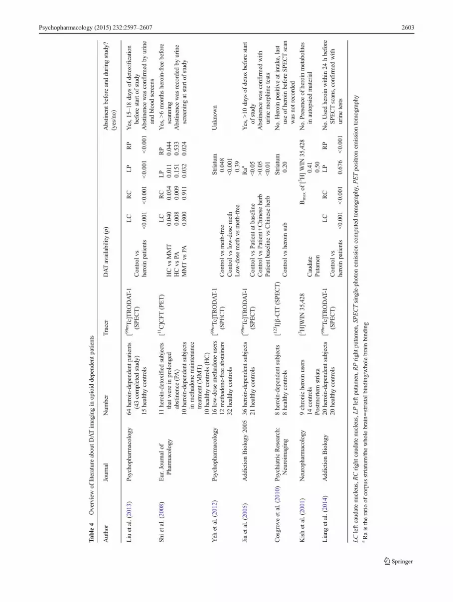
In line with our first hypothesis, we found significantlylower DAT binding at baseline in the putamen of detoxifiedheroin-dependent patients with a negative urine test for opi-oids compared to controls, which is in line with previous stud-ies (Jia et al. 2005; Shi et al. 2008; Yeh et al. 2012; Liu et al.2013, Table 4). This implicates that detoxified heroin patientshave lower striatal DAT availability. This reduction in DATavailability compared to controls may be related to long-termheroin abuse since patients and healthy controls were matchedfor other variables influencing DATavailability. However, dueto the design of our study, we cannot exclude the possibility ofpreexisting differences in DATavailability. Although the low-er DAT binding in heroin-dependent patients was not signifi-cant for the caudate nucleus and whole striatum, effect sizesindicate moderate to large effects of long-term heroin abuse onDAT binding, supporting the hypothesis that differences inthese areas may be found when larger sample sizes are includ-ed (Table 4; Liu et al. 2013).
Table 2 BPND per ROI for controls and heroin patients (mean±SD)
BPND (mean±SD) for cocaine-free subjects(defined as negative for cocaine on urineanalysis)
p value Cohen’s d
PB vs HC PB (n=10) HC (n=11)
Striatum, whole 3.64±1.00 3.82±0.63 0.139 −0.21Caudate nucleus 3.62±0.72 3.97±0.85 0.321 −0.44Putamen 3.45±0.88 3.80±0.61 0.067 −0.48
PO vs PB PO (n=8) PB (n=8)
Striatum, whole 3.60±0.59 3.65±1.12 0.901 −0.05Caudate nucleus 3.60±0.62 3.62±0.81 0.965 −0.02Putamen 3.42±0.72 3.42±0.99 0.999 0.00
BPND (mean±SD) for opioid-free subjects(defined as negative for both cocaineand opioids on urine analysis)
p value Cohen’s d
PB vs HC PB (n=8) HC (n=10)
Striatum, whole 3.36±0.47 3.82±0.66 0.155a −0.72a
Caudate nucleus 3.45±0.52 3.99±0.90 0.198a −0.63a
Putamen 3.19±0.43 3.80±0.64 0.049a −1.03a
PO vs PB PO (n=7) PB (n=7)
Striatum, whole 3.53±0.60 3.28±0.44 0.348 0.48
Caudate nucleus 3.55±0.65 3.39±0.53 0.579 0.27
Putamen 3.28±0.65 3.10±0.38 0.477 0.35
Cohen’s d: 0.20=small, 0.50=moderate, 0.80=large (Cohen 1977). Non-parametric test for PB vs HC: striatum and putamen in the cocaine-freesubjects’ analyses. Parametric tests were used for all other analyses listed.Means represent observed data that were not adjusted for FTND
PB patients at baseline, HC healthy controls, PO patients on XRNTtreatmenta Adjusted for FTND scores
Table 3 Beck Depression Inventory scores and Snaith-Hamilton Pleasure Scale scores for healthy controls and heroin patients (mean±SD) that had anegative UDS for cocaine and opioids
HC (n=10) PB ( n=8) PO (n=8) p value (Cohen’s d)
PB vs HC PO vs PB
BDI 5.20±4.83 12.75±7.40 7.75±7.21 0.019 (1.24) 0.004 (−0.68)SHAPS 24.00±5.74 24.88±5.22 22.75±6.71 0.742 (0.16) 0.326 (−0.35)
Cohen’s d: 0.20=small, 0.50=moderate, 0.80=large (Cohen 1977)
PB patients at baseline,HC healthy controls, PO patients on XRNT treatment, BDI Beck Depression Inventory, SHAPS Snaith-Hamilton Pleasure Scale
2602 Psychopharmacology (2015) 232:2597–2607
Tab
le4
Overviewof
literatureaboutD
ATim
agingin
opioid
dependentp
atients
Author
Journal
Num
ber
Tracer
DATavailability(p)
Abstin
entb
eforeandduring
study?
(yes/no)
Liu
etal.(2013)
Psychopharmacology
64heroin-dependent
patients
(43completed
study)
15healthycontrols
[99mTc]TRODAT-1
(SPECT)
LC
RC
LP
RP
Control
vsheroin
patients
<0.001
<0.001
<0.001
<0.001
Yes,15–18
days
ofdetoxificatio
nbefore
starto
fstudy
Abstin
ence
was
confirmed
byurine
andbloodscreens
Shi
etal.(2008)
Eur.Journalof
Pharmacology
11heroin-detoxifiedsubjects
thatwerein
prolonged
abstinence
(PA)
10heroin-dependent
subjects
inmethadone
maintenance
treatm
ent(MMT)
10healthycontrols(H
C)
[11C]CFT(PET)
LC
RC
LP
RP
HCvs
MMT
0.040
0.034
0.011
0.044
HCvs
PA0.008
0.009
0.151
0.533
MMTvs
PA0.800
0.911
0.032
0.024
Yes,>
6monthsheroin-freebefore
scanning
Abstin
ence
was
recorded
byurine
screeningatstarto
fstudy
Yeh
etal.(2012)
Psychopharmacology
16low-dosemethadone
users
12methadone-freeabstainers
32healthycontrols
[99mTc]TRODAT-1
(SPECT)
Striatum
Control
vsmeth-free
0.048
Control
vslow-dosemeth
<0.001
Low
-dosemethvs
meth-free
0.39
Unknown
Jiaetal.(2005)
Addictio
nBiology
2005
36heroin-dependent
subjects
21healthycontrols
[99mTc]TRODAT-1
(SPECT)
Raa
Control
vsPatient
atbaselin
e<0.05
Control
vsPatient+Chinese
herb
>0.05
Patient
baselin
evs
Chinese
herb
<0.01
Yes,>
10days
ofdetoxbefore
start
ofstudy
Abstin
ence
was
confirmed
with
urinemorphinetests
Cosgroveetal.(2010)
PsychiatricResearch:
Neuroim
aging
8heroin-dependent
subjects
8healthycontrols
[123I]β-CIT
(SPE
CT)
Striatum
Control
vsheroin
sub
0.20
No.Heroinpositiv
eatintake,last
useof
heroin
before
SPECTscan
was
notrecorded
Kishetal.(2001)
Neuropharmacology
9chronicheroin
users
14controls
Postmortem
striata
[3H]W
IN35,428
Bmaxof
[3H]WIN
35,428
Caudate
0.41
Putam
en0.50
No.Presenceof
heroin
metabolites
inautopsiedmaterial
Liang
etal.(2014)
Addictio
nBiology
20heroin-dependent
subjects
20healthycontrols
[99mTc]TRODAT-1
(SPECT)
LC
RC
LP
RP
Control
vsheroin
patients
<0.001
<0.001
0.676
<0.001
No.Usedheroin
with
in24
hbefore
SPECTscans,confirmed
with
urinetests
LCleftcaudatenucleus,RCrightcaudatenucleus,LPleftputamen,R
Prightp
utam
en,SPECTsingle-photonem
ission
computedtomography,PETpositron
emission
tomography
aRaistheratio
ofcorpus
striatum
/thewholebrain=striatalbinding/wholebrainbinding
Psychopharmacology (2015) 232:2597–2607 2603

One healthy control indicated that he had used paracetamolwith codeine for pain relief only on the day before scanning(two to three tablets). Exclusion of this subject from the anal-ysis did not change mean striatal DAT binding of healthycontrols. However, when we excluded the two heroin-dependent subjects who had a positive urine test for opioids,mean striatal DAT binding decreased and variation in DATbinding (SD) halved, indicating that acute use of codeinemay not influence striatal DAT binding, while acute opioiduse (i.e., heroin) may have a significant influence on striatalDAT binding. Indeed, acute opioid use increased striatal DArelease (Di Chiara and Imperato 1988; Wise et al. 1995) andconsequently may influence striatal DAT expression. One ofthe reasons that acute administration of the opioid receptoragonist codeine may not influence striatal DAT binding, whileother opioids may do, might be that the affinity of codeine tothe μ-opioid receptor is simply too low (Ki approximately79 nmol/l; Raynor et al. 1993) to induce indirect changes inDAT expression. In contrast, although heroin itself has a lowaffinity for the μ-opioid receptor, once in the brain, it is hy-droxylated to morphine (Yu 1996). Morphine has a high af-finity for the μ-receptor (Ki approximately 14 nmol/l; Raynoret al. 1993) and might consequently indirectly influence DATexpression. Indeed, acute or subchronic treatment with an-other high-affinity μ-opioid agonist, namely fentanyl (Ki
approximately 0.39 nmol/l; Raynor et al. 1993), decreasedin vivo striatal DAT binding (Bergstrom et al. 1998). Thus,our present data may indicate that it is relevant to analyze ahomogeneous group of subjects who are all truly and fullyabstinent for opioids if one is interested to study DATavailability.
In Table 4, we summarized the findings of DAT imagingstudies in heroin-dependent patients.
Our results from the analyses excluding subjects with apositive urine test for opioids (i.e., lower DAT binding in theputamen in the heroin-dependent patients) are in line withprevious studies showing lower striatal DAT binding in absti-nent heroin-dependent patients compared to healthy controls(Table 4: Jia et al. 2005; Shi et al. 2008; Yeh et al. 2012; Liuet al. 2013). In contrast, when subjects with a positive urinetest for opioids were included in the analyses, our results aremore consistent with the results from Cosgrove et al. (2010)who included heroin-dependent people testing positive forheroin (Table 4). Also, Kish and coworkers did not showlower DAT binding in a postmortem study in which eightout of the nine subjects died due to a heroin intoxication (Kishet al. 2001), although this is not in line with the results of arecent SPECTstudy (Liang et al. 2014). This again may stressthe potential effect of current use of opioids on striatal DATavailability, as discussed earlier. Our findings underscore thefact that homogeneity of drug use/abstinence in the heroin-dependent subjects is needed for a correct interpretation ofresults in this field of research.
Opioids inhibit the release of dopamine, serotonin, acetyl-choline, and norepinephrine, neurotransmitters that all mayplay an important role in the pathophysiology of depression(Miotto et al. 2002), and there is a high prevalence of depres-sion and anhedonia in heroin-dependent patients (Tiurina et al.2011). In line with these studies and our first hypothesis, wefound higher levels of self-reported symptoms of depressionin heroin-dependent subjects before XRNT treatment com-pared to healthy controls. However, in contrast to our expec-tation, no significant differences were found in anhedonicsymptoms between the heroin-dependent patients and healthycontrols at baseline. This might be explained by the fact thatour healthy controls had a higher mean score for SHAPS thanwas previously reported for healthy controls (Franken et al.2007). Another explanation may be that long-term opioid useincreases (certain) depressive symptoms but not anhedonia.
Our second hypothesis, that during XRNT treatment,heroin-dependent patients will show a decrease in striatalDAT binding compared to baseline and that this decrease isassociated with an increase in anhedonia and depression, wasnot confirmed. This hypothesis was based on findings of aprevious in vitro rodent study (Bhargava and Gudehithlu1996) in which a decreased striatal DAT availability afterXRNT treatment was reported using the DAT ligand[3H]GBR 12935. Although this is the first study conductedwith [123I]FP-CIT SPECT to image DAT binding duringXRNT treatment in humans, results implicate that XRNTtreatment does not decrease DAT availability. This is consis-tent with a recent rodent study (Zaaijer et al. 2015), in whichrats were treated with short acting naltrexone or vehicle for10 days, and no significant difference between groups wasfound in striatal DAT availability using [123I]FP-CIT storagephosphor imaging. However, we cannot rule out influences ofXRNT on other parts of the dopaminergic system, e.g., ondopamine receptor availability. Importantly, although studiesdemonstrated that naltrexone induced anhedonia and depres-sive symptoms in healthy volunteers (Hollister et al. 1981;Murphy et al. 1990; Daniel et al. 1992; Yeomans and Gray2002), our study and other studies investigating the influenceof XRNT treatment on anhedonia in heroin-dependent peopledid not find a significant increase in anhedonia during XRNTtreatment (O’Brien et al. 2010; Tiurina et al. 2011). In ourstudy, depressive symptoms improved significantly afterXRNT treatment. This is in line with results from Dean et al.(2006) and Mysels et al. (2011) who reported a decrease indepressive symptoms in heroin-dependent subjects that ad-hered to naltrexone treatment compared to baseline depressivesymptoms (Dean et al. 2006; Mysels et al. 2011). This caneither mean that XRNT treatment improves depressive symp-toms or simply that abstinence from illicit opioid use improvesdepressive symptoms caused by long-term illicit opioid use, orthat other factors are involved as well such as improvement ofpersonal life circumstances. Finally, it cannot be excluded that
2604 Psychopharmacology (2015) 232:2597–2607

the reduction in depressive symptoms is a result of positiveexpectations or normal fluctuations. In order to clarify this is-sue, a randomized placebo controlled trial is needed, althoughwe understand the ethical issues involved in such an experi-ment. However, whatever the reasons may be, the current studysupports previous findings that treatment with extended-releasenaltrexone does not lead to or worsens depressive symptoms.
In contrast to our third hypothesis, we found no significantcorrelations between naltrexone/6β-naltrexol plasma levels,striatal DAT binding, and BDI/SHAPS scores. In addition,contradictory to our fourth and fifth hypotheses, there wasno significant relation between striatal DAT binding and an-hedonia or depression at baseline for healthy controls andheroin-dependent subjects, and no relation between changesin DAT binding and changes in depression and anhedonia forheroin-dependent patients during XRNT treatment.
The main limitation of this study is the small sample size.However, after excluding subjects with a positive urine test foropioids, we found moderate to large effect sizes in thebetween-group analyses, indicating that significant differ-ences would have been found with a larger sample size(Table 2). Another important limitation is that this study didnot include a placebo arm to control for expectations andnormal fluctuations in DAT SPECT and behavioral parame-ters. However, given the treatment opportunities that are cur-rently available, such a strategy raises serious medical ethicalissues. Further limitations include the following: (1) the ab-sence of coregistration of SPECT images with MRI—coregistration of SPECT images with MRI may have improvedthe accuracy of placement of the ROIs; (2) the use of only oneparticular dose of XRNT and thus no possibility to study dose-effect relationships; (3) the use of only a single injection ofXRNT to study changes in striatal DAT binding after 2 weeksof treatment; (4) no female subjects—sex-dependent effects onstriatal DAT availability could not be accounted for; (5) noinformation on changes in personal circumstances related toincreased/decreased anhedonic and depressive symptoms; and(6) restriction to inhalation as the route of heroin administration,while worldwide injection is preferred over inhalation and itcould be that our results are not representative for heroin-dependent patients who inject heroin.
In conclusion, our results suggest that XRNT treatment indetoxified heroin-dependent patients does not decrease striatalDAT or increase anhedonia significantly, but is associatedwith a significant reduction of depressive symptoms.
Acknowledgments The experiments comply with the current laws ofthe Netherlands. We would like to thank the patients and healthy volun-teers for their contribution to this study. In addition, we would like tothank Jessica Bruijel, Jos Oudshoorn, and Marieke Nijdam for their ef-forts in the process of data collection.
Funding This study was funded by the Netherlands organization forHealth Research and Development (ZonMw: 60-60600-97-301). The
study medication (Vivitrol®) was kindly provided by Alkermes as partof their Investigator Initiated Trial (IIT) program. During the peer reviewprocess, the manufacturer of the study medication was offered the oppor-tunity to comment on the draft version of the paper. Changes resultingfrom the comments received from the manufacturer were made by theauthors on the basis of scientific and editorial merit only.
Conflict of interest ERZ, LvD, KdB, AEG, LAL, and MWJK have noconflicts of interest. WvdB is a member of the international advisoryboard for Lundbeck and Reckitt Benckiser; is a consultant for Lundbeck,Reckitt Benckiser, Bioproject and Novartis; and received speaker feesfrom Lundbeck, Eli Lilly, Reckitt Benckiser and Pfizer. JB is consultantat GE Healthcare.
Open Access This article is distributed under the terms of the CreativeCommons Attribution License which permits any use, distribution, andreproduction in any medium, provided the original author(s) and thesource are credited.
References
Arnou RC, Meagher MW, Norris MP, Bramson R (2001) Psychometricevaluation of the beck depression inventory-II with primary caremedical patients. Health Psychol 20:112–119
Bergstrom K, Jolkkonen J, Kuikka JT, Akerman KK, Viinamaki H,Airaksinen O, Lansimies E, Tiihonen J (1998) Fentanyl decreasesβ-CIT binding to the dopamine transporter. Synapse 29:413–415
Bhargava HN, Gudehithlu KP (1996) Chronic blockade of opioid recep-tors alters the binding of [3H]GBR 12935 to dopamine transporter inrat brain regions and spinal cord. Pharmacology 52:243–251
Booij J, Kemp P (2008) Dopamine transporter imaging with [123I]FP-CITSPECT: potential effects of drugs. Eur J Nucl MedMol Imaging 35:424–438
Booij J, Tissingh G, Boer GJ, Speelman JD, Stoof JC, Janssen AGM,Wolters ECH (1997) [123I] FP-CIT SPECT shows a pronounceddecline of striatal dopamine transporter labelling in early and ad-vanced Parkinson ’s disease. J Neurol Neurosurg Psychiatry 62:133–140
Booij J, Tissingh G, Winogrodzka A, van Royen EA (1999) Imaging ofthe dopaminergic neurotransmission system using single-photonemission tomography and positron emission tomography in patientswith parkinsonism. Eur J Nucl Med 26:171–182
Boot E, Booij J, Hasler G, Zinkstok JR, de Haan L, Linszen DH, vanAmelsvoort TA (2008) AMPT-induced monoamine depletion inhumans: evaluation of two alternative [123I]IBZM SPECT proce-dures. Eur J Nucl Med Mol Imaging 35:1350–1356
Cohen J (1977) Statistical power analysis for the behavioral sciences (rev.ed.). Lawrence Erlbaum Associates, Inc, Hillsdale
Cosgrove KP, Tellez-Jacques K, Pittman B, Petrakis I, Baldwin RM,Tamagnan G, Seibyl J, Kosten T, Staley JK (2010) Dopamine andserotonin transporter availability in chronic heroin users: a [123I]β-CIT SPECT imaging study. Psychiatry Res 184:192–195
Cruts G, van Laar M, Buster M (2013) Aantal en kenmerken vanproblematische opiatengebruikers in Nederland
Daniel M, Martin AD, Carter J (1992) Opiate receptor blockade by nal-trexone andmood state after acute physical activity. Br J SportsMed26:111–115
Danielson K, Truman P, Kivell BM (2011) The effects of nicotine andcigarette smoke on the monoamine transporters. Synapse 65:866–879
Psychopharmacology (2015) 232:2597–2607 2605

David R, Koulibaly M, Benoit M, Garcia R, Caci H, Darcourt J, Robert P(2008) Striatal dopamine transporter levels correlate with apathy inneurodegenerative diseases A SPECT study with partial volumeeffect correction. Clin Neurol Neurosurg 110:19–24
DeWinMML,Habraken JBA, Reneman L, van den BrinkW, denHeetenGJ, Booij J (2005) Validation of [123I]β-CIT SPECT to assess sero-tonin transporters in vivo in humans: a double-blind, placebo-con-trolled, crossover study with the selective serotonin reuptake inhib-itor citalopram. Neuropsychopharmacology 30:996–1005
Dean AJ, Saunders JB, Jones RT, Young RM, Connor JP, Lawford BR(2006) Does naltrexone treatment lead to depression? Findings froma randomized controlled trial in subjects with opioid dependence. JPsychiatry Neurosci 31:38–45
Degenhardt L, Whiteford H, Hall WD (2014) The Global Burden ofDisease projects: what have we learned about illicit drug use anddependence and their contribution to the global burden of disease?Drug Alcohol Rev 33:4–12
Di Chiara G, Imperato A (1988) Drugs abused by humans preferentiallyincrease synaptic dopamine concentrations in the mesolimbic sys-tem of freely moving rats. Proc Natl Acad Sci U S A 85:5274–5278
EMCDDA (2013) European Drug ReportFranken IHA, Rassin E, Muris P (2007) The assessment of anhedonia in
clinical and non-clinical populations: further validation of theSnaith-Hamilton Pleasure Scale (SHAPS). J Affect Disord 99:83–89
Gastfriend DR (2011) Intramuscular extended-release naltrexone: currentevidence. Ann N YAcad Sci 1216:144–166
Heatherton TF, Kozlowski LT, Frecker RC, Fagerström KO (1991) TheFagerström test for nicotine dependence: a revision of theFagerström tolerance questionnaire. Br J Addict 86:1119–1127
Hollister LE, Johnson K, Boukhabza D, Gillespie HK (1981) Aversiveeffects of naltrexone in subjects not dependent on opiates. DrugAlcohol Depend 8:37–41
Innis RB, Cunningham VJ, Delforge J, Fujita M, Gjedde A, Gunn R et al(2007) Consensus nomenclature for in vivo imaging of reversiblybinding radioligands. J Cereb Blood Flow Metab 27:1533–1539
Jia SW, Wang W, Liu Y, Wu ZM (2005) Neuroimaging studies of braincorpus striatum changes among heroin-dependent patients treatedwith herbal medicine, U’finer capsule. Addict Biol 10:293–297
King AC, Volpicelli JR, Gunduz M, O'Brien CP, Kreek MJ (1997)Naltrexone biotransformation and incidence of subjective side ef-fects: a preliminary study. Alcohol Clin Exp Res 21:906–909
Kish SJ, Kalasinsky KS, Derkach P, Schmunck GA, Guttman M, Ang L,Adams V, Furukawa Y, Haycock JW (2001) Striatal dopaminergicand s e r o t one rg i c ma rke r s i n human he ro i n u s e r s .Neuropsychopharmacology 24:561–567
Koob GF, Le Moal M (2008) Addiction and the brain antireward system.Annu Rev Psychol 59:29–53
Krupitsky EM, Blokhina EA (2010) Long-acting depot formulations ofnaltrexone for heroin dependence: a review. Curr Opin Psychiatry23:210–214
Liang C-S, Ho P-S, Yen C-H, YehY-W, Kuo S-C, Huang C-C, Chen C-Y,Shih M-C, Ma K-H, Huang S-Y (2014) Reduced striatal dopaminetransporter density associated with working memory deficits inopioid-dependent male subjects: a SPECT study. Addict Biol. doi:10.1111/adb.12203
Liu Y, HanM, Liu X, DengY, Li Y, Yuan J, Lv R,Wang Y, ZhangG, GaoJ (2013) Dopamine transporter availability in heroin-dependent sub-jects and controls: longitudinal changes during abstinence and theeffects of Jitai tablets treatment. Psychopharmacology (Berl) 230:235–244
Lobmaier P, Kornor H, Kunoe N, Bjørndal A (2008) Sustained-releasenaltrexone for opioid dependence. Cochrane Database Syst Rev 16:CD006140
Lobmaier PP, Kunøe N, Gossop M, Waal H (2011) Naltrexone depotformulations for opioid and alcohol dependence: a systematic re-view. CNS Neurosci Ther 17:629–636
Minozzi S, Amato L, Vecchi S, Davoli M, Kirchmayer U, Verster A(2011) Oral naltrexone maintenance treatment for opioid depen-dence. Cochrane Database Syst Rev 16:CD001333
Miotto K, Mccann M, Basch J, Rawson R, Ling W (2002) Naltrexoneand dysphoria : fact or myth ? Am J Addict 11:151–160
Murphy MR, Checkley SA, Seckl JR, Lightman SL (1990) Naloxoneinhibits oxytocin release at orgasm in man. J Clin EndocrinolMetab 71:1056–1058
Mysels DJ, Cheng WY, Nunes EV, Sullivan MA (2011) The associationbetween naltrexone treatment and symptoms of depression in opioiddependent patients. Am J Drug Alcohol Abuse 37:22–26
NDM (2012) National Drug Monitor Annual ReportNeale J, Nettleton S, Pickering L (2013) Does recovery-oriented treat-
ment prompt heroin users prematurely into detoxification and absti-nence programmes? Qualitative study. Drug Alcohol Depend 127:163–169
O’Brien CP, Gastfriend DR, Forman RF, Schweizer E, Pettinati HM(2010) Long-term opioid blockade and hedonic response: prelimi-nary data from two open-label extension studies with extended-release naltrexone. Am J Addict 20:106–112
Raynor K, Kong H, Chen Y, Yasuda K, Yu L, Bell GI, Reisine T (1993)Pharmacological characterization of the cloned κ-, δ-, and μ-opioidreceptors. Mol Pharmacol 45:330–334
Roselli F, Pisciotta NM, Perneczky R, Pennelli M, Aniello MS, De CaroMF, Ferrannini E, Tartaglione B, Defazio G, Rubini G, Livrea P(2009) Severity of neuropsychiatric symptoms and dopamine trans-porter levels in dementia with Lewy bodies: a 123I-FP-CIT SPECTstudy. Mov Disord 24:2097–2103
Sarchiapone M, Carli V, Camardese G, Cuomo C, Di Giuda D, CalcagniML, Focacci C, De Risio S (2006) Dopamine transporter binding indepressed patients with anhedonia. Psychiatry Res 147:243–248
Schmand B, Bakker D, Saan R, Louman J (1991) The Dutch ReadingTest for Adults: a measure of premorbid intelligence level. TijdschrGerontol Geriatr 22:15–19
Schmitt KC, Reith MEA (2010) Regulation of the dopamine transporter:aspects relevant to psychostimulant drugs of abuse. Ann NYAcadSci 1187:316–340
Shi J, Zhao L-Y, Copersino ML, Fang Y-X, Chen Y, Tian J, Deng Y,Shuai Y, Jin J, Lu L (2008) PET imaging of dopamine transporterand drug craving during methadonemaintenance treatment and afterprolonged abstinence in heroin users. Eur J Pharmacol 579:160–166
Slawson MH, Chen M, Moody D, Comer SD, Nuwayser ES, Fang WB,Foltz RL (2007) Quantitative analysis of naltrexone and 6β-nalt rexol in human, rat , and rabbi t plasma by liquidchromatography-electrospray ionization tandem mass spectrometrywith application to the pharmacokinetics of Depotrex in rabbits. JAnal Toxicol 31:453–461
Snaith RP, Hamilton M, Morley S, Humayan A, Hargreaves D, TrigwellP (1995) A scale for the assessment of hedonic tone the Snaith-Hamilton Pleasure Scale. Br J Psychiatry 167:99–103
Syed YY, Keating GM (2013) Extended-release intramuscular naltrexone(VIVITROL®): a review of its use in the prevention of relapse toopioid dependence in detoxified patients. CNS Drugs 27:851–861
Tissingh G, Bergmans P, Booij J, Winogrodzka A, van Royen EA (1998)Drug-naive patients with Parkinson ’s disease in Hoehn and Yahrstages I and II show a bilateral decrease in striatal dopamine trans-porters as revealed by [123I]β-CIT SPECT. J Neurol 245:14–20
Tiurina A, Krupitsky E, Blokhina E, Bushara N, Palatkin V, Zvartay E,BurakovA,MasalovD,Verbitskaya E,WoodyG (2011)Analysis ofthe impact of different forms of naltrexone on the syndrome ofanhedonia and depression among heroin addicts. EurNeuropsychopharmacol 21:S166
UNODC (2012) World drug report 2012. New YorkVan Vliet IM, De Beurs E (2007) Het mini internationaal
neuropsychiatrisch interview (MINI) Een kort gestructureerd
2606 Psychopharmacology (2015) 232:2597–2607

diagnostisch psychiatrisch interview voor. Tijdschr Psychiatr 49:393–397
Vaughan RA, Foster JD (2013) Mechanism of dopamine transporter reg-ulation in normal and disease states. Trends Pharmacol Sci 34(9):1–16
Wise RA, Leone P, Rivest R, Leeb K (1995) Elevations of nucleus ac-cumbens dopamine and DOPAC levels during intravenous heroinself-administration. Synapse 21:140–148
Wisselink DJ, Kuijpers WGT, Mol A (2013) Kerncijfers Verslavingszorg2013 Landelijk Alcohol en Drugs Informatie Systeem ( LADIS)
Yeh TL, Chen KC, Lin S-H, Lee IH, Chen PS, YaoWJ, Lee S-Y, Yang Y-K, Lu R-B, Liao M-H, Chiu N-T (2012) Availability of dopamineand serotonin transporters in opioid-dependent users–a two-isotopeSPECT study. Psychopharmacology (Berl) 220:55–64
Yeomans MR, Gray RW (2002) Opioid peptides and the controlof human ingestive behaviour. Neurosci Biobehav Rev 26:713–728
Yu L (1996) The mu opioid receptor: frommolecular cloning to function-al studies. Addict Biol 1:19–30
Zaaijer ER, de Bruin K, la Fleur SE, Goudriaan AE, van den Brink W,Booij J (2015) Subchronic administration of short acting naltrex-one has no effect on striatal dopamine transporter availability,food intake or body weight gain in rats. J. Psychopharmacol.doi:10.1177/0269881114565380
Zijlstra F, Booij J, van den Brink W, Franken IHA (2008) Striatal dopa-mine D2 receptor binding and dopamine release during cue-elicitedcraving in recently abstinent opiate-dependent males. EurNeuropsychopharmacol 18:262–270
Psychopharmacology (2015) 232:2597–2607 2607